Potential Therapeutic Targets for Combination Antibody ...
31
antibiotics Review Potential Therapeutic Targets for Combination Antibody Therapy against Pseudomonas aeruginosa Infections Luke L. Proctor 1 , Whitney L. Ward 1 , Conner S. Roggy 1 , Alexandra G. Koontz 1 , Katie M. Clark 1 , Alyssa P. Quinn 1 , Meredith Schroeder 2 , Amanda E. Brooks 1 , James M. Small 1 , Francina D. Towne 1, * and Benjamin D. Brooks 1, * Citation: Proctor, L.L.; Ward, W.L.; Roggy, C.S.; Koontz, A.G.; Clark, K.M.; Quinn, A.P.; Schroeder, M.; Brooks, A.E.; Small, J.M.; Towne, F.D.; et al. Potential Therapeutic Targets for Combination Antibody Therapy against Pseudomonas aeruginosa Infections. Antibiotics 2021, 10, 1530. https://doi.org/10.3390/ antibiotics10121530 Academic Editors: Jürgen Brem, Sónia Silva, Mark G. Moloney and Daniele Castagnolo Received: 5 November 2021 Accepted: 9 December 2021 Published: 14 December 2021 Publisher’s Note: MDPI stays neutral with regard to jurisdictional claims in published maps and institutional affil- iations. Copyright: © 2021 by the authors. Licensee MDPI, Basel, Switzerland. This article is an open access article distributed under the terms and conditions of the Creative Commons Attribution (CC BY) license (https:// creativecommons.org/licenses/by/ 4.0/). 1 Department of Biomedical Sciences, College of Osteopathic Medicine, Rocky Vista University, Parker, CO 80134, USA; [email protected] (L.L.P.); [email protected] (W.L.W.); [email protected] (C.S.R.); [email protected] (A.G.K.); [email protected] (K.M.C.); [email protected] (A.P.Q.); [email protected] (A.E.B.); [email protected] (J.M.S.) 2 Inovan, Inc., Fargo, ND 58102, USA; [email protected] * Correspondence: [email protected] (F.D.T.); [email protected] (B.D.B.) Abstract: Despite advances in antimicrobial therapy and even the advent of some effective vaccines, Pseudomonas aeruginosa (P. aeruginosa) remains a significant cause of infectious disease, primarily due to antibiotic resistance. Although P. aeruginosa is commonly treatable with readily available therapeutics, these therapies are not always efficacious, particularly for certain classes of patients (e.g., cystic fibrosis (CF)) and for drug-resistant strains. Multi-drug resistant P. aeruginosa infections are listed on both the CDC’s and WHO’s list of serious worldwide threats. This increasing emergence of drug resistance and prevalence of P. aeruginosa highlights the need to identify new therapeutic strategies. Combinations of monoclonal antibodies against different targets and epitopes have demonstrated synergistic efficacy with each other as well as in combination with antimicrobial agents typically used to treat these infections. Such a strategy has reduced the ability of infectious agents to develop resistance. This manuscript details the development of potential therapeutic targets for polyclonal antibody therapies to combat the emergence of multidrug-resistant P. aeruginosa infections. In particular, potential drug targets for combinational immunotherapy against P. aeruginosa are identified to combat current and future drug resistance. Keywords: polyclonal antibodies; antibiotic resistance; antibiotics; combination therapies; Pseudomonas aeruginosa; immunotherapies 1. Introduction P. aeruginosa is a Gram-negative bacillus implicated in a wide variety of human infec- tions. In acute infections, individual P. aeruginosa organisms exhibit swarming motility via a single flagellum and type 4 pili and express a wide variety of toxins, cell surface proteins, and other molecules that contribute to its immunogenicity and pathogenicity [1]. In order to establish chronic infection, P. aeruginosa transitions to a sessile, non-motile state marked by the formation of a mucoid biofilm, composed mainly of exo-polysaccharides, glycolipids, and mucin, which often poses a barrier to successful clinical treatment [2]. Regardless of if P. aeruginosa exists in an acute motile form or a chronic sessile biofilm, infection with P. aeruginosa is particularly perilous for immunosuppressed patients [1], ventilator- dependent patients, and cystic fibrosis patients. According to the CDC, P. aeruginosa infections were responsible for 32,600 nosocomial infections and 2700 deaths in 2017. Data collected from over 4500 hospitals in the United States’ National Healthcare Safety Network from 2011 to 2014 revealed the following rates of multidrug resistance among P. aeruginosa isolates [3]: Ventilator-associated pneumonia—20% Central line-associated bloodstream infection—18% Antibiotics 2021, 10, 1530. https://doi.org/10.3390/antibiotics10121530 https://www.mdpi.com/journal/antibiotics
-
Upload
khangminh22 -
Category
Documents
-
view
1 -
download
0
Transcript of Potential Therapeutic Targets for Combination Antibody ...

antibiotics
Review
Potential Therapeutic Targets for Combination AntibodyTherapy against Pseudomonas aeruginosa Infections
Luke L. Proctor 1, Whitney L. Ward 1, Conner S. Roggy 1, Alexandra G. Koontz 1, Katie M. Clark 1,Alyssa P. Quinn 1, Meredith Schroeder 2, Amanda E. Brooks 1 , James M. Small 1, Francina D. Towne 1,*and Benjamin D. Brooks 1,*
Citation: Proctor, L.L.; Ward, W.L.;
Roggy, C.S.; Koontz, A.G.; Clark,
K.M.; Quinn, A.P.; Schroeder, M.;
Brooks, A.E.; Small, J.M.; Towne, F.D.;
et al. Potential Therapeutic Targets
for Combination Antibody Therapy
against Pseudomonas aeruginosa
Infections. Antibiotics 2021, 10, 1530.
https://doi.org/10.3390/
antibiotics10121530
Academic Editors: Jürgen Brem,
Sónia Silva, Mark G. Moloney and
Daniele Castagnolo
Received: 5 November 2021
Accepted: 9 December 2021
Published: 14 December 2021
Publisher’s Note: MDPI stays neutral
with regard to jurisdictional claims in
published maps and institutional affil-
iations.
Copyright: © 2021 by the authors.
Licensee MDPI, Basel, Switzerland.
This article is an open access article
distributed under the terms and
conditions of the Creative Commons
Attribution (CC BY) license (https://
creativecommons.org/licenses/by/
4.0/).
1 Department of Biomedical Sciences, College of Osteopathic Medicine, Rocky Vista University,Parker, CO 80134, USA; [email protected] (L.L.P.); [email protected] (W.L.W.);[email protected] (C.S.R.); [email protected] (A.G.K.); [email protected] (K.M.C.);[email protected] (A.P.Q.); [email protected] (A.E.B.); [email protected] (J.M.S.)
2 Inovan, Inc., Fargo, ND 58102, USA; [email protected]* Correspondence: [email protected] (F.D.T.); [email protected] (B.D.B.)
Abstract: Despite advances in antimicrobial therapy and even the advent of some effective vaccines,Pseudomonas aeruginosa (P. aeruginosa) remains a significant cause of infectious disease, primarilydue to antibiotic resistance. Although P. aeruginosa is commonly treatable with readily availabletherapeutics, these therapies are not always efficacious, particularly for certain classes of patients(e.g., cystic fibrosis (CF)) and for drug-resistant strains. Multi-drug resistant P. aeruginosa infectionsare listed on both the CDC’s and WHO’s list of serious worldwide threats. This increasing emergenceof drug resistance and prevalence of P. aeruginosa highlights the need to identify new therapeuticstrategies. Combinations of monoclonal antibodies against different targets and epitopes havedemonstrated synergistic efficacy with each other as well as in combination with antimicrobial agentstypically used to treat these infections. Such a strategy has reduced the ability of infectious agentsto develop resistance. This manuscript details the development of potential therapeutic targets forpolyclonal antibody therapies to combat the emergence of multidrug-resistant P. aeruginosa infections.In particular, potential drug targets for combinational immunotherapy against P. aeruginosa areidentified to combat current and future drug resistance.
Keywords: polyclonal antibodies; antibiotic resistance; antibiotics; combination therapies; Pseudomonasaeruginosa; immunotherapies
1. Introduction
P. aeruginosa is a Gram-negative bacillus implicated in a wide variety of human infec-tions. In acute infections, individual P. aeruginosa organisms exhibit swarming motility viaa single flagellum and type 4 pili and express a wide variety of toxins, cell surface proteins,and other molecules that contribute to its immunogenicity and pathogenicity [1]. In orderto establish chronic infection, P. aeruginosa transitions to a sessile, non-motile state markedby the formation of a mucoid biofilm, composed mainly of exo-polysaccharides, glycolipids,and mucin, which often poses a barrier to successful clinical treatment [2]. Regardlessof if P. aeruginosa exists in an acute motile form or a chronic sessile biofilm, infectionwith P. aeruginosa is particularly perilous for immunosuppressed patients [1], ventilator-dependent patients, and cystic fibrosis patients. According to the CDC, P. aeruginosainfections were responsible for 32,600 nosocomial infections and 2700 deaths in 2017.Data collected from over 4500 hospitals in the United States’ National Healthcare SafetyNetwork from 2011 to 2014 revealed the following rates of multidrug resistance amongP. aeruginosa isolates [3]:
Ventilator-associated pneumonia—20%Central line-associated bloodstream infection—18%
Antibiotics 2021, 10, 1530. https://doi.org/10.3390/antibiotics10121530 https://www.mdpi.com/journal/antibiotics

Antibiotics 2021, 10, 1530 2 of 31
Catheter-associated urinary tract infection—18%Surgical site infection—4%This culminates in an estimated cost to the healthcare system of USD 767 million [4].
In cystic fibrosis patients alone, mean healthcare costs per patient increase by 87% oncea patient becomes colonized with P. aeruginosa, to nearly USD 67,000 annually [4]. Addi-tionally, P. aeruginosa has been recognized as the causative organism in catheter-associatedurinary tract infections, otitis externa, otitis media, contact lens keratitis, soft tissue infec-tions in burn victims and AIDS patients, septic arthritis, folliculitis, meningitis, and sepsis.In fact, this broad array of associated disease states (Figure 1) has led P. aeruginosa to berecognized as the sixth leading cause of hospital-acquired infections, the second most com-mon cause of ventilator-associated pneumonia and the most common multidrug-resistantGram-negative cause of ventilator-associated pneumonia, the third most common cause ofcatheter-associated UTI, and the fifth most common cause of surgical site infections [1].
Antibiotics 2021, 10, x FOR PEER REVIEW 2 of 33
Catheter-associated urinary tract infection—18% Surgical site infection—4% This culminates in an estimated cost to the healthcare system of USD 767 million [4].
In cystic fibrosis patients alone, mean healthcare costs per patient increase by 87% once a patient becomes colonized with P. aeruginosa, to nearly USD 67,000 annually [4]. Addi-tionally, P. aeruginosa has been recognized as the causative organism in catheter-associ-ated urinary tract infections, otitis externa, otitis media, contact lens keratitis, soft tissue infections in burn victims and AIDS patients, septic arthritis, folliculitis, meningitis, and sepsis. In fact, this broad array of associated disease states (Figure 1) has led P. aeruginosa to be recognized as the sixth leading cause of hospital-acquired infections, the second most common cause of ventilator-associated pneumonia and the most common multidrug-re-sistant Gram-negative cause of ventilator-associated pneumonia, the third most common cause of catheter-associated UTI, and the fifth most common cause of surgical site infec-tions [1].
Figure 1. Types of Acute P. Aeruginosa Infections [5]. P. aeruginosa is prevalent in skin and soft tissue infections (top right) including trauma, burns, and dermatitis. It also commonly causes swimmer’s’ ear (external otitis), hot tub folliculitis, and ocular infections, bacteremia and septicemia, especially in immunocompromised patients, and endocarditis associated with IV drug users and prosthetic heart valves (bottom right). P. aeruginosa can also cause central nervous system (CNS) infections such as meningitis and brain abscess (top left), bone and joint infections, including osteomyelitis and osteochon-dritis, respiratory tract infections, and hospital-acquired urinary tract infections (UTIs; bottom left). P. aeruginosa is also resistant to many common antibiotics [5].
Figure 1. Types of Acute P. Aeruginosa Infections [5]. P. aeruginosa is prevalent in skin and soft tissue infections (top right)including trauma, burns, and dermatitis. It also commonly causes swimmer’s’ ear (external otitis), hot tub folliculitis, andocular infections, bacteremia and septicemia, especially in immunocompromised patients, and endocarditis associated withIV drug users and prosthetic heart valves (bottom right). P. aeruginosa can also cause central nervous system (CNS) infectionssuch as meningitis and brain abscess (top left), bone and joint infections, including osteomyelitis and osteochondritis,respiratory tract infections, and hospital-acquired urinary tract infections (UTIs; bottom left). P. aeruginosa is also resistant tomany common antibiotics [5].
The vast array of infectious complications that can arise from normal commensaland environmental strains of P. aeruginosa indicates that it is an opportunistic, adaptable,

Antibiotics 2021, 10, 1530 3 of 31
common environmental pathogen, making P. aeruginosa very robust and difficult to treat.Several antimicrobial agents possess the ability to treat P. aeruginosa infections [3]; however,successful clinical treatment regimens should include pre-treatment sensitivity testing, asdifferent strains possess widely different antimicrobial resistances. Importantly, treatmentis often dictated by the antibiogram of a specific hospital or region. P. aeruginosa is often sus-ceptible to first-line agents, including beta-lactam antibiotics (e.g., piperacillin-tazobactamand ticarcillin-clavulanate), cephalosporins (e.g., ceftazidime, cefoperazone, and cefepime),and monobactams (e.g., Aztreonam). Carbapenems (e.g., meropenem and doripenem),which were historically seen as the “big gun”, last-ditch antimicrobials, can be used to treathighly resistant infections. However, as of 2019, the World Health Organization has listedcarbapenem-resistant P. aeruginosa as one of three bacterial diseases in critical need of newtreatment strategies, with up to 14% of P. aeruginosa isolates in the U.S. in 2019 expressingcarbapenem resistance (Figure 2) [6]. This highlights the need for expert guidance regard-ing treating carbapenem-resistant infections. Interestingly, fluoroquinolones, especiallyciprofloxacin, are the only class of oral antibiotics with specifically antipseudomonal activ-ity. Regardless, for those with risk for serious infections, combination therapy with agentsfrom different drug classes is generally suggested. Most commonly, a combination of abeta-lactam along with an aminoglycoside is chosen [3,7]. However, resistance to thesestandard antimicrobials is rapidly growing, particularly in hospital-acquired isolates [8]. Infact, 2.8 million cases of multidrug-resistant bacterial infections were reported in the U.S.alone in 2019 [9]. Unfortunately, although using two classes of drugs synergistically hasproven to be helpful, it has not solved the problem of multidrug-resistant, extensively drug-resistant, or pandrug-resistant strains. Approaching pathogens with an “out of the box”approach, such as that explored in this review, may be necessary to aid the antimicrobialstrategy of treatment in P. aeruginosa infections [3,10].
In the ongoing battle between humans and the pathogenic microbes that cause disease,the CDC recognizes that the development of newer antimicrobial pharmacotherapeuticscontinues to be a pressing need, despite several current pharmaceutical agents that arereserved for the treatment of multidrug-resistant isolates [7]. In response to advancingantimicrobial pharmacotherapies, particularly bactericidal therapies that impose selectivepressure, bacterial resistance mechanisms continue to evolve as opportunistic microbesadapt to an ever-changing therapeutic landscape. The evolution of multidrug-resistantP. aeruginosa can be considered as a case study based on its sophisticated quorum sensingcommunication system and phenotypic plasticity that has allowed it to adapt, survive,and thrive in a wide variety of environmental (e.g., aquatic and soil) and host condi-tions [11]. Among clinical isolates, a wide range of phenotypic variation has been identifiedincluding hyperpigmentation, small colony variant formation, autoaggregation, alginateoverproduction, and autolysis [12–16]. These phenotypes change and adapt as an infectionprogresses, allowing for long-term survival in the differing conditions of the host [17].The exact number of clinically significant strains of P. aeruginosa is unknown, but up to40 individual strains were identified in the late 1970s, and as of 2019, there have beenat least 66 clinical strains and 19 environmental strains identified [18,19]. This versatil-ity has led P. aeruginosa, which is prone to the development of antibiotic resistance thatrenders current treatment options inadequate [2,8], to be categorized as part of the ESKAPE(Enterococcus faecium Staphylococcus aureus, Klebsiella pneumonia, Acinetobacter baumannii,P. aeruginosa, and Enterobacter species) group of bacteria, deemed of particularly greathealth concern by the Infectious Diseases Society of America (IDSA) [20]. Microbial evolu-tion to circumvent and resist antimicrobial therapies necessitates distinctly new approachesto targeting multi-resistant bacterial species, which cause human disease and place a largeburden on the healthcare system.

Antibiotics 2021, 10, 1530 4 of 31Antibiotics 2021, 10, x FOR PEER REVIEW 4 of 33
Figure 2. Treatment strategy for carbapenem-resistant P. aeruginosa isolates including future treatment options based on combinatorial antibody therapies [21].
Antibiotic resistance continues to increase through the development of beta-lac-tamase-producing strains, shifts in porin conformations, efflux pumps, specific antibiotic inactivating enzymes, and non-specific porin modifications [8,22]. A deeper understand-ing of the mechanisms of resistance can inform drug discovery and development efforts. A central mechanism in the development of antibiotic resistance is the conversion to co-ordinated communal biofilm growth. As commensal P. aeruginosa changes to an oppor-tunistic pathogen or acquired pathogenic infection is established, genotypic and pheno-typic changes occur. Among the most important, P. aeruginosa switches from a more anti-biotic susceptible, motile planktonic phase to an antibiotic tolerant, non-motile biofilm phenotype. This switch represents a key element in the conversion to pathogenicity. Im-portantly, this mucoid, biofilm phenotype is seen in over 75% of CF-associated P. aeru-ginosa infections [23,24]. Once in its sessile phase, P. aeruginosa can attach to host epithelial cell layers to better avoid the host’s immune responses [2]. Although anti-P. aeruginosa IgA antibodies are also present lining the respiratory tract, limiting the spread of P. aeru-ginosa to the respiratory epithelium, they have shown to be ineffective at eliminating in-fection [25]. Interestingly, antibiotic use may cause IgA deficiency by inhibiting normal microbiota and Toll-like receptor (TLR) signals that induce activating factors of IgA pro-duction in plasma cells. Therefore, excessive antibiotic use may further increase the risk of P. aeruginosa infection or reinfection [25,26]. These problems underscore that passive immunity may provide the safest and most efficacious therapeutic and even potentially prophylactic option. A thorough analysis of the varied mechanisms of antibiotic resistance
Figure 2. Treatment strategy for carbapenem-resistant P. aeruginosa isolates including future treatment options based oncombinatorial antibody therapies [21].
Antibiotic resistance continues to increase through the development of beta-lactamase-producing strains, shifts in porin conformations, efflux pumps, specific antibiotic inactivat-ing enzymes, and non-specific porin modifications [8,22]. A deeper understanding of themechanisms of resistance can inform drug discovery and development efforts. A centralmechanism in the development of antibiotic resistance is the conversion to coordinated com-munal biofilm growth. As commensal P. aeruginosa changes to an opportunistic pathogenor acquired pathogenic infection is established, genotypic and phenotypic changes occur.Among the most important, P. aeruginosa switches from a more antibiotic susceptible, motileplanktonic phase to an antibiotic tolerant, non-motile biofilm phenotype. This switch rep-resents a key element in the conversion to pathogenicity. Importantly, this mucoid, biofilmphenotype is seen in over 75% of CF-associated P. aeruginosa infections [23,24]. Once in itssessile phase, P. aeruginosa can attach to host epithelial cell layers to better avoid the host’simmune responses [2]. Although anti-P. aeruginosa IgA antibodies are also present liningthe respiratory tract, limiting the spread of P. aeruginosa to the respiratory epithelium, theyhave shown to be ineffective at eliminating infection [25]. Interestingly, antibiotic use maycause IgA deficiency by inhibiting normal microbiota and Toll-like receptor (TLR) signalsthat induce activating factors of IgA production in plasma cells. Therefore, excessive antibi-otic use may further increase the risk of P. aeruginosa infection or reinfection [25,26]. Theseproblems underscore that passive immunity may provide the safest and most efficacioustherapeutic and even potentially prophylactic option. A thorough analysis of the variedmechanisms of antibiotic resistance is beyond the scope of this review, but some of thetypes of drug resistance mechanisms in P. aeruginosa are shown in Figure 3 for reference.

Antibiotics 2021, 10, 1530 5 of 31
Antibiotics 2021, 10, x FOR PEER REVIEW 5 of 33
is beyond the scope of this review, but some of the types of drug resistance mechanisms in P. aeruginosa are shown in Figure 3 for reference.
Figure 3. Mechanisms of antibiotic resistance in P. aeruginosa. These include all of the mechanisms in blue and biofilms.
Chronic bacterial infections are often heralded by the presence of a subpopulation of persister cells, bacterial cells capable of surviving an antibiotic assault. This subpopulation of bacteria is typically present in high numbers in bacterial biofilms [26]. Although ini-tially their ability to persist in the presence of a bactericidal agent may be indicative of some innate antibiotic resistance, a deeper dive into the mechanism suggests that this pop-ulation is able to decrease their metabolic and growth activity to a state of senescence, circumventing antibiotic action, which often requires active metabolism or proliferation. According to this hypothesis, as borne out by numerous studies, persistence may be a transient state, which may or may not be passed to subsequent generations based on the
Figure 3. Mechanisms of antibiotic resistance in P. aeruginosa. These include all of the mechanisms inblue and biofilms.
Chronic bacterial infections are often heralded by the presence of a subpopulation ofpersister cells, bacterial cells capable of surviving an antibiotic assault. This subpopulationof bacteria is typically present in high numbers in bacterial biofilms [26]. Although initiallytheir ability to persist in the presence of a bactericidal agent may be indicative of some in-nate antibiotic resistance, a deeper dive into the mechanism suggests that this population isable to decrease their metabolic and growth activity to a state of senescence, circumventingantibiotic action, which often requires active metabolism or proliferation. According to thishypothesis, as borne out by numerous studies, persistence may be a transient state, whichmay or may not be passed to subsequent generations based on the underlying mechanismof action. Alternatively, several genes have been identified (e.g., hip-high persistence [27]and GlpD—sn-glycerol-3-phosphate dehydrogenase [28]) and have been suggested toconfer phenotypic persistence, a characteristic which may prove heritable [29–31]. Thepersister cell’s phenotypic switch, which is likely the result of complex signaling cascade(s),

Antibiotics 2021, 10, 1530 6 of 31
between metabolically quiescent and metabolically active may prove to be an incrediblyimportant drug target; however, an in-depth discussion of the variability and complexityis beyond the scope of the current paper and the reader is referred to several good recentreviews of the topic, specifically that from Kaldalu et al., in 2020 [32] and Louwagie et al.,in 2021 [30].
P. aeruginosa isolates taken from cystic fibrosis patients with chronic colonization haveshown high levels of persister cells, one of the leading hypotheses behind the inabilityof antibiotics to effectively clear colonized bacteria [26,33,34]. While intrinsic antibioticresistance mechanisms lead to multidrug-resistant acute P. aeruginosa infections, chronicinfections may be more likely to display antibiotic-tolerant mechanisms such as these per-sister cells and biofilms [33,34]. Many of the quorum sensing mechanisms to be describedlater may underlie the development and proliferation of these persister cells, althoughthese processes remain poorly understood [26,35].
2. Host Immune Response
In addition to using mechanisms of P. aeruginosa antibiotic resistance to inform andframe drug development efforts, knowledge of the host immune response to develop-ing Pseudomonas infection can also inspire drug discovery and new clinical treatmentstrategies. A brief discussion of the host/microbe interaction and immunity providesnecessary context.
P. aeruginosa infection commonly induces a robust humoral response including IgG an-tibodies towards lipopolysaccharide (LPS), alginate, alkaline protease, elastase, exotoxin A,and many other surface antigens and proteins of P. aeruginosa, which are often upregulatedas virulence factors during various stages of biofilm development [36,37]. Unfortunately,the host antibodies produced typically have low affinity for their respective targets andare not effective at eliminating the infection [25]. As an aside, host opsonizing antibodiesalso cannot eliminate these mucoid microorganisms [38]. Nevertheless, anti-P. aeruginosaIgG binds to its antigen and immune complexes are formed, activating complement andrecruiting macrophages. As macrophages and immune cells bind the anti-P. aeruginosaIgG, they create reactive oxygen species (ROS), consuming oxygen, making the biofilmenvironment more anaerobic and thus more favorable for the organism. The anaerobicenvironment is unsuitable for host macrophages, neutrophils, and other immune cells.Phagocytosis may occur, but without sufficient oxygen, ROS cannot be produced to elimi-nate bacteria. This creates an inflammatory environment causing tissue damage withoutefficient disruption of P. aeruginosa biofilms [25]. The ongoing inflammatory state in chronicinfections is thus not linked to immunogenicity of the bacterial organisms themselves, butrather the secreted products that leave the biofilm and induce immune responses in theairway epithelium [23,24]; hence, the humoral responses produced by many people inresponse to P. aeruginosa infection are not effective in eliminating the infection.
The lack of a humoral immune response capable of eliminating P. aeruginosa has asignificant impact on vaccinologists’ ability to actively provoke the immune system witha Pseudomonas vaccine. Vaccines against P. aeruginosa have been developed and testedwith the same unsatisfactory effect. While successful at producing antibodies, to date,vaccines have been unsuccessful at preventing infection in many individuals [39]. Forexample, a recent clinical trial studied the efficacy and safety of IC43 vaccination againstP. aeruginosa in ICU patients. IC43 contains the P. aeruginosa outer membrane proteinsOprI and OprF. Findings suggest that while the vaccination is highly immunogenic, thecandidate vaccine did not reduce overall mortality [40]. Further work here, specifically ontarget identification, is warranted.
3. Description of Targets
Taking inspiration from the immune response and in the context of P. aeruginosa’slife cycle and its antibiotic resistance mechanisms, several potential targets secreted byP. aeruginosa were identified. These targets, outlined in Table 1, produce a wide variety of

Antibiotics 2021, 10, 1530 7 of 31
effects in hosts and the bacteria, contributing to the pathogenesis of the entire spectrumof infections caused by this organism. Some of the most promising targets identified arediscussed in the following sections; however, a more complete list can be found in Table 1.Identified targets are grouped into eight categories based on their function and location inthe cell.
Table 1. Potential Therapeutic Antibody Targets.
Location orClass Examples Activity/Effects on Host
Cell surface
Alginate Antiphagocytic, resists opsonic killing
Lipopolysaccharide Endotoxic, antiphagocytic, avoids preformed antibody topreviously encountered O antigens
Pili (produced by type IV secretion) Twitching motility, biofilm formation, adherence tohost tissues
Flagella Motility, biofilm formation, adherence to host tissues andmucin components
Injection of type III secretion factors PcrG, PcrV, PcrH, PopB, and PopD proteins form injectionbridge for type III effectors
Outer membrane Siderophore receptors Provides iron for microbial growth and survival
Efflux pumps Remove antibiotics
Secretion systems
• Type IIElastase, lipase, phospholipases, chitin-binding
protein, exotoxin A, and othersVariety of proteolytic, lipolytic, and toxic factors; degrade
host immune effectors
• Type III ExoS, ExoT, ExoU, ExoY Intoxicates cells (ExoS, ExoT); cytotoxic (ExoU); disruptsactin cytoskeleton
• Type VICytoplasmic and membrane-associated proteins,
ATPases, lipoproteins, Hcp1 protein
Poorly characterized but found in animal studies to beneeded for optimal virulence, particularly in
chronic infection
Iron acquisition Pyoverdin, pyochelin, HasAP Scavenge iron from the host for bacterial use
Secreted toxins Hemolysins, rhamnolipid phospholipases Kill leukocytes, hemolysis of red cells, degrade host cellsurface glycolipids
Secretedoxidative factors Pyocyanin, ferric pyochelin, HCN Produce reactive oxygen species: H2O2, O2
−
Inflammatory, disrupts epithelial cell function
Quorum sensing LasR/LasI, RhlR/RhlI, PQS Biofilm formation, regulation of virulence factor secretion
ATPases = adenosine triphosphatases; PQS = Pseudomonas quinolone signal.
3.1. Secreted Toxins and Invasins
Targeting extracellular-secreted toxins and invasins may have the greatest potentialto decrease virulence and improve clinical outcomes. “Secreted toxins,” here, is not ref-erencing toxins injected into the cytoplasm of host cells (i.e., intracellular toxins) whichare not highly available to immunotherapeutics. Targeting secreted extracellular toxinscan be impactful not only to the host, but also to commensal flora. Invasins penetrate hostcells, allowing entry during the initial stage of infection. Commonly, these extracellulartoxins and invasins are available to interact with circulating and secreted (e.g., IgA andIgM) antibodies.
Hence, this class has several very promising targets for antibody therapy development.Additionally, they do not impose “life-or-death” selective pressure, allowing therapeuticefficacy to be preserved beyond what is common for many antibiotics, which experienceemergence of resistance within a short time of their entrance to the market [31,37]. Severalcommon members of the toxin/invasin class, which are (1) ubiquitously and extracellularlyexpressed or secreted, (2) key mediators of virulence and pathogenicity, (3) responsive to

Antibiotics 2021, 10, 1530 8 of 31
antibodies, and (4) specific to P. aeruginosa, are discussed below and should be stronglyconsidered as a part of any therapeutic antibody strategy.
Exotoxin A. Exotoxin A is the most potent virulence factor produced by most clinicalstrains (88%) of P. aeruginosa [41]. Exotoxin A is a ribosylating enzyme that inhibitsprotein synthesis and interferes with immune functions of the host and causes widespreadapoptosis [42]. The regulation of exotoxin A is not completely understood, although it isthought to be upregulated for iron scavenging.43 However, it is known that exotoxin A issecreted through the Type II Secretion System (T2SS, see Figure 3), making it a promisingtarget for therapeutic antibody development [43]. In several studies, exotoxin A antibodieshave provided protection to clinical P. aeruginosa infections, including in exotoxin A toxoidvaccine trials [44].
Protease IV. Proteases are associated with the corneal damage seen with P. aeruginosakeratitis and play an important role in the tissue damage seen with soft tissue P. aeruginosainfections [45]. Protease IV, which acts to cleave or degrade host proteins such as im-munoglobulins, complement, and fibrinogen, is a somewhat unique extracellular virulencefactor [46]; its expression and potency are induced by quorum sensing (see below) [47].While host-derived antibodies to Protease IV fail to develop in acute infections, injectionof antibody–antigen complexes has been shown to develop strong, protective antibodyresponses [48,49]. Alternatively, antibody inhibitors of Protease IV could be developedas medications for P. aeruginosa infections. In one study, an antibody inhibitor showedcomplete inhibition of the protease [46].
Lipase A (LipA). Lipase A is abundant and a major secreted protein of P. aeruginosa [50].In fact, Lipase A is the main extracellular lipase of P. aeruginosa. LipA is transported across thecell envelope by a type II secretion system, which is a two-step ATP-dependent process. It hasbeen shown that Lipase A binds to the extracellular polysaccharide, alginate, by electrostaticinteraction [50]. This interaction seems to localize the enzyme near the cell surface and isthermo-stabilizing, which may be relevant for the growth and survival of biofilm residentP. aeruginosa [50]. LipA is also an immunomodulator [51,52]. Therefore, therapies that includeLipase A as a target would help decrease the establishment of infection and decrease virulence.
Phospholipase C. Phospholipase C (PLC), also known as lecithinase, is a commonclass of enzymes that cleave exogenous phosphatidylcholine (PC) into fatty acids andcholine. P. aeruginosa uses the processed phospholipid products as an energy source.P. aeruginosa has two types of PLC, hemolytic (PLCh) and nonhemolytic (PLCnh). Thehemolytic one seems to play a larger role in virulence [42]. It is hypothesized that PLChdecreases oxidative burst activity in neutrophils, which are the primary cells responsiblefor clearing P. aeruginosa from the lungs. In mouse models, PLCh has also been shown tocause an increase in vascular permeability, end-organ damage, and death [53]. P. aeruginosastrains with either disruptions or deletions in PLCh are less virulent than the wild-typestrain [53]. While commonly a membrane-bound protein, in P. aeruginosa infections, PLC isa secreted toxin that plays a role in pathogenesis [54]. PLC’s ability to damage the host cellmembrane allows disseminated infection and wound colonization, indicating that PLC mayplay an integral role in the establishment and maintenance of chronic wound infections [41].Virulence factors expressed by P. aeruginosa isolates from chronic leg wounds include betahemolysins (92.3%), lipase (76.9%), and lecithinase (61.5%) [41]. Additionally, a studyof 123 environmental and clinical strains of P. aeruginosa also expressed beta hemolysins(95.1%), lipase (100%), and lecithinase (100%), displaying high conservation of this class ofenzymes [55]. This high degree of conservation and known genetics (i.e., ExoT and AlgD,coded for phospholipases and protease IV, respectively [41]), makes this a particularlyattractive target for therapeutic enzymes [53]. Antibodies to PLC have been detected earlyand at high levels in many patients, but especially in CF patients with chronic P. aeruginosainfections. The antibodies remain elevated throughout the course of infection and increasewith acute exacerbations [56]. The one particularly important caveat to targeting PLC fortherapeutic antibody development is that while PLCh is secreted, PLC may be sequestered

Antibiotics 2021, 10, 1530 9 of 31
in secreted outer membrane vesicles, providing only limited availability [57]. Nevertheless,the potential of this class warrants further study.
LasA and LasB (Elastase A and B). LasA and LasB are two secreted proteases thatcleave immunoglobulins, inhibit cytokines, interfere with immune cell functions, andincrease the permeability of the tight junctions of the airway epithelium [58–65]. Both aresynthesized as proenzymes. LasA is secreted in its unprocessed form and then processedextracellularly. LasA is known to enhance pseudomonal colonization and immune evasionvia enhanced syndecan-1, IL-6 receptor, CD14, and TNF-α shedding, and facilitates thefunction of elastase [65]. LasA can also act on elastin but is limited to a few highly specificamino acid sequences [66]. In contrast, LasB is a zinc metalloprotease that can cleaveproteins at multiple sites. LasB is a propeptide that is initially secreted and then partiallydegraded extracellularly. LasB is able to degrade a wide range of host proteins, conferringmuch more virulence when compared to LasA [45,66]. Part of this enhanced virulence isalso the ability of LasB to induce damage to host tissue and subvert immune responses. Inaddition, LasB decreases the expression and activity of the CFTR ion channel in bronchialepithelial cells and has been shown to have the ability to degrade IL-6 and trappin-2, whichare both important for antimicrobial defense [45]. LasB seems to exacerbate this weakenedsystem and cause increased morbidity and mortality for cystic fibrosis patients [45]. LasBis prevalent and a major secreted protein in the secretome of the P. aeruginosa PA01 strain,driving virulence in a large percent of CF patients [45]. LasB is secreted by the type IIsecretion system (Figure 3) [45].
In addition to the central role of LasA and LasB in virulence, past studies of activeimmunity indicate that antibody-based therapeutics might be particularly effective. Anti-LasA and anti-elastase antibodies may decrease virulence by rendering P. aeruginosa morevulnerable to host immune mechanisms or antibiotics. Vaccine candidates to LasB and LasAwere efficacious in preventing and decreasing the severity of pneumonia in minks, cornealulcers and lung infections in rats and burns in mice [67–70]. LasA and LasB may be secretedin outer membrane vesicles and thus would have limited availability as drug targets [57].
Alkaline Protease (AprA). AprA, which is also a zinc metalloprotease [71] that hasactivity and function similar to LasB, is a prevalent secreted protein of the P. aeruginosaPA01 strain [71]. Its production is thought to be regulated not only by phosphatidylcholine,but it appears that P. aeruginosa is also able to induce the production of alkaline proteasein conditions of limited inorganic phosphate [72], tagging it as potentially relevant targetto squelch virulence [73]. AprA is found to be active during an inflammatory phase ofinfection and causes cellular necrosis and hemorrhage via increased vascular permeabilityand proteolysis in infected wound sites. This activity is further expressed in septicemia,in which proteolysis affects homeostatic mechanisms of plasma proteins [74]. UnlikeLasB, AprA is secreted as a part of the Type 1 secretion system which includes AprA, D,E, and F. Unfortunately, there is some evidence to suggest that Alkaline Protease maybe sequestered in OMVs and thus may have limited availability [57]. Thus, AprX, acaseinolytic extracellular protease, also secreted by T1SS, might serve as a good alternativeor supplementary target [75].
Caseinase. Caseinase is a major, soluble protease and virulence factor secreted byP. aeruginosa that breaks down casein [41], although there is some evidence to suggest thatcaseinase may be sequestered in OMVs after secretion, which may limit its utility as anantibody drug target [57]. In isolated strains of P. aeruginosa, caseinase was expressed in91.67% of chronic leg ulcers cultured [41]. Another study analyzed 123 clinical and environ-mental P. aeruginosa strains and reported a 99.2% conservation of caseinase. Interestingly,the clinical strains had considerably more proteolytic activity compared to environmentalstrains [55]. In wounds, caseinase delays healing and contributes to chronic lesions bylimiting essential amino acids in the area [41]. Additionally, caseinase and other secretedproteases aid in colonization, tissue damage, and immune evasion by P. aeruginosa [55].Thus, therapies that include caseinase as a target would theoretically decrease the estab-lishment of infection as well as the virulence of the strain.

Antibiotics 2021, 10, 1530 10 of 31
3.2. Secretion System Proteins
While the previous section focused on virulence factors and other key moleculessecreted, targeting key components of the secretion systems themselves may have thesame functional consequence. These secretion systems traverse bacterial membranes andare classified into eight distinct types based on their specific structure, composition, andactivity. Types I, II, III, V, and VI are particularly relevant due to their significance in thevirulence of P. aeruginosa infections, although evidence suggests that P. aeruginosa possessesall eight types [76].
Type 1 (T1SS) Secretion System. Two well-characterized, independent type I secre-tion systems (T1SS) have been identified in P. aeruginosa. T1SS are associated with virulenceand found to be active during the inflammatory phase of P. aeruginosa infection, 77 makingthem potential targets for pharmacotherapy. The Apr T1SS is an ABC transporter involvedin alkaline protease secretion (AprA) and (AprX) [76,77]. The HasF T1SS is a heme-bindingprotein secretion system (HasF) [68,69]. AprF and HasF are the outer membrane proteinsof the HasF T1SS involved in alkaline protease secretion and heme uptake [78,79] (seeFigure 4), and as such would be accessible to antibody and other traditional therapeutics.The main question is the availability of these membrane proteins since their immunogenic-ity has yet to be characterized in detail [76]. Further immunogenicity and accessibilitycharacterization is warranted. Two other T1SS have been identified using genomic analysisbut are not well-characterized currently.
Antibiotics 2021, 10, x FOR PEER REVIEW 11 of 33
inducing apoptosis. The T3SS consists of a multimeric protein complex that is divided into four major domains. Epitopes in each of the extracellular domains could serve as drug targets to reduce bacterial associated morbidity. Other attempts to actively target the T3SS with antibody therapies have yielded mixed results [86], with enough promise that anti-body therapy against T3SS, which in conjunction with T2SS is thought to significantly contribute to bacterial virulence [76], should be pursued.
Figure 4. Protein secretion systems in P. aeruginosa described further in the text.
PcrV:PcrG. PcrV and PcrG are heterodimers that are an integral part of binding the needle complex (NC)/T3SS apparatus (T3SA) of the T3SS to host cells. They are commonly found in the main secreted protein as part of the secretome of the P. aeruginosa PA01 strain and are anticipated to be available to circulating and secreted (IgA and IgM) antibody therapeutics. While PcrV’s role is not completely characterized, it (PcrV) seems to form a scaffold at the tip of the T3SS injection needle [76,87,88]. PcrV and PcrG are found in 100% of P. aeruginosa strains [89], while PcrV is specifically found in most P. aeruginosa strains associated with poor clinical outcomes [90]. In addition, the PcrV sequence and structure are associated with antibiotic resistance [90]. Vaccination to produce PcrV antibodies has been shown to be protective in mice infected with a lethal dose of virulent P. aeruginosa. Passive immunization with anti-PcrV IgG has also been demonstrated to be protective in animal models [91–99]. PcrV antibodies are accessible to the humoral immune system, as shown in clinical trials [92]. PcrG requires further study.
PcsF. PcsF is the needle part of the needle complex (see Figure 4). The molecule re-mains poorly characterized, although the PcsF is highly conserved even between P. aeru-ginosa and Yersinia [100]. Since PcsF is located extracellularly, it should represent an avail-able target that should be pursued [100].
PoP B&D. PoPB and PoPD are part of the T3SS that forms a heterodimer on lipid membranes, allowing for penetration of target cell membranes by assembling functional translocons [101]. Their membrane location also makes them accessible to circulating and secreted (IgA and IgM) antibodies [101]. PoP is active during an inflammatory phase of infection. PopB and PopD are found in most P. aeruginosa strains associated with poor clinical outcomes [102].
Type V (T5SS) secretion system. T5SS predominantly secretes virulence factors and enzymes that support biofilm formation [103]. While the Type V secretion system (T5SS) involves a two-step process: synthesis of a precursor molecule and a periplasmic interme-diate of the effector, it is considered the simplest secretion system [104].
Since they are autotransporters, the epitope of the antibody would need to target the cleaved domain(s) of the transport protein. While EstA and LepA are potential targets
Figure 4. Protein secretion systems in P. aeruginosa described further in the text.
Type II (T2SS) Secretion System. The T2SS secretes major toxins (e.g., noted above insection A, including LasA and LasB, type IV protease, and exotoxin A) into the extracellularspace during acute infections. Molecules secreted through T2SS secretions cause a signifi-cant number of the disease pathologies associated with P. aeruginosa infections [76]. XcpQand HxcQ are outer membrane proteins of the two major T2SSs (see Figure 4), makingthem accessible to circulating (e.g., IgG and IgA) and secreted (IgA and IgM) therapeuticantibodies. Unfortunately, as noted with the T1SS membrane proteins, these membraneproteins have only limited characterization of their immunogenicity [80].
Type III (T3SS) Secretion System. The T3SS is the other major secretion mechanismfor toxins. More accurately, the T3SS injects molecules, commonly DNA and toxins, into thecytoplasm of the host cells [81]. These T3SS toxins play a role in some localized infections,including the eye and lung [82–84]. While the toxins are not secreted extracellularly inmany types of P. aeruginosa infections, antibodies to these toxins might be consideredfor some indications. The hallmark of T3SS is the needle complex (NC) or injectisome(described below and in Figure 4). The T3SS is most often identified with Yersinia [76,81].However, the P. aeruginosa T3SS injects numerous exotoxins including ExoU,S,T,Y, MMP-12,and MMP-13 [85]. The injected toxins from T3SS alter the host’s cell cycle, commonly

Antibiotics 2021, 10, 1530 11 of 31
inducing apoptosis. The T3SS consists of a multimeric protein complex that is dividedinto four major domains. Epitopes in each of the extracellular domains could serve asdrug targets to reduce bacterial associated morbidity. Other attempts to actively target theT3SS with antibody therapies have yielded mixed results [86], with enough promise thatantibody therapy against T3SS, which in conjunction with T2SS is thought to significantlycontribute to bacterial virulence [76], should be pursued.
PcrV:PcrG. PcrV and PcrG are heterodimers that are an integral part of binding theneedle complex (NC)/T3SS apparatus (T3SA) of the T3SS to host cells. They are commonlyfound in the main secreted protein as part of the secretome of the P. aeruginosa PA01 strainand are anticipated to be available to circulating and secreted (IgA and IgM) antibodytherapeutics. While PcrV’s role is not completely characterized, it (PcrV) seems to form ascaffold at the tip of the T3SS injection needle [76,87,88]. PcrV and PcrG are found in 100%of P. aeruginosa strains [89], while PcrV is specifically found in most P. aeruginosa strainsassociated with poor clinical outcomes [90]. In addition, the PcrV sequence and structureare associated with antibiotic resistance [90]. Vaccination to produce PcrV antibodies hasbeen shown to be protective in mice infected with a lethal dose of virulent P. aeruginosa.Passive immunization with anti-PcrV IgG has also been demonstrated to be protective inanimal models [91–99]. PcrV antibodies are accessible to the humoral immune system, asshown in clinical trials [92]. PcrG requires further study.
PcsF. PcsF is the needle part of the needle complex (see Figure 4). The molecule remainspoorly characterized, although the PcsF is highly conserved even between P. aeruginosaand Yersinia [100]. Since PcsF is located extracellularly, it should represent an availabletarget that should be pursued [100].
PoP B&D. PoPB and PoPD are part of the T3SS that forms a heterodimer on lipidmembranes, allowing for penetration of target cell membranes by assembling functionaltranslocons [101]. Their membrane location also makes them accessible to circulating andsecreted (IgA and IgM) antibodies [101]. PoP is active during an inflammatory phase ofinfection. PopB and PopD are found in most P. aeruginosa strains associated with poorclinical outcomes [102].
Type V (T5SS) secretion system. T5SS predominantly secretes virulence factorsand enzymes that support biofilm formation [103]. While the Type V secretion system(T5SS) involves a two-step process: synthesis of a precursor molecule and a periplasmicintermediate of the effector, it is considered the simplest secretion system [104].
Since they are autotransporters, the epitope of the antibody would need to target thecleaved domain(s) of the transport protein. While EstA and LepA are potential targetsassociated with T5SS, PlpD is currently more characterized and thus has more potential asa drug target.
PlpD. PlpD is a phospholipase A1 enzyme in the patatin-like family that degradeslipids, especially in membranes [104]. PlpD is composed of multiple domains includinga secreted domain that is fused to a transporter domain. Due to its high virulence andavailability, it makes a solid therapeutic target.
Type VI (T6SS) Secretion System. T6SSs are effector translocation systems thatresemble inverted bacteriophage-puncturing devices. Effectors of P. aeruginosa T6SS in-clude Tse1-3, PldA, and PldB, well-known virulence factors [105]. Phospholipase D (PLD)enzymes, including PldA and PldB, are enzymes that catalyze the hydrolysis of phosphodi-ester bonds. In addition, T6SS machinery improves survival of P. aeruginosa by allowingbetter delivery of toxins to neighboring organisms; suppressing nonpathogenic normalflora may also help P. aeruginosa and translocate effector proteins directly into target hostcells [106]. Hence, T6SS, which also plays a role in biofilm formation, can be considereda virulence factor of P. aeruginosa. T6SS consists of several protein components, namelyhexameric rings of hemolysin-coregulated protein (Hcp), Val-Gly repeat (Vgr) proteins,and a PAAR protein. Hcp hexameric protein rings stack in vitro to form nanotubes thatresemble bacteriophage tail tubes while Vgr proteins are similar to the tail-spike puncturingdevice of a phage. PAAR proteins are thought to aid in facilitating the puncture of target

Antibiotics 2021, 10, 1530 12 of 31
membranes [107]. Importantly, PAAR proteins as well as Hcp and Vgr complexes mayconstitute a portion of the surface-exposed T6SS machine [106,107], making them potentialdrug targets to reduce morbidity secondary to P. aeruginosa infection.
3.3. Quorum Sensing/Metabolites
In a process called quorum sensing, P. aeruginosa secretes small molecules to commu-nicate local population density. As a result of these small quorum sensing (QS) moleculesbacteria will modulate gene expression to change their mode of growth (i.e., planktonic vs.sessile), increase their virulence, improve their resilience in the face of antibiotics and othertherapeutics, and act in a coordinated way to “benefit the group” [108]. Since QS plays acentral role in virulence, the receptors and small molecules that coordinate these effortshave earned a spot on the shortlist for drug targets. This section offers insight into potentialanti-virulence strategies using antibodies to small QS molecules and enzymes that producethem. Antibody development to small molecules is technically difficult; however, thesemolecules represent potentially highly efficacious therapeutic targets.
Pseudomonas Quinolone Signal (PQS; 2-heptyl-3-hydroxy-4-quinolone). PQS is aubiquitously expressed, small, intercellular signaling molecule that is actively expressedduring infection. Its intercellular nature means that it would be accessible to circulating andsecreted (IgA and IgM) antibody therapies. PQS regulates numerous virulence-related fac-tors and functions [109] in P. aeruginosa and may participate in the biogenesis of OMVs [110].The impact of PQS in surrounding tissues includes autolysis of damaged cells and de-creased cellular metabolic activity. There is also evidence that PQS may act as a protectivestress response signal, iron scavenger, and host immune modulator [110]. Finally, andperhaps most critically, PQS can both induce oxidative stress as well as provide an antiox-idative response. It is these PQS functions that may prove most protective for P. aeruginosaagainst damaging oxidative bursts from polymorphonuclear cells. These mechanisms killhost tissue and provide selective pressure on bacteria in both chronic biofilm infectionsand in acute infections [109]. The critical protective nature of PQS means that althoughdeveloping an antibody to PQS may be possible, it may lead to selective pressure and pushPseudomonas to evolve [109]. However, delivered properly, any therapeutic strategy thatincludes PQS could have a large impact on antibiotic-resistant P. aeruginosa.
Pyocyanin. Pyocyanin, or phenazine, which gives cultures their characteristic blue-green coloration, is a secreted virulence factor found at the site of infection in the host.Pyocyanin is a small-molecule toxin that is easily able to cross cell membranes usuallythrough a T2SS quorum-sensing dependent mechanism. Pyocyanin is a redox-activetricyclic zwitterion shown to affect the respiratory, cardiovascular, urological, and centralnervous systems by inducing oxidative stress within cells [111,112]. Pyocyanin directlyoxidizes and depletes reduced glutathione, effectively neutralizing one of the major redoxpathways. Additionally, pyocyanin induces IL-8 and LTB4, which attracts neutrophils, andalthough it seems counterintuitive, the compound then inhibits IL-2 and IL-2R expression,causing neutrophil and lymphocyte apoptosis. Neutrophil apoptosis releases proteases,leading to host tissue destruction and inflammation. Due to this cascade, pyocyaninreleased by P. aeruginosa is especially toxic to cystic fibrosis patients, potentially due toits ability to induce neutrophil extracellular traps (NETs), which are able to exacerbateairway inflammation for their own benefit [113]. Thus, pyocyanin is a powerful mediatorof cell injury used by P. aeruginosa and is a prime target to halt disease [112]. In fact, in vitrostudies have demonstrated that anti-pyocyanin antibodies can provide protection [114].However, just as with PQS, although antibody development to small molecules is oftenfeasible, it is difficult. Since pyocyanin is secreted, it is available for binding by bothcirculating and secreted (IgA and IgM) antibodies. As with endotoxin A, pyocyanin isubiquitous, available, and pathologic, and should be explored as a target.
N-(3-oxododecanoyl)-l-HSL(3-oxo-C12-HSL/OdDHL). 3-oxo-C12-HSL, a specific typeof acyl homoserine lactone (AHL), is an auto-inducing, quorum-sensing associated viru-lence factor that regulates swarming, toxin and protease production, and proper biofilm

Antibiotics 2021, 10, 1530 13 of 31
formation [115,116]. OdDHL inhibits naive T-Cell proliferation as well as subtype dif-ferentiation. It acts as a quorum sensor signal molecule and inhibits IL-12 and TNF [117].It also decreases antibody production at high concentrations and promotes IgE/IL-4 atlow concentrations. OdDHL also regulates Las gene expression, most notably LasR, andupregulates IL-8 in corneal infections [115,116]. Additionally, it has been shown to inhibitdendritic cell concentrations in a dose-dependent fashion. 3-oxo-C12-HSL inhibits PPARgamma functioning in host cells, leading to an active, expanding proinflammatory stateand bacterial swarming. Low-dose antibiotics can suppress the quorum sensing functionsof 3-oxo-C12-HSL; however, 3-oxo-C12-HSL can auto-induce Las systems, producing cy-totoxic effects targeting macrophages and neutrophils via pro-apoptotic pathways. Thismolecule also selectively regulates Nf-kB signaling, leading to decreased host TLR4 path-way utilization to fight P. aeruginosa infections [118–121].
Evidence suggests that mice immunized with a carrier-conjugated 3-oxo-C12-HSL wereable to generate a protective humoral response. Thus, as with other QS molecules, antibodygeneration, while challenging, is possible. Since 3-oxo-C12-HSL is secreted, it is available fortargeting and binding by circulating and secreted (IgA and IgM) antibodies [122].
Hydrogen cyanide (HCN). HCN production is an important mediator involved inquorum sensing (QS). HCN-producing bacteria help to maintain cooperativity and eliminate“cheating bacteria” [108]. Bacteria that participate in QS have increased resistance to HCNintoxication and ROS damage than the mutant cheaters [123]. These cheaters threatencooperativity and thus stability of the P. aeruginosa bacterial population. HCN has beendetected in sputum cultures of P. aeruginosa-infected CF patients and P.aeruginosa-infectedburn cultures. Researchers postulate that HCN decreases pulmonary function, especiallyin CF patients, by interfering with essential enzymes, such as superoxide dismutase, NOsynthase, cytochrome C oxidase, and others, disrupting aerobic respiration and cellularimmune functions [124]. In burn patients, HCN inhibits cytochrome c oxidase to decreasemetabolism at the site of infection [125]. Targeting HCN production may disrupt thedelicate nature of P. aeruginosa biofilms, helping to treat resistant infections [108,123].
To decrease HCN signaling, two targets are possible: (1) HCN, which is difficultgiven the tiny size of the HCN molecule or (2) HCN synthase, a membrane-bound enzymethat synthesizes HCN from glycine [125]. Targeting HCN synthase, which is a ubiquitous,membrane-bound molecule, available to circulating and secreted (IgA and IgM) therapeuticantibody delivery, may prove to be a viable strategy for combating QS.
Pyomelanin. Pyomelanin is a small molecule in the same family as melanin that issecreted as a part of the P. aeruginosa QS system. Pyomelanin overproduction is a commonphenotype among patients with cystic fibrosis and urinary tract infections, giving coloniesa brown phenotype and heightened resistance to phagocytosis. Production of pyomelaninis associated with an increased resistance to peroxide [126].
Since pyomelanin is a small molecule that is secreted, it is antibody accessible. How-ever, synthesis of antibodies to pyomelanin may not be a viable strategy due to the difficultyin production; nevertheless, if an antibody system could be developed against pyomelanin,it may be effective based on the accessibility of the antigen [127].
Other candidate molecules. As more information is gathered, several other quorum-sensing molecules may emerge as targets [128], including the Acyl Homoserine Lactones(AHLs): OdDHL, ConA, and BHL which are produced by the majority of P. aeruginosastrains. These molecules increase the organism’s virulence and have a wide array ofimmunomodulatory effects. Generally, AHLs are modulated and expressed secondary to avariety of quorum sensing signals. The variety of AHLs work in conjunction to divert theimmune response away from the P. aeruginosa organism [123,129]. AHLs suppress T-cellgrowth and proliferation, particularly in CD4+ T-cells. They also promote Th2 over Th1host immune responses. These molecules could serve as antibody-based drug targets.

Antibiotics 2021, 10, 1530 14 of 31
3.4. Antibiotic Resistance Determinants
Antibiotics remain the best tool to fight P. aeruginosa infections. Unfortunately, asnoted above, antibiotic resistance reduces antibiotic effectiveness. Targeting antibioticresistance mechanisms with antibody-based therapeutics may resurrect some antibiotics,create synergistic new combination therapies, and expand the toolbox available to cliniciansin the arms race against opportunistic pathogens.
SecYEG and SecA. Antibiotic resistance may be in part due to the Sec proteins’ func-tions as efflux pumps [76]. Hence, targeting the Sec proteins may be an effective coun-termeasure in the fight against antibiotic resistance. Upregulated efflux pumps, whichcan effectively eliminate intracellular acting antibiotics or other cytotoxic compoundsby pumping them out of the cell and into the periplasmic space, are a main driver ofantibiotic resistance [130].
The Sec proteins are all outer membrane accessible and thus potentially availableas drug targets. In addition, these proteins are conserved in most antibiotic-resistantP. aeruginosa strains [76]. Sec are potentially accessible to circulating and secreted (IgAand IgM) therapeutic antibodies. Since Sec proteins are ubiquitous and pathologic, anytherapeutic strategy could include these targets [76]. As noted with the T1SS membraneproteins, these membrane proteins have only limited immunogenicity characterization.
OprM. OprM is a protein on the tip of an efflux pump that specifically confersP. aeruginosa antibiotic resistance. The MexAB-OprM efflux system is in the resistance-nodulation cell division (RND) family of exporters [131,132], which includes 12 total RNDpumps identified in the P. aeruginosa PAO1 genome [133]. MexA and MexB proteinscomplex with the outer membrane porin-like OprM to form a one-step efflux pump respon-sible for resistance to a large number of antibiotic classes, such as β-lactams, β-lactamaseinhibitors, fluoroquinolones, tetracyclines, novobiocin, thiolactomycin, sulfonamides,macrolides, aminoglycosides, etc. [134].
As a ubiquitous outer membrane protein, OprM protein is accessible and thus poten-tially available as a drug target [135], although the exposed part is a relatively small partof the overall protein and prone to escape mutations. Nevertheless, OprM proteins areconserved in most P. aeruginosa strains that are antibiotic resistant. As noted with othermembrane proteins, OprM has only limited immunogenic characteristics.
3.5. Other Membrane Biomolecules
Beyond conferring antibiotic resistance, P. aeruginosa has many other cell surfacemolecules with a variety of functions. These functions include regulation of swarmingmotility, host cell modification, biofilm composition, and adhesion, all of which are promis-ing potential targets for antibody therapies. Unfortunately, generally speaking, thesebiomolecules are difficult to target as they have evolved to evade immune systems and arecommonly poorly immunogenic; however, engineering antibodies to these targets couldprove a fruitful endeavor if successful. The following section explores some of the mostpromising candidates.
LecA and LecB. The ability of P. aeruginosa to adhere to host cells is essential topathogenesis. Adhesion is mediated by pseudomonal lectin receptors to glycoproteins onthe surface of the host cell. LecA and LecB are located in the cytoplasm of P. aeruginosaand bind to galactose and fructose. LecA has been shown to have a cytotoxic effect onrespiratory epithelial cells by decreasing their growth rate. It has also been shown to alterthe permeability of intestinal epithelium to allow for increased absorption of other secretedtoxins such as exotoxin A. LecB has been shown to play a role in pilus biogenesis andprotease IV activity increasing virulence [136].
In mouse studies, administration of carbohydrate inhibitors such as methyl-d-galactoside(for LecA) led to the highest rates of survival, while administration of α-methyl-l-fucoside (forLecB) did not seem to impact survival. The lectin inhibiting carbohydrates also reduced therates of lung injury and showed an increase in bacterial clearance from the lungs in the study
Antibiotics 2021, 10, 1530 15 of 31
population [136]. These results highlight the potential of LecA as a drug target; however, LecBrequires further study to assess its potential value as a drug target.
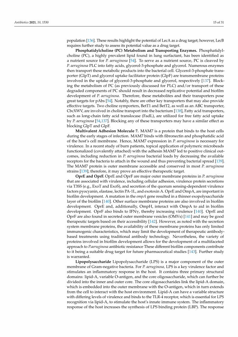
Phosphatidylcholine (PC) Metabolism and Transporting Enzymes. Phosphatidyl-choline (PC), a highly prevalent lipid found in lung surfactant, has been identified asa nutrient source for P. aeruginosa [54]. To serve as a nutrient source, PC is cleaved byP. aeruginosa PLC into fatty acids, glycerol-3-phosphate and glycerol. Numerous enzymesthen transport these metabolic products into the bacterial cell. Glycerol-3-phosphate trans-porter (GlpT) and glycerol uptake facilitator protein (GlpF) are transmembrane proteinsinvolved in the uptake of glycerol-3-phosphate and glycerol, respectively [137]. Block-ing the metabolism of PC (as previously discussed for PLC) and/or transport of thesedegraded components of PC should result in decreased replicative potential and biofilmdevelopment of P. aeruginosa. Therefore, these metabolites and their transporters posegreat targets for pAbs [54]. Notably, there are other key transporters that may also provideeffective targets. Two choline symporters, BetT1 and BetT2, as well as an ABC transporter,CbcXWV, are involved in choline transport into the bacterium [138], Fatty acid transporters,such as long-chain fatty acid translocase (FadL), are utilized for free fatty acid uptakeby P. aeruginosa [54,137]. Blocking any of these transporters may have a similar effect asblocking GlpT and GlpF.
Multivalent Adhesion Molecule 7. MAM7 is a protein that binds to the host cellsduring the early stages of infection. MAM7 binds with fibronectin and phosphatidic acidof the host’s cell membrane. Hence, MAM7 expression in P. aeruginosa is necessary forvirulence. In a recent study of burn patients, topical application of polymeric microbeadsfunctionalized (covalently attached) with the adhesin MAM7 led to positive clinical out-comes, including reduction in P. aeruginosa bacterial loads by decreasing the availablereceptors for the bacteria to attach in the wound and thus preventing bacterial spread [139].The MAM7 protein is outer membrane accessible and conserved in most P. aeruginosastrains [139]; therefore, it may prove an effective therapeutic target.
OprE and OprF. OprE and OprF are major outer membrane proteins in P. aeruginosathat are associated with virulence, including cellular adhesion, virulence protein secretionsvia T3SS (e.g., ExoT and ExoS), and secretion of the quorum sensing-dependent virulencefactors pyocyanin, elastase, lectin PA-1L, and exotoxin A. OprE and OmpA, are important inbiofilm development. A mutation in the ompA gene resulted in a thinner exopolysaccharidelayer of the biofilm [140]. Other surface membrane proteins are also involved in biofilmdevelopment. OprE and, additionally, OmpH, interact with OmpA to aid in biofilmdevelopment. OprF also binds to IFNγ, thereby increasing virulence [140]. OprE andOprF are also found in secreted outer membrane vesicles (OMVs) [141] and may be goodtherapeutic targets based on their accessibility [142]. However, as noted with the secretionsystem membrane proteins, the availability of these membrane proteins has only limitedimmunogenic characteristics, which may limit the development of therapeutic antibody-based treatments using traditional antibody technology. Nevertheless, the variety ofproteins involved in biofilm development allows for the development of a multifacetedapproach to P.aeruginosa antibiotic resistance These different biofilm components contributeto it being a suitable drug target for future pharmaceutical studies [143]. Further studyis warranted.
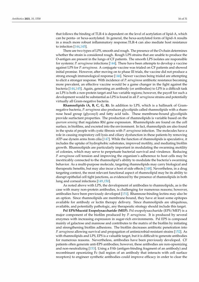
Lipopolysaccharide Lipopolysaccharide (LPS) is a major component of the outermembrane of Gram-negative bacteria. For P. aeruginosa, LPS is a key virulence factor andstimulates an inflammatory response in the host. It contains three primary structuraldomains: lipid-A, variable O-antigen, and the core oligosaccharide, which can further bedivided into the inner and outer core. The core oligosaccharides link the lipid-A domain,which is embedded into the outer membrane with the O-antigen, which in turn extendsfrom the cell to interact with the host environment. Lipid-A can have a variable structurewith differing levels of virulence and binds to the TLR-4 receptor, which is essential for LPSrecognition via lipid-A, to stimulate the host’s innate immune system. The inflammatoryresponse of the host increases the synthesis of LPS binding protein (LBP). The response
Antibiotics 2021, 10, 1530 16 of 31
that follows the binding of TLR-4 is dependent on the level of acetylation of lipid-A, whichcan be penta- or hexa-acetylated. In general, the hexa-acetylated form of lipid-A resultsin a much more robust inflammatory response.TLR-4 can also mediate host resistanceto infection [144,145].
There are two types of LPS, smooth and rough. The presence of the O-chain determineswhether the strain is considered rough. Rough LPS strains that are unable to produce theO-antigen are present in the lungs of CF patients. The smooth LPS isolates are responsiblefor systemic P. aeruginosa infections [144]. There have been attempts to develop a vaccineagainst LPS for P. aeruginosa. A conjugate vaccine was trialed on CF patients and showedinitial promise. However, after moving on to phase III trials, the vaccine did not produce astrong enough immunological response [146]. Newer vaccines being trialed are attemptingto elicit a stronger response. With incidence of P. aeruginosa antibiotic resistance becomingmore prevalent, an effective vaccine would be a game changer in the fight against thebacteria [144,145]. Again, generating an antibody (or antibodies) to LPS is a difficult taskas LPS is both a non-protein target and has variable regions; however, the payoff for such adevelopment would be substantial as LPS is found in all P. aeruginosa strains and potentiallyvirtually all Gram-negative bacteria.
Rhamnolipids (A, B, C, G, R). In addition to LPS, which is a hallmark of Gram-negative bacteria, P. aeruginosa also produces glycolipids called rhamnolipids with a rham-nose head group (glycosyl) and fatty acid tails. These membrane-bound glycolipidsprovide surfactant properties. The production of rhamnolipids is variable based on thequorum sensing that regulates Rhl gene expression. Rhamnolipids are found on the cellsurface, in biofilms, and excreted into the environment. In fact, rhamnolipids are also foundin the sputa of people with cystic fibrosis with P. aeruginosa infection. The molecules have arole in causing respiratory cell lysis and ciliary dysfunction in these patients by removingATP-ase dynein arms from cilia [147]. While the function of rhamnolipids is speculative, itincludes the uptake of hydrophobic substrates, improved motility, and mediating biofilmgrowth. Rhamnolipids are particularly important in modulating the swarming motilityof colonies, which may serve to perpetuate bacterial survival and virulence. ReducingP. aeruginosa cell tension and improving the organism’s adherence to host cells may beinextricably connected to the rhamnolipid’s ability to modulate the bacteria’s swarmingbehavior. As a multi-purpose molecule, targeting rhamnolipids may carry biological andtherapeutic benefits, but may also incur a host of side effects [148]. Nevertheless, in a drugtargeting context, the most relevant functional aspect of rhamnolipid may be its ability todisrupt epithelial cell tight junctions, as evidenced by the presence of rhamnolipids in bothlung and corneal infections [149,150].
As noted above with LPS, the development of antibodies to rhamnolipids, as is thecase with many non-protein antibodies, is challenging for numerous reasons; however,antibodies have been previously developed [151]. Rhamnose-binding lectins may also bean option. Since rhamnolipids are membrane-bound, they have at least some epitopesavailable for antibody or lectin therapy delivery. Since rhamnolipids are ubiquitous,available, and potentially pathologic, any therapeutic strategy should include this target.
Psl EPS/Mucoid Exopolysaccharide (MEP). Psl exopolysaccharide (EPS/MEP) is amajor component of the biofilm produced by P. aeruginosa. It is produced by severalenzymes with increasing expression in sugar-rich environments. Psl EPS is composedmainly of galactose and mannose and contributes to the matrix of the biofilm by initiatingand strengthening biofilm adhesions. The biofilm decreases antibiotic penetration intoP. aeruginosa allowing survival and propagation of antimicrobial-resistant strains [152]. Aswith rhamnolipids and LPS, EPS is a valuable target, but it is difficult to generate antibodiesfor numerous reasons. Nevertheless, antibodies have been previously developed. CFpatients often generate anti-EPS antibodies; however, these antibodies are non-opsonizingand non-neutralizing [153]. Using a FAb (antigen-binding fragment of an antibody) andrecombinant opsonizing Fc (tail region of an antibody that interacts with cell surfacereceptors) to engineer synthetic antibodies could improve efficacy in order to clear the

Antibiotics 2021, 10, 1530 17 of 31
infection [154]. Alternatively, as with other carbohydrate targets, lectins may also be anoption. Since EPSs are an integral membrane component, they are available to circulatingand secreted (IgA and IgM) antibody or lectin therapeutic delivery. Since EPS is ubiquitous,available, and potentially pathologic, any therapeutic strategy could include this target.
Alginate. Alginate is another exopolysaccharide produced by P. aeruginosa. It spansthe organism’s inner and outer membrane. Alginate enhances cell to cell adhesions andcell to host adhesions. These adhesions are protective for the organism; they increaseresistance to the host immune system [155]. P. aeruginosa evades the host immune systemduring chronic disease by decreasing its virulence so as to not provoke an acute immunereaction. Alginate is produced in large quantities during chronic infections. Inhibition ofmacrophage clearance and efferocytosis have been observed, but the mechanism of alginatein this process is still unknown [156]. Again, as with other lipids/carbohydrates discussedin this section, alginate is a difficult, but potentially valuable, target.
Secreted outer membrane vesicles (OMVs). OMVs are ubiquitously generated andsecreted by P. aeruginosa during all stages of the bacteria’s life cycle [141,157], particularlyunder conditions of stress. Growing experimental evidence suggests that OMVs are se-creted with numerous virulence factors, antibiotic resistance molecules, and quorum sensingmolecules, including hemolysin, phospholipase C, elastase, and alkaline phosphatase,antimicrobial quinolines, adhesions, CIF, and beta-lactamase into the host cells [158–161].Additionally, these vesicles carry virulence factors that can cause cytotoxicity, increaseadherence, and carry factors such as cystic fibrosis transmembrane conductance regulatorinhibitory factor [141]. OMVs can then fuse with host cell membranes and alter their func-tion, resulting in the breakdown of the host’s epithelial barrier cells, which can ultimatelyfacilitate the invasion of P. aeruginosa. Antibody therapies for these targets could includeseveral approaches. First, antibodies to CIF could be delivered inside the vesicle that fusesto the secreted outer membrane vesicles. Second, antibodies to the OMV itself could bedeveloped with an opsonization effector function to eliminate the OMV entirely. Morestudy into OMVs as either a drug target or natural drug delivery system is warranted.
CbpD. CbpD is one of the conserved outer membrane vesicle proteins found inthe majority of studied strains of P. aeruginosa. Due to its conservative expression andextracellular presence, CbpD may be a possible target of immunotherapy against the outermembrane vesicles and the virulence factors, which they carry [162].
3.6. Motility Factors
Bacterial motility plays a key role in P. aeruginosa virulence and biofilm formation.P. aeruginosa uses type IV pili and flagella for motility. Several studies have suggested thattargeting pili or flagella with drugs or vaccines would show strong efficacy [163].
Pili proteins. Type IV pili (TFP). TFP is a motorized fimbriae providing twitchingmotility for P. aeruginosa [164,165]. TFP is essential to both biofilm formation and virulence.Several adhesin proteins (the minor pilins) are at the tip of the pili including PilEXWV andFimU (see Figure 3). The pilus is extended through major pilins and includes PilA [165].Pili may also enable P. aeruginosa to perform transformation and conjugation, and exchangevirulence and antibiotic resistance genetic material with other bacteria and may at leastpartially explain the organism’s ability to rapidly acquire antibiotic resistance.
The TFP proteins are all outer membrane accessible and are conserved in mostP. aeruginosa strains [166]. TFP proteins are potentially accessible to circulating and se-creted (IgA and IgM) antibody therapy delivery. Since TFP proteins are ubiquitous andpathologic, any therapeutic strategy could include these targets.
Flagellar Proteins. P. aeruginosa acquires its motility from a single glycosylated polarflagellum. In addition to motility, the bacteria’s flagellum can play a role in adhesion tohost cells and stimulation of the immune system. The flagellum is an emerging drug targetdue to the many roles it plays in the pathogenesis of P. aeruginosa [167]. There are twocommon types of flagella proteins, PAK and PA01. Antibodies against PAK flagella andPA01 flagella were used to assess their potential as a future drug target [168]. There have

Antibiotics 2021, 10, 1530 18 of 31
been varying results on the effectiveness of a vaccine targeting the P. aeruginosa flagellum.Rabbits were passively immunized with antibodies against the monomeric flagella proteinand survival studies showed the antibodies provided little protection. However, antibodiestargeting polymeric flagella showed promising results with an 87% survival rate followingthe P. aeruginosa challenge. Although anti-flagella antibodies against the PAK and PA01strains showed only modest protection, flagella proteins appear to be a promising emergingdrug target [168].
3.7. Resource Scavenging Molecules
One of the key battlefronts between host and bacteria is molecular resource scavenging.Iron is a prime example as it is an essential nutrient for the biological processes of P.aeruginosa. Like host cells and other bacteria, P. aeruginosa requires iron as a cofactor formany enzymes. It uses specialized iron scavenging molecules, siderophores, to steal ironfrom its environment. Antibodies to these molecules commonly provide protection againstvirulence and thus these molecules are excellent drug targets [169].
Pyoverdine. Pyoverdine is the major siderophore produced by P. aeruginosa thatextracts iron from host complexes [170]. Pyoverdine has such a high affinity for iron that itcan steal iron from transferrin and extract iron from host cells, leaving them metabolicallylimited and damaging mitochondria [171]. Pyoverdine is an extracellular peptide found inmost P. aeruginosa infections, especially CF, and in most strains of P. aeruginosa [171]. As aconserved, extracellular protein [172], its accessibility to antibody development makes itan attractive candidate for therapeutic development.
Pyochelin (Pch). Pch is the second major siderophore secreted by P. aeruginosa toscavenge iron. Pch chelates iron and other metals from the ECM (extracellular matrix). Pchalso provides a competitive advantage by inhibiting other bacteria, including S. aureus [173].Pch is found extracellularly in most of the indications for P. aeruginosa infections and in moststrains of P. aeruginosa [174,175]. As with pyoverdine, Pch is an extracellular protein and isconserved in most P. aeruginosa strains [175], making it a target worthy of consideration inthe development of new therapeutic antibody strategies. Notably, Pch is a small moleculesynthesized from cysteine and thus will require additional engineering to generate anantibody to the molecule and so it may not be the best choice for the first generation ofantibodies developed.
HasAP. Aerobic metabolism of P. aeruginosa requires iron for many of the respiratoryenzymatic processes. HasAp is a secreted hemophore that, after being cleaved by aPseudomonas protease [176], captures iron as part of the heme acquisition system [176].Its release is regulated via quorum sensing (QS) in response to cell growth and biofilmestablishment [177]. HasAp is not constitutively expressed, but of the QS-regulated proteinsstudied, iron regulation proteins are the most abundant [178]. Targeting protease activity,or the hemophore activity of HasAp, could help eliminate chronic infection by starvingP. aeruginosa of essential iron needed for aerobic metabolism, and thus may be a worthytherapeutic antibody target.
3.8. Immunomodulators
Immunomodulators use regulatory molecules to adjust the immune response, includ-ing inducing, attenuating, and amplifying the host immune response. P. aeruginosa usesseveral mechanisms to modulate the immune system to reduce effective host immuneresponse, but P. aeruginosa also upregulates the immune response to fight competitorssuch as S. aureus. For example, P. aeruginosa increases IL-8 production and in response,S. aureus dampens Toll-like receptor (TLR1/TLR2)-mediated activation of the NF-κB path-way [179]. As noted earlier, P. aeruginosa uses LasA to inhibit competing S. aureus. Here, afew immunomodulators are highlighted as targets during a P. aeruginosa infection.
Lipoxygenase (LoxA). Lipoxygenases (LOXs) are extracellular, lipid-oxidizing enzymesthat have potent immunoregulatory properties in their hosts [180,181]. In P. aeruginosa, thelipoxygenase LoxA oxidizes polyunsaturated fats, resulting in the expression of bioactive

Antibiotics 2021, 10, 1530 19 of 31
lipid mediators and suppression of chemotactic molecules [182]. With greater than 90%conservation across P. aeruginosa strains, leukotrienes, a product of LoxA, are secretedto balance lipoxins and resolvins. LoxA also activates host ferroptosis, resulting in hostcell death and inflammation. LoxA increases LXA4 along with other LOX expressions,leading to an increase in neutrophil chemotaxis and pro-inflammatory signaling [183].Evidence from preclinical animal studies shows that P. aeruginosa clinical isolates secreteLoxA, indicating modulation of the host immune response during acute pneumonia [182].However, LoxA decreases in expression after 24 h of lung infection and has decreasedexpression in chronic infections. This indicates that LoxA is a significant pro-inflammatoryand bacterial invasion mediator in acute infections, while losing this effect when lunginfections become chronic. Thus, targeting LoxA during acute infection could help increasethe host immune response. Additionally, upregulation of CCR5, a receptor on white bloodcells for immune system chemokines, occurs after exposure of lung tissues to Lox throughunknown mechanisms. CCR5 acts as a chemokine receptor as well as a chemotactic agent,resulting in increased bacterial invasion [182]. Since LoxA is accessible to both circulatingand secreted (IgA and IgM) antibodies, it may prove to be a good target for therapeuticantibody development and delivery therapy.
Leukocidin. Leukocidin is a cytotoxin of P. aeruginosa that damages granulocytesand lymphocytes by forming a beta-barrel pore in cell membranes, thereby inhibitingessential bactericidal and phagocytic immune functions [184,185]. Leukocidin has longbeen known as a common, secreted virulence factor in P. aeruginosa. Targeting Leukocidin,which is accessible, ubiquitous, and pathogenic, could have direct influences on the courseof P. aeruginosa infection and could help increase the host immune response.
4. Antibodies as Therapeutics
As antibiotic resistance continues to increase throughout a wide spectrum of microbialinfections, new and innovative approaches to treating these infections must be sought.The development of antibodies to the relevant bacterial targets outlined above and inTable 1 may be the next step in combating antimicrobial resistance. Therapeutic antibodieswork by activating and modulating our own host effector mechanisms including directneutralization of toxins and pathogens, activation of the complement pathway, activation ofneutrophil and macrophage opsonophagocytosis, activation of natural killer cells, enhance-ment of antigen presentation from dendritic cells to T cells and follicular dendritic cells to Bcells, and degranulation of mast cells, eosinophils, and basophils. Additionally, Fc receptorsthat correspond to different classes of antibodies can activate the complement pathway,induce innate immune responses, and enhance natural adaptive immunity, providing amulti-faceted strategy to address disease [186].
Therapeutic monoclonal antibodies (mAbs) have emerged as a tour de force in thedrug market and are projected to capture a rising percentage of the worldwide drugmarket—a trend that reflects increased prescription use of existing therapeutic mAbs anda large number (~50) of new therapeutic mAb drugs approved for use [187]. Currently,mAbs have been developed to treat a broad array of clinical conditions including cancer,autoimmune diseases, transplants, infectious disease, and toxin neutralization [188].
In contrast to mAbs, polyclonal antibodies (pAbs) have multiple epitope binding sites,thereby allowing greater coverage of neutralizing antigens. Much like our native immunesystem, pAbs are produced as a collection of antibodies from multiple B cell lineages,thus producing many different sites and affinities for the same antigen. Having multipleepitope binding sites allows for greater neutralization options and less opportunities forpathogens to develop escape mutants [189]. By harboring the ability to work at a widenumber of sites in microbial pathogenesis, polyclonal antibodies have the potential to curbor slow the development of multidrug-resistant organisms and fill the gaps in treatmentleft by antibiotics and vaccines. Passive polyclonal antibodies have been used to treatdiseases for over 100 years with the oldest being patient- and blood-derived products(often “convalescent sera”) or intravenous immunoglobulins (IVIG) [190]. Additionally,

Antibiotics 2021, 10, 1530 20 of 31
more modern pAbs are currently used as antitoxins and antivenoms and are an effectivetreatment post-infection or post-exposure for botulism, varicella, vaccinia, rabies, CMV,hepatitis B, and tetanus [188]. This is not a new strategy, but merely a re-energized approachto treating bacterial pathogenesis.
In an emerging appreciation of rational design for new therapeutic options, combi-nations of mAbs with defined correlates of protection are driving an increasing numberof investigations into combination therapies, especially with a focus on broad effectormechanisms of actions beyond just simple binding. This strategy has been useful in recentviral outbreaks, where multiclonal cocktails, or convalescent sera (i.e., polyclonal) fromrecovered patients, have been used to treat diseases [189]. Engineering combination ther-apies with improved effector functions, specificity, affinity, and glycosylation generatesa stronger and more diverse immune response. Moreover, combination therapies alsogenerate a much higher barrier for pathogens to overcome; in other words, the pathogenscannot easily evolve to circumvent the treatment [189].
Combinatorial Therapy
Recently, it has been shown that antibodies and antibiotics can act concomitantlyor even synergistically against pathogens. Several studies have shown that exposure toantibiotic therapy alters the expression of secreted factors, cell surface proteins, and othercellular products that may be viable immunotargets [190]. These altered levels of expres-sion can change and potentially enhance the innate and humoral immune responses topathogens—namely through increased antigen exposure, increased protein secretion, andpotentially decreased virulence of pathogens [190]. Inspired by these same principles, com-bination or conjugate antibiotic/antibody therapeutic strategies, particularly those basedon polyclonal antibodies, could be designed to capitalize on these same altered expressionlevels to enhance therapeutic efficacy and combat resistance. Below is a non-exhaustivelook into the evidence demonstrating how combination antibiotic/antibody therapy canhave a synergistic effect and, in some cases, potentiate the antimicrobial response.
Several clinical isolates of Streptococcus pneumoniae have been shown to, when in thepresence of penicillins or cephalosporins, become more reactive to opsonization by theinnate immune response; acute phase reactants, such as c-reactive protein and serumamyloid P, more efficiently bind cellular antigens and clear infection quicker than whennot in the presence of these antibiotics [190]. By examining the synergistic activity be-tween antimicrobial drugs and innate immune responses in vivo, it can by deduced thatcombinations between antimicrobial and antibody therapy will likely work in a similarsynergistic fashion.
Similar to making Streptococcus pneumoniae more readily opsonized, penicillins,cephalosporins, and gentamicin have all been shown to alter gene expression and tran-scription in Staphylococcus aureus, leading to a change in the molecular structure of thecell walls of the organisms [191]. Surprisingly, changes in the expression of virulencefactors in S. aureus in response to antibiotic therapy may actually improve exposure tocellular products targeted by polyclonal antibody therapies. Using this observation as afoundation, further work is underway to characterize the response of organisms to spe-cific antimicrobial therapies, allowing polyclonal antibodies to be tailored to the expectedupregulation of virulence factors. In 2015, Lehar et al. utilized rifamycin conjugatedto monoclonal antibodies against wall-teichoic acids (cell wall peptidoglycans) to elim-inate residual methicillin-resistant S. aureus infections [191]. These antibiotic–antibodyconjugates effectively bound S. aureus cell walls allowing rifamycin to be taken up byphagolysosomes and exert its antimicrobial effect intracellularly, showing improved eradi-cation rates compared to Vancomycin treatment alone [192]. Such an effect was confirmedin a recent in vivo study.
A monoclonal antibody to PcrV (in P. aeruginosa) was used in combination with eitherciprofloxacin, tobramycin, or ceftazidime to improve survival rates in mice, noting that theeffect was more pronounced in more virulent strains of P. aeruginosa [192]. Protecting the

Antibiotics 2021, 10, 1530 21 of 31
lung against the cytotoxic effects of exotoxin in addition to amplifying the antimicrobialeffects of the antibiotics highlights the value of antibiotic–antibody conjugate therapy andpunctuates the need for further research into the human applications of these treatments.
5. Discussion
Therapeutic antibodies are highly specific with long half-lives and minimal dosingcompared to other drug treatments [193]. Currently, there are 79 US FDA-approved mono-clonal antibodies in use and well over 500 currently being studied in clinical trials [193].Most therapeutic antibodies target cancer, autoimmune diseases, or prevent transplantrejection, but their use in other disease realms is gaining momentum. Currently, there arefive approved infectious disease monoclonals: Bezlotoxumab for prevention of recurrentClostridiodes difficile, Ibalizumab for drug-resistant HIV, Palivizumab for prevention ofrespiratory syncytial virus, and Raxibacumab and Obiltoxaximab for inhalational anthrax,and casirivamab and imdevimab for COVID-19 [194].
New therapies and treatment options must be sought for P. aeruginosa, a highly prevalentand exceedingly antibiotic-resistant microorganism. Patients with the highest risk of infectioninclude those with compromised immunity, seen with chronic burns, chronic wounds, andHIV/AIDS patients. Those with deficient bacterial clearance mechanisms, as seen in cysticfibrosis, COPD, and ventilator-dependent patients [195], are also particularly susceptibleto P. aeruginosa. P. aeruginosa’s widely varied environmental niches, from respiratory tractinfections, skin, and soft tissue infections, and even septicemia make treatment especiallychallenging. Additionally, P. aeruginosa creates seemingly impenetrable biofilms, which areespecially prevalent on implanted and indwelling devices, such as catheters, endotrachealtubes, and prostheses. These biofilms create a plethora of challenges for the host innate andadaptive immunity as well as antimicrobials [92]. The most daunting challenge, however,may be the ability of P. aeruginosa to modulate host immune responses.
As noted earlier, multidrug-resistant pathogens present an emergent, serious pub-lic health threat at all levels. While we have detailed many of the potential targets ofP. aeruginosa, many other emerging infections are multipathogenic, multidrug-resistant,and even more deadly. With poly- and oligoclonal mixtures of therapeutic antibodies,increasing levels of efficacy can be reached by adjusting individual antibodies withinthe mixture for specific or broad pathogenic threats. The oligomixture can also haveprophylactic applications with adjustable dosing. Further, oligomixtures and antibioticscould be used concomitantly and potentially synergistically against individual or multiplepathogens. While vaccination is a good option, oligomixtures have significant advantagesand offer a strong alternative to treat multidrug resistance pathogens. Most notable is theability to neutralize identified, virulent targets without the inherent variability in responseto vaccination.
The effector function of an antibody plays a critical role in pathogen neutralization.With an oligomixture, the role of effector function can be engineered (see Figure 5). This isespecially true when compared to a vaccine in which effector function is merely a functionof genetic and/or environmental chance. With oligomixtures, the effector function optionscould be optimized based on any number of factors, including genetics. Effector functionscould be standardized for the entire mixture or diversified to allow different parts of theimmune system to enter the fight. The isotypes and thus the effector functions of theoligomixture could be highly dependent on the environment. For example, IgA could bethe most efficacious isotype for intestinal and endothelial pathogenic indications whereasIgG isotypes would likely be most efficacious in sepsis indications.

Antibiotics 2021, 10, 1530 22 of 31
Antibiotics 2021, 10, x FOR PEER REVIEW 23 of 33
the human genome, the engineering of more efficacious antibody technologies for diverse indications can also evolve, or even become personalized, just as P. aeruginosa evolves.
Figure 5. Effector function or mechanisms of killing by antibodies.
Author Contributions: Conceptualization, B.D.B., M.S. and F.D.T.; validation, J.M.S. and A.E.B.; formal analysis, M.S., B.D.B., F.D.T. and J.M.S.; investigation, C.S.R., W.L.W., A.G.K., A.P.Q. and L.L.P.; resources, C.S.R., W.L.W., A.G.K., A.P.Q. and L.L.P.; data curation, C.S.R., W.L.W., A.G.K., K.M.C. and L.L.P.; writing—original draft preparation, C.S.R., W.L.W., A.G.K., A.P.Q., K.M.C. and L.L.P.; writing—review and editing, C.S.R., W.L.W., A.G.K., L.L.P., J.M.S. and A.P.Q.; supervision, J.M.S., B.D.B. and F.D.T.; project administration, M.S. and B.D.B. All authors have read and agreed to the published version of the manuscript.
Funding: This research received no external funding.
Institutional Review Board Statement: Not applicable.
Informed Consent Statement: Not applicable.
Data Availability Statement: Data sharing not applicable.
Conflicts of Interest: The authors declare no conflict of interest.
Figure 5. Effector function or mechanisms of killing by antibodies.
Another major consideration moving forward will be drug delivery. As detailed in thebody of this review, the common indications that will benefit from polyclonal therapies arefound in diverse environments. In most cases detailed in this manuscript, the traditionaltherapeutic concentrations of antibiotics in serum and at the site of infection are belowMIC levels, decreasing the efficacy of the treatment and increasing the probability ofantibiotic resistance. Ideally, the route would be dependent on the site of infection. Forbacteremic infections, intravenous would be the route of choice. Beyond that, it gets trickier.A potential route would be topical administration of IgA/IgG via a nebulizer solutionfor pneumonia or patients with cystic fibrosis. Studies have supported this possibility;IgG and IgA that have been nebulized in a variety of solutions and viscosities have beenshown to increase immunoglobulin levels in bronchoalveolar lavage specimens up to48 h after administration as well as confer immunity to the specific immunoglobulintargets [196]. Potential for topical preparations for keratitis, folliculitis, and other soft tissueinfections exist as well, expanding the range of these therapeutics [197]. Recently it has beenshown that antibodies and antibiotics can act concomitantly or even synergistically againstpathogens. In particular, bacterial cell surfaces can be altered by antibodies, resulting inmultiple mechanisms that improve drug efficacy [198,199].

Antibiotics 2021, 10, 1530 23 of 31
P. aeruginosa, like many other bacteria, continues to and will continue to find ways tosubvert the efficacy of antibiotics. The immune system has evolved to attack microbes onmultiple fronts, but P. aeruginosa likewise evolves counterattacks, allowing it to colonizeand infect hosts. Engineering combination antibody therapies with numerous targets aspresented here would be the best option currently available to improve therapeutic efficacyand reduce the opportunity for resistance development. As the P. aeruginosa genome ischaracterized or evolves, the combination of antibodies can be adapted and optimized.Most importantly, with the improved engineering of antibodies and characterization ofthe human genome, the engineering of more efficacious antibody technologies for diverseindications can also evolve, or even become personalized, just as P. aeruginosa evolves.
Author Contributions: Conceptualization, B.D.B., M.S. and F.D.T.; validation, J.M.S. and A.E.B.;formal analysis, M.S., B.D.B., F.D.T. and J.M.S.; investigation, C.S.R., W.L.W., A.G.K., A.P.Q. andL.L.P.; resources, C.S.R., W.L.W., A.G.K., A.P.Q. and L.L.P.; data curation, C.S.R., W.L.W., A.G.K.,K.M.C. and L.L.P.; writing—original draft preparation, C.S.R., W.L.W., A.G.K., A.P.Q., K.M.C. andL.L.P.; writing—review and editing, C.S.R., W.L.W., A.G.K., L.L.P., J.M.S. and A.P.Q.; supervision,J.M.S., B.D.B. and F.D.T.; project administration, M.S. and B.D.B. All authors have read and agreed tothe published version of the manuscript.
Funding: This research received no external funding.
Institutional Review Board Statement: Not applicable.
Informed Consent Statement: Not applicable.
Data Availability Statement: Data sharing not applicable.
Conflicts of Interest: The authors declare no conflict of interest.
Abbreviations
AHL Acyl Homoserine LactoneAprA Alkaline Protease ACF Cystic FibrosisCIF CFTR Inhibiting FactorCNS Central Nervous SystemCRPA Carbapenem Resistant Pseudomonas aeruginosaExoU Exotoxin UExoS Exotoxin SExoT Exotoxin TExoY Exotoxin YFAb antigen-binding region of an antibodyFc cell receptor region of an antibodyGlpF Glycerol Uptake Facilitator ProteinGlpT Glycerol-2-Phosphate TransporterHCN Hydrogen CyanideIDSA Infectious Disease Society of AmericaIgA Immunoglobulin AIgE Immunoglobulin EIgG Immunoglobulin GIgM Immunoglobulin MIVIG Intravenous ImmunoglobulinLOX LipoxygenasemABs Monoclonal Antibodies (is mABs in text)MAM7 Multivalent Adhesion Molecule 7MEP Mucoid ExopolysaccharideMIC Minimum Inhibitory ConcentrationMMP Matrix MetallopeptideLPS LipopolysaccharideOMV Outer Membrane Vesicle

Antibiotics 2021, 10, 1530 24 of 31
NET Neutrophil Extracellular TrapPAB Polyclonal AntibodyPC PhosphatidylcholinePCH PyochelinPLC Phospholipase CPQS Pseudomonas Quinolone SignalQS Quorum SensingROS Reactive Oxygen SpeciesTFP Type IV PiliTNF Tumor Necrosis FactorTLR Toll-Like ReceptorT1SS Type 1 Secretion SystemT2SS Type 2 Secretion SystemT3SS Type 3 Secretion SystemUTI Urinary Tract Infection
References1. Kanj, S.; Sexton, M.D.; Daniel, M.D. Epidemiology, Microbiology, and Pathogenesis of Pseudomonas aeruginosa Infection. Available
online: https://www.uptodate.com/contents/epidemiology-microbiology-and-pathogenesis-of-pseudomonas-aeruginosa-infection?search=pseudomonas&source=search_result&selectedTitle=2~150&usage_type=default&display_rank=2 (accessedon 21 September 2020).
2. Owlia, P.; Nosrati, R.; Alaghehbandan, R.; Lari, A.R. Antimicrobial susceptibility differences among mucoid and non-mucoidPseudomonas aeruginosa isolates. GMS Hyg. Infect. Control. 2014, 9, 13. [CrossRef]
3. Souha Kanj, M.D. Principles of Antimicrobial Therapy of Pseudomonas aeruginosa Infections. Available online: https://www-uptodate-com.proxy.rvu.edu/contents/principles-of-antimicrobial-therapy-of-pseudomonas-aeruginosa-infections#H6675731 (accessed on 21 September 2020).
4. Centers for Disease Control and Prevention. Pseudomonas aeruginosa in Healthcare Settings. Available online: https://www.cdc.gov/hai/organisms/pseudomonas.html (accessed on 21 September 2020).
5. Todar, K. Pseudomonas. Online Textbook of Bacteriology. Available online: http://textbookofbacteriology.net/pseudomonas_4.html (accessed on 21 September 2020).
6. Nordmann, P.; Poirel, L. Epidemiology and Diagnostics of Carbapenem Resistance in Gram-negative Bacteria. Clin Infect Dis.2019, 69 (Suppl. S7), S521–S528. [CrossRef] [PubMed]
7. Tamma, P.D.; Aitken, S.L.; Bonomo, R.A.; Mathers, A.J.; van Duin, D.; Clancy, C.J. Infectious Diseases Society of AmericaGuidance on the Treatment of Extended-Spectrum β-lactamase Producing Enterobacterales (ESBL-E), Carbapenem-ResistantEnterobacterales (CRE), and Pseudomonas aeruginosa with Difficult-to-Treat Resistance (DTR-P. aeruginosa). Clin. Infect Dis. 2020,72, e169–e183. [CrossRef]
8. Pang, Z.; Raudonis, R.; Glick, B.R.; Lin, T.-J.; Cheng, Z. Antibiotic resistance in Pseudomonas aeruginosa: Mechanisms andalternative therapeutic strategies. Biotechnol. Adv. 2019, 37, 177–192. [CrossRef]
9. Centers for Disease Control and Prevention. Antibiotic-Resistant Germs: New Threats. Available online: https://www.cdc.gov/drugresistance/biggest-threats.html (accessed on 6 May 2020).
10. Kmeid, J.G.; Youssef, M.M.; Kanafani, Z.A.; Kanj, S.S. Combination therapy for Gram-negative bacteria: What is the evidence?Expert. Rev. Anti. Infect. Ther. 2013, 11, 1355–1362. [CrossRef]
11. Grekov, I.; Thöming, J.G.; Kordes, A.; Häussler, S. Evolution of Pseudomonas aeruginosa toward higher fitness under standardlaboratory conditions. ISME J. 2021, 15, 1165–1177. [CrossRef] [PubMed]
12. Fothergill, J.L.; Panagea, S.; Hart, C.A.; Walshaw, M.J.; Pitt, T.L.; Winstanley, C. Widespread pyocyanin over-production amongisolates of a cystic fibrosis epidemic strain. BMC Microbiol. 2007, 7, 45. [CrossRef] [PubMed]
13. Mathee, K.; Ciofu, O.; Sternberg, C.; Lindum, P.W.; Campbell, J.I.A.; Jensen, P.; Johnsen, A.H.; Givskov, M.; Ohman, D.E.; Søren,M.; et al. Mucoid conversion of Pseudomonas aeruginos by hydrogen peroxide: A mechanism for virulence activation in thecystic fibrosis lung. Microbiology 1999, 145, 1349–1357. [CrossRef] [PubMed]
14. Von Götz, F.; Häussler, S.; Jordan, D.; Saravanamuthu, S.S.; Wehmhöner, D.; Strüssmann, A.; Lauber, J.; Attree, I.; Buer, J.;Tümmler, B.; et al. Expression Analysis of a Highly Adherent and Cytotoxic Small Colony Variant of Pseudomonas aeruginosaIsolated from a Lung of a Patient with Cystic Fibrosis. J. Bacteriol. 2004, 186, 3837. [CrossRef] [PubMed]
15. Häußler, S.; Tümmler, B.; Weißbrodt, H.; Rohde, M.; Steinmetz, I. Small-Colony Variants of Pseudomonas aeruginosa in CysticFibrosis. Clin. Infect. Dis. 1999, 29, 621–625. [CrossRef]
16. D’Argenio, D.A.; Calfee, M.W.; Rainey, P.B.; Pesci, E.C. Autolysis and Autoaggregation in Pseudomonas aeruginosa ColonyMorphology Mutants. J. Bacteriol. 2002, 184, 6481. [CrossRef] [PubMed]
17. Smith, E.E.; Buckley, D.G.; Wu, Z.; Saenphimmachak, C.; Hoffman, L.R.; D’Argenio, D.A.; Miller, S.I.; Ramsey, B.W.; Speert, D.P.;Moskowitz, S.M.; et al. Genetic adaptation by Pseudomonas aeruginosa to the airways of cystic fibrosis patients. Proc. Natl. Acad.Sci. USA 2006, 103, 8487–8492. [CrossRef] [PubMed]

Antibiotics 2021, 10, 1530 25 of 31
18. Subedi, D.; Vijay, A.K.; Kohli, G.S.; Rice, S.A.; Willcox, M. Comparative genomics of clinical strains of Pseudomonas aeruginosastrains isolated from different geographic sites. Sci. Rep. 2018, 8, 15668. [CrossRef] [PubMed]
19. Weiser, R.; Green, A.E.; Bull, M.J.; Cunningham-Oakes, E.; Jolley, K.A.; Maiden, M.C.J.; Hall, A.J.; Winstanley, C.; Weightman, A.J.;Donoghue, D.; et al. Not all Pseudomonas aeruginosa are equal: Strains from industrial sources possess uniquely large multireplicongenomes. Microb. Genom. 2019, 5, e000276. [CrossRef]
20. Boucher, H.W.; Talbot, G.H.; Bradley, J.S.; Edwards, J.E.; Gilbert, D.; Rice, L.B.; Scheld, M.; Spellberg, B.; Bartlett, J. Bad bugs, nodrugs: No ESKAPE! An update from the Infectious Diseases Society of America. Clin. Infect. Dis. Off. Publ. Infect. Dis. Soc. Am.2009, 48, 1–12. [CrossRef]
21. Bassetti, M.; Peghin, M.; Vena, A.; Giacobbe, D.R. Treatment of Infections Due to MDR Gram-Negative Bacteria. Front. Med. 2019,6, 74. [CrossRef]
22. Henrichfreise, B.; Wiegand, I.; Pfister, W.; Wiedemann, B. Resistance Mechanisms of Multiresistant Pseudomonas aeruginosa Strainsfrom Germany and Correlation with Hypermutation. Antimicrob. Agents Chemother. 2007, 51, 4062–4070. [CrossRef]
23. Song, Z.; Wu, H.; Ciofu, O.; Kong, K.-F.; Høiby, N.; Rygaard, J.; Kharazmi, A.; Mathee, K. Pseudomonas aeruginosa alginate isrefractory to Th1 immune response and impedes host immune clearance in a mouse model of acute lung infection. J. Med.Microbiol. 2003, 52, 731–740. [CrossRef]
24. Worlitzsch, D.; Tarran, R.; Ulrich, M.; Schwab, U.; Cekici, A.; Meyer, K.C.; Birrer, P.; Bellon, G.; Berger, J.; Weiss, T.; et al. Effectsof reduced mucus oxygen concentration in airway Pseudomonas infections of cystic fibrosis patients. J. Clin. Investig. 2002, 109,317–325. [CrossRef]
25. Mauch, R.M.; Jensen, P.Ø.; Moser, C.; Levy, C.E.; Høiby, N. Mechanisms of humoral immune response against Pseudomonasaeruginosa biofilm infection in cystic fibrosis. J. Cyst. Fibros. 2018, 17, 143–152. [CrossRef]
26. Fauvart, M.; De Groote, V.N.; Michiels, J. Role of persister cells in chronic infections: Clinical relevance and perspectives onanti-persister therapies. J. Med. Microbiol. 2011, 60, 699–709. [CrossRef]
27. Moyed, H.S.; Bertrand, K.P. hipA, a newly recognized gene of Escherichia coli K-12 that affects frequency of persistence afterinhibition of murein synthesis. J. Bacteriol. 1983, 155, 768–775. [CrossRef] [PubMed]
28. Spoering, A.L.; Vulic, M.; Lewis, K. GlpD and PlsB participate in persister cell formation in Escherichia coli. J. Bacteriol. 2006, 188,5136–5144. [CrossRef] [PubMed]
29. Balaban, N.Q.; Merrin, J.; Chait, R.; Kowalik, L.; Leibler, S. Bacterial persistence as a phenotypic switch. Science 2004, 305,1622–1625. [CrossRef] [PubMed]
30. Wainwright, J.; Hobbs, G.; Nakouti, I. Persister cells: Formation, resuscitation and combative therapies. Arch Microbiol. 2021, 203,5899–5906. [CrossRef] [PubMed]
31. Louwagie, E.; Verstraete, L.; Michiels, J.; Verstraeten, N. Studying Bacterial Persistence: Established Methods and CurrentAdvances. Methods Mol. Biol. 2021, 2357, 3–20. [CrossRef] [PubMed]
32. Kaldalu, N.; Hauryliuk, V.; Turnbull, K.J.; La Mensa, A.; Putrinš, M.; Tenson, T. In Vitro Studies of Persister Cells. Microbiol. Mol.Biol. Rev. 2020, 84, e00070-20. [CrossRef]
33. Mulcahy, L.R.; Burns, J.L.; Lory, S.; Lewis, K. Emergence of Pseudomonas aeruginosa strains producing high levels of persister cellsin patients with cystic fibrosis. J. Bacteriol. 2010, 192, 6191–6199. [CrossRef]
34. Lewis, K. Multidrug tolerance of biofilms and persister cells. Curr. Top Microbiol. Immunol. 2008, 322, 107–131. [CrossRef][PubMed]
35. Möker, N.; Dean, C.R.; Tao, J. Pseudomonas aeruginosa increases formation of multidrug-tolerant persister cells in response toquorum-sensing signaling molecules. J. Bacteriol. 2010, 192, 1946–1955. [CrossRef]
36. Robak, O.H.; Heimesaat, M.M.; Kruglov, A.A.; Prepens, S.; Ninnemann, J.; Gutbier, B.; Reppe, K.; Hochrein, H.; Suter, M.;Kirschning, C.J.; et al. Antibiotic treatment-induced secondary IgA deficiency enhances susceptibility to Pseudomonas aeruginosapneumonia. J. Clin. Investig. 2018, 128, 3535–3545. [CrossRef] [PubMed]
37. Schroeder, M.; Brooks, B.D.; Brooks, A.E. The Complex Relationship between Virulence and Antibiotic Resistance. Genes 2017, 8,39. [CrossRef] [PubMed]
38. Meluleni, G.J.; Grout, M.; Evans, D.J.; Pier, G.B. Mucoid Pseudomonas aeruginosa growing in a biofilm in vitro are killed by opsonicantibodies to the mucoid exopolysaccharide capsule but not by antibodies produced during chronic lung infection in cysticfibrosis patients. J. Immunol. 1995, 155, 2029. [PubMed]
39. Rello, J.; Krenn, C.-G.; Locker, G.; Pilger, E.; Madl, C.; Balica, L.; Dugernier, T.; Laterre, P.-F.; Spapen, H.; Depuydt, P.; et al. Arandomized placebo-controlled phase II study of a Pseudomonas vaccine in ventilated ICU patients. Crit. Care Lond. Engl. 2017, 21, 22.[CrossRef]
40. Adlbrecht, C.; Wurm, R.; Depuydt, P.; Spapen, H.; Lorente, J.A.; Staudinger, T.; Creteur, J.; Zauner, C.; Meier-Hellmann, A.;Eller, P.; et al. Efficacy, immunogenicity, and safety of IC43 recombinant Pseudomonas aeruginosa vaccine in mechanically ventilatedintensive care patients—A randomized clinical trial. Crit. Care 2020, 24, 74. [CrossRef]
41. Georgescu, M.; Gheorghe, I.; Curutiu, C.; Lazar, V.; Bleotu, C.; Chifiriuc, M.-C. Virulence and resistance features of Pseudomonasaeruginosa strains isolated from chronic leg ulcers. BMC Infect. Dis. 2016, 16, 3–9. [CrossRef]
42. Michalska, M.; Wolf, P. Pseudomonas Exotoxin A: Optimized by evolution for effective killing. Front. Microbiol. 2015, 6, 963.[CrossRef]

Antibiotics 2021, 10, 1530 26 of 31
43. Morlon-Guyot, J.; Méré, J.; Bonhoure, A.; Beaumelle, B. Processing of Pseudomonas aeruginosa Exotoxin A Is Dispensable for CellIntoxication. Infect. Immun. 2009, 77, 3090–3099. [CrossRef]
44. Hertle, R.; Mrsny, R.; Fitzgerald, D.J. Dual-Function Vaccine for Pseudomonas aeruginosa: Characterization of Chimeric ExotoxinA-Pilin Protein. Infect. Immun. 2001, 69, 6962–6969. [CrossRef]
45. Saint-Criq, V.; Villeret, B.; Bastaert, F.; Kheir, S.; Hatton, A.; Cazes, A.; Xing, Z.; Sermet-Gaudelus, I.; Garcia-Verdugo, I.; Edelman,A.; et al. Pseudomonas aeruginosa LasB protease impairs innate immunity in mice and humans by targeting a lung epithelial cysticfibrosis transmembrane regulator-IL-6-antimicrobial-repair pathway. Thorax 2018, 73, 49–61. [CrossRef]
46. Engel, L.S.; Hill, J.M.; Caballero, A.R.; Green, L.C.; O’Callaghan, R.J. Protease IV, a Unique Extracellular Protease and VirulenceFactor from Pseudomonas aeruginosa. J. Biol. Chem. 1998, 273, 16792–16797. [CrossRef] [PubMed]
47. Oh, J.; Li, X.-H.; Kim, S.-K.; Lee, J.-H. Post-secretional activation of Protease IV by quorum sensing in Pseudomonas aeruginosa. Sci.Rep. 2017, 7, 4416. [CrossRef]
48. Sharma, A.; Krause, A.; Worgall, S. Recent developments for Pseudomonas vaccines. Hum. Vaccin. 2011, 7, 999–1011. [CrossRef][PubMed]
49. Thibodeaux, B.A.; Caballero, A.R.; Dajcs, J.J.; Marquart, M.E.; Engel, L.S.; O’Callaghan, R.J. Pseudomonas aeruginosa Protease IV: ACorneal Virulence Factor of Low Immunogenicity. Ocul. Immunol. Inflamm. 2005, 13, 169–182. [CrossRef] [PubMed]
50. Tielen, P.; Kuhn, H.; Rosenau, F.; Jaeger, K.-E.; Flemming, H.-C.; Wingender, J. Interaction between extracellular lipase LipA andthe polysaccharide alginate of Pseudomonas aeruginosa. BMC Microbiol. 2013, 13, 159. [CrossRef]
51. Funken, H.; Knapp, A.; Vasil, M.L.; Wilhelm, S.; Jaeger, K.-E.; Rosenau, F. The lipase LipA (PA2862) but not LipC (PA4813) fromPseudomonas aeruginosa influences regulation of pyoverdine production and expression of the sigma factor PvdS. J. Bacteriol. 2011,193, 5858–5860. [CrossRef]
52. Jaeger, K.-E.; Kharazmi, A.; Høiby, N. Extracellular lipase of Pseudomonas aeruginosa: Biochemical characterization and effect onhuman neutrophil and monocyte function in vitro. Microb. Pathog. 1991, 10, 173–182. [CrossRef]
53. Terada, L.S.; Johansen, K.A.; Nowbar, S.; Vasil, A.I.; Vasil, M.L. Pseudomonas aeruginosa Hemolytic Phospholipase C SuppressesNeutrophil Respiratory Burst Activity. Orndorff, P.E., Ed. Infect. Immun. 1999, 67, 2371–2376. [CrossRef] [PubMed]
54. Sun, Z.; Kang, Y.; Norris, M.; Troyer, R.M.; Son, M.S.; Schweizer, H.P.; Dow, S.W.; Hoang, T.T. Blocking phosphatidylcholineutilization in Pseudomonas aeruginosa, via mutagenesis of fatty acid, glycerol and choline degradation pathways, confirms theimportance of this nutrient source in vivo. PLoS ONE 2014, 9, e103778. [CrossRef]
55. Maroui, I.; Aboulkacem, A.; Mohammed, T.; Belhaj, A. Virulence profiles of clinical and environmental Pseudomonas aeruginosaisolates from Central Morocco. Afr. J. Microbiol. Res. 2016, 10, 473–480. [CrossRef]
56. Granström, M.; Ericsson, A.; Strandvik, B.; Wretlind, B.; Pavlovskis, O.R.; Berka, R.; Vasil, M.L. Relation between AntibodyResponse to Pseudomonas aeruginosa Exoproteins and Colonization/Infection in Patients with Cystic Fibrosis. Acta. Paediatr. 1984,73, 772–777. [CrossRef]
57. Yu, Y.-J.; Wang, X.-H.; Fan, G.-C. Versatile effects of bacterium-released membrane vesicles on mammalian cells and infec-tious/inflammatory diseases. Acta Pharmacol. Sin. 2018, 39, 514–533. [CrossRef] [PubMed]
58. Döring, G.; Dalhoff, A.; Vogel, O.; Brunner, H.; Dröge, U.; Botzenhart, K. In Vivo Activity of Proteases of Pseudomonas aeruginosain a Rat Model. J. Infect. Dis. 1984, 149, 532–537. [CrossRef] [PubMed]
59. Fick, R.B., Jr.; Baltimore, R.S.; Squier, S.U.; Reynolds, H.Y. IgG Proteolytic Activity of Pseudomonas aeruginosa in Cystic Fibrosis. J.Infect. Dis. 1985, 151, 589–598. [CrossRef] [PubMed]
60. Parmely, M.; Gale, A.; Clabaugh, M.; Horvat, R.; Zhou, W.W. Proteolytic inactivation of cytokines by Pseudomonas aeruginosa.Infect. Immun. 1990, 58, 3009. [CrossRef]
61. Horvat, R.T.; Parmely, M.J. Pseudomonas aeruginosa alkaline protease degrades human gamma interferon and inhibits its bioactivity.Infect. Immun. 1988, 56, 2925. [CrossRef]
62. Kharazmi, A.; Eriksen, H.O.; DÖRing, G.; Goldstein, W.; HØIby, N. Effect of pseudomonas aeruginosa proteases on human leukocytephagocytosis and bactericidal activity. Acta. Pathol. Microbiol. Scand. Ser. C Immunol. 1986, 94, 175–179. [CrossRef]
63. Theander, T.G.; Kharazmi, A.; Pedersen, B.K.; Christensen, L.D.; Tvede, N.; Poulsen, L.K.; Odum, N.; Svenson, M.; Bendtzen, K.Inhibition of human lymphocyte proliferation and cleavage of interleukin-2 by Pseudomonas aeruginosa proteases. Infect. Immun.1988, 56, 1673. [CrossRef]
64. Pedersen, B.K.; Kharazmi, A. Inhibition of human natural killer cell activity by Pseudomonas aeruginosa alkaline protease andelastase. Infect. Immun. 1987, 55, 986. [CrossRef]
65. Nomura, K.; Obata, K.; Keira, T.; Miyata, R.; Hirakawa, S.; Takano, K.-I.; Kohno, T.; Sawada, N.; Himi, T.; Kojima, T. Pseudomonasaeruginosa elastase causes transient disruption of tight junctions and downregulation of PAR-2 in human nasal epithelial cells.Respir. Res. 2014, 15, 21. [CrossRef] [PubMed]
66. Kessler, E.; Safrin, M.; Gustin, J.K.; Ohman, D.E. Elastase and the LasA Protease of Pseudomonas aeruginosa Are Secreted withTheir Propeptides. J. Biol. Chem. 1998, 273, 30225–30231. [CrossRef] [PubMed]
67. Homma, J.Y.; Abe, C.; Tanamoto, K.; Hirao, Y.; Morihara, K.; Tsuzuki, H.; Yanagawa, R.; Honda, E.; Aoi, Y.; Fujimoto, Y.; et al.Effectiveness of immunization with single and multi-component vaccines prepared from a common antigen (OEP), protease andelastase toxoids of Pseudomonas aeruginosa on protection against hemorrhagic pneumonia in mink due to P. aeruginosa. Jpn. J. Exp.Med. 1978, 48, 111–133. [PubMed]
Antibiotics 2021, 10, 1530 27 of 31
68. Hirao, Y.; Homma, J. Therapeutic effect of immunization with OEP, protease toxoid and elastase toxoid on corneal ulcers in micedue to Pseudomonas aeruginosa infection. Jpn. J. Exp. Med. 1978, 48, 41–51.
69. Sokol, P.A.; Kooi, C.; Hodges, R.S.; Cachia, P.; Woods, D.E. Immunization with a Pseudomonas aeruginosa Elastase Peptide ReducesSeverity of Experimental Lung Infections Due to P. aeruginosa or Burkholderia cepacia. J. Infect. Dis. 2000, 181, 1682–1692.[CrossRef]
70. Kawaharajo, K.; Homma, J. Effects of elastase, protease and common antigen (OEP) from Pseudomonas aeruginosa on protectionagainst burns in mice. Jpn. J. Exp. Med. 1977, 47, 495–500. [PubMed]
71. Casilag, F.; Lorenz, A.; Krueger, J.; Klawonn, F.; Weiss, S.; Häussler, S. The LasB Elastase of Pseudomonas aeruginosa Acts in Concertwith Alkaline Protease AprA To Prevent Flagellin-Mediated Immune Recognition. Infect. Immun. 2016, 84, 162. [CrossRef][PubMed]
72. Poole, K.; Hancock, R.E.W. Secretion of alkaline phosphatase and phsopholipase C in Pseudomonas aeruginosa is specific and doesnot involve an increase in outer membrane permeability. FEMS Microbiol. Lett. 1983, 16, 25–29. [CrossRef]
73. Liu, X.; Long, D.; You, H.; Yang, D.; Zhou, S.; Zhang, S.; Li, M.; He, M.; Xiong, M.; Wang, X. Phosphatidylcholine affects thesecretion of the alkaline phosphatase PhoA in Pseudomonas strains. Microbiol. Res. 2016, 192, 21–29. [CrossRef]
74. Miyoshi, S.-I.; Shinoda, S. Bacterial Metalloprotease as the Toxic Factor in Infection. J. Toxicol. Toxin. Rev. 1997, 16, 177–194.[CrossRef]
75. Zhang, C.; Bijl, E.; Svensson, B.; Hettinga, K. The Extracellular Protease AprX from Pseudomonas and its Spoilage Potential forUHT Milk: A Review. Compr. Rev. Food Sci. Food Saf. 2019, 18, 834–852. [CrossRef]
76. Filloux, A. Protein Secretion Systems in Pseudomonas aeruginosa: An Essay on Diversity, Evolution, and Function. Front. Microbiol.2011, 2, 155. [CrossRef]
77. Higgins, C.F. ABC transporters: Physiology, structure and mechanism—An overview. Res. Microbiol. 2001, 152, 205–210.[CrossRef]
78. Duong, F.; Lazdunski, A.; Carni, B.; Murgier, M. Sequence of a cluster of genes controlling synthesis and secretion of alkalineprotease in Pseudomonas aeruginosa: Relationships to other secretory pathways. Gene 1992, 121, 47–54. [CrossRef] [PubMed]
79. Caza, M.; Kronstad, J.W. Shared and distinct mechanisms of iron acquisition by bacterial and fungal pathogens of humans. Front.Cell Infect. Microbiol. 2013, 3, 80. [CrossRef] [PubMed]
80. Ball, G.; Durand, É.; Lazdunski, A.; Filloux, A. A novel type II secretion system in Pseudomonas aeruginosa. Mol. Microbiol. 2002,43, 475–485. [CrossRef]
81. Cornelis, G.R. The type III secretion injectisome, a complex nanomachine for intracellular ‘toxin’ delivery. Biol. Chem. 2010, 391,745–751. [CrossRef] [PubMed]
82. Fleiszig, S.M.; Zaidi, T.S.; Preston, M.J.; Grout, M.; Evans, D.J.; Pier, G.B. Relationship between cytotoxicity and corneal epithelialcell invasion by clinical isolates of Pseudomonas aeruginosa. Infect. Immun. 1996, 64, 2288–2294. [CrossRef] [PubMed]
83. Finck-Barbançon, V.; Goranson, J.; Zhu, L.; Sawa, T.; Wiener-Kronish, J.P.; Fleiszig, S.M.J.; Wu, C.; Mende-Mueller, L.; Frank,D.W. ExoU expression by Pseudomonas aeruginosa correlates with acute cytotoxicity and epithelial injury. Mol. Microbiol. 1997, 25,547–557. [CrossRef]
84. Roy-Burman, A.; Savel, R.H.; Racine, S.; Swanson, B.L.; Revadigar, N.S.; Fujimoto, J.; Sawa, T.; Frank, D.W.; Wiener-Kronish, J.P.Type III Protein Secretion Is Associated with Death in Lower Respiratory and Systemic Pseudomonas aeruginosa Infections. J. Infect.Dis. 2001, 183, 1767–1774. [CrossRef]
85. Hauser, A.R. The type III secretion system of Pseudomonas aeruginosa: Infection by injection. Nat. Rev. Microbiol. 2009, 7, 654–665.[CrossRef] [PubMed]
86. Anantharajah, A.; Mingeot-Leclercq, M.-P.; Van Bambeke, F. Targeting the Type Three Secretion System in Pseudomonas aeruginosa.Trends Pharmacol. Sci. 2016, 37, 734–749. [CrossRef]
87. Sato, H.; Frank, D.W. Multi-Functional Characteristics of the Pseudomonas aeruginosa Type III Needle-Tip Protein, PcrV Comparisonto Orthologs in other Gram-negative Bacteria. Front. Microbiol. 2011, 2, 142. [CrossRef] [PubMed]
88. Sawa, T.; Ito, E.; Nguyen, V.H.; Haight, M. Anti-PcrV antibody strategies against virulent Pseudomonas aeruginosa. Hum. VaccinesImmunother. 2014, 10, 2843–2852. [CrossRef] [PubMed]
89. Lynch, S.V.; Flanagan, J.; Sawa, T.; Fang, A.; Baek, M.S.; Rubio-Mills, A.; Ajayi, T.; Yanagihara, K.; Hirakata, Y.; Kohno, S.; et al.Polymorphisms in the Pseudomonas aeruginosa type III secretion protein, PcrV-implications for anti-PcrV immunotherapy. Microb.Pathog. 2010, 48, 197–204. [CrossRef] [PubMed]
90. Moriyama, K.; Wiener-Kronish, J.P.; Sawa, T. Protective effects of affinity-purified antibody and truncated vaccines againstPseudomonas aeruginosa V-antigen in neutropenic mice. Microbiol. Immunol. 2009, 53, 587–594. [CrossRef] [PubMed]
91. Sawa, T.; Yahr, T.; Ohara, M.; Kurahashi, K.; Gropper, M.A.; Wiener-Kronish, J.P.; Frank, D.W. Active and passive immunizationwith the Pseudomonas V antigen protects against type III intoxication and lung injury. Nat. Med. 1999, 5, 392–398. [CrossRef]
92. Baer, M.; Sawa, T.; Flynn, P.; Luehrsen, K.; Martinez, D.; Wiener-Kronish, J.P.; Yarranton, G.; Bebbington, C. An engineered humanantibody fab fragment specific for Pseudomonas aeruginosa PcrV antigen has potent antibacterial activity. Infect. Immun. 2009, 77,1083–1090. [CrossRef]
93. Faure, K.; Fujimoto, J.; Shimabukuro, D.W.; Ajayi, T.; Shime, N.; Moriyama, K.; Spack, E.G.; Wiener-Kronish, J.P.; Sawa, T. Effectsof monoclonal anti-PcrV antibody on Pseudomonas aeruginosa-induced acute lung injury in a rat model. J. Immun. Based Ther.Vaccines. 2003, 1, 2. [CrossRef] [PubMed]

Antibiotics 2021, 10, 1530 28 of 31
94. Frank, D.W.; Vallis, A.; Wiener-Kronish, J.P.; Roy-Burman, A.; Spack, E.G.; Mullaney, B.P.; Megdoud, M.; Marks, J.D.; Fritz, R.;Sawa, T. Generation and Characterization of a Protective Monoclonal Antibody to Pseudomonas aeruginosa PcrV. J. Infect. Dis. 2002,186, 64–73. [CrossRef]
95. Imamura, Y.; Yanagihara, K.; Fukuda, Y.; Kaneko, Y.; Seki, M.; Izumikawa, K.; Miyazaki, Y.; Hirakata, Y.; Sawa, T.; Wiener-Kronish,J.P.; et al. Effect of anti-PcrV antibody in a murine chronic airway Pseudomonas aeruginosa infection model. Eur. Respir. J. 2007, 29, 965.[CrossRef]
96. Shime, N.; Sawa, T.; Fujimoto, J.; Faure, K.; Allmond, L.R.; Karaca, T.; Swanson, B.L.; Spack, E.G.; Wiener-Kronish, J.P. TherapeuticAdministration of Anti-PcrV F(ab’)2 in Sepsis Associated with Pseudomonas aeruginosa. J. Immunol. 2001, 167, 5880. [CrossRef]
97. Song, Y.; Baer, M.; Srinivasan, R.; Lima, J.; Yarranton, G.; Bebbington, C.; Lynch, S.V. PcrV antibody–antibiotic combinationimproves survival in Pseudomonas aeruginosa-infected mice. Eur. J. Clin. Microbiol. Infect. Dis. 2012, 31, 1837–1845. [CrossRef][PubMed]
98. Wang, Q.; Li, H.; Zhou, J.; Zhong, M.; Zhu, D.; Feng, N.; Liu, F.; Bai, C.; Song, Y. PcrV antibody protects multi-drug resistantPseudomonas aeruginosa induced acute lung injury. Respir. Physiol. Neurobiol. 2014, 193, 21–28. [CrossRef]
99. Hosseinidoust, Z.; van de Ven, T.G.M.; Tufenkji, N. Evolution of Pseudomonas aeruginosa virulence as a result of phage predation.Appl. Environ. Microbiol. 2013, 79, 6110–6116. [CrossRef]
100. Vance, R.E.; Rietsch, A.; Mekalanos, J.J. Role of the Type III Secreted Exoenzymes S, T, and Y in Systemic Spread of Pseudomonasaeruginosa PAO1 In Vivo. Infect. Immun. 2005, 73, 1706–1713. [CrossRef] [PubMed]
101. Hauser, A.R.; Cobb, E.; Bodí, M.; Mariscal, D.; Vallés, J.; Engel, J.N.; Rello, J. Type III protein secretion is associated with poorclinical outcomes in patients with ventilator-associated pneumonia caused by Pseudomonas aeruginosa. Crit. Care Med. 2002, 30,521–528. [CrossRef]
102. Leo, J.C.; Grin, I.; Linke, D. Type V secretion: Mechanism(s) of autotransport through the bacterial outer membrane. Philos. Trans.R. Soc. B Biol. Sci. 2012, 367, 1088–1101. [CrossRef]
103. Salacha, R.; Kovacic, F.; Brochier-Armanet, C.; Wilhelm, S.; Tommassen, J.; Filloux, A.; Voulhoux, R.; Bleves, S. The Pseudomonasaeruginosa patatin-like protein PlpD is the archetype of a novel Type V secretion system. Environ. Microbiol. 2010, 12, 1498–1512.[CrossRef]
104. Chen, L.; Zou, Y.; She, P.; Wu, Y. Composition, function, and regulation of T6SS in Pseudomonas aeruginosa. Microbiol. Res. 2015,172, 19–25. [CrossRef] [PubMed]
105. The Type VI Secretion System: A Dynamic System for Bacterial Communication? Available online: https://www-ncbi-nlm-nih-gov.proxy.rvu.edu/pmc/articles/PMC5532429/ (accessed on 20 August 2021).
106. Wang, M.; Schaefer, A.L.; Dandekar, A.A.; Greenberg, E.P. Quorum sensing and policing of Pseudomonas aeruginosa social cheaters.Proc. Natl. Acad. Sci. USA 2015, 112, 2187. [CrossRef]
107. Häussler, S.; Becker, T. The Pseudomonas Quinolone Signal (PQS) Balances Life and Death in Pseudomonas aeruginosa Populations.Ausubel, F.M., Ed. PLoS Pathog. 2008, 4, e1000166. [CrossRef] [PubMed]
108. Lin, J.; Cheng, J.; Wang, Y.; Shen, X. The Pseudomonas Quinolone Signal (PQS): Not Just for Quorum Sensing Anymore. Front. CellInfect. Microbiol. 2018, 8, 230. [CrossRef] [PubMed]
109. Castañeda-Tamez, P.; Ramírez-Peris, J.; Pérez-Velázquez, J.; Kuttler, C.; Jalalimanesh, A.; Saucedo-Mora, M.Á.; Jiménez-Cortés,J.G.; Maeda, T.; González, Y.; Tomás, M.; et al. Pyocyanin Restricts Social Cheating in Pseudomonas aeruginosa. Front. Microbiol.2018, 9, 1348. [CrossRef] [PubMed]
110. Hall, S.; McDermott, C.; Anoopkumar-Dukie, S.; McFarland, A.J.; Forbes, A.; Perkins, A.V.; Davey, A.K.; Chess-Williams, R.; Kiefel,M.J.; Arora, D.; et al. Cellular Effects of Pyocyanin, a Secreted Virulence Factor of Pseudomonas aeruginosa. Toxins 2016, 8, 236.[CrossRef]
111. Branzk, N.; Lubojemska, A.; Hardison, S.E.; Wang, Q.; Gutierrez, M.G.; Brown, G.D.; Papayannopoulos, V. Neutrophils sensemicrobe size and selectively release neutrophil extracellular traps in response to large pathogens. Nat. Immunol. 2014, 15,1017–1025. [CrossRef] [PubMed]
112. Susilowati, H.; Artanto, S.; Yulianto, H.; Sosroseno, W.; Hutomo, S. The protective effects of antigen-specific IgY on pyocyanin-treated human lymphoma Raji cells [version 2; peer review: 2 approved]. F1000Research 2019, 8, 1008. [CrossRef]
113. Telford, G.; Wheeler, D.; Williams, P.; Tomkins, P.T.; Appleby, P.; Sewell, H.; Stewart, G.S.A.B.; Bycroft, B.W.; Pritchard, D.I. ThePseudomonas aeruginosa Quorum-Sensing Signal MoleculeN-(3-Oxododecanoyl)-l-Homoserine Lactone Has ImmunomodulatoryActivity. Infect. Immun. 1998, 66, 36–42. [CrossRef]
114. Zhu, H.; Conibear, T.C.R.; Thuruthyil, S.J.; Willcox, M.D.P. Pseudomonas aeruginosa Quorum-Sensing Signal Molecules Induce IL-8Production by Human Corneal Epithelial Cells. Eye Contact Lens. Sci. Clin. Pract. 2008, 34, 179–181. [CrossRef] [PubMed]
115. Gaisford, W.; Pritchard, D.I.; Cooke, A. OdDHL Inhibits T Cell Subset Differentiation and Delays Diabetes Onset in NOD Mice.Clin. Vaccine Immunol. 2011, 18, 1213–1220. [CrossRef]
116. Borges, A.; Sousa, P.; Gaspar, A.; Vilar, S.; Borges, F.; Simões, M. Furvina inhibits the 3-oxo-C12-HSL-based quorum sensingsystem of Pseudomonas aeruginosa and QS-dependent phenotypes. Biofouling 2017, 33, 156–168. [CrossRef]
117. Hooi, D.S.W.; Bycroft, B.W.; Chhabra, S.R.; Williams, P.; Pritchard, D.I. Differential Immune Modulatory Activity of Pseudomonasaeruginosa Quorum-Sensing Signal Molecules. Infect. Immun. 2004, 72, 6463–6470. [CrossRef] [PubMed]
118. Cooley, M.A.; Whittall, C.; Rolph, M.S. Pseudomonas signal molecule 3-oxo-C12-homoserine lactone interferes with binding ofrosiglitazone to human PPARγ. Microbes. Infect. 2010, 12, 231–237. [CrossRef] [PubMed]

Antibiotics 2021, 10, 1530 29 of 31
119. Jarosz, L.M.; Ovchinnikova, E.S.; Meijler, M.M.; Krom, B.P. Microbial Spy Games and Host Response: Roles of a Pseudomonasaeruginosa Small Molecule in Communication with Other Species. PLoS Pathog. 2011, 7, e1002312. [CrossRef] [PubMed]
120. Miyairi, S.; Tateda, K.; Fuse, E.T.; Ueda, C.; Saito, H.; Takabatake, T.; Ishii, Y.; Horikawa, M.; Ishiguro, M.; Standiford, T.J.; et al.Immunization with 3-oxododecanoyl-l-homoserine lactone–protein conjugate protects mice from lethal Pseudomonas aeruginosalung infection. J. Med. Microbiol. 2006, 55, 1381–1387. [CrossRef] [PubMed]
121. Yan, H.; Asfahl, K.L.; Li, N.; Sun, F.; Xiao, J.; Shen, D.; Dandekar, A.A.; Wang, M. Conditional quorum-sensing induction of acyanide-insensitive terminal oxidase stabilizes cooperating populations of Pseudomonas aeruginosa. Nat. Commun. 2019, 10, 4999.[CrossRef] [PubMed]
122. Ryall, B.; Davies, J.C.; Wilson, R.; Shoemark, A.; Williams, H.D. Pseudomonas aeruginosa, cyanide accumulation and lung functionin CF and non-CF bronchiectasis patients. Eur. Respir. J. 2008, 32, 740. [CrossRef]
123. Pessi, G.; Haas, D. Transcriptional control of the hydrogen cyanide biosynthetic genes hcnABC by the anaerobic regulator ANRand the quorum-sensing regulators LasR and RhlR in Pseudomonas aeruginosa. J. Bacteriol. 2000, 182, 6940–6949. [CrossRef]
124. Rodríguez-Rojas, A.; Mena, A.; Martín, S.; Borrell, N.; Oliver, A.; Blázquez, J. Inactivation of the hmgA gene of Pseudomonasaeruginosa leads to pyomelanin hyperproduction, stress resistance and increased persistence in chronic lung infection. Microbiology2009, 155, 1050–1057. [CrossRef]
125. Hunter, R.C.; Newman, D.K. A Putative ABC Transporter, HatABCDE, Is among Molecular Determinants of PyomelaninProduction in Pseudomonas aeruginosa. J. Bacteriol. 2010, 192, 5962. [CrossRef] [PubMed]
126. Defoirdt, T. Quorum-Sensing Systems as Targets for Antivirulence Therapy. Trends. Microbiol. 2018, 26, 313–328. [CrossRef]127. Gupta, R.K.; Chhibber, S.; Harjai, K. Acyl Homoserine Lactones from Culture Supernatants of Pseudomonas aeruginosa Accelerate
Host Immunomodulation. PLoS ONE 2011, 6, e20860. [CrossRef]128. Morita, Y.; Tomida, J.; Kawamura, Y. MexXY multidrug efflux system of Pseudomonas aeruginosa. Front. Microbiol. 2012, 3, 408.
[CrossRef] [PubMed]129. Li, X.-Z.; Plésiat, P.; Nikaido, H. The Challenge of Efflux-Mediated Antibiotic Resistance in Gram-Negative Bacteria. Clin.
Microbiol. Rev. 2015, 28, 337. [CrossRef] [PubMed]130. Brooks, B.D.; Brooks, A.E. Therapeutic strategies to combat antibiotic resistance. Adv. Drug. Deliv. Rev. 2014, 78, 14–27. [CrossRef]131. Yang, L.; Chen, L.; Shen, L.; Surette, M.; Duan, K. Inactivation of MuxABC-OpmB transporter system in Pseudomonas aeruginosa
leads to increased ampicillin and carbenicillin resistance and decreased virulence. J. Microbiol. 2011, 49, 107–114. [CrossRef]132. Ma, D.; Cook, D.N.; Hearst, J.E.; Nikaido, H. Efflux pumps and drug resistance in Gram-negative bacteria. Trends. Microbiol. 1994,
2, 489–493. [CrossRef]133. Bianco, N.; Neshat, S.; Poole, K. Conservation of the multidrug resistance efflux gene oprM in Pseudomonas aeruginosa. Antimicrob.
Agents Chemother. 1997, 41, 853–856. [CrossRef] [PubMed]134. Chemani, C.; Imberty, A.; de Bentzmann, S.; Pierre, M.; Wimmerová, M.; Guery, B.P.; Faure, K. Role of LecA and LecB Lectins in
Pseudomonas aeruginosa-Induced Lung Injury and Effect of Carbohydrate Ligands. Infect. Immun. 2009, 77, 2065–2075. [CrossRef]135. Son, M.S.; Matthews, W.J., Jr.; Kang, Y.; Nguyen, D.T.; Hoang, T.T. In vivo evidence of Pseudomonas aeruginosa nutrient acquisition
and pathogenesis in the lungs of cystic fibrosis patients. Infect. Immun. 2007, 75, 5313–5324. [CrossRef]136. Malek, A.A.; Chen, C.; Wargo, M.J.; Beattie, G.A.; Hogan, D.A. Roles of three transporters, CbcXWV, BetT1, and BetT3, in
Pseudomonas aeruginosa choline uptake for catabolism. J. Bacteriol. 2011, 193, 3033–3041. [CrossRef]137. Huebinger, R.M.; Stones, D.H.; Santos, M.D.S.; Carlson, D.L.; Song, J.; Vaz, D.P.; Keen, E.; Wolf, S.; Orth, K.; Krachler, A.M.
Targeting bacterial adherence inhibits multidrug-resistant Pseudomonas aeruginosa infection following burn injury. Sci. Rep. 2016,6, 39341. [CrossRef] [PubMed]
138. Wu, L.; Estrada, O.; Zaborina, O.; Bains, M.; Shen, L.; Kohler, J.E.; Patel, N.; Musch, M.W.; Chang, E.B.; Fu, Y.-X.; et al. Recognitionof Host Immune Activation by Pseudomonas aeruginosa. Science 2005, 309, 774. [CrossRef] [PubMed]
139. Metruccio, M.M.E.; Evans, D.J.; Gabriel, M.M.; Kadurugamuwa, J.L.; Fleiszig, S.M.J. Pseudomonas aeruginosa Outer MembraneVesicles Triggered by Human Mucosal Fluid and Lysozyme Can Prime Host Tissue Surfaces for Bacterial Adhesion. Front.Microbiol. 2016, 7, 871. [CrossRef]
140. Moghaddam, E.K.; Owlia, P.; Jahangiri, A.; Rasooli, I.; Rahbar, M.R.; Aghajani, M. Conserved OprF as a Selective Immunogenagainst Pseudomonas aeruginosa. Iran J. Pathol. 2017, 12, 165–170. [CrossRef]
141. Ezhang, W.; Esun, J.; Eding, W.; Lin, J.; Tian, R.; Elu, L.; Eliu, X.; Eshen, X.; Eqian, P.-Y. Extracellular matrix-associated proteinsform an integral and dynamic system during Pseudomonas aeruginosa biofilm development. Front. Cell. Infect. Microbiol. 2015, 5,40. [CrossRef]
142. Pier, G. Pseudomonas aeruginosa lipopolysaccharide: A major virulence factor, initiator of inflammation and target for effectiveimmunity. Int. J. Med. Microbiol. 2007, 297, 277–295. [CrossRef]
143. Kronborg, G.; Fomsgaard, A.; Galanos, C.; Freudenberg, M.A.; Høiby, N. Antibody responses to lipid A, core, and O sugars of thePseudomonas aeruginosa lipopolysaccharide in chronically infected cystic fibrosis patients. J. Clin. Microbiol. 1992, 30, 1848–1855.[CrossRef]
144. Cryz, S.J.; Sadoff, J.C.; Fürer, E. Octavalent Pseudomonas aeruginosa O-polysaccharide-toxin A conjugate vaccine. Microb. Pathog.1989, 6, 75–80. [CrossRef]
145. Hastie, A.T.; Hingley, S.T.; Higgins, M.L.; Kueppers, F.; Shryock, T. Rhamnolipid from Pseudomonas aeruginosa inactivatesmammalian tracheal ciliary axonemes. Cell. Motil. Cytoskeleton. 1986, 6, 502–509. [CrossRef]

Antibiotics 2021, 10, 1530 30 of 31
146. Caiazza, N.C.; Shanks, R.M.Q.; O’Toole, G.A. Rhamnolipids Modulate Swarming Motility Patterns of Pseudomonas aeruginosa. J.Bacteriol. 2005, 187, 7351–7361. [CrossRef]
147. Zhu, H.; Bandara, R.; Conibear, T.C.R.; Thuruthyil, S.J.; Rice, S.A.; Kjelleberg, S.; Givskov, M.; Willcox, M.D.P. Pseudomonasaeruginosa with LasI Quorum-Sensing Deficiency during Corneal Infection. Investig. Ophthalmol. Vis. Sci. 2004, 45, 1897–1903.[CrossRef]
148. Alarcon, I.; Kwan, L.; Yu, C.; Evans, D.J.; Fleiszig, S.M.J. Role of the corneal epithelial basement membrane in ocular defenseagainst Pseudomonas aeruginosa. Infect. Immun. 2009, 77, 3264–3271. [CrossRef]
149. Giagkas, D.C.; Choli-Papadopoulou, T.; Pantazaki, A.A. Development of an Antibody for Detection of Rhamnolipids Character-ized as a Major Bacterial Virulence Factor. Antibodies 2013, 2, 501–516. [CrossRef]
150. Ma, L.; Lu, H.; Sprinkle, A.; Parsek, M.R.; Wozniak, D.J. Pseudomonas aeruginosa Psl Is a Galactose- and Mannose-Rich Exopolysac-charide. J. Bacteriol. 2007, 189, 8353–8356. [CrossRef] [PubMed]
151. Pier, G.B.; Matthews, W.J., Jr.; Eardley, D.D. Immunochemical Characterization of the Mucoid Exopolysaccharide of Pseudomonasaeruginosa. J. Infect. Dis. 1983, 147, 494–503. [CrossRef] [PubMed]
152. Liu, X.; Pop, L.M.; Vitetta, E.S. Engineering therapeutic monoclonal antibodies. Immunol. Rev. 2008, 222, 9–27. [CrossRef][PubMed]
153. May, T.B.; Shinabarger, D.; Maharaj, R.; Kato, J.; Chu, L.; DeVault, J.D.; Roychoudhury, S.; Zielinski, N.A.; Berry, A.; Rothmel, R.K.Alginate synthesis by Pseudomonas aeruginosa: A key pathogenic factor in chronic pulmonary infections of cystic fibrosis patients.Clin. Microbiol. Rev. 1991, 4, 191–206. [CrossRef]
154. McCaslin, C.A.; Petrusca, D.N.; Poirier, C.; Serban, K.A.; Anderson, G.G.; Petrache, I. Impact of alginate-producing Pseudomonasaeruginosa on alveolar macrophage apoptotic cell clearance. J. Cyst. Fibros. Off. J. Eur. Cyst. Fibros. Soc. 2015, 14, 70–77. [CrossRef]
155. Ellis, T.N.; Kuehn, M.J. Virulence and Immunomodulatory Roles of Bacterial Outer Membrane Vesicles. Microbiol. Mol. Biol. Rev.2010, 74, 81. [CrossRef] [PubMed]
156. Augustin, D.K.; Heimer, S.R.; Tam, C.; Li, W.Y.; Le Due, J.M.; Evans, D.J.; Fleiszig, S.M.J. Role of defensins in corneal epithelialbarrier function against Pseudomonas aeruginosa traversal. Infect. Immun. 2011, 79, 595–605. [CrossRef]
157. Baumgarten, T.; Sperling, S.; Seifert, J.; Von Bergen, M.; Steiniger, F.; Wick, L.Y.; Heipieper, H.J. Membrane vesicle formation as amultiple-stress response mechanism enhances Pseudomonas putida DOT-T1E cell surface hydrophobicity and biofilm formation.Appl. Environ. Microbiol. 2012, 78, 6217–6224. [CrossRef]
158. Bloemberg, G.V.; O’Toole, G.A.; Lugtenberg, B.J.; Kolter, R. Green fluorescent protein as a marker for Pseudomonas spp. Appl.Environ. Microbiol. 1997, 63, 4543–4551. [CrossRef]
159. Bomberger, J.M.; Maceachran, D.P.; Coutermarsh, B.A.; Ye, S.; O’Toole, G.A.; Stanton, B.A. Long-distance delivery of bacterialvirulence factors by Pseudomonas aeruginosa outer membrane vesicles. PLoS Pathog. 2009, 5, e1000382. [CrossRef]
160. Koeppen, K.; Barnaby, R.; Jackson, A.A.; Gerber, S.A.; Hogan, D.A.; Stanton, B.A. Tobramycin reduces key virulence determinantsin the proteome of Pseudomonas aeruginosa outer membrane vesicles. PLoS ONE 2019, 14, e0211290. [CrossRef]
161. Potvin, E.; Lehoux, D.E.; Kukavica-Ibrulj, I.; Richard, K.L.; Lau, G.W.; Levesque, R.C. In vivo functional genomics of Pseudomonasaeruginosa for high-throughput screening of new virulence factors and antibacterial targets. Environ. Microbiol. 2003, 5, 1294–1308.[CrossRef]
162. Comolli, J.C.; Hauser, A.R.; Waite, L.; Whitchurch, C.B.; Mattick, J.S.; Engel, J.N. Pseudomonas aeruginosa Gene Products PilT andPilU Are Required for Cytotoxicity In Vitro and Virulence in a Mouse Model of Acute Pneumonia. Infect. Immun. 1999, 67, 3625.[CrossRef]
163. Burrows, L.L. Pseudomonas aeruginosa Twitching Motility: Type IV Pili in Action. Annu. Rev. Microbiol. 2012, 66, 493–520.[CrossRef]
164. Leighton, T.L.; Mok, M.C.; Junop, M.S.; Howell, P.L.; Burrows, L.L. Conserved, unstructured regions in Pseudomonas aeruginosaPilO are important for type IVa pilus function. Sci. Rep. 2018, 8, 2600. [CrossRef]
165. Curran, C.S.; Bolig, T.; Torabi-Parizi, P. Mechanisms and Targeted Therapies for Pseudomonas aeruginosa Lung Infection. Am. J.Respir. Crit. Care Med. 2018, 197, 708–727. [CrossRef]
166. Campodónico, V.L.; Llosa, N.J.; Grout, M.; Döring, G.; Maira-Litrán, T.; Pier, G.B. Evaluation of Flagella and Flagellin ofPseudomonas aeruginosa as Vaccines. Infect. Immun. 2010, 78, 746–755. [CrossRef]
167. Yeung, Y.A.; Foletti, D.; Deng, X.; Abdiche, Y.; Strop, P.; Glanville, J.; Pitts, S.; Lindquist, K.; Sundar, P.D.; Sirota, M.; et al.Germline-encoded neutralization of a Staphylococcus aureus virulence factor by the human antibody repertoire. Nat. Commun.2016, 7, 13376. [CrossRef] [PubMed]
168. Peek, M.E.; Bhatnagar, A.; McCarty, N.A.; Zughaier, S.M. Pyoverdine, the Major Siderophore in Pseudomonas aeruginosa, EvadesNGAL Recognition. Interdiscip. Perspect. Infect. Dis. 2012, 2012, 843509. [CrossRef]
169. Kang, D.; Kirienko, D.R.; Webster, P.; Fisher, A.L.; Kirienko, N.V. Pyoverdine, a siderophore from Pseudomonas aeruginosa,translocates into C. elegans, removes iron, and activates a distinct host response. Virulence 2018, 9, 804–817. [CrossRef]
170. Cezard, C.; Farvacques, N.; Sonnet, P. Sonnet Chemistry and Biology of Pyoverdines, Pseudomonas Primary Siderophores. Curr.Med. Chem. 2015, 22, 165–186. [CrossRef]
171. Orazi, G.; O’Toole, G.A. Pseudomonas aeruginosa Alters Staphylococcus aureus Sensitivity to Vancomycin in a Biofilm Model ofCystic Fibrosis Infection. mBio 2017, 8, e00873-17. [CrossRef]

Antibiotics 2021, 10, 1530 31 of 31
172. Braud, A.; Hannauer, M.; Mislin, G.L.A.; Schalk, I.J. The Pseudomonas aeruginosa Pyochelin-Iron Uptake Pathway and Its MetalSpecificity. J. Bacteriol. 2009, 191, 3517. [CrossRef]
173. Cox, M.J.; Allgaier, M.; Taylor, B.; Baek, M.S.; Huang, Y.J.; Daly, R.A.; Karaoz, U.; Andersen, G.L.; Brown, R.; Fujimura, K.E.; et al.Airway microbiota and pathogen abundance in age-stratified cystic fibrosis patients. PLoS ONE 2010, 5, e11044. [CrossRef]
174. Arevalo-Ferro, C.; Hentzer, M.; Reil, G.; Görg, A.; Kjelleberg, S.; Givskov, M.; Riedel, K.; Eberl, L. Identification of quorum-sensingregulated proteins in the opportunistic pathogen Pseudomonas aeruginosa by proteomics. Environ. Microbiol. 2003, 5, 1350–1369.[CrossRef]
175. Wandersman, C.; Delepelaire, P. Bacterial Iron Sources: From Siderophores to Hemophores. Annu. Rev. Microbiol. 2004, 58,611–647. [CrossRef]
176. Létoffé, S.; Redeker, V.; Wandersman, C. Isolation and characterization of an extracellular haem-binding protein from Pseudomonasaeruginosa that shares function and sequence similarities with the Serratia marcescens HasA haemophore. Mol. Microbiol. 1998, 28,1223–1234. [CrossRef]
177. Bhagirath, A.Y.; Li, Y.; Somayajula, D.; Dadashi, M.; Badr, S.; Duan, K. Cystic fibrosis lung environment and Pseudomonasaeruginosa infection. BMC Pulm. Med. 2016, 16, 174. [CrossRef]
178. Tam, V.C. Lipidomic profiling of bioactive lipids by mass spectrometry during microbial infections. Semin. Immunol. 2013, 25,240–248. [CrossRef] [PubMed]
179. Dennis, E.A.; Norris, P.C. Eicosanoid storm in infection and inflammation. Nat. Rev. Immunol. 2015, 15, 511–523. [CrossRef]180. Morello, E.; Pérez-Berezo, T.; Boisseau, C.; Baranek, T.; Guillon, A.; Bréa, D.; Lanotte, P.; Carpena, X.; Pietrancosta, N.; Hervé,
V.; et al. Pseudomonas aeruginosa Lipoxygenase LoxA Contributes to Lung Infection by Altering the Host Immune LipidSignaling. Front. Microbiol. 2019, 10, 1826. [CrossRef]
181. Serhan, C.N.; Chiang, N.; Van Dyke, T.E. Resolving inflammation: Dual anti-inflammatory and pro-resolution lipid mediators.Nat. Rev. Immunol. 2008, 8, 349–361. [CrossRef] [PubMed]
182. Alonzo, F., 3rd; Torres, V.J. The bicomponent pore-forming leucocidins of Staphylococcus aureus. Microbiol. Mol. Biol. Rev. MMBR2014, 78, 199–230. [CrossRef] [PubMed]
183. Scharmann, W. Cytotoxic effects of leukocidin from Pseudomonas aeruginosa on polymorphonuclear leukocytes from cattle.Infect. Immun. 1976, 13, 836–843. [CrossRef]
184. Abbas, A.; Lichtman, A.; Pillai, S. Basic Immunology: Functions and Disorders of the Immune System, 5th ed.; Elsevier: Amsterdam,The Netherlands, 2016.
185. Development of Antibody-Based Therapeutics—Translational Considerations. Available online: http://www.springer.com/biomed/pharmacology+%26+toxicology/book/978-1-4419-5953-9 (accessed on 24 September 2014).
186. Hoffman, W.; Lakkis, F.G.; Chalasani, G. B Cells, Antibodies, and More. Clin. J. Am. Soc. Nephrol. CJASN 2016, 11, 137–154.[CrossRef]
187. Ascoli, C.A.; Aggeler, B. Overlooked benefits of using polyclonal antibodies. BioTechniques 2018, 65, 127–136. [CrossRef]188. Arnold, J.N.; Wormald, M.R.; Sim, R.B.; Rudd, P.M.; Dwek, R.A. The Impact of Glycosylation on the Biological Function and
Structure of Human Immunoglobulins. Annu. Rev. Immunol. 2007, 25, 21–50. [CrossRef]189. Justiz Vaillant, A.A.; Ramphul, K. Immunoglobulin. In StatPearls; StatPearls Publishing: Treasure Island, FL, USA, 2020. Available
online: http://www.ncbi.nlm.nih.gov/books/NBK513460/ (accessed on 10 August 2020).190. Domenech, M.; Sempere, J.; de Miguel, S.; Yuste, J. Combination of Antibodies and Antibiotics as a Promising Strategy Against
Multidrug-Resistant Pathogens of the Respiratory Tract. Front. Immunol. 2018, 9, 2700. [CrossRef]191. Mariathasan, S.; Tan, M.-W. Antibody–Antibiotic Conjugates: A Novel Therapeutic Platform against Bacterial Infections. Trends
Mol. Med. 2017, 23, 135–149. [CrossRef] [PubMed]192. Yuste, J.; Botto, M.; Bottoms, S.E.; Brown, J.S. Serum Amyloid P Aids Complement-Mediated Immunity to Streptococcus
pneumoniae. PLoS Pathog. 2007, 3, 1208–1219. [CrossRef] [PubMed]193. Subrt, N.; Mesak, L.R.; Davies, J. Modulation of virulence gene expression by cell wall active antibiotics in Staphylococcus aureus.
J. Antimicrob. Chemother. 2011, 66, 979–984. [CrossRef]194. Lehar, S.M.; Pillow, T.; Xu, M.; Staben, L.; Kajihara, K.K.; Vandlen, R.; DePalatis, L.; Raab, H.; Hazenbos, W.L.; Morisaki, J.H.; et al.
Novel antibody–antibiotic conjugate eliminates intracellular S. aureus. Nature 2015, 527, 323–328. [CrossRef]195. Hansel, T.T.; Kropshofer, H.; Singer, T.; Mitchell, J.A.; George, A.J.T. The safety and side effects of monoclonal antibodies. Nat.
Rev. Drug. Discov. 2010, 9, 325–338. [CrossRef] [PubMed]196. Lu, R.-M.; Hwang, Y.-C.; Liu, I.-J.; Lee, C.-C.; Tsai, H.-Z.; Li, H.-J.; Wu, H.-C. Development of therapeutic antibodies for the
treatment of diseases. J. Biomed. Sci. 2020, 27, 1–30. [CrossRef]197. Mulcahy, L.R.; Isabella, V.M.; Lewis, K. Pseudomonas aeruginosa biofilms in disease. Microb. Ecol. 2014, 68, 1–12. [CrossRef]198. Vonarburg, C.; Loetscher, M.; Spycher, M.O.; Kropf, A.; Illi, M.; Salmon, S.; Roberts, S.; Steinfuehrer, K.; Campbell, I.; Koernig,
S.; et al. Topical application of nebulized human IgG, IgA and IgAM in the lungs of rats and non-human primates. Respir. Res.2019, 20, 99. [CrossRef]
199. Jones, R.G.A.; Martino, A. Targeted localized use of therapeutic antibodies: A review of non-systemic, topical and oral applications.Crit. Rev. Biotechnol. 2015, 36, 506–520. [CrossRef]