ANTIBODY – ANTIGEN REACTIONS - OMC E- library ::..
117
ANTIBODY – ANTIGEN REACTIONS When an antigen is mixed with its specific antibody they combine with each other in an observable manner. In the body, they form the basis of: 1.Antibody mediated immunity in infectious diseases 2.Tissue injury in some types of hypersensitivity and autoimmune diseases.
-
Upload
khangminh22 -
Category
Documents
-
view
4 -
download
0
Transcript of ANTIBODY – ANTIGEN REACTIONS - OMC E- library ::..

ANTIBODY – ANTIGEN REACTIONS
When an antigen is mixed with its specific antibody they combine with each other in an observable manner.
In the body, they form the basis of:
1.Antibody mediated immunity in infectious diseases
2.Tissue injury in some types of hypersensitivity and autoimmune diseases.

Importance in the Laboratory (invitro)
They help in the diagnosis of infectious diseases
And also of non infectious agents such as enzymes.
These reactions can be used for the detection and quantitation of either antigen or antibody

Characteristics of antigen – antibody reactions
They are specific but cross reactions may occur. This may be due to antigenic similarity or relatedness.
Antigen – antibody combination is firm but reversible
There is no denaturation of antigen or antibody during the reaction
Binding takes place on the surface therefore surface antigens are more relevant.
Contd’’
Entire molecules react; therefore when an antigenic determinant present on a large molecule or a carrier particle reacts with antibody, whole molecules or particles are agglutinated.

Definition of terms
Affinity: intensity of the attraction between an antibody combining site and an antigenic determinant
Avidity; strength of the bond after the formation of antigen – antibody

Contd’
Sensitivity: ability of a test to identify correctly all those who have the disease i.e. true positives.
A 90% sensitivity means that 90% of the diseased persons screened by the test will give a true positive and 10% will give a false negative result.
Contd’
Specificity: ability of a test to identify correctly all those who do not have the disease i.e. true negatives.
A 90% specificity means that 90% of non – diseased persons screened by the test will give a true negative and 10% false positive result.
In other words, 10% of non – diseased persons will be wrongly classified as diseased when they are not.

Ag- Ab reactions Antigens and antibodies combine with each other
specifically and in an observable manner
In the body, they form the basis of antibody mediated immunity in infectious diseases, or hypersensitivity and autoimmune diseases
Antigen – antibody reactions in vitro are known as serological reactions In laboratory, they help in diagnosis of infections, in epidemiological surveys, in the identification of infectious agents and enzymes

Ag- Ab reactions
Stages of Ag – Ab reactions:
1.Primary stage Initial interaction between Ag & Ab
invisible Rapid, occurs at low temperatures & obeys the general laws of physical chemistry & thermodynamics.
Reaction is reversible Ag & Ab is bound to each other by weak Van der Waal’s forces, Ionic bonds & Hydrogen bonding

Ag- Ab reactions
2. Secondary stage
Demonstrable events – Precipitation, agglutination, lysis of cells, killing of live antigens, neutralization of toxins, complement fixation, immobilization of motile organisms & enhancement of Phagocytosis.
Precipitin – Ab participate in precipitation
Agglutinin - Ab participate in agglutination
Precipitinogen – Ag participate in precipitation
Agglutinogen - Ag participate in agglutination

Ag- Ab reactions
3.Tertiary stage
Includes neutralization or destruction of injurious agents or tissue damage
Also includes humoral immunity against infectious diseases as well as clinical allergy & other immunological diseases

GENERAL FEATURES OF Ag – Ab REACTIONS
The reaction is specific
Entire molecules react and not the fragments
There is no denaturation of the antigen or antibody during the reaction
The combination occurs at the surface. So surface antigens are immunologically relevant
The combination is firm but reversible.
The firmness is influenced by the affinity & avidity of the reaction Antigens & antibodies can combine in varying proportions. Both Ags & Abs are multivalent

Contd’
• Affinity = attractive and repulsive forces of Ab Ag
High Affinity Ab Ag Low Affinity; Refers to the intensity of attraction between the antigen & antibody molecules.
It is the function of closeness of fit between the epitope & antigen binding region of its Ab
Contd’
Avidity ;
Strength of the bond after the formation Ag- Ab complexes.
The overall strength of binding between an Ag with many determinants and multivalent Abs.

Contd’
Two important parameters of serological tests are
Sensitivity and Specificity
Sensitivity Refers to the ability of the test to detect even very minute quantities of antigen or antibody
When a test is highly sensitive, false negative results will be absent or minimal
Contd’
Specificity:
Specificity Refers to the ability of the test to detect reactions between homologous antigens and antibodies only, and with no other
In a highly specific test, false positive reactions are absent or minimal In general, sensitivity and specificity of a test are in inverse proportion

Contd’
Cross Reactivity :
The ability of an individual Ab combining site to react with more than one antigenic determinant.
The ability of a population of Ab molecules to react with more than one Ag Anti-A Ab Ag A Anti-A Ab Ag B Shared epitope Anti-A Ab Ag C Similar epitope Cross reactions

Types of Antigen – Antibody
Reactions
Precipitation reaction
Agglutination reaction
Neutralization reaction
Opsonization

Serological tests based on Ag – Ab
reactions
Complement fixation test
Immunofluorescence
Radioimmunoassay
Enzyme immunoassay
PRECIPITATION REACTION PRINCIPLE ;When a soluble Ag combines with its specific
Ab in the presence of electrolytes ( NaCl ) at a suitable temperature & pH, the Ag- Ab complex forms an insoluble precipitate.
This precipitate usually settles down at the bottom of tube.
When instead of sedimenting , the precipitate remains suspended as floccules – Flocculation reaction.
Precipitation can take place in liquid media or in gels such as agar, agarose or polyacrylamide .

ZONE PHENOMENON The amount of precipitate formed is greatly
influenced by the relative proportions of Ags & Abs.
If increasing quantities of Ags are added to the same amount of antiserum in different tubes, precipitation is found to occur most rapidly & abundantly in the middle tubes.
Preceding tubes – Ab excess ( Prozone )
Middle tubes – Ag & Ab in equivalent proportions (Zone of equivalence )
Later tubes – Ag excess ( Post zone )

Zone phenomenon

Mechanism of precipitation
Marrack (1934) proposed the lattice hypothesis –
mechanism of precipitation ; The multivalent antigens combine with bivalent Abs in varying proportions, depending on the Ag – Ab ratio on the reacting mixture.
Precipitation results when a large lattice is formed consisting of alternating Ag & Ab

Applications of Precipitation
reaction
It can be carried out as either a quantitative or qualitative test Sensitive for the detection of;
a. Ags Identification of bacteria e.g. Lancefield’s grouping of Streptococcus
b.Detection of antibody for diagnostic purposes e.g. VDRL in syphilis

Contd’
It is very sensitive test for detecting antigens and is relatively less sensitive for detection of antibodies.
The precipitation test is able to detect antigens as little as micrograms.
It can be used for identification of blood and seminal stains and food adulterants.
Types of precipitation reactions
• Ring test
• Flocculation test
• Immunodiffusion
• Electroimmunodiffusion

RING TEST
Consists of layering Ag solution over a column of antisera in a narrow tube
E.g. Ascolis thermoprecipitin test, Grouping of Streptococci by Lancefield technique
Flocculation slide test
Flocculation Slide test
Slide test is an example of a flocculation test
When a drop of Ag & antiserum is placed on a slide & mixed by shaking, floccules will appear
E.g. VDRL test & RPR test for syphilis

Tube test
The Kahn test (tube flocculation) for syphilis
This is also employed for the standardization of toxins & toxoid
Serial dilutions of toxin/ toxoid are added to the tubes containing a fixed quantity of antitoxin
The amount of toxin that flocculates optimally with one unit of the antitoxin is the appropriate dose

Tube test precipitation

1.Immunodiffusion(precipitation in gel)
IMMUNODIFFUSION(precipitation in gel)
Advantages of immunodiffusion:
Reaction is visible as a distinct band of precipitation
Stable, can be stained for preservation
Indicates identity, cross reactions, non identity between different Ags
Contd’
When an antibody and its antigen are placed in an agar gel they diffuse towards each other and form an opaque band of precipitation at the junction of their front.
Precipitation in gel has several advantages over precipitation in liquid medium.

Contd’ 1. The reactions appears as a distinct band of
precipitation which can even be stained for better visibility and preservation.
2. Since each antigen-antibody reaction gives rise to one line of precipitation, therefore different number of antigens in mixture can be detected.
3. This technique also indicates identity, cross reaction and non identity between different antigens.

Various immunodiffusion tests
1. Single diffusion in one dimension ( Oudin procedure)
Ab is incorporated in agar gel in a test tube & Ag solution is layered over it
Ag diffuses downward through the agar gel – forming a line of precipitation .

Contd’
Wherever antigen reaches in optimum concentration with antibody a line of precipitation is formed.
As more antigen diffuses, the line of precipitation moves downwards.
Number of lines of precipitation indicates the number of antigens and antibodies present.
Contd’ 2. Double diffusion in one dimension (Oakley
Fulthorpe procedure)
Ab is incorporated in agar gel
Above this is placed a column of plain agar which in turn is overlaid with antigen
The Ag & Ab move towards each other(in one dimension)through the intervening column of plain agar & form a band of precipitation where they meet in optimum concentration.

Single and double diffusion – one dimension

Contd’
3. Single diffusion in two dimensions (Radial immunodiffusion)
Here the antisera is incorporated in a gel & poured on a flat surface
Wells are cut on the surface to which Ag is added It diffuses radially from the well & forms ring shaped bands of precipitation concentrically around the well

Contd’
This method is used to quantitate the amount of a specific antigen present in a sample and can be used for many antigens.
The most widely used diagnostic application of this procedure is to measure the amount of various Ig classes (IgG, IgA, IgM, IgD and IgE) in patient serum.

contd’ Monospecific antiserum (antiserum containing only
antibody against antigen which is to be assayed) is incorporated in agar gel.
It is poured onto a glass slide or a Petri dish and a number of wells are punched into it and different dilutions of the antigen are placed into the various wells.
As the antigen diffuses from the well, a ring of precipitate forms at that position where antibody and antigen are in optimal proportions
Contd’ The larger the concentration of antigen, the
further it diffuses to be in optimal concentration with the antibody incorporated in the gel. Therefore the diameter of the ring gives the estimate of the concentration of the antigen
Using known concentrations of the antigen in question, one can prepare a standard curve by plotting the diameter of the precipitin ring versus antigen concentration.
Contd’
With this standard plot, one needs only to measure the diameter of the precipitin ring versus antigen concentration.
Radial immunodiffusion is used for the lab diagnosis of multiple myeloma or aggammaglobulinemia.

Single diffusion in two dimensions
Diagrammatic representation of radial & double immunodiffusion. (Precipitation reactions in gels yield visible precipitin lines; no visible precipitate forms in regions of Ab or Ag excess.)
The region of equivalence

Contd’
Contd’
4.Double diffusion in two dimensions
( Ouchterlony procedure )
Helps to compare different antisera & antigens directly.
Agar gel is poured on a slide & wells are cut

Contd’
This is the most widely employed method.
Agar is poured on the slide and wells, usually seven are punched into it using a template.
The known antiserum is placed in the central well and different antigens in surrounding wells; one of these contains known positive antigen. it acts as a positive control.

Contd’ This technique is also useful for comparing different
antigens for the presence of identical or cross identical or cross reacting components. the samples are placed in adjacent wells and the corresponding antibody is placed in the central well.
If two precipitin bands fuse completely the pattern is termed reaction of identity. it indicates that the antigens in the adjacent wells are identical.

Diagram

Contd’ If no unrelated antigens are placed in adjacent
wells, they diffuse towards the central well containing antibodies for both, the two precipitin bands form independently and cross each other. This is known as reaction of non identity.
If the antigens in the two adjacent wells are cross reacting(partial identity), the precipitation bands fuse but form a spurlike projection. This is known as reaction of cross identity

Contd’
A special variety of double diffusion in two dimensions is the Elek’s test for toxigenicity of diphtheria bacilli.

5. Immunoelectrophoresis
This combines electrophoresis and immunodifusion(immuneprecipitation in gel)
It is performed on an agarose gel with an Ag well & Ab trough cut on it
The test serum is placed in the antigen well & electrophoresed for about 1 hour; Ab against human serum is placed in the trough & diffusion allowed for 18 – 24 hrs

Contd’
This method is used for analyzing complex antigens in biological fluids.
Because immunoelectrophoresis uses electric charge in addition to diffusion, it is more likely to separate antigen than in simple diffusion alone. By this method, over 30 different antigens can be identified in human serum. This technique is useful for detection of normal and abnormal proteins.

Immunoelectrophoresis contd’
Uses
1.By this technique, a number of antigens can be identified in human serum
2. It is particularly useful for detection of normal and abnormal serum proteins like myeloma proteins

Immunoelectrophoresis diag.
Ag
- + Ag
Ab
Ag
Ab

7.ELECTROIMMUNODIFFUSION
The development of precipitin lines can be speeded up by electrically driving the Ag & Ab
Two types : Counterimmunoelectrophoresis (One dimensional double electroimmunodiffusion )
Rocket electrophoresis (One dimensional single electroimmunodiffusion )

One dimensional double-Counterimmunoelectrophoresis
(CIE) This involves simultaneous electrophoresis of Ag & Ab
in gel in opposite directions resulting in precipitation at a point between them.
Used only when Ag and Ab have opposite charges Produce precipitation lines within 30 mins.
Clinical application: detecting Ags like alphafetoprotein in serum, Ags of Cryptococcus & Meningococcus in the CSF It is also applied for detecting hepatitis B antigens and antibodies

Contd’
The wells are punched about 1cm apart in an agar slab on a glass plate.
Antigen and Antibody solutions are placed in the wells towards cathode and anode sides respectively.
Electric field is the applied electrophoresing both antigens and antibodies from separate wells.

Contd’
The antigen migrates towards the antibody and antibody migrates towards antigens;A precipitin band is formed, in between the two wells where they meet in optimum proportions.

Advantages of electroimmunodiffusion- CIE
1. It is 10 times more sensitive than simple diffusion in agar. This is because electrophoresis forces the reactants into a small area allowing detection of small quantitities of antigens and antibodies.
2. It is a rapid assay. Precipitin bands form just in about 30 minutes.
This method is also quite useful in detection of anti-DNA antibody in the serum of patients with several autoimmune disorders.

CIE DIAG.
Ag Ab
- +

Rocket electrophoresis- one dimensional single electroimmunodifussion
Used for quantitative estimation of Ags
The antiserum to the Ag to be quantitated is incorporated in agarose gel on a slide
Ag in increasing concentrations, is placed in wells punched in the set gel.
The Ag is electrophoresed into the Ab containing agarose
The pattern of immunoprecipitation resembles a ROCKET The length of these rocket like structures corresponds to the concentration of the antigen

Contd’
The height(distance) from the antigen well to the top of the precipitin arc is proportional to the antigen concentration.
The main application of this technique is therefore for the quantitative estimation of antigen.

ROCKET ELECTROPHORESIS DIAG.

Agglutination
When particulate antigen combines with its antibody in the presence of electrolytes at an optimal temperature and pH, resulting in visible clumping of particles
More sensitive than precipitation for the detection of
antibodies The agglutination reaction takes place better with IgM
antibody

Contd’
False negative agglutination reactions can occur with some antisera in antibody excess(first few dilutions) – prozone phenomenon
Unagglutinated cells in prozone actually have antibody molecules adsorbed on their surface with both sites of bivalent antibody attached to the same cell resulting in poor or no lattice formation.

Types of agglutination reactions
Slide agglutination test
Tube agglutination test
The antiglobulin (Coombs) test
Heterophile agglutination test
Passive agglutination test
Slide agglutination test
A drop of saline is placed on a clean glass slide and a small culture from a solid medium is emulsified using an innoculating wire loop.
It is then examined using hands lens or low power microscope that the suspension is even and the bacteria autoagglutinable.
Then with a platinum wire loop a drop of specific antiserum is placed on the slide near the bacterial suspension.

Contd’
The serum and the bacterial are then mixed and examined for agglutination within a minute.
Nb: serum is used undiluted or in low dilutions for reliable results.
Clumping occurs instantly or within seconds when agglutination test is positive, A control consisting of antigen suspension in saline, without adding antiserum must be included on the same slide

Contd’
This method is useful only when small amounts of culture are available as in the identification of Bordetella pertussis or where agglutination is carried out with undiluted serum for example typing of pneumococci and streptococci
This method is also useful for the identification of cultures of Salmonella and shigella

Contd’
It is only practicable when clumping of organisms occurs instantaneously or within a minute because clumping occurring after a minute maybe due to drying of the fluid.
Contd’
Uses
1. It is a routine procedure to identify the bacterial strains isolated from clinical specimens ( e.g. Identification of Salmonella species)
2. It is also used for blood grouping and cross matching
Tube agglutination test
What is the titer of Ab ?
The titer is customarily reported as the reciprocal of the highest dilution of Ab that causes an obvious agglutination

Tube Agglutination Test contd’
Agglutination 1/10 1/20 1/40
No agglutination 1/80 1/160 1/320
In this case, the titre is 40. if the test has been carried out in in Iml Volumes, the titre of the serum is 256units /ml of serum.

Contd’
A fixed volume of a particulate antigen suspension is added to an equal volume of serial dilutions of the patient’s serum in test tubes or Perspex plates.
Following sevral hours of incubation, at 37 degrees agglutination is seen at the bottom of the tubes.

Contd’
Uses
Used for serological diagnosis of Enteric fever ( Widal test) Typhus fever (Weil-Felix reaction) Infectious mononucleosis (Paul- Bunnel test) Brucellosis (SAT) Primary atypical pneumonia (Streptococcus MG agglutination test)

Contd’
Problems related to tube agglutination:
Prozone phenomenon Blocking or incomplete antibodies may be detected by performing the test in hypertonic (5%) saline or albumin saline
Antiglobulin (Coombs) test is more reliable for detecting these antibodies

Contd’
In widal test, for enteric fever, two types of antigens are used; the flagellar H antigen and the somatic O antigen.
H antigen is formolised suspension of organisms which on combination with antibody forms large, loosed, and fluffy clumps resembling wisps of cotton wool. For H agglutination, conical tubes are use
Contd’
O antigen is prepared by treating the bacterial suspension with alcohol.
On combination with antibody, it forms fine granular deposit resembling chalk powder at the bottom of the tube.
Tube agglutination test for brucellosis maybe complicated by prozone phenonenom.

Contd’
IgM antibodies capable of agglutinating human red blood cells at 0 - 4 degrees (cold agglutinins) are sometimes found in certain human diseases including primary atypical pneumonia, malaria, trypanasomiasis, and acquired hemolytic anemia.
Serum from patient suffering from infectious mononucleosis agglutinate sheep RBCs.

The antiglobulin (Coombs test)
The antiglobulin (Coombs test) Originally devised by Coombs, Mourant and Race (1945) for the detection of incomplete anti- Rh antibodies
There are two types of Coombs test
1. Direct Coombs test
2. Indirect Coombs test

Contd’
The only difference between the two is that the sensitisation of the erythrocytes with incomplete antibodies takes place in vivo in direct type whereas it occurs in vitro in indirect type

Uses of Coombs test
For detection of anti- Rh antibodies
For demonstration of any type of incomplete antibody ( eg : Brucellosis)

Contd’
Anti – Rh antibodies are of IgG type, but they normally do not agglutinate Rh positive RBCs (blocking antibodies).
The inability of these antibodies to agglutinate is perhaps due to the presence of insufficient antigenic determinants on the RBC to permit the antibody to overcome the normal

Heterophile agglutination test
Heterophile antibodies have a property to react with microorganisms or cells of unrelated species due to common antigenic sharing.
Weil-Felix reaction Some proteus (OX19, OX2, and OXK) strains are agglutinated by sera of patients with rickettsial infections
This is due to antigenic sharing between these Proteus strains and Rickettsial species

Contd’
ii) Paul-Bunnel test Sheep erythrocytes are agglutinated by sera of infectious -mononucleosis’
iii) Streptococcus MG agglutination test It is positive in primary atypical pneumonia

Passive agglutination test A precipitation reaction can be converted into
agglutination test by attaching soluble antigens to the surface of carrier particles such as latex particles, bentonite and red blood cells
Such tests are called passive agglutination tests.
When instead of antigen, the antibody is adsorbed on the carrier particles for estimation of antigens, it is known as reversed passive agglutination

Contd’
Latex agglutination test Polystyrene latex particles (0.8 – 1 µm in diameter) are widely employed to adsorb several types of antigens

Contd’
• This test is convenient, rapid and specific Used for detection of hepatitis B antigen, ASO, CRP, RA factor, HCG and many other antigens Latex agglutination tile is used to perform this test
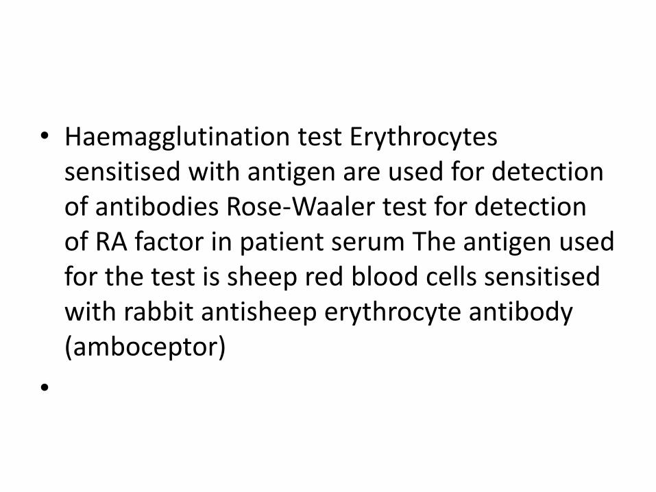
• Haemagglutination test Erythrocytes sensitised with antigen are used for detection of antibodies Rose-Waaler test for detection of RA factor in patient serum The antigen used for the test is sheep red blood cells sensitised with rabbit antisheep erythrocyte antibody (amboceptor)
•

• Coagglutination Some strains of Staphylococcus aureus (especially Cowan 1 strain) possess protein A on their surface When specific IgG molecule is coated on these strains, Fc portion of IgG molecule binds to protein A whereas antigen combining Fab terminal reamains free When the corresponding antigen is mixed with these coated cells, Fab terminal binds to antigen resulting in agglutination This test is used for detection of bacterial antigens in blood, urine and CSF (eg: Gonocooci, Streptococcus pyogenes and Haemophilus influenzae )
•

Complement fixation test
• Complement fixation test Complement is a protein (globulin) present in normal serum Whole complement system is made up of nine components: C1 to C9 Complement proteins are heat labile and are destroyed by heating at 56°C for 20 – 30 minutes
•

Contd’
• Principle The antigen-antibody complexes have ability to fix complement This reaction has no visible effect To detect the fixation of complement, an indicator system consisting of sheep erythrocytes coated with amboceptor is used
•

Contd’
• Components of CFT Test System Antigen: It may be soluble or particulate. Antibody: Human serum (May or may not contain Antibody towards specific Antigen) Complement: It is pooled serum obtained from 4 to 5 guinea pigs. It should be fresh or specially preserved as the complement activity is heat labile (stored at -30 °C in small fractions). The complement activity should be initially standardized before using in the test Indicator System ( Haemolytic system) Erythrocytes: Sheep RBC Amboceptor ( Hemolysin ): Rabbit antibody to sheep red cells prepared by inoculating sheep erythrocytes into rabbit under standard immunization protocol.
•

• Controls Antigen and serum controls are included in the test Complement control is used to ensure that the desired amount has been added Cell control to make sure that sensitised erythrocytes do not undergo lysis in the absence of complement
•

• Positive Test Step 1: At 37°C Antigen + Antibody + Complement Complement gets fixed (from serum) 1 Hour Step 2: At 37°C Fixed Complement complex + Haemolytic system No Haemolysis 1 Hour ( Test Positive)
•

Negative Test Step 1: At 37°C Antigen + Antibody absent + Complement Complement not fixed 1 Hour Step 2: At 37°C Free Complement + Haemolytic system Haemolysis 1 Hour ( Test Negative)
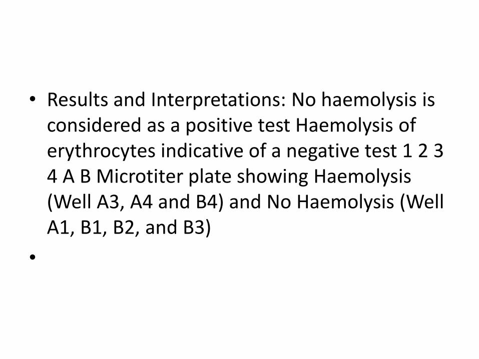
• Results and Interpretations: No haemolysis is considered as a positive test Haemolysis of erythrocytes indicative of a negative test 1 2 3 4 A B Microtiter plate showing Haemolysis (Well A3, A4 and B4) and No Haemolysis (Well A1, B1, B2, and B3)
•

• Indirect complement fixation test Certain avian (eg: duck, parrot) and mammalian (eg: horse, cat) sera cannot fix guinea pig complement Test is done in duplicate and after the first step, the standard antiserum known to fix complement is added in one set
•

• Positive test First step: Antigen + test serum (positive for antibody) + guinea pig complement Second step: Standard antiserum cannot react with antigen because has been used up by antibody in the first step, hence, complement is free Indicator system: Haemolysis occurs because complement is free
•

• Negative test First step: Antigen + test serum (negative for antibody) + guinea pig complement Second step: Standard antiserum will react with antigen and fix the free complement Indicator system: No haemolysis because complement is not free
•

Conglutination This is an alternative method for systems which do not fix guinea pig complement Uses horse complement which is non-hemolytic The indicator system is sheep erythrocytes sensitised with bovine serum Bovine serum contains a a beta globulin component named conglutinin, which acts as antibody to complement Conglutinin can cause agglutination of sensitised sheep erythrocytes, if these are combined with complement No agglutination – Positive result Agglutination – Negative result

Complement dependent serological tests Immobilisation test Immune adherence Cytolytic or cytocidal tests Neutralisation test Opsonisation
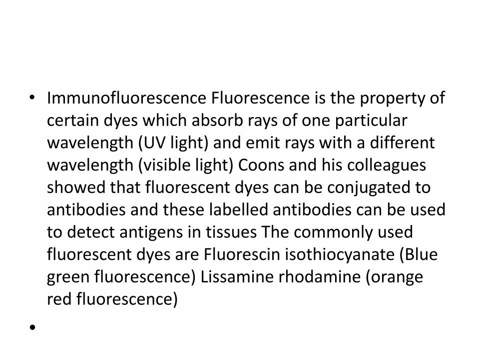
• Immunofluorescence Fluorescence is the property of certain dyes which absorb rays of one particular wavelength (UV light) and emit rays with a different wavelength (visible light) Coons and his colleagues showed that fluorescent dyes can be conjugated to antibodies and these labelled antibodies can be used to detect antigens in tissues The commonly used fluorescent dyes are Fluorescin isothiocyanate (Blue green fluorescence) Lissamine rhodamine (orange red fluorescence)
•

Immunofluorescence test is of two types Drect immunofluorescence test Indirect immunofluorescence test

Direct immunofluorescence test
• Uses 1. It is commonly employed for detection of bacteria, viruses or other antigens in blood, CSF, urine, faeces , tissues and other specimens 2. It is a sensitive method to diagnose rabies by detection of the rabies virus antigens in brain smears Disadvantage Separate specific fluorescent labelled antibody has to be prepared against each antigen to be tested
•

Indirect immunofluorescence test
• Advantages A single antihuman globulin fluorescent conjugate can be employed for detection of antibody to any antigen All antibodies are globulin in nature, therefore, antihuman globulin attaches to all antibodies This has overcome the disadvantage of direct immuno- -fluorescence test

• Radioimmunoassay (RIA) Berson and Yallow (1959) first described Since then it has been utilised for quantitation of hormones, drugs, HBsAg, IgE and viral antigens Radioimmunoassay is widely-used because of its great sensitivity Using antibodies of high affinity, it is possible to detect a few picograms (10 −12 g) of antigen The greater the specificity of the antiserum, the greater the specificity of the assay
•
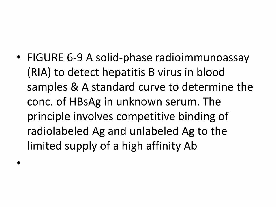
• FIGURE 6-9 A solid-phase radioimmunoassay (RIA) to detect hepatitis B virus in blood samples & A standard curve to determine the conc. of HBsAg in unknown serum. The principle involves competitive binding of radiolabeled Ag and unlabeled Ag to the limited supply of a high affinity Ab
•

• Disadvantages Radiation hazards: Uses radiolabelled reagents Requires specially trained persons Labs require special license to handle radioactive material Requires special arrangements for Requisition, storage of radioactive material radioactive waste disposal
•
•

Enzyme Linked Immunosorbent Assay (ELISA) Is a biochemical technique used mainly in immunology to detect the presence of an antibody or an antigen in a sample Term was coined by Engvall and Pearlmann in 1971 Similar to RIA, except no radiolabel Very sensitive, pg/mL
Different types of ELISAs Indirect ELISA Sandwich ELISA Competitive ELISA Cassette or cylinder ELISA
• Indirect ELISA Two commonly used enzymes Horseradish peroxidase (HRP) Alkaline phosphatase (AP)
•

• Sandwich ELISA Two antibodies required; must recognize different epitopes 1 st Antibody is referred to as capture Ab 2 nd antibody as detection Ab
•

• Competitive ELISA
The labelled antigen competes for primary antibody binding sites with the sample antigen (unlabelled) The more antigen in the sample, the less labelled antigen is retained in the well and the weaker the signal).

Cassette or cylinder ELISA It is a simple modification of ELISA for testing one or few samples of sera at a time The test is rapid (about ten minutes) as compared with the 2 - 4 hours taken for conventional ELISA

• Uses of ELISA Used for detection of antigens and antibodies for various microorganisms Detection of HIV antibodies in serum Detection of mycobacterial antibodies in tuberculosis Detection of rotavirus in faeces Detection of hepatitis B markers in serum Detection of enterotoxin of E. coli in faeces
•

Western blotting (Immunoblotting)
Western Blot for detection of HIV antibody:
Western Blot for detection of HIV antibody HIV-1 Western Blot Lane1: Positive Control Lane 2: Negative Control Sample A: Negative Sample B: Indeterminate Sample C: Positive