Echinococcus multilocularis specific antibody - PLOS
23
RESEARCH ARTICLE Echinococcus multilocularis specific antibody, systemic cytokine, and chemokine levels, as well as antigen-specific cellular responses in patients with progressive, stable, and cured alveolar echinococcosis: A 10-year follow-up Beate Gru ¨ ner ID 1‡ , Lynn Peters ID 1‡ , Andreas Hillenbrand 2 , Patrick Voßberg 3 , Jonas Schweiker ID 3 , Elisabeth G. Rollmann 3 , Laura H. Rodriguez ID 3 , Jasmin Blumhardt 3 , Sanne Burkert ID 1 , Peter Kern 1 , Carsten Ko ¨ hler ID 3‡ , Peter T. Soboslay ID 3‡ * 1 University Hospital of Ulm, Department of Internal Medicine III, Division of Infectious Diseases, Ulm, Germany, 2 Department of General and Visceral Surgery, Ulm University Hospital, Ulm, Germany, 3 University Clinics Tu ¨ bingen, Institute for Tropical Medicine, Eberhard-Karls University, Tu ¨ bingen, Germany ‡ BG and LP share first authorship on this work. CK and PTS are joint senior authors on this work. * [email protected] Abstract Background The infestation with Echinococcus multilocularis larvae may persist in humans for up to decades without evident clinical symptoms. Longitudinal investigations are needed to understand the dynamic immunological processes in alveolar echinococcosis (AE) patients associated with an active and progressive, a stable or a regressive course of disease. Methodology/Principal findings This study evaluated the E. multilocularis specific antibody responses, systemic cytokine, and chemokine serum levels over a 10-year follow-up period, as well as cellular responsive- ness in AE patients. Our results demonstrate a rapid decrease in antibodies against E. multi- locularis specific antigen Em2+. Especially in cured patients, these antibodies remained negative, making them a significant predictor for cured AE. E. multilocularis specific IgG4, and indirect hemagglutination IHA decreased later in time, after around 5 years. While total IgE did not show significant dynamics over the course of disease, E. multilocularis specific IgE decreased after one to two years, and increasing levels were a significant predictor of progressive disease. There was no significant change in systemic IL-8, IL-9, CCL18 or CCL20 serum levels over time. Univariate analysis across groups indicated lower IL-8 levels in cured patients; however, this result could not be confirmed by multivariate analysis. Lev- els of CCL17 decreased during treatment, especially in cured patients, and thus might serve as a predictive or risk factor for progressive disease. Levels of IL-10 and CCL13 decreased during disease, especially after five and ten years of intervention. The E. multilocularis anti- gen (EmAg) inducible cellular productions of MCP1(CCL13), TARC(CCL17) and PARC PLOS NEGLECTED TROPICAL DISEASES PLOS Neglected Tropical Diseases | https://doi.org/10.1371/journal.pntd.0010099 February 2, 2022 1 / 23 a1111111111 a1111111111 a1111111111 a1111111111 a1111111111 OPEN ACCESS Citation: Gru ¨ner B, Peters L, Hillenbrand A, Voßberg P, Schweiker J, Rollmann EG, et al. (2022) Echinococcus multilocularis specific antibody, systemic cytokine, and chemokine levels, as well as antigen-specific cellular responses in patients with progressive, stable, and cured alveolar echinococcosis: A 10-year follow-up. PLoS Negl Trop Dis 16(2): e0010099. https://doi.org/ 10.1371/journal.pntd.0010099 Editor: Gregory Deye, National Institute of Allergy and Infectious Diseases, UNITED STATES Received: December 23, 2020 Accepted: December 16, 2021 Published: February 2, 2022 Copyright: © 2022 Gru ¨ner et al. This is an open access article distributed under the terms of the Creative Commons Attribution License, which permits unrestricted use, distribution, and reproduction in any medium, provided the original author and source are credited. Data Availability Statement: All relevant data are within the manuscript. Data on individual patients cannot be shared publicly. Data are available from the Ethics Committee at University Ulm (Email: [email protected]; http://www.uni- ulm.de/ethikkommission/) for researchers who meet the criteria for access to confidential data.
-
Upload
khangminh22 -
Category
Documents
-
view
3 -
download
0
Transcript of Echinococcus multilocularis specific antibody - PLOS

RESEARCH ARTICLE
Echinococcus multilocularis specific antibody,
systemic cytokine, and chemokine levels, as
well as antigen-specific cellular responses in
patients with progressive, stable, and cured
alveolar echinococcosis: A 10-year follow-up
Beate GrunerID1‡, Lynn PetersID
1‡, Andreas Hillenbrand2, Patrick Voßberg3,
Jonas SchweikerID3, Elisabeth G. Rollmann3, Laura H. RodriguezID
3, Jasmin Blumhardt3,
Sanne BurkertID1, Peter Kern1, Carsten KohlerID
3‡, Peter T. SoboslayID3‡*
1 University Hospital of Ulm, Department of Internal Medicine III, Division of Infectious Diseases, Ulm,
Germany, 2 Department of General and Visceral Surgery, Ulm University Hospital, Ulm, Germany,
3 University Clinics Tubingen, Institute for Tropical Medicine, Eberhard-Karls University, Tubingen, Germany
‡ BG and LP share first authorship on this work. CK and PTS are joint senior authors on this work.
Abstract
Background
The infestation with Echinococcus multilocularis larvae may persist in humans for up to
decades without evident clinical symptoms. Longitudinal investigations are needed to
understand the dynamic immunological processes in alveolar echinococcosis (AE) patients
associated with an active and progressive, a stable or a regressive course of disease.
Methodology/Principal findings
This study evaluated the E. multilocularis specific antibody responses, systemic cytokine,
and chemokine serum levels over a 10-year follow-up period, as well as cellular responsive-
ness in AE patients. Our results demonstrate a rapid decrease in antibodies against E. multi-
locularis specific antigen Em2+. Especially in cured patients, these antibodies remained
negative, making them a significant predictor for cured AE. E. multilocularis specific IgG4,
and indirect hemagglutination IHA decreased later in time, after around 5 years. While total
IgE did not show significant dynamics over the course of disease, E. multilocularis specific
IgE decreased after one to two years, and increasing levels were a significant predictor of
progressive disease. There was no significant change in systemic IL-8, IL-9, CCL18 or
CCL20 serum levels over time. Univariate analysis across groups indicated lower IL-8 levels
in cured patients; however, this result could not be confirmed by multivariate analysis. Lev-
els of CCL17 decreased during treatment, especially in cured patients, and thus might serve
as a predictive or risk factor for progressive disease. Levels of IL-10 and CCL13 decreased
during disease, especially after five and ten years of intervention. The E. multilocularis anti-
gen (EmAg) inducible cellular productions of MCP1(CCL13), TARC(CCL17) and PARC
PLOS NEGLECTED TROPICAL DISEASES
PLOS Neglected Tropical Diseases | https://doi.org/10.1371/journal.pntd.0010099 February 2, 2022 1 / 23
a1111111111
a1111111111
a1111111111
a1111111111
a1111111111
OPEN ACCESS
Citation: Gruner B, Peters L, Hillenbrand A,
Voßberg P, Schweiker J, Rollmann EG, et al.
(2022) Echinococcus multilocularis specific
antibody, systemic cytokine, and chemokine levels,
as well as antigen-specific cellular responses in
patients with progressive, stable, and cured
alveolar echinococcosis: A 10-year follow-up. PLoS
Negl Trop Dis 16(2): e0010099. https://doi.org/
10.1371/journal.pntd.0010099
Editor: Gregory Deye, National Institute of Allergy
and Infectious Diseases, UNITED STATES
Received: December 23, 2020
Accepted: December 16, 2021
Published: February 2, 2022
Copyright: © 2022 Gruner et al. This is an open
access article distributed under the terms of the
Creative Commons Attribution License, which
permits unrestricted use, distribution, and
reproduction in any medium, provided the original
author and source are credited.
Data Availability Statement: All relevant data are
within the manuscript. Data on individual patients
cannot be shared publicly. Data are available from
the Ethics Committee at University Ulm (Email:
[email protected]; http://www.uni-
ulm.de/ethikkommission/) for researchers who
meet the criteria for access to confidential data.

(CCL18) were lowest in patients with cured AE and infection-free controls, while the EmAg
inducible cellular production of IFN-γ increased after cure. Significant positive cytokine and
chemokine correlations were observed in AE patients for IL-9, IL-10, CCL13(MCP-4),
CCL17(TARC) and CCL20(LARC)(for all p<0.001). E. multilocularis specific IgG4 response
correlated positively with TARC (p<0.001). Both markers enhanced over time in progressive
disease and decreased after cure. The levels of IL-8, IL-10, MCP4 and LARC enhanced
with AE regression.
Conclusions/Significance
Repeated biomarker surveys are advisable to evaluate progression or regression of disease
during longitudinal follow-up and such analyses can support imaging techniques and
improve staging of AE patients.
Author summary
Alveolar echinococcosis (AE) is a severe disease caused by Echinococcus multilocularis, the
fox tapeworm. Humans exposed to E. multilocularis may develop severe AE with progres-
sive tissue and organ infiltrating growth of the larval stage. The E. multilocularis larvae
appear to have developed effective immune evasion mechanisms which facilitate an
asymptomatic incubation and an extended host and parasite coexistence for decades.
Over a 10-year follow-up, this investigation aimed to gain a better understanding of the
immunological process associated with an active and progressive, a stable or a regressive
course of AE. In summary, the rapid decrease of antibodies against the E. multilocularisspecific antigen Em2+, especially in cured patients, makes them a significant predictor for
cured AE. The positive relation of E. multilocularis specific IgG4 responses and chemo-
kine levels of TARC can indicate AE progression when both enhance over time. Enhanced
levels of cytokines IL-8, IL-10, and chemokines MCP4 and LARC may predict AE regres-
sion. Repeated biomarker surveys are advisable to evaluate progression or regression of
AE during longitudinal follow up, and such analyses can support imaging techniques and
improve staging of AE patients.
Introduction
Alveolar echinococcosis (AE) is a severe disease caused by the cestode Echinococcus multilocu-laris, the fox tapeworm. The disease is most prominent in northwest China but also occurs in
central Europe [1]. AE morbidity and treatment costs are high, with a lethal course if left
untreated [2–5]. Together with cystic echinococcosis, AE is considered a neglected disease
[6,7]. Moreover, it is an emerging disease, and an increase in human cases of AE in Europe
have been observed over the past decades [2,8–10].
Humans exposed to E. multilocularis may develop severe AE with progressive tissue and
organ infiltrating growth of the larval stage [11]. E. multilocularis larvae may massively prolif-
erate in liver and other organs over decades without evident clinical symptoms, and in this
process the evasion and regulatory mechanisms of the parasite play a crucial role on the
human host immune response [12–14]. The E. multilocularis larvae appear to have developed
effective immune evasion mechanisms which facilitate an asymptomatic incubation and an
PLOS NEGLECTED TROPICAL DISEASES Echinococcus multilocularis infested patients’ timely changes of immunity
PLOS Neglected Tropical Diseases | https://doi.org/10.1371/journal.pntd.0010099 February 2, 2022 2 / 23
Funding: This study was supported by the post
graduate programme of the German centre for
infectious diseases (DZIF) at University Clinics
Tubingen (UKT: PV, JS, EGR, LHR, JB) and the
intramural research and teaching program at
University Hospital Ulm (BG). The funders had no
role in study design, data collection and analysis,
decision to publish, or preparation of the
manuscript.
Competing interests: The authors have declared
that no competing interests exist.

extended host and parasite coexistence. The larvae may persist for decades exerting a progres-
sive tissue invasive tumour-like growth [1,2,15], but few AE patients may present with a heal-
ing course of the disease. This diversity suggested immunological mechanisms which may
control the course of AE. Characteristic for a certain resistance to AE is the occurrence of Th1
type cytokines, while elevated production of IL-10 and Th2 cytokines were associated with
progressive Em larval growth [16–18].
Only in 10–30% of E. multilocularis seropositive cases [11] clinical disease will develop, sug-
gesting that effective immune responses may have eliminated the larvae when penetrating the
intestinal wall at an early stage of infestation [19]. Patients who express Th1-type immune
response are more likely able to limit or regress larval growth by forming peri-parasitic granu-
lomas with macrophages and T cells, as well as lesions, or fibrosis and necrosis to encapsulate
the parasite [20,21]. Successful immune evasion of the parasite leads to a tolerant Th2 immune
response which is unable to prevent the E. multilocularis larval growth [15,22]. The disease
spectrum is dependent on an acquired deviation of Th1-related immunity, and the spontane-
ous secretion of IL-10 by the PBMC was identified as the immunological hallmark of patients
with progressive forms of AE involved in the maintenance of tolerance and persistence of the
parasite [23–25]. Reactivity of peripheral blood cells to E. multilocularis antigens persisted for
years in AE patients after complete resection of the parasitic lesions suggesting that residual
parasite tissues will continue to stimulate cellular responses [26]. E. multilocularis may expand
regulatory T cell responses [27] and promote a largely Th2-biased response which allows long-
term parasite survival, proliferation, and maturation [17,21]. However, only few studies have
longitudinally addressed the changes of immunity at different stages of AE in patients.
This investigation aimed to gain a better understanding of the immunological processes at
distinct stages of AE disease. Over a 10-year follow-up, antibody responses to E. multilocularisand systemic cytokine and chemokine levels, as well as parasite antigen-specific cellular
responses were assessed in patients with progressive, stable, and cured AE. A better under-
standing of the differences in the elicited immune response may reveal new treatment
rationales.
Material and methods
Ethics statement
All patients gave their written consent to participate in this study, and approval for the latter
was obtained from the ethical board at University Clinics of Ulm (Ethik-Kommision Antrag
Nr. 71/2004, 71/2004-Amendment2010 and Antrag Nr. 372/2015).
Alveolar echinococcosis (AE) patients and disease staging
All AE patients were admitted to and consulted at the Echinococcosis Reference Centre at
University Hospitals of Ulm, Germany. The Echinococcosis Centre in Ulm and the University
Clinics of Tubingen are in the southwest of the federal state of Baden-Wurttemberg in Ger-
many, both towns being 70 km apart, in an endemic area for E. multilocularis [25]. The AE
patients in the present study were classified into cured, stable, and progressive according to
established criteria [3] at the time of the study.
Diagnosis and classification of AE was performed using positive imaging, serology and his-
tology and AE-case-definition according to criteria, which were established by the WHO
Informal Working Group on Echinococcosis [28] as well as PNM–staging system
(P = parasitic mass in the liver, N = involvement of neighbouring organs, and M = metastasis)
[1–3,29], and most patients were re-examined regularly during follow-ups.
PLOS NEGLECTED TROPICAL DISEASES Echinococcus multilocularis infested patients’ timely changes of immunity
PLOS Neglected Tropical Diseases | https://doi.org/10.1371/journal.pntd.0010099 February 2, 2022 3 / 23

The AE patients were staged into progressive, stable and cured AE. The application of imag-
ing techniques for AE staging is essential. Ultrasound (US) and computed tomography (CT)
were used to confirm alveolar echinococcosis [1], and magnetic resonance imaging (MRI) can
provide a more differentiated diagnosis, to identify blood vessel structures before surgery
[3,30]. Positron emission tomography (PET) or combined PET-CT was used to shows areas of
high metabolic activity, which may indicate inflammatory reactions and possible activity of the
parasite [31].
Clinical status was classified as:
1. cured: patients who underwent curative surgery and showed no signs of residual parasitic
mass or relapse for at least two years postoperative.
2. stable disease without BMZ treatment: inoperable patients without benzimidazole treat-
ment who show no signs of progressive disease
3. stable disease with BMZ treatment: inoperable patients on continuous benzimidazole treat-
ment who show no signs of progressive disease
4. progressive disease: inoperable patients with growing parasitic lesion and/or associated bili-
ary or vascular complications despite benzimidazole treatment.
Serum collection from AE patients and healthy controls
Serum samples from AE patients were collected at three-time points; in 2005 (year 1, n = 101
samples), 2010 (year 5, n = 57 samples) and 2016 (year10, n = 59 samples). Infection-free
healthy controls were blood donors (n = 35) from University Clinics of Tubingen; all controls
were negative in E. multilocularis antigen-specific antibody ELISA responses. For this study,
only patients that had donated several times were included to enable cross-time analysis.
Echinococcus multilocularis antigen (EmAg) specific IgG responses
Antibody-screening was performed with the Echinococcus-specific IgG indirect hemaggluti-
nin test (IHA) and confirmatory testing was done by the commercialized E. multilocularis-spe-
cific Em2+-antigen ELISA as described previously [32]. The detection of EmAg-specific IgG1,
IgG3, IgG4 and IgE was applied as described previously [33,34].
For analyses of EmAg-specific IgG sublass responses, ELISA microtiter 96 well plates
(CORNING; EIA/2 CatNo.3690) were coated overnight at 4˚ C with E. multilocularis metaces-
tode larval extract (EmAg, 12 μg/ml) in phosphate-buffered saline (PBS). For E. multilocularisantigen (EmAg) preparation, solid metacestode tissues were isolated from metacestode-
infected mongolian gerbils (Meriones unguiculatus), tissues were washed in phosphate-buff-
ered saline (PBS, pH 7.2–7.4) and ground on ice in a Ten-Broek tissue grinder until a homoge-
nous liquid extract appeared. The antigen suspension was then sonicated twice on ice for 10
min (30% cycle, Model 250, Branson Ultrasonics, Danbury, USA). The liquid extract was then
collected and centrifuged at 15,000 g for 30 min at 4˚ C. The supernatant was sterile-filtered
(0.22 μm) and stored in aliquots at -70˚ C for further use. The protein concentration of these
E. multilocularis antigens was determined using the bicinchoninic acid protein determination
kit (Pierece, Rockford, IL, USA). After coating, ELISA plates were washed twice with PBS
0.05% Tween 20 (PBST, pH 7.4) and subsequently blocked with PBS containing 5% BSA for
1.5 hours at 37˚ C. For the analyses of antibody responses IgG1, IgG3 and IgG4 were selected
because the carbohydrate rich antigens located on the laminated layer of metacestode larvae
will trigger these IgG subclasses. Serum samples from AE patients and echinococcosis-free
controls were diluted 1:10 with 5% BSA in PBS, added to each well and incubated at 37˚ C for
PLOS NEGLECTED TROPICAL DISEASES Echinococcus multilocularis infested patients’ timely changes of immunity
PLOS Neglected Tropical Diseases | https://doi.org/10.1371/journal.pntd.0010099 February 2, 2022 4 / 23

2 hours. Then plates were washed three times and detection was performed with monoclonal
anti-human IgG1, IgG3, or IgG4 antibody conjugated with HRP (Thermo Fisher, A-10648, A-
10654, 05–3600) diluted 1:500 with 5% BSA in PBS and added for 1.5 hours at 37˚ C. After
three times washing, Tetramethylbenzidine (TMB) solution was added, and colour reaction
was stopped with 0.5 M sulfuric acid. Optical density of sample wells was determined at 450
nm using the EL808 Photometer (BIOTEK) and Gen5 1.11 software. The data were processed
and statistically evaluated with JMP 14.2.0.
Isolation of PBMC, cell culture experiments and determination of cytokine
and chemokine production
Peripheral mononuclear blood cells (PBMC) from AE patients were isolated by Ficoll gradient
centrifugation and cultured in vitro as previously described [35]. Briefly, PBMC were adjusted
to 1x10⁷/ml in RPMI supplemented with 25 mM HEPES buffer, 100 U/ml penicillin, 100 mg/
ml streptomycin and 0.25 mg/ml amphotericin B. Freshly isolated PBMC were cultured at
2.5x10⁶ PBMC/ml in RPMI (as above) and stimulated with E. multilocularis metacestode anti-
gen extract (EmAg, 50 μg/ml) or remained without stimulation (baseline) for 48h in 5% CO2
at 37˚ C and saturated humidity. Cell culture supernatants were collected after 48h and stored
below –70˚ C until further use. Cytokine and chemokine secretion by stimulated PBMC was
quantified by sandwich enzyme-linked immunosorbent assay (ELISA) using monoclonal and
polyclonal antibodies for interleukin(IL) - 8 (IL-8, CXCL8), IL-9, IL-10, interferon-gamma
(IFN-γ), and the CC chemokines MCP-1 (CCL2), MCP-3 (CCL7), MCP-4 (CCL13), TARC
(CCL17), PARC (CCL18) and LARC (CCL20) as recommended by the manufacturers and
described previously [34–36]. The detection limits of the cytokine and chemokine ELISAs
(DuoSet, R&D Systems, Minneapolis, USA) were at 50 pg/ml.
Data analysis
Statistical analysis was performed using IBM SPSS version 25. All variables were described
with mean, median and standard deviation. Prior to every calculation, variables were tested for
normal distribution using graphical methods such as histogram, P-P- and Q-Q-plot as well the
Shapiro-Wilk test. Skewed data was log-transformed for further analysis. To assess changes in
serological markers and cytokine levels over time, repeated-measures ANOVA, and paired t-
test for normally distributed or Wilcoxon signed-rank test for non-parametric data were used.
Comparisons across groups were conducted with ANOVA and independent t-test for nor-
mally distributed, continuous data and Mann-Whitney-test for non-normally distributed, con-
tinuous variables. For categorical variables, the Chi-square test or Fisher’s exact test (if
appropriate) were used. Results were stated as mean and standard deviation in case of nor-
mally distributed data and as median, 25th and 75th percentile for non-normally distributed
data. Correlations were performed according to Pearson (continuous variables) or Spearman
(non-continuous variables). To evaluate the effect on the clinical status while adjusting for
confounders and interactions between variables, a multivariate analysis was conducted. Two
logistic regression models, one for progressive disease at any point in time and one for cured
disease at baseline were designed. With respect to the sample size, a maximum of ten variables
was included into the model to prevent over-fitting. Before the final model was established, a
screening for multicollinearity was conducted, excluding variables with a variance inflation
factor (VIF) of 10.0 or higher. Prior to the multivariate analysis, multiple imputation was per-
formed to adjust for missing dependant variables. As many auxiliary variables and cases as
possible were added to the imputation process to produce an accurate dataset. The number of
data sets generated was determined using the formula suggested by Newgard and Haukoos
PLOS NEGLECTED TROPICAL DISEASES Echinococcus multilocularis infested patients’ timely changes of immunity
PLOS Neglected Tropical Diseases | https://doi.org/10.1371/journal.pntd.0010099 February 2, 2022 5 / 23

[37], aiming for a relative efficiency of at least 95%, which is considered a high rate. Results
were considered significant with a p-value (probability to make a type I error) smaller than
5%.
The statistical package JMP 14.2.0 was used for analyses of chemokine serum levels and the
cellular production of cytokines and chemokines in patients with progressive, stable and cured
AE. Significant differences between groups were determined with Tukey’s test after logarith-
mic transformation to stabilize the variance of data, either of the brut production (chemokine
concentration in supernatants), or the net production (brut production minus chemokine
concentration of resting PBMC (baseline)). The net productions were calculated according to
the following formula: [production index = (brut production + 1)/(basline production + 1)].
We added one to the numerator and the denominator because of zero values in baseline and
brut productions. For multiple comparisons, and to avoid type I errors, differences between
infection groups were analyzed by the Tukey test. Bonferroni adjustment was applied, P values
below p<0.001 are considered.
Results
Timetable of follow-up of clinical examinations of AE patients
AE patients’ follow-up visits were conducted at different points in time as displayed in Table 1.
Of 63 patients at baseline, 12 patients could be follow-up for a mean of 11.3 years.
Echinococcus IgG indirect hemagglutinin test (IHA) results in AE patients
over time
Table 2 demonstrates the different levels of IgG IHA at different points in time. As many
patients did not show elevated IgG IHA levels (coded as ‘0’) medians were therefore ‘0’. To dis-
play more subtle differences between groups, the respective mean and standard deviation were
used as auxiliary variables in spite of the non-normal distribution A Wilcoxon test was per-
formed to conduct group wise comparisons between baseline and follow-up visits. Only after a
mean of 5 years (Visit 4) there was slight evidence (p = 0.05) that there was a significant
decrease of IgG levels over time. The results displayed in Table 2 indicate a decrease in IgG
IHA levels compared to baseline (p = 0.05) with a moderate Cohen’s effect size (r = 0.27). This
difference became clearer comparing IgG IHA levels after six months and five years (Z =
-2.092, N = 23, p = 0.036) with a strong Cohen’s effect size of 0.44.
E. multilocularis Em2+ antigen specific IgG responses in AE patients over
time
Antibodies against Em2+ were measured as dichotomous variable. At baseline (V0), these anti-
bodies were detected in 67 of 100 patients. Regarding the follow-up visits, a one-sided
Table 1. Time table of follow-up visits of AE patients. Vx = follow-up visit, N = number of cases that adhered to fol-
low-up visit, ΔtVx-tV0 = difference between baseline and follow-up visit.
Vx N ΔtVx-tV0
V0 63 -
V1 40 5.3 ± 1.9 months
V2 43 11.7 ± 2.1 months
V3 13 23.2 ± 5.5 months = 1.5 ± 0.7 years
V4 52 65.4 ± 8.8 months = 5.1 ± 0.9 years
V5 12 141.3 ± 8.9 months = 11.3 ± 0.7 years
https://doi.org/10.1371/journal.pntd.0010099.t001
PLOS NEGLECTED TROPICAL DISEASES Echinococcus multilocularis infested patients’ timely changes of immunity
PLOS Neglected Tropical Diseases | https://doi.org/10.1371/journal.pntd.0010099 February 2, 2022 6 / 23
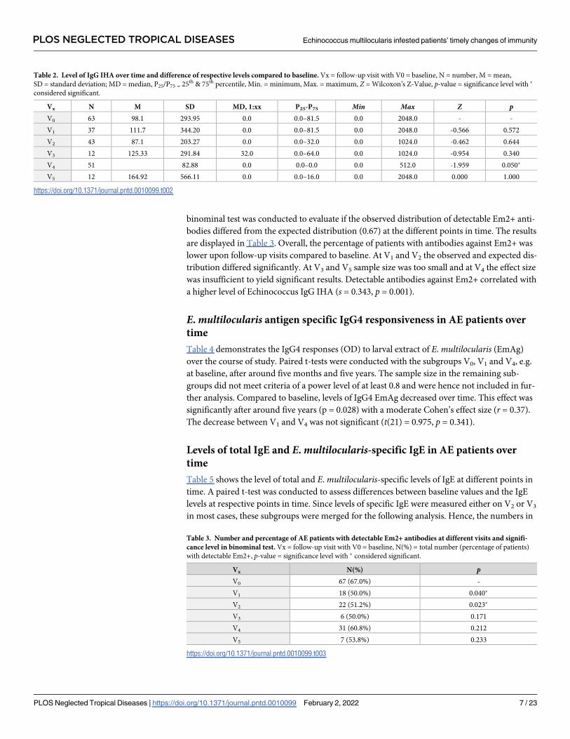
binominal test was conducted to evaluate if the observed distribution of detectable Em2+ anti-
bodies differed from the expected distribution (0.67) at the different points in time. The results
are displayed in Table 3. Overall, the percentage of patients with antibodies against Em2+ was
lower upon follow-up visits compared to baseline. At V1 and V2 the observed and expected dis-
tribution differed significantly. At V3 and V5 sample size was too small and at V4 the effect size
was insufficient to yield significant results. Detectable antibodies against Em2+ correlated with
a higher level of Echinococcus IgG IHA (s = 0.343, p = 0.001).
E. multilocularis antigen specific IgG4 responsiveness in AE patients over
time
Table 4 demonstrates the IgG4 responses (OD) to larval extract of E. multilocularis (EmAg)
over the course of study. Paired t-tests were conducted with the subgroups V0, V1 and V4, e.g.
at baseline, after around five months and five years. The sample size in the remaining sub-
groups did not meet criteria of a power level of at least 0.8 and were hence not included in fur-
ther analysis. Compared to baseline, levels of IgG4 EmAg decreased over time. This effect was
significantly after around five years (p = 0.028) with a moderate Cohen’s effect size (r = 0.37).
The decrease between V1 and V4 was not significant (t(21) = 0.975, p = 0.341).
Levels of total IgE and E. multilocularis-specific IgE in AE patients over
time
Table 5 shows the level of total and E. multilocularis-specific levels of IgE at different points in
time. A paired t-test was conducted to assess differences between baseline values and the IgE
levels at respective points in time. Since levels of specific IgE were measured either on V2 or V3
in most cases, these subgroups were merged for the following analysis. Hence, the numbers in
Table 2. Level of IgG IHA over time and difference of respective levels compared to baseline. Vx = follow-up visit with V0 = baseline, N = number, M = mean,
SD = standard deviation; MD = median, P25/P75 = 25th & 75th percentile, Min. = minimum, Max. = maximum, Z = Wilcoxon’s Z-Value, p-value = significance level with �
considered significant.
Vx N M SD MD, 1:xx P25-P75 Min Max Z pV0 63 98.1 293.95 0.0 0.0–81.5 0.0 2048.0 - -
V1 37 111.7 344.20 0.0 0.0–81.5 0.0 2048.0 -0.566 0.572
V2 43 87.1 203.27 0.0 0.0–32.0 0.0 1024.0 -0.462 0.644
V3 12 125.33 291.84 32.0 0.0–64.0 0.0 1024.0 -0.954 0.340
V4 51 82.88 0.0 0.0–0.0 0.0 512.0 -1.959 0.050�
V5 12 164.92 566.11 0.0 0.0–16.0 0.0 2048.0 0.000 1.000
https://doi.org/10.1371/journal.pntd.0010099.t002
Table 3. Number and percentage of AE patients with detectable Em2+ antibodies at different visits and signifi-
cance level in binominal test. Vx = follow-up visit with V0 = baseline, N(%) = total number (percentage of patients)
with detectable Em2+, p-value = significance level with � considered significant.
Vx N(%) pV0 67 (67.0%) -
V1 18 (50.0%) 0.040�
V2 22 (51.2%) 0.023�
V3 6 (50.0%) 0.171
V4 31 (60.8%) 0.212
V5 7 (53.8%) 0.233
https://doi.org/10.1371/journal.pntd.0010099.t003
PLOS NEGLECTED TROPICAL DISEASES Echinococcus multilocularis infested patients’ timely changes of immunity
PLOS Neglected Tropical Diseases | https://doi.org/10.1371/journal.pntd.0010099 February 2, 2022 7 / 23

V2/3 represent specific IgE levels around one to two years after baseline measurements. At V5,
no measurements of specific IgE levels were conducted. While no significant alteration in total
IgE can be found over the course of disease, mean and median values have a decreasing ten-
dency over time. There is a significant decrease in specific IgE after one to two years with a
moderate Cohen’s effect size (0.31). There was a positive correlation between EchinococcusIgG IHA and total IgE (s = 0.549, p< 0.001).
Cytokine and chemokine serum levels in AE patients over time
Cytokine levels are displayed in Table 6. Log transformation of values was performed prior to
further analysis. For IL-8, IL-9, CCL18 and CCL20, no significant changes between baseline
and the follow-up visits were found (paired t-test). Levels of IL-10 were significantly lower at
V4 (t(49) = 4.078, p< 0.001) and V5 (t(11) = 4.052, p = 0.002) comparing to baseline, reflecting
a significant decrease after five and ten years. The further decrease of IL-10 levels between five
and ten years after baseline was again significant (t(6) = 2.554, p = 0.043). The decrease in
CCL13 was highly significant after 5 years (t(49) = 5.151, p< 0.000) and ten years (t(11) =
3.278, p = 0.007). Compared to baseline, levels of CCL17 were significantly lower after five
years (t(48) = 2.915, p = 0.005) and ten years (t(12) = 3.025, p = 0.011) compared to baseline.
Levels seem to increase again between five and ten years after baseline (t(7) = -2.403,
p = 0.047), however, the sample size was small and the difference barely significant.
Table 4. Reactivity (indicated as optical densities OD) of IgG4 to larval extract of E. multilocularis (EmAg) over time and difference of respective levels compared
to baseline. Vx = follow-up visit with V0 = baseline, N = number, M = mean, MD = median, SD = standard deviation; Min. = minimum, Max. = maximum, t = difference
in units of standard error, df = degrees of freedom, p-value = significance level with � considered significant.
Vx N M MD SD Min Max t(df) pV0 84 0.560 0.626 0.337 0.054 1.2 - -V1 36 0.558 0.625 0.283 0.064 0.978 0.896 (34) 0.376V2 9 0.419 0.429 0.280 0.102 0.919 - -V3 1 1.105 1.105 - 1.105 1.105 - -V4 43 0.373 0.226 0.345 0.049 1.211 3.298 (33) 0.002�
V5 7 0.510 0.353 0.437 0.060 1.068 - -
https://doi.org/10.1371/journal.pntd.0010099.t004
Table 5. Levels of total and E.multilocularis-specific IgE in kU/l over time and difference of respective levels compared to baseline. Vx = follow-up visit with
V0 = baseline, N = number, M = mean, MD = median, SD = standard deviation; Min. = minimum, Max. = maximum, t = difference in units of standard error,
df = degrees of freedom, p-value = significance level with � considered significant.
Vx N M MD SD Min Max t(df) pLevels of total IgE in kU/l
V0 106 474.0 77.5 1,283.5 1.0 1,1150.0 - -
V1 40 377.2 51.4 875.4 3.3 4,765.0 -0.512 (38) 0.612
V2 43 330.1 49.9 754.5 0.0 4,373.0 -0.216 (41) 0.830
V3 13 136.0 84.4 218.9 9.5 841.0 -0.475 (12) 0.643
V4 51 296.3 63.3 926.2 1.1 6,286.0 -0.024 (48) 0.981
V5 12 407.6 63.6 792.7 2.5 2,363.0 -1.049 (10) 0.319
Levels of specific IgE in kU/l
V0 76 2.1 0.5 4.5 0.00 24.1 - -
V1 19 0.00 0.00 0.00 0.00 0.0 1.000 (18) 0.331
V2/3 48 1.4 2.7 0.00 15.0 2.127 (47) 0.035�
V4 36 1.6 0.4 3.0 0.00 12.7 0.050 (34) 0.961
https://doi.org/10.1371/journal.pntd.0010099.t005
PLOS NEGLECTED TROPICAL DISEASES Echinococcus multilocularis infested patients’ timely changes of immunity
PLOS Neglected Tropical Diseases | https://doi.org/10.1371/journal.pntd.0010099 February 2, 2022 8 / 23

E. multilocularis patients with progressive, stable and cured AE
For the analyses of EmAg specific IgG isotype responses, 217 blood samples were collected
from AE patients (Table 7). The AE patients were staged into three groups: those with progres-
sive AE, with stable and cured AE. At year 1 of the study, the proportion of patients staged
with progressive AE disease was 21.8%, in year 5 it reduced to 7% and in year 10 to 8.5%. The
proportion of patients with cured AE increased from year 1 with 17.8% to 24.6% and 28.8% in
year 5 and year 10, respectively. Patients with stable AE comprised the largest group (n = 137)
and with 60%, 62% and 68% the proportion of AE stable remained similar at the three time
points of study (Table 7).
E. multilocularis antigen (EmAg) specific IgG isotype responses
The EmAg specific IgG responses were highest with progressive, lower in stable and lowest in
cured AE patients (Fig 1). The EmAg-specific IgG1 responses were significantly higher with
Table 6. Levels of cytokines and chemokines over time. Cytokines IL-8(CXCL8), IL-9, IL-10 and chemokines MCP-4(CCL13), TARC(CCL17), PARC(CCL18) and
LARC(CCL20) were quantified in pg/ml in AE patients over time. Vx = follow-up visit with V0 = baseline, N = number, M = mean, SD = standard deviation; Min. = mini-
mum, Max. = maximum.
Vx N IL-8(CXCL8) IL-9
M MD SD Min Max M MD SD Min Max
V0 108 175.6 0.0 1,558.1 0.0 1,6191.0 44.8 0.0 128.3 0.0 930.4
V1 36 16,4 0.0 69.6 0.0 417.0 73.1 0.0 135.9 0.0 502.4
V2 9 89.7 0.0 269.0 0.0 807.0 180.4 0.0 328.3 0.0 970.4
V3 1 0.0 0.0 - 0.0 0.00 0.0 0.0 - 0.0 0.00
V4 50 24,3 0.0 137.2 0.0 966.0 73.4 0.0 319.4 0.0 1,902.7
V5 12 2,551.6 0.0 8,272.6 0.0 28,767.0 0.0 0.0 0.00 0.0 0.00
Vx N IL-10
M MD SD Min Max
V0 108 483.8 108.0 1,334.6 0.0 9,778.0
V1 36 415.7 103.0 804.3 0.0 3,391.0
V2 9 1,277.4 86.0 3,190.3 0.0 9,745.0
V3 1 7.0 7.0 - 7.0 7.0
V4 50 359.4 31.50 1,339.3 0.0 7,586.0
V5 12 125.3 0.0 405.48 0.0 1,411.0
Vx N MCP-4(CCL13) TARC(CCL17)
M MD SD Min Max M MD SD Min Max
V0 108 333.6 176.5 442.9 6.0 2,757.0 480.3 205.0 812.3 0.0 4,825.0
V1 36 364.5 321.5 310.9 25.0 2,665.0 562.0 332.0 705.1 0.0 3,571.0
V2 9 531.9 265.0 857.4 55.0 766.0 527.3 167.0 1,005.9 0.0 3,303.0
V3 1 237.0 237.0 - 237.0 237.0 448.0 448.0 - 448.0 448.0
V4 50 117.0 66.5 143.1 0.0 812.0 114.7 17.0 277.0 0.0 1,468.0
V5 12 227.8 114.0 400.9 18.0 1,468.0 388.6 185.0 528.6 0.0 1,523.0
Vx N PARC(CCL18) LARC(CCL20)
M MD SD Min Max M MD SD Min Max
V0 108 119,810.1 107,982.0 69,413.9 0.0 346,894.0 221.9 0.0 841.5 0.0 5,707.0
V1 36 122,144.0 107,982.0 81,536.8 0.0 347,700.0 180.1 0.0 471.3 0.0 2,353.0
V2 9 123,083.1 132,974.0 84,472.3 9,716.0 294,040.0 682.4 0.0 1,925.5 0.0 6,128.0
V3 1 139,982.0 139,982.0 - 139,982.0 139,982.0 0.0 0.0 - 0.0 0.0
V4 50 116,577.6 119,184.0 83,744.7 0.0 322,289.0 48.6 0.0 215.2 0.0 1,160.0
V5 12 116,071.0 119,897.0 69,963.7 0.0 230,965.0 91.8 0.0 330.9 0.0 1,193.0
https://doi.org/10.1371/journal.pntd.0010099.t006
PLOS NEGLECTED TROPICAL DISEASES Echinococcus multilocularis infested patients’ timely changes of immunity
PLOS Neglected Tropical Diseases | https://doi.org/10.1371/journal.pntd.0010099 February 2, 2022 9 / 23

progressive (p = 0.0001) and stable (all ages) (p�0.0001) than in cured AE patients (Fig 1, Part
A). Similarly, the EmAg-specific IgG3 responses were highest with progressive, lower with sta-
ble (all ages) and lowest in cured AE patients, without significant differences between the
study groups (Fig 1, Part B; Tukey Kramer Test). The EmAg specific IgG4 responses in pro-
gressive and stable (�60y) AE patients were significantly higher (p<0.0001) than in cured AE
patients (Fig 1, Part C). In cured AE patients, IgG4 responses were significantly lower
(p<0.0001) than at the progressive, stable (all age groups) and stable (�60y) stage of disease
(Fig 1, Part C). In infection-free controls, IgG4 reactivity was slightly lower than in cured AE
patients.
For the determination of age dependent EmAg specific IgG4 responses, the patients were
grouped in five age groups (Fig 1, Part D). The youngest age group of patients up to 30 years
showed the lowest and the 61–75 years old patients the highest IgG4 reactivity. Up to the age
of 61–74 years the mean OD values increased steadily, but in the oldest patients (76+ years)
slightly lower IgG4 responses to EmAg were measured. Significantly different EmAg-specific
IgG4 responses (p = 0.041) were found between the age groups of 0–30 and 61–75 years. In
younger stable AE patients (�60y) the EmAg-specific IgG4 responses were measured lower
than in older stable (�60y) AE patients (p = 0.021).
Chemokine serum levels in patients with progressive, stable and cured AE
Pro-inflammatory IL-8(CXCL8), regulatory IL-10 and TH-2 type IL-9 cytokines were mea-
sured in serum samples of AE patients and infection-free controls using specific ELISA
(Table 8). The neutrophil-activating protein IL-8 concentrations were lowest in progressive
and stable AE patients, enhanced in controls, and highest in cured AE patients, and differences
did not attain significance. The Th2-type cytokine IL-9 levels were highest in stable (�60y) AE
patients, slightly lower in progressive AE, older stable (�60y) and cured AE cases and at simi-
lar levels as measured in infection-free controls (Table 8). The regulatory cytokine IL-10
serum levels were highest in controls and younger (�60y) AE patients, and less in progressive
stable (�60y) and cured AE case.
The CC chemokines TARC(CCL17), PARC(CCL18) and LARC(CCL20) were measured in
serum samples of AE patients and infection-free controls (Table 8). Tissue and activation regu-
lated chemokine CCL17(TARC) levels in sera steadily diminished from progressive to stable
and cured AE patients, while being highest in controls. Pulmonary and activation regulated
chemokine CCL18(PARC) serum concentrations were equally high (�10.000pg/ml) in AE
patient groups and controls. Liver and activation regulated chemokine CCL20(LARC) was
Table 7. Alveolar echinococcus (AE) patients staged into groups with progressive, with stable and cured disease.
The mean age with minimum and maximum ist indicated in brackets. The average age was 58.7 years. The two youn-
gest patients were 19, the oldest patient 106 years old. One third of the patients were between 61 and 75 years old at the
time of blood collection, and 51% percent of the patients were female. The youngest age group (0-30years) was the
smallest with 8%, and the higher age groups of 31-45y, 46-60y, 61-75y and 76+y represented 16%, 24%, 36% and 16%
of all AE patients, respectively.
AE Progressive AE Stable AE Cured N
Year 1 22 (22%) 61 (60%) 18 (18%) 101
[63,8y (56;70)] [53,9y (23;76)] [59,1y (26; 106)
Year 5 4 (7%) 39 (68%) 14 (25%) 57
[65y (65; 65)] [48,4y (28; 74)] 51,7y [30; 75]
Year 10 5 (8%) 37 (63%) 17 (29%) 59
[54,2y (19;76)] [61,5 (32; 89] 59,1y [30; 80)]
Total 31 (14%) 137 (63) 49 (23%) 217
https://doi.org/10.1371/journal.pntd.0010099.t007
PLOS NEGLECTED TROPICAL DISEASES Echinococcus multilocularis infested patients’ timely changes of immunity
PLOS Neglected Tropical Diseases | https://doi.org/10.1371/journal.pntd.0010099 February 2, 2022 10 / 23

Fig 1. The E. multilocularis antigen-specific IgG reactivity (OD) in alveolar echinococcosis (AE) patients and infection-
free controls. The AE patients were staged into those with progressive AE, with stable (<60y), stable (�60) and cured AE. For
the determination of age dependent EmAg specific IgG4 responses, the patients were grouped in five age groups. Blood samples
from AE patients were collected at three time points in 2005 (year 1) in 2010 (year 5) and in 2016 (year10). Controls were
echinococcosis infection-free healthy blood donors. In (A) the EmAg specific IgG1, in (B) the EmAg specific IgG3 and in (C)
the EmAg specific IgG4 responses are shown as mean optical densities (OD) with the 95% confidence intervals for the means
(diamonds). The data presented in box plots show the median OD per group with the 25% and 75% quartiles and the 1,5x of the
interquartile range with outlier as individual points. In (D) the IgG4 reactivity in AE patients to EmAg is shown as mean OD
per age group with the 95% confidence intervals.
https://doi.org/10.1371/journal.pntd.0010099.g001
PLOS NEGLECTED TROPICAL DISEASES Echinococcus multilocularis infested patients’ timely changes of immunity
PLOS Neglected Tropical Diseases | https://doi.org/10.1371/journal.pntd.0010099 February 2, 2022 11 / 23

similarly low in all patient groups, but significantly higher in controls than in cured AE
patients.
E. multilocularis antigen-induced cellular cytokine and chemokine
production
To study the chemokine (MCP-1, MCP-3, MCP-4, PARC, and TARC) and cytokine (IL-10,
IFN-γ) production induced by E. multilocularis antigen (EmAg), PBMCs from AE patients
were stimulated and the release of cytokines and chemokines into cell culture supernatants
was quantified by specific ELISA (Fig 2). The monocyte chemoattractant MCP-1 production
by PBMC was significantly higher in patients with progressive than in stable (p = 0.0006) and
cured (p = 0.0002) patients (Fig 2, Part A). MCP-3 cellular production was slightly higher with
progressive than stable AE patients (p = 0.03). No differences were observed for MCP-4
between the AE patient groups. The EmAg inducible IL-10 release was highest in patients with
stable AE, and less in patients with vital/progressive or cured AE, and differences did not attain
significance. The EmAg-stimulated cellular release of IFN-γ by PBMC was significantly lower
in patients with progressive and stable AE (p<0.01) than in infection-free controls (Fig 2, Part
B). The activation regulated chemokines TARC(CCL17), and PARC(CCL18) were produced
lowest by PBMC from cured AE patients and infection-free controls (for TARC p<0.01 versus
controls) (Fig 2, Part C).
Correlations of cytokines and chemokines
Paired sample correlations of parameters in AE patients. The EmAg IgG4 responses
correlated positive (R = 0.425, p<0.0001) with an enhancing AE disease severity (stage 1–4).
The EmAg-specific IgG4 reactivity associated weakly with age (R = 0.222; p = 0.056) (Table 9)
and the patients’ age did not correlate with the AE disease severity stages. Significant positive
cytokine and chemokine correlations were observed in AE patients for IL-8 and IL-9 with
TARC(CCL17) and LARC(CCL20), and for IL-10 with IL-8, IL-9, MCP-4(CCL13), TARC,
LARC and PARC (for all p<0.001). Negatively correlated were cytokines IL-8, IL-9, IL-10, and
chemokines MCP-4, TARC and LARC with the study years. Solely TARC correlated positive
with the EmAg-specific IgG4 responses (R = 0.24, p = 0.0011). With the AE disease status neg-
atively correlated were IL-8 (R = -0.462, p<0.0001), IL-10 (R = -0.157, p = 0.013), MCP-4 (R =
-0.142, p = 0.024) and LARC (R = -0.262, p<0.0001).
Table 8. Serum concentrations of cytokines and chemokines. Cytokines IL-8, IL-9 and IL-10 and chemokines MCP-4(CCL13), TARC(CCL17), PARC(CCL-18), LARC
(CCL20) concentrations in pg/ml (mean [min, max]) were quantified by specific sandwich ELISA. The alveolar echinococcosis (AE) patients were staged in 4 groups with
progressive, stable with equal or younger than 60 years (�60y), stable older than 60 years (>60), and patients with cured AE. Control individuals were E. multilocularisinfection-free. For IL-8: �Tukey-Kramer Test: p = 0.04 Cured vs. Control and vs. Stable and vs. Vital/Progressive; For LARC(CCL20) ��Tukey-Kramer Test: p = 0.001 for
Cured vs. Control.
AE Study
Group
n IL-8(CXCL8) (pg/ml) [min;max] IL-9 IL-10 MCP-4 (CCL13) TARC (CCL17) PARC (CCL18) (ng/ml) LARC (CCL20)
Progressive 31 2 [0; 29] 66 [0;
1,186]
153 [0;
1991]
226 [15; 1391] 545 [0; 2115] 100 [0; 200] 79 [0; 1560]
Stable (<60y) 35 41 [0; 966] 98 [0; 1903] 699 [0;
7586]
307 [6; 2529] 357 [0; 2558] 97 [0; 200] 135 [0; 1812]
Stable (�60) 36 37 [0; 417] 31 [0; 378] 311 [0;
3606]
266 [18; 1117] 284 [0; 2641] 100 [0; 300] 149 [0; 2353]
Cured 49 969 [0; 28768]� 23 [0; 274] 223 [0;
2698]
224 [0; 1468] 215 [0; 1523] 100 [0; 200] 65 [0; 1193]
Control 35 318 [0; 2015] 46 [0; 775] 709 [0;
8572]
515 [101; 1978] 687 [1; 2785] 200 [0; 500] 641 [0; 4110]��
https://doi.org/10.1371/journal.pntd.0010099.t008
PLOS NEGLECTED TROPICAL DISEASES Echinococcus multilocularis infested patients’ timely changes of immunity
PLOS Neglected Tropical Diseases | https://doi.org/10.1371/journal.pntd.0010099 February 2, 2022 12 / 23

PLOS NEGLECTED TROPICAL DISEASES Echinococcus multilocularis infested patients’ timely changes of immunity
PLOS Neglected Tropical Diseases | https://doi.org/10.1371/journal.pntd.0010099 February 2, 2022 13 / 23

Fig 2. The E. multilocularis antigen inducible cellular production by PBMC of chemokines and cytokines. In (A) MCP-1(CCL2),
MCP-3(CCL7), MCP-4(CCL13), in (B) cytokines IL-10 and IFN-γ and in (C) chemokines CCL17(TARC) and CCL18(PARC) were
investigated in patients with progressive, stable, and cured AE, and in E. multilocularis infection-free controls. Freshly isolated and invitro cultured PBMC (2.5x10⁶/ml) were stimulated with E. multilocularis antigen (EmAg, 12 μg/ml) or remained without stimulation
(baseline) for 48 hours. Cytokines and chemokines secreted into cell culture supernatants were quantified with specific ELISA. The
concentrations are indicated as mean (net) amounts in pg/ml (with the 5% upper and 95% lower confidence interval) of cytokine or
chemokine released from stimulated PBMC minus the cellular production of unstimulated (baseline) PBMC. MCP-1(CCL2), MCP-3
(CCL7) and MCP-4(CCL13) production by PBMC was quantified in AE patients with progressive (n = 7) stable (n = 29) and cured
disease (n = 10) disease. For IL-10, IFN-γ and PARC(CCL18) and TARC(CCL17) production was investigated in AE patients with
progressive (n = 19), stable (n = 27) and cured disease (n = 14), and in E. multilocularis infection-free controls (n = 6).�� p<0.01
versus cured or control.
https://doi.org/10.1371/journal.pntd.0010099.g002
Table 9. Paired Correlations of serum levels of cytokines and chemokines (in pg/ml) in AE patients, with age (in years), EmAg-specific IgG4 responses (OD values),
stage of AE disease (4:progressive, 3:stable, 2:cured; 1:control) (study groups: n = 217 AE patients, n = 30 infection-free controls) and year of sample collection (year
1, 5, 10) are shown.
Variable 1 Variable 2 Corre lation pairs N lower 95% CI upper 95% CI p
AE Disease Status (1–4) IgG4 EmAg (OD) 0,4259 185 0,3001 0,5371 <,0001
AE Disease Status (1–4) Study Year (1,5,10) -0,1738 217 -0,3001 -0,0416 0,0103
Age (y) IgG4 EmAg (OD) 0,2116 82 -0,0056 0,4098 0,0563
Age (y) AE Disease Status (1–4) -0,0962 105 -0,2827 0,0972 0,3288
Age (y) Study Year (1,5,10) 0,0548 105 -0,1383 0,2439 0,5787
IL-8 [pg/ml] LARC(CCL20) 0,5431 247 0,4487 0,6255 <,0001
IL-8 AE Disease Status (1–4) -0,4629 247 -0,5556 -0,3588 <,0001
IL-8 TARC(CCL17) 0,3602 245 0,246 0,4645 <,0001
IL-8 Study Year (1,5,10) 0,1485 217 0,0156 0,2762 0,0288
IL-9 [pg/ml] LARC(CCL20) 0,4761 247 0,3735 0,5672 <,0001
IL-9 MCP-4(CCL13) 0,42 247 0,3115 0,5177 <,0001
IL-9 Study Year (1,5,10) -0,3336 217 -0,4469 -0,2097 <,0001
IL-9 IL-8 0,2545 247 0,1339 0,3676 <,0001
IL-9 TARC(CCL17) 0,2483 245 0,1269 0,3623 <,0001
IL-10 [pg/ml] IL-8 0,3795 247 0,2673 0,4815 <,0001
IL-10 IL-9 0,4841 247 0,3824 0,5742 <,0001
IL-10 MCP-4(CCL13) 0,6874 247 0,6154 0,748 <,0001
IL-10 PARC(CCL18) 0,2226 247 0,1005 0,338 0,0004
IL-10 TARC(CCL17) 0,2844 245 0,165 0,3957 <,0001
IL-10 LARC(CCL20) 0,5536 247 0,4607 0,6346 <,0001
IL-10 Study Year (1,5,10) -0,2244 217 -0,3472 -0,094 0,0009
IL-10 AE Disease Status (1–4) -0,1576 247 -0,2769 -0,0334 0,0132
MCP-4(CCL13) LARC(CCL20) 0,5592 247 0,467 0,6394 <,0001
MCP-4(CCL13) PARC(CCL18) 0,1994 247 0,0765 0,3163 0,0016
MCP-4(CCL13) TARC(CCL17) 0,439 245 0,3319 0,5349 <,0001
MCP-4(CCL13) IL-8 0,3905 247 0,2793 0,4914 <,0001
MCP-4(CCL13) Study Year (1,5,10) -0,2536 217 -0,3741 -0,1246 0,0002
MCP-4(CCL13) AE Disease Status (1–4) -0,1435 247 -0,2636 -0,019 0,0241
TARC(CCL17) LARC(CCL20) 0,3593 245 0,245 0,4638 <,0001
TARC(CCL17) IgG4 EmAg (OD) 0,2414 180 0,0986 0,3745 0,0011
TARC(CCL17) Study Year (1,5,10) -0,1762 215 -0,3028 -0,0434 0,0096
LARC(CCL20) AE Disease Status (1–4) -0,2625 247 -0,3751 -0,1424 <,0001
LARC(CCL20) Study Year (1,5,10) -0,1382 217 -0,2665 -0,0051 0,042
https://doi.org/10.1371/journal.pntd.0010099.t009
PLOS NEGLECTED TROPICAL DISEASES Echinococcus multilocularis infested patients’ timely changes of immunity
PLOS Neglected Tropical Diseases | https://doi.org/10.1371/journal.pntd.0010099 February 2, 2022 14 / 23

Unpaired Pearson’s correlations of cytokine and chemokine levels in AE patients. Sig-
nificant positive cytokine and chemokine correlations (Table 10) were observed in AE patients
for IL-9 with IL-10, CCL13(MCP-4), CCL17(TARC) and CCL20(LARC), for IL-10 with
CCL13, CCL17 and CCL20, for CCL13 with CCL17 and CCL20, and for CCL17 with CCL20
(for all p<0.001). Higher levels of EmAg specific IgG4 correlated with lower levels of CCL17 (r= -0.246, p = 0.025) and CCL18 (r = 0.428, p< 0.001).
Associations with clinical status
Both serological markers, Echinococcus IgG IHA and antibodies against Em2+ differed
between the respective clinical groups (Kruskal-Wallis-test, Χ2IHA(3) = 10.037, p = 0.018;
Χ2Em2(3) = 19.341, p< 0.000). Mean ranks in both Echinococcus IgG IHA and Em2+ tended to
increase from cured (31.0/30.6), stable without BMZ therapy (43.3/34.9), stable with BMZ
therapy (55.5/55.7) to progressive disease (52.3/67.0).
IL-8 differed significantly between the clinical groups (one-way ANOVA) (F(3) = 3.070,
p = 0.031). Cured patients showed significantly higher levels of IL-8 (M = 1619.1 pg/ml) than
those stable with (M = 28.6 pg/ml) or without BMZ therapy (M = 29.1 pg/ml).
CCL17 levels tended to be lower in those cured (M = 103.60) compared to those stable without
(M = 301.93) or with BMZ therapy (M = 557.63) or those with progressive disease (M = 544.33)
(F(3) = 2.534, p = 0.061). Specific IgE levels tended to be higher in progressive disease (8.61 kU/l)
compared to cured (2.01 kU/l) patients and those stable with (1.92 kU/l) and without BMZ ther-
apy (1.52 kU/l), however, this result was not significant (F(3) = 2.336, p = 0.081).
Multivariate analysis
To evaluate the effect on the clinical status while adjusting for confounders and interactions
between variables, a multivariate analysis was conducted. Two models, one for progressive dis-
ease at any point in time and one for cured disease at baseline were designed. Since there was a
strong correlation between cytokines, IL-8, IL-10 and CCL20 had to be removed from the
model because of multicollinearity (variance inflation factor VIF > 10.0). Since specific IgE
and total IgE as well as Em2+ and Echinococcus IgG IHA correlated strongly, the variable
which led to highest increase in R2 was added to the model. Age and gender were added as
effect modifiers. Prior to the multivariate analysis, multiple imputation was performed to
adjust for missing dependant variables. Both models are displayed in Table 11.
Table 10. Pearson’s correlations of cytokine and chemokine levels observed in AE patients are shown. r = Pearson’s r, p = significance level with � considered
significant.
Cytokine/Chemokine IL-8 IL-9 IL-10 MCP-4 (CCL13) TARC (CCL17) PARC (CCL18)
IL-9 r -0.01
p 0.994
IL-10 r 0.013 0.645
p 0.893 <0.001�
MCP-4(CCL13) r 0.019 0.667 0.877
p 0.843 <0.001� <0.001�
TARC(CCL17) r 0.037 0.425 0.777 0.723
p 0.706 <0.001� <0.001� <0.001�
PARC(CCL18) r 0.043 0.091 -0.110 0.001 0.010
p 0.660 0.354 0.256 0.993 0.921
LARC(CCL20) r 0.013 0.568 0.975 0.833 0.776 -0.151
p 0.894 < 0.001� <0.001� <0.001� <0.001� 0.121
https://doi.org/10.1371/journal.pntd.0010099.t010
PLOS NEGLECTED TROPICAL DISEASES Echinococcus multilocularis infested patients’ timely changes of immunity
PLOS Neglected Tropical Diseases | https://doi.org/10.1371/journal.pntd.0010099 February 2, 2022 15 / 23

Young age at first clinical presentation and high levels of specific IgE were significant risk
factors to develop progressive disease. With every unit the specific IgE increases, the odds for
progressive disease increase 1.3-fold. Similarly, the odds increase 1.1-fold with every decreas-
ing year of age at baseline, e.g. at first presentation. High levels of CCL17 might also indicate
progressive disease; however, the result missed the required significant level. Low or undetect-
able antibodies against Em2+ were significantly associated with being cured.
Discussion
This study investigated the dynamics of E. multilocularis specific and systemic antibody and
cytokine and chemokine levels, and, parasite-specific cellular responses during treatment of
AE patients. The E. multilocularis antigen specific IgE and IgG reactivity and the systemic and
parasite-specific cellular cytokine and chemokine responses were differentially expressed with
the clinical state of disease. Most AE patients have experienced a state of disease with progres-
sive parasite growth associated with high IgG4 responses [38]. IgG4 production is maintained
by chronic helminth infection and supported by Th2-type cytokine responses [39,40]. In AE
patients in whom larval lesions have regressed after partial surgical resection of parasite
infested tissues or organ, remnants of the laminate layer of E. multilocularis may persist for
many years and continue to stimulate specific IgG [41,42]. Our results demonstrate a rapid
decrease in antibodies against Em2+, especially in cured patients; these antibodies remained
negative, making them a significant predictor for cured AE. Previously observed, in patients
after curative surgery, seroreversion in the Em2+ ELISA indicated successful resection of
lesions in AE patients [41]. Antibodies directed against E. multilocularis crude antigens were
detectable longer than antibodies directed against the recombinant Em18 antigen in all patient
cohorts and stages with 95% sensitivity [42]. The IgG4 responses were similar in patients with
vital/progressive and stable >60years disease, and only with cure, the IgG4 reactivity dimin-
ished significantly. Furthermore, in patients with stable disease, the IgG4 reactivity increased
with age, indicating that inactive or residual cestode antigens will continue to stimulate IgG4.
Em2 is a major carbohydrate antigen located on the laminated layer of metacestode larvae.
Em2 inhibited polyclonal or parasite extract-induced splenocyte proliferation and triggered
IgG3 responses, and together with the related carbohydrate rich Em492 both may contribute
to immunosuppressive events and facilitate parasite survival [43,44]. With repeated longitudi-
nal follow up of the E. multilocularis-specific antibody isotype responses, the objective would
be to detect changes in AE disease activity that is not visible by imaging techniques. This
Table 11. Logistic regression models with variables associated with progressive or cured AE clinical status. OR = Odds, CI 95% = 95% confidence interval, p = signifi-
cance level with � considered significant.
Outcome Progressive AE Cured AE
Variable OR CI 95% p OR CI 95% pAge 0.926 0.866–0.991 0.026� 0.955 0.903–1.011 0.112
Sex 0.871 0.122–6.212 0.891 0.323 0.064–1.632 0.172
Em2+ antibodies n.a. 0.000 –n.a. 0.998 0.155 0.026–0.910 0.039�
IgG4 EmAg 0.996 0.963–1.031 0.827 1.000 0.998–1.002 0.995
WBC 0.989 0.976–1.002 0.107 1.002 0.999–1.004 0.198
Specific IgE 1.245 1.018–1.523 0.033� 0.562 0.150–2.101 0.386
IL-9 1.005 0.994–1.017 0.372 1.002 0.991–1.012 0.742
MCP-4(CCL13) 0.992 0.982–1.002 0.126 0.996 0.990–1.003 0.276
TARC(CCL17) 1.002 1.000–1.004 0.076 0.997 0.993–1.002 0.248
PARC(CCL18) 1.000 1.000–1.000 0.222 1.000 1.000–1.000 0.261
https://doi.org/10.1371/journal.pntd.0010099.t011
PLOS NEGLECTED TROPICAL DISEASES Echinococcus multilocularis infested patients’ timely changes of immunity
PLOS Neglected Tropical Diseases | https://doi.org/10.1371/journal.pntd.0010099 February 2, 2022 16 / 23

requires long term biobanking of patients’ samples and repeated serological analyses. Parallel
monitoring with imaging techniques and Em-specific IgE, IgG isotypes and Em2+ reactivity
could therefore maximize diagnostic accuracy and inform treatment decisions.
Most AE patients were at stable AE and may remain such for life, while individuals in
whom larval tissues regressed and parasite lesions calcified were considered immune compe-
tent to control AE progression [11]. Patients with Th1 dominant immune response were more
likely to show a curative course of the disease, and Th2 responses were more frequently
observed with progressive and vital larval growth [17]. In AE patients, the CD4+ T cell
responses weakened over time, and the expansion of regulatory T cells (Treg) was considered
an important factor in the immune evasion of E. multilocularis [12,14,24]. In experimentally E.
multilocularis inoculated mice, the T cell responses were increasingly suppressed with parasite
persistence, and depletion of FoxP3+ regulatory T cells as immunotherapy against either E.
multilocularis oral infection with eggs or intraperitoneal injection of larvae, resulted in signifi-
cantly smaller parasite lesions in the livers and lower parasite loads, respectively [45,46]. The
balance between T cell response and parasite load determines the disease outcome of mice
infected with different E. multilocularis inocula [47]; and a stronger expression of programmed
cell-death receptors (PDCD1, 2B4 and LAG3) associated with “exhausted” parasite antigen
induced T cell responses and an enhanced E. multilocularis metacestode growth at the late
stages of infection [47]. In the present work, the Th1-type cytokine IFN-γ production was
lower in EmAg-stimulated PBMC from patients with active and stable AE, and IFN-γ respon-
siveness enhanced with cured AE. Interestingly, therapy with IFN-γ bolus injection was
applied to stop AE, and disease progression was not detected during the observed period of 18
months [48]. The injected IFN-γ did not change the TH-2 dominated immune response pro-
file to E. multilocularis, but intracellular IL-10 was elevated [48].
In the sera of AE patients with progressive and stable AE, low concentrations of IL-8
(CXCL8) were measured, and concentrations enhanced with cured AE and at year 10 of study.
A substantial IL-8 production was observed when granulocytes of AE patients were stimulated
with EmAg [35,36] while PBMC released only low amounts. The neutrophil-activating inter-
leukin 8 (CXCL8) will trigger inflammatory responses, and angiogenesis and capillary tube
formation are supported by IL-8(CXCL8) [49]. IL-8 is mainly active in primary innate
responses and a lessened neutrophil-activating chemokine IL-8(CXCL8) cellular production
may silence effector cell migration into E. multilocularis-infested tissues and favour parasite
persistence. At year 10 of follow up, the serum levels of IL-8 enhanced, and increased angio-
genic activity around E. multilocularis metacestode larvae may favour chemo attraction of
effector granulocytes and trigger cytotoxic responses.
The T helper-type 9 subgroup (TH9) will produce IL 9 in large amounts, together with IL-
4, IL-5 or IL-13, and IL-9 can suppress Th1-type cells and support intestinal parasite expulsion,
as well as tissue renewal [27,50]. In patients with cystic echinococcosis, enhanced IL-9 concen-
trations and mRNA levels of TH9-type cells were detected when compared to controls [51]. In
this study, IL-9 serum concentrations were similar in the AE patient groups, but at year 10 of
study, levels of IL-9 and regulatory IL-10 diminished, suggesting a re-activation of Th1-type
immune responses. The positive associations of IL-9 (p<0.001) with several studied cytokines
and chemokines disclosed the gradual switch towards a Th1-type responsiveness.
IL-10 exerts anti-inflammatory effects during infections. It regulates and dampens immune
responses against pathogens, thus circumventing inflammatory processes which may damage
host tissues [52,53]. Regulatory T cell responses and IL-10 with AE are considered to play an
important role in parasite tolerance [27] and an increased basal IL-10 secretion was found in
patients with progressive AE [23]. Similarly, we detected lowest serum IL-10 with progressive
AE, but IL-10 was similarly inducible in vital/progressive, stable, and cured AE cases,
PLOS NEGLECTED TROPICAL DISEASES Echinococcus multilocularis infested patients’ timely changes of immunity
PLOS Neglected Tropical Diseases | https://doi.org/10.1371/journal.pntd.0010099 February 2, 2022 17 / 23

supporting persistent regulatory processes which may counteract inflammation and tissue
damage. A spontaneous release of IL-10 by PBMC is considered a sign for a progressive course
of the disease, and suppression of the innate immune response induced by IL-10 could pro-
mote parasite growth and survival. The positive associations of IL-10 (p<0.001, paired sample
correlation) with all studied cytokines and chemokines disclosed its broad regulatory function
on T-helper-cell subsets and highlighted IL-10 as a regulator of cellular responsiveness. The
IL-10 production appears favourable for parasite persistence and T-helper-cell responses regu-
lated by IL-10 and TGF-β may dampen proinflammatory Th1- and Th2-cells, thereby prevent-
ing pathogenic cellular hyperreactivity in AE patients.
The induction of the activation regulated chemokines TARC and PARC and the monocyte
chemoattractant MCP-1 after stimulation of PBMCs with EmAg was lowest in stable and
cured AE patients, which can be attributed to a suppressed or arrested proliferative growth of
the metacestode larvae, possibly supported by long-term therapeutic interventions with benz-
imidazole. MCP-1 rather contributes to Th2 than to Th1 cytokine-mediated granulomatous
inflammation and Th1 cell expansion [54]. The CC-chemokines MCP-3 and MCP-4 were sim-
ilar in the patient groups making these chemokines less suitable as predictive disease marker
[35]. The Th2-type chemokine TARC(CCL17) mediates chemotactic effects on macrophages,
monocytes, and eosinophil and basophil granulocytes [55]. Levels of TARC are particularly
elevated in inflammatory Th2-type responses with various skin diseases such as atopic derma-
titis, bullous pemphigoid, or mycosis fungoides, especially during the acute phases [56]. In
patently helminth-infested patients, the serum levels of TARC were strongly increased [57,58],
and even higher in helminth and protozoa poly-parasite infections [59]. Levels of CCL17
decreased during treatment, especially in cured AE patients, and CCL17 might serve as a pre-
dictive or risk factor for progressive disease.
In AE patients, serum levels of the pulmonary and activation regulated chemokine PARC
(100–500 ng/ml) and its cellular production was largely above the other cytokines and chemo-
kines. PARC(CCL18) levels did not change with AE and were not correlated the other tested
parameters. PARC(CCL18) is constitutively expressed in the lung and elevated concentrations
are detected in pulmonary fibrosis [60,61].
The liver and activation regulated LARC(CCL20) exerts a chemotactic effect on immature
dendritic cells (DC), effector/memory T-cells and B-cells, and Th17 cells [62]. LARC(CCL20)
can induce migration of dendritic cells through the epithelial tissue to stimulate maturation
and proliferation of T and B cells [63]. In Trichuris muris infection of mice, dendritic cell
recruitment mediated by LARC(CCL20) and RANTES(CCL5) will determine immunity or
chronic disease progression [64]. CCL20 contributes to the progression of many cancers, such
as liver cancer, colon cancer, breast cancer, pancreatic cancer, and gastric cancer [65]. LARC
(CCL20) and his sole receptor CCR6 will favour migration of Treg cells into a tumour micro-
environment and disease progression in hepatocellular carcinoma [66]. Increased levels were
detected in childhood with proliferative acute lymphoblastic leukemia [67] and LARC will pro-
mote cell metastatic spread in human colorectal malignancies [68]. The serum levels of LARC
(CCL20) correlated positive with IL-9, IL-10, MCP-1(CCL13) and TARC(CCL17) disclosing
their collective role on the expression of innate immune responses. Vital E. multilocularis lar-
val tissues or residual not resolved parasite lesions may have stimulated their production,
whether these chemokines affected larval growth remains uncertain, but they may serve as
markers for AE staging.
In summary, the rapid decrease of antibodies against Em2+, especially in cured patients,
makes them a significant predictor for cured AE. The positive relation of E. multilocularis spe-
cific IgG4 responses with chemokine TARC can indicate AE progression when both enhance
over time. Inflammatory and regulatory cytokines and chemokines persisted in AE patients
PLOS NEGLECTED TROPICAL DISEASES Echinococcus multilocularis infested patients’ timely changes of immunity
PLOS Neglected Tropical Diseases | https://doi.org/10.1371/journal.pntd.0010099 February 2, 2022 18 / 23

without an exclusive dominance of Th1-, Th2-, and Treg-type immune responsiveness.
Enhanced levels of IL-8, IL-10, MCP4 and LARC may predict AE regression. Repeated bio-
marker surveys are advisable to evaluate progression or regression of AE during longitudinal
follow up, and such analyses can support imaging techniques and improve staging of AE
patients.
Author Contributions
Conceptualization: Beate Gruner, Andreas Hillenbrand, Patrick Voßberg, Peter Kern, Car-
sten Kohler, Peter T. Soboslay.
Data curation: Beate Gruner, Lynn Peters, Patrick Voßberg, Jonas Schweiker, Peter T.
Soboslay.
Formal analysis: Beate Gruner, Lynn Peters, Patrick Voßberg, Jonas Schweiker, Sanne Bur-
kert, Peter T. Soboslay.
Funding acquisition: Beate Gruner, Peter Kern, Carsten Kohler, Peter T. Soboslay.
Investigation: Beate Gruner, Lynn Peters, Andreas Hillenbrand, Patrick Voßberg, Jonas
Schweiker, Elisabeth G. Rollmann, Laura H. Rodriguez, Jasmin Blumhardt, Carsten Kohler,
Peter T. Soboslay.
Methodology: Beate Gruner, Lynn Peters, Patrick Voßberg, Jonas Schweiker, Elisabeth G.
Rollmann, Laura H. Rodriguez, Jasmin Blumhardt, Peter T. Soboslay.
Project administration: Beate Gruner, Peter Kern, Carsten Kohler, Peter T. Soboslay.
Resources: Beate Gruner, Andreas Hillenbrand, Peter Kern, Carsten Kohler, Peter T.
Soboslay.
Software: Beate Gruner, Lynn Peters, Patrick Voßberg, Carsten Kohler, Peter T. Soboslay.
Supervision: Beate Gruner, Lynn Peters, Patrick Voßberg, Peter Kern, Carsten Kohler, Peter
T. Soboslay.
Validation: Beate Gruner, Lynn Peters, Patrick Voßberg, Jonas Schweiker, Elisabeth G. Roll-
mann, Laura H. Rodriguez, Jasmin Blumhardt, Sanne Burkert, Carsten Kohler, Peter T.
Soboslay.
Visualization: Beate Gruner, Patrick Voßberg, Jonas Schweiker, Elisabeth G. Rollmann, Laura
H. Rodriguez, Jasmin Blumhardt, Carsten Kohler, Peter T. Soboslay.
Writing – original draft: Beate Gruner, Lynn Peters, Patrick Voßberg, Jonas Schweiker,
Sanne Burkert, Carsten Kohler, Peter T. Soboslay.
Writing – review & editing: Beate Gruner, Lynn Peters, Sanne Burkert, Peter T. Soboslay.
References
1. Peters L, Burkert S, Gruner B. Parasites of the liver—epidemiology, diagnosis, and clinical manage-
ment in the European context. J Hepatol 2021; 75:202–218. https://doi.org/10.1016/j.jhep.2021.02.015
PMID: 33636243
2. Wen H, Vuitton L, Tuxun T, Li J, Vuitton DA, Zhang W, et al. Echinococcosis: Advances in the 21st Cen-
tury. Clin Microbiol Rev 2019; 32:e00075–18. https://doi.org/10.1128/CMR.00075-18 PMID: 30760475
3. Gruner B, Kern P, Mayer B, Grater T, Hillenbrand A, Barth TEF, et al. Comprehensive diagnosis and
treatment of alveolar echinococcosis: A single-center, long-term observational study of 312 patients in
Germany. GMS Infect Dis 2017; 5: Doc01. https://doi.org/10.3205/id000027 PMID: 30671323
PLOS NEGLECTED TROPICAL DISEASES Echinococcus multilocularis infested patients’ timely changes of immunity
PLOS Neglected Tropical Diseases | https://doi.org/10.1371/journal.pntd.0010099 February 2, 2022 19 / 23

4. Lotsch F, Budke CM, Auer H, Kaczirek K, Waneck F, Lagler H, et al. Evaluation of direct costs associ-
ated with alveolar and cystic echinococcosis in Austria. PLoS Negl Trop Dis 2019; 13:e0007110. https://
doi.org/10.1371/journal.pntd.0007110 PMID: 30703091
5. Kern P, Menezes da Silva A, Akhan O, Mullhaupt B, Vizcaychipi KA, Budke C, et al. The Echinococco-
ses: Diagnosis, Clinical Management and Burden of Disease. Adv Parasitol 2017; 96:259–369. https://
doi.org/10.1016/bs.apar.2016.09.006 PMID: 28212790
6. Menezes da Silva A. M. Human Echinococcosis: A Neglected Disease. Gastroenterol Res Pract 2010;
2010:583297. https://doi.org/10.1155/2010/583297 PMID: 20862339
7. Craig PS, Hegglin D, Lightowlers MW, Torgerson PR, Wang Q. Echinococcosis: Control and Preven-
tion. Adv Parasitol 2017; 96:55–158. https://doi.org/10.1016/bs.apar.2016.09.002 PMID: 28212791
8. Torgerson PR, Keller K, Magnotta M, Ragland N. The Global Burden of Alveolar Echinococcosis. PLoS
Negl Trop Dis 2010; 4:e722. https://doi.org/10.1371/journal.pntd.0000722 PMID: 20582310
9. Vuitton DA, Demonmerot F, Knapp J, Richou C, Grenouillet F, Chauchet A, et al. Clinical epidemiology
of human AE in Europe. Vet Parasitol 2015; 21:110–20.
10. Deplazes P, Rinaldi L, Alvarez Rojas CA, Torgerson PR, Harandi MF, Romig T, et al. Global Distribution
of Alveolar and Cystic Echinococcosis. Adv Parasitol 2017; 95:315–493. https://doi.org/10.1016/bs.
apar.2016.11.001 PMID: 28131365
11. Gottstein B, Felleisen R. Protective immune mechanisms against the metacestode of Echinococcus
multilocularis. Parasitol Today 1995; 11:320–6. https://doi.org/10.1016/0169-4758(95)80184-7 PMID:
15275313
12. Pan W, Xu HW, Hao WT, Sun FF, Qin YF, Hao SS, et al. The excretory-secretory products of Echino-
coccus granulosus protoscoleces stimulated IL-10 production in B cells via TLR-2 signaling. BMC
Immunol 2018; 19:29. https://doi.org/10.1186/s12865-018-0267-7 PMID: 30355335
13. Pittini A, Martınez-Acosta YE, Casaravilla C, Seoane PI, Ruckerl D, Quijano C, et al. Particles from the
Echinococcus granulosus Laminated Layer Inhibit CD40 Upregulation in Dendritic Cells by Interfering
with Akt Activation. Infect Immun 2019; 87:e00641–19. https://doi.org/10.1128/IAI.00641-19 PMID:
31570562
14. Wang J, Gottstein B. Immunoregulation in larval Echinococcus multilocularis infection. Parasite Immu-
nol 2016; 38:182–92. https://doi.org/10.1111/pim.12292 PMID: 26536823
15. Dıaz A, Casaravilla C, Allen JE, Sim RB, Ferreira AM. Understanding the laminated layer of larval Echi-
nococcus II: immunology. Trends Parasitol 2011; 27:264–73. https://doi.org/10.1016/j.pt.2011.01.008
PMID: 21376669
16. Wellinghausen N, Gebert P, Kern P. Interleukin (IL)-4, IL-10 and IL-12 profile in serum of patients with
alveolar echinococcosis. Acta Trop 1999; 73:165–74. https://doi.org/10.1016/s0001-706x(99)00027-3
PMID: 10465056
17. Godot V, Harraga S, Beurton I, Deschaseaux M, Sarciron E, Gottstein B, et al. Resistance/susceptibility
to Echinococcus multilocularis infection and cytokine profile in humans. I. Comparison of patients with
progressive and abortive lesions. Clin Exp Immunol 2000; 121:484–90. https://doi.org/10.1046/j.1365-
2249.2000.01308.x PMID: 10971515
18. Sturm D, Menzel J, Gottstein B, Kern P. Interleukin-5 is the predominant cytokine produced by periph-
eral blood mononuclear cells in alveolar echinococcosis. Infect Immun 1995; 63:1688–97. https://doi.
org/10.1128/iai.63.5.1688-1697.1995 PMID: 7729873
19. Gottstein B, Soboslay P, Ortona E, Wang J, Siracusano A, Vuitton DΑ. Immunology of Alveolar and
Cystic Echinococcosis (AE and CE). Adv Parasitol 2017; 96:1–54. https://doi.org/10.1016/bs.apar.
2016.09.005 PMID: 28212788
20. Gottstein B, Hemphill A. Immunopathology of echinococcosis. Cem. Immunol., 1997; 66:177–208.
Chem Immunol 1997;66:177–208. https://doi.org/10.1159/000058670 PMID: 9103670
21. Harraga S, Godot V, Bresson-Hadni S, Mantion G, Vuitton DA. Profile of cytokine production within the
periparasitic granuloma in human alveolar echinococcosis. Acta Trop 200; 85:231–6. https://doi.org/10.
1016/s0001-706x(02)00218-8 PMID: 12606101
22. Conchedda M, Gabriele E, Bortoletti G. Immunobiology of cystic echinococcosis. Parassitologia 2004;
46(4):375–380. PMID: 16044694
23. Godot V, Harraga S, Deschaseaux M, Bresson-Hadni S, Gottstein B, Emilie D. Increased basal produc-
tion of interleukin-10 by peripheral blood mononuclear cells in human alveolar echinococcosis. Eur
Cytokine Netw 1997; 8:401–8. PMID: 9459621
24. Harraga S, Godot V, Bresson-Hadni S, Mantion G, Vuitton DA. Profile of cytokine production within the
periparasitic granuloma in human alveolar echinococcosis. Acta Trop 2003; 85:231–6. https://doi.org/
10.1016/s0001-706x(02)00218-8 PMID: 12606101
PLOS NEGLECTED TROPICAL DISEASES Echinococcus multilocularis infested patients’ timely changes of immunity
PLOS Neglected Tropical Diseases | https://doi.org/10.1371/journal.pntd.0010099 February 2, 2022 20 / 23

25. Bresson-Hadni S, Vuitton DA, Lenys D, Liance M, Racadot E, Miguet JP. Cellular immune response in
Echinococcus multilocularis infection in humans. I. Lymphocyte reactivity to Echinococcus antigens in
patients with alveolar echinococcosis. Clin Exp Immunol 1989; 78:61–6. PMID: 2805424
26. Vuitton DA, Zhang SL, Yang Y, Godot V, Beurton I, Mantion G. et al. Survival strategy of Echinococcus
multilocularis in the human host. Parasitol Int. 2006; 55 Suppl:S51–5. https://doi.org/10.1016/j.parint.
2005.11.007 PMID: 16360335
27. Vuitton DA, Gottstein B. Echinococcus multilocularis and its intermediate host: a model of parasite-host
interplay. J Biomed Biotechnol. 2010; 2010:923193. https://doi.org/10.1155/2010/923193 PMID:
20339517
28. Kern P, Wen H, Sato N, Vuitton DA, Gruener B, Shao Y, et al. WHO classification of alveolar echinococ-
cosis: principles and application. Parasitol Int 2006; 55 Suppl:S283–7. https://doi.org/10.1016/j.parint.
2005.11.041 PMID: 16343985
29. Schmidberger J, Kratzer W, Stark K, Gruner B; Echinococcosis Working Group. Alveolar echinococco-
sis in Germany, 1992–2016. An update based on the newly established national AE database. Infection
2018; 46:197–206. https://doi.org/10.1007/s15010-017-1094-0 PMID: 29090421
30. Brunetti E, Kern P, Vuitton DA. Expert consensus for the diagnosis and treatment of cystic and alveolar
echinococcosis in humans. Acta Trop 2010; 114:1–16. https://doi.org/10.1016/j.actatropica.2009.11.
001 PMID: 19931502
31. Graeter T, Eberhardt N, Shi R, Schmidberger J, Beer AJ, Beer M, et al. Hepatic alveolar echinococco-
sis: correlation between computed tomography morphology and inflammatory activity in positron emis-
sion tomography. Sci Rep 2020; 10:11808. https://doi.org/10.1038/s41598-020-68624-9 PMID:
32678174
32. Reiter-Owona I, Gruner B, Frosch M, Hoerauf A, Kern P, Tappe D. Serological confirmatory testing of
alveolar and cystic echinococcosis in clinical practice: results of a comparative study with commercial-
ized and in-house assays. Clin Lab. 2009; 55:41–8. PMID: 19350848
33. Wellinghausen N, Kern P. A new ImmunoCAP assay for detection of Echinococcus multilocularis-spe-
cific IgE. Acta Trop. 2001; 79:123–7. https://doi.org/10.1016/s0001-706x(01)00087-0 PMID: 11369304
34. Huang X, Gruner B, Lechner CJ, Kern P, Soboslay PT. Distinctive cytokine, chemokine, and antibody
responses in Echinococcus multilocularis-infected patients with cured, stable, or progressive disease.
Med Microbiol Immunol 2014; 203:185–93. https://doi.org/10.1007/s00430-014-0331-8 PMID:
24509604
35. Kocherscheidt L, Flakowski AK, Gruner B, Hamm DM, Dietz K, Kern P, et al. Echinococcus multilocu-
laris: inflammatory and regulatory chemokine responses in patients with progressive, stable and cured
alveolar echinococcosis. Exp Parasitol 2008; 119:467–74. https://doi.org/10.1016/j.exppara.2008.04.
006 PMID: 18490012
36. Eger A, Kirch A, Manfras B, Kern P, Schulz-Key H, Soboslay PT. Pro-inflammatory (IL-1beta, IL-18)
cytokines and IL-8 chemokine release by PBMC in response to Echinococcus multilocularis metaces-
tode vesicles. Parasite Immunol 2003; 25:103–5. https://doi.org/10.1046/j.1365-3024.2003.00601.x
PMID: 12791106
37. Newgard CD, Haukoos JS. Advanced Statistics: Missing Data in Clinical Research—Part 2: Multiple
Imputation. Acad Emerg Med 2007; 14:669–78. https://doi.org/10.1197/j.aem.2006.11.038 PMID:
17595237
38. Dreweck CM, Luder CG, Soboslay PT, Kern P. Subclass-specific serological reactivity and IgG4-spe-
cific antigen recognition in human echinococcosis. Trop Med Int Health. 1997; 2:779–87. https://doi.org/
10.1046/j.1365-3156.1997.d01-385.x PMID: 9294548
39. Figueiredo CA, Barreto ML, Rodrigues LC, Cooper PJ, Silva NB, Amorim LD, et al. Chronic intestinal
helminth infections are associated with immune hyporesponsiveness and induction of a regulatory net-
work. Infect Immun. 2010; 78:3160–7. https://doi.org/10.1128/IAI.01228-09 PMID: 20404082
40. Geiger SM, Massara CL, Bethony J, Soboslay PT, Carvalho OS, Corrêa-Oliveira R. Cellular responses
and cytokine profiles in Ascaris lumbricoides and Trichuris trichiura infected patients. Parasite Immunol
2002; 24:499–509. https://doi.org/10.1046/j.1365-3024.2002.00600.x PMID: 12694600
41. Tappe D, Frosch M, Sako Y, Itoh S, Gruner B, Reuter S, et al. Close relationship between clinical
regression and specific serology in the follow-up of patients with alveolar echinococcosis in different
clinical stages. Am J Trop Med Hyg 2009; 80:792–7. PMID: 19407126
42. Tappe D, Sako Y, Itoh S, Frosch M, Gruner B, Kern P, et al. Immunoglobulin G subclass responses to
recombinant Em18 in the follow-up of patients with alveolar echinococcosis in different clinical stages.
Clin Vaccine Immunol 2010; 17:944–8. https://doi.org/10.1128/CVI.00026-10 PMID: 20392888
43. Dai WJ, Hemphill A, Waldvogel A, Ingold K, Deplazes P, Mossmann H, et al. Major carbohydrate anti-
gen of Echinococcus multilocularis induces an immunoglobulin G response independent of alphabeta+
PLOS NEGLECTED TROPICAL DISEASES Echinococcus multilocularis infested patients’ timely changes of immunity
PLOS Neglected Tropical Diseases | https://doi.org/10.1371/journal.pntd.0010099 February 2, 2022 21 / 23

CD4+ T cells. Infect Immun 2001; 69:6074–83. https://doi.org/10.1128/IAI.69.10.6074-6083.2001
PMID: 11553545
44. Walker M, Baz A, Dematteis S, Stettler M, Gottstein B, Schaller J, et al. Isolation and characterization of
a secretory component of Echinococcus multilocularis metacestodes potentially involved in modulating
the host-parasite interface. Infect Immun. 2004; 72:527–36. https://doi.org/10.1128/IAI.72.1.527-536.
2004 PMID: 14688134
45. Wang J, Muller S, Lin R, Siffert M, Vuitton DA, Wen H, et al. . .Depletion of FoxP3+ Tregs improves con-
trol of larval Echinococcus multilocularis infection by promoting co-stimulation and Th1/17 immunity.
Immun Inflamm Dis 2017; 5:435–447. https://doi.org/10.1002/iid3.181 PMID: 28621034
46. Wang J, Goepfert C, Mueller N, Piersigilli A, Lin R, Wen H, et al. Larval Echinococcus multilocularis
infection reduces dextran sulphate sodium-induced colitis in mice by attenuating T helper type 1/type
17-mediated immune reactions. Immunology. 2018; 154:76–88. https://doi.org/10.1111/imm.12860
PMID: 29121394
47. Zhang C, Shao Y, Yang S, Bi X, Li L, Wang H, et al. T-cell tolerance and exhaustion in the clearance of
Echinococcus multilocularis: role of inoculum size in a quantitative hepatic experimental model. Sci Rep
2017; 7:11153. https://doi.org/10.1038/s41598-017-11703-1 PMID: 28894272
48. Jenne L, Kilwinski J, Radloff P, Flick W, Kern P. Clinical efficacy of and immunologic alterations caused
by interferon gamma therapy for alveolar echinococcosis. Clin Infect Dis 1998; 26:492–4. https://doi.
org/10.1086/516316 PMID: 9502476
49. Li A, Dubey S, Varney ML, Dave BJ, Singh RK. IL-8 directly enhanced endothelial cell survival, prolifera-
tion, and matrix metalloproteinases production and regulated angiogenesis. J Immunol 2003;
170:3369–76. https://doi.org/10.4049/jimmunol.170.6.3369 PMID: 12626597
50. Veldhoen M, Uyttenhove C, van Snick J, Helmby H, Westendorf A, Buer J, et al. Stockinger B. Trans-
forming growth factor-beta ’reprograms’ the differentiation of T helper 2 cells and promotes an interleu-
kin 9-producing subset. Nat Immunol 2008; 9:1341–6. https://doi.org/10.1038/ni.1659 PMID:
18931678
51. Pang N, Zhang F, Ma X, Zhang Z, Zhao H, Xin Y, et al. Th9/IL-9 profile in human echinococcosis: their
involvement in immune response during infection by Echinococcus granulosus. Mediators Inflamm
2014; 2014:781649. https://doi.org/10.1155/2014/781649 PMID: 24799769
52. Couper KN, Blount DG, Riley EM. IL-10: the master regulator of immunity to infection. J Immunol 2008;
180:5771–7. https://doi.org/10.4049/jimmunol.180.9.5771 PMID: 18424693
53. Ouyang W, Rutz S, Crellin NK, Valdez PA, Hymowitz SG. Regulation and functions of the IL-10 family
of cytokines in inflammation and disease. Annu Rev Immunol 2011; 29:71–109. https://doi.org/10.1146/
annurev-immunol-031210-101312 PMID: 21166540
54. Chensue SW, Warmington KS, Ruth JH, Sanghi PS, Lincoln P, Kunkel SL, et al. Role of monocyte che-
moattractant protein-1 (MCP-1) in Th1 (mycobacterial) and Th2 (schistosomal) antigen-induced granu-
loma formation: relationship to local inflammation, Th cell expression, and IL-12 production. J Immunol
1996; 157:4602–8. PMID: 8906839
55. Kunkel EJ, Butcher EC. Chemokines and the tissue-specific migration of lymphocytes. Immunity 2002;
16:1–4. https://doi.org/10.1016/s1074-7613(01)00261-8 PMID: 11825560
56. Saeki H, Tamaki K. Thymus and activation regulated chemokine (TARC)/CCL17 and skin diseases. J
Dermatol Sci 2006; 43:75–84. https://doi.org/10.1016/j.jdermsci.2006.06.002 PMID: 16859899
57. Geiger SM, Jardim-Botelho A, Williams W, Alexander N, Diemert DJ, Bethony JM. Serum CCL11
(eotaxin-1) and CCL17 (TARC) are serological indicators of multiple helminth infections and are driven
by Schistosoma mansoni infection in humans. Trop Med Int Health 2013; 18:750–60. https://doi.org/10.
1111/tmi.12095 PMID: 23496801
58. Fendt J, Hamm DM, Banla M, Schulz-Key H, Wolf H, Helling-Giese G, et al. Chemokines in onchocerci-
asis patients after a single dose of ivermectin. Clin Exp Immunol 2005; 142:318–26. https://doi.org/10.
1111/j.1365-2249.2005.02910.x PMID: 16232219
59. Soboslay PT, Hamm DM, Pfafflin F, Fendt J, Banla M, Schulz-Key H. Cytokine and chemokine
responses in patients co-infected with Entamoeba histolytica/dispar, Necator americanus and Manso-
nella perstans and changes after anti-parasite treatment. Microbes Infect 2006; 8:238–47. https://doi.
org/10.1016/j.micinf.2005.06.019 PMID: 16239120
60. Hieshima K, Imai T, Baba M, Shoudai K, Ishizuka K, Nakagawa T, et al. A novel human CC chemokine
PARC that is most homologous to macrophage-inflammatory protein-1 alpha/LD78 alpha and chemo-
tactic for T lymphocytes, but not for monocytes. J Immunol 1997; 159:1140–9. PMID: 9233607
61. Prasse A, Pechkovsky DV, Toews GB, Schafer M, Eggeling S. CCL18 as an indicator of pulmonary
fibrotic activity in idiopathic interstitial pneumonias and systemic sclerosis. Arthritis Rheum 2007;
56:1685–93. https://doi.org/10.1002/art.22559 PMID: 17469163
PLOS NEGLECTED TROPICAL DISEASES Echinococcus multilocularis infested patients’ timely changes of immunity
PLOS Neglected Tropical Diseases | https://doi.org/10.1371/journal.pntd.0010099 February 2, 2022 22 / 23

62. Schutyser E, Struyf S, Van Damme J. The CC chemokine CCL20 and its receptor CCR6. Cytokine
Growth Factor Rev 2003; 14:409–26. https://doi.org/10.1016/s1359-6101(03)00049-2 PMID:
12948524
63. Kadomoto S, Izumi K, Mizokami A. The CCL20-CCR6 Axis in Cancer Progression. Int J Mol Sci 2020;
21: E5186. https://doi.org/10.3390/ijms21155186 PMID: 32707869
64. Cruickshank SM, Deschoolmeester ML, Svensson M, Howell G, Bazakou A, Logunova L, et al. Rapid
dendritic cell mobilization to the large intestinal epithelium is associated with resistance to Trichuris
muris infection. J Immunol 2009; 182:3055–62. https://doi.org/10.4049/jimmunol.0802749 PMID:
19234202
65. Chen W, Qin Y, Liu S. CCL20 Signalling in the Tumor Microenvironment. Adv Exp Med Biol 2020;
1231:53–65. https://doi.org/10.1007/978-3-030-36667-4_6 PMID: 32060846
66. Chen KJ, Lin SZ, Zhou L, Xie HY, Zhou WH, Taki-Eldin A, et al. Selective recruitment of regulatory T
cell through CCR6-CCL20 in hepatocellular carcinoma fosters tumour progression and predicts poor
prognosis. PLoS One. 2011; 6:e24671. https://doi.org/10.1371/journal.pone.0024671 PMID: 21935436
67. Struyf S, Schutyser E, Gouwy M, Gijsbers K, Proost P, Benoit Y, et al. PARC/CCL18 is a plasma CC
chemokine with increased levels in childhood acute lymphoblastic leukemia. Am J Pathol 2003;
163:2065–75. https://doi.org/10.1016/S0002-9440(10)63564-X PMID: 14578205
68. Frick VO, Rubie C, Keilholz U, Ghadjar P. Chemokine/chemokine receptor pair CCL20/CCR6 in human
colorectal malignancy: An overview. World J Gastroenterol 2016; 22:833–41. https://doi.org/10.3748/
wjg.v22.i2.833 PMID: 26811629
PLOS NEGLECTED TROPICAL DISEASES Echinococcus multilocularis infested patients’ timely changes of immunity
PLOS Neglected Tropical Diseases | https://doi.org/10.1371/journal.pntd.0010099 February 2, 2022 23 / 23