Improving B-cell epitope prediction and its application to global antibody-antigen docking
7
Improving B-cell epitope prediction and its application to global antibody-antigen docking Konrad Krawczyk, 1,* , Xiaofeng Liu, 2 , Terry Baker, 2 , Jiye Shi, 2,3 , and Charlotte M. Deane 1* 1 Department of Statistics, Oxford University, OX1 3TG, Oxford, UK 2 UCB Pharma, SL1 3WE, Slough, UK 3 Shanghai Institute of Applied Physics, Chinese Academy of Sciences, 201800, Shanghai, China ABSTRACT Motivation: Antibodies are currently the most important class of biopharmaceuticals. Development of such antibody-based drugs depends on costly and time consuming screening campaigns. Computational techniques such as antibody-antigen docking hold the potential to facilitate the screening process by rapidly providing a list of initial poses which approximate the native complex. Results We have developed a new method to identify the epitope region on the antigen, given the structures of the antibody and the antigen - EpiPred. The method combines conformational matching of the antibody-antigen structures and a specific antibody-antigen score. We have tested the method on both a large non-redundant set of antibody-antigen complexes and on homology models of the antibodies and/or the unbound antigen structure. On a non-redundant test set, our epitope prediction method achieves 44% recall at 14% precision against 23% recall at 14% precision for a background random distribution. We use our epitope predictions to re-score the global docking results of two rigid body docking algorithms: ZDOCK and ClusPro. In both cases including our epitope prediction increases the number of near native poses found among the top decoys. Availability: Our software is available from http://www.stats.ox.ac.uk/ research/proteins/resources. Contact: [email protected] Supplementary Information: Supplementary data are available at Bioinformatics online. 1 INTRODUCTION Antibodies are the the key protein actors in the acquired immune responses in vertebrates. The most common human antibody isotype is the IgG which is one of the main mediators of secondary immune responses (Raghunathan et al. (2012); Kuroda et al. (2012); Sela- Culang et al. (2013)). Antibodies have a conserved structure with more than 1700 solved structures available in the Protein Data Bank (Berman et al. (2000); Dunbar et al. (2013)). Most of the variability in antibodies (both sequence and structure) can be found in the antigen binding site which is chiefly comprised of the ∗ to whom correspondence should be addressed Complementarity Determining Region loops (CDRs) (Raghunathan et al. (2012)). The affinity and specificity of the antibody’s cognate antigen can be effectively modulated by only a few mutations to the CDRs (Raghunathan et al. (2012)). Due to their malleable binding properties, antibodies are currently one of the most important biopharmaceuticals (Murad et al. (2012); Wark and Hudson (2003)). The majority of the technologies employed for artificial antibody design are based on costly screening campaigns. There is however a growing number of computational methods aimed at aiding the process of artificial antibody design (Kuroda et al. (2012)). Two areas of computational antibody design are the focus of this manuscript: B-cell epitope prediction (e.g. EL-Manzalawy and Honavar (2010); Yao et al. (2013)) and global antibody-antigen docking (e.g. Brenke et al. (2012)). Given a sequence or structure of an antigen, in silico B-cell epitope prediction aims to identify a set of residues on the antigen capable of binding an antibody (Kringelum et al. (2012)). Many successful B-cell epitope prediction methods rely on structural information but sequence alone can also produce useful predictions (Lin et al. (2013)). The majority of current methods operate without antibody information, aiming to identify all potential antibody binding sites (Sela-Culang et al. (2013); Kuroda et al. (2012)). Attempting to map all epitopes might not be optimal since some antigens, such as hen egg white lysozyme, have been shown to form complexes with many different antibodies. These bind to different areas meaning that most of the lysozyme’s surface constitutes a part of some epitope (Sela-Culang et al. (2013)). Moreover, it has been shown that two different therapeutic antibodies, Gevokizumab and Canakimumab activate two distinct pathways by binding to different epitopes of IL-1β (Blech et al. (2012)). In this paper we create antibody-specific epitope predictions as we believe these will be more useful for the development of therapeutic antibodies (Zhao and Li (2010); Zhao et al. (2011); Soga et al. (2010)). Computational B-cell epitope prediction provides information about the regions of the antigen bound by the antibody but it does not directly contribute to the knowledge of the particular antibody residues that need to be mutated so as to modify its function. This problem can be tackled by antibody-antigen docking which given the structure of the antibody and the antigen provides a list of putative orientations of the two molecules with respect to each other. Antibody-antigen docking requires different methodology from that 1 Associate Editor: Prof. Anna Tramontano © The Author(s) 2014. Published by Oxford University Press. This is an Open Access article distributed under the terms of the Creative Commons Attribution License (http://creativecommons.org/licenses/by/3.0/ ), which permits unrestricted reuse, distribution, and reproduction in any medium, provided the original work is properly cited. Bioinformatics Advance Access published April 21, 2014 by guest on June 27, 2014 http://bioinformatics.oxfordjournals.org/ Downloaded from
Transcript of Improving B-cell epitope prediction and its application to global antibody-antigen docking
Improving B-cell epitope prediction and its application to
global antibody-antigen docking
Konrad Krawczyk,1,∗, Xiaofeng Liu,2, Terry Baker,2, Jiye Shi,2,3, andCharlotte M. Deane 1∗
1Department of Statistics, Oxford University, OX1 3TG, Oxford, UK2UCB Pharma, SL1 3WE, Slough, UK3Shanghai Institute of Applied Physics, Chinese Academy of Sciences, 201800, Shanghai, China
ABSTRACT
Motivation: Antibodies are currently the most important class of
biopharmaceuticals. Development of such antibody-based drugs
depends on costly and time consuming screening campaigns.
Computational techniques such as antibody-antigen docking hold the
potential to facilitate the screening process by rapidly providing a list
of initial poses which approximate the native complex.
Results We have developed a new method to identify the epitope
region on the antigen, given the structures of the antibody and the
antigen - EpiPred. The method combines conformational matching
of the antibody-antigen structures and a specific antibody-antigen
score. We have tested the method on both a large non-redundant
set of antibody-antigen complexes and on homology models of the
antibodies and/or the unbound antigen structure. On a non-redundant
test set, our epitope prediction method achieves 44% recall at 14%
precision against 23% recall at 14% precision for a background
random distribution. We use our epitope predictions to re-score the
global docking results of two rigid body docking algorithms: ZDOCK
and ClusPro. In both cases including our epitope prediction increases
the number of near native poses found among the top decoys.
Availability:
Our software is available from http://www.stats.ox.ac.uk/
research/proteins/resources.
Contact: [email protected]
Supplementary Information: Supplementary data are available at
Bioinformatics online.
1 INTRODUCTION
Antibodies are the the key protein actors in the acquired immune
responses in vertebrates. The most common human antibody isotype
is the IgG which is one of the main mediators of secondary immune
responses (Raghunathan et al. (2012); Kuroda et al. (2012); Sela-
Culang et al. (2013)). Antibodies have a conserved structure with
more than 1700 solved structures available in the Protein Data
Bank (Berman et al. (2000); Dunbar et al. (2013)). Most of the
variability in antibodies (both sequence and structure) can be found
in the antigen binding site which is chiefly comprised of the
∗to whom correspondence should be addressed
Complementarity Determining Region loops (CDRs) (Raghunathan
et al. (2012)). The affinity and specificity of the antibody’s cognate
antigen can be effectively modulated by only a few mutations to the
CDRs (Raghunathan et al. (2012)). Due to their malleable binding
properties, antibodies are currently one of the most important
biopharmaceuticals (Murad et al. (2012); Wark and Hudson (2003)).
The majority of the technologies employed for artificial antibody
design are based on costly screening campaigns. There is however
a growing number of computational methods aimed at aiding
the process of artificial antibody design (Kuroda et al. (2012)).
Two areas of computational antibody design are the focus of this
manuscript: B-cell epitope prediction (e.g. EL-Manzalawy and
Honavar (2010); Yao et al. (2013)) and global antibody-antigen
docking (e.g. Brenke et al. (2012)).
Given a sequence or structure of an antigen, in silico B-cell
epitope prediction aims to identify a set of residues on the antigen
capable of binding an antibody (Kringelum et al. (2012)). Many
successful B-cell epitope prediction methods rely on structural
information but sequence alone can also produce useful predictions
(Lin et al. (2013)). The majority of current methods operate without
antibody information, aiming to identify all potential antibody
binding sites (Sela-Culang et al. (2013); Kuroda et al. (2012)).
Attempting to map all epitopes might not be optimal since some
antigens, such as hen egg white lysozyme, have been shown to form
complexes with many different antibodies. These bind to different
areas meaning that most of the lysozyme’s surface constitutes a part
of some epitope (Sela-Culang et al. (2013)). Moreover, it has been
shown that two different therapeutic antibodies, Gevokizumab and
Canakimumab activate two distinct pathways by binding to different
epitopes of IL-1β (Blech et al. (2012)). In this paper we create
antibody-specific epitope predictions as we believe these will be
more useful for the development of therapeutic antibodies (Zhao and
Li (2010); Zhao et al. (2011); Soga et al. (2010)).
Computational B-cell epitope prediction provides information
about the regions of the antigen bound by the antibody but it does
not directly contribute to the knowledge of the particular antibody
residues that need to be mutated so as to modify its function. This
problem can be tackled by antibody-antigen docking which given
the structure of the antibody and the antigen provides a list of
putative orientations of the two molecules with respect to each other.
Antibody-antigen docking requires different methodology from that
1
Associate Editor: Prof. Anna Tramontano
© The Author(s) 2014. Published by Oxford University Press. This is an Open Access article distributed under the terms of the Creative Commons Attribution License (http://creativecommons.org/licenses/by/3.0/), which permits unrestricted reuse, distribution, and reproduction in any medium, provided the original work is properly cited.
Bioinformatics Advance Access published April 21, 2014 by guest on June 27, 2014
http://bioinformatics.oxfordjournals.org/
Dow
nloaded from
Konrad Krawczyk et al
employed for the corresponding problem concerning non-antibody
targets (Brenke et al. (2012); Mendez et al. (2005)). This is because
antibodies use very different residues in their binding sites when
compared to both general proteins and to antigens and thus an
asymmetric scoring system is required which accounts for these
discrepancies (Krawczyk et al. (2013); Brenke et al. (2012)).
In this manuscript we focus on epitope prediction and global
docking and how those two methods in concert can facilitate
computational artificial antibody design. We develop an antibody-
specific epitope prediction method EpiPred, which uses geometric
matching of the antibody and antigen interfaces coupled with an
antibody-antigen specific knowledge-based potential. We use our
epitope predictions to re-score the global docking results of two
fast rigid body docking algorithms, ZDOCK and ClusPro server
in antibody mode (Chen et al. (2003); Brenke et al. (2012)). We
demonstrate that including the epitope information in our global
docking pipeline enriches the top decoys with more native poses.
2 METHODS
DATA
A non-redundant dataset of crystal structures was downloaded from the
Structural Antibody Database (SAbDAb) (Dunbar et al. (2013)) in August
2013. The complexes were selected such that no two antibodies shared
more than 99% sequence identity and the corresponding antigens shared no
more than 90% sequence identity. All antigens were proteins as defined by
SAbDab (more than 50 residues) and the complexes had to be of resolution
3A or better. The final dataset consisted of 148 structures (SAbDab-nr), 30
of which were chosen at random to consist the test set, referred to as X-
test. The homology model dataset, H-test, consisted of 15 antibody-antigen
complexes as used in (Krawczyk et al. (2013)) and (Sircar and Gray (2010)).
These are model structures built with RosettaAntibody (Sivasubramanian
et al. (2009)). The homology models we obtained did not have the H3
loop modeled so these were modeled using FREAD (Choi and Deane (2011,
2010)). The PDB codes and the corresponding chains of structures used in
this study are given in the Supplementary section 1.
EPITOPE PREDICTION
2.1 Epitope Prediction Algorithm
Our epitope prediction algorithm is a combination of geometric fitting and
a knowledge based, asymmetric antibody-antigen scoring. The algorithm is
divided into three steps.
Firstly, epitope-like surface patches on the antigen are enumerated. These
are designed to be roughly the same size as the approximate epitopes
used in our earlier local docking study (Krawczyk et al. (2013)). An
epitope is defined here as surface antigen residues (more than 7% ∆ ASA)
whose heavy atoms are within 4.5A of a heavy atom on the antibody.
These candidate epitope patches are then scored using geometric fitting
and a specific antibody-antigen score. The geometric fit is calculated by
enumerating all possible contacts between the set of putative epitope residues
and the CDRs and evaluating which pairs of antibody-antigen contacts can
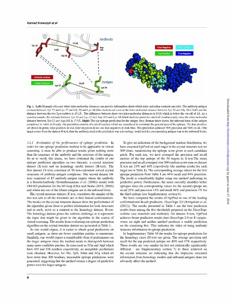
be satisfied simultaneously (see Figure 1 for an example). The final epitope
score for each patch is a sum of all possible contacts between the given
epitope and CDRs, weighted by the number of other contacts they can
satisfy simultaneously as well as the antibody-antigen Precision Score for
the particular amino acid contact pair. The Precision Score has been adapted
from our earlier work on local antibody-antigen docking (Krawczyk et al.
(2013)). In the final ranking of the candidate epitopes we only keep patches
with less than 30% of their residues in common.
2.2 Enumerating epitope-like patches
Our algorithm identifies plausible extended epitope-like patches on the
surface of the antigen and annotates each of them with a score indicating
the likelihood of containing the correct epitope. A putative epitope patch is
created for every surface residue on the antigen. The patch consists of the
neighborhood of the surface residue and is constructed by selecting every
surface residue within 4.5A of the chosen residue. This step is followed by
adding the surface residues within 4.5A of the current residues in the patch.
See Supplementary section 2 for details.
2.3 Precision score for the epitope prediction
EpiPred uses geometric matching of the antibody and antigen surfaces which
are weighted by a specific antibody-antigen score. The antibody specific
score is the Precision Score which we have previously shown to be able
to more reliably identify antibody-specific docking poses (Krawczyk et al.
(2013)). Here Pr(Tab, Tag) denotes the likelihood of the docking algorithm
to correctly pair a residue of type Tab on the antibody and a residue of type
Tag on the antigen (for instance glycine on the antibody and serine on the
antigen). The Precision Score Pr(Tab, Tag) was estimated by executing
ZDOCK on each of the 118 targets in SAbDab-nr that were not in X-test and
in the set of top 200 ZDOCK-scored poses counting how many times a given
pair of residues was matched correctly with respect to the native structures.
For details of the procedure see Supplementary section 3.
In order to ensure we have not over-trained the precision score for the H-
test dataset, we have removed all members of the SAbDab-nr that had more
than 90% sequence identity with any antigen and more than 99% with any
antibody in the H-test. The sequence identity was calculated using CD-HIT
(Li and Godzik (2006)).
2.4 Scoring putative epitopes
Let Epi denote the set of residues in a putative epitope and Ab the set of
residues supplied as the binding site on the antibody. We create a graph Gwhere each node n, corresponds to an element of the Cartesian product of
Epi and Ab: Epi×Ab. Thus if there was a tyrosine (Y) residue in Epi and
a histidine (H) residue in Ab, there will be a node n′ in G which corresponds
to this pair - (Y,H). Each of the nodes represents a possible inter-molecular
contact between antibody and antigen residues.
We add an edge between any two nodes in G if the antibody-antigen
contacts defined by those nodes can be geometrically satisfied at the same
time. Take node n1 which stands for a contact between antibody residue
rab1 and antigen residue rag1 and node n2 with antibody residue rab2and antigen residue rag2. Define dist(rab1, rab2) as the intra-molecular
distance between the two residues rab1 and rab2. We place an edge between
n1 and n2 only if the difference in intra-molecular distances on the antibody
and the antigen is below 1A as given by 1.
The choice of 1A was motivated by our analysis of the intra-molecular
distances between residues in contact. We concluded from this analysis that
1A offers a good balance between the coverage of the binding site and the
number of residues that can satisfy this condition (see Supplementary section
4).
|dist(rab1, rab2)− dist(rag1, rag2)| < 1A (1)
Let d(n) denote the degree of node n. The final score for a putative
epitope Epi is given by 2.
EpitopeScore(Epi,Ab) =∑
n∈G
d(n)Pr(Tab, Tag) (2)
where Tab and Tag are the amino acid types of the antibody and antigen
residues respectively which belong to node n.
The epitopes are ordered by their score and the top three non-overlapping
epitopes are kept. Overlapping epitopes are defined as those which share
more than 30% of the same residues with respect to the epitope with the
higher epitope score.
2
by guest on June 27, 2014http://bioinform
atics.oxfordjournals.org/D
ownloaded from
Improving B-cell epitope prediction and its application to global antibody-antigen docking
We use our epitope prediction algorithm on each of the targets in X-test
and H-test.
2.5 DiscoTope 2.0 Epitope Predictions
The structures of 30 antigens in X-test were submitted to the DiscoTope
2.0 (http://www.cbs.dtu.dk/services/DiscoTope/, (Kringelum et al. (2012)))
server using the five thresholds suggested by the service: -3.7, -2.5, -1.0,
0.5 and 1.1. The residues predicted by DiscoTope as part of an epitope were
compared to the native contacts (less than 4.5A from a heavy atom on the
antibody). The best results, by Matthews Correlation Coefficient (MCC,
(Matthews (1975))), were obtained for the threshold of -3.7 and thus were
used for the comparison with EpiPred.
2.6 Evaluating the performance of antibody-specificity
of EpiPred predictions.
We have tested the ability of EpiPred to identify the epitopes of different
antibodies on the same antigen using the test case of lysozyme. There are
five standard (non-camelid) antibody-lysozyme complexes available in the
PDB (we chose representatives: 1a2y, 1j1x, 1jhl, 1p2c and 2iff by clustering
by sequence identity (99%) and removing any with missing binding site
residues or high B-factors. We randomly select a structure from each cluster).
They bind to three distinct epitopes: epitope I (1a2y and 1jhl), epitope II
(1p2c and 2iff) and epitope III (1j1x) see Supplementary section 5.
We have re-trained EpiPred using our original set of antibody-antigen
complexes, ensuring that no antibody had more than 99% sequence identity
and antigens no more than 90% sequence identity to lysozyme. We have then
run EpiPred on each of the five cases.
GLOBAL DOCKING
The global docking pipeline we have developed is divided into three steps.
Firstly, up to three candidate epitope predictions from EpiPred are computed.
Secondly, we perform global docking using a fast rigid body algorithm
(ZDOCK or ClusPro). We do not provide any epitope information at this
point, only supplying the CDR residues to be masked. The final step
consists of re-scoring the poses produced by the docking algorithms using
AB DockSorter.
The input to AB DockSorter consists of a single antibody-antigen pose
supplied by the docking algorithm, the set of Chothia CDR residues and
a set of residues for one of the predicted epitopes. For each pose, the AB
DockSorter score is computed for each of the top three predicted epitopes as
given by EpiPred. The final score of a pose is the highest of the three scores.
The poses for a given target are then re-ranked by this score.
2.7 Docking algorithms
All the targets were subject to a random rotation and translation before
submission to either ZDOCK or ClusPro. ZDOCK was run on all of the
targets in the X-test set with the constraint on the antibody of the Chothia
CDRs and with no epitope information. The software was executed using
its default parameters, with the exception that the number of poses to probe
was set to 10000. As it was computationally feasible we performed 5 runs
of ZDOCK for each target, using different random seeds each time. An
analogous procedure was applied to the targets in H-test. The targets in X-
test and H-dataset were also submitted to ClusPro in antibody mode, using
automatic CDR masking.
2.8 Re-scoring decoys
Consider a set of decoys D returned by either ZDOCK or ClusPro for a
given target. We collect the top N decoys from D as ordered by the docking
method. For a given decoy d in the set of top N decoys from D, let Abdenote the set of residues used as the antibody constraint and Epi a set of
predicted epitope residues. Let (rab, rag) be any pair or residues in d, where
rab ∈ Ab and rag ∈ Epi. If the distance between rab and rag is observed
to be less than 4.5A in the decoy d, this pair of residues contributes the
value of Pr(Tab, Tag) to the score for this decoy where Tab is the type of
the amino acid type of rab and Tag is the amino acid type of rag . If we
let dist(rab, rag) denote the distance in Angstroms between residues raband rag , the score for decoy d using antibody constraint Ab and epitope
prediction Epi can be formalized by 3.
DecoyScore(d) =∑
rab∈Abrag∈Epi
dist(rab,rag)<4.5A
Pr(Tab, Tag) (3)
The top N decoys for a given target are given scores using our three
epitope predictions. For each decoy, we retain the highest score out of the
three. We then use those scores to re-order the top N decoys for a given
target.
In the case of ZDOCK for both X-test and H-test, we re-score the top 30
decoys for each target. We use the top 20 predictions for ClusPro as this is
the maximum number of decoys returned in most cases.
2.9 Evaluation criteria for docking
In order to evaluate the quality of each decoy we use the interfacial root mean
square deviation (Irmsd), one of the metrics used in the CAPRI experiment
(Mendez et al. (2005)). The value of Irmsd is the root mean square deviation
between the interface region of the decoy and the native structure when those
regions are optimally superimposed. The interface regions are defined as
those within neighborhood of 10A from any residue on the binding partner.
We define a close to native decoy in the same way as the authors of the
ClusPro antibody study (Brenke et al. (2012)). A close to native decoy is
defined as having Irmsd less than 10A from the native complex. For each
target in our test sets, we have the raw list of top N decoys as ordered by
the docking algorithm and our re-scored version thereof. So as to evaluate
which ordering is better we count the number of close to native decoys in the
first, top five and top ten entries in the raw and re-scored lists. For instance
if using our re-scored list we find three close to native decoys in the top five
and only one in the top five decoys in the raw list, we consider the re-scoring
to have improved the result. If in the top N of both lists no close to native
decoy is found we state there was no suitable decoy.
Since multiple runs were performed for ZDOCK, the reported results are
derived from the comparison of the averages of close to native decoys in the
raw and re-scored lists for each target.
3 RESULTS
3.1 Epitope prediction
The epitope prediction algorithm presented here was inspired by our
earlier work on local antibody-antigen docking where we showed
that it was possible to select close to native decoys when docking
the antibody into an approximate region of the epitope (Krawczyk
et al. (2013)).
In order to extend our local docking methodology to global
docking, we have developed an epitope prediction algorithm that
identifies surface patches on the antigen similar to the approximate
epitopes used in our earlier work (see Figure 1). Our epitope
prediction algorithm receives as input the structure of an antibody
and an antigen and returns a ranked list of epitope-like regions.
In our case the aim is to generate epitope predictions specific
for a given antibody in order to facilitate docking. Thus, the use
of antibody information is crucial. However, as shown below, the
antibody structure can be a homology model.
3
by guest on June 27, 2014http://bioinform
atics.oxfordjournals.org/D
ownloaded from
by guest on June 27, 2014http://bioinform
atics.oxfordjournals.org/D
ownloaded from
by guest on June 27, 2014http://bioinform
atics.oxfordjournals.org/D
ownloaded from
Konrad Krawczyk et al
Epitope Prediction
EpiPred DiscoTope 2.0 Random
PDB
Ag
size
Pre
cisi
on
(%)
Rec
all
(%)
MC
C
Pre
cisi
on
(%)
Rec
all
(%)
MC
C
Pre
cisi
on
(%)
Rec
all
(%)
4hj0 92 32 90 0.27 0 0 0.0 29 50
1tzh 94 1 6 0.04 73 87 0.72 12 25
4am0 96 13 70 0.09 33 20 0.19 14 60
2ih3 97 16 64 0.08 0 0 0.0 15 27
4i77 97 23 55 0.0 0 0 0.0 21 31
3q1s 113 19 81 0.15 0 0 0.0 20 37
1p2c 129 0 0 0.0 100 5 0.0 39 32
4ht1 131 5 14 0.05 0 0 0.0 28 44
3ab0 136 33 73 0.34 0 0 0.0 33 52
1v7m 145 26 77 0.29 0 0 0.0 9 16
4g3y 148 3 8 0.04 100 17 0.33 11 25
2vxt 156 4 9 0.04 47 36 0.3 14 23
3u9p 169 31 100 0.47 6 5 0.0 8 15
3o2d 178 32 64 0.28 0 0 0.0 9 16
1fns 196 0 0 0.0 100 7 0.0 33 11
3ma9 197 0 0 0.0 0 0 0.0 21 33
3rvv 223 25 93 0.39 15 17 0.07 6 15
3raj 230 0 0 0.0 0 0 0.0 24 21
1nfd 239 7 23 0.04 92 70 0.75 10 15
3i50 273 0 0 0.0 0 0 0.0 1 6
3gjf 276 15 66 0.2 5 11 0.05 6 15
3liz 329 26 68 0.34 0 0 0.0 10 15
3pgf 358 2 4 0.0 0 0 0.0 13 18
3zkm 375 32 88 0.46 0 0 0.0 11 15
3r1g 381 37 100 0.57 0 0 0.0 7 9
4jr9 409 19 85 0.34 46 50 0.46 4 11
4ene 442 0 0 0.0 0 0 0.0 1 5
3o0r 449 8 70 0.19 0 0 0.0 2 14
3t3p 453 0 0 0.0 25 4 0.0 4 6
1n8z 581 0 0 0.0 0 0 0.0 6 5
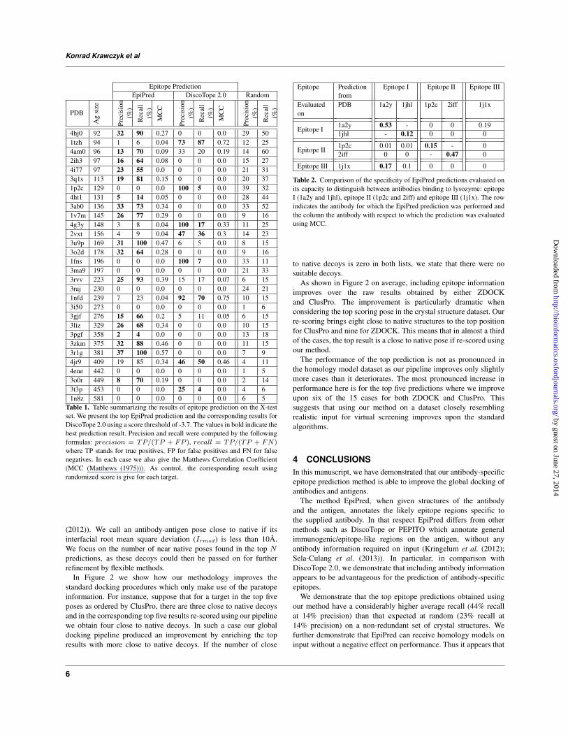
Table 1. Table summarizing the results of epitope prediction on the X-test
set. We present the top EpiPred prediction and the corresponding results for
DiscoTope 2.0 using a score threshold of -3.7. The values in bold indicate the
best prediction result. Precision and recall were computed by the following
formulas: precision = TP/(TP + FP ), recall = TP/(TP + FN)
where TP stands for true positives, FP for false positives and FN for false
negatives. In each case we also give the Matthews Correlation Coefficient
(MCC (Matthews (1975))). As control, the corresponding result using
randomized score is give for each target.
(2012)). We call an antibody-antigen pose close to native if its
interfacial root mean square deviation (Irmsd) is less than 10A.
We focus on the number of near native poses found in the top N
predictions, as these decoys could then be passed on for further
refinement by flexible methods.
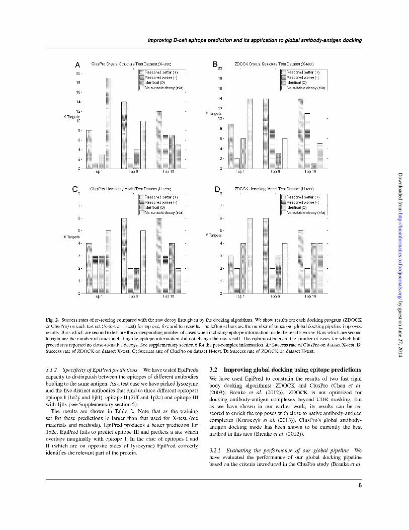
In Figure 2 we show how our methodology improves the
standard docking procedures which only make use of the paratope
information. For instance, suppose that for a target in the top five
poses as ordered by ClusPro, there are three close to native decoys
and in the corresponding top five results re-scored using our pipeline
we obtain four close to native decoys. In such a case our global
docking pipeline produced an improvement by enriching the top
results with more close to native decoys. If the number of close
Epitope Prediction
from
Epitope I Epitope II Epitope III
Evaluated
on
PDB 1a2y 1jhl 1p2c 2iff 1j1x
Epitope I1a2y 0.53 - 0 0 0.19
1jhl - 0.12 0 0 0
Epitope II1p2c 0.01 0.01 0.15 - 0
2iff 0 0 - 0.47 0
Epitope III 1j1x 0.17 0.1 0 0 0
Table 2. Comparison of the specificity of EpiPred predictions evaluated on
its capacity to distinguish between antibodies binding to lysozyme: epitope
I (1a2y and 1jhl), epitope II (1p2c and 2iff) and epitope III (1j1x). The row
indicates the antibody for which the EpiPred prediction was performed and
the column the antibody with respect to which the prediction was evaluated
using MCC.
to native decoys is zero in both lists, we state that there were no
suitable decoys.
As shown in Figure 2 on average, including epitope information
improves over the raw results obtained by either ZDOCK
and ClusPro. The improvement is particularly dramatic when
considering the top scoring pose in the crystal structure dataset. Our
re-scoring brings eight close to native structures to the top position
for ClusPro and nine for ZDOCK. This means that in almost a third
of the cases, the top result is a close to native pose if re-scored using
our method.
The performance of the top prediction is not as pronounced in
the homology model dataset as our pipeline improves only slightly
more cases than it deteriorates. The most pronounced increase in
performance here is for the top five predictions where we improve
upon six of the 15 cases for both ZDOCK and ClusPro. This
suggests that using our method on a dataset closely resembling
realistic input for virtual screening improves upon the standard
algorithms.
4 CONCLUSIONS
In this manuscript, we have demonstrated that our antibody-specific
epitope prediction method is able to improve the global docking of
antibodies and antigens.
The method EpiPred, when given structures of the antibody
and the antigen, annotates the likely epitope regions specific to
the supplied antibody. In that respect EpiPred differs from other
methods such as DiscoTope or PEPITO which annotate general
immunogenic/epitope-like regions on the antigen, without any
antibody information required on input (Kringelum et al. (2012);
Sela-Culang et al. (2013)). In particular, in comparison with
DiscoTope 2.0, we demonstrate that including antibody information
appears to be advantageous for the prediction of antibody-specific
epitopes.
We demonstrate that the top epitope predictions obtained using
our method have a considerably higher average recall (44% recall
at 14% precision) than that expected at random (23% recall at
14% precision) on a non-redundant set of crystal structures. We
further demonstrate that EpiPred can receive homology models on
input without a negative effect on performance. Thus it appears that
6
by guest on June 27, 2014http://bioinform
atics.oxfordjournals.org/D
ownloaded from
Improving B-cell epitope prediction and its application to global antibody-antigen docking
EpiPred only requires the sequence of an antibody and the structure
of the antigen to produce meaningful results.
We have used the epitope predictions from EpiPred to re-rank the
outputs of two fast rigid-body docking algorithms and find that re-
scoring the decoys in this manner significantly enriches the number
of close to native poses among the top one, five and ten results. This
result holds for targets where the antibody is a homology model
and the antigen is in the unbound structure. We have also tested
our global docking pipeline on a blind test case supplied by UCB
Pharma where once again including EpiPred predictions improved
global docking results (see Supplementary section 8).
In conclusion, our global pipeline increases the confidence that
the close to native decoy will be among the top five poses. This is
already a significant reduction of the potential set of possibilities
experimentalists need to deal with when deciding on how to adjust
the antibody sequence against the antigen. A researcher might
choose to infer information by examining these or they could further
refine the results using more time consuming flexible docking
procedures.
5 ACKNOWLEDGEMENTS
We would like to thank James Dunbar, Jinwo Leem and Nicholas
Pearce for comments on the manuscript.
6 FUNDING
This work was jointly funded by UCB Pharma and EPSRC grant for
the SABS IDC (to Charlotte M. Deane).
REFERENCES
Berman,H.M., Westbrook,J., Feng,Z., Gilliland,G., Bhat,T.,
Weissig,H., Shindyalov,I.N. and Bourne,P.E. (2000) The protein
data bank. Nucleic acids research, 28 (1), 235–242.
Blech,M., Peter,D., Fischer,P., Bauer,M., Hafner,M., Zeeb,M. and
Nar,H. (2012) One target-two different binding modes: structural
insights into gevokizumab and canakinumab interactions to
interleukin-1β (il-1β). Journal of molecular biology, 425.
Brenke,R., Hall,D., Chuang,G., Comeau,S., Bohnuud,T.,
Beglov,D., Schueler-Furman,O., Vajda,S. and Kozakov,D.
(2012) Application of asymmetric statistical potentials to
antibody–protein docking. Bioinformatics, 28 (20), 2608–2614.
Chen,R., Li,L. and Weng,Z. (2003) Zdock: an initial-stage protein
docking algorithm. Proteins, 1, 80–87.
Choi,Y. and Deane,C.M. (2010) Fread revisited: accurate loop
structure prediction using a database search algorithm. Proteins,
78, 1431–40.
Choi,Y. and Deane,C.M. (2011) Predicting antibody
complementarity determining region structures without
classification. Molecular BioSystems, 7 (12), 3327–3334.
Dunbar,J., Krawczyk,K., Leem,J., Baker,T., Fuchs,A., Georges,G.,
Shi,J. and Deane,C.M. (2013) Sabdab: the structural antibody
database. Nucleic acids research, doi:gkt1043.
EL-Manzalawy,Y. and Honavar,V. (2010) Recent advances in b-cell
epitope prediction methods. Immunome Res., 2, S2.
Krawczyk,K., Baker,T., Shi,J. and Deane,C.M. (2013) Antibody
i-patch prediction of the antibody binding site improves rigid
local antibody–antigen docking. Protein Engineering Design and
Selection, 26 (10), 621–629.
Kringelum,J.V., Lundegaard,C., Lund,O. and Nielsen,M. (2012)
Reliable b cell epitope predictions: impacts of method
development and improved benchmarking. PLoS computational
biology, 8 (12), e1002829.
Kuroda,D., Shirai,H., Jacobson,M.P. and Nakamura,H. (2012)
Computer-aided antibody design. Protein Engineering Design
and Selection, 25 (10), 507–522.
Li,W. and Godzik,A. (2006) Cd-hit: a fast program for clustering
and comparing large sets of protein or nucleotide sequences.
Bioinformatics, 22 (13), 1658–1659.
Lin,S.Y., Cheng,C.W. and Su,E.C. (2013) Prediction of b-cell
epitopes using evolutionary information and propensity scales.
BMC bioinformatics, 14 (Suppl 2), S10.
Matthews,B.W. (1975) Comparison of the predicted and observed
secondary structure of t4 phage lysozyme. Biochimica et
Biophysica Acta (BBA)-Protein Structure, 405 (2), 442–451.
Mendez,R., Leplae,R., Lensink,M.F. and Wodak,S.J. (2005)
Assessment of capri predictions in rounds 35 shows progress in
docking procedures. BMC Bioinformatics, 60, 150–169.
Murad,J., Lin,O., Paez,E. and Khasawneh,F. (2012) Current
and experimental antibody-based therapeutics: insights,
breakthroughs, setbacks and future directions. Current
molecular medicine, 2.
Raghunathan,G., Smart,J., Williams,J. and Almagro,J. (2012)
Antigen-binding site anatomy and somatic mutations in
antibodies that recognize different types of antigens. Journal of
Molecular Recognition, 25 (3), 103–113.
Sela-Culang,I., Kunik,V. and Ofran,Y. (2013) The structural basis of
antibody-antigen recognition. Frontiers in Immunology, 4, 302.
Sircar,A. and Gray,J.J. (2010) Snugdock: paratope structural
optimization during antibody-antigen docking compensates for
errors in antibody homology models. PLoS Comput Biol., 6,
e1000644.
Sivasubramanian,A., Sircar,A., Chaudhury,S. and Gray,J.J. (2009)
Toward high-resolution homology modeling of antibody fv
regions and application to antibody-antigen docking. Proteins,
74, 497–514.
Soga,S., Kuroda,D., Shirai,H., Kobori,M. and Hirayama,N. (2010)
Use of amino acid composition to predict epitope residues of
individual antibodies. Protein Engineering Design and Selection,
23 (6), 441–448.
Wark,K.L. and Hudson,P.J. (2003) Latest technologies for the
enhancement of antibody affinity. Adv Drug Deliv Rev., 5,
657–70.
Yao,B., Zheng,D., Liang,S. and Zhang,C. (2013) Conformational
b-cell epitope prediction on antigen protein structures: a review
of current algorithms and comparison with common binding site
prediction methods. PloS one, 8 (4), e62249.
Zhao,L. and Li,J. (2010) Mining for the antibody-antigen interacting
associations that predict the b cell epitopes. BMC structural
biology, 10 (Suppl 1), S6.
Zhao,L., Wong,L. and Li,J. (2011) Antibody-specified b-cell
epitope prediction in line with the principle of context-
awareness. Computational Biology and Bioinformatics,
IEEE/ACM Transactions on, 8 (6), 1483–1494.
7
by guest on June 27, 2014http://bioinform
atics.oxfordjournals.org/D
ownloaded from