NMR-based determination of the binding epitope and conformational analysis of MUC-1 glycopeptides...
12
NMR-based determination of the binding epitope and conformational analysis of MUC-1 glycopeptides and peptides bound to the breast cancer-selective monoclonal antibody SM3 Heiko Mo ¨ ller 1 , Nida Serttas 1 , Hans Paulsen 1 , Joy M. Burchell 2 , Joyce Taylor-Papadimitriou 2 and Bernd Meyer 1 1 Institute of Organic Chemistry, University of Hamburg, Germany; 2 Imperial Cancer Research Fund Breast Cancer Biology Group, Guy’s Hospital, London, UK Mucin glycoproteins on breast cancer cells carry shortened carbohydrate chains. These partially deglycosylated mucin 1 (MUC-1) structures are recognized by the monoclonal antibody SM3, which is being tested for its diagnostic utility. We used NMR spectroscopy to analyze the binding mode and the binding epitope of peptide and glycopeptide antigens to the SM3 antibody. The pentapeptide PDTRP and the glycopentapeptide PDT(O-a-D-GalNAc)RP are known lig- ands of the monoclonal antibody. The 3D structures of the ligands in the bound conformation were determined by an- alyzing trNOESY build-up rates. The peptide was found to adopt an extended conformation that fits into the binding pocket of the antibody. The binding epitopes of the ligands were determined by saturation transfer difference (STD) NMR spectroscopy. The peptide’s epitope is predominantly located in the N-terminal PDT segment whereas the C-ter- minal RP segment has fewer interactions with the protein. In contrast, the glycopeptide is interacting with SM3 utilizing all its amino acids. Pro1 shows the strongest binding effect that slightly decays towards Pro5. The GalNAc resi- due interacts mainly via the N-acetyl residue while the other protons show less interactions similar to that of Pro5. The glycopeptide in the bound state also has an extended con- formation of the peptide with the carbohydrate oriented towards the N-terminus. Docking studies showed that pep- tide and glycopeptide fit the binding pocket of the mAb SM3 very well. Keywords: glycopeptide antibody complex; STD NMR; breast cancer; MUC-1; binding epitope. The extracellular part of the epithelial glycoprotein MUC-1 consists of tandem repeats of 20 amino acids (PDTRPAPGSTAPPAHGVTSA, where the start of the tandem repeat peptide sequence varies. We follow here the definition by Gendler et al. who defined the start at PDTRP [1]. Residues of peptides, that were elongated at the N-terminus, are designated by an apostrophe, e.g. Ala20¢ - Pro1-Asp2-Thr3-Arg4-Pro5.) [2]. Each repeat can carry up to five O-glycosyl chains at Ser and Thr residues that account for the high carbohydrate content of the mucins [3]. Usually, 70–100 repeats are found in mucins. The clustering of O-linked glycans on MUC-1 leads to an extended protein core. Membrane-bound mucins extend several hundred nanometers into the lumen and thus represent a first barrier to the environment. They have important functions in cell- cell recognition and shield the cell from microorganisms, toxins and proteolytic attack [4]. Many diseases affect the production of mucus. Both the amount and the characteristics of the mucus can be altered. In cystic fibrosis, for example, due to changes of the ionic environment dramatic alterations in rheological properties correlate with changes in carbohydrate composition [2]. Modified oligosaccharides are also found in mucins of patients with Crohn’s disease [2]. Epithelial cells express the membrane-bound MUC-1 at their apical surface. In carcinomas, the localization at the apical surface is lost. High concentrations of MUC-1 spread out over the whole cell surface. This may protect the cells against low pH and may interfere with immune surveillance by causing steric hindrance of surface antigen presentation [2,4]. In breast cancer, the MUC-1 glycoprotein is overex- pressed and aberrantly glycosylated. Thus, in contrast to the mucin produced by normal breast epithelial cells, which carry core2 based structures [5], MUC1 from breast cancer cells carries highly truncated, mainly core 1 based oligosac- charide structures [6,7]. In some cases, the first sugar added, N-acetyl galactosamine is not extended, or is sialylated to form the cancer-related sialyl Tn epitope. Because of the shorter side chains, the peptide core of the cancer mucin is more exposed, and antibodies have been developed which recognize epitopes exposed in the cancer mucin, which are normally masked by large oligosaccharide side chains. These antigenic peptide sequences therefore constitute cancer-associated epitopes which are also found in the Correspondence to B. Meyer, Institute of Organic Chemistry, University of Hamburg, Martin-Luther-King-Platz 6, 20146 Hamburg, Germany. Fax: + 49 (0)40 42838 2878, Tel.: + 49 (0)40 42838 5913, E-mail: [email protected] Abbreviations: MUC-1, mucin 1 glycoprotein; SM3, breast cancer- selective monoclonal antibody; STD NMR, saturation transfer difference NMR; trNOE, transferred nuclear Overhauser enhance- ment; SPR, surface plasmon resonance; SAR, structure activity relationship; MD, molecular dynamics. (Received 28 August 2001, revised 5 December 2001, accepted 14 January 2002) Eur. J. Biochem. 269, 1444–1455 (2002) Ó FEBS 2002
-
Upload
independent -
Category
Documents
-
view
3 -
download
0
Transcript of NMR-based determination of the binding epitope and conformational analysis of MUC-1 glycopeptides...
NMR-based determination of the binding epitopeand conformational analysis of MUC-1 glycopeptides and peptidesbound to the breast cancer-selective monoclonal antibody SM3
Heiko Moller1, Nida Serttas1, Hans Paulsen1, Joy M. Burchell2, Joyce Taylor-Papadimitriou2
and Bernd Meyer1
1Institute of Organic Chemistry, University of Hamburg, Germany; 2Imperial Cancer Research Fund Breast Cancer Biology Group,
Guy’s Hospital, London, UK
Mucin glycoproteins on breast cancer cells carry shortenedcarbohydrate chains. These partially deglycosylatedmucin 1(MUC-1) structures are recognized by the monoclonalantibody SM3, which is being tested for its diagnostic utility.We used NMR spectroscopy to analyze the binding modeand thebinding epitope of peptide and glycopeptide antigensto the SM3 antibody. The pentapeptide PDTRP and theglycopentapeptide PDT(O-a-D-GalNAc)RP are known lig-ands of the monoclonal antibody. The 3D structures of theligands in the bound conformation were determined by an-alyzing trNOESY build-up rates. The peptide was found toadopt an extended conformation that fits into the bindingpocket of the antibody. The binding epitopes of the ligandswere determined by saturation transfer difference (STD)NMR spectroscopy. The peptide’s epitope is predominantly
located in the N-terminal PDT segment whereas the C-ter-minal RP segment has fewer interactions with the protein.In contrast, the glycopeptide is interacting with SM3utilizing all its amino acids. Pro1 shows the strongest bindingeffect that slightly decays towards Pro5. The GalNAc resi-due interacts mainly via the N-acetyl residue while the otherprotons show less interactions similar to that of Pro5. Theglycopeptide in the bound state also has an extended con-formation of the peptide with the carbohydrate orientedtowards the N-terminus. Docking studies showed that pep-tide and glycopeptide fit the binding pocket of themAbSM3very well.
Keywords: glycopeptide antibody complex; STD NMR;breast cancer; MUC-1; binding epitope.
The extracellular part of the epithelial glycoprotein MUC-1consists of tandem repeats of 20 amino acids(PDTRPAPGSTAPPAHGVTSA, where the start of thetandem repeat peptide sequence varies. We follow here thedefinition byGendler et al.who defined the start at PDTRP[1]. Residues of peptides, that were elongated at theN-terminus, are designated by an apostrophe, e.g. Ala20¢-Pro1-Asp2-Thr3-Arg4-Pro5.) [2]. Each repeat can carry upto five O-glycosyl chains at Ser and Thr residues thataccount for the high carbohydrate content of the mucins [3].Usually, 70–100 repeats are found in mucins. The clusteringof O-linked glycans onMUC-1 leads to an extended proteincore. Membrane-bound mucins extend several hundrednanometers into the lumen and thus represent a first barrierto the environment. They have important functions in cell-
cell recognition and shield the cell from microorganisms,toxins and proteolytic attack [4].
Many diseases affect the production of mucus. Both theamount and the characteristics of the mucus can be altered.In cystic fibrosis, for example, due to changes of the ionicenvironment dramatic alterations in rheological propertiescorrelate with changes in carbohydrate composition [2].Modified oligosaccharides are also found in mucins ofpatients with Crohn’s disease [2].
Epithelial cells express the membrane-bound MUC-1 attheir apical surface. In carcinomas, the localization at theapical surface is lost. High concentrations ofMUC-1 spreadout over the whole cell surface. This may protect the cellsagainst low pH and may interfere with immune surveillanceby causing steric hindrance of surface antigen presentation[2,4].
In breast cancer, the MUC-1 glycoprotein is overex-pressed and aberrantly glycosylated. Thus, in contrast to themucin produced by normal breast epithelial cells, whichcarry core2 based structures [5], MUC1 from breast cancercells carries highly truncated, mainly core 1 based oligosac-charide structures [6,7]. In some cases, the first sugar added,N-acetyl galactosamine is not extended, or is sialylated toform the cancer-related sialyl Tn epitope. Because of theshorter side chains, the peptide core of the cancer mucin ismore exposed, and antibodies have been developed whichrecognize epitopes exposed in the cancer mucin, which arenormally masked by large oligosaccharide side chains.These antigenic peptide sequences therefore constitutecancer-associated epitopes which are also found in the
Correspondence to B. Meyer, Institute of Organic Chemistry,
University of Hamburg, Martin-Luther-King-Platz 6,
20146 Hamburg, Germany.
Fax: + 49 (0)40 42838 2878, Tel.: + 49 (0)40 42838 5913,
E-mail: [email protected]
Abbreviations: MUC-1, mucin 1 glycoprotein; SM3, breast cancer-
selective monoclonal antibody; STD NMR, saturation transfer
difference NMR; trNOE, transferred nuclear Overhauser enhance-
ment; SPR, surface plasmon resonance; SAR, structure activity
relationship; MD, molecular dynamics.
(Received 28 August 2001, revised 5 December 2001, accepted
14 January 2002)
Eur. J. Biochem. 269, 1444–1455 (2002) Ó FEBS 2002
short sugar side chains (e.g. T, ST and TF antigens) [8,9].The monoclonal antibody SM3 was raised in mice againstpartially deglycosylated human MUC-1. It shows a highspecificity to theMUC-1 of breast cancer cells. SM3 is beingtested for its diagnostic value [10,11] and also has a hightherapeutic potential.
The minimum peptide antigen epitope to SM3 wasidentified by the pepscan technique using ELISA detection.Using heptapeptides the resulting binding epitope wasidentified asAsp2-Thr3 [12], using octapeptides the resultingepitope was Pro1-Asp2-Thr3-Arg4-Pro5 [13] and usingnona- and 20mer peptides the resulting epitope wasidentified as Ala20¢-Pro1-Asp2-Thr3-Arg4-Pro5 and Pro1-Asp2-Thr3, respectively [14,15]. For SM3 reacting withpentamers and dimers of the MUC-1 tandem repeat thebinding constants were determined by surface plasmonresonance to be Kd ¼ 6.25 · 10)8 to 4.5 · 10)7
M [16].Previous NMR studies of peptide and glycopeptide
fragments of MUC-1 containing the amino-acid sequencePDTRP in the central part which were carried out insolution without an antibody present reveal that thissequence motif seems to adopt a knob-like or bent structure[17] (J. Dojahn, C. Diotel, M. Paulsen and B. Meyer,unpublished results). It was postulated that this knob-likestructure renders this region especially accessible to proteininteractions necessary for stimulation of immune responses.Also in this context, the oligosaccharides attached to Thr3are most accessible for interaction with the cells of theimmune system.
It is not clear against what actual epitope SM3 wasdeveloped. The antibody binds more strongly to a MUC-1that has only part of its carbohydrate chains removed [10].It was later shown on a molecular level that a smalloligosaccharide attached to Thr3 enhances binding affinityof glycopeptides to the antibody [18].
Conventional pepscan analysis however, does not alloweasy analysis of the contribution of the carbohydrateportion. To assess the involvement of carbohydrates inantibody recognition of glycosylated structures numerousglycopeptides would have to be synthesized and even thisapproach would not directly reveal what part of theoligosaccharides interacts with the protein.
Dokurno et al. published an X-ray structure ana-lysis of SM3 complexed with the MUC-1 peptide TSAPDTRPAPGST [19]. At each end of the antigenic peptidePDTRP two additional amino acids are resolved in theX-ray crystal structure, while Thr18¢, Gly8-Ser9-Thr10 andthe side chain of Ser19¢ are disordered in the crystal. Theamino acids of the peptide sequence (S)APDTRPAP havemany interactions with the antibody’s surface. The coveredsurface of the individual amino acids varies strongly. WhileSer19¢-Ala20¢, Thr3 and Pro5-Ala6 have relatively smallcontact areas to the protein, Pro1-Asp2 and Arg4 are muchmore buried by the antibody.
NMR spectroscopy can be used to assess bindingproperties of ligands under near physiological conditionsin solution by a variety of methods, e.g. trNOEs [20], STDNMR [21–23], and SAR by NMR [24]. TrNOE spectra canalso be used to elucidate the 3D structure of the boundligand. Saturation transfer difference (STD) NMR is atechnique that can be used to characterize and identifybinding [21–23]. It can also be used to identify the bindingepitope of ligands to a protein receptor [21]. This feature can
be used to quickly identify the binding contribution fromeither peptide or carbohydrate, especially in the case ofglycopeptides. In contrast to conventional methods onlyone substrate is necessary to obtain that information.
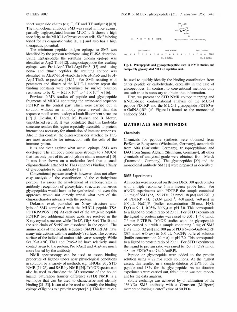
Here, we present the STD NMR epitope mapping andtrNOE-based conformational analysis of the MUC-1peptide PDTRP and the MUC-1 glycopeptide PDT(O-a-D-GalNAc)RP (cf. Figure 1) bound to the monoclonalantibody SM3.
M A T E R I A L S A N D M E T H O D S
Chemicals
Chemicals for peptide synthesis were obtained fromPerSeptive Biosystems (Wiesbaden, Germany), acetonitrilefrom Alfa (Karlsruhe, Germany), triisopropylsilane andD2O from Sigma Aldrich (Steinheim, Germany), all otherchemicals of analytical grade were obtained from Merck(Darmstadt, Germany). The glycopeptides [29] and themonoclonal antibody SM3 [1] were prepared as described.
NMR Experiments
All spectra were recorded on BrukerDRX 500 spectrometerwith a triple resonance 5 mm inverse probe head. FortrNOE experiments with PDTRP the sample contained3.6 mg of SM3 (Mr 156 kDa, 23 nmol, 38 lM) and 270 lgof PDTRP (Mr 583.64 gÆmol)1, 460 nmol, 760 lM) in600 lL NaCl/Pi (buffer concentration 20 mM, H2O/D2O ¼ 9 : 1, 0.05% NaN3) at pH 7.0. This correspondsto a ligand to protein ratio of 20 : 1. For STD experimentsthe ligand to protein ratio was raised to 200 : 1 (4.6 lmol,7.6 mM PDTRP). TrNOE studies with the glycopeptidewere carried out with a sample containing 3 mg of SM3(19.2 nmol, 32 lM) and 300 lg of PDT(O-a-D-GalNAc)RP(384 nmol, 640 lM) in 600 lL NaCl/Pi buffered solution(buffer concentration 20 mM) at pH 7.0. This correspondsto a ligand to protein ratio of 20 : 1. For STD experimentsthe ligand to protein ratio was raised to 150 : 1 (2.88 lmol,4.8 mM PDT(O-a-D-GalNAc)RP).
Peptide or glycopeptide were added to the proteinsolution using � 22 mM stock solutions. At the highestexcess, this resulted in a sample dilution of 25% for thepeptide and 18% for the glycopeptide. As no titrationexperiments were carried out, this dilution was not import-ant for the data analysis.
Solute exchange was achieved by ultrafiltration of the156-kDa SM3 antibody with a Centricon (Millipore)membrane having a cutoff value of 50 kDa.
Fig. 1. Pentapeptide and glycopentapeptide used in NMR studies and
completely glycosylated MUC-1 repetitive unit.
Ó FEBS 2002 NMR of MUC-1 glycopeptides (Eur. J. Biochem. 269) 1445
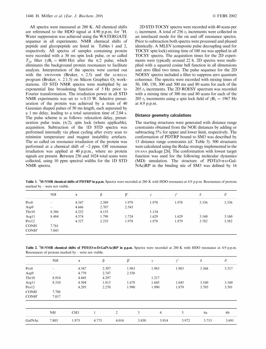
All spectra were measured at 280 K. All chemical shiftsare referenced to the HDO signal at 4.90 p.p.m. for 1H.Water suppression was achieved using the WATERGATEsequence in all experiments. NMR chemical shifts ofpeptide and glycopeptide are listed in Tables 1 and 2,respectively. All spectra of samples containing proteinwere recorded with a 30 ms spin lock pulse, or so calledT1q filter (cB1 ¼ 4680 Hz) after the p/2 pulse, whicheliminates the background protein resonances to facilitateanalysis. Interpretation of the spectra were carried outwith the XWINNMR (Bruker, v. 2.5) and the AURELIA
program (Bruker, v. 2.1.5) on Silicon Graphics O2 work-stations. 1D STD NMR spectra were multiplied by anexponential line broadening function of 5 Hz prior toFourier transformation. The irradiation power in all STDNMR experiments was set to � 0.15 W. Selective presat-uration of the protein was achieved by a train of 40Gaussian shaped pulses of 50 ms length, each separated bya 1 ms delay, leading to a total saturation time of 2.04 s.The pulse scheme is as follows: relaxation delay, presat-uration pulse train, (p/2), spin lock (where applicable),acquisition. Subtraction of the 1D STD spectra wasperformed internally via phase cycling after every scan tominimize temperature and magnet instability artefacts.The so called on resonance irradiation of the protein wasperformed at a chemical shift of )2 ppm. Off resonanceirradiation was applied at 40 p.p.m., where no proteinsignals are present. Between 256 and 1024 total scans werecollected, using 10 ppm spectral widths for the 1D STDNMR spectra.
2D STDTOCSY spectra were recorded with 40 scans pert1 increment. A total of 256 t1 increments were collected inan interlaced mode for the on and off resonance spectra.Prior to subtraction both spectra were processed and phasedidentically. A MLEV (composite pulse decoupling used forTOCSY spin lock) mixing time of 100 ms was applied in allTOCSY spectra. The acquisition times for the 2D experi-ments were typically around 22 h. 2D spectra were multi-plied with a squared cosine bell function in all dimensionsand zero filled two times. The pulse sequence for the 2DNOESY spectra included a filter to suppress zero quantumcoherence. The spectra were recorded with mixing times of50, 100, 150, 300 and 500 ms and 80 scans for each of the205 t1 increments. The 2D ROESY spectrum was recordedwith a mixing time of 300 ms and 80 scans for each of the205 t1 increments using a spin lock field of cB1 ¼ 1967 Hzat 4.9 p.p.m.
Distance geometry calculations
The starting structures were generated with distance rangeconstraints obtained from the NOE distances by adding orsubtracting 5% for upper and lower limit, respectively. Theconformation of PDTRP bound to SM3 was described by13 distance range constraints (cf. Table 3). 500 structureswere calculated using the Redac strategy implemented in theDYANA package [26]. The conformation with lowest targetfunction was used for the following molecular dynamics(MD) simulation. The structure of PDT(O-a-D-Gal-NAc)RP in the binding site of SM3 was defined by 16
Table 1. 1H-NMR chemical shifts of PDTRP in p.p.m. Spectra were recorded at 280 K with HDO resonance at 4.9 p.p.m. Resonances of protons
marked by – were not visible.
NH a b b¢ c c¢ d d¢
Pro8 – 4.347 2.389 1.978 1.978 1.978 3.336 3.336
Asp9 – 4.666 2.707 2.543
Thr10 8.386 4.252 4.155 1.134
Arg11 8.484 4.574 1.798 1.724 1.629 1.629 3.160 3.160
Pro12 4.327 2.255 1.978 1.978 1.879 3.782 3.582
CONH 7.761
CONH¢ 7.043
Table 2.1H-NMR chemical shifts of PDT(O-a-D-GalNAc)RP in p.p.m. Spectra were recorded at 280 K with HDO resonance at 4.9 p.p.m.
Resonances of protons marked by – were not visible.
NH a b b¢ c c¢ d d¢
Pro8 – 4.347 2.397 1.983 1.983 1.983 3.368 3.317
Asp9 – 4.779 2.747 2.558
Thr10 8.914 4.445 4.297 1.217
Arg11 8.510 4.504 1.813 1.678 1.645 1.645 3.160 3.160
Pro12 4.295 2.270 1.990 1.990 1.879 3.705 3.591
CONH 7.788
CONH¢ 7.017
NH CH3 1 2 3 4 5 6a 6b
GalNAc 7.803 1.975 4.775 4.016 3.850 3.914 3.972 3.715 3.691
1446 H. Moller et al. (Eur. J. Biochem. 269) Ó FEBS 2002
distance range constraints (cf. Table 4). The distancegeometry calculations were performed by an internalalgorithm in SYBYL (v. 6.3, Tripos). A total of 100 structureswere generated and energetically optimized. The lowestenergy conformation acted as starting structure for thefollowing MD simulation.
MD simulations
Constrained MD simulations were carried out with theSYBYL program on Silicon Graphics Octane (R12000)computers, using the Tripos force field. A harmonicpotential was employed at the edges of the distance rangeconstraints. The force field constants were set to 2 kcalÆ(mol A2))1. Constraints to pseudoatoms, generated bySYBYL, were used for nonstereospecifically assigned methyl-ene groups and methyl groups. The starting structures wereplaced in water boxes (PDTRP: 931 water molecules,30 · 30 · 30 A3, PDTRP/SM3 complex: 1708 watermolecules, 40 · 41 · 40 A3, PDT(O-a-D-GalNAc)RP:1152 water molecules, 33 · 33 · 33 A3, PDT(O-a-D-GalNAc)RP/SM3 complex: 2521 water molecules, 45 ·48 · 42 A3).
Before starting the MD simulation the box was energyoptimized over 200 steps. The constrained simulation wasperformed at 300 K. The charges were calculated with theGasteiger Marsili method and a dielectric constant of fourwas used. A cutoff radius of 8 A was used for thenonbonded interactions. The initial velocities for the atomswere taken from a Boltzmann distribution at 300 K and thestep size for the integration of Newton’s equation was 1 fs.The coupling to the temperature bath was set to 100 fs andthe nonbonded interactions were updated every 25 fs. TheMD simulations ran for 100 ps at constant volume andtemperature.
The final structures were energy minimized over 1000steps and overlaid to the PDTRP fragment of the ligand ofthe X-ray structure (RCSB PDB entry 1SM3). After smallmanual corrections, the ligands were docked into thebinding site of SM3 using the FLEXIDOCK module of theSYBYL software package. The docking structures after
100 000 generations were subjected to finalMD simulationsin the binding site of the antibody with flexible proteinresidues in a perimeter of 10 A from the ligand.
R E S U L T S
The small PDTRP peptide and its glycosylated derivativewere used because larger peptides did not show measurabletrNOE effects. This is most likely due to slow exchangebetween the bound and the free state. For dimers andpentamers of theMUC-1 tandem repeat [16], i.e. 40mer and60mer peptides, the dissociation constant was determined toKd ¼ 10)7 by SPR. At an on-rate of kon ¼ 106 M
)1Æs)1
typical for antibody interactions one would have an off-ratekoff ¼ 0.1 s)1, which is too slow for obtaining measurabletrNOE effects. The exact kinetic constants were notpublished. More importantly, the larger peptides decom-posed in the presence of the antibody within a few days(N. Serttas, H. Moller, J.M. Burchell, J. Taylor-Papadimi-triou, B. Meyer and H. Paulsen, unpublished results). Toovercome these problems with large peptides, we used shortpeptides to utilize their faster dissociation rates [25] and theirstability in the presence of SM3. With the pentapeptide andglycopentapeptide we obtained strong STD effects andweak trNOEs.
SM3 in complex with the peptide PDTRP
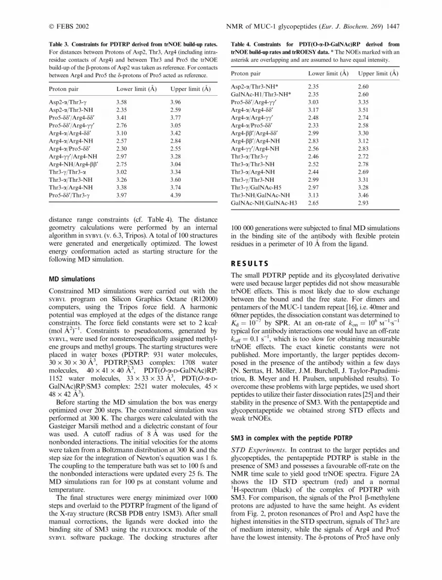
STD Experiments. In contrast to the larger peptides andglycopeptides, the pentapeptide PDTRP is stable in thepresence of SM3 and possesses a favourable off-rate on theNMR time scale to yield good trNOE spectra. Figure 2Ashows the 1D STD spectrum (red) and a normal1H-spectrum (black) of the complex of PDTRP withSM3. For comparison, the signals of the Pro1 b-methyleneprotons are adjusted to have the same height. As evidentfrom Fig. 2, proton resonances of Pro1 and Asp2 have thehighest intensities in the STD spectrum, signals of Thr3 areof medium intensity, while the signals of Arg4 and Pro5have the lowest intensity. The d-protons of Pro5 have only
Table 3. Constraints for PDTRP derived from trNOE build-up rates.
For distances between Protons of Asp2, Thr3, Arg4 (including intra-
residue contacts of Arg4) and between Thr3 and Pro5 the trNOE
build-up of the b-protons of Asp2 was taken as reference. For contacts
between Arg4 and Pro5 the d-protons of Pro5 acted as reference.
Proton pair Lower limit (A) Upper limit (A)
Asp2-a/Thr3-c 3.58 3.96
Asp2-a/Thr3-NH 2.35 2.59
Pro5-dd¢/Arg4-dd¢ 3.41 3.77
Pro5-dd¢/Arg4-cc¢ 2.76 3.05
Arg4-a/Arg4-dd¢ 3.10 3.42
Arg4-a/Arg4-NH 2.57 2.84
Arg4-a/Pro5-dd¢ 2.30 2.55
Arg4-cc¢/Arg4-NH 2.97 3.28
Arg4-NH/Arg4-bb¢ 2.75 3.04
Thr3-c/Thr3-a 3.02 3.34
Thr3-a/Thr3-NH 3.26 3.60
Thr3-a/Arg4-NH 3.38 3.74
Pro5-dd¢/Thr3-c 3.97 4.39
Table 4. Constraints for PDT(O-a-D-GalNAc)RP derived from
trNOE build-up rates and trROESY data. * TheNOEsmarked with an
asterisk are overlapping and are assumed to have equal intensity.
Proton pair Lower limit (A) Upper limit (A)
Asp2-a/Thr3-NH* 2.35 2.60
GalNAc-H1/Thr3-NH* 2.35 2.60
Pro5-dd¢/Arg4-cc¢ 3.03 3.35
Arg4-a/Arg4-dd¢ 3.17 3.51
Arg4-a/Arg4-cc¢ 2.48 2.74
Arg4-a/Pro5-dd¢ 2.33 2.58
Arg4-bb¢/Arg4-dd¢ 2.99 3.30
Arg4-bb¢/Arg4-NH 2.83 3.12
Arg4-cc¢/Arg4-NH 2.56 2.83
Thr3-a/Thr3-c 2.46 2.72
Thr3-a/Thr3-NH 2.52 2.78
Thr3-a/Arg4-NH 2.44 2.69
Thr3-c/Thr3-NH 2.99 3.31
Thr3-c/GalNAc-H5 2.97 3.28
Thr3-NH/GalNAc-NH 3.13 3.46
GalNAc-NH/GalNAc-H3 2.65 2.93
Ó FEBS 2002 NMR of MUC-1 glycopeptides (Eur. J. Biochem. 269) 1447
25% relative intensity in the STD spectrum. Obviously,Pro1 and Asp2 get more saturation from the protein thanthe remaining residues of the ligand and therefore havemore and tighter contacts to the antibody’s surface. Themean STD intensities of each residue are summarized inFig. 2B. Here, it is evident that the mean intensities ofsignals of Pro5 have only 40% intensity relative to those ofPro1. Overall, there is a continuous drop in intensity fromthe N-terminus to the C-terminus with a 50% value beingreached at Thr3.
By2DSTDTOCSYexperimentsonecanusethe increaseddispersion for amore detailed epitopemapping. InFig. 3 theSTD and normal TOCSY spectrum of the PDTRP/SM3complex are shown. The peaks of Arg4 and Pro5 are so lowin intensity that they do not appear at the intensity cutoffshown. Signals of Pro1, Asp2 and Thr3 are clearly visibleconfirming the results from the 1D STD experiments. Thestrongest signals are again cross peaks from Pro1.
trNOE experiments with PDTRP and SM3
The conformation of the peptide ligand PDTRP bound toSM3 was obtained from transferred NOE spectra. In a
trNOESY spectrum of PDTRP in presence of SM3 (datanot shown) all crosspeaks are of the same sign as thediagonal signals and have relatively weak intensity. Thus,these negative NOEs originate from the bound conforma-tion. In absence of the antibody PDTRP shows exclusivelypositive NOEs. Most contacts are sequential or intraresidueNOEs, which is in agreement with the elongated confor-mation presented below. Pro5-d/Thr3-c is the only longrange interaction that can be detected in the trNOEspectrum.
The trNOE spectra were recorded as a function ofthe mixing time with intervals of 50, 100, 150, 300, and
Fig. 2. STD data. (A) Superposition of a 1D STD spectrum (red) and
a reference 1H-spectrum (black) of PDTRP in complex with the
antibody SM3. The intensity is adjusted, so that the Pro1-b-methylene
signal is of same height in both spectra. Clearly visible are strong STD
effects for protons of Pro1 and Asp2 whereas Arg4 and Pro5 show
weaker signals in the STD spectrum. (B)Mean STD values (in percent)
of the protons of the individual amino acids calculated for each amino
acid of PDTRP from the 1D spectrum.
Fig. 3. TOCSY spectra. (A) 2D STD TOCSY and (B) conventional
TOCSY spectrum of PDTRP in complex with SM3. The strongest
STD signals originate from protons of Pro1, Asp2 and Thr3. The
signals of Arg4 and Pro5 visible in the reference TOCSY (B) vanish
completely or have low intensity in the STD experiment (A).
1448 H. Moller et al. (Eur. J. Biochem. 269) Ó FEBS 2002
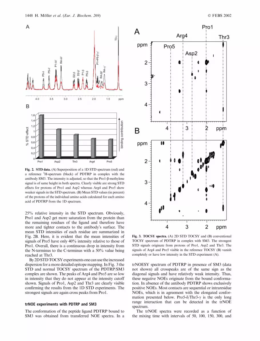
500 ms. Shorter mixing times did not give spectra with aninterpretable signal/noise ratio. Inter proton distances werecalculated from the extrapolated slope at mixing time zerousing a biexponential fitting algorithm on the trNOE build-up curves. The distance is obtained by comparing thetrNOE build-up of an interesting proton pair with that of areference proton pair which has a known fixed distance, i.e.geminal protons. PDTRP offers three well resolved refer-ence points, each of which forms a pair of geminal protonswith a proton/protondistance of 1.8 A:Asp2-b/b¢, Pro5-d/d¢and the C-terminal carboxamide NH protons. As can beseen from the three panels in Fig. 4, the initial slopes of thebuild-up curves of the geminal proton pairs are verydifferent. The build-up rate of the b protons of Asp2 isabout 2.2-fold as big as that of the Pro5 d protons. This bigdifference in NOEs converts to about 10% difference in thecorresponding distances because of the r)6 dependence ofthe NOEs on the distances. As a result, using Asp2-b/b¢ asreference the distance of Pro5-d/d¢ was calculated to be2.06 A while with Pro5-d/d¢ as reference theAsp2methyleneprotons should have a distance of 1.57 A.
There are two possible explanations for this behavior:(a) the two segments of the peptide have a very differentrotational correlation time, i.e. have very different degrees offreedom, or (b) the NOE between the Asp2 bprotons isrelayed by a protein proton. Neither explanation can easilybe proven.
It is, however, unlikely that a transfer through proteinprotons is responsible for the enhanced cross relaxation ofthe Asp2 b protons. Assuming a binding mode as found inthe X-ray structure analysis the closest distance of a proteinproton to the Asp2-b/b¢ proton is 2.7 and 3.4 A, respect-ively. This relay proton contributes to the observed crossrelaxation rate of Asp2-b with b¢ with 2% only. All otherprotons are further away and thus have less contributions.The observed difference of more than 100% compared withthe cross relaxation rate of Pro5-d/d¢ can consequently not
be explained by a relay phenomenon. We find on the otherhand that the differences in segment flexibility are in perfectagreement with the STD-based epitope mapping presentedabove.
To accommodate these variations throughout the mole-cule we chose to reference distances to reference atom pairsin the same segment, i.e. distances between protons of Asp2,Fig. 4. trNOE build-up rates of PDTRP in presence of SM3 plotted as
percentage trNOE vs. mixing time (ms). The ligand to protein ratio is
20 : 1. The three curves represent geminal protons with a fixed distance
of 1.8 A that are normally used as reference pairs. The build-up of the
trNOEbetweenAsp2-b and b¢ is much faster than that of the other two
reference points indicating differences of the rotational correlation
times of these proton pairs.
Fig. 5. PDTRP structures. (A) TrNOE-derived structure of PDTRP.
The constraints (black lines) lead to a good conformational definition
from Asp2 to Pro5. There were no NOE contact from Pro1 to Asp2
such that this segment was adjusted to fit the binding site of SM3.
(B) PDTRP (yellow) in the binding site of SM3 (atom colored surface)
(RCSB PDB entry 1SM3). This image shows the peptide antibody
complex after 100 ps constrained MD simulation and minimization
over 200 steps. Both the ligand and the binding site were kept flexible
during the simulation. (C) Superposition of PDTRP (red) with the
ligand of the X-ray structure analysis AAPDTRPAP (blue).
Ó FEBS 2002 NMR of MUC-1 glycopeptides (Eur. J. Biochem. 269) 1449
Thr3, Arg4 (including intra residue contacts of Arg4) andbetween Thr3 and Pro5 were referenced to the b-protons ofAsp2. The d-protons of Pro5 were used as reference forcontacts between Arg4 and Pro5. This approach inherentlycarries the possibility of up to 10% error of the distances ineither segment. We also carried out structure calculationswith exclusively referencing on Asp2-b/b¢ or Pro5-d/d¢. Thisproduced similar conformations as in the mixed referencingapproach but with more constraint violations (data notshown). The carboxamide protons were not used as areference pair because their initial slope was even lower thanthat of Pro5-d/d¢which is probably due to exchange with thesolvent. The final constraints that went into the distancegeometry/molecular dynamics simulation are summarizedin Table 3.
The calculations for the bound structures were per-formed in several steps. (a) We generated conformationsby distance geometry calculations using the constraints
from NOE experiments with the program DYANA [26]. (b)The structures with the lowest target function were thensubjected to constrained MD simulations over 100 ps. (c)The resulting structures were superimposed on the peptidefrom X-ray crystallography [19]. Due to the relativelysmall number of constraints we could not obtain a highresolution structure. Conformations that could not befitted into the protein of the X-ray structure analysis werenot followed further. (d) After small manual correctionsto avoid clashes with the protein, we docked the ligandinto the binding site with the software tool FLEXIDOCK
within the software package SYBYL. (e) We carried outanother constrained MD over 100 ps in the bindingpocket with ligand and protein flexible. All MD simula-tions were carried out in water boxes.
Figure 5 A shows the resulting peptide conformationwith constraints depicted as lines. The peptide in the binding
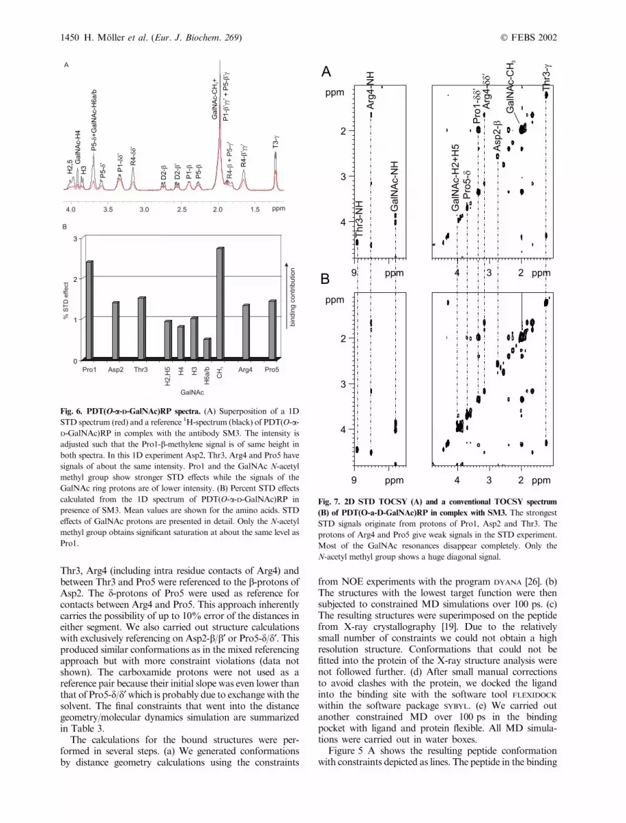
Fig. 6. PDT(O-a-D-GalNAc)RP spectra. (A) Superposition of a 1D
STD spectrum (red) and a reference 1H-spectrum (black) of PDT(O-a-D-GalNAc)RP in complex with the antibody SM3. The intensity is
adjusted such that the Pro1-b-methylene signal is of same height in
both spectra. In this 1D experiment Asp2, Thr3, Arg4 and Pro5 have
signals of about the same intensity. Pro1 and the GalNAc N-acetyl
methyl group show stronger STD effects while the signals of the
GalNAc ring protons are of lower intensity. (B) Percent STD effects
calculated from the 1D spectrum of PDT(O-a-D-GalNAc)RP in
presence of SM3. Mean values are shown for the amino acids. STD
effects of GalNAc protons are presented in detail. Only the N-acetyl
methyl group obtains significant saturation at about the same level as
Pro1.
Fig. 7. 2D STD TOCSY (A) and a conventional TOCSY spectrum
(B) of PDT(O-a-D-GalNAc)RP in complex with SM3. The strongest
STD signals originate from protons of Pro1, Asp2 and Thr3. The
protons of Arg4 and Pro5 give weak signals in the STD experiment.
Most of the GalNAc resonances disappear completely. Only the
N-acetyl methyl group shows a huge diagonal signal.
1450 H. Moller et al. (Eur. J. Biochem. 269) Ó FEBS 2002
site of SM3 is shown in Fig. 5B. For comparison of X-rayand NMR structure a least square superposition of bothconformations can be seen in Fig. 5C. It is obvious that theNMR based structure determination of the bound confor-mation of the pentapeptide agrees with that obtained in thecrystal. Probably because of fast exchange with the solventH2O at a pH of 7.0 the amide proton of Asp2 was invisible.The normal remedy for this is lowering the pH, whichcannot be used here because we wanted to preserve nearphysiological conditions in the sample. As a consequence ofthis exchange phenomenon there are no NOE contactsbetween Pro1 and Asp2. This segment of the ligand is thusill defined and was manually adjusted to fit the X-raystructure of the peptide.
SM3 in complex with the glycopeptidePDT(O-a-D-GalNAc)RP
STD Experiments. MUC-1 peptides with O-glycosylationat Thr3 show increased binding to SM3 [18]. As there is nopublished X-ray structure of a glycopeptide binding to SM3it is not known how a sugar moiety contributes to bindingenergy and whether peptide and glycopeptide bind in asimilar way. From STDNMR spectra (cf. Figures 6 and 7)it is obvious that the ring protons of the GalNAc residue ofPDT(O-a-D-GalNAc)RP receive overall less saturationthan each of the amino acids. Only the N-acetyl methylgroup has a strong STDNMR signal. It is very unlikely thatthis effect is due to different relaxation rates of the methylgroup because we have shown earlier that carbohydratesinteracting with a protein through their ring protons do notshow an STD effect on the N-acetyl methyl group [21]. Themean of all STD values in each residue is presented inFig. 6B confirming strong interactions of Pro1 and theGalNAc N-acetyl methyl group with the antibody. Thedifferences between amino acids are less pronounced than incase of the unglycosylated peptide indicating that either theglycopeptide is less flexible or that the binding contributionsare more evenly distributed within the glycopeptide. It has
been established in literature that the O-type glycosylationin peptides introduces a stabilization of that particularpeptide fragment [27,28].
trNOE experiments with PDT(O-a-D-GalNAc)RP and SM3
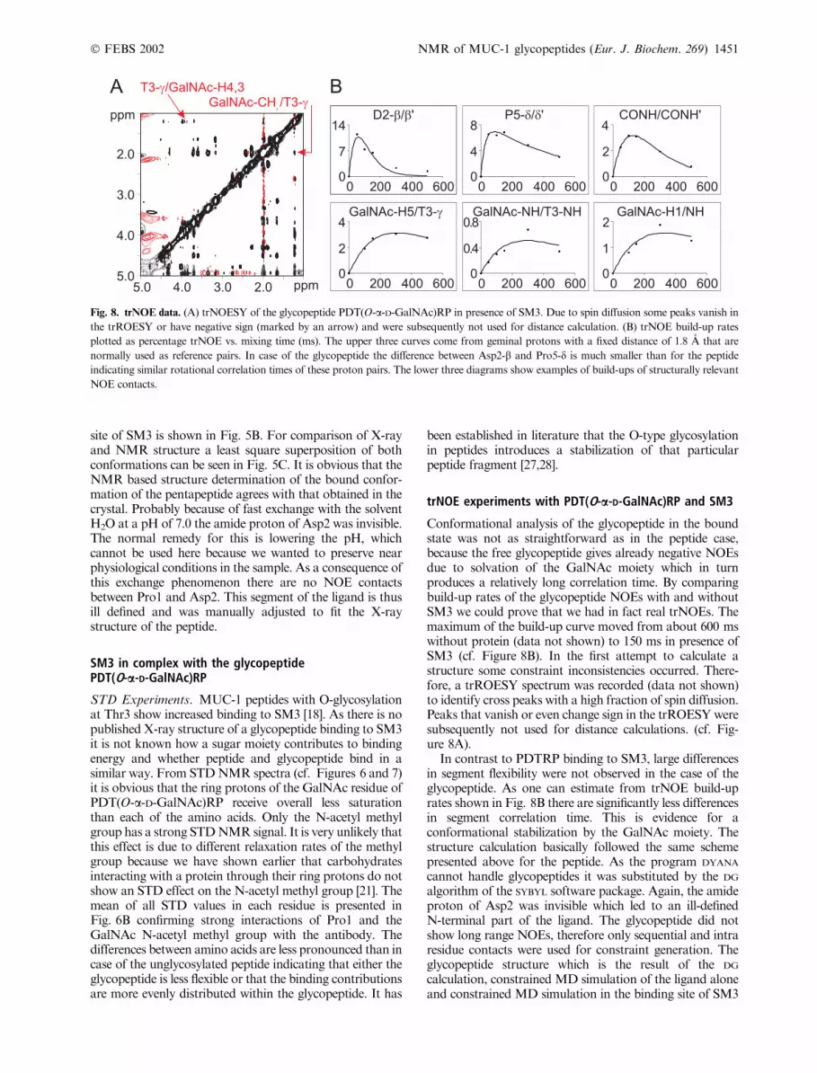
Conformational analysis of the glycopeptide in the boundstate was not as straightforward as in the peptide case,because the free glycopeptide gives already negative NOEsdue to solvation of the GalNAc moiety which in turnproduces a relatively long correlation time. By comparingbuild-up rates of the glycopeptide NOEs with and withoutSM3 we could prove that we had in fact real trNOEs. Themaximum of the build-up curve moved from about 600 mswithout protein (data not shown) to 150 ms in presence ofSM3 (cf. Figure 8B). In the first attempt to calculate astructure some constraint inconsistencies occurred. There-fore, a trROESY spectrum was recorded (data not shown)to identify cross peaks with a high fraction of spin diffusion.Peaks that vanish or even change sign in the trROESYweresubsequently not used for distance calculations. (cf. Fig-ure 8A).
In contrast to PDTRP binding to SM3, large differencesin segment flexibility were not observed in the case of theglycopeptide. As one can estimate from trNOE build-uprates shown in Fig. 8B there are significantly less differencesin segment correlation time. This is evidence for aconformational stabilization by the GalNAc moiety. Thestructure calculation basically followed the same schemepresented above for the peptide. As the program DYANA
cannot handle glycopeptides it was substituted by the DG
algorithm of the SYBYL software package. Again, the amideproton of Asp2 was invisible which led to an ill-definedN-terminal part of the ligand. The glycopeptide did notshow long range NOEs, therefore only sequential and intraresidue contacts were used for constraint generation. Theglycopeptide structure which is the result of the DG
calculation, constrained MD simulation of the ligand aloneand constrained MD simulation in the binding site of SM3
Fig. 8. trNOE data. (A) trNOESY of the glycopeptide PDT(O-a-D-GalNAc)RP in presence of SM3. Due to spin diffusion some peaks vanish in
the trROESY or have negative sign (marked by an arrow) and were subsequently not used for distance calculation. (B) trNOE build-up rates
plotted as percentage trNOE vs. mixing time (ms). The upper three curves come from geminal protons with a fixed distance of 1.8 A that are
normally used as reference pairs. In case of the glycopeptide the difference between Asp2-b and Pro5-d is much smaller than for the peptide
indicating similar rotational correlation times of these proton pairs. The lower three diagrams show examples of build-ups of structurally relevant
NOE contacts.
Ó FEBS 2002 NMR of MUC-1 glycopeptides (Eur. J. Biochem. 269) 1451
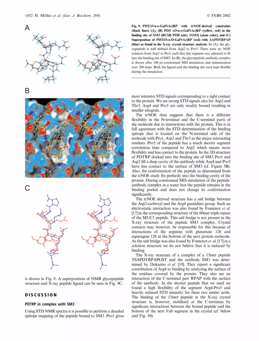
is shown in Fig. 9. A superposition of NMR glycopeptidestructure and X-ray peptide ligand can be seen in Fig. 9C.
D I S C U S S I O N
PDTRP in complex with SM3
Using STDNMR spectra it is possible to perform a detailedepitope mapping of the peptide bound to SM3. Pro1 gives
most intensive STD signals corresponding to a tight contactto the protein. We see strong STD signals also for Asp2 andThr3. Arg4 and Pro5 are only weakly bound resulting insmaller integrals.
The trNOE data suggests that there is a differentflexibility in the N-terminal and the C-terminal parts ofthe molecule due to interactions with the protein. This is infull agreement with the STD determination of the bindingepitope that is located on the N-terminal side of themolecule with Pro1, Asp2 and Thr3 as the major interactingresidues. Pro5 of the peptide has a much shorter segmentcorrelation time compared to Asp2 which means moreflexibility and less contact to the protein. In the 3D structureof PDTRP docked into the binding site of SM3 Pro1 andAsp2 fill a deep cavity of the antibody while Arg4 and Pro5have less contact to the surface of SM3 (cf. Figure 5B).Also, the conformation of the peptide as determined fromthe trNOE study fits perfectly into the binding cavity of theprotein. During constrained MD simulation of the peptide/antibody complex in a water box the peptide remains in thebinding pocket and does not change its conformationsignificantly.
The trNOE derived structure has a salt bridge betweenthe Asp2-carboxyl and the Arg4 guanidino group. Such anelectrostatic interaction was also found by Fontenot et al.[17] in the corresponding structure of the 60mer triple repeatof the MUC1 peptide. This salt bridge is not present in theX-ray structure of the peptide SM3 complex. Crystalcontacts may however, be responsible for this because ofinteractions of the arginine with glutamate 126 andasparagine 128 at the bottom of the next protein molecule.As the salt bridge was also found by Fontenot et al. [17] in asolution structure we do not believe that it is induced bybinding.
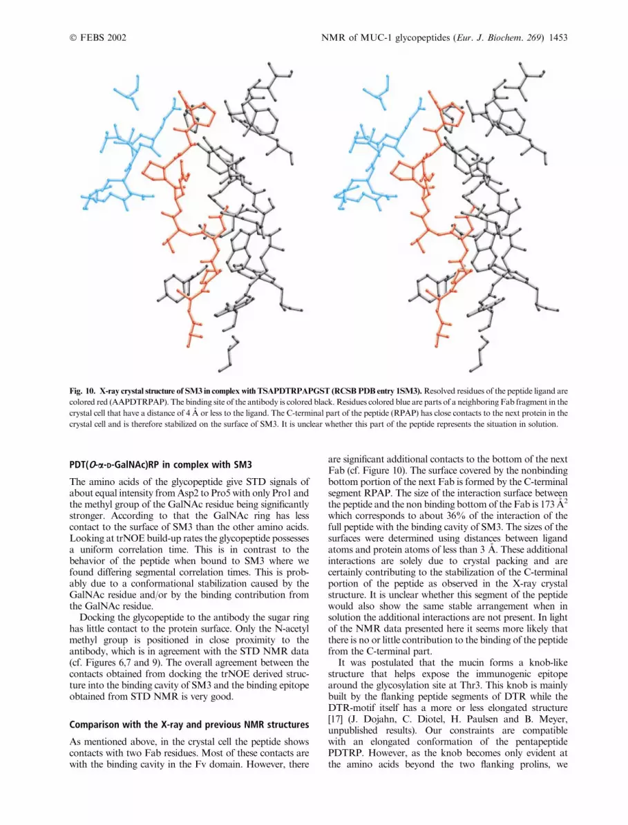
The X-ray structure of a complex of a 13mer peptideTSAPDTRPAPGST and the antibody SM3 was deter-mined by Dokurno et al. [19]. They report a significantcontribution of Arg4 to binding by analyzing the surface ofthe residues covered by the protein. They also see aninteraction of the C-terminal part RPAP with the surfaceof the antibody. In the shorter peptide that we used wefound a high flexibility of the segment Arg4-Pro5 andheavily reduced STD intensity for these two amino acids.The binding of the 13mer peptide in the X-ray crystalstructure is, however, stabilized at the C-terminus bysignificant interactions between the bound peptide and thebottom of the next Fab segment in the crystal (cf. belowand Fig. 10).
Fig. 9. PDT(O-a-D-GalNAc)RP with trNOE-derived constraints
(black lines) (A), (B) PDT (O-a-D-GalNAc)RP (yellow, red) in the
binding site of SM3 (RCSB PDB entry 1SM3) (atom color), and (C)
Superposition of PDT(O-a-D-GalNAc)RP (red) with AAPDTRPAP
(blue) as found in the X-ray crystal structure analysis. In (A), the gly-
copeptide is well defined from Asp2 to Pro5. There were no NOE
contacts from Asp2 to Pro1 such that this segment was adjusted to fit
into the binding site of SM3. In (B), the glycopeptide antibody complex
is shown after 100 ps constrained MD simulation and minimization
over 200 steps. Both the ligand and the binding site were kept flexible
during the simulation.
1452 H. Moller et al. (Eur. J. Biochem. 269) Ó FEBS 2002
PDT(O-a-D-GalNAc)RP in complex with SM3
The amino acids of the glycopeptide give STD signals ofabout equal intensity fromAsp2 to Pro5with only Pro1 andthe methyl group of the GalNAc residue being significantlystronger. According to that the GalNAc ring has lesscontact to the surface of SM3 than the other amino acids.Looking at trNOE build-up rates the glycopeptide possessesa uniform correlation time. This is in contrast to thebehavior of the peptide when bound to SM3 where wefound differing segmental correlation times. This is prob-ably due to a conformational stabilization caused by theGalNAc residue and/or by the binding contribution fromthe GalNAc residue.
Docking the glycopeptide to the antibody the sugar ringhas little contact to the protein surface. Only the N-acetylmethyl group is positioned in close proximity to theantibody, which is in agreement with the STD NMR data(cf. Figures 6,7 and 9). The overall agreement between thecontacts obtained from docking the trNOE derived struc-ture into the binding cavity of SM3 and the binding epitopeobtained from STD NMR is very good.
Comparison with the X-ray and previous NMR structures
As mentioned above, in the crystal cell the peptide showscontacts with two Fab residues. Most of these contacts arewith the binding cavity in the Fv domain. However, there
are significant additional contacts to the bottom of the nextFab (cf. Figure 10). The surface covered by the nonbindingbottom portion of the next Fab is formed by the C-terminalsegment RPAP. The size of the interaction surface betweenthe peptide and the non binding bottom of the Fab is 173 A2
which corresponds to about 36% of the interaction of thefull peptide with the binding cavity of SM3. The sizes of thesurfaces were determined using distances between ligandatoms and protein atoms of less than 3 A. These additionalinteractions are solely due to crystal packing and arecertainly contributing to the stabilization of the C-terminalportion of the peptide as observed in the X-ray crystalstructure. It is unclear whether this segment of the peptidewould also show the same stable arrangement when insolution the additional interactions are not present. In lightof the NMR data presented here it seems more likely thatthere is no or little contribution to the binding of the peptidefrom the C-terminal part.
It was postulated that the mucin forms a knob-likestructure that helps expose the immunogenic epitopearound the glycosylation site at Thr3. This knob is mainlybuilt by the flanking peptide segments of DTR while theDTR-motif itself has a more or less elongated structure[17] (J. Dojahn, C. Diotel, H. Paulsen and B. Meyer,unpublished results). Our constraints are compatiblewith an elongated conformation of the pentapeptidePDTRP. However, as the knob becomes only evident atthe amino acids beyond the two flanking prolins, we
Fig. 10. X-ray crystal structure of SM3 in complex with TSAPDTRPAPGST (RCSBPDB entry 1SM3).Resolved residues of the peptide ligand are
colored red (AAPDTRPAP). The binding site of the antibody is colored black. Residues colored blue are parts of a neighboring Fab fragment in the
crystal cell that have a distance of 4 A or less to the ligand. The C-terminal part of the peptide (RPAP) has close contacts to the next protein in the
crystal cell and is therefore stabilized on the surface of SM3. It is unclear whether this part of the peptide represents the situation in solution.
Ó FEBS 2002 NMR of MUC-1 glycopeptides (Eur. J. Biochem. 269) 1453
cannot reject or confirm the presence of a knob-likestructure based on our data from the pentapeptide. Ourconclusion is that the peptide segment recognized by theantibody SM3 is basically elongated while the adjacentamino acids may still form the knob (cf. Fontenot et al.[17]).
C O N C L U S I O N S
Here, we demonstrate that binding studies of peptides andglycopeptides with large proteins can easily be performedwith STD NMR. Combined with trNOE information onecan obtain a 3D picture with the binding residues of theligand identified in their relative orientation in space andthus efficiently optimize on the structure of the ligand forfurther immunization studies.
In contrast to the X-ray structure analysis NMRspectroscopy does not show artefacts from crystal packing.Thus, NMR spectroscopy was able to obtain the informa-tion on binding from a sample that is close to the naturalphysiological environment.
A C K N O W L E D G E M E N T S
This work was supported by the Deutsche Forschungsgemeinschaft
through Sonderforschungsbereich 470/B2, the Graduate College GRK
464 and a grant from the Fonds der chemischen Industrie (FCI) to
H. M.
R E F E R E N C E S
1. Gendler, S.J., Burchell, J.M., Duhig, T., Lamport, D., White, R.,
Parker, M. & Taylor-Papadimitriou, J. (1987) Cloning of partial
cDNA encoding differentiation and tumor-associated mucin
glycoproteins expressed by human mammary epithelium. Proc.
Natl Acad. Sci. USA 84, 6060–6064.
2. Strous, G.J. & Dekker, J. (1992) Mucin-type glycoproteins. Crit.
Rev. Biochem. Mol. Biol. 27, 57–92.
3. Muller, S., Goletz, S., Packer, N., Gooley, A., Lawson, A.M. &
Hanisch, F.G. (1997) Localization of O-glycosylation sites on
glycopeptide fragments from lactation-associated MUC1. All
putative sites within the tandem repeat are glycosylation targets
in vivo. J. Biol. Chem. 272, 24780–24793.
4. Devine, P.L. &McKenzie, I.F. (1992)Mucins: structure, function,
and associations with malignancy. Bioessays 14, 619–625.
5. Hanisch, F.G., Uhlenbruck, G., Peter-Katalinic, J., Egge, H.,
Dabrowski, J. & Dabrowski, U. (1989) Structures of neutral
O-linked polylactosaminoglycans on human skim milk mucins.
A novel type of linearly extended poly-N-acetyllactosamine
backbones with Gal beta (1–4) GlcNAc beta (1–6) repeating units.
J. Biol. Chem. 264, 872–883.
6. Hull, S.R., Bright, A., Carraway, K.L., Abe, M., Hayes, D.F. &
Kufe, D.W. (1989) Oligosaccharide differences in the DF3 sialo-
mucin antigen from normal human milk and the BT-20 human
breast carcinoma cell line. Cancer Commun. 1, 261–267.
7. Lloyd, K.O., Burchell, J., Kudryashov, V., Yin, B.W. & Taylor-
Papadimitriou, J. (1996) Comparison of O-linked carbohydrate
chains in MUC-1 mucin from normal breast epithelial cell lines
and breast carcinoma cell lines. Demonstration of simpler and
fewer glycan chains in tumor cells. J. Biol. Chem. 271, 33325–
33334.
8. Jerome, K.R., Barnd, D.L., Bendt, K.M., Boyer, C.M., Taylor-
Papadimitriou, J., McKenzie, I.F., Bast, R.C. & Finn, O.J. (1991)
Cytotoxic T-lymphocytes derived from patients with breast ade-
nocarcinoma recognize an epitope present on the protein core of a
mucinmolecule preferentially expressed bymalignant cells.Cancer
Res. 51, 2908–2916.
9. Baeckstrom, D., Nilsson, O., Price, M.R., Lindholm, L. &
Hansson, G.C. (1993) Discrimination of MUC1 mucins from
other sialyl-Le(a)-carrying glycoproteins produced by colon
carcinoma cells using a novel monoclonal antibody. Cancer Res.
53, 755–761.
10. Burchell, J., Gendler, S., Taylor-Papadimitriou, J., Girling, A.,
Lewis, A., Millis, R. & Lamport, D. (1987) Development and
characterization of breast cancer reactive monoclonal antibodies
directed to the core protein of the human milk mucin.Cancer Res.
47, 5476–5482.
11. Biassoni, L., Granowska, M., Carroll, M.J., Mather, S.J.,
Howell, R., Ellison, D., MacNeill, F.A., Wells, C.A.,
Carpenter, R. & Britton, K.E. (1998) 99mTc-labelled SM3 in the
preoperative evaluation of axillary lymph nodes and primary
breast cancer with change detection statistical processing as an aid
to tumour detection. Br. J. Cancer 77, 131–138.
12. Petrakou, E., Murray, A. & Price, M.R. (1998) Epitope mapping
of anti-MUC1 mucin protein core monoclonal antibodies.
Tumour Biol. 19, 21–29.
13. Burchell, J., Taylor-Papadimitriou, J., Boshell, M., Gendler, S. &
Duhig, T. (1989) A short sequence, within the amino acid tandem
repeat of a cancer- associated mucin, contains immunodominant
epitopes. Int. J. Cancer 44, 691–696.
14. Blockzjil, A., Nilsson, K. & Nilsson, O. (1998) Epitope
characterization of MUC1 antibodies. Tumour Biol. 19, 46–56.
15. Schol, D.J., Meulenbroek, M.F., Snijdewint, F.G., von
Mensdorff-Pouilly, S., Verstraeten, R.A., Murakami, F.,
Kenemans, P. & Hilgers, J. (1998) ÔEpitope fingerprintingÕ usingoverlapping 20-mer peptides of the MUC1 tandem repeat
sequence. Tumour Biol. 19, 35–45.
16. Karanikas, V., Patton, K., Jamieson, G., Pietersz, G. &
McKenzie, I. (1998) Affinity of antibodies to MUC1 antigens.
Tumour Biol. 19, 71–78.
17. Fontenot, J.D., Mariappan, S.V., Catasti, P., Domenech, N.,
Finn, O.J. & Gupta, G. (1995) Structure of a tumor associated
antigen containing a tandemly repeated immunodominant
epitope. J. Biomol. Struct. Dyn. 13, 245–260.
18. Karsten, U., Diotel, C., Klich, G., Paulsen, H., Goletz, S.,
Muller, S. & Hanisch, F.G. (1998) Enhanced binding of anti-
bodies to the DTR motif of MUC1 tandem repeat peptide is
mediated by site-specific glycosylation.Cancer Res. 58, 2541–2549.
19. Dokurno, P., Bates, P.A., Band, H.A., Stewart, L.M., Lally, J.M.,
Burchell, J.M., Taylor-Papadimitriou, J., Snary, D., Sternberg,
M.J. & Freemont, P.S. (1998) Crystal structure at 1.95 A resolu-
tion of the breast tumour-specific antibody SM3 complexed with
its peptide epitope reveals novel hypervariable loop recognition.
J. Mol. Biol. 284, 713–728.
20. Ni, F. (1994) Recent developments in transferred NOE methods.
Prog. NMR Spectrosc. 26, 517–606.
21. Mayer, M. & Meyer, B. (2001) Group epitope mapping by
saturation transfer difference NMR to identify segments of a
ligand in direct contact with a protein receptor. J. Am. Chem. Soc.
123, 6108–6117.
22. Mayer, M. &Meyer, B. (1999) Characterization of ligand binding
by saturation transfer difference NMR spectroscopy. Angew.
Chem. Int. Ed. Engl. 38, 1784–1788.
23. Klein, J., Meinecke, R., Mayer, M. & Meyer, B. (1999) Detecting
binding affinity to immobilized receptor proteins in compound
libraries by HR-MAS STD NMR. J. Am. Chem. Soc. 121, 5336–
5337.
24. Shuker, S.B., Hajduk, P.J., Meadows, R.P. & Fesik, S.W. (1996)
Discovering high-affinity ligands for proteins: SAR by NMR.
Science 274, 1531–1534.
25. Anglister, J. & Zilber, B. (1990) Antibodies against a peptide of
cholera toxin differing in cross- reactivity with the toxin differ in
1454 H. Moller et al. (Eur. J. Biochem. 269) Ó FEBS 2002
their specific interactions with the peptide as observed by 1H
NMR spectroscopy. Biochemistry 29, 921–928.
26. Gunthert, P., Mumenthaler, C. & Wuthrich, K. (1997) Torsion
angle dynamics for NMR structure calculation with the new
program DYANA. J. Mol. Biol. 273, 283–298.
27. Paulsen, H., Pollex-Kruger, A. & Sinnwell, V. (1991) Conforma-
tional analysis of N-terminal O-glycopeptide sequences of inter-
leukin-2. Carbohydr. Res. 214, 199–226.
28. Pieper, J., Ott, K.H. & Meyer, B. (1996) Stabilization of the T1
fragment of glycophorin A (N) through interactions with N- and
O-linked glycans. Nat. Struct. Biol. 3, 228–232.
29. Klich, G., Paulsen, H., Meyer, B., Meldal, M. & Bock, K. (1997)
Synthesis and characterisation of highly glycosylated glyco-
peptides with Tn-antigenic structures corresponding to human
glycophorin AN. Carbohydr. Res. 299, 33–48.
Ó FEBS 2002 NMR of MUC-1 glycopeptides (Eur. J. Biochem. 269) 1455