Development and Characterization of Monoclonal Antibodies ...
14
toxins Article Development and Characterization of Monoclonal Antibodies to Botulinum Neurotoxin Type E Candace S. Bever 1 , Miles Scotcher 1 , Luisa W. Cheng 1, *, Robert M. Hnasko 2 and Larry H. Stanker 1, * 1 Foodborne Toxin Detection and Prevention Research Unit, Agricultural Research Service, United States Department of Agriculture, 800 Buchanan Street, Albany, CA 94710, USA 2 Produce Safety and Microbiology Research Unit, Agricultural Research Service, United States Department of Agriculture, 800 Buchanan Street, Albany, CA 94710, USA * Correspondence: [email protected] (L.W.C.); [email protected] (L.H.S.); Tel.: +1-510-559-6337 (L.W.C.); +1-510-559-5984(L.H.S.) Received: 29 May 2019; Accepted: 12 July 2019; Published: 13 July 2019 Abstract: Botulism is a devastating disease caused by botulinum neurotoxins (BoNTs) secreted primarily by Clostridium botulinum. Mouse bioassays without co-inoculation with antibodies are the standard method for the detection of BoNTs, but are not capable of distinguishing between the different serotypes (A–G). Most foodborne intoxications are caused by serotypes BoNT/A and BoNT/B. BoNT/E outbreaks are most often observed in northern coastal regions and are associated with eating contaminated marine animals and other fishery products. Sandwich enzyme-linked immunosorbent assays (ELISAs) were developed for the detection of BoNT/E3. Monoclonal antibodies (mAbs) were generated against BoNT/E3 by immunizing with recombinant peptide fragments of the light and heavy chains of BoNT/E3. In all, 12 mAbs where characterized for binding to both the recombinant peptides and holotoxin, as well as their performance in Western blots and sandwich ELISAs. The most sensitive sandwich assay, using different mAbs for capture and detection, exhibited a limit of detection of 0.2 ng/ml in standard buffer matrix and 10 ng/mL in fish product matrices. By employing two different mAbs for capture and detection, a more standardized sandwich assay was constructed. Development of sensitive and selective mAbs to BoNT/E would help in the initial screening of potential food contamination, speeding diagnosis and reducing use of laboratory animals. Keywords: monoclonal antibodies; botulinum neurotoxin serotype E; BoNT/E; ELISA; immunoassay Key Contribution: An alternative strategy is described wherein peptide fragments were used as immunogens for the production of new mouse monoclonal antibodies to botulinum neurotoxin serotype E (BoNT/E3). Furthermore, a pair of these monoclonal antibodies were then formatted to generate a sensitive sandwich ELISA for the detection of BoNT/E3. 1. Introduction Botulinum neurotoxins (BoNT) represent a family of toxins produced primarily by Clostridium botulinum and are the cause of botulism in both animals and humans. Botulism is a severe neurological disease that if untreated is often fatal. Seven toxin serotypes (BoNT/A–G) have been described, as well as potentially type H and a tentatively-named type X that are less well studied [1,2]. All BoNTs have a molecular weight of about 150 kDa and consist of two polypeptide chains, a light chain (LC) ~50 kDa and a heavy chain (HC) ~100 kDa linked via a single disulfide bridge. BoNT serotypes A and B, are responsible for the majority (90%) of confirmed human botulism cases in the U.S. followed by serotype E responsible for 10% (3 of 29) of the foodborne cases reported Toxins 2019, 11, 407; doi:10.3390/toxins11070407 www.mdpi.com/journal/toxins
-
Upload
khangminh22 -
Category
Documents
-
view
1 -
download
0
Transcript of Development and Characterization of Monoclonal Antibodies ...

toxins
Article
Development and Characterization of MonoclonalAntibodies to Botulinum Neurotoxin Type E
Candace S. Bever 1, Miles Scotcher 1, Luisa W. Cheng 1,*, Robert M. Hnasko 2 andLarry H. Stanker 1,*
1 Foodborne Toxin Detection and Prevention Research Unit, Agricultural Research Service,United States Department of Agriculture, 800 Buchanan Street, Albany, CA 94710, USA
2 Produce Safety and Microbiology Research Unit, Agricultural Research Service,United States Department of Agriculture, 800 Buchanan Street, Albany, CA 94710, USA
* Correspondence: [email protected] (L.W.C.); [email protected] (L.H.S.);Tel.: +1-510-559-6337 (L.W.C.); +1-510-559-5984(L.H.S.)
Received: 29 May 2019; Accepted: 12 July 2019; Published: 13 July 2019�����������������
Abstract: Botulism is a devastating disease caused by botulinum neurotoxins (BoNTs) secretedprimarily by Clostridium botulinum. Mouse bioassays without co-inoculation with antibodies arethe standard method for the detection of BoNTs, but are not capable of distinguishing between thedifferent serotypes (A–G). Most foodborne intoxications are caused by serotypes BoNT/A and BoNT/B.BoNT/E outbreaks are most often observed in northern coastal regions and are associated with eatingcontaminated marine animals and other fishery products. Sandwich enzyme-linked immunosorbentassays (ELISAs) were developed for the detection of BoNT/E3. Monoclonal antibodies (mAbs) weregenerated against BoNT/E3 by immunizing with recombinant peptide fragments of the light andheavy chains of BoNT/E3. In all, 12 mAbs where characterized for binding to both the recombinantpeptides and holotoxin, as well as their performance in Western blots and sandwich ELISAs. The mostsensitive sandwich assay, using different mAbs for capture and detection, exhibited a limit of detectionof 0.2 ng/ml in standard buffer matrix and 10 ng/mL in fish product matrices. By employing twodifferent mAbs for capture and detection, a more standardized sandwich assay was constructed.Development of sensitive and selective mAbs to BoNT/E would help in the initial screening ofpotential food contamination, speeding diagnosis and reducing use of laboratory animals.
Keywords: monoclonal antibodies; botulinum neurotoxin serotype E; BoNT/E; ELISA; immunoassay
Key Contribution: An alternative strategy is described wherein peptide fragments were used asimmunogens for the production of new mouse monoclonal antibodies to botulinum neurotoxinserotype E (BoNT/E3). Furthermore, a pair of these monoclonal antibodies were then formatted togenerate a sensitive sandwich ELISA for the detection of BoNT/E3.
1. Introduction
Botulinum neurotoxins (BoNT) represent a family of toxins produced primarily by Clostridiumbotulinum and are the cause of botulism in both animals and humans. Botulism is a severe neurologicaldisease that if untreated is often fatal. Seven toxin serotypes (BoNT/A–G) have been described, as wellas potentially type H and a tentatively-named type X that are less well studied [1,2]. All BoNTs have amolecular weight of about 150 kDa and consist of two polypeptide chains, a light chain (LC) ~50 kDaand a heavy chain (HC) ~100 kDa linked via a single disulfide bridge.
BoNT serotypes A and B, are responsible for the majority (90%) of confirmed human botulismcases in the U.S. followed by serotype E responsible for 10% (3 of 29) of the foodborne cases reported
Toxins 2019, 11, 407; doi:10.3390/toxins11070407 www.mdpi.com/journal/toxins
Toxins 2019, 11, 407 2 of 14
in 2016 [3]. Within the US and abroad, serotype E is the most prevalent toxin type identified in fish,fishery products, and aquatic environments, including the shoreline soil [4–6]. BoNT/E outbreaks havealso been found in water birds in the Great Lakes, USA [7]. In Alaska, BoNT/E is the most frequentserotype, accounting for 82% (249 of 303) of the foodborne cases from 1950–2016 [8]. The propensityfor exposure to BoNT/E in Alaska is likely due to the documented presence of C. botulinum typeE spores within the environment, as well as cultural preferences for food preparation proceduresfor aquatic animals (i.e., butchering animals in coastal environments, as well as “fermenting” fishand sea mammals) [6,8–11]. Specifically, whales, seals, and Salmon (including Salmon eggs) arefrequently involved.
All of the BoNT serotypes are immunologically distinct, and therefore antibody-based reagentscan be used to distinguish each serotype. Mono- and polyclonal antibodies (mAbs and pAbs) possessunique specificities for their targets and allow for the identification of particular serotypes. Thus far, awide variety of antibody reagents specific for BoNT/E have been generated, such as rabbit pAbs [12,13],horse pAbs [14], mouse mAbs [15–18], humanized single-chain variable fragments (scFvs) [19], andsingle-domain heavy chain-only antibodies (VHH) [20,21].
Antibody-based methods can be used for the detection of BoNTs in food and other biologicalsamples. Highly selective and sensitive antibodies are employed in enzyme-linked immunosorbentassays (ELISAs), electrochemiluminescence immunoassays, lateral flow tests, and flow cytometry, aswell as incorporated with other technologies, such as immuno-polymerase chain reaction, endopep-massspectrometry combined with antibody capture, and fluorescence resonance energy transfer assayswith immunocapture [22–24]. In most configurations, antibodies capable of binding the toxin in thesolution phase (i.e., as a capture antibody) are the most suitable for sample analysis. Moreover, assaysthat utilize mAbs as both the capture and the detector antibodies are the most desirable because ofthe consistency and longevity in the supply of those reagents as opposed to using a pAb as either acapture or detector, since pAbs have a more limited supply.
A key consideration is that not all immunoreagents are transferrable among different assayplatforms. For example, some reagents can be used in sandwich assays, but do not bind immobilizedtoxin-complexes, and vice-versa [16,25]. Furthermore, some mAbs that do not react to the native toxinin a detection platform, possess neutralizing activity. Likewise, many of the mAbs used to developdetection tools to BoNT/E have also been used to analyze the structure of the isotype [26,27] and toelucidate toxin-neutralizing activity [16,19,28]. These situations are not uncommon and therefore asuite of antibodies with varying characteristics are needed in order to develop assays for a wide rangeof applications to advance our understanding of BoNT structure/function relationships.
In this article, we show that we have expanded the repertoire of available and reproducibleimmunoreagents specific for BoNT/E. Our objective was to develop a sandwich ELISA for BoNT/E3,thus requiring the identification of two separate mAbs to work as a capture-detector pair. We appliedour previously described strategy of using recombinant peptide fragments as immunogens [29,30], andconstructed a series of peptide fragments for both the light chain (LC) and heavy chain (HC) regions ofBoNT/E3. Screening procedures utilizing both direct binding and capture-capture methods were usedto increase the possibility of isolating antibodies to the solution phase toxin. Binding of these newlygenerated mAbs to both BoNT/E and other BoNT serotypes was evaluated by Western blot and ELISA.Finally, we applied the sandwich ELISA to the detection of BoNT/E spiked in food matrices.
2. Results
2.1. Isolation and Characterization of Monoclonal Antibodies
Mice were immunized with the following recombinant glutathione S-transferase (GST) fusionpeptides: recBoNT-LC-GST, recBoNT-HCP1-GST, and recBoNT-HCP5-GST (Figure 1). These correspondto the intact LC, the translocation domain, and the receptor binding domain, respectively. Two fusionscreening assays were utilized to identify antibody producing hybridoma cells. For screening the

Toxins 2019, 11, 407 3 of 14
LC-immunized mice, an ELISA in which purified BoNT/E complex was immobilized onto microtiter platesby adsorption was used. For screening the HC-immunized mice, a capture-capture ELISA was used. Goatanti-mouse immunoglobulin (Fc-specific) was absorbed onto microtiter plates, hybridoma supernatantswere then applied, followed by the addition of BoNT/E3 toxin. Toxin captured from solution by putativemAbs secreted from the newly formed hybridoma cells was detected with a rabbit anti-BoNT/E pAbfollowed by a horseradish peroxidase (HRP)-conjugated anti-rabbit pAb.
Toxins 2019, 11, x FOR PEER REVIEW 3 of 15
Two fusion screening assays were utilized to identify antibody producing hybridoma cells. For screening the LC-immunized mice, an ELISA in which purified BoNT/E complex was immobilized onto microtiter plates by adsorption was used. For screening the HC-immunized mice, a capture-capture ELISA was used. Goat anti-mouse immunoglobulin (Fc-specific) was absorbed onto microtiter plates, hybridoma supernatants were then applied, followed by the addition of BoNT/E3 toxin. Toxin captured from solution by putative mAbs secreted from the newly formed hybridoma cells was detected with a rabbit anti-BoNT/E pAb followed by a horseradish peroxidase (HRP)-conjugated anti-rabbit pAb.
Figure 1. Peptide fragments of botulinum neurotoxins (BoNT)/E3. Glutathione S-transferase (GST) was fused on the N-terminus of each peptide.
A total of 12 stable mAb producing hybridoma cell lines were established following at least two cloning steps by limited dilution. They were designated as BoE 33-10, BoE 33-12, BoE 33-13-17-9, BoE 33-13-17-4, BoE 34-16, BoE 9-15, BoE 66-29, BoE 66-81, BoE 66-106, BoE 71-9, BoE 71-14, and BoE 71-95. The recombinant immunogen employed to produce each of these antibodies, the screening method used, the myeloma fusion partner, and the antibody heavy chain isotype (all possessed kappa light-chains) are summarized in Table 1.
Table 1. Characterization of monoclonal antibodies (mAbs) developed against BoNT/E3.
Antibody Immunogen 1 Myeloma
Fusion Partner
Fusion Screen Isotype 2
Peptides Bound 1
Binding Domain
Serotype Specificity 3
BoE 33-10 recBoNT/E-LC Sp2/0 Direct binding IgG2b LC, LCP1 M1–K225 E ,F > B > C,
D, G
BoE 33-12 recBoNT/E-LC Sp2/0 Direct binding IgG1 LC, LCP1 M1–K225 E, F > B > C, D, G, A
BoE 33-13-17-9 recBoNT/E-LC Sp2/0 Direct binding IgG1 LC, LCP1 M1–K225 E, F > B > C,
D, G BoE 33-13-
17-4 recBoNT/E-LC Sp2/0 Direct binding IgG1 LC, LCP1 M1–K225 E >> B
BoE 34-16 recBoNT/E-LC Sp2/0 Direct binding IgG2a LC, LCP1 M1–K225 E, F > B > C, D, G
BoE 9-15 recBoNT/E-HCP1
Sp2/0 Capture/Capture IgG2b HC, HCP1, HCP4
K423– I847
E
BoE 66-29 recBoNT/E-
HCP5 P3XU.1 Capture/Capture IgG1 HC, HCP2,
HCP4, HCP5 K848–T1069 E
Figure 1. Peptide fragments of botulinum neurotoxins (BoNT)/E3. Glutathione S-transferase (GST)was fused on the N-terminus of each peptide.
A total of 12 stable mAb producing hybridoma cell lines were established following at least twocloning steps by limited dilution. They were designated as BoE 33-10, BoE 33-12, BoE 33-13-17-9,BoE 33-13-17-4, BoE 34-16, BoE 9-15, BoE 66-29, BoE 66-81, BoE 66-106, BoE 71-9, BoE 71-14, and BoE71-95. The recombinant immunogen employed to produce each of these antibodies, the screeningmethod used, the myeloma fusion partner, and the antibody heavy chain isotype (all possessed kappalight-chains) are summarized in Table 1.
Table 1. Characterization of monoclonal antibodies (mAbs) developed against BoNT/E3.
Antibody Immunogen 1 MyelomaFusion Partner Fusion Screen Isotype 2 Peptides Bound 1 Binding
DomainSerotype
Specificity 3
BoE 33-10 recBoNT/E-LC Sp2/0 Direct binding IgG2b LC, LCP1 M1–K225 E, F > B > C, D, GBoE 33-12 recBoNT/E-LC Sp2/0 Direct binding IgG1 LC, LCP1 M1–K225 E, F > B > C, D, G, A
BoE 33-13-17-9 recBoNT/E-LC Sp2/0 Direct binding IgG1 LC, LCP1 M1–K225 E, F > B > C, D, GBoE 33-13-17-4 recBoNT/E-LC Sp2/0 Direct binding IgG1 LC, LCP1 M1–K225 E >> B
BoE 34-16 recBoNT/E-LC Sp2/0 Direct binding IgG2a LC, LCP1 M1–K225 E, F > B > C, D, GBoE 9-15 recBoNT/E-HCP1 Sp2/0 Capture/Capture IgG2b HC, HCP1, HCP4 K423– I847 E
BoE 66-29 recBoNT/E-HCP5 P3XU.1 Capture/Capture IgG1 HC, HCP2, HCP4,HCP5 K848–T1069 E
BoE 66-81 recBoNT/E-HCP5 P3XU.1 Capture/Capture IgG2b HC, HCP2, HCP4,HCP5 K848–T1069 E
BoE 66-106 recBoNT/E-HCP5 P3XU.1 Capture/Capture IgG1 HC, HCP3, HCP5 N1070– K1253 E >> B
BoE 71-9 recBoNT/E-HCP5 Sp2/0 Capture/Capture IgG1 HC, HCP2, HCP4,HCP5 K848–T1069 E (weak)
BoE 71-14 recBoNT/E-HCP5 Sp2/0 Capture/Capture IgG1 HC, HCP2, HCP4,HCP5 K848–T1069 E
BoE 71-95 recBoNT/E-HCP5 Sp2/0 Capture/Capture IgG1 HC, HCP2, HCP4,HCP5 K848–T1069 E
1 Refer to Figure 1 for definition of peptide designations. 2 All with kappa light chains. 3 Binding specificitydetermined using immobilized toxin by ELISA (see Figures S1 and S2).
2.2. Antibody Specificity
Even though recombinant peptides were used to generate the antibodies, the binding specificity toneurotoxin HC vs. LC was confirmed by examining their binding profiles on reduced and non-reducedWestern blots following electrophoresis of BoNT/E3 nicked holotoxin (Figure 2a,b). As expected,those mAbs isolated following immunization with recBoNT/E3-LC-GST peptide preferentially bound

Toxins 2019, 11, 407 4 of 14
the toxin LC, with little binding to the HC bands (Figure 2b, lanes 15–18; and Figure 2b, lanes 5–8,respectively). Likewise, following sample reduction, antibodies produced using recombinant peptidefragments corresponding to regions of the toxin HC (HCP1 and HCP5) bound the HC with no bindingto the LC (Figure 2a,b). All of the mAbs bound intact holotoxin in these Western blot experiments.
Toxins 2019, 11, x FOR PEER REVIEW 4 of 15
BoE 66-81 recBoNT/E-HCP5
P3XU.1 Capture/Capture IgG2b HC, HCP2, HCP4, HCP5
K848–T1069
E
BoE 66-106 recBoNT/E-
HCP5 P3XU.1 Capture/Capture IgG1 HC, HCP3,
HCP5 N1070– K1253 E >> B
BoE 71-9 recBoNT/E-
HCP5 Sp2/0 Capture/Capture IgG1 HC, HCP2,
HCP4, HCP5 K848–T1069 E (weak)
BoE 71-14 recBoNT/E-HCP5
Sp2/0 Capture/Capture IgG1 HC, HCP2, HCP4, HCP5
K848–T1069
E
BoE 71-95 recBoNT/E-HCP5 Sp2/0 Capture/Capture IgG1 HC, HCP2,
HCP4, HCP5 K848–T1069 E
1 Refer to Figure 1 for definition of peptide designations. 2 All with kappa light chains. 3 Binding specificity determined using immobilized toxin by ELISA (see Figures S1 and S2).
2.2. Antibody Specificity
Even though recombinant peptides were used to generate the antibodies, the binding specificity to neurotoxin HC vs. LC was confirmed by examining their binding profiles on reduced and non-reduced Western blots following electrophoresis of BoNT/E3 nicked holotoxin (Figure 2a,b). As expected, those mAbs isolated following immunization with recBoNT/E3-LC-GST peptide preferentially bound the toxin LC, with little binding to the HC bands (Figure 2b, lanes 15–18; and Figure 2b, lanes 5–8, respectively). Likewise, following sample reduction, antibodies produced using recombinant peptide fragments corresponding to regions of the toxin HC (HCP1 and HCP5) bound the HC with no binding to the LC (Figure 2a,b). All of the mAbs bound intact holotoxin in these Western blot experiments.
(a)
(b)
Figure 2. Western blots of BoNT/E3 nicked holotoxin with and without dithiothreitol (DTT). Bands (HT = holotoxin; HC = heavy chain; LC = light chain) were visualized by the following: (a) lanes 1–2, silver stain; lanes 3–4, BoE 71-9; lanes 5–6, BoE 71-14; lanes 7–8, BoE 71-95; lanes 9–10, BoE 66-29; lanes 11–12, BoE 66-81; lanes 13–14, BoE 66-106; lanes 15–16, BoE 33-13-17-4; lanes 17–18, BoE 34-16; (b) lanes 1–2, silver stain; lanes 3–4 BoE 9-15; lanes 5–6, BoE 33-10; 7–8 BoE 33-12. (BoE 33-17-9 not shown).
Figure 2. Western blots of BoNT/E3 nicked holotoxin with and without dithiothreitol (DTT). Bands(HT = holotoxin; HC = heavy chain; LC = light chain) were visualized by the following: (a) lanes1–2, silver stain; lanes 3–4, BoE 71-9; lanes 5–6, BoE 71-14; lanes 7–8, BoE 71-95; lanes 9–10, BoE 66-29;lanes 11–12, BoE 66-81; lanes 13–14, BoE 66-106; lanes 15–16, BoE 33-13-17-4; lanes 17–18, BoE 34-16;(b) lanes 1–2, silver stain; lanes 3–4 BoE 9-15; lanes 5–6, BoE 33-10; 7–8 BoE 33-12. (BoE 33-17-9 notshown). The silver stained (lanes 1–2) band ~80 kDa is unknown/unidentified material not recognizedby the antibodies.
Antibody binding to BoNT serotypes A–G was evaluated using immobilized holotoxin in ELISA.These data are summarized in Table 1 and Figures S1 and S2. The mAbs generated to LC fragments fallinto two distinct binding groups. The first group (comprising mAbs BoE 33-10, BoE 33-12, BoE 33-13-17-9,and BoE 34-16) bound multiple BoNT serotypes with the strongest binding to serotypes E,F, moderatebinding to serotypes B,C,D, and G, and virtually no binding to serotype A (except for BoE 33-12). In sharpcontrast, the second group of toxin LC binders is comprised of just one mAb (BoE 33-13-17-4) whichdemonstrated strong binding only to BoNT/E3 holotoxin (weak binding to serotype B was consistentlyobserved), but no binding to serotypes A,C,D, and F (Figure S1C). All of the BoNT HC specific mAbs, werespecific for BoNT/E3 with the exception of mAb BoE 66-106 that showed weak but consistent reactivity toBoNT serotype B as well as strong binding to serotype E3 (Figure S2).
2.3. Binding Epitopes
In addition to the peptides generated for use as immunogens, peptides were generated to half ofthe LC (recLCP1 and recLCP2), half of the receptor binding domain (recHCP2 and recHCP3), as well as
Toxins 2019, 11, 407 5 of 14
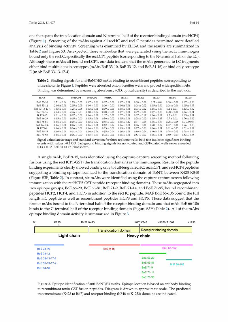
one that spans the translocation domain and N-terminal half of the receptor binding domain (recHCP4)(Figure 1). Screening of the mAbs against all recHC and recLC peptides permitted more detailedanalysis of binding activity. Screening was examined by ELISA and the results are summarized inTable 2 and Figure S3. As expected, those antibodies that were generated using the recLc immunogenbound only the recLC, specifically the recLCP1 peptide (corresponding to the N-terminal half of the LC).Although these mAbs all bound recLCP1, our data indicate that the mAbs generated to LC fragmentseither bind multiple toxin serotypes (mAbs BoE 33-10, BoE 33-12, and BoE 34-16) or bind only serotypeE (mAb BoE 33-13-17-4).
Table 2. Binding signals for anti-BoNT/E3 mAbs binding to recombinant peptides corresponding tothose shown in Figure 1. Peptides were absorbed onto microtiter wells and probed with specific mAbs.Binding was determined by measuring absorbency (OD, optical density) as described in the methods.
mAb recLC recLCP1 recLCP2 recHC HCP1 HCP2 HCP3 HCP4 HCP5
BoE 33-10 1.71 ± 0.04 1.79 ± 0.03 0.07 ± 0.00 0.07 ± 0.01 0.07 ± 0.01 0.08 ± 0.01 0.07 ± 0.0 0.08 ± 0.01 0.07 ± 0.00BoE 33-12 2.06 ± 0.01 2.05 ± 0.03 0.06 + 0.00 0.06 + 0.00 0.06 ± 0.01 0.08 ± 0.02 0.05 ± 0.00 0.08 ± 0.04 0.05 ± 0.03
BoE 33-13-17-4 1.05 ± 0.09 1.25 ± 0.08 0.13 ± 0.01 0.09 ± 0.01 0.08 ± 0.01 0.13 ± 0.02 0.16 ± 0.00 0.1 ± 0.01 0.13 ± 0.02BoE 34-16 2.44 ± 0.04 2.46 ± 0.03 0.88 ± 0.02 0.08 ± 0.01 0.07 + 0.01 0.09 ± 0.01 0.07 ± 0.00 0.08 ± 0.01 0.06 + 0.01BoE 9-15 0.11 ± 0.08 0.07 ± 0.01 0.06 ± 0.02 1.17 ± 0.02 1.37 ± 0.01 0.07 ± 0.17 0.06 ± 0.02 1.2 ± 0.01 0.05 + 0.01
BoE 66-29 0.05 ± 0.00 0.05 ± 0.00 0.05 ± 0.01 0.59 ± 0.02 0.05 ± 0.01 0.78 ± 0.02 0.05 + 0.15 0.7 ± 0.02 0.70 ± 0.02BoE 66-81 0.06 ± 0.03 0.05 ± 0.00 0.05 ± 0.02 0.55 ± 0.00 0.05 ± 0.12 0.91 + 0.04 0.06 ± 0.03 0.78 ± 0.08 0.7 ± 0.003BoE 66-106 0.06 ± 0.01 0.06 ± 0.01 0.06 + 0.01 0.62 ± 0.02 0.06 ± 0.01 0.06 ± 0.01 0.78 ± 0.02 0.07 ± 0.01 0.76 ± 0.03
BoE 71-9 0.06 ± 0.01 0.06 ± 0.01 0.06 ± 0.00 0.23 ± 0.01 0.05 ± 0.01 0.77 ± 0.06 0.06 ± 0.01 0.61 + 0.03 0.51 ± 0.02BoE 71-14 0.06 ± 0.01 0.01 ± 0.01 0.06 ± 0.01 0.55 ± 0.04 0.06 ± 0.01 0.89 ± 0.04 0.10 ± 0.01 0.78 ± 0.03 0.74 + 0.03BoE 71-95 0.06 ± 0.01 0.06 ± 0.00 0.05 + 0.00 0.32 ± 0.01 0.06 ± 0.01 0.87 ± 0.07 0.06 ± 0.01 0.50 + 0.03 0.83 ± 0.05
Signal values are average and standard deviation for three replicate wells; bold text indicates significant bindingevents with values >0.2 OD. Background binding signals for non-coated and GST-coated wells never exceeded0.13 ± 0.02. BoE 33-13-17-9 not shown.
A single mAb, BoE 9-15, was identified using the capture-capture screening method followingfusions using the recHCP1-GST (the translocation domain) as the immunogen. Results of the peptidebinding experiments clearly showed binding only to full-length recHC, recHCP1, and recHCP4 peptidessuggesting a binding epitope localized to the translocation domain of BoNT, between K423-K848(Figure S3E; Table 2). In contrast, six mAbs were identified using the capture-capture screen followingimmunization with the recHCP5-GST peptide (receptor binding domain). These mAbs segregated intotwo epitope groups, BoE 66-29, BoE 66-81, BoE 71-9, BoE 71-14, and BoE 71-95, bound recombinantpeptides HCP2, HCP4, and HCP5 in addition to the recHC peptide. MAb BoE 66-106 bound the fulllength HC peptide as well as recombinant peptides HCP3 and HCP5. These data suggest that theformer mAbs bound to the N-terminal half of the receptor binding domain and that mAb BoE 66-106binds to the C-terminal half of the receptor binding domain (Figure S3H; Table 2). All of the mAbsepitope binding domain activity is summarized in Figure 3.
Toxins 2019, 11, x FOR PEER REVIEW 6 of 15
BoE 71-95 0.06 ± 0.01
0.06 ± 0.00
0.05 + 0.00
0.32 ± 0.01
0.06 ± 0.01
0.87 ± 0.07
0.06 ± 0.01
0.50 + 0.03
0.83 ± 0.05
Signal values are average and standard deviation for three replicate wells; bold text indicates significant binding events with values >0.2 OD. Background binding signals for non-coated and GST-coated wells never exceeded 0.13 ± 0.02. BoE 33-13-17-9 not shown.
A single mAb, BoE 9-15, was identified using the capture-capture screening method following fusions using the recHCP1-GST (the translocation domain) as the immunogen. Results of the peptide binding experiments clearly showed binding only to full-length recHC, recHCP1, and recHCP4 peptides suggesting a binding epitope localized to the translocation domain of BoNT, between K423-K848 (Figure S3E; Table 2). In contrast, six mAbs were identified using the capture-capture screen following immunization with the recHCP5-GST peptide (receptor binding domain). These mAbs segregated into two epitope groups, BoE 66-29, BoE 66-81, BoE 71-9, BoE 71-14, and BoE 71-95, bound recombinant peptides HCP2, HCP4, and HCP5 in addition to the recHC peptide. MAb BoE 66-106 bound the full length HC peptide as well as recombinant peptides HCP3 and HCP5. These data suggest that the former mAbs bound to the N-terminal half of the receptor binding domain and that mAb BoE 66-106 binds to the C-terminal half of the receptor binding domain (Figure S3H; Table 2). All of the mAbs epitope binding domain activity is summarized in Figure 3.
Figure 3. Epitope identification of anti-BoNT/E3 mAbs. Epitope location is based on antibody binding to recombinant toxin-GST fusion peptides. Diagram is drawn to approximate scale. The predicted transmembrane (K423 to I847) and receptor binding (K848 to K1253) domains are indicated.
2.4. Capture ELISA
The objective of producing a large panel of mAbs is to then screen these for antibody pairs useful for generation of a sensitive capture ELISA to detect toxin. Therefore, each antibody presented in Table 1 was conjugated with biotin. Next, non-biotin labeled antibodies were individually absorbed onto 96-well microtiter plates, probed with varying amounts of neurotoxin holotoxin (BoNT/E3) and bound toxin measured using each of the biotin labeled mAbs. The three most sensitive assays coincidentally all used mAb BoE 66-29 as the capture antibody and were paired with either BoE 9-15, BoE 71-9, or BoE 71-95 as the detector antibody (Figure 4). These data suggest that the lower limit of detection (LOD) based on adding three standard deviations about the average signal of a zero toxin blank varied from 0.2 ng/mL for BoE 9-15, 0.9 ng/mL for BoE 71-9, and 20 ng/mL for BoE 71-95.
Figure 3. Epitope identification of anti-BoNT/E3 mAbs. Epitope location is based on antibody bindingto recombinant toxin-GST fusion peptides. Diagram is drawn to approximate scale. The predictedtransmembrane (K423 to I847) and receptor binding (K848 to K1253) domains are indicated.

Toxins 2019, 11, 407 6 of 14
2.4. Capture ELISA
The objective of producing a large panel of mAbs is to then screen these for antibody pairs usefulfor generation of a sensitive capture ELISA to detect toxin. Therefore, each antibody presented in Table 1was conjugated with biotin. Next, non-biotin labeled antibodies were individually absorbed onto96-well microtiter plates, probed with varying amounts of neurotoxin holotoxin (BoNT/E3) and boundtoxin measured using each of the biotin labeled mAbs. The three most sensitive assays coincidentallyall used mAb BoE 66-29 as the capture antibody and were paired with either BoE 9-15, BoE 71-9, or BoE71-95 as the detector antibody (Figure 4). These data suggest that the lower limit of detection (LOD)based on adding three standard deviations about the average signal of a zero toxin blank varied from0.2 ng/mL for BoE 9-15, 0.9 ng/mL for BoE 71-9, and 20 ng/mL for BoE 71-95.
Toxins 2019, 11, x FOR PEER REVIEW 7 of 15
Figure 4. A capture ELISA for the detection of BoNT/E3 holotoxin using mAb BoE 66-29 as the capture antibody paired with biotinylated mAbs BoE 9-15, BoE 71-9, or BoE 71-95 as the detector antibody. Points represent average (n = 3) measurements, error bars are standard deviation, and the horizontal dashed lines equal the average of the zero spike plus three standard deviation (SD). Where not visible, error bars are within the symbols.
2.5. Matrix Study
To evaluate the utility of this capture ELISA for detecting toxin in food matrices, two types of roe and one canned fish product were tested. Since the toxin would likely be found on the surface of the roe, the specimens were not macerated, but instead rinsed with standard matrix buffer (3% non-fat dry milk in tris buffered saline with Tween 20 (TBST) to generate relevant food matrices. The food matrices did reduce the detectability, which resulted in an LOD of 10 ng/mL for the roe rinsates and for the 250-fold or greater dilutions of the brine from canned mackerel (Figure 5). While performing these matrix studies, the signal from the standard matrix curves generated each day continually dropped (Figure S4). This highlights the importance of running a standard curve each day, but also the potential instability of BoNT/E3 holotoxin, which can be detected by immunoassay.
Figure 5. Analysis of matrix effects in the BoNT/E3 holotxin sandwich ELISA. The matrices (a) Salmon and Tobiko roe rinsates and (b) canned mackerel at dilutions of 1:100, 1:250, and 1:500 were spiked with BoNT/E3 holotoxin and are shown alongside the standard curve (blue circles) using 3% non-fat dried milk in tris buffered saline with Tween 20 (TBST) as the matrix. Where not visible, error bars are within the symbols.
3. Discussion
The more often performed method for generating mouse mAbs specific to a toxin, and in particular to BoNT/E, is to use the toxoid as the immunogen [15–18]. In theory, this method allows the antibodies to recognize any antigenic epitope, but in practice, the selected antibodies may not bind a diverse number of epitopes. As an alternative, here we demonstrated a strategy of utilizing peptide fragments as immunogens, which then limits each antibody to bind an antigenic epitope on
0.01 0.1 1 10 100 1000102
103
104
105
106
BoNT/E holotoxin concentration (ng/mL)
Rel
ativ
e lig
ht u
nits
9-15 LOD cut-off71-9 LOD cut-off71-95 LOD cut-off
Detector mAbBoE 9-15BoE 71-9BoE 71-95
Figure 4. A capture ELISA for the detection of BoNT/E3 holotoxin using mAb BoE 66-29 as the captureantibody paired with biotinylated mAbs BoE 9-15, BoE 71-9, or BoE 71-95 as the detector antibody.Points represent average (n = 3) measurements, error bars are standard deviation, and the horizontaldashed lines equal the average of the zero spike plus three standard deviation (SD). Where not visible,error bars are within the symbols.
2.5. Matrix Study
To evaluate the utility of this capture ELISA for detecting toxin in food matrices, two types of roeand one canned fish product were tested. Since the toxin would likely be found on the surface of theroe, the specimens were not macerated, but instead rinsed with standard matrix buffer (3% non-fat drymilk in tris buffered saline with Tween 20 (TBST) to generate relevant food matrices. The food matricesdid reduce the detectability, which resulted in an LOD of 10 ng/mL for the roe rinsates and for the250-fold or greater dilutions of the brine from canned mackerel (Figure 5). While performing thesematrix studies, the signal from the standard matrix curves generated each day continually dropped(Figure S4). This highlights the importance of running a standard curve each day, but also the potentialinstability of BoNT/E3 holotoxin, which can be detected by immunoassay.

Toxins 2019, 11, 407 7 of 14
Toxins 2019, 11, x FOR PEER REVIEW 7 of 15
Figure 4. A capture ELISA for the detection of BoNT/E3 holotoxin using mAb BoE 66-29 as the capture antibody paired with biotinylated mAbs BoE 9-15, BoE 71-9, or BoE 71-95 as the detector antibody. Points represent average (n = 3) measurements, error bars are standard deviation, and the horizontal dashed lines equal the average of the zero spike plus three standard deviation (SD). Where not visible, error bars are within the symbols.
2.5. Matrix Study
To evaluate the utility of this capture ELISA for detecting toxin in food matrices, two types of roe and one canned fish product were tested. Since the toxin would likely be found on the surface of the roe, the specimens were not macerated, but instead rinsed with standard matrix buffer (3% non-fat dry milk in tris buffered saline with Tween 20 (TBST) to generate relevant food matrices. The food matrices did reduce the detectability, which resulted in an LOD of 10 ng/mL for the roe rinsates and for the 250-fold or greater dilutions of the brine from canned mackerel (Figure 5). While performing these matrix studies, the signal from the standard matrix curves generated each day continually dropped (Figure S4). This highlights the importance of running a standard curve each day, but also the potential instability of BoNT/E3 holotoxin, which can be detected by immunoassay.
Figure 5. Analysis of matrix effects in the BoNT/E3 holotxin sandwich ELISA. The matrices (a) Salmon and Tobiko roe rinsates and (b) canned mackerel at dilutions of 1:100, 1:250, and 1:500 were spiked with BoNT/E3 holotoxin and are shown alongside the standard curve (blue circles) using 3% non-fat dried milk in tris buffered saline with Tween 20 (TBST) as the matrix. Where not visible, error bars are within the symbols.
3. Discussion
The more often performed method for generating mouse mAbs specific to a toxin, and in particular to BoNT/E, is to use the toxoid as the immunogen [15–18]. In theory, this method allows the antibodies to recognize any antigenic epitope, but in practice, the selected antibodies may not bind a diverse number of epitopes. As an alternative, here we demonstrated a strategy of utilizing peptide fragments as immunogens, which then limits each antibody to bind an antigenic epitope on
0.01 0.1 1 10 100 1000102
103
104
105
106
BoNT/E holotoxin concentration (ng/mL)
Rel
ativ
e lig
ht u
nits
9-15 LOD cut-off71-9 LOD cut-off71-95 LOD cut-off
Detector mAbBoE 9-15BoE 71-9BoE 71-95
Figure 5. Analysis of matrix effects in the BoNT/E3 holotxin sandwich ELISA. The matrices (a) Salmonand Tobiko roe rinsates and (b) canned mackerel at dilutions of 1:100, 1:250, and 1:500 were spikedwith BoNT/E3 holotoxin and are shown alongside the standard curve (blue circles) using 3% non-fatdried milk in tris buffered saline with Tween 20 (TBST) as the matrix. Where not visible, error bars arewithin the symbols.
3. Discussion
The more often performed method for generating mouse mAbs specific to a toxin, and in particularto BoNT/E, is to use the toxoid as the immunogen [15–18]. In theory, this method allows the antibodiesto recognize any antigenic epitope, but in practice, the selected antibodies may not bind a diversenumber of epitopes. As an alternative, here we demonstrated a strategy of utilizing peptide fragmentsas immunogens, which then limits each antibody to bind an antigenic epitope on that particularpeptide. While both methods have been used successfully, they each have their limitations, such thatepitopes that are now presented on the immunogen may not be present in the whole native toxin.
In light of immunogen presentation, other means to influence antigen selectivity is by the screeningprocedure used. Screening measures that are as similar as possible to the final application shouldbe employed to identify the antibodies desired depending on the downstream application of thesereagents. For this reason, in this work we utilized both direct binding assays (used to screen foranti-LC mAbs) and capture-capture binding assays (used to screen for anti-HC mAbs). Previous workyielding a mAb-mAb sandwich ELISA for BoNT/E utilized a direct binding screening method forselecting potential hybridomas [15]. In our work, it is interesting to note that the cross-reactivity ofthe mAbs generated to LC fragments is considerably more variable (Table 1). This may be due tothe screening procedure or just an inherent result of the peptides used, but cannot be differentiatedby these experiments. Nonetheless, we showed the production of new mAbs that can provide newinsights when used individually or together depending on the desired applications.
The most common applications of these mAbs are to neutralize toxin, identify structure–functionrelationships of the toxins, and to develop toxin detection assays. Our goal was to primarily developa sandwich ELISA for detecting BoNT/E in suspected samples. Since mAbs are immunochemicallyhomogeneous and more reproducible than pAbs, we sought to develop this sandwich ELISA usinga pair of mAbs. In this work, we developed a mAb-mAb sandwich ELISAs for BoNT/E3 (Figure 4).Other applications of mAb-mAb sandwich-type assays have yielded improved LODs using otherformats, such as microarrays [31]. In assays utilizing pAb sources [14,32,33], improved LODs wereobtained and therefore it might be of interest for future research to combine multiple mAbs from thisstudy to improve performance. Furthermore, using these mAbs in other formats and with differentreporting molecules may also yield improved LODs.
While testing these mAbs for neutralization and other functionalities was outside the scope ofthis work, all of the mAbs described in this paper may be suitable for other applications. For example,most mAbs generated to date recognize the HC of BoNT/E [15]. Here we describe five new mAbs thatbind the LC—as determined by Western blot or binding of the recLC fragment by ELISA (Figure 2 and

Toxins 2019, 11, 407 8 of 14
Table 2). Further characterization of all of these mAbs could include determining their ability to detectBoNT/E complex, to detect BoNT/E subtypes E1–12, and if they are capable of neutralizing toxin.
In instances when immunoassays are sought to be used with food matrices, matrix effects mayrepresent a major hurdle to the feasibility of the assay. Due to the documented prevalence of BoNT/Ein fish products, we sought to evaluate our sandwich ELISA with roe and canned fish products. Weobserved a 50-fold decrease in LOD from 0.2 ng/mL in buffer to 10 ng/mL in fish product matrices.This study aligns with previous studies by demonstrating that there is commonly a decrease in theamount of toxin detected when toxin is extracted from a food matrix [15,31,33]. For instance, in a studyexamining 30 food matrices (none of which were fish products) they report a 12-fold decrease in LODfrom 163 pg/mL in buffer to 2 ng/mL in food matrices [33]. An evaluation of ELISA kits demonstrateda LOD in smoked Salmon matrix of 60 pg/mL [34]. While decreased sensitivity impacts the utility ofimmunoassays as a quantitative method for measuring toxin, as a screening tool, if any toxin is presentit would generate a positive signal and suggest further analysis. Although the matrix might impededetection, our study substantiates that matrix interferences in ELISA are manageable with a simpledilution step [24] (Figure 5b). It was speculated that a lethal oral amount of botulinum toxin for a70-kg human would be 70 µg [35]. While the sensitivity of this assay is not likely suitable for humanbiospecimen diagnostic purposes, it is approaching relevant levels for food analysis. To improvesensitivity, the mAbs could be used in other purification/concentration steps (such as beads coatedwith anti-BoNT/E mAbs) prior to food testing.
In summary, this work demonstrates the production of new mAbs to BoNT/E3 and when pairedtogether generated a sensitive sandwich ELISA for the detection of BoNT/E3. This strategy utilizedpeptide fragments as immunogens to induce antibodies selective for various fragments of BoNT/E3.Additional peptides not used as immunogens were used to further narrow down the binding regionsof the induced antibodies. Furthermore, a capture-capture screening procedure was used to isolateantibodies that functionally bound toxin in solution, as opposed to binding to an immobilized target,which may distort particular epitopes. Ultimately, this work yielded a mAb-mAb sandwich ELISAthat can detect BoNT/E3 holotoxin at 0.2 ng/mL in buffer and 10 ng/mL in spiked fish products.
4. Materials and Methods
4.1. Reagents
Solutions at 1 mg mL−1 of BoNT serotypes A–G, the 150 kDa serotype E holotoxin (BoNT/E)produced by Clostridium botulinum Strain E3/Alaska, and anti-BoNT/E rabbit pAbs were purchasedfrom Metabiologics Inc. (Madison, WI, USA). Nicked BoNT/E3 was obtained from List BiologicalLaboratories (Campbell, CA, USA). Total genomic DNA from C. botulinum (Strain E3/Alaska) wasgenerously provided by Eric Johnson (University of Wisconsin, Madison, WI, USA). All toxins werehandled in a BSL-2 biosafety cabinet and all materials and waste solutions were treated before disposal.
Bovine serum albumin (BSA), ovalbumin (OVA), goat anti-mouse immunoglobulin G conjugatedto horseradish peroxidase (IgG-HRP), polyoxyethylene sorbitan monolaurate (Tween-20), SigmaAdjuvant System, Protein-G conjugated Sepharose, and the following buffers: 0.01 M phosphatebuffered saline (PBS), 0.138 M NaCl, 0.0027 M KCl, pH 7.4, and 0.02 M TRIS-buffered saline (TBS),0.9% NaCl, pH 7.4 were purchased from Sigma Chemical Co. (St. Louis, MO, USA). Goat anti-mouseIgG Fc gamma was purchased from Millipore (Billerica MA). Black and clear Maxisorp 96-well Nuncmicrotiter plates were obtained from PGC Scientific (Gaithersburg, MD, USA). BCA kit, EZ-LinkSulfo-NHS-LC-Biotin kit, SuperSignal Femto Max Sensitivity substrate, and SuperSignal West DuraExtended Duration substrate were purchased from Pierce Inc. (Rockford, IL, USA). SBA ClonotypingSystem-HRP was purchased from Southern Biotech (Birmingham, AL, USA). Enhanced K-Blue TMBsubstrate and Red Stop Solution were obtained from Neogen Corporation (Lexington, KY, USA).Non-fat dry milk (NFDM), fresh Tobiko roe, fresh Salmon roe, and canned in brine mackerel werepurchased from a local food store.

Toxins 2019, 11, 407 9 of 14
Commercial enzymes (Phusion High-Fidelity DNA Polymerase, BamHI, XhoI, T4 polynucleotidekinase (3’ phosphatase minus) and T4 DNA ligase were purchased from New England BioLabs, Inc.(Bethesda, MD, USA). Plasmid construction and manipulation were performed using pCR4-TOPOvector and Escherichia coli TOP10 cells from Invitrogen (Carlsbad, CA, USA) Plasmid pGS-21 andQuickClean 5M kits (Miniprep, PCR purification, and Gel extraction) were from GenScript Corp.(Piscataway, NJ, USA). All automated DNA sequencing was performed on a 3730 DNA Analyzerusing the BigDye Terminator Version 3.1 cycle sequencing kit and XTerminator reagents purchasedfrom Applied Biosystems, (Foster City, CA, USA). Primers designed specifically for this project werepurchased from Integrated DNA Technologies (Coralville, IA, USA) and are shown in Table 3.
Table 3. Primers designed for this study. Sites for restriction enzymes BamHI (GGATCC) and XhoI(CTCGAG) are shown underlined. Stop codons are shown in bold, in the 3’ to 5’ (TTA) orientation.
Primer Name Sequence
AlaskaLCF TAGC GGA TCC ATG CCA AAA ATT AAT AGT TTT AAT TAT AATAlaskaLCF2 TAGC GGA TCC GGG ATT ACT ACA ACT TGT ATT ATAAlaskaLCR TAGC CTC GAG TTA CCT TAT GCC TTT TAC AGA AAC AA
AlaskaLCR2 TAGC CTC GAG TTA TTT AGC CCC ATA TAG TCC ATG TAlaskaHCF TAGC GGA TCC AAA TCA ATA TGT ATC GAA ATA AAT AATAlaskaHCF2 TAGC GGA TCC AAA AGT AGT TCA GTT TTA AAT ATG AGAlaskaHCF3 TAGC GGA TCC AAT ATT TTG AAG GAT TTT TGG GGAAlaskaHCR TAGC CTC GAG TTA TTT TTC TTG CCA TCC ATG TTAlaskaHCR1 TAGC CTC GAG TTA AAT TCT CTT AAA GAA TTT ATT AAA ATA TAlaskaHCR2 TAGC CTC GAG TTA TGT ATT AGG TTC ATT GCT ATA TAA A
4.2. Recombinant BoNT/E3-GST Fusion Proteins
The expression and purification of all GST-fusion proteins was performed as previouslydescribed [30] with the following modifications. Total genomic DNA from C. botulinum StrainE3/Alaska was used as a template to amplify the fragments of the light and heavy chains (LC, LCP1,LCP2, HC, HCP1, HCP2, HCP3, HCP4, HCP5) using the primers indicated (see Figure 1 and Table 1).Stop codons (TAA) were introduced when not present within the genomic DNA of the cloned region.The resulting BoNT/E3 DNA fragments were cloned into plasmid pCR4-TOPO and sequenced usingM13F and M13R primers. The pCR4-derived plasmids were digested using restriction enzymesBamHI and XhoI, purified, and then ligated into BamHI- and XhoI-digested pGS-21a to yield thecorrespondingly named pGS plasmid (e.g., pGS-HCP1 for fragment HCP1). All pGS-21a-derivedplasmids were sequenced using pGS-F and pGS-R primers, to confirm the correct integration of theBoNT/E3 fragment into the vector. The recombinant DNA methods used in this study were approvedby the Institutional Biosafety Committee.
4.3. Monoclonal Antibody Procedure
Monoclonal antibody production was as previously described [29,36]. Briefly, solutions of threepeptide fragments, separately, at the concentrations indicated (LC, 94 µg/mL; HCP1, 79 µg/mL; HCP5,176 µg/mL) were mixed with Sigma Adjuvant System according to the manufacturer’s instructions.Three groups of five female BALB/cJ mice (Simonsen Laboratories, Gilroy, CA, USA) were immunizedthree times at two-week intervals by intraperitoneal injection (i.p.) with 100 µL of each antigen-adjuvantsolution. Two weeks after the third injection, serum was obtained from each mouse and evaluated foranti-BoNT/E3 antibodies via direct binding ELISA screens. Mice were injected i.p. with 2 µg of theappropriate peptide fragment in PBS three days prior to being euthanized and cell fusion. Supernatantsfrom cell fusion plates were subjected to screening either by direct binding ELISA (for mice immunizedwith the recBoNT/E3-LC-GST peptide fragments) or by a capture-capture ELISA screen (for the miceimmunized with the recBoNT/E3-HCP1-GST and recBoNT/E3-HCP5-GST peptide fragments).

Toxins 2019, 11, 407 10 of 14
The Institutional Animal Care and Use Committee of the United States Department of Agriculture,Western Regional Research Center approved the experimental procedures used in these studies (protocol#’s: 04-1-H-05; 09-3). All animal experiments and husbandry involved in the studies presented in thismanuscript were conducted under the guidelines of the U.S. Government Principles for the Utilizationand Care of Vertebrate Animals Used in Testing, Research and Training.
4.4. Screening Methods
4.4.1. Directing Binding ELISA
Direct binding ELISA screens of the recBoNT/E3-LC-GST immunized mice were performed aspreviously described [36], using black microtiter plates coated with 50 µL per well of a 0.1 µg/mLsolution of BoNT/E3 in 0.05 M sodium carbonate buffer, pH 9.6, overnight at 4◦C. The solution wasaspirated and then blocked by adding 300 µL per well of 3% NFDM in TBS containing 0.05% Tween-20(NFDM-TBST) and incubated for 1 h at 37 ◦C. The plates were washed once with TBST, and then thecell culture supernatants were added. After another incubation at 37 ◦C for 1 h, the plates were washed3×with TBST. Then 50 µL of a 1 µg/mL solution of goat anti-mouse HRP-conjugated pAb was addedand the plates incubated at 37 ◦C for 1 h. After washing 3×with TBST, the plates were visualized usingSuperSignal West Dura Extended Duration Substrate according to the manufacturer’s instructions.The plates were incubated for 3 min at room temperature and luminescent counts recorded using aVictor3 Multilabel Counter (PerkinElmer Inc., Waltham, MA, USA).
4.4.2. Capture-Capture ELISA
Fusions of the recBoNT/E3-HCP1-GST and recBoNT/E3-HCP5-GST immunized mice were screenedusing a capture-capture ELISA screen as previously described with minor modifications [37,38]. Briefly,50 µL per well of all solutions were used. Black microtiter plates were coated with a 1 µg/mL solutionof goat anti-mouse IgG Fc gamma in 0.05 M sodium carbonate buffer, pH 9.6 overnight at 4 ◦C. The IgGsolution was aspirated and non-coated sites blocked by adding 300 µL per well of 3% NFDM-TBST andthe plates were incubated for 1 h at 37 ◦C. The plates were washed once with TBST, then cell culturesupernatants were added and the plates were incubated at 37 ◦C for 1 h. The plates were washed threetimes with TBST, then a solution of BoNT/E3 in NFDM-TBST (50 ng/mL) was added and the plateswere incubated at 37 ◦C for 1 h. Plates were washed three times as before, then a 1 µg/mL solution ofanti-BoNT/E rabbit pAbs in NFDM-TBST was added and the plates were incubated at 37 ◦C for 1 h.Plates were washed three times as before, then a 1 µg/mL solution of goat anti-rabbit HRP-conjugatedpAbs was added and the plates were incubated at 37 ◦C for 1 h. Plates were again washed three times,and binding was visualized as described above.
Cells from wells giving positive signals for antibody production by either screening method werecloned by limiting dilution. Cells were then expanded and small amounts (usually less than 10 mL)of ascites fluids obtained (Covance Research Products, Inc., Denver, PA, USA) or 1–200 mL of spenthybridoma media was produced. MAbs were purified by affinity chromatography on Protein-G (forIgG) Sepharose. Bound mAbs were eluted with 0.1 M glycine-HCl, pH 2.7 and dialyzed overnightagainst PBS. Protein concentrations were determined using the microplate BCA method suggested bythe manufacturer. Then, the mAbs were conjugated to biotin using EZ-Link Sulfo-NHS-LC-Biotin asdescribed by the manufacturer using a 50-fold molar excess of the biotin reagent. MAbs were stored at4 ◦C until needed.
4.5. Antibody Isotyping, Western Blotting, Peptide Binding
The isotype of each mAb was determined using the SBA Clonotyping System-HRP in ELISAformat, according to manufacturer’s instructions. Western blotting was performed as previouslydescribed [36] using nicked BoNT/E3 using both reduced and non-reduced conditions. Binding ofeach antibody to the recBoNT/E3-GST peptides was examined by direct binding ELISA. The wells of

Toxins 2019, 11, 407 11 of 14
clear microtiter plates were coated with each GST-fusion protein (5 µg/mL in 0.05 M sodium carbonatebuffer, pH 9.6), incubated overnight at 4 ◦C, blocked with 3% NFDM-TBST. Purified mAb was addedto each well and incubated for 1 h at 37 ◦C. The plates were washed 3× with TBST, then 50 µL of a1 µg/mL solution of goat anti-mouse HRP-conjugated pAb was added and the plates incubated at37 ◦C for 1 h. Plates were again washed three times, then K-Blue TMB substrate was added (100 µLper well) and incubated with agitation for 5 min at room temperature. Stop solution was added(100 µL per well), and absorbance at 450 nm was measured using a VersaMax microplate reader(Molecular Devices, Sunnyvale, CA, USA). Each antibody was tested in triplicate. In some experimentsSuperSignal West Dura Extended Duration Substrate was substituted for K-Blue and plates read on theVictor3 as described above.
4.6. Binding of Antibodies to BoNT Serotypes A Through G
Black microtiter plates were coated with the different serotypes of BoNT, A through G for directbinding ELISA analysis as described above. Purified anti-BoNT/E3 mAb at a concentration of 10 µg/mLin NFDM-TBST was added to each BoNT serotype, and incubated for 1 h at 37 ◦C. The plates werewashed, then a 1 µg/mL solution of goat anti-mouse HRP-conjugated pAb was added and the plateswere incubated at 37 ◦C for 1 h. Plates were again washed three times, and binding was visualizedusing SuperSignal West Dura Extended Duration Substrate as described above. Each antibody wasassayed against each serotype in triplicate.
4.7. Sandwich Assay Procedure
When performing the sandwich assay, for every mAb, the unlabeled mAb was used as the captureantibody and the biotinylated form was used as the detector antibody. Each mAb was tested incombination with all of the mAbs to identify pairs that can detect the BoNT/E3 toxin in the solutionphase. Black plates were coated with 100 µL of mAb at 2 µg/mL in 0.05 M sodium carbonate buffer, pH9.6, incubated overnight at 4 ◦C, and then blocked with 3% NFDM-TBST. The blocking solution wasremoved and BoNT/E3 toxin in 3% NFDM-TBST was added at an initial concentration of 500 ng/mL andserially diluted two-fold, including wells with no toxin. Next, biotinylated-mAb was added in triplicateat 1 µg/mL, 100 µL/well in 3% NFDM-TBST. Plates were incubated for 1 h at 37 ◦C, washed 6×withTBST, and visualized using SuperSignal West Dura Extended Duration Substrate as described above.
When the most sensitive combination of capture mAb and detector mAb-biotin were selected, thefinal sandwich assays were performed in triplicate using BoNT/E3 holotoxin at a starting concentrationof 1000 ng/mL, serially diluted three-fold, including wells with no toxin, and visualized usingSuperSignal Femto Max Sensitivity substrate. Limit of detection cut-off values were determined usingthree times the standard deviation of the wells with no toxin.
4.8. Preparation of Spiked Food Matrices
Since fish and fishery products are frequently cited as sources for serotype E related foodbornebotulism cases, we tested one canned fish product (mackerel in brine) and two types of fish roe (Tobikoand Salmon) to determine to what extent the matrices would interfere with toxin detection in ournewly developed sandwich ELISA. For the canned mackerel, the liquid fraction was poured into aconical tube and centrifuged for 20 minutes at 4000× g. The oil and fat layers were discarded, while theaqueous fraction was collected and used as the sample matrix. For each roe specimen, separately, 4 g ofroe was weighed and rinsed with 40 mL of 3% NFDM-TBST. This rinsate was collected and used as thesample matrix. As described above, the sandwich assay procedure with the most sensitive combinationof mAbs was used (BoE 66-29 as capture and BoE 9-15 as detector), but this time the BoNT/E3 holotoxinwas diluted in sample matrix to generate standard curves. The assays were performed in triplicateusing BoNT/E3 at a starting concentration of 1000 ng/mL, serially diluted three-fold into a samplematrix, including wells with no toxin. The sample matrices were evaluated as is (i.e., neat) for the roerinsates, and at dilutions of 1:100, 1:250, and 1:500 with 3% NFDM-TBST for the aqueous brine matrix.

Toxins 2019, 11, 407 12 of 14
Experiments were visualized using SuperSignal Femto Max Sensitivity substrate and data analyzedusing GraphPad Prism 7 Software (La Jolla, CA, USA).
Supplementary Materials: The following are available online at http://www.mdpi.com/2072-6651/11/7/407/s1,Figure S1: Binding of LC-Specific anti-BoNT/E3 mAbs (10 µg/mL) to toxin serotypes A–G, used as coating antigensin a direct binding ELISA, Figure S2: Binding of HC-specific anti-BoNT/E3 mAbs (10 µg/mL) to toxin serotypesA–G, used as coating antigens in a direct binding ELISA, Figure S3: Direct binding ELISA of anti-BoNT/E3 mAbsto recombinant peptide fragments. Different antibodies are shown in A–K, Figure S4: Chemiluminescent signalintensity of the ELISA over time at the maximal concentration (1000 ng/mL of BoNT/E3 holotoxin) tested on thestandard curve.
Author Contributions: L.H.S., L.W.C., M.S., R.M.H., and C.S.B. conceived and designed the experiments; L.H.S.,M.S., and C.S.B. performed the experiments; L.H.S., M.S., C.S.B., R.M.H., and L.W.C. analyzed the experiments,L.H.S. and C.S.B. wrote the paper. All authors have read and approved the final manuscript.
Funding: This research was funded by the United States Department of Agriculture, Agricultural Research Service,National Program project NP108, CRIS 2030-42000-049-00D. LHS and CSB were also funded by interagencyagreement IAA #60-2030-5-004 with the Department of Homeland Security.
Acknowledgments: We gratefully thank Thomas Henderson for his assistance with recombinant peptideexpression and purification and Paul Merrill for antibody purification, biotin conjugation, and ELISA.
Conflicts of Interest: The authors declare no conflicts of interest. The funders had no role in the design of thestudy; in the collection, analyses, or interpretation of data; in the writing of the manuscript, or in the decision topublish the results.
References
1. Peck, M.W.; Smith, T.J.; Anniballi, F.; Austin, J.W.; Bano, L.; Bradshaw, M.; Cuervo, P.; Cheng, L.W.; Derman, Y.;Dorner, B.G.; et al. Historical perspectives and guidelines for botulinum neurotoxin subtype nomenclature.Toxins 2017, 9, 38. [CrossRef] [PubMed]
2. Zhang, S.; Masuyer, G.; Zhang, J.; Shen, Y.; Lundin, D.; Henriksson, L.; Miyashita, S.I.; Martinez-Carranza, M.;Dong, M.; Stenmark, P. Identification and characterization of a novel botulinum neurotoxin. Nat. Commun.2017, 8, 14130. [CrossRef] [PubMed]
3. CDC. Botulism Annual Summary, 2016; US Department of Health and Human Services: Atlanta, Georgia,2017. Available online: https://www.cdc.gov/botulism/pdf/Botulism-2016-SUMMARY-508.pdf (accessed on1 April 2019).
4. Carter, A.T.; Peck, M.W. Genomes, neurotoxins and biology of Clostridium botulinum group I and group II.Res. Microbiol. 2015, 166, 303–317. [CrossRef] [PubMed]
5. Hyytia, E.; Hielm, S.; Korkeala, H. Prevalence of Clostridium botulinum type E in finnish fish and fisheryproducts. Epidemiol. Infect. 1998, 120, 245–250. [CrossRef]
6. Leclair, D.; Farber, J.M.; Pagotto, F.; Suppa, S.; Doidge, B.; Austin, J.W. Tracking sources of c Clostridiumbotulinum type E contamination in seal meat. Int. J. Circumpol. Heal. 2017, 76, 1380994. [CrossRef]
7. Chipault, J.G.; White, C.L.; Blehert, D.S.; Jennings, S.K.; Strom, S.M. Avian botulism type E in waterbirds ofLake Michigan, 2010–2013. J. Great Lakes Res. 2015, 41, 659–664. [CrossRef]
8. Botulism in Alaska—November 2017; Alaska Division of Public Health, 2017; p. 34. Available online:http://dhss.alaska.gov/dph/Epi/id/siteassets/Pages/botulism/monograph.pdf (accessed on 1 April 2019).
9. Miller, L.G. Observations on the distribution and ecology of Clostridium botulinum type E in Alaska. Can. J.Microbiol. 1975, 21, 920–926. [CrossRef] [PubMed]
10. McLaughlin, J.B.; Sobel, J.; Lynn, T.; Funk, E.; Middaugh, J.P. Botulism type E outbreak associated with eatinga beached whale, Alaska. Emerg. Infect. Dis. 2004, 10, 1685–1687. [CrossRef]
11. Sobel, J.; Tucker, N.; Sulka, A.; McLaughlin, J.; Maslanka, S. Foodborne botulism in the United States,1990–2000. Emerg. Infect. Dis. 2004, 10, 1606–1611. [CrossRef]
12. Notermans, S.; Dufrenne, J.; Kozaki, S. Enzyme-linked immunosorbent assay for detection of Clostridiumbotulinum type E toxin. Appl. Environ. Microbiol. 1979, 37, 1173–1175. [CrossRef]
13. Cadieux, B.; Blanchfield, B.; Smith, J.P.; Austin, J.W. A rapid chemiluminescent slot blot immunoassay for thedetection and quantification of Clostridium botulinum neurotoxin type E. Int. J. Food Microbiol. 2005, 101, 9–16.[CrossRef] [PubMed]

Toxins 2019, 11, 407 13 of 14
14. Poli, M.A.; Rivera, V.R.; Neal, D. Development of sensitive colorimetric capture ELISAs for Clostridiumbotulinum neurotoxin serotypes E and F. Toxicon 2002, 40, 797–802. [CrossRef]
15. Abbasova, S.G.; Rudenko, N.V.; Gorokhovatskii, A.Y.; Kapralova, M.V.; Vinogradova, I.D.; Vertiev, Y.V.;Nesmeyanov, V.A.; Grishin, E.V. Monoclonal antibodies to botulinum neurotoxins of types A, B, E, and F.Russ. J. Bioorg. Chem. 2011, 37, 307–315. [CrossRef]
16. Kamata, Y.; Kozaki, S.; Nagai, T.; Sakaguchi, G. Production of monoclonal-antibodies against Clostridiumbotulinum type E derivative toxin. FEMS Microbiol. Lett. 1985, 26, 305–309. [CrossRef]
17. Tsuzuki, K.; Yokosawa, N.; Syuto, B.; Ohishi, I.; Fujii, N.; Kimura, K.; Oguma, K. Establishment of amonoclonal antibody recognizing an antigenic site common to c Clostridium botulinum type B, C1, D, and Etoxins and tetanus toxin. Infect. Immun. 1988, 56, 898–902.
18. Wong, P. Detection of Clostridium botulinum type E toxin by monoclonal antibody enzyme immunoassay.J Rapid Methods Autom. Microbiol. 1996, 4, 191–206. [CrossRef]
19. Garcia-Rodriguez, C.; Razai, A.; Geren, I.N.; Lou, J.; Conrad, F.; Wen, W.-H.; Farr-Jones, S.; Smith, T.J.;Brown, J.L.; Skerry, J.C.; et al. A three monoclonal antibody combination potently neutralizes multiplebotulinum neurotoxin serotype E subtypes. Toxins 2018, 10, 105. [CrossRef]
20. Bakherad, H.; Mousavi Gargari, S.L.M.; Rasooli, I.; Rajabibazl, M.; Mohammadi, M.; Ebrahimizadeh, W.;Safaee Ardakani, L.S.; Zare, H. In vivo neutralization of botulinum neurotoxins serotype E with heavy-chaincamelid antibodies (VHH). Mol. Biotechnol. 2013, 55, 159–167. [CrossRef]
21. Conway, J.O.; Sherwood, L.J.; Collazo, M.T.; Garza, J.A.; Hayhurst, A. Llama single domain antibodiesspecific for the 7 botulinum neurotoxin serotypes as heptaplex immunoreagents. PLoS ONE 2010, 5, e8818.[CrossRef]
22. Cheng, L.W.; Land, K.M.; Stanker, L.H. Current methods for detecting the presence of botulinum neurotoxinsin food and other biological samples. In Bioterrorism; Morse, S., Ed.; IntechOpen: Rijeka, Croatia, 2012;Chapter 1; pp. 1–16.
23. Lindstrom, M.; Korkeala, H. Laboratory diagnostics of botulism. Clin. Microbiol. Rev. 2006, 19, 298–314.[CrossRef]
24. Capek, P.; Dickerson, T.J. Sensing the deadliest toxin: Technologies for botulinum neurotoxin detection.Toxins 2010, 2, 24–53. [CrossRef] [PubMed]
25. Shriver-Lake, L.C.; Zabetakis, D.; Goldman, E.R.; Anderson, G.P. Evaluation of anti-botulinum neurotoxinsingle domain antibodies with additional optimization for improved production and stability. Toxicon 2017,135, 51–58. [CrossRef] [PubMed]
26. Kozaki, S.; Kamata, Y.; Nagai, T.; Ogasawara, J.; Sakaguchi, G. The use of monoclonal-antibodies to analyzethe structure of Clostridium botulinum type E derivative toxin. Infect. Immun. 1986, 52, 786–791. [PubMed]
27. Fischer, A.; Garcia-Rodriguez, C.; Geren, I.; Lou, J.L.; Marks, J.D.; Nakagawa, T.; Montal, M. Moleculararchitecture of botulinum neurotoxin E revealed by single particle electron microscopy. J. Biol. Chem. 2008,283, 3997–4003. [CrossRef] [PubMed]
28. Kubota, T.; Watanabe, T.; Yokosawa, N.; Tsuzuki, K.; Indoh, T.; Moriishi, K.; Sanda, K.; Maki, Y.; Inoue, K.;Fujii, N. Epitope regions in the heavy chain of Clostridium botulinum type E neurotoxin recognized bymonoclonal antibodies. Appl. Environ. Microbiol. 1997, 63, 1214–1218. [PubMed]
29. Scotcher, M.C.; Cheng, L.W.; Stanker, L.H. Detection of botulinum neurotoxin serotype B at sub mouse LD50levels by a sandwich immunoassay and its application to toxin detection in milk. PLoS ONE 2010, 5, e11047.[CrossRef] [PubMed]
30. Scotcher, M.C.; Johnson, E.A.; Stanker, L.H. Characterization of the epitope region of f1-2 and f1-5, twomonoclonal antibodies to botulinum neurotoxin type A. Hybridoma 2009, 28, 315–325. [CrossRef] [PubMed]
31. Jenko, K.L.; Zhang, Y.; Kostenko, Y.; Fan, Y.; Garcia-Rodriguez, C.; Lou, J.; Marks, J.D.; Varnum, S.M.Development of an ELISA microarray assay for the sensitive and simultaneous detection of ten biodefensetoxins. Analyst 2014, 139, 5093–5102. [CrossRef]
32. Rivera, V.R.; Gamez, F.J.; Keener, W.K.; White, J.A.; Poli, M.A. Rapid detection of Clostridium botulinumtoxins A, B, E, and F in clinical samples, selected food matrices, and buffer using paramagnetic bead-basedelectrochemiluminescence detection. Anal. Biochem. 2006, 353, 248–256. [CrossRef]
33. Sharma, S.K.; Ferreira, J.L.; Eblen, B.S.; Whiting, R.C. Detection of type A, B, E, and F Clostridium botulinumneurotoxins in foods by using an amplified enzyme-linked immunosorbent assay with digoxigenin-labeledantibodies. Appl. Environ. Microb. 2006, 72, 1231–1238. [CrossRef]
Toxins 2019, 11, 407 14 of 14
34. Singh, A.; Datta, S.; Sachdeva, A.; Maslanka, S.; Dykes, J.; Skinner, G.; Burr, D.; Whiting, R.C.; Sharma, S.K.Evaluation of an enzyme-linked immunosorbent assay (ELISA) kit for the detection of botulinum neurotoxinsA, B, E, and F in selected food matrices. Health Secur. 2015, 13, 37–44. [CrossRef] [PubMed]
35. Arnon, S.S.; Schechter, R.; Inglesby, T.V.; Henderson, D.A.; Bartlett, J.G.; Ascher, M.S.; Eitzen, E.; Fine, A.D.;Hauer, J.; Layton, M.; et al. Botulinum toxin as a biological weapon - medical and public health management.JAMA 2001, 285, 1059–1070. [CrossRef] [PubMed]
36. Stanker, L.H.; Merrill, P.; Scotcher, M.C.; Cheng, L.W. Development and partial characterization of high-affinitymonoclonal antibodies for botulinum toxin type A and their use in analysis of milk by sandwich ELISA.J. Immunol. Methods 2008, 336, 1–8. [CrossRef] [PubMed]
37. Stanker, L.H.; Hnasko, R.M. A double-sandwich ELISA for identification of monoclonal antibodies suitablefor sandwich immunoassays. Methods Mol. Biol. 2015, 1318, 69–78. [PubMed]
38. Stanker, L.H.; Serban, A.V.; Cleveland, E.; Hnasko, R.; Lemus, A.; Safar, J.; DeArmond, S.J.; Prusiner, S.B.Conformation-dependent high-affinity monoclonal antibodies to prion proteins. J. Immunol. 2010, 185,729–737. [CrossRef] [PubMed]
© 2019 by the authors. Licensee MDPI, Basel, Switzerland. This article is an open accessarticle distributed under the terms and conditions of the Creative Commons Attribution(CC BY) license (http://creativecommons.org/licenses/by/4.0/).