Development of a multi-epitope peptide vaccine against ...
338
HAL Id: tel-02387247 https://tel.archives-ouvertes.fr/tel-02387247 Submitted on 29 Nov 2019 HAL is a multi-disciplinary open access archive for the deposit and dissemination of sci- entific research documents, whether they are pub- lished or not. The documents may come from teaching and research institutions in France or abroad, or from public or private research centers. L’archive ouverte pluridisciplinaire HAL, est destinée au dépôt et à la diffusion de documents scientifiques de niveau recherche, publiés ou non, émanant des établissements d’enseignement et de recherche français ou étrangers, des laboratoires publics ou privés. Development of a multi-epitope peptide vaccine against human leishmaniases Joana da Silva Pissarra To cite this version: Joana da Silva Pissarra. Development of a multi-epitope peptide vaccine against human leishmaniases. Human health and pathology. Université Montpellier, 2019. English. NNT: 2019MONTT013. tel- 02387247
-
Upload
khangminh22 -
Category
Documents
-
view
1 -
download
0
Transcript of Development of a multi-epitope peptide vaccine against ...

HAL Id: tel-02387247https://tel.archives-ouvertes.fr/tel-02387247
Submitted on 29 Nov 2019
HAL is a multi-disciplinary open accessarchive for the deposit and dissemination of sci-entific research documents, whether they are pub-lished or not. The documents may come fromteaching and research institutions in France orabroad, or from public or private research centers.
L’archive ouverte pluridisciplinaire HAL, estdestinée au dépôt et à la diffusion de documentsscientifiques de niveau recherche, publiés ou non,émanant des établissements d’enseignement et derecherche français ou étrangers, des laboratoirespublics ou privés.
Development of a multi-epitope peptide vaccine againsthuman leishmaniases
Joana da Silva Pissarra
To cite this version:Joana da Silva Pissarra. Development of a multi-epitope peptide vaccine against human leishmaniases.Human health and pathology. Université Montpellier, 2019. English. �NNT : 2019MONTT013�. �tel-02387247�

THÈSE POUR OBTENIR LE GRADE DE DOCTEUR
DE L’UNIVERSITÉ DE MONTPELLIER
En Biologie Santé
École Doctorale Sciences Chimiques et Biologiques pour la Santé
Unité de recherche UMR177 INTERTRYP – Institut de Recherche pour le Développement (IRD)
Présentée par Joana PISSARRA Le 26 Juin 2019
Sous la direction du Dr. Jean-Loup LEMESRE
Devant le jury composé de
Bernard MAILLÈRE, Dr., CEA-Saclay
Claude LECLERC, Pr., Institut Pasteur Paris
Sylviane PIED, Dr., Institut Pasteur Lille
Nicolas BLANCHARD, Dr., Université de Toulouse
Rachel BRAS-GONÇALVES, Dr., IRD Montpellier
Philippe HOLZMULLER, Dr., CIRAD Montpellier
Amel GARNAOUI, Dr., Institut Pasteur Tunis
Stéphane DELBECQ, Pr., Faculté de Pharmacie Montpellier
Jean-Loup LEMESRE, Dr., IRD Montpellier
Président / Examinateur
Rapporteur
Rapporteur
Examinateur
Co-encadrant
Co-encadrant
Co-encadrant
Invité
Directeur de thèse
Development of a mult i -epitope peptide vacc ine against human le ishmaniases

i
ACKNOWLEDGEMENTS
Undertaking this PhD has been a eventful and exciting experience and it would not have been
possible without the support and guidance that I received.
This project has received funding from the European Union's Horizon 2020 research and
innovation programme under the Marie Sklodowska-Curie International Training Network grant
agreement No 642609. I gratefully acknowledge the additional funding received towards my PhD
from Fondation des Treilles and SATT AxLR.
I would like to express my sincere gratitude to my supervisor Dr. Jean-Loup Lemesre for his
guidance and help throughout my PhD. I am grateful for his continuous support, encouragement and
optimism which have made my PhD experience productive and stimulating.
I would like to thank the thesis committee and all the PhD defense jury members. A special
thanks to Dr. Rachel Bras-Gonçalves and Dr Phillippe Holzmuller for their insightful comments and
expertise, but also for the fruitful discussions and their contribution to this work. I would like to thank
Dr. Amel Garnaoui for the scientific collaboration, her support and assistance of my thesis work
during my stays in Tunis.
My sincere thanks also goes to Dr. Bernard Maillere who provided me an opportunity to visit
the laboratory and research facilities. His precious support was invaluable for this research project
and my PhD thesis.
I would like to thank Dr. Etienne Loire and Dr. Vincent Bonhomme for their significant aid and
support, determinant to this thesis work. My thanks also go out to the support I received from the
collaborative work I undertook with the Plateforme de Protéomique Fonctionnelle de Montpellier.
I would also like to thank Elodie Petitdidier and Julie Pagniez, great labmates who have helped
and motivated me, and made the lab a friendly place.
I am grateful to my sister, mother and father for the support they provided me my entire life.
A special and heartfelt thanks to Nuno. I am also grateful to my other family members and friends
who have supported me along the way.

ii
ABSTRACT
Leishmaniasis is a vector-borne neglected tropical disease endemic to 98 countries
worldwide. Twenty Leishmania species are capable of establishing intracellular infection within
human macrophages, causing different clinical presentations. Vaccine development against
leishmaniases is supported by evidence of natural immunity against infection, mediated by a
dominant cellular Th1 response and production of IFN-γ, IL-2 and TNF-α by polyfunctional TCD4+
and TCD8+ cells, ultimately leading to macrophage activation and parasite killing.
Excreted-secreted proteins are important virulence factors present throughout Leishmania
life stages and are able to induce durable protection in dogs, a good model for human infection. We
aim to develop a second generation vaccine from the Leishmania secretome, with the potential for
large scale dissemination in a cost-effective, reproducible approach.
The secretome of six main pathogenic species (plus L. tarentolae) was analysed by Mass-
Spectrometry and conserved candidate antigens were searched in the complete dataset. A total of 52
vaccine antigen candidates were selected, including 28 previously described vaccine candidates, and
an additional 24 new candidates discovered through a reverse vaccinology approach.
In silico HLA-I and –II epitope binding prediction analysis was performed on all selected
vaccine antigens, with world coverage regarding HLA restriction. To select the best epitopes, an
automated R script was developed in-house, according to strict rational criteria. From thousands of
potential epitopes, the automated script, in combination with optimal IC50, homology to host and
solubility properties, allowed us to select 50 class I and 24 class II epitopes, synthesized as individual
peptides. In vitro toxicity assays showed these selected peptides are non-toxic to cells.
The peptides’ immunogenicity was evaluated using immunoscreening assays with immune
cells from human donors, allowing for the validation of in silico epitope predictions and selection, and
the assessment of the peptide’s immunogenicity and prophylactic potential. Healed individuals, which
had active infection and received treatment, possess Leishmania-specific memory responses and are
resistant to reinfection, being considered the gold standard of protective immunity. On the other
hand, the naive population is extremely important to include in the experimental validation step since
it is the target population to vaccinate with a prophylactic vaccine. Importantly, a minimum specific
T-cell precursor frequency is needed to induce long-lasting memory protective responses.
Furthermore, there is also a positive correlation between immunodominant epitopes and a high
frequency of specific T-cell precursors. Peptides able to induce Th1 and/or cytotoxic immune
responses in both background are promising candidates for a vaccine formulation. Altogether,
iii
experimental validation exclusively in human samples will provide us a very strong base for a vaccine
formulation and allow to accelerate translation to the field.
Results show Leishmania-specific peptides successfully induce IFN-γ production by total
PBMC from healed donors, and by specific T cells amplified from the naïve repertoire. Preliminary
evidence exists for peptides which are immunogenic in both immune backgrounds (eight HLA-class I
9-mer peptides and five class II 15-mer peptides) which are, for now, the most promising candidates
to advance for the multi-epitope peptide design.
Through the combination of proteomic analysis and in silico tools, promising peptide
candidates were swiftly identified and the secretome was further established as an optimal starting
point for vaccine development. The proposed vaccine preclinical development pipeline delivered a
rapid selection of immunogenic peptides, providing a powerful approach to fast-track the deployment
of an effective pan-specific vaccine against leishmaniases.

iv
RESUMÉ
La leishmaniose est une maladie tropicale négligée à transmission vectorielle qui est
endémique dans 98 pays dont les plus pauvres. Vingt espèces de Leishmania sont capables d’établir
une infection intracellulaire au sein des macrophages humains, provoquant différentes
manifestations cliniques. Le développement d'un vaccin contre les leishmanioses est étayé par des
preuves d'immunité naturelle contre l'infection, induite par une réponse à médiation cellulaire de
type Th1 dominante associée à la production d'IFN-γ, d'IL-2 et de TNF-α par des cellules T
polyfonctionnelles TCD4+ et TCD8+, conduisant à l'activation classique des macrophages entrainant
la destruction des parasites. Induire une protection robuste et durable et déterminer les épitopes
immunodominants responsables de la protection naturelle représente un véritable défi.
Les protéines sécrétées sont des facteurs de virulence jouant un rôle important dans le cycle
de vie des leishmanies et sont capables d’induire une protection durable chez le chien, un bon modèle
pour l’infection humaine. Notre objectif est de développer, à partir du sécrétome de Leishmania, un
vaccin de seconde génération reproductible et facile à produire à bas prix dans les zones d’endémie,
avec des rendements de production rendant possible son utilisation à grande échelle.
Les sécrétomes des six espèces les plus pathogènes de leishmanie (plus L. tarentolae) ont été
analysés et comparées par spectrométrie de masse. Les antigènes candidats ont été recherchés dans
l'ensemble des données protéomiques disponibles. 52 antigènes candidats vaccin ont ainsi été
sélectionnés, dont 28 avaient déjà été décrits dans la littérature et 24 sont nouveaux et découverts
grâce à une approche de vaccinologie réverse.
Une analyse de la prédiction de liaison des épitopes in silico HLA-I et –II a été réalisée sur tous
les antigènes candidats vaccin, prenant ainsi en compte le polymorphisme HLA de la population
mondiale. Pour sélectionner les meilleurs épitopes parmi des milliers d’épitopes potentiels, un script
R automatisé a été développé en interne, selon des critères rationnels stricts. Ainsi, 50 épitopes de
classe I et 24 épitopes de classe II ont été sélectionnés et synthétisés sous forme de peptides
individuels. Des essais de toxicité in vitro ont montré l’absence de toxicité cellulaire de ces peptides.
Les individus guéris par chimiothérapie généralement développent des réponses
immunitaires protectrices à Leishmania. Des tests de stimulation des PBMC ont donc été réalisés avec
des échantillons biologiques provenant de donneurs guéris de Tunisie et la production d'IFN-γ a été
évaluée par ELISpot. De plus, il était important d'inclure dans l'étape de validation expérimentale des
peptides des échantillons provenant d’individus naïfs, population cible à vacciner avec un vaccin
prophylactique. Les résultats montrent que des peptides spécifiques de Leishmania induisent avec

v
succès la production d'IFN-γ par les PBMC totaux provenant de donneurs guéris et par les
lymphocytes T spécifiques amplifiés à partir du répertoire naïf.
Globalement, la validation expérimentale des peptides réalisée exclusivement sur des
échantillons humains nous fournira une base préclinique très solide pour développer un vaccin
efficace capable de protéger les populations touchées par ces maladies. Elle constituera un moyen sûr
et rentable de mieux sélectionner les candidats retenus pour le vaccin et d'éliminer ceux qui
présentent un risque d'échec élevé au tout début du processus de développement du vaccin.
Grâce à la combinaison de l'analyse protéomique et d'outils in silico, des candidats
peptidiques prometteurs ont été rapidement identifiés pour le développement d'un vaccin. Le
« pipeline » de développement préclinique du vaccin proposé fournit une sélection rapide de peptides
immunogènes, offrant une approche puissante pour accélérer le déploiement d'un vaccin pan-
spécifique efficace contre les leishmanioses.

vi
RESUMÉ DETAILLÉ
Les leishmanioses sont des maladies parasitaires à transmission vectorielle liées à l’infection
par plus de 20 espèces de parasites protozoaires flagellés appartenant au genre Leishmania. Parmi
les parasitoses, la leishmaniose est le deuxième plus grand tueur dans le monde après le paludisme,
avec 50 000 décès estimés par an. Signalées dans 98 pays (dont 72 sont des pays en voie de
développement), elles exposent 1 milliard de personnes au risque d’être infecté et de développer une
des 4 formes cliniques de la maladie : la leishmaniose viscérale (LV), forme la plus sévère aussi connue
sous le nom de kala-azar ; la leishmaniose cutanée (LC), forme la plus fréquente ; la leishmaniose
cutanéo-muqueuse (LCM), forme la plus mutilante et défigurante; ou la leishmaniose cutanée post-
kala-azar (LDPKA), pouvant présenter des complications graves après une LV.
Le contrôle de la leishmaniose repose principalement sur la lutte anti-vectorielle et le
traitement, qui présentent plusieurs inconvénients, notamment la toxicité, le prix et le manque
d'efficacité. A l’heure actuelle, aucun vaccin contre les leishmanioses humaines n’est disponible sur le
marché. La vaccination est pourtant le moyen le plus adapté pour interrompre la transmission des
leishmanies et contribuer à l’élimination des leishmanioses.
Des recherches menées par mon laboratoire d’acceuil sur les antigènes d’excrétion-sécrétion
(AES) purifiés de leishmanies, est né CaniLeish® : le premier vaccin antiparasitaire Européen contre
la leishmaniose viscérale canine, commercialisé par la société Virbac depuis 2011. C’est une
innovation majeure en immunologie parasitaire et un atout essentiel dans la prévention de la
leishmaniose canine mais aussi humaine, le chien étant le principal réservoir de parasites
potentiellement transmissibles à l’homme. CaniLeish® est capable de déclencher une réponse à
médiation cellulaire protectrice de type Th1 par immunisation avec des AES purifiés de cultures de
promastigotes de Leishmania infantum. La résistance à Leishmania chez l'Homme est également basée
sur une réponse Th1 et des réponses cytotoxiques (production d'IFN-γ, d'IL-2 et de TNF-α par les
lymphocytes T CD4 + et CD8 + polyfonctionnels), conduisant à l'activation des macrophages et à la
destruction des parasites intracellulaires. Par contre, la progression de la maladie est associée à des
réponses cellulaires de type Th2 (prédominance d’IL-10 et d’IL-4).
L'objectif principal de mon projet de thèse est de développer un vaccin de deuxième
génération contre les leishmanioses humaines. Fort du succès du vaccin CaniLeish®, les AES de
leishmanies ont été choisis comme source d'antigènes pour le développement d'une stratégie de
vaccination prophylactique à visée humaine, à base de peptides multi-epitopiques, réunissant "les
meilleures parties des meilleurs antigènes" en un seul candidat vaccin polyvalent. Notre objectif est

vii
de concevoir et de synthétiser des peptides avec des épitopes immunodominants multiples (poly-
épitopiques) et appropriés dérivés de protéines excrétées / sécrétées identifiées à partir de données
protéomiques provenant de 6 espèces pathogènes de leishmanies: L. infantum, L. major, L. tropica, L.
amazonensis, L. braziliensis et L. donovani). Des études de phase préclinique ont été réalisées
exclusivement sur des cellules humaines afin d'évaluer l'immunogénicité des peptides sélectionnés.
La validation expérimentale des peptides synthétiques a consisté à évaluer les profils immunitaires
après stimulation peptidique de cellules d'individus exposés ayant développé une immunité à
l'infection par Leishmania (individus guéris) par rapport à des sujets naïfs. D'autre part, il est
extrêmement important d'inclure dans l'étape de validation expérimentale de l’immunogénicité des
peptides des échantillons provenant d’individus naïfs car il s'agit de la population cible à vacciner
avec un vaccin prophylactique. De manière importante, une fréquence minimale de précurseur
spécifique de cellules T est nécessaire pour induire des réponses protectrices mémoire.
D’autres résultats étaient attendus des données protéomiques comme la caractérisation
détaillée du sécrétome et la découverte d’éventuelles corrélations entre la distribution géographique,
les manifestations cliniques, l'immunomodulation et la pathogenèse. Nous avons également inclus
dans notre étude les données du sécrétome d'une espèce de Leishmania non pathogène : L. tarentolae.
Nous espérons aussi contribuer à accroître nos connaissances sur la variabilité interspécifique des
leishmanies et éventuellement à identifier et caractériser de nouveaux facteurs de virulence pouvant
contribuer au diagnostic de ces maladies et/ou à la mise au point de médicaments contre les
leishmanioses.
Les tâches accomplies dans ce projet de recherche comprennent :
· l’identification exhaustive et la caractérisation des AES présents dans le sécrétome de six
espèces pathogènes de Leishmania par spectrométrie de masse (L. infantum, l’espèce utilisée
pour la production de CaniLeish ®, et aussi L. major, L. tropica, L. amazonensis, L. braziliensis
et L. donovani) ainsi qu’une espèce non pathogène pour l’homme, L. tarentolae;
· la sélection de 52 antigènes vaccinaux les plus pertinents sur l'ensemble des données
protéomiques générées. Ceux-ci comprennent 28 protéines déjà décrites dans la littérature
scientifique comme des candidats vaccins (Set A), et 24 nouvelles protéines sélectionnées, en
utilisant une approche de vaccinologie reverse (Set B) selon un critère de « non-homologie de
séquences protéiques avec celles de l'hôte » ;
· l’identification d’épitopes T (séquences peptidiques qui se lient spécifiquement aux molécules
HLA-I et -II, et qui activent le système immunitaire adaptatif) les plus affins pour l’ensemble
des molécules HLA majoritairement représentées dans les populations humaines (36 allèles

viii
HLA-I, correspondant à 11 supertypes et 98% de la population mondiale, et 21 allèles HLA-II,
correspondant à 95% de la population mondiale), à l’aide de plusieurs serveurs de prédiction
d’épitopes hautement performants;
· la seléction des épitopes le plus immunogènes, avec l’aide d’un script R automatisé, que j’ai
développé pour ce projet (Pissarra J et al, publication en cours de révision), permettant de
sélectionner facilement les épitopes les plus pertinents en intégrant différents critères
prédéfinis. Les critères utilisés pour la hiérarchisation des épitopes sont : la forte affinité de
liaison, la conservation entre les espèces, la prédiction par au moins 2 algorithmes différents,
et la faible homologie des séquences peptidiques avec celles de l'hôte. Le script R applique ces
critères aux données brutes collectées à partir des algorithmes de prédiction pour filtrer
rapidement un nombre extrêmement important d’épitopes potentiels (stratégie "best of"). Le
script est très polyvalent et applicable à d'autres prédicteurs, à d'autres protéines, mais aussi
à d'autres pathogènes. Ainsi, nous avons sélectionné 50 épitopes HLA-I (9-mer) et 24 épitopes
HLA-II (15-mer). Les peptides HLA-I proviennent de 23 protéines différentes (11 du Set A
et12 du Set B), et les peptides HLA-II proviennent de 15 protéines (7 du Set A et 8 du Set B).
L’originalité et la pertinence de notre stratégie vaccinale réside aussi dans l’utilisation de tests
fonctionnels précliniques comme voie d’exploration de l’efficacité de nos candidats vaccins :
l’utilisation ex vivo de cellules humaines pour développer un vaccin humain. En effet, les modèles
murins sont inappropriés pour étudier les réponses immunitaires humaines contre Leishmania. Il est
difficile et dangereux d’extrapoler des résultats obtenus chez la souris à des hôtes naturels de
l’infection. Globalement, la validation expérimentale des peptides réalisée exclusivement sur des
cellules humaines devait fournir une base préclinique très solide pour développer un vaccin humain
efficace capable de protéger les populations touchées par ces maladies. Elle constituait un moyen sûr
et rentable de mieux sélectionner les candidats retenus pour le vaccin et d'éliminer ceux qui
présentaient un risque d'échec élevé au tout début du processus de développement du vaccin.
Grâce à la combinaison de l'analyse protéomique et d'outils in silico, des candidats
peptidiques prometteurs ont été rapidement identifiés pour le développement d'un vaccin. Le
« pipeline » de développement préclinique du vaccin proposé fournit une sélection rapide de peptides
immunogènes, offrant une approche puissante pour accélérer le déploiement d'un vaccin pan-
spécifique efficace contre les leishmanioses. Les individus guéris d’une infection leishmanienne
possèdent des réponses immunitaires mémoires contre les parasites qui les rendent résistants à la
réinfection, et sont considérés comme le gold standard de l'immunité protectrice. La validation

ix
expérimentale des peptides doit également être effectuée sur des échantillons provenant de sujets
ayant d'autres statuts immunitaires : individus asymptomatiques et naïfs. Les individus
asymptomatiques sont infectés par le parasite mais ne développent pas la maladie, ce qui signifie que
leur système immunitaire parvient à contrôler l'infection sans pour autant éliminer le parasite. De
plus, la population naïve est extrêmement importante à inclure dans la validation expérimentale, car
elle représente la population cible pour un vaccin prophylactique. En effet, une fréquence minimale
de précurseurs de cellules T spécifiques est nécessaire pour induire une réponse protectrice de
longue durée. Il existe également une corrélation positive entre les épitopes immunodominants et la
fréquence élevée de précurseurs de cellules T spécifiques, ce que nous chercherons dans notre étude.
Les peptides sont validés pour leur capacité à induire des réponses mémoires préexistantes
spécifiques à Leishmania sur des échantillons provenant d’individus guéris (en collaboration avec
l’Institut Pasteur de Tunis). L’efficacité des peptides à stimuler ex vivo des cellules mononuclées du
sang périphérique humain (PBMC) et à produire des cytokines de type Th1 comme l’IFN-γ (cytokine
associée à la protection) a été évaluée.
Pour tester l’efficacité des peptides à activer le répertoire naïf d’individus sains, des essais de
co-culture de cellules T sont réalisés avec plusieurs cycles d'amplification cellulaire (en raison de la
rareté des cellules spécifiques), et la production d'IFN-y spécifique est recherchée par la technique
ELISpot. Le typage HLA des donneurs naïfs permet une sélection « sur mesure » pour l’optimisation
des immuno-essais, et aussi la conclusion effective sur les résultats de prédiction d’épitopes et la
restriction HLA associée.
Ce projet aura aussi des répercussions importantes sur la connaissance de la biologie du
parasite, grâce notamment au jeu de données protéomiques et à l’identification des régions
d’immunogénicité des antigènes choisis (epitope-mapping). Ainsi, cela permettra d'augmenter nos
connaissances sur la variabilité inter-espèces, et de révéler potentiellement de nouveaux facteurs de
virulence importants et utiles pour le diagnostic de la leishmaniose ou le développement de nouveaux
médicaments.
Nous pensons que ce projet contribuera à la découverte de peptides immunogènes pouvant,
sous forme de peptides multi-épitopes, entrer dans la composition d’un vaccin efficace contre les
leishmanioses humaines. Notre stratégie devrait concourir à minimiser les risques d’échec à un stade
précoce du développement du vaccin et lors de la réalisation de futurs essais cliniques. Enfin, elle
offre aussi les bases méthodologiques nécessaires aux suivis immunologiques des individus vaccinés
lors d’essais cliniques de vaccination en zone d’endémie.

x
THESIS OUTLINE
The present thesis describes the preclinical development of a peptide-based vaccine against
human leishmaniasis and preliminary experimental validation on the proposed peptide candidates.
Chapter I provides a contextualization of Leishmania biology, the problematic of
leishmaniasis control and the importance of vaccine development. Particularly, it reviews current
epidemiologic information on Leishmania spp. parasites, host-pathogen interactions and host
immune responses against the parasite, as well as current vaccine pipeline and peptide-based
vaccines.
Chapter II describes the proteomic analysis of the secretome of seven Leishmania species,
responsible for the main clinical forms of leishmaniasis, and characterization of the secretome as an
important source of virulence factors and of vaccine antigen candidates. Also, the results of this
section provide the dataset used for vaccine antigen selection.
Chapter III exposes the strategies selected for vaccine antigen selection. A total of 52 protein
candidates were selected from the secretome proteomic datasets through two parallel approaches:
searching peptide candidates previously described in the literature (set A) and through a reverse
vaccinology approach (set B).
Chapter IV describes the immuno-informatic tools available and used in this study, in silico
epitope human leukocyte antigen (HLA)-binding predictions and epitope selection, including the
development of a semi-automated R script to select the best epitopes from the vast HLA-binding
prediction data corresponding to the selected 52 protein antigens. The selected HLA class-I and -II
epitopes were synthesized as 9- and 15-mer peptides, respectively, for experimental validation.
In Chapter V, current methods to assess T-cell mediated peptide immunogenicity are
reviewed, and cellular immune responses against the peptide candidates are evaluated through
immunoassays with samples from humans with different immune status regarding Leishmania
infection (naive and healed individuals).
Finally, Chapter VI comprehends a synthesis of main findings of this thesis work, general
discussion and conclusions, as well as future perspectives.

xi
TABLE OF CONTENTS
ACKNOWLEDGEMENTS ......................................................................................................................................... i
ABSTRACT ............................................................................................................................................................. ii
RESUMÉ ................................................................................................................................................................ iv
RESUMÉ DETAILLÉ .............................................................................................................................................. vi
THESIS OUTLINE................................................................................................................................................... x
TABLE OF CONTENTS .......................................................................................................................................... xi
LIST OF FIGURES .............................................................................................................................................. xvi
LIST OF TABLES ................................................................................................................................................ xix
ABBREVIATIONS............................................................................................................................................... xxi
LIST OF PUBLICATIONS AND PRESENTATIONS .......................................................................................... xxiv
CHAPTER I - GENERAL INTRODUCTION
1. LEISHMANIASIS, A NEGLECTED INFECTIOUS DISEASE ................................................................................... 1
2. LEISHMANIA PARASITES AND LEISHMANIASES ............................................................................................... 3
2.1. Leishmania spp. life cycle ................................................................................................................ 3
2.2. Leishmaniasis distribution ................................................................................................................ 5
2.3. Clinical syndromes ........................................................................................................................... 7
3. LEISHMANIA PATHOGENESIS AND HOST IMMUNITY .......................................................................................12
3.1. Innate immunity against Leishmania spp. infection ......................................................................12
a) First players: the complement system ....................................................................................12
b) Neutrophils and macrophages, evasion and exploitation of host innate immune responses 15
c) Sensing danger – TLR signalling ...........................................................................................16
d) Innate lymphoid cells – NK cells.............................................................................................17
3.2. Bridging the gap – innate and adaptive immune response coordination .......................................18
a) Antigen-presentation and the Major Histocompatibility Complex (MHC) ...............................20
b) Immunological synapse between APC and T cells ................................................................24
3.3. Adaptive immunity against Leishmania spp. infection ...................................................................27
a) Primary and secondary immune responses ...........................................................................27
b) T cell activation - from peptide presentation to T cell effector functions ................................28
c) Adaptive immunity against Leishmania spp. infection – Lessons from mice .........................30

xii
d) Adaptive immunity - pathogenesis and human-specific mechanisms against Leishmania
spp. infection ..........................................................................................................................31
3.4. Generation of immunological memory is essential for vaccine development ...............................33
a) Human immunological memory ..............................................................................................33
b) Cell populations involved in immune responses against Leishmania parasites ....................35
c) Memory maintenance and concomitant immunity ..................................................................37
3.5. Leishmaniasis is a vaccine-preventable disease ...........................................................................39
3.6. Immune correlates of protection against leishmaniasis .................................................................41
4. VACCINES AGAINST HUMAN LEISHMANIASIS ................................................................................................45
4.1. Current vaccine pipeline ................................................................................................................45
4.2. Leishmania Excreted-Secreted Antigens as promising vaccine candidates and the successful
canine vaccine CaniLeish® ............................................................................................................49
4.3. Peptide-based vaccines .................................................................................................................51
4.4. Leishmania-specific peptides tested in human cells ......................................................................53
5. PROJECT OBJECTIVES AND APPROACH ......................................................................................................59
6. BIBLIOGRAPHY .........................................................................................................................................61
CHAPTER II - THE LEISHMANIA SECRETOME
1. INTRODUCTION .........................................................................................................................................86
2. METHODS ................................................................................................................................................90
2.1. Leishmania parasite cultures in aseric medium, generation and purification of total excreted-
secreted proteins ............................................................................................................................90
2.2. Protein separation ..........................................................................................................................91
2.3. High-performance liquid chromatography and MS measurements ...............................................91
2.4. Bioinformatics analysis ...................................................................................................................92
2.5. Secretion pathway analysis (SecretomeP) ....................................................................................94
2.6. Exosome marker analysis ..............................................................................................................94
3. RESULTS .................................................................................................................................................96
3.1. Proteins found in the secretome correspond to 12-17% of total Leishmania proteins ..................96
3.2. The majority of Leishmania excreted-secreted proteins are non-classically secreted ..................99
3.3. Functionality of excreted-secreted proteins is conserved among Leishmania species .................91
3.4. Several important virulent factors are found among the most abundant proteins (iBAQ analysis)
......................................................................................................................................................105
4. DISCUSSION ...........................................................................................................................................109
5. BIBLIOGRAPHY .......................................................................................................................................111

xiii
CHAPTER III - VACCINE ANTIGEN SELECTION
1. INTRODUCTION .......................................................................................................................................118
2. METHODS ...............................................................................................................................................119
2.1. Bioinformatic analysis of proteomic datasets ...............................................................................119
2.2. Protein set A – previously described vaccine antigen candidates ...............................................119
2.3. Protein set B - reverse vaccinology approach for antigen selection ............................................121
2.4. Evaluation of protein abundance with iBAQ .................................................................................122
3. RESULTS ................................................................................................................................................123
3.1. Around 40% of previously described Leishmania vaccine antigens are found in the secretome
(protein set A) ...............................................................................................................................123
3.2. A reverse vaccinology approach allowed the identification of 24 novel antigen candidates,
including 3 antigens previously described in the literature (protein set B) ...................................133
3.3. Most protein antigens selected are well represented in the secretome .......................................137
4. DISCUSSION ...........................................................................................................................................139
5. BIBLIOGRAPHY .......................................................................................................................................143
CHAPTER IV - IN SILICO EPITOPE DISCOVERY AND SELECTION
1. INTRODUCTION .......................................................................................................................................145
1.1. T cell epitope prediction and immunoinformatics .........................................................................146
1.2. In silico epitope predictions and implications for vaccine development .......................................152
2. METHODS ...............................................................................................................................................153
2.1. Selection of HLA allele lists ..........................................................................................................153
a) HLA-class I alleles ................................................................................................................153
b) HLA-class II alleles ..............................................................................................................155
2.2. Selection and usage of in silico HLA-binding prediction algorithms ............................................156
2.3. Integration of HLA-binding prediction data ...................................................................................158
2.4. SILVI – an open-source pipeline for T-cell epitope selection .......................................................160
2.5. Peptide toxicity assays .................................................................................................................160
3. RESULTS ................................................................................................................................................161
3.1. The T-cell epitope selection pipeline greatly reduced initial epitope lists ....................................161
3.2. Successful selection of 50 HLA-class I and 24 HLA-class II Leishmania-specific epitopes ........163
3.3. HLA-class I peptides are mostly water-soluble ............................................................................165
3.4. Selected class I peptides are not toxic to human cells ................................................................165
4. DISCUSSION ...........................................................................................................................................167
5. BIBLIOGRAPHY .......................................................................................................................................173

xiv
CHAPTER V - EXPERIMENTAL VALIDATION OF SYNTHETIC PEPTIDES
1. INTRODUCTION .......................................................................................................................................178
2. EXPERIMENTAL STRATEGY ......................................................................................................................182
3. METHODS ..............................................................................................................................................183
3.1. Ethics statement ...........................................................................................................................183
3.2. Preparation of total soluble Leishmania antigens (TSLA) ............................................................183
3.3. Synthetic Peptides........................................................................................................................183
3.4. Matrix-based peptide pools ..........................................................................................................184
3.5. Immunoassays with samples from naive donors .........................................................................185
a) Naive donor bank ................................................................................................................185
b) Generation of monocyte-derived dendritic cells ...................................................................185
c) Cell counting .........................................................................................................................185
d) Immunoassays with samples from naive donors (MN01-MN04) .........................................186
3.6. Immunoassays with samples from healed donors .......................................................................190
a) Healed donor samples from a Leishmania-endemic area in Tunisia ...................................190
b) Total PBMC stimulation assays ............................................................................................190
3.7. Interferon-γ (IFN-γ) enzyme-linked immunospot (ELISPOT) .......................................................191
3.8. Statistical Analysis........................................................................................................................192
3.9. HLA-typing ....................................................................................................................................192
4. RESULTS - IMMUNOSCREENINGS WITH SAMPLES FROM NAIVE DONORS ......................................................195
4.1. The naive donor bank and respective HLA-typing results ...........................................................195
4.2. HLA allele frequency of the naive donor bank matches the French population ..........................198
4.3. HLA allele frequency of the naive donor bank includes the most prevalent alleles in the
populations affected by leishmaniasis..........................................................................................200
4.4. Monocyte-derived dendritic cells are efficiently generated in vitro ..............................................203
4.5. Leishmania-specific TCD8+ cells are present in the naïve repertoire ..........................................205
4.6. Total TCD8+ cell in vitro amplification needs at least three stimulation rounds ...........................208
4.7. In vitro amplification of total TCD8+ cells depends on the antigen-presenting cells and respective
stimulation protocol ......................................................................................................................210
4.8. IFN-γ production by Leishmania-specific total TCD8+ cells was successfully induced and detected
with adequate DC stimulation ......................................................................................................214
5. DISCUSSION - IMMUNOSCREENINGS WITH NAIVE DONOR SAMPLES ............................................................217
5.1. Naive donor bank .........................................................................................................................217
5.2. Peptide immunogenicity testing with samples from naive donors ...............................................218
6. RESULTS - IMMUNOSCREENINGS WITH SAMPLES FROM HEALED INDIVIDUALS FROM ENDEMIC AREAS (TUNISIA). 5
6.1. Healed donors’ personal information and HLA-typing results ......................................................217

xv
6.2. Healed status validated by positive responses against TSLA .....................................................228
6.3. IFN-γ responses against Leishmania-specific peptides ...............................................................229
7. DISCUSSION - ASSAYS WITH HEALED DONOR SAMPLES .............................................................................238
8. CONCLUSIONS ........................................................................................................................................241
9. BIBLIOGRAPHY .......................................................................................................................................244
CHAPTER VI – GENERAL DISCUSSION AND FUTURE PERSPECTIVES
1. A VACCINE AS THE MOST PROMISING TOOL FOR LEISHMANIASIS CONTROL ..................................................248
2. INTEREST AND LIMITATIONS OF USING PROTEOMICS IN A VACCINE DEVELOPMENT PIPELINE .........................250
3. LESSONS FROM OTHER FIELDS (CANCER IMMUNOTHERAPY) ......................................................................251
4. CONSIDERATIONS FOR THE FINAL PEPTIDE VACCINE FORMULATION ................................................................ 251
5. BIBLIOGRAPHY ................................................................................................................................................... 253
APPENDIX II.1 ......................................................................................................................................................... 254
APPENDIX V.1 .......................................................................................................................................................... 273
APPENDIX V.2 .......................................................................................................................................................... 280

xvi
LIST OF FIGURES
CHAPTER I – GENERAL INTRODUCTION
Page
Figure I.1 Leishmania parasites and lifecycle 4
Figure I.2 Leishmania world distribution 6
Figure I.3 Spectrum of Leishmania infection and disease 10
Figure I.4 Risk factors for the development of active or asymptomatic leishmaniasis 11
Figure I.5 Overview of the acute phase of inflammation 12
Figure I.6 Receptor-mediated phagocytosis of Leishmania parasites 14
Figure I.7 Crosstalk between innate and adaptive immune responses 19
Figure I.8 Naive T cell priming 20
Figure I.9 HLA class I and class II molecules 21
Figure I.10 Genetic loci in chromosome 6 encoding for all HLA proteins 22
Figure I.11 HLA gene nomenclature 22
Figure I.12 MHC binding cleft and interacting residues along the peptide sequence 23
Figure I.13 Interactions between T cells and antigen-presenting cells 25
Figure I.14 Naive T cell differentiation in Th1 or Th2 effector cells 26
Figure I.15 T cell clonal selection model 27
Figure I.16 Characteristics of primary and secondary cellular responses 28
Figure I.17 T cell subset populations and differentiation 34
Figure I.18 Model for the maintenance of concomitant immunity against Leishmania parasites 38
Figure I.19 Overview of the project’s workplan, the key challenges or bottlenecks and how these will
be addressed 60
CHAPTER II – THE LEISHMANIA SECRETOME
Page
Figure II.1 General workflow for the preparation of the Leishmania promastigote secretome 90
Figure II.2 General workflow for the mass spectrometry analysis of the Leishmania promastigote
secretome 92
Figure II.3 Exosome marker analysis of 100 exosome protein markers from ExoCarta 95
Figure II.4 Leishmania secretome protein identifications 96
Figure II.5 Comparison of protein identification lists 98
Figure II.6 Secretion pathway analysis 100
Figure II.7 Gene Ontology (GO) terms annotation as percentage of total identified proteins 104
Figure II.8 Log-transformed iBAQ values normalised to overall average 107

xvii
CHAPTER III – VACCINE ANTIGEN SELECTION
Page
Figure III.1 A reverse vaccinology approach to find novel excreted-secreted Leishmania antigen
candidates. 122
Figure III.2 Normalised iBAQ values show most of the selected protein antigens are more abundant
than average 137
Figure III.3 Estimation of selected protein antigens’ abundance with normalised iBAQ values 138
Figure III.4 Overall protein antigen selection results from the secretome proteomic data 142
CHAPTER IV - IN SILICO EPITOPE DISCOVERY AND SELECTION
Page
Figure IV.1 T-cell epitope selection pipeline 159
Figure IV.2 Excel® software code (VBA code) for analysis of final peptide list (from the selection
script in R) 160
Figure IV.3 Summary of selection filters and list reduction for HLA-class I and –class II binding
predictions 162
Figure IV.4 Stepwise HLA-class I binding prediction results per protein antigen and protein
conservation 163
Figure IV.5 Stepwise HLA-class II binding prediction results per protein antigen and protein
conservation 164
Figure IV.6 Peptide toxicity assay results 166
Figure IV.7 Current challenges affecting reverse vaccinology approaches for vaccine design 167
CHAPTER V - EXPERIMENTAL VALIDATION OF SYNTHETIC PEPTIDES
Page
Figure V.1 Summary of the current toolbox of cell-based assays for immunogenicity testing to assess
T-cell-dependent immunogenicity 179
Figure V.2 Specific cell frequency in different immune repertoires 181
Figure V.3 Proposed in vitro T-cell assays to assess immunogenicity of synthetic peptides containing
HLA-class-I and class-II Leishmania-specific epitopes 182
Figure V.4 Matrix-based pool design 184
Figure V.5 Experimental planning for immunoassays with samples from healed donors 191
Figure V.6 Percentage of individuals in the naive donor bank expressing HLA class I alleles
according to allele supertypes 198
Figure V.7 HLA class I allele frequency in the French population and in the naive donor bank. 199
Figure V.8 HLA class II allele frequency in the French population and in the naive donor bank 200
Figure V.9 Average HLA class I allele frequencies according to world region 201
Figure V.10 Average HLA-class II allele frequencies according to world region 202

xviii
Figure V.11 Monocyte-derived DC phenotype analysis for donors MPL9 and MPL10 204
Figure V.12 IFN-γ ELISpot results for the MN01 experiment: T cell amplification assay with naive
TCD8 cells from naive donor MPL3 205
Figure V.13 Four peptide pools successfully generated over 2 positive T cell lines 206
Figure V.14 IFN-γ ELISpot results for the MN02 experiment (96-well format), T cell amplification assay
with total TCD8+ cells from donor MPL9 209
Figure V.15 IFN-γ ELISpot results for the MN03 experiment (96-well format), T cell amplification assay
with total TCD8 cells from donor MPL9 211
Figure V.16 IFN-γ ELISpot results for the MN03 experiment (48-well format), T cell amplification assay
with total TCD8 cells from donor MPL9 212
Figure V.17 MPL10 HLA-typing for HLA-class I loci and peptides selected to compose the peptide
pool 214
Figure V.18 Results for experiment MN04, T cell amplification assays with total TCD8 T cells from
donor MPL10 215
Figure V.19 HLA-class I allele frequency in the Tunisian populations and in the healed donor series 227
Figure V.20 HLA class II allele frequency in the Tunisian populations and in the healed donor series 228
Figure V.21 IFN-γ ELISpot results for controls for 1st healed series (n=9) 229
Figure V.22 Responder frequency per peptide pool for the 1st healed series (n=9) 230
Figure V.23 IFN-γ ELISpot results for TSLA-positive donors TUN3 (A), TUN4 (B), TUN5 (C) and TUN5
(D) 232
Figure V.24 Magnitude of response against matrix-based peptide pools for all healed donors. Fold-
change differences in IFN-γ production after peptide stimulation 234
Figure V.25 Double-positive immunogenic peptides (peptides present in two positive matrix-based
pools) 236
CHAPTER VI – GENERAL DISCUSSION AND FUTURE PERSPECTIVES
Page
Figure VI.1 Summary of project outcomes 24

xix
LIST OF TABLES
CHAPTER I – GENERAL INTRODUCTION
Page
Table I.1 Leishmania parasites and major clinical syndromes. 7
Table I.2 Ratio between number of asymptomatic and active disease cases, according to
geographical region. 9
Table I.3 Toll-like receptor agonists and Th1-inducing adjuvants for use in vaccine formulations. 52
Table I.4 Leishmania-specific peptide vaccine candidates validated using human samples. 56
CHAPTER II – THE LEISHMANIA SECRETOME
Page
Table II.1 Leishmania spp. strains cultured for the generation of ESAp. 90
Table II.2 Ordered species and group-species list established to define a "leading" protein based on
the taxonomy criteria. 93
Table II.3 Leishmania reference proteomes, respective protein counts, and proteins found in the
secretome. 98
Table II.4 List of Leishmania-secreted proteins associated with exosome-like and glycosomal
vesicles found in the secretome datasets. 101
Table II.5 List of exosome markers and genes found in the secretome. 102
Table II.6 Leishmania proteins with moonlighting functions in the MoonProt database. 105
CHAPTER III – VACCINE ANTIGEN SELECTION
Page
Table III.1 Initial list of vaccine antigen candidates found in the literature. 120
Table III.2 A total of 72 protein antigens listed from the selected publications was searched in the
secretome proteomic datasets. 124
Table III.3 Protein description of the 28 selected antigen candidates (Set A). 128
Table III.4 Deconvolution of peroxidoxin sequences annotated in the UniProt and Genbank
databases. 130
Table III.5 Protein set A includes 28 antigenic proteins (A1 to A32) described in the literature as
potential vaccine antigens against Leishmania spp. 131
Table III.6 ClustalOmega alignment results (percent identity matrix) for 33 uncharacterized proteins
identified with the reverse vaccinology approach 134
Table III.7 Protein set B includes 14 annotated proteins and 10 uncharacterised proteins (B1 to
B24). 135

xx
CHAPTER IV - IN SILICO EPITOPE DISCOVERY AND SELECTION
Page
Table IV.1 Determinants of immunodominance 146
Table IV.2 Available T-cell epitope databases 147
Table IV.3 The most commonly used sequence-based algorithms for T cell epitope prediction 149
Table IV.4 HLA-class I alleles included in T-cell epitope prediction. 154
Table IV.5 HLA-class II alleles included in T-cell epitope prediction. 155
Table IV.6 Selected databases and HLA-class I binding prediction algorithms 157
Table IV.7 Selected databases and HLA-class II binding prediction algorithms 157
CHAPTER V - EXPERIMENTAL VALIDATION OF SYNTHETIC PEPTIDES
Page
Table V.1 T cell amplification assays performed 186
Table V.2 Description and size of 17 French population studies from AFND with HLA data
considered for HLA allele frequency analysis 193
Table V.3 Description and size of 8 Tunisian population studies from AFND with HLA data
considered for HLA frequency analysis 193
Table V.4 Populations with HLA allele frequency data from four world regions most affected by
leishmaniases 194
Table V.5 Naive donor bank description 195
Table V.6 HLA-typing results for donors MPL3 to MP21. 196
Table V.7 Personal information and medical history from recruited healed donors (TUN1-TUN20) 224
Table V.8 HLA-typing results for the first healed donor series (TUN1-TUN3 to TUN10) 226
Table V.9 Double-positive immunogenic peptides identified in the naive and memory repertoires 241

xxi
ABBREVIATIONS
a.a. Amino-acid
Abs Antibodies
APC Antigen-Presenting Cells
CCL Chemokine (C-C motif) ligand
CCR C-C chemokine receptor type
CD40L ligand of CD154
CL Cutaneous leishmaniasis
CMI cellular-mediated immune
response
CTL Cytotoxic T lymphocytes
DAMP Damage-Associated Molecular
Pattern
DAT Direct Agglutination Test
DC Dendritic cells
DCL Diffuse Cutaneous
Leishmaniasis
DMSO Dimethyl sulfoxide
ELISpot Enzyme-linked Immunospot
assay
ELISA enzyme-linked immunosorbent
assay
ESA Excreted-secreted antigens
ESP Excreted-secreted proteins
FcR Fc receptor
GM-
CSF
Granulocyte-macrophage
colony-stimulating factor
HLA Human Leukocyte Antigen
IDRI Infectious Disease Research
Institute
IL Interleukin
ILC Innate Lymphoid Cell
IgG Immunoglobulin G
IFN- Interferon
L. Leishmania
LCL Local cutaneous leishmaniasis
LPS Lipopolysaccharide
LST Leishmanin Skin Test
LZ Leishmanization
MCL Mucocutaneous leishmaniasis
MHC Major histocompatibility complex
MoDC Monocyte-derived dendritic cells
MPL-
SE
monophosphoryl lipid A
NF-κB Nuclear factor-kappa B
NK
cells
Natural killer cells
NO Nitric Oxide
NTD Neglected Tropical Disease
PAMP Pathogen-Associated Molecular
pattern
PBMC Peripheral Blood Mononuclear
cells
PBS Phosphate-buffered saline
PHA Phytohemagglutinin-L
PKDL Post Kala-Azar Dermal
Leishmaniasis
pMHC peptide:MHC complex
PMN Polymorphonuclear Neutrophils
PRR Pattern Recognition Receptor
PSA Promastigote Surface Antigen
R&D Research and Development
SLA Soluble Leishmania Antigen
SSG Sodium StiboGluconate
TCM Central Memory T cell
TCR T-cell receptor
TEM Effector Memory T cell
TFH Follicular T Helper
TGF Transforming Growth Factor
Th1 Type 1 T helper
Th2 Type 2 T helper
Th17 Type 17 T helper
TLR Toll-Like Receptor
TNF Tumor Necrosis Factor
Tregs Regulatory T cells
TRM Resident memory T cell
TSCM Stem cell memory T cell
VL Visceral leishmaniasis

xxii
LIST OF PUBLICATIONS AND PRESENTATIONS
Peer-reviewed articles:
· Geiger et al 2016 (1)
Geiger A, Bossard G, Sereno D, Pissarra J, Lemesre J-L, Vincendeau P, et al. Escaping Deleterious
Immune Response in Their Hosts: Lessons from Trypanosomatids. Front Immunol. 2016;7(May):1–
21
· Holzmuller et al 2018 (2)
Holzmuller P, Geiger A, Nzoumbou-Boko R, Pissarra J, Hamrouni S, Rodrigues V, et al.
Trypanosomatid Infections: How Do Parasites and Their Excreted–Secreted Factors Modulate the
Inducible Metabolism of l-Arginine in Macrophages? Front Immunol. Frontiers; 2018 Apr 20;9:778
· Pissarra et al (under review)
Pissarra J, Bonhomme V, Loire E, Lemesre J-L, Holzmuller P. Exploring -omics datasets for epitope-
based development of vaccines and therapeutics – SILVI, an open-source pipeline script for T-cell
epitope selection. (submitted to Plos One).
· Petitdidier E et al (submitted to npj Vaccines)
Petitdidier E, Pagniez J, Pissarra J, Holzmuller P, Papierok G, Vincendeau P, Lemesre J-L, Bras-
Goncalves R. Peptide-based vaccine successfully induced protective immunity against canine visceral
leishmaniasis.
· Pissarra et al (under preparation)
Provisional title: Proteomic analysis of the total secretome of seven Leishmania species.
Poster presentations:
· 11th ENII EFIS EJI Summer School on Advanced Immunology, Sardinia, Italy (May 7-14th
2016)
Pissarra J, Holzmuller P, Guizani-Tabbane L, Lemesre J-L. “Development of a multiepitope peptide-
based vaccine against human leishmaniasis”

xxiii
· 2nd International ParaFrap Conference (French Parasitology Alliance for Health Care,
ParaFrap), Ile des Embiez, France (October 2-5th 2016)
Pissarra J, Holzmuller P, Bras-Gonçalves R, Guizani-Tabbane L, Lemesre J-L. " Development of a multi-
epitope peptide vaccine against human leishmaniasis”
· CBS2 day 2016 (Journée de l’Ecole Doctorale CBS2), Montpellier, France (May 26th 2016)
Pissarra J, Holzmuller P, Bras-Gonçalves R, Garnaoui A, Lemesre J-L. “A new peptide-based vaccine
against human leishmaniasis – optimised preclinical development to prevent late-stage failure”.
· 10th Meeting Club de Vaccinologie, Lyon, France (March 20-21st 2017)
Pissarra J, Holzmuller P, Guizani-Tabbane L, Bras-Gonçalves R, Lemesre J-L. "A new peptide-based
vaccine against human leishmaniases – optimised preclinical development to prevent late-stage
failure".
· 6th World Congress on Leishmaniasis (Worldleish), Toledo, Spain (May 16-20th 2017)
Pissarra J, Holzmuller P, Bras-Gonçalves R, Garnaoui A, Lemesre J-L. “A new peptide-based vaccine
against human leishmaniasis – optimised preclinical development to prevent late-stage failure”.
· TwinntoInfect Spring School in Infection and Immunity 2018 by the TwinnToInfect Network
(Instituto de Medicina Molecular, Francis Crick Institute, Institut Pasteur Paris)
Pissarra J, Holzmuller P, Bras-Gonçalves R, Garnaoui A, Lemesre J-L. "Preclinical development of a
new peptide-based vaccine against human leishmaniases".
Oral presentations:
· CBS2 day 2017 (Journée de l’Ecole Doctorale CBS2), Montpellier, France (May 24th 2017)
Oral presentation (awarded prize for best oral presentation): Pissarra J, Holzmuller P, Bras-Goncalves
R, Garnaoui A, Lemesre JL. “A new peptide-based vaccine against human leishmaniasis – optimized
preclinical development to prevent late-stage failure”.
· 10th European Congress on Tropical Medicine and International Health Institute of
Tropical Medicine (ECTMIH) during Session 1S5 – Vaccinology (October 16-20th 2018)
Oral presentation: Pissarra J, Holzmuller P, Bras-Gonçalves R, Garnaoui A, Lemesre J-L. “Preclinical
development of a new peptide-based vaccine against human leishmaniasis".

xxiv
The abstracts of the 10th ECTMIH were published at Oral Presentation Sessions. Tropical Medicine
and International Health 2017 Oct;22:2–114 (3).
· 1st International Caparica Congress on Leishmaniasis 2018, Lisbon, Portugal (October 29-
31st 2018)
Shotgun Presentation (awarded prize for best shotgun presentation): Pissarra J, Holzmuller P, Bras-
Gonçalves R, Garnaoui A, Lemesre J-L. “A new epitope-based peptide vaccine against human
leishmaniasis”.
· Final EUROLEISH.net meeting, Barcelona, Spain (November 12-13th 2018)
Oral presentation (awarded prize for best oral presentation): Pissarra J. “Preclinical development of a
new peptide-based vaccine against human leishmaniasis” .

CHAPTER I
GENERAL INTRODUCTION

CHAPTER I – GENERAL INTRODUCTION
1
1. Leishmaniasis is a Neglected Tropical Disease
Leishmaniasis is a term that refers to any form of a complex group of diseases caused by
protozoan parasites of the genus Leishmania (belonging to the Trypanosomatida order:
Trypanosomatidae family) and are transmitted by sand flies, phlebotomine vectors. Over 20 different
Leishmania (L.) species are known to cause disease in humans and other mammals. Despite being
closely related and sharing a common lifecycle and an invertebrate host, different Leishmania species
are transmitted by different vector species, have different epidemiological features, namely, zoonotic
or anthroponotic transmission, and cause quite different clinical presentations. The main four clinical
forms of leishmaniasis are: cutaneous leishmaniasis (CL), mucocutaneous leishmaniasis (MCL),
visceral leishmaniasis (VL) also known as kala-azar, and post-kala-azar dermal leishmaniasis (PKDL).
Neglected Tropical Diseases (NTDs), also known as Neglected Infectious Diseases (NIDs),
englobe several communicable diseases caused by diverse infectious organisms. They prevail in 149
tropical and subtropical countries causing a massive economic and development burden to the
affected societies (http://www.who.int/neglected_diseases/diseases/en/). Poor and/or rural
populations are the most vulnerable to these infections, and the least likely to have access to
healthcare services. NIDs share features that advocate both for Public Health initiatives that
successfully detect, prevent disease and treat patients, but also for the development of new and more
effective control tools.
Leishmaniasis is one of the most neglected tropical diseases as diagnostics and treatment
tools are often ineffective or toxic, and improvement and development are impaired by the lack of
funding and R&D (4–6). Leishmaniasis is endemic to the poorest countries in the world, and evidence
shows that epidemics (or increases in incidence) are closely associated with socio-economic
conditions, war and conflicts, malnutrition and food insecurity, and access to healthcare (7–10).
Leishmaniasis surveillance is seldom based on passive case detection which further contributes to
the underestimation of its burden and impairment of control efforts, whilst contributing to active
parasite transmission (11,12).
In the last decades, efforts towards controlling NIDs have increased, notably since the World
Health Organization’s 2012 Roadmap on NTDs, which calls for enhanced control, prevention,
elimination and eradication of NTDs, namely the regional elimination of VL in the Indian subcontinent
by 2020 (13). Shortly after, the London Declaration on NTDs was signed
(www.who.int/neglected_diseases/London_Declaration_NTDs.pdf), wherein several pharmaceutical

CHAPTER I – GENERAL INTRODUCTION
2
companies, donors, endemic countries and non-government organisations declare their commitment
to contribute to NIDs elimination through R&D and control programme implementation. The latest
Report of the WHO’s Strategic and Technical Advisory Group for Neglected Tropical Diseases
(www.who.int/neglected_diseases/NTD_STAG_report_2017.pdf) further cements the commitment
towards NIDs elimination and shows the progress achieved so far. Regarding leishmaniasis
specifically, the Resolution WHA60.13 was adopted by the 60th World Health Assembly in 2007 to
promote the awareness of the burden of leishmaniasis, and to monitor of progress of leishmaniasis
control programmes (11,14).
Leishmaniasis prevention needs an integrated approach targeting both human and animal
hosts (One Health approach). Measures that aim at reducing the incidence of leishmaniasis are
directed: i) to people, e.g. diagnosis and treatment of cases (15); ii) to the reservoir, e.g. applying
protective insecticide treatment to dogs (16); and iii) to vector, e.g. insecticide spraying. Since no
effective vaccine against human leishmaniasis exists, the most effective method of controlling
Leishmania transmission to date is vector control (17,18).
A large VL elimination campaign was launched in 2005 in South Asia, relying heavily on indoor
residual spraying, long-lasting insecticidal bed nets, and environment management, has contributed
to the reduction of reported cases (12,19,20). However, success longevity depends on continuous
application of control measures, which may not be assured once targets have been achieved, and/or
given the cyclical and geographical shifts in leishmaniasis transmission (12,20). As most sand fly
species bite mostly outdoors, there is no strong argument for insecticide spraying – in this scenario,
and for L. infantum- and L. major-endemic areas, reservoir control may prove to be a more useful tool.
The main milestones concerning leishmaniasis, since the implementation of the objectives set
in 2012’s Roadmap for NTDs, are i) the improvement of surveillance and case management (‘District
Health Information System’ platforms), and ii) the introduction of standardized tools for the
collection of indicators from all member states, some accessible through the Global Health
Observatory website (http://apps.who.int/gho/data/node.main.NTDLEISH), and others limited to
high-burden countries (11,12). Regarding reservoir control, three canine vaccines are licensed and
currently available in Europe and Brazil, Canileish® and Letifend®, and Leish-Tec®, respectively.
Overall, current existing tools can greatly contribute to decrease Leishmania transmission,
however, the need for investment in new diagnostics, treatment and prevention tools still stands (21–
23). Alternative approaches such as immunochemotherapy or immunotherapy should also be further
explored (12,21), and beyond innovation, product accessibility must also be taken into account
(24,25).

CHAPTER I – GENERAL INTRODUCTION
3
Particularly, the introduction of vaccines in endemic areas remains a primary objective in the
context of leishmaniasis control (26–28), either a prophylactic vaccine preventing infection, disease
progression and transmission, and/or an immunotherapeutic vaccine (12).
A few cost-effectiveness studies on the impact of the introduction of human leishmaniasis
vaccines were performed, and these have demonstrated that vaccines remain the most cost-effective
tool for leishmaniasis control programmes. A vaccine against VL in the endemic Bihar state in India
with conferring only 50% protection during 5 years is highly cost-effective compared with current
treatments, as well as a vaccine against CL deployed in American countries with providing 70%
protection during 10 years (29,30).
2. Leishmania parasites and leishmaniases
2.1. Leishmania spp. life cycle
The general life cycle is common to all Leishmania species (Figure I.1 panel A), and the
vertebrate host stage begins when an infected female sand fly takes a blood meal from a naive host.
The main reservoirs for Leishmania spp. are dogs, rodents and humans.
Sand flies from the genus Phlebotomus (Old World) or from the genus Lutzomyia (New World)
are modified pool feeders, meaning they bite superficially multiple times and feed on pooled blood.
Less competent flyers than mosquitoes, sand flies breed in walls, rubbish or rubble, or rodent
burrows (31–33). Other transmission routes, which remain exceptional, include congenital
transmission, blood transfusion, sharing of infected needles, or (rarely) sexual transmission (31,34–
36).

CHAPTER I – GENERAL INTRODUCTION
4
Figure I. 1 Leishmania parasites and lifecycle. A) Leishmania parasites general lifecycle (37). B) Microscopy images of Leishmania promastigotes cultured in vitro. Left image, contrast microscopy (© IRD Lemesre, Jean-Loup); right image, parasite DNA stained with red fluorescence dye (© IRD Vergnes, Baptiste). C) Microscopy images of Leishmania amastigotes inside human macrophages (© IRD Vergnes, Baptiste). Top image, overlap between green-labeled parasites and DNA stained with red fluorescence dye.
Once inside the sand fly midgut, the parasite differentiates into a motile extracellular
promastigote, firstly to a proliferative procyclic promastigote, and subsequently, to a non-
proliferative infectious metacyclic promastigote within approximately one week (Figure I.1 panel B).
The infected sandfly takes a bloodmeal from a naive host, injecting metacyclic promastigotes that
invade phagocytes (mostly macrophages and neutrophils), where they differentiate into intracellular
amastigotes and establish infection (Figure I.1 panel C). The sand fly saliva enhances promastigote
infectivity as it contains vasodilator and immunomodulatory molecules (38,39).

CHAPTER I – GENERAL INTRODUCTION
5
Once in the vertebrate host, these parasites have developed key strategies that allow them to
thrive in drastic conditions destined to kill them, inside acidic phagolysosomes. Phagolysosomes are
cytoplasmic organelles formed after the fusion of the phagosome with one or more lysosomes, that
become acidic and contain antimicrobial peptides and hydrolytic enzymes, killing intracellular
pathogens (40). The complex Leishmania lifecycle is highly adapted to the host’s immune system,
which is actively manipulated to the parasite’s benefit.
2.2. Leishmaniasis distribution
Leishmaniases are distributed worldwide across the tropical, subtropical, and temperate
regions in 98 countries, 72 of which are in developing areas of the world (Figure I.2) (12). 350 million
people are at risk worlwide and an estimated 12 million people suffer from leishmaniasis (11). There
are an estimated half a million new VL cases per year, and 1 to 1.5 million new CL cases per year, with
2.4 million disability-adjusted life-years (DALYs), but these numbers are likely underestimated. Over
90% of all cases of VL are found in seven countries (Brazil, Ethiopia, India, Kenya, Somalia, South
Sudan, Sudan) (11,12). Approximately 90% of all CL cases occur in Afghanistan in Central Asia; Iran,
Saudi Arabia, and Syria in the Middle East; and in Brazil and Peru in Latin America. CL is also a
substantial issue for travellers, and military personnel visiting endemic areas (31). Finally, 90% of
the cases of MCL occur in three South American countries: Bolivia, Brazil, and Peru.
Co-infection with HIV has emerged as an important public health threat in areas in southern
Europe and other regions where the two diseases coexist, as well as for immunocompromised
individuals, as is the case of organ transplants and other conditions affecting cell-mediated immunity
(41).

CHAPTER I – GENERAL INTRODUCTION
6
Figure I. 2 Leishmania world distribution (31,42). A) World distribution of CL-causing Leishmania species. B) World distribution of VL-causing Leishmania species.

CHAPTER I – GENERAL INTRODUCTION
7
2.3. Clinical syndromes
The main disease-causing Leishmania species and respective clinical syndromes are described
in Table I.1.
Table I. 1 Leishmania parasites and major clinical syndromes. Adapted from Burza S. et al 2018 and Magill A. 2015
(12,31). ©WHO, World Health Organization campaign photos.
Clinical Syndromes
Leishmania spp. and location Natural Progression
Visceral leishmaniasis (VL), also known as kala-azar: generalized involvement of the reticuloendothelial system (spleen, bone marrow, liver, lymph nodes)
L. (L.) donovani causes classic VL in Asia; L. (L.) infantum causes infantile VL in the Old World. L. (L.) chagasi=L. (L.) infantum causes VL in the Americas; L. (L.) donovani and L. (L.) infantum in East Africa (Ethiopia, Kenya, Somalia, Sudan, Uganda); L. (L.) amazonensis is an uncommon cause of atypical VL in the Americas; L. (L.) tropica is rarely associated with VL syndrome, often atypical.
VL is fatal within 2 years (natural progression)
© WHO
Post–kala-azar dermal leishmaniasis (PKDL)
L. (L.) donovani (Indian subcontinent) L. (L.) donovani, L. (L.) infantum (East Africa)
PKDL develops in apparently cured VL individuals (5-10% in India, 50-60% in Sudan); PKDL lesions self-heal in up to 85% of cases in Africa but rarely in India
© WHO
Old World cutaneous leishmaniasis (CL): single or limited number of skin lesions
L. (L.) major (also known as moist or rural oriental sore) L. (L.) tropica (also known as dry or urban oriental sore) L. (L.) aethiopica L. (L.) infantum=L. (L.) chagasi (rare) L. (L.) donovani, L. (L.) infantum
Self-healing in over 50% of cases within 8 or 12 months (different ulcer morphology and scarring/species); L. (L.) infantum healed lesions confer individual immunity
© WHO/C.Black
New World cutaneous leishmaniasis (CL): single or limited number of skin lesions
L. (L.) mexicana (chiclero’s ulcer) (Central and South America) L. (L.) amazonensis (Brazil and Amazon Basin) L. (V.) braziliensis (Central and South America) L. (V.) guyanensis (Guyana, Surinam, Amazon basin) L. (V.) peruviana (uta) (Western Andes) L. (V.) panamensis (Central America) L. (V.) colombiensis (Central America) L. (L.) infantum/L. (L.) chagasi (Central and South America)
Ulcerating lesions; often self-healing lesions within 3-4 months (L. mexicana) or within 6 months (L. guyanensis)
(31)

CHAPTER I – GENERAL INTRODUCTION
8
Table I.1 (continued)
Clinical Syndromes
Leishmania spp. and location Natural Progression
Leishmaniasis recidivans (LR)
L. (L.) tropica (North Africa, Middle East) May last for many years (Tuberculosis-like presentation)
(31)
Diffuse cutaneous leishmaniasis (DCL)
L. (L.) amazonensis (Brazil, Amazon basin) L. (L.) mexicana (Central and South America) L. (L.) aethiopica (East Africa)
Slow to heal lepra-like lesions (within 2 to 5 years); DCL and HIV co-infection seldom reported
(43)
Disseminated leishmaniasis
L. (V.) braziliensis and L (V.) amazonensis (Brazil)
Not well described
Mucocutaneous leishmaniasis (MCL)
L. (V.) braziliensis (espundia) (Central and South America) Other Leishmania (V.) spp. (guyanensis, panamensis) are rare
Ulcerating lesions (palpable lymph nodes before and early on in the onset of lesions; Possible self-healing) 2-5% of patients infected by these species develop MCL
(31)

CHAPTER I – GENERAL INTRODUCTION
9
Depending on the infecting species and host immunity, different clinical syndromes develop.
However, most infected individuals remain asymptomatic, indicative of the development of an
effective immune response, even if incomplete. Evidently, the ratio between asymptomatic:active
infection varies widely according to the infecting species and endemic region (Table I.2).
Table I. 2 Ratio between number of asymptomatic and active disease cases, according to geographical region. Adapted from Singh OP et al 2014 (44).
Country Ratio
(asymptomatic:active infection) Reference
Sudan 1:2,4 (45)
Kenya 4:1 (46)
Ethiopia 5,6:1 (47)
Brazil 18:1 (48)
Spain 50:1 (49)
Bangladesh 4:1 (50)
India and Nepal 8,9:1 (51,52)
In patients where immune responses are inadequate to control parasite proliferation, the
infection progresses to active disease. Leishmaniasis encompasses a spectrum of clinical syndromes
(Table I.1 and Figure I.3), from self-healing CL, to chronic, or disseminated visceral disease, indicative
of both parasite diversity and variable host responses (31,53,54).

CHAPTER I – GENERAL INTRODUCTION
10
Figure I. 3 Spectrum of Leishmania infection and disease (31). DCL, diffuse cutaneous leishmaniasis; DTH, delayed-type hypersensitivity; LCL, localised cutaneous leishmaniasis; LR, leishmaniasis recidivans; ML, mucosal leishmaniasis; PCR, polymerase chain reaction; VL, visceral leishmaniasis.
Polyparasitic active leishmaniasis (DCL, PKDL or VL) presents with heavily parasitized
macrophages in the dermis, with few lymphocytes present, and peripheral PBMC that do not
proliferate upon antigenic stimulus, do not produce IFN-γ or IL-2, and patients do not produce any
DTH reaction and show high levels of IL-10 (31). Oligoparasitic active leishmaniasis is characterised
by the slow progression of chronic lesions, with high PBMC recruitment and infiltration, and can
progress to granuloma formation as in the case of L. donovani liver infection, a site of chronic
inflammation usually triggered by persistent infectious agents which have a central area of
macrophages, often fused into multinucleate giant cells, surrounded by T lymphocytes (31,55).
Asymptomatic and subclinical infections are not yet well defined. Cases can be detected by a
positive serological test, PCR or Leishmanin skin test (LST) in individuals otherwise healthy.
However, currently available serological tests were developed to detect active VL, and PCR positivity
fluctuates greatly over time (low parasitemia, different target sequences, and short DNA half-life in
the body). The LST is useful to detect cellular-mediated responses in patients or exposed individuals,
but it is seldom negative in active VL patients. Recovered / healed individuals can usually be identified
by a positive LST, as well as positive responses against Leishmania antigens in vitro stimulation,

CHAPTER I – GENERAL INTRODUCTION
11
although with lower sensitivity (56). Recovered and self-resolving individuals present long-term
protection against disease, in the absence of immunosuppression (57,58). Despite no technique is
really adapted and developed for asymptomatic case detection, it is common to use two or more
markers for asymptomatic infection detection, seldom serology and PCR (59).
It is still not possible to known which patients will progress to active disease, remain
asymptomatic carriers or achieve parasite clearance (44,60). Factors that contribute to the spectrum
of Leishmania infection and disease are associated to complex interactions between environmental
factors, parasite and host-related factors (Figure I.4). The main risk factors for developing human
leishmaniasis include environmental or behavioural risks (migration, urbanisation, deforestation,
irrigation, lack of bed nets, open houses, house dampness), vector-associated factors (proximity to
sand fly breeding sites, proportion of infected vectors, preferred sand fly feeding behaviour), host
factors (malnutrition, immune status including HIV infection, other co-infections, age, genetic
background), and parasite-associated factors (infecting species, tropism, virulence, co-infections with
Leishmania RNA virus) (23,41).
Figure I. 4 Risk factors for the development of active or asymptomatic leishmaniasis (44). DAT, direct agglutination test; SLA, soluble leishmania antigen; ELISA, enzyme-linked immunosorbent assay; CMI, cellular-mediated immune response; LST, leishmanin skin test; IGRA, Interferon-gamma release assay.

CHAPTER I – GENERAL INTRODUCTION
12
3. Leishmania spp. pathogenesis and host immunity
3.1. Innate immunity against Leishmania spp. infection
The first immune barriers for the parasite to overcome are the complement system and innate
immune cells (neutrophils, innate lymphoid cells, antigen-presenting cells) (Figure I.5). Leishmania
parasites initially interact with skin-resident cells – dermal macrophages, Langerhans cells, and
keratinocytes (61). Interestingly, keratinocytes provide a mechanical obstacle against infection, but
may also have an important role in the immune response, since they secrete effector cytokines,
namely IL-6 which is associated to protection in mice (61–63).
Figure I. 5 Overview of the acute phase of inflammation (64). Tissue damage causes mast cell degranulation in the tissues thereby releasing histamine and chemotactic factors. This increases the expression of adhesion molecules, enabling phagocytic neutrophils to adhere and cross into the tissue. Innate immune responses are triggered first. Neutrophils, guided by chemotactic factors, ingest microorganisms by phagocytosis. Increased permeability allows complement components to enter, generating a variety of antimicrobial and pro-inflammatory molecules. Meanwhile, tissue macrophages ingest any microorganisms, releasing simultaneously inflammatory cytokines. These cause vasodilation, increased permeability and expression of adhesion molecules. Inflammatory cytokines also cause Langerhans cells (not represented) to migrate to draining lymph nodes where they activate T lymphocytes to initiate an adaptive immune response.

CHAPTER I – GENERAL INTRODUCTION
13
a) First obstacles: the complement system
The complement system comprises a large number of plasma proteins which interact among
themselves and react with other components of the immune system, enhancing antibody and
phagocyte function and inducing inflammatory responses against infection (65). There are three
complement activation routes – alternative, lectin, or classical pathways (65). All pathways culminate
with the activation of C3 convertase which generates C3b and C3a from C3 (complement component
3). C3b is deposited on the surface of the pathogens allowing for parasite killing through opsonisation
and subsequent phagocytosis by neutrophils and macrophages, or through the formation of the
membrane attack complex (MAC) complex and cell lysis (66) (Figure I.6).
All three pathways seem to be important for Leishmania clearance, and undoubtedly the
alternative complement activation is critical for parasite elimination (67). Between 85% to all
promastigotes are killed by complement within a few minutes in human blood (L. donovani, L.
infantum, L. major, and L. amazonensis), so parasites must quickly establish infection inside
phagocytes, namely inside macrophages, their definitive host cell (67,68).
Leishmania parasites found elegant solutions to not only overcome targeting by the
complement system, but also to exploit host opsonins to invade host cells and modulate their
response in its favour, mostly through the interaction with glycocalyx molecules and other virulence
factors (62,69,70). The major parasite surface proteins involved in initial first host-pathogen
molecular interactions are: lipophosphoglycan (LPG), a glycoconjugate; the GP63 metalloprotease,
also known as promastigote surface protease (PSP) or leishmanolysin, present only in promastigotes;
and glycosyl-inositol phospholipids (GPI), present in large numbers in both promastigotes and
amastigotes (63,69).
Both LPG and GIPLs bind to the mannan-binding serum protein (MBP), which can activate the
complement system in an antibody-dependent manner through the lectin activation pathway (63).
LPG is the major acceptor of C3b. Simultaneously, LPG and GP63 are involved in promastigote
complement resistance. The LPG on the parasite surface prevents the complement membrane attack
complex insertion: when the C5b-MAC complex binds, it does not affect the parasite membrane, and
is released as soluble C5b-9 complex (63,71). GP63 can inactivate C3b and totally inhibit the
formation of the C5b convertase complex (63,68). In addition, Leishmania promastigote membrane
kinases (LPK-1 and potentially others) phosphorylate the C3, C5 and C9 components of the
complement, inhibiting complement activation (63,72) (Figure I.6).

CHAPTER I – GENERAL INTRODUCTION
14
Moreover, GP63 cleaves C3, and resulting iC3b opsonises promastigotes promoting
macrophage through the interaction with surface C3 receptor, also known as CR3 or MAC-1
(68,73,74). The CR3-dependent entry route is important for establishing infection inside
macrophages, favouring parasite development and persistence (73,75) due to associated IL-12
downregulation (63,76). GP63 also cleaves host tyrosine phosphatase SHP1 preventing IFN-γ-
mediated classical macrophage activation (63). LPG interacts with the early inflammatory C-reactive
protein, triggering phagocytosis (77) (Figure I. 6).
C3b-coated parasites can be internalized by macrophages via the CR1 receptor (complement
receptor 1) (73). Promastigotes can also invade macrophages through interactions with host
macrophage Fc-γ, mannose or fibronectin receptors (68,74). FcγR-dependent Leishmania
phagocytosis is required for clearance and protection from disease (78) (Figure I. 6).
Figure I. 6 Receptor-mediated phagocytosis of Leishmania parasites (73). A) GP63-mediated entry via interaction with CR3 receptor. CR3 may also mediate direct binding to promastigotes via a yet unknown surface epitope on promastigotes. GP63 also binds fibronectin, which then bridges the parasite to fibronectin receptors (FnRs). B) LPG expression on amastigotes is absent, possibly allowing the low levels of GP63 to become opsonized with iC3b protein and subsequently ligate CR3. Antibody and fibronectin detection of amastigotes leads to ligation of Fc gamma receptors (FcγRs) and FnRs, respectively. B) Immediately following inoculation by the sand fly, promastigotes parasitize predominantly Polymorphonuclear Neutrophils (PMN). Promastigotes enter PMN via CR3 and then enter macrophages or DC while contained in the short-lived granulocyte. Promastigotes and amastigotes may also directly enter DC via DC-specific DC-SIGN.

CHAPTER I – GENERAL INTRODUCTION
15
b) Neutrophils and macrophages, evasion and exploitation of host innate immune
responses
Shortly after parasites are injected in the skin, neutrophils are the first cells to be recruited to
the inflammation site (79). Because of the sand fly’s natural pool feeding behaviour, the tissue damage
caused and the injection of saliva proteins together with the promastigote gel and Leishmania-
produced exosomes, create a highly inflammatory environment through the release of alarmins
(signal for tissue damage), cytokines, and chemokines (63,80). Neutrophils can act against the
intracellular Leishmania parasites through reactive oxygen species (ROS), neutrophil elastase (NE),
and neutrophil extracellular traps (NETs) (79,81–87). Nevertheless, neutrophils become infected by
promastigotes during the first 18 hours post-infection, providing a transient shelter allowing for
parasite development and immunomodulation of subsequent response cascades (79,88).
The parasite has evolved to both evade and exploit host innate immune responses, reviewed
in Geiger et al 2016 (1). There is evidence that Leishmania infection prolongs neutrophil survival in
vitro and in vivo, as parasites delay the onset of apoptosis in infected neutrophils (68,89). This delay
provides the time needed for the recruitment of antigen-presenting cells (macrophages and DC) to
the infection site. As neutrophils undergo apoptosis, the parasites take advantage of a Trojan horse
strategy, where phagocytosis of apoptotic neutrophils by dermal macrophages or dendritic cells
triggers anti-inflammatory signal pathways (high TGF-β, IL-10 and low IL-12), providing an optimal
route for intracellular development, while simultaneously impairing subsequent adaptive immune
responses (90–93).
Furthermore, Leishmania disease development depends on the presence of apoptotic
promastigotes in the virulent inoculum, which expose phosphatidylserine (PS) also enabling a silent
cell invasion and inducing the production of anti-inflammatory cytokine TGF-β (68,94,95). It is
interesting to note that neutrophils readily phagocytized promastigotes, but amastigotes are not
uptaken by these cells, rather by macrophages through endocytosis (CR3- or clathrin- and caveolae-
mediated) (63,94). 48 hours after infection, the majority of infected cells are macrophages (96).
Parasite are also capable of escaping pre-apoptotic macrophages in membrane blebs that are uptaken
by bystander macrophages (68).
During promastigote internalization, the parasite transforms the phagolysosomal
microenvironment and survives inside an adapted parasitophorous vacuole (PV) allowing a
successful differentiation to amastigote form, by either delaying phagolysosomal fusion (L. major, L.
infantum, L. donovani, with or without LPG), or producing large vacuoles that dilute and impair
hydrolytic enzymes (L. mexicana and L. amazonensis) (97,98).

CHAPTER I – GENERAL INTRODUCTION
16
The production of reactive nitrogen and oxygen species (RNS, ROS) by macrophages is tightly
controlled by the balance between energy metabolism and cell redox state (2). The enzyme arginase
produces either ornithine, used for polyamine synthesis, or it produces RNS and ROS (namely, O2-,
NO, H2O2, and peroxynitrite ONOO-), responsible for intracellular parasite killing. Leishmania
parasites actively modulate this balance, skewing arginase function towards glycolysis, and ensuring
parasite intracellular survival. These mechanisms are reviewed in Holzmuller et al 2018 (2).
Neutrophils and macrophages have concerted activities: neutrophils augment macrophage
function to quickly neutralise early inflammatory stimulus, and apoptotic neutrophils signal
macrophages to decrease inflammation and promote healing (99,100). In the context of non-healing
lesions, neutrophils contribute to the development of chronic infection through the impairment of the
recruitment of inflammatory monocytes and monocyte-derived DC and of subsequent Th1 response
(92,99,101–104).
c) Sensing danger – TLR signalling
In addition to receptor-mediated phagocytosis, macrophages and other antigen-presenting
cells (APC) possess receptors responsible for sensing danger and reacting accordingly through the
production of cytokines and chemokines. In this context, danger is perceived as any molecule able to
harm the host, from pathogens (Pathogen-Associated Molecular patterns, PAMPs) or from cell stress
and injury (Damage-Associated Molecular Patterns, DAMPs). The main PAMPs are microbial nucleic
acid, lipoproteins, surface glycoproteins, and other membrane components, and are recognised by
Pattern Recognition Receptors (PRRs), including toll-like receptors (TLRs) (66).
Humans have ten different membrane-bound TLRs, most use MyD88 for intracellular signal
transduction, except TLR3 that uses MyD88-independent mechanisms (TRIF, TIR-domain-containing
adapter-inducing interferon-β; TIRAP, TIR domain containing adaptor protein; or TRAM, TRIF-
related adaptor molecule), and TLR4 that uses both. Among them, TLR2 and TLR4 can recognize
extracellular promastigotes, TLR3 and TLR9 can recognize parasites in intracellular vacuoles
(66,105).
Studies have shown a major role for TLR activation in anti-Leishmania immunity, namely
TLR4 and TLR9 (106). TLR9 activation has been linked to protective adaptive responses (107). TLR
recognition results in the activation of transcription factors (NF-κb and IRFs) and the production of
inflammatory cytokines and promotion of NO production (68). TLR4 inhibition results in a M2b
macrophage phenotype, high IL-10 production contributing to Th2 responses, whereas TLR4 ligands

CHAPTER I – GENERAL INTRODUCTION
17
are shown to induce IL-12 and NO production, with low concomitant IL-10 production, suggesting a
protective effect by the TLR4/MyD88/IL-12 pathway (108,109).
TLRs are crucial for the coordination of the type and magnitude of the innate response, with
a dual role in protection against Leishmania parasites. Albeit a non-decisive role in infection outcome,
TLRs contribute towards an early Th2 bias, promoting parasite survival (108). TLR signalling and
control of early innate responses becomes particularly relevant for Leishmania immunity since
PAMPs are conserved and expressed constitutively across pathogenic species, and they are key
virulence factors, suggesting their inclusion when designing vaccine formulations against Leishmania
(108).
d) Innate lymphoid cells – NK cells
Natural killer (NK) cells belong to the group of Innate lymphoid cells (ILCs), immune cells that
mirror T cell phenotypes and functions, yet ILCs do not express antigen receptors not do they undergo
clonal selection and expansion upon activation (110). According to cytokine production patterns and
associated transcription factors, ILCs can be divided in 3 groups. NK cells belong to group 1 ILCs
(ILC1s) that predominantly express interferon-γ (IFN-γ) and depend on the transcription factor T-
bet, associated with Th1 T-cell development (111).
NK cells are an important source of IFN-γ in early host parasite interactions, driving TCD4+
cell differentiation into a Th1 phenotype. This secretion can be dependent on IL-2 produced by
specific TCD4+ cells and IL-12 secretion by DC, as in the case of L. infantum infection, where NK cell
activation depends on IL-12 production and TLR9 expression by myeloid DC, emphasising the
complex interactions that will eventually lead to either infection control or progression (61,112). LPG
interacts with TLR2 and activates NK cells, triggering IFN-γ and TNF-α production, contributing for
host protection in the mouse model, possibly through TLR9 activation (108,113). In L. major infection,
NK cells produce IFN-γ, which can amplify IL-12 production by DC and induce Th1 differentiation
(protective role). Pathology-associated functions have also been described – NK cells (NKp46+
CD49b+) are recruited to the spleen and hepatic granulomas where they secrete IL-10 and impair
protective immunity in experimental VL models (114,115). During L. donovani infection, extensive
activation of NK cells induces IL-10 secretion (negative immunoregulatory role of cell-mediated
immunity) (114).

CHAPTER I – GENERAL INTRODUCTION
18
3.2. Bridging the gap – innate and adaptive immune response coordination
The early innate immune effector mechanisms described above, namely phagocytosis,
cytokine and chemokine production, expression of co-stimulatory molecules on APC, are essential for
T cell differentiation. Early Leishmania infection is marked by a ‘silent phase’ during which parasites
replicate inside host cells without any associated symptoms for about 4 to 5 weeks (116,117). The
development of a protective immune response requires the coordinated action of cells of the innate
and adaptive immune response (63,117,118) (Figure I.7). The complex early innate immune
responses and induced adaptive immune responses are summarised in Figure I.7.
Figure I. 7 Crosstalk between innate and adaptive immune responses (119). Macrophages, neutrophils and NK cells are attracted to the site of pathogen entry. Dendritic cells take up antigen and migrate to regional lymph nodes. Here antigen presentation to T cells takes place, and these differentiate into helper T cells (Th1 or Th2 T CD4+ cells) and cytotoxic T cells (T CD8+ cells). Activation of B cells and immunoglobulin production are also initiated.
Skin-resident DC, Langerhans cells and dermal DC can efficiently uptake and present parasite
antigens in a pro-inflammatory environment, migrate to the lymph nodes where they can activate

CHAPTER I – GENERAL INTRODUCTION
19
specific naive T cells (120). Most DC become infected after contacting with infected neutrophils.
However, some weeks post-infection, the number of DC in the lesion site is higher due to increased
recruitment, and these infected DC can prime naive T cells locally, an effector function essential for
acquired resistance against Leishmania (120) (Figure I.7).
After antigen recognition, naive T cell precursors differentiate into effector cells (priming)
(Figure I. 8). Naive TCD8+ cells recognise specific peptides presented by major histocompatibility
complex (MHC)-class I molecules on the surface of nucleated cells, and differentiate into cytotoxic
CD8+ T lymphocytes which kill infected cells. On the other hand, CD4+ T lymphocytes are activated by
APC and differentiate into different effector subsets – T helper 1 (Th1), Th2, Th17 or follicular TFH
which activate the target cells; or regulatory T cells (Tregs), which inhibit cell activation (121). Naive
T cell activation depends on i) antigen recognition of specific pMHC by the T-cell receptor (TCR); ii)
expression and interaction of co-stimulatory molecules and, iii) the cytokine environment which
determines the differentiation pathway (121). T cell activation will be further discussed below (3.2 b
Immunological synapse between APC and T cells).
Figure I. 8 Naive T cell priming. Interaction between antigen-presenting cells and naive T cells (represented here: TCR/pMHC interaction and costimulatory molecules) leading to the differentiation into effector cells (122).

CHAPTER I – GENERAL INTRODUCTION
20
a) Antigen presentation and the Major Histocompatibility Complex (MHC)
Two classes of MHC molecules, membrane-associated glycoproteins, present antigens to
TCD4+ and TCD8+ cells:
- MHC-class I exist on the surface of all nucleated cells, and present 8-12 amino-acid peptide
epitopes to TCD8+ cells, acting against intracellular pathogens. Briefly, short peptides from
intracellular antigens are processed in the proteasome, transported to the endoplasmatic
reticulum by the transporter associated with antigen processing (TAP protein), where they
are trimmed and loaded onto nascent MHC-class I molecules (123,124).
- MHC-class II exist only on the surface of APC (B cells, macrophages, DC, epithelial thymus
cells), and they present 13-25 amino-acid epitopes to TCD4+ cells, acting against extracellular
pathogens as well as inducing differentiation of other T helper phenotypes thereby regulating
the immune response. Briefly, endocytosed extracellular antigens or antigens from intra-
vesicular pathogens inside macrophages are degraded in endocytic vesicles and presented by
MHC class II molecules to TCD4+ cells resulting in APC activation (123).
Soluble antigenic peptides of the correct length and sequence can directly bind to MHC-class
I molecules. Although artificial, this direct binding can be exploited in the context of immunoassays,
when peptides cannot be presented via endogenous class I presentation (123–125).
Exogenous peptides can be presented by MHC class I molecules through cross-presentation
(126), and it has been described to occur during Leishmania infection (127,128).
In humans, MHC molecules are named Human Leukocyte Antigen (HLA) (Figure I.9).
Figure I. 9 HLA class I and class II molecules (©STEMCELL Technologies Inc. 2017).

CHAPTER I – GENERAL INTRODUCTION
21
The HLA molecules are encoded in a cluster of closely linked genes at chromosome 6
comprising 6 class I loci and 13 class II loci (Figure I.10). There are two components to HLA diversity:
i) gene families, encoding HLA-class I heavy chains and -class II alpha and beta chains; and ii) genetic
polymorphism, responsible for multiple alternative forms of a given gene (alleles), and respective
encoded protein (allotype). HLA gene polymorphism corresponds to variability at a gene locus in
which all variants occur at a frequency greater than 1% (55). HLA gene expression is codominant, the
two alleles are expressed in roughly equal amounts in the heterozygote, and each individual expresses
a unique set of HLA alleles, specific to a unique set of antigenic peptides (123,124).
A given isoform is the product of an individual HLA allele (123). There are monomorphic
genes and oligomorphic genes, for which different alleles have been described (Figure I.10). Six HLA-
class I isotypes: A, B, C, highly polymorphic genes, and with a role in antigen presentation to TCD8+
cells; F, monomorphic gene, intracellular; E and G genes, oligomorphic and important for NK-
mediated responses. Also, there are five HLA-class II isotypes: DM, DO, oligomorphic genes encoding
proteins involved in the loading of other class II molecules; DR, DQ, DP, highly polymorphic genes,
encoding proteins responsible for presentation to TCD4+ cells (123).
Figure I. 10 Genetic loci in chromosome 6 encoding for all HLA proteins (©STEMCELL Technologies Inc. 2017).
HLA gene nomenclature describes this diversity (Figure I.11), e.g. HLA-A*02:101:01:02N
corresponds to: “HLA” prefix for human genes; “A” genomic locus; “02” allele group; “02:101” is the
encoded protein, the specific HLA protein numbered by order of discovery; “01” is the allele variant,
a silent/synonymous polymorphism within the coding region; “02” is a SNP number, denotes
differences in a non-coding region; “N” is a suffix to denote additional information. For example,
alleles that have been shown not to be expressed - 'Null' alleles - have been given the suffix 'N'. Alleles
that have been shown to be alternatively expressed may have the suffix 'L', 'S', or 'Q'. (Figure I.11)
(129).

CHAPTER I – GENERAL INTRODUCTION
22
Figure I. 11 HLA gene nomenclature (130).
The peptide-binding cleft or groove is the longitudinal cleft in the top surface of an MHC
molecule into which the antigenic peptide is bound (55). Each allele has a unique binding groove that
defines its specificity, and many of the MHC polymorphisms change the pockets affecting peptide
binding specificities (123). The bonds between MHC molecules and the peptidic epitope are
determined by their molecular structure and main anchor binding positions, respectively. The
peptides insert their amino-acid side chains (anchor residues) into pockets in the peptide binding
groove. For MHC-class I molecules, the peptide binding groove is formed by the α1 and α2 domains,
and the 9-mer peptide anchor residues are often determined by the B and F pockets (peptide’s P2 and
P9 residues) (124) (Figure I.12). On the other hand, the peptide binding groove of MHC-class II
molecules is formed by the α1 and β1 domains and, contrarily to MHC-class I, they are open at both
ends, presenting longer peptides of variable length (123). Also, MHC-class II molecules present higher
binding pocket variability, which hinders the identification of anchor residues, although four main
pockets were identified which interact with peptide’s P1, P4, P6 and P9 residues (131) (Figure I.12).
The MHC-class II binding pockets accommodate a 9-mer core region which determines binding
affinity and specificity (131) (Figure I.12).

CHAPTER I – GENERAL INTRODUCTION
23
Figure I. 12 MHC binding cleft and interacting residues along the peptide sequence (124). A) MHC-class I binding groove. B) MHC-class II binding groove.
HLA supertypes are allele groups with shared binding specificities (supermotifs), meaning
that if a peptide is able to bind to an allele within a supertype, it will bind to other alleles in the same
supertype (132,133). From binding data, structural analysis and motif identification, nine HLA-class
I supertypes were described - HLA-A1, -A2, -A3, -A24, -B7, -B27, -B44, -B58, -B62 (134). Lund et al
further defined three new HLA-class I supertypes (HLA-A26, -B8, and -B39) (135). Sidney et al
proposed a revised version of these groups, and found that 80% of the 945 alleles evaluated were
classified into one of the nine supertypes previously identified (133). However, some alleles were
found to have binding specificities pertaining to different supertypes (9 alleles A01A03, and 10 alleles
A01A024) and some alleles were ‘unclassified’ (133). MHC-binding motif information is readily
accessible (www.iedb.org) and MHC sequence data are also available in the IMGT, the international
ImMunoGeneTics information system database (www.imgt.org). Allele classification in supertypes
allows to reduce their inherent complexity, assisting experimental design and validation of potential
epitopes (132). Evidently, despite the practical advantages, oversimplification of the HLA allele
complexity may introduce unwanted bias.
Moreover, 62% of all EBV- and HIV-derived peptides have motifs associated to two or more
supertypes and include 21% of peptides with supermotifs bound with an affinity of maximum 100nM
(133). 95% of all epitopes were recognized by individuals expressing different alleles and even other
supertypes (133), and this feature is referred to as epitope promiscuity (136).
Although evidence is scarce, some HLA polymorphisms have been associated with
leishmaniasis susceptibility. Genome-wide association studies in Indian and Brazilian populations
found polymorphisms in the HLA-DRB1–HLA-DQA1 class II region contributes to visceral
leishmaniasis susceptibility, suggesting that genetic risk factors for visceral leishmaniasis are shared

CHAPTER I – GENERAL INTRODUCTION
24
despite differences in geography and infecting parasite species (137). Another study in the Sri Lankan
population, found a high heterogeneity in detected alleles, but provides preliminary evidence for
some alleles associated with protection (the B*07 allele and the DRB1*15 DQB1*06 haplotype which
are over represented in controls; and the haplotypes DRB1*04 DQB1*02 and DRB1*07 DQB1*02
which were absent in patients) or susceptibility (DRB1*15 allele which is over represented in
patients), suggesting a role for certain class I and class II HLA genes in LCL susceptibility. However,
another study with 110 Peruvian individuals, which also focussed on HLA class II loci, did not find any
association between the different alleles and haplotypes detected, and susceptibility to CL or MCL
(138). Another study focusing in HLA class I genes showed a statistical significance between HLA-A26
expression and susceptibility to disease in VL patients (139).
b) Immunological synapse between APC and T cells
Naive T cells recognise specific peptide:MHC complexes (pMHC) in the APC surface, in
combination with co-stimulatory signals, which triggers T cell proliferation (IL-2 mediated) and
differentiation (122). Briefly, naive T cell priming depends on 3 consecutive signals (Figure I.13):
i) stable interaction between the pMHC and the TCR and CD4 or CD8 (activation signal);
ii) the interaction between costimulatory molecules CD28 on the T cell with B7.1/B7.2 (also
known as CD80/CD86), and between CD40L on T cells and CD40 on the APC surface
(survival or costimulatory signal); the lack of costimulary molecules inhibits cell maturation
and activation;
iii) cytokine signalling, namely IL-2, IL-6, IL-12, IL-4, TGF-β (differentiation signal).
Once cells are in contact, when T-cell receptors bind to an antigen and cell-adhesion molecules
bind to their counterparts on the two cells, the immunological synapse is formed, a highly organised
interface between a T-cell and the target cell, and the adaptive response ensues.
Specifically, macrophages and dendritic cells need two signals for activation and
differentiation from Th1 cells – IFN-γ signalling, and CD40 ligand (CD40L) interaction on the T cell
surface. These signals further increase expression of CD40 and TNF-α receptors, produced by APC
themselves, functioning in synergy with IFN-γ to raise and maintain activation (Figure I.13). Secreted
or receptor-mediated costimulatory signals are essential for cell activation and for the generation
of protective immunity (Figure I.13).

CHAPTER I – GENERAL INTRODUCTION
25
Figure I. 13 Interactions between T cells and antigen-presenting cells. A) TCD4 cell interaction with MHC-class II molecules on the surface of a dendritic cells and intracellular signalling. B) TCD8 cell interaction with MHC-class I on the target cell surface (122).
After cell proliferation, the interaction with infected macrophages or dendritic cells, results in
the differentiation and acquisition of Th1 or Th2 effector functions: naive TCD4+ cells activated in
presence of IL-12 and IFN-γ commit to differentiate into Th1 cells, whereas naive TCD4+ cells
activated in the presence of IL-4 commit to a Th2 differentiation (Figure I.14). The main factors that
influence the differentiation fate are the cytokine environment, the type of APC, the abundance of
pMHC and the binding affinity with the TCR receptor. Molecules produced by activated macrophages
are toxic to the host as well, so Th1 responses are tightly controlled by Th2 responses (IL-4, TGF-β,
IL-10, IL-13) and regulatory T cells (122) (Figure I.14).

CHAPTER I – GENERAL INTRODUCTION
26
Figure I. 14 Naive T cell differentiation in Th1 or Th2 effector cells and cross-regulation, adapted from (121). A) Th0 cells differentiate into Th1 or Th2 effector cells according to cytokine environment. B) Th1/Th2/Treg cross-regulation.
Leishmania parasites actively prevent macrophage activation and interfere with the development
of adaptive immune responses through several mechanisms (69,140,141):
i) impairing cell function, by decreasing MHC class-I and -II expression through direct parasite
internalization or targeted vacuolar fusion; or by changing TCR interaction through increased
membrane fluidity, or by interfering with adhesion molecules and co-receptors;
ii) Leishmania cathepsin-B-like protease induces TGF-β production, activating macrophage
arginase, and resulting in high ornithine, low NO levels, which favour parasite survival;
iii) anergy induction, in active VL TCD8+ cells typically do not produce IFN-γ, and express high
levels of CTLA-4 and PD-1, negative regulators of T cell activity associated with T cell anergy
and exhaustion.

CHAPTER I – GENERAL INTRODUCTION
27
3.3. Adaptive immunity mechanisms
a) Primary and secondary immune responses
The human adaptive immune system includes both naive T cells (with randomly assigned
TCR specificity by genetic recombination during thymic development) and memory T cells (selected
and differentiated from effector cells after initial antigen encounter into long-lasting specific T cells).
Each TCR is uniquely arranged and randomly selected from over 1010 possible combinations.
TCR expression is clonal, meaning each mature lymphocyte has a single type of receptor of unique
specificity (Figure I.15). The overall diversity of T cell clones in an individual comprises at least 108
different lymphocyte specificities, providing the ability to recognise virtually all possible epitopes
from a given pathogen and the basis for the complexity of the adaptive immune system. Upon antigen
recognition, specific T cells become activated, undergo clonal expansion and become effector cells
(Figure I.15). However, lymphocyte diversity and the unique TCR specificity per clone also imply that
few antigen-specific T cell clones for a given epitope are present, which poses a problem for response
detection.
Figure I. 15 T cell clonal selection model (142). 1) during development, thousands of cell clones bearing unique TCR are generated in central lymphoid organs. Clones that bind self-antigens with high affinity are deleted and the remainder colonise the secondary lymphoid tissues (primary immune repertoire). 2) The introduction of foreign antigen induces activation, proliferation and differentiation of the specific T cell clones.3) a proportion of these antigen-specific T cells differentiates into memory T cells responsible for stronger and faster responses upon re-exposure.

CHAPTER I – GENERAL INTRODUCTION
28
During a primary response against infection, specific naive T cells proliferate and differentiate
into effector T cells that eliminate the pathogen or infected cells. As the immune response enters the
resolution phase, some T cell clones differentiate into long-lasting memory cells and remain in
circulation (Figure I.14). Activated memory T cells will proliferate more rapidly and produce higher
amounts of effector cytokines. Re-stimulation of these cells results in another proliferation cycle,
followed by a new resting phase (Figure I.16). The expansion and maintenance of the antigen-reactive
cell populations provide the basis for immunological memory, which translates into enhanced and
prolonged secondary responses (Figure I.16). The T-cell clonal selection model applies to both
humans and mice.
Figure I. 16 Characteristics of primary and secondary cellular responses (143). The figure represents the cellular responses (TCD8 lymphocytes) in mice against an Influenza A virus-specific peptide. Mice were immunized twice intranasally, 8 months apart; rechallenge 8 months after initial challenge induces a faster and greater cytotoxic TCD8 response in the lungs.
b) T cell activation - from peptide presentation to T cell effector functions
Besides peptide- and MHC-related features, T-cell activation depends TCR signalling and
TCR-pMHC binding parameters, namely binding affinity and avidity (144).
The TCR-peptide-loaded MHC (pMHC) binding strength is referred to as affinity, the sharing
of a monovalent interaction, or, the binding strength of one molecule to its ligand at a single site
(55,125). Antigen-presentation dictates the fate of developing T cells. In the periphery, unlike in the
thymus or bone marrow, T cells are activated if they recognise antigens with high affinity. The T cells

CHAPTER I – GENERAL INTRODUCTION
29
recognising antigens with low affinity generally become nonresponsive or anergic (145). Chronic
infections, such as leishmaniasis, can induce high or chronic doses of antigen, in which case, T cells
can become anergic, exhausted or apoptotic.
Affinity by itself cannot directly describe the activation extent of a given TCR-pMHC
interaction. A high TCR-pMHC dissociation time and serial TCR triggering are more stimulatory than
a strong affinity (125,144).
Binding avidity refers to all multivalent interactions involved in cell-cell interaction, i.e. the
sum of the strength of binding of two molecules or cells to another at multiple sites (55). Avidity-
based mechanisms mediate antigen sensitivity in T cells, which results in different binding and
activation parameters in naive versus memory cells with the same TCR specificity (146). Throughout
the immune response, primed T cells acquire greater sensitivity to pMHC signalling than naive T
cells, undergoing avidity maturation (147). Avidity maturation may be related to decreasing antigen
levels during the resolution phase, in preparation for the generation of specific memory T cells, more
effective against secondary infection (148).
Although TCD8+ cells are undoubtedly important for host immune responses against
Leishmania parasites (see below), some controversial results were observed in animal models,
namely, CD8-knockout mice which are resistant to infection (149). These differences may be
attributed to the antigen dose, as evidenced by the fact that TCD8+ cells are effective against low
parasite doses, but not against high parasite loads (150). In turn, these observations further
strengthen the need for optimal T cell priming for the generation of long-lasting T-cell memory.
Requirements for the successful induction of an effective adaptive immune response:
- Magnitude of response, a sufficient number of specific T cells is needed;
- Breadth of response, broader diversity of T cell specificity is more effective than responses
targeting one epitope;
- Avidity, infection control mediated by high avidity T cells is more effective;
- Cell function, the induction of adequate effector phenotypes is needed (e.g. Th1 and cytotoxic
responses)

CHAPTER I – GENERAL INTRODUCTION
30
c) Adaptive immunity against Leishmania spp. infection – lessons from mice
Two pivotal aspects regarding adaptive immunity make it an intrinsic mechanism for
immunization – response specificity, through the use of highly variable and specific receptors, and
memory generation, allowing for a stronger secondary immune response (151).
Generally, protective immunity against leishmaniasis is associated with a predominant
inflammatory Th1 response, while disease is associated with a predominant anti-inflammatory Th2
response (63,152). During the initial ‘silent phase’, immune cells in the infection site secrete IL-4,
promoting a Th2 response and thereby an environment that favours parasite development (116).
Interestingly, the Th1/Th2 dichotomy observed in L. major mouse infection was incorporated
in the first description of Th1 and Th2 helper T cell populations by Mosmann TR et al 1988 (153).
The correlation of Th1 responses and protection against Leishmania originates from
landmark discoveries from experimental infection studies in mice:
i) L. major infection in mice produces different outcome according to genetic background –
BALB/c mice develop a Th2 immune response (IL-4-mediated) and are susceptible; C57BL/6
mice develop a Th1 response (with IFN-γ and IL-12 production) and are resistant (121,154);
ii) Leishmania spp. immunomodulation of IL-12 production – the decreased IL-12 production by
infected macrophages, prevents IFN-γ production by NK cells, and Th1 differentiation and
function favouring parasite development (121,155,156);
iii) Leishmania spp. induces IL-10 production by regulatory T cells, preventing parasite clearance
– the increased IL-10 downregulates MHC-II expression, TNF-α and NO production, leading to
reduced parasite clearance and suppressed activation of Th1 cells (157–159);
iv) Passive T cell transfer from resistant C57BL/6 mice to irradiated BALB/c susceptible mice
generates protection against L. major infection (160).
More recent studies in L. major mouse infection (in resistant C57BL/6), CCL2/MCP-1
production resulted in the recruitment of Ly6C+ inflammatory monocytes, capable of capturing and
killing parasites by oxidative burst and migration to lymph nodes where they differentiate into
specialized DC subsets (161). These CCR2+ monocytes capture L major parasites, produce IL-12 and
differentiate into iNOS-expressing DC in the lymph nodes, promoting Th1-mediated protection,
making them an interesting target cell population for vaccine formulations (161). Also, studies have
found that in experimental VL and CL (mice), resistance needs anti-parasitic Tbet+ IFN-γ producing
TCD4+ cells (161,162).

CHAPTER I – GENERAL INTRODUCTION
31
d) Adaptive immunity – pathogenesis and human-specific mechanisms against
Leishmania spp. infection
Human leishmaniases often exhibit a mixed type 1 and type 2 cytokine profiles, so the
Th1/Th2 dichotomy observed in mouse models does not apply (163–165). Although mouse models
are extremely useful to detail the molecular mechanisms involved in immunopathology, these only
offer an incomplete assessment human-specific mechanisms (163,166,167). Notwithstanding, the
complex immune responses observed in dogs against canine leishmaniases bear a much closer
resemblance to human responses (168).
During active VL and DCL (diffuse cutaneous leishmaniasis), the host immune response is
dysfunctional, and in human CL and MCL immune-mediated tissue pathology is observed. Active VL
patients possess weak T-cell mediated responses unable to control infection – Th1 responses are
balanced with immunosuppressive mechanisms, i.e. specific Th1 cells are activated but not enough to
prevent disease development.
In human CL, the cell-mediated responses at the lesion site are crucial for disease outcome.
Human CL caused by L. major induces high levels of IFN-γ, IL-10, IL-12 mRNA in lesions, indicative of
mixed TCD4+ responses. IFN-γ-producing cells are definitely predominant in healing cutaneous
lesions, but in chronic cutaneous lesions and mucocutaneous lesions we observe both Th1 and Th2
cytokines, with high levels of IL-4 and IL-10 (169).
In human VL, no clear correlation exists with increased IL-4 levels (169). However, IL-10 has
been implicated with the development of active disease in infected patients (169,170). In active VL,
high levels of IL-4 and IFN-γ are detected in the spleen, but these levels decline after cure, and the
same profile is observed upon antigenic in vitro PBMC stimulation (165,169). IL-10 is produced by
different cell types (Tregs, Th2, Th1 and other cells) and differences between CL and VL are still not
clear.
The anti-inflammatory cytokine IL-10 plays a key role in the regulation of host immune
responses (121). There is a consensus regarding the suppressive effect of IL-10 on the immune
response in VL and its association with disease severity, since this cytokine is an important
immunosuppressant and inhibitor of macrophage microbicidal activity in both mice and humans with
VL (63,152,171). In human VL, IL-10 is increased in plasma, as well as increased mRNA levels in the
spleen, bone marrow and lymph nodes (170). Total PBMC from healed VL patients stimulated with
Leishmania soluble antigens co-produce IFN-γ and IL-10 (172). On the other hand, IL-10 blockade in
VL patient sera or splenic aspirate cultures results in suppressed parasite replication in macrophages

CHAPTER I – GENERAL INTRODUCTION
32
and enhanced Th1 responses (170,173). IL-10 immunomodulation may also involve the induction of
T cell exhaustion and anergy (174). It is interesting to note that while in VL patients IL-10 is detected
in the plasma (170), this is not the case for patients infected with L. major and L. aethiopica (175).
However, IL-10 mRNA is detected in lesions caused by L. major and L. tropica (176). This is most likely
due to the immune response compartmentalisation, and possibly due to different sources of IL-10
production.
The mechanisms behind Th1/immunosuppression regulation during Leishmania infection
are not fully understood, however, the cytokine IL-10 has a crucial role in these processes. T cell-
derived IL-10 is mostly produced by IFN-γ-producing TCD4+ cells, Type 1 regulatory cells (Tr1,
conventional T cells that convert to FoxP3+ regulatory cells) and, to a lesser extent, by thymus-
derived Tregs (CD4+CD25+FoxP3+) (177). The Treg cell subpopulation has an important dual role in
immunosuppression and promotion of concomitant immunity in CL. Treg-produced IL-10 modulates
APC functions and suppresses TCD4+ effector functions against L. major and L. braziliensis and helps
maintain a stable infected macrophage reservoir. Tregs bind IL-2 (high levels of CD25 = IL2R) and
deprive other cells from this growth factor, causing apoptosis (176,178). Ambiguous and little
evidence for Treg involvement in human or experimental VL needs further development
(173,179,180).
PKDL pathogenesis remains mostly unknown, there is consensus that immune suppression
allows the multiplication of latent parasites from the viscera or residing in the skin. PKDL is
characterised by increased IFN-γ and TNF-α, and IL-10 and TGF-β at lesion sites (181). In Indian
PKDL, a low expression of receptors is observed, a high number of TCD8+ cells in circulation and in
lesions, as well as increased antigen-induced IL-10 production by TCD8+ cells, impaired antigen-
induced proliferation and increased Th17 responses (IL-17A, IL-23, RORyT) (182). The presence of
Tregs at the skin (FoxP3+CD25+CTLA4+) is correlated with parasite burden in Indian PKDL (183). In
Sudanese PKDL, some susceptibility-associated polymorphisms in the IFN-γ receptor have been
described, and immune responses similar to cured VL patients (184). PBMC from Sudanese PKDL
patients proliferate in response to Leishmania antigens and TCD4+ cells secrete IL-10 and IFN-γ,
similarly to responses observed in cured VL patients. Th17 cells may have a role in parasite clearance
in PKDL: PBMC from Indian PKDL patients stimulated with Leishmania antigens produce IL-17A and
IL-23, and PBMC stimulated with IL-17A show enhanced production of TNF-α and NO (182).

CHAPTER I – GENERAL INTRODUCTION
33
MCL is characterised by the chronic inflammation of the nasal mucosa and a hyperactive T
cell response (high levels of IFN-γ and TNF-α, decreased levels of IL-10 and TGF-β). In this case,
pathogenesis is caused by a poorly regulated T cell response (174,185). In human MCL patients, the
number of TCD8+ cells in the lesion site increases as disease progresses. These cells express high
levels of granzymes and perforin (cytolytic activity), contributing to inflammation and disease
pathology via perforin-mediated cytotoxicity (186). Th17 cells is also associated with pathogenesis
in MCL patients: MCL lesions have increased IL-17A mRNA levels, and TGF-β, RORyT, IL-23
(associated with Th17 differentiation), and IL-17 is produced by not only TCD4+ but also TCD8+,
CD14+ and CCR6+ cells (187). High IL-17 increases neutrophil recruitment, suggesting it promotes
inflammatory response making it a potential target for immunotherapy (187).
DCL is a severe manifestation of CL, characterised by high antibody titers, TGF-β in
circulation, and a defective cellular immune response against Leishmania antigens which is restricted
to anti-parasitic responses, as responses to unrelated antigens rest intact (188). Human DCL patients
have increased parasite numbers in skin lesions, as well as low levels IFN-γ, IL-2 mRNA, and high
levels of IL-10, IL-4, IL-5. Therapeutic cure enhances IFN-y production with low IL-10, further
suggesting the need for Th1 responses. In DCL, high antigen exposure may be responsible for T cell
unresponsiveness, alternatively, T cell responses may promote localized parasite growth in the skin
(189). DCL patients respond poorly to drug treatment and better results are obtained in combination
with immunomodulation (IFN-γ and BCG plus antimonial treatment) (189).
3.4. Generation of immunological memory is essential for vaccine development
a) Human immunological memory
Immunological memory is defined as the ability of the immune system to respond more
rapidly and more effectively on a second encounter with an antigen; immunological memory is
specific for a particular pathogen and is long-lived (55).
Memory T cells are identified by the expression of CD45RO isoform, and by the lack of CD45RA
isoform expression (CD45RO+CD45RA-) (190) (Figure I.17). CD45RA is considered a marker for naive
T cells (CD45RO-CD45RA+). They are also categorized according to the expression of lymph node-
homing molecules, such as CCR7 – central memory T cells (TCM) express CCR7 and traffic to lymphoid
tissues, and effector memory T cells (TEM) do not express CCR7 and migrate to peripheral tissues

CHAPTER I – GENERAL INTRODUCTION
34
(191). Both subsets produce effector cytokines in response to infection and other stimuli, yet TCM cells
produce more IL-2, and TEM have higher proliferative capacity (191). Stem cell memory T cells (TSCM)
are CD45RO-CD45RA+, similarly to naïve T cells, they also express co-receptors CD27 and CD28, IL-
7Rα, CD62L and CCR7, and most importantly, can differentiate into other subsets, including TCM and
TEM, hence the designation. Current consensus lies in a differentiation lineage between the different
T cell subsets, based on signal strength and degree of activation, from precursor to effector functions.
TCM, TEM and TSCM all produce IFN-γ, TNF-α and IL-2 but vary in the proportion of cells producing these
effector cytokines (191,192) (Figure I.17).
Figure I. 17 T cell subset populations and differentiation (193). A) Memory TCD8+ cell differentiation. B) Memory TCD4+ cell differentiation.
Overall, memory TCD4+ or TCD8+ memory cells in blood circulation rapidly produce IFN- γ
and IL-2 upon non-specific stimulation with phorbol 12-myristate 13-acetate (PMA)/ionomycin and
can produce multiple cytokines in effector responses. The presence of polyfunctional memory T cells
is associated to strong recall and protective responses (191,194).
Initial T cell recruitment site following an infection dictates the generation of memory cells
and even a compartmentalization of pathogen-specific memory T cell responses, as observed by
studies with canine leishmaniasis models (195), other viral infections (196,197), or Mycobacterium
tuberculosis PPD intradermal administration (191) .
The distribution is an important aspect regarding memory T cell populations, as most subsets
were described in peripheral blood but there is evidence for memory T cell presence in tissues in mice
and humans (191,192). Tissue-resident memory T cells (TRM), described initially in mice, have been
shown to be present throughout the human body, with TCD4+ subsets persisting in either the mucosa

CHAPTER I – GENERAL INTRODUCTION
35
(CCR7-) or lymph nodes (CCR7+), and TCD8+ cells persisting mostly as CCR7- cells in peripheral tissue.
Most of these cells express a putative marker CD69, absent in circulating memory T cells. Tissue-
specific properties have also been described – skin TRM express cutaneous lymphocyte antigen (CLA)
and skin-associated homing chemokine receptors CCR4 and CCR10; TRM in the intestinal mucosa
express gut-homing chemokines CCR9 and integrin α4β7 – suggesting tissue-specific mechanisms for
memory generation (191,192). Most human TRM cells (bone-marrow, lung, mucosal) are
polyfunctional for effector and cytolytic cytokines and produce IL-17, whereas skin-specific TRM
produce IL-22.
Immunological memory responses against Leishmania after drug treatment or self-healing
involves central and effector memory TCD4+ and TCD8+ cells (57,80,198–200).
b) Cell populations involved in immune responses against Leishmania parasites
The Leishmanin skin test (LST), or Montenegro test, evaluates delayed-type hypersensitivity
(DTH) reactions, a form of cell-mediated immunity elicited by an antigen in the skin stimulating
sensitized Th1 CD4 and CD8 lymphocytes are methods used to evaluate infected individuals’ immune
response to Leishmania (55). To detect Leishmania-specific responses, Leishmania antigen
(Leishmanin) is injected intradermally in the skin, and induration is measured 48 to 72 hours after
(generally indurations>5 mm are considered positive). While active VL patients are generally LST-
negative due to cell anergy, six months after successful chemotherapy, around 80% of patients
become LST-positive. Hence, while the LST has little value for diagnostic purposes, it may prove useful
to detect asymptomatic infections and/or previous exposure in epidemiological studies.
Nevertheless, LST is rarely performed nowadays due to supply issues and lack of standardization in
antigen production (12,201).
In individuals with history of CL (L. major or L. tropica), studies have shown the presence of
both TEM cells (CD4+CD45RO+CD45RA−CCR7−) that produce IFN-γ, and TCM cells
(CD4+CD45RO+CD45RA−CCR7+) that produce IL-2, and their role in protective recall responses (202).
An important role was also found for memory CD8+ TEM cell subsets (CD8+CD45RO+CD45RA-CCR7-)
in recall responses in healed CL individuals after L. major or L. tropica infection (200).
Similarly, it was shown that individuals who recovered from L. infantum infection, or who are
asymptomatic, possess effective and specific memory responses against Leishmania (57). Stimulated
PBMC from healed individuals showed increased CD69+ expression, a T cell activation marker, in both
TCD4+ and TCD8+ cells, increased CD25 expressing in TCD8+ cells, and increased percentage of TCD4+
cells expressing memory marker CD45RO when compared with controls and individuals with

CHAPTER I – GENERAL INTRODUCTION
36
symptomatic disease (57). Asymptomatic individuals can control L. infantum infection through the
presence of IFN-γ producing TCD8+ cells (53,203).
Furthermore, studies with cured individuals after L. braziliensis infection have shown that
while both CD8+ and CD4+ specific T cells are present, with the number of TCD8+ cells increasing after
cure, CD4+ TEM cells are the main source of IFN-γ produced after antigenic stimulation (204,205). Also,
cured CL patients show increased lymphoproliferative potential and higher IFN-γ production, while
asymptomatic individuals infected with L. braziliensis have higher levels of IL-10, probably involved
in the balance between immunoregulatory and effector responses responsible for parasite control
without associated tissue damage (205).
After therapy, parasite burden decreases along with Leishmania-specific immune responses,
an equilibrium that appears to be essential for long-term protection – constant antigenic stimuli (due
to reinfection or parasite persistence) can induce and maintain specific memory T cells but also
contribute to a chronic activation of effector cells and T cell exhaustion.
There is evidence that time after cure may influence host responses since a reduction in TCD4+
and TCD8+ cell activation is observed two years after the initial L. braziliensis infection and cure, and
this is again consistent with increased regulatory responses (206). Differences between early lesions
and late lesions (L. braziliensis) positively correlate the duration of illness with increased CD4+CD69+
T cells, and negatively with CD4+CD25+ T cells (207). Furthermore, again during active CL caused by
L. braziliensis, both CD4+ and CD8+ TEM cells (CD45RO+CCR7–) are present in lesion sites, in much
higher frequency than TCM cells, and this enrichment in effector cells can be associated with
immunopathology and tissue damage (207). Nevertheless, the recall responses present in healed
individuals persist, and CD4+ and CD8+ TEM cells proliferate and produce IFN-γ after secondary
stimulation, whilst having regulatory mechanisms that prevent immunopathology without the loss of
protective immunity (206).
Humans immunized with a whole crude vaccine (prepared from promastigote antigens from
New World Leishmania species) are resistant to reinfection and mount similar responses to those
found in recovered patients after L. braziliensis infection, showing IFN-γ production without IL-4 as
well as a higher proportion of TCD8+ cells, consistent with a long-lasting protective role of TCD8+
cells (53,175,208,209). IL-4 production is only detected during active MCL infection (175).
It is important to note, most human studies are performed with peripheral blood, and
additional tissue-specific memory responses cannot be excluded.

CHAPTER I – GENERAL INTRODUCTION
37
c) Memory maintenance and concomitant immunity
Memory T cell maintenance mechanisms remain unclear, and no successful human vaccine
targeting cellular-mediated immunity exists, but there is evidence of long-lasting memory T cells.
Even for antibody-based vaccines, as is the case of individuals vaccinated against the vaccinia virus,
there are specific memory T cells in circulation 25 to 70 years after immunization (191,210).
Moreover, a study has found a direct correlation between influenza-specific memory TCD4+ cells and
reduced disease severity after challenge (211), and circulating memory TCD4+ cells are associated
with non-progression of HIV infection (194).
In mice, the cytokines IL-17 and IL-15 have been implicated in the maintenance of TCD8+
memory T cells, whereas TCD4+ memory cells need TCR and/or MHC-II signalling for their functional
maintenance (191,192). In humans, these differences may be associated with tissue distribution
where memory cells in the bone marrow, lymphoid tissue, spleen and blood can be maintained by IL-
17 and IL-15, and memory T cells in the mucosa are maintained by TCR cross-reactions due to the
high antigen density (191,212–214).
Interestingly, healed and asymptomatic individuals continue to carry viable parasites, in
roughly constant numbers throughout their life (215,216). This is a very important aspect for
infection transmission (217) and for potential relapse in immunosuppressive conditions (44), but
also for memory generation and maintenance. Persistent parasites may be necessary for memory
maintenance, as revealed by the fact that sterile cure of persistently infected mice makes them
susceptible to secondary infection, and the prominent role of TEM cells in anti-Leishmania recall
responses (218). As previously referred in 2.3, asymptomatic infection is much more common than
active disease and even more than sterile cure, and these infections show low parasite levels, no
pathology, and protective immunity against reinfection and/or disease severity.
Persistent parasites appear to include two subpopulations: i) quiescent non-replicative
parasites which can resist host immune responses; and ii) persistent replicative parasites, similar to
acute infection parasites, maintained through a numbers game between active parasite replication
and immune clearance (215,218) (Figure I.18). HIV-Leishmania co-infection case studies, wherein an
underlying Leishmania infection becomes active upon immunosuppression (219–221), and the
reactivation of latent infection after administering iNOS or IFN-γ inhibitors in mice (222), further
support this model. Also, constant parasite replication and killing help maintain the effector memory
T cells, and agrees with the loss of immune protection after sterile cure (57,198,215,223,224).
This process of memory maintenance is named concomitant immunity. Immune memory
against Leishmania is mediated through continuous cell priming that maintains memory cells in

CHAPTER I – GENERAL INTRODUCTION
38
circulation and in the skin, allowing for a small number of parasites to develop and replicate inside
APC and maintain the memory cell populations responsible for protective immunity (53,215) (Figure
I. 18). In mouse models, protection against L. major reinfection is associated to parasite persistence
mediated by CD4+CD25+ Treg cells that produce IL-10 (218,225,226).
Figure I. 18 Model for the maintenance of concomitant immunity against Leishmania parasites (227).
In humans, there is evidence for TEM and TCM cells participation in maintaining immunity
against CL, and only TEM require persistent parasite antigens (80,202). After treatment, parasites
persist in original site of infection, partly due to IL-10 mediated mechanisms. Persistent parasites
maintain CD4+ TEM cells that protect against reinfection (161,218). Alternatively, long-lasting central
memory TCD4+ cells (TCM) develop in the absence of persisting antigens and acquire effector
functions upon reinfection (223). TCM require additional IL-12 signals to fully develop into functional
Th1 cells. In the absence of IL-12, they can convert into IL-4 producing cells. These cells are generated
early in infection and are responsible for clearance of primary infection and control of secondary
infections (174). Lastly, skin-resident memory TCD8+ cells (TRM) are also maintained in the absence
of persistent parasites and provide protection against L. major in the mouse model (198,228). In mice,
TRM induce the recruitment of inflammatory monocytes, which produce ROS and NO, contributing to
the control of parasite growth (228). CD69+CCR4+CCR10+CLA+ skin TRM cells may be important targets
in anti-Leishmania vaccination (198).

CHAPTER I – GENERAL INTRODUCTION
39
- Early immune responses greatly impact subsequent adaptive response;
- Both TCD8+ and TCD4+ cells mediate protection or susceptibility to Leishmania infection;
- IFN-γ is absolutely required to control the parasites sufficiently and control the disease;
- IL-4 is associated with the development of a non-protective Th2 phenotype;
- Human memory subsets involved in long-lasting protection are not very well described, but
an important role for TEM and TCM was described;
- Memory maintenance against Leishmania is associated with parasite/antigen persistence.
3.5. Leishmaniasis is a vaccine-preventable disease
Overall, the main observations supporting vaccine feasibility are: i) protection against
reinfection following spontaneous or drug-induced recovery; ii) protection can be induced through
leishmanization; iii) Th1 cells are essential for resistance; iv) TCD4+ and TCD8+ cells are involved in
immune memory.
The strongest evidence supporting the idea of inducing protection against Leishmania
infection comes from leishmanization (LZ), the deliberate inoculation with live, virulent L major
parasites (229,230). The intradermal inoculation of naive individuals with live promastigotes mimics
a natural cutaneous infection, inducing self-resolving lesions, typically smaller than by natural
infection, which in turn protect the individual from secondary infection (229,230). The outcomes of
LZ reflect the natural host response diversity observed in natural infection, with most of the
individuals developing a self-healing lesion and some being able to control infection and never
develop a lesion. LZ was used in large scale control programs in endemic countries in Asia and Middle
East (namely in Uzbekistan, Iran, Iraq) until the 80‘s, and has proven to be efficacious against Old
World CL (231).
LZ-induced protection is associated with parasite persistence and development of
concomitant immunity (see 3.4.c). This protection is T cell-mediated: IFN-γ-producing TCD4+ cells are
recruited to dermal site of infection, where they perform effector functions including activating
microbicidal mechanisms in infected macrophages (232–234).
This protection depends on high parasite virulence and infectivity, as low virulence parasites
stimulate DTH responses but do not provide protection (174,232). LZ has also been proposed as an
assay to measure vaccine candidates’ efficacy, as it stands as the gold-standard regarding correlates
of protection (231). LZ was abandoned due to logistical and safety issues related to dose control,

CHAPTER I – GENERAL INTRODUCTION
40
strain and virulence standardization, and also the occasional development of persistent lesions and
associated ethical concerns (229,230).
LZ can provide cross-protection against VL and there is further evidence for cross-species
protection:
i) L. major-infected CL patients seem to be protected against L. infantum infection (235,236);
ii) longitudinal studies have shown that humans in L. major endemic areas in Sudan are
protected against L. donovani VL (237);
iii) a CL-causing L. donovani clinical isolate from Sri Lanka protects mice against visceral disease
(238);
iv) in experimental VL, C57BL/B6 mice infected with L. major are protected from L. infantum VL,
associated with the recruitment of IFN-γ producing Ly6C+ CD4+ cells to skin and visceral
organs (162).
Also, several parasite-related factors support the development of a vaccine against leishmaniases
(239,240):
i) No significant antigenic variation is observed between Leishmania spp. life stage forms
(detailed in chapter II);
ii) The preferential host target cell is the macrophage, and current tools allow for modulation
of macrophage intracellular killing and functional enhancement;
iii) High level of antigenic conservation among pathogenic species (detailed in chapter II);
iv) New suitable Th1-inducing adjuvants are today under development (see Table I.3);
v) Parasite – and vector-associated antigens can be used to induce protection.
The three available canine vaccines (CaniLeish®, LeishTec® and Letifend®) provide additional
evidence that it is possible to induce protection against leishmaniasis through immunisation in the
dog model.
Thus far, no major breakthroughs in the development of a second-generation vaccine were
observed. There are many gaps in the knowledge about immune responses against Leishmania,
correlates of protection and even knowledge on the number of natural challenges after the first
inoculation. From 11 published studies testing first generation vaccines (fractions of the parasite or
whole killed Leishmania with or without adjuvants), only 4 showed a decrease in human infection,
measured by LST or Montenegro skin test (MST) seroconversion, highlighting the need for better
methods to assess of T-cell mediated responses and correlates of protection (241).

CHAPTER I – GENERAL INTRODUCTION
41
Several inactivated/killed and subunit vaccines have all shown to be quite ineffective in
human hosts, despite promising results in animal studies (242). Also, vaccines tested in endemic
regions have not performed as well as when tested in healthy volunteers (226,242). There seem to be
several underlying issues at play: the use of mouse models to validate antigen candidates (golden
hamsters as promising alternative as an experimental VL model); intravenous challenges instead of
natural challenge used in the experimental designs; the ability of the immune system to effectively
recognize live, but not killed, parasites (242); and the methods to test and measure protective
immune responses (226).
Vaccine feasibility:
- Natural protective immunity after recovery from infection;
- Leishmanization;
- Cost-effectiveness studies;
- Parasite-related features.
Main challenges for leishmaniasis vaccine development:
- No cellular immunity-inducing vaccine available;
- Lack of approved Th1-inducing adjuvants for human vaccine development;
- Lack of reliable correlates with immune protection;
- Poor translation from animal models;
- Undefined optimal administration route.
3.6. Immune correlates of protection against leishmaniasis
Specific immune correlates of protection, defined as measurable immune responses that are
responsible for and statistically interrelated with protection (243), are largely unknown and non-
standardised in vaccine development against Leishmania. The mechanisms behind protection, and
their evaluation, must be understood so that an effective vaccine is developed.
As detailed above, the resolution of human CL depends on Th1 cells that secrete IFN-γ and
activate macrophages for intracellular parasite killing. Similarly for VL, if Th1 cells are absent, or if
Th2 cells are predominant, patients develop chronic progressive VL (160,244).

CHAPTER I – GENERAL INTRODUCTION
42
The major role attributed to TCD8+ cells during anti-Leishmania immune responses is via IFN-
γ secretion that, in turn, will induce nitric oxide production and resistance against reinfection, as well
as cytotoxic activity towards infected macrophages (244–246). TCD8+ cell-produced IFN-γ has been
implicated in both resistance and immunopathology (247).
The known role of TCM and TEM populations in long-lasting protective responses should be
explored to assess the quality of induced responses after immunisation and evaluate protection
throughout time.
Antibody production, used as a correlate of protection for currently available antibody-based
vaccines, does not provide quality evidence of protective responses against Leishmania. .
VL and DCL are associated with high parasite burdens and, as infection progresses, DTH responses
become poor and total antibody titres rise (248). The anti-Leishmania antibodies detected by the
diagnostic tests rK39 (India) or rK28 (Africa) persist many years after cure, which can be due to
repeated exposure or parasite persistence, and cannot distinguish between asymptomatic, previous
or current infection (248). The presence of IgG2 antibodies is detected in healed patients, however,
they are not actively functional in infection control (60). On the contrary, there are reports showing
a deleterious effect by IgG antibodies in animal models, through the production of IL-10 by
macrophages after binding to the Fcγ receptor (249).
Da-Cruz et al performed studies with healed patients infected by L. braziliensis, and suggested
two markers for protective immunity: i) cure is correlated with high, but decreasing, numbers of
Leishmania-specific TCD4+ cells, in the absence of IL-4; and ii) decreasing levels IL-5 are associated
with long-term cure (175). These data are confirmed by studies with healed patients after L. major
infection (250). In MCL long-term healing, 1 to 17 years after treatment, there is a slight increase in
TCD8+ subpopulation for a final ratio TCD4+/TCD8+ of around 1 (175). After treatment and parasite
elimination, it is expected that the population of effector TCD8+ specific cells will retract. The
maintenance of specific TCD8+ cells in circulation, and that can re-expand and differentiate upon
restimulation, is probably mediated by the TCD4+ cell population. Interestingly, although MCL healed
patients showed increasing IFN-γ levels, these were variable, with high and low responders. Probably,
persistent Th1 CD4+ cells, together with decreasing proportions TCD4+ and TCD8+ specific
populations in the absence of IL-4 production, are important for infection control and prevention of
relapse (175).

CHAPTER I – GENERAL INTRODUCTION
43
Polyfunctional T cells produce several effector cytokines simultaneously and often in higher
amounts, particularly pro-inflammatory cytokines IFN-γ, TNF-α, and IL-2. Polyfunctionality broadens
the range of effector functions, and has been implicated in the development of vaccines against
intracellular pathogens, i.e. targeting cellular-mediated immunity (251–253). Polyfunctionality
correlates with T cell efficacy and can be used during vaccine development, for the design of
candidates that target such responses and to analyse vaccine-induced responses (254).
The magnitude of response, as in the number of antigen-specific effector cells, is used for
humoral responses as a correlate of protection (253). However, the quality of a given immune
response, determined by the effector functions (proliferation, cytotoxicity, cytokine and chemokine
production) and associated with polyfunctionality, is a better determinant for protection than the
magnitude of response (253). Considering this, a high-quality response is one that specifically
balances different effector functions to produce an effective and long-lasting protective immune
response. Also, the diversity of antigens targeted by specific T cells (breadth of response) can be used
as a correlate of protection – in studies with HIV patients, broader antigenic diversity correlates with
protective immune responses (255).
Leishmania-specific Th1 cells that produce IL-2 and/or TNF-α are mostly CCR7+ TCM cells;
(256). It is possible that polyfunctional cells differentiate into IFN-γ-producing (only) T cells due to
chronic antigen exposure (257), so vaccines must induce adequate Th1 responses, neither too low
nor too high, to assure both effector functions and memory maintenance. These responses depend of
antigen dose, stimulation duration, antigen-presenting cells targeted, and the early cytokine
environment induced by the vaccine formulation (253,258).
Second and third generation vaccines typically do not induce antigen persistence, which is
probably required to maintain anti-Leishmania immunity (157,218), as it occurs in natural infection
or whole-organism vaccines. In highly endemic areas, natural boosts may occur, nevertheless, this is
an important aspect to consider and include during vaccine design and preclinical development, and,
for example, adapt vaccination schedules to include boost immunizations.
Finally, it has been demonstrated that memory responses in vaccinees correlate with the
number of precursor cells in the naïve repertoire (259–261). This crucial feature should be exploited
during the preclinical development of vaccine candidates through the comparison of immunogenicity
profiles induced by the antigens in different immune backgrounds – naïve, healed VL, healed CL, etc.
Antigens that correlate both with the presence of memory responses and the existence of naïve
precursor cells are strong candidates for a successful vaccine formulation, able to induce memory in

CHAPTER I – GENERAL INTRODUCTION
44
naive individuals (prophylactic vaccine) or boost existing responses to help control infection
(therapeutic vaccine).
In summary, the most important markers of immunity against leishmaniasis are: a
predominant Th1 response and with the presence of polyfunctional TCD4+ cells (IFN-γ, IL-2, TNF-α),
and with low IL-10 (80). This immune environment will ultimately favour the production of effector
anti-leishmanial molecules such as reactive nitrogen and oxygen intermediates for parasite control
(63,68,141). Also, a relevant aspect for the induction of immunity against Leishmania seems to be the
need for a balanced ratio between IFN-γ/IL-12 and IL-10/IL-4 (159), which should be preserved to
prevent immunopathology and assure memory generation and maintenance.
The lack of well-established correlates of protection hinders vaccine development against
leishmaniasis:
- Mouse models are of limited extrapolation to human disease and should not be used to
screen potential candidates designed for human vaccine formulations;
- Protective responses against Leishmania are characterised by mixed Th1/Th2 responses
with predominant IFN-γ and low IL-10;
- Memory T cells (CD45RO+) present in healed individuals are expected to proliferate and
acquire effector functions after vaccine antigen stimulation;
- Vaccine candidates should induce Th1 cellular responses, and respective associated
cytokines, maintaining the balance high IFN-γ/IL-12 and low IL-10/IL-4;
- Cell surface phenotype should be performed to identify the cell populations involved in the
induced responses;
- It is advisable to include a complete panel of induced cytokines (Th1, Th2, Th17 and Treg) to
assess polyfunctionality and to better elucidate the immune mechanisms induced by the
vaccine candidates.

CHAPTER I – GENERAL INTRODUCTION
45
4. Vaccines against human leishmaniasis
Since the end of leishmanization, several attempts were made to develop other first
generation vaccines (killed or live attenuated Leishmania parasites), as well as second generation
(recombinant proteins or peptide-based vaccines) or third generation vaccines (DNA-based vaccines)
(106,262,263).
Several first generation vaccines using crude antigens from Leishmania promastigotes,
prepared with or without BCG as the adjuvant, were tested in human clinical trials. These candidates
were found to be safe but generally ineffective, as is the case of autoclaved L. major (ALM) vaccine
which showed results comparable to BCG injection alone (169). Highly variable efficacy levels were
reported against CL (0-75%) and very low efficacy against VL (169). Three other first generation
vaccine candidates have been tested: 'Mayrink vaccine‘; 'Convit vaccine’ and 'Razi Institute vaccine‘
with inconclusive or negative results for prophylaxis potential, but somewhat encouraging for
therapeutic indications (264). These studies, although unsuccessful in launching a vaccine against
leishmaniasis, further established the vaccine feasibility. Moreover, they paved the way for the
development of second and third generation vaccines. Second and third generation vaccines need to
be formulated with an adjuvant to increase immunogenicity and response specificity.
4.1. Current vaccine pipeline
In the last decades, several antigens have been explored as candidate antigens for both
prophylactic and therapeutic second-generation vaccines against leishmaniasis. These antigens will
be further characterised in chapter II.
The most advanced vaccine candidates are described below.
The vaccine candidate Leish-111f (also named LEISH-F1 or MML) was the first subunit
vaccine to progress to human phase I and II clinical trials, safety and immunogenicity testing in
healthy subjects (265). Leish-111f consists of a chimeric recombinant protein containing three
antigens in tandem (Leishmania elongation initiation factor, LeIF; L. major stress-inducible protein,
LmSTI1; and thiol-specific antioxidant, TSA), combined with monophosphoryl lipid A-stable emulsion

CHAPTER I – GENERAL INTRODUCTION
46
(MPL-SE), and induces a potent Th1-type immune response (265). This formulation was tested in five
clinical trials:
i) Phase I trials in 2005, “Study to Evaluate the Leish-111f + MPL-SE Vaccine in Healthy Adults
Not Previously Exposed to Leishmania Parasite” and “Safety Study to Evaluate the Leish-111f
+ MPL-SE Vaccine in the Prevention of Cutaneous Leishmaniasis in Healthy Subjects
Previously Exposed to the Leishmania Parasite” in Colombia (ClinicalTrials.gov Identifiers:
NCT00121862 and NCT00121849, respectively);
ii) Phase I trials in 2005-2007, “Study to Evaluate the Leish-111F + MPL-SE Vaccine in the
Treatment of Mucosal Leishmaniasis” in Peru, and “Study to Evaluate the Leish-111F + MPL-
SE Vaccine in the Treatment of Cutaneous Leishmaniasis” in Brazil (ClinicalTrials.gov
Identifiers: NCT00111514 and NCT00111553, respectively);
iii) Phase I trial in 2005-2006, “Safety Study to Evaluate the Leish-111f + MPL-SE Vaccine in the
Prevention of Cutaneous Leishmaniasis in Healthy Subjects Previously Exposed to the
Leishmania Parasite” (ClinicalTrials.gov Identifier: NCT00121849);
iv) Phase I trial in 2007-2015, “Open-Label Safety Study of Three-Antigen Leishmania Polyprotein
with Adjuvant MPL-SE in Healthy Adults in India” (ClinicalTrials.gov Identifier
NCT00486382).
The LEISH-F1+MPL-SE vaccine was partially protective in experimental VL, but ineffective
against canine leishmaniasis in phase III field trials (266). In human trials, this formulation was found
to be safe and well tolerated, and immunogenic against VL and CL (230). Furthermore, LEISH-F1
showed therapeutic efficacy against MCL, in terms of accelerated time to cure, when used in
combination with chemotherapy (267).
Researchers at the Infectious Disease Research Institute (IDRI) redesigned Leish-111f into a
new construct Leish-110f (also named LEISH-F2), to better comply with regulatory and manufacture
concerns – the 6-Histidine tag near the amino terminus was removed, and the residue Lys274 was
replaced by glutamine, so to eliminate an apparent proteolytic site (230,268). The safety,
immunogenicity and efficacy of the LEISH-F2+MPL-SE candidate were tested in a phase I clinical trial
with promising results, and in a phase II trial to assess its immunotherapeutic potential against CL in
comparison with standard chemotherapy with SSG (showing very limited success):
i) Interventional Phase I trial in 2009-2011, “Safety and Immunogenicity of the LEISH-F2 + MPL-
SE Vaccine with sodium stibogluconate (SSG) for Patients with PKDL” in Sudan
(ClinicalTrials.gov Identifier NCT00982774);

CHAPTER I – GENERAL INTRODUCTION
47
ii) Interventional Phase II trials in 2009-2013, “A Study of the Efficacy and Safety of the LEISH-
F2 + MPL-SE Vaccine for Treatment of Cutaneous Leishmaniasis” in Peru (ClinicalTrials.gov
Identifier NCT01011309).
A third vaccine candidate was developed by IDRI, LEISH-F3. From previously identified 43
antigen candidates recognised by VL patients’ serum from Sudan, researchers applied rational
criteria (sequence conservation among species, low homology with human proteins, and proved
efficacy in animal models) and selected two proteins - L. infantum/donovani nonspecific nucleoside
hydrolase (NH) and sterol 24-c-methyltransferase (SMT). These proteins were combined to form the
recombinant protein LEISH-F3 and adjuvanted with either MPL-SE, glucopyranosyl lipid A
formulated as stable emulsion (GLA-SE), or second generation lipid adjuvant stable emulsion (SLA-
SE) (269).
The LEISH-F3 vaccine candidate was tested in three clinical trials to assess safety and
immunogenicity of the different formulations (270):
i) Interventional Phase I trial in 2011-2013, “Phase 1 LEISH-F3 Vaccine Trial in Healthy Adult
Volunteers” in USA (ClinicalTrials.gov Identifier NCT01484548);
ii) Interventional Phase I trial in 2012-2016, “LEISH-F3 + GLA-SE and the LEISH-F3 + MPL-SE
Vaccine” in USA (ClinicalTrials.gov Identifier NCT01751048);
iii) Interventional Phase I trial in 2014-2016, “Phase 1 LEISH-F3 + SLA-SE Vaccine Trial in Healthy
Adult Volunteers” in USA (ClinicalTrials.gov Identifier NCT02071758).
Later, the LEISH-F3 candidate was slightly modified with the addition of the cysteine protease
B (CPB) to increase antigen recognition and extend the geographical affectation of the vaccine (271).
This new candidate was formulated with GLA-SE and is called LEISH-F3+. This vaccine has evaluated
in experimental infection models (hamsters) and has shown to provide robust immunity, similar to
LEISH-F3 (271). The LEISH-F3+GLA-SE vaccine candidate was also proven to be safe, and induced
strong and specific immune responses, measured by cytokine production and immunoglobulin
subclass information. This candidate should, therefore, be tested in additional clinical trials in
endemic countries, in populations susceptible to Leishmania infection (269).
Another promising candidate in the pipeline is the ChAd63-KH vaccine candidate, also named
Leish2a, developed by researchers at York University. It consists of a chimpanzee adenovirus-based

CHAPTER I – GENERAL INTRODUCTION
48
vaccine (ChAd63) carrying a synthetic HASPB gene linked to a KMP11 gene with a viral 2A sequence
(272). This candidate progressed to a Phase I clinical trial where the safety profile was confirmed,
and strong TCD8+ cellular responses were observed (273). The Leish2a vaccine was also assessed in
a Phase II clinical trial in Sudan to test safety and induced immune responses in individuals with
persistent PKDL infection (ClinicalTrials.gov Identifier: NCT02894008, “A Study of a New Leishmania
Vaccine Candidate ChAd63-KH (Leish2a)”).
In earlier stages of development, there is also a new candidate developed by the European
FP7 network MuLeVaClin, which is based on defined Leishmania antigens KMP11 and LeishF3+, and
sand fly saliva antigen LJL143, formulated into Influenza virus-like particles (VLP) and adjuvanted
with GLA-SE, a TLR-4 agonist (274). Pre-clinical studies in the mouse model show this candidate is
safe and immunogenic, particularly against the vector-derived antigen (274).
Furthermore, other polypeptidic/chimeric vaccines against leishmaniasis are in pre-clinical
stages of development, and were validated in human samples.
The protein Nucleoside Hydrolase NH36 is the main component of the Leishmune® vaccine,
once commercialised in Brazil. The immunogenicity of three NH36 polypeptides (N-terminal, central,
C-terminal domains) was tested pre-clinically with samples from cured CL and VL patients, from areas
endemic to L. infantum in Spain (275). Recombinant NH36 and the N-terminal domain (F1) induced
lymphoproliferation in samples from cured and asymptomatic individuals, accompanied by increased
IFN-γ, TNF-α and granzymeB production. Also, F1 induced IL-17 production cured CL patients and
asymptomatic patients. Additional studies were performed with samples from healed and
asymptomatic individuals, from areas endemic to L. braziliensis in Brazil (276). Here, the F1 domain
induced Th1 and Th17 responses in cured/exposed patients infected with L. (L.) infantum (chagasi).
The polyprotein KSAC includes fragments of the proteins KMP11, SMT (24-c-
methyltransferase), A2 (A2 amastigote-specific protein), and CPB (cysteine protease B) (277). KSAC
formulated with monophosphoryl lipid A (MPL-SE) induced antigen-specific multifunctional Th1 cells
and conferred protection against L. infantum and L. major challenge in mice. Post-challenge responses
in the KSAC/MPL-SE-vaccinated mice show higher IFN-γ/IL-4 ratios and decreased IgG1 responses
to SLA (268,277).
A recombinant chimeric protein composed by T cell epitopes specific to human and mice MHC
alleles and derived from 4 Leishmania infantum proteins (LiHyp1, LiHyp6, LiHyV and HRF proteins)

CHAPTER I – GENERAL INTRODUCTION
49
was formulated with saponin and tested in BALB/c mice. Vaccinated mice and challenged with L.
amazonensis were protected against infection. The candidate induced a specific IFN-γ, IL-12 and GM-
CSF production, lower IL-4 and IL-10 levels, as well as high IgG2a isotype antibody levels. In addition,
antileishmanial nitrite production by splenocytes was detected. Interestingly, the recombinant
protein administered alone induced a partial protection against challenge (278).
4.2. Leishmania Excreted-Secreted Antigens as promising vaccine candidates
and the successful canine vaccine CaniLeish®
Leishmania excreted-secreted proteins (ESP or ESA) are important virulence factors, and are
implicated in early host-parasite interactions, establishment of infection and in the regulation of host
immune responses (279–281). Their role in the modulation and evasion of the host immune
responses is reviewed in Holzmuller et al 2018 (2).
In the last decades, particularly after the completion of genome and proteome annotation of
several pathogenic Leishmania species, these excreted-secreted proteins were further characterised.
Additionally, ESP have been shown to be processed and presented by antigen-presenting cells (127),
as well as, to trigger protective cellular immune responses (282–285). Gour et al described the
fractions F1 (11, 13 and 16 KDa) and F3 (26, 29 and 33 KDa) from Leishmania ESA, which contained
Th1-inducing proteins (283). Interestingly, several of the antigens used in the most advanced vaccine
candidates are found in the Leishmania secretome, such as KMP11, nucleoside hydrolase, thiol-
specific antioxidant (TSA) or translation initiation factors (detailed in chapter III – Vaccine Antigen
Selection).
The Lemesre lab (UMR177, IRD, France) developed a method for the production of naturally
excreted-secreted antigens from culture supernatants of L. infantum promastigotes, and evaluated
their potential use in a vaccine formulation. Notably, LiESAp were shown to provide significant
protection in dogs by inducing a strong and long-lasting Th1 response, leading to the development of
the european canine vaccine Canileish® (284,286). The adaptation and scaling-up of this vaccine for
human use is impossible due to elevated costs, and because the active principle remains undefined,
although likely to contain a plethora of different protein antigens, which may induce non-specific
responses and/or response variability. Also, this vaccine is formulated with QA-21 (purified extract
of Quillaja saponaria) as adjuvant, which is currently not approved for human use. However, QA-21
is derived from the QS-21 adjuvant which has been approved for human use by the FDA.

CHAPTER I – GENERAL INTRODUCTION
50
The canine vaccine is well-tolerated and safety studies did not show any significant adverse
effects (286,287). However, a study in Spain has revealed a high rate of adverse yet mild reactions in
vaccine users included in the survey (82%), the most frequent being local reactions, apathy, fever and
gastroenteritis (288). Still, one often overlooked aspect is that each vaccine dose contains a fixed
amount of QA-21 adjuvant, which is not adapted to the dog’s weight. This highlights the need for
optimal selection and dosage of the adjuvant used human vaccine formulations. An epitope-based
approach and new Th1-inducing adjuvants will greatly mitigate these concerns.
Some attempts were made to identify the protective antigens contained in total ESP
preparations. From Leishmania excreted/secreted proteins, research developed at the Lemesre Lab
showed that soluble Promastigote Surface Antigens (PSA) is an immunodominant component of L.
amazonensis and L. infantum secretome, and highly conserved among Leishmania species (289).
Moreover, the vaccinated dogs with recombinant PSA (rPSA) of L. amazonensis or its carboxy-
terminal part, both combined with QA-21 as adjuvant, were protected at 78.8% and 80%, respectively
(290). This cross-protection was associated with hallmarks of a dominant Th1-type immune
response. In L. infantum and L. major-protected humans (healed individuals), it was also clearly
demonstrated that the rPSA induced a dominant Th1 response associated to cytotoxicity in vitro.
During the FP7-funded RAPSODI project (2009-2012), the Lemesre Lab and collaborators developed
a vaccine candidate containing the Promastigote Surface Antigen (PSA) from L. amazonensis, whose
immunogenicity was validated on human cells (282). However, the very low productivity of
recombinant LaPSA would result in an expensive vaccine, which is unfeasible for human mass
vaccination. As peptide production is far less expensive, the adopted approach was to select only
peptides derived from proteins of interest for vaccine development.

CHAPTER I – GENERAL INTRODUCTION
51
4.3. Peptide-based vaccines
The final formulation is of extreme importance for the development of an effective vaccine to
be deployed in low-resource settings. The ideal target product profile (TPP) for a vaccine against
leishmaniasis englobes:
i) good safety profile;
ii) a minimum number of immunizations and boosts needed;
iii) the absence of animal products and impurities;
iv) cost-effectiveness;
v) a prophylactic and therapeutic effectiveness;
vi) optimal delivery;
vii) preferably, the lack of cold-chain supply (169,270).
A peptide-based vaccine formulation would answer many of these requirements (291).
Peptide-based vaccines explore the minimal immunogenic units necessary to induce protective
immune responses (epitopes) and have become an increasingly important tendency in vaccine
development (291,292). Peptide-based approaches allow for a highly specific stimulation of host
immune responses, while decreasing the risk of unwanted cross-reactions and other adverse
reactions (292).
Several epitope discovery strategies have been exploited in peptide-based vaccine design
(293,294), particularly:
i) in silico epitope mapping through HLA-binding epitope prediction, selection of
conserved antigens by phylogenetic analysis, protein localization prediction or
through reverse vaccinology approaches;
ii) phage display assays;
iii) immunodominance assays and peptide competition assays.
An epitope-based approach, in contrast to subunit recombinant protein or inactivated live
vaccines, offers several advantages, namely: i) easiness of production, ii) superior stability and no
need for cold-chain supply; iii) higher purity and absence of potentially harmful substances; iv) lower
antigen complexity; v) low scaling-up costs; vi) high response specificity; and vii) ability to combine
epitopes to design multi-epitopic and/or multi-specific vaccines (292,295).

CHAPTER I – GENERAL INTRODUCTION
52
From the CaniLeish® and recombinant PSA development studies, our lab has identified the
main immunogenic portion from carboxy-terminal region of rPSA, representing 19.4% of the rPSA
sequence. Vaccinated dogs with three peptides derived from this region, combined with QA-21 as
adjuvant, were protected at 60% (Petitdidier E. et al. submitted). This protection level is significant
given the small size of the immunogenic peptides used in this vaccine formulation, in comparison to
total rPSA (78.8% of protection) or PSA carboxy-terminal region (80% of protection). This study
clearly demonstrates peptide-based vaccines are a promising approach for the design of vaccines
against leishmaniases.
In human leishmaniases, we need to consider the diversity of Leishmania species and HLA
diversity due to population variability from numerous endemic areas.
Altogether, peptide-based vaccines are a promising approach for the design of vaccines
against leishmaniases, through the ability to include multiple immunogenic and conserved epitopes
from various protein antigens, increasing the chances of immunogenicity and both parasite strain and
target population coverage.
However, short peptides are known poor immunogens when administered alone. The main
associated challenges are the avoidance of inactivation or degradation by the host immune system,
and the enhancement of peptide immunogenicity. This low immunogenicity can be overcome with
the addition of adjuvants in the final vaccine formulation that direct and boost the induced responses.
There is the need to improve adjuvants for the final vaccine formulations, adjuvants that are more
efficacious, less toxic, and approved for human use (226). Some new promising adjuvants are being
developed, particularly Th1-inducing adjuvants and TLR agonists, which can be used in anti-
Leishmania vaccine formulations (Table I.3.)
Table I. 3 Toll-like receptor agonists and Th1-inducing adjuvants for use in vaccine formulations, adapted from
(239).
Adjuvant Class Mechanism of action Type of immune response
Lipid A analogues (e.g., MPL, GLA)
Immunomodulatory molecule
Toll-like receptor 4 Antibody, Th1
Imidazoquinolines (e.g.,Imiquimod, R848)
Immunomodulatory molecule
Toll-like receptor 7 / 8 Antibody, Th1
CpG ODN Immunomodulatory
molecule Toll-like receptor 9
Antibody, Th1, Th2, TCD8+ cells
Saponins (e.g., QS21) Immunomodulatory
molecule Unknown
Antibody and cell-mediated immune responses
Virosomes Particulate formulation Antigen delivery Antibody, Th1, Th2
GLA-SE Combination Toll-like receptor 4 Antibody, Th1

CHAPTER I – GENERAL INTRODUCTION
53
In summary, the most important bottlenecks in peptide-based vaccine design are the epitope
choice, the adjuvant selection and availability; and vaccine administration. Despite vaccines targeting
cellular immunity are not yet available, recent advancements human Th1 inducing adjuvants make
this objective are a closer reality.
Moreover, synthetic peptides present great versatility for adaptation to innovative delivery
systems (292,295). The exploitation of innovative approaches should also be considered for anti-
Leishmania vaccine development, such novel devices for optimal intradermal delivery (i.e. dermal
patches, nano-delivery systems, etc) that ensure proper immune response priming throughout time
(296). Also, chimeric proteins combining peptides, polyproteins and delivery vectors are promising
formulations.
4.4. Leishmania-specific peptides tested in human cells
Potential peptides are tested for immunogenicity in mice or human PBMC. Most
immunogenicity testing of candidate proteins and peptides in leishmaniasis vaccine development
have been performed in mouse models. Although these models provided important insights into
Leishmania pathogenesis and host immunity, they are inconclusive in terms of vaccine antigen design.
Many vaccine candidates under development fail in early stages of clinical development probably due
to weak antigenicity and lack of predictive animal models. Promising candidates against
leishmaniasis developed with animal studies often were unsuccessful in human trials (297,298). The
failure of protective vaccines in human trials may be because of differences in peptide processing
(TAP binding) and presentation between murine H-2 and human HLA systems. In which case,
humanized animal models may provide a useful tool for vaccine preclinical development.
Still, some peptide vaccine candidates against Leishmania were tested in human PBMC. These
studies used mostly samples from LST+ or healed patients, and lymphoproliferation and cytokine
production by ELISA as readout. Briefly, studies testing Leishmania-specific peptide immunogenicity
in human immune cells are:
i) Russo 1993 (299): peptides from L.major GP63 (13 peptides with 14 a.a.) (300), three of
which increased PBMC proliferation, and IFN-γ production. Epitope prediction was
performed with an algorithm developed by Rothbard and Taylor (301) based on sequence
motifs based on known human, mouse and guinea pig epitopes.

CHAPTER I – GENERAL INTRODUCTION
54
ii) Delgado G 2003 (302): peptides from L. panamensis KMP11 (6 peptides with 20 a.a.
spanning the whole protein sequence) induced lymphoproliferation and IFN-γ production
by T-cells. Authors assess peptide binding to HLA molecules with binding assays
performed with four purified HLA-DR molecules (DRB1* 0101, 0401, 0701 and 1101).
Donors were HLA-typed (PCR to amplify the exon 2 of the DRB gene) and the correlation
between in vitro immune response and peptide binding assay predictions was
ascertained.
iii) Basu R 2007 (303): peptides from L. donovani KMP11 (84 octamers spanning the whole
protein sequence), some of which induced IFN-γ, IL-10 and IL-17 production by TCD8+
cells and increased splenocyte proliferation. Authors propose potential HLA restriction
according to the experimental data and donor HLA typing (HLA-A, -B and –C alleles).
iv) Seyed N 2011 (304): peptide pools from L. major antigens (18 nonamers from CPB, CPC,
TSA, LeIF, LmSTI, LPG) increased IFN-γ production by TCD8+ cells. The six protein
antigens were examined for epitopes restricted to the HLA-A*0201 allele using in silico
HLA-binding prediction algorithms (see Chapter IV). Authors used a 2-step epitope
selection pipeline using 2 prediction algorithms (SYFPEITHI and BIMAS) and 5 five other
algorithms (EpiJen, Rankpep, nHLApred, NetCTL and Multipred) which further filtered
the peptide list and, finally, analysed with NetMHCpan1.1 to check for the possibility of
binding to different alleles of HLA-A2 supertype (HLA- A*0202-A*0206 and A*0209).
BLAST was then used to reject peptides that were 100% identical with mice and human
proteins.
v) Elfaki ME 2012 (305): peptides from L. donovani GP63 (4 peptides with 15-21 a.a.), two
of which increased IL-10 production, while another peptide and peptide pools decreased
IL-10 production, with no significant IL-4 production. Epitope prediction was performed
with EpiMatrix (www.immunome.org/iVAX/) for alleles DRB1*1101 and DRB1*0804 and
a panel of 8 HLA-DR “supertype” alleles (> 90% of human populations worldwide).
Peptide hydrophobicity analysis and BLAST against the human genome were performed.
vi) Naouar I 2016 (306): peptides from L. major antigens (78 nonamers from 33 proteins),
six of which induced cytotoxicity, assessed by granzyme B production, but low IFN-γ and
IL-10 levels were found in PBMC culture supernatants. Epitope prediction was performed
for the HLA-A*0201 allele and proteasomal cleavage prediction (RANKPEP algorithm,
imed.med.ucm.es/Tools/rankpep.HTML). Affinity to HLA-0201 alleles was assessed with
stabilisation assays.

CHAPTER I – GENERAL INTRODUCTION
55
vii) Freitas e Silva R 2016 (307): ten peptides (15-mer) conserved in L. braziliensis, L. major
and L. infantum discovered with a reverse vaccinology approach using proteomic
datasets, five of which induced PBMC proliferation. Linear epitope prediction was
performed for 9 HLA-class I supertypes (HLA-A1, -A2, -A3, -A24, -A26, -B7, -B27, -B44 and
-B58) and for 4 HLA-class II supertypes (HLA-DPA, HLA-DPB, HLA-DQA, HLA-DRB). The
algorithms NETMHC and NETCTL were used to predict HLA-class I-binding epitopes, and
NETMHC Class II was used for a MHC II prediction. Additionally, the linear peptides were
structurally modelled to MHC molecules to find the most stable in silico epitope + allele
complexes. Structures from 33 different alleles of MHC I (21) and MHC II (12) were used.
viii) Mahantesh V 2017 (308): peptides from L. donovani 3’-nucleotidase (5 nonamers)
induced IFN-γ and IL-2 production, increased T-cell proliferation in PBMC culture and CTL
activity. Epitope prediction was performed for HLA-A*02 and HLA- B40 alleles with six
HLA-binding prediction algorithms (SYFPEITHI, BIMAS, RankPep, NetCTL1.2, IEDB
tools.iedb.org, and ProPredI). In the last epitope selection step, the selected peptides were
analysed by NetMHCpan3.4 to check the binding affinity to other HLA class I alleles, and
promiscuous epitopes that bind to at least 10 HLA-A*02 or 5 HLA-B40 were selected.
These studies are further detailed in Table I.4.

CHAPTER I – GENERAL INTRODUCTION
56
Table I. 4 Leishmania-specific peptide vaccine candidates validated using human samples. ‘Ref’, bibliographical references.

CHAPTER I – GENERAL INTRODUCTION
57
Table I.4 (continued)

CHAPTER I – GENERAL INTRODUCTION
58
Table I.4 (continued)

CHAPTER I – GENERAL INTRODUCTION
59
5. Project objectives and approach
The present doctoral project was part of the Marie Sklodowska-Curie Innovative Training
Network EUROLEISH.net (project 2.1 - Development of a multiepitope peptide-based vaccine against
human leishmaniasis). Project 2.1 is integrated within the 15 EUROLEISH.net projects as a basic
sciences project focused on leishmaniasis prevention.
The project’s main objective is the development of a human-compatible second-generation
peptide-based vaccine against leishmaniasis, with global coverage, and based on
immunogenic epitopes conserved among several pathogenic Leishmania species.
The major aims are:
• To identify the antigenic proteins present in the Leishmania secretome;
• To design and elaborate species-conserved peptides with multiple and appropriate
immunogenic epitopes to be used as vaccine antigens;
• To perform preclinical phase studies in human cells to evaluate the predictive peptide’s
immunogenic and immunoprotective properties;
• to evaluate the immunoprotective profiles of exposed individuals who have developed
immunity to Leishmania infection.
Considering the limitations of previous candidates and restrictive budgets associated with NTD
research, the proposed strategy aims combine several data and methods to increase cost-efficiency
and shorten development time to maximise late-stage success of a vaccine against human
leishmaniases (Figure I.19).

CHAPTER I – GENERAL INTRODUCTION
60
Figure I. 19 Overview of the project’s work plan, the key challenges or bottlenecks and how these will be addressed.

CHAPTER I – GENERAL INTRODUCTION
61
6. Bibliography
1. Geiger A, Bossard G, Sereno D, Pissarra J, Lemesre J-L, Vincendeau P, et al. Escaping
Deleterious Immune Response in Their Hosts: Lessons from Trypanosomatids. Front
Immunol. 2016;7(May):1–21.
2. Holzmuller P, Geiger A, Nzoumbou-Boko R, Pissarra J, Hamrouni S, Rodrigues V, et al.
Trypanosomatid Infections: How Do Parasites and Their Excreted–Secreted Factors Modulate
the Inducible Metabolism of l-Arginine in Macrophages? Front Immunol. Frontiers; 2018 Apr
20;9:778.
3. Oral Presentation Sessions. Trop Med Int Heal. 2017 Oct;22:2–114.
4. Hailu T. Challenges in visceral leishmaniasis control and elimination in the developing
countries: A review. 2016;
5. Okwor I, Uzonna J. Social and Economic Burden of Human Leishmaniasis. Am J Trop Med Hyg.
The American Society of Tropical Medicine and Hygiene; 2016 Mar;94(3):489–93.
6. Savoia D. Recent updates and perspectives on leishmaniasis. J Infect Dev Ctries. 2015 Jul
4;9(06):588–96.
7. Al-Salem WS, Pigott DM, Subramaniam K, Haines LR, Kelly-Hope L, Molyneux DH. Cutaneous
leishmaniasis and conflict in Syria. Emerg Infect Dis. 2016;22.
8. Du R, Hotez PJ, Al-Salem WS, Acosta-Serrano A. Old World cutaneous leishmaniasis and
refugee crisis in the Middle East and North Africa. PLoS Negl Trop Dis. 2016;10.
9. Al-Salem W, Herricks JR, Hotez PJ. A review of visceral leishmaniasis during the conflict in
South Sudan and the consequences for East African countries. Parasites and Vectors.
2016;9(460).
10. Hayani K, Dandashli A, Weisshaar E. Cutaneous Leishmaniasis in Syria: Clinical Features,
Current Status and the Effects of War. Acta Derm Venereol. 2015;95(1):62–6.
11. WHO. WHO: Weekly epidemiological record: Global leishmaniasis update, 2006-2015, a
turning point in leishmaniasis surveillance. World Heal Organ. 2017;92(38):557–72.
12. Burza S, Croft SL, Boelaert M. Leishmaniasis. Vol. 392, The Lancet. Elsevier; 2018. p. 951–70.
13. Savioli L, Daumerie D, Crompton DW. Accelerating Work to Overcome the Global Impact of
Neglected Tropical Diseases: A Roadmap for Implementation. World Health Organization.
2012.
14. The Sixtieth World Health Assembly. Resolution WHA60.13 Control of leishmaniasis. 2007.
15. Lacerda MM. The Brazilian Leishmaniasis Control Program. Mem Inst Oswaldo Cruz. 1994

CHAPTER I – GENERAL INTRODUCTION
62
Sep;89(3):489–95.
16. Gavgani ASM, Hodjati M, Mohite H, Davies C. Effect of insecticide-impregnated dog collars on
incidence of zoonotic visceral leishmaniasis in Iranian children: a matchedcluster
randomised trial. Lancet. Elsevier; 2002 Aug 3;360(9330):374–9.
17. Dye C. The logic of visceral leishmaniasis control. Am J Trop Med Hyg. 1996 Aug;55(2):125–
30.
18. Ready PD. Epidemiology of visceral leishmaniasis. Clin Epidemiol. 2014;6(1):147–54.
19. Picado A, Ostyn B, Rijal S, Sundar S, Singh SP, Chappuis F, et al. Long-lasting Insecticidal Nets
to Prevent Visceral Leishmaniasis in the Indian Subcontinent; Methodological Lessons
Learned from a Cluster Randomised Controlled Trial. PLoS Negl Trop Dis. 2015 Apr;9(4).
20. Olliaro PL, Shamsuzzaman TAKM, Marasini B, Dhariwal AC, Be-Nazir A, Mondal D, et al.
Investments in Research and Surveillance Are Needed to Go Beyond Elimination and Stop
Transmission of Leishmania in the Indian Subcontinent. Lockwood DNJ, editor. PLoS Negl
Trop Dis. Public Library of Science; 2017 Jan 26;11(1):e0005190.
21. Chappuis F, Sundar S, Hailu A, Ghalib H, Rijal S, Peeling RW, et al. Visceral leishmaniasis: what
are the needs for diagnosis, treatment and control? Nat Rev Microbiol. 2007 Nov;5(11):873–
82.
22. Biswas S, Subramanian A, Elmojtaba IM, Chattopadhyay J, Sarkar RR. Optimal combinations
of control strategies and cost-effective analysis for visceral leishmaniasis disease
transmission. 2017;
23. Kamhawi S. The yin and yang of leishmaniasis control. Aksoy S, editor. PLoS Negl Trop Dis.
Public Library of Science; 2017 Apr 20;11(4):e0005529.
24. Sunyoto T, Potet J, Boelaert M. Visceral leishmaniasis in Somalia: A review of epidemiology
and access to care. Satoskar AR, editor. PLoS Negl Trop Dis. Public Library of Science; 2017
Mar 9;11(3):e0005231.
25. Sunyoto T, Potet J, Boelaert M. Why miltefosine—a life-saving drug for leishmaniasis—is
unavailable to people who need it the most. BMJ Glob Heal. BMJ Specialist Journals; 2018 May
3;3(3):e000709.
26. Ghorbani M, Farhoudi R. Leishmaniasis in humans: drug or vaccine therapy? Drug Des Devel
Ther. Dove Press; 2017 Dec 22;Volume 12:25–40.
27. Fox-Rushby JA, Kaddar M, Levine R, Brenzel L. The economics of vaccination in low- and
middle-income countries. Bull World Health Organ. 2004;82(9):640.
28. Hotez PJ, Bottazzi ME, Strych U. New Vaccines for the World’s Poorest People. Annu Rev Med.

CHAPTER I – GENERAL INTRODUCTION
63
2016;67(1):405–17.
29. Lee BY, Bacon KM, Shah M, Kitchen SB, Connor DL, Slayton RB. The economic value of a
visceral leishmaniasis vaccine in Bihar State, India. Am J Trop Med Hyg. 2012;86(3):417–25.
30. Bacon KM, Hotez PJ, Kruchten SD, Kamhawi S, Bottazzi ME, Valenzuela JG, et al. The potential
economic value of a cutaneous leishmaniasis vaccine in seven endemic countries in the
Americas. Vaccine. Elsevier; 2013 Jan 7;31(3):480–6.
31. Magill AJ. Chapter 277 - Leishmania Species: Visceral (Kala-Azar), Cutaneous, and Mucosal
Leishmaniasis. In: Bennett JE, Dolin R, Blaser MJ, editors. Mandell, Douglas, and Bennett’s
Principles and Practice of Infectious Diseases. 8th ed. Elsevier Saunders; 2015. p. 3493–525.
32. Bates PA. Transmission of Leishmania metacyclic promastigotes by phlebotomine sand flies.
Int J Parasitol. 2007 Aug;37(10):1097–106.
33. Akhoundi M, Kuhls K, Cannet A, Votýpka J, Marty P, Delaunay P, et al. A Historical Overview of
the Classification, Evolution, and Dispersion of Leishmania Parasites and Sandflies. Bañuls A-
L, editor. PLoS Negl Trop Dis. Public Library of Science; 2016 Mar 3;10(3):e0004349.
34. Dey A, Singh S. Transfusion transmitted leishmaniasis: a case report and review of literature.
Indian J Med Microbiol. Medknow Publications; 2006 Jul;24(3):165–70.
35. Meinecke CK, Schottelius J, Oskam L, Fleischer B. Congenital transmission of visceral
leishmaniasis (Kala Azar) from an asymptomatic mother to her child. Pediatrics. American
Academy of Pediatrics; 1999 Nov 1;104(5):e65.
36. St. C. Symmers W. LEISHMANIASIS ACQUIRED BY CONTAGION: A CASE OF MARITAL
INFECTION IN BRITAIN. Lancet. Elsevier; 1960 Jan 16;275(7116):127–32.
37. Harhay MO, Olliaro PL, Lamounier Costa D, Nery Costa CH. Urban parasitology: visceral
leishmaniasis in Brazil. Trends Parasitol. 2011;27:403–9.
38. Andrade BB, de Oliveira CI, Brodskyn CI, Barral A, Barral-Netto M. Role of Sand Fly Saliva in
Human and Experimental Leishmaniasis: Current Insights. Scand J Immunol. John Wiley &
Sons, Ltd; 2007 Aug 1;66(2–3):122–7.
39. Lestinova T, Rohousova I, Sima M, de Oliveira CI, Volf P. Insights into the sand fly saliva:
Blood-feeding and immune interactions between sand flies, hosts, and Leishmania. Milon G,
editor. PLoS Negl Trop Dis. Public Library of Science; 2017 Jul 13;11(7):e0005600.
40. Murphy K, Weaver C. Chapter 3 - The Induced Responses of Innate Immunity. In: Janeway’s
Immunobiology. 9th ed. Garland Science; 2017. p. 77–137.
41. Oryan A, Akbari M. Worldwide risk factors in leishmaniasis. Asian Pac J Trop Med.
2016;9(10):925–32.

CHAPTER I – GENERAL INTRODUCTION
64
42. Aronson N, Herwaldt BL, Libman M, Pearson R, Lopez-Velez R, Weina P, et al. Diagnosis and
Treatment of Leishmaniasis: Clinical Practice Guidelines by the Infectious Diseases Society of
America (IDSA) and the American Society of Tropical Medicine and Hygiene (ASTMH). Am J
Trop Med Hyg. 2017;96(1):24–45.
43. Torres-Guerrero E, Quintanilla-Cedillo MR, Ruiz-Esmenjaud J, Arenas R. Leishmaniasis: a
review. F1000Research. 2017 May 26;6:750.
44. Singh OP, Hasker E, Sacks D, Boelaert M, Sundar S. Asymptomatic Leishmania infection: a new
challenge for Leishmania control. Clin Infect Dis. 2014 May 15;58(10):1424–9.
45. Zijlstra EE, el-Hassan AM, Ismael A, Ghalib HW. Endemic kala-azar in eastern Sudan: a
longitudinal study on the incidence of clinical and subclinical infection and post-kala-azar
dermal leishmaniasis. Am J Trop Med Hyg. 1994 Dec;51(6):826–36.
46. Schaefer KU, Kurtzhals JA, Gachihi GS, Muller AS, Kager PA. A prospective sero-
epidemiological study of visceral leishmaniasis in Baringo District, Rift Valley Province,
Kenya. Trans R Soc Trop Med Hyg. 89(5):471–5.
47. Ali A, Ashford RW. Visceral leishmaniasis in Ethiopia. IV. Prevalence, incidence and relation of
infection to disease in an endemic area. Ann Trop Med Parasitol. 1994 Jun;88(3):289–93.
48. Evans TG, Teixeira MJ, McAuliffe IT, Vasconcelos I, Vasconcelos AW, Sousa A de A, et al.
Epidemiology of visceral leishmaniasis in northeast Brazil. J Infect Dis. 1992
Nov;166(5):1124–32.
49. Moral L, Rubio EM, Moya M. A leishmanin skin test survey in the human population of
l’Alacantí region (Spain): implications for the epidemiology of Leishmania infantum infection
in southern Europe. Trans R Soc Trop Med Hyg. 96(2):129–32.
50. Bern C, Haque R, Chowdhury R, Ali M, Kurkjian KM, Vaz L, et al. The epidemiology of visceral
leishmaniasis and asymptomatic leishmanial infection in a highly endemic Bangladeshi
village. Am J Trop Med Hyg. 2007 May;76(5):909–14.
51. Ostyn B, Gidwani K, Khanal B, Picado A, Chappuis F, Singh SP, et al. Incidence of Symptomatic
and Asymptomatic Leishmania donovani Infections in High-Endemic Foci in India and Nepal:
A Prospective Study. Milon G, editor. PLoS Negl Trop Dis. Public Library of Science; 2011 Oct
4;5(10):e1284.
52. Topno RK, Das VNR, Ranjan A, Pandey K, Singh D, Kumar N, et al. Asymptomatic infection
with visceral leishmaniasis in a disease-endemic area in bihar, India. Am J Trop Med Hyg. The
American Society of Tropical Medicine and Hygiene; 2010 Sep;83(3):502–6.
53. Brake DA. Parasites and Immune Responses: Memory Illusion? DNA Cell Biol.

CHAPTER I – GENERAL INTRODUCTION
65
2003;22(6):405–19.
54. Solbach W, Laskay T. The Host Response to Leishmania Infection. In: Advances in
Immunology. Academic Press; 1999. p. 275–317.
55. Murphy K, Weaver C. Glossary. In: Janeway’s Immunobiology. 9th ed. Garland Science; 2017.
p. 818–54.
56. SASSI A, LOUZIR H, BEN SALAH A BEN, MOKNI M, BEN OSMAN A BEN, DELLAGI K.
Leishmanin skin test lymphoproliferative responses and cytokine production after
symptomatic or asymptomatic Leishmania major infection in Tunisia. Clin Exp Immunol.
Wiley/Blackwell (10.1111); 1999 Apr 1;116(1):127–32.
57. Rodrigues-Neto JF, Monteiro GR, Keesen TSL, Lacerda HG, Carvalho EM, Jeronimo SMB.
CD45RO+ T Cells and T Cell Activation in the Long-Lasting Immunity after Leishmania
infantum Infection. Am J Trop Med Hyg. 2018;98(3):875–82.
58. Gradoni L, Gramiccia M. Chapter 2.1.11 Leishmaniosis. In: Manual of Diagnostic Tests and
Vaccines for Terrestrial Animals. 8th ed. World Organisation for Animal Health (OIE); 2018.
59. Michel G, Pomares C, Ferrua B, Marty P. Importance of worldwide asymptomatic carriers of
Leishmania infantum (L. chagasi) in human. Acta Trop. Elsevier; 2011 Aug 1;119(2–3):69–75.
60. Singh OP, Gidwani K, Kumar R, Nylén S, Jones SL, Boelaert M, et al. Reassessment of immune
correlates in human visceral leishmaniasis as defined by cytokine release in whole blood. Clin
Vaccine Immunol. American Society for Microbiology; 2012 Jun 1;19(6):961–6.
61. Mougneau E, Bihl F, Glaichenhaus N. Cell biology and immunology of Leishmania. Immunol
Rev. 2011 Mar;240(1):286–96.
62. Walker DM, Oghumu S, Gupta G, McGwire BS, Drew ME, Satoskar AR. Mechanisms of cellular
invasion by intracellular parasites. Cell Mol Life Sci. NIH Public Access; 2014
Apr;71(7):1245–63.
63. de Morais CGV, Castro Lima AK, Terra R, dos Santos RF, Da-Silva SAG, Dutra PML. The
Dialogue of the Host-Parasite Relationship: Leishmania spp. and Trypanosoma cruzi Infection.
Biomed Res Int. 2015;2015:1–19.
64. Davies DH. Immune System. In: Encyclopedia of Life Sciences. John Wiley & Sons, Ltd; 2013.
65. Murphy K, Weaver C. Chapter 2 - Innate immunity: First Lines of Defense. In: Janeway’s
Immunobiology. 9th ed. Garland Science; 2017. p. 37–76.
66. Gurung P, Kanneganti TD. Innate immunity against Leishmania infections. Cell Microbiol.
2015 Sep 1;17(9):1286–94.
67. Domínguez M, Moreno I, López-Trascasa M, Toraño A. Complement interaction with

CHAPTER I – GENERAL INTRODUCTION
66
trypanosomatid promastigotes in normal human serum. J Exp Med. Rockefeller University
Press; 2002 Feb 18;195(4):451–9.
68. Cecí-lio P, Pérez-Cabezas B, Santarém N, Maciel J, Rodrigues V, Cordeiro da Silva A. Deception
and Manipulation: The Arms of Leishmania, a Successful Parasite. Front Immunol.
2014;5(October):1–16.
69. Rossi M, Fasel N. How to master the host immune system? Leishmania parasites have the
solutions!
70. Kumar R, Nylén S. Immunobiology of visceral leishmaniasis. Front Immunol. Frontiers;
2012;3:251.
71. Puentes SM, Da Silva RP, Sacks DL, Hammer CH, Joiner KA. Serum resistance of metacyclic
stage Leishmania major promastigotes is due to release of C5b-9. J Immunol. American
Association of Immunologists; 1990 Dec 15;145(12):4311–6.
72. Hermoso T, Fishelson Z, Becker SI, Hirschberg K, Jaffe CL. Leishmanial protein kinases
phosphorylate components of the complement system. EMBO J. 1991;10(13):4061–7.
73. Ueno N, Wilson ME. Receptor-mediated phagocytosis of Leishmania: Implications for
intracellular survival. Trends Parasitol. Elsevier Ltd; 2012;28(8):335–44.
74. Brittingham A, Morrison CJ, McMaster WR, McGwire BS, Chang K-P, Mosser DM. Role of the
Leishmania Surface Protease gp63 in Complement Fixation, Cell Adhesion, and Resistance to
Complement-Mediated Lysis. J Immunol . 1995;155:3102–11.
75. Carter CR, Whitcomb JP, Campbell JA, Mukbel RM, McDowell MA. Complement receptor 3
deficiency influences lesion progression during Leishmania major infection in BALB/c mice.
Infect Immun. American Society for Microbiology Journals; 2009 Dec 1;77(12):5668–75.
76. Marth T, Kelsall BL. Regulation of interleukin-12 by complement receptor 3 signaling. J Exp
Med. Rockefeller University Press; 1997 Jun 2;185(11):1987–95.
77. Culley FJ, Harris RA, Kaye PM, McAdam KP, Raynes JG. C-reactive protein binds to a novel
ligand on Leishmania donovani and increases uptake into human macrophages. J Immunol.
American Association of Immunologists; 1996 Jun 15;156(12):4691–6.
78. Woelbing F, Kostka SL, Moelle K, Belkaid Y, Sunderkoetter C, Verbeek S, et al. Uptake of
Leishmania major by dendritic cells is mediated by Fcgamma receptors and facilitates
acquisition of protective immunity. J Exp Med. Rockefeller University Press; 2006 Jan
23;203(1):177–88.
79. Charmoy M, Auderset F, Allenbach C, Tacchini-Cottier F. The prominent role of neutrophils
during the initial phase of infection by leishmania parasites. J Biomed Biotechnol.

CHAPTER I – GENERAL INTRODUCTION
67
2010;2010:2–9.
80. Kaye P, Scott P. Leishmaniasis: complexity at the host–pathogen interface. Nat Rev Microbiol.
Nature Publishing Group; 2011;9(8):604–15.
81. Denkers E, Abi Abdallah D. Neutrophils cast extracellular traps in response to protozoan
parasites. Front Immunol. 2012;3:382.
82. Sousa LMA, Carneiro MBH, Resende ME, Martins LS, Dos Santos LM, Vaz LG, et al. Neutrophils
have a protective role during early stages of Leishmania amazonensis infection in BALB/c
mice. Parasite Immunol. 2014 Jan;36(1):13–31.
83. Chang KP. Leishmanicidal mechanisms of human polymorphonuclear phagocytes. Am J Trop
Med Hyg. 1981;30(2):322–33.
84. Pearson RD, Steigbigel RT, Zandbergen G van, Laufs H, Hansen B, Müller K, et al. Phagocytosis
and killing of the protozoan Leishmania donovani by human polymorphonuclear leukocytes. J
Immunol. American Association of Immunologists; 1981 Oct 15;127(4):1438–43.
85. Guimarães-Costa AB, Nascimento MTC, Froment GS, Soares RPP, Morgado FN, Conceicao-
Silva F, et al. Leishmania amazonensis promastigotes induce and are killed by neutrophil
extracellular traps. Proc Natl Acad Sci. 2009;106(16):6748–53.
86. Morgado FN, Nascimento MTC, Saraiva EM, De Oliveira-Ribeiro C, De Fátima Madeira M, Da
Costa-Santos M, et al. Are neutrophil extracellular traps playing a role in the parasite control
in active American tegumentary leishmaniasis lesions? PLoS One. 2015;10(7).
87. de Souza Carmo EV, Katz S, Barbiéri CL. Neutrophils reduce the parasite burden in
Leishmania (Leishmania) amazonensis-infected macrophages. PLoS One. Public Library of
Science; 2010 Nov 3;5(11):e13815.
88. Carlsen ED, Liang Y, Shelite TR, Walker DH, Melby PC, Soong L. Permissive and protective
roles for neutrophils in leishmaniasis. Clin Exp Immunol. 2015 Nov;182(2):109–18.
89. Aga E, Katschinski DM, van Zandbergen G, Laufs H, Hansen B, Müller K, et al. Inhibition of the
spontaneous apoptosis of neutrophil granulocytes by the intracellular parasite Leishmania
major. J Immunol. American Association of Immunologists; 2002 Jul 15;169(2):898–905.
90. Filardy AA, Pires DR, Nunes MP, Takiya CM, Freire-de-Lima CG, Ribeiro-Gomes FL, et al.
Proinflammatory clearance of apoptotic neutrophils induces an IL-12(low)IL-10(high)
regulatory phenotype in macrophages. J Immunol. American Association of Immunologists;
2010 Aug 15;185(4):2044–50.
91. John B, Hunter CAH. Neutrophil Soldiers or Trojan Horses? Science (80- ). 2008;321:9–10.
92. Ribeiro-Gomes FL, Peters NC, Debrabant A, Sacks DL. Efficient Capture of Infected

CHAPTER I – GENERAL INTRODUCTION
68
Neutrophils by Dendritic Cells in the Skin Inhibits the Early Anti-Leishmania Response.
Müller I, editor. PLoS Pathog. Public Library of Science; 2012 Feb 16;8(2):e1002536.
93. Ritter U, Frischknecht F, van Zandbergen G. Are neutrophils important host cells for
Leishmania parasites? Trends Parasitol. 2009 Nov;25(11):505–10.
94. Wenzel UA, Bank E, Florian C, Forster S, Zimara N, Steinacker J, et al. Leishmania major
parasite stage-dependent host cell invasion and immune evasion. FASEB J. 2012;26(1):29–
39.
95. van Zandbergen G, Bollinger A, Wenzel A, Kamhawi S, Voll R, Klinger M, et al. Leishmania
disease development depends on the presence of apoptotic promastigotes in the virulent
inoculum. Proc Natl Acad Sci. 2006 Sep 12;103(37):13837–42.
96. Peters NC, Egen JG, Secundino N, Debrabant A, Kimblin N, Kamhawi S, et al. In vivo imaging
reveals an essential role for neutrophils in leishmaniasis transmitted by sand flies. Science.
American Association for the Advancement of Science; 2008 Aug 15;321(5891):970–4.
97. Michael A. J. Ferguson. The structure, biosynthesis and functions of
glycosylphosphatidylinositol anchors, and the contributions of trypanosome research. J Cell
Sci. 1999;112(Pt17):2799–809.
98. Sundar S, Singh B. Identifying vaccine targets for anti-leishmanial vaccine development.
Expert Rev Vaccines. 2014;13(4):489–505.
99. Soehnlein O, Steffens S, Hidalgo A, Weber C. Neutrophils as protagonists and targets in
chronic inflammation. Nat Rev Immunol. Nature Publishing Group; 2017;advance on.
100. Silva MT, Correia-Neves M. Neutrophils and macrophages: the main partners of phagocyte
cell systems. Front Immunol. 2012 Jan;3:174.
101. Hurrell BP, Schuster S, Grün E, Coutaz M, Williams RA, Held W, et al. Rapid Sequestration of
Leishmania mexicana by Neutrophils Contributes to the Development of Chronic Lesion.
PLoS Pathog. Public Library of Science; 2015 May 28;11(5):e1004929.
102. Ribeiro-Gomes FL, Sacks D. The influence of early neutrophil-Leishmania interactions on the
host immune response to infection. Front Cell Infect Microbiol. Frontiers Media SA; 2012
Jan;2:59.
103. Shio MT, Hassani K, Isnard A, Ralph B, Contreras I, Gomez MA, et al. Host cell signalling and
leishmania mechanisms of evasion. Vol. 2012, Journal of Tropical Medicine. Hindawi; 2012. p.
819512.
104. Ritter U, Moll H, Laskay T, Brocker E-B, Velazco O, Becker I, et al. Differential Expression of
Chemokines in Patients with Localized and Diffuse Cutaneous American Leishmaniasis. Vol.

CHAPTER I – GENERAL INTRODUCTION
69
173, The Journal of Infectious Diseases. 1995.
105. Ribeiro-Gomes FL, Moniz-de-Souza MCA, Alexandre-Moreira MS, Dias WB, Lopes MF, Nunes
MP, et al. Neutrophils activate macrophages for intracellular killing of Leishmania major
through recruitment of TLR4 by neutrophil elastase. J Immunol. American Association of
Immunologists; 2007 Sep 15;179(6):3988–94.
106. Jain K, Jain NK. Vaccines for visceral leishmaniasis: A review. J Immunol Methods.
2015;422:1–12.
107. Flynn B, Wang V, Sacks DL, Seder RA, Verthelyi D. Prevention and treatment of cutaneous
leishmaniasis in primates by using synthetic type D/A oligodeoxynucleotides expressing CpG
motifs. Infect Immun. American Society for Microbiology Journals; 2005 Aug 1;73(8):4948–
54.
108. Chauhan P, Shukla D, Chattopadhyay D, Saha B. Redundant and regulatory roles for Toll-like
receptors in Leishmania infection. Clin Exp Immunol. Wiley-Blackwell; 2017;190(2):167–86.
109. Paul J, Naskar K, Chowdhury S, Alam N, Chakraborti T, De T. TLR4-mediated activation of
MyD88 signaling induces protective immune response and IL-10 down-regulation in
Leishmania donovani infection. Vol. 51, Indian Journal of Biochemistry & Biophysics. 2014.
110. Eberl G, Colonna M, Santo JPD, McKenzie ANJ. Innate lymphoid cells: A new paradigm in
immunology. Science (80- ). 2015;348(6237).
111. Mckenzie ANJ, Spits H, Eberl G. Innate Lymphoid Cells in Inflammation and Immunity. 2014;
112. Liese J, Schleicher U, Bogdan C. The innate immune response against Leishmania parasites.
Immunobiology. Urban & Fischer; 2008 May 14;213(3–4):377–87.
113. Becker I, Salaiza N, Aguirre M, Delgado J, Carrillo-Carrasco N, Kobeh LG, et al. Leishmania
lipophosphoglycan (LPG) activates NK cells through toll-like receptor-2. Mol Biochem
Parasitol. Elsevier; 2003 Aug 31;130(2):65–74.
114. Maroof A, Beattie L, Zubairi S, Svensson M, Stager S, Kaye PM. Posttranscriptional Regulation
of Il10 Gene Expression Allows Natural Killer Cells to Express Immunoregulatory Function.
Immunity. Cell Press; 2008 Aug 15;29(2):295–305.
115. Kaye PM, Svensson M, Ato M, Maroof A, Polley R, Stager S, et al. The immunopathology of
experimental visceral leishmaniasis. Immunol Rev. 2004;201:239–53.
116. Belkaid Y, Mendez S, Lira R, Kadambi N, Milon G, Sacks D. A Natural Model of Leishmania
major Infection Reveals a Prolonged “Silent” Phase of Parasite Amplification in the Skin
Before the Onset of Lesion Formation and Immunity. J Immunol. American Association of
Immunologists; 2000 Jul 15;165(2):969–77.

CHAPTER I – GENERAL INTRODUCTION
70
117. von Stebut E, Tenzer S. Cutaneous leishmaniasis: Distinct functions of dendritic cells and
macrophages in the interaction of the host immune system with Leishmania major. Int J Med
Microbiol. Urban & Fischer; 2017 Jan 1;308(1):206–14.
118. Iwasaki A, Medzhitov R. Control of adaptive immunity by the innate immune system. Nat
Immunol. NIH Public Access; 2015 Apr;16(4):343–53.
119. Pedersen AE. Immunity to Infection. In: Encyclopedia of Life Sciences. John Wiley & Sons, Ltd;
2001.
120. Kedzierski L, Evans KJ. Immune responses during cutaneous and visceral leishmaniasis.
Parasitology. 2014;1–19.
121. Murphy K, Weaver C. Chapter 9 - T-cell-Mediated Immunity. In: Janeway’s Immunobiology.
9th ed. Garland Science; 2017. p. 345–98.
122. Parham P. The Immune System. 2nd ed. Garland Science; 2005.
123. Murphy K, Weaver C. Chapter 6 - Antigen Presentation to T Lymphocytes. In: Janeway’s
Immunobiology. 9th ed. Garland Science; 2017. p. 213–56.
124. Zhang L, Udaka K, Mamitsuka H, Zhu S. Toward more accurate pan-specific MHC-peptide
binding prediction: A review of current methods and tools. Brief Bioinform. 2012 May
1;13(3):350–64.
125. Zhang W, Moldovan I, Targoni OS, Subbramanian RA, Lehmann P V. How much of virus-
specific CD8 T cell reactivity is detected with a peptide pool when compared to individual
peptides? Viruses. Multidisciplinary Digital Publishing Institute; 2012 Oct 29;4(11):2636–49.
126. Kreer C, Rauen J, Zehner M, Burgdorf S. Cross-presentation: How to get there - or how to get
the ER. Front Immunol. 2012;2(January):1–10.
127. Goldszmid RS, Sher A. Processing and presentation of antigens derived from intracellular
protozoan parasites. Curr Opin Immunol. 2010;22(1):118–23.
128. Bertholet S, Sacks D, Desjardins M, Debrabant A, Goldszmid R, Morrot A, et al. Leishmania
Antigens Are Presented to CD8+ T Cells by a Transporter Associated with Antigen
Processing-Independent Pathway In Vitro and In Vivo. J Immunol. American Association of
Immunologists; 2014 Sep 15;177(6):3525–33.
129. Marsh SGE, Albert ED, Bodmer WF, Bontrop RE, Dupont B, Erlich H a., et al. Nomenclature for
factors of the HLA system, 2010. Tissue Antigens. 2010;75(4):291–455.
130. Marsh SGE, Albert ED, Bodmer WF, Bontrop RE, Dupont B, Erlich HA, et al. Nomenclature for
factors of the HLA system, 2010. Tissue Antigens. John Wiley & Sons, Ltd (10.1111); 2010 Apr
1;75(4):291–455.

CHAPTER I – GENERAL INTRODUCTION
71
131. Wang P, Sidney J, Dow C, Mothé B, Sette A, Peters B. A Systematic Assessment of MHC Class II
Peptide Binding Predictions and Evaluation of a Consensus Approach. PLoS Comput Biol.
2008;4(4):e1000048.
132. Wang M, Claesson MH. Chapter 17 - Classification of human leukocyte antigen (HLA)
supertypes. In: Tomar N, De RK, editors. Immunoinformatics Methods in Molecular Biology
(Methods and Protocols). 2nd ed. Humana Press; 2014. p. 309–17.
133. Sidney J, Peters B, Frahm N, Brander C, Sette A. HLA class I supertypes: a revised and updated
classification. BMC Immunol. 2008 Jan;9:1.
134. Sette A, Sidney J. Nine major HLA class I supertypes account for the vast preponderance of
HLA-A and -B polymorphism. Immunogenetics. Springer-Verlag; 1999 Nov 22;50(3–4):201–
12.
135. Lund O, Nielsen M, Kesmir C, Petersen AG, Lundegaard C, Worning P, et al. Definition of
supertypes for HLA molecules using clustering of specificity matrices. Immunogenetics.
Springer-Verlag; 2004 Mar 1;55(12):797–810.
136. Panina-Bordignon P, Tan A, Termijtelen A, Demotz S, Corradin G, Lanzavecchia A. Universally
immunogenic T cell epitopes: promiscuous binding to human MHC class II and promiscuous
recognition by T cells. Eur J Immunol. John Wiley & Sons, Ltd; 1989 Dec 1;19(12):2237–42.
137. Fakiola M, Langford C, Pontes NN, Edkins S, Mathew CG, Giannoulatou E, et al. Common
variants in the HLA-DRB1–HLA-DQA1 HLA class II region are associated with susceptibility
to visceral leishmaniasis. Nat Genet. Nature Publishing Group; 2013 Feb 6;45(2):208–13.
138. Piñero J, Martínez E, Pacheco R, Carmelo E, Zurita A, De Armas F, et al. Lack of association
between HLA class II antigens polymorphism and leishmaniosis in a perivian endemic region.
Rev Ibérica Parasitol. 2002;62(1–2):15–9.
139. Faghiri Z, Tabei SZ, Taheri F. Study of the Association of HLA Class I Antigens with Kala-Azar.
Hum Hered. Karger Publishers; 1995;45(5):258–61.
140. Roy S, Mukhopadhyay D, Mukherjee S, Ghosh S, Kumar S, Sarkar K, et al. A Defective Oxidative
Burst and Impaired Antigen Presentation are Hallmarks of Human Visceral Leishmaniasis. J
Clin Immunol. 2015;35(1):56–67.
141. Rodrigues V, Cordeiro-da-Silva A, Laforge M, Ouaissi A, Akharid K, Silvestre R, et al.
Impairment of T cell function in parasitic infections. PLoS Negl Trop Dis. Public Library of
Science; 2014 Feb;8(2):e2567.
142. Klaus GG. Lymphocyte development. In: Encyclopedia of Life Sciences. John Wiley & Sons,
Ltd; 2001.

CHAPTER I – GENERAL INTRODUCTION
72
143. Ademokun AA, Dunn-Walters D. Immune Responses: Primary and Secondary. In:
Encyclopedia of Life Sciences. John Wiley & Sons, Ltd; 2010.
144. Lever M, Maini PK, van der Merwe PA, Dushek O. Phenotypic models of T cell activation. Nat
Rev Immunol. Nature Publishing Group; 2014;14(9):619–29.
145. Udono H, Wang JO, Watanabe T. Antigen Presentation to Lymphocytes. In: Encyclopedia of
Life Sciences. John Wiley & Sons, Ltd; 2007.
146. Stone JD, Chervin AS, Kranz DM. T-cell receptor binding affinities and kinetics: impact on T-
cell activity and specificity. Immunology. 2009;126(2):165–76.
147. Leggatt G. Peptide Dose and/or Structure in Vaccines as a Determinant of T Cell Responses.
Vaccines. Multidisciplinary Digital Publishing Institute; 2014 Jul 2;2(3):537–48.
148. Kersh EN, Kaech SM, Onami TM, Moran M, Wherry EJ, Miceli MC, et al. TCR Signal
Transduction in Antigen-Specific Memory CD8 T Cells. J Immunol. American Association of
Immunologists; 2003 Jun 1;170(11):5455–63.
149. Huber M, Timms E, Mak TW, Röllinghoff M, Lohoff M. Effective and Long-Lasting Immunity
against the Parasite Leishmania major in CD8-Deficient Mice. Infect Immun. American Society
for Microbiology Journals; 1998 Aug 1;66(8):3968–70.
150. Belkaid Y, Von Stebut E, Mendez S, Lira R, Caler E, Bertholet S, et al. CD8+ T Cells Are
Required for Primary Immunity in C57BL/6 Mice Following Low-Dose, Intradermal
Challenge with Leishmania major. J Immunol. 2002;168:3992–4000.
151. Kaufmann SH, Kabelitz D. The Immune Response to Infectious Agents. In: Kaufmann SH,
Kabelitz D, editors. Immunology of Infection. 3rd ed. Elsevier; 2010.
152. McMahon-Pratt D, Alexander J. Does the Leishmania major paradigm of pathogenesis and
protection hold for New World cutaneous leishmaniases or the visceral disease? Immunol
Rev. Wiley/Blackwell (10.1111); 2004 Oct 1;201(1):206–24.
153. Mosmann TR, Coffman RL. TH1 and TH2 Cells: Different Patterns of Lymphokine Secretion
Lead to Different Functional Properties. Annu Rev Immunol. 2003 Apr;7(1):145–73.
154. Bradley DJ. Genetics of susceptibility and resistance in the vertebrate host. In: Peters W,
Killick-Kendrick R, editors. The Leishmaniases in Biology and Medicine 551. 1987.
155. Feng G-J, Goodridge HS, Harnett MM, Wei X-Q, Nikolaev A V., Higson AP, et al. Extracellular
Signal-Related Kinase (ERK) and p38 Mitogen-Activated Protein (MAP) Kinases Differentially
Regulate the Lipopolysaccharide-Mediated Induction of Inducible Nitric Oxide Synthase and
IL-12 in Macrophages: Leishmania Phosphoglycans Subvert Macr. J Immunol. American
Association of Immunologists; 1999;163:6403–3412.

CHAPTER I – GENERAL INTRODUCTION
73
156. Okwor I, Uzonna JE. Pathways leading to interleukin-12 production and protective immunity
in cutaneous leishmaniasis. Cell Immunol. 2016;309:32–6.
157. Okwor I, Uzonna J. Persistent parasites and immunologic memory in cutaneous
leishmaniasis: implications for vaccine designs and vaccination strategies. Immunol Res.
2008 Jun 4;41(2):123–36.
158. Murphy K, Weaver C. Chapter 11 - Integrated Dynamics of Innate and Adaptive Immunity. In:
Janeway’s Immunobiology. 9th ed. Garland Science; 2017. p. 445–91.
159. Stober CB, Lange UG, Roberts MTM, Alcami A, Blackwell JM. IL-10 from regulatory T cells
determines vaccine efficacy in murine Leishmania major infection. J Immunol. American
Association of Immunologists; 2005 Aug 15;175(4):2517–24.
160. Reiner SL, Locksley RM. The Regulation of Immunity to Leishmania Major. Annu Rev
Immunol. Annual Reviews 4139 El Camino Way, P.O. Box 10139, Palo Alto, CA 94303-0139,
USA ; 1995 Apr 28;13(1):151–77.
161. Peters NC, Pagán AJ, Lawyer PG, Hand TW, Henrique Roma E, Stamper LW, et al. Chronic
Parasitic Infection Maintains High Frequencies of Short-Lived Ly6C+CD4+ Effector T Cells
That Are Required for Protection against Re-infection. Gazzinelli RT, editor. PLoS Pathog.
Public Library of Science; 2014 Dec 4;10(12):e1004538.
162. Romano A, Doria NA, Mendez J, Sacks DL, Peters NC. Cutaneous Infection with Leishmania
major Mediates Heterologous Protection against Visceral Infection with Leishmania
infantum. J Immunol. American Association of Immunologists; 2015 Oct 15;195(8):3816–27.
163. Aebischer T, Matuschewski K, Hartmann S. Editorial: Parasite Infections: From Experimental
Models to Natural Systems. Front Cell Infect Microbiol. Frontiers; 2018 Feb 2;8:12.
164. Pirmez C. Cytokine Patterns in the Pathogenesis of Human Leishmaniasis. 1993;
165. Kharazmi A. T-cell response in human leishmaniasis. 1999;
166. Loeuillet C, Bañuls A-L, Hide M. Study of Leishmania pathogenesis in mice: experimental
considerations. Parasit Vectors. BioMed Central; 2016 Dec 11;9(1):144.
167. Loria-Cervera EN, Andrade-Narvaez FJ. Animal Models for the Study of Leishmaniasis
Immunology. Rev Inst Med Trop Sao Paulo. 2014;56(1):1–11.
168. Hosein S, Blake DP, Solano-Gallego L. Insights on adaptive and innate immunity in canine
leishmaniosis. 2019;
169. Coler RN, Reed SG. Second-generation vaccines against leishmaniasis. Trends Parasitol.
Elsevier Current Trends; 2005 May 1;21(5):244–9.
170. Ghalib HW, Piuvezam MR, Skeiky YA, Siddig M, Hashim FA, el-Hassan AM, et al. Interleukin 10

CHAPTER I – GENERAL INTRODUCTION
74
production correlates with pathology in human Leishmania donovani infections. J Clin Invest.
American Society for Clinical Investigation; 1993 Jul 1;92(1):324–9.
171. Cummings HE, Tuladhar R, Satoskar AR. Cytokines and their STATs in cutaneous and visceral
leishmaniasis. J Biomed Biotechnol. Hindawi; 2010 Mar 15;2010:294389.
172. Kumar R, Goto Y, Gidwani K, Cowgill KD, Sundar S, Reed SG. Evaluation of ex vivo human
immune response against candidate antigens for a visceral leishmaniasis vaccine. Am J Trop
Med Hyg. The American Society of Tropical Medicine and Hygiene; 2010 May;82(5):808–13.
173. Nylén S, Sacks D. Interleukin-10 and the pathogenesis of human visceral leishmaniasis.pdf.
Trends Immunol. 2007;28(9):378–84.
174. Kumar R, Chauhan SB, Ng SS, Sundar S, Engwerda CR. Immune checkpoint targets for host-
directed therapy to prevent and treat leishmaniasis. Front Immunol. Frontiers; 2017 Nov
8;8(NOV):1492.
175. Da-Cruz AM, Bittar R, Mattos M, Oliveira-Neto MP, Nogueira R, Pinho-Ribeiro V, et al. T-cell-
mediated immune responses in patients with cutaneous or mucosal leishmaniasis: long-term
evaluation after therapy. Clin Diagn Lab Immunol. American Society for Microbiology; 2002
Mar 1;9(2):251–6.
176. Katara GK, Raj A, Kumar R, Avishek K, Kaushal H, Ansari NA, et al. Analysis of localized
immune responses reveals presence of Th17 and Treg cells in cutaneous leishmaniasis due to
Leishmania tropica. BMC Immunol. 2013;14(Il):52.
177. Belkaid Y. Regulatory T cells and infection: a dangerous necessity. Nat Rev Immunol. Nature
Publishing Group; 2007 Nov 1;7(11):875–88.
178. Costa DL, Guimarães LH, Cardoso TM, Queiroz A, Lago E, Roselino AM, et al. Characterization
of regulatory T cell (Treg) function in patients infected with Leishmania braziliensis. Hum
Immunol. 2013;72(12).
179. Maurya R, Kumar R, Prajapati VK, Manandhar KD, Sacks D, Sundar S, et al. Human visceral
leishmaniasis is not associated with expansion or accumulation of Foxp3+ CD4 cells in blood
or spleen. Parasite Immunol. John Wiley & Sons, Ltd (10.1111); 2010 Mar 19;32(7):479–83.
180. Stäger S, Maroof A, Zubairi S, Sanos SL, Kopf M, Kaye PM. Distinct roles for IL-6 and IL-12p40
in mediating protection againstLeishmania donovani and the expansion of IL-10+ CD4+ T
cells. Eur J Immunol. John Wiley & Sons, Ltd; 2006 Jul 1;36(7):1764–71.
181. Zijlstra EE. The immunology of post-kala-azar dermal leishmaniasis (PKDL). Parasit Vectors.
BioMed Central; 2016;9(1):464.
182. Katara GK, Ansari NA, Singh A, Ramesh V, Salotra P. Evidence for Involvement of Th17 Type

CHAPTER I – GENERAL INTRODUCTION
75
Responses in Post Kala Azar Dermal Leishmaniasis (PKDL). Kamhawi S, editor. PLoS Negl
Trop Dis. Public Library of Science; 2012 Jun 19;6(6):e1703.
183. Katara GK, Ansari NA, Verma S, Ramesh V, Salotra P. Foxp3 and IL-10 Expression Correlates
with Parasite Burden in Lesional Tissues of Post Kala Azar Dermal Leishmaniasis (PKDL)
Patients. Engwerda CR, editor. PLoS Negl Trop Dis. Public Library of Science; 2011 May
31;5(5):e1171.
184. Salih MA, Ibrahim ME, Blackwell JM, Miller EN, Khalil EAG, ElHassan AM, et al. IFNG and
IFNGR1 gene polymorphisms and susceptibility to post-kala-azar dermal leishmaniasis in
Sudan. Genes Immun. Nature Publishing Group; 2007 Jan 30;8(1):75–8.
185. Bacellar O, Lessa H, Schriefer A, Machado P, Ribeiro de Jesus A, Dutra WO, et al. Up-regulation
of Th1-type responses in mucosal leishmaniasis patients. Infect Immun. American Society for
Microbiology Journals; 2002 Dec 1;70(12):6734–40.
186. Muniz AC, Bacellar O, Lima Lago E, Carvalho AM, Carneiro PP, Guimarães LH, et al.
Immunologic markers of protection in leishmania (Viannia) braziliensis infection: A 5-year
cohort study. J Infect Dis. Oxford University Press; 2016 Aug 15;214(4):570–6.
187. Soria-Guerra RE, Nieto-Gomez R, Govea-Alonso DO, Rosales-Mendoza S, Duarte A, Queiroz
AT, et al. The baculovirus expression vector system: A commercial manufacturing platform
for viral vaccines and gene therapy vectors. Vaccine. Elsevier Ltd; 2015;14(2):1–12.
188. Convit J, Kerdel-Vegas F, Gordon B. Disseminated Anergic Cutaneous Leishmaniasis. Br J
Dermatol. John Wiley & Sons, Ltd (10.1111); 1962 Apr 1;74(4):132–5.
189. Hashiguchi Y, Gomez EL, Kato H, Martini LR, Velez LN, Uezato H. Diffuse and disseminated
cutaneous leishmaniasis: clinical cases experienced in Ecuador and a brief review. Trop Med
Health. BioMed Central; 2016 Dec 14;44(1):2.
190. Pinto L, Covas MJ, Victorino RM. Loss of CD45RA and gain of CD45RO after in vitro activation
of lymphocytes from HIV-infected patients. Immunology. Wiley-Blackwell; 1991
Jun;73(2):147–50.
191. Farber DL, Yudanin NA, Restifo NP. Human memory T cells: generation,
compartmentalization and homeostasis. Nat Rev Immunol. 2014 Jan;14(1):24–35.
192. Mahnke YD, Brodie TM, Sallusto F, Roederer M, Lugli E. The who’s who of T-cell
differentiation: Human memory T-cell subsets. Eur J Immunol. 2013;43(11):2797–809.
193. Golubovskaya V, Wu L. Different subsets of T cells, memory, effector functions, and CAR-T
immunotherapy. Cancers (Basel). 2016;8(3).
194. Seder RA, Darrah PA, Roederer M. T-cell quality in memory and protection: implications for

CHAPTER I – GENERAL INTRODUCTION
76
vaccine design. Nat Rev Immunol. Nature Publishing Group; 2008 Apr 1;8(4):247–58.
195. Rodríguez-Cortés A, Carrillo E, Martorell S, Todolí F, Ojeda A, Martínez-Flórez A, et al.
Compartmentalized Immune Response in Leishmaniasis: Changing Patterns throughout the
Disease. Stäger S, editor. PLoS One. Public Library of Science; 2016 May 12;11(5):e0155224.
196. Jaimes MC, Rojas OL, González AM, Cajiao I, Charpilienne A, Pothier P, et al. Frequencies of
Virus-Specific CD4+ and CD8+ T Lymphocytes Secreting Gamma Interferon after Acute
Natural Rotavirus Infection in Children and Adults. J Virol. American Society for Microbiology
Journals; 2002 May 15;76(10):4741–9.
197. González PA, Prado CE, Leiva ED, Carreño LJ, Bueno SM, Riedel CA, et al. Respiratory Syncytial
Virus Impairs T Cell Activation by Preventing Synapse Assembly with Dendritic Cells. Proc
Natl Acad Sci U S A. 2008;105(39):14999–1500.
198. Glennie ND, Scott P. Memory T cells in cutaneous leishmaniasis. Cell Immunol. 2016;
199. Schenkel JM, Fraser KA, Beura LK, Pauken KE, Vezys V, Masopust D, et al. T cell memory.
Resident memory CD8 T cells trigger protective innate and adaptive immune responses.
Science. American Association for the Advancement of Science; 2014 Oct 3;346(6205):98–
101.
200. Khamesipour A, Nateghi Rostami M, Tasbihi M, Miramin Mohammadi A, Shahrestani T,
Sarrafnejad A, et al. Phenotyping of circulating CD8 + T cell subsets in human cutaneous
leishmaniasis. Microbes Infect. Elsevier Masson; 2012 Aug 1;14(9):702–11.
201. Boelaert M, Sundar S. Leishmaniasis. In: Farrar J, Hotez PJ, Junghanss T, Kang G, Lalloo D,
White NJ, editors. Manson’s Tropical Infectious Diseases. Elsevier; 2014. p. 631–651e4.
202. Keshavarz Valian H, Nateghi Rostami M, Tasbihi M, Miramin Mohammadi A, Eskandari SE,
Sarrafnejad A, et al. CCR7+ central and CCR7 effector memory CD4+ T cells in human
cutaneous leishmaniasis. J Clin Immunol. 2013;33(1):220–34.
203. Mary C, Auriault V, Faugère B, Dessein AJ. Control of Leishmania infantum infection is
associated with CD8(+) and gamma interferon- and interleukin-5-producing CD4(+) antigen-
specific T cells. Infect Immun. American Society for Microbiology Journals; 1999 Nov
1;67(11):5559–66.
204. Carvalho AM, Magalhães A, Carvalho LP, Bacellar O, Scott P, Carvalho EM. Immunologic
response and memory T cells in subjects cured of tegumentary leishmaniasis. BMC Infect Dis.
BioMed Central; 2013 Dec 9;13(1):529.
205. Bittar RC, Nogueira RS, Vieira-Gonçalves R, Pinho-Ribeiro V, Mattos MS, Oliveira-Neto MP, et
al. T-cell responses associated with resistance to Leishmania infection in individuals from

CHAPTER I – GENERAL INTRODUCTION
77
endemic areas for Leishmania (Viannia) braziliensis. Mem Inst Oswaldo Cruz.
2007;102(5):625–30.
206. Pereira-Carvalho R, Mendes-Aguiar CO, Oliveira-Neto MP, Covas CJF, Bertho ÁL, Da-Cruz AM,
et al. Leishmania braziliensis-Reactive T Cells Are Down-Regulated in Long-Term Cured
Cutaneous Leishmaniasis, but the Renewal Capacity of T Effector Memory Compartments Is
Preserved. Rodrigues MM, editor. PLoS One. Public Library of Science; 2013 Nov
26;8(11):e81529.
207. de Oliveira Mendes-Aguiar C, Vieira-Gonçalves R, Guimarães LH, de Oliveira-Neto MP,
Carvalho EM, Da-Cruz AM. Effector memory CD4+ T cells differentially express activation
associated molecules depending on the duration of American cutaneous leishmaniasis
lesions. Clin Exp Immunol. John Wiley & Sons, Ltd (10.1111); 2016 Aug 1;185(2):202–9.
208. Mendonça VR de, Barral-Netto M. Immunoregulation in human malaria: the challenge of
understanding asymptomatic infection. Memorias do Inst Oswaldo Cruz. Instituto Oswaldo
Cruz; 2015 Dec;110(8):945–55.
209. De Luca PM, Macedo ABB. Cutaneous Leishmaniasis Vaccination: A Matter of Quality. Front
Immunol. Frontiers; 2016 Jan 21;7:151.
210. Hammarlund E, Lewis MW, Hansen SG, Strelow LI, Nelson JA, Sexton GJ, et al. Duration of
antiviral immunity after smallpox vaccination. Nat Med. Nature Publishing Group; 2003 Sep
17;9(9):1131–7.
211. Wilkinson TM, Li CKF, Chui CSC, Huang AKY, Perkins M, Liebner JC, et al. Preexisting
influenza-specific CD4+ T cells correlate with disease protection against influenza challenge
in humans. Nat Med. Nature Publishing Group; 2012 Feb 29;18(2):274–80.
212. Henrickson SE, Perro M, Loughhead SM, Senman B, Stutte S, Quigley M, et al. Antigen
availability determines CD8+T cell-dendritic cell interaction kinetics and memory fate
decisions. Immunity. NIH Public Access; 2013 Sep 19;39(3):496–507.
213. Wherry EJ, Teichgräber V, Becker TC, Masopust D, Kaech SM, Antia R, et al. Lineage
relationship and protective immunity of memory CD8 T cell subsets. 2003;
214. Okhrimenko A, Grün JR, Westendorf K, Fang Z, Reinke S, von Roth P, et al. Human memory T
cells from the bone marrow are resting and maintain long-lasting systemic memory. Proc
Natl Acad Sci U S A. 2014 Jun 24;111(25):9229–34.
215. Mandell MA, Beverley SM, Lin P, Flynn J, Taylor M, Hoerauf A, et al. Concomitant Immunity
Induced by Persistent Leishmania major Does Not Preclude Secondary Re-Infection:
Implications for Genetic Exchange, Diversity and Vaccination. Debrabant A, editor. PLoS Negl

CHAPTER I – GENERAL INTRODUCTION
78
Trop Dis. Public Library of Science; 2016 Jun 28;10(6):e0004811.
216. Aebischer T, Moody SF, Handman E. Persistence of virulent Leishmania major in murine
cutaneous leishmaniasis: a possible hazard for the host. Infect Immun. American Society for
Microbiology Journals; 1993 Jan 1;61(1):220–6.
217. Kimblin N, Peters N, Debrabant A, Secundino N, Egen J, Lawyer P, et al. Quantification of the
infectious dose of Leishmania major transmitted to the skin by single sand flies. Proc Natl
Acad Sci U S A. 2008 Jul 22;105(29):10125–30.
218. Belkaid Y, Piccirillo CA, Mendez S, Shevach EM, Sacks DL. CD4+ CD25+ regulatory T cells
control Leishmania major persistence and immunity. Nature. 2002;420.
219. Diro E, van Griensven J, Mohammed R, Colebunders R, Asefa M, Hailu A, et al. Atypical
manifestations of visceral leishmaniasis in patients with HIV in north Ethiopia: a gap in
guidelines for the management of opportunistic infections in resource poor settings. Lancet
Infect Dis. Elsevier; 2015 Jan 1;15(1):122–9.
220. Távora LGF, Nogueira MB, Gomes ST. Visceral Leishmaniasis/HIV co-infection in northeast
Brazil: evaluation of outcome. Vol. 19, The Brazilian Journal of Infectious Diseases. 2015.
221. Desjeux P, Alvar J. Leishmania /HIV co-infections: epidemiology in Europe. Ann Trop Med
Parasitol. 2003 Oct 2;97(sup1):3–15.
222. Stenger S, Donhauser N, Thüring H, Röllinghoff M, Bogdan C. Reactivation of latent
leishmaniasis by inhibition of inducible nitric oxide synthase. J Exp Med. Rockefeller
University Press; 1996 Apr 1;183(4):1501–14.
223. Zaph C, Uzonna J, Beverley SM, Scott P. Central memory T cells mediate long-term immunity
to Leishmania major in the absence of persistent parasites. Nat Med. Nature Publishing
Group; 2004 Oct 26;10(10):1104–10.
224. Mendez S, Reckling SK, Piccirillo CA, Sacks D, Belkaid Y. Role for CD4(+) CD25(+) regulatory T
cells in reactivation of persistent leishmaniasis and control of concomitant immunity. J Exp
Med. Rockefeller University Press; 2004 Jul 19;200(2):201–10.
225. Martínez-López M, Soto M, Iborra S, Sancho D. Leishmania Hijacks myeloid cells for immune
escape. Front Microbiol. Frontiers; 2018 May 7;9(MAY):883.
226. Srivastava S, Shankar P, Mishra J, Singh S, Alvar J, Velez I, et al. Possibilities and challenges for
developing a successful vaccine for leishmaniasis. Parasit Vectors. BioMed Central; 2016 Dec
12;9(1):277.
227. Mandell MA, Beverley SM. Continual renewal and replication of persistent Leishmania major
parasites in concomitantly immune hosts. Proc Natl Acad Sci U S A. National Academy of

CHAPTER I – GENERAL INTRODUCTION
79
Sciences; 2017 Jan 31;114(5):E801–10.
228. Glennie ND, Volk SW, Scott P. Skin-resident CD4+T cells protect against Leishmania major by
recruiting and activating inflammatory monocytes. PLoS Pathog. 2017;13(4).
229. Product Development for Vaccines Advisory Committee W. Status of Vaccine Research and
Development of Vaccines for Leishmaniasis. 2011;1–4.
230. Gillespie PM, Beaumier CM, Strych U, Hayward T, Hotez PJ, Bottazzi ME. Status of vaccine
research and development of vaccines for leishmaniasis. Vaccine. 2016;34(26):2992–5.
231. Khamesipour A. Leishmanization: Use of an old method for evaluation of candidate vaccines
against leishmaniasis. 2005;
232. Noazin S, Modabber F, Khamesipour A, Smith PG, Moulton LH, Nasseri K, et al. First
generation leishmaniasis vaccines: A review of field efficacy trials. Vaccine. Elsevier; 2008
Dec 9;26(52):6759–67.
233. Handman E. Leishmaniasis: current status of vaccine development. Clin Microbiol Rev.
American Society for Microbiology Journals; 2001 Apr 1;14(2):229–43.
234. Dunning N. Leishmania vaccines: from leishmanization to the era of DNA technology. Biosci
Horizons. Oxford University Press; 2009 Feb 17;2(1):73–82.
235. Streit JA, Recker TJ, Filho FG, Beverley SM, Wilson ME. Protective Immunity Against the
Protozoan Leishmania chagasi Is Induced by Subclinical Cutaneous Infection with Virulent
But Not Avirulent Organisms. J Immunol. American Association of Immunologists; 2001 Feb
1;166(3):1921–9.
236. Cecílio P, Oliveira F, da Silva AC. Vaccines for Human Leishmaniasis: Where Do We Stand and
What Is Still Missing? In: Leishmaniases as Re-emerging Diseases. InTech; 2018.
237. Khalil EAG, Zijlstra EE, Kager PA, El Hassan AM. Epidemiology and clinical manifestations of
Leishmania donovani infection in two villages in an endemic area in eastern Sudan. Trop Med
Int Heal. John Wiley & Sons, Ltd (10.1111); 2002 Jan 1;7(1):35–44.
238. McCall LI, Zhang WW, Dejgaard K, Atayde VD, Mazur A, Ranasinghe S, et al. Adaptation of
leishmania donovani to cutaneous and visceral environments: In vivo selection and
proteomic analysis. J Proteome Res. 2015;14(2):1033–59.
239. Reed S, Coler R, Mondal D, Kamhawi S, Valenzuela J. Leishmania vaccine development:
exploiting the host–vector–parasite interface. Expert Rev Vaccines. 2015;0584(November
2015):1–10.
240. Duthie MS, Van Hoeven N, MacMillen Z, Picone A, Mohamath R, Erasmus J, et al. Heterologous
Immunization With Defined RNA and Subunit Vaccines Enhances T Cell Responses That

CHAPTER I – GENERAL INTRODUCTION
80
Protect Against Leishmania donovani. Front Immunol. Frontiers; 2018 Oct 17;9:2420.
241. Stockdale L, Newton R. A Review of Preventative Methods against Human Leishmaniasis
Infection. Büscher P, editor. PLoS Negl Trop Dis. Public Library of Science; 2013 Jun
20;7(6):e2278.
242. Mendonça SCF. Differences in immune responses against Leishmania induced by infection
and by immunization with killed parasite antigen: Implications for vaccine discovery.
Parasites and Vectors. 2016 Dec 6;9(1):492.
243. Plotkin SA. Correlates of Protection Induced by Vaccination. Clin Vaccine Immunol. American
Society for Microbiology (ASM); 2010;17(7):1055.
244. Röllinghoff M, Bogdan C, Gessner A, Lohoff M. Immunity to protozoa. In: Encyclopedia of Life
Sciences. 2001.
245. Müller I, Kropf P, Louis JA, Milon G. Expansion of gamma interferon-producing CD8+ T cells
following secondary infection of mice immune to Leishmania major. Infect Immun. American
Society for Microbiology Journals; 1994 Jun 1;62(6):2575–81.
246. Brodskyn CI, Barral A, Boaventura V, Carvalho E, Barral-Netto M. Parasite-driven in vitro
human lymphocyte cytotoxicity against autologous infected macrophages from mucosal
leishmaniasis. J Immunol. American Association of Immunologists; 1997 Nov 1;159(9):4467–
73.
247. Novais FO, Carvalho LP, Graff JW, Beiting DP, Ruthel G, Roos DS, et al. Cytotoxic T Cells
Mediate Pathology and Metastasis in Cutaneous Leishmaniasis. PLoS Pathog. 2013;9(7).
248. Gidwani K, Picado A, Ostyn B, Singh SP, Kumar R, Khanal B, et al. Persistence of Leishmania
donovani antibodies in past visceral leishmaniasis cases in India. Clin Vaccine Immunol.
American Society for Microbiology (ASM); 2011 Feb;18(2):346–8.
249. Buxbaum LU. A detrimental role for IgG and FcgammaR in Leishmania mexicana infection.
Immunol Res. Humana Press Inc; 2008 Oct 11;42(1–3):197–209.
250. Ajdary S, Alimohammadian MH, Eslami MB, Kemp K, Kharazmi A. Comparison of the immune
profile of nonhealing cutaneous leishmaniasis patients with those with active lesions and
those who have recovered from infection. Infect Immun. 2000;68(4):1760–4.
251. Griffiths KL, Khader SA. Novel vaccine approaches for protection against intracellular
pathogens. Curr Opin Immunol. Elsevier Ltd; 2014;28(1):58–63.
252. Lewinsohn DA, Lewinsohn DM, Scriba TJ. Polyfunctional CD4+T cells as targets for
tuberculosis vaccination. Vol. 8, Frontiers in Immunology. Frontiers; 2017. p. 1262.
253. Morrow WJW, Sheikh NA, Schmidt CS, Davies DH. Vaccinology: Principles and Practice.

CHAPTER I – GENERAL INTRODUCTION
81
Vaccinology: Principles and Practice. Wiley-Blackwell; 2012.
254. Boyd A, Almeida JR, Darrah PA, Sauce D, Seder RA, Appay V, et al. Pathogen-specific T cell
polyfunctionality is a correlate of T cell efficacy and immune protection. PLoS One.
2015;10(6).
255. Goulder PJR, Watkins DI. Impact of MHC class I diversity on immune control of
immunodeficiency virus replication. Nat Rev Immunol. Nature Publishing Group; 2008 Aug
1;8(8):619–30.
256. Darrah PA, Patel DT, De Luca PM, Lindsay RWB, Davey DF, Flynn BJ, et al. Multifunctional TH1
cells define a correlate of vaccine-mediated protection against Leishmania major. Nat Med.
Nature Publishing Group; 2007 Jul 10;13(7):843–50.
257. Iezzi G, Karjalainen K, Lanzavecchia A. The duration of antigenic stimulation determines the
fate of naive and effector T cells. Immunity. Cell Press; 1998 Jan 1;8(1):89–95.
258. O’Garra A. Cytokines Induce the Development of Functionally Heterogeneous T Helper Cell
Subsets. Immunity. Cell Press; 1998 Mar 1;8(3):275–83.
259. Kwok WW, Tan V, Gillette L, Littell CT, Soltis MA, LaFond RB, et al. Frequency of epitope-
specific naive CD4(+) T cells correlates with immunodominance in the human memory
repertoire. J Immunol. American Association of Immunologists; 2012 Mar 15;188(6):2537–
44.
260. Moon JJ, Chu HH, Pepper M, McSorley SJ, Jameson SC, Kedl RM, et al. Naive CD4+ T Cell
Frequency Varies for Different Epitopes and Predicts Repertoire Diversity and Response
Magnitude. Immunity. Cell Press; 2007 Aug 24;27(2):203–13.
261. Besse MA, Levy Y, Schwartz O, Casartelli BN, Grygar C, Schneider A, et al. Hierarchy of CD4 T
Cell Epitopes of the ANRS Lipo5 Synthetic Vaccine Relies on the Frequencies of Pre-Existing
Peptide-Specific T Cells in Healthy Donors. J Immunol. 2013;190:5757–63.
262. Taslimi Y, Zahedifard F, Rafati S. Leishmaniasis and various immunotherapeutic approaches.
2016;
263. Singh B, Sundar S. Leishmaniasis: Vaccine candidates and perspectives. Vaccine. Elsevier Ltd;
2012;30(26):3834–42.
264. WHO. Control of the leishmaniases. World Health Organization technical report series. 2010.
265. Coler RN, Goto Y, Bogatzki L, Raman V, Reed SG. Leish-111f, a recombinant polyprotein
vaccine that protects against visceral leishmaniasis by elicitation of CD4+T cells. Infect
Immun. 2007 Sep 1;75(9):4648–54.
266. Gradoni L, Foglia Manzillo V, Pagano A, Piantedosi D, De Luna R, Gramiccia M, et al. Failure of

CHAPTER I – GENERAL INTRODUCTION
82
a multi-subunit recombinant leishmanial vaccine (MML) to protect dogs from Leishmania
infantum infection and to prevent disease progression in infected animals. Vaccine. Elsevier;
2005 Nov 1;23(45):5245–51.
267. Nascimento E, Fernandes DF, Vieira EP, Campos-Neto A, Ashman JA, Alves FP, et al. A clinical
trial to evaluate the safety and immunogenicity of the LEISH-F1 + MPL-SE vaccine when used
in combination with meglumine antimoniate for the treatment of cutaneous leishmaniasis.
Vaccine. Elsevier; 2010 Sep 14;28(40):6581–7.
268. Bertholet S, Goto Y, Carter L, Bhatia A, Howard RF, Carter D, et al. Optimized subunit vaccine
protects against experimental leishmaniasis. Vaccine. Elsevier; 2009 Nov 23;27(50):7036–45.
269. Coler RN, Duthie MS, Hofmeyer KA, Guderian J, Jayashankar L, Vergara J, et al. From mouse to
man: safety, immunogenicity and efficacy of a candidate leishmaniasis vaccine LEISH-
F3+GLA-SE. Clin Transl Immunol. Wiley-Blackwell; 2015 Apr;4(4):e35.
270. Didwania N, Shadab M, Sabur A, Ali N. Alternative to Chemotherapy—The Unmet Demand
against Leishmaniasis. Front Immunol. Frontiers; 2017 Dec 21;8:1779.
271. Duthie MS, Pereira L, Favila M, Hofmeyer KA, Reed SJ, Metangmo S, et al. A defined subunit
vaccine that protects against vector-borne visceral leishmaniasis. npj Vaccines. 2017;2(1):23.
272. Kreutzer S. Nursing Body and Soul in the Parish: Lutheran Deaconess Motherhouses in
Germany and the United States. Nurs Hist Rev. 2010;18(1):134–50.
273. Osman M, Mistry A, Keding A, Gabe R, Cook E, Forrester S, et al. A third generation vaccine for
human visceral leishmaniasis and post kala azar dermal leishmaniasis: First-in-human trial of
ChAd63-KH. McDowell MA, editor. PLoS Negl Trop Dis. Public Library of Science; 2017 May
12;11(5):e0005527.
274. Cecílio P, Pérez-Cabezas B, Fernández L, Moreno J, Carrillo E, Requena JM, et al. Pre-clinical
antigenicity studies of an innovative multivalent vaccine for human visceral leishmaniasis.
Louzir H, editor. PLoS Negl Trop Dis. Public Library of Science; 2017 Nov
27;11(11):e0005951.
275. Carrillo E, Fernandez L, Ibarra-Meneses AV, Santos MLB, Nico D, de Luca PM, et al. F1 Domain
of the Leishmania (Leishmania) donovani Nucleoside Hydrolase Promotes a Th1 Response in
Leishmania (Leishmania) infantum Cured Patients and in Asymptomatic Individuals Living in
an Endemic Area of Leishmaniasis. Front Immunol. 2017;8:750.
276. Santos MLB, Nico D, de Oliveira FA, Barreto AS, Palatnik-de-Sousa I, Carrillo E, et al.
Leishmania donovani nucleoside Hydrolase (NH36) domains induce T-cell cytokine
responses in human visceral leishmaniasis. Front Immunol. Frontiers; 2017 Mar

CHAPTER I – GENERAL INTRODUCTION
83
7;8(MAR):227.
277. Goto Y, Bhatia A, Raman VS, Liang H, Mohamath R, Picone AF, et al. KSAC, the first defined
polyprotein vaccine candidate for visceral leishmaniasis. Clin Vaccine Immunol. American
Society for Microbiology; 2011 Jul 1;18(7):1118–24.
278. Martins VT, Lage DP, Duarte MC, Carvalho AMRS, Costa LE, Mendes TAO, et al. A recombinant
fusion protein displaying murine and human MHC class I- and II-specific epitopes protects
against Leishmania amazonensis infection. Cell Immunol. 2016;
279. Lambertz U, Silverman JM, Nandan D, McMaster WR, Clos J, Foster LJ, et al. Secreted virulence
factors and immune evasion in visceral leishmaniasis. J Leukoc Biol. 2012;91(6):887–99.
280. Hassani K, Antoniak E, Jardim A, Olivier M. Temperature-Induced Protein Secretion by
Leishmania mexicana Modulates Macrophage Signalling and Function. PLoS One.
2011;6(5):e18724.
281. Rosa R, Roos Rodrigues O, Marques C, Santos-Gomes GM. Leishmania infantum: Soluble
proteins released by the parasite exert differential effects on host immune response. Exp
Parasitol. 2005;109(2):106–14.
282. Chamakh-Ayari R, Bras-Gonçalves R, Bahi-Jaber N, Petitdidier E, Markikou-Ouni W, Aoun K, et
al. In vitro evaluation of a soluble Leishmania promastigote surface antigen as a potential
vaccine candidate against human leishmaniasis. PLoS One. 2014;9(5):1–12.
283. Gour JK, Kumar V, Singh N, Bajpai S, Pandey HP, Singh RK. Identification of Th1-responsive
leishmanial excretory–secretory antigens (LESAs). Exp Parasitol. Elsevier Inc.;
2012;132(3):355–61.
284. Lemesre J-L, Holzmuller P, Cavaleyra M, Gonçalves RB, Hottin G, Papierok G. Protection
against experimental visceral leishmaniasis infection in dogs immunized with purified
excreted secreted antigens of Leishmania infantum promastigotes. Vaccine.
2005;23(22):2825–40.
285. Tonui WK, Mejia JS, Hochberg L, Mbow ML, Ryan JR, Chan AST, et al. Immunization with
Leishmania major exogenous antigens protects susceptible BALB/c mice against challenge
infection with L. major. Infect Immun. 2004;72(10):5654–61.
286. Lemesre JL, Holzmuller P, Gonçalves RB, Bourdoiseau G, Hugnet C, Cavaleyra M, et al. Long-
lasting protection against canine visceral leishmaniasis using the LiESAp-MDP vaccine in
endemic areas of France: Double-blind randomised efficacy field trial. Vaccine.
2007;25(21):4223–34.
287. Oliva G, Nieto J, Foglia Manzillo V, Cappiello S, Fiorentino E, Di Muccio T, et al. A randomised,

CHAPTER I – GENERAL INTRODUCTION
84
double-blind, controlled efficacy trial of the LiESP/QA-21 vaccine in naïve dogs exposed to
two leishmania infantum transmission seasons. PLoS Negl Trop Dis. Public Library of Science;
2014 Oct;8(10):e3213.
288. Lladró S, Picado A, Ballart C, Portús M, Gállego M. Management, prevention and treatment of
canine leishmaniosis in north-eastern Spain: An online questionnaire-based survey in the
province of Girona with special emphasis on new preventive methods (CaniLeish vaccine and
domperidone). Vet Rec. 2017 Jan 14;180(2):47.
289. Bras-Gonçalves R, Petitdidier E, Pagniez J, Veyrier R, Cibrelus P, Cavaleyra M, et al.
Identification and characterization of new Leishmania promastigote surface antigens, LaPSA-
38S and LiPSA-50S, as major immunodominant excreted/secreted components of L.
amazonensis and L. infantum. Infect Genet Evol. Elsevier; 2014 Jun 1;24:1–14.
290. Petitdidier E, Pagniez J, Papierok G, Vincendeau P, Lemesre J-L, Bras-Gonçalves R.
Recombinant Forms of Leishmania amazonensis Excreted/Secreted Promastigote Surface
Antigen (PSA) Induce Protective Immune Responses in Dogs. Louzir H, editor. PLoS Negl
Trop Dis. World Intellectual Property Organization; 2016 May 25;10(5):e0004614.
291. Series W technical R. Synthetic Peptide Vaccines.Pdf. 1999.
292. Skwarczynski M, Toth I. Peptide-based synthetic vaccines. Chem Sci. Royal Society of
Chemistry; 2016 Jan 26;7(2):842–54.
293. De Brito RCF, Cardoso JM d. O, Reis LES, Vieira JF, Mathias FAS, Roatt BM, et al. Peptide
vaccines for leishmaniasis. Vol. 9, Frontiers in Immunology. Frontiers; 2018. p. 1043.
294. Pietersz GA, Pouniotis DS, Apostolopoulos V. Design of peptide-based vaccines for cancer.
Curr Med Chem. 2006;13(14):1591–607.
295. De Brito RCF, Cardoso JMDO, Reis LES, Vieira JF, Mathias FAS, Roatt BM, et al. Peptide
Vaccines for Leishmaniasis. Front Immunol. Frontiers; 2018 May 11;9:1043.
296. Hickling J, Jones K, Friede M, Zehrung D, Chen D, Kristensen D. Intradermal delivery of
vaccines: potential benefits and current challenges. Bull World Health Organ. World Health
Organization; 2011 Mar 1;89(3):221–6.
297. Kumar R, Engwerda C. Vaccines to prevent leishmaniasis. Clin Transl Immunol. Nature
Publishing Group; 2014;3(3):e13.
298. Costa CHN, Peters NC, Maruyama SR, de Brito EC, de Miranda Santos IKF, Santos IKF de M.
Vaccines for the Leishmaniases: Proposals for a Research Agenda. Louzir H, editor. PLoS Negl
Trop Dis. Public Library of Science; 2011 Mar 29;5(3):e943.
299. Russo DM, Jardim A, Carvalho EM, Sleath PR, Armitage RJ, Olafson RW, et al. Mapping human

CHAPTER I – GENERAL INTRODUCTION
85
T cell epitopes in leishmania gp63 - Identification of cross-reactive and species-specific
epitopes. J Immunol. 1993;150:932–9.
300. Jardim A, Alexander J, Teh HS, Ou D, Olafson RW. Immunoprotective Leishmania major
synthetic T cell epitopes. J Exp Med. Rockefeller University Press; 1990 Aug 1;172(2):645–8.
301. Rothbard JB, Taylor WR. A sequence pattern common to T cell epitopes. EMBO J. John Wiley &
Sons, Ltd; 1988 Jan 1;7(1):93–100.
302. Delgado G, Vargas LE, Torres A, Alonso C, Spinel C, Patarroyo ME. Characterizing cellular
immune response to kinetoplastid membrane protein-11 (KMP-11) during. Parasite
Immunol. 2003;(26):199–209.
303. Basu R, Roy S, Walden P. HLA Class I–Restricted T Cell Epitopes of the Kinetoplastid
Membrane Protein–11 Presented by Leishmania donovani –Infected Human Macrophages. J
Infect Dis. Oxford University Press; 2007 May 1;195(9):1373–80.
304. Seyed N, Zahedifard F, Safaiyan S, Gholami E, Doustdari F, Azadmanesh K, et al. In silico
analysis of six known leishmania major antigens and in vitro evaluation of specific epitopes
eliciting HLA-A2 restricted CD8 T cell response. Louzir H, editor. PLoS Negl Trop Dis.
2011;5(9):e1295.
305. Elfaki MEE, Khalil EAG, De Groot AS, Musa AM, Gutierrez A, Younis BM, et al. Immunogenicity
and immune modulatory effects of in silico predicted L. donovani candidate peptide vaccines.
Hum Vaccines Immunother. 2012;8(12):1769–74.
306. Naouar I, Boussoffara T, Chenik M, Gritli S, Ben Ahmed M, Belhadj Hmida N, et al. Prediction
of T Cell Epitopes from Leishmania major Potentially Excreted/Secreted Proteins Inducing
Granzyme B Production. PLoS One. Public Library of Science; Jan;11(1):e0147076.
307. de Freitas e Silva R, Ferreira LFGR, Hernandes MZ, de Brito MEF, de Oliveira BC, da Silva AA,
et al. Combination of in silico methods in the search for potential CD4+ and CD8+ T cell
epitopes in the proteome of Leishmania braziliensis. Front Immunol. 2016;7(327).
308. Mahantesh V, Amit A, Dikhit MR, Singh AK, Venkateshwaran T, Das VNR, et al. Immuno-
informatics based approaches to identify CD8+ T cell epitopes within the Leishmania
donovani 3-ectonucleotidase in cured visceral leishmaniasis subjects. Microbes Infect.
2017;19(6):358–69.

CHAPTER II
THE LEISHMANIA SECRETOME

CHAPTER II – THE LEISHMANIA SECRETOME
86
1. Introduction
In summary, the principal Leishmania species causing cutaneous leishmaniasis (CL) in the
Mediterranean Basin, Africa, Middle East, and India are L. (L.) major (zoonotic CL, ZCL), L. (L.) tropica
(anthroponotic CL); whereas in Central and South America, the main CL-causing species are L. (L.)
mexicana, L. (L.) amazonensis, and L. (V.) braziliensis (1). VL is caused by L. (L.) donovani parasites in
the Indian and African regions, and L. (L.) infantum parasites in the Mediterranean Basin, Central and
South America and China (1–3). Other Leishmania species infect non-human vertebrate hosts, as is
the case of L. tarentolae whose natural host is the lizard (4). For pathogenesis and epidemiological
aspects see Chapter I – General Introduction.
The term “co-evolution” was actually used for the first time to describe the relationship
between Leishmania parasites and host sand fly species (5). Environmental cues lead to the
differentiation from procyclic promastigote to metacyclic promastigote inside the sand fly, and to
intracellular amastigote in the vertebrate host (temperature, pH, exposure to reactive oxygen and
reactive nitrogen species, extracellular proteolytic activity, or nutritional requirements) (6,7).
Leishmania parasites have co-evolved with vertebrate hosts since the Jurassic period, and have
adapted to diverse epidemiological scenarios divergently for 20-100 million years, and yet still share
very high levels of genomic homology and synteny (6).
The first full genome sequence of a Leishmania parasite was published in 2005 (8) marking
the beginning of the post-genomic era for these parasites. L. major parasites have a 32,8 megabase
haploid genome, containing over 8200 protein-coding genes, and over 94% of all mRNA molecules
are constitutively expressed between promastigotes and amastigotes (9,10). Since then, three other
Leishmania species genomes have been sequenced and annotated – L. (L.) infantum (11), L. (V.)
braziliensis (11), and L. (L.) mexicana (12). Comparative analysis showed a high level of genetic
conservation among species (7392 common genes), but quite remarkable large-scale genetic
differences in terms of ploidy and gene copy number (12).
The high level of conservancy observed among Leishmania genomes, with minor changes in
mRNA levels between promastigotes and amastigotes, suggests that differentiation, virulence and
pathogenesis depend on post-transcriptional and post-translational expression control mechanisms
(6,9,13–18). Predicted genes are expressed as polycistronic units present on the same DNA strand,
much alike bacterial operons. However, genes grouped in a common orientation are not regulated by
common mechanisms nor are they functionally related (19,20). Regulation of single gene expression
does not rely in RNA polymerase (RNApol) promoter control – control of gene expression is
performed at multiple levels (trans-splicing, mRNA polyadenylation, mRNA stability, transcript

CHAPTER II – THE LEISHMANIA SECRETOME
87
elongation, and protein stability) (19–21). Accordingly, very few RNApol promoters were identified,
none for RNApol II, except for the splice leader RNA promoter and RNApol I promoter sites in the
rDNA locus (10).
Post-translational modifications (PTM) are proposed to be a major mechanism in Leishmania
control of gene expression (6,22,23). Over 200 different types of PTM have been generally described,
and Leishmania proteins can be cleaved eliminating signal sequences, pro-peptides and initiator
methionines, or cleaved by specific proteases to generate final functional protein forms (10).
Leishmania protein modifications include methylation, acetylation, phosphorylation, hexosylation,
fucosylation; the addition of complex molecules such as lipids, sugars or glycolipids; ubiquination,
nytrosylation and hyperoxidation of sulfhydryl group (6,10,22).
Proteomic studies are therefore optimal tools to study Leishmania pathogenesis since gene
expression is for the most part regulated post-transcriptionally (6). The first proteomic studies
compared promastigote and amastigote forms and were performed for L. (L.) infantum (24), L. (L.)
donovani (25), L. (L.) mexicana (26), and L. (V.) panamensis (27).
The main pathways upregulated in amastigotes are fatty acid oxidation and gluconeogenesis,
mitochondrial respiration, proteins with basic pH, and some specific proteins such as tryparedoxin
peroxidase, methylthioadenosine phosphatase, required for survival under oxidative conditions in
the host cell (6,25,26,28). By contrast, Leishmania promastigotes rely mostly on glycolysis and amino-
acid metabolism for energy generation and are also capable of hydrolysing dissacharides. In
promastigotes, the glycolytic pathway is upregulated, and in metacyclic promastigotes the proteins
PFR1D, alpha- and beta-tubulin, cysteine protease B, trypanoredoxin, GP63, and GP46 show higher
expression levels (6,25,26). Other differentially expressed proteins are associated with diverse
metabolic functions – cytoskeleton components, stress responses, amino acid and carbohydrate
metabolism, detoxification and proteolysis (6,25,26). Likewise, some modest differences were found
between cutaneous- and visceral-adapted L. donovani strains, the latter having increased translation,
biosynthetic processes, antioxidant protection and signalling functions (29).
The specific activities of some glycolytic enzymes are slightly decreased (hexokinase,
phosphofructokinase and glucose-6-phophate dehydrogenase) or greatly reduced (pyruvate kinase)
in amastigotes. In the amastigote form, parasites use mostly fatty acids and amino-acid metabolism
for energy generation, as access to glucose and other sugars is limited (6,25,30). The capacity for
sugar uptake is also reduced in amastigotes in comparison to promastigote forms, so glycolytic and
pentose-phosphate pathways are present and functional but with lower activity levels. Also, glucose
degradation results in succinate production (instead of pyruvate) through the action of
phosphoenolpyruvate carboxykinase/malate dehydrogenase enzymes (6,25,30).

CHAPTER II – THE LEISHMANIA SECRETOME
88
Sugar residues required by Leishmania amastigotes are generated de novo through
gluconeogenesis (6,30). Both metacyclic promastigotes and amastigotes accumulate β-mannan, a
complex polyssacharide, as intracellular energy storage. However, contrary to promastigotes, this
molecule is essential for amastigote survival and replication and is dependent on hexose uptake
(6,30).
The term ‘secretome’ was firstly defined by Tjalsma et al 2000 as a subset of the proteome
comprising both all secreted proteins and the pathways and machinery required for protein
transport/secretion (31). There is increasing interest in the secretome and excreted-secreted
proteins as they play a pivotal role in virulence and interaction with the host, in this case, for
Leishmania establishment of infection and immunomodulation (32–40). These early secreted
proteins allow Leishmania parasites to survive in an otherwise lethal environment, inside the
phagolysosome, as well as to modulate host response by suppressing host cell signalling and
macrophage activation, while deviating the host’s immune response towards a permissive anti-
inflammatory phenotype (32–40).
Proteomic analysis of the secretome of L. (L.) donovani promastigotes (41), L. (L.) mexicana
promastigotes (42), L. (V.) braziliensis promastigotes (43), and L. (L.) major promastigotes inside the
sand fly midgut (44) were performed, contributing to an increased knowledge about the function of
these proteins. These studies show the main secretion pathway in Leishmania parasites is through
exosome/micro-vesicle production, which are crucial for parasite virulence (37,40,45). Only a small
proportion of proteins possesses a N-terminal secretion signal peptide or is non-classically
(ER/Golgi-independently) secreted (46). Interestingly, amastigotes continue to secrete exosomes to
the cytoplasm of infected and neighbouring cells (46). Exosomes are increasingly implicated in host-
pathogen interactions and are determinant for the promotion or inhibition of host immunity (36,47).
A plethora of candidate virulence factors has been identified in the Leishmania secretome,
meaning functional characterization is extremely difficult. It is generally accepted that the Leishmania
pathogenesis depends on parasite-mediated early immune priming and continuous
immunomodulation, as well as maintenance of the parasitophorous vacuole conditions (30,32,48,49).
Molecules found in the secretome can be associated to any of these essential functions.
Furthermore, other than virulence factors necessary for parasite survival, many interesting
molecules have been identified in the secretome, namely drug and vaccine candidates. As referred in
Chapter I – General Introduction, Leishmania secreted antigens have been shown to trigger Th1
responses in several disease models (39,50–52) and several antigen candidates in the human vaccine

CHAPTER II – THE LEISHMANIA SECRETOME
89
development pipeline are known to be secreted. However, the total content of the naturally secreted
LiESAp, the active principle of the CaniLeish® canine vaccine, remains largely undefined. The
development of a second-generation vaccine based on antigens present in the Leishmania secretome
requires the comprehensive characterisation of the antigenic proteins responsible for the observed
immune responses. The present analysis aims to fill this knowledge gap, to identify all the proteins
present the L. infantum secretome produced in aseric conditions. Furthermore, we provide the first
simultaneous analysis of five other major pathogenic Leishmania.
The present chapter describes the identification of the total secretome of six Leishmania
species, exclusive human pathogens, as well as L. tarentolae, non-pathogenic to humans. The
secretome was prepared according to the method used to produce LiESAp from promastigote cultures
growing in aseric medium, used in the CaniLeish® vaccine.

CHAPTER II – THE LEISHMANIA SECRETOME
90
2. Material and Methods
2.1. Leishmania parasite cultures in aseric medium, generation and purification
of total excreted-secreted proteins
The in vitro culture of Leishmania promastigotes (Table II.1) was performed in a completely
chemically defined medium (CDM/LP) free of serum, macromolecules, proteins and peptides as
previously described (53,54). When parasite concentration reached 2–3×10⁷ promastigotes per ml
in a 6-days period, culture was centrifuged (2000×g, 20 min, 4 ◦C) to remove parasites. The
supernatant was collected, filtered (0.2-μm-pore-size filter, Millipore) to eliminate removal
promastigotes, concentrated approximately 100-fold and dialysed by ultrafiltration with a 3-kDa-
cutoff filter unit (Pall). Protein concentration was determined according to Bradford method (Bio-
Rad Laboratories). The purified naturally excreted secreted antigens from Leishmania promastigotes
are designated as ESAp (LiESAps are used as the CaniLeish® vaccine antigen). Leishmania ESAps were
stored at -80°C until use.
Table II. 1 Leishmania spp. strains cultured for the generation of ESAp.
Leishmania spp. strains cultured for the generation of ESAp
Leishmania infantum MHOM/MA/67/ITMAP-263
Leishmania major MHOM/SY/91/LEM-2420
Leishmania tropica MHOM/SY/90/LEM-2067
Leishmania donovani MHOM/IN/80/DD8
Leishmania braziliensis MHOM/BR/75/M-2904
Leishmania amazonensis MHOM/BR/76/LTB-012
Leishmania tarentolae WT commercial strain
Figure II. 1 General workflow for the preparation of the Leishmania promastigote secretome.

CHAPTER II – THE LEISHMANIA SECRETOME
91
2.2. Protein separation
Proteins were separated on 1D SDS-PAGE gels (12 % polyacrylamide, Mini-PROTEAN® TGX™
Precast Gels, Bio-Rad, Hercules USA). Gels were stained with PageBlue Protein Staining Solution
(Fermentas) and scanned using a computer-assisted densitometer (Epson Perfection V750 PRO). Gel
lanes were cut into 12 gel pieces (11 bands plus the well fraction) and destained with three washes
in 50% acetonitrile and 50 mM triethylammonium bicarbonate buffer (TEABC). After protein
reduction (with 10 mM dithiothreitol in 50mM TEABC at 56 °C for 45 min) and alkylation (55 mM
iodoacetamide TEABC at room temperature for 30 min) proteins were digested in-gel using trypsin
(800 ng/band, Gold, Promega, Madison USA) as previously described (1). Desalting and pre-
concentration of samples were performed with a ZipTip C18. Digest products were dehydrated in a
vacuum centrifuge and reduced to 3 µL.
2.3. High-performance liquid chromatography and MS measurements
Peptide samples were dehydrated in a vacuum centrifuge, solubilized in 3 µl of 0.1% formic
acid-2% acetonitrile. Three µL were analyzed online by nano-flow HPLC-nanoelectrospray ionization
using a LTQ Orbitrap XL mass spectrometer (LTQ Orbitrap XL, Thermo Fisher Scientific, San Jose, CA)
coupled with an Ultimate 3000 HPLC (Dionex). Desalting and pre-concentration of samples were
performed on-line on a Pepmap® precolumn (0.3 mm x 10 mm). A gradient consisting of 0-40% A in
30 min, 80% B in 15 min (A = 0.1% formic acid, 2% acetonitrile in water; B = 0.1 % formic acid in
acetonitrile) at 300 nl/min was used to elute peptides from the capillary (0.075 mm x 150 mm)
reverse-phase column (Pepmap®, Dionex). LC-MS/MS experiments comprised cycles of 5 events; an
MS1 scan with orbitrap mass analysis at 60000 resolution followed by CID of the five most abundant
precursors. Fragment ions generated by CID were detected at the linear trap. Normalized collision
energy of 35 eV and activation time of 30 ms were used for CID. Spectra were acquired with the
instrument operating in the information-dependent acquisition mode throughout the HPLC gradient.
The mass scanning range was m/z 400-2000 and standard mass spectrometric conditions for
experiments were: spray voltage, 1.9 to 2.4 kV; no sheath and auxiliary gas flow; heated capillary
temperature, 200°C; capillary voltage, 40 V and tube lens, 120 V. For all full scan measurements with
the Orbitrap detector, a lock-mass ion from ambient air (m/z 445.120024) was used as an internal
calibrant as described (55).

CHAPTER II – THE LEISHMANIA SECRETOME
92
Figure II. 2 General workflow for the mass spectrometry analysis of the Leishmania promastigote secretome.
2.4. Bioinformatics analysis
Raw MS spectra were processed using the MaxQuant environment (56) and Andromeda for
database search with iBAQ algorithm enabled (57). The MS/MS spectra were matched against the
Leishmania entries of UniProtKB SwissProt and TrEMBL sections (release 2017_01;
http://www.uniprot.org) and 250 frequently observed contaminants (MaxQuant contaminants
database) as well as reversed sequences of all entries. The following settings were applied for
database interrogation: mass tolerance of 7 ppm (MS) and 0.5 Th (MS/MS), trypsin/P enzyme
specificity, up to two missed cleavages allowed for protease digestion, minimal peptide length at 7,
cysteine carbamidomethylation as fixed modification and oxidation of methionine as variable
modification. FDR was set at 0.01 for peptides and proteins.
A representative protein ID in each protein group was automatically selected using in-house
bioinformatics tools (leading v2.2 and multi-species script), developed by Oana Vigy (Plateforme de
Proteomique Fonctionnelle de Montpellier), and the Perseus software (version 1.5.3.0). First, proteins
with the most numerous identified peptides are isolated in a “match group” (proteins from the
“Protein IDs” column with the maximum number of “peptides counts (all)”). For the match groups
where more than one protein ID is present after filtering (no specific tryptic peptides), the “leading”
protein is firstly chosen as the best annotated protein in UniProtKB (reviewed SwissProt entries
rather than automatic TrEMBL entries) corresponding to the ‘Leading_AUTO’ identification status. In
case of ambiguous peptide identifications, proteins are identified by a given species and group-

CHAPTER II – THE LEISHMANIA SECRETOME
93
species order preference, corresponding to a ‘Leading_SPECIES’ identification status (Table II.2). If
ambiguous identifications cannot be distinguished with specific peptides or taxonomy, these remain
identified as a match group (‘MultiSpecies’ identification status).
Table II. 2 Ordered species and group-species list established to define a "leading" protein based on the
taxonomy criteria (only for match groups cases where more than one protein ID are present after filtering).
Taxonomy Group Species Order
Leishmania infantum 1
Leishmania chagasi 2
Leishmania donovani (strain BPK282A1) 4
Leishmania donovani 3
Leishmania donovani donovani 4
Leishmania donovani archibaldi 4
Leishmania donovani complex sp. CR-2013 4
Leishmania major 5
Leishmania gerbilli 6
Leishmania turanica 6
Leishmania arabica 6
Leishmania tropica 7
Leishmania tropica complex sp. CR-2013 8
Leishmania aethiopica 8
Leishmania killicki 8
Leishmania mexicana (strain MHOM/GT/2001/U1103) 10
Leishmania mexicana 10
Leishmania amazonensis 9
Leishmania mexicana mexicana 10
Leishmania mexicana venezuelensis 10
Leishmania pifanoi 10
Leishmania braziliensis 11
Leishmania braziliensis complex EV-2015 12
Leishmania guyanensis 12
Leishmania peruviana 12
Leishmania panamensis 12
Leishmania shawi 12
Leishmania lainsoni 13
Leishmania naiffi 13
Leishmania utingensis 13
Leishmania lindenbergi 13
Leishmania sp. MHOM/BR/2002/NMT-RBO004 14
Leishmania sp. CR-2014 14
Leishmania sp. 14

CHAPTER II – THE LEISHMANIA SECRETOME
94
2.5. Secretion pathway analysis (SecretomeP)
SecretomeP is a sequence-based method for the prediction of eukaryotic secreted proteins
targeted to a non-classical secretory pathway, i.e. proteins without an N-terminal signal peptide (58).
SecretomeP queries other feature prediction servers to obtain information on various post-
translational and localisational aspects of the protein, which are integrated into the final secretion
prediction. This method is also capable of predicting (signal peptide-containing) secretory proteins
where only the mature part of the protein has been annotated, or cases where the signal peptide
remains uncleaved.
The SecretomeP 1.0 standalone version was downloaded (http://www.cbs.dtu.dk/cgi-
bin/nph-sw_request?secretomep) and fasta files with protein sequences corresponding to total
identifications per sample were retrieved from UniProtKB. Each sample list was analysed with
SecretomeP 1.0 (Signal-P and non-classical eukaryotic secretion predictions were performed).
Proteins with NNscore > 0.5 were considered non-classically secreted.
2.6. Exosome marker analysis
The secretome datasets were screened for exosome cargo protein markers, from two different
sources: i) ExoCarta, an exosome database, which includes the contents that were identified in
exosome cargo in multiple organisms (59); and ii) Silverman et al 2008 (41) describes 26 Leishmania-
secreted proteins associated with exosome-like and glycosomal vesicles.
To find the Leishmania genes ortholog to the most common exosome markers described by
the ExoCarta database, a search strategy in OrthoMCL database was performed (Figure II.3). Briefly,
100 exosome markers (GenBank gene accessions) were downloaded from the Exocarta database (59).
Firstly, a text search (‘Identify Sequences based on text terms’) was performed at OrthoMCL database
web server with the 100 Gene Symbols. This search retrieved 3631 sequences (step 1), 83 of which
correspond to human-specific proteins (step 2). These sequences were transformed to groups (step
3) to compare with four Leishmania phyletic ortholog groups in the database (step 4). These
Leishmania phyletic groups were then retransformed into sequences (step 5), and 72 sequences from
L. infantum, L. mexicana, L. major and L. braziliensis were retrieved (step 6) (Figure II.3 panel A). In
parallel, 2140 accessions identified in the secretome proteomic datasets were converted to GeneDB
IDs in the UNIProtKB database web server. 1860 out of 2140 identifiers from UniProtKB accessions
identified in the secretome were successfully mapped to 1988 GeneDB Ids. This list was then

CHAPTER II – THE LEISHMANIA SECRETOME
95
compared (list intersection) with the 72 Leishmania ortholog exosome marker genes in TriTrypDB
(Figure II.3 panel B).
Figure II. 3 Exosome marker analysis of 100 exosome protein markers from ExoCarta. A) OrthoMCL search strategy for Leishmania ortholog genes of exosome cargo markers from ExoCarta. B) Intersection between 72 Leishmania ortholog genes and 1981 GeneDB IDs identified in the secretome proteomic analysis.

CHAPTER II – THE LEISHMANIA SECRETOME
96
3. Results
3.1. Proteins found in the secretome correspond to 12-17% of total Leishmania
proteins
The method used for the preparation of the secretome is analogous to the large-scale
production of LiESAps for the CaniLeish® vaccine, which allows the production and analysis of
naturally excreted-secreted proteins. Leishmania promastigotes are cultured in a completely defined
aseric medium until the growth stationary phase (day 6).
To identify the proteins present in the Leishmania secretome, 11 bands (plus the well fraction)
from the 1D gel were carefully isolated and analysed per sample, covering all proteins per lane (Figure
II.4 panel A). A total of 2140 unique proteins were identified (UniProtKB database release 2017_01),
between 909 and 1357 total accessions per sample (Figure II.4 Panel B), considering all identification
categories defined by the bioinformatics identification script. Amongst the identified proteins, only a
portion correspond to characterised proteins with SwissProt annotation (2.8%). Remaining proteins
correspond to transcripts with an in silico characterisation in the TrEMBL database.
Figure II. 4 Leishmania secretome protein identifications. A) Synthetic images of one-dimensional gel electrophoresis of total secretome of different Leishmania species. B) Total number of protein accessions identified. Total number of accessions identified per sample, according to the bioinformatic script: ‘Leading_AUTO’ (accessions
unique to the tested species), ‘Leading_SPECIES’ (ambiguous identifications assigned according to tested species), and ‘Multi_species’ (ambiguous identifications, impossible to distinguish between species).

CHAPTER II – THE LEISHMANIA SECRETOME
97
All six human pathogenic species analysed share a high number of common accessions (326),
corresponding to 24% up to 35.9% of total identifications per sample (Figure II.5 panel A). The
number of commonly identified proteins decreases to 306 accessions when considering all seven
Leishmania species, meaning 20 proteins were not identified in the L. tarentolae sample.
The 20 accessions identified only in human pathogenic species are: beta-fructofuranosidase-
like protein (EC 3.2.1.26); chaperonin HSP60, mitochondrial; cystathionine beta-synthase (EC
4.2.1.22); putative cytochrome c; GDP-mannose pyrophosphorylase (EC 2.7.7.22); GDPMP protein
(EC 2.7.7.13); mannose-1-phosphate guanyltransferase (EC 2.7.7.13); paraflagellar_rod_component_-
_putative; putative 2-hydroxy-3-oxopropionate reductase (EC 1.1.1.60); putative ribosomal protein
S6; putative small GTP-binding protein Rab1; S-methyl-5'-thioadenosine phosphorylase (EC 2.4.2.28)
(MTA phosphorylase); superoxide dismutase (EC 1.15.1.1); three uncharacterised proteins (A4HIL9;
E9AZC3; E9B159) and one hypothetical conserved protein (A4I5W4).
As expected, the L. braziliensis and L. major samples, two species with reference proteomes in
UniProtKb (LEIBR and LEIMA), have the highest number of specific proteins (Figure II.2 panel B).
Interestingly, LEIAM and LEITA, although underrepresented in the databases, show a high number of
specific proteins, respectively, 66 and 63 accessions. Also, shared proteins between LEIIN and LEIDO
samples are more numerous than LEIIN-specific proteins (Figure II.2 panel B).
Considering the reference proteomes available in UniprotKB, an average total of 8034
proteins are encoded in the Leishmania genome. The proteins identified in the secretome correspond
to 12% up to 17% of total proteins (Table II.3).

CHAPTER II – THE LEISHMANIA SECRETOME
98
Figure II. 5 Comparison of protein identification lists. A) Venn diagram comparing proteins identified in six human pathogenic species. B) Intersection lists between all Leishmania species, individual unique lists, and relevant groups: human pathogens (all except L. tarentolae), VL-causing species (LEIIN+LEIDB), Old World CL-causing species (LEIMA+LEITR), Mediterranean species (LEITR+LEIIN+LEIMA), New World CL-causing species (LEIAM+LEIBR), CL-causing species (LEIAM+LEITR+LEIBR+LEIMA).
Table II. 3 Leishmania reference proteomes, respective protein counts, and proteins found in the secretome.
Proteome ID Organism Organism
ID Protein count
Secretome protein counts
% of total
proteins
UP000000542 Leishmania major (Strain: MHOM/IL/81/Friedlin) 5664 8038 1290 16,1
UP000008153 Leishmania infantum (Strain: JPCM5) 5671 8045 1241 15,4
UP000007258 Leishmania braziliensis (Strain: MHOM/BR/75/M2904)
5660 8084 981 12,1
UP000008980 Leishmania donovani (strain BPK282A1) 981087 7960 1357 17,1

CHAPTER II – THE LEISHMANIA SECRETOME
99
3.2. The majority of Leishmania excreted-secreted proteins are non-classically
secreted
A bioinformatics analysis of the secretome protein lists was performed with SecretomeP to
identify proteins containing secretion signal peptides and proteins which are non-classically secreted
(also known as leaderless secretion, unconventional or non-conventional secretory pathway) (58).
The consensus regarding protein secretion in eukaryotes is that membrane-associated or
secreted proteins reach their final localisation via the classical secretory pathway. Proteins contain a
signal peptide and/or transmembrane domain which targets them to the ER, from where they exit in
vesicles, reaching the Golgi and eventually the membrane. Yet, there are extracellularly active
proteins which do not enter the ER/Golgi pathway (60).
Results show that only a few proteins identified in the Leishmania secretome contain a
classical secretion signal peptide (from 4.8% up to 12.9%). These results agree with previous
proteomic studies which have shown as well that classical secretion is not the main secretion pathway
in Leishmania parasites (33,46).
Over 42% of proteins are predicted to be secreted through non-classical pathways for all
Leishmania species analysed. ‘Leaderless’ proteins are extracellularly active despite not having a
signal peptide or a transmembrane domain. So far, four non-conventional mechanisms have been
described in eukaryotes: i) pore-mediated translocation (type I); ii) ABC transporter-based secretion
(type II or membrane flip-flop) of acylated proteins; iii) organelle-based translocation or
autophagosome/endosome-based secretion (type III) by diverting endosomal and other membrane-
bound compartments from their normal function to become secretory (60). The fourth
unconventional protein secretion mechanism occurs when proteins containing signal peptides or
transmembrane domains bypass the Golgi and are still secreted (type IV) (60). Interestingly, the type
II membrane flip-flop mechanism (membrane translocation through N-terminus dual acylation)
although not yet well-studied, has been demonstrated for Leishmania protein HASPB (61).
Importantly, the proteins secreted via non-classical secretion are predicted to be 3 to 9 times
more frequent than proteins containing signal peptides for classical secretion.

CHAPTER II – THE LEISHMANIA SECRETOME
100
Figure II. 6 Secretion pathway analysis. Total accessions identified in each sample were analysed with SecretomeP 1.0 (mammalian proteins). Percentage of proteins with NN score > 0,5 and no predicted signal peptide are predicted to be non-classically secreted (blue bars). SignalP analysis predicts the presence of signal peptides which target proteins for classical secretion (percentage of total proteins, red bars).
As indicated by the SecretomeP results, and as previous secretome studies have shown,
Leishmania-secreted proteins are associated with exosome-like vesicles as well as glycosomal
vesicles. In the latter case, either Leishmania parasites secrete whole glycosomes or glycosomal cargo
into the extracellular moiety (41). Exosomes are 30-100 nm membrane vesicles of endocytic origin
secreted by most cell types in vitro (47).
Interestingly, in the present study we find 8 proteins associated with vesicle-based secretion,
from the 26 proteins described by Silverman et al 2008 (Table II.4). Furthermore, several Leishmania
proteins ortholog to exosome protein markers from other eukaryotic cell types were identified in the
secretome datasets (Table II.5). The ExoCarta database describes the 100 most common proteins
often associated with exosome cargo from different tissues and cell types, and across 10 different
species, including humans (59). Remarkably, a high number of ortholog genes were found in
Leishmania, 18 orthologs per species for a total of 72 genes identified. These correspond to 16
different proteins, 9 of which are found in the secretome datasets (Table II.5).

CHAPTER II – THE LEISHMANIA SECRETOME
101
Table II. 4 List of Leishmania-secreted proteins associated with exosome-like and glycosomal vesicles found
in the secretome datasets (Silverman et al 2008). AP, adipocyte exosome (adiposome); GLY, Leishmania glycosome; BC, B-cell lymphocyte exosome; DC, dendritic cell exosome
GeneDB
accession number Protein name
Microvesicle
association
LmjF28.2860 Cytosolic malate dehydrogenase, putative AP
LmjF24.2060 Transketolase, putative GLY
LmjF28.2770 Heat-shock protein hsp70, putative BC, DC, AP
LmjF14.1160 Enolase BC, DC, AP
LmjF05.0350 Trypanothione reductase GLY
LmjF16.0540 Aspartate carbamoyltransferase, putative GLY
LmjF31.1070 Biotin/lipoate protein ligase-like protein AP
LmjF36.3210 14-3-3 Protein-like protein DC, AP

CHAPTER II – THE LEISHMANIA SECRETOME
102
Table II. 5 List of exosome markers and genes found in the secretome. From the 100 most abundant proteins associated with exosome cargo (Exocarta database) (59), 72 Leishmania ortholog genes were found (18 per species), and 20 of these were found in this dataset through OrthoMCL and TriTrypDB searches, corresponding to 9 different proteins. Yellow = proteins with signal peptide prediction (SignalP).
GeneIDs Found in
Secretome [Gene Product]
[PFam Domains]
Domain name
LbrM.15.1040; LbrM.33.0760; LinJ.15.1060; LinJ.33.0770; LmjF.15.1000; LmjF.33.0720; LmxM.15.1000; LmxM.32.0720
LinJ.15.1060; LinJ.33.0770; LmjF.15.1000; LmjF.33.0720
60S ribosomal protein L6, putative
PF01159 Ribosomal protein L6e
LbrM.30.0540; LinJ.30.0470; LmjF.30.0460; LmxM.29.0460;
LinJ.30.0470; LmxM.29.0460
aspartyl-tRNA synthetase, putative
PF00152, PF01336
tRNA synthetases class II (D, K and N), OB-fold nucleic acid binding domain
LbrM.32.0470; LinJ.32.0410; LmjF.32.0400; LmxM.31.0400; LinJ.15.1060
LinJ.32.0410; LmxM.31.0400
ATP-dependent RNA helicase, putative
PF00270, PF00271
DEAD/DEAH box helicase, Helicase conserved C-terminal domain
LbrM.34.3140; LinJ.35.3280; LmjF.35.3230; LmxM.34.3230; LmxM.15.1000
LbrM.34.3140; LinJ.35.3280
cystathione gamma lyase, putative
PF01053 Cys/Met metabolism PLP-dependent enzyme
LbrM.29.2180; LinJ.29.2310; LmjF.29.2200; LmxM.08_29.2200; LmjF.35.3230
LbrM.29.2180; LinJ.29.2310; LmjF.29.2200; LmxM.08_29.2200
GTP-binding protein, putative
PF00350, PF02212, PF01031
Biotin-requiring enzyme
LbrM.32.4110; LmxM.31.3870; LinJ.32.4020; LmjF.32.3870; LmjF.36.2660
LinJ.32.4020; LmxM.31.3870
myosin XXI PF00063 Myosin head (motor domain)
LbrM.11.0920; LinJ.11.1140; LmjF.11.1160; LmxM.11.1160; LinJ.32.4020
LmjF.11.1160 protein transport protein Sec31, putative
PF00400 WD domain, G-beta repeat
LbrM.19.0760; LinJ.19.0440; LmjF.19.0440; LmxM.19.0440; LmjF.36.3660
LbrM.19.0760 nucleosome assembly protein, putative
PF00956 Nucleosome assembly protein (NAP)
LbrM.31.1980; LinJ.31.1770; LmjF.31.1750; LmxM.30.1750; LmjF.11.1160
LinJ.31.1770; LmjF.11.1160
nucleosome assembly protein-like protein
PF00956 Nucleosome assembly protein (NAP)
LbrM.14.0470; LinJ.14.0470; LmjF.14.0460; LmxM.14.0460; LmjF.30.0460
None found cystathionine beta-lyase-like protein
PF01053 Cys/Met metabolism PLP-dependent enzyme
LbrM.24.1520; LinJ.24.1400; LmjF.24.1360; LmxM.24.1360; LmjF.14.0460
None found hypothetical protein, conserved
null
LbrM.20.2180; LinJ.34.2450; LmjF.34.2620; LmxM.33.2620; LmjF.29.2200
None found RNA helicase, putative,mitochondrial, putative
PF00271 Helicase conserved C-terminal domain
LbrM.35.3890; LinJ.36.3840; LmjF.36.3660; LmxM.36.3660; LmjF.24.1360
None found nudix hydrolase-like protein
PF00293 NUDIX domain
LbrM.29.2720; LinJ.13.0260; LmjF.13.0260; LmxM.13.0260; LmjF.34.2620
None found hypothetical protein, conserved
PF00583 Acetyltransferase (GNAT) family
LbrM.31.2920; LinJ.31.2650; LmjF.31.2580; LmxM.30.2580; LmjF.13.0260
None found ubiquinol-cytochrome-c reductase-like protein
PF02320 Ubiquinol-cytochrome C reductase hinge protein
LbrM.20.0631; LinJ.34.0740; LmjF.34.0705; LmxM.33.0705; LmjF.19.0440
None found hypothetical protein, conserved
PF09799 Predicted membrane protein

CHAPTER II – THE LEISHMANIA SECRETOME
103
3.3. Functionality of excreted-secreted proteins is conserved among
Leishmania species
To better elucidate the specialised functions performed by the Leishmania-secreted
proteins, a Gene Ontology (GO) annotation analysis was performed. Gene Ontology provides a
representation or categorisation of the properties assigned to different gene products, and covers
three main domains which represent how individual genes contribute to organism biology:
Molecular function, the molecular activities of individual gene products; Cellular component,
where the gene products are active; and Biological process, the pathways to which the gene
product’s activities contribute (62).
Results show secreted proteins by all seven Leishmania species analysed have conserved
functions, across all GO domains (Figure II.7).
In all analysed Leishmania species, the vast majority of secreted Leishmania proteins
possess, regarding molecular function pathways, either binding (GO:0005488, refers to the
selective, non-covalent, often stoichiometric, interaction of a molecule with one or more specific
sites on another molecule) or catalytic activity functions (GO:0003824, refers to the catalysis of
a biochemical reaction at physiological temperatures; gene products possess specific binding sites
for substrates, and are usually composed wholly or largely of protein). The remaining molecular
functions identified are structural molecule activity (GO:0005198, refers to the action of a
molecule that contributes to the structural integrity of a complex or its assembly within or outside
a cell) or antioxidant activity (GO:0016209, refers to the inhibition of the reactions brought
about by dioxygen (O2) or peroxides; usually the antioxidant is effective because it can itself be
more easily oxidized than the substance protected) (Figure II.7 panel A). These results agree
entirely with previous proteomic analysis of L. donovani and L. braziliensis secretomes (41,43).
Regarding the biological process domain, a high number of proteins are involved in
cellular (GO:0009987, refers to any process that is carried out at the cellular level, but not
necessarily restricted to a single cell, for example, cell communication) or metabolic processes
(GO:0008152, refers to the chemical reactions and pathways, including anabolism and catabolism,
by which living organisms transform chemical substances), regulation (GO:0065007, refers to
any process that modulates a measurable attribute of any biological process, quality or function.)
and response to stimulus (GO:0050896, refers to Any process that results in a change in state or
activity of a cell or an organism as a result of a stimulus in terms of movement, secretion, enzyme
production, gene expression, etc.) (Figure II.7 panel B).
Cellular compartment GO annotations are more disperse, but still conserved among
Leishmania species (Figure II.7 panel C). Most gene products are described to be active in the
cytoplasm (GO:0005737, refers to all of the contents of a cell excluding the plasma membrane

CHAPTER II – THE LEISHMANIA SECRETOME
104
and nucleus, but including other subcellular structures) and nucleus (GO:0005634, refers to
membrane-bounded organelle of eukaryotic cells in which chromosomes are housed and
replicated). The main gene products which possess these functions are: tubulin, elongation
factors, proteasome-associated proteins and histones.
Figure II. 7 Gene Ontology (GO) terms annotation as percentage of total identified proteins. A) Molecular function GO terms. B) Cellular component GO terms. C) Biological process GO terms.

CHAPTER II – THE LEISHMANIA SECRETOME
105
Additionally, multifunctional proteins have been described, particularly, moonlighting
proteins that perform multiple independent, and often unrelated, functions, in the absence of gene
fusion, splicing variants or different catalytic domains (63). Several moonlighting proteins have
been described in both eukaryotes and prokaryotes which were incorporated in the MoonProt
Database, a Database for Moonlighting Proteins (64). This database includes four Leishmania-
specific proteins with described moonlighting functions (Table II.6). These four proteins were
found in secretome datasets from all analysed Leishmania species.
Table II. 6 Leishmania proteins with moonlighting functions in the MoonProt database (64).
UniProt
ID Protein Name Function 1 (primary function)
Function 2 (moonlighting
function)
Species
Name
Q3HL75 Enolase
enolase, enzyme reversible
conversion of 2-phosphoglycerate to
phosphoenolpyruvate
binds plasminogen Leishmania mexicana
E9BTJ1
fructose-1,6-
bisphosphate
aldolase
aldolase, cleavage of fructose-1,6-
bisphosphate to glyceraldehyde 3-
phosphate and dihydroxyacetone
phosphate, in glycolysis
Activation of mouse (host) macrophage
protein tyrosine phosphatase-1 (SHP-1),
causes macrophage disfunction
Leishmania donovani
Q95VF2 elongation
factor 1-alpha
translation elongation factor, the rate
and fidelity of protein translation
binds to and activates Src homology 2
domain containing tyrosine phosphatase-
1 (SHP-1) in host macrophages, inhibits
activity of infected macrophages
Leishmania donovani
Q95U89
mitochondrial
2-cysteine
peroxiredoxin
peroxidase activity, detoxification of
reactive oxygen species (ROS),
removal of peroxide, use redox
active cysteine residue (peroxidatic
Cys) to reduce substrates like H2O2
chaperone and activators of signal
transduction cascades, prevents thermal
aggregation of citrate synthase in vitro,
lack of expression makes promastigoes
more sensitive to temperature in the
mammalian host (37°C)
Leishmania infantum
3.4. Several important virulent factors are found among the most abundant
proteins (iBAQ analysis)
The analysis of the proteomic data (UniProtKB database release 2017_01) resulted in a
total of 2140 accessions identified, from all identification status (Leading_AUTO,
Leading_SPECIES, Multispecies = LEADING_CHECK). A normalised iBAQ value of 1 corresponds to
the overall iBAQ average (absolute iBAQ average 1,81x10⁷; Log10 iBAQ average 6,195). 582
accessions with normalised iBAQ values over 1,1 were considered as abundant (see Appendix
II.1).
The most abundant protein overall is nucleoside diphosphate kinase, the only identified
accession with a normalised iBAQ value over 1,5 (Figure II.8). This enzyme is required for the
synthesis of nucleoside triphosphates (other than ATP) and catalyses the reaction transforming
ATP and nucleoside diphosphate in ADP and nucleoside triphosphate. The generated nucleoside
triphosphates are then used for nucleic acid, lipid or polyssacharide synthesis, protein elongation,
signal transduction and microtubule polymerisation. It is likely this protein possesses additional

CHAPTER II – THE LEISHMANIA SECRETOME
106
moonlighting functions such as protein binding, similarly to other kinases described in the
MoonProt database.
Several important well-documented virulence factors were identified in the Leishmania
promastigote secretome. Leishmania parasites secrete virulence factors to the host cell cytoplasm
where they actively modulate host signalling molecules and immune responses (33,65).
There are fourteen highly abundant proteins in the secretome, with normalised iBAQ
values over 1,4, including well-known virulence factors (Figure II.8, dark orange). By descending
order: soluble promastigote surface antigen PSA-38S, tubulin beta chain (2 accessions),
tubulin alpha chain, putative small myristoylated protein-3, ubiquitin-60S ribosomal protein
L40, an uncharacterized protein, infective insect stage-specific protein, peroxidoxin 2,
soluble promastigote surface antigen PSA-34S (Fragment), adenosylhomocysteinase (2
accessions), putative heat-shock protein hsp70, and elongation factor 1-alpha (Appendix II.1).
Among the most abundant proteins, we wish to highlight HSP70, a protein chaperone with
key function for parasite’s adaptation to higher temperatures in vertebrate hosts; and ubiquitin-
60S ribosomal protein L40 involved in gene regulation. Also, the promastigote surface antigen
is a major surface glycoprotein, related to glycoprotein 46 (gp46), whose functions are not fully
understood, but is associated to the evasion of complement lysis (66) and parasite virulence, as
evidenced by its upregulation in PKDL-causing Leishmania parasites (67). Furthermore, the
elongation factor 1-alpha contributes to parasite persistence by inhibiting macrophage function
through SHP-1-mediated disruption of JAK1, JAK2, STAT1 phosphorylation (68). Additionally,
EF1alpha interacts with ribosomal subunits, for example, the 60S subunit also identified here,
associated with protein translation (65). In turn, ribosomal subunits also interact with S-
adenosylhomocysteine hydrolase, also very abundant in the secretome (65). The infective
insect stage-specific protein expressed by gene META1 is specific to infective metacyclic
promastigotes (69).
Several of these proteins have also been described as important antigens, namely, the
protein PSA-38S (51) and elongation factor 1-alpha (70). Interestingly, the enzyme
adenosylhomocysteinase, involved in amino-acid biosynthesis (synthesis of L-homocysteine
from S-adenosyl-L-homocysteine) was found to be highly abundant, which is expected for
Leishmania promastigotes, and it has also been shown to be immunogenic in hamsters and human
PBMC stimulation assays (71). The uncharacterised protein (A4H3U0) identified among the
most abundant proteins is actually the most abundant in the LEIBR sample. This protein is
predicted to have a metalloendopeptidase activity with cell adhesion functions and could be
analysed further as a potential antigen candidate.
Moreover, heat-shock proteins, other cytoplasmic and cytoskeleton proteins, such as
tubulin and actin, as well as membrane-associated proteins are ubiquitous proteins identified as

CHAPTER II – THE LEISHMANIA SECRETOME
107
exosome cargo. Indeed, among the most abundant proteins identified (normalised iBAQ value
over 1.4) the proteins tubulin (alpha and beta chains), heat-shock protein 70 and surface antigens
are found. The high abundance of these proteins further supports the importance of exosome-
based secretion in Leishmania parasites.
Figure II. 8 Log-transformed iBAQ values normalised to overall average. The iBAQ values from 2140 accessions identified in the Leishmania secretome were analysed. A normalised iBAQ value superior to 1,1 is associated to abundant proteins.
Among the second most abundant accessions with normalised iBAQ values above 1,3, the
main virulence factors identified were promastigote surface antigens and soluble forms of the
two major Leishmania virulence factors, glycoprotein 63 (GP63) or leishmanolysin. GP63 is a
zinc-dependent surface metalloprotease with a major role in promastigote evasion of the immune
system, as well as adhesion and invasion of host macrophages (72). Other important virulence
factors include several cysteine proteases, enzymes involved in the Leishmania antioxidant
system (superoxide dismutase, trypanothione reductase, tryparedoxin peroxidase, and thiol
specific antioxidant); and other enzymes such as peptidyl-prolyl cis-trans isomerase
(cyclophilin), and enolase with probable key roles in tissue invasion and virulence (73,74). The
protein aldolase (putative 2,4-dihydroxyhept-2-ene-1,7-dioic acid aldolase) binds host tyrosine
phosphatase SHP-1, inducing macrophage dysfunction and promoting parasite persistence (75).
The putative histidine secretory acid phosphatase is found on the parasite’s surface and is
CHAPTER II – THE LEISHMANIA SECRETOME
108
continuously secreted by all species analysed to date and, although its role in pathogenesis is for
the most part unknown, it is implicated in nutrient scavenging as well as modulation of host
environment (72).
The identification of histone proteins in stationary phase samples can be associated with the
release of apoptotic vesicles. Indeed, several histone proteins are found in all datasets - Histone
H4 and Histone H2B are the most abundant (normalised iBAQ values between 1.37 and 1.38).
Furthermore, we also found Histone H3 (1.29), Histone H2A (1.22), and Histone H2A.1 (1.18). The
histones are core components of the nucleosome, the DNA packaging unit in eukaryotes consisting
of histones bound to the DNA strands (chromatin). In the MoonProt database, the only histone
described to have a moonlighting function is mouse histone H1 which acts as a thyroglobulin
receptor.

CHAPTER II – THE LEISHMANIA SECRETOME
109
4. Discussion
To our knowledge, this is the first proteomic analysis of the secretome comparing multiple
Leishmania species, pathogenic and non-pathogenic to humans (publication in preparation).
Furthermore, the method used to produce naturally secreted proteins using aseric in vitro growth
conditions is recommended to analyse such proteins. Seldom the secretome analysed in
proteomic studies is prepared from cultured parasites with medium containing serum, which
negatively affects protein secretion. The lack of duplicates for each sample was due to cost
constraints and it can pose some limitations to the present study. However, previous experiments
from the development of the canine vaccine show limited variability between secretome batches
(vaccine quality control, unpublished data).
The present study focuses on the Leishmania promastigote secretome, a critical player in
early stage host-pathogen interactions. Leishmania parasites do undergo differentiation to
amastigotes inside the vertebrate host, however, previous studies show low levels of differential
gene expression, with a big role for post-translational regulation of the stage-specific gene
expression and survival in the intracellular environment. As gene and protein annotation improve,
so will our understanding of all players involved in host-parasite interactions. The key to
potentially discover targets or markers and develop the much-needed improved treatments and
diagnostics.
As expected, classical secretion mediated by N-terminal signal peptides is the least
important secretion mechanism in Leishmania parasites and most proteins are predicted to be
secreted through non-conventional pathways. Still, a large portion of the proteins are not
predicted to be secreted despite being identified in the secretome. Despite its usefulness in
deciphering the protein secretion pathways, it has been demonstrated that the SecretomeP
prediction algorithm presents some limitations in predicting non-classical secretion for plant
proteins and other eukaryotic organisms (76,77). These limitations may also apply to Leishmania
proteins because of the algorithm training data. Possibly, Leishmania proteins possess unexpected
features or share features with proteins from both eukaryotes and prokaryotes which are not
considered by the algorithm. To evaluate this possibility, an additional bioinformatics analysis
with SecretomeP predictions for gram-negative/gram-positive bacteria non-classical secretion
will be performed and included in the publication.
Nevertheless, the results presented here are completely aligned with previous studies on
Leishmania secreted proteins and the importance of vesicle-based secretion in Leishmania
parasites (33,37,40,45). Exosome cargo is increasingly studied and implicated in host-host and
host-pathogen interactions (35,47,78), and in this database we find proteins that are identified
often in exosomes. We found many exosome marker proteins in the Leishmania secretome (9

CHAPTER II – THE LEISHMANIA SECRETOME
110
different proteins out of 16 Leishmania orthologs to genes described in the Exocarta). This
suggests a highly conserved cell communication mechanism across eukaryotic organisms, to
which the parasites perfectly adapted to interrupt and manipulate so to enable their replication
and continued transmission.
Several important and well-described virulence factors are among the most abundant
proteins (PSA, leishmanolysin, HSP70, and others). Some of these proteins have not only been
described to be key players in early establishment of infection but also as potential vaccine antigen
candidates (79–81).
The biological reason for the presence of certain proteins or protein families remains
elusive. We expect to find a mix of virulence factors, immunomodulators, and excretion products.
Interestingly, Leishmania proteins with moonlighting functions have been described, all of which
are found in the secretome. Considering the existence of these moonlighting functions, the
remaining proteins and virulence factors identified in the secretome are probably performing
additional functions which remain unidentified.
Importantly, the functions attributed to the secreted proteins are highly conserved among
Leishmania species. L. braziliensis is the most divergent species among the human infective species
analysed and L. tarentolae which is non-infective to human. Still, they share gene product
functions across all Gene Ontology domains. The secretome data generated deserves further in-
depth analysis to evaluate if certain GO terms are enriched in this biological compartment. This
analysis will be performed and discussed in the manuscript in preparation, aiming at unravelling
the essentiality of excreted-secreted proteins in host-pathogen interactions. Nevertheless, we
provide evidence of the high degree of Leishmania species conservation on the proteomic level
(active end products of genome expression) which further supports the discovery of conserved
virulence factors and/or antigens.
The datasets generated with the proteomic analysis constitute an ancillary expected
outcome of the present project. Hopefully, the detailed characterization of the secretome will
contribute to novel discoveries on potential connections between geographical distribution,
clinical presentation, immunomodulation and pathogenesis. Also, by including a non-pathogenic
Leishmania species (L. tarentolae), we hope to contribute to increase our knowledge about
interspecies variability and possibly reveal novel important virulence factors that can contribute
to leishmaniasis diagnosis or drug development.
The high conservation of the proteins identified in the Leishmania secretome datasets
further corroborates the proposed strategy of using promastigotes secreted proteins as a source
for antigen discovery and pan-Leishmania vaccine development. The following chapter will
explore these datasets to highlight relevant vaccine antigen candidates.

CHAPTER II – THE LEISHMANIA SECRETOME
111
5. References
1. Burza S, Croft SL, Boelaert M. Leishmaniasis. Vol. 392, The Lancet. Elsevier; 2018. p. 951–
70.
2. Magill AJ. Chapter 277 - Leishmania Species: Visceral (Kala-Azar), Cutaneous, and
Mucosal Leishmaniasis. In: Bennett JE, Dolin R, Blaser MJ, editors. Mandell, Douglas, and
Bennett’s Principles and Practice of Infectious Diseases. 8th ed. Elsevier Saunders; 2015.
p. 3493–525.
3. Zijlstra EE. The immunology of post-kala-azar dermal leishmaniasis (PKDL). Parasit
Vectors. BioMed Central; 2016;9(1):464.
4. Akhoundi M, Kuhls K, Cannet A, Votýpka J, Marty P, Delaunay P, et al. A Historical
Overview of the Classification, Evolution, and Dispersion of Leishmania Parasites and
Sandflies. Bañuls A-L, editor. PLoS Negl Trop Dis. Public Library of Science; 2016 Mar
3;10(3):e0004349.
5. Killick-Kendrick R. Some epidemiological consequences of the evolutionary fit between
Leishmaniae and their phlebotomine vectors. Bull Soc Pathol Exot Filiales. 1985;78(5 Pt
2):747–55.
6. Batista de Jesus J, Mesquita-Rodrigues C, Cuervo P. Chapter 14 - Proteomics Advances in
the Study of Leishmania Parasites and Leishmaniasis. In: Santos AL, Branquinha MH,
D’Avila-Levy CM, Kneipp LF, Sodré CL, editors. Proteins and Proteomics of Leishmania
and Trypanosoma. 1st ed. Springer; 2014. p. 323–49.
7. Besteiro S, Williams RAM, Coombs GH, Mottram JC. Protein turnover and differentiation in
Leishmania. Int J Parasitol. 2007;37(10):1063–75.
8. Ivens AC, Peacock CS, Worthey E a, Murphy L, Aggarwal G, Berriman M, et al. The genome
of the kinetoplastid parasite, Leishmania major. Science. 2005;309(5733):436–42.
9. Leifso K, Cohen-Freue G, Dogra N, Murray A, McMaster WR. Genomic and proteomic
expression analysis of Leishmania promastigote and amastigote life stages: The
Leishmania genome is constitutively expressed. Mol Biochem Parasitol. 2007;152(1):35–
46.
10. Fasel N, Acestor N, El Fadili-Kundig A, Gonzalez I, Masina S. The Leishmania proteome. In:
Leishmania. 2008.
11. Peacock CS, Seeger K, Harris D, Murphy L, Ruiz JC, Quail MA, et al. Comparative genomic
analysis of three Leishmania species that cause diverse human disease. Nat Genet. Nature
Publishing Group; 2007 Jul 17;39(7):839–47.
12. Rogers MB, Hilley JD, Dickens NJ, Wilkes J, Bates PA, Depledge DP, et al. Chromosome and
gene copy number variation allow major structural change between species and strains of

CHAPTER II – THE LEISHMANIA SECRETOME
112
Leishmania. Genome Res. 2011 Dec;21(12):2129–42.
13. Teixeira SM, de Paiva RMC, Kangussu-Marcolino MM, Darocha WD. Trypanosomatid
comparative genomics: Contributions to the study of parasite biology and different
parasitic diseases. Genet Mol Biol. Sociedade Brasileira de Genética; 2012 Jan;35(1):1–17.
14. Rosenzweig D, Smith D, Opperdoes F, Stern S, Olafson RW, Zilberstein D. Retooling
Leishmania metabolism: from sand fly gut to human macrophage. FASEB J. 2007 Sep
20;22(2):590–602.
15. Alcolea PJ, Alonso A, Gómez MJ, Postigo M, Molina R, Jiménez M, et al. Stage-specific
differential gene expression in Leishmania infantum: from the foregut of Phlebotomus
perniciosus to the human phagocyte. BMC Genomics. BioMed Central; 2014 Jan
3;15(1):849.
16. Cohen-Freue G, Holzer TR, Forney JD, McMaster WR. Global gene expression in
Leishmania. Int J Parasitol. 2007;37(10):1077–86.
17. Rochette A, Raymond F, Corbeil J, Ouellette M, Papadopoulou B. Whole-genome
comparative RNA expression profiling of axenic and intracellular amastigote forms of
Leishmania infantum. Mol Biochem Parasitol. 2009;165(1):32–47.
18. Wenzel UA, Bank E, Florian C, Forster S, Zimara N, Steinacker J, et al. Leishmania major
parasite stage-dependent host cell invasion and immune evasion. FASEB J.
2012;26(1):29–39.
19. Clayton CE. Life without transcriptional control? From fly to man and back again. EMBO J.
EMBO Press; 2002 Apr 15;21(8):1881–8.
20. Campbell DA, Thomas S, Sturm NR. Transcription in kinetoplastid protozoa: why be
normal? Microbes Infect. Elsevier Masson; 2003 Nov 1;5(13):1231–40.
21. Palenchar JB, Bellofatto V. Gene transcription in trypanosomes. Mol Biochem Parasitol.
Elsevier; 2006 Apr 1;146(2):135–41.
22. Rosenzweig D, Smith D, Myler PJ, Olafson RW, Zilberstein D. Post-translational
modification of cellular proteins duringLeishmania donovani differentiation. Proteomics.
WILEY-VCH Verlag; 2008 May;8(9):1843–50.
23. Oliveira I a, Freire-de-lima L, Penha LL, Dias WB, Todeschini AR. Proteins and Proteomics
of Leishmania and Trypanosoma. Vol. 74, Subcellular Biochemistry. 2014. 181-201 p.
24. El Fakhry Y, Ouellette M, Papadopoulou B. A proteomic approach to identify
developmentally regulated proteins in Leishmania infantum. Proteomics. Wiley-
Blackwell; 2002 Aug 1;2(8):1007.
25. Bentel M, Harder S, Wiesgigl M, Heukeshoven J, Gelhaus C, Krause E, et al.
Developmentally induced changes of the proteome in the protozoan parasite Leishmania
donovani. In: Proteomics. Wiley-Blackwell; 2003. p. 1811–29.

CHAPTER II – THE LEISHMANIA SECRETOME
113
26. Nugent PG, Karsani SA, Wait R, Tempero J, Smith DF. Proteomic analysis of Leishmania
mexicana differentiation. Mol Biochem Parasitol. Elsevier; 2004 Jul 1;136(1):51–62.
27. Walker J, Vasquez J-J, Gomez MA, Drummelsmith J, Burchmore R, Girard I, et al.
Identification of developmentally-regulated proteins in Leishmania panamensis by
proteome profiling of promastigotes and axenic amastigotes. Mol Biochem Parasitol.
Elsevier; 2006 May 1;147(1):64–73.
28. Pescher P, Blisnick T, Bastin P, Späth GF. Quantitative proteome profiling informs on
phenotypic traits that adapt Leishmania donovani for axenic and intracellular
proliferation. Cell Microbiol. John Wiley & Sons, Ltd; 2011 Jul 1;13(7):978–91.
29. McCall LI, Zhang WW, Dejgaard K, Atayde VD, Mazur A, Ranasinghe S, et al. Adaptation of
leishmania donovani to cutaneous and visceral environments: In vivo selection and
proteomic analysis. J Proteome Res. 2015;14(2):1033–59.
30. Sampaio P, Veras T, Perrone J, De Menezes B. Using Proteomics to Understand How
Leishmania Parasites Survive inside the Host and Establish Infection. 2016;
31. Tjalsma H, Bolhuis A, Jongbloed JDH, Bron S, van Dijl JM. Signal Peptide-Dependent
Protein Transport in Bacillus subtilis: a Genome-Based Survey of the Secretome.
Microbiol Mol Biol Rev. 2000 Sep 1;64(3):515–47.
32. Lambertz U, Silverman JM, Nandan D, McMaster WR, Clos J, Foster LJ, et al. Secreted
virulence factors and immune evasion in visceral leishmaniasis. J Leukoc Biol.
2012;91(6):887–99.
33. Corrales RM, Sereno D, Mathieu-Daudé F. Deciphering the Leishmania exoproteome: what
we know and what we can learn. FEMS Immunol Med Microbiol. 2010;58(1):27–38.
34. Rosa R, Roos Rodrigues O, Marques C, Santos-Gomes GM. Leishmania infantum: Soluble
proteins released by the parasite exert differential effects on host immune response. Exp
Parasitol. 2005;109(2):106–14.
35. Silverman JM, Reiner NE. Exosomes and other microvesicles in infection biology:
organelles with unanticipated phenotypes. Cell Microbiol. Blackwell Publishing Ltd; 2011
Jan;13(1):1–9.
36. Twu O, Johnson PJ, Hargett L, Bauer N, Harding C, Heuser J, et al. Parasite Extracellular
Vesicles: Mediators of Intercellular Communication. Knoll LJ, editor. PLoS Pathog. Public
Library of Science; 2014 Aug 28;10(8):e1004289.
37. Silverman JM, Reiner NE. Leishmania Exosomes Deliver Preemptive Strikes to Create an
Environment Permissive for Early Infection. Front Cell Infect Microbiol.
2012;1(January):1–8.
38. Markikou-Ouni W, Drini S, Bahi-Jaber N, Chenik M, Meddeb-Garnaoui A, Alvar J, et al.
Immunomodulatory Effects of Four Leishmania infantum Potentially Excreted/Secreted

CHAPTER II – THE LEISHMANIA SECRETOME
114
Proteins on Human Dendritic Cells Differentiation and Maturation. Haziot A, editor. PLoS
One. Public Library of Science; 2015 Nov 18;10(11):e0143063.
39. Gour JK, Kumar V, Singh N, Bajpai S, Pandey HP, Singh RK. Identification of Th1-
responsive leishmanial excretory–secretory antigens (LESAs). Exp Parasitol. Elsevier Inc.;
2012;132(3):355–61.
40. Silverman JM, Clos J, Horakova E, Wang AY, Wiesgigl M, Kelly I, et al. Leishmania
Exosomes Modulate Innate and Adaptive Immune Responses through Effects on
Monocytes and Dendritic Cells. J Immunol. 2010 Nov 1;185(9):5011–22.
41. Silverman JM, Chan SK, Robinson DP, Dwyer DM, Nandan D, Foster LJ, et al. Proteomic
analysis of the secretome of Leishmania donovani. Genome Biol. BioMed Central;
2008;9(2):R35.
42. Hassani K, Antoniak E, Jardim A, Olivier M. Temperature-Induced Protein Secretion by
Leishmania mexicana Modulates Macrophage Signalling and Function. PLoS One.
2011;6(5):e18724.
43. Cuervo P, De Jesus JB, Saboia-Vahia L, Mendonça-Lima L, Domont GB, Cupolillo E.
Proteomic characterization of the released/secreted proteins of Leishmania (Viannia)
braziliensis promastigotes. J Proteomics. Elsevier; 2009 Nov 2;73(1):79–92.
44. Atayde VD, Aslan H, Townsend S, Hassani K, Kamhawi S, Olivier M. Exosome Secretion by
the Parasitic Protozoan Leishmania within the Sand Fly Midgut. Cell Rep. 2015
Oct;13(5):957–67.
45. Atayde VD, Hassani K, da Silva Lira Filho A, Borges AR, Adhikari A, Martel C, et al.
Leishmania exosomes and other virulence factors: Impact on innate immune response
and macrophage functions. Cell Immunol. 2016;
46. Silverman JM, Clos J, De’Oliveira CC, Shirvani O, Fang Y, Wang C, et al. An exosome-based
secretion pathway is responsible for protein export from Leishmania and communication
with macrophages. J Cell Sci. 2010;123(6):842–52.
47. Schorey JS, Cheng Y, Singh PP, Smith VL. Exosomes and other extracellular vesicles in
host-pathogen interactions. EMBO Rep. EMBO Press; 2015 Jan 5;16(1):24–43.
48. Hassani K, Olivier M. Immunomodulatory impact of leishmania-induced macrophage
exosomes: a comparative proteomic and functional analysis. PLoS Negl Trop Dis. Public
Library of Science; 2013 Jan 2;7(5):e2185.
49. Tsigankov P, Gherardini PF, Helmer-Citterich M, Zilberstein D. What has proteomics
taught us about Leishmania development? Parasitology. 2012;139(09):1146–57.
50. Tonui WK, Mejia JS, Hochberg L, Mbow ML, Ryan JR, Chan AST, et al. Immunization with
Leishmania major exogenous antigens protects susceptible BALB/c mice against
challenge infection with L. major. Infect Immun. 2004;72(10):5654–61.

CHAPTER II – THE LEISHMANIA SECRETOME
115
51. Chamakh-Ayari R, Bras-Gonçalves R, Bahi-Jaber N, Petitdidier E, Markikou-Ouni W, Aoun
K, et al. In vitro evaluation of a soluble Leishmania promastigote surface antigen as a
potential vaccine candidate against human leishmaniasis. PLoS One. 2014;9(5):1–12.
52. Lemesre J-L, Holzmuller P, Cavaleyra M, Gonçalves RB, Hottin G, Papierok G. Protection
against experimental visceral leishmaniasis infection in dogs immunized with purified
excreted secreted antigens of Leishmania infantum promastigotes. Vaccine.
2005;23(22):2825–40.
53. Merlen T, Sereno D, Brajon N, Rostand F, Lemesre J-LL. Leishmania spp.: Completely
defined medium without serum and macromolecules (CDM/LP) for the continuous in
vitro cultivation of infective promastigote forms. Am J Trop Med Hyg. 1999;60(1):41–50.
54. Lemesre J-L. Method for the culture in vitro of different stages of tissue parasites.
Organisation Mondiale de la Propriete Intellectuelle; WO 94/26899, 1994.
55. Olsen J V, de Godoy LMF, Li G, Macek B, Mortensen P, Pesch R, et al. Parts per Million Mass
Accuracy on an Orbitrap Mass Spectrometer via Lock Mass Injection into a C-trap. Mol
Cell Proteomics. American Society for Biochemistry and Molecular Biology; 2005 Dec
1;4(12):2010–21.
56. Cox J, Mann M. MaxQuant enables high peptide identification rates, individualized p.p.b.-
range mass accuracies and proteome-wide protein quantification. Nat Biotechnol. Nature
Publishing Group; 2008 Dec 30;26(12):1367–72.
57. Cox J, Neuhauser N, Michalski A, Scheltema RA, Olsen J V., Mann M. Andromeda: A peptide
search engine integrated into the MaxQuant environment. J Proteome Res. American
Chemical Society; 2011 Apr;10(4):1794–805.
58. Bendtsen JD, Jensen LJ, Blom N, Von Heijne G, Brunak S. Feature-based prediction of non-
classical and leaderless protein secretion. Protein Eng Des Sel. Narnia; 2004 May
4;17(4):349–56.
59. Keerthikumar S, Chisanga D, Ariyaratne D, Al Saffar H, Anand S, Zhao K, et al. ExoCarta: A
Web-Based Compendium of Exosomal Cargo. J Mol Biol. Academic Press; 2016 Feb
22;428(4):688–92.
60. Rabouille C. Pathways of Unconventional Protein Secretion. Trends Cell Biol.
2017;27(3):230–40.
61. Denny PW, Gokool S, Russell DG, Field MC, Smith DF. Acylation-dependent protein export
in Leishmania. J Biol Chem. American Society for Biochemistry and Molecular Biology;
2000 Apr 14;275(15):11017–25.
62. Ashburner M, Ball CA, Blake JA, Botstein D, Butler H, Cherry JM, et al. Gene ontology: Tool
for the unification of biology. Nat Genet. 2000 May;25(1):25–9.
63. Huberts DHEW, van der Klei IJ. Moonlighting proteins: An intriguing mode of

CHAPTER II – THE LEISHMANIA SECRETOME
116
multitasking. Biochim Biophys Acta - Mol Cell Res. Elsevier; 2010 Apr 1;1803(4):520–5.
64. Chen C, Zabad S, Liu H, Wang W, Jeffery C. MoonProt 2.0: an expansion and update of the
moonlighting proteins database. Nucleic Acids Res. Narnia; 2018 Jan 4;46(D1):D640–4.
65. Da Fonseca Pires S, Fialho LC, Silva SO, Melo MN, De Souza CC, Tafuri WL, et al.
Identification of virulence factors in leishmania infantum strains by a proteomic
approach. J Proteome Res. 2014;13(4):1860–72.
66. Lincoln LM, Ozaki M, Donelson JE, Beetham JK. Genetic complementation of Leishmania
deficient in PSA (GP46) restores their resistance to lysis by complement. Mol Biochem
Parasitol. Elsevier; 2004 Sep 1;137(1):185–9.
67. Salotra P, Duncan RC, Singh R, Subba Raju BV, Sreenivas G, Nakhasi HL. Upregulation of
surface proteins in Leishmania donovani isolated from patients of post kala-azar dermal
leishmaniasis. Microbes Infect. 2006 Mar;8(3):637–44.
68. Nandan D, Reiner NE. Attenuation of gamma interferon-induced tyrosine phosphorylation
in mononuclear phagocytes infected with Leishmania donovani: selective inhibition of
signaling through Janus kinases and Stat1. Infect Immun. American Society for
Microbiology Journals; 1995 Nov 1;63(11):4495–500.
69. Nourbakhsh F, Uliana SRB, Smith DF. Characterisation and expression of a stage-regulated
gene of Leishmania major. Mol Biochem Parasitol. Elsevier; 1996 Feb 1;76(1–2):201–13.
70. Naouar I, Boussoffara T, Chenik M, Gritli S, Ben Ahmed M, Belhadj Hmida N, et al.
Prediction of T Cell Epitopes from Leishmania major Potentially Excreted/Secreted
Proteins Inducing Granzyme B Production. PLoS One. Public Library of Science;
Jan;11(1):e0147076.
71. Khare P, Jaiswal AK, Tripathi CDP, Sundar S, Dube A. Immunoprotective responses of T
helper type 1 stimulatory protein-S-adenosyl-L-homocysteine hydrolase against
experimental visceral leishmaniasis. Clin Exp Immunol. Wiley-Blackwell;
2016;185(2):165–79.
72. Olivier M, Atayde VD, Isnard A, Hassani K, Shio MT. Leishmania virulence factors: Focus
on the metalloprotease GP63. Microbes Infect. 2012;14(15):1377–89.
73. Avilán L, Gualdrón-López M, Quiñones W, González-González L, Hannaert V, Michels PAM,
et al. Enolase: a key player in the metabolism and a probable virulence factor of
trypanosomatid parasites-perspectives for its use as a therapeutic target. Enzyme Res.
Hindawi Limited; 2011;2011:932549.
74. Yurchenko V, Xue Z, Sherry B, Bukrinsky M. Functional analysis of Leishmania major
cyclophilin. Int J Parasitol. NIH Public Access; 2008 May;38(6):633–9.
75. Nandan D, Tran T, Trinh E, Silverman JM, Lopez M. Identification of leishmania fructose-
1,6-bisphosphate aldolase as a novel activator of host macrophage Src homology 2

CHAPTER II – THE LEISHMANIA SECRETOME
117
domain containing protein tyrosine phosphatase SHP-1. Biochem Biophys Res Commun.
2007 Dec 21;364(3):601–7.
76. Lonsdale A, Davis MJ, Doblin MS, Bacic A. Better Than Nothing? Limitations of the
Prediction Tool SecretomeP in the Search for Leaderless Secretory Proteins (LSPs) in
Plants. Front Plant Sci. Frontiers; 2016 Sep 27;7:1451.
77. Nielsen H, Petsalaki EI, Zhao L, Stühler K. Predicting eukaryotic protein secretion without
signals. Biochim Biophys Acta - Proteins Proteomics. Elsevier; 2018 Dec 4;
78. van Niel G, D’Angelo G, Raposo G. Shedding light on the cell biology of extracellular
vesicles. Nat Rev Mol Cell Biol. Nature Publishing Group; 2018 Jan 17;19(4):213–28.
79. Jain K, Jain NK. Vaccines for visceral leishmaniasis: A review. J Immunol Methods.
2015;422:1–12.
80. Singh B, Sundar S. Leishmaniasis: Vaccine candidates and perspectives. Vaccine. Elsevier
Ltd; 2012;30(26):3834–42.
81. Paape D, Aebischer T. Contribution of proteomics of Leishmania spp. to the
understanding of differentiation, drug resistance mechanisms, vaccine and drug
development. J Proteomics. 2011;74(9):1614–24.

CHAPTER III
VACCINE ANTIGEN SELECTION

CHAPTER III – VACCINE ANTIGEN SELECTION
118
Objective: to search for Leishmania-specific vaccine antigens that will advance for epitope
prediction analysis for the development of a human vaccine formulation against human
leishmaniases.
Aims:
- To screen a maximum number of antigens, so to fully explore the potential of the
secretome proteomic datasets and increase the chances of finding strong binding
epitopes;
- To list and search previously described antigens in Leishmania secretome, which will
allow the identification of known vaccine candidates, according to the literature;
- To perform a reverse vaccinology (RV) approach which will allow the identification of
novel protein antigen candidates based on (low) homology with human host proteins.
1. Introduction
An antigen is a molecule that can bind specifically to an antibody or generate peptide
fragments that are recognised by a T cell receptor, both produced by adaptive immune responses
(1). Immunogens are molecules that, on their own, can elicit an adaptive immune response on
injection into a person or animal (1). All antigens have the potential of binding specific antibodies
or TCR, however, not all antigens are immunogenic, as they can be recognised by specific
antibodies or TCR without inducing an immune response. In this sense, all immunogens are
antigens, but not all antigens are immunogens, and vaccine candidates must be immunogenic.
As mentioned in the previous chapter, there is a high level of antigenic conservation
among pathogenic species, as all species share high genome homology and synteny. Also, there is
not a significant antigenic variation among Leishmania parasite forms (promastigote and
amastigote).
Over the last decades, Leishmania vaccine antigens have been increasingly detailed, from
non-defined crude antigen mixtures to the generation of recombinant proteins. Several factors
have greatly contributed to these advances: i) the availability of pathogen- and host-specific
genome and proteome information; ii) increased knowledge on mechanisms behind antigen
recognition, presentation and subsequent immune activation cascades; and iii) the improvement
of computational tools for epitope mapping and prediction.

CHAPTER III – VACCINE ANTIGEN SELECTION
119
2. Methods
2.1. Bioinformatic analysis of proteomic datasets
Raw MS spectra (AESLMTS_110414, AESLMTS_151026, AESLMTS_160115) were
processed using the MaxQuant environment (version 1.5.5.0), as in Chapter II. The MS/MS spectra
were matched against the Leishmania entries of UniProtKB SwissProt and TrEMBL sections
(release 2016_05: 50997 entries with 229 SwissProt + 50768 TrEMBL). Again, a representative
protein ID in each protein group was automatically selected using in-house bioinformatics tools
(leading v2.2 and multi-species scripts) developed by Oana Vigy (Plateforme de Proteomique
Fonctionnelle de Montpellier), and the Perseus software (version 1.5.3.0).
All Leishmania entries available in UniProtKB (UniProtKB release 2016_05) were used for
tryptic peptide identification: four reference proteomes from Leishmania infantum strain JPCM5
(RefProteome_LEIIN-all_2016_01.fasta), Leishmania major strain MHOM/IL/81/Friedlin
(RefProteome_LEIMA-all_2016_01.fasta), Leishmania braziliensis strain MHOM/BR/75/M2904
(RefProteome_LEIBR-all_2016_01.fasta), Leishmania mexicana strain MHOM/GT/2001/U1103,
Leishmania donovani strain BPK282A1 (Proteome_LEIDB-all_2016_01.fasta). Also, all L.
amazonensis and L. tropica proteins in the UniProtKB were included (Uniprot_LEIAM-
all_2016_01.fasta; Uniprot_LEITR-all_2016_01.fasta).
2.2. Protein set A – previously described vaccine antigen candidates
The complete list of previously known antigen candidates (Table III.1) was retrieved from the
following publications:
i) Singh B, Sundar S. 2012. Leishmaniasis: Vaccine candidates and perspectives. Vaccine
30:3834– 3842 (2);
ii) Kumar R, Engwerda C. 2014. Vaccines to prevent leishmaniasis. Clinical & Translational
Immunology 3, e13 (3);
iii) Lakshmi BS, Wang R, Madhubala R. 2014. Leishmania genome analysis and high-
throughput immunological screening identifies tuzin as a novel vaccine candidate against
visceral leishmaniasis. Vaccine 32:3816–3822 (4);
iv) Sundar S, Singh B. 2014. Identifying vaccine targets for anti-leishmanial vaccine
development. Expert Rev Vaccines. 13(4): 489–505 (5);
v) Alvar J, Croft S, Kaye P, Khamesipour A, Sundar S, Reed SG. 2013. Case study for a vaccine
against leishmaniasis. Vaccine 31S B244–B249 (6);

CHAPTER III – VACCINE ANTIGEN SELECTION
120
vi) Mou Z et al. 2015 Identification of broadly conserved cross-species protective Leishmania
antigen and its responding CD4+ T cells. Sci. Transl. Med. 7, 310ra167 (7).
Table III. 1 Initial list of vaccine antigen candidates found in the literature.
Reference Leishmania vaccine antigen candidates
(2) Heat-shock proteins | PSA-2 and GP46 (surface glycoproteins) | A2 & HASPB | Cysteine proteases | LACK | CRK (creatine kinase) & MAPK (mitogen-activated protein kinase) | Histones | Glycolytic enzymes & SMT (sterol 24-c-methyltransferase) | Recombinant antigens (Leishmune/fucose mannose ligand, MML-MPL) | PFR | LPG | Initiation factors | GSH (glutathione) & TSH (trypanothione reductase) | LmSTI1 | TSA Topoisomerase and RIC (RNA import complex) | Lcr | Ldp23 KMP11 | Purine salvage system & nucleoside hydrolase (NH)
(3) fucose mannose ligand (leishmune) | KMP11 | SMT | A2 | cysteine protease B | Lb EIF | HASPB | LACK | PSA | NH | GP63 | Leish-111f (TSA::STI1::EIF)
(4) Tuzin | FGP | phospholipase A1-like protein (PLA1) | potassium voltage-gated channel protein (K VOLT)
(5) Leish-111f |Leishmune (FML vaccine) | Leish-Tec (adenovirus with L. donovani A2 protein) | H1 | CPa+CPb | LACK | P0 | A2 | HASPB | LPG | LiESA/QA-21
(6) LACK | Leishmanolysin (GP63) | TSA | STI1 | EIF | HASPB | SMT | KMP11 | A2 | Cysteine protease B | NH | Methionine aminopeptidase 45 | Protein disulfide isomerase | Elongation Factor-2
(7) Glycosomal phosphoenolpyruvate carboxykinase (gPEPCK)
After listing all vaccine antigen candidates cited in the selected publications, a manual search
was performed in the secretome datasets, either by protein accession or by protein name, as
follows:
i) A word search by protein name was performed on the 3 already available secretome
proteomic datasets from experiment AESLMTS_110414 (L. infantum, L. major and L.
tropica datasets analysed with the UniProtKB database release 2011_04
(SwissProt_TrEMBL_2011_04: 15,082,690 sequences; 4,872,194,356 residues /
Leishmania 42,035 sequences). All associated identifiers (protein entry numbers) were
retrieved from all match groups and respective identified tryptic peptides, so that each
protein contains at least 3 species-specific sequences (protein x = ID1 + ID2 + ID3 + …).
ii) The identified tryptic peptides were aligned with the UniProtKB protein sequences
(through ClustalOmega alignments) to check if all peptides are found and to check for
unique residues or peptides.
iii) For L. tropica, with less annotated proteins in UniProtKB, proteins are often identified as
one of the species with complete or reference proteome in UniProtKB. To include the
maximal number of species-specific sequences and include L. tropica-specific sequences,
the coding genes were searched in the TriTrypDB. The corresponding translated proteins
were retrieved and aligned with identified tryptic peptides – if the sequences contained
the tryptic peptides, they were included as L. tropica species-specific sequence.

CHAPTER III – VACCINE ANTIGEN SELECTION
121
iv) The ortholog sequences for the remaining 3 species datasets (L. donovani, L. braziliensis,
L. amazonensis) were retrieved with a UniProtKB BLAST search. Briefly, the L. infantum
accession for each protein of interest was ‘blasted’ to find other Leishmania-specific
sequences through sequence similarity. All ‘Leishmania’ accessions were retrieved,
including protein accession numbers, organism, and sequence identity (%).
v) All accession numbers per protein of interest were searched in the secretome proteomic
datasets generated by the analysis with the UniProtKB release 2016_05, including the
additional species L. infantum, L. donovani, L. braziliensis, L. amazonensis and L. tarentolae
(experiments AESLMTS_110414, AESLMTS_151026, AESLMTS_160115). All identified
tryptic peptide counts per sequence were retrieved, as well as respective identification
status assigned by the script leading v2.2 (LEADING_AUTO / LEADING_SPECIES /
Multispecies). Species-specific sequences with higher homology levels and containing the
highest number of identified peptides were included.
vi) The final list for protein Set A contains 4 to 6 species-specific sequences per protein
antigen, meaning only proteins present in all studied species were selected;
2.3. Protein set B - reverse vaccinology approach for antigen selection
A total of 618 accessions were identified in all 6 proteomic datasets from pathogenic
Leishmania species (UniProtKB release 2016_05) (Figure III.1 panel A).
All 618 protein sequences were submitted to BLASTp analysis against the human
proteome (Homo sapiens taxid:9606 RefSeq protein database from
ftp.ncbi.nlm.nih.gov/genomes/H_sapiens/protein/). This analysis was performed using the
standalone BLAST tool for Windows™, with imported search parameters from the online version,
to keep standard alignment parameters (Figure III.1 panel B). Briefly, to import the default
BLASTp search strategy, one random accession was run on the BLASTp online tool, against Human
RefSeq proteins, with standard algorithm parameters. The search strategy was saved to a local
directory as an “.asn” file and used in the standalone BLAST analysis (Figure III.1 panel B).
The alignment results were retrieved using the tabular output format is a “.csv” file
(outfmt=6), containing the columns (Figure III.1 panel B): query sequence name (qseqid), length
(qlen), sequence (qseq); subject sequence name (sseqid), alignment length (length), start of
alignment in query (qstart), end of alignment in query (qend), expect value (evalue), bit score
(bitscore), percentage of identical matches (pident), number of identical matches (nident),
percentage of positive-scoring matches (ppos), number of positive-scoring matches (positive).
All accessions with “no hits found” were included in the protein set B.

CHAPTER III – VACCINE ANTIGEN SELECTION
122
Sequences were retrieved from UniProtKB and aligned to identify protein groups. In order
to complete the protein groups with species-specific sequences, accessions per protein group
were ‘blasted’ in UniProtKB. Species-specific sequences with higher homology levels and
containing the highest number of identified peptides were included, resulting in 4 to 6 species-
specific sequences per protein.
Figure III. 1 A reverse vaccinology approach to find novel excreted-secreted Leishmania antigen candidates. A) Venn diagram comparing total identified proteins from 6 Leishmania species – L. infantum (LEIIN), L. major (LEIMA), L. donovani (LEIDB), L. braziliensis (LEIBR), L. tropica (LEITR) and L. amazonensis (LEIAM) showing 618 accessions were identified in all samples. B) Standalone blastp code (MS-DOS) used to blast 618 common accessions against the human proteome (RefSeq proteome) using default parameters from the online BLASTp server.
2.4. Evaluation of protein relative abundance with iBAQ
To rank the relative abundance of different proteins, an intensity-based absolute
quantification (iBAQ) algorithm was used. Raw data files were analysed with the MaxQuant
software package (version 1.5.5.0). As detailed in the previous chapter, the iBAQ value is obtained
by dividing peptide intensities by the number of theoretically observable peptides of the protein
(all fully tryptic peptides with 6 to 30 amino acids calculated by in silico protein digestion).
The iBAQ values corresponding to 2333 LEADING_CHECK accessions were retrieved. In
order to estimate relative antigen abundance of protein sets A and B, absolute iBAQ values (2333
total LEADING_CHECK accessions, 88 set A accessions and 68 set B accessions) were log-

CHAPTER III – VACCINE ANTIGEN SELECTION
123
transformed (log10) and normalised to overall iBAQ average (average iBAQ value 2,4x10⁷; log10
average iBAQ value = 6,3302).
3. Results
3.1. Around 40% of previously described Leishmania vaccine antigens are
found in the secretome (protein set A)
All previously described vaccine antigens were manually searched in the secretome
datasets, either by protein accession or protein name (Table III.1). Only proteins present in all
studied species were included in protein set A.
An initial list of 72 protein antigens was retrieved from the literature (Table III.2). These
candidates include kinases, translation initiation factors, several proteases and other virulence
factors.
The 72 proteins listed from the database (Table III.2) were manually searched in the
proteomic datasets. From these, 36 were found in the secretome, and 28 are present in all tested
species which corresponds roughly to 40% of the initial list (Table III.3).

CHAPTER III – VACCINE ANTIGEN SELECTION
124
Table III. 2 A total of 72 protein antigens listed from the selected publications was searched in the secretome
proteomic datasets (2–7).
Antigen category
Individual proteins to search in proteomic datasets
Description
Heat-shock proteins
HSP70 Heat-shock proteins (HSP) were one of the first proposed antigen candidates. These proteins are encoded by a single copy gene, and the temperature shift the parasites undergo increases HSP expression, to adapt to survival in the vertebrate host. HSP90 is the most abundant form, with 17 tandem gene copies. HSP70 has been identified has the immunodominant antigen in antibody responses against Leishmania and has shown to stimulate DC maturation and cytokine production. HSP90 and HSP70 are B-cell mitogens.
HSP71
HSP60 mitochondrial
HSP90
Enzymes involved in the trypanothione antioxidant system
Trypanothione reductase
A recombinant L. donovani trypanothione reductase (LdTPR) induces lymphoproliferation and NO production in vitro, and protects hamsters against challenge, but also, in human samples from active and cured VL patients. LdTPR induces PBMC lymphoproliferation, even if slightly milder. In cured VL samples, LdTPR also induced IL-12 and IFN-γ production, accompanied by low IL-10 levels, contrary to soluble L. donovani antigens, which induce a mixed Th1/Th2 response with higher IL-10 levels and low IFN-γ.
Peroxidoxin 1 and 2
Pxn1 and Pxn2 tested in mice exhibit differences in the elicited response. Both are immunogenic, however Pxn1 induced a predominant Th2 response and high IgG1 whereas Pxn2 induced a mixed Th1/Th2 response with high IgG2a antibody levels.
Tryparedoxin peroxidase (also known as TSA, thiol-specific antioxidant antigen) and mitochondrial tryparedoxin peroxidase
3 forms in L. infantum with different immunological properties: cytosolic tryparedoxin peroxidase (LicTXNPx), mitochondrial tryparedoxin peroxidase (LimTXNPx), and tryparedoxin (LiTXN1). LiTXNPx elicits strong humoral response and has no influence on cytokine production. LmTXNPx, non-secreted, decreases IL-4 secretion both in vitro and in vivo. LiTXN1 is poorly immunogenic and promotes induces IL-10 secretion both in vitro and in vivo favouring parasite internalization and survival. TSA elicits strong Th1 responses in infected BALB/c mice infected by L. major, and there is evidence for its immunogenicity in humans. TSA is abundantly present and homogeneously distributed on the promastigote surface. TSA-based DNA vaccine induced cytotoxicity and increased levels of IgG1 and IgG2a.
Surface glycoprotein
GP46 GP46, also known as M-2, is a promastigote surface membrane glycoprotein. When administered with adjuvant, it induces protective cellular immune responses.
Promastigote surface antigen-2 Promastigote Surface Antigen (PSA) exists in soluble and membrane-anchored form, and contains a leucine rich repeat (LRR) region which is an immunogenic epitope after protein fragmentation, inducing IFN-γ production and Th1 responses. Sequence analysis between PSA molecules from 9 different Leishmania species revealed highly conserved segments in the N-terminal region, while the central LRR domain and C-terminal regions are more divergent
Putative surface antigen protein 2
Putative surface antigen protein
Flagellar and flagelle-associated proteins
PFR-2C (paraflagellar rod protein)
Flagelar proteins are upregulated in promastigote forms. PFR-2 is a highly conserved immunogen inducing a Th1 immune response in dogs and mice. FPG was identified in a high-throughput in vitro immunological screening (with mouse splenocytes) and induced a predominant Th1 response (IL-12 and IFN-γ). Lcr1 is present in L. infantum amastigotes, it stimulates Th1 cytokines (IFN-γ) and antibodies, partially protecting BALB/c mice.
PFR-1D (paraflagellar rod protein)
Flagellar glycoprotein-like protein (FPG)
Lcr1 peptide (T-cell proliferation stimulating peptide)

CHAPTER III – VACCINE ANTIGEN SELECTION
125
Table III.2 (continued)
Antigen category
Individual proteins to search in proteomic datasets
Description
Histones
H2A Histones are evolutionary conserved DNA-binding nuclear proteins. In Leishmania, these present relatively low homology with human proteins and are stably expressed by both amastigotes and promastigotes. H2B has been shown to be a promising candidate, inducing PBMC proliferation, IFN-γ production, promoting a Th1 protective immune response. H1 from L. infantum and L. braziliensis induce humoral responses
H2B
H3
H4
Translation initiation factors
LeIF (Leishmania elongation initiation factor)
LeIF protein belongs to the DEAD box protein family, it is homologous to eIF4A. There is evidence LeIF functions as a Th1-type natural adjuvant, mimicking IL-12-mediated downregulation of IL-4 production in lymph node cultures.
Putative eukaryotic translation initiation factor 2 subunit
Putative eukaryotic translation initiation factor 3 subunit 8
Putative eukaryotic translation initiation factor 3 subunit 7
Probable eukaryotic initiation factor 4A; Short=eIF-4A; EC=3.6.4.13
Eukaryotic initiation factor 5a
Glycolytic enzymes
Hexokinase (EC=2.7.1.1)
Leishmania glycolytic enzymes are compartmentalized in glycosomes, unique trypanosomatid organelles, and are phylogenetically distant from mammalian host enzymes, making them useful vaccine antigen candidates.
Glucose-6-phosphate isomerase
Phosphofructokinase
Fructose-biphosphate aldolase
Triosephosphate isomerase
Glyceraldehyde-3-phosphate dehydrogenase
Phosphoglycerate kinase and mutase
Phosphopyrvate hydratase (Enolase)
Pyruvate kinase (EC=2.7.1.40)
Other kinases
mitogen activated protein (MAP) kinase (Putative MAP kinase)
CRK (creatine kinase)
Putative fucose kinase; EC=2.7.1.52;
Glycosomal phosphoenolpyruvate carboxykinase (EC=4.1.1.-)
Putative nima-related protein kinase; EC=2.7.11.1;
Enzymes involved in the purine salvage pathway
Nucleoside diphosphate kinase; EC=2.7.4.6;
Leishmania parasites depend upon the purine salvage system to utilize purine bases from their mammalian hosts; for this purpose they utilize a variety of nucleoside transporters that can be the potent targets for vaccine development. During parasite-delayed macrophage apoptosis, Leishmania nucleoside diphosphate kinase can inhibit ATP binding to P2X receptors thereby preventing apoptosis. The nucleoside hydrolase (NH) is a glycoprotein, part of the Leishmania fucose-mannose ligand (FML) complex, and essential for early infection establishment. NH is a known vaccine antigen, known to induce protective immune responses in mice and dogs against experimental infection.
Putative adenosine kinase; EC=2.7.1.20;
Nucleotide hydrolase
Topoisomerases and other isomerases
DNA topoisomerase
DNA topoisomerases in the kinetoplastid parasites are mainly involved in kDNA replication, essential for the survival of parasite. These are, however, distinct from other eukaryotic counterparts, making them interesting antigen candidates.
Protein disulfide isomerase (PDI) PDI catalyses thiol-disulfide interchange to prevent cell toxicity associated with ER stress and protein misfolding.

CHAPTER III – VACCINE ANTIGEN SELECTION
126
Table III.2 (continued)
Antigen category
Individual proteins to search in proteomic datasets
Description
Cyclophilins (peptidyl-prolyl-isomerases)
Peptidyl-prolyl cis-trans isomerase; EC=5.2.1.8;
Cyclophilins possess peptidyl-prolyl cis-trans isomerase activity, can perform different functions and are known receptors for cyclosporine, an immunosuppressive drug. Leishmania presents different cyclophilin isoforms, not fully characterized. Immunization with the L. infantum recombinant cyclophilin protein-1 confers partial protection in mice and generates specific memory T cells.
Peptidyl-prolyl cis-trans isomerase; EC=5.2.1.8; (Cyclophilin-40)
Sterol 24-c methyltransferase and other transferases
SMT (sterol 24-c-methyltransferase)
SMT is involved in the final step of the sterol biosynthetic pathway in Leishmania, leading to the production of ergosterol.
arginine methyltransferase
Putative carnitine/choline acetyltransferase; EC=2.3.1.-
Aspartate aminotransferase
serine hydroxy methyl transferase
Proteases
Cysteine protease A and B
Leishmania cysteine protease is the major protease involved in survival and adaptation to the host cell. Cysteine proteases have been shown to stimulate Th2 responses (IL-4 and IL-1). However, L. mexicana cysteine protease is a T-cell immunogen causes the development of protective Th1 responses (IL-12 and IFN-γ). GP63 or major surface protease (MSP) is a zinc protease and a key virulence factor responsible for evasion of the immune system, and degradation of fibronectin enhancing movement within the connective tissue (see chapter I). The cpL and cpB cysteine proteases are responsible for the lack of MHC-II invariant chain, therefore, modulating the cytokine production (increased IL-4 and IL-1) to the parasite’s benefit. Also, the human host’s papain-like cysteine proteases cathepsin A-like and B-like proteases, are targets of Leishmania immunomodulation and induction of Th2 responses. Ldp45 is involved in protein maturation. It is recognized by human T-cells and protects hamsters when formulated with BCG.
GP63 (surface metalloprotease) / MSP (major surface protease) / leishmanolysin
Cathepsin B-like (cpB) and cathepsin L-like (cpL) cysteine proteases
Ldp45 (Methionine aminopeptidase 45)
Putative aminopeptidase (Metallo-peptidase, clan mf, family m17; EC=3.4.11.)
Putative aminopeptidase; (Metallo-peptidase, clan ma(E), family m1; EC=3.4.-.-)
Putative aminopeptidase P (Metallo-peptidase, clan mg, family m24)
Putative carboxypeptidase (Metallo-peptidase, clan ma(E), family 32)
Putative thimet oligopeptidase (Metallo-peptidase, clan ma(E), family m3; EC=3.4.24.15)
Putative dipeptidyl-peptidase III (Metallo-peptidase, clan m-, family m49; EC=3.4.14.4)
Putative peptidyl dipeptidase (Metallo-peptidase, Clan MA(E), Family M3,putative, partial)

CHAPTER III – VACCINE ANTIGEN SELECTION
127
Table III.2 (continued)
Antigen category
Individual proteins to search in proteomic datasets
Description
Other antigens
KMP11 (kinetoplastid membrane protein-11)
KMP11 is a widely known amastigote and promastigote antigen, non-covalently bound to LPG, and conserved in trypanosomatids. B and T cell immunogen. There is evidence it is processed by DCs and presented by MHC-II molecules, activating specific T cells. The IFN-γ produced is enough to foster TNF-α and NO production resulting in parasite death inside macrophages. In susceptible individuals, KMP11 induces IL-4, IL-10 and IL-13 production perpetuating macrophage infection.
LACK (Leishmania analogue of the kinase receptor C)
LACK is present in both amastigotes and promastigotes, and is highly conserved among Leishmania species. It is homologue of mammalian RACK1. LACK belongs to the WD repeat protein family. It is important for DNA replication, RNA synthesis, signal transduction and cell cycle, and also in the differentiation from metacyclic promastigote to amastigote, as there is a minimum threshold of this protein to establish infection. LACK is known to induce Th2 responses (IL-4 and IL-10 production).
Elongation factor 2 EF-2 is recognised by human T cells and provides protection in hamsters.
Ldp23 or 23 kDa cell surface protei (probable 60S ribosomal protein)
Ldp23 is present on the surface of L.donovani and L. major amastigotes and promastigotes, it accelerates IFN-y production and inhibits IL-4 production.
A-2
A2 is the major antigen in the Leishmune® canine vaccine, it is only present in amastigotes. It contains multiple copies of a 10 amino-acid repeat, and is known to induce Th1 responses and provide partial protection against Leishmania species.
HASPB and HASPA (hydrophylic acylated surface proteins A and B)
Leishmania HASPB is a lipoprotein that is exported via an unconventional secretory pathway. Recombinant HASPB1 confers protection against experimental challenge in mice.

CHAPTER III – VACCINE ANTIGEN SELECTION
128
Table III. 3 Protein description of the 28 selected antigen candidates (Set A). In yellow, the antigens used in current vaccine formulation. Significant differential expression information from Alcolea P. et al 2010. Transcriptomics throughout the life cycle of Leishmania infantum: high down-regulation rate in the amastigote stage. Int J Parasitol. 40(13):1497-516 (8).
Protein name REFS
differential expression in
stationary phase promastigotes VS
amastigotes?
Protein function (associated GO terms)
Flagellar glycoprotein-like protein (FPG) 1, 3 integral component of membrane [GO:0016021]; motile cilium [GO:0031514]
Trypanothione reductase 1 -25,7 +- 1,4 cell [GO:0005623]; disulfide oxidoreductase activity [GO:0015036]; flavin adenine dinucleotide binding [GO:0050660]; trypanothione-disulfide reductase activity [GO:0015042]; cell redox homeostasis [GO:0045454]
Cysteine protease_cathepsinB-like 1, 4 - cysteine-type peptidase activity [GO:0008234]
LmSTI1 (Lm stress-inducible 1) homolog 1 - 5 -
TSA (thiol-specific antigen) / Tryparedoxin peroxidase 1 - 5 - peroxidase activity [GO:0004601]; peroxiredoxin activity [GO:0051920]
LACK (leishmania analogue of the kinase receptor C) 1 - 5 - ribosome [GO:0005840]; kinase activity [GO:0016301]; regulation of cytokinesis [GO:0032465]
Protein disulfide isomerase-2 4, 5 - endoplasmic reticulum [GO:0005783]; protein disulfide isomerase activity [GO:0003756]; cell redox homeostasis [GO:0045454]
KINASES
Nucleoside diphosphate kinase; EC=2.7.4.6; (Purine salvage system) 1 14+-0 ATP binding [GO:0005524]; nucleoside diphosphate kinase activity [GO:0004550]; CTP biosynthetic process [GO:0006241]; GTP biosynthetic process [GO:0006183]; UTP biosynthetic process [GO:0006228]
Putative adenosine kinase; EC=2.7.1.20; (Purine metabolism) 1 - adenosine kinase activity [GO:0004001]; purine ribonucleoside salvage [GO:0006166]
Pyruvate kinase; EC=2.7.1.40; (glycolytic enzyme) 1 - kinase activity [GO:0016301]; magnesium ion binding [GO:0000287]; potassium ion binding [GO:0030955]; pyruvate kinase activity [GO:0004743]
Putative glycosomal phosphoenolpyruvate carboxykinase; EC=4.1.1.-; 6 N.D. ATP binding [GO:0005524]; kinase activity [GO:0016301]; phosphoenolpyruvate carboxykinase (ATP) activity [GO:0004612]; gluconeogenesis [GO:0006094]
Putative hexokinase; EC=2.7.1.1; (glycolytic enzyme) 1 cell [GO:0005623]; ATP binding [GO:0005524]; glucose binding [GO:0005536]; hexokinase activity [GO:0004396]; cellular glucose homeostasis [GO:0001678]; glycolytic process [GO:0006096]
INITIATION FACTORS
Putative eukaryotic translation initiation factor 2 subunit 1, 2 N.D. GTP binding [GO:0005525]; GTPase activity [GO:0003924]; translation initiation factor activity [GO:0003743]

CHAPTER III – VACCINE ANTIGEN SELECTION
129
Table III.3 (continued)
Protein name REFS
differential expression in
stat.promast vs amastigotes?
[Alcolea PJ 2010]
Protein function (associated GO terms)
Putative eukaryotic translation initiation factor 5a 1, 2 N.D.
ribosome binding [GO:0043022]; translation elongation factor activity [GO:0003746]; translation initiation factor activity [GO:0003743]; positive regulation of translational elongation [GO:0045901]; positive regulation of translational termination [GO:0045905]; translational frameshifting [GO:0006452]
eIF4A 1, 2, 5 - ATP binding [GO:0005524]; helicase activity [GO:0004386]; translation initiation factor activity [GO:0003743]
HISTONES
Histone2A 1, 4 - nucleosome [GO:0000786]; nucleus [GO:0005634]; DNA binding [GO:0003677]
Histone H2B 1, 4 2,6 +- 0,2 nucleosome [GO:0000786]; nucleus [GO:0005634]; DNA binding [GO:0003677]
Histone H4 1, 4 - nucleosome [GO:0000786]; nucleus [GO:0005634]; DNA binding [GO:0003677]
OTHER
Triosephosphate isomerase 1 - triose-phosphate isomerase activity [GO:0004807]; gluconeogenesis [GO:0006094]; glycolytic process [GO:0006096]; pentose-phosphate shunt [GO:0006098]
Nucleoside hydrolase 1, 2, 5 - hydrolase activity [GO:0016787]
PFR-2C 1 - motile cilium [GO:0031514]
HSP60 - cytoplasm [GO:0005737]; ATP binding [GO:0005524]; protein refolding [GO:0042026]
GP63 5 - membrane [GO:0016020]; metalloendopeptidase activity [GO:0004222]; cell adhesion [GO:0007155]
Metallo-peptidase, clan mf, family m17); EC=3.4.11.-; - cytoplasm [GO:0005737]; aminopeptidase activity [GO:0004177]; manganese ion binding [GO:0030145]; metalloexopeptidase activity [GO:0008235]
Putative aminopeptidase (Metallo-peptidase, clan ma(E), family m1); EC=3.4.-.-;
- aminopeptidase activity [GO:0004177]; metallopeptidase activity [GO:0008237]; zinc ion binding [GO:0008270]
Putative dipeptidyl-peptidase III (Metallo-peptidase, clan m-, family m49); EC=3.4.14.4;
- cytoplasm [GO:0005737]; dipeptidyl-peptidase activity [GO:0008239]; metal ion binding [GO:0046872]
Putative thimet oligopeptidase (Metallo-peptidase, clan ma(E), family m3); EC=3.4.24.15;
metal ion binding [GO:0046872]; metalloendopeptidase activity [GO:0004222]
Elongation factor 2 1 - GTP binding [GO:0005525]; GTPase activity [GO:0003924]; translation elongation factor activity [GO:0003746]
KMP11 1 - 5
Calpain-like cysteine protease 1

CHAPTER III – VACCINE ANTIGEN SELECTION
130
At times, because proteins were manually searched by protein name, redundant proteins
and isoform sequences are found, as for the case of peroxidoxins. Through sequence alignment
and gene IDs three groups for peroxidoxins were generated according to their function in the anti-
oxidation pathway – tryparedoxin peroxidase, peroxidoxin and tryparedoxin (Table III.4).
Table III. 4 Deconvolution of peroxidoxin sequences annotated in the UniProt and Genbank databases. Underlined sequences were selected.
Species Name UniProt Acc Nr UniProt GeneID Genbank ID
L. major
thiol-specific antioxidant antigen A9LJZ6_LEIMA TSA ABX11567.1.
thiol-specific antioxidant antigen Q4QF68_LEIMA TRYP7 AAC31146.1
tryparedoxin peroxidase Q4QF74_LEIMA TRYP4 CAJ03332.1
tryparedoxin peroxidase Q4QF80_LEIMA TRYP1 CAJ03330.1
tryparedoxin peroxidase Q4QF76_LEIMA TRYP3 CAJ03334.1
tryparedoxin peroxidase A9LNR9_LEIMA TRYP6 ABX26130.1
peroxidoxin Q9TZS4_LEIMA N/A AAC79432.1
peroxidoxin Q4QBH2_LEIMA LMJF_23_0040 CAJ03825.1
Tryparedoxin E9ADX3_LEIMA TXN2 / LMJF_29_1150 CBZ12452.1
L.
infantum
thiol-specific antioxidant antigen Q8MU50_LEIIN N/A AAK58478.1
tryparedoxin peroxidase A4HWK3_LEIIN TRYP / LINJ_15_1120 CAM66832.1
tryparedoxin peroxidase A4HWK2_LEIIN TRYP / LINJ_15_1100 CAM66830.1
cytosolic peroxiredoxin Q95NF5_LEIIN TRYP / LINJ_15_1140 AAL25847.1
putative mitochondrial
peroxiredoxin Q95U89_LEIIN mTXNPx / LINJ_23_0050 AAL25846.1
Tryparedoxin Q6RYT3_LEIIN TXN1 / LINJ_29_1250 AAS48350.1./
CAM69684.1.
L.
tropica peroxidoxin 1 Q07DU6_LEITR Pxn1 AAZ23600.1
peroxidoxin 2 Q07DU5_LEITR Pxn2 AAZ23601.1
Each protein antigen in set A includes 4 to 6 species-specific sequences (Table III.5). The
proteins with 5 and/or 6 species-specific sequences include proteins specific to L. tropica and/or
L. amazonensis samples, which are automatically identified as one of the reference proteome
species by the bioinformatics analysis. The gene search in TriTrypDB and respective translated
proteins allowed the inclusion of species-specific sequences.
Finally, the total 28 protein antigens from protein set A include 3 protein antigens with 4
species-specific sequences, 14 protein antigens with 5 species-specific sequences, and 11 protein
antigens with 6 species-specific sequences (Table III.5).
In summary, the protein set A includes i) potential vaccine antigens; ii) antigens present in the
secretome; and iii) antigens common to six pathogenic Leishmania species.

CHAPTER III – VACCINE ANTIGEN SELECTION
131
Table III. 5 Protein Set A includes 28 antigenic proteins (A1 to A32) described in the literature as potential
vaccine antigens against Leishmania spp. Each protein antigen includes 4 to 6 species-specific sequences. The peptide counts allowing the identification of each sequence in the several samples are shown, as well as the protein length, percentage of identity among sequences and the identification (ID) status according the bioinformatic script used (see chapter II). L. braziliensis (LEIBR), L. donovani (LEIDB), L. infantum (LEIIN), L. amazonensis (LEIAM), L. major (LEIMA) and L. tropica (LEITR).

CHAPTER III – VACCINE ANTIGEN SELECTION
132
Table III.5 (continued)

CHAPTER III – VACCINE ANTIGEN SELECTION
133
3.2. A reverse vaccinology approach allowed the identification of 24 novel
antigen candidates, including 3 antigens previously described in the
literature
The BLASTp analysis of the common 618 accessions generated a total of 15720 alignment
results for 542 unique protein sequences. Proteins with no significant homology with human
proteins were selected – a total of 76 accessions with “no hits found”. The 76 accessions include
43 annotated sequences, corresponding to 14 proteins, and 33 accessions corresponding to
uncharacterised proteins (UP). These 33 UP sequences were aligned with the ClustalOmega
alignment tool, and 10 clusters were identified (Table III.6).
Considering 14 proteins and 10 UP, protein set B contains 24 protein antigen candidates.
The protein set B includes 7 protein antigens with 4 species-specific sequences, 16 proteins
antigens with 5, and one protein antigen with 6 species-specific sequences (Table III.7).
Among the proteins selected through this approach, 4 accessions were already included in
the protein set A. These accessions correspond to 3 different proteins: i) L. infantum nucleoside
hydrolase-like protein and L. major putative inosine-guanine nucleoside hydrolase; ii) L. infantum
paraflagellar rod protein 2C; and iii) L. infantum kinetoplastid membrane protein 11C.

CHAPTER III – VACCINE ANTIGEN SELECTION
134
Table III. 6 ClustalOmega alignment results (percent identity matrix) for 33 uncharacterized proteins identified with the reverse vaccinology approach

CHAPTER III – VACCINE ANTIGEN SELECTION
135
Table III. 7 Protein set B includes 14 annotated proteins and 10 uncharacterised proteins (B1 to B24). Each protein antigen includes 4 to 6 species-specific sequences. The peptide counts allowing the identification of each sequence in the several samples are shown, as well as the protein length, percentage of identity among sequences and the identification (ID) status according the bioinformatic script used (see chapter II). L. braziliensis (LEIBR), L.
donovani (LEIDB), L. infantum (LEIIN), L. amazonensis (LEIAM), L. major (LEIMA) and L. tropica (LEITR).

CHAPTER III – VACCINE ANTIGEN SELECTION
136
Table III.7 (continued)

CHAPTER III – VACCINE ANTIGEN SELECTION
137
3.3. Most protein antigens selected are well represented in the secretome
Normalisation of iBAQ values allowed the determination of the relative abundance of a
given protein, providing a rough estimation of overall antigen abundance. The absolute iBAQ
values for the 2333 LEADING accessions identified with the proteomic analysis range from 2524,8
(103) to 3526300000 (109). Nine protein accessions have iBAQ values of 0, including 4
uncharacterized proteins, Calpain-like cysteine peptidase (Fragment), Putative ubiquitin
conjugation factor E4 B; Tubulin binding cofactor A-like protein; Serine/threonine-protein
phosphatase and DNA-directed RNA polymerase subunit beta. The null iBAQ values imply the
detection of specific tryptic peptides but with very low intensity, still, these values were included
in the analysis. After normalisation, iBAQ values range from 0,537 to 1,508, and proteins with
normalised iBAQ values over 1 can be considered abundant in the secretome.
Figure III. 2 Normalised iBAQ values show most of the selected protein antigens are more abundant than
average. Absolute iBAQ values were log-transformed (log10) and normalized to overall log10-iBAQ average value. Total identified proteins (yellow), 88 set A protein antigen sequences (purple), and 68 set B protein antigen sequences (green).
From 147 unique protein accessions in the protein set A, 88 iBAQ values are available for
the LEADING_CHECK accessions, with absolute values ranging from 32613 (104) to 3526300000
(109). The normalised iBAQ values for protein set A range from 0,713 to 1,508, including one
highly abundant protein with a normalised iBAQ value above 1,5. The protein set A contains 67
accessions with normalised iBAQ values above 1, which corresponds to 76% of total accessions,

CHAPTER III – VACCINE ANTIGEN SELECTION
138
indicating most set A proteins are well represented in the secretome (Figure III.3). The protein
set A normalised iBAQ average is 1,117 (1,06x10⁸).
The most abundant protein in set A is also the most abundant protein in the overall
proteomic analysis. The nucleoside diphosphate kinase (Q9GP00; LEADING_SPECIES) is the only
protein with normalised iBAQ value superior to 1,5. There are 10 other very abundant proteins in
protein set A with normalised iBAQ values superior to 1,319 (trypanothione reductase;
triosephosphate isomerase; elongation factor 2; probable eukaryotic initiation factor 4A;
leishmanolysin; histone H2B; histone H2B; nucleoside diphosphate kinase; histone H4; putative
small myristoylated protein-3). The least abundant protein in set A, with normalised IBAQ value
under 0,8, is a putative aminopeptidase (E9ALJ9; LEADING_AUTO).
The protein set B includes 114 unique accessions, 68 of which correspond to
LEADING_CHECK identifications with iBAQ values. The absolute iBAQ values range from 86138
(104) to 604490000 (108). The normalised iBAQ values for protein set B range from 0,78 to 1,39.
73,5% of all accessions in protein set B are abundant, with 50 accessions presenting normalised
iBAQ values above 1 (Figure III.3). The protein set B normalised iBAQ average is 1,077 (4,1x10⁷).
The most abundant proteins in set B have normalised iBAQ values above 1,35 (putative
aldolase; putative beta-fructofuranosidase; putative small myristoylated protein-1). The least
abundant proteins in the set B, with normalised iBAQ values under 0,8, are a putative eukaryotic
translation initiation factor 3 and glutamate dehydrogenase.
Figure III. 3 Estimation of selected protein antigens’ abundance with normalised iBAQ values.

CHAPTER III – VACCINE ANTIGEN SELECTION
139
4. Discussion
A potential limitation of the overall analysis of the promastigote secretome is to overlook
amastigote-specific proteins, potentially very important in a vaccine formulation. Some
amastigote-specific proteins have been described and are discussed in the selected publications
from which antigens were listed, notably the A2 protein. These proteins evidently were not found
present in the promastigote secretome. However, there is evidence that antigens present in the
promastigote secretome can provide immunity against intracellular forms and that many secreted
antigens are continuously secreted during intracellular life stages.
The simultaneous high of conservation between Leishmania species, long-term co-
evolution and selective pressure by the immune system suggests the parasite relies on early
immune responses to establish infection. The vertebrate hosts are exposed to metacyclic
promastigote forms in early infection, when the immune response cascade is activated. This
provides a unique opportunity for antigen discovery since early immune responses against
promastigotes will dictate the fate of the adaptive immune response. Moreover, gene expression
remains stable after parasite differentiation into intracellular amastigote forms, providing the
advantage of targeting the parasites in early stages of infection as well as intracellular replicative
forms (9–12). Additionally, it is established from the dog infection model that the Leishmania
promastigote secretome is a great antigenic source for Leishmania recognition and induction of
protective immune responses (13) (see Chapter I).
In this light, it is not surprising that multiple known candidates are found in the secretome.
Precisely, 36 proteins out of 72 potential antigen candidates are excreted-secreted proteins. These
include several protein antigens currently in use by the most advanced vaccine candidates against
leishmaniasis – initiation factors, cysteine proteases; TSA (thiol-specific antigen); LACK; protein
disulfide isomerase-2; Putative glycosomal phosphoenolpyruvate carboxykinase; nucleoside
hydrolase; GP63; and KMP11, among others.
The second-generation vaccine LEISH-F3, the most advanced human vaccine in clinical
trial, is based on a fusion recombinant protein combining the Nucleoside hydrolase from L.
donovani, Sterol-24-c-methyl-transferase from L. infantum; delta cysteine protease “B” from L.
infantum. Interestingly, both nucleoside hydrolase and cysteine protease B antigens are found in
the Leishmania secretome. Furthermore, the other antigen candidates included in vaccines LEISH-
F1 and -F2, the Leishmania elongation initiation factor (LeIF) and thiol-specific antioxidant (TSA)
are also present in the Leishmania secretome.

CHAPTER III – VACCINE ANTIGEN SELECTION
140
In eukaryotic organisms, glutathione (GSH) is an antioxidant that works as a redox buffer
during the glutathione redox cycle, protecting the cells from reactive oxygen species. GSH reduces
disulfide bonds of cytoplasmic proteins becoming oxidized (GSSG) and the ratio between GSH and
GSSG is a measure of cell toxicity. Instead, Leishmania parasites have trypanothione (TSH),
trypanothione reductase (TryR), tryparedoxin, tryparedoxin peroxidase also known as TSA (thiol-
specific antigen), peroxidoxin, and mitochondrial peroxiredoxin. These proteins make-up their
anti-oxidant system for protection against reactive oxygen and nitrogen species, and are therefore
essential for parasite survival inside the phagolysosomal vesicles. Two of these were found in the
secretome of all 6 species – trypanothione reductase and tryparedoxin peroxidase, also known as
TSA (thiol-specific antigen). These proteins are important virulence factors and targets
particularly for drug development, but little information exists on protein immunogenicity and
sequence annotation is still incomplete for the many existing isoforms.
Proteases are ubiquitous enzymes that catalyse the hydrolysis of peptide bonds and are
important in several biological activities. There are at least 6 classes of proteases classified
according to the nucleophilic group responsible for the first step in the proteolysis: serine,
cysteine, metallo, aspartate, glutamate, and threonine proteases. The two major types in
eukaryotes are cysteine (papain-like) proteases and serine (trypsin-like) proteases. Cysteine
proteases are categorized into 72 families, but not all are represented in protozoan parasites. The
most abundant and well-characterized cysteine proteases are the clan CA papain-family enzymes.
Cysteine proteases are vital virulence factors that ensure parasite survival and
establishment of infection. Moreover, some proteases were described as potential vaccine antigen
candidates, namely GP63, methionine aminopeptidase p45, and cathepsin-like proteases.
Although not specifically described as potential candidates, other Leishmania proteases were also
searched (aminopeptidases and metallo-proteases) to expand the protease antigens included in
the protein set A.
The antigens trypanothione reductase and glycosomal phosphoenolpyruvate
carboxykinase (gPEPCK), although not used in vaccine formulation, are among the most studied
vaccine antigens against Leishmania, showing very promising pre-clinical results (7). gPEPCK is
upregulated in stationary phase promastigotes and L. donovani amastigotes probably due to
gluconeogenesis activation (14).
The recombinant fusion protein Q is the main component of the latest canine vaccine
approved in Europe (Letifend®), shown to be effective in pre-clinical trials in dogs. This fusion

CHAPTER III – VACCINE ANTIGEN SELECTION
141
protein contains portions of L. infantum histone H2A and ribosomal proteins (p2a, p2b, and p0).
Again, these proteins are found in the Leishmania secretome.
To expand the antigen port-folio to proceed for in silico epitope prediction, we adopted a
reverse vaccinology approach, using low human host homology as selection criterium. This
approach allows the comprehensive exploration of the secretome proteomic analysis data thus
allowing the screening of numerous protein antigen candidates.
Results show there were 618 accessions common to six Leishmania species (UniProtKB
release 2016_05). These 618 common protein accessions correspond to about a third of total
identifications, indicative of the high conservation among pathogenic species, and in agreement
with previous studies studying excreted-secreted proteins (see chapter II). Common identified
accessions between the tested samples also imply that peptides that lead to each protein
identification are found in all samples (peptide counts) and are from conserved regions. The total
number of species-specific sequences per protein (4 to 6) takes into account sequence variability
for all given antigens.
The BLASTp alignments are a powerful tool to find interesting protein candidates which
share little homology with host proteins. Several cut-off values have been suggested for selection,
according to Evalue, Bit score, minimum alignment length, and/or minimum sequence identity
(15,16). In the present analysis we selected only the proteins with no significant homology found,
so no cutoff values were applied. The generated proteomic datasets could be further explored by
including proteins with a related host protein but still with significant partial sequence
divergence.
Some candidates found with the RV approach were already described as Leishmania
antigen candidates – KMP11, nucleoside hydrolase, paraflagellar rod proteins and calpain-like
cysteine peptidase. We believe the identification of these proteins through the RV approach
further supports the validity of the proposed approach.
Some uncharacterised and hypothetical proteins have been proposed as Leishmania
antigen candidates, such as the hypothetical protein LiHyR (XP_001568689.1), which
corresponds to the protein A4HNR3 in the UniProtKB database (17). LiHyR induced Th1
responses and reduced parasite burden in mice. More importantly, it is also immunogenic in
human PBMC from healthy donors and cured VL patients. This protein not found in the secretome,
nor any of its related proteins (A4ICT2, E9ASH2, E9BTB7, A0A088S1U1, Q4Q223). The high
number of hypothetical proteins in the databases is a result of the lack of protein annotation and
functional characterisation of Leishmania proteins. However, this should not deter the study of

CHAPTER III – VACCINE ANTIGEN SELECTION
142
their immunogenic properties. Accordingly, the 10 uncharacterised proteins identified with the
reverse vaccinology approach were included in the protein set B.
Antigen abundance is a pre-requisite for the induction of immune responses. the
secretome as an antigen source contains over 1000 proteins, around 15% of total Leishmania
proteins. The non-quantitative approach used does not allow to ascertain in absolute terms the
amount of each identified protein. However, relative protein abundance across all tested species
can be used as an indicator of overall abundance. We find most protein antigens are well
represented in the secretome, 76% for set A and 74% for set B proteins. This analysis is merely
descriptive, as antigen abundance was not used as an inclusion or exclusion criterion. However,
this information may be useful in later stages, to favour or eliminate epitopes from more or less
abundant proteins, respectively. Also, these proteomic datasets can be further explored to find
new and highly abundant antigens (e.g. protein set C).
The total number of protein antigens to proceed for in silico prediction analysis is limited
by the lack of high-throughput epitope selection analysis tools. Still, a total of 52 protein antigen
candidates from the Leishmania secretome were selected and will proceed for epitope prediction
analysis (Figure III.4).
Figure III. 4 Overall protein antigen selection results from the secretome proteomic data.

CHAPTER III – VACCINE ANTIGEN SELECTION
143
5. Bibliography
1. Murphy K, Weaver C. Glossary. In: Janeway’s Immunobiology. 9th ed. Garland Science;
2017. p. 818–54.
2. Singh B, Sundar S. Leishmaniasis: Vaccine candidates and perspectives. Vaccine. Elsevier
Ltd; 2012;30(26):3834–42.
3. Kumar R, Engwerda C. Vaccines to prevent leishmaniasis. Clin Transl Immunol. Nature
Publishing Group; 2014;3(3):e13.
4. Lakshmi BS, Wang R, Madhubala R. Leishmania genome analysis and high-throughput
immunological screening identifies tuzin as a novel vaccine candidate against visceral
leishmaniasis. Vaccine. Elsevier Ltd; 2014;32(30):3816–22.
5. Sundar S, Singh B. Identifying vaccine targets for anti-leishmanial vaccine development.
Expert Rev Vaccines. 2014;13(4):489–505.
6. Alvar J, Croft SL, Kaye P, Khamesipour A, Sundar S, Reed SG. Case study for a vaccine
against leishmaniasis. Vaccine. 2013 Apr 18;31 Suppl 2:B244-9.
7. Mou Z, Li J, Boussoffara T, Kishi H, Hamana H, Ezzati P, et al. Identification of broadly
conserved cross-species protective Leishmania antigen and its responding CD4 + T cells.
Sci Transl Med. 2015;7(310):1–13.
8. Alcolea PJ, Alonso A, Gómez MJ, Moreno I, Domínguez M, Parro V, et al. Transcriptomics
throughout the life cycle of Leishmania infantum: High down-regulation rate in the
amastigote stage. Int J Parasitol. 2010;40(13):1497–516.
9. Rosa R, Marques C, Rodrigues OR, Santos-Gomes GM. Leishmania infantum released
proteins specifically regulate cytokine expression and production patterns by CD4+ and
CD8+ T cells. Acta Trop. 2006;97(3):309–17.
10. Lambertz U, Silverman JM, Nandan D, McMaster WR, Clos J, Foster LJ, et al. Secreted
virulence factors and immune evasion in visceral leishmaniasis. J Leukoc Biol.
2012;91(6):887–99.
11. Rosa R, Roos Rodrigues O, Marques C, Santos-Gomes GM. Leishmania infantum: Soluble
proteins released by the parasite exert differential effects on host immune response. Exp
Parasitol. 2005;109(2):106–14.
12. Gour JK, Kumar V, Singh N, Bajpai S, Pandey HP, Singh RK. Identification of Th1-
responsive leishmanial excretory–secretory antigens (LESAs). Exp Parasitol. Elsevier Inc.;
2012;132(3):355–61.
13. Hosein S, Blake DP, Solano-Gallego L. Insights on adaptive and innate immunity in canine
leishmaniosis. 2019;
14. Alcolea PJ, Alonso A, Gómez MJ, Postigo M, Molina R, Jiménez M, et al. Stage-specific

CHAPTER III – VACCINE ANTIGEN SELECTION
144
differential gene expression in Leishmania infantum: from the foregut of Phlebotomus
perniciosus to the human phagocyte. BMC Genomics. BioMed Central; 2014 Jan
3;15(1):849.
15. Pearson WR. An Introduction to Sequence Similarity (“ Homology ”) Searching. Curr
Protoc Bioinforma. 2013;1–9.
16. Carmona SJ, Sartor PA, Leguizamón MS, Campetella OE, Agüero F. Diagnostic Peptide
Discovery: Prioritization of Pathogen Diagnostic Markers Using Multiple Features. PLoS
One. 2012;7(12):e50748.
17. Ribeiro PAF, Dias DS, Lage DP, Costa LE, Martins VT, Tavares GSV, et al. Evaluation of a
Leishmania hypothetical protein administered as DNA vaccine or recombinant protein
against Leishmania infantum infection and its immunogenicity in humans. Cell Immunol.
Academic Press; 2018 Sep 1;331:67–77.

CHAPTER IV
IN SILICO EPITOPE DISCOVERY
AND SELECTION

CHAPTER IV – IN SILICO EPITOPE DISCOVERY AND SELECTION
145
Objective: to discover T-cell epitopes present in antigens from the Leishmania secretome capable
of inducing cellular immune responses mediated by TCD8+ and TCD4+ cells.
Aims:
- To select the best performing immunoinformatic tools;
- To perform epitope prediction targeting world HLA coverage;
- To select immunogenic epitopes according to in silico prediction data.
1. Introduction
An epitope, also called an antigenic determinant, is the site on an antigen recognised by an
antibody or an antigen receptor – T-cell epitopes are short peptides bound to MHC molecules, and B-
cell epitopes are typically structural motifs on the antigen surface (1). Epitopes are amino-acid
sequences characterised by a low level of similarity to the proteome of the immunoreacting host.
The epitopes to include in epitope-based vaccine development should be (2):
i) Conserved among pathogen species;
ii) High affinity binders to HLA molecules;
iii) Presentable in target host population (HLA restriction);
iv) Immunogenic (able to induce T cell activation);
v) Able to induce long-term protection (memory responses);
vi) Not cross-reactive with self-proteins (autoimmunity).
Immunodominant epitopes present in an antigen are preferentially recognised by T cells,
such that T cells specific for those epitopes come to dominate the immune response (1).
Immunodominance depends both on antigen-related factors and on T cell-related factors (Table IV.1).
Discovering immunodominant epitopes is desirable in vaccine development, as these will elicit the
strongest immune responses. However, in the case of mutation, a vaccine based only on
immunodominant epitopes would be rendered ineffective. Moreover, while the observed responses
against dominant epitopes will be the strongest, they are more likely to present high response
variability among individuals and, therefore, potentially generate an uneven vaccine performance.

CHAPTER IV – IN SILICO EPITOPE DISCOVERY AND SELECTION
146
Table IV. 1 Determinants of immunodominance. Adapted from (3).
Antigen-related factors T cell-related factors
Kinetics of transcription and translation; Timing of CTL clonal expansion;
Antigen processing and transport; T cell precursor frequency;
Antigen abundance; TCR repertoire;
Affinity to MHC molecules; Signal strength;
Stability and turnover of pMHC complexes; TCR affinity and avidity;
Type of APCs Proliferative capacity;
Intrinsic ability to respond;
Pre-conditioning or priming;
Dwell time on APCs;
Elimination of APCs;
Competition for resources (cytokines,
antigens or physical niche).
On the other hand, immunoprevalent T-cell epitopes are frequently immunogenic in the
context of multiple MHC alleles. These epitopes can induce specific IFN-γ responses with high
responding T-cell frequency within the repertoire and are common across individuals with different
HLA types (4). In summary, while immunodominant epitopes will be recognised more vigorously,
immunoprevalent epitopes will be recognised more frequently. Epitope-based vaccines should
privilege immunoprevalent epitopes, particularly vaccines targeting large populations or against
pathogens with multiple antigenic sources.
Immunodominance and immunoprevalence are relative terms rather than absolute, as they
always depend on the total composition of antigenic molecules and on competitive high-affinity
binding to MHC molecules. Both should be considered when designing peptide-based vaccines.
1.1. T cell epitope prediction and immunoinformatics
The knowledge of immunogenicity determinants and immunogenic epitopes increased
greatly in recent years, demonstrated by available databases such as IEDB (Immune Epitope
DataBase) (5–8).

CHAPTER IV – IN SILICO EPITOPE DISCOVERY AND SELECTION
147
Table IV. 2 Available T-cell epitope databases. General epitope databases, and pathogen- and tumor-specific databases of T-cell epitopes (9).
General
Database Description Availability
MHCPEP Database of MHC-binding peptides ftp://ftp.webi.edu.au/pub/biology/mhcpep
SYFPEITHI Database of MHC ligand and peptide motifs http://www.syfpeithi.de
AntiJen
(JenPep) Quantitative immunology database http://www.ddg-pharmfac.net/antijen
MHCBN Database of MHC/TAP-binding peptides and T-
cell epitopes http://www.imtech.res.in/raghava/mhcbn
EPIMHC Database for customised computational
vaccinology http://bio.dfci.harvard.edu/epimhc/
IEDB Immune epitope database http://www.iedb.org
IMGT/HLA IMGT/HLA database http://www.ebi.ac.uk/ipd/imgt/hla/
Pathogen- and tumor-specific databases
AntigenDB Database of pathogen antigens http://www.imtech.res.in/raghava/antigendb
Protegen Database of protective antigens http://violinet.org/protegen
HIV Molecular
immunology
database
HIV database http://hiv.lanl.gov/content/immunology
HCV
immunology
database
HCV database http://cancerimmunity.org/peptide/
TANTIGEN Database of tumor T-cell antigens http://cvc.dfci.harvard.edu/tadb
The IEDB is the largest and most complete epitope database, capturing epitope information
from 99% of all publications describing immune epitopes, all except HIV- and cancer-specific epitopes
(10,11). The IEDB contains both epitope and assay information regarding epitopes from infectious
diseases, autoimmune and allergic diseases, and alloantigens for humans, primates, mice and other
host species (12).
The IPD-IMGT/HLA (or IPD-IMGT/MH-DB) database is one of the seven databases from the
the Immuno Polymorphism Database (IPD) (13). The IPD-IMGT/HLA database is part of the
International Immunogenetics Information System® (www.imgt.org) and is the main specialised
repository for the sequence data of polymorphic gene sequences (14). It contains allelic sequences of
HLA genes and official sequences named by the WHO Nomenclature Committee for Factors of the HLA
System (as of April 2019, release 3.36.0 contains 16,200 HLA Class I Alleles, 6,162 HLA Class II Alleles
and 186 other non-HLA Alleles).
The MHCPEP database contains both naturally processed and synthetic peptides (over 4000
binder peptides) (15), whereas SYFPEITHI is a manually curated database from the published
literature and exclusively contains naturally processed peptides (over 7000 MHC ligands, motifs and
epitopes), but both these databases are no longer updated (16). The MHCBN is a curated database

CHAPTER IV – IN SILICO EPITOPE DISCOVERY AND SELECTION
148
containing binder and non-binder epitopes (over 25800 peptide entries) with information about TAP
interactions and MHC-linked autoimmune diseases, last updated in 2006 (17).
Parallel to databases, analysis resources allow the prediction of T cell epitopes. Several
sophisticated immunoinformatics tools, of variable performance, are available to evaluate some of
the characteristics associated with immunogenic epitopes (7,8,18,19). A determinant aspect for
algorithm performance is the amount and quality of the training datasets. As epitope data increases
and improves, so will the performance of epitope prediction algorithms.
T-cell epitopes prediction can be performed through direct (predicting T-cell receptor, TCR
recognition) or indirect methods (predicting epitope binding to MHC/HLA molecules), the latter
extensively more accurate than the former (20).
HLA-binding affinity has become the first criterion when trying to predict if a given peptide
sequence constitutes an epitope, since it is the first requirement for T-cell activation and it correlates
with peptide linear sequences (7,20). The first algorithms developed used basic motif listings to
prediction T-cell epitopes. Subsequently, HLA-binding predictions based on machine-learning
algorithms were developed, based on Artificial Neural Networks (ANNs) and Support Vector
Machines (SVMs), which display better predictive performance (4,21–23) (Table IV.3). HLA-binding
predictions are affected by two main issues – prediction accuracy and sensitivity, and the MHC-class
I and -class II alleles for which predictions are available (10).
The performance of the several predictors was performed through benchmark studies, using
curated datasets different from the algorithm’s training datasets (24–27). The evaluated predictors
show in general somewhat similar performance, but ANN-based and consensus predictions have the
best overall performance (10,27). The IEDB analysis resource centre has the highest number of
predictors, including consensus predictions, the most effective prediction algorithm (23,25,28).

Table IV. 3 The most commonly used sequence-based algorithms for T cell epitope prediction (29).

CHAPTER IV – IN SILICO EPITOPE DISCOVERY AND SELECTION
150
Table IV. 3 (continued)

CHAPTER IV – IN SILICO EPITOPE DISCOVERY AND SELECTION
151
Table IV. 3 (continued)

1.2. In silico epitope predictions and implications for vaccine development
MHC-class II binding predictions must take into account the high peptide length variability
due to the conformation of the binding groove, and are currently slightly less accurate than class I
binding predictions (25,30). Due to the longer length of class II epitopes (around 15 to 25-mer),
several binding registers or cores may be present in the same peptide (29,31).
Proteasomal cleavage analysis and TAP-transport prediction, although very informative in
theory, do not improve MHC-binding predictions (10,32). These algorithms often display low
prediction efficiency, and there are still significant knowledge gaps regarding protein intracellular
processing.
Also, for the particular case of parasite-delivered antigens, the mechanisms responsible for cross-
presentation remain poorly understood (32,33). Professional APC, namely DC are the main cells
capable of cross-presenting antigens. However, due to the extensive modulation by Leishmania
parasites and preference for macrophage infection these mechanisms remain largely unknown (33).
In this case, the use of experimental data detailing peptide-specific immune responses is
indispensable to ascertain which peptides are indeed associated with natural protection and can be
used in a vaccine formulation.
Additional aspects related with immunogenicity assist in epitope prediction and selection.
Analysis of subcellular localization, protein abundance and good expression dynamics are the filters
with the highest selective power (32). Highly conserved epitopes are ideal for vaccine development
because of pan-specific protection across multiple strains of a given pathogen (34). Furthermore,
combinatorial approaches that use multiple predictors are beneficial since they increase the
confidence level in the peptides’ predicted binding affinity/HLA restriction (31,35).
Using homology to host proteins as rejection criterion is an unreliable filter. Self-recognition
depends on the TCR-pMHC interaction which allows a reasonable amount of molecular mimicry, and
therefore difficult to predict (36–38). However, potential interferences resulting in auto-immunity
are correlated with epitope conservancy. BLASTp alignments can be used to compare and describe
similarities pathogen- and host-derived peptide sequences.
The ability to analyse the vast amount of data generated by immunoinformatic algorithms is
an additional challenge researchers must overcome (39–41).

CHAPTER IV – IN SILICO EPITOPE DISCOVERY AND SELECTION
153
Ultimately, in silico immunogenicity predictions on the epitope level should privilege:
i) antigen abundance, subcellular localization and expression dynamics, as abundant and early
expressed pathogen-specific epitopes have increased chances of being processed and
presented (32,42);
ii) peptide-MHC complex (pMHC) binding affinity and stability (2,43,44);
iii) homology, either as positive selection criterium of conserved sequences among pathogenic
species, or as negative selection criterium of sequences homologous to host proteins (20);
Other biochemical properties can also be taken into account for optimal formulation and
handling, such as solubility, since peptides should be soluble in aqueous solution (2,21).
We propose an epitope selection pipeline that starts with HLA-binding affinity prediction
analysis, by at least two different algorithms, of a strong antigen pool of conserved, exposed and
accessible proteins, expressed from early infection (antigens present in the Leishmania secretome).
Additional adjustable filters are homology to host proteins, promiscuity, binding affinity, and
solubility, with which we can rank epitopes.
2. Methods
2.1. Selection of HLA allele lists
The allele lists to include in the epitope prediction algorithms were defined from the IEDB
reference sets with maximal population coverage.
a) HLA-class I alleles
The HLA-class I reference allele list includes 28 alleles from HLA-A and HLA-B genes
corresponding to >97% world population coverage (file from IEDB: hla_ref_set.class_i.txt) (45,46).
To the 28 alleles included in the IEDB HLA-class I reference list, 8 more were included, for a
total of 36 alleles included in the HLA-binding prediction analysis. The IEDB reference stipulates
epitopes with variable length, with 8 up to 11 amino-acid long epitopes. However, in the present study
only 9-mer epitopes were searched.

CHAPTER IV – IN SILICO EPITOPE DISCOVERY AND SELECTION
154
The HLA-class I alleles selected are: IEDB MHC-I binding predictions for 36 alleles (11
supertypes), SYFPEITHI predictions for 22 alleles (11 supertypes); and NetMHCpan predictions for
11 alleles (11 supertype representatives) (Table IV.4).
Table IV. 4 HLA-class I alleles included in T-cell epitope prediction (45).
Supertype
group Allele
Predictor
(I = IEDB/
N=NetMHCpan /
S=SYFPEITHI)
Population
frequency of
allele
Allele specific
affinity cut-off
(IC50 nM)
A1 A*0101 I + N + S 16.2 884
A*3002 I 5 674
A*3201 I 5.7 131
A2 A*0201 I + N + S 25.2 255
A*0203 I 3.3 92
A*0206 I 4.9 60
A*0211 I 500
A3 A*0301 I + N + S 15.4 602
A*1101 I + S 12.9 382
A*3001 I 5.1 109
A*3101 I 4.7 329
A*3301 I 3.2 606
A*6601 I 500
A*6801 I + S 4.6 197
A24 A*2301 I 6.4 740
A*2402 I + N + S 16.8 849
A26 A*2601 I + N + S 4.7 815
B7 B*0702 I + N + S 13.3 687
B*3501 I + S 6.5 348
B*3503 I 1.2 888
B*5101 I + S 5.5 939
B*5301 I + S 5.4 538
B8 B*0801 I + N + S 11.5 663
B27 B*1402 I + S 2.8 700
B*2705 I + N + S 2 584
B*3801 I + S 2 944
B*4801 I 1.8 887
B44 B*1801 I + S 4.4 732
B*4001 I + N + S 10.3 639
B*4402 I + S 9.2 904
B*4403 I 7.6 780
B*4501 I + S 500
B58 B*5801 I + N + S 3.6 446
B*5701 I + S 3.2 716
B*1517 I 500
B62 B*1501 I + N + S 5.2 528

CHAPTER IV – IN SILICO EPITOPE DISCOVERY AND SELECTION
155
b) HLA-class II alleles
The HLA-class II reference allele list includes 27 alleles from HLA-DRB, and -DQ and -DP
(alpha and beta chain combinations) genes corresponding to >99% world population coverage (file
from IEDB: hla_ref_set.class_ii.txt) (46).
The HLA-DQ alleles were excluded from this dataset due to their association with recognition
of human epitopes and the induction of immune tolerance, to prevent a bias toward cross-reacting
epitopes (47). The removal of these alleles does not greatly reduce population coverage, as only the
eight common DR alleles (DRB1*0101, DRB1*0301, DRB1*0401, DRB1*0701, DRB1*0801,
DRB1*1101, DRB1*1301, and DRB1*1501) cover around 97% of human populations worldwide (48).
The HLA-class II alleles selected: IEDB MHC-II binding predictions for 21 alleles, and
NetMHCIIpan predictions for 21 alleles (same allele lists) (Table IV.4).
Table IV. 5 HLA-class II alleles included in T-cell epitope prediction (46).
HLA-class II alleles
HLA-DPA1*01:03/DPB1*02:01
HLA-DPA1*01/DPB1*04:01
HLA-DPA1*02:01/DPB1*01:01
HLA-DPA1*02:01/DPB1*05:01
HLA-DPA1*02:01/DPB1*14:01
HLA-DPA1*03:01/DPB1*04:02
HLA-DRB1*01:01
HLA-DRB1*03:01
HLA-DRB1*04:01
HLA-DRB1*04:05
HLA-DRB1*07:01
HLA-DRB1*08:02
HLA-DRB1*09:01
HLA-DRB1*11:01
HLA-DRB1*12:01
HLA-DRB1*13:02
HLA-DRB1*15:01
HLA-DRB3*01:01
HLA-DRB3*02:02
HLA-DRB4*01:01
HLA-DRB5*01:01

CHAPTER IV – IN SILICO EPITOPE DISCOVERY AND SELECTION
156
2.2. Selection and usage of in silico HLA-binding prediction algorithms
The following publications were reviewed for algorithm selection:
- Tung CW. Chapter 6 - Databases for T-Cell Epitopes. Immunoinformatics 2014, 2nd Ed (9);
- Desai SV, Kulkarni-Kale. Chapter 19 - T-Cell Epitope Prediction Methods: An Overview.
Immunoinformatics 2014, 2nd Ed (20);
- Wang M, Mogens MH. Chapter 17 - Classification of Human Leukocyte Antigen (HLA)
Supertypes;
- Algorithm benchmarking studies – Lin HH et al 2008 (24), Wang P et al 2008 (25), Peters B et
al 2006 (26), Trolle T et al 2015 (27);
- Castelli M et al 2013 Clin Transl Immunology ID:521231 (29).
To improve prediction sensitivity, the main criteria used for the epitope prediction and
selection were i) to include at least 2 different epitope databases; ii) to include at least 2 non-
redundant algorithms; iii) to use ANN- and SVM-based algorithms (9,20).
Some analysis tools were explored but not selected (data not shown). EPIBOT does provide
consensus predictions (NetMHC, SYFPEITHII, BIMAS, SVMHC and IEDB) but revealed no advantage
over IEDB_consensus since it is based in a very restricted database of mouse-restricted epitopes (831
known epitopes from 397 proteins from IEDB). EPIMHC was excluded because it is based on a
relatively limited database of 4867 distinct peptide sequences, that users must customise, and uses
Position-Specific Scoring Matrices (PSSM) (RANKPEP). CTLPred is the only algorithm allowing
predictions from the MHCBN database, but it was excluded because it does not preclude HLA
restriction, and there are quite divergent results between the in-built ANN and SVM algorithms (data
not shown). Although SYFPEITHI performs PSSM-based predictions, the associated epitope database
is unique and valuable as it is manually curated and complementary to IEDB.
HLA-class I binding predictions were performed with: i) NetMHCpan 3.0 predictions for 11
supertype representative alleles (49), ii) IEDB MHC-I binding, prediction Method Version 2013-02-
22, recommended predictions (consensus > ANN > SMM > NetMHCpan > CombLib) for 36 alleles (50),
and iii) SYFPEITHI predictions (default predictions for 22 alleles) (16) (Table IV.6). Allele-specific
binding affinity cut-off valued were applied (Table IV.4), and when not available, the general cut-off
value of 500 nM was applied.

CHAPTER IV – IN SILICO EPITOPE DISCOVERY AND SELECTION
157
Table IV. 6 Selected databases and HLA-class I binding prediction algorithms.
Database Prediction algorithm Epitope selection
IEDB MHC-I Binding
IEDBrecommended
ANN_IC50 below allele-specific
cut-off
IEDB + IMGT/HLA NetMHCpan (ANN) IC50 below allele-specific cut-off
SYFPEITHI SYFPEITHI (PSSM) No cutoff applied
HLA-class II binding predictions were performed with: i) NetMHCIIpan, pan-specific
predictions for 21 alleles (51), and ii) IEDB MHC-II binding, recommended predictions (Consensus
approach considers a combination of any three of the four methods, NN-align, SMM-align, CombLib
and Sturniolo) for 21 alleles, and nn_align core and IC50 values (22,25) (Table IV.7). The top 10% of
binding predictions was selected.
Table IV. 7 Selected databases and HLA-class II binding prediction algorithms.
Database Prediction algorithm Epitope selection
IEDB MHC-II Binding
IEDBrecommended Top 10% predictions
IEDB + IMGT/HLA NetMHCIIpan (ANN)
The sequences of all 52 protein antigens, including 4 to 6 species-specific sequences, were
retrieved from UniProtKB in fasta format. HLA-binding predictions were performed separately for
each protein from Sets A and B.
HLA-binding predictions were performed through the online servers, where the fasta files
were uploaded per protein. Results were downloaded as .csv or .html files. Result tables were further
formatted to comply with the input data settings imposed by the epitope selection script developed
in R (see below):
- HLA-class I predictions files contain 4 columns (allele|peptide|seq_num|score), and are
named as ‘proteincode_predictorcode.csv’, e.g. “a1_iedb.csv”.
- HLA-class II prediction files contain 6 columns (allele|seq_num|full_peptide|rank|core|ic50)
and are named as ‘proteincode_predictorcode.csv’, e.g. “a1_iedbii.csv”.
The data generated by each algorithm for each protein antigen were saved as .csv files and integrated
in the epitope selection script developed in R.

CHAPTER IV – IN SILICO EPITOPE DISCOVERY AND SELECTION
158
2.3. Integration of HLA-binding prediction data
To merge and analyse protein-specific epitope prediction data, a selection script was
developed in R (Figure IV.1 panel A). Briefly, the selection pipeline allows the integration of HLA-
binding prediction data from multiple algorithms, and a first selection of conserved peptides (100%
conserved among the six Leishmania species tested) and predicted by at least 2 non-redundant
algorithms.
After this first selection step, short-BLASTp results are added (position-specific total
mismatches and/or anchor position mismatches compared to host proteins). Briefly, in the BLASTp
online server, the human RefSeq proteins were used as the host proteome (Homo sapiens Taxid:9606).
The corresponding .txt result file was downloaded to the working directory and renamed
“alignment_I.txt” or “alignment_II.txt” (Figure IV.1 panel A).
The result file generated by the selection script after these 2 selection steps was further
analysed with the help of spreadsheet analysis software Excel® (Figure IV.1 panel B).

CHAPTER IV – IN SILICO EPITOPE DISCOVERY AND SELECTION
159
Figure IV. 1 T-cell epitope selection pipeline. A) Epitopes were selecting through the application of relevant filters by a selection script in R. B) The result table containing conserved peptides was further analysed with spreadsheet software Excel®.
The result table from the selection script contains the columns: peptide (epitope sequence),
blastp results (middle; middle2; subject); position-specific mismatches (m_1 up to m_9), and protein
supertype. Then, using Excel®, relevant physical-chemical properties were added (MW,
hydrophobicity, pI) to the result table from the selection script after analysis of the peptide list with
the ProPAS software (52). Moreover, epitope promiscuity and HLA restriction were calculated (Excel
function ‘UniqueFromCell’ – Figure IV.2 panel A) and used to duplicate peptide information (Excel
macro ‘Splt’ – Figure IV.2 panel B), so to generate a filterable table per supertype.

CHAPTER IV – IN SILICO EPITOPE DISCOVERY AND SELECTION
160
Figure IV. 2 Excel® software code (VBA code) for analysis of final peptide list, from the selection script in R. A) VBA code for the creation of the ‘UniqueFromCell’ function, that identifies common HLA restriction between IEDB and
NetMHCpan predictions. B) VBA code for the ‘Splt’ macro that duplicates peptide rows according to predicted
(consensual) promiscuity.
2.4. SILVI – an open-source pipeline for T-cell epitope selection
The final selection steps performed in the spreadsheet analysis software were integrated in
the R selection script for publication purposes. The package Peptides was used for the addition of
physical-chemical properties (MW, pI and hydrophobicity). Also, promiscuity and HLA restriction are
now automatically calculated by the script. The final development version of the SILVI epitope
selection pipeline will be published in Pissarra J et al 2019 (under review for publication in PLoS One).
2.5. Peptide toxicity assays
Cytotoxicity assays were performed with the PrestoBlue® viability dye. Briefly, 2x10⁵ human total
PBMC were seeded per well in 96-well plates and incubated overnight at 37°C, 5% CO2. Individual
peptides were added at 3 different concentrations in triplicate wells (1 or 5, 10 and 25 µM). After 4
hours, 10% final PrestoBlue® (Life Technologies, Switzerland) was added to each well and the plates
were returned to the incubator for 16 hours. The fluorescence intensity (bottom-read) was measured
using a multiwell plate reader (EnVision 2105 Multimode Plate Reader, Perkin Elmer) excitation 560

CHAPTER IV – IN SILICO EPITOPE DISCOVERY AND SELECTION
161
nm, emission 595 nm. 560 nm absorbance values were normalized to the 595 nm values for the
experimental wells.
3. Results
3.1. The T-cell epitope selection pipeline greatly reduced initial epitope lists
A T-cell epitope selection pipeline was developed in-house to optimise the process of epitope
selection from vast amount of data produced by available algorithms, and to add extra relevant
information, thus helping to refine the search of the most relevant epitopes through the application
of filtering criteria. The R script reads epitope binding prediction data from different predictors,
processes and compares data, assimilates BLASTp alignment results (53). The selection script feeds
a final table with all relevant information to perform the desired selections. Additional epitope-
specific information was added afterwards, in the spreadsheet analysis software Excel® (molecular
weight, pI, hydrophobicity, NetMHCpan score).
The script successfully integrated epitope prediction information from all 52 proteins.
Considering only one species-specific sequence per protein, these 52 proteins comprise 20712 9-mer
overlapping peptides, and 20412 15-mer overlapping peptides (Figure IV.3). Peptides that are 100%
identical in all species-specific sequences per protein were selected in the first selection step, so each
peptide in the “blast_me.fasta” file are protein- and Leishmania-specific.
The result file “3_blast_mismatches_I.csv”, after the second R script selection step, includes
HLA-class I binding predictions for 1048 unique peptides from Set A proteins and 1069 unique
peptides from Set B proteins. The total 2117 peptides correspond to 2277 predictions, after
considering promiscuous peptides. These predictions are common between at least two algorithms
(I+S, I+N, I+N+S).
The result file “3_blast_mismatches_II.csv”, after the second R script selection step, includes
HLA-class II binding predictions for 847 unique cores. These epitope core predictions are common
among IEDB MHC-II binding and NetMHCIIpan (I+N).

CHAPTER IV – IN SILICO EPITOPE DISCOVERY AND SELECTION
162
Figure IV. 3 Summary of selection filters and list reduction for HLA-class I and –class II binding predictions.
For HLA-class I predictions, the 20712 overlapping 9-mer peptides from 52 protein antigens
were analysed. The allele-specific binding cut-off together with first script selection step reduced this
list to 2117 unique peptides, conserved among species and predicted by three predictors (I+N+S)
(Figure IV.3). The HLA-class I epitope list was reduced further with promiscuity and hydrophobicity
filters (hydrophobity < 0,5; promiscuity > 0) after the first selection step. Moreover, we only selected
peptides with at least 2 total mismatches and at least 1 anchor position mismatch. These filters
reduced the list to 480 unique epitopes (Figure IV.3).
For HLA-class II binding predictions, the 20412 overlapping 15-mer full-length peptides from
52 protein antigens were analysed, each containing a 9-mer binding core. The top 10% predictions
together with the first script selection step, conserved among species and predicted by two
algorithms (I+N), reduced this list to 847 unique cores (Figure IV.3). The HLA-class II epitope list was
further reduced to 121 unique cores with promiscuity, hydrophobicity and total mismatch filters
(hydrophobity < 0,5; promiscuity > 0; and total mismatches > 2) after the first selection step (Figure
IV.3).

CHAPTER IV – IN SILICO EPITOPE DISCOVERY AND SELECTION
163
3.2. Successful selection of 50 HLA-class I and 24 HLA-class II Leishmania-
specific epitopes
The final epitope list containing 480 epitopes was ranked according predicted IC50, and
selections were performed per supertype – most promiscuous and strongest binders per supertype.
Finally, between 3 up to 6 epitopes per supertype were selected, for a total of 50 HLA-class I epitopes
(Figure IV.4).
Figure IV. 4 Stepwise HLA-class I binding prediction results per protein antigen and protein conservation. The first selection step (in blue) includes epitopes under allele-specific IC50 cut-off values, 100% conserved among species and predicted by at least 2 algorithms. These epitope lists were reduced in a second selection step (in green) with the application of filtering criteria (promiscuity>0; prediction by all 3 used algorithms; hydrophobicity<0,5; total mismatches > 1; anchor mismatches > 0). Epitopes were selected according to best predicted IC50 values per supertype (orange).
The 50 HLA-class I restricted peptides come from 23 different protein antigens (11 from Set
A; 12 from Set B). 20 epitopes are from Set A antigens and 30 are from Set B antigens.
All peptides are predicted to be strong binders, with average predicted IC50 of 40 nM
(minimum 3,5 nM; maximum 111,5 nM). Most peptides are not predicted to be promiscuous, 6
epitopes are predicted to be promiscuous to 2 supertypes, and 2 epitopes are promiscuous to 3 allele
supertypes.

CHAPTER IV – IN SILICO EPITOPE DISCOVERY AND SELECTION
164
The final HLA-class II epitope list containing 121 unique cores was ranked according
predicted promiscuity, and selections were performed per allele. The most promiscuous epitopes
were selected (20), and 4 other epitopes were selected according to best predicted IC50 for remaining
alleles (not represented in the overall promiscuity prediction) (Figure IV.5).
Figure IV. 5 Stepwise HLA-class II binding prediction results per protein antigen and protein conservation. The first selection step (in blue) includes the top 10% of predicted epitopes, 100% conserved among species and predicted by at least 2 algorithms. These epitope lists were reduced in a second selection step (in green) with the application of filtering criteria (promiscuity>0; prediction by 2 algorithms; hydrophobicity<0,5; total mismatches > 3). Epitopes were selected according to predicted promiscuity (orange).
The 24 HLA-class II-restricted epitopes come from 15 different protein antigens (7 from Set
A; 8 from Set B). 12 epitopes originate from Set A antigens and 12 from Set B antigens. The average
prediction percentile is 1,49% (minimum 0.01%; maximum 7.32%; median 0.53). These epitopes are
predicted to be promiscuous, from 3 to 13 alleles. Between 4 to 18 epitopes per allele were selected.
For HLA-class II binding predictions, the total epitope size is 15-mer, however, the
determining motif for HLA binding is the epitope core or register. The predictive power of different
algorithms correlates better with the ability for core prediction (4). By comparing core predictions,

CHAPTER IV – IN SILICO EPITOPE DISCOVERY AND SELECTION
165
we compare different predictors and consider all potential cores within a 15-mer peptide, selecting
the best core/full_peptide combination according to predicted IC50.
3.3. HLA-class I peptides are mostly water-soluble
From the 50 HLA-class I peptides ordered, one was never successfully synthesized (B44_2):
peptide Charge Hydrophobicity pI Protein Supertype IC50 Mismatches
(Total/Anchor)
B44_2 0
(neutral) -0.1999 5,99 b16 A2+B44+B44 N_18,6 4/1
Upon peptide solubilisation, 43 peptides are soluble in water; 5 soluble in sodium bicarbonate
0.5M solution; and 1 soluble only in DMSO.
24 HLA-class II peptides were directly and successfully solubilised in 10% DMSO.
3.4. Selected HLA-class I peptides are not toxic to human cells
No toxicity towards human PBMC was detected for any of the 49 HLA-class I peptides tested,
regardless of peptide concentration (Figure IV.6).
Peptide toxicity assays confirm the synthetic peptides can be used for in vitro cellular
stimulation of human peripheral immune cells, in subsequent experimental validation steps. Peptide
toxicity assays were not performed with class II peptides because of reduced peptide availability.

CHAPTER IV – IN SILICO EPITOPE DISCOVERY AND SELECTION
166
Figure IV. 6 Peptide toxicity assay results. Peptide toxicity was assessed for 49 9-mer peptides in human total PBMC at three concentrations (1 or 5 µM, 10 µM and 25 µM) with PrestoBlue® Viability dye after 20-hour incubation.

CHAPTER IV – IN SILICO EPITOPE DISCOVERY AND SELECTION
167
4. Discussion
Experimental validation steps limit the number of peptide candidates to test, meaning that
these candidates must be carefully selected to increase the chances of selecting immunogenic
peptides. The addition of an initial in silico screening in the development pipeline greatly diminishes
costs associated with epitope mapping experiments by decreasing the number of peptides to test.
Nevertheless, current in silico epitope prediction tools still present some shortcomings (Figure IV.8).
The lack of gene and protein annotations as well as an underrepresentation of protozoan-derived
epitopes in the databases renders sequence-based prediction less accurate (35). Similarly, protozoan
proteins with different physicochemical composition are not represented in the databases (35).
Leishmania-specific epitopes are underrepresented in epitope databases. To date, the IEDB database
contains a total of 538,374 peptidic epitopes, which include only 965 Leishmania epitopes (IEDB
search: any epitopes + Leishmania ID 5658, February 2019 annotation). Of these, only 555 are T-cell
epitopes, and if we consider positive assays only this number decreases to 379, corresponding to less
than 1% of total 448402 T-cell epitopes with positive results in the database.
Figure IV. 7 Current challenges affecting reverse vaccinology approaches for vaccine design.

CHAPTER IV – IN SILICO EPITOPE DISCOVERY AND SELECTION
168
The term reverse vaccinology was first described by Rappuoli R. (31) to designate approaches
that screen entire genomes or proteomes to filter out proteins of interest (antigens) using
bioinformatic tools. Subsequently, particular features associated with antigenicity are searched,
namely, subcellular localisation or expression timing.
At the time of the present analysis, only four (true) reverse vaccinology studies had been
performed for vaccine development against Leishmania, with modest success in identifying strong
epitopes (54,55). Briefly, most studies used BLASTp alignment to eliminate peptides or proteins with
significant homology to host proteins (human or mouse). Also, most studies used sequence-based
HLA-binding predictions for T-cell epitope discovery.
In the first study, in 2009, Herrera-Najera et al analysed the complete L. major proteome
(8272 annotated proteins) and performed class I epitope predictions for mouse alleles. The first
epitope prediction round used the algorithm RANKPEP based on position-specific score matrices
(PSSM), subsequently, proteins with top scoring peptides were re-analysed using multiple HLA-
binding prediction algorithms to generate consensus predictions (SYFPEITHI, BIMAS, ProPred-I, and
MAPPP which are PSSM-based; ANNPred; SVMHC; ComPred which is combines ANN and QM; and
Predep, a structure-based algorithm). The last analysis step involved a BLASTp analysis against
human and mouse proteins, wherein epitopes showing over 80% of sequence identity were
discarded, and epitopes conserved among kinetoplastids were prioritised. However, predictions were
only performed for mouse alleles (H-2Kd andH-2Dd). Also, proteasomal cleavage filters were applied,
potentially introducing a bias in epitope prediction. Experimental validation was performed in mice.
This approach eliminated the known candidates GP63, LACK, histone 2B, LmSTI1, TSA, CPb, NH36 or
beta-tubulin in the first selection step.
Similarly, Guerfali FZ et al also used the complete L. major proteome for epitope prediction.
Again, predictions were performed only for mouse alleles (BALB/c and C57BL/6 alleles) and with
redundant PSSM-based algorithms (SYFPEITHI, BIMAS, RANKPEP). BLASTp analysis was also used to
exclude epitopes with high levels of similarity with host proteins, but only 100% identical sequences
were excluded. Moreover, SignalP predictions were used to select secreted protein antigens for
epitope prediction, which is extremely unsensitive for Leishmania excreted-secreted proteins since,
as shown by the literature and in chapter II of the present thesis, only up to 10% of proteins in the
secretome are classically secreted.
In 2012, John L. et al retrieved total protein sequences from L. major and L. infantum and 8122
common proteins were identified through BLASTp analysis. Subcellular localisation analysis was
performed with PSORT and TMHMM to select cell surface and secreted proteins (cytoplasmic

CHAPTER IV – IN SILICO EPITOPE DISCOVERY AND SELECTION
169
proteins, and proteins with more than one transmembrane domain were excluded), and BLASTp
analysis was used to select non-homologous proteins to human and mouse. Finally, epitopes were
predicted through HLA-binding algorithms for class I alleles (BIMAS, SYFPEITHI, ProPred1
predictions for human alleles HLA_0201, HLA-A2, HLA-A 0205, HLA-Cw 0602, HLA- A2.1, HLA-A3,
HLA-B14, HLA-B 5401, and HLA-B 5102) and for class II alleles (ProPred predictions for human
alleles DRB1_0101, DRB1_0102, DRB1_1101, DRB1_1104, DRB1_1501, DRB1_1502, DRB1_0402,
DRB1_0404, DRB1_0405, DRB1_1301, and DRB1_1302). All predicted peptides were again ‘blasted’
against the human and mouse proteomes, 19 peptides showed no similarity and were selected but
not validated experimentally.
Overall, these RV studies focus on mouse alleles instead of human alleles (except John L et al),
so extrapolation for human immune responses is very limited. Also, the filtering criteria used
(proteasomal cleavage, SignalP and PSORT predictions) are likely to reject important Leishmania-
specific proteins and/or epitopes. Discovered peptides were either validated in the mouse model or
not at all.
In 2016, Freitas e Silva et al performed a reverse vaccinology approach followed by peptide
validation in human samples. This study is detailed in chapter I - Table I.4 (Leishmania-specific
peptide vaccine candidates validated using human samples) and describes the most complete RV
approach. It includes the proteomes of three Leishmania species (L. braziliensis, L. major, and L.
infantum), it uses one of the best available HLA-binding prediction algorithms (NetMHC), BLASTp
analysis and structure-based epitope prediction. However, the proposed pipeline uses intensive
computational analysis, namely structural modelling to HLA molecules, which is difficult to reproduce
due to its extensive computational resource requirements.
We believe the successful identification of immunogenic epitopes depends on both the quality
of used immunoinformatic algorithms and on the rationality of epitope selection criteria. These
criteria should be permissive enough not to falsely reject immunogenic epitopes, and restrictive
enough to effectively filter HLA-binding prediction data. Hence, there is an optimal balance between
the used tools and the criteria chosen to filter their results, which sought to optimise. Overall, our
proposed reverse vaccinology pipeline uses proteomic information from six Leishmania species, the
most clinically relevant ones (L. braziliensis, L. major, L. infantum, L. donovani, L.amazonensis and L.
tropica), it makes use of BLASTp analysis and the best available HLA-binding prediction algorithms.
Because our starting sample is the Leishmania secretome, an optimal antigenic source, no subcellular
localization filters need to be applied.

CHAPTER IV – IN SILICO EPITOPE DISCOVERY AND SELECTION
170
The best sequence-based HLA-binding prediction tools currently available were used in this
study (24–27). Structure-based epitope prediction tools are also available, which combine binding
data, sequence information, known crystallographic data of pMHC complexes, and computational
modelling. However, structure-based prediction tools only apply to the few alleles with
crystallographic data and PDB files (Protein Data Bank), thereby excluding alleles and affecting
vaccine coverage or introducing bias in predictions for other alleles (56). Moreover, these tools
require great computational processing power and time. Altogether, it is more practical and accurate
to use sequence-based pan-specific methods and based on machine-learning algorithms for epitope
prediction.
Some previously described Leishmania protein antigens were excluded from epitope
prediction analysis. The antigen KMP11 was removed due to ambiguous results showing evidence of
induced Th2 responses (57). Also, the LACK antigen was excluded since it may have an important
role in parasite immunomodulation of host immune responses. Interestingly, studies have shown that
LACK-specific TCD4+ cells are present in individuals never exposed to Leishmania (4,5). These cells
are primed in gut-associated lymphoid tissues by cross-reactive microbial antigens and are able to
quickly secrete IL-4 (6). Finally, the promastigote surface antigen was not included in the present
epitope prediction analysis because this analysis previously performed by our team, both soluble and
membrane-associated isoforms (58–60).
Furthermore, after performing in silico epitope prediction, the ability to compare different
data sources and to synergistically combine diverse algorithms in the context of epitope prediction
remains challenging. To this end, the selection script developed in R greatly assisted in data
integration and epitope selection.
The binding affinity and peptide conservation filters applied to the result table allowed a 90%
reduction of the initial epitope list. Despite this broad selection, testing over 2100 9-mer peptides or
over 840 15-mer peptides is still an issue for the experimental validation assays and experiment costs.
To further reduce this list, peptides were selected by relative comparison according to the predicted
HLA restriction, promiscuity and binding affinity to select the best epitopes per supertype or allele.
Binding affinity is a key characteristic of peptide immunogenicity and the general cut-off value of 500

CHAPTER IV – IN SILICO EPITOPE DISCOVERY AND SELECTION
171
nM has been extensively used in T-cell epitope selection. Allele-specific binding information allows
the establishment of specific thresholds which correlate well with epitope prediction (61).
Homology was used firstly as a positive selection filter for highly conserved epitopes among
Leishmania species. Logically, epitopes from proteins that show higher homology are more likely to
be 100% conserved and, therefore, more likely to be selected. However, the 52 antigens processed
are generally conserved, with between 60% up to 99% homology levels. Still, because selections were
performed according to predicted IC50, there are conserved epitopes selected from less conserved
proteins – e.g. B3, A28 proteins for HLA class-I epitopes, and A1, A10 for class-II epitopes.
Homology to host was then used as negative selection filter, not to exclude prediction data,
but for epitope ranking and description. In this case, epitopes were aligned with human RefSeq
proteome, and position-specific mismatches were counted. This allowed us to establish a low
stringency threshold for homology with human proteins, (at least 2 amino-acids in 9-mer peptides or
at least 3 amino-acid mismatches for 15-mer peptides). Although the mechanisms eliciting self-
recognition and autoimmune responses are not fully understood, and therefore difficult to predict,
the selection of epitopes with lower homology to host proteins is likely to decrease the chances of
unwanted cross-reactivity.
The hydrophobicity filter, although not sensitive to immunogenicity, allows the discovery of
soluble epitopes, which can be easily produced, solubilised and tested in vitro. After peptide synthesis,
a vast majority of class I peptides was found to be water soluble, which facilitates handling. Solubility
assays were not performed for class II peptides because of reduced peptide availability – HLA-class II
peptides were directly solubilised in 10% DMSO to improve peptide stability upon freezing.
After peptide selection, the selection script was adapted to accommodate the steps performed
in the spreadsheet analysis software and will be published under the title “Exploring -omics datasets
for epitope-based development of vaccines and therapeutics – SILVI, an open-source pipeline for T-
cell epitope selection”. This publication aims to make SILVI available to the community and, hopefully,
assist future studies in epitope selection analysis. In the manuscript, an example protein was included
(Hepatitis C Virus Genome polyprotein P26664) to illustrate the workflow and SILVI’s potential in
assisting epitope selection. When compared to validated epitopes in the IEDB, the application of low
stringency filters (promiscuity > 0 and scoreN under 1000 nM) to class I epitope predictions results
in 81% sensitivity and 82% specificity (out of 614 peptides selected, 100 are P26664 validated
epitopes in the IEDB). Notably, these results are not improved after proteasomal cleavage analysis.

CHAPTER IV – IN SILICO EPITOPE DISCOVERY AND SELECTION
172
Finally, the final epitope selections include proteins from both antigen datasets (Sets A and
B). For the HLA-class I epitope list, Set A proteins correspond to 40%, with 20 epitopes compared to
30 from Set B proteins. For HLA-class II epitope lists, both datasets are equally represented (12 each).
It is noteworthy that the new antigen candidates in Set B are equally or even potentially more
immunogenic than known protein candidates.
We successfully selected 50 HLA-class I- and 24 HLA-class II-restricted epitopes, through an
epitope selection pipeline using high-performing HLA-binding predictions (27), and homology
alignments (53). Based on this information, together with conservation among species, physico-
chemical properties and target population HLA restriction, prediction data was filtered, thereby
reducing the number of peptides to test experimentally while increasing the chances of identifying
immunogenic peptides.
The final peptide list includes high affinity binders with high immunogenicity potential
according to in silico predictions. The synthetic peptides, and respective predicted immunogenicity,
will be validated with experimental assays, detailed in Chapter V.

CHAPTER IV – IN SILICO EPITOPE DISCOVERY AND SELECTION
173
5. Bibliography
1. Murphy K, Weaver C. Glossary. In: Janeway’s Immunobiology. 9th ed. Garland Science; 2017.
p. 818–54.
2. Calis JJA, Maybeno M, Greenbaum JA, Weiskopf D, De Silva AD, Sette A, et al. Properties of
MHC Class I Presented Peptides That Enhance Immunogenicity. Asquith B, editor. PLoS
Comput Biol. Public Library of Science; 2013 Oct 24;9(10):e1003266.
3. Gaddis DE, Fuller MJ, Zajac AJ. 6 CD8 T-cell Immunodominance, Repertoire, and Memory. In:
Frelinger JA, editor. Immunodominance: The Choice of the Immune System. 1st ed. WILEY-
VCH; 2006. p. 109–35.
4. Singh SP, Mishra BN. Major histocompatibility complex linked databases and prediction tools
for designing vaccines. Hum Immunol. 2016;77(3):295–306.
5. Vita R, Overton JA, Greenbaum JA, Ponomarenko J, Clark JD, Cantrell JR, et al. The immune
epitope database (IEDB) 3.0. Nucleic Acids Res. Oxford University Press; 2015 Jan
28;43(D1):D405–12.
6. Lund O, Nielsen M, Lundegaard C, Kesmir C, Brunak S. Immunological Bioinformatics.
Methods. Cambridge, MA: MIT Press; 2005.
7. Tomar N, De RK. Immuno-informatics: A Brief Review. 2nd ed. Tomar N, De RK, editors. Vol.
1184, Immunoinformatics. Methods in Molecular Biology (Methods and Protocols). New
York, NY: Humana Press; 2014.
8. Liljeroos L, Malito E, Ferlenghi I, Bottomley MJ. Structural and Computational Biology in the
Design of Immunogenic Vaccine Antigens. J Immunol Res. 2015;156241.
9. Tung C-W. Chapter 6 - Databases for T-Cell Epitopes. In: De RK, Tomar N, editors.
Immunoinformatics. New York, NY: Humana Press; 2014. p. 123–34.
10. Salimi N, Fleri W, Peters B, Sette A. Design and utilization of epitope-based databases and
predictive tools.
11. Fleri W, Vaughan K, Salimi N, Vita R, Peters B, Sette A. The Immune Epitope Database: How
Data Are Entered and Retrieved. J Immunol Res. 2017;2017.
12. Vita R, Mahajan S, Overton JA, Dhanda SK, Martini S, Cantrell JR, et al. The Immune Epitope
Database (IEDB): 2018 update. Nucleic Acids Res. 2019 Jan 8;47(D1):D339–43.
13. Robinson J, Halliwell JA, McWilliam H, Lopez R, Marsh SGE. IPD—the Immuno Polymorphism
Database. Nucleic Acids Res. Narnia; 2012 Nov 23;41(D1):D1234–40.
14. Robinson J, Halliwell JA, McWilliam H, Lopez R, Parham P, Marsh SGE. The IMGT/HLA

CHAPTER IV – IN SILICO EPITOPE DISCOVERY AND SELECTION
174
database. Nucleic Acids Res. Narnia; 2012 Oct 17;41(D1):D1222–7.
15. Brusic V, Rudy G, Harrison LC. MHCPEP: a database of MHC-binding peptides. Nucleic Acids
Res. 1994 Sep;22(17):3663–5.
16. Rammensee H-G, Bachmann J, Emmerich NN, Bachor OA, Stevanovic S. SYFPEITHI: database
for MHC ligands and peptide motifs. Immunogenetics. 1999;50:213–9.
17. Lata S, Bhasin M, Raghava GPS. MHCBN 4.0: A database of MHC/TAP binding peptides and T-
cell epitopes. BMC Res Notes. 2009 Jan;2:61.
18. Backert L. Immunoinformatics and epitope prediction in the age of next-generation
sequencing and personalized medicine. Genome Med. Genome Medicine; 2015;1–12.
19. Lundegaard C, Hoof I, Lund O, Nielsen M. State of the art and challenges in sequence based T-
cell epitope prediction. Immunome Res. 2010 Jan 3;6(Suppl 2):S3.
20. V. Desai D, Kulkarni-Kale U. Chapter 19 - T-Cell Epitope Prediction Methods: An Overview. In:
De RK, Tomar N, editors. Immunoinformatics Methods in Molecular Biology (Methods and
Protocols). 2nd ed. New York: Humana Press; 2014.
21. Morrow WJW, Sheikh NA, Schmidt CS, Davies DH. Vaccinology: Principles and Practice.
Vaccinology: Principles and Practice. Wiley-Blackwell; 2012.
22. Wang P, Sidney J, Kim Y, Sette A, Lund O, Nielsen M, et al. Peptide binding predictions for HLA
DR, DP and DQ molecules. BMC Bioinformatics. 2010;11(1):568.
23. Zhang L, Udaka K, Mamitsuka H, Zhu S. Toward more accurate pan-specific MHC-peptide
binding prediction: A review of current methods and tools. Brief Bioinform. 2012 May
1;13(3):350–64.
24. Lin HH, Zhang GL, Tongchusak S, Reinherz EL, Brusic V. Evaluation of MHC-II peptide binding
prediction servers: applications for vaccine research. BMC Bioinformatics. 2008;9 Suppl
12:S22.
25. Wang P, Sidney J, Dow C, Mothé B, Sette A, Peters B. A Systematic Assessment of MHC Class II
Peptide Binding Predictions and Evaluation of a Consensus Approach. PLoS Comput Biol.
2008;4(4):e1000048.
26. Peters B, Bui H-H, Frankild S, Nielsen M, Lundegaard C, Kostem E, et al. A Community
Resource Benchmarking Predictions of Peptide Binding to MHC-I Molecules. PLoS Comput
Biol. Public Library of Science; 2006;2(6):e65.
27. Trolle T, Metushi IG, Greenbaum JA, Kim Y, Sidney J, Lund O, et al. Automated benchmarking
of peptide-MHC class I binding predictions. Bioinformatics. 2015;31(13):2174–2181.
28. Mallios RR. A consensus strategy for combining HLA-DR binding algorithms. Hum Immunol.

CHAPTER IV – IN SILICO EPITOPE DISCOVERY AND SELECTION
175
Elsevier; 2003 Sep 1;64(9):852–6.
29. Castelli M, Cappelletti F, Diotti RA, Sautto G, Criscuolo E, Dal Peraro M, et al. Peptide-based
vaccinology: experimental and computational approaches to target hypervariable viruses
through the fine characterization of protective epitopes recognized by monoclonal antibodies
and the identification of T-cell-activating peptides. Clin Dev Immunol. 2013 Jan;2013(ID
521231).
30. Ferrante A, Gorski J. Cooperativity of hydrophobic anchor interactions: evidence for epitope
selection by MHC class II as a folding process. J Immunol. 2007;178(11):7181–9.
31. Sette A, Rappuoli R. Reverse Vaccinology: Developing Vaccines in the Era of Genomics.
Immunity. 2010;33(4):530–41.
32. Aebischer T. Leishmania spp. Proteome Data Sets: A Comprehensive Resource for Vaccine
Development to Target Visceral Leishmaniasis. Front Immunol. Frontiers; 2014 Jan 10;5:260.
33. Rock KL, Shen L. Cross-presentation: underlying mechanisms and role in immune
surveillance. Immunol Rev. 2005 Oct 1;207(1):166–83.
34. Vivona S, Gardy JL, Ramachandran S, Brinkman FSL, Raghava GPS, Flower DR, et al.
Computer-aided biotechnology: from immuno-informatics to reverse vaccinology. Trends
Biotechnol. 2008;26(4):190–200.
35. Resende DM, Rezende AM, Oliveira NJ, Batista ICA, Corrêa-Oliveira R, Reis AB, et al. An
assessment on epitope prediction methods for protozoa genomes. BMC Bioinformatics.
2012;13(1):309.
36. De Groot AS, Moise L, Liu R, Gutierrez AH, Tassone R, Bailey-Kellogg C, et al. Immune
camouflage: Relevance to vaccines and human immunology. Hum Vaccin Immunother.
2015;10(12):3570–5.
37. Noazin S, Modabber F, Khamesipour A, Smith PG, Moulton LH, Nasseri K, et al. First
generation leishmaniasis vaccines: A review of field efficacy trials. Vaccine. Elsevier; 2008
Dec 9;26(52):6759–67.
38. Weber CA, Mehta PJ, Ardito M, Moise L, Martin B, De Groot AS. T cell epitope: Friend or Foe?
Immunogenicity of biologics in context. Adv Drug Deliv Rev. 2009;61(11):965–76.
39. Settleman J, Sawyers CL, Hunter T. Challenges in validating candidate therapeutic targets in
cancer. Elife. eLife Sciences Publications Limited; 2018 Feb 8;7:e32402.
40. Kaufmann SH, Juliana McElrath M, Lewis DJ, Del Giudice G. Challenges and responses in
human vaccine development. Curr Opin Immunol. 2014;28:18–26.
41. Hagan T, Nakaya HI, Subramaniam S, Pulendran B. Systems vaccinology: Enabling rational

CHAPTER IV – IN SILICO EPITOPE DISCOVERY AND SELECTION
176
vaccine design with systems biological approaches. Vaccine. 2015 Apr 6;33(40):5294–301.
42. Yewdell JW, Bennink JR. Immunodominance in major histocompatibility complex class I–
restricted T lymphocyte responses. Annu Rev Immunol. 1999;17:51–88.
43. Lazarski CA, Chaves FA, Jenks SA, Wu S, Richards KA, Weaver JM, et al. The kinetic stability of
MHC class II:peptide complexes is a key parameter that dictates immunodominance.
Immunity. 2005 Jul;23(1):29–40.
44. Sant AJ, Chaves FA, Jenks SA, Richards KA, Menges P, Weaver JM, et al. The relationship
between immunodominance, DM editing, and the kinetic stability of MHC class II:peptide
complexes. Immunol Rev. 2005 Oct 1;207(1):261–78.
45. Weiskopf D, Angelo MA, De Azeredo EL, Sidney J, Greenbaum JA, Fernando AN, et al.
Comprehensive analysis of dengue virus-specific responses supports an HLA-linked
protective role for CD8 + T cells. 2013;
46. Greenbaum J, Sidney J, Chung J, Brander C, Peters B, Sette A. Functional classification of class
II human leukocyte antigen (HLA) molecules reveals seven different supertypes and a
surprising degree of repertoire sharing across supertypes. Immunogenetics. 2011
Jun;63(6):325–35.
47. Manczinger M, Kemény L. Peptide presentation by HLA-DQ molecules is associated with the
development of immune tolerance. PeerJ. PeerJ, Inc; 2018;6:e5118.
48. Koren E, De Groot AS, Jawa V, Beck KD, Boone T, Rivera D, et al. Clinical validation of the “in
silico” prediction of immunogenicity of a human recombinant therapeutic protein. Clin
Immunol. 2007;124(1):26–32.
49. Jurtz V, Paul S, Andreatta M, Marcatili P, Peters B, Nielsen M. NetMHCpan-4.0: Improved
Peptide–MHC Class I Interaction Predictions Integrating Eluted Ligand and Peptide Binding
Affinity Data. J Immunol. 2017 Nov 1;199(9):3360–8.
50. Moutaftsi M, Peters B, Pasquetto V, Tscharke DC, Sidney J, Bui HH, et al. A consensus epitope
prediction approach identifies the breadth of murine TCD8+-cell responses to vaccinia virus.
Nat Biotechnol. 2006 Jul 11;24(7):817–9.
51. Andreatta M, Karosiene E, Rasmussen M, Stryhn A, Buus S, Nielsen M. Accurate pan-specific
prediction of peptide-MHC class II binding affinity with improved binding core identification.
Immunogenetics. NIH Public Access; 2015 Nov;67(11–12):641–50.
52. Wu S, Zhu Y. ProPAS: standalone software to analyze protein properties. Bioinformation.
Biomedical Informatics Publishing Group; 2012;8(3):167–9.
53. McGinnis S, Madden TL. BLAST: At the core of a powerful and diverse set of sequence analysis

CHAPTER IV – IN SILICO EPITOPE DISCOVERY AND SELECTION
177
tools. Nucleic Acids Res. 2004 Jul 1;32:W20–5.
54. John L, John GJ, Kholia T. A Reverse Vaccinology Approach for the Identification of Potential
Vaccine Candidates from Leishmania spp. Appl Biochem Biotechnol. 2012;167(5):1340–50.
55. Herrera-Najera C, Piña-Aguilar R, Xacur-Garcia F, Ramirez-Sierra MJ, Dumonteil E. Mining the
Leishmania genome for novel antigens and vaccine candidates. Proteomics. WILEY-VCH
Verlag; 2009 Mar;9(5):1293–301.
56. He L, Zhu J. Computational tools for epitope vaccine design and evaluation. Curr Opin Virol.
Elsevier; 2015 Apr 1;11:103–12.
57. Agallou M, Athanasiou E, Koutsoni O, Dotsika E, Karagouni E. Experimental validation of
multi-epitope peptides including promising MHC class I- and II-restricted epitopes of four
known Leishmania infantum proteins. Front Immunol. 2014;5(June):1–16.
58. Chamakh-Ayari R, Bras-Gonçalves R, Bahi-Jaber N, Petitdidier E, Markikou-Ouni W, Aoun K, et
al. In vitro evaluation of a soluble Leishmania promastigote surface antigen as a potential
vaccine candidate against human leishmaniasis. PLoS One. 2014;9(5):1–12.
59. Petitdidier E, Pagniez J, Papierok G, Vincendeau P, Lemesre J-L, Bras-Gonçalves R.
Recombinant Forms of Leishmania amazonensis Excreted/Secreted Promastigote Surface
Antigen (PSA) Induce Protective Immune Responses in Dogs. Louzir H, editor. PLoS Negl
Trop Dis. World Intellectual Property Organization; 2016 May 25;10(5):e0004614.
60. Bras-Gonçalves R, Petitdidier E, Pagniez J, Veyrier R, Cibrelus P, Cavaleyra M, et al.
Identification and characterization of new Leishmania promastigote surface antigens, LaPSA-
38S and LiPSA-50S, as major immunodominant excreted/secreted components of L.
amazonensis and L. infantum. Infect Genet Evol. Elsevier; 2014 Jun 1;24:1–14.
61. Paul S, Weiskopf D, Angelo MA, Sidney J, Peters B, Sette A. HLA class I alleles are associated
with peptide binding repertoires of different size, affinity and immunogenicity.
2014;191(12):6–10.

CHAPTER V
EXPERIMENTAL VALIDATION OF
SYNTHETIC PEPTIDES

CHAPTER V – EXPERIMENTAL VALIDATION OF SYNTHETIC PEPTIDES
178
Objectives: to assess T-cell-dependent peptide immunogenicity and discover Leishmania-specific
epitopes restricted to HLA-class I and -class II alleles for vaccine development, using
immunoassays exclusively with human samples.
Aims:
- to evaluate peptide in vitro immunogenicity in both naive and healed immune backgrounds;
- to validate the in silico epitope predictions;
- to select the most immunogenic peptides that will proceed for multi-epitope peptide design
1. Introduction
Immunogenicity testing is a key aspect in the development of vaccines, immunotherapies or
biological therapeutics, from both a regulatory standpoint and a preclinical development perspective
(1). Researchers may seek unwanted immunogenic regions within therapeutic molecules, for
instance, therapeutic antibodies, or, on the other hand, immunogenic epitopes to induce specific
protective immune responses through vaccination or immunotherapy. In either case, immunoassays
must be performed, during clinical development and post-marketing surveillance, to identify and
assess the ability of a given molecule to induce humoral or cellular immune responses in exposed or
naive individuals.
The mechanisms of immunogenicity include T cell-dependent and T cell-independent
responses. In the context of T-cell dependent immune responses, T-cell epitope mapping can be
performed initially through in silico methods, subsequently validated with in vitro and in vivo assays,
to describe Th1, Th2, cytotoxic, or regulatory epitopes. The most common marker associated with
Th1-cell activation is IFN-γ, however, a maximum number of parameters should be included in the
characterisation of induced responses.
There are currently several methods available to assess immunogenicity and T-cell responses
in vitro, summarised in Figure V.1. Importantly, several tools exist to assess the magnitude and
breadth of specific responses, as well as to describe cell function according to cytokine production or
cell phenotype.

CHAPTER V – EXPERIMENTAL VALIDATION OF SYNTHETIC PEPTIDES
179
Figure V. 1 Summary of the current toolbox of cell-based assays to assess T-cell-dependent immunogenicity,
adapted from (2). A) In vitro T cell assay methods and respective application and readout. B) Advantages and disadvantages regarding the different methods.
The CEF peptide pools are commonly used as positive control for peptide T cell activation in
assays using total PBMC, both for HLA class I and class II peptide presentation (3). These pools contain
well-defined epitopes capable of inducing memory cellular responses, from Cytomegalovirus (CMV),
Epstein-Barr Virus (EBV) and Influenza virus (Flu) (CEF), which cause chronic infection with multiple
reactivations or repeated acute infections.
There is, however, a lack of standardisation in immunogenicity testing protocols, specifically
regarding the methods used, and the evidence needed regarding type, quantity and quality of the
observed immune responses. For example, to address these issues in clinical studies using flow
cytometry-based immunophenotyping assays, the Human Immune Phenotyping Consortium (HIPC)
promoted standardisation guidelines, so that data could be compared across sites and studies (4).
For all mentioned techniques, the number of available cells is a limiting factor. Moreover, the
time, cost and workload needed to perform the assays can be restrictive (5) (Figure V.1). High- or

CHAPTER V – EXPERIMENTAL VALIDATION OF SYNTHETIC PEPTIDES
180
medium-throughput approaches allow for the reduction of the number of cells needed and overall
experiment costs (6).
Although in vitro lymphocyte cultures do not allow assessment of cell migration and
systemic interactions, they provide extremely useful information on antigen recognition and cellular
activation. Still, the cell frequency in peripheral blood differs greatly between naive and memory T
cell populations.
Naive T cells specific to any given antigen have been shown to constitute only 0.01 to 1%
of total T cells – 1 in 105 up to 106 T cells (Figure V.2). Memory cells, on the other hand, constitute
more than 1% of total T cells in peripheral blood (Figure V.2). These differences in cell frequency
imply that detection of induced responses in both repertoires must be performed differently.
Recall responses in immunized or protected individuals can be readily detected using total
PBMC since specific memory cells are present at high frequencies in circulation (Figure V.2). Usually,
short-term cultures (5 to 10 days) are performed in triplicates of 2.105 total PBMC, for a total of 6.105
cells tested per condition.
On the other hand, naive T cells clones are rare, which constitutes a limiting issue for detection
sensitivity. A method based on peptide:MHC tetramers uses four specific peptide:MHC complexes
bound to a single molecule of fluorescently labelled streptavidin (13). It has proven extremely useful
to identify populations of antigen-specific T cells in the naive repertoire, for example, describing the
number of T cell precursors specific to ovalbumin and viral-specific epitopes (14). However, this
technique can only evaluate one epitope per experiment, it is not applicable to large numbers of MHC
alleles and requires previous knowledge about the peptide sequences. Also, it is very expensive and,
therefore, unsuited for epitope mapping or peptide screening experiments (12).
A suitable method to prime naive T cells in vitro is to use antigen-pulsed autologous DC, and
several rounds of stimulation inducing T cell activation, proliferation and effector phenotypes (7–11).
Cellular amplification allows the analysis and measurement of antigen-specific responses mediated
by a single precursor cell or population (12). Also, the use of purified cell populations, enriched in
lymphocytes, helps increase the level of detection of such responses (2).

CHAPTER V – EXPERIMENTAL VALIDATION OF SYNTHETIC PEPTIDES
181
Figure V. 2 Specific cell frequency in different immune repertoires (5).
Precursor frequency experiments use limiting dilution conditions to determine the relative
frequency of antigen-reactive cells in each population. Cell suspensions are distributed with the goal
of isolating one antigen-specific cell in each well of a 96-well plate. Specific T cells are then amplified
through re-stimulations with peptide-pulsed antigen-presenting cells. Due to the low frequency of
specific cells in circulation, not all wells seeded will contain specific T cells that will proliferate in
response to antigen recognition. Therefore, a minimum of 2 million cells should be tested to maximize
the chances of detecting specific naive T cells. Wells or cell lines with one or more specific T cell will
be considered positive, whereas negative wells never contained antigen-specific T cells. Using this
assay, it is possible to estimate the specific cell frequency through applying the Poisson distribution
formula to the number of total positive and negative wells. This approach also reduces inter-assay
variability. Yet, if there is a high number of peptides to test, this approach can be difficult to perform
as it is extremely laborious. Methods using batch cell stimulation can provide a viable alternative, so
long as response specificity is confirmed.

CHAPTER V – EXPERIMENTAL VALIDATION OF SYNTHETIC PEPTIDES
182
2. Experimental strategy:
Immunoassays will be performed exclusively in human cells and using two immune
backgrounds or status relative to Leishmania infection – naive and healed individuals (Figure V.3).
Most healed individuals after Leishmania infection possess specific memory T cells, responsible for
immunity against reinfection. Importantly, peptides that successfully induce T cell activation in both
backgrounds are promising vaccine candidates (see Chapter I). These candidates are associated with
protective recall responses and will be able to induce long-lasting memory responses in naive
individuals, the objective of prophylactic vaccines.
The primary focus is the experimental validation of Leishmania-specific peptides, in order to
validate in silico predictions and pre-select the most immunogenic peptides (Figure V.3). To this end,
the in vitro immunoassays with naive human samples were optimised, healed donor samples were
collected from endemic areas in Tunisia, and the 49 HLA-class I- and 24 HLA-class II-restricted
synthetic peptides were screened in both immune backgrounds. The main experimental readout for
the proposed immunoassay screenings will be IFN-γ production, assessed by IFN-γ ELISpot assays.
Figure V. 3 Proposed in vitro T-cell assays to assess immunogenicity of synthetic peptides containing HLA-
class-I and class-II Leishmania-specific epitopes. A) General view of in vitro assays with purified T cells from naive donors (naive repertoire). B) General view of the protocol used to assess immunogenicity in cells from healed individuals (memory repertoire).

CHAPTER V – EXPERIMENTAL VALIDATION OF SYNTHETIC PEPTIDES
183
3. Methods
3.1. Ethics statement
The recruitment and sampling collection of different groups of volunteers were done in
accordance with Good Clinical Practice (GCP).
In France, the Etablissement Français du Sang (EFS) is an established scientific partner of the
Institut de Recherche pour le Développement (IRD), and performed the recruitment of blood donors
and sample collection at the human blood bank of Toulouse.
In Tunisia, the recruitment of healed donors was based on the recommendations and approval
of the local ethical committee from the Institut Pasteur de Tunis (IPT), the Comité d'éthique de l'Institut
Pasteur de Tunis (convention de collaboration N°305256/00). A written informed consent was
obtained from all subjects involved in this study.
3.2. Preparation of total soluble Leishmania promastigote antigens
(TSLA)
All antigen extracts were prepared from promastigote stationary phase parasite cultures of L.
infantum. Briefly, TSLA were obtained from washed parasites in 1x phosphate-buffered saline (PBS),
centrifuged at 1000×g/10 min at 4°C and supernatants were removed. The pellets were resuspended
in lysis buffer (50 mM Tris/5 mM EDTA/HCl, pH7. 1 mL/1×109 parasites), subjected to three rapid
freeze/thaw cycles followed and to three sonication pulses of 20 seconds/40W. Samples were
centrifuged at 5000×g for 20 min at 4°C, and supernatants were collected, aliquoted and stored at
−80°C until use. Protein quantification was performed using Bradford method.
3.3. Synthetic Peptides
HLA-class I 9-mer peptides were synthesized by the Peptide Synthesis Platform from the
Institut de Biologie Paris-Seine (UMPC-Sorbonne Universités, Paris, France), and HLA-class II 15-mer
peptides by Proteogenix (Schiltigheim, France). The first peptide stocks were stored as 200 µM
solutions in sterile MilliQ water, or Sodium Bicarbonate 0.2 mM (used in Tunisia and MN01-02), and
the second peptide stocks were stored as 500 µM solutions in 10% DMSO and sterile MilliQ water
(MN03-04).

CHAPTER V – EXPERIMENTAL VALIDATION OF SYNTHETIC PEPTIDES
184
The CEF-I control peptide pool is comprised of 23 well-defined peptides derived from
Cytomegalovirus (CMV), Epstein Bar virus (EBV) and Influenza viruses and is widely used as a
positive control for CD8 T cell activation, designed to stimulate T cells with a broad array of HLA types.
CEF-I stock solution (Mabtec) was diluted 1:100 in culture medium for a final well concentration of 2
µg/mL.
3.4. Matrix-based peptide pools
A matrix-based pool testing strategy was chosen to validate and select the most immunogenic
epitopes among the initial 48 class I and 24 class II peptides (Figure V.4). Briefly, peptides are
attributed a random peptide number (pi1 to pi48 for HLA-class-I peptides, and pii1 to pii24 for HLA-
class II peptides) and arranged in a matrix. Each peptide is included in two independent pools (1
vertical and 1 horizontal), and double-positive peptides are considered immunogenic.
Figure V. 4 Matrix-based pool design. A) 49 peptides restricted to HLA-class I alleles (pi) were randomly distributed in 14 pools, pools 1 to 7 and pools A to G. B) 24 peptides restricted to HLA-class II alleles (pii) were randomly distributed in 10 pools, pools 1 to 5 and pools A to E (15). Pools highlighted in yellow are example positive pools, leading to the identification of 2 immunogenic peptides.

CHAPTER V – EXPERIMENTAL VALIDATION OF SYNTHETIC PEPTIDES
185
3.5. Immunoassays with samples from naive donors
a) Naive donor bank
Buffy coat samples from naive donors are collected in Toulouse by the EFS and shipped to
Montpellier. Each naive donor was given an internal sample identification code – MPLn. PBMC were
purified using Ficoll-Paque PLUS density gradients (GE Healthcare, Uppsala, Sweden). Briefly, buffy
coat samples are diluted in PBS 1x 2mM EDTA, gently layered on top of the Ficoll gradient and
centrifuged for 30minutes at 400g. After centrifugation, the interface ring layer is collected and
washed in PBS 1x 2mM EDTA. Red blood cell lysis was performed with homemade ACK lysis buffer –
0.15 M ammonium chloride (NH4Cl), 10 mM potassium bicarbonate (KHCO3), 0.1 mM Na2EDTA. Total
PBMC are seeded in T175 flasks for 2 hours at 37°C for monocyte adherence. Non-adherent PBMC
(PBMC NA) are collected and cryopreserved at -150°C for future use.
b) Generation of monocyte-derived dendritic cells
Monocyte-derived dendritic cells (MoDC) were generated from plastic-adherent PBMCs (from
500 million to 1 billion total PBMC), after 5-day culture in AIM-V medium (Invitrogen) supplemented
with 1000 U/mL of interleukin-4 (rhIL-4, Miltenyi Biotec) and 1000 U/mL of granulocyte macrophage
colony-stimulating factor (rhGM-CSF, Miltenyi Biotec).
To evaluate the in vitro generation of monocyte-derived immature DC, the differentiation
status of MPL9 and MPL10’s immature DC was assessed with the Mo-DC Differentiation Inspector
human (Miltenyi Biotec, ref. 130-093-567), according to manufacturer’s instructions. It comprises
monoclonal antibodies recognizing CD14, CD83, and CD209 and corresponding isotype controls. After
staining, samples were acquired by flow cytometry (FACSCanto, Becton Dickinson) and analysed with
the BD FACSDivaTM software (version 6.2).
c) Cell counting
Total PBMC were counted manually using a KOVATM GlassticTM slide, after Trypan Blue 0,4%
staining (cell suspensions diluted in PBS1x).
Purified T cells were counted using the LUNA-FLTM Automated Cell Counter (Logos
Biosystems, Annandale, VA), after Trypan Blue 0,4% staining (cell suspension diluted in PBS1X, 10 µL
are added to the LUNA slide, focus is manually adjusted, and cells/mL are counted in the range 5x10⁴
to 1x10⁷ cells/mL).

CHAPTER V – EXPERIMENTAL VALIDATION OF SYNTHETIC PEPTIDES
186
d) Immunoassays with samples from naive donors (MN01-MN04)
A total of four optimisation experiments with naive donor samples were performed,
summarised in Table V.8.
Table V. 1 T cell amplification assays performed. Four different protocols were tested and optimised: in experiment MN01, the Wolfl protocol described in (16) was used. In experiment MN02, the T cell amplification assay was adapted from the protocol for TCD4 cells described in (8,9). In experiment MN03, this protocol was further optimised through the addition of a third co-culture and different peptide concentrations, tested in two different formats (96-well plate format and 48-well batch stimulation format). Finally, experiment MN04 used a 48-well format, batch stimulation assay, with the long peptide protocol for APC stimulation (immature dendritic cells, matured and pulsed for 16 hours).
· Naive T cell assay adapted from Wolfl et al (MN01):
Naive T CD8+ cells were isolated from total PBMC from donor MPL3, in a two step-procedure,
firstly by depletion of non-naive T cells and NK cells (cocktail of biotin-conjugated human monoclonal
antibodies against CD45RO, CD56, CD57, CD244), and, subsequently, by positive selection of TCD8+
naive cells using an anti-CD8 monoclonal antibody coupled to magnetic microbeads (Miltenyi CD8
MicroBeads), as recommended by the manufacturer (Naive CD8+ T cell Isolation Kit, ref. 130-093-244,
Miltenyi Biotec). Cell purity was not assessed due to technical problems with the flow cytometer.
Naive TCD8+ cells were plated in 96-well plate, 200.000 per well (200k/w), and 10 wells per
condition (2 million TCD8 cells), in CellGro medium (CellGenix #0020801-0500) supplemented with
Penicillin/Streptomycin 1X and 5% human serum type AB (SAB, Lonza lot#0000166597)
(=complete CellGro). Autologous PBMC (50.000 per well) were seeded in 96-well plates in complete
CellGro medium (for a APC:TCD8 1:4 ratio). PBMC were loaded with peptide pools (1 µM each

CHAPTER V – EXPERIMENTAL VALIDATION OF SYNTHETIC PEPTIDES
187
peptide), medium alone or CEF peptide control pool (2 µg/mL) and incubated for 4 hours at 37°C/5%
CO2. Pulsed PBMC were washed and irradiated 6 minutes under 50 Hz, 312 nm UV light (VL-6.M lamp,
Fischer BioBlock Scientific). Naive T CD8+ cells were added to pulsed PBMC and cultured in complete
CellGro medium supplemented with human recombinant (rh) IL-7 (500 U/mL, Miltenyi Biotec) and
rhIL-15 (75 U/mL, Miltenyi Biotec). Medium was changed after 3 days. Naive T CD8+ cells were re-
stimulated on day 7 and day 14, for a total of 3 co-cultures in the same conditions (d0, d7, d14).
The protocol recommended by Wolfl et al was adapted to use total PBMC as APC. The protocol
compares several cell types as APC, including total PBMC, and suggests the use of monocyte-derived
dendritic cells. Monocyte-derived DCs were not generated for donor MPL3 so the adapted protocol
uses total PBMC, pulsed with peptides for 4 hours, without any differentiation or maturation factors.
IFN-γ production was assessed on day 21 by IFN-γ enzyme-linked immunospot assay
(ELISpot). All cells from culture wells were seeded in the ELISpot plate and compared with
unstimulated cells from the same individual. Naive TCD8+ cell lines were considered positive when a
spot count was twofold higher in the presence of the peptides than in their absence, with a minimal
difference of 44 spots, the highest NS spot count (STIM spot count > 2xNS spot count average+44
spots).
· T cell amplification protocol_v1 (MN02):
The T cell amplification protocol using total TCD8+ cells from the naive repertoire was
adapted from the protocol for TCD4 cells developed by the Bernard Maillere Lab (Laboratoire
d’immunochimie de la réponse immune cellulaire, Institut de Biologie et technologies, CEA –
Saclay)(8,10,17). This protocol uses magnetically purified total TCD8 cells which are co-cultured with
autologous peptide-pulsed mature DC.
Immune cell populations (monocytes, TCD8+ and TCD4+ cells) from MPL9 buffy coat sample
were purified differently from other donors. This sample was used to test a semi-automatic cell
separator and sequential cell isolation and assess the protocol feasibility for the generation of the
naive donor bank. Monocytes, TCD8+ and TCD4+ cell populations were magnetically isolated with the
use of a MultiMACS™ Cell24 Separator Plus (Miltenyi Biotec). Briefly, the buffy coat sample was
diluted in PBS1x 2mM EDTA and the distinct cell populations were sequentially purified: i) CD14+
monocytes through positive selection with the StraightFrom®Buffy Coat CD14 MicroBead Kit, human
(Miltenyi Biotec, ref 130-114-976); ii) total TCD8+ cells through positive selection with the
StraightFrom® Buffy Coat CD8 MicroBead Kit, human (Miltenyi Biotec, ref. 130-114-978); and,

CHAPTER V – EXPERIMENTAL VALIDATION OF SYNTHETIC PEPTIDES
188
finally, iii) total TCD4+ cells through positive selection with the StraightFrom® Buffy Coat CD4
MicroBead Kit,human (Miltenyi Biotec, ref. 130-114-980).
Monocytes were cultured in T175 flasks for the generation of monocyte-derived DC (see 3.5.b)
and, subsequently, immature DCs were cryopreserved. All T cell populations were cryopreserved and
kept at -150°C until use. Cell purity was assessed by surface marker staining with the following
antibodies: mouse anti-CD8-PE antibody (BD Biosciences); mouse anti-CD4-PeCy7 antibody (BD
Biosciences); mouse anti-CD14-FITC antibody (BD Biosciences); and mouse anti-CD45-PerCPCy5 (BD
Biosciences).
Three cryovials containing total TCD8+ cells from donor MPL9 were gently thawed (vials with
50 million, 25 million and 12.5 million TCD8+ cells). Total TCD8+ cells were seeded in 96-well plate,
200.000 cells per well in Iscove Modified Dulbecco medium (IMDM) supplemented with 10% human
AB serum, 1x non-essential amino-acids (Invitrogen), 50 U/mL of penicillin and 50 μg/mL of
streptomycin (Invitrogen) (=IMDMc), for a total of 10 cell lines (2 million TCD8+ cells) per condition.
Autologous DCs (20000 per well) were seeded in 96-well plates and pulsed with the different stimuli
using the short peptide protocol – immature DCs are incubated in AIMV medium (Invitrogen) and
matured with lipopolyssacharide (LPS, 1 µg/mL) and Resiquimod (R848, 10 µg/mL) for 16 hours, and
then pulsed with either medium alone, peptide pools or positive controls for 4 hours at 37°C/5% CO2.
After peptide stimulation, APC were washed and irradiated 6 minutes under 50 Hz, 312 nm UV light
(VL-6.M lamp, Fischer BioBlock Scientific).
TCD8 cell lines were generated by adding total TCD8+ cells to the pre-pulsed DC wells and
cultured in IMDMc supplemented with rhIL-21 (30 U/mL, Miltenyi Biotec) for cell priming. Medium
was changed after 3 days to IMDMc supplemented with rhIL-7 (500 U/mL, Miltenyi Biotec) and rhIL-
15 (75 U/mL, Miltenyi Biotec). Cells were stimulated on day 0 and 7, for a total of 2 co-cultures, plus
the ELISpot stimulation.
The specificity of the TCD8+ cell lines was analysed with ELISpot assays: 20.000 T cells from
each cell line were incubated with autologous iDCs alone (cNS control) or with iDCs previously loaded
with peptide pools (1 culture well = 2 ELISpot wells with 20.000 cells each). TCD8 cell lines were
considered specific when a spot count was twofold higher in the presence of the protein than in its
absence, with a minimal difference of 25 spots (STIM spot count > 2xNS spot count average+25 spots).

CHAPTER V – EXPERIMENTAL VALIDATION OF SYNTHETIC PEPTIDES
189
· T cell amplification protocol_v2 (MN03):
Three cryovials containing total TCD8+ cells from donor MPL9 were gently thawed (vials with
50 milion, 25 million and 12,5 million cells each). Total TCD8 cells were counted and cultured in
IMDMc medium in: i) 96-well plates, 200.000 TCD8 cells per well and 10 cell lines per condition, or
ii) 48-well plates, 1 million TCD8 cells per well and 2 wells per condition. In both formats, TCD8 cells
were co-cultured at 1:10 ratio with DC cells stimulated using the short peptide protocol with mature
monocyte-derived DCs, and a total of 3 co-cultures were performed (d0, d7, d14) plus the ELISpot
stimulation. Different peptide concentrations were tested – 1, 2,5 and 5 µM per peptide in each pool.
After peptide stimulation, APC were washed and irradiated 6 minutes under 50 Hz, 312 nm UV light
(VL-6.M lamp, Fischer BioBlock Scientific).
For the IFN-γ ELISpot, and for all peptide concentrations, total TCD8 cells were incubated with
autologous DCs alone (control) or with DCs previously loaded with peptide pools at a ratio of 1:10
(APC:TCD8). For cell lines cultured in 96-well plates, 20000 TCD8 cells were seeded in the ELISpot
plate. For cells cultured in 48-well plates, 50.000, 100.000 or 150.000 TCD8 cells were seeded in the
ELISpot plate. Responses were considered specific when a spot count was twofold higher in the
presence of the protein than in its absence, with a minimal difference of 25 spots (STIM spot count >
2xNS spot count average+25 spots).
· T cell amplification protocol_v3 (MN04):
Total TCD4 and TCD8 cells were isolated using magnetic microbeads according to the
manufacturer’s instructions. Briefly, cryopreserved non-adherent PBMCs (PBMC NA) from healthy
donor MPL10 were gently thawed. Total TCD4+ lymphocytes were isolated by positive selection using
an anti-CD4 monoclonal antibody coupled to magnetic microbeads (Miltenyi Biotec, ‘CD4 Microbeads
kit’ ref 130-045-101), as recommended by the manufacturer, and cryopreserved at -150°C for future
use. Total TCD8+ cells were isolated from the flow through by negative selection using magnetic
microbeads (Miltenyi Biotec, ‘CD8+ T cell isolation kit’ ref 130-096-495) according to manufacturer
instructions.
In this experiment, only the 48-well format (batch stimulation) was used, 1 million TCD8 cells
were seeded per well and 2 wells per condition (2 million TCD8 tested). A total of 3 co-cultures were
performed (d0, d7, d14) and monocyte-derived DCs stimulated with the i) short peptide protocol
(see above), and ii) long peptide protocol (simultaneous maturation with LPS/R848 and peptide

CHAPTER V – EXPERIMENTAL VALIDATION OF SYNTHETIC PEPTIDES
190
stimulation during 16h at 37°C). After peptide stimulation, APC were washed and irradiated 6
minutes under 50 Hz, 312 nm UV light (VL-6.M lamp, Fischer BioBlock Scientific).
The selected donor was MPL10, and a tailored peptide pool was designed according to the
HLA-typing results. The peptide pool consists of 3 peptides per supertype (best predicted IC50), for a
total of 12 peptides (poolGOOD). Autologous monocyte-derived DCs were stimulated with 2,5 µM
per peptide.
TCD8 cells were considered specific when a spot count was twofold higher in the presence of
the protein than in its absence, with a minimal difference of 25 spots (STIM spot count > 2xNS spot
count average+25 spots).
3.6. Immunoassays with samples from Healed Donors
a) Healed donor samples from a Leishmania-endemic area in Tunisia
Human donor groups (cured individuals and healthy individuals with no history of
leishmaniasis) were recruited from endemic areas for CL, based on the following defined inclusion
criteria: i) individuals living in the Gadarif Region in Tunisia, endemic foci to L. major transmission,
and who have not moved away in the last 10 years; and ii) the presence of typical scars for cured CL
group. A complete medical questionnaire was completed during examination. Adult healthy
individuals recruited in low or non-endemic areas (Tunis), and no or low IFN-γ response to SLA (<100
pg/ml) will be considered as non-immune/naive. Exclusion criteria were immunosuppressive
diseases other than leishmaniasis, long term treatment and pregnancy. Each donor was assigned an
internal sample identification code – TUNn.
Heparinized blood was collected from a total of 20 healed donors and 10 healthy controls (1st
healed series n=10, 2nd healed series n=10, naive series n=10).
b) Total PBMC stimulation assays
Total PBMC were isolated from blood by density centrifugation through Ficoll-Hypaque (GE
Healthcare Bio-Sciences AB, Uppsala, Sweden). Whole blood samples were transported at room
temperature, in a cooler to keep temperature stability during the car trip from Gafsa Hospital to the
Institut Pasteur of Tunis. Upon arrival in the lab, the cells were cultured in RPMI 1640 supplemented
with 10% heat inactivated Human AB serum (SAB, Lonza), 100 IU/mL penicillin, 100 µg/mL
streptomycin, 2 mM L-glutamin, 1 mM sodium pyruvate and 1X non-essential amino acids. Briefly,

CHAPTER V – EXPERIMENTAL VALIDATION OF SYNTHETIC PEPTIDES
191
cells were plated in 96-well plates (TPP, Switzerland), 200.000 PBMC/well, and were kept with media
alone (NS) or stimulated with: i) Phytohemagglutinin 10 µg/mL (PHA, Sigma-Aldrich); ii) TSLA 10
µg/mL; iii) CaniLeish® Antigen 10 µg/mL (GMP-produced LiESAp lyophilised without adjuvant) as
positive controls; and iv) peptide pools (5 µM per peptide). Cells were cultured at 37°C, 5% CO2 for
10 days. On day 1, 4 and 6, human recombinant IL-2 (100 U/mL, R&D systems) was added to the
wells.
The peptide pool conditions tested were: i) one pool containing class I 9-mer peptides (pool
ALL_I with 48 peptides); ii) fourteen matrix-based class I peptide pools 1 to 7 and pools A to G
(containing 6 or 7 9-mer peptides each); iii) one pool containing class II 15-mer peptides (pool ALL_II
with 24 peptides); iv) and ten matrix-based class II peptide pools 1 to 5 and pools A to E (containing
4 or 5 15-mer peptides each).
Figure V. 5 Experimental planning for immunoassays with samples from healed donors.
3.7. Interferon-γ (IFN-γ) enzyme-linked immunospot (ELISPOT)
Briefly, 96-well polyvinylidene difluoride (PVDF) plates (MSIP, Millipore) were coated
overnight at 4°C with 2,5 ug/mL anti-human IFN-γ monoclonal antibody (mAb 1-D1K; Mabtech). Cells
were cultured overnight (16-20h) with positive controls, peptide pools, or with culture medium as
negative control, in triplicate. IFN-γ secretion was detected by addition of biotinylated anti-human
IFN-γ mAb (7-B6-1; Mabtech) in PBS 1% bovine serum albumin (BSA) for 2 hours at 37°C. After
washing, extravidin-phosphatase solution (dilution 1:3000 in PBS 1% BSA; Sigma-Aldrich) was added
to the plate for 1 hour at room temperature, and spots were revealed with BCIP substate solution
(tablets, Sigma-Aldrich) for 10 min (maximum revelation time of 20 min). IFN-γ spots were quantified
using an AID Immunospot analyzer (C.T.L.).

CHAPTER V – EXPERIMENTAL VALIDATION OF SYNTHETIC PEPTIDES
192
3.8. Statistical Analysis
Statistical analysis was performed using GraphPad Prism version 6 software (GraphPad). For
healed experiment series, the non-parametric Mann-Whitney test (one-tailed with confidence
interval 95%) was applied to compare independent sample groups (STIM vs NS). A p-value ≤ 0.05
was considered statistically significant.
3.9. HLA-typing
All HLA-typing services were provided by DKMS Life Science Lab, an affiliated company with
DKMS German Bone Marrow Donor Centre. High resolution HLA typing (99%) is performed by Next-
Generation Sequencing (Long Range Sequencing Service). Genomic DNA was extracted using the
QIAamp DNA Blood Mini Kit (Qiagen, ref 51104) and shipped to DKMS Lab in Germany. Sequencing
was performed for exons 2 and 6 for 6 HLA loci (HLA-A, -B, -C, -DRB1, -DQB1, and -DPB1) leading to
the 6-digit identification of twelve HLA alleles per donor.
In order to compare allele frequencies between the recruited donors and country-specific
populations, allele frequency data was retrieved from the Allele Frequency Net Database (AFND). The
AFND provides a central source, freely available to all, for the storage of allele frequencies from
different polymorphic areas in the Human Genome. Currently, collected data are in allele, haplotype
and genotype format. In the present analysis, only HLA studies were considered (18). Allele frequency
information with two-digit level of resolution for the French Population was retrieved from the AFND
(Table V.2). Allele frequency information with two-digit level of resolution for the Tunisian
Population was retrieved from the AFND (Table V.3).
Allele frequency refers to the total number of copies of the allele in the population sample
(Alleles/2n) in decimal format. Some studies present results as percentage of individuals that have
the allele (% ind), and it refers to the percentage of individuals who have the allele in the population
(heterozygous allele count / n). Since Tunisian studies use mostly allele frequency to present results,
for consistency purposes, only studies with results expressed as allele frequency were included.

CHAPTER V – EXPERIMENTAL VALIDATION OF SYNTHETIC PEPTIDES
193
Table V. 2 Description and size of 17 French population studies from AFND with HLA data considered for HLA
allele frequency analysis
Population (Studies) Ethnicity Study Sample
Size HLA Loci
France Bordeaux Mixed HLA 990 -A, -B, -C, -DRB1
France Ceph Caucasoid HLA 124 -DPA1, -DPB1, -DQA1, -DQB1
France Corsica Island Caucasoid HLA 100 -A, -B, -C
France Grenoble Mixed HLA 1 -A, -B, -C, -DRB1
France Grenoble, Nantes and Rennes Caucasoid HLA 6,094 -A, -B, -C, -DRB1
France Lille Caucasoid HLA 95 -DPB1, -DQA1
France Lyon Caucasoid HLA 4,813 -A, -B, -C, -DRB1
France Marseille Mixed HLA 1 -A, -B, -C, -DRB1
France Reims Caucasoid HLA 102 -A, -B
France Rennes Caucasoid HLA 200 -DPB1, -DQB1, -DRB1
France Rennes pop 2 Caucasoid HLA 148 -DPB1
France Rennes pop 3 Caucasoid HLA 200 -A, -B, -DQB1, -DRB1
France Rennes pop 4 Mixed HLA 1 -A, -B, -C, -DRB1
France South Caucasoid HLA 350 -DQA1, -DQB1, -DRB1
France Southeast Caucasoid HLA 130 -A, -B, -C, -DPB1, -DQB1, -DRB1
France West Caucasoid HLA 100 -C, -DQB1, -DRB1
France West Breton Caucasoid HLA 150 -DPB1, -DQA1, -DQB1, -DRB1
Table V. 3 Description and size of 8 Tunisian population studies from AFND with HLA data considered for
HLA frequency analysis
Population (Studies) Ethnicity Study Sample
Size HLA Loci
Tunisia Arab HLA 100 -A, -B, -C, -DPB1, -DQA1, -DQB1, -DRB1
Tunisia Gabes Mixed HLA 95 -A, -B, -DQB1, -DRB1
Tunisia Gabes Arab Arab HLA 96 -DQB1, -DRB1
Tunisia Ghannouch Arab HLA 82 -A, -B, -DQB1, -DRB1
Tunisia Jerba Berber Berber HLA 55 -DQB1, -DRB1
Tunisia Matmata Berber Berber HLA 81 -DQB1, -DRB1
Tunisia pop 2 Arab HLA 111 -DQB1, -DRB1
Tunisia pop 3 Arab HLA 104 -A, -B, -DQB1, -DRB1
For the analysis of HLA allele frequency in different world populations affected by
leishmaniasis, data was collected from the AFND for the following world regions: South and Central
America, North Africa, Western Asia, South Asia (Appendix V.1). Data were retrieved from AFND
through a ‘HLA allele freq (Classical)’ search with the following filters: i) region name; ii) level of
resolution=2-digits; iii) Population standard=Gold and Silver (19); iv) show frequencies=only
positives (Table V.4).

CHAPTER V – EXPERIMENTAL VALIDATION OF SYNTHETIC PEPTIDES
194
Table V. 4 Populations with HLA allele frequency data from four world regions most affected by leishmaniases. Data from AFND, world regions plus filters (level of resolution: 2 digits + population standard: Gold and Silver only + only positive results). “records” are all combinations of 2-digit allele/population per world region.
World Region countries included FULL
results (records)
records WITH
FILTERS
allele list
(filtered results rows)
Western Asia Armenia, Gaza, Georgia, Iran, Jordan, Iraq, Israel, Lebanon, Oman, Turkey, Saudi Arabia, UAE
8976 66 94
South and Central America
Argentina, Bolivia, Brazil, Chile, Colombia, Costa Rica, Cuba, Guatemala, Ecuador, Jamaica
13700 411 96
North Africa Algeria, Ethiopia, Morocco, Sudan, Tunisia 6210 53 83
South Asia India, Bangladesh, Pakistan, Sri Lanka 4707 325 85

CHAPTER V – EXPERIMENTAL VALIDATION OF SYNTHETIC PEPTIDES
195
4. Results - Immunoscreenings with samples from naive donors
4.1. The naive donor bank and respective HLA-typing results
A total of 21 buffy coat samples from naive individuals were processed (MPL1-21) (Table V.9).
HLA-typing was performed for donors MPL3-21 included in the naive donor bank (Table V.10).
Individual HLA-class I alleles sharing peptide binding properties were grouped in allele groups
(supertypes) according to the classification by Sidney J et al 2008 – supertypes A01, A02, A03, A24,
A26, B07, B08, B27, B44, B58, B62 (cf chapter IV). Whenever specific alleles possess B and F pocket
specificities that are shared by different allele groups they can be classified, for example, as A01A24
or remain unclassified.
Table V. 5 Naive donor bank description. Buffy coat samples from 21 individuals from the French blood bank EFS (Établissement Français du Sang), in Toulouse, France. EFS code = collection site code/internal sample ID. Total PBMC were recovered using Ficoll density gradient.

CHAPTER V – EXPERIMENTAL VALIDATION OF SYNTHETIC PEPTIDES
196
Table V. 6 HLA-typing results for donors MPL3 to MP21. A) Six loci corresponding to HLA-class I and class II polymorphic regions (HLA-A, -B, -C, -DRB1, -DPB1, -DQB1) were sequenced by NGS (exons 2 and 3), leading to the 6-digit identification of the alleles expressed by each donor. Allele supertype classification by Sidney et al 2008 (20). B) Ambiguous identifications are given a multiple allele code (MAC, https://hml.nmdp.org/MacUI/).

CHAPTER V – EXPERIMENTAL VALIDATION OF SYNTHETIC PEPTIDES
197
HLA typing results for the 19 individuals describe the diploid loci for HLA-A, -B, -C, -DRB1, -
DQB1 and -DPB1 genes corresponding to 12 alleles per donor. The only HLA class I genes analysed
were the HLA-A and -B genes (4 gene loci per donor), and homozygous alleles were excluded from
allele counts. The only HLA class II genes analysed were the HLA-DRB1 and -DPB1 genes (4 loci) and
homozygous alleles were excluded from allele counts.
HLA-typing results for the 19 naive donors included in the naive donor bank show the most
frequent HLA-A allele is HLA-A*03, with 42,1% of individuals expressing an A*03 allele (8 allele
counts) (Figure V.6). Almost 80% of individuals express an allele from the A03 supertype (15 in 19
donors), which also includes the alleles HLA-A*11:01:01G, A*31:01:02G, A*33:03:01G, A*66:01:01G
(Table V.10).
The most frequent HLA-B allele expressed by the naive donors is HLA-B*07, with 36,8% of
individuals expressing HLA-B*07 alleles (7 allele counts) (Figure V.6). Almost 75% of individuals
express an allele from the B07 supertype (14 in 19 donors), which also includes the B*35 alleles,
namely HLA-B*35:01:01G (Table V.10). The individuals expressing alleles that belong to the B07
supertype include 36,8% and 31,6% of individuals who express HLA-B*07 and -B*35 alleles,
respectively. Even if these alleles share peptide binding specificities, the high frequency of B*35
alleles in the human populations could justify a separate allele group to minimise bias in epitope
selection.
Another common HLA class I allele expressed by naive donors is the HLA-B*44, with 26,3%
individuals expressing HLA-B*44 alleles (5 allele counts) and 63,2% of individuals expressing an
allele from the B44 supertype (12 in 19 donors) (Figure V.6). The other main alleles belong to
supertypes A01 and A02, with 8 and 6 individuals expressing alleles from these supertypes,
respectively (Figure V.6). Three of the donors are homozygous for one of the HLA class I loci: donor
MPL3 (HLA-A*23:01:01G, A24), donor MPL07 (HLA-B*07:02:01G, B07), and donor MPL14 (HLA-
B*44:03:01G, B44).
The HLA-class I supertype B62, with B*15:01 as the representative allele, is not represented
in the donor bank, and the supertypes B58 (B*58:01) and A01A24 are only represented once. The
only B*15 allele expressed is B*15:03:01G (donor MPL10) which is classified in the B27 supertype.
The supertype A01A24, according to Sidney J et al 2008, includes alleles with specificities that
are compatible with multiple supertypes (i.e HLA-A*29:02) – B pocket specificity for small, aliphatic
and aromatic residues, and F pocket specificity for aromatic and large hydrophobic residues. In this
case, these features are compatible with both the A1 supertype (B pocket specificity for small and
aliphatic residues, and F pocket specificity for aromatic and large hydrophobic residues), and the A24

CHAPTER V – EXPERIMENTAL VALIDATION OF SYNTHETIC PEPTIDES
198
supertype (B pocket specificity for aromatic and aliphatic residues, and a F pocket specificity for
aromatic, aliphatic and hydrophobic residues).
Figure V. 6 Percentage of individuals in the naive donor bank expressing HLA class I alleles according to allele
supertypes. Results for HLA-class I alleles (HLA-A and -B genes) grouped in supertypes (allele groups with shared binding properties).
4.2. HLA allele frequency of the naive donor bank matches the French
population
Studies from the AFND describing allele frequency for HLA-A and -B alleles in the French
population show that the most prevalent HLA-A class I alleles are, by descending order, HLA-A*02,
HLA-B*44, HLA-A*01, HLA-A*03, HLA-B*35, HLA-A*24, HLA-B*07, and HLA-B*08 (Figure V.7).
The overall allele frequencies in the French population are similar to the results obtained for
the naive donors included in the bank, as the most prevalent HLA-A genes expressed are also HLA-
A*02, HLA-A*01, HLA-A*03, HLA-A*24, and HLA-B genes expressed are HLA-B*44, HLA-B*35 and
HLA-B*07 (Figure V.7).
Naive donors express mostly HLA-A*03 alleles (0,2105 frequency / 42,1% of individuals),
HLA-B*07 alleles (0,1842 frequency / 36,8% of donors), HLA-B*35 alleles (0,1579 frequency / 31,6%

CHAPTER V – EXPERIMENTAL VALIDATION OF SYNTHETIC PEPTIDES
199
of individuals), HLA-B*44 alleles (0,1316 frequency / 26,3% of individuals), HLA-A*01 alleles (0,1316
frequency / 26,3% of individuals), and HLA-B*08 alleles (0,079 frequency / 15,8% of individuals)
(Figure V.7).
Figure V. 7 HLA class I allele frequency in the French population and in the naive donor bank. Allele frequency for HLA-A and -B genes (allele / 2n).
Similar results were obtained for HLA-class II alleles, wherein the most prevalent alleles in
the French population correspond to the most common alleles expressed by the naive donors. The
main HLA class II alleles, for HLA-DRB1 and -DPB1 genes, are HLA-DPB1*04, HLA-DPB1*02, HLA-
DRB1*04, HLA-DRB1*07, HLA-DRB1*13, HLA-DRB1*15, HLA-DRB1*11, HLA-DRB1*01 and HLA-
DRB1*03 (Figure V.8).
The most expressed HLA-class II allele is HLA-DPB1*04 with 68,4% of all donors expressing
this allele (allele count 13, frequency 0,342). The most frequent HLA-DRB1 allele, and the second most
frequent DRB allele overall, is HLA-DRB1*07 expressed by 36,8% of individuals (allele count 7,
frequency 0,184).
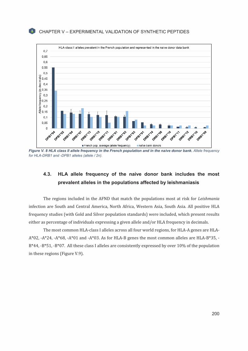
CHAPTER V – EXPERIMENTAL VALIDATION OF SYNTHETIC PEPTIDES
200
Figure V. 8 HLA class II allele frequency in the French population and in the naive donor bank. Allele frequency for HLA-DRB1 and -DPB1 alleles (allele / 2n).
4.3. HLA allele frequency of the naive donor bank includes the most
prevalent alleles in the populations affected by leishmaniasis
The regions included in the AFND that match the populations most at risk for Leishmania
infection are South and Central America, North Africa, Western Asia, South Asia. All positive HLA
frequency studies (with Gold and Silver population standards) were included, which present results
either as percentage of individuals expressing a given allele and/or HLA frequency in decimals.
The most common HLA-class I alleles across all four world regions, for HLA-A genes are HLA-
A*02, -A*24, -A*68, -A*01 and -A*03. As for HLA-B genes the most common alleles are HLA-B*35, -
B*44, -B*51, -B*07. All these class I alleles are consistently expressed by over 10% of the population
in these regions (Figure V.9).

CHAPTER V – EXPERIMENTAL VALIDATION OF SYNTHETIC PEPTIDES
201
Figure V. 9 Average HLA-class I allele frequencies according to world region. Averaged results from the population studies considered (%individuals and allele frequency) for HLA-class II alleles HLA-A and HLA-B. A) South and Central America populations. B) North Africa population. C) Western Asia populations. D) South Asia populations.

CHAPTER V – EXPERIMENTAL VALIDATION OF SYNTHETIC PEPTIDES
202
The most frequent HLA-class II alleles across all four world regions, expressed by over 10%
of individuals in all these populations are HLA-DRB1*03, -DRB1*04, -DRB1*07, -DRB1*08, -DRB1*11,
-DRB1*13, -DRB1*15. The allele HLA-DRB1*01 is less prevalent is the western and south asian
populations. Some alleles are often not included in the allele frequency studies, as for example the
HLA-DPB1 locus, which impedes comparisons among populations.
Typing of the HLA-DQB1 loci was performed for the individuals in the naive donor bank
(Table V.10), and data exists for some of the selected populations, however, these were not included
in the analysis since epitope prediction was not performed to HLA-DQ alleles.
HLA allele frequencies for these four world regions match the most prevalent alleles in the
naive donor bank, suggesting the allele frequencies of naive donors are representative of these human
populations.
Figure V. 10 Average HLA-class II allele frequencies according to world region. Averaged results from the population studies considered (%individuals and allele frequency) for HLA-class II alleles HLA-DRB1 and HLA-DPB1. A) South and Central America populations. B) North African populations. C) Western Asian populations. D) South Asia populations.

CHAPTER V – EXPERIMENTAL VALIDATION OF SYNTHETIC PEPTIDES
203
4.4. Monocyte-derived dendritic cells are efficiently generated in vitro
The surface markers CD14, CD209 (DC-SIGN) and CD83 were used to determine the
phenotype of monocyte-derived dendritic cells, after differentiation from monocytes and, for MPL10,
after maturation. CD14 is highly expressed by monocytes, and functions as a co-receptor for LPS and
LPS-binding protein (LBP). CD14+ monocytes can differentiate into DCs in a IL-4/GM-CSF cytokine
environment. CD209 (DC-SIGN) is a C-type lectin and enables TCR engagement by stabilisation of the
DC-T-cell contact zone, and expression is limited to tissue DCs and monocyte-derived DCs. CD83 is
regarded as a maturation marker for DCs, it regulates antigen expression presentation and is only
expressed by DCs, Langerhans cells and B cells. Hence, immature DCs are CD14-CD209+CD83- and
mature DCs are CD14- CD209+CD83+.
In vitro monocyte differentiation into immature DC induced by IL-4 and GM-CSF is highly
efficient, resulting in the recovery of 99,1% for MPL9 and 98,1% for MPL10 CD14- cells, excluding
debris and contaminant cells (Figure V.11).
For MPL9, immature DCs as defined by the expression of CD209 and the absence of CD83 are
42,8% of total CD14- cells. Some mature DC expressing CD83 are found (2,3%) (Figure V.11 panel A).
For donor MPL10, the immature and mature DC populations were compared. Results show
that after in vitro monocyte differentiation, immature DCs represent 17,9% of total CD14- cells, with
2,3% also expressing CD83 (Figure V.11 panel B). For mature DCs, after overnight maturation with
LPS and Resiquimod, the percentage of mature DC (CD14-CD209+CD83+) increases to 28,6%, while
8,3% of cells remain in an immature state (CD14-CD209+CD83+) (Figure V.11 panel C).

CHAPTER V – EXPERIMENTAL VALIDATION OF SYNTHETIC PEPTIDES
204
Figure V. 11 Monocyte-derived DC phenotype analysis for donors MPL9 and MPL10. DC were stained in independent experiments with Mo-DC Inspector kit, human (Miltenyi) that detects CD14, CD209 (DC-SIGN), and CD83 expression. A) immature DC from donor MPL9. B) immature DC from donor MPL10. C) mature DC from donor MPL10.

CHAPTER V – EXPERIMENTAL VALIDATION OF SYNTHETIC PEPTIDES
205
4.5. Leishmania-specific TCD8+ cells are present in the naive repertoire
The first peptide immunoscreening performed tested all 49 HLA-class I peptides in matrix-
based pools using the protocol adapted from Wolfl et al, with magnetically isolated naive TCD8+ cells
from donor MPL3 (experiment MN01).
Figure V. 12 IFN-γ ELISpot results for the MN01 experiment: T cell amplification assay with naive TCD8 cells
from naive donor MPL3. A) number of positive cell lines per condition (14 class I peptide pools). B) control results (TCD8 NS, PBMC NS, CEF and PHA). C) Spot counts per cell line and the threshold mark for well positivity. D) ELISpot well pictures and respective spot counts.
In experiment MN01, we successfully generated and detected specific T cell lines against
Leishmania-specific peptide pools (Figure V.12 panel A). A total of 17 positive cell lines (out of 140)
specific to 9 different peptide pools were generated from donor MPL3. Results show that 5 (out of 14)
peptide pools generated 1 positive cell line, and 4 pools generated between 2 and 5 specific cell lines.
ELISpot results for negative controls (never stimulated TCD8 cells) showed overall low
background levels (NS spot counts: 25, 20, 44, 13, 39, 3) for an average of 24 spots (±15,5). The

CHAPTER V – EXPERIMENTAL VALIDATION OF SYNTHETIC PEPTIDES
206
highest NS value (44) was included in the empirical test to determine well positivity (2*NS
average+44 spots). The PHA solution used in this experiment is thought to have been from on old
stock, probably degraded, as only 2 wells showed high spot counts, and still much lower than expected
(PHA spot counts plate A: 222, 196, 60, 56; plate B: 46, 36) (Figure V.12 panel B).
In this experiment, the CEF-stimulated naive TCD8 cells underwent three co-cultures plus the
ELISpot stimulation. The spot counts of the CEF peptide pool are quite variable with some low values
(47, 79, 30, 3), which is expected since this assay uses a purified population of naive T cells, and very
few memory cells are present (Figure V.12 panel B). Memory T cells are responsible for long-term
responses against the CEF peptide pool (CMV-, EBV-, influenza-specific epitopes), so low spot counts
can be expected when using purified naive T cell populations. CD45RA+ cell enrichment after magnetic
purification was not assessed due to technical problems with the flow cytometer, even though
staining was performed. Although there are specific naive T cells in naive individuals against the CEF
peptides which can be stimulated an amplified in vitro, only 600.000 naive TCD8+ cells (triplicate
wells) were tested. Still, it is noteworthy that some Leishmania-specific peptide pools induced much
higher number of IFN-γ-producing cells than the CEF control pool.
Figure V. 13 Four peptide pools successfully generated over 2 positive cell lines. A) Cross-matching results with matrix peptide pool constitution leads to the identification of 4 double-positive, hence immunogenic peptides. B) Naive donor MPL3’s HLA-typing results for 6 HLA loci (HLA-A, -B, -C, -DRB, -DP, -DQ)
Peptide pool 2 induced 5 positive cell lines out of 10, the highest number overall, ranging from
99 to 198 spots. Pool B had the second highest number of positive lines, 3 in 10, with spots counts
ranging from 99 to 269. Pool D and Pool 6 induced 2 positive cell lines each, with spot counts of,

CHAPTER V – EXPERIMENTAL VALIDATION OF SYNTHETIC PEPTIDES
207
respectively, 124/360, and 142/218. Pools E and F induced only 1 cell line but with very high spot
counts of 274 and 278, respectively (Figure V.12 panels C and D).
If we consider a minimum of 2 positive cell lines, 4 immunogenic peptide pools are identified
– pools B and D, and pools 2 and 6 (Figure V.13 panel A). Cross-matching these results with the pool
matrix, four double-positive immunogenic peptides are identified – B7_3, A24_3, B8_3, B58_3 (Figure
V.12 panel A).
The donor MPL3 expresses the alleles HLA-A*23:01:01G (A24), HLA-A*23:01:01G (A24),
HLA-B*44:03:01G (B44), HLA-B*49:01:01G (unclassified but probable B44) (Figure V.12 panel B).
Considering this information, only one peptide (A24_3) corresponds to the donor’s HLA typing,
implying peptides are more promiscuous than predicted by in silico algorithms.
If we consider all peptide pools with at least 1 positive well, there are 9 positive matrix-based
peptide pools leading to the identification of 20 double-positive peptides. However, from these 20
double positive peptides, only 2 correspond to the donor’s HLA-typing. The number of restricted
peptides is not significantly increased as these do not include more donor-specific supertypes.
Interestingly, peptide pool E which generated one cell line with high spot count (274) contains both
A24- and B44-restricted peptides.
This assay constitutes preliminary evidence of the presence of T-cell precursors specific to
Leishmania epitopes in naive individuals’ immune repertoire. Although the magnitude of response
was assessed for in vitro stimulated cell lines, specificity was not absolutely confirmed because spot
counts were compared to NS wells (never stimulated autologous TCD8+ cells). An ELISpot well
containing culture-stimulated cells but unstimulated during the ELISpot (cNS) will demonstrate that
IFN-γ production is dependent on peptide-stimulus and, therefore, specific.
The remaining issues with this experiment are i) that no cell counts were performed before
seeding in ELISpot plate (in this experiment 1 culture well corresponds to 1 ELISpot well) which may
introduce bias when comparing wells; ii) the use of an empirical rule for well comparison, because
there are no triplicates no statistical test can be applied; iii) LiESAp or CaniLeish® antigen, the original
antigenic mixture, were not included as positive controls; iv) finally, the high number of purified naive
TCD8 cells needed per experiment and per individual (MINIMUM 200.000*10 wells/condition) is
logistically unviable.
In this experiment total PBMC were used as APC, without supplementation with
differentiation or maturation factors. We expect the antigen presentation protocol can be further

CHAPTER V – EXPERIMENTAL VALIDATION OF SYNTHETIC PEPTIDES
208
optimised using professional APC, such as monocyte-derived DCs for short peptide presentation in
HLA class-I molecules.
4.6. Total TCD8+ cell in vitro amplification needs at least three stimulation
rounds
In experiment MN02, total TCD8 cells from donor MPL9 were used, and the same matrix-
based peptide pools were tested at 5 µM concentration per peptide. Cell populations from MPL9 were
isolated differently from other donors using the MultiMACS™ Cell24 Separator Plus (see Material and
Methods). Surface marker phenotyping shows that CD8+CD45+ cells were present at 88,2% purity
(66,4% of total events excluding debris) (data not shown). Also, CD14+CD45+ monocytes were
isolated with 93,9% purity (89,4% of total event excluding debris) and CD4+CD45+ cells were isolated
with 96% purity (84,7% of total events excluding debris) (data not shown).
The protocol used was adapted from analogous TCD4+ cell amplification assays (8,21), and
uses the total TCD8+ cell population and mature monocyte-derived DCs (mDC) as APC. Since the class
I peptides are only 9-mer in length, and do not necessarily need processing, a short peptide
stimulation protocol was adopted, where monocyte-derived immatured DCs are firstly matured with
LPS and TLR-7/8 agonist Resiquimod/R848, and only after stimulated with peptides for 4 hours.
In this experiment, no specific cell lines were generated against any of the pools at 5 µM
peptide concentration (Figure V.14 panel A) after two co-cultures.
Negative control background levels from never stimulated TCD8+ cells remained low (NS spot
counts 15/6/20/20/17, average 16). As expected, since in this experiment total TCD8 cells were used,
the CEF control pool results were higher than MN01, even with just one co-culture plus the ELISpot
stimulation (CEF spot counts 138/49/153/51, CEF MN02 Elispot2: 34, 238 (Too Numerous To
Count), 39, 186 (TNTC), with only 20.000 TCD8+ cells seeded in the ELISpot wells.
PHA stimulation results (spot counts 366, TNTC, 404, TNTC) and average cell viability of 77%
(± 9.9%) assessed before ELISpot seeding, confirm cells were viable and responsive (Figure V.13
panel B). We estimate viability is higher than calculated by LUNA reader, for there is some cellular
debris from DC cells and SAB used for the co-cultures, considered as dead cells by the automatic
counter (a cell size filter was not applied).
The positive controls LiESAp and CaniLeish® antigen were tested in this experiment, however,
the observed negative results remain inconclusive since the DC stimulation protocol was not adapted
for protein processing. The short peptide stimulation protocol was used for all conditions and we

CHAPTER V – EXPERIMENTAL VALIDATION OF SYNTHETIC PEPTIDES
209
estimate a 4-hour incubation time of matured DCs is not sufficient to process and present the full
protein antigens, and subsequent T-cell activation is abrogated (Figure V.14 panel B).
Figure V. 14 IFN-γ ELISpot results for the MN02 experiment (96-well format), T cell amplification assay with total
TCD8+ cells from donor MPL9. A) Number of specific T cell lines generated per condition. B) Spot counts for control conditions. C) Spot counts per condition showing individual cell lines; cNS, culture-stimulated cells but unstimulated in the ELISpot plate.

CHAPTER V – EXPERIMENTAL VALIDATION OF SYNTHETIC PEPTIDES
210
In this experiment, response specificity per cell line was confirmed with culture-stimulated
cells but unstimulated during the ELISpot (cNS). Overall, observed spot count values in stimulated
wells (STIM) are sometimes higher than cNS, but not enough to consider as positive responses (Figure
V.14 panel C). Otherwise, spot counts are similar to respective cNS unstimulated wells. Also, STIM and
cNS are mostly similar to background levels for negative controls (never stimulated TCD8 cells – NS)
but much more variable. All conditions and all cell lines show some variability in background levels
(cNS spot counts), and even some cell lines present high background levels (e.g. pools 1, C, E, F and G)
(Figure V.14 panel C). For peptide pool C, one cell line has cNS spot count of 93 and STIM spot count
of 142, which if we compared with the never stimulated TCD8+ negative control would be considered
a positive cell line (Figure V.13 panel C). This can be due to approximation errors when averaging cell
counts to calculate 20.000 seeded cells in the ELISpot (3 in 10 wells measured per condition). Still,
pools that successfully generated more than one cell line in MN01 – pools B, D, 2 and 6 – did not induce
IFN-γ production/positive cell lines (maximum spot counts were 41, 43, 39 and 25, respectively).
The use of statistical analysis to assess positive responses is more reliable and recommended
as they are based on a theoretical background, have a universal application across experiments, and
even more suitable to detect weaker responses. Furthermore, the primary objective of the peptide
immunoscreenings is to exclude non-immunogenic and unstable peptides, before inquiring about
precursor frequency. Therefore, the following adaptations were made to the protocol: i) an additional
total TCD8:DC co-culture (3 instead of 2); ii) testing different peptide concentrations and fresh
aliquots diluted in 10% DMSO; and iii) testing a different assay format that allow the application of
statistical tests to determine well positivity, the counting of all culture wells and show results as
SFC/million, a widely accepted way to present ELISpot results.
4.7. In vitro amplification of total TCD8+ cells depends on the antigen-
presenting cells and respective stimulation protocol
In experiment MN03, total TCD8 cells from donor MPL9 were used, and all peptides were
tested in two pools at different peptide concentrations. At the time of this experiment, HLA-typing
results of MPL9 donor were not yet available to design one personalised peptide pool, so all class I
peptides were tested in two pools, containing 22 peptides with predicted restriction to HLA-A genes
(pool22), and 27 peptides with predicted restriction to HLA-B genes (pool27). Furthermore, two
assay formats were tested: 96-well plate format, establishment of cell lines and empirical rule to
determine positive responses, and the 48-well plate format (batch stimulation), that allows for

CHAPTER V – EXPERIMENTAL VALIDATION OF SYNTHETIC PEPTIDES
211
statistical determination of well positivity and cell counting before seeding in the ELISpot plates,
while still testing the same number of cells.
Figure V. 15 IFN-γ ELISpot results for the MN03 experiment (96-well format), T cell amplification assay with total
TCD8 cells from donor MPL9. A) Number of specific cell lines and IFN-γ ELISpot spot counts for 96-well plate format control conditions; B. all results per cell line, 7 seven lines per condition (1, 2.5 and 5µM peptide concentrations).
The addition of a third re-stimulation or different peptide concentrations did not improve
results obtained with the 96-well format T cell amplification assay, and no specific cell lines were
generated (Figure V.15 panel A). With this format, positive control CEF stimulation also yielded very
low spot counts (16/8/11), when cells are responsive, since PHA-stimulation yielded high IFN-γ
production (PHA_20k spot counts 784, 693, 715 in plate A, and 595, 589, 608 in plate B).
Background levels of culture-stimulated cells but unstimulated in the ELISpot wells (cNS) of
all cell lines are less variable than in the previous experiment MN02, but spot counts of stimulated
cells are very similar to background (Figure V.15 panel C).

CHAPTER V – EXPERIMENTAL VALIDATION OF SYNTHETIC PEPTIDES
212
Figure V. 16 IFN-γ ELISpot results for the MN03 experiment (48-well format), T cell amplification assay with total
TCD8 cells from donor MPL9. A) IFN-γ ELISpot results for control conditions. B) Pool22 IFN-γ ELISpot results for all
conditions tested: 1, 2.5 and 5µM peptide concentrations and 50.000 (50k), 100.00 (100k), or 150.000 (150k) TCD8 cells per ELISpot well. C) Pool27 IFN-γ ELISpot results for all conditions tested: 1, 2.5 and 5µM peptide concentrations
and 50.000 (50k), 100.00 (100k), or 150.000 (150k) TCD8 cells per ELISpot well.

CHAPTER V – EXPERIMENTAL VALIDATION OF SYNTHETIC PEPTIDES
213
Likewise, in the 48-well batch stimulation format, no differences were found in IFN-γ
productions were observed between unstimulated (cNS) and stimulated cells (STIM). Nevertheless,
better results were obtained with the CEF peptide control pool (CEF_100k spot counts 72/72/62 in
plate A, and 36/47/56 in plate B, large spots), and high IFN-γ responses PHA-stimulated wells
(PHA_50k spot counts: TNTC) (Figure V.16 panel C).
The reference number of cells seeded in the ELISpot plate is 10% of the culture well, in
accordance with the 96-well plate format assay (200.000 TCD8 per culture well and 20.000 TCD8
seeded in the ELISpot well). To optimise response detection in the 48-well batch stimulation assay,
we expect to use 100.000 TCD8 cells per ELISpot well (10% of 1 million TCD8 cells per culture well),
but different cell numbers were tested – 50.000 TCD8 (50k), 100.000 TCD8 (100k), 150.000 TCD8
(150k) per ELISpot well. Response specificity for all cell densities was assessed by including culture-
stimulated wells unstimulated in the ELISpot wells (cNS).
For some conditions, in the 48-well assay format, stimulated conditions had fewer spot counts
than unstimulated cNS wells (Figure V.16 panels B and C). Also, there appears to be a dose-dependent
effect between peptide concentration and decreased spot counts in stimulated wells, very noticeable
at 5 µM per peptide (Figure V.15 panels B and C). This effect is probably not due to peptide toxicity
since cell viability was assessed, but probably to peptide binding competition, as each pool contains
a large number of peptides (22 and 27 peptides).
From this experiment, we realized that stimulation of monocyte-derived matured DCs is not
optimal and should be further optimised. Even if class I peptides are 9-mer in length, apparently these
were not successfully presented by MHC molecules and were unable to prime specific T cells in vitro
under these conditions. Furthermore, the cells used in the MN01 experiment were highly immature
and were successful for T cell activation and cell line generation. Therefore, two different protocols
for monocyte-derived DC stimulation were adopted: i) short peptide protocol, immature DC are
matured (with LPS and Resiquimod/R848) for 16 hours, and then pulsed with peptides for 4 hours
at 37°C/5% CO2; and ii) long peptide protocol, immature DC are matured (with LPS and
Resiquimod/R848), and simultaneously pulsed with peptides for 16 hours at 37°C/5% CO2.

CHAPTER V – EXPERIMENTAL VALIDATION OF SYNTHETIC PEPTIDES
214
4.8. IFN-γ production by Leishmania-specific total TCD8+ cells was
successfully induced and detected with adequate DC stimulation
In experiment MN04, total TCD8 cells from donor MPL10 were magnetically isolated and co-
cultured with mDC (short peptide protocol) or iDC (long peptide protocol).
Since we week to validate the proposed protocol for DC stimulation, only 12 peptides were
used in one peptide pool, customised according to the naive donor’s HLA alleles, instead of testing all
49 HLA-class I peptides. Naive donor MPL10 expresses the HLA-class I alleles HLA-A*03:01:01G, HLA-
A*02:05:01G, HLA-B*15:03:01G, HLA-B*07:02:01G, belonging to supertypes A3, A2, B27 and B7,
respectively (Figure V.17). Accordingly, the peptide pool tested (poolGOOD) includes 3 peptides per
supertype, each at a 2,5 µM concentration – peptides A3_1, A3_2, A3_3, A2_1, A2_2, A2_6, B27_2,
B27_3, B27_5, B7_1, B7_2, B7_3 (Figure V.17).
Figure V. 17 MPL10 HLA-typing for HLA-class I loci and peptides selected to compose the peptide pool.

CHAPTER V – EXPERIMENTAL VALIDATION OF SYNTHETIC PEPTIDES
215
Figure V. 18 IFN-γ ELISpot results for experiment MN04, T cell amplification assays with total TCD8 T cells from
donor MPL10. A) TCD8+ amplified with the short peptide protocol (mDC stimulation). B) TCD8+ cells amplified with the long peptide protocol (iDC stimulation). C) ELISpot well images and respective spot counts for CEF and positive well.
Again, to confirm optimal cell density for the detection of the induced response after
stimulation in the 48-well plates, different TCD8+ cell numbers were seeded in the ELISpot plate in
duplicate wells, similarly to experiment MN03. For the same number of seeded cells (100.000 TCD8
cells/well), iDC stimulation (long peptide protocol) with CEF control peptide pool yielded better
results than mDC stimulation (short peptide protocol) – CEF 100k/iDC spot counts 60 and 83 and CEF
100k/mDC spot counts 53 and 54 (Figure V.18). Background levels of cNS wells are low, maximum
spot count of 10, but are proportional to cell density (Figure V.18).
In this experiment, stimulated cells with the short peptide protocol show low spot counts
regardless of cell density, with a maximum of 13 spot counts (Figure V.18 panel A). On the contrary,

CHAPTER V – EXPERIMENTAL VALIDATION OF SYNTHETIC PEPTIDES
216
successful amplification and detection of specific TCD8 cells is observed when stimulated with pulsed
iDC, (long peptide protocol) (Figure V.18 panel B). The observed responses are highly specific, as cNS
wells (culture-stimulated cells unstimulated in the ELISpot) show very low spot counts, meaning IFN-
γ-producing cells are primed through peptide-specific stimulus and become activated.

CHAPTER V – EXPERIMENTAL VALIDATION OF SYNTHETIC PEPTIDES
217
5. Discussion - Immunoscreenings with naive donor samples
The immunoassays with naive donor samples currently still present several challenges. The
main limitation is the number of cells needed per condition, since specific cells are rare. These
experiments are also extremely time-consuming (with over 20-day cultures), and resource-
consuming due to reagents’ costs. Also, in vitro cellular amplification occurs under non-physiological
conditions, so analysis should be performed with care. Finally, a high number of individuals should
be tested to assess the peptides’ overall immunogenicity in the naive repertoire. Specific approaches
were adopted to mitigate some of these issues, namely: i) the generation of a naive donor bank and
respective HLA-typing; and ii) the use of matrix-based peptide pools.
Peptide immunogenicity testing through immunoassays with samples from naive donors is
rather innovative in the field of peptide-based vaccine development. Several laboratories, namely the
Bernard Maillere Lab (CEA – Saclay), have developed techniques of in vitro cellular amplification that
allow the analysis of specific T cell precursors in the naive repertoire (8–10,17,22–24).
Immunogenicity testing is often focused on samples from exposed/healed individuals or animal
models. We believe immunogenicity testing in the naive repertoire is essential to assess the
prophylactic potential of peptide vaccine candidates, since there is a minimum threshold of specific
naive T cells in circulation needed to generate specific effector and memory T cells. Also, the TCR-
specificity of responding naive T cells correlates with the specific memory T cells present in immune
individuals. We propose to assess and compare the peptide immunogenicity profiles in the naive and
memory repertoires to select immunogenic peptide candidates to use in a final vaccine formulation
against leishmaniasis. Overall, this approach allows for the fast validation of peptide vaccine
candidates with increased confidence in late-stage human in vivo immunogenicity and vaccine
effectiveness.
5.1. Naive donor bank
The generation of a naive donor bank and respective HLA-typing results allows the
optimization of samples to use in the immunoscreening experiments. Donors can be selected
according to HLA-typing results to maximise HLA coverage while testing a minimal number of
individuals.

CHAPTER V – EXPERIMENTAL VALIDATION OF SYNTHETIC PEPTIDES
218
The naive donor bank was generated with samples from the French Blood Bank in Toulouse.
The HLA-typing results for HLA-A, -B, -DRB1, and -DPB1 alleles expressed by the naive donors match
that of the French population. The HLA diversity represented in the naive donor bank also
corresponds to the most common alleles expressed by populations in South and Central America,
North Africa, Western Asia and South Asia. These regions include the populations most at risk of
developing leishmaniasis and are the target populations for an anti-Leishmania vaccine.
Unquestionably, the naive donor bank is an extremely small population compared with the
populations considered for the allele frequency studies, but the most frequent alleles are nonetheless
represented.
Ultimately, we aim at increasing the sample size of the naive donor bank and increase the
number of represented alleles, particularly those which are currently underrepresented. However,
the most prevalent HLA alleles in the world population, and particularly in populations in endemic
leishmaniasis areas, already represented in the naive donor bank will provide a great HLA coverage
for future experiments.
A peptide-based vaccine formulation that includes peptides restricted to nine HLA-class I
alleles (HLA-A*02, -A*24, -A*68, -A*01, -A*03, -B*35, -B*44, -B*51, -B*07) will be effective in these
world regions. Regarding HLA-class II alleles, although these are expected to be more promiscuous
than class I peptides, peptides restricted to HLA-DRB1*03, -DRB1*04, -DRB1*07, -DRB1*08, -
DRB1*11, -DRB1*13, and -DRB1*15 should be included in the final vaccine formulation.
5.2. Peptide immunogenicity testing with samples from naive donors
The naive T cell assay (experiment MN01) confirmed the presence of Leishmania-specific
TCD8 cells in the naive repertoire, with 4 peptide pools generating at least 2 positive cell lines (in 10
tested per condition, i.e. 2 million T CD8+ cells). This assay shall be repeated in the future, after pre-
selecting peptides, with fewer peptides further along the peptide validation pipeline and with some
protocol changes (i.e. cell counting before ELISpot seeding, ELISpot triplicates and higher number of
cell lines).
The protocol used in experiment MN01 was adapted from Wolfl et al, to a simplified version
using total PBMC as APC. While the authors recommend the use of monocyte-derived dendritic cells,
at the time of this experiment it was not possible to induce DC differentiation in vitro, and the use of
autologous total PBMC as APC is commonly done as well (25). The B cells and monocytes present in
this population are for the most part in an immature state.

CHAPTER V – EXPERIMENTAL VALIDATION OF SYNTHETIC PEPTIDES
219
The donor MPL3 is homozygous for the HLA-A loci (HLA-A*23:01:01G alleles classified as A24
supertype) and expresses HLA-B*44:03:01G (B44 supertype) and HLA-B*49:01:01G, an unclassified
allele. Considering four double-positive immunogenic peptides that generated more than 2 specific
cell lines, only one (A24_3) matches MPL3’s HLA typing results. Interestingly, the allele B*49:01 has
ambiguous binding pocket specificities. This allele has a B pocket specificity matching to the B44
supertype, and F pocket binding like the B*38:01 allele (hydrophobic), so it likely this allele shares
binding specificities with other alleles in the B44 supertype, and perhaps even other supertypes.
Surprisingly, when the total TCD8 stimulation assay protocol was adopted, the use of mature
DCs with the short peptide protocol no longer stimulated T cell proliferation and IFN-γ production.
The number of co-cultures performed (MN02: 2 co-cultures, MN03: 3 co-cultures), the peptide
concentration (1, 2,5 or 5 µM), or the assay format do not seem to be as important as the DC
stimulation protocol, since IFN-γ responses were not detected in both MN02 and MN03 experiments.
Many factors may contribute to this and apparently these Leishmania-specific peptides, even if short
(9-mer) may need intracellular processing. Furthermore, we estimate that, even for short peptides,
optimal DC stimulation requires peptide processing and presentation with simultaneous DC
maturation through TLR activation, due to peptides’ intrinsic weak immunogenicity.
In experiment MN04, when immature DCs were again used as APC, and stimulated during 16
hours with both peptides and maturation factors, specific IFN-γ production was induced and
detected. These results suggest APC’s immature state and the simultaneous peptide/TLR stimulation
are key for proper peptide presentation and T cell activation.
During in vivo responses, immature DC uptake antigen in the periphery and migrate to the
lymph nodes as mature cells where they prime responding T cells. Immature DCs are phagocytic and
process and present exogenous antigens. TLR stimulation of immature DCs induces cell maturation
process consisting of the upregulation of MHC and costimulatory molecules (CD40, CD80 and CD86)
and migration to the lymph nodes. Mature DC become powerful APC, able to prime naive T cells and
influence T-cell differentiation. TLRs are therefore a unique link between pathogen recognition and
induction of T cell responses. The maturation conditions and stimulation by different pattern
recognition receptors (PRR) influence DC’s costimulatory molecules and the ability to produce
inflammatory cytokines, therefore impacting the observed T cell responses (26,27).
Notably, both immature and mature DC populations generated from donors MPL9 and MPL10
include cells with intermediary phenotypes, not expressing any of the stained markers (CD14-CD209-

CHAPTER V – EXPERIMENTAL VALIDATION OF SYNTHETIC PEPTIDES
220
CD83-). To describe these populations other markers should be included in the analysis. Also, some
contaminant cells (probably non-adherent PBMC) and debris due to cell thawing prior to staining are
observed (18 to 47% of total events). Nevertheless, the differentiation of monocytes into DCs is
successful as CD14- cells correspond to over 98% of all cells.
For MPL9 over 40% of total CD14- cells are immature DCs (CD14-CD209+CD83-). The
percentage of immature DCs after differentiation for MPL10 is much lower (18% of total CD14- cells).
These results may result from a shorter differentiation time for monocytes from MPL10 (four-day
stimulation instead of five). However, overnight maturation with LPS and Resiquimod greatly
increased the number of mature DCs (from 2,3% to 28,6% of CD14-CD209+CD83+ cells).
The experiments MN02-04 used monocyte-derived DC as antigen-presenting cells. These
were co-cultured with TCD8+ cells at a 1:10 ratio. Although no optimisation experiments were
performed to improve this ratio, there is evidence that optimal APC:T-cell ratio can go up to 1:160, or
be as low as 1:2, with lower ratios having potentially negative impact on T cell stimulation (28). The
intermediary 1:10 ratio was adopted, same as in the total TCD4+ cell amplification protocol.
After experiment MN01, a total TCD8 amplification assay protocol was adopted due to the
high number of conditions to test and to decrease the experiment-associated costs. Even though naive
donors are expected to have never been exposed to leishmaniasis, there is an increased risk of
triggering cross-reactive responses, potential cross-specific memory T cells in circulation against
other pathogens or antigens. To assure no recall responses are being detected, short term total PBMC
stimulation experiments will be performed with both TSLA and peptide pools to confirm,
respectively, the naive donor status (TSLA-negative) and IFN-γ production by amplified specific naive
T cells and not cross-specific memory cells.
Peptide concentrations in the peptide pools tested ranged from 1 to 5 μM. While the nature of
the induced response to change remains unchanged regardless of the peptide concentration, a
positive effect on T cell activation is expected in the range of 1-10 µg/mL of a 9-mer peptide (29).
Nevertheless, the peptide pool composition may also influence the observed responses. Response
specificity against a single peptide may be decreased if pools contain many peptides, and likewise,
pool sensitivity may be increased if there are multiple positive peptides. On the other hand, peptide
competition and low frequency responses may hinder response detection. We expect that the
peptides tested present binding competition since they are all predicted to be strong binders. In

CHAPTER V – EXPERIMENTAL VALIDATION OF SYNTHETIC PEPTIDES
221
experiment MN03, peptide pools with over 20 peptides each were used and stimulated cells at 5 µM
show decreased spot counts when compared with the other tested concentrations. Therefore, it is
possible that increased peptide numbers and/or increased peptide concentration inhibit T cell
activation, either through peptide competition or due to the absence of high avidity specific T cells.
We believe peptide competition is higher in the tested peptide pools than expected for traditional
epitope mapping experiments, where only a few immunogenic regions are found within a full protein,
and many negative peptides are included in the peptide pools. The matrix-based pools contain 7
peptides, and the poolGOOD used in MN04 contains 12 peptides at 2,5 µM each. Peptide pools
containing up to 12 peptides at 2,5 µM do not seem to inhibit T cell activation, so this peptide
concentration will be adopted for future experiments.
Peptide pools are an efficient and sensitive way to optimise epitope mapping experiments and
even to detect low level responses while decreasing experiment-associated costs and sample size. The
use of matrix-based pools provides additional advantages regarding the number of assays performed
and cryovials needed (compared with single peptide testing or mini-pool peptide testing). However,
some unclear results regarding double positive peptides argue for the need of a second ELISpot to
confirm peptide specificity (two-stage matrix-based pool approach).
The CEF control peptide pool provides a good control for peptide processing and presentation
to total TCD8+ cells. Although results were quite variable (from 16 spot counts up to TNTC), they
were consistently positive and with large spots. Evidently, the observed responses are mediated by
memory T cells instead of naive T cells. While CEF-induced responses do not necessarily provide
evidence for amplification of specific T cell populations through clonal expansion, they still provide
evidence that antigen presentation mechanisms by peptide-pulsed APC are effective in priming
specific T cells in vitro. True positive control peptides for the naive T cell repertoire should be
included, however, universal control peptides for naive T cell stimulation do not exist. A few peptides
were so far described to have specific naive T cells in the naive repertoire. The most studied are
peptides from the Melan-A/MART-1 antigen (Melanoma-Associated Antigen) (30,31). Still, these are
restricted to HLA-A*02 alleles and have an unusually high precursor frequency. We propose and
expect to find the most immunogenic peptides with specific naive T cells through the optimisation of
T cell amplification assays, which have been shown to be effective in detecting specific T cells in the
naive repertoire.

CHAPTER V – EXPERIMENTAL VALIDATION OF SYNTHETIC PEPTIDES
222
In experiment MN04, which used the long peptide stimulation and a tailored peptide pool
containing 12 peptides (2,5 µM each), we successfully amplified and detected Leishmania-specific T
cells from the naive repertoire. Only one culture well was positive (1 million TCD8 cells seeded)
among two stimulated wells (2 million TCD8 cells tested), as expected for low frequency specific cells.
The 48-well batch stimulation assay format allows for the successful amplification of specific
T cells present in the naive repertoire, as well as practical advantages that permit cell counting,
ELISpot well triplicates, and therefore the application of statistical tests for well positivity
determination. Although, in this experiment, only two wells per condition were seeded in the ELISpot
plate due to the optimisation of peptide concentration and cell density.
We aim to use the 48-well batch stimulation assay with a higher number of donor samples
(n=10) to screen the synthetic peptides and select the most immunogenic ones. Possibly, the number
of total TCD8 cells tested per condition can be increased (3 or 4 million total TCD8 per condition).
However, the 48-well batch stimulation assay format eliminates the application of the Poisson
distribution formula for the calculation of the specific T cell precursor frequency. Hence, we propose
to use the batch stimulation assays to pre-select the (most) immunogenic peptides and, subsequently,
to perform precursor frequency analysis experiments with a fewer number of pre-validated
immunogenic peptides, and possibly bypassing polyclonal activation with peptide pools but
performing co-cultures with DC pulsed with single peptides.
Because naive T cell amplification assays were performed in a limited number of individuals
with all peptides, comparison is very limited. However, MN01 and MN04 clearly evidence the
existence of a minimum number of naive T cells specific to Leishmania peptides in naive donors,
hence, validating their predicted immunogenicity and the feasibility of using such peptides in an
effective vaccine formulation. The naive donor bank, which will be augmented with additional naive
donor samples, and the in vitro stimulation protocol, which is now optimised, will allow to screen all
peptides and select the most immunogenic by serial routine experiments.

CHAPTER V – EXPERIMENTAL VALIDATION OF SYNTHETIC PEPTIDES
223
6. Results - Immunoscreenings in healed individuals from endemic areas
(Tunisia)
The first peptide immunoscreenings performed with healed donors’ samples tested all 49
class I peptides and 24 HLA-class I peptides in matrix-based pools, using a short term total PBMC
stimulation protocol.
Two series of blood samples were received, both including 10 samples from healed donors –
first series TUN1 to TUN10, and second series TUN11 to TUN20. In the first series (TUN1 to 10), one
blood sample was hemolysed (TUN2) so no PBMC were purified. IFN-γ ELISpot was performed for
both series and culture supernatants were collected and stored at -80°C. The series of blood samples
with naive donors was performed in the Institut Pasteur Tunis, no IFN-γ ELISpot was performed for
these samples.
Peptide immunogenicity or the capacity to prime in vitro specific recall responses was
successfully assessed for 9 healed donors (see Appendix V.2 for individual data).
6.1. Healed donors’ personal information and HLA-typing results
The average age for the first healed donor series was 43 years-old, ranging from 26 to 64
years-old (Table V.7). Male healed donors are much more frequent than female donors in this series,
with only one woman in nine donors (TUN4). The presence of CL lesions was inquired in the medical
questionnaire. On average, healed donors in the first series presented 2 cutaneous lesions, between
one up to five lesions per donor. Most cutaneous lesions were localised to the legs or arms, only a few
CL lesions were localised in the face (two donors, 11%) (Table V.7).
HLA-typing results for the first healed donor series was performed (Table V.8)

CHAPTER V – EXPERIMENTAL VALIDATION OF SYNTHETIC PEPTIDES
224
Table V. 7 Personal information and medical history from recruited healed donors (TUN1-TUN20).

CHAPTER V – EXPERIMENTAL VALIDATION OF SYNTHETIC PEPTIDES
225
Table V.7 (continued)

CHAPTER V – EXPERIMENTAL VALIDATION OF SYNTHETIC PEPTIDES
226
Table V. 8 HLA-typing results for the first healed donor series (TUN1-TUN3 to TUN10). A) Six loci corresponding to HLA-class I and class II polymorphic regions (HLA-A, -B, -C, -DRB1, -DPB1, -DQB1) were sequenced by NGS (exons 2 and 3), leading to the 6-digit identification of the alleles expressed by each donor. Allele supertype classification by Sidney et al 2008 (20). B) Ambiguous identifications are given a multiple allele code (MAC, https://hml.nmdp.org/MacUI/)

CHAPTER V – EXPERIMENTAL VALIDATION OF SYNTHETIC PEPTIDES
227
The HLA-typing results for the healed donors, although the sample size is quite small, mostly
match the most frequent alleles in the Tunisian population (Figure V.19 and V.20). The HLA-class I
alleles HLA-A*02 are the most frequent. HLA-A*24 alleles, although very frequent in the Tunisian
population, were not represented in the healed donor series. On the other hand, other alleles such as
HLA-B*44, -B*35 and -A*01 were more frequent in the healed donor series (Figure V.19).
Figure V. 19 HLA class I allele frequency in the Tunisian populations and in the healed donor series. Allele frequency for HLA-A and -B alleles (allele / 2n).
The HLA-class II allele HLA-DPB1*04 is the most frequent in the Tunisian populations yet
poorly represented in the healed donor series (Figure V.20). On the other hand, the second most
frequent allele HLA-DRB1*07 is also well represented. Other alleles such as HLA-DPB1*03, -DPB1*06,
-DRB1*11 and -DRB1*03 were more frequent in the healed donor series (Figure V.20).

CHAPTER V – EXPERIMENTAL VALIDATION OF SYNTHETIC PEPTIDES
228
Figure V. 20 HLA class II allele frequency in the Tunisian populations and in the healed donor series. Allele frequency for HLA-DRB1 and -DPB1 alleles (allele / 2n).
6.2. Healed status validated by positive responses against TSLA
As expected, most donors show positive TSLA reactivity, defined by IFN-γ production by
ELISpot (Figure V.19). Four donors (TUN3, TUN4, TUN5 and TUN10) have significantly higher IFN-γ
responses against TSLA (TSLA-positive donors). Donor TUN7 has almost statistically significant
differences (p-value=0,0571). Statistical significance was impossible to determine for donor TUN6
because only two ELISpot wells can be analysed due to an error during seeding (CaniLeish®-
stimulated cells were stimulated with TSLA), but 192,5 spot count average is considerably higher than
autologous NS spot counts (28,83 ±22,07). Two other donors (TUN9, TUN10) were considered TSLA-
positive since ELISpot results show IFN-γ production during culture stimulation, evidenced by dark,
highly saturated wells, even though spot counts are low or innumerable. In addition to TSLA,
CaniLeish® was also used as positive control. Interestingly, CaniLeish® antigens induced vigorous
IFN-γ responses in >75% of individuals (7 in 9 donors). Moreover, donors TUN1, TUN7, TUN8 and
TUN9 had significantly higher responses against CaniLeish® but not TSLA (Figure V.21).
IFN-γ ELISpot results show donors have quite different background levels, from an average
NS spot count of 14,5 (TUN7) up to an average spot count of 161,5 (TUN1) (Figure V.21).

CHAPTER V – EXPERIMENTAL VALIDATION OF SYNTHETIC PEPTIDES
229
Figure V. 21 IFN-γ ELISpot results for controls for 1st healed series (n=9). A) Spot counts for NS (never stimulated PBMC), for positive controls PHA, TSLA and CaniLeish®, and pools ALL_I (49 class I peptides), ALL_II (24 class II peptides) and ALL_I+II (all 73 Leishmania-specific peptides). B) Table with averaged spot counts for all controls (* significantly different from autologous NS with the Mann-Whitney statistical test, p-value ≤ 0.05).
6.3. IFN-γ responses against Leishmania-specific peptides
To evaluate recall responses against Leishmania-specific peptides, total PBMC were
stimulated in vitro in the absence or presence of peptide pools containing HLA-class I or -class II
peptides. Positive IFN-γ responses were induced by multiple peptide pools and by most donors –
seven responder donors for HLA-class I peptide pools, and nine responder donors for HLA-class II
peptide pools (Figure V.22). The number of responders and the number of peptide-specific responses
induced for each donor were different according to the peptide pool (Figure V.22).
Overall, all HLA-class II pools induced IFN-γ production in at least one individual (II_pool5
and B), up to 7 individuals (II_pool4) (Figure V.22 panel B). On the other hand, three HLA-class I
peptide pools did not elicit IFN-γ production in any of individuals tested (I_pool4, A and C), and the
remaining 11 pools induced IFN-γ production in at least one donor (I_pool3 and 7), up to 4 donors

CHAPTER V – EXPERIMENTAL VALIDATION OF SYNTHETIC PEPTIDES
230
(I_pool1 and E) (Figure V.22 panel A). Individualised ELISpot results are included in Appendix V.2
(donors TUN1, TUN3-10).
For HLA-class II peptide pools, two donors had only one positive peptide pool so single
immunogenic peptides were not identified (TUN1, II_poolE and TUN6, II_pool1). Likewise, for HLA-
class I peptide pools, TUN6 had only one positive peptide pool (I_poolE) (Figure V.22). Results
regarding classI peptide pools I_pool5 and I_pool6 are inconclusive for donor TUN7 since results are
not available (no revelation antibody added to row C during ELISpot revelation).
Figure V. 22 Responder frequency per peptide pool for the 1st healed series (n=9). Number of responders, individuals with significantly increased IFN-γ production (responders) per peptide pool. A) Responder donors to class I peptide pools. B) Responder donors to class II peptide pools.

CHAPTER V – EXPERIMENTAL VALIDATION OF SYNTHETIC PEPTIDES
231
TSLA-positive donor TUN3 showed positive IFN-γ responses against HLA-class II peptide
pools only, but against a high number of pools (7 out of 10: II_pool1, 2, 3, 4, II_poolA, C, and D) (Figure
V.23 panel A). Twelve double-positive HLA-class II peptides were identified (14_b12, 16_b13, 3_a11,
22_b4, 7_a14, 12_a32, 21_b4, 17_b15, 4_a11, 5_a12, 9_a25, 13_b12). These peptides are predicted to
be very promiscuous and bind to four up to eleven HLA-class II alleles.
For TSLA-positive donor TUN4, HLA-class I peptide pools did not induce significantly higher
responses, but two positive HLA-class II pools did (II_pool4 and D). One double-positive immunogenic
class II peptide was identified (pii19/13_b12) (Figure V.23 panel B). The 13_b12 peptide is predicted
to be highly promiscuous, predicted to bind to seven class II alleles: HLA-DPA1*01:03-DPB1*02:01, -
DPA1*01:03-DPB1*04:01, -DPA1*02:01-DPB1*01:01, -DPA1*03:01-DPB1*04:02, -DPA1*03:01-
DPB1*04:02, -DRB1*07:01, -DRB1*09:01, and -DRB1*13:02. These include alleles expressed by
TUN4 namely, HLA-DRB1*07:01 and -DPB1*01:01 (Table V.8).
TSLA-positive donor TUN5 had positive IFN-γ responses against four HLA-class I pools
(I_pool2, 7, E, F) and three HLA-class II pools (II_pool4, A, C) (Figure V.23 panel C). The double-
positive HLA-class I immunogenic peptides B62_2, B8_4, B44_1 and B44_4 were identified. Yet, the
donor TUN5 expresses HLA alleles which belong to A26 (A*26:01:01G), A03 (A*34:02:01), B07
(B*07:02:01G), and B08 (B*08:01:01G) supertypes, meaning only one in four positive peptides match
the donors's HLA typing (B8_4).Additionally, the double-positive immunogenic HLA-class II peptides
22_b4 and 17_b15 were identified in donor TUN5. Peptide 22_b4 is predicted to be promiscuous and
bind to five class II alleles (HLA-DPA1*01:03-DPB1*02:01, -DPA1*01:03-DPB1*04:01, -DPA1*02:01-
DPB1*01:01, -DPA1*03:01-DPB1*04:02, -DRB1*09:01). Peptide 17_b15 is predicted to be
promiscuous and bind to seven class II alleles (HLA-DRB1*01:01, -DRB1*03:01, -DRB1*07:01, -
DRB1*09:01, -DRB1*11:01, -DRB3*01:01, -DRB5*01:01, -DRB5*01:01). Some of these allele
specificities match donor TUN5 typing results for HLA-class II loci -DRB1 and -DPB1, namely HLA-
DRB1*03:01, -DPB1*04:01, and -DPB1*01:01 alleles (Table V.8).
TSLA-positive donor TUN10 responded to seven positive HLA-class I pools (I_pool1, 2, 5, 6, B,
D, G) and five positive HLA-class II pools (II_pool1, 4, 5, A, D) (Figure V.23 panel D). The double-
positive immunogenic class I peptides identified were A1_1, A1_4, A3_2, A24_3, B7_3, B7_4, B8_3,
B27_1, B44_5, B58_3, B58_4, B58_5. Given donor TUN10’s HLA typing results, 5 in 16 peptides match
the donor’s HLA allele typing (B7_3, B7_4, B7_5, B44_5, A2_4) (Table V.8). The double-positive
immunogenic HLA-class II peptides identified in donor TUN10 were 14_b12, 22_b4, 18_b15, 4_a11,
13_b12, 10_a25. Again, all these peptides are predicted to be very promiscuous and bind to five up
to eleven class II alleles.

CHAPTER V – EXPERIMENTAL VALIDATION OF SYNTHETIC PEPTIDES
232
Figure V. 23 IFN-γ ELISpot results for TSLA-positive donors TUN3 (A), TUN4 (B), TUN5 (C), and TUN10 (D). Highlighted pools (*) induced significantly higher IFN-γ production when compared with autologous NS (Mann-Whitney test, p-value < 0.5).

CHAPTER V – EXPERIMENTAL VALIDATION OF SYNTHETIC PEPTIDES
233
Figure V.23 (continued)
Donor TUN7 is also highly responsive. This donor shows the lowest background values out of
all the donors which may influence the observed results. Positive responses may be overestimated
since this donor also shows the weakest responses to TSLA and CaniLeish®. Nevertheless, from the
positive HLA-class I pools and from the 8 immunogenic peptides identified (A1_1; A3_3; A24_1; A3_1;
A2_4; B7_2; B58_5 and B8_1), three peptides correspond to the donor’s HLA-typing results who
expresses HLA-class I alleles which belong to the A01 (A*32:02:01G), A01A24 (A*29:01:01G), B07
(B*35:03:01G) and B44 (B*44:03:01G) supertypes – peptides A1_1, A24_1 and B7_2 (Table V.8) .
As expected, individuals with lower background correspond to higher magnitude of response,
with TUN7 and TUN10 presenting the strongest IFN-γ responses (Figure V.24). Overall, HLA-class II

CHAPTER V – EXPERIMENTAL VALIDATION OF SYNTHETIC PEPTIDES
234
peptide pools induced stronger responses, with response magnitude ranging from 2-fold up to almost
24-fold higher responses by peptide stimulated cells (Figure V.24).
Figure V. 24 Magnitude of response against matrix-based peptide pools for all healed donors. Fold-change differences in IFN-γ production after peptide stimulation. A) Pools containing HLA-class I peptides. B) Pools containing HLA-class II peptides.
Cross-matching the matrix-based peptide pools with positive results for all donors, a total of
25 HLA-class I and 14 HLA-class II peptides were identified as double-positive immunogenic peptides
(Figure V.25). Although donors expressed different HLA-DR and -DQ molecules, the immunoprevalent

CHAPTER V – EXPERIMENTAL VALIDATION OF SYNTHETIC PEPTIDES
235
HLA-class II-restricted peptide pii19 (13_b12) was immunogenic for 6 donors, becoming
undoubtedly the most promising peptide candidate so far (Figure V.25 panel B). HLA-class II peptides
pii4 (22_b4) and pii16 (4_a11) were immunogenic for 3 donors. Two HLA-class II peptides
(pii14/17_b15 and pii1/14_b12) and seven HLA-class I peptides (pi8/B27_1, pi12/B58_4,
pi22/A1_1, pi29/A24_1, pi36/A2_4, pi37/b4_B8, pi43/B58_5) were immunogenic for two donors.
The remaining nine double-positive HLA-class II peptides and eighteen HLA-class I peptides were
immunogenic only for one donor (Figure V.25).

CHAPTER V – EXPERIMENTAL VALIDATION OF SYNTHETIC PEPTIDES
236
Figure V. 25 Double-positive immunogenic peptides (peptides present in two positive matrix-based pools). A) 25 class I immunogenic peptides identified in 6 healed donors. B) 14 class II immunogenic peptides identified in 7 healed donors. C) Selected class I and class II peptides per protein antigen and peptides found to be immunogenic in samples from healed donors.

CHAPTER V – EXPERIMENTAL VALIDATION OF SYNTHETIC PEPTIDES
237
Only a fraction of observed responses is specific to HLA-class I-restricted epitopes, as most of
the reactions were induced by peptides not necessarily predicted to bind to the donors’ HLA alleles.
From the seven donors with positive responses to HLA-class I pools, only for TUN3 (1 in 4 peptides),
TUN7 (3 in 8 peptides), and for TUN10 (5 in 16 peptides) correspond to the donor’s HLA-typing. The
other immunogenic HLA-class I peptides do not match the donors’ allele specificity, and yet are not
expected to be promiscuous. This observation agrees with the presence of unpredicted epitope
promiscuity, the ability of peptides to bind to two or more different HLA alleles.
The analysis of cytokine production in response to Leishmania-specific peptides will help not
only confirm, but also better characterise the immune responses and cellular functional activity
induced by the peptide pools. Cytokine analysis of culture supernatants will be performed by
Cytokine-Bead assays (CBA) for healed donors and the naive control donors, for which no results
were presented here.

CHAPTER V – EXPERIMENTAL VALIDATION OF SYNTHETIC PEPTIDES
238
7. Discussion - Assays with Healed donor samples
Healed LCL donors, patients who developed active CL and either self-cured or received
chemotherapy, generally possess strong and specific immune responses to Leishmania parasites and
resistance to reinfection or relapse. In Tunisia, there are endemic areas for CL-causing L. major and L.
tropica transmission. Historically, the Gafsa region was always an endemic area for CL, with typically
sporadic and occasionally epidemic transmission (32). The Gafsa region in Tunisia is estimated to
include a population of almost 324000 people, of which almost 147000 are at risk of developing
leishmaniasis (33). Although parasitological confirmation was not performed for all donors, we
expect L. major to be the species responsible for the primary infection.
The ELISpot results for the first series of healed donors are available (n=9) and presented
here. The conclusion of the CBA analysis of these samples will allow the definite confirmation of the
ELISpot responses and identified immunogenic epitopes, and more importantly, characterise in more
detail the induced immune response by including a complete Th1/Th2/Th17 cytokine panel. The
assessment of the production of the immunoregulatory cytokine IL-10 will be particularly
informative. It has been described that Tunisian LST-positive individuals produce IL-10, also, IL-10
mRNA is detected in CL lesions caused by L. major. These observations further support the analysis
of balanced Th1/Th2 responses in individuals with protective immune responses against Leishmania.
The second series of samples yielded no ELISpot results (data not shown), with few and faint
spots in the control wells indicating very low cell viability before seeding in the ELISpot plates,
although ELISpot revelation was successful after 20-minute incubation with substrate solution. These
results are probably due to the change, at culture day 5, from commercial human AB serum to
homemade serum. This change probably caused cell death, and cells were mostly unviable when
seeded in the ELISpot plate. Still, the CBA analysis on the culture supernatants will still be performed
to confirm this hypothesis.
The first immunoscreening assays performed with healed donor samples successfully
identified IFN-γ responses specific to Leishmania peptides and have led to the identification of 25
HLA-class I and 14 HLA-class II immunogenic peptides. Overall, HLA-class II peptides appear to be
more immunogenic as they induced higher IFN-γ production and in more individuals. An ‘ELISA’ effect
in the ELISpot plates is observed at times – a dark background and high well saturation due to leftover
IFN-γ from the culture wells. Yet, the AID reader successfully read and count all plates (Appendix V.2).

CHAPTER V – EXPERIMENTAL VALIDATION OF SYNTHETIC PEPTIDES
239
Four donors had significantly higher IFN-γ production after TSLA stimulation (TUN3, TUN4,
TUN5, TUN10). Some individuals (n=5) did not have significantly increased responses after TSLA
stimulation by ELISpot. However, for two of these donors (TUN9, TUN10), although spot count may
be low or innumerable, an ‘ELISA’ effect is observed, where secreted IFN-γ diffuses into the
supernatant and is absorbed on the ELISpot plate membrane producing a color carpet. This effect was
observed probably because cells were not washed before seeding the ELISpot plate (plate centrifuge
unavailable in the laboratory), a simple media change was performed. These individuals were
considered as TSLA-positive since there is evidence of IFN-γ production during culture stimulation.
Donor TUN1, who appears to not respond to TSLA stimulation, however, responds strongly to
CaniLeish®. Curiously, the four TSLA-positive donors received different treatment regimens
(respectively, cryotherapy plus antibiotherapy, arab traditional treatment, 15-day hospitalization
with administration of intramuscular Glucantime® 60mg/Kg, or cryotherapy only). The time since
diagnosis also differed greatly between donors (6 donors with date of parasitological confirmation),
ranging from 1 to 12 years, and of 10 years for TUN4 and 2 years for TUN5. There appears to be no
correlation with type of treatment or healing time and development of TSLA-specific immune
responses in this healed donor sample series.
Two individuals did not respond to any HLA-class I peptide pool, and although there are
peptides specific to all possible alleles present in the tested individuals. This may due to reduced
peptide stability which prevents peptide processing or presentation and T cell activation, as class I
peptides were solubilized in water. Peptide stability assays will be performed with pre-selected
peptides.
Only one peptide concentration was tested for total PBMC stimulation in these experiments.
A possible inhibitory effect on induced IFN-γ responses is observed when peptide pools contain 24
or 49 peptides (peptide pools ALL_I, ALL_II, and ALL_I+II). The 5 µM concentration per peptide in
these pools may inhibit T cell responses or induce apoptosis or anergy. This effect may also be due to
a high dilution of culture medium, since stock peptide solutions were at 200 mM, depriving the cells
from essential nutrients. For this reason, new peptide stocks were prepared at 500 mM concentration.
To establish if the peptide pools significantly increased IFN-γ production in total PBMCs from
healed donors, the Mann-Whitney non-parametric statistical test compared the triplicate STIM wells
and the 6 autologous NS wells. A high variability in the ELISpot background is observed, as well as

CHAPTER V – EXPERIMENTAL VALIDATION OF SYNTHETIC PEPTIDES
240
some discrepancies in the ELISpot triplicates, possibly because red blood cell (RBC) lysis was not
performed before seeding (reagents unavailable in the laboratory), and the number of contaminant
RBC can affect successful T cell activation in a given well. Furthermore, dividing each culture well in
two ELISpot seeding wells, may affect the minimum number of 100.000 PBMC per well resulting in
failure to detect the specific T cells. Cell viability was also not assessed prior to ELISpot seeding,
however, PHA responses were extremely high indicating the cells were viable and responsive.
The unstimulated ELISpot wells containing cells that were stimulated in the culture (cNS)
have often an intermediary spot count value between the individuals’ NS and the STIM wells. This
may be because in a stimulated culture well there are cells in different activation stages, especially
since culture is supplemented with rhIL-2, and some cells are still producing IFN-γ from the culture
stimulation. In future experiments, cells should rested for at least 3 days after the last media change,
washed and checked for viability before seeding in the ELISpot plate, and a minimum number of
200.000 PBMC seeded per well.
There does not seem to be a clear correlation between in silico-predicted HLA-class I
restriction and individuals’ HLA typing. There is increasing evidence of extensive promiscuity in HLA-
class I antigen presentation which the in silico HLA-binding prediction algorithms do not account for
(34,35). It is possible that in silico HLA-binding algorithms can predict immunogenic epitopes from
antigen sequence data but perform less well in predicting HLA-restriction for promiscuous peptides.
This argues for the selection of epitopes predicted to be promiscuous by the immunoinformatic tools
available but, likewise, to expect unpredicted peptide HLA restriction.
There is, however, a good overlap regarding the most frequent alleles between the Tunisian
population and the healed donors. Furthermore, if we consider the HLA coverage of the 25
immunogenic HLA-class I peptides, the most frequent alleles in the Tunisian and world population
are covered (A*01, A*02, A*03, A*24, B*07, B*08, B*44), although only one peptide is restricted to
A*02 alleles.
The cellular source of IFN-γ production was not assessed. TCD4+ cells are the most common
origin for IFN-γ production in response to TSLA, and probably to HLA-class II peptide pools, but this
remains to be confirmed.
Finally, positive immune responses are observed against several Leishmania-specific peptide
pools, as PBMC from healed donors produced IFN-γ in response to in vitro stimulation. Also, it is the
first time IFN-γ responses from human PBMC after CaniLeish® stimulation are observed. This

CHAPTER V – EXPERIMENTAL VALIDATION OF SYNTHETIC PEPTIDES
241
preliminary observation needs further characterisation. As expected from the dog immune responses
and observed here through in vitro human PBMC stimulation, the Leishmania promastigote secretome
contains promising vaccine antigen candidates, which are also immunogenic in humans and
associated with memory responses.
8. Conclusions
T cell activation in vitro occurs under non-physiological conditions, and factors influencing T
cell activation should be controlled whenever possible, namely antigen-presentation, peptide dose,
and stimulation conditions. Nevertheless, in vitro assays with human immune cells are still more
conclusive on peptide immunogenicity than animal models. Cell-based immunoscreening assays to
assess peptide immunogenicity in vitro are suitable and informative because they recreate the T cell
responses induced after in vivo peptide administration, and allow the analysis of T cell populations
involved. Furthermore, immunoscreening assays can be performed with cells from human donors of
different immune status regarding leishmaniasis, allowing the evaluation of both correlations with
protective responses and prophylactic potential of the peptide candidates. We expect to find peptides
that are associated with protective T cell responses against Leishmania present in healed patients, as
well as to find corresponding peptide-specific T cell precursors in the naive repertoire.
Although immunoscreenings in the naive repertoire were mostly optimisation experiments
and only a few individuals were tested, preliminary evidence exists for peptides which are
immunogenic in both immune backgrounds. Notably, the four double positive peptides identified in
experiment MN01 from pool stimulations with more than one positive cell line (B7_3, A24_3, B8_3
and B58_3) were immunogenic in one healed donor sample (TUN10). The peptide B7_3 is also
included in the poolGOOD (experiment MN04) which induced specific IFN-γ production by T cells
from the naive repertoire (Table V.9).
Other peptides included in the poolGOOD induced both one positive naive T cell line
(experiment MN01) and higher IFN-γ responses in healed donors: peptides A3_1, A3_3, B7_2 were
immunogenic in donor TUN7, and the peptide A3_2 was immunogenic in donor TUN10. The peptide
A2_1, which induced one positive naive T cell line (experiment MN01) was included in the poolGOOD
(Table V.9).

CHAPTER V – EXPERIMENTAL VALIDATION OF SYNTHETIC PEPTIDES
242
Some of the peptides which induced one positive cell line in experiment MN01 were also
immunogenic in healed donor samples. Namely, the peptide B8_1 was immunogenic in donor TUN7,
also the peptides A3_2 and B7_4 were immunogenic for donor TUN10. The double-positive peptide
B8_4 identified in experiment MN01 (one positive cell line) induced significant IFN-γ responses in
TSLA-positive donors TUN5 and TUN10. Similarly, peptide B62_2 which induced one positive cell line
in experiment MN01 was also identified in TSLA-positive donor TUN5 (Table V.9).
Some double-positive peptides inducing one positive T cell line in experiment MN01 did not
induce significantly higher responses in healed donors (peptides A2_1, A2_2, A2_3, A2_5, B27_3,
A26_1). Still, peptides B27_2, A2_1, and A2_2 were included in the poolGOOD that induced positive
IFN-γ responses in naive donor MPL10 (Table V.9).
Table V. 9 Double-positive immunogenic peptides identified in the naive and memory repertoires.
HLA-class I peptides Positive results
in the naive repertoire
Positive results
in the memory repertoire
A24_3, B8_3, B58_3 MN01 (2 positive cell lines) 1 healed donor (TUN7 or TUN10)
B7_3 MN01 (2 positive cell lines)
+ MN04 (poolGOOD) 1 healed donor (TUN10)
A3_2 MN01 (1 positive cell line)
+ MN04 (poolGOOD) 1 healed donor (TUN10)
A3_1, A3_3, B7_2 MN04 (poolGOOD) 1 healed donor (TUN7 or TUN10)
B8_4 MN01 (1 positive cell line) 2 healed donors (TUN5 and TUN10)
B8_1, B7_4, and B62_2 MN01 (1 positive cell line) 1 healed donor (TUN7, TUN10 or
TUN5, respectively)
B27_2, A2_1, and A2_2 MN01 (1 positive cell line)
+ poolGOOD -
B27_1, B58_4, A1_1,
A24_1, A2_4, B58_5 -
2 healed donors (TUN1, TUN5,
TUN7, TUN9, or TUN10)
Assays were not yet performed with TCD4+ cells from the naive repertoire, so no conclusion
can be made on corresponding patterns for HLA-class II peptides. However, more healed individuals
responded to the HLA-class II peptides, and 14 immunogenic HLA-class II peptides were identified.
Some immunoprevalent peptides were identified which induced higher IFN-γ responses in over half
the individuals tested (peptide 13_b12), and in three donors (22_b4 and 4_a11). Remarkably, two of
these peptides come from new protein antigen candidates discovered through the reverse
vaccinology approach. Due to intellectual property issues, peptide sequences and antigen names must
remain confidential and cannot be divulged.
Overall, twelve HLA-class I 9-mer peptides (A24_3, A3_1, A3_2, A3_3, B58_3, B62_2, B7_2,
B7_3, B7_4, B8_1, B8_3, B8_4) and five HLA-class II 15-mer peptides (13_b12; 22_b4; 17_b15; 4_a11;
14_b12) are, for now, the most promising candidates to advance for the multi-epitope peptide design.

CHAPTER V – EXPERIMENTAL VALIDATION OF SYNTHETIC PEPTIDES
243
We aim to re-test all the synthetic peptides in more immunoscreenings with samples from
both naive and healed donors. Possibly, re-test the matrix-based pools and these pre-selected
peptides, with some small protocol improvements. Also, we seek to include healed donors from more
endemic areas, namely L. braziliensis, L. infantum and L. donovani endemic areas.
The present results provide evidence that the proposed peptide-based vaccine development
pipeline, from the in silico epitope selection to the immunoscreenings exclusively in human samples,
swiftly delivered some potentially interesting vaccine candidates.
Although still presenting some limitations, experimental validation with human samples from
naive and healed individuals is conclusive regarding future in vivo immunogenicity of vaccine
candidates, and it may accelerate clinical trial testing while preventing late-stage failure. This
approach particularly benefits vaccine research against leishmaniases, since experience shows a
development gap between promising vaccine candidates tested in animal challenge experiments and
de facto candidate immunogenicity in humans living in endemic areas,
This evaluation model for preclinical development of peptide-based vaccines, especially in the
context of NTDs, provides a powerful approach to fast-track the development and deployment of
effective tools that will assist leishmaniasis control and improve Public Health programmes in
affected areas.

CHAPTER V – EXPERIMENTAL VALIDATION OF SYNTHETIC PEPTIDES
244
9. Bibliography
1. Weber CA, Mehta PJ, Ardito M, Moise L, Martin B, De Groot AS. T cell epitope: Friend or Foe?
Immunogenicity of biologics in context. Adv Drug Deliv Rev. 2009;61(11):965–76.
2. Jawa V, Cousens LP, Awwad M, Wakshull E, Kropshofer H, De Groot AS. T-cell dependent
immunogenicity of protein therapeutics: Preclinical assessment and mitigation. Clin
Immunol. 2013;149(3):534–55.
3. Currier JR, Kuta EG, Turk E, Earhart LB, Loomis-Price L, Janetzki S, et al. A panel of MHC class
I restricted viral peptides for use as a quality control for vaccine trial ELISPOT assays. J
Immunol Methods. Elsevier; 2002 Feb 1;260(1–2):157–72.
4. Finak G, Langweiler M, Jaimes M, Malek M, Taghiyar J, Korin Y, et al. Standardizing Flow
Cytometry Immunophenotyping Analysis from the Human ImmunoPhenotyping Consortium.
Nat Publ Gr. 2016;
5. Li Pira G, Ivaldi F, Moretti P, Manca F. High Throughput T Epitope Mapping and Vaccine
Development. J Biomed Biotechnol. 2010;2010:1–12.
6. Li Pira G, Ivaldi F, Starc N, Landi F, Locatelli F, Rutella S, et al. Miniaturized and high-
throughput assays for analysis of T-cell immunity specific for opportunistic pathogens and
HIV. Clin Vaccine Immunol. American Society for Microbiology; 2014 Apr;21(4):488–95.
7. Wolfl M, Kuball J, Ho WY, Nguyen H, Manley TJ, Bleakley M, et al. Activation-induced
expression of CD137 permits detection, isolation, and expansion of the full repertoire of
CD8+ T cells responding to antigen without requiring knowledge of epitope specificities.
Blood. 2007;110(1):201–10.
8. Delluc S, Ravot G, Maillere B. Quantitative analysis of the CD4 T-cell repertoire specific to
therapeutic antibodies in healthy donors. FASEB J. 2011;25(6):2040–8.
9. Castelli FA, Leleu M, Pouvelle-Moratille S, Farci S, Zarour HM, Andrieu M, et al. Differential
capacity of T cell priming in naive donors of promiscuous CD4+ T cell epitopes of HCV NS3
and Core proteins. Eur J Immunol. Wiley-Blackwell; 2007 Jun;37(6):1513–23.
10. Maillère B, Bats A-S, Castelli FA, Tartour E, Oudard S, Hans F, et al. The Tumor Antigen Cyclin
B1 Hosts Multiple CD4 T Cell Epitopes Differently Recognized by Pre-Existing Naive and
Memory Cells in Both Healthy and Cancer Donors. J Immunol. 2015;(195):1891–901.
11. Ho WY, Nguyen HN, Wolfl M, Kuball J, Greenberg PD. In vitro methods for generating CD8+ T-
cell clones for immunotherapy from the naïve repertoire. J Immunol Methods. Elsevier; 2006
Mar 20;310(1–2):40–52.

CHAPTER V – EXPERIMENTAL VALIDATION OF SYNTHETIC PEPTIDES
245
12. Geiger R, Duhen T, Lanzavecchia A, Sallusto F. Human naive and memory CD4 + T cell
repertoires specific for naturally processed antigens analyzed using libraries of amplified T
cells. J Exp Med. 2009;206(7):1525–34.
13. Murphy K, Weaver C. Glossary. In: Janeway’s Immunobiology. 9th ed. Garland Science; 2017.
p. 818–54.
14. Obar JJ, Khanna KM, Lefrançois L. Endogenous Naive CD8+ T Cell Precursor Frequency
Regulates Primary and Memory Responses to Infection. Immunity. Cell Press; 2008 Jun
13;28(6):859–69.
15. Fiore-Gartland A, Manso BA, Friedrich DP, Gabriel EE, Finak G, Moodie Z, et al. Pooled-Peptide
Epitope Mapping Strategies Are Efficient and Highly Sensitive: An Evaluation of Methods for
Identifying Human T Cell Epitope Specificities in Large-Scale HIV Vaccine Efficacy Trials. Lu S,
editor. PLoS One. Public Library of Science; 2016 Feb 10;11(2):e0147812.
16. Wölfl M, Greenberg PD. Antigen-specific activation and cytokine-facilitated expansion of
naive, human CD8+ T cells. Nat Protoc. 2014;9(4):950–66.
17. Castelli FA, Houitte D, Munier G, Szely N, Lecoq A, Briand JP, et al. Immunoprevalence of the
CD4+ T-cell response to HIV Tat and Vpr proteins is provided by clustered and disperse
epitopes, respectively. Eur J Immunol. 2008;38(10):2821–31.
18. González-Galarza FF, Takeshita LYC, Santos EJM, Kempson F, Maia MHT, Silva ALS da, et al.
Allele frequency net 2015 update: new features for HLA epitopes, KIR and disease and HLA
adverse drug reaction associations. Nucleic Acids Res. Oxford University Press; 2015 Jan
28;43(D1):D784–8.
19. dos Santos EJM, McCabe A, Gonzalez-Galarza FF, Jones AR, Middleton D. Allele Frequencies
Net Database: Improvements for storage of individual genotypes and analysis of existing
data. Hum Immunol. 2016;77(3):238–48.
20. Sidney J, Peters B, Frahm N, Brander C, Sette A. HLA class I supertypes: a revised and updated
classification. BMC Immunol. 2008 Jan;9:1.
21. Meunier S, Menier C, Marcon E, Lacroix-Desmazes S, Maillère B. CD4 T cells specific for factor
VIII are present at high frequency in healthy donors and comprise naïve and memory cells.
Blood Adv. American Society of Hematology; 2017 Sep 26;1(21):1842–7.
22. Hamze M, Meunier S, Karle A, Gdoura A, Goudet A, Szely N, et al. Characterization of CD4 T
Cell Epitopes of Infliximab and Rituximab Identified from Healthy Donors. Front Immunol.
2017;8:500.
23. Besse MA, Levy Y, Schwartz O, Casartelli BN, Grygar C, Schneider A, et al. Hierarchy of CD4 T

CHAPTER V – EXPERIMENTAL VALIDATION OF SYNTHETIC PEPTIDES
246
Cell Epitopes of the ANRS Lipo5 Synthetic Vaccine Relies on the Frequencies of Pre-Existing
Peptide-Specific T Cells in Healthy Donors. J Immunol. 2013;190:5757–63.
24. Maillere B. Comment on ’ ’The Role of Naive T Cell Precursor Frequency and Recruitment in
dictating Immune Response Magnitude" J Immunol Ref J Immunol Clin Immunol Eur J
Immunol Cancer Immunol Immunother. 2013;190(56):2404–11.
25. Wullner D, Zhou L, Bramhall E, Kuck A, Goletz TJ, Swanson S, et al. Considerations for
optimization and validation of an in vitro PBMC derived T cell assay for immunogenicity
prediction of biotherapeutics. Clin Immunol. 2010;137:5–14.
26. López-Albaitero A, Mailliard R, Hackman T, Andrade Filho PA, Wang X, Gooding W, et al.
Maturation pathways of dendritic cells determine TAP1 and TAP2 levels and cross-
presenting function. J Immunother. NIH Public Access; 2009 Jun;32(5):465–73.
27. Dissanayake D, Murakami K, Tran MD, Elford AR, Millar DG, Ohashi PS. Peptide-Pulsed
Dendritic Cells Have Superior Ability to Induce Immune-Mediated Tissue Destruction
Compared to Peptide with Adjuvant. Labrecque N, editor. PLoS One. Public Library of Science;
2014 Mar 19;9(3):e92380.
28. Moser JM, Sassano ER, Leistritz DC, Eatrides JM, Phogat S, Koff W, et al. Optimization of a
dendritic cell-based assay for the in vitro priming of naive human CD4+ T cells. J Immunol
Methods. Elsevier B.V.; 2010;353(1–2):8–19.
29. Bullock TNJ, Colella TA, Engelhard VH. The Density of Peptides Displayed by Dendritic Cells
Affects Immune Responses to Human Tyrosinase and gp100 in HLA-A2 Transgenic Mice. J
Immunol. American Association of Immunologists; 2014 Jul 15;164(5):2354–61.
30. Pittet MJ, Valmori D, Dunbar PR, Speiser DE, Liénard D, Lejeune F, et al. High Frequencies of
Naive Melan-a/Mart-1–Specific Cd8+ T Cells in a Large Proportion of Human
Histocompatibility Leukocyte Antigen (Hla)-A2 Individuals. J Exp Med. Rockefeller University
Press; 1999 Sep 6;190(5):705–16.
31. Dunbar PR, Smith CL, Chao D, Salio M, Shepherd D, Mirza F, et al. A Shift in the Phenotype of
Melan-A-Specific CTL Identifies Melanoma Patients with an Active Tumor-Specific Immune
Response. J Immunol. 2014;165(11):6644–52.
32. Aoun K, Bouratbine A. Cutaneous leishmaniasis in North Africa: a review. Parasite. 2014
Jan;21:14.
33. Chalghaf B, Chlif S, Mayala B, Ghawar W, Bettaieb J, Harrabi M, et al. Ecological Niche
Modeling for the Prediction of the Geographic Distribution of Cutaneous Leishmaniasis in
Tunisia. Am J Trop Med Hyg. 2016;94(4):844–51.

CHAPTER V – EXPERIMENTAL VALIDATION OF SYNTHETIC PEPTIDES
247
34. Frahm N, Yusim K, Suscovich TJ, Adams S, Sidney J, Hraber P, et al. Extensive HLA class I allele
promiscuity among viral CTL epitopes. Eur J Immunol. John Wiley & Sons, Ltd; 2007 Sep
1;37(9):2419–33.
35. Axelsson-Robertson R, Weichold F, Sizemore D, Wulf M, Skeiky YAW, Sadoff J, et al. Extensive
major histocompatibility complex class I binding promiscuity for Mycobacterium
tuberculosis TB10.4 peptides and immune dominance of human leucocyte antigen (HLA)-
B*0702 and HLA-B*0801 alleles in TB10.4 CD8+ T-cell responses. Immunology. John Wiley &
Sons, Ltd (10.1111); 2010 Apr 1;129(4):496–505.

CHAPTER VI
GENERAL DISCUSSION AND
FUTURE PERSPECTIVES
CHAPTER VI - GENERAL DISCUSSION AND FUTURE PERSPECTIVES
248
The development of new or more effective tools for leishmaniasis control is challenging
and is often not a priority for standard product development pipelines, as the market does not
allow the recovery of development costs, similarly to other NTDs. Furthermore, leishmaniasis is
associated with poverty, which implies that costs are determinant for the feasibility of any tool or
product (diagnosis, treatment, prevention). Also, when new products are available, these will
likely need government and/or philanthropic subsidisation, and access in endemic areas must be
facilitated.
Nevertheless, improving NTD control, including vaccine development against
leishmaniasis, is part of the Millennium Development Goal 3, a United Nations initiative (target
3.3: by 2030, end the epidemics of AIDS, tuberculosis, malaria and neglected tropical diseases and
combat hepatitis, water-borne diseases and other communicable diseases), and has received
increasing focus and resources by pharmaceutical companies and other stake-holders.
Furthermore, peptide-based vaccines and therapeutics are a promising way of reducing
production costs, rather than other expensive biologicals (purified recombinant proteins or
antibodies). Additionally, peptide-based products do not need cold chain transport conditions,
posing a great advantage particularly for low-resource settings.
1. A vaccine as the most promising tool for leishmaniasis control
The vaccine development pipeline proposed in this project helps mitigate some of the
bottlenecks associated with product development for NTDs (Figure VI.1). Briefly, the exploitation
of large proteomic datasets, comprising the most important pathogenic Leishmania species,
allowed the selection of 52 relevant vaccine antigens from the Leishmania secretome, a known
immunogenic antigen pool. Moreover, this screening approach was coupled to an HLA-based in
silico epitope prediction and selection workflow leading to the synthesis of over 70 promising
short peptide vaccine candidates. Then, these peptide candidates were tested exclusively in
human samples, a key aspect that ensures peptide immunogenicity in human hosts and vaccine
population coverage. Experimental validation with samples from both healed individuals from
endemic areas and naive individuals is extremely important to effectively assess the vaccine
candidate’s feasibility (Figure VI.1).

CHAPTER VI - GENERAL DISCUSSION AND FUTURE PERSPECTIVES
249
Figure VI. 1 Summary of major project outcomes.
Most studies assessing Leishmania-specific peptide immunogenicity performed so far (see
Chapter I) compromise on either the number of antigens tested or the number of alleles
considered, when HLA restriction is analysed. Our selected approach eliminates a priori
assumptions, considering the full antigenic repertoire of the parasite’s secretome and taking into
account the vast HLA variability of the human host. The overall approach is decisive for the
development of a pan-specific vaccine against multiple Leishmania species, and targeting multiple
human populations.
In a more general view, vaccine development against Leishmania is like playing card
without a complete deck. Information on correlates of protection is lacking, and many studies
performed with animal models may skew our understanding of human-specific immunity
mechanisms. The basic knowledge of Leishmania-specific immunity is quite limited, particularly
if we compare with other protozoan parasites such as Plasmodium. Studies like the present that
focus exclusively in human immunity and propose reproducible strategies for Leishmania vaccine
development and peptide immunogenicity testing will greatly benefit the field.
Ultimately, the discovery of immunogenic epitopes in the Leishmania secretome
associated with protective immune responses will fast-track the development of a peptide-based
vaccine, which targets the main pathogenic Leishmania species and with worldwide HLA coverage
(Figure VI.1).

CHAPTER VI - GENERAL DISCUSSION AND FUTURE PERSPECTIVES
250
2. Interest and limitations of using proteomics in a vaccine development
pipeline
Proteomics is the most suitable tool for the analysis of Leishmania antigens as these
parasites regulate gene expression mostly at the post-translational level, even if databases still
need further improvement.
It is important to note that new protein annotations in the UniProtKB Database changed
the proteomic analysis results due to a database update. The same raw data were used and the
same bioinformatic analysis was performed, but the database was updated between June 2016
(analysis used for antigen selection, Chapter III) and March 2017 (analysis used for the proteomic
analysis, Chapter II). The database annotation updates increased the number of species-specific
proteins, as a higher number of unique proteins are identified. This affects protein identification
and the number of common accessions among species. Briefly, if the Reverse Vaccinology
approach for antigen discovery would be applied to the datasets generated with the new analysis
(March 2017), only 326 accessions would be included, instead of 618. After BLASTp against the
human proteome, only 33 common accessions are identified as non-homologous to human
proteins. This corresponds to a loss of selected protein antigens (33 accessions/11 proteins) from
the initial 24 protein antigens (76 accessions). Overall, 9 in 50 selected HLA-class I epitopes, and
7 in 24 selected HLA-class II epitopes are no longer identified. These peptides were already
synthesised when the new analysis was performed, and they were included in subsequent
experimental validation.
Nevertheless, database updates are frequent in -omics analysis, so these changes are fairly
expected. In our case, the decrease in the total number of protein antigens analysed is actually
undesirable for a comprehensive vaccine antigen selection. The core objective of the proposed
peptide-based vaccine development pipeline was to screen the highest number possible of
secreted proteins as antigen candidates, in order to maximise the chances of discovering
immunogenic epitopes. Nevertheless, the epitope conservation filter included in the epitope
selection step ensures that the peptides are identical among Leishmania target species even if the
species-specific protein sequences are different. Finally, the database updates improved the
molecular analysis of the secretome of the different Leishmania species (Chapter II), but did not
interfere with the overarching goal of epitope-based vaccine development (Chapter III).

CHAPTER VI - GENERAL DISCUSSION AND FUTURE PERSPECTIVES
251
3. Lessons from other fields (cancer immunotherapy)
Importantly, although a prophylactic vaccine remains the gold-standard, a therapeutic
vaccine (immunotherapeutic formulation) would also greatly advance leishmaniasis control. The
proposed multiepitope peptides should also be tested for treatment, in combination and/or
comparison with current chemotherapy. A peptide candidate that enhances treatment would be
a major tool to help control leishmaniasis in endemic settings.
Currently, cancer immunotherapy is harnessing patients’ immune system to recognize and
kill cancer cells (1). Leishmaniasis chronic infection shares some immunoregulatory features with
cancer, so it is possible to incorporate inhibitors of specific immune checkpoints into vaccine or
immunotherapeutic formulations which can transiently reduce immunosuppression to allow the
generation of robust vaccine-mediated anti-parasitic immunity. In this context, the CTLA-4/PD-1
(Cytotoxic T-lymphocyte-associated-protein 4/Programmed cell death-1) blockade could be
explored to improve drug treatment and enhance vaccine efficacy against leishmaniasis. There is
preliminary evidence that CTLA-4/PD-1 blockade promotes parasite clearance and treatment
efficiency in animal models (2–4). Much like cancer blockade therapies, it is expected that specific
types and combinations of immune checkpoint blockade will work differently for different types
of leishmaniasis (VL/CL/MCL/PKDL/DCL). Further research into Leishmania immune responses
and immunoregulation must be performed.
4. Considerations for the final peptide vaccine formulation
The peptide vaccine candidate against leishmaniases that will stem from this research will
consist of synthetic peptides which include multiple Leishmania-specific immunogenic epitopes
with worldwide HLA coverage. Evidently, the assembly design of the multi-epitope peptides will
be carefully performed to maximise antigen uptake and presentation without affecting the
induced immune response. Altogether, a successful vaccine must integrate the safety, efficacy and
cost requirements to be deployed in the field.
Also, peptide dose and the administration regimen must be optimised, in order to ensure
both large number of responding T cells and an appropriate quality of the induced response (5).
Specific targeting of tissue-resident dendritic cells for the induction of protective responses and
the presence of adjuvants to enhance memory generation should be included in vaccine design for
improved in situ T cell memory generation (6). Dermal DC are the optimal target for the peptide
vaccine formulations since they then migrate to the lymph nodes and provide the ideal antigen
presentation to specific T cells inducing the desired immune response. Besides adequate
adjuvantation, another important aspect to consider is the administration route, and how it affects

CHAPTER VI - GENERAL DISCUSSION AND FUTURE PERSPECTIVES
252
product dispersion in the dermis and the number of targeted dermal DC (7). The anatomic location
for vaccine administration is essential for optimal efficacy (6). For example, the administration in
the skin or nasal mucosa could boost the generation of TRM cells in vivo. Early life vaccination is
also preferential since memory T cells generated early in life have been shown to be maintained
in peripheral blood, and there are fewer memory T cell clones to occupy pre-existent tissue niche
environments where they were shown to persist until adulthood (6). Another key aspect of the
successful assessment of vaccine immunogenicity and safety is the inclusion of sand fly challenges
instead of intravenous infection in the design of clinical trials.
In this view, we believe the most well-suited final vaccine formulation against
leishmaniases will include appropriate Th1-inducing and/or TLR agonist as adjuvant(s), and with
optimised administration, possibly intradermally, with multiple or large injection sites, different
prime-boost regimens or even novel administration devices, in order to maximise antigen uptake
by DCs and ensure the ideal quality and quantity of the induced immune response.

CHAPTER VI - GENERAL DISCUSSION AND FUTURE PERSPECTIVES
253
5. Bibliography
1. Pardoll DM. The blockade of immune checkpoints in cancer immunotherapy. Nat Rev
Cancer. Nature Publishing Group; 2012 Apr 22;12(4):252–64.
2. Murphy ML, Cotterell SE, Gorak PM, Engwerda CR, Kaye PM. Blockade of CTLA-4 enhances
host resistance to the intracellular pathogen, Leishmania donovani. J Immunol.
1998;161(8):4153–60.
3. Chiku VM, Silva KLO, de Almeida BFM, Venturin GL, Leal AAC, de Martini CC, et al. PD-1
function in apoptosis of T lymphocytes in canine visceral leishmaniasis. Immunobiology.
Urban & Fischer; 2016 Aug 1;221(8):879–88.
4. Murray HW, Lu CM, Brooks EB, Fichtl RE, DeVecchio JL, Heinzel FP. Modulation of T-Cell
Costimulation as Immunotherapy or Immunochemotherapy in Experimental Visceral
Leishmaniasis. Infect Immun. 2003 Nov 1;71(11):6453–62.
5. Leggatt G. Peptide Dose and/or Structure in Vaccines as a Determinant of T Cell
Responses. Vaccines. Multidisciplinary Digital Publishing Institute; 2014 Jul 2;2(3):537–
48.
6. Farber DL, Yudanin NA, Restifo NP. Human memory T cells: generation,
compartmentalization and homeostasis. Nat Rev Immunol. 2014 Jan;14(1):24–35.
7. Salerno EP, Shea SM, Olson WC, Petroni GR, Smolkin ME, McSkimming C, et al. Activation,
dysfunction and retention of T cells in vaccine sites after injection of incomplete Freund’s
adjuvant, with or without peptide. Cancer Immunol Immunother. Springer-Verlag; 2013
Jul 9;62(7):1149–59.

254
Appendix II.1
Proteins identified in the secretome datasets ranked according to abundance (normalised
iBAQ values) - 582 protein accessions with normalised iBAQ values over 1,10.
LEADING_CHECK CASE_CHECK DESC_CHECK iBAQ LOG10 NORMALISED
TO LOG10 AVERAGE
Q9GP00 LEADING_SPECIES
- DO NOT CHANGE!
Nucleoside diphosphate kinase 2552100000 9,4069 1,5186
D1GJ50 LEADING_AUTO - DO NOT CHANGE!
Soluble promastigote surface antigen PSA-38S
1908300000 9,2806 1,4982
A4I8U5 Multi SPECIES Tubulin beta chain 1456100000 9,1632 1,4792
A4HTR1 Multi SPECIES Tubulin beta chain 1456100000 9,1632 1,4792
A4H727 LEADING_SPECIES - DO NOT CHANGE!
Tubulin alpha chain 1090500000 9,0376 1,4590
Q4QFP8 LEADING_AUTO - DO NOT CHANGE!
Putative small myristoylated protein-3 664170000 8,8223 1,4242
P69201 LEADING_AUTO - DO NOT CHANGE!
Ubiquitin-60S ribosomal protein L40 604610000 8,7815 1,4176
A4H3U0 LEADING_AUTO - DO NOT CHANGE!
Uncharacterized protein 582400000 8,7652 1,4150
Q25307 LEADING_AUTO - DO NOT CHANGE!
Infective insect stage-specific protein 579490000 8,7630 1,4146
Q07DU4 LEADING_AUTO - DO NOT CHANGE!
Peroxidoxin 2 578140000 8,7620 1,4145
D1GJ46 LEADING_AUTO - DO NOT CHANGE!
Soluble promastigote surface antigen PSA-34S (Fragment)
532290000 8,7261 1,4087
A4HPQ9 Multi SPECIES Adenosylhomocysteinase 485670000 8,6863 1,4023
A4HPQ8 Multi SPECIES Adenosylhomocysteinase 485670000 8,6863 1,4023
Q4Q7Y4 LEADING_AUTO - DO NOT CHANGE!
Putative heat-shock protein hsp70 482920000 8,6839 1,4019
E9ARD0 LEADING_AUTO - DO NOT CHANGE!
Elongation factor 1-alpha 481690000 8,6828 1,4017
O02614 LEADING_AUTO - DO NOT CHANGE!
Peptidyl-prolyl cis-trans isomerase 458460000 8,6613 1,3982
A4H3V1 LEADING_AUTO - DO NOT CHANGE!
Putative beta-fructofuranosidase 452080000 8,6552 1,3972
A4I058 LEADING_SPECIES - DO NOT CHANGE!
Putative endoribonuclease L-PSP (Pb5)
427180000 8,6306 1,3933
O43990 LEADING_SPECIES - DO NOT CHANGE!
Infective insect stage-specific protein 393100000 8,5945 1,3874
P15706 LEADING_AUTO - DO NOT CHANGE!
Leishmanolysin 366080000 8,5636 1,3824
A4I7Z7 LEADING_SPECIES - DO NOT CHANGE!
Superoxide dismutase 346670000 8,5399 1,3786
A4HYX4 LEADING_AUTO - DO NOT CHANGE!
Putative small myristoylated protein-1 345990000 8,5391 1,3785
Q4QJ67 LEADING_AUTO - DO NOT CHANGE!
Peptidyl-prolyl cis-trans isomerase 329800000 8,5183 1,3751
A4HW98 LEADING_SPECIES - DO NOT CHANGE!
Histone H4 327030000 8,5146 1,3745
Q27673 LEADING_AUTO - DO NOT CHANGE!
Leishmanolysin 318890000 8,5036 1,3728
Q4QDL6 Multi SPECIES Histone H2B 307890000 8,4884 1,3703
Q9BJC7 Multi SPECIES Histone H2B 307890000 8,4884 1,3703
Q4QHP1 Multi SPECIES Histone H2B 307890000 8,4884 1,3703

Appendix II.1
255
LEADING_CHECK CASE_CHECK DESC_CHECK iBAQ LOG10 NORMALISED
TO LOG10 AVERAGE
A4HKT8 Multi SPECIES Nucleoside diphosphate kinase 305540000 8,4851 1,3698
A4HKT7 Multi SPECIES Nucleoside diphosphate kinase 305540000 8,4851 1,3698
A4HW62 LEADING_SPECIES - DO NOT CHANGE!
Enolase 294720000 8,4694 1,3672
A4I1P9 LEADING_AUTO - DO NOT CHANGE!
Putative 2,4-dihydroxyhept-2-ene-1,7-dioic acid aldolase
252150000 8,4017 1,3563
A4HSP4 LEADING_SPECIES - DO NOT CHANGE!
Histone H4 250580000 8,3989 1,3559
A4I2J4 LEADING_SPECIES - DO NOT CHANGE!
Uncharacterized protein 237210000 8,3751 1,3520
E9AHM9 LEADING_SPECIES - DO NOT CHANGE!
Heat shock protein 83-1 218720000 8,3399 1,3463
A4IDG6 LEADING_SPECIES - DO NOT CHANGE!
Inosine-guanosine transporter 216660000 8,3358 1,3457
Q8MNZ1 LEADING_AUTO - DO NOT CHANGE!
Leishmanolysin 215660000 8,3338 1,3453
Q25225 LEADING_AUTO - DO NOT CHANGE!
Probable eukaryotic initiation factor 4A
215380000 8,3332 1,3452
Q4JI42 LEADING_AUTO - DO NOT CHANGE!
Promastigote surface antigen 204160000 8,3100 1,3415
E9BU45 LEADING_AUTO - DO NOT CHANGE!
14-3-3 protein-like protein 197980000 8,2966 1,3393
A4HQG6 LEADING_AUTO - DO NOT CHANGE!
Putative histidine secretory acid phosphatase
196730000 8,2939 1,3389
Q4QEN5 LEADING_AUTO - DO NOT CHANGE!
Cytochrome c 185550000 8,2685 1,3348
A4ICW8 LEADING_SPECIES - DO NOT CHANGE!
Elongation factor 2 180130000 8,2556 1,3327
Q4Q412 LEADING_AUTO - DO NOT CHANGE!
Macrophage migration inhibitory factor-like protein
156670000 8,1950 1,3229
A4I1D2 LEADING_AUTO - DO NOT CHANGE!
Peptidyl-prolyl cis-trans isomerase 146240000 8,1651 1,3181
A4I076 LEADING_SPECIES - DO NOT CHANGE!
Putative NADP-dependent alcohol dehydrogenase
144480000 8,1598 1,3173
E9B8C5 LEADING_AUTO - DO NOT CHANGE!
Trypanothione reductase 139940000 8,1459 1,3150
A4ICA5 LEADING_SPECIES - DO NOT CHANGE!
Putative histidine secretory acid phosphatase
139460000 8,1444 1,3148
A4I0Y8 LEADING_SPECIES - DO NOT CHANGE!
Putative IgE-dependent histamine-releasing factor
134210000 8,1278 1,3121
A1Y2D3 Multi SPECIES Prostaglandin f2-alpha synthase
(Fragment) 133050000 8,1240 1,3115
A4I6Z4 Multi SPECIES Prostaglandin f2-alpha synthase/D-
arabinose dehydrogenase 133050000 8,1240 1,3115
Q5SDH5 LEADING_AUTO - DO NOT CHANGE!
Putative calpain-like cysteine peptidase
130000000 8,1139 1,3099
A4HYX1 LEADING_SPECIES - DO NOT CHANGE!
Putative calpain-like cysteine peptidase
128160000 8,1078 1,3089
A4HU13 LEADING_SPECIES - DO NOT CHANGE!
Putative calmodulin 126090000 8,1007 1,3077
Q4QF80 Multi SPECIES Tryparedoxin peroxidase 124200000 8,0941 1,3067
Q4QF76 Multi SPECIES Tryparedoxin peroxidase 124200000 8,0941 1,3067
Q4QF68 Multi SPECIES Thiol specific antioxidant 124200000 8,0941 1,3067
E9AGG5 LEADING_SPECIES - DO NOT CHANGE!
Putative surface antigen protein 2 120010000 8,0792 1,3042
A4ZZ66 LEADING_AUTO - DO NOT CHANGE!
Cytoplasmic tryparedoxin peroxidase 116510000 8,0664 1,3022
Q6RYT3 LEADING_SPECIES - DO NOT CHANGE!
Tryparedoxin 111070000 8,0456 1,2988

Appendix II.1
256
LEADING_CHECK CASE_CHECK DESC_CHECK iBAQ LOG10 NORMALISED
TO LOG10 AVERAGE
Q4QAP8 LEADING_AUTO - DO NOT CHANGE!
Triosephosphate isomerase 100230000 8,0010 1,2916
P40285 LEADING_AUTO - DO NOT CHANGE!
Histone H3 98197000 7,9921 1,2902
A4HE26 LEADING_AUTO - DO NOT CHANGE!
Peptidyl-prolyl cis-trans isomerase 97628000 7,9896 1,2898
A4HJ63 LEADING_AUTO - DO NOT CHANGE!
Putative amino acid permease 97306000 7,9881 1,2895
A4H7P0 Multi SPECIES Putative calpain-like cysteine
peptidase 96683000 7,9854 1,2891
A4H7N9 Multi SPECIES Putative calpain-like cysteine
peptidase 96683000 7,9854 1,2891
A4HVE5 LEADING_AUTO - DO NOT CHANGE!
Putative carboxypeptidase 96330000 7,9838 1,2888
Q4QGI0 LEADING_AUTO - DO NOT CHANGE!
Putative surface antigen protein 95339000 7,9793 1,2881
P48499 LEADING_AUTO - DO NOT CHANGE!
Triosephosphate isomerase 90700000 7,9576 1,2846
A4H6I8 LEADING_AUTO - DO NOT CHANGE!
Putative aminopeptidase 90500000 7,9566 1,2845
A4HS26 LEADING_AUTO - DO NOT CHANGE!
Putative beta-fructofuranosidase 89468000 7,9517 1,2837
A4I784 LEADING_SPECIES - DO NOT CHANGE!
Putative ADP-ribosylation factor 88068000 7,9448 1,2825
A4I8F6 LEADING_SPECIES - DO NOT CHANGE!
Dihydrolipoyl dehydrogenase 87918000 7,9441 1,2824
J9XRQ3 LEADING_SPECIES - DO NOT CHANGE!
Activated protein kinase C receptor 86331000 7,9362 1,2812
A4I4D6 LEADING_SPECIES - DO NOT CHANGE!
Cysteine peptidase C (CPC) 80992000 7,9084 1,2767
A4I2G1 LEADING_SPECIES - DO NOT CHANGE!
Putative 60S ribosomal protein L35 80572000 7,9062 1,2763
Q9U5N6 LEADING_AUTO - DO NOT CHANGE!
Fructose-bisphosphate aldolase 79363000 7,8996 1,2753
Q4Q740 LEADING_AUTO - DO NOT CHANGE!
Putative heat shock 70-related protein 1,mitochondrial
78529000 7,8950 1,2745
A4HWX3 LEADING_SPECIES - DO NOT CHANGE!
Transaldolase 77296000 7,8882 1,2734
A4IA34 LEADING_SPECIES - DO NOT CHANGE!
Uncharacterized protein 76200000 7,8820 1,2724
Q4QDM4 LEADING_AUTO - DO NOT CHANGE!
Uncharacterized protein 75509000 7,8780 1,2718
A4IAQ1 LEADING_SPECIES - DO NOT CHANGE!
Pyruvate kinase 73487000 7,8662 1,2699
A4ICP0 Multi SPECIES Putative 40S ribosomal protein S18 73320000 7,8652 1,2697
A4ICP1 Multi SPECIES Putative 40S ribosomal protein S18 73320000 7,8652 1,2697
E9B376 LEADING_AUTO - DO NOT CHANGE!
Nucleoside diphosphate kinase 73229000 7,8647 1,2696
E9AN57 LEADING_AUTO - DO NOT CHANGE!
GP63, leishmanolysin 72288000 7,8591 1,2687
E9ALV8 LEADING_AUTO - DO NOT CHANGE!
Putative tryparedoxin 71414000 7,8538 1,2679
E9BCF2 LEADING_AUTO - DO NOT CHANGE!
Tryparedoxin peroxidase 70641000 7,8491 1,2671
A4H3T9 LEADING_SPECIES - DO NOT CHANGE!
Uncharacterized protein 69862000 7,8442 1,2663
A4HTI0 LEADING_SPECIES - DO NOT CHANGE!
Superoxide dismutase 69739000 7,8435 1,2662
Q5UDS8 Multi SPECIES Tryparedoxin peroxidase 2 69656000 7,8430 1,2661

Appendix II.1
257
LEADING_CHECK CASE_CHECK DESC_CHECK iBAQ LOG10 NORMALISED
TO LOG10 AVERAGE
A0A088RNK9 Multi SPECIES Tryparedoxin peroxidase 69656000 7,8430 1,2661
Q71S90 LEADING_AUTO - DO NOT CHANGE!
Superoxide dismutase 68715000 7,8371 1,2652
E9AH84 LEADING_SPECIES - DO NOT CHANGE!
Inhibitor of cysteine peptidase 67939000 7,8321 1,2644
E9BPK5 LEADING_AUTO - DO NOT CHANGE!
Carboxypeptidase, putative 65230000 7,8144 1,2615
D1GJ51 LEADING_AUTO - DO NOT CHANGE!
Putative surface antigen protein 2 65151000 7,8139 1,2614
A4HZI9 LEADING_SPECIES - DO NOT CHANGE!
Proteasome subunit alpha type 65126000 7,8138 1,2614
A4HIL9 LEADING_AUTO - DO NOT CHANGE!
Uncharacterized protein 64451000 7,8092 1,2607
A4I5W4 LEADING_SPECIES - DO NOT CHANGE!
Uncharacterized protein 62531000 7,7961 1,2585
A4HMB3 LEADING_AUTO - DO NOT CHANGE!
Putative aspartate aminotransferase 61566000 7,7893 1,2574
A4HUJ7 LEADING_SPECIES - DO NOT CHANGE!
Putative nuclear transport factor 2 58882000 7,7700 1,2543
A4HHC8 LEADING_AUTO - DO NOT CHANGE!
TXN1 protein 58595000 7,7679 1,2540
E9B701 LEADING_AUTO - DO NOT CHANGE!
Putative glycine cleavage system H protein
58571000 7,7677 1,2540
A4ID05 LEADING_SPECIES - DO NOT CHANGE!
Adenosylhomocysteinase 58281000 7,7655 1,2536
Q4QC13 LEADING_AUTO - DO NOT CHANGE!
Proteasome subunit alpha type 58068000 7,7639 1,2533
A4HVP9 LEADING_SPECIES - DO NOT CHANGE!
Nucleobase transporter 57780000 7,7618 1,2530
A4I0C0 LEADING_SPECIES - DO NOT CHANGE!
Putative 3-ketoacyl-CoA thiolase 56788000 7,7543 1,2518
A4IDG8 LEADING_SPECIES - DO NOT CHANGE!
Phosphomannomutase 56592000 7,7528 1,2515
A4I421 LEADING_SPECIES - DO NOT CHANGE!
Malate dehydrogenase 56273000 7,7503 1,2511
Q95U89 LEADING_AUTO - DO NOT CHANGE!
Peroxidoxin 55334000 7,7430 1,2500
A4I8S7 LEADING_SPECIES - DO NOT CHANGE!
Uncharacterized protein 54538000 7,7367 1,2490
A4HUU6 LEADING_SPECIES - DO NOT CHANGE!
Putative 14-3-3 protein 54518000 7,7365 1,2489
E9ACG7 LEADING_AUTO - DO NOT CHANGE!
Putative delta-1-pyrroline-5-carboxylate dehydrogenase
54478000 7,7362 1,2489
C6KJE0 LEADING_SPECIES - DO NOT CHANGE!
Actin 53651000 7,7296 1,2478
A4I218 LEADING_SPECIES - DO NOT CHANGE!
Putative 40S ribosomal protein S16 52927000 7,7237 1,2468
A4IB24 LEADING_SPECIES - DO NOT CHANGE!
Putative ubiquitin-conjugating enzyme e2
52202000 7,7177 1,2459
A4HUX3 LEADING_AUTO - DO NOT CHANGE!
Putative aminopeptidase 50612000 7,7043 1,2437
Q4Q7X1 LEADING_AUTO - DO NOT CHANGE!
Glutamate dehydrogenase 49710000 7,6964 1,2425
E9B8I6 LEADING_AUTO - DO NOT CHANGE!
Dipeptidyl-peptidase III, putative 49125000 7,6913 1,2416
Q4Q1R5 LEADING_AUTO - DO NOT CHANGE!
Proteasome subunit alpha type 48011000 7,6813 1,2400
A4I7K4 LEADING_SPECIES - DO NOT CHANGE!
Putative ATP-dependent RNA helicase
47890000 7,6802 1,2398
A4ID39 LEADING_SPECIES - DO NOT CHANGE!
Cysteine synthase 47706000 7,6786 1,2396

Appendix II.1
258
LEADING_CHECK CASE_CHECK DESC_CHECK iBAQ LOG10 NORMALISED
TO LOG10 AVERAGE
E9ACZ6 LEADING_AUTO - DO NOT CHANGE!
Proteasome endopeptidase complex 47523000 7,6769 1,2393
A4HSK5 LEADING_SPECIES - DO NOT CHANGE!
S-methyl-5'-thioadenosine phosphorylase
47140000 7,6734 1,2387
A4H399 LEADING_AUTO - DO NOT CHANGE!
Histone H4 46531000 7,6677 1,2378
A4IA55 LEADING_SPECIES - DO NOT CHANGE!
Putative 40S ribosomal protein S19 protein
44623000 7,6496 1,2349
A4H3U1 LEADING_AUTO - DO NOT CHANGE!
Uncharacterized protein 44275000 7,6462 1,2343
Q4QJ78 LEADING_AUTO - DO NOT CHANGE!
Histone H4 44221000 7,6456 1,2342
E9BQW3 LEADING_SPECIES - DO NOT CHANGE!
Uncharacterized protein 44193000 7,6454 1,2342
E9AWJ0 LEADING_AUTO - DO NOT CHANGE!
Putative uncharacterized protein 44191000 7,6453 1,2342
A4H638 LEADING_AUTO - DO NOT CHANGE!
GP63, leishmanolysin 43860000 7,6421 1,2337
A4IAX3 LEADING_SPECIES - DO NOT CHANGE!
Putative proteasome activator protein pa26
43473000 7,6382 1,2331
A4HMZ0 LEADING_SPECIES - DO NOT CHANGE!
Putative cystathione gamma lyase 43360000 7,6371 1,2329
Q4QEI9 LEADING_AUTO - DO NOT CHANGE!
Elongation factor 1-alpha 43095000 7,6344 1,2324
A4I0S4 LEADING_SPECIES - DO NOT CHANGE!
Triosephosphate isomerase 42952000 7,6330 1,2322
A4ICG5 LEADING_SPECIES - DO NOT CHANGE!
Putative membrane-bound acid phosphatase 2
41589000 7,6190 1,2299
A4HCN4 LEADING_AUTO - DO NOT CHANGE!
Putative endoribonuclease L-PSP (Pb5)
41346000 7,6164 1,2295
A4HUD9 Multi SPECIES Isocitrate dehydrogenase [NADP] 40868000 7,6114 1,2287
A2CIA0 Multi SPECIES Isocitrate dehydrogenase [NADP] 40868000 7,6114 1,2287
A4I120 LEADING_SPECIES - DO NOT CHANGE!
Uncharacterized protein 40548000 7,6080 1,2282
A8I4U5 LEADING_AUTO - DO NOT CHANGE!
Cytosolic tryparedoxin 40238000 7,6046 1,2276
A4HXS6 LEADING_SPECIES - DO NOT CHANGE!
Aconitate hydratase 40189000 7,6041 1,2275
E9AGQ5 LEADING_AUTO - DO NOT CHANGE!
Putative heat shock protein 39876000 7,6007 1,2270
A4I4A3 LEADING_SPECIES - DO NOT CHANGE!
ADF/Cofilin 39746000 7,5993 1,2268
E9AGR8 LEADING_SPECIES - DO NOT CHANGE!
Putative aminopeptidase 39564000 7,5973 1,2264
A0A142BXV0 Multi SPECIES Glucose-6-phosphate isomerase
(Fragment) 39449000 7,5960 1,2262
A0A142BXU6 Multi SPECIES Glucose-6-phosphate isomerase
(Fragment) 39449000 7,5960 1,2262
Q3C165 Multi SPECIES Glucose-6-phosphate isomerase 39449000 7,5960 1,2262
Q2PDE4 Multi SPECIES Glucose-6-phosphate isomerase 39449000 7,5960 1,2262
Q2PDE2 Multi SPECIES Glucose-6-phosphate isomerase 39449000 7,5960 1,2262
E9AE35 Multi SPECIES Histone H2A 38536000 7,5859 1,2246
E9AE33 Multi SPECIES Histone H2A 38536000 7,5859 1,2246
Q9N9V4 LEADING_AUTO - DO NOT CHANGE!
40S ribosomal protein S25 38350000 7,5838 1,2243
A4I650 Multi SPECIES 40S ribosomal protein S14 37676000 7,5761 1,2230
A4I3H3 Multi SPECIES 40S ribosomal protein S14 37676000 7,5761 1,2230

Appendix II.1
259
LEADING_CHECK CASE_CHECK DESC_CHECK iBAQ LOG10 NORMALISED
TO LOG10 AVERAGE
E9AF59 LEADING_AUTO - DO NOT CHANGE!
Putative aminopeptidase P 37283000 7,5715 1,2223
Q4QFX2 LEADING_AUTO - DO NOT CHANGE!
Putative inosine-guanine nucleoside hydrolase
37112000 7,5695 1,2220
E9AHQ2 LEADING_SPECIES - DO NOT CHANGE!
Succinyl-CoA:3-ketoacid-coenzyme A transferase
36958000 7,5677 1,2217
P42556 LEADING_AUTO - DO NOT CHANGE!
Pteridine reductase 1 36905000 7,5671 1,2216
E9AH20 LEADING_SPECIES - DO NOT CHANGE!
Uncharacterized protein 36583000 7,5633 1,2210
A4HXU4 LEADING_AUTO - DO NOT CHANGE!
Probable citrate synthase, mitochondrial
36237000 7,5592 1,2203
A4HUK1 LEADING_SPECIES - DO NOT CHANGE!
Peptidylprolyl isomerase 35893000 7,5550 1,2196
E9AHH9 LEADING_SPECIES - DO NOT CHANGE!
Putative ribosomal protein S20 35723000 7,5529 1,2193
Q25332 Multi SPECIES Secreted acid phosphatase 1 (SAP1) 35397000 7,5490 1,2186
Q25336 Multi SPECIES Secreted acid phosphatase 2 (SAP2) 35397000 7,5490 1,2186
E9AU82 Multi SPECIES Putative histidine secretory acid
phosphatase 35397000 7,5490 1,2186
A4HW29 LEADING_SPECIES - DO NOT CHANGE!
Putative calpain-like cysteine peptidase
33911000 7,5303 1,2156
A4I212 LEADING_SPECIES - DO NOT CHANGE!
Glutathione peroxidase 33714000 7,5278 1,2152
E9AXG9 LEADING_AUTO - DO NOT CHANGE!
Peptidyl-prolyl cis-trans isomerase 33389000 7,5236 1,2146
A4HVS0 LEADING_SPECIES - DO NOT CHANGE!
Ubiquitin-conjugating enzyme-like protein
33161000 7,5206 1,2141
E9BRR9 LEADING_AUTO - DO NOT CHANGE!
ATP-dependent DEAD-box RNA helicase, putative
33156000 7,5206 1,2141
A4HC04 LEADING_SPECIES - DO NOT CHANGE!
Putative RNA helicase 33023000 7,5188 1,2138
Q9N856 Multi SPECIES Uncharacterized protein 32890000 7,5171 1,2135
Q9N853 Multi SPECIES Uncharacterized protein 32890000 7,5171 1,2135
Q9N852 Multi SPECIES Uncharacterized protein 32890000 7,5171 1,2135
A4H7T6 LEADING_AUTO - DO NOT CHANGE!
ENOL protein 32754000 7,5153 1,2132
A4IB88 LEADING_SPECIES - DO NOT CHANGE!
Putative 60S ribosomal protein L5 32079000 7,5062 1,2117
Q95PT4 Multi SPECIES Myo-inositol-1-phosphate synthase 31448000 7,4976 1,2104
E9APY4 Multi SPECIES Myo-inositol-1-phosphate synthase 31448000 7,4976 1,2104
A4HUY6 Multi SPECIES Putative 40S ribosomal protein S21 31231000 7,4946 1,2099
A4HUY7 Multi SPECIES Putative 40S ribosomal protein S21 31231000 7,4946 1,2099
A4H7V6 LEADING_AUTO - DO NOT CHANGE!
INO1 protein 30521000 7,4846 1,2083
Q868G9 LEADING_AUTO - DO NOT CHANGE!
Inhibitor of cysteine peptidase 30102000 7,4786 1,2073
A0A0G3EHC9 LEADING_AUTO - DO NOT CHANGE!
Trypanothione reductase 29692000 7,4726 1,2063
E9BAX6 LEADING_SPECIES - DO NOT CHANGE!
Proteasome subunit beta type 29690000 7,4726 1,2063
P83851 LEADING_AUTO - DO NOT CHANGE!
Inosine-uridine preferring nucleoside hydrolase
29061000 7,4633 1,2048
A4I971 LEADING_SPECIES - DO NOT CHANGE!
Macrophage migration inhibitory factor-like protein
28576000 7,4560 1,2036
A4HT65 LEADING_SPECIES - DO NOT CHANGE!
Putative 3-hydroxyacyl-ACP dehydratase
28388000 7,4531 1,2032

Appendix II.1
260
LEADING_CHECK CASE_CHECK DESC_CHECK iBAQ LOG10 NORMALISED
TO LOG10 AVERAGE
E9AK13 Multi SPECIES Putative beta-fructofuranosidase 28374000 7,4529 1,2031
A0A0D3RKQ2 Multi SPECIES Secretory invertase 28374000 7,4529 1,2031
Q4QGJ9 LEADING_AUTO - DO NOT CHANGE!
Putative surface antigen protein 2 27751000 7,4433 1,2016
E9AGU0 LEADING_AUTO - DO NOT CHANGE!
Phosphoglycerate kinase 27664000 7,4419 1,2014
A4I307 LEADING_SPECIES - DO NOT CHANGE!
Uncharacterized protein 27643000 7,4416 1,2013
A4I1I6 LEADING_SPECIES - DO NOT CHANGE!
GTP-binding nuclear protein 27585000 7,4407 1,2012
E9ADT5 LEADING_AUTO - DO NOT CHANGE!
Cysteine peptidase C (CPC) 27569000 7,4404 1,2011
Q3HL75 LEADING_AUTO - DO NOT CHANGE!
Enolase 27224000 7,4350 1,2002
Q25278 LEADING_AUTO - DO NOT CHANGE!
Probable 60S ribosomal protein L14 27119000 7,4333 1,2000
Q07DU5 LEADING_AUTO - DO NOT CHANGE!
Peroxidoxin 2 27078000 7,4326 1,1999
A4I9I3 LEADING_AUTO - DO NOT CHANGE!
Malate dehydrogenase 27077000 7,4326 1,1999
Q25298 LEADING_SPECIES - DO NOT CHANGE!
Kinetoplastid membrane protein 11C 26979000 7,4310 1,1996
Q4QI64 Multi SPECIES Cathepsin L-like protease 26855000 7,4290 1,1993
K7PPA6 Multi SPECIES Cysteine protease 26855000 7,4290 1,1993
K7PNP9 Multi SPECIES Cysteine protease 26855000 7,4290 1,1993
K7PN53 Multi SPECIES Cysteine protease 26855000 7,4290 1,1993
K7P5C3 Multi SPECIES Cathepsin L-like protease 26855000 7,4290 1,1993
K7P522 Multi SPECIES Cathepsin L-like protease 26855000 7,4290 1,1993
Q4QI61 Multi SPECIES Cathepsin L-like protease 26855000 7,4290 1,1993
P90628 Multi SPECIES Cathepsin L-like protease 26855000 7,4290 1,1993
Q4QBD1 LEADING_AUTO - DO NOT CHANGE!
Aldose 1-epimerase-like protein 26805000 7,4282 1,1992
E9AKP0 LEADING_AUTO - DO NOT CHANGE!
Peptidyl-prolyl cis-trans isomerase 26448000 7,4224 1,1982
O44012 LEADING_SPECIES - DO NOT CHANGE!
40S ribosomal protein S6 26281000 7,4196 1,1978
Q4QDZ7 LEADING_AUTO - DO NOT CHANGE!
Carboxypeptidase 25952000 7,4142 1,1969
Q4FX34 LEADING_AUTO - DO NOT CHANGE!
Aspartate aminotransferase 25771000 7,4111 1,1964
E9AHZ7 LEADING_SPECIES - DO NOT CHANGE!
2,3-bisphosphoglycerate-independent phosphoglycerate
mutase 25610000 7,4084 1,1960
E9ADX3 LEADING_AUTO - DO NOT CHANGE!
Tryparedoxin 25411000 7,4050 1,1954
E9BMG9 LEADING_AUTO - DO NOT CHANGE!
Cytoskeleton-associated protein CAP5.5, putative
25225000 7,4018 1,1949
Q4Q873 LEADING_AUTO - DO NOT CHANGE!
Putative zinc transporter 25212000 7,4016 1,1949
A4IDS4 LEADING_SPECIES - DO NOT CHANGE!
40S ribosomal protein SA 25134000 7,4003 1,1946
E9AGQ7 Multi SPECIES Histone H2B 25050000 7,3988 1,1944
A4HU57 Multi SPECIES Histone H2B 25050000 7,3988 1,1944
A4HY42 Multi SPECIES Histone H2B 25050000 7,3988 1,1944

Appendix II.1
261
LEADING_CHECK CASE_CHECK DESC_CHECK iBAQ LOG10 NORMALISED
TO LOG10 AVERAGE
A0A088S198 LEADING_AUTO - DO NOT CHANGE!
Heat shock protein 83 24959000 7,3972 1,1941
A4H879 Multi SPECIES Tryparedoxin peroxidase 24803000 7,3945 1,1937
A4H877 Multi SPECIES TRYP1 protein 24803000 7,3945 1,1937
A4HWJ3 LEADING_SPECIES - DO NOT CHANGE!
Putative 40S ribosomal protein S3 24557000 7,3902 1,1930
A4HTZ8 LEADING_SPECIES - DO NOT CHANGE!
Oligopeptidase bwith=GeneDB:LmjF09.0770
24421000 7,3878 1,1926
A4H9T4 LEADING_SPECIES - DO NOT CHANGE!
Proteasome endopeptidase complex 24333000 7,3862 1,1924
Q6TDF7 LEADING_AUTO - DO NOT CHANGE!
70 kDa heat shock protein 23922000 7,3788 1,1912
A4GVE9 LEADING_SPECIES - DO NOT CHANGE!
Eukaryotic translation initiation factor 5A
23850000 7,3775 1,1910
A4I1G1 LEADING_SPECIES - DO NOT CHANGE!
ATP synthase subunit beta 23611000 7,3731 1,1903
A4HWS2 LEADING_SPECIES - DO NOT CHANGE!
Protein tyrosine phosphatase-like protein
23549000 7,3720 1,1901
E9AIM4 LEADING_AUTO - DO NOT CHANGE!
Contig, possible fusion of chromosomes 20 and 34
23089000 7,3634 1,1887
A4HSH2 LEADING_SPECIES - DO NOT CHANGE!
ATPase alpha subunit 22992000 7,3616 1,1884
Q4QDQ1 LEADING_AUTO - DO NOT CHANGE!
Putative pyruvate dehydrogenase E1 component alpha subunit
22768000 7,3573 1,1877
Q4QF35 LEADING_AUTO - DO NOT CHANGE!
Proliferating cell nuclear antigen 22599000 7,3541 1,1872
A4I115 LEADING_AUTO - DO NOT CHANGE!
Transketolase 22546000 7,3531 1,1870
E9ADX4 LEADING_AUTO - DO NOT CHANGE!
Tryparedoxin 22397000 7,3502 1,1866
A4HWV1 LEADING_SPECIES - DO NOT CHANGE!
Putative aspartate carbamoyltransferase
22299000 7,3483 1,1862
E9AHW0 Multi SPECIES Putative 60S ribosomal protein L12 22190000 7,3462 1,1859
A4I130 Multi SPECIES Putative 60S ribosomal protein L12 22190000 7,3462 1,1859
A4ICW2 LEADING_SPECIES - DO NOT CHANGE!
Eukaryotic translation initiation factor 3 subunit L
22141000 7,3452 1,1858
E9AFW0 LEADING_AUTO - DO NOT CHANGE!
Proteasome subunit alpha type 22054000 7,3435 1,1855
E9BBD0 LEADING_AUTO - DO NOT CHANGE!
Lectin, putative 21785000 7,3382 1,1846
E9AST7 LEADING_AUTO - DO NOT CHANGE!
Proteasome subunit beta type 21714000 7,3367 1,1844
A4HVQ0 Multi SPECIES 40S ribosomal protein S4 21692000 7,3363 1,1843
A4HVQ1 Multi SPECIES Putative 40S ribosomal protein S4 21692000 7,3363 1,1843
A4ICY0 LEADING_SPECIES - DO NOT CHANGE!
Stress-inducible protein STI1 homolog
21384000 7,3301 1,1833
A4I7Q0 LEADING_SPECIES - DO NOT CHANGE!
Uncharacterized protein 21333000 7,3291 1,1831
A4HZI8 LEADING_SPECIES - DO NOT CHANGE!
60S ribosomal protein L37a 21279000 7,3280 1,1830
P36400 Multi SPECIES Cysteine proteinase B 21075000 7,3238 1,1823
Q05094 Multi SPECIES Cysteine proteinase 2 21075000 7,3238 1,1823
A4HLC9 Multi SPECIES Tubulin beta chain 20900000 7,3201 1,1817
A4HLC8 Multi SPECIES Tubulin beta chain 20900000 7,3201 1,1817
A4HT77 Multi SPECIES Putative 60S ribosomal protein L7a 20811000 7,3183 1,1814
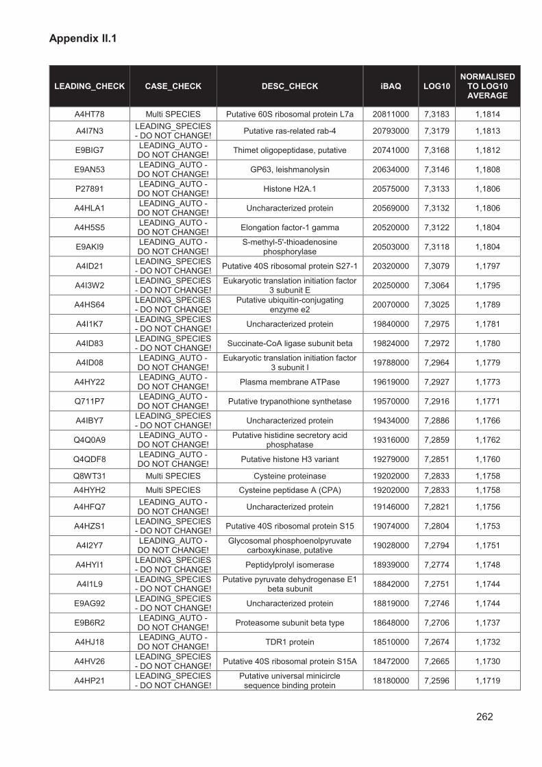
Appendix II.1
262
LEADING_CHECK CASE_CHECK DESC_CHECK iBAQ LOG10 NORMALISED
TO LOG10 AVERAGE
A4HT78 Multi SPECIES Putative 60S ribosomal protein L7a 20811000 7,3183 1,1814
A4I7N3 LEADING_SPECIES - DO NOT CHANGE!
Putative ras-related rab-4 20793000 7,3179 1,1813
E9BIG7 LEADING_AUTO - DO NOT CHANGE!
Thimet oligopeptidase, putative 20741000 7,3168 1,1812
E9AN53 LEADING_AUTO - DO NOT CHANGE!
GP63, leishmanolysin 20634000 7,3146 1,1808
P27891 LEADING_AUTO - DO NOT CHANGE!
Histone H2A.1 20575000 7,3133 1,1806
A4HLA1 LEADING_AUTO - DO NOT CHANGE!
Uncharacterized protein 20569000 7,3132 1,1806
A4H5S5 LEADING_AUTO - DO NOT CHANGE!
Elongation factor-1 gamma 20520000 7,3122 1,1804
E9AKI9 LEADING_AUTO - DO NOT CHANGE!
S-methyl-5'-thioadenosine phosphorylase
20503000 7,3118 1,1804
A4ID21 LEADING_SPECIES - DO NOT CHANGE!
Putative 40S ribosomal protein S27-1 20320000 7,3079 1,1797
A4I3W2 LEADING_SPECIES - DO NOT CHANGE!
Eukaryotic translation initiation factor 3 subunit E
20250000 7,3064 1,1795
A4HS64 LEADING_SPECIES - DO NOT CHANGE!
Putative ubiquitin-conjugating enzyme e2
20070000 7,3025 1,1789
A4I1K7 LEADING_SPECIES - DO NOT CHANGE!
Uncharacterized protein 19840000 7,2975 1,1781
A4ID83 LEADING_SPECIES - DO NOT CHANGE!
Succinate-CoA ligase subunit beta 19824000 7,2972 1,1780
A4ID08 LEADING_AUTO - DO NOT CHANGE!
Eukaryotic translation initiation factor 3 subunit I
19788000 7,2964 1,1779
A4HY22 LEADING_AUTO - DO NOT CHANGE!
Plasma membrane ATPase 19619000 7,2927 1,1773
Q711P7 LEADING_AUTO - DO NOT CHANGE!
Putative trypanothione synthetase 19570000 7,2916 1,1771
A4IBY7 LEADING_SPECIES - DO NOT CHANGE!
Uncharacterized protein 19434000 7,2886 1,1766
Q4Q0A9 LEADING_AUTO - DO NOT CHANGE!
Putative histidine secretory acid phosphatase
19316000 7,2859 1,1762
Q4QDF8 LEADING_AUTO - DO NOT CHANGE!
Putative histone H3 variant 19279000 7,2851 1,1760
Q8WT31 Multi SPECIES Cysteine proteinase 19202000 7,2833 1,1758
A4HYH2 Multi SPECIES Cysteine peptidase A (CPA) 19202000 7,2833 1,1758
A4HFQ7 LEADING_AUTO - DO NOT CHANGE!
Uncharacterized protein 19146000 7,2821 1,1756
A4HZS1 LEADING_SPECIES - DO NOT CHANGE!
Putative 40S ribosomal protein S15 19074000 7,2804 1,1753
A4I2Y7 LEADING_AUTO - DO NOT CHANGE!
Glycosomal phosphoenolpyruvate carboxykinase, putative
19028000 7,2794 1,1751
A4HYI1 LEADING_SPECIES - DO NOT CHANGE!
Peptidylprolyl isomerase 18939000 7,2774 1,1748
A4I1L9 LEADING_SPECIES - DO NOT CHANGE!
Putative pyruvate dehydrogenase E1 beta subunit
18842000 7,2751 1,1744
E9AG92 LEADING_SPECIES - DO NOT CHANGE!
Uncharacterized protein 18819000 7,2746 1,1744
E9B6R2 LEADING_AUTO - DO NOT CHANGE!
Proteasome subunit beta type 18648000 7,2706 1,1737
A4HJ18 LEADING_AUTO - DO NOT CHANGE!
TDR1 protein 18510000 7,2674 1,1732
A4HV26 LEADING_SPECIES - DO NOT CHANGE!
Putative 40S ribosomal protein S15A 18472000 7,2665 1,1730
A4HP21 LEADING_SPECIES - DO NOT CHANGE!
Putative universal minicircle sequence binding protein
18180000 7,2596 1,1719

Appendix II.1
263
LEADING_CHECK CASE_CHECK DESC_CHECK iBAQ LOG10 NORMALISED
TO LOG10 AVERAGE
A4I841 LEADING_SPECIES - DO NOT CHANGE!
Uncharacterized protein 17935000 7,2537 1,1710
A4ID74 LEADING_SPECIES - DO NOT CHANGE!
40S ribosomal protein S24 17933000 7,2537 1,1710
Q4Q8S3 LEADING_AUTO - DO NOT CHANGE!
Uncharacterized protein 17791000 7,2502 1,1704
A4IE56 LEADING_SPECIES - DO NOT CHANGE!
Putative oxidoreductase 17719000 7,2484 1,1701
P23223 LEADING_AUTO - DO NOT CHANGE!
Leishmanolysin 17636000 7,2464 1,1698
A4IBR8 LEADING_SPECIES - DO NOT CHANGE!
Putative 60S ribosomal protein L23 17432000 7,2413 1,1690
A4HSZ7 LEADING_SPECIES - DO NOT CHANGE!
Protein disulfide isomerase 17418000 7,2410 1,1689
Q4Q6Z5 Multi SPECIES Glyceraldehyde-3-phosphate
dehydrogenase 17309000 7,2383 1,1685
Q4Q6Z4 Multi SPECIES Glyceraldehyde-3-phosphate
dehydrogenase 17309000 7,2383 1,1685
E9ARK6 LEADING_AUTO - DO NOT CHANGE!
Citrate synthase 17114000 7,2334 1,1677
E9B3L2 LEADING_AUTO - DO NOT CHANGE!
Heat shock protein 83-1 17075000 7,2324 1,1675
E9B8M5 LEADING_SPECIES - DO NOT CHANGE!
Proteasome subunit beta type 16932000 7,2287 1,1669
A4I7P2 LEADING_SPECIES - DO NOT CHANGE!
Putative RNA binding protein 16759000 7,2242 1,1662
Q4QHH2 Multi SPECIES GP63, leishmanolysin 16224000 7,2102 1,1640
Q4QHH1 Multi SPECIES GP63, leishmanolysin 16224000 7,2102 1,1640
Q4QHG9 Multi SPECIES GP63, leishmanolysin 16224000 7,2102 1,1640
B8YDG1 Multi SPECIES GP63 16224000 7,2102 1,1640
Q9N9V3 Multi SPECIES Putative ribosomal protein L10 16206000 7,2097 1,1639
A4HS71 Multi SPECIES Putative 60S ribosomal protein L10 16206000 7,2097 1,1639
P62884 LEADING_SPECIES - DO NOT CHANGE!
Guanine nucleotide-binding protein subunit beta-like protein
16001000 7,2041 1,1630
A4I426 LEADING_SPECIES - DO NOT CHANGE!
Glutamate dehydrogenase 15985000 7,2037 1,1629
A4I2C4 LEADING_SPECIES - DO NOT CHANGE!
Uncharacterized protein 15795000 7,1985 1,1621
P14700 LEADING_AUTO - DO NOT CHANGE!
Membrane antigen containing repeating peptides (Fragment)
15610000 7,1934 1,1612
A4HFK0 LEADING_SPECIES - DO NOT CHANGE!
Putative small GTP-binding protein Rab1
15572000 7,1923 1,1611
A4IAD2 LEADING_SPECIES - DO NOT CHANGE!
Putative 60S ribosomal protein L21 15564000 7,1921 1,1610
A4IB25 LEADING_SPECIES - DO NOT CHANGE!
Histone H4 15499000 7,1903 1,1607
Q5EXB3 LEADING_AUTO - DO NOT CHANGE!
Hemoglobin receptor 15466000 7,1894 1,1606
A4I935 LEADING_SPECIES - DO NOT CHANGE!
Peptidyl-prolyl cis-trans isomerase 15412000 7,1879 1,1604
E9BHI2 LEADING_AUTO - DO NOT CHANGE!
Protein phosphatase, putative 15357000 7,1863 1,1601
E9B099 LEADING_AUTO - DO NOT CHANGE!
Putative heat-shock protein hsp70 15261000 7,1836 1,1597
A4HGY1 LEADING_AUTO - DO NOT CHANGE!
Putative heat-shock protein hsp70 15244000 7,1831 1,1596
A4H9Q9 LEADING_SPECIES - DO NOT CHANGE!
Nonspecific nucleoside hydrolasewith=GeneDB:LmjF18.1580
15134000 7,1800 1,1591

Appendix II.1
264
LEADING_CHECK CASE_CHECK DESC_CHECK iBAQ LOG10 NORMALISED
TO LOG10 AVERAGE
A4I574 LEADING_SPECIES - DO NOT CHANGE!
Putative aspartyl-tRNA synthetase 15031000 7,1770 1,1586
A4IBL4 LEADING_SPECIES - DO NOT CHANGE!
Putative cystathione gamma lyase 15003000 7,1762 1,1585
A0A1E1J358 Multi SPECIES ADP-ribosylation factor, putative 14981000 7,1755 1,1584
A0A088RXX3 Multi SPECIES ADP-ribosylation factor, putative 14981000 7,1755 1,1584
A4IDB2 LEADING_SPECIES - DO NOT CHANGE!
Putative translation elongation factor 1-beta
14958000 7,1749 1,1583
A0A1E1IN58 Multi SPECIES Putative small GTP binding protein
rab6-like protein 14863000 7,1721 1,1578
A0A088RH69 Multi SPECIES Small GTP binding protein rab6-like
protein 14863000 7,1721 1,1578
A4I0C2 Multi SPECIES Acetyl-coenzyme A synthetase 14842000 7,1715 1,1577
A4I093 Multi SPECIES Acetyl-coenzyme A synthetase 14842000 7,1715 1,1577
E9AE96 LEADING_AUTO - DO NOT CHANGE!
Uncharacterized protein 14748000 7,1687 1,1573
A4HWZ0 LEADING_SPECIES - DO NOT CHANGE!
Sucrose-phosphate synthase-like protein
14734000 7,1683 1,1572
Q8T6M2 LEADING_AUTO - DO NOT CHANGE!
Guanosine permease 14696000 7,1672 1,1570
Q9U4E3 LEADING_AUTO - DO NOT CHANGE!
ADP-ribosylation factor-like 3A 14634000 7,1654 1,1567
A4IAU1 Multi SPECIES 40S ribosomal protein S3a-2 14547000 7,1628 1,1563
A4IAU0 Multi SPECIES 40S ribosomal protein S3a-1 14547000 7,1628 1,1563
Q4QJG7 LEADING_AUTO - DO NOT CHANGE!
Trypanothione reductase 14514000 7,1618 1,1561
E9ACW0 LEADING_AUTO - DO NOT CHANGE!
Putative heat shock protein DNAJ 14428000 7,1592 1,1557
A4IAS5 LEADING_SPECIES - DO NOT CHANGE!
60S ribosomal protein L30 14388000 7,1580 1,1555
A9LNR9 LEADING_AUTO - DO NOT CHANGE!
Tryparedoxin peroxidase 14334000 7,1564 1,1553
A4H8V4 LEADING_SPECIES - DO NOT CHANGE!
Elongation factor 1-alpha 14289000 7,1550 1,1550
A4ID12 LEADING_SPECIES - DO NOT CHANGE!
Putative glycyl tRNA synthetase 14283000 7,1548 1,1550
E9BTH9 LEADING_SPECIES - DO NOT CHANGE!
Short chain 3-hydroxyacyl-CoA dehydrogenase, putative
14209000 7,1526 1,1547
A4HS12 LEADING_AUTO - DO NOT CHANGE!
Uncharacterized protein 14148000 7,1507 1,1544
A4HKH0 Multi SPECIES Superoxide dismutase 13808000 7,1401 1,1526
A4HKH1 Multi SPECIES Superoxide dismutase 13808000 7,1401 1,1526
A4ICS4 Multi SPECIES Putative ubiquitin/ribosomal protein
S27a 13696000 7,1366 1,1521
A4HW73 Multi SPECIES Putative ubiquitin/ribosomal protein
S27a 13696000 7,1366 1,1521
E9AK40 LEADING_AUTO - DO NOT CHANGE!
Putative spermidine synthase 13618000 7,1341 1,1517
A4H6Z3 LEADING_SPECIES - DO NOT CHANGE!
Putative NADH:flavin oxidoreductase/NADH oxidase
13527000 7,1312 1,1512
E9AUR8 LEADING_AUTO - DO NOT CHANGE!
Putative calpain-like cysteine peptidase
13524000 7,1311 1,1512
E9ARW7 LEADING_AUTO - DO NOT CHANGE!
Putative aminopeptidase 13469000 7,1293 1,1509
A4IAZ8 LEADING_SPECIES - DO NOT CHANGE!
Putative casein kinase 13413000 7,1275 1,1506

Appendix II.1
265
LEADING_CHECK CASE_CHECK DESC_CHECK iBAQ LOG10 NORMALISED
TO LOG10 AVERAGE
A0A142BXX3 Multi SPECIES 6-phosphogluconate dehydrogenase,
decarboxylating (Fragment) 13367000 7,1260 1,1504
A0A142BXX2 Multi SPECIES 6-phosphogluconate dehydrogenase,
decarboxylating (Fragment) 13367000 7,1260 1,1504
I3VJT5 Multi SPECIES 6-phosphogluconate dehydrogenase,
decarboxylating (Fragment) 13367000 7,1260 1,1504
Q18L52 Multi SPECIES 6-phosphogluconate dehydrogenase,
decarboxylating 13367000 7,1260 1,1504
Q18L04 Multi SPECIES 6-phosphogluconate dehydrogenase,
decarboxylating 13367000 7,1260 1,1504
Q5SDH3 LEADING_AUTO - DO NOT CHANGE!
Putative small myristoylated protein 4 13217000 7,1211 1,1496
Q8I496 LEADING_SPECIES - DO NOT CHANGE!
Hs1vu complex proteolytic subunit-like
13207000 7,1208 1,1495
A4HDR8 LEADING_SPECIES - DO NOT CHANGE!
Transketolase 13203000 7,1207 1,1495
A4I8P2 LEADING_SPECIES - DO NOT CHANGE!
Thiol-dependent reductase 1 13162000 7,1193 1,1493
A4IB89 Multi SPECIES Putative 60S Ribosomal protein L36 13160000 7,1193 1,1493
A4HZ73 Multi SPECIES Putative 60S Ribosomal protein L36 13160000 7,1193 1,1493
A4HWN5 LEADING_SPECIES - DO NOT CHANGE!
Putative ribosomal protein S6 13075000 7,1164 1,1488
E9BET4 LEADING_AUTO - DO NOT CHANGE!
Calpain-like cysteine peptidase, putative
13065000 7,1161 1,1488
A4HHL6 LEADING_SPECIES - DO NOT CHANGE!
Rab GDP dissociation inhibitor 12988000 7,1135 1,1484
Q4QDX9 LEADING_AUTO - DO NOT CHANGE!
Putative 60S ribosomal protein L10a 12928000 7,1115 1,1480
Q4QIE0 LEADING_AUTO - DO NOT CHANGE!
Superoxide dismutase 12908000 7,1109 1,1479
A4I7Q4 LEADING_SPECIES - DO NOT CHANGE!
60S ribosomal protein L18a 12906000 7,1108 1,1479
A4I1Q0 LEADING_SPECIES - DO NOT CHANGE!
Uncharacterized protein 12794000 7,1070 1,1473
A4I996 LEADING_SPECIES - DO NOT CHANGE!
Putative aminopeptidase 12740000 7,1052 1,1470
Q4QD68 LEADING_AUTO - DO NOT CHANGE!
Cysteine peptidase A (CPA) 12736000 7,1050 1,1470
E9B1Z9 LEADING_AUTO - DO NOT CHANGE!
GP63-like protein, leishmanolysin-like protein
12688000 7,1034 1,1467
A4H445 Multi SPECIES Surface antigen-like protein 12624000 7,1012 1,1464
A4H446 Multi SPECIES Surface antigen-like protein 12624000 7,1012 1,1464
A4H440 Multi SPECIES Surface antigen-like protein 12624000 7,1012 1,1464
A4H3Y2 Multi SPECIES Surface antigen-like protein 12624000 7,1012 1,1464
E9AP91 LEADING_AUTO - DO NOT CHANGE!
Putative lectin 12620000 7,1011 1,1463
E9BUX4 LEADING_AUTO - DO NOT CHANGE!
Uncharacterized protein 12590000 7,1000 1,1462
E9ADY9 LEADING_AUTO - DO NOT CHANGE!
Putative carnitine/choline acetyltransferase
12582000 7,0997 1,1461
A4ICV5 LEADING_SPECIES - DO NOT CHANGE!
Putative proteasome beta 2 subunit 12464000 7,0957 1,1455
E9AHL7 LEADING_SPECIES - DO NOT CHANGE!
Putative ADP-ribosylation factor 12447000 7,0951 1,1454
A4H4D9 LEADING_SPECIES - DO NOT CHANGE!
Putative dipeptidyl-peptidase III 12376000 7,0926 1,1450
E9AKY4 LEADING_AUTO - DO NOT CHANGE!
Protein disulfide isomerase 12352000 7,0917 1,1448

Appendix II.1
266
LEADING_CHECK CASE_CHECK DESC_CHECK iBAQ LOG10 NORMALISED
TO LOG10 AVERAGE
E9AZH2 LEADING_AUTO - DO NOT CHANGE!
Proteasome subunit beta type 12337000 7,0912 1,1447
A4I191 Multi SPECIES RNA-binding protein, putative, UPB2 12286000 7,0894 1,1445
E9AH94 Multi SPECIES RNA-binding protein, putative, UPB1 12286000 7,0894 1,1445
A4HYZ5 LEADING_SPECIES - DO NOT CHANGE!
Putative 40S ribosomal protein S11 12261000 7,0885 1,1443
O00912 LEADING_SPECIES - DO NOT CHANGE!
Histone H4 12223000 7,0872 1,1441
E9AGX4 LEADING_SPECIES - DO NOT CHANGE!
Putative 40S ribosomal protein S23 12165000 7,0851 1,1438
A4HQG9 LEADING_AUTO - DO NOT CHANGE!
Putative histidine secretory acid phosphatase
12145000 7,0844 1,1436
E9AWS6 LEADING_AUTO - DO NOT CHANGE!
Triosephosphate isomerase 12118000 7,0834 1,1435
A4I1V1 LEADING_SPECIES - DO NOT CHANGE!
Adenine phosphoribosyltransferase 12044000 7,0808 1,1431
E9BBL6 LEADING_AUTO - DO NOT CHANGE!
Pyrroline-5-carboxylate reductase 11965000 7,0779 1,1426
A0A1E1IRP4 Multi SPECIES Uncharacterized protein 11957000 7,0776 1,1426
A0A088RML4 Multi SPECIES Uncharacterized protein 11957000 7,0776 1,1426
E8NHG6 LEADING_AUTO - DO NOT CHANGE!
Putative uncharacterized protein LmxM_30_0440_1
11954000 7,0775 1,1425
E9B2V9 LEADING_AUTO - DO NOT CHANGE!
Superoxide dismutase 11930000 7,0766 1,1424
E9AHK3 LEADING_SPECIES - DO NOT CHANGE!
S-adenosylmethionine synthase 11867000 7,0743 1,1420
A4HVX3 LEADING_AUTO - DO NOT CHANGE!
Proteasome subunit alpha type 11839000 7,0733 1,1419
A4HRR9 LEADING_SPECIES - DO NOT CHANGE!
Dipeptylcarboxypeptidase 11757000 7,0703 1,1414
A4HVW1 LEADING_SPECIES - DO NOT CHANGE!
Uncharacterized protein 11671000 7,0671 1,1409
E9AHJ2 Multi SPECIES Putative paraflagellar rod protein 1D 11634000 7,0657 1,1406
Q66V59 Multi SPECIES Paraflagellar rod protein 1 11634000 7,0657 1,1406
A4I4N5 Multi SPECIES Putative paraflagellar rod protein 1D 11634000 7,0657 1,1406
O18699 LEADING_SPECIES - DO NOT CHANGE!
Cysteine protease 11603000 7,0646 1,1404
A4HW72 LEADING_SPECIES - DO NOT CHANGE!
Uncharacterized protein 11565000 7,0631 1,1402
A4ICB5 LEADING_SPECIES - DO NOT CHANGE!
Putative prolyl oligopeptidase 11466000 7,0594 1,1396
E9B3P3 LEADING_AUTO - DO NOT CHANGE!
Uncharacterized protein 11466000 7,0594 1,1396
A4I4E1 LEADING_SPECIES - DO NOT CHANGE!
Putative guanine deaminase 11359000 7,0553 1,1390
A4HX65 LEADING_SPECIES - DO NOT CHANGE!
Uncharacterized protein 11357000 7,0553 1,1389
A4HV05 LEADING_SPECIES - DO NOT CHANGE!
40S ribosomal protein S5 11343000 7,0547 1,1389
E9BCQ4 LEADING_AUTO - DO NOT CHANGE!
Uncharacterized protein 11332000 7,0543 1,1388
Q4Q504 LEADING_SPECIES - DO NOT CHANGE!
Putative ribosomal protein L27 11260000 7,0515 1,1383
A4HTJ5 LEADING_SPECIES - DO NOT CHANGE!
Putative small ubiquitin protein 11221000 7,0500 1,1381
A4ICK8 LEADING_SPECIES - DO NOT CHANGE!
Fructose-bisphosphate aldolase 11178000 7,0484 1,1378

Appendix II.1
267
LEADING_CHECK CASE_CHECK DESC_CHECK iBAQ LOG10 NORMALISED
TO LOG10 AVERAGE
E8NHC1 LEADING_AUTO - DO NOT CHANGE!
Proteasome subunit beta type 11054000 7,0435 1,1371
A4HLW4 LEADING_AUTO - DO NOT CHANGE!
Putative carboxypeptidase 10882000 7,0367 1,1360
A4I885 LEADING_SPECIES - DO NOT CHANGE!
Putative cysteine desulfhydrase 10870000 7,0362 1,1359
Q9BHZ6 Multi SPECIES Elongation factor-1 gamma 10862000 7,0359 1,1358
A4HU19 Multi SPECIES Elongation factor-1 gamma 10862000 7,0359 1,1358
A4HU18 Multi SPECIES Elongation factor-1 gamma 10862000 7,0359 1,1358
A4I3Z9 LEADING_SPECIES - DO NOT CHANGE!
Uncharacterized protein 10842000 7,0351 1,1357
E9AW99 LEADING_AUTO - DO NOT CHANGE!
Putative uncharacterized protein 10740000 7,0310 1,1350
Q4QF83 LEADING_AUTO - DO NOT CHANGE!
NAD-specific glutamate dehydrogenase
10736000 7,0308 1,1350
O43992 LEADING_AUTO - DO NOT CHANGE!
40S ribosomal protein S2 10688000 7,0289 1,1347
E9ACC9 LEADING_AUTO - DO NOT CHANGE!
Uncharacterized protein 10528000 7,0223 1,1336
A0A088RLI0 LEADING_AUTO - DO NOT CHANGE!
Histone H2B 10499000 7,0211 1,1334
A4HVR4 LEADING_SPECIES - DO NOT CHANGE!
Uncharacterized protein 10498000 7,0211 1,1334
E9AYP9 LEADING_AUTO - DO NOT CHANGE!
Putative uncharacterized protein 10449000 7,0191 1,1331
A0A1E1J8P0 LEADING_AUTO - DO NOT CHANGE!
Putative S-adenosylhomocysteine hydrolase
10445000 7,0189 1,1331
A4HQL6 LEADING_AUTO - DO NOT CHANGE!
Protein disulfide-isomerase 10442000 7,0188 1,1331
E9AHB0 Multi SPECIES Putative 60S ribosomal protein L7 10367000 7,0157 1,1326
A4I1V4 Multi SPECIES Putative 60S ribosomal protein L7 10367000 7,0157 1,1326
Q4QBL1 LEADING_AUTO - DO NOT CHANGE!
Farnesyl pyrophosphate synthase 10321000 7,0137 1,1322
A0A1E1J0K7 Multi SPECIES 40S ribosomal protein S17, putative 10315000 7,0135 1,1322
A0A088RXB2 Multi SPECIES 40S ribosomal protein S17, putative 10315000 7,0135 1,1322
A4HMQ9 LEADING_AUTO - DO NOT CHANGE!
Putative aminopeptidase P 10241000 7,0103 1,1317
A4I5C0 LEADING_SPECIES - DO NOT CHANGE!
Putative adenosine kinase 10235000 7,0101 1,1317
A4I5Z0 LEADING_SPECIES - DO NOT CHANGE!
Uncharacterized protein 10164000 7,0071 1,1312
A4HY61 LEADING_SPECIES - DO NOT CHANGE!
Putative 40S ribosomal protein S13 10143000 7,0062 1,1310
Q4QDX3 LEADING_AUTO - DO NOT CHANGE!
Probable citrate synthase, mitochondrial
10117000 7,0051 1,1308
A4HI30 LEADING_SPECIES - DO NOT CHANGE!
Putative 4-methyl-5(Beta-hydroxyethyl)-thiazole
monophosphate synthesis protein 10093000 7,0040 1,1307
A4I890 LEADING_SPECIES - DO NOT CHANGE!
Putative ribosomal protein L27 10091000 7,0039 1,1307
A4HU23 LEADING_SPECIES - DO NOT CHANGE!
Uncharacterized protein 10064000 7,0028 1,1305
A4H746 LEADING_AUTO - DO NOT CHANGE!
40S ribosomal protein S12 10051000 7,0022 1,1304
E9AII6 LEADING_SPECIES - DO NOT CHANGE!
Contig, possible fusion of chromosomes 20 and 34
10038000 7,0016 1,1303
A4I067 LEADING_SPECIES - DO NOT CHANGE!
Pteridine reductase 1 9968900 6,9986 1,1298

Appendix II.1
268
LEADING_CHECK CASE_CHECK DESC_CHECK iBAQ LOG10 NORMALISED
TO LOG10 AVERAGE
Q4Q6F6 LEADING_AUTO - DO NOT CHANGE!
Biotin/lipoate protein ligase-like protein
9954600 6,9980 1,1297
A4HX73 Multi SPECIES Elongation factor 1-alpha 9950300 6,9978 1,1297
E9AGP5 Multi SPECIES Elongation factor 1-alpha 9950300 6,9978 1,1297
A4HT92 LEADING_SPECIES - DO NOT CHANGE!
Putative 40S ribosomal protein S9 9923600 6,9967 1,1295
A4HV19 LEADING_SPECIES - DO NOT CHANGE!
Putative 60S ribosomal protein L28 9922600 6,9966 1,1295
A4HDN8 LEADING_AUTO - DO NOT CHANGE!
Inhibitor of cysteine peptidase 9877400 6,9946 1,1292
A4HCH8 LEADING_AUTO - DO NOT CHANGE!
FPPS protein 9716400 6,9875 1,1280
E9AEF4 LEADING_AUTO - DO NOT CHANGE!
Uncharacterized protein 9692000 6,9864 1,1278
E9ACQ0 LEADING_AUTO - DO NOT CHANGE!
Surface antigen-like protein 9659700 6,9850 1,1276
A4HXT8 LEADING_AUTO - DO NOT CHANGE!
Putative 60S ribosomal protein L10a 9620100 6,9832 1,1273
A4HE56 LEADING_SPECIES - DO NOT CHANGE!
Aldehyde dehydrogenase, mitochondrial
9561600 6,9805 1,1269
Q27686 LEADING_AUTO - DO NOT CHANGE!
Pyruvate kinase 9543900 6,9797 1,1268
A4I2F5 LEADING_AUTO - DO NOT CHANGE!
Putative nitrilase 9474400 6,9766 1,1262
Q4QIX1 LEADING_AUTO - DO NOT CHANGE!
Protein disulfide isomerase 9325200 6,9697 1,1251
F8QV42 LEADING_AUTO - DO NOT CHANGE!
Tubulin beta chain 9321600 6,9695 1,1251
A4H942 LEADING_AUTO - DO NOT CHANGE!
Uncharacterized protein 9313200 6,9691 1,1250
A4I1Z8 LEADING_SPECIES - DO NOT CHANGE!
Uncharacterized protein 9266800 6,9669 1,1247
A4I9G0 LEADING_SPECIES - DO NOT CHANGE!
Small nuclear ribonucleoprotein SmD2
9223300 6,9649 1,1244
B8Y658 LEADING_SPECIES - DO NOT CHANGE!
Methylene-tetrahydrofolate dehydrogenase/cyclohydrolase
9209300 6,9642 1,1242
Q70GE8 LEADING_AUTO - DO NOT CHANGE!
Thiol-dependent reductase 1 9171100 6,9624 1,1240
A4I849 LEADING_SPECIES - DO NOT CHANGE!
Uncharacterized protein 9114400 6,9597 1,1235
E9AGX7 LEADING_SPECIES - DO NOT CHANGE!
T-complex protein 1 subunit delta 9067600 6,9575 1,1232
E9BQ78 LEADING_SPECIES - DO NOT CHANGE!
Mitogen activated protein kinase, putative
8964200 6,9525 1,1224
A4HB47 LEADING_SPECIES - DO NOT CHANGE!
Contig, possible fusion of chromosomes 20 and 34
8918500 6,9503 1,1220
A4I412 Multi SPECIES Putative heat-shock protein hsp70 8894100 6,9491 1,1218
E9AHH1 Multi SPECIES Putative heat-shock protein hsp70 8894100 6,9491 1,1218
Q4QGD8 LEADING_AUTO - DO NOT CHANGE!
Uncharacterized protein 8874200 6,9481 1,1217
A4HWC9 LEADING_SPECIES - DO NOT CHANGE!
Putative ribonucleoprotein p18, mitochondrial
8809100 6,9449 1,1211
A4I2T4 LEADING_SPECIES - DO NOT CHANGE!
Putative arginyl-tRNA synthetase 8701500 6,9396 1,1203
E9AHP6 LEADING_SPECIES - DO NOT CHANGE!
Uncharacterized protein 8670600 6,9380 1,1200
A4HSP6 LEADING_AUTO - DO NOT CHANGE!
Uncharacterized protein 8649100 6,9370 1,1198

Appendix II.1
269
LEADING_CHECK CASE_CHECK DESC_CHECK iBAQ LOG10 NORMALISED
TO LOG10 AVERAGE
A4HFU7 LEADING_AUTO - DO NOT CHANGE!
Uncharacterized protein 8636800 6,9364 1,1197
Q6QMI0 Multi SPECIES Nonspecific nucleoside hydrolase
(Fragment) 8601200 6,9346 1,1195
Q6QMJ1 Multi SPECIES Nonspecific nucleoside hydrolase
(Fragment) 8601200 6,9346 1,1195
Q6QMI7 Multi SPECIES Nonspecific nucleoside hydrolase
(Fragment) 8601200 6,9346 1,1195
Q2PDC6 Multi SPECIES Inosine-uridine preferring nucleoside
hydrolase 8601200 6,9346 1,1195
Q2PDC5 Multi SPECIES Inosine-uridine preferring nucleoside
hydrolase 8601200 6,9346 1,1195
Q2PDB9 Multi SPECIES Inosine-uridine preferring nucleoside
hydrolase 8601200 6,9346 1,1195
Q2PD97 Multi SPECIES Inosine-uridine preferring nucleoside
hydrolase 8601200 6,9346 1,1195
Q2PD96 Multi SPECIES Inosine-uridine preferring nucleoside
hydrolase 8601200 6,9346 1,1195
A4I4C9 LEADING_SPECIES - DO NOT CHANGE!
Putative lipophosphoglycan biosynthetic protein
8589400 6,9340 1,1194
C7EX18 LEADING_AUTO - DO NOT CHANGE!
Gp63 8559700 6,9325 1,1191
A4ICD5 LEADING_SPECIES - DO NOT CHANGE!
Protein disulfide-isomerase 8539900 6,9315 1,1190
A4H8B2 LEADING_SPECIES - DO NOT CHANGE!
Proliferating cell nuclear antigen 8539700 6,9314 1,1190
A4I1F4 LEADING_SPECIES - DO NOT CHANGE!
Aldehyde dehydrogenase, mitochondrial
8522100 6,9305 1,1188
Q4QFJ8 LEADING_AUTO - DO NOT CHANGE!
Inositol-3-phosphate synthase 8477500 6,9283 1,1184
E9AWR7 LEADING_AUTO - DO NOT CHANGE!
Malic enzyme 8476600 6,9282 1,1184
A4HXD8 LEADING_SPECIES - DO NOT CHANGE!
Uncharacterized protein 8463000 6,9275 1,1183
A0A1E1ISN0 Multi SPECIES Putative cystathionine beta-synthase 8460200 6,9274 1,1183
A0A088RP82 Multi SPECIES Cystathionine beta-synthase 8460200 6,9274 1,1183
A4ID19 LEADING_SPECIES - DO NOT CHANGE!
Nascent polypeptide-associated complex subunit beta
8399300 6,9242 1,1178
A4I7N0 LEADING_SPECIES - DO NOT CHANGE!
Profilin 8323000 6,9203 1,1172
A4I082 LEADING_SPECIES - DO NOT CHANGE!
Aldose 1-epimerase-like protein 8314600 6,9198 1,1171
A0A0S1M2A5 Multi SPECIES Putative spermidine synthase 1
(Fragment) 8296200 6,9189 1,1169
A0A0S1M298 Multi SPECIES Putative spermidine synthase 1
(Fragment) 8296200 6,9189 1,1169
A0A0S1M285 Multi SPECIES Putative spermidine synthase 1
(Fragment) 8296200 6,9189 1,1169
A4H3X8 Multi SPECIES Putative spermidine synthase 8296200 6,9189 1,1169
A4IDL0 LEADING_SPECIES - DO NOT CHANGE!
Tyrosine aminotransferase 8254100 6,9167 1,1166
A4I3Q8 LEADING_SPECIES - DO NOT CHANGE!
Putative replication factor A, 51kDa subunit
8238700 6,9159 1,1164
E9BB38 LEADING_AUTO - DO NOT CHANGE!
Alanine aminotransferase 8238400 6,9158 1,1164
A4HRT6 LEADING_SPECIES - DO NOT CHANGE!
Putative ribosomal protein L38 8214700 6,9146 1,1162
A4H635 LEADING_AUTO - DO NOT CHANGE!
GP63-4 protein 8210400 6,9144 1,1162

Appendix II.1
270
LEADING_CHECK CASE_CHECK DESC_CHECK iBAQ LOG10 NORMALISED
TO LOG10 AVERAGE
A4I253 LEADING_AUTO - DO NOT CHANGE!
Heat shock protein 70-related protein 8198100 6,9137 1,1161
A9YYK9 Multi SPECIES p4 nuclease (Fragment) 8185800 6,9131 1,1160
A4I5I0 Multi SPECIES p1/s1 nuclease 8185800 6,9131 1,1160
E9JUH3 Multi SPECIES Trypanothione reductase (Fragment) 8182100 6,9129 1,1160
E9JUG5 Multi SPECIES Trypanothione reductase (Fragment) 8182100 6,9129 1,1160
E9BB84 LEADING_AUTO - DO NOT CHANGE!
Carboxypeptidase, putative 8178000 6,9126 1,1159
A4IAG9 LEADING_SPECIES - DO NOT CHANGE!
Nucleolar protein family a member-like protein
8167600 6,9121 1,1158
Q8I8E1 Multi SPECIES Protein disulfide-isomerase 8159700 6,9117 1,1158
Q4Q059 Multi SPECIES Protein disulfide-isomerase 8159700 6,9117 1,1158
E9BG32 LEADING_AUTO - DO NOT CHANGE!
GDP-mannose pyrophosphorylase 8137900 6,9105 1,1156
A4HWJ8 LEADING_AUTO - DO NOT CHANGE!
60S ribosomal protein L6 8124700 6,9098 1,1155
I3VJK5 Multi SPECIES Fumarate hydratase (Fragment) 8066700 6,9067 1,1150
A2CIQ7 Multi SPECIES Fumarate hydratase 8066700 6,9067 1,1150
A2CIQ1 Multi SPECIES Fumarate hydratase 8066700 6,9067 1,1150
A2CIP4 Multi SPECIES Fumarate hydratase 8066700 6,9067 1,1150
A2CIN2 Multi SPECIES Fumarate hydratase 8066700 6,9067 1,1150
A2CIN0 Multi SPECIES Fumarate hydratase 8066700 6,9067 1,1150
A4H9H8 LEADING_AUTO - DO NOT CHANGE!
Probable citrate synthase, mitochondrial
8021700 6,9043 1,1146
A4IBC5 LEADING_SPECIES - DO NOT CHANGE!
Uncharacterized protein 7993600 6,9027 1,1143
A2CIM6 Multi SPECIES Glucose-6-phosphate 1-
dehydrogenase 7927800 6,8992 1,1137
A2CIL1 Multi SPECIES Glucose-6-phosphate 1-
dehydrogenase 7927800 6,8992 1,1137
A2CIK1 Multi SPECIES Glucose-6-phosphate 1-
dehydrogenase 7927800 6,8992 1,1137
A2CIJ8 Multi SPECIES Glucose-6-phosphate 1-
dehydrogenase 7927800 6,8992 1,1137
A4HVI6 LEADING_SPECIES - DO NOT CHANGE!
40S ribosomal protein S12 7922800 6,8989 1,1137
P42865 LEADING_AUTO - DO NOT CHANGE!
Probable quinone oxidoreductase 7871000 6,8960 1,1132
A7UFI6 Multi SPECIES Malate dehydrogenase 7838600 6,8942 1,1130
A4HAC0 Multi SPECIES Malate dehydrogenase 7838600 6,8942 1,1130
Q1A5Y1 LEADING_AUTO - DO NOT CHANGE!
Tubulin beta chain (Fragment) 7770400 6,8904 1,1123
Q9N9V2 Multi SPECIES Nascent polypeptide associated complex homologue, alpha chain
7764500 6,8901 1,1123
A4HS73 Multi SPECIES Nascent polypeptide associated
complex subunit-like protein, copy 1 7764500 6,8901 1,1123
A4HS72 Multi SPECIES Nascent polypeptide associated
complex subunit-like protein, copy 2 7764500 6,8901 1,1123
P25204 LEADING_AUTO - DO NOT CHANGE!
40S ribosomal protein S8 7762500 6,8900 1,1123
E9B3B0 LEADING_AUTO - DO NOT CHANGE!
Putative chaperonin alpha subunit 7704400 6,8867 1,1117
Q4QGJ6 LEADING_AUTO - DO NOT CHANGE!
Putative surface antigen protein 2 7645300 6,8834 1,1112

Appendix II.1
271
LEADING_CHECK CASE_CHECK DESC_CHECK iBAQ LOG10 NORMALISED
TO LOG10 AVERAGE
Q2HZY7 Multi SPECIES Elongation factor 2 7632600 6,8827 1,1111
A4HNM7 Multi SPECIES Elongation factor 2 7632600 6,8827 1,1111
A4I478 LEADING_SPECIES - DO NOT CHANGE!
Putative thymine-7-hydroxylase 7508000 6,8755 1,1099
Q4Q276 LEADING_AUTO - DO NOT CHANGE!
Uncharacterized protein 7437800 6,8714 1,1093
E9ARS1 LEADING_AUTO - DO NOT CHANGE!
Putative heat shock protein 7388800 6,8686 1,1088
A4HCV5 LEADING_SPECIES - DO NOT CHANGE!
Uncharacterized protein 7387700 6,8685 1,1088
A4HXQ3 LEADING_SPECIES - DO NOT CHANGE!
Glycogen synthase kinase 3 7374100 6,8677 1,1087
Q9N9V5 LEADING_SPECIES - DO NOT CHANGE!
Putative 40S ribosomal protein S33 7367100 6,8673 1,1086
A4HKX6 LEADING_SPECIES - DO NOT CHANGE!
Dihydrolipoyl dehydrogenase 7358900 6,8668 1,1085
A4HCZ3 LEADING_SPECIES - DO NOT CHANGE!
T-complex protein 1 subunit gamma 7346600 6,8661 1,1084
Q2PD92 LEADING_SPECIES - DO NOT CHANGE!
Aspartate aminotransferase 7265000 6,8612 1,1076
A0A1E1J180 Multi SPECIES Aspartyl
aminopeptidase,putative,metallo-peptidase, Clan MH, Family M20
7237900 6,8596 1,1074
A0A088RY37 Multi SPECIES Aspartyl aminopeptidase, putative 7237900 6,8596 1,1074
A4ICM4 LEADING_SPECIES - DO NOT CHANGE!
Putative ribosomal protein L24 7176300 6,8559 1,1068
Q4Q8S1 LEADING_AUTO - DO NOT CHANGE!
6-phosphogluconolactonase 7162800 6,8551 1,1066
A4HSS8 LEADING_SPECIES - DO NOT CHANGE!
Putative glutamine synthetase 7136800 6,8535 1,1064
A4HFS1 LEADING_SPECIES - DO NOT CHANGE!
Uncharacterized protein 7075200 6,8497 1,1058
A4HI66 LEADING_SPECIES - DO NOT CHANGE!
Importin subunit alpha 7069400 6,8494 1,1057
A4HUB4 LEADING_SPECIES - DO NOT CHANGE!
Putative ribosomal protein l35a 7035600 6,8473 1,1054
E9BEP5 LEADING_AUTO - DO NOT CHANGE!
1,2-dihydroxy-3-keto-5-methylthiopentene dioxygenase
7032900 6,8471 1,1053
A4IA81 LEADING_SPECIES - DO NOT CHANGE!
Uncharacterized protein 6980300 6,8439 1,1048
P42922 LEADING_SPECIES - DO NOT CHANGE!
60S ribosomal protein L11 6869200 6,8369 1,1037
E9AKK2 LEADING_AUTO - DO NOT CHANGE!
Putative dipeptidyl-peptidase III (Metallo-peptidase, clan m-, family
m49) 6803900 6,8328 1,1030
E9AYH6 LEADING_AUTO - DO NOT CHANGE!
Putative uncharacterized protein 6794400 6,8322 1,1029
A4HUL2 LEADING_SPECIES - DO NOT CHANGE!
Nucleoside phosphorylase-like protein
6661600 6,8236 1,1015
E9AGM7 Multi SPECIES Putative nucleoside transporter 1 6651400 6,8229 1,1014
E9AGM6 Multi SPECIES Putative nucleoside transporter 1 6651400 6,8229 1,1014
E9AGM5 Multi SPECIES Putative nucleoside transporter 1 6651400 6,8229 1,1014
A4HWK9 Multi SPECIES Putative nucleoside transporter 1 6651400 6,8229 1,1014
A4HU05 LEADING_SPECIES - DO NOT CHANGE!
Uncharacterized protein 6633800 6,8218 1,1013
A4HHW2 LEADING_SPECIES - DO NOT CHANGE!
Putative 2-hydroxy-3-oxopropionate reductase
6632500 6,8217 1,1012

Appendix II.1
272
LEADING_CHECK CASE_CHECK DESC_CHECK iBAQ LOG10 NORMALISED
TO LOG10 AVERAGE
E9AQI0 LEADING_AUTO - DO NOT CHANGE!
Protein tyrosine phosphatase-like protein
6618900 6,8208 1,1011
E9AU19 LEADING_AUTO - DO NOT CHANGE!
Putative kinetoplast-associated protein
6607400 6,8200 1,1010
Q4QAI0 LEADING_SPECIES - DO NOT CHANGE!
Putative IgE-dependent histamine-releasing factor
6601000 6,8196 1,1009
A4ID58 Multi SPECIES Putative ribosomal protein L29 6597900 6,8194 1,1009
A4ID57 Multi SPECIES Putative ribosomal protein L29 6597900 6,8194 1,1009
A4HNP0 LEADING_SPECIES - DO NOT CHANGE!
Putative proteasome beta 2 subunit 6574500 6,8179 1,1006
E9ATD8 LEADING_AUTO - DO NOT CHANGE!
Putative cysteine synthase 6562000 6,8170 1,1005
Q4QBF5 LEADING_AUTO - DO NOT CHANGE!
Putative endoribonuclease L-PSP (Pb5)
6543800 6,8158 1,1003
E9B6B5 LEADING_AUTO - DO NOT CHANGE!
Putative aminopeptidase P 6530900 6,8150 1,1002
A4HSB6 LEADING_SPECIES - DO NOT CHANGE!
Putative RNA-binding protein 6519300 6,8142 1,1000

273
Appendix V.1
Data was collected from the Allele Frequency Net Database for the following world
regions: South and Central America (Table 1), North Africa (Table 2), Western Asia (Table 3),
South Asia (Table 4).
Data were retrieved from AFND through a ‘HLA allele freq (Classical)’ search with the
following filters: i) region name; ii) level of resolution=2-digits; iii) Population standard=Gold
and Silver (8); iv) show frequencies=only positives
Table 1 Population studies from AFND included in the HLA allele frequency analysis of the South and Central America World region.
World region: SOUTH AND CENTRAL AMERICA
Population Ethnicity Study Sample
Size HLA Loci
Argentina Buenos Aires Caucasoid HLA 466 A, B, C, DPB1, DQA1, DQB1, DRB1
Argentina Buenos Aires pop 2 Caucasoid HLA 1216 A, B, DRB1
Argentina Chiriguano Amerindian HLA 54 A, B, DPB1, DQA1, DQB1, DRB1
Argentina Chubut Tehuelche Amerindian HLA 23 DRB1
Argentina Corrientes Mixed HLA 155 A, B, DRB1
Argentina Cuyo Region Caucasoid HLA 420 A, B, DRB1
Argentina Gran Chaco Eastern Toba
Amerindian HLA 135 A, B, DPB1, DQA1, DQB1, DRB1
Argentina Gran Chaco Mataco Wichi
Amerindian HLA 49 A, B, DPB1, DQA1, DQB1, DRB1
Argentina Gran Chaco Western Toba Pilaga
Amerindian HLA 19 A, B, DPB1, DQA1, DQB1, DRB1
Argentina Kolla Amerindian HLA 61 DQB1, DRB1
Argentina La Plata Caucasoid HLA 100 A, B, C, DRB1
Argentina Mapuche Amerindian HLA 48 DPB1, DQA1, DQB1, DRB1
Argentina Rio Negro Mapuche Amerindian HLA 34 DRB1
Argentina Rosario Toba Amerindian HLA 86 A, B, DQA1, DQB1, DRB1
Argentina Salta Wichi pop 2 Amerindian HLA 19 A, B, DPB1, DQA1, DQB1, DRB1
Bolivia Aymara Amerindian HLA 102 A, B, DQB1, DRB1
Bolivia Quechua HLA 80 DQB1, DRB1, A, B
Brazil Belem Mixed Mixed HLA 100 B
Brazil Belo Horizonte Caucasian Caucasoid HLA 95 A, B
Brazil Central Plateau Xavante Amerindian HLA 74 DPB1, DQA1, DQB1, DRB1
Brazil Curitiba-Parana Mixed Mixed HLA 264 A, B, C, DRB1
Brazil Guarani Kaiowa Amerindian HLA 155 DQA1, DQB1, DRB1
Brazil Guarani M bya Amerindian HLA 93 DQA1, DQB1, DRB1

Appendix V.1
274
Table 1 (continued)
Population Ethnicity Study Sample
Size HLA Loci
Brazil Guarani Nandeva Amerindian HLA 86 DQA1, DQB1, DRB1
Brazil Kaingang Amerindian HLA 235 DQA1, DQB1, DRB1
Brazil Mato Grosso do Sul Mixed HLA 203 DRB1
Brazil Minas Gerais State Mixed Mixed HLA 1 A, B, DRB1
Brazil North East Mixed Mixed HLA 205 DQA1, DQB1, DRB1
Brazil Parana Afro Brazilian Black HLA 77 A, B, DRB1
Brazil Parana Cafuzo Mixed HLA 319 A, B, DRB1
Brazil Parana Caucasian Caucasoid HLA 2775 A, B, DRB1
Brazil Parana Japanese HLA 192 A B C
Brazil Parana Mulatto Mulatto HLA 186 A, B, DRB1
Brazil Parana Oriental Oriental HLA 33 A, B, DRB1
Brazil Pernambuco Mixed Mixed HLA 101 A, B, C
Brazil Piaui Mixed Mixed HLA 21943 A, B, DRB1
Brazil REDOME Acre Mixed HLA 859 A, B, DRB1
Brazil REDOME Alagoas Mixed HLA 25349 A, B, DRB1
Brazil REDOME Amapa Mixed HLA 17,864 A, B, DRB1
Brazil REDOME Amazonas Mixed HLA 24,129 A, B, DRB1
Brazil REDOME Bahia Mixed HLA 47399 A, B, DRB1
Brazil REDOME Ceara Mixed HLA 101217 A, B, DRB1
Brazil REDOME Espirito Santo Mixed HLA 88,485 A, B, DRB1
Brazil REDOME Federal District Mixed HLA 29549 A, B, DRB1
Brazil REDOME Goias Mixed HLA 88,574 A, B, DRB1
Brazil REDOME Maranhao Mixed HLA 10180 A, B, DRB1
Brazil REDOME Mato Grosso Mixed HLA 34649 A, B, DRB1
Brazil REDOME Mato Grosso do Sul Mixed HLA 95667 A, B, DRB1
Brazil REDOME Minas Gerais Mixed HLA 211275 A, B, DRB1
Brazil REDOME Para Mixed HLA 72637 A, B, DRB1
Brazil REDOME Paraiba Mixed HLA 43868 A, B, DRB1
Brazil REDOME Parana Mixed HLA 341639 A, B, DRB1
Brazil REDOME Pernambuco Mixed HLA 92332 A, B, DRB1
Brazil REDOME Piaui Mixed HLA 46140 A, B, DRB1
Brazil REDOME Rio de Janeiro Mixed HLA 139322 A, B, DRB1
Brazil REDOME Rio Grande do Norte
Mixed HLA 46603 A, B, DRB1
Brazil REDOME Rio Grande do Sul Mixed HLA 241329 A, B, DRB1
Brazil REDOME Rondonia Mixed HLA 54396 A, B, DRB1
Brazil REDOME Roraima Mixed HLA 4140 A, B, DRB1
Brazil REDOME Santa Catarina Mixed HLA 106673 A, B, DRB1
Brazil REDOME Sao Paulo Mixed HLA 800809 A, B, DRB1

Appendix V.1
275
Table 1 (continued)
Population Ethnicity Study Sample
Size HLA Loci
Brazil REDOME Sergipe Mixed HLA 7321 A, B, DRB1
Brazil REDOME Tocantins Mixed HLA 20692 A, B, DRB1
Brazil Rio Grande do Norte Mestizo Mestizo HLA 12973 A, B, DRB1
Brazil Rio Grande do Sul Black Black HLA 248 A, B, DRB1
Brazil Rio Grande do Sul Caucasoid Caucasoid HLA 4428 A, B, DRB1
Brazil Rio Grande do Sul Mixed Mixed HLA 324 A, B, DRB1
Brazil Sao Paulo Bauru Mixed Mixed HLA 3542 A, B, DRB1
Brazil Sao Paulo Mixed Mixed HLA 239 A, B, DQA1, DQB1, DRB1
Brazil South East Cord Bllod Unknown HLA 11409 A B DRB1
Brazil South Ribeirao Preto Mixed HLA 184 A, B, DRB1
Brazil Southeast Campinas Mixed Mixed HLA 99 DRB1
Chile Easter Island Amerindian HLA 21 A, B, C, DRB1
Chile Huilliche Amerindian HLA 40 DRB1
Chile Santiago HLA 920 A, B, DRB1
Chile Santiago Mixed Mixed HLA 70 A, B, C, DQB1, DRB1
Colombia Antioquia Paisa Amerindian HLA 100 DQB1, DRB1
Colombia Barranquilla HLA 188 A, B, DRB1
Colombia Bogota and Medellin Mestizo
Mestizo HLA 65 DQB1, DRB1
Colombia Guajira Peninsula Wayuu Amerindian HLA 88 DQA1, DQB1, DRB1
Colombia Jaidukama HLA 39 A, B, DQB1, DRB1
Colombia Northwest Tule Amerindian HLA 29 DPB1, DQA1, DQB1, DRB1
Colombia San Basilio de Palenque HLA 42 A B DQB1 DRB1
Colombia Sierra Nevada de Santa Marta Arhuaco
Amerindian HLA 107 DQA1, DQB1, DRB1
Colombia Sierra Nevada de Santa Marta Arsario
Amerindian HLA 18 DQA1, DQB1, DRB1
Colombia Sierra Nevada de Santa Marta Kogi
Amerindian HLA 42 DQA1, DQB1, DRB1
Colombia Wayu from Guajira Peninsula
Wayu HLA 48 A B DQB1 DRB1
Costa Rica Central Valley Mestizo Mestizo HLA 130 A, B, C, DQB1, DRB1, DRB3 DRB4 DRB5
Cuba Caucasian Caucasoid HLA 70 A, B
Cuba Mixed Mixed HLA 78 A, B, DQA1, DQB1, DRB1
Cuba Mixed pop 2 Mixed HLA 189 A, B, C, DRB1
Cuba Mulatto Mulatto HLA 42 A, B
Ecuador African Black HLA 58 DPA1, DPB1, DQA1, DQB1
Ecuador Amazonia Mixed Ancestry HLA 39 A, B, DQB1, DRB1
Ecuador Andes Mixed Ancestry HLA 824 A, B, DQB1, DRB1
Ecuador Cayapa Amerindian HLA 183 A, DPB1, DQA1, DQB1, DRB1
Ecuador Coast Mixed Ancestry HLA 238 A, B, DQB1, DRB1
Ecuador Mixed Ancestry HLA 1173 A, B, DQB1, DRB1
Guatemala Mayan Amerindian HLA 132 A, B, DQB1, DRB1
Jamaica Black HLA 132 DQA1, DQB1, DRB1

Appendix V.1
276
Table 2 Population studies from AFND included in the HLA allele frequency analysis for the North Africa World region.
World region: NORTH AFRICA
Population Ethnicity Study Sample
Size HLA Loci
Algeria pop 2 Arab HLA 106 A, B, C, DQA1, DQB1, DRB1
Ethiopia Amhara Black HLA 98 DQA1, DQB1, DRB1
Ethiopia Oromo Black HLA 83 DQA1, DQB1, DRB1
Morocco Arab HLA 96 A, B, DQA1, DQB1, DRB1
Morocco Casablanca Arab HLA 100 A, B
Morocco pop 2 Mixed HLA 110 A, B, DQB1, DRB1
Morocco Souss Region Arab HLA 98 DQA1, DQB1, DRB1
Sudan Central Shaigiya Mixed Mixed HLA 36 A, B, C, DRB1
Sudan East Rashaida Arab HLA 27 A, B, C, DRB1
Sudan Mixed Mixed HLA 200 A, B, C, DPB1, DQB1, DRB1
Sudan South Nuba Black HLA 46 A, B, C, DRB1
Tunisia Arab HLA 100 A, B, C, DPB1, DQA1, DQB1, DRB1
Tunisia Ghannouch Arab HLA 82 A, B, DQB1, DRB1
Tunisia pop 3 Arab HLA 104 A, B, DQB1, DRB1
Table 3 Population studies from AFND included in the HLA allele frequency analysis for the Western Asia World region.
World region: WESTERN ASIA
Population Ethnicity Study Sample
Size HLA Loci
Armenia combined Regions Arab HLA 100 A, B, DRB1
Armenia Ghegharkunik Arab HLA 242 A, B, DRB1
Armenia living in Iran Arab HLA 85 A, B, DRB1
Armenia living in Karabakh Arab HLA 445 A, B, DRB1
Armenia living in Lebanon Arab HLA 368 A, B, DRB1
Armenia living in USA Arab HLA 233 A, B, DRB1
Armenia Lori Arab HLA 102 A, B, DRB1
Armenia Shirak Arab HLA 76 A, B, DRB1
Armenia Syunik Arab HLA 117 A, B, DRB1
Armenia Yerevan Arab HLA 445 A, B, DRB1
Gaza Palestinians Arab HLA 165 A, B, DQB1, DRB1
Georgia Kurds Kurds HLA 30 A, B, DQB1, DRB1
Georgia Svaneti Region
Svan Caucasoid HLA 80 A, B, DPB1, DQA1, DQB1, DRB1
Iran Persian HLA 58 DQA1, DQB1, DRB1
Iran Baloch Persian HLA 100 A, B, C, DQA1, DQB1, DRB1
Iran pop 1 Arab HLA 64 A, B
Iran pop 3 Arab HLA 100 DQB1, DRB1

Appendix V.1
277
Table 3 (continued)
Population Ethnicity Study Sample
Size HLA Loci
Iran Royan Cord Blood
Bank Persian HLA 15600 A, B, DRB1
Iran Tehran Persian HLA 120 C
Iraq Erbil HLA 372 A B C DQB1 DRB1
Israel Arab Arab HLA 109 DQA1, DQB1, DRB1
Israel Arab HLA 12301 DRB1, A, B
Israel Ashkenazi Jews Jew HLA 80 DQA1, DQB1, DRB1
Israel Ashkenazi Jews HLA 4625 DRB1, A, B
Israel Ashkenazi Jews pop
2 Jew HLA 132 A, B, DQA1, DQB1, DRB1
Israel Ethiopian Jews Jew HLA 122 DQA1, DQB1, DRB1
Israel Iranian Jews Jew HLA 101 DQA1, DQB1, DRB1
Israel Jews pop 2 Jew HLA 23000 A, B, DRB1
Israel Libyan Jews Jew HLA 119 DQA1, DQB1, DRB1
Israel Moroccan Jews Jew HLA 113 A, B, DQA1, DQB1, DRB1
Israel Non Ashkenazi Jews Jew HLA 80 DQA1, DQB1, DRB1
Israel Yemenite Jews Jew HLA 76 DQA1, DQB1, DRB1
Jordan Arab HLA 15141 A, B, DRB1
Jordan Amman Arab HLA 146 A, B, C, DQA1, DQB1, DRB1
Iraq Kurdistan Region Kurd HLA 209 A, B, C, DQB1, DRB1
Lebanon Kafar Zubian Arab HLA 94 DPB1, DQB1, DRB1
Lebanon Niha el Shouff Arab HLA 61 DPB1, DQB1, DRB1
Lebanon Yuhmur Arab HLA 82 DPB1, DQB1, DRB1
Oman Arab HLA 118 A, B
Saudi Arabia Arab HLA 18 A, B, DPB1, DQB1, DRB1
Saudi Arabia pop 2 Arab HLA 383 A, B, DQB1, DRB1
Saudi Arabia pop 4 Arab HLA 499 A, B, C, DQB1, DRB1
Turkey Ankara Caucasoid HLA 50 DQA1, DQB1, DRB1
Turkey Istanbul Caucasoid HLA 250 DRB1
Turkey pop 1 Caucasoid HLA 250 DQA1, DQB1, DRB1
Turkey pop 2 Caucasoid HLA 228 A, B, DQB1, DRB1
Turkey pop 3 Caucasoid HLA 50 DQA1, DQB1
Turkey pop 5 Caucasoid HLA 142 A, B, C
United Arab Emirates pop 2 Arab HLA 373 A, B, DRB1

Appendix V.1
278
Table 4 Population studies from AFND included in the HLA allele frequency analysis for the South Asia World region.
World Region: SOUTH ASIA
Population Ethnicity Study Sample
Size HLA Loci
Bangladesh Dhaka Bangalee Asian HLA 141 A, B, DRB1
India Andhra Pradesh Brahmin
Asian HLA 98 DRB1
India Andhra Pradesh Sunni Asian HLA 100 DRB1
India Bombay Asian HLA 59 DPA1, DPB1, DQA1, DQB1
India Delhi pop 2 Asian HLA 90 A, B, C
India Jalpaiguri Toto Asian HLA 40 A, B
India Kerala Adiya Asian HLA 21 B, C
India Kerala Hindu Ezhava Asian HLA 24 A, B, C
India Kerala Hindu Nair Asian HLA 41 A, B, C
India Kerala Hindu Namboothiri
Asian HLA 40 A, B, C
India Kerala Hindu Pulaya Asian HLA 16 A, B, C
India Kerala Kanikkar Asian HLA 22 B, C
India Kerala Kattunaikka Asian HLA 17 B, C
India Kerala Kurichiya Asian HLA 10 B, C
India Kerala Kuruma Asian HLA 15 B, C
India Kerala Malabar Muslim Asian HLA 34 A, B, C
India Kerala Malapandaram Asian HLA 10 B, C
India Kerala Paniya Asian HLA 10 B, C
India Kerala Syrian Christian Asian HLA 31 A, B, C
India Lucknow Asian HLA 123 DPB1, DQA1, DQB1, DRB1
India New Delhi pop 2 Asian HLA 102 DQB1, DRB1
India North Asian HLA 85 DRB1
India North Gujarat Asian HLA 338 A, B, C, DQB1, DRB1
India North pop 2 Asian HLA 72 A, B, C, DQA1, DQB1, DRB1
India North pop 3 Asian HLA 587 A, B, DRB1
India Northeast Kayastha Asian HLA 190 DQA1, DQB1, DRB1
India Northeast Lachung Asian HLA 58 DQA1, DQB1, DRB1
India Northeast Mathur Asian HLA 155 DQA1, DQB1, DRB1
India Northeast Mech Asian HLA 63 DQA1, DQB1, DRB1
India Northeast Rajbanshi Asian HLA 98 DQA1, DQB1, DRB1
India Northeast Rastogi Asian HLA 196 DQA1, DQB1, DRB1
India Northeast Shia Asian HLA 190 DQA1, DQB1, DRB1
India Northeast Sunni Asian HLA 188 DQA1, DQB1, DRB1
India Northeast Vaish Asian HLA 198 DQA1, DQB1, DRB1
India Tamil Nadu Chennai Asian HLA 137 DRB1
India Tamil Nadu Dravidian Asian HLA 156 DRB1
India Uttar Pradesh Asian HLA 202 DQA1, DQB1, DRB1

Appendix V.1
279
Table 4 (continued)
Population Ethnicity Study Sample
Size HLA Loci
Pakistan Baloch Asian HLA 66 A, B, C, DQB1, DRB1
Pakistan Brahui Asian HLA 104 A, B, C, DQB1, DRB1
Pakistan Burusho Asian HLA 92 A, B, C, DQB1, DRB1
Pakistan Kalash Asian HLA 69 A, B, C, DQB1, DRB1
Pakistan Karachi Parsi Asian HLA 91 A, B, C, DQB1, DRB1
Pakistan Mixed Pathan Mixed HLA 100 A, B, C, DQB1, DRB1
Pakistan Mixed Sindhi Mixed HLA 101 A, B, C, DQB1, DRB1
Sri Lanka Colombo Sinhalese Asian HLA 101 A, B, C, DQB1, DRB1

280
Appendix V.2
Peptide screening in samples from healed individuals
- Total PBMC stimulation IFN-γ ELISpot results -
1st healed donor series (20/09/2017)
Whole blood samples from 10 healed individuals (Gafsa): TUN1 to 10
1 blood sample was hemolysed (TUN2) so PBMC were not purified
HLA-CLASS I peptides:
- 5 µM each peptide;
- All solubilized in sterile water (or sodium
bicarbonate);
- 14 peptide pools with 7 peptides per pool
(except I_pool7 and G with 6 peptides);
- pool ALL_I (49 peptides), I_pool1-7 and
I_poolA-G.
HLA-CLASS II peptides:
- 5 µM each peptide;
- All solubilized in 10% DMSO + sterile water;
- 10 peptide pools with 5 peptides per pool (except II_pool5
and E with 4 peptides);
- pool ALL_II (24 peptides), II_pool1-5 and II_poolA-E
Controls:
PHA 10 µg/mL
CaniLeish® 10 µg/mL
TSLA 10 µg/mL

Appendix V.2
281
OVERALL RESULTS - CONTROLS
Non-parametric significance test (Mann-Whitney)
* p-value < 0.05
TNTC = spot count 400

Appendix V.2
282
OVERALL RESULTS - CONTROLS
Non-parametric significance test (Mann-Whitney)
* p-value < 0.05
TNTC = spot count 400

Appendix V.2
283
OVERALL RESULTS - CONTROLS
Non-parametric significance test (Mann-Whitney)
* p-value < 0.05
TNTC = spot count 400
Donor NS TSLA stim CaniLeish® stim
TUN1 161.5 160,3 287.3 TUN3 80.2 133* 162.3* TUN4 95.3 209* 229.3* TUN5 98.8 165,75* 400 (TNTC)* TUN6 28.8 192.5 113.5 TUN7 14.5 42.75 (0.0571) 29.33* TUN8 60.2 90.25 213* TUN9 39.7 77 102.3* TUN10 18.333 400 (TNTC)* 400 (TNTC)*

Appendix V.2
284
OVERALL RESULTS - Pools ‘ALL’
cNS = culture well not stimulated in ELISPOT
NeverStimulated average in the legend

Appendix V.2
285
OVERALL RESULTS – HLA-CLASS I Pools 1-7
cNS = culture well not stimulated in ELISPOT
NeverStimulated average in the legend
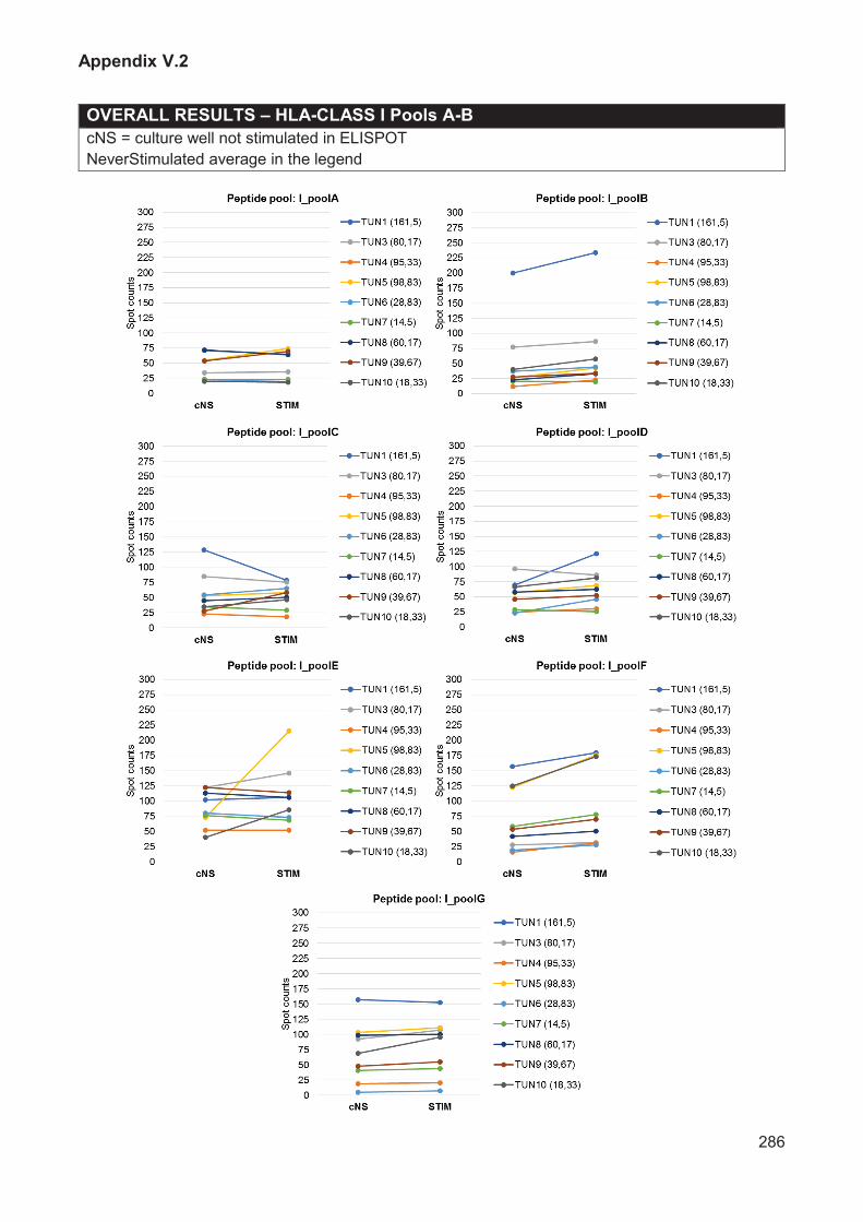
Appendix V.2
286
OVERALL RESULTS – HLA-CLASS I Pools A-B
cNS = culture well not stimulated in ELISPOT
NeverStimulated average in the legend

Appendix V.2
287
OVERALL RESULTS – HLA-CLASS II Pools 1-5
cNS = culture well not stimulated in ELISPOT
NeverStimulated average in the legend

Appendix V.2
288
OVERALL RESULTS – HLA-CLASS II Pools A-E
cNS = culture well not stimulated in ELISPOT
NeverStimulated average in the legend

Appendix V.2
289
TUN1
M / Age 33
1 scar (leg)

Appendix V.2
290
TUN1
M / Age 33
1 scar (leg)

Appendix V.2
291
TUN3
M / Age 33
1 scar (leg)
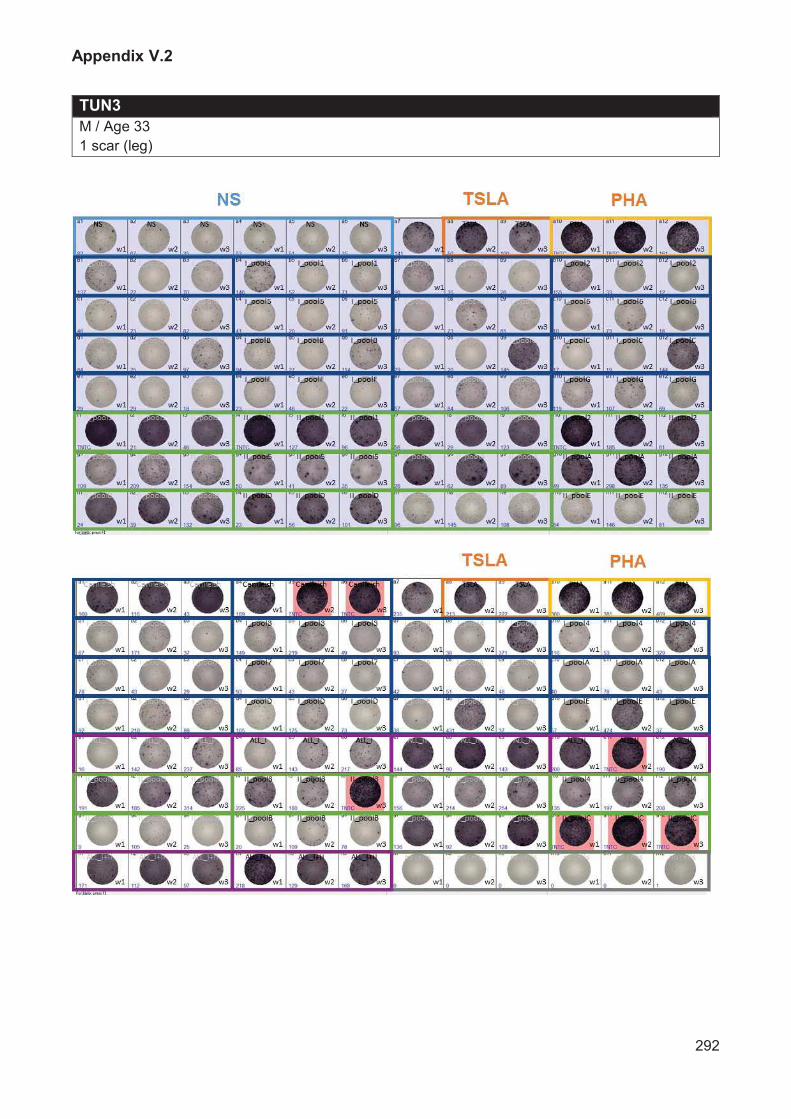
Appendix V.2
292
TUN3
M / Age 33
1 scar (leg)

Appendix V.2
293
TUN4
F / Age 36
2 scars (nose, wrist)

Appendix V.2
294
TUN4
F / Age 36
2 scars (nose, wrist)

Appendix V.2
295
TUN5
M / Age 49
1 scar (leg)

Appendix V.2
296
TUN5
M / Age 49
1 scar (leg)

Appendix V.2
297
TUN6
M / Age 26
1 scar (leg)

Appendix V.2
298
TUN6
M / Age 26
1 scar (leg)

Appendix V.2
299
TUN7
M / Age 51
5 scars (arm, leg)

Appendix V.2
300
TUN7
M / Age 51
5 scars (arm, leg)

Appendix V.2
301
TUN8
M / Age 45
2 scars (abdomen, foot)

Appendix V.2
302
TUN8
M / Age 45
2 scars (abdomen, foot)

Appendix V.2
303
TUN9
M / Age 64
1 scars (leg)

Appendix V.2
304
TUN9
M / Age 64
1 scars (leg)

Appendix V.2
305
TUN10
M / Age 64
1 scars (leg)

Appendix V.2
306
TUN10
M / Age 64
1 scars (leg)