Structure and Immunogenicity of a Peptide Vaccine Including the Complete HIV-1 gp41 2F5 Epitope....
22
Structure and Immunogenicity of a Peptide Vaccine, Including the Complete HIV-1 gp41 2F5 Epitope IMPLICATIONS FOR ANTIBODY RECOGNITION MECHANISM AND IMMUNOGEN DESIGN * Received for publication, October 17, 2013, and in revised form, December 30, 2013 Published, JBC Papers in Press, January 15, 2014, DOI 10.1074/jbc.M113.527747 Soraya Serrano ‡1,2 , Aitziber Araujo §1,3 , Beatriz Apellániz § , Steve Bryson ¶ , Pablo Carravilla § , Igor de la Arada § , Nerea Huarte § , Edurne Rujas §3 , Emil F. Pai ¶4 , José L. R. Arrondo § , Carmen Domene** ‡‡5 , María Angeles Jiménez ‡6 , and José L. Nieva §7 From the ‡ Institute of Physical Chemistry “Rocasolano,” Consejo Superior de Investigaciones Científicas (IQFR-CSIC), Serrano 119, E-28006 Madrid, Spain, the § Biophysics Unit, Consejo Superior de Investigaciones Científicas and University of the Basque Country (CSIC-UPV/EHU) and Department of Biochemistry and Molecular Biology, University of the Basque Country (UPV/EHU), P. O. Box 644, 48080 Bilbao, Spain, the ¶ Departments of Biochemistry, Medical Biophysics, and Molecular Genetics, University of Toronto, Toronto, Ontario M5S 1A8, Canada, The Campbell Family Institute for Cancer Research, Ontario Cancer Institute/University Health Network, Toronto, Ontario M5G 1L7, Canada, the **Chemistry Research Laboratory, Mansfield Road, University of Oxford, Oxford OX1 3TA, United Kingdom, and the ‡‡ Department of Chemistry, King’s College London, Franklin-Wilkins Building, 150 Stamford Street, London SE1 9NH, United Kingdom Background: HIV-1 vaccines should elicit broadly neutralizing antibodies as the gp41 “membrane-proximal external region” targeting MAb2F5. Results: NMR disclosed unprecedented 2F5 peptide-epitope structures. Although overall conformation was preserved in dif- ferent adjuvants, recovered antibodies after vaccination were functionally different. Conclusion: Membrane-inserted helical oligomers may encompass effective 2F5 peptide vaccines. Significance: Disclosing the structures that generate 2F5-like antibodies may guide future vaccine development. The membrane-proximal external region (MPER) of gp41 harbors the epitope recognized by the broadly neutralizing anti- HIV 2F5 antibody, a research focus in HIV-1 vaccine develop- ment. In this work, we analyze the structure and immunogenic properties of MPERp, a peptide vaccine that includes the follow- ing: (i) the complete sequence protected from proteolysis by the 2F5 paratope; (ii) downstream residues postulated to establish weak contacts with the CDR-H3 loop of the antibody, which are believed to be crucial for neutralization; and (iii) an aromatic rich anchor to the membrane interface. MPERp structures solved in dodecylphosphocholine micelles and 25% 1,1,1,3,3,3- hexafluoro-2-propanol (v/v) confirmed folding of the complete 2F5 epitope within continuous kinked helices. Infrared spec- troscopy (IR) measurements demonstrated the retention of main helical conformations in immunogenic formulations based on alum, Freund’s adjuvant, or two different types of liposomes. Binding to membrane-inserted MPERp, IR, molecular dynamics simulations, and characterization of the immune responses further suggested that packed helical bundles partially inserted into the lipid bilayer, rather than monomeric helices adsorbed to the mem- brane interface, could encompass effective MPER peptide vaccines. Together, our data constitute a proof-of-concept to support MPER-based peptides in combination with liposomes as stand- alone immunogens and suggest new approaches for structure- aided MPER vaccine development. The envelope (Env) 8 glycoprotein subunits gp120 (surface) and gp41 (transmembrane), which mediate receptor binding and virus-cell fusion, respectively, are organized as trimers of noncovalently associated heterodimers on the surface of the HIV-1-producing cells and assembled virions (1, 2). Upon receptor/co-receptor engagement by gp120, the gp41 ectodo- main undergoes a series of conformational changes to deliver the energy required for membrane merger (3–5). The func- tional Env complex is also targeted by the broadly neutralizing antibodies (bNAbs) known to block infection by a wide range of HIV-1 strains (1, 4, 6). These antibodies are triggered in a * This work was supported in part by Spanish MINECO Grants BIO2011-29792 (to J. L. N.), BFU2010-22103 (to J. L. R. A.), and CTQ2011-22514 (to M. A. J.) and Basque Government Grants IT838-13 and IT852-13 (to J. L. N. and J. L. R. A., respectively). □ S This article contains supplemental Tables S1 and S2. The atomic coordinates and structure factors (codes 2M8M and 2M8O) have been deposited in the Protein Data Bank (http://wwpdb.org/). 1 H chemical shifts for MPERp in HFIP and DPC were deposited in the BioMagResBank under accession number 19262 and 19263. 1 Both authors contributed equally to this work. 2 Recipient of a predoctoral fellowship from Spanish MINECO. 3 Recipients of predoctoral fellowships from the Basque Government. 4 Supported by Canadian Institutes for Health Research Grant NRF-126638 and the Canada Research Chairs Program. 5 Supported in part by the National Science Foundation through Major Research Instrumentation Grant CNS-09-58854. 6 To whom correspondence may be addressed. Tel.: 34-91-745-9541; E-mail: [email protected]. 7 To whom correspondence may be addressed. Tel.: 34-94-601-3353; Fax: 34-94-601-3360; E-mail: [email protected]. 8 The abbreviations used are: Env, envelope; bNAb, broadly neutralizing anti- body; Chol, cholesterol; DPC, dodecylphosphocholine; HFIP, 1,1,1,3,3,3- hexafluoro-2-propanol; MPER, membrane-proximal external region; PA, phosphatidic acid; POPC, 1-palmitoyl-2-oleoylphosphatidylcholine; POPG, 1-palmitoyl-2-oleoylphosphatidylglycerol; TMD, transmembrane domain; PDB, Protein Data Bank; CDR, complementarity-determining region; MDS, molecular dynamics simulation. THE JOURNAL OF BIOLOGICAL CHEMISTRY VOL. 289, NO. 10, pp. 6565–6580, March 7, 2014 © 2014 by The American Society for Biochemistry and Molecular Biology, Inc. Published in the U.S.A. MARCH 7, 2014 • VOLUME 289 • NUMBER 10 JOURNAL OF BIOLOGICAL CHEMISTRY 6565 by guest on April 18, 2016 http://www.jbc.org/ Downloaded from by guest on April 18, 2016 http://www.jbc.org/ Downloaded from by guest on April 18, 2016 http://www.jbc.org/ Downloaded from by guest on April 18, 2016 http://www.jbc.org/ Downloaded from by guest on April 18, 2016 http://www.jbc.org/ Downloaded from by guest on April 18, 2016 http://www.jbc.org/ Downloaded from by guest on April 18, 2016 http://www.jbc.org/ Downloaded from
-
Upload
independent -
Category
Documents
-
view
3 -
download
0
Transcript of Structure and Immunogenicity of a Peptide Vaccine Including the Complete HIV-1 gp41 2F5 Epitope....

Structure and Immunogenicity of a Peptide Vaccine,Including the Complete HIV-1 gp41 2F5 EpitopeIMPLICATIONS FOR ANTIBODY RECOGNITION MECHANISM AND IMMUNOGEN DESIGN*
Received for publication, October 17, 2013, and in revised form, December 30, 2013 Published, JBC Papers in Press, January 15, 2014, DOI 10.1074/jbc.M113.527747
Soraya Serrano‡1,2, Aitziber Araujo§1,3, Beatriz Apellániz§, Steve Bryson¶�, Pablo Carravilla§, Igor de la Arada§,Nerea Huarte§, Edurne Rujas§3, Emil F. Pai¶�4, José L. R. Arrondo§, Carmen Domene**‡‡5, María Angeles Jiménez‡6,and José L. Nieva§7
From the ‡Institute of Physical Chemistry “Rocasolano,” Consejo Superior de Investigaciones Científicas (IQFR-CSIC), Serrano 119,E-28006 Madrid, Spain, the §Biophysics Unit, Consejo Superior de Investigaciones Científicas and University of the Basque Country(CSIC-UPV/EHU) and Department of Biochemistry and Molecular Biology, University of the Basque Country (UPV/EHU), P. O. Box644, 48080 Bilbao, Spain, the ¶Departments of Biochemistry, Medical Biophysics, and Molecular Genetics, University of Toronto,Toronto, Ontario M5S 1A8, Canada, �The Campbell Family Institute for Cancer Research, Ontario Cancer Institute/University HealthNetwork, Toronto, Ontario M5G 1L7, Canada, the **Chemistry Research Laboratory, Mansfield Road, University of Oxford, OxfordOX1 3TA, United Kingdom, and the ‡‡Department of Chemistry, King’s College London, Franklin-Wilkins Building, 150 StamfordStreet, London SE1 9NH, United Kingdom
Background: HIV-1 vaccines should elicit broadly neutralizing antibodies as the gp41 “membrane-proximal externalregion” targeting MAb2F5.Results: NMR disclosed unprecedented 2F5 peptide-epitope structures. Although overall conformation was preserved in dif-ferent adjuvants, recovered antibodies after vaccination were functionally different.Conclusion: Membrane-inserted helical oligomers may encompass effective 2F5 peptide vaccines.Significance: Disclosing the structures that generate 2F5-like antibodies may guide future vaccine development.
The membrane-proximal external region (MPER) of gp41harbors the epitope recognized by the broadly neutralizing anti-HIV 2F5 antibody, a research focus in HIV-1 vaccine develop-ment. In this work, we analyze the structure and immunogenicproperties of MPERp, a peptide vaccine that includes the follow-ing: (i) the complete sequence protected from proteolysis by the2F5 paratope; (ii) downstream residues postulated to establishweak contacts with the CDR-H3 loop of the antibody, which arebelieved to be crucial for neutralization; and (iii) an aromaticrich anchor to the membrane interface. MPERp structuressolved in dodecylphosphocholine micelles and 25% 1,1,1,3,3,3-hexafluoro-2-propanol (v/v) confirmed folding of the complete2F5 epitope within continuous kinked helices. Infrared spec-troscopy (IR) measurements demonstrated the retention of
main helical conformations in immunogenic formulations basedon alum, Freund’s adjuvant, or two different types of liposomes.Binding to membrane-inserted MPERp, IR, molecular dynamicssimulations, and characterization of the immune responses furthersuggested that packed helical bundles partially inserted into thelipid bilayer, rather than monomeric helices adsorbed to the mem-brane interface, could encompass effective MPER peptide vaccines.Together, our data constitute a proof-of-concept to supportMPER-based peptides in combination with liposomes as stand-alone immunogens and suggest new approaches for structure-aided MPER vaccine development.
The envelope (Env)8 glycoprotein subunits gp120 (surface)and gp41 (transmembrane), which mediate receptor bindingand virus-cell fusion, respectively, are organized as trimers ofnoncovalently associated heterodimers on the surface of theHIV-1-producing cells and assembled virions (1, 2). Uponreceptor/co-receptor engagement by gp120, the gp41 ectodo-main undergoes a series of conformational changes to deliverthe energy required for membrane merger (3–5). The func-tional Env complex is also targeted by the broadly neutralizingantibodies (bNAbs) known to block infection by a wide range ofHIV-1 strains (1, 4, 6). These antibodies are triggered in a
* This work was supported in part by Spanish MINECO Grants BIO2011-29792(to J. L. N.), BFU2010-22103 (to J. L. R. A.), and CTQ2011-22514 (to M. A. J.)and Basque Government Grants IT838-13 and IT852-13 (to J. L. N. andJ. L. R. A., respectively).
□S This article contains supplemental Tables S1 and S2.The atomic coordinates and structure factors (codes 2M8M and 2M8O) have
been deposited in the Protein Data Bank (http://wwpdb.org/).1H chemical shifts for MPERp in HFIP and DPC were deposited in the BioMagResBank
under accession number 19262 and 19263.1 Both authors contributed equally to this work.2 Recipient of a predoctoral fellowship from Spanish MINECO.3 Recipients of predoctoral fellowships from the Basque Government.4 Supported by Canadian Institutes for Health Research Grant NRF-126638
and the Canada Research Chairs Program.5 Supported in part by the National Science Foundation through Major
Research Instrumentation Grant CNS-09-58854.6 To whom correspondence may be addressed. Tel.: 34-91-745-9541; E-mail:
[email protected] To whom correspondence may be addressed. Tel.: 34-94-601-3353; Fax:
34-94-601-3360; E-mail: [email protected].
8 The abbreviations used are: Env, envelope; bNAb, broadly neutralizing anti-body; Chol, cholesterol; DPC, dodecylphosphocholine; HFIP, 1,1,1,3,3,3-hexafluoro-2-propanol; MPER, membrane-proximal external region; PA,phosphatidic acid; POPC, 1-palmitoyl-2-oleoylphosphatidylcholine; POPG,1-palmitoyl-2-oleoylphosphatidylglycerol; TMD, transmembrane domain;PDB, Protein Data Bank; CDR, complementarity-determining region; MDS,molecular dynamics simulation.
THE JOURNAL OF BIOLOGICAL CHEMISTRY VOL. 289, NO. 10, pp. 6565–6580, March 7, 2014© 2014 by The American Society for Biochemistry and Molecular Biology, Inc. Published in the U.S.A.
MARCH 7, 2014 • VOLUME 289 • NUMBER 10 JOURNAL OF BIOLOGICAL CHEMISTRY 6565
by guest on April 18, 2016
http://ww
w.jbc.org/
Dow
nloaded from
by guest on April 18, 2016
http://ww
w.jbc.org/
Dow
nloaded from
by guest on April 18, 2016
http://ww
w.jbc.org/
Dow
nloaded from
by guest on April 18, 2016
http://ww
w.jbc.org/
Dow
nloaded from
by guest on April 18, 2016
http://ww
w.jbc.org/
Dow
nloaded from
by guest on April 18, 2016
http://ww
w.jbc.org/
Dow
nloaded from
by guest on April 18, 2016
http://ww
w.jbc.org/
Dow
nloaded from

fraction of infected individuals only upon prolonged contactwith the virus (7, 8). It has been proposed that vaccinationstrategies focused on the induction of antibodies qualita-tively similar to those bNAbs might result in the effectiveprevention of infection (7, 9).
The isolation of bNAbs in the form of monoclonal antibodies(MAbs) has revealed common structural trends useful for guid-ing the rational design of immunogens eliciting protective anti-bodies (9 –11). Broad neutralization is attained by antibodiesthat bind to a handful of invariable but accessible regions ofgp120 and gp41. Broadly neutralizing sera raised to gp120have been found to contain antibodies that target the recep-tor-binding site, the glycan-V3 site, and the V1V2 loops,whereas antibodies in broadly neutralizing sera raised togp41 appear to bind exclusively to the “membrane-proximalexternal region” (MPER) or pre-transmembrane domainwithin this subunit (7–10, 12, 13).
In contrast to the structurally complex, discontinuousepitopes recognized by anti-gp120 bNAbs, it is hypothesizedthat MPER embodies a single continuous linear epitope (14 –18). Following this idea, it has been suggested that syntheticpeptides constrained into the neutralization-competent MPERstructures might constitute stand-alone vaccines (19, 20). Oneanti-MPER bNAb that has focused much attention in thisresearch area is the 2F5 antibody. 2F5 was isolated in mAb formby Katinger and co-workers (21, 22) from a panel of sera fromnaturally infected asymptomatic individuals. Given the neutral-ization breath and potency shown by the bNAb 2F5 (13, 21,23–26), development of peptide-based vaccines targeting the2F5 epitope has since been pursued (6, 22, 27–33).
Binding specificity of MAb2F5 was initially mapped to N-ter-minal 662ELDKWA667 MPER residues (21, 24, 26). Based onmass spectrometry and proteolytic protection assays, thiscore epitope was later extended to span the 656NEQELLELDK-WASLWN671 sequence (34). Comparable full epitope lengthswere subsequently suggested by competition ELISA (35) andstructural analyses (14, 36). X-ray crystallography further indi-cated that epitope binding does not involve the hydrophobicapex of the long complementarity-determining region(CDR)-H3 loop, an element shown to be crucial for the neutral-izing function of the antibody (37, 38). Given the close proxim-ity of the epitope to the envelope surface, it has been proposedthat the 2F5 CDR-H3 loop might interact directly with viralmembrane lipids (14, 39 – 41). Alternatively, data have beenrecently reported suggesting that the CDR-H3 loop apex mayestablish additional contacts with MPER C-terminal residues inhelical conformation (25, 38). These two options need not bemutually exclusive for bivalent antibodies targeting the 2F5epitope on the surface of virions. It has been argued thatMAb2F5-like antibodies could use a heteroligation strategy (i.e.to combine strong binding to gp41 and weak binding to viralmembrane) to increase its avidity under conditions existing inthe HIV envelope (9).
Here, we provide unprecedented results on the structure andimmunogenicity of a peptide spanning the sequence 656NEQEL-LELDKWASLWNWFNITNWLWYIK683, which includes thecomplete 2F5 epitope (underlined), the downstream regionproposed to establish weak contacts with the CDR-H3 loop of
the antibody, and an aromatic-rich block that allows its inser-tion into the membrane interface (Fig. 1). The NMR data onthis peptide, termed MPERp, support the folding of the com-plete HIV-1 2F5 epitope within a continuous kinked helix.
IR confirmed the preservation of the main helical conforma-tion in adjuvants representing licensed vaccine formulations(i.e. aluminum salt and water-in-oil emulsions) and in two dif-ferent types of liposomes. Because it is predicted that the lipo-somal MPERs that mimic the 2F5 epitope will be bound by thefunctional neutralizing antibody, we performed assays to cor-relate function and binding. Consistent with previous reports(37, 38), cell infection blocking in our in-house assay wasdependent on the CDR-H3 loop. 2F5 binding to MPERp in lipo-somes made of anionic phospholipid and lipid A was alsodependent on the CDR-H3 loop, whereas binding to the pep-tide on the surface of lesser charged Chol-containing vesiclesdid not require this element.
All tested MPERp vaccines were immunogenic. However,significant amounts of 2F5 epitope-targeting antibodies withthe capacity of blocking cell infection were only recovered fromsera of rabbits immunized with liposomal vaccines displaying acorrelation between 2F5 antibody function and binding, i.e.those based on the anionic phospholipid and lipid A. Insightsinto the structural basis for functional antibody generationcould be gained by combining IR and molecular dynamics sim-ulation (MDS) analyses. These data suggest that membrane-inserted helical bundles, rather than monomers adsorbed to themembrane interface, may embody efficient MPER vaccines.Together, our structural and immunogenicity data conform tothe prediction that MPER may fold as a single contiguous anti-genic determinant, competent in generating a neutralizingresponse and therefore supporting the application of derivedpeptides in combination with liposomes as stand-alone vac-cines to target the 2F5 epitope.
EXPERIMENTAL PROCEDURES
Materials—MPERp and the 2F5 peptide epitopes used in theimmunological studies were synthesized in C-terminal carbox-amide form by solid phase methods using Fmoc (N-(9-fluore-nyl)methoxycarbonyl) chemistry, purified by reverse phaseHPLC, and characterized by matrix-assisted time-of-flight(MALDI-TOF) mass spectrometry (purity �95%). Peptides wereroutinely dissolved in dimethyl sulfoxide (DMSO, spectroscopygrade), and their concentration was determined by the bicin-choninic acid microassay (Pierce). 1-Palmitoyl-2-oleoylphos-phatidylglycerol (POPG), 1-palmitoyl-2-oleoylphosphatidylcho-line (POPC), phosphatidic acid (PA), Chol, and lipid A detoxified(SalmonellaminnesotaR595)werepurchasedfromAvantiPolarLip-ids (Birmingham, AL). Dodecylphosphocholine (DPC) was fromAnatrace (Maumee, OH). MAb2F5 was kindly donated by Diet-mar Katinger (Polymun Scientific, Klosterneuburg, Austria).Fab2F5-WT and Fab2F5-�CDR-H3 were produced as describedpreviously (38).
Recording of NMR Spectra—NMR samples were prepared bydissolving the lyophilized peptides (�1 mg) in 0.5 ml of a H2O/D2O (9:1 ratio by volume) solution containing 2 mM HEPESbuffer at pH 7.0 and either 25% (v/v) 1,1,1,3,3,3-hexafluoro-2-propanol (HFIP; D2, 98%; Cambridge Isotopes Lab) or 20 mM
Structure Immunogenicity of the Complete 2F5 Epitope
6566 JOURNAL OF BIOLOGICAL CHEMISTRY VOLUME 289 • NUMBER 10 • MARCH 7, 2014
by guest on April 18, 2016
http://ww
w.jbc.org/
Dow
nloaded from

deuterated DPC (D38, 98%; Cambridge Isotopes Lab). Peptideconcentrations were �0.5 mM. pH was measured with a glassmicroelectrode and not corrected for isotope effects. A metha-nol sample was used to calibrate the temperature of the NMRprobe. Chemical shifts were referenced to internal sodium2,2-dimethyl-2-silapentane-5-sulfonate.
The 1H NMR spectra were acquired on a Bruker Avance-600spectrometer operating at a proton frequency of 600.13 MHzand equipped with a cryoprobe. One-dimensional spectra wereacquired using 32 K data points, which were zero-filled to 64 Kdata points before performing the Fourier transformation. Phase-sensitive two-dimensional correlated spectroscopy (COSY), totalcorrelated spectroscopy (TOCSY), and nuclear Overhauserenhancement spectroscopy (NOESY) spectra were recorded bystandard techniques using presaturation of the water signal andthe time-proportional phase incrementation mode, as reportedpreviously (42). NOESY mixing times were 100 or 150 ms, andTOCSY spectra were recorded using 60 ms DIPSI2 with z filterspin-lock sequence. Acquisition data matrices were defined by2048 � 512 points in t2 and t1, respectively. Data were pro-cessed using the standard TOPSPIN program (Bruker Biospin,Karlsruhe, Germany). The two-dimensional data matrix wasmultiplied by either a square-sine-bell or a sine-bell windowfunction with the corresponding shift optimized for every spec-trum and zero-filled to a 2K � 1K complex matrix prior toFourier transformation. Baseline correction was applied in bothdimensions.
Structure Calculation—The same protocol was followed tocalculate the structures of MPERp in the presence of DPCmicelles and in 25% HFIP from distance and dihedral angleconstraints derived from NMR parameters. Distance con-straints were obtained from the 150-ms two-dimensional1H-1H NOESY spectra, with the cross-peaks observed in the100-ms two-dimensional 1H-1H NOESY being essentially thesame. Dihedral angle restraints for � and � angles were derivedfrom 1H� chemical shifts using the program TALOS (43).Structures were calculated using a three-step protocol. First,the standard iterative procedure for automatic NOE assign-
ment of the program CYANA 2.1 (44) was applied. The proto-col consists of seven cycles of combined automated NOEassignment and structure calculation of 100 conformers percycle (45). The list of distance constraints resulting from the lastautomatic cycle was checked by inspection of the correspond-ing NOESY spectra, and ambiguous constraints were removedor relaxed to generate the final list used as input for a standardsimulated annealing CYANA 2.1 calculation of 100 conform-ers. The 20 conformers with the lowest target function valueswere selected and subjected to 2000 steps of energy minimiza-tion using the generalized Born continuum solvation modelwith a nonbonded cutoff of 10 Å as implemented in the pro-gram AMBER9 (Case DA, Darden TA, Cheatham III TE, Uni-versity of California, San Francisco). The structural statisticsdata for the final ensembles of 20 structures obtained forMPERp are provided in Table 1. The quality of these final struc-tures was assessed using PROCHECK/NMR (46) as imple-mented at the Protein Structure Validation Suite server. All ofthe residues were either in the most favored or allowed regionsof the Ramachandran map (Table 1). The structural ensemblescalculated for MPERp have been deposited in the PDB DataBank with accession codes 2M8M (HFIP) and 2M8O (DPC).These structures were visualized and examined using the pro-grams MOLMOL (47) and Swiss-Pdb viewer (48).
Membrane Binding Assays—Vesicle flotation experiments insucrose gradients were performed following the methoddescribed by Yethon et al. (49). In brief, 100 �l of a samplecontaining rhodamine-labeled liposomes (3 mM lipid concen-tration) was adjusted to a sucrose concentration of 1.4 M in afinal volume of 300 �l, and subsequently overlaid with 400- and300-�l layers of 0.8 and 0.5 M sucrose, respectively. The gradi-ent was centrifuged at 436,000 � g for 3 h in a TLA 120.2 rotor(Beckman Coulter, Brea CA). After centrifugation, four 250-�lfractions were collected. Material adhered to tubes was col-lected into a 5th fraction by washing with 250 �l of hot (100 °C)1% (w/v) SDS.
Infrared Spectroscopy—Infrared spectra were recorded in aThermo Nicolet 5700 spectrometer equipped with a mercury-
TABLE 1Structural statistics for the ensemble of the 20 lowest energy NMR structures of MPERp in DPC (20 mM deuterated dodecylphosphocholine, 2 mM
HEPES, pH 7.0, H2O/D2O, 9:1, v/v) and HFIP (25% deuterated 1,1,1,3,3,3-hexafluoro-2-propanol in 2 mM HEPES, pH 7.0, H2O/D2O, 9:1, v/v)DPC HFIP
No. of distance restraints Intraresidue (i � j � 0) 154 118Sequential (�i � j� � 1) 103 74Medium range (1 � �i � j� � 5) 115 35Total number 372 227Averaged total no./residue 13.3 8.0
No. of dihedral angle constraints � angles 26 26� angles 25 22Total number 51 48
Average maximum violations/structure Distance (Å) 0.15 0.01 0.02 0.04Dihedral angle (°) 3.5 0.1 0.1 0.1
Averaged structure energies CYANA target function value 0.35 0.01 0.01 0.01AMBER energy (kcal/mol) �1187 �602van der Waals energy (kcal/mol) �195 �182Electrostatic energy (kcal/mol) �2247 �2235
Deviations from ideal geometry Bond length (Å) 0.014 0.015Bond angle (°) 1.8 1.7
Pairwise root mean square deviation (Å) (residues 2–27) Backbone atoms 0.4 0.4 1.6 0.7All heavy atoms 1.1 0.3 2.5 0.7
Ramachandran plot (%) Residues in most favored regions 99.6 98.8Residues in additional allowed regions 0.4 1.2Residues in generously allowed regions 0.0 0.0Residues in disallowed regions 0.0 0.0
Structure Immunogenicity of the Complete 2F5 Epitope
MARCH 7, 2014 • VOLUME 289 • NUMBER 10 JOURNAL OF BIOLOGICAL CHEMISTRY 6567
by guest on April 18, 2016
http://ww
w.jbc.org/
Dow
nloaded from

cadmium-telluride detector using a Peltier-based temperaturecontroller (TempCon, BioTools Inc., Wauconda, IL) with cal-cium fluoride cells (BioCell, BioTools Inc., Wauconda, IL).MPERp-containing samples were lyophilized and subsequentlyprepared at 4 mg/ml in D2O buffer (5 mM HEPES, pD 7.4, 100mM NaCl). A 25-�l sample aliquot was deposited on a cell thatwas sealed with a second cell. Reference windows without pep-tide were prepared similarly. Typically 370 scans were collectedfor each background and sample, and the spectra were obtainedwith a nominal resolution of 2 cm�1. Data treatment and banddecomposition of the original amide I have been described else-where (50).
Cell Entry Assays—For the in-house cell entry assays (38),HIV-1 pseudoviruses were produced by transfection of humankidney HEK293T cells with the full-length Env clone JRCSF(kindly provided by Jamie K. Scott and Naveed Gulzar, SimonFraser University, British Columbia, Canada) using calciumphosphate. Cells were co-transfected with vectors pWXLP-GFP and pCMV8.91, encoding, respectively, a green fluores-cent protein and an Env-deficient HIV-1 genome (provided byPatricia Villace, Consejo Superior de Investigaciones Científi-cas, Madrid, Spain). After 24 h, the medium was replaced withOptiMEM-Glutamax II (Invitrogen) without serum. Two daysafter transfection, the pseudovirus particles were harvested,passed through 0.45-�m pore sterile filters (Millex� HV, Milli-pore NV, Brussels, Belgium), and finally concentrated by ultra-centrifugation in a sucrose gradient. HIV entry was determinedusing TZM-bl target cells (AIDS Research and Reference Rea-gent Program, Division of AIDS, NIAID, National Institutes ofHealth, contributed by J. Kappes). Antibody samples were setup in duplicate in 96-well plates and incubated for 1 h at 37 °Cwith a 10 –15% tissue culture infectious dose of pseudovirus.After antibody-pseudovirus co-incubation, 10,000 target cellswere added in the presence of 15 �g/ml DEAE-dextran (Sigma).Infection levels after 72 h were inferred from the number ofGFP-positive cells as determined by flow cytometry using aFACSCalibur flow cytometer (BD Biosciences).
Molecular Dynamics Simulations—Atomic coordinates ofthe MPER peptide were taken from the NMR structure calcu-lated in DPC micelles (first model). Only gp41 residues 656 to683 were included in the model, with sequence NEQELLELD-KWASLWNWFNITNWLWYIK. Default protonation stateswere used for all the ionizable residues. N and C termini wereamidated and acetylated, respectively.
A pre-equilibrated bilayer containing a mixture of POPC/1-palmitoyl-2-oleoyl phosphatidic acid/Chol in ratios 2:1.5:0.2and pure POPG in a one-component bilayer were used. Thesystems were solvated by �37,000 water molecules. Sodiumand chloride ions were added to neutralize the systems up to afinal experimental concentration of 150 mM. Four MPER pep-tides were randomly placed in the solution at the start of thesimulations. The total production runs were 235 ns for eachsimulation. MD trajectories were simulated with the version 2.9of NAMD (51), using the CHARMM27 force field with CMAPcorrections for the peptides (52), the CHARMM36 force fieldfor lipids (53), the TIP3P model for water molecules (54), andthe model of Cournia et al. (55) for cholesterol. Standardparameters for ions in the CHARMM27 force field were
adopted. Simulations were performed in the NpT ensemble.Pressure was kept at 1 atm by the Nose-Hoover Langevin pistonmethod (56, 57) with a damping time constant of 100 ps and aperiod of 200 ps. Temperature was kept at 300 K by coupling toa Langevin thermostat, with a damping coefficient of 5 ps�1
(57). Electrostatic interactions were treated by the ParticleMesh Ewald algorithm, with grid spacing below 1 Å (58). vander Waals interactions were truncated at 12 Å and smoothed at10 Å. Hydrogen atoms were restrained by the SETTLE algo-rithm (59), which allowed a 2-fs time step.
Rabbit Immunization and Antibody Purification—For immuni-zation in Freund’s adjuvant or alum, MPERp was dissolved in 0.5ml of PBS and mixed with an equal volume of 1.3% (w/v) alu-minum hydroxide (Alhydrogel, Superfos Biosector, Denmark)or Freund’s adjuvant (Sigma). Liposome-based formulationswere prepared following the methods described by Dreesman etal. (60) and Maeso et al. (61) and included lipid A as adjuvant(62). MPERp in DMSO was added at a final peptide-to-lipidratio of 1:50 (mol/mol) to a stirring solution of freeze-thawPOPC/Chol/PA/lipid A (2.0:1.5:0.2:0.01 molar ratio) or POPG/lipid A (3.7:0.01 molar ratio) vesicles dispersed in PBS. Afterincubation for 30 min, the samples were lyophilized. New Zea-land White rabbits were inoculated intradermally at multiplesites on day 0 with 1 ml of sample reconstituted in pure water,which contained 0.5 mg of peptide. For subsequent boostinginjections, 1 ml of the reconstituted liposome formulation con-taining 0.3 mg of peptide was used on day 15 (0.3 mg peptide),and 0.2 mg of liposomal peptide was injected on days 30, 45, and60. The 2F5 epitope-specific antibodies were recovered fromsera through affinity purification. To that end, the 2F5ep-Cys(NEQELLELDKWASLWN-C) peptide was immobilized onto abeaded agarose support using a Sulfolink immobilization kit forpeptides (Thermo Scientific, Rockford, IL) and following themanufacturer’s instructions. The remaining nonspecific bind-ing sites in columns were blocked adding L-cysteine�HCl at 50mM. Every analyzed serum was loaded on the columns afterdiluting and filtering it to remove the particulate material. Theywere allowed to flow through the columns five times thus allow-ing the binding of all the antibodies present in the serum thatrecognize specifically the immobilized peptide. After washingthe columns with at least 10 bed volumes of 500 mM NaCl con-taining buffer to dispose of nonspecifically bound antibodiesand serum proteins, the specific antibodies were eluted using100 mM glycine buffer at pH 2.5. The fraction that is not recov-ered using acidic pH was eluted using freshly made 100 mM
triethylamine buffer at pH 11.5.
RESULTS
Designation of the MPER Peptide Containing the Complete2F5 Epitope—The diagram displayed in Fig. 1A designatesMPER as the membrane-proximal sequence that connectsthe gp41 globular ectodomain (FP-NHR-loop-CHR) with themembrane-spanning domain (TMD). Position for the coreepitope recognized by the 2F5 bNAb is also displayed. Theorganization of this region within the pre-fusion gp41 struc-ture recognized by this antibody is presently unknown.Nonetheless, MPER is postulated to embody a single neutral-ization-competent structure (19, 20).
Structure Immunogenicity of the Complete 2F5 Epitope
6568 JOURNAL OF BIOLOGICAL CHEMISTRY VOLUME 289 • NUMBER 10 • MARCH 7, 2014
by guest on April 18, 2016
http://ww
w.jbc.org/
Dow
nloaded from

Fine definition of the 2F5 epitope suggests the involvementin antibody recognition of a helical stretch that follows the coreepitope residues (34, 36, 63). It has been argued that theMAb2F5’s CDR-H3 loop establishes contact with residueslocated further downstream within a continuous helix (25, 38).Because the CDR-H3 loop is absolutely required for the 2F5neutralizing activity (see below and Ref. 37), it is inferred thatthose residues constrained into a relevant conformation mightbe required for peptide vaccines to elicit broadly neutralizing2F5-like antibodies. The reported three-dimensional NMRstructures of MPER peptides are either disconnected from theMPER C-terminal residues (e.g. PDB codes 1LCX or 1MZI inFig. 1B) or depleted from the N-terminal 2F5 epitope residues(e.g. PDB codes 1JAV or 2PV6 in Fig. 1B). Thus, we firstattempted the NMR structure resolution of a synthetic peptidevaccine (MPERp, Fig. 1B), which included both a complete2F5 epitope sequence and the C-terminal residues putativelyengaged in secondary interactions with the CDR-H3 ofMAb2F5 (25, 38).
NMR Solution Structure of MPERp—The 1H NMR signals ofMPERp in the presence of DPC micelles and in 25% HFIP at pH7.0 and 25 °C were assigned by standard sequential assignmentmethods (64, 65). These chemical shifts have been deposited atBioMagResBank with accession codes BMRB-19263 (DPC) andBMRB-19262 (HFIP). In this peptide, under both conditions,most 1H chemical shifts (supplemental Tables S1 and S2) devi-ated significantly from random coil values (66). In particular,most H� protons showed large negative ��H� values (Fig. 2A).This is a clear indication that MPERp adopted helical structuresunder both conditions, because according to the well estab-lished empirical relationship between the ��H� and the � and as in previous page 3 dihedral angles, positive and negative��H� values are characteristic of �-strands and �-helices,respectively. Additional and stronger evidence for the adoption
FIGURE 1. Design of MPER-derived peptide vaccine. A, scheme describingthe HIV-1 gp41 organization and the sequence of the MPER peptide vaccineused in this study (HIV-1 Env residues 656 – 683, numbering and sequencederived from the prototypic HXBc2 isolate). The gp41 ectodomain regionsdesignated in the top diagram include the following abbreviations: FP, fusionpeptide; NHR and CHR, N- and C-terminal helical regions, respectively; Cyt,cytosolic domain. The MPER sequence below highlights the five Trp residuesin green and the core epitope residues recognized by 2F5 antibody under-lined. The line on top spans the extended 2F5 epitope as defined by pro-teomic analyses (34). Blue asterisks denote residues implied in secondarybinding by CDR-H3 loop (25) and the box an aromatic rich anchor to themembrane interface. B, structures adopted by MPER-derived peptides. PDBaccession numbers indicated in the panel designate structures in solution(1LCX and 1MZI) or in contact with DPC micelles (1JAV and 2PV6). Lateral sidechains of Trp residues are depicted in green to align the structures with theMPER amino acid sequence.
FIGURE 2. NMR parameters for MPERp. A, bar graphics showing the ��H�
(��H� � �H�observed � �H�
RC, ppm) values as a function of sequence in 20 mM
DPC (black bars) or 25% HFIP (gray bars) at pH 7.0 and 25 °C. Dashed linesindicate the random coil (RC) ranges. Random coil values for C�H protonswere taken from Wishart et al. (66). The N- and C-terminal residues areexcluded because of charged end effects. B and C, NOE summaries for thepeptide in 20 mM DPC and 25% HFIP. The intensities of the sequential NOEs,classified as strong, medium, and weak, are indicated by the thickness of thelines.
Structure Immunogenicity of the Complete 2F5 Epitope
MARCH 7, 2014 • VOLUME 289 • NUMBER 10 JOURNAL OF BIOLOGICAL CHEMISTRY 6569
by guest on April 18, 2016
http://ww
w.jbc.org/
Dow
nloaded from

of helical structures in both media came from the set ofobserved NOEs, because NOE cross-peaks are only observedfor pairs of spatially closed protons (approximately at a distanceless than 5 Å). The nonsequential NOEs presented by the pep-tide were those characteristic of helices, i.e. d�N(i � 2), d�N(i � 3),d�N(i � 4), and d��(i � 3) (Fig. 2, B and C). Some of these mediumrange NOEs can be observed in the NOESY spectral regionsshown in Fig. 3.
Structure calculations were further performed to visualizethe features of the structure adopted by MPERp in the presenceof DPC or when dissolved in 25% HFIP (see under “Experimen-tal Procedures”). In contrast to the structures displayed in Fig.1B, the structures resolved for MPERp disclosed continuous andwell defined helical structures in both media (Fig. 4A). Althoughuninterrupted, in both cases the complete set of models showedchanges in the direction of the helix axis at certain points. In HFIP,the helix was bent at position 665KW666 forming an angle of �40°(Fig. 4A, top). More conspicuous changes could be observed inthe presence of DPC (Fig. 4A, bottom). In all calculated DPCstructures, the N-terminal residues 661LEL663 formed a short310-helix, and the following main �-helix bent forming anangle of �30° at the 673FNI675 tripeptide.
IR Structure of MPERp in Classical Adjuvants—High resolu-tion NMR spectroscopy supports the folding of the 2F5 epitopewithin a continuous MPER helical structure. To determinewhether this structure was preserved in vaccine formulations,we measured MPERp conformations in different adjuvants byIR spectroscopy. The reference spectra obtained under theNMR conditions (Fig. 4A, left panels) showed the absorptionof the amide-I centered at �1650 cm�1, consistent with the�-helix conformation adopted by MPERp in HFIP and DPCmixtures (50). Band decomposition disclosed in both cases anadditional low frequency band centered at �1630 cm�1, corre-sponding to the solvent-exposed �-helix fraction (67–69). In con-junction, both bands accounted for � 85% of the total absorption
(red-dotted band components). The analysis also revealed less sig-nificant contributions by bands in the 1680- to 1660-cm�1 region,indicative of additional fractions of �-turn/310-helix plus lessdefined coil structures (Table 2).
Subsequently, we assessed the conformational changesundergone by MPERp in vaccines formulated with aluminumhydroxide (alum) and water-in-oil dispersions (incompleteFreund’s adjuvant), which provided two viscous media relevantfor the adjuvants currently used in vaccination protocols (70).Consistent with preservation of the main �-helical conforma-tion in these adjuvants, the IR amide-I band maximum waslocated at �1650 cm�1 in both cases (Fig. 4B). However, ascompared with DPC or HFIP samples, the peptide dispersed inalum displayed an additional band component centered at 1620cm�1, indicative of significant denaturation-aggregation evolv-ing in these samples (Table 3). In the inverted micelle mediumprovided by Freund’s adjuvant, the band was broader than inDPC or HFIP, and its decomposition revealed that �-turn, 310-helix, and less ordered conformers absorbing in the 1680 –1660cm�1 region were comparatively more abundant than in theNMR references (Table 3).
FIGURE 3. Selected NOESY spectral regions of MPERp in DPC or HFIP con-ditions (left and right panels, respectively). The depicted regions showintraresidue �-� and nonsequential �-� (i, i�3) NOES. The nonsequentialNOEs are boxed.
FIGURE 4. Structures adopted by MPERp in 25% HFIP or DPC 20 mM andclassical vaccine adjuvants. A, calculated NMR structures and correspond-ing IR spectra. Lateral side chains of Trp residues are depicted in green andaligned with the amino acid sequence as in the caption for Fig. 1. Both struc-tures are continuous helices. Additional structural signatures common to allcalculated models are highlighted (see text). The corresponding IR spectradisplayed on the left disclose in red the absorption band components arisingfrom the helical conformation (Table 2). B, IR absorption in the amide I regionby MPERp in D2O-based buffer mixed with solutions of alum and Freund’sadjuvant (FA) following the procedure used for preparing vaccines. In all pan-els the IR spectra were decomposed into different band components (numer-ical values are disclosed in Table 3). Red dotted lines correspond to �-helixcomponents.
Structure Immunogenicity of the Complete 2F5 Epitope
6570 JOURNAL OF BIOLOGICAL CHEMISTRY VOLUME 289 • NUMBER 10 • MARCH 7, 2014
by guest on April 18, 2016
http://ww
w.jbc.org/
Dow
nloaded from

2F5 Binding to Liposomal Vaccines—In addition to the stand-ard adjuvants, MPERp was combined with liposomes to com-pose synthetic vaccines. It has been suggested that interactionswith the membrane interface on the surface of liposomes mayselect for the MPER conformations that are relevant for theinduction of specific antibodies (62), whereas polyreactivitywith membrane lipids could increase the binding avidity ofthose antibodies (9). For this study, we selected two differentlipid compositions. In one formulation MPERp was adminis-tered together with POPC/Chol/PA (2:1.5:0.2 mol/mol) lipo-somes, and in a second formulation the peptide was combinedwith vesicles made of the anionic phospholipid POPG. It isassumed that peptide structures relevant for the 2F5 epitopewill be bound by the functional 2F5 antibody but not by theirinactive mutants. Thus, to discriminate functional versus non-functional binding, we used CDR-H3 mutant Fabs that bindpeptide epitopes in solution but are not neutralizing (Fig. 5).
Fig. 5A compares the capacity for inhibiting viral entry ofthe MAb2F5 and two derived Fabs, Fab2F5-WT and Fab2F5-�CDR-H3, with the latter representing a mutant with theCDR-H3 loop deleted (38). MAb2F5 and Fab2F5-WT inhibitedpseudovirus cell infection in our assay (Fig. 5A, black and bluesymbols, respectively), whereas Fab2F5-�CDR-H3 had noeffect (red symbols). To determine a functional correlation withepitope binding, we next compared the capacity of these anti-bodies to bind the liposomal vaccines (Fig. 5, B–E). Experi-ments were set up for obtaining pure vesicles floating on the topfractions of sucrose gradients (Fig. 5B). These assays indicatedquantitative MPERp incorporation into the POPC/Chol/PAand POPG liposomes (Fig. 5C). Results displayed in Fig. 5Dfurther demonstrated that MAb2F5 could effectively bind tothe membrane-inserted peptide epitopes in both types of lipo-
somes. Thus, according to this sole criterion, both types of vesiclescontaining peptide might encompass effective 2F5 immunogens.However, the comparison of the Fabs revealed different patterns(Fig. 5D). The functional Fab2F5-WT reproduced the binding pat-tern of the mAb (Fig. 5D, top). In contrast, the nonfunctionalFab2F5-�CDR-H3 could bind effectively to POPC/Chol/PA-MPERp liposomes but not to POPG-MPERp liposomes (Fig. 5D,bottom).
Thus, cell entry inhibition and binding to POPG-MPERp lipo-somes were both dependent on the CDR-H3 loop. Together, theseresults allowed establishing a correlation between function andbinding to POPG-bound MPERp, which was not found for thePOPC/Chol/PA-MPERp liposomes. Accordingly, we inferred thatwhen used as immunogens POPG-MPERp vaccines would bemore selective than POPC/Chol/PA-MPERp vaccines in activat-ing 2F5-like B-cell receptors.
MPERp Structure in Liposomal Vaccines—To gain insightsinto the membrane-associated structures at the origin of thefunction-binding correlation, we carried out a combined infra-red spectroscopy-molecular dynamics simulation study (Fig. 6).Samples of MPERp in contact with POPC/Chol/PA liposomesclosely reproduced the IR absorption spectrum measured in theFreund samples (Fig. 6A, left). This finding is consistent withthe comparable structures adopted by membrane-binding pep-tides at interfaces of reverse micelles and membranes (71). Insharp contrast, the amide-I band measured in the POPG-MPERp samples was narrower, suggestive of higher conforma-tional order, and corresponded almost exclusively to �-helicalconformers (Fig. 6B, left). In these samples, the maximum shiftof the low frequency �-helix band and its increase in intensitywere consistent with the existence of packing interactionsbetween solvated helices (67, 68).
Further insights into the membrane-associated structurescausing these spectral differences were obtained from thedynamics of the MPERp DPC structure simulated in the pres-ence of POPC/Chol/PA and POPG lipid bilayers (Fig. 6, rightpanels). MPERp was observed to associate as a single monomerwith the POPC/Chol/PA membrane (Fig. 6A, right). In thecourse of the simulation, this peptide monomer was observedto contact first with the membrane surface through the hydro-phobic C-terminal side, and the amphipathic N terminus wasinserted later. At the end of the simulation (235 ns), the peptideadopted a continuous helix, whose main axis was parallel to thelipid bilayer plane. In this state, the membrane interface-em-bedded side chains made contact preferentially with the polarheadgroups of POPC, but not with Chol or PA. In addition, 2F5
TABLE 2Band position, assignment, and % area of the components obtainedafter curve-fitting of IR spectra displayed in Fig. 4A
HFIP DPCBand positiona Areab Band positiona Areab
% %1675 11 1678/1665 14
(�-Turns/310-helix) (�-Turns/310-helix)1652 66 1650 66
(�-helix-buried) (�-Helix-buried)1632 20 1630 19
(�-helix-solvated) (�-Helix-solvated)1615 2 1611 1
(aggregation) (aggregation)a Wave numbers in cm�1. The conformation assigned for each position is indi-
cated below (50, 68).b The values have been rounded off to the nearest integer.
TABLE 3Band position, assignment, and % area of the components obtained after curve-fitting of IR spectra displayed in Figs. 4B and 6, A and B
Alum Freund’s adjuvant POPC/Chol/PA POPGBand positiona Area (%)b Band positiona Area (%)b Band positiona Area (%)b Band positiona Area (%)b
1680/1669 11 1678/1665 26 1685/1671 26 1665 3(�-Turns/310-helix) (�-Turns/310-helix) (�-Turns/310-helix) (310-Helix)
1653 45 1651 52 1652 46 1654 52(�-Helix-buried) (�-Helix-buried) (�-Helix-buried) (�-Helix-buried)
1635 19 1631 17 1637 13 1639 39(�-Helix-solvated) (�-Helix-solvated) (�-Helix-solvated) (�-Helix-solvated)
1620/1693 25 1617 5 1615/1627/1686 15 1628 5(Aggregation) (Aggregation) (Aggregation) (Aggregation)
a Wave numbers are in cm�1. The conformation assigned for each position is indicated below (50, 68).b The values have been rounded off to the nearest integer.
Structure Immunogenicity of the Complete 2F5 Epitope
MARCH 7, 2014 • VOLUME 289 • NUMBER 10 JOURNAL OF BIOLOGICAL CHEMISTRY 6571
by guest on April 18, 2016
http://ww
w.jbc.org/
Dow
nloaded from

epitope key residues Lys-665 and Trp-666 (Fig. 6A, red sidechains), as well as residues establishing putative contacts withthe CDR-H3 loop (i.e. Leu-669, Trp-672, and Phe-673) (blueside chains) were involved in these lipid interactions and there-fore not accessible from the water solution.
In contrast, the four peptides, randomly placed in solution atthe beginning of the simulation, were found assembled into abundle at 194 ns. This bundle of peptides was associated withthe POPG bilayer (Fig. 6B, right) from 194 ns until the end of thesimulation (235 ns). The MPERp bundle inserted through the
aromatic rich C-terminal hydrophobic tip of one of the constitu-ent monomers (Fig. 6B, residues in green) during the simulation. Inthe course of the simulation, distinct MPERp ensembles retainingoverall helical conformation were observed several times to stick inthe membrane by the action of a monomer; however, insertion ofsingle peptides was not observed. Moreover, the more polar Nterminus of the membrane-inserted monomer never contactedthe negatively charged membrane surface. In the membrane-in-serted state, residues involved in MAb2F5 recognition (Lys-665,Trp-666, Leu-669, Trp-672, and Phe-673 (see below and Refs. 25,
FIGURE 5. Correlation between 2F5 antibody function and binding to liposomal vaccines. A, cell entry inhibition assay. Left, pseudoviruses were preincu-bated with MAb2F5 or the recombinant 2F5 Fab constructs, and single cell entry events were monitored by FACS after incubation with TZM-bl target cells.Fab2F5 WT inhibited cell entry (blue), albeit with lower potency than the bi-functional mAb (black). In contrast Fab2F5 �CDR-H3 was almost completely unableto inhibit the process (red). Right, displays specificity controls for the HIV-1 Env-mediated cell entry. bNAbs (2F5 and 4E10) and T-20 were applied at 2 and 50�g/ml, respectively. Means S.D. of six measurements in three independent experiments are displayed. B–E, vesicle flotation experiments in sucrosegradients. Rhodamine-labeled liposomes were collected in the 1st and 2nd fractions (i.e. floating fractions) (B). C, MPERp (30 �M) was incubated in solution inabsence (top) or presence of liposomes (peptide-to-lipid ratio of 1:100, bottom panels) for 15 min before centrifugation. The presence of the peptide in thedifferent fractions was probed with MAb2F5 in Western blot. Virtually all input peptide co-floated with liposomes indicating quantitative partitioning intomembranes. D, MAb2F5 (15 �g ml�1) was incubated for 15 min with MPERp-containing or empty liposomes before centrifugation (top and bottom panels,respectively). Consistent with antibody binding to membrane-inserted MPERp, MAb2F5 was recovered from the floating fractions upon incubation withpeptide-containing liposomes. Similarly, Fab2F5 WT co-floated with both types of MPERp-containing liposomes (E, top panels). In contrast, in the case ofPOPG-MPERp vesicles, Fab2F5 �CDR-H3 was predominantly recovered from pellets (E, bottom panels).
Structure Immunogenicity of the Complete 2F5 Epitope
6572 JOURNAL OF BIOLOGICAL CHEMISTRY VOLUME 289 • NUMBER 10 • MARCH 7, 2014
by guest on April 18, 2016
http://ww
w.jbc.org/
Dow
nloaded from

36, 63) were all exposed to the solution. Nonetheless, the overallshape of the peptide ensemble restricted their accessibility fromsolvent in some of the monomers.
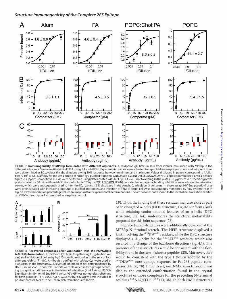
Immunogenicity of MPERp in the Different Adjuvants—Todetermine the immunogenicity of the structures describedabove, the MPERp-based alum, Freund’s adjuvant, and lipo-some formulations were next compared in their capacity foractivating B-cell responses (Fig. 7). Antigen-specific IgGs couldbe recovered upon immunization of rabbits with MPERp dis-persed in alum or Freund’s adjuvants (Fig. 7A, left and left-center panels). The midpoint titers in these samples were on theorder of 103. The responses triggered by the peptide-liposomeformulations were weaker by comparison, showing midpointtiters in the order of 102 (Fig. 7A, right-center and right panels).Nonetheless, antibodies raised against the 2F5 epitope could berecovered from all sera through binding affinity (Fig. 7B). Sup-porting the same range of affinity, incubation in solution of thepurified antibodies with 2F5 peptide epitope inhibited withcomparable potencies their binding to MPERp deposited onplates (IC50 values in the order of 10 �M in all cases). In contrast,the experiments displayed in Fig. 7C indicated that the 2F5-targeting antibodies raised by the different formulations werequalitatively different. Those experiments revealed that the
only detectable inhibition of HIV entry occurred with thePOPG/lipid A-MPERp formulation (Fig. 7C, right-hand panel).Of note, blocking VSV-GP-mediated cell infection was notobserved in these experiments, which underscores the specific-ity of the recovered antibodies.
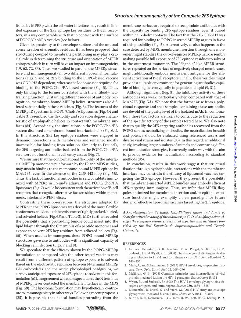
The inhibitory activity of these antibodies was nonetheless�50 –100 times lower than that displayed by the MAb2F5 inthe cell entry assays (Fig. 5A). Thus, to establish the significanceof this activity, we extended our studies on the immunogenicityof the POPG/lipid A-MPERp formulation (Fig. 8). Fig. 8A com-pares midpoint titers and cell entry inhibition by the 2F5-spe-cific antibodies isolated from the sera of four different rabbits.These two values did not correlate, thereby suggesting thateven if the immunogenicity levels were different, antibodieswere functional in the cell entry assay after isolation in affinitycolumns. Two groups of sera, R2/R3 and R1/R4, were estab-lished as a function of the inhibitory strength of the isolatedantibodies (Fig. 8B). For the two groups, the inhibition levels ofHIV-Env-mediated cell entry were significantly higher thanthose in the VSV-GP controls. Thus, according to our data inFigs. 7 and 8, we may conclude that antibody samples recoveredfrom POPG sera bore an inhibitory activity of cell entry that wasnot found in the samples recovered from alum, Freund’s adju-vant, or POPC/Chol/PA sera.
DISCUSSION
It has been hypothesized that the membrane-proximalsequence connecting the gp41 subunit’s ectodomain with thetransmembrane anchor, termed the MPER domain, includes acontinuous epitope. This implies that peptides recreating its nativestructure might in principle compose stand-alone HIV vaccines (6,13, 19, 20, 72, 73). Although intensively studied, data supportingthe structural MPER connectivity within a synthetic peptide vac-cine were lacking. Here, we considered the incorporation within asingle peptide of the sequence 656NEQELLELDKWASLWN671
spanning the full epitope recognized by the MAb2F5 as defined byproteomic analyses, competition ELISA, and crystallography (14,34–36), plus the following 672WFNITNWLWYIK683 Trp-richstretch that precedes the TMD (Fig. 1) (74). Our NMR datarevealed the structuring of the resulting MPERp synthetic surro-gate as a continuous helical structure (Figs. 2–4A). Structures ofshorter or even longer peptides show spots of partial structuring as310-and �-helix (33, 35, 73, 75–77), but none of them display con-tinuous helical structures for the sequence spanning the full 2F5epitope plus the downstream aromatic rich sequence precedingthe TMD anchor (Fig. 1). This suggests that inclusion of the com-plete sequence covered by the 2F5 paratope (34) might be requiredfor the long range interactions sustaining MPER folding as a con-tinuous helix.
Although continuously helical, MPERp NMR structuressolved in HFIP and DPC showed features implying a certaindegree of conformational flexibility. The kink adopted by the675FNI677 residues at the C terminus of the DPC structure wasconsistent with the 673FN674 hinge described previously for ashorter peptide (73). By comparison, the hinge of the shorterpeptide induced a more abrupt change in backbone main direc-tion and adopted a looser conformation (PDB code 2PV6, Fig.
FIGURE 6. Combined IR and molecular dynamics simulations of MPERpinteracting with POPC/Chol/PA (A) or POPG (B) lipid bilayers. Left pan-els, IR absorption in the amide I region by MPERp in D2O-based buffermixed with solutions of POPC/Chol/PA (2:1.5:0.2 mol/mol) liposomes orPOPG liposomes, following the procedure used for preparing vaccines. Inboth panels, the IR spectra were decomposed into different band compo-nents (numerical values are disclosed in Table 3). Red dotted lines corre-spond to �-helix components. Right panels, snapshots of MPERp weretaken at times 215 and 233 ns (top and bottom, respectively). Side views ofthe peptides display in space-filling representation residues Lys-665/Trp-666 in red and Leu-669/Trp-672/Phe-673 in blue. Phospholipids are shownin stick representation. Residues depicted in green have at least one atomwithin �3 Å from the phospholipid molecules.
Structure Immunogenicity of the Complete 2F5 Epitope
MARCH 7, 2014 • VOLUME 289 • NUMBER 10 JOURNAL OF BIOLOGICAL CHEMISTRY 6573
by guest on April 18, 2016
http://ww
w.jbc.org/
Dow
nloaded from
1B). Thus, the finding that these residues may also exist as partof an elongated �-helix (HFIP structure, Fig. 4A) or form a kinkwhile retaining conformational features of an �-helix (DPCstructure, Fig. 4A), underscores the structural metastabilityproposed for this joint sequence (73).
Unprecedented structures were additionally observed at theMPERp N-terminal stretch. The HFIP structure displayed akink involving the 665KW666 residues, while the DPC structuredisplayed a 310-helix for the 661LEL663 residues, which alsoresulted in a change of the backbone direction (Fig. 4A). Thepresence of these structures would be consistent with the flex-ibility found in the case of shorter peptides (35). Moreover, theywould be consistent with the type I �-turn adopted by the664DKW666 core epitope sequence in Fab2F5-peptide com-plexes (14, 36, 78). In contrast, our NMR structures did notdisplay the extended conformation found in the crystalstructures of those complexes for the preceding N-terminalresidues 656NEQELLEL663 (14, 36). In both NMR structures
FIGURE 7. Immunogenicity of MPERp formulated with different adjuvants. A, midpoint IgG titers in sera from rabbits immunized with MPERp in thedifferent adjuvants. Sera were titrated in ELISA using 1.4 �M MPERp. Experimental values were adjusted to sigmoid dose-response curves, and midpoint titerswere determined as EC50 values (i.e. the dilutions giving 50% response between minimum and maximum). Values displayed in panels correspond to 1/dilu-tion � 103 S.E. B, affinity for the 2F5 epitope of rabbit IgG purified from sera with 2F5ep-Cys (NEQELLELDKWASLWN-C) peptide immobilized onto a beadedagarose support. Competitive ELISAs were performed using plates coated with MPERp (1.4 �M). Prior to adding to the plates, 0.1 �g/ml of 2F5-specific IgG waspreincubated for 30 min with serial dilutions of soluble 2F5ep (NEQELLELDKWASLWN) peptide. Percentages of binding inhibition were adjusted to saturationcurves, which were subsequently used to infer the IC50 values S.E. displayed in the panels. C, inhibition of cell entry. In these assays HIV-Env pseudoviruseswere preincubated with increasing amounts of purified antibodies, and infection of TZM-bl target cells was subsequently monitored by flow cytometry as inFig. 5A. Plotted inhibition percentage values are means of four experimental determinations. The red columns correspond to the level of neutralization exertedon VSV-G-pseudotyped viruses used as negative control.
FIGURE 8. Recovered responses after vaccination with the POPG/lipidA-MPERp formulation. A, midpoint IgG titers (negative log EC50 dilution val-ues) and inhibition of cell entry by 2F5-specific antibodies in the sera of fourdifferent rabbits (R1–R4). Antibodies purified with 2F5ep-Cys were used at100 �g/ml in the latter assay. B, levels of inhibition of cell entry mediated byHIV-1-Env or VSV-GP controls. Rabbits were classified in two groups accord-ing to significant differences in the levels of inhibition (R1/R4 versus R2/R3).Significant inhibition of Env-HIV-1 versus VSV-GP was nonetheless observedfor both groups (**, p � 0.005; *, p � 0.05). MAb2F5 (2 �g/ml) was included aspositive control. Means S.D. of six determinations are shown.
Structure Immunogenicity of the Complete 2F5 Epitope
6574 JOURNAL OF BIOLOGICAL CHEMISTRY VOLUME 289 • NUMBER 10 • MARCH 7, 2014
by guest on April 18, 2016
http://ww
w.jbc.org/
Dow
nloaded from

reported here, these residues rather adopted a helical con-formation (Fig. 4A).
Even though one should be cautious in interpreting a pep-tide’s conformational states and extrapolating back to thenative functional protein, the potential relevance of the helicalconformation adopted by MPERp N-terminal residues isemphasized by the structure of an antigenically near-native Envconstruct termed “SOSIP” gp140 (79). Although truncated atposition 664, the recently solved crystal structure at 4.7 Å pro-
vides insights into the gp41 ectodomain organization in thecontext of cleaved, stabilized HIV-1 Env trimers (80). TheSOSIP structure supports the location of the 656NEQELLEL663
residues into a solvent-exposed helix within the native Envstructure.
One crystallographic structure of the 2F5 Fab complexedwith peptide further displayed the turn sequence 664DKW666
followed by residues 667ASLW670 adopting a canonical �-helixconformation (36), which is also present in our NMR structures
FIGURE 9. 2F5 epitope organization in MPERp and putative mechanism of antibody recognition. A, 2F5 epitope in HFIP and DPC structures. Core epitoperesidues ELDKWA are shown in red, and downstream residues putatively implied in secondary interactions with CDR-H3 loop are depicted in blue. Trp-666 andLeu-669/Trp-672/Phe-673 are displayed on the same side of the helix. B, comparison of 2F5 epitope structure in Fab� complex (PDB code 3D0L) and MPERp. Thechain portion spanning residues Leu-661–Trp-670 is shown in gray in the three structures, with projecting side chains of Asp-664 (left) or Lys-665 (right) andTrp-666 in red. Side chain of Leu-669 is displayed in blue to establish the relative position of the downstream helix. The comparison suggests that the 310-helixobserved in DPC might include an intermediate of the conformational change required for positioning Asp-664 side chain into the 2F5 paratope. Lys-665accommodation into the paratope would not require by comparison major conformational changes of the peptide backbone. C, fitting of the MPERp helix intoFab�-bound peptide. The Fab paratope structure (PDB code 3D0L) is displayed in ribbon representation. The base of the flexible loop of the heavy chain (notsolved in the crystal) is marked by the yellow side chains of residues Pro-98 and Arg-100B. The MPER residues Trp-666 and Leu-669 in the bound peptide aredisplayed in red and blue, respectively. In the right panel, the helix turn of MPERp (DPC structure) containing Leu-669 (displayed in blue) has been fitted into theFab-bound structure. The dotted lines mark the estimated position of the loop relative to the MPERp helix.
Structure Immunogenicity of the Complete 2F5 Epitope
MARCH 7, 2014 • VOLUME 289 • NUMBER 10 JOURNAL OF BIOLOGICAL CHEMISTRY 6575
by guest on April 18, 2016
http://ww
w.jbc.org/
Dow
nloaded from

(Fig. 4A, see also Fig. 9C). Thus, in combination, the structuralevidence suggests that the native structure recognized by the2F5 antibody might consist of a continuous helical structureinterrupted by a flexible kink at positions 664 – 666 that redi-rects the gp41 backbone at the pre-transmembrane region.
Implications for 2F5 Epitope Recognition Mechanism—TheMPERp NMR structures solved in this work recreate kinkedhelical motifs (Fig. 9A), which, in combination with previouslyreported structures (Fig. 9B), sustain proposals that this regionhas evolved to sample alternative conformations after activa-tion of the fusion cascade (25). Within this context, a putativemechanism for 2F5 epitope recognition is presented in Fig. 9B.The figure displays the orientations adopted by the 664DKW666
residues in MPERp structures and the Fab-bound peptide. TheTrp-666 and Leu-669 side chains are oriented in parallel in thethree structures, while the negative charge of Asp-664 side-chain projects from the main axis in different directions (Fig.9B, left). By contrast, the alkyl-� stacking between Lys-665 andTrp-666 side chains found in contact with Fab could be fairlyreproduced by the structure solved in the DPC structure (Fig.9B, right). In the HFIP structure, further rotation of the Lys-665side chain would allow its insertion into the Fab binding pocket,without requiring major changes of the peptide backbone con-formation. Thus, the NMR structures suggest that binding to ahelical MPER peptide might first involve contacting Lys-665,Trp-666, and Leu-669 residues and then require induction bythe antibody of a conformational transition in the C� chain forinserting Asp-664 into the binding pocket. Comparison of thethree structures further suggests that the short 310-helix foundin the DPC structure may encompass an intermediate betweenthe fully helical and the extended conformations observed inHFIP- and Fab-bound structures, respectively.
The NMR structures described in this work may in additionprovide insights into secondary interactions of the 2F5 anti-body with MPER residues C-terminal to the core epitope (Fig.9C). Screening of phage-displayed peptide libraries with theMAb2F5 identified Leu-669 as an almost invariant residue atthe C terminus of the core epitope (63). Further competitionELISA demonstrated that the CDR-H3 loop increased bindingaffinity when C-terminal 672WFNITNWLWYIK683 residueswere added to the full 656NEQELLELDKWASLWN681 epitopesequence (38). This finding raised the possibility that the neu-tralization dependence on the loop apex was caused by weakersecondary binding to C-terminal MPER residues (38).
Recently reported compelling mutagenesis of the CDR-H3loop by Güenaga and Wyatt (25) supports that idea. A signifi-cant correlation was found between neutralization potency ofCDR-H3 mutants and affinity to an MPER peptide spanning res-idues 657EQELLELDKWASLWNWFNITNWLWYIK683. Thiscorrelation was lost in the case of the 659ELLELDKWASL669
sequence structurally constrained into a protein scaffold (30).Moreover, L669A, W670A, N671A, W672A, and F673A substi-tutions, in residues immediately C-terminal to the core epitope,resulted in an affinity decrease. It was further proposed thatweak contacts involving � stacking interactions among aro-matic residues present in the antibody CDR-H3 loop and theMPER peptide sequence might be responsible for this effect(25). According to these authors, this mode of recognition
would in addition enable 2F5 epitope binding when MPERorganizes as a helical bundle.
The MPERp structures solved in this work, displaying therelative positions of the 2F5 core epitope and the downstreamresidues encompassing this secondary antibody-binding site,substantiate such a hypothesis (Fig. 9C). Fitting of the MPERpDPC helix 667ASLW670 stretch into the corresponding Fab-bound structure (36) disclosed the Leu-669 side chain at thebase of the CDR-H3 loop in remarkably similar orientation andaligned the MPER helix axis with the parts of the loops visible inthe crystal structure. Thus, the model that emerges from com-bining both structures supports the possibility that the tip of theCDR-H3 loop may establish contacts with Trp-672 and Phe-673 residues on the MPER helix (25, 38).
Implications for Immunogen Design—Overall our structuraldata support 2F5 docking to the surface of a kinked MPERphelix exposing the invariable residues Trp-666, Leu-669, Trp-672, and Phe-673, and the putative involvement of the CDR-H3loop in the process. In previous studies, it was hypothesized thatinclusion of residues responsible for the secondary bindingprocess would be required to elicit 2F5-like neutralizingresponses (25, 36, 38). To test the immunogenicity of an MPERhelix containing all elements involved in epitope recognition bythe bNAb2F5, we carried out immunization studies usingMPERp as a vaccine. To establish a potential structure-immu-nogenicity relationship, we monitored conformational changesin the vaccine formulations. IR allows performing reliablemeasurements (i.e. resolution or sensitivity are unaffected) inturbid suspensions such as aggregated membranes and particulateemulsions. Therefore, IR was the technique of choice for confirm-ing the preservation of MPERp helical structures in the viscousmedia provided by the aluminum salts, “Freund’s” water-in-oilemulsion, and membrane adjuvants (Figs. 4B and 6).
Even though MPERp dispersed in alum and Freund’s adju-vant triggered effective MPER-targeting immunoresponses, theantibodies isolated from these sera were not functional in thecell entry inhibition assays (Fig. 7). It has been reported thatinteractions of protein antigens with the highly charged alumi-num salts may destabilize their structures (81). Consistent withthis finding, MPERp in contact with alum underwent signifi-cant aggregation, as denoted by the IR absorption at 1620 cm�1
(Fig. 4B, left). We speculate that MPERp unfolding/aggregationin the highly polar alum medium might result in the presenta-tion of neutralization-irrelevant 2F5 epitope structures to theB-cell receptor. In contrast, IR demonstrated the absence ofsignificant MPERp aggregation in Freund’s reverse micelles(Fig. 4B, right, and Table 3).
It has been argued that the water molecules confined at theinterfacial region of the water-in-oil reverse micelles are non-bulk-like, resulting in a low dielectric environment at the water-oil interface, reminiscent of that found at the membrane-waterinterface (82). Thus, amphipathic peptides in contact withreverse micelles may attain helical structures compatible withthose adopted when adsorbed to membrane surfaces. In partic-ular, this has been demonstrated using IR for the antimicrobialpeptide mastoparan X (71). Our results suggest that this is alsothe case for MPERp in Freund’s adjuvant and POPC/Chol/PAvesicles (Table 3). We infer that hydrophobic contacts estab-
Structure Immunogenicity of the Complete 2F5 Epitope
6576 JOURNAL OF BIOLOGICAL CHEMISTRY VOLUME 289 • NUMBER 10 • MARCH 7, 2014
by guest on April 18, 2016
http://ww
w.jbc.org/
Dow
nloaded from
lished by MPERp with the oil-water interface may result in lim-ited exposure of the 2F5 epitope key residues to B-cell recep-tors, in a way comparable with that in contact with the surfaceof POPC/Chol/PA vesicles (see below).
Given its proximity to the envelope surface and the unusualconcentration of aromatic residues, it has been proposed thatstructuring coupled to membrane partitioning may play a cru-cial role in determining the structure and orientation of MPERepitopes, which in turn will have an impact on immunogenicity(19, 62, 72, 83). Thus, we studied in parallel the MPERp struc-ture and immunogenicity in two different liposomal formula-tions (Figs. 5 and 6). 2F5 binding to the POPG-based vaccinewas CDR-H3 dependent, whereas the loop was not required forbinding to the POPC/Chol/PA-based vaccine (Fig. 5). Thus,only binding to the former correlated with the antibody-neu-tralizing function. Sustaining different modes of antibody rec-ognition, membrane-bound MPERp helical structures also dif-fered substantially in these vaccines (Fig. 6). The features of theMPERp IR spectrum in POPC/Chol/PA liposomes (Fig. 6A andTable 3) resembled the flexibility and solvation degree charac-teristic of amphipathic helices in contact with membrane sur-faces (84). Accordingly, the simulations in the POPC/Chol/PAsystem disclosed a membrane-bound interfacial helix (Fig. 6A).In this structure, 2F5 key epitope residues were engaged indynamic interactions with membranes and remained mostlyinaccessible for binding from solution. Similarly to Freund’s,the 2F5-targeting antibodies isolated from the POPC/Chol/PAsera were not functional in cell entry assays (Fig. 7).
We surmise that the conformational flexibility of the interfa-cial MPERp monomers put forward by the IR and MDS studies,may sustain binding to the membrane-bound MPER epitope byMAb2F5, even in the absence of the CDR-H3 loop (Fig. 5E).Thus, the lack of functional antibodies in sera of rabbits immu-nized with MPERp in Freund’s adjuvant and POPC/Chol/PAliposomes (Fig. 7) would be consistent with the activation of B-cellreceptors that recognize alternative faces/residues within mono-meric, interfacial MPER helices.
Contrasting these observations, the structure adopted byMPERp in the POPG liposomes was devoid of the more flexibleconformers and denoted the existence of tightly packed, buried,and solvated helices (Fig. 6B and Table 3). MDS further revealedthe possibility that a protruding bundle could insert into thelipid bilayer through the C terminus of a peptide monomer andexpose to solvent 2F5 key residues from adhered helices (Fig.6B). When used as immunogens, these POPG-bound MPERpstructures gave rise to antibodies with a significant capacity ofblocking cell infection (Figs. 7 and 8).
We speculate that the efficacy shown by the POPG-MPERpformulation as compared with the other tested vaccines mayresult from a different pattern of epitope exposure to solvent.Based on the electrostatic repulsion between N-terminal MPERpGlu carboxylates and the acidic phospholipid headgroups, wealready anticipated exposure of 2F5 epitope to solvent in this for-mulation (61). In agreement with our expectations, the N terminusof MPERp never contacted the membrane interface in the MDS(Fig. 6B). The liposomal formulation may hypothetically contrib-ute to immunogenicity in other ways. Following recent proposals(25), it is possible that helical bundles protruding from the
membrane surface are required to recapitulate antibodies withthe capacity for binding 2F5 epitope residues, even if buriedwithin helix-helix contacts. The fact that the 2F5 CDR-H3 wasrequired for binding to POPG-inserted MPERp argues in favorof this possibility (Fig. 5). Alternatively, as also happens in thecase detected by MDS, membrane insertion through one mon-omer might stabilize the out-of-register MPERp helix assemblymaking possible full exposure of 2F5 epitope residues to solventin the outermost monomer. The “flagpole”-like MPER struc-tures repeated on the surface of negatively charged membranes,might additionally embody multivalent antigens for the effi-cient activation of B-cell receptors. Finally, these vesicles mightprovide a suitable environment for generating antibodies capa-ble of binding heterotypically to peptide and lipid (9, 31).
Although significant (Fig. 8), the inhibitory activity of theseantibodies was weak, particularly when compared with that ofMAb2F5 (Fig. 5A). We note that the former arise from a poly-clonal response and that samples containing these antibodiesare devoid of the purity level of the isolated mAb. In combina-tion, those two factors are likely to contribute to the reductionof the specific activity of the samples tested here. We also notethat to qualify the 2F5-targeting antibodies recovered from thePOPG sera as neutralizing antibodies, the neutralization breadthand potency should be evaluated using referenced assays anddiverse viral strains and isolates (85). In this regard, an additionalstudy, involving larger numbers of animals and comparing differ-ent immunization strategies, is currently under way with the aimto provide evidence for neutralization according to standardmethods (86).
In conclusion, results in this work suggest that structuralfixation through hydrophobic interactions with the membraneinterface may constrain the efficacy of liposomal vaccines tar-geting the 2F5 epitope. However, they present the possibilitythat membrane-inserted MPER bundles may embody efficient2F5-targeting immunogens. Thus, we infer that MPER flag-poles optimized for membrane insertion and/or epitope-expo-sure functions might exemplify a new paradigm for futuredesign of effective liposomal vaccines targeting the 2F5 epitope.
Acknowledgments—We thank Jean-Philippe Julien and Jamie K.Scott for critical reading of the manuscript. C. D. thankfully acknowl-edges the computer resources, technical expertise, and assistance pro-vided by the Red Española de Supercomputación and TempleUniversity.
REFERENCES1. Karlsson Hedestam, G. B., Fouchier, R. A., Phogat, S., Burton, D. R.,
Sodroski, J., and Wyatt, R. T. (2008) The challenges of eliciting neutraliz-ing antibodies to HIV-1 and to influenza virus. Nat. Rev. Microbiol. 6,143–155
2. Merk, A., and Subramaniam, S. (2013) HIV-1 envelope glycoprotein struc-ture. Curr. Opin. Struct. Biol. 23, 268 –276
3. Melikyan, G. B. (2008) Common principles and intermediates of viralprotein-mediated fusion: the HIV-1 paradigm. Retrovirology 5, 111
4. Wyatt, R., and Sodroski, J. (1998) The HIV-1 envelope glycoproteins: fu-sogens, antigens, and immunogens. Science 280, 1884 –1888
5. Blumenthal, R., Durell, S., and Viard, M. (2012) HIV entry and envelopeglycoprotein-mediated fusion. J. Biol. Chem. 287, 40841– 40849
6. Burton, D. R., Desrosiers, R. C., Doms, R. W., Koff, W. C., Kwong, P. D.,
Structure Immunogenicity of the Complete 2F5 Epitope
MARCH 7, 2014 • VOLUME 289 • NUMBER 10 JOURNAL OF BIOLOGICAL CHEMISTRY 6577
by guest on April 18, 2016
http://ww
w.jbc.org/
Dow
nloaded from

Moore, J. P., Nabel, G. J., Sodroski, J., Wilson, I. A., and Wyatt, R. T. (2004)HIV vaccine design and the neutralizing antibody problem. Nat. Immunol.5, 233–236
7. Stamatatos, L., Morris, L., Burton, D. R., and Mascola, J. R. (2009) Neu-tralizing antibodies generated during natural HIV-1 infection: good newsfor an HIV-1 vaccine? Nat. Med. 15, 866 – 870
8. Mascola, J. R., and Montefiori, D. C. (2010) The role of antibodies in HIVvaccines. Annu. Rev. Immunol. 28, 413– 444
9. Klein, F., Mouquet, H., Dosenovic, P., Scheid, J. F., Scharf, L., and Nussen-zweig, M. C. (2013) Antibodies in HIV-1 vaccine development and ther-apy. Science 341, 1199 –1204
10. Walker, L. M., and Burton, D. R. (2010) Rational antibody-based HIV-1vaccine design: current approaches and future directions. Curr. Opin. Im-munol. 22, 358 –366
11. Kwong, P. D., and Mascola, J. R. (2012) Human antibodies that neutralizeHIV-1: identification, structures, and B cell ontogenies. Immunity 37,412– 425
12. Gray, E. S., Madiga, M. C., Moore, P. L., Mlisana, K., Abdool Karim, S. S.,Binley, J. M., Shaw, G. M., Mascola, J. R., and Morris, L. (2009) Broadneutralization of human immunodeficiency virus type 1 mediated byplasma antibodies against the gp41 membrane proximal external region.J. Virol. 83, 11265–11274
13. Zwick, M. B., Labrijn, A. F., Wang, M., Spenlehauer, C., Saphire, E. O.,Binley, J. M., Moore, J. P., Stiegler, G., Katinger, H., Burton, D. R., andParren, P. W. (2001) Broadly neutralizing antibodies targeted to the mem-brane-proximal external region of human immunodeficiency virus type 1glycoprotein gp41. J. Virol. 75, 10892–10905
14. Ofek, G., Tang, M., Sambor, A., Katinger, H., Mascola, J. R., Wyatt, R., andKwong, P. D. (2004) Structure and mechanistic analysis of the anti-humanimmunodeficiency virus type 1 antibody 2F5 in complex with its gp41epitope. J. Virol. 78, 10724 –10737
15. Cardoso, R. M., Brunel, F. M., Ferguson, S., Zwick, M., Burton, D. R.,Dawson, P. E., and Wilson, I. A. (2007) Structural basis of enhanced bind-ing of extended and helically constrained peptide epitopes of the broadlyneutralizing HIV-1 antibody 4E10. J. Mol. Biol. 365, 1533–1544
16. Pejchal, R., Gach, J. S., Brunel, F. M., Cardoso, R. M., Stanfield, R. L.,Dawson, P. E., Burton, D. R., Zwick, M. B., and Wilson, I. A. (2009) Aconformational switch in human immunodeficiency virus gp41 revealedby the structures of overlapping epitopes recognized by neutralizing anti-bodies. J. Virol. 83, 8451– 8462
17. Zhu, Z., Qin, H. R., Chen, W., Zhao, Q., Shen, X., Schutte, R., Wang, Y.,Ofek, G., Streaker, E., Prabakaran, P., Fouda, G. G., Liao, H. X., Owens, J.,Louder, M., Yang, Y., Klaric, K. A., Moody, M. A., Mascola, J. R., Scott, J. K.,Kwong, P. D., Montefiori, D., Haynes, B. F., Tomaras, G. D., and Dimitrov,D. S. (2011) Cross-reactive HIV-1-neutralizing human monoclonal anti-bodies identified from a patient with 2F5-like antibodies. J. Virol. 85,11401–11408
18. Huang, J., Ofek, G., Laub, L., Louder, M. K., Doria-Rose, N. A., Longo,N. S., Imamichi, H., Bailer, R. T., Chakrabarti, B., Sharma, S. K., Alam,S. M., Wang, T., Yang, Y., Zhang, B., Migueles, S. A., Wyatt, R., Haynes,B. F., Kwong, P. D., Mascola, J. R., and Connors, M. (2012) Broad andpotent neutralization of HIV-1 by a gp41-specific human antibody. Na-ture 491, 406 – 412
19. Montero, M., van Houten, N. E., Wang, X., and Scott, J. K. (2008) Themembrane-proximal external region of the human immunodeficiency vi-rus type 1 envelope: dominant site of antibody neutralization and targetfor vaccine design. Microbiol. Mol. Biol. Rev. 72, 54 – 84
20. Montero, M., Gulzar, N., Klaric, K. A., Donald, J. E., Lepik, C., Wu, S., Tsai,S., Julien, J. P., Hessell, A. J., Wang, S., Lu, S., Burton, D. R., Pai, E. F.,Degrado, W. F., and Scott, J. K. (2012) Neutralizing epitopes in the mem-brane-proximal external region of HIV-1 gp41 are influenced by the trans-membrane domain and the plasma membrane. J. Virol. 86, 2930 –2941
21. Muster, T., Steindl, F., Purtscher, M., Trkola, A., Klima, A., Himmler, G.,Rüker, F., and Katinger, H. (1993) A conserved neutralizing epitope ongp41 of human immunodeficiency virus type 1. J. Virol. 67, 6642– 6647
22. Muster, T., Guinea, R., Trkola, A., Purtscher, M., Klima, A., Steindl, F.,Palese, P., and Katinger, H. (1994) Cross-neutralizing activity against di-vergent human immunodeficiency virus type 1 isolates induced by the
gp41 sequence ELDKWAS. J. Virol. 68, 4031– 403423. Binley, J. M., Wrin, T., Korber, B., Zwick, M. B., Wang, M., Chappey, C.,
Stiegler, G., Kunert, R., Zolla-Pazner, S., Katinger, H., Petropoulos, C. J.,and Burton, D. R. (2004) Comprehensive cross-clade neutralization anal-ysis of a panel of anti-human immunodeficiency virus type 1 monoclonalantibodies. J. Virol. 78, 13232–13252
24. Conley, A. J., Kessler, J. A., 2nd, Boots, L. J., Tung, J. S., Arnold, B. A., Keller,P. M., Shaw, A. R., and Emini, E. A. (1994) Neutralization of divergenthuman immunodeficiency virus type 1 variants and primary isolates byIAM-41-2F5, an anti-gp41 human monoclonal antibody. Proc. Natl. Acad.Sci. U.S.A. 91, 3348 –3352
25. Guenaga, J., and Wyatt, R. T. (2012) Structure-guided alterations of thegp41-directed HIV-1 broadly neutralizing antibody 2F5 reveal new prop-erties regarding its neutralizing function. PLoS Pathog. 8, e1002806
26. Purtscher, M., Trkola, A., Gruber, G., Buchacher, A., Predl, R., Steindl, F.,Tauer, C., Berger, R., Barrett, N., and Jungbauer, A. (1994) A broadlyneutralizing human monoclonal antibody against gp41 of human immu-nodeficiency virus type 1. AIDS Res. Hum. Retroviruses 10, 1651–1658
27. Coëffier, E., Clément, J. M., Cussac, V., Khodaei-Boorane, N., Jehanno, M.,Rojas, M., Dridi, A., Latour, M., El Habib, R., Barré-Sinoussi, F., Hofnung,M., and Leclerc, C. (2000) Antigenicity and immunogenicity of the HIV-1gp41 epitope ELDKWA inserted into permissive sites of the MalE protein.Vaccine 19, 684 – 693
28. McGaughey, G. B., Citron, M., Danzeisen, R. C., Freidinger, R. M., Garsky,V. M., Hurni, W. M., Joyce, J. G., Liang, X., Miller, M., Shiver, J., andBogusky, M. J. (2003) HIV-1 vaccine development: constrained peptideimmunogens show improved binding to the anti-HIV-1 gp41 MAb. Bio-chemistry 42, 3214 –3223
29. Dennison, S. M., Sutherland, L. L., Jaeger, F. H., Anasti, K. M., Parks, R.,Stewart, S., Bowman, C., Xia, S. M., Zhang, R., Shen, X., Scearce, R. M.,Ofek, G., Yang, Y., Kwong, P. D., Santra, S., Liao, H. X., Tomaras, G.,Letvin, N. L., Chen, B., Alam, S. M., and Haynes, B. F. (2011) Induction ofantibodies in rhesus macaques that recognize a fusion-intermediate con-formation of HIV-1 gp41. PLoS One 6, e27824
30. Guenaga, J., Dosenovic, P., Ofek, G., Baker, D., Schief, W. R., Kwong, P. D.,Karlsson Hedestam, G. B., and Wyatt, R. T. (2011) Heterologous epitope-scaffold prime:boosting immuno-focuses B cell responses to the HIV-1gp41 2F5 neutralization determinant. PLoS One 6, e16074
31. Matyas, G. R., Wieczorek, L., Beck, Z., Ochsenbauer-Jambor, C., Kappes,J. C., Michael, N. L., Polonis, V. R., and Alving, C. R. (2009) Neutralizingantibodies induced by liposomal HIV-1 glycoprotein 41 peptide simulta-neously bind to both the 2F5 or 4E10 epitope and lipid epitopes. AIDS 23,2069 –2077
32. Huarte, N., Araujo, A., Arranz, R., Lorizate, M., Quendler, H., Kunert, R.,Valpuesta, J. M., and Nieva, J. L. (2012) Recognition of membrane-boundfusion-peptide/MPER complexes by the HIV-1 neutralizing 2F5 antibody:implications for anti-2F5 immunogenicity. PLoS One 7, e52740
33. Joyce, J. G., Hurni, W. M., Bogusky, M. J., Garsky, V. M., Liang, X., Citron,M. P., Danzeisen, R. C., Miller, M. D., Shiver, J. W., and Keller, P. M. (2002)Enhancement of �-helicity in the HIV-1 inhibitory peptide DP178 leads toan increased affinity for human monoclonal antibody 2F5 but does notelicit neutralizing responses in vitro. Implications for vaccine design.J. Biol. Chem. 277, 45811– 45820
34. Parker, C. E., Deterding, L. J., Hager-Braun, C., Binley, J. M., Schülke, N.,Katinger, H., Moore, J. P., and Tomer, K. B. (2001) Fine definition of theepitope on the gp41 glycoprotein of human immunodeficiency virus type1 for the neutralizing monoclonal antibody 2F5. J. Virol. 75, 10906 –10911
35. Barbato, G., Bianchi, E., Ingallinella, P., Hurni, W. H., Miller, M. D., Cili-berto, G., Cortese, R., Bazzo, R., Shiver, J. W., and Pessi, A. (2003) Struc-tural analysis of the epitope of the anti-HIV antibody 2F5 sheds light intoits mechanism of neutralization and HIV fusion. J. Mol. Biol. 330,1101–1115
36. Julien, J. P., Bryson, S., Nieva, J. L., and Pai, E. F. (2008) Structural details ofHIV-1 recognition by the broadly neutralizing monoclonal antibody 2F5:epitope conformation, antigen-recognition loop mobility, and anion-binding site. J. Mol. Biol. 384, 377–392
37. Zwick, M. B., Komori, H. K., Stanfield, R. L., Church, S., Wang, M., Parren,P. W., Kunert, R., Katinger, H., Wilson, I. A., and Burton, D. R. (2004) The
Structure Immunogenicity of the Complete 2F5 Epitope
6578 JOURNAL OF BIOLOGICAL CHEMISTRY VOLUME 289 • NUMBER 10 • MARCH 7, 2014
by guest on April 18, 2016
http://ww
w.jbc.org/
Dow
nloaded from

long third complementarity-determining region of the heavy chain is im-portant in the activity of the broadly neutralizing anti-human immunode-ficiency virus type 1 antibody 2F5. J. Virol. 78, 3155–3161
38. Julien, J. P., Huarte, N., Maeso, R., Taneva, S. G., Cunningham, A., Nieva,J. L., and Pai, E. F. (2010) Ablation of the complementarity-determiningregion H3 apex of the anti-HIV-1 broadly neutralizing antibody 2F5 ab-rogates neutralizing capacity without affecting core epitope binding. J. Vi-rol. 84, 4136 – 4147
39. Sánchez-Martínez, S., Lorizate, M., Katinger, H., Kunert, R., and Nieva,J. L. (2006) Membrane association and epitope recognition by HIV-1 neu-tralizing anti-gp41 2F5 and 4E10 antibodies. AIDS Res. Hum. Retroviruses22, 998 –1006
40. Kim, M., Sun, Z. Y., Rand, K. D., Shi, X., Song, L., Cheng, Y., Fahmy, A. F.,Majumdar, S., Ofek, G., Yang, Y., Kwong, P. D., Wang, J. H., Engen, J. R.,Wagner, G., and Reinherz, E. L. (2011) Antibody mechanics on a mem-brane-bound HIV segment essential for GP41-targeted viral neutraliza-tion. Nat. Struct. Mol. Biol. 18, 1235–1243
41. Alam, S. M., Morelli, M., Dennison, S. M., Liao, H. X., Zhang, R., Xia, S. M.,Rits-Volloch, S., Sun, L., Harrison, S. C., Haynes, B. F., and Chen, B. (2009)Role of HIV membrane in neutralization by two broadly neutralizing an-tibodies. Proc. Natl. Acad. Sci. U.S.A. 106, 20234 –20239
42. Mirassou, Y., Santiveri, C. M., Pérez de Vega, M. J., González-Muñiz, R.,and Jiménez, M. A. (2009) Disulfide bonds versus TrpTrp pairs in irregular�-hairpins: NMR structure of vammin loop 3-derived peptides as a casestudy. ChemBioChem 10, 902–910
43. Cornilescu, G., Delaglio, F., and Bax, A. (1999) Protein backbone anglerestraints from searching a database for chemical shift and sequence ho-mology. J. Biomol. NMR 13, 289 –302
44. Güntert, P., Mumenthaler, C., and Wüthrich, K. (1997) Torsion angledynamics for NMR structure calculation with the new program DYANA.J. Mol. Biol. 273, 283–298
45. Güntert, P. (2004) Automated NMR structure calculation with CYANA.Methods Mol. Biol. 278, 353–378
46. Laskowski, R. A., Rullmannn, J. A., MacArthur, M. W., Kaptein, R., andThornton, J. M. (1996) AQUA and PROCHECK-NMR: programs forchecking the quality of protein structures solved by NMR. J. Biomol. NMR8, 477– 486
47. Koradi, R., Billeter, M., and Wüthrich, K. (1996) MOLMOL: a program fordisplay and analysis of macromolecular structures. J. Mol. Graph. 14,51-55
48. Guex, N., and Peitsch, M. C. (1997) SWISS-MODEL and the Swiss-Pdb-Viewer: an environment for comparative protein modeling. Electrophore-sis 18, 2714 –2723
49. Yethon, J. A., Epand, R. F., Leber, B., Epand, R. M., and Andrews, D. W.(2003) Interaction with a membrane surface triggers a reversible confor-mational change in Bax normally associated with induction of apoptosis.J. Biol. Chem. 278, 48935– 48941
50. Arrondo, J. L., and Goñi, F. M. (1999) Structure and dynamics of mem-brane proteins as studied by infrared spectroscopy. Prog. Biophys. Mol.Biol. 72, 367– 405
51. Phillips, J. C., Braun, R., Wang, W., Gumbart, J., Tajkhorshid, E., Villa, E.,Chipot, C., Skeel, R. D., Kalé, L., and Schulten, K. (2005) Scalable molec-ular dynamics with NAMD. J. Comput. Chem. 26, 1781–1802
52. MacKerell, A. D., Bashford, D., Bellott, Dunbrack, R. L., Evanseck, J. D.,Field, M. J., Fischer, S., Gao, J., Guo, H., Ha, S., Joseph-McCarthy, D.,Kuchnir, L., Kuczera, K., Lau, F. T. K., Mattos, C., Michnick, S., Ngo, T.,Nguyen, D. T., Prodhom, B., Reiher, W. E., Roux, B., Schlenkrich, M.,Smith, J. C., Stote, R., Straub, J., Watanabe, M., Wiórkiewicz-Kuczera, J.,Yin, D., and Karplus, M. (1998) All-atom empirical potential for molecularmodeling and dynamics studies of proteins. J. Phys. Chem. B 102,3586 –3616
53. Klauda, J. B., Venable, R. M., Freites, J. A., O’Connor, J. W., Tobias, D. J.,Mondragon-Ramirez, C., Vorobyov, I., MacKerell, A. D., Jr., and Pastor,R. W. (2010) Update of the CHARMM all-atom additive force field forlipids: validation on six lipid types. J. Phys. Chem. B. 114, 7830 –7843
54. Jorgensen, W. L., Chandrasekhar, J., Madura, J. D., Impey, R. W., andKlein, M. L. (1983) Comparison of simple potential functions for simulat-ing liquid water. J. Chem. Phys. 79, 926 –935
55. Cournia, Z., Smith, J. C., and Ullmann, G. M. (2005) A molecular mechan-ics force field for biologically important sterols. J. Comput. Chem. 26,1383–1399
56. Martyna, G. J., Tobias, D. J., and Klein, M. L. (1994) Constant pressuremolecular dynamics algorithms. J. Chem. Phys. 101, 4177– 4189
57. Feller, S. E., Zhang, Y., Pastor, R. W., and Brooks, B. R. (1995) Constantpressure molecular dynamics simulation:the Langevin piston method.J. Chem. Phys. 103, 4613– 4621
58. Essmann, U., Perera, L., and Berkowitz, M. (1995) A smooth particle meshEwald method. J. Chem. Phys. 103, 8577– 8593
59. Miyamoto, S., and Kollman, P. (1992) Settle: an analytical version of theSHAKE and RATTLE algorithm for rigid water models. J. Comput. Chem.13, 952–962
60. Dreesman, G. R., Sanchez, Y., Ionescu-Matiu, I., Sparrow, J. T., Six, H. R.,Peterson, D. L., Hollinger, F. B., and Melnick, J. L. (1982) Antibody tohepatitis B surface antigen after a single inoculation of uncoupled syn-thetic HBsAg peptides. Nature 295, 158 –160
61. Maeso, R., Huarte, N., Julien, J. P., Kunert, R., Pai, E. F., and Nieva, J. L.(2011) Interaction of anti-HIV type 1 antibody 2F5 with phospholipidbilayers and its relevance for the mechanism of virus neutralization. AIDSRes. Hum. Retroviruses 27, 863– 876
62. Alving, C. R., Rao, M., Steers, N. J., Matyas, G. R., and Mayorov, A. V.(2012) Liposomes containing lipid A: an effective, safe, generic adjuvantsystem for synthetic vaccines. Expert Rev. Vaccines 11, 733–744
63. Menendez, A., Chow, K. C., Pan, O. C., and Scott, J. K. (2004) Humanimmunodeficiency virus type 1-neutralizing monoclonal antibody 2F5 ismultispecific for sequences flanking the DKW core epitope. J. Mol. Biol.338, 311–327
64. Wuthrich, K. (1986) NMR of Proteins and Nucleic Acids. John Wiley &Sons, Inc., New York
65. Wüthrich, K., Billeter, M., and Braun, W. (1984) Polypeptide secondarystructure determination by nuclear magnetic resonance observation ofshort proton-proton distances. J. Mol. Biol. 180, 715–740
66. Wishart, D. S., Bigam, C. G., Holm, A., Hodges, R. S., and Sykes, B. D.(1995) 1H, 13C, and 15N random coil NMR chemical shifts of the commonamino acids. I. Investigations of nearest-neighbor effects. J. Biomol. NMR5, 67– 81
67. Reisdorf, W. C., Jr., and Krimm, S. (1996) Infrared amide I� band of thecoiled coil. Biochemistry 35, 1383–1386
68. Walsh, S. T., Cheng, R. P., Wright, W. W., Alonso, D. O., Daggett, V.,Vanderkooi, J. M., and DeGrado, W. F. (2003) The hydration of amides inhelices; a comprehensive picture from molecular dynamics, IR, and NMR.Protein Sci. 12, 520 –531
69. Vu, D. M., Myers, J. K., Oas, T. G., and Dyer, R. B. (2004) Probing thefolding and unfolding dynamics of secondary and tertiary structures in athree-helix bundle protein. Biochemistry 43, 3582–3589
70. Coffman, R. L., Sher, A., and Seder, R. A. (2010) Vaccine adjuvants: puttinginnate immunity to work. Immunity 33, 492–503
71. Mukherjee, S., Chowdhury, P., DeGrado, W. F., and Gai, F. (2007) Site-specific hydration status of an amphipathic peptide in AOT reverse mi-celles. Langmuir 23, 11174 –11179
72. Lorizate, M., Huarte, N., Sáez-Cirión, A., and Nieva, J. L. (2008) Interfacialpre-transmembrane domains in viral proteins promoting membrane fu-sion and fission. Biochim. Biophys. Acta 1778, 1624 –1639
73. Sun, Z. Y., Oh, K. J., Kim, M., Yu, J., Brusic, V., Song, L., Qiao, Z., Wang,J. H., Wagner, G., and Reinherz, E. L. (2008) HIV-1 broadly neutralizingantibody extracts its epitope from a kinked gp41 ectodomain region on theviral membrane. Immunity 28, 52– 63
74. Suárez, T., Gallaher, W. R., Agirre, A., Goñi, F. M., and Nieva, J. L. (2000)Membrane interface-interacting sequences within the ectodomain of thehuman immunodeficiency virus type 1 envelope glycoprotein: putativerole during viral fusion. J. Virol. 74, 8038 – 8047
75. Biron, Z., Khare, S., Samson, A. O., Hayek, Y., Naider, F., and Anglister, J.(2002) A monomeric 310-helix is formed in water by a 13-residue peptiderepresenting the neutralizing determinant of HIV-1 on gp41. Biochemistry41, 12687–12696
76. Schibli, D. J., Montelaro, R. C., and Vogel, H. J. (2001) The membrane-proximal tryptophan-rich region of the HIV glycoprotein, gp41, forms a
Structure Immunogenicity of the Complete 2F5 Epitope
MARCH 7, 2014 • VOLUME 289 • NUMBER 10 JOURNAL OF BIOLOGICAL CHEMISTRY 6579
by guest on April 18, 2016
http://ww
w.jbc.org/
Dow
nloaded from

well defined helix in dodecylphosphocholine micelles. Biochemistry 40,9570 –9578
77. Coutant, J., Yu, H., Clément, M. J., Alfsen, A., Toma, F., Curmi, P. A., andBomsel, M. (2008) Both lipid environment and pH are critical for deter-mining physiological solution structure of 3-D-conserved epitopes of theHIV-1 gp41-MPER peptide P1. FASEB J. 22, 4338 – 4351
78. Bryson, S., Julien, J. P., Hynes, R. C., and Pai, E. F. (2009) Crystallographicdefinition of the epitope promiscuity of the broadly neutralizing anti-human immunodeficiency virus type 1 antibody 2F5: vaccine design im-plications. J. Virol. 83, 11862–11875
79. Depetris, R. S., Julien, J. P., Khayat, R., Lee, J. H., Pejchal, R., Katpally, U.,Cocco, N., Kachare, M., Massi, E., David, K. B., Cupo, A., Marozsan, A. J.,Olson, W. C., Ward, A. B., Wilson, I. A., Sanders, R. W., and Moore, J. P.(2012) Partial enzymatic deglycosylation preserves the structure ofcleaved recombinant HIV-1 envelope glycoprotein trimers. J. Biol. Chem.287, 24239 –24254
80. Julien, J. P., Cupo, A., Sok, D., Stanfield, R. L., Lyumkis, D., Deller, M. C.,Klasse, P. J., Burton, D. R., Sanders, R. W., Moore, J. P., Ward, A. B., andWilson, I. A. (2013) Crystal structure of a soluble cleaved HIV-1 envelopetrimer. Science 342, 1477–1483
81. Jones, L. S., Peek, L. J., Power, J., Markham, A., Yazzie, B., and Middaugh,
C. R. (2005) Effects of adsorption to aluminum salt adjuvants on the struc-ture and stability of model protein antigens. J. Biol. Chem. 280, 13406 –13414
82. Dokter, A. M., Woutersen, S., and Bakker, H. J. (2006) Inhomogeneousdynamics in confined water nanodroplets. Proc. Natl. Acad. Sci. U.S.A.103, 15355–15358
83. Zwick, M. B. (2005) The membrane-proximal external region of HIV-1gp41: a vaccine target worth exploring. Aids 19, 1725–1737
84. Haris, P. I., Ramesh, B., Brazier, S., and Chapman, D. (1994) The confor-mational analysis of a synthetic S4 peptide corresponding to a voltage-gated potassium ion channel protein. FEBS Lett. 349, 371–374
85. Polonis, V. R., Brown, B. K., Rosa Borges, A., Zolla-Pazner, S., Dimitrov,D. S., Zhang, M. Y., Barnett, S. W., Ruprecht, R. M., Scarlatti, G., Fenyö,E. M., Montefiori, D. C., McCutchan, F. E., and Michael, N. L. (2008)Recent advances in the characterization of HIV-1 neutralization assays forstandardized evaluation of the antibody response to infection and vacci-nation. Virology 375, 315–320
86. Gulzar, N., Klaric, K., Parfyonov, M., Montero, M., Wang, S., Huarte, N.,Appellaniz, B., Nieva, J., Lu, S., and Scott, J. (2013) Study 1: Optimizingvaccines that target the 2F5 epitope of the membrane proximal externalregion of HIV-1 gp41. AIDS Res. Hum. Retroviruses 29, 13– 63
Structure Immunogenicity of the Complete 2F5 Epitope
6580 JOURNAL OF BIOLOGICAL CHEMISTRY VOLUME 289 • NUMBER 10 • MARCH 7, 2014
by guest on April 18, 2016
http://ww
w.jbc.org/
Dow
nloaded from

1
Supplemental Data:
Structure and Immunogenicity of a Peptide Vaccine Including the Complete HIV-1 gp41 2F5 Epitope. Implications for Antibody Recognition Mechanism and
Immunogen Design
Soraya Serrano, Aitziber Araujo, Beatriz Apellániz, Steve Bryson, Pablo Carravilla, Igor de la Arada, Nerea Huarte, Edurne Rujas, Emil F. Pai, José L.R. Arrondo, Carmen
Domene, María Angeles Jiménez, and José L. Nieva

2
Table S1. 1H chemical shifts (ppm, from DSS) for peptide MPERp in a H2O/D2O (9:1 ratio by volume)
solution containing 20 mM deuterated dodecylphosphocholine (DPC) and 2 mM HEPES buffer at pH 7.0
and 25ºC. Those 13C chemical shifts that could be unambiguously assigned from a 2D 1H,13C-HSQC
recorded at natural 13C abundance are listed in italics. Those methylene protons (β or γ) and Leu methyl
groups that were stereo-specifically assigned are indicated by an asterisk.
Residue HN CαH CβH Others
Asn 1 4.14 2.89, 2.89 NδH2 6.99, 7.70
Glu 2 4.26 1.98, 2.08 Cγγ’H 2.29, 2.29, 33.9
Gln 3 8.41 4.25 2.03, 2.09 Cγγ’H 2.36, 2.36; NεH2 6.85, 7.63
Glu 4 8.32 4.25 1.96, 2.04 Cγγ’H 2.26, 2.26, 36.4
Leu 5 8.29 4.27 1.58, 1.71 CγH 1.67; CδH3 0.87, 23.8; Cδ’H3 0.92, 25.1
Leu 6 8.06 4.36 1.56, 1.66 CγH 1.61; CδH3 0.83, 23.8; Cδ’H3 0.87, 25.2
Glu 7 8.25 4.32 1.98, 2.09* Cγγ’H 2.23, 2.29, 36.4
Leu 8 8.29 4.18 1.61, 1.71* CγH 1.71; CδH3 0.84, 23.8; Cδ’H3 0.92, 25.1
Asp 9 8.48 4.50 2.72, 2.72
Lys 10 8.08 4.08 1.72, 1.63* Cγγ’H 1.16, 1.16; Cδδ’H 1.51, 1.51;
Cεε’H 2.80, 2.80, 41.6
Trp 11
7.94 4.53 3.43, 3.24* Cδ1H 7.38; Nε1H 10.65; Cε3H 7.51;
Cζ3H 6.92; Cη2H 7.06; Cζ2H 7.42
Ala 12 8.11 4.08 1.51, 18.5
Ser 13 8.10 4.31 3.90, 3.98
Leu 14 7.83 4.15 1.79, 1.65* CγH 1.66; CδH3 0.79*; Cδ’H3 0.85*
Trp 15 7.94 4.59 3.43, 3.24* Cδ1H 7.25; Nε1H 10.30; Cε3H 7.54;
Cζ3H 6.91; Cη2H 7.04; Cζ2H 7.41
Asn 16 8.02 4.58 2.82, 2.88 NδH2 7.00, 7.63
Trp 17 nd 4.37 3.31, 3.31 Cδ1H 7.18; Nε1H 10.50; Cε3H 7.36;
Cζ3H 6.91; Cη2H 7.06; Cζ2H 7.44
Phe 18 7.98 4.22 2.95, 3.10* Cδδ’H 7.21, 7.21; Cεε’H 7.12, 7.12; CζH nd
Asn 19 7.64 4.55 2.62, 2.81 NδH2 7.67, 6.70*

3
Ile 20 8.12 3.67 1.61 Cγγ’H 1.14, 1.42*; CγH3 0.76, 17.8;
CδH3 0.75, 13.5
Thr 21 8.03 3.85 4.02 CγH3 1.05, 21.9
Asn 22 7.90 4.57 2.69, 2.69 NδH2 6.70, 7.59
Trp 23 7.99 4.58 3.33, 3.33 Cδ1H 7.19; Nε1H 10.27; Cε3H 7.42;
Cζ3H 6.85; Cη2H 6.98; Cζ2H 7.33
Leu 24 8.23 4.03 1.50, 1.79* CγH 1.79; CδH3 0.91*; Cδ’H3 0.85*
Trp 25 7.93 4.26 3.33, 3.33 Cδ1H 7.16; Nε1H 10.24; Cε3H 7.45;
Cζ3H 7.06; Cη2H 7.21; Cζ2H 7.49
Tyr 26 7.74 4.00 3.00, 3.12 Cδδ’H 7.09, 7.09; Cεε’H 6.81, 6.81
Ile 27 7.83 3.77 1.92 Cγγ’H 1.64, 1.30*; CγH3 0.90, 18.2;
CδH3 0.88, 13.7
Lys 28 7.69 4.04 1.73, 1.80 Cγγ’H 1.41, 1.47; Cδδ’H 1.58, 1.58;
Cεε’H 2.81, 2.81, 41.6
CONH2 6.96, 7.26

4
Table S2. 1H chemical shifts (ppm, from DSS) for peptide MPERp in a H2O/D2O (9:1 ratio by volume)
solution containing 2 mM HEPES buffer at pH 7.0, and 25 % HFIP at 25ºC. Those 13C chemical shifts that
could be unambiguously assigned from a 2D 1H,13C-HSQC recorded at natural 13C abundance are listed in
italics. An asterisk indicates the stereo-specifically assigned methylene protons. nd stands for not
determined.
Residue HN CαH CβH Others
Asn 1 nd 3.21, 3.36, 37.0 NδH2 6.99, 7.72
Glu 2 4.17 2.15, 2.15 Cγγ’H 2.49, 2.49, 33.9
Gln 3 8.34 4.04 2.15, 2.18 Cγγ’H 2.46, 2.46, 33.9; NεH2 nd
Glu 4 8.09 4.09 2.13, 2.20 Cγγ’H 2.45, 2.52, 33.7
Leu 5 7.56 4.22 1.77, 1.83 CγH 1.76; CδH3 0.93, 23.1; Cδ’H3 0.99, 23.7
Leu 6 7.91 4.18 1.65, 1.93 CγH 1.83; CδH3 0.84, 23.5; Cδ’H3 0.97, 24.3
Glu 7 7.92 4.17 2.21, 2.27 Cγγ’H 2.49, 2.58, 34.4
Leu 8 8.17 4.30 1.88, 1.88 CγH 1.83; CδH3 0.94, 22.5; Cδ’H3 1.00, 23.5
Asp 9 8.48 4.50 2.92, 3.01
Lys 10 8.17 4.09 2.00, 2.07 Cγγ’H 1.42, 1.60; Cδδ’H 1.68, 1.68;
Cεε’H 2.88, 2.88, 42.1
Trp 11
8.33 4.48 3.40, 3.51 Cδ1H 7.11; Nε1H 9.06; Cε3H 7.64;
Cζ3H 7.04; Cη2H 6.97; Cζ2H 7.11
Ala 12 9.12 3.98 1.61, 17.6
Ser 13 8.15 4.32 nd
Leu 14 7.99 4.29 1.73, 1.83 CγH 1.71; CδH3 0.88, 23.4; Cδ’H3 0.92, 23.8
Trp 15 8.61 4.39 2.80, 3.13 Cδ1H 7.25; Nε1H 8.96; Cε3H 7.52;
Cζ3H 7.06; Cη2H 7.24; Cζ2H 7.44
Asn 16 8.46 4.42 2.77, 3.08 NδH2 6.34, 7.37
Trp 17 8.30 4.36 3.42, 3.59* Cδ1H 7.01; Nε1H 9.02; Cε3H 7.57;
Cζ3H 7.06; Cη2H 7.18; Cζ2H 7.36
Phe 18 9.14 3.84 3.12, 3.21 Cδδ’H 7.09, 7.09; Cεε’H 7.29, 7.29; CζH nd
Asn 19 7.98 4.20 2.39, 2.66 NδH2 nd

5
Ile 20 8.45 3.83 1.82 Cγγ’H 1.14, 1.70; CγH3 0.93, 16.6;
CδH3 0.85, 12.4
Thr 21 7.98 3.73 3.71 CγH3 0.70, 20.4
Asn 22 7.62 4.44 2.47, 2.65 NδH2 5.32, 6.73
Trp 23 7.99 3.34, 3.53 Cδ1H 7.09; Nε1H 9.09; Cε3H 7.57;
Cζ3H 7.01; Cη2H 7.17; Cζ2H 7.36
Leu 24 8.46 3.87 1.84, 1.40* CγH 1.85; CδH3 0.84, 22.4; Cδ’H3 0.86, 24.4
Trp 25 8.11 4.20 3.27, 3.32 Cδ1H 6.96; Nε1H 9.41; Cε3H 7.49;
Cζ3H 7.11; Cη2H 7.22; Cζ2H 7.42
Tyr 26 7.83 4.09 3.00, 3.11 Cδδ’H 7.06, 7.06; Cεε’H 6.85, 6.85
Ile 27 8.11 3.73 1.70 Cγγ’H 1.10, 1.23; CγH3 0.66, 17.0; CδH3 0.65, 11.8
Lys 28 8.00 4.03 1.78, 1.78 Cγγ’H 1.39, 1.39; Cδδ’H 1.58, 1.58;
Cεε’H 2.84, 2.84, 41.8
CONH2 6.66, 6.92

Domene, María Angeles Jiménez and José L. NievaIgor de la Arada, Nerea Huarte, Edurne Rujas, Emil F. Pai, José L. R. Arrondo, Carmen
Soraya Serrano, Aitziber Araujo, Beatriz Apellániz, Steve Bryson, Pablo Carravilla,MECHANISM AND IMMUNOGEN DESIGN
HIV-1 gp41 2F5 Epitope: IMPLICATIONS FOR ANTIBODY RECOGNITION Structure and Immunogenicity of a Peptide Vaccine, Including the Complete
doi: 10.1074/jbc.M113.527747 originally published online January 15, 20142014, 289:6565-6580.J. Biol. Chem.
10.1074/jbc.M113.527747Access the most updated version of this article at doi:
Alerts:
When a correction for this article is posted•
When this article is cited•
to choose from all of JBC's e-mail alertsClick here
Supplemental material:
http://www.jbc.org/content/suppl/2014/01/15/M113.527747.DC1.html
http://www.jbc.org/content/289/10/6565.full.html#ref-list-1
This article cites 85 references, 27 of which can be accessed free at
by guest on April 18, 2016
http://ww
w.jbc.org/
Dow
nloaded from