Full efficacy and long-term immunogenicity induced ... - Nature
15
ARTICLE OPEN Full efficacy and long-term immunogenicity induced by the SARS-CoV-2 vaccine candidate MVA-CoV2-S in mice Adrián Lázaro-Frías 1,2,7 , Patricia Pérez 1,2,7 , Carmen Zamora 1 , Pedro J. Sánchez-Cordón 3 , María Guzmán 4 , Joanna Luczkowiak 5 , Rafael Delgado 5,6 , José M. Casasnovas 4 , Mariano Esteban 1 ✉ and Juan García-Arriaza 1,2 ✉ Two doses of the MVA-CoV2-S vaccine candidate expressing the SARS-CoV-2 spike (S) protein protected K18-hACE2 transgenic mice from a lethal dose of SARS-CoV-2. This vaccination regimen prevented virus replication in the lungs, reduced lung pathology, and diminished levels of pro-inflammatory cytokines. High titers of IgG antibodies against S and receptor-binding domain (RBD) proteins and of neutralizing antibodies were induced against parental virus and variants of concern, markers that correlated with protection. Similar SARS-CoV-2-specific antibody responses were observed at prechallenge and postchallenge in the two-dose regimen, while the single-dose treatment does not avoid vaccine breakthrough infection. All vaccinated animals survived infection and were also protected to SARS-CoV-2 reinfection. Furthermore, two MVA-CoV2-S doses induced long-term memory S-specific humoral and cellular immune responses in C57BL/6 mice, 6 months after immunization. The efficacy and immunological benefits of the MVA-CoV2-S vaccine candidate against COVID-19 supports its consideration for human clinical trials. npj Vaccines (2022)7:17 ; https://doi.org/10.1038/s41541-022-00440-w INTRODUCTION The COVID-19 pandemic, responsible for one of the major tragedies caused by an infectious agent in the course of one year (about 238 million infections and 4.8 million deaths by October 2021), outweighs the human deaths by other infectious diseases. In an unprecedented record time, numerous vaccine candidates against COVID-19 have been developed (mainly based on the SARS-CoV-2 spike (S) protein) and evaluated in phase I/II/III clinical trials, leading to the approval of some of them by the regulatory agencies. The most advanced vaccines in mass vaccination are based on adenovirus vectors (AstraZeneca, Janssen) and on mRNA (Pfizer/BioNTech and Moderna). It is remarkable the high efficacy of these vaccines, which ranges from 62-95% protection against virus infection and/or clinical disease 1–3 . Development of novel vaccine candidates against COVID-19 with different modes of action is of great interest. We and others have described the advantage of the poxvirus vector modified vaccinia virus Ankara (MVA) as an immunizing agent against SARS- CoV-2, probing high immunogenicity and efficacy in mouse and macaque models 4–8 . Moreover, MVA-based vaccines against prevalent and emerging human viral diseases are highly immunogenic and effective in animal models 9–12 . Due to the complexity of the poxvirus vectors in comparison with other non- replicating live attenuated vectors, it is essential to provide accurate knowledge on the immune parameters triggered by the MVA-based vector vaccines and its correlation with protection. Previously, we have reported that an MVA vector expressing a human codon optimized full-length SARS-CoV-2 S protein (termed MVA-CoV2-S or MVA-S in the abbreviated form) elicited potent adaptive S-specific CD4 + and CD8 + T-cellular and humoral immune responses in wild-type C57BL/6 mice immunized with either heterologous DNA/MVA or homologous MVA/MVA immu- nization regimens 5 . Moreover, one or two doses of MVA-CoV2-S controlled morbidity (weight loss) and mortality caused by SARS- CoV-2 infection in K18-hACE2 transgenic mice, being the two-dose regimen the most effective 5 . Here, we provide an evaluation of the efficacy and immuno- genicity of one or two doses of MVA-CoV2-S in K18-hACE2 transgenic mice challenged with SARS-CoV-2, as well as the demonstration of long-term memory S-specific T-cell and humoral responses in immunized wild-type C57BL/6 mice. Overall, our findings add further support to the benefits of the recombinant MVA-CoV2-S vector as a vaccine candidate against SARS-CoV-2, worth to move forward into clinical trials. RESULTS Two doses of MVA-CoV2-S vaccine candidate prevented SARS- CoV-2 virus replication, reduced lung pathology and levels of pro-inflammatory cytokines in infected K18-hACE2 transgenic mice Here, we extended a previous efficacy study with MVA-CoV2-S vaccine candidate (also termed MVA-S) in K18-hACE2 transgenic mice 5 , susceptible to SARS-CoV-2 infection 13–16 . We further evaluated in detail the efficacy triggered after vaccination with one or two doses of MVA-CoV2-S, through analysis after SARS- CoV-2 infection of viral load, histopathology, and pro- inflammatory cytokine expression levels in lung samples. Thus, K18-hACE2 mice (n = 11/group) were intramuscularly immunized at weeks 0 and 4 with two doses of MVA-CoV2-S or at week 4 with one dose of MVA-CoV2-S, and then challenged 5 weeks later with a lethal dose of SARS-CoV-2 (MAD6 isolate, 10 5 plaque-forming 1 Department of Molecular and Cellular Biology, Centro Nacional de Biotecnología (CNB), Consejo Superior de Investigaciones Científicas (CSIC), 28049 Madrid, Spain. 2 Centro de Investigación Biomédica en Red de Enfermedades Infecciosas (CIBERINFEC), Madrid, Spain. 3 Pathology Department, Centro de Investigación en Sanidad Animal (CISA), Instituto Nacional de Investigación y Tecnología Agraria y Alimentaria (INIA), Consejo Superior de Investigaciones Científicas (CSIC), 28130 Valdeolmos, Madrid, Spain. 4 Department of Macromolecular Structures, Centro Nacional de Biotecnología (CNB), Consejo Superior de Investigaciones Científicas (CSIC), 28049 Madrid, Spain. 5 Instituto de Investigación Hospital Universitario 12 de Octubre (imas12), 28041 Madrid, Spain. 6 Universidad Complutense School of Medicine, 28040 Madrid, Spain. 7 These authors contributed equally: Adrián Lázaro-Frías, Patricia Pérez. ✉ email: [email protected]; [email protected] www.nature.com/npjvaccines Published in partnership with the Sealy Institute for Vaccine Sciences 1234567890():,;
-
Upload
khangminh22 -
Category
Documents
-
view
2 -
download
0
Transcript of Full efficacy and long-term immunogenicity induced ... - Nature

ARTICLE OPEN
Full efficacy and long-term immunogenicity induced by theSARS-CoV-2 vaccine candidate MVA-CoV2-S in miceAdrián Lázaro-Frías1,2,7, Patricia Pérez1,2,7, Carmen Zamora1, Pedro J. Sánchez-Cordón3, María Guzmán4, Joanna Luczkowiak5,Rafael Delgado 5,6, José M. Casasnovas 4, Mariano Esteban 1✉ and Juan García-Arriaza 1,2✉
Two doses of the MVA-CoV2-S vaccine candidate expressing the SARS-CoV-2 spike (S) protein protected K18-hACE2 transgenic micefrom a lethal dose of SARS-CoV-2. This vaccination regimen prevented virus replication in the lungs, reduced lung pathology, anddiminished levels of pro-inflammatory cytokines. High titers of IgG antibodies against S and receptor-binding domain (RBD)proteins and of neutralizing antibodies were induced against parental virus and variants of concern, markers that correlated withprotection. Similar SARS-CoV-2-specific antibody responses were observed at prechallenge and postchallenge in the two-doseregimen, while the single-dose treatment does not avoid vaccine breakthrough infection. All vaccinated animals survived infectionand were also protected to SARS-CoV-2 reinfection. Furthermore, two MVA-CoV2-S doses induced long-term memory S-specifichumoral and cellular immune responses in C57BL/6 mice, 6 months after immunization. The efficacy and immunological benefits ofthe MVA-CoV2-S vaccine candidate against COVID-19 supports its consideration for human clinical trials.
npj Vaccines (2022) 7:17 ; https://doi.org/10.1038/s41541-022-00440-w
INTRODUCTIONThe COVID-19 pandemic, responsible for one of the majortragedies caused by an infectious agent in the course of one year(about 238 million infections and 4.8 million deaths by October2021), outweighs the human deaths by other infectious diseases.In an unprecedented record time, numerous vaccine candidatesagainst COVID-19 have been developed (mainly based on theSARS-CoV-2 spike (S) protein) and evaluated in phase I/II/III clinicaltrials, leading to the approval of some of them by the regulatoryagencies. The most advanced vaccines in mass vaccination arebased on adenovirus vectors (AstraZeneca, Janssen) and on mRNA(Pfizer/BioNTech and Moderna). It is remarkable the high efficacyof these vaccines, which ranges from 62-95% protection againstvirus infection and/or clinical disease1–3.Development of novel vaccine candidates against COVID-19
with different modes of action is of great interest. We and othershave described the advantage of the poxvirus vector modifiedvaccinia virus Ankara (MVA) as an immunizing agent against SARS-CoV-2, probing high immunogenicity and efficacy in mouse andmacaque models4–8. Moreover, MVA-based vaccines againstprevalent and emerging human viral diseases are highlyimmunogenic and effective in animal models9–12. Due to thecomplexity of the poxvirus vectors in comparison with other non-replicating live attenuated vectors, it is essential to provideaccurate knowledge on the immune parameters triggered by theMVA-based vector vaccines and its correlation with protection.Previously, we have reported that an MVA vector expressing ahuman codon optimized full-length SARS-CoV-2 S protein (termedMVA-CoV2-S or MVA-S in the abbreviated form) elicited potentadaptive S-specific CD4+ and CD8+ T-cellular and humoralimmune responses in wild-type C57BL/6 mice immunized with
either heterologous DNA/MVA or homologous MVA/MVA immu-nization regimens5. Moreover, one or two doses of MVA-CoV2-Scontrolled morbidity (weight loss) and mortality caused by SARS-CoV-2 infection in K18-hACE2 transgenic mice, being the two-doseregimen the most effective5.Here, we provide an evaluation of the efficacy and immuno-
genicity of one or two doses of MVA-CoV2-S in K18-hACE2transgenic mice challenged with SARS-CoV-2, as well as thedemonstration of long-term memory S-specific T-cell and humoralresponses in immunized wild-type C57BL/6 mice. Overall, ourfindings add further support to the benefits of the recombinantMVA-CoV2-S vector as a vaccine candidate against SARS-CoV-2,worth to move forward into clinical trials.
RESULTSTwo doses of MVA-CoV2-S vaccine candidate prevented SARS-CoV-2 virus replication, reduced lung pathology and levels ofpro-inflammatory cytokines in infected K18-hACE2 transgenicmiceHere, we extended a previous efficacy study with MVA-CoV2-Svaccine candidate (also termed MVA-S) in K18-hACE2 transgenicmice5, susceptible to SARS-CoV-2 infection13–16. We furtherevaluated in detail the efficacy triggered after vaccination withone or two doses of MVA-CoV2-S, through analysis after SARS-CoV-2 infection of viral load, histopathology, and pro-inflammatory cytokine expression levels in lung samples. Thus,K18-hACE2 mice (n= 11/group) were intramuscularly immunizedat weeks 0 and 4 with two doses of MVA-CoV2-S or at week 4 withone dose of MVA-CoV2-S, and then challenged 5 weeks later witha lethal dose of SARS-CoV-2 (MAD6 isolate, 105 plaque-forming
1Department of Molecular and Cellular Biology, Centro Nacional de Biotecnología (CNB), Consejo Superior de Investigaciones Científicas (CSIC), 28049 Madrid, Spain. 2Centro deInvestigación Biomédica en Red de Enfermedades Infecciosas (CIBERINFEC), Madrid, Spain. 3Pathology Department, Centro de Investigación en Sanidad Animal (CISA), InstitutoNacional de Investigación y Tecnología Agraria y Alimentaria (INIA), Consejo Superior de Investigaciones Científicas (CSIC), 28130 Valdeolmos, Madrid, Spain. 4Department ofMacromolecular Structures, Centro Nacional de Biotecnología (CNB), Consejo Superior de Investigaciones Científicas (CSIC), 28049 Madrid, Spain. 5Instituto de InvestigaciónHospital Universitario 12 de Octubre (imas12), 28041 Madrid, Spain. 6Universidad Complutense School of Medicine, 28040 Madrid, Spain. 7These authors contributed equally:Adrián Lázaro-Frías, Patricia Pérez. ✉email: [email protected]; [email protected]
www.nature.com/npjvaccines
Published in partnership with the Sealy Institute for Vaccine Sciences
1234567890():,;
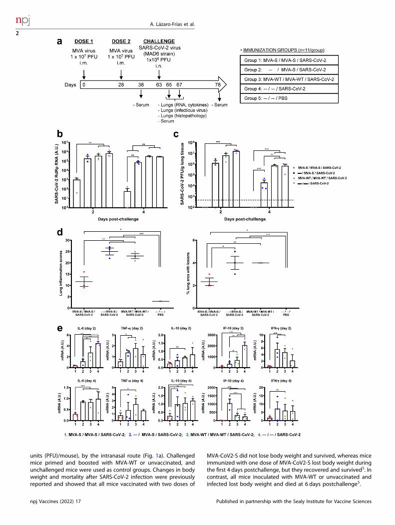
units (PFU)/mouse), by the intranasal route (Fig. 1a). Challengedmice primed and boosted with MVA-WT or unvaccinated, andunchallenged mice were used as control groups. Changes in bodyweight and mortality after SARS-CoV-2 infection were previouslyreported and showed that all mice vaccinated with two doses of
MVA-CoV2-S did not lose body weight and survived, whereas miceimmunized with one dose of MVA-CoV2-S lost body weight duringthe first 4 days postchallenge, but they recovered and survived5. Incontrast, all mice inoculated with MVA-WT or unvaccinated andinfected lost body weight and died at 6 days postchallenge5.
A. Lázaro-Frías et al.
2
npj Vaccines (2022) 17 Published in partnership with the Sealy Institute for Vaccine Sciences
1234567890():,;
To examine the effect of MVA-CoV2-S vaccination on SARS-CoV-2 virus replication, three mice per group were sacrificed at days 2and 4 after SARS-CoV-2 virus challenge, lungs were collected andprocessed, and presence of SARS-CoV-2 genomic RNA (Fig. 1b)and live infectious virus (Fig. 1c) was analyzed. Two doses of MVA-CoV2-S were highly effective to prevent SARS-CoV-2 replicationand virus yield at 2 and 4 days postchallenge, in comparison withthe high levels of SARS-CoV-2 genomic RNA and infectious virusdetected in control infected mice or in mice immunized with onedose of MVA-CoV2-S (Fig. 1b, c).The histopathological evaluation of the lung showed that mice
vaccinated with two doses of MVA-CoV2-S displayed at 4 dayspostchallenge significantly lower lung lesion scores (Fig. 1d, leftpanel and Supplementary Fig. 1a) and lower percentages of lungarea with lesions (Fig. 1d, right panel) than control MVA-WTinoculated mice or mice immunized with one dose of MVA-CoV2-S, which could be indicative of low breakthrough infections. Micevaccinated with two doses of MVA-CoV2-S only displayed focalthickening of the alveolar septae, and occasional presence ofinflammatory cells within the alveoli (Supplementary Fig. 1b).However, mice immunized with one dose of MVA-CoV2-S orcontrol MVA-WT inoculated mice showed more severe diffusethickening of the alveolar septae, higher presence of mononuclearcell infiltrates within alveolar spaces, as well as the presence oflarger multifocal perivascular and peribronchiolar mononuclearinfiltrates. Unvaccinated mock-infected mice did not displayremarkable inflammatory lesions (Supplementary Fig. 1b).Next, we evaluated the effect of MVA-CoV2-S on the cytokine
expression pattern induced in mice, as an extensive upregulationof several pro-inflammatory cytokines are associated with COVID-19 disease progression and severity17–19. Thus, at days 2 and 4postchallenge, mRNA levels of key cytokines on lung homo-genates were analyzed by RT-qPCR and the results showed thattwo doses of MVA-CoV2-S significantly down-regulated IL-6, TNF-α, IL-10, IP-10, and IFN-γ mRNA levels, compared to controlinfected mice or mice immunized with one dose of MVA-CoV2-S(Fig. 1e).
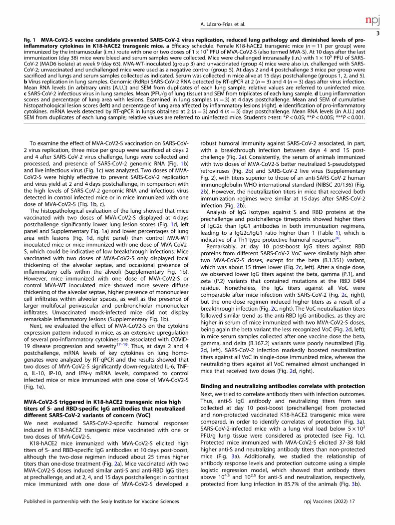
MVA-CoV2-S triggered in K18-hACE2 transgenic mice hightiters of S- and RBD-specific IgG antibodies that neutralizeddifferent SARS-CoV-2 variants of concern (VoC)We next evaluated SARS-CoV-2-specific humoral responsesinduced in K18-hACE2 transgenic mice vaccinated with one ortwo doses of MVA-CoV2-S.K18-hACE2 mice immunized with MVA-CoV2-S elicited high
titers of S- and RBD-specific IgG antibodies at 10 days post-boost,although the two-dose regimen induced about 25 times highertiters than one-dose treatment (Fig. 2a). Mice vaccinated with twoMVA-CoV2-S doses induced similar anti-S and anti-RBD IgG titersat prechallenge, and at 2, 4, and 15 days postchallenge; in contrastmice immunized with one dose of MVA-CoV2-S developed a
robust humoral immunity against SARS-CoV-2 associated, in part,with a breakthrough infection between days 4 and 15 post-challenge (Fig. 2a). Consistently, the serum of animals immunizedwith two doses of MVA-CoV2-S better neutralized S-pseudotypedretroviruses (Fig. 2b) and SARS-CoV-2 live virus (SupplementaryFig. 2), with titers superior to those of an anti-SARS-CoV-2 humanimmunoglobulin WHO international standard (NIBSC 20/136) (Fig.2b). However, the neutralization titers in mice that received bothimmunization regimes were similar at 15 days after SARS-CoV-2infection (Fig. 2b).Analysis of IgG isotypes against S and RBD proteins at the
prechallenge and postchallenge timepoints showed higher titersof IgG2c than IgG1 antibodies in both immunization regimens,leading to a IgG2c/IgG1 ratio higher than 1 (Table 1), which isindicative of a Th1-type protective humoral response20.Remarkably, at day 10 post-boost IgG titers against RBD
proteins from different SARS-CoV-2 VoC were similarly high aftertwo MVA-CoV2-S doses, except for the beta (B.1.351) variant,which was about 15 times lower (Fig. 2c, left). After a single dose,we observed lower IgG titers against the beta, gamma (P.1), andzeta (P.2) variants that contained mutations at the RBD E484residue. Nonetheless, the IgG titers against all VoC werecomparable after mice infection with SARS-CoV-2 (Fig. 2c, right),but the one-dose regimen induced higher titers as a result of abreakthrough infection (Fig. 2c, right). The VoC neutralization titersfollowed similar trend as the anti-RBD IgG antibodies, as they arehigher in serum of mice immunized with two MVA-CoV2-S doses,being again the beta variant the less recognized VoC (Fig. 2d, left);in mice serum samples collected after one vaccine dose the beta,gamma, and delta (B.167.2) variants were poorly neutralized (Fig.2d, left). SARS-CoV-2 infection markedly boosted neutralizationtiters against all VoC in single-dose immunized mice, whereas theneutralizing titers against all VoC remained almost unchanged inmice that received two doses (Fig. 2d, right).
Binding and neutralizing antibodies correlate with protectionNext, we tried to correlate antibody titers with infection outcomes.Thus, anti-S IgG antibody and neutralizing titers from seracollected at day 10 post-boost (prechallenge) from protectedand non-protected vaccinated K18-hACE2 transgenic mice werecompared, in order to identify correlates of protection (Fig. 3a).SARS-CoV-2-infected mice with a lung viral load below 5 × 10²
PFU/g lung tissue were considered as protected (see Fig. 1c).Protected mice immunized with MVA-CoV2-S elicited 37-38 foldhigher anti-S and neutralizing antibody titers than non-protectedmice (Fig. 3a). Additionally, we studied the relationship ofantibody response levels and protection outcome using a simplelogistic regression model, which showed that antibody titersabove 104.5 and 102.5 for anti-S and neutralization, respectively,protected from lung infection in 85.7% of the animals (Fig. 3b).
Fig. 1 MVA-CoV2-S vaccine candidate prevented SARS-CoV-2 virus replication, reduced lung pathology and diminished levels of pro-inflammatory cytokines in K18-hACE2 transgenic mice. a Efficacy schedule. Female K18-hACE2 transgenic mice (n= 11 per group) wereimmunized by the intramuscular (i.m.) route with one or two doses of 1 × 10⁷ PFU of MVA-CoV2-S (also termed MVA-S). At 10 days after the lastimmunization (day 38) mice were bleed and serum samples were collected. Mice were challenged intranasally (i.n.) with 1 × 10⁵ PFU of SARS-CoV-2 (MAD6 isolate) at week 9 (day 63). MVA-WT-inoculated (group 3) and unvaccinated (group 4) mice were also i.n. challenged with SARS-CoV-2; unvaccinated and unchallenged mice were used as a negative control (group 5). At days 2 and 4 postchallenge 3 mice per group weresacrificed and lungs and serum samples collected as indicated. Serum was collected in mice alive at 15 days postchallenge (groups 1, 2, and 5).b Virus replication in lung samples. Genomic (RdRp) SARS-CoV-2 RNA detected by RT-qPCR at 2 (n= 3) and 4 (n= 3) days after virus infection.Mean RNA levels (in arbitrary units [A.U.]) and SEM from duplicates of each lung sample; relative values are referred to uninfected mice.c SARS-CoV-2 infectious virus in lung samples. Mean (PFU/g of lung tissue) and SEM from triplicates of each lung sample. d Lung inflammationscores and percentage of lung area with lesions. Examined in lung samples (n= 3) at 4 days postchallenge. Mean and SEM of cumulativehistopathological lesion scores (left) and percentage of lung area affected by inflammatory lesions (right). e Identification of pro-inflammatorycytokines. mRNA levels detected by RT-qPCR in lungs obtained at 2 (n= 3) and 4 (n= 3) days postchallenge. Mean RNA levels (in A.U.) andSEM from duplicates of each lung sample; relative values are referred to uninfected mice. Student’s t-test: *P < 0.05; **P < 0.005; ***P < 0.001.
A. Lázaro-Frías et al.
3
Published in partnership with the Sealy Institute for Vaccine Sciences npj Vaccines (2022) 17
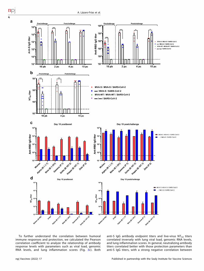
To further understand the correlation between humoralimmune responses and protection, we calculated the Pearsoncorrelation coefficient to analyze the relationship of antibodyresponse levels with parameters such as viral load, genomicRNA levels, and lung inflammation scores (Fig. 3c). Both
anti-S IgG antibody endpoint titers and live-virus NT50 titerscorrelated inversely with lung viral load, genomic RNA levels,and lung inflammation scores. In general, neutralizing antibodytiters correlated better with those protection parameters thananti-S IgG titers, with a strong negative correlation between
A. Lázaro-Frías et al.
4
npj Vaccines (2022) 17 Published in partnership with the Sealy Institute for Vaccine Sciences

neutralizing antibodies titers and lung viral load (R2 = 0.7896;p < 0.0001).
K18-hACE2 transgenic mice vaccinated with one and twodoses of MVA-CoV2-S are protected against SARS-CoV-2reinfectionNext, we evaluated whether K18-hACE2 mice immunized with oneor two doses of MVA-CoV2-S (n= 5/group) were protected againsta SARS-CoV-2 reinfection performed 7 weeks after the first SARS-CoV-2 challenge (Fig. 4a); body weight and mortality weremonitored daily before animals were sacrificed at 6 days post-rechallenge.All mice vaccinated with one or two doses of MVA-CoV2-S did
not lose body weight (Fig. 4b) and survived (Fig. 4c), while allunvaccinated and challenged mice lost body weight and died at6 days after rechallenge (Fig. 4b, c). The analysis of SARS-CoV-2virus replication in lung samples at day 6 post-reinfection showedthat both MVA-CoV2-S immunization regimes completely abro-gated SARS-CoV-2 replication (subgenomic and genomic mRNA)(Fig. 4d) and virus yields (Fig. 4e). Furthermore, both groups ofvaccinated mice displayed lower lung lesion scores and percen-tages of damaged lung area than control infected mice (Fig. 4fand Supplementary Fig. 3a, b).Evaluation of SARS-CoV-2-specific humoral responses at day 6
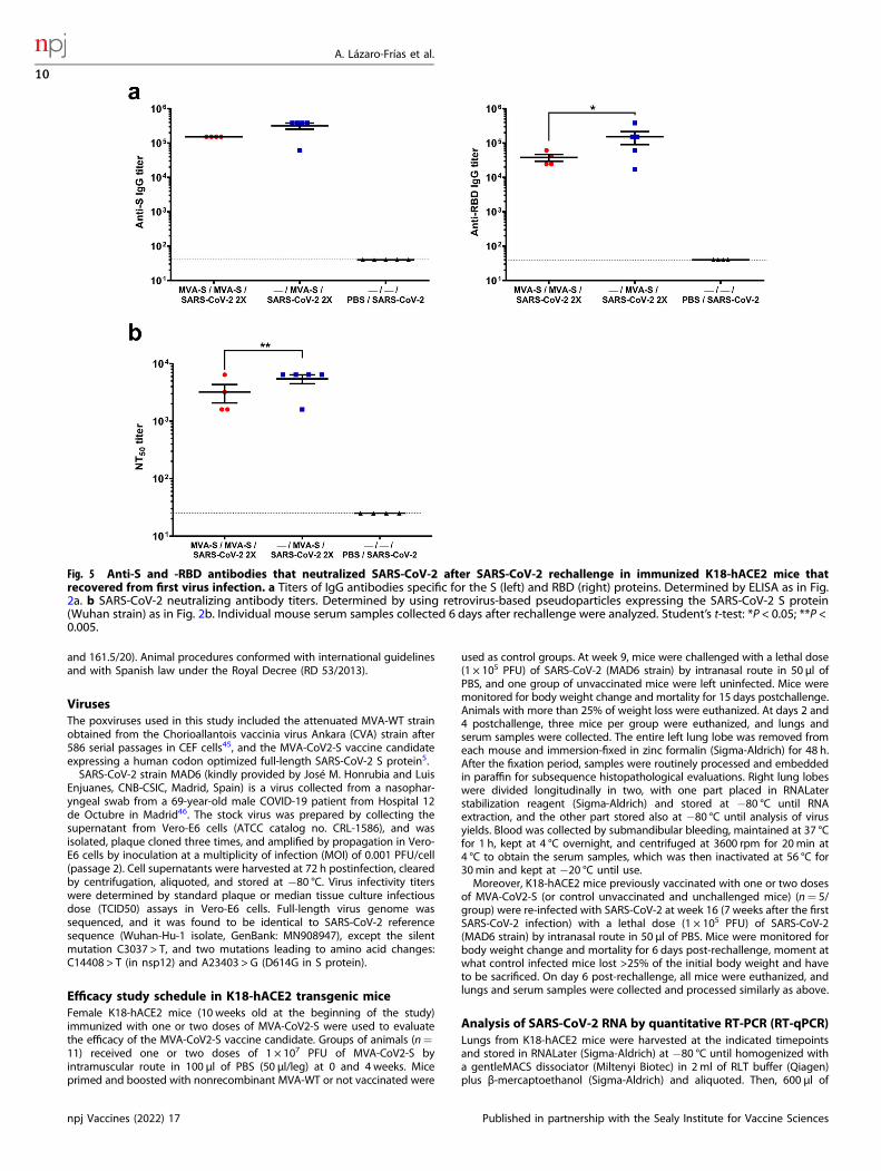
post-reinfection showed that vaccinated mice elicited high titersof S- and RBD-specific IgG antibodies. Mice that received a singleMVA-CoV2-S dose had slightly higher levels of anti-S IgGantibodies and significantly higher anti-RBD IgG titers than micevaccinated with two doses (Fig. 5a). Furthermore, vaccinated miceinduced S- and RBD-specific IgG2c/IgG1 ratios higher than 1 (Table1), indicative again of a Th1-type response. Moreover, immunizedmice elicited high titers of SARS-CoV-2 neutralizing antibodies,again with the one-dose treatment triggering significant higherneutralizing titers than the two-dose regimen (Fig. 5b).
Immunization of C57BL/6 mice with two doses of MVA-CoV2-Sinduces long-term SARS-CoV-2 S-specific CD4+ and CD8+
T-cell and humoral memory immune responsesWe previously described that MVA-CoV2-S induced potentadaptive S-specific CD4+ and CD8+ T-cell and humoral immuneresponses5, but the longevity of these responses was unknown.Thus, groups of C57BL/6 mice (n= 5/group) were immunizedtwice with MVA-CoV2-S (intramuscularly, 1 × 10⁷ PFU/mouse), atweeks 0 and 2, and 6months later animals were sacrificed toevaluate the long-term SARS-CoV-2-specific T-cell and humoralimmune responses induced in splenocytes and serum samples,respectively.Mice vaccinated with two doses of MVA-CoV2-S elicited high
levels of long-term IFNγ-secreting cells, reactive to a mixture ofSARS-CoV-2 S1+ S2 peptide pools, as revealed by ELISpot (Fig. 6a).Robust long-term S-specific CD4+ and CD8+ T-cells expressingCD107a, and secreting IFNγ, TNFα, and/or IL-2 were induced, witha higher overall response mainly mediated by the CD8+ T-cell
repertoire and a CD4+ Th1 profile, as measured by intracellularcytokine staining (ICS) (Fig. 6b). Long-term S-specific CD4+ andCD8+ T-cell responses were directed mainly against the S1peptide pool (Fig. 6c). S-specific CD4+ and CD8+ T-cell responseswere mostly of the T central memory and T effector memoryphenotypes, respectively (Fig. 6d). Moreover, the analysis of thequality of the S-specific T-cell responses showed that MVA-CoV2-Striggered highly polyfunctional long-term CD4+ and CD8+ T cells(Fig. 6e), with 100 and 90% of S-specific CD4+ and CD8+ T cells,respectively, exhibiting three or four functions (Fig. 6e, pie charts).In particular, CD4+ T cells expressing CD107a-IFNγ-TNFα-IL-2 andCD8+ T cells expressing CD107a-IFNγ-TNFα were the mostabundant CD4+ and CD8+ T cell populations (Fig. 6e, bars).The analysis of SARS-CoV-2-specific humoral immune responses
showed that mice vaccinated with two doses of MVA-CoV2-Sinduced long-term high titers of S- and RBD-specific binding IgGantibodies (Fig. 6f). Induced IgG antibodies also recognized RBDproteins from several VoC, although IgG titers to the beta variantwere lower than to the other VoC (Fig. 6g). Furthermore, S- andRBD-specific IgG2c/IgG1 ratios were above one (Table 1), likelysuggestive of a Th1-like immune response. Remarkably, high titersof SARS-CoV-2 neutralizing antibodies were maintained at6 months post-immunization with MVA-CoV2-S (Fig. 6h) andrecognized several VoC, being the beta variant the less recognizedVoC (Fig. 6i). The extrapolation of neutralizing antibody titersinduced at 6 months post-immunization in C57BL/6 mice to theregression model performed in protected and non-protected K18-hACE2 mice (Fig. 3a, b) suggested that long-lived antibody levelsinduced should have over 90% probability to confer protection.
DISCUSSIONDespite the wide variety of vaccine candidates against COVID-19with robust immunogenicity and efficacy, there is still a globalhigh-priority for a stable, safer, more immunogenic, and effectiveCOVID-19 vaccine against all kinds of emerging variants. More-over, it remains to be defined the durability of the immuneresponses and efficacy in the long run. One promising vaccinevector is the highly attenuated poxvirus strain MVA, non-replicative in human cells that have been extensively studyingin preclinical and clinical trials as vaccine candidate against a widespectrum of diseases with excellent safety, immunogenicity, andefficacy profiles21,22.Recently, we described the generation and immunogenic
characterization of the MVA-CoV2-S vaccine candidate againstCOVID-19, which is based on an MVA vector that expresses ahuman codon optimized full-length SARS-CoV-2 S protein thatwas highly immunogenic in mice, inducing a robust activation ofboth arms of the adaptive immune system, T and B cells5.Furthermore, we also reported that one or two doses of MVA-CoV2-S controlled morbidity (weight loss) and mortality caused bySARS-CoV-2 infection in K18-hACE2 transgenic mice5, a SARS-CoV-2 susceptible mouse model that exhibits fatal respiratory infectionand mortality following intranasal SARS-CoV-2 administration13–16.
Fig. 2 MVA-CoV2-S vaccine candidate induced high levels of humoral responses in vaccinated and challenged K18-hACE2 transgenicmice. a Titers of IgG antibodies specific for the S (left) and RBD (right) proteins (Wuhan strain). Determined by ELISA in individual mouseserum samples collected at day 10 post-boost (prechallenge; n= 11/group) and at days 2 (n= 3/group), 4 (n= 3/group) and 15 (n= 5/group)postchallenge from K18-hACE2 mice. Mean values and SEM are represented. Dashed line represents the limit of detection. b SARS-CoV-2neutralizing antibody titers. NT50 titers were evaluated in individual mouse serum samples collected at day 10 post-boost (prechallenge) andat 4 and 15 days postchallenge, using retrovirus-based pseudoparticles expressing the SARS-CoV-2 S protein (Wuhan strain). Mean NT50 valuesand SEM are represented. Upper dotted line represents the levels obtained with the NIBSC 20/136 international standard plasma (containingpooled plasma obtained from 11 individuals recovered from SARS-CoV-2 infection). Bottom dotted line represented the limit of detection.c Titers of IgG antibodies against RBD from different SARS-CoV-2 VoC. Determined by ELISA in pooled sera samples collected at days 10 post-boost (left) and 15 postchallenge (right). d SARS-CoV-2 neutralizing antibody titers against SARS-CoV-2 VoC. NT50 titers were evaluated inpooled mouse serum samples collected at days 10 post-boost (left) and 15 postchallenge (right), using VSV-based pseudoparticles expressingthe SARS-CoV-2 S protein of different VoC. Mean NT50 values and SEM are represented. Student’s t-test: *P < 0.05; ***P < 0.001.
A. Lázaro-Frías et al.
5
Published in partnership with the Sealy Institute for Vaccine Sciences npj Vaccines (2022) 17

Here, we provide a detailed evaluation of the efficacy andimmunogenicity of one or two doses of MVA-CoV2-S in K18-hACE2transgenic mice. The results obtained indicated that two doses ofMVA-CoV2-S resulted in the highest level of protection. At 2 and4 days, postchallenge SARS-CoV-2 replication in the lungs(genomic mRNA and viral load) was completely abolished, andhistopathological lung lesions and levels of pro-inflammatorycytokines were significantly reduced when compared to controlinfected mice or mice vaccinated with one dose of MVA-CoV2-S.Since SARS-CoV-2 replication in the lungs was completelyabolished in mice vaccinated with two doses of MVA-CoV2-S, itis likely that virus shedding in these animals is limited, because ithas been reported that SARS-CoV-2 shedding in transgenic mice isoften associated with a high viral titer in lung tissues andvaccinated transgenic mice did not shed virus as early as day 2postinfection, when virus was cleared from the lungs6. Although asingle dose of MVA-CoV2-S had a significant inhibitory effect inRNA and viral load at 4 days postchallenge, other parametersanalyzed indicated that one dose does not completely protectsfrom lung infection, at least in the first days postinfection.A detailed analysis of histopathological lung lesions revealed
that after challenge with SARS-CoV-2, mice vaccinated with twodoses of MVA-CoV2-S displayed less extensive and less severelung inflammatory lesions compared to mice immunized with onedose of MVA-CoV2-S or control infected mice. The most severelyaffected mice displayed inflammatory lesions similar to thosedescribed in previous experimental infections with SARS-CoV-2 intransgenic ACE2 mice23 and non-human primates24, andresembled histopathological lung lesions observed in humanpatients infected with SARS-CoV-225,26. Moreover, surviving K18-hACE2 mice immunized with one or two doses of MVA-CoV2-Sand challenged with a second lethal dose of SARS-CoV-2 displayedsignificantly lower lung lesions scores compared to controlinfected mice. Some mice, especially those immunized with twodoses of MVA-CoV2-S, did not display focal lesions.Overproduction of several pro-inflammatory cytokines is a
marker of COVID-19 disease progression and mortality17–19,27–29.Here, we found that after SARS-CoV-2 infection of transgenic K18-hACE2 mice vaccinated with two doses of MVA-CoV2-S there wasa significant downregulation in the mRNA levels of several pro-inflammatory cytokines in lung homogenates, such as IL-6, TNF-α,IL-10, IFN-γ, and IP-10, compared to control infected mice wherehigh levels of these cytokines were detected. All these resultsindicated that two doses of MVA-CoV2-S prevented the increase inpro-inflammatory cytokines induced by SARS-CoV-2 infection,helping to prevent COVID-19 related lung damage. Other MVA-based vaccine candidates against SARS-CoV-2 have probe toreduce transcripts associated with inflammation and hyperim-mune activation in lungs of vaccinated and challenged mice6 andnon-human primates7.Of significance, MVA-CoV2-S induced high titers of binding IgG
antibodies against S and RBD and neutralizing antibodies againstSARS-CoV-2, with the two-dose regime eliciting higher titers thanthe one-dose regime. Remarkably, MVA-CoV2-S vaccination alsoinduced high titers of IgG and neutralizing antibodies recognizingseveral VoC (alpha, beta, gamma, delta, kappa, and zeta), being betathe less recognized variant. This highlights the potent and widespectrum of humoral responses elicited by MVA-CoV-2 vaccinecandidates able to neutralize different VoC, especially the delta VoCthat is actually the main strain circulating in the human population.Additionally, a Th1-type immune response was induced in allvaccinated mice. All those specific immune markers have beenlinked to SARS-CoV-2 vaccine efficacy and control of COVID-19progression, such as high titers of IgG antibodies against SARS-CoV-2S and RBD proteins30, a Th1 profile immune response20, induction ofpotent SARS-CoV-2 neutralizing antibodies31,32, and low levels ofpro-inflammatory cytokines19. Furthermore, evidence from preclini-cal studies in non-human primates, hamsters, and transgenic miceTa
ble1.
Isotypean
alysisofan
ti-S
andan
ti-RBD
IgG
antibodiesin
immunized
mice.
Timep
oints
analyzed
IgG2c
andIgG1titers
andRatio
IgG2c/IgG1
a
MVA
-S/MVA
-S/SA
RS-CoV-2
—/MVA
-S/SA
RS-CoV-2
MVA
-S/MVA
-S
(K18
-hACE2
mice)
(K18
-hACE2
mice)
(C57
BL/6mice)
SRBD
SRBD
SRBD
IgG1
IgG2c
IgG2c/
IgG1
IgG1
IgG2c
IgG2c/
IgG1
IgG1
IgG2c
IgG2c/
IgG1
IgG1
IgG2c
IgG2c/
IgG1
IgG1
IgG2c
IgG2c/
IgG1
IgG1
IgG2c
IgG2c/
IgG1
10dayspost-boost
(prech
allenge)
24.414
152.58
76,25
1.56
261
.035
39,07
1.56
29.76
56,25
625
3.90
66,25
––
––
––
Day
4postch
allenge
24.414
152.58
76,25
438
61.035
139,51
1.56
29.76
56,25
100
3.90
639
,06
––
––
––
Day
15postch
allenge
24.414
152.58
76,25
1.56
261
.035
39,07
152.58
795
3.67
46,25
9.76
515
2.58
715
,63
––
––
––
Day
6post-rechallenge
24.414
152.58
76,25
625
42.724
69,36
61.035
953.67
415
,63
2.73
415
2.58
755
,81
––
––
––
6monthspost-boost
––
––
––
––
––
––
3.90
617
.089
4,38
625
9.76
515
,62
a Meantiters
ofIgG2c
andIgG1isotypean
tibodiesag
ainst
San
dRBDproteinsfrom
duplicates
ofpooledsera
samplesobtained
from
thedifferen
tim
munizationregim
ensstudiedarerepresented,includingthe
ratioIgG2c/IgG1.
–:n
otap
plicab
le.
A. Lázaro-Frías et al.
6
npj Vaccines (2022) 17 Published in partnership with the Sealy Institute for Vaccine Sciences

indicates that vaccine-induced SARS-CoV-2 neutralizing antibodiescorrelate with protection against lung infection and clinicaldisease33–36. Moreover, a longitudinal study of SARS-CoV-2-infected patients also reveals a high correlation between neutraliz-ing antibodies and COVID-19 severity37. Here, the robust protectionobserved in K18-hACE2 transgenic mice vaccinated with two dosesof MVA-CoV2-S (determined by the low levels of SARS-CoV-2 RNA,
viral load, and inflammation scores in lung samples) significantlycorrelated with high titers of binding IgG and neutralizingantibodies; although neutralizing antibody titers correlated betterwith protection than binding anti-S IgG antibody titers. The NT50titers elicited by two doses of MVA-CoV2-S were higher than 3 × 10³,and were comparable to those induced by other viral vector-basedvaccines, such as ChAdOx1 nCoV-1938, and different recombinant
A. Lázaro-Frías et al.
7
Published in partnership with the Sealy Institute for Vaccine Sciences npj Vaccines (2022) 17
MVA vectors also expressing the full-length S protein4,6–8, or higherthan those of an anti-SARS-CoV-2 human immunoglobulin WHOinternational standard, reflecting the potent humoral responsesinduced by the vaccine candidate. Furthermore, we also found astrong correlation between the neutralizing antibody NT50 titersmeasured using live SARS-CoV-2 or retrovirus-based pseudoparticlesexpressing the S protein, similarly as previously reported37.Here, we also found that either one or two doses of MVA-CoV2-
S induced a predominantly Th1-type response to S and RBDproteins, with IgG2c > IgG1. This is relevant, as activation of astrong Th1 cell response has been associated with less severecases of COVID-19, while Th2 cell responses have been associatedwith more severe lung disease in humans39. Moreover, previousstudies also demonstrated the induction of predominantly Th1-type immune responses in mice immunized with MVA-basedvaccines encoding SARS-CoV-2 S antigens6,8 or by other COVID-19vaccines20.Mice vaccinated with two doses of MVA-CoV2-S induced similar
high titers of IgG and neutralizing antibodies against parentalSARS-CoV-2 and VoC before and after SARS-CoV-2 infection.However, at later timepoints after SARS-CoV-2 infection animalsvaccinated with one dose of MVA-CoV2-S developed an anamnes-tic response through breakthrough infection, with higher levels ofS- and RBD-specific IgGs and neutralizing antibodies againstparental SARS-CoV-2 and VoC at 15 days postchallenge that weresimilar or superior to those elicited by the two-dose regime. Thisindicates that SARS-CoV-2 infection boosted the immune systemand induced comparable humoral responses to two MVA-CoV2-S doses.All vaccinated animals were completely resistant to a second
SARS-CoV-2 reinfection 7 weeks after the first challenge, indepen-dently of the one or two dose vaccination regimes. The high levelsof SARS-CoV-2-specific antibodies induced at late timepoints afterinfection with both immunization regimes were sufficient toconfer protection against a reinfection. Therefore, vaccinatedindividuals with two MVA-CoV2-S doses and infected people thathad received a single vaccine dose should, in principle, beprotected from a first or second infection, respectively, as shownhere with animal models.Generation of robust, durable and Th1-skewed T- and B-cell
memory immune responses are crucial to prevent SARS-CoV-2infection, to stop virus replication and to facilitate virus clearancewithout respiratory complications40, which is the main goal ofSARS-CoV-2 vaccines. Recently, it has been reported the persis-tence of antibodies 6 months after the second dose of mRNA-1273vaccine (Moderna) in a phase 1 follow-up study41. Moreover, SARS-CoV-2-specific T and B cell responses during COVID-19 diseaseprogression have been tracked in blood samples, and individualsrecovered from mild COVID-19 develop and sustain multifacetedSARS-CoV-2-specific immunological memory: SARS-CoV-2-specificIgG antibodies, neutralizing plasma, and memory B and T cells thatpersisted for at least 3 months42. Of significance, our resultsdemonstrated that, similar to natural infection and to othervaccines administered in the human population, immunization ofC57BL/6 mice with two doses of MVA-CoV2-S vaccine candidate
induces long-term, strong, and polyfunctional SARS-CoV-2S-specific CD4+ and CD8+ T-cell memory immune responses, aswell as long-term SARS-CoV-2-specific humoral immune responses(binding IgG and neutralizing antibodies) against parental Wuhanstrain and several VoC. Those responses were detected at6 months after the last immunization, with titers of neutralizingantibodies that are presumed to correlate with protection.Notably, all the animals remained healthy after 6 months offollow-up, suggesting safety with no adverse effects of the MVA-CoV2-S vaccine.All the efficacy and immunogenicity results reported here
support that two doses of MVA-CoV2-S induced robust protectionin transgenic K18-hACE2 mice, with the one-dose regimen beingless effective. Other groups have also recently reported thegeneration of MVA vectors expressing SARS-CoV-2 S antigens thatelicited efficacy in susceptible mice and macaques with one ortwo doses4,6–8, similar to the reported in this study with transgenicK18-hACE2 mice. In particular, Liu and colleagues have also showncontrol of SARS-CoV-2 infection by a MVA vector vaccine in upperand lower respiratory tracts, a critical event to prevent SARS-CoV-2transmission6.Will the poxvirus vectors have a niche in the current market
with several established and efficacious vaccines against SARS-CoV-2? We still do not know the durability of the S-specificimmune responses, and booster doses are now considered toassure long-term and broader efficacy. Poxvirus vectors enhanceimmune responses when used in combination with other vaccinetechnologies, being most effective when delivered as a booster,such as we and others described5,11,43,44. Hence, vaccines basedon MVA or other poxvirus vectors could be implemented in clinicaltrials, either alone or in combination with other vaccines currentlyin the market, and provide an advantage in the general control ofthe COVID-19 pandemic. The MVA-CoV2-S dose used in thismouse preclinical study (1 × 10⁷ PFU/mouse) corresponds to a lowhuman dose, as the standard dose for MVA-based vaccines used inclinical trials is 1 × 10⁸ PFU/human.In summary, the present study provided valuable evidence of
MVA-CoV2-S preclinical efficacy and immunogenicity, addingfurther support for the evaluation of this vaccine in clinical trials.
METHODSEthics statementFemale transgenic K18-hACE2 mice, expressing the human angiotensin-converting enzyme-2 (ACE2) receptor, were obtained from The JacksonLaboratory (034860-B6.Cg-Tg(K18-ACE2)2Prlmn/J, genetic backgroundC57BL/6 J × SJL/J)F2) and efficacy experiments were performed in thebiosafety level 3 (BSL-3) facilities at Centro de Investigación en SanidadAnimal (CISA)-Instituto Nacional de Investigaciones Agrarias (INIA-CSIC)(Valdeolmos, Madrid, Spain). Female C57BL/6OlaHsd mice (6–8 weeks old)used for long-term immunogenicity assays were purchased from EnvigoLaboratories and stored in the animal facility of Centro Nacional deBiotecnología (CNB) (Madrid, Spain). The efficacy and immunogenicityanimal studies were approved by the Ethical Committee of AnimalExperimentation (CEEA) of the CNB (Madrid, Spain) and by the Division ofAnimal Protection of the Comunidad de Madrid (PROEX 49/20, 169.4/20
Fig. 3 Binding IgG and neutralizing antibodies correlate with protection. a K18-hACE transgenic mice were immunized with one or twodoses of MVA-CoV2-S, or with MVA-WT, and then inoculated with 1 × 10⁵ PFU of SARS-CoV-2, and 2 and 4 days later sacrificed for virologicalanalysis of lung tissue (n= 18). Prior to virus inoculation (at 10 days post-boost) serum samples were analyzed for anti-S IgG antibody titersand virus-neutralizing antibody titers. Protection was defined as a viral load below 5 × 10² PFU/g in lung tissue, irrespective of vaccine regimen(Fig. 1c). Median antibody responses per group is indicated with horizontal lines. Dotted lines indicate the limit of detection. b Logisticregression models were built with binding anti-S and neutralizing antibody titers from pooled data as an independent variable, andprotection outcome as the dependent variable. c Correlation analysis. Correlation of binding anti-S IgG endpoint titers (upper row) and NT50titers (bottom row) (in log10) determined 3.5 weeks before challenge (10 days post-boost) with viral load (log10 PFU/g in lung tissue) (left),genomic RNA levels (log10 genomic RNA in A.U,) (middle) and lung inflammation scores (log10) (right) determined at 2 and 4 dayspostchallenge. Red lines reflect the best linear fit relationship between these variables. R2 and P-values reflect two-sided Pearson correlationcoefficients.
A. Lázaro-Frías et al.
8
npj Vaccines (2022) 17 Published in partnership with the Sealy Institute for Vaccine Sciences

Fig. 4 Outcome of SARS-CoV-2 inoculation (rechallenge) in immunized K18-hACE2 mice that recovered from first virus infection.a Scheme of immunization and SARS-CoV-2 infection procedures in K18-hACE2 mice (n= 5 per group), to determine the effect of second virusinoculation. Body weight (b) and mortality (c) were monitored for 6 days after rechallenge with SARS-CoV-2 virus. All non-vaccinated mice(group 3) died at day 6 post-rechallenge. SARS-CoV-2 genomic (RdRp gene) and subgenomic (E gene) RNA (d) and infectious virus (e) in lungsamples at day 6 postchallenge were evaluated as indicated in Fig. 1. f Lung inflammation scores and percentage of lung area with lesions, inlung samples at day 6 post-rechallenge. Mean and SEM of cumulative histopathological lesion scores (left) and percentage of lung areaaffected by inflammatory lesions (right). Student’s t-test: *P < 0.05; **P < 0.005; ***P < 0.001.
A. Lázaro-Frías et al.
9
Published in partnership with the Sealy Institute for Vaccine Sciences npj Vaccines (2022) 17
and 161.5/20). Animal procedures conformed with international guidelinesand with Spanish law under the Royal Decree (RD 53/2013).
VirusesThe poxviruses used in this study included the attenuated MVA-WT strainobtained from the Chorioallantois vaccinia virus Ankara (CVA) strain after586 serial passages in CEF cells45, and the MVA-CoV2-S vaccine candidateexpressing a human codon optimized full-length SARS-CoV-2 S protein5.SARS-CoV-2 strain MAD6 (kindly provided by José M. Honrubia and Luis
Enjuanes, CNB-CSIC, Madrid, Spain) is a virus collected from a nasophar-yngeal swab from a 69-year-old male COVID-19 patient from Hospital 12de Octubre in Madrid46. The stock virus was prepared by collecting thesupernatant from Vero-E6 cells (ATCC catalog no. CRL-1586), and wasisolated, plaque cloned three times, and amplified by propagation in Vero-E6 cells by inoculation at a multiplicity of infection (MOI) of 0.001 PFU/cell(passage 2). Cell supernatants were harvested at 72 h postinfection, clearedby centrifugation, aliquoted, and stored at −80 °C. Virus infectivity titerswere determined by standard plaque or median tissue culture infectiousdose (TCID50) assays in Vero-E6 cells. Full-length virus genome wassequenced, and it was found to be identical to SARS-CoV-2 referencesequence (Wuhan-Hu-1 isolate, GenBank: MN908947), except the silentmutation C3037 > T, and two mutations leading to amino acid changes:C14408 > T (in nsp12) and A23403 > G (D614G in S protein).
Efficacy study schedule in K18-hACE2 transgenic miceFemale K18-hACE2 mice (10 weeks old at the beginning of the study)immunized with one or two doses of MVA-CoV2-S were used to evaluatethe efficacy of the MVA-CoV2-S vaccine candidate. Groups of animals (n=11) received one or two doses of 1 × 10⁷ PFU of MVA-CoV2-S byintramuscular route in 100 μl of PBS (50 μl/leg) at 0 and 4 weeks. Miceprimed and boosted with nonrecombinant MVA-WT or not vaccinated were
used as control groups. At week 9, mice were challenged with a lethal dose(1 × 10⁵ PFU) of SARS-CoV-2 (MAD6 strain) by intranasal route in 50 μl ofPBS, and one group of unvaccinated mice were left uninfected. Mice weremonitored for body weight change and mortality for 15 days postchallenge.Animals with more than 25% of weight loss were euthanized. At days 2 and4 postchallenge, three mice per group were euthanized, and lungs andserum samples were collected. The entire left lung lobe was removed fromeach mouse and immersion-fixed in zinc formalin (Sigma-Aldrich) for 48 h.After the fixation period, samples were routinely processed and embeddedin paraffin for subsequence histopathological evaluations. Right lung lobeswere divided longitudinally in two, with one part placed in RNALaterstabilization reagent (Sigma-Aldrich) and stored at −80 °C until RNAextraction, and the other part stored also at −80 °C until analysis of virusyields. Blood was collected by submandibular bleeding, maintained at 37 °Cfor 1 h, kept at 4 °C overnight, and centrifuged at 3600 rpm for 20min at4 °C to obtain the serum samples, which was then inactivated at 56 °C for30min and kept at −20 °C until use.Moreover, K18-hACE2 mice previously vaccinated with one or two doses
of MVA-CoV2-S (or control unvaccinated and unchallenged mice) (n= 5/group) were re-infected with SARS-CoV-2 at week 16 (7 weeks after the firstSARS-CoV-2 infection) with a lethal dose (1 × 10⁵ PFU) of SARS-CoV-2(MAD6 strain) by intranasal route in 50 μl of PBS. Mice were monitored forbody weight change and mortality for 6 days post-rechallenge, moment atwhat control infected mice lost >25% of the initial body weight and haveto be sacrificed. On day 6 post-rechallenge, all mice were euthanized, andlungs and serum samples were collected and processed similarly as above.
Analysis of SARS-CoV-2 RNA by quantitative RT-PCR (RT-qPCR)Lungs from K18-hACE2 mice were harvested at the indicated timepointsand stored in RNALater (Sigma-Aldrich) at −80 °C until homogenized witha gentleMACS dissociator (Miltenyi Biotec) in 2ml of RLT buffer (Qiagen)plus β-mercaptoethanol (Sigma-Aldrich) and aliquoted. Then, 600 μl of
Fig. 5 Anti-S and -RBD antibodies that neutralized SARS-CoV-2 after SARS-CoV-2 rechallenge in immunized K18-hACE2 mice thatrecovered from first virus infection. a Titers of IgG antibodies specific for the S (left) and RBD (right) proteins. Determined by ELISA as in Fig.2a. b SARS-CoV-2 neutralizing antibody titers. Determined by using retrovirus-based pseudoparticles expressing the SARS-CoV-2 S protein(Wuhan strain) as in Fig. 2b. Individual mouse serum samples collected 6 days after rechallenge were analyzed. Student’s t-test: *P < 0.05; **P <0.005.
A. Lázaro-Frías et al.
10
npj Vaccines (2022) 17 Published in partnership with the Sealy Institute for Vaccine Sciences
homogenized lung tissue was used to isolate total RNA using the RNeasyMini Kit (Qiagen), according to the manufacturer’s specifications. First-strand cDNA synthesis and subsequent real-time PCR were performed inone step using NZYSpeedy One-step RT-qPCR Master Mix (NZYTech),according to the manufacturer’s specifications using ROX as reference dye.SARS-CoV-2 viral RNA content was determined using previously validated
set of primers and probes specific for the SARS-CoV-2 subgenomic RNA forthe protein E, the genomic virus RNA dependent RNA polymerase (RdRp)gene and the cellular 28 S rRNA for normalization47 (Supplementary Table1). Data were acquired with a 7500 real-time PCR system (AppliedBiosystems) and analyzed with 7500 software v2.0.6. Relative RNA arbitraryunits (A.U.) were quantified relative to the negative group (uninfected
A. Lázaro-Frías et al.
11
Published in partnership with the Sealy Institute for Vaccine Sciences npj Vaccines (2022) 17
mice) and were performed using the 2-ΔΔCt method. All samples weretested in duplicate.
Analysis of SARS-CoV-2 virus yields by plaque assayLungs from K18-hACE2 mice were harvested at the indicated timepoints,weighted, and stored directly at −80 °C until homogenized with agentleMACS dissociator (Miltenyi Biotec) in 2 ml of PBS buffer andaliquoted. Then, undiluted and serial ten-fold dilutions of homogenizedlung tissue were added in triplicate to Vero-E6 cell monolayers seeded in12-well plates at 5 × 10⁵ cells/well and after 1 h of adsorption the inoculumwas removed and plates were incubated at 37 °C, 5% CO2 in 2:1 DMEM 2X-4% fetal bovine serum (FBS):2% Agar. After 4 days, cells were fixed for 1 hwith 10% formaldehyde (Sigma-Aldrich), and then the agarose wasremoved and plaques were visualized by adding 0.5% crystal violet(Sigma-Aldrich). SARS-CoV-2 titers were determined in PFUs per gram oflung tissue.
Lung cytokine profile analysis by RT-qPCRReverse transcription of 1000 ng of RNA isolated as described above fromlung homogenates of K18-hACE2 mice was performed with the QuantiTectreverse transcription kit (Qiagen, Hilden, Germany), according to themanufacturer’s recommendations. RT-qPCR was performed with a 7500real-time PCR system (Applied Biosystems) using Power SYBR green PCRMaster Mix (Applied Biosystems), as previously described5,48. The mRNAexpression levels of the genes for IL-6, TNF-α, IL-10, IP-10 and IFN-γ wereanalyzed by real-time PCR with specific oligonucleotides (SupplementaryTable 1). Specific gene expression was expressed relative to the expressionof the cellular 28 S ribosomal RNA gene in fold change units using the 2−ΔΔCt method. All samples were tested in triplicate.
Lung histopathologyThe entire left lung lobe was removed from each K18-hACE2 mouse andimmersion-fixed in zinc formalin (Sigma-Aldrich) for 48 h. After fixationperiod, samples were routinely processed and embedded in paraffinblocks that were then sectioned at 4 µm thickness on a microtome,mounted onto glass slides and routinely stained with hematoxylin andeosin (H&E). Lung sections were microscopically evaluated using anOlympus BX43 microscope by a single veterinary pathologist who wasblinded to the identity and group of individual mice. To assess thecharacter and severity of histopathological lesions, lung inflammationscoring parameters based on previous reports on SARS-CoV-2 infection inmouse models were used23. The histopathological parameters evaluatedwere the follows: capillary endothelial cell activation; alveolar hemor-rhages; alveolar edema; perivascular edema; alveolar septal thickening(interstitial pneumonia); alveolar damage and hyaline membranes inalveoli; inflammatory cell infiltration in alveoli; bronchi/bronchioles withepithelial necrosis, detached epithelium or inflammatory cells in the lumen(bronchitis/bronchiolitis); peribronchial/peribronchiolar and perivascularmononuclear infiltrates; pneumocytes hyperplasia; cytopathic effect orsyncytia; squamous metaplasia; uniform interstitial fibrosis; organizedfibrotic tissue around the bronchi/bronchioles or intrabronchiolar (bronch-iolitis obliterans) and pleural thickening. The histopathological parameters
were graded following a semi-quantitative scoring system as follows: (0) nolesion; (1) minimal lesion; (2) mild lesion; (3) moderate lesion; (4) severelesion. The cumulative scores of histopathological lesions provided thetotal score per animal. In each experimental group, the individual scoreswere used to calculate the group average. In addition, H&E-stainedsections were visually scored 0–6 based on the percentage of lung areaaffected by inflammatory lesions as follows: 0% of the lung injured (score0); <5% (score 1); 6–10% (score 2); 11–20% (score 3); 21–30% (score 4);31–40% (score 5); >40% (score 6). In each experimental group, theindividual scores were used to calculate the group average.
Peptides and proteinsSARS-CoV-2 S peptide pools were used in the cellular immunogenicityanalysis and were purchased from JPT Peptide Technologies (Berlin,Germany, catalog number PM-WCPV-S). The S peptide pools were dividedinto two groups spanning the S1 and S2 regions of the S protein, with eachpeptide pool containing 158 (S1) or 157 peptides (S2) as consecutive 15-mers overlapping by 11 amino acids.The SARS-CoV-2 soluble S and RBD proteins were produced in
mammalian cells as previously described5, and they were used to analyzeby ELISA the levels of IgG antibodies in mice serum samples. The Ssequence (residues 1–1208; Wuhan-Hu-1 strain, GenBank accessionnumber MN908947.3) contained a T4 fibritin trimerization sequence, aFlag epitope and an 8xHis-tag at the C-terminus. In the S protein, the furin-recognition motif (RRAR) was replaced by the GSAS sequence, and it alsocontained the A942P, K986P, and V987P substitutions in the S2 portion.The S protein was purified by nickel-nitrilotriacetic acid (Ni-NTA) affinitychromatography from transfected cell supernatants and it was transferredto HEPES buffered saline (HBS) pH 7.5, during concentration or by size-exclusion chromatography (SEC).The RBD proteins with the S residues 332–534 were produced with an
N-terminal HA-tag (YPYDVPDYA) and fused to either a T4 fibritintrimerization sequence, a Flag epitope and an 8xHis-tag (RBD-TFH), or tothe human IgG1 Fc region (RBD-Fc) at its C-terminus. RBD-Fc proteins withRBD substitutions in VoC were also produced: alpha (B.1.1.7; N501Y), beta(B.1.351; K417N-E484K-N501Y), gamma (P.1.; K417T-E484K-N501Y), delta(B.1.617.2; L452R-T478K), kappa (B.1.617.1; L452R-E484Q) and zeta (P.2;E484K). The RBD-TFH and RBD-Fc proteins were purified by affinitychromatography with Ni-NTA and IgSelect (GE Healthcare) columns,respectively.
Immunogenicity study schedule in C57BL/6 miceTo evaluate the long-term memory immunogenicity of the MVA-CoV2-Svaccine candidate MVA-CoV2-S prime/MVA-CoV2-S boost immunizationprotocol was performed in female C57BL/6 mice (6–8 weeks old) aspreviously described5. Groups of animals (n= 5) received two doses of 1 ×10⁷ PFU of MVA-CoV2-S or MVA-WT (used as negative control mice) byintramuscular route in 100 μl of PBS (50 μl/leg) at 0 and 2 weeks. At6 months after the last immunization, mice were sacrificed using CO2. Next,blood from each individual mouse was collected by cardiac puncture andprocessed as described above to obtain serum samples to analyze thetiters of IgG antibodies, IgG isotypes, and neutralizing antibodies againstSARS-CoV-2; the spleens of each group were pooled and processed to
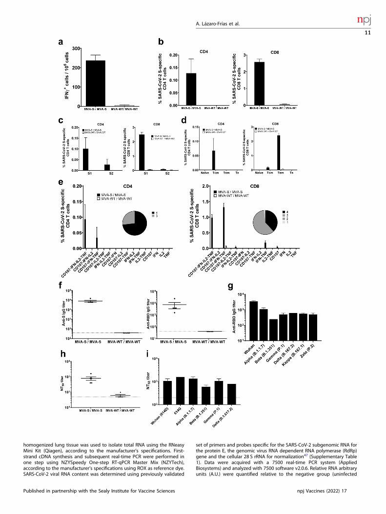
Fig. 6 MVA-CoV2-S elicited long-lasting SARS-CoV-2 S-specific T-cell and humoral immune responses in immunized C57BL/6 mice.a Magnitude of SARS-CoV-2 S-specific cell responses. Cells secreting IFN-γ per million of splenocytes and directed against S1+ S2 peptidepools in immunized C57BL/6 mice were evaluated at 6months post-boost by an ELISpot assay from a pool of splenocytes derived from 5immunized mice per group. Mean values and standard deviation of triplicate samples. b Magnitude of S-specific CD4+ and CD8+ T-cellimmune responses evaluated at 6months post-immunization. Percentages of CD4+ or CD8+ T cells expressing CD107a and/or producing IFN-γ and/or TNF-α and/or IL-2 against a mixture of S1 and S2 peptide pools in immunized mice. Cell percentages were determined by ICS fromsplenocyte pools. c S-specific T-cell immune responses against S1 and S2 regions. Percentages of S1- or S2-specific CD4+ and CD8+ T cellsdetermined as in panel B. d Percentages of Naïve (CD127-/CD62L−), T central memory (Tcm, CD127+/CD62L+), T effector memory (Tem,CD127+/CD62L−), and T effector (Te, CD127−/CD62L−) CD4+ and CD8+ T cells specific for S1 and S2 peptide pools, and expressing any of themarkers CD107a, IFN-γ, TNF-α, and IL-2. e Polyfunctional profiles (based on the expression of selected markers CD107a, IFN-γ, TNF-α, and IL-2)of total S-specific CD4+ or CD8+ T-cell immune responses directed against a mixture of S1 and S2 peptide pools. The pie charts summarize thepercentage of S-specific T cells exhibiting 1, 2, 3, or 4 markers. f Titers of IgG antibodies specific for the S (left) and RBD (right) proteins.Determined by ELISA in individual serum samples as in Fig. 2a. g Titers of IgG antibodies against RBD from different SARS-CoV-2 VoC.Determined by ELISA in pooled sera samples as in Fig. 2c. h SARS-CoV-2 neutralizing antibody titers. Determined by using retrovirus-basedpseudoparticles expressing the SARS-CoV-2 S protein (Wuhan strain) in individual serum samples as in Fig. 2b. i SARS-CoV-2 neutralizingantibody titers against SARS-CoV-2 VoC. NT50 titers were evaluated by using VSV-based pseudoparticles expressing the SARS-CoV-2 S proteinof different VoC, as in Fig. 2d. Individual or pooled mouse serum samples collected at 6months post-boost were analyzed.
A. Lázaro-Frías et al.
12
npj Vaccines (2022) 17 Published in partnership with the Sealy Institute for Vaccine Sciences
measure the long-term memory T-cell immune responses to the SARS-CoV-2 S antigen by ELISpot and ICS assays. No adverse effects were detected inimmunized mice.
ELISpot assayThe ELISpot assay was used to detect long-term memory SARS-CoV-2S-specific IFNγ-secreting cells and was performed as previously described5.Briefly, 96-well nitrocellulose-bottom plates (Millipore) were covered with75 μl/well of a solution of the rat anti-mouse IFN-γ monoclonal antibody(Pharmingen) at a concentration of 6 μg/ml in PBS. After incubatingovernight at room temperature, the wells were washed three times withRPMI medium and blocked with RPMI-10% FCS for at least 1 h at 37 °C in a5% CO2 atmosphere. After spleen processing, 106 splenocytes percondition were restimulated in triplicate with a mix of 1 μg/ml of theSARS-CoV-2 S1 and S2 peptide pools (JPT Peptide Technologies) or withRPMI-10% FCS. The plates were incubated with the peptides for 48 h at37 °C in a 5% CO2 atmosphere, washed five times with PBS-Tween 20, andincubated with 2 μg/ml of biotinylated rat anti-mouse IFN-γ monoclonalantibody XMG1.2 (Pharmingen) diluted in PBS-Tween 20 for 2 h at roomtemperature. The plates were then washed five times with PBS-Tween 20,and a 1:800 dilution of HRP-conjugated streptavidin (0.5 mg/ml; Sigma-Aldrich) was added. After 1 h at room temperature, it was washed threetimes with PBS-Tween 20 and two times with PBS, and finally, 1 μg/ml ofthe diaminobenzidine (DAB) substrate (Sigma-Aldrich) resuspended in50mM Tris-Cl (pH 7.5) and 0.015% H2O2 was added to develop the plates.The reaction was stopped by washing the plate with abundant water, andonce it was dry, the spots were counted using the ELISpot Reader SystemELR02 plate reader (AID Autoimmun Diagnostika GmbH) with the aid ofAID ELISpot reader system software (Vitro).
ICS assayThe magnitude, breadth, polyfunctionality, and memory phenotype of thelong-term SARS-CoV-2 S-specific CD4+ and CD8+ T-cell memory immuneresponses expressing CD107a, and/or IFNγ, and/or TNFα, and/or IL-2 wereanalyzed by ICS as previously described5, in splenocytes stimulated withSARS-CoV-2 S1 and S2 peptide pools. After spleen processing, 4 × 10⁶ freshsplenocytes (depleted of red blood cells) were seeded on M96 plates andstimulated for 6 h in complete RPMI 1640 medium supplemented with10% FCS containing 1 μl/ml Golgiplug (BD Biosciences) to inhibit cytokinesecretion, 1X monensin (eBioscience, Thermo Fisher Scientific), anti-CD107a–fluorescein isothiocyanate (FITC) (BD Biosciences), and the SARS-CoV-2 S1 and S2 peptide pools (JPT Peptide Technologies, 1 μg/ml). Cellswere then washed, stained for surface markers, fixed, permeabilized(Cytofix/Cytoperm kit; BD Biosciences), and stained intracellularly with theappropriate fluorochromes. Dead cells were excluded using the violet LIVE/DEAD stain kit (Invitrogen). The fluorochrome-conjugated antibodies usedfor functional analyses were CD3-phycoerythrin (PE)-CF594, CD4-allophycocyanin (APC)-Cy7, CD8-V500, IFN-γ–PE-Cy7, TNF-α–PE, and IL-2–APC. In addition, the antibodies used for memory phenotypic analyseswere CD62L-Alexa Fluor 700 and CD127-peridinin chlorophyll protein(PerCP)-Cy5.5. All antibodies were from BD Biosciences. Cells were acquiredwith a Gallios flow cytometer (Beckman Coulter), and analyses of the datawere performed with the FlowJo software version 10.4.2 (Tree Star), aspreviously described5. Gating strategy is shown in Supplementary Fig. 4.
ELISAThe titers of binding IgG, IgG1, and IgG2c anti-S and -RBD antibodies inindividual or pooled sera samples from immunized K18-hACE2 or C57BL/6mice were measured by ELISA, as previously described5. Briefly, 96-wellNunc MaxiSorp plates were coated with 50 μl of purified recombinantSARS-CoV-2 S or RBD proteins at a concentration of 2 μg/ml in PBS at 4 °Covernight. Plates were washed with PBS (Gibco-Life Technologies)supplemented with 0.05% Tween 20 (PBS-Tween) and blocked with 5%milk in PBS for 2 h at room temperature (RT). Individual or pooled serumsamples from each immunization group were diluted in PBS-Tween-1%milk, added to plates, and incubated for 1.5 h at RT. Plates were thenwashed, and secondary HRP-conjugated goat anti-mouse IgG, IgG1, orIgG2c antibodies (Southern Biotech; all diluted 1:1000 in PBS-Tween-1%milk) were added and incubated for 1 h at RT. Plates were washed, the TMBsubstrate (Sigma-Aldrich) was added, and the reaction was stopped byadding 1M H2SO4. Absorbance was read at 450 nm. Total binding IgG titerswere measured as the last serum dilution that gives an absorbance value at450 nm at least three times higher than the absorbance of a naive serum.
Purified RBD-Fc proteins without (WT, Wuhan) or with mutations in VoC(alpha, beta, gamma, delta, kappa and zeta) were used in ELISA assays todetermine the RBD-specific IgG antibody titers. Similar amounts of plastic-bound RBD-Fc proteins were used, as determined with an anti-HAantibody (2.5–0.0025 μg/ml) that recognized an HA tag at the RBDN-terminus. Differences in the HA antibody signal at 450 nm were used tostandardize the anti-mouse IgG binding to RBD variants with respect to theWT before IgG titer determination.
SARS-CoV-2 neutralizationCapacity of the sera obtained from K18-hACE2 or C57BL/6 mice immunizedwith MVA-CoV2-S to neutralize SARS-CoV-2 virus was determined usingretrovirus-based pseudoparticles expressing SARS-CoV-2 S protein, aspreviously described5. Pseudotyped viruses were produced by transfectionof a DNA plasmid expressing the full-length S protein5, together withpackaging plasmids expressing the structural proteins Gag and Pol (kindlyprovided by F. L. Cosset [INSERM, Lyon, France]) in HEK-293T cells, aspreviously described49. Briefly, Vero-E6 cells were seeded in 96-well platesat 10,000 cells/well in DMEM-10% FBS medium. Twenty-four hours postseeding, a mix of retrovirus-based pseudoparticles expressing SARS-CoV-2S protein and serial two-fold dilutions of mouse serum samples werepreincubated for 1 h at 37 °C and then added to the cells in triplicates inDMEM-2% FBS medium. The medium was replaced at 24 h postinfection,and 24 h later cells were lysed in passive lysis buffer (Promega) andluciferase activity was measured in a luminometer (Thermo Appliskanmultimode microplate reader; Thermo Fisher Scientific). Titers of neutraliz-ing antibodies were determined as the highest serum dilution whichresulted in a 50% reduction of luciferase units (neutralizing titer 50 [NT50])compared with pseudotyped viruses not incubated with serum.Moreover, live-virus SARS-CoV-2 neutralizing antibodies were also
measured using a microneutralization test (MNT) assay in a BSL-3laboratory at the CNB-CSIC. Serially two-fold diluted mouse serum samplesin DMEM-2% FBS medium were incubated at a 1:1 ratio with 100 TCID50 ofSARS-CoV-2 MAD6 isolate in 96-well tissue culture plates for 1 h at 37 °C.Then, mixtures of serum samples and SARS-CoV-2 virus were added induplicate to Vero-E6 cell monolayers seeded in 96-well plates at 30,000cells/well, and plates were incubated at 37 °C, in a 5% CO2 incubator for3 days. Then, cells were fixed with 10% formaldehyde for 1 h and stainedwith crystal violet. When plates were dried, crystal violet was diluted inH2O-10% SDS and optical density was measured in a luminometer at570 nm. NT50 titers were calculated as the reciprocal dilution resulting in50% inhibition of cell death following a methodology previouslydescribed50. A WHO International Standard containing pooled plasmaobtained from eleven individuals recovered from SARS-CoV-2 infection(NIBSC code: 20/136) was used for the calibration and harmonization ofboth serological assays detecting anti-SARS-CoV-2 neutralizing antibodies.
Neutralization of SARS-CoV-2 variants of concernCapacity of serum samples obtained from K18-hACE2 or C57BL/6 miceimmunized with MVA-CoV2-S to neutralize different SARS-CoV-2 VoC wastested by using SARS-CoV-2 pseudotyped Vesicular Stomatitis Viruses (VSV)expressing S protein. SARS-CoV-2 S protein pseudotyped rVSV-lucrecombinant viruses (PSV) were produced as described elsewhere51.SARS-CoV-2 S variants used were S_614D, S_614G, alpha (B.1.1.7), beta(B.1.351), gamma (P.1), and delta (B.1.617.2). SARS-CoV-2 S mutant D614Gwas generated by site-directed mutagenesis (Q5 Site-Directed MutagenesisKit; New England Biolabs) following the manufacturer’s instructions andusing as an input DNA a pcDNA3.1 expression vector encoding SARS-CoV-2S_614D5. SARS-CoV-2 VoC alpha (B.1.1.7; GISAID: EPI_ISL_608430), beta(B.1.351; GISAID: EPI_ISL_712096), gamma (P.1; GISAID: EPI_ISL_833140)and delta (B.1.617.2; GISAID: EPI_ISL_1970335) were optimized, synthesizedand cloned into pcDNA3.1 by GeneArt (Thermo Fisher Scientific, GeneArtGmbH, Regensburg, Germany).The neutralization activity of serum samples was tested by triplicates at
dilutions 1:200 to 1:12800. For neutralization experiments, viruses-containing transfection supernatants were normalized for infectivity toan MOI of 0.5–1 and incubated with the dilutions of serum samples at 37 °Cfor 1 h in 96-well plates. After the incubation time, 2 × 10⁴ Vero-E6 cellswere seeded onto the virus-serum mixture and incubated at 37 °C for 24 h.Cells were then lysed, assayed for luciferase expression and NT50 titerswere calculated.
A. Lázaro-Frías et al.
13
Published in partnership with the Sealy Institute for Vaccine Sciences npj Vaccines (2022) 17

Correlation analysisCorrelates of protection analysis were performed using GraphPad Prism9.2.0 (GraphPad Software) following the methodology previouslydescribed52. Mice were classified either as protected or non-protectedfrom SARS-CoV-2 infection, defined as a lung viral load of either below orabove 5 × 10² PFU/g, respectively. Simple logistic regression models werebuilt from binding anti-S IgG and neutralizing antibody data grouped fromdifferent immunization regimens, with protection outcome as thedependent variable, and Log10 transformed endpoint antibody and NT50titers before challenge as the independent variable. Correlations betweenLog10 transformed endpoint antibody and NT50 titers before challengeand data related to protection (log10 transformed viral load, gRNA, andlung inflammation scores) were assessed by two-sided Pearson correlationcoefficient. R2 values >−0.5 and −0.75 indicate a moderate and strongcorrelation, respectively. P-values < 0.05 were considered significant.
Statistical proceduresFor statistical analysis one-way ANOVA of transform data followed by post-hoc Student’s t-test comparisons was used to establish the differencesbetween the two groups. Statistical analysis of the ICS assay results wasrealized as previously described53, using an approach that correctsmeasurements for background response, calculating confidence intervalsand p-values. The statistical significances are indicated as follows: *p < 0.05;**p < 0.005; ***p < 0.001.
Reporting summaryFurther information on research design is available in the Nature ResearchReporting Summary linked to this article.
DATA AVAILABILITYThe datasets generated and/or analyzed during the current study are available fromthe corresponding author on reasonable request.
Received: 20 July 2021; Accepted: 14 January 2022;
REFERENCES1. Baden, L. R. et al. Efficacy and safety of the mRNA-1273 SARS-CoV-2 vaccine. N.
Engl. J. Med. 384, 403–416 (2021).2. Polack, F. P. et al. Safety and efficacy of the BNT162b2 mRNA covid-19 vaccine. N.
Engl. J. Med. 383, 2603–2615 (2020).3. Voysey, M. et al. Safety and efficacy of the ChAdOx1 nCoV-19 vaccine (AZD1222)
against SARS-CoV-2: an interim analysis of four randomised controlled trials inBrazil, South Africa, and the UK. Lancet 397, 99–111 (2021).
4. Chiuppesi, F. et al. Development of a multi-antigenic SARS-CoV-2 vaccine can-didate using a synthetic poxvirus platform. Nat. Commun. 11, 6121 (2020).
5. Garcia-Arriaza, J. et al. COVID-19 vaccine candidates based on modified vacciniavirus Ankara expressing the SARS-CoV-2 spike induce robust T- and B-cellimmune responses and full efficacy in mice. J. Virol. 95, e02260–20 (2021).
6. Liu, R. et al. One or two injections of MVA-vectored vaccine shields hACE2transgenic mice from SARS-CoV-2 upper and lower respiratory tract infection.Proc. Natl. Acad. Sci. USA 118, e2026785118 (2021).
7. Routhu, N. K. et al. A modified vaccinia Ankara vector-based vaccine protectsmacaques from SARS-CoV-2 infection, immune pathology, and dysfunction in thelungs. Immunity 54, 542–556.e9 (2021).
8. Tscherne, A. et al. Immunogenicity and efficacy of the COVID-19 candidate vectorvaccine MVA-SARS-2-S in preclinical vaccination. Proc. Natl. Acad. Sci. USA 118,e2026207118 (2021).
9. Garcia-Arriaza, J. et al. A novel poxvirus-based vaccine, MVA-CHIKV, is highlyimmunogenic and protects mice against chikungunya infection. J. Virol. 88,3527–3547 (2014).
10. Lazaro-Frias, A. et al. Distinct immunogenicity and efficacy of poxvirus-basedvaccine candidates against Ebola virus expressing GP and VP40 proteins. J. Virol.92, e00363–18 (2018).
11. Marin, M. Q. et al. Potent anti-hepatitis C virus (HCV) T cell immune responsesinduced in mice vaccinated with DNA-launched RNA replicons and modifiedvaccinia virus Ankara-HCV. J. Virol. 93, e00055–19 (2019).
12. Perez, P. et al. A vaccine based on a modified vaccinia virus Ankara vectorexpressing Zika virus structural proteins controls Zika virus replication in mice.Sci. Rep. 8, 17385 (2018).
13. Moreau, G. B. et al. Evaluation of K18-hACE2 mice as a model of SARS-CoV-2infection. Am. J. Trop. Med. Hyg. 103, 1215–1219 (2020).
14. Winkler, E. S. et al. SARS-CoV-2 infection of human ACE2-transgenic mice causessevere lung inflammation and impaired function. Nat. Immunol. 21, 1327–1335(2020).
15. Yinda, C. K. et al. K18-hACE2 mice develop respiratory disease resembling severeCOVID-19. PLoS Pathog. 17, e1009195 (2021).
16. Zheng, J. et al. COVID-19 treatments and pathogenesis including anosmia in K18-hACE2 mice. Nature 589, 603–607 (2020).
17. Hirano, T. & Murakami, M. COVID-19: A new virus, but a familiar receptor andcytokine release syndrome. Immunity 52, 731–733 (2020).
18. Huang, C. et al. Clinical features of patients infected with 2019 novel coronavirusin Wuhan, China. Lancet 395, 497–506 (2020).
19. Mehta, P. et al. COVID-19: consider cytokine storm syndromes and immuno-suppression. Lancet 395, 1033–1034 (2020).
20. Sahin, U. et al. COVID-19 vaccine BNT162b1 elicits human antibody and TH1 T cellresponses. Nature 586, 594–599 (2020).
21. Gomez, C. E., Perdiguero, B., Garcia-Arriaza, J. & Esteban, M. Clinical applicationsof attenuated MVA poxvirus strain. Expert Rev. Vaccines 12, 1395–1416 (2013).
22. Volz, A. & Sutter, G. Modified vaccinia virus Ankara: history, value in basicresearch, and current perspectives for vaccine development. Adv. Virus Res. 97,187–243 (2017).
23. Sun, S. H. et al. A mouse model of SARS-CoV-2 infection and pathogenesis. CellHost Microbe 28, 124–133 e124 (2020).
24. Salguero, F. J. et al. Comparison of rhesus and cynomolgus macaques as aninfection model for COVID-19. Nat. Commun. 12, 1260 (2021).
25. Oprinca, G. C. & Muja, L. A. Postmortem examination of three SARS-CoV-2-positive autopsies including histopathologic and immunohistochemical analysis.Int J. Leg. Med. 135, 329–339 (2021).
26. Zhang, H. et al. Histopathologic changes and SARS-CoV-2 immunostaining in thelung of a patient with COVID-19. Ann. Intern. Med. 172, 629–632 (2020).
27. Cummings, M. J. et al. Epidemiology, clinical course, and outcomes of critically illadults with COVID-19 in New York city: a prospective cohort study. Lancet 395,1763–1770 (2020).
28. Vaninov, N. In the eye of the COVID-19 cytokine storm. Nat. Rev. Immunol. 20, 277(2020).
29. Zhou, F. et al. Clinical course and risk factors for mortality of adult inpatients withCOVID-19 in Wuhan, China: a retrospective cohort study. Lancet 395, 1054–1062(2020).
30. Liu, A., Li, Y., Peng, J., Huang, Y. & Xu, D. Antibody responses against SARS-CoV-2in COVID-19 patients. J. Med. Virol. 93, 144–148 (2021).
31. Addetia, A. et al. Neutralizing antibodies correlate with protection from SARS-CoV-2 in humans during a fishery vessel outbreak with a high attack rate. J. Clin.Microbiol. 58, e02107–20 (2020).
32. Salazar, E. et al. Treatment of coronavirus disease 2019 patients with con-valescent plasma reveals a signal of significantly decreased mortality. Am. J.Pathol. 190, 2290–2303 (2020).
33. Corbett, K. S. et al. Evaluation of the mRNA-1273 Vaccine against SARS-CoV-2 innonhuman primates. N. Engl. J. Med. 383, 1544–1555 (2020).
34. Khoury, D. S. et al. Neutralizing antibody levels are highly predictive of immuneprotection from symptomatic SARS-CoV-2 infection. Nat. Med. 27, 1205–1211(2021).
35. Mercado, N. B. et al. Single-shot Ad26 vaccine protects against SARS-CoV-2 inrhesus macaques. Nature 586, 583–588 (2020).
36. Tostanoski, L. H. et al. Ad26 vaccine protects against SARS-CoV-2 severe clinicaldisease in hamsters. Nat. Med. 26, 1694–1700 (2020).
37. Legros, V. et al. A longitudinal study of SARS-CoV-2-infected patients reveals ahigh correlation between neutralizing antibodies and COVID-19 severity. Cell Mol.Immunol. 18, 318–327 (2021).
38. van Doremalen, N. et al. ChAdOx1 nCoV-19 vaccine prevents SARS-CoV-2pneumonia in rhesus macaques. Nature 586, 578–582 (2020).
39. Chen, Z. & John Wherry, E. T cell responses in patients with COVID-19. Nat. Rev.Immunol. 20, 529–536 (2020).
40. Toor, S. M., Saleh, R., Sasidharan Nair, V., Taha, R. Z. & Elkord, E. T-cell responsesand therapies against SARS-CoV-2 infection. Immunology 162, 30–43 (2021).
41. Doria-Rose, N. et al. Antibody persistence through 6 months after the seconddose of mRNA-1273 vaccine for covid-19. N. Engl. J. Med 384, 2259–2261(2021).
42. Jarjour, N. N., Masopust, D. & Jameson, S. C. T cell memory: understanding COVID-19. Immunity 54, 14–18 (2021).
A. Lázaro-Frías et al.
14
npj Vaccines (2022) 17 Published in partnership with the Sealy Institute for Vaccine Sciences

43. Li, S. et al. Priming with recombinant influenza virus followed by administrationof recombinant vaccinia virus induces CD8+ T-cell-mediated protective immu-nity against malaria. Proc. Natl. Acad. Sci. USA 90, 5214–5218 (1993).
44. Lorenzo, G., Lopez-Gil, E., Ortego, J. & Brun, A. Efficacy of different DNA and MVAprime-boost vaccination regimens against a Rift Valley fever virus (RVFV) chal-lenge in sheep 12 weeks following vaccination. Vet. Res. 49, 21 (2018).
45. Mayr, A., Stickl, H., Muller, H. K., Danner, K. & Singer, H. [The smallpox vaccinationstrain MVA: marker, genetic structure, experience gained with the parenteralvaccination and behavior in organisms with a debilitated defence mechanism(author’s transl)]. Zentralbl Bakteriol. B 167, 375–390 (1978).
46. Diez, J. M. et al. Cross-neutralization activity against SARS-CoV-2 is present incurrently available intravenous immunoglobulins. Immunotherapy 12, 1247–1255(2020).
47. Corman, V. M. et al. Detection of 2019 novel coronavirus (2019-nCoV) by real-timeRT-PCR. Euro Surveill. 25, 2000045 (2020).
48. Perez, P. et al. Deletion of vaccinia virus A40R gene improves the immunogenicityof the HIV-1 vaccine candidate MVA-B. Vaccines (Basel) 8, 70 (2020).
49. Bartosch, B., Dubuisson, J. & Cosset, F. L. Infectious hepatitis C virus pseudo-particles containing functional E1-E2 envelope protein complexes. J. Exp. Med.197, 633–642 (2003).
50. Manenti, A. et al. Evaluation of SARS-CoV-2 neutralizing antibodies using a CPE-based colorimetric live virus micro-neutralization assay in human serum samples.J. Med Virol. 92, 2096–2104 (2020).
51. Thepaut, M. et al. DC/L-SIGN recognition of spike glycoprotein promotes SARS-CoV-2 trans-infection and can be inhibited by a glycomimetic antagonist. PLoSPathog. 17, e1009576 (2021).
52. van der Lubbe, J. E. M. et al. Ad26.COV2.S protects Syrian hamsters againstG614 spike variant SARS-CoV-2 and does not enhance respiratory disease. NPJVaccines 6, 39 (2021).
53. Garcia-Arriaza, J., Najera, J. L., Gomez, C. E., Sorzano, C. O. & Esteban, M. Immu-nogenic profiling in mice of a HIV/AIDS vaccine candidate (MVA-B) expressingfour HIV-1 antigens and potentiation by specific gene deletions. PLoS One 5,e12395 (2010).
ACKNOWLEDGEMENTSThis research was supported by Fondo COVID-19 grant COV20/00151 (Spanish HealthMinistry, Instituto de Salud Carlos III (ISCIII)), Fondo Supera COVID-19 (CrueUniversidades-Banco Santander) grant and CSIC grant 202120E079 (to J.G.-A.), CSICgrant 2020E84, La CaixaImpulse grant CF01-00008, Ferrovial and MAPFRE donations(to M.E.) and MICINN, Spanish Research Agency grant PID2020-114481RB-I00 (to J.G.-A. and M.E.). This research work was also funded by the European Commission-NextGenerationEU, through CSIC’s Global Health Platform (PTI Salud Global) (toJ.G.-A. and M.E.). J.M.C. acknowledges MICINN and CSIC support (project number202020E079). R.D. received grants from the European Commission Horizon 2020Framework Programme: Project VIRUSCAN FETPROACT-2016: 731868 and EPIC-CROWN-2-2021:101046084, and Fundación Caixa-Health Research HR18-00469(Project StopEbola). We thank the Spanish Research Council (CSIC) and the SpanishMinistry of Science and Innovation (MICINN) for continuous support. We thank Centrode Investigación en Sanidad Animal (CISA)-Instituto Nacional de InvestigacionesAgrarias (INIA-CSIC) (Valdeolmos, Madrid, Spain) for the BSL-3 facilities. SARS-CoV-2
MAD6 virus isolate was kindly provided by José M. Honrubia and Dr. Luis Enjuanes(CNB-CSIC, Madrid, Spain). We thank Carlos Óscar Sánchez Sorzano (CNB-CSIC,Madrid, Spain) for help with the statistical analysis. We thank the Histology Facility atCNB-CSIC for the histological preparation of biological samples.
AUTHOR CONTRIBUTIONSConceptualization: J.G.-A., and M.E.; formal analysis: A.L.-F., P.P., and J.G.-A.; fundingacquisition: J.G.-A., J.M.C., R.D., and M.E.; investigation: A.L.-F., P.P., C.Z., P.J.S.-C., M.G.,J.L., R.D., J.M.C., and J.G.-A.; methodology: A.L.-F., P.P., C.Z., P.J.S.-C., M.G., J.L., R.D., J.M.C., and J.G.-A.; resources: J.M.C., R.D.; supervision: J.G.-A., and M.E.; validation: A.L.-F.,P.P., and J.G.-A.; visualization: A.L.-F., P.P., and J.G.-A.; writing—original draft: J.G.-A.,A.L.-F., P.P., and M.E.; writing—review and editing: all authors. All authors have readand agreed to the published version of the manuscript.
COMPETING INTERESTSThe authors declare no competing interests.
ADDITIONAL INFORMATIONSupplementary information The online version contains supplementary materialavailable at https://doi.org/10.1038/s41541-022-00440-w.
Correspondence and requests for materials should be addressed to Mariano Estebanor Juan García-Arriaza.
Reprints and permission information is available at http://www.nature.com/reprints
Publisher’s note Springer Nature remains neutral with regard to jurisdictional claimsin published maps and institutional affiliations.
Open Access This article is licensed under a Creative CommonsAttribution 4.0 International License, which permits use, sharing,
adaptation, distribution and reproduction in anymedium or format, as long as you giveappropriate credit to the original author(s) and the source, provide a link to the CreativeCommons license, and indicate if changes were made. The images or other third partymaterial in this article are included in the article’s Creative Commons license, unlessindicated otherwise in a credit line to the material. If material is not included in thearticle’s Creative Commons license and your intended use is not permitted by statutoryregulation or exceeds the permitted use, you will need to obtain permission directlyfrom the copyright holder. To view a copy of this license, visit http://creativecommons.org/licenses/by/4.0/.
© The Author(s) 2022
A. Lázaro-Frías et al.
15
Published in partnership with the Sealy Institute for Vaccine Sciences npj Vaccines (2022) 17