
Role of metalloproteases in vaccinia virus epitope processing for transporter associated with...
25
Metalloproteases in vaccinia antigen presentation 1 THE ROLE OF METALLOPROTEASES IN VACCINIA VIRUS EPITOPE PROCESSING FOR TRANSPORTER ASSOCIATED WITH ANTIGEN PROCESSING (TAP)-INDEPENDENT HUMAN LEUKOCYTE ANTIGEN (HLA)-B7 CLASS I ANTIGEN PRESENTATION Elena Lorente 1 , Ruth García 1 , Carmen Mir 1 , Alejandro Barriga 1 , François A. Lemonnier 2 , Manuel Ramos 1 , and Daniel López 1 From 1 Centro Nacional de Microbiología, Instituto de Salud Carlos III, 28220 Majadahonda (Madrid), Spain. 2 Unité d'Immunité Cellulaire Antivirale, Département d'Immunologie, Institut Pasteur, Paris Cedex 15, France. Running title: Metalloproteases in vaccinia antigen presentation Adress correspondence to: Dr. Daniel López. Unidad de Procesamiento Antigénico. Centro Nacional de Microbiología. Instituto de Salud Carlos III. 28220 Majadahonda (Madrid), Spain. Tel: +34 91 822 37 08, FAX: +34 91 509 79 19, E-mail address: [email protected] Keywords: Aminopeptidase, Antigen processing, Carboxypeptidase, Metalloprotease, Major histocompatibility Complex, Pox viruses, Protease inhibitors, Proteasome, Proteolytic enzymes, T cell Background: Individuals with non- functional transporter associated with antigen processing (TAP) present HLA class I ligands generated by TAP- independent processing pathways. Result: Different subsets of metalloproteinases generate two vaccinia- derived TAP-independent epitopes. Conclusion: Various proteolytic systems contribute to the antiviral cellular immune response, thereby facilitating immunosurveillance. Significance: This may explain why TAP- deficient individuals live normal life spans without any increased susceptibility to viral infections. SUMMARY The transporter associated with antigen processing (TAP) translocates the viral proteolytic peptides generated by the proteasome and other proteases in the cytosol to the endoplasmic reticulum lumen. There, they complex with nascent human leukocyte antigen (HLA) class I molecules, which are subsequently recognized by the CD8 + lymphocyte cellular response. However, individuals with non-functional TAP complexes or tumor or infected cells with blocked TAP molecules are able to present HLA class I ligands generated by TAP-independent processing pathways. Herein, using a TAP-independent polyclonal vaccinia virus-polyspecific CD8 + T cell line, two conserved vaccinia-derived TAP- independent HLA-B*0702 epitopes were identified. The presentation of these epitopes in normal cells occurs via complex antigen processing pathways involving the proteasome and/or different subsets of metalloproteinases (amino-, carboxy-, and endoproteases), which were blocked in infected cells with specific chemical inhibitors. These data support the hypothesis that the abundant cellular proteolytic systems contribute to the supply of peptides recognized by the antiviral cellular immune response, thereby facilitating immunosurveillance. These data may explain why TAP- deficient individuals live normal life spans without any increased susceptibility to viral infections. Newly synthesized viral proteins are recognized constantly by CD8 + lymphocytes as short peptides bound to human leukocyte antigen (HLA) class I molecules at the cell surface of infected cells (1). Proteolysis by the proteasome and other cytosolic proteases generates most of the peptides presented by HLA class I http://www.jbc.org/cgi/doi/10.1074/jbc.M111.314856 The latest version is at JBC Papers in Press. Published on February 1, 2012 as Manuscript M111.314856 Copyright 2012 by The American Society for Biochemistry and Molecular Biology, Inc.
-
Upload
independent -
Category
Documents
-
view
0 -
download
0
Transcript of Role of metalloproteases in vaccinia virus epitope processing for transporter associated with...

Metalloproteases in vaccinia antigen presentation
1
THE ROLE OF METALLOPROTEASES IN VACCINIA VIRUS EPITOPE
PROCESSING FOR TRANSPORTER ASSOCIATED WITH ANTIGEN PROCESSING
(TAP)-INDEPENDENT HUMAN LEUKOCYTE ANTIGEN (HLA)-B7 CLASS I
ANTIGEN PRESENTATION
Elena Lorente1, Ruth García
1, Carmen Mir
1, Alejandro Barriga
1, François A. Lemonnier
2, Manuel
Ramos1, and Daniel López
1
From 1Centro Nacional de Microbiología, Instituto de Salud Carlos III, 28220
Majadahonda (Madrid), Spain. 2 Unité d'Immunité Cellulaire Antivirale, Département
d'Immunologie, Institut Pasteur, Paris Cedex 15, France.
Running title: Metalloproteases in vaccinia antigen presentation
Adress correspondence to: Dr. Daniel López. Unidad de Procesamiento Antigénico. Centro
Nacional de Microbiología. Instituto de Salud Carlos III. 28220 Majadahonda (Madrid), Spain.
Tel: +34 91 822 37 08, FAX: +34 91 509 79 19, E-mail address: [email protected]
Keywords: Aminopeptidase, Antigen processing, Carboxypeptidase, Metalloprotease, Major
histocompatibility Complex, Pox viruses, Protease inhibitors, Proteasome, Proteolytic enzymes,
T cell
Background: Individuals with non-
functional transporter associated with
antigen processing (TAP) present HLA
class I ligands generated by TAP-
independent processing pathways.
Result: Different subsets of
metalloproteinases generate two vaccinia-
derived TAP-independent epitopes.
Conclusion: Various proteolytic systems
contribute to the antiviral cellular immune
response, thereby facilitating
immunosurveillance.
Significance: This may explain why TAP-
deficient individuals live normal life spans
without any increased susceptibility to viral
infections.
SUMMARY
The transporter associated with
antigen processing (TAP) translocates
the viral proteolytic peptides generated
by the proteasome and other proteases in
the cytosol to the endoplasmic reticulum
lumen. There, they complex with nascent
human leukocyte antigen (HLA) class I
molecules, which are subsequently
recognized by the CD8+ lymphocyte
cellular response. However, individuals
with non-functional TAP complexes or
tumor or infected cells with blocked TAP
molecules are able to present HLA class I
ligands generated by TAP-independent
processing pathways. Herein, using a
TAP-independent polyclonal vaccinia
virus-polyspecific CD8+ T cell line, two
conserved vaccinia-derived TAP-
independent HLA-B*0702 epitopes were
identified. The presentation of these
epitopes in normal cells occurs via
complex antigen processing pathways
involving the proteasome and/or
different subsets of metalloproteinases
(amino-, carboxy-, and endoproteases),
which were blocked in infected cells with
specific chemical inhibitors. These data
support the hypothesis that the abundant
cellular proteolytic systems contribute to
the supply of peptides recognized by the
antiviral cellular immune response,
thereby facilitating immunosurveillance.
These data may explain why TAP-
deficient individuals live normal life
spans without any increased
susceptibility to viral infections.
Newly synthesized viral proteins
are recognized constantly by CD8+
lymphocytes as short peptides bound to
human leukocyte antigen (HLA) class I
molecules at the cell surface of infected
cells (1). Proteolysis by the proteasome and
other cytosolic proteases generates most of
the peptides presented by HLA class I
http://www.jbc.org/cgi/doi/10.1074/jbc.M111.314856The latest version is at JBC Papers in Press. Published on February 1, 2012 as Manuscript M111.314856
Copyright 2012 by The American Society for Biochemistry and Molecular Biology, Inc.

Metalloproteases in vaccinia antigen presentation
2
molecules. These peptides are transported
into the endoplasmic reticulum (ER) by the
transporter associated with antigen
processing (TAP) and subsequent N-
terminal trimming by the metallo-
aminoproteases ERAP1 and 2 is often
required (2;3). Viral peptides assembled
with newly synthesized β2-microglobulin
and HLA class I heavy chain generates
stable peptide/HLA complexes that are
exported to the cell membrane (reviewed in
(4)).
Mutations in the TAP genes that
generate non-functional TAP complexes
have been described in both humans (5) and
mice (6). This HLA class I deficiency
implies a reduced functional CD8+
population but does not correlate with any
increased susceptibility to viral infections
or neoplasms. Thus, TAP-deficient patients
live normal life spans with only a limited
susceptibility to chronic respiratory
bacterial infections. Therefore, their
immune systems must be reasonably
efficient, and antibodies, NK cells, CD8+
γδ
T cells, and the reduced cytolytic CD8+ αβ
T subpopulation that is specific for TAP-
independent antigens may all contribute to
immune defenses that protect against severe
viral infections in these individuals. Some
viruses block TAP expression or function to
prevent cellular immune responses from
identifying infected cells (reviewed in (7)).
Therefore, TAP-independent pathways
must be important for killing cells infected
with these viruses. TAP-independent
pathways of antigen presentation of various
pathogenic epitopes by MHC class I
molecules have previously been reported
(reviewed in (7-9)).
Cross-protective vaccination with
orthopoxviruses, first with an empirically
developed vaccine against cowpox virus
and later through the massive worldwide
administration of vaccinia virus (VACV),
achieved the eradication of smallpox, a
pandemic disease caused by variola major
virus (10). The role of cellular responses in
this cross-protection is well documented
(11;12). The cowpox protein CPXV12
inhibits peptide translocation by TAP,
thereby interfering with MHC class
I/peptide complex formation (13). Thus, the
identification of TAP-independent epitopes
conserved among orthopoxviruses could be
relevant to the study of the mechanisms of
early empirical vaccination against
smallpox disease performed with cowpox
virus. In this study, using a TAP-
independent polyclonal vaccinia virus-
polyspecific CD8+ T cell line, we identified
two VACV-derived TAP-independent
epitopes that are conserved among the
Orthopoxviridae family, including cowpox
virus.
EXPERIMENTAL PROCEDURES
Mice - H-2 class I double-knockout HLA-
B*0702-transgenic mice (14) were bred in
our animal facilities in strict accordance
with the recommendations in the Guide for
the Care and Use of Laboratory Animals of
the Spanish “Comisión Nacional de
Bioseguridad” of the “Ministerio de Medio
Ambiente y Medio Rural y Marino”
(accreditation number 28079-34A). The
protocol was approved by the Committee
on the Ethics of Animal Experiments of the
Institute of Health “Carlos III” (Permit
Number: PI-283). All surgery was
performed under sodium pentobarbital
anesthesia, and all efforts were made to
minimize suffering.
Cell lines - The mouse cell lines RMA
(TAP positive) and RMA-S (TAP negative)
stably expressing HLA-B*0702 α1α2
domains plus the mouse H-2Db α3
transmembrane and cytoplasmic domains
have been previously described (14). All
cell lines were cultured in RPMI 1640
supplemented with 10% fetal bovine serum
and 5 µM β-mercaptoethanol.
Synthetic peptides - Peptides were
synthesized in a peptide synthesizer (model
433A; Applied Biosystems, Foster City,
CA) and purified by reverse-phase HPLC.
The correct molecular mass of the peptides
was established by MALDI-TOF MS, and
their correct composition was verified by
quadrupole ion trap microHPLC.
Inhibitors - Brefeldin A (BFA) and all
protease inhibitors were purchased from
Sigma-Aldrich, with the exception of
leupeptin (Amersham-UBS), pepstatin

Metalloproteases in vaccinia antigen presentation
3
(Boehringer Mannheim), Z-VAD.fmk
(Enzyme System Products, CA, USA), and
lactacystin (Dr. E. J. Corey, Harvard
University). The specificity and activity of
inhibitors used in this study are summarized
in Table 1. As a control for the activity of
protease inhibitors that do not block antigen
presentation, RMA-HLA-A*0201 cells (1 x
108) were disrupted by sonication for 15
min at 4ºC and centrifuged as previously
reported (15). A supernatant aliquot
corresponding to 1 x 10⁷ cells was directly
frozen (non-degraded control). Equivalent
aliquots were incubated in the presence of
individual inhibitors at 200 M and
digestion by cellular proteases was allowed
for 5 days at 37ºC in PBS. Inhibitors were
renewed daily. A sample incubated without
inhibitors was taken as the degraded
control. After SDS-PAGE separation and
Coomassie Blue staining of these samples,
the overall protein content of each lane was
quantitated by densitometry with the TINA
2.09e program (Isopenme geräte, GmbH,
Germany). Percent inhibition of protein
degradation caused by each inhibitor was
calculated as follows: 100 x (sample with
inhibitor – degraded)/(non-degraded –
degraded).
Ligand Prediction - The online program
SYFPEITHI
(http://www.syfpeithi.de/Scripts/MHCServ
er.dll/EpitopePrediction.htm) was used to
predict HLA-B*0702-specific ligands of
VACV as described previously (16).
Ex vivo intracellular cytokine staining
(ICS) - Intracellular cytokine staining
assays were performed as described
previously (17). Spleen cells were obtained
from HLA-B*0702 transgenic mice at 7
days (acute response) or up to 30 days
(memory response) post intraperitoneal
(i.p.) infection with 1 x 10⁷ PFU VACV-
WR as previously described (18). After
harvest, cells were stimulated for 2 h with
RMA HLA-B*0702 cells infected with
VACV-WR and incubated for 3 h in the
presence of 5 µg/ml BFA. Later, cells were
incubated with FITC-conjugated anti-CD8
mAb (ProImmune, Oxford, UK) for 30 min
at 4°C, fixed with Intrastain kit reagent A
(DakoCytomation, Glostrup, Denmark),
and incubated with PE-conjugated anti-
IFN-γ mAb (BD PharMingen, San Diego,
CA) in the presence of Intrastain kit
permeabilizing reagent B for 30 min at 4°C.
Results were acquired on a FACSCanto
flow cytometer (BD Biosciences, San Jose,
CA, USA) and analyzed using CellQuest
Pro 2.0 software (BD Bioscience).
T cell lines, cytotoxicity assays, and ICS -
The TAP-independent polyclonal VACV-
polyspecific CD8+ T cell line was generated
by immunizing mice i.p. with 1 x 10⁷ PFU
VACV-WR. Splenocytes from immunized
mice were re-stimulated in vitro weekly
with mitomycin C-treated VACV-infected
RMA or RMA-S HLA-B*0702 cells as
antigen presenting cells. Also, uninfected
mitomycin C-treated spleen cells of
allogeneic BALB/C (H-2d haplotype), C3H
(H-2k haplotype), and SJL (H-2
s haplotype)
mice were alternately used as feeder cells.
This allogeneic system prevents cross-
presentation of TAP-dependent HLA-
B*0702-restricted peptides in the cell
culture. The CD8+ T cell line was re-
stimulated with VACV-infected RMA-S
HLA-B*0702 cells and was cultured in α-
MEM supplemented with 10% FBS, and
1% 2-ME and was used after five re-
stimulations as effector cells in standard 4 h
cytolytic assays (18) or ICS staining
similarly to ex vivo ICS.
Polyclonal SIINFEKL or VACV
peptide-monospecific CTLs were generated
by immunizing mice i.p. with 1 x 10⁷ PFU
VACV-OVA257–264 encoding the
miniprotein MSIINFEKL or VACV-WR as
previously described (15;19), respectively. Splenocytes from immunized mice were re-
stimulated in vitro with mitomycin C-
treated spleen cells pulsed with 10-6
M of
the respective peptide and cultured in α-
MEM supplemented with 10% FBS, 1x10⁻⁷
M peptide and 1% 2-ME. Recombinant
human interleukin-2 was generously
provided by Hoffmann-La Roche for the
long-term propagation of all CD8+ T cell
lines. ICS assays to detect the recognition
of infected cells by polyclonal CD8+ T cell
lines were performed as previously
described (17). CD8+ T cell lines were
stimulated for 4 h with RMA HLA-B*0702
target cells that had been infected with
VACV or VACV-OVA257–264 overnight and

Metalloproteases in vaccinia antigen presentation
4
in the presence of 5 μg/ml BFA. When
protease inhibitors were used, all drugs
were added 15 min before the virus and
kept at a 5-fold higher concentration during
the 1 hr adsorption period than that used
throughout the infection. After the virus
inoculums were washed, the inhibitors were
kept at the concentrations indicated for the
individual experiments. The inhibitors were
not toxic at the indicated concentrations
because they did not affect antigen
presentation of either the J6R303-311 or
D1RL807-817 epitopes (see below) or the
VACV infection when the Omnitope
antiserum with specificity for VACV
proteins from purified virions was used
(ViroStat Inc., Portland, ME)
(Supplemental Table 1). ICS with
polyclonal CD8+ T cell lines was performed
similarly to ex vivo ICS. The percentage of
specific inhibition obtained by the addition
of the inhibitors was calculated as:
[(ICS VACV + Inhibitor) – ICS without infection]
% Specific Inhibition = 100 - ----------------------------------- x 100
ICS VACV – ICS without infection
Statistical analysis - To analyze statistical
significance, an unpaired Student t test was
used. P values < 0.01 were considered to be
significant.
RESULTS
Identification of two VACV-derived
TAP-independent HLA-B*0702 epitopes
Spleen cells from HLA-B*0702
transgenic mice were primed with VACV
and re-stimulated with the TAP negative
RMA-S cell line transfected with HLA-
B*0702 to specifically recognize VACV-
infected target cells. We observed 45%
specific lysis versus 1% without virus in a
standard 51
Cr-release assay. By
intracellular cytokine staining (ICS) assays,
we found that 32% of IFN-γ secreting cells
had a specific response versus 1% without
virus. Later, this CD8+ cell line was used to
identify TAP-independent epitopes with
target cells previously pulsed with the
VACV synthetic peptides previously
reported as HLA-B*0702 epitopes
identified from either HLA-B*0702
transgenic mice (A34R82-90, D1R808-817, and
J2R116-124) (20) or human vaccines (AC1L97-
106, D1R686-694, F4L6-14, and J6R303-311)
(21;22). In addition, 14 HLA-B7 potential
ligands from a VACV proteome-based in
silico prediction of high binding were also
included in the study to identify new
epitopes (Supplemental Table 2). In a
preliminary 51
Cr-release assay, only two
synthetic peptides, D1R808-817 and J6R303-311,
were recognized by the TAP-independent
CTL cell line (Fig. 1A, arrows). Additional
ICS experiments with all synthetic peptides
confirmed that only the D1R808-817 and
J6R303-311 peptides specifically stimulated
the production of IFN-γ in the CD8+ T cells
specific for VACV ligands (Fig. 1B). Fig. 2
shows that D1R808-817 peptide was
recognized 10-fold less efficiently in TAP-
deficient cells versus TAP-suficient cells.
Both viral ligands were conserved among
the Orthopoxviridae family, including
cowpox virus (NCBI database
(http://blast.ncbi.nlm.nih.gov). In summary,
these results indicate that two conserved
TAP-independent HLA-B7 epitopes were
present in the TAP-deficient VACV-
infected cells.
Partial interspecies overlap in the CD8+
repertoire against HLA-B*0702 viral
epitopes
Previously, the D1R808-817 viral
epitope was immunogenic in the HLA
transgenic mouse model (20), whereas the
J6R303-311 peptide was recognized by
PBMCs of an HLA-B7+ donor immunized
with VACV (21). No interspecies overlap
in the CD8+ repertoire against these two
VACV epitopes or the other five HLA-
B*0702 epitopes has been reported (20-22).
Because the HLA-B*0702 transgenic mice
used in the present study (14) have a
different origin from those used the
previous study (20), the VACV-specific
CD8+ acute and memory responses in our
HLA-B7 transgenic model was evaluated
using ex vivo ICS assays. A strong acute
response (6.1 ± 0.4% of IFN-γ-secreting
cells) specific for the D1R808-817 synthetic
peptide was detected (Fig. 3). The A34R82-
90 and D1R686-694 viral peptides were also
immunogenic, with 3.0 ± 0.7% and 1.2 ±
0.4% CD8+ IFN-γ
+ cells, respectively.
Additionally, a small fraction of VACV-

Metalloproteases in vaccinia antigen presentation
5
specific CD8+ T lymphocytes recognized
the J6R303-311 peptide (0.3 ± 0.05% of IFN-
γ+ cells, Fig. 3). When the VACV memory
response was analyzed (Fig. 3, filled bars),
an epitope hierarchy similar to the acute
response was found, except that the
percentage of IFN-γ secreting cells was
slightly higher with the J6R303-311 peptide
(0.7 ± 0.2%) than with the D1R686-694
epitope (0.5 ± 0.1%). None of the other 17
VACV peptides tested (for list see Fig. 1
and Supplemental Table 1) stimulated the
production of IFN-γ in the VACV-specific
CD8+ T cell acute or memory response
(data not shown). Thus, two epitopes from
both previously described transgenic mouse
models (D1R808-817 and A34R82-90) and
human donors (D1R686-694 and J6R303-311) are
responsible for this specific CD8+ response
against VACV. The same ex vivo epitope
hierarchy was also found using a TAP-
dependent polyclonal VACV-polyspecific
CD8+ T cell line generated by re-
stimulation in vitro with mitomycin C-
treated VACV-infected RMA (TAP+) HLA-
B*0702 cells as antigen presenting cells
(data not shown). By contrast, in the
previous study with the HLA transgenic
model, the epitope hierarchy was A34R82-90
> J2R116-142 > D1R808-817 (20). These results
show both the quantitative and the
qualitative differences between the two
HLA-B*0702 transgenic mouse models
available, although only the CD8+ response
of the HLA-B7 model used in our study
showed partial overlap in CD8+ repertoire
with the study of human vaccines (21). In
addition, these data show that half of the
HLA-B7-restricted viral epitopes detected
in a normal TAP-dependent T cell response
could also be presented in a TAP-
independent manner in the HLA-B7
transgenic model.
Endogenous processing of TAP-
independent HLA- B*0702 epitopes
To study all antigen processing
pathways involved in the endogenous
generation of the D1R808-817 and J6R303-311
viral epitopes, polyclonal CD8+ T cell lines
monospecific for the two TAP-independent
HLA-B7 viral epitopes were generated.
Later, we investigated the presentation of
these epitopes to respective specific
cytotoxic T lymphocytes (CTLs) in the
presence of diverse protease inhibitors in
VACV-infected TAP-proficient cells. To
test whether these HLA-B7-restricted
epitopes require endogenous processing, we
analyzed their presentation in the presence
of BFA. This drug blocks class I export
beyond the cis-Golgi compartment (23;24),
preventing the surface expression of newly
assembled class I-peptide complexes of
endogenous origin (Table 1 summarizes the
specificity of all inhibitors used in this
study). We observed complete inhibition of
specific secretion of IFN-γ in the two
specific CD8+ T cell lines by the addition of
BFA during infection (Fig. 4),
demonstrating that these epitopes were
indeed generated from proteins
endogenously processed in VACV-infected
cells.
Proteasome inhibitor differentially
affects the antigen presentation of TAP-
independent HLA- B*0702 viral ligands
Lactacystin (LC), a bacterial
metabolite (25-27) (Table I), was used to
study the role of the proteasome in the
presentation of these epitopes. LC partially
blocks (45 ± 15%) the specific recognition
of target cells infected with VACV by
J6R303-311-specific CD8+ T cells (Fig. 4). By
contrast, in the same experiment, this drug
had no effect on the presentation of the
D1R808-817 epitope (3 ± 12%)(Fig. 4). We
observed complete inhibition of infected
cell recognition by another VACV-specific
TAP+ CD8
+ T cell line with LC treatment
(96 ± 4%, Lorente E & D López,
manuscript in preparation), indicating that
LC-mediated inhibition of proteasome
activity is not absolutely required for
antigen processing of the D1R808-817
epitope. This further suggests that the
proteasome partially contributes to the
generation of the J6R303-311 peptide in
infected cells.
A metallo-peptidase inhibitor specifically
blocks the recognition of HLA-B*0702
epitopes
To characterize proteases distinct
from proteasomes that may contribute to the
processing of HLA-B*0702-restricted
ligands, experiments with several specific

Metalloproteases in vaccinia antigen presentation
6
protease inhibitors were performed.
Leupeptin (LEU) (28), pepstatin (PEP)
(28;29), 1,10-phenanthroline (PHE)
(29;30), and E64 (31) inhibitors were
initially tested because they are specific for
different protease families (Table I) and
cover a wide range of protease classes.
Puromycin (32) has also previously been
suggested to generate pathogen-derived
peptides; thus, the possible role of this
enzyme in endogenous presentation of
TAP-independent viral epitopes was
studied using a specific inhibitor (Table I).
Four of five inhibitors had no effect on the
specific recognition of target cells infected
with VACV with the two specific CD8+ T
cell lines tested (Fig. 5). Thus, the enzymes
inhibited by these drugs are not formally
involved in the generation of TAP-
independent ligands.
In contrast, PHE inhibited the
recognition of infected cells by J6R303-311
(42 ± 12%) and D1R808-817 (72 ± 20%) -
specific CD8+ T cells (Fig. 5). We wanted
to exclude the possibility that the inhibitory
effect of PHE was due to toxic effects on
target cells or on VACV replication rather
than to a specific block of the respective
proteases. To this end, experiments similar
to those shown in Fig. 5 were performed in
parallel using VACV-OVA257–264-infected
target cells. These infected cells were
efficiently recognized by the SIINFEKL-
specific CD8+ T cell line and no inhibition
was detected (10 ± 4%, Fig. 5). These data
indicate that inhibition of specific
recognition in both CD8+ T cell lines by
addition of PHE is formally due to specific
blockage of the specific proteases not to a
block in VACV replication (see also
Supplemental Table 1). In summary, these
results indicate that either a
metallopeptidase or different
metalloproteases are involved in the
generation of these two HLA-B*0702
epitopes.
Carboxy- and amino-metalloproteases
but not ERAP are differentially involved
in the generation of TAP-independent
HLA- B*0702 epitopes
A variety of functional
metallopeptidases are located in the cytosol
or in other compartments related to the
MHC class I presentation pathway, such as
the ER and the trans-Golgi network
(reviewed in (33)). Any of these enzymes
may play a role in the endogenous pathway
of antigen processing. These enzymes can
be grouped into aminopeptidases,
endopeptidases, carboxypeptidases, and
carboxy-dipeptidases, among others, based
on their respective cleavage mechanism
(reviewed in (34)). Some of these groups
can be distinguished by the use of different
specific inhibitors (summarized in Table I).
To more specifically identify the
metallopeptidase group involved in antigen
processing of both D1R808-817 and J6R303-311
viral peptides, VACV-infected target cells
were treated with specific subfamily
inhibitors (Table I). The caspase-1-specific
inhibitor zVAD was also included due to
the sensitivity of this cysteine protease to
PHE (30). None of the inhibitory
compounds used, except for leucinethiol
(LeuSH), prevented antigen presentation of
VACV-infected cells to the CD8+ T cell
line specific for the J6R303-311 viral peptide
(Fig. 6, open bars). When similar
experiments were carried out with D1R808-
817-specific CD8+ T cells, complete
blockage of antigen recognition (84 ± 11%)
was also detected in the presence of the
LeuSH inhibitor. The inhibitory effect of
LeuSH was specific to these two viral
epitopes because the recognition of VACV-
OVA257–264-cells by the SIINFEKL-specific CD8
+ T cell line was not abrogated in the
presence of this compound (12 ± 10%, Fig.
6). Thus, these data implicate ERAP or
other metallo-aminopeptidases in the
generation of the two VACV epitopes
studied.
In addition, as shown in Figure 6, a
partial but specific blockage of the
endogenous processing of the D1R808-817
epitope by either the metalloaminoprotease
inhibitor bestatin (BES) or the
metallocarboxypeptidase inhibitor benzyl
succynil acid (BEN) was detected (44 ±
15% of specific inhibition with BES and 42
± 16% with BEN). Thus, both amino- and
carboxy-metalloproteases are involved in
the antigen processing of the D1R808-817
epitope.

Metalloproteases in vaccinia antigen presentation
7
The J6R303-311 epitope may be processed
in parallel either by proteasomes or by
metallopeptidases independently
The inhibition of antigen
recognition by either LC (Fig. 4) or PHE
(Fig. 5) indicates that both proteasomes and
metalloproteases are involved in the antigen
presentation of the J6R303-311 epitope. The
identical partial inhibition of VACV-
infected cell recognition detected with both
reagents (45 ± 15% and 42 ± 12% in the
presence of LC or PHE, respectively) is
compatible with two possible explanations.
First, this epitope could be processed by
proteasomes and metallopeptidases in a
sequential pathway, and other
uncharacterized proteases may be
responsible for the other half of the antigen.
Alternatively, these epitopes could be
processed in parallel by proteasomes or by
metallopeptidases independently, meaning
that these two antigen processing pathways
would need to be inhibited at the same time
to fully abrogate the specific recognition by
J6R303-311-specific CD8+ T cells. To test
these hypotheses, the effects of the
combination of both inhibitors on antigen
presentation in vaccinia-infected cells were
analyzed. A total block of presentation (97
± 3%) was observed in target cells treated
simultaneously with LC and PHE (Fig. 7).
The inhibitory effect of LC and PHE was
specific to these two viral epitopes because
the recognition of VACV-OVA257–264-cells
by the SIINFEKL-specific CD8+ T cell line
was not abrogated in presence of these
drugs (5 ± 10%, Fig. 7). These results
demonstrate that proteasomes and
metallopeptidases are involved in two
different antigen processing pathways that
contribute independently to the presentation
of the J6R303-311 epitope.
Sequential cleavage by amino- and
carboxy-metallopeptidases is involved in
antigen processing of the D1R808-817
epitope
Like the J6R303-311 epitope, the
partial block of D1R808-817 epitope
recognition detected in the presence of BES
or BEN (44 ± 15% and 42 ± 16%
respectively) is compatible with either
sequential cleavage by amino- and carboxy-
metallopeptidases or the activity of these
enzymes in two different antigen processing
pathways to produce the D1R808-817 epitope.
The incubation of VACV-infected target
cells with a mix of both reagents produced a
partial inhibition of antigen presentation (46
± 4%, Fig. 7), similar to two single
inhibitors (Fig. 6). The inhibitory effect to
these two metallopeptidase inhibitors was
specific of these two viral epitopes because
the recognition of VACV-OVA257–264-cells
by the SIINFEKL-specific CD8+ T cell line
was not abrogated in presence of BEN plus
BES (7 ± 9 %, Fig. 7). Thus, both amino-
and carboxy-metalloproteases contribute to
D1R808-817 epitope cleavage in the same
antigen processing pathway. In addition,
another uncharacterized protease is
responsible for the BEN plus BES-resistant
antigen processing detected with D1R808-817-
specific CD8+ T cells.
Diversity of proteases involved in antigen
recognition of vaccinia HLA-B7-
restricted epitopes
Table 2 summarizes the various
inhibition patterns for antigen recognition
of the J6R303-311 and D1R808-817 epitopes
obtained with the drugs used in this study
(Table I). The inhibition observed with
BFA indicates that both viral peptides were
endogenously processed. The block with
LC and PHE indicates that both
proteasomes and metalloproteases are used
in the processing of the J6R303-311 epitope.
Also, the LeuSH inhibitor impaired antigen
recognition of target cells by J6R303-311-
specific CD8+ T cells. The second epitope,
D1R808-817, shows a different inhibition
pattern. LC had no effect on the
presentation of this epitope and thus, LC-
mediated inhibition of proteasome activity
is not absolutely required for antigen
processing of the D1R808-817 epitope. By
contrast, PHE, BEN, BES, and LeuSH
significantly decreased antigen presentation
of the D1R808-817 epitope; thus, a variety of
metalloproteases are involved in the
generation of this TAP-independent
epitope.
DISCUSSION
This study was undertaken to
identify TAP-independent HLA-B*0702

Metalloproteases in vaccinia antigen presentation
8
epitopes from the vaccinia virus and to
study their antigen presentation pathways.
First, our results indicate that two of four
epitopes detected in the standard antiviral
response from the H-2 class I double-
knockout HLA-B*0702-transgenic mice
(14) are presented by TAP-independent
pathways: the immunodominant D1R808-817
epitope and the J6R303-311 vaccinia peptide
detected in human donors. Thus, TAP-
independent HLA- B*0702 antigen
presentation is sufficient to control vaccinia
virus infection in the absence of a
functional TAP complex. If these data are
typical for all HLA class I molecules, this
may help explain why individuals with
unusable TAP complexes do not seem
particularly susceptible to viral infections
and may appear asymptomatic for much of
their lives (reviewed in (5)).
The sources of the two TAP-
independent epitopes identified were the
vaccinia J6R and D1R proteins. The J6R
protein is a component of the viral RNA
polymerase complex (35). The D1R protein
is the large subunit of the viral mRNA
capping enzyme (36) and is needed for
early transcription termination (37).
Presumably the RNA polymerase carries
the capping enzyme along as it transverses
the template as a transcription elongation
complex (38). Presentation of cytosolic
proteins in cells lacking TAP has been
previously reported (39;40). This
presentation of peptides could occur by
passive diffusion (41), hydrophobic
peptides with the ability to traverse
membranes (42), or unidentified transport.
Thus, either these VACV proteins or their
respective ligands could be accessible to
HLA-containing compartments with
resident proteases for their TAP-
independent HLA class I antigen processing
and presentation.
Several proteases have been
implicated in the processing of
endogenously synthesized antigens
independent of the classical proteasome
pathway: signal peptidase (43;44), furin
(45;46), tripeptidyl peptidase II (47-49),
lysosomal chloroquine-sensitive enzymes
(50;51), and caspases (52;53). In the present
report, diverse proteolytic activities are
required to generate the two HLA-B7-
restricted epitopes studied. Our results
using various protease inhibitors
(summarized in Table 2) are consistent with
the models depicted in Fig. 8. The block
with LC indicates that the proteasome plays
a role in the processing of the J6R303-311
epitope but not the D1R808-817 viral ligand.
The presentation of both vaccinia peptides
was dependent on PHE-sensitive proteases,
indicating that metalloprotease activity is
required to process these epitopes. The
LeuSH inhibitor impaired antigen
recognition of target cells by both D1R808-
817 and J6R303-311-specific CD8+ T cells.
Because the pan-specific metalloprotease
inhibitor PHE and the inhibitor of general
aminoprotease activities BES did not block
or had very little effect on ERAP (54;55),
the inhibition of D1R808-817 or J6R303-311-
specific recognition requires both ERAP
and other similar metalloproteases. In
addition, the partial but selective impaired
recognition of VACV-infected cells by
D1R808-817-specific T cells in the presence
of BEN or BES, which was not increased
when both drugs were added together,
implies that amino- (different from ERAP)
and carboxy-metallopeptidases contribute
sequentially to D1R808-817 antigen
processing. By contrast, the higher
inhibition detected in the presence of LC
and PHE versus single inhibitors
demonstrated that the antigen processing of
the J6R303-311 epitope requires proteasomes
and metallopeptidases independently.
Last, the recognition of VACV-
infected cells by J6R303-311-specific CD8+ T
cells was partially blocked by PHE but not
by the inhibitors of metalloproteases used
in this study. Because phosphoramidon
(PHO) very efficiently inhibits bacterial
metallo-endopeptidases but does not block
multiple higher vertebrate metallo-
endoproteases, the most likely explanation
for PHE-specific inhibition of J6R303-311
recognition is that some mammalian
metallo-endopeptidases that are not blocked
by PHO are involved in the antigen
processing of this viral ligand. Similarly,
there was higher inhibition of the antigen
recognition of the D1R808-817 epitope with
PHE than with the combination of BEN and
BES, suggesting additional metallo-

Metalloproteases in vaccinia antigen presentation
9
endoprotease activity. More than 100
different well-characterized higher-
vertebrate metallo-endopeptidases are
resistant to this reagent (33), and drugs that
collectively and specifically block the
endoproteolytic activity of this group of
enzymes have not been described.
Therefore, positive identification of the
peptidase involved in the processing of
these TAP-independent vaccinia epitopes
awaits further characterization.
In summary, the J6R303-311 product
appears to be processed in parallel either by
proteasomes or by metallo-endopeptidases
independently. Later, ERAP trims the final
epitope. The processing of the D1R808-817
epitope is more complex, involving a
branched pathway in which metallo-
endoproteases and a sequential cleavage of
both amino- and carboxy-metalloproteases
are required to generate this epitope. As
preceding epitope, the trimming by ERAP
generates the final D1R808-817 epitope.
Metallopeptidases are among the
hydrolases in which the nucleophilic attack
on a peptide bond is mediated by a water
molecule previously activated by a divalent
metal cation (33). These proteases are
allocated to fourteen clans and subdivided
into 62 families (MEROPS database,
http://merops.sanger.ac.uk) (56). Based on
their respective cleavage mechanism these
enzymes can be grouped into
aminopeptidases, endopeptidases,
carboxypeptidases, and carboxy-
dipeptidases, among others (reviewed in
(33)). The ER-resident amino-
metallopeptidase ERAP play an essential
role in the trimming of different MHC class
I ligands (3;54;57). In our study, we found
that an ERAP-specific inhibitor blocks
antigen presentation of both TAP-
independent vaccinia ligands studied. Other
amino-metallopeptidases also travel by the
secretory pathway to their destination
organelle or to the extracellular medium
(33) and could be responsible for the BES-
mediated inhibition observed in the antigen
processing of the D1R808-817 epitope. Also,
carboxy-metallopeptidases abound in the
secretory pathway and in vesicular
compartments accessible to HLA class I
molecules (33). Currently, no individual
carboxypeptidases have been implicated in
antigen processing in the vesicular pathway,
but indirect evidence has been reported in
two cases. First, the proteolytic action of
furin in the secretory pathway is required to
generate the antigenic CMV pp89 epitope
located in the sHBe chimera (46). After this
cleavage, nine COOH-terminal residues
must be trimmed from the precursor peptide
to generate the optimal 9pp89 epitope, and
thus carboxypeptidases may be involved.
Second, various signal-sequence derived
peptides generated by signal peptidase
complexes have C-terminal-extended
residues compared to the optimal HLA-
bound epitope isolated (58-60) ; thus,
carboxypeptidases may be involved in
antigen processing of these epitopes. The
present report directly implicates carboxy-
metallopeptidases in the antigen processing
of the vaccinia D1R808-817 epitope. Last,
unknown metallo-endopeptidases are
involved in the processing of an HIV-1
epitope in a sequential TAP-dependent
pathway that also implicates cleavage by
proteasomes (15). In our study,
proteasomes and uncharacterized metallo-
endopeptidases were found to be involved
in J6R303-311 cleavage in two different
antigen processing pathways.
In conclusion, the results reported
here highlight the diversity of proteases
involved in antigen recognition and uncover
the complexity of antigen processing
pathways.

Metalloproteases in vaccinia antigen presentation
10
REFERENCES
1. York, I. A., Goldberg, A. L., Mo, X. Y., and Rock, K. L. (1999) Immunol.Rev. 172, 49-
66
2. Rock, K. L., York, I. A., and Goldberg, A. L. (2004) Nat.Immunol. 5, 670-677
3. Saveanu, L., Carroll, O., Lindo, V., Del Val, M., López, D., Lepelletier, Y., Greer, F.,
Schomburg, L., Fruci, D., Niedermann, G., and van Endert, P. M. (2005) Nat.Immunol.
6, 689-697
4. Jensen, P. E. (2007) Nat.Immunol. 8, 1041-1048
5. Cerundolo, V. and de la Salle, H. (2006) Semin.Immunol. 18, 330-336
6. van Kaer, L., Ashton Rickardt, P. G., Ploegh, H. L., and Tonegawa, S. (1992) Cell 71,
1205-1214
7. Larsen, M. V., Nielsen, M., Weinzierl, A., and , L. O. (2006) Current Immunology
Reviews 2, 233-245
8. Del Val, M. and López, D. (2002) Mol.Immunol. 39, 235-247
9. Johnstone, C. and Del Val, M. (2007) Traffic 8, 1486-1494
10. Fenner, F., Henderson, D. A., Arita, I., Jezek, Z., and Ladnyi, I. (2004) (W.H.O.,
Geneva)
11. Freed, E. R., Duma, R. J., and Escobar, M. R. (1972) Am.J.Med. 52, 411-420
12. Redfield, R. R., Wright, D. C., James, W. D., Jones, T. S., Brown, C., and Burke, D. S.
(1987) N.Engl.J.Med. 316, 673-676
13. Alzhanova, D., Edwards, D. M., Hammarlund, E., Scholz, I. G., Horst, D., Wagner, M.
J., Upton, C., Wiertz, E. J., Slifka, M. K., and Fruh, K. (2009) Cell Host.Microbe 6,
433-445
14. Rohrlich, P. S., Cardinaud, S., Firat, H., Lamari, M., Briand, P., Escriou, N., and
Lemonnier, F. A. (2003) Int.Immunol. 15, 765-772
15. López, D., Gil-Torregrosa, B. C., Bergmann, C., and Del Val, M. (2000) J.Immunol.
164, 5070-5077
16. Reche, P. A. and Reinherz, E. L. (2007) Methods Mol.Biol. 409, 185-200
17. Samino, Y., López, D., Guil, S., de León, P., and Del Val, M. (2004) J.Biol.Chem. 279,
1151-1160
18. López, D., Samino, Y., Koszinowski, U. H., and Del Val, M. (2001) J.Immunol. 167,
4238-4244
19. Medina, F., Ramos, M., Iborra, S., de León, P., Rodríguez-Castro, M., and Del Val, M.
(2009) J.Immunol. 183, 4639-4647
Metalloproteases in vaccinia antigen presentation
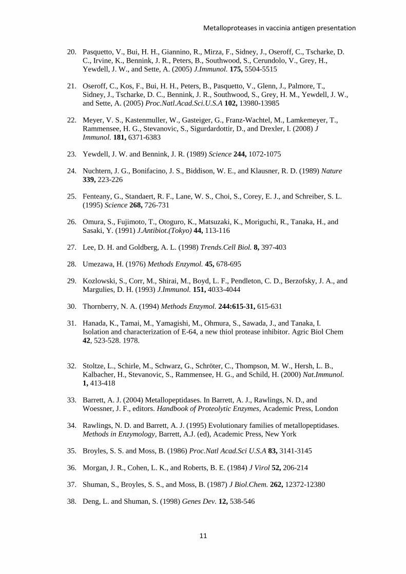
11
20. Pasquetto, V., Bui, H. H., Giannino, R., Mirza, F., Sidney, J., Oseroff, C., Tscharke, D.
C., Irvine, K., Bennink, J. R., Peters, B., Southwood, S., Cerundolo, V., Grey, H.,
Yewdell, J. W., and Sette, A. (2005) J.Immunol. 175, 5504-5515
21. Oseroff, C., Kos, F., Bui, H. H., Peters, B., Pasquetto, V., Glenn, J., Palmore, T.,
Sidney, J., Tscharke, D. C., Bennink, J. R., Southwood, S., Grey, H. M., Yewdell, J. W.,
and Sette, A. (2005) Proc.Natl.Acad.Sci.U.S.A 102, 13980-13985
22. Meyer, V. S., Kastenmuller, W., Gasteiger, G., Franz-Wachtel, M., Lamkemeyer, T.,
Rammensee, H. G., Stevanovic, S., Sigurdardottir, D., and Drexler, I. (2008) J
Immunol. 181, 6371-6383
23. Yewdell, J. W. and Bennink, J. R. (1989) Science 244, 1072-1075
24. Nuchtern, J. G., Bonifacino, J. S., Biddison, W. E., and Klausner, R. D. (1989) Nature
339, 223-226
25. Fenteany, G., Standaert, R. F., Lane, W. S., Choi, S., Corey, E. J., and Schreiber, S. L.
(1995) Science 268, 726-731
26. Omura, S., Fujimoto, T., Otoguro, K., Matsuzaki, K., Moriguchi, R., Tanaka, H., and
Sasaki, Y. (1991) J.Antibiot.(Tokyo) 44, 113-116
27. Lee, D. H. and Goldberg, A. L. (1998) Trends.Cell Biol. 8, 397-403
28. Umezawa, H. (1976) Methods Enzymol. 45, 678-695
29. Kozlowski, S., Corr, M., Shirai, M., Boyd, L. F., Pendleton, C. D., Berzofsky, J. A., and
Margulies, D. H. (1993) J.Immunol. 151, 4033-4044
30. Thornberry, N. A. (1994) Methods Enzymol. 244:615-31, 615-631
31. Hanada, K., Tamai, M., Yamagishi, M., Ohmura, S., Sawada, J., and Tanaka, I.
Isolation and characterization of E-64, a new thiol protease inhibitor. Agric Biol Chem
42, 523-528. 1978.
32. Stoltze, L., Schirle, M., Schwarz, G., Schröter, C., Thompson, M. W., Hersh, L. B.,
Kalbacher, H., Stevanovic, S., Rammensee, H. G., and Schild, H. (2000) Nat.Immunol.
1, 413-418
33. Barrett, A. J. (2004) Metallopeptidases. In Barrett, A. J., Rawlings, N. D., and
Woessner, J. F., editors. Handbook of Proteolytic Enzymes, Academic Press, London
34. Rawlings, N. D. and Barrett, A. J. (1995) Evolutionary families of metallopeptidases.
Methods in Enzymology, Barrett, A.J. (ed), Academic Press, New York
35. Broyles, S. S. and Moss, B. (1986) Proc.Natl Acad.Sci U.S.A 83, 3141-3145
36. Morgan, J. R., Cohen, L. K., and Roberts, B. E. (1984) J Virol 52, 206-214
37. Shuman, S., Broyles, S. S., and Moss, B. (1987) J Biol.Chem. 262, 12372-12380
38. Deng, L. and Shuman, S. (1998) Genes Dev. 12, 538-546

Metalloproteases in vaccinia antigen presentation
12
39. Johnstone, C., Guil, S., García-Barreno, B., López, D., Melero, J. A., and Del Val, M.
(2008) J.Gen.Virol. 89, 2194-2203
40. Neumeister, C., Nanan, R., Cornu, T. I., Lüder, C. G. K., ter Meulen, V., Naim, H., and
Niewiesk, S. (2001) J.Gen.Virol. 82, 441-447
41. Lautscham, G., Mayrhofer, S., Taylor, G., Haigh, T., Leese, A., Rickinson, A., and
Blake, N. (2001) J.Exp.Med. 194, 1053-1068
42. Lautscham, G., Rickinson, A., and Blake, N. (2003) Microbes.Infect. 5, 291-299
43. Wei, M. L. and Cresswell, P. (1992) Nature 356, 443-446
44. Henderson, R. A., Michel, H., Sakaguchi, K., Shabanowitz, J., Appella, E., Hunt, D. F.,
and Engelhard, V. H. (1992) Science 255, 1264-1266
45. Gil-Torregrosa, B. C., Castaño, A. R., López, D., and Del Val, M. (2000) Traffic 1, 641-
651
46. Gil-Torregrosa, B. C., Castaño, A. R., and Del Val, M. (1998) J.Exp.Med. 188, 1105-
1116
47. Seifert, U., Marañón, C., Shmueli, A., Desoutter, J. F., Wesoloski, L., Janek, K.,
Henklein, P., Diescher, S., Andrieu, M., de la Salle, H., Weinschenk, T., Schild, H.,
Laderach, D., Galy, A., Haas, G., Kloetzel, P. M., Reiss, Y., and Hosmalin, A. (2003)
Nat.Immunol. 4, 375-379
48. Guil, S., Rodríguez-Castro, M., Aguilar, F., Villasevil, E. M., Antón, L. C., and Del
Val, M. (2006) J.Biol.Chem. 281, 39925-39934
49. York, I. A., Bhutani, N., Zendzian, S., Goldberg, A. L., and Rock, K. L. (2006)
J.Immunol. 177, 1434-1443
50. Tiwari, N., Garbi, N., Reinheckel, T., Moldenhauer, G., Hammerling, G. J., and
Momburg, F. (2007) J.Immunol. 178, 7932-7942
51. Lorente, E., Garcia, R., and Lopez, D. (2011) J Biol.Chem. 286, 38054-38059
52. López, D., Garcia-Calvo, M., Smith, G., and Del Val, M. (2010) J.Immunol. 184, 5193-
5199
53. López, D., Jiménez, M., García-Calvo, M., and Del Val, M. (2011) J Biol.Chem. 286,
16910-16913
54. Saric, T., Chang, S. C., Hattori, A., York, I. A., Markant, S., Rock, K. L., Tsujimoto,
M., and Goldberg, A. L. (2002) Nat.Immunol. 3, 1169-1176
55. Schomburg, L., Kollmus, H., Friedrichsen, S., and Bauer, K. (2000) Eur.J.Biochem.
267, 3198-3207
56. Rawlings, N. D., Barrett, A. J., and Bateman, A. (2010) Nucleic Acids Res 38, D227-
D233
57. Serwold, T., González, F., Kim, J., Jacob, R., and Shastri, N. (2002) Nature 419, 480-
483

Metalloproteases in vaccinia antigen presentation
13
58. Suri, A., Walters, J. J., Levisetti, M. G., Gross, M. L., and Unanue, E. R. (2006)
Eur.J.Immunol. 36, 544-557
59. Oliveira, C. C., van Veelen, P. A., Querido, B., de Ru, A., Sluijter, M., Laban, S., van
der Burg, S. H., Offringa, R., and van Hall, T. (2009) J Exp.Med
60. Weinzierl, A. O., Rudolf, D., Hillen, N., Tenzer, S., van Endert, P., Schild, H.,
Rammensee, H. G., and Stevanovic, S. (2008) Eur J Immunol.
61. McDonald, J. K., Reilly, T. J., Zeitman, B. B., and Ellis, S. (1968) J Biol.Chem. 243,
2028-2037
62. Howell, S., Caswell, A. M., Kenny, A. J., and Turner, A. J. (1993) Biochem.J. 290, 159-
164
63. Slee, E. A., Zhu, H., Chow, S. C., MacFarlane, M., Nicholson, D. W., and Cohen, G. M.
(1996) Biochem.J. 315, 21-24
FOOTNOTES
*
This work was supported by grants provided by the FIPSE Foundation and Ministerio
de Ciencia e Innovación.
The abbreviations used are as follows: BFA, brefeldin A; BEN, benzyl succynil acid; BES,
bestatin; CAP, captopril; CTLs, cytotoxic T lymphocytes; ER, endoplasmic reticulum; HLA,
human leukocyte antigen; IFN- , interferon- ; ICS, intracellular cytokine staining; LC,
lactacystin; LeuSH, leucinethiol; LEU, leupeptin; PEP, pepstatin; PHE, 1,10-phenanthroline;
PHO, phosphoramidon; PUR, puromycin; TAP, transporters associated with antigen processing;
VACV, vaccinia virus.
FIGURE LEGENDS
Figure 1. Recognition of HLA-B7-restricted synthetic peptides by a VACV-specific TAP-
independent CD8+ T cell line
RMA-HLA-B*0702 target cells pre-pulsed with 10-5
M of the indicated VACV-
synthetic peptides were tested in a standard cytolytic assay (panel A) or by ICS for CD8+ T cell
activation (panel B) with VACV-specific CD8+ T cells. The VACV-specific CD8
+ T cells were
obtained from HLA-B*0702-transgenic mice immunized with VACV up to 30 days prior and
restimulated in vitro with the TAP negative RMA-S cell line transfected with HLA-B*0702.
Arrows in panel A indicate the synthetic peptides detected with 2-fold higher specific lysis than
the negative control (no peptide) in three E:T ratios. The data are the mean of four independent
experiments ± SD (panel B). Significant P values: ***, P <0.001.
Figure 2. Recognition of TAP+ and TAP
– cell lines by VACV-specific CD8
+ T lymphocytes
Recognition by ICS for CD8+ T cell activation of titration curves of D1R808-817 synthetic peptide
in HLA-B*0702 TAP+ (RMA, circles) and TAP
– (RMA-S, squares) cells. The VACV-specific
CD8+ T cells were obtained as Figure 1. Results are the mean of three experiments.
Figure 3. Immunogenicity of VACV-derived HLA-B*0702-restricted peptides in HLA-
B*0702 transgenic mice
Metalloproteases in vaccinia antigen presentation
14
RMA-HLA-B*0702 target cells pre-pulsed with the indicated VACV-synthetic peptides in
Figure 1 were analyzed by ICS for CD8+ T cell activation with VACV-specific splenocytes
obtained from HLA-B*0702 transgenic mice immunized 7 days (acute response, open bars) or
up to 30 days (memory, closed bars) post infection. The results are calculated as the mean of
three or four independent experiments ± SD. Significant P values: *, P < 0.05; **, P < 0.01;
***, P <0.001.
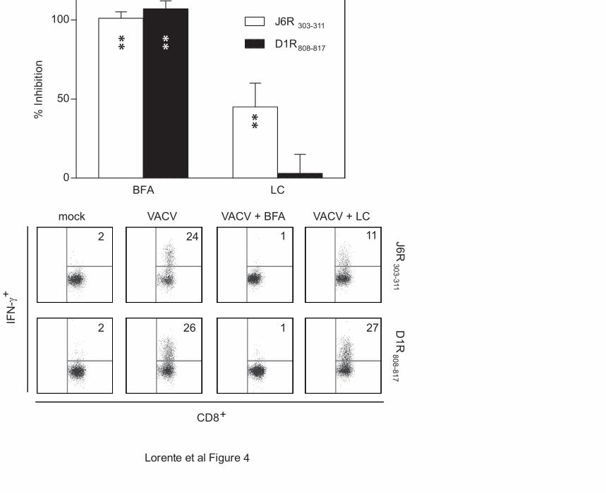
Figure 4. Effect of BFA and a proteasome inhibitor on the recognition of the J6R303-311 or
the D1R808-817 viral epitopes
RMA-HLA-B*0702 target cells infected for 16 hr with VACV at a multiplicity of infection of
40 plaque-forming units/cell were treated with BFA or LC. An ICS assay was used to test for
recognition by the J6R303-311- (open bars) or D1R808-817-specific (closed bars) CD8+ T cell lines.
The data are expressed as percentage of inhibition ± SD as determined by ICS in the presence of
the indicated inhibitors and are means of three to four independent experiments. Significant P
values: **, P < 0.01. Representative ICS assays with J6R303-311- and D1R808-817-specific CD8+ T
cell lines were depicted in the medium and bottom panels respectively. The percentages of IFN-
γ-expressing CD8+ T-cells are indicated in each dot plot.
Figure 5. Recognition of VACV-infected cells by J6R303-311- or D1R808-817-specific CD8+ T
cells in the presence of general protease inhibitors
Cells infected as described in Fig. 4 were treated with LEU (trypsin-like and cysteine protease
inhibitor), PEP (aspartic protease inhibitor), PHE (metallopeptidase inhibitor), E64 (cysteine
protease C1 inhibitor) or PUR (dipeptidyl-peptidase II and PSA), as indicated, before the ICS
assay. J6R303-311- (open bars), D1R808-817- (closed bars) or SIINFEKL-specific (hatched bars) CD8
+ T cell lines were used. The percentage of specific inhibition was calculated as in Fig. 4.
The data are means ± SD of three or four independent experiments. Significant P values: **, P <
0.01. Representative ICS assays with J6R303-311- and D1R808-817-specific CD8+ T cell lines were
depicted in the medium and bottom panels respectively. The percentages of IFN-γ-expressing
CD8+ T-cells are indicated in each dot plot.
Figure 6. Recognition of target cells infected in the presence of metalloproteinase
subfamily inhibitors
Cells infected as described in Fig. 4 were treated with CAP (ACE-like metallopeptidase
inhibitor), BEN (inhibits metallo-carboxypeptidases), BES (metallo-aminopeptidase inhibitor),
PHO (bacterial metallo-endopeptidase inhibitor), LeuSH (mainly ERAP and other metallo-
aminopeptidase inhibitor), or zVAD (blocks caspases) as indicated, before the ICS assay. The
figure is labeled as in Fig. 5. The percentage of specific inhibition was calculated as in Fig. 4.
The data are means ± SD of three to four independent experiments. Significant P values: **, P <
0.01. Representative ICS assays with D1R808-817-specific CD8+ T cell lines were depicted in the
bottom panels. The percentages of IFN-γ-expressing CD8+ T-cells are indicated in each dot plot.
Figure 7. Effect of combinations of inhibitors in the recognition of VACV-infected cells
Cells infected as described in Fig. 4 were treated with the combination of either LC and PHE or
BEN and BES, as indicated, before the ICS assay. The figure is labeled as in Fig. 5. The
percentage of specific inhibition was calculated as in Fig. 4. The data are means ± SD of three
or four independent experiments. Significant P values: **, P < 0.01.
Figure 8. Diversity of proteases and processing pathways involved in J6R303-311 or D1R808-
817 epitope presentation

Metalloproteases in vaccinia antigen presentation
15
The models show the components involved in each of the proposed pathways for the J6R303-311
(upper panel) or D1R808-817 (lower panel) epitopes. The role of proteases is deduced from the
sensitivity of respective CD8+ T cells to the various inhibitors (see Table 2).

Metalloproteases in vaccinia antigen presentation
16
Table 1
Specificity and activity of the inhibitors used in this study
Inhibitor Abbreviation Specificity Reference Concentration % Inhibition of
degradation a
Brefeldin A BFA Vesicle transport (23;24) 5 g/ml ND
Lactacystin LC Proteasome (25;26) 10 M ND
Leupeptin LEU Trypsin-like proteases and cysteine proteases (28) 100 M 38 ± 18
Pepstatin PEP Aspartic proteases (28;29) 100 M 50 ± 5
1-10 Phenanthroline PHE Metalloproteases and caspase-1 (29;30) 50 M ND
E64 E64 Cystein proteases C1 (31) 100 M ND
Puromycin PUR Dipeptidyl-peptidase II and PSA (61) 0.5 g/ml ND
Captopril CAP ACE and ACE-like proteases (29) 100 M 25 ± 2
Benzyl Succynil Acid BEN Metallo-carboxypeptidases A & B (29) 100 M -10 ± 8
Bestatin BES Most of metallo-aminopeptidases (29) 50 M ND
Phosphoramidon PHO All bacterial metallo-endopeptidases but few of
mammalian origin
(29;62) 100 M 15 ± 4
Leucinethiol LeuSH Metallo-aminopeptidases including ERAP (57) 30 M ND
z-VAD.fmk z-VAD Caspases (63) 100 M ND b
a Activity of these inhibitors was measured as their ability to prevent proteolytic degradation in cellular extracts as in reference (15). The amount of protein
still present after incubation in the case of the degraded control sample was considered 0 % inhibition of degradation, and the non-degraded sample was taken
as 100 % inhibition. Data are means of two independent experiments. Negative values indicate that there was enhanced degradation in the presence of the
compound. ND, not done.
b The compound was found to block apoptosis (data not shown).

Metalloproteases in vaccinia antigen presentation
17
Table 2
Summary of inhibition patterns
Epitope BFA a LC PHE BEN BES LeuSH LC + PHE BEN + BES
J6R 303-311 +++ b + + - - +++ +++ -
D1R 807-817 +++ - ++ + + +++ +++ +
a For specificity of different inhibitors see Table I.
b -, +. ++, and +++ indicate % inhibition < 20%, 40-60%, 61-80%, and > 81% respectively. All + inhibitions show significant P values (P < 0.01) versus
controls without an inhibitor.

* * *
* * *


3
**
** *** *
******
*
4
808-817
303-311
VACV + BFA VACV + LC
CD8
mock
+
IFN
-γ +
VACVJ6R
303-311 D
1R808-817
1
2 26 1 27
** **
**
2 24 11

808-817 303-311
VACV + PHEmock VACV
J6R303-311
D1R
808-817
5
**
**
CD8 +
IFN
-γ +
2 26
2 24
4
10

808-817 303-311
***
*
**
VACV + BEN VACV + BESmock VACV VACV + LeuSH
**
CD8 +
6
IFN
-γ +
D1R
808-817
0 45 31 30 13

7
808-817 303-311
**
**
**

Proteasome
Metallo-endoprotease
ERAPJ6R 303-311
Carboxy-metalloprotease
Amino-metalloprotease ERAPD1R 808-817
Lorente et al Figure 8
Metallo-endoprotease