Astrocytes: Targets for Neuroprotection in Stroke
21
Astrocytes: Targets for Neuroprotection in Stroke George Barreto, Robin E. White, Yibing Ouyang, Lijun Xu, and Rona G. Giffard * Department of Anesthesia, Stanford University School of Medicine, Stanford, CA, 94305, USA Abstract In the past two decades, over 1000 clinical trials have failed to demonstrate a benefit in treating stroke, with the exception of thrombolytics. Although many targets have been pursued, including antioxidants, calcium channel blockers, glutamate receptor blockers, and neurotrophic factors, often the focus has been on neuronal mechanisms of injury. Broader attention to loss and dysfunction of non-neuronal cell types is now required to increase the chance of success. Of the several glial cell types, this review will focus on astrocytes. Astrocytes are the most abundant cell type in the higher mammalian nervous system, and they play key roles in normal CNS physiology and in central nervous system injury and pathology. In the setting of ischemia astrocytes perform multiple functions, some beneficial and some potentially detrimental, making them excellent candidates as therapeutic targets to improve outcome following stroke and in other central nervous system injuries. The older neurocentric view of the central nervous system has changed radically with the growing understanding of the many essential functions of astrocytes. These include K + buffering, glutamate clearance, brain antioxidant defense, close metabolic coupling with neurons, and modulation of neuronal excitability. In this review, we will focus on those functions of astrocytes that can both protect and endanger neurons, and discuss how manipulating these functions provides a novel and important strategy to enhance neuronal survival and improve outcome following cerebral ischemia. Keywords Brain ischemia; astrocytes; neurons; inflammation; neuroprotection; clinical trials 1. INTRODUCTION Stroke is the third leading cause of death in the United States and results in substantial health-care expenditures; the mean lifetime cost resulting from an ischemic stroke is estimated at $140,048 per patient, and this estimation is higher for people over 45 years. Nationwide in 2010, the estimated direct and indirect costs of stroke totaled $73.7 billion [1]. Although many clinical trials have been completed in stroke patients, none of these have demonstrated protective efficacy except for thrombolysis [2, 3]. In the case of cardiac arrest and resuscitation only hypothermia has been shown to have clinical utility [4]. In some sense the two therapies that have been effective thus far clinically have broad targets, and do not only affect a single injury mechanism. In contrast, of the failed trials, many targeted neuron- specific injury mechanisms [5]. This may reflect too narrow a view of what is needed for brain preservation. A large body of work has shown that astrocytes play key roles both in normal and pathological central nervous system functioning [6]. Astrocytes are the most abundant brain cell type, and in addition to their multiple important homeostatic roles, they © 2011 Bentham Science Publishers Ltd. * Address correspondence to this author at the Department of Anesthesia, Stanford University School of Medicine, S272, Stanford, CA 94305, USA; Tel: 650-725-8482; Fax: 650-725-8052; [email protected]. NIH Public Access Author Manuscript Cent Nerv Syst Agents Med Chem. Author manuscript; available in PMC 2011 December 8. Published in final edited form as: Cent Nerv Syst Agents Med Chem. 2011 June 1; 11(2): 164–173. NIH-PA Author Manuscript NIH-PA Author Manuscript NIH-PA Author Manuscript
Transcript of Astrocytes: Targets for Neuroprotection in Stroke

Astrocytes: Targets for Neuroprotection in Stroke
George Barreto, Robin E. White, Yibing Ouyang, Lijun Xu, and Rona G. Giffard*
Department of Anesthesia, Stanford University School of Medicine, Stanford, CA, 94305, USA
AbstractIn the past two decades, over 1000 clinical trials have failed to demonstrate a benefit in treatingstroke, with the exception of thrombolytics. Although many targets have been pursued, includingantioxidants, calcium channel blockers, glutamate receptor blockers, and neurotrophic factors,often the focus has been on neuronal mechanisms of injury. Broader attention to loss anddysfunction of non-neuronal cell types is now required to increase the chance of success. Of theseveral glial cell types, this review will focus on astrocytes. Astrocytes are the most abundant celltype in the higher mammalian nervous system, and they play key roles in normal CNS physiologyand in central nervous system injury and pathology. In the setting of ischemia astrocytes performmultiple functions, some beneficial and some potentially detrimental, making them excellentcandidates as therapeutic targets to improve outcome following stroke and in other central nervoussystem injuries. The older neurocentric view of the central nervous system has changed radicallywith the growing understanding of the many essential functions of astrocytes. These include K+
buffering, glutamate clearance, brain antioxidant defense, close metabolic coupling with neurons,and modulation of neuronal excitability. In this review, we will focus on those functions ofastrocytes that can both protect and endanger neurons, and discuss how manipulating thesefunctions provides a novel and important strategy to enhance neuronal survival and improveoutcome following cerebral ischemia.
KeywordsBrain ischemia; astrocytes; neurons; inflammation; neuroprotection; clinical trials
1. INTRODUCTIONStroke is the third leading cause of death in the United States and results in substantialhealth-care expenditures; the mean lifetime cost resulting from an ischemic stroke isestimated at $140,048 per patient, and this estimation is higher for people over 45 years.Nationwide in 2010, the estimated direct and indirect costs of stroke totaled $73.7 billion[1]. Although many clinical trials have been completed in stroke patients, none of these havedemonstrated protective efficacy except for thrombolysis [2, 3]. In the case of cardiac arrestand resuscitation only hypothermia has been shown to have clinical utility [4]. In some sensethe two therapies that have been effective thus far clinically have broad targets, and do notonly affect a single injury mechanism. In contrast, of the failed trials, many targeted neuron-specific injury mechanisms [5]. This may reflect too narrow a view of what is needed forbrain preservation. A large body of work has shown that astrocytes play key roles both innormal and pathological central nervous system functioning [6]. Astrocytes are the mostabundant brain cell type, and in addition to their multiple important homeostatic roles, they
© 2011 Bentham Science Publishers Ltd.*Address correspondence to this author at the Department of Anesthesia, Stanford University School of Medicine, S272, Stanford, CA94305, USA; Tel: 650-725-8482; Fax: 650-725-8052; [email protected].
NIH Public AccessAuthor ManuscriptCent Nerv Syst Agents Med Chem. Author manuscript; available in PMC 2011 December 8.
Published in final edited form as:Cent Nerv Syst Agents Med Chem. 2011 June 1; 11(2): 164–173.
NIH
-PA Author Manuscript
NIH
-PA Author Manuscript
NIH
-PA Author Manuscript

organize the structural architecture of the brain, help organize communication pathways, andmodulate neuronal plasticity (for recent review see [7, 8]). Thus, astrocytes are now thoughtto be important potential targets for manipulation.
Ischemic stroke is caused by an interruption of cerebral blood flow that leads to stress, celldeath, and inflammation. Neurons are more susceptible to injury than astrocytes whenstudied under some in vitro conditions [9, 10]. Neurons have less endogenous antioxidantsand are susceptible to excito-toxicity [10]. Both normally and after ischemia, astrocytessupport neurons by providing antioxidant protection [11, 12], substrates for neuronalmetabolism [13], and glutamate clearance REF. Although astrocytes are sometimes moreresilient than neurons, injury can result in impaired astrocyte function even when astrocytesdo not die. Impaired astrocyte function can amplify neuronal death [14]. Therefore, manyrecent efforts have focused on the astrocyte-neuron interaction and how astrocyte functioncan be improved after stroke to enhance neuronal support and survival [10, 15, 16]. Agrowing body of data demonstrates that astrocytes play a multifaceted and complex role inthe response to ischemia, with potential to both enhance and impair neuronal survival andregeneration [17]. Many recent studies focus on the astrocyte-neuron interaction and severalinvestigate ways in which astrocyte function can be improved after stroke to enhanceneuronal survival.
This review provides a brief overview of the pathophysiological events underlying ischemicbrain damage, and considers how these events affect astrocyte-mediated support of neurons.In addition, we discuss some experimental approaches to enhance the neuronal supportiverole of astrocytes as a novel strategy against stroke. Finally, we explore how theseapproaches may eventually be applied in the clinical setting to improve stroke outcome forpatients.
2. ASTROCYTE VIABILITY AFTER ISCHEMIA2.1. In Vitro Studies
In vitro studies have provided substantial insight into the mechanisms governing the survivalof astrocytes following simulated ischemia. These investigations have shown that astrocytesare generally more resistant than neurons to oxygen-glucose deprivation (OGD) performedin media at physiologically normal pH, an in vitro model of ischemia [10, 18]. Most neuronsin astrocyte-neuronal co-cultures will die after 60–90 min of OGD, while astrocyte culturesonly suffer a similar extent of injury after 4–6 hours [9, 18, 19]. Different astrocytepopulations exist and astrocytes isolated from different brain regions such as cortex,striatum, and hippocampus differ in their sensitivity to OGD [15, 20, 21]. Furthermore,Lukaszevicz and colleagues [22] reported that protoplasmic astrocytes lose their integrityfaster than fibrous astrocytes, which may explain the regional differences in susceptibility toischemia between white matter astrocytes which are fibrous and grey matter astrocytes thatare protoplasmic. Although less susceptible to OGD-induced damaged in vitro studies havehighlighted certain elements that are highly toxic to astrocytes. For example, acidosis hasbeen found to be very effective in killing astrocytes [23–26], in contrast to neurons, whichare protected in acidic conditions [24, 26].
2.2. Focal Cerebral IschemiaMuch of the information about the recovery of astrocytes in vivo has been provided bystudies using immunohistological markers for astrocyte specific proteins, such as glialfibrillary acidic protein (GFAP) and glutamine synthetase GS; Fig. 1. Using these markersas tools, several investigations suggest that astrocytes are better preserved than neurons inanimal models of stroke outside the core where all cells die [27–29]. Though neuronalmarkers are decreased as soon as 1 hour after MCAO, GFAP expression is preserved over
Barreto et al. Page 2
Cent Nerv Syst Agents Med Chem. Author manuscript; available in PMC 2011 December 8.
NIH
-PA Author Manuscript
NIH
-PA Author Manuscript
NIH
-PA Author Manuscript

the first 3 hours of reperfusion after 2 hour MCAO [29] and GS is increased 3 hoursfollowing a 3 hour MCAO [28]. At later reperfusion periods, GFAP increases in the peri-infarct area that later develops into the glial scar [29–32]. In contrast, Liu and colleagues[33] reported the deterioration of some astrocyte markers prior to that of neuronal markers.Discrepancy in findings may be due to differences in detection (i.e., protein vs. mRNA) andinjury paradigms.
2.3. Forebrain IschemiaExcitotoxic neuronal injury is a common mechanism in both acute and chronicneurodegenerative diseases. It has long been appreciated that inhibition of astrocyteglutamate uptake [34, 35], and more recently inhibition of astrocyte mitochondrial function[36], impairs neuronal survival from excitotoxic injury. Brief forebrain ischemia is a modelof the delayed hippocampal neuronal loss seen in patients following cardiac arrest andresuscitation, and in part involves excitotoxicity. Increased generation of reactive oxygenspecies (ROS) and mitochondrial dysfunction in CA1 astrocytes contributes to ischemia-induced loss of GLT-1 and ultimately to delayed death of CA1 neurons [15]. Our studiesand those of other laboratories have demonstrated that selective dysfunction of hippocampalCA1 subregion astrocytes, with loss of glutamate transport activity and immunoreactivityfor glutamate transporter 1 (GLT-1), occurs at early reperfusion times, hours to days beforethe death of CA1 neurons [15, 37, 38].
The heterogeneous degeneration of astrocytic processes and mitochondria was tightlyassociated with the appearance of disseminated selective neuronal necrosis and itsmaturation after temporary ischemia [39]. By electronmicroscopy the same investigators[40] found that focal infarction is exacerbated by temporary microvascular obstruction dueto compression by swollen astrocytic end-feet. However, hypoxia has multiple effects onastrocytes and their ability to support neuronal viability [41]. For example, hypoxia inducesastrocyte-dependent protection of neurons following hypoxic preconditioning. Yet, hypoxiainduces processes in astrocytes that augment neuronal death in other situations, such as thecoincidence of hypoxia with inflammatory signaling.
3. REACTIVE ASTROGLIA: GOOD OR BAD AFTER STROKE?The astrocyte response to ischemia has traditionally been viewed as detrimental to recovery,as the astrocyte-rich glial scar has both physical and chemical inhibitory properties [42, 43].As components of the glial scar, astrocytes exhibit hypertrophied, interdigitated processesthat form a physical barrier. Astrocytes produce inhibitory molecules including chondroitinsulfate proteoglycans (CSPGs) that contribute to chemical inhibition [44, 45]. In the acutesetting, astrocytic gap junctions may remain open following ischemia [46], allowingsubstances such as proapoptotic factors to spread through the syncytium, thereby expandingthe size of the infarct [47]. As discussed below, astrocytes can also produce a variety of pro-inflammatory cytokines.
Many studies have shown that decreased astrogliosis often correlates with decreased infarctsize. Nonspecific inhibition of cell proliferation following ischemia using a cyclin kinaseinhibitor decreases astrocyte proliferation and results in improved functional recovery [48].In addition, treatment with alpha-melanocyte stimulating hormone [49], cysteinylleukotriene receptor antagonist [50], cliostazol [51], and caffeic acid [52] result in reducedinfarct size accompanied by a decrease in astrogliosis. Treadmill exercise [28] andacupuncture [53] are similarly associated with improved outcome and reduced astrogliosis.Thus, results from several studies suggest that treatments that decrease infarct size are oftenaccompanied by attenuated astrocyte response. Despite the frequent association of decreased
Barreto et al. Page 3
Cent Nerv Syst Agents Med Chem. Author manuscript; available in PMC 2011 December 8.
NIH
-PA Author Manuscript
NIH
-PA Author Manuscript
NIH
-PA Author Manuscript

astrogliosis with improved outcome, it is difficult to determine cause and effect, since theextent of astrogliosis likely reflects the severity of the injury, as well as influencing it.
In addition to their role in glial scar formation, astrocytes also respond to ischemia withfunctions important for neuroprotection and repair. These include protecting spared tissuefrom further damage [14], taking up excess glutamate as discussed above, rebuilding theblood brain barrier [54, 55], and producing neurotrophic factors [10]. GFAP knockout miceexhibit larger lesions than their wild-type littermates following focal ischemia [56], andmice lacking both GFAP and vimentin have impaired astrocyte activation, decreasedglutamate uptake abilities, and attenuated PAI-1 expression after ischemia [57]. Applicationof astrocyte-conditioned media after transient MCAO results in decreased infarct volumeand regained blood-brain barrier function [58], suggesting that factors released by astrocytesfollowing ischemia are important for neuroprotection.
Although few studies other than the use of animals lacking vimentin and GFAP havespecifically targeted astrocyte activation after ischemia, there is correlational evidencesuggesting that astrogliosis may be beneficial. Environmental enrichment, which results inreduced infarct size and improved recovery following ischemia, also leads to increasedastrocyte proliferation [59, 60]. After focal ischemia, aged rats exhibit increased tissuedamage and increased astrocyte hypertrophy, but have decreased astrocyte proliferationcompared to young rats [61]. Systemic infusion of bone marrow stromal cells followingMCAO increases gliogenesis and decreases lesion size [62, 63]. In addition, administrationof transforming growth factor α (TGFα), a known mitogen for astrocytes [64], followingMCAO leads to reduced infarct size and improved functional recovery [65]. Furthermore,ischemic preconditioning that produces a neuroprotective state leads to prolonged astrocyteexpression of Hsp27 [66]. Finally, mice lacking connexin 43, the gap junction connectingastrocyte networks that is needed for proper neurotransmitter and potassium regulation, haveincreased infarcts following MCAO [67]. Thus, astrocytes have the potential to be bothdetrimental and beneficial following ischemic insult, making them promising targets formanipulation to improve outcome.
4. ASTROCYTE-MEDIATED INFLAMMATION AFTER STROKE: A DOUBLE-EDGED SWORD
Inflammation plays both detrimental and beneficial roles in brain ischemia, depending uponthe timing and severity of the inflammation. Within minutes after injury, injured neurons inthe core and penumbra of the lesion and glial cells in the core produce pro-inflammatorymediators, cytokines, and reactive oxygen species, which activate both astrocytes andmicroglia [68]. Activated astrocytes can produce the proinflammatory cytokines IL-6,TNFα, IL-1α and β, interferon γ, and others [68–70]. High levels of these cytokines can bedetrimental to ischemic recovery [71–75] by directly inducing apoptosis of neuronal cellsand/or increasing toxic nitric oxide levels [76] and inhibiting neurogenesis [77]. Indeed,inactivation of astrocyte NfκB signaling, shown to induce astrocyte production of pro-inflammatory cytokines [78], decreases cytokine production and protects neurons afterischemic injury [79]. Hsp72 overexpression is associated with lower NfκB activation andlower TNFα [80]. In addition to cytokines, reactive astrocytes also produce chemokinesfollowing ischemia [81]. Chemokines upregulate adhesion molecules in vascular endothelialcells, resulting in attraction of immune cells, which may worsen ischemia-induced damage[82]. Overall, some aspects of the local inflammatory response contribute to secondaryinjury to potentially viable tissue and lead to apoptotic and necrotic neuronal cell deathhours to days after injury [83], while other aspects are beneficial.
Barreto et al. Page 4
Cent Nerv Syst Agents Med Chem. Author manuscript; available in PMC 2011 December 8.
NIH
-PA Author Manuscript
NIH
-PA Author Manuscript
NIH
-PA Author Manuscript

Although the potential benefits of inflammation after stroke have received relatively littleattention so far, indirect evidence suggests that some specific inflammatory reactions areneuroprotective and neuroregenerative [84–91]. In addition to providing defense against theinvasion of pathogens, inflammation is also involved in clearing damaged tissue, and inangiogenesis, tissue remodeling, and regeneration [89]. This is probably best studied inwound healing, which is severely compromised if inflammation is inhibited [89, 91]. Thereis also evidence suggesting that specific inflammatory factors can be protective in somecircumstances. IL-6, produced by astrocytes acutely after MCAO [69], is likelyneuroprotective early after ischemia [84]. Interestingly, ischemic preconditioning resultingin protection appears to be dependent on TLR-4 signaling, and is accompanied by increasedTNFα, NFκB, and COX-2 expression [90]. Indeed, in vitro work has shown thatadministration of TNFα in combination with Hsp70 results in decreased expression of pro-apoptotic proteins following hypoxia [88]. Thus, it is important to consider these factors,along with timing, when trying to determine the best strategy to reduce damage and improverecovery and regeneration.
5. ASTROCYTE SUPPORT OF NEURONS AFTER STROKE5.1. Antioxidant Production
One hallmark of the cellular response to ischemia is a rapid, dramatic increase in damagingfree radicals, including nitric oxide (NO), superoxide, and peroxynitrite [92]. Nitric oxidesynthetase levels increase as soon as 10 minutes after induction of MCAO [93], followed byNO production that persists for at least one week after MCAO [94]. Nitric oxide can causecell death by inducing the release of cytochrome-c from mitochondria, leading to apoptosis[95]. Nitric oxide can also induce necrotic death [96]. Furthermore, the production of nitricoxide and other free radicals can modify oxidative metabolism and impair ATP production[13, 19]. Changes in mitochondrial properties can further limit oxidative metabolism [97].Not surprisingly, several studies have shown that antioxidant treatment enhancesneuroprotection and recovery after stroke [98–101].
The release of glutathione and SOD by astrocytes has been reported and is suggested to playan important role in maintaining and enhancing neuronal survival, yet they are able toreduce ascorbate for further neuronal antioxidant defense Fig. (2) [10, 102–106].Interestingly, neurons cocultured with astrocytes exhibit higher levels of glutathionecompared with neurons cultured alone [107]. Although astrocytes upregulate SOD aftercerebral ischemia [108], they do not appear to increase levels of glutathione in ischemicconditions [109]. It is unknown whether ischemia alters astrocytic ascorbate levels, butosmotic swelling from ischemia results in increased astrocyte release of ascorbate in vitro[110], suggesting that similar mechanisms may occur in vivo.
Several treatments that attenuate ischemic injury result in increased glutathione levels [111,112]. SOD converts superoxide into oxygen and hydrogen peroxide. Similar to glutathione,many treatments that ameliorate stroke damage are accompanied by an increase in SOD[113, 114]. Furthermore, rodents overexpressing SOD1 have significantly smaller injuriesafter both focal and global ischemia [115, 116], while mice with decreased SOD1 havelarger infarcts [117]. Finally, ascorbate can also reduce oxidative stress [118]. Treatmentwith dehydroascorbic acid, a blood-brain-barrier-permeable precursor to ascorbic acid, isprotective after MCAO [119]. Dehydroascorbic acid is taken up by astrocytes and releasedas ascorbic acid [12], a process increased by propofol [120], a treatment that can beprotective after stroke [121]. In summary, astrocytes are important producers of antioxidantsin the normal CNS, and astrocyte production of these molecules after stroke may enhanceneuronal survival and protect astrocyte function.
Barreto et al. Page 5
Cent Nerv Syst Agents Med Chem. Author manuscript; available in PMC 2011 December 8.
NIH
-PA Author Manuscript
NIH
-PA Author Manuscript
NIH
-PA Author Manuscript

5.2. Glutamate RegulationAstrocytes are key players in the regulation of neuro-transmitters in the CNS. Astrocytesmake glutamine, the precursor for the neurotransmitters glutamate and GABA [122] Fig. (2).Astrocyte production of neurotransmitter precursors is impaired after MCAO, andalterations in neuro-transmitter levels occur throughout the brain following stroke, possiblycontributing to neuronal death [123, 124].
Astrocytes are primarily responsible for glutamate uptake in the normal brain using theastrocyte specific glutamate transporters GLAST and GLT-1 (Fig. 2) [125–127], as excessglutamate leads to cell death via excitotoxicity [128]. Glutamate transporter levels inastrocytes decrease acutely following global ischemia [38, 129] and neonatal hypoxia-ischemia [130], most likely exacerbating neuronal death as a result of glutamate-inducedexcitoxicity. Despite the therapeutic potential of increasing astrocyte glutamate transportafter stroke, few groups have explored this possibility. Carnosine, shown to be protectiveafter focal ischemia, may partially be effective because it prevents loss of GLT-1 onastrocytes, resulting in attenuated excitotoxicity [131]. In a more direct assessment of howpost-ischemic astrocyte glutamate transporters contribute to neuronal survival, ourlaboratory has shown that upregulation of GLT-1 on astrocytes using ceftriaxone protectsCA1 neurons after global ischemia [129], similar to its effects in focal cerebral ischemia[132].
5.3. Potassium Uptake and Energy MetabolismAstrocytes also regulate neuronal activation by extracellular potassium uptake [133] Fig. (2).Neurons release potassium after activation, and increased extracellular potassium leads toneuronal hyperexcitability [133], a phenomenon that occurs in ischemic conditions [134]. Inaddition to regulating neuronal activation, proper maintenance of ion gradients, such aspotassium, is important in regulating cell volume in both normal and ischemic conditions[135, 136]. Astrocytes increase potassium transporter activity in response to transient invitro ischemia [137]. Due to its effects on both neuronal activity and cell volume, increasingastrocytic potassium uptake may be a possible therapeutic target for stroke.
Astrocytes are also integral to normal maintenance of neuronal metabolism. Whenastrocytes take up extracellular glutamate as a result of neuronal activity, the Na+/ K+-ATPase, along with AMPA signaling, triggers astrocyte uptake of glucose from the blood,as astrocytic endfeet contact capillaries [138, 139]. This glucose is then made into lactate, asubstrate for neuronal energy, to further “fuel” active neurons [140] Fig. (2). As mentionedabove, astrocytes produce glutathione. In addition to its antioxidant properties, glutathione isneeded for the conversion of methylglyoxal, a toxic by-product of metabolism, into D-Lactate by glyoxalase 1 [141]. Although the role of astrocyte metabolism is relatively well-established in normal tissue, the post-ischemic role of astrocyte metabolism maintenance isless clear [142]. After ischemia, astrocytes upregulate glucose transporters in order toprovide energy to stressed/dying neuronal cells [143, 144]. Ethyl pyruvate, a derivative ofthe energy substrate pyruvate, is neuroprotective after stroke only when astrocytes areviable, suggesting that astrocytes are necessary for improvement in post-ischemic energymetabolism [122].
6. NOVEL STRATEGIES TO IMPROVE THE NEURONAL SUPPORTIVE ROLEOF ASTROCYTES
Although few studies have specifically targeted astrocytes for repair after stroke, there issome evidence that this can be a successful strategy. Recent results indicate that induction ofBDNF in astrocytes by galectin-1 reduces neuronal apoptosis in ischemic boundary zone
Barreto et al. Page 6
Cent Nerv Syst Agents Med Chem. Author manuscript; available in PMC 2011 December 8.
NIH
-PA Author Manuscript
NIH
-PA Author Manuscript
NIH
-PA Author Manuscript

and improves functional recovery [145]. In addition, protection by pyruvate againstglutamate neurotoxicity is mediated by astrocytes through a glutathione-dependentmechanism [146]. Our recent study demonstrated that enhancing astrocyte resistance toischemic stress by overexpressing protective proteins or antioxidant enzyme results inimproved survival of CA1 neurons following forebrain ischemia Fig. (3) [16]. Two well-studied protective proteins, heat shock protein 72 (Hsp72) and mitochondrial SOD, weregenetically targeted for expression in astrocytes using the astrocyte-specific human GFAPpromoter. In both cases protection was accompanied by preservation of the astrocyticglutamate transporter GLT-1, and reduced evidence of oxidative stress in the CA1 region[16]. Similarly, selective overexpression of excitatory amino acid transporter 2 (EAAT2) inastrocytes enhances neuroprotection from moderate hypoxia-ischemia [147].
7. TRANSLATING INSIGHTS INTO PROTECTION INTO CLINICALAPPLICATIONS
Many factors have been identified that likely contribute to the failure in translation seen sofar with stroke therapies. Currently, the only approved stroke therapy is thrombolysisinduced by intravenous administration of recombinant tissue plasminogen activator [148];however, because of a short therapeutic time window, only a small fraction of patientsbenefit from this treatment. Hypothermia is the only accepted acute treatment to reducebrain injury following cardiac arrest and resuscitation [4]. Thus far many clinical trials havefocused on treatments that would likely be beneficial to neurons, with fewer studies focusedon mechanisms that might benefit all cell types or specifically targeting other cell types,such as astrocytes. Often the consequence of these treatments on the astrocyte response isnot considered. Several examples of past and ongoing clinical trials are discussed below,with specific attention to how these treatments may alter astrocyte response or viability.
Several clinical trials have targeted manipulation of the inflammatory response to ischemia,as stroke patients with systemic inflammation exhibit poorer outcomes [149]. Although anti-inflammatory therapy decreases infarct size and improves neurological sequelae inexperimental animal models of stroke [150], human trials with anti-neutrophil therapy havenot shown a clear benefit [151, 152]. In addition, recent clinical trials in which anti-CD11/18antibodies were administered to human subjects were unsuccessful [153]. Likewise, adouble-blinded, placebo-controlled clinical trial in which anti–ICAM-1 antibody wasadministered within 6 hours of stroke symptoms showed disappointing results [151]. Inunderstanding these results it is important to recall that while experimental stroke isrelatively homogeneous concerning size, territory, and etiology, with more consistentinflammatory response, human stroke is extremely heterogeneous [154], with differentvascular territories and extents of injury. In addition, these mediators are known to affectmany organ systems beyond the central nervous system. Systemic administration of anti-inflammatory agents may have exacerbated the relative state of immunocompromise seen instroke patients, thereby confounding the outcome. Furthermore, inflammation and astrocyteresponse are likely closely connected. Although there is little evidence for a directrelationship between neutrophils and astrocytes, it has been shown that mice with a bluntedinflammatory response exhibit increased loss of GFAP-positive astrocytes after cortical stabinjury [155]. Because astrocytic glial scar formation is important in protection of sparedtissue from further damage [156], it is possible that treatments that drastically attenuateinflammation lead to a stunted astrocyte response that is deleterious to recovery.
Another drug that has advanced to clinical study is DP-b99, currently in phase III studies foracute stroke. DP-b99 is a membrane active chelator derivative of the known calciumchelator, BAPTA spell out [157]. A lipophilic chelator of calcium, zinc and copper ions,DP-b99 sequesters metal ions only within and in to cell membranes. This clinical trial is
Barreto et al. Page 7
Cent Nerv Syst Agents Med Chem. Author manuscript; available in PMC 2011 December 8.
NIH
-PA Author Manuscript
NIH
-PA Author Manuscript
NIH
-PA Author Manuscript

especially attractive because sequestration of calcium, zinc, and copper are potentiallybeneficial not only to neurons, but also to astrocytes. It has been shown in Alzheimer’sdisease that beta amyloid increases astrocyte calcium influx, which causes decreasedglutathione levels [158]. Zinc chloride is toxic to astrocytes as well as neurons in vitro [159].Similarly, astrocytes exposed to neocuprine exhibit increased copper influx and undergoapoptotic cell death [160]. Approaches that benefit multiple cell types, including astrocytes,are more likely to be successful.
Other current ongoing clinical trials focus on neuroprotective agents for the purpose ofaiding neurological recovery after stroke. Minocycline (Phase I), edavarone (Phase IV),propanolol (a β-blocker; phase II and III), and more recently arundic acid have beenpreviously shown to be protective and enhance neuronal survival in stroke [161–165],though by targeting different mechanisms. Some additional completed and ongoing trials aresummarized in Table 1. Preclinical research needs to consider these clinical results, andassess effects on astrocytes as well as neurons.
Although anti-inflammatory strategies to diminish ischemic brain injury have failed thus far,continued elucidation of the complex interactions involved in modulating the inflammatoryresponse may still enable novel therapeutic approaches that translate successfully intoclinical efficacy.
CONCLUSIONSTraditionally, stroke research has focused on neurons and often ignored effects on glial cells.It is increasingly evident that glia are vital to both normal CNS functioning and also playimportant roles in neuropathological conditions. Although astrocytes form an inhibitory glialscar following ischemia, they also perform functions necessary for neuronal survival andwell-being, such as maintaining low extracellular glutamate levels and providing antioxidantprotection. Because they have a great many functions, astrocytes are attractive candidates astherapeutic targets. By striving to shift astrocytes towards a pro-reparative, neuronal-supportive phenotype following stroke, future clinical therapies may well be moresuccessful in protecting neurons from ischemic damage and promoting repair.
AcknowledgmentsThis work was supported by NIH grants CM49831, N5053898, and NS014543 to RGG.
REFERENCES1. Lloyd-Jones D, Adams RJ, Brown TM, Carnethon M, Dai S, De Simone G, Ferguson TB, Ford E,
Furie K, Gillespie C, Go A, Greenlund K, Haase N, Hailpern S, Ho PM, Howard V, Kissela B,Kittner S, Lackland D, Lisabeth L, Marelli A, McDermott MM, Meigs J, Mozaffarian D, MussolinoM, Nichol G, Roger VL, Rosamond W, Sacco R, Sorlie P, Roger VL, Thom T, Wasserthiel-SmollerS, Wong ND, Wylie-Rosett J. Heart disease and stroke statistics--2010 update: a report from theAmerican Heart Association. Circulation. 2010; 121:e46–e215. [PubMed: 20019324]
2. Blakeley JO, Llinas RH. Thrombolytic therapy for acute ischemic stroke. J. Neurol. Sci. 2007;261:55–62. [PubMed: 17618652]
3. Marder VJ, Jahan R, Gruber T, Goyal A, Arora V. Thrombolysis with plasmin: implications forstroke treatment. Stroke. 2010; 41:S45–S49. [PubMed: 20876504]
4. Bernard SA, Gray TW, Buist MD, Jones BM, Silvester W, Gutteridge G, Smith K. Treatment ofcomatose survivors of out-of-hospital cardiac arrest with induced hypothermia. N. Engl. J. Med.2002; 346:557–563. [PubMed: 11856794]
Barreto et al. Page 8
Cent Nerv Syst Agents Med Chem. Author manuscript; available in PMC 2011 December 8.
NIH
-PA Author Manuscript
NIH
-PA Author Manuscript
NIH
-PA Author Manuscript
5. Howells DW, Porritt MJ, Rewell SS, O'Collins V, Sena ES, van der Worp HB, Traystman RJ,Macleod MR. Different strokes for different folks: the rich diversity of animal models of focalcerebral ischemia. J. Cereb. Blood Flow Metab. 30:1412–1431. [PubMed: 20485296]
6. Kettenmann, H.; Ramson, BR. Neuroglia. New York: Oxford University Press; 1995.7. Nedergaard M, Ransom B, Goldman SA. New roles for astrocytes: redefining the functional
architecture of the brain. Trends Neurosci. 2003; 26:523–530. [PubMed: 14522144]8. Sofroniew MV, Vinters HV. Astrocytes: biology and pathology. Acta Neuropathol. 2010; 119:7–35.
[PubMed: 20012068]9. Goldberg MP, Choi DW. Combined oxygen and glucose deprivation in cortical cell culture:
calcium-dependent and calcium-independent mechanisms of neuronal injury. J. Neurosci. 1993;13:3510–3524. [PubMed: 8101871]
10. Swanson RA, Ying W, Kauppinen TM. Astrocyte influences on ischemic neuronal death. Curr.Mol. Med. 2004; 4:193–205. [PubMed: 15032713]
11. Dringen R, Hirrlinger J. Glutathione pathways in the brain. Biol. Chem. 2003; 384:505–516.[PubMed: 12751781]
12. Wilson JX. Antioxidant defense of the brain: a role for astrocytes. Can. J. Physiol. Pharmacol.1997; 75:1149–1163. [PubMed: 9431439]
13. Rossi DJ, Brady JD, Mohr C. Astrocyte metabolism and signaling during brain ischemia. Nat.Neurosci. 2007; 10:1377–1386. [PubMed: 17965658]
14. Nedergaard M, Dirnagl U. Role of glial cells in cerebral ischemia. Glia. 2005; 50:281–286.[PubMed: 15846807]
15. Ouyang YB, Voloboueva LA, Xu LJ, Giffard RG. Selective dysfunction of hippocampal CA1astrocytes contributes to delayed neuronal damage after transient forebrain ischemia. J. Neurosci.2007; 27:4253–4260. [PubMed: 17442809]
16. Xu L, Emery JF, Ouyang YB, Voloboueva LA, Giffard RG. Astrocyte targeted overexpression ofHsp72 or SOD2 reduces neuronal vulnerability to forebrain ischemia. Glia. 2010; 58:1042–1049.[PubMed: 20235222]
17. Anderson MF, Blomstrand F, Blomstrand C, Eriksson PS, Nilsson M. Astrocytes and stroke:networking for survival? Neurochem. Res. 2003; 28:293–305. [PubMed: 12608702]
18. Giffard RG, Swanson RA. Ischemia-induced programmed cell death in astrocytes. Glia. 2005;50:299–306. [PubMed: 15846803]
19. Almeida A, Delgado-Esteban M, Bolanos JP, Medina JM. Oxygen and glucose deprivation inducesmitochondrial dysfunction and oxidative stress in neurones but not in astrocytes in primary culture.J. Neurochem. 2002; 81:207–217. [PubMed: 12064468]
20. Xu L, Sapolsky RM, Giffard RG. Differential sensitivity of murine astrocytes and neurons fromdifferent brain regions to injury. Exp. Neurol. 2001; 169:416–424. [PubMed: 11358455]
21. Zhao G, Flavin MP. Differential sensitivity of rat hippocampal and cortical astrocytes to oxygen-glucose deprivation injury. Neurosci. Lett. 2000; 285:177–180. [PubMed: 10806315]
22. Lukaszevicz AC, Sampaio N, Guegan C, Benchoua A, Couriaud C, Chevalier E, Sola B, LacombeP, Onteniente B. High sensitivity of protoplasmic cortical astroglia to focal ischemia. J. Cereb.Blood Flow Metab. 2002; 22:289–298. [PubMed: 11891434]
23. Bondarenko A, Chesler M. Rapid astrocyte death induced by transient hypoxia, acidosis, andextracellular ion shifts. Glia. 2001; 34:134–142. [PubMed: 11307162]
24. Giffard RG, Monyer H, Choi DW. Selective vulnerability of cultured cortical glia to injury byextracellular acidosis. Brain Res. 1990; 530:138–141. [PubMed: 2176914]
25. Swanson RA, Farrell K, Stein BA. Astrocyte energetics, function, and death under conditions ofincomplete ischemia: a mechanism of glial death in the penumbra. Glia. 1997; 21:142–153.[PubMed: 9298857]
26. Tombaugh GC, Sapolsky RM. Mechanistic distinctions between excitotoxic and acidotichippocampal damage in an in vitro model of ischemia. J. Cereb. Blood Flow Metab. 1990;10:527–535. [PubMed: 1971825]
27. Chen Y, Swanson RA. Astrocytes and brain injury. J. Cereb. Blood Flow Metab. 2003; 23:137–149. [PubMed: 12571445]
Barreto et al. Page 9
Cent Nerv Syst Agents Med Chem. Author manuscript; available in PMC 2011 December 8.
NIH
-PA Author Manuscript
NIH
-PA Author Manuscript
NIH
-PA Author Manuscript

28. Lee MH, Kim H, Kim SS, Lee TH, Lim BV, Chang HK, Jang MH, Shin MC, Shin MS, Kim CJ.Treadmill exercise suppresses ischemia-induced increment in apoptosis and cell proliferation inhippocampal dentate gyrus of gerbils. Life Sci. 2003; 73:2455–2465. [PubMed: 12954454]
29. Li Y, Chopp M, Zhang ZG, Zhang RL. Expression of glial fibrillary acidic protein in areas of focalcerebral ischemia accompanies neuronal expression of 72-kDa heat shock protein. J. Neurol. Sci.1995; 128:134–142. [PubMed: 7738589]
30. Schroeter M, Schiene K, Kraemer M, Hagemann G, Weigel H, Eysel UT, Witte OW, Stoll G.Astroglial responses in photochemically induced focal ischemia of the rat cortex. Exp Brain Res.1995; 106:1–6. [PubMed: 8542965]
31. Van Beek J, Chan P, Bernaudin M, Petit E, MacKenzie ET, Fontaine M. Glial responses, clusterin,and complement in permanent focal cerebral ischemia in the mouse. Glia. 2000; 31:39–50.[PubMed: 10816605]
32. Yamashita K, Vogel P, Fritze K, Back T, Hossmann KA, Wiessner C. Monitoring the temporal andspatial activation pattern of astrocytes in focal cerebral ischemia using in situ hybridization toGFAP mRNA: comparison with sgp-2 and hsp70 mRNA and the effect of glutamate receptorantagonists. Brain Res. 1996; 735:285–297. [PubMed: 8911667]
33. Liu D, Smith CL, Barone FC, Ellison JA, Lysko PG, Li K, Simpson IA. Astrocytic demiseprecedes delayed neuronal death in focal ischemic rat brain. Brain Res. Mol. Brain Res. 1999;68:29–41. [PubMed: 10320781]
34. Dugan LL, Bruno VM, Amagasu SM, Giffard RG. Glia modulate the response of murine corticalneurons to excitotoxicity: glia exacerbate AMPA neurotoxicity. J. Neurosci. 1995; 15:4545–4555.[PubMed: 7540679]
35. Rosenberg PA, Aizenman E. Hundred-fold increase in neuronal vulnerability to glutamate toxicityin astrocyte-poor cultures of rat cerebral cortex. Neurosci. Lett. 1989; 103:162–168. [PubMed:2570387]
36. Voloboueva LA, Suh SW, Swanson RA, Giffard RG. Inhibition of mitochondrial function inastrocytes: implications for neuroprotection. J. Neurochem. 2007; 102:1383–1394. [PubMed:17488276]
37. Chen JC, Hsu-Chou H, Lu JL, Chiang YC, Huang HM, Wang HL, Wu T, Liao JJ, Yeh TS. Down-regulation of the glial glutamate transporter GLT-1 in rat hippocampus and striatum and itsmodulation by a group III metabotropic glutamate receptor antagonist following transient globalforebrain ischemia. Neuro-pharmacology. 2005; 49:703–714.
38. Yeh TH, Hwang HM, Chen JJ, Wu T, Li AH, Wang HL. Glutamate transporter function of rathippocampal astrocytes is impaired following the global ischemia. Neurobiol. Dis. 2005; 18:476–483. [PubMed: 15755674]
39. Ito U, Hakamata Y, Kawakami E, Oyanagi K. Degeneration of astrocytic processes and theirmitochondria in cerebral cortical regions peripheral to the cortical infarction: heterogeneity of theirdisintegration is closely associated with disseminated selective neuronal necrosis and maturationof injury. Stroke. 2009; 40:2173–2181. [PubMed: 19359621]
40. Ito U, Hakamata Y, Kawakami E, Oyanagi K. Temporary focal cerebral ischemia results inswollen astrocytic end-feet that compress microvessels and lead to focal cortical infarction. J.Cereb. Blood Flow Metab. 2010
41. Vangeison G, Rempe DA. The Janus-faced effects of hypoxia on astrocyte function.Neuroscientist. 2009; 15:579–588. [PubMed: 19359669]
42. Bidmon HJ, Jancsik V, Schleicher A, Hagemann G, Witte OW, Woodhams P, Zilles K. Structuralalterations and changes in cytoskeletal proteins and proteoglycans after focal cortical ischemia.Neuroscience. 1998; 82:397–420. [PubMed: 9466450]
43. Yasuda Y, Tateishi N, Shimoda T, Satoh S, Ogitani E, Fu-jita S. Relationship between S100betaand GFAP expression in astrocytes during infarction and glial scar formation after mild transientischemia. Brain Res. 2004; 1021:20–31. [PubMed: 15328028]
44. Smith GM, Strunz C. Growth factor and cytokine regulation of chondroitin sulfate proteoglycansby astrocytes. Glia. 2005; 52:209–218. [PubMed: 15968632]
45. Gris P, Tighe A, Levin D, Sharma R, Brown A. Transcriptional regulation of scar gene expressionin primary astrocytes. Glia. 2007; 55:1145–1155. [PubMed: 17597120]
Barreto et al. Page 10
Cent Nerv Syst Agents Med Chem. Author manuscript; available in PMC 2011 December 8.
NIH
-PA Author Manuscript
NIH
-PA Author Manuscript
NIH
-PA Author Manuscript

46. Martinez AD, Saez JC. Regulation of astrocyte gap junctions by hypoxia-reoxygenation. BrainRes. Brain Res. Rev. 2000; 32:250–258. [PubMed: 10751675]
47. Lin JH, Weigel H, Cotrina ML, Liu S, Bueno E, Hansen AJ, Hansen TW, Goldman S, NedergaardM. Gap-junction-mediated propagation and amplification of cell injury. Nat. Neurosci. 1998;1:494–500. [PubMed: 10196547]
48. Wang W, Redecker C, Yu ZY, Xie MJ, Tian DS, Zhang L, Bu BT, Witte OW. Rat focal cerebralischemia induced astrocyte proliferation and delayed neuronal death are attenuated by cyclin-dependent kinase inhibition. J. Clin. Neurosci. 2008; 15:278–285. [PubMed: 18207409]
49. Forslin Aronsson S, Spulber S, Popescu LM, Winblad B, Post C, Oprica M, Schultzberg M. alpha-Melanocyte-stimulating hormone is neuroprotective in rat global cerebral ischemia.Neuropeptides. 2006; 40:65–75. [PubMed: 16414116]
50. Fang SH, Wei EQ, Zhou Y, Wang ML, Zhang WP, Yu GL, Chu LS, Chen Z. Increased expressionof cysteinyl leukotriene receptor-1 in the brain mediates neuronal damage and astrogliosis afterfocal cerebral ischemia in rats. Neuroscience. 2006; 140:969–979. [PubMed: 16650938]
51. Ye YL, Shi WZ, Zhang WP, Wang ML, Zhou Y, Fang SH, Liu LY, Zhang Q, Yu YP, Wei EQ.Cilostazol, a phosphodiesterase 3 inhibitor, protects mice against acute and late ischemic braininjuries. Eur. J. Pharmacol. 2007; 557:23–31. [PubMed: 17161838]
52. Zhou Y, Fang SH, Ye YL, Chu LS, Zhang WP, Wang ML, Wei EQ. Caffeic acid ameliorates earlyand delayed brain injuries after focal cerebral ischemia in rats. Acta Pharmacol. Sin. 2006;27:1103–1110. [PubMed: 16923329]
53. Chung JH, Lee EY, Jang MH, Kim CJ, Kim J, Ha E, Park HK, Choi S, Lee H, Park SH, Leem KH,Kim EH. Acupuncture decreases ischemia-induced apoptosis and cell proliferation in dentategyrus of gerbils. Neurol. Res. 2007; 29 Suppl 1:S23–S27. [PubMed: 17359636]
54. del Zoppo GJ. Inflammation and the neurovascular unit in the setting of focal cerebral ischemia.Neuroscience. 2009; 158:972–982. [PubMed: 18824084]
55. Kaur C, Ling EA. Blood brain barrier in hypoxic-ischemic conditions. Curr. Neurovasc. Res. 2008;5:71–81. [PubMed: 18289024]
56. Nawashiro H, Brenner M, Fukui S, Shima K, Hallenbeck JM. High susceptibility to cerebralischemia in GFAP-null mice. J. Cereb. Blood Flow Metab. 2000; 20:1040–1044. [PubMed:10908037]
57. Li L, Lundkvist A, Andersson D, Wilhelmsson U, Nagai N, Pardo AC, Nodin C, Stahlberg A,Aprico K, Larsson K, Yabe T, Moons L, Fotheringham A, Davies I, Carmeliet P, Schwartz JP,Pekna M, Kubista M, Blomstrand F, Mara-gakis N, Nilsson M, Pekny M. Protective role ofreactive astrocytes in brain ischemia. J. Cereb. Blood Flow Metab. 2008; 28:468–481. [PubMed:17726492]
58. Kinoshita A, Yamada K, Kohmura E, Hayakawa T. Effect of astrocyte-derived factors on ischemicbrain edema induced by rat MCA occlusion. APMIS. 1990; 98:851–857. [PubMed: 2223039]
59. Buchhold B, Mogoanta L, Suofu Y, Hamm A, Walker L, Kessler C, Popa-Wagner A.Environmental enrichment improves functional and neuropathological indices following stroke inyoung and aged rats. Restor. Neurol. Neurosci. 2007; 25:467–484. [PubMed: 18334765]
60. Keiner S, Wurm F, Kunze A, Witte OW, Redecker C. Rehabilitative therapies differentially alterproliferation and survival of glial cell populations in the perilesional zone of cortical infarcts. Glia.2008; 56:516–527. [PubMed: 18240310]
61. Popa-Wagner A, Badan I, Walker L, Groppa S, Patrana N, Kessler C. Accelerated infarctdevelopment, cytogenesis and apoptosis following transient cerebral ischemia in aged rats. ActaNeuropathol. 2007; 113:277–293. [PubMed: 17131130]
62. Li Y, Chen J, Zhang CL, Wang L, Lu D, Katakowski M, Gao Q, Shen LH, Zhang J, Lu M, ChoppM. Gliosis and brain remodeling after treatment of stroke in rats with marrow stromal cells. Glia.2005; 49:407–417. [PubMed: 15540231]
63. Yang M, Wei X, Li J, Heinel LA, Rosenwasser R, Iacovitti L. Changes in Host Blood Factors andBrain Glia Accompanying the Functional Recovery after Systemic Administration of BoneMarrow Stem Cells in Ischemic Stroke Rats. Cell Transplant. 2010
Barreto et al. Page 11
Cent Nerv Syst Agents Med Chem. Author manuscript; available in PMC 2011 December 8.
NIH
-PA Author Manuscript
NIH
-PA Author Manuscript
NIH
-PA Author Manuscript

64. Sharif A, Legendre P, Prevot V, Allet C, Romao L, Studler JM, Chneiweiss H, Junier MP.Transforming growth factor alpha promotes sequential conversion of mature astrocytes into neuralprogenitors and stem cells. Oncogene. 2007; 26:2695–2706. [PubMed: 17057735]
65. Justicia C, Perez-Asensio FJ, Burguete MC, Salom JB, Planas AM. Administration of transforminggrowth factor-alpha reduces infarct volume after transient focal cerebral ischemia in the rat. JCereb Blood Flow Metab. 2001; 21:1097–1104. [PubMed: 11524614]
66. Currie RW, Ellison JA, White RF, Feuerstein GZ, Wang X, Barone FC. Benign focal ischemicpreconditioning induces neuronal Hsp70 and prolonged astrogliosis with expression of Hsp27.Brain Res. 2000; 863:169–181. [PubMed: 10773205]
67. Nakase T, Fushiki S, Naus CC. Astrocytic gap junctions composed of connexin 43 reduceapoptotic neuronal damage in cerebral ischemia. Stroke. 2003; 34:1987–1993. [PubMed:12843358]
68. Tuttolomondo A, Di Raimondo D, di Sciacca R, Pinto A, Licata G. Inflammatory cytokines inacute ischemic stroke. Curr. Pharm. Des. 2008; 14:3574–3589. [PubMed: 19075734]
69. Lau LT, Yu AC. Astrocytes produce and release interleukin-1, interleukin-6, tumor necrosis factoralpha and interferon-gamma following traumatic and metabolic injury. J. Neurotrauma. 2001;18:351–359. [PubMed: 11284554]
70. Orzylowska O, Oderfeld-Nowak B, Zaremba M, Januszewski S, Mossakowski M. Prolonged andconcomitant induction of astroglial immunoreactivity of interleukin-1beta and interleukin-6 in therat hippocampus after transient global ischemia. Neurosci. Lett. 1999; 263:72–76. [PubMed:10218914]
71. Yang GY, Gong C, Qin Z, Liu XH, Lorris Betz A. Tumor necrosis factor alpha expressionproduces increased blood-brain barrier permeability following temporary focal cerebral ischemiain mice. Brain Res. Mol. Brain Res. 1999; 69:135–143. [PubMed: 10350645]
72. Wang ZQ, Wu DC, Huang FP, Yang GY. Inhibition of MEK/ERK 1/2 pathway reduces pro-inflammatory cytokine inter-leukin-1 expression in focal cerebral ischemia. Brain Res. 2004;996:55–66. [PubMed: 14670631]
73. Huang J, Upadhyay UM, Tamargo RJ. Inflammation in stroke and focal cerebral ischemia. Surg.Neurol. 2006; 66:232–245. [PubMed: 16935624]
74. Clark WM, Lutsep HL. Potential of anticytokine therapies in central nervous system ischaemia.Exp. Opin. Biol. Ther. 2001; 1:227–237.
75. Venters HD, Dantzer R, Kelley KW. Tumor necrosis factor-alpha induces neuronal death bysilencing survival signals generated by the type I insulin-like growth factor receptor. Ann. N. Y.Acad. Sci. 2000; 917:210–220. [PubMed: 11268346]
76. Stoll G, Jander S, Schroeter M. Inflammation and glial responses in ischemic brain lesions. Prog.Neurobiol. 1998; 56:149–171. [PubMed: 9760699]
77. Monje ML, Toda H, Palmer TD. Inflammatory blockade restores adult hippocampal neurogenesis.Science. 2003; 302:1760–1765. [PubMed: 14615545]
78. Mattson MP. NF-kappaB in the survival and plasticity of neurons. Neurochem. Res. 2005; 30:883–893. [PubMed: 16187223]
79. Dvoriantchikova G, Barakat D, Brambilla R, Agudelo C, Hernandez E, Bethea JR, Shestopalov VI,Ivanov D. Inactivation of astroglial NF-kappa B promotes survival of retinal neurons followingischemic injury. Eur. J. Neurosci. 2009; 30:175–185. [PubMed: 19614983]
80. Hoehn B, Ringer TM, Xu L, Giffard RG, Sapolsky RM, Steinberg GK, Yenari MA.Overexpression of HSP72 after induction of experimental stroke protects neurons from ischemicdamage. J. Cereb. Blood Flow Metab. 2001; 21:1303–1309. [PubMed: 11702045]
81. Kim JS. Cytokines and adhesion molecules in stroke and related diseases. J. Neurol. Sci. 1996;137:69–78. [PubMed: 8782158]
82. Sofroniew MV. Astrocyte failure as a cause of CNS dysfunction. Mol. Psychiatry. 2000; 5:230–232. [PubMed: 10889520]
83. Wang Q, Tang XN, Yenari MA. The inflammatory response in stroke. J. Neuroimmunol. 2007;184:53–68. [PubMed: 17188755]
84. Ali C, Nicole O, Docagne F, Lesne S, MacKenzie ET, Nouvelot A, Buisson A, Vivien D.Ischemia-induced interleukin-6 as a potential endogenous neuroprotective cytokine against
Barreto et al. Page 12
Cent Nerv Syst Agents Med Chem. Author manuscript; available in PMC 2011 December 8.
NIH
-PA Author Manuscript
NIH
-PA Author Manuscript
NIH
-PA Author Manuscript

NMDA receptor-mediated excitotoxicity in the brain. J. Cereb. Blood Flow Metab. 2000; 20:956–966. [PubMed: 10894179]
85. Chamorro A, Hallenbeck J. The harms and benefits of inflammatory and immune responses invascular disease. Stroke. 2006; 37:291–293. [PubMed: 16410468]
86. del Zoppo GJ, Becker KJ, Hallenbeck JM. Inflammation after stroke: is it harmful? Arch Neurol.2001; 58:669–672. [PubMed: 11296002]
87. Dirnagl U. Inflammation in stroke: the good, the bad, and the unknown. Ernst. Schering Res.Found. Workshop. 2004:87–99. [PubMed: 15032055]
88. Goel G, Guo M, Ding J, Dornbos D 3rd, Ali A, Shenaq M, Guthikonda M, Ding Y. Combinedeffect of tumor necrosis factor (TNF)-alpha and heat shock protein (HSP)-70 in reducing apoptoticinjury in hypoxia: a cell culture study. Neurosci. Lett. 2010; 483:162–166. [PubMed: 20691248]
89. Hart J. Inflammation. 2: Its role in the healing of chronic wounds. J. Wound. Care. 2002; 11:245–249. [PubMed: 12192842]
90. Pradillo JM, Fernandez-Lopez D, Garcia-Yebenes I, Sobrado M, Hurtado O, Moro MA, LizasoainI. Toll-like receptor 4 is involved in neuroprotection afforded by ischemic preconditioning. J.Neurochem. 2009; 109:287–294. [PubMed: 19200341]
91. Redd MJ, Cooper L, Wood W, Stramer B, Martin P. Wound healing and inflammation: embryosreveal the way to perfect repair. Philos. Trans. R Soc. Lond. B Biol. Sci. 2004; 359:777–784.[PubMed: 15293805]
92. Suzuki M, Tabuchi M, Ikeda M, Tomita T. Concurrent formation of peroxynitrite with theexpression of inducible nitric oxide synthase in the brain during middle cerebral artery occlusionand reperfusion in rats. Brain Res. 2002; 951:113–120. [PubMed: 12231464]
93. Kader A, Frazzini VI, Solomon RA, Trifiletti RR. Nitric oxide production during focal cerebralischemia in rats. Stroke. 1993; 24:1709–1716. [PubMed: 7694393]
94. Zhang Q, Chen C, Lu J, Xie M, Pan D, Luo X, Yu Z, Dong Q, Wang W. Cell cycle inhibitionattenuates microglial proliferation and production of IL-1beta, MIP-1alpha, and NO after focalcerebral ischemia in the rat. Glia. 2009; 57:908–920. [PubMed: 19115378]
95. Sugawara T, Chan PH. Reactive oxygen radicals and patho-genesis of neuronal death after cerebralischemia. Antioxid Redox Signal. 2003; 5:597–607. [PubMed: 14580316]
96. Mitrovic B, Ignarro LJ, Vinters HV, Akers MA, Schmid I, Uittenbogaart C, Merrill JE. Nitricoxide induces necrotic but not apoptotic cell death in oligodendrocytes. Neuroscience. 1995;65:531–539. [PubMed: 7777166]
97. Sims NR, Anderson MF. Mitochondrial contributions to tissue damage in stroke. Neurochem. Int.2002; 40:511–526. [PubMed: 11850108]
98. Clark WM, Rinker LG, Lessov NS, Lowery SL, Cipolla MJ. Efficacy of antioxidant therapies intransient focal ischemia in mice. Stroke. 2001; 32:1000–1004. [PubMed: 11283403]
99. Cash D, Beech JS, Rayne RC, Bath PM, Meldrum BS, Williams SC. Neuroprotective effect ofaminoguanidine on transient focal ischaemia in the rat brain. Brain Res. 2001; 905:91–103.[PubMed: 11423083]
100. Zhao Z, Cheng M, Maples KR, Ma JY, Buchan AM. NXY-059, a novel free radical trappingcompound, reduces cortical infarction after permanent focal cerebral ischemia in the rat. BrainRes. 2001; 909:46–50. [PubMed: 11478919]
101. Thiyagarajan M, Kaul CL, Sharma SS. Neuroprotective efficacy and therapeutic time window ofperoxynitrite decomposition catalysts in focal cerebral ischemia in rats. Br. J. Pharmacol. 2004;142:899–911. [PubMed: 15197101]
102. Bambrick L, Kristian T, Fiskum G. Astrocyte mitochondrial mechanisms of ischemic brain injuryand neuroprotection. Neurochem. Res. 2004; 29:601–608. [PubMed: 15038607]
103. Dringen R. Metabolism and functions of glutathione in brain. Prog. Neurobiol. 2000; 62:649–671.[PubMed: 10880854]
104. Dringen R, Gutterer JM, Hirrlinger J. Glutathione metabolism in brain metabolic interactionbetween astrocytes and neurons in the defense against reactive oxygen species. Eur. J. Biochem.2000; 267:4912–4916. [PubMed: 10931173]
105. Lindenau J, Noack H, Possel H, Asayama K, Wolf G. Cellular distribution of superoxidedismutases in the rat CNS. Glia. 2000; 29:25–34. [PubMed: 10594920]
Barreto et al. Page 13
Cent Nerv Syst Agents Med Chem. Author manuscript; available in PMC 2011 December 8.
NIH
-PA Author Manuscript
NIH
-PA Author Manuscript
NIH
-PA Author Manuscript

106. Sims NR, Nilsson M, Muyderman H. Mitochondrial glutathione: a modulator of brain cell death.J. Bioenerg. Biomembr. 2004; 36:329–333. [PubMed: 15377867]
107. Giordano G, Kavanagh TJ, Costa LG. Mouse cerebellar astrocytes protect cerebellar granuleneurons against toxicity of the polybrominated diphenyl ether (PBDE) mixture DE-71.Neurotoxicology. 2009; 30:326–329. [PubMed: 19150461]
108. Liu XH, Kato H, Nakata N, Kogure K, Kato K. An immunohistochemical study of copper/zincsuperoxide dismutase and manganese superoxide dismutase in rat hippocampus after transientcerebral ischemia. Brain Res. 1993; 625:29–37. [PubMed: 7694776]
109. Bragin DE, Zhou B, Ramamoorthy P, Muller WS, Connor JA, Shi H. Differential changes ofglutathione levels in astrocytes and neurons in ischemic brains by two-photon imaging. J. Cereb.Blood Flow Metab. 2010; 30:734–738. [PubMed: 20104233]
110. Siushansian R, Dixon SJ, Wilson JX. Osmotic swelling stimulates ascorbate efflux from cerebralastrocytes. J. Neurochem. 1996; 66:1227–1233. [PubMed: 8769888]
111. Hoda MN, Singh I, Singh AK, Khan M. Reduction of lipoxidative load by secretoryphospholipase A2 inhibition protects against neurovascular injury following experimental strokein rat. J. Neuroinflammation. 2009; 6:21. [PubMed: 19678934]
112. Rong ZT, Gong XJ, Sun HB, Li YM, Ji H. Protective effects of oleanolic acid on cerebralischemic damage in vivo and H(2)O(2)-induced injury in vitro. Pharm. Biol. 2010
113. Shukla PK, Khanna VK, Ali MM, Khan MY, Srimal RC. Anti-ischemic effect of curcumin in ratbrain. Neurochem. Res. 2008; 33:1036–1043. [PubMed: 18204970]
114. Dhote V, Balaraman R. Anti-oxidant activity mediated neuroprotective potential of trimetazidineon focal cerebral ischaemia-reperfusion injury in rats. Clin. Exp. Pharmacol. Physiol. 2008;35:630–637. [PubMed: 18318745]
115. Chan PH, Kawase M, Murakami K, Chen SF, Li Y, Calagui B, Reola L, Carlson E, Epstein CJ.Overexpression of SOD1 in transgenic rats protects vulnerable neurons against ischemic damageafter global cerebral ischemia and reperfusion. J. Neurosci. 1998; 18:8292–8299. [PubMed:9763473]
116. Yang G, Chan PH, Chen J, Carlson E, Chen SF, Wein-stein P, Epstein CJ, Kamii H. Humancopper-zinc superoxide dismutase transgenic mice are highly resistant to reperfusion injury afterfocal cerebral ischemia. Stroke. 1994; 25:165–170. [PubMed: 8266365]
117. Kondo T, Reaume AG, Huang TT, Carlson E, Murakami K, Chen SF, Hoffman EK, Scott RW,Epstein CJ, Chan PH. Reduction of CuZn-superoxide dismutase activity exacerbates neuronalcell injury and edema formation after transient focal cerebral ischemia. J. Neurosci. 1997;17:4180–4189. [PubMed: 9151735]
118. Rose RC, Bode AM. Biology of free radical scavengers: an evaluation of ascorbate. FASEB J.1993; 7:1135–1142. [PubMed: 8375611]
119. Huang J, Agus DB, Winfree CJ, Kiss S, Mack WJ, McTaggart RA, Choudhri TF, Kim LJ, MoccoJ, Pinsky DJ, Fox WD, Israel RJ, Boyd TA, Golde DW, Connolly ES Jr. Dehydroascorbic acid, ablood-brain barrier transportable form of vitamin C, mediates potent cerebroprotection inexperimental stroke. Proc. Natl. Acad. Sci. USA. 2001; 98:11720–11724. [PubMed: 11573006]
120. Daskalopoulos R, Korcok J, Tao L, Wilson JX. Accumulation of intracellular ascorbate fromdehydroascorbic acid by astrocytes is decreased after oxidative stress and restored by propofol.Glia. 2002; 39:124–132. [PubMed: 12112364]
121. Wang HY, Wang GL, Yu YH, Wang Y. The role of phosphoinositide-3-kinase/Akt pathway inpropofol-induced postconditioning against focal cerebral ischemia-reperfusion injury in rats.Brain Res. 2009; 1297:177–184. [PubMed: 19703434]
122. Tokumaru O, Kuroki C, Yoshimura N, Sakamoto T, Takei H, Ogata K, Kitano T, Nisimaru N,Yokoi I. Neuroprotective effects of ethyl pyruvate on brain energy metabolism after ischemia-reperfusion injury: a 31P-nuclear magnetic resonance study. Neurochem. Res. 2009; 34:775–785.[PubMed: 18985448]
123. Haberg A, Qu H, Saether O, Unsgard G, Haraldseth O, Sonnewald U. Differences inneurotransmitter synthesis and intermediary metabolism between glutamatergic and GABAergicneurons during 4 hours of middle cerebral artery occlusion in the rat: the role of astrocytes inneuronal survival. J. Cereb. Blood Flow Metab. 2001; 21:1451–1463. [PubMed: 11740207]
Barreto et al. Page 14
Cent Nerv Syst Agents Med Chem. Author manuscript; available in PMC 2011 December 8.
NIH
-PA Author Manuscript
NIH
-PA Author Manuscript
NIH
-PA Author Manuscript

124. Haberg AK, Qu H, Sonnewald U. Acute changes in intermediary metabolism in cerebellum andcontralateral hemisphere following middle cerebral artery occlusion in rat. J. Neurochem. 2009;109 Suppl 1:174–181. [PubMed: 19393025]
125. Anderson CM, Swanson RA. Astrocyte glutamate transport: review of properties, regulation, andphysiological functions. Glia. 2000; 32:1–14. [PubMed: 10975906]
126. Romera C, Hurtado O, Botella SH, Lizasoain I, Cardenas A, Fernandez-Tome P, Leza JC,Lorenzo P, Moro MA. In vitro ischemic tolerance involves upregulation of glutamate transportpartly mediated by the TACE/ADAM17-tumor necrosis factor-alpha pathway. J. Neurosci. 2004;24:1350–1357. [PubMed: 14960606]
127. Schousboe A, Waagepetersen HS. Glial modulation of GABAergic and glutamat ergicneurotransmission. Curr. Top. Med. Chem. 2006; 6:929–934. [PubMed: 16787266]
128. Westbrook GL. Glutamate receptors and excitotoxicity. Res. Publ. Assoc. Res. Nerv. Ment. Dis.1993; 71:35–50. [PubMed: 8380238]
129. Ouyang C, Guo L, Lu Q, Xu X, Wang H. Enhanced activity of GABA receptors inhibitsglutamate release induced by focal cerebral ischemia in rat striatum. Neurosci. Lett. 2007;420:174–178. [PubMed: 17531382]
130. Fukamachi S, Furuta A, Ikeda T, Ikenoue T, Kaneoka T, Rothstein JD, Iwaki T. Alteredexpressions of glutamate transporter subtypes in rat model of neonatal cerebral hypoxia-ischemia. Brain Res. Dev. Brain Res. 2001; 132:131–139.
131. Shen Y, He P, Fan YY, Zhang JX, Yan HJ, Hu WW, Ohtsu H, Chen Z. Carnosine protects againstpermanent cerebral ischemia in histidine decarboxylase knockout mice by reducing glutamateexcitotoxicity. Free Radic. Biol. Med. 2010; 48:727–735. [PubMed: 20043985]
132. Chu K, Lee ST, Sinn DI, Ko SY, Kim EH, Kim JM, Kim SJ, Park DK, Jung KH, Song EC, LeeSK, Kim M, Roh JK. Pharmacological Induction of Ischemic Tolerance by GlutamateTransporter-1 (EAAT2) Upregulation. Stroke. 2007; 38:177–182. [PubMed: 17122424]
133. Walz W. Role of astrocytes in the clearance of excess extracellular potassium. Neurochem. Int.2000; 36:291–300. [PubMed: 10732996]
134. Hansen AJ. Effect of anoxia on ion distribution in the brain. Physiol. Rev. 1985; 65:101–148.[PubMed: 3880896]
135. Kahle KT, Simard JM, Staley KJ, Nahed BV, Jones PS, Sun D. Molecular mechanisms ofischemic cerebral edema: role of electroneutral ion transport. Physiology (Bethesda). 2009;24:257–265. [PubMed: 19675357]
136. Leis JA, Bekar LK, Walz W. Potassium homeostasis in the ischemic brain. Glia. 2005; 50:407–416. [PubMed: 15846795]
137. Stanimirovic DB, Ball R, Durkin JP. Glutamate uptake and Na,K-ATPase activity in rat astrocytecultures exposed to ischemia. Acta Neurochir. Suppl. 1997; 70:1–3. [PubMed: 9416261]
138. Magistretti PJ. Neuron-glia metabolic coupling and plasticity. J. Exp. Biol. 2006; 209:2304–2311.[PubMed: 16731806]
139. Caesar K, Hashemi P, Douhou A, Bonvento G, Boutelle MG, Walls AB, Lauritzen M. Glutamatereceptor-dependent increments in lactate, glucose and oxygen metabolism evoked in ratcerebellum in vivo. J. Physiol. 2008; 586:1337–1349. [PubMed: 18187464]
140. Magistretti PJ, Pellerin L. Cellular mechanisms of brain energy metabolism and their relevance tofunctional brain imaging. Philos. Trans. R Soc. Lond. B Biol. Sci. 1999; 354:1155–1163.[PubMed: 10466143]
141. Cliffe EE, Waley SG. The mechanism of the glyoxalase I reaction, and the effect of ophthalmicacid as an inhibitor. Biochem. J. 1961; 79:475–482. [PubMed: 13694091]
142. Dienel GA, Hertz L. Astrocytic contributions to bioenergetics of cerebral ischemia. Glia. 2005;50:362–388. [PubMed: 15846808]
143. Lee WH, Bondy CA. Ischemic injury induces brain glucose transporter gene expression.Endocrinology. 1993; 133:2540–2544. [PubMed: 8243275]
144. Vannucci SJ, Reinhart R, Maher F, Bondy CA, Lee WH, Vannucci RC, Simpson IA. Alterationsin GLUT1 and GLUT3 glucose transporter gene expression following unilateral hypoxia-ischemia in the immature rat brain. Brain Res. Dev. Brain Res. 1998; 107:255–264.
Barreto et al. Page 15
Cent Nerv Syst Agents Med Chem. Author manuscript; available in PMC 2011 December 8.
NIH
-PA Author Manuscript
NIH
-PA Author Manuscript
NIH
-PA Author Manuscript

145. Qu WS, Wang YH, Wang JP, Tang YX, Zhang Q, Tian DS, Yu ZY, Xie MJ, Wang W.Galectin-1 Enhances Astrocytic BDNF Production and Improves Functional Outcome in RatsFollowing Ischemia. Neurochem. Res. 2010
146. Miao Y, Qiu Y, Lin Y, Miao Z, Zhang J, Lu X. Protection by pyruvate against glutamateneurotoxicity is mediated by astrocytes through a glutathione-dependent mechanism. Mol. Biol.Rep. 2010
147. Weller ML, Stone IM, Goss A, Rau T, Rova C, Poulsen DJ. Selective overexpression ofexcitatory amino acid transporter 2 (EAAT2) in astrocytes enhances neuroprotection frommoderate but not severe hypoxia-ischemia. Neuroscience. 2008; 155:1204–1211. [PubMed:18620031]
148. Wardlaw JM, Sandercock PA, Berge E. Thrombolytic therapy with recombinant tissueplasminogen activator for acute ischemic stroke: where do we go from here? A cumulative meta-analysis. Stroke. 2003; 34:1437–1442. [PubMed: 12730560]
149. McColl BW, Rothwell NJ, Allan SM. Systemic inflammation alters the kinetics ofcerebrovascular tight junction disruption after experimental stroke in mice. J. Neurosci. 2008;28:9451–9462. [PubMed: 18799677]
150. Xu L, Voloboueva LA, Ouyang Y, Emery JF, Giffard RG. Overexpression of mitochondrialHsp70/Hsp75 in rat brain protects mitochondria, reduces oxidative stress, and protects from focalischemia. J. Cereb. Blood Flow Metab. 2009; 29:365–374. [PubMed: 18985056]
151. Investigators E.A.S.T. Use of anti-ICAM-1 therapy in ischemic stroke: results of the EnlimomabAcute Stroke Trial. Neurology. 2001; 57:1428–1434. [PubMed: 11673584]
152. Schneider D, Berrouschot J, Brandt T, Hacke W, Ferbert A, Norris SH, Polmar SH, Schafer E.Safety, pharmacokinetics and biological activity of enlimomab (anti-ICAM-1 antibody): an open-label, dose escalation study in patients hospitalized for acute stroke. Eur. Neurol. 1998; 40:78–83. [PubMed: 9693236]
153. Mocco J, Choudhri T, Huang J, Harfeldt E, Efros L, Klingbeil C, Vexler V, Hall W, Zhang Y,Mack W, Popilskis S, Pinsky DJ, Connolly ES Jr. HuEP5C7 as a humanized monoclonal anti-E/P-selectin neurovascular protective strategy in a blinded placebo-controlled trial of nonhumanprimate stroke. Circ. Res. 2002; 91:907–914. [PubMed: 12433835]
154. Wu O, Christensen S, Hjort N, Dijkhuizen RM, Kucinski T, Fiehler J, Thomalla G, Rother J,Ostergaard L. Characterizing physiological heterogeneity of infarction risk in acute humanischaemic stroke using MRI. Brain. 2006; 129:2384–2393. [PubMed: 16891322]
155. Hampton DW, Seitz A, Chen P, Heber-Katz E, Fawcett JW. Altered CNS response to injury inthe MRL/MpJ mouse. Neuroscience. 2004; 127:821–832. [PubMed: 15312895]
156. Bush TG, Puvanachandra N, Horner CH, Polito A, Ostenfeld T, Svendsen CN, Mucke L, JohnsonMH, Sofroniew MV. Leukocyte infiltration, neuronal degeneration, and neurite outgrowth afterablation of scar-forming, reactive astrocytes in adult transgenic mice. Neuron. 1999; 23:297–308.[PubMed: 10399936]
157. Rosenberg G, Angel I, Kozak A. Clinical pharmacology of DP-b99 in healthy volunteers: firstadministration to humans. Br. J. Clin. Pharmacol. 2005; 60:7–16. [PubMed: 15963088]
158. Abramov AY, Canevari L, Duchen MR. Beta-amyloid peptides induce mitochondrial dysfunctionand oxidative stress in astrocytes and death of neurons through activation of NADPH oxidase. J.Neurosci. 2004; 24:565–575. [PubMed: 14724257]
159. Swanson RA, Sharp FR. Zinc toxicity and induction of the 72 kD heat shock protein in primaryastrocyte culture. Glia. 1992; 6:198–205. [PubMed: 1335969]
160. Chen SH, Lin JK, Liu SH, Liang YC, Lin-Shiau SY. Apoptosis of cultured astrocytes induced bythe copper and neocuproine complex through oxidative stress and JNK activation. Toxicol. Sci.2008; 102:138–149. [PubMed: 18056745]
161. Iwata M, Inoue S, Kawaguchi M, Nakamura M, Konishi N, Furuya H. Posttreatment but notpretreatment with selective beta-adrenoreceptor 1 antagonists provides neuroprotection in thehippocampus in rats subjected to transient forebrain ischemia. Anesth. Analg. 2010; 110:1126–1132. [PubMed: 20357154]
162. Kikuchi K, Tancharoen S, Matsuda F, Biswas KK, Ito T, Morimoto Y, Oyama Y, Takenouchi K,Miura N, Arimura N, Nawa Y, Meng X, Shrestha B, Arimura S, Iwata M, Mera K, Sameshima
Barreto et al. Page 16
Cent Nerv Syst Agents Med Chem. Author manuscript; available in PMC 2011 December 8.
NIH
-PA Author Manuscript
NIH
-PA Author Manuscript
NIH
-PA Author Manuscript

H, Ohno Y, Maenosono R, Tajima Y, Uchikado H, Kuramoto T, Nakayama K, Shigemori M,Yoshida Y, Hashiguchi T, Maruyama I, Kawahara K. Edaravone attenuates cerebral ischemicinjury by suppressing aquaporin-4. Biochem. Biophys. Res. Commun. 2009; 390:1121–1125.[PubMed: 19737535]
163. Pettigrew LC, Kasner SE, Gorman M, Atkinson RP, Funakoshi Y, Ishibashi H. Effect of arundicacid on serum S-100beta in ischemic stroke. J. Neurol. Sci. 2006; 251:57–61. [PubMed:17092520]
164. Ueno Y, Zhang N, Miyamoto N, Tanaka R, Hattori N, Urabe T. Edaravone attenuates whitematter lesions through endothelial protection in a rat chronic hypoperfusion model.Neuroscience. 2009; 162:317–327. [PubMed: 19409967]
165. Yenari MA, Xu L, Tang XN, Qiao Y, Giffard RG. Microglia potentiate damage to blood-brainbarrier constituents: improvement by minocycline in vivo and in vitro. Stroke. 2006; 37:1087–1093. [PubMed: 16497985]
Barreto et al. Page 17
Cent Nerv Syst Agents Med Chem. Author manuscript; available in PMC 2011 December 8.
NIH
-PA Author Manuscript
NIH
-PA Author Manuscript
NIH
-PA Author Manuscript
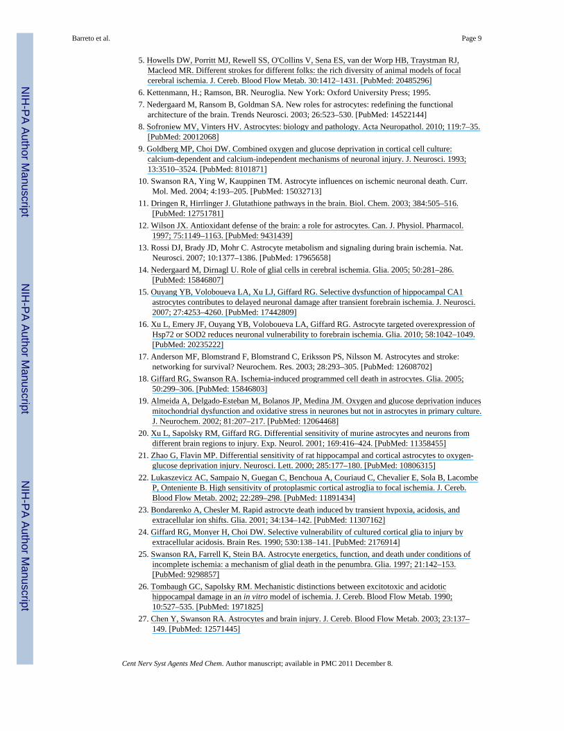
Fig. (1).Expression of different astrocytic proteins following stroke. Increased expression of GFAPis a hallmark of astrocytes activation, as is induction/re-expression of vimentin. Astrocytesnormally express glutamine synthetase (GS) and S100β, genes that are widely expressed inboth reactive and non-reactive astrocytes. The GFAP and S100β labelling are for the samecell, while the Vim and GS staining labels are for of other cells. Scale bar, 50µm.
Barreto et al. Page 18
Cent Nerv Syst Agents Med Chem. Author manuscript; available in PMC 2011 December 8.
NIH
-PA Author Manuscript
NIH
-PA Author Manuscript
NIH
-PA Author Manuscript

Fig. (2).Mechanisms of astrocyte support of neurons important in stroke. Antioxidant defenseincludes release of glutathione and ascorbate. Regulation of extracellular levels of ions andneuro-transmitters, especially K+ and glutamate, strongly influences neuronal excitability.Elevated extracellular K+ triggers astrocyte glycolysis and enhances lactate and pyruvaterelease which support neuronal metabolism. Sodium dependent glutamate uptake byastrocytes activates the Na+/K+ ATPase, stimulating glycolytic activity and production oflactate. Astrocytes and neurons are also coupled by the glutamate-glutamine cycle.Astrocytes take up glutamate, convert it to glutamine, and release glutamine to theextracellular space where it is taken up by neurons and used to synthesize glutamate toreplenish the neurotransmitter pool.
Barreto et al. Page 19
Cent Nerv Syst Agents Med Chem. Author manuscript; available in PMC 2011 December 8.
NIH
-PA Author Manuscript
NIH
-PA Author Manuscript
NIH
-PA Author Manuscript

Fig. (3).Targeted over-expression of Hsp72 in astrocytes reduces the vulnerability of CA1 neuronsto forebrain ischemia. Selective overexpression of Hsp72 in astrocytes by expressing it fromthe astrocyte specific GFAP promoter was achieved by unilateral stereotaxic injection of theexpression plasmid just above the CA1 region of the hippocampus (black arrows formicroinjection tracks) 2 days before rats were subjected to 15 min forebrain ischemia.Selective loss of CA1 hippocampal neurons (between white arrows in middle panel) wasobserved at 7 days reperfusion by cresyl violet staining. The loss of CA1 hippocampalneurons was significantly reduced with astrocytic Hsp72 overex-pression (right panel)compared to the neuronal loss seen with injection of control DNA (middle panel). Modifiedfrom [16].
Barreto et al. Page 20
Cent Nerv Syst Agents Med Chem. Author manuscript; available in PMC 2011 December 8.
NIH
-PA Author Manuscript
NIH
-PA Author Manuscript
NIH
-PA Author Manuscript

NIH
-PA Author Manuscript
NIH
-PA Author Manuscript
NIH
-PA Author Manuscript
Barreto et al. Page 21
Table 1
Overview of Some Completed and Ongoing Clinical Trials for Stroke
Mechanism and Compound
Anticoagulants
Ancrod® (Viprinex)
Aspirin added to IV TPA
Angiotensin II Receptor Antagonist
Diovan® (valsartan)
Losartan and amlodipine
Calcium Channel Blockers
Cilnidipine
Amlodipine
Inhibiton of Myeloperoxidases
Dapsone
Antiplatelet Therapy
Eptifibatide and rt-PA
Glutamate Antagonists
YM872
Granulocyte Colony Stimulating Factor
AX200 (G-CSF)
Free Radical Scavenging
NXY-059
Sources: http://clinicaltrials.gov/; http://strokecenter.stanford.edu/trials/
Cent Nerv Syst Agents Med Chem. Author manuscript; available in PMC 2011 December 8.