Cognitive impairment and memory dysfunction after a stroke diagnosis: a post-stroke memory...
15
© 2014 Al-Qazzaz et al. This work is published by Dove Medical Press Limited, and licensed under Creative Commons Attribution – Non Commercial (unported, v3.0) License. The full terms of the License are available at http://creativecommons.org/licenses/by-nc/3.0/. Non-commercial uses of the work are permitted without any further permission from Dove Medical Press Limited, provided the work is properly attributed. Permissions beyond the scope of the License are administered by Dove Medical Press Limited. Information on how to request permission may be found at: http://www.dovepress.com/permissions.php Neuropsychiatric Disease and Treatment 2014:10 1677–1691 Neuropsychiatric Disease and Treatment Dovepress submit your manuscript | www.dovepress.com Dovepress 1677 REVIEW open access to scientific and medical research Open Access Full Text Article http://dx.doi.org/10.2147/NDT.S67184 Noor Kamal Al-Qazzaz 1,5 Sawal Hamid Ali 1 Siti Anom Ahmad 2 Shabiul Islam 3 Khairiyah Mohamad 4 1 Department of Electrical, Electronic and Systems Engineering, Faculty of Engineering and Built Environment, Universiti Kebangsaan Malaysia, Bangi, Selangor, Malaysia; 2 Department of Electrical and Electronic Engineering, Faculty of Engineering, Universiti Putra Malaysia, Serdang, Selangor, Malaysia; 3 Institute of Microengineering and Nanoelectronics (IMEN), Universiti Kebangsaan Malaysia, Bangi, Selangor, Malaysia; 4 Neurology Unit, Department of Medicine, Universiti Kebangsaan Malaysia Medical Center, Cheras, Kuala Lumpur, Malaysia; 5 Department of Biomedical Engineering, Al-Khwarizmi College of Engineering, Baghdad University, Baghdad, Iraq Correspondence: Noor Kamal Al-Qazzaz Department of Electrical, Electronic and Systems Engineering, Faculty of Engineering and Built Environment, Universiti Kebangsaan Malaysia, Bangi, Selangor 43600, Malaysia Email [email protected] Cognitive impairment and memory dysfunction after a stroke diagnosis: a post-stroke memory assessment Abstract: Cognitive impairment and memory dysfunction following stroke diagnosis are common symptoms that significantly affect the survivors’ quality of life. Stroke patients have a high potential to develop dementia within the first year of stroke onset. Currently, efforts are being exerted to assess stroke effects on the brain, particularly in the early stages. Numerous neuropsychological assessments are being used to evaluate and differentiate cognitive impairment and dementia following stroke. This article focuses on the role of available neuropsychological assessments in detection of dementia and memory loss after stroke. This review starts with stroke types and risk factors associated with dementia development, followed by a brief description of stroke diagnosis criteria and the effects of stroke on the brain that lead to cognitive impair- ment and end with memory loss. This review aims to combine available neuropsychological assessments to develop a post-stroke memory assessment (PSMA) scheme based on the most recognized and available studies. The proposed PSMA is expected to assess different types of memory functionalities that are related to different parts of the brain according to stroke loca- tion. An optimal therapeutic program that would help stroke patients enjoy additional years with higher quality of life is presented. Keywords: dementia, vascular dementia, memory, neuropsychological assessment Introduction Cognitive impairment and memory loss are common after a stroke. Approximately 30% of stroke patients develop dementia within 1 year of stroke onset. 1 Stroke affects the cognitive domain, which includes attention, memory, language, and orientation. The most affected domains are attention and executive functions; at the time of stroke diag- nosis, memory problems are often prominent. Post-stroke dementia (PSD), particularly vascular dementia (VaD), reflects the vascular risk factors that are mostly correlated with cerebral vascular disease (CVD). Post-stroke cognitive impairment is the evolu- tion of CVD that predisposes individuals to the vascular cognitive impairment (VCI) spectrum. Thus, understanding the VCI spectrum stages is necessary to evaluate the mental state of post-stroke patients, particularly the cognitive dysfunction and memory decline during the period following a stroke diagnosis. Until recently, no specific neu- ropsychological assessment to evaluate PSD including memory loss existed. Current efforts are focused on combining more than one of the available neuropsychological assessments to obtain a significant diagnosis of cognitive decline severity following a stroke. The aim of this study was to develop a post-stroke memory assessment (PSMA) based on the most popular and available neuropsychological assessments. The proposed PSMA is expected to assess different types of memory functionalities
Transcript of Cognitive impairment and memory dysfunction after a stroke diagnosis: a post-stroke memory...

© 2014 Al-Qazzaz et al. This work is published by Dove Medical Press Limited, and licensed under Creative Commons Attribution – Non Commercial (unported, v3.0) License. The full terms of the License are available at http://creativecommons.org/licenses/by-nc/3.0/. Non-commercial uses of the work are permitted without any further
permission from Dove Medical Press Limited, provided the work is properly attributed. Permissions beyond the scope of the License are administered by Dove Medical Press Limited. Information on how to request permission may be found at: http://www.dovepress.com/permissions.php
Neuropsychiatric Disease and Treatment 2014:10 1677–1691
Neuropsychiatric Disease and Treatment Dovepress
submit your manuscript | www.dovepress.com
Dovepress 1677
R e v i e w
open access to scientific and medical research
Open Access Full Text Article
http://dx.doi.org/10.2147/NDT.S67184
Noor Kamal Al-Qazzaz1,5
Sawal Hamid Ali1
Siti Anom Ahmad2
Shabiul islam3
Khairiyah Mohamad4 1Department of electrical, electronic and Systems engineering, Faculty of engineering and Built environment, Universiti Kebangsaan Malaysia, Bangi, Selangor, Malaysia; 2Department of electrical and electronic engineering, Faculty of engineering, Universiti Putra Malaysia, Serdang, Selangor, Malaysia; 3institute of Microengineering and Nanoelectronics (iMeN), Universiti Kebangsaan Malaysia, Bangi, Selangor, Malaysia; 4Neurology Unit, Department of Medicine, Universiti Kebangsaan Malaysia Medical Center, Cheras, Kuala Lumpur, Malaysia; 5Department of Biomedical engineering, Al-Khwarizmi College of engineering, Baghdad University, Baghdad, iraq
Correspondence: Noor Kamal Al-Qazzaz Department of electrical, electronic and Systems engineering, Faculty of engineering and Built environment, Universiti Kebangsaan Malaysia, Bangi, Selangor 43600, Malaysia email [email protected]
Cognitive impairment and memory dysfunction after a stroke diagnosis: a post-stroke memory assessment
Abstract: Cognitive impairment and memory dysfunction following stroke diagnosis are
common symptoms that significantly affect the survivors’ quality of life. Stroke patients have
a high potential to develop dementia within the first year of stroke onset. Currently, efforts are
being exerted to assess stroke effects on the brain, particularly in the early stages. Numerous
neuropsychological assessments are being used to evaluate and differentiate cognitive impairment
and dementia following stroke. This article focuses on the role of available neuropsychological
assessments in detection of dementia and memory loss after stroke. This review starts with stroke
types and risk factors associated with dementia development, followed by a brief description
of stroke diagnosis criteria and the effects of stroke on the brain that lead to cognitive impair-
ment and end with memory loss. This review aims to combine available neuropsychological
assessments to develop a post-stroke memory assessment (PSMA) scheme based on the most
recognized and available studies. The proposed PSMA is expected to assess different types of
memory functionalities that are related to different parts of the brain according to stroke loca-
tion. An optimal therapeutic program that would help stroke patients enjoy additional years
with higher quality of life is presented.
Keywords: dementia, vascular dementia, memory, neuropsychological assessment
IntroductionCognitive impairment and memory loss are common after a stroke. Approximately 30%
of stroke patients develop dementia within 1 year of stroke onset.1 Stroke affects the
cognitive domain, which includes attention, memory, language, and orientation. The
most affected domains are attention and executive functions; at the time of stroke diag-
nosis, memory problems are often prominent. Post-stroke dementia (PSD), particularly
vascular dementia (VaD), reflects the vascular risk factors that are mostly correlated
with cerebral vascular disease (CVD). Post-stroke cognitive impairment is the evolu-
tion of CVD that predisposes individuals to the vascular cognitive impairment (VCI)
spectrum. Thus, understanding the VCI spectrum stages is necessary to evaluate the
mental state of post-stroke patients, particularly the cognitive dysfunction and memory
decline during the period following a stroke diagnosis. Until recently, no specific neu-
ropsychological assessment to evaluate PSD including memory loss existed. Current
efforts are focused on combining more than one of the available neuropsychological
assessments to obtain a significant diagnosis of cognitive decline severity following
a stroke. The aim of this study was to develop a post-stroke memory assessment
(PSMA) based on the most popular and available neuropsychological assessments.
The proposed PSMA is expected to assess different types of memory functionalities
Journal name: Neuropsychiatric Disease and TreatmentJournal Designation: ReviewYear: 2014Volume: 10Running head verso: Al-Qazzaz et alRunning head recto: Cognitive impairment and memory dysfunction after a strokeDOI: http://dx.doi.org/10.2147/NDT.S67184
Neuropsychiatric Disease and Treatment 2014:10submit your manuscript | www.dovepress.com
Dovepress
Dovepress
1678
Al-Qazzaz et al
that are related to different parts of the brain according to
the affected memory. Results are then correlated and related
to the stroke location and severity. PSMA may provide a
promising tool for evaluating post-stroke VaD and assisting
medical doctors and clinicians in the assessment as well as
evaluation of post-stroke memory impairment severity.
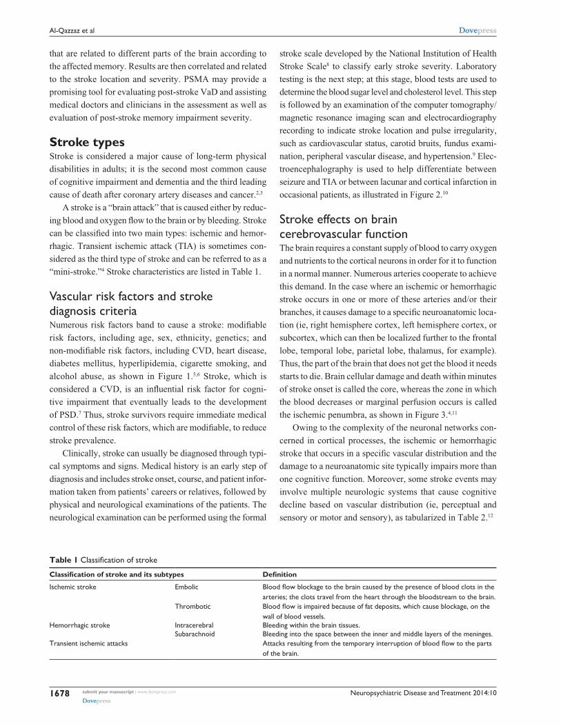
Stroke types Stroke is considered a major cause of long-term physical
disabilities in adults; it is the second most common cause
of cognitive impairment and dementia and the third leading
cause of death after coronary artery diseases and cancer.2,3
A stroke is a “brain attack” that is caused either by reduc-
ing blood and oxygen flow to the brain or by bleeding. Stroke
can be classified into two main types: ischemic and hemor-
rhagic. Transient ischemic attack (TIA) is sometimes con-
sidered as the third type of stroke and can be referred to as a
“mini-stroke.”4 Stroke characteristics are listed in Table 1.
vascular risk factors and stroke diagnosis criteriaNumerous risk factors band to cause a stroke: modifiable
risk factors, including age, sex, ethnicity, genetics; and
non-modifiable risk factors, including CVD, heart disease,
diabetes mellitus, hyperlipidemia, cigarette smoking, and
alcohol abuse, as shown in Figure 1.5,6 Stroke, which is
considered a CVD, is an influential risk factor for cogni-
tive impairment that eventually leads to the development
of PSD.7 Thus, stroke survivors require immediate medical
control of these risk factors, which are modifiable, to reduce
stroke prevalence.
Clinically, stroke can usually be diagnosed through typi-
cal symptoms and signs. Medical history is an early step of
diagnosis and includes stroke onset, course, and patient infor-
mation taken from patients’ careers or relatives, followed by
physical and neurological examinations of the patients. The
neurological examination can be performed using the formal
stroke scale developed by the National Institution of Health
Stroke Scale8 to classify early stroke severity. Laboratory
testing is the next step; at this stage, blood tests are used to
determine the blood sugar level and cholesterol level. This step
is followed by an examination of the computer tomography/
magnetic resonance imaging scan and electrocardiography
recording to indicate stroke location and pulse irregularity,
such as cardiovascular status, carotid bruits, fundus exami-
nation, peripheral vascular disease, and hypertension.9 Elec-
troencephalography is used to help differentiate between
seizure and TIA or between lacunar and cortical infarction in
occasional patients, as illustrated in Figure 2.10
Stroke effects on brain cerebrovascular functionThe brain requires a constant supply of blood to carry oxygen
and nutrients to the cortical neurons in order for it to function
in a normal manner. Numerous arteries cooperate to achieve
this demand. In the case where an ischemic or hemorrhagic
stroke occurs in one or more of these arteries and/or their
branches, it causes damage to a specific neuroanatomic loca-
tion (ie, right hemisphere cortex, left hemisphere cortex, or
subcortex, which can then be localized further to the frontal
lobe, temporal lobe, parietal lobe, thalamus, for example).
Thus, the part of the brain that does not get the blood it needs
starts to die. Brain cellular damage and death within minutes
of stroke onset is called the core, whereas the zone in which
the blood decreases or marginal perfusion occurs is called
the ischemic penumbra, as shown in Figure 3.4,11
Owing to the complexity of the neuronal networks con-
cerned in cortical processes, the ischemic or hemorrhagic
stroke that occurs in a specific vascular distribution and the
damage to a neuroanatomic site typically impairs more than
one cognitive function. Moreover, some stroke events may
involve multiple neurologic systems that cause cognitive
decline based on vascular distribution (ie, perceptual and
sensory or motor and sensory), as tabularized in Table 2.12
Table 1 Classification of stroke
Classification of stroke and its subtypes Definition
ischemic stroke embolic Blood flow blockage to the brain caused by the presence of blood clots in the arteries; the clots travel from the heart through the bloodstream to the brain.
Thrombotic Blood flow is impaired because of fat deposits, which cause blockage, on the wall of blood vessels.
Hemorrhagic stroke intracerebral Bleeding within the brain tissues.Subarachnoid Bleeding into the space between the inner and middle layers of the meninges.
Transient ischemic attacks Attacks resulting from the temporary interruption of blood flow to the parts of the brain.

Neuropsychiatric Disease and Treatment 2014:10 submit your manuscript | www.dovepress.com
Dovepress
Dovepress
1679
Cognitive impairment and memory dysfunction after a stroke
Cognitive disorder following a strokeDementia is associated with neurodegenerative disorder
diversity, neuronal dysfunction, and neuronal death. Demen-
tia occurs when the brain is affected by a specific disease or
condition that causes cognitive impairment.13 In the case of
a stroke, one or more cognitive domains may be affected,
including attention, memory, language, and orientation. The
highest impact of stroke at the time of diagnosis is on the
attention and executive functions rather than on memory,
which may be impaired at various post-stroke intervals.
Previous studies show that post-stroke memory prevalence
varies from 23% to 55% 3 months after stroke, ending with
a decline from 11% to 31% 1 year after stroke onset.3,14
Cognitive impairment after a stroke is common and leads
to PSD. PSD includes all dementia types that occur after a
stroke, including VaD; degenerative dementia, particularly
Alzheimer’s disease (AD); or mixed dementia (VaD plus
AD).2 VaD, the second leading cause of dementia in the world
after AD, occurs as a result of stroke. Between 1% and 4%
of elderly people aged 65 years and older suffer from VaD,
and its prevalence will double every 5–10 years after this
age.15,16 VaD is characterized by impairment in the cogni-
tive function due to vascular lesion and infarction resulting
from the stroke. The clinical manifestation of VaD varies
based on the size, location, and type of cerebral damage.15
Figure 4 illustrates the cognitive impairment sequences which
predispose individuals to the VCI spectrum.
The VCI spectrum can be viewed as a cognitive conse-
quence in the cognitive domain, starting from mild cognitive
impairment (MCI) and ending with severe dementia. The
period beyond dementia in which the brain is at risk is called
“cognitive impairment no dementia.”17
MCI causes a more considerable decline in cognitive
function with respect to individual age and education level,
but not notably with the activities of daily life.18,19 Clinically,
MCI is the transitional stage between early normal cognition
and late severe dementia, and it is considered heterogeneous
because some MCI patients develop dementia while others
stay and continue as MCI patients for many years. How-
ever, by default, patients diagnosed with MCI have a high
Figure 1 Risk factors and dementia.
Figure 2 Clinical evaluation.Abbreviations: CT, computed tomography; eCG, electrocardiography; eeG, electroencephalography; MRi, magnetic resonance imaging.

Neuropsychiatric Disease and Treatment 2014:10submit your manuscript | www.dovepress.com
Dovepress
Dovepress
1680
Al-Qazzaz et al
Figure 3 Core and penumbra after stroke.Note: Reprinted from Journal of Radiology Nursing, 30(3), Summers D, Malloy R, CT and MR imaging in the acute ischemic stroke patient: a nursing perspective,104–115, Copyright 2011, with permission from elsevier.56
Table 2 Stroke outcome due to vessel infarction
Brain artery infarction Stroke outcome
Left middle cerebral artery AphasiaMutismBuccofacial apraxiaAgraphiaAcalculiaideational apraxiaRight/left confusion
Right middle cerebral artery Neglect (personal, extrapersonal, and representational)visuospatial failures and visuoconstructive disordersAprosodiaLanguage usage (pragmatic language)DisordersAnosognosiaAnosodiaphoria
Posterior cerebral artery Color agnosiaAssociative visual agnosiaAlexia (hemianopic and pure)Facial agnosiaBálint’s syndromeAmnesia
Anterior cerebral artery Deficits in planning, initiation, monitoring, concentration, and flexibilityContralateral leg weaknessSensory loss
Subcortical infarcts(include thalamic infarcts)
impaired arousal, attention, motivation, initiation, and executive functionMemory (verbal, visual, episodic declarative, anterograde, and retrograde)
Caudate infarcts impaired problem solving and attentionMemory
Subcortical(infarcts of the inferior genu of the internal capsule)
ConfusionMemory disturbance
Subarachnoid hemorrhage(anterior communicating artery aneurysm)
AmnesiaPersonality changesConfabulationAbulia due to damage to the mesial frontal cortex
Limbic and paralimbic lesion implicated in a failure to learn and retain new informationAffective changes

Neuropsychiatric Disease and Treatment 2014:10 submit your manuscript | www.dovepress.com
Dovepress
Dovepress
1681
Cognitive impairment and memory dysfunction after a stroke
potential to develop dementia within the third month from
the time dementia symptoms begin to arise.2,20 The most
observed symptoms of MCI are limited to memory, but the
patient’s daily living activities are preserved.21 This article
is focused on VaD as a common cause of cognitive impair-
ment following a stroke and the effect of VaD on memory
loss. It likewise discusses the available neuropsychological
assessments that assess and predict the effect of dementia
based on the dementia spectrum as well as aids in detecting
signs of dementia, particularly memory disturbance. A num-
ber of diagnosis criteria and clinical neuropsychological
assessments are combined. The most common diagnosis
criteria are developed and characterized by the National
Institute of Neurological Disorders and Stroke and Associa-
tion Internationale pour la Recherché et l’Enseignement en
Neurosciences for VaD22–26 and Diagnostic and Statistical
Manual of Mental Disorders, Fourth Edition criteria.27 The
severity of cognitive symptoms could be assessed using the
Clinical Dementia Rating Scale.28 The most usable test to
evaluate the early dementia stages, even severity of dementia
in clinical practice, is the Mini-Mental State Examination
(MMSE).29
Brain memory and causes of memory lossThe brain memory system refers to the process of how our
brain transmits and stores available information for future
use, with or without conscious awareness. The human
brain memory system is a complex structure, with different
functionalities, as shown in Table 3. Based on stroke loca-
tion and severity, memory disorder may occur for one or
more memory types, eventually ending in memory decline
and loss.30
Figure 4 Block diagram of vascular cognitive impairment spectrum.Abbreviations: AD, Alzheimer’s disease; CvD, cerebral vascular disease; MCi, mild cognitive impairment; PSD, post-stroke dementia; vaD, vascular dementia; vCi, vascular cognitive impairment.
Table 3 Types of memory
Types of memory system Anatomy (brain lobes storage)
Long-term memory
episodic memory
Medial temporal lobe, diencephalon
Semantic memory
inferior and lateral temporal lobe
Procedural memory
Basal ganglia, cerebellum
Short-term memory
working memory
Prefrontal cortex

Neuropsychiatric Disease and Treatment 2014:10submit your manuscript | www.dovepress.com
Dovepress
Dovepress
1682
Al-Qazzaz et al
Memory loss can be caused by several factors, such as
lifestyle, brain injury, infection, thyroid dysfunction, aging,
MCI, and dementia (Table 4).31
This article focuses on stroke as the major cause of cogni-
tive impairment resulting in memory decline. The effect of
stroke varies based on its type, location, and severity.2 After a
stroke, the most prominent impairment can be recognized in the
patient’s processing speed, attention, and executive function.
Note that 20%–50% of stroke patients suffer from memory
intricacy that manifests during the period following a stroke
diagnosis. PSD, particularly VaD, causes slowing in cognitive
flexibility, perceptual disorder, and impairment information
retrieval at the time of stroke diagnosis. This period corresponds
to MCI in the VCI spectrum, followed by a decline in episodic
memory function in case of dementia, and ending in severe
dementia and impairment of all cognitive properties.32–35
Cognitive domain and memory assessment after a strokeCognitive impairment, particularly memory problems following
a stroke, can be evaluated and assessed through neuropsycho-
logical assessments. Clinically, different neuropsychological
Table 4 Brain memory loss causes
Cause of memory loss Subcases of memory loss Memory loss type
Lifestyle factors MedicationSleep pills, anti-histamine, anti-anxiety, schizophrenia medication, pain medication after surgery
Alcoholic and illicit drug useDeficiency in vitamin B1, change in chemical memory
Stressemotional trauma (chronic or short-term stress)
Sleep deprivationStress, insomnia, sleep apnea
Nutritional deficienciesLoss of vitamin B1, loss of vitamin B12
Marijuana consumption
Learning Memory consolidationLTMepisodic memory
Brain injury Acquired brain injuryTraumatic brain injury (assaults, road traffic accident, fall)
Non-traumatic brain injuryStroke (ischemic, hemorrhagic, TiA)Tumors (pediatric glial, non-glial, recurrent, metastatic, others: cysts, neurofibromatosis, pseudotumor cerebri, tuberous sclerosis)Metabolic disorder (liver disease, kidney disease, diabetes, ischemia, oxygen hypoxia to the brain, poison through ingestion or inhalation of toxic substance)
Cognitive brain injury (present at birth)Brain cognition (dementia), multiple sclerosis, Parkinson’s disease
LTM (episodic, semantic)STMworking memoryProcedural memory
infection Hiv, tuberculosis, syphilis, herpes, encephalitis, meningitis STMLTM
Thyroid dysfunction Underactive, overactive STMworking memory
Aging Dehydration, normal aging Recall memory Ability to think
Depression (common with aging) episodic memoryProcedural memoryworking memory
Mild cognitive impairment early stage of dementia working memoryDementia AD
Cortical amyloid plaques, neurofibrillary tanglesvaD
Stroke, deficiencies of (thyroid hormone, vitamin B12, folic acid), hydrocephalus, hypercalcemia
Mixed (AD + vaD), Lewy body disease, Parkinson’s disease, frontotemporal, alcoholic
episodic memorySemantic memoryworking memoryLTMSTM
Abbreviations: AD, Alzheimer’s disease; HIV, human immunodeficiency virus; LTM, long-term memory; STM, short-term memory; TIA, transient ischemic attack; vaD, vascular dementia.
Neuropsychiatric Disease and Treatment 2014:10 submit your manuscript | www.dovepress.com
Dovepress
Dovepress
1683
Cognitive impairment and memory dysfunction after a stroke
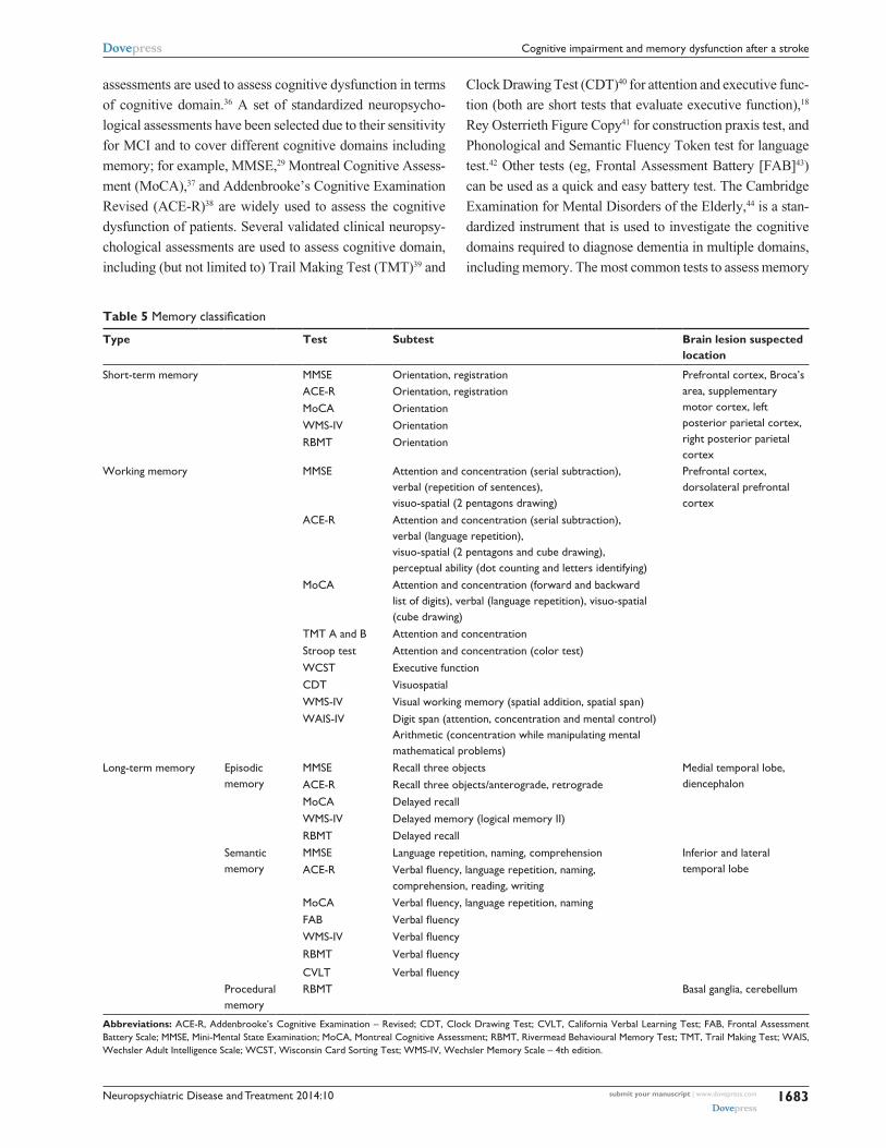
assessments are used to assess cognitive dysfunction in terms
of cognitive domain.36 A set of standardized neuropsycho-
logical assessments have been selected due to their sensitivity
for MCI and to cover different cognitive domains including
memory; for example, MMSE,29 Montreal Cognitive Assess-
ment (MoCA),37 and Addenbrooke’s Cognitive Examination
Revised (ACE-R)38 are widely used to assess the cognitive
dysfunction of patients. Several validated clinical neuropsy-
chological assessments are used to assess cognitive domain,
including (but not limited to) Trail Making Test (TMT)39 and
Clock Drawing Test (CDT)40 for attention and executive func-
tion (both are short tests that evaluate executive function),18
Rey Osterrieth Figure Copy41 for construction praxis test, and
Phonological and Semantic Fluency Token test for language
test.42 Other tests (eg, Frontal Assessment Battery [FAB]43)
can be used as a quick and easy battery test. The Cambridge
Examination for Mental Disorders of the Elderly,44 is a stan-
dardized instrument that is used to investigate the cognitive
domains required to diagnose dementia in multiple domains,
including memory. The most common tests to assess memory
Table 5 Memory classification
Type Test Subtest Brain lesion suspected location
Short-term memory MMSe Orientation, registration Prefrontal cortex, Broca’s area, supplementary motor cortex, left posterior parietal cortex, right posterior parietal cortex
ACe-R Orientation, registrationMoCA OrientationwMS-iv OrientationRBMT Orientation
working memory MMSe Attention and concentration (serial subtraction),verbal (repetition of sentences), visuo-spatial (2 pentagons drawing)
Prefrontal cortex, dorsolateral prefrontal cortex
ACe-R Attention and concentration (serial subtraction),verbal (language repetition),visuo-spatial (2 pentagons and cube drawing),perceptual ability (dot counting and letters identifying)
MoCA Attention and concentration (forward and backward list of digits), verbal (language repetition), visuo-spatial (cube drawing)
TMT A and B Attention and concentrationStroop test Attention and concentration (color test)wCST executive functionCDT visuospatial wMS-iv visual working memory (spatial addition, spatial span)wAiS-iv Digit span (attention, concentration and mental control)
Arithmetic (concentration while manipulating mental mathematical problems)
Long-term memory episodic memory
MMSe Recall three objects Medial temporal lobe, diencephalonACe-R Recall three objects/anterograde, retrograde
MoCA Delayed recallwMS-iv Delayed memory (logical memory ii)RBMT Delayed recall
Semantic memory
MMSe Language repetition, naming, comprehension inferior and lateral temporal lobeACe-R Verbal fluency, language repetition, naming,
comprehension, reading, writingMoCA Verbal fluency, language repetition, namingFAB Verbal fluencywMS-iv Verbal fluency
RBMT Verbal fluency
CvLT Verbal fluencyProcedural memory
RBMT Basal ganglia, cerebellum
Abbreviations: ACe-R, Addenbrooke’s Cognitive examination – Revised; CDT, Clock Drawing Test; CvLT, California verbal Learning Test; FAB, Frontal Assessment Battery Scale; MMSe, Mini-Mental State examination; MoCA, Montreal Cognitive Assessment; RBMT, Rivermead Behavioural Memory Test; TMT, Trail Making Test; wAiS, wechsler Adult intelligence Scale; wCST, wisconsin Card Sorting Test; wMS-iv, wechsler Memory Scale – 4th edition.

Neuropsychiatric Disease and Treatment 2014:10submit your manuscript | www.dovepress.com
Dovepress
Dovepress
1684
Al-Qazzaz et al
(Continued)
Table 6 Neuropsychological assessment characteristics
Assessments Items Subtests Maximum score CharacteristicsMMSe Orientation Orientation to place and time 10 Time 10 minutes
Registration Repeat “ball, flag, tree” 3
Calculation/wORLD
Serial 7 subtraction/wORLD backward
5
Memory recall Recall “ball, flag, tree” 3
Language naming Name of two objects (a watch and a pen)
2
Language comprehension
“Close your eyes,” “Pick up the paper in your right hand, fold it in half, and set it on the floor”
3
Language writing write a sentence 1
Language repetition Repeat “No ifs, ands, or buts” 1
visuo-spatial abilities Draw two pentagons 1
MMSe total score 30
ACe-R Orientation* Orientation to place and time 10 Time 15–20 minutes
Registration* Repeat “lemon, key, ball” 3
Calculation/wORLD*
Serial 7 subtraction/wORLD backward
5
Recall* Recall “apple, table, penny” 3
Anterograde Repeat name and address, best of three trials
7
Retrograde memory Current prime minister, last prime minister, current US president, last US president
4
Letter fluency** No of words in 1 minute 7
Category fluency No of animals in 1 minute 7
Comprehension* Obey written instruction “close your eyes,” perform three-step command
4
writing* write a sentence 1
Repetition* Repeat “hippopotamus, eccentricity, unintelligible, statistician,” “above, beyond, and below,” and “no ifs, ands, or buts”
4
Naming* Confrontation naming (12 line drawings)
12
Picture comprehension
For example, “point to the object with a nautical connection”
4
Reading Read list of five words 1
visuoexecutive* intersecting pentagons, cube, and clock drawing
8
visuoperceptual Dot counting without pointing, recognition of fragmented letters
8
Address recall Recall of name and address learned earlier
7
Address recognition Recognition of name and address items (if not recalled spontaneously)
5
ACe-R total score 100
MoCA visuoexecutive* Trail B test, cube copy, clock drawing
5 Time 10–15 minutes
Naming* Confrontation naming (lion, hippo, camel)
3

Neuropsychiatric Disease and Treatment 2014:10 submit your manuscript | www.dovepress.com
Dovepress
Dovepress
1685
Cognitive impairment and memory dysfunction after a stroke
Table 6 (Continued)
Assessments Items Subtests Maximum score CharacteristicsDigit span Forward (five digits), backward
(three digits)2
Attention Tapping at the letter A in letter list
1
Calculation* Serial 7 subtractions 3Repetition* Repetition of two complex
sentences2
Verbal fluency** .11 words beginning with the letter F in 1 minute
1
Abstraction Similarities (eg, train and bicycle = transport)
2
Recall* Recall a list of five words 5Orientation* Date, month, year, day, place, city 6MoCA total score 30
FAB Similarities Conceptualization 3 Time 10–15 minutesLexical fluency** Mental flexibility 3Motor series Programming 3Conflicting instructions
Sensitivity to interference 3
Go-No-Go inhibitory control 3Prehension behavior environmental autonomy 3FAB total score 18
Notes: *ACe-R and MoCA contain MMSe items; **MoCA and FAB same item.Abbreviations: ACe-R, Addenbrooke’s Cognitive examination – Revised; FAB, Frontal Assessment Battery Scale; MMSe, Mini-Mental State examination; MoCA, Montreal Cognitive Assessment.
evaluate memory in terms of retention, retrieval, and encoding
(eg, the Wechsler Memory Scale (WMS)-Revised45 may be
employed to distinguish amnesia from dementia in patients).
For verbal memory, numerous assessments are used, including
the WMS,46 Rey Auditory Verbal Learning Test,47 Rivermead
Behavioral Memory Test (RBMT),48 and California Verbal
Learning Test.48 Memory disorder in elderly dementia patients
can be assessed using the Free and Cued Selective Reminding
Test. This test aids in distinguishing dementia from normal
aging with acceptable accuracy.36
Until recently, no specific assessment was developed
specifically to assess short-term memory, working memory,
and long-term memory impairment following stroke VaD.
Thus, evaluating memory in terms of its types to predict
stroke effect on memory retrieval is important.
PSMAThe decline in memory as a result of stroke VaD and the
characterization of memory complaint based on VaD devel-
opment can be assessed through a PSMA. This assessment
is based on the most popular studies and is a combination of
available neuropsychological assessment tests.49,50 Memory
evaluation is proposed to be associated with memory types.
Thus, short-term memory and working memory refer to
the perceptual and learning areas of the cognitive domain,
which are processed by the frontal lobe. Episodic and
semantic long-term memory refers to memory, language,
and visuospatial domains, which are processed by the pari-
etal, medial temporal lobe, and hippocampus. Procedural
memory refers to the procedural domain and is processed
by the cerebellum and basil ganglia. Table 5 describes the
proposed PSMA, which achieves this demand. The concept
integrated the most usable neuropsychological assessments
(MMSE, ACE-R, MoCA, WMS-IV, RBMT, TMT A and
B, CDT, FAB, Wechsler Adult Intelligence Scale – Fourth
Edition, and others) and reconstructed them to evaluate
memory types.50
PSMA was designed with inspected administration time
of 30 minutes, as illustrated in the Supplementary materials.
The test examines the following:
• Orientation: in time and place
• Short-term memory: a seven-digit number, phone num-
ber, and postal code
• Working memory: attention and concentration, verbal
working memory, and visuospatial working memory
• Explicit long-term memory: episodic memory and
semantic memory
• Procedural memory.
Neuropsychiatric Disease and Treatment 2014:10submit your manuscript | www.dovepress.com
Dovepress
Dovepress
1686
Al-Qazzaz et al
Discussion Neuropsychological assessments are used in evaluating
and assessing cognitive impairment and dementia. Specific
assessment is urgently needed to evaluate different types
of memory functionalities after stroke. The present study
focused on using available neuropsychological assessments
to develop a PSMA scheme based on scientific knowledge,
which is available through neuropsychological testing.
PSMA may help provide impetus to detect the earliest stages
of dementia before significant mental decline. Therefore,
efforts are being exerted to use more than one assessment to
evaluate cognitive impairment and memory dysfunction. For
instance, the MMSE is a brief test with extensive international
usage; however, several studies have mentioned that the
MMSE alone can be used in a sensitive test to detect cognitive
impairment, except if cutoff is increased or combined with
other neuropsychological tests.51,52 Therefore, the MMSE was
used with MoCA and ACE-R to detect MCI because the last
two assessments are used to assess early stages of dementia
and executive function, as well as identify frontal subcorti-
cal infarction.50,53,54 In addition, ACE-R has good sensitivity
for dementia, whereas MoCA is specifically used in MCI
screening. Moreover, TMT, Stroop, and CDT tests can be
used with the MMSE to evaluate frontal lesion verbal fluency,
and visuospatial skills can be evaluated through Rey Oster-
rieth figure recall. FAB has been reported to identify frontal
temporal lobe dysfunction.55 MMSE, ACE-R, MoCA, and
FAB characteristics are shown in Table 6. It can be noticed
from the table that the administration time ranged from
35–45 minutes for four assessments. The PSMA administra-
tion time was reduced approximately to 30 minutes. PSMA
has been designed to incorporate more than one neuropsy-
chological assessment to evaluate short-term, working, and
long-term memory with less time consumed compared with
multiple test usage. Using more than one assessment to evalu-
ate patient mentality takes a longer time, resulting in patient
difficulty in concentrating on the assessment items. PSMA
evaluates the cognitive domain and focuses on memory types
that are affected by VaD.
Conclusion Currently, no specific neuropsychological assessment to
assess memory in terms of its types exists. This article pro-
vides an overview of the effects of stroke on the brain and on
cognitive impairment, including memory evaluation with the
most commonly used neuropsychological tests. The article
proposes a PSMA to assess different types of memory based
on the available assessments. It likewise uses the widely
available neuropsychological assessments to study the asso-
ciation between memory as a part of cognitive domain and
cognitive impairment, which lead to memory decline in the
period following stroke onset.
DisclosureThe authors declare that there are no conflicts of interest in
this work.
References 1. Cullen B, O’Neill B, Evans JJ, Coen RF, Lawlor BA. A review of screen-
ing tests for cognitive impairment. J Neurol Neurosurg Psychiatry. 2007;78(8):790–799.
2. Leys D, Hénon H, Mackowiak-Cordoliani M-A, Pasquier F. Poststroke dementia. Lancet Neurol. 2005;4(11):752–759.
3. Cumming TB, Marshall RS, Lazar RM. Stroke, cognitive deficits, and reha-bilitation: still an incomplete picture. Int J Stroke. 2013;8(1):38–45.
4. Mohr JP. Stroke: Pathophysiology, Diagnosis, and Management. Elsevier Health Sciences; 2004.
5. Sahathevan R, Brodtmann A, Donnan GA. Dementia, stroke, and vascular risk factors: a review. Int J Stroke. 2012;7(1):61–73.
6. Iemolo F, Duro G, Rizzo C, Castiglia L, Hachinski V, Caruso C. Pathophysiology of vascular dementia. Immun Ageing. 2009;6(1):13.
7. Sibolt G, Curtze S, Melkas S, et al. Poststroke dementia is associated with recurrent ischaemic stroke. J Neurol Neurosurg Psychiatry. 2013; 84(7):722–726.
8. Brott T, Adams H, Olinger CP, et al. Measurements of acute cerebral infarction: a clinical examination scale. Stroke. 1989;20(7):864–870.
9. Demarin V, Zavoreo I, Kes VB. Carotid artery disease and cognitive impairment. J Neurol Sci. 2012;322(1–2):107–111.
10. Sacco RL, Adams R, Albers G, et al; American Heart Association/American Stroke Association Council on Stroke; Council on Cardio-vascular Radiology and Intervention; American Academy of Neurology. Guidelines for prevention of stroke in patients with ischemic stroke or transient ischemic attack: a statement for healthcare professionals from the American Heart Association/American Stroke Association Council on Stroke: co-sponsored by the Council on Cardiovascular Radiology and Intervention: the American Academy of Neurology affirms the value of this guideline. Circulation. 2006;113(10):e409–e449.
11. Foreman B, Claassen J. Quantitative EEG for the detection of brain ischemia. Crit Care. 2012;16(2):216.
12. Donovan NJ, Kendall DL, Heaton SC, Kwon S, Velozo CA, Duncan PW. Conceptualizing functional cognition in stroke. Neurorehabil Neural Repair. 2008;22(2):122–135.
13. Borson S, Frank L, Bayley PJ, et al. Improving dementia care: the role of screening and detection of cognitive impairment. Alzheimers Dement. 2013;9(2):151–159.
14. Snaphaan L, de Leeuw F-E. Poststroke memory function in nonde-mented patients: a systematic review on frequency and neuroimaging correlates. Stroke. 2007;38(1):198–203.
15. McVeigh C, Passmore P. Vascular dementia: prevention and treatment. Clin Interv Aging. 2006;1(3):229.
16. Ruitenberg A, Ott A, van Swieten JC, Hofman A, Breteler MM. Inci-dence of dementia: does gender make a difference? Neurobiol Aging. 2001;22:575–580.
17. Jacova C, Kertesz A, Blair M, Fisk JD, Feldman HH. Neuropsychologi-cal testing and assessment for dementia. Alzheimer Dement. 2007;3(4): 299–317.
18. Korczyn AD, Vakhapova V, Grinberg LT. Vascular dementia. J Neurol Sci. 2012;322(1–2):2–10.
19. Ankolekar S, Geeganage C, Anderton P, Hogg C, Bath PM. Clinical trials for preventing post stroke cognitive impairment. J Neurol Sci. 2010;299(1–2):168–174.
Neuropsychiatric Disease and Treatment 2014:10 submit your manuscript | www.dovepress.com
Dovepress
Dovepress
1687
Cognitive impairment and memory dysfunction after a stroke
20. Winblad B, Palmer K, Kivipelto M, et al. Mild cognitive impairment – beyond controversies, towards a consensus: report of the International Working Group on Mild Cognitive Impairment. J Intern Med. 2004; 256(3):240–246.
21. Andrade C, Radhakrishnan R. The prevention and treatment of cognitive decline and dementia: an overview of recent research on experimental treatments. Indian J Psychiatry. 2009;51(1):12–25.
22. Sheng B, Cheng LF, Law CB, Li HL, Yeung KM, Lau KK. Coexist-ing cerebral infarction in Alzheimer’s disease is associated with fast dementia progression: applying the National Institute for Neurological Disorders and Stroke/Association Internationale pour la Recherche et l’Enseignement en Neurosciences Neuroimaging Criteria in Alzheimer’s Disease with Concomitant Cerebral Infarction. J Am Geriatr Soc. 2007;55(6):918–922.
23. Jack CR Jr, Albert M, Knopman DS, et al. Introduction to revised criteria for the diagnosis of Alzheimer’s disease: National Institute on Aging and the Alzheimer Association workgroups. Alzheimers Dement. 2011; 7(3):257–262.
24. Albert MS, DeKosky ST, Dickson D, et al. The diagnosis of mild cog-nitive impairment due to Alzheimer’s disease: recommendations from the National Institute on Aging-Alzheimer’s Association workgroups on diagnostic guidelines for Alzheimer’s disease. Alzheimers Dement. 2011;7(3):270–279.
25. McKhann GM, Knopman DS, Chertkow H, et al. The diagnosis of dementia due to Alzheimer’s disease: Recommendations from the National Institute on Aging-Alzheimer’s Association workgroups on diagnostic guidelines for Alzheimer’s disease. Alzheimers Dement. 2011;7(3):263–269.
26. Sperling RA, Aisen PS, Beckett LA, et al. Toward defining the pre-clinical stages of Alzheimer’s disease: recommendations from the National Institute on Aging-Alzheimer’s Association workgroups on diagnostic guidelines for Alzheimer’s disease. Alzheimers Dement. 2011;7(3):280–292.
27. Association AP. Diagnostic and statistical manual of mental disorders fourth edition. Washington, DC: American Psychiatric Association; 1994.
28. Hughes CP, Berg L, Danziger WL, Coben LA, Martin RL. A new clinical scale for the staging of dementia. Br J Psychiatry. 1982;140(6): 566–572.
29. Folstein MF, Folstein SE, McHugh PR. Mini-mental state. A prac-32. 1998.
30. D’Esposito M. Chapter 11. Working memory. In: Goldenberg G, Miller BL, editors. Handbook of Clinical Neurology. Volume 88. Elsevier; 2008:237–247.
31. Buckner RL. Memory and executive function in aging and AD: multiple factors that cause decline and reserve factors that compensate. Neuron. 2004;44(1):195–208.
32. Lim C, Alexander MP. Stroke and episodic memory disorders. Neu-ropsychologia. 2009;47(14):3045–3058.
33. Snaphaan L, Rijpkema M, van Uden I, Fernandez G, de Leeuw FE. Reduced medial temporal lobe functionality in stroke patients: a functional magnetic resonance imaging study. Brain. 2009;132(Pt 7): 1882–1888.
34. Planton M, Peiffer S, Albucher JF, et al. Neuropsychological outcome after a first symptomatic ischaemic stroke with “good recovery”. Eur J Neurol. 2012;19(2):212–219.
35. Cooper S, Greene JD. The clinical assessment of the patient with early dementia. J Neurol Neurosurg Psychiatry. 2005;76(Suppl 5):v15–v24.
36. Pasquier F. Early diagnosis of dementia: neuropsychology. J Neurol. 1999;246(1):6–15.
37. Smith T, Gildeh N, Holmes C. The Montreal Cognitive Assessment: validity and utility in a memory clinic setting. Can J Psychiatry. 2007; 52(5):329–332.
38. Mathuranath P, Nestor P, Berrios G, Rakowicz W, Hodges J. A brief cognitive test battery to differentiate Alzheimer’s disease and fronto-temporal dementia. Neurology. 2000;55(11):1613–1620.
39. Amodio P, Wenin H, Del Piccolo F, et al. Variability of trail making test, symbol digit test and line trait test in normal people. A normative study taking into account age-dependent decline and sociobiological variables. Aging Clin Exp Res. 2002;14(2):117–131.
40. Shulman KI. Clock-drawing: is it the ideal cognitive screening test? Int J Geriatr Psychiatry. 2000;15(6):548–561.
41. Caffarra P, Vezzadini G, Dieci F, Zonato F, Venneri A. Rey-Osterrieth complex figure: normative values in an Italian population sample. Neurol Sci. 2002;22(6):443–447.
42. Carlesimo G, Caltagirone C, Gainotti G, et al. The Mental Deterioration Battery: normative data, diagnostic reliability and qualitative analyses of cognitive impairment. Eur Neurol. 1996;36(6):378–384.
43. Dubois B, Slachevsky A, Litvan I, Pillon B. The FAB: a frontal assess-ment battery at bedside. Neurology. 2000;55(11):1621–1626.
44. Roth M. CAMDEX-R: the Cambridge examination for mental disorders of the elderly. Cambridge University Press; 1998.
45. Wechsler D. Wechsler Memory Scale – Revised. San Antonio, TX: Psychological Corporation; 1987.
46. Wechsler D. Wechsler memory scale. New York: Psychological Cor-poration; 1945.
47. Osterrieth PA. Le test de copie d’une figure complexe. Arch Psychol. 1944;30:206–356.
48. Wilson BA, Cockburn J, Baddeley AD. The Rivermead Behavioural Memory Test. Suffolk, UK: Thames Valley Test Company; 1991.
49. Pendlebury ST, Mariz J, Bull L, Mehta Z, Rothwell PM. MoCA, ACE-R, and MMSE Versus the National Institute of Neurological Disorders and Stroke – Canadian Stroke Network Vascular Cognitive Impairment Harmonization Standards Neuropsychological Battery After TIA and Stroke. Stroke. 2012;43(2):464–469.
50. Bagnoli S, Failli Y, Piaceri I, et al. Suitability of neuropsychological tests in patients with vascular dementia (VaD). J Neurol Sci. 2012; 322(1–2):41–45.
51. Moulaert VR, Verbunt JA, van Heugten CM, Wade DT. Cognitive impairments in survivors of out-of-hospital cardiac arrest: a systematic review. Resuscitation. 2009;80(3):297–305.
52. Cao M, Ferrari M, Patella R, Marra C, Rasura M. Neuropsychological findings in young-adult stroke patients. Arch Clin Neuropsychol. 2007; 22(2):133–142.
53. Sikaroodi H, Yadegari S, Miri SR. Cognitive impairments in patients with cerebrovascular risk factors: a comparison of Mini Mental Status Exam and Montreal Cognitive Assessment. Clin Neurol Neurosurg. 2013;115(8):1276–1280.
54. Kandiah N, Wiryasaputra L, Narasimhalu K, et al. Frontal subcortical ischemia is crucial for post stroke cognitive impairment. J Neurol Sci. 2011;309(1–2):92–95.
55. Bagnoli S, Failli Y, Piaceri I, et al. Suitability of neuropsychological tests in patients with vascular dementia (VaD). J Neurol Sci. 2012; 322(1):41–45.
56. Summers D, Malloy R. CT and MR imaging in the acute ischemic stroke patient: a nursing perspective. J Radiol Nurs. 2011;30(3):104–115.

Neuropsychiatric Disease and Treatment 2014:10submit your manuscript | www.dovepress.com
Dovepress
Dovepress
1688
Al-Qazzaz et al
Supplementary materials
Post-stroke memory assessment
Name:Sex:
Date of test: Date of stroke diagnosis:
Handedness: Date of birth:
Site of stroke:Type of stroke:
Education:Occupation:
Tester’s name:
Nationality: Level of education:Did not go to school Primary educationSecondary education DiplomaDegree Post graduate
Orientation: Note: The score is added to short-term memory.What is the: AM/PM Day Date Month Year /5
Which: Building Floor Town State Country /5
1. Short-term memory: Total score: /20i. Registration: /7
i am going to read a list of 7 words. i want you to repeat after me:Ball, Flag, Tree, Yellow, Apple, Tiger, Office
ii. 7 digits number: /1i am going to say a series of numbers for you to remember. When I am finished, I want you to select which group I said:7 digits number: 8-6-0-2-9-1-71. 8-6-0-5-9-1-72. 8-0-6-2-4-1-73. 8-6-0-2-9-1-74. 8-6-2-0-4-1-75. 8-6-0-5-4-1-76. 8-0-6-2-9-1-7
iii. The phone number and postal code: /2i am going to say a phone number and postal code for you to remember. When I am finished, I want you to repeat what I said:Postal code: 00964The phone number is: 5463279
2. Working memory: Total score: /30A. Attention and concentration: /10
i. Number forward /1i am going to read a list of 5 numbers. i want you to repeat them in the forward order.2-1-8-5-4
ii. Number backward /1i am going to read a list of 3 numbers. i want you to repeat them in the backward order.7-4-2
iii. Serial subtraction /3Serial 7 subtractions are starting at 100.[ ] 93 [ ] 86 [ ] 79 [ ] 72 [ ] 65 [ ] 58 [ ] 51Notes: 4 or 5 correct subtractions: 3 pts, 2 or 3 correct: 2 pts, 1 correct: 1 pt, 0 correct: 0 pt.
iv. Alternating trail making /1Join the following circles as in the example.
(Continued)

Neuropsychiatric Disease and Treatment 2014:10 submit your manuscript | www.dovepress.com
Dovepress
Dovepress
1689
Cognitive impairment and memory dysfunction after a stroke
v. Color test: /2i am going to show you a list of color words that are printed in ink colors unrelated to the printed words. i want you to name the color of the printed word not to read the word.
Red Blue
Green Yellow
vi. Perceptual abilities (A): /1– i am going to show you a square of stars; i want you to count the stars group that i am pointing.
vii. Perceptual abilities (B): /1– i am going to show you a group of letters. i want you to identify the letters.
B. Verbal working memory: /10i. Memory of sentences: /2
i am going to read a sentence; i want you to repeat it.The boy rode a horse at the zooii. Memory of stories: /2
i am going to read a story. i want you to retell as much of the story as possible.The Bank RobberyThree armed men burst through the doors of the bank at Hillstone on Tuesday afternoon, just after half past two. They ordered a frightened 19-year-old teller to fill the six large, red suitcases they carried with money. When the bags were filled, the three men ran to a green, late-model station wagon and drove off along Mark Street.iii. Reading span: /2
You will read a series of sentences and then, sequentially, I want you recall the final word in each sentence.1. The pencil is above the flower2. The teacher will read the story after lunch
iv. Listening span: /2I am going to read a series of sentences and then you will recall the final word of each sentence.1. My friend got a rabbit for her birthday2. The cup is in the box
v. Operation span: /2i am going to show you a simple math problem you try to solve it:(12÷2)-4=?
C. Visuospatial working memory: /10i. Cube: /1
i am going to show you a cube; i want you to copy the diagram.
(Continued)
Neuropsychiatric Disease and Treatment 2014:10submit your manuscript | www.dovepress.com
Dovepress
Dovepress
1690
Al-Qazzaz et al
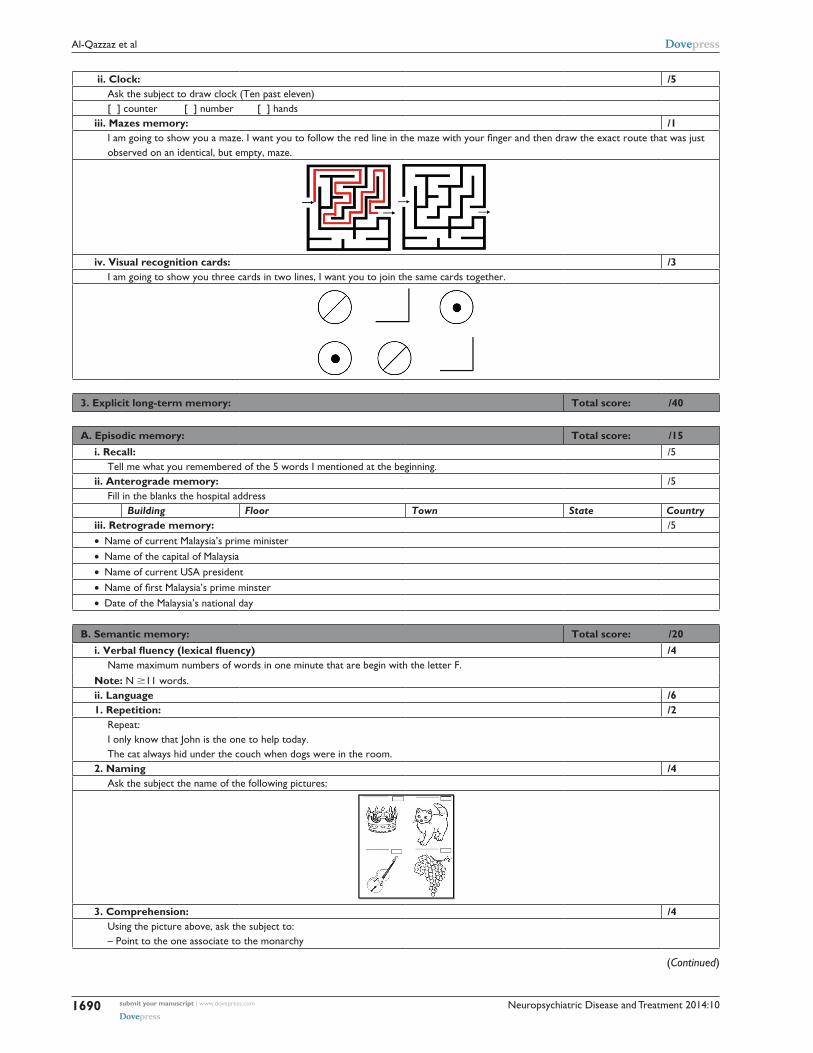
ii. Clock: /5Ask the subject to draw clock (Ten past eleven)[ ] counter [ ] number [ ] hands
iii. Mazes memory: /1I am going to show you a maze. I want you to follow the red line in the maze with your finger and then draw the exact route that was just observed on an identical, but empty, maze.
iv. Visual recognition cards: /3i am going to show you three cards in two lines, i want you to join the same cards together.
3. Explicit long-term memory: Total score: /40
A. Episodic memory: Total score: /15i. Recall: /5
Tell me what you remembered of the 5 words i mentioned at the beginning.ii. Anterograde memory: /5
Fill in the blanks the hospital addressBuilding Floor Town State Country
iii. Retrograde memory: /5• Name of current Malaysia’s prime minister• Name of the capital of Malaysia• Name of current USA president• Name of first Malaysia’s prime minster• Date of the Malaysia’s national day
B. Semantic memory: Total score: /20i. Verbal fluency (lexical fluency) /4
Name maximum numbers of words in one minute that are begin with the letter F.Note: N 11 words.ii. Language /61. Repetition: /2
Repeat: i only know that John is the one to help today.The cat always hid under the couch when dogs were in the room.
2. Naming /4Ask the subject the name of the following pictures:
3. Comprehension: /4Using the picture above, ask the subject to:– Point to the one associate to the monarchy
(Continued)

Neuropsychiatric Disease and Treatment
Publish your work in this journal
Submit your manuscript here: http://www.dovepress.com/neuropsychiatric-disease-and-treatment-journal
Neuropsychiatric Disease and Treatment is an international, peer-reviewed journal of clinical therapeutics and pharmacology focusing on concise rapid reporting of clinical or pre-clinical studies on a range of neuropsychiatric and neurological disorders. This journal is indexed on PubMed Central, the ‘PsycINFO’ database and CAS,
and is the official journal of The International Neuropsychiatric Association (INA). The manuscript management system is completely online and includes a very quick and fair peer-review system, which is all easy to use. Visit http://www.dovepress.com/testimonials.php to read real quotes from published authors.
Dovepress
Neuropsychiatric Disease and Treatment 2014:10 submit your manuscript | www.dovepress.com
Dovepress
Dovepress
1691
Cognitive impairment and memory dysfunction after a stroke
– Point to the one which is a mammal– Point to the one which is a musical instrument– Point to the one which is a fruit
4. Reading: /2Ask the subject to read the following words:Sew, Pint, Soot, Height
5. Writing: /4i am going to read words; i want you to write the following:Blue, England, Yellow, Italy
4. Procedural memory: Total score: /10Ask the subject to:
• Dial a phone number• Fold the paper into two sides
Delayed recall episodic memory: Total score: /5Ask “Now tell me what you remember of the 5 words I mentioned at the beginning”.
Sub scores Total scoresShort-term memory
Orientation /10 /20Registration /77 digits number /1Phone no and postal code /2
Workingmemory
Attention and concentration
/10 /30
Verbal WM /10Visuospatial WM /10
Long-termmemory
Episodic memory /15 /35Semantic memory /20
Procedural memory
Dial a phone number /5 /10Fold the paper into two sides /5
Episodic Delay recall /5 /5