aging associated memory impairment in mice - PLOS
27
RESEARCH ARTICLE Curcumin improves D-galactose and normal- aging associated memory impairment in mice: In vivo and in silico-based studies Md. Ashrafur Rahman ID 1,2 *, Arif Anzum Shuvo ID 1 , Asim Kumar Bepari ID 1 , Mehedi Hasan ApuID 1 , Manik Chandra Shill 1 , Murad Hossain 1 , Mohammed Uddin 3,4 , Md. Rabiul IslamID 5 , Monjurul Kader Bakshi 1 , Javed Hasan 1 , Atiqur Rahman 1 , Ghazi Mohammad Sayedur Rahman 1 , Hasan Mahmud Reza 1 * 1 Department of Pharmaceutical Sciences, North South University, Bashundhara, Dhaka, Bangladesh, 2 Department of Pharmaceutical Sciences, Jerry H. Hodge School of Pharmacy, Texas Tech University Health Science Center (TTUHSC), Amarillo, TX, United States of America, 3 College of Medicine, Mohammed Bin Rashid University of Medicine and Health Sciences, Dubai, UAE, 4 Cellular Intelligence (Ci) Lab, GenomeArc Inc., Toronto, ON, Canada, 5 Department of Pharmacy, University of Asia Pacific, Dhaka, Bangladesh * [email protected] (MAR); [email protected] (HMR) Abstract Aging-induced memory impairment is closely associated with oxidative stress. D-Galactose (D-gal) evokes severe oxidative stress and mimics normal aging in animals. Curcumin, a natu- ral flavonoid, has potent antioxidant and anti-aging properties. There are several proteins like glutathione S-transferase A1 (GSTA1), glutathione S-transferase omega-1 (GSTO1), kelch- like ECH-associated protein 1 (KEAP1), beta-secretase 1 (BACE1), and amine oxidase [fla- vin-containing] A (MAOA) are commonly involved in oxidative stress and aging. This study aimed to investigate the interaction of curcumin to these proteins and their subsequent effect on aging-associated memory impairment in two robust animal models: D-Gal and normal aged (NA) mice. The aging mice model was developed by administering D-gal intraperitoneally (i.p). Mice (n = 64) were divided into the eight groups (8 mice in each group): Vehicle, Curcumin- Control, D-gal (100mg/kg; i.p), Curcumin + D-gal, Astaxanthin (Ast) + D-gal, Normal Aged (NA), Curcumin (30mg/kg Orally) + NA, Ast (20mg/kg Orally) + NA. Retention and freezing memories were assessed by passive avoidance (PA) and contextual fear conditioning (CFC). Molecular docking was performed to predict curcumin binding with potential molecular targets. Curcumin significantly increased retention time (p < 0.05) and freezing response (p < 0.05) in PA and CFC, respectively. Curcumin profoundly ameliorated the levels of glutathione, super- oxide dismutase, catalase, advanced oxidation protein products, nitric oxide, and lipid peroxi- dation in mice hippocampi. In silico studies revealed favorable binding energies of curcumin with GSTA1, GSTO1, KEAP1, BACE1, and MAOA. Curcumin improves retention and freezing memory in D-gal and nature-induced aging mice. Curcumin ameliorates the levels of oxidative stress biomarkers in mice. Anti-aging effects of curcumin could be attributed to, at least par- tially, the upregulation of antioxidant enzymes through binding with GSTA1, GSTO1, KEAP1, and inhibition of oxidative damage through binding with BACE1 and MAOA. PLOS ONE PLOS ONE | https://doi.org/10.1371/journal.pone.0270123 June 29, 2022 1 / 27 a1111111111 a1111111111 a1111111111 a1111111111 a1111111111 OPEN ACCESS Citation: Rahman M.A, Shuvo AA, Bepari AK, Hasan Apu M, Shill MC, Hossain M, et al. (2022) Curcumin improves D-galactose and normal-aging associated memory impairment in mice: In vivo and in silico-based studies. PLoS ONE 17(6): e0270123. https://doi.org/10.1371/journal. pone.0270123 Editor: Michelle Melgarejo da Rosa, UFPE: Universidade Federal de Pernambuco, BRAZIL Received: September 23, 2021 Accepted: June 3, 2022 Published: June 29, 2022 Peer Review History: PLOS recognizes the benefits of transparency in the peer review process; therefore, we enable the publication of all of the content of peer review and author responses alongside final, published articles. The editorial history of this article is available here: https://doi.org/10.1371/journal.pone.0270123 Copyright: © 2022 Rahman et al. This is an open access article distributed under the terms of the Creative Commons Attribution License, which permits unrestricted use, distribution, and reproduction in any medium, provided the original author and source are credited. Data Availability Statement: We have uploaded a file (defined as Data set_ Curcumin_Plos one) in
-
Upload
khangminh22 -
Category
Documents
-
view
1 -
download
0
Transcript of aging associated memory impairment in mice - PLOS
RESEARCH ARTICLE
Curcumin improves D-galactose and normal-
aging associated memory impairment in mice:
In vivo and in silico-based studies
Md. Ashrafur RahmanID1,2*, Arif Anzum ShuvoID
1, Asim Kumar BepariID1, Mehedi Hasan
ApuID1, Manik Chandra Shill1, Murad Hossain1, Mohammed Uddin3,4, Md. Rabiul IslamID
5,
Monjurul Kader Bakshi1, Javed Hasan1, Atiqur Rahman1, Ghazi Mohammad
Sayedur Rahman1, Hasan Mahmud Reza1*
1 Department of Pharmaceutical Sciences, North South University, Bashundhara, Dhaka, Bangladesh,
2 Department of Pharmaceutical Sciences, Jerry H. Hodge School of Pharmacy, Texas Tech University
Health Science Center (TTUHSC), Amarillo, TX, United States of America, 3 College of Medicine,
Mohammed Bin Rashid University of Medicine and Health Sciences, Dubai, UAE, 4 Cellular Intelligence (Ci)
Lab, GenomeArc Inc., Toronto, ON, Canada, 5 Department of Pharmacy, University of Asia Pacific, Dhaka,
Bangladesh
* [email protected] (MAR); [email protected] (HMR)
Abstract
Aging-induced memory impairment is closely associated with oxidative stress. D-Galactose
(D-gal) evokes severe oxidative stress and mimics normal aging in animals. Curcumin, a natu-
ral flavonoid, has potent antioxidant and anti-aging properties. There are several proteins like
glutathione S-transferase A1 (GSTA1), glutathione S-transferase omega-1 (GSTO1), kelch-
like ECH-associated protein 1 (KEAP1), beta-secretase 1 (BACE1), and amine oxidase [fla-
vin-containing] A (MAOA) are commonly involved in oxidative stress and aging. This study
aimed to investigate the interaction of curcumin to these proteins and their subsequent effect
on aging-associated memory impairment in two robust animal models: D-Gal and normal aged
(NA) mice. The aging mice model was developed by administering D-gal intraperitoneally (i.p).
Mice (n = 64) were divided into the eight groups (8 mice in each group): Vehicle, Curcumin-
Control, D-gal (100mg/kg; i.p), Curcumin + D-gal, Astaxanthin (Ast) + D-gal, Normal Aged
(NA), Curcumin (30mg/kg Orally) + NA, Ast (20mg/kg Orally) + NA. Retention and freezing
memories were assessed by passive avoidance (PA) and contextual fear conditioning (CFC).
Molecular docking was performed to predict curcumin binding with potential molecular targets.
Curcumin significantly increased retention time (p < 0.05) and freezing response (p < 0.05) in
PA and CFC, respectively. Curcumin profoundly ameliorated the levels of glutathione, super-
oxide dismutase, catalase, advanced oxidation protein products, nitric oxide, and lipid peroxi-
dation in mice hippocampi. In silico studies revealed favorable binding energies of curcumin
with GSTA1, GSTO1, KEAP1, BACE1, and MAOA. Curcumin improves retention and freezing
memory in D-gal and nature-induced aging mice. Curcumin ameliorates the levels of oxidative
stress biomarkers in mice. Anti-aging effects of curcumin could be attributed to, at least par-
tially, the upregulation of antioxidant enzymes through binding with GSTA1, GSTO1, KEAP1,
and inhibition of oxidative damage through binding with BACE1 and MAOA.
PLOS ONE
PLOS ONE | https://doi.org/10.1371/journal.pone.0270123 June 29, 2022 1 / 27
a1111111111
a1111111111
a1111111111
a1111111111
a1111111111
OPEN ACCESS
Citation: Rahman M.A, Shuvo AA, Bepari AK,
Hasan Apu M, Shill MC, Hossain M, et al. (2022)
Curcumin improves D-galactose and normal-aging
associated memory impairment in mice: In vivo
and in silico-based studies. PLoS ONE 17(6):
e0270123. https://doi.org/10.1371/journal.
pone.0270123
Editor: Michelle Melgarejo da Rosa, UFPE:
Universidade Federal de Pernambuco, BRAZIL
Received: September 23, 2021
Accepted: June 3, 2022
Published: June 29, 2022
Peer Review History: PLOS recognizes the
benefits of transparency in the peer review
process; therefore, we enable the publication of
all of the content of peer review and author
responses alongside final, published articles. The
editorial history of this article is available here:
https://doi.org/10.1371/journal.pone.0270123
Copyright: © 2022 Rahman et al. This is an open
access article distributed under the terms of the
Creative Commons Attribution License, which
permits unrestricted use, distribution, and
reproduction in any medium, provided the original
author and source are credited.
Data Availability Statement: We have uploaded a
file (defined as Data set_ Curcumin_Plos one) in
1. Introduction
Aging is a natural process characterized by gradual deterioration in diverse physiological func-
tions [1], including oxidative damage-driven memory loss [2]. Memory dysfunction could be
triggered by an imbalance among reactive oxygen species (ROS), reactive nitrogen species
(RNS), and antioxidant enzyme activities [3]. The excess generation of ROS, RNS, and reduc-
tion in antioxidant enzyme activities in the brain escalate lipid peroxidation, protein oxidation,
and mitochondrial DNA (mtDNA) damage [4], contributing to brain aging [5]. Brain aging
and accompanied cognitive dysfunction are typical features of neurodegenerative diseases [6].
One study claimed that D-galactose (D-gal) accelerates the brain-aging process in animal
models similar to the normal aging in humans [7]. D-gal-induced aging mice model has
gained popularity among researchers because of its feasibility, fewer side effects, and higher
survival rate of animals. It was reported that D-gal evokes aging-induced memory impairment
through neurodegeneration, aberrant immune responses, and abnormal gene expression [8].
Another study suggests that D-gal increases malondialdehyde (MDA) level and decreases the
antioxidant enzymes such as superoxide dismutase (SOD) and glutathione (GSH) [9]. At high
doses, D-gal can accelerate cellular ROS generation through multiple mechanisms, such as
adenosine triphosphate (ATP) depletion, redox homeostasis impairment, and elevation of the
advanced glycation end product (AGE), the receptor for the advanced glycation end product
(RAGE), and nicotinamide adenine dinucleotide phosphate (NADPH) oxidase [10]. Overpro-
duction of ROS induces oxidative stress, cellular apoptosis, inflammation, and mitochondrial
dysfunction, which leads to neuronal degeneration [11]. Raised free radical levels were impli-
cated in cholinergic neuron dysfunctions in the brain [12] and aging-associated memory
impairment [13]. Currently, several acetyl-cholinesterase (AChE) inhibitors (donepezil, rivas-
tigmine, and galantamine) and an N-Methyl-D-aspartate (NMDA) receptor antagonists
(memantine) are used in aging-associated memory impairment. The outcome of the treatment
has not reached the optimum level owing to side effects and cost-ineffectiveness. Studies
showed that natural compounds could be a promising solution for aging-associated memory
impairment due to their antioxidants, anti-inflammatory, and anti-aging properties [14]. Fur-
thermore, plant-derived compounds can be a choice on account of their cost-effectiveness.
Curcumin, a natural compound found in Curcuma Longa, is regularly used as spicey ingredi-
ents and possesses anti-inflammatory, antioxidant, and anti-aging properties [15] by regulat-
ing several proteins such as tumor necrosis factor alpha (TNFα) [16], mammalian target of
rapamycin (mTOR), sirtuin, and adenosine monophosphate-activated protein kinase (AMPK)
[17]. However, several proteins like glutathione S-transferase A1 (GSTA1), glutathione S-
transferase omega-1 (GSTO1), kelch-like ECH-associated protein 1 (KEAP1), beta-secretase 1
(BACE1), and amine oxidase [flavin-containing] A (MAOA) are commonly involved in oxida-
tive stress and aging [18]. GSTA1 regulates GSH conjugation, which may reduce oxidative
stress [19]. Another protein, GSTO1, modulates conjugation of GSH [19], the activation of
interleukin-1β, and inflammation in aging-associated neurodegenerative disease [20]. KEAP1
plays an antioxidative role by regulating the Nrf2 cytoprotective signaling pathway [21].
BACE1 is closely associated with the generation of β-amyloid (Aβ) in aging-induced neurolog-
ical disease, Alzheimer’s Disease [22]. Overproduction of MAOA in the brain triggers oxida-
tive overload [23]. However, no study has detected the interaction of curcumin with
antioxidant and aging regulating proteins GSTA1, GSTO1, KEAP1, BACE1, MAOA and their
consequent impact on improving the memory. Therefore, it would be interesting to find the
binding affinity of curcumin with these proteins in order to determine their subsequent
impacts on modulating the oxidative biomarkers in the aging process. We have extensively
PLOS ONE Curcumin ameliorates ageing-induced memory impairment
PLOS ONE | https://doi.org/10.1371/journal.pone.0270123 June 29, 2022 2 / 27
the supporting information section containing all
data sets of our manuscript.
Funding: This work was partially supported by the
research grant provided by the CTRG-North South
University 2019–2020 and the National Institute of
Science and Technology (NST- 2020). The funders
had no role in study design, data collection,
analysis, decision to publish, or manuscript
preparation. No additional external funding was
received for this study.
Competing interests: Author Mohammed Uddin is
employed by Cellular Intelligence (Ci) Lab,
GenomeArc Inc., Toronto, ON, Canada. The
remaining authors declare that the research was
conducted in the absence of any commercial or
financial relationships that could be construed as a
potential conflict of interest.
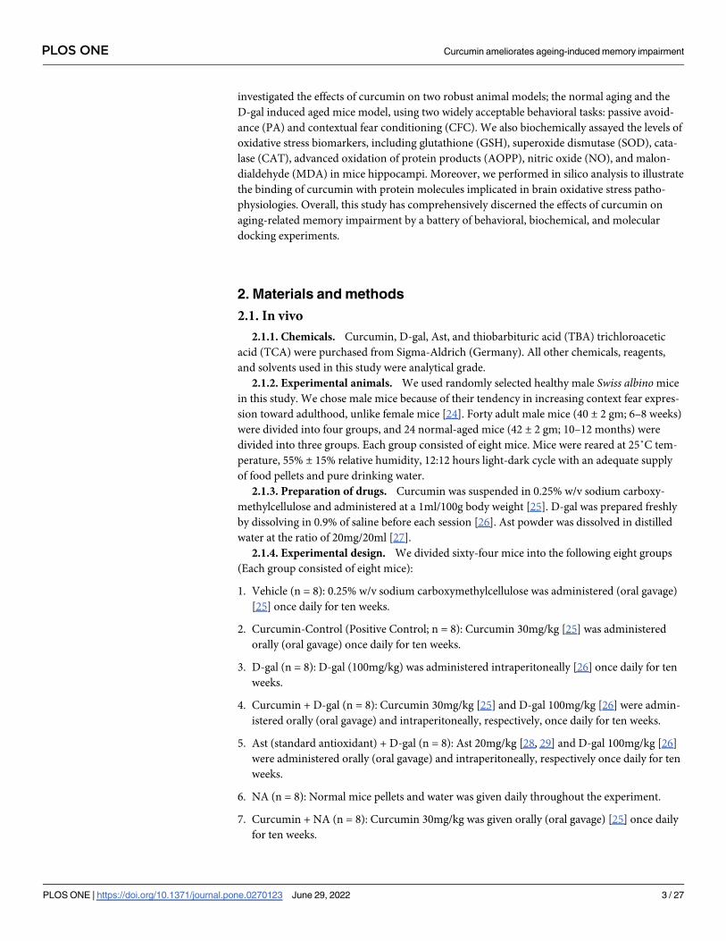
investigated the effects of curcumin on two robust animal models; the normal aging and the
D-gal induced aged mice model, using two widely acceptable behavioral tasks: passive avoid-
ance (PA) and contextual fear conditioning (CFC). We also biochemically assayed the levels of
oxidative stress biomarkers, including glutathione (GSH), superoxide dismutase (SOD), cata-
lase (CAT), advanced oxidation of protein products (AOPP), nitric oxide (NO), and malon-
dialdehyde (MDA) in mice hippocampi. Moreover, we performed in silico analysis to illustrate
the binding of curcumin with protein molecules implicated in brain oxidative stress patho-
physiologies. Overall, this study has comprehensively discerned the effects of curcumin on
aging-related memory impairment by a battery of behavioral, biochemical, and molecular
docking experiments.
2. Materials and methods
2.1. In vivo
2.1.1. Chemicals. Curcumin, D-gal, Ast, and thiobarbituric acid (TBA) trichloroacetic
acid (TCA) were purchased from Sigma-Aldrich (Germany). All other chemicals, reagents,
and solvents used in this study were analytical grade.
2.1.2. Experimental animals. We used randomly selected healthy male Swiss albino mice
in this study. We chose male mice because of their tendency in increasing context fear expres-
sion toward adulthood, unlike female mice [24]. Forty adult male mice (40 ± 2 gm; 6–8 weeks)
were divided into four groups, and 24 normal-aged mice (42 ± 2 gm; 10–12 months) were
divided into three groups. Each group consisted of eight mice. Mice were reared at 25˚C tem-
perature, 55% ± 15% relative humidity, 12:12 hours light-dark cycle with an adequate supply
of food pellets and pure drinking water.
2.1.3. Preparation of drugs. Curcumin was suspended in 0.25% w/v sodium carboxy-
methylcellulose and administered at a 1ml/100g body weight [25]. D-gal was prepared freshly
by dissolving in 0.9% of saline before each session [26]. Ast powder was dissolved in distilled
water at the ratio of 20mg/20ml [27].
2.1.4. Experimental design. We divided sixty-four mice into the following eight groups
(Each group consisted of eight mice):
1. Vehicle (n = 8): 0.25% w/v sodium carboxymethylcellulose was administered (oral gavage)
[25] once daily for ten weeks.
2. Curcumin-Control (Positive Control; n = 8): Curcumin 30mg/kg [25] was administered
orally (oral gavage) once daily for ten weeks.
3. D-gal (n = 8): D-gal (100mg/kg) was administered intraperitoneally [26] once daily for ten
weeks.
4. Curcumin + D-gal (n = 8): Curcumin 30mg/kg [25] and D-gal 100mg/kg [26] were admin-
istered orally (oral gavage) and intraperitoneally, respectively, once daily for ten weeks.
5. Ast (standard antioxidant) + D-gal (n = 8): Ast 20mg/kg [28, 29] and D-gal 100mg/kg [26]
were administered orally (oral gavage) and intraperitoneally, respectively once daily for ten
weeks.
6. NA (n = 8): Normal mice pellets and water was given daily throughout the experiment.
7. Curcumin + NA (n = 8): Curcumin 30mg/kg was given orally (oral gavage) [25] once daily
for ten weeks.
PLOS ONE Curcumin ameliorates ageing-induced memory impairment
PLOS ONE | https://doi.org/10.1371/journal.pone.0270123 June 29, 2022 3 / 27
8. Ast (standard antioxidant) + NA (n = 8): Ast (20mg/kg) was administered orally (oral
gavage) [28, 29] once daily for ten weeks.
After ten weeks of treatment, we investigated retention and freezing memory by PA and
CFC tests, respectively. The animals were then euthanized for collecting brain tissues to assay
oxidative stress biomarkers. Finally, the molecular targets of curcumin were assessed by in sil-
ico analysis (Fig 1).
2.1.5. Passive avoidance test. The method of PA was performed as demonstrated by Tab-
rizian [30]. The PA behavior study was executed with a step-through type avoidance learning
task that calculates the memory retention of mice. This test consisted of three sessions. Firstly,
each mouse was placed gently in the experimental apparatus in the habituation session and
allowed to habituate for at least 5 minutes on day 1. Mice that did not enter into the dark
chamber after keeping them in a light chamber for more than 120 s were excluded from the
experiment. Secondly, the training session was used to evaluate the learning and exploration.
This session consisted of 3 trials with 30 min intervals. In the first trial, each mouse was gently
placed in the light, and the sliding door was opened after 10 seconds. Each mouse was allowed
to explore both chambers for 5 minutes. Each mouse’s time before entering the dark chamber
with all four paws was recorded as the retention period (300 s was considered a cut-off point).
The second trial was performed in a similar way to the first trial. In the third trial, once a
mouse stepped through the dark chamber with all four paws, the sliding door was closed. An
electrical foot shock (0.3–0.5 mA for 2 s) was delivered. After the foot shock, each mouse was
kept in the dark chamber for an additional 10 s to build an association between chambers and
the foot shock, and then the mouse was returned to the home cage. Repeated training was con-
ducted similarly for five consecutive days with 24 hours intervals. The objective of this testing
session was to determine memory RT after 24 and 48 hours of training. No electrical foot-
Fig 1. Schematic diagram of total experimental procedures: The treatment was continued for ten weeks. After that, the behavioral tests were
performed using Passive Avoidance (PA) and Contextual Fear Conditioning (CFC). The biomarkers were detected after completing the
behavioral tasks. In-silico studies were used to determine curcumin’s binding affinity to targeted proteins.
https://doi.org/10.1371/journal.pone.0270123.g001
PLOS ONE Curcumin ameliorates ageing-induced memory impairment
PLOS ONE | https://doi.org/10.1371/journal.pone.0270123 June 29, 2022 4 / 27

shock was applied during this session. The retention period was measured using a stopwatch.
The apparatus was adequately cleaned with 70% ethanol before the experiment.
2.1.6. Contextual fear conditioning test. The CFC Test was performed after seven days
of completion of the PA test. The method of CFC was performed as demonstrated by Shoji
[31]. In brief, each mouse was placed in a transparent acrylic chamber (33 × 25 × 28 cm) with
a stainless-steel grid floor (0.2 cm diameter, spaced 0.5 cm apart; O’Hara & Co., Tokyo, Japan)
and was allowed to navigate independently for 2 min. Next, the conditioned stimulus (CS) of
55 dB white noise was presented for 30 s. During the last 2 s of CS presentation, the uncondi-
tioned stimulus (US) of a mild foot shock (0.3 mA, 2 s) was applied. Two more CS-US pairings
were presented with a 2-min inter-stimulus interval. A context test was conducted to detect
contextually conditioned fear memory in the same chamber approximately 24 hours (2a) and
30 days (31a) after the conditioning session. A cued test was performed to detect a novel fear
memory in an altered context. The cued test was conducted in a triangular chamber (33 × 29 ×32 cm) made of opaque white plastic located in a different room. The test was done a few
hours after the context test on day 2 (2b) and day 30 (31b). A computer-based infrared video
system (Med Associates, Inc. USA) was used to monitor the freezing in mice [32].
2.1.7. Tissue processing. Eight representative mice data of each group were used to per-
form the biochemical analyses. Mice were anesthetized by administering 200μl of ketamine
(50 mg/ml, Incepta Pharmaceuticals Ltd., Bangladesh) intraperitoneally. Mice were sacrificed
by decapitation. The brain was extracted from the skull and transferred immediately to Petri
dishes placed over ice. The hippocampal tissue was microdissected and immediately stored at
–80˚C. On the next day, the homogenate of hippocampal tissue 10% (w/v) was prepared in
sodium phosphate buffer (1× PBS pH 7.0) supplemented with 1:100 protease inhibitor cocktail
(Sigma, Saint Louis, MO, USA) by using Ultra-Turrax T25 (United States) homogenizer. Soni-
cation of homogenized tissue was performed at a 5-s cycle for 150 s using an ultrasonic proces-
sor. The homogenized tissue was centrifuged at 10,000 rpm (RCF 11200) for 10 min at 4˚C.
The clear supernatants were diluted with 0.1x PBS buffer and performed the biochemical
analysis.
2.1.8. Oxidative stress measurement. All determinations were normalized by the protein
concentration of the samples. Total protein content was measured by Lowry’s method [33].
2.1.8.1. Estimation of Glutathione level. GSH level was detected according to the previous
method [34, 35]. Briefly, 2.7 ml of phosphate buffer (0.1 M, pH 8) and 0.2 ml of 5, 5-dithiol-bis
(2-nitrobenzoic acid) were added with 1 ml of hippocampal homogenate. The color progressed
was determined instantaneously at 412 nm. Results were expressed in μmol/mg protein.
2.1.8.2. Determination of superoxide dismutase activity. The SOD level was estimated based
on a modified previous protocol [36, 37]. In short, the reaction mixture carried 50 mM sodium
phosphate (pH 7.8), 13 mM methionine, 75 mM nitroblue tetrazolium (NBT), 2 mM ribofla-
vin, 100 mM EDTA and 2 mL of hippocampal tissue homogenate. The change in absorbance
of each sample was then documented at 560 nm after forming the blue formazan. The activity
of SOD was expressed in U/30sec.
2.1.8.3. Measurement of catalase activity. The CAT activity was measured based on a previ-
ous method spectrometrically at a wavelength of 240 nm [38]. The reaction mixture (1.5 ml)
comprised 1.0 ml of 0.01 M phosphate buffer (pH 7.0), 0.1 ml of hippocampal tissue homoge-
nate, and 0.4 ml of 2 M H2O2. The reaction was stopped by adding 2.0 ml of dichromate-acetic
acid reagent (5% potassium dichromate and glacial acetic acid were mixed in a 1:3 ratio). The
activity of catalase was expressed in μmol/min/mg protein.
2.1.8.4. Determination of advanced oxidation protein products. AOPP was detected spectro-
photometrically using a previous protocol [39, 40]. Briefly, 50 μl of hippocampal tissue homog-
enate was diluted with phosphate-buffered saline (PBS) at a ratio of 1:2. Chloramine T (0–100
PLOS ONE Curcumin ameliorates ageing-induced memory impairment
PLOS ONE | https://doi.org/10.1371/journal.pone.0270123 June 29, 2022 5 / 27
mmol/L) was used for preparing the calibration curve. PBS was used as a blank. 100 μl of 1.16
M potassium iodide and 50 μl of acetic acid were added to each well, and absorbance at 340
nm was determined subsequently. Concentrations of AOPP were represented in chloramine
units (μmol/ml).
2.1.8.5. Estimation of nitric oxide level. NO level was estimated according to a previous proto-
col [41] using the Griess-Illosvoy reagent. Griess–Illosvoy reagent was modified by using
naphthyl ethylene diamine dihydrochloride (NED) (0.1% w/v) instead of 1-napthylamine (5%).
The hippocampal tissue homogenates, phosphate buffer saline (0.5 mL), NED (1 mL), and sulfa-
nilamide (1 mL) were diluted with PBS (2:8 ratio) and incubated at 25˚C for 15 min in a 96-well
plate [35]. The absorbances were measured at a wavelength of 540 nm against the blank read-
ings of the spectrophotometer. The concentration of NO was expressed in mmol/mg.
2.1.8.6. Measurement of malondialdehyde level. MDA was detected through colorimetric
analysis by determining thiobarbituric acid reactive substances (TBARS) according to a previ-
ous protocol [42]. Briefly, 0.1 ml of hippocampal tissue homogenate in Tris–HCl buffer (pH
7.5) was treated with 2 ml of TBA-TCA-HCl (1:1:1 ratio) reagent (thiobarbituric acid 0.37%,
0.25 N HCl, and 15% TCA) and put in a water bath at 70˚C for 15 min and cooled. The absor-
bance of the clear supernatant was estimated against the reference blank at 535 nm [43]. The
MDA level was detected by using a standard curve and represented in nmol/ml.
2.2. In silico (molecular docking)
2.2.1. Prediction of molecular target. We predicted the molecular targets of curcumin
from the literature search. We also used the SwissTargetPrediction (http://www.swisstarget
prediction.ch) webserver [44].
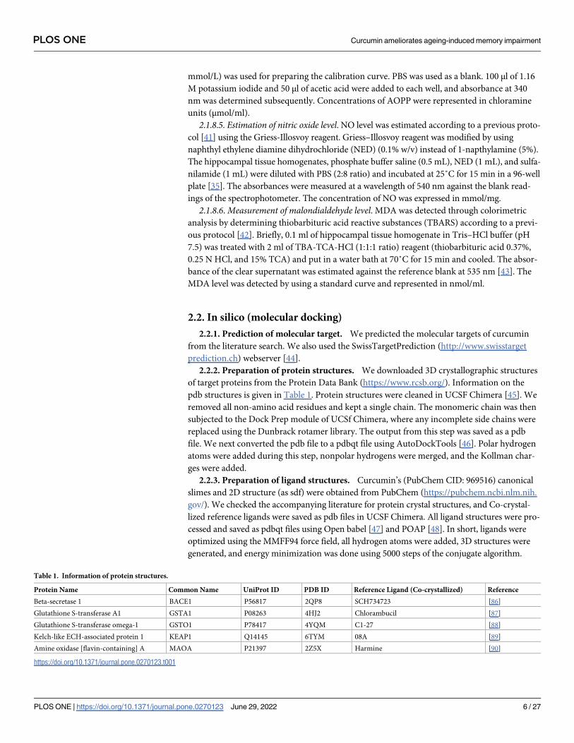
2.2.2. Preparation of protein structures. We downloaded 3D crystallographic structures
of target proteins from the Protein Data Bank (https://www.rcsb.org/). Information on the
pdb structures is given in Table 1. Protein structures were cleaned in UCSF Chimera [45]. We
removed all non-amino acid residues and kept a single chain. The monomeric chain was then
subjected to the Dock Prep module of UCSf Chimera, where any incomplete side chains were
replaced using the Dunbrack rotamer library. The output from this step was saved as a pdb
file. We next converted the pdb file to a pdbqt file using AutoDockTools [46]. Polar hydrogen
atoms were added during this step, nonpolar hydrogens were merged, and the Kollman char-
ges were added.
2.2.3. Preparation of ligand structures. Curcumin’s (PubChem CID: 969516) canonical
slimes and 2D structure (as sdf) were obtained from PubChem (https://pubchem.ncbi.nlm.nih.
gov/). We checked the accompanying literature for protein crystal structures, and Co-crystal-
lized reference ligands were saved as pdb files in UCSF Chimera. All ligand structures were pro-
cessed and saved as pdbqt files using Open babel [47] and POAP [48]. In short, ligands were
optimized using the MMFF94 force field, all hydrogen atoms were added, 3D structures were
generated, and energy minimization was done using 5000 steps of the conjugate algorithm.
Table 1. Information of protein structures.
Protein Name Common Name UniProt ID PDB ID Reference Ligand (Co-crystallized) Reference
Beta-secretase 1 BACE1 P56817 2QP8 SCH734723 [86]
Glutathione S-transferase A1 GSTA1 P08263 4HJ2 Chlorambucil [87]
Glutathione S-transferase omega-1 GSTO1 P78417 4YQM C1-27 [88]
Kelch-like ECH-associated protein 1 KEAP1 Q14145 6TYM 08A [89]
Amine oxidase [flavin-containing] A MAOA P21397 2Z5X Harmine [90]
https://doi.org/10.1371/journal.pone.0270123.t001
PLOS ONE Curcumin ameliorates ageing-induced memory impairment
PLOS ONE | https://doi.org/10.1371/journal.pone.0270123 June 29, 2022 6 / 27
2.2.4. AutoDock Vina molecular docking. We performed Vina molecular docking [46]
using the virtual screening tool of POAP. The grid box size was 24x24x24 angstroms, and the
box was centered on the co-crystallized ligand in the pdb structure. The exhaustiveness was 8
for Vina docking. We recorded the estimated binding energy values for the best-docked poses
of the ligands.
2.2.4.1. Protein-ligand interactions. From Vina docking simulations, we retrieved pdb files
of the complexes with the best-docked poses (lowest estimated binding energy). Protein-ligand
interactions were visualized using Discovery Studio Visualizer (BIOVIA, Dassault Systèmes,
Discovery Studio Visualizer, v20.1.0.192, 2019). We compared the interactions of the co-crys-
tallized ligand in the pdb file with that of the curcumin.
2.2.4.2. 3D-rendering of protein-ligand complexes. We used open-source Pymol [49] to ren-
der 3D images of ligands and the binding sites in the proteins.
2.3. Statistical analysis
A One-Way ANOVA followed by the Brown-Forsythe test was performed to determine the
effect of the treatment groups. All analyses were executed by the GraphPad prism software.
The difference was considered significant when the p-value was less than 0.05. Data were rep-
resented as mean ± SEM (Standard Error of the Mean).
2.4. Ethics
The institutional animal care and use committee (IACUC) of North South University (2019/
OR-NSU/IACUC- No.0903) approved the study protocol. All the experimental procedures
were performed following the NIH Guide for the care and use of laboratory animals. Through-
out the experiments, all efforts were given to minimize the number of animals and to optimize
their comfort.
3. Results
3.1. Effects of curcumin on retention time by adopting passive avoidance
task
After 24 hours of training, the Vehicle and Curcumin-Control (Cur-Con) groups displayed
mean retention times (RTs) of 210.93 ± 6.33 s and 276.31 ± 8.51 s, respectively (Fig 2). The RT
was 116.43 ± 2.62 s in the D-gal injected mice group, indicating a significant decrease com-
pared with the Vehicle (F1, 14 = 2.07, p< 0.0001) and the Cur-Con (F1, 14 = 2.17, p< 0.0001)
groups (Fig 2). Contrarily, the decreasing trend of RT was prominently protected in the Cur-
cumin + D-gal mice group (255.62 ± 14.11 s) (Fig 2). A One-Way ANOVA followed by the
Brown-Forsythe test demonstrated a statistically significant difference in RT values between
the Curcumin + D-gal and the D-gal injected mice groups (F1, 14 = 88.31, p< 0.0001). An RT
value of 249.87 ± 8.97 s was observed in the Ast + D-gal mice, which was significantly higher
than the D-gal treated mice group (F1, 14 = 7.95, p> 0.05; Fig 2). A similar tendency was
observed in NA mice. The RT was 132.12 ± 3.11 s in the NA mice group (Fig 2). On the con-
trary, compared to the NA mice group, there was a significant protection from the decreasing
trend of RT observed in the Curcumin + NA (247.25 ± 10 s; F1, 14 = 31.10, p< 0.0001) and Ast
+ NA (269.81 ± 8.02 s; F1, 14 = 2.06, p< 0.0001) mice groups (Fig 2).
Similarly, after 48 hours of training, the Vehicle and Cur-Con groups exhibited RTs of
202.43 ± 3.99 s, 271.87 ± 5.41 s, respectively (S1 Fig). As expected, RT values were considerably
low in the D-gal injected mice (105.87 ± 3.58 s) and significantly high both in the Curcumin
+ D-gal (246.81 ± 14.38 s) and the Ast + D-gal mice (239.75 ± 10.04 s) (S1 Fig). Comparables
PLOS ONE Curcumin ameliorates ageing-induced memory impairment
PLOS ONE | https://doi.org/10.1371/journal.pone.0270123 June 29, 2022 7 / 27

RTs were also observed in the NA (125.06 ± 4.33 s), the Curcumin + NA (239.81 ± 10.38 s),
and the Ast + NA (260.31 ± 9.16 s) groups.
3.2. Effects of curcumin on freezing response by performing contextual fear
conditioning task
3.2.1. Effects of curcumin on freezing response in conditioning session. The contextual
fear conditioning test was performed to evaluate the fear memory. On day 2(2a) of condition-
ing, the baseline activity was evaluated during the first 2 minutes in the novel environment. In
this session, freezing response (FR) was detected without presenting the conditioned stimulus
(CS, white noise) or unconditioned stimulus (US, foot-shock). The baseline activity was almost
similar among Vehicle, Cur-Con, D-gal, Curcumin + D-gal, and Ast + D-gal groups (Fig 3A).
During the last 6 min with CS-US pairings, the Vehicle and Cur-Con groups exhibited an FR
of 61.87 ± 3.45%, 71.45 ± 3.17%, respectively (Fig 3A). The D-gal administered mice displayed
an FR, 42.5 ± 3.60%, indicating a significant decrease compared with the Vehicle (F1, 14 = 0.39,
p< 0.001) and the Cur-Con (F1, 14 = 0.52, p< 0.0001) groups (Fig 3A). Intriguingly, the
decreasing tendency of FR was significantly protected in the Curcumin + D-gal mice group
(67.60 ± 2.62%; F1, 14 = 0.76, p< 0.0001; Fig 3A) and similar trend was detected in the Ast
(standard antioxidant drug) +D-gal mice group (66.56 ± 1.77%; F1, 14 = 1.96, p> 0.05). Like-
wise, the baseline activity was almost similar among the NA, Curcumin + NA, and Ast + NA
groups (Fig 3B). During the last 6 minutes with CS-US pairing, the NA group exhibited an FR
of 45.62 ± 0.82% (Fig 3B). Contrarily, compared to the NA mice group, the declining trend of
FR was remarkably protected in the Curcumin + NA (64.79 ± 2%; F1, 14 = 10.41, p< 0.0001)
mice group (Fig 3B). A similar change in FR was observed in the Ast (standard antioxidant
drug) +NA mice group (63.95 ± 2.88%; p> 0.05; Fig 3B).
On day 31(31a) of conditioning, the Vehicle and Cur-Con groups displayed an FR of
55.83 ± 3.53%, and 66.04 ± 2.51%, respectively, during the last 6 min with CS-US pairings (Fig
3A). The FR was 35 ± 2.45% in the D-gal administered mice group, illustrating a significant
Fig 2. Effect of curcumin on RT in D-gal and NA mice group after 24 hours of training. The RT was calculated by performing PA tasks among Vehicle, Cur-
Con, D-gal, Curcumin + D-gal, Ast + D-gal, NA, Curcumin + NA, Ast + NA groups. RT was expressed in second. Data was presented as mean ± SEM, n = 8
each group; ����p< 0.0001, ns = not significant.
https://doi.org/10.1371/journal.pone.0270123.g002
PLOS ONE Curcumin ameliorates ageing-induced memory impairment
PLOS ONE | https://doi.org/10.1371/journal.pone.0270123 June 29, 2022 8 / 27
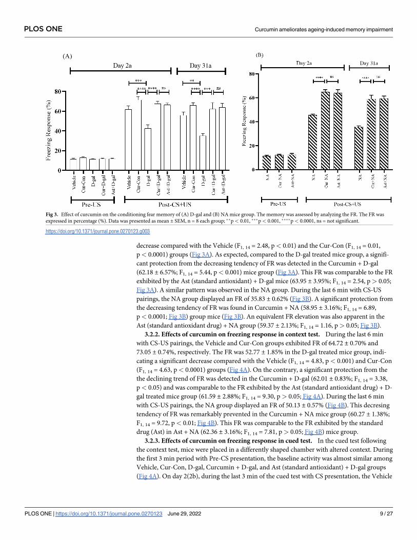
decrease compared with the Vehicle (F1, 14 = 2.48, p< 0.01) and the Cur-Con (F1, 14 = 0.01,
p< 0.0001) groups (Fig 3A). As expected, compared to the D-gal treated mice group, a signifi-
cant protection from the decreasing tendency of FR was detected in the Curcumin + D-gal
(62.18 ± 6.57%; F1, 14 = 5.44, p< 0.001) mice group (Fig 3A). This FR was comparable to the FR
exhibited by the Ast (standard antioxidant) + D-gal mice (63.95 ± 3.95%; F1, 14 = 2.54, p> 0.05;
Fig 3A). A similar pattern was observed in the NA group. During the last 6 min with CS-US
pairings, the NA group displayed an FR of 35.83 ± 0.62% (Fig 3B). A significant protection from
the decreasing tendency of FR was found in Curcumin + NA (58.95 ± 3.16%; F1, 14 = 6.89,
p< 0.0001; Fig 3B) group mice (Fig 3B). An equivalent FR elevation was also apparent in the
Ast (standard antioxidant drug) + NA group (59.37 ± 2.13%; F1, 14 = 1.16, p> 0.05; Fig 3B).
3.2.2. Effects of curcumin on freezing response in context test. During the last 6 min
with CS-US pairings, the Vehicle and Cur-Con groups exhibited FR of 64.72 ± 0.70% and
73.05 ± 0.74%, respectively. The FR was 52.77 ± 1.85% in the D-gal treated mice group, indi-
cating a significant decrease compared with the Vehicle (F1, 14 = 4.83, p< 0.001) and Cur-Con
(F1, 14 = 4.63, p< 0.0001) groups (Fig 4A). On the contrary, a significant protection from the
the declining trend of FR was detected in the Curcumin + D-gal (62.01 ± 0.83%; F1, 14 = 3.38,
p< 0.05) and was comparable to the FR exhibited by the Ast (standard antioxidant drug) + D-
gal treated mice group (61.59 ± 2.88%; F1, 14 = 9.30, p> 0.05; Fig 4A). During the last 6 min
with CS-US pairings, the NA group displayed an FR of 50.13 ± 0.57% (Fig 4B). This decresing
tendency of FR was remarkably prevented in the Curcumin + NA mice group (60.27 ± 1.38%;
F1, 14 = 9.72, p< 0.01; Fig 4B). This FR was comparable to the FR exhibited by the standard
drug (Ast) in Ast + NA (62.36 ± 3.16%; F1, 14 = 7.81, p> 0.05; Fig 4B) mice group.
3.2.3. Effects of curcumin on freezing response in cued test. In the cued test following
the context test, mice were placed in a differently shaped chamber with altered context. During
the first 3 min period with Pre-CS presentation, the baseline activity was almost similar among
Vehicle, Cur-Con, D-gal, Curcumin + D-gal, and Ast (standard antioxidant) + D-gal groups
(Fig 4A). On day 2(2b), during the last 3 min of the cued test with CS presentation, the Vehicle
Fig 3. Effect of curcumin on the conditioning fear memory of (A) D-gal and (B) NA mice group. The memory was assessed by analyzing the FR. The FR was
expressed in percentage (%). Data was presented as mean ± SEM, n = 8 each group; ��p< 0.01, ���p< 0.001, ����p< 0.0001, ns = not significant.
https://doi.org/10.1371/journal.pone.0270123.g003
PLOS ONE Curcumin ameliorates ageing-induced memory impairment
PLOS ONE | https://doi.org/10.1371/journal.pone.0270123 June 29, 2022 9 / 27
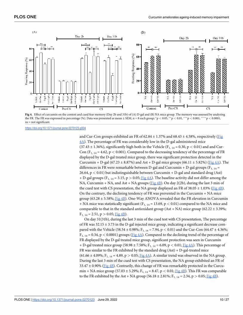
and Cur-Con groups exhibited an FR of 62.84 ± 1.37% and 68.43 ± 4.58%, respectively (Fig
4A). The percentage of FR was considerably low in the D-gal administered mice
(37.43 ± 1.36%), significantly high both in the Vehicle (F1, 14 = 0.30, p< 0.01) and and Cur-
Con (F1, 14 = 4.62, p< 0.001). Compared to the decreasing tendency of the percentage of FR
displayed by the D-gal treated mice group, there was significant protection detected in the
Curcumin + D-gal (67.23 ± 8.87%) and Ast + D-gal mice groups (66.11 ± 5.82%) (Fig 4A). The
differences in FR were remarkable between D-gal and Curcumin + D-gal groups (F1, 14 =
26.64, p< 0.01) but indistinguishable between Curcumin + D-gal and standard drug (Ast)
+ D-gal groups (F1, 14 = 3.15, p> 0.05; Fig 4A). The baseline activity did not differ among the
NA, Curcumin + NA, and Ast + NA groups (Fig 4B). On day 2(2b), during the last 3 min of
the cued test with CS presentation, the NA group displayed an FR of 38.05 ± 1.03% (Fig 4B).
On the contrary, the declining tendency of FR was prevented in the Curcumin + NA mice
group (63.28 ± 5.58%; Fig 4B). One-Way ANOVA revealed that the FR elevation in Curcumin
+ NA mice was statistically significant (F1, 14 = 13.69, p< 0.01) compared to the NA mice and
comparable to that in the standard antioxidant group (Ast + NA) mice group (62.22 ± 3.39%;
F1, 14 = 2.51, p> 0.05; Fig 4B).
On day 31(31b), during the last 3 min of the cued test with CS presentation, The percentage
of FR was 32.15 ± 3.73 in the D-gal injected mice group, indicating a significant decrease com-
pared with the Vehicle (58.54 ± 0.98%; F1, 14 = 7.94, p< 0.01) and the Cur-Con (64.47 ± 4.36%;
F1, 14 = 0.34, p< 0.0001) groups (Fig 4A). Compared to the declining trend of the percentage of
FR displayed by the D-gal treated mice group, significant protection was seen in Curcumin
+ D-gal treated mice group (58.98 ± 7.58%; F1, 14 = 6.09, p< 0.01; Fig 4A). This percentage of
FR was similar to the FR exhibited by the standard drug (Ast) + D-gal-treated mice
(61.66 ± 4.09%; F1, 14 = 4.89, p> 0.05; Fig 4A). A similar trend was observed in the NA group.
During the last 3 min of the cued test with CS presentation, the NA group exhibited an FR of
33.47 ± 0.90% (Fig 4B). Contrarily, this change of FR was remarkably protected in the Curcu-
min + NA mice group (57.03 ± 5.29%; F1, 14 = 8.47, p< 0.01; Fig 4B). This FR was comparable
to the FR exhibited by the Ast + NA group (56.18 ± 2.81%; F1, 14 = 2.34, p> 0.05; Fig 4B).
Fig 4. Effect of curcumin on the context and cued fear memory (Day 2b and 31b) of (A) D-gal and (B) NA mice group. The memory was assessed by analyzing
the FR. The FR was expressed in percentage (%). Data was presented as mean ± SEM, n = 8 each group; �p< 0.05, ��p< 0.01, ���p< 0.001, ����p< 0.0001,
ns = not significant.
https://doi.org/10.1371/journal.pone.0270123.g004
PLOS ONE Curcumin ameliorates ageing-induced memory impairment
PLOS ONE | https://doi.org/10.1371/journal.pone.0270123 June 29, 2022 10 / 27

3.3. Effects of curcumin on oxidative stress biomarkers
3.3.1. Glutathione. The GSH level detected in the Vehicle and Cur-Con groups were
9.34 ± 0.87 μmol/mg and 12.72 ± 0.97 μmol/mg, respectively (Fig 5A). The level of GSH in the
D-gal treated mice group was 3.19 ± 0.31 μmol/mg, illustrating a significant decrease of GSH
compared with Vehicle (F1, 14 = 3.63, p< 0.0001) and Cur-Con (F1, 14 = 2.58, p< 0.0001)
groups (Fig 5A). The GSH level in Curcumin + D-gal mice group was 10.77 ± 1.02 μmol/mg,
indicating a remarkable protection from the decreasing trend of GSH level (F1, 14 = 16.49,
p< 0.0001) (Fig 5A). A similar GSH elevation was apparent in the Ast + D-gal mice
(10.81 ± 0.52 μmol/mg; F1, 14 = 7.73, p> 0.05; Fig 5A).
The GSH level in the NA group was 3.78 ± 0.59 μmol/mg (Fig 5B). Conversely, this decreas-
ing trend of GSH level was remarkably prevented in the Curcumin + NA mice group
(10.16 ± 0.74 μmol/mg; F1, 14 = 0.46, p< 0.0001; Fig 5B). This GSH level was comparable to
the GSH level detected in the Ast + NA (11.28 ± 0.61 μmol/mg; F1, 14 = 0.74, P> 0.05; Fig 5B)
mice group.
3.3.2. Superoxide dismutase. The SOD activity was 27.16 ± 0.32 U/30s and 40.24 ± 3.46
U/30s in the Vehicle and Cur-Con groups, respectively (Fig 5C). The SOD activity in D-gal
administered mice group was 11.45 ± 0.27 U/30s, indicating a significant decrease in the activ-
ity compared with Vehicle (F1, 14 = 0.25, p< 0.05) and Cur-Con (F1, 14 = 21.52, p < 0.0001)
groups (Fig 5C). Interestingly, curcumin produced an efficient protection of the SOD activity
(35.68 ± 3.46 U/30s) detected in Curcumin + D-gal mice group, which was statistically signifi-
cant (F1, 14 = 28.37, p< 0.0001) compared to the D-gal treated mice group but insignificant
compared to the Ast + D-gal group (37.27 ± 5.46 U/30s; F1, 14 = 0.67, p> 0.05; Fig 5C) mice.
The SOD activity in the NA mice group was 13.03 ± 0.26 U/30s (Fig 5D). Contrarily, this
decreasing activity was protected in the Curcumin + NA mice group (31.27 ± 1.63 U/30s; Fig
5D). A One-Way ANOVA followed by the Brown-Forsythe test demonstrated a statistically
significant change in the SOD activity of the curcumin mice compared to the SOD activity of
the NA mice (F1, 14 = 7.15, p< 0.01). The effect of curcumin on the SOD activity was very sim-
ilar to the effect of Ast (35.39 ± 4.80 U/30s; F1, 14 = 14.68, p> 0.05; Fig 5D) found in Ast + NA
group mice.
3.3.3. Catalase. The CAT activity was 6.83 ± 0.39 μmol/min/mg and 12.33 ± 0.92 μmol/
min/mg in the Vehicle and Cur-Con groups, respectively. The CAT activity in D-gal treated
mice group was 2.82 ± 0.18 μmol/min/mg, illustrating a significant decrease in activity
Fig 5. Effect of curcumin on GSH concentration (A and B), SOD (C and D) and CAT (E and F) activity in D-gal and NA mice group. The GSH, SOD, CAT was detected
by using bioassay technique among Vehicle, Cur-Con, D-gal, Curcumin + D-gal, Ast + D-gal, NA, Curcumin + NA, Ast + NA groups. GSH level, SOD and CAT activity
were expressed in μmol/mg, U/30s, and μmol/min/mg, respectively. Data was presented as mean ± SEM, n = 8 each group; �p< 0.05, ��p< 0.01 ���p< 0.001,����p< 0.0001 ns = not significant.
https://doi.org/10.1371/journal.pone.0270123.g005
PLOS ONE Curcumin ameliorates ageing-induced memory impairment
PLOS ONE | https://doi.org/10.1371/journal.pone.0270123 June 29, 2022 11 / 27

compared with Vehicle (F1, 14 = 0.81, p< 0.05) and Cur-Con (F1, 14 = 29.53, p< 0.0001)
groups (Fig 5E). On the contrary, a statistical significant protection from the decreasing activ-
ity of CAT activity was detected in the Curcumin + D-gal mice group (10.29 ± 0.95 μmol/min/
mg; F1, 14 = 4.6, p< 0.0001; Fig 5E). This activity was comparable to the CAT activity of Ast
+ D-gal mice group (10.06 ± 1.51 μmol/min/mg; F1, 14 = 0.78, p> 0.05; Fig 5E).
Similarly, the CAT activity in the NA mice was 3.15 ± 0.26 μmol/min/mg (Fig 5F). Curcumin
produced efficient protection of the CAT activity (8.31 ± 0.22 μmol/min/mg Fig 5F), detected in
the Curcumin + NA mice group. A One-Way ANOVA followed by the Brown-Forsythe test
confirmed a statistically significant change in the CAT activity by curcumin (F1, 14 = 0.47,
p< 0.001) compared to the CAT activity of the NA mice. This CAT activity was comparable to
that of the Ast + NA mice (8.85 ± 0.91 μmol/min/mg; F1, 14 = 3.24, p> 0.05; Fig 5F).
3.3.4. Advanced oxidation of protein products. The AOPP level was drastically elevated
to 146.41 ± 16.60 μmol/ml in the D-gal group, illustrating a significant increase of AOPP level
compared with the Vehicle (84.80 ± 11.87 μmol/ml; F1, 14 = 0.99, p< 0.01) and Cur-Con
(49.65 ± 7.03 μmol/ml; F1, 14 = 5.11, p< 0.0001) groups (Fig 6A). However, a statistical signifi-
cant protection from the increasing tendency of AOPP level was found in the Curcumin + D-
gal mice group (61.55 ± 10.80 μmol/ml; Fig 6A; F1, 14 = 2.30, p< 0.0001), but was indistin-
guishable from the Ast + D-gal group (56.44 ± 10.44 μmol/ml; F1, 14 = 0.13, p> 0.05; Fig 6A).
Similarly, the AOPP level was significantly declined to 69.52 ± 9.90 μmol/ml in the Curcumin
+ NA mice group compared to the NA (137.67 ± 15.24 μmol/ml) mice (F1, 14 = 0.76, p< 0.01;
Fig 6B) group. This level was comparable to the level of Ast + NA mice group
(61.28 ± 7.04 μmol/ml; F1, 14 = 0.28, p> 0.05; Fig 6B).
3.3.5. Nitric oxide. D-gal administration profoundly enhanced the NO level to
9.40 ± 1.01 mmol/mg, indicating a significant increase of NO level compared with the Vehicle
(4.62 ± 0.33 mmol/mg; F1, 14 = 6.02, p< 0.0001) and Cur-Con (3.23 ± 0.75 mmol/mg; F1, 14 =
0.22, p< 0.0001) groups (Fig 6C). Conversely, a statistical significant protection from the
increasing tendency of NO level was detected in the Curcumin + D-gal mice group
(4.47 ± 0.53 mmol/mg; F1, 14 = 2.75, p< 0.0001; Fig 6C). This level was comparable to the NO
level of Ast + D-gal mice (4.09 ± 0.55 mmol/mg; F1, 14 = 0.32, p> 0.05; Fig 6C). A similar pat-
tern was observed in the normal-aged mice. The NO level in the NA group was 8.06 ± 0.29
mmol/mg (Fig 6D). On the contrary, this increasing tendency of NO level was prevented in
the Curcumin + NA (5.28 ± 0.25 mmol/mg) group mice (Fig 6D). A One-Way ANOVA fol-
lowed by the Brown-Forsythe test demonstrated a statistically significant effect of curcumin
Fig 6. Effect of curcumin on AOPP (A and B), NO (C and D), and MDA (E and F) concentration in D-gal and NA mice group. The AOPP level was assessed using
bioassay technique among Vehicle, Cur-Con, D-gal, Curcumin + D-gal, Ast + D-gal, NA, Curcumin + NA, Ast + NA groups. AOPP, NO, and MDA level was represented
in μmol/ml, mmol/mg, and nmol/ml, respectively. Data was presented as mean ± SEM, n = 8 each group; �p< 0.05, ��p< 0.01, ����p< 0.0001 ns = not significant.
https://doi.org/10.1371/journal.pone.0270123.g006
PLOS ONE Curcumin ameliorates ageing-induced memory impairment
PLOS ONE | https://doi.org/10.1371/journal.pone.0270123 June 29, 2022 12 / 27
(F1, 14 = 0.91, p< 0.05). The NO level in the curcumin mice was comparable to that of the Ast
+ NA mice group (4.72 ± 0.33 mmol/mg; F1, 14 = 1.64, p> 0.05; Fig 6D).
3.3.6. Lipid peroxidation. The MDA level was 39.37 ± 4.62 nmol/ml and 27.64 ± 6.40
nmol/ml in the Vehicle and Cur-Con groups, respectively. The level of MDA in the D-gal
group was 130.37 ± 10.81 nmol/ml, indicating a significant increase in the MDA level com-
pared with Vehicle (F1, 14 = 5.32, p< 0.0001) and Cur-Con (F1, 14 = 2.69, p< 0.0001) groups
(Fig 6E). A statistical significant protection from the increasing trend of MDA level was found
in the Curcumin + D-gal mice group (40.05 ± 4.39 nmol/ml; F1, 14 = 5.66, p< 0.0001; Fig 6E).
This MDA level was comparable to that of the Ast + D-gal mice group (41.29 ± 5.53 nmol/ml;
F1, 14 = 0.78, p> 0.05; Fig 6E). Similarly, the MDA level in the NA group was 111.50 ± 10.49
nmol/ml (Fig 6F). Interestingly, this increasing pattern of the MDA level was significantly pre-
vented in the Curcumin + NA mice group (43.74 ± 6.97 nmol/ml; F1, 14 = 0.53, p< 0.0001)
and was comparable to the Ast + NA mice group (41.08 ± 6.06 nmol/ml; (F1, 14 = 0.31,
p> 0.05; Fig 6F).
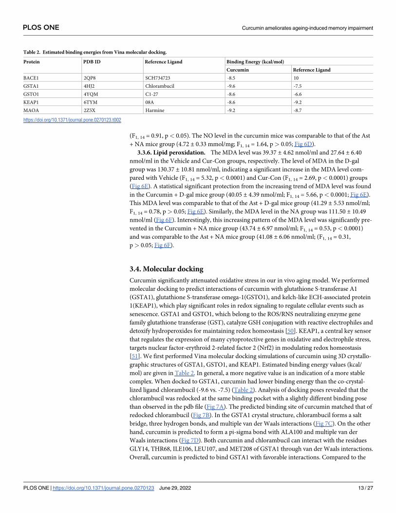
3.4. Molecular docking
Curcumin significantly attenuated oxidative stress in our in vivo aging model. We performed
molecular docking to predict interactions of curcumin with glutathione S-transferase A1
(GSTA1), glutathione S-transferase omega-1(GSTO1), and kelch-like ECH-associated protein
1(KEAP1), which play significant roles in redox signaling to regulate cellular events such as
senescence. GSTA1 and GSTO1, which belong to the ROS/RNS neutralizing enzyme gene
family glutathione transferase (GST), catalyze GSH conjugation with reactive electrophiles and
detoxify hydroperoxides for maintaining redox homeostasis [50]. KEAP1, a central key sensor
that regulates the expression of many cytoprotective genes in oxidative and electrophile stress,
targets nuclear factor-erythroid 2-related factor 2 (Nrf2) in modulating redox homeostasis
[51]. We first performed Vina molecular docking simulations of curcumin using 3D crystallo-
graphic structures of GSTA1, GSTO1, and KEAP1. Estimated binding energy values (kcal/
mol) are given in Table 2. In general, a more negative value is an indication of a more stable
complex. When docked to GSTA1, curcumin had lower binding energy than the co-crystal-
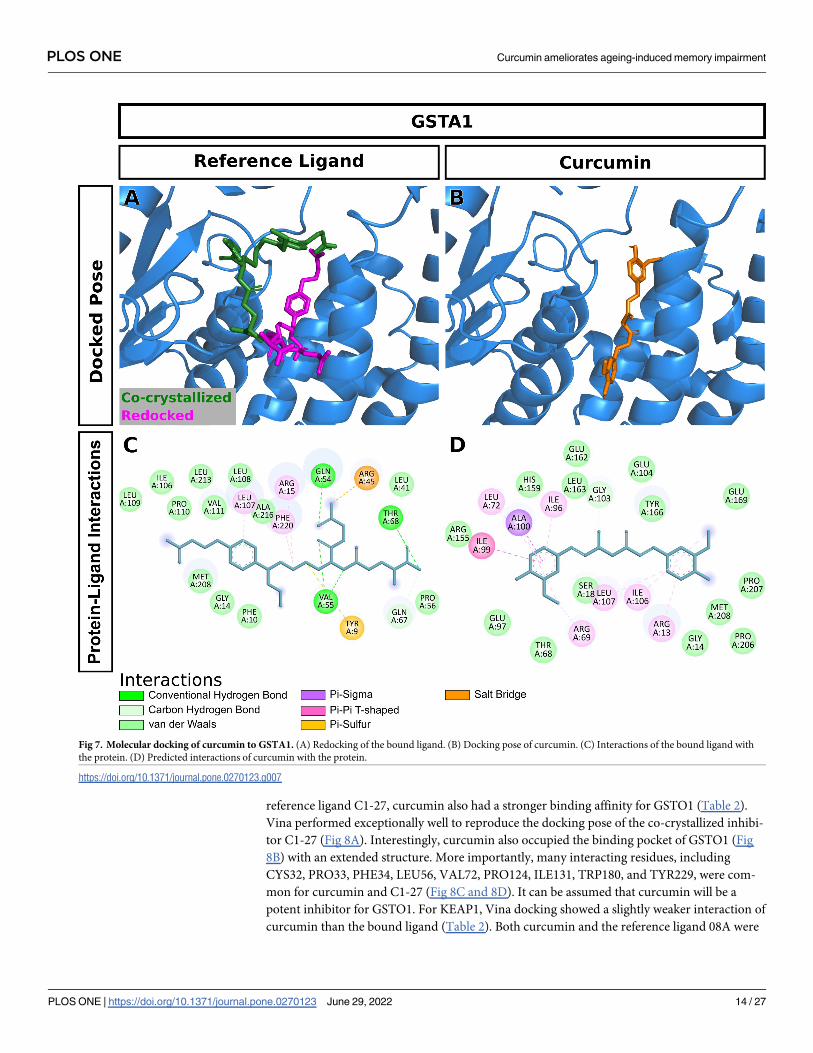
lized ligand chlorambucil (-9.6 vs. -7.5) (Table 2). Analysis of docking poses revealed that the
chlorambucil was redocked at the same binding pocket with a slightly different binding pose
than observed in the pdb file (Fig 7A). The predicted binding site of curcumin matched that of
redocked chlorambucil (Fig 7B). In the GSTA1 crystal structure, chlorambucil forms a salt
bridge, three hydrogen bonds, and multiple van der Waals interactions (Fig 7C). On the other
hand, curcumin is predicted to form a pi-sigma bond with ALA100 and multiple van der
Waals interactions (Fig 7D). Both curcumin and chlorambucil can interact with the residues
GLY14, THR68, ILE106, LEU107, and MET208 of GSTA1 through van der Waals interactions.
Overall, curcumin is predicted to bind GSTA1 with favorable interactions. Compared to the
Table 2. Estimated binding energies from Vina molecular docking.
Protein PDB ID Reference Ligand Binding Energy (kcal/mol)
Curcumin Reference Ligand
BACE1 2QP8 SCH734723 -8.5 10
GSTA1 4HJ2 Chlorambucil -9.6 -7.5
GSTO1 4YQM C1-27 -8.6 -6.6
KEAP1 6TYM 08A -8.6 -9.2
MAOA 2Z5X Harmine -9.2 -8.7
https://doi.org/10.1371/journal.pone.0270123.t002
PLOS ONE Curcumin ameliorates ageing-induced memory impairment
PLOS ONE | https://doi.org/10.1371/journal.pone.0270123 June 29, 2022 13 / 27
reference ligand C1-27, curcumin also had a stronger binding affinity for GSTO1 (Table 2).
Vina performed exceptionally well to reproduce the docking pose of the co-crystallized inhibi-
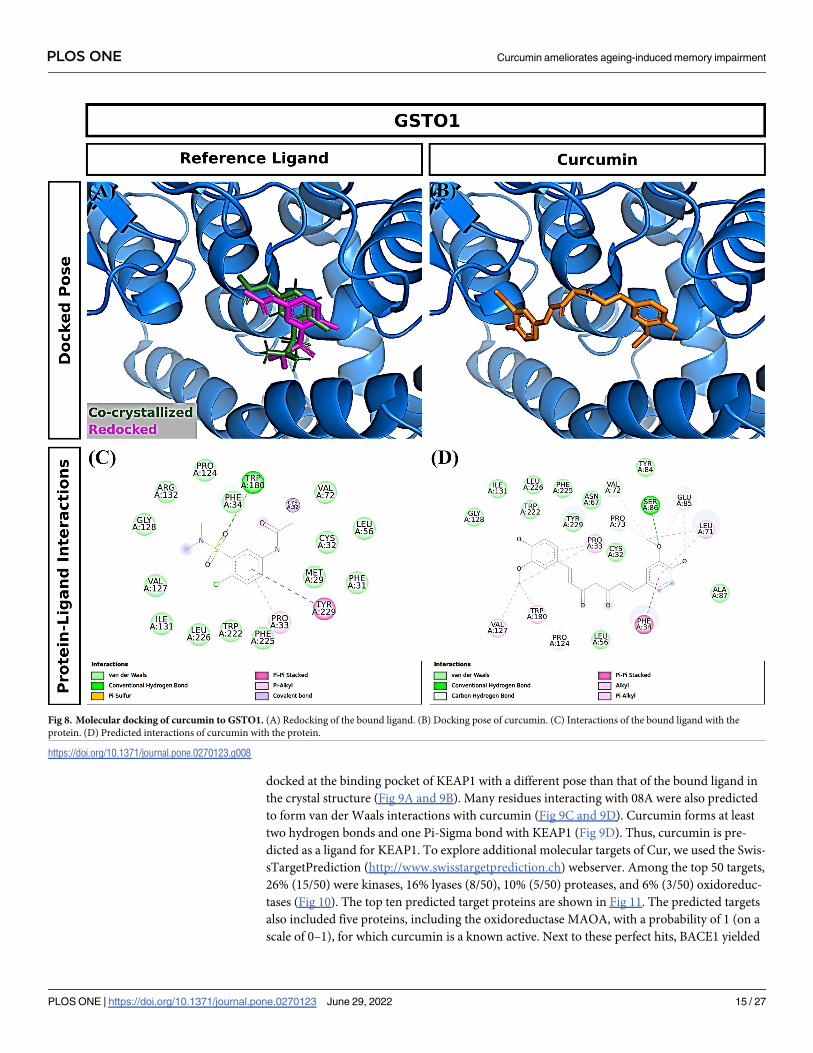
tor C1-27 (Fig 8A). Interestingly, curcumin also occupied the binding pocket of GSTO1 (Fig
8B) with an extended structure. More importantly, many interacting residues, including
CYS32, PRO33, PHE34, LEU56, VAL72, PRO124, ILE131, TRP180, and TYR229, were com-
mon for curcumin and C1-27 (Fig 8C and 8D). It can be assumed that curcumin will be a
potent inhibitor for GSTO1. For KEAP1, Vina docking showed a slightly weaker interaction of
curcumin than the bound ligand (Table 2). Both curcumin and the reference ligand 08A were
Fig 7. Molecular docking of curcumin to GSTA1. (A) Redocking of the bound ligand. (B) Docking pose of curcumin. (C) Interactions of the bound ligand with
the protein. (D) Predicted interactions of curcumin with the protein.
https://doi.org/10.1371/journal.pone.0270123.g007
PLOS ONE Curcumin ameliorates ageing-induced memory impairment
PLOS ONE | https://doi.org/10.1371/journal.pone.0270123 June 29, 2022 14 / 27
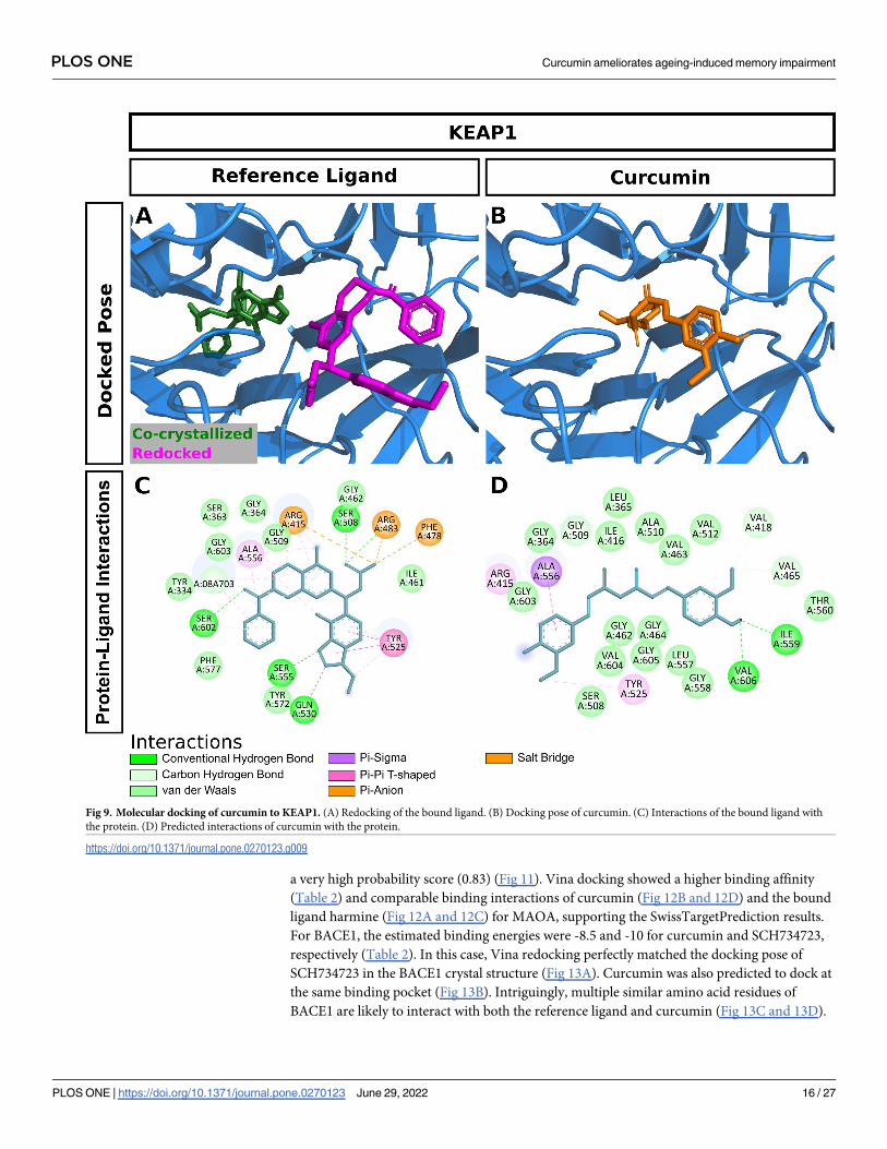
docked at the binding pocket of KEAP1 with a different pose than that of the bound ligand in
the crystal structure (Fig 9A and 9B). Many residues interacting with 08A were also predicted
to form van der Waals interactions with curcumin (Fig 9C and 9D). Curcumin forms at least
two hydrogen bonds and one Pi-Sigma bond with KEAP1 (Fig 9D). Thus, curcumin is pre-
dicted as a ligand for KEAP1. To explore additional molecular targets of Cur, we used the Swis-
sTargetPrediction (http://www.swisstargetprediction.ch) webserver. Among the top 50 targets,
26% (15/50) were kinases, 16% lyases (8/50), 10% (5/50) proteases, and 6% (3/50) oxidoreduc-
tases (Fig 10). The top ten predicted target proteins are shown in Fig 11. The predicted targets
also included five proteins, including the oxidoreductase MAOA, with a probability of 1 (on a
scale of 0–1), for which curcumin is a known active. Next to these perfect hits, BACE1 yielded
Fig 8. Molecular docking of curcumin to GSTO1. (A) Redocking of the bound ligand. (B) Docking pose of curcumin. (C) Interactions of the bound ligand with the
protein. (D) Predicted interactions of curcumin with the protein.
https://doi.org/10.1371/journal.pone.0270123.g008
PLOS ONE Curcumin ameliorates ageing-induced memory impairment
PLOS ONE | https://doi.org/10.1371/journal.pone.0270123 June 29, 2022 15 / 27
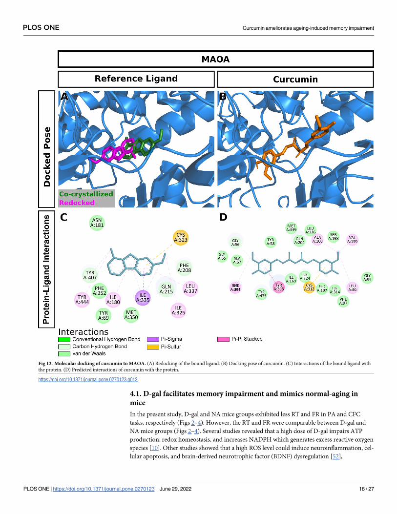
a very high probability score (0.83) (Fig 11). Vina docking showed a higher binding affinity
(Table 2) and comparable binding interactions of curcumin (Fig 12B and 12D) and the bound
ligand harmine (Fig 12A and 12C) for MAOA, supporting the SwissTargetPrediction results.
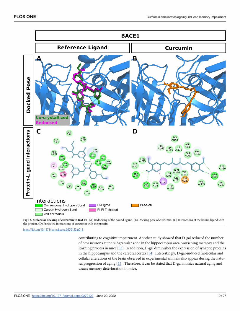
For BACE1, the estimated binding energies were -8.5 and -10 for curcumin and SCH734723,
respectively (Table 2). In this case, Vina redocking perfectly matched the docking pose of
SCH734723 in the BACE1 crystal structure (Fig 13A). Curcumin was also predicted to dock at
the same binding pocket (Fig 13B). Intriguingly, multiple similar amino acid residues of
BACE1 are likely to interact with both the reference ligand and curcumin (Fig 13C and 13D).
Fig 9. Molecular docking of curcumin to KEAP1. (A) Redocking of the bound ligand. (B) Docking pose of curcumin. (C) Interactions of the bound ligand with
the protein. (D) Predicted interactions of curcumin with the protein.
https://doi.org/10.1371/journal.pone.0270123.g009
PLOS ONE Curcumin ameliorates ageing-induced memory impairment
PLOS ONE | https://doi.org/10.1371/journal.pone.0270123 June 29, 2022 16 / 27

For instance, GLY72, GLN73, ILE171, and TYR132 interact with both molecules. Ser71,
GLY74, and ARG296 can form conventional hydrogen bonds with the curcumin. Overall, our
molecular docking identifies curcumin as a candidate BACE1 inhibitor.
4. Discussion
This current study investigated the effects of curcumin on D-gal and normal aging-induced
memory impairment. In vivo study revealed that curcumin protected the decreasing tendency
of D-gal and Normal aging-induced RT and FR in PA and CFC tasks. In addition, curcumin
ameliorated the level of oxidative stress biomarkers (GSH, SOD, CAT, AOPP, NO MDA). In
silico study discerned that curcumin-mediated antioxidant effects in mice could result from, at
least partially, binding with several regulatory proteins such as GSTA1, GSTO1, KEAP1,
BACE1, and MAOA.
Fig 10. Relative abundance of the class of top 50 predicted molecular targets of curcumin obtained from
SwissTargetPrediction.
https://doi.org/10.1371/journal.pone.0270123.g010
Fig 11. Names and target probabilities of top 10 predicted molecular targets of curcumin obtained from Swiss TargetPrediction.
https://doi.org/10.1371/journal.pone.0270123.g011
PLOS ONE Curcumin ameliorates ageing-induced memory impairment
PLOS ONE | https://doi.org/10.1371/journal.pone.0270123 June 29, 2022 17 / 27
4.1. D-gal facilitates memory impairment and mimics normal-aging in
mice
In the present study, D-gal and NA mice groups exhibited less RT and FR in PA and CFC
tasks, respectively (Figs 2–4). However, the RT and FR were comparable between D-gal and
NA mice groups (Figs 2–4). Several studies revealed that a high dose of D-gal impairs ATP
production, redox homeostasis, and increases NADPH which generates excess reactive oxygen
species [10]. Other studies showed that a high ROS level could induce neuroinflammation, cel-
lular apoptosis, and brain-derived neurotrophic factor (BDNF) dysregulation [52],
Fig 12. Molecular docking of curcumin to MAOA. (A) Redocking of the bound ligand. (B) Docking pose of curcumin. (C) Interactions of the bound ligand with
the protein. (D) Predicted interactions of curcumin with the protein.
https://doi.org/10.1371/journal.pone.0270123.g012
PLOS ONE Curcumin ameliorates ageing-induced memory impairment
PLOS ONE | https://doi.org/10.1371/journal.pone.0270123 June 29, 2022 18 / 27
contributing to cognitive impairment. Another study showed that D-gal reduced the number
of new neurons at the subgranular zone in the hippocampus area, worsening memory and the
learning process in mice [53]. In addition, D-gal diminishes the expression of synaptic proteins
in the hippocampus and the cerebral cortex [54]. Interestingly, D-gal-induced molecular and
cellular alterations of the brain observed in experimental animals also appear during the natu-
ral progression of aging [55]. Therefore, it can be stated that D-gal mimics natural aging and
draws memory deterioration in mice.
Fig 13. Molecular docking of curcumin to BACE1. (A) Redocking of the bound ligand. (B) Docking pose of curcumin. (C) Interactions of the bound ligand with
the protein. (D) Predicted interactions of curcumin with the protein.
https://doi.org/10.1371/journal.pone.0270123.g013
PLOS ONE Curcumin ameliorates ageing-induced memory impairment
PLOS ONE | https://doi.org/10.1371/journal.pone.0270123 June 29, 2022 19 / 27
4.2. Curcumin improves D-gal and normal-aging induced memory
impairment
In the current study, Curcumin + D-gal, and Curcumin + NA mice groups showed substan-
tial protection of RT and FR in PA and CFC, respectively (Figs 2–4). The RT and FR were
comparable between Curcumin + D-gal and Curcumin + NA mice groups (Figs 2–4). More-
over, curcumin was comparable to Ast, a standard antioxidant, in improving retention and
freezing memory. Many studies showed that curcumin possesses anti-neuroinflammation,
antioxidant and anti-aging properties [56, 57]. Another study reported that curcumin plays
a protective role in brain aging by modulating cell proliferation, neuronal degeneration,
and cellular senescence [14]. Therefore, it can be inferred that curcumin improves memory
owing to its antioxidant and anti-aging properties in mice. Similar to previous findings
[58], our study suggested that curcumin ameliorates memory impairment induced by D-gal
or normal-aging in mice.
4.3. Curcumin ameliorates the oxidative biomarkers in D-gal and NA mice
In the present study, we found decreased levels of antioxidants such as GSH, SOD, and
CAT (Fig 5) in D-gal and NA mice. Moreover, these levels were comparable between D-gal
and NA mice (Fig 5). Many studies showed that the abnormal changes of ROS and RNS aug-
ment aging processes induced by D-gal [59] and NA [60]. In normal conditions, the antioxi-
dant defense system comprises endogenous non-enzymatic and enzymatic compounds that
counteract the deleterious effect of ROS and RNS [61]. Glutathione (GSH), a non-enzymatic
antioxidant tripeptide, has a thiol group that interacts with ROS and RNS directly and
reduces H2O2 to form H2O [62]. In contrast, enzymatic antioxidant Superoxide Dismutase
(SOD) catalyzes the dismutation of O2• − into H2O2 and O2 [63]. Another enzymatic antiox-
idant, Catalase (CAT), converts H2O2 into H2O and O2; thus, cell protection from deleteri-
ous effects of H2O2 continues [63]. These antioxidants (GSH, SOD, CAT) prevent lipid
peroxidation in the cell membrane and maintain redox homeostasis [64]. On the other
hand, low levels of GSH, SOD, and CAT fail to protect against the overproduction of ROS
and RNS and expedite the aging process [65].
We found a high level of AOPP, NO, and MDA in D-gal and NA mice (Fig 6), suggesting
an induction of the aging process [29]. A high level of protein oxidation is known to generate
excess AOPP [66]. Nitric Oxide (NO) reacts with superoxide (O2•−) to form a stronger oxidant
peroxynitrite anion (ONOO-). A high level of NO generates more RNS, such as •NO2 and
N2O3 [67], whereas an increased level of MDA promotes ROS generation [68]. Therefore, high
levels of MDA, AOPP, and NO contribute to the aging process by increasing oxidative stress
in the brain.
We found that both curcumin and Ast prevented the decreasing trend of GSH, SOD, and
CAT levels detected in D-gal and NA groups (Fig 5), suggesting that curcumin’s antioxidant
activity minimized the aging-associated oxidative burden in mice [69]. Our results are also in
agreement with previous studies [70].
On the other hand, substantial protection from the increasing tendency of AOPP, NO, and
MDA was detected in Curcumin + D-gall and Curcumin +NA mice groups (Fig 6). These
effects were comparable in the Ast group (Fig 6). Another study [71] supported these findings,
suggesting that curcumin exerts its antioxidant activity by controlling the overproduction of
AOPP, NO, and MDA.
PLOS ONE Curcumin ameliorates ageing-induced memory impairment
PLOS ONE | https://doi.org/10.1371/journal.pone.0270123 June 29, 2022 20 / 27
4.4. Predicted interactions of curcumin with glutathione S-transferase A1,
glutathione S-transferase omega-1, kelch-like ECH-associated protein 1,
beta-secretase 1, and amine oxidase (flavin-containing) A to exert
antioxidant activity
In our in silico studies, estimated binding energies were assessed by adopting Vina molecular
docking. Estimated binding energies of curcumin with GSTA1, GSTO1, and KEAP1 were
comparable with reference ligands such as Chlorambucil, C1-27, 08A, respectively (Table 2).
Furthermore, curcumin is predicted to bind more strongly with GSTA1 and GSTO1 compared
with the reference ligands (Table 2).
GSTA1, GSTO1, and KEAP1 are abundantly present in the hippocampus, a critical
brain region crucial for hippocampus-dependent learning tasks [72, 73]. Western blot
analysis showed that the upregulation of GSTA1 in the CA1 area [74] and the downregula-
tion of GSTO1 in the hippocampus were linked to cognitive impairment, [75] commonly
seen in aging animals. Other studies showed that these proteins are closely associated with
oxidative stress and aging-induced neurodegenerative diseases such as memory
impairment [76]. GSTA1 and GSTO1 are primary phase II detoxification enzymes and
catalyze GSH conjugation in the presence of electrophile substrates [19]. Furthermore,
GSTA1 suppresses the activation of c-Jun N-terminal kinase (JNK) signaling by a pro-
inflammatory cytokine and oxidative stress [77]. Moreover, GSTO1 regulates the activa-
tion of interleukin-1β and stops the inflammation process in aging-associated neurode-
generative disease [20]. The Keap1-Nrf2 system plays a crucial role in regulating oxidative
stress-mediated disorders [78]. A western blot analysis found a lower expression of
KEAP1 in CA3 and dentate gyrus of the hippocampus under oxidative conditions [78].
The KEAP1 is closely associated with the Nrf2 cytoprotective signaling pathway and plays
an antioxidative role. Under the homeostatic state, KEAP1 controls the level of Nrf2 upon
binding. During stressful conditions, the KEAP1 gets oxidized in the presence of electro-
phile, stopping Nrf2 ubiquitylation. These cause Nrf2 to move into the nucleus forming a
heterodimer with musculoaponeurotic proteins (Mafs) and initiating cytoprotective mole-
cules such as GSH, SOD, CAT after binding with antioxidant response element (ARE)
[21]. Under oxidative stress conditions, the expression of GSTA1, GSTO1, and KEAP1 are
down-regulated. In contrast, the expressions are reversed upon treating with curcumin
[18, 79], suggesting that curcumin is predicted to interact with GSTA1, GSTO1, and
KEAP1 and potentiates the antioxidant activity.
Opposite to the down-regulation of GSTA1, GSTO1, and KEAP1, the BACE1 and
MAOA proteins are elevated during the aging-associated memory impairment [80, 81].
Immunohistochemical studies showed a high level of MAOA was found in the CA3 area of
the hippocampus, an important region sensitive for brain aging [82]. Studies showed that
BACE1 and MAOA proteins are strongly linked to aging-associated memory impairment
[22, 83]. BACE1 is widely distributed in CA1 and CA3 areas, and the absence of this protein
is responsible for the altered level of synaptic plasticity in aging mice [84]. Furthermore,
BACE1 plays a vital role in cleaving Aβ-peptide and causing the accumulation of amyloid-β(Aβ) peptides in the brain [85]. Likewise, MAOA produces hydrogen peroxide by oxidation
of monoamine substrates in the mitochondrial outer membrane and facilitates oxidative
stress [23]. We found that the estimated binding energies of curcumin upon BACE1 and
MAOA were comparable with respective reference ligands (SCH734723, Harmine, respec-
tively) (Table 2), suggesting that curcumin is predicted to interact with BACE1 and MAOA
and potentiates its antioxidant role in brain aging.
PLOS ONE Curcumin ameliorates ageing-induced memory impairment
PLOS ONE | https://doi.org/10.1371/journal.pone.0270123 June 29, 2022 21 / 27
5. Conclusion
We investigated the detailed effects of curcumin on oxidative stress in the D-gal and nature-
induced aging mice model. Our in vivo study suggested that curcumin improves memory and
rescues learning impairment by modulating oxidative stress levels. Furthermore, our in-silico
study demonstrated that curcumin has good binding affinities for several molecular targets
implicated in redox homeostasis. Finally, based on our in vivo and computational studies, it
can be stated that curcumin improves D-gal and Normal aging-associated memory
impairment by reducing oxidative overload in mice.
Supporting information
S1 Fig. Effect of curcumin on RT in D-gal and NA mice group after 48 hours of training.
The RT was calculated by performing PA tasks among Vehicle, Cur-Con, D-gal, Curcumin
+ D-gal, Ast + D-gal, NA, Curcumin + NA, Ast + NA groups. RT was expressed in second.
Data was presented as mean ± SEM, n = 8 each group; ����p< 0.0001, ns = not significant.
(TIF)
S1 Data.
(XLS)
Author Contributions
Conceptualization: Md. Ashrafur Rahman, Arif Anzum Shuvo, Manik Chandra Shill, Ghazi
Mohammad Sayedur Rahman, Hasan Mahmud Reza.
Data curation: Md. Ashrafur Rahman, Hasan Mahmud Reza.
Formal analysis: Md. Ashrafur Rahman, Arif Anzum Shuvo, Asim Kumar Bepari, Mehedi
Hasan Apu, Manik Chandra Shill, Murad Hossain, Monjurul Kader Bakshi, Hasan Mah-
mud Reza.
Investigation: Md. Ashrafur Rahman, Arif Anzum Shuvo, Mehedi Hasan Apu, Murad Hos-
sain, Monjurul Kader Bakshi.
Methodology: Md. Ashrafur Rahman, Arif Anzum Shuvo, Asim Kumar Bepari, Monjurul
Kader Bakshi, Hasan Mahmud Reza.
Project administration: Md. Ashrafur Rahman, Mehedi Hasan Apu, Mohammed Uddin.
Resources: Manik Chandra Shill, Murad Hossain, Mohammed Uddin, Md. Rabiul Islam,
Ghazi Mohammad Sayedur Rahman.
Software: Md. Ashrafur Rahman, Asim Kumar Bepari, Atiqur Rahman.
Supervision: Md. Ashrafur Rahman, Manik Chandra Shill, Murad Hossain, Mohammed
Uddin, Javed Hasan, Hasan Mahmud Reza.
Validation: Md. Ashrafur Rahman, Mehedi Hasan Apu.
Visualization: Md. Ashrafur Rahman, Hasan Mahmud Reza.
Writing – original draft: Md. Ashrafur Rahman, Arif Anzum Shuvo, Asim Kumar Bepari,
Mohammed Uddin, Md. Rabiul Islam, Javed Hasan, Ghazi Mohammad Sayedur Rahman,
Hasan Mahmud Reza.
Writing – review & editing: Md. Ashrafur Rahman, Asim Kumar Bepari, Md. Rabiul Islam,
Atiqur Rahman, Ghazi Mohammad Sayedur Rahman, Hasan Mahmud Reza.
PLOS ONE Curcumin ameliorates ageing-induced memory impairment
PLOS ONE | https://doi.org/10.1371/journal.pone.0270123 June 29, 2022 22 / 27
References1. Lopez-Otın C, Blasco MA, Partridge L, Serrano M, Kroemer G. The Hallmarks of Aging. Cell. 2013;
153: 1194–1217. https://doi.org/10.1016/j.cell.2013.05.039 PMID: 23746838
2. Mattson MP, Arumugam TV. Hallmarks of Brain Aging: Adaptive and Pathological Modification by Meta-
bolic States. Cell Metabolism. 2018; 27: 1176–1199. https://doi.org/10.1016/j.cmet.2018.05.011 PMID:
29874566
3. Lee J-S, Kim H-G, Lee H-W, Han J-M, Lee S-K, Kim D-W, et al. Hippocampal memory enhancing activ-
ity of pine needle extract against scopolamine-induced amnesia in a mouse model. Sci Rep. 2015; 5:
9651. https://doi.org/10.1038/srep09651 PMID: 25974329
4. Liu J, Wang X, Shigenaga MK, Yeo HC, Mori A, Ames BN. Immobilization stress causes oxidative dam-
age to lipid, protein, and DNA in the brain of rats. The FASEB Journal. 1996; 10: 1532–1538. https://doi.
org/10.1096/fasebj.10.13.8940299 PMID: 8940299
5. Yanar K, Aydın S, Cakatay U, Mengi M, Buyukpınarbaşılı N, Atukeren P, et al. Protein and DNA Oxida-
tion in Different Anatomic Regions of Rat Brain in a Mimetic Ageing Model. Basic & Clinical Pharmacol-
ogy & Toxicology. 2011; 109: 423–433. https://doi.org/10.1111/j.1742-7843.2011.00756.x PMID:
21733122
6. Lesne S, Koh MT, Kotilinek L, Kayed R, Glabe CG, Yang A, et al. A specific amyloid-β protein assembly
in the brain impairs memory. Nature. 2006; 440: 352–357. https://doi.org/10.1038/nature04533 PMID:
16541076
7. Ullah F, Ali T, Ullah N, Kim MO. Caffeine prevents d-galactose-induced cognitive deficits, oxidative
stress, neuroinflammation and neurodegeneration in the adult rat brain. Neurochemistry International.
2015; 90: 114–124. https://doi.org/10.1016/j.neuint.2015.07.001 PMID: 26209154
8. Ali T, Badshah H, Kim TH, Kim MO. Melatonin attenuates D-galactose-induced memory impairment,
neuroinflammation and neurodegeneration via RAGE/NF-KB/JNK signaling pathway in aging mouse
model. Journal of Pineal Research. 2015; 58: 71–85. https://doi.org/10.1111/jpi.12194 PMID: 25401971
9. Coban J, Doğan-Ekici I, Aydın AF, Betul-Kalaz E, Doğru-Abbasoğlu S, Uysal M. Blueberry treatment
decreased D-galactose-induced oxidative stress and brain damage in rats. Metab Brain Dis. 2015; 30:
793–802. https://doi.org/10.1007/s11011-014-9643-z PMID: 25511550
10. Salehpour F, Ahmadian N, Rasta SH, Farhoudi M, Karimi P, Sadigh-Eteghad S. Transcranial low-level
laser therapy improves brain mitochondrial function and cognitive impairment in D-galactose–induced
aging mice. Neurobiology of Aging. 2017; 58: 140–150. https://doi.org/10.1016/j.neurobiolaging.2017.
06.025 PMID: 28735143
11. Yokota T, Igarashi K, Uchihara T, Jishage K, Tomita H, Inaba A, et al. Delayed-onset ataxia in mice
lacking α-tocopherol transfer protein: Model for neuronal degeneration caused by chronic oxidative
stress. PNAS. 2001; 98: 15185–15190. https://doi.org/10.1073/pnas.261456098 PMID: 11752462
12. Lei M, Su Y, Hua X, Ding J, Han Q, Hu G, et al. Chronic systemic injection of D-galactose impairs the
septohippocampal cholinergic system in rats. NeuroReport. 2008; 19: 1611–1615. https://doi.org/10.
1097/WNR.0b013e3283136a1f PMID: 18845941
13. Qu Z, Zhang J, Yang H, Huo L, Gao J, Chen H, et al. Protective effect of tetrahydropalmatine against d-
galactose induced memory impairment in rat. Physiology & Behavior. 2016; 154: 114–125. https://doi.
org/10.1016/j.physbeh.2015.11.016 PMID: 26592138
14. Sundaram JR, Poore CP, Sulaimee NHB, Pareek T, Cheong WF, Wenk MR, et al. Curcumin Amelio-
rates Neuroinflammation, Neurodegeneration, and Memory Deficits in p25 Transgenic Mouse Model
that Bears Hallmarks of Alzheimer’s Disease. J Alzheimers Dis. 2017; 60: 1429–1442. https://doi.org/
10.3233/JAD-170093 PMID: 29036814
15. Goudarzi N, Mohammad Valipour S, Nooritahneh A, Motaghinejad M, Motevalian M, Safari S, et al.
Pharmacological Evidences for Curcumin Neuroprotective Effects against Lead-Induced Neurodegen-
eration: Possible Role of Akt/GSK3 Signaling Pathway. Iran J Pharm Res. 2020; 19: 494–508. https://
doi.org/10.22037/ijpr.2020.1101210 PMID: 33680047
16. Aggarwal S, Ichikawa H, Takada Y, Sandur SK, Shishodia S, Aggarwal BB. Curcumin (Diferuloyl-
methane) Down-Regulates Expression of Cell Proliferation and Antiapoptotic and Metastatic Gene
Products through Suppression of IκBα Kinase and Akt Activation. Mol Pharmacol. 2006; 69: 195–206.
https://doi.org/10.1124/mol.105.017400 PMID: 16219905
17. Bielak-Zmijewska A, Grabowska W, Ciolko A, Bojko A, Mosieniak G, Bijoch Ł, et al. The Role of Curcu-
min in the Modulation of Ageing. IJMS. 2019; 20: 1239. https://doi.org/10.3390/ijms20051239 PMID:
30871021
18. Tomobe K, Shinozuka T, Kuroiwa M, Nomura Y. Age-related changes of Nrf2 and phosphorylated
GSK-3β in a mouse model of accelerated aging (SAMP8). Archives of Gerontology and Geriatrics.
2012; 54: e1–e7. https://doi.org/10.1016/j.archger.2011.06.006 PMID: 21784539
PLOS ONE Curcumin ameliorates ageing-induced memory impairment
PLOS ONE | https://doi.org/10.1371/journal.pone.0270123 June 29, 2022 23 / 27
19. Tew KD, Townsend DM. Glutathione-S-Transferases As Determinants of Cell Survival and Death. Anti-
oxidants & Redox Signaling. 2012; 17: 1728–1737. https://doi.org/10.1089/ars.2012.4640 PMID:
22540427
20. Piacentini S, Polimanti R, Squitti R, Mariani S, Migliore S, Vernieri F, et al. GSTO1*E155del polymor-
phism associated with increased risk for late-onset Alzheimer’s disease: Association hypothesis for an
uncommon genetic variant. Neuroscience Letters. 2012; 506: 203–207. https://doi.org/10.1016/j.neulet.
2011.11.005 PMID: 22100662
21. Espinosa-Diez C, Miguel V, Mennerich D, Kietzmann T, Sanchez-Perez P, Cadenas S, et al. Antioxi-
dant responses and cellular adjustments to oxidative stress. Redox Biology. 2015; 6: 183–197. https://
doi.org/10.1016/j.redox.2015.07.008 PMID: 26233704
22. Ohno M, Sametsky EA, Younkin LH, Oakley H, Younkin SG, Citron M, et al. BACE1 Deficiency Res-
cues Memory Deficits and Cholinergic Dysfunction in a Mouse Model of Alzheimer’s Disease. Neuron.
2004; 41: 27–33. https://doi.org/10.1016/s0896-6273(03)00810-9 PMID: 14715132
23. Edmondson DE. Hydrogen peroxide produced by mitochondrial monoamine oxidase catalysis: biologi-
cal implications. Curr Pharm Des. 2014; 20: 155–160. https://doi.org/10.2174/13816128113190990406
PMID: 23701542
24. Colon L, Odynocki N, Santarelli A, Poulos AM. Sexual differentiation of contextual fear responses.
Learn Mem. 2018; 25: 230–240. https://doi.org/10.1101/lm.047159.117 PMID: 29661835
25. Kumar A, Prakash A, Dogra S. Protective effect of curcumin (Curcuma longa) against D-galactose-
induced senescence in mice. J Asian Nat Prod Res. 2011; 13: 42–55. https://doi.org/10.1080/
10286020.2010.544253 PMID: 21253949
26. Wei H, Li L, Song Q, Ai H, Chu J, Li W. Behavioural study of the d-galactose induced aging model in
C57BL/6J mice. Behavioural Brain Research. 2005; 157: 245–251. https://doi.org/10.1016/j.bbr.2004.
07.003 PMID: 15639175
27. Al-Amin MM, Chowdury MIA, Saifullah ARM, Alam MN, Jain P, Hossain M, et al. Levocarnitine
Improves AlCl3-Induced Spatial Working Memory Impairment in Swiss albino Mice. Front Neurosci.
2019;13.
28. Pei L, Dong F, Hui B. [Effects of Astaxanthin on the damage of osteoblast induced by H2O2]. Zhongguo
Gu Shang. 2008; 21: 187–189. PMID: 19105434
29. Al-Amin MdM, Reza HM, Saadi HM, Mahmud W, Ibrahim AA, Alam MM, et al. Astaxanthin ameliorates
aluminum chloride-induced spatial memory impairment and neuronal oxidative stress in mice. Euro-
pean Journal of Pharmacology. 2016; 777: 60–69. https://doi.org/10.1016/j.ejphar.2016.02.062 PMID:
26927754
30. Tabrizian K, Yaghoobi NS, Iranshahi M, Shahraki J, Hashemzaei M. Auraptene consolidates memory,
reverses scopolamine- disrupted memory in passive avoidance task, and ameliorates retention deficits
in mice. Iran J Basic Med Sci. 2015; 18: 6.
31. Shoji H, Takao K, Hattori S, Miyakawa T. Contextual and Cued Fear Conditioning Test Using a Video
Analyzing System in Mice. JoVE (Journal of Visualized Experiments). 2014; e50871. https://doi.org/10.
3791/50871 PMID: 24637495
32. Anagnostaras SG, Josselyn SA, Frankland PW, Silva AJ. Computer-Assisted Behavioral Assessment
of Pavlovian Fear Conditioning in Mice. Learn Mem. 2000; 7: 58–72. https://doi.org/10.1101/lm.7.1.58
PMID: 10706603
33. Lowry Oliver H, Rosebrough Nira J, Farr AL, Randall Rose J. PROTEIN MEASUREMENT WITH THE
FOLIN PHENOL REAGENT. Journal of Biological Chemistry. 1951; 193: 265–275. https://doi.org/10.
1016/S0021-9258(19)52451-6 PMID: 14907713
34. Ellman GL. Tissue sulfhydryl groups. Archives of Biochemistry and Biophysics. 1959; 82: 70–77.
https://doi.org/10.1016/0003-9861(59)90090-6 PMID: 13650640
35. Al-Amin MdM, Rahman MdM, Khan FR, Zaman F, Mahmud Reza H. Astaxanthin improves behavioral
disorder and oxidative stress in prenatal valproic acid-induced mice model of autism. Behavioural Brain
Research. 2015; 286: 112–121. https://doi.org/10.1016/j.bbr.2015.02.041 PMID: 25732953
36. Ma L, Liu J, Li N, Wang J, Duan Y, Yan J, et al. Oxidative stress in the brain of mice caused by translo-
cated nanoparticulate TiO2 delivered to the abdominal cavity. Biomaterials. 2010; 31: 99–105. https://
doi.org/10.1016/j.biomaterials.2009.09.028 PMID: 19783296
37. Al-Amin MM, Choudhury MFR, Chowdhury AS, Chowdhury TR, Jain P, Kazi M, et al. Pretreatment With
Risperidone Ameliorates Systemic LPS-Induced Oxidative Stress in the Cortex and Hippocampus.
Front Neurosci. 2018;12. https://doi.org/10.3389/fnins.2018.00384
38. Sinha AK. Colorimetric assay of catalase. Analytical Biochemistry. 1972; 47: 389–394. https://doi.org/
10.1016/0003-2697(72)90132-7 PMID: 4556490
PLOS ONE Curcumin ameliorates ageing-induced memory impairment
PLOS ONE | https://doi.org/10.1371/journal.pone.0270123 June 29, 2022 24 / 27
39. Witko-Sarsat V, Friedlander M, Capeillère-Blandin C, Nguyen-Khoa T, Nguyen AT, Zingraff J, et al.
Advanced oxidation protein products as a novel marker of oxidative stress in uremia. Kidney Interna-
tional. 1996; 49: 1304–1313. https://doi.org/10.1038/ki.1996.186 PMID: 8731095
40. Al-Amin MdM, Akhter S, Hasan AT, Alam T, Nageeb Hasan SM, Saifullah ARM, et al. The antioxidant
effect of astaxanthin is higher in young mice than aged: a region specific study on brain. Metab Brain
Dis. 2015; 30: 1237–1246. https://doi.org/10.1007/s11011-015-9699-4 PMID: 26116165
41. Tracey WR, Tse J, Carter G. Lipopolysaccharide-induced changes in plasma nitrite and nitrate concen-
trations in rats and mice: pharmacological evaluation of nitric oxide synthase inhibitors. J Pharmacol
Exp Ther. 1995; 272: 1011–1015. PMID: 7534350
42. Niehaus WG, Samuelsson B. Formation of Malonaldehyde from Phospholipid Arachidonate during
Microsomal Lipid Peroxidation. European Journal of Biochemistry. 1968; 6: 126–130. https://doi.org/10.
1111/j.1432-1033.1968.tb00428.x PMID: 4387188
43. Al-Amin MdM, Hasan SMN, Alam T, Hasan AT, Hossain I, Didar RR, et al. Tadalafil enhances working
memory, and reduces hippocampal oxidative stress in both young and aged mice. European Journal of
Pharmacology. 2014; 745: 84–90. https://doi.org/10.1016/j.ejphar.2014.10.026 PMID: 25446565
44. Daina A, Michielin O, Zoete V. SwissTargetPrediction: updated data and new features for efficient pre-
diction of protein targets of small molecules. Nucleic Acids Research. 2019; 47: W357–W364. https://
doi.org/10.1093/nar/gkz382 PMID: 31106366
45. Pettersen EF, Goddard TD, Huang CC, Couch GS, Greenblatt DM, Meng EC, et al. UCSF Chimera—A
visualization system for exploratory research and analysis. Journal of Computational Chemistry. 2004;
25: 1605–1612. https://doi.org/10.1002/jcc.20084 PMID: 15264254
46. Morris GM, Huey R, Lindstrom W, Sanner MF, Belew RK, Goodsell DS, et al. AutoDock4 and Auto-
DockTools4: Automated docking with selective receptor flexibility. Journal of Computational Chemistry.
2009; 30: 2785–2791. https://doi.org/10.1002/jcc.21256 PMID: 19399780
47. O’Boyle NM, Banck M, James CA, Morley C, Vandermeersch T, Hutchison GR. Open Babel: An open
chemical toolbox. Journal of Cheminformatics. 2011; 3: 33. https://doi.org/10.1186/1758-2946-3-33
PMID: 21982300
48. Samdani A, Vetrivel U. POAP: A GNU parallel based multithreaded pipeline of open babel and Auto-
Dock suite for boosted high throughput virtual screening. Computational Biology and Chemistry. 2018;
74: 39–48. https://doi.org/10.1016/j.compbiolchem.2018.02.012 PMID: 29533817
49. Schrodinger, LLC. PyMOL | pymol.org. 2015 [cited 6 Jul 2021]. Available: https://pymol.org/2/
50. Hayes JD, Flanagan JU, Jowsey IR. Glutathione Transferases. Annual Review of Pharmacology and
Toxicology. 2005; 45: 51–88. https://doi.org/10.1146/annurev.pharmtox.45.120403.095857 PMID:
15822171
51. Furukawa M, Xiong Y. BTB protein Keap1 targets antioxidant transcription factor Nrf2 for ubiquitination
by the Cullin 3-Roc1 ligase. Mol Cell Biol. 2005; 25: 162–171. https://doi.org/10.1128/MCB.25.1.162-
171.2005 PMID: 15601839
52. Zhao W, Xu Z, Cao J, Fu Q, Wu Y, Zhang X, et al. Elamipretide (SS-31) improves mitochondrial dys-
function, synaptic and memory impairment induced by lipopolysaccharide in mice. J Neuroinflamma-
tion. 2019; 16: 230. https://doi.org/10.1186/s12974-019-1627-9 PMID: 31747905
53. Yoo DY, Kim W, Lee CH, Shin BN, Nam SM, Choi JH, et al. Melatonin improves d-galactose-induced
aging effects on behavior, neurogenesis, and lipid peroxidation in the mouse dentate gyrus via increas-
ing pCREB expression. Journal of Pineal Research. 2012; 52: 21–28. https://doi.org/10.1111/j.1600-
079X.2011.00912.x PMID: 21718363
54. Wu D, Lu J, Zheng Y, Zhou Z, Shan Q, Ma D. Purple sweet potato color repairs d-galactose-induced
spatial learning and memory impairment by regulating the expression of synaptic proteins. Neurobiology
of Learning and Memory. 2008; 90: 19–27. https://doi.org/10.1016/j.nlm.2008.01.010 PMID: 18316211
55. Anderton BH. Ageing of the brain. Mechanisms of Ageing and Development. 2002; 123: 811–817.
https://doi.org/10.1016/s0047-6374(01)00426-2 PMID: 11869738
56. Sandur SK, Ichikawa H, Pandey MK, Kunnumakkara AB, Sung B, Sethi G, et al. Role of pro-oxidants
and antioxidants in the anti-inflammatory and apoptotic effects of curcumin (diferuloylmethane). Free
Radical Biology and Medicine. 2007; 43: 568–580. https://doi.org/10.1016/j.freeradbiomed.2007.05.
009 PMID: 17640567
57. Bala K, Tripathy BC, Sharma D. Neuroprotective and Anti-ageing Effects of Curcumin in Aged Rat Brain
Regions. Biogerontology. 2006; 7: 81–89. https://doi.org/10.1007/s10522-006-6495-x PMID: 16802111
58. Banji OJF, Banji D, Ch K. Curcumin and hesperidin improve cognition by suppressing mitochondrial
dysfunction and apoptosis induced by D-galactose in rat brain. Food and Chemical Toxicology. 2014;
74: 51–59. https://doi.org/10.1016/j.fct.2014.08.020 PMID: 25217884
PLOS ONE Curcumin ameliorates ageing-induced memory impairment
PLOS ONE | https://doi.org/10.1371/journal.pone.0270123 June 29, 2022 25 / 27
59. Zhang J, Zheng Y, Luo Y, Du Y, Zhang X, Fu J. Curcumin inhibits LPS-induced neuroinflammation by
promoting microglial M2 polarization via TREM2/ TLR4/ NF-κB pathways in BV2 cells. Mol Immunol.
2019; 116: 29–37. https://doi.org/10.1016/j.molimm.2019.09.020 PMID: 31590042
60. Jurk D, Wilson C, Passos JF, Oakley F, Correia-Melo C, Greaves L, et al. Chronic inflammation induces
telomere dysfunction and accelerates ageing in mice. Nat Commun. 2014; 5: 4172. https://doi.org/10.
1038/ncomms5172 PMID: 24960204
61. Souza LC, Antunes MS, Filho CB, Del Fabbro L, de Gomes MG, Goes ATR, et al. Flavonoid Chrysin
prevents age-related cognitive decline via attenuation of oxidative stress and modulation of BDNF levels
in aged mouse brain. Pharmacology Biochemistry and Behavior. 2015; 134: 22–30. https://doi.org/10.
1016/j.pbb.2015.04.010 PMID: 25931267
62. Benzi G, Marzatico F, Pastoris O, Villa RF. Influence of oxidative stress on the age-linked alterations of
the cerebral glutathione system. Journal of Neuroscience Research. 1990; 26: 120–128. https://doi.org/
10.1002/jnr.490260116 PMID: 2162971
63. Weydert CJ, Cullen JJ. Measurement of superoxide dismutase, catalase and glutathione peroxidase in
cultured cells and tissue. Nat Protoc. 2010; 5: 51–66. https://doi.org/10.1038/nprot.2009.197 PMID:
20057381
64. Seminotti B, Zanatta A, Ribeiro RT, da Rosa MS, Wyse ATS, Leipnitz G, et al. Disruption of Brain
Redox Homeostasis, Microglia Activation and Neuronal Damage Induced by Intracerebroventricular
Administration of S-Adenosylmethionine to Developing Rats. Mol Neurobiol. 2019; 56: 2760–2773.
https://doi.org/10.1007/s12035-018-1275-6 PMID: 30058022
65. Falone S, Mirabilio A, Carbone MC, Zimmitti V, Di Loreto S, MariggiòMA, et al. Chronic exposure to
50Hz magnetic fields causes a significant weakening of antioxidant defence systems in aged rat brain.
The International Journal of Biochemistry & Cell Biology. 2008; 40: 2762–2770. https://doi.org/10.1016/
j.biocel.2008.05.022 PMID: 18585472
66. Choi J, Forster MJ, McDonald SR, Weintraub ST, Carroll CA, Gracy RW. Proteomic identification of
specific oxidized proteins in ApoE-knockout mice: relevance to alzheimer’s disease. Free Radical Biol-
ogy and Medicine. 2004; 36: 1155–1162. https://doi.org/10.1016/j.freeradbiomed.2004.02.002 PMID:
15082069
67. Ridnour LA, Thomas DD, Mancardi D, Espey MG, Miranda KM, Paolocci N, et al. The chemistry of nitro-
sative stress induced by nitric oxide and reactive nitrogen oxide species. Putting perspective on stress-
ful biological situations. Biological Chemistry. 2004; 385: 1–10. https://doi.org/10.1515/BC.2004.001
PMID: 14977040
68. Mecocci P, Fano G, Fulle S, MacGarvey U, Shinobu L, Polidori MC, et al. Age-dependent increases in
oxidative damage to DNA, lipids, and proteins in human skeletal muscle. Free Radical Biology and Med-
icine. 1999; 26: 303–308. https://doi.org/10.1016/s0891-5849(98)00208-1 PMID: 9895220
69. Rajeswari A. Curcumin protects mouse brain from oxidative stress caused by 1-methyl-4-phenyl-
1,2,3,6-tetrahydropyridine. Eur Rev Med Pharmacol Sci. 2006; 10: 157–161. PMID: 16910344
70. Zaki SM, Algaleel WAA, Imam RA, Soliman GF, Ghoneim FM. Nano-curcumin versus curcumin in ame-
lioration of deltamethrin-induced hippocampal damage. Histochem Cell Biol. 2020; 154: 157–175.
https://doi.org/10.1007/s00418-020-01871-z PMID: 32227291
71. Seriner R, Dağlıoğlu K, Coşkun G, Bilgin R. Examination of the effect of curcumin in experimental liver
damage created by diethylnitrosamine in swiss albino mice to superoxide dismutase and catalase activ-
ities and glutathione, malondialdehyde, advanced oxidation protein products levels. Biotechnology and
Applied Biochemistry. 2021;n/a. https://doi.org/10.1002/bab.2198 PMID: 34041781
72. Rahman MA, Tanaka N, Usui K, Kawahara S. Role of Muscarinic Acetylcholine Receptors in Serial Fea-
ture-Positive Discrimination Task during Eyeblink Conditioning in Mice. PLOS ONE. 2016; 11:
e0147572. https://doi.org/10.1371/journal.pone.0147572 PMID: 26808980
73. Rahman MA, Tanaka N, Nuruzzaman Md, DebNath S, Kawahara S. Blockade of the M1 muscarinic
acetylcholine receptors impairs eyeblink serial feature-positive discrimination learning in mice. Sakaki-
bara M, editor. PLoS ONE. 2020; 15: e0237451. https://doi.org/10.1371/journal.pone.0237451 PMID:
32790748
74. Landfield P, Blalock E, Chan K-C, Fossex T. Gene expression profile biomarkers and therapeutic tar-
gets for brain aging and age-related cognitive impairment. US20050071088A1, 2005. Available: https://
patents.google.com/patent/US20050071088/en
75. Lee J-M, Lee JH, Song MK, Kim Y-J. NXP032 Ameliorates Aging-Induced Oxidative Stress and Cogni-
tive Impairment in Mice through Activation of Nrf2 Signaling. Antioxidants. 2022; 11: 130. https://doi.
org/10.3390/antiox11010130 PMID: 35052634
76. Wu J, Li Q, Wang X, Yu S, Li L, Wu X, et al. Neuroprotection by curcumin in ischemic brain injury
involves the Akt/Nrf2 pathway. PLoS One. 2013; 8: e59843. https://doi.org/10.1371/journal.pone.
0059843 PMID: 23555802
PLOS ONE Curcumin ameliorates ageing-induced memory impairment
PLOS ONE | https://doi.org/10.1371/journal.pone.0270123 June 29, 2022 26 / 27
77. Romero L, Andrews K, Ng L, O’Rourke K, Maslen A, Kirby G. Human GSTA1-1 reduces c-Jun N-termi-
nal kinase signalling and apoptosis in Caco-2 cells. Biochemical Journal. 2006; 400: 135–141. https://
doi.org/10.1042/BJ20060110 PMID: 16836488
78. Yao W, Zhang J, Ishima T, Dong C, Yang C, Ren Q, et al. Role of Keap1-Nrf2 signaling in depression
and dietary intake of glucoraphanin confers stress resilience in mice. Sci Rep. 2016; 6: 30659. https://
doi.org/10.1038/srep30659 PMID: 27470577
79. Wu J, Ibtisham F, Niu YF, Wang Z, Li GH, Zhao Y, et al. Curcumin inhibits heat-induced oxidative stress
by activating the MAPK-Nrf2 / ARE signaling pathway in chicken fibroblasts cells. Journal of Thermal
Biology. 2019; 79: 112–119. https://doi.org/10.1016/j.jtherbio.2018.12.004 PMID: 30612671
80. Zhu Z, Li C, Wang X, Yang Z, Chen J, Hu L, et al. 2,20,40-Trihydroxychalcone from Glycyrrhiza glabra as
a new specific BACE1 inhibitor efficiently ameliorates memory impairment in mice. Journal of Neuro-
chemistry. 2010; 114: 374–385. https://doi.org/10.1111/j.1471-4159.2010.06751.x PMID: 20412384
81. Delumeau JC, Bentue-Ferrer D, Gandon JM, Amrein R, Belliard S, Allain H. Monoamine oxidase inhibi-
tors, cognitive functions and neurodegenerative diseases. In: Tipton KF, Youdim MBH, Barwell CJ, Call-
ingham BA, Lyles GA, editors. Amine Oxidases: Function and Dysfunction. Vienna: Springer; 1994. pp.
259–266. https://doi.org/10.1007/978-3-7091-9324-2_34 PMID: 7931235
82. Vitalis T, Fouquet C, Alvarez C, Seif I, Price D, Gaspar P, et al. Developmental expression of mono-
amine oxidases A and B in the central and peripheral nervous systems of the mouse. J Comp Neurol.
2002; 442: 331–347. https://doi.org/10.1002/cne.10093 PMID: 11793338
83. Singh C, Bortolato M, Bali N, Godar SC, Scott AL, Chen K, et al. Cognitive abnormalities and hippocam-
pal alterations in monoamine oxidase A and B knockout mice. Proceedings of the National Academy of
Sciences. 2013; 110: 12816–12821. https://doi.org/10.1073/pnas.1308037110 PMID: 23858446
84. Laird FM. BACE1, a Major Determinant of Selective Vulnerability of the Brain to Amyloid- Amyloidogen-
esis, is Essential for Cognitive, Emotional, and Synaptic Functions. Journal of Neuroscience. 2005; 25:
11693–11709. https://doi.org/10.1523/JNEUROSCI.2766-05.2005 PMID: 16354928
85. Okada H, Zhang W, Peterhoff C, Hwang JC, Nixon RA, Ryu SH, et al. Proteomic identification of sorting
nexin 6 as a negative regulator of BACE1-mediated APP processing. The FASEB Journal. 2010; 24:
2783–2794. https://doi.org/10.1096/fj.09-146357 PMID: 20354142
86. Iserloh U, Wu Y, Cumming JN, Pan J, Wang LY, Stamford AW, et al. Potent pyrrolidine- and piperidine-
based BACE-1 inhibitors. Bioorganic & Medicinal Chemistry Letters. 2008; 18: 414–417. https://doi.org/
10.1016/j.bmcl.2007.10.116 PMID: 18023580
87. Karpusas M, Axarli I, Chiniadis L, Papakyriakou A, Bethanis K, Scopelitou K, et al. The Interaction of
the Chemotherapeutic Drug Chlorambucil with Human Glutathione Transferase A1-1: Kinetic and
Structural Analysis. PLOS ONE. 2013; 8: e56337. https://doi.org/10.1371/journal.pone.0056337 PMID:
23460799
88. Ramkumar K, Samanta S, Kyani A, Yang S, Tamura S, Ziemke E, et al. Mechanistic evaluation and
transcriptional signature of a glutathione S -transferase omega 1 inhibitor. Nat Commun. 2016; 7:
13084. https://doi.org/10.1038/ncomms13084 PMID: 27703239
89. Ma B, Lucas B, Capacci A, Lin EY-S, Jones JH, Dechantsreiter M, et al. Design, synthesis and identifi-
cation of novel, orally bioavailable non-covalent Nrf2 activators. Bioorganic & Medicinal Chemistry Let-
ters. 2020; 30: 126852. https://doi.org/10.1016/j.bmcl.2019.126852 PMID: 31898999
90. Son S-Y, Ma J, Kondou Y, Yoshimura M, Yamashita E, Tsukihara T. Structure of human monoamine
oxidase A at 2.2-Å resolution: The control of opening the entry for substrates/inhibitors. PNAS. 2008;
105: 5739–5744. https://doi.org/10.1073/pnas.0710626105 PMID: 18391214
PLOS ONE Curcumin ameliorates ageing-induced memory impairment
PLOS ONE | https://doi.org/10.1371/journal.pone.0270123 June 29, 2022 27 / 27