File S1. - PLOS
24
1 FILE S1: Combined Supporting Information files Supplementary background S1 Several studies have reported an increase of the number of peribulbar and perivascular mast cells (MCs) in alopecia areata (AA) lesions [12,14-16]. However, other authors did not find any differences in AA skin compared to controls with respect to MC histochemistry [62]. Thus, an as yet ill-defined role for MCs in AA has previously been speculated [12,14-16], their role in the pathobiology of AA remains quite unclear. In mammalian skin, MCs are located in the dermal and subcutaneous tissue and are particularly prominent in the connective tissue sheath (CTS) of the hair follicle (HF) [s1], where they play a role in the regulation of HF cycling [40,41,43,44]. After stimulation with the endogenous MC secretagogues, substance P (SP) or corticotropin-releasing hormone, anagen HFs can be induced to prematurely enter into catagen, the regression phase of HF cycling [44,45,60,s2,s3]. Interestingly, premature entry into catagen is also a hallmark of AA [1,2]. In contrast, MCs have also been proposed to exert protective effects on the HF [12,16,53]. Other authors have hypothesized that histamine release by degranulating MCs in AA skin may inhibit the maturation of suppressor T lymphocytes [15]. Recently, it was shown that IL-10 deficient mice develop a form of alopecia that is MC-dependent [s4]. While this encourages one to systematically explore the role of MCs in human HF biology and AA pathology, their role remains controversial and obscure. Supplementary background S2
-
Upload
khangminh22 -
Category
Documents
-
view
1 -
download
0
Transcript of File S1. - PLOS

1
FILE S1: Combined Supporting Information files
Supplementary background S1
Several studies have reported an increase of the number of peribulbar and
perivascular mast cells (MCs) in alopecia areata (AA) lesions [12,14-16]. However,
other authors did not find any differences in AA skin compared to controls with
respect to MC histochemistry [62]. Thus, an as yet ill-defined role for MCs in AA has
previously been speculated [12,14-16], their role in the pathobiology of AA remains
quite unclear.
In mammalian skin, MCs are located in the dermal and subcutaneous tissue and are
particularly prominent in the connective tissue sheath (CTS) of the hair follicle (HF)
[s1], where they play a role in the regulation of HF cycling [40,41,43,44]. After
stimulation with the endogenous MC secretagogues, substance P (SP) or
corticotropin-releasing hormone, anagen HFs can be induced to prematurely enter
into catagen, the regression phase of HF cycling [44,45,60,s2,s3]. Interestingly,
premature entry into catagen is also a hallmark of AA [1,2]. In contrast, MCs have
also been proposed to exert protective effects on the HF [12,16,53]. Other authors
have hypothesized that histamine release by degranulating MCs in AA skin may
inhibit the maturation of suppressor T lymphocytes [15]. Recently, it was shown that
IL-10 deficient mice develop a form of alopecia that is MC-dependent [s4]. While this
encourages one to systematically explore the role of MCs in human HF biology and
AA pathology, their role remains controversial and obscure.
Supplementary background S2

2
In this study we have used several markers to analyze MCs. C-Kit (CD117) is the
high-affinity tyrosine kinase receptor for stem cell factor (SCF) expressed on the
surface of MCs, toluidine blue (TB) specifically stains for MC metachromatic granules
such as heparin and histamine, while tryptase is a neutral protease contained in pre-
formed MC granules. Since SCF is the most important cytokine for the development
of MCs and their survival, c-Kit receptor is expressed on mature and immature MCs
[45,47,s5,s6]. Therefore, c-Kit staining is detectable on almost all MCs while TB and
tryptase stain only mature MCs [47,s6].
Supplementary material and methods S3
Skin biopsies derived from healthy human subjects, AA patients and the
humanized AA mouse model [5, 57] were fixed in 4% formalin for at least 48 hours
and embedded in paraffin, while harvested back skin from C3H/HeJ [55,56] mice
was immediately snap frozen in liquid nitrogen, and processed for sectioning.
Paraffin sections were deparaffinised and heated with either sodium-citrate or
TRIS-EDTA buffers while cryosections were fixed in acetone for 10 min at -20°C.
For detection of c-Kit (CD117), 4 µm skin sections were immunostained following
established protocols [58,59] by using a monoclonal rabbit anti-human c-Kit
antibody (1:400, DAKO, Hamburg, Germany in DAKO antibody diluent)
(Supplementary table S1), followed by a biotinylated secondary goat anti-rabbit
antibody (1:200, Jackson Immunoresearch Laboratories (JIR), West Grove, PA,
USA in DAKO antibody diluent). The reaction was developed using the peroxidase-
based avidin-biotin complex (ABC-HRP, Vector Laboratories, Burlingame, CA,
USA) method and peroxidase-chromogen 3,3’-diaminobenzidine (DAB). As

3
counterstain for c-Kit immunostaining, Mayer’s hematoxylin (Merck, Darmstadt,
Germany) was used (Table 2).
For double or triple-immunostaining, skin sections were serially stained for each
protein. Skin sections were incubated with the first primary antibody
(Supplementary table S1) dissolved either in tris buffered saline (TBS), TBS and
normal serum, antibody diluent (DAKO) or DCS LabLine Antikörper-
Verdünnungspuffer (DCS, Innovative Diagnostik-Systeme, Hamburg, Germany)
followed by a biotinylated secondary goat anti-mouse/goat anti-rabbit antibody
(1:200, JIR or Beckman Coulter) and developed using either ABC-HRP and DAB or
3-amino-9-ethylcarbazole (AEC, Vector) as substrate (Table 2). After blocking with
either normal serum, normal serum plus bovine serum albumin (BSA), BSA or
normal serum plus BSA and X-triton 0,5%, this was followed by immunostaining for
the second primary antibody (Supplementary table S1). The skin sections were
then incubated with an appropriate secondary antibody and the ABC-alkaline
phosphatase (ABC-AP, Vector) detection system using either SIGMAFASTTM
(Sigma) or Vector Blue® as substrate (Table 2). In order to detect the third protein,
after proper blocking, the third primary antibody (Supplementary table S1) was
applied, followed by the incubation of the secondary antibody, which was detected
either with ABC-HRP or ABC-AP and DAB, SIGMAFASTTM (Table 2).
For mMCP6/CD8 double-immunostaining, the mMCP6 protein (Supplementary
table S1) was detected by using Envision®-HRP (DAKO) [59], following the
manufacturer´s protocol (Table 2). As described before, skin sections were then
incubated with a second primary antibody (CD8) (Supplementary table S1) which
was detected by ABC-HRP and DAB as a substrate (Table 2).

4
For CD200 triple staining, we used HRP conjugated donkey anti-goat (JIR) as a
secondary antibody for goat anti-human CD200 therefore it was not necessary to
use ABC-HRP (Table 2).
In some cases, skin sections were finally incubated with either Mayer’s hematoxylin
(Merck) or Methyl green (DAKO) as a counterstain (Table 2).
For double- [116] or triple-IF, as secondary antibodies goat anti-mouse/goat anti-
rabbit IgG conjugated with fluorescein isothiocyanate (1:400, JIR, FITC), rhodamine
(1:400, JIR) or Dy Light 350 (1:50, Thermo Scientific) were used. Counterstaining of
nuclei was achieved with DAPI (4’,6-diamidine-2’-phenylindoldihydrochloride,
Boehringer Mannheim, Germany) (Table 2).
Supplementary results S4
Comparing the different staining methods, c-Kit immunostaining detected
significantly higher numbers of MCs than tryptase IHC or TB histochemistry and
showed the strongest increase of MC density in the PFD area, whereas the other
two methods visualised higher MC numbers in the CTS (Figure 1H). This was also
seen by c-Kit/tryptase double-IF (Figure 1G). MC increase in AA in different
specific skin compartments (upper dermis, dermis and subcutis), was slightly
variable when comparing the different staining methods (data not shown).
In order to analyse if the MC increase in AA compared to controls is localized only
around HFs, we evaluated the number of tryptase+ MCs using Ki-67/tryptase IHC
in one extra area demarcated 200µm to 400µm from the basement membrane of
the HFs. The analysis revealed no change in MC density between this extra area
and PFD (data not shown).

5
Supplementary result S5
HF-IP is collapsed in AA patients
Since AA pathogenesis is characterized by a collapse of the IP of anagen hair
bulbs [1,2,6,9], we investigated whether this is also seen in the AA skin samples
examined in the current study by analysing intrafollicular TGFβ1 protein
expression, one of the chief guardians of HF-IP [1,6-9,69]. TGFβ1
immunoreactivity (IR) showed the expected strong expression in the HF ORS of
healthy skin, as well as in some perifollicular cells, including MCs (Supplementary
Figure S4A-B). Quantitative (immuno-)histomorphometry revealed a strong
diminution of TGFβ1 IR in the ORS of lesional AA HFs (Supplementary Figure
S4A-C).
Supplementary result S6
AA MCs show prominent MHC class I immunoreactivity while
interacting with CD8+ T-cells
MHC class I is expressed in all nucleated cells of the human body, apart from IP
sites [1,7-9]. MCs are able to present autoantigens to CD8+ T-cells via MHC class
I and can drive and control CD8+ T-cell-dependent immune responses [35]. Here,
perifollicular MCs strongly expressed MHC class I in lesional human AA skin, also
when they physically interacted with CD8+ T-cells (Supplementary Figure S5A-D).
Therefore, it is conceivable that MCs may operate as autoantigen-presenting cells
in AA.
Supplementary result S7

6
Organ culture experiments do not allow one to functionally probe MC-
CD8+ T-cell interactions in situ
In order to probe the effects of MC secretagogues on MC-CD8+ T-cell interactions,
human HF organ culture was performed as described [s2], treating HFs with SP (10-
8M and 10-10M). However the total number of detectable CD8+ T-cells in the HF’s
CTS was so low under the assay conditions (n = 4 positive cells in 26 HFs analysed)
that their interactions with MCs could not be meaningfully investigated. Therefore, full
thickness human scalp skin organ culture [s7] was employed as an alternative
method, which contained higher numbers of detectable CD8+ T-cells (n = 9.26±1.98
positive cells/mm2 after 3 day culture in vehicle group). Healthy human scalp skin
was treated with the endogenous MC secretagogue, SP (10-8M and 10-10M), or the
exogenous standard secretagogue, compound 48/80 (5µg/µl). This experiment was
repeated twice, using skin fragments from three distinct individuals, including
increasing the concentration of compound 48/80 to 50µg/µl. In one patient, we found
that SP indeed increased the number of CD8+ (vehicle: 9.21±1.98, SP10-8M:
20.72±4.69 positive cells/mm2) and tryptase+ (vehicle: 46.26±2,94, SP10-8M:
58.10±4,88 positive cells/mm2) perifollicular cells. However, again, the frequency of
detectable MC-CD8+ T-cell contacts (0.53±0.2 complexes/mm2 after 3 days of culture
in vehicle group) was too low to obtain significant results.
For obvious reasons, large AA skin biopsies needed for organ culture are essentially
unobtainable and are ethically difficult to justify. However, we had the unique
opportunity of obtaining, with written consent, a larger strip of alopecic scalp skin for
full thickness skin organ culture from a female patient (age 67) with long-standing AA
totalis (duration >10 years) who underwent cosmetic facelift surgery. Due to the long
duration of the disease, only very few miniaturized HFs and a very discrete
inflammatory cell infiltrate were seen, as expected from the literature [65], and the

7
infiltrate was likely further artificially reduced by loss of immunocytes after 3 days of
organ-culture. However, treatment with SP showed a (non-significant) tendency
towards increased MC interactions with CD8+ T-cells (vehicle: 1.51±0.74, SP10-10M:
5.2±2.24, SP10-8M: 1.28±0.7 complexes/mm2) while no effect was seen with
compound 48/80 (1.48±.0,58 complexes/mm2). Slightly decreased MC interactions
with CD8+ T-cells were observed after treatment with cromoglycate (10-7M:
0.66±0.47 and 10-4:0.21±0.2M respectively).
Supplementary discussion S8
Confirming previous observations [12,14-16], we found not only a significant MC
density increase in AA patients compared to control skin (Figure 1H), but also an
up-regulation of MC proliferation (Figure 1O) and degranulation (Figure 1P). The
slight, but conspicuous difference between the staining methods is likely to be
explained by the different MC markers that were targeted [45,47,s5,s6]. Moreover,
the fact that significantly more c-Kit+ than TB+ and tryptase+ MCs were detected
(Figure 1H), suggests that the skin of AA patients shows a relative increase in the
percentage of immature (c-Kit+/TB-/tryptase-) MCs. This is corroborated by our
observation of the increased MC proliferation in AA patients, which was
proportional to the up-regulation of mature MCs. Moreover, since MCs are able to
differentiate from MC precursors also without proliferation [45-47], this would
explain the increase of mature MCs in AA. At the same time, TB histochemistry
revealed more positive cells than tryptase immunostaining (Figure 1H). This was
expected from the much broader specificity of the histochemical technique, which
detects multiple different MC granule contents (incl. heparin and histamine) as
opposed to monospecific tryptase IHC. With both techniques one has to keep in
mind that these granule-dependent markers may fail to demarcate anaphylactically

8
degranulated MCs since the degranulated state can last for hours until subsequent
granule re-synthesis [44,47,s5,s8,s9].
Considering the immediate release of pre-formed pro-inflammatory proteins during
MC degranulation [29,58], it is interesting to note that the percentage of
degranulating MCs in AA patients was significantly up-regulated compared to the
controls (Figure 1P). The massive release of pre-formed MC granules containing
e.g. neutral proteases (tryptase, chymase), histamine, proteoglycans (heparin and
chondroitin sulphate E) and cytokines like TNF-α [26,29,30,32,36,44,54,73-
75,77,s10], together with the substantial increase in the total number of MCs in
lesional AA skin (Figure 1H), is expected to create a strongly pro-inflammatory
perifollicular microenvironment which may promote the pathogenesis cascade
leading to the AA phenotype [2].
Supplementary discussion S9
In the following, we briefly discuss the MC mediators examined here in regards to
CD8+ T-cells and their potential role in AA pathogenesis.
Apart from stimulating CD8+ T-cells [30,s11], MC derived tryptase (Figure
1C,F,I,J,M,O-P and 2E-I) in AA could play a role in MC-directed collagenolysis
[31,72,s12], leading to a disruption of the HF basement membrane; this could
facilitate immigration of immunocytes into the – normally relatively shielded – HF
epithelium, and could activate CD8+ T-cells. Moreover, it can promote the release
of immune-mediators from keratinocytes [30,s13] and/or enhance MC activation
[30,70,s14]. A key role for MC-dependent neurogenic inflammation is now well-
appreciated in psoriasis [30,s15,s16] and may also apply to AA. Tryptase also

9
elicits action potentials in sensory skin nerves [30,s10,s17]. This may be
particularly relevant in the context of MC-dependent neurogenic skin inflammation
[s18,s19], as psychoemotional stress-induced neurogenic inflammation is
increasingly viewed as a notable contributing factor in AA pathogenesis
[126,s2,s3,s18,s19].
Confirming previous in vitro results obtained for cultured human and mouse MCs
[76,77,s20,s21,s22,s23] and in chronic GVHD [s24], here we show that MCs can
express OX40L (syn: CD252, CD134L) in human healthy and AA skin in situ
(Figure 4A-J). While OX40L can be found in the nucleus, cytoplasm or cell
membrane of human skin MCs, eventually, OX40L becomes preferentially localized
at one side of the cells, thus facilitating juxtacrine signalling [80]. As shown in
Figure 4A, this phenomenon was also seen in human skin MCs. OX40L
expression on the surface of MCs may therefore facilitate MC-CD8+ T-cell
interactions in both healthy human and AA skin, since OX40L appeared to be the
most prominently expressed co-stimulatory molecule on MCs in physical contact
with CD8+ T-cells (Figure 4L). Given that OX40L+ MCs can enhance T-cell
activation, proliferation, survival and cytokine production in vitro
[27,29,76,77,80,81,s23], OX40L-OX40 interactions may be important in modulating
MC-CD8+ T-cell contacts not only in AA, but also in healthy skin.
The up-regulation of the number of CD30L+ (syn: CD153, TNFSF8) MC found
here in AA skin (Figure 4Q-S) has already been reported in several pathological
conditions, such as Hodgkin lymphoma [88], cancer [86], atopic dermatitis and
psoriasis [87]. The latter observation is interesting since psoriasis shares some
features with AA, e.g. both represent Th1-mediated inflammatory processes, and
10
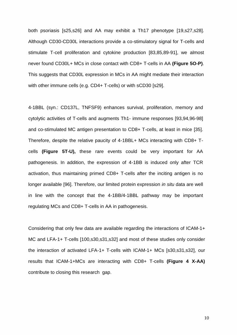
both psoriasis [s25,s26] and AA may exhibit a Th17 phenotype [19,s27,s28].
Although CD30-CD30L interactions provide a co-stimulatory signal for T-cells and
stimulate T-cell proliferation and cytokine production [83,85,89-91], we almost
never found CD30L+ MCs in close contact with CD8+ T-cells in AA (Figure 5O-P).
This suggests that CD30L expression in MCs in AA might mediate their interaction
with other immune cells (e.g. CD4+ T-cells) or with sCD30 [s29].
4-1BBL (syn.: CD137L, TNFSF9) enhances survival, proliferation, memory and
cytolytic activities of T-cells and augments Th1- immune responses [93,94,96-98]
and co-stimulated MC antigen presentation to CD8+ T-cells, at least in mice [35].
Therefore, despite the relative paucity of 4-1BBL+ MCs interacting with CD8+ T-
cells (Figure 5T-U), these rare events could be very important for AA
pathogenesis. In addition, the expression of 4-1BB is induced only after TCR
activation, thus maintaining primed CD8+ T-cells after the inciting antigen is no
longer available [96]. Therefore, our limited protein expression in situ data are well
in line with the concept that the 4-1BB/4-1BBL pathway may be important
regulating MCs and CD8+ T-cells in AA in pathogenesis.
Considering that only few data are available regarding the interactions of ICAM-1+
MC and LFA-1+ T-cells [100,s30,s31,s32] and most of these studies only consider
the interaction of activated LFA-1+ T-cells with ICAM-1+ MCs [s30,s31,s32], our
results that ICAM-1+MCs are interacting with CD8+ T-cells (Figure 4 X-AA)
contribute to closing this research gap.

11
Supplementary References:
s1. Christoph T, Muller-Rover S, Audring H, Tobin DJ, Hermes B, et al. (2000) The
human hair follicle immune system: cellular composition and immune privilege.
Br J Dermatol 142: 862-873.
s2. Peters EM, Liotiri S, Bodo E, Hagen E, Biro T, et al. (2007) Probing the effects of
stress mediators on the human hair follicle: substance P holds central position.
Am J Pathol 171: 1872-1886.
s3. Siebenhaar F, Sharov AA, Peters EM, Sharova TY, Syska W, et al. (2007)
Substance P as an immunomodulatory neuropeptide in a mouse model for
autoimmune hair loss (alopecia areata). J Invest Dermatol 127: 1489-1497.
s4. Vanderford DA, Greer PK, Sharp JM, Chichlowski M, Rouse DC, et al. (2010)
Alopecia in IL-10-deficient mouse pups is c-kit-dependent and can be
triggered by iron deficiency. Exp Dermatol 19: 518-526.
s5. Sugawara K, Zakany N, Hundt T, Emelianov V, Tsuruta D, et al. (2013)
Cannabinoid receptor 1 controls human mucosal-type mast cell degranulation
and maturation in situ. J Allergy Clin Immunol 132: 182-193.
s6. Taketomi Y, Ueno N, Kojima T, Sato H, Murase R, et al. (2013) Mast cell
maturation is driven via a group III phospholipase A2-prostaglandin D2-DP1
receptor paracrine axis. Nat Immunol 14: 554-563.
s7. Lu Z, Hasse S, Bodo E, Rose C, Funk W, et al. (2007) Towards the development
of a simplified long-term organ culture method for human scalp skin and its
appendages under serum-free conditions. Exp Dermatol 16: 37-44.
s8. Dvorak AM (2002) Ultrastructure of human mast cells. Int Arch Allergy Immunol
127: 100-105.

12
s9. Hammel I, Lagunoff D, Galli SJ (2010) Regulation of secretory granule size by the
precise generation and fusion of unit granules. J Cell Mol Med 14: 1904-1916.
s10. Spinnler K, Frohlich T, Arnold GJ, Kunz L, Mayerhofer A (2011) Human tryptase
cleaves pro-nerve growth factor (pro-NGF): hints of local, mast cell-dependent
regulation of NGF/pro-NGF action. J Biol Chem 286: 31707-31713.
s11. Li T, He S (2006) Induction of IL-6 release from human T cells by PAR-1 and
PAR-2 agonists. Immunol Cell Biol 84: 461-466.
s12. Krejci-Papa NC, Paus R (1998) A novel in-situ-zymography technique localizes
gelatinolytic activity in human skin to mast cells. Exp Dermatol 7: 321-326.
s13. Lohi J, Harvima I, Keski-Oja J (1992) Pericellular substrates of human mast cell
tryptase: 72,000 dalton gelatinase and fibronectin. J Cell Biochem 50: 337-
349.
s14. Moormann C, Artuc M, Pohl E, Varga G, Buddenkotte J, et al. (2006) Functional
characterization and expression analysis of the proteinase-activated receptor-
2 in human cutaneous mast cells. J Invest Dermatol 126: 746-755.
s15. Balato A, Lembo S, Mattii M, Schiattarella M, Marino R, et al. (2012) IL-33 is
secreted by psoriatic keratinocytes and induces pro-inflammatory cytokines
via keratinocyte and mast cell activation. Exp Dermatol 21: 892-894.
s16. Hunter HJ, Griffiths CE, Kleyn CE (2013) Does psychosocial stress play a role in
the exacerbation of psoriasis? Br J Dermatol 169: 965-974.
s17. Steinhoff M, Neisius U, Ikoma A, Fartasch M, Heyer G, et al. (2003) Proteinase-
activated receptor-2 mediates itch: a novel pathway for pruritus in human skin.
J Neurosci 23: 6176-6180.
s18. Kim HM, Lim YY, Kim MY, Son IP, Kim DH, et al. (2013) Inhibitory effect of
tianeptine on catagen induction in alopecia areata-like lesions induced by
ultrasonic wave stress in mice. Clin Exp Dermatol 38: 758-767.

13
s19. Zhang X, Yu M, Yu W, Weinberg J, Shapiro J, et al. (2009) Development of
alopecia areata is associated with higher central and peripheral hypothalamic-
pituitary-adrenal tone in the skin graft induced C3H/HeJ mouse model. J
Invest Dermatol 129: 1527-1538.
s20. Fujita T, Kambe N, Uchiyama T, Hori T (2006) Type I interferons attenuate T cell
activating functions of human mast cells by decreasing TNF-alpha production
and OX40 ligand expression while increasing IL-10 production. J Clin Immunol
26: 512-518.
s21. Gri G, Piconese S, Frossi B, Manfroi V, Merluzzi S, et al. (2008) CD4+CD25+
regulatory T cells suppress mast cell degranulation and allergic responses
through OX40-OX40L interaction. Immunity 29: 771-781.
s22. Nakano N, Nishiyama C, Yagita H, Koyanagi A, Akiba H, et al. (2009) Notch
signaling confers antigen-presenting cell functions on mast cells. J Allergy Clin
Immunol 123: 74-81 e71.
s23. Sibilano R, Frossi B, Suzuki R, D'Inca F, Gri G, et al. (2012) Modulation of
FcepsilonRI-dependent mast cell response by OX40L via Fyn, PI3K, and
RhoA. J Allergy Clin Immunol 130: 751-760 e752.
s24. Kotani A, Hori T, Fujita T, Kambe N, Matsumura Y, et al. (2007) Involvement of
OX40 ligand+ mast cells in chronic GVHD after allogeneic hematopoietic stem
cell transplantation. Bone Marrow Transplant 39: 373-375.
s25. Ariza ME, Williams MV, Wong HK (2013) Targeting IL-17 in psoriasis: from
cutaneous immunobiology to clinical application. Clin Immunol 146: 131-139.
s26. Girolomoni G, Mrowietz U, Paul C (2012) Psoriasis: rationale for targeting
interleukin-17. Br J Dermatol 167: 717-724.

14
s27. Lew BL, Cho HR, Haw S, Kim HJ, Chung JH, et al. (2012) Association between
IL17A/IL17RA Gene polymorphisms and susceptibility to alopecia areata in
the Korean population. Ann Dermatol 24: 61-65.
s28. Tojo G, Fujimura T, Kawano M, Ogasawara K, Kambayashi Y, et al. (2013)
Comparison of interleukin-17- producing cells in different clinical types of
alopecia areata. Dermatology 227: 78-82.
s29. Velasquez SY, Garcia LF, Opelz G, Alvarez CM, Susal C (2013) Release of
soluble CD30 after allogeneic stimulation is mediated by memory T cells and
regulated by IFN-gamma and IL-2. Transplantation 96: 154-161.
s30. Brill A, Baram D, Sela U, Salamon P, Mekori YA, et al. (2004) Induction of mast
cell interactions with blood vessel wall components by direct contact with
intact T cells or T cell membranes in vitro. Clin Exp Allergy 34: 1725-1731.
s31. Inamura N, Mekori YA, Bhattacharyya SP, Bianchine PJ, Metcalfe DD (1998)
Induction and enhancement of Fc(epsilon)RI-dependent mast cell
degranulation following coculture with activated T cells: dependency on ICAM-
1- and leukocyte function-associated antigen (LFA)-1-mediated heterotypic
aggregation. J Immunol 160: 4026-4033.
s32. Nagai K, Takahashi Y, Mikami I, Fukusima T, Oike H, et al. (2009) The
hydroxyflavone, fisetin, suppresses mast cell activation induced by interaction
with activated T cell membranes. Br J Pharmacol 158: 907-919.
s33. Shikotra A, Choy DF, Ohri CM, Doran E, Butler C, et al. (2012) Increased
expression of immunoreactive thymic stromal lymphopoietin in patients with
severe asthma. J Allergy Clin Immunol 129: 104-111 e101-109.
s34. Phillips TA, Ni J, Hunt JS (2003) Cell-specific expression of B lymphocyte
(APRIL, BLyS)- and Th2 (CD30L/CD153)-promoting tumor necrosis factor
superfamily ligands in human placentas. J Leukoc Biol 74: 81-87.

15
s35. Zhao S, Zhang H, Xing Y, Natkunam Y (2013) CD137 ligand is expressed in
primary and secondary lymphoid follicles and in B-cell lymphomas: diagnostic
and therapeutic implications. Am J Surg Pathol 37: 250-258.
s36. Goval JJ, Greimers R, Boniver J, de Leval L (2006) Germinal center dendritic
cells express more ICAM-1 than extrafollicular dendritic cells and ICAM-1/LFA-
1 interactions are involved in the capacity of dendritic cells to induce PBMCs
proliferation. J Histochem Cytochem 54: 75-84.
s37. Poindexter NJ, Sahin A, Hunt KK, Grimm EA (2004) Analysis of dendritic cells in
tumor-free and tumor-containing sentinel lymph nodes from patients with
breast cancer. Breast Cancer Res 6: R408-415.
s38. Kshirsagar SK, Alam SM, Jasti S, Hodes H, Nauser T, et al. (2012)
Immunomodulatory molecules are released from the first trimester and term
placenta via exosomes. Placenta 33: 982-990.
s39. Lee EJ, Xu L, Kim GH, Kang SK, Lee SW, et al. (2012) Regeneration of
peripheral nerves by transplanted sphere of human mesenchymal stem cells
derived from embryonic stem cells. Biomaterials 33: 7039-7046.
s40. Delfortrie S, Pinte S, Mattot V, Samson C, Villain G, et al. (2011) Egfl7 promotes
tumor escape from immunity by repressing endothelial cell activation. Cancer
Res 71: 7176-7186.
s41. Shi F, Shi M, Zeng Z, Qi RZ, Liu ZW, et al. (2011) PD-1 and PD-L1 upregulation
promotes CD8(+) T-cell apoptosis and postoperative recurrence in
hepatocellular carcinoma patients. Int J Cancer 128: 887-896.
s42. Garza LA, Yang CC, Zhao T, Blatt HB, Lee M, et al. (2011) Bald scalp in men
with androgenetic alopecia retains hair follicle stem cells but lacks CD200-rich
and CD34-positive hair follicle progenitor cells. J Clin Invest 121: 613-622.

16
s43. Paus R, van der Veen C, Eichmuller S, Kopp T, Hagen E, et al. (1998)
Generation and cyclic remodeling of the hair follicle immune system in mice. J
Invest Dermatol 111: 7-18.
s44. Shin K, Watts GF, Oettgen HC, Friend DS, Pemberton AD, et al. (2008) Mouse
mast cell tryptase mMCP-6 is a critical link between adaptive and innate
immunity in the chronic phase of Trichinella spiralis infection. J Immunol 180:
4885-4891.

17
Supplementary Figure S1. Positive controls for triple-immunostainings
Specific single OX40L staining (A) showing OX40L+ activated T lymphocytes in the
marginal zone of tonsil follicles [s33] and triple OX40L/CD8/Tryptase staining (B).
Blue arrows indicate CD8+ T-cells, pink arrows indicate tryptase+ cells and green
arrows indicate OX40L+/tryptase+ MCs. CD30L is expressed in human placental
villous endothelial cells [s34], a positive control (C), along with tonsil sections (D)
for the triple-staining CD30L/c-Kit/CD8. Red arrows indicate CD30L+ cells, blue
arrows indicate c-Kit+ MCs, brown arrows indicate CD8+ T-cells and green arrows
indicate CD30L+/c-Kit+ MCs. 4-1BBL+ cells are found in the marginal zone of
human tonsil follicles [s35] (E), as shown also by triple 4-1BBL/c-Kit/CD8
immunostaining (F). Red arrows indicate 4-1BBL+ cells, blue arrows c-Kit+ MCs,
brown arrows CD8+ T-cells, and green arrows 4-1BBL+/c-Kit+ MCs. Specific single
staining for ICAM-1 (G) showing ICAM-1 immunoreactivity in the germinal centre
[s36] and sparse positive cells of human tonsils (H), our positive control for ICAM-
1/CD8/Tryptase triple-immunostaining. Brown arrows indicate ICAM-1+ cells, blue
arrows CD8+ T-cells, and pink arrows tryptase+ cells. Many IL-10+ cells are found
in tonsil sections [s37] as shown in our single IL-10 (I) but also triple IL-10/c-
Kit/CD8 (J) stainings. Red arrows indicate IL-10+ cells, blue arrows indicate c-Kit+
MCs, brown arrows indicate CD8+ T-cells and green arrows indicate IL-10+/c-Kit+
MCs. PD-L1 immunoreactivity in chorionic villi of placenta [s38] detected by single
PD-L1 (K) and triple PD-L1/c-Kit/CD8 (L) stainings. Red arrows indicate PD-L1+
cells, blue arrows indicate c-Kit+ MCs, brown arrows indicate CD8+ T-cells and
green arrows indicate PD-L1+/c-Kit+ MCs. Positive CD200+ cells are found in the

18
HF bulge [8,59] (M), also for the triple staining CD200/CD8/Tryptase (N). Brown
arrows indicate CD200+ cells, pink arrows indicate tryptase+ cells, blue arrows
indicate CD8+ cells.
Supplementary Figure S2. MC density is significantly increased in lesional
skin compared to non-lesional skin of AA patients and scalp skin from
healthy subjects. The number of proliferating c-Kit+ MC is tendentially
increased in AA lesional skin compared to control skin.
Quantitative analysis of the tryptase+ MCs (analysed using OX40L/CD8/tryptase) in
lesional AA compared to non-lesional skin from AA patients and scalp skin from
healthy subjects. Analysis derived from 17-21 areas of 6-14 HFs of 6-7 healthy
controls and of 11-21 areas of 4-12 HFs of 3 AA patients for non-lesional skin and
of 17-21 areas of 16-17 HFs of 7 AA patients for lesional skin (A). ***p≤0.001,

19
*p≤0.05 ±SEM, One-Way ANOVA followed respectively by Bonferroni´s multiple
comparison tests.
Quantitative analysis of MC proliferation by Ki-67/c-Kit IHC (B). Analysis deriving
from 31 areas of 30 HFs of 10 AA patients and 35 areas of 35 HFs of 7 healthy
controls, ±SEM, Mann-Whitney-U-Test (ns).
Connective tissue sheath (CTS), perifollicular dermis (PFD).
Supplementary Figure S3. The maximal increase of MC density is found in
AA patients in the subacute stage of disease.
Representative pictures showing tryptase+ cells increase in acute (A), subacute
(B) and chronic (C) AA patients detected by Ki-67/tryptase double IHC.
Scale bars: 50µm.
Fold increase of MC density according to the histological features and clinical
evaluation groups evaluated using Ki-67/tryptase IHC (D). Analysis derived from
81 areas (HFs) of 17 AA patients and 50 areas (HFs) of 7 healthy controls, ±SEM,
One Way Anova (p<0.0001) (including the control values, black line) followed by
Bonferroni´s Test (**p≤0.01, ***p≤0.001).
Fold increase of MC density according to the histological features and clinical
evaluation groups evaluated using Ki-67/c-Kit IHC (E). Analysis derived from 31

20
areas of 30 HFs of 10 AA patients and 35 areas of 35 HFs of 7 healthy controls,
±SEM, One Way Anova (p<0.0001) (including the control values, black line)
followed by Bonferroni´s Test (*p≤0.05, **p≤0.01, ***p≤0.001). Black line indicates
the control. The fold increase was calculated by dividing all the MC density values
of acute, subacute, chronic AA and controls (n.of MC/mm2) with the mean value of
the control CTS or PFD. AA patients were divided into three groups following their
histological features [65] and clinical evaluations supplied by the dermatologist
which are respectively acute (n=3-4), subacute (n=10) and chronic (n=3) stages.
Connective tissue sheath (CTS), hair follicle (HF), perifollicular dermis (PFD),
sebaceous gland (SG).
Supplementary Figure S4. TGFβ1 immunoreactivity is decreased in the HF
ORS of AA patients.

21
Representative pictures of TGFβ1 IR in the HF ORS of healthy individuals (A) and
AA patients (B). Scale bars: 100µm.
Quantitative (immuno-)histomorphometry by using Image J of TGFβ1 IR in AA HFs
compared to healthy HFs (C).
Analysis derived from 41 areas of 21 HFs of 4 healthy controls and of 90 areas of
61 HFs of 17 AA patients and, ±SEM, Student t-Test, *p≤0.05, ***p≤0.001.
Connective tissue sheath (CTS), inner root sheath (IRS), outer root sheath (ORS).
Supplementary Figure S5. MHCI/CD8/Tryptase triple staining showing MHC
class I+ MCs in close contact with CD8+ T-cells
Representative pictures of MHCI/CD8/Tryptase in human tonsil (A-B), the positive
control and AA skin (C-D). Higher magnification of MHC class I+ MCs in close
contact with CD8+ T-cells in the small panels (C-D). Scale bars: 50µm.

22

23
Supplementary Table S1. Primary Antibodies
Antibodies used for immmunohistochemical and immunofluorescence stainings are
listed and described in detail.
Anti-human antibodies
Primary
antibody Origin Clone Vendor Dilution Sections Reference
C-Kit Rabbit DAKO 1:100
1:400 Paraffin [47]
Tryptase Mouse AA1
Abcam,
Cambridge,
UK
1:500
1:1000 Paraffin [47]
Ki-67 Mouse Tec-3 DAKO 1:10 Paraffin [60]
TGFß1 Rabbit Santa Cruz 1:100 Paraffin [58]
MHCI Abcam EMR8-5 Abcam 1:50 Paraffin [169]
CD8 Mouse C8/144B DAKO 1:100
1:500 Paraffin, [59]
OX40L Mouse 159403 R&D
Sytem 1:25 Paraffin [76]
CD30L Mouse 116614 R&D
Sytem 1:50 Paraffin [86]
4-1BBL Rabbit Abcam 1:200 Paraffin [s35]
ICAM-1 Rabbit EP1442Y Abcam 1:100 Paraffin [s40]
IL-10 Mouse 23738 R&D
Sytem 1:25 Paraffin [s37]
PD-L1 Mouse 29E2A3 BioLegend 1:100 Paraffin [s41]
CD200 Goat R&D
System 1:200 Paraffin [s42]
Anti-mouse antibodies
C-Kit Rat 2B8 BD
Pharmigen 1:100 Cryo [s43]
CD8a Rat 53-6.7 BD
Pharmigen 1:10 Cryo [58]
mMCP6 Rabbit M. Gurish 1:500 Cryo [s44]
24
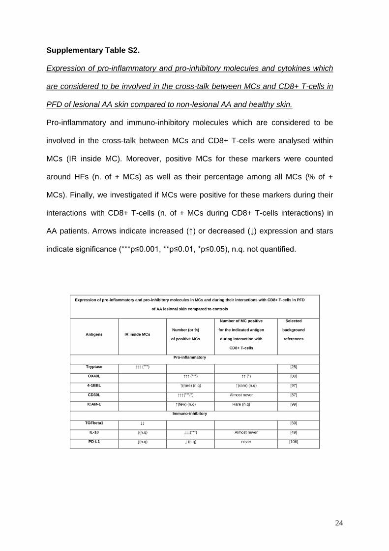
Supplementary Table S2.
Expression of pro-inflammatory and pro-inhibitory molecules and cytokines which
are considered to be involved in the cross-talk between MCs and CD8+ T-cells in
PFD of lesional AA skin compared to non-lesional AA and healthy skin.
Pro-inflammatory and immuno-inhibitory molecules which are considered to be
involved in the cross-talk between MCs and CD8+ T-cells were analysed within
MCs (IR inside MC). Moreover, positive MCs for these markers were counted
around HFs (n. of + MCs) as well as their percentage among all MCs (% of +
MCs). Finally, we investigated if MCs were positive for these markers during their
interactions with CD8+ T-cells (n. of + MCs during CD8+ T-cells interactions) in
AA patients. Arrows indicate increased (↑) or decreased (↓) expression and stars
indicate significance (***p≤0.001, **p≤0.01, *p≤0.05), n.q. not quantified.
Expression of pro-inflammatory and pro-inhibitory molecules in MCs and during their interactions with CD8+ T-cells in PFD
of AA lesional skin compared to controls
Antigens IR inside MCs Number (or %)
of positive MCs
Number of MC positive
for the indicated antigen
during interaction with
CD8+ T-cells
Selected
background
references
Pro-inflammatory
Tryptase ↑↑↑ (***) [25]
OX40L ↑↑↑ (***) ↑↑ (*) [80]
4-1BBL ↑(rare) (n.q) ↑(rare) (n.q) [97]
CD30L ↑↑↑(***/*) Almost never [87]
ICAM-1 ↑(few) (n.q) Rare (n.q) [99]
Immuno-inhibitory
TGFbeta1 ↓↓ [69]
IL-10 ↓(n.q) ↓↓↓(***) Almost never [49]
PD-L1 ↓(n.q) ↓ (n.q) never [106]