plos pathogens
38
RESEARCH ARTICLE Differential contribution of two organelles of endosymbiotic origin to iron-sulfur cluster synthesis and overall fitness in Toxoplasma Sarah Pamukcu 1 , Aude Cerutti ID 1 , Yann Bordat ID 1 , Sonia Hem 2 , Vale ´ rie Rofidal ID 2 , Se ´ bastien BesteiroID 3 * 1 LPHI, Univ Montpellier, CNRS, Montpellier, France, 2 BPMP, Univ Montpellier, CNRS, INRAE, Institut Agro, Montpellier, France, 3 LPHI, Univ Montpellier, CNRS, INSERM, Montpellier, France * [email protected] Abstract Iron-sulfur (Fe-S) clusters are one of the most ancient and ubiquitous prosthetic groups, and they are required by a variety of proteins involved in important metabolic processes. Api- complexan parasites have inherited different plastidic and mitochondrial Fe-S clusters bio- synthesis pathways through endosymbiosis. We have investigated the relative contributions of these pathways to the fitness of Toxoplasma gondii, an apicomplexan parasite causing disease in humans, by generating specific mutants. Phenotypic analysis and quantitative proteomics allowed us to highlight notable differences in these mutants. Both Fe-S cluster synthesis pathways are necessary for optimal parasite growth in vitro, but their disruption leads to markedly different fates: impairment of the plastidic pathway leads to a loss of the organelle and to parasite death, while disruption of the mitochondrial pathway trigger differ- entiation into a stress resistance stage. This highlights that otherwise similar biochemical pathways hosted by different sub-cellular compartments can have very different contribu- tions to the biology of the parasites, which is something to consider when exploring novel strategies for therapeutic intervention. Author summary Toxoplasma gondii is a ubiquitous unicellular parasite that harbours two organelles of endosymbiotic origin: the mitochondrion, and a relict plastid named the apicoplast. Each one of these organelles contains its own machinery for synthesizing iron-sulfur clusters, which are important protein co-factors. In this study, we show that interfering with either the mitochondrial or the plastidic iron-sulfur cluster synthesizing machinery has a pro- found impact on parasite growth. However, while disrupting the plastidic pathway led to an irreversible loss of the organelle and subsequent death of the parasite, disrupting the mitochondrial pathway led to conversion of the parasites into a stress resistance form. We used a comparative quantitative proteomic analysis of the mutants, combined with experi- mental validation, to provide mechanistic clues into these different phenotypic outcomes. Although the consequences of disrupting each pathway were manifold, our data PLOS PATHOGENS PLOS Pathogens | https://doi.org/10.1371/journal.ppat.1010096 November 18, 2021 1 / 38 a1111111111 a1111111111 a1111111111 a1111111111 a1111111111 OPEN ACCESS Citation: Pamukcu S, Cerutti A, Bordat Y, Hem S, Rofidal V, Besteiro S (2021) Differential contribution of two organelles of endosymbiotic origin to iron-sulfur cluster synthesis and overall fitness in Toxoplasma. PLoS Pathog 17(11): e1010096. https://doi.org/10.1371/journal. ppat.1010096 Editor: Dominique Soldati-Favre, University of Geneva, SWITZERLAND Received: September 1, 2021 Accepted: November 5, 2021 Published: November 18, 2021 Peer Review History: PLOS recognizes the benefits of transparency in the peer review process; therefore, we enable the publication of all of the content of peer review and author responses alongside final, published articles. The editorial history of this article is available here: https://doi.org/10.1371/journal.ppat.1010096 Copyright: © 2021 Pamukcu et al. This is an open access article distributed under the terms of the Creative Commons Attribution License, which permits unrestricted use, distribution, and reproduction in any medium, provided the original author and source are credited. Data Availability Statement: All raw MS data and MaxQuant files generated have been deposited to the ProteomeXchange Consortium via the PRIDE
-
Upload
khangminh22 -
Category
Documents
-
view
0 -
download
0
Transcript of plos pathogens

RESEARCH ARTICLE
Differential contribution of two organelles of
endosymbiotic origin to iron-sulfur cluster
synthesis and overall fitness in Toxoplasma
Sarah Pamukcu1, Aude CeruttiID1, Yann BordatID
1, Sonia Hem2, Valerie RofidalID2,
Sebastien BesteiroID3*
1 LPHI, Univ Montpellier, CNRS, Montpellier, France, 2 BPMP, Univ Montpellier, CNRS, INRAE, Institut
Agro, Montpellier, France, 3 LPHI, Univ Montpellier, CNRS, INSERM, Montpellier, France
Abstract
Iron-sulfur (Fe-S) clusters are one of the most ancient and ubiquitous prosthetic groups, and
they are required by a variety of proteins involved in important metabolic processes. Api-
complexan parasites have inherited different plastidic and mitochondrial Fe-S clusters bio-
synthesis pathways through endosymbiosis. We have investigated the relative contributions
of these pathways to the fitness of Toxoplasma gondii, an apicomplexan parasite causing
disease in humans, by generating specific mutants. Phenotypic analysis and quantitative
proteomics allowed us to highlight notable differences in these mutants. Both Fe-S cluster
synthesis pathways are necessary for optimal parasite growth in vitro, but their disruption
leads to markedly different fates: impairment of the plastidic pathway leads to a loss of the
organelle and to parasite death, while disruption of the mitochondrial pathway trigger differ-
entiation into a stress resistance stage. This highlights that otherwise similar biochemical
pathways hosted by different sub-cellular compartments can have very different contribu-
tions to the biology of the parasites, which is something to consider when exploring novel
strategies for therapeutic intervention.
Author summary
Toxoplasma gondii is a ubiquitous unicellular parasite that harbours two organelles of
endosymbiotic origin: the mitochondrion, and a relict plastid named the apicoplast. Each
one of these organelles contains its own machinery for synthesizing iron-sulfur clusters,
which are important protein co-factors. In this study, we show that interfering with either
the mitochondrial or the plastidic iron-sulfur cluster synthesizing machinery has a pro-
found impact on parasite growth. However, while disrupting the plastidic pathway led to
an irreversible loss of the organelle and subsequent death of the parasite, disrupting the
mitochondrial pathway led to conversion of the parasites into a stress resistance form. We
used a comparative quantitative proteomic analysis of the mutants, combined with experi-
mental validation, to provide mechanistic clues into these different phenotypic outcomes.
Although the consequences of disrupting each pathway were manifold, our data
PLOS PATHOGENS
PLOS Pathogens | https://doi.org/10.1371/journal.ppat.1010096 November 18, 2021 1 / 38
a1111111111
a1111111111
a1111111111
a1111111111
a1111111111
OPEN ACCESS
Citation: Pamukcu S, Cerutti A, Bordat Y, Hem S,
Rofidal V, Besteiro S (2021) Differential
contribution of two organelles of endosymbiotic
origin to iron-sulfur cluster synthesis and overall
fitness in Toxoplasma. PLoS Pathog 17(11):
e1010096. https://doi.org/10.1371/journal.
ppat.1010096
Editor: Dominique Soldati-Favre, University of
Geneva, SWITZERLAND
Received: September 1, 2021
Accepted: November 5, 2021
Published: November 18, 2021
Peer Review History: PLOS recognizes the
benefits of transparency in the peer review
process; therefore, we enable the publication of
all of the content of peer review and author
responses alongside final, published articles. The
editorial history of this article is available here:
https://doi.org/10.1371/journal.ppat.1010096
Copyright: © 2021 Pamukcu et al. This is an open
access article distributed under the terms of the
Creative Commons Attribution License, which
permits unrestricted use, distribution, and
reproduction in any medium, provided the original
author and source are credited.
Data Availability Statement: All raw MS data and
MaxQuant files generated have been deposited to
the ProteomeXchange Consortium via the PRIDE

highlighted potential changes at the metabolic level. For instance, the plastidic iron-sulfur
cluster synthesis pathway may be important for maintaining the lipid homeostasis of the
parasites, while the mitochondrial pathway is likely crucial for maintaining their respira-
tory capacity. Interestingly, we have discovered that other mutants severely impacted for
mitochondrial function, in particular the respiratory chain, are able to survive and initiate
conversion to the stress resistance form. This illustrates a different capacity for T. gondiito adapt for survival in response to distinct metabolic dysregulations.
Introduction
Endosymbiotic events were crucial in the evolutionary timeline of eukaryotic cells. Mitochon-
dria and plastids evolved from free-living prokaryotes that were taken up by early eukaryotic
ancestors and transformed into permanent subcellular compartments that have become essen-
tial for harnessing energy or synthesizing essential metabolites in present-day eukaryotes [1].
As semiautonomous organelles, they contain a small genome, but during the course of evolu-
tion a considerable part of their genes have been transferred to the cell nucleus. Yet, they rely
largely on nuclear factors for their maintenance and expression. Both organelles are involved
in critically important biochemical processes. Mitochondria, which are found in most eukary-
otic organisms, are mostly known as the powerhouses of the cell, owing to their ability to pro-
duce ATP through respiration. Importantly, they are also involved in several other metabolic
pathways [2], including the synthesis of heme groups, steroids, amino acids, and iron-sulfur
(Fe-S) clusters. Moreover, they have important cellular functions in regulating redox and cal-
cium homeostasis. Similarly, plastids that are found in plants, algae and some other eukaryotic
organisms, host a diverse array of pathways that contribute greatly to the cellular metabolism
[3]. While often identified mainly as compartments where photosynthesis occurs, plastids host
other important metabolic pathways. For example, they are involved in the assimilation of
nitrogen and sulfur, as well as the synthesis of carbohydrates, amino acids, fatty acids and spe-
cific lipids, hormone precursors, and also Fe-S clusters. The best-characterized plastid is argu-
ably the plant cell chloroplast, but not all plastids have photosynthetic function, and in land
plants they are in fact a diverse group of organelles that share basal metabolic pathways, but
also have specific physiological roles [4].
The phylum Apicomplexa comprises a large number of single-celled protozoan parasites
responsible for serious disease in animals, including humans. For example, this phylum
includes parasites of the genus Plasmodium that are responsible for the deadly malaria, and
Toxoplasma gondii a ubiquitous parasite that can lead to a severe pathology in immunocom-
promised individuals. Apicomplexan parasites evolved from a photosynthetic ancestor and
many of them still retain a plastid [5,6]. This plastid, named the apicoplast, originated from a
secondary endosymbiotic event [7,8]. It has lost its photosynthetic properties as the ancestors
of Apicomplexa switched to an intracellular parasitic lifestyle [9]. The apicoplast nevertheless
still hosts four main metabolic pathways [10,11]: a so-called non-mevalonate pathway for the
synthesis of isoprenoid precursors, a type II fatty acid synthesis pathway (FASII), part of the
heme synthesis pathway, and a Fe-S cluster synthesis pathway. As the apicoplast is involved in
these important biological processes for the parasite, and as they markedly differ from those of
the host (because of their algal origin), that makes it a valuable potential drug target. Apicom-
plexan parasites also generally contain a single tubular mitochondrion, although its aspect may
vary during parasite development [12,13]. The organelle is an important contributor to the
parasite’s metabolic needs [14]. It classically hosts tricarboxylic acid (TCA) cycle reactions,
PLOS PATHOGENS Iron-sulfur cluster synthesis in Toxoplasma
PLOS Pathogens | https://doi.org/10.1371/journal.ppat.1010096 November 18, 2021 2 / 38
partner repository (https://www.ebi.ac.uk/pride/)
with the dataset identifier PXD023854.
Funding: AC was supported by a fellowship from
the Fondation pour la Recherche Medicale (Equipe
FRM EQ20170336725), https://www.frm.org/; SB
acknowlegdes support from the Labex Parafrap
(ANR-11-LABX-0024), https://www.labex-parafrap.
fr/en/, and the Agence Nationale de la Recherche
(ANR-19-CE15-0023), https://anr.fr/en/. The
funders had no role in study design, data collection
and analysis, decision to publish, or preparation of
the manuscript.
Competing interests: The authors have declared
that no competing interests exist.

which are the main source of electrons that feeds the mitochondrial electron transport chain
(ETC) which generates a proton gradient used for ATP production. It also contains additional
metabolic pathways, like a Fe-S cluster synthesis pathway and part of the heme synthesis path-
way operating in collaboration with the apicoplast. The latter reflects obvious functional links
between the organelles and potential metabolic interactions, which is also illustrated by their
physical connection during parasite development [15,16].
Fe-S clusters are simple and ubiquitous cofactors involved in a great variety of cellular pro-
cesses. As their name implies, they are composed of iron and inorganic sulfur whose chemical
properties confer key structural or electron transfer features to proteins in all kingdoms of life.
They are important to the activities of numerous proteins that play essential roles to sustain
fundamental life processes including, in addition to electron transfer and exchange, iron stor-
age, protein folding, oxygen/nitrogen stress sensing, and gene regulation [17]. The synthesis of
Fe-S clusters and their insertion into apoproteins requires complex machineries and several
distinct pathways have been identified in bacteria for synthesizing these ancient cofactors [18].
They include the ISC (iron-sulfur cluster) pathway for general Fe–S cluster assembly [19], and
the SUF (sulfur formation) pathway [20] that is potentially activated in oxidative stress condi-
tions [21]. Eukaryotes have inherited machineries for synthesizing Fe-S cluster through their
endosymbionts [22]. As a result, organisms with both mitochondria and plastids, like land
plants, use the ISC pathway for assembling Fe-S clusters in the mitochondria and the SUF
pathway for Fe-S clusters in the plastids [23]. Additional protein components that constitute a
cytosolic Fe-S cluster assembly machinery (CIA) have also been identified: this pathway is
important for the generation of cytosolic, but also of nuclear Fe-S proteins, and is highly
dependent on the ISC mitochondrial pathway for providing a sulfur-containing precursor
[24].
Like in plants and algae, apicoplast-containing Apicomplexa seem to harbour the three
(ISC, SUF and CIA) Fe-S cluster synthesis pathways. Although the CIA pathway was recently
shown to be important for Toxoplasma fitness [25], investigations in apicomplexan parasites
have been so far mostly conducted in Plasmodium species and they essentially focused on the
apicoplast-located SUF pathway [26–31]. The SUF pathway was shown to be essential for the
viability of malaria parasites during both the erythrocytic and sexual stages of development,
and has thus been recognized as a putative avenue for discovering new antiparasitic drug tar-
gets (reviewed in [32]). Contrarily to the ISC pathway, which is also present in the mammalian
hosts of apicomplexan parasites, the SUF pathway may indeed yield interesting specificities
that may be leveraged for therapeutic intervention. However, very little is known about Fe-S
clusters synthesis in other apicomplexan parasites, including T. gondii. For instance, out of the
four known metabolic pathways hosted by the apicoplast, Fe-S cluster synthesis was the only
one remaining to be functionally investigated in T. gondii, while the others were all shown to
be essential for the tachyzoite stage of the parasite (a fast replicating developmental stage
responsible for the symptoms of the disease) [33–36]. Here, we present the characterization of
two T. gondii mutants we generated to specifically impact the plastidic and mitochondrial SUF
and ISC pathways, respectively. Our goal was to assess the relative contributions of these com-
partmentalized pathways to parasite development and fitness.
Results
TgNFS2 and TgISU1 are functional homologs of components of the Fe-S
cluster synthesis pathways
Fe-S cluster biosynthesis pathways in the mitochondrion and the plastid follow a similar gen-
eral pattern: cysteine desulfurases (NFS1, NFS2) produce sulfur from L-cysteine, scaffold
PLOS PATHOGENS Iron-sulfur cluster synthesis in Toxoplasma
PLOS Pathogens | https://doi.org/10.1371/journal.ppat.1010096 November 18, 2021 3 / 38

proteins (ISU1, SufB/C/D) provide a molecular platform allowing iron and sulfur to meet and
form a cluster, and finally carrier proteins (like IscA or SufA) deliver the cluster to target apo-
proteins [23]. The cytosolic CIA pathway, which is responsible for the de novo formation of
Fe-S clusters to be incorporated in cytosolic and nuclear proteins, is dependent on the ISC
pathway, as its first step requires the import of a yet unknown sulfur-containing precursor that
is translocated to the cytosol from the mitochondrion [24]. To get a general overview of the
predicted components for the Fe-S cluster machinery in T. gondii, we conducted homology
searches in the ToxoDB.org database [37], using well-characterized proteins from plants (Ara-bidopsis thaliana) belonging to the SUF, ISC and CIA pathways (S1 Table). Data from global
mapping of protein subcellular location by hyperLOPIT spatial proteomics [38] was in general
in good accordance with the expected localization of the homologs (with the noticeable excep-
tion of members of the NBP35/HCF101 ATP-binding proteins). Overall, our search revealed
that T. gondii appears to have a good conservation of all the main components of the three
ISC, SUF and CIA Fe-S synthesis pathways (S1 Table and Fig 1A). Additional information
available on ToxoDB.org such as scores from a CRISPR/Cas9-based genome-wide screening
[39], highlighted that most components of the three pathways are predicted to be important
for parasite fitness. This suggests several Fe-S proteins localizing to the endosymbiotic organ-
elles, but also the cytosol/nucleus, are essential for the optimal growth of tachyzoites. In order
to verify this, our aim was to specifically interfere with the apicoplast-localized SUF pathway
or the mitochondrion-localized ISC pathway in T. gondii tachyzoites. More precisely, we
wanted to target the homologs of A. thaliana NFS2 and ISU1, which are both central (and pre-
sumably essential) to their respective pathways (Fig 1A): NFS2 is a cysteine desulfurase that
provides the inorganic sulfur part of plastidic Fe-S clusters, while ISU1 is a scaffold protein
important for cluster assembly in the mitochondrial pathway. Interfering with these key
Fig 1. TgNFS2 and TgISU1 are functional homologs of components of iron-sulfur cluster synthesis pathways. A) Putative Fe-S cluster synthesis
pathways and associated molecular machinery in Toxoplasma. B) Functional complementation of bacterial mutants for IscU (top) and SufS (bottom).
Growth of ‘wild-type’ (WT) E. coli K12 parental strain, bacterial mutant strains and strains complemented (‘comp’) by their respective T. gondiihomologues (‘comp’), was assessed by monitoring the optical density at 600 nm in the presence or not of an iron chelator (2,2’-bipyridyl, ‘chel’). Values
are mean from n = 3 independent experiments ±SEM. � denotes p� 0.05, Student’s t-test, when comparing values obtained in the absence of chelator
for mutant strains versus complemented ones.
https://doi.org/10.1371/journal.ppat.1010096.g001
PLOS PATHOGENS Iron-sulfur cluster synthesis in Toxoplasma
PLOS Pathogens | https://doi.org/10.1371/journal.ppat.1010096 November 18, 2021 4 / 38

players of the upstream machinery would likely lead to a comparable disruption of the respec-
tive Fe-S cluster biogenesis pathways hosted by each organelle [40].
As a first step, we sought to determine whether TgNFS2 (TGGT1_216170) and TgISU1
(TGGT1_237560) were real functional homologs by performing complementation assays of
bacterial mutants. In many bacteria, the ISC machinery is the primary system for general Fe-S
cluster biosynthesis, while the SUF system plays a similar general role, but is mostly operative
under stress conditions (like iron limitation or oxidative stress). In Escherichia coli, both path-
ways are partially redundant, but their individual disruption results in slowed bacterial growth,
especially when limiting iron availability with a specific chelator [41]. Expression of the pre-
dicted functional domains of TgNFS2 and TgISU1 in mutant strains for the corresponding E.
coli proteins (named SufS and IscU, respectively) improved bacterial growth, in the presence
of an iron chelator or not (Fig 1B). The complementation seemed partial as complemented
strains remained more sensitive to the iron chelator than the wild-type strain. Yet, and
although stationary phase was reached earlier than for the wild-type bacteria, contrarily to the
mutants the complemented strains showed a bacterial density close to that of the WT at sta-
tionary phase. This suggests TgNFS2 and TgISU1, in addition to a good sequence homology
with their bacterial homologues (S1 Fig), have a conserved function.
We next determined the sub-cellular localizations of TgNFS2 and TgISU1 by epitope tag-
ging of the native proteins. This was achieved in the TATi ΔKu80 cell line, which favors
homologous recombination and would allow transactivation of a Tet operator-modified pro-
moter we would later use for generating a conditional mutant in this background [42–44]. A
sequence coding for a C-terminal triple hemagglutinin (HA) epitope tag was inserted at the
endogenous TgNFS2 or TgISU1 locus by homologous recombination (S2 Fig). Using the anti-
HA antibody, by immunoblot we detected two products for each protein (Fig 2A and 2B),
likely corresponding to their immature and mature forms (ie after cleavage of the transit pep-
tide upon import into the organelle). Accordingly, the analysis of TgNFS2 and TgISU1
sequences with several subcellular localization and N-terminal sorting signals site predictors
confirmed they likely contained sequences for plastidic and mitochondrial targeting [45],
respectively, although no consensus position of the exact cleavage sites could be determined.
Immunofluorescence assay (IFA) in T. gondii tachyzoites confirmed HA-tagged TgNFS2 and
TgISU1 co-localize with markers of the apicoplast and the mitochondrion, respectively (Fig
2C and 2D).
NFS2 is a cysteine desulfurase whose activity is enhanced by an interaction with the SUFE
protein [46]. Similar to plants that express several SUFE homologues [47], there are two puta-
tive SUFE-like proteins in T. gondii (S1 Table), one of which was already predicted to reside in
the apicoplast by hyperLOPIT (TgSUFE1, TGGT1_239320). We generated a cell line express-
ing an HA-tagged version of the other, TgSUFE2 (TGGT1_277010, S3A–S3C Fig), whose
localization was previously unknown. Like for TgNFS2, several programs predicted a plastidic
transit peptide, which was confirmed by immunoblot analysis (detecting TgSUFE2 immature
and mature forms, S3D Fig). IFA showed TgSUFE2 co-localizes with an apicoplast marker
(S3E Fig). This further confirms that the initial steps of Fe-S cluster biogenesis in the apicoplast
are likely functionally-conserved.
Disruption of either the plastidic or the mitochondrial Fe-S cluster
pathway has a profound impact on parasite growth
In order to get insights into plastidic and mitochondrial Fe-S biogenesis, we generated condi-
tional mutant cell lines in the TgNFS2-HA or TgISU1-HA-expressing TATi ΔKu80 back-
ground [44]. Replacement of the endogenous promoters by an inducible-Tet07SAG4
PLOS PATHOGENS Iron-sulfur cluster synthesis in Toxoplasma
PLOS Pathogens | https://doi.org/10.1371/journal.ppat.1010096 November 18, 2021 5 / 38
promoter, through a single homologous recombination at the loci of interest (S4 Fig), yielded
TgNFS2 and TgISU1 conditional knock-down cell lines (cKD TgNFS2-HA and cKD TgI-
SU1-HA, respectively). In these cell lines, the addition of anhydrotetracycline (ATc) can
repress transcription through a Tet-Off system [48]. For each cKD cell line several transgenic
clones were obtained and were found to behave similarly in the initial phenotypic assays we
performed, so only one was analysed further. Transgenic parasites were grown for various
periods of time in presence of ATc, and protein down-regulation was evaluated. Immunoblot
and IFA analyses of cKD TgNFS2 -HA and cKD TgISU1-HA parasites showed that the addi-
tion of ATc efficiently down-regulated the expression of TgNFS2 (Fig 3A and 3C) and TgISU1(Fig 3B and 3D), and most of the proteins were undetectable after two days of incubation.
We also generated complemented cell lines expressing constitutively an additional copy of
TgNFS2 and TgISU1 from the uracil phosphoribosyltransferase (UPRT) locus from a tubulinpromoter in their respective conditional mutant backgrounds (S5A and S5B Fig). We con-
firmed by semi-quantitative RT-PCR (S5C Fig) that the transcription of TgNFS2 and TgISU1qenes was effectively repressed in the cKD cell lines upon addition of ATc, whereas the corre-
sponding complemented cell lines exhibited a high transcription level regardless of ATc addi-
tion (due to the expression from the strong tubulin promoter).
We next evaluated the consequences of TgNFS2 and TgISU1 depletion on T. gondii growth
in vitro. First, to assess the impact on the parasite lytic cycle, the capacity of the mutants and
complemented parasites to produce lysis plaques was analyzed on a host cells monolayer in
Fig 2. TgNFS2 and TgISU1 localize to the apicoplast and the mitochondrion, respectively. Detection by
immunoblot of C-terminally HA-tagged TgNFS2 (A) and TgISU1 (B) in parasite extracts reveals the presence of both
precusor (p) and mature (m) forms of the proteins. Anti-actin (TgACT1) antibody was used as a loading control.
Immunofluorescence assay shows TgNFS2 co-localizes with apicoplast marker TgCPN60 (C) and TgISU1 co-localizes
with mitochondrial marker F1 β ATPase (D). Scale bar represents 5 μm. DNA was labelled with DAPI. DIC:
differential interference contrast.
https://doi.org/10.1371/journal.ppat.1010096.g002
PLOS PATHOGENS Iron-sulfur cluster synthesis in Toxoplasma
PLOS Pathogens | https://doi.org/10.1371/journal.ppat.1010096 November 18, 2021 6 / 38

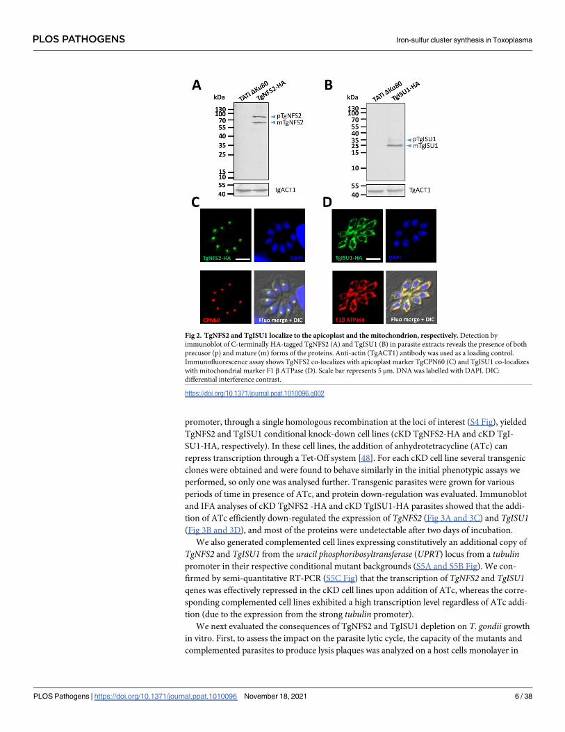
absence or continuous presence of ATc for 7 days (Fig 4A and 4B). Depletion of both proteins
completely prevented plaque formation, which was restored in the complemented cell lines.
To assess whether this defect in the lytic cycle is due to a replication problem, all cell lines were
preincubated in absence or presence of ATc for 48 hours and released mechanically, before
infecting new host cells and growing them for an additional 24 hours in ATc prior to parasite
counting. We noted that incubation with ATc led to an accumulation of vacuoles with fewer
parasites, yet that was not the case in the complemented cell lines (Fig 4C and 4D). Overall,
these data show that either TgNFS2 or TgISU1 depletion impacts parasite growth.
Then, we sought to assess if the viability of the mutant parasites was irreversibly affected.
We thus performed another series of plaque assays, but at the end of the 7-day incubation, we
washed out the ATc, incubated the parasites for an extra 7 days in the absence of ATc and eval-
uated plaque formation (Fig 4E). In these conditions, cKD TgNFS2-HA parasites displayed
very small plaques suggesting their viability was irreversibly impacted. In contrast, cKD TgI-
SU1-HA parasites showed plaques, suggesting parasite growth had at least partly resumed after
ATc washout, while host cell lysis remained limited if the drug was kept continuously during
the same period of time. This suggests that although depletion of TgISU1 has a marked impact
on parasite growth, it is not completely lethal.
We performed IFAs to assess possible morphological defects that may explain the impaired
growths of cKD TgNFS2-HA and cKD TgISU1-HA parasites. We stained the apicoplast and
mitochondrion of parasites kept in the continuous presence of ATc for several days. cKD
TgNFS2-HA parasites managed to grow and egress after three days and were seeded onto new
host cells, where there were kept for two more days in the presence of ATc. During this second
phase of intracellular development, and in accordance with the replication assays (Fig 4C),
growth was slowed down considerably. Strikingly, while the mitochondrial network seemed
Fig 3. Efficient down-regulation of TgNFS2 and TgISU1 expression with anhydrotetracyclin (ATc). A) Immunoblot analysis with
anti-HA antibody shows efficient down-regulation of TgNFS2 after 48h of incubation with ATc. Anti-SAG1 antibody was used as a
loading control. B) Immunoblot analysis with anti-HA antibody shows efficient down-regulation of TgISU1 after 24h of incubation with
ATc. Anti-SAG1 antibody was used as a loading control. C) and D) Immunofluorescence assays show TgNFS2 and TgISU1 are not
detectable anymore after 48h of incubation with ATc. Scale bar represents 5 μm. DNA was labelled with DAPI. DIC: differential
interference contrast.
https://doi.org/10.1371/journal.ppat.1010096.g003
PLOS PATHOGENS Iron-sulfur cluster synthesis in Toxoplasma
PLOS Pathogens | https://doi.org/10.1371/journal.ppat.1010096 November 18, 2021 7 / 38
normal, we noticed a progressive loss of the apicoplast marker TgCPN60 (Fig 5A), which was
quantified (Fig 5B). As this could reflect a specific impact on this protein marker rather than a
loss of the organelle, we also stained the parasites with fluorescent streptavidin, which mainly
detects the biotinylated apicoplast protein acetyl-CoA carboxylase [49], confirming a similar
loss of signal (S6 Fig). This suggests there is a general impact of TgNFS2 depletion on the
organelle. Moreover, the fact that TgNFS2-depleted parasites eventually managed to perform a
first lytic cycle and reinvade host cells before being blocked (Fig 5A) is reminiscent to the
“delayed death” effect observed in apicoplast-defective parasites [6,50,51]. On the other hand,
we were able to grow cKD TgISU1-HA parasites for five days of continuous culture: they
developed large vacuoles and showed little sign of egress from the host cells (Fig 5C). Both the
mitochondrion and the apicoplast appeared otherwise normal morphologically. These large
vacuoles could reflect a defect in the parasite egress stage of the lytic cycle [52]. We thus per-
formed an egress assay on cKD TgISU1-HA parasites that were kept for up to five days in the
presence of ATc, and they were able to egress normally upon addition of a calcium ionophore
(Fig 5D). These large vacuoles are also reminiscent of cyst-like structures [53], so alternatively
this may reflect spontaneous stage conversion. Cysts are intracellular structures that contain
the slow-growing form of T. gondii, called the bradyzoite stage (which is responsible for the
Fig 4. Depletion of TgNFS2 or TgISU1 affects in vitro growth of the tachyzoites. Plaque assays were carried out by infecting HFF monolayers with
the TATi ΔKu80 cell line, the cKD TgNFS2-HA (A) or the cKD TgISU1-HA (B) cell lines, or parasites complemented with a wild-type version of the
respective proteins. They were grown for 7 days ± ATc. Measurements of lysis plaque areas are shown on the right and highlight a significant defect in
the lytic cycle when TgNFS2 (A) or TgISU1 (B) were depleted. Values are means of n = 3 experiments ± SEM. Mean value of TATi ΔKu80 control was
set to 100% as a reference. ���� denotes p� 0.0001, Student’s t-test. Scale bars = 1mm. TgNFS2 (C) and TgISU1 (D) mutant and complemented cell
lines, as well as their parental cell lines and the TATi ΔKu80 control, were grown in HFF in the presence or absence of ATc for 48 hours, and
subsequently allowed to invade and grow in new HFF cells for an extra 24 hours in the presence of ATc. Parasites per vacuole were then counted.
Values are means ± SEM from n = 3 independent experiments for which 200 vacuoles were counted for each condition. E) Plaque assays for the
TgNFS2 and TgISU1 mutants were performed as described in A) and B), but ATc was washed out after 7 days (7 days+ 7 days-) or not (14 days+), and
parasites were left to grow for an extra 7 days. Plaque area was measured. Data are means ± SEM from three independent experiments. ��� p� 0.001,
Student’s t-test. Arrowheads show plaques forming in the TgISU1 upon ATc removal. Scale bar = 1mm.
https://doi.org/10.1371/journal.ppat.1010096.g004
PLOS PATHOGENS Iron-sulfur cluster synthesis in Toxoplasma
PLOS Pathogens | https://doi.org/10.1371/journal.ppat.1010096 November 18, 2021 8 / 38

chronic phase of the disease), and they may appear even during in vitro growth in particular
stress conditions [54]. Yet, this mutant cell line was generated in a type I T. gondii strain,
which is associated with acute toxoplasmosis in the mouse model [55], and typically does not
spontaneously form cysts. So, to be confirmed this hypothesis needed further investigations, as
we will see later in the manuscript.
In any case, our data show that interfering with the plastidic and mitochondrial Fe-S pro-
tein pathways both had important consequences on parasite growth, but had a markedly dif-
ferent impact at a cellular level.
Use of label-free quantitative proteomics to identify pathways affected by
TgNFS2 or TgISU1 depletion
There is a wide variety of eukaryotic cellular processes that are depending on Fe-S cluster pro-
teins. To get an overview of the potential T. gondii Fe-S proteome, we used a computational
tool able to predict metal-binding sites in protein sequences [56], and performed subsequent
manual curation to refine the annotation. We identified 64 proteins encompassing various cel-
lular functions or metabolic pathways that included, beyond the Fe-S synthesis machinery
Fig 5. Impact of TgNFS2 and TgISU1 depletion on intracellular tachyzoites. A) Depletion of TgNFS2 impacts the apicoplast. cKD TgNFS2-HA
parasites were kept in the presence of ATc and the aspect of the apicoplast and mitochondrion was evaluated by microscopic observation using specific
markers (CPN60 and F1β ATPase, respectively). After 72 hours, parasites egressed and were used to reinvade new host cells for subsequent timepoints.
Scale bar represents 5 μm. DNA was labelled with DAPI. DIC: differential interference contrast. B) Quantification of apicoplast loss in vacuoles
containing cKD TgNFS2-HA parasites after 72 to 120 hours of incubation with ATc. Data are mean values from n = 3 independent experiments ±SEM.�� p� 0.01, ���� p� 0.0001, Student’s t-test. C) Depletion of TgISU1 does not impact mitochondrial and overall parasite morphologies, but affects
parasite growth. cKD TgISU1-HA parasites were grown in the presence of ATc for up to five days and the aspect of the apicoplast and mitochondrion
was evaluated by microscopic observation using specific markers described in A). Growth in the presence of ATc was continuous for up to five days.
Scale bar represents 5 μm. DNA was labelled with DAPI. DIC: differential interference contrast. D) Egress is not affected by TgISU1depletion. An egress
assay was performed using calcium ionophore A23187. On the left are representative images of vacuoles containing parasites that egressed normally or
did not. GRA3 (parasitophorous vacuole marker) staining is shown in green and GAP45 (parasite periphery marker) in red. Scale bars = 10 μm. On the
right is the quantification of egress for cKD TgISU1-HA parasites kept in the presence of ATc or not. Mean values ± SEM from n = 3 independent
biological experiments are represented.
https://doi.org/10.1371/journal.ppat.1010096.g005
PLOS PATHOGENS Iron-sulfur cluster synthesis in Toxoplasma
PLOS Pathogens | https://doi.org/10.1371/journal.ppat.1010096 November 18, 2021 9 / 38

itself, several DNA and RNA polymerases, proteins involved in redox control and electron
transfer, and radical S-adenosylmethionine (SAM) enzymes involved in methylation and
methylthiolation (S2 Table). HyperLOPIT data or manual curation helped us assign a putative
localization for these candidates. A considerable proportion (19%) of these were predicted to
localize to the nucleus, where many eukaryotic Fe-S proteins are known to be involved in
DNA replication and repair [57]. Yet, strikingly, most of the predicted Fe-S proteins likely
localize to the endosymbiotic organelles. Several (19%) are predicted to be apicoplast-resident
proteins, including radical SAM enzymes lipoate synthase (LipA) [58] and MiaB, a tRNA mod-
ification enzyme [59], as well as the IspG and IspH oxidoreductases of the non-mevalonate iso-
prenoid pathway [60]. Finally, for the most part (43%), candidate Fe-S proteins were predicted
to be mitochondrial, with noticeably several important proteins of the respiratory chain
(SDHB, the Fe-S subunit of the succinate dehydrogenase complex, Rieske protein and TgApi-
Cox13) [61–63], but also enzymes involved in other metabolic pathways such as heme or
molybdopterin synthesis. CRISPR/Cas9 fitness scores [39] confirmed many of these putative
Fe-S proteins likely support essential functions for parasite growth.
We sought to confirm these results experimentally. Thus, in order to uncover the pathways
primarily affected by the depletion of TgISU1 and TgNFS2, and to identify potential Fe-S pro-
tein targets, we conducted global label-free quantitative proteomic analyses. Like most plastidic
or mitochondrial proteins, candidate Fe-S acceptors residing in these organelles are nuclear-
encoded and thus need to be imported after translation and have to be unfolded to reach the
stroma of the organelle. This not only implies the addition of the Fe-S cofactor should happen
locally in the organelle, but also that this may have a role in proper folding of these proteins.
We thus assumed that disrupting a specific pathway may have a direct effect on the stability
and expression levels of local Fe-S proteins. Cellular downstream pathways or functions may
also be affected, while other pathways may be upregulated in compensation. Parasites were
treated for two days with ATc (cKD TgISU1-HA) or three days (cKD TgNFS2-HA, as it takes
slightly longer to be depleted, Fig 3A), prior to a global proteomic analysis comparing protein
expression with the ATc-treated TATi ΔKu80 control. For each mutant, we selected candidates
with a log2(fold change)�-0.55 or�0.55 (corresponding to a ~1.47-fold change in decreased
or increased expression) and a p-value <0.05 (ANOVA, n = 4 biological replicates) (S3 and S4
Tables and Fig 6A and 6B). To get a more exhaustive overview of proteins whose amounts var-
ied drastically, we completed this dataset by selecting some candidates that were consistently
and specifically absent from the mutant cell lines or only expressed in these (S3 and S4 Tables).
Overall, depletion of TgISU1 led to a higher variability in protein expression and while the
pattern of expression was essentially specific for the respective mutants, a number of shared
variant proteins were found (Fig 6C and S5 Table). For instance, common lower expressed
candidates include a SAM synthase, possibly reflecting a general perturbation of SAM biosyn-
thesis upon loss of function of Fe-S-containing radical SAM enzymes [64]. Using dedicated
expression data [65,66] available on ToxoDB.org we realized that, strikingly, many of the com-
mon variant proteins were stage-specific proteins (S5 Table). For instance, the protein whose
expression went down the most is SAG-related sequence (SRS) 19F. The SRS family contains
GPI-anchored surface antigens related to SAG1, the first characterized T. gondii surface anti-
gen, and whose expression is largely stage-specific [67]. This protein, SRS19F, may be most
highly expressed in stages present in the definitive host [66,68]. Conversely, SRS44, also
known as CST1 and one of the earliest marker of stage conversion to bradyzoites [69], was
upregulated in both mutants. Several other bradyzoite proteins whose expression increased
included Ank1, a tetratricopeptide-repeat protein highly upregulated in the cyst-stages but not
necessary for stage conversion [70], aspartyl protease ASP1, an α-galactosidase, as well as sev-
eral dense granule proteins (GRA). Dense granules are specialized organelles that secrete GRA
PLOS PATHOGENS Iron-sulfur cluster synthesis in Toxoplasma
PLOS Pathogens | https://doi.org/10.1371/journal.ppat.1010096 November 18, 2021 10 / 38

Fig 6. Change in protein expression induced by TgNFS2 and TgISU1 depletion. Volcano plots showing the protein expression difference based
on label-free quantitative proteomic data from TgNFS2 (A) and TgISU1 (B) mutants grown in the presence of ATc. X-axis shows log2 fold change
versus the TATi ΔKu80 control grown in the same conditions, and the Y-axis shows -log10(p value) after ANOVA statistical test for n = 4
independent biological replicates. Selected variant protein categories are highlighted in color. C) Venn diagram representation of the shared and
unique proteins whose expression is affected by the depletion of TgNFS2 and TgISU1. D) and E): mapping of less or more abundant candidates
(circled dots) in the TgNFS2 and TgISU1 mutants, respectively, on the spatial proteome map representation of the hyperLOPIT data, highlighting
clusters denoting putative subcellular localization. Full details available at: https://proteome.shinyapps.io/toxolopittzex/.
https://doi.org/10.1371/journal.ppat.1010096.g006
PLOS PATHOGENS Iron-sulfur cluster synthesis in Toxoplasma
PLOS Pathogens | https://doi.org/10.1371/journal.ppat.1010096 November 18, 2021 11 / 38

proteins that are known to participate in nutrient acquisition, immune evasion, and host cell-
cycle manipulation. Many GRA have been characterized in the tachyzoite stage, but several are
stage-specific and expressed in bradyzoites [71]. It should be noted that bradyzoite-specific
proteins were generally much strongly expressed upon TgISU1 depletion than TgNFS2 deple-
tion. Nevertheless, altogether these results show that altering either the plastidic or the mito-
chondrial Fe-S cluster synthesis pathway led to an initial activation of the expression of some
markers of the bradyzoite stage, whose involvement in the stress-mediated response is well
documented [54].
Depletion of TgNFS2 has an impact on the apicoplast, but also beyond the
organelle
We next focused on proteins that varied specifically upon depletion of TgNFS2 (S3 Table).
Using the hyperLOPIT data available on ToxoDB.org, we assessed the putative localization of
the candidates (Fig 6D and S7A Fig) and we also defined putative functional classes based on
manual curation (Fig 6A and S7B Fig). Surprisingly, few apicoplast proteins were impacted.
This could reflect a limited impact on apicoplast Fe-S apoproteins, but this is in contradiction
with the late, yet pronounced, effect we see on the organelle in the absence of TgNFS2 (Fig 5A
and 5B). There might also be a bias due to an overall low protein abundance: less than half of
the apicoplast candidates of the predicted Fe-S proteome (S2 Table) were robustly detected
even in the control for instance, including our target protein TgNFS2. Finally, of course it is
possible that depletion of Fe-S clusters, while impacting the functionality of target proteins,
did not have a considerable effect on their abundance. We sought to verify this for apicoplast
stroma-localized LipA, a well-established and evolutionarily-conserved Fe-S cluster protein,
which was found to be only marginally less expressed in our analysis (S3 Table). LipA is
responsible for the lipoylation of a single apicoplast target protein, the E2 subunit of the pyru-
vate dehydrogenase (PDH) [33]. Using an anti-lipoic acid antibody on cKD TgNFS2-HA pro-
tein extracts, we could already see a marked decrease in lipoylated PDH-E2 after only one day
of ATc incubation (Fig 7A). This was not due to a general demise of the apicoplast as it consid-
erably earlier than the observed loss of the organelle (Fig 5A and 5B), and levels of the CPN60
apicoplast marker were clearly not as markedly impacted (Fig 7A). This is also unlikely to
reflect a general decrease of TgPDH-E2 levels upon TgNFS2 knock-down, as our quantitative
proteomics data, which was performed after 3 days of ATc incubation, show the same amount
of unique peptides for TgPDH-E2 (32 ± 2.5 for the control and 32.25 ± 2.75 for the mutant,
~60% of sequence coverage). This finding suggests apicoplast Fe-S-dependent activities may
be specifically affected in this mutant, which would happen before observing the general
demise and loss of the organelle. Long term incubation of cKD TgNFS2-HA parasites with
ATc and co-staining with apicoplast and inner membrane complex (IMC) markers, revealed
general cell division defects, including organelle segregation problems and an abnormal mem-
branous structures (Fig 7B). Overall, this suggests impacting the Fe-S cluster synthesis pathway
in the apicoplast had important consequences beyond the organelle itself.
Depletion of TgISU1 impacts the mitochondrial respiratory chain
We also analyzed the proteins whose abundance changed upon TgISU1 depletion (S4 Table).
Again, we used hyperLOPIT data to determine the localization of variant proteins (Fig 6E and
S8A Fig) and we also inferred their potential function from GO terms or manual curation (Fig
6B and S8B Fig). Depletion of TgISU1 had a notable impact locally, as numerous mitochon-
drial proteins were found in lower abundance. Remarkably, most of these proteins were identi-
fied as members of the mitochondrial respiratory chain: while our proteomic study detected
PLOS PATHOGENS Iron-sulfur cluster synthesis in Toxoplasma
PLOS Pathogens | https://doi.org/10.1371/journal.ppat.1010096 November 18, 2021 12 / 38

peptides corresponding to as much as 92% of the hyperLOPIT-predicted mitochondrial mem-
brane and soluble proteins, 12% of these were found to be significantly less expressed upon
TgISU1 depletion, 60% of which are known mitochondrial ETC components (Figs 6A and 8A
and S4 Table). This suggests a specific effect of TgISU1 depletion on the mitochondrial ETC.
This ETC comprises four complexes in Apicomplexa (which typically lack complex I), in
which several Fe-S proteins have important function. As mentioned earlier, they include the
Fe-S subunit of the succinate dehydrogenase complex (SDHB, part of complex II), the Rieske
protein (part of complex III) and TgApiCox13 (part of complex IV) [61–63]. Not only these
three Fe-S cluster proteins were found to be less expressed upon TgISU1 depletion, but about
60% and 80% of complexes III and IV components (including recently characterized parasite-
specific subunits [62,63]), respectively, were also significantly less abundant (S4 Table and Fig
8A). The impact on components of complexes III and IV beyond their respective Fe-S-depen-
dent subunits is not surprising: as shown by others in T. gondii depletion of selected members
of a mitochondrial ETC complex can result in stalled assembly or impaired stability of the
whole complex [61–63,72].
We sought to verify the impact of TgISU1 depletion on proteins of the mitochondrial respi-
ratory chain by tagging two candidates, TgSDHB and TgApiCox13. In order to do this, due to
the lack of efficient selectable marker in the cKD TgISU1-HA cell line, we first had to generate
a new independent cKD TgISU1 untagged mutant from the TATi ΔKu80 cell line (S9A and
S9B Fig). In this cell line, we verified proper regulation of TgISU1 expression by ATc (S9C Fig)
and impact of TgISU1 depletion on parasite growth (S9D Fig). In this cKD TgISU1 mutant, a
sequence coding for a C-terminal triple HA epitope tag was inserted at the endogenous
Fig 7. TgNFS2 depletion impacts apicoplast-related pathways and has deleterious effects on parasite replication.
A) A decrease in the lipoylation of the E2 subunit of pyruvate dehydrogenase (TgPDH-E2), which depends on the Fe-
S-containing lipoyl synthase called LipA in the apicoplast, was observed by immunoblot using an anti-lipoic acid
antibody on cell extracts from cKD TgNFS2-HA parasites kept with ATc for an increasing period of time. TgCPN60
was used as a control for apicoplast integrity. TgSAG1 was used as a loading control. Decrease of lipoylated TgPDH-E2
was quantified by band densitometry and normalized with the internal loading control. Data represented are mean
±SEM of n = 3 independent experiments. �� p� 0.01, ��� p� 0.001 ANOVA comparison. B) cKD TgNFS2-HA
parasites that were grown in the presence of ATc for 5 days were co-stained with anti-TgIMC3 (to outline parasites
and internal buds) and anti-CPN60 (an apicoplast marker), which highlighted abnormal membrane structures and
organelle segregation problems. Scale bar represents 5 μm. DNA was labelled with DAPI. DIC: differential interference
contrast.
https://doi.org/10.1371/journal.ppat.1010096.g007
PLOS PATHOGENS Iron-sulfur cluster synthesis in Toxoplasma
PLOS Pathogens | https://doi.org/10.1371/journal.ppat.1010096 November 18, 2021 13 / 38

Fig 8. TgISU1 depletion impacts the mitochondrial respiratory chain. A) Schematic representation of the T. gondii mitochondrial respiratory chain;
listed are the subunits of the different complexes that were found less abundant upon TgISU1 depletion; Fe-S proteins are highlighted in green. SDH:
succinate dehydrogenase; CoQ: coenzyme Q B) Immunofluorescence analysis of cKD TgISU1 parasites expressing HA-tagged TgSDHB (top) or
TgApiCox13 (bottom) showing a decrease in expression in the HA-tagged candidates after 3 days of incubation with ATc. Images were taken with the
same exposure time for similar channels. TgHSP29 was used as a mitochondrial marker. Scale bar represents 10 μm. DNA was labelled with DAPI. C)
Immunoblot analysis of TgSDHB (top) and TgApiCox13 (bottom) levels upon depletion of TgISU1 after 3 days of incubation with ATc. TgACT1 was
PLOS PATHOGENS Iron-sulfur cluster synthesis in Toxoplasma
PLOS Pathogens | https://doi.org/10.1371/journal.ppat.1010096 November 18, 2021 14 / 38

TgSDHB or TgApiCox13 locus by homologous recombination (S10 Fig). We then incubated
these parasites with ATc for three days and used an anti-HA antibody to detect and quantify
the proteins of interest by IFA (Fig 8B) and immunoblot (Fig 8C). In accordance with the
quantitative proteomics data, both TgSDHB and TgApiCox13 were found to be less expressed
in absence of TgISU1.
This suggested the mitochondrial membrane potential and consequently the respiratory
capacity of the mitochondrion were likely altered in the absence of a functional mitochondrial
Fe-S cluster synthesis pathway. To verify this, we performed flow cytometry quantification
using JC-1, a monomeric green fluorescent carbocyanine dye that accumulates as a red fluores-
cent aggregates in mitochondria depending on their membrane potential (Fig 8D). Depletion
of TgISU1 led to a marked decrease of the parasite population displaying a strong red signal
(Fig 8D). The effect was maximal after two days of ATc treatment and not further increased by
a four-day treatment, which is consistent with the quantitative proteomics data already show-
ing strong impact on proteins from complexes II, III and IV after only two days of ATc treat-
ment. It should be noted that although we believe the drop in mitochondrial membrane
potential is likely due to a specific alteration of the respiratory chain, it may also be due to a
loss of parasite viability. Yet, reassuringly our results are in line with the recent findings
obtained by Aw et al., who generated a mutant of mitochondrial Fe-S cluster synthesis (by
depleting TgNFS1), and observed a sharp decrease in TgSDHB abundance and a clear drop in
mitochondrial O2 consumption rate [25].
Concomitantly to the lesser expression of mitochondrial respiratory chain subunits, the
proteomics analysis revealed TgISU1 depletion induced a significant increase in cytosolic
enzymes involved in glycolysis, as well as its branching off pentose phosphate pathway (Fig 8A
and 8B and S4 Table). The upregulation of glycolytic enzymes potentially reflects a metabolic
compensation for mitochondrial defects in energy production due to the impairment of the
respiratory chain. Other proteins whose abundance was markedly decreased were predicted to
be cytoplasmic or nuclear, including proteins involved in DNA repair and replication (S4
Table), which is perhaps unsurprising as the cytosolic CIA Fe-S cluster assembly pathway is
supposedly dependent from the ISC pathway [24]. Finally, the changes in abundance of several
RNA-binding proteins involved in mRNA half-life or transcription/translation regulation may
also reflect adaptation to a stress (S4 Table).
Depletion of TgISU1 initiates conversion into bradyzoites
Another feature highlighted by the quantitative proteomics analysis of the TgISU1 mutant is
the change in the expression of stage-specific proteins (S4 Table). The expression of several
bradyzoite-specific proteins including GRAs and proteins of the SRS family, was strongly
increased. At the same time, some tachyzoite-specific SRS and GRA proteins were found to be
less expressed. This was supporting the idea that intracellularly developing parasites lacking
TgISU1 may convert into bona fide cyst-contained bradyzoites, as suggested by our initial
morphological observations (Fig 5C). To verify this experimentally, we used a lectin from the
used as a loading control. Protein levels were quantified by band densitometry and normalized with the internal loading control. Data represented are
mean ±SEM of n = 5 independent experiments. �� p� 0.01, ��� p� 0.001, ANOVA comparison. D) Impact of TgISU1 depletion on the parasite
mitochondrial membrane potential was measured by JC-1 labelling. cKD TgISU1-HA parasites were grown or not in the presence of ATc, mechanically
released from their host cells and labelled with the JC-1 dye. This dye exhibits potential-dependent accumulation in the mitochondrion, indicated by a
switch from green fluorescence for the monomeric form of the probe, to a concentration-dependent formation of red aggregates (inset, DNA is labelled
with DAPI and shown in blue, scale = 1μm). Parasites were then analysed by flow cytometry. Unlabelled parasites (no JC-1) was used as a control for
gating. Numbers represent the percentage of cells in each of the subpopulations (P1, P2, P3). One representative experiment out of n = 3 biological
replicates is shown.
https://doi.org/10.1371/journal.ppat.1010096.g008
PLOS PATHOGENS Iron-sulfur cluster synthesis in Toxoplasma
PLOS Pathogens | https://doi.org/10.1371/journal.ppat.1010096 November 18, 2021 15 / 38

plant Dolichos biflorus (DBL), which recognizes the SRS44/CST1 glycoprotein that is exported
to the nascent wall of differentiating cysts [69]. We could see that during continuous growth of
cKD TgISU1-HA parasites in the presence of ATc, there was an increasing number of DBL-
positive structures (Fig 9A). This was quantified during the first 48 hours of intracellular devel-
opment (Fig 9B) and, interestingly, was shown to mimic the differentiation induced by nitric
oxide, a known factor of stage conversion [73], and a potent damaging agent of Fe-S clusters
[74]. We combined RNAseq expression data for tachyzoite and bradyzoite stages [66] to estab-
lish a hierarchical clustering of the SRS proteins detected in our quantitative proteomics exper-
iments for the two mutants (Fig 9C). Overall, this revealed an increase in the expression of
bradyzoite-specific SRS in the TgISU1 mutant, although not all bradyzoite-specific SRS were
strongly increased, perhaps reflecting an atypical or incomplete stage conversion. As men-
tioned earlier, some were also upregulated in the TgNFS2 mutant but in much lesser propor-
tions. The strongest increase in bradyzoite-specific SRS expression upon TgNFS2 depletion
was for SRS44/CST1, which happens to be the protein DBL preferentially binds to [69]. How-
ever, contrarily to the TgISU1 mutant, labelling experiments did not indicate any detectable
increase in DBL recruitment in the TgNFS2 mutant (Fig 9B), confirming that impairing the
plastidic Fe-S center synthesis pathway does not trigger full stage conversion.
Fig 9. Depletion of TgISU1 triggers parasite differentiation. A) cKD TgISU1-HA parasites were grown in the presence of ATc and labelled with anti-
TgIMC3 (to outline parasites) and a lectin of Dolicos biflorus (DBL) to specifically outline nascent cyst walls. Scale bar represents 10 μm. DNA was
labelled with DAPI. DIC: differential interference contrast. B) Quantification of DBL-positive vacuoles after 24 hours or 48 hours of culture of 1) the
cKD TgISU1-HA and cKD TgNFS2-HA mutants in the presence or absence of ATc 2) the Tati ΔKu80 cell line, as a negative control, 3) the Tati ΔKu80
cell line in the presence of 100μM nitric oxide (NO), as a positive control. Data are from n = 3 independent experiments. Values are mean ±SEM. �
p� 0.05, �� p� 0.01, Student’s t-test C) Clustering of bradyzoite (Bz) or tachyzoite (Tz)-specific proteins of the SRS family shows specific enrichment
of bradyzoite proteins upon TgISU1 depletion. D) The cKD TgISU1-HA mutant was grown for up to 14 days in the presence of ATc and co-stained
with early cyst wall marker DBL together with tachyzoite marker SAG1, or intermediate (P18/SAG4), or late (P21) bradyzoite markers. Scale bar
represents 10 μm. DNA was labelled with DAPI. E) Measurement of the cyst area size after growing the cKD TgISU1-HA mutant for 7 and 14 days in
the presence of ATc and labelling the cyst wall with DBL and measuring the surface of 60 cysts per condition. Mean ±SD is represented. One
representative experiment out of three independent biological replicates is shown. ���� denotes p� 0.0001, Student’s t-test.
https://doi.org/10.1371/journal.ppat.1010096.g009
PLOS PATHOGENS Iron-sulfur cluster synthesis in Toxoplasma
PLOS Pathogens | https://doi.org/10.1371/journal.ppat.1010096 November 18, 2021 16 / 38
Stage conversion is a progressive process that happens over the course of several days, as it
involves the expression of distinct transcriptomes and proteomes [54]. Markers for specific
steps of in vitro cyst formation had been previously described [75], so we have used several of
these to check the kinetics of stage conversion in the TgISU1-depleted parasites. We kept the
cKD TgISU1-HA parasites for up to 14 days in the presence of ATc and tested for the presence
of SAG1 (tachyzoite maker), DBL (early bradyzoite marker), P18/SAG4 (intermediate brady-
zoite marker) and P21 (late bradyzoite marker) (Fig 9D and S11B Fig). After 7 days of ATc
treatment, the DBL-positive cyst contained parasites that were still expressing SAG1 and not
yet P18/SAG4, whereas after 14 days parasites with P18/SAG4 labelling were found, but there
was still a residual SAG1 expression; expression of late marker P21 was, however, never
detected. As controls, the TATi ΔKu80 parental cell line (derived from the RH type I strain)
and the Prugniaud cystogenic type II strain were subjected to alkaline stress-induced stage
conversion [76] for a similar duration (S11A and S11B Fig). While a majority of DBL-positive
cysts of the type II strain expressed the P18/SAG4 and P21 markers after 14 days of differentia-
tion, this was not the case for the type I TATi ΔKu80 cell line. Cysts containing TgISU1-de-
pleted parasites thus displayed a somewhat intermediate phenotype with the expression of
P18/SAG4, but not P21. This suggests stage conversion of these parasites progresses beyond
the appearance of early cyst wall markers, but it seems incomplete. In fact, DBL-positive cysts
showed a marked decrease in their mean size between the 7 and 14 days timepoints upon
TgISU1 depletion (Fig 9D and S11C Fig). Smaller cyst size seemed to be a feature of the paren-
tal TATi ΔKu80 cell line when compared with the type II strain (S11C Fig), and noticeably
these type I parasites also kept growing largely as tachyzoites and were able to reinitiate inva-
sion cycles, leading to considerable host cell lysis during the course of the pH stress-induced
conversion. Decrease in cyst size over time may thus reflect incomplete conversion, and be a
consequence of later reactivation/reinvasion events; in the case of the TgISU1 mutant, the
markedly smaller cyst size after 14 days of protein depletion (Fig 9E and S11C Fig) may also
suggest a lack of fitness in the long term.
Lack of lethality and initiation of stage conversion are features shared by
other mitochondrial mutants
As impairment of the mitochondrial ETC and stage conversion are the two main features
observed for the TgISU1 mutant, it raised the possibility the former may be involved in trigger-
ing the latter. We thus sought to evaluate in more details viability and differentiation of other
mitochondrial mutants. We used the ATc-regulatable cKD cell line for TgQCR11 [63], a com-
plex III subunit found less abundant in absence of TgISU1 (Fig 8A), and which was found by
others to be essential for mitochondrial respiration and parasite growth [62,63]. We also
included in our study an ATc-regulatable cKD cell line for TgmS35, a mitoribosomal protein
whose depletion impacts organelle morphology and function (including mitochondrial respi-
ration), and overall parasite fitness [77].
Like for TgISU1 (Fig 4E), we assessed whether or not the fitness phenotype of these mutants
was reversible by performing plaque assays in the presence of ATc for 7 days, and then moni-
tored plaque formation for another 7 days upon drug removal (Fig 10A). The TgQCR11
mutant behaved very similarly to the TgISU1 mutant, with virtually no plaques formed when
incubated with ATc, while it was able to reactivate the lytic cycle upon ATc washout. The
TgmS35 mutant seemed less affected by protein depletion: not only it generated small plaques
after 7 days in the presence of ATc, but whether or not the drug was washed out, prolonged
incubation led to large lysis plaques in the host cells. Overall, this experiment showed that
mitochondrial mutants, whether they are affected in a metabolic or more structural function,
PLOS PATHOGENS Iron-sulfur cluster synthesis in Toxoplasma
PLOS Pathogens | https://doi.org/10.1371/journal.ppat.1010096 November 18, 2021 17 / 38
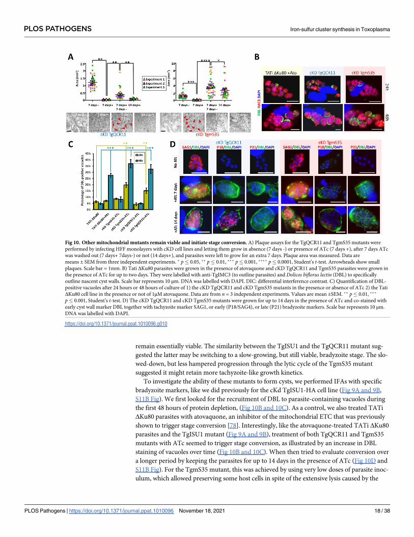
remain essentially viable. The similarity between the TgISU1 and the TgQCR11 mutant sug-
gested the latter may be switching to a slow-growing, but still viable, bradyzoite stage. The slo-
wed-down, but less hampered progression through the lytic cycle of the TgmS35 mutant
suggested it might retain more tachyzoite-like growth kinetics.
To investigate the ability of these mutants to form cysts, we performed IFAs with specific
bradyzoite markers, like we did previously for the cKd TgISU1-HA cell line (Fig 9A and 9B,
S11B Fig). We first looked for the recruitment of DBL to parasite-containing vacuoles during
the first 48 hours of protein depletion, (Fig 10B and 10C). As a control, we also treated TATi
ΔKu80 parasites with atovaquone, an inhibitor of the mitochondrial ETC that was previously
shown to trigger stage conversion [78]. Interestingly, like the atovaquone-treated TATi ΔKu80
parasites and the TgISU1 mutant (Fig 9A and 9B), treatment of both TgQCR11 and TgmS35
mutants with ATc seemed to trigger stage conversion, as illustrated by an increase in DBL
staining of vacuoles over time (Fig 10B and 10C). When then tried to evaluate conversion over
a longer period by keeping the parasites for up to 14 days in the presence of ATc (Fig 10D and
S11B Fig). For the TgmS35 mutant, this was achieved by using very low doses of parasite inoc-
ulum, which allowed preserving some host cells in spite of the extensive lysis caused by the
Fig 10. Other mitochondrial mutants remain viable and initiate stage conversion. A) Plaque assays for the TgQCR11 and TgmS35 mutants were
performed by infecting HFF monolayers with cKD cell lines and letting them grow in absence (7 days -) or presence of ATc (7 days +), after 7 days ATc
was washed out (7 days+ 7days-) or not (14 days+), and parasites were left to grow for an extra 7 days. Plaque area was measured. Data are
means ± SEM from three independent experiments. � p� 0.05, �� p� 0.01, ��� p� 0.001, ���� p� 0.0001, Student’s t-test. Arrowheads show small
plaques. Scale bar = 1mm. B) Tati ΔKu80 parasites were grown in the presence of atovaquone and cKD TgQCR11 and TgmS35 parasites were grown in
the presence of ATc for up to two days. They were labelled with anti-TgIMC3 (to outline parasites) and Dolicos biflorus lectin (DBL) to specifically
outline nascent cyst walls. Scale bar represents 10 μm. DNA was labelled with DAPI. DIC: differential interference contrast. C) Quantification of DBL-
positive vacuoles after 24 hours or 48 hours of culture of 1) the cKD TgQCR11 and cKD TgmS35 mutants in the presence or absence of ATc 2) the Tati
ΔKu80 cell line in the presence or not of 1μM atovaquone. Data are from n = 3 independent experiments. Values are mean ±SEM. �� p� 0.01, ���
p� 0.001, Student’s t-test. D) The cKD TgQCR11 and cKD TgmS35 mutants were grown for up to 14 days in the presence of ATc and co-stained with
early cyst wall marker DBL together with tachyzoite marker SAG1, or early (P18/SAG4), or late (P21) bradyzoite markers. Scale bar represents 10 μm.
DNA was labelled with DAPI.
https://doi.org/10.1371/journal.ppat.1010096.g010
PLOS PATHOGENS Iron-sulfur cluster synthesis in Toxoplasma
PLOS Pathogens | https://doi.org/10.1371/journal.ppat.1010096 November 18, 2021 18 / 38

parasites. Like for the TgISU1 mutant, even long term differentiation-inducing conditions did
not allow complete disappearance of the SAG1 tachyzoite marker in the TgQCR11 and
TgmS35 mutants, and staining of the late bradyzoites marker P21 was not observed. However,
while a few DBL-labelled cysts remained in the TgmS35 culture after 14 days of ATc treatment,
they very rarely showed staining with the intermediate bradyzoite marker P18/SAG4 (S11B
Fig). On the contrary, substantial staining was observed with this marker upon long term
depletion of TgQCR11, like we observed for the TgISU1 mutant (S11B Fig). Of note, as
observed for the TgISU1 mutant and parental TATi ΔKu80 cell line, these other mutants gen-
erated in a type I background strain also displayed smaller cyst size than the type II parasites
after long term induction of stage conversion (S11C Fig), suggesting reactivation may happen
and incomplete differentiation is likely a general feature of RH-derived parasites.
In conclusion, our findings suggest that interfering with general function like mitochon-
drial translation, or targeting more specifically the mitochondrial ETC, does not irreversibly
impair parasite viability and instead leads to an initiation of stage conversion into bradyzoites,
although it may not necessarily be complete.
Discussion
Because of their origin and metabolic importance, the two apicomplexan endosymbiotic
organelles have gathered considerable interest as potential drug targets [79,80]. It may be obvi-
ous, as for example the plastid hosts several metabolic pathways which are not present in the
mammalian hosts of these parasites. Yet, even for conserved housekeeping functions or, in the
case of the mitochondrion early phylogenetic divergence, there may still be enough molecular
differences to allow selective chemical inhibition. In fact, several drugs used for prophylactic
or curative treatments against Apicomplexa-caused diseases are already targeting these organ-
elles [81]. They are essentially impacting the organellar protein synthesis by acting on the
translation machinery [82], although the mitochondrial ETC inhibitor atovaquone is also used
to treat malaria and toxoplasmosis [83]. One main difference when targeting Plasmodium and
Toxoplasma by drugs is that the latter easily converts into the encysted bradyzoite resistance
form. It has been known for some time that treatment of tachyzoites with mitochondrial inhib-
itors triggers stage conversion [73,78,84]. This may be efficient to counteract the acute phase
of toxoplasmosis, but at the same time may favour persistence of the parasites in the host.
Here we characterized Fe-S cluster synthesis pathways which are very similar biochemically,
but are located into two distinct endosymbiotic organelles, and whose inactivation has drasti-
cally different consequences for parasite fitness. Fe-S clusters are ancient, ubiquitous and fun-
damental to many cellular functions, but their synthesis by distinct biosynthetic pathways was
inherited by plastids or by the mitochondrion through distinct bacterial ancestors, and have
thus specialized into adding these cofactors to different client proteins [22]. A key function of
Fe-S clusters, owing to their mid-range redox potential, is electron transfer and redox reac-
tions, mainly as components the respiratory and photosynthetic ETCs. They also have impor-
tant functions in stabilizing proteins, redox sensing, or catalysis through SAM enzymes.
Several of these are not retained in Apicomplexa, whose plastid has lost its photosynthetic abil-
ity for example. Nevertheless, our prediction of the T. gondii Fe-S proteins repertoire suggests
many key functions associated with the apicoplast or the mitochondrion are likely to be
affected by a perturbation of Fe-S assembly (S2 Table).
For the apicoplast, these include lipoic acid or isoprenoid synthesis. Inactivation of the api-
coplast-located TgNFS2 had a late but marked effect on the organelle itself, as it led ultimately
to a partial loss of the apicoplast, which is consistent with the phenotype observed when dis-
rupting the SUF pathway in Plasmodium [26]. IspG and IspH, which are key Fe-S-dependent
PLOS PATHOGENS Iron-sulfur cluster synthesis in Toxoplasma
PLOS Pathogens | https://doi.org/10.1371/journal.ppat.1010096 November 18, 2021 19 / 38

enzymes of the non-mevalonate isoprenoid synthesis pathway [60], were only found margin-
ally less expressed in our quantitative analysis after TgNFS2 depletion. However, our proteo-
mics dataset provided indirect clues that their function may be impacted. Isoprenoid synthesis
is vital for T. gondii tachyzoites [34], and it has implication beyond the apicoplast, as preny-
lated proteins or isoprenoid precursors are involved in more general cellular processes includ-
ing intracellular trafficking or mitochondrial respiration [85]. Isoprenoids are for instance
important for synthesizing ubiquinone/coenzyme Q, and the single predicted mitochondrial
candidate that was significantly less expressed upon TgNFS2 depletion is a putative UbiE/
COQ5 methyltransferase, involved in synthesis of this co-factor [86]. Isoprenoids are also
important for dolichol-derived protein glycosylation and glycosylphosphatidylinositol (GPI)-
anchor biosynthesis, and interestingly the three putative rhoptry-localized candidates signifi-
cantly less expressed in the TgNFS2 mutant (S3 Table) are potentially GPI-anchored and/or
glycosylated, as predicted through sequence analysis by dedicated webservers [87–89]. Overall,
this might be an indication that TgNFS2 depletion impacts isoprenoid synthesis in the apico-
plast, which in turn would impact other metabolic pathways.
Impairing isoprenoid synthesis does not, however, necessarily lead to a loss of the organelle
[26]. There may thus be another explanation for this phenotype. Interestingly, we could show
that perturbing the SUF pathway, which is supposedly important for Fe-S-containing enzyme
LipA, impacts the lipoylation of the E2 subunit of the apicoplast-located PDH (Fig 7A). The
PDH complex catalyzes the production of acetyl-CoA, which is the first step of the FASII sys-
tem, and perturbation of either the PDH or other steps of the FASII system leads to a loss of
the organelle and severely impairs fitness of the parasites [34,90]. Interestingly, long term
depletion of TgNFS2 leads to cell division problems, and affects membrane compartments
such as the IMC (Fig 7B), which are defects previously observed in parasites where the FASII
system has been genetically- or chemically-disrupted [91–93]. Our quantitative proteomic
analysis shows potential compensatory mechanisms may be used by the parasites in response
this early perturbation of the apicoplast lipid metabolism that precedes organelle loss. Tachy-
zoites are indeed known to be able to use exogenous lipid sources to adapt metabolically
[90,92] and, interestingly, upon depletion of TgNFS2 we observed a pattern of overexpression
for ER-located enzymes involved in the synthesis of several phospholipids and ceramides (Fig
6A and 6D and S3 Table). These lipids are usually synthesized in the ER from apicoplast-gen-
erated precursors, as both organelles cooperate for FA and phospholipid (PL) synthesis [94].
Yet, the ER-localized PL-synthesis machinery can also use FA scavenged from the host [95].
The increased expression of ER-localized lipid-related enzymes may thus reflect an increased
synthesis, potentially from exogenous lipid precursors, in compensation for a defect in the api-
coplast-localized machinery. In spite of this potential compensation mechanism, it seems the
alteration of the SUF pathway in T. gondii has such a profound impact that it ultimately leads
to the irreversible demise of the parasites (Fig 4E). It would be interesting to use recently
described approaches like stable isotope labelling of lipid precursors combined to lipidomic
analysis [92], to investigate in the SUF pathway mutant the potential changes in de novo syn-
thesis of FA or in lipid scavenging from the host.
In the mitochondrion, important pathways potentially involving Fe-S proteins include the
respiratory ETC, the TCA cycle, as well as molybdenum and heme synthesis (S2 Table).
Accordingly, perhaps the most obvious consequence of disrupting the ISC pathway was the
profound impact on the mitochondrial respiratory capacity, as evidenced experimentally by
measuring the mitochondrial membrane potential (Fig 8D), and supported by quantitative
analyses showing a clear drop in expression of many respiratory complex proteins (Fig 8A and
8C and S4 Table). This is also in line with the recent description of another T. gondii mito-
chondrial Fe-S cluster synthesis mutant that showed a marked alteration of its respiratory
PLOS PATHOGENS Iron-sulfur cluster synthesis in Toxoplasma
PLOS Pathogens | https://doi.org/10.1371/journal.ppat.1010096 November 18, 2021 20 / 38

capacity [25]. Although the mitochondrion, through the TCA cycle and the respiratory chain/
oxidative phosphorylation, contributes to energy production in tachyzoites [96], the glycolytic
flux is also believed to be a major source of carbon and energy for these parasites [97]. Thus,
rather coherently, as highlighted by our quantitative proteomic analysis, disruption of the ISC
pathway led to a potential overexpression of glycolytic enzymes concurrently with the lower
expression of mitochondrial ETC components (Fig 6B and S4 Table). The possible overexpres-
sion of enzymes of the pentose phosphate pathway, which is branching off from glycolysis and
is providing redox equivalents and precursors for nucleotide and amino acid biosynthesis, is
also potentially indicative of a higher use of glucose in these conditions. The metabolic changes
encountered by ISC-deficient parasites do not cause their rapid demise, as they are able to ini-
tiate conversion to the bradyzoite stage, which has been suggested to rely essentially on glycol-
ysis for energy production [98]. Our analysis of the viability of other mitochondrial mutants
confirmed results obtained by others showing that the organelle, and in particular the ETC,
seem important for parasite fitness [62,63,77]. Yet, perhaps because of their metabolic flexibil-
ity, mutant parasites seem to retain the ability to survive as tachyzoites (as shown here and in
[62]), or initiate a switch to bradyzoites.
The inactivation of the ISC pathway likely has consequences on other important cellular
housekeeping functions besides mitochondrial metabolism. In other eukaryotes, the ISC path-
way provides a yet unknown precursor molecule as a sulfur provider for the cytosolic CIA Fe-
S cluster assembly pathway [24]. The ISC pathway thus not only governs the proper assembly
of mitochondrial Fe-S proteins, but also of cytoplasmic and nuclear ones. Our quantitative
proteomics data suggests it is also the case in T. gondii, as several putative nuclear Fe-S proteins
involved in gene transcription (such as DNA-dependent RNA polymerases) or DNA repair
(like DNA endonulease III) were found to be impacted by TgISU1 depletion. The CIA path-
way has recently been shown to be important for tachyzoite proliferation [25], and several of
the cytoplasmic or nuclear Fe-S cluster-containing proteins are likely essential for parasite via-
bility. It is thus possible that, in spite of their conversion to a stress-resistant form, the long-
term viability of TgISU1 parasites could be affected beyond recovery. In vivo experiment in
the mouse model may be used to assess this.
The transition from tachyzoite to bradyzoite is known to involve a considerable change in
gene expression [65,66], and it takes several days of in vitro differentiation-inducing condi-
tions to obtain mature cysts [99,100]. Quantitative proteomic analysis showed that TgISU1-de-
pleted parasites rapidly display a high expression of bradyzoite-specific surface antigens and
GRA markers (S4 Table and Fig 9C). In all the mitochondrial mutants investigated in this
study, long term perturbation of the organelle led to the appearance of parasite-containing
structures with typical cyst-like morphology (Figs 5, 9 and 10). However, using specific anti-
bodies against early or late bradyzoite markers, the differentiating parasites never appeared to
reach a fully mature bradyzoite stage (Figs 9 and 10, S11 Fig). Also, contrarily to the mutants
where the ETC may be more directly impacted (TgQCR11 and TgISU1), the mitochondrial
translation mutant (TgmS35), while seemingly initiating conversion to bradyzoite, then con-
tinued to multiply to some extent and lyse its host cells, likely as tachyzoites (Fig 10A). How-
ever, as the mitochondrion encodes three proteins that are subunits of mitochondrial ETC
complexes III (cytochrome b) and IV (CoxI and III), this mutant should also be supposedly
impacted for ETC function. This apparent discrepancy might then be explained by a looser
control of down-regulation of protein expression, or by compensatory mechanisms that may
be specifically at play in this particular mutant. It is also possible that more directly targeting
the ETC is a much stronger inducer of differentiation, and thus different mitochondrial
mutants may behave differently regarding stage conversion. Importantly, one main reason
potentially explaining the incomplete differentiation of these mitochondrial mutants is that
PLOS PATHOGENS Iron-sulfur cluster synthesis in Toxoplasma
PLOS Pathogens | https://doi.org/10.1371/journal.ppat.1010096 November 18, 2021 21 / 38

they were generated in a laboratory-adapted type I T. gondii strain (RH) that typically does not
form cysts: these type I tachyzoites may upregulate specific bradyzoite genes and produce bra-
dyzoite-specific proteins or cyst wall components, but they are largely incapable of forming
mature bradyzoite cysts [101] (S11 Fig). This calls for further investigations, and in particular
generating similar mitochondrion-related mutants in more cystogenic type II parasites, which
could be very insightful.
Our quantitative proteomics analysis shows that SUF-impaired parasites also seem to initi-
ate an upregulation of some bradyzoite markers early after TgNFS2 depletion. Yet, these para-
sites did not display the hallmarks of bradyzoite morphology. They did not progress towards
stage conversion and instead they displayed considerable perturbation of the cell division pro-
cess (Fig 7B), and eventually died. Both the apicoplast and the mitochondrion have established
a close metabolic symbiosis with their host cell, so there are likely multiple mechanisms allow-
ing these organelles to communicate their status to the rest of the cell. This raises the question
as to why mitochondrion, but not apicoplast, dysfunction can lead to differentiation into bra-
dyzoites. This may be due to differences in the kinetics or the severity of apicoplast-related
phenotypes that may not allow stage conversion (which is typically a long process) to happen.
Alternatively, there might be differentiation signals specifically associated to the mitochon-
drion. In fact this organelle is increasingly seen as a signalling platform, able to communicate
its fitness through the release of specific metabolites, reactive oxygen species, or by modulating
ATP levels [102]. Interestingly, it was shown in other eukaryotes that mitochondrial dysfunc-
tions, such as altered oxidative phosphorylation, significantly impair cellular proliferation,
oxygen sensing or specific histone acetylation, yet without diminishing cell viability and
instead may lead to quiescent states [103–105]. Consequences of mitochondrial dysfunction
include a restricted energy supply and thus constitutes a metabolic challenge that can trigger
important cellular adaptations that ultimately determine eukaryotic cell fate and survival. Our
results suggest that this is also possibly the case for T. gondii. It is for instance quite interesting
to see that altering the respiratory activity of the organelle, whether it is by generating specific
mutants or by the use of drugs such as atovaquone, seems to lead to a similar differentiation
phenotype.
More generally, the environmental or metabolic cues that drive specific gene expression to
induce a functional shift leading to conversion into bradyzoites are not fully identified. More-
over, how these stimuli are integrated is also largely unknown. A high-throughput approach
has allowed the recent identification of a master transcriptional regulator of stage conversion
[106], but how upstream events are converted into cellular signals to mobilize the master regu-
lator is still an important, yet unresolved, question. Translational control [107] may play a role
in regulating this factor in the context of the integrated stress response [108]. In fact, an essen-
tial part of the eukaryotic cell stress response occurs post-transcriptionally and is achieved by
RNA-binding proteins [109]. Interestingly, among the proteins significantly less abundant in
the spontaneously-differentiating TgISU1 mutant were many RNA-binding proteins. They
include components of stress granules (PolyA-binding protein, PUF1, Alba1 and 2, some of
which are linked to stage conversion [110–112]), which are potentially involved in mRNA
sequestration from the translational machinery, but also two regulators of the large 60S ribo-
somal subunit assembly, as well as the gamma subunit of the eukaryotic translation initiation
factor (eIF) complex 4 (known to be down-regulated in the bradyzoite stage [113]). Variation
in these candidates may have a considerable impact on the translational profile and on the pro-
teostasis of differentiating parasites, and how they may help regulating stage conversion in this
context should be investigated further. Understanding the mechanisms that either lead to
encystment or death of the parasites is crucial to the development of treatments against toxo-
plasmosis. This question is key to the pathology, because long term persistence of bradyzoites
PLOS PATHOGENS Iron-sulfur cluster synthesis in Toxoplasma
PLOS Pathogens | https://doi.org/10.1371/journal.ppat.1010096 November 18, 2021 22 / 38

and their resistance to current treatments makes them a durable threat for their human hosts.
Comparative studies of stress-induced or spontaneously differentiating conditional mutants
may bring further insights on how the parasites integrate upstream stresses or dysfunctions
into global regulation of stage conversion.
Materials and methods
Parasites and cells culture
Tachyzoites of the type I RH TATi RHΔKu80 T. gondii cell line [44], as well as derived trans-
genic parasites generated in this study and the type II Prugniaud strain, were maintained by
serial passage in human foreskin fibroblast (HFF, American Type Culture Collection, CRL
1634) cell monolayer grown in Dulbecco’s modified Eagle medium (DMEM, Gibco), supple-
mented with 5% decomplemented fetal bovine serum (FBS), 2-mM L-glutamine and a cocktail
of penicillin-streptomycin at 100 μg/ml. The TgQCR11 [63] and TgmS35 [77] conditional
mutants cell lines were generously provided by the Sheiner laboratory.
Bioinformatic analyses
Sequence alignments were performed using the MUltiple Sequence Comparison by Log-
Expectation (MUSCLE) algorithm of the Geneious 6.1.8 software suite (http://www.geneious.
com). Transit peptide and localization predictions were done using IPSORT (http://ipsort.hgc.
jp/), Localizer 1.0.4 (http://localizer.csiro.au/), and Deeploc 1.0 (http://www.cbs.dtu.dk/
services/DeepLoc-1.0/) algorithms.
The putative Fe-S proteome was predicted using the MetalPredator webserver (http://
metalweb.cerm.unifi.it/tools/metalpredator/) [56]. The whole complement of T. gondii anno-
tated proteins was downloaded in FASTA format from the ToxoDB database (https://toxodb.
org [37], release 45) and used for analysis in the MetalPredator webserver. Additional manual
curation included homology searches for known Fe-S proteins from plants (see appendix A in
[114]), and search for homologues in the Uniprot database (https://www.uniprot.org) that
were annotated as containing a Fe-S cofactor. For proteomics candidates, annotations were
inferred from ToxoDB, KEGG (https://www.genome.jp/kegg/) and the Liverpool Library of
Apicomplexan Metabolic Pathways (http://www.llamp.net/ [115]).
N-glycosylation predictions were done with the GlycoEP webserver (http://crdd.osdd.net/
raghava/glycoep/index.html). GPI anchor predictions were done with the PredGPI (http://
gpcr.biocomp.unibo.it/predgpi/) and GPI-SOM (http://gpi.unibe.ch/) webservers.
Candidate variant proteins identified by quantitative proteomics were mapped on the
graphic representation of the high-resolution spatial proteome map of T. gondii using the
hyperLOPIT dataset [38] (https://proteome.shinyapps.io/toxolopittzex/).
Heterologous expression in E. coliConstructs for designing recombinant proteins were defined by aligning TgNFS2 and TgISU1
amino acid sequences with their E. coli counterparts. For TgNFS2, a 1,438 bp fragment corre-
sponding to amino acids 271–699, was amplified by polymerase chain reaction (PCR) from T.
gondii cDNA using primers ML4201/ML4012 (sequences of the primers used in this study are
found in S6 Table). For TgISU1, a 393 bp fragment corresponding to amino acids 64–194, was
amplified by PCR from T. gondii cDNA using primers ML4204/ML4205. The fragments were
cloned into the pUC19 (Thermo Fisher Scientific) using the HindIII/BamHI and SphI/BamHI
restriction sites, respectively. E. coli mutants from the Keio collection (obtained from the The
Coli Genetic Stock Center at the University of Yale: stain numbers JW1670-1 for SufS,
PLOS PATHOGENS Iron-sulfur cluster synthesis in Toxoplasma
PLOS Pathogens | https://doi.org/10.1371/journal.ppat.1010096 November 18, 2021 23 / 38
JW2513-1 for IscU), were transformed with plasmids for expressing recombinant TgNFS2 and
TgISU1 and selected with ampicillin. For growth assays [41], overnight stationary phase cul-
tures were adjusted to the same starting OD600 of 0.6 in salt-supplemented M9 minimal media
containing 0.4% glucose and varying amounts of the 2,20-Bipyridyl iron chelator (Sigma-
Aldrich). Parental E. coli (strain K12) were included as a control. Growth was monitored
through OD600 measurement after 7, 14 and 24 hours at 37˚C in a shaking incubator.
Generation of HA-tagged TgNFS2, TgSUFE2 and TgISU1 cell lines
The ligation independent strategy [43] was used for C-terminal hemagglutinin (HA)3-tagging
of TgISU1. A fragment corresponding to the 3’ end of TgISU1 was amplified by PCR from
genomic DNA, with the Q5 DNA polymerase (New England BioLabs) using primers ML4208/
ML4209 and inserted in frame with the sequence coding for a triple HA tag, present in the pLI-
C-HA3-chloramphenicol acetyltransferase (CAT) plasmid. The resulting vector was linearized
and 40 μg of DNA was transfected into the TATi ΔKu80 cell line to allow integration by single
homologous recombination, and transgenic parasites of the TgISU1-HA cell line were selected
with chloramphenicol and cloned by serial limiting dilution.
For TgNFS2 and TgSUFE2, a CRISPR-based strategy was used. Using the pLIC-HA3-CAT
plasmid as a template, a PCR was performed with the KOD DNA polymerase (Novagen) to
amplify the tag and the resistance gene expression cassette with primers ML3978/ML3979
(TgNFS2) and ML4023/ML4162 (TgSUFE2), that also carry 30 bp homology with the 30 end of
the corresponding genes. A specific single-guide RNA (sgRNA) was generated to introduce a
double-stranded break at the 30 of the respective loci. Primers used to generate the guides were
ML3948/ML3949 (TgNFS2) and ML4160/ML4161 (TgSUFE2) and the protospacer sequences
were introduced in the Cas9-expressing pU6-Universal plasmid (Addgene, ref #52694) [39].
Again, the TATi ΔKu80 cell line was transfected and transgenic parasites of the TgNFS2-HA
or TgSUFE2-HA cell lines were selected with chloramphenicol and cloned by serial limiting
dilution.
Generation of TgNFS2 and TgISU1 conditional knock-down and
complemented cell lines
The conditional knock-down cell lines for TgNFS2 and TgISU1 were generated based on the
Tet-Off system using the DHFR-TetO7Sag4 plasmid [116].
For TgISU1, a 930 bp 5’ region of the gene, starting with the initiation codon, was amplified
from genomic DNA by PCR using Q5 polymerase (New England Biolabs) with primers
ML4212/ML4213 and cloned into the DHFR-TetO7Sag4 plasmid, downstream of the anhy-
drotetracycline (ATc)-inducible TetO7Sag4 promoter, yielding the DHFR-TetO7Sag4-TgISU1
plasmid. The plasmid was then linearized and transfected into the TgISU1-HA cell line. Trans-
fected parasites were selected with pyrimethamine and cloned by serial limiting dilution. A
similar approach was used to generate an independent cKD TgISU1 cell line, but starting from
the TATi ΔKu80 cell line instead of the TgISU1-HA cell line, to allow subsequent tagging of
mitochondrial candidates.
For TgNFS2, a CRISPR-based strategy was used. Using the DHFR-TetO7Sag4 plasmid as a
template, a PCR was performed with the KOD DNA polymerase (Novagen) to amplify the
promoter and the resistance gene expression cassette with primers ML4154/ML4155 that also
carry 30 bp homology with the 50 end of the TgNFS2 gene. A specific sgRNA was generated to
introduce a double-stranded break at the 50 of the TgNFS2 locus. Primers used to generate the
guide were ML4156/ML4157 and the protospacer sequences were introduced in the pU6-Uni-
versal plasmid (Addgene ref#52694) [39]. The TgNFS2-HA cell line was transfected with the
PLOS PATHOGENS Iron-sulfur cluster synthesis in Toxoplasma
PLOS Pathogens | https://doi.org/10.1371/journal.ppat.1010096 November 18, 2021 24 / 38

donor sequence and the Cas9/guide RNA-expressing plasmid and transgenic parasites were
selected with pyrimethamine and cloned by serial limiting dilution.
The cKD TgNFS2-HA and cKD TgISU1-HA cell lines were complemented by the addition
of an extra copy of the respective genes put under the dependence of a tubulin promoter at the
uracil phosphoribosyltransferase (UPRT) locus. TgNFS2 (2097 bp) and TgISU1 (657 bp) whole
cDNA sequences were amplified by reverse transcription (RT)-PCR with primers ML4576/
ML4577 and ML4455/ML4456, respectively. They were then cloned downstream of the tubulinpromoter sequence of the pUPRT-TUB-Ty vector [44] to yield the pUPRT-TgNFS2 and
pUPRT-TgISU1plasmids, respectively. These plasmids were then linearized prior to transfec-
tion of the respective mutant cell lines. The recombination efficiency was increased by co-
transfecting with the Cas9-expressing pU6-UPRT plasmids generated by integrating UPRT-
specific protospacer sequences (with primers ML2087/ML2088 for the 3’, and primers
ML3445/ML3446 for the 5’) which were designed to allow a double-strand break at the UPRTlocus. Transgenic parasites were selected using 5-fluorodeoxyuridine and cloned by serial lim-
iting dilution to yield the cKD TgNFS2-HA comp cKD TgISU1-HA comp cell lines,
respectively.
Generation of HA-tagged TgSDHB and TgApiCox13 cell lines
A CRISPR-based strategy was used. Using the pLIC-HA3-CAT plasmid as a template, a PCR
was performed with the KOD DNA polymerase (Novagen) to amplify the tag and the resis-
tance gene expression cassette with primers ML5116/ML5117 (TgSDHB) and ML5114/
ML5115 (TgApiCox13), that also carry 30 bp homology with the 30 end of the corresponding
genes. A specific sgRNA was generated to introduce a double-stranded break at the 30 of the
respective loci. Primers used to generate the guides were ML4986/ML4987 (TgSDHB) and
ML4984/ML4985 (TgApiCox13) and the protospacer sequences were introduced in the
Cas9-expressing pU6-Universal plasmid (Addgene, ref #52694) [39]. The cKD TgISU1 cell
line was transfected and parasites were selected with chloramphenicol and cloned by serial
limiting dilution.
Immunoblot analysis
Protein extracts from 107 freshly egressed tachyzoites were prepared in Laemmli sample buffer,
separated by SDS-PAGE and transferred onto nitrocellulose membrane using the BioRad
Mini-Transblot system according to the manufacturer’s instructions. Rat monoclonal antibody
(clone 3F10, Roche) was used to detect HA-tagged proteins. Other primary antibodies used
were rabbit anti-lipoic acid antibody (ab58724, Abcam), mouse anti-SAG1 [117], rabbit anti-
CPN60 [118] and mouse anti-actin [119]. Protein quantification was performed by band den-
sitometry using FIJI (https://imagej.net/software/fiji/).
Immunofluorescence microscopy
For immunofluorescence assays (IFA), intracellular tachyzoites grown on coverslips contain-
ing HFF monolayers, were either fixed for 20 min with 4% (w/v) paraformaldehyde (PFA) in
PBS and permeabilized for 10 min with 0.3% Triton X-100 in PBS or fixed for 5 min in cold
methanol (for the use of cyst-specific antibodies). Slides/coverslips were subsequently blocked
with 0.1% (w/v) BSA in PBS. Primary antibodies used (at 1/1,000, unless specified) to detect
subcellular structures were rabbit anti-CPN60 [118], mouse monoclonal anti-F1-ATPase beta
subunit (gift of P. Bradley), mouse monoclonal anti-GRA3 [120], rabbit anti-TgHSP28 [121],
rabbit anti-GAP45 [122], mouse monoclonal anti-SAG1 [117], anti P18/SAG4 (diluted 1/200,
T8 3B1) and anti P21 (diluted 1/200, T8 4G10) [123]. Rat monoclonal anti-HA antibody
PLOS PATHOGENS Iron-sulfur cluster synthesis in Toxoplasma
PLOS Pathogens | https://doi.org/10.1371/journal.ppat.1010096 November 18, 2021 25 / 38

(clone 3F10, Roche) was used to detect epitope-tagged proteins. Staining of DNA was per-
formed on fixed cells by incubating them for 5 min in a 1 μg/ml 4,6-diamidino-2-phenylindole
(DAPI) solution. All images were acquired at the Montpellier RIO imaging facility from a
Zeiss AXIO Imager Z1 epifluorescence microscope driven by the ZEN software v2.3 (Zeiss).
Z-stack acquisition and maximal intensity projection was performed to visualize larger struc-
tures such as in vitro cysts. Adjustments for brightness and contrast were applied uniformly
on the entire image.
Plaque assay
Confluent monolayers of HFFs were infected with freshly egressed parasites, which were left to
grow for 7 days in the absence or presence of ATc (added to a final concentration of 1 μg/ml).
They were then fixed with 4% v/v PFA and plaques were revealed by staining with a 0.1% crys-
tal violet solution (V5265, Sigma-Aldrich). For the washout experiments, after 7 days the cul-
ture medium was removed from the wells and one gentle wash was performed with Hanks’
Balanced Salt Solution (HBSS), taking care not to disturb the cells; then new DMEM medium
containing or not ATc was added for another 7-day incubation, before termination of the
experiment by PFA fixation and crystal violet staining.
Egress assay
T. gondii tachyzoites were grown for 40 (without ATc) or 120 (with ATc) hours on HFF cells
with coverslips in 24-well plates. The infected host cells were incubated for 7 min at 37˚C with
DMEM containing 3 μM of calcium ionophore A23187 (C7522, Sigma-Aldrich) prior to fixa-
tion with 4% PFA. Immunofluorescence assays were performed as previously described [124]:
the parasites and the parasitophorous vacuole membrane were labelled with anti-GAP45 and
anti-GRA3, respectively. The proportion of egressed and non-egressed vacuoles was calculated
by counting 250 vacuoles in three independent experiments. Data are presented as mean
values ± SEM.
Alkaline stress-induced stage conversion
Tachyzoites were seeded onto HFF monolayers grown on coverslips in 24-well plates. The
inoculum was adapted to the cell line used to avoid early lysis of the host cells, with as much as
20,000 parasites per well for the type II Prugniaud strain, and as little as 300 for the type I RH
TATi ΔKu80 cell line. 24 hours after infection, in order to induce to induce alkaline stress as
previously described [76], the regular culture medium was changed to a pH 8.2 differentiation
medium: Minimum Essential Medium (MEM) without NaHCO3 (Gibco), 3% FBS, 50 mM
HEPES, pH 8.2. Cultures were maintained at 37˚C in air (to avoid pH variation due to CO2),
and the medium was changed every 48 hours for the whole duration of the experiment.
Semi-quantitative RT-PCR
Total mRNAs of freshly egressed extracellular parasites from the cKD TgNFS2-HA, cKD TgI-
SU1-HA and their respective complemented cell lines, as well as cKD TgISU1 (incubated with
or without ATc at 1.5 μg/mL for 3 days) were extracted using Nucleospin RNA II Kit
(Macherey-Nagel). The cDNAs were synthesized with 450 ng of total RNA per RT-PCR reac-
tion using High-Capacity cDNA Reverse Transcription Kit (Applied Biosystems). Specific
primers for TgNFS2 (ML4686/ML4687), TgISU1 (ML4684/ML4685) and, as a control, Tubulinβ (ML841/ML842) or actin (ML843/ML844) were used to amplify specific transcripts with the
PLOS PATHOGENS Iron-sulfur cluster synthesis in Toxoplasma
PLOS Pathogens | https://doi.org/10.1371/journal.ppat.1010096 November 18, 2021 26 / 38

GoTaq DNA polymerase (Promega). PCR was performed with 21 cycles of denaturation (30 s,
95˚C), annealing (20 s, 55˚C), and elongation (30 s, 72˚C).
Mitochondrial membrane potential measurement
Parasites grown for the indicated time with or without ATc were mechanically released from
their host cells, purified on a glass wool fiber column, washed and adjusted to 107 parasites/ml
in phenol red-free medium, and incubated in with 1.5 μM of the JC-1 dye (5,5’,6,6’-tetra-
chloro-1,1’,3,3’-tetraethylbenzimidazolylcarbocyanine Iodide, T3168, Invitrogen) for 30 min
at 37˚C, washed phenol red-free medium and analyzed by flow cytometry or microscopy. Flow
cytometry analysis was performed on a FACSAria III flow cytometer (Becton Dickinson). An
unstained control was used to define gates for analysis. 50,000 events per condition were col-
lected and data were analysed using the FlowJo Software.
Quantitative label-free mass spectrometry
Parasites of the TATi ΔKu80 and cKD TgISU1-HA cell lines were grown for two days in the
presence of ATc; parasites of the cKD TgNFS2-HA were grown for three days in the presence
of ATc. Then they were mechanically released from their host cells, purified on a glass wool
fiber column, washed in Hanks’ Balanced Salt Solution (Gibco). Samples were first normalized
on parasite counts, but further adjustment was performed after parasite pellet resuspension in
SDS lysis buffer (50 mm Tris-HCl pH8, 10 mm EDTA pH8, 1% SDS) and protein quantifica-
tion with a bicinchoninic acid assay kit (Abcam). For each condition, 20 μg of total proteins
were separated on a 12% SDS-PAGE run for 20 min at 100 V, stained with colloidal blue
(Thermo Fisher Scientific), and each lane was cut in three identical fractions. Trypsin digestion
and mass spectrometry analysis in the Q Exactive Plus mass spectrometer (Thermo Fisher Sci-
entific) were carried out as described previously [125].
For peptide identification and quantification, the raw files were analyzed with MaxQuant
version 1.6.10.43 using default settings. The minimal peptide length was set to 6. Carbamido-
methylation of cysteine was selected as a fixed modification and oxidation of methionine, N-
terminal-pyroglutamylation of glutamine and glutamate and acetylation (protein N terminus)
as variable modifications. Up to two missed cleavages were allowed. The files were searched
against the T. gondii proteome (March 2020 -https://www.uniprot.org/proteomes/
UP000005641-8450entries). Identified proteins were filtered according to the following crite-
ria: at least two different trypsin peptides with at least one unique peptide, an E value below
0.01 and a protein E value smaller than 0.01 were required. Using the above criteria, the rate of
false peptide sequence assignment and false protein identification were lower than 1%. Peptide
ion intensity values derived from MaxQuant were subjected for label-free quantitation. Unique
and razor peptides were considered [126]. Statistical analyses were carried out using R package
software. ANOVA test with threshold of 0.05 was applied to identify the significant differences
in the protein abundance. Hits were retained if they were quantified in at least three of the four
replicates in at least one experiment. Additional candidates that consistently showed absence
or presence of LFQ values versus the control, and mean LFQ was only considered if peptides
were detected in at least three out of the four biological replicates.
Statistical analysis for phenotypic assays
Unless specified, values are usually expressed as means ± standard error of the mean (SEM).
Data were analysed for comparison using unpaired Student’s t-test with equal variance (homo-
scedastic) for different samples or paired Student’s t-test for similar samples before and after
PLOS PATHOGENS Iron-sulfur cluster synthesis in Toxoplasma
PLOS Pathogens | https://doi.org/10.1371/journal.ppat.1010096 November 18, 2021 27 / 38

treatment. For comparisons with of ratios of groups of samples with a reference set to 1, analy-
sis of variance (ANOVA) was used.
Supporting information
S1 Fig. Alignment of SufS/NFS2 and IscU/ISU1 homologs. TgNFS2 (A) and TgISU1 (B)
homologs were aligned to their counterparts from plant (Arabidopsis thaliana) and bacteria
(Escherichia coli). Key conserved cysteine residue for cysteine desulfurase activity is indicated.
(PDF)
S2 Fig. Generation of HA-tagged TgNFS2 and TgISU1 cell lines. A) Schematic representa-
tion of the strategy for expressing HA-tagged versions of TgNFS2 (left) and TgISU1 (right) by
homologous recombination at the native locus of the corresponding gene of interest. Chloram-
phenicol was used to select transgenic parasites based on their expression of the Chloramphen-
icol acetyltransferase (CAT). B) Diagnostic PCR for verifying correct integration of the
construct. The amplified fragments correspond to the blue or red arrows in A), and specific
primers used were ML3982/ML1476 (TgNFS2) and ML4208/ML1476 (TgISU1).
(PDF)
S3 Fig. HA-tagging of TgSUFE2 shows it is an apicoplast protein. A) Sequence alignment of
TgSUFE2 (TGGT1_277010) with plant (A. thaliana) and bacterial (E. coli) homologues. B)
Schematic representation of the strategy for expressing an HA-tagged version of TgSUFE2 by
double homologous recombination at the native locus. Chloramphenicol was used to select
transgenic parasites based on their expression of the Chloramphenicol acetyltransferase
(CAT). C) Diagnostic PCR for verifying correct integration of the construct. The amplified
fragment corresponds to the red arrows in B), and specific primers used were ML4101/
ML1476. D) Detection by immunoblot of C-terminally HA-tagged TgSUFE2 in parasite
extracts reveals the presence of both precusor and mature forms of the protein. Anti-actin anti-
body (TgACT1) was used as a loading control. E) Immunofluorescence assay shows TgSUFE2
co-localizes with apicoplast marker TgCPN60. Scale bar represents 5 μm. DNA was labelled
with DAPI. DIC: differential interference contrast.
(PDF)
S4 Fig. Generation of TgNFS2 and TgISU1 conditional mutants. A) Schematic representa-
tion of the strategy for generating TgNFS2 (top) and TgISU1 (bottom) conditional knock-
down cell lines by homologous recombination at the native locus. Pyrimethamine was used to
select transgenic parasites based on their expression of Dihydrofolate reductase (DHFR). B)
Diagnostic PCR for verifying correct integration of the construct. The amplified fragments
confirming 5’ and 3’ integration correspond to the blue and red arrows displayed in A), respec-
tively, and specific primers used were: ML4158/ML687 (TgNFS2 5’ integration), ML1041/
ML4159 (TgNFS2 3’ integration), ML1774/ML4388 (TgISU1 5’ integration), ML1771/ML4387
(TgISU1 3’ integration).
(PDF)
S5 Fig. Generation of TgNFS2 and TgISU1 complemented cell lines. A) Schematic represen-
tation of the strategy for generating TgNFS2 and TgISU1 complemented cell lines by integrat-
ing an extra copy of the gene of interest (GOI) by double homologous recombination at the
Uracil Phosphoribosyltransferase (UPRT) locus. Negative selection with 5-fluorodeoxyuridine
(FUDR) was used to select transgenic parasites based on their absence of UPRT expression. B)
Diagnostic PCR for verifying correct integration of the construct. The amplified fragments
confirming integration correspond to the red arrows displayed in A), and specific primers
PLOS PATHOGENS Iron-sulfur cluster synthesis in Toxoplasma
PLOS Pathogens | https://doi.org/10.1371/journal.ppat.1010096 November 18, 2021 28 / 38

used were: ML2866/ML4686 (TgNFS2 integration), ML2866/ML4455 (TgISU1 5’ integration).
C) Semi-quantitative RT-PCR analysis from cKD TgNFS2-HA, cKD TgISU1-HA and their
respective complemented cell lines grown for three days in the presence or absence of ATc,
using specific primers couples ML4686/ML4687 (TgNFS2) and ML4684/ML4685 (TgISU1). It
shows complemented cell lines express high levels of the corresponding mRNA. Specific β-tubulin primers (ML841/ML842) were used as controls.
(PDF)
S6 Fig. Quantification of apicoplast loss upon TgNFS2 depletion using streptavidin. Per-
centage of cKD TgNFS2-HA parasites-containing vacuoles displaying a loss of apicoplast sig-
nal when labeled with streptavidin-Fluorescein Isothiocyanate after culture in the presence or
absence of ATc for 120 hours. Data are mean values from n = 3 independent experiments
±SEM. ���� p� 0.0001, Student’s t-test. Inset: typical streptavidin labeling of the apicoplast
(green) in a tachyzoites-containing vacuole, the inner membrane complex was stained with
anti-TgIMC3 (red) and the DNA with DAPI (blue); scale bar = 5μm.
(PDF)
S7 Fig. Quantitative proteomics shows depletion of TgNFS2 does not have a global impact
on the apicoplast, but may suggest compensatory response from other cellular pathways in
response to specific apicoplast-related lipid synthesis defects. Classification of variant pro-
teins according to their putative cellular localization (A) and function (B). N/A: not available;
ER: endoplasmic reticulum; PM: plasma membrane; VAC: vacuolar compartment; GRA:
dense granule protein; SRS: SAG-related sequence. In particular, the increased expression of
ER-located lipid metabolism enzymes suggests possible compensation for loss of apicoplast-
related lipid synthesis function.
(PDF)
S8 Fig. TgISU1-depleted parasites show a marked decrease in proteins related to mito-
chondrial respiration, and a strong increase in bradyzoite-specific dense granules proteins
and surface antigens. Classification of variant proteins according to their putative cellular
localization (A) and function (B). N/A: not available; ER: endoplasmic reticulum; PM: plasma
membrane; VAC: vacuolar compartment; GRA: dense granule protein; SRS: SAG-related
sequence. A large proportion of components of complexes II, III and IV of the mitochondrial
respiratory chain, which involve Fe-S proteins, were found to be less abundant. Conversely,
the abundance of many bradyzoite-specific dense granule proteins of plasma membrane-
located surface antigens increased.
(PDF)
S9 Fig. Generation of a tag-free cKD TgISU1 cell line. A) Schematic representation of
the strategy for generating the conditional knock-down cell line by homologous recombina-
tion at the native locus. Pyrimethamine was used to select transgenic parasites based on
their expression of Dihydrofolate reductase (DHFR). B) Diagnostic PCR for verifying
correct integration of the construct. The amplified fragments confirming 5’ and 3’ integration
correspond to the blue and red arrows displayed in A), respectively, and specific primers used
were ML1774/ML4388 (5’ integration), and ML1771/ML4387 (3’ integration). C) Semi-quan-
titative RT-PCR analysis of the cKD TgISU1 cell line grown for up to three days in the pres-
ence or absence of ATc, using specific primers couple ML4684/ML4685, showing effcient
down-regulation of TgISU1 expression. Specific actin primers (ML843/ML844) were used as
controls. D) Plaque assays were carried out by infecting HFF monolayers with the newly gen-
erated cKD TgISU1 cell line or the original cKD TgISU1-HA mutant cell line as a control.
They were grown for 7 days ± ATc. Measurements of lysis plaque areas are shown on the right
PLOS PATHOGENS Iron-sulfur cluster synthesis in Toxoplasma
PLOS Pathogens | https://doi.org/10.1371/journal.ppat.1010096 November 18, 2021 29 / 38

and confirm a significant defect in the lytic cycle in the two mutant cell lines upon ATc addi-
tion. Values are means of n = 3 experiments ± SEM. ���� denotes p� 0.0001, ANOVA. Scale
bar = 1mm.
(PDF)
S10 Fig. Tagging of mitochondrial candidates in the cKD TgISU1 background. A)
Schematic representation of the strategy for expressing HA-tagged versions of TgSDHB
and TgApiCox13 by homologous recombination at the native locus of the corresponding
gene of interest. Chloramphenicol was used to select transgenic parasites based on their
expression of the Chloramphenicol acetyltransferase (CAT). B) Diagnostic PCR for verifying
correct integration of the construct. The amplified fragment corresponds to the red arrows in
A), and specific primers used were ML1476/ML5013 (TgSDHB), ML1476/ML5006 (TgApi-
Cox13).
(PDF)
S11 Fig. Comparison of stage conversion for type I and II strains. A) Stage conversion was
induced by alkaline pH stress on type I parasites of the RH Tati Δ80 parental cell line or the
cystogenic type II Prugniaud strain for up to 14 days. Fixed samples were co-stained with cyst
wall marker DBL, together with tachyzoite marker SAG1, or intermediate (P18/SAG4) or late
(P21) bradyzoite markers. Scale bar represents 10 μm. DNA was labelled with DAPI. B) The
percentage of DBL-positive cysts containing P18 or P21 staining was evaluated on samples
after 14 days of ATc treatment (for TgISU1, TgQCR11 and TgmS35 conditional mutants) or
pH stress (for RH TATi ΔKu80 and Prugniaud parasites). Values are mean ±SEM from n = 3
independent experiments. C) Measurement of the cyst area size after growing the cell lines for
7 and 14 days in cyst-inducing conditions, then labelling the cyst wall with DBL and measuring
the surface of at least 25 cysts per condition. Values are mean ±SD from three independent
biological replicates. � p� 0.05, �� p� 0.01, Student’s t-test, when comparing values after 14
days between the type II Prugniaud strain and the type I cell lines.
(PDF)
S1 Table. Predicted Toxoplasma homologues of the iron-sulfur cluster synthesis machin-
ery. Homology searches were conducted in ToxoBD.org using Arabidopsis thaliana proteins
as a query. Putative subcellular localization was obtained from the hyperLOPIT data available
on ToxoDB.org, or by manual annotation. CRISPR fitness score data was obtained from Tox-
oDB.org.
(XLSX)
S2 Table. Predicted Toxoplasma iron-sulfur proteome. The Toxoplasma predicted whole
proteome was obtained from the ToxoDB.org database and searched for putative iron-sulfur-
containing proteins with the MetalPredator web server (http://metalweb.cerm.unifi.it/tools/
metalpredator/). Putative subcellular localization was obtained from the hyperLOPIT data
available on ToxoDB.org, or by manual annotation. CRISPR fitness score data was obtained
from ToxoDB.org.
(XLSX)
S3 Table. Proteins with lower or higher expression upon depletion of TgNFS2 as found by
label-free quantitative proteomics. For each protein candidate (with www.ToxoDB.org and
www.Uniprot.org identifier), log2 of the different ratio were calculated between the mean
MaxQuant LFQ values (‘moyLFQ’) found for the TgISU1 (‘Mito’) and TgNFS2 (‘Apicoplast’)
mutants, and the TATi ΔKu80 control (‘CTRL’). -log10(pvalue) is also provided. Putative sub-
cellular localization was obtained from the hyperLOPIT data available on ToxoDB.org, or by
PLOS PATHOGENS Iron-sulfur cluster synthesis in Toxoplasma
PLOS Pathogens | https://doi.org/10.1371/journal.ppat.1010096 November 18, 2021 30 / 38

manual annotation. CRISPR fitness score and transcriptomic data for tachyzoites (Tz) and
bradyzoites (Bz) were obtained from ToxoDB.org.
(XLSX)
S4 Table. Proteins with lower or higher expression upon depletion of TgISU1 as found by
label-free quantitative proteomics. See legend of S3 Table. Candidates from the Fe-S prote-
ome (S2 Table) that were found to have a lower expression upon TgISU1 depletion are
highlighted in red.
(XLSX)
S5 Table. Common proteins with lower or higher expression upon depletion of TgNFS2 or
TgISU1, as found by label-free quantitative proteomics. See legend of S3 Table.
(XLSX)
S6 Table. Oligonucleotides used in this study.
(XLSX)
Acknowledgments
We are grateful to L. Sheiner, P. Bradley, B. Striepen, V. Carruthers, S. Lourido, S. Angel and
D. Soldati-Favre for providing cell lines, antibodies and plasmids. We thank the developers
and the managers of the VeupathDB.org/ToxoDB.org databases, as well as scientists who con-
tributed datasets. We also thank the MRI imaging facility for providing access to their micro-
scopes and flow cytometers, and the Mass Spectrometry Proteomics Platform (MSPP) of the
BPMP laboratory. Thanks to F. Vignols for insights into the biochemistry of Fe-S proteins.
Author Contributions
Conceptualization: Sebastien Besteiro.
Data curation: Sebastien Besteiro.
Formal analysis: Sarah Pamukcu, Aude Cerutti, Sonia Hem, Valerie Rofidal, Sebastien
Besteiro.
Funding acquisition: Sebastien Besteiro.
Investigation: Sarah Pamukcu, Aude Cerutti, Yann Bordat, Valerie Rofidal, Sebastien
Besteiro.
Methodology: Sonia Hem.
Supervision: Sebastien Besteiro.
Writing – original draft: Sebastien Besteiro.
Writing – review & editing: Sebastien Besteiro.
References1. Zimorski V, Ku C, Martin WF, Gould SB. Endosymbiotic theory for organelle origins. Current Opinion in
Microbiology. 2014; 22: 38–48. https://doi.org/10.1016/j.mib.2014.09.008 PMID: 25306530
2. Spinelli JB, Haigis MC. The multifaceted contributions of mitochondria to cellular metabolism. Nat Cell
Biol. 2018; 20: 745–754. https://doi.org/10.1038/s41556-018-0124-1 PMID: 29950572
3. Rolland N, Bouchnak I, Moyet L, Salvi D, Kuntz M. The main functions of plastids. In: Marechal E, edi-
tor. Plastids. New York, NY: Springer US; 2018. pp. 73–85.
4. Inaba T, Ito-Inaba Y. Versatile roles of plastids in plant growth and development. Plant and Cell Physi-
ology. 2010; 51: 1847–1853. https://doi.org/10.1093/pcp/pcq147 PMID: 20889507
PLOS PATHOGENS Iron-sulfur cluster synthesis in Toxoplasma
PLOS Pathogens | https://doi.org/10.1371/journal.ppat.1010096 November 18, 2021 31 / 38

5. McFadden GI, Reith ME, Munholland J, Lang-Unnasch N. Plastid in human parasites. Nature. 1996;
381: 482–482. https://doi.org/10.1038/381482a0 PMID: 8632819
6. Fichera ME, Roos DS. A plastid organelle as a drug target in apicomplexan parasites. Nature. 1997;
390: 407–409. https://doi.org/10.1038/37132 PMID: 9389481
7. Keeling PJ. The number, speed, and impact of plastid endosymbioses in eukaryotic evolution. Annu
Rev Plant Biol. 2013; 64: 583–607. https://doi.org/10.1146/annurev-arplant-050312-120144 PMID:
23451781
8. Gould SB, Maier U-G, Martin WF. Protein import and the origin of red complex plastids. Current Biol-
ogy. 2015; 25: R515–R521. https://doi.org/10.1016/j.cub.2015.04.033 PMID: 26079086
9. van Dooren GG, Striepen B. The algal past and parasite present of the apicoplast. Annu Rev Microbiol.
2013; 67: 271–289. https://doi.org/10.1146/annurev-micro-092412-155741 PMID: 23808340
10. Sheiner L, Vaidya AB, McFadden GI. The metabolic roles of the endosymbiotic organelles of Toxo-
plasma and Plasmodium spp. Curr Opin Microbiol. 2013; 16: 452–458. https://doi.org/10.1016/j.mib.
2013.07.003 PMID: 23927894
11. van Dooren GG, Hapuarachchi SV. The dark side of the chloroplast: biogenesis, metabolism and
membrane biology of the apicoplast. Advances in Botanical Research. Elsevier; 2017. pp. 145–185.
https://doi.org/10.1016/bs.abr.2017.06.007
12. de Souza W, Attias M, Rodrigues JCF. Particularities of mitochondrial structure in parasitic protists
(Apicomplexa and Kinetoplastida). The International Journal of Biochemistry & Cell Biology. 2009; 41:
2069–2080. https://doi.org/10.1016/j.biocel.2009.04.007 PMID: 19379828
13. Ovciarikova J, Lemgruber L, Stilger KL, Sullivan WJ, Sheiner L. Mitochondrial behaviour throughout
the lytic cycle of Toxoplasma gondii. Sci Rep. 2017; 7: 42746. https://doi.org/10.1038/srep42746
PMID: 28202940
14. Seeber F, Limenitakis J, Soldati-Favre D. Apicomplexan mitochondrial metabolism: a story of gains,
losses and retentions. Trends in Parasitology. 2008; 24: 468–478. https://doi.org/10.1016/j.pt.2008.
07.004 PMID: 18775675
15. Kobayashi T, Sato S, Takamiya S, Komaki-Yasuda K, Yano K, Hirata A, et al. Mitochondria and apico-
plast of Plasmodium falciparum: Behaviour on subcellular fractionation and the implication. Mitochon-
drion. 2007; 7: 125–132. https://doi.org/10.1016/j.mito.2006.11.021 PMID: 17289446
16. Nishi M, Hu K, Murray JM, Roos DS. Organellar dynamics during the cell cycle of Toxoplasma gondii.
J Cell Sci. 2008; 121: 1559–1568. https://doi.org/10.1242/jcs.021089 PMID: 18411248
17. Lill R. Function and biogenesis of iron–sulphur proteins. Nature. 2009; 460: 831–838. https://doi.org/
10.1038/nature08301 PMID: 19675643
18. Roche B, Aussel L, Ezraty B, Mandin P, Py B, Barras F. Iron/sulfur proteins biogenesis in prokaryotes:
formation, regulation and diversity. Biochimica et Biophysica Acta (BBA)—Bioenergetics. 2013; 1827:
455–469. https://doi.org/10.1016/j.bbabio.2012.12.010 PMID: 23298813
19. Zheng L, Cash VL, Flint DH, Dean DR. Assembly of Iron-Sulfur Clusters: identification of an iscSUA-
hscBA-fdx gene cluster from Azotobacter vinelandii. J Biol Chem. 1998; 273: 13264–13272. https://
doi.org/10.1074/jbc.273.21.13264 PMID: 9582371
20. Takahashi Y, Tokumoto U. A third bacterial system for the assembly of Iron-Sulfur clusters with homo-
logs in archaea and plastids. J Biol Chem. 2002; 277: 28380–28383. https://doi.org/10.1074/jbc.
C200365200 PMID: 12089140
21. Boyd ES, Thomas KM, Dai Y, Boyd JM, Outten FW. Interplay between Oxygen and Fe–S cluster bio-
genesis: insights from the Suf pathway. Biochemistry. 2014; 53: 5834–5847. https://doi.org/10.1021/
bi500488r PMID: 25153801
22. Tsaousis AD. On the origin of Iron/Sulfur cluster biosynthesis in eukaryotes. Front Microbiol. 2019; 10:
2478. https://doi.org/10.3389/fmicb.2019.02478 PMID: 31781051
23. Couturier J, Touraine B, Briat J-F, Gaymard F, Rouhier N. The iron-sulfur cluster assembly machiner-
ies in plants: current knowledge and open questions. Front Plant Sci. 2013;4. https://doi.org/10.3389/
fpls.2013.00004 PMID: 23355843
24. Lill R, Srinivasan V, Muhlenhoff U. The role of mitochondria in cytosolic-nuclear iron–sulfur protein bio-
genesis and in cellular iron regulation. Current Opinion in Microbiology. 2014; 22: 111–119. https://doi.
org/10.1016/j.mib.2014.09.015 PMID: 25460804
25. Aw YTV, Seidi A, Hayward JA, Lee J, Victor Makota F, Rug M, et al. A key cytosolic iron-sulfur cluster
synthesis protein localises to the mitochondrion of Toxoplasma gondii. Mol Microbiol. 2020;
mmi.14651. https://doi.org/10.1111/mmi.14651 PMID: 33222310
26. Gisselberg JE, Dellibovi-Ragheb TA, Matthews KA, Bosch G, Prigge ST. The Suf iron-sulfur cluster
synthesis pathway is required for apicoplast maintenance in malaria parasites. Seeber F, editor. PLoS
Pathog. 2013; 9: e1003655. https://doi.org/10.1371/journal.ppat.1003655 PMID: 24086138
PLOS PATHOGENS Iron-sulfur cluster synthesis in Toxoplasma
PLOS Pathogens | https://doi.org/10.1371/journal.ppat.1010096 November 18, 2021 32 / 38

27. Haussig JM, Matuschewski K, Kooij TWA. Identification of vital and dispensable sulfur utilization fac-
tors in the Plasmodium apicoplast. Waller RF, editor. PLoS ONE. 2014; 9: e89718. https://doi.org/10.
1371/journal.pone.0089718 PMID: 24586983
28. Kumar B, Chaubey S, Shah P, Tanveer A, Charan M, Siddiqi MI, et al. Interaction between sulphur
mobilisation proteins SufB and SufC: Evidence for an iron–sulphur cluster biogenesis pathway in the
apicoplast of Plasmodium falciparum. International Journal for Parasitology. 2011; 41: 991–999.
https://doi.org/10.1016/j.ijpara.2011.05.006 PMID: 21722645
29. Charan M, Singh N, Kumar B, Srivastava K, Siddiqi MI, Habib S. Sulfur mobilization for Fe-S cluster
assembly by the essential SUF pathway in the Plasmodium falciparum apicoplast and its Inhibition.
Antimicrob Agents Chemother. 2014; 58: 3389–3398. https://doi.org/10.1128/AAC.02711-13 PMID:
24709262
30. Charan M, Choudhary HH, Singh N, Sadik M, Siddiqi MI, Mishra S, et al. [Fe-S] cluster assembly in the
apicoplast and its indispensability in mosquito stages of the malaria parasite. FEBS J. 2017; 284:
2629–2648. https://doi.org/10.1111/febs.14159 PMID: 28695709
31. Sadik Mohammad, Afsar Mohammad, Ramachandran R, Habib S. [Fe–S] biogenesis and unusual
assembly of the ISC scaffold complex in the Plasmodium falciparum mitochondrion. Mol Microbiol.
2021; 116: 606–623. https://doi.org/10.1111/mmi.14735 PMID: 34032321
32. Pala ZR, Saxena V, Saggu GS, Garg S. Recent advances in the [Fe–S] cluster biogenesis (SUF) path-
way functional in the apicoplast of Plasmodium. Trends in Parasitology. 2018; 34: 800–809. https://
doi.org/10.1016/j.pt.2018.05.010 PMID: 30064903
33. Mazumdar J, Wilson EH, Masek K, Hunter CA, Striepen B. Apicoplast fatty acid synthesis is essential
for organelle biogenesis and parasite survival in Toxoplasma gondii. Proc Natl Acad Sci USA. 2006;
103: 13192–13197. https://doi.org/10.1073/pnas.0603391103 PMID: 16920791
34. Nair SC, Brooks CF, Goodman CD, Sturm A, Strurm A, McFadden GI, et al. Apicoplast isoprenoid pre-
cursor synthesis and the molecular basis of fosmidomycin resistance in Toxoplasma gondii. J Exp
Med. 2011; 208: 1547–1559. https://doi.org/10.1084/jem.20110039 PMID: 21690250
35. Bergmann A, Floyd K, Key M, Dameron C, Rees KC, Thornton LB, et al. Toxoplasma gondii requires
its plant-like heme biosynthesis pathway for infection. PLoS Pathog. 2020; 16: e1008499. https://doi.
org/10.1371/journal.ppat.1008499 PMID: 32407406
36. Tjhin ET, Hayward JA, McFadden GI, van Dooren GG. Characterization of the apicoplast-localized
enzyme TgUroD in Toxoplasma gondii reveals a key role of the apicoplast in heme biosynthesis. J Biol
Chem. 2020; 295: 1539–1550. https://doi.org/10.1074/jbc.RA119.011605 PMID: 31914409
37. Harb OS, Roos DS. ToxoDB: functional genomics resource for Toxoplasma and related
organisms. Methods Mol Biol. 2020; 2071: 27–47. https://doi.org/10.1007/978-1-4939-9857-9_2
PMID: 31758445
38. Barylyuk K, Koreny L, Ke H, Butterworth S, Crook OM, Lassadi I, et al. A comprehensive subcellular
atlas of the Toxoplasma proteome via hyperLOPIT provides spatial context for protein functions. Cell
Host & Microbe. 2020; S193131282030514X. https://doi.org/10.1016/j.chom.2020.09.011 PMID:
33053376
39. Sidik SM, Huet D, Ganesan SM, Huynh M-H, Wang T, Nasamu AS, et al. A Genome-wide CRISPR
Screen in Toxoplasma Identifies Essential Apicomplexan Genes. Cell. 2016; 166: 1423–1435.e12.
https://doi.org/10.1016/j.cell.2016.08.019 PMID: 27594426
40. Braymer JJ, Freibert SA, Rakwalska-Bange M, Lill R. Mechanistic concepts of iron-sulfur protein bio-
genesis in Biology. Biochimica et Biophysica Acta (BBA)—Molecular Cell Research. 2021;1868:
118863. https://doi.org/10.1016/j.bbamcr.2020.118863 PMID: 33007329
41. Outten FW, Djaman O, Storz G. A suf operon requirement for Fe-S cluster assembly during iron star-
vation in Escherichia coli: suf operon role during iron starvation. Molecular Microbiology. 2004; 52:
861–872. https://doi.org/10.1111/j.1365-2958.2004.04025.x PMID: 15101990
42. Fox BA, Ristuccia JG, Gigley JP, Bzik DJ. Efficient gene replacements in Toxoplasma gondii strains
deficient for nonhomologous end joining. Eukaryotic Cell. 2009; 8: 520–529. https://doi.org/10.1128/
EC.00357-08 PMID: 19218423
43. Huynh M-H, Carruthers VB. Tagging of endogenous genes in a Toxoplasma gondii strain lacking
Ku80. Eukaryot Cell. 2009; 8: 530–539. https://doi.org/10.1128/EC.00358-08 PMID: 19218426
44. Sheiner L, Demerly JL, Poulsen N, Beatty WL, Lucas O, Behnke MS, et al. A Systematic Screen to
Discover and Analyze Apicoplast Proteins Identifies a Conserved and Essential Protein Import Factor.
PLoS Pathog. 2011; 7. https://doi.org/10.1371/journal.ppat.1002392 PMID: 22144892
45. Pino P, Foth BJ, Kwok L-Y, Sheiner L, Schepers R, Soldati T, et al. Dual targeting of antioxidant and
metabolic enzymes to the mitochondrion and the apicoplast of Toxoplasma gondii. PLoS Pathog.
2007; 3: e115. https://doi.org/10.1371/journal.ppat.0030115 PMID: 17784785
PLOS PATHOGENS Iron-sulfur cluster synthesis in Toxoplasma
PLOS Pathogens | https://doi.org/10.1371/journal.ppat.1010096 November 18, 2021 33 / 38

46. Ollagnier-de-Choudens S, Lascoux D, Loiseau L, Barras F, Forest E, Fontecave M. Mechanistic stud-
ies of the SufS-SufE cysteine desulfurase: evidence for sulfur transfer from SufS to SufE. FEBS Lett.
2003; 555: 263–267. https://doi.org/10.1016/s0014-5793(03)01244-4 PMID: 14644425
47. Narayana Murthy UM, Ollagnier-de-Choudens S, Sanakis Y, Abdel-Ghany SE, Rousset C, Ye H, et al.
Characterization of Arabidopsis thaliana SufE2 and SufE3: functions in chloroplast iron-sulfur cluster
assembly and NAD synthesis. J Biol Chem. 2007; 282: 18254–18264. https://doi.org/10.1074/jbc.
M701428200 PMID: 17452319
48. Meissner M, Brecht S, Bujard H, Soldati D. Modulation of myosin A expression by a newly established
tetracycline repressor-based inducible system in Toxoplasma gondii. Nucleic Acids Res. 2001; 29:
E115. https://doi.org/10.1093/nar/29.22.e115 PMID: 11713335
49. Jelenska J, Crawford MJ, Harb OS, Zuther E, Haselkorn R, Roos DS, et al. Subcellular localization of
acetyl-CoA carboxylase in the apicomplexan parasite Toxoplasma gondii. Proceedings of the National
Academy of Sciences. 2001; 98: 2723–2728. https://doi.org/10.1073/pnas.051629998 PMID:
11226307
50. Pfefferkorn ER, Nothnagel RF, Borotz SE. Parasiticidal effect of clindamycin on Toxoplasma gondii
grown in cultured cells and selection of a drug-resistant mutant. Antimicrob Agents Chemother. 1992;
36: 1091–1096. https://doi.org/10.1128/AAC.36.5.1091 PMID: 1510399
51. He CY, Shaw MK, Pletcher CH, Striepen B, Tilney LG, Roos DS. A plastid segregation defect in the
protozoan parasite Toxoplasma gondii. EMBO J. 2001; 20: 330–339. https://doi.org/10.1093/emboj/
20.3.330 PMID: 11157740
52. Blader IJ, Coleman BI, Chen C-T, Gubbels M-J. Lytic cycle of Toxoplasma gondii: 15 years later. Annu
Rev Microbiol. 2015; 69: 463–485. https://doi.org/10.1146/annurev-micro-091014-104100 PMID:
26332089
53. Dubey JP, Lindsay DS, Speer CA. Structures of Toxoplasma gondii tachyzoites, bradyzoites, and spo-
rozoites and biology and development of tissue cysts. Clin Microbiol Rev. 1998; 11: 267–299. https://
doi.org/10.1128/CMR.11.2.267 PMID: 9564564
54. Cerutti A, Blanchard N, Besteiro S. The bradyzoite: a key developmental stage for the persistence and
pathogenesis of toxoplasmosis. Pathogens. 2020; 9. https://doi.org/10.3390/pathogens9030234
PMID: 32245165
55. Sibley LD, Boothroyd JC. Virulent strains of Toxoplasma gondii comprise a single clonal lineage.
Nature. 1992; 359: 82–85. https://doi.org/10.1038/359082a0 PMID: 1355855
56. Valasatava Y, Rosato A, Banci L, Andreini C. MetalPredator: a web server to predict iron-sulfur cluster
binding proteomes. Bioinformatics. 2016; 32: 2850–2852. https://doi.org/10.1093/bioinformatics/
btw238 PMID: 27273670
57. Fuss JO, Tsai C-L, Ishida JP, Tainer JA. Emerging critical roles of Fe–S clusters in DNA replication
and repair. Biochimica et Biophysica Acta (BBA)—Molecular Cell Research. 2015; 1853: 1253–1271.
https://doi.org/10.1016/j.bbamcr.2015.01.018 PMID: 25655665
58. Thomsen-Zieger N, Schachtner J, Seeber F. Apicomplexan parasites contain a single lipoic acid
synthase located in the plastid. FEBS Lett. 2003; 547: 80–86. https://doi.org/10.1016/s0014-5793(03)
00673-2 PMID: 12860390
59. Pierrel F, Douki T, Fontecave M, Atta M. MiaB protein is a bifunctional radical-S-adenosylmethionine
enzyme involved in thiolation and methylation of tRNA. J Biol Chem. 2004; 279: 47555–47563. https://
doi.org/10.1074/jbc.M408562200 PMID: 15339930
60. Imlay L, Odom AR. Isoprenoid metabolism in apicomplexan parasites. Curr Clin Microbiol Rep. 2014;
1: 37–50. https://doi.org/10.1007/s40588-014-0006-7 PMID: 25893156
61. Seidi A, Muellner-Wong LS, Rajendran E, Tjhin ET, Dagley LF, Aw VY, et al. Elucidating the mitochon-
drial proteome of Toxoplasma gondii reveals the presence of a divergent cytochrome c oxidase. Elife.
2018; 7. https://doi.org/10.7554/eLife.38131 PMID: 30204084
62. Hayward JA, Rajendran E, Zwahlen SM, Faou P, van Dooren GG. Divergent features of the coenzyme
Q:cytochrome c oxidoreductase complex in Toxoplasma gondii parasites. PLoS Pathog. 2021; 17:
e1009211. https://doi.org/10.1371/journal.ppat.1009211 PMID: 33524071
63. Maclean AE, Bridges HR, Silva MF, Ding S, Ovciarikova J, Hirst J, et al. Complexome profile of Toxo-
plasma gondii mitochondria identifies divergent subunits of respiratory chain complexes including new
subunits of cytochrome bc1 complex. PLoS Pathog. 2021; 17: e1009301. https://doi.org/10.1371/
journal.ppat.1009301 PMID: 33651838
64. Lanz ND, Booker SJ. Auxiliary iron–sulfur cofactors in radical SAM enzymes. Biochimica et Biophysica
Acta (BBA)—Molecular Cell Research. 2015; 1853: 1316–1334. https://doi.org/10.1016/j.bbamcr.
2015.01.002 PMID: 25597998
PLOS PATHOGENS Iron-sulfur cluster synthesis in Toxoplasma
PLOS Pathogens | https://doi.org/10.1371/journal.ppat.1010096 November 18, 2021 34 / 38

65. Pittman KJ, Aliota MT, Knoll LJ. Dual transcriptional profiling of mice and Toxoplasma gondii during
acute and chronic infection. BMC Genomics. 2014; 15: 806. https://doi.org/10.1186/1471-2164-15-
806 PMID: 25240600
66. Hehl AB, Basso WU, Lippuner C, Ramakrishnan C, Okoniewski M, Walker RA, et al. Asexual expan-
sion of Toxoplasma gondii merozoites is distinct from tachyzoites and entails expression of non-over-
lapping gene families to attach, invade, and replicate within feline enterocytes. BMC Genomics. 2015;
16: 66. https://doi.org/10.1186/s12864-015-1225-x PMID: 25757795
67. Jung C, Lee CY-F, Grigg ME. The SRS superfamily of Toxoplasma surface proteins. International
Journal for Parasitology. 2004; 34: 285–296. https://doi.org/10.1016/j.ijpara.2003.12.004 PMID:
15003490
68. Li L, Brunk BP, Kissinger JC, Pape D, Tang K, Cole RH, et al. Gene discovery in the apicomplexa as
revealed by EST sequencing and assembly of a comparative gene database. Genome Res. 2003; 13:
443–454. https://doi.org/10.1101/gr.693203 PMID: 12618375
69. Tomita T, Bzik DJ, Ma YF, Fox BA, Markillie LM, Taylor RC, et al. The Toxoplasma gondii cyst wall pro-
tein CST1 is critical for cyst wall integrity and promotes bradyzoite persistence. PLoS Pathog. 2013; 9:
e1003823. https://doi.org/10.1371/journal.ppat.1003823 PMID: 24385904
70. Yang J, Zhang L, Diao H, Xia N, Zhou Y, Zhao J, et al. ANK1 and DnaK-TPR, two tetratricopeptide
repeat-containing proteins primarily expressed in Toxoplasma bradyzoites, do not contribute to brady-
zoite differentiation. Front Microbiol. 2017; 8: 2210. https://doi.org/10.3389/fmicb.2017.02210 PMID:
29180989
71. Nadipuram SM, Thind AC, Rayatpisheh S, Wohlschlegel JA, Bradley PJ. Proximity biotinylation
reveals novel secreted dense granule proteins of Toxoplasma gondii bradyzoites. PLoS One. 2020;
15: e0232552. https://doi.org/10.1371/journal.pone.0232552 PMID: 32374791
72. Huet D, Rajendran E, van Dooren GG, Lourido S. Identification of cryptic subunits from an apicom-
plexan ATP synthase. eLife. 2018; 7: e38097. https://doi.org/10.7554/eLife.38097 PMID: 30204085
73. Bohne W, Heesemann J, Gross U. Reduced replication of Toxoplasma gondii is necessary for induc-
tion of bradyzoite-specific antigens: a possible role for nitric oxide in triggering stage conversion. Infect
Immun. 1994; 62: 1761–1767. https://doi.org/10.1128/iai.62.5.1761-1767.1994 PMID: 8168938
74. Crack JC, Green J, Thomson AJ, Le Brun NE. Iron-sulfur clusters as biological sensors: the chemistry
of reactions with molecular oxygen and nitric oxide. Acc Chem Res. 2014; 47: 3196–3205. https://doi.
org/10.1021/ar5002507 PMID: 25262769
75. Soete M, Fortier B, Camus D, Dubremetz JF. Toxoplasma gondii: kinetics of bradyzoite-tachyzoite
interconversion in vitro. Exp Parasitol. 1993; 76: 259–264. https://doi.org/10.1006/expr.1993.1031
PMID: 7684705
76. Soete M, Camus D, Dubremetz JF. Experimental induction of bradyzoite-specific antigen expression
and cyst formation by the RH strain of Toxoplasma gondii in vitro. Experimental Parasitology. 1994;
78: 361–370. https://doi.org/10.1006/expr.1994.1039 PMID: 8206135
77. Lacombe A, Maclean AE, Ovciarikova J, Tottey J, Muhleip A, Fernandes P, et al. Identification of the
Toxoplasma gondii mitochondrial ribosome, and characterisation of a protein essential for mitochon-
drial translation. Mol Microbiol. 2019; 112: 1235–1252. https://doi.org/10.1111/mmi.14357 PMID:
31339607
78. Tomavo S, Boothroyd JC. Interconnection between organellar functions, development and drug resis-
tance in the protozoan parasite, Toxoplasma gondii. International Journal for Parasitology. 1995; 25:
1293–1299. https://doi.org/10.1016/0020-7519(95)00066-b PMID: 8635881
79. Biddau M, Sheiner L. Targeting the apicoplast in malaria. Biochem Soc Trans. 2019; 47: 973–983.
https://doi.org/10.1042/BST20170563 PMID: 31383817
80. Mather MW, Henry KW, Vaidya AB. Mitochondrial drug targets in apicomplexan parasites. Curr Drug
Targets. 2007; 8: 49–60. https://doi.org/10.2174/138945007779315632 PMID: 17266530
81. Dunay IR, Gajurel K, Dhakal R, Liesenfeld O, Montoya JG. Treatment of toxoplasmosis: historical per-
spective, animal models, and current clinical practice. Clin Microbiol Reviews. 2018; 31: e00057–17,
/cmr/31/4/e00057-17.atom. https://doi.org/10.1128/CMR.00057-17 PMID: 30209035
82. Goodman CD, Pasaje CFA, Kennedy K, McFadden GI, Ralph SA. Targeting protein translation in
organelles of the Apicomplexa. Trends in Parasitology. 2016; 32: 953–965. https://doi.org/10.1016/j.
pt.2016.09.011 PMID: 27793563
83. Baggish AL, Hill DR. Antiparasitic agent atovaquone. Antimicrob Agents Chemother. 2002; 46: 1163–
1173. https://doi.org/10.1128/AAC.46.5.1163-1173.2002 PMID: 11959541
84. Gross U, Pohl F. Influence of antimicrobial agents on replication and stage conversion of Toxoplasma
gondii. Curr Top Microbiol Immunol. 1996; 219: 235–245. https://doi.org/10.1007/978-3-642-51014-4_
21 PMID: 8791704
PLOS PATHOGENS Iron-sulfur cluster synthesis in Toxoplasma
PLOS Pathogens | https://doi.org/10.1371/journal.ppat.1010096 November 18, 2021 35 / 38

85. Kennedy K, Crisafulli EM, Ralph SA. Delayed death by plastid inhibition in apicomplexan parasites.
Trends in Parasitology. 2019; 35: 747–759. https://doi.org/10.1016/j.pt.2019.07.010 PMID: 31427248
86. Kawamukai M. Biosynthesis of coenzyme Q in eukaryotes. Bioscience, Biotechnology, and Biochem-
istry. 2016; 80: 23–33. https://doi.org/10.1080/09168451.2015.1065172 PMID: 26183239
87. Chauhan JS, Rao A, Raghava GPS. In silico platform for prediction of N-, O- and C-glycosites in
eukaryotic protein sequences. PLoS One. 2013; 8: e67008. https://doi.org/10.1371/journal.pone.
0067008 PMID: 23840574
88. Pierleoni A, Martelli PL, Casadio R. PredGPI: a GPI-anchor predictor. BMC Bioinformatics. 2008; 9:
392. https://doi.org/10.1186/1471-2105-9-392 PMID: 18811934
89. Fankhauser N, Maser P. Identification of GPI anchor attachment signals by a Kohonen self-organizing
map. Bioinformatics. 2005; 21: 1846–1852. https://doi.org/10.1093/bioinformatics/bti299 PMID:
15691858
90. Liang X, Cui J, Yang X, Xia N, Li Y, Zhao J, et al. Acquisition of exogenous fatty acids renders apico-
plast-based biosynthesis dispensable in tachyzoites of Toxoplasma. J Biol Chem. 2020; 295: 7743–
7752. https://doi.org/10.1074/jbc.RA120.013004 PMID: 32341123
91. Amiar S, MacRae JI, Callahan DL, Dubois D, van Dooren GG, Shears MJ, et al. Apicoplast-localized
lysophosphatidic acid precursor assembly is required for bulk phospholipid synthesis in Toxoplasma
gondii and relies on an algal/plant-like glycerol 3-phosphate acyltransferase. PLOS Pathogens. 2016;
12: e1005765. https://doi.org/10.1371/journal.ppat.1005765 PMID: 27490259
92. Amiar S, Katris NJ, Berry L, Dass S, Duley S, Arnold C-S, et al. Division and adaptation to host envi-
ronment of apicomplexan parasites depend on apicoplast lipid metabolic plasticity and host organelle
remodeling. Cell Reports. 2020; 30: 3778–3792.e9. https://doi.org/10.1016/j.celrep.2020.02.072
PMID: 32187549
93. Martins-Duarte ES, Carias M, Vommaro R, Surolia N, de Souza W. Apicoplast fatty acid synthesis is
essential for pellicle formation at the end of cytokinesis in Toxoplasma gondii. J Cell Sci. 2016; 129:
3320–3331. https://doi.org/10.1242/jcs.185223 PMID: 27457282
94. Ramakrishnan S, Docampo MD, Macrae JI, Pujol FM, Brooks CF, van Dooren GG, et al. Apicoplast
and endoplasmic reticulum cooperate in fatty acid biosynthesis in apicomplexan parasite Toxoplasma
gondii. J Biol Chem. 2012; 287: 4957–4971. https://doi.org/10.1074/jbc.M111.310144 PMID:
22179608
95. Dubois D, Fernandes S, Amiar S, Dass S, Katris NJ, Botte CY, et al. Toxoplasma gondii acetyl-CoA
synthetase is involved in fatty acid elongation (of long fatty acid chains) during tachyzoite life stages. J
Lipid Res. 2018; 59: 994–1004. https://doi.org/10.1194/jlr.M082891 PMID: 29678960
96. MacRae JI, Sheiner L, Nahid A, Tonkin C, Striepen B, McConville MJ. Mitochondrial metabolism of glu-
cose and glutamine is required for intracellular growth of Toxoplasma gondii. Cell Host Microbe. 2012;
12: 682–692. https://doi.org/10.1016/j.chom.2012.09.013 PMID: 23159057
97. Shukla A, Olszewski KL, Llinas M, Rommereim LM, Fox BA, Bzik DJ, et al. Glycolysis is important
for optimal asexual growth and formation of mature tissue cysts by Toxoplasma gondii. International
Journal for Parasitology. 2018; 48: 955–968. https://doi.org/10.1016/j.ijpara.2018.05.013 PMID:
30176233
98. Denton H, Roberts CW, Alexander J, Thong KW, Coombs GH. Enzymes of energy metabolism in the
bradyzoites and tachyzoites of Toxoplasma gondii. FEMS Microbiol Lett. 1996; 137: 103–108. https://
doi.org/10.1111/j.1574-6968.1996.tb08090.x PMID: 8935663
99. Dzierszinski F, Nishi M, Ouko L, Roos DS. Dynamics of Toxoplasma gondii differentiation. Eukaryotic
Cell. 2004; 3: 992–1003. https://doi.org/10.1128/EC.3.4.992-1003.2004 PMID: 15302832
100. Watts E, Zhao Y, Dhara A, Eller B, Patwardhan A, Sinai AP. Novel Approaches Reveal that Toxo-
plasma gondii Bradyzoites within Tissue Cysts Are Dynamic and Replicating Entities In Vivo. MBio.
2015; 6: e01155–01115. https://doi.org/10.1128/mBio.01155-15 PMID: 26350965
101. McHugh TD, Holliman RE, Butcher PD. The in vitro model of tissue cyst formation in Toxoplasma gon-
dii. Parasitology Today. 1994; 10: 281–285. https://doi.org/10.1016/0169-4758(94)90148-1 PMID:
15275448
102. Chandel NS. Evolution of mitochondria as signaling organelles. Cell Metab. 2015; 22: 204–206.
https://doi.org/10.1016/j.cmet.2015.05.013 PMID: 26073494
103. Martınez-Reyes I, Diebold LP, Kong H, Schieber M, Huang H, Hensley CT, et al. TCA cycle and mito-
chondrial membrane potential are necessary for diverse biological functions. Mol Cell. 2016; 61: 199–
209. https://doi.org/10.1016/j.molcel.2015.12.002 PMID: 26725009
104. Sagot I, Laporte D. The cell biology of quiescent yeast–a diversity of individual scenarios. J Cell Sci.
2019; 132: jcs213025. https://doi.org/10.1242/jcs.213025 PMID: 30602574
PLOS PATHOGENS Iron-sulfur cluster synthesis in Toxoplasma
PLOS Pathogens | https://doi.org/10.1371/journal.ppat.1010096 November 18, 2021 36 / 38

105. Yalamanchili N, Kriete A, Alfego D, Danowski KM, Kari C, Rodeck U. Distinct cell stress responses
induced by ATP restriction in quiescent human fibroblasts. Front Genet. 2016; 7: 171. https://doi.org/
10.3389/fgene.2016.00171 PMID: 27757122
106. Waldman BS, Schwarz D, Wadsworth MH, Saeij JP, Shalek AK, Lourido S. Identification of a master
regulator of differentiation in Toxoplasma. Cell. 2020; S0092867419313753. https://doi.org/10.1016/j.
cell.2019.12.013 PMID: 31955846
107. Hassan MA, Vasquez JJ, Guo-Liang C, Meissner M, Nicolai Siegel T. Comparative ribosome profiling
uncovers a dominant role for translational control in Toxoplasma gondii. BMC Genomics. 2017; 18:
961. https://doi.org/10.1186/s12864-017-4362-6 PMID: 29228904
108. Holmes MJ, Augusto L da S, Zhang M, Wek RC, Sullivan WJ. Translational control in the latency of
apicomplexan parasites. Trends in Parasitology. 2017; 33: 947–960. https://doi.org/10.1016/j.pt.2017.
08.006 PMID: 28942109
109. Harvey R, Dezi V, Pizzinga M, Willis AE. Post-transcriptional control of gene expression following
stress: the role of RNA-binding proteins. Biochem Soc Trans. 2017; 45: 1007–1014. https://doi.org/10.
1042/BST20160364 PMID: 28710288
110. Lirussi D, Matrajt M. RNA granules present only in extracellular Toxoplasma gondii increase parasite
viability. Int J Biol Sci. 2011; 7: 960–967. https://doi.org/10.7150/ijbs.7.960 PMID: 21850205
111. Liu M, Miao J, Liu T, Sullivan WJ, Cui L, Chen X. Characterization of TgPuf1, a member of the Puf fam-
ily RNA-binding proteins from Toxoplasma gondii. Parasit Vectors. 2014; 7: 141. https://doi.org/10.
1186/1756-3305-7-141 PMID: 24685055
112. Gissot M, Walker R, Delhaye S, Alayi TD, Huot L, Hot D, et al. Toxoplasma gondii Alba proteins are
involved in translational control of gene expression. J Mol Biol. 2013; 425: 1287–1301. https://doi.org/
10.1016/j.jmb.2013.01.039 PMID: 23454356
113. Gastens MH, Fischer H-G. Toxoplasma gondii eukaryotic translation initiation factor 4A associated
with tachyzoite virulence is down-regulated in the bradyzoite stage. Int J Parasitol. 2002; 32: 1225–
1234. https://doi.org/10.1016/s0020-7519(02)00096-6 PMID: 12204222
114. Balk J, Pilon M. Ancient and essential: the assembly of iron–sulfur clusters in plants. Trends in Plant
Science. 2011; 16: 218–226. https://doi.org/10.1016/j.tplants.2010.12.006 PMID: 21257336
115. Shanmugasundram A, Gonzalez-Galarza FF, Wastling JM, Vasieva O, Jones AR. Library of Apicom-
plexan Metabolic Pathways: a manually curated database for metabolic pathways of apicomplexan
parasites. Nucleic Acids Research. 2013; 41: D706–D713. https://doi.org/10.1093/nar/gks1139 PMID:
23193253
116. Morlon-Guyot J, Berry L, Chen C-T, Gubbels M-J, Lebrun M, Daher W. The Toxoplasma gondii cal-
cium-dependent protein kinase 7 is involved in early steps of parasite division and is crucial for parasite
survival. Cell Microbiol. 2014; 16: 95–114. https://doi.org/10.1111/cmi.12186 PMID: 24011186
117. Couvreur G, Sadak A, Fortier B, Dubremetz JF. Surface antigens of Toxoplasma gondii. Parasitology.
1988; 97 (Pt 1): 1–10. https://doi.org/10.1017/s0031182000066695 PMID: 3174230
118. Agrawal S, van Dooren GG, Beatty WL, Striepen B. Genetic evidence that an endosymbiont-derived
endoplasmic reticulum-associated protein degradation (ERAD) system functions in import of apico-
plast proteins. J Biol Chem. 2009; 284: 33683–33691. https://doi.org/10.1074/jbc.M109.044024
PMID: 19808683
119. Herm-Gotz A. Toxoplasma gondii myosin A and its light chain: a fast, single-headed, plus-end-directed
motor. The EMBO Journal. 2002; 21: 2149–2158. https://doi.org/10.1093/emboj/21.9.2149 PMID:
11980712
120. Bermudes D, Dubremetz J-F, Achbarou A, Joiner KA. Cloning of a cDNA encoding the dense granule
protein GRA3 from Toxoplasma gondii. Molecular and Biochemical Parasitology. 1994; 68: 247–257.
https://doi.org/10.1016/0166-6851(94)90169-4 PMID: 7739670
121. de Miguel N, Echeverria PC, Angel SO. Differential subcellular localization of members of the Toxo-
plasma gondii small heat shock protein family. Eukaryot Cell. 2005; 4: 1990–1997. https://doi.org/10.
1128/EC.4.12.1990-1997.2005 PMID: 16339717
122. Plattner F, Yarovinsky F, Romero S, Didry D, Carlier M-F, Sher A, et al. Toxoplasma profilin is essen-
tial for host cell invasion and TLR11-dependent induction of an interleukin-12 response. Cell Host
Microbe. 2008; 3: 77–87. https://doi.org/10.1016/j.chom.2008.01.001 PMID: 18312842
123. Tomavo S, Fortier B, Soete M, Ansel C, Camus D, Dubremetz JF. Characterization of bradyzoite-spe-
cific antigens of Toxoplasma gondii. Infect Immun. 1991; 59: 3750–3753. https://doi.org/10.1128/iai.
59.10.3750-3753.1991 PMID: 1894373
124. Jia Y, Marq J-B, Bisio H, Jacot D, Mueller C, Yu L, et al. Crosstalk between PKA and PKG controls pH-
dependent host cell egress of Toxoplasma gondii. EMBO J. 2017; 36: 3250–3267. https://doi.org/10.
15252/embj.201796794 PMID: 29030485
PLOS PATHOGENS Iron-sulfur cluster synthesis in Toxoplasma
PLOS Pathogens | https://doi.org/10.1371/journal.ppat.1010096 November 18, 2021 37 / 38

125. Berger N, Vignols F, Przybyla-Toscano J, Roland M, Rofidal V, Touraine B, et al. Identification of client
iron–sulfur proteins of the chloroplastic NFU2 transfer protein in Arabidopsis thaliana. Takahashi H,
editor. Journal of Experimental Botany. 2020; 71: 4171–4187. https://doi.org/10.1093/jxb/eraa166
PMID: 32240305
126. Cox J, Mann M. MaxQuant enables high peptide identification rates, individualized p.p.b.-range mass
accuracies and proteome-wide protein quantification. Nat Biotechnol. 2008; 26: 1367–1372. https://
doi.org/10.1038/nbt.1511 PMID: 19029910
PLOS PATHOGENS Iron-sulfur cluster synthesis in Toxoplasma
PLOS Pathogens | https://doi.org/10.1371/journal.ppat.1010096 November 18, 2021 38 / 38