VARIABILITY IN PLANT PATHOGENS - chedro3
21
113 Unit 1 MODULE 8 VARIABILITY IN PLANT PATHOGENS I. Introduction Plant pathogen that causes plant diseases varies from fungi, bacteria, virus, and others. And these pathogens can be further classified according to taxonomy. Still, of all those millions of species, variability of plant pathogen remains as whole different factor of classification. This molecular difference still affect the behavior of a plant pathogen and the patterns of disease effects it can cause a plant. That is why studying the field of variability in plant pathogens is important to accurately classify the type of pathogen that infected our crops for us to be able to formulate an effective control measure against it. Wrong diagnosis could not only let the pathogens remain inside the field but can also result to mutation resulting to chemical resistance making it more difficult to eradicate. II. Learning Objectives At the end of this module, the students are expected to: 1. Recognize the concept of variability in plant pathogens. 2. Discuss the importance and application of variability in plant pathogens. 3. List the different stages of variability in fungi, bacteria, and virus. 4. Name the different molecular methods to identify the variability in plant pathogens. 5. Demonstrate the proper way of isolating plant pathogen.
-
Upload
khangminh22 -
Category
Documents
-
view
5 -
download
0
Transcript of VARIABILITY IN PLANT PATHOGENS - chedro3
113
Unit 1 MODULE
8
VARIABILITY IN PLANT PATHOGENS
I. Introduction
Plant pathogen that causes plant diseases varies from fungi, bacteria, virus, and others. And these pathogens can be further classified according to taxonomy. Still, of all those millions of species, variability of plant pathogen remains as whole different factor of classification.
This molecular difference still affect the behavior of a plant pathogen and the patterns of disease effects it can cause a plant. That is why studying the field of variability in plant pathogens is important to accurately classify the type of pathogen that infected our crops for us to be able to formulate an effective control measure against it. Wrong diagnosis could not only let the pathogens remain inside the field but can also result to mutation resulting to chemical resistance making it more difficult to eradicate.
II. Learning Objectives
At the end of this module, the students are expected to:
1. Recognize the concept of variability in plant pathogens.
2. Discuss the importance and application of variability in plant pathogens.
3. List the different stages of variability in fungi, bacteria, and virus.
4. Name the different molecular methods to identify the variability in plant pathogens.
5. Demonstrate the proper way of isolating plant pathogen.
114
III. Pre-Test
Question
What is variability?
Question
Why do we need to study the variability in plant pathogen?
Question
How can we apply our knowledge about variability in plant pathogens in the field?

115
IV. Discussion
Variability in Plant Pathogens
One of the dynamic and significant aspects of biology: individuals have
different characteristics, not fixed i.e. phenomenon of Variation
– This is more in sexually reproducing organisms (e.g. oomycetes,
uradinales, etc.)
– Low in asexually reproducing ones, however, some have much higher
variability (e.g. C. lindemuthianum)
Terminology
Physiological specialization – within the species of a pathogen there exist
certain individuals that are morphologically similar but differs with respect
to their physiology, biochemical characters and pathogenicity and are
differentiated on the basis of their reaction on certain host genera or
cultivars.
Physiologic race – individuals within the species of a pathogen that
morphologically similar but differ with respect to their pathogenicity on
particular set of host varieties.
Forma specialis (f. sp.) – individuals within the species of a pathogen that
morphologically similar but differ with respect to their pathogenicity on
particular host genera. E.g. Puccinia graminis f.sp. tritici
Variability - it is the property of an organism to change its characters from
one generation to the other.
Variation - when progeny of an individual show variation in characters
from parents such a progeny is called a variant.
Pathotype - A pathotype is a population of a parasite species in which all
individuals have a stated pathosystem character (pathogenicity or
parasitic ability) in common.
Biotype - progeny developed by variant having similar heredity is called a
biotype or a subgroup of individuals within the species, usually
characterized by the possession of single or few characters in common.

116
Stages of Variation in pathogens
Certain common traits in organism make up species e.g Puccina graminis
In species some individual attack only some host genera e.g. wheat,
barley, oats, this give rise to group – f.sp. Puccina graminis f.sp. tritici, F.
oxysporum f.sp. pisi
Within species or f.sp. There are further subgroups of individual that infect
different varieties of the host– such subgroups are called races/ strains.
These may further have individuals that attack some new variety in severe
form than others are--- Biotypes.
Variability in fungi
Mutation
o It is a more or less abrupt change in the genetic material of an
organism i.e. DNA and the change is heritable to the progeny.
o Mutation represents change in sequences of the bases in DNA
either by
▪ Substitution or by deletion or addition
▪ May be by amplification of particular segment of DNA to
multiple copies by insertion or excision of a transposable
element into coding or regulatory sequences of the gene
▪ Mutations are spontaneous
▪ It is fast and expressed soon in single celled organism
mostly recessive
▪ Also reported in the extra nuclear DNA (cytoplasmic DNA)
Recombination
o Most changes in the characteristics of pathogens are the result of
recombination occurs during sexual processes.
o When two haploid nuclei (1N) containing different genetic material
unite to form diploid (2N) nucleus called a Zygote, when undergo
meiotic division produce new haploid. Recombination of genetic
factor occurs during meiotic division of zygote as a result of cross
over in which part of chromatid of one chromosome of a pair are
expressed with that of the other
o Recombination can also occur during mitotic division of cell in the
course of growth of the individual and is important in fungi Puccinia
graminis. The majority of fungi being haploid, have only a very brief

117
diploid phase, very often undergoing meiosis very soon after
karyogamy.
o Some Oomycetes are predominantly diploid
o Many Basidiomycetes are functionally diploid by virtue of their
extended dikaryon phase.
o In case of homothallic fungi which are essentially self-compatible
and in a single thallus. In these fungi, the extent of out-crossing will
be close to zero and there will be little opportunity for recombination
between genetically different individuals.
o On the other hand, there are homothallic fungi with physiologic
distant mycelia upon which are produced compatible male and
female gametes. Here the extent of out crossing might reach 100%.
o A good example is the black stem rust fungus.
Heterokaryosis
o In some fungi, hyphae or parts of hyphae contain nuclei, which are
genetically different, generally of two different kinds. This condition
is known as heterokaryosis
o The phenomenon is commonly brought about by hyphal
anastomosis between mycelia of two parental genotypes.
o In Ascomycotina and Basidiomycotina some fungi possess cells
containing numerous nuclei and these may be heterokaryotic. The
underlying implication of this state is that the fungus may respond
to selection by varying the proportion of the dissimilar nuclei in the
cells.
o There can be no doubt that heterokaryosis is involved in the
production of fungal variation in Cochliobolus sativus,
Leptosphaeria avenaria and Helminthosporium gramineum.
Parasexualism
o Parasexualism is the
process by which genetic
recombination can occur
within fungal heterokaryon
o In heterokaryotic fungal
mycelium there is always
the opportunity for dissimilar
nuclei to fuse and produce
diploids or what is known as
mitotic recombination. Mitotic recombination can then occur

118
producing a random re-assortment of genetic material that is
released in progeny after haploidization. This sequence of events
has been described in the parasexual cycle.
o The parasexual cycle (genetic recombination without meiosis).
Stages of the parasexual cycle are numbered as follows (1) Hyphal
conjugation (plasmogamy). (2) Heterokaryosis. (3) Nuclear fusion
(karyogamy). (4) Mitotic recombination and nondisjunction. (5)
Haploidization and nuclear segregation leading to homokaryosis.
o First demonstrated by Pontecorvo (1956) in Aspergillus nidulans.
o Parasexuality has also been reported in many rusts, including
P.graminis tirtici, P. coronata and in some smuts Ustilago hordei
and U. maydis.
o In rust fungi as P. graminis tritici, mitotic recombination may
represent a most important method of generating new races
especially in countries such as India where sexual stage of the
fungus is rare due to scarcity of the alternate host, the barberry.
o For other fungi with no known sexual stage such as P.striiformis,
mitotic recombination is the only means of genetic assortment.
Heteroploidy
o Heteroploidy is the existence of cells tissues or whole organisms
with numbers of chromosomes per nucleus.
o Heteroploids may be haploids, diploid, triploid or tetraploids i.e.
have one or more extra chromosomes from normal euploid number
e.g.N+1
o This represents a normal situation in eukaryotes
Application and Example of Variability in Fungi
Identification of Physiologic races/strains
Differential varieties
Pathogen isolates
Facility for inoculations under artificial epiphytotic conditions
Wheat rust differential set
Levine and Stakman (1918) first used the term ‘differential host’ in
identifying pathotypes of Puccinia graminis f. sp. tritici, cause of
wheat stem rust.
Their set of differentials included varieties of bread wheat, Triticum
aestivum; tetraploid durum, T. turgidum; and diploid einkorn, T.
monococcum.

119
Mains and Jackson (1926) used a set of 11 varieties of the single
species T. aestivum to identify pathotypes of wheat leaf rust (P.
recondita).
Variability in Bacteria
Conjugation
o Transfer of DNA from one bacterial cell to
another
o Donor cell (F+ or Hfr) transfers DNA to
recipient cell (F-)
o In this two compatible bacteria come in contact and exchange the
portion o plasmid or chromosome through Conjugation Bridge or
pilus.
Transformation
o DNA taken up from external environment by absorption.

120
Transduction
o Transfer of bacterial genes with a
bacteriophage.
Variability in Viruses
Recombination
• May results from mixed infection of two strains of the virus
• Occurs mostly during replication
Reassortment
•
Mutation
• Results from nucleotide changes in the coding regions due to
addition or deletion or replacement.
• Ultimately leads to functional changes in the genes.
One of the major constraints to crop production is the biotic stress which is
being caused by various fungi, bacteria and viruses. Successful management of
plant disease is mainly dependent on the accurate and efficient detection of plant
pathogens, amount of genetic and pathogenic variability present in pathogen
population, development of resistant cultivars and deploying of effective
resistance gene in different epidemiological region. In case of most of the fungal
and bacterial diseases, the main reason for frequent ―breakdown of effective
resistances is the variability that exists in the pathogen population, which
necessitates a continual replacement of cultivars due to disease susceptibility.

121
Methods in Identifying Variability
The conventional methods for identifying the variability in the pathogens at
species, subspecies and intra sub species level is being done by study of
virulence reactions using disease rating scales on set of host differentials.
Molecular techniques are more precise tools for differentiation between species,
and identification of new strains/ isolates. Biotechnological methods can be used
to characterize pathogen populations and assess the genetic variability much
more accurately.
Molecular methods
RAPD
RFLP
AFLP
SSR
ISSR
rDNA markers
These are being used to distinguish between closely related species with
few morphological differences and to distinguish strains within species. These
markers can detect differences at single base pair level and has been
successfully used for detection of fungi and bacteria. In future, the development
of simple PCR based protocols that can be used to detect the pathogen
population present in the farmer’s fields so that we can use selective breeding
lines with specific resistance to a particular pathotype. These resistance (QTL)
can be utilized in developing varieties and hybrid cultivar with higher levels of
disease resistance.
Food losses due to crop infections from pathogens such as fungi, bacteria
and viruses are major issues in agriculture at global level. In order to minimize
the disease incidence in crop and to increase the productivity, advance disease
detection and prevention in crop are necessary (Fang and Ramasmy, 2015). In
order to assist the breeding programs evaluation of genetic diversity of pathogen
and its molecular characterization are crucial. Genetic analysis of pathogen
populations is fundamental to understanding the mechanisms generating genetic
variation, host-pathogen co-evolution, and in the management of resistance
(Aradhya et al., 2001).
Different pathogen develops different mechanism for generation of
variability. Variability is essential for the survival of pathogen. When a new
cultivar is introduced and the existing population of pathogen show avirulance to

122
the newly introduced cultivar then pathogen have to produce variability in order
survive.
Molecular or biotechnological methods for characterization of variability
Different molecular markers are used for the characterization of genetic variability
in plant pathogens (Sharma et al., 1999). Molecular techniques are most precise
tools for differentiation between species, and identification of new strain/ isolates
collected from infected samples. The molecular methods vary with respect to
discriminatory power, reproducibility, ease of use and interpretation (Lasker,
2002). DNA fingerprinting has been successfully used for Fusarium in
characterization of individual isolates and grouping them into standard racial
classes Lal and Dutta, 2012). This is also particularly useful when any unknown
fungal sample is to be identified. Comparison at the DNA sequences level
provides accurate classification of fungal species; they are beginning to elucidate
the evolutionary and ecological relationships among diverse species. Molecular
biology has brought many powerful new for rapid identification of isolates and
methods for rapid determination of virulence or toxicity of strains.
Molecular methods have also been used to distinguish between closely
related species with few morphological differences and to distinguish strains (or
even specific isolates) within a species. Molecular markers monitor the variations
in DNA sequences within and between the species and provide accurate
identification. In recent years, different marker system such as Restriction
Fragment Length Polymorphisms (RFLP), Random Amplified Polymorphic DNA
(RAPD), Sequence Tagged Sites (STS), Amplified Fragment Length
Polymorphisms (AFLP), Simple Sequence Repeats (SSR) or microsatellites,
Single Nucleotide Polymorphism (SNPs) and others have been developed and
applied to different fungus species. The ribosomal DNA (rDNA) based
classification is also the method of choice especially when classifying the related
species. The nucleotide sequence analysis of rDNA region has been widely
accepted to have phylogenetic significance and is therefore useful in taxonomy
and the study of phylogenetic relationships (Hibbett, 1992).
Random Amplified Polymorphic DNA (RAPD)
This is one of the simplest PCR based molecular methods available for
the characterization of pathogen population. It uses random primers (Williams et
al., 1990) and can be applied to any species without requiring any information
about the nucleotide sequence. The amplification products from this analysis
exhibit polymorphism and thus can be used as genetic markers. The presence of
a RAPD band, however, does not allow distinction between hetero- and
homozygous states. Genetic variability is assessed by employing short single
123
primer of arbitrary nucleotide sequences. Specific sequence information of the
organism under investigation is not required and amplification of genomic DNA is
initiated at target sites which are distributed throughout the genome.
Restriction Fragment Length Polymorphisms (RFLP)
The procedures involve isolation of DNA, digestion of DNA with restriction
endonucleases, size fractionation of the resulting DNA fragments by
electrophoresis, DNA transfer from electrophoresis gel matrix to membrane,
preparation of radiolabeled and chemiluminiscent probes, and hybridization to
membrane-bound DNA. RFLP fingerprinting technique is regarded as the most
sensitive method for strain identification. Direct analysis of DNA polymorphism is
a more general approach to establishing genetic variation in organism. RFLP is
comparatively more time consuming than PCR based methods in analyzing a
large number of strains. It requires large quantities of pure DNA sample, probe
preparation and fastidious procedures of Southern-blotting and hybridization.
Amplified Fragment Length Polymorphisms (AFLP)
Amplified fragment length polymorphism (AFLP) is a variation of RAPD,
able to detect restriction site polymorphisms without prior sequence knowledge
using PCR amplification. AFLP analysis is one of the robust multiple-locus
fingerprinting techniques among genetic marker techniques that have been
evaluated for genotypic characterization. It is technically similar to restriction
fragment length polymorphism analysis, except that only a subset of the
fragments are displayed and the number of fragments generated can be
controlled by primer extensions. The advantage of AFLP over other techniques is
that multiple bands are derived from all over the genome. This prevents over-
interpretation or misinterpretation due to point mutations or single-locus
recombination, which may affect other genotypic characteristics. The main
disadvantage of AFLP markers is that alleles are not easily recognized (Majer et
al., 1998). AFL Panalysis offer the possibility of broader genome coverage and
its usefulness in characterizing bacterial populations was shown by Janssen et
al., (1996).
Simple Sequence Repeats (SSR)
Simple sequence repeats (SSR) provide a powerful tool for taxonomic and
population genetic studies. This method facilitates DNA fingerprinting by the use
of mini and microsatellites, which are hyper variable and dispersed DNA
sequences in the form of long arrays of short tandem repeat units throughout the
genome. The fragments generated by flanking primers differ in length based on
the number of repeats in the amplified fragments. This length polymorphism is

124
revealed by Agarose/ metaphor or PAGE. In the RFLP based DNA fingerprinting
method di, tri and tetra nucleotide repeats have been radio-labelled and used as
probes to characterize the pathogen population. However, it is different from
conventional RFLP in terms of the probe used and the way the reaction is carried
out. Though, the method is very precise, because of the DNA hybridization it can
be laborious and time consuming.
Inter Simple Sequence Repeats (ISSR)
This method is a robust, PCR based technique that produces dominant
molecular-markers by DNA amplification of putative microsatellite regions
(Zietkiewicz et al., 1994). ISSR markers show a higher level of polymorphism
than RAPD markers (Esselman et al., 1999) and have been used extensively in
other fungal population analysis (Muller et al., 1997). Intra-specific variation of
ISSR products has been shown to be high in some fungal species (Hantula and
Muller, 1997). The ISSR fingerprinting with four primers generates highly
polymorphic markers for F. culmorum and proved to be authentic and reliable
molecular markers for inferring the genetic relationships within and between
Fusarium species (Mishra et al., 2003).

125
V. Activity
Laboratory Exercise No. 6
ISOLATION OF PATHOGENS TO PURECULTURE
I. Introduction
The pathogen or the signs of the pathogens found
associated with diseased plants must be isolated to pure culture
before their characteristics can be observed and studied. Isolation
refers to the separation of the suspected pathogens from the host
tissues and its culture on a nutrient medium. Fungi are usually
grown in potato dextrose agar (PDA) while bacteria are cultivated in
nutrient agar (NA). Obligate parasites like viruses, viroid and
phytoplasmas, however, cannot be grown in an artificial medium
and therefore, must be maintained on a live host after isolation.
The isolation procedure is an important step in the Kock
Postulates or Rules of Proof of Pathogenicity as proposed by
Robert Koch in 1896. These rules must be satisfied first before it
can be accepted that a particular microorganism isolated from a
diseased plant is the cause of the disease and not an unrelated
contaminant
II. Objectives
1. To learn how to prepare culture media for isolation of fungi and
bacteria.
2. To isolate plant pathogens to pure culture using some common
techniques.
III. Materials
1. Diseased specimens
2. Leaf spot of corn (Bipolarismaydis)
3. Soft rot of vegetable (Pectobateriumcarotovarum)
4. Bacterial wilt of vegetable (Ralstoniasplanocearum)
5. Nematode-infested soil
Name: SCORE:
Course, Year and Section:
Instructor:
Date of Submission:

126
6. Sterilized Petri plants and test tubes
7. Sodium hypochlorite
8. Sterile paper tower or tissue paper
9. Sterile distilled water
10. Razor blade/ forceps / transfer needle / wire loop
11. Alcohol lamp
12. PDA plants and slants/ NA plates and slants
13. Sterile glass slides
14. Tissue paper
15. Funnel with rubber tubing and pinch-cock
16. Iron ring stand or funnel stands
17. Circular wire screen
18. Watch glass
19. Dissecting microscope
20. Marking pen
21. Beef extract
22. Peptone
23. White gulaman bars
24. Dextrose
25. Distilled water
26. White potatoes
27. Autoclave/pressure cooker
IV. Procedure
A. Preparation and Sterilization of culture media
Potato Dextrose Agar (PDA)
1. Wash thoroughly 200 grams white potatoes. Cut into cubes.
2. Boil the potatoes in 1 liter of distilled water until cooked.
3. Decant the broth slowly in another container.
4. Add 20 grams of dextrose and 20 grams of shredded agar to the
broth
5. Bring to a boil and stir continuously until agar dissolved. Restore
the original volume by adding enough hot distilled water.
6. Dispense in flask or test tubes, plug with cotton.
7. Wrap the plugged container with aluminum foil or used paper in
such a way that the cotton plugs will not become wet while
sterilizing
8. Sterilize in a pressure cooker or autoclave at 15 psi for 15-20
mins.
127
Nutrient Agar (NA)
Dissolve 3 grams of beef extract and 5 grams of peptone in 1 liter
of distilled water.
1. Warm the broth to hasten the solution of the materials. Stir
solution with stirring rod continuously.
2. Add to the solution 20 grams of shredded white gulaman/agar
while stirring continuously
3. After the agar is dissolved, restore to the original volume by
adding enough hot distilled water.
4. Dispense in flask or test tube and plug with cotton.
5. Wrap the upper end of the plugged container with aluminum foil
or used paper in such a way that the cotton plug will not become
wet while sterilizing.
6. Sterilize in a pressure cooker of autoclave for 15 psi for 15-20
minutes.
B. Fungal Isolation (Tissue Culture or Tissue Planting Techniques)
1. Wash corn leaves showing symptoms of the diseases in running
water to remove surface dirt.
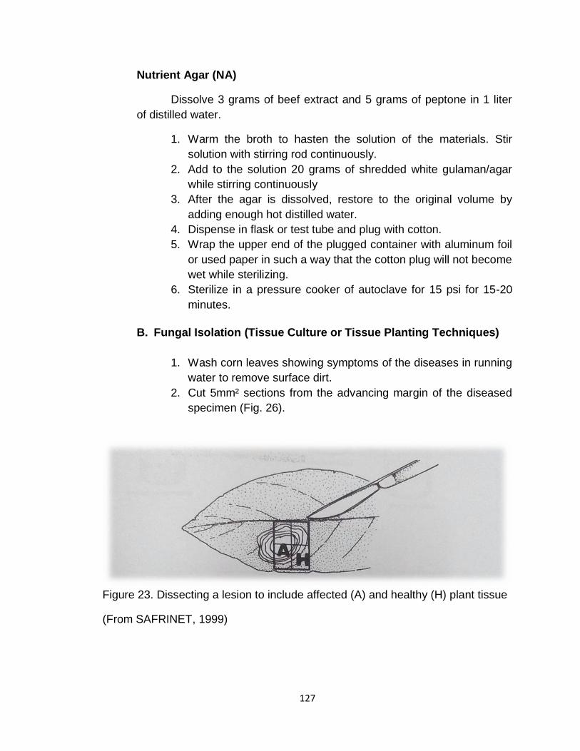
2. Cut 5mm² sections from the advancing margin of the diseased
specimen (Fig. 26).
Figure 23. Dissecting a lesion to include affected (A) and healthy (H) plant tissue
(From SAFRINET, 1999)

128
3. Dip the leaf sections in sodium hypochlorite for 1-2 minutes and rinses in three changes of sterile distilled water (Fig. 3) blot dries the leaf tissues in sterile paper towel of tissue paper.
4. Plant the leaf tissues equidistantly in plated potato dextrose agar (PDA) prepared at least 12 hours earlier using sterilized forceps or flame sterilized transfer needler (Fig. 4).
5. Incubate plates upside down at room temperature and observe for the growth of the fungus. After 24-72 hours.
6. Transfer bits of the mycelia from the advancing margin of the fungal growth into newly plated PDA using sterile transfer needle for the pure culture preparation.
7. Transfer pure culture of the fungus onto PDA slants.
Figure 24. Surface disinfection of Materials
Plant Material
Figure 25. Tissue planted equidistantly in plated PDA
129
C. Bacterial Isolation (Streak Plate Method)
1. Wash specimen under running tap water to remove surface dirt. 2. Induce bacterial oozing
a. For soft rot - Cut about 3 mm² sections from the advancing margin of
the diseased specimen, including both healthy and diseased parts.
- Transfer the sections to a drop sterile distilled water on a sterile glass slide
- Observe for bacterial oozing under the microscope
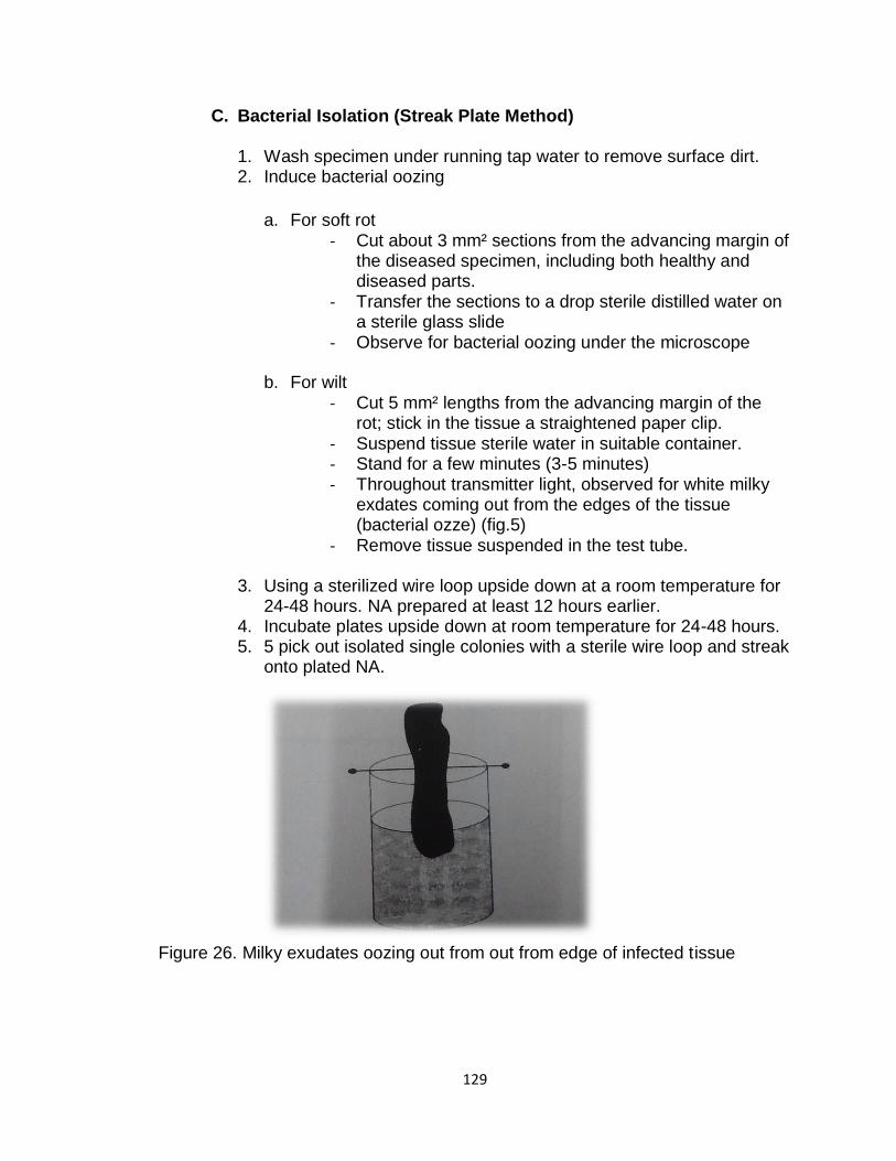
b. For wilt - Cut 5 mm² lengths from the advancing margin of the
rot; stick in the tissue a straightened paper clip. - Suspend tissue sterile water in suitable container. - Stand for a few minutes (3-5 minutes) - Throughout transmitter light, observed for white milky
exdates coming out from the edges of the tissue (bacterial ozze) (fig.5)
- Remove tissue suspended in the test tube. 3. Using a sterilized wire loop upside down at a room temperature for
24-48 hours. NA prepared at least 12 hours earlier. 4. Incubate plates upside down at room temperature for 24-48 hours. 5. 5 pick out isolated single colonies with a sterile wire loop and streak
onto plated NA.
Figure 26. Milky exudates oozing out from out from edge of infected tissue

130
6. Allow the bacteria grow for 48-72 hours and purify the culture by streaking sterile water suspension of the bacteria on agar and picking out single isolated colonies. Repeat procedures 2 or 3 times to get pure colonies. Characterize the colony of the pure culture.
7. Transfer pure culture into NA slants
D. Nematode isolation (Baermann Funnel Method. Fig 6) 1. Collect nematode-infester soil. 2. Place funnel fitted with rubber tubing and pinch cock in a ring stand
or funnel stand. 3. Fit a circular wire screen into the funnel. 4. Fill funnel with water up to the level of the wire screen 5. Place three layers of tissue paper on the wire screen.
Figure 7. Baermann funnel apparatus
6. Place about 50g (handful) of nematode-infested soil on the tissue paper.
7. Let the Baermann funnel set-up stand overnight. 8. Draw out a small amount of water into a watch glass by opening
cock and examine for nematodes under a dissecting microscope.
V. Result/Discussion 1. Submit pure cultures of bacterium and fungus isolated from the
diseased specimens. 2. Show nematodes extracted from the soil to your instructor.

131
VI. Question to Answer
1. Name other techniques employed in isolating bacterial pathogens. What are their metris? _____________________________________________________
_____________________________________________________
_____________________________________________________
_____________________________________________________
_____________________________________________________
_____________________________________________________
_____________________________________________________
_____________________________________________________
________
2. What is the purpose of soaking the tissue section in sodium hypochlorite? _____________________________________________________
_____________________________________________________
_____________________________________________________
_____________________________________________________
_____________________________________________________
_____________________________________________________
_____________________________________________________
_____________________________________________________
________
3. What is the principle in the Baermann funnel method of extracting nematodes from soils? _____________________________________________________
_____________________________________________________
_____________________________________________________
_____________________________________________________
_____________________________________________________
_____________________________________________________
_____________________________________________________
_____________________________________________________
________

132
4. How would you isolate nematodes from infested plant parts? _____________________________________________________
_____________________________________________________
_____________________________________________________
_____________________________________________________
_____________________________________________________
_____________________________________________________
_____________________________________________________
_____________________________________________________
5. In the tissue planting technique, why do you cut tissue sections from the advancing margin of lesion? _____________________________________________________
_____________________________________________________
_____________________________________________________
_____________________________________________________
_____________________________________________________
_____________________________________________________
_____________________________________________________
_____________________________________________________
________________________
VII. References _____________________________________________________
_____________________________________________________
_____________________________________________________
_____________________________________________________

133
VI. Summary
➢ Variability in plant pathogen is one of the dynamic and significant aspects
of biology: individuals have different characteristics, not fixed. Many of the
cases that two pathogen with the same morphological characteristics differ
with respect to their physiology, biochemical characters and pathogenicity.
➢ Stages of variation differ depending on the type of pathogen.
➢ Variability in fungi are mutation, recombination, heterokaryosis,
parasexualism, and heteroploidy.
➢ Stages of variability in bacteria are conjugation, transformation, and
transduction.
➢ Stages of variability in virus are recombination, re-assortment, and
mutation.
➢ The conventional methods for identifying the variability in the pathogens at
species, subspecies and intra sub species level is being done by study of
virulence reactions using disease rating scales on set of host differentials.
Molecular techniques are more precise tools for differentiation between
species, and identification of new strains/ isolates.
➢ The different marker system are Restriction Fragment Length
Polymorphisms (RFLP), Random Amplified Polymorphic DNA (RAPD),
Sequence Tagged Sites (STS), Amplified Fragment Length
Polymorphisms (AFLP), Simple Sequence Repeats (SSR) or
microsatellites, and ribosomal DNA (rDNA) markers.
VII. References
Kumar, S., & Verma, S. (2019). Variability in Plant Pathogens and Tools for its
Characterization. Int. J. Curr. Microbiol. App. Sci, 8(2), 2887-2902.
MacLachlan, James N. and Edward J. Dubovi (2017). Chapter 6-Epidemiology
and Control of Viral Diseases, Fenner's Veterinary Virology (Fifth Edition),
Academic Press, 2017, Pages 131-153, ISBN 9780128009468,
https://doi.org/10.1016/B978-0-12-800946-8.00006-4.
Internet Source
https://www.slideshare.net/aru15/pathogen-variability
http://www.hillagric.ac.in/edu/coa/ppath/lect/plpath111/Lect.%2012%20Pl%20Pat
h%20111-%20Variability%20in%20Plant%20Pathogens.pdf
https://www.ijcmas.com/8-2-
2019/Sunil%20Kumar%20and%20Shalini%20Verma.pdf