Nanomaterials for controlling bacterial pathogens and ...
189
Aleksandra Ivanova Nanomaterials for controlling bacterial pathogens and resistance occurrence Universitat Politècnica de Catalunya
-
Upload
khangminh22 -
Category
Documents
-
view
1 -
download
0
Transcript of Nanomaterials for controlling bacterial pathogens and ...

Aleksandra Ivanova
Nanomaterials for controlling bacterial
pathogens and resistance occurrence
Universitat Politècnica de Catalunya

Nanomaterials for controlling bacterial
pathogens and resistance occurrence
Aleksandra Asenova Ivanova
A thesis submitted in fulfilment of the requirements for degree of
Doctor of Philosophy
at the
Universitat Politècnica de Catalunya
Supervised by Dr. Tzanko Tzanov
Group of Molecular and Industrial Biotechnology (GBMI)
Department of Chemical Engineering
Universitat Politècnica de Catalunya
Terrassa (Barcelona)

2022
The work carried out during the thesis was financially supported by:
Pre-commercial lines for the production of surface nanostructured antimicrobial and antibiofilm
textiles, medical devices, and water treatment membranes, H2020-720851
SKin Healthcare by Innovative NanoCAPsuleS, H2020-685909

To my family,
“Hardship often prepares an ordinary person
for an extraordinary destiny”
Christopher Markus
“The energy of the mind is the essence of life”
Aristotle


Abstract
Infectious diseases are the leading cause of death worldwide, while the constantly
raising antimicrobial resistance (AMR) is a major concern for the public health. During
the infection establishment, bacterial pathogens communicate via expression of
signaling molecules, controlled through a phenomenon called quorum sensing (QS). As
a result of this, bacteria produce virulence factors and form resistant biofilms on living
and non-living surfaces causing persistent infections. The infection complexity,
especially in chronic diseases, requires the use of broad-spectrum antibiotics responsive
for the appearance and the spread of drug resistant species. Infections caused by
antibiotic-resistant pathogens are associated with high morbidity, mortality, and huge
economic burden. Unlike the decrease over the past three decades of the number of
novel marketed antimicrobial drugs, the number of antibiotic resistant bacterial strains
steadily increases. Thus, there is an urgent need for development of alternative
strategies to manage difficult-to-treat infections.
This thesis aims at the engineering of advanced nano-enabled materials and
nanostructured coatings for controlling bacterial pathogenesis and resistance
occurrence. To achieve this, biopolymers, antibiofilm and anti-infective enzymes, and
inorganic compounds were nano-hybridized as alternative modalities to the
conventional antibiotics. The nanoform was able to provide enhanced interaction with
bacterial cell membranes and easier penetration into biofilms, increasing the
antimicrobial efficacy at lower dosages, while preventing from development of
antimicrobial resistance. Additionally, specific targeting moieties increased the
nanomaterial’s interaction with the pathogens, avoiding the drug resistance appearance
and cytotoxicity.
The first part of the thesis describes the functionalization of biologically inert
nanoparticles (NPs) with membrane disturbing antimicrobial aminocellulose (AM) and
biocompatible hyaluronic acid (HA) in a layer-by-layer (LbL) fashion for elimination of
medically relevant pathogens. The generated nanoentities demonstrated high potential to
inhibit the biofilm formation, without affecting the human cell viability.
Further, the LbL technique was applied to decorate antimicrobial, but potentially toxic
silver (Ag) nano-templates with biocompatible AM and quorum quenching (QQ)
acylase in order to obtain safe antibacterial and antibiofilm nanomaterials. The
deposition of acylase and AM on the Ag core interfered with the QS signaling and

II
bacterial pathogenesis, and enhanced the NPs interaction with the bacterial membrane.
The integration of triple mechanisms of action in the hybrid nanoentities resulted in
complete bacteria and biofilm eradication and improved biocompatibility of the AgNPs.
The thesis also describes the development of targeted nanocapsules (NCs) for selective
elimination of Staphylococcus aureus. Herein, self-assembling nanoencapsulation
technology using the biocompatible and biodegradable protein zein was applied for the
generation of zein NCs loaded with bactericidal oregano essential oil (EO). An antibody
specifically targeting S. aureus was covalently grafted on the NCs surface. The obtained
targeted NCs demonstrated antibacterial selectivity in a mixed bacterial inoculum, and
the treatment efficacy was validated in an in vitro coculture model of bacteria and
mammalian cells.
Finally, high intensity ultrasound (US) process was employed for engineering of
durable antibacterial/antibiofilm coating on urinary catheters. The simultaneous
deposition of zinc oxide (ZnO) NPs and a matrix-degrading amylase enzyme improved
the NPs adhesion on the silicone material, and prevented its bacterial colonization and
biofilm formation in vitro. The hybrid nanostructured coating prevented the occurrence
of early onset urinary tract infections (UTIs) and showed excellent biosafety in an in
vivo animal model.
Key words: biopolymers, enzymes, essential oils, metal nanoparticles, Layer-by-Layer
assembly, sonochemistry, bacteria targeting, antibacterial and antibiofilm properties,
antibiotic resistance

III
Table of Contents
Abstract ............................................................................................................................ i
Table of Contents ........................................................................................................... iii
Abbreviation list ........................................................................................................... vii
1 Introduction ................................................................................................................. 2
2 State-of-the-art ............................................................................................................. 4
2.1 Antimicrobial resistance ....................................................................................... 5
2.2 Bacterial biofilms and quorum sensing system .................................................. 7
2.2.1 Biofilms and biofilm-related bacterial resistance ........................................ 7
2.2.2 Quorum sensing systems in bacteria .......................................................... 10
2.3 Therapeutic strategies to manage bacterial infections and antibiotic
resistance .................................................................................................................... 13
2.3.1 Anti-virulent strategies-inhibition of bacterial pathogenesis ................... 14
2.3.2 Biofilm-disrupting enzymes ......................................................................... 17
2.3.3 Antimicrobial polymers, biopolymers, and natural compounds ............. 18
2.3.4 Nano-enabled materials to manage bacterial infections and resistance
development ........................................................................................................... 20
2.3.4.1 Inorganic nanoparticles .......................................................................... 22
2.3.4.2 Non-metallic nanoparticles ..................................................................... 24
2.3.4.3 Hybrid nanoentities ................................................................................. 24
2.3.4.4 Hybrid nanostructured coatings .............................................................. 26
2.4 Layer-by-Layer assembly as a tool for engineering of highly efficient
antimicrobial nanomaterials .................................................................................... 27
2.5 Sonochemistry for manufacturing of functional nanomaterials and
nanostructured coatings ........................................................................................... 32
3 Objectives of the Thesis ............................................................................................. 37
4 Materials and Methods ............................................................................................. 39
4.1 Materials and reagents ....................................................................................... 40
4.2 Bacteria and human cells ................................................................................... 40
4.3 Experimental methods ........................................................................................ 41
4.3.1 Layer-by-layer decorated nanoparticles with tunable antibacterial and
antibiofilm properties against both Gram-positive and Gram-negative bacteria
................................................................................................................................. 41
4.3.1.1 LbL coating of NPs .................................................................................. 41
4.3.1.2 LbL coating of NPs with fluorescein isothiocyanate-labeled AM ........... 42
4.3.1.3 Characterization of the NPs .................................................................... 42

IV
4.3.1.4 Determination of primary amino groups on the NP surface ................... 42
4.3.1.5 Antibacterial activity of LbL engineered NPs ......................................... 43
4.3.1.6 Antibacterial activity of AM against S. aureus and E. coli. .................... 43
4.3.1.7 Bacteria time-killing kinetics ................................................................... 43
4.3.1.8 Interaction of the NPs with cell membrane models ................................. 44
4.3.1.9 Biofilm inhibition test .............................................................................. 44
4.3.1.10 Biocompatibility of NPs ......................................................................... 45
4.3.2 Layer-by-layer coating of aminocellulose and quorum quenching acylase
on silver nanoparticles synergistically eradicate bacteria and their biofilms.. 45
4.3.2.1 AgNPs synthesis ....................................................................................... 45
4.3.2.2 LbL coating of AgNPs with AM and acylase ........................................... 46
4.3.2.3 NPs characterization ............................................................................... 46
4.3.2.4 Enzymatic activity .................................................................................... 47
4.3.2.5 Quorum quenching activity ..................................................................... 47
4.3.2.6 Antibacterial activity of the developed NPs ............................................ 48
4.3.2.7 TEM observation of NPs interaction with bacteria ................................. 48
4.3.2.8 Determination of ROS generation ........................................................... 48
4.3.2.9 Biofilm inhibition test .............................................................................. 49
4.3.2.10 Biofilm eradication assay ...................................................................... 49
4.3.2.11 Biocompatibility .................................................................................... 49
4.3.3 Antibody-enabled antimicrobial nanocapsules for selective elimination of
Staphylococcus aureus .......................................................................................... 50
4.3.3.1 Protein A antibody interaction with S. aureus ........................................ 50
4.3.3.2 Antibody-enabled NCs formulation ......................................................... 50
4.3.3.2 NCs characterization ............................................................................... 51
4.3.3.3 Quartz crystal microbalance with dissipation monitoring ...................... 51
4.3.3.4 Antibacterial activity tests ....................................................................... 52
4.3.3.5 Nanocapsules interaction with bacteria assessed by scanning electron
microscopy ........................................................................................................... 52
4.3.3.6 Biocompatibility evaluation .................................................................... 53
4.3.3.7 Antibacterial efficacy of the NCs in vitro in a coculture model of S.
aureus and human cells ........................................................................................ 53
4.3.4 Nano-engineered hybrid zinc oxide/amylase coatings to prevent the
occurrence of catheter-associated urinary tract infections ............................... 54
4.3.4.1 Ultrasound-assisted coating of catheters with ZnO NPs and α-amylase 54
4.3.4.2 Surface characterization of the coated catheters .................................... 54

V
4.3.4.3 Stability of the coatings ........................................................................... 54
4.3.4.4 Amylase activity in the coatings .............................................................. 55
4.3.4.5 Quantification of the total biofilm formation .......................................... 55
4.3.4.6 Antibacterial activity ............................................................................... 55
4.3.4.7 Dynamic biofilm inhibition tests .............................................................. 56
4.3.4.8 Biocompatibility assessment .................................................................... 56
4.3.4.9 In vivo tests in rabbit model .................................................................... 57
4.3.4.9.1 Microbiological tests ............................................................................ 58
4.3.4.9.2 Histopathological examination ............................................................ 58
4.3.4.9.3 Biochemical analyses ........................................................................... 59
4.3.4.10 Statistical analysis ................................................................................. 59
5 Results and Discussion .............................................................................................. 60
5.1 Layer-by-layer decorated nanoparticles with tunable antibacterial and
antibiofilm properties against both Gram-positive and Gram-negative bacteria
.................................................................................................................................... 61
5.1.1 NPs characterization .................................................................................... 63
5.1.2 Antibacterial activity of the NPs against S. aureus and E. coli. ............... 66
5.1.3 Interaction of the coated NPs with model bacterial membranes ............. 72
5.1.4 Biofilm Inhibition ......................................................................................... 73
5.1.5 Biocompatibility of the LbL decorated NPs. ............................................. 74
5.1.6 Conclusions ................................................................................................... 76
5.2 Layer-by-layer coating of aminocellulose and quorum quenching acylase on
silver nanoparticles synergistically eradicate bacteria and their biofilms .......... 77
5.2.1 NPs characterization .................................................................................... 79
5.2.2 Antibacterial efficiency of AgNPs and LbL Ag@AM_AC NPs ............... 82
5.2.3 Antibiofilm activity ...................................................................................... 85
5.2.3.1 Inhibition of P. aeruginosa biofilm formation by LbL Ag@AM_AC NPs 86
5.2.3.2 Elimination of P. aeruginosa biofilms with LbL Ag@AM_AC NPs ........ 88
5.2.4 Biocompatibility assessment ........................................................................ 90
5.2.5 Conclusions ................................................................................................... 92
5.3 Antibody-enabled antimicrobial nanocapsules for selective elimination of
Staphylococcus aureus .............................................................................................. 93
5.3.1 Targeted NCs formulation and characterization ...................................... 95
5.3.2 Antibacterial activity towards S. aureus .................................................... 99
5.3.3 Real-time monitoring of the NCs interaction with S. aureus ................. 100

VI
5.3.4 Selective antibacterial activity of the Ab@EO NCs towards targeted S.
aureus.................................................................................................................... 102
5.3.5 Cytotoxicity of the Ab@EO NCs .............................................................. 105
5.3.6 Antibacterial efficacy of the Ab@EO NCs in an in vitro coculture model
of S. aureus and human cells .............................................................................. 107
5.3.7 Conclusions ................................................................................................. 110
5.4 Sonochemically engineered nano-enabled zinc oxide/amylase coatings
prevent the occurrence of catheter-associated urinary tract infections ............ 111
5.4.1 Characterization of the coated urinary catheters ................................... 113
5.4.2 Stability of the coatings in artificial urine ................................................ 115
5.4.3 Antibiofilm activity and functional stability of the coated urinary
catheters ............................................................................................................... 117
5.4.4 Biofilm inhibition tests under dynamic conditions in a model of
catheterized human bladder ............................................................................... 121
5.4.5 Cytotoxicity of the coatings ....................................................................... 125
5.4.6 In vivo efficacy assessment of ZnO@AM NPs-coated catheters in a rabbit
model .................................................................................................................... 127
5.4.6.1 Microbiological analysis of urine and catheters´ surfaces for UTIs detection
............................................................................................................................... 127
5.4.6.2 Histopathology after catheterization ......................................................... 130
5.4.6.3 Hematological and biochemical analyses ................................................. 133
5.4.7 Conclusions ................................................................................................. 136
6 Conclusions and Future Plans ................................................................................ 137
6.1 Final conclusions ............................................................................................... 138
6.2 Future perspectives ........................................................................................... 140
Acknowledgements ..................................................................................................... 142
Appendix: Scientific Contribution ............................................................................ 144
References: .................................................................................................................. 147

VII
Abbreviation list
A
AAPH 2, 2-azobis (2-amidino-propane) dihydrochloride
A. baumannii Acinetobacter baumannii
AIs autoinducers
AIPs auto-inducing peptides
pro-AIP auto-inducing peptide precursor
Ag silver
AgNO3 silver nitrate
Agr accessory gene regulator
A. tumefaciens Agrobacterium tumefaciens
AHLs acyl-homoserine lactones
AM aminocellulose
AMR antimicrobial resistance
ANOVA a one way analysis of variance
ATCC American type culture collection
A. melleus Aspergillus melleus
Au gold
B
B. cereus Bacillus cereus
B. mycoides Bacillus mycoides
B. subtilis Bacillus subtilis
B. thuringiensis Bacillus thuringiensis
BSA bovine serum albumin

VIII
C
C6-HSL N-hexanoyl-L-homoserine lactone
CaO calcium oxide
CAUTIs catheters-associated urinary tract infections
C. albicans Candida albicans
CECT Spanish type culture collection
CFU colony-forming unit
CO2 carbon dioxide
C. violaceum Chromobacterium violaceum
CVC central venous catheters
CuO copper oxide
D
DCFDA 2ʹ, 7ʹ-dichlorofluorescin diacetate
DCFHDA dichlorodihydrofluorescein diacetate
DLS dynamic light scattering
DMEM Dulbecco’s modified Eagle’s medium
DMSO dimethyl sulfoxide
DNA deoxyribonucleic acid
DNase I deoxyribonuclease I
DNS 3, 5-dinitrosalicylic acid
E
E. coli Escherichia coli
eDNA extracellular deoxyribonucleic acid
EDTA ethylenediaminetetraacetic acid

IX
EDX energy dispersive X-ray
EE encapsulation efficiency
E. faecalis Enterococcus faecalis
EO essential oils
EPM extracellular polymeric matrix
EPS exopolysaccharide
F
Fe3O4 iron oxide
FITC fluorescein isothiocyanate
H
HA hyaluronic acid
HCl hydrochloric acid
HNO3 nitric acid
HPLC high-performance liquid chromatography
HRSEM high resolution scanning electron microscopy
I
ICP inductively coupled plasma
IUPAC International Union of Pure and Applied Chemistry
K
K. pneumoniae Klebsiella pneumoniae
L
LB Luria Bertani broth
LbL layer-by-layer
M

X
MBEC Minimum Biofilm Eradication Concentration
MBIC Minimum Biofilm Inhibitory Concentration
MDR multidrug resistance
MgO magnesium oxide
MIC Minimum Inhibitory Concentration
MHB Mueller Hinton broth
MRSA multidrug resistant Staphylococcus aureus
N
NAMET N-acetyl-L-methionine
NB nutrient broth
NCs nanocapsules
NPs nanoparticles
NTA nanoparticles tracking analysis
O
OD optical density
P
P. aeruginosa Pseudomonas aeruginosa
PB phosphate buffer
PBS phosphate buffered saline
PE L-α-phosphatidylethanolamine
PI propidium iodide
PMMA poly (methacrylic acid)
Pt platinum
Q

XI
QCM-D quartz crystal microbalance with dissipation
QQ quorum quenching
QS quorum sensing
R
RNA ribonucleic acid
RIP RNAIII inhibiting peptide
ROS reactive oxygen species
RT room temperature
S
SEM scanning electron microscopy
S. aureus Staphylococcus aureus
S. epidermidis Staphylococcus epidermidis
STEM scanning transmission electron microscope
T
TEM transmission electron microscopy
Ti titanium
TiO2 titanium dioxide
TSB tryptic soy broth
U
US ultrasound
UTIs urinary tract infections
UV ultraviolet
Z
ZnO zinc oxide
XII
Figure, Scheme, and Table Captions
Figure 2. 1 Mechanisms of antibiotic action. Antibiotics are able to inhibit the cell wall
synthesis or injure the bacterial cell membrane. Other targets are the synthesis of
essential metabolites, DNA and protein synthesis. .......................................................... 6
Figure 2. 2 Biofilm growth cycle. Initially the free-floating bacterial cells attach to a
solid surface, and secrete matrix components, leading to formation of mature biofilm
structure. Finally, parts of the established biofilm and bacteria are dispersed in the
surrounded environment able to attach and colonize new surfaces and start the biofilm
cycle again. ....................................................................................................................... 8
Figure 2. 3 QS signaling in Gram-negative (A) and Gram-positive bacteria (B). In
Gram-negative bacteria, AHL QS signals (orange circles) are produced by AHL
synthases (red rectangle) and secreted in the cells surrounding. When the concentration
of AHLs in the surrounding is above the threshold, the signals pass through the cell
membrane via diffusion and activate specific AHL receptors (blue motif), promoting the
target genes expression. In Gram-positive bacteria, small AIPs (grey pentagons) are
post-translationally synthesized in the cells and exported through specific membrane
bound transporters (light blue motif). At threshold level, AIPs bind to a two-component
histidine kinase sensor, which auto-phosphorylates and alters the target genes
expression. ...................................................................................................................... 12
Table 2. 1 Common QS inhibitors and their application in infection control................ 14
Figure 2. 4 Schematic representation of the mechanisms involved in the NPs interaction
with free-floating bacterial cells. .................................................................................... 21
Figure 2. 5 LbL assembly technologies. Schematical representation of the three major
technology categories for LbL assembly used for development of functional materials.
........................................................................................................................................ 29
Figure 2. 6 Main steps in the US process. (A) Formation, growth and collapse of
cavitation bubbles, generated upon sonication of liquids. (B) Microjets and shock waves
produced upon the collapse of the acoustic bubbles project the developed NPs at high
velocities towards the materials’ surface. ....................................................................... 33
Figure 5. 1 Alternation of zeta-potential as a function of the AM and HA deposition on
the NP template. ............................................................................................................. 63

XIII
Figure 5. 2 Microscopic images of FITC-labeled AM/HA multilayers deposited on NPs
surface. ............................................................................................................................ 64
Figure 5. 3 TEM images of the NP template (A), NPAM/HA-AM (B), NPAM/HA-HA (C).
........................................................................................................................................ 65
Figure 5. 4 S. aureus growth after 24 h incubation with coated NPs. ........................... 66
Figure 5. 5 (A) S. aureus and (B) E. coli growth after 24 h incubation with NP template
and LbL coated NPs. (C) and (D) Representative images after culturing the samples on
specific agar plates. (C) Black colonies of S. aureus and (D) blue colonies of E. coli
were formed after 24 h incubation with NPAM/HA-HA (left side images). No colonies
were observed after incubation with NPAM/HA-AM (right side images). ........................ 68
Figure 5. 6 Bacterial growth after 24 h incubation with different concentrations of AM
bulk solution. .................................................................................................................. 69
Figure 5. 7 Time-kill curves of LbL coated NPs against (A) S. aureus and (B) E. coli. S.
aureus and E. coli were incubated with 4 x 10¹⁰ NPs mL-1 NPAM/HA-HA (blue) and
NPAM/HA-AM (red) and the bacteria surviving the treatment were estimated after 15, 45,
and 60 min. All data are mean values of three independent experiments. ..................... 70
Figure 5. 8 Fluorescence images (100 x magnification) of (A) S. aureus, (B) S. aureus
treated with NPAM/HA-AM, (C) E. coli, (D) E. coli treated with NPAM/HA-AM. The green
and red fluorescence images are overlaid. Nonviable bacteria are stained in red, while
the viable bacteria appear green. .................................................................................... 71
Figure 5. 9 Kinetic adsorption process after the incorporation of the NP template and
LbL coated NPs into the air/water interface of the PE monolayer at π = 33 mN m-1. ... 72
Figure 5. 10 Crystal violet assessment of biofilm formation of S. aureus (black bars)
and E. coli (red bars) after incubation for 24 h with the biopolymer-coated NPs. ......... 74
Figure 5. 11 Viability (%) of human fibroblasts exposed to LbL coated NPs. ............. 75
Figure 5. 12 Morphology of human fibroblasts exposed to (A) NPAM/HA-AM; (B)
NPAM/HA-HA; (C) Control human fibroblast cells. ......................................................... 75
Figure 5. 13 UV-Vis scan of AgNPs. ............................................................................ 79

XIV
Figure 5. 14 LbL Ag@AM_AC NPs preparation and characterization. (A) Schematic
representation of AgNPs template coating with AM and AC using LbL approach. (B)
Zeta-potential of the NPs after each layer deposition. (C, D) TEM images of LbL
Ag@AM_AC NPs at different magnifications. (E) Histogram of LbL Ag@AM_AC
NPs size distribution based on the total count using ImageJ software. (F) Elemental
mapping image and (G) STEM-EDX spectrum of an individual LbL Ag@AM_AC NP.
........................................................................................................................................ 80
Figure 5. 15 L-Methionine production in the presence of AgNPs and LbL
Ag@AM_AC NPs. ......................................................................................................... 81
Figure 5. 16 Violacein production by C. violaceum. (A) The AHL signals bind to the
cytoplasmic QS receptor and switches on the vio genes, activating the violacein
production (left). The presence of QQ active NPs breaks down the AHL signals,
silencing the expression of the vio genes, subsequently inhibiting the violacein
production (right). (B) Quantification of QS regulated violacein production (%) by C.
violaceum and (C) Representative images of violacein production in presence of AgNPs
and LbL Ag@AM_AC NPs. (D) C. violaceum cell viability after incubation with
AgNPs and LbL Ag@AM_AC NPs. .............................................................................. 82
Figure 5. 18 NPs induced ROS generation in P. aeruginosa. ....................................... 85
Figure 5. 19 P. aeruginosa biofilm inhibition. (A) MBIC determination of the NPs
using crystal violet assay. (B) Live/dead™ BacLight™ kit microscopic visualization.
The green and red fluorescence images are overlaid. Nonviable bacteria are stained in
red, while the viable bacteria appear green. ................................................................... 87
Figure 5. 20 Elimination of 24h grown P. aeruginosa biofilm. (A) MBEC
determination of the NPs using crystal violet assay. (B) Live/dead™ BacLight™ kit
microscopic visualization of P. aeruginosa biofilm after 24 h treatment with AgNPs and
LbL Ag@AM_AC NPs at the same concentration (6.25 × 10⁷ NPs mL-1). The green and
red fluorescence images are overlaid. Nonviable bacteria are stained in red, while the
viable bacteria appear green. .......................................................................................... 89
Figure 5. 22 S. aureus growth reduction after 24 h incubation with free (black lines)
and encapsulated EO (red lines) at different concentrations. The EO encapsulation led to
enhanced antibacterial efficacy against S. aureus at lower amounts, when compared to
the amount of pristine oil. ............................................................................................... 95
Figure 5. 23 Fabrication and characterization of Ab@EO NCs. (A) Schematic
representation of Ab@EO NCs preparation. (B) Fluorescent intensity measurement at
Ex/Em = 490/525 nm upon FITC-protein A binding with the Ab@EO NCs. (C) Green

XV
color development after antibody interaction with targeted S. aureus. (D) Interaction of
antibody-enabled EO NCs with FITC-labelled protein A. (E) Florescence microscopy
images of FITC-protein A bound Ab@EO NCs. (F) TEM images of Ab@EO NCs at
different magnifications. (G) Histogram of the Ab@EO NCs size distribution based on
the total count of 60 NPs using ImageJ software. (H) NTA analysis of Ab@EO NCs and
(I) zeta-potential values for pristine EO NCs, EO_AC NCs and Ab@EO NCs. ........... 98
Figure 5. 24 Antibacterial activity of EO and Ab@EO NCs (concentration ≈ 1.5 x 10⁹
NCs mL-1) against S. aureus. .......................................................................................... 99
Figure 5. 25 (A) Interaction of EO NCs and Ab@EO NCs with S. aureus assessed by
QCM-D. The shift in the frequency and dissipation are represented with solid and
dashed lines, respectively. The numbers I, II, III, IV and V indicate the different zones,
respectively the baseline with PBS, bacterial adhesion, baseline after washing of loosely
adherent cells, NCs insertion and PBS washing. (B) Interaction of EO NCs and Ab@EO
NCs with P. aeruginosa assessed by QCM-D. The shift in the frequency and dissipation
are represented with solid and dashed lines, respectively. The numbers I, II, III, IV and
V indicate the different zones, respectively the baseline with PBS, bacterial adhesion,
baseline after washing of loosely adherent cells, NCs insertion and PBS washing. .... 102
Figure 5. 26 Antibacterial activity of EO NCs and Ab@EO NCs in mixed bacterial
inoculum. (A) Schematic representation of the EO NCs and Ab@EO NCs interaction
with S. aureus and P. aeruginosa, when are growing together. (B) S. aureus and P.
aeruginosa growth reduction (Log (CFU mL-1)) upon exposure to the EO NCs and
Ab@EO NCs. (C) SEM images of S. aureus (round-shaped) and P. aeruginosa (rod-
shaped) bacteria without any treatment and incubated with EO NCs and Ab@EO NCs.
White and red arrows indicate live and damaged bacterial cells, respectively. ........... 104
Figure 5. 27 Cytotoxicity of Ab@EO NCs. (A) Viability (%) of human fibroblasts
exposed to Ab@EO NCs after 24 h of incubation, determined by AlamarBlue assay. (B)
Live/Dead assay of human fibroblasts after 24 h exposure to Ab@EO NCs. Overlapped
images of live (green) and dead (red) cells. The bars aside each image represent the
green and fluorescence intensities obtained after measuring at λex/em = 494/517 nm and
λex/em = 528/617 nm for calcein and ethidium homodimer-1, respectively. Scale bar
corresponds to 100 µm. ................................................................................................ 107
Figure 5. 28 Live/Dead kit staining of non-infected and S. aureus infected human cells
with and without treatment with Ab@EO NCs. ........................................................... 108
Figure 5. 29 S. aureus growth reduction in an in vitro coculturing model with human
cells after treatment with EO and Ab@EO NCs. ......................................................... 109

XVI
Figure 5. 31 Enzymatic activity of amylase-containing coating before and after
incubation in artificial urine for 7 days with shaking. The results are expressed as the
percentage of amylase activity compared to the enzyme before incubation in artificial
urine. ............................................................................................................................. 116
Figure 5. 32 HRSEM images of pristine, ZnO and ZnO@AM NPs-coated silicone
catheter after 7 days of incubation in artificial urine. ................................................... 117
Figure 5. 33 Biofilm inhibition (%) at static condition of the coated catheters towards
(A) S. aureus and (B) E. coli assessed before and after incubation in artificial urine for 7
days under continuous agitation. The results are represented in %, compared with the
biofilm inhibition of pristine catheter (non-inhibition). Stars represent the statistical
differences between the different groups of samples; p < 0.05. ................................... 119
Figure 5. 34 Biofilm inhibition activity of the ZnO@BSA NPs-coated catheters. ..... 120
Figure 5. 35 Antibacterial activity of ZnO NPs and ZnO@AM NPs-coated catheters.
...................................................................................................................................... 121
Figure 5. 36 In vitro biofilm inhibition in catheterized bladder model with urine
recirculation. (A) In vitro model of catheterized human bladder. Total biofilm
quantification of (B) S. aureus and (C) E. coli biofilm mass on ZnO@AM coated
urinary catheters. Stars represent the statistical differences between the treated and
pristine catheters; p < 0.05. (D) Fluorescence microscopy images of live (green) and
dead (red) bacteria in the biofilms grown on ZnO@AM-coated and pristine silicone
Foley catheter balloon. The green and red fluorescence images are overlaid. ............. 124
Figure 5. 37 Viability of HaCaT and BJ-5ta cell lines after exposure to the ZnO@AM
NPs-coated catheters assessed by (A) AlamarBlue and (B) Live/Dead kit assays. The
green and red fluorescence images are overlaid. Scale bar corresponds to 100 μm. ... 126
Figure 5. 38 Antibacterial properties of pristine and ZnO@AM NPs-coated catheters.
(A) Colony count of bacteria from the urine samples collected from animals
catheterized with pristine and ZnO@AM NPs-coated catheters. (B) Colony count of live
bacteria adherent on the non-treated and ZnO@AM NPs functionalized catheters after 7
days of catheterization in the rabbit model. .................................................................. 129
Figure 5. 39 Histopathology of (A) rabbit penile, (B) prostatic urethra, (C) bladder and
(D) rabbit kidney of non-catheterized (group 1) and catheterized with pristine (group 2)
and ZnO@AM NPs (group 3) catheters. Black arrows indicate epithelial lining; red
arrow displays urinary bladder mucosa with transitional epithelial cell lining; red
arrowheads show glomeruli. (E) Blood smears of rabbits. Black arrow indicates the

XVII
neutrophil granulocytes. (F) Urine sediments of non-catheterized (group 1) and
catheterized rabbits (group 2 and 3). Red arrows indicate the umbrella cells from the
urinary system mucosal linings. ................................................................................... 132
Scheme 4. 1 Schematic illustration of the polymeric NP template decoration in an LbL
fashion yielding 8 × 10¹⁰ NPs mL -1 ............................................................................... 41
Scheme 4. 2 In vitro model of catheterized human bladder. .......................................... 56
Scheme 5. 1 Schematic representation of the assays performed in vivo in a rabbit model.
...................................................................................................................................... 127
Table 2. 1 Common QS inhibitors and their application in infection control................ 14
Table 5. 1 Amount of ZnO (wt %) on ZnO NPs and ZnO@AM NPs-coated catheters
before and after incubation in artificial urine. .............................................................. 115
Table 5. 2 Hematological blood results of non-catheterized and catheterized rabbits. 134
Table 5. 3 Biochemical results of urine collected from non-catheterized and
catheterized rabbits. ...................................................................................................... 135

XVIII

1 Introduction

2
In the past decades, the discovery of antibiotics has been a breakthrough in the
management of infectious diseases.1 However, the AMR is a natural evolutionary
response whereby bacteria evoke mechanisms to overcome the antimicrobial therapies.2
The AMR is now described as the “silent pandemic” of 21st century, associated with
impaired treatment and prevention of infections caused by parasites, fungi, bacteria, and
viruses.3 The misuse and overuse of antibiotics led to the appearance of multidrug
resistance (MDR) strains capable to withstand the effects of the conventional
antimicrobials.4 The high incidence of MDR is responsible for 700 000 deaths annually
worldwide and is expected to grow to 10 million deaths by 2050.5,6 Moreover, bacteria
have affinity to attach and colonize surfaces, such as those of indwelling devices,
tissues, and organs, forming microbial communities, called biofilms, which are up to
1000 times more resistant to the existing antibiotics than the free-floating cells.7 In these
communities, the bacterial cells are protected by multi-layered self-produced
extracellular polymeric matrix (EPM), able to restrict the antibiotic penetration, causing
severe chronic infections.8 Frequently, the treatment of biofilm-related infections
involves intensive antibiotic therapies, associated with side effects, such as
inflammatory response or cytotoxicity.9 Given the growing impact of antibiotic
resistance, there is an urgent need to develop novel non-antibiotic antibacterial
strategies with different mechanisms of action to outsmart the bacterial evolutionary
mechanisms and resistance occurrence.
Significant attention has been posed to the nanotechnology as a novel emerging branch
for engineering of non-conventional antibacterial chemotherapies to tackle the current
challenges for managing the MDR and bacterial diseases.10 Natural polymers and
natural-occurring compounds have received considerable attention due to their
antibacterial features and are suggested as powerful alternatives for controlling bacterial
infections.11,12 Furthermore, approaches which interfere with the bacterial pathogenesis-
regulating QS system, make the bacteria more susceptible to antibacterial agents and the
host immune system, reducing the risk of resistance development.13 The
nanoformulation of materials and their targeted delivery may overcome most of the
limitations of the traditional bulk therapeutics with the same composition, and improve
their antibacterial activity and therapeutic index.14,15
In this thesis, innovative highly efficient antibiotic-free nano-composites and
nanocoatings were developed for prevention and reduction of bacterial

3
contamination and biofilm formation, minimizing the spread of drug resistance.
This was achieved by an innovative platform of: i) hybrid assemblies of antibacterial
and antifouling (bio)polymers onto inert NP template, ii) hybrid nano-assemblies of QQ
enzymes/antibacterial polymers onto active NP template, iii) antibody-enabled NCs for
targeted drug delivery and selective bacterial elimination, and iv) hybrid nanocomposite
coatings of bactericidal metal/antibiofilm enzyme NPs for prevention of bacterial
infections.
The rationale of these innovative nano-enabled materials and coatings for infection
control was based on the synergy of:
Biopolymers with intrinsic antimicrobial activity hybridized with inorganic and
polymeric NPs able to eradicate the pathogens through unspecific interactions
with bacterial cell membranes.
Anti-biofilm and anti-infective enzymes to break down the QS signals in the
extracellular space of Gram-negative, unlike conventional biocides, and inhibit
the virulence and capacity of bacteria to establish biofilms. Combination of
these enzymes with inorganic NPs, adding a new mechanism to their action- a
membrane disturbing capacity due to the nanoform- will further reduce the
possibility of resistance development.
Biofilm matrix-degrading enzymes that degrade the adhesive components of the
EPM weakening and dispersing the biofilm. Enzyme induced EPM degradation
will significantly reduce the biofilm occurrence and increase the susceptibility of
both Gram-negative and Gram-positive bacteria to antimicrobials.
Nano-sized combination of the new antimicrobials with targeting moieties will
allow the development of highly potent and specific targeted nanosystems able
to address both evolutionary and cooperatively acquired AMR. Such therapeutic
approach was expected to eliminate the targeted pathogen, exerting less selective
pressure for resistance development, lowering the side effects to human cells and
maintaining the microbiome balanced.

4

2 State-of-the-art

5
2.1 Antimicrobial resistance
AMR and the spread of drug-resistant pathogens with new resistance mechanisms have
emerged as a major public health issue of the 21st century that threatens the effective
prevention and treatment of bacterial infections.16 Since the number of drug-resistant
strains constantly increases, while novel and efficient antimicrobials are not entering the
market, there is a serious concern about the dramatically growing number of untreatable
infections.16 AMR infections are the main reason for the increased morbidity and
mortality in the healthcare facilities, longer hospital stays, and elevated financial burden
worldwide.17 Assessment of the future impact of AMR predicts almost 10 million
deaths annually worldwide by 2050 if new antibiotic alternatives are not developed.5
The antibiotic over usage is interconnected with the transmission of AMR to the
humans via animal products as well. Diverse studies have reported that the antibiotic
administration duration, the selected drug, and the indications are irrelevant in 30-50 %
of the prescribed antibiotic therapies. Moreover, the antibiotics are applied as growth
promoters in livestock.18 The majority of nosocomial infections or hospital-acquired
infections occurs after the patient received healthcare treatment.19 The patient’s
exposure to the microorganisms in the hospital settings, to medical personnel or visitors,
increases the risk of infections occurrence within 48 h after hospital admission, or in up
to 30 days after surgery.20 Besides, the risk of infections with MDR pathogen is
increased in patients with compromised health conditions, such as cancer, diabetes,
human immunodeficiency virus, and renal failure.21,22 Some drug-resistant bacteria can
infect children, immunodeficient patients, and elderly, causing worse clinical outcomes
or death.23
Bacteria possess multiple mechanisms to evade the actions of antibiotics. This AMR
can be intrinsic (naturally occurred) or acquired (caused by the mutations in the genetic
materials).24 The intrinsic form of resistance is inherent and specific class of antibiotics
can be ineffective against certain bacterial specie.23 The outer membrane of the cell wall
in Gram-negative bacteria and the efflux pumps are the most common intrinsic drug
resistant mechanisms.22 Gram-negative bacteria demonstrate intrinsic resistance to
glycopeptide antibiotics, e.g. vancomycin, due to its incapability to pass through the
outer cell membrane.22 For example, triclosan has been widely used against Gram-
positive bacteria and some Gram-negative species; however, it is inactive against
Pseudomonas due to its intrinsic resistance to this bactericidal agent.18

6
The antibiotics act against bacteria through obstructing certain targets, such as nucleic
acid replication or protein and cell wall synthesis25 (Figure 2.1). The bacterial
pathogens can develop several acquired resistant mechanisms that can “neutralize” the
antibiotic action, such as altering the drug target site and enzymatic degradation of the
drugs.26 The bacteria secrete enzymes, e.g. β-lactamases, phosphorylases, adenylases,
and acetylases that can modify and destroy the antibiotic structure.
The antibiotics, depending on the class, act by binding to a specific site in the selected
bacteria. The evocation of changes in the structure of the targeted site may cause drug
disability for binding to the target.27 Additionally, the bacteria have evoked multiple
mutations that led to development of MDR, generally defined as “superbugs”,
associated with the occurrence of severe infections. This form of resistance is related to
mutations in the bacterial genome, which mutations can be in the bacterial chromosomal
deoxyribonucleic acid (DNA) or can be acquired genetically, and subsequently
transferred to the next generation of bacteria.24
Figure 2. 1 Mechanisms of antibiotic action. Antibiotics are able to inhibit the cell wall
synthesis or injure the bacterial cell membrane. Other targets are the synthesis of essential
metabolites, DNA and protein synthesis.
S. aureus is one of the most common Gram-positive pathogens responsible for serious
nosocomial infections, including skin, bloodstream, and surgical wound infections.

7
Staphylococci sp., producing penicillinase acount for more than 90 % of the patient
isolates, making the penicillin no longer usable for treatment of staphylococcal
infections.28 Furthermore, the appearance of methicillin-resistant S. aureus (MRSA)
strains is the main clinical problem and one of the leading cause for the high incidence
of nosocomial infections. Meta analytical studies reported that MRSA bacteremia is
associated with a significant increase in mortality and prolonged hospitalization among
patients with bloodstream infections.29 Methicillin-resistant staphylococci are sensitive
only to small number of antibacterial agents and often are characterized with MDR to
large number of antibiotics such as macrolides, β-lactams, lincosamides,
aminoglycosides, and tetracycline.30 The continuous growth of drug-resistance
community-acquired MRSA and the decreased susceptibility of MRSA to the main
antibiotic vancomycin used for staphylococcal infections is an urgent matter for the
public healthcare.18 Additionally, of great concern are the Escherichia coli and
nontyphoidal Salmonella resistance to fluoroquinolones, impairing the treatment of
UTIs and diarrhea, respectively. Currently, many species of tuberculosis, staphylococci,
and pneumococci are insusceptible to most of the existing antibiotics, increasing the
economic and human burden.31
2.2 Bacterial biofilms and quorum sensing system
2.2.1 Biofilms and biofilm-related bacterial resistance
Bacteria adhere on living or non-living surfaces forming communities of cells encased
in self-produced EPM, called biofilms.32 The bacterial transformation from planktonic
to sessile mode of growth involves multiple and complex processes (Figure 2.2).
During the first stage, the bacterial cells not adhered to the surface are susceptible to
antibiotics. The free-floating bacteria interact with solid surfaces via pili, flagella,
fimbriae, and production of exopolysaccharide (EPS), leading to bacterial attachment
and formation of micro-colonies. The EPM substances secreted around the colonies
comprise mainly polysaccharides (50-90 %), extracellular DNA (eDNA), lipids, and
proteins. The pathogen proliferation and the secretion of EPM components result in
establishment of three-dimensional mature biofilm structures. Subsequently, the
morphology and topography of these biofilm structure alter and the pathogens disperse
from the biofilm colonizing other solid surfaces.32

8
Figure 2. 2 Biofilm growth cycle. Initially the free-floating bacterial cells attach to a solid
surface, and secrete matrix components, leading to formation of mature biofilm structure.
Finally, parts of the established biofilm and bacteria are dispersed in the surrounded
environment able to attach and colonize new surfaces and start the biofilm cycle again.
An increased percentage of bacterial planktonic cells is already resistant to most of the
existing antibiotics; however, their biofilm-mode of growth is even more challenging
for eradication by the available therapeutics. The formation of the densely packed EPM
makes the cells within the biofilm 100 to 1000 times more insusceptible to
antibacterials, than their free-floating counterparts.33 The formation of biofilms is
responsible for more than 60 % of infectious diseases such as cystic fibrosis, sepsis,
otitis, endocarditis, chronic wounds, periodontitis, and dental caries.34 The treatment of
biofilm-associated infections involves the combination of various antibiotics at high
dosages.35 The Gram-negative E. coli, Klebsiella pneumoniae, Pseudomonas
aeruginosa and Proteus mirabilis, and the Gram-positive Enterococcus faecalis, S.
aureus, Staphylococcus epidermidis, as well as the yeast Candida albicans, are among
the most common biofilm forming species found in severe and persistent nosocomial
infections.36 Several mechanisms, such as limited drug penetration, decelerated bacterial
growth in the biofilm structure, and enzyme drug neutralization have been proposed for
the high level of biofilm-related bacterial resistant nature.32 P. aeruginosa is an
opportunistic Gram-negative bacterium able to form drug resistant biofilms, responsible
for cutaneous wound infections and chronic leg ulcers.37 The alginate-producing P.

9
aeruginosa and mucoid-biofilms in the patient’s lungs cause persistent and chronic
infections, associated with increased morbidity and mortality.38 The presence of EPM
reduces the aminoglycosides and fluoroquinolones penetration, allowing the pathogen
to respond to the stress via acquired defensive mechanisms. For example, the secretion
of cephalosporinase AmpC enzymes in cystic fibrosis is responsible for the P.
aeruginosa resistance to β-lactam.32 Another study proposed that EPM could retard the
drug penetration and trigger the expression of genes that induce drug resistance and
genetic adaptation to various conditions.39 In similar fashion, the biofilm matrix can
limit the nutrient availability to the bacterial cells, decelerating the growth rate, and
increasing the antibiotic resistance. Several antibiotics such as ampicillin and penicillin
can act only when bacterial cells are growing. The Gram-negative E. coli and P.
aeruginosa have demonstrated resistance to cetrimide and tobramycin, respectively due
to the slow bacterial growth.32 Despite growing in single strain biofilms, these bacterial
species are also able to communicate and interact, forming even more resistant sessile
structures. For example, the Gram-positive S. aureus colonizes chronic wounds and
frequently forms mixed-species biofilm structures in infectious conditions.40
The frequent usage of indwelling devices, medical contact lenses, organ transplantation,
and intervention therapies led to an increased occurrence of nosocomial infections
heavily affecting the patients with serious illnesses. The most common infections in
intensive care hospital facilities are the UTIs (40 %),41 bloodstream infections (10.5 %),
skin and soft tissue infections (6.7 %),42 respiratory tract infections and pneumonia
(22.8 %), surgical-site infections (15.7 %),36 associated with drug resistant biofilms. For
instance, patients with long-term inserted central venous catheters (CVC) have an
increased risk of bloodstream infections. It has been reported that the bacterial
pathogens colonized the CVC already after one day of catheterization.36,43 Bacteria have
affinity to attach to endotracheal tubes, forming drug resistant biofilms within 24 h
causing ventilator-associated pneumonia, leading to prolonged hospitalization and
increased mortality rates.36 Surgical-site infections due to Staphylococcus sp. are
frequently occurring within 30 days after the surgery.44 The catheter-associated UTIs
(CAUTIs) account for 80 % of all UTIs and are the main cause of prolonged
hospitalization and increased risk of resistance occurrence. The bacterial pathogens
have affinity for colonization of catheter’s surface and can easily enter in contact with
tissues and organs, leading to occurrence of UTIs. The bacteria can gain access to the

10
patient’s body during the catheter insertion, or from the urethral meatus together with
the catheter-urethral interface, or due to contamination of the drainage system. Upon
insertion, the indwelling urinary catheters provide a suitable surface for pathogenic
colonization. Furthermore, during the catheterization, the device may cause damage of
the uroepithelial mucosa, favoring the bacteria to attach on new surfaces.45 Several
studies reported that the insertion of indwelling urinary catheter increased the risk of
infection appearance by 10 % in the urethra, bladder or kidney, and 10-50 % of the
patients have developed asymptomatic bacteriuria after the short-term catheterization
(0-2 weeks).36,46
2.2.2 Quorum sensing systems in bacteria
Bacteria have the ability to “talk” in a cell density dependent manner through a process
called QS.47 It has been reported that the microbial pathogenesis and biofilm formation
is under the control of the QS system. In QS, bacteria release signaling molecules-
autoinducers (AIs)-into their surrounding that after reaching high concentrations are
recognized by the intracellular receptors of Gram-negative or the membrane receptors
of Gram-positive bacteria. The signals for cells communication are different in Gram-
negative and Gram-positive bacteria. Gram-negative bacteria use acyl-homoserine
lactones (AHLs),48,49 while Gram-positive bacteria produce oligopeptides, termed
autoinducing peptides (AIPs) (Figure 2.3A).50 There is also a third QS signal type, AI-
2, which is found in both bacteria, and have gained the interest as target to control
multispecies infections.51
The QS signals of Gram-negative bacteria are produced by LuxI-type AHL synthases,
using AHL precursors acyl-acyl carrier protein and S-adenosyl-methionine. The signals
accumulate and saturate in the extracellular environment and when the concentrations
are above the threshold level they pass through the cell membrane via diffusion.52,53
Once in the cells, the AHLs conjugate to specific QS transcription regulators, frequently
from the LuxR family, which in turn promote the target genes expression (Figure
2.3A).53 Unlike the communication system in Gram-negative bacteria, the signals for
Gram-positive bacteria are small AIPs that are post-translationally synthesized in the
bacterial cells and subsequently exported through specific membrane bound
transporters. When their concentration exceeds the threshold level, these peptides bind
to a two-component histidine kinase sensor, which auto-phophorylates and in turns alter

11
the target genes expression and corresponding group related behaviors (Figure 2.3B).
The QS system in S. aureus, for example, is under the control of the accessory gene
regulator (agr) locus which encodes the AIPs secretion.54,55 The agr system regulates
the expression of many toxins and degradative exoenzymes, predominantly controlled
by P2 and P3 promoters, producing two divergent transcripts respectively RNAII and
RNAIII.56The RNAII transcript is composed of four genes -agrA, agrB, agrC and agrD
-encoding the production of AIP precursors, the signal maturation and its transduction
by histidine kinase. The RNAIII is an intracellular effector molecule and is responsible
for the upregulation of α-haemolysin and proteases, as well as for the downregulation of
surface proteins, such as protein A among others. 57,58
Except for the interspecies way of talking, bacteria possess the ability to “understand”
the messages sent by other species present in the environment.59 In this case, they use
another type of QS molecules procured from 4, 5-dihydroxy-2, 3-pentanedione, also
termed AI-2 interspecies signal. The synthesis of AI-2 signal, obtained from S-
ribosylhomocysteine is catalyzed by the LuxS type synthase involved in the regulation
of the AI-2 QS system in multiple bacterial species.60 LuxS, for example controls the
expression of over 400 genes involved in surface adhesion, motility and toxin
production by E. coli. 61

12
Figure 2. 3 QS signaling in Gram-negative (A) and Gram-positive bacteria (B). In Gram-
negative bacteria, AHL QS signals (orange circles) are produced by AHL synthases (red
rectangle) and secreted in the cells surrounding. When the concentration of AHLs in the
A
B

13
surrounding is above the threshold, the signals pass through the cell membrane via diffusion and
activate specific AHL receptors (blue motif), promoting the target genes expression. In Gram-
positive bacteria, small AIPs (grey pentagons) are post-translationally synthesized in the cells
and exported through specific membrane bound transporters (light blue motif). At threshold
level, AIPs bind to a two-component histidine kinase sensor, which auto-phosphorylates and
alters the target genes expression.
2.3 Therapeutic strategies to manage bacterial infections and antibiotic resistance
The usage of antibiotics to treat and control bacterial infections has a huge impact on
the public healthcare. However, the misuse or overuse of antimicrobial agents
progressively led to development of drug resistance and occurrence of life-threatening
infections.62 The ability of bacteria to encase in self-produced EPM onto medical
devices or living tissues is associated with severe bacterial infections with restricted
antibiotic efficacy and resistance to the host defense mechanisms.63 The current
strategies to prevent and treat drug-resistant infections involve prolonged administration
of aggressive combinations of antibiotics. However, these intensive therapies may
provoke hypersensibility, cytotoxicity, inflammatory response or development of MDR
bacterial strains.64 Taking into consideration the failure of conventional antibiotics, the
spread of AMR, drug resistant biofilms, and the small pipeline of novel therapies, there
is an urgent need for development of new efficient antimicrobial alternatives with
different mechanisms of action. Effective strategies for prevention and treatment of
bacterial infections with lower risk of resistance appearance should integrate: i)
bacterial eradication without creating selective pressure for development of new
resistant mechanisms,65 ii) prevention and elimination of biofilm formation,66,67 iii)
biocompatibility, protection of the beneficial strains and host environment,68 iv) long-
term stability.69 Targeting the essential for bacterial pathogenesis QS system and
biofilm matrix are viable alternatives to impair the bacterial virulence and biofilms
formation.70 Significant efforts have been directed to the application of natural and
synthetic materials due to their broad-spectrum antibacterial activity and
biocompatibility.71 Taking into account the key factors involved in the drug resistance
occurrence, the combination of different actives with complementary modes of action is
a new paradigm for designing biosafety highly effective therapeutics. Thus, special
attention is devoted to the nano-sized hybrid materials and targeted nanoentities as
promising therapies for overcoming the current antibiotic challenges in treating
infectious diseases without inducing toxicity to mammalian cells and beneficent flora.72

14
The nanomaterials possess several advantages over their bulk counterparts, such as
enhanced antibacterial efficacy and lower risk of resistance development. Moreover, the
development of nanostructured coatings onto medical devices, hindering the bacterial
viability and mitigating the bacterial colonization, is an innovative approach for
prevention and control of device-associated infections.14
2.3.1 Anti-virulent strategies-inhibition of bacterial pathogenesis
Targeting QS system is an attractive strategy to reduce the QS-controlled virulence
factors expression and prevent biofilm formation, without affecting the bacterial growth,
making the pathogens more susceptible to the antibiotics and host immune response.73,74
Recently, numerous anti-QS compounds, altering the pathogenic behavior, without
inhibiting the bacterial growth have been developed. Different strategies interfering QS
process have been developed as new alternatives to tackle the antibiotic challenges in
managing infectious diseases.
Table 2. 1 Common QS inhibitors and their application in infection control. Compound Mechanism of
action
Main achievements
Adenosyl-
homocysteine
Inhibition of AHL
synthesis in P.
aeruginosa
Attenuation of QS regulated toxins secretion
and prevention of bacterial infections 75
Lactonase AHLs lactone ring
hydrolysis
Inhibition of Vibrio parahaemolyticus
biofilm growth and intestinal colonization in
shrimp 76
Acylase AHLs amide bond
hydrolysis
Coating of medical devices for controlling
biofilm related urinary tract infections77
Flavanoids Blocking the QS
receptors
Suggested as alternatives to the traditional
antibiotics for treatment of P. aeruginosa
associated infectious diseases

15
Structural
analogue of
AHL signal
Blocking the QS
receptors
Attenuation of P. aeruginosa pathogenesis
through the decreased production of
rhamnolipids, elastase and protease by the
bacterium78
RNAIII-
inhibiting
peptide (RIP)
Inactivating
staphylococcal
TRAP/agr system
Grafting on urinary stents to counteract S.
aureus urinary tract infections 79
Curcumin with
antibiotics
Synergistic
mechanism of action
Development of combined therapy against
bacterial pathogens 80
For example, LuxI, HdtS and LuxM are identified as signal producers in Gram-negative
bacteria.81 In particular, LuxI has been found in many bacterial strains and has been
investigated as a potential target to control toxins secretion and biofilm formation. It has
been reported that compounds such as S-adenosylhomocysteine, butyryl-S-adenosyl
methione and sinefungin inhibit the in vitro AHL synthesis in pathogenic P.
aeruginosa.75 Chang and co-authors have reported that trans-cinnamaldehyde was able
to efficiently inhibit the AHL production by Rhl QS system in P. aeruginosa. In
addition, trans-cinnamaldehyde could reduce the pyocyanin production in P. aeruginosa
by up to 40 % without affecting the normal bacterial growth.82
Altering the QS signals in the extracellular environment is among the most promising
anti-QS approaches, avoiding the need to penetrate the cells and reach the specific
signal receptors. Several enzymes degrading the AIs have been reported until now. They
are produced by bacterial strains such as Bacillus strain COT1, Bacillus sp. 240B1,
Agrobacterium tumefaciens, Arthrobacter sp. IBN110, Bacillus cereus, Bacillus
mycoides, Rhodococcus, Anabaena, Ralstonia, Bacillus thuringiensis, and Streptomyces
sp.83
Bacillus sp. possess aiiA that encodes AHL-inactivating enzyme lactonase. Lactonase
inhibits the QS regulated virulence in various Gram-negative pathogens via the
hydrolysis of the AHLs lactone ring.84 AHL lactonase, from metallo-β-lactamase
superfamily has been identified recently and characterized by Tang et al. The enzyme
has the ability to decompose AHLs with different side chain length, with or without
oxo-group at the position C-3.85 The lactonase did not affect the bacterial cells viability,

16
but has the ability to increase the bactericidal activity of antibiotics and reduce the
sessile growth of P. aeruginosa and Acinetobacter baumannii.86 The QS inhibition by
lactonase has been utilized as a tool to boost the activity of gentamicin and
ciprofloxacin towards the drug-resistant biofilm forms of P. aeruginosa.87 Furthermore,
the synergistic effect with ciprofloxacin has been demonstrated against P. aeruginosa
PAO1 in a burn infection model on mice. Topical application of gels containing the
anti-QS enzyme lactonase inhibited the spread of skin pathogens and reduced to
minimum the mice mortality after the addition of lower antibiotic dosage.88
Some bacterial strains such as Variovorax paradoxus and Ralstonia sp. produce another
AHL-degrading enzyme, named acylase. Acylase hydrolyzes the amide bond of the
AHLs and interrupts the QS pathways in plant and human pathogens.83 Acylase
expression in pathogenic P. aeruginosa PAO1 significantly reduced the production of
pyocyanin and elastase, declined the bacterium swarming motility and weakened its
pathogensis in vivo in C. elegans infection model.89 In our group, silicone urinary
catheters have been coated with fungal acylase from Aspergillus melleus using the LbL
technique. The acylase-coated catheters disrupted the AHLs based QS signaling and
inhibited P. aeruginosa biofilm development in vitro under conditions simulating the
real situation during catheterization.90 Our group developed multi-enzymatic coatings
on silicone catheters comprised of acylase and another hydrolytic enzyme amylase,
which acts on the bacterial exopolysaccharides. The generated enzyme coatings
demonstrated better antibiofilm activity depending on the outer layer (acylase vs
amylase). The coatings with acylase as uppermost layer inactivated the QS signals and
demonstrated higher antibiofilm activity against P. aeruginosa and E. coli single and
dual species cultures. The enzymes multilayers could delay the in vivo biofilm
formation in a catheterized rabbit model.77
Numerous components blocking the QS receptors have been described. Recently, it has
been demonstrated that flavonoids can bind to the QS receptors and significantly reduce
their ability to activate the corresponding genes expression in P. aeruginosa.91 Yang et
al. reported a structural analogue of AHL signal, named N-decanoyl-L-homoserine
benzyl ester that blocks the cognate receptor in P. aeruginosa and counteracts the
production of rhamnolipids, elastase and protease, without killing the bacterial cells.
The receptor antagonist also potentiated the antibacterial activity on various antibiotics,
lowering their therapeutic concentrations for P. aeruginosa infections.78 Loughlin et al.

17
synthesized metachloro-thiolactone, and meta-bromo-thiolactone, and demonstrated
their anti-virulent potential through the inhibition of pyocyanin production. The last one
not only could downregulate the virulence factors expression and biofilm formation, but
also could protect C. elegans and human lung epithelial cells from the virulent P.
aeruginosa.92 Geske et al. have developed AHLs analogues that can bind to the LuxR,
TraR and LasR receptors in Vibrio fischeri, A. tumefaciens and P. aeruginosa,
respectively.93 In another investigation, Kim et al. evaluated the potential of 6-gingerol
for controlling bacterial pathogenesis and infections occurrence. The authors confirmed
that this anti-QS compound was effective and can repress the QS genes involved in the
exoprotease, rhamnolipid and pyocyanin production in P. aeruginosa .94
Targeting the agr operon in Gram-positive bacteria has received noteworthy attention.
Changing the amino acids or removing the side chain of the AIPs have been shown as
efficient methods for designing analogues that block the agr receptors.95 RIP is another
promising inhibitor targeting the staphylococcal TRAP/agr system. Animal model of
rats treated with RIP demonstrated its role in preventing methicillin-resistant S. aureus
infections. RIP has also been shown to be very effective in controlling biofilm related
staphylococcal infections caused by the use of ureteral stents, central orthopedic
implants, and venous catheters. The RIP deposition onto surface of the medical devices
resulted in suppression of S. aureus biofilm formation.79 Other investigations have been
carried out to demonstrate the synergism of curcumin with antibiotics against P.
aeruginosa. Drastic decrease of virulent QS-genes expression and mini- mum inhibitory
concentrations of gentamicin and azithromycin was obtained when the actives were
applied simultaneously.80 These finding are considered significant, because the decrease
of the toxins production will increase host defense as well as the efficiency of the
antimicrobials, which is a promising strategy for treatment of infectious diseases.
2.3.2 Biofilm-disrupting enzymes
The disruption of the biofilm structure integrity may increase the bacterial susceptibility
to antibacterial agents. Thus, targeting the essential for the biofilms EPM is another
promising strategy to manage bacterial pathogenesis. Dispersin B is one of the most
studied enzymes able to degrade the poly-N-acetylglucosamine polysaccharide, playing
key role in the Actinobacillus actinomycetemcomitans attachment to the surfaces and
conducting the bacterial resistance to host immune cells and antibacterial agents.

18
Pavlukhina et al. developed LbL matrices of poly (allylamine hydrochloride) and poly
(methacrylic acid) (PMMA), cross-linked with glutaraldehyde and pH-triggered
removal of PMAA, generating a stable poly (allylamine hydrochloride) hydrogel
matrix, employed for loading of matrix-degrading dispersin B. The presence of the
enzyme in the coating led to ≥ 98 % biofilm inhibition of two S. epidermidis strains
compared to mock-loaded coatings, without affecting the human osteoblast cells
attachment and growth.96 In another study, dispersin B and antibacterial triclosan were
deposited on the surface of vascular catheters. The developed hybrid coating
demonstrated synergistic antibacterial and antibiofilm activities against S. aureus, S.
epidermidis, and E. coli, compared to the pristine catheters.97 Deoxyribonuclease I
(DNase I) is another widely used enzyme, degrading the eDNA in biofilm matrix,
showing negative impact on the biofilm structure of medically relevant pathogens such
as P. aeruginosa, E. coli, S. aureus, and K. pneumonia. The employment of very low
concentration of DNase I increased the bacterial susceptibility to cefotaxime, rifampin,
ampicillin, azithromycin, and levofloxacin and increased the antibiotic efficacy.98
Alginate lyase cleaves β-glycosidic bonds of alginate, presented in the biofilm matrix of
P. aeruginosa and reduces the bacterial colonization in the lungs of patients with cystic
fibrosis. The application of the enzyme in combination with antibiotics showed
enhanced the antibacterial efficacy for managing bacterial infections such as cystic
fibrosis, caused by P. aeruginosa infections.99 The α-amylases have been demonstrated
very strong antibiofilm activity towards Gram-positive S. aureus. The enzyme
hydrolyzes the α-1,4-glycosidic linkages in polysaccharides in biofilm structures and
dispersed the S. aureus mature biofilms within less than 30 min of contact.100
2.3.3 Antimicrobial polymers, biopolymers, and natural compounds
The antimicrobial polymers and biopolymers appeared as new promising agents against
bacterial contamination in the healthcare, food and textile industry with minimalized
risk of resistance development.101 Natural polyphenols (e.g. gallic acid, tannic acid,
curcumin)102,103 and polysaccharides (e.g. chitosan)104 have demonstrated strong
bactericidal activities and are considered as efficient antimicrobial agents able to
synergistically potentiate the bactericidal activities of the existing drugs at very low
concentrations.

19
Biocidal polymer architectures are obtained by the polymerization of antibacterial
cationic, anionic or neutral monomers.105 Of particular interest is the engineering of
cationic polymers, acting as contact active agents due to their ability to interact with the
negatively charged bacterial cell wall membrane, causing bacterial cell to burst.104
Generally, the engineered positively charged polymers have demonstrated selectivity to
bacteria over the mammalian cells, mimicking the host-defense antibacterial peptides.106
Poly(ε-lysine) and chitosan exhibit strong antibacterial properties due to their cationic
nature and the availability of amino-bearing groups.105 Several studies have reported
that chitosan interacts electrostatically with the lipopolysaccharide of Gram-negative
bacteria, causing membrane disturbance and perforation, leading to cell death.
Moreover, the chitosan reduces the bacterial adhesion by affecting the protein
adsorption on the pathogen surface.107 Similarly, poly(ε-lysine) interact electrostatically
with the bacterial membrane, causing membrane disruption integrity, pores formation
that promoting the DNA binding associated with the generation of reactive oxygen
species (ROS), causing cell death.105 AM is considered as an efficacious antibacterial
active able to disrupt the bacterial membrane through electrostatic interactions leading
to permeability of the cell membrane, osmotic damage, cytoplasmic leakage and cell
death.104,108 The synthetic polymers have been reported as low-density antibacterial
agents with lessen risk of resistance development.109 Quaternary ammonium polymers
and polyhexamethylene biguanides have been demonstrated strong bacterial elimination
due to the electrostatic interaction between the ammonium groups and bacterial cell
membrane, causing membrane damage and cell lysis.110,111 Besides the contact killing
polymers, the antimicrobial materials can be biocide-releasing polymers. Compounds
containing N-halamine groups are associated with slow release of the halogens that
react with the thiol or amino groups of bacterial cells, causing cell death.112
Furthermore, polymers able to deliver reactive nitrogen species such as nitric acid have
shown broad antibacterial activity and are explored with variety of materials like
titanium (Ti)113 or polystyrene for biomedical and water treatment applications.114
Phospho- and sulfo-containing polymers disturb the bacterial cell membrane. For
example, the phosphonium containing polycationic agents demonstrated strong
bactericidal properties.115
Recently, the potential of natural compounds as novel therapeutic agents to combat
bacterial resistance and infectious diseases was confirmed. EOs, plant extracts, and their

20
derivatives are secreted as secondary metabolites, generally recognized as safe and are
widely exploited for multi-purpose applications in the food and pharmaceutical
industries. Nowadays, the EOs are employed to various medical conditions such as
stress, cancer, and infectious diseases.116 The EOs of thyme, oregano, clove, rosemary,
and cinnamon possess high antibacterial properties against Gram-positive and Gram-
negative bacteria. The strong antibacterial activity was associated with the presence of
carvacrol, eugenol, cinnamic aldehyde, and thymol. These plant-derived components
have demonstrated antibacterial potential against Vibrio vulnificus, E. coli, Listeria
monocytogenes, and Salmonella typhimurium.117 EOs from coriander, basil, and
oregano showed very strong inhibition towards S. aureus and P. aeruginosa at very low
concentrations.118 The proposed mechanisms of antibacterial action of EOs are various.
The EOs are able to destabilize the cell membrane integrity, causing membrane
permeability, disrupting the metabolic functions.119 Furthermore, the EOs are reported
to easily penetrate through the bacterial membranes due to their lipophilic nature,
causing ions leakage and cell death. For example, tea three oil inhibited the E. coli and
S. aureus growth due to altered cell permeability and loss of K+ ions.119,120 The ability
of the EOs to pass through the cell membrane may disarrange the membrane
composition of polysaccharides and fatty acids. In similar fashion, the hydrophobic
carvacrol affects the fatty acids composition and permeability of the cell membranes,
leading to intracellular leakage and death. Likewise, citronellal and carveol have
demonstrated to disrupt the bacterial cell integrity, causing K+ ions loss.121 Such
mechanisms of action are reported to have low risk of drug resistance development.122
2.3.4 Nano-enabled materials to manage bacterial infections and resistance
development
Nano-sized materials are considered as a potential alternative to the conventional
antibiotics and could mitigate the drug failure due to their unique physico-chemical
properties such as greater surface area to volume ratio, size, chemical reactivity,
biological mobility.123 Several studies of nanovehicles have reported to be more
effective towards MDR pathogens and biofilms, compared to their bulk solutions.124,125
Overall, the NPs may act via diverse mechanisms of action, such as disrupting the
membrane integrity or ROS production (Figure 2.4).126 NPs are capable to interact
electrostatically with the bacterial cell membranes, causing membrane perforation,

21
interruption of membrane transport functions, cell leakage, and cell death.126 The
oxidative burst due to the large amount of ROS release causes damage to the cell
organelles, DNA and ribonucleic acid (RNA), enzymatic inhibition, and alteration of
macromolecules. The low ROS concentrations provoke mutations, whereas the high e-
ROS concentrations lead to cell death.127
Figure 2. 4 Schematic representation of the mechanisms involved in the NPs interaction with
free-floating bacterial cells.

22
2.3.4.1 Inorganic nanoparticles
Metal and metal oxide NPs appeared to be more effective antimicrobial agents with
lower risk of resistance development, compared to many existing antibiotics.128 The
most widely investigated metal and metal oxide NPs are Ag, ZnO, copper oxide (CuO),
titanium oxide (TiO2), magnesium oxide (MgO), calcium oxide (CaO), and iron oxide
(Fe3O4).129 The antibacterial mechanisms of action of the metal NPs involve cell
damage, disruption of bacterial cell wall, metal ion release, and ROS generation,
associated with lower risk of resistance development.128,129 Due to their antibacterial
potential, the metal NPs have been used in food processing and packaging, textiles,
surgical and medical devices.130
AgNPs are recognized as potent antimicrobial agent against bacterial, viruses, and fungi
from ancient times. They are one of the most exploited metal NPs in medical devices,
wound dressing materials, textiles, and water membranes.131 It is known that the
antibacterial properties are size-dependent and the smaller NPs demonstrate better
killing activities. Moreover, the bacteria are less vulnerable to develop resistance to Ag,
compared to existing antibiotics.128 Despite that AgNPs could efficiently eradicate the
free-floating cells; their bactericidal activity was significantly decreased on the sessile
bacterial communities. This phenomenon is suggested to be due to the NPs aggregation
onto the established biofilms, causing loss of antibacterial properties.132 The
mechanisms of action of AgNPs are not clearly understood, however, several studies
have suggested that the NPs disturb the bacterial membrane, inhibit the respiratory
chains, or induce the ROS production. Nevertheless, the AgNPs application is limited
due to low stability and toxicity towards mammalian cells.133 Gold (Au) NPs have been
widely used for theranostic applications (e.g. cancer therapy, biosensing), gene therapy,
and biological purposes. A number of reducing agents and stabilizers have been adopted
to obtain stable and biocompatible AuNPs with controlled size and shapes, able to
functionalize different antimicrobial peptides, capable to interact with bacterial cell
membrane and disintegrate it.134
CuO NPs, ZnO NPs, and TiO2 are considered as strong bactericidal agents towards
Gram-positive and Gram-negative bacteria.135 In general, metal oxide NPs release metal
ions very slowly and can be easily up taken by the cell from the extracellular
environment. Inside the cell, they can react with carboxyl, thiol, and amino functional
groups from nucleic acids and proteins, causing cell structural changes and alteration of

23
the normal cell physiological processes. Furthermore, the metal oxide NPs can interact
with the lipoproteins, phospholipids, and lipid polysaccharides from Gram-negative
bacterial cell wall, and with peptidoglycans in the Gram-positive bacterial membrane,
interrupting the cell functions.136 For example, CuO NPs were widely used as anti-
septic compound in agricultural systems and antibacterial agent in skin healthcare for
controlling severe skin infections. The CuO NPs can induce ROS generation and harm
the lipids, proteins and nucleic acids, associated with cell death.137 Over the past years,
the cytotoxicity and antibacterial properties of ZnO NPs have been substantially
investigated. Several studies demonstrated the strong bactericidal potential of ZnO NPs
against medically relevant pathogens.138 Moreover, ZnO NPs have been widely used in
combination with variety of polymers for functionalization of fabrics,138,139 medical
devices,140 and contact lenses.141 Joost et al. reported that TiO2 NPs disturbed the
bacterial cell membrane, associated with cell leakage and death.142 NPs, such as MgO
and CaO NPs, showed high antibacterial efficacy due to interaction with the bacterial
cell membrane, ROS production and alkalinity.143
Recently, the integration of two different metals in the same nanostructure is of
particular interest due to their enhanced synergistic antibacterial efficiencies compared
to the monometallic counterparts. For instance, ZnMgO nanocomposites have
demonstrated to be more antibacterial compared to the ZnO NPs and MgO NPs alone.
ZnO nanocrystals were more antibacterial towards Gram-negative E. coli and Gram-
positive Bacillus subtilis, in comparison to nano-MgO, however, the nano-ZnO was
toxic to human HeLa cells. The integration of both metal oxides led to safe-by-design
ZnMgO nanostructures with strong bactericidal activity towards B. subtilis and
improved biocompatibility, suggesting their potential as alternative therapeutics to
manage bacterial infections.144 In another study, Zhao et al. described the engineering of
Au/platinum (Pt) NPs as promising antibacterial agents towards sensitive and drug-
resistant pathogens, compared to the pristine AuNPs and PtNPs, which did not display
any antibacterial activities. The bimetallic NPs disrupted the bacterial cell membrane,
leading to dissipation of the diffusion potential in bacterial cell membrane and increased
the adenosine triphosphate levels. Moreover, the bimetallic Au/Pt NPs did not affect the
umbilical vein endothelial cells viability, providing new promising perspectives to
combat bacterial pathogens.145

24
2.3.4.2 Non-metallic nanoparticles
High interest is directed to the nanotransformation as a potential approach that may
solve the failure of existing antimicrobials.124 The engineering of nanoscaled cationic
polymers, e.g. AM and chitosan resulted in stronger antibacterial activity, compared to
the bulk solutions due to the improved interaction with bacterial cell membrane and
enhanced membrane disturbance.108 Phenolic compounds such as lignin in nanoform
demonstrated increased antibacterial properties against Gram-positive and Gram-
negative bacteria due to the stronger interaction with the bacterial species and inhibiting
essential biological processes.146 The development of penicillin G nanospheres led to
elimination of penicillin-resistant Gram-negative E. coli and P. aeruginosa and
suppressed the biofilms growth.125 Moreover, the nanoformulation of vancomycin
enabled the drug interaction with the bacterial cell membrane and boosted the
antibacterial activity towards Gram-negative E. coli and P. aeruginosa. The
nanospherisation increased the bacterial susceptibility to the vancomycin in the biofilm
structure, compared to the pristine counterparts.124 Nanotechnology, considered as a
strategy to improve the drug stability and antibacterial activity, and lower the risk of
resistance appearance, mostly relies on liposomes, lipid NPs, and NCs as drug carriers
to the site of infection.147 The encapsulation of antibiotics such as streptomycin,
gentamicin, ciprofloxacin, and penicillin in liposomes gave them a “second life” against
MDR pathogens, compared to their pristine counterparts. Gentamicin-polyehylenglycol
liposomes demonstrated strong antibacterial activity against drug resistant K.
pneumonia in in vivo studies.148 Tobramycin-loaded liposomes resulted in enhanced
antibacterial activity towards medically relevant S. aureus, E. coli, and P. aeruginosa at
lower concentration of the antibiotic.149
2.3.4.3 Hybrid nanoentities
The engineering of hybrid nanoentities composed of at least two different compounds is
appealing approach to overcome the limits of the single materials and/or to impart novel
multifunctional properties not possible for the individual nano-architectures.150 The
most attractive approach is the combination of metals/metal oxides with
polymers/biopolymers for generation of stable hybrid nanocomposites with high
antibacterial efficiency at low non-toxic concentrations. Generally, the increased surface
area and the small size of the nanomaterials acquires an overage surface energy,

25
compared to the pristine counterparts, making the NPs thermodynamically unstable, e.g.
unprotected Au or AgNPs showed irreversible aggregation and precipitation. Reduction
of the NPs’ surface energy could improve their thermodynamic stability and prevent
them from aggregation.151 The reduction of the metal ions using polymers, biopolymers,
surfactants or enzymes enhanced the NPs stability and biological properties.152,153
Chitosan was employed as reducing and capping agents for the synthesis of AgNPs with
long-term stability and enhanced antibacterial efficacy. The developed hybrid
nanoentities resulted in stronger bactericidal properties in comparison with pristine
chitosan and AgNPs due to the cationic nature of the polymer which increased the NPs
interaction with bacterial cell membrane, causing membrane disturbance.152 In another
study, biocompatible lignin was used for the production of novel lignin-capped AgNPs
against MDR bacteria. The lignin shell enhanced the NPs surface activity and their
capability to interact with the bacterial membrane, eradicating Gram-positive and Gram-
negative MDR clinical isolates.154 Employing lignin as a reducing and capping agent,
hybrid lignin-tellurium NPs were developed. These hybrid nanocomposites showed
complete elimination of medically relevant Gram-negative E. coli and P. aeruginosa
and inhibited the Gram-positive S. aureus growth due to bacterial membrane
disturbance and ROS generation.155
On the other hand, the antimicrobial peptides have demonstrated high potential to
manage MDR bacteria and inhibit biofilm formation. The engineering of hybrid
polymyxin B and AgNPs increased the bacterial membrane disturbance due to
interaction between positively charged NPs and the negatively charged bacterial
membranes, resulting in increased bactericidal activity, compared to the individual
compounds.156 Non-specific mechanisms of action, such as membrane disturbance,
decreased the possibility of resistance development.151
Recently, two complementary strategies have been integrated to develop novel hybrid
nanocomposites of bactericidal Ag and biofilm degrading α-amylase with improved
functionalities against planktonic and sessile Gram-positive and Gram-negative
bacteria. The application of α-amylase could degrade the extracellular polymeric matrix
and increased the bacterial susceptibility to the bactericidal agent.153
QQ enzymes interrupt the processes implicated in the bacterial pathogenesis without
affecting the bacterial growth, possessing lower selective pressure, making the bacteria
more vulnerable to the antibacterial agents.157 Vinoj et al. decorated AuNPs with

26
lactonase against the biofilm formation of pathogenic Proteus species. The enzyme
coated NPs exhibited increased antibiofilm activity and decreased exopolysaccharides
production compared to the enzyme in bulk form.76
Engineering of targeted NPs as drug delivery nanosystems showed high potential for
treatment of bacterial infections. The combination of a bactericidal nanoparticulate
carrier with specific targeting element drives the drug to the specific target increasing
the therapeutic index of the biocide and reducing its undesirable side effects.123
Engineering of vancomycin-loaded NPs tagged with the cyclic 9-amino acid peptide
CARGGLKSC for selective elimination of S. aureus in infected tissues has been
reported. The developed targeted nanodevice increased the antibacterial activity
specifically towards S. aureus and decreased the needed systemic dose, reducing the
side effects in vitro and in vivo.15 In another study, Meeker et al. developed
daptomycin-loaded polydopamine-coated Au nanocages, conjugated to specific
antibody as a hybrid photothermal and drug delivery nanocostruct targeting S. aureus.
The conjugation of antibody on the NPs surface demonstrated increased therapeutic
synergy and selective antibiotic efficacy towards both planktonic and sessile MRSA
strain.158
2.3.4.4 Hybrid nanostructured coatings
Significant efforts are being made for managing medical devices-associated bacterial
infections by engineering of hybrid nanostructured antibacterial/antibiofilm coatings on
the devices that integrate different antibacterial compounds with multimodal
antimicrobial mechanisms, acting synergistically to hinder the biofilm formation and
growth.159 Numerous studies of surface functionalization with hybrid nanocomposites
of enzymes,77 QS inhibitors,79 metal/metal oxide NPs,139,140 polymeric NPs108 have been
reported. For example, Wei et al. described the development of bifunctional AgNPs
with the antithrombotic enzyme nattokinase and subsequently embedded them into an
LbL implant coating in order to prevent bacterial infections and thrombosis. The coated
material displayed high antibacterial efficacy against E. coli and B. subtilis coupled to
anticoagulant activity.160 A dual-level antibacterial coatings of Ag/chitosan or Ag/AM
synergistically inhibited the bacterial growth of the Gram-positive S. aureus and Gram-
negative E. coli during a 5-days course, and demonstrated long-term stability under
continuous flow of water, indicating their promising application for water treatment.161

27
A stimuli-responsive hybrid multilayer coating of AM nanospheres, combined with HA
onto silicone catheters has been developed. The obtained hybrid nanocoating was stable
in the absence of bacteria, whereas in the presence of bacteria, the produced by the
pathogen virulent factor hyaluronidase degraded the HA layer in the coating. Upon the
HA degradation the layers were disassembled, subsequently the bactericidal AM
spheres were released and the bacterial growth and biofilm formation on the catheters’
surface were impaired.108 Anghel et al. developed nano-modified prosthetic device with
magnetic NPs coated with EO from Mentha piperita for prevention of colonization of S.
aureus. These dual-functional nanostructures impeded the bacterial colonization on the
material’s surface due to the bactericidal/anti-adherence properties of the EO and its
stabilization by the core/shell NPs, showing to be promising strategy to prevent
prosthesis-associated infections.162 Nanostructured coatings of bimetallic Ag/Ti NPs
decreased the bacterial cell viability of sensitive E. coli and P. aeruginosa, and MDR
Acinetobacter baumannii and P. aeruginosa within 3 h of contact.163 In another study,
Srisang et al. reported the functionalization of Foley urinary catheters with
chlorhexidine-loaded NPs (in form of nanospheres and micelles) using dip and spray
coating methods. The nanocoatings showed stability and impaired the encrustation and
crystallization on the catheters’ surface, compared to the pristine catheters in in vivo
model, suggesting the efficacy of these functionalization to delay the occurrence of
early onset of CAUTIs.164
2.4 Layer-by-Layer assembly as a tool for engineering of highly efficient
antimicrobial nanomaterials
The LbL assembly approach is a simple and versatile technique for design and
engineering of multifunctional coatings on a diversity of surfaces without the utilization
of harsh chemicals and costly manufacturing processes.165 The technique was used to
coat substrates providing controlled thickness of the tailored multilayer structures for
various applications. The LbL method comprises spin, spray, and immersive assembling
strategies for development of innovative coatings with desirable nanofeatures,
integrating for instance enzymes, biomacromolecules, NPs, dyes, or viruses, onto
inert/active templates and 2D and 3D surfaces of medical devices (Figure 2.5).166,167
The main driving force implicated in spin assembly is the spin speed of the pristine
substrate enabling the layers’ deposition with well-defined micropatterns. The spinning

28
assembly technology allows for faster and more homogeneous multilayers build up on
the substrate surface, compared to the immersive assembly, however, its application is
limited to small planar material surfaces.168,169 A set of forces, e.g. electrostatic
interactions, which cause the materials deposition and rearrangement, and viscous,
centrifugal, and air shear forces, enhancing the desorption of the weak bounds and film
dehydration are involved in this coating process.169
The spray assembly strategy deposits aerosolizing polyelectrolyte solutions onto the
substrates. The bulk movement in the spray and the random movement in the liquid
layer are the main driving forces in the deposition process. The random movement in
the liquid layer leads to material arrangement and increases its convection close to the
substrate surface. Due to its simplicity and speediness in coating of large surfaces with
different topography, the spray assembly technique is suitable for industrial-scale
applications.167
The immersive assembly is the most common method enabling the engineering of
interpenetrated multilayer compositions based on dip-and-rinse principles. Practically,
due to its flexibility, the method is compatible for coating of different substrates with
any charged layer material. This approach has been applied for coating of a variety of
surfaces such as spherical nano/micro particles, planar surfaced or inside pores with
desirable materials. After each layer deposition, an additional washing step is required
for removing the excess of non-assembled material.167
The LbL assembly allows the formation of more homogenous coatings, compared to
other non-LbL methods, based on the alternate deposition of oppositely charged
polyelectrolyte layers without the necessity of any chemical modifications. In LbL the
electrostatic interactions are the main driving forces between the building layers,
however, other interactions can be also involved, such as Van der Waals forces,
hydrogen bonds, charge transfer, covalent bonds, and hydrophobicity.170 The LbL
assembling allows to control the thickness and roughness of the multilayered structures
by varying the experimental conditions, including the concentration of the
polyelectrolytes, pH, and ionic strength.171

29
Figure 2. 5 LbL assembly technologies. Schematical representation of the three major
technology categories for LbL assembly used for development of functional materials.

30
The versatility in the control of the nanostructured architectures makes the LbL
technique an attractive approach in the biomedical field for engineering of
multifunctional nanocomposites,108 nanocoatings,77 delivery nanoentities,172 and
membranes.173 The coating strategy has been employed for the generation of multilayer
films and stand-alone structures for biomedical applications.174 In the past three
decades, new therapeutic avenues have been developed to eliminate the bacterial
pathogens and inhibit the biofilm formation based on the LbL method.
The multilayer deposition on nanoparticle templates has found application in
biomedicine as drug delivery micro/nanosystems with controlled drug release and
improved drug stability. The delivery of the antibacterial cargo relies upon external
stimuli, such as temperature, pH, and electric field.174,175 Metabolites secreted by the
bacterial pathogens in the extracellular environment could also trigger the active agents’
release. Bacterial production of lactic acid and acetic acid, lowering the environmental
pH, leads to destabilization of the layers build up and elution of the bactericidals. For
example, Zhuk et al. developed pH-responsive self-defensive films of tannic acid and
positively charged tobramycin, gentamicin, and polymyxin B in an LbL fashion. The
increment of acidification causes the disassembly of the layers and drug liberation from
the films.176 Despite the decrease of pH at the site of infection, the pathogens produce
multiple virulence factors such as toxins and enzymes.177 The polymer-hydrolyzing
enzymes are able to degrade the layers, resulting in cargo release from the coatings.
Unlike the pH- or temperature-responsive strategies, the enzymes secretion is specific
for the bacterial species and could allow for a selective eradication of the targeted
pathogens from the multilayer structures.176 Thus, the stimuli responsive nanosystems
could enhance the bacterial eradication and minimize the risk of drug resistance
occurrence.178 Wu et al. reported enzyme-responsive biohybrid nanomaterials of drug-
loaded mesoporous silica NPs decorated by LbL with lysozyme, HA, and 1, 2-
ethanediamine modified polyglycerol methacrylate for eradication of the Gram-positive
S. aureus. The cationic nature of the polymers allowed the multivalent interaction with
negatively charged bacterial cell membrane, and the release of encapsulated drug upon
hyaluronidase production by S. aureus.179 In a similar fashion, Cortese et al. generated
biocompatible pH- and enzyme-responsive LbL NPs of drug loaded poly-caprolactone
NPs, decorated with AgNPs, pH-responsive chitosan, and an enzyme-sensitive polymer
for eradication of Gram-negative E. coli and Gram-positive S. aureus.180

31
Although the synthetic polyelectrolytes have been widely explored for development of
delivery systems, their clinical application is limited due to their non-biodegradability
and cytotoxicity. Natural polysaccharides and their derivatives have been used for
preparation of LbL drug delivery nanosystems due to their excellent biodegradability,
biocompatibility, and bioactivities. Multilayer building blocks made of chitosan, sodium
alginate, dextran sulfate, carboxymethyl cellulose, and HA have been described for
preparation of shell capsules.181 For example, Cui et al. reported the engineering of
quaternized chitosan and HA multilayer contact-killing microcapsules on sacrificial
colloidal templates. The obtained capsules with upper layer of quaternized chitosan
demonstrated to be more efficient towards E. coli, compared to the capsules ending with
HA due to the enhanced microcapsule interaction with the bacterial cell membrane, and
further were not toxic to myoblast cells.172
The possibility of incorporating different antibacterial compounds into the multilayer
capsules is one of the most appealing advantages of the LbL technique for drug
encapsulation coupled to reduced toxic side effects. Sangfai et al. described the
functionalization of Ti implant surfaces with vancomycin-loaded gelatin NPs, coated
with multilayers of gelatin and chitosan. The deposition of the LbL NPs onto Ti surface
led to better surface roughness-controlled osteoblast cells proliferation and showed
strong antibacterial activity towards S. aureus.182 Gnanadhas et al. developed pH-
responsive LbL chitosan-dextran sulphate NCs, loaded with ciprofloxacin as efficient
targeted drug delivery nanosystem. The NCs targeted and eradicated intracellular
Salmonella infection at reduced dosage of the ciprofloxacin in the in vitro and in vivo
studies, compared to the bulk antibiotic.183 In another study, a photosensitizer curcumin
was encapsulated in zeolitic imidazolate framework-8 nanomaterials and subsequently
decorated with biocompatible HA and amino-bearing chitosan using LbL technique.
The developed light responsive NPs synergistically eliminated E. coli and S. aureus
under blue-light irradiation due to the enhanced interaction with the bacterial cell
membrane and the generation of ROS.184
The engineering of hybrid nanomaterials could synergistically improve the therapeutic
efficacy at lower concentrations of the antibacterials, reducing their cytotoxic effect.
Wang et al. have developed antibacterial and recyclable LbL AgNPs armed magnetic
nanoentities coated with lysozyme and tannic acid, responsive to the application of
magnetic field. The developed nanocomposites demonstrated strong efficiency against

32
E. coli and S. aureus.185 Similarly, magnetic Fe2O3 NPs core were decorated with
chitosan/poly (ethylene glycol) shell and loaded with gentamicin to achieve a potent
antibiotic delivery. The chitosan/poly (ethylene glycol) shell was protonated under
acidic conditions, improving the drug stability at low pH and promoted the interaction
with negatively charged bacterial cell membrane. The developed nanocomposites
resulted in better antibacterial activity against S. aureus, compared to the antibiotic bulk
solution. Moreover, the employment of external magnetic field led to better NPs
penetration within established biofilm structure, allowing the antibiotic delivery for
elimination of mature biofilms.186
2.5 Sonochemistry for manufacturing of functional nanomaterials and
nanostructured coatings
Most of the methods for engineering of durable hybrid NPs coating on medical devices,
textiles, or water filter membranes are time-consuming and cost-inefficient and require
chemical pre-treatment and further processing steps.141,187 Moreover, the complexity
and inability to transfer the technology from lab to large scale frequently limit their
industrial applications. Recently, the International Union of Pure and Applied
Chemistry (IUPAC) announced sonochemistry as a leading emerging technology in
chemistry in 2021 and highlighted the potential of US in manufacturing of functional
coatings.188 US is considered as an environmentally friendly and highly efficient
technique for generation of different types of antibacterial/antibiofilm nanocomposites
and nanostructured coatings on medical devices140 or textiles in a scalable one-step
process.139 This technique goes beyond the current “stepwise” approach for NPs
synthesis and their inclusion in the final products. The efficiency of US irradiation in
liquids induces acoustic cavitation phenomena, involving the formation, growth, and
collapse of cavitation bubbles (Figure 2.6A). The US generates a strong cavitation that
improves the homogeneity of the NPs solution, controlling the NPs aggregation.140,189
The approach has been widely applied for the in situ formation and simultaneous
coating of surfaces with multi-material NPs or in a “throwing stones” mode of
sonochemical coating with commercial NPs without damaging the coated
materials.138,190 The collapse of the cavitation bubbles formed upon sonication, produces
shock waves and microjets that drive the NPs with high velocities towards the
material’s surface (Figure 2.6B).191 The versatility of the US method has been

33
demonstrated by incorporating a variety of nanomaterials such as metal/metal oxides,
antimicrobial and antibiofilm polymers and enzymes on woven/nonwoven materials,
water treatment membranes, and medical devices.139,140,187,189
Figure 2. 6 Main steps in the US process. (A) Formation, growth and collapse of cavitation
bubbles, generated upon sonication of liquids. (B) Microjets and shock waves produced upon
the collapse of the acoustic bubbles project the developed NPs at high velocities towards the
materials’ surface.
The sonochemical technology for engineering of multifunctional nano-enabled coatings
was recently widely employed due to its simplicity, fastness, and mild processing
conditions. Petkova et al. generated hybrid coating of ZnO NPs and chitosan on cotton
fabrics in a one-step sonochemical process. The combination of metal NPs and
biopolymer improved the durability of the coating on textiles, maintaining strong
antibacterial activity against S. aureus and E. coli after multiple washing cycles.139 In
another study, cotton medical textiles were functionalized with bactericidal ZnO NPs
and gallic acid in a single step sono-enzymatic approach. The obtained hybrid coating
demonstrated strong stability and antibacterial activity after 60 washing cycles at
hospital laundry conditions.138 US functionalization of urinary catheter with Zn-doped

34
CuO NPs exhibited antibiofilm activity and biosafety in vitro and in vivo models,
showing to be a promising strategy to delay CAUTIs.140 High intensity US has been
employed for the development of AgNPs, reduced in the presence of ethylene glycol
and ammonia and simultaneously deposited on nylon 6, 6 surfaces. The obtained
nanocoating displayed ability to eliminate Gram-positive and Gram-negative bacteria.192
Natal et al. developed ZnO and MgF2 NPs coating on the cochlear implant that
synergistically inhibited the bacterial growth and colonization by S. pneumonia and S.
aureus.189 Hoyo et al. reported the functionalization of contact lenses with antibacterial
ZnO NPs, chitosan and antioxidant gallic acid in a one-step US process. The presence
of gallic acid and chitosan imparted antioxidant properties, increased the wettability of
the contact lenses and improved the comfort of the material. The engineered
nanostructured coating resulted in stronger antibacterial performance towards S. aureus,
without affecting the mammalian cell lines viability.141

3 Objectives of the Thesis

37
The main objective of the thesis is to engineer nanomaterials for controlling the
bacterial pathogenesis and resistance occurrence. Novel strategies based on
nanotechnology, QS, biopolymers, and targeting elements will be employed to
develop antimicrobial and antibiofilm approaches with minimal risk of bacterial
resistance appearance. To achieve the main goal of the thesis, the following specific
objectives have been defined:
i) To develop multilayered coating of antibacterial amino-bearing AM and
biocompatible HA on biologically inert NP templates using the LbL technique. The
presence of cationic AM layers onto the NP template will create high local
positive charge density, which, synergistically with the nanoform would
enhance NPs interaction with the bacterial membrane, associated with
reduced risk of resistance development.
ii) To develop LbL hybrid enzyme/biopolymer/metal nanoentities with enhanced
antibacterial and antibiofilm activities against the Gram-negative P. aeruginosa.
The inclusion of QQ acylase on the bactericidal nano-Ag core will increase the
pathogen susceptibility to lower and safe to human cells AgNPs concentrations
and will inhibit the biofilm formation. The deposition of membrane disturbing
AM is expected synergistically with the nanotemplate to improve the hybrid NPs
interaction with the bacterial membrane, potentiating the bactericidal activity of
AgNPs.
iii) To generate targeted antibody-enabled NCs loaded with antibacterial oregano
EO for selective elimination of Gram-positive S. aureus. The antibiotic-free EO
NCs grafted with a targeting antibody will increase the NCs bactericidal
efficacy against the Gram-positive S. aureus at a lower dosage and, at the same
time, will reduce the resistance development and the toxic effects on non-targeted
bacteria and human cells.
iv) To develop durable hybrid nanostructured coatings on urinary catheters in
a single-step high-intensity US process. The simultaneous deposition of
antibacterial ZnO NPs and matrix-degrading amylase, targeting the
extracellular matrix polysaccharide will inhibit the bacterial colonization on
the catheters surface providing bacterial susceptibility to ZnO NPs, and
further bacterial elimination. The engineered nano-enabled coating would

38
impede the biofilm formation on indwelling medical devices and prevent the
occurrence of CAUTIs in an in vivo model.
.

4 Materials and Methods

40
4.1 Materials and reagents
Microcrystalline cellulose (Avicel PH-101 from Fluka) was used for the preparation of
the 6-deoxy-6-(ω-aminoethyl) AM (25 kDA) derivative as previously described.193
Negatively charged NPs of monobutyl ester of poly (methyl vinyl ether/maleic acid)
with hydrodynamic diameter 83.1 nm and zeta-potential –22.7 mV were produced
according to the proprietary technology of Bionanoplus (Spain).194 HA, with an average
Mw 25 kDa was obtained from Lifecore Biomedical (USA). Acylase from A. melleus
(E.C 3.5.1.14; 4.5 % (w/w) protein content and 2.49 U mg-1 specific activity, pI = 2.3)
was purchased from Sigma Aldrich (Spain). Zein, a protein extracted from maize was
obtained from Flo Chemical Corporation (MA, USA) and used for the NCs preparation.
Oregano EO from Thymbra capitata (100 % pure) was kindly provided by the TELIC
S.A. (Barcelona, Spain). AlamarBlue® cell viability reagent was purchased from
Invitrogen, Life Technologies Corporation (Spain). ZnO NPs (size < 100 nm, zeta-
potential = +28 mV) and α-amylase from Bacillus amyloliquefaciens (32 mg mL-1
protein content and 20 U mg-1 specific activity) were purchased from Sigma Aldrich
(Spain). Ultrapure water (MilliQ® plus system, Millipore) with 18.2 MΩ.cm resistivity
was used in most of the experiments. Nutrient broth (NB) was provided by Sharlab
(Spain). L-α-phosphatidylethanolamine (PE) from E. coli, was purchased from Avanti
Polar Lipids, Inc. (Alabama, USA). Polydimethyl/vinylmethyl siloxane urinary (Foley)
catheters were provided by Degania Silcone Ltd. (Israel). All other chemical and
microbiological reagents were purchased from Sigma Aldrich unless otherwise
specified.
4.2 Bacteria and human cells
The bacterial strains S. aureus (ATCC® 25923™), E. coli (ATCC® 25922™), P.
aeruginosa bacterium (ATCC® 10145™), the human foreskin fibroblast cells (ATCC®
CRL-4001™, BJ-5ta), and keratinocyte (HaCaT cell line) were obtained from American
Type Culture Collection (ATCC LGC Standards, Spain). Chromobacterium violaceum
(CECT 5999), a mini-Tn5 mutant of C. violaceum (ATCC® 31532™) which cannot
produce violacein without exogenous addition of AHLs, was purchased from Spanish
Type Culture Collection (CECT, Spain).

41
4.3 Experimental methods
4.3.1 Layer-by-layer decorated nanoparticles with tunable antibacterial and
antibiofilm properties against both Gram-positive and Gram-negative bacteria
4.3.1.1 LbL coating of NPs
Multilayer NPs were developed in an LbL fashion (Scheme 4.1). Negatively charged
monobutyl ester of poly (methyl vinyl ether/maleic) acid NPs was used as a template for
alternating deposition of polycationic AM and oppositely charged HA. For LbL
assembly, 7.35 mL of NP template (in mQ water, pH 4.5) was mixed with 0.65 mL of
10 mg mL−1 AM solution (in mQ water, pH 4.5). The samples were agitated for 5 min
and left for 1 h to allow the deposition of the first layer. Afterward, the NPAM/HA-AM
was ultracentrifuged at 18 000g for 50 min. The pellets were resuspended in 7.35 mL of
water with pH 4.5 and then incubated for 1 h with the negatively charged HA (10 mg
mL−1, pH 4.5). The steps were repeated until five bilayers of AM/HA were formed. The
concentration of the NP template was 7.72 × 10¹⁴ NPs mL -1, whereas the final
concentration of the LbL-coated NPs was adjusted to 8 × 10¹⁰ NPs mL−1.
Scheme 4. 1 Schematic illustration of the polymeric NP template decoration in an LbL fashion
yielding 8 × 10¹⁰ NPs mL -1

42
4.3.1.2 LbL coating of NPs with fluorescein isothiocyanate-labeled AM
10 mg mL-1 solution of AM was prepared in sodium carbonate buffer pH 9. Fluorescein
isothiocyanate (FITC) was dissolved in anhydrous dimethyl sulfoxide (DMSO) at 1 mg
mL-1. For each 1 mL of AM solution, 50 µL of FITC solution were added slowly under
stirring and then the reaction was incubated in dark for 8 h at 4 °C. The unreacted FITC
was removed using PD-10 desalting columns (Sigma Aldrich). The LbL assembly of
FITC labeled AM and HA was performed on the polymer NPs template. The coating of
colloidal particles was confirmed using fluorescence microscope. The detected
fluorescence is from the successful multilayered deposition of the cationic AM on the
NPs’ surface.
4.3.1.3 Characterization of the NPs
The successful buildup of the LbL constructs was studied by measuring the variation of
zeta-potential of the NPs after each layer of deposition. The zeta-potential of NPs in
water was determined using a Zetasizer Nano Z (Malvern Instruments Inc., U.K.). After
the LbL assembly, the size of the NPs was assessed by nanoparticles tracking analysis
(NTA) performed in flow mode with a NanoSight LM10 instrument (Malvern
Instruments Inc., U.K.) equipped with LM10 Laser 405 nm NP viewing unit and
sCMOS camera. Software NTA 3.2 was used to capture several frames of the NP
suspension to obtain the size of particles and their final concentration. The reported
average particle size represents the mean values ± standard deviations of five
measurements per sample. The size and morphology of the LbL NPs were also
examined by transmission electron microscope (TEM) using a TEM JEOL 1010
equipped with a camera CCD Orius (GATAN) at an operating voltage of 80 kV. The
sample was prepared by dropping the NP solution on formvar/carbon-coated grids and
stained with uranyl acetate.
4.3.1.4 Determination of primary amino groups on the NP surface
Fluorescamine is a nonfluorescent dye that reacts with primary amino groups and forms
highly fluorescent compounds.195 It was used to determine the available amino groups
on the AM polymer and multilayered NPs. For the assay, 75 μL of the samples was
mixed with 25 μL of 3 mg mL−1 fluorescamine reagent (dissolved in DMSO) and
incubated at room temperature (RT) for 3 min. Then, the fluorescence at 390/470 nm

43
was measured and the amino groups were calculated from the calibration curve built
with L-methionine as a standard.
4.3.1.5 Antibacterial activity of LbL engineered NPs
The antibacterial activity of LbL decorated NPs was assessed toward Gram-positive S.
aureus and Gram-negative E. coli. Briefly, bacteria were grown overnight at 37 °C in
NB medium. Then, 50 μL each of the LbL NPAM/HA-AM and LbL NPAM/HA-HA with
concentrations 8 × 10¹⁰, 4 × 10¹⁰, and 2 × 10¹⁰ NPs mL−1 were mixed with 50 μL of
bacterial inoculum (optical density (OD)600 = 0.01), ∼ 105 −106 colony-forming units
(CFU) mL−1 in 96-well polystyrene plates. The samples were incubated for 24 h at 37
°C with shaking at 230 rpm. Then, bacterial growth in the presence of NPs was assessed
measuring the OD at 600 nm in a microplate reader (Infinite M200, Tecan, Austria).
The samples where no turbidity (no bacteria growth) was observed (OD600 ∼ 0) were
considered as minimum inhibitory concentrations (MIC). In addition, the number of
survived bacteria during NP treatment was determined after plating 15 μL of the
suspensions onto specific agar and further incubation for 24 h at 37 °C. Bacterial
inoculum without NPs served as the negative control (no inhibition) and gentamicin
(100 μg mL−1) as a positive control (growth inhibition) in all of the experiments.
4.3.1.6 Antibacterial activity of AM against S. aureus and E. coli
The antibacterial activity of the AM was assessed against S. aureus and E. coli. The OD
measurements showed that 80 µg mL-1 of AM (3.5 µmol mL-1 amino groups) is the
minimal concentration needed to eradicate both bacterial strains. The amino groups
functionalities on the surface of the NPs (1.09 µmol mL-1) and those present in the bulk
AM solution (3.5 µmol mL-1) were different and confirmed the synergy between the
nanoform and amino groups.
4.3.1.7 Bacteria time-killing kinetics
The minimal time necessary for the NPs to eradicate bacteria was determined using a
time-killing kinetics assay. Overnight-grown bacterial suspensions were diluted in
sterile 100 mM phosphate buffered saline (PBS, pH 7.4) to an OD600 = 0.01 (∼ 105−106
CFU mL−1). For the assay, 200 μL of the bacteria was mixed with 200 μL of the

44
samples, 4 × 10¹⁰ NPs mL−1 (concentration, corresponding to MIC), and incubated at 37
°C with 230 rpm shaking. At 15, 45, and 60 min, the survived bacteria were enumerated
using the drop plate method. In total, 15 μL of diluted E. coli and S. aureus suspensions
was plated on selective chromogenic coliform and Baird Parker agar, respectively. After
24 h incubation at 37 °C, the grown colonies were counted. Bacteria in PBS were used
as a negative control (no bactericidal activity). Bacterial cell viability was further
assessed with fluorescence microscopy using Live/Dead BacLight Bacterial Viability
kit comprising two nucleic acid fluorescent dyes, SYTO9 and propidium iodide (PI).
LbL-coated NPs ending with AM (100 μL) were incubated for 1 h with S. aureus and E.
coli and then mixed with 20 μL of SYTO9 and PI in the ratio 1:1. After 15 min, the
samples were visualized under fluorescence microscopy using 100 × objective lens. The
live cells were stained in green and the dead ones in red.
4.3.1.8 Interaction of the NPs with cell membrane models
The mechanism of action of the developed NPs was studied under biomimetic
conditions by means of the Langmuir technique. The interaction of the NPs with a cell
membrane model-a monolayer composed of PE extracted from E. coli-was assessed in a
Langmuir trough equipped with two mobile barriers (KSV NIMA Langmuir-Blodgett
Deposition Troughs, model KN2002, Finland) mounted on an antivibration table and
housed in an insulation box at 23 ± 1 °C. The surface pressure (π) was measured by a
Wilhelmy balance connected to the trough. The PE monolayer was prepared by
spreading 40 μL of 0.5 mg mL−1 phospholipid on PBS (pH 7.4) subphase and
compressing until the pressure of natural cell membranes (π = 33 mN m−1) was reached.
After stabilization of the monolayer, 300 μL of the NPs at concentration 8 × 10⁵ NPs
mL−1 was injected beneath the monolayer and the variation of the pressure over time
was recorded.
4.3.1.9 Biofilm inhibition test
The antibiofilm activity of the AM/HA decorated NPs was also evaluated using
colorimetric crystal violet assay. Briefly, 50 μL of 4 × 10¹⁰ NPs mL−1 NPAM/HA-AM and
NPAM/HA-HA and 50 μL of bacteria in tryptic soy broth (TSB) (OD600 = 0.01) were
placed in a 96-well microplate. The microplate was then incubated for 24 h at 37 °C
under static conditions to allow bacteria to form biofilms. After the incubation, the non-

45
adhered bacterial cells were washed three times with 200 μL of distilled H2O and the
microplate was incubated for 60 min at 60 °C for biofilm fixation. The biofilm was
stained for 5-10 min with 200 μL of 0.1 % (w/v) crystal violet solution, washed three
times with distilled H2O, and dried at 60 °C. Next, 200 μL of 30 % (v/v) acetic acid was
added to dissolve the crystal violet and the plate was incubated for 15 min at RT. Then,
125 μL of each well was transferred to a new 96-well microplate, and the absorbance
was measured at 595 nm. The amount of the dye is directly proportional to the total
biomass on the surface, including bacteria cells and EPM. Gentamicin (100 μg mL−1)
was used as a positive control (biofilm inhibition).
4.3.1.10 Biocompatibility of NPs
Human fibroblast cell line BJ-5ta was used to determine the toxicity of the LbL-coated
NPs toward mammalian cells. Briefly, the cells were maintained in Dulbecco’s
modified Eagle’s medium (DMEM, ATCC) containing 10 % (v/v) fetal bovine serum, 1
% penicillin, and 200 mM glutamine, at 37 °C in a humidified atmosphere with 5 %
CO2. At preconfluence, the cells were harvested using trypsin-EDTA
(ethylenediaminetetraacetic acid) (ATCC-30-2101, 0.25 % (w/v) trypsin/0.53 mM
EDTA solution in Hank’s BSS without Ca or Mg) and seeded at a density of 60 × 104
cells per well on a 96-well tissue culture treated polystyrene plate (Nunc). After 24 h,
the cells were washed with sterile PBS (pH 7.4) and the NPs at their bactericidal
concentrations were placed in the wells. The cells were incubated at 37 °C for 45 min
within contact with the NPs. Then the samples were removed, the growth media
withdrawn, and the cells washed with 100 mM PBS, pH 7.4, and stained for 4 h at 37
°C with 100 μL of 10 % (v/v) AlamarBlue cell viability reagent in DMEM. Thereafter,
the absorbance at 570 nm was measured, using 600 nm as a reference wavelength, in a
microplate reader. All results are reported as mean values ± standard deviations (n = 3).
4.3.2 Layer-by-layer coating of aminocellulose and quorum quenching acylase on
silver nanoparticles synergistically eradicate bacteria and their biofilms
4.3.2.1 AgNPs synthesis
Single step citrate reduction reaction was employed for the preparation of AgNPs. The
glassware used for AgNPs synthesis was thoroughly washed with aqua regia (HCl and
HNO3 in 3:1). 0.02 % Silver nitrate (AgNO3 w/v) was dissolved in 100 mL mQ water

46
and boiled at 150 °C under continuous stirring. 2 mL of sodium citrate solution (in mQ
water) were added to the AgNO3 solution dropwise. Then, the solution was left for ≈
30–40 min at 150 °C under continuous stirring.
4.3.2.2 LbL coating of AgNPs with AM and acylase
Negatively charged AgNPs were used as colloidal templates for LbL deposition of the
positively charged AM and negatively charged acylase. AgNPs were diluted (2 x) in
mQ water and the pH was adjusted to 8. The deposition of each oppositely charged
layer onto the AgNPs template was carried out by mixing 7.35 mL of the diluted AgNPs
with 0.65 mL of 10 mg mL−1 of AM (in mQ water) deposited as a first layer, and
acylase (in 100 mM Tricine buffer, pH 8.0) as a second layer. The samples were
agitated for 5 min and were left for 1 h at RT. After the AM deposition, the NPs were
ultracentrifuged at 18 000 rpm for 50 min, while after the acylase deposition the
centrifugation was carried out for 15 min at 15 000 rpm. Finally, the pellets of LbL
Ag@AM_AC NPs were collected and resuspended in 1 mL mQ water. In order to
outline the role of acylase in the hybrid nanoentities, further control was developed NPs
decorated with noncatalytically active 10 mg mL−1 bovine serum albumin (BSA),
instead of QQ acylase. These samples (LbL Ag@AM_BSA NPs) were used as a control
in the antibacterial test.
4.3.2.3 NPs characterization
Spectrophotometric measurements were performed in a microplate reader Infinite
M200, Tecan (Austria). The UV-vis spectra were collected in the range of 300-600 nm,
recording absorbance at a 2 nm step. TEM and dynamic light scattering was used to
determine the size and the zeta-potential of the AgNPs. The successful buildup of the
LbL constructs was studied by measuring the variation in zeta-potential of the NPs after
each layer deposition. The zeta-potential and the NPs concentrations were assessed
using Zetasizer Nano Z (Malvern Instruments Inc., U.K.) and NTA as previously
described in Section 4.3.1.3. The size and morphology of the LbL NPs were also
examined by TEM using a TEM JEOL 1010 equipped with a camera CCD Orius
(GATAN) at an operating voltage of 80 kV. The sample was prepared by dropping the
NP solution on carbon-coated grids.

47
4.3.2.4 Enzymatic activity
The enzyme activity of LbL Ag@AM_AC NPs was evaluated by a colorimetric assay
using N-acetyl-L-methionine (NAMET) as a substrate and in a bioassay using C.
violaceum CECT 5999. In the colorimetric assay, 200 µL of tricine buffer (100 mM, pH
8.0) and 200 µL of 0.5 mM cobalt chloride solution were added to 200 µL samples and
equilibrated at 37 °C. Then, 200 µL of 100 mM NAMET in tricine buffer (100 mM, pH
8.0) were added, and the test tubes were incubated for 30 min. The reaction was stopped
by rising the temperature to 100 °C for 4 min. The product of the enzymatic reaction
was determined using the ninhydrin assay as follows: 200 µL of the sample was mixed
with 400 µL of 2 % w/v ninhydrin solution (prepared in 200 mM citrate buffer, pH 5
and ethylene glycol monomethyl ether (1:1)) and 20 µL 1.6 % w/v stannous chloride
solution. The samples were incubated for 20 min at 100 °C and cooled down before
measuring their absorbance at 570 nm. Noncoated AgNPs were used as a control in all
the experiments.
4.3.2.5 Quorum quenching activity
C. violaceum CECT 5999 was used to test the QQ activity of acylase and its ability to
interfere with the QS process through the degradation of C6-HSLs. C. violaceum CECT
5999 is a mutant bacterial strain that in the presence of AHLs produces the purple
pigment violacein. Briefly, the bacterium was grown on Luria Bertani broth (LB) agar
(supplemented with 25 µg mL−1 kanamycin), and the plates were incubated at 26 °C for
48 h. Afterward, the bacterium was inoculated in LB medium supplemented with 25 µg
mL−1 kanamycin at 26 °C for 12-16 h. For the bioassay, C. violaceum was diluted to
optical density 0.004 (OD660) in fresh medium supplemented with 25 µg mL−1
kanamycin and 5 × 10 µM AHLs (C6-HSL). Then, 350 µL of the inoculum were mixed
with 350 µL of LbL Ag@AM_AC NPs and uncoated AgNPs template at their sub-
inhibitory concentrations, and the samples were left at 26 °C overnight with 550 rpm
shaking. Bacterium without NPs was used as a control. After the time of incubation, 200
µL of each sample were transferred in 2 mL Eppendorf, mixed with 200 µL of 10 %
sodium dodecyl sulfate and vortexed for 5 s. Following that, 500 µL of 1-butanol were
added for the violacein extraction and the samples were centrifuged for 5 min at 13 000
rpm. The butanol phase containing violacein was collected and the absorbance was
measured at 584 nm.

48
4.3.2.6 Antibacterial activity of the developed NPs
The MIC and MBC of AgNPs and LbL Ag@AM_AC NPs were assessed toward the
Gram-negative P. aeruginosa. The bacteria were grown overnight at 37 °C in Mueller
Hinton Broth (MHB) medium. The antibacterial performance of the developed NPs was
assessed as previously described in Section 4.3.1.5.
4.3.2.7 Transmission electron microscopy observation of NPs interaction with
bacteria
LbL Ag@AM_AC NPs were incubated with P. aeruginosa (OD600 = 0.01, ≈ 105-106
CFU mL−1) for 3 h at 37 °C with 130 rpm shaking. Afterward, the samples were
centrifuged at 4000 g for 5 min and resuspended in fresh fixative solution containing 2.5
% glutaraldehyde and 2 % paraformaldehyde 100 mM phosphate buffer (PB), pH 7.4.
The samples were incubated overnight at 4 °C, washed three times with 100 mM PB,
pH 7.4 and fixed in 1 % osmium tetraoxide. Then, the cells were stained with 2 %
uranyl acetate, dehydrated in an ethanol, and embedded in Spurr resin. Ultrathin
sections were obtained with an Ultracut E (Reichert-Jung) ultramicrotome and
counterstained with lead citrate. The slices were deposited on bare mesh copper grids
and the sections were observed using JEOL 1100 transmission electron microscope at
80 kV.
4.3.2.8 Determination of ROS generation
2, 7-dichlorodihydrofluorescein diacetate (DCFDA) assay was used to quantitatively
measurement of ROS produced in the bacterial cells. Briefly, the bacteria were grown
overnight in MHB and was centrifuged at 4000 g for 15 min and subsequently washed
in sterile 100 mM PBS, pH 7.4. Then, the bacterial cells ((OD600 = 0.01), ≈ 105-106
CFU mL−1) were resuspended in sterile 100 mM PBS, pH 7.4 and were treated with
different concentrations of AgNPs and LbL Ag@AM_AC for 3 h at 37 °C at 230 rpm.
Then, 25 µL of 100 × 10 µM DCFH-DA were added to the samples and the
fluorescence was measured at Ex/Em = 485/535 nm. 2, 2-azobis (2-amidino-propane)
dihydrochloride (AAPH) was used as a positive control.

49
4.3.2.9 Biofilm inhibition test
The antibiofilm activity of the AgNPs and LbL Ag@AM_AC NPs against Gram-
negative P. aeruginosa was evaluated using colorimetric crystal violet and Live/Dead
assays. The crystal violet assay was performed as previously described in Section
4.3.1.9.
The live and dead bacteria into the biofilms were observed with fluorescence
microscopy using Live/Dead™ BacLight™ kit, which is comprised of the two nucleic
acid fluorescent dyes, SYTO9 and PI. After the incubation, the non-adhered bacterial
cells were washed three times with 200 μL sterile 100 mM PBS, pH 7.4 and the
microplate was incubated with 20 µL of SYTO9 and PI in ratio 1:1. After 15 min of
incubation in a dark, the samples were visualized under fluorescence microscopy using
20 x objective lens. The live cells were stained in green and the dead ones in red.
4.3.2.10 Biofilm eradication assay
The effect of the NPs on the already established P. aeruginosa biofilm was studied
using crystal violet and Live/Dead assays. 100 µL of bacteria in TSB (OD600 = 0.01)
were placed in 96-well microplate and the biofilm was allowed to form for 24 h at 37 °C
under static conditions and the planktonic cells were washed with sterile 100 mM PBS,
pH 7.4. 60 µL of NPs and 60 µL of TSB were placed in the 96-well microplate,
subsequently left for 24 h at 37 °C under static conditions. After the incubation, the non-
adhered bacterial cells were washed three times with 200 µL sterile 100 mM PBS, pH
7.4. For determination of the total biofilm mass the biofilms were left for fixation for 1
h at 60 °C. Then, the formed biofilms were stained with crystal violet (0.1 % w/v) and
dried at 60 °C and further dissolved using 30 % v/v acetic acid. 125 µL were transferred
into 96-well microplate and the absorbance was recorded at 595 nm. The live and dead
bacteria were observed using fluorescence microscopy (20 x objective lens) after
incubation with 20 µL of SYTO9 and PI in ratio 1:1.
4.3.2.11 Biocompatibility
The BJ-5ta cells were maintained as previously described in Section 4.3.1.10 and the
biocompatibility performance of the developed NPs is detailed below. The LbL
Ag@AM_AC NPs were placed in contact with the previously seeded cells and after 24
h the number of viable cells was quantified using Alamar Blue assay (AlamarBlue,

50
Invitrogen) as described previously in Section 4.3.1.10. The cells’ morphology was
examined by Live/Dead kit assay for mammalian cells. Briefly, the cells were stained
for 15 min in the dark with a mixture of both stains in a ratio calcein: ethidium
homodimer (4:1) in 100 mM PBS, pH 7.4. The non-reacted stains were further washed
with 100 mM PBS, pH 7.4, and the cells were observed using a fluorescence
microscope.
4.3.3 Antibody-enabled antimicrobial nanocapsules for selective elimination of
Staphylococcus aureus
4.3.3.1 Protein A antibody interaction with S. aureus
The specificity of the antibody was assessed against the targeted S. aureus bacterium
and nontargeted P. aeruginosa (as a control). Briefly, bacteria were allowed to grow and
adhere on the surface of a 96-well polystyrene plate overnight at 37 °C. After being
washed with PBS, bacteria were incubated for 4 h at RT with 100 μL of a 1/500 dilution
(in 100 mM PBS, pH 7.4) of the specific antibody against protein A of S. aureus cell
wall. Then bacteria were incubated for 1 h at RT with a secondary antibody conjugated
to the enzyme horseradish peroxidase. Bacteria binding protein A-antibody were further
visualized in green upon the addition of a substrate mixture containing 6.5 mM 4-
hydroxybenzoic acid, 1.8 mM 2, 2′-azino-bis (3-ethylbenzothiazoline-6-sulfonic acid),
and 5.1 mM hydrogen peroxide in 100 mM PBS, pH 7.4.
4.3.3.2 Antibody-enabled NCs formulation
For the EO NCs preparation, zein was dissolved in propylene glycol under magnetic
agitation. Then surfactant (Tween 20, Panreac, Madrid, Spain) and cosurfactants
(propanediol, Dupont Tate & Lyle Bioproducts, TN, USA; denatured alcohol, Valencia,
Spain; and oleic acid, Panreac, Madrid, Spain) were added under agitation until a clear,
transparent solution was obtained. Oregano EO was then added to that mixture to
achieve a homogeneous organic phase. The resultant surfactant-EO-zein mixture was
added under continuous agitation into water at a ratio of 3:7 (organic phase/water) to
form EO NCs. Afterward, 22.5 mL of the EO NCs (negatively charged) were mixed
with 5 mL of 10 mg mL−1 AM aqueous solution, at pH 3.3, and incubated for 1 h at RT.
After that, the sample was centrifuged at 29 500g for 50 min, resuspended in 3.75 mL of
mQ water, and sonicated to disaggregate the NCs for 20 min at 20 °C. The NCs were
functionalized with rabbit protein A antibody using 50/20 mM/mM of EDAC/sulfo -

51
NHS in 100 mM PB, pH 6.5. The reaction was performed at RT for 24 h with shaking.
Then the NCs were centrifuged for 40 min at 25 000g, resuspended in 500 μL of mQ
water, and subjected to further analysis.
4.3.3.2 NCs characterization
The encapsulation efficiency (EE) was determined by carvacrol quantification via high
performance liquid chromatography (HPLC). The zeta-potential of the NCs was
determined using a Zetasizer Nano ZS as described in Section 4.3.1.3. The NCs average
size, size distribution, and number of NCs per milliliter were assessed by NTA using the
NanoSight NS 300 (Malvern Instruments Inc., UK) in flow mode and software NTA 3.2
to capture several frames of the NCs suspension, obtaining the final concentration and
the hydrodynamic diameter of the NCs. Results are displayed as distribution plots of
concentration (NCs mL−1) versus particle size. The reported average NCs size
represents the mean values ± standard deviations of five measurements per sample. The
size and the morphology of the NCs were examined by TEM (Tecnai G2 F20, FEI
company, USA) at 80 kV acceleration voltage. Prior analysis, 10 μL of the samples was
placed on an ultrathin carbon film on holey carbon grids and air-dried. The
immobilization of active antibody onto the EO NCs was assessed with FITC-labeled
protein A. Briefly, 200 μL of Ab@EO NCs and EO NCs (3 × 10⁹ NCs mL−1) was
incubated with 2 μL of FITC-protein A in the dark for 1 h with 160 rpm shaking. The
samples were centrifuged at 25 000g for 45 min to remove the excess of FITC-protein A
and then were resuspended in 200 μL of 100 mM PB, pH 6.5. One hundred microliters
of the NCs suspension was transferred to 96-well black plates and the fluorescence
intensity of each test sample was measured at Ex/Em = 490/525 nm using TECAN
Infinite M 200 (Austria). In addition, 20 μL of the samples was spread on a glass slide
and NCs were observed under a fluorescence microscope (Nikon/Eclipse Ti-S, The
Netherlands).
4.3.3.3 Quartz crystal microbalance with dissipation monitoring
The effect of EO and Ab@EO NCs on S. aureus was followed in real time with a
Quartz crystal microbalance with dissipation (QCM-D) (E4 system, Q-Sense, Sweden)
equipped with Teflon tubing. Gold sensors (QSX 301, QSense, Sweden) were
sequentially cleaned in acetone, ethanol, and isopropanol in an ultrasonic bath for 10

52
min at 40 °C. The sensor was then dried with nitrogen and placed in the QCM-D flow
chambers at 37 °C. The experiments were performed under flow-through conditions
using a digital peristaltic pump operating in push mode for the solutions that were
injected into the sensor crystal chamber. At first, a stable baseline with sterile 100 mM
PBS, pH 7.4, was acquired at 20 μL min−1 at 37 °C. The deposition of S. aureus and P.
aeruginosa cells on the sensor was achieved after 3 h of circulating the inoculum
(OD600 = 0.2) in MHB. Thereafter, PBS was flowed through for ∼ 1h to remove the
loosely deposited bacteria and establish a second baseline. Afterward, EO and Ab@EO
NCs (3 × 10⁹ NCs mL−1) were circulated for 30 min at 20 μL min−1, and 100 mM PBS,
pH 7.4, was flowed to establish the third baseline. The experiments were performed in
triplicate for each sample. For simplification of the data interpretation, only the
normalized frequency (f) and dissipation (D) shifts as a function of time of one
representative sample per experimental group (fifth harmonic) are shown.
4.3.3.4 Antibacterial activity tests
The antibacterial activity of EO NCs and antibody-functionalized EO NCs was assessed
toward the targeted S. aureus in a single and mixed coculture with nontargeted P.
aeruginosa. Briefly, bacteria were grown overnight at 37 °C in a MHB medium. Then
50 μL of the EO NCs and Ab@EO NCs (3 × 10⁹ NCs mL−1) were mixed with 50 μL of
a single S. aureus or dual S. aureus and P. aeruginosa inoculum in 100 mM PBS, pH
7.4 (OD600 = 0.01, ∼ 105-106 CFU mL−1). The samples were incubated for 24 h at 37 °C
with shaking at 230 rpm. The number of surviving bacteria during the NCs treatment
was determined after the plating of 15 μL of the suspensions onto selective agar plates
and further incubation for 24 h at 37 °C. A bacterial inoculum without NCs served as
the negative control (no bactericidal activity).
4.3.3.5 Nanocapsules interaction with bacteria assessed by scanning electron
microscopy
The specific interaction of Ab@ EO NCs with S. aureus in a mixed bacterial inoculum
with non-targeted P. aeruginosa was studied by scanning electron microscopy (SEM).
Briefly, 250 μL of EO and Ab@EO NCs (3 × 10⁹ NCs mL−1) were mixed with 250 μL
of a bacterial inoculum of S. aureus and P. aeruginosa (OD600 = 0.01, ∼ 105-106 CFU
mL−1) in 100 mM PB, pH 7.4, for 3 h at 37 °C with shaking. After that, bacteria were
collected and purified with 100 mM PB, pH 7.4, by centrifugation (4000g, 15 min). The

53
pellet was resuspended in 2 % paraformaldehyde/2.5 % glutaraldehyde fixative solution
(in PB, pH 7.4) and left overnight at 4 °C. Then 100 μL of the samples was deposited on
a glass slide piece. After 30 min, the cells were dehydrated using ascending grades of
ethanol (25 %, 50 %, 75 %, and 100 %, 1 h each). After the samples underwent
chemical drying with hexamethyldisilazane, they were sputter-coated with carbon using
Emitech K-950X and analyzed in a field emission SEM (JEOL, J-7001F), operating at
10 kV with a secondary electron detector.
4.3.3.6 Biocompatibility evaluation
AlamarBlue assay and Live/Dead kit assay were used to evaluate the toxicity of the
NCs towards BJ-5ta cells as previously described in Sections 4.3.1.10 and 4.3.2.11.
4.3.3.7 Antibacterial efficacy of the NCs in vitro in a coculture model of S. aureus
and human cells
The antibacterial efficacy of the NCs was evaluated in vitro in a coculture model of
human cells and S. aureus. The cells were seeded at a density of 60 × 104 cells per well
on a 96-well tissue culture treated polystyrene plate (Nunc) and incubated for 24 h at 37
°C. Afterward, the cells were washed with 100 mM PBS, pH 7.4, and inoculated with
100 μL of S. aureus (OD600 = 0.01) in DMEM (without antibiotic) for 1 h more to
allow bacteria to adhere to the cells. The cells were washed with PBS, and then 100 μL
of the NCs was added at their bactericidal concentrations to test their antibacterial effect
on S. aureus and the toxicity on the fibroblast cells. Control samples not treated and not
infected, not treated and infected, and also treated with 10 μg mL-1 gentamycin sulfate
were also performed. After 24 h of incubation, the cells were washed with 100 mM
PBS, pH 7.4, and 0.5 % Triton X-100 was added for 15 min at 37 °C to disrupt the cell
membrane and obtain the intracellular bacteria. The suspensions were then diluted in
100 mM PBS, pH 7.4, and further seeded on agar plates to count the live bacterial
colonies. In parallel, the cells treated with the NCs were washed and immediately
subjected to cytotoxicity assessment using a Live/Dead kit assay for mammalian cells as
described in Section 4.3.2.11.

54
4.3.4 Nano-engineered hybrid zinc oxide/amylase coatings to prevent the
occurrence of catheter-associated urinary tract infections
4.3.4.1 Ultrasound-assisted coating of catheters with ZnO NPs and α-amylase
The sonochemical coating was carried out using an ultrasonic transducer Ti-horn (20
kHz, Sonics and Materials VC750, USA). At first, silicone Foley (size 16 French)
catheters were immersed in the ultrasonic pot containing 400 mL aqueous solutions of
ZnO NPs (1 mg mL-1) and/or 3.4 % α-amylase (v/v) and the coating of the samples was
carried out during 15 min at 20 °C and amplitude of 50 %. The ultrasonic horn was
dipped 2 cm in the solution at a distance from the catheter of approximately 5 cm. After
the coating process, the samples were thoroughly washed with distilled water and air-
dried. For the in vivo studies, entire silicone Foley catheters (size 8 French) were US
coated following the same conditions as described before.
4.3.4.2 Surface characterization of the coated catheters
The surface morphology of the coated silicone catheters was studies by high resolution
SEM (HRSEM) (Quanta 200 FEG from FEI (USA)). Size histogram of the deposited
ZnO@AM NPs on the catheter was build using ImageJ software. The amount of the
zinc oxide in the coatings was determined after extraction with 0.5 M nitric acid by
Inductively Coupled Plasma (ICP)-atomic emission spectroscopy on an ICP-
spectrometer ULTIMA JY2501 (France). The content of the developed materials was
also assessed using energy dispersive X-ray (EDX).
4.3.4.3 Stability of the coatings
The treated silicone samples were incubated in 5 mL of artificial urine containing 25 g
L-1 urea, 9 g L-1 sodium chloride, 2.5 g L-1 disodium hydrogen orthophosphate, 3 g L-1
ammonium chloride, 2.5 g L-1 potassium dihydrogen orthophosphate, 2 g L-1 creatinine,
and 3 g L-1 sodium sulfite, pH 6.8. Then, the test tubes were placed at 37 °C under slow
shaking (20 rpm) for 7 days. The stability of the metal/enzyme coatings was further
studied through the changes of the amylase activity in the coatings before and after
incubation in artificial urine. ICP technique was used to determine the zinc oxide
leaching from the catheter surfaces.

55
4.3.4.4 Amylase activity in the coatings
The enzyme activity in the coatings was determined using the 3, 5-dinitrosalicylic acid
(DNS) assay. Briefly, 1 cm of coated catheter was incubated with 2 mL of 1 % starch
(w/v) solution in 20 mM sodium phosphate buffer (pH 6.9) for 1 h, at 37 °C with 142
rpm shaking. Then, 1 mL of the sample was mixed with 1 mL DNS reagent for 15 min
at 100 °C. The samples were left to cool down on ice and the absorbance were measured
at 540 nm.
4.3.4.5 Quantification of the total biofilm formation
Biofilm formation on the modified silicone materials was assessed with E. coli and S.
aureus. Briefly, 1 cm of catheter samples and 1.5 mL of bacteria in TSB, (OD600 = 0.01)
were placed in a 24-well microplate. The microplate was then incubated for 24 h at 37
°C under static conditions to allow bacteria to form biofilms. After the incubation, the
non-adhered bacterial cells were washed three times with 2 mL of distilled H2O and the
bacterial biofilms were fixed for 60 min at 60 °C and stained with 1 mL of 0.1 % (w/v)
crystal violet solution for 15 min. After being washed with water until no dye was
released, the silicone pieces were placed in vials containing 1 mL of 30 % acetic acid to
redissolve the crystal violet dye fixed on the samples. The absorbance of the resulting
solutions was measured at 595 nm. The same procedure was used to assessed the
durability of the antibiofilm activity of the coatings after their incubation in artificial
urine for 7 days.
4.3.4.6 Antibacterial activity
The antibacterial activity of the modified silicone materials was assessed against E. coli
and S. aureus. Briefly, 1 cm of catheter and 2 mL of bacteria in 100 mM PBS, pH 7.4
(CFU mL-1 ~ 105) were placed in 15 mL sterile falcons. The falcons were then
incubated for 24 h at 37 °C with 230 rpm shaking. After the incubation, the survived
bacteria were plated on selective agars, the plates were incubated for 24 h for 37 °C, and
the survived bacteria were count using drop count method.

56
4.3.4.7 Dynamic biofilm inhibition tests
The biofilm activity of the hybrid coating of ZnO@AM NPs was analyzed under
dynamic conditions using an in vitro physical model of catheterized human bladder
(Scheme 4.2) as previously described.77 Briefly, the model was autoclaved (121 °C for
15 min), subsequently the non-treated and coated silicone Foley catheters were inserted
into the system and the catheter’s balloon was inflated with 5 mL sterile PBS, pH 7.4.
According to UNE EN1616 (Sterile Urethral Catheters for Single Use), the bladder was
filled up to the catheter’s eye with sterile artificial urine, pH 6.8 (supplemented with 1
mg mL-1 TSB), containing Gram-negative E. coli or Gram-positive S. aureus (final
OD600 = 0.01 for each bacterium). During the 7 days of catheterization, the model was
maintained at 37 °C and supplied with sterile artificial urine at a flow rate of 1 mL min-
1. Then, the catheter was removed, and the biofilm formation on the surface (catheter’s
tip and balloon) was assessed. Non-treated silicone Foley catheter served as a control
sample (no biofilm inhibition). The catheters were also subjected to Live/Dead
BacLight kit assay for evaluation of live and dead bacteria into the biofilms. Briefly, the
samples were stained with a mixture of Syto 9 and PI (1:1) for 15 min. Then, the
biofilms were rinsed with 1 mL of 100 mM PBS pH 7.4 and analyzed by fluorescent
microscope. The live cells were stained in green and the dead ones in red.
Scheme 4. 2 In vitro model of catheterized human bladder.
4.3.4.8 Biocompatibility assessment
The cytotoxicity of the coated catheters was evaluated using human fibroblast cells line
BJ-5ta and HaCaT keratinocytes. The functionalized catheters were placed in contact
with the previously cultured cells and after 24 h and 7 days of contact, the viable cells

57
were quantified using AlamarBlue assay kit as previously described in Section 4.3.1.10.
The cells’ morphology was also observed by Live/Dead kit assay for mammalian cells
after exposure of the cells to the coated catheters for 24 h and 7 days as previously
described in Section 4.2.11.
4.3.4.9 In vivo tests in rabbit model
The antibacterial efficacy of the hybrid NPs coatings of amylase and ZnO was assessed
in vivo. All the experiments were carried out in animal research facility in the Institute
of Experimental Morphology, Pathology and Anthropology with Museum, Bulgarian
Academy of Sciences (Permit number: 11 30 127) in accordance to the national
regulation № 20/01.11.2012 regarding laboratory animals and animal welfare, European
legislation, International Standard ISO 10993-1:2009 for biological evaluation of
medical devices and were also confirmed by the Institutional Animal Care and Use
Committee. New Zealand male rabbits (n = 9), 4-5 months of age and mean body
weight of 3 kg, were divided into 3 groups as follow: 1st group-is the control group of
non-catheterized animals; 2nd group- is the group of animals catheterized with pristine
silicone Foley catheters (size 8 French) for 7 days and the 3rd group- is composed of
rabbits catheterized with hybrid ZnO@AM NPs-coated catheters for 7 days.
At the beginning of the experiment, all animals were checked clinically for their health
status and were diagnosed as healthy. The catheterization procedure was carried out
under general anesthesia using anesthesia mixture of tiletamine/zolazepam, xylazine and
butorphanol in doses 5 mg kg-1, 4 mg kg-1, and 0.15 mg kg-1, respectively, intramuscular
administration. After application of lubricant, the sterilized under UV non-treated and
treated catheters were aseptically inserted in the orificium urethrae externum and the
lumen of the urethra. The insertion of the catheters was controlled and visualized using
ultrasound (Ultrasound “Mindray DP-20 Vet”). After the insertion, the catheter´s
balloon was inflated with air and the catheters of group 2 and 3 were secured with
several dermal sutures in ventral abdominal wall to prevent their removal from the
animal. Medical bandages of the pelvic zone and cervical collars were applied on the
rabbits for hygienic and safety purposes, without affecting their capability to feed and
drink freely. During the experiments, the heart levels, the breath rates and temperature
of the animals were monitored. All animals recover smoothly and no mortality during

58
the anesthesia was observed. After the period of catheterization, blood and urine from
each animal group were collected for further analyses.
4.3.4.9.1 Microbiological tests
The urine samples from all groups were collected aseptically and subjected to
microbiological analysis. From the 1st-control group, 10-20 mL urine was collected after
single catheterization followed by immediate removal of the device. In the
microbiological studies, 0.1 mL of the collected urine were seeded on a differentiation
medium (in triplicate). Additionally, the method of calibrated inoculating loops was
performed to detect the presence of microorganisms. Microbiological tests were also
performed for the catheters after their removal from the animals at the end of the
experiment. The indwelling urinary catheters were cut and placed on
ChromoBio®Urine and MH agar plates as well as in nutrient rich MHB (BUL BIO
NCSPB-Sofia). The results after incubation under aerobic conditions at 37 °C for 48-96
h were reported. The quantification of microorganisms was performed by determining
the arithmetic mean number of developing colonies and calculating the amount of CFU.
Microscopic examinations were performed by Pfeifer method with fuchsin staining and
performed under immersion with a digital microscope with a camera model B-190TV,
Optika, Italy. The isolation and identification of the bacteria and fungi was carried out
in accordance with the Bergey International Identifier.
4.3.4.9.2 Histopathological examination
On the seventh day of post-catheterization, the animals from each group were sacrificed
and humanely euthanized for collecting materials for histology and evaluation of the
local tissue reactions and inflammation indications. Tissue samples (1 cm3 in size) from
urethra, urinary bladder and kidneys were first fixed in 10 % neutral buffered formalin,
then dehydrated in ethanol and embedded in paraffin. Tissue sections (3-5 µm thick)
were stained in hematoxylin and eosin and examined by light microscope (Leica DM
5000B, Wetzlar, Germany). The sections were scored for the presence of epithelial
desquamation or erosions, fibrosis, inflammatory reactions and foreign body responses
or other pathological lesions to evaluate the morphological tissue reactions after the
catheterization in both cases. Tissue samples from the control rabbits were used for
comparison.

59
4.3.4.9.3 Biochemical analyses
Blood for complete blood count and biochemical analysis were performed by Automatic
Hematology Analyzer “Mindray BC-2800 Vet” and Blood Chemistry Analyzer
“MNCHIP Celercare V2”. Urine test was carried out using urine analyzer with urine test
strips (“Urit-50 Vet”). Additionally, whole blood and urine sediment smears were
prepared for microscopic examination. Peripheral blood was obtained after puncture of
the marginal dorsal vein of the ear and drops were spread on clean sterilized slides and
left to air dry. Then, the dried samples were stained with DiaPath May-Grundwald
Giemsa Fast Method and were examined by light microscope (Leica DM 5000B,
Wetzlar, Germany).
Fresh and mixed sample of each urine was collected in a centrifuge tube and centrifuged
using Centrifuge 5702R, RH Eppendorf, AG, Germany. After removing of the
supernatant, the formed pellet was resuspended in 10 µL urine. The samples, were
further processed for microscopic observation.
4.3.4.10 Statistical analysis
All data are presented as mean ± standard deviation. For multiple comparisons,
statistical analysis by a one-way analysis of variance (ANOVA) followed by posthoc
Tukey test or the unpaired two-tailed Student’s t-test method, carried out using Graph
Pad Prism Software 5.04 for windows (USA). P values less than 0.05 were considered
statistically significant.

60

5 Results and Discussion

61
5.1

62
In the current work, for the first
time, inert monobutyl ester of
poly (methyl vinyl ether/maleic)
acid NP templates was decorated
in an LbL fashion with
antimicrobial AM and the
biocompatible and antifouling
polysaccharide HA. Using
biocompatible polymer templates will avoid the need for core dissolution, which is a
critical step for preserving the integrity of the multilayered composite NP. The
deposition of cationic AM layers onto biologically inert NP templates was expected to
create high local positive charge density, which synergistically with the nanoform of the
template, would impart antibacterial efficiency to the nanoconjugates at very low
concentrations. The antibacterial and antibiofilm activities of the obtained NPs were
assessed against S. aureus and E. coli, in parallel to their cytotoxicity to human
fibroblast cells.
________________________________________
This section is based on the following publication:
Ivanova A, Ivanova K, Hoyo J, Heinze T, Sanchez-Gomez S, Tzanov T. Layer-by-layer
decorated nanoparticles with tunable antibacterial and antibiofilm properties against
both gram-positive and gram-negative bacteria. ACS Applied Materials & Interfaces,
2018, doi: 10.1021/acsami.7b16508.

63
5.1.1 NPs characterization
LbL technique was used to functionalize the biologically inert monobutyl ester of poly
(methyl vinyl ether/maleic) acid NP template with AM and HA. The samples with
outermost layer AM are designated as NPAM/HA-AM and those ending with HA as
NPAM/HA-HA. The biopolymer multilayer buildup onto the NPs was confirmed by
variations in their surface charge after each polyelectrolyte deposition step (Figure 5.1).
The deposition of the first AM layer changed the template charge to a positive zeta-
potential around +35.0 mV (Figure 5.1). After the adsorption of the second anionic HA
layer, the surface charge reversed to approximately −38.0 mV, demonstrating the
successful polyelectrolyte assembly on the NP template. After five AM/HA bilayer
depositions, the zeta-potential of the NPs with AM in the outermost layer reached +40.6
mV, whereas the charge of those with HA was −26.7 mV (Figure 5.1). The presence of
AM on the inert polymer templates was further confirmed by the green fluorescence of
these NPs after five bilayer depositions of FITC-labeled AM (Figure 5.2).
Figure 5. 1 Alternation of zeta-potential as a function of the AM and HA deposition on the NP
template.

64
Figure 5. 2 Microscopic images of FITC-labeled AM/HA multilayers deposited on NPs surface.
The size and morphology of the NPs before and after the LbL coating with HA/AM
were assessed by NTA and TEM. The template demonstrated lower hydrodynamic
diameter (83.1 nm) and narrower size distribution compared to those of NPAM/HA-AM
(95.8 nm) and NPAM/HA-HA (124.1 nm). The increase in the hydrodynamic diameter
confirmed the coating of the inert NP template. Furthermore, TEM images revealed that
the spherical NP templates present an average size of ∼ 30 nm (Figure 5.3), whereas
the size of the coated NPs increased up to ∼ 40 and ∼ 50 nm for NPAM/HA-AM and
NPAM/HA-HA, respectively. The difference before and after LbL evidenced the
biopolymer-shell buildup on the NP template. Moreover, the original spherical shape of
the polymer NPs was not altered during the LbL assembly. The differences in the NP
size obtained by NTA and TEM are due to the conditions used to measure the NPs, for
example, in liquid and dry states, respectively.

65
Figure 5. 3 TEM images of the NP template (A), NPAM/HA-AM (B), NPAM/HA-HA (C).
AM is derivative of cellulose, which is synthesized by chemo- and regioselective
nucleophilic displacement reaction of p-toluenesulfonic acid ester of cellulose with
ethylenediamine.161 The cellulose modification originated an antibacterial biopolymer
with free amino groups and positive charge (36.4 ± 0.75 mV). The amino groups per
gram of modified polymer were determined fluorimetrically by a fluorescamine reagent
(43.8 ± 0.08 μmol g−1 polymer). In contrast to other colorimetric and ultraviolet tests for

66
amino group detection, the fluorescent assay is more sensitive and allows the
determination of the surface amino groups rather than their total amount.196 The amount
of available amino groups on the NPAM/HA-AM was 1.09 ± 0.03 μmol mL−1, whereas
only 0.04 ± 0.011 μmol mL−1 was determined on the surface of the NPAM/HA-HA. The
HA adsorption onto the previously deposited AM layer resulted in the reduction of the
surface available amino groups for interaction with the fluorescent dye. However,
because of the interpenetrating nature of the polyelectrolyte multilayer,197 some amino
groups were still present on the NPAM/HA-HA surface.
5.1.2 Antibacterial activity of the NPs against S. aureus and E. coli.
The growth inhibition of the Gram-positive S. aureus and Gram-negative E. coli was
evaluated by measuring bacterial OD before and after 24 h treatment with NPAM/HA-AM
and NPAM/HA-HA. Five AM bilayers was the minimum number of layers needed to
obtain stable, small size LbL-coated NPs with desired antibacterial functionality, since
lower number of bilayers did not demonstrate antibacterial activity (Figure 5.4). The
NPAM/HA-AM at concentration of 4 × 10¹⁰ NPs mL−1 was able to inhibit S. aureus and E.
coli growth (OD ∼ 0), an effect not observed for the nonmodified NP templates and the
NPs with HA as the outermost layer (Figure 5.5 A, B). The high positive charge density
of NPAM/HA-AM would facilitate their binding to the negatively charged bacterial
membrane, inducing cell damage.
Figure 5. 4 S. aureus growth after 24 h incubation with NPs coated with 2.5 AM/HA bilayers.

67
Cationic nanostructured materials with improved membrane-disturbing capacity and
antibacterial activity toward Gram-negative E. coli have been previously developed in
our group by the sonochemical emulsification method.104 This method utilizes high-
intensity US to synthesize oil-filled nano-sized capsules of the active agents. The
sonochemically-generated NCs contain surfactants in their shells that provide stability
and enhance the interactions with the membrane of Gram-negative bacteria. 104,124,125 In
the current work, we take advantage of the potentially enhanced interaction of the
carriers with bacterial cell membranes owing to their nanosize, to engineer stable
multilayered biopolymer-shell NPs without the addition of excipients and stabilizers.
The antibacterial potential of the nanoconjugates was a function of the AM position in
the layers. When AM was applied as an outermost layer, the NPs inhibited 100 % (OD
∼ 0) S. aureus and E. coli planktonic growth (Figure 5.5). The bactericidal activities of
these LbL NPs were then proved by culturing the samples on specific agar plates.
Apparently, survived bacteria were not observed after 24 h exposure to the NPAM/HA-
AM (Figure 5.5C, D). Moreover, the multilayered AM-shelled NPs demonstrated
improved bactericidal activity than that of the bulk AM (Figure 5.6). The quantity of
free amino groups on the NPAM/HA-AM was 3.5-fold lower than in the free AM, which
demonstrates that the nanoform improves the AM activity, reducing the amount of the
cationic agent required for complete eradication of S. aureus and E. coli. The small size
and high surface-to-volume ratio of the NP template enhance the availability of the
positively charged amino groups and consequently define better the interaction with
bacterial cells. On the contrary, the NPs with an outermost HA layer were not able to
alter the bacterial membrane and induce cell death (Figure 5.5). These NPs inhibited
only 50 and 20 % of E. coli and S. aureus growth, respectively, acting as bacteriostatic
rather than as bactericidal agents, due to the insufficient positive charge available on
their surface. We reported that the specific activity of enzyme multilayer nanocoatings
toward bacterial cells and biofilms is a function of the number of deposited layers as
well as the position of the active agent in the assemblies.77
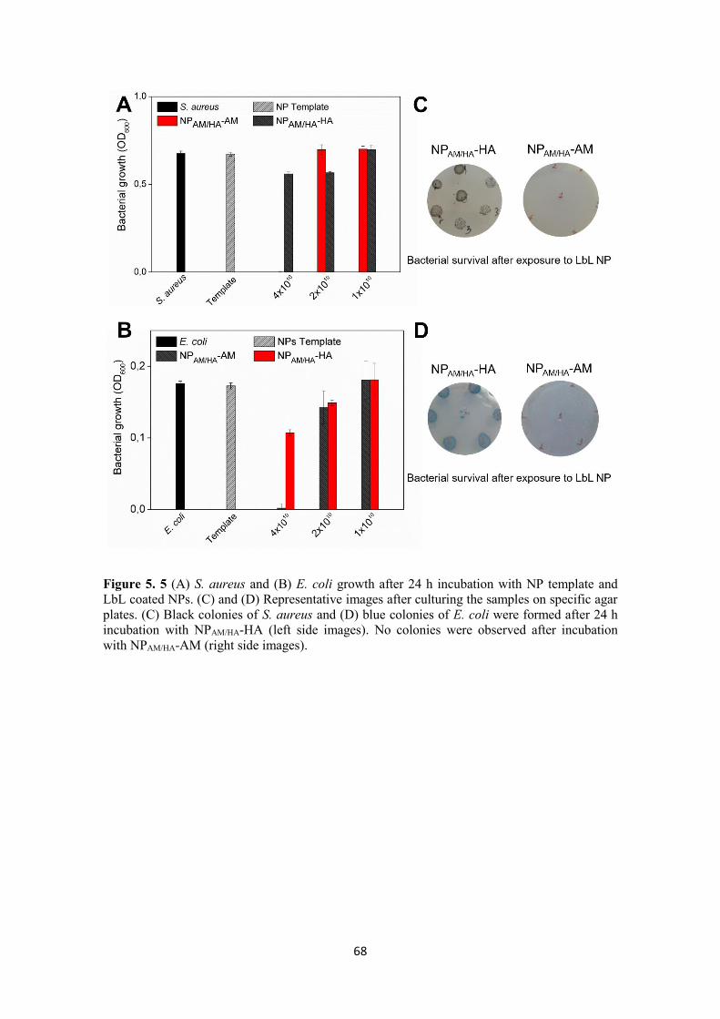
68
Figure 5. 5 (A) S. aureus and (B) E. coli growth after 24 h incubation with NP template and
LbL coated NPs. (C) and (D) Representative images after culturing the samples on specific agar
plates. (C) Black colonies of S. aureus and (D) blue colonies of E. coli were formed after 24 h
incubation with NPAM/HA-HA (left side images). No colonies were observed after incubation
with NPAM/HA-AM (right side images).

69
Figure 5. 6 Bacterial growth after 24 h incubation with different concentrations of AM bulk
solution.
Time-killing kinetics of the LbL-coated NPs (at concentration 4 × 10¹⁰ NPs mL−1) was
performed to determine the viability-(CFU mL−1)-of the treated bacteria and define the
rate at which the NPs exert their bactericidal properties.198 The killing kinetic curves of
NPAM/HA-AM and NPAM/HA-HA for both strains are shown in Figure 5.7. The NPs with
outermost layer of AM completely eliminated S. aureus within 15 min (10 logs
reduction). However, a longer incubation time was required in the case of E. coli, where
3 logs bacterial reduction was achieved within 15 min and 10 logs (full kill) after 45
min (Figure 5.7B). The different killing rates of the NPAM/HA-AM against S. aureus and
E. coli could be explained with the specific structure of the bacterial cell wall in Gram-
positive and Gram-negative bacteria.199 In Gram-positive bacteria, the thick outer cell
wall of negatively charged polysaccharides readily interacts with the antimicrobial
cationic AM, resulting in cell membrane damage. However, in Gram-negative bacteria,
the cell wall is a complex double membrane structure (composed of inner and outer
membranes), which is an efficient barrier to many available antimicrobials. It protects
bacteria and makes the cells more resistant to numerous drugs compared to the Gram-
positive bacteria.200 From another side, NPs with HA outermost layer did not show any
bactericidal activity against the studied bacterial species over the time of incubation. All

70
of these results demonstrated the importance of the active agent position in the NP
multilayer shell as well as the synergistically enhanced killing efficiency of the
combination of nanoform and high cationic surface charge density. The higher
antimicrobial efficiency of the NPs with AM outermost layer in comparison to that of
the NPs terminated with HA was due to the larger amount of surface available amino
groups (1.09 vs 0.04 μmol mL−1, respectively) able to interact with the bacterial cell
wall of both tested strains.201,202
Figure 5. 7 Time-kill curves of LbL coated NPs against (A) S. aureus and (B) E. coli. S. aureus
and E. coli were incubated with 4 x 10¹⁰ NPs mL-1 NPAM/HA-HA (blue) and NPAM/HA-AM (red)
and the bacteria surviving the treatment were estimated after 15, 45, and 60 min. All data are
mean values of three independent experiments.

71
The membrane permeability of bacterial cells treated with NPAM/HA-AM was further
evaluated using Live/Dead Kit assay. The assay contains two nucleic acid dyes- PI and
SYTO9. SYTO9 stains both live and dead bacteria in green, penetrating the cell
membranes, whereas the PI penetrates only the damaged membranes, displaces the
SYTO9, and stains the dead cells in red. The obtained microscopic images (Figure 5.8)
supported the viable-cell counting in the time-killing kinetic assay (Figure 5.7).
Microscopic images of live/dead stained S. aureus and E. coli cells after incubation with
the coated NPAM/HA-AM (Figure 5.8) revealed the presence of only dead bacteria
visualized by red fluorescence. All results taken together demonstrated that the NPs
functionalized with an AM outermost layer were remarkably efficient against these
bacterial species.
Figure 5. 8 Fluorescence images (100 x magnification) of (A) S. aureus, (B) S. aureus treated
with NPAM/HA-AM, (C) E. coli, (D) E. coli treated with NPAM/HA-AM. The green and red
fluorescence images are overlaid. Nonviable bacteria are stained in red, while the viable bacteria
appear green.

72
5.1.3 Interaction of the coated NPs with model bacterial membranes
To corroborate the in vitro results and confirm the bactericidal mechanism of action of
the multilayered biopolymer-shell NPs, solutions of the template and the LbL-coated
NPs were injected into the subphase beneath the model PE monolayer, previously
formed at 33 mN m−1. The increase of the surface pressure (π) as a function of time
demonstrated the membrane disrupting effect of NPAM/HA-AM, whereas no effect or
negligible effect was observed for the template and NPAM/HA-AM, respectively (Figure
5.9). The disrupting effect of the AM-terminated NPs correlates well with the higher
concentration of the amino groups (27-fold more) in their outermost layer, compared to
that of the HA ending NPs. The latter NPs showed little or almost negligible membrane
disturbance effect and were not able to kill the cells because most of their amino groups
are buried under the HA layer and are not accessible for interaction with PE. Disrupting
bacterial cell membrane as a result of the electrostatic interaction between the positively
charged NPs and the negatively charged bacterial membrane is considered a valuable
alternative to effectively reduce bacterial spread and drug resistance occurrence.
Bacterial membrane is highly evolutionarily conserved, and the possibility to be
modified by fast single mutation is very low.203
Figure 5. 9 Kinetic adsorption process after the incorporation of the NP template and LbL
coated NPs into the air/water interface of the PE monolayer at π = 33 mN m-1.

73
5.1.4 Biofilm Inhibition
The ability of the LbL-coated NPs of restricting S. aureus and E. coli biofilm growth on
a model polystyrene surface was further examined. Despite the different conditions used
in the biofilm inhibition tests (e.g., growth medium and static conditions), the AM-
terminated NPs inhibited by 94 and 40 %, respectively, the S. aureus and E. coli cells
adhesion and biofilm formation (Figure 5.10). At bactericidal concentration (4 × 10¹⁰
NPs mL−1), the NPs were able to interact with the negatively charged S. aureus cell
membrane, eradicate the bacteria, and consequently inhibit cell adhesion and biofilm
establishment on the surface. However, the NP antibiofilm potential toward Gram-
negative E. coli was significantly lower. The decrease in the antibiofilm activity of AM-
terminated NPs toward E. coli could be due to the diminished antibacterial effect of the
NPs on Gram-negative bacteria compared to that on Gram-positive species under the
conditions tested.199 Moreover, several studies have reported that positively charged
polymers may interact with E. coli, increase the cell aggregation, and stimulate bacterial
sedimentation on the surfaces. Zhang et al. found that the electrostatic interactions
between the positively charged polymer and negatively charged E. coli cells, instead of
killing the cells, increase the bacterium insusceptibility to antibiotics and induce biofilm
formation. In the presence of cationic polymers, the cells aggregated and formed
clusters where bacteria grew and accumulated signaling molecules inducing biofilm
formation.204 In a similar way, the NPAM/HA-AM may bind to the outer cell membrane
and agglomerate the E. coli cells into biofilm clusters on the surface. At nonbactericidal
concentration, the NPAM/HA-HA NPs were able also to affect planktonic cells and
prevent E. coli and S. aureus biofilm establishment. The addition of the NP formulation
in bacterial suspension reduced the surface colonization and the total biofilm growth of
E. coli and S. aureus by 62 and 99 %, respectively. HAs have been widely used in
engineering antifouling coatings on materials and surfaces to improve their stability and
functionalities for the intended biomedical application.205 Its antifouling properties
appeared very promising to reduce nonspecific protein adsorption and strengthen NP
stability in complex serum containing media. 206 In this study, we employed the
antifouling HA to build an antibacterial multilayered shell with AM on a biocompatible
NP template. We found that the HA position as an outermost layer of the NPs defines
better efficacy toward E. coli biofilms. Nevertheless, the exact mechanism of
antibiofilm action of HA-terminated NPs is unclear; we believe that its deposition

74
contributed to the improved colloidal stability of NPAM/HA-HA in TSB growth medium
and consequently enhanced NP efficacy toward Gram-negative strains (Figure 5.10).
Figure 5. 10 Crystal violet assessment of biofilm formation of S. aureus (black bars) and E. coli
(red bars) after incubation for 24 h with the biopolymer-coated NPs.
In general, the developed multilayer NPs of HA and AM demonstrated strong
antibacterial activity and ability of restricting the biofilm formation of common Gram-
negative and Gram-positive pathogens. The engineered LbL NPs could be further
exploited in the development of skincare formulations for treatment of skin infections,
wound dressings, or coatings on medical surfaces for controlling bacterial infections
and developing drug resistance.
5.1.5 Biocompatibility of the LbL decorated NPs.
The biocompatibility of the antibacterial agents is a major issue for applications in
humans. Therefore, the potential toxicity of the coated NPs toward fibroblasts cells was
evaluated. A study with human fibroblast cells demonstrated that all coated NPs were
biocompatible, with no difference in cell viability observed among the experimental
groups (Figure 5.11). The morphology of the human cells treated with bactericidal
concentration of LbL-coated NPs was not changed when compared to that of the

75
control-only fibroblast cells with growth medium (Figure 5.12). These results
demonstrate that the developed NPs are safe for uses in contact with human cells. The
mammalian and bacterial cells differ in the lipid composition of their membranes, which
is the reason for the highly selective toxicity of positively charged polymers toward
bacteria.207 The LbL-engineered NPs showed high antibacterial and antibiofilm
efficiencies coupled to biocompatibility, which would make possible their fast uptake in
biomedical applications.
Figure 5. 11 Viability (%) of human fibroblasts exposed to LbL coated NPs.
Figure 5. 12 Morphology of human fibroblasts exposed to (A) NPAM/HA-AM; (B) NPAM/HA-HA;
(C) Control human fibroblast cells.

76
5.1.6 Conclusions
In an attempt to produce novel antibacterial agents, bioinert polymer NP templates were
functionalized with multilayers of biocompatible HA and antimicrobial AM in an LbL
coating approach. The LbL-engineered nanoassemblies were able not only to kill
planktonic bacterial cells, but also to prevent biofilm formation. The NPs with AM as
an outermost layer showed strong antibacterial efficiency against both Gram-positive
and Gram-negative bacteria for exposure time of less than 1 h. Moreover, the
functionalized NPs demonstrated enhanced capacity of the nanolayered AM to interact
with and disrupt the negatively charged bacterial cell membranes at lower concentration
in comparison to its bulk solution. On the contrary, the LbL functionalized NPs were
able to inhibit the formation of biofilm by S. aureus and impede biofilm growth by E.
coli, without affecting the morphology of human fibroblast cells. The antibacterial and
antibiofilm efficiencies of the AM-ended NPs coupled to their biocompatibility suggest
that this novel antibacterial agent may have a great potential in prevention of bacterial
infections. Further investigations in vivo should be pursued to study the potential of
these NPs for treatment of infectious diseases.

77
5.2

78
In the current study, the coating of
antibacterial AgNPs with membrane
disrupting biopolymer is expected to
create high local positive charge
density, which synergistically with the
activity of the nano-sized template will
improve the interaction of the novel
hybrid NPs with bacterial membranes
and potentiate the bactericidal activity
of AgNPs at lower dosage. Furthermore, the inclusion of acylase in the nano-hybrids
will inhibit the establishment of P. aeruginosa biofilm, making the pathogen susceptible
to low and safe to human cells AgNPs concentrations. The functionality of the novel
NPs will be evaluated in terms of QS inhibition potential, antibacterial activity and
capacity to prevent and eradicate P. aeruginosa biofilms. Finally, the cytotoxicity of the
NPs will be assessed in vitro toward human fibroblasts cells.
________________________________________
This section is based on the following publication:
Ivanova A, Ivanova K, Tied A, Heinze T, and Tzanov T. Layer-by-Layer Coating of
Aminocellulose and Quorum Quenching Acylase on Silver Nanoparticles
Synergistically Eradicate Bacteria and Their Biofilms. Advanced Functional Materials,
2020, doi.org/10.1002/adfm.202001284.

79
5.2.1 NPs characterization
AgNPs were produced and used as an active antibacterial NP-template for LbL self-
assembling with amino-bearing AM and anti-QS acylase (Figure 5.14A). The synthesis
of AgNPs was carried out in an aqueous solution as previously described.208 The change
from transparent to green-grey colored suspension and the appearance of the absorbance
peak at 400 nm confirmed AgNPs formation (Figure 5.13).
Figure 5. 13 UV-Vis scan of AgNPs.
The resulting NPs, with −56,15 mV zeta-potential and 30 nm size were then
sequentially coated with oppositely charged AM and acylase in an LbL fashion. The
positively charged AM was applied as a first layer, changing the AgNPs zeta potential
to +30.8 mV. Subsequently, the deposition of negatively charged QQE acylase switched
the NPs surface charge to −29.97 mV forming stable LbL Ag@AM_AC NPs (Figure
5.14B). The morphology of the LbL Ag@AM_AC NPs was further assessed using
TEM. TEM images (Figure 5.14C, D) revealed the generation of quasi-spherical in
shape NPs with an average size of 30-35 nm, which are embedded in a hybrid
macromolecular shell of AM and acylase. The data revealed a small size of the NPs
(Figure 5.14E). The scanning transmission electron microscope (STEM) mapping and
EDX spectroscopy of an individual LbL Ag@AM_AC NP further confirmed the
presence of elemental silver in the metal NP core (bright central region), as well as

80
carbon and oxygen in the AM and acylase shell (Figure 5.14F). The Cu ions detected
by the EDX (Figure 5.14G) are from the grid used for the analysis.
Figure 5. 14 LbL Ag@AM_AC NPs preparation and characterization. (A) Schematic
representation of AgNPs template coating with AM and AC using LbL approach. (B) Zeta-
potential of the NPs after each layer deposition. (C, D) TEM images of LbL Ag@AM_AC NPs
at different magnifications. (E) Histogram of LbL Ag@AM_AC NPs size distribution based on
the total count using ImageJ software. (F) Elemental mapping image and (G) STEM-EDX
spectrum of an individual LbL Ag@AM_AC NP.
The activity of acylase in the LbL Ag@AM_AC NPs was further assessed following a
colorimetric method and an in vitro bioassay with a reporter bacterial strain. In the
colorimetric assay, the enzymatic activity of acylase was confirmed via the degradation
of N-acetyl-L-methionine substrate to L-methionine and the subsequent development of
purple color upon interaction with ninhydrin90 (Figure 5.15).

81
Figure 5. 15 L-Methionine production in the presence of AgNPs and LbL Ag@AM_AC NPs.
The QQ activity of the enzyme after its deposition onto the AgNPs templates was
demonstrated through the degradation of the QS signals in C. violaceum CV026. C.
violaceum CV026 is mini-Tn5 mutant that produces a purple pigment (violacein) upon
the addition of AHL signaling molecules.209 The QQE degrades the AHL signals,
consequently inhibiting the violacein production.210 This Gram-negative bacterium has
been used in our group to demonstrate the anti-QS potential of acylase from A.
melleus.77,90 LbL Ag@AM_AC NPs, at sub-inhibitory concentrations (1.56 × 10⁷ NPs
mL−1), degraded the AHLs and disrupted the QS process, reducing the violacein
production by 45 % (Figure 5.16B, C), without affecting C. violaceum growth (Figure
5.16D). The same amount of pristine AgNPs, however, did not influence the production
of this pigment, confirming the presence of active enzyme on the nanotemplate, which
is able to quench the signals and inhibit the QS-regulated behavior in the Gram-negative
pathogen P. aeruginosa. Interruption of QS in the extracellular environment is a new
approach for attenuation of bacterial virulence and boosting the bactericidal activity of
existing antimicrobials at lower dosage.157 Moreover, the anti-QS based strategy
reduces the risk of drug resistance development.

82
Figure 5. 16 Violacein production by C. violaceum. (A) The AHL signals bind to the
cytoplasmic QS receptor and switches on the vio genes, activating the violacein production
(left). The presence of QQ active NPs breaks down the AHL signals, silencing the expression of
the vio genes, subsequently inhibiting the violacein production (right). (B) Quantification of QS
regulated violacein production (%) by C. violaceum and (C) Representative images of violacein
production in presence of AgNPs and LbL Ag@AM_AC NPs. (D) C. violaceum cell viability
after incubation with AgNPs and LbL Ag@AM_AC NPs.
5.2.2 Antibacterial efficiency of AgNPs and LbL Ag@AM_AC NPs
The stand-alone AgNPs demonstrated antibacterial activity against P. aeruginosa at
concentration of 1 × 10⁹ NPs mL−1 (Figure 5.17). However, the functionalization of
AgNPs with AM enhanced their bactericidal activity by 2-fold. The nanoscale
transformation of amino-bearing polymers enhanced the availability of the positively
charged amino groups, resulting in increased interaction with bacterial cells and
stronger bactericidal effect than the pristine solution.104 Furthermore, the inclusion of

83
QQ acylase improved 4-fold the NPs inhibitory efficacy against P. aeruginosa (Figure
5.17).
In order to study the physical changes of bacteria upon exposure to LbL Ag@AM_AC
NPs, ultrastructural analysis using TEM was performed. A clear difference in the
morphology between the non-treated (Figure 5.17C) and treated with NPs (Figure
5.17D) P. aeruginosa cells was observed. The LbL Ag@AM_AC NPs adsorbed onto
the bacterium surface, leading to outer membrane disintegration and cytoplasmic
leakage. Moreover, some of the LbL NPs were observed in the cytoplasmic space,
indicating their ability to penetrate the membrane and affect essential for bacterial cells
life-supporting metabolic processes as cells respiration (Figure 5.17D).

84
Figure 5. 17 (A) P. aeruginosa growth after 24 h incubation with AgNP template, LbL
Ag@AM_AC NPs, LbL Ag@AM_BSA NPs, and LbL Ag@AM_AC NPs at different
concentrations. (B) Representative images after culturing the samples on selective agar plates.
TEM images of P. aeruginosa (C) before and (D) after exposure to LbL Ag@AM_AC NPs.
The ability of acylase to interfere with QS in P. aeruginosa and attenuate its virulence is
thought to potentiate the bactericidal capacity of the actives, lowering their antibacterial
effective concentrations. In order to outline the role of the enzyme, the AgNPs template
were coated with antibacterial AM and a catalytically inert protein, e.g., BSA, instead of
acylase (Figure 5.17A). The obtained control NPs possess similar physicochemical
properties (i.e., size, zeta-potential and concentration) as the LbL Ag@ AM_AC NPs,
but were less active toward P. aeruginosa, confirming the synergistic role of acylase
and AM in enhancing the antibacterial properties of the AgNPs template. The
bactericidal activity of LbL Ag@AM_AC was further confirmed after plating onto
selective Cetrimide agar. The LbL Ag@AM_AC NPs completely eliminated the P.
aeruginosa cells at lower concentrations when compared to the same amount of AgNPs
due to the complementary action of the bactericidal nano-sized core and the anti-QS
acylase (Figure 5.17B). Although, our pervious findings in Section 5.1 have shown that
higher number of layers is needed to introduce desired antibacterial functionality on
inert NP template, herein the deposition of more AM/acylase layers did not result in
enhanced bactericidal activity. In contrary, more layers diminished the effect of the
active AgNPs template and no synergy between the nanoform and polymeric AM
acylase shell was observed (Figure 5.17A, B).
C D

85
It could be anticipated that the novel hybrid NPs kill bacteria via a combination of
mechanisms including interaction with bacterial cell wall, ROS production211 and
modulation of microbial signal transduction pathways.212 The potential of AgNPs
template and LbL Ag@AM_AC NPs to generate ROS and kill P. aeruginosa was
further investigated. In contrast to the positive control, (ROS inducer-bacteria incubated
with AAPH, the AgNPs and LbL Ag@ AM_AC NPs did not generate ROS similar to
the negative control containing only bacteria (Figure 5.18). Thus, the bactericidal
AgNPs and LbL Ag@AM_AC NPs cause cellular death without ROS-related oxidative
damage.
Figure 5. 18 NPs induced ROS generation in P. aeruginosa.
5.2.3 Antibiofilm activity
P. aeruginosa is an opportunistic Gram-negative pathogen, responsible for variety of
difficult to treat infections due to its ability to attach and form robust biofilm structures
on medical devices and living tissues that aggravate the host immune response and
resist the antibiotic therapies. P. aeruginosa biofilm formation is a complex bacterial

86
mode of growth, which is under the control of QS.71 In this work, we aimed to develop
hybrid NPs that interrupt the QS process via degradation of the AHL signaling
molecules and thus effectively inhibit and eliminate the biofilm growing P. aeruginosa
cells at lower concentrations than the stand alone AgNPs. The antibiofilm activity
(biofilm growth inhibition and elimination) of the NPs was studied by crystal violet and
Live/dead BacLight assays to quantify the total biofilm mass and visualize the live and
dead cells on a model polystyrene surface.
5.2.3.1 Inhibition of P. aeruginosa biofilm formation by LbL Ag@AM_AC NPs
P. aeruginosa was cultured in the presence of different concentrations of LbL
Ag@AM_AC NPs and AgNPs for 24 h to determine the minimum biofilm inhibitory
concentration (MBIC). The MBIC is defined as the concentration at which the total
biofilm mass was inhibited by > 90 %, when compared to the control with only bacteria
corresponding to 100 % biofilm formation. The MBIC of the NPs decorated with AM
and anti-QS enzyme was 8-fold lower (6.25 × 10⁷ NPs mL−1) than the MBIC of the
pristine AgNPs (5 × 10⁸ NPs mL−1) (Figure 5.19A).

87
Figure 5. 19 P. aeruginosa biofilm inhibition. (A) MBIC determination of the NPs using crystal
violet assay. (B) Live/dead™ BacLight™ kit microscopic visualization. The green and red
fluorescence images are overlaid. Nonviable bacteria are stained in red, while the viable bacteria
appear green.
The obtained microscopic images of P. aeruginosa biofilms after 24 h incubation with
LbL Ag@AM_AC NPs at the determined MBIC further confirmed their enhanced
antibiofilm activity (Figure 5.19B). The LbL Ag@AM_AC NPs at their MBIC
completely inhibited the biofilm growth of P. aeruginosa observing only few individual
cells on the surface. In contrast, the AgNPs at the same concentration, showed
negligible antibiofilm activity and formed typical for the mature biofilm bacterial

88
clusters, similar to the control (only P. aeruginosa) (Figure 5.19B). All these results
indicated the synergy between the bactericidal nano-sized template and the antibiofilm
acylase enzyme. Interrupting the QS process through acylase-catalyzed degradation of
bacterial signals in the extracellular bacterial space have been shown very effective for
inhibiting P. aeruginosa virulence and establishment of drug resistant biofilms, without
bacteria killing.157 Herein, the deposition of acylase on the bactericidal AgNPs yielded
multifunctional nanoentities able to inhibit the QS-regulated pathological processes and
at the same time completely eliminate the biofilm forming bacteria at lower dosage of
the bactericidal agent, thus exerting less evolutionary pressure on bacteria for resistance
development. The engineered LbL NPs demonstrated high potential for eradicating P.
aeruginosa and therefore could be exploited in the development of coatings on
medically relevant surfaces for preventing biofilm associated infections.
5.2.3.2 Elimination of P. aeruginosa biofilms with LbL Ag@AM_AC NPs
P. aeruginosa cells in already established biofilms are enclosed in EPM, which is the
bacterium protective barrier against the action of the immune system and the effective
penetration of the conventional drugs.71 Therefore, the potential of the LbL
Ag@AM_AC NPs to overcome the inherent biofilm resistance and penetrate through
the P. aeruginosa biofilm was further assessed. The minimum biofilm eradication
concentration (MBEC) of the LbL Ag@AM_AC NPs, i.e., the lowest NPs
concentration at which bacteria fail to re-grow, was determined using crystal violet. The
MBEC of the novel NPs was 8-fold lower (1.25 × 10⁸ NPs mL−1) than the MBEC
determined for the stand-alone AgNPs template (2 × 10⁹ NPs mL−1) (Figure 5.20A).

89
Figure 5. 20 Elimination of 24h grown P. aeruginosa biofilm. (A) MBEC determination of the
NPs using crystal violet assay. (B) Live/dead™ BacLight™ kit microscopic visualization of P.
aeruginosa biofilm after 24 h treatment with AgNPs and LbL Ag@AM_AC NPs at the same
concentration (6.25 × 10⁷ NPs mL-1). The green and red fluorescence images are overlaid.
Nonviable bacteria are stained in red, while the viable bacteria appear green.
Although the anti-QS approaches are considered more relevant in inhibiting the early
stage of biofilm formation, rather than acting on already established surface-attached
bacterial communities, several studies have demonstrated the ability of acylase to
reduce bacterial biofilms. These findings are in line with the assumption that the QS
signaling has an important role for maintaining the integrity of already formed P.
aeruginosa biofilm.213–215 In particular, the process is part of the eDNA release and the
structural stability of the biofilm.71 We believe that the nanoformulated acylase weakens

90
and destabilizes the protective EPM barrier, enhancing the access of therapeutic NPs to
the bacterial cells in the mature biofilm. Moreover, decorating the AgNPs with a
cationic polymer adds an additional mechanism for bacteria killing, through membrane
disruption, potentiating the antibacterial and antibi-ofilm activities of AgNPs at lower
concentrations.216,217 Previous results of our group, have demonstrated enhanced
antibacterial and biofilm activities of nanoformulated antimicrobials (such as AM,
penicillin and vancomycin) in comparison to their bulk counterparts, due to easier
penetration into biofilm structures and access to the target bacteria.218 The effect of LbL
Ag@AM_AC NPs and AgNPs template (at concentration 1.25 × 10⁸ NPs mL−1) on
established biofilms was assessed with fluorescence microscopy after Live/Dead
staining. A significant decrease in the cell viability and biofilm density was observed
when the 24 h old biofilm was treated with LbL Ag@AM_AC NPs (Figure 5.20B). In
contrast, the application of unmodified AgNPs resulted in reduced cells viability,
however, without any significant decrease/elimination of the total biofilm mass (Figure
5.20B). Therefore, the novel LbL Ag@AM_AC NPs possess great therapeutic potential
and could be employed in the design of topical formulations for treatment of severe skin
infections or for coating of medical devices and surfaces in contact with humans.
5.2.4 Biocompatibility assessment
AgNPs are known antibacterial agent widely used as disinfectant or coating of
medically relevant surfaces,219however their biocompatibility is an important issue to be
considered for biomedical application. Therefore, the viability of human cells upon
exposure to AgNPs template and LbL-decorated NPs at their bactericidal concentrations
was assessed after 24 h of incubation. AgNPs demonstrated high toxicity and reduced
by 20 % the viability of human cell (Figure 5.21), while nearly 100 % of the skin cell
fibroblasts were viable in the presence of functionalized with AM and acylase AgNPs
(Figure 5.21A). Unlike the AgNPs, the LbL Ag@AM_AC NPs did not cause cell
morphology changes compared to the negative control (Figure 5.21B). The toxicity of
AgNPs greatly depends on their concentration, size, shape, surface chemistry/coating
and dissolved silver ions from the NPs.220 Here, to benefit from the outstanding
performance of AgNPs in terms of microbial elimination and at the same time minimize
their toxicity, we engineered in a safety approach novel biomaterial-coated AgNPs
Functionalization of the AgNPs with biocompatible and antibacterial polymers and

91
enzymes is a promising strategy to change the AgNPs toxicity profile and boost their
bactericidal properties.
Figure 5. 21 Cytotoxicity evaluation. (A) Viability (%) of human fibroblasts exposed to AgNPs
and LbL Ag@AM_AC NPs after 24 h of incubation assessed by the AlamarBlue assay. (B)
Live/death assay of human fibroblasts after 24 h exposure to AgNPs and LbL Ag@AM_AC
NPs. The green and red fluorescence images are overlaid.

92
5.2.5 Conclusions
In this work AgNPs were LbL coated with antimicrobial AM and QQ acylase in order
to generate safe and highly efficient antibacterial and antibiofilm hybrid nanoentities.
The bilayered decoration of AgNPs template resulted in 4-fold enhanced bactericidal
activity toward the Gram-negative P. aeruginosa due to the synergy between the metal
core and the nanoformulated antibacterial AM and antibiofilm acylase. Moreover, these
hybrid NPs were able to degrade the AHL signaling molecules of Gram-negative
bacteria, inhibiting the QS regulated biofilm formation by P. aeruginosa at lower
concentrations than the pristine AgNPs. The anti-QS acylase has also affected the
pathogenicity and the integrity of already established P. aeruginosa biofilm, increasing
the bacterium susceptibility to antibacterial AgNPs. LbL building of a biocompatible
shell of AM and acylase around the AgNPs is a safe-by-design approach for
development of metal-based nanoantimicrobials with decreased toxicity to mammalian
cells and improved efficacy. The combination of membrane-disrupting AM and QQ
acylase on the AgNPs surface provides an innovative tool for treatment of bacterial
infections, at minor risk for resistance development.

93
5.3

94
In this study, NCs, loaded with
antibacterial oregano EO for effective
elimination of Gram-positive S. aureus
were generated. The EO NCs were
coated with AM to provide reactive
groups on the NCs surface for the
grafting of an antibody that is specific
against S. aureus. The EO NCs
functionalization with a targeting
antibody increased the NCs
bactericidal efficacy against the Gram-
positive S. aureus at a lower dosage and, at the same time, will reduce the toxic effects
on non - targeted bacteria and human cells. The selective antibacterial activity of the
antibody - functionalized EO NCs toward S. aureus was further evaluated in vitro in a
single and mixed bacterial inoculum with non-targeted P. aeruginosa. Finally, the
biocompatibility and the treatment efficacy were validated in an in vitro co-culture
model comprising human fibroblast cells and S. aureus bacteria.
________________________________________
This section is based on the following publication:
Ivanova K, Ivanova A, Ramon E, Hoyo J, Sanchez-Gomez S, Tzanov T. Antibody-
Enabled Antimicrobial Nanocapsules for Selective Elimination of Staphylococcus
aureus. ACS Applied Materials & Interfaces 2020,
http://dx.doi.org/10.1021/acsami.0c09364.

95
5.3.1 Targeted NCs formulation and characterization
EO NCs were produced using a proprietary self-assembly nanoencapsulation
technology194 based on propylene glycol, a water-miscible nonvolatile organic solvent,
and zein, a hydrophobic protein found in maize. The technology is simple and versatile
and allowed the formation of stable and homogeneous zein NCs loaded with the
hydrophobic oregano EO from Thymbra capitata.194 High EO EE, of about 86 %, was
determined by carvacrol quantification with HPLC. Zein NCs containing EO with an
average size of 94 ± 3.7 nm, low polydispersity index < 0.2, and −33 ± 0.5 mV zeta-
potential (at pH 3.3) (Figure Figure 5.23), indicating uniform size distribution and
colloidal stability, were stored for 6 months at RT without any visible signs of
aggregation and precipitation. The negative surface charge of the EO NCs could be due
to the surfactant excipients employed to provide higher colloidal stability of the final
formulation or modulation of the zein conformation upon nanotransformation.221–223 As
anticipated, and based on our previous experience,104 the EO encapsulation led to
enhanced antibacterial efficacy against the targeted S. aureus at lower dosage, when
compared to the same amount of the pristine oil (Figure 5.22).
Figure 5. 22 S. aureus growth reduction after 24 h incubation with free (black lines) and
encapsulated EO (red lines) at different concentrations. The EO encapsulation led to enhanced
antibacterial efficacy against S. aureus at lower amounts, when compared to the amount of
pristine oil.

96
The negatively charged EO NCs were further coated with positively charged AM (36.4
± 0.75 mV), following the principles of electrostatic-driven self-assembly, to introduce
more reactive amino groups onto the NCs surface for grafting of the S. aureus targeting
antibody (Figure 5.23A). The AM deposition onto the EO NCs led to a change of the
zeta-potential from −33 ± 0.5 to +33.8 ± 0.4 mV (Figure 5.23I), as well as an increase
of the average NCs size from 94 ± 3.7 to 114 ± 3.5 nm. The presence of amino groups
on the AM-decorated EO NCs (AM_EO NCs) surface was confirmed by staining with
fluorescamine reagent. The amount of available primary amino groups on the NCs was
27-fold higher in the case with the AM_EO NCs than that of the pristine EO NCs,
where negligible amounts (1.4 μg mL–1 ± 0.2), originating from the low-availability of
lysine and arginine zein amino acids,224 were determined. These AM_EO NCs
demonstrated enhanced antibacterial efficacy (Figure 5.24) against S. aureus compared
to the pristine EO NCs at the same concentrations (∼ 1.5 × 10⁹ NCs mL–1) because of
the higher availability of primary amino groups, leading to improved interaction and
disruption of the bacterial membrane.104 Consequently, those groups were lost upon the
immobilization of the targeting antibody. This was confirmed by the changes in the
zeta-potential from positive to negative for the antibody-enabled EO NCs (Ab@NCs)
(Figure 5.23I), suggesting that the AM would not play a role in the NCs antibacterial
activity.
The AM_EO NCs were functionalized with an antibody that was specific against S.
aureus158 by carboxyl-to-amine cross-linking using 1-ethyl-3-(3-(dimethylamino)
propyl) carbodiimide and N-hydroxysulfosuccinimide to obtain the targeted Ab@EO
NCs (Figure 5.23A). Prior to immobilization, the specificity of the antibody toward the
targeted bacterium was confirmed using a secondary antibody conjugated to the enzyme
horseradish peroxidase.225 The development of the green color was observed only in
those samples where S. aureus bacterium was present, but was not detected in the
control sample of the nontargeted P. aeruginosa, indicating the specific interaction with
the bacterium of interest (Figure 5.23C).
Considering the fact that the carboxyl groups are ubiquitous throughout the antibody’s
structure including the antigen binding site, the binding affinity of the immobilized on
EO NCs antibody with the antigen was assessed using FITC-conjugated protein A from
S. aureus (Figure 5.23D).226 The FITC-protein A binding with the Ab@EO NCs
resulted in a green fluorescent light emission measured at 525 nm using a fluorescence

97
spectrophotometer (Figure 5.23B). The green light from the Ab@EO NCs was also
observed by a fluorescence microscope (Figure 5.23E). Such behavior was not found in
the EO NCs, which evidenced the successful grafting of the antibody, which was able to
interact with protein A at the S. aureus surface.
The developed Ab@EO NCs had a mean size of about 134.9 ± 13.2 nm and −28.63 ±
1.19 mV zeta-potential (Figure 5.23I). This high zeta-potential (≥ 20 mv) was further
reflected in the high colloidal stability, without any visible signs of precipitation, for a
long period (up to 6 months) of storage at 4 °C. The Ab@EO NCs size distribution
profile, obtained by NTA, showed predominantly three size subtypes at 96.4 ± 9.8, 133
± 8.6, and 180.9 ± 23.7 nm, with a very narrow distribution profile and low
polydispersity (Figure 5.23H). Furthermore, the morphology and the size of the
Ab@EO NCs were examined by TEM. The images revealed predominantly sphere-like-
shaped Ab@EO NCs with a size distribution in the range between 60 and 140 nm
(Figure 5.23F, G), similar to the results obtained by NTA (Figure 5.23H). Small
aggregates due to the samples drying during the preparation for TEM analysis could
also be observed.

98
Figure 5. 23 Fabrication and characterization of Ab@EO NCs. (A) Schematic representation of
Ab@EO NCs preparation. (B) Fluorescent intensity measurement at Ex/Em = 490/525 nm upon
FITC-protein A binding with the Ab@EO NCs. (C) Green color development after antibody
interaction with targeted S. aureus. (D) Interaction of antibody-enabled EO NCs with FITC-
labelled protein A. (E) Florescence microscopy images of FITC-protein A bound Ab@EO NCs.
(F) TEM images of Ab@EO NCs at different magnifications. (G) Histogram of the Ab@EO
NCs size distribution based on the total count of 60 NPs using ImageJ software. (H) NTA
analysis of Ab@EO NCs and (I) zeta-potential values for pristine EO NCs, EO_AC NCs and
Ab@EO NCs.

99
5.3.2 Antibacterial activity towards S. aureus
The antibacterial efficiency of the developed nontargeted and targeted EO NCs was
assessed in vitro against S. aureus by quantitative plate count method. EO NCs
effectively reduced the growth of S. aureus at 4-fold lower concentrations than the bulk
oil (Figure 5.22). The active principle of the NCs-oregano EO- is composed of
oxygenated monoterpenes (77.5 % of the composition), the monoterpenoid carvacrol
being the main antibacterial constituent (73.8 %).157 This monoterpenoid is the
component responsible for the strong antimicrobial activity of the oregano oil against
both Gram-negative and Gram-positive bacteria and its higher bactericidal effect at very
low concentrations than other known EOs such as clove bud and thyme.227 It has been
proved that carvacrol can affect the lipids’ ordering and stability of bacterial membrane
that in turn increases the membrane permeability, causing cellular lysis and death.
Additionally, carvacrol may inactivate intracellular components, such as enzymes
involved in energy production, which also leads to bacterial damage and
elimination.119,228
Although the nontargeted EO NCs were active against S. aureus, they showed lower
antibacterial activity than the developed Ab@EO NCs at the same concentrations (1.5 ×
109 NCs mL–1). Up to 2-fold improvement was observed for Ab@EO NCs because the
antibody drives the bactericidal EO NCs directly to the S. aureus, increasing the local
bactericide concentration on the bacterial surface for a short period of time and thus
potentiating its bactericidal efficacy (Figure 5.24).
Figure 5. 24 Antibacterial activity of EO and Ab@EO NCs (concentration ≈ 1.5 x 10⁹ NCs mL-
1) against S. aureus.

100
5.3.3 Real-time monitoring of the NCs interaction with S. aureus
QCM-D was used to monitor in real time the effect of the Ab@EO NCs on the targeted
bacterium under dynamic conditions. QCM-D allowed us to study the NCs-cells
interactions as the piezoelectric mass sensing together with the monitoring of
dissipation changes provide information about the different stages of bacterial growth,
for example, cells attachment, biofilm formation, cellular lysis, and dispersion.229 This
technique has been routinely used in our group to study the toxic effects of antibacterial
nanomaterials on biomimetic bacterial and mammalian membranes, as well as to
identify time-dependent changes in bacterial attachment and growth.153 In this work, S.
aureus was first deposited onto the QCM sensor, and the changes in the frequency and
dissipation upon the circulation of the targeted and nontargeted EO NCs were assessed
(Figure 5.25). At first, the addition of the bacterial inoculum caused a rapid decrease of
the frequency (∼ 20 Hz) (Figure 5.25A zone II) from the 100 mM PBS, pH 7.4 baseline
(Figure 5.25A zone I), attributed to the combined effect of a fast “bulk shift” caused by
the change from PBS to MHB, and the adsorption of MHB components and bacterial
cells onto the disk.153 After 3 h of circulation, adhesion of S. aureus was achieved, as
confirmed by the decrease of the frequency and increase in the dissipation (Figure
5.25A zone II). This was supported by the results obtained following a PBS rinse of
loosely adhered cells (Figure 5.25A zone III), leading to a slight frequency
increase/dissipation decrease and ultimately the establishment of a steady-state
frequency and dissipation signals at levels lower than the original baseline (Figure
5.25A zone I).
Following the formation of a stable S. aureus adlayer, the NCs were flowed through
with a speed of 20 μL min–1 at 37 °C for 45 min. The injection of the Ab@EO NCs
caused a decrease of the frequency, possibly translating into a rapid removal of the cells
from the sensor (Figure 5.25A zone IV). Upon PBS rinsing, the frequency decrease
remained stable at the level of the clean, that is, bacteria-free, crystal (Figure 5.25A
zone I), implying a decrease of the cellular mass and almost complete S. aureus
elimination from the surface.229,230 Moreover, the Ab@EO NCs led to changes in the
dissipation shifts, which was ascribed to increased water content, rearrangements of the
cells on the crystal surface, and morphology changes.153 A similar tendency was
obtained for the control EO NCs; however, the frequency measurements reached a
stable state at higher levels (≅ -15 Hz) than the Ab@EO NCs (≅ -5 Hz), suggesting a

101
lower efficacy of these NCs against the targeted cells (Figure 5.25A zone V). These
results corroborated the improved killing efficacy of the Ab@EO NCs compared to the
pristine EO NCs observed in the antimicrobial tests (Figure 5.24). Such behavior was
not observed when the experiments were performed with the nontargeted P. aeruginosa
cells (Figure 5.25B). Therefore, targeting via an antibody that is specific against S.
aureus could be a way to ensure the rapid delivery of the nanoformulated bactericide
and the effective elimination of the pathogen from the site of infection.
A

102
Figure 5. 25 (A) Interaction of EO NCs and Ab@EO NCs with S. aureus assessed by QCM-D.
The shift in the frequency and dissipation are represented with solid and dashed lines,
respectively. The numbers I, II, III, IV and V indicate the different zones, respectively the
baseline with PBS, bacterial adhesion, baseline after washing of loosely adherent cells, NCs
insertion and PBS washing. (B) Interaction of EO NCs and Ab@EO NCs with P. aeruginosa
assessed by QCM-D. The shift in the frequency and dissipation are represented with solid and
dashed lines, respectively. The numbers I, II, III, IV and V indicate the different zones,
respectively the baseline with PBS, bacterial adhesion, baseline after washing of loosely
adherent cells, NCs insertion and PBS washing.
5.3.4 Selective antibacterial activity of the Ab@EO NCs towards targeted S. aureus
S. aureus is an opportunistic human pathogen found in many skin infections.231
Currently, control of skin infections has been obtained with antiseptics (e.g.,
chlorhexidine and triclosan) and antibiotics (e.g., mupirocin and fusidic acid), which
affect the beneficial skin microbiota, increase the risk of the development of AMR, and
at higher amounts may cause side effects.64 In this work, we aimed to develop targeted
bactericidal nanosystems to selectively eradicate the S. aureus pathogen, without
affecting other beneficial bacterial strains. The specific action of the Ab@EO NCs on
the targeted S. aureus was confirmed in vitro upon their incubation with a mixed
inoculum containing S. aureus and nontargeted P. aeruginosa as model Gram-negative
bacteria found in the skin flora (Figure 5.26A). The bacterial viability after exposure to
the antibody-functionalized and nonfunctionalized EO NCs was assessed by plating on
B

103
selective-for -S. aureus and -P. aeruginosa agars. As expected, the EO NCs
demonstrated antibacterial effect on both bacteria (Figure 5.26B). Up to 1.9 and 1.3 log
reduction was obtained for S. aureus and P. aeruginosa, respectively. In contrast, the
EO NCs tagged with antibody that was specific against S. aureus resulted in more
localized delivery of the nano-sized bactericide to the target, and up to 3 log reduction of the
initial bacterial load was obtained. Such effect, however, was not observed for the nontargeted
P. aeruginosa because of the absence of the specific ligand that could drive the antibacterial EO
NCs to the target cells (Figure 5.26B).
A

104
Figure 5. 26 Antibacterial activity of EO NCs and Ab@EO NCs in mixed bacterial inoculum.
(A) Schematic representation of the EO NCs and Ab@EO NCs interaction with S. aureus and
P. aeruginosa, when are growing together. (B) S. aureus and P. aeruginosa growth reduction
(Log (CFU mL-1)) upon exposure to the EO NCs and Ab@EO NCs. (C) SEM images of S.
aureus (round-shaped) and P. aeruginosa (rod-shaped) bacteria without any treatment and
incubated with EO NCs and Ab@EO NCs. White and red arrows indicate live and damaged
bacterial cells, respectively.
C
B

105
The selective bactericidal activity of the Ab@EO NCs was further studied using SEM.
The SEM images demonstrated that the control S. aureus and P. aeruginosa cells
without the NCs were intact and did not present any morphological changes (Figure
5.26C). S. aureus appeared spherical with a size of up to 1 μm in diameter, whereas P.
aeruginosa were rod-shaped, measuring 0.2-0.4 μm in width and 1-1.5 μm in length.
Clear differences between the control and the bacteria treated with EO NCs and
Ab@EO NCs were observed. In the case of EO NCs, changes in the surface
morphology of both S. aureus and P. aeruginosa cells associated with cellular death
could be detected (Figure 5.26C), which is in agreement with the results from the live
cells counting (Figure 5.26B). On the other hand, the interactions driven by the
antibody functionalization do not seem to induce any antibacterial effect on the
nontargeted P. aeruginosa because the overall microorganism morphology was not
affected and appeared similar to that of the control without bactericidal treatment. This,
however, was not found for S. aureus and significant morphological changes were
observed after incubation with Ab@EO NCs (Figure 5.26C). The altered cell
morphology indicated cell damage, which eventually led to leakage of cytoplasmic
contents and cellular death. These results corroborated the selective antibacterial
efficacy observed in the quantitative plate count method (Figure 5.26B), and therefore
confirmed that in a mixed bacterial inoculum the antibody-functionalized NCs
specifically interact with the S. aureus surface and could be used for effective
elimination of this bacterium at the site of infection.
5.3.5 Cytotoxicity of the Ab@EO NCs
Nanoformulation of EO represents an effective approach to enhance their physical
stability and potentiate the bactericidal activity toward both susceptible and AMR
bacteria.136 However, the potential toxicity of the nano-sized materials, associated with
their unique physicochemical properties, may impede their biomedical application. The
cytotoxicity of the developed Ab@EO NCs was evaluated in vitro on human fibroblast
cells using AlamarBlue reagent for quantification of the metabolically active cells, and a
live/dead viability/cytotoxicity assay kit for microscopic visualization of live and dead
cells. The Live/Dead assay kit is based on the simultaneous determination of live and
dead cells with two fluorescence probes: (i) calcein, which is well-retained within the

106
live cells, producing green fluorescence, and (ii) ethidium homodimer-1 that enters cells
with damaged membranes, producing red fluorescence in the dead cells.
The results from both tests did not show any significant cytotoxicity and
antiproliferative effect of the Ab@EO NCs on the human cells after 24 h of exposure at
their bactericidal effective concentration. Up to 80 % of the skin fibroblasts were
metabolically active, as calculated in comparison to the control-cells without NCs-
indicating desirable innocuousness of the novel Ab@EO NCs for therapeutic
antibacterial applications (Figure 5.27A). Additionally, fluorescence microscopic
images after live/dead staining demonstrated that the NCs did not induce any changes in
the cellular morphology and most of the cells were alive, thus appearing green (Figure
5.27B).
A

107
Figure 5. 27 Cytotoxicity of Ab@EO NCs. (A) Viability (%) of human fibroblasts exposed to
Ab@EO NCs after 24 h of incubation, determined by AlamarBlue assay. (B) Live/Dead assay
of human fibroblasts after 24 h exposure to Ab@EO NCs. Overlapped images of live (green)
and dead (red) cells. The bars aside each image represent the green and fluorescence intensities
obtained after measuring at λex/em = 494/517 nm and λex/em = 528/617 nm for calcein and
ethidium homodimer-1, respectively. Scale bar corresponds to 100 µm.
5.3.6 Antibacterial efficacy of the Ab@EO NCs in an in vitro coculture model of S.
aureus and human cells
Finally, the selective bacterial eradication and treatment efficacy of the Ab@EO NCs
was evaluated in an in vitro coculture model composed of the target S. aureus bacterium
and human skin fibroblasts, which are critical for skin healing and recovery.232 Human
cells were infected with S. aureus and then exposed to the Ab@EO NCs at their defined
antibacterial dosage. Afterward, S. aureus bacterial growth was quantified via a plate
count method, while the viability of the skin fibroblasts was determined by a live/dead
viability/cytotoxicity assay kit. The fluorescence microscopy images after staining of
the cocultured cells demonstrated the negative (i.e., lethal) effect of S. aureus on the
human cells, supported by the increased number of dead fibroblasts. S. aureus produces
a variety of toxins (e.g., hemolysins and leukotoxin) to damage biological membranes
and cause cell death during the infection establishment. The bacterium has been
reported to induce direct cytotoxic effect on different human cells, organs, and
tissues.231 In contrast to the nontreated sample, single supplementation of the targeted
NCs protected the fibroblast cells from the S. aureus-induced cellular damage,
B

108
preserving the cells’ viability and morphology (Figure 5.28). At the same time, the S.
aureus growth was inhibited by 80 % (Figure 5.29). It is worth mentioning that despite
the promising antibacterial activity of pristine EO NCs, these demonstrated negligible
bactericidal effects on S. aureus in our in vitro infection model in comparison to the
Ab@EO NCs, and at the same time did not maintain the skin fibroblasts viability
(Figure 5.29, 5.28). Therefore, the novel Ab@EO NCs developed in this work are
effective and safe antibacterial agents for selective elimination of the pathogen.
Antibody-enabled targeting of the antibacterial EO is a promising strategy to boost their
bactericidal activity toward S. aureus and lower the adverse effects on the human cells.
Figure 5. 28 Live/Dead kit staining of non-infected and S. aureus infected human cells with and
without treatment with Ab@EO NCs.

109
Figure 5. 29 S. aureus growth reduction in an in vitro coculturing model with human cells after
treatment with EO and Ab@EO NCs.

110
5.3.7 Conclusions
AMR has raised the need for engineering novel highly effective antibacterials with
reduced toxicity and lower selection for resistant bacterial strains. Herein, a novel
targeted antibacterial strategy for selective elimination of Gram-positive S. aureus was
developed on the basis of plant-derived bactericidal EO with a low potential for
resistance development. At first, stable and highly antibacterial EO NCs were
formulated by self-assembly nanoencapsulation technology employing the plant-derived
protein zein. The EO NCs with improved bactericidal efficacy compared to the bulk oil
were further coated with biocompatible and biodegradable AM to introduce more
reactive groups onto the NCs surface for chemical immobilization of the S. aureus-
targeting antibody. The Ab@EO NCs, combining the high reactivity of the nanoform
with the specific driving force of the antibody, generated a novel nanosystem with
enhanced antibacterial activity and the potential to selectively eradicate S. aureus while
keeping the nontargeted P. aeruginosa bacterium alive. Moreover, the Ab@EO NCs
inhibited the S. aureus growth and protected the skin fibroblast cells from this pathogen,
as was confirmed in an in vitro infection model. These results demonstrated the capacity
of the Ab@EO NCs to reach and attack only the pathogen of interest, and thus could be
an efficient therapeutic approach for managing S. aureus infections, avoiding the use of
antibiotics and preventing the development of AMR.

111
5.4

112
In this study, for the first
time, a durable hybrid
nanocoating of antibiofilm
enzyme amylase and
antibacterial ZnO NPs has
been developed onto silicone
urinary catheters in a one-step
sonochemical process without
the need for pre-activation of
the silicone material. The hydrolytic enzyme α-amylase has been assembled with
antibacterial ZnO NPs templates and simultaneously deposited onto silicone Foley
catheters under high-intensity ultrasonic field. The US nanoformulation of dual active
ZnO@AM NPs was expected to synergistically improve the antibiofilm activity of the
hybrid nanocoating at minimal, non-toxic levels of the metal oxide biocide. The
antimicrobial efficacy of the engineered hybrid nano-coatings has been assessed first in
vitro under dynamic conditions simulating the urine circulation during catheterization
for up to 7 days- the time frame for catheter colonization by pathogens and occurrence
of CAUTIs. The biocompatibility of the coatings has been further evaluated upon
exposure to mammalian cell lines for the same time period. In addition to the in vitro
evaluation, the antibiofilm efficacy of the novel coatings for reducing the occurrence of
early CAUTIs and possible side effects, such as toxicity and inflammation, has been
validated in vivo in a rabbit model.
________________________________________
This section is based on the following publication:
Ivanova A, Ivanova K, Perelshtein I, Gedanken A, Todorova K, Milcheva R, Dimitrov
P, Popova T, Tzanko T. “Sonochemically Engineered Nano-enabled Zinc
Oxide/Amylase Coatings Prevent the Occurrence of Catheter-Associated Urinary Tract
Infections”. Materials Science & Engineering C, 2021, doi:
10.1016/j.msec.2021.112518

113
5.4.1 Characterization of the coated urinary catheters
High intensity US induced mass transfer, supported by electrostatically driven self-
assembling, was applied to decorate the ZnO NP core with an enzyme shell in order to
develop in situ dual active bio-nanohybrids. The NPs surface charge in the solution was
measured after the coating process. The zeta-potential of the pristine ZnO NPs changed
from 17.1 ± 0.8mV to -22.7 ± 0.5 mV, due to the formation of an amylase layer onto the
nano-template. Simultaneously, the obtained hybrid ZnO@AM NPs were deposited
onto silicone urinary catheters in a “throwing stone” mode upon the application of US
and taking advantage of the non-specific protein adsorption on silicones (Figure
5.30A). The surface morphology of the catheters coated with ZnO@AM NPs was
observed using HRSEM (Figure 5.30C). The HRSEM analysis of the coated sample
showed a dense layer of ZnO@AM NPs with an average size of 192 nm and a narrow
NPs size distribution (Figure 5.30B).
Previous studies have reported that proteins may adsorb onto colloid particles through a
combination of hydrogen bonding, electrostatic, van der Waals, and hydrophobic
interactions.166 In our work, the electrostatic attraction between the positively charged
ZnO NPs and negatively charged amylase led to the formation of NP composites that
were simultaneously deposited in a uniform layer onto the catheter’s surface applying
high-intensity US. The formation, growth and collapse of cavitation bubbles, generated
upon sonication of liquids, determine the rationale of the deposition process. Microjets
and shock waves formed upon the collapse of the acoustic bubbles 189 project the
synthesized ZnO@AM NPs at high velocities towards the catheter’s surface where the
NPs attach by physical interactions between the highly hydrophobic
polydimethyl/vinylmethyl siloxane and hydrophobic protein domains. Hydrophobic
clusters of a large number of aromatic amino acid residues, e.g. tryptophan, tyrosine and
phenylalanine, exposed in α-amylase structure promote the protein adsorption. 233
ICP analysis, carried out to determine the amount of ZnO onto the catheters treated with
ZnO NPs in presence and absence of amylase, showed up to 10-fold lower amount of
ZnO (ca. 0.002 wt %) in the hybrid ZnO@AM NPs coating when compared to the
coating prepared with only ZnO NPs (ca. 0.038 wt %) (Table 5.1).

114
Figure 5. 30 Surface characterization of sonochemically coated with ZnO@AM NPs silicone
catheter. (A) Schematic representation of the US assisted ZnO@AM NPs generation and their
deposition on the silicone catheter. (B) Histogram of ZnO@AM NPs size distribution based on
the total count of up to 100 NPs using ImageJ software. (C) HRSEM images of pristine and
ZnO@AM NPs-coated silicone catheter.

115
Table 5. 1 Amount of ZnO (wt %) on ZnO NPs and ZnO@AM NPs-coated catheters before and
after incubation in artificial urine.
Coated catheter ZnO (wt %)
0 days
(initial)
7 days
(remaining)
ZnO NPs 0.038 0.011
ZnO@AM 0.0023 0.0019
5.4.2 Stability of the coatings in artificial urine
The stability of the coatings in urine is an important parameter to be considered for their
practical use. Urine has a complex chemical composition, containing inorganic salts that
can affect the catalytic activity of amylase and reduce the antibiofilm potential of the
coatings. 234,77 Therefore, the activity of the enzyme on both amylase- and ZnO@AM
NPs-coated catheters was assessed after 7 days of incubation in artificial urine (prepared
according to DIN EN 1616:1999). The silicone devices coated with amylase alone lost
almost 100 % of their enzymatic activity after 7 days in contact with urine under
continuous agitation. However, the amylase-containing hybrid NPs coating was still
active, retaining up to 80 % of its initial activity, as confirmed by the production of
maltose upon the enzymatic degradation of starch (Figure 5.31). α-amylase is a rather
robust, industrially relevant enzyme, which activity reflects the changes in the protein
conformation under the effect of the application environment. The conformational
stability of the enzyme is a function of external variables, such as ionic strength and
composition of this environment. Prolonged exposure (7 days) to chemical denaturants,
such as urea233 and salts (NH4+ is a strong deactivating cation) in the artificial urine,
although not in denaturing concentrations, apparently deactivated the amylase alone,
while the assembling of the enzyme onto the metal oxide NP-templates and their
subsequent deposition on the catheter ensured the conformational stability of the
enzyme protein and its hydrolytic activity in the nano-enabled coating.

116
Figure 5. 31 Enzymatic activity of amylase-containing coating before and after incubation in
artificial urine for 7 days with shaking. The results are expressed as the percentage of amylase
activity compared to the enzyme before incubation in artificial urine.
Additionally, the leaching of ZnO NPs from the catheters over the same period of
incubation in artificial urine was evaluated. ICP measurement quantified the total
amount of ZnO in the ZnO and ZnO@AM NPs coatings before and after incubation in
artificial urine (Table 5.1). Considerably lower amount of ZnO was detected on the
catheters treated in the presence of amylase in comparison to the catheters treated only
with ZnO NPs. On the other hand, the ICP data revealed higher stability of the hybrid
ZnO@AM coatings than the individual ZnO NPs materials. High amount (83 %) of
ZnO initially present on the ZnO@AM NPs-coated catheter remained on its surface
after incubation in urine, while only 25 % of ZnO was detected in the case of ZnO NPs-
coated catheter. The release of ZnO from these coatings is explained with the ZnO NPs
dissolution induced by the phosphates in the artificial urine.235,236 Apparently, the
complexation of ZnO NPs with amylase in the hybrid coatings led to improved stability
as confirmed by ICP and HRSEM data. HRSEM images of the coated catheters
following their immersion in artificial urine for 7 days (Figure 5.32) showed a removal
of the ZnO NPs, whereas no significant difference was observed for the hybrid
ZnO@AM NPs coating before and after incubation. The sonochemically deposited
metal oxides are not stable, and in most cases the substrate requires a pretreatment to
improve the coating stability.139,237 Here, the coating stability was provided by the

117
amylase acting as an adhesive between the hybrid metal oxide/enzyme nanocomplexes
and the hydrophobic polydimethyl/vinylmethyl siloxane catheter.
Figure 5. 32 HRSEM images of pristine, ZnO and ZnO@AM NPs-coated silicone catheter after
7 days of incubation in artificial urine.
5.4.3 Antibiofilm activity and functional stability of the coated urinary catheters
The biofilm formation by Gram-positive S. aureus and Gram-negative E. coli, the most
common pathogens found in CAUTIs, was assessed on treated and non-treated catheters
under static conditions using crystal violet assay.238 Catheters, coated either with
amylase or ZnO NPs alone inhibited the S. aureus biofilm formation by approximately
20 % and 70 %, respectively (Figure 5.33A). In the case of E. coli, both amylase and
ZnO NPs coatings inhibited the biofilm formation on the catheter by around 70 % when
compared to non-treated silicone (Figure 5.33B). Unlike E. coli which produces several
types of potential substrates for amylase, S. aureus only secretes one dominant EPS
involved mainly in the intercellular aggregation of the cells at the surface.239,240 The
amylase-induced degradation of several biofilm adhesives simultaneously is probably
the reason for the higher efficacy of amylase-coated catheters on E. coli when compare
to S. aureus.

118
However, the simultaneous coating with both actives led to inhibition of the total
biofilm formation by 81 % for S. aureus and 75 % for E. coli (Figure 5.33A, B). In the
hybrid ZnO@AM NPs the amylase degraded essential for biofilm adhesion and growth
polysaccharides,77 inhibiting the biofilm formation by both bacterial strains, and at the
same time, increasing the susceptibility of the individual bacterial cells to the
bactericidal ZnO NPs at lower concentration. Amylase has been employed in our group
both individually and in combination with other enzymes (e.g. acylase)77 or antibacterial
agents (e.g. Ag)153 to inhibit biofilm maturation, detach pre-formed biofilms, and
synergistically increase the susceptibility of the biofilm forming bacteria to biocides. In
contrast to commonly used antimicrobials, these hybrid antibacterial/antibiofilm agents
eradicate pathogens via non-specific mechanisms of action, such as membrane damage,
oxidative stress and virulence attenuation, unlikely to induce AMR development.241

119
Figure 5. 33 Biofilm inhibition (%) at static condition of the coated catheters towards (A) S.
aureus and (B) E. coli assessed before and after incubation in artificial urine for 7 days under
continuous agitation. The results are represented in %, compared with the biofilm inhibition of
pristine catheter (non-inhibition). Stars represent the statistical differences between the different
groups of samples; p < 0.05.
In order to highlight the role of the enzyme and confirm the synergism between ZnO
NPs and amylase, sonochemical coatings containing ZnO NPs and non-active protein
(BSA) instead of amylase, were developed. The coatings did not show any ability to
reduce the bacterial attachment and biofilm formation (Figure 5.34), which
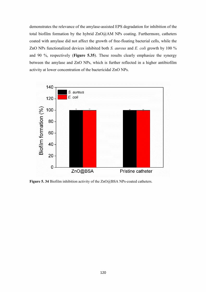
120
demonstrates the relevance of the amylase-assisted EPS degradation for inhibition of the
total biofilm formation by the hybrid ZnO@AM NPs coating. Furthermore, catheters
coated with amylase did not affect the growth of free-floating bacterial cells, while the
ZnO NPs functionalized devices inhibited both S. aureus and E. coli growth by 100 %
and 90 %, respectively (Figure 5.35). These results clearly emphasize the synergy
between the amylase and ZnO NPs, which is further reflected in a higher antibiofilm
activity at lower concentration of the bactericidal ZnO NPs.
Figure 5. 34 Biofilm inhibition activity of the ZnO@BSA NPs-coated catheters.

121
Figure 5. 35 Antibacterial activity of ZnO NPs and ZnO@AM NPs-coated catheters.
Engineering of stable at use antibiofilm coatings on indwelling urinary catheters is
crucial for the effective prevention of CAUTIs. Therefore, the functional stability of the
coated catheters, i.e. their ability to impair the bacterial attachment and colonization,
was assessed after 7 days of incubation in artificial urine under continuous agitation.77
Catheters functionalized with amylase or ZnO NPs alone, practically lost their ability to
inhibit the static biofilm formation of both S. aureus and E. coli after incubation in
urine. However, when amylase and ZnO were simultaneously deposited onto the
silicone surface, a negligible decrease of their biofilm inhibition properties was
observed in comparison to the non-incubated in urine samples. ZnO@AM NPs coatings
lost only 10 % and 20 % of their antibiofilm activity against S. aureus and E. coli,
respectively (Figure 5.35). These results indicate that the hybridization of ZnO NPs and
amylase into a nanostructured coating on catheters is an effective antibiofilm approach
against both Gram-positive and Gram-negative bacterial strains even after 7 days of
exposure to artificial urine.
5.4.4 Biofilm inhibition tests under dynamic conditions in a model of catheterized
human bladder
Despite of the high stability and efficiency of ZnO@AM NPs coatings in controlling the
biofilm formation of both Gram-positive and Gram-negative bacteria at static

122
conditions, these conditions do not simulate the real scenario during catheterization. For
that reason, the novel hybrid nano-enabled coatings were further challenged in dynamic
biofilm inhibition tests in a model of catheterized human bladder (Figure 5.36), where
the dynamically varying shear stress associated with urine flow may affect the bacteria
adhesion on the surface or provoke coating removal.242 This model has been widely
used in our group for assessing in vitro the antibiofilm functionality of catheter
coatings77,234
Since 80 % of bacterial adhesion onto medical devices and the buildup of drug-resistant
biofilms occur during the first days after the device insertion,243,244 strategies for
prevention of biofilm formation within the first days of catheterization are needed. The
total biofilm mass formed on the pristine and ZnO@AM NPs-coated Foley urinary
devices after 7 days of catheterization was evaluated using crystal violet and Live/Dead
kit assays. The sonochemical coating with ZnO@AM NPs led to reduction of both S.
aureus and E. coli total biofilm formation on the catheter’s balloon by 60 % and 80 %,
respectively (Figure 5.36). The balloon of the catheter inflated inside the bladder is
entirely immersed in urine during catheterization. This part of the indwelling urinary
device is the most susceptible to colonization and consequent establishment of antibiotic
resistant biofilms by urinary tract pathogens. Considering previous works on the biofilm
growth in artificial urine,77 we presume that the enzyme in the hybrid coatings affects
the S. aureus and E. coli initial colonization on the catheter surface degrading the
polysaccharide adhesives essential for bacterial attachment and growth of biofilms. The
degradation of the EPS by amylase inhibits the formation of mature biofilm, further
enhancing the bacterial susceptibility to bactericidal ZnO NPs in the coatings. Although,
the extent of biofilm reduction was different between the different bacteria the proposed
mechanism of antibiofilm action of the hybrid ZnO@AM NPs coatings is valid for both
species. The variation in the antibiofilm activity is explained by the different EPS 245
and their functional role in the biofilm establishment as well as different structure of
Gram-positive and Gram-negative bacterial membrane.187
Microscopic observations revealed less S. aureus than E. coli biofilm formation on the
non-treated catheter balloon, explained with the difference between the Gram-positive
and Gram-negative bacterial species and their ability to establish in vitro biofilms under
dynamic conditions.246 Noteworthy, the biofilm formation by S. aureus and E. coli on
ZnO@AM NPs-coated catheters was completely inhibited and only few individual cells
were observed. The appearance of dead bacterial cells (red cells) in the microscopic

123
images was due to the bactericide effect of ZnO NPs. The consistency of the results for
the antibiofilm efficiency of ZnO@AM NPs coatings in both static and dynamic
conditions clearly demonstrate the potential of these hybrid coatings to prevent the
biofilm formation on silicone urinary catheters and therefore decrease the incidence of
CAUTIs.

124
Figure 5. 36 In vitro biofilm inhibition in catheterized bladder model with urine recirculation.
(A) In vitro model of catheterized human bladder. Total biofilm quantification of (B) S. aureus
and (C) E. coli biofilm mass on ZnO@AM coated urinary catheters. Stars represent the
statistical differences between the treated and pristine catheters; p < 0.05. (D) Fluorescence
microscopy images of live (green) and dead (red) bacteria in the biofilms grown on ZnO@AM-
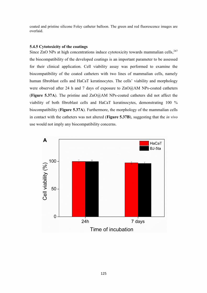
125
coated and pristine silicone Foley catheter balloon. The green and red fluorescence images are
overlaid.
5.4.5 Cytotoxicity of the coatings
Since ZnO NPs at high concentrations induce cytotoxicity towards mammalian cells,247
the biocompatibility of the developed coatings is an important parameter to be assessed
for their clinical application. Cell viability assay was performed to examine the
biocompatibility of the coated catheters with two lines of mammalian cells, namely
human fibroblast cells and HaCaT keratinocytes. The cells’ viability and morphology
were observed after 24 h and 7 days of exposure to ZnO@AM NPs-coated catheters
(Figure 5.37A). The pristine and ZnO@AM NPs-coated catheters did not affect the
viability of both fibroblast cells and HaCaT keratinocytes, demonstrating 100 %
biocompatibility (Figure 5.37A). Furthermore, the morphology of the mammalian cells
in contact with the catheters was not altered (Figure 5.37B), suggesting that the in vivo
use would not imply any biocompatibility concerns.

126
Figure 5. 37 Viability of HaCaT and BJ-5ta cell lines after exposure to the ZnO@AM NPs-
coated catheters assessed by (A) AlamarBlue and (B) Live/Dead kit assays. The green and red
fluorescence images are overlaid. Scale bar corresponds to 100 μm.

127
5.4.6 In vivo efficacy assessment of ZnO@AM NPs-coated catheters in a rabbit
model
The ability of the coatings to reduce bacterial biofilm growth and respectively the
CAUTIs occurrence was assessed in vivo in an established rabbit model (Scheme 5.1).
This model does not use specific pathogen inoculum, but rather mimics the natural
process by which microorganisms from the surrounding environment colonize the
device surface and induce CAUTIs.140 Additionally, the model allows to assess in vivo
the biological safety of the coated catheters.
In the in vivo tests, animal groups 2 and 3 (n = 3), were subjected to catheterization,
respectively with pristine and ZnO@AM NPs-coated indwelling urinary catheters,
under identical experimental conditions for 7 days (this is the usual time frame for
catheter colonization by pathogens and subsequent appearance of CAUTIs). Non-
catheterized rabbits (group 1) were used as a control (healthy animals) in all
experiments.
Scheme 5. 1 Schematic representation of the assays performed in vivo in a rabbit model.
5.4.6.1 Microbiological analysis of urine and catheters´ surfaces for UTIs detection
At the end of the catheterization period, urine samples and catheters from each group
were collected and examined for bacterial contamination. However, the samples from
the urine of the rabbits and the surface of the pristine catheters from group 2 were both

128
colonized with bacteria, mainly from genus Enterococcus (Figure 5.38). The
Enterococcus concentration in the urine was 3.65x10³ + 0.54 CFU ml-1 and was
sufficient to cause UTI, as confirmed by the appearance of concomitant clinical
symptoms such as kidney lesions and signs of developing glomerulonephritis (Figure
5.39A).248 Bacteria were also detected on the catheter´s surface (17.9 + 1.33 CFU cm-2)
and their organization into stable biofilms could further lead to difficult to treat UTIs.249
In contrast to the animal group catheterized with pristine catheters, the urine samples
collected from the animals treated with ZnO@AM NPs-coated catheters (group 3) were
practically sterile. Less than 103 CFU ml-1 (ca. 6.5 x 10² + 1.68 CFU ml-1) bacteria from
the genus Enterococcus were found in the urine samples, which is not indicative for
infection.250 Although the urine had some bacterial load, the surface of the ZnO@AM
NPs-coated catheters remained bacteria–free over the time of catheterization (Figure
5.38). These in vivo results are in agreement with the in vitro observations and
confirmed the potential of the hybrid ZnO@AM NPs coating for clinical uses.

129
Figure 5. 38 Antibacterial properties of pristine and ZnO@AM NPs-coated catheters. (A)
Colony count of bacteria from the urine samples collected from animals catheterized with
pristine and ZnO@AM NPs-coated catheters. (B) Colony count of live bacteria adherent on the
non-treated and ZnO@AM NPs functionalized catheters after 7 days of catheterization in the
rabbit model.

130
5.4.6.2 Histopathology after catheterization
The urethra of the control animals from group 1 did not show any abnormalities (Figure
5.39A). Typical features of the epithelial lining, starting with transitional cells near the
bladder, followed by pseudostratified, stratified columnar epithelium and stratified
squamous approaching the orificium (Figure 5.39A, black arrow), were observed. In
contrast, very mild erosions of the mucosal columnar epithelium, due to the mechanical
pressure of the catheters on parts of the penile urethra during catheterization, were
detected in the catheterized rabbits from groups 2 and 3 (Figure 5.39B, black arrows),
while the urothelium in the prostatic urethra was not affected (Figure 5.39B). The
anatomical position of the penile part of the urethra and the size of the catheters explain
these findings.
Significant histopathological alterations due to catheterization were not detected in the
bladders of the animals from all groups (Figure 5.39C). Groups 1 and 3 had normal
morphology of the bladder mucosa with transitional epithelial cell lining (red arrow).
Some of the animals in group 2 (non-coated catheters) appeared with mucosal lesion
expressed in focal damage of epithelial linings forming microerosions of the cell barrier
(Figure 5.39C, black arrows) and showed bladder inflammation when compare to
group 3, catheterized with ZnO@AM NPs-coated devices.
The kidneys of the rabbits from group 1 (without catheterization) and group 3 (using
ZnO@AM NPs-coated catheter) did not show any pathological lesions (Figure 5.39D).
Their glomeruli and Bowman’s capsules as well as the urinary tubular system were
found to be with normal morphology. However, in rabbits from group 2 (pristine
catheters) the pathohistological observations showed localized clusters of crescentic
glomerulonephritis and vasculitis (extravasation and tissue infiltration from the
bloodstream with inflammatory cells) (Figure 5.39D, black arrows) and preserved
morphology of glomeruli (Figure 5.39D, red arrow heads). Zonal infiltration of renal
perivascular tissues and circumferential periglomerular renal parenchyma was observed,
where the pathology involved abundant local concentration of inflammatory cells. We
suggest that this is related to a disorder in the lower urinary tract as the microbiological
results revealed bacteria in the urine and on the surface of the pristine catheters.
Morphological observations of all blood smears from non-catheterized rabbits (group 1)
and catheterized with ZnO@AM NPs-coated devices (group 3) showed normal cell
ratio, unlike the group 2 (pristine catheters), which showed increased number of
neutrophil granulocytes (Figure 5.39E, black arrow) associated with leukocyturia and

131
indicating the appearance of urothelial infection.251 In the urine, sediments (many
colorless to yellow-brown calcium carbonate crystals in the form of large or smaller
spheroids with radial striations) of the animals from groups 1 and 3 were found (Figure
5.39F). Cellular constituents from urothelial linings of bladder, renal pelvis of kidneys
or urethra were not seen in these groups. Microscopic aspects of the urine sediments of
the animals catheterized with pristine catheters (group 2) revealed leukocytes and
erythrocytes, associated with inflammation,252 transitional epithelial cells from the
mucosal linings (including umbrella cells) and sperm (Figure 5.39F) supporting the
pathological processes.

132
Figure 5. 39 Histopathology of (A) rabbit penile, (B) prostatic urethra, (C) bladder and (D)
rabbit kidney of non-catheterized (group 1) and catheterized with pristine (group 2) and
ZnO@AM NPs (group 3) catheters. Black arrows indicate epithelial lining; red arrow displays
urinary bladder mucosa with transitional epithelial cell lining; red arrowheads show glomeruli.
(E) Blood smears of rabbits. Black arrow indicates the neutrophil granulocytes. (F) Urine
sediments of non-catheterized (group 1) and catheterized rabbits (group 2 and 3). Red arrows
indicate the umbrella cells from the urinary system mucosal linings.

133
5.4.6.3 Hematological and biochemical analyses
Hematological and biochemical parameters253,254 from randomly chosen animals that
inform about pathological processes were further analyzed (Table 5.2 and Table 5.3).
Generally, the blood counts and biochemistry did not show any serious deviations with
the exception of the animals treated with pristine catheters (group 2) developing
glomerulonephritis, which white blood cells and monocytes count was higher than the
normal levels. The granulocytes count were slightly elevated in rabbits with ZnO@AM
NPs-coated catheters, whereas in group 2 the granulocytes increased significantly
(Table 5.2), which is indicative of bacterial infection.255
Urine samples collected from all animal groups showed slight deviations of some
parameters (Table 5.3). Glucose and ketone values in all studied groups were negative,
which means that the rabbits did not present any severe disease. Creatinine was elevated
in animals from group 1 and group 2. The histology results from the rabbits treated with
the pristine catheter revealed elevated numbers of leukocytes and red blood cells,
associated with UTIs. Furthermore, the urine of the animals from this group had pH 6,
which is lower compared to pH measured for the healthy non-catheterized animals
(group 1) and those treated with ZnO@AM NPs-coated catheters (group 3). Slightly
increased levels of blood cells were found in the urine of the catheterized animals
(group 2 and 3) due to the movement of the indwelling devices in the bladder lumen
causing eventually microtraumas. In a summary, the conducted in vivo studies
validated: i) the antibiofilm efficiency, ii) capacity to reduce the incidence of CAUTIs,
iii) the enhanced life span and iv) safety of the novel ZnO@AM NPs-coated catheters.

134
Table 5. 2 Hematological blood results of non-catheterized and catheterized rabbits.
Parameter Rabbit
(Group 1)
Pristine
catheter
(Group 2)
ZnO@AM
coated catheter
(Group 3)
Standards
White Blood Count (L-1) 9.5x10⁹ 15.4x10⁹ 12.9x10⁹ 5.2-13.5
Lymphocytes (L-1) 2.5x10⁹ 2.4x10⁹ 3.5x10⁹ 3.2-9.0
Monocytes (L-1) 0.5x10⁹ 0.7x10⁹ 0.6x10⁹ 0.1-0.6
Granulocytes (L-1) 6.5x10⁹ 12.3x10⁹ 8.8x10⁹ 2.0-7.5
Lymphocytes (%) 26.1 15.8 27.13 35.2-75.6
Monocytes (%) 5.7 4.6 4.65 2.5-6.0
Granulocytes (%) 68.2 79.6 68.22 20.2-59.3
Red Blood Cells
(L-1)
6.82x10¹² 6.92x10¹² 6.42x10¹² 5.00-7.60
Hemoglobin (g L-1) 152 158 158 105-170
Mean Corpuscular
Hemoglobin (pg)
22.2 23.8 24.6 20.1-25.1
Mean Corpuscular
Hemoglobin Concentration
(g L-1)
320 348 361 320-370
Albumin (g L-1) 39.5 42.1 41.0 27-45
Globulin (g L-1) 23.2 19.4 36.0 15-57
Albumin/Globulin 1.7 2.2 1.1
Blood urea nitrogen
(mmol L-1)
6.68 5.26 8.58 3.6-10.7
Phosphate (mmol L-1) 1.66 1.24 1.75 1.1-2.74
Total Bilirubin (µmol L-1) 4.10 2.56 2.85 0-10.3
Creatinine (µmol L-1) 72 83 87 27-141
Blood urea nitrogen
/Creatinine
23 16 24

135
Table 5. 3 Biochemical results of urine collected from non-catheterized and catheterized
rabbits.
Parameter Rabbit
(Group 1)
Pristine catheter
(Group 2)
ZnO@AM coated
catheter
(Group 3)
Leukocytes (cell µL-1) 15 125 15
Ketones (mmol L-1) 0 0 0
Nitrites - - -
Urobilinoge Normal Normal Normal
Bilirubin (µmol µL-1) 8.6 8.6 8.6
Gluose (mmol L-1) 0 0 0
Specific gravity 1.010 1.020 1.030
Red blood cells (cell µL-
1)
0 200 25
MA (mg L-1) ≥ 150 ≥ 150 ≥150
Calcium (mmol L-1) ≥ 10 5.0 2.5
Creatinine (mmol L-1) ≥ 26.4 26.4 8.8

136
5.4.7 Conclusions
Silicone urinary catheters were simultaneously coated with matrix-degrading amylase
and antibacterial ZnO NPs in a one-step US process. In vitro, the engineered nanohybrid
coatings demonstrated significant biofilm inhibition against the most common Gram-
positive and Gram-negative bacterial representatives found in CAUITs as well as high
functional stability upon 7 days of incubation in artificial urine. The ZnO@AM NPs-
coated catheters were also able to inhibit the S. aureus and E. coli biofilm formation by
up to 60 % and 80 %, respectively, under dynamic conditions in vitro in a model of
catheterized human bladder. The novel antibiofilm coatings were 100 % biocompatible
with HaCaT and fibroblast cells and subsequently their efficacy was validated in vivo in
a rabbit model. In vivo, the ZnO@AM NPs-coated catheters reduced significantly the
growth of Enterococcus uropathogens, delaying the early onset of CAUTIs, and at the
same time did not induce toxicity to the rabbit’s kidney, bladder and urethra after 7 days
of catheterization. Overall, the urinary catheters coated with the synergistic combination
of biofilm matrix-degrading amylase and antibacterial ZnO NPs appear as a viable
solution with high clinical potential for preventing CAUTIs.

6 Conclusions and Future Plans

138
6.1 Final conclusions
The spread of bacterial infections and antimicrobial resistance have called for
development of highly efficiently therapeutic strategies with lower risk of resistance
occurrence. In addition, the ability of bacteria to colonize the surfaces of medical
devices and form persistence biofilms, associated with difficult-to-treat infections rises
the demand for novel nano-enabled antibacterial and antibiofilm coatings. To answer
these needs, the current thesis designed a platform of innovative engineered nanoentities
and nanostructured coatings for control and prevention of bacterial infections with
reduced risk of resistance development:
• Nanomaterials aimed at controlling planktonic bacterial growth and biofilm
establishment:
Multilayered coating of antibacterial AM and biocompatible HA on biologically
inert nanotemplate
- Polymeric NPs decorated with antibacterial AM and biocompatible HA in an
LbL approach were able to eliminate Gram-positive and Gram-negative planktonic
bacteria upon contact within less than 1 h.
- The LbL NPs impeded the S. aureus and E. coli biofilm formation by 94 and 40
% without affecting the cell viability and morphology of human fibroblast cells.
Biosafety nanocoating of AM and QQ acylase onto active AgNPs template
- Antibacterial AM and QQ enzyme acylase, assembled by LbL on AgNPs
template, were able to interfere with QS system and increase the susceptibility of Gram-
negative P. aeruginosa to the bactericidal AgNPs.
- The developed LbL NPs demonstrated stronger ability to counteract planktonic
cells and the formation of biofilm structures, compared to the pristine AgNPs alone.
- The inclusion of AM/AC shell on the AgNPs core led to elimination of already
established biofilms and improvement of the biocompatibility of the bactericidal
AgNPs.

139
Targeting of antimicrobial drug delivery nanosystem for selective bacterial
elimination
- Highly antibacterial and biocompatible EO NCs were formulated by self-
assembly nanoencapsulation technology, based on hydrophobic protein zein, and tagged
with a S. aureus-targeting antibody.
- The immobilized on the NCs surface antibody provided selective elimination of
the targeted S. aureus without reducing the non-targeted P. aeruginosa bacterium.
- The targeted NCs demonstrated ability to protect the skin fibroblast cells from S.
aureus in an in vitro infection model.
• Hybrid enzyme-metal nanocomposites for prevention of CAUTIs:
Simultaneous sonochemical coating of urinary catheters with antibacterial
ZnO/amylase hybrid NPs
- Durable nanocomposite coatings of antibacterial ZnO NPs and biofilm matrix
degrading amylase were developed on urinary catheters in a one-step high intensity US
process.
- The combination of both enzyme and metal oxide NPs resulted in improved
stability and stronger antibiofilm activity against medically relevant pathogens,
compared to the coatings with the individual ZnO NPs or amylase.
- The developed hybrid nanocoatings delayed the early onset of CAUTIs and
demonstrated biosafety in vivo in an animal model, showing potential to prevent device-
associated infections.
- A pilot line within the European project framework of PROTECT- Pre-
commercial lines for the production of surface nanostructured antimicrobial and
antibiofilm textiles, medical devices, and water treatment membranes, H2020-720851
based on scale-up of innovative US-assisted coated urinary catheters was developed and
optimized at the new US batch machine installed at Degania, Israel. The functional
activities of the produced coated silicone catheters were proven in vitro at BIU, Israel
and at UPC, Spain.

140
6.2 Future perspectives
The engineering of highly efficient antibacterial/antibiofilm materials and coatings with
lower risk of resistance development is a key for the control and prevention of bacterial
infections and spread of drug-resistant bacterial species. The results obtained in this
thesis validated the efficiency of a wide range of innovative hybrid nanoentities and
nanostructured coatings to control bacterial infections being at the same time safe to
human cells.
The obtained during the thesis nanoentities could be incorporated into wound dressing
materials or skin care products to manage bacterial infections. Moreover, these hybrid
nanomaterials will be employed as functional elements for development of nano-
enabled stimuli-responsive hydrogel coatings on medical devices in the ongoing project
CoatToSave-Nano-enabled hydrogel coatings against antimicrobial resistant catheter-
related infections, PID2019-104111RB-I00. These nanostructured coatings will be
endowed with bacteria-triggered responsiveness to virulence factors (e.g. enzymes) and
changes in the local environment (e.g. pH) due to bacterial infection, to release the
nanoformulated antibacterial cargo. The nanocoating approach could be extended to a
broad range of material surfaces, such as textiles, contact lenses, or personal protective
equipment.
The realization of this thesis has open new research directions that will be followed
under running and new European and national projects at the Group of Molecular and
Industrial Biotechnology (GBMI) of Universitat Politècnica de Catalunya. Versatile
nano-enabling techniques, such as self-assembling, enzymatic crosslinking,
sonochemistry, LbL and combinations thereof, will be used to engineer alternative to
the conventional antibiotics antimicrobial nanomaterials and composites. The platform
of novel antibiotic-free nano-sized materials obtained under the PhD thesis, combining
QQE, natural compounds, polymers, and inorganic NPs will be employed in innovative
approaches for controlling infectious diseases and resistance occurrence, including:
Combination and nanoformulation of QQEs that interfere with the
communication of Gram-negative bacteria and phage lytic enzymes targeting
Gram-positive pathogens into the same nanoentities with improved
antimicrobial activity, allowing to simultaneously address multiple target
pathogens.

141
Targeted to specific bacteria nanoactives, using human-derived antibodies and
QQ signaling molecules as targeting elements, which will allow maintaining a
balanced human microbiome, strengthening the host immune system against
bacterial and viral pathogens.
Novel transportation systems based on non-pathogenic bacteria (e.g. B. subtilis
or human microbiome constituents) acting as biological carriers to deliver
nanoantimicrobials to the infection site. Bimetallic NPs templates with magnetic
properties coated with QQE and zwitterionic polymers in a LbL fashion for
controlled delivery upon external magnetic field.
Bacterial cell membrane camouflaged bactericidal nano-modalities to make the
bioactives “invisible” to the defense mechanisms of the pathogens.
Stimuli-responsive mesoporous silica NPs as carriers for bactericidal agents
loaded using US. The bioactive-loaded NPs will be coated with an antimicrobial
but biocompatible cationic polymer and biofilm matrix-degrading, QQE or
phage enzymes using LbL.
Self-Assembled 3D nano-enabled hydrogel for pH-triggered delivery of multiple
nanoactives. The hydrogel will be designed using two complementary
nanoparticle building blocks: i) cationic nanogels loaded with antioxidants or
anti-inflamatory acteives and (ii) anionic antibacterial NPs. These oppositely
charged unitary building blocks will in situ self-assemble via interparticle
electrostatic interactions, producing multifunctional hydrogel constructs for
managing bacterial infections.

142
Acknowledgements
Foremost, I wish to address my deepest thanks to my supervisor Dr. Tzanko Tzanov for
the support, constructive criticism, and insightful guidance during the realization of my
PhD study and research. I am also thankful for all the opportunities to participate in
European projects, and international congresses, and gain experience from the entire
world.
Further I am sincerely thankful to Dr. Kristina Ivanova for her continues help and
advices about the work during my PhD, sharing her knowledge and experience.
Working with you is always a pleasure! Благодаря ти за всичко, за опората, за
насоките, този път, който успях да измина, до голяма степен е благодарение на
теб! Благодаря ти, че си страхотен човек, приятел и пример за подражание!
Благодаря на теб и Станимир за веселите моменти, споделени с тапас и вино и за
релаксиращите кратки почивки!
I wish to express my gratitude to Prof. Paride Mantecca for the opportunity to spend a
short stay at University of Milano-Bicocca and to all gorgeous members (Rossella,
Alessandra, Alessandra Colantuoni, Francesca, Pamela, Tiziano, and Massimiliano) I
met in Milano for helping feel as at home and spend unforgettable moments during my
stay there. I would like specially to thank to Dr. Luisa Fiandra for her advices and
sharing her expertise with 3D tissue models and human cell culture.
Thanks to current (Eva, Gala, Guillem, Silvia, Antonio, Marina,) and former (Julio,
Javi, Arnau, Alfredo, Ivo) members of our part of GBMI for the support and sharing so
many positive moments during my thesis. Agraeixo de manera molt especial a l'Eva per
l'ajuda i els consells durant la meva tesi i les classes de pràctiques. Agradezco de
manera muy especial a Marina, Antonio y Julio por su apoyo en los momentos difíciles,
los momentos increíbles y muchas risas, compartiendo muchos chocolates, cafés y
cervezas. Agradezco a Gala por los momentos compartidos de risas y consejos en el
tren, así los viajes nunca fueron aburridos. Agradezco a Guillem por los momentos
compartidos en los congresos, compartiendo un buen tiempo.
The teamwork makes the dream work!
I would like sincerely to thank Prof. Pere Garriga and the other part of the current (Pol
and Feifei) and former (Lupita, Neda, Merce, and Miguel) members. Thank you for the
spent time and funny moments and conversations, creating a positive environment.

143
Thanks to everyone who has passed through the laboratory from all the world,
especially a Giacomo, Barış, Konstans, Donata, Marc, Sofia, Francesca, who were a
part of GBMI group. Thank you for the funny moments, for the shared beers, and lot of
laughs.
Special thanks to my friends Dimi, Josep, Nadya, and Roser. Thank you for your moral
support, for your time giving me strength to continue.
A special note of thanks to my boyfriend Joan who has accompanied me throughout this
journey. Moltes gràcies per la teva ajuda! Gràcies per estar sempre al meu costat!
Gràcies per acompanyar-me els dissabtes i diumenges! Gràcies per trobar sempre la
manera de calmar-me, d'entendre'm, d'animar-me i de no deixar-me mai renunciar als
meus objectius! T’estimo moltíssim! Agraeixo de manera molt especial a la família de
Joan per sempre estar-hi quan els he necessitat.
Last but not least, many thanks to my parents, my sister, and Tihomir! Безкрайно съм
ви благодарна за цялата опора, която ми давате, без вас нямаше да съм това, което
съм! Благодаря ви, че вярвахте в мен, когато аз не вярвах! Брат, благодаря ти, че
винаги ме разсмиваш, когато ми се плаче! Думите не стигат да ти изкажа
благодарности за всичко, което правиш за мен! Безкрайно много ви обичам!

144
Appendix: Scientific Contribution
Peer reviewed publications
Ivanova A, Ivanova K, and Tzanov T. “Simultaneous ultrasound-assisted hybrid
polyzwitterion/antimicrobial peptide nanoparticles synthesis and deposition on silicone
urinary catheters for prevention of biofilm-associated infections”. Nanomaterials, 2021,
http://doi.org/10.3390/nano11113143 (Q1, IF: 5.076)
Ivanova A, Ivanova K, Perelshtein I, Gedanken A, Todorova K, Milcheva R, Dimitrov
P, Popova T, Tzanko T. “Sonochemically engineered nano-enabled zinc oxide/amylase
coatings prevent the occurrence of catheter-associated urinary tract infections”.
Materials Science & Engineering C, 2021, doi: 10.1016/j.msec.2021.112518 (Q1, IF:
7.328)
Ivanova K, Ivanova A, Ramon E, Hoyo J, Sanchez-Gomez S, Tzanov T. “Antibody-
enabled antimicrobial nanocapsules for selective elimination of Staphylococcus
aureus”. ACS Applied Materials & Interfaces, 2020,
http://dx.doi.org/10.1021/acsami.0c09364. (Q1, IF: 9.229)
Ivanova A, Ivanova K, Tied A, Heinze T, and Tzanov T. “Layer-by-layer coating of
aminocellulose and quorum quenching acylase on silver nanoparticles synergistically
eradicate bacteria and their biofilms”. Advanced Functional Materials, 2020,
https://doi.org/10.1002/adfm.202001284 (Q1, IF: 18.808)
Ivanova A, Ivanova K, Hoyo J, Heinze T, Sanchez-Gomez S, Tzanov T. “Layer-by-
layer decorated nanoparticles with tunable antibacterial and antibiofilm properties
against both Gram-positive and Gram-negative bacteria”. ACS Applied Materials &
Interfaces, 2018, doi: 10.1021/acsami.7b16508 (Q1, IF: 8.456)
Book chapters
Ivanova A, Ivanova K, Tzanov T. “Inhibition of Quorum-Sensing: A new paradigm in
controlling bacterial virulence and biofilm formation.” In "Biotechnological
Applications of Quorum Sensing Inhibitors", edited by Kalia, VC, ISBN: 978-981-10-
9026-43-21, Springer, Singapore, 2018, 3-21.
Communications to meetings
Ivanova A, Ivanova K, Ramon E, Tzanov T.” Engineering of nano-enabled therapeutic
nanoparticles to manage bacterial infections and resistance development” (oral),
Pacifichem, December 2021, Honolulu, Hawaii (USA), virtual.
Ivanova A, Ivanova K, Tzanov T. “Novel nano-enabled antibacterial strategies to
prevent medical devices- associated bacterial infections and resistance occurrence”
(poster), 7th Nano Today, November 2021, Guangzhou, China, virtual.

145
Ramon E, Ivanova K, Ivanova A, Tzanov T. “Nano-enabled cosmetic products with
enhanced anti-ageing efficacy” (poster), 7th Nano Today, November 2021, Guangzhou,
China, virtual.
Bastos-Arrieta J, Puertas-Segura A, Ivanova A, Ivanova K, Tzanov T. “Bacteriabots:
rod – shape biological active colloid systems as potential (nano) delivery systems”
(poster), 7th Nano Today, November 2021, Guangzhou, China, virtual.
Ivanova K, Ivanova A, Natan M, Banin E, Mantecca P, Tzanov T. “Metal/bio-based
nano-hybrids for managing microbial infections and resistance occurrence” (poster), 7th
Nano Today, November 2021, Guangzhou, China, virtual.
Ivanova A, Ivanova K, Tzanov T. “Ultrasound-assisted nanostructured coating of
silicone with AgAu NPs and gelatin-dopamine for prevention of bacterial infections”
(oral), Designer Biology, September 2021, virtual.
Bastos-Arrieta J, Puertas-Segura A, Ivanova A, Ivanova K, Tzanov T. “Bacteriabots as
motile “stealth” biological carriers: characterization and perspectives” (poster),
Designer Biology, September 2021, virtual.
Ivanova K, Ivanova A, Ramon E, Hoyo J, Tzanov T. “Antibody-enabled antimicrobial
nanocapsules for selective elimination of Staphylococcus aureus” (poster), Designer
Biology, September 2021, virtual.
Ivanova A, Ivanova K, Tzanov T. “Enzyme-decorated mesoporous silica nanoparticles
for elimination of antibiotic resistant Pseudomonas aeruginosa biofilms” (oral), 3rd
International Conference on Advances in Functional Materials, University of California,
Los Angeles, August 2021, (USA)
Ivanova K, Ivanova A, Hoyo J, Tzanov T. “Nano-enabled multilayer coatings with
switchable bacteria-killing activities for prevention of catheter-related infections” (oral),
3rd International Conference on Advances in Functional Materials, University of
California, Los Angeles, August 2021, (USA)
Bastos-Arrieta J, Ivanova A, Ivanova K, Ramon E, Tzanov T. “Bacteriabots - motile
“stealth” biological carriers of novel nano-antimicrobial actives” (oral), 3rd
International Conference on Advances in Functional Materials, University of California,
Los Angeles, August 2021, (USA)
Ivanova A, Ivanova K, Perelshtein I, Gedanken A, Tzanov T. “Simultaneous
sonochemical functionalization of urinary catheters with biofilm matrix degrading
amylase and antibacterial zinc oxide nanoparticles for prevention of bacterial
infections” (oral), ACS National Meeting & Exposition, April 2021, virtual
Ivanova A, Ivanova K, Heinze T, Tzanov T. “Layer-by-Layer decoration of AgNPs
with aminocellulose and quorum quenching acylase for controlling bacterial pathogens
and their biofilms” (oral), NanoBio&Med Conference, November 2019, Barcelona
(Spain)
Ivanova K, Ramon E, Ivanova A, Tzanov T. “Nanoengineering strategies for safe by
design antimicrobial materials” (oral), 257th ACS National Meeting and Exposition,
March 2019, Orlando, Florida (USA)

146
Ivanova A, Ivanova K, Heinze T, Tzanov T. “Safe-by-Design hybrid nanoparticles of
antimicrobial silver, aminocellulose, and quorum quenching acylase eradicate bacteria
and their biofilms” (oral), 257th ACS National Meeting and Exposition, March 2019,
Orlando, Florida (USA)
Ivanova K, Ivanova A, Hoyo J, Perez S, Tzanov T. “Nanoenabled therapeutic agents
against bacterial pathogens” (oral), ETPN2018 - 13th Annual event of the Europen
Technology Platform on Nanomedicine, May 2018, Berlin (Germany)
Ivanova A, Ivanova K, Hoyo J, Heinze T, Tzanov T. “Antibacterial and antibiofilm
layer-by-layer decorated nanoparticles” (oral), 255th ACS National Meeting &
Exposition, March 2018, New Orleans, Louisiana (USA)
Ivanova K, Hoyo J, Ivanova A, Perez S, Tzanov T. “Hybrid nanoantibacterials for
controlling bacterial infections and spread of drug resistance” (oral), 255th ACS
National Meeting & Exposition, March 2018, New Orleans, Louisiana (USA)
Ivanova K, Ivanova A, Hoyo J, Tzanov T. “Hybrid nanocoatings of quorum quenching
enzyme and antibiotic for prevention of bacterial biofilms” (oral), 2nd International
Caparica Conference in Antibiotic Resistance, June 12-15, Caparica (Portugal)
Ivanova A, Ivanova K, Hoyo J, Heinze T, and Tzanov T. "Layer-by-Layer decorated
nanoparticles: an efficient antibacterial and antibiofilm agent" (oral), Designer Biology:
from proteins and cells to scaffolds and materials, June 7-9, 2017, Vienna (Austria)
Ivanova K, Ivanova A, Heinze T, Tzanov T. “Antibacterial Layer-by-Layer coating of
nanoparticles” (poster), 253rd American Chemical Society National Meeting &
Exposition, April 2017, San Francisco (USA)

147
References:
(1) Lewis, K. The Science of Antibiotic Discovery. Cell 2020, 181, 29–45.
(2) Annunziato, G. Strategies to Overcome Antimicrobial Resistance (AMR) Making
Use of Non-Essential Target Inhibitors: A Review. Int. J. Mol. Sci. 2019, 20,
5844.
(3) Mahoney, A. R.; Safaee, M. M.; Wuest, W. M.; Furst, A. L. The Silent
Pandemic: Emergent Antibiotic Resistances Following the Global Response to
SARS-CoV-2. iScience 2021, 24, 102304.
(4) Ventola, C. L. The Antibiotic Resistance Crisis: Part 1: Causes and Threats.
Pharm. Ther. 2015, 40, 277–283.
(5) O’Neill, J. Tackling Drug-Resistant Infections Globally: Final Report and
Recommendations. Rev. Antimicrob. Resist. 2016, 1–84.
(6) AMR Indistry Alliance: AMR Industry Alliance. 2020 Progress Report; 2020.
(7) Divakar, S.; Lama, M.; Asad U., K. Antibiotics versus Biofilm: An Emerging
Battleground in Microbial Communities. Antimicrob. Resist. Infect. Control
2019, 8, 76.
(8) Yin, W.; Wang, Y.; Liu, L.; He, J. Biofilms: The Microbial “protective Clothing”
in Extreme Environments. Int. J. Mol. Sci. 2019, 20, 3423.
(9) Khatoon, Z.; McTiernan, C. D.; Suuronen, E. J.; Mah, T. F.; Alarcon, E. I.
Bacterial Biofilm Formation on Implantable Devices and Approaches to Its
Treatment and Prevention. Heliyon 2018, 4, e01067.
(10) Lee, N. Y.; Ko, W. C.; Hsueh, P. R. Nanoparticles in the Treatment of Infections
Caused by Multidrug-Resistant Organisms. Front. Pharmacol. 2019, 10, 1153.
(11) Shahidi, F.; Ambigaipalan, P. Phenolics and Polyphenolics in Foods, Beverages
and Spices: Antioxidant Activity and Health Effects - A Review. J. Funct. Foods
2015, 18, 820–897.
(12) Martau, G. A.; Mihai, M.; Vodnar, D. C. The Use of Chitosan, Alginate, and
Pectin in the Biomedical and Food Sector-Biocompatibility, Bioadhesiveness,
and Biodegradability. Polymers (Basel). 2019, 11, 1837.
(13) Zhao, X.; Yu, Z.; Ding, T. Quorum-Sensing Regulation of Antimicrobial
Resistance in Bacteria. Microorganisms 2020, 8, 425.
(14) Vallet-Regí, M.; González, B.; Izquierdo-Barba, I. Nanomaterials as Promising
Alternative in the Infection Treatment. Int. J. Mol. Sci. 2019, 20, 3806.
(15) Hussain, S.; Joo, J.; Kang, J.; Kim, B.; Braun, G. B.; She, Z.; Kim, D.; Mann, A.
P.; Mölder, T.; Teesalu, T.; Carnazza, S.; Guglielmino, S.; Sailor, M. J.;
Ruoslahti, E. Antibiotic-Loaded Nanoparticles Targeted to the Site of Infection
Enhance Antibacterial Efficacy. Nat. Biomed. Eng. 2018, 2, 95–103.
(16) Prestinaci, F.; Pezzotti, P.; Pantosti, A. Antimicrobial Resistance: A Global
Multifaceted Phenomenon. Pathog. Glob. Health 2015, 109, 309–318.
(17) Dadgostar, P. Antimicrobial Resistance: Implications and Costs. Infect. Drug
Resist. 2019, 12, 3903–3910.

148
(18) Aslam, B.; Wang, W.; Arshad, M. I.; Khurshid, M.; Muzammil, S.; Rasool, M.
H.; Nisar, M. A.; Aslam, M. A.; Qamar, M. U.; Salamat, M. K. F.; Baloch, Z.
Antibiotic Resistance : A Rundown of a Global Crisis. Infect. Drug Resist. 2018,
11, 1645–1658.
(19) Boev, C.; Kiss, E. Hospital-Acquired Infections: Current Trends and Prevention.
Crit. Care Nurs. Clin. North Am. 2017, 29, 51–65.
(20) Revelas, A. Healthcare - Associated Infections: A Public Health Problem. Niger.
Med. J. 2012, 53, 59–64.
(21) Young, P. Y.; Khadaroo, R. G. Surgical Site Infections. Surg. Clin. North Am.
2014, 94, 1245–1264.
(22) C Reygaert, W. An Overview of the Antimicrobial Resistance Mechanisms of
Bacteria. AIMS Microbiol. 2018, 4, 482–501.
(23) Cantas, L.; Shah, S. Q. A.; Cavaco, L. M.; Manaia, C. M.; Walsh, F.; Popowska,
M.; Garelick, H.; Bürgmann, H.; Sørum, H. A Brief Multi-Disciplinary Review
on Antimicrobial Resistance in Medicine and Its Linkage to the Global
Environmental Microbiota. Front. Microbiol. 2013, 4, 1–14.
(24) Mi, G.; Shi, D.; Wang, M.; Webster, T. J. Reducing Bacterial Infections and
Biofilm Formation Using Nanoparticles and Nanostructured Antibacterial
Surfaces. Adv. Healthc. Mater. 2018, 7, 1–23.
(25) Kohanski, M. A.; Dwyer, D. J.; Collins, J. J. How Antibiotics Kill Bacteria: From
Targets to Networks. Nat. Rev. Microbiol. 2010, 8, 423–435.
(26) Arora, G.; Sajid, A.; Kalia, V. C. Drug Resistance in Bacteria, Fungi, Malaria,
and Cancer; Cham: Springer International Publishing; 2017. pp. 609-292017.
(27) Solís-Salas, L. M.; Sierra-Rivera, C. A.; Cobos-Puc, L. E.; Ascacio-Valdés, J. A.;
Silva-Belmares, S. Y. Antibacterial Potential by Rupture Membrane and
Antioxidant Capacity of Purified Phenolic Fractions of Persea Americana Leaf
Extract. Antibiotics 2021, 10, 508.
(28) Tong, S. Y. C.; Chen, L. F.; Jr, V. G. F. Colonization, Pathogenicity, Host
Susceptibility and Therapeutics for Staphylococcus Aureus: What Is the Clinical
Relevance? Semin Immunopathol 2013, 34, 185–200.
(29) Mulvey, M. R.; Simor, A. E. Antimicrobial Resistance in Hospitals: How
Concerned Should We Be? Can. Med. Assoc. J. 2009, 180, 408–415.
(30) Nikaido, H. Multidrug Resistance in Bacteria. Annu Rev Biochem 2009, 78, 119–
146.
(31) World Health Organization. Prevention of Hospital-Acquired Infections: A
Practical Guide; 2002.
(32) Jamal, M.; Tasneem, U.; Hussain, T.; Andleeb, and S. Bacterial Biofilm: Its
Composition, Formation and Role in Human Infections. Res. Rev. J. Microbiol.
Biotechnol. 2015, 4, 1–14.
(33) Taraszkiewicz, A.; Fila, G.; Grinholc, M.; Nakonieczna, J. Innovative Strategies
to Overcome Biofilm Resistance. Biomed Res. Int. 2013, 2013, 150653.

149
(34) Han, C.; Romero, N.; Fischer, S.; Dookran, J.; Berger, A.; Doiron, A. L. Recent
Developments in the Use of Nanoparticles for Treatment of Biofilms.
Nanotechnol. Rev. 2017, 6, 383–404.
(35) Ciofu, O.; Rojo-Molinero, E.; Macià, M. D.; Oliver, A. Antibiotic Treatment of
Biofilm Infections. Acta Pathol. Microbiol. Immunol. Scand. 2017, 125, 304–
319.
(36) Percival, S. L.; Suleman, L.; Donelli, G. Healthcare-Associated Infections,
Medical Devices and Biofilms: Risk, Tolerance and Control. J. Med. Microbiol.
2015, 64, 323–334.
(37) Zhao, G.; Usui, M. L.; Lippman, S. I.; James, G. A.; Stewart, P. S.; Fleckman, P.;
Olerud, J. E. Biofilms and Inflammation in Chronic Wounds. Adv. wound care
2013, 2, 389–399.
(38) McCaslin, C. A.; Petrusca, D. N.; Poirier, C.; Serban, K. A.; Anderson, G. G.;
Petrache, I. Impact of Alginate-Producing Pseudomonas Aeruginosa on Alveolar
Macrophage Apoptotic Cell Clearance. J. Cyst. Fibros. 2015, 14, 70–77.
(39) Jefferson, K. K.; Goldmann, D. A.; Pier, G. B. Use of Confocal Microscopy to
Analyze the Rate of Vancomycin Penetration through Staphylococcus Aureus
Biofilms. Antimicrob. Agents Chemother. 2005, 49, 2467–2473.
(40) Lima, M.; Teixeira-Santos, R.; Gomes, L. C.; Faria, S. I.; Valcarcel, J.; Vázquez,
J. A.; Cerqueira, M. A.; Pastrana, L.; Bourbon, A. I.; Mergulhão, F. J.
Development of Chitosan-Based Surfaces to Prevent Singleand Dual-Species
Biofilms of Staphylococcus Aureus and Pseudomonas Aeruginosa. Molecules
2021, 26, 4378.
(41) Haque, M.; Sartelli, M.; Mckimm, J.; Abu Bakar, M. Health Care-Associated
Infections-an Overview. Infect. Drug Resist. 2018, 11, 2321–2333.
(42) Putensen, C.; Ellger, B.; Sakka, S. G.; Weyland, A.; Schmidt, K.; Zoller, M.;
Weiler, N.; Kindgen-Milles, D.; Jaschinski, U.; Weile, J.; Lindau, S.; Kieninger,
M.; Faltlhauser, A.; Jung, N.; Teschendorf, P.; Adamzik, M.; Gründling, M.;
Wahlers, T.; Gerlach, H.; Litty, F. A. Current Clinical Use of Intravenous
Fosfomycin in ICU Patients in Two European Countries. Infection 2019, 47,
827–836.
(43) Anaissie, E.; Samonis, G.; Kontoyiannis, D.; Costerton, J.; Sabharwal, U.;
Bodey, G.; Raad, I. Role of Catheter Colonization and Infrequent Hematogenous
Seeding in Catheter-Related Infections. Eur. J. Clin. Microbiol. Infect. Dis. 1995,
14, 134–137.
(44) Badia, J. M.; Casey, A. L.; Petrosillo, N.; Hudson, P. M.; Mitchell, S. A.; Crosby,
C. Impact of Surgical Site Infection on Healthcare Costs and Patient Outcomes:
A Systematic Review in Six European Countries. J. Hosp. Infect. 2017, 96, 1–15.
(45) Jacobsen, S. M.; Stickler, D. J.; Mobley, H. L. T.; Shirtliff, M. E. Complicated
Catheter-Associated Urinary Tract Infections due to Escherichia Coli and Proteus
Mirabilis. Clin. Microbiol. Rev. 2008, 21, 26–59.
(46) Donlan, R. M. Biofilms and Device-Associated Infections. Emerg. Infect. Dis.
2001, 7, 277–281.

150
(47) Hawver, L. A.; Jung, S. A.; Ng, W. Specificity and Complexity in Bacterial
Quorum-Sensing Systems. FEMS Microbiol. Rev. 2016, 40, 738–752.
(48) Lee, J.; Zhang, L. The Hierarchy Quorum Sensing Network in Pseudomonas
Aeruginosa. Protein Cell 2014, 6, 26–41.
(49) Bortolotti, D.; LeMaoult, J.; Trapella, C.; Luca, D. Di; Carosella, E. D.; Rizzo, R.
Pseudomonas Aeruginosa Quorum Sensing Molecule N-(3-Oxododecanoyl)-L-
Homoserine-Lactone Induces HLA-G Expression in Human Immune Cells.
Infect. Immun. 2015, 83, 3918–3925.
(50) Monnet, V.; Juillard, V.; Gardan, R. Peptide Conversations in Gram-Positive
Bacteria. Crit. Rev. Microbiol. 2016, 42, 339-351.
(51) Scott, S. R.; Hasty, J. Quorum Sensing Communication Modules for Microbial
Consortia. ACS Synth. Biol. 2016, 5, 969–977.
(52) Montebello, A. N.; Brecht, R. M.; Turner, R. D.; Ghali, M.; Pu, X.; Nagarajan, R.
Acyl-ACP Substrate Recognition in Burkholderia Mallei bmai1 Acyl-
Homoserine Lactone Synthase. Biochemistry 2014, 53, 6231–6242.
(53) Papenfort, K.; Bassler, B. Quorum-Sensing Signal-Response Systems in Gram-
Negative Bacteria. Nat. Rev. Microbiol. 2016, 14, 576-88.
(54) Yerushalmi, S. M.; Buck, M. E.; Lynn, D. M.; Gabriel, N.; Meijler, M. M.
Multivalent Alteration of Quorum Sensing in Staphylococcus Aureus. Chem.
Commun 2013, 49, 5177–5179.
(55) Pollitt, E. J. G.; West, S. A.; Crusz, S. A.; Burton-Chellew, M. N.; Diggle, S. P.
Cooperation, Quorum Sensing, and Evolution of Virulence in Staphylococcus
Aureus. Infect. Immun. 2014, 82, 1045–1051.
(56) Le, K. Y.; Otto, M. Quorum-Sensing Regulation in Staphylococci-an Overview.
Front. Microbiol. 2015, 6, 1174.
(57) Tay, S. B.; Yew, W. S. Development of Quorum-Based Anti-Virulence
Therapeutics Targeting Gram-Negative Bacterial Pathogens. Int. J. Mol. Sci.
2013, 14, 16570–16599.
(58) Cheung, G. Y. C.; Joo, H. S.; Chatterjee, S. S.; Otto, M. Phenol-Soluble
Modulins - Critical Determinants of Staphylococcal Virulence. FEMS Microbiol.
Rev. 2014, 38, 698–719.
(59) Xue, T.; Zhao, L.; Sun, B. LuxS/AI-2 System Is Involved in Antibiotic
Susceptibility and Autolysis in Staphylococcus Aureus NCTC 8325. Int. J.
Antimicrob. Agents 2013, 41, 85–89.
(60) Rezzonico, F.; Smits, T. H. M.; Duffy, B. Detection of AI-2 Receptors in
Genomes of Enterobacteriaceae Suggests a Role of Type-2 Quorum Sensing in
Closed Ecosystems. Sensors (Switzerland) 2012, 12, 6645–6665.
(61) Antunes, L. C. M.; Ferreira, R. B. R.; Buckner, M. M. C.; Finlay, B. B. Quorum
Sensing in Bacterial Virulence. Microbiology 2010, 156, 2271–2282.
(62) Llor, C.; Bjerrum, L. Antimicrobial Resistance: Risk Associated with Antibiotic
Overuse and Initiatives to Reduce the Problem. Ther. Adv. Drug Saf. 2014, 5,
229–241.

151
(63) Gilbert, P.; McBain, A. J. Biofilms: Their Impact on Health and Their
Recalcitrance toward Biocides. Am. J. Infect. Control 2001, 29, 252–255.
(64) Williamson, D. A.; Carter, G. P.; Howden, B. P. Current and Emerging Topical
Antibacterials and Antiseptics: Agents, Action, and Resistance Patterns. Clin.
Microbiol. Rev 2017, 30, 827−860.
(65) Clatworthy, A. E.; Pierson, E.; Hung, D. T. Targeting Virulence: A New
Paradigm for Antimicrobial Therapy. Nat. Chem. Biol. 2007, 3, 541–548.
(66) Wu, H.; Moser, C.; Wang, H. Z.; Høiby, N.; Song, Z. J. Strategies for Combating
Bacterial Biofilm Infections. Int. J. Oral Sci. 2015, 7, 1–7.
(67) Bi, Y.; Xia, G.; Shi, C.; Wan, J.; Liu, L.; Chen, Y.; Wu, Y.; Zhang, W.; Zhou,
M.; He, H.; Liu, R. Therapeutic Strategies against Bacterial Biofilms. Fundam.
Res. 2021, 1, 193–212.
(68) Blaser, M. Antibiotic Overuse: Stop the Killing of Beneficial Bacteria. Nature
2011, 476, 393–394.
(69) Rigo, S.; Cai, C.; Gunkel-Grabole, G.; Maurizi, L.; Zhang, X.; Xu, J.; Palivan, C.
G. Nanoscience-Based Strategies to Engineer Antimicrobial Surfaces. Adv. Sci.
2018, 5, 1700892.
(70) Kalia, V. C. Biotechnological Applications of Quorum Sensing Inhibitors; Kalia,
V. C., Ed.
(71) Rasamiravaka, T.; Labtani, Q.; Duez, P.; El Jaziri, M. The Formation of Biofilms
by Pseudomonas Aeruginosa : A Review of the Natural and Synthetic
Compounds Interfering with Control Mechanisms. Biomed Res. Int. 2015, 2015,
759348.
(72) Nguyen, K. T.; Zhao, Y. Engineered Hybrid Nanoparticles for On-Demand
Diagnostics and Therapeutics. Acc. Chem. Res. 2015, 48, 3016–3025.
(73) Beceiro, A.; Tomás, M.; Bou, G. Antimicrobial Resistance and Virulence: A
Successful or Deleterious Association in the Bacterial World? Clin. Microbiol.
Rev. 2013, 26, 185–230.
(74) Munita, J. M.; Arias, C. A.; Unit, A. R.; Santiago, A. De. Mechanism of
Antibiotic Resistance. Microbiol Spectr 2016, 4, 10.1128/microbiolspec.VMBF-
0016-2015.
(75) Hentzer, M.; Givskov, M. Pharmacological Inhibition of Quorum Sensing for the
Treatment of Chronic Bacterial Infections. J. Clin. Invest. 2003, 112, 1300–1307.
(76) Vinoj, G.; Vaseeharan, B.; Thomas, S.; Spiers, A. J.; Shanthi, S. Quorum-
Quenching Activity of the AHL-Lactonase from Bacillus Licheniformis DAHB1
Inhibits Vibrio Biofilm Formation In Vitro and Reduces Shrimp Intestinal
Colonisation and Mortality. Mar. Biotechnol. 2014, 16, 707–715.
(77) Ivanova, K.; Fernandes, M. M.; Francesko, A.; Mendoza, E.; Guezguez, J.;
Burnet, M.; Tzanov, T. Quorum-Quenching and Matrix-Degrading Enzymes in
Multilayer Coatings Synergistically Prevent Bacterial Biofilm Formation on
Urinary Catheters. ACS Appl. Mater. Interfaces 2015, 7, 27066–27077.
(78) Yang, Y. X.; Xu, Z. H.; Zhang, Y. Q.; Tian, J.; Weng, L. X.; Wang, L. H. A New

152
Quorum-Sensing Inhibitor Attenuates Virulence and Decreases Antibiotic
Resistance in Pseudomonas Aeruginosa. J. Microbiol. 2012, 50, 987–993.
(79) Cirioni, O.; Ghiselli, R.; Minardi, D.; Orlando, F.; Mocchegiani, F.; Silvestri, C.;
Muzzonigro, G.; Saba, V.; Scalise, G.; Balaban, N.; Giacometti, A. RNAIII-
Inhibiting Peptide Affects Biofilm Formation in a Rat Model of Staphylococcal
Ureteral Stent Infection. Antimicrob. Agents Chemother. 2007, 51, 4518–4520.
(80) Bahari, S.; Zeighami, H.; Mirshahabi, H.; Roudashti, S.; Haghi, F. Inhibition of
Pseudomonas Aeruginosa Quorum Sensing by Subinhibitory Concentrations of
Curcumin with Gentamicin and Azithromycin. J. Glob. Antimicrob. Resist. 2017,
10, 21–28.
(81) Li, Z.; Nair, S. K. Quorum Sensing: How Bacteria Can Coordinate Activity and
Synchronize Their Response to External Signals? Protein Sci. 2012, 21, 1403–
1417.
(82) Chang, C.; Krishnan, T.; Wang, H.; Chen, Y.; Yin, W.; Chong, Y. Non-
Antibiotic Quorum Sensing Inhibitors Acting against N -Acyl Homoserine
Lactone Synthase as Druggable Target. PLOS Sci. Reports 2014, 4, 7245.
(83) Lade, H.; Paul, D.; Kweon, J. H. yang. Quorum Quenching Mediated Approaches
for Control of Membrane Biofouling. Int. J. Biol. Sci. 2014, 10, 550–565.
(84) Cheng, G.; Hao, H.; Xie, S.; Wang, X.; Dai, M.; Huang, L.; Yuan, Z. Antibiotic
Alternatives: The Substitution of Antibiotics in Animal Husbandry? Front.
Microbiol. 2014, 5, 217.
(85) Tang, K.; Su, Y.; Brackman, G.; Cui, F.; Zhang, Y.; Shi, X.; Coenye, T.; Zhang,
X. H. MomL, a Novel Marine-Derived N-Acyl Homoserine Lactonase from
Muricauda Olearia. Appl. Environ. Microbiol. 2015, 81, 774–782.
(86) Zhang, Y.; Brackman, G.; Coenye, T. Pitfalls Associated with Evaluating
Enzymatic Quorum Quenching Activity: The Case of MomL and Its Effect on
Pseudomonas Aeruginosa and Acinetobacter Baumannii Biofilms. PeerJ 2017, 5,
e3251.
(87) Kiran, S.; Sharma, P.; Harjai, K.; Capalash, N. Enzymatic Quorum Quenching
Increases Antibiotic Susceptibility of Multidrug Resistant Pseudomonas
Aeruginosa. Iran. J. Microbiol. 2011, 3, 1–12.
(88) Gupta, P.; Chhibber, S.; Harjai, K. Efficacy of Purified Lactonase and
Ciprofloxacin in Preventing Systemic Spread of Pseudomonas Aeruginosa in
Murine Burn Wound Model. Burns 2015, 41, 153–162.
(89) Lin, Y. H.; Xu, J. L.; Hu, J.; Wang, L. H.; Leong Ong, S.; Renton Leadbetter, J.;
Zhang, L. H. Acyl-Homoserine Lactone Acylase from Ralstonia Strain XJ12B
Represents a Novel and Potent Class of Quorum-Quenching Enzymes. Mol.
Microbiol. 2003, 47, 849–860.
(90) Ivanova, K.; Fernandes, M. M.; Mendoza, E.; Tzanov, T. Enzyme Multilayer
Coatings Inhibit Pseudomonas Aeruginosa Biofilm Formation on Urinary
Catheters. Appl. Microbiol. Biotechnol. 2015, 99, 4373–4385.
(91) Jon E. Paczkowskia, Sampriti Mukherjeea, Amelia R. McCreadya, Jian-Ping
Conga, Christopher J. Aquinob, Hahn Kimc, Brad R. Henked, Chari D. Smitha,

153
B. L. B. Flavonoids Suppress Pseudomonas Aeruginosa Virulence through
Allosteric Inhibition of Quorum-Sensing Receptors. Am. Soc. Biochem. Mol.
Biol. 2017, 10, 4064–4076.
(92) Loughlin, C. T. O.; Miller, L. C.; Siryaporn, A.; Drescher, K.; Semmelhack, M.
F. A Quorum-Sensing Inhibitor Blocks Pseudomonas Aeruginosa Virulence and
Biofilm Formation. Proc. Natl. Acad. Sci. 2013, 110, 17981–17986.
(93) Geske, G. D.; Mattmann, M. E.; Blackwell, H. E. Evaluation of a Focused
Library of N-Aryl L-Homoserine Lactones Reveals a New Set of Potent Quorum
Sensing Modulators. Bioorganic Med. Chem. Lett. 2008, 18, 5978–5981.
(94) Kim, H.; Lee, S.; Byun, Y.; Park, H. 6-Gingerol Reduces Pseudomonas
Aeruginosa Biofilm Formation and Virulence via Quorum Sensing Inhibition.
Sci. Rep. 2015, 5, 8656.
(95) LaSarre, B.; Federle, M. J. Exploiting Quorum Sensing To Confuse Bacterial
Pathogens. Microbiol. Mol. Biol. Rev. 2013, 77, 73–111.
(96) Pavlukhina, S. V.; Kaplan, J. B.; Xu, L.; Chang, W.; Yu, X.; Madhyastha, S.;
Yakandawala, N.; Mentbayeva, A.; Khan, B.; Sukhishvili, S. A. Noneluting
Enzymatic Antibiofilm Coatings. ACS Appl. Mater. Interfaces 2012, 4,
4708−4716.
(97) Darouiche, R. O.; Mansouri, M. D.; Gawande, P. V.; Madhyastha, S.
Antimicrobial and Antibiofilm Efficacy of Triclosan and DispersinB
Combination. J. Antimicrob. Chemother. 2009, 64, 88–93.
(98) Tetz, G. V.; Artemenko, N. K.; Tetz, V. V. Effect of DNase and Antibiotics on
Biofilm Characteristics. Antimicrob. Agents Chemother. 2009, 53, 1204–1209.
(99) Alkawash, M. A.; Soothill, J. S.; Schiller, N. L. Alginate Lyase Enhances
Antibiotic Killing of Mucoid Pseudomonas Aeruginosa in Biofilms. Apmis 2006,
114, 131–138.
(100) Bradford, C.; Dashiff, A.; Kadouri, D. E. The Use of Commercially Available
Alpha-Amylase Compounds to Inhibit and Remove Staphylococcus Aureus
Biofilms. Open Microbiol. J. 2011, 5, 21–31.
(101) Huang, K.; Yang, C.; Huang, S.; Chen, C.; Lu, Y. Recent Advances in
Antimicrobial Polymers :A Mini-Review. Int. J. Mol. Sci. 2016, 17, 1578.
(102) Sendamangalam, V.; Choi, O. K.; Seo, Y.; Kim, D.-S. Antimicrobial and
Antioxidant Activities of Polyphenols against Streptococcus Mutans. Free
Radicals Antioxidants 2011, 1, 48–55.
(103) Shahzad, M.; Millhouse, E.; Culshaw, S.; Edwards, C. A.; Ramage, G.; Combet,
E. Selected Dietary (Poly)phenols Inhibit Periodontal Pathogen Growth and
Biofilm Formation. Food Funct. 2015, 6, 719–729.
(104) Fernandes, M. M.; Francesko, A.; Torrent-Burgués, J.; Carrión-Fité, F. J.;
Heinze, T.; Tzanov, T. Sonochemically Processed Cationic Nanocapsules:
Efficient Antimicrobials with Membrane Disturbing Capacity.
Biomacromolecules 2014, 15, 1365–1374.
(105) Santos, M. R. E.; Fonseca, A. C.; Mendonça, P. V.; Branco, R.; Serra, A. C.;

154
Morais, P. V.; Coelho, J. F. J. Recent Developments in Antimicrobial Polymers:
A Review. Materials (Basel). 2016, 9, 599.
(106) Carmona-Ribeiro, A. M.; de Melo Carrasco, L. D. Cationic Antimicrobial
Polymers and Their Assemblies. Int. J. Mol. Sci. 2013, 14, 9906–9946.
(107) Matica, M. A.; Aachmann, F. L.; Tøndervik, A.; Sletta, H.; Ostafe, V. Chitosan
as a Wound Dressing Starting Material: Antimicrobial Properties and Mode of
Action. Int. J. Mol. Sci. 2019, 20, 1–33.
(108) Francesko, A.; Fernandes, M. M.; Ivanova, K.; Amorim, S.; Reis, R. L.;
Pashkuleva, I.; Mendoza, E.; Pfeifer, A.; Heinze, T.; Tzanov, T. Bacteria-
Responsive Multilayer Coatings Comprising Polycationic Nanospheres for
Bacteria Biofilm Prevention on Urinary Catheters. Acta Biomater. 2016, 33, 203–
212.
(109) Nederberg, F.; Zhang, Y.; Tan, J. P. K.; Xu, K.; Wang, H.; Yang, C.; Gao, S.;
Guo, X. D.; Fukushima, K.; Li, L.; Hedrick, J. L.; Yang, Y. Biodegradable
Nanostructures with Selective Lysis of Microbial Membranes. Nat. Chem. 2011,
3, 409–414.
(110) Waschinski, C. J.; Barnert, S.; Theobald, A.; Schubert, R.; Kleinschmidt, F.;
Hoffmann, A.; Saalwächter, K.; Tiller, J. C. Insights in the Antibacterial Action
of Poly(methyloxazoline)s with a Biocidal End Group and Varying Satellite
Groups. Biomacromolecules 2008, 9, 1764–1771.
(111) Broxton, P.; Woodcock, P. M.; Heatley, F.; Gilbert, P. Interaction of Some
Polyhexamethylene Biguanides and Membrane Phospholipids in Escherichia
Coli. J. Appl. Bacteriol. 1984, 57, 115–124.
(112) Yañez-Macías, R.; Muñoz-Bonilla, A.; De Jesús-Tellez, M. A.; Maldonado-
Textle, H.; Guerrero-Sánchez, C.; Schubert, U. S.; Guerrero-Santos, R.
Combinations of Antimicrobial Polymers with Nanomaterials and Bioactives to
Improve Biocidal Therapies. Polymers (Basel). 2019, 11, 1789.
(113) Zeng, Q.; Zhu, Y.; Yu, B.; Sun, Y.; Ding, X.; Xu, C.; Wu, Y. W.; Tang, Z.; Xu,
F. J. Antimicrobial and Antifouling Polymeric Agents for Surface
Functionalization of Medical Implants. Biomacromolecules 2018, 19, 2805–
2811.
(114) Chen, Y.; Worley, S. D.; Kim, J.; Wei, C. I.; Chen, T. Y.; Santiago, J. I.;
Williams, J. F.; Sun, G. Biocidal Poly(styrenehydantoin) Beads for Disinfection
of Water. Ind. Eng. Chem. Res. 2003, 42, 280–284.
(115) Kamaruzzaman, N. F.; Tan, L. P.; Hamdan, R. H.; Choong, S. S.; Wong, W. K.;
Gibson, A. J.; Chivu, A.; De Fatima Pina, M. Antimicrobial Polymers: The
Potential Replacement of Existing Antibiotics? Int. J. Mol. Sci. 2019, 20, 2747.
(116) Abers, M.; Schroeder, S.; Goelz, L.; Sulser, A.; St. Rose, T.; Puchalski, K.;
Langland, J. Antimicrobial Activity of the Volatile Substances from Essential
Oils. BMC Complement. Med. Ther. 2021, 21, 21–124.
(117) Kim, J.; Marshall, M. R.; Wei, C. i. Antibacterial Activity of Some Essential Oil
Components against Five Foodborne Pathogens. J. Agric. Food Chem. 1995, 43,
2839–2845.

155
(118) Skandamis, P.; Tsigarida, E.; Nychas, G. J. E. The Effect of Oregano Essential
Oil on Survival/death of Salmonella Typhimurium in Meat Stored at 5°C under
Aerobic, VP/MAP Conditions. Food Microbiol. 2002, 19, 97–103.
(119) Lv, F.; Liang, H.; Yuan, Q.; Li, C. In Vitro Antimicrobial Effects and Mechanism
of Action of Selected Plant Essential Oil Combinations against Four Food-
Related Microorganisms. Food Res. Int. 2011, 44, 3057–3064.
(120) Cox, S. D.; Gustafson, J. E.; Mann, C. M.; Markham, J. L.; Liew, Y. C.;
Hartland, R. P.; Bell, H. C.; Warmington, J. R.; Wyllie, S. G. Tea Tree Oil
Causes K+ Leakage and Inhibits Respiration in Escherichia Coli. Lett. Appl.
Microbiol. 1998, 26, 355–358.
(121) Lopez-Romero, J. C.; González-Ríos, H.; Borges, A.; Simões, M. Antibacterial
Effects and Mode of Action of Selected Essential Oils Components against
Escherichia Coli and Staphylococcus Aureus. Evidence-based Complement.
Altern. Med. 2015, 2015.
(122) Swamy, M. K.; Akhtar, M. S.; Sinniah, U. R. Antimicrobial Properties of Plant
Essential Oils against Human Pathogens and Their Mode of Action: An Updated
Review. Evidence-based Complement. Altern. Med. 2016, 2016, 21.
(123) Patra, J. K.; Das, G.; Fraceto, L. F.; Campos, E. V. R.; Rodriguez-Torres, M. D.
P.; Acosta-Torres, L. S.; Diaz-Torres, L. A.; Grillo, R.; Swamy, M. K.; Sharma,
S.; Habtemariam, S.; Shin, H. S. Nano Based Drug Delivery Systems: Recent
Developments and Future Prospects. J. Nanobiotechnology 2018, 16, 71.
(124) Fernandes, M. M.; Ivanova, K.; Hoyo, J.; Pérez-Rafael, S.; Francesko, A.;
Tzanov, T. Nanotransformation of Vancomycin Overcomes the Intrinsic
Resistance of Gram-Negative Bacteria. ACS Appl. Mater. Interfaces 2017, 9,
15022−15030.
(125) Fernandes, M. M.; Ivanova, K.; Francesko, A.; Rivera, D.; Torrent-Burgu??s, J.;
Gedanken, A.; Mendonza, E.; Tzanov, T. Escherichia Coli and Pseudomonas
Aeruginosa Eradication by Nano-Penicillin G. Nanomedicine Nanotechnology,
Biol. Med. 2016, 12, 2061–2069.
(126) Wang, L.; Hu, C.; Shao, L. The Antimicrobial Activity of Nanoparticles: Present
Situation and Prospects for the Future. Int. J. Nanomedicine 2017, 12, 1227–
1249.
(127) Schieber, M.; Chandel, N. S. ROS Function in Redox Signaling. Curr. Biol.
2014, 24, 453–462.
(128) Raghunath, A.; Perumal, E. Metal Oxide Nanoparticles as Antimicrobial Agents:
A Promise for the Future. Int. J. Antimicrob. Agents 2017, 49, 137–152.
(129) Attarilar, S.; Yang, J.; Ebrahimi, M.; Wang, Q.; Liu, J.; Tang, Y.; Yang, J. The
Toxicity Phenomenon and the Related Occurrence in Metal and Metal Oxide
Nanoparticles: A Brief Review From the Biomedical Perspective. Front. Bioeng.
Biotechnol. 2020, 8, 822.
(130) Martinez-Gutierrez, F.; Olive, P. L.; Banuelos, A.; Orrantia, E.; Nino, N.;
Sanchez, E. M.; Ruiz, F.; Bach, H.; Av-Gay, Y. Synthesis, Characterization, and
Evaluation of Antimicrobial and Cytotoxic Effect of Silver and Titanium

156
Nanoparticles. Nanomedicine Nanotechnology, Biol. Med. 2010, 6, 681–688.
(131) Richter, A. P.; Brown, J. S.; Bharti, B.; Wang, A.; Gangwal, S.; Houck, K.;
Hubal, E. A. C.; Paunov, V. N.; Stoyanov, S. D.; Velev, O. D. An
Environmentally Benign Antimicrobial Nanoparticle Based on a Silver-Infused
Lignin Core. Nat. Nanotechnol. 2015, 10 (9), 817–823.
(132) Wirth, S. M.; Lowry, G. V.; Tilton, R. D. Natural Organic Matter Alters Biofilm
Tolerance to Silver Nanoparticles and Dissolved Silver. Environ. Sci. Technol.
2012, 46, 12687–12696.
(133) Panáček, A.; Kvítek, L.; Smékalová, M.; Večeřová, R.; Kolář, M.; Röderová, M.;
Dyčka, F.; Šebela, M.; Prucek, R.; Tomanec, O.; Zbořil, R. Bacterial Resistance
to Silver Nanoparticles and How to Overcome It. Nat. Nanotechnol. 2018, 13,
65–71.
(134) Sánchez-López, E.; Gomes, D.; Esteruelas, G.; Bonilla, L.; Lopez-Machado, A.
L.; Galindo, R.; Cano, A.; Espina, M.; Ettcheto, M.; Camins, A.; Silva, A. M.;
Durazzo, A.; Santini, A.; Garcia, M. L.; Souto, E. B. Metal-Based Nanoparticles
as Antimicrobial Agents: An Overview. Nanomaterials 2020, 10, 1–39.
(135) Eshed, M.; Lellouche, J.; Matalon, S.; Gedanken, A.; Banin, E. Sonochemical
Coatings of ZnO and CuO Nanoparticles Inhibit Streptococcus Mutans Biofilm
Formation on Teeth Model. Langmuir 2012, 28, 12288–12295.
(136) Baptista, P. V; Mccusker, M. P.; Carvalho, A.; Ferreira, D. A.; Mohan, N. M.;
Martins, M.; Fernandes, A. R. Nano-Strategies to Fight Multidrug Resistant
Bacteria —“ A Battle of the Titans .” Front. Microbiol. 2018, 9, 1441.
(137) Zhao, S.; Su, X.; Wang, Y.; Yang, X.; Bi, M.; He, Q.; Chen, Y. Copper Oxide
Nanoparticles Inhibited Denitrifying Enzymes and Electron Transport System
Activities to Influence Soil Denitrification and N2O Emission. Chemosphere
2020, 245, 125394.
(138) Salat, M.; Petkova, P.; Hoyo, J.; Perelshtein, I.; Gedanken, A.; Tzanov, T.
Durable Antimicrobial Cotton Textiles Coated Sonochemically with ZnO
Nanoparticles Embedded in an in-Situ Enzymatically Generated Bioadhesive.
Carbohydr. Polym. 2018, 189, 198–203.
(139) Petkova, P.; Francesko, A.; Fernandes, M. M.; Mendoza, E.; Perelshtein, I.;
Gedanken, A.; Tzanov, T. Sonochemical Coating of Textiles with Hybrid
ZnO/chitosan Antimicrobial Nanoparticles. ACS Appl. Mater. Interfaces 2014, 6,
1164–1172.
(140) Shalom, Y.; Perelshtein, I.; Perkas, N.; Gedanken, A.; Banin, E. Catheters Coated
with Zn-Doped CuO Nanoparticles Delay the Onset of Catheter-Associated
Urinary Tract Infections. Nano Res. 2017, 10, 520–533.
(141) Hoyo, J.; Ivanova, K.; Guaus, E.; Tzanov, T. Multifunctional ZnO NPs-Chitosan-
Gallic Acid Hybrid Nanocoating to Overcome Contact Lenses Associated
Conditions and Discomfort. J. Colloid Interface Sci. 2019, 543, 114–121.
(142) Joost, U.; Juganson, K.; Visnapuu, M.; Mortimer, M.; Kahru, A.; Nõmmiste, E.;
Joost, U.; Kisand, V.; Ivask, A. Photocatalytic Antibacterial Activity of Nano-
TiO2 (Anatase)-Based Thin Films: Effects on Escherichia Coli Cells and Fatty

157
Acids. J. Photochem. Photobiol. B Biol. 2015, 142, 178–185.
(143) Sawai, J.; Himizu, K.; Yamamoto, O. Kinetics of Bacterial Death by Heated
Dolomite Powder Slurry. Soil Biol. Biochem. 2005, 37, 1484–1489.
(144) Vidic, J.; Stankic, S.; Haque, F.; Ciric, D.; Le Goffic, R.; Vidy, A.; Jupille, J.;
Delmas, B. Selective Antibacterial Effects of Mixed ZnMgO Nanoparticles. J.
Nanoparticle Res. 2013, 15, 1595.
(145) Zhao, Y.; Ye, C.; Liu, W.; Chen, R.; Jiang, X. Tuning the Composition of AuPt
Bimetallic Nanoparticles for Antibacterial Application. Angew. Chemie - Int. Ed.
2014, 53, 8127–8131.
(146) Yang, W.; Fortunati, E.; Gao, D.; Balestra, G. M.; Giovanale, G.; He, X.; Torre,
L.; Kenny, J. M.; Puglia, D. Valorization of Acid Isolated High Yield Lignin
Nanoparticles as Innovative Antioxidant/Antimicrobial Organic Materials. ACS
Sustain. Chem. Eng. 2018, 6, 3502–3514.
(147) Singh, B. N.; Prateeksha; Upreti, D. K.; Singh, B. R.; Defoirdt, T.; Gupta, V. K.;
De Souza, A. O.; Singh, H. B.; Barreira, J. C. M.; Ferreira, I. C. F. R.; Vahabi, K.
Bactericidal, Quorum Quenching and Anti-Biofilm Nanofactories: A New Niche
for Nanotechnologists. Crit. Rev. Biotechnol. 2017, 37, 525–540.
(148) Schiffelers, R. M.; Storm, G.; Bakker-Woudenberg, I. A. J. M. Therapeutic
Efficacy of Liposomal Gentamicin in Clinically Relevant Rat Models. Int. J.
Pharm. 2001, 214, 103–105.
(149) Sachetelli, S.; Beaulac, C.; Riffon, R.; Lagacé, J. Evaluation of the Pulmonary
and Systemic Immunogenicity of Fluidosomes, a Fluid Liposomal-Tobramycin
Formulation for the Treatment of Chronic Infections in Lungs. Biochim. Biophys.
Acta 1999, 1428, 334–340.
(150) Ma, D. Hybrid Nanoparticles: An Introduction. In Noble Metal-Metal Oxide
Hybrid Nanoparticles: Fundamentals and Applications; Mohapatra, S., Nguyen,
T. A., Nguyen-Tr, P., Eds.; 2019; pp 3–6.
(151) Karakocak, B. B.; Liang, J.; Biswas, P.; Ravi, N. Hyaluronate Coating Enhances
the Delivery and Biocompatibility of Gold Nanoparticles. Carbohydr. Polym.
2018, 186, 243–251.
(152) Francesko, A.; Cano Fossas, M.; Petkova, P.; Fernandes, M. M.; Mendoza, E.;
Tzanov, T. Sonochemical Synthesis and Stabilization of Concentrated
Antimicrobial Silver-Chitosan Nanoparticle Dispersions. J. Appl. Polym. Sci.
2017, 134, 1–8.
(153) Ferreres, G.; Bassegoda, A.; Hoyo, J.; Torrent-burgue, J.; Tzanov, T. Metal −
Enzyme Nanoaggregates Eradicate Both Gram-Positive and Gram-Negative
Bacteria and Their Bio Fi Lms ,. ACS Appl. Mater. Interfaces 2018.
(154) Slavin, Y. N.; Ivanova, K.; Hoyo, J.; Perelshtein, I.; Owen, G.; Haegert, A.; Lin,
Y. Y.; Lebihan, S.; Gedanken, A.; Häfeli, U. O.; Tzanov, T.; Bach, H. Novel
Lignin-Capped Silver Nanoparticles against Multidrug-Resistant Bacteria. ACS
Appl. Mater. Interfaces 2021, 13, 22098–22109.
(155) Morena, A. G.; Bassegoda, A.; Hoyo, J.; Tzanov, T. Hybrid Tellurium-Lignin
Nanoparticles with Enhanced Antibacterial Properties. ACS Appl. Mater.

158
Interfaces 2021, 13, 14885–14893.
(156) Ruden, S.; Hilpert, K.; Berditsch, M.; Wadhwani, P.; Ulrich, A. S. Synergistic
Interaction between Silver Nanoparticles and Membrane-Permeabilizing
Antimicrobial Peptides. Antimicrob. Agents Chemother. 2009, 53, 3538–3540.
(157) Ivanova, A.; Ivanova, K.; Tzanov, T. Inhibition of Quorum-Sensing: A New
Paradigm in Controlling Bacterial Virulence and Biofilm Formation. In
Biotechnological Applications of Quorum Sensing Inhibitors; Kalia, V. C., Ed.;
Springer Singapore: Singapore, 2018; pp 3–21.
(158) Meeker, D. G.; Jenkins, S. V.; Miller, E. K.; Beenken, K. E.; Loughran, A. J.;
Powless, A.; Muldoon, T. J.; Galanzha, E. I.; Zharov, V. P.; Smeltzer, M. S.;
Chen, J. Synergistic Photothermal and Antibiotic Killing of Biofilm-Associated
Staphylococcus Aureus Using Targeted Antibiotic-Loaded Gold Nanoconstructs.
ACS Infect. Dis. 2016, 2, 241–250.
(159) Li, X.; Sun, L.; Zhang, P.; Wang, Y. Novel Approaches to Combat Medical
Device-Associated Biofilms. Coatings 2021, 11, 1–31.
(160) Wei, X.; Luo, M.; Liu, H. Preparation of the Antithrombotic and Antimicrobial
Coating through Layer-by-Layer Self-Assembly of Nattokinase-Nanosilver
Complex and Polyethylenimine. Colloids Surfaces B Biointerfaces 2014, 116,
418–423.
(161) Francesko, A.; Blandón, L.; Vázquez, M.; Petkova, P.; Morató, J.; Pfeifer, A.;
Heinze, T.; Mendoza, E.; Tzanov, T. Enzymatic Functionalization of Cork
Surface with Antimicrobial Hybrid Biopolymer/silver Nanoparticles. ACS Appl.
Mater. Interfaces 2015, 7, 9792–9799.
(162) Anghel, I.; Grumezescu, A. M. Hybrid Nanostructured Coating for Increased
Resistance of Prosthetic Devices to Staphylococcal Colonization. Nanoscale Res.
Lett. 2013, 8, 6.
(163) Benetti, G.; Cavaliere, E.; Canteri, A.; Landini, G.; Rossolini, G. M.; Pallecchi,
L.; Chiodi, M.; Van Bael, M. J.; Winckelmans, N.; Bals, S.; Gavioli, L. Direct
Synthesis of Antimicrobial Coatings Based on Tailored Bi-Elemental
Nanoparticles. APL Mater. 2017, 5, 36105.
(164) Srisang, S.; Boongird, A.; Ungsurungsie, M.; Wanasawas, P.; Nasongkla, N. In
Vivo Catheterization Study of Chlorhexidine-Loaded Nanoparticle Coated Foley
Urinary Catheters in Male New Zealand White Rabbits. J. Biomed. Mater. Res. -
Part B Appl. Biomater. 2021, 109, 1836–1843.
(165) Lichter, J. A.; Van Vlietpa, K. J.; Rubner, M. F. Design of Antibacterial Surfaces
and Interfaces: Polyelectrolyte Multilayers as a Multifunctional Platform.
Macromolecules 2009, 42, 8573–8586.
(166) Caruso, F.; Schüler, C. Enzyme Multilayers on Colloid Particles: Assembly,
Stability, and Enzymatic Activity. Langmuir 2000, 16, 9595–9603.
(167) Richardson, J. J.; Bjornmalm, M.; Caruso, F. Technology-Driven Layer-by-Layer
Assembly of Nanofilms. Science. 2015, 348, aaa2491.
(168) Zhang, S.; Xing, M.; Li, B. Biomimetic Layer-by-Layer Self-Assembly of
Nanofilms, Nanocoatings, and 3D Scaffolds for Tissue Engineering. Int. J. Mol.

159
Sci. 2018, 19, 1641.
(169) Cho, J.; Char, K.; Hong, J.-D.; Lee, K.-B. Fabrication of Highly Ordered
Multilayer Films Using a Spin Self‐assembly Method. Adv. Mater. 2001, 13,
1076–1078.
(170) Boudou, T.; Crouzier, T.; Ren, K.; Blin, G.; Picart, C. Multiple Functionalities of
Polyelectrolyte Multilayer Films: New Biomedical Applications. Adv. Mater.
2010, 22, 441–467.
(171) Séon, L.; Lavalle, P.; Schaaf, P.; Boulmedais, F. Polyelectrolyte Multilayers: A
Versatile Tool for Preparing Antimicrobial Coatings. Langmuir 2015, 31, 12856–
12872.
(172) Cui, D.; Szarpak, A.; Pignot-Paintrand, I.; Varrot, A.; Boudou, T.; Detrembleur,
C.; Jérôme, C.; Picart, C.; Auzély-Velty, R. Contact-Killing Polyelectrolyte
Microcapsules Based on Chitosan Derivatives. Adv. Funct. Mater. 2010, 20,
3303–3312.
(173) Francesko, A.; Ivanova, K.; Hoyo, J.; Pérez-Rafael, S.; Petkova, P.; Fernandes,
M. M.; Heinze, T.; Mendoza, E.; Tzanov, T. Bottom-up Layer-by-Layer
Assembling of Antibacterial Freestanding Nanobiocomposite Films.
Biomacromolecules 2018, 19, 3628–3636.
(174) Zhao, S.; Caruso, F.; Dahne, L.; Decher, G.; De Geest, B. G.; Fan, J.; Feliu, N.;
Gogotsi, Y.; Hammond, P. T.; Hersam, M. C.; Khademhosseini, A.; Kotov, N.;
Leporatti, S.; Li, Y.; Lisdat, F.; Liz-Marzán, L. M.; Moya, S.; Mulvaney, P.;
Rogach, A. L.; Roy, S.; Shchukin, D. G.; Skirtach, A. G.; Stevens, M. M.;
Sukhorukov, G. B.; Weiss, P. S.; Yue, Z.; Zhu, D.; Parak, W. J. The Future of
Layer-by-Layer Assembly : A Tribute to ACS Nano Associate Editor Helmuth
Möhwald. ACS Nano 2019, 13, 6151–6169.
(175) Johnston, A. P. R.; Cortez, C.; Angelatos, A. S.; Caruso, F. Layer-by-Layer
Engineered Capsules and Their Applications. Curr. Opin. Colloid Interface Sci.
2006, 11 (4), 203–209.
(176) Zhuk, I.; Jariwala, F.; Attygalle, A. B.; Wu, Y.; Libera, M. R.; Sukhishvili, S. A.
Self-Defensive Layer-by-Layer Films with Bacteria-Triggered Antibiotic
Release. ACS Nano 2014, 8, 7733–7745.
(177) Høiby, N.; Bjarnsholt, T.; Givskov, M.; Molin, S.; Ciofu, O. Antibiotic
Resistance of Bacterial Biofilms. Int. J. Antimicrob. Agents 2010, 35 (4), 322–
332.
(178) Canaparo, R.; Foglietta, F.; Giuntini, F.; Della Pepa, C.; Dosio, F.; Serpe, L.
Recent Developments in Antibacterial Therapy: Focus on Stimuli-Responsive
Drug-Delivery Systems and Therapeutic Nanoparticles. Molecules 2019, 24, 1–
15.
(179) Wu, Y.; Long, Y.; Li, Q.; Han, S.; Yang, Y.; Gao, H. Layer-by-Layer ( LBL )
Self-Assembled Biohybrid Nanomaterials for Efficient Antibacterial
Applications. Appl. Mater. Interfaces 1–33.
(180) Cortese, B.; D’Amone, S.; Testini, M.; Ratano, P.; Palamà, I. E. Hybrid Clustered
Nanoparticles for Chemo-Antibacterial Combinatorial Cancer Therapy. Cancers

160
(Basel). 2019, 11, 1–18.
(181) Zhao, Q.; Mao, Z.; Gao, C.; Shen, J. Assembly of Multilayer Microcapsules on
CaCO3 Particles from Biocompatible Polysaccharides. J. Biomater. Sci. Polym.
Ed. 2006, 17, 997–1014.
(182) Sangfai, T.; Dong, F.; Tantishaiyakul, V.; Jandt, K. D.; Lüdecke, C.; Boonrat, O.;
Hirun, N. Layer-by-Layer Gelatin/chitosan Polyelectrolyte Coated Nanoparticles
on Ti Implants for Prevention of Implant-Associated Infections. Express Polym.
Lett. 2017, 11, 73–82.
(183) Gnanadhas, D. P.; Ben Thomas, M.; Elango, M.; Raichur, A. M.; Chakravortty,
D. Chitosan-Dextran Sulphate Nanocapsule Drug Delivery System as an
Effective Therapeutic against Intraphagosomal Pathogen Salmonella. J.
Antimicrob. Chemother. 2013, 68, 2576–2586.
(184) Duan, S.; Zhao, X.; Su, Z.; Wang, C.; Lin, Y. Layer-by-Layer Decorated
Nanoscale ZIF-8 with High Curcumin Loading Effectively Inactivates Gram-
Negative and Gram-Positive Bacteria. ACS Appl. Bio Mater. 2020, 3, 3673–3680.
(185) Wang, X.; Cao, W.; Xiang, Q.; Jin, F.; Peng, X.; Li, Q.; Jiang, M.; Hu, B.; Xing,
X. Silver Nanoparticle and Lysozyme/tannic Acid Layer-by-Layer Assembly
Antimicrobial Multilayer on Magnetic Nanoparticle by an Eco-Friendly Route.
Mater. Sci. Eng. C 2017, 76, 886–896.
(186) Wang, X.; Deng, A.; Cao, W.; Li, Q.; Wang, L.; Zhou, J.; Hu, B.; Xing, X.
Synthesis of Chitosan/poly (Ethylene Glycol)-Modified Magnetic Nanoparticles
for Antibiotic Delivery and Their Enhanced Anti-Biofilm Activity in the
Presence of Magnetic Field. J. Mater. Sci. 2018, 53, 6433–6449.
(187) Petkova, P.; Francesko, A.; Perelshtein, I.; Gedanken, A.; Tzanov, T.
Simultaneous Sonochemical-Enzymatic Coating of Medical Textiles with
Antibacterial ZnO Nanoparticles. Ultrason. Sonochem. 2016, 29, 244–250.
(188) IUPAC. Top ten emerging technologies in Chemistry 2021.
(189) Natan, M.; Edin, F.; Perkas, N.; Yacobi, G.; Perelshtein, I.; Segal, E.; Homsy, A.;
Laux, E.; Keppner, H.; Rask-andersen, H.; Gedanken, A.; Banin, E. Two Are
Better than One : Combining ZnO and MgF 2 Nanoparticles Reduces
Streptococcus Pneumoniae and Staphylococcus Aureus Biofi Lm Formation on
Cochlear Implants. Adv. Funct. Mater. 2016, 26, 2473–2481.
(190) Perelshtein, I.; Applerot, G.; Perkas, N.; Grinblat, J.; Gedanken, A. A One-Step
Process for the Antimicrobial Finishing of Textiles with Crystalline TiO 2
Nanoparticles. Chem. - A Eur. J. 2012, 18, 4575–4582.
(191) Trends, R.; Evidences, C. Nano Medicine and Nano Safety; Das M.K., P. Y. V.,
Ed.; Springer, Singapore, 2020.
(192) Perkas, N.; Amirian, G.; Dubinsky, S.; Gazit, S.; Gedanken, A. Ultrasound-
Assisted Coating of Nylon 6,6 with Silver Nanoparticles. J. Appl. Polym. Sci.
2007, 104, 1423–1430.
(193) Rahn, K.; Diamantoglou, M.; Klemm, D.; Berghmans, H.; Heinze, T.
Homogeneous Synthesis of Cellulose P-Toluenesulfonates in N , N-
Dimethylacetamidel LiCl Solvent System. Die Angew. Makromol. Chemie 1996,

161
238, 143–163.
(194) Salman, H. H. A.; Goni Azcarate, I.; Esparza Catalan, I. Nanoparticles
Comprising a Vegetable Hydrophobic Protein and a Water Miscible Non-Volatile
Organic Solvent and Uses Thereof. WO2013120856, 2013.
(195) Damjanovich, S.; Bähr, W.; Jovin, T. M. The Functional and Fluorescence
Properties of Escherichia Coli RNA Polymerase Reacted with Fluorescamine.
Eur. J. Biochem. 1977, 72, 559–569.
(196) Chen, Y.; Zhang, Y. Fluorescent Quantification of Amino Groups on Silica
Nanoparticle Surfaces. Anal. Bioanal. Chem. 2011, 399, 2503–2509.
(197) Abu-Sharkh, B. Stability and Structure of Polyelectrolyte Multilayers Deposited
from Salt Free Solutions. J. Chem. Phys. 2005, 123, 114907−114913.
(198) Arhin, F. F.; McKay, G. A.; Beaulieu, S.; Sarmiento, I.; Parr, T. R.; Moeck, G.
Time-Kill Kinetics of Oritavancin and Comparator Agents against Streptococcus
Pyogenes. Int. J. Antimicrob. Agents 2009, 34, 550–554.
(199) Macdonald, T. J.; Wu, K.; Sehmi, S. K.; Noimark, S.; Peveler, W. J.; du Toit, H.;
Voelcker, N. H.; Allan, E.; MacRobert, A. J.; Gavriilidis, A.; Parkin, I. P. Thiol-
Capped Gold Nanoparticles Swell-Encapsulated into Polyurethane as Powerful
Antibacterial Surfaces Under Dark and Light Conditions. Sci. Rep. 2016, 6, No.
39272.
(200) Hamblin, M. R.; Hasan, T. Photodymamic Therapy: A New Antimicrobial
Approach to Infectious Disease? Photochem Photobiol Sci. 2004, 3, 436–450.
(201) Másson, M.; Holappa, J.; Hjálmarsdóttir, M.; Rúnarsson, Ö. V.; Nevalainen, T.;
Järvinen, T. Antimicrobial Activity of Piperazine Derivatives of Chitosan.
Carbohydr. Polym. 2008, 74, 566–571.
(202) Lim, S.-H.; Hudson, S. M. Review of Chitosan and Its Derivatives as
Antimicrobial Agents and Their Uses as Textile Chemicals. J. Macromolacular
Sci. Part C Polym. Rev. 2003, 43, 223–269.
(203) Thoma, L. M.; Boles, B. R.; Kuroda, K. Cationic Methacrylate Polymers as
Topical Antimicrobial Agents against Staphylococcus Aureus Nasal
Colonization. Biomacromolecules 2014, 15, 2933–2943.
(204) Zhang, P.; Lu, H.; Chen, H.; Zhang, J.; Liu, L.; Lv, F.; Wang, S. Cationic
Conjugated Polymers-Induced Quorum Sensing of Bacteria Cells. Anal. Chem.
2016, 88, 2985–2988.
(205) Li, Y.; Xu, Y.; Fleischer, C. C.; Huang, J.; Lin, R.; Yang, L.; Mao, H. Impact of
Anti-Biofouling Surface Coatings on the Properties of Nanomaterials and Their
Biomedical Applications. J. Mater. Chem. B 2018, 6, 9–24.
(206) Bračič, M.; Fras-Zemljič, L.; Pérez, L.; Kogej, K.; Stana-Kleinschek, K.; Kargl,
R.; Mohan, T. Protein-Repellent and Antimicrobial Nanoparticle Coatings from
Hyaluronic Acid and a Lysine-Derived Biocompatible Surfactant. J. Mater.
Chem. B 2017, 5, 3888–3897.
(207) Som, A.; Gregory, N. T. Influence of Lipid Composition on Membrane Activity
of Antimicrobial Phenylene Ethynylene Oligomers. ACS J. Phys. Chem. B 2008,

162
112, 3495–3502.
(208) Lee, P. C.; Melsel, D. Adsorption and Surface-Enhanced Raman of Dyes on
Silver and Gold Sols. J. Phys. Chem. 1982, 86, 3391–3395.
(209) Steindler, L.; Venturi, V. Detection of Quorum-Sensing N -Acyl Homoserine
Lactone Signal Molecules by Bacterial Biosensors. FEMS Microbiol Lett 2007,
266, 1–9.
(210) Rehman, Z. U.; Leiknes, T. Quorum-Quenching Bacteria Isolated From Red Sea
Sediments Reduce Biofilm Formation by Pseudomonas Aeruginosa. Front.
Microbiol. 2018, 9, 1354.
(211) Dwivedi, S.; Wahab, R.; Khan, F.; Mishra, Y. K.; Musarrat, J.; Al-khedhairy, A.
A. Reactive Oxygen Species Mediated Bacterial Biofilm Inhibition via Zinc
Oxide Nanoparticles and Their Statistical Determination. PLoS One 2014, 9,
e111289.
(212) Ali, S. G.; Ansari, M. A.; Khan, H. M.; Jalal, M.; Mahdi, A. A.; Cameotra, S. S.
Antibacterial and Antibiofilm Potential of Green Synthesized Silver
Nanoparticles against Imipenem Resistant Clinical Isolates of P . Aeruginosa.
Bionanoscience 2018, 8, 544–553.
(213) Shirtliff, M. E.; Mader, J. T.; Camper, A. K. Molecular Interactions in Biofilms.
Chem. Biol. 2002, 9, 859–871.
(214) Paul, D.; Kim, Y. S.; Ponnusamy, K.; Kweon, J. H. Application of Quorum
Quenching to Inhibit Biofilm Formation. Environ. Eng. Sci. 2009, 26, 1319–
1324.
(215) Chow, J. Y.; Yang, Y.; Tay, S. B.; Chua, K. L.; Yew, W. S. Disruption of
Biofilm Formation by the Human Pathogen Acinetobacter Baumannii Using
Engineered Quorum-Quenching Lactonases. Antimicrob. Agents Chemother.
2014, 58, 1802–1805.
(216) Giri, K.; Yepes, L. R.; Duncan, B.; Parameswaran, P. K.; Yan, B.; Jiang, Y.;
Bilska, M.; Moyano, D. M.; Thompson, M.; Rotello, V. M.; Prakash, Y. S.
Targeting Bacterial Biofilms via Surface Engineering of Gold Nanoparticles.
RSC Adv. 2015, 5, 105551–105559.
(217) Venkatesh, M.; Barathi, V. A.; Goh, E. T. L.; Anggara, R.; Fazil, M. H. U. T.;
Ng, A. J. Y.; Harini, S.; Aung, T. T.; Fox, S. J.; Liu, S.; Yang, L.; Barkham, T.
M. S.; Loh, X. J.; Verma, N. K.; Beuerman, R. W.; Lakshminarayanana, R.
Antimicrobial Activity and Cell Selectivity of Synthetic and Biosynthetic
Cationic Polymers. Antimicrob Agents Chemother 2017, 61, e00469-17.
(218) Pelgrift, R. Y.; Friedman, A. J. Nanotechnology as a Therapeutic Tool to Combat
Microbial Resistance. Adv. Drug Deliv. Rev. 2013, 65, 1803–1815.
(219) Akter, M.; Sikder, T.; Rahman, M.; Ullah, A. K. M. A.; Fatima, K.; Hossain, K.
F. B.; Banik, S.; Hosokawa, T.; Saito, T.; Kurasaki, M. A Systematic Review on
Silver Nanoparticles-Induced Cytotoxicity : Physicochemical Properties and
Perspectives. J. Adv. Res. 2017, 9, 1–16.
(220) Das, B.; Tripathy, S.; Adhikary, J.; Chattopadhyay, S.; Mandal, D.; Dash, S. K.;
Das, S.; Dey, A.; Dey, S. K.; Das, D.; Roy, S. Surface Modification Minimizes
163
the Toxicity of Silver Nanoparticles: An in Vitro and in Vivo Study. J. Biol.
Inorg. Chem. 2017, 22, 893–918.
(221) Gagliardi, A.; Paolino, D.; Iannone, M.; Palma, E.; Fresta, M.; Cosco, D. Sodium
Deoxycholate-Decorated Zein Nanoparticles for a Stable Colloidal Drug Delivery
System. Int. J. Nanomedicine 2018, 13, 601–614.
(222) Dai, L.; Sun, C.; Wang, D.; Gao, Y. The Interaction between Zein and Lecithin in
Ethanol-Water Solution and Characterization of Zein-Lecithin Composite
Colloidal Nanoparticles. PLoS One 2016, 11, e0167172.
(223) Podaralla, S.; Perumal, O. Influence of Formulation Factors on the Preparation of
Zein Nanoparticles. AAPS PharmSciTech 2012, 13, 919–927.
(224) Padua, G. W.; Guardiola, L. V. Microcapsules Produced from Zein. In
Microencapsulation and Microspheres for Food Applications; Sagis, L. M. C.,
Ed.; Elsevier Inc., 2015; Vol. 1, pp. 3–20.
(225) Escosura-Muniz, A. D. La; Ivanova, K.; Tzanov, T. Electrical Evaluation of
Bacterial Virulence Factors Using Nanopores. ACS Appl. Mater. Interfaces 2019,
11, 13140–13146.
(226) Welch, N. G.; Scoble, J. A.; Muir, B. W.; Pigram, P. J. Orientation and
Characterization of Immobilized Antibodies for Improved Immunoassays.
Biointerphases 2017, 12, 02D301.
(227) Chouhan, S.; Sharma, K.; Guleria, S. Antimicrobial Activity of Some Essential
Oils — Present Status and Future Perspectives. Medicines 2017, 4, 58.
(228) Ciandrini, E.; Campana, R.; Federici, S.; Manti, A.; Battistelli, M.; Falcieri, E.;
Papa, S.; Baffone, W. In Vitro Activity of Carvacrol against Titanium-Adherent
Oral Biofilms and Planktonic Cultures. Clin. Oral Investig. 2014, 18, 2001–2013.
(229) Alexander, T. E.; Lozeau, L. D.; Camesano, T. A. QCM-D Characterization of
Time-Dependence of Bacterial Adhesion. Cell Surf. 2019, 5, 100024.
(230) Poitras, C.; Tufenkji, N. A QCM-D-Based Biosensor for E. Coli O157:H7
Highlighting the Relevance of the Dissipation Slope as a Transduction Signal.
Biosens. Bioelectron. 2009, 24, 2137–2142.
(231) Kahl, B. C.; Becker, K.; Löffler, B. Clinical Significance and Pathogenesis of
Staphylococcal Small Colony. Am. Soc. Microbiol. 2016, 29, 401–427.
(232) Landis, R. F.; Li, C.-H.; Gupta, A.; Lee, Y.-W.; Yazdani, M.; Ngernyuan, N.;
Altinbasak, I.; Mansoor, S.; Khichi, M. A. S.; Sanyal, A.; Rotello, V. M.
Biodegradable Nanocomposite Antimicrobials for the Eradication of Multidrug-
Resistant Bacterial Biofilms Without Accumulated Resistance. J Am Chem Soc
2018, 140, 6176–6182.
(233) Singh, K.; Shandilya, M.; Kundu, S.; Kayastha, A. M. Heat, Acid and
Chemically Induced Unfolding Pathways, Conformational Stability and
Structure-Function Relationship in Wheat α-Amylase. PLoS One 2015, 10,
e0129203.
(234) Blanco, C. D.; Ortner, A.; Dimitrov, R.; Navarro, A.; Mendoza, E.; Tzanov, T.
Building an Antifouling Zwitterionic Coating on Urinary Catheters Using an

164
Enzymatically Triggered Bottom-Up Approach. Appl. Mater. Interfaces 2014, 6,
11385−11393.
(235) David, C. A.; Galceran, J.; Quattrini, F.; Puy, J.; Rey-Castro, C. Dissolution and
Phosphate-Induced Transformation of ZnO Nanoparticles in Synthetic Saliva
Probed by AGNES without Previous Solid-Liquid Separation. Comparison with
UF-ICP-MS. Environ. Sci. Technol. 2019, 53, 3823–3831.
(236) Venkatesh, C.; Laurenti, M.; Bandeira, M.; Lanzagorta, E.; Lucherini, L.; Cauda,
V.; Devine, D. M. Biodegradation and Antimicrobial Properties of Zinc Oxide–
polymer Composite Materials for Urinary Stent Applications. Coatings 2020, 10,
1–22.
(237) Shimanovich, U.; Perelshtein, I.; Cavaco-Paulo, A.; Gedanken, A. Releasing Dye
Encapsulated in Proteinaceous Microspheres on Conductive Fabrics by Electric
Current. ACS Appl. Mater. Interfaces 2012, 4, 2926–2930.
(238) Monteiro, C.; Costa, F.; Pirttilä, A. M.; Tejesvi, M. V.; Martins, M. C. L.
Prevention of Urinary Catheter-Associated Infections by Coating Antimicrobial
Peptides from Crowberry Endophytes. Sci. Rep. 2019, 9, 1–14.
(239) Sharma, G.; Sharma, S.; Sharma, P.; Chandola, D.; Dang, S.; Gupta, S.; Gabrani,
R. Escherichia Coli Biofilm: Development and Therapeutic Strategies. J. Appl.
Microbiol. 2016, 121, 309–319.
(240) Limoli, D. H.; Jones, C. J.; Wozniak, D. J. Bacterial Extracellular
Polysaccharides in Biofilm Formation and Function. Microbiol. Spectr. 2015, 3,
1–30.
(241) Sirelkhatim, A.; Mahmud, S.; Seeni, A.; Kaus, N. H. M.; Ann, L. C.; Bakhori, S.
K. M.; Hasan, H.; Mohamad, D. Review on Zinc Oxide Nanoparticles:
Antibacterial Activity and Toxicity Mechanism. Nano-Micro Lett. 2015, 7, 219–
242.
(242) Morris, N. S.; Stickler, D. J.; Mclean, R. J. C. The Development of Bacterial
Biofilms on Indwelling Urethral Catheters. World J. Urol. 1999, 17, 345–350.
(243) Yavari, S. A.; Castenmiller, S. M.; van Strijp, J. A. G.; Croes, M. Combating
Implant Infections: Shifting Focus from Bacteria to Host. Adv. Mater. 2020, 25,
2002962.
(244) Saint, S. Clinical and Economic Consequences of Nosocomial Catheter-Related
Bacteriuria. Am. J. Infect. Control 2000, 28, 68–75.
(245) Karygianni, L.; Ren, Z.; Koo, H.; Thurnheer, T. Biofilm Matrixome:
Extracellular Components in Structured Microbial Communities. Trends
Microbiol. 2020, 28, 668–681.
(246) Stepanović, S.; Vuković, D.; Ježek, P.; Pavlović, M.; Švabic-Vlahović, M.
Influence of Dynamic Conditions on Biofilm Formation by Staphylococci. Eur.
J. Clin. Microbiol. Infect. Dis. 2001, 20, 502–504.
(247) Fiandra, L.; Bonfanti, P.; Piunno, Y.; Nagvenkar, A. P.; Perlesthein, I.;
Gedanken, A.; Saibene, M.; Colombo, A.; Mantecca, P. Hazard Assessment of
Polymer-Capped CuO and ZnO Nanocolloids: A Contribution to the Safe-by-
Design Implementation of Biocidal Agents. NanoImpact 2020, 17, 100195.

165
(248) Mody, L.; Juthani-Mehta, M. Urinary Tract Infections in Older Women: A
Clinical Review. JAMA - J. Am. Med. Assoc. 2014, 311, 844–854.
(249) Xiong, S.; Liu, X.; Deng, W.; Zhou, Z.; Li, Y.; Tu, Y.; Chen, L.; Wang, G.; Fu,
B. Pharmacological Interventions for Bacterial Prostatitis. Front. Pharmacol.
2020, 11, 1–18.
(250) Colodner, R.; Eliasberg, T.; Chazan, B.; Raz, R. Clinical Significance of
Bacteriuria with Low Colony Counts of Enterococcus Species. Eur. J. Clin.
Microbiol. Infect. Dis. 2006, 25, 238–241.
(251) Lacerda Mariano, L.; Ingersoll, M. A. The Immune Response to Infection in the
Bladder. Nat. Rev. Urol. 2020, 17, 439–458.
(252) Desalegn, G.; Pabst, O. Inflammation Triggers Immediate rather than Progressive
Changes in Monocyte Differentiation in the Small Intestine. Nat. Commun. 2019,
10, 1–14.
(253) Harcourt-Brown, F. M.; Harcourt-Brown, S. Clinical Value of Blood Glucose
Measurement in Pet Rabbits. Vet. Rec. 2012, 170, 674.
(254) Melillo, A. Rabbit Clinical Pathology. J. Exot. Pet Med. 2007, 16, 135–145.
(255) Döhrmann, S.; Cole, J. N.; Nizet, V. Conquering Neutrophils. PLoS Pathog.
2016, 12, 1–8.