pathogens - Diva Portal
30
pathogens Review Coronavirus Disease Pandemic (COVID-19): Challenges and a Global Perspective Yashpal Singh Malik 1, * , Naveen Kumar 2 , Shubhankar Sircar 1 , Rahul Kaushik 3 , Sudipta Bhat 1 , Kuldeep Dhama 4, * , Parakriti Gupta 5 , Kapil Goyal 5 , Mini P. Singh 5 , Ujjala Ghoshal 6 , Mohamed E. El Zowalaty 7,8 , VinodhKumar O. R. 9 , Mohd Iqbal Yatoo 10 , Ruchi Tiwari 11 , Mamta Pathak 4 , Shailesh Kumar Patel 4 , Ranjit Sah 12 , Alfonso J. Rodriguez-Morales 13,14 , Balasubramanian Ganesh 15 , Prashant Kumar 16 and Raj Kumar Singh 17 1 Division of Biological Standardization, ICAR-Indian Veterinary Research Institute, Izatnagar, Bareilly, Uttar Pradesh 243122, India; [email protected] (S.S.); [email protected] (S.B.) 2 ICAR-National Institute of High Security Animal Diseases, OIE Reference Laboratory for Avian Influenza, Bhopal, Madhya Pradesh 462 022, India; [email protected] 3 Laboratory for Structural Bioinformatics, Biosystems Dynamics Research Center, Riken 250-0047, Japan; [email protected] 4 Division of Pathology, ICAR-Indian Veterinary Research Institute, Izatnagar, Bareilly, Uttar Pradesh 243122, India; [email protected] (M.P.); [email protected] (S.K.P.) 5 Medical Microbiology, Department of Virology, PGIMER, Chandigarh 160012, India; [email protected] (P.G.); [email protected] (K.G.); [email protected] (M.P.S.) 6 Department of Microbiology, Sanjay Gandhi Postgraduate Institute of Medical Sciences, Raebareli Road, Lucknow, Uttar Pradesh 226014, India; [email protected] 7 Zoonosis Science Center, Department of Medical Biochemistry and Microbiology, Uppsala University, SE-75 123 Uppsala, Sweden; [email protected] 8 Department of Clinical Sciences, College of Medicine, University of Sharjah, Sharjah 27272, UAE 9 Division of Epidemiology, ICAR-Indian Veterinary Research Institute, Izatnagar, Bareilly, Uttar Pradesh 243122, India; [email protected] 10 Sher-E-Kashmir University of Agricultural Sciences and Technology of Kashmir, Shalimar, Srinagar, Jammu and Kashmir 190025, India; [email protected] 11 Department of Veterinary Microbiology and Immunology, College of Veterinary Sciences, UP Pandit Deen Dayal Upadhayay Pashu Chikitsa Vigyan Vishwavidyalay Evum Go-Anusandhan Sansthan (DUVASU), Mathura, Uttar Pradesh 281001, India; [email protected] 12 Department of Microbiology, Tribhuvan University Teaching Hospital, Institute of Medicine, Kathmandu P.O. BOX 1524, Nepal; [email protected] 13 Public Health and Infection Research Group, Faculty of Health Sciences, Universidad Tecnologica de Pereira, Pereira 660001, Colombia; [email protected] 14 Grupo de Investigacion Biomedicina, Faculty of Medicine, Fundacion Universitaria Autonoma de las Americas, Pereira, Risaralda 660003, Colombia 15 Laboratory Division, Indian Council of Medical Research -National Institute of Epidemiology, Ministry of Health & Family Welfare, Ayapakkam, Chennai, Tamil Nadu 600077, India; [email protected] 16 Amity Institute of Virology and Immunology, J-3 Block, Amity University, Sector-125, Noida, Uttar Pradesh 201303, India; [email protected] 17 Division of Veterinary Biotechnology, ICAR-Indian Veterinary Research Institute, Izatnagar, Bareilly, Uttar Pradesh 243122, India; rks_virology@rediffmail.com * Correspondence: [email protected] (Y.S.M.); kdhama@rediffmail.com (K.D.); Tel.: +91-5812302777 (Y.S.M.); Fax: +91-5812301757 (Y.S.M.) Received: 22 May 2020; Accepted: 25 June 2020; Published: 28 June 2020 Abstract: The technology-driven world of the 21st century is currently confronted with a major threat to humankind, represented by the coronavirus disease (COVID-19) pandemic, caused by the Pathogens 2020, 9, 519; doi:10.3390/pathogens9070519 www.mdpi.com/journal/pathogens
-
Upload
khangminh22 -
Category
Documents
-
view
0 -
download
0
Transcript of pathogens - Diva Portal

pathogens
Review
Coronavirus Disease Pandemic (COVID-19):Challenges and a Global Perspective
Yashpal Singh Malik 1,* , Naveen Kumar 2 , Shubhankar Sircar 1 , Rahul Kaushik 3 ,Sudipta Bhat 1, Kuldeep Dhama 4,* , Parakriti Gupta 5, Kapil Goyal 5, Mini P. Singh 5,Ujjala Ghoshal 6, Mohamed E. El Zowalaty 7,8 , VinodhKumar O. R. 9, Mohd Iqbal Yatoo 10 ,Ruchi Tiwari 11 , Mamta Pathak 4, Shailesh Kumar Patel 4, Ranjit Sah 12,Alfonso J. Rodriguez-Morales 13,14 , Balasubramanian Ganesh 15 , Prashant Kumar 16
and Raj Kumar Singh 17
1 Division of Biological Standardization, ICAR-Indian Veterinary Research Institute, Izatnagar, Bareilly,Uttar Pradesh 243122, India; [email protected] (S.S.); [email protected] (S.B.)
2 ICAR-National Institute of High Security Animal Diseases, OIE Reference Laboratory for Avian Influenza,Bhopal, Madhya Pradesh 462 022, India; [email protected]
3 Laboratory for Structural Bioinformatics, Biosystems Dynamics Research Center, Riken 250-0047, Japan;[email protected]
4 Division of Pathology, ICAR-Indian Veterinary Research Institute, Izatnagar, Bareilly,Uttar Pradesh 243122, India; [email protected] (M.P.); [email protected] (S.K.P.)
5 Medical Microbiology, Department of Virology, PGIMER, Chandigarh 160012, India;[email protected] (P.G.); [email protected] (K.G.); [email protected] (M.P.S.)
6 Department of Microbiology, Sanjay Gandhi Postgraduate Institute of Medical Sciences, Raebareli Road,Lucknow, Uttar Pradesh 226014, India; [email protected]
7 Zoonosis Science Center, Department of Medical Biochemistry and Microbiology, Uppsala University,SE-75 123 Uppsala, Sweden; [email protected]
8 Department of Clinical Sciences, College of Medicine, University of Sharjah, Sharjah 27272, UAE9 Division of Epidemiology, ICAR-Indian Veterinary Research Institute, Izatnagar, Bareilly,
Uttar Pradesh 243122, India; [email protected] Sher-E-Kashmir University of Agricultural Sciences and Technology of Kashmir, Shalimar, Srinagar,
Jammu and Kashmir 190025, India; [email protected] Department of Veterinary Microbiology and Immunology, College of Veterinary Sciences, UP Pandit Deen
Dayal Upadhayay Pashu Chikitsa Vigyan Vishwavidyalay Evum Go-Anusandhan Sansthan (DUVASU),Mathura, Uttar Pradesh 281001, India; [email protected]
12 Department of Microbiology, Tribhuvan University Teaching Hospital, Institute of Medicine,Kathmandu P.O. BOX 1524, Nepal; [email protected]
13 Public Health and Infection Research Group, Faculty of Health Sciences, Universidad Tecnologica de Pereira,Pereira 660001, Colombia; [email protected]
14 Grupo de Investigacion Biomedicina, Faculty of Medicine, Fundacion Universitaria Autonoma de lasAmericas, Pereira, Risaralda 660003, Colombia
15 Laboratory Division, Indian Council of Medical Research -National Institute of Epidemiology,Ministry of Health & Family Welfare, Ayapakkam, Chennai, Tamil Nadu 600077, India;[email protected]
16 Amity Institute of Virology and Immunology, J-3 Block, Amity University, Sector-125, Noida,Uttar Pradesh 201303, India; [email protected]
17 Division of Veterinary Biotechnology, ICAR-Indian Veterinary Research Institute, Izatnagar, Bareilly,Uttar Pradesh 243122, India; [email protected]
* Correspondence: [email protected] (Y.S.M.); [email protected] (K.D.);Tel.: +91-5812302777 (Y.S.M.); Fax: +91-5812301757 (Y.S.M.)
Received: 22 May 2020; Accepted: 25 June 2020; Published: 28 June 2020�����������������
Abstract: The technology-driven world of the 21st century is currently confronted with a majorthreat to humankind, represented by the coronavirus disease (COVID-19) pandemic, caused by the
Pathogens 2020, 9, 519; doi:10.3390/pathogens9070519 www.mdpi.com/journal/pathogens

Pathogens 2020, 9, 519 2 of 30
severe acute respiratory syndrome, coronavirus-2 (SARS-CoV-2). As of now, COVID-19 has affectedmore than 6 million confirmed cases and took 0.39 million human lives. SARS-CoV-2 spreads muchfaster than its two ancestors, SARS-CoV and Middle East respiratory syndrome-CoV (MERS-CoV),but has low fatality rates. Our analyses speculate that the efficient replication and transmissionof SARS-CoV-2 might be due to the high-density basic amino acid residues, preferably positionedin close proximity at both the furin-like cleavage sites (S1/S2 and S2′) within the spike protein.Given the high genomic similarities of SARS-CoV-2 to bat SARS-like CoVs, it is likely that batsserve as a reservoir host for its progenitor. Women and children are less susceptible to SARS-CoV-2infection, while the elderly and people with comorbidities are more prone to serious clinical outcomes,which may be associated with acute respiratory distress syndrome (ARDS) and cytokine storm.The cohesive approach amongst researchers across the globe has delivered high-end viral diagnostics.However, home-based point-of-care diagnostics are still under development, which may provetransformative in current COVID-19 pandemic containment. Similarly, vaccines and therapeuticsagainst COVID-19 are currently in the pipeline for clinical trials. In this review, we discuss thenoteworthy advancements, focusing on the etiological viral agent, comparative genomic analysis,population susceptibility, disease epidemiology and diagnosis, animal reservoirs, laboratory animalmodels, disease transmission, therapeutics, vaccine challenges, and disease mitigation measures.
Keywords: coronavirus; pandemic; SARS-CoV-2; COVID-19; pathobiology; diagnosis;vaccines; therapeutics
1. Introduction
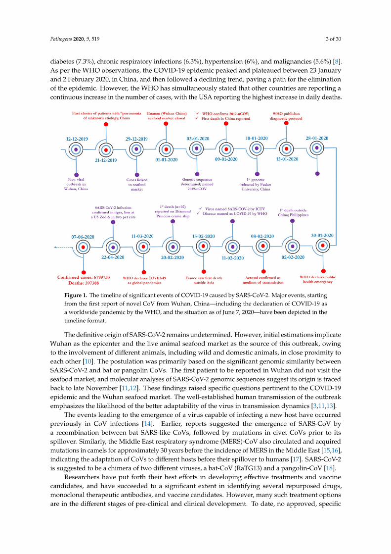
The ongoing global threat, declared a pandemic by international health agencies, originated froma newly emerging coronavirus (CoV), designated as severe acute respiratory syndrome coronavirus-2(SARS-CoV-2) [1]. A live animal market in the Hubei Province of Wuhan in Mainland China wasidentified as the first epicenter of the pandemic [2]. Initially, an outbreak was suspected when acluster of patients was admitted to local health care facilities with complaints of fever, cough, dyspnea,and fatigue, resembling symptoms common to viral pneumonia [3]. The first case of respiratory illnessof unknown etiology occurred on 8 December 2019, as shown in Figure 1. Consequently, a clusterof patients reported respiratory illness to the local health care facilities, and on 30 December 2019,the Chinese National Health Commission (CNHC) suspected an outbreak of pneumonia of unknownetiology in Wuhan. With the increasing number of cases in neighboring countries and other areasworldwide, the disease spread to several countries worldwide and it was declared a Public HealthEmergency of International Concern by the World Health Organization (WHO) on 31 January 2020,as shown in Figure 1. The epidemic intensified and spread without boundaries and affected more than215 countries and a few international conveyances, which led the WHO to declare COVID-19 as apandemic on 11 March 2020 [4].
As of 21 May 2020, 4,893,186 cases have been reported, with 323,256 deaths, of which 1,501,876confirmed cases and 90,203 deaths were from the United States of America, followed by Brazil(271,628; 17,971), the United Kingdom (248,297; 35,704), Spain (232,555; 27,888), Italy (227,364; 32,330),France (141,312; 28,081), and China (84,507; 4645) [5]. Notably, all age groups show susceptibilityto SARS-CoV-2 infection, from newborns to the elderly [6]. The average age noted for SARS-CoV-2infection susceptibility is 55.5 years, while that for the case fatality rates (CFRs) is 75 years [7,8]. However,the CFR varies among different age groups and is highest (14.8%) among the elderly (>80 years ofage). The incidences and CFR are reportedly higher in males (67.67% and 2.8%, respectively) thanin females, owing to the higher frequency of engagement in outdoor activities and smoking statusin males [9]. Immunocompromised individuals and patients with comorbidities are at a higher risk.The most common reported comorbidities in COVID-19 cases include cardiovascular diseases (10.5%),
Pathogens 2020, 9, 519 3 of 30
diabetes (7.3%), chronic respiratory infections (6.3%), hypertension (6%), and malignancies (5.6%) [8].As per the WHO observations, the COVID-19 epidemic peaked and plateaued between 23 Januaryand 2 February 2020, in China, and then followed a declining trend, paving a path for the eliminationof the epidemic. However, the WHO has simultaneously stated that other countries are reporting acontinuous increase in the number of cases, with the USA reporting the highest increase in daily deaths.
Pathogens 2020, 9, x FOR PEER REVIEW 3 of 31
January 23rd and February 2nd, 2020, in China, and then followed a declining trend, paving a path for the elimination of the epidemic. However, the WHO has simultaneously stated that other countries are reporting a continuous increase in the number of cases, with the USA reporting the highest increase in daily deaths.
Figure 1. The timeline of significant events of COVID-19 caused by SARS-CoV-2. Major events, starting from the first report of novel CoV from Wuhan, China—including the declaration of COVID-19 as a worldwide pandemic by the WHO, and the situation as of June 7, 2020—have been depicted in the timeline format.
The definitive origin of SARS-CoV-2 remains undetermined. However, initial estimations implicate Wuhan as the epicenter and the live animal seafood market as the source of this outbreak, owing to the involvement of different animals, including wild and domestic animals, in close proximity to each other [10]. The postulation was primarily based on the significant genomic similarity between SARS-CoV-2 and bat or pangolin CoVs. The first patient to be reported in Wuhan did not visit the seafood market, and molecular analyses of SARS-CoV-2 genomic sequences suggest its origin is traced back to late November [11,12]. These findings raised specific questions pertinent to the COVID-19 epidemic and the Wuhan seafood market. The well-established human transmission of the outbreak emphasizes the likelihood of the better adaptability of the virus in transmission dynamics [3,11,13].
The events leading to the emergence of a virus capable of infecting a new host have occurred previously in CoV infections [14]. Earlier, reports suggested the emergence of SARS-CoV by a recombination between bat SARS-like CoVs, followed by mutations in civet CoVs prior to its spillover. Similarly, the Middle East respiratory syndrome (MERS)-CoV also circulated and acquired mutations in camels for approximately 30 years before the incidence of MERS in the Middle East [15,16], indicating the adaptation of CoVs to different hosts before their spillover to humans [17]. SARS-CoV-2 is suggested to be a chimera of two different viruses, a bat-CoV (RaTG13) and a pangolin-CoV [18].
Researchers have put forth their best efforts in developing effective treatments and vaccine candidates, and have succeeded to a significant extent in identifying several repurposed drugs, monoclonal therapeutic antibodies, and vaccine candidates. However, many such treatment options are in the different stages of pre-clinical and clinical development. To date, no approved, specific
Figure 1. The timeline of significant events of COVID-19 caused by SARS-CoV-2. Major events, startingfrom the first report of novel CoV from Wuhan, China—including the declaration of COVID-19 asa worldwide pandemic by the WHO, and the situation as of June 7, 2020—have been depicted in thetimeline format.
The definitive origin of SARS-CoV-2 remains undetermined. However, initial estimations implicateWuhan as the epicenter and the live animal seafood market as the source of this outbreak, owingto the involvement of different animals, including wild and domestic animals, in close proximity toeach other [10]. The postulation was primarily based on the significant genomic similarity betweenSARS-CoV-2 and bat or pangolin CoVs. The first patient to be reported in Wuhan did not visit theseafood market, and molecular analyses of SARS-CoV-2 genomic sequences suggest its origin is tracedback to late November [11,12]. These findings raised specific questions pertinent to the COVID-19epidemic and the Wuhan seafood market. The well-established human transmission of the outbreakemphasizes the likelihood of the better adaptability of the virus in transmission dynamics [3,11,13].
The events leading to the emergence of a virus capable of infecting a new host have occurredpreviously in CoV infections [14]. Earlier, reports suggested the emergence of SARS-CoV bya recombination between bat SARS-like CoVs, followed by mutations in civet CoVs prior to itsspillover. Similarly, the Middle East respiratory syndrome (MERS)-CoV also circulated and acquiredmutations in camels for approximately 30 years before the incidence of MERS in the Middle East [15,16],indicating the adaptation of CoVs to different hosts before their spillover to humans [17]. SARS-CoV-2is suggested to be a chimera of two different viruses, a bat-CoV (RaTG13) and a pangolin-CoV [18].
Researchers have put forth their best efforts in developing effective treatments and vaccinecandidates, and have succeeded to a significant extent in identifying several repurposed drugs,monoclonal therapeutic antibodies, and vaccine candidates. However, many such treatment optionsare in the different stages of pre-clinical and clinical development. To date, no approved, specific

Pathogens 2020, 9, 519 4 of 30
antiviral agent or vaccine against SARS-CoV-2 is available for human use. The development ofnext-generation effective diagnostics has undergone a great leap. This review highlights the significantprogress made in the measures for the resolution of the COVID-19 pandemic, using virus genomicanalysis, the assessment of global epidemiological trends, transmission patterns, the present-daystatus of therapeutics, and public health preparedness plans for controlling the wider dissemination ofthe disease.
2. Previous Human CoV Epidemics
Usually, infections caused by the Betacoronaviruses are mild to asymptomatic [19,20]. Since thefirst report of CoV in the 1960s, humans have been affected by CoV infection. To date, six strains ofCoVs, causing mild to severe respiratory illnesses in humans, have been identified [21]. Of these, fourCoVs (HCoV-NL63, HCoV-229E, HCoV-OC43, and HCoV-HKU1) cause mild symptoms in humans.Contrarily, two highly pathogenic CoVs caused outbreaks of severe respiratory illness due to zoonotictransmission in humans, namely the severe acute respiratory syndrome (SARS) and MERS.
2.1. SARS-CoV
The first incidence of SARS-CoV infection was reported on 16 November 2002, in the coastalGuangdong province of southeast China bordering Hong Kong and Macau. This infection spreadacross 31 countries at a moderate speed. The virus was predicted to originate from bats and civet catswith animal-to-human (zoonotic) and human-to-human modes of transmission, as shown in Table 1.A winter season predilection was also noted in the pattern of this epidemic. After adopting strictpublic health measures, consisting of quarantine and the lockdown of air traffic, the disease was finallycontained by 5 July 2003 [22]. During this period, SARS-CoV infected 8422 humans and caused 916deaths at a CFR of 10.87%. Since then, SARS has phased out, and the outbreak was managed.
2.2. MERS-CoV
Since the first occurrence of MERS in Saudi Arabia in June 2012, sporadic cases are still reported,primarily in the Arabian Peninsular region, and MERS has spread to more than 27 countries worldwide.Notably, the CFR in MERS is the highest (34.77%) among all CoVs. To date, it has infected 2496 humanswith the deaths of 868 patients. The disease was reported in Saudi Arabia, the UAE and the Republicof Korea, as shown in Table 1. A few travel-related cases have been reported in Europe in individualswith a travel history to the Arabian Peninsular countries. The source of viral origin was traced tobats, and camels were the intermediate hosts for the virus, which followed animal-to-human (zoonoticdisease) and human-to-human modes of virus transmission [19,23].
2.3. SARS-CoV-2
The first case of SARS-CoV-2 infection was reported in the first week of December 2019 in WuhanCity, Hubei Province, China. Using deep sequencing methods, the causative agent for this deadlypandemic was identified as a new member of the Betacoronavirus genus [24,25]. The newly identifiedCoV had an 82% sequence identity with the human SARS-CoV [26]. It was initially designated as2019-novel coronavirus (2019-nCoV). Later, the International Committee on Taxonomy of Virusesofficially named it as the severe acute respiratory syndrome coronavirus-2 (SARS-CoV-2), owing to itssignificant genetic relatedness to SARS-CoV. Subsequently, the WHO named the disease caused bySARS-CoV-2 as coronavirus disease-2019 (COVID-19) on 11 February 2020, as shown in Figure 1.
Pathogens 2020, 9, 519 5 of 30
Table 1. Comparison of the typical features of human coronaviruses isolated between 2002–2020(SARS-CoV-2, MERS-CoV, and SARS-CoV).
Features SARS MERS COVID-19
Causative agent SARS-CoV MERS-CoV SARS-CoV-2
Incubation period 2–10 days 2–10 days 2–14 days
The median age ofinfected human cases 65 years 50 years 59 years
Source of origin Bats, civet cats Bats, camel Seafood, bats, pangolin(proposed)
TransmissionAnimal–humanHuman–humanZoonotic disease
Animal–humanHuman–humanZoonotic disease
Animal–humanHuman–humanHuman–animalZoonotic disease
Speed of spread Moderate Low High
Seasonal occurrence Winter (Dec–Jan) Summer (May–July) Winter (Dec–Jan)
Place of Origin Guangdong, China Jeddah, Saudi Arabia Wuhan, China
First incidence 16 November 2002 13 June 2012 7 December 2019
The last case reportedand present status 5 July 2003 18 February 2020
(Ongoing) Ongoing
Total number of cases 8422 2496 1,434,426 as of 8 April2020
Overall fatality 916 (10.87%) 868 (34.77%)(as of now)
82,220 (5.73%) as of 8April 2020
No. Countries affected 31 27 212 (till now)
Intermediate host Paguma larvata Camelus dromedaries Pangolin, Mink(possible)
Definitive host Rhinolophus sinicus Pipistrellus hesperidus Rhinolophus affinis(possible)
Taxonomy Betacoronavirus(Serbecovirus)
Betacoronavirus(Merbecovirus)
Betacoronavirus(Serbecovirus)
Genome length (bases) 29751 30119 29903
Major Regionaldistribution
Guangdong province ofsouthern China, and
later to western pacificcountries
Saudi Arabia, followedby UAE and Republic of
Korea
Hubei, especially, Wuhanin China, followed by
worldwide
Treatment/Vaccine Glucocorticoid andInterferon
No effective approvedtreatment or vaccine
Lopinavir/Ritonavir (intesting)
Receptor Angiotensin-convertingenzyme-2 (ACE-2)
Dipeptidyl peptidase 4(DPP4) ACE-2
3. SARS-CoV-2 Taxonomy, Structure, and Genomics
SARS-CoV-2 belongs to the family Coronaviridae, subfamily Orthocoronavirinae, under the orderNidovirales. The Orthocoronavirinae subfamily consists of four genera, namely Alphacoronavirus,Betacoronavirus, Gammacoronavirus, and Deltacoronavirus [27]. SARS-CoV-2 is a Betacoronavirus.Betacoronaviruses are further subdivided into five subgenera, namely Embecovirus, Hibecovirus,Merbecovirus, Nobecovirus, and Sarbecovirus. Phylogenetically, it forms a discrete lineage withinthe subgenus of the Sarbecovirus [28]. CoVs, as their name suggests, have a crown-shaped appearance,as observed under the electron microscope, owing to the spikes on the surface of the virion.
SARS-CoV-2 is a 0.12µm, enveloped, non-segmented virus, and its genome comprises a single-strandedpositive-sense RNA (+ssRNA) of ≈29.8 kb [29,30]. The genome is arranged in a structure typical to
Pathogens 2020, 9, 519 6 of 30
coronaviruses in the following order: 5′-ORF1a/ab-Spike (S)-Envelope (E)-Membrane (M)-Nucleocapsid(N)−3′; however, it lacks the hemagglutinin–esterase gene, similar to bat SARS-like-CoVs, humanSARS-CoVs, and MERS-CoVs. Fourteen open reading frames (ORFs) have been identified that encodetwenty-seven proteins, both structural and non-structural proteins (nsps). At the 5′ terminus, the firstORF1a/ab, which occupies approximately two-thirds of the entire genome length, encodes the pp1aand pp1ab proteins, respectively, and both collectively comprise 15 non-structural proteins (nsp 1 to10 and nsp 12 to 16). The functions of the nsps of a typical coronavirus and SARS-CoV-2 are listedin Supplementary Table S1 [26,31]. The ORFs toward the 3′- terminus encode at least four majorstructural proteins: the spike (S), matrix membrane (M), envelope (E), and nucleocapsid (N) proteins.Besides, accessory proteins (3a, 6, 7a, 7b, 8, and 10) are also encoded by the genome of SARS-CoV-2Wuhan HU1. The 5′- and 3′- UTR sequences of SARS-CoV-2 have ≈83.0% nucleotide identity withother members of the Betacoronavirus genus. The genomes of SARS-CoV, SARS-CoV-2, and bat-SARSCoV are considerably similar. The RNA secondary structures in the 5′-UTR were more identical tothose of SARS-CoV than to the bat SARS-like CoV; however, the 3′-UTR was more conserved comparedto other related Betacoronaviruses [26].
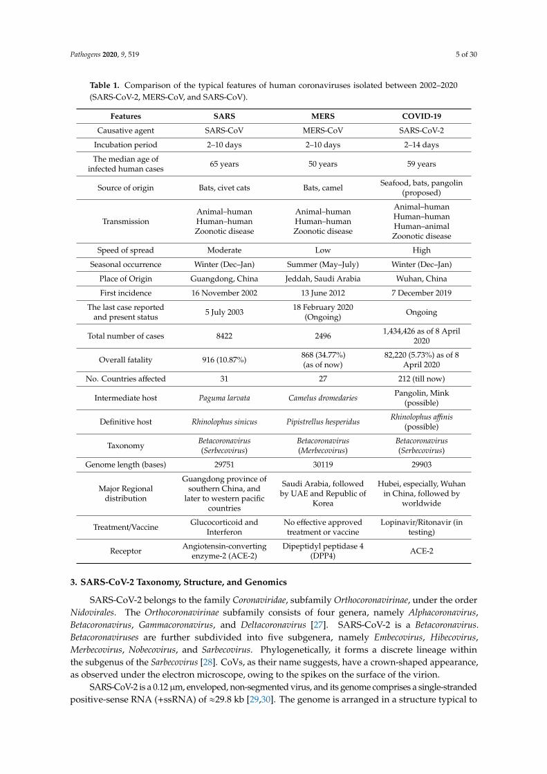
A detailed comparative genomic organization of important and related Betacoronavirus members,including a Deltacoronavirus as well, is provided in Figure 2. At the genomic level, the sequencehomology of SARS-CoV-2 with MERS-CoV is lower (50–51.8%) than that with SARS-CoV (79%) [32].Phylogenetically, SARS-CoV-2 is most similar to the bat SARS-like-CoVZC45 concerning mostgenes [33,34]. Even at the amino acid level, SARS-CoV-2 is relatively similar to bat SARS-like-CoVZC45,although there exist certain notable differences. For example, the 6, 7b, and 10 proteins are presentin SARS-CoV-2, whereas these are absent in bat SARS-like CoVZC45. Compared to SARS-CoV,the sequence for 8a is absent in the SARS-CoV-2 genome; the sequence for 3b is shorter, while that for8b has an extra 73 amino acids in SARS-CoV-2 [29]. The spike protein-encoding gene in SARS-CoV-2(1273 amino acids) is longer than those of bat SARS-like-CoVZC45 (1246 amino acids) and SARS-CoVTor2 (1255 amino acids). Certain accessory proteins, such as the 6 and 7b proteins of SARS-CoV Tor2,are similar to those in SARS-CoV-2, except for a marginal variation in protein length, as shown inFigure 2. The majority of the accessory proteins in MERS-CoV EMC are distinctive, including a longerspike protein (1353 amino acids), compared to those in SARS-CoV-2. In comparison to SARS-CoV-2,bovine CoV ENT has the longest spike protein (1363 amino acids) and a unique hemagglutinin–esterasegene, while porcine CoV HKU15 (a delta-CoV) has the shortest spike protein (1159 amino acids) andlacks several accessory proteins. Hence, it would be interesting to characterize the spike and accessoryproteins of SARS-CoV-2 and study the role of these proteins in the pathogenesis and transmissibility ofthe virus.
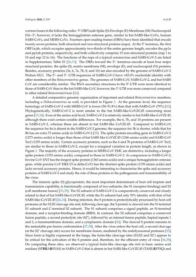
The trimeric spike (S) glycoprotein, the most important determinant of host cell tropism andtransmission capability, is functionally composed of two subunits, the S1 (receptor binding) and S2(cell membrane fusion) [33,35]. The S2 subunit of SARS-CoV-2 is comparatively conserved and closelyrelated to that of bat SARS-like-CoVZC45, while the S1 subunit had only 70% identity with that of batSARS-like-CoVZC45 [26,34]. During infection, the S protein is proteolytically processed by host cellproteases at the S1/S2 cleavage site and, following cleavage, the S protein is cleaved into the N-terminalS1 subunit and C-terminal S2 subunit. The S1 subunit comprises a signal peptide, an N-terminaldomain, and a receptor-binding domain (RBD). In contrast, the S2 subunit comprises a conservedfusion peptide, a second proteolytic site (S2′), followed by an internal fusion peptide, heptad repeats 1and 2, a transmembrane domain, and a cytoplasmic domain [36]. The cleaved S protein remains inthe metastable pre-fusion conformation [37,38]. After the virus enters the host cell, a second cleavage(at the S2′ cleavage site) occurs for membrane fusion, mediated by the endolysosomal proteases [33].Since furin is highly expressed in the lungs, the furin-like cleavage sites (S1/S2 and S2′) appear tobe critical for the activation of the S protein and, therefore, for the efficient entry of virus [36,39].On comparing these sites, we observed a typical furin-like cleavage site rich in basic amino acidresidues (SPRRARSVAS) in SARS-CoV-2 that is absent in bat SARS-like-CoVZC45 (TASILRSTSQ) and

Pathogens 2020, 9, 519 7 of 30
human SARS-CoV Tor2 (TVSLLRSTSQ), as shown in Figure 3. However, the S2′ cleavage site patterns(XKPXKRSF) in all three viruses were considerably similar.
Similarly, furin might cleave the MERS-CoV EMC spike protein less efficiently at the S2′
cleavage site, owing to the lack of typical basic amino acid residues observed in SARS-CoV-2,bat SARS-like-CoVZC45, and human SARS-CoV Tor2. Although the S1/S2 cleavage site in the bovineCoV ENT (KRRSRRSITT) spike protein is rich in basic amino acids, its S2′ cleavage site (NKVSSRSA)lacks the typical basic amino acid residues, as shown in Figure 3. The porcine CoV HKU15 contains amonobasic cleavage site (NSTENNRFTTT), which could lead to low-efficiency cleavage. Based on thefindings from these comparative studies, we speculate that the high-density basic amino acid residues,preferably positioned in close proximity at both cleavage sites (S1/S2 and S2′), might have facilitatedthe efficient replication and transmission of SARS-CoV-2. However, these propositions need to bevalidated by further experimental investigations.
Pathogens 2020, 9, x FOR PEER REVIEW 7 of 31
characterize the spike and accessory proteins of SARS-CoV-2 and study the role of these proteins in the pathogenesis and transmissibility of the virus.
Figure 2. Genome organization of SARS-CoV-2 and its comparison with other coronaviruses. The coding regions of beta-CoVs, SARS-CoV-2 Wuhan-HU-1 (NC_045512.2; 29,903 bps), bat SARS-like-CoVZC45 (MG772933.1; 29,802 bps), human SARS-CoV Tor2 (AY274119.3; 29,751 bps), MERS-CoV EMC (NC_019843.3; 30,119 bps), bovine CoV ENT (NC_003045.1; 31,028 bps), and a delta-CoV, the porcine CoV HKU15 (NC_039208.1; 25425 bps) are presented. The genomes consist of a 5′ untranslated region (5′ UTR), an open reading frame (ORF 1a/ab) encoding non-structural proteins (nsps), structural proteins, hemagglutinin–esterase, spike (S), membrane (M), envelope (E), and nucleocapsid (N) proteins, several accessory proteins, and a 3′ untranslated region (3′ UTR). The lengths of the ORFs, nsps, and accessory proteins are not drawn to scale.
The trimeric spike (S) glycoprotein, the most important determinant of host cell tropism and transmission capability, is functionally composed of two subunits, the S1 (receptor binding) and S2 (cell membrane fusion) [33,35]. The S2 subunit of SARS-CoV-2 is comparatively conserved and closely related to that of bat SARS-like-CoVZC45, while the S1 subunit had only 70% identity with that of bat SARS-like-CoVZC45 [26,34]. During infection, the S protein is proteolytically processed by host cell proteases at the S1/S2 cleavage site and, following cleavage, the S protein is cleaved into the N-terminal S1 subunit and C-terminal S2 subunit. The S1 subunit comprises a signal peptide, an N-terminal domain, and a receptor-binding domain (RBD). In contrast, the S2 subunit comprises aconserved fusion peptide, a second proteolytic site (S2’), followed by an internal fusion peptide,heptad repeats 1 and 2, a transmembrane domain, and a cytoplasmic domain [36]. The cleaved Sprotein remains in the metastable pre-fusion conformation [37,38]. After the virus enters the host cell,a second cleavage (at the S2’ cleavage site) occurs for membrane fusion, mediated by theendolysosomal proteases [33]. Since furin is highly expressed in the lungs, the furin-like cleavage
Figure 2. Genome organization of SARS-CoV-2 and its comparison with other coronaviruses. The codingregions of beta-CoVs, SARS-CoV-2 Wuhan-HU-1 (NC_045512.2; 29,903 bps), bat SARS-like-CoVZC45(MG772933.1; 29,802 bps), human SARS-CoV Tor2 (AY274119.3; 29,751 bps), MERS-CoV EMC(NC_019843.3; 30,119 bps), bovine CoV ENT (NC_003045.1; 31,028 bps), and a delta-CoV, the porcineCoV HKU15 (NC_039208.1; 25425 bps) are presented. The genomes consist of a 5′ untranslatedregion (5′ UTR), an open reading frame (ORF 1a/ab) encoding non-structural proteins (nsps), structuralproteins, hemagglutinin–esterase, spike (S), membrane (M), envelope (E), and nucleocapsid (N) proteins,several accessory proteins, and a 3′ untranslated region (3′ UTR). The lengths of the ORFs, nsps,and accessory proteins are not drawn to scale.

Pathogens 2020, 9, 519 8 of 30
Pathogens 2020, 9, x FOR PEER REVIEW 8 of 31
sites (S1/S2 and S2’) appear to be critical for the activation of the S protein and, therefore, for the efficient entry of virus [36,39]. On comparing these sites, we observed a typical furin-like cleavage site rich in basic amino acid residues (SPRRARSVAS) in SARS-CoV-2 that is absent in bat SARS-like-CoVZC45 (TASILRSTSQ) and human SARS-CoV Tor2 (TVSLLRSTSQ), as shown in Figure 3. However, the S2’ cleavage site patterns (XKPXKRSF) in all three viruses were considerably similar.
Similarly, furin might cleave the MERS-CoV EMC spike protein less efficiently at the S2’ cleavage site, owing to the lack of typical basic amino acid residues observed in SARS-CoV-2, bat SARS-like-CoVZC45, and human SARS-CoV Tor2. Although the S1/S2 cleavage site in the bovine CoV ENT (KRRSRRSITT) spike protein is rich in basic amino acids, its S2’ cleavage site (NKVSSRSA) lacks the typical basic amino acid residues, as shown in Figure 3. The porcine CoV HKU15 contains a monobasic cleavage site (NSTENNRFTTT), which could lead to low-efficiency cleavage. Based on the findings from these comparative studies, we speculate that the high-density basic amino acid residues, preferably positioned in close proximity at both cleavage sites (S1/S2 and S2’), might have facilitated the efficient replication and transmission of SARS-CoV-2. However, these propositions need to be validated by further experimental investigations.
Figure 3. Comparative sequence and structural analyses of the SARS-CoV-2 spike protein. (A) The cleavage sites of SARS-CoV-2 spike proteins (S1/S2 and S’ protease cleavage sites) and their comparison with those in other coronaviruses. The superimposition of the receptor-binding domain
Figure 3. Comparative sequence and structural analyses of the SARS-CoV-2 spike protein. (A) Thecleavage sites of SARS-CoV-2 spike proteins (S1/S2 and S’ protease cleavage sites) and their comparisonwith those in other coronaviruses. The superimposition of the receptor-binding domain (RBDs)of SARS-CoV-2 (pdb: 6VSB) on (B) human SARS-CoV (pdb: 2AJF), (C) MERS-CoV (pdb: 4KR0),(D) BtCoV-HKU4 (pdb: 4QZV), and (E) HCoV-NL63 (pdb: 3KBH). (F) The superimposition of theRBDs of porcine respiratory CoV (pdb: 4F5C) on HCoV-NL63 (pdb: 3KBH). RMSD: Root MeanSquare Deviation. UCSF Chimera was used for the superposition and visualization of the RBDs of thedifferent CoVs [40].
4. CoV Receptors
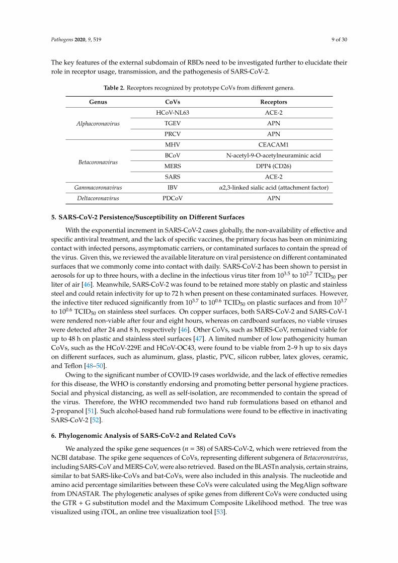
Host cell receptors and viral surface glycoprotein/ligand binding serve as the most importantdeterminants of host range, cross-species transmission, and antiviral targeting inventions [41,42].As shown in Table 2, CoVs can infect a wide range of host species, including humans, animals, andbirds. CoVs from four genera recognize at least five different receptors and, therefore, exhibit anintricate host receptor recognition pattern. The differential usage of host receptors by different CoVsindicates the structural diversity in the RBDs of the S glycoprotein. However, CoV infections in humansare primarily driven by interactions between the envelope-anchored spike glycoprotein (S protein) ofCoV and the host cell receptor, the angiotensin-converting enzyme 2 (ACE2) [43–45]. Viruses from thesame genus can recognize different receptors (for example, SARS-ACE2 and MERS-DPP4), while CoVsfrom different genera can bind to the same receptor as well, such as ACE-2 (HCoV-NL63 and SARS).
Pathogens 2020, 9, 519 9 of 30
The key features of the external subdomain of RBDs need to be investigated further to elucidate theirrole in receptor usage, transmission, and the pathogenesis of SARS-CoV-2.
Table 2. Receptors recognized by prototype CoVs from different genera.
Genus CoVs Receptors
Alphacoronavirus
HCoV-NL63 ACE-2
TGEV APN
PRCV APN
Betacoronavirus
MHV CEACAM1
BCoV N-acetyl-9-O-acetylneuraminic acid
MERS DPP4 (CD26)
SARS ACE-2
Gammacoronavirus IBV α2,3-linked sialic acid (attachment factor)
Deltacoronavirus PDCoV APN
5. SARS-CoV-2 Persistence/Susceptibility on Different Surfaces
With the exponential increment in SARS-CoV-2 cases globally, the non-availability of effective andspecific antiviral treatment, and the lack of specific vaccines, the primary focus has been on minimizingcontact with infected persons, asymptomatic carriers, or contaminated surfaces to contain the spread ofthe virus. Given this, we reviewed the available literature on viral persistence on different contaminatedsurfaces that we commonly come into contact with daily. SARS-CoV-2 has been shown to persist inaerosols for up to three hours, with a decline in the infectious virus titer from 103.5 to 102.7 TCID50 perliter of air [46]. Meanwhile, SARS-CoV-2 was found to be retained more stably on plastic and stainlesssteel and could retain infectivity for up to 72 h when present on these contaminated surfaces. However,the infective titer reduced significantly from 103.7 to 100.6 TCID50 on plastic surfaces and from 103.7
to 100.6 TCID50 on stainless steel surfaces. On copper surfaces, both SARS-CoV-2 and SARS-CoV-1were rendered non-viable after four and eight hours, whereas on cardboard surfaces, no viable viruseswere detected after 24 and 8 h, respectively [46]. Other CoVs, such as MERS-CoV, remained viable forup to 48 h on plastic and stainless steel surfaces [47]. A limited number of low pathogenicity humanCoVs, such as the HCoV-229E and HCoV-OC43, were found to be viable from 2–9 h up to six dayson different surfaces, such as aluminum, glass, plastic, PVC, silicon rubber, latex gloves, ceramic,and Teflon [48–50].
Owing to the significant number of COVID-19 cases worldwide, and the lack of effective remediesfor this disease, the WHO is constantly endorsing and promoting better personal hygiene practices.Social and physical distancing, as well as self-isolation, are recommended to contain the spread ofthe virus. Therefore, the WHO recommended two hand rub formulations based on ethanol and2-propanol [51]. Such alcohol-based hand rub formulations were found to be effective in inactivatingSARS-CoV-2 [52].
6. Phylogenomic Analysis of SARS-CoV-2 and Related CoVs
We analyzed the spike gene sequences (n = 38) of SARS-CoV-2, which were retrieved from theNCBI database. The spike gene sequences of CoVs, representing different subgenera of Betacoronavirus,including SARS-CoV and MERS-CoV, were also retrieved. Based on the BLASTn analysis, certain strains,similar to bat SARS-like-CoVs and bat-CoVs, were also included in this analysis. The nucleotide andamino acid percentage similarities between these CoVs were calculated using the MegAlign softwarefrom DNASTAR. The phylogenetic analyses of spike genes from different CoVs were conducted usingthe GTR + G substitution model and the Maximum Composite Likelihood method. The tree wasvisualized using iTOL, an online tree visualization tool [53].
Pathogens 2020, 9, 519 10 of 30
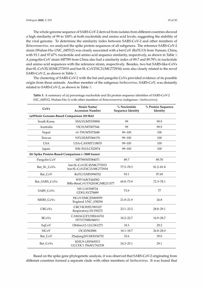
The whole-genome sequence of SARS-CoV-2 derived from isolates from different countries showeda high similarity of 99 to 100% at both nucleotide and amino acid levels, suggesting the stability ofthe viral genome. To determine the similarity index between SARS-CoV-2 and other members ofBetacoronavirus, we analyzed the spike protein sequences of all subgenera. The reference SARS-CoV-2strain (Wuhan-Hu-1/NC_045512) was closely associated with a bat-CoV (RaTG13) from Yunnan, China,with 93.1 and 97.67% nucleotide and amino acid sequence similarity, respectively, as shown in Table 3.A pangolin-CoV strain MP789 from China also had a similarity index of 89.7 and 89.78% in nucleotideand amino acid sequences with the reference strain, respectively. Besides, two bat SARS-like-CoVs(bat-SL-CoVZC45/MG772933 and bat-SL-CoVZXC21/MG772934) were also closely related to the novelSARS-CoV-2, as shown in Table 3.
The clustering of SARS-CoV-2 with the bat and pangolin CoVs provided evidence of its possibleorigin from these animals. Another member of the subgenus Sarbecovirus, SARS-CoV, was distantlyrelated to SARS-CoV-2, as shown in Table 3.
Table 3. A summary of (a) percentage nucleotide and (b) protein sequence identities of SARS-CoV-2(NC_045512, Wuhan-Hu-1) with other members of Betacoronavirus (subgenus—Sarbecovirus).
CoVs Strain Name/Accession Number
% NucleotideSequence Identity
% Protein SequenceIdentity
(a)Whole Genome-Based Comparison (29.9kb)
South Korea SNU01/MT039890 99 99.9
Australia VIC01/MT007544 99 99.9
Nepal 61-TW/MT072688 99–100 100
Taiwan NTU02/MT066176 99–100 100
USA USA-CA9/MT118835 99–100 100
Japan WK-501/LC522974 99–100 100
(b) Spike Protein-Based Comparison (~3800 bases)
Pangolin CoV MP789/MT084071 89.7 89.78
Bat_SL_CoVs bat-SL-CoVZC45/MG772933bat-SL-CoVZXC21/MG772934 77.5–78.3 81.2–81.8
Bat_CoV RaTG13/MN996532 93.1 97.69
Bat_SARS_CoVs WIV16/KT444582BtRs-BetaCoV/YN2018C/MK211377 69.8–73.9 72.3–78.1
SARS_CoVs NS-1/AY508724GD01/AY278489 73.9 77
MERS_CoVs HCoV-EMC/JX869059England 1/NC_038294 21.8–21.9 26.8
CRCoVs CRCVK39/EU983107Respiratory/AY150272 23.1–23.2 28.8–29.1
BCoVs CAMAGUEY/HE616741HT317/MK046011 18.2–22.7 16.9–28.7
EqCoV Obihiro12-1/LC061273 18.3 29.2
HCoV OC43/S62886 18.1–18.7 26.8–28.0
Bat_CoV Zhejiang2013/KF636752 33.6 39.0
Bat_CoVs KHU9-1/EF065513GCCDC1 356/KU762338 24.3–25.1 29.1
Based on the spike gene phylogenetic analysis, it was observed that SARS-CoV-2 originating fromdifferent countries formed a separate clade with other members of Sarbecovirus. It was found that

Pathogens 2020, 9, 519 11 of 30
the bat-CoV (RaTG13/MN996532), pangolin-CoV (MP789/MT084071), and two bat SARS-like-CoVs(bat-SL-CoVZC45/MG772933 and bat-SL-CoVZXC21/MG772934) closely clustered with SARS-CoV-2,while other bat-CoVs and SARS-CoVs clustered within the Sarbecovirus subgenus clade, as shown inFigure 4. Similarly, CoVs from different subgenera, which included bat-CoVs belonging to Hibecovirusand Nobecovirus, human and camel MERS-CoV belonging to Merbecovirus, and human, canine, bovine,and equine CoVs belonging Embecovirus subgenera, clustered separately, as shown in Figure 4.Pathogens 2020, 9, x FOR PEER REVIEW 12 of 31
Figure 4. Maximum likelihood tree of Betacoronaviruses, including SARS-CoV-2. The phyloanalysis included SARS-CoV-2 originating from China, the USA, Australia, Japan, and South Korea. Different subgenera of Betacoronaviruses have been labeled with different colors according to the color legend on the left side of the tree. Major species of each subgenus have been depicted in front of each clade.
7. Epidemiology of SARS-CoV-2 Infection
The Wuhan seafood market in China was implicated as the epicenter of the COVID-19 pandemic, and China was marked as the index country. Seven cases of pneumonia of unknown etiology were reported in Wuhan from December 8th to 18th, 2019. The first cluster of such patients was identified on December 21st, 2019. Pneumonia of unknown etiology is defined as an illness without an identified causative pathogen that fulfills the following criteria: fever (≥ 38 °C), radiographic evidence of pneumonia, low or average white-cell count or low lymphocyte count, and no symptomatic improvement after antimicrobial treatment for 3 to 5 days following standard clinical guidelines [11,54]. The patients presented with a severe acute respiratory infection and some developed acute respiratory distress syndrome (ARDS) and associated complications [55].
7.1. Transmission
Information about the mode of transmission of SARS-CoV-2 is continuously evolving. Transmission primarily occurs via the person-to-person route through respiratory droplets (>5-10 µm in diameter) and fomites. Droplet infection is associated mainly with coughing, sneezing, and talking to an infected person, and the virus is transmitted to humans through direct contact with mucus membranes. Transmission can also occur by touching an infected surface, followed by contact with eyes, nose, or mouth. The travel range of the droplets is less than six feet (approximately two meters), and these are not retained in the air for long. Satisfactory evidence of the airborne transmission of SARS-CoV-2 remains unavailable [46]. Although clinically infected patients readily transmit the virus [14], asymptomatic infected patients can also transmit the virus, and therefore, these carriers should be tested to effectively contain the spread [11,56,57].
There is evidence of intestinal infection from SARS-CoV-2, and subsequent excretion through feces [56,58]. In China, out of ten pediatric COVID-19 patients without respiratory symptoms, eight were positive for fecal SARS-CoV-2 shedding; however, the nasopharyngeal samples from these
Figure 4. Maximum likelihood tree of Betacoronaviruses, including SARS-CoV-2. The phyloanalysisincluded SARS-CoV-2 originating from China, the USA, Australia, Japan, and South Korea. Differentsubgenera of Betacoronaviruses have been labeled with different colors according to the color legend onthe left side of the tree. Major species of each subgenus have been depicted in front of each clade.
7. Epidemiology of SARS-CoV-2 Infection
The Wuhan seafood market in China was implicated as the epicenter of the COVID-19 pandemic,and China was marked as the index country. Seven cases of pneumonia of unknown etiology werereported in Wuhan from 8–18 December 2019. The first cluster of such patients was identified on 21 December2019. Pneumonia of unknown etiology is defined as an illness without an identified causative pathogenthat fulfills the following criteria: fever (≥38 ◦C), radiographic evidence of pneumonia, low or averagewhite-cell count or low lymphocyte count, and no symptomatic improvement after antimicrobialtreatment for 3 to 5 days following standard clinical guidelines [11,54]. The patients presented with asevere acute respiratory infection and some developed acute respiratory distress syndrome (ARDS)and associated complications [55].
7.1. Transmission
Information about the mode of transmission of SARS-CoV-2 is continuously evolving. Transmissionprimarily occurs via the person-to-person route through respiratory droplets (>5–10 µm in diameter)and fomites. Droplet infection is associated mainly with coughing, sneezing, and talking to aninfected person, and the virus is transmitted to humans through direct contact with mucus membranes.Transmission can also occur by touching an infected surface, followed by contact with eyes, nose,or mouth. The travel range of the droplets is less than six feet (approximately two meters), and these arenot retained in the air for long. Satisfactory evidence of the airborne transmission of SARS-CoV-2 remains
Pathogens 2020, 9, 519 12 of 30
unavailable [46]. Although clinically infected patients readily transmit the virus [14], asymptomaticinfected patients can also transmit the virus, and therefore, these carriers should be tested to effectivelycontain the spread [11,56,57].
There is evidence of intestinal infection from SARS-CoV-2, and subsequent excretion throughfeces [56,58]. In China, out of ten pediatric COVID-19 patients without respiratory symptoms,eight were positive for fecal SARS-CoV-2 shedding; however, the nasopharyngeal samples from thesesubjects tested negative for the virus [59]. These findings were further supported by a study in whichthe virus was successfully cultured from feces samples [59,60]. However, there have been no reportsof fecal–oral transmission of SARS-CoV-2 to date. Some reports suggest SARS-CoV-2 transmissionthrough semen and the infection of SARS-CoV-2 in newborns; however, there is no evidence for verticaltransmission or intrauterine infection in a COVID-19 patient during the late pregnancy stage [55].
7.2. Incubation Period
The incubation period for SARS-CoV-2 infection ranged from 2 to 14 days in human-to-humantransmission, while, in some cases, it extended up to 24 days [57]. However, a median incubationperiod of 5-6 days has recently been reported by the WHO [61].
7.3. Basic Reproduction Number (R0)
The basic reproduction number plays a pivotal role in infectious disease epidemiology andindicates the risk a pathogen carries with respect to its spread. In other words, R0 provides informationabout the transmissibility of a virus and represents the number of new infections originating froman infected individual in a population. A recent comparison of different studies on the estimation ofR0 for COVID-19 revealed that the estimated mean R0 for COVID-19 is 3.28, with a median of 2.79,which is higher than the WHO estimate of 1.95 [62]. However, considerable variability was noted,owing to the use of different methods, insufficient data, and the short onset time of the disease.
7.4. Population Susceptibility
There is no age limit for vulnerability to SARS-CoV-2 infection. However, cases with highermorbidity and chances of mortality have been reported in older adults. Individuals above 65 yearsof age or those who have comorbidities (such as hypertension, diabetes, cardiovascular diseases,and respiratory system diseases) are highly susceptible to the infection [25]. Women are less susceptibleto SARS-CoV-2 infection than men and this is possibly due to different innate immunity, and factorsrelated to sex chromosomes in women [63]. For example, the biallelic expression of TLR7 in womenallows higher immune responses, while the immune regulatory genes encoded by X chromosomescause comparatively less virus load and inflammation. Recently, studies have shown that SARS-CoV-2infection in children (though less severely affected), may develop the Kawasaki disease-like forms [64].However, it is still not clear whether Kawasaki disease-like forms are a result of SARS-CoV-2 infectionalone or just SARS-CoV-2 induced aggravations in Kawasaki disease patients.
7.5. Global Scenario
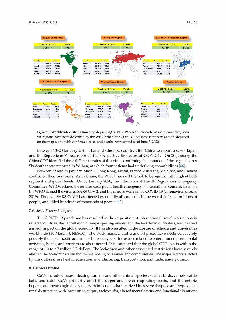
COVID-19 has affected almost every country across the globe. On 31 December 2019, an “urgentnotice on the treatment of pneumonia of unknown cause” was issued to the Wuhan Municipal HealthCenter. The market premises were disinfected, and the stallholders were instructed to wear masks.Till 3 January 2020, 44 case-patients with pneumonia of unknown etiology were reported to the WHOfrom China, although the cause was not identified. On 7 January 2020, the Chinese authorities identifieda new type of CoV and shared its genetic sequence on 12 January 2020. On 9 January, the death ofa 61-year-old man was the first reported case of death from COVID-19. The WHO published interimlaboratory guidelines for the detection of SARS-CoV-2 [65]. As of 7 June, 2020, COVID-19 distributionwas seen globally with large number of confirmed cases and deaths. The WHO defined six regions todistribute the COVID-19 disease (Figure 5).
Pathogens 2020, 9, 519 13 of 30Pathogens 2020, 9, x FOR PEER REVIEW 14 of 31
Figure 5. Worldwide distribution map depicting COVID-19 cases and deaths in major world regions. Six regions have been described by the WHO where the COVID-19 disease is present and are depicted on the map along with confirmed cases and deaths represented as of June 7, 2020.
Between January 13th to 20th, 2020, Thailand (the first country after China to report a case), Japan, and the Republic of Korea, reported their respective first cases of COVID-19. On January 20th, the China CDC identified three different strains of this virus, confirming the mutation of the original virus. Six deaths were reported in Wuhan, of which four patients had underlying comorbidities [66].
Between January 22nd and 25th, Macau, Hong Kong, Nepal, France, Australia, Malaysia, and Canada confirmed their first cases. As in China, the WHO assessed the risk to be significantly high at both regional and global levels. On January 30th, 2020, the International Health Regulations Emergency Committee, WHO declared the outbreak as a public health emergency of international concern. Later on, the WHO named the virus as SARS-CoV-2, and the disease was named COVID-19 (coronavirus disease 2019). Thus far, SARS-CoV-2 has affected essentially all countries in the world, infected millions of people, and killed hundreds of thousands of people [67].
7.6. Socio-Economic Impact
The COVID-19 pandemic has resulted in the imposition of international travel restrictions in several countries, the cancellation of major sporting events, and the lockdown of borders, and has had a major impact on the global economy. It has also resulted in the closure of schools and universities worldwide (March 10th, UNESCO). The stock markets and crude oil prices have declined severely, possibly the most drastic occurrence in recent years. Industries related to entertainment, ceremonial activities, hotels, and tourism are also affected. It is estimated that the global GDP loss is within the range of 1.0 to 2.7 trillion US dollars. The lockdown and other associated restrictions have severely affected the economic status and the well-being of families and communities. The major sectors affected by this outbreak are health, education, manufacturing, transportation, and trade, among others.
8. Clinical Profile
CoVs include viruses infecting humans and other animal species, such as birds, camels, cattle, bats, and cats. CoVs primarily affect the upper and lower respiratory tracts, and the enteric, hepatic, and neurological systems, with infections characterized by severe dyspnea and hypoxemia, renal
Figure 5. Worldwide distribution map depicting COVID-19 cases and deaths in major world regions.Six regions have been described by the WHO where the COVID-19 disease is present and are depictedon the map along with confirmed cases and deaths represented as of June 7, 2020.
Between 13–20 January 2020, Thailand (the first country after China to report a case), Japan,and the Republic of Korea, reported their respective first cases of COVID-19. On 20 January, theChina CDC identified three different strains of this virus, confirming the mutation of the original virus.Six deaths were reported in Wuhan, of which four patients had underlying comorbidities [66].
Between 22 and 25 January, Macau, Hong Kong, Nepal, France, Australia, Malaysia, and Canadaconfirmed their first cases. As in China, the WHO assessed the risk to be significantly high at bothregional and global levels. On 30 January 2020, the International Health Regulations EmergencyCommittee, WHO declared the outbreak as a public health emergency of international concern. Later on,the WHO named the virus as SARS-CoV-2, and the disease was named COVID-19 (coronavirus disease2019). Thus far, SARS-CoV-2 has affected essentially all countries in the world, infected millions ofpeople, and killed hundreds of thousands of people [67].
7.6. Socio-Economic Impact
The COVID-19 pandemic has resulted in the imposition of international travel restrictions inseveral countries, the cancellation of major sporting events, and the lockdown of borders, and has hada major impact on the global economy. It has also resulted in the closure of schools and universitiesworldwide (10 March, UNESCO). The stock markets and crude oil prices have declined severely,possibly the most drastic occurrence in recent years. Industries related to entertainment, ceremonialactivities, hotels, and tourism are also affected. It is estimated that the global GDP loss is within therange of 1.0 to 2.7 trillion US dollars. The lockdown and other associated restrictions have severelyaffected the economic status and the well-being of families and communities. The major sectors affectedby this outbreak are health, education, manufacturing, transportation, and trade, among others.
8. Clinical Profile
CoVs include viruses infecting humans and other animal species, such as birds, camels, cattle,bats, and cats. CoVs primarily affect the upper and lower respiratory tracts, and the enteric,hepatic, and neurological systems, with infections characterized by severe dyspnea and hypoxemia,renal dysfunction with lower urine output, tachycardia, altered mental status, and functional alterations

Pathogens 2020, 9, 519 14 of 30
of organs in animals and humans. Individuals infected with SARS-CoV-2 may be asymptomaticcarriers or active patients with the clinical presentation of symptoms, such as fever, fatigue, dry cough,myalgia, dyspnea, severe respiratory distress syndrome, and acute cardiac injury [57,68,69]. There arethree types of ARDS: mild, moderate, and severe ARDS. An increase in body temperature up to39.0 ◦C (102.2 ◦F), accompanied by coarse breathing sounds from both lungs at auscultation arecommon clinical symptoms. Symptoms, such as headache, sputum production, hemoptysis, diarrhea,and pleuritic chest pain were also observed in rare cases. The infection more commonly affects peoplewith cardiopulmonary or renal diseases, and pregnant females, infants, and the elderly (at a two-foldgreater risk than the general population). There is no evidence to suggest greater susceptibility amongchildren. Pregnant women are at greater risk for viral infections, as suggested by data from previousoutbreaks; therefore, it is advisable to avoid contact with infected individuals [70].
9. Clinical Pathology and Immunopathobiology
9.1. Clinical Pathology
Based on the time of occurrence and intensity of symptoms, the infection has been categorizedinto three stages; mild, severe, and critical stage [71,72]. In the mild stage, mild pneumonia/nopneumonia develops, although symptoms of upper respiratory tract viral infection, marked by mildfever, dry cough, sore throat, nasal congestion, malaise, headache, and muscle pain, prevail [73].In severely affected patients, the rate of respiration is above 30/min due to dyspnea, respiratorysymptoms, such as cough and shortness of breath, or tachypnea, and oxygen saturation in the bloodbelow 93%, indicating hypoxia, and these developments are observed within a time period of 24–48 h.The critical stage is marked by symptoms, such as severe pneumonia, septic shock, respiratoryfailure, cardiac arrest, and/or multiple organ failure, which may eventually lead to the death of thepatient [74,75].
Radiological images indicate that two to five lobes of the lungs may be affected. The most commonand pronounced lesions are marked by patchy ground-glass opacities and patchy consolidation inthe mid, external, and sub-pleural areas of the lung [76]. A chest radiograph, computer tomographic(CT) scan, and/or lung ultrasound indicate the presence of bilateral opacities [55,77,78]. Thoracic CTscan imaging depicted bilateral ground-glass opacity and bilateral multiple lobular and sub-segmentalareas of consolidation [79–81]. The pathological image of a COVID-19 patient presented bilateraldiffuse alveolar damage with cellular fibromyxoid exudates. The cytopathic effects were marked bythe presence of interstitial mononuclear inflammatory infiltrates, with a majority of lymphocytes andmultinucleated syncytial cells, along with atypical enlarged pneumocytes containing large nuclei andamphophilic granular cytoplasm, in the intra-alveolar spaces. In some infected patients, microvascularsteatosis and mild lobular and portal activity were recorded in hepatic tissues, while interstitialmononuclear inflammatory infiltrates were found in the cardiac tissues [8].
9.2. Immunopathobiology
Existing literature documents that patients suffering from severe SARS-CoV-2 infection mightsuffer from the cytokine storm syndrome that could eventually lead to death [82,83]. In a viralinfection, cell-mediated immunity is triggered to overcome the infection; however, owing to theoverproduction of cytokines, interferons, and other factors, such events can have drastic effectsin certain patients [84]. Evidently, respiratory failure due to the ARDS is the leading cause ofmortality, and, apart from this, secondary hemophagocytic lymphohistiocytosis is also induced ina limited number of adult COVID-19 patients. Usually, it is one of the less-recognized syndromescharacterized by hyperinflammatory, hyperferritinemia, fulminant and lethal hypercytokinemia,cytopenias, coagulopathy, and thrombocytopenia, along with multiple organ failure. The severelyaffected group of COVID-19 patients presented with a marked increase in the levels of plasmapro-inflammatory cytokines, interleukin (IL)-2, IL-7, macrophage inflammatory protein 1-α, granulocyte

Pathogens 2020, 9, 519 15 of 30
colony-stimulating factor, tumor necrosis factor-α, interferon-γ inducible protein 10, and monocytechemoattractant protein 1. Mortality in COVID-19 could be attributed to such hyperinflammatoryreactions [82,85].
Clinical laboratory findings suggested leucopenia (white blood cell count less than 2.91 × 10⁹/l)and lymphopenia (lymphocyte count less than 1.0 × 10⁹/l), high erythrocyte sedimentation rate,altered myocardial zymogram with elevated levels of lactate dehydrogenase and creatinine kinase,increased levels of alanine aminotransferase or aspartate aminotransferase as indicators of irregularliver function, a rise in hypersensitive troponin I (hs-cTnI) as a sign of virus-induced heart tissuedamage, and a marked increase in the concentration of serum C-reactive protein and D-dimer,hyperbilirubinemia, and acidosis [3,24].
Studies on the pathogenesis of COVID-19 have not been undertaken thus far; however,understanding the mechanisms underlying the pathogenesis of MERS-CoV and SARS-CoV mayhelp delineate key factors associated with COVID-19 pathogenesis. Once the virus enters the cellsthrough a specific interaction between the S protein and ACE-2, the antigen-presenting cells are likelyto process the viral proteins through the major histocompatibility complex (MHC or human leukocyteantigen in humans). Following the MHC antigen presentation of SARS-CoV-2, both humoral andcellular immunity are activated, mediated by the antibody-producing B cells and virus-specific Tlymphocytes. The seroconversion in COVID-19 patients was observed to occur at a similar pointor slightly earlier than in SARS-CoV infection [86,87]. A recently released antibody profile againstSARS-CoV-2 has indicated that the seroconversion rates for antibodies, IgM, and IgG were 93.1%(161/173), 82.7% (143/173), and 64.7% (112/173), respectively [88]. Studies on cellular immunity againstSARS-CoV-2 are limited. A recent study reported a significant reduction in the levels of CD4 and CD8T cells in the peripheral blood of SARS-CoV-2-infected patients [89]. Studies on COVID-19-recoveredpatients can help significantly in elucidating the role of specific memory T cell-mediated immuneresponses against SARS-CoV-2.
Increasing evidence suggests that fatalities resulting from SARS-CoV-2 infection could be primarilyattributed to the ARDS [90,91], which may be associated with comorbidities and followed by multipleorgan failure, leading to death [92]. ARDS is an immunopathological feature common to SARS-CoV-1,MERS-CoV, and the more recent SARS-CoV-2 infections [89,93]. ARDS occurs primarily in response tothe cytokine storm, the fatal uncontrolled systemic inflammatory response, resulting from the releaseof an excess quantity of pro-inflammatory cytokines (IL-6, IL-1β, IL-2, IL-8, IL-17, G-CSF, GM-CSF,IP10, MCP1, MIP1α, and TNF) from immune effector cells during SARS-CoV-2 infection [91,93–98].In this context, the increased levels of IL-6 were considered a reliable indicator of poor prognosis in thesevere stage of the disease [98]. The cytokine storm triggers a severe attack by the immune systemon the lung and the body, inducing ARDS and multiple organ failure, and finally leading to death insevere cases of SARS-CoV-2 infections, similar to that in SARS-CoV and MERS-CoV infections [89,94].A recent study also supported the proposition that SARS-CoV-2 acts on lymphocytes and leads to acytokine storm and a series of immune dysregulation events in the body [94,95,97].
10. Animal Reservoirs
The identification of animal reservoirs or intermediate host(s) and the evolution of SARS-CoV-2 iscritical to our understanding of the molecular mechanism underlying interspecies transfer, and hence,devising effective control measures to prevent its further spread.
The preliminary whole-genome analyses of SARS-CoV-2 denoted a high genetic identity (96.2%)with a bat (BetaCoV/bat/Yunnan/RaTG13/2013) virus, detected in Rhinolophus affinis in the Yunnanprovince of China [99], leading to speculation that bats might act as a possible reservoir host. Notably,the homology modeling studies of RBD of SARS-CoV-2 suggest that it has a structure similar to that ofSARS-CoV RBD, with few differences in the key residues at the amino acid level [33]. Another studybased on synonymous codon usage bias suggested snakes as an intermediate host; however, this virushas not been detected in any snake species yet [100]. Later on, pangolins were found to be a potential
Pathogens 2020, 9, 519 16 of 30
intermediate host. The recent detection and whole-genome sequence analysis of SARS-CoV-2 fromMalayan pangolins indicated a sequence identity of 85.5 to 92.4% with pangolin SARS-CoV-2, which islower compared to that of bat CoV RaTG13 (96.2%). However, the RBD of the S protein from pangolinSARS-CoV-2 exhibited 97.4% amino acid similarity to that of SARS-CoV-2, which is higher comparedto that of RaTG13 (89.2%) [101]. Furthermore, the SARS-CoV-2 RBD shares five identical key residueswith that of pangolin-CoV, while it shares only one residue with that of RaTG13 [101]. This findingsuggests that pangolins may act as a potential intermediate host. However, studies suggesting bats asa reservoir host and pangolins as an intermediate host for SARS-CoV-2 need to be investigated further.
Susceptibility in different animals has recently been evaluated through an experiment onSARS-CoV-2 infection, where SARS-CoV-2 was found to replicate efficiently in cats and ferrets,while it replicated poorly in pigs, dogs, chickens, and ducks [102]. Viral transmission via respiratorydroplets was also noted in cats in this study. A dog and a tiger were reported to test positive forSARS-CoV-2 [103]. The gene sequence analysis of the viral isolate from a dog indicates that the dogmight have acquired the infection from infected individuals. These findings suggest low chances ofSARS-CoV-2 transmission from pet animals to the human population; however, further investigationis warranted.
11. Animal Models in SARS-CoV-2 Research
There is an ongoing search for a suitable animal model to study SARS-CoV-2 infection [104].After the SARS outbreak, numerous inbred mouse strains were evaluated as models for the SARS-CoVdisease with varying levels of results [105,106]. The genetically engineered mouse model, known asthe humanized ACE2, was developed in response to the SARS outbreak. This model could be infectedby SARS-CoV-2 and develop mild pneumonia. The other promising animal models for SARS-CoV-2vaccine research could include hamsters and monkeys [107]. Recently, ferrets have been identified asan infection and transmission animal model for studying SARS-CoV-2. This finding may facilitate thedevelopment of SARS-CoV-2 drugs and vaccines [108].
12. Diagnosis of COVID-19
12.1. Sample Collection
For patients under investigation, the collection of upper respiratory samples (nasopharyngealand oropharyngeal swabs) and lower respiratory samples (sputum, if possible) is recommendedirrespective of the presentation. Nasopharyngeal swab, bronchoalveolar lavage, endotracheal aspiratesor wash, oropharyngeal swab, and saliva are the preferred samples. Stool and urine samples can alsobe tested to exclude other unidentified modes of transmission [3]. Respiratory samples should becollected in viral transport medium with antibiotics and antifungals and transported in the cold chain.Sample collection and packaging have to be conducted using proper personal protective equipment.The triple packaging of samples is necessary to prevent spillage and should be conducted in a negativepressure room with Class II Biosafety cabinets to contain transmission. However, depending upon theavailable resources, certain modifications can be incorporated in the Standard Operating Procedures tominimize the risk. Lately, saliva samples have been found to test positive for SARS-CoV-2 in 91.7% ofthe symptomatic cases, and a declining trend in viral load has also been reported. This finding is ofimmense importance as the collection of saliva is a non-invasive technique, requiring less stringentconditions, and this can also be used as a screening tool [109]. Zhang et al. used anal swabs in additionto respiratory samples and found a higher number of positive cases using anal swabs than respiratoryswabs, especially in the later stages of infection [60].
12.2. Diagnostic Technologies
Sensitive and specific diagnostic methods are always preferred, especially in the cases of epidemicsor pandemics, for the rapid and accurate diagnosis of viruses. Molecular (RT-PCR) assays are fast,

Pathogens 2020, 9, 519 17 of 30
highly sensitive, and specific, and are gradually replacing conventional methods. According to datamaintained by the Foundation for Innovative New Diagnostics (FIND), one of the WHO’s partners forthe assessment of diagnostics, at least 143 COVID-19 molecular diagnostic kits are in commercial useworldwide [110]. Among these, 22 kits have already been assigned for emergency use authorization(EUA) by the U.S. Food and Drug Administration (FDA). The details of the selected kits are providedin Table 4.
Table 4. A summary of selected molecular diagnostic tests that received emergency use authorization(EUA) from regulatory bodies.
Company Assay Targets Regulatory Status
Centers for Disease Controland Prevention (CDC)
CDC 2019-nCoV Real-Time RT-PCRDiagnostic Panel (CDC)
N1, N2 and RNase P(control) USA FDA-EUA
Roche Molecular Systems,Inc. (RMS) cobas SARS-CoV-2 ORF1ab, E gene, RNase
P (control) USA FDA-EUA
Thermo Fisher Scientific, Inc. TaqPath COVID-19 Combo Kit ORF1ab, N gene, S gene,MS2 (control) USA FDA-EUA
Primerdesign Ltd. Primerdesign Ltd. COVID-19 genesigReal-Time PCR assay RdRp gene USA FDA-EUA
Abbott Molecular Abbott RealTime SARS-CoV-2 assay RdRp and N genes USA FDA-EUA
Cepheid Xpert Xpress SARS-CoV-2 test N2 and E genes USA FDA-EUA
DAAN Gene Co., Ltd. ofSun Yat-sen University
Detection Kit for 2019 NovelCoronavirus (2019-nCoV) RNA
(PCR-Fluorescence Probing)ORF1ab and N gene China EUA
Seegene, Inc. Allplex 2019-nCoV assay E, RdRp and N genes Korea EUA
The successful limitation of virus spread in Singapore, Taiwan, and Hong Kong, despite theirgeographical proximity to China, indicates that widespread and intense testing, combined withstrict quarantine measures for positive and suspected cases, are crucial for combatting SARS-CoV-2.Large-scale screening is arduous with the currently used molecular tests; therefore, deployment ofrapid point-of-care tests (POCTs) can act as a breakthrough in the current scenario [111]. In this context,a recent breakthrough in COVID-19 diagnosis was marked by the Xpert® Xpress SARS-CoV-2 test(an RT-qPCR test) developed in the USA and reported as a rapid diagnostic test that provides resultin 45 min. Moreover, it is an automated POCT with EUA from the FDA and enables the detection ofSARS-CoV-2 in the nasal wash, nasopharyngeal swab, and aspirated samples [112]. The Abbott IDNOW™ COVID-19 test, a rapid point-of-care (POC) PCR isothermal test, which can deliver results infive minutes, has recently received the EUA from the FDA. Another FDA-approved POC test, a rapidIgM–IgG-combined antibody test for COVID-19, developed by Biomedomics (Becton Dickinson)can detect antiviral antibodies with 88.7% sensitivity and 90.6% specificity within 10–15 min [113].These POC tests, which can be used at the community level, are likely to improve the testingcapacities, and hence, would be pivotal in disrupting the SARS-CoV-2 transmission cycle. Recently,a CRISPR-based Specific High Sensitivity Enzymatic Reporter Unlocking (SHERLOCK) technique hasbeen used to diagnose COVID-19 in less than an hour without using multiple instruments [114]. A studyemployed the combination of RT-PCR, CRISPR, and metagenomic next-generation sequencing (mNGS)for the detection of SARS-CoV-2 and confirmed the effective clinical diagnosis of COVID-19 [115].
12.3. Artificial Intelligence in COVID-19 Diagnosis
Artificial intelligence (AI) tools are being tested and applied for preliminary screening of possibleearly infections of SARS-CoV-2 among individuals. Based on the AI-based learning framework,individuals are being categorized as high, moderate, or minimal risk individuals [116]. However,multiple experts differ in their opinion over the data collected by these AI tools, as it is often consideredsensitive, owing to concerns of national security [117].

Pathogens 2020, 9, 519 18 of 30
The chest CT-based diagnosis, usually in COVID-19, has certain limitations in terms ofdifferentiation between COVID-19 and community-acquired pneumonia. Therefore, a deep learningmodel for COVID-19 was developed and employed to distinguish the common community-acquiredpneumonia from COVID-19 [118]. With a lack of point-of-care diagnostics, AI-driven tools can proveuseful in determining the risk involved and the transmission dynamics among different populationgroups. Presently, owing to the involvement of active learning processes in AI tools, the rate ofconfidence in decision-making processes has increased [28,119]. Therefore, there is an urgent need tostandardize protocols to develop AI-based devices that can be used during such disasters.
13. Vaccines and Therapeutics
13.1. Vaccine Development
The most common preventive and effective approach to decelerate the COVID-19 pandemicis the use of vaccines in humans. Currently, there are significant research efforts being madeglobally to develop safe and effective vaccines against SARS-CoV-2 infection [120]. Approximately200 vaccine candidates have already been developed worldwide and are at different stages ofclinical evaluation [121]. Most of these vaccine candidates have been developed using at least sevendifferent approaches, which include mRNA vaccine, viral vector vaccine, DNA vaccine, recombinantprotein-based vaccine, peptide-based, virus-like particles, killed vaccine, and live-attenuated vaccine.Currently, the COVID-19 Treatment and Vaccine Tracker, maintained by the Milken Institute School ofPublic Health, USA, provides comprehensive publicly-available information on all drugs and vaccinesagainst SARS-CoV-2 that are currently being studied.
Notably, two vaccine candidates have reached phase II clinical trials. The first one, the mRNA-1273,a novel lipid nanoparticle encapsulated mRNA vaccine, which encodes a prefusion-stabilized form ofthe SARS-CoV-2 spike protein, has been jointly developed by the National Institute of Allergy andInfectious Diseases (NIAID), USA and Moderna, Cambridge, Massachusetts, as shown in Table 5 [122].The potential advantage of such a vaccine is its ability to mimic natural infection to trigger a morepotent immune response and rapid production. The phase I clinical trial has recently commenced atthe Kaiser Permanente Washington Health Research Institute in Seattle, USA. However, the availabilityof this vaccine for global immunization is likely to take more time as it faces numerous challengesassociated with clinical development, since no previous mRNA vaccine has been approved. The secondvaccine, the Ad5-nCov, a replication-defective adenovirus vector expressing the SARS-CoV-2 spikeprotein, has been developed jointly by Cansino Biologics Inc. (Tianjin, China) and the Beijing Instituteof Biotechnology (Beijing, China). The Sartorius’ Biostat STR single-use bioreactor system was usedfor the upstream preparation of the recombinant vaccine. The phase I clinical trial of this vaccinecandidate recently commenced at the Tongji Hospital in Wuhan, China [123].
The phase I clinical trials for two other vaccine candidates (BNT-162 and INO-4800) are duein April 2020 in China and the USA, respectively. The BNT-162, an mRNA vaccine candidate,expressing codon-optimized undisclosed SARS-CoV-2 protein(s) encapsulated in lipid nanoparticles,has been jointly developed by Biontech AG (Mainz, Germany), Shanghai Fosun PharmaceuticalCo. Ltd. (Shanghai, China) and Pfizer Inc. (New York, NY, USA) [13] (Hodgson, 2020). Anothercandidate, the INO-4800, a DNA vaccine supported by CELLECTRA® 3PSP, a hand-held smartdevice for intradermal delivery, developed by Inovio Pharmaceuticals Inc. (Plymouth Meeting, PA,USA) and Beijing Advaccine Biotechnology Co. Ltd. (Beijing, China) [13], is undergoing phase Iclinical trials. The globally coordinated efforts to combat SARS-CoV-2 have led to the developmentof potential vaccine candidates in the shortest possible time; however, these candidates need to besubjected to rigorous clinical trials to prove their efficacy and safety before being considered forglobal immunization. Therefore, in the current situation, therapeutic drugs are the most effectivealternative for the containment of COVID-19 and restoration of public health. Recently, two morevaccine candidates also entered phase I clinical trials, the details of which are summarized in Table 5.
Pathogens 2020, 9, 519 19 of 30
A concept of protection through changing the viral genetic code was given by researchers fromChina in 2016 while working on influenza A virus. They generated premature termination codons(PTCs), and these PTC-containing viruses showed promising results in mice, guinea pigs, and ferretsagainst influenza challenge. However, such studies have not been replicated further, though in thehour of need during the COVID-19 crisis, must be evaluated for the beneficial and effective controlof infections.
13.2. Development of Antiviral Therapeutics
The rapid spread of SARS-CoV-2 demands the immediate development of effective and safetherapeutic strategies. In the absence of licensed vaccines and approved antiviral drugs, the mostpromising approach adopted by researchers is the repurposing of drugs [12,124]. However, no specificanti-SARS-CoV-2 treatment has been recommended by the US FDA, yet owing to the lack ofsupportive data.
Favilavir (Favipiravir), the first antiviral drug tested against SARS-CoV-2, approved by theNational Medical Products Administration of China in February 2020, is commonly used to treatinfluenza infection in China and Japan and has shown promising results in shortening the course ofSARS-CoV-2 infection [12,124,125]. This drug acts by inhibiting the RNA-dependent RNA polymeraseactivity (RdRp) of SARS-CoV-2. Despite its potential effectiveness, the drug is yet to be approved bythe U.S. FDA. Remdisivir has also been reported to treat COVID-19 patients to full recovery [126,127].In fact, remdisivir is also a repurposed drug that was initially developed for the treatment of Ebolavirus infection. Remdisivir is reported to prevent viral replication by the premature termination ofRNA transcription. Currently, remdisivir is under clinical trials for the treatment of COVID-19 patients.Furthermore, a combination of lopinavir and ritonavir has been reported to significantly improve theconditions of COVID-19 patients in South Korea [126]. Similar to remdisivir, a combination of lopinavirand ritonavir was developed to treat HIV-1 infections in adults. However, the combination of lopinavirand ritonavir failed to serve its initial purpose when used for the treatment of COVID-19 patients inrecent clinical trials in China [128]. Hydroxychloroquine, an anti-malarial drug, in combination withazithromycin, has shown promising results in an open-label non-randomized clinical trial in reducingthe viral load in COVID-19 patients [129]. However, its associated side-effects could be more severe inCOVID-19 patients with pre-existing chronic medical conditions.
Apart from these drugs, convalescent plasma therapy (plasma from recovered COVID-19 patientsthat contains antibodies against SARS-CoV-2) could also be used as an alternative treatment strategy toimprove the survival rate of COVID-19 patients [130]. Recently, the FDA allowed the use of plasmatherapy for critically ill patients under an emergency investigational new drug protocol (FDA, 2020).The treatment strategies must focus on curbing hyper inflammation, and the existing readily availabletherapies may prove beneficial in patients with severe SARS-CoV-2 infection. A humanized monoclonalantibody, viz. tocilizumab, competitively binds to the IL-6 receptors and subsequently prevents thebinding of IL-6 to its receptor [131]. Notably, the blocking of the IL-6 receptor by tocilizumab mayprove to be crucial and reduce the number of deaths associated with severe stage COVID-19. In thiscontext, a clinical trial (ChiCTR2000029765) using tocilizumab reported its significant effectiveness incontrolling fever and improving the respiratory function in individuals with severe COVID-19 [98].Apart from the aforementioned drugs, there are several other drugs that are either under clinical trialsor are being considered for a clinical trial for the treatment of COVID-19. Of note, the informationprovided in this study is neither exhaustive nor should be used for self-medication.
13.3. Challenges of COVID-19 Vaccines and Therapeutics
With the global cohesive approach, dozens of vaccine candidates have reached a status needinghuman trials. The human trials can only provide an idea on the status of vaccine effectiveness.The search for volunteers is a challenge, though in few places a number of people have registered for theclinical trials. The time estimated for the release of any vaccine looks lengthier, with a waiting period of

Pathogens 2020, 9, 519 20 of 30
nearly 12 months, involving quality checks, regulatory, and scale-up issues, with the required numberof doses covering those who are suffering globally. A few of the candidate vaccines cleared the animaltrials with safe outcome and need an assessment of immune responses and disease enhancement. Lately,other concerns posing a challenge are higher mutability seen in circulating strains of SARS-CoV-2worldwide and the antibody-dependent enhancement (ADE) of disease. This ADE mechanism enablesthe entry of the virus into the host cells and is well described in viral infections. “Will the changing virusremain susceptible to the upcoming vaccine?” and “Could it be helping in ADE?” are the questions yetto be resolved.
Table 5. A summary of vaccine and drug candidates being used or under consideration (phase I clinicaltrials) for the prevention/treatment of COVID-19.
Vaccine/DrugCandidates Developer Status Remarks
Vaccines candidates
mRNA-1273 Moderna/NIAID Phase 1(NCT04283461)
Clinical trials started at KaiserPermanente Washington Health
Research Institute in Seattle, USA
Ad5-nCovCanSino Biological Inc.and Beijing Institute of
Biotechnology
Phase 1(ChiCTR2000030906)
Clinical trials recently started atTongji Hospital in Wuhan, China
INO-4800 Inovio Pharmaceuticals Phase I (NCT04336410) DNA plasmid encoding S proteindelivered by electroporation
LV-SMENP-DC Shenzhen Geno-ImmuneMedical Institute Phase I (NCT04276896)
Dendritic cells modified withlentiviral vector and expressing
synthetic minigene based ondomains of selected viral proteins
Pathogen-specific aAPC Shenzhen Geno-ImmuneMedical Institute Phase I (NCT04299724)
Artificial antigen-presenting cells(aAPCs) modified with lentiviralvector and expressing syntheticminigene based on domains of
selected viral proteins
Drug candidates
Favilavir Hisun Pharmaceutical, China Approved by NMPA, China Inhibition of RNA-dependent RNApolymerase of SARS-CoV-2
Hydroxychloroquineand azithromycin USA Clinical trials are under
progressBoth have shown in vitro activity
against SARS-CoV-2
Remdesivir Gilead Sciences, USA Clinical trials are underprogress
Developed for treatment of Ebolavirus infection
Lopinavir + Ritonavir AbbVie, USA Further research is underprocess Developed to treat HIV-1 infections
14. Mitigation Strategies
In the absence of approved antiviral agents and vaccines, the most effective strategies are basedon the disruption of the transmission cycle of the virus (human-to-human) via droplets or close contact.To achieve this, the most critical step would involve maintaining strict hand hygiene and adherence tobasic cough etiquettes, across all strata from children to the elderly. A study reported the employmentof prevention and control strategies at three levels: case-related population, general population,and national level [132]. Additionally, the National Health Commission of China issued protocols forthe mitigation of COVID-19 in order to contain the further spread of the virus by a “big isolation andbig disinfection” policy during the Chinese spring festival [133]. A national-level mitigation strategyhas also been adopted for the elderly population and in rural areas [134,135]. Awareness regarding theconnotations of hand hygiene amongst the general public, instead of stressing the usage of masks, is theneed of the hour in view of the multiple modes of transmission. The use of sanitizers or soaps is one ofthe essential components of the prevention and control of COVID-19. Sanitizers containing 60–70%ethanol or 70% isopropanol are effective against SARS-CoV-2. Similarly, household detergents or soapsare also useful in sanitization. While sanitizers are sufficient for relatively clean hands, soiled hands
Pathogens 2020, 9, 519 21 of 30
need to be washed using soap and water for 20 s. Additionally, the WHO issued guidelines on theuse of face masks during homecare visits and in the community, along with health care settings in thecontext of COVID-19 [136]. Moreover, the use of particulate respirators, such as certified N95 or FFP2,is recommended for health care workers who perform aerosol-generating procedures.
In contrast, medical masks are recommended while providing clinical care to suspected orconfirmed COVID-19 cases [136]. The WHO has approved the use of N95 masks for patients at alltimes for source control and for healthcare providers only in certain situations, such as when they arein the close vicinity of a patient (within three feet) or are performing aerosol-generating procedures onsymptomatic patients. These masks are not recommended for use by the general public. In situations ofclose contact (within three to six feet) with a suspected or confirmed case, a facemask ought to be usedinstead of an N95 or powered air-purifying respirator. If the patient is not using a facemask, the healthcare provider should wear an N95 mask, even when at a distance of three to six feet from the patient.In low-resource outbreak settings, the reuse of N95 masks after ethylene oxide sterilization may beconsidered. General public awareness should be increased by the display of posters with clear “DO’s”and “DON’Ts,” illustrating symptoms, routes of transmission, and preventive measures, with emphasison personal hygiene to curtail transmission. Physical and social distancing campaigns should bepromoted to lessen physical contact and to contain the spread. Fitbit devices and other smartphonefitness devices can be used during such outbreaks to monitor the symptoms [137,138]. Smartphonesand internet services can also be utilized to disseminate information regarding the presentation andprevention of the infection.
Disposable protective clothing, eye gear, gloves, masks, and shoe covers should be used whilehandling animals and animal products, and the same should be restricted to the workplace to preventthe spread to family members. The consumption of undercooked and raw meat should strictly beavoided. The proper heating of food to a temperature of 60 ◦C for thirty minutes, mimicking thermalshock conditions, a common practice in India, is sufficient to kill the virus. Breath analyzers andother public sharing equipment should use a disposable mouthpiece to prevent the human-to-humantransfer of the virus. The proper cleaning of all surfaces should be practiced in hospitals, airports, atworkplaces, and in domestic settings to allow for the appropriate action of detergents since SARS-CoVis an enveloped virus. The virus has been found to persist on inanimate surfaces from two hoursup to nine days at room temperature, depending upon the type of surface. At temperatures of 4 ◦C,certain strains remain viable for up to 28 days. Surface disinfection with 0.1% hypochlorite or 62-71%ethanol was found to be effective in reducing the infectivity of a surface within an exposure time ofone minute [139].
Unfortunately, this is the third event of the spillover of a zoonotic respiratory virus to humans inthis decade of the 21st century, highlighting the necessity of prerequisite multidisciplinary coordinationfor curbing the emergence of novel viruses and controlling such apocalyptic events [140–142]. The “Onehealth approach” is one such initiative that incorporates preventive strategies for the emergence ofzoonosis in all three arms: humans, animals, and the environment [143,144]. WHO initiatives, such asthe Battle against Respiratory Viruses (BRaVe), that support innovative multidisciplinary research inthe field of emerging and re-emerging respiratory viruses are encouraging [145]. Consortiums andopen-access platforms, such as ProMed, International Severe Acute Respiratory and Emerging InfectionConsortium, and BioDiaspora, act as benefactors by partaking in, stipulating, and disseminatingcomprehensive knowledge on the prevention and management of such events worldwide.
Travelers from COVID-19 affected countries need to be quarantined appropriately, as they havebeen the root cause of spreading SARS-CoV-2 from China to subsequently more than 200 countries.Measures, such as public awareness programs about COVID-19 associated health risks and how toavoid contact transmission, issuing updated advisories from time to time, visa cancellations to avoidtravelers coming in from affected countries, understanding the high socio-economic implicationsof SARS-CoV-2 with a follow up of suitable ameliorating measures, closing schools, offices, malls,and other public places, staying at home in affected areas during lockdown conditions in countries

Pathogens 2020, 9, 519 22 of 30
where this virus has been severely affected, strengthening medical facilities, adopting high biosafetymeasures for doctors, nurses, and health-care workers, along with following high standards of hygieneand health practices while diagnosing and treating COVID-19 patients, are a few of the criticalprevention and control measures to check the high spread of this pandemic virus.
Because of the current pandemic situation affecting more than 200 countries, as well as the globalthreats imposed by SARS-CoV-2/COVID-19, as indicated by the increasing number of cases and deaths,researchers are working to develop effective vaccines and therapeutics as soon as possible. Appropriatesurveillance, prevention, and control measures are being implemented worldwide, while taking lessonsfrom earlier virus outbreaks, such as SARS-CoV, MERS-CoV, Ebola, Zika, Nipah, and others, as well asfrom the advances in science and technology in the modern era, to halt the further spread of this virusand to restrain COVID-19 [118,146–154].
15. Conclusions
The emergence of SARS-CoV-2 highlights gaps in the current knowledge about human–animalbarriers and zoonotic diseases, as well as the necessity for a prerequisite for extensive comprehensiveresearch at the human–animal–environment interface of the One Health concept [155], to determine theorigins and crossroads of interspecies transfer impediment by the identification of the ecological andevolutionary carriers of the infection. Several questions on SARS-CoV-2 remain unanswered, includingthe factors behind the virus crossing the species barrier, the definitive origin of the virus, the exactroles of distinct ORFs, the critical point mutation differences in virus transmission and pathogenesis,the reason for the death of specific infected individuals while others remain asymptomatic, and thereappearance of infection in specific cured individuals, among others. With the availability of morecomplete genome sequences from different parts of the world, it is becoming apparent that severalvirus mutants are circulating and need close monitoring. There is a need to identify the role of the ADEmechanism in SARS-CoV-2, as well to plan for better immunomodulatory therapies for the effectivetreatment and control of COVID-19 infections.
Supplementary Materials: The following are available online at http://www.mdpi.com/2076-0817/9/7/519/s1,Table S1: The functions of non-structural proteins in a typical coronavirus and SARS-CoV-2 (predicted bybioinformatics tools).
Author Contributions: All the authors substantially contributed in this review. Y.S.M., N.K., S.S. and S.B. involvedin conceptualization, design, analysis and manuscript drafting; R.K. performed structural analysis; B.G. and P.K.involved in data collection and analysis; U.G., V.O.R. and M.E.E.Z. performed epidemiological data analysis anddrafting of immunopathology section; P.G., K.G. and M.P.S. collected material on clinical diagnosis and clinicalprofile; M.I.Y., R.T., M.P. and S.K.P. involved in data collection and updating material on vaccine, therapy andprevention control; K.D., R.S., A.J.R.-M. and R.K.S. involved in revising, updating, checking, and approving of thefinal version of the manuscript. We all agree to be accountable for the content of this manuscript. All authors haveread and agreed to the published version of the manuscript
Funding: This research received no external funding.
Acknowledgments: All the authors acknowledge and thank their respective institutes, hospital, and universities.Y.S.M. acknowledges the support of the Education Division, Indian Council of Agricultural Research, New Delhi,India, in the form of ICAR-National Fellowship.
Conflicts of Interest: The authors declare no conflict of interest.
References
1. Gorbalenya, A.E.; Baker, S.C.; Baric, R.S.; de Groot, R.J.; Drosten, C.; Gulyaeva, A.A.; Haagmans, B.L.;Lauber, C.; Leontovich, A.M.; Neuman, B.W.; et al. Severe acute respiratory syndrome-related coronavirus:The species and its viruses—A statement of the Coronavirus Study Group. Nat. Microbiol. 2020, 55, 536–544.[CrossRef]
2. Seven days in medicine: 8–14 January 2019. BMJ Case Rep. 2020. [CrossRef]3. Huang, C.; Wang, Y.; Li, X.; Ren, L.; Zhao, J.; Hu, Y.; Zhang, L.; Fan, G.; Xu, J.; Gu, X.; et al. Clinical features
of patients infected with 2019 novel coronavirus in Wuhan, China. Lancet 2020, 395, P497–P506. [CrossRef]

Pathogens 2020, 9, 519 23 of 30
4. Cucinotta, D.; Vanelli, M. WHO Declares COVID-19 a Pandemic. Acta Biomed. 2020, 91, 157–160. [PubMed]5. World Health Organization (WHO). Coronavirus Disease 2019 (COVID-19) Situation Report–93. Available
online: https://www.who.int/docs/default-source/coronaviruse/situation-reports/20200422-sitrep-93-covid-19.pdf?sfvrsn=35cf80d7_4 (accessed on 23 April 2020).
6. Wei, M.; Yuan, J.; Liu, Y.; Fu, T.; Yu, X.; Zhang, Z.J. Novel coronavirus infection in hospitalized infants under1 year of age in China. J. Am. Med. Assoc. 2020, 323, 1313–1314. [CrossRef]
7. Chen, N.; Zhou, M.; Dong, X.; Qu, J.; Gong, F.; Han, Y.; Qiu, Y.; Wang, J.; Liu, Y.; Wei, Y.; et al. Epidemiologicaland clinical characteristics of 99 cases of 2019 novel coronavirus pneumonia in Wuhan, China: A descriptivestudy. Lancet 2020, 395, 507–513. [CrossRef]
8. Wang, D.; Hu, B.; Hu, C.; Zhu, F.; Liu, X.; Zhang, J.; Wang, B.; Xiang, H.; Cheng, Z.; Xiong, Y.; et al. Clinicalcharacteristics of 138 hospitalized patients with 2019 novel coronavirus-infected pneumonia in Wuhan,China. J. Am. Med. Assoc. 2020, 323, 1061–1069. [CrossRef]
9. Cai, H. Sex difference and smoking predisposition in patients with COVID-19. Lancet Respir. Med. 2020, 8, e20.[CrossRef]
10. Yu, W.-B.; Tang, G.-D.; Zhang, L.; Corlett, R. Decoding the evolution and transmissions of the novelpneumonia coronavirus (SARS-CoV-2) using the whole genomic data. Zool Res. 2020, 41, 247–257. [CrossRef]
11. Li, Q.; Guan, X.; Wu, P.; Wang, X.; Zhou, L.; Tong, Y.; Ren, R.; Leung, K.S.M.; Lau, E.H.Y.; Wong, J.Y.; et al.Early transmission dynamics in Wuhan, China, of novel coronavirus-infected pneumonia. N. Engl. J. Med.2020, 382, 1199–1207. [CrossRef]
12. Guo, Y.R.; Cao, Q.D.; Hong, Z.S.; Tan, Y.Y.; Chen, S.D.; Jin, H.J.; Tan, K.S.; Wang, D.Y.; Yan, Y. The origin,transmission and clinical therapies on coronavirus disease 2019 (COVID-19) outbreak—An update on thestatus. Mil. Med. Res. 2020, 7, 11. [CrossRef] [PubMed]
13. Hodgson, J. The pandemic pipeline. Nat. Biotechnol. 2020, 38, 523–532. [CrossRef] [PubMed]14. Rothe, C.; Schunk, M.; Sothmann, P.; Bretzel, G.; Froeschl, G.; Wallrauch, C.; Zimmer, T.; Thiel, V.; Janke, C.;
Guggemos, W.; et al. Transmission of 2019-nCoV infection from an asymptomatic contact in Germany.N. Engl. J. Med. 2020, 382, 970–971. [CrossRef] [PubMed]
15. Cui, J.; Li, F.; Shi, Z.L. Origin and evolution of pathogenic coronaviruses. Nat. Rev. Microbiol. 2019, 17,181–192. [CrossRef]
16. Müller, M.A.; Corman, V.M.; Jores, J.; Meyer, B.; Younan, M.; Liljander, A.; Bosch, B.J.; Lattwein, E.;Hilali, M.; Musa, B.E.; et al. MERS coronavirus neutralizing antibodies in camels, Eastern Africa, 1983–1997.Emerg. Infect. Dis. 2014, 20, 2093–2095. [CrossRef]
17. Ellwanger, J.H.; Chies, J.A.B. Wind: A neglected factor in the spread of infectious diseases. Lancet Planet. Health2018, 2, e475. [CrossRef]
18. Lam, T.T.-Y.; Shum, M.H.-H.; Zhu, H.-C.; Tong, Y.-G.; Ni, X.-B.; Liao, Y.-S.; Wei, W.; Cheung, W.Y.-M.; Li, W.-J.;Li, L.-F.; et al. Identifying SARS-CoV-2 related coronaviruses in Malayan pangolins. Nature 2020. [CrossRef]
19. Fung, T.S.; Liu, D.X. Human coronavirus: Host-pathogen interaction. Annu. Rev. Microbiol. 2019, 73, 529–557.[CrossRef]
20. Corman, V.M.; Albarrak, A.M.; Omrani, A.S.; Albarrak, M.M.; Farah, M.E.; Almasri, M.; Muth, D.; Sieberg, A.;Meyer, B.; Assiri, A.M.; et al. Viral shedding and antibody response in 37 patients with Middle Eastrespiratory syndrome coronavirus infection. Clin. Infect. Dis. 2016, 62, 477–483. [CrossRef]
21. Su, S.; Wong, G.; Shi, W.; Liu, J.; Lai, A.C.; Zhou, J.; Liu, W.; Bi, Y.; Gao, G.F. Epidemiology, geneticrecombination, and pathogenesis of coronaviruses. Trends Microbiol. 2016, 24, 490–502. [CrossRef]
22. Vijayanand, P.; Wilkins, E.; Woodhead, M. Severe acute respiratory syndrome (SARS): A review.Clin. Med. (Lond.) 2004, 4, 152–160. [CrossRef]
23. Ramadan, N.; Shaib, H. Middle East respiratory syndrome coronavirus (MERS-CoV): A review. Germs2019, 9, 35–42. [CrossRef] [PubMed]
24. Zhou, P.; Yang, X.L.; Wang, X.G.; Hu, B.; Zhang, L.; Zhang, W.; Si, H.R.; Zhu, Y.; Li, B.; Huang, C.L.; et al.A pneumonia outbreak associated with a new coronavirus of probable bat origin. Nature 2020, 579, 270–273.[CrossRef] [PubMed]
25. Yang, J.; Zheng, Y.; Gou, X.; Pu, K.; Chen, Z.; Guo, Q.; Ji, R.; Wang, H.; Wang, Y.; Zhou, Y. Prevalence ofcomorbidities and its effects in patients infected with SARS-CoV-2: A systematic review and meta-analysis.Int. J. Infect. Dis. 2020, 94, 91–95. [CrossRef] [PubMed]

Pathogens 2020, 9, 519 24 of 30
26. Chan, J.F.; Kok, K.H.; Zhu, Z.; Chu, H.; To, K.K.; Yuan, S.; Yuen, K.Y. Genomic characterization of the 2019novel human-pathogenic coronavirus isolated from a patient with atypical pneumonia after visiting Wuhan.Emerg. Microbes Infect. 2020, 9, 221–236. [CrossRef]
27. de Groot, R.J.; Baker, S.; Baric, R.; Enjuanes, L.; Gorbalenya, A.; Holmes, K.; Perlman, S.; Poon, L.; Rottier, P.;Talbot, P. Family coronaviridae. In Virus Taxonomy: Ninth Report of the International Committee on Taxonomy ofViruses; Academic Press: Cambridge, MA, USA, 2012; pp. 806–828.
28. Zhu, N.; Zhang, D.; Wang, W.; Li, X.; Yang, B.; Song, J.; Zhao, X.; Huang, B.; Shi, W.; Lu, R.; et al. A novelcoronavirus from patients with pneumonia in China, 2019. N. Engl. J. Med. 2020, 382, 727–733. [CrossRef]
29. Wu, A.; Peng, Y.; Huang, B.; Ding, X.; Wang, X.; Niu, P.; Meng, J.; Zhu, Z.; Zhang, Z.; Wang, J.;et al. Genome composition and divergence of the novel coronavirus (2019-nCoV) originating in China.Cell Host Microbe 2020, 27, 325–328. [CrossRef]
30. Fehr, A.R.; Perlman, S. Coronaviruses: An overview of their replication and pathogenesis. Methods Mol. Biol.2015, 1282, 1–23.
31. Chen, Y.; Liu, Q.; Guo, D. Emerging coronaviruses: Genome structure, replication, and pathogenesis.J. Med. Virol. 2020, 92, 418–423. [CrossRef]
32. Malik, Y.S.; Sircar, S.; Bhat, S.; Sharun, K.; Dhama, K.; Dadar, M.; Tiwari, R.; Chaicumpa, W. Emerging novelCoronavirus (2019-nCoV)-Current scenario, evolutionary perspective based on genome analysis and recentdevelopments. Vet. Q. 2020, 40, 68–76. [CrossRef]
33. Lu, G.; Wang, Q.; Gao, G.F. Bat-to-human: Spike features determining ‘host jump’ of coronaviruses SARS-CoV,MERS-CoV, and beyond. Trends Microbiol. 2015, 23, 468–478. [CrossRef] [PubMed]
34. Lu, R.; Zhao, X.; Li, J.; Niu, P.; Yang, B.; Wu, H.; Wang, W.; Song, H.; Huang, B.; Zhu, N.; et al.Genomic characterisation and epidemiology of 2019 novel coronavirus: Implications for virus originsand receptor binding. Lancet 2020, 395, 565–574. [CrossRef]
35. He, Y.; Zhou, Y.; Liu, S.; Kou, Z.; Li, W.; Farzan, M.; Jiang, S. Receptor-binding domain of SARS-CoVspike protein induces highly potent neutralizing antibodies: Implication for developing subunit vaccine.Biochem. Biophys. Res. Commun. 2004, 324, 773–781. [CrossRef] [PubMed]
36. Le Coupanec, A.; Desforges, M.; Meessen-Pinard, M.; Dube, M.; Day, R.; Seidah, N.G.; Talbot, P.J. Cleavageof a neuroinvasive human respiratory virus spike glycoprotein by proprotein convertases modulatesneurovirulence and virus spread within the central nervous system. PLoS Pathog. 2015, 11, e1005261.[CrossRef] [PubMed]
37. Kirchdoerfer, R.N.; Cottrell, C.A.; Wang, N.; Pallesen, J.; Yassine, H.M.; Turner, H.L.; Corbett, K.S.;Graham, B.S.; McLellan, J.S.; Ward, A.B. Pre-fusion structure of a human coronavirus spike protein.Nature 2016, 531, 118–121. [CrossRef] [PubMed]
38. Walls, A.C.; Tortorici, M.A.; Snijder, J.; Xiong, X.; Bosch, B.J.; Rey, F.A.; Veesler, D. Tectonic conformationalchanges of a coronavirus spike glycoprotein promote membrane fusion. Proc. Natl. Acad. Sci. USA 2017, 114,11157–11162. [CrossRef]
39. Bassi, D.E.; Zhang, J.; Renner, C.; Klein-Szanto, A.J. Targeting proprotein convertases in furin-rich lungcancer cells results in decreased in vitro and in vivo growth. Mol. Carcinog. 2017, 56, 1182–1188. [CrossRef]
40. Yang, Z.; Lasker, K.; Schneidman-Duhovny, D.; Webb, B.; Huang, C.C.; Pettersen, E.F.; Goddard, T.D.;Meng, E.C.; Sali, A.; Ferrin, T.E. UCSF Chimera, MODELLER, and IMP: An integrated modeling system.J. Struct. Biol. 2012, 179, 269–278. [CrossRef]
41. Li, F. Receptor recognition and cross-species infections of SARS coronavirus. Antivir. Res. 2013, 100, 246–254.[CrossRef]
42. Li, F. Receptor recognition mechanisms of coronaviruses: A decade of structural studies. J. Virol. 2015, 89,1954–1964. [CrossRef]
43. Hoffmann, M.; Kleine-Weber, H.; Schroeder, S.; Krüger, N.; Herrler, T.; Erichsen, S.; Schiergens, T.S.; Herrler, G.;Wu, N.-H.; Nitsche, A.; et al. SARS-CoV-2 cell entry depends on ace2 and tmprss2 and is blocked by aclinically proven protease inhibitor. Cell 2020, 181, 271–280. [CrossRef] [PubMed]
44. Wong, S.K.; Li, W.; Moore, M.J.; Choe, H.; Farzan, M. A 193-amino acid fragment of the SARS coronavirus Sprotein efficiently binds angiotensin-converting enzyme 2. J. Biol. Chem. 2004, 279, 3197–3201. [CrossRef][PubMed]
Pathogens 2020, 9, 519 25 of 30
45. Wong, A.H.; Tomlinson, A.C.; Zhou, D.; Satkunarajah, M.; Chen, K.; Sharon, C.; Desforges, M.; Talbot, P.J.;Rini, J.M. Receptor-binding loops in alphacoronavirus adaptation and evolution. Nat. Commun. 2017, 8,1–10. [CrossRef]
46. van Doremalen, N.; Bushmaker, T.; Morris, D.H.; Holbrook, M.G.; Gamble, A.; Williamson, B.N.; Tamin, A.;Harcourt, J.L.; Thornburg, N.J.; Gerber, S.I.; et al. Aerosol and surface stability of SARS-CoV-2 as comparedwith SARS-CoV-1. N. Engl. J. Med. 2020, 382, 1564–1567. [CrossRef] [PubMed]
47. Van Doremalen, N.; Bushmaker, T.; Munster, V. Stability of Middle East respiratory syndrome coronavirus(MERS-CoV) under different environmental conditions. Eurosurveillance 2013, 18, 20590. [CrossRef] [PubMed]
48. Sizun, J.; Yu, M.; Talbot, P. Survival of human coronaviruses 229E and OC43 in suspension and after dryingonsurfaces: A possible source ofhospital-acquired infections. J. Hosp. Infect. 2000, 46, 55–60. [CrossRef]
49. Warnes, S.L.; Little, Z.R.; Keevil, C.W. Human coronavirus 229E remains infectious on common touch surfacematerials. mBio 2015, 6, e01697–e01715. [CrossRef]
50. Rabenau, H.; Cinatl, J.; Morgenstern, B.; Bauer, G.; Preiser, W.; Doerr, H. Stability and inactivation of SARScoronavirus. Med. Microbiol. Immunol. 2005, 194, 1–6. [CrossRef]
51. Kratzel, A.; Todt, D.; V’kovski, P.; Steiner, S.; Gultom, M.L.; Thao, T.T.N.; Ebert, N.; Holwerda, M.;Steinmann, J.; Niemeyer, D.; et al. Efficient inactivation of SARS-CoV-2 by WHO-recommended hand rubformulations and alcohols. bioRxiv 2020. [CrossRef]
52. Kratzel, A.; Todt, D.; V’Kovski, P.; Steiner, S.; Gultom, M.; Thao, T.T.N.; Ebert, N.; Holwerda, M.; Steinmann, J.;Niemeyer, D.; et al. Inactivation of severe acute respiratory syndrome coronavirus 2 by WHO-Recommendedhand rub formulations and alcohols. Emerg. Infect. Dis. 2020, 26, 1592–1595. [CrossRef]
53. Letunic, I.; Bork, P. Interactive Tree of Life (iTOL) v4: Recent updates and new developments. Nucleic Acids Res.2019, 47, W256–W259. [CrossRef] [PubMed]
54. Xiang, N.; Havers, F.; Chen, T.; Song, Y.; Tu, W.; Li, L.; Cao, Y.; Liu, B.; Zhou, L.; Meng, L.; et al. Use ofnational pneumonia surveillance to describe influenza A(H7N9) virus epidemiology, China, 2004–2013.Emerg. Infect. Dis. 2013, 19, 1784–1790. [CrossRef] [PubMed]
55. Chen, H.; Guo, J.; Wang, C.; Luo, F.; Yu, X.; Zhang, W.; Li, J.; Zhao, D.; Xu, D.; Gong, Q.; et al. Clinicalcharacteristics and intrauterine vertical transmission potential of COVID-19 infection in nine pregnantwomen: A retrospective review of medical records. Lancet 2020, 395, 809–815. [CrossRef]
56. Yeo, C.; Kaushal, S.; Yeo, D. Enteric involvement of coronaviruses: Is faecal-oral transmission of SARS-CoV-2possible? Lancet Gastroenterol. Hepatol. 2020, 5, 335–337. [CrossRef]
57. Bai, Y.; Yao, L.; Wei, T.; Tian, F.; Jin, D.-Y.; Chen, L.; Wang, M. Presumed asymptomatic carrier transmission ofCOVID-19. J. Am. Med. Assoc. 2020, 323, 1406–1407. [CrossRef]
58. Hindson, J. COVID-19: Faecal–oral transmission? Nat. Rev. Gastroenterol. Hepatol. 2020, 17, 259. [CrossRef]59. Xu, Y.; Li, X.; Zhu, B.; Liang, H.; Fang, C.; Gong, Y.; Guo, Q.; Sun, X.; Zhao, D.; Shen, J. Characteristics of
pediatric SARS-CoV-2 infection and potential evidence for persistent fecal viral shedding. Nat. Med. 2020, 26,502–505. [CrossRef]
60. Zhang, W.; Du, R.H.; Li, B.; Zheng, X.S.; Yang, X.L.; Hu, B.; Wang, Y.Y.; Xiao, G.F.; Yan, B.; Shi, Z.L.; et al.Molecular and serological investigation of 2019-nCoV infected patients: Implication of multiple sheddingroutes. Emerg. Microbes Infect. 2020, 9, 386–389. [CrossRef]
61. World Health Organization (WHO). Coronavirus Disease 2019 (COVID-19). Situation Report–30. Availableonline: https://www.who.int/docs/default-source/coronaviruse/situation-reports/20200219-sitrep-30-covid-19.pdf?sfvrsn=3346b04f_2 (accessed on 8 April 2020).
62. Liu, Y.; Gayle, A.A.; Wilder-Smith, A.; Rocklöv, J. The reproductive number of COVID-19 is higher comparedto SARS coronavirus. J. Travel Med. 2020, 27, taaa021. [CrossRef]
63. Conti, P.; Younes, A. Coronavirus COV-19/SARS-CoV-2 Affects Women Less Than Men: Clinical Response toViral Infection. J. Biol. Regul. Homeost. Agents. 2020, 34, 71. [CrossRef]
64. Ronconi, G.; Teté, G.; Kritas, S.K.; Gallenga, C.E.; Caraffa, A.; Ross, R.; Conti, P. SARS-CoV-2, Which InducesCOVID-19, Causes Kawasaki-Like Disease in Children: Role of Pro-Inflammatory and Anti-InflammatoryCytokines. J. Biol. Regul. Homeost. Agents. 2020, 34. [CrossRef]
65. World Health Organization (WHO). Coronavirus Disease (COVID-19) Technical Guidance. Availableonline: https://www.who.int/emergencies/diseases/novel-coronavirus-2019/technical-guidance/laboratory-guidance (accessed on 28 March 2020).

Pathogens 2020, 9, 519 26 of 30
66. World Health Organization (WHO). Coronavirus Disease 2019 (COVID-19) Situation Report–2. Availableonline: https://www.who.int/docs/default-source/coronaviruse/situation-reports/20200122-sitrep-2-2019-ncov.pdf?sfvrsn=4d5bcbca_2 (accessed on 15 March 2020).
67. World Health Organization (WHO). Coronavirus Disease 2019 (COVID-19) Situation Report–81. Availableonline: https://www.who.int/docs/default-source/coronaviruse/situation-reports/20200410-sitrep-81-covid-19.pdf?sfvrsn=ca96eb84_2 (accessed on 22 March 2020).
68. Rothan, H.A.; Byrareddy, S.N. The epidemiology and pathogenesis of coronavirus disease (COVID-19)outbreak. J. Autoimmun. 2020. [CrossRef] [PubMed]
69. Hu, Z.; Song, C.; Xu, C.; Jin, G.; Chen, Y.; Xu, X.; Ma, H.; Chen, W.; Lin, Y.; Zheng, Y. Clinical characteristicsof 24 asymptomatic infections with COVID-19 screened among close contacts in Nanjing, China. Sci. ChinaLife Sci. 2020, 63, 706–711. [CrossRef]
70. Centers for Disease Control and Prevention (CDC). Coronavirus Disease (COVID-19) Pregnancy andBreastfeeding. Available online: https://www.cdc.gov/coronavirus/2019-ncov/need-extra-precautions/pregnancy-breastfeeding.html (accessed on 7 June 2020).
71. Singhal, T. A review of coronavirus disease-2019 (COVID-19). Indian J. Pediatr. 2020, 87, 281–286. [CrossRef]72. He, F.; Deng, Y.; Li, W. Coronavirus disease 2019: What we know? J. Med. Virol. 2020, 92, 719–725. [CrossRef]
[PubMed]73. Xu, Z.; Shi, L.; Wang, Y.; Zhang, J.; Huang, L.; Zhang, C.; Liu, S.; Zhao, P.; Liu, H.; Zhu, L.; et al. Pathological
findings of COVID-19 associated with acute respiratory distress syndrome. Lancet Respir. Med. 2020, 8,420–422. [CrossRef]
74. Chan, K.H.; Peiris, J.S.; Lam, S.Y.; Poon, L.L.; Yuen, K.Y.; Seto, W.H. The Effects of temperature and relativehumidity on the viability of the SARS coronavirus. Adv. Virol. 2011, 2011, 734690. [CrossRef]
75. Chan, J.F.; Yuan, S.; Kok, K.H.; To, K.K.; Chu, H.; Yang, J.; Xing, F.; Liu, J.; Yip, C.C.; Poon, R.W.; et al.A familial cluster of pneumonia associated with the 2019 novel coronavirus indicating person-to-persontransmission: A study of a family cluster. Lancet 2020, 395, 514–523. [CrossRef]
76. Yao, X.H.; Li, T.Y.; He, Z.C.; Ping, Y.F.; Liu, H.W.; Yu, S.C.; Mou, H.M.; Wang, L.H.; Zhang, H.R.; Fu, W.J.; et al.A pathological report of three COVID-19 cases by minimally invasive autopsies. J. Pathol. 2020, 49, 411–417.
77. Xu, Y.H.; Dong, J.H.; An, W.M.; Lv, X.Y.; Yin, X.P.; Zhang, J.Z.; Dong, L.; Ma, X.; Zhang, H.J.; Gao, B.L. Clinicaland computed tomographic imaging features of novel coronavirus pneumonia caused by SARS-CoV-2.J. Infect. 2020, 80, 394–400. [CrossRef]
78. Chen, L.; Liu, H.G.; Liu, W.; Liu, J.; Liu, K.; Shang, J.; Deng, Y.; Wei, S. Analysis of clinical features of29 patients with 2019 novel coronavirus pneumonia. Int. J. Tuberc. Lung. Dis. 2020, 43, 203–208.
79. Pan, Y.; Guan, H.; Zhou, S.; Wang, Y.; Li, Q.; Zhu, T.; Hu, Q.; Xia, L. Initial CT findings and temporal changesin patients with the novel coronavirus pneumonia (2019-nCoV): A study of 63 patients in Wuhan, China.Eur. Radiol. 2020, 30, 3306–3309. [CrossRef] [PubMed]
80. Pan, F.; Ye, T.; Sun, P.; Gui, S.; Liang, B.; Li, L.; Zheng, D.; Wang, J.; Hesketh, R.L.; Yang, L.; et al. Time courseof lung changes on chest CT during recovery from 2019 novel coronavirus (COVID-19) pneumonia. Radiology2020, 295, 715–721. [CrossRef] [PubMed]
81. Lei, J.; Li, J.; Li, X.; Qi, X. CT imaging of the 2019 novel coronavirus (2019-nCoV) pneumonia. Radiology 2020,295, 18. [CrossRef] [PubMed]
82. Mehta, P.; McAuley, D.F.; Brown, M.; Sanchez, E.; Tattersall, R.S.; Manson, J.J. COVID-19: Consider cytokinestorm syndromes and immunosuppression. Lancet 2020, 395, 1033–1034. [CrossRef]
83. Giwa, A.; Desai, A. Novel coronavirus COVID-19: An overview for emergency clinicians. Emerg. Med. Pract.2020, 22, 1–28.
84. Qin, C.; Zhou, L.; Hu, Z.; Zhang, S.; Yang, S.; Tao, Y. Dysregulation of immune response in patients withCOVID-19 in Wuhan, China. Clin. Infect. Dis. 2020, 53, 1689–1699. [CrossRef]
85. Conti, P.; Ronconi, G.; Caraffa, A.; Gallenga, C.E.; Ross, R.; Frydas, I.; Kritas, S.K. Induction ofpro-inflammatory cytokines (IL-1 and IL-6) and lung inflammation by Coronavirus-19 (COVID-19 orSARS-CoV-2): Anti-inflammatory strategies. J. Biol. Regul. Homeost. Agents 2020, 34, 1.
86. Ehmann, K.Z.; Drosten, C.; Wendtner, C.; Zange, M.; Vollmar, P.; Rosina Ehmann, D.; Zwirglmaier, K.;Guggemos, M.; Seilmaier, M.; Niemeyer, D. Virological assessment of hospitalized patients with COVID-2019.Nature 2020, 581, 465–469.
Pathogens 2020, 9, 519 27 of 30
87. Peiris, J.; Lai, S.; Poon, L.; Guan, Y.; Yam, L.; Lim, W.; Nicholls, J.; Yee, W.; Yan, W.; Cheung, M. Coronavirusas a possible cause of severe acute respiratory syndrome. Lancet 2003, 361, 1319–1325. [CrossRef]
88. Zhao, J.; Yuan, Q.; Wang, H.; Liu, W.; Liao, X.; Su, Y.; Wang, X.; Yuan, J.; Li, T.; Li, J. Antibody responses toSARS-CoV-2 in patients of novel coronavirus disease 2019. Clin. Infect. Dis. 2020. [CrossRef] [PubMed]
89. Chen, G.; Wu, D.; Guo, W.; Cao, Y.; Huang, D.; Wang, H.; Wang, T.; Zhang, X.; Chen, H.; Yu, H. Clinicaland immunological features of severe and moderate coronavirus disease 2019. J. Clin. Investig. 2020, 130.[CrossRef] [PubMed]
90. Glass, W.G.; Subbarao, K.; Murphy, B.; Murphy, P.M. Mechanisms of host defense following severe acuterespiratory syndrome-coronavirus (SARS-CoV) pulmonary infection of mice. J. Immunol. 2004, 173, 4030–4039.[CrossRef] [PubMed]
91. Helms, J.; Tacquard, C.; Severac, F.; Leonard-Lorant, I.; Ohana, M.; Delabranche, X.; Merdji, H.; Clere-Jehl, R.;Schenck, M.; Gandet, F.F. High risk of thrombosis in patients with severe SARS-CoV-2 infection: A multicenterprospective cohort study. Intensive Care Med. 2020, 46, 1–10. [CrossRef] [PubMed]
92. Liang, W.; Guan, W.; Chen, R.; Wang, W.; Li, J.; Xu, K.; Li, C.; Ai, Q.; Lu, W.; Liang, H. Cancer patients inSARS-CoV-2 infection: A nationwide analysis in china. Lancet Oncol. 2020, 21, 335–337. [CrossRef]
93. Cameron, M.J.; Bermejo-Martin, J.F.; Danesh, A.; Muller, M.P.; Kelvin, D.J. Human immunopathogenesis ofsevere acute respiratory syndrome (SARS). Virus Res. 2008, 133, 13–19. [CrossRef] [PubMed]
94. Prompetchara, E.; Ketloy, C.; Palaga, T. Immune responses in COVID-19 and potential vaccines: Lessonslearned from SARS and MERS epidemic. Asian Pac. J. Allergy Immunol. 2020, 38, 1–9.
95. Liu, J.; Li, S.; Liu, J.; Liang, B.; Wang, X.; Wang, H.; Li, W.; Tong, Q.; Yi, J.; Zhao, L.; et al. Longitudinalcharacteristics of lymphocyte responses and cytokine profiles in the peripheral blood of SARS-CoV-2 infectedpatients. EBioMedicine 2020, 55, 102763. [CrossRef]
96. Tetro, J.A. Is COVID-19 receiving ADE from other coronaviruses? Microbes Infect. 2020, 22, 72–73. [CrossRef]97. Pedersen, S.F.; Ho, Y.-C. SARS-CoV-2: A storm is raging. J. Clin. Investig. 2020, 130. [CrossRef]98. Cao, X. COVID-19: Immunopathology and its implications for therapy. Nat. Rev. Immunol. 2020, 22, 72–73.
[CrossRef] [PubMed]99. Li, X.; Zai, J.; Zhao, Q.; Nie, Q.; Li, Y.; Foley, B.T.; Chaillon, A. Evolutionary history, potential intermediate
animal host, and cross-species analyses of SARS-CoV-2. J. Med. Virol. 2020, 92, 602–611. [CrossRef] [PubMed]100. Ji, W.; Wang, W.; Zhao, X.; Zai, J.; Li, X. Cross-species transmission of the newly identified coronavirus
2019-nCoV. J. Med. Virol. 2020, 92, 433–440. [CrossRef] [PubMed]101. Han, G.-Z. Pangolins harbor SARS-CoV-2-related coronaviruses. Trends Microbiol. 2020, 28, 515–517.
[CrossRef] [PubMed]102. Shi, J.; Wen, Z.; Zhong, G.; Yang, H.; Wang, C.; Huang, B.; Liu, R.; He, X.; Shuai, L.; Sun, Z.; et al. Susceptibility
of ferrets, cats, dogs, and other domesticated animals to SARS–coronavirus 2. Science 2020, 368, 1016–1020.[CrossRef] [PubMed]
103. American Veterinary Medical Association (AVMA). SARS-CoV-2 in Animals, Including Pets. Availableonline: https://www.avma.org/resources-tools/animal-health-and-welfare/covid-19/sars-cov-2-animals-including-pets (accessed on 10 April 2020).
104. Callaway, E. Labs rush to study coronavirus in transgenic animals—Some are in short supply. Nature2020, 579, 183. [CrossRef]
105. Wentworth, D.E.; Gillim-Ross, L.; Espina, N.; Bernard, K.A. Mice susceptible to SARS coronavirus.Emerg. Infect. Dis. 2004, 10, 1293–1296. [CrossRef] [PubMed]
106. Hogan, R.J.; Gao, G.; Rowe, T.; Bell, P.; Flieder, D.; Paragas, J.; Kobinger, G.P.; Wivel, N.A.; Crystal, R.G.;Boyer, J.; et al. Resolution of primary severe acute respiratory syndrome-associated coronavirus infectionrequires Stat1. J. Virol. 2004, 78, 11416–11421. [CrossRef]
107. Roberts, A.; Vogel, L.; Guarner, J.; Hayes, N.; Murphy, B.; Zaki, S.; Subbarao, K. Severe acute respiratorysyndrome coronavirus infection of golden Syrian hamsters. J. Virol. 2005, 79, 503–511. [CrossRef]
108. Kim, Y.-I.; Kim, S.-G.; Kim, S.-M.; Kim, E.-H.; Park, S.-J.; Yu, K.-M.; Chang, J.-H.; Kim, E.J.; Lee, S.;Casel, M.A.B.; et al. Infection and rapid transmission of SARS-CoV-2 in ferrets. Cell Host Microbe 2020, 27,C704–C709. [CrossRef]
109. To, K.K.; Tsang, O.T.; Chik-Yan Yip, C.; Chan, K.H.; Wu, T.C.; Chan, J.M.C.; Leung, W.S.; Chik, T.S.; Choi, C.Y.;Kandamby, D.H.; et al. Consistent detection of 2019 novel coronavirus in saliva. Clin. Infect. Dis. 2020.[CrossRef] [PubMed]
Pathogens 2020, 9, 519 28 of 30
110. Foundation for Innovative New Diagnostic (FIND). COVID-19 Diagnostics Resource Centre. Availableonline: https://www.finddx.org/covid-19 (accessed on 12 April 2020).
111. Sheridan, C. Fast, portable tests come online to curb coronavirus pandemic. Nat. Biotechnol. 2020, 38, 515–518.[CrossRef] [PubMed]
112. CEepheid. Xpert®Xpress SARS-CoV-2. Available online: https://www.cepheid.com/coronavirus (accessed on2 April 2020).
113. Li, Z.; Yi, Y.; Luo, X.; Xiong, N.; Liu, Y.; Li, S.; Sun, R.; Wang, Y.; Hu, B.; Chen, W.; et al. Developmentand clinical application of a rapid IGM-IGG combined antibody test for SARS-CoV-2 infection diagnosis.J. Med. Virol. 2020. [CrossRef] [PubMed]
114. Zheng, J. SARS-CoV-2: An emerging coronavirus that causes a global threat. Int. J. Biol. Sci. 2020, 16,1678–1685. [CrossRef]
115. Ai, J.W.; Zhang, Y.; Zhang, H.C.; Xu, T.; Zhang, W.H. Era of molecular diagnosis for pathogen identificationof unexplained pneumonia, lessons to be learned. Emerg. Microbes Infect. 2020, 9, 597–600. [CrossRef]
116. Rao, A.S.S.; Vazquez, J.A. Identification of COVID-19 can be quicker through artificial intelligence frameworkusing a mobile phone-based survey in the populations when cities/towns are under quarantine. Infect. ControlHosp. Epidemiol. 2020. [CrossRef]
117. Allam, Z.; Jones, D.S. On the coronavirus (COVID-19) outbreak and the smart city network: Universaldata sharing standards coupled with artificial intelligence (AI) to benefit urban health monitoring andmanagement. Healthcare 2020, 8, 46. [CrossRef]
118. Li, L.; Qin, L.; Xu, Z.; Yin, Y.; Wang, X.; Kong, B.; Bai, J.; Lu, Y.; Fang, Z.; Song, Q. Artificial intelligencedistinguishes COVID-19 from community acquired pneumonia on chest CT. Radiology 2020. [CrossRef]
119. Santosh, K. AI-driven tools for coronavirus outbreak: Need of active learning and cross-population train/testmodels on multitudinal/multimodal data. J. Med. Syst. 2020, 44, 15. [CrossRef]
120. Pang, J.; Wang, M.X.; Ang, I.Y.H.; Tan, S.H.X.; Lewis, R.F.; Chen, J.I.; Gutierrez, R.A.; Gwee, S.X.W.;Chua, P.E.Y.; Yang, Q.; et al. Potential rapid diagnostics, vaccine and therapeutics for 2019 novel coronavirus(2019-nCoV): A systematic review. J. Clin. Med. 2020, 9, 623. [CrossRef]
121. Le, T.T.; Andreadakis, Z.; Kumar, A.; Roman, R.G.; Tollefsen, S.; Saville, M.; Mayhew, S. The COVID -19vaccine development landscape. Nat. Rev. Drug Discov. 2020, 19, 305–306.
122. ClinicalTrials.gov. Safety and Immunogenicity Study of 2019-nCoV Vaccine (mRNA-1273) for ProphylaxisSARS CoV-2 Infection (COVID-19). Available online: https://clinicaltrials.gov/ct2/show/NCT04283461(accessed on 10 April 2020).
123. Chinese Clinical Trial Registry. Available online: http://www.chictr.org.cn/showprojen.aspx?proj=51154,ChiCTR2000030906(accessed on 10 April 2020).
124. Guo, D. Old weapon for new enemy: Drug repurposing for treatment of newly emerging viral diseases.Virol. Sin. 2020. [CrossRef] [PubMed]
125. Mifsud, E.J.; Hayden, F.G.; Hurt, A.C. Antivirals targeting the polymerase complex of influenza viruses.Antiviral Res. 2019, 169, 104545. [CrossRef] [PubMed]
126. Wang, C.; Li, W.; Drabek, D.; Okba, N.M.A.; van Haperen, R.; Osterhaus, A.D.M.E.; van Kuppeveld, F.J.M.;Haagmans, B.L.; Grosveld, F.; Bosch, B.-J. A human monoclonal antibody blocking SARS-CoV-2 infection.Nat. Commun. 2020, 11, 2251. [CrossRef]
127. Sheahan, T.P.; Sims, A.C.; Leist, S.R.; Schafer, A.; Won, J.; Brown, A.J.; Montgomery, S.A.; Hogg, A.; Babusis, D.;Clarke, M.O.; et al. Comparative therapeutic efficacy of remdesivir and combination lopinavir, ritonavir, andinterferon beta against MERS-CoV. Nat. Commun. 2020, 11, 222. [CrossRef]
128. Cao, B.; Wang, Y.; Wen, D.; Liu, W.; Wang, J.; Fan, G.; Ruan, L.; Song, B.; Cai, Y.; Wei, M.; et al. A trial oflopinavir–ritonavir in adults hospitalized with severe COVID-19. N. Engl. J. Med. 2020, 382, 1787–1799.[CrossRef]
129. Gautret, P.; Lagier, J.C.; Parola, P.; Hoang, V.T.; Meddeb, L.; Mailhe, M.; Doudier, B.; Courjon, J.;Giordanengo, V.; Vieira, V.E.; et al. Hydroxychloroquine and azithromycin as a treatment of COVID-19:Results of an open-label non-randomized clinical trial. Int. J. Antimicrob. Agents 2020. [CrossRef]
130. Chen, L.; Xiong, J.; Bao, L.; Shi, Y. Convalescent plasma as a potential therapy for COVID-19. Lancet Infect. Dis.2020, 20, 398–400. [CrossRef]
131. Rubbert-Roth, A.; Furst, D.E.; Nebesky, J.M.; Jin, A.; Berber, E. A review of recent advances using tocilizumabin the treatment of rheumatic diseases. Rheumatol. Ther. 2018, 5, 21–42. [CrossRef]
Pathogens 2020, 9, 519 29 of 30
132. Adhikari, S.P.; Meng, S.; Wu, Y.J.; Mao, Y.P.; Ye, R.X.; Wang, Q.Z.; Sun, C.; Sylvia, S.; Rozelle, S.; Raat, H.; et al.Epidemiology, causes, clinical manifestation and diagnosis, prevention and control of coronavirus disease(COVID-19) during the early outbreak period: A scoping review. Infect. Dis. Poverty 2020, 9, 29. [CrossRef]
133. National Health Commission of People’s Republic of China (NHC). Notice on Printing and Distributing theWork Plan for Prevention and Control of Pneumonia Caused by Novel Coronavirus Infection in the NearFuture. Available online: http://www.nhc.gov.cn/tigs/s7848/202001/808bbf75e5ce415aa19f74c78ddc653f.shtml(accessed on 11 April 2020).
134. National Health Commission of People’s Republic of China (NHC). Notice on Prevention and Control ofNovel Coronavirus Infection Pneumonia in the Elderly People. Available online: http://www.nhc.gov.cn/
lljks/tggg/202001/96e82ba8a14d41b283da990d39771493.shtml (accessed on 31 January 2020).135. National Health Commission of People’s Republic of China (NHC). Notice on Further Prevention and
Control of Pneumonia Caused by Novel Coronavirus Infection in Rural Areas. Available online: http://www.nhc.gov.cn/jkj/s3578/202001/f8d45f6af1d24ef18151c1d91cf8a028.shtml (accessed on 31 January 2020).
136. World Health Organization (WHO). Advice on the Use of Masks in the Community, during Home Care andin Health Care Settings in the Context of the Novel Coronavirus 2019-nCoV Outbreak (Interim Guidance).Available online: WHO/2019-nCov/IPC_Masks/2020.3 (accessed on 8 April 2020).
137. Radin, J.M.; Wineinger, N.E.; Topol, E.J.; Steinhubl, S.R.H. Fitbit-informed influenza forecasts.Lancet Digit. Health 2020. [CrossRef]
138. Radin, J.M.; Wineinger, N.E.; Topol, E.J.; Steinhubl, S.R. Harnessing wearable device data to improvestate-level real-time surveillance of influenza-like illness in the USA: A population-based study. Lancet Digit.Health 2020, 2, E85–E93. [CrossRef]
139. Kampf, G.; Todt, D.; Pfaender, S.; Steinman, E. Persistence of coronaviruses on inanimate surfaces and theirinactivation with biocidal agents. J. Hosp. Infect. 2020, 114, 6. [CrossRef] [PubMed]
140. Murdoch, D.R.; French, N.P. COVID-19: Another infectious disease emerging at the animal-human interface.N. Z. Med. J. 2020, 133, 12–15.
141. Malik, Y.S.; Sircar, S.; Bhat, S.; Vinodhkumar, O.R.; Tiwari, R.; Sah, R.; Rabaan, A.A.; Rodriguez-Morales, A.J.;Dhama, K. Emerging coronavirus disease (COVID-19), a pandemic public health emergency with animallinkages: Current status update. Preprints 2020. [CrossRef]
142. Dhama, K.; Patel, S.K.; Sharun, K.; Pathak, M.; Tiwari, R.; Yatoo, M.I.; Malik, Y.S.; Sah, R.; Rabaan, A.A.;Panwar, P.K.; et al. SARS-CoV-2: Jumping the species barrier, lessons from SARS and MERS, its zoonoticspillover, transmission to humans, preventive and control measures and recent developments to counter thispandemic virus. Preprints 2020. [CrossRef]
143. Bonilla-Aldana, D.; Dhama, K.; Rodriguez-Morales, A. Editorial: Revisiting the one health approach in thecontext of COVID-19: A look into the ecology of this emerging disease. Adv. Animal Vet. Sci. 2020, 8, 1–3.[CrossRef]
144. Dhama, K.; Verma, A.; Tiwari, R.; Chakraborty, S.; Vora, K.; Sanjay, K.; Rajib, D.; Karthik, K.; Singh, R.;Munir, M.; et al. A perspective on applications of geographical information system (GIS); an advancedtracking tool for disease surveillance and monitoring in veterinary epidemiology. Adv. Animal Vet. Sci.2013, 1, 14–24.
145. World Health Organization (WHO). Battle against Respiratory Viruses (BRaVe) Initiative. Available online:https://www.who.int/influenza/patient_care/clinical/brave/en/ (accessed on 19 April 2020).
146. Singh, R.K.; Dhama, K.; Chakraborty, S.; Tiwari, R.; Natesan, S.; Khandia, R.; Munjal, A.; Vora, K.S.;Latheef, S.K.; Karthik, K.; et al. Nipah virus: Epidemiology, pathology, immunobiology and advancesin diagnosis, vaccine designing and control strategies: A comprehensive review. Vet. Q. 2019, 39, 26–55.[CrossRef]
147. Rodriguez-Morales, A.; Bonilla-Aldana, D.; Tiwari, R.; Sah, R.; Rabaan, A.; Dhama, K. COVID-19 an emergingcoronavirus infection: Current scenario and recent developments—An Overview. J. Pure Appl. Microbiol.2020, 14, 6150. [CrossRef]
148. Rabaan, A.A.; Al-Ahmed, S.H.; Haque, S.; Sah, R.; Tiwari, R.; Malik, Y.S.; Dhama, K.; Yatoo, M.I.;Bonilla-Aldana, D.K.; Rodriguez-Morales, A.J. SARS-CoV-2, SARS-CoV, and MERS-CoV: A comparativeoverview. Infez. Med. 2020, 28, 174–184. [PubMed]

Pathogens 2020, 9, 519 30 of 30
149. Rabaan, A.A.; Al-Ahmed, S.H.; Sah, R.; Tiwari, R.; Yatoo, M.I.; Patel, S.K.; Pathak, M.; Malik, Y.S.; Dhama, K.;Singh, K.P.; et al. SARS-CoV-2/COVID-19 and advances in developing potential therapeutics and vaccines tocounter this emerging pandemic virus: A review. Preprints 2020. [CrossRef]
150. Munjal, A.; Khandia, R.; Dhama, K.; Sachan, S.; Karthik, K.; Tiwari, R.; Malik, Y.S.; Kumar, D.; Singh, R.K.;Iqbal, H.M.N.; et al. Advances in developing therapies to combat Zika Virus: Current knowledge and futureperspectives. Front. Microbiol. 2017, 8, 1469. [CrossRef] [PubMed]
151. Dhama, K.; Sharun, K.; Tiwari, R.; Dadar, M.; Malik, Y.S.; Singh, K.P.; Chaicumpa, W. COVID-19, an emergingcoronavirus infection: Advances and prospects in designing and developing vaccines, immunotherapeutics,and therapeutics. Hum. Vaccin. Immunother. 2020. [CrossRef]
152. Dhama, K.; Karthik, K.; Khandia, R.; Chakraborty, S.; Munjal, A.; Latheef, S.K.; Kumar, D.;Ramakrishnan, M.A.; Malik, Y.S.; Singh, R.; et al. Advances in designing and developing vaccines,drugs, and therapies to counter Ebola virus. Front. Immunol. 2018, 9, 1803. [CrossRef]
153. Chatterjee, P.; Nagi, N.; Agarwal, A.; Das, B.; Banerjee, S.; Sarkar, S.; Gupta, N.; Gangakhedkar, R.R. The2019 novel coronavirus disease (COVID-19) pandemic: A review of the current evidence. Indian J. Med. Res.2020, 151, 147. [CrossRef]
154. Conti, P.; Gallenga, C.E.; Tetè, G.; Caraffa, A.; Ronconi, G.; Younes, A.; Toniato, E.; Ross, R.; Kritas, S.K.How to reduce the likelihood of coronavirus-19 (CoV-19 or SARS-CoV-2) infection and lung inflammationmediated by IL-1. J. Biol. Regul. Homeost. Agents. 2020, 34. [CrossRef]
155. El Zowalaty, M.E.; Järhult, J.D. From SARS to COVID-19: A previously unknown SARS- related coronavirus(SARS-CoV-2) of pandemic potential infecting humans—Call for a one health approach. One Health2020, 9, 100124. [CrossRef]
© 2020 by the authors. Licensee MDPI, Basel, Switzerland. This article is an open accessarticle distributed under the terms and conditions of the Creative Commons Attribution(CC BY) license (http://creativecommons.org/licenses/by/4.0/).