Environmental Toxicity of Nanomaterials - Taylor & Francis eBooks

IOP PUBLISHING ADVANCES IN NATURAL SCIENCES: NANOSCIENCE AND NANOTECHNOLOGY
Adv. Nat. Sci.: Nanosci. Nanotechnol. 2 (2011) 045004 (6pp) doi:10.1088/2043-6262/2/4/045004
Some biomedical applications ofchitosan-based hybrid nanomaterials
Dai Lam Tran1, Gia Dien Pham2, Xuan Phuc Nguyen1, Dinh Hoang Vu3,Ngoc Thinh Nguyen3, Vinh Hoang Tran3, Thi Thu Trang Mai1,Hai Binh Nguyen1, Quang Duong Le1,4, Thi Ngoan Nguyen2
and Thi Cham Ba2
1 Institute of Materials Science, Vietnam Academy of Science and Technology, 18 Hoang Quoc Viet,Hanoi, Vietnam2 Institute of Chemistry, Vietnam Academy of Science and Technology, 18 Hoang Quoc Viet, Hanoi,Vietnam3 School of Chemical Engineering, Hanoi University of Science and Technology, 1 Dai Co Viet, Hanoi,Vietnam4 University of Technology and Management, N3 Building, Ho Tung Mau, Hanoi,Vietnam
E-mail: [email protected]
Received 18 August 2011Accepted for publication 9 September 2011Published 31 October 2011Online at stacks.iop.org/ANSN/2/045004
AbstractBeing naturally abundant resources and having many interesting physicochemical andbiological properties, chitin/chitosan have been found to be useful in many fields,especially biomedical ones. This paper describes the strategy to design multifunctional,hybrid chitosan-based nanomaterials and test them in some typical biomedicalapplications.
Keywords: biomedical application, chitosan, nanomaterials, drug delivery
Classification numbers: 5.02, 5.08, 5.09, 6.09
1. Introduction
Chitin, a natural polysaccharide first identified in 1884, isthe second most abundant polymer after cellulose. The mostimportant derivative of chitin is chitosan (CS), obtainedby deacetylation of chitin in the solid state under alkalineconditions, resulting in a heterogeneous distribution of acetylgroups along its chains, or by enzymatic hydrolysis in thepresence of chitin deacetylase (figure 1). A wide variety ofbiomedical applications in tissue engineering [1,2], woundhealing, drug and gene delivery [3–7] have been based onbiocompatible, biodegradable and non-toxic CS. This paperdemonstrates some synthetic strategies and the most importantbiomedical applications of chitosan-based nanoparticles andnanocomposites.
2. Materials and methods
2.1. Antibacterial and antiproliferative tests
2.1.1. Antimicrobial assays. Two bacterial strains of Bacillussubtilis ATCC 6633 and Pseudomonas aeruginosa ATCC15442 (denoted as Bs and Pa, respectively) were investigatedin this study. Bacterial strains were kept at −80 ◦C with5 × 107 colony forming units (CFU) ml−1. Pre-diluted testsilver nanoparticles (AgNPs) were treated with bacterialand fungal containing solution at 5 × 10⁵ CFU ml−1.The multimicrodilution assay was used to evaluate theantibacterial activity. Namely, the 96-wells test plates wereincubated overnight at 37 ◦C and optical densities weremeasured at 600 nm with a TECAN Genios multifunctionfluorescence, absorbance and luminescence microplate readerequipped with an ultraviolet-visible (UV-Vis) fluorescenceand absorbance reader.
2043-6262/11/045004+06$33.00 1 © 2011 Vietnam Academy of Science & Technology

Adv. Nat. Sci.: Nanosci. Nanotechnol. 2 (2011) 045004 D L Tran et al
Deacetylation
Chitosan
Chitin
OOHO
NH2
OO
O
OHOHO
OH
NH2
HOO
OH
NHO
CH3
OOHO
NH
OCH3
OO
O
OHOHO
OH
NH
OCH3
HOO
OH
NHO
CH3
Figure 1. The structure of chitin and chitosan.
2.1.2. Antiproliferative tests. Cytotoxicity was determinedby means of a colorimetric microculture assay (MTTassay, MTT = 3-(4,5-dimethyl-2-thiazolyl)-2,5-diphenyl-2H-tetrazolium bromide). Cell densities of 1.5 × 10⁴ forhuman breast cancer cell line MCF-7 (Michigan CancerFoundation-7) were chosen in order to ensure exponentialgrowth throughout drug exposure. After 24 h preincubation,cells were exposed to solutions of the test compounds(0.39–10 µg ml−1 of AgNPs) in culture medium for 72 h.Optical densities were measured at 600 nm with the TECANGenios.
All the antibacterial and anti-proliferative parameterswere determined triplicate.
2.2. Evaluation of the macrophage-colony stimulating factor
Human buffycoat was obtained from the National Instituteof Hematology and Transfusion (Vietnam). Mononuclearcells were isolated by density gradient centrifugation using1.077 g ml−1 Ficoll. Cells were cultured in RPMI 1640medium (Moore et al at Roswell Park Memorial Institute)with 1 µg ml−1 of human granulocyte macrophage-colonystimulating factor (HGM-CSF), MP Biomedicals. Then7–12-week-old Swiss mice were obtained from the NationalInstitute of Hygiene and Epidemiology (Vietnam). Humanmonocytes or mouse primary peritoneal macrophages weregrown for 24 h on glass coverslips. 106 cells were incubatedwith 0.05 mg magnetic nanoparticles (MNPs) for 2–15 h,then treated with either a cluster of differentiation 14(CD14) antihuman antibody (Bio Legend) or actins antibody(Invitrogen) for taking laser scanning confocal microscope(LSCM) images.
2.3. Confocal microscope analysis
LSCM images with excitation light of 488 nm were collectedwith the use of a ZEISS 510 LSCM with 20× or 40× or63× oil immersion objectives. The magnetic properties weremeasured using a physical properties measurement system(PPMS) from Quantum Design at fields ranging from –20 to20 kOe at 25 ◦C, with accuracy of 10−5 emu.
2.4. Immobilization of oligodeoxynucleotide (ODN) probeand hybridization with complementary sequences ontoFe3O4/CS screen printed electrodes (SPEs)
In order to demonstrate the applicability of ODN, the humanimmunodeficiency virus (HIV) sequence was chosen as theODN model. For this purpose, 25-mer gene expression ofmodulator 91 (GEM 91 inhibits HIV-1 replication) was usedas DNA probe (5′
→ 3′) (p-CTC TC G CAC CCA TCT CTCTCC TTC TAG). GEM probe (0.01 µM) was covalentlyimmobilized on CS/Fe3O4 via phosphoramidate reactionbetween the amine group of CS and phosphate group (denotedas p) at 5′ terminal of the GEM sequence, in which EDC(1-ethyl-3-(3-dimethylaminopropyl) carbodiimide, 0.03 M)and MIA (1-methyl imidazole, 0.01 M) were used to activatethe amine group, with the formation of intermediate labileester. Hybridization experiments were carried out in 1 ×
PBS solution (pH 7) containing 0.01 µM of targets. Targetsequences (5′
→ 3′) are c-HIV (A GAA GGA GAG AGATGG GTG C), 1m-HIV (A GAA GGA GAG AAATGG GTG C), 2m-HIV (A GAA GAA GAG AAA TGGGTG C) and A20 (A AAA AAA AAA AAA AAA AAA A)which serve as complementary, 1-mismatch, 2-mismatchand non-complementary sequences, respectively. 1m-HIV,2m-HIV and A20 were used in control experiments ofselectivity.
3. Results and discussion
3.1. Antibacterial and proliferative activities of silver/chitosan nanoparticles
Effectively, different mechanisms for bacterial action ofAgNPs are the following: (i) Ag+ ions are supposed tobind to sulfhydryl groups, which lead to protein denaturationby the reduction of disulfide bonds; (ii) Ag+ can forma complex with electron donor groups containing sulfur,oxygen or nitrogen that are normally present as thiol orphosphate on amino acids and nucleic acids. Also, AgNPshave been found to attach to the surface of the cell membrane
2
Adv. Nat. Sci.: Nanosci. Nanotechnol. 2 (2011) 045004 D L Tran et al
(a) (b) (c)
(d) (e) (f)
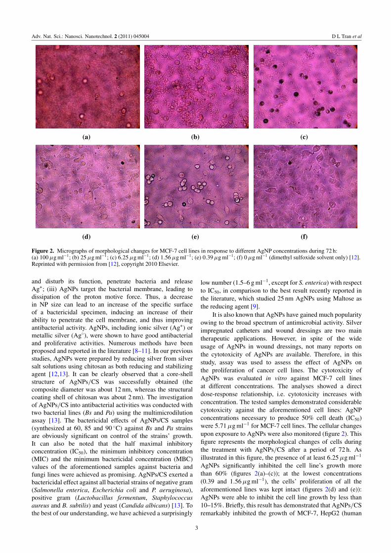
Figure 2. Micrographs of morphological changes for MCF-7 cell lines in response to different AgNP concentrations during 72 h:(a) 100 µg ml−1; (b) 25 µg ml−1; (c) 6.25 µg ml−1; (d) 1.56 µg ml−1; (e) 0.39 µg ml−1; (f) 0 µg ml−1 (dimethyl sulfoxide solvent only) [12].Reprinted with permission from [12], copyright 2010 Elsevier.
and disturb its function, penetrate bacteria and releaseAg+; (iii) AgNPs target the bacterial membrane, leading todissipation of the proton motive force. Thus, a decreasein NP size can lead to an increase of the specific surfaceof a bactericidal specimen, inducing an increase of theirability to penetrate the cell membrane, and thus improvingantibacterial activity. AgNPs, including ionic silver (Ag+) ormetallic silver (Ag◦), were shown to have good antibacterialand proliferative activities. Numerous methods have beenproposed and reported in the literature [8–11]. In our previousstudies, AgNPs were prepared by reducing silver from silversalt solutions using chitosan as both reducing and stabilizingagent [12,13]. It can be clearly observed that a core-shellstructure of AgNPs/CS was successfully obtained (thecomposite diameter was about 12 nm, whereas the structuralcoating shell of chitosan was about 2 nm). The investigationof AgNPs/CS into antibacterial activities was conducted withtwo bacterial lines (Bs and Pa) using the multimicrodilutionassay [13]. The bactericidal effects of AgNPs/CS samples(synthesized at 60, 85 and 90 ◦C) against Bs and Pa strainsare obviously significant on control of the strains’ growth.It can also be noted that the half maximal inhibitoryconcentration (IC50), the minimum inhibitory concentration(MIC) and the minimum bactericidal concentration (MBC)values of the aforementioned samples against bacteria andfungi lines were achieved as promising. AgNPs/CS exerted abactericidal effect against all bacterial strains of negative gram(Salmonella enterica, Escherichia coli and P. aeruginosa),positive gram (Lactobacillus fermentum, Staphylococcusaureus and B. subtilis) and yeast (Candida albicans) [13]. Tothe best of our understanding, we have achieved a surprisingly
low number (1.5–6 g ml−1, except for S. enterica) with respectto IC50, in comparison to the best result recently reported inthe literature, which studied 25 nm AgNPs using Maltose asthe reducing agent [9].
It is also known that AgNPs have gained much popularityowing to the broad spectrum of antimicrobial activity. Silverimpregnated catheters and wound dressings are two maintherapeutic applications. However, in spite of the wideusage of AgNPs in wound dressings, not many reports onthe cytotoxicity of AgNPs are available. Therefore, in thisstudy, assay was used to assess the effect of AgNPs onthe proliferation of cancer cell lines. The cytotoxicity ofAgNPs was evaluated in vitro against MCF-7 cell linesat different concentrations. The analyses showed a directdose-response relationship, i.e. cytotoxicity increases withconcentration. The tested samples demonstrated considerablecytotoxicity against the aforementioned cell lines: AgNPconcentrations necessary to produce 50% cell death (IC50)were 5.71 µg ml−1 for MCF-7 cell lines. The cellular changesupon exposure to AgNPs were also monitored (figure 2). Thisfigure represents the morphological changes of cells duringthe treatment with AgNPs/CS after a period of 72 h. Asillustrated in this figure, the presence of at least 6.25 µg ml−1
AgNPs significantly inhibited the cell line’s growth morethan 60% (figures 2(a)–(c)); at the lowest concentrations(0.39 and 1.56 µg ml−1), the cells’ proliferation of all theaforementioned lines was kept intact (figures 2(d) and (e)):AgNPs were able to inhibit the cell line growth by less than10–15%. Briefly, this result has demonstrated that AgNPs/CSremarkably inhibited the growth of MCF-7, HepG2 (human
3

Adv. Nat. Sci.: Nanosci. Nanotechnol. 2 (2011) 045004 D L Tran et al
4h 1h 2h 6h
Figure 3. LCSM image of Fe3O4-Cur taken into macrophage with various incubation times [14]. Reprinted with permission from [14],copyright 2010 Elsevier.
Figure 4. The solubility of Cur in aqueous solutions without (left)and with (right) Gossypol/CS nanocarrier.
hepatocellular carcinoma), Lu (human lung carcinoma), andKB (human epidermic carcinoma) cancer lines, although theirinhibitory effects differed from one to another [12].
3.2. Drug delivery of a CS-based system
The unique physicochemical and biological characteristics ofCS make it suitable for drug delivery systems. A strategyto design a multifunctional, nanosized magnetofluorescentwater-dispersible Fe3O4-curcumin conjugate (diameter lessthan 500 nm) and its multiple ability to label, target andtreat the tumor cells was developed. The conjugate possessesa magnetic Fe3O4 nanocore, CS as the outer shell andentrapped curcumin (Cur, an anti-oxidant, anti-inflammatoryand anti-tumor substance), serving the dual function ofnaturally autofluorescent dye as well as antitumor model drug,delivered to the cells with the help of macrophage whoserole is to phagocytise (engulf and then digest) cellular debrisand pathogens, either as stationary or as mobile cells, andto stimulate lymphocytes and other immune cells to respondto the pathogen. Primary peritoneal macrophage isolation isdescribed in detail elsewhere [14]. Hence, it can be used as apotential vehicle for transport of MNPs into the core of tumorcells.
Fe3O4-Cur conjugate could be visualized dually by afluorescence microscope, LSCM as well as magnetizationmeasurement (Physical Properties Measurement Systems,PPMS). LCSM images (figure 3), taken at 1, 2, 4and 6 h of incubation, showed that the number of
300 400 500 600-0.2
0.0
0.2
0.4
0.6
0.8
1.0
1.2
1.4
1.6
2 4 6 8 100.2
0.4
0.6
0.8
1.0
1.2
1.4
Y = 0.15X - 0.0312R2 = 0.9993
Abs
orba
nce
(a.u
)
Concentration (mg/ml)
Abs
orba
nce
(a.u
)
Wavelength (nm)
2mg/ml 4mg/ml 6mg/ml 8mg/ml 10mg/ml Cur/GCS
Figure 5. The relationship between absorbance and wavelength atdifferent concentrations of Cur, released from Gossypol/CSnanocarrier.
Fe3O4-Cur taken into macrophage cytoplasm increasedclearly with incubation time [14]. The green fluorescencewas observed clearly, started at about 0.5–1 h and reachingits maximal at 6 h, confirming that the Fe3O4-Cur particleswere efficiently loaded. Alternatively, PPMS magnetizationcurves of macrophage samples at 1, 2, 4 and 6 h ofincubation (figure not shown) also confirmed that themagnetization of macrophage increases (magnetization ofthe remaining supernatants decreases). This magnetizationresult is, therefore, in good accordance with that achievedby the above-demonstrated in-situ observation by LSCMfluorescence [14].
Further, the excellent loading and releasing capacity ofCur from Gossypol/CS carrier was clearly demonstrated infigures 4 and 5, respectively. The successful development ofan effective drug nanocarrier for other therapeutic treatmentscan be logically expected.
3.3. Magnetic inductive heating capacity of the ferrofluids
Hyperthermia is a promising approach to cancer therapy.It is well-known that MNPs exhibit a so-called magneticheating (MH) effect—a very specific property that hasattracted much research interest recently because of its
4

Adv. Nat. Sci.: Nanosci. Nanotechnol. 2 (2011) 045004 D L Tran et al
Figure 6. Schematic representation of HIV electrochemical detection on CS/Fe3O4 SPE using MB as an intercalator [20]. Reprinted withthe permission from [20], copyright 2011 Elsevier.
-0.6 -0.5 -0.4 -0.3 -0.2 -0.1 0.0 0.1 0.2
0.2 μ
Α
d
c
b
a
f
e
a 400 pMb 300 pMc 250 pMd 200 pMe 150 pMf 100 pM
Cur
rent
I/μΑ
Potential E/V (vs. SCE)
0.0 2.0k 4.0k 6.0k 8.0k 10.0k
0
1k
2k
3k
4k
19 Hz3.95 Hz
1.38 Hz1.06 Hz-Z
''(Ω
)
Z'(Ω)
pGEM c-HIV: 50 pM c-HIV: 100 pM c-HIV: 200 pM c-HIV: 300 pM
Figure 7. Quantitative detection by EIS-Nyquist plots and by SWV [20]. Reprinted with the permission from [20], copyright 2011 Elsevier.
potential applications in cancer hyperthermia, drug release
and also the desorption of toxic substances [15,16]. However,
to be applicable in such applications, Fe3O4 has to be
encapsulated to avoid agglomeration or to make monodisperse
in suspension. Therefore it is essential to modify the surface
of these particles to increase the stability by surfactants,
among which biocompatible polymers like CS and its
derivatives, e.g. O-carboxymethylchitosan (OCMCS), are the
most potential candidates owing to their biocompatibility and
biodegradability [13].
3.4. Electrochemical detection of ODN sequences onCS/Fe3O4 SPEs
A selective and sensitive response to mismatch is alwaysamong the most difficult technical challenges of anyelectrochemical biosensor [17–19]. The whole procedure ofHIV detection is schematically presented in figure 6 [20].The effective discrimination against one point mutationof 1m-HIV was systematically carried out. As expected,there was no significant current difference observed for theGEM91-modified SPEs when they were hybridized witheither non-complementary or single base mismatch 1m-HIV
5

Adv. Nat. Sci.: Nanosci. Nanotechnol. 2 (2011) 045004 D L Tran et al
sequences. Hybridization-induced signal decrease occurs onthe electrode platform where GEM probes were covalentlyfixed on the surface and were subsequently hybridized withtruly complementary c-HIV sequences. Meanwhile, there wasno signal change during hybridization with 1m-HIV, whosestrand was differentiated by one mismatched base (adenine(A) instead of guanine (G)), compared to that of the c-HIVstrand.
The square wave voltammetry (SWV) and electro-chemical impedance spectroscopy-Nyquist plot (EIS-Nyquistplot) response to different target DNA concentrations isdisplayed in figure 7. The decrease of the magnitude ofthe voltammetric signal thus reflected the extent of thehybrid formation. The signal for the hybridization withc-HIV is linear from 50 pM up to approximately 300 pM ofc-HIV target, and levels-off beyond this concentration limit,indicating that the full surface coverage of the probe or GEM91 was involved in hybridization at that target concentration(probe site saturation was achieved). The phenomenon thatthe current of methylene blue (MB) decreases with increasingconcentration of HIV is explained by the higher affinity ofMB for a single strand rather than double strands of DNA.These results showed reasonable agreement with the commonconcept for MB intercalator [20].
4. Conclusion
Although not fully described/reviewed, this paper presentssome useful chemical routes to functionalize hybridchitosan-based nanomaterials and demonstrates their use insome potential biomedical applications (antibacterial andantiproliferative activities, drug delivery, biodetection). Theability of these materials was clearly observed by manyphysico-chemical methods. Further developments in materialdesign/synthesis for enhanced therapeutic and diagnosticeffects will be continued and reported in upcoming papers.
Acknowledgments
This work was performed under the financial support ofthe Ministry of Science and Technology project, code
08/2011/HD-NDT. The authors would like to expresstheir gratitude to Academician Nguyen Van Hieu forhis encouragement and to the second ASEAN-PakistanConference on Materials Science (APCOM2) for support.
References
[1] Jayakumar R, Menon D, Manzoor K, Nair S V and Tamura H2010 Carbohydr. Polym. 82 227
[2] Madhumathi K, Binulal N S, Nagahama H, Tamura H,Shalumon K T, Selvamurugan N, Nair S V and Jayakumar R2009 Int. J. Biol. Macromol. 44 1
[3] Jayakumar R, Prabaharan M, Reis R L and Mano J F 2005Carbohydr. Polym. 62 142
[4] Jayakumar R, New N, Tokura S and Tamur H 2007 Int. J. Biol.Macromol. 40 175
[5] Borchard G 2001 Adv. Drug Deliv. Rev. 52 145[6] Jayakumar R, Chennazhi K P, Muzzarelli R A A, Tamura H,
Nair S V and Selvamurugan N 2010 Carbohydr. Polym.79 1
[7] Bruyere O, Pavelka K, Rovati L C, Gatterova J, Giacovelli Gand Olejarova M 2008 Osteoarthr. Cartilage 16 254
[8] Wei D, Sun W, Qian W, Ye Y Z and Ma X 2009 Carbohyd.Res. 344 2375
[9] Sharma V K, Yngard R A and Lin Y 2009 Adv. ColloidInterface Sci. 145 83
[10] Ghosh S, Kaushik R, Nagalakshmi K, Hoti S L, Menezes G A,Harish B N and Vasan H N 2010 Carbohydr. Res. 345 2220
[11] Seo Y I, Hong K H, Kim D G and Kima Y D 2010 ColloidSurf. B 81 369
[12] Tran V H, Tran D L, Ba T C, Vu D H, Nguyen T N, Pham G Dand Nguyen X P 2010 Colloid Surf. A 360 32
[13] Tran D L, Tran V H, Mai T T, Ha P T, Nguyen H B, Thai H,Vu D H, Pham G D, Nguyen X P and Park J K 2011J. Chitin Chitosan 16 7
[14] Tran D L et al 2010 Colloid Surf. A 371 104[15] Xu L, Kim M J, Kim K D, Choa Y H and Kim H T 2008
Colloid Surf. A 350 8[16] Hiergeist R, Andra W, Buske N, Hergt R, Richter U and Kaiser
W 1999 J. Magn. Magn. Mater. 201 420[17] Hergt R, Hiergeist R, Zeisberger M, Schüler D, Heyen U,
Hilger I and Kaiser W 2005 J. Magn. Magn. Mater. 293 80[18] Katz E and Willner I 2003 Electroanalysis 15 913[19] Tran D L, Nguyen T D, Nguyen H B, Do P Q and Nguyen L H
2011 Talanta 85 1560[20] Tran D L, Nguyen H B, Nguyen V H, Tran V H, Nguyen L H
and Nguyen X P 2011 Mater. Sci. Eng. 31 477
6
Copyright © 2022 FDOKUMEN