Engineering Nanomaterials to Overcome Barriers in Cancer ...
149
Engineering Nanomaterials to Overcome Barriers in Cancer Therapy Christine Erline Wang A dissertation submitted in partial fulfillment of the requirements for the degree of Doctor of Philosophy University of Washington 2016 Reading Committee: Suzie H. Pun, Chair Anthony J. Convertine André Lieber Program Authorized to Offer Degree: Bioengineering
-
Upload
khangminh22 -
Category
Documents
-
view
0 -
download
0
Transcript of Engineering Nanomaterials to Overcome Barriers in Cancer ...

Engineering Nanomaterials to Overcome
Barriers in Cancer Therapy
Christine Erline Wang
A dissertation
submitted in partial fulfillment of the
requirements for the degree of
Doctor of Philosophy
University of Washington
2016
Reading Committee:
Suzie H. Pun, Chair
Anthony J. Convertine
André Lieber
Program Authorized to Offer Degree:
Bioengineering


©Copyright 2016
Christine Erline Wang


University of Washington
Abstract
Engineering Nanomaterials to Overcome Barriers in Cancer Therapy
Christine Erline Wang
Chair of the Supervisory Committee:
Professor Suzie H. Pun
Bioengineering
Cytotoxic chemotherapy is used for the frontline treatment of most types of cancer but is
associated with significant toxicity due to the lack of cell specificity of these drugs. Newer
treatment strategies, such as polymeric drug delivery vehicles that preferentially
accumulate in tumors via the EPR effect and oncolytic adenoviruses that replicate
conditionally in tumor cells, can reduce the adverse side effects associated with systemic
anti-cancer treatments. However, there remain numerous barriers to the successful clinical
translation of these therapeutics. Part I focuses on the diffusional barriers to drug delivery
to solid tumors. In Chapter 1, we investigate tight junction-opening proteins as a means to
enhance nanoparticle penetration into tumors. Part II describes the development of
polymer nanostructures for anti-cancer drug delivery. Chapter 2 summarizes the major
design parameters for drug delivery to tumors and introduces controlled living
polymerization as a synthetic tool. Chapters 3 and 4 describe the synthesis of polymeric
drug carriers with a novel sunflower-like architecture. Part III focuses on methods to
improve the safety of adenoviruses for cancer gene therapy. Chapter 5 provides an overview
of adenovirus pharmacology and current modification strategies, while Chapter 6 describes
a new approach to developing materials that can shield adenoviruses against immune
recognition. Finally, Chapter 7 summarizes the major findings of this work and concludes
with recommendations for future directions.


i
TABLE OF CONTENTS
List of Figures ............................................................................................................................ vi
List of Tables ............................................................................................................................. ix
Part I. Overcoming physical barriers to drug delivery in solid tumors
Chapter 1. Investigating the size limitations of JO protein-mediated nanoparticle
delivery ................................................................................................................. 3
1.1 Introduction ......................................................................................................... 3
1.2 Materials and methods ........................................................................................ 4
1.2.1 AuNP surface modification ................................................................... 4
1.2.2 Particle characterization ...................................................................... 5
1.2.3 Animals .................................................................................................. 5
1.2.4 Cell culture ............................................................................................ 5
1.2.5 Biodistribution of AuNPs ...................................................................... 6
1.2.6 Light and fluorescence microscopy ...................................................... 6
1.2.7 Image analysis ...................................................................................... 7
1.3 Results and discussion ........................................................................................ 7
1.3.1 AuNP sizing and stability ..................................................................... 7
1.3.2 Biodistribution of AuNPs with JO-4 pretreatment ........................... 10
1.3.3 Intratumoral penetration of AuNPs .................................................. 15
1.4 Conclusions and future work ............................................................................ 16
1.5 Acknowledgements ............................................................................................ 17
References ...................................................................................................................... 17
Part II. Polymer nanostructures for tumor-targeted drug delivery
Chapter 2. Polymer nanostructures for tumor-targeted drug delivery: design parameters
and synthetic approaches .................................................................................. 23

ii
2.1 Introduction ....................................................................................................... 23
2.2 Designing polymer carriers with desired pharmacokinetics and
biodistribution for anticancer drug delivery .................................................... 24
EPR and tumor penetration ............................................................... 24 2.2.1
Active targeting ................................................................................... 26 2.2.2
2.3 Considerations in integrating drugs with polymeric carriers ......................... 27
Drug loading ........................................................................................ 27 2.3.1
Drug release ........................................................................................ 28 2.3.2
2.4 Controlled polymerization techniques .............................................................. 29
Atom transfer radical polymerization (ATRP) .................................. 31 2.4.1
Reversible addition-fragmentation chain-transfer (RAFT) 2.4.2
polymerization ..................................................................................... 31
Metathesis polymerization ................................................................. 32 2.4.3
2.5 Polymer architectures for drug delivery ........................................................... 32
Star polymers ...................................................................................... 33 2.5.1
Polymer brushes .................................................................................. 34 2.5.2
Hyperbranched polymers .................................................................... 34 2.5.3
Macrocyclic polymers .......................................................................... 35 2.5.4
2.6 Conclusions and future perspectives ................................................................ 36
References ...................................................................................................................... 36
Chapter 3. ATRP synthesis of sunflower polymers using cyclic multimacroinitiators .... 42
3.1 Introduction ....................................................................................................... 42
3.2 Materials and methods ...................................................................................... 44
Materials ............................................................................................. 44 3.2.1
Polymer synthesis ............................................................................... 44 3.2.2
Polymer characterization .................................................................... 49 3.2.3
Cell culture .......................................................................................... 49 3.2.4
In vitro uptake and competition studies ............................................ 50 3.2.5
In vitro cytotoxicity studies ................................................................ 50 3.2.6
3.3 Results and discussion ...................................................................................... 51
Synthesis and characterization of sunflower polymers .................... 51 3.3.1

iii
Synthesis of folate-targeted sunflower polymers and evaluation of 3.3.2
uptake in FR+ cells ............................................................................. 55
Cytotoxicity of sunflower polymers .................................................... 58 3.3.3
3.4 Conclusions ........................................................................................................ 58
3.5 Acknowledgements ............................................................................................ 58
References ...................................................................................................................... 58
Supporting Information ................................................................................................ 61
Chapter 4. Sunflower polymers for folate-mediated drug delivery ................................... 63
4.1 Introduction ....................................................................................................... 63
4.2 Materials and methods ...................................................................................... 66
4.2.1 Materials ............................................................................................. 66
4.2.2 Polymer synthesis and Dox conjugation ............................................ 66
4.2.3 Polymer characterization .................................................................... 67
4.2.4 Quantification of Dox release ............................................................. 67
4.2.5 Cell culture .......................................................................................... 68
4.2.6 Serum stability studies ....................................................................... 68
4.2.7 In vitro uptake and competition studies ............................................ 68
4.2.8 Confocal imaging ................................................................................. 69
4.2.9 In vitro cytotoxicity studies ................................................................ 69
4.2.10 Animals ................................................................................................ 70
4.2.11 Tumor inhibition studies .................................................................... 70
4.3 Results and discussion ...................................................................................... 72
4.3.1 Synthesis and characterization of sunflower polymers with different
core sizes .............................................................................................. 72
4.3.2 Uptake of large core sunflower polymers in FR+ cells ...................... 74
4.3.3 Dox release kinetics ............................................................................ 75
4.3.4 Confocal imaging of intracellular drug delivery ................................ 76
4.3.5 Cytotoxicity of sunflower polymers .................................................... 77
4.3.6 Anti-tumor efficacy in vivo ................................................................. 78
4.4 Conclusions ........................................................................................................ 81
4.5 Acknowledgements ............................................................................................ 82
References ...................................................................................................................... 82

iv
Supporting Information ................................................................................................ 85
Part III. Modifications enabling systemic administration of adenovirus
Chapter 5. Adenoviral vectors for cancer gene therapy: pharmacology and strategies for
modification ........................................................................................................ 89
5.1 Introduction ....................................................................................................... 89
5.2 Adenoviral vectors ............................................................................................. 90
5.3 Adenovirus pharmacology ................................................................................. 91
5.3.1 CAR paradigm of adenovirus infection .............................................. 91
5.3.2 In vivo interactions ............................................................................. 92
5.4 Strategies for adenovirus modification ............................................................. 93
5.4.1 Genetic modification ........................................................................... 93
5.4.2 Polymer conjugation ........................................................................... 94
5.4.3 Non-covalent coating ........................................................................... 94
5.5 Future perspectives ........................................................................................... 95
References ...................................................................................................................... 95
Chapter 6. Identification of adenovirus-binding peptides for use in self-assembling
polymer shields .................................................................................................. 99
6.1 Introduction ....................................................................................................... 99
6.2 Materials and methods .................................................................................... 100
6.2.1 Phage panning ................................................................................... 100
6.2.2 Phage binding studies ....................................................................... 101
6.2.3 Peptide synthesis .............................................................................. 102
6.2.4 Peptide binding studies .................................................................... 102
6.2.5 Polymer synthesis and characterization .......................................... 103
6.2.6 Preparation of peptide-grafted polymers ......................................... 104
6.2.7 Cell culture ........................................................................................ 104
6.2.8 Cell transduction ............................................................................... 104
6.3 Results and discussion .................................................................................... 105
6.3.1 Identification of Ad-binding phage candidates ................................ 105
6.3.2 Phage binding studies ....................................................................... 107

v
6.3.3 Peptide binding studies .................................................................... 108
6.3.4 Transduction of HeLa cells with poly(2-04)-“coated” viruses ......... 108
6.4 Conclusions and future studies ....................................................................... 110
6.5 Acknowledgements .......................................................................................... 111
References .................................................................................................................... 111
Part IV. Future perspectives
Chapter 7. Summary of major findings and recommendations for future work ............. 115
7.1 Summary of major findings ............................................................................. 115
7.1.1 Junction opener proteins for enhancing nanoparticle delivery to solid
tumors ................................................................................................ 115
7.1.2 Sunflower polymers for tumor-targeted drug delivery ................... 115
7.1.3 Self-assembling materials for cloaking adenoviral vectors ............ 116
7.2 Recommendations for future work .................................................................. 116
7.2.1 JO-conjugated sunflower polymers for tumor-targeted drug delivery
............................................................................................................ 116
7.2.2 Sunflower polymers with increased drug loading ........................... 119
7.2.3 Polymer-modified adenoviruses: a “grafting-from” approach ......... 120
References .................................................................................................................... 123
Appendix. MATLAB code for AuNP penetration analysis .............................................. 126

vi
LIST OF FIGURES
Figure 1.1 Particle sizing of unmodified and PEGylated AuNPs as determined by DLS. 8
Figure 1.2 Stability of unmodified and PEGylated 100 nm AuNPs incubated in PBS or
PBS containing 10% serum as monitored by DLS. ............................................ 9
Figure 1.3 Stability of unmodified and PEGylated 5 nm AuNPs incubated in PBS or PBS
containing 10% serum as monitored by DLS and red shift in absorbance. .... 10
Figure 1.4 Tumor and liver accumulation of two different sizes of AuNPs 6 h post-NP
injection in control or JO-4 pretreated mice bearing 200-300 mm3 or 500-600
mm3 tumors. ....................................................................................................... 11
Figure 1.5 Biodistribution of two different sizes of AuNPs 6 h post-NP injection in
control or JO-4 pretreated mice bearing 500-600 mm3 tumors. ...................... 13
Figure 1.6 Representative images of a tumor section and image analysis strategy. ....... 14
Figure 1.7 Normalized histograms of AuNP penetration distances. ................................ 16
Figure 2.1 Design parameters for drug delivery to solid tumors. ..................................... 25
Figure 2.2 Schematic representation of controlled polymerization methods. .................. 30
Figure 2.3 Schematic representations of advanced macromolecular architectures being
investigated for drug delivery. .......................................................................... 33
Figure 3.1 Schematic illustration of sunflower polymer containing targeting ligands and
cargo. .................................................................................................................. 44
Figure 3.2 Synthesis of folate-sunflower polymer-fluorescein. ......................................... 51
Figure 3.3 ATRP kinetics for the synthesis of P(HEMA-st-EGMA) copolymer. ............... 52
Figure 3.4 GPC elution traces and FT-IR spectra of linear and cyclic P(HEMA41-st-
EGMA12). ............................................................................................................ 53
Figure 3.5 ATRP kinetics of sunflower polymer prepared from cyclic
multimacroinitiator. .......................................................................................... 54
Figure 3.6 GPC elution traces of sunflower polymer and comb-like polymer prepared
using cyclic and linear multimacroinitiators. .................................................. 55

vii
Figure 3.7 Summary of Z-average sizes of sunflower polymers with petals polymerized
for different lengths of time. ............................................................................. 55
Figure 3.8 Uptake of FA-SF-fluor, untargeted SF-fluor, and FA-comb-fluor polymers in
FR+ KB cells and FR- A549 cells in the absence and presence of competing
free folate. ........................................................................................................... 57
Figure 4.1 Schematic illustrations of sunflower polymer containing FA targeting ligands
and Dox drug and proposed delivery mechanism. ........................................... 65
Figure 4.2 Synthesis of folate-sunflower polymer-Dox. ..................................................... 71
Figure 4.3 GPC elution traces and FT-IR spectra of linear and cyclic P(HEMA80-st-
EGMA24). ............................................................................................................ 73
Figure 4.4 ATRP kinetics of sunflower polymers prepared using cyclic
multimacroinitiator of DP 100. ......................................................................... 73
Figure 4.5 Z-average diameters of sunflower polymers with cyclic core of DP 100 and
petals polymerized for different lengths of time. ............................................. 74
Figure 4.6 Uptake of FA-SF-fluor with DP 100 core in FR+ KB cells and FR- A549 cells
in the absence and presence of competing free folate. ..................................... 75
Figure 4.7 Release kinetics of Dox from FA-SF-Dox polymer at pH 7.4, 6.8, and 5.5. .... 76
Figure 4.8 Confocal imaging of FA-SF-Dox and Dox uptake in KB cells. ........................ 77
Figure 4.9 In vivo tumor growth in tumor-bearing mice dosed with free Dox or FA-SF-
Dox at 6 mg Dox/kg mouse on day 0, or with multiple doses each at 4 mg/kg
on days 0, 3, and 6. ............................................................................................ 79
Figure 4.10 Survival curve for tumor-bearing mice. ............................................................ 80
Figure 4.11 Normalized body weights of tumor-bearing mice receiving a single or multiple
chemotherapy treatments. ................................................................................ 81
Figure 5.1 False-colored electron micrograph and schematic representation of the
icosahedral structure of adenovirus. ................................................................ 91
Figure 6.1 Phage panning strategy using competitive displacement with FX protein to
elute adenovirus-binding phage. ..................................................................... 101
Figure 6.2 Titers of eluted phage over 4 rounds of panning. ........................................... 106

viii
Figure 6.3 Selected phage clones demonstrate specificity for Ad5 by an elution and
titering assay. .................................................................................................. 107
Figure 6.4 BLI sensorgram of Ad5 binding to immobilized peptides. ............................. 108
Figure 6.5 HeLa transduction efficiency of uncoated Ad5-GFP and Ad5-GFP complexed
with poly(2-04) or control polymers. ............................................................... 109
Figure 7.1 Anti-JO1 Western blot of GB3_JO1 and GB7_JO1 proteins before and after
click conjugation to sunflower polymers. ........................................................ 118
Figure 7.2 Hydrolysis of tert-butyl methacrylate (tBMA) to methacrylic acid (MAA). .. 119
Figure 7.3 RAFT kinetics of aqueous, low temperature polymerization of P(OEGMA) as
determined by NMR. ....................................................................................... 123

ix
LIST OF TABLES
Table 1.1 Statistics for AuNP penetration analysis. ........................................................ 15
Table 4.1 IC50 values and 95% confidence intervals for Dox and polymer formulations in
KB (FR+) and A549 (FR-) cell lines. ................................................................. 78
Table 6.1 Possible consensus peptide sequences and their frequencies after the third
and fourth rounds of panning. ........................................................................ 106

x
ACKNOWLEDGEMENTS
This thesis would not have been possible without the contributions and support of many
people over the years. Words cannot express how grateful I am to my family, friends,
mentors, coworkers, and collaborators who have helped me during this journey.
First and foremost, I would like to thank my advisor, Suzie Pun, for the opportunity to
work in her lab and for her guidance and scientific insight over the seven years I have
known her. She has always served as a positive role model, challenging me to do more than
I knew I could and providing encouragement in the face of numerous setbacks. I feel
incredibly fortunate to have found a mentor who cares so deeply about my personal well
being in addition to my professional development.
I would also like to thank the members of my supervisory committee, André Lieber,
Tony Convertine, Pat Stayton, and Christine Luscombe, who have given generously of their
intellect, time, and lab resources. Your scientific input has been invaluable in the
completion of this work.
I would like to acknowledge many of my labmates, past and present, for their scientific
and non-scientific contributions:
• Hua Wei, who taught me about the practical aspects of polymer chemistry. When I
began working with Hua on sunflower polymers, I never would have guessed what a
big part they would play in my graduate work, and this thesis would not have been
possible without him.
• Kat Wang, who mentored me as a rotation student, for always being supportive and
down-to-earth.
• Julie Shi and honorary lab member Sergio Haro, who have been my best friends and
surrogate big siblings for the past five years. I will always remember the food
adventures, bike rides around Greenlake, late night talks at the kitchen table about
lab and life, and eye-opening lessons in parenthood (which will eventually come in
handy).
• Dave Chu, my officemate and first friend in grad school, for always being so
generous with his time and expertise in the lab, and for the countless pep talks and
chicken and wine Tuesdays outside of the lab.

xi
• Leslie Chan and Maryelise Cieslewicz, for being great friends, and for continually
impressing me with their hard work and baking skills.
• Kevin Tan, my cohort-mate, for commiserating with me through grad school, and for
providing fashion advice and endless entertainment in the form of random Facebook
videos, pop culture trivia, and deep interrogations of fellow labmates.
• Chayanon Ngambenjawong, whose work ethic and creativity in the lab are inspiring,
and who provided comic relief at the most unexpected moments.
• Bob Lamm, for being an extremely caring friend and lab citizen, and for encouraging
me to be social while also recognizing when I wanted to introvert.
• Gary Liu, for his positive attitude and for putting up with my incessant teasing.
• Joan Go Schellinger, Drew Sellers, and Heather Gustafson, for their mentorship and
guidance in the lab.
I would also like to acknowledge my friends Austin Day, Tom Long, and Joe Phan, for
providing an outlet from lab with karaoke nights, board games, and hiking.
I would especially like to thank my family – my older sister, Melissa, for being the
“guinea pig” child and paving the way for me in everything I’ve done, including the Ph.D.
My parents, James and Huei-Ching, have always emphasized the importance of education
and hard work. This work is truly the culmination of the love and support they have always
shown me.
Finally, I would like to thank my husband, Jonathan Lin, for his unwavering love,
encouragement, and proofreading. I am continually amazed by his intense curiosity and
commitment to doing good science, and these have made me a better scientist in turn. I
could not ask for a better collaborator in life.

xii

xiii
DEDICATION
To my husband, Jonathan,
my parents, James and Huei-Ching,
and my sister, Melissa.


1
Part I.
Overcoming Physical Barriers to
Drug Delivery in Solid Tumors

2

3
Chapter 1.
INVESTIGATING THE SIZE LIMITATIONS OF JO PROTEIN-
MEDIATED NANOPARTICLE DELIVERY
Christine E. Wang, Roma Yumul, Jonathan Lin, André Lieber, and Suzie H. Pun
Abstract
JO is a recombinant protein that transiently opens intercellular junctions in epithelial
tumors by cleaving the junction protein DSG2. Co-administration of JO has been shown to
significantly increase the efficacy of various monoclonal antibodies and chemotherapy drugs
in murine tumor models by allowing for increased intratumoral penetration of the drugs. To
investigate the size-dependent effect of JO on nanocarriers, we used PEGylated gold
nanoparticles (AuNPs) of two different sizes as model drugs and investigated their
biodistribution following JO protein treatment. JO was found to significantly increase
tumor accumulation of AuNPs in a manner dependent on particle size and tumor volume.
Preliminary analysis of intratumoral nanoparticle distribution also indicates that AuNPs
can be observed at increased distances from tumor blood vessels following JO treatment.
1.1 Introduction
Nanoparticle-based drug carriers are attractive because of their ability to exploit the
enhanced permeability and retention (EPR) effect for selective accumulation in tumors.1
However, the ability of these carriers to penetrate into solid tumors is another important
consideration, especially because many small molecule anticancer drugs are diffusion-
limited, and the incomplete distribution of drugs in the tumor tissue can lead to
chemotherapy resistance.2,3 In particular, epithelial tumors are characterized by the
presence of intercellular junctions which restrict the penetration of molecules.4,5 One such
epithelial junction protein is desmoglein-2 (DSG2), which has been found to be upregulated
in a number of malignant cell types including gastric cancer,6 ovarian cancer,7 and breast
cancer.8

4
Recently, DSG2 was identified as the receptor used by several human adenoviruses
(Ad), including Ad serotype 3, for cellular infection.7 In subsequent studies, a recombinant
protein derived from the Ad3 fiber knob was produced.9 This protein, named junction
opener-1 (JO-1), was found to trigger the transient opening of intercellular junctions
through binding and cleavage of the DSG2 dimers between epithelial cells.9,10 This effect
was observed in mice with epithelial tumors within one hour of intravenous injection of JO-
1.10 Co-administration of JO-1 has been shown to facilitate intratumoral penetration and
therapeutic efficacy of monoclonal antibodies (mAbs) such as the anti-Her2/neu mAb
trastuzumab (Herceptin) and the EGFR inhibitor cetuximab (Erbitux).10 Furthermore, JO-1
was tested in combination with several chemotherapeutic drugs including paclitaxel
(Taxol), irinotecan (Camptosar), nanoparticle albumin-bound paclitaxel (Abraxane), and
liposomal doxorubicin (Doxil). JO-1 co-therapy enhanced the efficacy of these drugs and
overcame drug resistance in several models, while reducing the drug doses necessary for
therapeutic effect.8 JO-1 and variants of this protein (such as the affinity-enhanced version,
JO-411) are therefore interesting for clinical application.
To develop a better understanding of the size limitations of JO protein-mediated
disruption of tight junctions, we sought to investigate the effect of JO pre-treatment on the
in vivo biodistribution of gold nanoparticles of different sizes. Gold nanoparticles were
selected as a surrogate for nano-sized drug carriers (e.g., liposomes, micelles, polymer-drug
conjugates, etc.) because they can be synthesized with defined sizes and over a wide size
range, surface-modified through reactions with thiol groups, quantified by inductively
coupled plasma mass spectrometry (ICP-MS) with excellent sensitivity, and visualized by
light and electron microscopy. Herein, we synthesize polyethylene glycol (PEG)-modified
gold nanoparticles and quantify their biodistribution in JO-treated and untreated mice by
ICP-MS. We also demonstrate a technique to investigate the intratumoral distribution of
nanoparticles using microscopy and quantitative image analysis.
1.2 Materials and methods
1.2.1 AuNP surface modification
Gold nanoparticles (AuNPs) with diameters of 5 and 100 nm were purchased from Ted
Pella (Redding, CA) and Nanopartz (Loveland, CO), respectively (actual diameters: 5.5 and

5
103 nm as determined by the manufacturers). AuNPs were surface-modified by reaction
with PEG5000-thiol (Laysan Bio, Arab, AL); PEG-SH was added in excess (4 PEG molecules
per nm2 gold surface area,12 assuming spherical particles) and allowed to react for 30 min at
room temperature prior to characterization or use.
1.2.2 Particle characterization
The effective diameters of AuNPs were measured by dynamic light scattering (DLS) with a
ZetaPlus analyzer (Brookhaven Instruments, Holtsville, NY) at a detection angle of 90°.
Unmodified or PEGylated AuNPs with initial diameters of 5 or 100 nm were first measured
in nanopure water. Particle size measurements were acquired for 6 independent samples,
using 5 1-minute runs for each sample.
To confirm particle stability in the presence of physiological salt concentrations,
unmodified or PEGylated AuNPs were diluted with an equal volume of 2× PBS (or
nanopure water as a negative control) immediately before sizing. Size was measured again
after 2 h and 24 h; measurements were completed using 3 independent samples. Similarly,
particle stability in serum was assessed by diluting AuNPs with an equal volume of 2× PBS
containing 20% fetal bovine serum (FBS), for a final serum concentration of 10%. Serum
stability was monitored by DLS or by measuring absorbance spectra for the samples from
400-800 nm using a Tecan Safire2 plate reader (Ma ̈nnedorf, Switzerland).
1.2.3 Animals
Male Scid-beige (CB17) mice (8-10 weeks) were obtained from The Jackson Laboratory
(strain name: NOD.CB17-Prkdcscid298/J). All experimental procedures were performed in
accordance with protocols approved by the University of Washington Institutional Animal
Care and Use Committee.
1.2.4 Cell culture
A549 cells (ATCC CCL-185) were maintained in F-12K medium (Corning cellgro)
supplemented with 10% fetal bovine serum (FBS, HyClone) and 1% penicillin/streptomycin

6
(HyClone). Cells were cultured as a monolayer in a 37°C, 5% CO2 environment. Medium
was replaced every 2-3 days. Cells were passaged at ~70-80% confluence by incubation with
Trypsin-EDTA, followed by resuspension in complete growth medium.
1.2.5 Biodistribution of AuNPs
To develop xenograft tumors, mice were inoculated subcutaneously in the right flank with
5×10⁶ A549 cells in 100 µL of F-12K medium without serum.
After tumor inoculation, mice were randomly distributed into groups of 5-6 mice each.
Biodistribution studies were initiated when the tumors reached the specified volumes (200-
300 mm3 or 500-600 mm3). Mice receiving JO-4 pretreatment were first injected with 2 mg
JO-4 protein/kg mouse in PBS via tail vein injection. One hour later, mice were injected
with 35 or 120 nm PEGylated AuNPs in PBS at a dose of 100 µg gold/kg mouse via tail vein
injection. After 6 hours, mice were anesthesized by intraperitoneal injection with 2.5%
Avertin solution (300 µL/20 g mouse). Mice were then perfused with PBS, and tumors and
organs were harvested. Gold content in the tumor and liver was analyzed for all mice. Gold
content in the brain, colon, heart, intestine, kidney, lung, and spleen was analyzed for a
subset of 3 mice per group, selecting for mice with tumor weights closest to the average
overall tumor weight (~240 mg).
Gold content in tissue samples was measured by ICP-MS at the Environmental Health
Laboratory & Trace Organics Analysis Center at the University of Washington. The gold
content of each sample was normalized to sample mass. Statistical significance was
assessed using a Student’s two-tailed t-test.
1.2.6 Light and fluorescence microscopy
For imaging studies, JO-4 and AuNP injections were completed as described above, with
the tumors harvested following perfusion. Tumors were embedded in optimal cutting
temperature (OCT) compound (Tissue-Tek, Sakura Finetek USA, Torrance, CA) in
cryomolds, flash frozen, and cryosectioned into 8 µm-thick sections.
Tumor sections were post-fixed in 4% paraformaldehyde (PFA) in PBS for 15 min at
room temperature and stained for blood vessels with rat anti-mouse CD31 antibody (clone

7
MEC 13.3, BD Pharmingen) and Alexa Fluor 488 donkey anti-rat IgG secondary antibody.
After immunofluorescence staining, sections were stained for AuNPs by incubating with
silver enhancement solution (Ted Pella) for 20 min at room temperature. Finally, sections
were washed, coverslipped using Fluoromount-G (eBioscience), and imaged on a Nikon
E800 upright microscope with a 60x objective.
1.2.7 Image analysis
A total of 9 image pairs (brightfield for AuNPs, fluorescence for CD31) were obtained of
each tumor section. Images were thresholded using Fiji image processing software.
MATLAB was then used to overlay the thresholded brightfield and fluorescence images,
perform particle detection, and calculate the penetration distance for each AuNP, defined
as the distance from the particle to the nearest CD31-stained pixel. Distance data were
aggregated from all images of a tumor section and binned in MATLAB. Sample MATLAB
code can be found in the Appendix of this dissertation.
1.3 Results and discussion
1.3.1 AuNP sizing and stability
AuNPs were surface modified by reaction with PEG-thiol; PEGylation of nanoparticles is
commonly employed to reduce aggregation and protein adsorption, thereby increasing
circulation half-life in vivo. Particle sizing by DLS was performed for AuNPs of 2 different
diameters before and after PEGylation (Figure 1.1). The diameters of the unmodified
particles were generally consistent with those reported by the manufacturers
(experimental: 10.8 and 103.9 nm; reported: 5.5 and 103 nm). The discrepancy in the
smaller particles likely results from inaccuracies in DLS sizing given the relatively small
size and low count rate of these particles (whereas the manufacturer’s specifications are
determined using a combination of DLS, TEM, and UV-Vis spectroscopy). The final
diameters of the PEGylated AuNPs were measured to be 33.2 ± 1.4 nm and 121.1 ± 1.2 nm,
demonstrating a moderate size increase after PEG modification consistent with previous
reports.13,14 These particle sizes were selected based on the sizes of other nanocarriers that

8
have been clinically approved or are in late-stage clinical trials, such as Genexol-PM (~24
nm)15 and similar polymeric micelles (20-40 nm)16 at the lower size range and Doxil (80-100
nm)17 and BIND-014 (~100 nm)18 at the higher range.
Figure 1.1 Particle sizing of unmodified and PEGylated AuNPs as determined by DLS. Data are reported as the mean ± S.D., n = 6.
Nanoparticles were then evaluated for stability in salt and serum. In the presence of
physiological salt concentrations, unmodified AuNPs were shown to aggregate rapidly
(Figure 1.2 and Figure 1.3a), whereas PEGylated AuNPs remained stable in PBS even after
24 h. Serum stability was also assessed by suspending particles in PBS containing 10%
FBS. The addition of serum conferred increased stability on unmodified AuNPs; particle
sizing indicated that unmodified 100 nm AuNPs did not form large aggregates over 24 h,
although the slight increase in effective diameter suggests that some aggregation behavior
or protein adsorption to the nanoparticles may be occurring (Figure 1.2). Meanwhile, no
change in particle size was observed for PEGylated AuNPs incubated in serum.
10.8
103.9
33.2
121.1
0
20
40
60
80
100
120
140
5 nm 100 nm
Effec
tive
diam
eter
(nm
)
Particle size
Unmodified NP
PEG-NP

9
Figure 1.2 Stability of unmodified and PEGylated 100 nm AuNPs incubated in PBS or PBS containing 10% serum as monitored by DLS. Data are reported as the mean ± S.D., n = 3.
Due to the low count rate of the smaller AuNPs on DLS and the presence of protein
aggregates in serum solutions, the serum stability of 5 nm AuNPs was confirmed by an
alternative method. Because aggregation of AuNPs results in a red shift in their absorbance
spectra, serum stability was assessed by monitoring absorbance over the range 400-800
nm.12 Similar to the larger particle size, PEGylated 5 nm AuNPs in serum displayed stable
absorbance spectra over 2 h (Figure 1.3b). Significant aggregation behavior was observed
only for unmodified 5 nm AuNPs in PBS, as indicated by a noticeable shift in this spectrum
toward longer wavelengths, corroborating the results obtained by DLS (Figure 1.3a).
Overall, these results indicate that PEG coating yields AuNPs that are sterically stabilized
in the presence of salt and serum.
0 2 240
50
100
150
200
500
800
100 nm AuNPs
Time (h)
Effe
ctiv
edi
amet
er(n
m) Unmodified NP + H2O
Unmodified NP + PBSUnmodified NP + serumPEG-NP + H2OPEG-NP + PBSPEG-NP + serum

10
Figure 1.3 Stability of unmodified and PEGylated 5 nm AuNPs incubated in PBS or PBS containing 10% serum as monitored by (a) DLS and (b) red shift in absorbance. Data are reported as the mean ± S.D., n = 3.
1.3.2 Biodistribution of AuNPs with JO-4 pretreatment
PEGylated AuNPs are referred to by their hydrodynamic sizes after PEG modification, 35
nm and 120 nm. The A549 lung cancer model was used to assess the in vivo biodistribution
of nanoparticles administered alone or in conjunction with JO-4 pretreatment. AuNP
biodistribution was initially analyzed for the tumor and the liver (the primary site of
nanoparticle clearance);19,20 studies were conducted in mice bearing two different sizes of
tumors (tumor volumes of 200-300 mm3 or 500-600 mm3) to represent changes in
extracellular matrix and tight junction development with tumor progression.21,22
400 500 600 700 8000.00
0.05
0.10
0.15
0.20
0.25
0.30
Wavelength (nm)
Abs
orba
nce
t = 0 h
400 500 600 700 8000.00
0.05
0.10
0.15
0.20
0.25
0.30
Wavelength (nm)
Abs
orba
nce
Unmodified NP + H2OUnmodified NP + PBSUnmodified NP + serumPEG-NP + H2OPEG-NP + PBSPEG-NP + serum
t = 2 h
0 2 240
10203040
200
800
1400
5 nm AuNPs
Time (h)
Effe
ctiv
edi
amet
er(n
m) Unmodified NP + H2O
Unmodified NP + PBSPEG-NP + H2OPEG-NP + PBS
(a)
(b)

11
Figure 1.4 Tumor and liver accumulation of two different sizes of AuNPs 6 h post-NP injection in control or JO-4 pretreated mice bearing (a) 200-300 mm3 or (b) 500-600 mm3 tumors. Data are reported as the mean ± S.D., n = 5 or 6. *p < 0.05, **p < 0.01
Biodistribution results (as normalized to tissue weight) indicated that JO-4
pretreatment significantly increased tumor accumulation of the smaller 35 nm AuNPs
(Figure 1.4). This result is unsurprising given that the A549 tumor model was previously
used to demonstrate increased efficacy of cetuximab,10 irinotecan (586.7 Da),8 and Doxil
(~90 nm)8 with JO-1 co-therapy. Interestingly, although the increase was observed with
both tumor volumes, the magnitude of the JO-mediated increase was less pronounced for
Untreated / 35 nm
JO-4 / 35 nm
Untreated / 120 nm
JO-4 / 120 nm
Tumor0.00
0.04
0.08
0.12
0.16Goldcontent(ugAu/gtissue) **
Liver0.0
0.1
0.2
0.3
0.4
0.5
0.6
0.7 *
Organ
Tumor0.00
0.04
0.08
0.12
0.16
Goldcontent(ugAu/gtissue) *
Liver0.0
0.1
0.2
0.3
0.4
0.5
0.6
0.7
Organ
(a)
(b)

12
the larger 500-600 mm3 tumors. This is in good agreement with recent work by Chan and
coworkers demonstrating that AuNP accumulation increases with tumor volume due to the
higher porosity and decreased rigidity of the extracellular matrix in larger tumors.22
Meanwhile, JO-4 pretreatment did not significantly affect tumor accumulation of 120 nm
AuNPs regardless of tumor size (Figure 1.4), suggesting that this particle size may be just
above the threshold of junction opening for this particular tumor model. However, given the
previous success of this strategy for the delivery of Doxil, differences in particle rigidity are
also likely to influence JO-facilitated transport through epithelial junctions, as Doxil
comprises a deformable lipid bilayer.
Surprisingly, a significant increase in liver accumulation of 35 nm AuNPs was also
observed for the JO-4 pretreated group with smaller tumors (Figure 1.4a), although DSG2
is not broadly accessible in the liver (DSG2 is not found on hepatocytes but is detectable in
the intrahepatic bile duct epithelium).23 This trend was not present in the mice bearing
larger tumors, which demonstrate higher liver accumulation of 35 nm AuNPs even in the
absence of JO-4 (Figure 1.4b). One possible explanation is that interstitial fluid pressure
(IFP) increases with tumor growth24 and could transiently exceed the microvascular fluid
pressure, leading to intravasation of nanoparticles back into the blood supply25 and
ultimately clearance by the liver.
To establish a complete picture of AuNP biodistribution, gold content in the colon,
heart, intestine, kidney, lung, brain, and spleen was analyzed for a subset of mice bearing
500-600 mm3 tumors. JO-4 pretreatment had no significant effect on AuNP accumulation in
any of the organs examined (Figure 1.5), suggesting that the protein acts specifically on the
DSG2-expressing tumor cells. Accumulation of 120 nm AuNPs was particularly high in the
spleen, likely due to size-dependent uptake by splenic macrophages.26

13
Figure 1.5 Biodistribution of two different sizes of AuNPs 6 h post-NP injection in control or JO-4 pretreated mice bearing 500-600 mm3 tumors. Data are reported as the mean ± S.D., n = 3.
Colon Heart Intestine Kidney Lung0.0
0.1
0.2
0.3
0.4
Goldcontent(ugAu/gtissue)
Brain0.000
0.002
0.004
0.006
0.008
Spleen0
1
2
3
4
Organ
Untreated / 35 nm
JO-4 / 35 nm
Untreated / 120 nm
JO-4 / 120 nm

14
Figure 1.6 Representative images of a tumor section and image analysis strategy. (a) 35 nm AuNPs (black) were visualized by silver enhancement and brightfield microscopy. (b) Blood vessels were stained with anti-CD31 antibody (green) and imaged by fluorescence microscopy for the same field of view. Images are shown before (a,b) and after (c,d) thresholding. (e) Composite image showing relative locations of AuNPs (blue) and blood vessels (green) in a tumor section, with red lines indicating the shortest distance from each AuNP to a blood vessel.

15
1.3.3 Intratumoral penetration of AuNPs
Because ICP-MS provides only a bulk measurement of gold content in tissue and not spatial
information, we sought to develop an imaging technique to enable quantification of AuNP
penetration into tumors. The tumors of mice injected with 35 nm AuNPs were subjected to
this image analysis because they were significantly affected by JO-4 pretreatment. AuNPs
and CD31 (a marker for blood vessels) were imaged by brightfield microscopy and
fluorescence microscopy, respectively, and images were analyzed to determine the
penetration distance for each AuNP, defined as the distance from the particle to the nearest
CD31-stained pixel (Figure 1.6). This process was repeated for 9 different fields of view of a
single tumor section per mouse, and penetration distances for thousands of individual
AuNPs were aggregated into histograms.
Interestingly, more than 3 times as many AuNPs were detected in the images of JO-4
treated tumors compared to untreated tumors (Table 1.1), a greater difference than
indicated by the ICP-MS data (Figure 1.4). In addition, a large fraction of AuNPs in all mice
had penetration distances close to 0, indicating that these particles remained colocalized
with the vasculature; however, these blood vessel-associated AuNPs accounted for a greater
fraction of the total in the untreated mice (Table 1.1). Analysis of the subset of AuNPs
penetrating farther than 5 µm from a blood vessel reveals that AuNPs were found at
greater distances from the tumor vasculature in the JO-treated mice as compared to the
untreated mice (Figure 1.7). Overall, the median penetration distance for these
nanoparticles was increased from approximately 17 µm to over 20 µm with JO-4
pretreatment (Table 1.1). Analysis of tumors from additional mice and of additional sections
per tumor is ongoing.
Table 1.1 Statistics for AuNP penetration analysis.
Treatment Mouse ID
Average # of AuNPs per
image
% of AuNPs ≤5 µm from blood vessel
% of AuNPs >5 µm from blood vessel
Median penetration
distance (µm)
Untreated / 35 nm 593 372 ± 94 31.5 68.5 17.3
594 203 ± 125 33.0 67.0 17.4
JO-4 / 35 nm 583 1381 ± 660 27.6 72.4 22.4
584 1630 ± 725 21.5 78.5 20.7

16
1.4 Conclusions and future work
The limited penetration of small molecule drugs or nanoparticulate drug carriers into solid
tumors poses a critical barrier for chemotherapy efficacy, as exposure to subtoxic drug
concentrations can give rise to resistance in distal tumor cells. In this work, JO-4 was found
to increase tumor delivery of 35 nm AuNPs, but not of 120 nm AuNPs, in two different sizes
of tumors. Importantly, junction opening appeared to be specific to tumor tissue without off-
target effects in other organs. In addition, preliminary image analysis indicates that AuNPs
penetrate to greater distances from the tumor vasculature in JO-treated mice. Additional
work is underway to increase the sample size for this analysis. The methods described here
can also be applied to study a variety of tumor and animal models or evaluate other
strategies for altering drug penetration into solid tumors. Overall, a mechanistic
understanding of the size limitations of junction opening in vivo could elucidate design
criteria for novel drug carriers that can best exploit this delivery mechanism.
Figure 1.7 Normalized histograms of AuNP penetration distances. Data were aggregated from 9 different fields of view of a single tumor section per mouse, n = 1.

17
1.5 Acknowledgements
This work was supported by NIH 1R01CA177272 and a National Science Foundation
Graduate Research Fellowship to C.E.W. ICP-MS analysis was conducted at the
Environmental Health Laboratory & Trace Organics Analysis Center at the University of
Washington with the assistance of Dr. Russell Dills, Dr. Susan Tao, and Rebecca Christ.
References
1. Matsumura, Y. & Maeda, H. A new concept for macromolecular therapeutics in cancer chemotherapy: mechanism of tumoritropic accumulation of proteins and the antitumor agent smancs. Cancer Res. 46, 6387–6392 (1986).
2. Tannock, I. F., Lee, C. M., Tunggal, J. K., Cowan, D. S. M. & Egorin, M. J. Limited penetration of anticancer drugs through tumor tissue: a potential cause of resistance of solid tumors to chemotherapy. Clin. Cancer Res. 8, 878–884 (2002).
3. Minchinton, A. I. & Tannock, I. F. Drug penetration in solid tumours. Nat. Rev. Cancer 6, 583–592 (2006).
4. Christiansen, J. & Rajasekaran, A. K. Biological impediments to monoclonal antibody-based cancer immunotherapy. Mol. Cancer Ther. 3, 1493–1501 (2004).
5. Green, S. K., Karlsson, M. C. I., Ravetch, J. V. & Kerbel, R. S. Disruption of cell-cell adhesion enhances antibody-dependent cellular cytotoxicity: implications for antibody-based therapeutics of cancer. Cancer Res. 62, 6891–6900 (2002).
6. Biedermann, K., Vogelsang, H., Becker, I., Plaschke, S., Siewert, J. R., Höfler, H. & Keller, G. Desmoglein 2 is expressed abnormally rather than mutated in familial and sporadic gastric cancer. J. Pathol. 207, 199–206 (2005).
7. Wang, H., Li, Z.-Y., Liu, Y., Persson, J., Beyer, I., Möller, T., Koyuncu, D., Drescher, M. R., Strauss, R., Zhang, X.-B., Wahl, J. K., Urban, N., Drescher, C., Hemminki, A., Fender, P. & Lieber, A. Desmoglein 2 is a receptor for adenovirus serotypes 3, 7, 11 and 14. Nat. Med. 17, 96–104 (2011).
8. Beyer, I., Cao, H., Persson, J., Song, H., Richter, M., Feng, Q., Yumul, R., van Rensburg, R., Li, Z., Berenson, R., Carter, D., Roffler, S., Drescher, C. & Lieber, A. Coadministration of epithelial junction opener JO-1 improves the efficacy and safety of chemotherapeutic drugs. Clin. Cancer Res. 18, 3340–3351 (2012).
9. Wang, H., Li, Z., Yumul, R., Lara, S., Hemminki, A., Fender, P. & Lieber, A. Multimerization of adenovirus serotype 3 fiber knob domains is required for efficient binding of virus to desmoglein 2 and subsequent opening of epithelial junctions. J. Virol. 85, 6390–6402 (2011).
10. Beyer, I., van Rensburg, R., Strauss, R., Li, Z., Wang, H., Persson, J., Yumul, R., Feng, Q., Song, H., Bartek, J., Fender, P. & Lieber, A. Epithelial junction opener JO-1 improves monoclonal antibody therapy of cancer. Cancer Res. 71, 7080–7090 (2011).
11. Richter, M., Yumul, R., Wang, H., Saydaminova, K., Ho, M., May, D., Baldessari, A., Gough, M., Drescher, C., Urban, N., Roffler, S., Zubieta, C., Carter, D., Fender, P. & Lieber, A. Preclinical safety and efficacy studies with an affinity-enhanced epithelial junction opener and PEGylated liposomal doxorubicin. Mol Ther Methods Clin Dev 2,

18
15005 (2015). 12. Bergen, J. M., Recum, von, H. A., Goodman, T. T., Massey, A. P. & Pun, S. H. Gold
nanoparticles as a versatile platform for optimizing physicochemical parameters for targeted drug delivery. Macromol Biosci 6, 506–516 (2006).
13. Ghosn, B., van de Ven, A. L., Tam, J., Gillenwater, A., Sokolov, K. V., Richards-Kortum, R. & Roy, K. Efficient mucosal delivery of optical contrast agents using imidazole-modified chitosan. J Biomed Opt 15, 015003 (2010).
14. Qian, X., Peng, X.-H., Ansari, D. O., Yin-Goen, Q., Chen, G. Z., Shin, D. M., Yang, L., Young, A. N., Wang, M. D. & Nie, S. In vivo tumor targeting and spectroscopic detection with surface-enhanced Raman nanoparticle tags. Nat. Biotechnol. 26, 83–90 (2008).
15. Werner, M. E., Cummings, N. D., Sethi, M., Wang, E. C., Sukumar, R., Moore, D. T. & Wang, A. Z. Preclinical Evaluation of Genexol-PM, a Nanoparticle Formulation of Paclitaxel, as a Novel Radiosensitizer for the Treatment of Non-Small Cell Lung Cancer. Int J Radiat Oncol Biol Phys 86, 463–468 (2013).
16. Cabral, H. & Kataoka, K. Progress of drug-loaded polymeric micelles into clinical studies. J Control Release 190, 465–476 (2014).
17. Soundararajan, A., Bao, A., Phillips, W. T. & Perez, R. [186 Re] Liposomal doxorubicin (Doxil): in vitro stability, pharmacokinetics, imaging and biodistribution in a head and neck squamous cell carcinoma xenograft model. Nuclear medicine and … (2009).
18. Hoff, Von, D. D., Mita, M. M., Ramanathan, R. K., Weiss, G. J., Mita, A. C., LoRusso, P. M., Burris, H. A., Hart, L. L., Low, S. C., Parsons, D. M., Zale, S. E., Summa, J. M., Youssoufian, H. & Sachdev, J. C. Phase I Study of PSMA-Targeted Docetaxel-Containing Nanoparticle BIND-014 in Patients with Advanced Solid Tumors. Clin. Cancer Res. 22, 3157–3163 (2016).
19. Alexis, F., Pridgen, E., Molnar, L. K. & Farokhzad, O. C. Factors affecting the clearance and biodistribution of polymeric nanoparticles. Mol. Pharm. 5, 505–515 (2008).
20. Tsoi, K. M., MacParland, S. A., Ma, X.-Z., Spetzler, V. N., Echeverri, J., Ouyang, B., Fadel, S. M., Sykes, E. A., Goldaracena, N., Kaths, J. M., Conneely, J. B., Alman, B. A., Selzner, M., Ostrowski, M. A., Adeyi, O. A., Zilman, A., McGilvray, I. D. & Chan, W. C. W. Mechanism of hard-nanomaterial clearance by the liver. Nature Materials 15, 1212–1221 (2016).
21. Pitteri, S. J., Kelly-Spratt, K. S., Gurley, K. E., Kennedy, J., Buson, T. B., Chin, A., Wang, H., Zhang, Q., Wong, C.-H., Chodosh, L. A., Nelson, P. S., Hanash, S. M. & Kemp, C. J. Tumor microenvironment-derived proteins dominate the plasma proteome response during breast cancer induction and progression. Cancer Res. 71, 5090–5100 (2011).
22. Sykes, E. A., Dai, Q., Sarsons, C. D., Chen, J., Rocheleau, J. V., Hwang, D. M., Zheng, G., Cramb, D. T., Rinker, K. D. & Chan, W. C. W. Tailoring nanoparticle designs to target cancer based on tumor pathophysiology. Proc. Natl. Acad. Sci. U.S.A. 113, E1142–51 (2016).
23. Wang, H., Beyer, I., Persson, J., Song, H., Li, Z., Richter, M., Cao, H., van Rensburg, R., Yao, X., Hudkins, K., Yumul, R., Zhang, X.-B., Yu, M., Fender, P., Hemminki, A. & Lieber, A. A new human DSG2-transgenic mouse model for studying the tropism and pathology of human adenoviruses. J. Virol. 86, 6286–6302 (2012).
24. Boucher, Y., Baxter, L. T. & Jain, R. K. Interstitial pressure gradients in tissue-isolated and subcutaneous tumors: implications for therapy. Cancer Res. 50, 4478–

19
4484 (1990). 25. Rofstad, E. K., Tunheim, S. H., Mathiesen, B., Graff, B. A., Halsør, E. F., Nilsen, K. &
Galappathi, K. Pulmonary and lymph node metastasis is associated with primary tumor interstitial fluid pressure in human melanoma xenografts. Cancer Res. 62, 661–664 (2002).
26. Storm, G., Belliot, S. O., Daemen, T. & Lasic, D. D. Surface modification of nanoparticles to oppose uptake by the mononuclear phagocyte system. Advanced Drug Delivery Reviews 17, 31–48 (1995).

20

21
Part II.
Polymer Nanostructures for
Tumor-Targeted Drug Delivery

22

23
Chapter 2.
POLYMER NANOSTRUCTURES FOR TUMOR-TARGETED
DRUG DELIVERY: DESIGN PARAMETERS AND SYNTHETIC
APPROACHES†
2.1 Introduction
In recent years, polymeric nanoparticles have joined established liposome technology (e.g.
Doxil) as clinically approved anticancer drug delivery vehicles. The first approved
formulation, Genexol-PM, a polyethylene glycol-b-poly(D,L-lactide) (PEG-PDLLA) micelle
encapsulating paclitaxel, is available in Korea and is undergoing Phase II clinical trials in
the US.1 Several PEG-polypeptide micelle formulations are also in mid- to late-stage
clinical trials for delivery of cisplatin, paclitaxel, and doxorubicin,1 and a targeted micelle
formulation for docetaxel delivery (BIND-014) is currently in Phase II trials. These
aforementioned micelles are self-assembled structures that mitigate the toxicity profiles
and improve solubility of highly hydrophobic drugs. However, ongoing challenges with such
systems include the need for a formulation step during manufacturing and incomplete
control of drug release profiles. For example, studies with fluorescent dye-loaded PEG-
PDLLA micelles showed that the micelles were destabilized and their cargo rapidly
released as early as 15 minutes post-injection due interactions with α- and β-globulins in
the blood.2 Other polymeric formulations, including linear polymer-drug conjugates such as
CRLX101 (a camptothecin-cyclodextrin polymer conjugate), are progressing in clinical
trials.3-5
Advances in controlled polymerization techniques have led to the development of new
polymers with well-defined and controllable nanoscale size, morphology, and composition.
These materials, here referred to as “polymer nanostructures,” are well suited for drug
delivery applications. Polymers with advanced architectures synthesized by controlled
polymerization can approach the size of micellar systems without the need for self-
† Adapted from Wang, C. E., Stayton, P. S., Pun, S. H., and Convertine, A. J. Journal of Controlled Release (2015). doi:10.1016/j.jconrel.2015.08.054.

24
assembly. Drugs and targeting ligands can be readily conjugated to the polymers via both
reversible and stable bonds, respectively. Here, we review design guidelines for nano-sized,
tumor drug delivery systems that have been defined by experimental reports from the
literature and discuss controlled polymerization techniques that are being applied to the
synthesis of novel drug and gene delivery vehicles.
2.2 Designing polymer carriers with desired pharmacokinetics and
biodistribution for anticancer drug delivery
EPR and tumor penetration 2.2.1
The “enhanced permeability and retention” (EPR) effect, first reported by Maeda and
colleagues in 1986, has been widely exploited for drug delivery to solid tumors.6 The EPR
effect results from the unique pathophysiology of the tumor, particularly the tumor
vasculature, and comprises several factors (Figure 2.1).7 First, tumor cells induce
angiogenesis as a means of supplying oxygen and other nutrients to the growing tumor,
leading to hypervascularization within tumors. However, this neovasculature is often
abnormal in morphology or leaky, with large fenestrations in the endothelium that allow for
increased permeation of macromolecules. In addition, solid tumors have impaired lymphatic
drainage, leading to prolonged retention of macromolecules in the tumor tissue or tumor
interstitium.8
To use the EPR effect for preferential tumor delivery, the concentration of drug in
plasma must remain high for >6 h.7,9 This can be achieved with anticancer drugs or drug
carriers with sizes above the renal clearance threshold (>40 kDa, or diameter >5 nm).10
Another factor in prolonged drug circulation is avoidance of the reticuloendothelial system
(RES, also known as the mononuclear phagocyte system, or MPS) in the liver and spleen.
Larger particle sizes as well as greater surface charges (either positive or negative) are
subject to increased surface opsonization, complement activation, and ultimately
scavenging by Kupffer and liver endothelial cells, along with other phagocytic cells of the
RES. In addition, cell membranes and blood vessel lumens are highly anionic; cationic drug
formulations that interact strongly with these surfaces are therefore expected to have poor
stability and short plasma half-lives.9 A common strategy to reduce opsonization and
increase circulation half-life is modification with poly(ethylene glycol) (PEG).10-12 PEGs with

25
molecular weights of 2,000 to 10,000 Da and at a high grafting density (such that the PEG
chains adopt a “brush” conformation rather than a low density “mushroom” conformation)
are typically used to prevent protein adsorption and prolong circulation.13
The first step in cancer drug delivery is extravasation of the drug or drug carrier out of
the vasculature and into the tumor. At the lower bound, drugs should be >2-6 nm in
diameter to avoid extravasation into normal tissue14. As mentioned previously, the leaky
vasculature of the tumor can allow for the extravasation of large macromolecules
(diameters up to 400 nm15 or even >1 µm16 in some models), although it should also be
noted that the extent of this effect has been found to be highly heterogeneous both within a
Figure 2.1 Design parameters for drug delivery to solid tumors. Drug carriers in circulation (1) are passively targeted to the tumor site by the “enhanced permeability and retention” effect, which encompasses extravasation of the carrier into the tissue via leaky tumor vasculature (2) and prolonged residence due to defective lymphatic clearance. Diffusional barriers often prevent vehicles from penetrating into the tumor tissue (3). Carrier functionalization with “active” targeting ligands can facilitate uptake of drug carriers by cancer cells (4). Finally, release of drug cargo can occur intracellularly or in the extracellular space in response to stimuli such as pH or protease activity (5).

26
single tumor as well as between different tumor models and patients.17-19 In addition,
tumors are often characterized by elevated interstitial fluid pressure, which reduces
convective transport across the vessel wall, particularly for large particles.11 Therefore, the
upper size limit must be balanced against the requirements for effective transvascular
transport and drug penetration into the tumor tissue.
After initial accumulation due to the leaky vasculature, large macromolecules can have
prolonged residence time in tumor tissues. This results from the defective lymphatic
drainage system within tumors and the difference in clearance rate between solid tumors
and normal tissues.8 Gradual accumulation of drug at the tumor results in a form of passive
localized drug delivery. However, a key limitation in nanoparticle-based drug delivery to
solid tumors is poor penetration into the tumor due to limited diffusion past cell-cell
junctions and the extracellular matrix. This is an important consideration because many
small molecule anticancer drugs exhibit limited tissue penetration, and the resulting drug
concentration gradient likely plays a role in drug resistance and tumor recurrence.20-22
Tumor penetration has been shown to be highly dependent on vehicle size. Vehicles smaller
than ~50 nm in diameter are generally most effective for tumor penetration, while larger
vehicles may be restricted to the perivascular space.23-25 Vehicle charge is an additional
consideration; neutral or slightly negatively charged particles may have improved
penetration as compared to their slightly positive counterparts26, due to the negative charge
of the extracellular matrix.
Active targeting 2.2.2
While the EPR effect forms the basis of "passive targeting" to tumors, "active targeting"
using various targeting moieties has also been widely investigated. Targeting ligands can
include small molecules, peptides, proteins, or aptamers that bind specifically to receptors
expressed (or overexpressed) on the cells or tissues of interest. Ligands that have been
commonly used for anticancer targeting include the small molecule folate27, the protein
transferrin28, and antibodies to tumor markers such as HER229.
Selection of an appropriate targeting ligand requires the consideration of several
parameters:

27
a. Binding affinity. The binding affinity between targeting ligand and receptor should
be high enough to permit ligand recognition at therapeutically-relevant doses and in
the possible presence of natural ligands, while being reversible enough so that tumor
penetration can occur.30 Studies with single-chain variable fragments (scFvs) of
antibodies found that tumor retention required a minimum binding affinity (Kd) of
~10-8 M but plateaued at affinities >10-9 M, and that scFv with ~10-11 M affinity was
only able to penetrate 2-3 cell diameters into a solid tumor.31,32 One strategy to
enhance targeting by low affinity ligands is by using multivalent display of targeting
ligands to increase avidity.
b. Size. The size of the targeting ligand impacts the final size of the targeted construct
as well as the ligand density that can be achieved. For example, full antibodies are
~10 nm, relatively large in the context of the requirements for tumor penetration
(sub-50 nm). Antibody derivatives such as scFvs have reduced size while retaining
antibody specificity. Small molecules and peptide ligands have minimal effect on
overall vehicle size and can therefore be conjugated at greater densities.
c. Ease of synthesis, modification, and conjugation. Small molecules can be chemically
synthesized with varying functional groups and at relatively low cost. At the other
extreme, proteins and antibodies require recombinant expression, which adds to
cost, and have a limited range of site-specific conjugation chemistries and reaction
conditions. Peptides and aptamers typically offer a compromise between small
molecules and proteins in terms of manufacture cost, storage stability, and binding
affinity.
d. Receptor-mediated endocytosis. Targeting ligands can often mediate internalization
of drug carriers into the target cells via receptor-mediated endocytosis. Effective
internalization is dependent on nanoparticle size, shape, as well as the target
receptor.
2.3 Considerations in integrating drugs with polymeric carriers
Drug loading 2.3.1
The drug loading capacity and efficiency of potential carriers are important parameters in
determining clinical relevance. Low drug loading necessarily increases the amount of

28
material required to achieve a therapeutic dose of drug. This problem is further
exacerbated by the fact that delivery formulations are often less effective than free drug,
due to incomplete drug release or inactivation of the drug. Methods for drug loading in
polymer carriers vary based on the structure of the drug carrier and the physicochemical
properties of the drug, such as size and solubility. The three most common methods rely on
hydrophobic interactions, ionic attraction, and covalent conjugation.
Micelles, formed from assemblies of amphiphilic block copolymers or other polymers
with hydrophobic domains, can be particularly useful as carriers of poorly water-soluble
drugs. Many anticancer agents, including camptothecin and paclitaxel, contain multiple
aromatic rings and are hydrophobic, a property that intrinsically contributes to their
efficacy by facilitating penetration across the cell membrane.33 Hydrophobic drugs can be
physically entrapped within the micelle core; drug loading capacity and efficiency depends
on the miscibility of polymer and drug, as well as the length of the hydrophobic block.34,35
Polymer-drug conjugates based on hydrophilic polymers are another class of drug
carriers. Drugs can be conjugated to the polymer termini (although loading is limited with
this approach) or to pendant groups along the polymer backbone. Drug content is
dependent on the molecular weight of the polymer itself and the number of functional
groups incorporated for drug attachment. A high density of hydrophobic drugs can lead to
polymer aggregation into micelles or other supramolecular assemblies36; this should be
taken into account when designing polymer nanostructures, e.g. by shielding hydrophobic
drugs within a polymeric brush.
Drug release 2.3.2
Ideally, a drug delivery vehicle should protect its payload during circulation and allow for
drug release only upon reaching the target site. For delivery formulations such as polymeric
micelles in which drug is physically but not covalently encapsulated, drug release can occur
due either to premature vehicle disassembly or via passive diffusion, leading to drug
leakage in circulation. The structural stability of micelles has been improved by introducing
covalent crosslinks in the hydrophilic shell, hydrophobic core, or core-shell interface.37
Stimuli-responsiveness must then be introduced to allow for particle destabilization in
response to pH, temperature, or reducing conditions. For example, polymeric micelles have

29
been reported that destabilize under acidic pH (e.g. poly(L-histidine)-containing polymers)
or increased temperature (e.g. poly(N-isopropylacrylamide) (pNIPAAm)-containing
polymers).38,39 In addition, micelles that are based on disulfide-containing polymers or that
contain reducible crosslinks can exploit the highly reducing environment of the cytosol for
triggered intracellular degradation.40,41
Covalent drug attachment can reduce drug leakage during circulation. However, drugs
often have limited potency while attached to their polymeric carriers, so release of the
active drug at the target site is necessary. Various conjugation chemistries have been
employed for triggered drug release in tumor tissue or intracellularly. The pH-sensitive
hydrazone linkage is often used because it hydrolyzes under acidic conditions.42,43 This
allows for intracellular drug release primarily in the late endosomes and lysosomes (pH
~5.5). Some extracellular release may also occur as the tumor microenvironment is known
to be mildly acidic (pH ~6.8)44, although hydrolysis of the hydrazone bond is slower at this
pH. Enzyme-cleavable linkages are also attractive because in vivo degradation occurs in
very specific environments. Efficient intracellular drug release has been demonstrated
using peptide-based linkers that are substrates for the lysosomal protease cathepsin B;
these linkers are stable in circulation but are rapidly cleaved following endocytosis.45-47 As
another example, matrix metalloproteinases (MMPs) are often overexpressed in tumors due
to their role in extracellular remodeling and tumor progression48; consequently, MMP-
sensitive peptide linkers have also been used for triggered drug release within the tumor
microenvironment.49
2.4 Controlled polymerization techniques
Natural polymers such as albumin, chitosan, and heparin have been used in FDA-approved
drug delivery formulations. Despite this precedence, the heterogeneity, cost, and difficulty
of working with biopolymers has generated interest in developing synthetic polymers with
enhanced drug delivery potential.50 Historically, the preparation of monodisperse polymers
with controlled, spatially-defined functional groups for drug delivery applications has been
quite challenging. Conventional addition and chain growth polymerization techniques
typically yield polymers with broad molecular weight distributions (Mw/Mn~2)51 and cannot

30
be used to prepare advanced polymer architectures such as block copolymers and polymeric
stars.
With the advent of controlled “living” polymerization methods such as atom transfer
radical polymerization (ATRP), reversible addition-fragmentation chain-transfer (RAFT)
polymerization, and ring opening metathesis polymerization (ROMP), the preparation of
synthetic polymers for drug delivery applications has been greatly simplified.52,53 These
methods are applicable to a wide range of functional monomers and can be conducted in
most solvents, including water, using commercially available reagents. This unprecedented
synthetic latitude is for the first time allowing for the preparation of water-soluble or
amphiphilic architectures with precise dimensions and appropriate functionality for the
attachment and targeted delivery of diagnostic and therapeutic agents. These techniques
can be roughly divided into controlled radical polymerization (CRP, e.g. ATRP and RAFT)
and metathesis polymerization (e.g. ROMP and REMP) (Figure 2.2).
Figure 2.2 Schematic representation of controlled polymerization methods that have been widely employed to prepare sophisticated polymer architectures for drug delivery.

31
Atom transfer radical polymerization (ATRP) 2.4.1
ATRP, first reported by Matyjaszewski and coworkers in 1995, is an extremely versatile
technique for preparing sophisticated polymer nanostructures with dense multivalent
polymer structures.54 In ATRP, the active-dormant equilibrium is established between non-
propagating alkyl halide initiators (R-X) and radicals that are produced by the homolytic
cleavage of the R−X bond. This cleavage is accomplished with the use of a redox-active
transition metal complex, which is raised to a higher oxidation state with the transfer of a
coordinated (pseudo) halide ligand. The transition metal catalyst is generally copper but
other metals have also been reported.55 The resultant radicals (Pn•) can then react with
monomer (M) to produce polymeric radicals (Pn•) before undergoing deactivation with the
higher oxidation state metal halide complex, regenerating the original catalyst and
reversibly terminating the growing polymer chain. Because ATRP is a radical process, the
activation-deactivation equilibrium must be shifted to yield predominately dormant chains
in order to minimize radical termination events. This equilibrium is determined by a
number of factors including the solvent polarity, ligand composition, temperature, solvent
polarity, alkyl halide, and catalyst. Especially careful control is required when conducting
ATRP in polar solvents such as water where excessive radical formation can lead to a loss of
“livingness.”
Reversible addition-fragmentation chain-transfer (RAFT) polymerization 2.4.2
Another CRP technique that has been widely employed to prepare sophisticated polymer
nanostructures for drug delivery applications is RAFT. This technique was first reported by
Commonwealth Scientific and Industrial Research Organization (CSIRO) in 1998.56 Since
that time RAFT has become one of the premier methods to prepare sophisticated polymers
for biotechnology applications because of its broad chemical compatibility and ease of use.
RAFT employs a thiocarbonyl thio compound as a degenerate chain transfer agent (CTA),
which is most commonly a dithioester or trithiocarbonate.56 By simple manipulation of the
initial monomer, CTA, and radical initiator stoichiometry, it is possible to prepare near
monodisperse materials over a range of predefined molecular weights. These agents contain
the general structure shown in Figure 2.2, where R is a radical initiating group and Z
stabilizes the thiocarbonyl towards radical addition. Through a series of chain transfer

32
steps, polymerizations proceed in a controlled process with most polymer chains containing
R and Z groups at their α and ω chain termini, respectively. Following polymerization of a
given monomer or monomers, the resultant macroCTA can be isolated for use in subsequent
block (co)polymerization steps. Because this methodology does not require the use of any
metal catalysts, it is particularly well suited for use in biotechnology applications. RAFT
polymerizations are also well-controlled in water, assuming appropriate pH conditions are
maintained, making them ideal for many bioconjugation applications.57,58
Metathesis polymerization 2.4.3
Similar to ATRP and RAFT, ring-opening metathesis polymerization (ROMP) is a chain
growth polymerization process that can be used to yield well-controlled polymers with
narrow molecular weight distributions.59 In ROMP, cyclic olefins are converted to polymers
via a metal-mediated double bond exchange process. Unlike radical initiated
polymerizations, where the thermodynamic driving force for propagation is the conversion
of double to single bonds, the driving force in ROMP is the relief of ring strain and
associated steric considerations. Cyclic olefin monomers commonly employed in ROMP
usually possess considerable ring strain and include cyclobutenes, cyclopentenes, cis-
cyclooctenes, and norbornenes. A variation of ROMP is ring expansion metathesis
polymerization (REMP), which can be employed to prepare macrocyclic polymers. In REMP,
the catalyst contains a short tether that forms a cyclic structure with the alkylidene. Ring
opening reactions with the cyclic catalyst leads to the formation of larger macrocyclic
structures.
2.5 Polymer architectures for drug delivery
Controlled polymerization can be used to synthesize sophisticated new polymeric materials
that retain the desirable properties of traditional nanoparticle therapies (e.g improving
drug solubility and increasing therapeutic indices) while substantially reducing or
eliminating the need for complex formulations. This section summarizes the impact of
controlled polymerization techniques on the synthesis of advanced polymeric architectures:

33
star polymers, polymer brushes, hyperbranched polymers, and macrocyclic polymers
(Figure 2.3).
Figure 2.3 Schematic representations of advanced macromolecular architectures being investigated for drug delivery.
Star polymers 2.5.1
Star polymers have been investigated for biomedical and pharmaceutical applications
because of their well-defined globular structures, low solution viscosities, and core-shell
architectures.60 Star polymers consist of at least three linear macromolecular segments
radiating from a single central core. Star polymers are generally prepared using one of three synthetic strategies: the
grafting-onto approach, the core-first approach, and the arm-first approach.61 In the core-
first approach, a multifunctional ATRP initiator or chain transfer agent is attached to a
central scaffold from which polymerization of the arms proceed. Star polymers can also be
synthesized using the arm-first crosslinking method; in this approach, linear copolymers
with “living” chain ends are linked together by conducting a subsequent copolymerization in
the presence of divinyl monomers to form a crosslinked core.62,63
Star polymers are an attractive architecture because they can be prepared in a single
step with good control over segment length and number. However, only a limited number of

34
arms are possible before star-star coupling becomes significant. Additionally, the small
number of polymeric arms limits the overall molecular weights that can be achieved.
Polymer brushes 2.5.2
Cylindrical polymer brushes generally consist of a linear macromolecular scaffold from
which polymeric segments radiate. Because of the large amount of steric repulsion between
the polymeric arms, the scaffold adopts an elongated anisotropic confirmation.64 This
unique conformation, which has a large directional persistence length similar to DNA, may
provide substantially enhanced in vivo circulation times and cellular uptake.65 Polymer
brushes can be synthesized with high number of arms and large molecular weights.
Brush copolymers are prepared using some of the same general approaches as star
polymers. However, the large number of arm segments can lead to high levels of brush-
brush coupling when using radical-based polymerization techniques. For this reason, low
instantaneous radical concentrations in combination with limiting the overall monomer
conversion are commonly employed to mitigate termination. ATRP is well-suited for
preparing dense polymer brushes via the core-first approach because macromolecular
scaffolds can be easily functionalized using commercially available α-haloesters or benzyl
halides, and because the instantaneous radical concentration can be carefully controlled to
minimize brush-brush coupling.66 ROMP has also been widely employed to prepare brush
copolymers because of its ability to polymerize large macromonomers with controllable
molecular weights and low molar mass dispersities.67,68
Hyperbranched polymers 2.5.3
Dendrimers and dendrimer-like polymers have attracted considerable interest as potential
platforms for drug delivery. These materials have a high degree of branching and can
incorporate a large number of functional groups at spatially-defined positions within the
globular structure. Dendritic materials can also be designed to be hydrolytically or
enzymatically degradable by incorporating degradable linkages at branch junctions.
Traditionally, dendrimers have been prepared via stepwise synthesis from a central core to
generate materials with a highly regular branching pattern, low molar mass dispersities,

35
and controllable functional group density.69. The high cost and limited selection of
commercially available dendrimers has led to the development of hyperbranched polymers
with many of the same properties as dendrimers but with simpler preparation methods.70
Hyperbranched polymers can be conveniently prepared via the use of initiator-
monomers (inimers). Polymers synthesized via this method have larger molar mass
dispersities; however, their constituent segments are still somewhat controlled.71 Inimers
such as acrylic-substituted ATRP initiators and acrylic-substituted RAFT agents can be
incorporated into CRPs where they serve as branch points along the growing polymer
chains. The average degree of branching/crosslinking can then be controlled by simple
manipulation of the inimer to monomer ratio and overall monomer conversion. For
example, inimers have been employed to prepare architecturally distinct antigen carriers
with pH-responsive endosomal-releasing segments.72 In these studies, dendritically
branched copolymers were synthesized using a methacrylate-functionalized RAFT CTA.
Antigen delivery with the hyperbranched and cross-linked polymer architecture enhanced
in vitro MHC-I antigen presentation relative to free antigen, whereas the linear construct
did not have a discernible effect.
Macrocyclic polymers 2.5.4
Macrocyclic polymers are an interesting polymer structure due to their lack of chain ends,
smaller hydrodynamic diameters, and lower radii of gyration compared to their linear
analogs. These polymers have been shown to have increased circulation time in vivo
compared to their linear analogs, leading to higher tumor accumulation.73,74 The smaller
hydrodynamic sizes of macrocyclic polymers may also be advantageous for improved tumor
penetration.
The synthesis of macrocyclic polymers can be achieved by the ring closure (RC) or ring
expansion (RE) methods. In the RC method, linear polymers containing telechelic
functional groups undergo intramolecular coupling to form the macrocyclic polymers.75 A
major advantage of this strategy is that the backbone can be prepared by CRP, using a wide
range of commercially available functional monomers and functional chain transfer agents
(CTAs) or ATRP initiators. Following polymerization, the chain end is modified to introduce
the appropriate complementary functional group at the ω chain terminus for end-to-end

36
cyclization. However, the RC method is limited by the need for dilute cyclization conditions
and/or the need for preparative size exclusion chromatography to purify the final cyclic
polymers.
Ring-expansion metathesis polymerization (REMP), which was first reported by
Grubbs and coworkers in 2002, provides an attractive alternative to ring closing methods
for producing cyclic polymers.76 In this technique, initiation occurs via insertion of cyclic
olefin monomers into a Ru-alkyl-indene bond, expanding the cyclic catalyst ring. REMP can
be used to prepare large quantities of pure cyclic polymer with high molecular weights even
at high polymer concentrations. However, this technique is applicable to a more limited
range of monomers and can show some broadening of the molar mass dispersity as a result
of competing intramolecular chain transfer reactions.77
2.6 Conclusions and future perspectives
Recent advances in controlled living polymerization have made it possible to synthesize
polymers with increasingly sophisticated architectures. ATRP and RAFT in particular are
powerful techniques for synthesizing polymers with defined chemical compositions, low
molar mass dispersities, engineered three-dimensional structures, and spatially defined
functional segments. Additionally, the high valency of these systems allows for the creation
of drug delivery vehicles with high loading capacity without the need for complex
formulations. Although relatively few of these nanostructures have been investigated for
drug delivery applications thus far, materials that can be synthesized reproducibly and
with well-defined properties will be highly attractive for clinical translation.
References
1. Cabral, H. & Kataoka, K. Progress of drug-loaded polymeric micelles into clinical studies. J Control Release 190, 465–476 (2014).
2. Chen, H., Kim, S., He, W., Wang, H., Low, P. S., Park, K. & Cheng, J.-X. Fast release of lipophilic agents from circulating PEG-PDLLA micelles revealed by in vivo forster resonance energy transfer imaging. Langmuir 24, 5213–5217 (2008).
3. Sanna, V., Pala, N. & Sechi, M. Targeted therapy using nanotechnology: focus on cancer. Int J Nanomedicine 9, 467–483 (2014).
4. Eblan, M. J. & Wang, A. Z. Improving chemoradiotherapy with nanoparticle therapeutics. Transl Cancer Res 2, 320–329 (2013).

37
5. Hu, C.-M. J., Fang, R. H., Luk, B. T. & Zhang, L. Polymeric nanotherapeutics: clinical development and advances in stealth functionalization strategies. Nanoscale 6, 65–75 (2014).
6. Matsumura, Y. & Maeda, H. A new concept for macromolecular therapeutics in cancer chemotherapy: mechanism of tumoritropic accumulation of proteins and the antitumor agent smancs. Cancer Res. 46, 6387–6392 (1986).
7. Iyer, A. K., Khaled, G., Fang, J. & Maeda, H. Exploiting the enhanced permeability and retention effect for tumor targeting. Drug Discov. Today 11, 812–818 (2006).
8. Noguchi, Y., Wu, J., Duncan, R., Strohalm, J., Ulbrich, K., Akaike, T. & Maeda, H. Early phase tumor accumulation of macromolecules: a great difference in clearance rate between tumor and normal tissues. Jpn. J. Cancer Res. 89, 307–314 (1998).
9. Maeda, H. The enhanced permeability and retention (EPR) effect in tumor vasculature: the key role of tumor-selective macromolecular drug targeting. Adv Enzyme Regul 41, 189–207 (2001).
10. Longmire, M., Choyke, P. L. & Kobayashi, H. Clearance properties of nano-sized particles and molecules as imaging agents: considerations and caveats. Nanomedicine 3, 703–717 (2008).
11. Jain, R. K. & Stylianopoulos, T. Delivering nanomedicine to solid tumors. Nat Rev Clin Oncol 7, 653–664 (2010).
12. Moghimi, S. M., Hunter, A. C. & Murray, J. C. Long-circulating and target-specific nanoparticles: theory to practice. Pharmacol. Rev. 53, 283–318 (2001).
13. Perry, J. L., Reuter, K. G., Kai, M. P., Herlihy, K. P., Jones, S. W., Luft, J. C., Napier, M., Bear, J. E. & DeSimone, J. M. PEGylated PRINT nanoparticles: the impact of PEG density on protein binding, macrophage association, biodistribution, and pharmacokinetics. Nano Lett 12, 5304–5310 (2012).
14. Jain, R. K. Transport of molecules across tumor vasculature. Cancer Metastasis Rev 6, 559–593 (1987).
15. Yuan, F., Dellian, M., Fukumura, D., Leunig, M., Berk, D. A., Torchilin, V. P. & Jain, R. K. Vascular permeability in a human tumor xenograft: molecular size dependence and cutoff size. Cancer Res. 55, 3752–3756 (1995).
16. Hashizume, H., Baluk, P., Morikawa, S., McLean, J. W., Thurston, G., Roberge, S., Jain, R. K. & McDonald, D. M. Openings between defective endothelial cells explain tumor vessel leakiness. Am J Pathol 156, 1363–1380 (2000).
17. Lammers, T., Kiessling, F., Hennink, W. E. & Storm, G. Drug targeting to tumors: principles, pitfalls and (pre-) clinical progress. J Control Release 161, 175–187 (2012).
18. Bertrand, N., Wu, J., Xu, X., Kamaly, N. & Farokhzad, O. C. Cancer nanotechnology: the impact of passive and active targeting in the era of modern cancer biology. Advanced Drug Delivery Reviews 66, 2–25 (2014).
19. Prabhakar, U., Maeda, H., Jain, R. K., Sevick-Muraca, E. M., Zamboni, W., Farokhzad, O. C., Barry, S. T., Gabizon, A., Grodzinski, P. & Blakey, D. C. Challenges and key considerations of the enhanced permeability and retention effect for nanomedicine drug delivery in oncology. in Cancer Res. 73, 2412–2417 (2013).
20. Tannock, I. F., Lee, C. M., Tunggal, J. K., Cowan, D. S. M. & Egorin, M. J. Limited penetration of anticancer drugs through tumor tissue: a potential cause of resistance of solid tumors to chemotherapy. Clin. Cancer Res. 8, 878–884 (2002).
21. Minchinton, A. I. & Tannock, I. F. Drug penetration in solid tumours. Nat. Rev. Cancer 6, 583–592 (2006).
22. Beyer, I., Cao, H., Persson, J., Song, H., Richter, M., Feng, Q., Yumul, R., van Rensburg, R., Li, Z., Berenson, R., Carter, D., Roffler, S., Drescher, C. & Lieber, A.

38
Coadministration of epithelial junction opener JO-1 improves the efficacy and safety of chemotherapeutic drugs. Clin. Cancer Res. 18, 3340–3351 (2012).
23. Goodman, T. T., Olive, P. L. & Pun, S. H. Increased nanoparticle penetration in collagenase-treated multicellular spheroids. Int J Nanomedicine 2, 265–274 (2007).
24. Ng, C. P. & Pun, S. H. A perfusable 3D cell-matrix tissue culture chamber for in situ evaluation of nanoparticle vehicle penetration and transport. Biotechnol. Bioeng. 99, 1490–1501 (2008).
25. Perrault, S. D., Walkey, C., Jennings, T., Fischer, H. C. & Chan, W. C. W. Mediating tumor targeting efficiency of nanoparticles through design. Nano Lett 9, 1909–1915 (2009).
26. Davis, M. E., Chen, Z. G. & Shin, D. M. Nanoparticle therapeutics: an emerging treatment modality for cancer. Nat Rev Drug Discov 7, 771–782 (2008).
27. Lu, Y. & Low, P. S. Folate-mediated delivery of macromolecular anticancer therapeutic agents. Advanced Drug Delivery Reviews 54, 675–693 (2002).
28. Daniels, T. R., Bernabeu, E., Rodríguez, J. A., Patel, S., Kozman, M., Chiappetta, D. A., Holler, E., Ljubimova, J. Y., Helguera, G. & Penichet, M. L. The transferrin receptor and the targeted delivery of therapeutic agents against cancer. Biochim. Biophys. Acta 1820, 291–317 (2012).
29. Sadat, S. M. A., Saeidnia, S., Nazarali, A. J. & Haddadi, A. Nano-pharmaceutical Formulations for Targeted Drug Delivery against HER2 in Breast Cancer. Curr Cancer Drug Targets 15, 71–86 (2015).
30. Fujimori, K., Covell, D. G., Fletcher, J. E. & Weinstein, J. N. Modeling analysis of the global and microscopic distribution of immunoglobulin G, F(ab')2, and Fab in tumors. Cancer Res. 49, 5656–5663 (1989).
31. Adams, G. P., Schier, R., Marshall, K., Wolf, E. J., McCall, A. M., Marks, J. D. & Weiner, L. M. Increased affinity leads to improved selective tumor delivery of single-chain Fv antibodies. Cancer Res. 58, 485–490 (1998).
32. Adams, G. P., Schier, R., McCall, A. M., Simmons, H. H., Horak, E. M., Alpaugh, R. K., Marks, J. D. & Weiner, L. M. High affinity restricts the localization and tumor penetration of single-chain fv antibody molecules. Cancer Res. 61, 4750–4755 (2001).
33. Torchilin, V. P. Targeted polymeric micelles for delivery of poorly soluble drugs. Cell. Mol. Life Sci. 61, 2549–2559 (2004).
34. Kataoka, K., Harada, A. & Nagasaki, Y. Block copolymer micelles for drug delivery: design, characterization and biological significance. Advanced Drug Delivery Reviews 47, 113–131 (2001).
35. Kim, S., Shi, Y., Kim, J. Y., Park, K. & Cheng, J.-X. Overcoming the barriers in micellar drug delivery: loading efficiency, in vivo stability, and micelle-cell interaction. Expert Opin Drug Deliv 7, 49–62 (2010).
36. Pu, Y., Chang, S., Yuan, H., Wang, G., He, B. & Gu, Z. The anti-tumor efficiency of poly(L-glutamic acid) dendrimers with polyhedral oligomeric silsesquioxane cores. Biomaterials 34, 3658–3666 (2013).
37. Li, Y., Xiao, K., Zhu, W., Deng, W. & Lam, K. S. Stimuli-responsive cross-linked micelles for on-demand drug delivery against cancers. Advanced Drug Delivery Reviews 66, 58–73 (2014).
38. Lee, E. S., Na, K. & Bae, Y. H. Super pH-sensitive multifunctional polymeric micelle. Nano Lett 5, 325–329 (2005).
39. Zhu, X., Chen, S., Luo, Q., Ye, C., Liu, M. & Zhou, X. Body temperature sensitive micelles for MRI enhancement. Chem. Commun. (Camb.) 51, 9085–9088 (2015).
40. Ghosh, S., Irvin, K. & Thayumanavan, S. Tunable disassembly of micelles using a

39
redox trigger. Langmuir 23, 7916–7919 (2007). 41. Kakizawa, Y., Harada, A. & Kataoka, K. Environment-Sensitive Stabilization of
Core−Shell Structured Polyion Complex Micelle by Reversible Cross-Linking of the Core through Disulfide Bond. J. Am. Chem. Soc. 121, 11247–11248 (1999).
42. Bae, Y., Fukushima, S., Harada, A. & Kataoka, K. Design of environment-sensitive supramolecular assemblies for intracellular drug delivery: polymeric micelles that are responsive to intracellular pH change. Angew. Chem. Int. Ed. Engl. 42, 4640–4643 (2003).
43. Padilla De Jesús, O. L., Ihre, H. R., Gagne, L., Fréchet, J. M. J. & Szoka, F. C. Polyester Dendritic Systems for Drug Delivery Applications: In Vitro and In Vivo Evaluation. Bioconjug. Chem. 13, 453–461 (2002).
44. Gerweck, L. E. & Seetharaman, K. Cellular pH gradient in tumor versus normal tissue: potential exploitation for the treatment of cancer. Cancer Res. 56, 1194–1198 (1996).
45. Duncan, R. Polymer conjugates as anticancer nanomedicines. Nat. Rev. Cancer 6, 688–701 (2006).
46. Dubowchik, G. M., Firestone, R. A., Padilla, L., Willner, D., Hofstead, S. J., Mosure, K., Knipe, J. O., Lasch, S. J. & Trail, P. A. Cathepsin B-labile dipeptide linkers for lysosomal release of doxorubicin from internalizing immunoconjugates: model studies of enzymatic drug release and antigen-specific in vitro anticancer activity. Bioconjug. Chem. 13, 855–869 (2002).
47. Doronina, S. O., Toki, B. E., Torgov, M. Y., Mendelsohn, B. A., Cerveny, C. G., Chace, D. F., DeBlanc, R. L., Gearing, R. P., Bovee, T. D., Siegall, C. B., Francisco, J. A., Wahl, A. F., Meyer, D. L. & Senter, P. D. Development of potent monoclonal antibody auristatin conjugates for cancer therapy. Nat. Biotechnol. 21, 778–784 (2003).
48. Choi, K. Y., Swierczewska, M., Lee, S. & Chen, X. Protease-activated drug development. Theranostics 2, 156–178 (2012).
49. Chau, Y., Padera, R. F., Dang, N. M. & Langer, R. Antitumor efficacy of a novel polymer-peptide-drug conjugate in human tumor xenograft models. Int. J. Cancer 118, 1519–1526 (2006).
50. Chan, L. W., Wang, X., Wei, H., Pozzo, L. D., White, N. J. & Pun, S. H. A synthetic fibrin cross-linking polymer for modulating clot properties and inducing hemostasis. Sci Transl Med 7, 277ra29–277ra29 (2015).
51. Braunecker, W. A. & Matyjaszewski, K. Controlled/living radical polymerization: Features, developments, and perspectives. Progress in Polymer Science 32, 93–146 (2007).
52. Boyer, C., Bulmus, V., Davis, T. P., Ladmiral, V., Liu, J. & Perrier, S. Bioapplications of RAFT polymerization. Chemical Reviews 109, 5402–5436 (2009).
53. Matyjaszewski, K. & Xia, J. Atom transfer radical polymerization. Chemical Reviews 101, 2921–2990 (2001).
54. Wang, J.-S. & Matyjaszewski, K. Controlled/‘Living’ Radical Polymerization. Halogen Atom Transfer Radical Polymerization Promoted by a Cu(I)/Cu(II) Redox Process. Macromolecules 28, 7901–7910 (1995).
55. Zhang, Y., Wang, Y. & Matyjaszewski, K. ATRP of methyl acrylate with metallic zinc, magnesium, and iron as reducing agents and supplemental activators. Macromolecules 44, 683–685 (2011).
56. Chiefari, J., Chong, Y. K., Ercole, F., Krstina, J., Jeffery, J., Le, T. P. T., Mayadunne, R. T. A., Meijs, G. F., Moad, C. L., Moad, G., Rizzardo, E. & Thang, S. H. Living free-radical polymerization by reversible addition-fragmentation chain transfer: the

40
RAFT process. Macromolecules 5559–5562 (1998). at <http://pubs.acs.org/doi/pdf/10.1021/ma9804951>
57. Thomas, D. B., Convertine, A. J., Hester, R. D., Lowe, A. B. & McCormick, C. L. Hydrolytic Susceptibility of Dithioester Chain Transfer Agents and Implications in Aqueous RAFT Polymerizations†. Macromolecules 37, 1735–1741 (2004).
58. Thomas, D. B., Convertine, A. J., Myrick, L. J., Scales, C. W., Smith, A. E., Lowe, A. B., Vasilieva, Y. A., Ayres, N. & McCormick, C. L. Kinetics and Molecular Weight Control of the Polymerization of Acrylamide via RAFT †. Macromolecules 37, 8941–8950 (2004).
59. Bielawski, C. W. & Grubbs, R. H. Living ring-opening metathesis polymerization. Progress in Polymer Science 32, 1–29 (2007).
60. Trollsås, M. & Hedrick, J. L. Dendrimer-like Star Polymers. J. Am. Chem. Soc. 4644–4651 (1998). doi:10.1021/ja973678w
61. Hawker, C. J. Architectural Control in ‘Living’ Free Radical Polymerizations: Preparation of Star and Graft Polymers. Angewandte Chemie International Edition in English 34, 1456–1459 (1995).
62. Boyer, C., Teo, J., Phillips, P., Erlich, R. B., Sagnella, S., Sharbeen, G., Dwarte, T., Duong, H. T. T., Goldstein, D., Davis, T. P., Kavallaris, M. & McCarroll, J. Effective Delivery of siRNA into Cancer Cells and Tumors Using Well-Defined Biodegradable Cationic Star Polymers. … pharmaceutics 10, 2435–2444 (2013).
63. Li, Y., Duong, H. T. T., Laurent, S., MacMillan, A., Whan, R. M., Elst, L. V., Muller, R. N., Hu, J., Lowe, A., Boyer, C. & Davis, T. P. Nanoparticles based on star polymers as theranostic vectors: endosomal-triggered drug release combined with MRI sensitivity. Advanced healthcare materials 4, 148–156 (2015).
64. Wintermantel, M., Gerle, M., Fischer, K. & Schmidt, M. Molecular Bottlebrushes†. Macromolecules 29, 978–983 (1996).
65. Hörtz, C., Birke, A., Kaps, L., Decker, S., Wächtersbach, E., Fischer, K., Schuppan, D., Barz, M. & Schmidt, M. Cylindrical Brush Polymers with Polysarcosine Side Chains: A Novel Biocompatible Carrier for Biomedical Applications. Macromolecules 48, 2074–2086 (2015).
66. Pyun, J., Kowalewski, T. & Matyjaszewski, K. Synthesis of Polymer Brushes Using Atom Transfer Radical Polymerization. Macromolecular Rapid … 24, 1043–1059 (2003).
67. Gao, A. X., Liao, L. & Johnson, J. A. Synthesis of Acid-Labile PEG and PEG-Doxorubicin-Conjugate Nanoparticles via Brush-First ROMP. ACS Macro Lett 3, 854–857 (2014).
68. Greenwald, R. B., Pendri, A., Conover, C. D., Zhao, H., Choe, Y. H., Martinez, A., Shum, K. & Guan, S. Drug Delivery Systems Employing 1,4- or 1,6-Elimination: Poly(ethylene glycol) Prodrugs of Amine-Containing Compounds. J. Med. Chem. 42, 3657–3667 (1999).
69. Bugno, J., Hsu, H.-J. & Hong, S. Recent advances in targeted drug delivery approaches using dendritic polymers. Biomaterials Science (2015). doi:10.1039/C4BM00351A
70. Smet, M. in Synthesis, Properties, and Applications 387–413 (John Wiley & Sons, Inc., 2011). doi:10.1002/9780470929001.ch15
71. Sun, H., Kabb, C. P. & Sumerlin, B. S. Thermally-labile segmented hyperbranched copolymers: using reversible-covalent chemistry to investigate the mechanism of self-condensing vinyl copolymerization. Chemical Science 5, 4646–4655 (2014).
72. Wilson, J. T., Postma, A., Keller, S., Convertine, A. J., Moad, G., Rizzardo, E.,

41
Meagher, L., Chiefari, J. & Stayton, P. S. Enhancement of MHC-I Antigen Presentation via Architectural Control of pH-Responsive, Endosomolytic Polymer Nanoparticles. AAPS J 17, 358–369–369 (2015).
73. Nasongkla, N., Chen, B., Macaraeg, N., Fox, M. E., Fréchet, J. M. J. & Szoka, F. C. Dependence of Pharmacokinetics and Biodistribution on Polymer Architecture: Effect of Cyclic versus Linear Polymers. J. Am. Chem. Soc. 131, 3842–3843 (2009).
74. Chen, B., Jerger, K., Fréchet, J. M. J. & Szoka, F. C. The influence of polymer topology on pharmacokinetics: differences between cyclic and linear PEGylated poly(acrylic acid) comb polymers. J Control Release 140, 203–209 (2009).
75. Laurent, B. A. & Grayson, S. M. An efficient route to well-defined macrocyclic polymers via ‘click’ cyclization. J. Am. Chem. Soc. 128, 4238–4239 (2006).
76. Bielawski, C. W., Benitez, D. & Grubbs, R. H. An ‘endless’ route to cyclic polymers. Science 297, 2041–2044 (2002).
77. Zhang, K. & Tew, G. N. Cyclic polymers as a building block for cyclic brush polymers and gels. Reactive and Functional Polymers 80, 40–47 (2014).

42
Chapter 3.
ATRP SYNTHESIS OF SUNFLOWER POLYMERS USING
CYCLIC MULTIMACROINITIATORS
Hua Wei*, Christine E. Wang*, Nicholas Tan, Andrew J. Boydston, and Suzie H. Pun
* Equally contributing authors
Abstract
Polymers with advanced architectures can now be readily and reproducibly synthesized
using controlled living polymerization. These materials are attractive as potential drug
carriers due to their tunable size, versatile methods of drug incorporation and release, and
ease of functionalization with targeting ligands. In this work, we report the design and
development of macrocyclic brush, or “sunflower,” polymers, synthesized by controlled
radical polymerization of hydrophilic “petals” from a cyclic multimacroinitiator “core.”
These nanostructures can be synthesized with low polydispersity and controlled sizes
depending on polymerization time. We further demonstrate that folate-functionalized
sunflower polymers facilitate receptor-mediated uptake into cancer cells. These materials
therefore show potential as drug carriers for anti-cancer therapies.†
3.1 Introduction
Polymeric drug carriers are used to alter the biodistribution and pharmacokinetics of their
drug cargo in order to improve therapeutic indices. Desirable characteristics of polymeric
carriers that allow for facile adaptation to drug-specific applications include: (i) tunable size
and surface charge, (ii) high drug loading capacity, (iii) controllable drug release, (iv)
mechanisms for preferential accumulation at the target site based on passive or active
targeting strategies, and (v) reproducible synthesis. Polymer nanostructures with advanced
architectures (e.g. cyclic polymers,1-3 polymer brushes,4,5 star polymers,6,7 and
hyperbranched polymers8,9) are a promising class of materials for this application.10 Indeed, † Reprinted with permission from Wei, H., Wang, C. E., et al. ACS Macro Letters, 4, pp. 938-941. Copyright 2015 American Chemical Society.
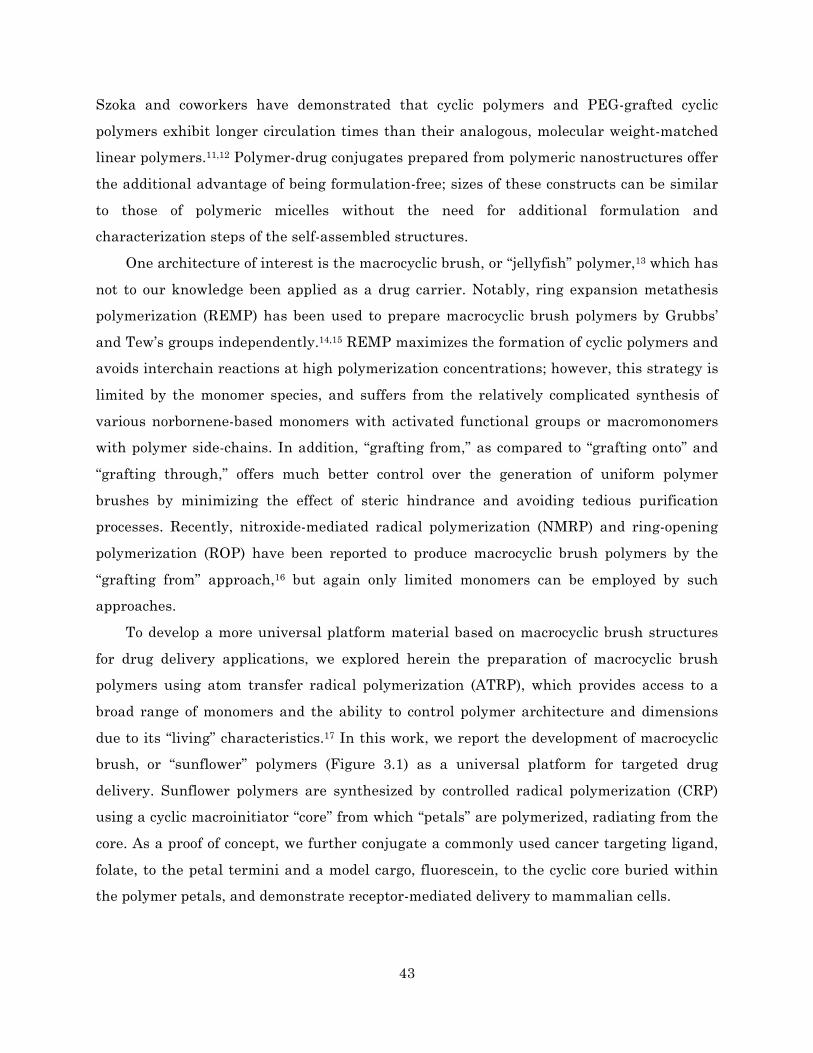
43
Szoka and coworkers have demonstrated that cyclic polymers and PEG-grafted cyclic
polymers exhibit longer circulation times than their analogous, molecular weight-matched
linear polymers.11,12 Polymer-drug conjugates prepared from polymeric nanostructures offer
the additional advantage of being formulation-free; sizes of these constructs can be similar
to those of polymeric micelles without the need for additional formulation and
characterization steps of the self-assembled structures.
One architecture of interest is the macrocyclic brush, or “jellyfish” polymer,13 which has
not to our knowledge been applied as a drug carrier. Notably, ring expansion metathesis
polymerization (REMP) has been used to prepare macrocyclic brush polymers by Grubbs’
and Tew’s groups independently.14,15 REMP maximizes the formation of cyclic polymers and
avoids interchain reactions at high polymerization concentrations; however, this strategy is
limited by the monomer species, and suffers from the relatively complicated synthesis of
various norbornene-based monomers with activated functional groups or macromonomers
with polymer side-chains. In addition, “grafting from,” as compared to “grafting onto” and
“grafting through,” offers much better control over the generation of uniform polymer
brushes by minimizing the effect of steric hindrance and avoiding tedious purification
processes. Recently, nitroxide-mediated radical polymerization (NMRP) and ring-opening
polymerization (ROP) have been reported to produce macrocyclic brush polymers by the
“grafting from” approach,16 but again only limited monomers can be employed by such
approaches.
To develop a more universal platform material based on macrocyclic brush structures
for drug delivery applications, we explored herein the preparation of macrocyclic brush
polymers using atom transfer radical polymerization (ATRP), which provides access to a
broad range of monomers and the ability to control polymer architecture and dimensions
due to its “living” characteristics.17 In this work, we report the development of macrocyclic
brush, or “sunflower” polymers (Figure 3.1) as a universal platform for targeted drug
delivery. Sunflower polymers are synthesized by controlled radical polymerization (CRP)
using a cyclic macroinitiator “core” from which “petals” are polymerized, radiating from the
core. As a proof of concept, we further conjugate a commonly used cancer targeting ligand,
folate, to the petal termini and a model cargo, fluorescein, to the cyclic core buried within
the polymer petals, and demonstrate receptor-mediated delivery to mammalian cells.

44
3.2 Materials and methods
Materials 3.2.1
All chemicals were purchased from Sigma-Aldrich and used without further purification
unless otherwise noted.
Polymer synthesis 3.2.2
Synthesis of ethyl glycinate methacrylamide (EGMA) monomer
Ethyl glycinate hydrochloride (10.15 g, 0.072 mol) was dissolved in 100 mL of dry
dichloromethane (DCM). Anhydrous TEA (20 mL, 0.144 mol) was then added at room
temperature, and the solution was cooled to 0°C in an ice bath. Methacryloyl chloride (7.251
mL, 0.072 mol) was added dropwise via a syringe pump to the cooled ethyl glycinate
solution over 2 h. After completion of this addition, the reaction mixture was warmed up to
room temperature and was stirred for another 2 h. After the reaction, the solution was
filtered to remove the by-product triethylamine hydrochloride precipitate. Subsequently,
the solvent was removed by rotary evaporation, and the crude product was purified by
column chromatography with an ethyl acetate/hexane mixture (1/2, v/v, Rf = 0.2-0.3 on
silica). The product was isolated by evaporation of the solvents and further dried in a
vacuum oven to form a colorless oily residue. Yield: 78% (9.55 g).
Figure 3.1 Schematic illustration of sunflower polymer containing targeting ligands and cargo.
Cyclic core P(OEGMA) petal
Targeting ligand Cargo

45
Synthesis of linear P(HEMA-st-EGMA) precursors (Alkyne-P(HEMA-st-EGMA)-N3)
The linear precursor with Br terminus was prepared by ATRP of 2-hydroxyethyl
methacrylate (HEMA) and EGMA in a 2-propanol (IPA)/N,N-dimethylformamide (DMF)
mixed solution, using propargyl 2-bromoisobutyrate18 as the initiator and N,N,N′,N′′,N′′-
pentamethyldiethylenetriamine (PMDETA)/Cu(I)Br as the catalyst. Typically, a 10 mL
round-bottom flask was charged with propargyl 2-bromoisobutyrate (0.041 g, 0.2 mmol),
HEMA (1.30 g, 10 mmol), EGMA (0.57 g, 3.33 mmol) to obtain a HEMA and initiator molar
ratio ([HEMA]/[Initiator]) of 50. The above mixture was dissolved in a 9:1 % w/w IPA/DMF
mixture to obtain a 50% w/w HEMA solution. The flask was degassed using a stream of dry
nitrogen gas to remove any trace of oxygen in the system. CuBr catalyst and PMDETA
(relative molar ratios of initiator:CuBr:PMDETA = 1:1:1) were then added quickly under a
nitrogen flow. Finally, the reaction mixture was sealed, followed by immersing the flask
into an oil bath preheated to 65°C to start the polymerization. The reaction mixture turned
green and became viscous as the reaction progressed. After 16 h, the reaction mixture was
quenched by exposing to air. The reaction mixture was diluted with 2 mL of methanol and
then subjected directly to dialysis against distilled water to remove the copper catalyst. The
product was harvested by freeze-drying. Yield: 70~80%.
A non-clickable linear analogue was synthesized following the same reaction conditions
except using ethyl 2-bromoisobutyrate (EBB) as the initiator.
Afterward, the linear precursor with azide terminus was obtained as follows. Alkyne-
P(HEMA-st-EGMA)-Br (1.0 g) and NaN3 in a 20-fold molar excess were dissolved in a 1/4
v/v % water/DMF mixed solvent (10 mL) in a round-bottom flask equipped with a magnetic
stirrer. The flask was sealed with a rubber septum and was stirred at 45°C for 48 h. After
purification by extensive dialysis to remove residual sodium salts, the linear precursor,
alkyne-P(HEMA-st-EGMA)-N3, was obtained by freeze-drying. Yield: 90% (0.9 g).
The bromo-terminus of the non-clickable linear copolymer was also converted to an
azide group following the same methods.
Synthesis of cyclic P(HEMA-st-EGMA) copolymer by intra-chain click cyclization
In a typical procedure, 750 mL of DMF was placed in a 1000 mL three-neck flask and
degassed by bubbling dry nitrogen gas for 1 h. 20-fold molar equivalents of PMDETA and
CuBr were then charged into the flask under the protection of nitrogen flow. A solution of

46
alkyne-P(HEMA-st-EGMA)-N3 linear precursor (0.5 g) in degassed DMF (10 mL) was added
to the copper catalyst solution via a syringe pump at the rate of 0.007 mL/min. The reaction
was carried out at 100°C in a nitrogen atmosphere for 24 h. At the end of the polymer
solution addition, the mixture was allowed to proceed for another 24 h. After the mixture
was cooled to room temperature, DMF was removed under reduced pressure, and the
concentrated residue was transferred directly to a dialysis tube (MWCO: 3.5 kDa, Fisher
Scientific) and dialyzed against distilled water to remove the copper catalyst. The resulting
cyclic polymer, cyclic P(HEMA-st-EGMA), was harvested by freeze-drying. Yield: 80% (0.4
g).
Preparation of cyclic poly(2-(2-bromoisobutyrnyloxy)ethyl methacrylate-st-EGMA)
(P((HEMA-iBuBr)-st-EGMA)) macroinitiator
Cyclic poly(2-(2-bromoisobutyrnyloxy)ethyl methacrylate-st-EGMA) (P((HEMA-iBuBr)-st-
EGMA)) macroinitiator was prepared by esterification of cyclic P(HEMA-st-EGMA). Cyclic
P(HEMA-st-EGMA) (0.2 g, 1.07 mmol of HEMA units) was dissolved in 10 mL of dry DMF,
and then anhydrous TEA (581 µL, 4.17 mmol) was added at room temperature. The solution
was cooled to 0°C in an ice bath. 2-bromoisobutyryl bromide (404 µL, 3.2 mmol) was added
dropwise via a syringe pump to the cooled polymer solution over 30 min. After completion of
this addition, the solution was further stirred at 0°C for 1 h and at room temperature for 48
h. After the reaction, the reaction mixture was poured into distilled water to precipitate the
product. The product was separated by centrifugation and further purified twice by
redissolving/reprecipitating with DMF/water, and finally dried in a vacuum oven for 24 h,
yielding a light yellow powdery product. Yield: 58% (0.21 g).
Linear multimacroinitiator for the preparation of comb-like polymer was prepared
following the same procedures except using non-clickable linear copolymer, P(HEMA-st-
EGMA)-N3.
Preparation of sunflower polymer, P((HEMA-sunflower-oligo(ethylene glycol) monomethyl
ether methacrylate-st-EGMA) (P((HEMA-sf-OEGMA)-st-EGMA))
A panel of sunflower polymers, P((HEMA-sf-OEGMA)-st-EGMA), with OEGMA radiating
rays was prepared by ATRP using cyclic P((HEMA-iBuBr)-st-EGMA) macroinitiator. Cyclic

47
P((HEMA-iBuBr)-st-EGMA) macroinitiator (0.05 g, 0.149 mmol of initiator sites), and
OEGMA (Mn = 300 g/mol, 4.46 g, 14.9 mmol) were dissolved in a 1/2 v/v % anisole/DMF
mixed solution (30 mL). The solution was split in equal volumes into five 10 mL round-
bottom flasks. After putting in a stir bar and sealing with a rubber septum, each solution
was thoroughly degassed by nitrogen bubbling for 10 min. Meanwhile, a catalyst stock
solution of CuBr2 (3.3 mg, 14.9 µmol) and 2,2’-bipyridine (51.2 mg, 327.8 µmol) was made in
DMF (500 µL). The solution was bubbled with nitrogen to deoxygenate the mixture. Solid
CuBr (21 mg, 149 µmol) was added to the copper stock solution under nitrogen atmosphere
and sonicated to promote dissolution. The resultant copper catalyst stock solution (100 µL)
was added quickly to each cyclic initiator-monomer mixture under a nitrogen blanket.
Finally, the reaction mixtures were sealed, followed by immersing the flasks into an oil
bath preheated to 60°C to start the polymerization. The reactions were subsequently
quenched by exposing flasks to air at predetermined time intervals. Each reaction mixture
was poured into ethyl ether to precipitate the product. The product was separated by
centrifugation and further purified by extensive dialysis against distilled water to remove
the copper catalyst. The resulting sunflower polymer was harvested by freeze-drying. The
conversion of polymerization and molecular weight of each sunflower polymer were
determined by 1H NMR and GPC analyses.
Comb-like polymer was prepared following the same polymerization condition except
using the linear multimacroinitiator.
Preparation of folate-conjugated sunflower polymer (FA-sunflower polymer) by click
coupling
Similar to the synthesis of alkyne-P(HEMA-st-EGMA)-N3 linear precursor, sunflower
polymer with an azide terminus on each OEGMA radiating ray was prepared by reacting
the resultant sunflower polymer with NaN3 in a 20-fold molar excess in a 1/4 v/v %
water/DMF mixed solvent. The reaction was carried out at 45°C for 48 h. After the reaction,
the product was purified by extensive dialysis to remove residual sodium salts, and
harvested by freeze-drying.
To a round-bottom flask containing thoroughly degassed DMF solution (10 mL) of
azide-terminated sunflower polymer (210 mg, 97 µmol of azide terminus) and alkyne-
functionalized folate19 (0.05 g, 106 µmol), PMDETA (20.3 µL, 97 µmol), and CuBr (14 mg, 97

48
µmol) were added quickly under a nitrogen flow. The reaction mixture was stirred at room
temperature for 24 h. After the reaction, the mixture was poured into ethyl ether to
precipitate the product and to remove the unreacted propargyl folate. The product was
separated by centrifugation and further purified by dialysis against distilled water to
remove the copper catalyst. The resulting FA-sunflower polymer was isolated by freeze-
drying. Yield: 86% (220 mg). The amount of folate conjugated to each polymer was
determined by absorbance at 360 nm based on a standard curve obtained from solutions of
free folic acid.
Folate-conjugated comb-like polymer was prepared following the same reaction
conditions.
Preparation of P((HEMA-sf-OEGMA-folate)-st-(GMA-hydrazide))
A 50 mL falcon tube equipped with a magnetic stirrer was charged with FA-sunflower
polymer (220 mg, 32.3 µmol of EGMA units), hydrazine hydrate (18 µL, 370 µmol), and
anhydrous methanol (6 mL). The reaction was performed at room temperature for 10 h.
After the reaction, the resultant polymer precursor, P((HEMA-sf-OEGMA-folate)-st-(GMA-
hydrazide)), was purified by extensive dialysis against distilled water and collected by
freeze-drying. Yield: 82% (180 mg). The content of hydrazide was determined by modified
TNBSA assays.20,21
Preparation of P((HEMA-sf-OEGMA-folate)-st-(GMA-fluorescein))
A 50 mL falcon tube equipped with a magnetic stirrer was charged with the polymer
precursor P((HEMA-sf-OEGMA-folate)-st-(GMA-hydrazide)) (90.0 mg, 14.6 µmol of
hydrazide), NHS-Fluorescein (PI-46410, Thermo Scientific, MW = 473.4 g/mol, 13.8 mg,
29.2 µmol), and anhydrous methanol (4 mL). The tube was sealed, and the reaction was
allowed to proceed in the dark at room temperature for 48 h. The resultant fluorescein-
labeled sunflower polymer, P((HEMA-sf-OEGMA-folate)-st-(GMA-fluorescein)), was
purified by extensive dialysis against distilled water (4L) for 24 h and collected by freeze-
drying. Yield: 75.1% (67.6 mg). The amount of fluorescein conjugated to each polymer was
determined by 1H NMR spectroscopy.
Folate-comb polymer-fluorescein was prepared following the same reaction conditions.

49
Polymer characterization 3.2.3
1H NMR spectra were recorded on a Bruker AV 500 nuclear magnetic resonance (NMR)
instrument using DMSO-d6, CDCl3, and D2O as the solvents.
Fourier transform infrared (FT-IR) spectra were recorded on a Bruker Vector 33 FT-IR
spectrometer. Samples were pressed into potassium bromide (KBr) pellets for
measurements.
Gel permeation chromatography (GPC) was used to determine molecular weight and
polydispersity (Mw/Mn, PDI) of polymer samples prepared. SEC Tosoh TSK-GEL R-3000
and R-4000 columns (Tosoh Bioscience, Montgomeryville, PA) were connected in series to a
Agilent 1200 series (Agilent Technologies, Santa Clara, CA), Optilab-rEX refractometer,
and miniDAWN TREOS triple-angle static light scattering detector (Wyatt Technology,
Santa Barbara, CA). HPLC-grade DMF containing 0.1 wt% LiBr at 60°C was used as the
mobile phase at a flow rate of 1 mL/min. Absolute molecular weight averages and dn/dc
were calculated using ASTRA software (Wyatt).
Average dimensions of sunflower polymers in an aqueous phase were measured by
dynamic light scattering (DLS) with a ZetaPALS (Brookhaven Instruments Corp.) at a
detection angle of 90°. The measurements were performed in triplicate. The aqueous
solution was passed through a 0.22 µm pore-sized syringe filter before measurements.
Cell culture 3.2.4
KB cells (ATCC CCL-17, a derivative of the HeLa human cervical adenocarcinoma line) and
A549 cells (ATCC CCL-185, lung epithelial carcinoma) were maintained in folate-free RPMI
1640 (Life Technologies) and F-12K (Corning cellgro) media, respectively. Media was
supplemented with 10% fetal bovine serum (HyClone) and 1% penicillin/streptomycin
(HyClone). Cells were cultured as a monolayer in a 37°C, 5% CO2 environment. Medium
was replaced every 2-3 days. Cells were passaged at ~70-80% confluence by incubation with
Trypsin-EDTA, followed by resuspension in complete growth medium.

50
In vitro uptake and competition studies 3.2.5
Cells were seeded in 24-well plates at a density of 40,000 cells per well in 1 mL of complete
growth medium and incubated in a 37°C, 5% CO2 environment for 24 h. Cells were rinsed
once with PBS and incubated in 1 mL of supplemented folate-free RPMI 1640 medium +/- 2
mM folate competition for 1 h at room temperature. Polymer samples at varying
concentrations were prepared in serial dilutions in water and then diluted 10-fold in
supplemented folate-free RPMI 1640 medium +/- 2 mM folate. The media was aspirated
from each well, and cells were incubated with 200 µL of polymer solution for 20 min at
37°C. After incubation, the polymer solutions were aspirated, and the cells were rinsed
twice with PBS/1% BSA. Cells were then harvested by incubation with 200 µL of Trypsin-
EDTA, followed by resuspension with 1 mL of complete growth medium. Cells were
transferred to 1.5 mL microcentrifuge tubes and pelleted at 300 g for 5 min at 4°C. The
supernatant was aspirated, and the cell pellets were resuspended in 200 µL of PBS/1% BSA
containing 2 µg/mL propidium iodide (PI) as a marker for cell viability. Cells were analyzed
for uptake of fluorescent polymer using a MACSQuant Analyzer flow cytometer (Miltenyi).
In vitro cytotoxicity studies 3.2.6
The cytotoxicities of various polymers were evaluated in vitro using the MTS assay. The
cells were seeded in 96-well plates at a density of 2500 cells per well in 100 µL of complete
growth medium and incubated in a 37°C, 5% CO2 environment for 24 h. Samples were
prepared in serial dilutions in water and then diluted 10-fold in Opti-MEM medium
(Invitrogen). The cells were rinsed once with PBS and incubated with 40 µL of the sample
solutions with different polymer concentrations at 37°C for 4 h. Cells were then rinsed with
PBS and the medium was replaced with 100 µL of culture medium. At 48 h, 20 µL of 3-(4,5-
dimethylthiazol-2-yl)-5-(3-carboxymethoxyphenyl)-2-(4-sulfophenyl)-2H-tetrazolium (MTS,
Promega) reagent was added to each well. Cells were then incubated at 37°C, 5% CO2 for 3
h. The absorbance of each well was measured at 490 nm on a Tecan Safire2 plate reader
(Ma ̈nnedorf, Switzerland). Cell viability (%) for each concentration was determined by
normalizing to the cells only signal.

51
Figure 3.2 Synthesis of folate-sunflower polymer-fluorescein.
3.3 Results and discussion
Synthesis and characterization of sunflower polymers 3.3.1
The two main steps in sunflower polymer synthesis are (i) synthesis of the cyclic
multimacroinitiator and (ii) polymerization of “petals” from the cyclized core (Figure 3.2).
O
OBr
OO
OH
HNO
OO
O
OBr
x y
OO
OH
HNO
OO
+ +ATRP
HEMA
EGMAp(HEMA-st-EGMA)-Br
O
ON3x y
OO
OH
HNO
OO
p(HEMA-st-EGMA)-N3
NaN3
N
xy
OO
OH
HNO
OO
Cyclic p(HEMA-st-EGMA)
O NN
O
N
xy
OO
O
HNO
OO
Cyclic multimacroinitiator
O NN
O
Br O
intra-chainclick cyclization
2-bromoisobutyrylbromide
BrBr
O
N
xy
OO
O
HNO
OO
Sunflower polymer
O NN
O
OBr
OO
z
ATRPof OEGMA
O
O
O
4-5
O4-5
N
xy
OO
O
HNO
OO
Sunflower polymer-N3
O NN
O
ON3
OO
z
O4-5
NaN3
N
xy
OO
O
HNO
OO
Folate-sunflower polymer(FA-SF)
O NN
O
ON
OO
z
O4-5
NN
Folate
click chemistryFolate C CH
N
xy
OO
O
HNO
NHNH2
O
Folate-sunflower polymer-hydrazide
O NN
O
ON
OO
z
O4-5
NN
Folate
NH2NH2 H2O
N
xy
OO
O
HNO
O
Folate-sunflower polymer-fluorescein(FA-SF-fluor)
O NN
O
O
NO
OzO
4-5N
N Folate
NHS-Fluorescein
NHNH
O
OH
O
OHO
O

52
The cyclic multimacroinitiator was first synthesized by (i) atom transfer radical
copolymerization of ethyl glycinate methacrylamide (EGMA) and hydroxyethylmethacrylate
(HEMA) using an alkyne-terminated initiator, (ii) conversion of the terminal bromine to an
azide and intrachain click cyclization of the α-alkyne-ω-azide linear polymers, and (iii)
conversion of HEMA monomers to alkyl halide ATRP initiators.
The ATRP conditions for the synthesis of P(HEMA-st-EGMA) copolymer were adapted
from reported procedures22 with some modifications considering that ATRP of acrylamide-
type monomers remains challenging,23 and more active ligands, such as 1,1,4,7,10,10-
hexamethyltriethyllenetetramine (HMTETA),24 N,N,N',N'',N''-pentamethyldiethylene-
triamine (PMDETA),25 and Tris[2-(dimethylamino)ethyl]amine (Me6TREN),26 can offer
improved control over the polymerization. Linear P(HEMA-st-EGMA) was first synthesized
using the CuBr/PMDETA catalyst in isopropanol/N,N'-dimethylformamide (IPA/DMF) (9/1,
w/w) at 65°C for 16 h. The benefits of this current polymerization condition are confirmed
by a kinetics study. The statistical copolymer panel showed a clear shift of the gel
permeation chromatography (GPC) elution trace toward higher molecular weight with
polymerization time (Figure 3.3). The living characteristics were reflected by the pseudo-
first-order kinetics and narrow PDI during the whole polymerization process (Figure 3.3). A
high conversion (~82%) of monomers was achieved after polymerization for 16 h, affording
polymer of DP ~41, close to the target value of 50. Moreover, there was no obvious high
molecular weight “shoulder” recorded in the GPC elution trace (Figure 3.3 and Figure 3.4a),
indicating minimal coupling termination of polymer chains during polymerization and
retention of chain end functionalities for further azidonation.
Figure 3.3 ATRP kinetics for the synthesis of P(HEMA-st-EGMA) copolymer.

53
The molar ratio of HEMA and EGMA in the resulting copolymers was determined to be
3.3:1 by 1H NMR spectroscopy, close to the feed molar ratio (3:1), indicating good control of
the copolymer composition by ATRP. The resulting copolymer was thus denoted P(HEMA41-
st-EGMA12). GPC shows that the copolymers have molecular weight (Mn) of 4.86 kDa and
relatively low polydispersity (PDI) of 1.27, indicating controlled synthesis of copolymers
(Figure 3.4a).
Figure 3.4 (a) GPC elution traces and (b) FT-IR spectra of linear and cyclic P(HEMA41-st-EGMA12).
A survey of the literature showed that a large excess of sodium azide (10-40 fold molar
excess compared to bromo-termini)27-30 is usually used for chain end azidonation to achieve
complete conversion of the chain termini. Therefore, the linear precursor, P(HEMA-st-
EGMA)-N3, was prepared by substitution with a 20-fold molar excess of sodium azide.
Cyclic polymers were then prepared by intra-chain Cu(I)-catalyzed azide-alkyne
cycloaddition (CuAAc) of linear precursors following reported procedures.31,32 Successful
cyclization was confirmed by FT-IR and GPC analyses, which show absence of the azide
group (~2100 cm-1) after polymer cyclization and a clear shift toward longer retention times
for the cyclic polymer compared to the linear precursor (Figure 3.4).
Cyclic poly(2-(2-bromoisobutyrnyloxy)ethyl methacrylate-st-EGMA) (P((HEMA-iBuBr)-
st-EGMA)) multimacroinitiator was then prepared by esterification of cyclic P(HEMA-st-
EGMA) with 2-bromoisobutyryl bromide. The disappearance of the characteristic signal at
4.8 ppm, assigned to the hydroxyl group of HEMA units, in the 1H NMR spectrum of cyclic

54
multimacroinitiator compared to the parent cyclic polymer (Figure S3.1) confirms complete
conjugation of ATRP initiating sites to the pendant hydroxyl groups of the cyclic polymer.
The cyclic multimacroinitiator and its linear analogue were used for ATRP of
oligoethylene glycol methacrylate (OEGMA) to prepare sunflower and comb polymers,
respectively. ATRP was performed using the catalyst CuBr/2,2'-bipyridine (bpy) in
anisole/DMF (1/2, v/v) at 60°C for different lengths of time. Three important parameters
were optimized to minimize the generation of cross-linked structures and free polymer
chains in the ATRP process due to the use of multimacroinitiators: (i) the feed
concentration of OEGMA monomer was kept at 0.5 M; (ii) the monomer conversion was
controlled to a low level (below 25%); and (iii) a small amount of CuBr2 ([CuBr2] =
0.1[CuBr]) was added to slow down the polymerization kinetics. A kinetic study was carried
out using cyclic multimacroinitiator with a target DP of 100 for each radiating petal. The
sunflower polymer panel showed a clear shift of the GPC elution trace toward higher
molecular weight with polymerization time (Figure 3.5). The living characteristics were
reflected by the pseudo-first-order kinetics and narrow PDI (around 1.3) during the whole
polymerization process (Figure 3.5). Furthermore, the difference in topology of sunflower
versus comb-like polymers was confirmed by GPC analyses. Sunflower polymers eluted at
later times than comb polymers, indicating more compact structure and smaller effective
molecular weight (Figure 3.6).
Figure 3.5 ATRP kinetics of sunflower polymer prepared from cyclic multimacroinitiator, with a target DP of 100 for each petal.

55
Figure 3.6 GPC elution traces of sunflower polymer and comb-like polymer prepared with the same polymerization time using cyclic and linear multimacroinitiators.
Finally, the hydrodynamic Z-average diameters of sunflower polymers were
determined by dynamic light scattering (DLS). Particle size increased with increasing
P(OEGMA) petal polymerization time, ranging from 13.5 to 16.8 nm for the panel (Figure
3.7). The ability to control particle size is a particular advantage of using this
polymerization strategy for synthesizing drug carriers.
Synthesis of folate-targeted sunflower polymers and evaluation of uptake in FR+ 3.3.2
cells
The sunflower polymer with petals synthesized by 3 h ATRP of OEGMA (Mn = 101 kDa,
PDI = 1.26, Rh = 14 nm) was selected for further biological evaluation. Targeted polymer
Figure 3.7 Summary of Z-average sizes of sunflower polymers with petals polymerized for different lengths of time.

56
constructs were prepared by conjugation of alkyne-functionalized folate (FA) to the
P(OEGMA) petal termini via CuAAC “click” coupling. FA was selected for targeting due to
significant literature precedence establishing drug carrier targeting to cancer cells via the
folate receptor (FR).33-36 FA conjugation was confirmed by 1H NMR analysis, which shows
the appearance of characteristic signals at 6.7-8.7 ppm (assigned to the aromatic protons of
folate) in the spectrum of folate-sunflower polymer, and by an increase in UV absorbance on
GPC (Figure S3.2). Each polymer was found to contain approximately 25-28 folate groups, a
conjugation efficiency of 61-69% based on the theoretical polymer composition of 41 HEMA
groups available for polymerization of petals and subsequent folate conjugation.
We next investigated FR-mediated uptake of the sunflower polymers in KB (FR+) and
A549 (FR-) cells. Fluorescein was selected as a model drug due to its fluorescent and cell
compatibility properties; in future work, aldehyde or ketone-containing drugs could be
similarly conjugated to sunflowers for acid-triggered release at tumor sites. FA-SF-fluor
was prepared by converting EGMA side chains in the polymer’s cyclic core to hydrazide
groups, followed by conjugation with the N-hydroxysuccinimide ester of fluorescein. An
estimated 6-8 fluorescein molecules were conjugated per polymer, a conjugation efficiency of
59-78% based on the theoretical polymer composition of 12 EGMA groups and 85%
conversion of EGMA to hydrazide groups for fluorescein incorporation.
Cells were then incubated with FA-SF-fluor at various concentrations, followed by flow
cytometry analysis for binding. Although FA-SF-fluor uptake is dose-dependent in both cell
lines, greater uptake was observed in the FR+ KB cells than in the A549 cells, as indicated
by higher median fluorescence intensities of these cells at all polymer concentrations
(Figure 3.8a). Minimal uptake of untargeted SF-fluor was observed in either cell line
(Figure 3.8b). In competition studies, cells were treated with FA-SF-fluor in the presence of
2 mM free folate. Competition with free folate significantly reduced polymer uptake in KB
cells but not in A549 cells. These results further support folate receptor-mediated
endocytosis as a major uptake mechanism for FA-targeted sunflower polymers by FR+ KB
cells.
Interestingly, when uptake of the comb-like linear analogue, FA-comb-fluor, was
investigated, competition with free folate produced only a small decrease in polymer uptake
in KB cells even at a low polymer concentration (Figure 3.8c). This may suggest higher
levels of non-specific uptake of comb polymers relative to sunflower polymers. The effect of

57
polymer architecture on internalization mechanism will therefore be an interesting topic for
future investigation.
Figure 3.8 Uptake of (a) FA-SF-fluor, (b) untargeted SF-fluor, and (c) FA-comb-fluor polymers in FR+ KB cells and FR- A549 cells in the absence (black bars) and presence (grey bars) of 2 mM competing free folate.
(-) folate (+) folate
cellsonly
100 nM
500 nM
1000nM
05101520
[FA-SF-fluor]
Nor
mal
ized
MFI
KB (FR+)
cellsonly
100 nM
500 nM
1000nM
05101520
[FA-SF-fluor]N
orm
aliz
edM
FI
A549 (FR-)(a)
cellsonly
100 nM
500 nM
1000nM
05101520
[SF-fluor]
Nor
mal
ized
MFI
KB (FR+)
cellsonly
100 nM
500 nM
1000nM
05101520
[SF-fluor]
Nor
mal
ized
MFI
A549 (FR-)(b)
cellsonly
100 nM
05101520
[FA-comb-fluor]
Nor
mal
ized
MFI
KB (FR+)
cellsonly
100 nM
05101520
[FA-comb-fluor]
Nor
mal
ized
MFI
A549 (FR-)(c)

58
Cytotoxicity of sunflower polymers 3.3.3
Because sunflower polymers demonstrate potential as drug carriers, we investigated the
cytotoxicity of the materials using KB and A549 cells. An MTS metabolic activity assay was
performed to determine cell viability. The base and FA-modified sunflower polymers were
found to be minimally toxic in either cell line, with IC50, or concentration for 50% cell
killing, greater than 3.5 mg/mL in all cases.
3.4 Conclusions
In summary, this work introduces the sunflower polymer platform as a potential drug
carrier. Using a combination of ATRP and click chemistry, sunflower polymers can be
synthesized with control over hydrodynamic size based on polymerization time. The ability
to tailor sizes within the relevant range for tumor delivery (5-50 nm), combined with the
demonstrated advantage over comb-like polymers in targeted cellular uptake, support
further investigation of these materials for tumor-targeted drug delivery and molecular
imaging.
3.5 Acknowledgements
This work was supported by the National Institutes of Health (1R01CA177272), the
National Science Foundation (DMR 1206426), and a National Science Foundation Graduate
Research Fellowship to C.E.W.
References
1. Laurent, B. A. & Grayson, S. M. Synthesis of cyclic dendronized polymers via divergent "graft-from" and convergent click ‘graft-to’ routes: preparation of modular toroidal macromolecules. J. Am. Chem. Soc. 133, 13421–13429 (2011).
2. Laurent, B. A. & Grayson, S. M. Synthesis of cyclic amphiphilic homopolymers and their potential application as polymeric micelles. Polym Chem 3, 1846–1855 (2012).
3. Liu, L., Parameswaran, S., Sharma, A., Grayson, S. M., Ashbaugh, H. S. & Rick, S. W. Molecular dynamics simulations of linear and cyclic amphiphilic polymers in aqueous and organic environments. J Phys Chem B 118, 6491–6497 (2014).
4. Johnson, J. A., Lu, Y. Y., Burts, A. O., Lim, Y.-H., Finn, M. G., Koberstein, J. T.,

59
Turro, N. J., Tirrell, D. A. & Grubbs, R. H. Core-clickable PEG-branch-azide bivalent-bottle-brush polymers by ROMP: grafting-through and clicking-to. J. Am. Chem. Soc. 133, 559–566 (2011).
5. Sheiko, S. S., Sumerlin, B. S. & Matyjaszewski, K. Cylindrical molecular brushes: Synthesis, characterization, and properties. Progress in Polymer Science 33, 759–785 (2008).
6. Hawker, C. J., Fréchet, J. M. J., Grubbs, R. B. & Dao, J. Preparation of Hyperbranched and Star Polymers by a ‘Living’, Self-Condensing Free Radical Polymerization. J. Am. Chem. Soc. 117, 10763–10764 (1995).
7. Trollsås, M. & Hedrick, J. L. Dendrimer-like Star Polymers. J. Am. Chem. Soc. 4644–4651 (1998). doi:10.1021/ja973678w
8. Lane, D. D., Chiu, D. Y., Su, F. Y., Srinivasan, S., Kern, H. B., Press, O. W., Stayton, P. S. & Convertine, A. J. Well-defined single polymer nanoparticles for the antibody-targeted delivery of chemotherapeutic agents. Polym Chem 6, 1286–1299 (2015).
9. Smet, M. Biological and Medical Applications of Hyperbranched Polymers. Hyperbranched Polymers: Synthesis, Properties, and Applications 387–413 (2011).
10. Wang, C. E., Stayton, P. S., Pun, S. H. & Convertine, A. J. Polymer Nanostructures Synthesized by Controlled Living Polymerization for Tumor-Targeted Drug Delivery. J Control Release (2015). doi:10.1016/j.jconrel.2015.08.054
11. Chen, B., Jerger, K., Fréchet, J. M. J. & Szoka, F. C. The influence of polymer topology on pharmacokinetics: differences between cyclic and linear PEGylated poly(acrylic acid) comb polymers. J Control Release 140, 203–209 (2009).
12. Nasongkla, N., Chen, B., Macaraeg, N., Fox, M. E., Fréchet, J. M. J. & Szoka, F. C. Dependence of Pharmacokinetics and Biodistribution on Polymer Architecture: Effect of Cyclic versus Linear Polymers. J. Am. Chem. Soc. 131, 3842–3843 (2009).
13. Coulembier, O., Moins, S., De Winter, J., Gerbaux, P., Leclère, P., Lazzaroni, R. & Dubois, P. From Jellyfish Macromolecular Architectures to Nanodoughnut Self-Assembly. Macromolecules 43, 575–579 (2010).
14. Xia, Y., Boydston, A. J. & Grubbs, R. H. Synthesis and Direct Imaging of Ultrahigh Molecular Weight Cyclic Brush Polymers. Angew. Chem. Int. Ed. Engl. 50, 5882–5885 (2011).
15. Zhang, K. & Tew, G. N. Cyclic Brush Polymers by Combining Ring-Expansion Metathesis Polymerization and the ‘Grafting from’ Technique. ACS Macro Lett. 1, 574–579 (2012).
16. Jia, Z. F. & Monteiro, M. J. Cyclic Polymers: Methods and Strategies. J. Polym. Sci. A Polym. Chem. 50, 2085–2097 (2012).
17. Matyjaszewski, K. & Tsarevsky, N. V. Macromolecular engineering by atom transfer radical polymerization. J. Am. Chem. Soc. 136, 6513–6533 (2014).
18. Wei, H., Chu, D. S., Zhao, J., Pahang, J. A. & Pun, S. H. Synthesis and evaluation of cyclic cationic polymers for nucleic acid delivery. ACS Macro Lett. 2, 1047–1050 (2013).
19. Zhang, H., Cai, Z., Sun, Y., Yu, F., Chen, Y. & Sun, B. Folate-conjugated β-cyclodextrin from click chemistry strategy and for tumor-targeted drug delivery. Journal of Biomedical Materials Research Part A 100, 2441–2449 (2012).
20. Etrych, T., Jelı ́nková, M., Řı ́hová, B. & Ulbrich, K. New HPMA copolymers containing doxorubicin bound via pH-sensitive linkage: synthesis and preliminary in vitro and in vivo biological properties. J. Control. Release 73, 89–102 (2001).
21. Zhan, F., Chen, W., Wang, Z., Lu, W., Cheng, R., Deng, C., Meng, F., Liu, H. &

60
Zhong, Z. Acid-activatable prodrug nanogels for efficient intracellular doxorubicin release. Biomacromolecules 12, 3612–3620 (2011).
22. Yang, P. & Armes, S. P. Preparation of Well-Defined Poly (2-hydroxyethyl methacrylate) Macromonomers via Atom Transfer Radical Polymerization. Macromol. Rapid Commun. 35, 242–248 (2014).
23. Teodorescu, M. & Matyjaszewski, K. Atom transfer radical polymerization of (meth) acrylamides. Macromolecules 32, 4826–4831 (1999).
24. Xu, F., Zheng, S. Z. & Luo, Y. L. Thermosensitive t-PLA-b-PNIPAAm tri-armed star block copolymer nanoscale micelles for camptothecin drug release. J. Polym. Sci. A Polym. Chem. 51, 4429–4439 (2013).
25. Wang, J., Gao, P., Ye, L., Zhang, A.-Y. & Feng, Z.-G. Dual thermo-responsive polyrotaxane-based triblock copolymers synthesized via ATRP of N-isopropylacrylamide initiated with self-assemblies of Br end-capped Pluronic F127 with β-cyclodextrins. Polym Chem 2, 931–940 (2011).
26. Xia, Y., Yin, X., Burke, N. A. & Stöver, H. D. Thermal response of narrow-disperse poly (N-isopropylacrylamide) prepared by atom transfer radical polymerization. Macromolecules 38, 5937–5943 (2005).
27. Cai, T., Yang, W. J., Neoh, K.-G. & Kang, E.-T. Preparation of jellyfish-shaped amphiphilic block-graft copolymers consisting of a poly(?-caprolactone)-block-poly(pentafluorostyrene) ring and poly(ethylene glycol) lateral brushes. Polym. Chem. 3, 1061–1068 (2012).
28. Chen, F., Liu, G. & Zhang, G. Synthesis of cyclic polyelectrolyte via direct copper(I)-catalyzed click cyclization. J. Polym. Sci. A Polym. Chem. 50, 831–835 (2011).
29. Jiang, X., Shi, Y., Zhu, W., Chen, Y. & Xi, F. Synthesis of mikto-topology star polymer containing one cyclic arm. J. Polym. Sci. A Polym. Chem. 50, 4239–4245 (2012).
30. Ren, J. M., Satoh, K., Goh, T. K., Blencowe, A., Nagai, K., Ishitake, K., Christofferson, A. J., Yiapanis, G., Yarovsky, I., Kamigaito, M. & Qiao, G. G. Stereospecific Cyclic Poly(methyl methacrylate) and Its Topology-Guided Hierarchically Controlled Supramolecular Assemblies. Angew. Chem. Int. Ed. Engl. 53, 459–464 (2013).
31. Laurent, B. A. & Grayson, S. M. An efficient route to well-defined macrocyclic polymers via ‘click’ cyclization. J. Am. Chem. Soc. 128, 4238–4239 (2006).
32. Wei, H., Chu, D. S., Zhao, J., Pahang, J. A. & Pun, S. H. Synthesis and evaluation of cyclic cationic polymers for nucleic acid delivery. ACS Macro Lett. 2, 1047–1050 (2013).
33. Lee, S.-M., Chen, H., O’Halloran, T. V. & Nguyen, S. T. ‘Clickable’ polymer-caged nanobins as a modular drug delivery platform. J. Am. Chem. Soc. 131, 9311–9320 (2009).
34. Low, P. S., Henne, W. A. & Doorneweerd, D. D. Discovery and development of folic-acid-based receptor targeting for imaging and therapy of cancer and inflammatory diseases. Acc. Chem. Res. 41, 120–129 (2007).
35. Lu, Y. & Low, P. S. Folate-mediated delivery of macromolecular anticancer therapeutic agents. Advanced Drug Delivery Reviews 64, 342–352 (2012).
36. Yang, X., Grailer, J. J., Pilla, S., Steeber, D. A. & Gong, S. Tumor-targeting, pH-responsive, and stable unimolecular micelles as drug nanocarriers for targeted cancer therapy. Bioconjug. Chem. 21, 496–504 (2010).

61
Supporting Information
Figure S3.1 1H NMR spectra of (a) cyclic P(HEMA-st-EGMA) and (b) cyclic multimacroinitiator.

62
Figure S3.2 GPC elution traces and UV-detected signals of sunflower polymer and folate-conjugated sunflower polymer.

63
Chapter 4.
SUNFLOWER POLYMERS FOR FOLATE-MEDIATED
DRUG DELIVERY
Christine E. Wang*, Hua Wei*, Nicholas Tan, Andrew J. Boydston, and Suzie H. Pun
* Equally contributing authors
Abstract
Polymeric delivery vehicles can improve the safety and efficacy of chemotherapy drugs by
facilitating preferential tumor delivery. Polymer-drug conjugates are especially attractive
carriers because additional formulation steps are not required during manufacturing, and
drug release profiles can be altered based on linker choice. For clinical translation, these
vehicles should also be reproducibly and controllably synthesized. Recently, we reported the
development of a class of materials called “sunflower polymers,” synthesized by controlled
radical polymerization of hydrophilic “petals” from a cyclic multimacroinitiator “core.” This
synthesis strategy afforded control over the size of the polymer nanostructures based on
their petal polymerization time. In this work, we demonstrate that particle size can be
further tuned by varying the degree of polymerization of the cyclic core in addition to that
of the petals. Additionally, we investigate the application of these materials for tumor-
targeted drug delivery. We demonstrate that folate-targeted, doxorubicin-conjugated
sunflower polymers undergo receptor-mediated uptake into cancer cells and pH-triggered
drug release leading to cytotoxicity. These materials are attractive as drug carriers due to
their discrete and small size, shielded drug cargo that can be triggered for release, and
relative ease of synthesis.†
4.1 Introduction
Cytotoxic chemotherapy remains the preferred frontline strategy for treatment against
most types of cancer but is associated with significant side effects such as major organ † Reprinted with permission from Wang, C. E., et al. Biomacromolecules, 17, pp. 69-75. Copyright 2016 American Chemical Society.

64
damage, infertility, immunosuppression, and nausea/vomiting that severely compromise
patient quality of life.1,2 The development of improved chemotherapy formulations therefore
remains an important research priority with significant medical impact. The adverse side
effects associated with chemotherapy can be ameliorated by developing delivery
formulations that rely on tumor localization via the “enhanced permeability and retention”
(EPR) effect.3 These formulations are large enough to be retained in the circulation after
injection, avoiding tissue distribution and rapid renal clearance, but remain small enough
to extravasate from abnormal tumor vasculature into tumors.4,5 This paradigm is used by
clinically-approved chemotherapy formulations such as liposomal anthracyclines (e.g., Doxil
and Myocet) and albumin-bound paclitaxel (Abraxane) to offer improved safety profiles over
equivalent solution formulations.
Still, drawbacks to these formulations remain. Multiple formulation steps are often
required in manufacturing, increasing scale-up complexity.6 Interstitial tumor penetration
is also reduced due to the larger size of these formulations (50-150 nm) compared to free
drug.7-9 Finally, drug leakage from the vehicle prior to tumor localization can result in off-
target effects.10,11
Polymer-drug conjugates are an alternative delivery technology in the development
pipeline.12,13 The smaller size of these constructs compared to liposomal carriers and drug
attachment via labile linkages allow for improved tumor penetration and controlled drug
release. In contrast to polymeric carriers such as micelles or nanoencapsulates,14,15 polymer-
drug conjugates do not require formulation steps during manufacturing. Dendrimers, star
polymers, and cyclic polymers have prolonged circulation in vivo compared to linear
polymers, an important property in tumor accumulation via EPR.16-18 Therefore, polymers
with advanced architectures that can be reproducibly and controllably synthesized are of
great interest as drug carriers.
We have recently described a strategy for synthesizing macrocyclic brush or
“sunflower” (SF) polymers19 using atom transfer radical polymerization (ATRP) and a
“grafting-from” approach.20,21 We demonstrated that sunflower polymers could be
synthesized with relatively low polydispersity and with control over their hydrodynamic
size based on the polymerization time of their radiating “petals.” Because the folate
receptor (FR) is commonly overexpressed on various cancer cell types,22,23 we also
conjugated the petal termini with folate (FA) as a targeting ligand and the polymer “core”
with fluorescein as a model drug to demonstrate receptor-mediated uptake in FR-positive

65
cells. These sunflower polymers appeared to have an advantage over their linear analogues
(comb-like polymers), which were shown to be taken up by non-specific mechanisms.
In this work, we have expanded on our platform by synthesizing sunflower polymers
with two different core sizes and then applied these materials for targeted drug delivery
through conjugation of FA to the sunflower petals and doxorubicin to the sunflower cores.
Together with the petal polymerization time, controlling core size allows for greater control
over the hydrodynamic size of the polymers. We investigate the uptake properties as
compared to the previously reported small-core sunflowers. We further demonstrate that
sunflower polymers can be used for targeted, intracellular drug delivery by conjugating the
chemotherapy drug doxorubicin (Dox) via hydrazone bonds that release drug under acidic
conditions (Figure 4.1). Finally, we investigate the cytotoxicity of the various polymer-Dox
constructs synthesized.
Figure 4.1 Schematic illustrations of (a) sunflower polymer containing FA targeting ligands and Dox drug and (b) proposed delivery mechanism: extravasation into tumor tissue by EPR effect, internalization by FR-expressing cancer cells, and intracellular, pH-triggered Dox release.

66
4.2 Materials and methods
4.2.1 Materials
All chemicals were purchased from Sigma-Aldrich and used without further purification
unless otherwise noted.
4.2.2 Polymer synthesis and Dox conjugation
Preparation of P((HEMA-sunflower-OEGMA-folate)-st-(GMA-hydrazide)) (FA-SF-hydrazide)
All steps for the synthesis of the folate-targeted, hydrazide-containing sunflower polymer
were performed as reported previously,19 but using a HEMA:initiator molar ratio of 100:1 in
the synthesis of the linear P(HEMA-st-EGMA) precursor to yield sunflower polymers with a
larger core.
Preparation of P((HEMA-sf-OEGMA-folate)-st-(GMA-fluorescein)) (FA-SF-fluor)
A 50 mL falcon tube equipped with a magnetic stirrer was charged with the polymer
precursor P((HEMA-sf-OEGMA-folate)-st-(GMA-hydrazide)) (146.0 mg, 14.5 µmol of
hydrazide), NHS-Fluorescein (Thermo Scientific, 13.7 mg, 29.0 µmol), and anhydrous
methanol (4 mL). The tube was sealed, and the reaction mixture was placed in the dark at
room temperature for 48 h. The resultant fluorescein-labeled sunflower polymer, P((HEMA-
sf-OEGMA-folate)-st-(GMA-fluorescein)), was purified by extensive dialysis against distilled
water (4 L) for 24 h and collected by freeze-drying. Yield: 67.0% (97.8 mg). The amount of
fluorescein conjugated to the sunflower polymer was determined by 1H NMR analysis as
described previously.19
Preparation of P((HEMA-sf-OEGMA-folate)-st-(GMA-Dox)) (FA-SF-Dox)
A 50 mL falcon tube equipped with a magnetic stirrer was charged with the polymer
precursor P((HEMA-sf-OEGMA-folate)-st-(GMA-hydrazide)) (180.0 mg, 29.2 µmol of
hydrazide), doxorubicin hydrochloride (Dox•HCl, LC Laboratories, Woburn, MA) (33.8 mg,
58.4 µmol), a drop of acetic acid, and anhydrous methanol (6 mL). The tube was sealed, and

67
the reaction mixture was placed in the dark at room temperature for 48 h. The resultant
sunflower polymeric prodrug, P((HEMA-sf-OEGMA-folate)-st-(GMA-Dox)), was purified by
extensive dialysis against anhydrous methanol (2 L) for 24 h, followed by dialysis against
distilled water (4 L) for another 24 h, and collected by freeze-drying. Yield: 78.7% (155 mg).
The amount of Dox conjugated to the sunflower polymer was determined by absorbance at
480 nm based on a standard curve obtained from free Dox•HCl in water.
4.2.3 Polymer characterization
1H NMR spectra were recorded on a Bruker AV 500 nuclear magnetic resonance (NMR)
instrument using DMSO-d6 as the solvent.
Fourier transform infrared (FT-IR) spectra were recorded on a Bruker Vector 33 FT-IR
spectrometer. Samples were pressed into potassium bromide (KBr) pellets for
measurements.
Gel permeation chromatography (GPC) was used to determine molecular weight and
polydispersity (Mw/Mn, PDI) of polymer samples prepared. SEC Tosoh TSK-GEL R-3000
and R-4000 columns (Tosoh Bioscience, Montgomeryville, PA) were connected in series to
an Agilent 1200 Series System (Agilent Technologies, Santa Clara, CA), Optilab-rEX
refractometer, and miniDAWN TREOS triple-angle static light scattering detector (Wyatt
Technology, Santa Barbara, CA). HPLC-grade DMF containing 0.1 wt% LiBr at 60°C was
used as the mobile phase at a flow rate of 1 mL/min. Absolute molecular weight averages
and dn/dc were calculated using ASTRA software (Wyatt).
Average dimensions of sunflower polymers in an aqueous phase were measured by
dynamic light scattering (DLS) with a ZetaPALS (Brookhaven Instruments Corp.) at a
detection angle of 90°. The measurements were performed in triplicate. The aqueous
solution was passed through a 0.22 µm pore-sized syringe filter before measurements.
4.2.4 Quantification of Dox release
FA-SF-Dox polymer was dissolved in release buffer at different pHs (PBS at pH 7.4,
phosphate buffer at pH 6.8, or citrate buffer at pH 5.5) at a concentration of 1 mg/mL.
Spectra/Por Float-A-Lyzer G2 Dialysis Devices (MWCO: 8-10 kDa, Spectrum Laboratories)

68
were pre-soaked in release buffer for 30 min, then loaded with 1 mL of polymer solution
each. Each device was immersed in a 50 mL conical containing 12 mL of the same release
buffer and incubated in the dark at 37°C with shaking at 125 rpm. At predetermined times,
1 mL of the dialysate was sampled and replaced with an equal volume of fresh buffer.
Dialysate samples were stored at -20°C for later analysis. After all time points were
collected, dialysate samples were analyzed for Dox fluorescence (excitation: 480 nm,
emission: 580 nm) on a Tecan Safire2 plate reader (Ma ̈nnedorf, Switzerland). The
concentration of released Dox in each sample was calculated based on standard curves
obtained from free Dox•HCl in the respective release buffer.
4.2.5 Cell culture
KB cells (ATCC CCL-17) and A549 cells (ATCC CCL-185) were maintained in folate-free
RPMI 1640 (Life Technologies) and F-12K (Corning cellgro) media, respectively. Media was
supplemented with 10% fetal bovine serum (FBS, HyClone) and 1% penicillin/streptomycin
(HyClone). Cells were cultured as a monolayer in a 37°C, 5% CO2 environment. Medium
was replaced every 2-3 days. Cells were passaged at ~70-80% confluence by incubation with
Trypsin-EDTA, followed by resuspension in complete growth medium.
4.2.6 Serum stability studies
Sunflower polymers were incubated in cell culture medium (MEM supplemented with 10%
FBS) for 4 h at 37°C. The resultant polymer was recovered by extraction using
dichloromethane (DCM), vacuum dried, and analyzed by GPC.
4.2.7 In vitro uptake and competition studies
Cells were seeded in 24-well plates at a density of 40,000 cells per well in 1 mL of complete
growth medium and incubated in a 37°C, 5% CO2 environment for 24 h. Cells were rinsed
once with PBS and incubated in 1 mL of supplemented folate-free RPMI 1640 medium +/- 2
mM folate competition for 1 h at room temperature. Polymer samples at varying
concentrations were prepared in serial dilutions in water and then diluted 10-fold in

69
supplemented folate-free RPMI 1640 medium +/- 2 mM folate. The media was aspirated
from each well, and cells were incubated with 200 µL of polymer solution for 20 min at
37°C. After incubation, the polymer solutions were aspirated, and the cells were rinsed
twice with PBS/1% BSA. Cells were then harvested by incubation with 200 µL of Trypsin-
EDTA, followed by resuspension with 1 mL of complete growth medium. Cells were
transferred to 1.5 mL microcentrifuge tubes and pelleted at 300 g for 5 min at 4°C. The
supernatant was aspirated, and the cell pellets were resuspended in 200 µL of PBS/1% BSA
containing 2 µg/mL propidium iodide (PI) as a marker for cell viability. Cells were analyzed
for uptake of fluorescent polymer using a MACSQuant Analyzer flow cytometer (Miltenyi).
4.2.8 Confocal imaging
KB cells were seeded on poly-D-lysine-coated glass coverslips in 24-well plates at a density
of 80,000 cells per well in 1 mL of complete growth medium and incubated in a 37°C, 5%
CO2 environment for 12 h. Solutions of FA-SF-Dox polymer and free Dox were prepared in
complete growth medium at concentrations equal to 25% of their respective IC50 values.
Cells were treated with 400 µL of FA-SF-Dox or free Dox for 12 h at 37°C. Cells were rinsed
3 times with HBSS/1% BSA, fixed with 300 µL of 4% paraformaldehyde (PFA) solution for
15 min at room temperature, and rinsed twice with HBSS/1% BSA. Finally, cells were
counterstained with 300 µL of 5 µg/mL 4’,6-diamidino-2-phenylindole (DAPI) in HBSS/1%
BSA for 5 min at room temperature and rinsed twice more. Coverslips were mounted onto
glass slides with Fluoromount-G (eBioscience) and imaged using a Leica TCS SP8X confocal
microscope.
4.2.9 In vitro cytotoxicity studies
The cytotoxicities of various formulations were evaluated in vitro using the MTS assay. The
cells were seeded in 96-well plates at a density of 2500 cells per well in 100 µL of complete
growth medium and incubated in a 37°C, 5% CO2 environment for 24 h. Samples were
prepared in serial dilutions in water and then diluted 10-fold in OptiMEM medium
(Invitrogen). The cells were then rinsed once with PBS and incubated with 40 µL of the
sample solutions with different polymer or Dox concentrations at 37°C for 4 h. Cells were

70
then rinsed with PBS, and the medium was replaced with 100 µL of culture medium. At 48
h, 20 µL of 3-(4,5-dimethylthiazol-2-yl)-5-(3-carboxymethoxyphenyl)-2-(4-sulfophenyl)-2H-
tetrazolium (MTS, Promega) reagent was added to each well. Cells were then incubated at
37°C, 5% CO2 for 3 h. The absorbance of each well was measured at 490 nm on a Tecan
Safire2 plate reader (Ma ̈nnedorf, Switzerland). Cell viability for each treatment condition
was determined by normalizing to the cells only signal.
4.2.10 Animals
Female NCr nude mice (4-5 weeks) were purchased from Taconic. All experimental
procedures were performed in accordance with protocols approved by the University of
Washington Institutional Animal Care and Use Committee.
4.2.11 Tumor inhibition studies
NCr nude mice were placed on a folic acid-deficient diet (Harlan Laboratories, Indianapolis,
IN) for at least one week before tumor inoculation and were maintained on the special feed
throughout the study. To develop xenograft tumors, mice were inoculated subcutaneously in
the right hind flank with 5×106 KB cells in 100 µL of folate-free RPMI 1640 medium
without serum.
After tumor inoculation, mice were randomly distributed into treatment groups of 7
mice each. Mice receiving a single treatment (on day 7 after tumor inoculation) were
injected with free Dox or FA-SF-Dox at a dose of 6 mg Dox/kg mouse, or FA-SF at an
equivalent polymer concentration. Mice receiving three treatments (on days 7, 10, and 13
after tumor inoculation) were injected with free Dox or FA-SF-Dox at a dose of 4 mg Dox/kg
mouse per treatment, or FA-SF at an equivalent polymer concentration. Each formulation
was administered intravenously by retroorbital injection in 5% glucose and in a volume of
less than 150 µL. In addition, control mice were given a single injection of 5% glucose
solution.
Tumor volume was calculated by measuring the tumor with calipers and using the
formula volume = 0.5 × a × b2, where a is the longer of the two dimensions. Tumor
measurements and mouse weights were recorded every 2-3 days for the duration of the

71
study. Mice were euthanized if tumor volume exceeded 10% of body weight, tumor
ulceration was observed, or body weight decreased by 20%.
Figure 4.2 Synthesis of folate-sunflower polymer-Dox.
O
OBr
OO
OH
HNO
OO
O
OBr
x y
OO
OH
HNO
OO
+ +ATRP
HEMA
EGMAp(HEMA-st-EGMA)-Br
O
ON3x y
OO
OH
HNO
OO
p(HEMA-st-EGMA)-N3
NaN3
N
xy
OO
OH
HNO
OO
Cyclic p(HEMA-st-EGMA)
O NN
O
N
xy
OO
O
HNO
OO
Cyclic multimacroinitiator
O NN
O
Br O
intra-chainclick cyclization
2-bromoisobutyrylbromide
BrBr
O
N
xy
OO
O
HNO
OO
Sunflower polymer
O NN
O
OBr
OO
z
ATRPof OEGMA
O
O
O
4-5
O4-5
N
xy
OO
O
HNO
OO
Sunflower polymer-N3
O NN
O
ON3
OO
z
O4-5
NaN3
N
xy
OO
O
HNO
OO
Folate-sunflower polymer(FA-SF)
O NN
O
ON
OO
z
O4-5
NN
Folate
click chemistryFolate C CH
N
xy
OO
O
HNO
NHNH2
O
Folate-sunflower polymer-hydrazide
O NN
O
ON
OO
z
O4-5
NN
Folate
NH2NH2 H2O
N
xy
OO
O
HNO
O
Folate-sunflower polymer-Dox(FA-SF-Dox)
O NN
O
O
NO
OzO
4-5N
N Folate
Dox HCl
HN NO
OHH2N
HO
O
OH
O O
HO
O
OH

72
4.3 Results and discussion
4.3.1 Synthesis and characterization of sunflower polymers with different core sizes
We have previously reported a strategy for synthesizing sunflower polymers using a
combination of ATRP and click chemistry.19 The general steps of the synthesis are (i)
synthesis of linear statistical copolymers of hydroxyethyl methacrylate (HEMA) and ethyl
glycinate methacrylamide (EGMA), (ii) cyclization of the polymers by intra-chain coupling
of the chain ends under highly diluted conditions, (iii) conversion of HEMA groups in the
polymer core to alkyl halide ATRP initiators, and (iv) ATRP of oligoethylene glycol
methacrylate (OEGMA) to generate petals that radiate from the polymer’s cyclic core
(Figure 4.2).
In this report, we synthesize sunflower polymers with two different core sizes and then
investigate these materials for targeted anti-cancer drug delivery to cultured cells.
Sunflower polymers with smaller cores were prepared as described previously; these
polymers were determined to have, on average, a core composition of 53 monomers (~41
HEMA and ~12 EGMA) and are therefore referred to as core 50 polymers.19 Sunflower
polymers with larger cores were prepared by first synthesizing the linear P(HEMA-st-
EGMA) precursor using an alkyne-containing ATRP initiator and a target DP of 100 for
HEMA units. A 16 h polymerization time was used to achieve a conversion of 80% and an
actual DP of ~80. Gel permeation chromatography (GPC) indicates that the resulting
copolymer has a number-average molar mass (Mn) of 8.37 kDa and relatively low dispersity
(PDI) of 1.33, demonstrating good control over the polymerization (Figure 4.3a). The molar
ratio of HEMA and EGMA in the copolymer was determined to be 3.3:1 by 1H NMR
spectroscopy, the same as for the linear P(HEMA-st-EGMA) precursor with a target DP of
50. The resulting copolymer was thus denoted P(HEMA80-st-EGMA24) and used to prepare
core 100 sunflower polymers.
Following conversion of the terminal bromine of the polymer to an azide, the linear
precursor was cyclized by an intra-chain copper-catalyzed azide-alkyne cycloaddition
(CuAAC) reaction24 between the polymer chain ends.25-27 Cyclization was confirmed by a
shift toward longer retention times on GPC relative to the linear precursor and by the
disappearance of the characteristic azide signal (2100 cm-1) from the Fourier transform-
infrared (FT-IR) spectrum (Figure 4.3).

73
Cyclic multimacroinitiators were prepared by esterifying the HEMA units of cyclic
P(HEMA-st-EGMA) with the reagent 2-bromoisobutyryl bromide as described previously.19
Finally, polymerization of OEGMA from the cyclic multimacroinitiator yielded large core
sunflowers. As with the small core sunflowers from our earlier work, a kinetic study
indicates an increase in molecular weight with petal polymerization time, pseudo-first-
order kinetics of the polymerization process, and relatively low PDI (~1.2 when conversion
is limited to ~10%) (Figure 4.4).
Figure 4.3 (a) GPC elution traces and (b) FT-IR spectra of linear and cyclic P(HEMA80-st-EGMA24).
Figure 4.4 ATRP kinetics of sunflower polymers prepared using cyclic multimacroinitiator of DP 100, with a target DP of 100 for each petal.

74
The Z-average hydrodynamic diameters of the large core sunflower polymers as
determined by dynamic light scattering were shown to increase with petal polymerization
time and ranged from 39.7 to 65.6 nm for the panel (Figure 4.5). Particle size is a
particularly important factor in tumor delivery applications. Prolonged circulation is
achieved by increasing polymer size above the threshold for renal filtration and
extravasation into normal tissue (>5 nm28 and >2-6 nm29, respectively), but the optimal
particle size for maximizing tumor penetration is generally less than ~50 nm in diameter.30-
32 The sizes of these sunflower polymers are therefore relevant for tumor targeted delivery.
Figure 4.5 Z-average diameters of sunflower polymers with cyclic core of DP 100 and petals polymerized for different lengths of time.
4.3.2 Uptake of large core sunflower polymers in FR+ cells
Core 100 sunflower polymers with petals synthesized by 3 h ATRP of OEGMA were
selected for biological evaluation. We first confirmed that sunflower polymers retain their
structure in the presence of serum-containing cell culture media. Polymers were incubated
with complete media for 4 h, recovered by extraction, and analyzed by size exclusion
chromatography. Chromatography traces reveal that serum-treated polymers have elution
times similar to those of untreated polymers (data not shown), indicating that molecular
weight and structure are not significantly changed by serum enzymes, which could
potentially hydrolyze polymers during the course of the experiment. Therefore, polymers
were conjugated with FA-alkyne targeting ligands at petal terminal azides (59% efficiency,

75
determined by absorbance at 360 nm) and NHS-fluorescein at core hydrazides (49%
efficiency, determined by 1H NMR). These constructs, named FA-SF-fluor, were
investigated for FR-mediated uptake in KB (FR+) and A549 (FR-) cells. Although KB cells
demonstrated higher normalized median fluorescence intensities (MFIs) than A549 cells at
the same polymer concentrations, competition with free FA surprisingly did not reduce
polymer uptake in either cell line. This indicates that substantial uptake of these large core
sunflowers can occur by non-FR-mediated mechanisms (Figure 4.6). This is in contrast to
the results observed for their small core counterparts, which undergo FR-mediated
endocytosis specifically in KB cells.19 On the basis of this finding, we decided to focus on the
small (DP 50) core sunflower polymers for targeted drug delivery applications.
Figure 4.6 Uptake of FA-SF-fluor with DP 100 core in FR+ KB cells and FR- A549 cells in the absence (black bars) and presence (grey bars) of 2 mM competing free folate.
4.3.3 Dox release kinetics
Ideally, polymer-drug conjugates for cancer therapy should remain stable during circulation
and release drug in the tumor environment or after tumor cell uptake in order to minimize
off-site exposure and toxicity. Dox hydrochloride was therefore conjugated to the FA-
targeted sunflower polymer (FA-SF) via an acyl hydrazone bond, shown previously to
provide preferred in vivo release kinetics at tumor sites.33,34 Following conversion of the
EGMA side chains in the polymer core from ethyl esters to hydrazides (85% efficiency,
determined by trinitrobenzenesulfonic acid [TNBS] assay), Dox was covalently linked via
(-) folate (+) folate
cellsonly
100 nM
500 nM
1000nM
010203040
[FA-SF-fluor core 100]
Nor
mal
ized
MFI
KB (FR+)
cellsonly
100 nM
500 nM
1000nM
010203040
[FA-SF-fluor core 100]
Nor
mal
ized
MFI
A549 (FR-)

76
its hydroxyl ketone group (38% efficiency, determined by absorbance at 480 nm; ~2.5
w/w%). Dox conjugation was also confirmed by an increased signal at 8.0 ppm in the 1H
NMR spectrum of the targeted, drug-loaded sunflower polymers, and DLS sizing indicated
minimal polymer aggregation (Figure S4.1).
To investigate the pH-dependent kinetics of Dox release, FA-sunflower-Dox polymer
(FA-SF-Dox) was incubated in PBS (pH 7.4), phosphate buffer (pH 6.8), or citrate buffer
(pH 5.5) at 37°C, and Dox release was quantified by dialysis method (Figure 4.7).35 The
conjugates were highly stable under physiological conditions, with only 6.5% of the Dox
being released over 4 d at pH 7.4. In contrast, incubation of the FA-SF-Dox at pH 5.5
(comparable to the pH of late endosomes and lysosomes) resulted in over 74% Dox release
from the polymer after 24 h. Incubation at pH 6.8 (the pH of the tumor microenvironment)36
resulted in intermediate levels of hydrolysis, with 33% Dox release after 24 h.
4.3.4 Confocal imaging of intracellular drug delivery
The intracellular trafficking of FA-SF-Dox in KB cells was then examined by confocal
microscopy. Dox fluorescence was clearly seen in the nuclei of cells incubated with either
FA-SF-Dox or free Dox for 12 h, suggesting that drug is released from the polymer and
localizes to the nucleus, or that the entire polymer construct undergoes intracellular
Figure 4.7 Release kinetics of Dox from FA-SF-Dox polymer at pH 7.4, 6.8, and 5.5.
0 8 16 240
20
40
60
80
100
48 72 96Time (h)
Cum
ulat
ive
Dox
rele
ase
(%)
pH 5.5
pH 6.8
pH 7.4

77
trafficking and nuclear localization in a pattern consistent with the free drug (Figure 4.8).
Some punctate staining throughout the cytoplasm could also be observed in the FA-SF-Dox-
treated cells, but not cells treated with free Dox, suggesting that the polymer traffics
through an endocytic pathway, consistent with the internalization mechanism of other FA-
conjugated carriers.23
Figure 4.8 Confocal imaging of FA-SF-Dox and Dox (red) uptake in KB cells (nuclei stained blue with DAPI). Cells were treated with polymer or free drug at 25% of their respective IC50 values to minimize cell death. (BF: brightfield image.)
4.3.5 Cytotoxicity of sunflower polymers
The cytotoxicity of sunflower polymer constructs to FR+ KB and FR- A549 cells was
assessed by MTS cell viability studies (Table 4.1). The base sunflower (SF) and FA-modified
SF (FA-SF) polymers were minimally toxic to cells (IC50, or concentration for 50% cell
killing, at least 1 mg/mL), although cytotoxicity of polymers is increased by conjugation of
FA, likely due to higher levels of polymer internalization mediated by the targeting ligand.

78
In KB cells, the IC50 of free Dox and FA-SF-Dox were 0.92 and 2.47 µg/mL in Dox
equivalents, respectively. The increased IC50 of FA-SF-Dox compared to free Dox is likely
due to slower internalization mechanism (endocytosis vs. direct membrane permeation) and
the release kinetics of free drug from polymer. In A549 cells, the IC50 of the FA-SF-Dox
formulation is increased to an even greater extent relative to free Dox and is not
significantly different from the untargeted SF-Dox formulation.
The cytotoxicity results also confirm trends in uptake based on polymer architecture.
FA-SF-Dox polymer, which was previously shown to undergo FR-dependent uptake,
exhibited the best selectivity in cell killing for KB cells over A549 cells. Meanwhile, the
analogous linear comb-like polymer, FA-comb-Dox, was similarly cytotoxic in both KB cells
and non-target A549 cells. This non-specific cytotoxicity correlates well with the previous
uptake data suggesting non-FR-mediated uptake of the comb polymer.19 It is possible that
the constrained architecture of the sunflower polymer facilitates display of the FA targeting
ligand and subsequent interaction with cell surface receptors; the mechanistic basis for this
difference in uptake warrants further study.
4.3.6 Anti-tumor efficacy in vivo
The efficacy of the FA-SF-Dox formulation in vivo was tested using a KB xenograft tumor
model. Tumor-bearing mice were treated according to one of two dosing strategies: a single
injection of free Dox or FA-SF-Dox at a dose of 6 mg Dox/kg mouse, or three injections of 4
mg Dox/kg mouse over the course of one week. FA-SF polymer without Dox was also tested
Table 4.1 IC50 values and 95% confidence intervals for Dox and polymer formulations in KB (FR+) and A549 (FR-) cell lines.
IC50 in Dox equivalents (µg/mL)
(95% CI)
Cell line SF FA-SF Free Dox SF-Dox FA-SF-Dox FA-comb-Dox
KB N/A* (>5.5 mg/mL)
3.59 mg/mL (3.40, 3.79)
0.918 (0.866, 0.972)
4.15 (3.93, 4.38)
2.47 (2.22, 2.75)
2.42 (2.22, 2.64)
A549 N/A* (>5.5 mg/mL)
N/A* (>5.5 mg/mL)
1.65 (1.47, 1.84)
7.23 (6.79, 7.70)
8.14 (7.64, 8.67)
3.40 (3.07, 3.75)
*Cell survival >90% at the highest tested concentration.

79
as a negative control. Tumor volumes and mouse weights were monitored for 38 days as
indicators of drug efficacy and toxicity, respectively.
Figure 4.9 In vivo tumor growth in tumor-bearing mice dosed with free Dox or FA-SF-Dox at 6 mg Dox/kg mouse on day 0, or with multiple doses each at 4 mg/kg on days 0, 3, and 6. (a) Raw tumor volumes and (b) tumor volumes normalized to day 0.
Rapid tumor growth was observed for mice receiving injection buffer or FA-SF controls
(Figure 4.9). Three doses of free Dox were extremely effective for suppressing tumor growth
over the initial 2-3 weeks, while a single larger dose of Dox showed initial inhibition for
about 1 week after injection. Some promising activity was observed with three doses of FA-
SF-Dox, which appeared to have an effect comparable to a single dose of free Dox; however,
FA-SF-Dox was not effective if administered as a single dose. This is likely due to
differences in pharmacokinetics and drug release for free drug vs. the polymer-drug
formulation. In addition, after normalization to initial tumor volume (Figure 4.9b), the
efficacy of a single dose of Dox was less pronounced (the mice in this group had somewhat
smaller tumors initially), while multiple doses of FA-SF-Dox resulted in slightly sustained
tumor inhibition.
Overall, large variability in tumor growth was observed, and the differences in tumor
volumes between the groups were not statistically significant. This variability underscores
the need for a robust and consistent tumor model or a model that allows more accurate
quantification of tumor burden (e.g. luciferase-expressing tumor cells).
Tumor volumes were not plotted after day 22; these trends became skewed as animals
reached their endpoints. Therefore, tumor burden was also evaluated using a Kaplan-Meier
survival curve. In this study, mice were euthanized if tumor volume exceeded 10% of body
weight, tumor ulceration was observed, or body weight decreased by 20%. Because this is a
0 2 4 6 8 10 12 14 16 18 20 220.0
0.2
0.4
0.6
0.8
Day
Tumorvolume(cm3 )
injection bufferfree Dox (6)free Dox (4/4/4)FA-SF-Dox (6)FA-SF-Dox (4/4/4)FA-SF (6)FA-SF (4/4/4)
0 2 4 6 8 10 12 14 16 18 20 220
2
4
6
8
10
Day
Normalizedtumorvolume
1
(a) (b)

80
relatively slow-growing tumor model, most euthanasia events were the result of tumor
ulceration. However, tumor ulceration did not necessarily correlate with tumor size.
Therefore, the endpoints for survival were defined as (1) an event resulting in euthanasia
or (2) a tumor volume exceeding 0.5 cm3. Using these criteria, both Dox and FA-SF-Dox
appeared to extend survival over the 38-day period when administered in multiple doses
(Figure 4.10). However, differences in survival were statistically significant only for mice
treated with three doses of free Dox.
Figure 4.10 Survival curve for tumor-bearing mice. Endpoint criteria were: tumor volume exceeding 0.5 cm3 or 10% of body weight, tumor ulceration, or 20% decrease in body weight.
Polymeric drug formulations have previously been shown to reduce the toxicity that
can result from chemotherapy treatment.37,38 The weights of treated mice were monitored
as an indication of systemic toxicity (Figure 4.11). Normalized weights indicate that
multiple doses of free Dox resulted in sustained and gradual weight loss over the initial
treatment period. Mice receiving only a single Dox treatment did not demonstrate
significant weight loss (the apparent weight loss in this group on day 4 after injection is
attributed to an adverse reaction to the injection in a single mouse, resulting in significant
weight loss and euthanasia). Interestingly, some weight loss was observed in mice treated
with control polymer but not in mice treated with FA-SF-Dox, even when multiple doses
were used. Overall, this indicates that the polymer-drug formulation is well tolerated and
could potentially increase the maximum tolerated dose over the free Dox equivalent, while
mitigating systemic toxicity and adverse side effects.
0 10 20 30 400
50
100
Tumor vol>0.5 cm^3 or euthanized
Day
%Su
rviv
al
injection bufferfree Dox (6)free Dox (4/4/4)FA-SF-Dox (6)FA-SF-Dox (4/4/4)FA-SF (6)FA-SF (4/4/4)

81
Figure 4.11 Normalized body weights of tumor-bearing mice receiving a single or multiple chemotherapy treatments.
4.4 Conclusions
In this work, we have extended the sunflower polymer platform by investigating the degree
of polymerization of the core as an additional tunable parameter for synthesizing polymers
with precise hydrodynamic sizes. Preliminary findings indicate that polymers of different
core sizes have significantly different uptake behaviors; the mechanisms underlying this
phenomenon may be interesting for further investigation. We have also demonstrated that
drugs can be conjugated to the cyclic core buried within the polymer petals, thereby
reducing polymer aggregation that has been observed in dendrimer-drug conjugates
induced by hydrophobic drug-drug interactions.39 Using this platform, doxorubicin can be
incorporated via a pH-sensitive linker and triggered for intracellular release of active drug.
In vivo studies indicate that the FA-SF-Dox formulation avoids the systemic toxicity
associated with free Dox treatment, although limited efficacy was shown. This effect may be
attributed to incomplete Dox release, as well as the low drug content of the polymer overall.
In future work, drug loading (currently 2-5 w/w%) might be increased by altering sunflower
polymer structure, for example, conjugating drugs onto petals instead of cores or
encapsulating drugs by hydrophobic or ionic interactions, methods that have been shown to
improve drug loading (15-50 w/w%).40,41
In summary, sunflower polymers represent a unique platform for targeted drug
delivery. Sunflower polymers combine the structural complexity and nano-size of
dendrimers with the relative ease of synthesis of traditional linear polymers. These
0 2 4 6 8 10 12 14 16 18 20 220.90
0.95
1.00
1.05
1.10
Baseline-corrected of mouse weight
Day
Nor
mal
ized
wei
ght
injection bufferfree Dox (6)free Dox (4/4/4)FA-SF-Dox (6)FA-SF-Dox (4/4/4)FA-SF (6)FA-SF (4/4/4)

82
polymers integrate multiple characteristics to address the challenges of drug delivery to
tumors – such as appropriate size for tumor penetration, preferential drug release in the
tumor microenvironment, and cell-specific delivery – into a single material.
4.5 Acknowledgements
This work was supported by the National Institutes of Health (1R01CA177272), the
National Science Foundation (DMR 1206426), a National Science Foundation Graduate
Research Fellowship to C.E.W., and the National Natural Science Foundation of China
(51473072) to H.W. Confocal imaging was completed at the UW Keck Microscopy Facility
with help from Greg Martin. We gratefully acknowledge Roma Yumul for assistance with in
vivo testing.
References
1. Meirow, D. & Nugent, D. The effects of radiotherapy and chemotherapy on female reproduction. Hum. Reprod. Update 7, 535–543 (2001).
2. Singal, P. K. & Iliskovic, N. Doxorubicin-induced cardiomyopathy. N. Engl. J. Med. 339, 900–905 (1998).
3. Matsumura, Y. & Maeda, H. A new concept for macromolecular therapeutics in cancer chemotherapy: mechanism of tumoritropic accumulation of proteins and the antitumor agent smancs. Cancer Res. 46, 6387–6392 (1986).
4. Davis, M. E., Chen, Z. G. & Shin, D. M. Nanoparticle therapeutics: an emerging treatment modality for cancer. Nat Rev Drug Discov 7, 771–782 (2008).
5. Peer, D., Karp, J. M., Hong, S., Farokhzad, O. C., Margalit, R. & Langer, R. Nanocarriers as an emerging platform for cancer therapy. Nat Nanotechnol 2, 751–760 (2007).
6. Desai, N. Challenges in Development of Nanoparticle-Based Therapeutics. AAPS J 14, 282–295 (2012).
7. Beyer, I., Cao, H., Persson, J., Song, H., Richter, M., Feng, Q., Yumul, R., van Rensburg, R., Li, Z., Berenson, R., Carter, D., Roffler, S., Drescher, C. & Lieber, A. Coadministration of epithelial junction opener JO-1 improves the efficacy and safety of chemotherapeutic drugs. Clin. Cancer Res. 18, 3340–3351 (2012).
8. Moghimi, S. M. & Szebeni, J. Stealth liposomes and long circulating nanoparticles: critical issues in pharmacokinetics, opsonization and protein-binding properties. Prog. Lipid Res. 42, 463–478 (2003).
9. Sun, X., Yan, Y., Liu, S., Cao, Q., Yang, M., Neamati, N., Shen, B., Niu, G. & Chen, X. 18F-FPPRGD2 and 18F-FDG PET of response to Abraxane therapy. J. Nucl. Med. 52, 140–146 (2011).
10. Elzoghby, A. O., Samy, W. M. & Elgindy, N. A. Albumin-based nanoparticles as

83
potential controlled release drug delivery systems. J Control Release 157, 168–182 (2012).
11. Gabizon, A., Shmeeda, H. & Barenholz, Y. Pharmacokinetics of Pegylated Liposomal Doxorubicin. Clin Pharmacokinet 42, 419–436 (2003).
12. Du, J.-Z., Du, X.-J., Mao, C.-Q. & Wang, J. Tailor-made dual pH-sensitive polymer-doxorubicin nanoparticles for efficient anticancer drug delivery. J. Am. Chem. Soc. 133, 17560–17563 (2011).
13. Tong, R. & Cheng, J. Ring-opening polymerization-mediated controlled formulation of polylactide-drug nanoparticles. J. Am. Chem. Soc. 131, 4744–4754 (2009).
14. Kataoka, K., Harada, A. & Nagasaki, Y. Block copolymer micelles for drug delivery: design, characterization and biological significance. Advanced Drug Delivery Reviews 47, 113–131 (2001).
15. Tong, R. & Cheng, J. Paclitaxel-Initiated, Controlled Polymerization of Lactide for the Formulation of Polymeric Nanoparticulate Delivery Vehicles. Angew. Chem. 120, 4908–4912 (2008).
16. Fox, M. E., Guillaudeu, S., Fréchet, J. M. J., Jerger, K., Macaraeg, N. & Szoka, F. C. Synthesis and in vivo antitumor efficacy of PEGylated poly(l-lysine) dendrimer-camptothecin conjugates. Mol. Pharm. 6, 1562–1572 (2009).
17. Kaminskas, L. M., Boyd, B. J., Karellas, P., Henderson, S. A., Giannis, M. P., Krippner, G. Y. & Porter, C. J. H. Impact of surface derivatization of poly-L-lysine dendrimers with anionic arylsulfonate or succinate groups on intravenous pharmacokinetics and disposition. Mol. Pharm. 4, 949–961 (2007).
18. Nasongkla, N., Chen, B., Macaraeg, N., Fox, M. E., Fréchet, J. M. J. & Szoka, F. C. Dependence of Pharmacokinetics and Biodistribution on Polymer Architecture: Effect of Cyclic versus Linear Polymers. J. Am. Chem. Soc. 131, 3842–3843 (2009).
19. Wei, H., Wang, C. E., Tan, N., Boydston, A. J. & Pun, S. H. ATRP Synthesis of Sunflower Polymers Using Cyclic Multimacroinitiators. ACS Macro Lett. 938–941 (2015). doi:10.1021/acsmacrolett.5b00565
20. Matyjaszewski, K. & Tsarevsky, N. V. Macromolecular engineering by atom transfer radical polymerization. J. Am. Chem. Soc. 136, 6513–6533 (2014).
21. Neugebauer, D., Zhang, Y., Pakula, T., Sheiko, S. S. & Matyjaszewski, K. Densely-Grafted and Double-Grafted PEO Brushes via ATRP. A Route to Soft Elastomers. Macromolecules 36, 6746–6755 (2003).
22. Low, P. S., Henne, W. A. & Doorneweerd, D. D. Discovery and development of folic-acid-based receptor targeting for imaging and therapy of cancer and inflammatory diseases. Acc. Chem. Res. 41, 120–129 (2008).
23. Lu, Y. & Low, P. S. Folate-mediated delivery of macromolecular anticancer therapeutic agents. Advanced Drug Delivery Reviews 64, 342–352 (2012).
24. Kolb, H. C., Finn, M. G. & Sharpless, K. B. Click Chemistry: Diverse Chemical Function from a Few Good Reactions. Angew. Chem. Int. Ed. Engl. 40, 2004–2021 (2001).
25. Laurent, B. A. & Grayson, S. M. An efficient route to well-defined macrocyclic polymers via ‘click’ cyclization. J. Am. Chem. Soc. 128, 4238–4239 (2006).
26. Laurent, B. A. & Grayson, S. M. Synthesis of cyclic dendronized polymers via divergent "graft-from" and convergent click ‘graft-to’ routes: preparation of modular toroidal macromolecules. J. Am. Chem. Soc. 133, 13421–13429 (2011).
27. Laurent, B. A. & Grayson, S. M. Synthesis of cyclic amphiphilic homopolymers and their potential application as polymeric micelles. Polym Chem 3, 1846–1855 (2012).
28. Duncan, R. The dawning era of polymer therapeutics. Nat Rev Drug Discov 2, 347–

84
360 (2003). 29. Jain, R. K. Transport of molecules across tumor vasculature. Cancer Metastasis Rev
6, 559–593 (1987). 30. Goodman, T. T., Olive, P. L. & Pun, S. H. Increased nanoparticle penetration in
collagenase-treated multicellular spheroids. Int J Nanomedicine 2, 265–274 (2007). 31. Ng, C. P. & Pun, S. H. A perfusable 3D cell-matrix tissue culture chamber for in situ
evaluation of nanoparticle vehicle penetration and transport. Biotechnol. Bioeng. 99, 1490–1501 (2008).
32. Perrault, S. D., Walkey, C., Jennings, T., Fischer, H. C. & Chan, W. C. W. Mediating tumor targeting efficiency of nanoparticles through design. Nano Lett 9, 1909–1915 (2009).
33. Lee, C. C., Gillies, E. R., Fox, M. E., Guillaudeu, S. J., Fréchet, J. M. J., Dy, E. E. & Szoka, F. C. A single dose of doxorubicin-functionalized bow-tie dendrimer cures mice bearing C-26 colon carcinomas. Proc. Natl. Acad. Sci. U.S.A. 103, 16649–16654 (2006).
34. Padilla De Jesús, O. L., Ihre, H. R., Gagne, L., Fréchet, J. M. J. & Szoka, F. C. Polyester Dendritic Systems for Drug Delivery Applications: In Vitro and In Vivo Evaluation. Bioconjug. Chem. 13, 453–461 (2002).
35. Kim, D., Lee, E. S., Oh, K. T., Gao, Z. G. & Bae, Y. H. Doxorubicin-loaded polymeric micelle overcomes multidrug resistance of cancer by double-targeting folate receptor and early endosomal pH. Small 4, 2043–2050 (2008).
36. Gerweck, L. E. & Seetharaman, K. Cellular pH gradient in tumor versus normal tissue: potential exploitation for the treatment of cancer. Cancer Res. 56, 1194–1198 (1996).
37. Kim, T. H., Mount, C. W., Gombotz, W. R. & Pun, S. H. The delivery of doxorubicin to 3-D multicellular spheroids and tumors in a murine xenograft model using tumor-penetrating triblock polymeric micelles. Biomaterials 31, 7386–7397 (2010).
38. Chen, C.-Y., Kim, T. H., Wu, W.-C., Huang, C.-M., Wei, H., Mount, C. W., Tian, Y., Jang, S.-H., Pun, S. H. & Jen, A. K.-Y. pH-dependent, thermosensitive polymeric nanocarriers for drug delivery to solid tumors. Biomaterials (2013).
39. Pu, Y., Chang, S., Yuan, H., Wang, G., He, B. & Gu, Z. The anti-tumor efficiency of poly(L-glutamic acid) dendrimers with polyhedral oligomeric silsesquioxane cores. Biomaterials 34, 3658–3666 (2013).
40. Kataoka, K., Matsumoto, T., Yokoyama, M., Okano, T., Sakurai, Y., Fukushima, S., Okamoto, K. & Kwon, G. S. Doxorubicin-loaded poly(ethylene glycol)-poly(beta-benzyl-L-aspartate) copolymer micelles: their pharmaceutical characteristics and biological significance. J Control Release 64, 143–153 (2000).
41. Kim, J. O., Kabanov, A. V. & Bronich, T. K. Polymer micelles with cross-linked polyanion core for delivery of a cationic drug doxorubicin. J Control Release 138, 197–204 (2009).

85
Supporting Information
Figure S4.1 Raw particle size distribution by volume of FA-SF-Dox with DP 50 core and OEGMA petals polymerized for 3 h.

86

87
Part III.
Modifications Enabling Systemic
Administration of Adenovirus

88

89
Chapter 5.
ADENOVIRAL VECTORS FOR CANCER GENE THERAPY:
PHARMACOLOGY AND STRATEGIES FOR MODIFICATION
5.1 Introduction
Gene therapy involves the introduction of nucleic acids into cells in order to replace
mutated genes or to alter cellular phenotypes. As a result, gene therapy has the potential to
treat a wide variety of hereditary and acquired diseases. In the context of cancer, gene
therapy encompasses diverse treatment strategies including the delivery of suicide genes or
anti-angiogenic genes, as well as immunotherapy approaches, whereby cancer cells are
induced to express highly antigenic proteins or immunostimulatory cytokines to encourage
recognition by the host immune system.1,2
Gene delivery has traditionally been approached with either viral3 or non-viral4
vectors. Viruses are particularly well suited as gene carriers because they are highly
evolved for gene transfer, often carrying various proteins to navigate the cell interior; this
typically results in very efficient transduction and robust or long-term transgene
expression. Furthermore, genetically engineered viruses known as oncolytic viruses have
the unique ability to preferentially infect and lyse cancer cells while leaving normal cells
intact.5 Clinical application of viral gene delivery vectors is primarily limited by safety
concerns. Viruses are inherently immunogenic, which can lead to undesirable immune
reactions and even resistance after repeat administrations. To date, only two gene therapy
products have been clinically approved. Oncorine (H101), an oncolytic adenovirus targeting
p53-deficient tumor cells,6 was approved by China’s State Food and Drug Administration in
2005 for the treatment of head and neck cancer.7 In 2015, Imlygic, a herpes simplex virus
for the treatment of melanoma lesions, became the first oncolytic virus to be approved by
the U.S. Food and Drug Administration. Notably, both products are administered by direct
injection into the tumors or lesions; studies have demonstrated dramatically reduced anti-
tumor efficacy for viruses administered systemically rather than intratumorally.6,8

90
In contrast to viral vectors, synthetic gene delivery vectors, such as those based on
cationic lipids and polymers, can be synthesized with relatively good biocompatibility and
limited immunogenicity in vivo, even after systemic administration. However, non-viral
vectors generally achieve much lower levels of gene transfer efficiency than their viral
counterparts. As a result, research is turning toward “hybrid” vectors, which combine both
viral and synthetic components to maximize safety and efficacy.9
In this work, we investigate the use of polymers to mitigate the immunogenicity of
adenoviruses after systemic administration. This chapter provides a broad overview of
adenoviral vector biology, pharmacology, and current strategies for re-engineering
adenoviruses to provide contextual background for the research described in Part III of this
thesis.
5.2 Adenoviral vectors
Adenoviruses (Ads) are one of the most commonly used delivery vectors in gene therapy
clinical trials to date.10 Ads are a family of non-enveloped viruses carrying double-stranded
DNA and have several advantages as a delivery platform: they can be easily produced to
high titer, efficiently transduce both dividing and non-dividing cells, have a large coding
capacity for transgenes (genome size of ~36 kilobase pairs), and do not integrate into the
host genome. However, Ad also suffer from significant safety issues which have limited
their clinical translation, as we will describe in Section 5.3.
Ads have an icosahedral capsid structure with a diameter of approximately 100 nm.
The major capsid proteins are the hexon, penton, and fiber (Figure 5.1). Each of the
icosahedron’s 20 triangular faces is composed of 12 hexon homotrimers, a total of 240
trimers or 720 individual hexon proteins per virion.11 Each of the 12 vertices of the
icosahedron consists of a pentameric penton base (60 monomers total) and a trimeric fiber
(36 monomers).

91
Figure 5.1 False-colored electron micrograph and schematic representation of the icosahedral structure of adenovirus, depicting the major capsid proteins: hexon (green), penton (blue) and fiber (red). Adapted with permission from Ref. 12.
There are more than 50 different Ad serotypes known, classified into 7 different species
from A to G based on genetic similarity.13 Different species may vary in the lengths of their
fiber shafts. In addition, the knob domain of the fiber determines specificity for cellular
receptors. Most species of adenoviruses, including the species C Ads (Ads 1, 2, 5, and 6), are
known to bind the coxsackievirus and adenovirus receptor (CAR),14 while several species B
viruses use human CD46 as their receptor.15 In this work, we have focused specifically on
Ad of serotype 5 (Ad5), the most studied and most utilized member of the adenovirus
family.
5.3 Adenovirus pharmacology
5.3.1 CAR paradigm of adenovirus infection
Ad5 infection in vitro occurs efficiently through CAR. Ad5 first binds CAR via its fiber knob
with high affinity. Ad5 has a relatively long and flexible fiber shaft;16 after binding, the
shaft flexes to allow interaction between RGD (arginine-glycine-aspartic acid) motifs in the
penton base and cell surface integrins αvβ1, αvβ3, αvβ5, or α3β1.17 RGD-integrin binding
subsequently triggers receptor-mediated endocytosis of the virus. If cells lack CAR or CAR
binding is ablated on the fiber knob, infection can occur slowly by direct (but lower-affinity)
interaction of the penton base and integrins.18 Cells lacking both CAR and integrins are
therefore relatively nonpermissive to transduction.

92
5.3.2 In vivo interactions
Upon systemic administration, adenoviruses are subject to a host of interactions with
various blood and cell components and do not follow the simple CAR infection paradigm.
Moreover, in an in vivo context, CAR is found as a component of intercellular tight
junctions and therefore is not accessible for Ad binding without the disruption of tight
junctions.19
The first major barrier to Ad use is that a large percentage of the population has pre-
existing adaptive immunity to Ad5, with neutralizing antibodies (NAbs) that inactivate
virus, while naïve individuals rapidly develop NAbs that prevent repeated vector
administration.18 As the most abundant capsid protein, most anti-Ad NAbs are directed
against the hexon.20
Intravenously delivered Ad are primarily sequestered in the liver and spleen and also
trigger a potent innate immune response. Up to 90% of viral particles are quickly scavenged
by Kupffer cells (KCs), the liver resident macrophages, which clear Ad from the
bloodstream without being productively transduced.21 Phagocytic uptake by KCs is
accompanied by the release of pro-inflammatory cytokines such as TNF and IL-6, leading to
hepatotoxicity.22 The primary mechanism of Ad sequestration in the liver is interaction of
the KC scavenger receptor SRA-II with anionic surfaces such as hexon. Specifically,
hypervariable regions (HVRs) 1, 2, 5, and 7 of the Ad5 hexon have been implicated in
scavenger receptor recognition;23 these HVRs are clustered on the surface of the hexon
trimer and may lead to KC binding at either a depression at the center of each hexon trimer
or a depression at the center of three hexon trimers. In the spleen, Ad particles accumulate
in MARCO- and CD169-positive marginal zone macrophages, leading to an IL-1α-mediated
innate immune response.24 Liver sinusoidal endothelial cells (LSECs) are also capable of
scavenging Ad from the bloodstream, and as seen with KCs, Ad uptake by LSECs does not
lead to efficient transduction.25
At doses surpassing saturation of KCs and LSECs, transduction is primarily observed
in hepatocytes.26 Liver transduction is mediated by the high-affinity (229 pM) binding of
blood coagulation factor X (FX) to the Ad5 hexon.27 Binding of the γ-carboxyglutamic acid
(Gla) domain of FX28 to hexon subsequently “bridges” the virus to heparan sulfate
proteoglycans expressed on the surface of hepatocytes.29 The Ad5-FX interaction has been
localized to HVRs 5 and 730 at the center depression of the hexon trimer, although data

93
suggest that FX and scavenger receptor do not bind the same surface on Ad5 hexon.23
Critically, Ad5 vectors with hexon mutations that prevent FX binding fail to transduce
hepatocytes in vivo.27 Recent studies also found that innate immune sensors respond to the
misplacement of FX from blood into intracellular compartments during Ad5 infection, and
that preventing the formation of the Ad5-FX complex may be key to minimizing the innate
immune response against Ad.31
5.4 Strategies for adenovirus modification
Efforts to improve adenoviral vectors have focused on engineering Ad with reduced
immunogenicity, evasion of NAbs, and/or altered in vivo tropism. Current strategies for Ad
modification include genetic modification, polymer conjugation, and non-covalent coating of
vectors.
5.4.1 Genetic modification
Ad vectors have been genetically engineered to eliminate undesirable interactions, typically
by substitution with domains from other Ad serotypes or by modification of specific capsid
motifs. However, this approach typically does little to modulate viral immunogenicity. For
example, hexon mutations that prevent FX binding to Ad5 vectors have been shown to
reduce liver transduction but with no change to systemic inflammatory profiles.32 Recent
work also found that FX binding has the unexpected ability to protect Ad5 from attack by
natural IgM antibodies and the classical complement pathway.33 Consequently, engineering
Ad5 for ablated FX binding may render these vectors more susceptible to inactivation in
vivo.
Furthermore, studies by Di Paolo et al. suggest that ablating liver sequestration of Ad
requires simultaneous inactivation of three major mechanisms: 1) Kupffer cell scavenging,
2) hepatocyte transduction mediated by FX binding, and 3) Ad5 penton RGD motif-
mediated interactions with LSECs and hepatocytes, which cause virus retention in the
space of Disse.34 Extensive genetic modifications may be poorly tolerated because they often
prevent self-assembly of the viral capsid, resulting in reduced or ablated virus production.
Consequently, rather than systematically engineering Ad to eliminate each of the

94
numerous potential interactions, strategies which focus on masking viruses have come to
the forefront.
5.4.2 Polymer conjugation
Covalent chemical modifications of Ad capsids using synthetic polymers such as
polyethylene glycol (PEG) and poly-N-(2-hydroxypropyl) methacrylamide (HPMA) have
yielded some of the most promising results thus far.35 Polymer-conjugated Ad have been
shown to evade pre-existing NAbs and KC scavenging, in addition to detargeting from the
liver and blunted innate immune responses.36-40 However, repeated dosing remains
problematic because antibodies are generated against the sites of polymer attachment to
the virus.41 Studies also suggest that polymer conjugation, particularly with low molecular
weight PEGs, provides incomplete shielding of the virus.39 Still, it may be difficult to
achieve high-density conjugation of large PEGs to viral capsids due to steric hindrance
between PEG molecules. Furthermore, a high degree of covalent modification can
significantly inhibit viral transduction efficiency, potentially by interfering with the
intracellular trafficking of vector particles.37,39 Viral inactivation resulting from heavy
modification must be carefully balanced with the need to mask the virus effectively.
5.4.3 Non-covalent coating
Various materials have been investigated for the non-covalent modification of Ad vectors.
Because the Ad hexon is negatively charged, cationic lipids, polymers, and polypeptides
have been electrostatically complexed with the Ad capsid.42-44 Some groups have taken
advantage of the high-affinity Ad5 hexon-FX interaction by coating Ad5 with fusion
proteins containing the Gla domain of FX,45 and with PEGylated FX.46 In contrast to the
covalent modifications described previously, these self-assembly approaches have the
advantage of being reversible, allowing for native virus trafficking in the intracellular
environment. Unfortunately, promising in vitro results such as resistance to NAbs and
reduced hepatocyte transduction failed to translate to in vivo experiments in most cases. It
is likely that non-covalent coatings are prematurely displaced in vivo as a result of

95
interactions with charged molecules or endogenous coagulation factors that are present in
the blood at high concentrations.
5.5 Future perspectives
Overall, clinical translation of adenoviral gene delivery vectors has been limited as a result
of high seroprevalence in the general population and sequestration of the majority of
intravenously injected adenovirus by Kupffer cells and hepatocytes. Reducing
immunogenicity and detargeting virus away from the liver are therefore critical challenges
in developing more effective Ad-based vectors that can be administered systemically for
cancers that are not amenable to intratumoral injections or for metastatic cancer. Although
modification of viruses with polymers has yielded some promising results thus far, many of
the complex in vivo interactions between virus and host are still poorly understood. As
these interactions are elucidated, further modifications can be made to significantly
improve the safety and biodistribution profiles of these vectors. In addition, advances in
polymer synthesis can be harnessed to develop sophisticated new materials for use in
adenovirus-polymer hybrid vectors.
References
1. Cross, D. & Burmester, J. K. Gene therapy for cancer treatment: past, present and future. Clin Med Res 4, 218–227 (2006).
2. Amer, M. H. Gene therapy for cancer: present status and future perspective. Mol Cell Ther 2, 27 (2014).
3. Warnock, J. N., Daigre, C. & Al-Rubeai, M. Introduction to viral vectors. Viral Vectors for Gene Therapy (2011).
4. Pack, D. W., Hoffman, A. S., Pun, S. H. & Stayton, P. S. Design and development of polymers for gene delivery. Nat Rev Drug Discov 4, 581–593 (2005).
5. Liu, T.-C. & Kirn, D. Gene therapy progress and prospects cancer: oncolytic viruses. Gene Ther. 15, 877–884 (2008).
6. Ries, S. & Korn, W. M. ONYX-015: mechanisms of action and clinical potential of a replication-selective adenovirus. Br. J. Cancer 86, 5–11 (2002).
7. Garber, K. China approves world's first oncolytic virus therapy for cancer treatment. J Natl Cancer Inst 98, 298–300 (2006).
8. Demers, G. W., Johnson, D. E., Tsai, V., Wen, S.-F., Quijano, E., Machemer, T., Philopena, J., Ramachandra, M., Howe, J. A., Shabram, P., Ralston, R. & Engler, H. Pharmacologic indicators of antitumor efficacy for oncolytic virotherapy. Cancer Res. 63, 4003–4008 (2003).

96
9. Ramsey, J. D., Vu, H. N. & Pack, D. W. A top-down approach for construction of hybrid polymer-virus gene delivery vectors. J Control Release 144, 39–45 (2010).
10. J. Gene Med. Gene Therapy Clinical Trials Worldwide. at <http://www.wiley.com/legacy/wileychi/genmed/clinical/>
11. Campos, S. K. & Barry, M. A. Current advances and future challenges in Adenoviral vector biology and targeting. Curr Gene Ther 7, 189–204 (2007).
12. Singh, R. & Kostarelos, K. Designer adenoviruses for nanomedicine and nanodiagnostics. Trends Biotechnol. 27, 220–229 (2009).
13. Arnberg, N. Adenovirus receptors: implications for tropism, treatment and targeting. Rev. Med. Virol. 19, 165–178 (2009).
14. Roelvink, P. W., Lizonova, A., Lee, J. G., Li, Y., Bergelson, J. M., Finberg, R. W., Brough, D. E., Kovesdi, I. & Wickham, T. J. The coxsackievirus-adenovirus receptor protein can function as a cellular attachment protein for adenovirus serotypes from subgroups A, C, D, E, and F. J. Virol. 72, 7909–7915 (1998).
15. Gaggar, A., Shayakhmetov, D. M. & Lieber, A. CD46 is a cellular receptor for group B adenoviruses. Nat. Med. 9, 1408–1412 (2003).
16. Wu, E. & Nemerow, G. R. Virus yoga: the role of flexibility in virus host cell recognition. Trends Microbiol 12, 162–169 (2004).
17. Wickham, T. J., Mathias, P., Cheresh, D. A. & Nemerow, G. R. Integrins αvβ3 and αvβ5 promote adenovirus internalization but not virus attachment. Cell 73, 309–319 (1993).
18. Khare, R., Chen, C. Y., Weaver, E. A. & Barry, M. A. Advances and future challenges in adenoviral vector pharmacology and targeting. Curr Gene Ther 11, 241–258 (2011).
19. Cohen, C. J., Shieh, J. T., Pickles, R. J., Okegawa, T., Hsieh, J. T. & Bergelson, J. M. The coxsackievirus and adenovirus receptor is a transmembrane component of the tight junction. Proc. Natl. Acad. Sci. U.S.A. 98, 15191–15196 (2001).
20. Sumida, S. M., Truitt, D. M., Lemckert, A. A. C., Vogels, R., Custers, J. H. H. V., Addo, M. M., Lockman, S., Peter, T., Peyerl, F. W., Kishko, M. G., Jackson, S. S., Gorgone, D. A., Lifton, M. A., Essex, M., Walker, B. D., Goudsmit, J., Havenga, M. J. E. & Barouch, D. H. Neutralizing antibodies to adenovirus serotype 5 vaccine vectors are directed primarily against the adenovirus hexon protein. J. Immunol. 174, 7179–7185 (2005).
21. Alemany, R., Suzuki, K. & Curiel, D. T. Blood clearance rates of adenovirus type 5 in mice. J. Gen. Virol. 81, 2605–2609 (2000).
22. Lieber, A., He, C. Y., Meuse, L., Schowalter, D., Kirillova, I., Winther, B. & Kay, M. A. The role of Kupffer cell activation and viral gene expression in early liver toxicity after infusion of recombinant adenovirus vectors. J. Virol. 71, 8798–8807 (1997).
23. Khare, R., Reddy, V. S., Nemerow, G. R. & Barry, M. A. Identification of adenovirus serotype 5 hexon regions that interact with scavenger receptors. J. Virol. 86, 2293–2301 (2012).
24. Di Paolo, N. C., Miao, E. A., Iwakura, Y., Murali-Krishna, K., Aderem, A., Flavell, R. A., Papayannopoulou, T. & Shayakhmetov, D. M. Virus binding to a plasma membrane receptor triggers interleukin-1 alpha-mediated proinflammatory macrophage response in vivo. Immunity 31, 110–121 (2009).
25. Hegenbarth, S., Gerolami, R., Protzer, U., Tran, P. L., Brechot, C., Gerken, G. & Knolle, P. A. Liver sinusoidal endothelial cells are not permissive for adenovirus type 5. Hum. Gene Ther. 11, 481–486 (2000).
26. Tao, N., Gao, G. P., Parr, M., Johnston, J., Baradet, T., Wilson, J. M., Barsoum, J. & Fawell, S. E. Sequestration of adenoviral vector by Kupffer cells leads to a nonlinear

97
dose response of transduction in liver. Mol. Ther. 3, 28–35 (2001). 27. Kalyuzhniy, O., Di Paolo, N. C., Silvestry, M., Hofherr, S. E., Barry, M. A., Stewart,
P. L. & Shayakhmetov, D. M. Adenovirus serotype 5 hexon is critical for virus infection of hepatocytes in vivo. Proc. Natl. Acad. Sci. U.S.A. 105, 5483–5488 (2008).
28. Waddington, S. N., McVey, J. H., Bhella, D., Parker, A. L., Barker, K., Atoda, H., Pink, R., Buckley, S. M. K., Greig, J. A., Denby, L., Custers, J., Morita, T., Francischetti, I. M. B., Monteiro, R. Q., Barouch, D. H., van Rooijen, N., Napoli, C., Havenga, M. J. E., Nicklin, S. A. & Baker, A. H. Adenovirus serotype 5 hexon mediates liver gene transfer. Cell 132, 397–409 (2008).
29. Bradshaw, A. C., Parker, A. L., Duffy, M. R., Coughlan, L., van Rooijen, N., Kähäri, V.-M., Nicklin, S. A. & Baker, A. H. Requirements for receptor engagement during infection by adenovirus complexed with blood coagulation factor X. PLoS Pathog. 6, – (2010).
30. Alba, R., Bradshaw, A. C., Parker, A. L., Bhella, D., Waddington, S. N., Nicklin, S. A., van Rooijen, N., Custers, J., Goudsmit, J., Barouch, D. H., McVey, J. H. & Baker, A. H. Identification of coagulation factor (F)X binding sites on the adenovirus serotype 5 hexon: effect of mutagenesis on FX interactions and gene transfer. Blood 114, 965–971 (2009).
31. Doronin, K., Flatt, J. W., Di Paolo, N. C., Khare, R., Kalyuzhniy, O., Acchione, M., Sumida, J. P., Ohto, U., Shimizu, T., Akashi-Takamura, S., Miyake, K., Macdonald, J. W., Bammler, T. K., Beyer, R. P., Farin, F. M., Stewart, P. L. & Shayakhmetov, D. M. Coagulation factor X activates innate immunity to human species C adenovirus. Science 338, 795–798 (2012).
32. Alba, R., Bradshaw, A. C., Coughlan, L., Denby, L., McDonald, R. A., Waddington, S. N., Buckley, S. M. K., Greig, J. A., Parker, A. L., Miller, A. M., Wang, H., Lieber, A., van Rooijen, N., McVey, J. H., Nicklin, S. A. & Baker, A. H. Biodistribution and retargeting of FX-binding ablated adenovirus serotype 5 vectors. Blood 116, 2656–2664 (2010).
33. Xu, Z., Qiu, Q., Tian, J., Smith, J. S., Conenello, G. M., Morita, T. & Byrnes, A. P. Coagulation factor X shields adenovirus type 5 from attack by natural antibodies and complement. Nat. Med. 19, 452–457 (2013).
34. Di Paolo, N. C., van Rooijen, N. & Shayakhmetov, D. M. Redundant and synergistic mechanisms control the sequestration of blood-born adenovirus in the liver. Mol. Ther. 17, 675–684 (2009).
35. Kreppel, F. & Kochanek, S. Modification of adenovirus gene transfer vectors with synthetic polymers: a scientific review and technical guide. Mol. Ther. 16, 16–29 (2008).
36. O'Riordan, C. R., Lachapelle, A., Delgado, C., Parkes, V., Wadsworth, S. C., Smith, A. E. & Francis, G. E. PEGylation of adenovirus with retention of infectivity and protection from neutralizing antibody in vitro and in vivo. Hum. Gene Ther. 10, 1349–1358 (1999).
37. Doronin, K., Shashkova, E. V., May, S. M., Hofherr, S. E. & Barry, M. A. Chemical modification with high molecular weight polyethylene glycol reduces transduction of hepatocytes and increases efficacy of intravenously delivered oncolytic adenovirus. Hum. Gene Ther. 20, 975–988 (2009).
38. Prill, J.-M., Espenlaub, S., Samen, U., Engler, T., Schmidt, E., Vetrini, F., Rosewell, A., Grove, N., Palmer, D., Ng, P., Kochanek, S. & Kreppel, F. Modifications of adenovirus hexon allow for either hepatocyte detargeting or targeting with potential evasion from Kupffer cells. Mol. Ther. 19, 83–92 (2011).

98
39. Mok, H., Palmer, D. J., Ng, P. & Barry, M. A. Evaluation of polyethylene glycol modification of first-generation and helper-dependent adenoviral vectors to reduce innate immune responses. Mol. Ther. 11, 66–79 (2005).
40. Croyle, M. A., Le, H. T., Linse, K. D., Cerullo, V., Toietta, G., Beaudet, A. & Pastore, L. PEGylated helper-dependent adenoviral vectors: highly efficient vectors with an enhanced safety profile. Gene Ther. 12, 579–587 (2005).
41. Croyle, M. A., Chirmule, N., Zhang, Y. & Wilson, J. M. ‘Stealth’ adenoviruses blunt cell-mediated and humoral immune responses against the virus and allow for significant gene expression upon readministration in the lung. J. Virol. 75, 4792–4801 (2001).
42. Fasbender, A., Zabner, J., Chillón, M., Moninger, T. O., Puga, A. P., Davidson, B. L. & Welsh, M. J. Complexes of adenovirus with polycationic polymers and cationic lipids increase the efficiency of gene transfer in vitro and in vivo. J Biol Chem 272, 6479–6489 (1997).
43. Wang, C.-H. K., Chan, L. W., Johnson, R. N., Chu, D. S. H., Shi, J., Schellinger, J. G., Lieber, A. & Pun, S. H. The transduction of Coxsackie and Adenovirus Receptor-negative cells and protection against neutralizing antibodies by HPMA-co-oligolysine copolymer-coated adenovirus. Biomaterials 32, 9536–9545 (2011).
44. Jiang, Z. K., Koh, S. B. S., Sato, M., Atanasov, I. C., Johnson, M., Zhou, Z. H., Deming, T. J. & Wu, L. Engineering polypeptide coatings to augment gene transduction and in vivo stability of adenoviruses. J Control Release 166, 75–85 (2013).
45. Chen, C. Y., May, S. M. & Barry, M. A. Targeting adenoviruses with factor x-single-chain antibody fusion proteins. Hum. Gene Ther. 21, 739–749 (2010).
46. Matsui, H., Sakurai, F., Katayama, K., Yamaguchi, T., Okamoto, S., Takahira, K., Tachibana, M., Nakagawa, S. & Mizuguchi, H. A hexon-specific PEGylated adenovirus vector utilizing blood coagulation factor X. Biomaterials 33, 3743–3755 (2012).

99
Chapter 6.
IDENTIFICATION OF ADENOVIRUS-BINDING PEPTIDES FOR
USE IN SELF-ASSEMBLING POLYMER SHIELDS
Christine E. Wang, Hua Wei, André Lieber, Dmitry Shayakhmetov, and Suzie H. Pun
Abstract
Clinical translation of adenovirus (Ad)-based gene delivery vectors remains challenging due
to innate and adaptive immune responses triggered by the virus, as well as significant liver
sequestration of viral particles preventing delivery to peripheral disease sites such as
tumors. We hypothesized that a self-assembling, non-covalent polymeric coating could be
used to shield Ad vectors against undesirable interactions in vivo while maintaining the
activity of the native virus. In this work, phage display was used to screen for novel peptide
sequences with affinity for the major Ad capsid protein. Although several promising peptide
candidates were isolated from panning experiments and demonstrated binding to Ad when
expressed in phage, binding was not observed for purified peptide.
6.1 Introduction
Adenoviruses, particularly Ad serotype 5 (Ad5), are the most common vectors used in
clinical gene therapy trials because of their efficient transduction of many cell types and
ease of production.1 However, broad clinical utility of adenoviral vectors has been limited by
safety issues such as the induction of potent innate immune responses after systemic Ad
administration,2 the strong liver tropism of Ad5 in vivo,3,4 and a high prevalence of pre-
existing anti-Ad5 immunity in the population. Genetic and chemical modifications that can
allow Ad vectors to overcome these hurdles are therefore of great interest.
Recent work in our lab demonstrated that copolymers of N-(2-hydroxypropyl)
methacrylamide (HPMA) and cationic oligolysine peptides can complex with negatively-
charged Ad capsids through electrostatic interactions.5 In vitro transductions performed in
the presence of neutralizing antibodies (NAbs) against Ad5 demonstrated that these

100
polymer-coated viruses were substantially resistant to NAb inactivation. Unfortunately,
subsequent testing in mice pre-immunized against Ad5 indicated that the coating did not
confer resistance to NAbs in vivo (unpublished data). This finding is thought to be the
result of premature displacement of the polymer coating in vivo.
We hypothesized that a novel self-assembling polymer coating based on peptides with
specificity and affinity for adenovirus would be more stable in vivo than the previous
coating based on charge interactions. To this end, the goals of this work were to (1) identify
Ad-binding peptides and (2) synthesize and evaluate Ad-coating polymers in vitro. Several
promising peptide candidates were identified through phage display and demonstrated
binding to Ad in the context of isolated phage; however, no binding was observed for the
purified peptide alone. Interestingly, complexation of viral particles with peptide-polymer
conjugates led to an unexpected increase in viral transduction in vitro.
6.2 Materials and methods
6.2.1 Phage panning
Phage display was used to screen for peptide sequences that bind specifically to the hexon
protein of Ad5 through a competitive displacement panning technique (Figure 6.1). 6×10¹⁰
vp of Ad5 in 0.1 M NaHCO3 (pH 8.6) buffer were adsorbed to a polystyrene plate overnight
at 4°C, followed by blocking with a solution of 0.5% bovine serum albumin (BSA) in
NaHCO3 buffer for 1 h at 4°C. In the first round of panning, surface-immobilized Ad5 were
incubated with 2×10¹¹ plaque forming units (pfu) of phage from the Ph.D-12 library (New
England Biolabs, NEB) for 1 h at room temperature with gentle shaking. The plate was
then washed 5 times with TBST (Tris-buffered saline containing 0.1% Tween-20 detergent)
to remove unbound or weakly bound phage. Capitalizing on the fact that the blood protein
coagulation factor X (FX) binds a known site on the Ad5 hexon with picomolar affinity,3 the
remaining phage were eluted via competitive displacement by the FX protein. The sample
was incubated with a 50-fold excess of FX protein (over the number of immobilized virus
hexon trimers) in TBS containing 2 mM CaCl2 and 2 mM MgCl2 for 1.5 h at room
temperature with gentle shaking, and the supernatant containing eluted phage was then
collected.

101
Eluted phage were titered and amplified following standard protocols from NEB.
2×10¹¹ pfu of the amplified library were then used as the input library for the subsequent
panning round; a total of 4 rounds of panning were performed in this manner. To increase
the stringency of each successive round, the phage library incubation time was decreased
(to 45 min, 30 min, and 20 min for the second, third, and fourth rounds, respectively) while
the concentration of Tween-20 in the wash buffer was increased (to 0.5%, 1%, and 1%) in
order to favor retention of high-affinity binders. A 1 h elution was used for the second
through fourth rounds.
Following the third and fourth rounds of panning, individual phage clones were
amplified, and their DNA was purified using a QIAprep Spin M13 Kit (Qiagen). Samples
containing 300-350 ng of DNA and 25 pmol of -96 gIII primer were submitted to GENEWIZ
for sequencing.
6.2.2 Phage binding studies
Selected phage clones were tested for binding to Ad5 in an elution and titering-based assay
mirroring the format and conditions of the fourth round of panning. 3×10¹⁰ vp of Ad5 in
NaHCO3 buffer were adsorbed to each well of a 96-well polystyrene plate overnight at 4°C,
followed by blocking with 0.5% BSA in NaHCO3 buffer for 1 h at 4°C. 1011 pfu of each
isolated phage clone were then added to different virus-coated wells and incubated for 20
min at room temperature with gentle shaking. Wells were washed 5 times with 1% TBST to
Figure 6.1 Phage panning strategy using competitive displacement with FX protein to elute adenovirus-binding phage.

102
remove unbound phage. Bound phage were eluted by incubating wells with FX solution for
1 h at room temperature with gentle shaking and then collecting the supernatants. Finally,
samples were titered according to NEB protocols to determine the number of phage eluted
from each well. As a negative control, each phage clone was also tested for binding to
blocked, uncoated wells. M13KE, an “insertless” phage displaying no peptide, was also
tested.
6.2.3 Peptide synthesis
The 2-04 peptide and a scrambled version of the peptide (scr2-04) were synthesized on solid
support by standard Fmoc solid phase peptide synthesis (SPPS) using an automated PS3
Peptide Synthesizer (Protein Technologies, Phoenix, AZ). Peptides were synthesized with C-
terminal Gly3-Ser spacers, using the Biotin-PEG NovaTag resin (Novabiochem) to yield a
biotin at the C-termini of the peptides or Fmoc-Cys(Trt)-Wang resin (Novabiochem) to add a
thiol residue at the C-terminal for conjugation to polymer. The resulting sequences are as
follows: 2-04-biotin (EHTPRLSWLEPSGGGS-PEG3-biotin), 2-04-Cys
(EHTPRLSWLEPSGGGSC), scr2-04-biotin (HRSPESTWELLPGGGS-PEG3-biotin), and
scr2-04-Cys (HRSPESTWELLPGGGSC).
Peptides were cleaved from resin using a cocktail of trifluoroacetic acid
(TFA)/triisopropylsilane (TIPS)/H2O (95:2.5:2.5, v/v/v) for biotin-terminated peptides or
TFA/TIPS/dichloromethane (DCM)/1,2-ethanedithiol (EDT) (92.5/2.5/2.5/2.5, v/v/v/v) for
cysteine-terminated peptides. Cleaved peptides were precipitated in cold ether, dissolved in
methanol, and reprecipitated in cold ether. Peptides were purified to >90% purity using
reverse-phase high performance liquid chromatography (RP-HPLC) and verified by matrix-
assisted laser desorption/ionization time-of-flight mass spectrometry (MALDI-TOF MS).
6.2.4 Peptide binding studies
Binding of the 2-04 and scr2-04 peptides to Ad5 was measured using an Octet Red96
(ForteBio). Biotinylated peptides (100 nM) in binding buffer (PBS containing 1% BSA and
0.05% Tween-20) were immobilized to streptavidin biosensors (ForteBio) for 300 s.
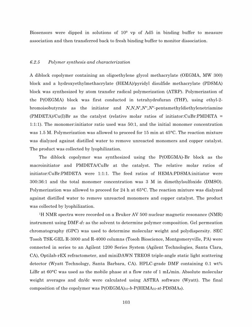
103
Biosensors were dipped in solutions of 109 vp of Ad5 in binding buffer to measure
association and then transferred back to fresh binding buffer to monitor dissociation.
6.2.5 Polymer synthesis and characterization
A diblock copolymer containing an oligoethylene glycol methacrylate (OEGMA, MW 300)
block and a hydroxyethylmethacrylate (HEMA)/pyridyl disulfide methacrylate (PDSMA)
block was synthesized by atom transfer radical polymerization (ATRP). Polymerization of
the P(OEGMA) block was first conducted in tetrahydrofuran (THF), using ethyl-2-
bromoisobutyrate as the initiator and N,N,N′,N′′,N′′-pentamethyldiethylenetriamine
(PMDETA)/Cu(I)Br as the catalyst (relative molar ratios of initiator:CuBr:PMDETA =
1:1:1). The monomer:initiator ratio used was 50:1, and the initial monomer concentration
was 1.5 M. Polymerization was allowed to proceed for 15 min at 45°C. The reaction mixture
was dialyzed against distilled water to remove unreacted monomers and copper catalyst.
The product was collected by lyophilization.
The diblock copolymer was synthesized using the P(OEGMA)-Br block as the
macroinitiator and PMDETA/CuBr at the catalyst. The relative molar ratios of
initiator:CuBr:PMDETA were 1:1:1. The feed ratios of HEMA:PDSMA:initiator were
300:36:1 and the total monomer concentration was 3 M in dimethylsulfoxide (DMSO).
Polymerization was allowed to proceed for 24 h at 65°C. The reaction mixture was dialyzed
against distilled water to remove unreacted monomers and copper catalyst. The product
was collected by lyophilization.
1H NMR spectra were recorded on a Bruker AV 500 nuclear magnetic resonance (NMR)
instrument using DMF-d7 as the solvent to determine polymer composition. Gel permeation
chromatography (GPC) was used to determine molecular weight and polydispersity. SEC
Tosoh TSK-GEL R-3000 and R-4000 columns (Tosoh Bioscience, Montgomeryville, PA) were
connected in series to an Agilent 1200 Series System (Agilent Technologies, Santa Clara,
CA), Optilab-rEX refractometer, and miniDAWN TREOS triple-angle static light scattering
detector (Wyatt Technology, Santa Barbara, CA). HPLC-grade DMF containing 0.1 wt%
LiBr at 60°C was used as the mobile phase at a flow rate of 1 mL/min. Absolute molecular
weight averages and dn/dc were calculated using ASTRA software (Wyatt). The final
composition of the copolymer was P(OEGMA)33-b-P(HEMA55-st-PDSMA8).

104
6.2.6 Preparation of peptide-grafted polymers poly(2-04) and poly(scr2-04)
Peptide conjugation to the diblock copolymer was performed as described previously with
slight modification.6 8 mg of P(OEGMA)33-b-P(HEMA55-st-PSDMA8) polymer (0.36 µmol
polymer, 2.91 µmol PDS groups) was dissolved in 1 mL of a 1:1 mixture of 0.5 M NaCl/20
mM HEPES buffer at pH 7.1 and N,N-dimethylformamide (DMF) in a 5 mL pear-shaped
flask. 10.6 mg of 2-04-Cys or scr2-04-Cys peptide (5.82 µmol, 2 equivalents relative to
diblock copolymer PDS groups) was dissolved in 0.2 mL of buffer/DMF, added into the flask,
and allowed to stir under argon at room temperature. After overnight reaction, the
absorbance of released 2-pyridinethione was measured at 343 nm to determine the extent of
the disulfide exchange reaction. The reaction mixture was dialyzed against distilled water
to remove DMF and unconjugated peptide. The product was collected by lyophilization.
6.2.7 Cell culture
HeLa cells (ATCC CCL-2) were maintained in MEM medium (Corning cellgro)
supplemented with 10% fetal bovine serum (HyClone) and 1% antibiotic-antimycotic
solution (HyClone). Cells were cultured as a monolayer in a 37°C, 5% CO2 environment.
Medium was replaced every 2-3 days. Cells were passaged at ~70-80% confluence by
incubation with Trypsin-EDTA, followed by resuspension in complete growth medium.
6.2.8 Cell transduction
Cell transduction studies with polymer-coated viruses were performed as described
previously with some modifications.5 Cells were seeded in 24-well plates at a density of
20,000 cells per well in 1 mL of complete growth medium and incubated in a 37°C, 5% CO2
environment for 24 h prior to transduction.
Polymer solutions (dissolved in sterile dH2O) were added to Ad5-GFP virus solutions
(University of Michigan Vector Core) to achieve the desired polymer to viral particle mole
ratios. Solutions were then incubated at room temperature for 30 min (or at 37°C for 3 days
for long-term stability studies) to allow polymer-virus complexes to form.

105
After incubation, adenovirus or polymer-coated virus solutions were diluted 10-fold in
growth medium without serum. Cells were rinsed once with PBS, and 200 µL of virus or
polymer-coated virus solution was added to each well. A particle multiplicity of infection
(pMOI, viral particles per cell) of 5000 was used for viral transduction. Cells were incubated
with virus at 37°C for pre-determined amounts of time and then rinsed once with PBS. 1
mL of complete growth medium with serum was added to each well and cells were returned
to the incubator.
24 h after transduction, cells were prepared for flow cytometry analysis. Cells were
rinsed once with PBS and then harvested by incubation with 200 µL of Trypsin-EDTA,
followed by resuspension with 1 mL of complete growth medium. Cells were transferred to
1.5 mL microcentrifuge tubes and pelleted at 1000 g for 5 min at 4°C. The supernatant was
aspirated, and the cell pellets were resuspended in complete growth medium containing 2
µg/mL propidium iodide (PI) and incubated at 4°C for 5 min. Cells were pelleted,
resuspended in fresh growth medium, and analyzed for GFP expression using a
MACSQuant Analyzer flow cytometer (Miltenyi).
6.3 Results and discussion
6.3.1 Identification of Ad-binding phage candidates
2×10¹¹ pfu of phage were used as input in each round of panning. The results demonstrated
an increase in the titer of phage eluted with each successive round (Figure 6.2). This
suggests progressive enrichment of the phage pool with phage bearing specificity for the
Ad5 target.

106
Figure 6.2 Titers of eluted phage over 4 rounds of panning.
Following the third and fourth rounds of panning, individual phage clones were
isolated and sequenced to identify the displayed peptides. From the sequencing results, a
number of possible consensus sequences were identified based on the frequency of their
occurrence and/or similarities to other sequences (Table 6.1). The remaining phage clones
not represented in the table (13 other clones after the third round and 6 others after the
fourth round) had unique sequences with no overlapping motifs and were not considered to
be consensus sequences.
Three potential binding sequences were identified following the fourth round of
panning. Two homologous sequences, clones 2-04 and 2-13, were found to overlap at 8 out of
12 amino acid positions and together accounted for 26% and 42% of the clones sequenced
after the third and fourth rounds, respectively. A third, non-homologous sequence (2-15)
occurred in 9% and 21% of the clones sequenced after the third and fourth rounds,
respectively.
Table 6.1 Possible consensus peptide sequences and their frequencies after the third and fourth rounds of panning. Sequence overlaps between clones 2-04/2-13 and 2-05/2-10 are underlined.
Clone name Peptide sequence Frequency after
3rd round Frequency after
4th round
2-04 EHTPRLSWLEPS 21.7% (5/23) 10.5% (2/19)
2-13 EHTAALSWLPPL 4.3% (1/23) 31.6% (6/19)
2-15 NHWPFIIRYDSL 8.7% (2/23) 21.1% (4/19)
6.40E+05 3.16E+06
3.62E+07 2.50E+08
1.E+00
1.E+02
1.E+04
1.E+06
1.E+08
1.E+10
1 2 3 4
Tota
l pha
ge e
lute
d (p
fu)
Round of panning

107
2-05 KLWDYKLSYTPV 4.3% (1/23) 5.3% (1/19)
2-10 KIWDISLSPPFT 4.3% (1/23) 0.0% (0/19)
Interestingly, two other clones, 2-05 and 2-10, were identified after the third round of
panning and overlapped one another at 5 out of 12 amino acids (and also contained the “LS”
motif present in the 2-04/2-13 sequences). However, these two clones had low overall
occurrence among the sequenced phage, particularly after the fourth round.
6.3.2 Phage binding studies
Because they were highly represented among the sequences obtained from the third and
fourth rounds of panning, phage clones 2-04, 2-13, and 2-15 were tested for binding to Ad5.
Overall, all three phage clones demonstrated specificity for Ad5, with 2- to 11-fold higher
bound phage titers over the M13KE insertless control phage (Figure 6.3). Therefore,
binding was believed to be mediated by the displayed peptide and not by another portion of
the bacteriophage coat. A low and consistent level of phage binding to uncoated polystyrene
wells was observed for all clones, suggesting that binding was predominantly virus-
dependent.
Figure 6.3 Selected phage clones demonstrate specificity for Ad5 by an elution and titering assay. (a) Raw eluate titers of phage clones bound to Ad5-coated polystyrene (black bars) and uncoated polystyrene (grey bars). (b) Peptide-mediated increase in binding, calculated by normalizing the eluate titer for each clone to the corresponding titer for the M13KE control phage.

108
6.3.3 Peptide binding studies
Having demonstrated Ad5 binding by the phage-displayed peptides, the isolated peptides
were synthesized for further characterization. Although phage clone 2-13 demonstrated the
most promising phage binding data (in addition to being the most prevalent clone found in
sequencing after the fourth round of panning), its displayed peptide is highly hydrophobic
and was predicted to have poor water solubility. Thus, we chose to investigate the 2-04
peptide due to its promising phage binding, relative hydrophilicity, and sequence similarity
to 2-13. The 2-04 peptide and a scrambled version of the peptide (scr2-04) were synthesized
to contain C-terminal biotin tags.
Biolayer interferometry (BLI) was used to assess binding interactions between the 2-04
peptide and Ad5. Biotinylated peptides were immobilized to streptavidin-coated biosensor
tips before immersing the tips in virus solution. However, only non-specific binding was
observed, as the greatest signal was seen for Ad5 binding to unloaded tips (without any
peptide immobilized), and the signal-to-noise ratio was low overall (Figure 6.4). This result
suggests that the 2-04 peptide does not bind the target or binds with extremely low affinity.
⎯⎯ –peptide/+Ad ⎯⎯ +2-04/+Ad ⎯⎯ +scr2-04/+Ad
Figure 6.4 BLI sensorgram of Ad5 binding to immobilized peptides.
6.3.4 Transduction of HeLa cells with poly(2-04)-“coated” viruses
Because each phage virion displays 5 copies of its peptide variant, avidity effects could
explain the discrepancy in binding between the phage clone and the synthesized peptide.
We therefore sought to synthesize peptide-polymer conjugates for multivalent display of the

109
peptides. A copolymer of oligoethylene glycol methacrylate (OEGMA),
hydroxylethylmethacrylate (HEMA), and pyridyl disulfide methacrylate (PDSMA) was
synthesized for preliminary studies. 2-04-Cys and scr2-04-Cys peptides were conjugated via
the PDSMA groups of the polymer to yield poly(2-04) and poly(scr2-04), respectively. UV
absorbance measurements indicate that approximately 5 peptides were conjugated per
polymer chain, consistent with the efficiency reported in the literature.6
Because the virus-binding peptides were localized to one block of the polymer, we
believed that interactions of poly(2-04) with the Ad5 surface would produce a virus that was
effectively masked by the polymer’s OEGMA block. We hypothesized that this neutral,
hydrophilic OEGMA shield would decrease viral interaction with (and therefore decrease
transduction of) HeLa cells, a cell line permissive for Ad5 infection. Surprisingly, the use of
poly(2-04) to coat Ad5-GFP was found to increase the percentage of cells that were GFP-
positive after a 1 h transduction (Figure 6.5a). These results were statistically significant
for two different molar ratios of polymer to viral particle tested, 1000:1 and 5000:1, relative
to their virus only, base polymer, and poly(scr2-04) controls. These trends were further
confirmed in a duplicate study with similar results.
Figure 6.5 HeLa transduction efficiency of uncoated Ad5-GFP and Ad5-GFP complexed with poly(2-04) or control polymers at polymer:viral particle ratios of 1000:1 and 5000:1. Complexes were incubated for (a) 30 min at room temperature or (b) 3 days at 37°C prior to transduction. Data are reported as the mean ± S.D., n = 3. *p < 0.05 compared to virus only, base polymer, and poly(scr2-04) at the same polymer:virus ratio.
Based on these trends, we hypothesized that the increased transduction efficiency of
poly(2-04)-coated viruses might be due to polymer-mediated stabilization of the virus, as
has been demonstrated for adenovirus nanocapsule formulations.7 To test this hypothesis,
naked virus and polymer-virus complexes were incubated for 3 days at 37°C prior to cell
15 min 1 h0
5
10
15
20
25
30
Transduction time
%G
FP+
cells
cells onlyvirus onlybase polymer 1000:1poly(2-04) 1000:1poly(scr2-04) 1000:1base polymer 5000:1poly(2-04) 5000:1poly(scr2-04) 5000:1
1 h 4 h0
5
10
15
20
25
30
Transduction time
%G
FP+
cells
(a) (b)
* *

110
transduction. The transduction efficiency of naked virus was significantly reduced following
3 day incubation, and transduction was not improved by the addition of poly(2-04) coating
(Figure 6.5b). Meanwhile, infection with an equal pMOI of fresh virus resulted in ~40% and
90% GFP+ cells for 1 h and 4 h transductions, respectively (data not shown). Overall, these
results indicate that the polymer does not stabilize the virus to inactivation over long
periods of time.
6.4 Conclusions and future studies
The goal of this research was to identify peptides with specificity for Ad5 hexon to be
incorporated into self-assembling polymer shields. As the first step toward developing
rationally designed polymer-adenovirus hybrids, this study demonstrated the identification
of several potential adenovirus-binding peptide sequences, using phage display panning
against surface-immobilized Ad5 with FX-based phage elution. The specificity of the
candidate phage clones for adenovirus was demonstrated via a phage titering assay.
However, binding affinity was negligible for the synthesized 2-04 peptide alone. Moreover,
preliminary studies with multivalent peptide-polymer conjugates indicated a surprising
ability of polymers containing the 2-04 peptide to increase rather than mask viral
transduction in HeLa cells. Additional studies can be performed to investigate the
mechanisms underlying increased transduction by poly(2-04)-coated viruses.
Several alternative approaches toward the development of Ad-binding polymer shields
are possible. Phage panning can be repeated using a different strategy, such as by using
solution-phase panning rather than surface panning to reduce the likelihood of selecting for
plastic-binding phage, by incorporating negative selection steps (subtractive panning) with
non-FX-binding virus mutants, or by panning against isolated Ad5 hexon protein rather
than the intact virion. Additionally, rather than identifying novel Ad-binding peptides
through phage display, the hexon-binding portion of the FX protein can be synthesized for
incorporation into polymer shields. Fender and coworkers have reported the synthesis of a
FX Gla domain-derived 40-mer peptide (termed Glamim).8 However, despite binding to hexon
with nanomolar affinity, Glamim was not able to compete with FX for hexon binding and in
fact appeared to have a different binding site than the full-length FX protein. Multivalent

111
display of the Glamim peptide on a hydrophilic polymer backbone could potentially provide
sufficient avidity and steric hindrance to compete with the FX protein.
6.5 Acknowledgements
This work was supported by the National Science Foundation (DMR 1206426), and a
National Science Foundation Graduate Research Fellowship to C.E.W. We acknowledge
Jamil Qazi for assistance with peptide synthesis.
References
1. Khare, R., Chen, C. Y., Weaver, E. A. & Barry, M. A. Advances and future challenges in adenoviral vector pharmacology and targeting. Curr Gene Ther 11, 241–258 (2011).
2. Lieber, A., He, C. Y., Meuse, L., Schowalter, D., Kirillova, I., Winther, B. & Kay, M. A. The role of Kupffer cell activation and viral gene expression in early liver toxicity after infusion of recombinant adenovirus vectors. J. Virol. 71, 8798–8807 (1997).
3. Kalyuzhniy, O., Di Paolo, N. C., Silvestry, M., Hofherr, S. E., Barry, M. A., Stewart, P. L. & Shayakhmetov, D. M. Adenovirus serotype 5 hexon is critical for virus infection of hepatocytes in vivo. Proc. Natl. Acad. Sci. U.S.A. 105, 5483–5488 (2008).
4. Waddington, S. N., McVey, J. H., Bhella, D., Parker, A. L., Barker, K., Atoda, H., Pink, R., Buckley, S. M. K., Greig, J. A., Denby, L., Custers, J., Morita, T., Francischetti, I. M. B., Monteiro, R. Q., Barouch, D. H., van Rooijen, N., Napoli, C., Havenga, M. J. E., Nicklin, S. A. & Baker, A. H. Adenovirus serotype 5 hexon mediates liver gene transfer. Cell 132, 397–409 (2008).
5. Wang, C.-H. K., Chan, L. W., Johnson, R. N., Chu, D. S. H., Shi, J., Schellinger, J. G., Lieber, A. & Pun, S. H. The transduction of Coxsackie and Adenovirus Receptor-negative cells and protection against neutralizing antibodies by HPMA-co-oligolysine copolymer-coated adenovirus. Biomaterials 32, 9536–9545 (2011).
6. Schellinger, J. G., Pahang, J. A., Johnson, R. N., Chu, D. S., Sellers, D. L., Maris, D. O., Convertine, A. J., Stayton, P. S., Horner, P. J. & Pun, S. H. Melittin-grafted HPMA-Oligolysine Based Copolymers for Improved Gene Delivery. Biomaterials 34, 2318–2326 (2013).
7. Weng, D., Jiang, Z. K., Jin, J., Wu, L. & Lu, Y. Enhanced structural stability of adenovirus nanocapsule. Progress in Natural Science: Materials International 24, 171–174 (2014).
8. Sumarheni, S., Hong, S.-S., Josserand, V., Coll, J.-L., Boulanger, P., Schoehn, G. & Fender, P. Human full-length coagulation factor X and a GLA domain-derived 40-mer polypeptide bind to different regions of the adenovirus serotype 5 hexon capsomer. Hum. Gene Ther. 25, 339–349 (2014).

112

113
Part IV.
Future Perspectives

114

115
Chapter 7.
SUMMARY OF MAJOR FINDINGS AND RECOMMENDATIONS
FOR FUTURE WORK
7.1 Summary of major findings
7.1.1 Junction opener proteins for enhancing nanoparticle delivery to solid tumors
Proteins that disrupt intercellular tight junctions can be used to enhance the diffusion of
chemotherapeutic agents into epithelial tumors. In Chapter 1, PEGylated gold
nanoparticles (AuNPs) were synthesized and injected in mice to investigate the size-
dependent effect of the junction opener protein JO-4 on particle biodistribution. JO-4
pretreatment significantly increased tumor accumulation of 35 nm AuNPs but had no effect
on larger particles of 120 nm. In addition to increasing the overall number of AuNPs
localized to the tumor, JO-4 increased nanoparticle penetration into the tumor. These
findings are important to further the design and development of drug carriers that utilize
this delivery mechanism.
7.1.2 Sunflower polymers for tumor-targeted drug delivery
Polymer-drug conjugates with advanced architectures were investigated as an alternative
to traditional drug carriers such as liposomes and micelles, which require formulation steps
during manufacturing and can release drug prematurely in vivo. Chapter 2 summarizes the
major considerations when designing anti-cancer drug delivery vehicles and highlights
controlled living polymerization techniques enabling the synthesis of increasingly complex
polymer nanostructures. In Chapter 3, atom transfer radical polymerization was used to
synthesize polymers with a sunflower-like architecture. Sunflower polymers were
synthesized with low polydispersity and good control over particle size based on
polymerization time. Polymers were targeted to cancer cells by conjugation of folate to
radiating “petals.” These targeted sunflower polymers demonstrated receptor-mediated
uptake by folate receptor-positive KB cells, while their linear analogues (comb polymers)

116
were taken up by non-specific mechanisms. In Chapter 4, this platform was extended by
conjugating doxorubicin (Dox) to the polymer core via a pH-sensitive linker; targeted, Dox-
loaded sunflower polymers demonstrated intracellular release of active drug leading to
cytotoxicity in KB cells. However, in vivo efficacy of these polymers was limited due to low
drug loading.
7.1.3 Self-assembling materials for cloaking adenoviral vectors
Polymeric coatings have the potential to improve the safety and efficacy of adenoviral gene
delivery vectors in vivo. Chapter 5 reviews the challenges in translating adenoviruses to the
clinic and current strategies for virus modification. In Chapter 6, phage display was used to
screen for novel peptide ligands that bind specifically to the hexon protein of adenovirus
serotype 5 (Ad5), with the ultimate goal of incorporating these peptides into self-assembling
polymer shields for the virus. Multiple consensus sequences were identified, and several
phage clones were found to bind specifically to adenovirus. However, binding affinity to
adenovirus was negligible for the purified peptide alone.
7.2 Recommendations for future work
7.2.1 JO-conjugated sunflower polymers for tumor-targeted drug delivery
Background and significance
In previous work by Lieber and coworkers1,2 and in Chapter 1 of this thesis, junction opener
(JO) proteins were administered one hour prior to injection with a therapeutic compound or
model drug. JO proteins have not yet been conjugated directly to drugs, nanoparticles, or
other systems; however, given that the junction opening effect appears to be specific to
tumors (DSG2 is generally not accessible for JO binding in normal tissues3), JO conjugation
may be a useful strategy for enhancing the tumor localization, penetration, and therapeutic
efficacy of anti-cancer drug delivery vehicles.
Aim 1: Synthesis of JO-conjugated AuNPs and quantification of biodistribution
JO proteins containing a C-terminal cysteine will be provided by PAI Life Sciences (Seattle,
WA) and used to surface modify 5 nm AuNPs via thiol-gold reaction. After reaction, AuNPs

117
will be pelleted by centrifugation and washed, and unbound protein will be removed with
the supernatant. JO-conjugated AuNPs (JO-AuNPs) will be characterized by dynamic light
scattering. The biodistribution and intratumoral penetration of JO-AuNPs in A549 tumor-
bearing mice will be analyzed following the methods described in Chapter 1. Results will be
compared to those obtained with the traditional administration strategy (injection of JO
protein followed by a separate injection of AuNPs one hour later). Furthermore, JO content
on the AuNPs can be varied to determine if there is a JO surface density for which tumor
accumulation is maximized.
Aim 2: Synthesis of JO-conjugated sunflower polymers
JO proteins containing a C-terminal cysteine will be labeled with a dibenzocyclooctyne
(DBCO)-PEG4-maleimide crosslinker (Click Chemistry Tools, Scottsdale, AZ) to introduce
cyclooctyne functionality. Labeled protein will be purified using a PD-10 desalting column
to remove unreacted crosslinker. The resulting JO-DBCO protein will be conjugated to the
sunflower polymers synthesized in Chapters 3 and 4 by strain-promoted copper-free click
reaction between the cyclooctyne and the azide termini of sunflower petals. The final JO-
sunflower conjugates will be purified by size exclusion chromatography and characterized
by dynamic light scattering, SDS-PAGE, and Western blot analysis.
The ability of the JO-sunflower conjugates to bind and open tight junctions will be
verified using PEG permeability assays as described previously.1 In addition, cytotoxicity
studies will be performed as described in Chapter 4 to ensure that JO conjugation does not
affect the potency of the sunflower-Dox formulation.
Aim 3: In vivo evaluation of JO-conjugated sunflower polymers
JO-sunflower conjugates will be tested for anti-tumor efficacy using DSG2 transgenic mice.3
Mice will be inoculated subcutaneously with ID8-DSG2 cells, a murine ovarian cancer cell
line that has been engineered to express human DSG2. After tumors are established, the
following treatment groups will be tested: free Dox, free JO protein, sunflower-Dox
formulation, JO-sunflower-Dox formulation, or a noncovalent mixture of JO protein and
sunflower-Dox formulation. Tumor volumes and mouse weights will be measured every 2-3
days for the duration of the study. In initial studies, mice will receive a single treatment; if
no anti-tumor efficacy is observed, the study may be repeated using a multiple-dose
strategy. If promising efficacy is observed, JO-sunflower conjugates bearing a fluorescent

118
label can also be synthesized to analyze biodistribution and intratumoral penetration of the
polymers.
Preliminary results
In preliminary studies, the sunflower polymers described in Chapter 3 were conjugated
with two different JO-derived protein constructs, named GB3_JO1 and GB7_JO1, by
copper-free click chemistry. Anti-JO1 Western blot analysis of the crude reaction mixtures
indicated that GB3_JO1-sunflower conjugates were successfully generated and that
conjugates were stable under reducing conditions (A in Figure 7.1). However, GB7_JO1
conjugation appeared to be unsuccessful (no shift in protein migration at B in Figure 7.1);
this may be the result of poor labeling of GB7_JO1 protein with the DBCO-PEG4-maleimide
crosslinker.
Figure 7.1 Anti-JO1 Western blot of GB3_JO1 and GB7_JO1 proteins before and after click conjugation to sunflower polymers.

119
7.2.2 Sunflower polymers with increased drug loading
Background and significance
In Chapters 3 and 4, we reported the synthesis of sunflower polymers containing ethyl
glycinate methacrylamide (EGMA) monomers in the polymer core. The EGMA sidechains
were then converted from ethyl esters to hydrazides to allow for direct conjugation of
doxorubicin (Dox). However, these polymers suffered from a low level of drug loading (<5
w/w%). In order to increase the drug content of the sunflower polymer formulation, a second
generation of sunflower polymers can be synthesized with carboxylic acid groups in the
polymer core. These functional groups can be used for electrostatic complexation of Dox
(which is cationic at neutral pH).4,5
Aim 1: Synthesis of sunflower polymers for electrostatic Dox loading
Modified sunflower polymers will be synthesized and characterized as described in
Chapters 3 and 4 with slight variation. The protected monomer tert-butyl methacrylate
(tBMA) will be used in place of the EGMA monomer in the sunflower polymer core. After
completing the sunflower polymer synthesis, an excess of trifluoroacetic acid (TFA) will be
used to deprotect tBMA monomers, yielding carboxylic acid groups (Figure 7.2).6,7 The final
sunflower polymer composition will therefore be P[(HEMA-sunflower-P(OEGMA))-st-
methacrylic acid]. Sunflower polymers will then be drug-loaded by noncovalent
complexation with Dox.
Figure 7.2 Hydrolysis of tert-butyl methacrylate (tBMA) to methacrylic acid (MAA).
As an alternative strategy to increase the number of sites available for drug loading,
each “petal” of the sunflower polymer can be synthesized as a diblock copolymer, such that
the block adjacent to the polymer core is composed of tBMA while the outer block of the
petal is composed of a hydrophilic monomer such as oligoethylene glycol methacrylate
(OEGMA) to shield the drug cargo.
O
O
HO
O
TFA
t-BMA MAA

120
7.2.3 Polymer-modified adenoviruses: a “grafting-from” approach
Background and significance
One strategy for engineering adenoviral vectors with reduced immunogenicity and altered
biodistribution in vivo involves conjugating polymers such as polyethylene glycol (PEG) and
poly-N-(2-hydroxypropyl) methacrylamide (HPMA) to the Ad capsid.8 Covalent
modifications have shown great promise for circumventing the major hurdles to in vivo
delivery but can also lead to decreased infectivity of the viruses.9-11 Bioresponsive linkers
including reducible disulfide bonds11,12 and pH-sensitive hydrazone bonds12 can allow for
“re-activation” of the virus upon uncoating. Even so, these bioresponsive polymers are
typically conjugated to the Ad capsid via surface amine groups, and it has been suggested
that residual linker groups left behind at the site of attachment could increase particle
immunogenicity.13 Indeed, studies with non-responsive PEG modification have
demonstrated that the neutralizing antibody response may be directed against the site of
PEG conjugation.14
Recently, Kreppel and co-workers reported the development of AdHexCys, an Ad5-
based vector which was genetically modified to contain a cysteine residue in hypervariable
region 5 (HVR5) of the hexon.13 Using this vector, “traceless” bioresponsive shielding could
be achieved using HPMA-based polymers incorporating pyridyl-dithio groups for reaction
with the introduced cysteines. As compared to non-responsive shielding based on
irreversible maleimide conjugations, bioresponsive shields maintained high transduction
efficiency both in vitro and in vivo. However, in vivo data suggested that these polymers
provided incomplete shielding against IgM binding.
Covalent modification of vectors using pre-synthesized polymers (the “grafting-to”
approach) is limited in its ability to achieve a high density of surface conjugation due to
steric hindrance between bulky polymer molecules. In contrast, a “grafting-from” approach,
in which polymer chains are grown directly from the Ad capsid, may allow for the
generation of a more uniform coating. Although previous results demonstrate that
conjugation of large PEG moieties of at least 20 kDa is necessary for efficient Ad
detargeting away from hepatocytes in vivo,15 it is possible that a denser layer of shorter
polymer chains may afford the same protection.
We propose to synthesize polymer-modified adenoviruses using a grafting-from
approach, in which AdHexCys is conjugated with chain transfer agents (CTAs) for

121
reversible addition-fragmentation chain-transfer (RAFT) polymerization. Grafting-from
RAFT polymerization using protein-based macroCTAs has been demonstrated
previously,16,17 although it has not yet been applied to biologically active proteins such as
viruses. This strategy can potentially enable high-density covalent (yet bioresponsive)
coating of adenovirus, thereby maximizing shielding while maintaining the activity of the
native virus.
Aim 1: Synthesis and characterization of pOEGMA-Ad vectors
A RAFT CTA containing a pyridyl disulfide functional group, trithiocarbonic acid 1-cyano-1-
methyl-3-[2-(pyridin-2-yldisulfanyl)-ethylcarbamoyl]-propyl ester ethyl ester (PyrECT),18
will be reacted to free thiols on the AdHexCys surface via disulfide exchange. PyrECT
conjugation will be performed under physiological conditions as described previously.18,19
PyrECT-conjugated AdHexCys will be used as “multi-macroCTAs” in RAFT
polymerization to generate polymer-virus conjugates. RAFT polymerization will be
conducted in aqueous buffer at 35°C to minimize inactivation of the virus, using OEGMA as
the monomer and VA-044 as the initiator. Free ethyl cyanovaleric trithiocarbonate (ECT)
will also be included as sacrificial CTA due to the relatively low quantities of virus used.
The ratio of monomer to ECT/PyrECT can be varied to produce viruses with different
lengths of their polymer shield. This can be used to identify a polymer length that is
optimal for downstream virus activity. The final polymer-modified virus, referred to as
pOEGMA-Ad, will be purified using an Amicon Ultra Centrifugal Filter device with 100
kDa MWCO (Millipore).
Successful modification of the virus will be assessed by SDS-PAGE. pOEGMA-Ad will
be incubated at 100°C for 5 min in reducing or non-reducing Laemmli sample buffer and
run on a 10% Mini-PROTEAN TGX gel (Bio-Rad), followed by staining with a SilverQuest
Silver Staining Kit (Invitrogen). Under non-reducing conditions, the electrophoretic
mobility of hexon proteins is expected to be retarded by covalent polymer modification; the
addition of reducing agent should release the polymer and allow for hexon migration into
the gel.11
The pOEGMA chains grown from sacrificial ECT will be used as a proxy to characterize
the polymer chains grown from the virus surface. Free pOEGMA will be separated from
virus using an Amicon Ultra Centrifugal Filter device, followed by gel permeation
chromatography (GPC) to determine molecular weight and polydispersity.

122
Finally, dynamic light scattering (DLS) will be used to measure pOEGMA-Ad particle
size and zeta potential. Relative to naked Ad, pOEGMA modification is expected to produce
a slight increase in size and in zeta potential (from negative to near-neutral) depending on
the length of polymer chains.20
Aim 2: In vitro evaluation of pOEGMA-Ad vectors
CAR-positive HeLa cells will be infected with pOEGMA-Ad encoding GFP or naked Ad-GFP
and analyzed for GFP expression as described in Chapter 6. Because Ad5 transduces HeLa
cells efficiently via CAR (through interactions with the Ad5 fiber protein), transduction
efficiency is expected to be similar between the naked and polymer-modified vectors.
However, if polymer modification compromises transduction efficiency, activity should be
restored with the addition of a reducing agent such as tris(2-carboxyethyl) phosphine
(TCEP).
CAR-negative SKOV3 (human ovarian carcinoma) cells will be infected with pOEGMA-
Ad or naked Ad in the absence or presence of a physiological concentration (8 µg/mL) of
human FX protein (Haematologic Technologies). The addition of FX is expected to
significantly enhance the transduction efficiency of unmodified Ad, as others have shown;21
however, FX addition should have minimal effect on transduction efficiency of pOEGMA-
Ad, as these should be resistant to FX binding.
pOEGMA-Ad and naked Ad will be incubated with serum containing anti-Ad5
neutralizing antibodies for 20 min at 37°C and then used to infect HeLa cells.20 The
addition of NAbs is expected to severely compromise the transduction efficiency of naked
Ad5 while pOEGMA-Ad will be resistant to inactivation by NAbs.
Preliminary results
Conditions for aqueous RAFT polymerization of OEGMA were optimized in the absence of
virus. Polymerizations were conducted in 0.1 M phosphate buffer (pH 6.5) using ECT as the
RAFT CTA and VA-044 as the initiator (I). The initial OEGMA monomer concentration was
0.25 M, and the molar ratios of monomer:CTA:I at the start of polymerization were 150:1:1.
Polymerizations were carried out in a 35°C oil bath. A study of the polymerization kinetics
indicated that 25% conversion (corresponding to a theoretical polymer molecular weight of
approximately 11.3 kDa) was achieved by 1 h (Figure 7.3).
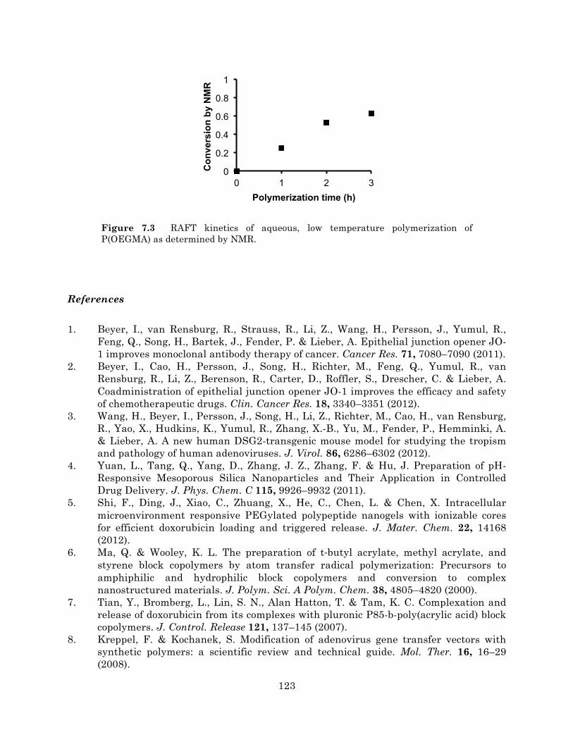
123
Figure 7.3 RAFT kinetics of aqueous, low temperature polymerization of P(OEGMA) as determined by NMR.
References
1. Beyer, I., van Rensburg, R., Strauss, R., Li, Z., Wang, H., Persson, J., Yumul, R., Feng, Q., Song, H., Bartek, J., Fender, P. & Lieber, A. Epithelial junction opener JO-1 improves monoclonal antibody therapy of cancer. Cancer Res. 71, 7080–7090 (2011).
2. Beyer, I., Cao, H., Persson, J., Song, H., Richter, M., Feng, Q., Yumul, R., van Rensburg, R., Li, Z., Berenson, R., Carter, D., Roffler, S., Drescher, C. & Lieber, A. Coadministration of epithelial junction opener JO-1 improves the efficacy and safety of chemotherapeutic drugs. Clin. Cancer Res. 18, 3340–3351 (2012).
3. Wang, H., Beyer, I., Persson, J., Song, H., Li, Z., Richter, M., Cao, H., van Rensburg, R., Yao, X., Hudkins, K., Yumul, R., Zhang, X.-B., Yu, M., Fender, P., Hemminki, A. & Lieber, A. A new human DSG2-transgenic mouse model for studying the tropism and pathology of human adenoviruses. J. Virol. 86, 6286–6302 (2012).
4. Yuan, L., Tang, Q., Yang, D., Zhang, J. Z., Zhang, F. & Hu, J. Preparation of pH-Responsive Mesoporous Silica Nanoparticles and Their Application in Controlled Drug Delivery. J. Phys. Chem. C 115, 9926–9932 (2011).
5. Shi, F., Ding, J., Xiao, C., Zhuang, X., He, C., Chen, L. & Chen, X. Intracellular microenvironment responsive PEGylated polypeptide nanogels with ionizable cores for efficient doxorubicin loading and triggered release. J. Mater. Chem. 22, 14168 (2012).
6. Ma, Q. & Wooley, K. L. The preparation of t‐butyl acrylate, methyl acrylate, and styrene block copolymers by atom transfer radical polymerization: Precursors to amphiphilic and hydrophilic block copolymers and conversion to complex nanostructured materials. J. Polym. Sci. A Polym. Chem. 38, 4805–4820 (2000).
7. Tian, Y., Bromberg, L., Lin, S. N., Alan Hatton, T. & Tam, K. C. Complexation and release of doxorubicin from its complexes with pluronic P85-b-poly(acrylic acid) block copolymers. J. Control. Release 121, 137–145 (2007).
8. Kreppel, F. & Kochanek, S. Modification of adenovirus gene transfer vectors with synthetic polymers: a scientific review and technical guide. Mol. Ther. 16, 16–29 (2008).
0
0.2
0.4
0.6
0.8
1
0 1 2 3
Con
vers
ion
by N
MR
Polymerization time (h)

124
9. Mok, H., Palmer, D. J., Ng, P. & Barry, M. A. Evaluation of polyethylene glycol modification of first-generation and helper-dependent adenoviral vectors to reduce innate immune responses. Mol. Ther. 11, 66–79 (2005).
10. Wortmann, A., Vöhringer, S., Engler, T., Corjon, S., Schirmbeck, R., Reimann, J., Kochanek, S. & Kreppel, F. Fully detargeted polyethylene glycol-coated adenovirus vectors are potent genetic vaccines and escape from pre-existing anti-adenovirus antibodies. Mol. Ther. 16, 154–162 (2008).
11. Subr, V., Kostka, L., Selby-Milic, T., Fisher, K. D., Ulbrich, K., Seymour, L. W. & Carlisle, R. C. Coating of adenovirus type 5 with polymers containing quaternary amines prevents binding to blood components. J Control Release 135, 152–158 (2009).
12. Espenlaub, S., Corjon, S., Engler, T., Fella, C., Ogris, M., Wagner, E., Kochanek, S. & Kreppel, F. Capsomer-specific fluorescent labeling of adenoviral vector particles allows for detailed analysis of intracellular particle trafficking and the performance of bioresponsive bonds for vector capsid modifications. Hum. Gene Ther. 21, 1155–1167 (2010).
13. Prill, J.-M., Subr, V., Pasquarelli, N., Engler, T., Hoffmeister, A., Kochanek, S., Ulbrich, K. & Kreppel, F. Traceless Bioresponsive Shielding of Adenovirus Hexon with HPMA Copolymers Maintains Transduction Capacity In Vitro and In Vivo. PLoS ONE 9, e82716 (2014).
14. Croyle, M. A., Chirmule, N., Zhang, Y. & Wilson, J. M. ‘Stealth’ adenoviruses blunt cell-mediated and humoral immune responses against the virus and allow for significant gene expression upon readministration in the lung. J. Virol. 75, 4792–4801 (2001).
15. Prill, J.-M., Espenlaub, S., Samen, U., Engler, T., Schmidt, E., Vetrini, F., Rosewell, A., Grove, N., Palmer, D., Ng, P., Kochanek, S. & Kreppel, F. Modifications of adenovirus hexon allow for either hepatocyte detargeting or targeting with potential evasion from Kupffer cells. Mol. Ther. 19, 83–92 (2011).
16. Boyer, C., Bulmus, V., Liu, J., Davis, T. P., Stenzel, M. H. & Barner-Kowollik, C. Well-defined protein-polymer conjugates via in situ RAFT polymerization. J. Am. Chem. Soc. 129, 7145–7154 (2007).
17. Li, H., Li, M., Yu, X., Bapat, A. P. & Sumerlin, B. S. Block copolymer conjugates prepared by sequentially grafting from proteins via RAFT. Polym Chem (2011).
18. Duvall, C. L., Convertine, A. J., Benoit, D. S. W., Hoffman, A. S. & Stayton, P. S. Intracellular delivery of a proapoptotic peptide via conjugation to a RAFT synthesized endosomolytic polymer. Mol. Pharm. 7, 468–476 (2010).
19. Schellinger, J. G., Pahang, J. A., Johnson, R. N., Chu, D. S., Sellers, D. L., Maris, D. O., Convertine, A. J., Stayton, P. S., Horner, P. J. & Pun, S. H. Melittin-grafted HPMA-Oligolysine Based Copolymers for Improved Gene Delivery. Biomaterials 34, 2318–2326 (2013).
20. Wang, C.-H. K., Chan, L. W., Johnson, R. N., Chu, D. S. H., Shi, J., Schellinger, J. G., Lieber, A. & Pun, S. H. The transduction of Coxsackie and Adenovirus Receptor-negative cells and protection against neutralizing antibodies by HPMA-co-oligolysine copolymer-coated adenovirus. Biomaterials 32, 9536–9545 (2011).
21. Alba, R., Bradshaw, A. C., Parker, A. L., Bhella, D., Waddington, S. N., Nicklin, S. A., van Rooijen, N., Custers, J., Goudsmit, J., Barouch, D. H., McVey, J. H. & Baker, A. H. Identification of coagulation factor (F)X binding sites on the adenovirus serotype 5 hexon: effect of mutagenesis on FX interactions and gene transfer. Blood 114, 965–971 (2009).

125

126
APPENDIX
MATLAB code for AuNP penetration analysis
Sample code to calculate penetration distances for a collection of thresholded brightfield
and fluorescence image pairs:
% mouse ID: 583 (JO/35 nm) filepath = '\\studentfile.student.bioeng.washington.edu\usr$\cewang\Desktop\NP analysis\Thresholded\JO-35\583'; % path to folder containing pairs of thresholded brightfield and fluorescence images (e.g., 1B.tif, 1F.tif, 2B.tif, 2F.tif,...) savepath = '\\studentfile.student.bioeng.washington.edu\usr$\cewang\Desktop\NP analysis\Output\583_'; % path for output distance_data_aggregate = []; N_aggregate = []; cd(filepath); filelist = dir; for ii = 3:length(filelist) if strcmp(filelist(ii).name(end-4:end),'B.tif') % import a pair of brightfield and fluorescence images bf = imread(filelist(ii).name); fl = imread(strcat(filelist(ii).name(1:end-5),'F.tif')); else continue end % find all regions of fluorescence (thresholded as white on black) [fl_L ~] = bwlabel(fl); fl_stats = regionprops(fl_L); for zz = 1:length(fl_stats) object_size = fl_stats(zz).Area; % remove fluorescent regions with areas smaller than 9 pixels % (assumed to be noise) if object_size<=9 [r,c] = find(fl_L==zz); fl_L(r,c)=0; end end fl_L(fl_L>0)=255; fl = uint8(fl_L); % obtain coordinates for all fluorescent pixels [fl_y fl_x] = find(fl > 0); % find all AuNPs in brightfield image (thresholded as white on black) [L N] = bwlabel(bf);

127
stats = regionprops(L); centroids_x = []; centroids_y = []; % calculate centroid of each AuNP for jj = 1:length(stats) centroids_x = [centroids_x stats(jj).Centroid(1)]; centroids_y = [centroids_y stats(jj).Centroid(2)]; end centroids_x_length = length(centroids_x); fl_x_length = length(fl_x); centroids_x_tiled = repmat(centroids_x,fl_x_length,1); fl_x_tiled = repmat(fl_x,1,centroids_x_length); centroids_y_length = length(centroids_y); fl_y_length = length(fl_y); centroids_y_tiled = repmat(centroids_y,fl_y_length,1); fl_y_tiled = repmat(fl_y,1,centroids_y_length); % calculate minimum pixel distance from each AuNP centroid to a % fluorescent pixel distances = sqrt((centroids_x_tiled-fl_x_tiled).^2 + ... (centroids_y_tiled-fl_y_tiled).^2); [distance_data,index]=min(distances); % overlay brightfield/fluorescence images figure im = cat(3,zeros(size(bf)),fl, bf); imshow(im); hold on % draw lines to indicate penetration distances for jj = 1:length(centroids_x) plot([centroids_x(jj),fl_x(index(jj))], ... [centroids_y(jj),fl_y(index(jj))],'r') end title(filelist(ii).name); savefig(strcat(savepath,filelist(ii).name(1:end-5))); % save distances and # of nanoparticles for this image pair to data file save(strcat(savepath,filelist(ii).name(1:end-5)),'N','distance_data'); pause(2); hold off close distance_data_aggregate = [distance_data_aggregate distance_data]; N_aggregate = [N_aggregate N]; end % aggregate distances and # of nanoparticles for all images analyzed save(strcat(savepath,'aggregate'),'distance_data_aggregate','N_aggregate');

128
Sample code to generate histograms and statistics for penetration distance data:
filepath = '\\studentfile.student.bioeng.washington.edu\usr$\cewang\Desktop\NP analysis\Output'; % path to folder containing aggregated distance data cd(filepath); % load distance data and convert to microns according to an image scale of % 0.15 um/pixel dat583 = load('583_aggregate.mat'); dist583 = dat583.distance_data_aggregate .* 0.15; N583 = dat583.N_aggregate; dat593 = load('593_aggregate.mat'); dist593 = dat593.distance_data_aggregate .* 0.15; N593 = dat593.N_aggregate; % remove distances <= 5 um dist583_2 = dist583(find(dist583>5)); dist593_2 = dist593(find(dist593>5)); % plot histograms of penetration distances figure hist583_2 = histogram(dist583_2, 'Normalization', 'probability'); hist583_2.BinWidth = 5; xlim([5 135]) hold on hist593_2 = histogram(dist593_2, 'Normalization', 'probability', ... 'BinEdges', hist583_2.BinEdges); legend('583: JO-4 / 35 nm','593: Untreated / 35 nm') xlabel('Penetration distance (\mum)') ylabel('Fraction') % return statistics on number of NPs analyzed fprintf('Total nanoparticles analyzed\n\n') fprintf('583: ') disp(sum(N583)) fprintf('593: ') disp(sum(N593)) % return statistics on average number of nanoparticles per image fprintf('\n\nAverage and SD of nanoparticles per image\n\n') fprintf('583: ') disp(mean(N583)) disp(std(N583)) fprintf('593: ') disp(mean(N593))

129
disp(std(N593)) % return statistics on percent of NPs <= 5 um from blood vessel fprintf('\n\nFraction of NPs within 5 um of blood vessel\n\n') fprintf('583: ') disp(1-(length(dist583_2)/length(dist583))) fprintf('593: ') disp(1-(length(dist593_2)/length(dist593))) % return statistics on median penetration distance fprintf('\n\nMedian penetration distance in um (excluding values below 5 um) \n\n') fprintf('583: ') disp(median(dist583_2)) fprintf('593: ') disp(median(dist593_2))