Characterisation of "ESKAPE" Pathogens with Special ...
27
Characterisation of "ESKAPE" Pathogens with Special Reference to Multidrug Resistance and Bioヲlm Production in a Nepalese Hospital Rosy Pandey ( [email protected] ) Tribhuvan University Angela Shrestha St. Xacier's College Shyam Kumar Mishra Institute of Medicine, Tribhuvan University Research article Keywords: ESBL, ESKAPE Pathogens, MBL, MRSA, VRE Posted Date: December 9th, 2019 DOI: https://doi.org/10.21203/rs.2.18490/v1 License: This work is licensed under a Creative Commons Attribution 4.0 International License. Read Full License Version of Record: A version of this preprint was published at Infection and Drug Resistance on June 1st, 2021. See the published version at https://doi.org/10.2147/IDR.S306688.
-
Upload
khangminh22 -
Category
Documents
-
view
3 -
download
0
Transcript of Characterisation of "ESKAPE" Pathogens with Special ...

Characterisation of "ESKAPE" Pathogens withSpecial Reference to Multidrug Resistance andBio�lm Production in a Nepalese HospitalRosy Pandey ( [email protected] )
Tribhuvan UniversityAngela Shrestha
St. Xacier's CollegeShyam Kumar Mishra
Institute of Medicine, Tribhuvan University
Research article
Keywords: ESBL, ESKAPE Pathogens, MBL, MRSA, VRE
Posted Date: December 9th, 2019
DOI: https://doi.org/10.21203/rs.2.18490/v1
License: This work is licensed under a Creative Commons Attribution 4.0 International License. Read Full License
Version of Record: A version of this preprint was published at Infection and Drug Resistance on June 1st,2021. See the published version at https://doi.org/10.2147/IDR.S306688.

1
CHARACTERISATION OF ESKAPE PATHOGENS WITH
SPECIAL REFERENCE TO MULTIDRUG RESISTANCE AND
BIOFILM PRODUCTION IN A NEPALESE HOSPITAL Rosy Pandey1* , Shyam Kumar Mishra 2, Angela Shrestha1
Address:
1 Tribhuvan University, St. Xavier's College, Department of Microbiology, Kathmandu, Nepal.
2 Tribhuvan University, Institute of Medicine, Department of Microbiology, Kathmandu, Nepal.
E-mail:
Rosy Pandey- [email protected],
Shyam Kumar Mishra - [email protected]
Angela Shrestha- [email protected]
*Corresponding author: [email protected]
ABSTRACT
1. Background: “ESKAPE” is an acronym for group of organisms as Enterococcus
faecium, Staphylococcus aureus, Klebsiella pneumoniae, Acinetobacter calcoaceticus
baumannii complex, Pseudomonas aeruginosa and Enterobacter spp. They are associated
in causing life threatening infections. Global efforts on controlling multidrug resistant
(MDR) organisms have been hampered by their rapid emergence, inadequate tests for
rapid detection and their ability to escape the antibacterial drugs. The objective of this
study was to determine the prevalence of ESKAPE pathogens with prime focus on
biofilm production and antibiotic resistance.
2. Methods: A total of 8756 clinical specimens were processed for the isolation and
identification of ESKAPE pathogens following standard microbiological protocol. These
isolates were subjected to antibiotic sensitivity test as per Clinical and Laboratory
Standards Institute (CLSI) guidelines. Detection of resistance phenotypes, viz., extended-
spectrum-beta-lactamase (ESBL), metallo-beta-lactamase (MBL), Methicillin-resistant
Staphylococcus aureus (MRSA), Vancomycin-resistant enterococci (VRE) was done by
disk diffusion method and E- test method as applicable. The VRE isolates were subjected
for detection of Van A and Van B genes. All the isolates were processed for biofilm
detection by tube adherence method.
3. Results: The percentage distribution of Staphylococcus aureus was 33.5%, followed by
Klebsiella pneumoniae 33.0%, Pseudomonas aeruginosa 18.3%, Acinetobacter
calcoaceticus baumannii complex 8.7%, Enterococcus faecium 5.6% and Enterobacter
aerogenes 0.9%. MRSA was 57.6% and Vancomycin resistance among Enterococcus

2
faecium was 20%. ESBL and MBL producing Klebsiella pneumoniae were 16.1%, and
8.1%, Acb complex 10.3% each and Pseudomonas aeruginosa 10.7% and 8.3%
respectively. A total of 42.3% of isolates were biofilm producers. Linezolid was drug of
choice for VRE isolates. Piperacillin- tazobactam was found to be effective against
Pseudomonas aeruginosa, Klebsiella pneumoniae and Enterobacter aerogenes;
Ampicillin-sulbactam was the most effective drug against Acb complex excluding
polymyxins. Van A gene was detected in all the VRE isolates.
4. Conclusion: This study estimates the burden of the ESKAPE organisms and their
antibiotic resistance pattern in a Nepalese hospital. The increasing percentages of drug
resistance among these biofilm-producing pathogens pose great threat in medical setting.
Surveillance targeting ESKAPE pathogens should be incorporated in infection control
policy in Nepal.
Keywords: ESBL, ESKAPE Pathogens, MBL, MRSA, VRE
Background
Emergence of bacterial pathogens with acquired resistance to almost all available antibiotics
thus limiting therapeutic choices is one of the major concerns today. These pathogens are also
named as 'superbugs', particularly Enterococcus faecium, Staphylococcus aureus, Klebsiella
pneumoniae, Acinetobacter calcoaceticus baumannii complex, Pseudomonas aeruginosa and
Enterobacter spp. (ESKAPE) are capable of escaping the bactericidal effect of antibiotics 1.
Inefficiency of antibiotics is due to various phenotypic and genotypic mechanisms as drug
inactivation, modification of drug binding sites/ targets, changes in cell permeability and
mutation 2, 3.
Methicillin resistant Staphylococcus aureus (MRSA) emerged in 1960s and currently its
infection rate in hospital setting is around 60-70% and in community setting it is up to 50% 4.
Staphylococcus aureus with auxiliary PBP-2a encoded by Staphylococcal cassette
chromosome mec (SCC mec A, C) mediate β-lactam resistance due to low affinity 5, 6.
Beta-lactamase is the enzyme produced by organisms which hydrolyze β-lactam antibiotics
mediating resistance and making the drug in treatment impotent. It is classified into two
categories, i.e. Ambler molecular classification scheme and Bush-Jacoby-Medieros
functional classification system 7. Ambler classification scheme divides beta lactamase into
four major classes (class A-D), where A, C and D are serine β-lactamase and class B enzyme

3
is MBL. Ambler class B enzyme is characterized by its ability to hydrolyze carbapenems and
its resistance to all available beta-lactam but is inhibited in presence of metal chelators 8.
ESBL is defined as an enzyme that mediates resistance to penicillin, third generation
cephalosporins and aztreonam (but not cephamycins and carbapenems) 9. They are inhibited
by β-lactamase inhibitors as clavulanic acid, sulbactam and tazobactam. The most common
variant of ESBL is CTX-1 type 10. A variety of carbapenemases have been described
belonging to three classes of β-lactamase, the Ambler class A, C and D 11. This has brought
urgent threat in utility of carbapenem in clinical laboratory, which is the major drug used for
treatment of serious infections caused by ESBL- producing organisms (particularly, gram-
negative members of ESKAPE pathogens). Carbapenems possess broad spectrum of activity
against Gram-negative organisms and is often referred to as the last resort drug for treatment
of gravely ill or suspected drug resistant infection 7. Aminoglycoside resistance and reduced
susceptibility to glycopeptides as vancomycin in Gram positive bacteria as Enterococcus
faecium and S. aureus have also been reported 12, 13.
The misuse of antibiotics and the proneness of organisms to carry the resistant genes
mediating multidrug resistant infection is a major issue these days. Due to the capacity of
these potential pathogens to escape from antibacterial agents causing infection, there is an
outcry for more potent antibiotics. Antibiotic resistance among Gram-negative organisms as
ESBL, MBL (IMP, VIM, SPM, GIM, and SIM types), AmpC, production of Ambler class A
enzymes (KPC and GES) and class D enzymes (OXA-48), along with MRSA, VRE, among
Gram-positive organisms have widely emerged 14. This demand arises due to the fact that the
pathogenic microorganism, in one way or another, are able to resist the effect of drugs in
course of time and evolution. On the same ground, drug resistant strains of Staphylococcus
aureus, mainly MRSA, Vancomycin Resistant Enterococcus faecium (VRE) and β- lactam
resistant pathogens have proved to be the leading threats in clinical arena 15. These pathogens
can survive in the hospital setting for longer duration, escape the biocidal effect easily and
can be transported from one individual to other, hence spreading in community and/or
hospital 1 .
Biofilm is layer of cell clusters embedded in a matrix of extracellular polysaccharide, called
polysaccharide intercellular adhesins (PIA), which consists of β-1, 6-N-acetylglycosamine
synthesized by N-acetylglycosaminyl transferase. This creates a layer hindering antimicrobial

4
agents from penetration causing treatment failure 16. All member of ESKAPE pathogens are
potential biofilm producers 17.
In this study prevalence of ESKAPE pathogens, drug ineffectiveness due to production of
biofilms or enzymes like beta lactamases, methicillin resistance among Staphylococcus
aureus, vancomycin resistance among Enterococcus faecium was studied. Among the nine
phenotypic variants of vancomycin resistance in Enterococci (Van A, B, C, D, E G, L, M and
N), strains possessing Van A and Van B are found to be responsible for human infections 14.
Therefore, in this study detection of Van A / Van B gene was done on the clinical isolates of
VRE.
Materials and Methods
This was a cross-sectional descriptive study carried out from January to July 2018. A total of
8756 specimens (urine, swab and bodily fluids as pus, blood, sputum, tracheal aspirates, ear,
wound throat swabs, pleural fluid, endotracheal secretions, fluid of ovarian cysts,
cerebrospinal fluid, semen) were processed from Outpatient department (OPD) and Inpatient
department (IPD) of a hospital in Kathmandu. During sample processing, all the tests were
carried out appropriately in aseptic conditions. Clinical and microbiological details were
recorded of each patient. A repeated specimen from the same patient within 48 hours was not
included in the study to exclude selection bias.
Sample Collection and Processing
Samples as urine, Sputum, other bodily fluids and swabs were collected, transported and
processed following standard laboratory operating protocol 18, 19.
Antibiotic Susceptibility Testing
Antimicrobial susceptibility testing (AST) was carried out on the isolates by Kirby-Bauer
disc diffusion method. Determination of isolates exhibiting MRSA, VRE, ESBL,
Carbapenemase and MLSB characteristics were done following procedures recommended by
Clinical and Laboratory Standards Institute, 2016 20. Standard international terminology has
been ascribed for MDR, XDR and PDR (Pan-drug resistance) by European Centre for
Disease Prevention and Control (ECDC), Centers for Disease Control and Prevention (CDC)
and are well defined 21. MDR is defined as acquired non-susceptibility to at least one agent
in three or more selective antimicrobial agents 21, 22.

5
XDR is defined as non-susceptibility to at least one agent in all but two or fewer
antimicrobial categories and PDR is defined as non-susceptibility to all agents in all
antimicrobial categories 21. For detection of VRE and MBL, minimum inhibitory
concentration (MIC) determination by E-strips was performed.
Detection of ESBL
i. Double Disk Synergy Test (DDST)
DDST was used as primary screening method and combined disk diffusion method was used
as the confirmatory method for detection of ESBL producers. The diameter of zone of
inhibition with third generation cephalosporin (Cefotaxime 30 µg) alone and with
combination of β-lactam inhibitor (Amoxicillin- Clavulanate 20+10 µg) on an inoculated
Mueller Hinton agar (MHA) plate was measured after overnight incubation at 37°C. ESBL
production was inferred positive if synergy was seen in the zone of inhibition between
Cefotaxime disk and clavulanate containing disk 23.
ii. Cephalosporins/ Clavulanate Combinational Disk Method
As per CLSI guideline, when the difference in zone of inhibition of Cefotaxime (30g) and
Ceftazidime (30g) in comparison with Cefotaxime clavulanate and Ceftazidime-clavulanate
(30 g + 10 g) is ≥5mm , then the isolates were considered ESBL positive 20.
iii. The E- Test Method (Epsilometer Test) (HiMedia, India)
In this method, E-strip in which one end consisting of stable gradient of Cefotaxime and other
end consisting of gradient of Cefotaxime with constant concentration of clavulanate was
used. The isolate was confirmed as ESBL producer if the MIC ratio of Cephalosporin alone
compared to Cephalosporin + Clavulanate MIC was ≥ 8 20.
Tests for Metallo-Beta Lactamase Detection
A. Screening Test for MBL Detection
Carbapenem resistant isolates were screened for production of metallo-β-lactamase (MBL) test 20.
B. Combined disk diffusion method
Phenotypic MBL detection was done by combined disk method where two Imipenem disks
(each 10µg), one containing 10 microliter of 0.1M (292 µg) anhydrous EDTA (Thermo
Fisher Scientific India Pvt. Ltd) and another without EDTA were placed 25 mm apart

6
(center to center). An increase in zone diameter of >4mm around the IPM-EDTA disk
compared to IPM disk alone was considered positive for MBL 24.
C. E- Test Method (Epsilometer Test)
This test uses E-strip in which one end consists of stable gradient of Imipenem and other end
consists of gradient of Imipenem with constant concentration of EDTA. MBL production was
inferred positive if the MIC ratio of Carbapenem alone vs. Imipenem+EDTA MIC was ≥ 8
20. The test was done according to manufacturer’s instructions (bioMérieux SA, France).
Phenotypic detection of VRE
Enterococcus faecium isolates showing insusceptibility to Vancomycin disk (30 µg) were
screened as VRE. Then they were subjected to Vancomycin E-test according to
manufacturer's instruction (E-Test technical manual, Hi-Media, India; 2018).
Figure 1: Epsilometer test of Vancomycin Resistant Enterococcus faecium
Molecular Detection of VRE
Phenotypically confirmed VRE isolates were processed for molecular detection as follows.
A. DNA Extraction
Plasmid extraction of the VRE isolates was done by alkaline hydrolysis method as described
in Sambrook and Russell (2001) 25.
MIC>256mcg/ml

7
B. Polymerase Chain Reaction (PCR)
Primers for amplification of Van A and Van B gene were used following Kirkan et al. 26. For
Van A and Van B, amplification, reaction was performed in thermocycler under following
conditions. Primers used were
Van A amplicon size 732 base pair (Forward- GGGAAAAACGACAATTGC)
(Reverse-GTACAATGCGGCCGTTA) and
Van B amplicon size 300 base pair (Forward- ACCTACCCTGTCTTTGTGAA)
(Reverse- AATGTCTGCTGGAACGATA) 26.
Initial denaturation at 95°C for 5 minutes, 30 cycles of 95°C for 30 seconds, 54°C for 1
minute and 72°C for 1 minute. Final elongation at 72°C for 10 minutes 27.
C. Gel Electrophoresis
After PCR, the amplicon were analyzed by agarose gel electrophoresis method and the DNA
bands were analyzed in UV trans-illuminator.
Statistical Analysis
All the results were entered in statistical package for social science (SPSS) version 16.0
software packages. The P-value <0.05 was assumed significant for analysis.
Results
Culture Positivity of Specimens
Out of 8756 clinical specimens processed, 2384 (27.2%) showed significant growth
indicating infection among which 452 (18.96%) showed infection caused by ESKAPE
pathogens. The most common isolate was Staphylococcus aureus (n=151, 33.4%) followed
by K. pneumoniae (n=149, 33%), P. aeruginosa (n=84, 18.6%) Acinetobacter calcoaceticus
baumannii complex (n=39, 8.6%), Enterococcus faecium (n=25, 5.5%) and Enterobacter
aerogenes (n=4, 0.9%).
Distribution of ESKAPE in various specimens
Among the different clinical specimens processed, ESKAPE pathogens were most commonly
isolated from urine specimens followed by sputum, pus and other bodily fluids (Table 1).

8
Table 1 Distribution of ESKAPE in various specimens.
E.
faecium
(N)
S.
aureus
(N)
K. pneumoniae
(N)
Acb complex
(N)
P. aeruginosa
(N) E. aerogenes
(N)
Total (N)
%
Urine 23 60 101 9 30 2 225 49.9%
Pus 1 65 13 2 20 0 101 22.3%
Sputum 1 8 29 25 32 2 97 21.5%
Blood 0 9 2 1 0
0 12 2.7%
Semen 0 5 1 0 0 0 6 1.3%
High vaginal swab
0 2 1 0 0 0
3 0.7%
Wound swab
0 1 - 0 0 0 2 0.4%
E.T tube 0 0 0 1 1 0 2 0.4%
Ear swab 0 1 0 0 0 0 1 0.2%
Broncho alveolar lavage
0 0 1 0 0 0 1 0.2%
Suction tip
0 0 0 1 - 0 1 0.2%
Oral swab
0 0 0 0 1 0 1 0.2%
Total 25 151 149 39 84 4 452 100%
On the basis of gender, 249 (55.1%) ESKAPE pathogens were isolated from female and 203
(49.9%) from male. The maximum number of patients infected were of the age group 61-70
years (16.4%) followed by 21-30 years of age 14.9%. There was a significant association
found in between age group and ESKAPE isolates (p=0.001).
Antibiotic Susceptibility pattern of ESKAPE pathogens.
A. Antibiotic susceptibility pattern of Klebsiella pneumoniae
Only 51% of K. pneumoniae isolates were sensitive to third generation cephalosporin
(Cefixime). It is noteworthy that 17.4% of the isolates showed resistance to Meropenem.
Approximately, 70% urinary isolates were susceptible to Nitrofurantoin.
B. Antibiotic susceptibility pattern of Acb complex
Acinetobacter calcoaceticus baumannii complex were most commonly sensitive to
ampicillin-sulbactam (64.1%) followed by Piperacillin-tazobactam (53.8%), Amikacin and
Doxycycline (51.3% each).

9
C. Antibiotic susceptibility pattern of Pseudomonas aeruginosa
Majority of P. aeruginosa were sensitive to Piperacillin-tazobactam 91.7% followed by to
Meropenem 84.5%, to Amikacin 82.1% to Ceftazidime70.2% and to Cefepime 60%.
D. Antibiotic susceptibility pattern of Enterobacter aerogenes (n=4)
The Enterobacter aerogenes isolates were sensitive to Amikacin (75%), Ciprofloxacin 75%,
Trimethoprim- sulphomethoxazole 50% and all were susceptible to Piperacillin-tazobactam,
Cefepime, Chloramphenicol, Meropenem and Tigecycline.
Table 2: Antibiotic susceptibility pattern of Gram negative Isolates of ESKAPE pathogens
Antibiotic
category
Antibiotics K. pneumoniae P. aeruginosa Acb complex Sensitive n (%)
Resistant n (%)
Sensitive n (%)
Resistant n (%)
Sensitive n (%)
Resistant n (%)
Aminoglycoside Amikacin 112 (75.2) 37 (24.8) 69 (82.1) 15 (17.9) 20 (51.3) 19 (48.7) Nitrofurans* Nitrofurantoin 71 (69.6) 31 (30.4) 23 (76.7) 7 (23.3) 9 (100%) 0 (0) Folate pathway inhibitors
Cotrimoxazole 99 (66.4) 50 (33.6) - - 14 (35.9) 25 (64.1)
Fluoroquinolones Ciprofloxacin 101 (67.8) 48 (32.2) - - 18 (46.2) 21 (53.8) Levofloxacin - - - - 19 (48.7) 20 (51.3)
Penicillins+β-lactamase inhibitors
Amoxicillin-clavulanic acid
88 (59.1) 61 (40.9) - -
Ampicillin- Sulbactam
- - - - 25 (64.1) 14 (35.9)
Extended spectrum β-lactamase (3rd generation cephalosporins)
Cefixime
76 (51) 73 (49) - - - -
Antipseudomonas Cephalosporin (4th generation)
Ceftazidime - - 59(70.2) 25 (29.8) 14 (35.9) 25 (64.1)
Cefepime - - 60(71.4) 24 (28.6) 16 (41) 23 (59)
Antipseudomonal penicillins+β-lactamase inhibitors
Piperacillin tazobactam
117 (78.5) 32 (21.5) 77 (91.7) 7 (8.3) 21 (53.8) 18 (46.2)
Phenicols Chloramphenicol 132 (88.6) 17 (11.4) - - - -
Carbapenems Meropenem 123 (82.6) 26 (17.4) 71 (84.5) 13 (15.5) 19 (48.7) 20 (51.3)
Glycylclines Tigecycline 142 (95.3) 7 (4.7) - -
Polymyxins Colistin 149 (100) 0 (0) 84 (100) 0 (0) 39 (100) 0 (0)
Polymyxin B 149 (100) 0 (0) 84 (100) 84 (100) 39 (100) 0 (0) Tetracyclines Doxycycline 129 (86.6) 20 (13.4) - - 20 (51.3) 19 (48.7)
E. Antibiotic susceptibility pattern of Staphylococcus aureus (n=151)
All 151 S. aureus isolates were susceptible to Vancomycin, Teicoplanin, Tigecycline and
Linezolid. Nearly 92% of S. aureus isolated from urine specimens were sensitive to
Nitrofurantoin. Among 151 isolates, 0.7% was XDR and 68.2% were MDR (Table 3).

10
Table 3: Antibiotic susceptibility pattern of Staphylococcus aureus
Antimicrobial category
Antibiotics used
Susceptibility pattern
Sensitive Resistant
No. (%) No. (%)
Glycopeptides Vancomycin $ 151 100 0 0
Teicoplanin 151 100 0 0
Oxazolidinones Linezolid 151 100 0 0
Glycylcyclines Tigecycline 151 100 0 0
Phenicols Chloramphenicol 149 98.68 2 1.32
Tetracyclines Tetracycline 149 98.68 2 1.32
Doxycycline 148 98.02 3 1.98
Nitrofurans* Nitrofurantoin 55 91.7 5 8.3
Aminoglycosides Amikacin 118 78.1 33 21.9
Folate pathway inhibitors Trimethoprim
sulphomethoxazole
68 45 83 55
Cephamycins Cefoxitin 63 42.4 88 57.6
Fluoroquinolones Ciprofloxacin 63 41.7 88 58.3
Lincosamides # Clindamycin 33 39.3 51 60.7
Macrolides # Erythromycin 21 25 63 75
*For urinary isolates only # For isolates other than from urine specimen. $ E- test for vancomycin.

11
F. Antibiotic susceptibility pattern of Enterococcus faecium
Out of total 25 isolates Enterococcus faecium subjected for antibiotic susceptibility test with
10 different antibiotics, 100% were sensitive to Linezolid, 88% to Teicoplanin 80% to
Vancomycin, 52% each Tetracycline and Tigecycline, 48% Gentamicin (high level). A high
percentage of resistance (92%) was seen in case of fluoroquinolones (Ciprofloxacin and
Levofloxacin). Nitrofurantoin was found effective against 56.5% of urine isolates (Table 4).
Table 4: Antibiotic susceptibility pattern of Enterococcus faecium (n=25)
Antimicrobial
category
Antibiotics used
Susceptibility Pattern
Sensitive
No. (%)
Resistant
No. (%)
Oxazolidinones Linezolid 25 100 0 0
Glycopeptides Teicoplanin 22 88 3 12
Vancomycin 20 80 5 20
Nitrofurans* Nitrofurantoin 13 56.5 10 43.5
Tetracyclines Tetracycline 13 52 12 48
Glycylcyclines Tigecycline 13 52 12 48
Aminoglycosides High-level Gentamicin 12 48 13 52
Fluoroquinolones
Ciprofloxacin 2 8 23 92
Levofloxacin 2 8 23 92
Carbapenem# Meropenem 0 0 25 100
Penicillins Ampicillin 0 0 25 100
Nitrofurans*= for urine isolates only Carbapenem# = intrinsic resistant (tested for species identification).
MDR AND XDR Producing Gram Positive Isolates of ESKAPE Pathogens
Among the Gram-positive isolates of ESKAPE pathogens, more than 68% were MDR and
almost 5% were XDR.
Molecular Characterization of Van A and Van B Genes among VRE isolates Molecular screening was done by PCR using gene specific primers for both Van A and Van B
genes using gene specific primers specific for forward and reverse regions of Van A and Van
B genes that encoded resistance in VRE isolates. All 5 VRE isolates were found to carry Van
A gene. However, none of the isolates were found to harbor Van B gene.

12
Figure 2: Gel Electrophoresis of PCR amplification of Van A gene
Lane 1 indicates DNA Ladder (1 Kbp), Lane 2: Blank (Negative Control), Lane 3: Positive Control, Lane 4,5,6,7,8 VRE clinical isolate positive with Van A gene.
MDR, XDR, ESBL and MBL Producing Gram-negative members of ESKAPE
Pathogens
Nearly 27% (n=74) of the Gram-negative isolates were found to be MDR and 14% (n=37)
were XDR. The major drug resistant pathogens among Gram-negative member of ESKAPE
was found to be Acb complex (MDR 35.3%, XDR 35.3%) followed by K. pneumoniae (MDR
32.2%, XDR 12.8%), P. aeruginosa (MDR 14.3% and XDR 7.1% each). In case of E.
aerogenes, 2 among 4 isolates were found to be MDR (Table 5).
Table 5 MDR and XDR Gram-negative members of ESKAPE pathogens
Organism
MDR
XDR
No. Percentage No. Percentage
Acb complex (n=39) 12 30.76 12 35.76
E. aerogenes (n=4) 2 50 0 -
K. pneumoniae (n=149) 48 32.2 19 12.8
P. aeruginosa (n=84) 12 14.3 6 7.1
Total 74 37
ESBL- and MBL-producing Gram negative ESKAPE pathogens
Eighty-three isolates of Gram-negative ESKAPE pathogens were resistant to third generation
cephalosporin among which 37 isolates were ESBL positive by both DDST and CDT
methods. Fifty three isolates of ESKAPE pathogens were resistant to carbapenem (Imipenem)
among which 23 were phenotypically confirmed to be MBL by CDT and E-test Method. K.
pneumoniae was major ESBL (16.1%) and MBL (8.1%) producer in number followed by

13
P. aeruginosa (10.7% ESBL, 8.3% MBL) and Acb complex (10.3% ESBL and MBL each).
No ESBL or MBL producers were isolated among Enterobacter aerogenes (Table 6).
Table 6: Gram negative members of ESKAPE Isolates
Organism
ESBL MBL
No. Percentage No. Percentage
Acb complex (n=39) 4 10.3 4 10.3
E. aerogenes (n=4) 0 - 0 -
K. pneumoniae (n =149) 24 16.1 12 8.1
P. aeruginosa (n=84) 9 10.7 7 8.3
Total 37 23
MDR and XDR Gram Positive Isolates of ESKAPE Pathogens
Among the Gram positive isolates of ESKAPE pathogens, more than 68% were MDR and
almost 5% were XDR. The MDR and XDR E. faecium was higher than that of S. aureus.
Table 7: MDR and XDR producing Gram Positive isolates of ESKAPE pathogens
Organism MDR XDR
No. Percentage No. Percentage
E. faecium (n=25) 18 72 7 28
S. aureus (n=151) 102 67.5 1 0.6
Total 120 8
14
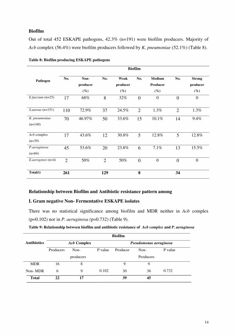
Biofilm
Out of total 452 ESKAPE pathogens, 42.3% (n=191) were biofilm producers. Majority of
Acb complex (56.4%) were biofilm producers followed by K. pneumoniae (52.1%) (Table 8).
Table 8: Biofilm producing ESKAPE pathogens
Pathogen
Biofilm
No. Non-
producer
(%)
No. Weak
producer
(%)
No. Medium
Producer
(%)
No. Strong
producer
(%)
E.faecium (n=25) 17 68% 8 32% 0 0 0 0
S.aureus (n=151) 110 72.9% 37 24.5% 2 1.3% 2 1.3%
K. pneumoniae
(n=149)
70 46.97% 50 33.6% 15 10.1% 14 9.4%
Acb complex
(n=39)
17 43.6% 12 30.8% 5 12.8% 5 12.8%
P.aeruginosa
(n=84)
45 53.6% 20 23.8% 6 7.1% 13 15.5%
E.aerogenes (n=4) 2 50% 2 50% 0 0 0 0
Total(t) 261 129 8 34
Relationship between Biofilm and Antibiotic resistance pattern among
I. Gram negative Non- Fermentative ESKAPE isolates
There was no statistical significance among biofilm and MDR neither in Acb complex
(p=0.102) nor in P. aeruginosa (p=0.732) (Table 9).
Table 9: Relationship between biofilm and antibiotic resistance of Acb complex and P. aeruginosa
Antibiotics
Biofilm
Acb Complex Pseudomonas aeruginosa
Producers Non-
producers
P value Producer Non-
Producers
P value
MDR 16 8
0.102
9 9
0.732 Non- MDR 6 9 30 36
Total 22 17 39 45

15
II. Gram Negative Fermentative Bacterial ESKAPE isolates
Among fermentative bacterial isolates of ESKAPE pathogens, significant association in
between biofilm and MDR K. pneumoniae (p value= 0.050) was present. However, in case of
E. aerogenes, there was no significant association seen (p value= 1.00) (Table 10).
Table 10: Relationship between biofilm and antibiotic resistance of Klebsiella pneumoniae and
Enterobacter aerogenes
Antibiotics
Biofilm
K. pneumoniae E. aerogenes
Produce
rs
Non-
Producers
P
value
Producers Non-
Producers
P value
MDR 42 26 0.050
1 1 1.00
Non- MDR 37 44 1 1
Total 79 70 2 2
III. Relationship between Biofilm and Antibiotic Resistant S. aureus and
E. faecium
There was no significant association in between biofilm and MDR isolates of S. aureus (p
value= 0.424) and Enterococcus faecium (p value= 0.484).
Table 11: Relationship between biofilm and antibiotic resistance of S. aureus and E. faecium
Antibiotics
Biofilm
Staphylococcus aureus Enterococcus faecium
Producers Non- Producers P Value Producers Non- Producers P- Value
MDR 30 73 0.424 8 17 0.484
Non- MDR 11 37 - -
Total 41 110 8 17
Discussion
Antibiotic resistance is a major clinical problem in treating nosocomial and community
acquired infections caused by ESKAPE pathogens 28, and this situation is in an alarming
stage in Nepal as well 29 , 30 31. All members of ESKAPE pathogens fall under WHO's critical
and high priority list of pathogens for research and development of antibiotics 32, which
further highlights the clinical importance of these organisms.
In this study among the ESKAPE pathogens, the most common isolate was Staphylococcus
aureus this may be due to the fact that it is also a normal commensal of human skin and are

16
capable of causing wide range of infection 33. The major proportion (55.1%) of total patients
were females and maximum number of patients infected with were of 61-70 years of age
which was similar to a study conducted by Bhatta et al. 34. This result may be because of the
fact that age group 61-70 consists of debilitating and immune-compromised patients.
The Vancomycin resistant Enterococcus faecium was found to be 20% which is higher than
a study carried out by Acharya et al. 35. Among the eight phenotypic variants Vancomycin
resistance in Enterococci (Van A, B, C, D, E, G, L, M and N), the 5 phenotypically confirmed
VRE isolates (MIC>256 mcg/ml) in this study were subjected to molecular characterisation
for Van A and Van B genes, since VRE is predominantly mediated by these two genes 14.
Van A phenotye shows high level resistance to both Vancomycin and Teicoplanin whereas
Van B and Van C strains exhibit low level or variable resistance to Vancomycin but
susceptible to Teicoplanin36. However in this study, all the VRE isolates weren't resistant to
Teicoplanin. Among the 5 VRE isolates, 2 were susceptible to Teicoplanin and the other 3
were resistant. In this study all the 5 isolates were found to possess Van A gene and none
possessed Van B gene. Van A was found predominant in the isolated VRE which is similar to
other findings worldwide 14, 36,37, 38.
The MRSA was found to be 54.6% which is higher than a study of Parajuli et al. reported in a
teaching hospital in Kathmandu (45% ) 39, and 39.6% found by study of Sanjana et al. 40 . The
MDR S. aureus were 68.2% (n=108) which is higher than that of Sanjana et al. 40 where 25.4
% (n=16) were MDR even though they were MSSA. Similarly, less than one percent of
isolates was found to be XDR.
Multidrug resistance among Gram-negative members of ESKAPE pathogens comprised of K.
pneumoniae 32.2% (n=48), which is similar to study of Llaca-Diaz et al.41, and XDR were
12.8% (n=19). MDR Acb complex was 16.2% (n=12) which is less than a study conducted by
Shrestha et al (2015) 42 and XDR were 32.4% which is similar to study conducted by Llaca-
Diaz et al (2013) 41. MDR P. aeruginosa comprised of 14.3% (n=12) and XDR were 7.1%
(n=6) which was lower than study of Mehta and Rossolini et al. 43, 44. Rates of antibiotic
resistance in P. aeruginosa in this study was higher than a similar study conducted in
nosocomial isolates by Shrestha et al. 45 but lower to study of Chu et al 51. and Mehta et al 43.
XDR 7.1% (n=6). Enterobacter aerogenes was the least prevalent isolate (0.9%) among the
ESKAPE pathogens which was similar to the finding by Pathak et al. 46.

17
In case of ESBL- and MBL- producers, K. pneumoniae were highest ESBL producer
comprising of 16.1% (n=24) which is similar to a study conducted by Raut et al. 47. MBL
were 8.1% (n=12) which is lower than study of Nepal et al. 48. ESBL- and MBL- producing
Acb complex were 10.3% each which is similar to a study conducted by Bhandari et al. 49 but
lower than a study of Shrestha et al 45. ESBL producing P. aeruginosa was 10.7% (n=9) and
7% were MBL which correlated with a similar study of Pathak et al. and Chander et al. 46 50
Ampicillin- Sulbactam was drug of choice for Acb complex showing 64% sensitivity.
Sulbactam containing beta-lactam drug is a good therapeutic agent against Acb complex (Chu
et al (2013). Higher percentage (91.7%) of P. aeruginosa showed susceptibility against
Piperacillin- tazobactam when compared with similar study of Mehta et al 43. In case of K.
pneumoniae, more than 82% were susceptible to Meropenem, 78.5% Piperacillin-tazobactam
and 75.2% to Amikacin among first line antibiotics. These findings were in harmony with
findings of other studies 48 , 52. There was no resistance shown by Enterobacter aerogenes
against Meropenem which was similar to study of Praharaj et al. 53.
Among the Gram positive members, all the isolates were sensitive to vancomycin which was
similar to study by Sanjana et al. 40. Nearly 20% of E. faecium were resistant to vancomycin
and 12% to Teicoplanin. In Nepal VRE in clinical isolate was reported from Manipal
teaching hospital (one isolate) by disc diffusion method 54 and one VRE (MIC=32 mcg/ml)
from case of peritonitis patient in a continuous ambulatory patient in B.P Koirala institute of
health science 55. These studies are probably the only available reports on clinically isolated
VRE detected by agar dilution method in Nepal till date as mentioned by Amatya et al. 13. In
case of other modes of transmission as from poultry and fomite-borne transmission, almost 5
- 19% resistance has been found in minced meat supply in Chitwan 56. Similarly, study
conducted in a hospital in Kathmandu found 2 VRE among 9 isolates extracted from patient's
medical charts Thapa et.al 57. This indicates an intense possibility of fomite borne and food
borne transmission of multiple drug resistant organisms which may lead to nosocomial
infections in compromised patients. Vancomycin- a last resort drug for Gram-positive
bacteria was found resistant in up to 20% among clinically isolated E. faecium which was
similar to a study of Amatya et al. 13. Linezolid was drug of choice for VRE isolates showing
100% effectiveness in vitro; however, combinational therapy is also suggested 58.
This study showed Colistin sulphate and Polymyxin B as the drug of choice among MDR
Gram-negative isolates. These drugs are regarded as reserved drugs for MDR and

18
extensively-drug-resistant Gram negative bacteria 59. All the isolate in this study were
susceptible to Polymyxin; however, in Nepal resistance to Polymyxin has been reported as
high as 29% among Pseudomonas spp. in tertiary care hospital 60. Antimicrobials are used
widely around the world as veterinary medicine to promote growth of livestock/ poultry in
animal husbandry 61. Similarly resistance to Colistin is seen as high as 28% in Nepalese
chicken; This coexistence of MDR infection and MDROs in food chain may exacerbate
antimicrobial resistance problem leading to emergence of pan-drug-resistant organism 62.
U.S food and drug Administration (FDA) has banned the use of medically important drugs
for animal growth promotion CDC 63. Recently health Ministry of India has also banned use
of manufacture, sale and distribution of Colistin in poultry, aqua farming and animal feed
supplements 64. It should be taken into consideration by Nepal Ministry of Health to take
necessary steps in banning or limiting use of broad spectrum and strong antibiotics in animal
husbandry.
Almost 43% of ESKAPE isolates were found to be biofilm producers; however, there was no
statistical significance in between MDR and biofilm producing isolates of Enterococcus
faecium (p=0.484), S. aureus (p=0.424) , Acb complex (p=0.102), E. aerogenes (p=1.00) and
P. aeruginosa (p= 0.732) which concur with similar studies of Cepas et al. 66. However, a
statistical significance in between MDR K. pneumoniae and biofilm was seen (p 0.050). This
propensity of MDR resistant K. pneumoniae capable of forming biofilm was found similar to
a study by Vuotto et al. 67.
This study reveals the prevalence of Multidrug resistance as ESBL, MBL, MRSA and VRE
among the clinical isolates. This high level of antimicrobial resistance among the ESKAPE
isolates accounts for one of the important factors for dissemination of antibiotic resistance
particularly in hospital environment. This study will be helpful for clinicians to identify the
most appropriate antibiotic suitable for the treatment of infected patients by prescribing
appropriate antibiotic at correct dose, time and duration 15.
5. Conclusion
It is quite alarming to note that the status of biofilm-producing MDR and XDR ESKAPE
pathogens. This increasing antibiotic resistance is an important issue to be addressed by
policy makers. Formulation of strict antibiotic stewardship policies is warranted in our
country. Early detection and diagnosis of MDROs is indispensable for the choice of most
appropriate antibiotic therapy.

19
Declarations
A. Ethics Approval

20
B. Consent for Publication
C. Availability of data and materials
The data related to this study can be made available by the authors if requested.
D. Competing Interest
The authors declare that they have no competing interest.
E. Funding
This is a self-funded research done for the partial fulfillment of post graduate study.
F. Authors Contribution
RP and SKM designed, conceived the study and carried out the research work. RP analysed data and wrote the manuscript. SKM monitored the research work. SKM and AS supervised the study. All authors read and approved final manuscript.

21
G. Acknowledgement
We would like to thank Mr. Anil Kumar Shah, Research Officer, Annapurna Research
Centre for the providing Molecular Laboratory facility for our Research. H. Author's Information:
1* Ms. Rosy Pandey, MSc. Public Health Microbiology
St. Xavier's College, Tribhuvan University, Nepal Student, Researcher [email protected]
Contact No +9779803005426
2 Mr. Shyam Kumar Mishra, MScMLT Microbiology, MLS(ASCPi)CM
Clinical Microbiologist, Assistant Professor, Department of Microbiology, Institute of Medicine, Tribhuvan University, Nepal [email protected]
Contact No +9779851169698
ORCID Identifier: orchid.org/0000-0002-3888-7319
1 Ms. Angela Shrestha
M. Tech
Lecturer, Department of Microbiology, St. Xavier's College, Tribhuvan University, Nepal
[email protected] Contact No: +9779841808024
References
1. Boucher HW, Talbot GH, Rice LB et al. Bad Bugs, No Drugs: No ESKAPE! An
Update from the Infectious Diseases Society of America. Clin. Infect. Dis. 2009; 48:
1–12. 2. Blair MA, Webber MA, Baylay AJ, Ogbolu DO & Piddock LJ. Molecular mechanisms
of antibiotic resistance. Nat. Rev. Microbiol. 2015; 13, 42–51. 3. Munita JM & Arias CA. Mechanisms of Antibiotic Resistance. Microbiol
Spectrum.2016; 4(2):1–24. 4. Khanal LK & Jha BK. Prevalence of methicillin resistant Staphylococcus aureus
(MRSA) among skin infection cases at a hospital in Chitwan, Nepal. Nepal Med. Coll.
2010; 12: 224–8. 5. EUCAST guidelines for detection of resistance mechanisms and specific resistanceof
clinical and/or epidemiological importance.2012; 1–43. 6. Becker K, Sarah A, Evgeny I, Nina S, Jochen S, Alexander M, Ursula K, Georg P et
al. Plasmid-Encoded Transferable mecB -Mediated Methicillin Resistance in
Staphylococcus aureus. Emerg. Infect. Dis 2018; 24: 242–248. 7. Drawz SM. & Bonomo RA. Three decades of β-lactamase inhibitors. Clin. Microbiol.
Rev. 2010; 23:160–201. 8. Linscott AJ, Linscott AJ, Brown, WJ & Brown WJ. Evaluation of four commercially

22
available extended-spectrum beta-lactamase phenotypic confirmation tests. J. Clin.
Microbiol 2005; 43:1081–1085. 9. CDC. Laboratory Detection of Extended-Spectrum β-Lactamases (ESBLs). (2010).
Available at: https://www.cdc.gov/hai/settings/lab/lab_esbl.html. Accessed 9 march
2017. 10. Paterson DL & Bonomo RA. Extended-Spectrum beta-Lactamases : A Clinical
Update. Clin. Microbiol. Rev. 2005;18: 657–686. 11. Queenan AM & Bush K. Carbapenemases: The versatile β-lactamases. Clin.
Microbiol. Rev. 2007; 20: 440–458. 12. Lama U, Shah D & Shrestha UT. Vancomycin Resistant Staphylococcus aureus
Reported from Tertiary Care Hospital in Nepal. Tribhuvan Univ. J. Microbiol. 2018; 4:
63–72. 13. Amatya R, Jha B, Shrestha S, Adhikari RP & Timsina S. Prevalence of high level
gentamicin and vancomycin resistance among clinical isolates of enterococci from a
tertiary care hospital in central Nepal. Nepal Med. Coll. J. 2014; 16:125–127. 14. Molton JS, Tambyah PA, Ang BSP, Ling ML & Fisher DA. The global spread of
healthcare-associated multidrug-resistant bacteria: A perspective from Asia. Clin.
Infect. doi. 2013; 56:1310–1318. 15. Pendleton JN, Gorman SP& Gilmore BF. Clinical relevance of the ESKAPE
pathogens. Expert Rev. Anti. Infect. Ther. 2013; 11: 297–308. 16. Vu B, Chen M, Crawford RJ & Ivanova EP. Bacterial extracellular polysaccharides
involved in biofilm formation. Molecules 2009; 14:2535–2554. 17. Mishra SK, Basukala P, Basukala O, Parajuli K, Pokhrel BM, Basista et al. Detection
of Biofilm Production and Antibiotic Resistance Pattern in Clinical Isolates from
Indwelling Medical Devices. Curr. Microbiol.2014; 70:128-134. 18. Isenberg HD & Garcia L. Clinical microbiology procedures handbook: American
Society for Microbiology, 2nd Edition Washington, D.C:ASM Press; 2007. 19. Monica Cheesbrough. District Tropical Practice in Laboratory Countries. (Cambridge
University Press, 2006). 20. The Clinical and Laboratory Standards Institute. Performance Standards for
Antimicrobial Susceptibility Testing CLSI supplement M100S. Clinical and
Laboratory Standards Institute, Wayne, PA ;2016. 21. Magiorakos A, Srinivasan A, Carey RB et al. Multidrug‐resistant, extensively drug‐
resistant and pandrug‐resistant bacteria an international expert proposal for interim standard definitions for acquired resistance. Clin Microbiol Infect 2012; 18: 268–281.
22. Tamma PD, Girdwood SCT, Gopaul R et al. The use of cefepime for treating AmpC β-
lactamase-producing enterobacteriaceae. Clin. Infect. Dis.2013; 57:781–788. 23. Garrec H, Drieux-Rouzet L, Golmard JL, Jarlier V & Robert J. Comparison of nine
phenotypic methods for detection of extended-spectrum β-lactamase production by
enterobacteriaceae. J. Clin. Microbiol.2011; 49: 1048–1057. 24. Franklin C, Liolios L & Peleg AY. Phenotypic detection of carbapenem-susceptible
metallo-β-lactamase- producing gram-negative bacilli in the clinical laboratory. J. Clin.
Microbiol.2006; 44: 3139–3144. 25. Sambrook J, Russell DW. Molecular Cloning: A Laboratory Manual: 3rd edition.Cold

23
Spring Harbor Laboratoey Press: New York; 2001. 26. Kirkan S, Parin U & Balat G. Antimicrobial resistance of Enterococcus faecium
isolated from the urinary system of dogs. Maced. Vet. Rev. 2019; 42:15–21. 27. Akpaka PE, Kissoon S, Jayaratne P et al. Genetic characteristics and molecular
epidemiology of vancomycin-resistant Enterococci isolates from Caribbean countries.
PLoS One. 2017; 12: 1–11. 28. Sirijan S & Nitaya I. Mechanisms of antimicrobial resistance in bacteria in ESKAPE
pahogens. Biomed Res. Int. 2016; 1–8. 29. Parajuli NP, Acharya SP, Mishra SK et al. High burden of antimicrobial resistance
among gram negative bacteria causing healthcare associated infections in a critical
care unit of Nepal. Antimicrob. Resist. Infect. Control. 2017; 6:1–9. 30. Shrestha B, Pokhrel B & Mohapatra T. Study of nosocomial isolates of Staphylococcus
aureus with special reference to methicillin resistant S. aureus in a tertiary care
hospital in Nepal. Nepal Med. College J. 2009; 11:123–126. 31. WHO. Antimicrobial resistance. Global report on surveillance. WHO.2014 doi:
10.1007/s13312-014-0374-3. 32. WHO, WHO priority pathogens list for R&D of new antibiotics. (2017).
https://www.who.int/news-room/detail/27-02-2017-who-publishes-list-of-bacteria-for-which-new-antibiotics-are-urgently-neededAccessed: 24th July 2019.
33. Ansari S, Nepal SP, Gautam R et al. Childhood septicemia in Nepal: Documenting the bacterial etiology and its susceptibility to antibiotics. Int. J. Microbiol. 2014;
doi:http://dx.doi.org/10.1155/2014/452648. 34. Bhatta DR, Himal D, Shrestha R et al. Burden of multidrug resistant respiratory
pathogens in intensive care units of tertiary care hospital. Asian J. Med. Sci. 2019; 10:
14–19. 35. Acharya A, Khanal A, Kanungo R & Mohapatra T. Characterization and susceptibility
patterns of clinically important Enterococcus species in eastern Nepal. Nepal Med. Coll. J. 2007;9: 250–254.
36. Praharaj I, Sujatha S, P. S. Phenotypic & genotypic characterization of vancomycin resistant Enterococcus isolates from clinical specimens. Indian J. Med. Res. 2013; 4;
549–556. 37. Ko KS, Baek JY, Lee JY et al. Molecular Characterization of Vancomycin-Resistant
Enterococcus faecium Isolates from Korea Molecular Characterization of Vancomycin-Resistant Enterococcus faecium Isolates from Korea.2005; 43: 2303–2306.
38. Miele A, Bandera M & Goldstein BP. Use of Primers Selective for Vancomycin
Resistance Genes To Determine van Genotype in Enterococci and To Study Gene
Organization in VanA Isolates.1995; 39: 1772–1778. 39. Parajuli NP, Parajuli H, Pandit R, Shakya J & Khanal PR. Evaluating the trends of
bloodstream infections among pediatric and adult patients at a teaching hospital of Kathmandu, Nepal: Role of drug resistant pathogens. Can. J. Infect. Dis. Med.
Microbiol. 2017; doi:https://doi.org/10.1155/2017/8763135. 40. Sanjana RK, Shah R, Chaudhary N & Singh YI. Prevalence and antimicrobial
susceptibility pattern of methicillin-resistant Staphylococcus aureus ( MRSA ) in
CMS-teaching hospital : a preliminary report. J. Coll. Med. Sci. 2010; 6: 1–6.

24
41. Llaca-Díaz, JM, Mendoza-Olazarán S, Camacho-Ortiz A, Flores S & Garza-González E. One-year surveillance of eskape pathogens in an intensive care unit of monterrey,
Mexico. Chemotherapy 2013; 58: 475–481. 42. Shrestha S, Tada T, Akiyama TM et al. Molecular epidemiology of multidrug-resistant
Acinetobacter baumannii isolates in a university hospital in Nepal reveals the emergence of a novel epidemic clonal lineage. Int. J. Antimicrob. Agents 2015; 46:
526–531. 43. Mehta A, Rosenthal V, Mehta Y et al. Device-associated nosocomial infection rates in
intensive care units of seven Indian cities. Findings of the International Nosocomial
Infection Control Consortium. J. Hosp. Infect.2007; 67: 168–174. 44. Rossolini GM. & Mantengoli E. Treatment and control of severe infections caused by
multiresistant Pseudomonas aeruginosa. Clin. Microbiol. Infect.2005;11: 17–32. 45. Shrestha S, Chaudhari R, Karmacharya S, Kattel HP, Mishra SK, Dahal RK, Bam N,
Rijal BP, Sherchand JB, Ohara H , Koirala J, P. B. Prevalence of nosocomial lower respiratory tract infections caused by Multi- drug resistance pathologens. J Inst Med
2011; 1–17. 46. Pathak P, Jaishi N, Kumar BY , Shah PK et al. Prevalence of Extended Spectrum Beta
Lactamases (ESBL) and Metallo Beta Lactamases (MBL) Mediated Resistance in Gram Negative Bacterial Pathogens. Antimicrobial Resistance in Nepal. Tribhuvan
Univ. J. Microbiol.2017; 4:49-54. 47. Raut S, Gokhale S & Adhikari B. Prevalence of Extended Spectrum Beta-Lactamases
among Escherichia coli and Klebsiella spp isolates in Manipal Teaching Hospital,
Pokhara, Nepal. J. Microbiol. Infect. Dis. 2015; 5(2): 69-75. 48. Nepal K, Pant ND, Neupane B et al. Extended spectrum beta-lactamase and metallo
beta-lactamase production among Escherichia coli and Klebsiella pneumoniae isolated from different clinical samples in a tertiary care hospital in Kathmandu, Nepal. Ann.
Clin. Microbiol. Antimicrob. 2017; 16: 1–7. 49. Bhandari P, Thapa G, Pokhrel BM, Bhatta DR & Devkota U. Nosocomial Isolates and
Their Drug Resistant Pattern in ICU Patients at National Institute of Neurological and
Allied Sciences, Nepal. Int. J. Microbiol. 2015; doi: 10.1155/2015/572163. 50. Chander A & Raza M. Antimicrobial Susceptibility Patterns of Pseudomonas
aeruginosa Clinical Isolates At a Tertiary Care Hospital in Kathmandu, Nepal. Asian
J. Pharm.2013; 6: 235–238. 51. Chu H, Zhao M, Wang M et al. Sulbactam-based therapy for Acinetobacter baumannii
infection: A systematic review and meta-analysis. Brazilian J. Infect. Dis. 2013; 17:
389–394. 52. Shashwati N, Kiran T & Dhanvijay A. Study of extended spectrum β-lactamase
producing Enterobacteriaceae and antibiotic coresistance in a tertiary care teaching
hospital. J. Nat. Sci. Biol. Med. 2014;5: 30. 53. Khajuria A, Praharaj AK, Kumar M & Grover N. Carbapenem Resistance among
Enterobacter Species in a Tertiary Care Hospital in Central India . Chemother. Res.
Pract. 2014; doi: 10.1155/2015/572163. 54. Ghosh AN, Bhatta DR, Ansari MT et al. Application of WHONET in the antimicrobial
resistance surveillance of uropathogens: A first user experience from Nepal. J. Clin.
Diagnostic Res.2013; 7:845–848.

25
55. Nepal H. Khanal B, Sharma SK et al. Peritonitis in a continuous ambulatory peritoneal dialysis patient by two different species of enterococci: A rare finding. Indian J.
Nephrol. 2014; 24: 324. 56. Ghimire S, Basnet HB, Joshi LR & Sapkota M. Prevalence of Vancomycin Resistant
Enterococci Species in Minced Buffalo Meat of Chitwan, Nepal. Int. J. Appl. Sci.
Biotechnol. 2014; 2: 409–412. 57. Thapa R, Pant ND, Yadav UN et al. Isolation of Multidrug Resistant Bacteria from
Patients Medical Charts. J. Nepal Health Res. Counc.2017; 15: 146–149. 58. Patel R & Gallagher JC. Vancomycin-Resistant Enterococcal Bacteremia
Pharmacotherapy. Ann. Pharmacother. 2015; 49: 69–85. 59. Poirel L, Jayol A et al. Polymyxins: Antibacterial Activity, Susceptibility Testing, and
Resistance Mechanisms Encoded by Plasmids or Chromosomes. 2017 ; 30: 557–596. 60. Bhandari, S., Banjara, M. R., Lekhak, B., Bhatta, D. R. & Regmi, S. R. Multi-Drug
and Pan-Drug Resistant Pseudomonas aeruginosa: A Challenge in Post- Antibiotic
Era. Nepal J. Sci. Technol. 2013; 13; 197–202. 61. Van B, TP et al. Global trends in antimicrobial use in food animals. Proc. Natl. Acad.
Sci. 2015; 112: 5649–5654. 62. Joshi PR, Paudel S, Acharya M, et al. Molecular Characterization of Colistin-
Resistant Escherichia coli Isolated from Chickens: First Report from Nepal Microb
Drug Resist. 2019. doi: https://doi.org/10.1089/mdr.2018.0326. 63. Centers for Disease Control and Prevention (CDC). Antibiotic / Antimicrobial
Resistance, food and Food animals. (2018). Available at: https://www.cdc.gov/drugresistance/food.html.
64. Madlen D, Stocktona B. Health Ministry bans sale, distribution of antibiotic Colistin for food producing animals. (2019). Available at: https://www.thehindubusinessline.com/news/science/health-ministry-bans-sale-distribution-of-antibiotic-colistin-for-food-producing-animals/article28627093.ece. (Accessed: 21st July 2019).
65. Cepas V. Lopez Y et al. Relationship Between Biofilm Formation and Antimicrobial Resistance in Gram-Negative Bacteria. Microb. Drug Resist. 2018; doi:
10.1089/mdr.2018.0027. 66. Sanchez CJ, Mende K, Beckius M et al. Biofilm formation by clinical isolates and the
implications in chronic infections. BMC Infect. Dis.2013; 13: 1–12. 67. Vuotto C, Longo F, Balice M, Donelli G & Varaldo P. Antibiotic Resistance Related
to Biofilm Formation in Klebsiella pneumoniae. Pathogens 2014;3: 743–758.

Figures
Figure 1
Epsilometer test of Vancomycin Resistant Enterococcus faecium
Figure 2
Gel Electrophoresis of PCR ampli�cation of Van A gene