Identification and management of wasabi pathogens in British ...
96
Identification and management of wasabi pathogens in British Columbia by Emily C. Betz B.Sc., University of British Columbia, 2016 Thesis Submitted in Partial Fulfillment of the Requirements for the Degree of Master of Science in the Department of Biological Sciences Faculty of Science © Emily C. Betz 2020 SIMON FRASER UNIVERSITY Spring 2020 Copyright in this work rests with the author. Please ensure that any reproduction or re-use is done in accordance with the relevant national copyright legislation.
-
Upload
khangminh22 -
Category
Documents
-
view
0 -
download
0
Transcript of Identification and management of wasabi pathogens in British ...

Identification and management of wasabi pathogens in
British Columbia
by
Emily C. Betz
B.Sc., University of British Columbia, 2016
Thesis Submitted in Partial Fulfillment of the
Requirements for the Degree of
Master of Science
in the
Department of Biological Sciences
Faculty of Science
© Emily C. Betz 2020
SIMON FRASER UNIVERSITY
Spring 2020
Copyright in this work rests with the author. Please ensure that any reproduction
or re-use is done in accordance with the relevant national copyright legislation.

ii
Approval
Name:
Degree:
Title:
Examining Committee:
Date Defended/Approved:
Emily C. Betz
Master of Science
Identification and management of wasabi
pathogens in British Columbia
Chair: Vicki Marlatt
Assistant Professor
Zamir K. Punja
Senior Supervisor
Professor
Margo Moore
Supervisor
Professor Emerita
Siva Sabaratnam
Supervisor
Plant Pathologist
British Columbia Ministry of Agriculture
Sherryl Bisgrove
Internal Examiner
Associate Professor
March 27th, 2020

iii
Abstract
Wasabi (Wasabia japonica) plants in British Columbia are grown in moist conditions
ideal for pathogens, and therefore, are prone to various diseases. Over 3 years, seven
wasabi greenhouses were surveyed for pathogens. Prevalence and severity of diseases
were documented. Pathogenic species including Phoma wasabiae (Leptosphaeria
biglobosa), Botrytis cinerea, and Erysiphe cruciferarum were found in multiple
greenhouses. A new disease of wasabi with symptoms of vascular blackening and wilt
was discovered. Using morphological and molecular techniques, the causal organism was
identified as Verticillium isaacii. Powdery mildew of wasabi caused by E. cruciferarum
was prevalent in half the greenhouses surveyed. In order to evaluate management options
for powdery mildew, 4 commercially available products, Actinovate®, Cueva®,
Rhapsody®, and Regalia® were applied biweekly onto greenhouse plants. Both Cueva®
and Regalia® significantly reduced the progression of powdery mildew on wasabi plants.
Keywords: Wasabia japonica; Erysiphe cruciferarum; Verticillium isaacii; powdery
mildew; verticillium wilt; wasabi

iv
Dedication
To my parents, Rick and Monika, who always cheered me on and at least pretended to be
interested in whatever scientific concept I was blathering on about. I would not be where
I am today without your continuing support.
v
Acknowledgements
Thanks to Dr. Laila Benkrima for going above and beyond to help me understand
commercial wasabi production, for helping set up my growing system at SFU, and
helping with months of greenhouse experiments. Thanks also to Harry Otsuki of Your
Wasabi Farms Ltd. (Abbotsford, BC) for assistance with my powdery mildew trials.
Thanks to Dr. Siva Sabaratnam (BC Ministry of Agriculture, Abbotsford BC) and
Dr. Margo Moore for being fonts of knowledge and advice. Your help and support were
essential in the completion of this thesis. Thanks also to Siva for providing pathogen
cultures for comparison, and for assistance with identification of my bacterial isolates.
Thank you to Dr. Zamir Punja for your enthusiasm and helpful guidance in
pursing this degree. Thanks to my fellow colleagues in the Punja lab for the
encouragement, advice, and good memories. Thanks especially to Hayley Reekie for her
great ability in problem solving. To my horde of undergraduate volunteers over the years
– Amneet Mann, Kathryn Ryan, Samantha Lung, Katherine Fegan, and Alastair Roberts.
Without their help, I would have surely drowned in research tasks.
Lastly, thank you to my family, for continuing to believe in me even when I
forgot to believe in myself. To Mom, for plunking me down at coffee shops and forcing
me to write. To Dad, for the smiles, hugs, and jokes. And to Chris, whose encouragement
never faltered.
Funding for this project was provided by Growing Forward 2 (URAGF-406), a
federal-provincial-territorial initiative, through British Columbia Ministry of Agriculture.

vi
Table of Contents
Approval ............................................................................................................................. ii
Abstract .............................................................................................................................. iii
Dedication .......................................................................................................................... iv
Acknowledgements ............................................................................................................. v
Table of Contents ............................................................................................................... vi
List of Tables ..................................................................................................................... ix
List of Figures ..................................................................................................................... x
Chapter 1. Introduction ................................................................................................. 1
1.1. The biology and production of Wasabia japonica in British Columbia ................... 1
1.1.1. Introduction to wasabi ...................................................................................... 1
1.1.2. Greenhouse production in British Columbia .................................................... 3
1.2. Previously reported pathogens of wasabi ................................................................. 4
1.2.1. Fungi ................................................................................................................. 5
1.2.2. Oomycetes and other fungus-like organisms .................................................... 6
1.2.3. Bacteria ............................................................................................................. 8
1.2.4. Viruses .............................................................................................................. 9
1.3. Current disease control options............................................................................... 10
1.3.1. Cultural methods............................................................................................. 10
1.3.2. Pesticides approved for use on wasabi in Canada .......................................... 10
1.4. Verticillium wilt ...................................................................................................... 13
1.4.1. Taxonomy and identification .......................................................................... 13
1.4.2. Verticillium spp. as plant pathogens ............................................................... 13
1.4.3. Verticillium isaacii ......................................................................................... 15
1.5. Powdery mildew ..................................................................................................... 15
1.5.1. Taxonomy and identification .......................................................................... 15
1.5.2. Erysiphe spp. as plant pathogens .................................................................... 16
1.5.3. Erysiphe cruciferarum .................................................................................... 16
1.6. Research objectives ................................................................................................. 17
Chapter 2. Surveys of microbes associated with wasabi diseases in British
Columbia greenhouses .......................................................................................... 19
2.1. Introduction ............................................................................................................. 19
2.2. Materials and methods ............................................................................................ 20
2.2.1. Sample collection ........................................................................................... 20
2.2.2. Microbe isolation ............................................................................................ 20
2.2.3. Morphological Identification .......................................................................... 21
2.2.4. Molecular Identification ................................................................................. 21
2.3. Results and discussion ............................................................................................ 22
vii
Chapter 3. First report of Verticillium isaacii causing wilt and vascular blackening
on wasabi (Wasabia japonica) plants in Canada ................................................ 27
3.1. Introduction ............................................................................................................. 27
3.2. Materials and methods ............................................................................................ 28
3.2.1. Sample collection and pathogen isolation ...................................................... 28
3.2.2. Identification of Verticillium isolates ............................................................. 30
Morphological criteria ............................................................................................... 30
Molecular identification ............................................................................................ 30
3.2.3. Effect of temperature on pathogen growth ..................................................... 31
3.2.4. Pathogenicity tests .......................................................................................... 31
Detached leaf, petiole, and rhizome inoculations ...................................................... 31
Whole plant inoculations ........................................................................................... 31
3.3. Results ..................................................................................................................... 33
3.3.1. Disease symptoms .......................................................................................... 33
3.3.2. Species identification ...................................................................................... 33
Morphological criteria ............................................................................................... 33
Molecular identification ............................................................................................ 34
3.3.3. Effect of temperature on pathogen growth ..................................................... 38
3.3.4. Pathogenicity tests .......................................................................................... 38
Detached leaf, petiole, and rhizome inoculations ...................................................... 38
Whole plant inoculations ........................................................................................... 40
3.4. Discussion ............................................................................................................... 42
Chapter 4. Management of powdery mildew, caused by Erysiphe cruciferarum, on
wasabi (Wasabia japonica) plants in British Columbia ..................................... 45
4.1. Introduction ............................................................................................................. 45
4.2. Materials and methods ............................................................................................ 48
4.2.1. Sample collection and pathogen identification ............................................... 48
4.2.2. Applications of environmentally sustainable products for disease management
49
Trial 1. May – August 2017 ...................................................................................... 50
Trial 2. September – December 2017 ........................................................................ 50
Trial 3. March – May 2018........................................................................................ 51
4.2.3. Assessment of disease development ............................................................... 51
4.3. Results ..................................................................................................................... 52
4.3.1. Pathogen identification ................................................................................... 52
4.3.2. Assessment of disease development ............................................................... 54
4.4. Discussion ............................................................................................................... 59
Chapter 5. General discussion and conclusions ........................................................ 64
5.1. Disease surveys ....................................................................................................... 64
5.2. Control of wasabi diseases ...................................................................................... 64

viii
5.3. Future research ........................................................................................................ 65
References ........................................................................................................................ 67
Appendix A. Dual culture assays to assess in vitro pathogen inhibition by
biofungicides .......................................................................................................... 79
Appendix B. Effect of temperature on pathogen growth in vitro.......................... 82
Appendix C. Fungitoxic effect of Cueva® on Verticillium isaacii .......................... 84
ix
List of Tables
Table 1.1 List of registered pesticides for use on wasabi in Canada ........................ 12
Table 2.1 Summary of known wasabi diseases identified during surveys of BC
greenhouses in the summers of 2016, 2017, and 2018. ............................ 24
Table 2.2 Summary of potential wasabi diseases identified during surveys of BC
greenhouses in the summers of 2016, 2017, and 2018. ............................ 25
Table 3.1 Isolates of V. isaacii used in pathogenicity studies................................... 30
x
List of Figures
Figure 1.1 A) A mature wasabi rhizome, B) a mature wasabi plant with leaves arising
from petioles at the rhizome crown, C) a terminal inflorescence, and D)
wasabi seedpods. ......................................................................................... 2
Figure 2.1 Common leaf diseases of wasabi in British Columbia. A) powdery mildew
caused by Erysiphe cruciferarum, B) leaf spot caused by Leptosphaeria
biglobosa, and C) leaf blight caused by Botrytis cinerea. ........................ 26
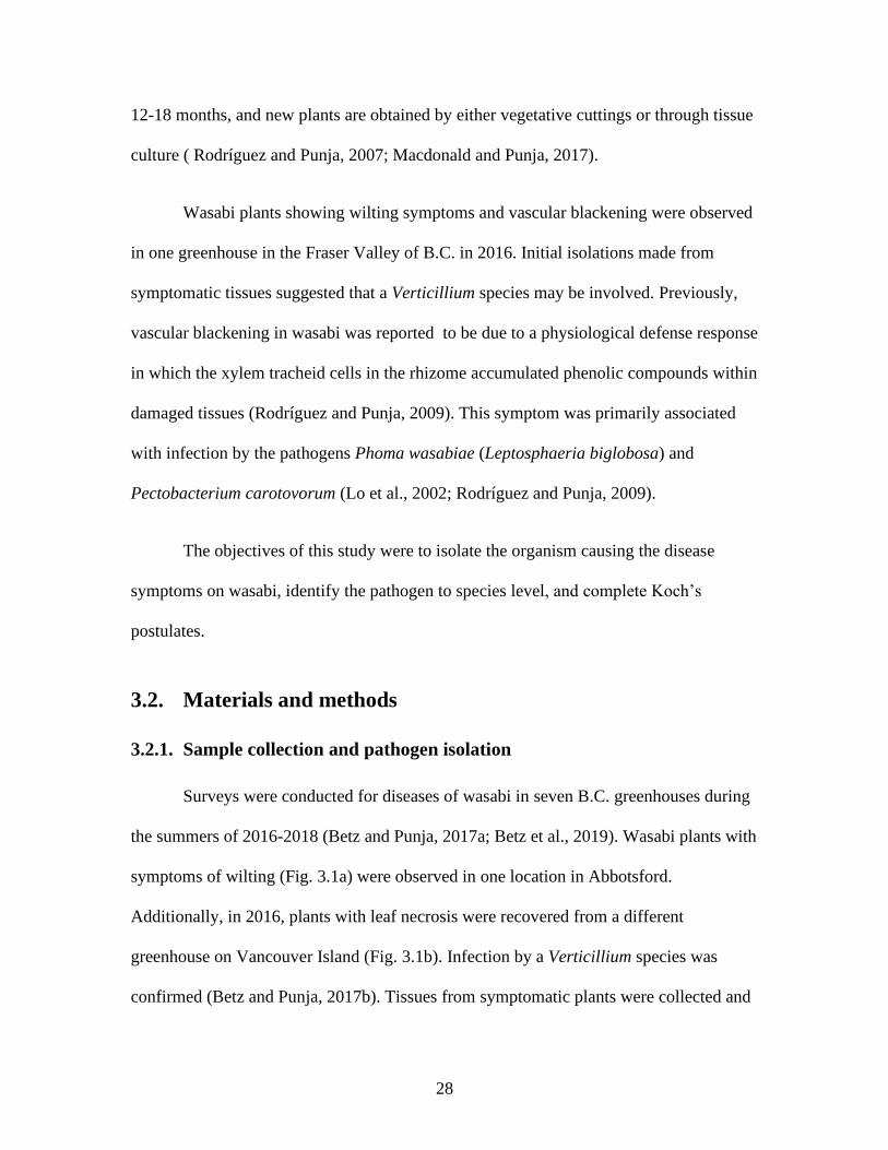
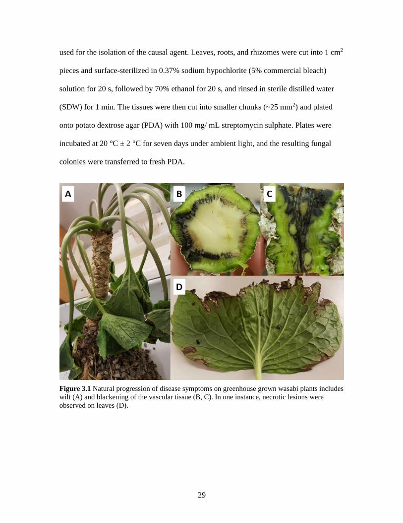
Figure 3.1 Natural progression of disease symptoms on greenhouse grown wasabi plants
includes wilt (A) and blackening of the vascular tissue (B, C). In one
instance, necrotic lesions were observed on leaves (D). ........................... 29
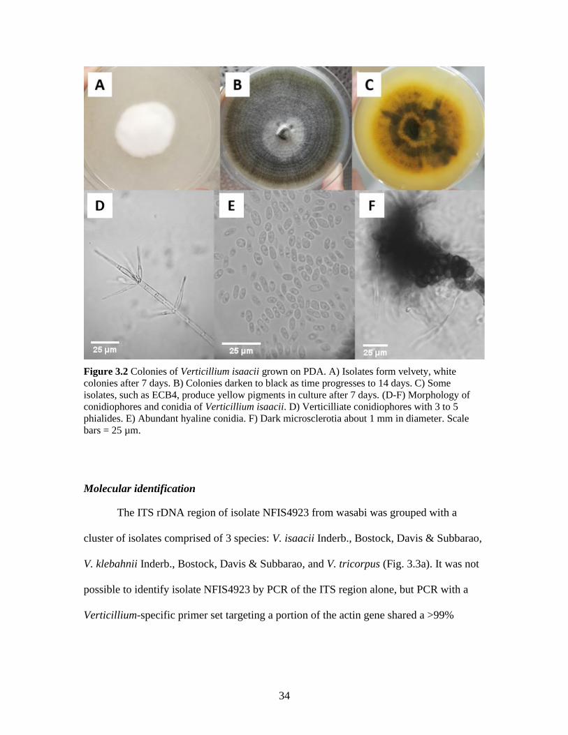
Figure 3.2 Colonies of Verticillium isaacii grown on PDA. A) Isolates form velvety,
white colonies after 7 days. B) Colonies darken to black as time
progresses to 14 days. C) Some isolates, such as ECB4, produce yellow
pigments in culture after 7 days. (D-F) Morphology of conidiophores and
conidia of Verticillium isaacii. D) Verticilliate conidiophores with 3 to 5
phialides. E) Abundant hyaline conidia. F) Dark microsclerotia about 1
mm in diameter. Scale bars = 25 µm. ....................................................... 34
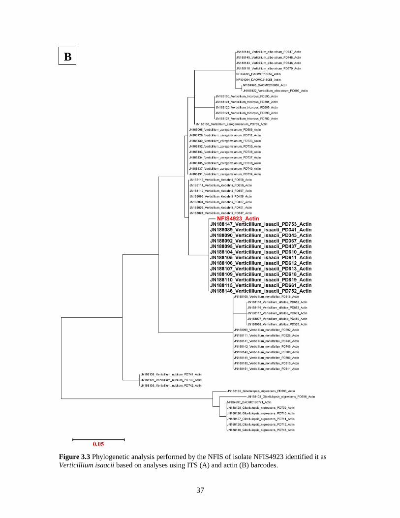
Figure 3.3 Phylogenetic analysis performed by the NFIS of isolate NFIS4923 identified
it as Verticillium isaacii based on analyses using ITS (A) and actin (B)
barcodes. 37
Figure 3.4 V. isaacii grown for 7 days at temperatures from 5° to 35° C. Graph displays
average diameter of colonies based on two trials. Error bars = 95% CI. .. 38
Figure 3.5 Detached and wounded tissues inoculated with a mixed-isolate suspension of
V. isaacii resulted in chlorotic lesions on leaves (A), blackening of
rhizome tissue (C), and vascular blackening of petiole tissue (E). Control
tissues (B,D,F) were asymptomatic. ......................................................... 39
Figure 3.6 After 3 months, plants inoculated with a mixed-isolate mycelial suspension of
V. isaacii show visibly sparser roots (A) compared to a control plant (B).
All inoculated plants had some degree of vascular blackening in their
rhizome tissues (C), while controls were asymptomatic (D). Wilting
occurred in one of the three inoculated plants (E) while the controls
remained unwilted (F). .............................................................................. 41
Figure 4.1 Powdery mildew development on wasabi leaves caused by Erysiphe
cruciferarum. (A) Early development of disease, showing white colonies.
(B) Advanced stages of infection. (C, D) Severe powdery mildew
infection that can cause yellowing. ........................................................... 49
Figure 4.2 Experimental design for Trial 2. Treatments were divided into 4 groups/
treatment with each group containing 5 plants. At least 4 untreated buffer
plants were placed between treated plants. ............................................... 51
Figure 4.3 Phylogenetic analysis of powdery mildew isolates identified as E.
cruciferarum from several cruciferous hosts, including the wasabi
powdery mildew from Korea (Δ). The sequence from the BC wasabi

xi
isolate (▲) was subjected to NCBI Blast, and aligned in MEGA X using
ClustalW, and compared using a Neighbour-Joining tree. ....................... 53
Figure 4.4 Morphology of conidiophores and conidia of E. cruciferarum. a)
Conidiophores and hyphae growing across a wasabi leaf surface (scale bar
= 25 µm). b) A close-up of a developing conidium (scale bar = 2.5 µm).
Note the lack of conidial chains. c) Conidia observed under light
microscopy (scale bar = 250 µm). ............................................................ 54
Figure 4.5 Progression of powdery mildew infection on wasabi plants following
treatments with Actinovate, Cueva, Regalia, Rhapsody, and a water
control. A) Trial 1 (10 weeks), B) Trial 2 (10 weeks), and C) Trial 3 (12
weeks). Bars represent 95% confidence intervals. 56
Figure 4.6 Development of powdery mildew on wasabi plants, 10 weeks after treatments
were initiated. (A) Cueva and (B) Regalia applications reduced powdery
mildew development compared to a water control (E). (C) Rhapsody
showed slight-to-moderate disease suppressive capability. (D) Actinovate
had no disease suppression compared to the water control (E,F). ............ 57
Figure 4.7 AUDPC assessments to evaluate disease severity over time on wasabi plants
treated with reduced risk products. A) Trial 1, B) Trial 2, C) Trial 3.
Disease progression was similar in all trials. ............................................ 58
Figure 4.8 Regalia applications may cause phytotoxic effects on wasabi leaves including
chlorosis and blackening of the leaf. ......................................................... 62

1
Chapter 1. Introduction
1.1. The biology and production of Wasabia japonica in British
Columbia
1.1.1. Introduction to wasabi
Wasabi (Wasabia japonica (Miq.) Matsumura, syn. Eutrema japonicum Matsum.)
is a perennial plant in the Brassicaceae family. It produces a central, above ground, stem
(commonly referred to as a rhizome) (Fig. 1.1a); older plants often produce offshoots from
the central rhizome. At the crown of the rhizome, a multitude of petioles arise, each up to 50
cm long and ending with a single smooth, globose to cordate, leaf which can be up to 25 cm
in diameter (Chadwick et al., 1993) (Fig. 1.1b). In spring, terminal single inflorescences may
emerge from peduncles (Adachi, 1987; Chadwick et al., 1993). The flowers are white in
colour and bracteate, raceme, and cruciform in shape (Fig. 1.1c). Due to self-incompatibility,
cross-pollination is generally required (Chadwick, 1993; Palmer, 1990). Up to eight seeds are
borne in seedpods (Fig. 1.1d) (Chadwick, 1993).
Wasabi is grown for its valuable rhizome, which is traditionally ground into a
paste and used as a condiment in Japanese cuisine (Adachi, 1987). The leaves of the
wasabi plant are also edible and can be eaten as salad greens (Chadwick et al., 1993).
Both the leaves and rhizomes are approved by Health Canada for nutraceutical use as
antioxidant supplements (pers. comm. with manufacturer). The rhizomes generally fetch
a price between $250-350/kg (J. MacDonald, Agriculture and Agri-Food Canada,
Summerland BC, pers. comm).

2
Figure 1.1 A) A mature wasabi rhizome, B) a mature wasabi plant with leaves arising from
petioles at the rhizome crown, C) a terminal inflorescence, and D) wasabi seedpods.
The natural habitat of wasabi is that of mountain streambeds or riverbanks in
Japan. It prefers cool, moist environments with ideal growing temperatures from 15-18
°C. In fact, the plant ceases growth at temperatures above 25 °C (Adachi, 1987;
Chadwick et al., 1993).
Wasabi is traditionally a Japanese crop, but it is now grown in several countries
including Taiwan, China, South Korea, New Zealand, the United States, Britain,

3
Northern Ireland, and Canada (Adachi, 1987; Palmer, 1990; Chadwick et al., 1993; Lo
and Wang, 2000; Weng et al., 2010; Choi et al., 2014; MacDonald and Punja, 2016;
MacDonald, 2018). The cool, wet environment required to grow wasabi makes the
Pacific Northwest an ideal area for commercial production, and wasabi has been grown in
British Columbia for over 20 years.
1.1.2. Greenhouse production in British Columbia
Commercial production of wasabi in Canada has been almost exclusively in the
Pacific Northwest region of British Columbia. Wasabi in British Columbia (B.C.) is
usually commercially grown in double layer polyethylene (double-poly) greenhouses,
using semi-hydroponic overhead sprinkling systems (MacDonald and Punja, 2016).
These double-poly greenhouses have vents to help manage temperature and aid with
cooling in the summer, and the greenhouses can be heated in the winter (MacDonald,
2018). In order to help reduce UV radiation, these greenhouses are covered by 70% shade
cloth for most of the year (MacDonald, 2018). The plants are sown into beds of river rock
along the greenhouse floor. The sprinklers mist at short, regular intervals (sometimes less
than 5 minutes apart) and are designed to provide water, nutrients, and cooling
(MacDonald, 2018). This creates high moisture conditions, of up to 100% relative
humidity, which are ideal for pathogen growth.
To combat the problem of pathogens, some greenhouses have switched to using
drip fertigation. Other types of greenhouse production are being tested, including using
glass greenhouses instead of double-poly. Substrates used in these production systems
include gravel, hydroton (expanded clay pellets), and organic soil. Plants are grown in
4
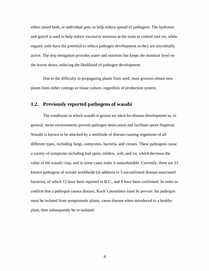
either raised beds, or individual pots, to help reduce spread of pathogens. The hydroton
and gravel is used to help reduce excessive moisture at the roots to control root rot, while
organic soils have the potential to reduce pathogen development as they are microbially
active. The drip fertigation provides water and nutrients but keeps the moisture level on
the leaves down, reducing the likelihood of pathogen development.
Due to the difficulty in propagating plants from seed, most growers obtain new
plants from either cuttings or tissue culture, regardless of production system.
1.2. Previously reported pathogens of wasabi
The conditions in which wasabi is grown are ideal for disease development as, in
general, moist environments prevent pathogen desiccation and facilitate spore dispersal.
Wasabi is known to be attacked by a multitude of disease-causing organisms of all
different types, including fungi, oomycetes, bacteria, and viruses. These pathogens cause
a variety of symptoms including leaf spots, mildew, wilt, and rot, which decrease the
value of the wasabi crop, and in some cases make it unmarketable. Currently, there are 23
known pathogens of wasabi worldwide (in addition to 5 unconfirmed disease-associated
bacteria), of which 12 have been reported in B.C., and 8 have been confirmed. In order to
confirm that a pathogen causes disease, Koch’s postulates must be proven: the pathogen
must be isolated from symptomatic plants, cause disease when introduced to a healthy
plant, then subsequently be re-isolated.

5
1.2.1. Fungi
Perhaps the most notorious disease of wasabi is blackleg caused by Phoma
wasabiae (teleomorph, Leptosphaeria maculans or Leptosphaeria biglobosa). This
fungus causes internal and external blackening of the wasabi rhizome, resulting in an
unmarketable crop. It has been found on wasabi in Japan (Adachi, 1987), Taiwan (Lo et
al., 2002), China (Zhang et al., 2004), New Zealand (Broadhurst and Wright, 1998), the
United States, and Canada (Punja et al., 2017). In greenhouse grown wasabi in B.C., only
L. biglobosa has been found. Phoma wasabiae also causes necrotic leaf spots which
reduce plant productivity. Leptosphaeria maculans and Leptosphaeria biglobosa are part
of a species complex, and both are ubiquitous pathogens of brassica species, most notably
of oilseed crops including canola, causing blackleg or phoma stem canker (Fitt et al.,
2006). They likely spread to wasabi via nearby brassica crops or weeds (Punja et al.,
2017). In B.C., this may include crops such as broccoli and cauliflower.
Botrytis cinerea is another fungus previously reported on wasabi in B.C.
(MacDonald and Punja, 2016). It causes leaf blight, resulting in chlorotic and necrotic
leaf tissue. Botrytis cinerea is a necrotrophic pathogen with a very broad host range,
affecting more than 200 plant species including brassica crops such as broccoli
(Williamson et al., 2007). Likely inoculum sources include nearby crops or weeds.
Erysiphe cruciferarum is a powdery mildew fungus that attacks crucifer species.
It has been previously reported on wasabi in Korea (Park et al., 2016) and Japan (Oku et
al, 1993). It has been previously reported infecting wasabi in B.C. in 2013 and 2015
(Joshi et al., 2014; Joshi and Jeffries, 2016).
6
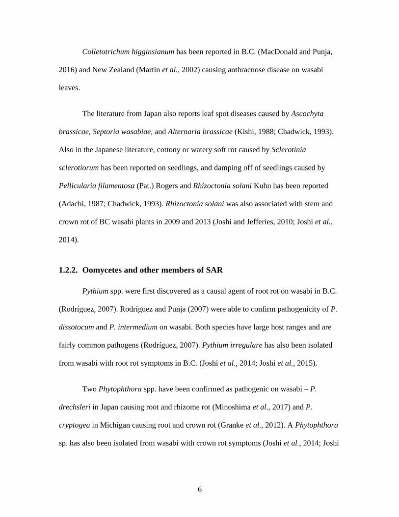
Colletotrichum higginsianum has been reported in B.C. (MacDonald and Punja,
2016) and New Zealand (Martin et al., 2002) causing anthracnose disease on wasabi
leaves.
The literature from Japan also reports leaf spot diseases caused by Ascochyta
brassicae, Septoria wasabiae, and Alternaria brassicae (Kishi, 1988; Chadwick, 1993).
Also in the Japanese literature, cottony or watery soft rot caused by Sclerotinia
sclerotiorum has been reported on seedlings, and damping off of seedlings caused by
Pellicularia filamentosa (Pat.) Rogers and Rhizoctonia solani Kuhn has been reported
(Adachi, 1987; Chadwick, 1993). Rhizoctonia solani was also associated with stem and
crown rot of BC wasabi plants in 2009 and 2013 (Joshi and Jefferies, 2010; Joshi et al.,
2014).
1.2.2. Oomycetes and other members of SAR
Pythium spp. were first discovered as a causal agent of root rot on wasabi in B.C.
(Rodríguez, 2007). Rodríguez and Punja (2007) were able to confirm pathogenicity of P.
dissotocum and P. intermedium on wasabi. Both species have large host ranges and are
fairly common pathogens (Rodríguez, 2007). Pythium irregulare has also been isolated
from wasabi with root rot symptoms in B.C. (Joshi et al., 2014; Joshi et al., 2015).
Two Phytophthora spp. have been confirmed as pathogenic on wasabi – P.
drechsleri in Japan causing root and rhizome rot (Minoshima et al., 2017) and P.
cryptogea in Michigan causing root and crown rot (Granke et al., 2012). A Phytophthora
sp. has also been isolated from wasabi with crown rot symptoms (Joshi et al., 2014; Joshi

7
et al., 2015), and P. cryptogea was isolated from wasabi with symptoms of leaf and
petiole blight (Joshi and Jeffries, 2016) in B.C.
Albugo spp. are obligate pathogens causing white blister rust on leaves. Albugo
spp. (particularly A. candida) are cosmopolitan and attack plants of the Brassicaceae
family. On wasabi, A. wasabiae has been reported in Japan and Taiwan (Adachi, 1987;
Lo and Wang, 2000). Albugo spp. were first reported on wasabi in B.C. in 2009 (Joshi
and Jeffries, 2010). MacDonald and Punja (2016) were able to confirm species identity as
A. candida, which has also been recently reported on wasabi in Korea (Choi et al., 2014).
In B.C., a genetically identical isolate was found on shepherd’s purse (Capsella bursa-
pastoris), suggesting that cruciferous weeds are an inoculum source of A. candida
(MacDonald and Punja, 2016).
Plasmodiophora brassicae is a pathogen of crucifers causing the formation of
root galls commonly referred to as clubroot. Galls can become large enough that they can
restrict water intake via the xylem tissues, resulting in chlorosis of the leaves, and
eventually death of the plant (Agrios, 2005). P. brassicae has been reported on wasabi in
Japan (Adachi, 1987) and China (Chai et al., 2014). Clubroot symptoms were found on
B.C. wasabi in 2013 and 2014, but these samples could not be molecularly identified as
P. brassicae and Koch’s postulates were not confirmed (Joshi et al., 2014; Joshi et al.,
2015; Z. Punja, Professor, Simon Fraser University, pers. comm.).
Peronospora alliariae f. sp. wasabiae causing downy mildew has been reported
in Japan (Chadwick, 1993).
8
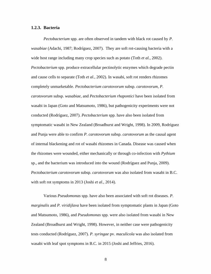
1.2.3. Bacteria
Pectobacterium spp. are often observed in tandem with black rot caused by P.
wasabiae (Adachi, 1987; Rodríguez, 2007). They are soft rot-causing bacteria with a
wide host range including many crop species such as potato (Toth et al., 2002).
Pectobacterium spp. produce extracellular pectinolytic enzymes which degrade pectin
and cause cells to separate (Toth et al., 2002). In wasabi, soft rot renders rhizomes
completely unmarketable. Pectobacterium carotovorum subsp. carotovorum, P.
carotovorum subsp. wasabiae, and Pectobacterium rhapontici have been isolated from
wasabi in Japan (Goto and Matsumoto, 1986), but pathogenicity experiments were not
conducted (Rodríguez, 2007). Pectobacterium spp. have also been isolated from
symptomatic wasabi in New Zealand (Broadhurst and Wright, 1998). In 2009, Rodríguez
and Punja were able to confirm P. carotovorum subsp. carotovorum as the causal agent
of internal blackening and rot of wasabi rhizomes in Canada. Disease was caused when
the rhizomes were wounded, either mechanically or through co-infection with Pythium
sp., and the bacterium was introduced into the wound (Rodríguez and Punja, 2009).
Pectobacterium carotovorum subsp. carotovorum was also isolated from wasabi in B.C.
with soft rot symptoms in 2013 (Joshi et al., 2014).
Various Pseudomonas spp. have also been associated with soft rot diseases. P.
marginalis and P. viridijlava have been isolated from symptomatic plants in Japan (Goto
and Matsumoto, 1986), and Pseudomonas spp. were also isolated from wasabi in New
Zealand (Broadhurst and Wright, 1998). However, in neither case were pathogenicity
tests conducted (Rodríguez, 2007). P. syringae pv. maculicola was also isolated from
wasabi with leaf spot symptoms in B.C. in 2015 (Joshi and Jeffries, 2016).

9
Corynebacterium spp. have also been reported causing vascular wilts, blights, leaf
spot and ring rot on wasabi in Japan (Adachi, 1987; Matsumoto et al., 1985)
1.2.4. Viruses
Wasabi mottle virus (WMoV) has been previously reported in Japan (Shimamoto
et al., 1998) and Taiwan. Recently, plants in B.C. have been found carrying the virus
(MacDonald et al., 2019). It is quite distinctive, and easily recognizable due to the leaf
spots, mottle, and vein-clearing symptoms it produces. The strain found in B.C.is
genetically similar to the strain reported in Taiwan, and as Taiwan is the source of the
tissue-culture plants of cultivar ‘Green Thumb’ used in Canadian production (L.
Benkrima, Lead Scientist, Your Wasabi Farms Ltd., pers. comm.), this is a possible point
of entry for the virus. In B.C., it is believed that the virus is maintained in asymptomatic
tissue culture plants at wasabi nurseries, and symptoms develop in plants that are heat
stressed.
Other viruses previously reported on wasabi in Japan include Tobacco mosaic
virus (TMV), Turnip mosaic virus (TuMV), and Cucumber mosaic virus (CMV)
(Chadwick et al., 1993). Additionally, CMV has been found on wasabi in Australia
(Wilson, 1998). These viral diseases cause stunting on wasabi, along with leaf
discolouration (Chadwick et al., 1993). Alfalfa mosaic virus (AMV) has been reported on
wasabi in New Zealand with symptoms appearing as interveinal mottle, mosaic, and leaf
crinkling (Fletcher, 1989). Currently, none of these viruses have been reported on wasabi
in North America.

10
1.3. Current disease control options
1.3.1. Cultural methods
In Canada, the lack of approved pesticides on wasabi means that most control of
diseases is managed through cultural methods. As such, monitoring for disease symptoms
becomes especially important. Leaf diseases are controlled via pruning infected leaves.
This is cost effective for small production systems, but in months when disease pressure
is heavy, growers are easily overwhelmed (L. Benkrima, Lead Scientist, Your Wasabi
Farms Ltd., pers. comm.). The potential to spread diseases, including rhizome diseases
and viruses, through unsanitary pruning implements is also of concern.
Some growers have attempted using drip irrigation to reduce moisture levels in
greenhouses, as opposed to the more traditional overhead sprinkling systems. Those
greenhouses with drip irrigation tend to develop less rot, such as from Botrytis cinerea,
but, conversely, encounter more powdery mildew as leaf conditions are more optimal.
Tissue culture plants are also used by some growers to obtain pathogen free
plants. There is a tissue culture facility in the Lower Mainland which specializes in
wasabi micropropagation. Tissue culture plants are more expensive of an option than
vegetative cuttings, and there is also the concern that tissue culture plants may harbour
viruses if mother plants are not tested for ‘virus-free’ status (MacDonald et al., 2019).
1.3.2. Pesticides approved for use on wasabi in Canada
Currently, the only chemical-based insecticide approved for use on wasabi is
Ambush 500EC (Permethrin), and there are no chemical fungicides that are approved to
11
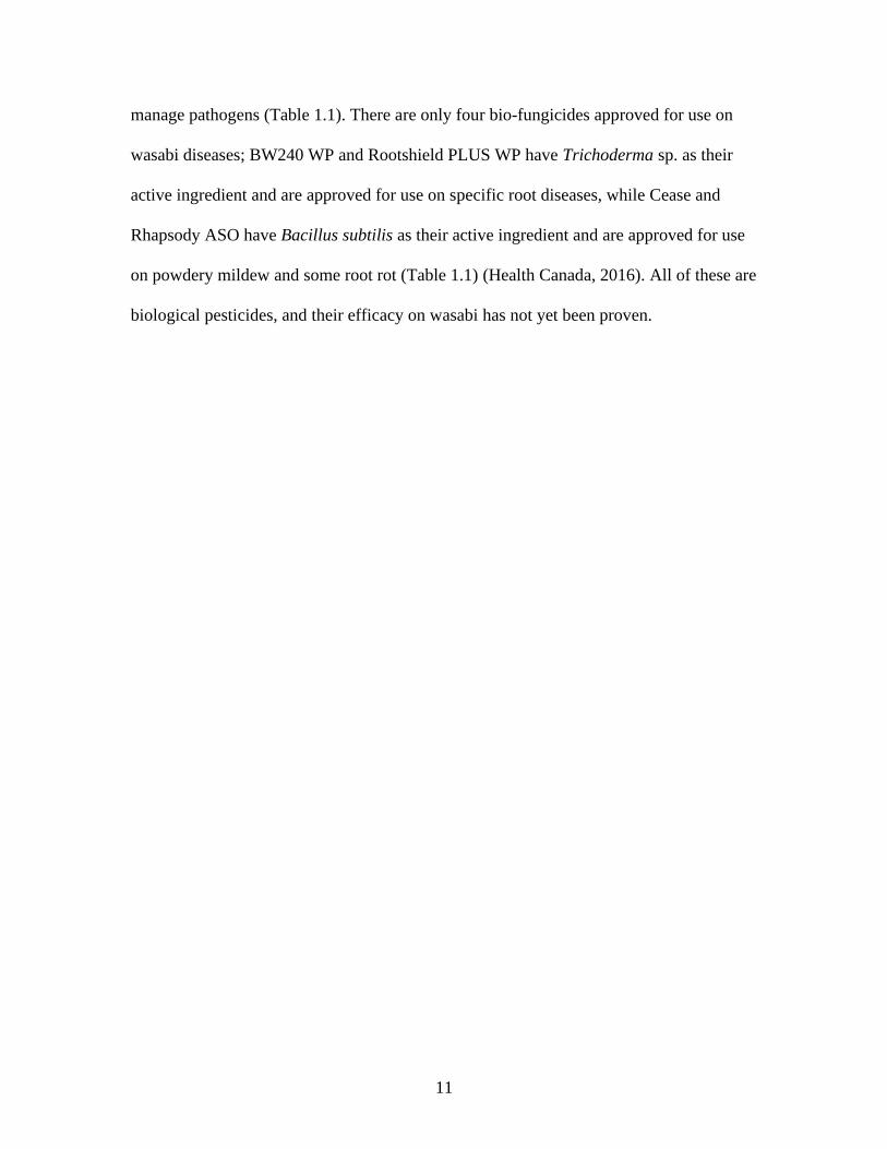
manage pathogens (Table 1.1). There are only four bio-fungicides approved for use on
wasabi diseases; BW240 WP and Rootshield PLUS WP have Trichoderma sp. as their
active ingredient and are approved for use on specific root diseases, while Cease and
Rhapsody ASO have Bacillus subtilis as their active ingredient and are approved for use
on powdery mildew and some root rot (Table 1.1) (Health Canada, 2016). All of these are
biological pesticides, and their efficacy on wasabi has not yet been proven.

12
Table 1.1 List of registered pesticides, including biocontrol products, for use on
wasabi in Canada
Product Active Ingredient Pest/ Disease Manufacturer
Ambush 500EC Permethrin
• Cabbage looper
• Diamondback moth larvae
• Crucifer flea beetle
AMVAC Chemical Corp.
Bioprotec CAF
Bacillus thuringiensis kurstaki
• Cabbage looper
• Alfalfa looper
AEF Global, Inc.
Bioprotec PLUS
Bacillus thuringiensis kurstaki
• Cabbage looper
• Alfalfa looper
AEF Global, Inc.
Botanigard 22WP
Beauveria bassiana GHA
• Aphids LAM International Corp.
Botanigard ES
Beauveria bassiana GHA
• Aphids LAM International Corp.
BW240 WP Trichoderma harzianum KRL-AG2 and Trichoderma virens G-41
• Fusarium root rot
• Phytophthora root rot
• Pythium root rot and damping off
• Rhizoctonia root rot and damping off
BioWorks, Inc.
Cease®
Bacillus subtilis QST 713
• Powdery mildew
• Pythium root rot
• Phytophthora crown rot and root rot
BioWorks, Inc.
Dipel® 2X DF Bacillus thuringiensis kurstaki
• Cabbage looper
• Alfalfa looper
Valent BioSciences Corp.
Rhapsody® ASO™
Bacillus subtilis QST 713
• Powdery mildew
• Pythium root rot
• Phytophthora crown rot and root rot
Bayer CropScience Inc.
Rootshield® PLUS WP
Trichoderma harzianum KRL-AG2 and Trichoderma virens G-41
• Fusarium root rot
• Phytophthora root rot
• Pythium root rot and damping off
• Rhizoctonia root rot and damping off
BioWorks, Inc.
13
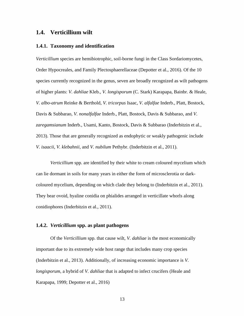
1.4. Verticillium wilt
1.4.1. Taxonomy and identification
Verticillium species are hemibiotrophic, soil-borne fungi in the Class Sordariomycetes,
Order Hypocreales, and Family Plectosphaerellaceae (Depotter et al., 2016). Of the 10
species currently recognized in the genus, seven are broadly recognized as wilt pathogens
of higher plants: V. dahliae Kleb., V. longisporum (C. Stark) Karapapa, Bainbr. & Heale,
V. albo-atrum Reinke & Berthold, V. tricorpus Isaac, V. alfalfae Inderb., Platt, Bostock,
Davis & Subbarao, V. nonalfalfae Inderb., Platt, Bostock, Davis & Subbarao, and V.
zaregamsianum Inderb., Usami, Kanto, Bostock, Davis & Subbarao (Inderbitzin et al.,
2013). Those that are generally recognized as endophytic or weakly pathogenic include
V. isaacii, V. klebahnii, and V. nubilum Pethybr. (Inderbitzin et al., 2011).
Verticillium spp. are identified by their white to cream coloured mycelium which
can lie dormant in soils for many years in either the form of microsclerotia or dark-
coloured mycelium, depending on which clade they belong to (Inderbitzin et al., 2011).
They bear ovoid, hyaline conidia on phialides arranged in verticillate whorls along
conidiophores (Inderbitzin et al., 2011).
1.4.2. Verticillium spp. as plant pathogens
Of the Verticillium spp. that cause wilt, V. dahliae is the most economically
important due to its extremely wide host range that includes many crop species
(Inderbitzin et al., 2013). Additionally, of increasing economic importance is V.
longisporum, a hybrid of V. dahliae that is adapted to infect crucifers (Heale and
Karapapa, 1999; Depotter et al., 2016)
14
The disease cycle of Verticillium sp. starts with microsclerotia which germinate in
the presence of root exudates, and the resulting mycelium colonizes the cortical cells of
the plant root, initiating the biotrophic stage of infection (Karapapa et al., 1997),. Often
the biotrophic phase is asymptomatic, and asymptomatic plants can be infected for
several months without showing symptoms (Karapapa et al., 1997). In a successful
infection, the mycelia enter the xylem where the hyphae proliferate, conidia are formed
and travel via the transpiration stream to infect systemically (Karapapa et al., 1997). At
this point, symptoms of chlorosis and necrosis appear in the plant (Heale and Karapapa,
1999). The fungus then switches to a necrotrophic stage, colonizes the dead tissue, and
produces microsclerotia (Heale and Karapapa, 1999; Karapapa et al., 1997). As the plant
tissue decays, the microsclerotia are released back into the soil where they can remain
viable for over a decade (Karapapa et al., 1997; Pegg and Brady, 2001).
Verticillium wilt is found primarily on crops grown in temperate climates with
warm, dry summers and cool winters and are especially problematic in irrigated regions
(Pegg and Brady, 2001). Wilt conditions are exacerbated by moist soil conditions, and
optimal temperatures for infection are between 21-27 °C (Pegg and Brady, 2001).
However, it is thought that stresses brought on by drought and high temperatures are
likely to increase disease severity and yield loss in infected plants (Heale and Karapapa,
1999).
In laboratory studies, it was demonstrated that Verticillium dahliae cultures can be
killed in 4 minutes at 55 °C and microsclerotia can be killed in 10 minutes at 50 °C or 40
minutes at 47 °C (Miller and Stoddard, 1956; Nelson and Wilheim, 1958). The optimal
15
soil pH for V. dahliae growth was determined to be 5.5, and the disease severity on
ornamentals (Antirrhinum sp.) was greater in alkaline soil than acidic soil (Dutta, 1981).
1.4.3. Verticillium isaacii
Verticillium isaacii was first described in 2011, by Inderbitzin et al. It was
previously considered as part of Verticillium tricorpus but was found to be genetically
distinct (Inderbitzin et al., 2011). It has been isolated from spinach, artichoke, and
lettuce, but it is sometimes endophytic in these plants rather than pathogenic (Gurung et
al., 2015). Isolates of V. isaacii were found to weakly infect sunflower, potato,
strawberry, artichoke, and lettuce (Gurung et al., 2015; Wheeler and Johnson, 2019).
1.5. Powdery mildew
1.5.1. Taxonomy and identification
Pathogens of powdery mildew are obligately biotrophic, filamentous,
phytopathogens in the Order Erysiphales and Phylum Ascomycota (Glawe, 2008). Most
species grow epiphytically on plant surfaces, including leaves, stems, flowers, and fruits
(Heffer et al., 2006; Glawe, 2008). Powdery mildew pathogens are pleomorphic and have
both sexual and asexual stages; however, either of these stages may be absent depending
on the species (Glawe, 2008). The asexual conidia are borne on the terminal end of
condiophores, while the sexual ascospores are enclosed in asci which are encapsulated in
enclosed hyphal structures known as chasmothecia (Heffer et al., 2006).
Powdery mildew pathogens are identified by their white mycelia and the abundant
amounts of conidia they produce, which gives them a ‘powdery’ appearance (Glawe,
16
2008). The taxonomy of these pathogens is complex and still under review, but, broadly,
there are 5 holomorphic tribes – Phyllactinieae, Erysipheae, Blumerieae,
Golvinomyceteae, and Cystotheceae (Glawe, 2008).
Erysiphe spp. belong to the tribe Erysipheae (Heffer et al., 2006). The genus
Erysiphe is split into three sections – section Erysiphe, section Microsphaera, and section
Uncinula (Heffer et al., 2006). The anamorphs are all Oidium species. Erysiphe spp. can
be distinguished from other powdery mildew species by their conidia which form singly
or in pseudochains, rather than true chains (Heffer et al., 2006). The sections of the
Erysiphe genus can be distinguished by the appendages on their chasmothecia (if
present); section Erysiphe has simple appendages, section Microsphaera has dictomously
branched appendages, and section Uncinula has coiled or hooked appendages (Heffer et
al., 2006).
1.5.2. Erysiphe spp. as plant pathogens
Individual Erysiphe species tend to be highly specialized plant pathogens and
typically have small host ranges (Glawe, 2008). Haustoria are specialized hyphal cells
which are formed in the space between the plant cell wall and cell membrane, and they
function to absorb nutrients from plant epidermal cells (Heffer et al., 2006).
1.5.3. Erysiphe cruciferarum
Erysiphe cruciferarum is a pathogen responsible for powdery mildew on brassica
species including oilseed rape, broccoli, and cabbage (Koike et al., 2007; Alkooranee et
al., 2015). Colonies may be grey in colour, due to host responses produce black speckling
17
underneath the colony (Koike et al., 2007). Heavily diseased plants have symptoms of
chlorosis, necrosis, and early defoliation (Koike et al., 2007).
E. cruciferarum has singly borne, ovoid conidia typical of Erysiphe spp.
Appressoria (cells responsible for penetrating into host tissues) are variable, ranging from
simple to lobed, and haustoria are multilobed (Koch and Slusarenko, 1990). Observations
of chasmothecia in E. cruciferarum are rare. The sexual stage has not been observed in
the Pacific Northwest, and how this species overwinters here has not yet been determined
(Glawe, 2006).
1.6. Research objectives
Wasabi growers in the Pacific Northwest region of B.C. are at a disadvantage
when it comes to managing diseases – partially due to the lack of information on wasabi
pathogens present in the region, and partially due to the lack of available management
options, including pesticides, that are available to them. Additionally, much of the current
literature on wasabi pathogens is on field grown wasabi, not greenhouse grown wasabi.
In order to assist growers, improved knowledge of current wasabi pathogens, their
occurrence, and their severity is needed. Additionally, commercial fungicide products
must be tested for efficacy on wasabi diseases. Growers are increasingly interested in
organic production and are requesting management solutions, including biological
fungicides and other reduced risk products, for controlling diseases.
The objectives of this research were to:
1) Survey B.C. wasabi greenhouses for diseases caused by pathogens.
18
2) Determine the causal agent of wilt and rhizome blackening symptoms on
wasabi grown in B.C. greenhouses.
3) Confirm the causal agent of powdery mildew on wasabi in B.C.
4) Identify commercially available reduced risk fungicide products that are
efficacious at reducing disease severity of powdery mildew on greenhouse grown
wasabi.
19
Chapter 2. Surveys of microbes associated with wasabi
diseases in British Columbia greenhouses
2.1. Introduction
As wasabi is a relatively new crop to North America, there is a lack of knowledge
of what pathogens are present on B.C. grown wasabi. Additionally, growers are reporting
symptoms of previously unknown wasabi diseases and they have no way to combat these
pathogens (S. Sabaratnam, Plant Pathologist, B.C. Ministry of Agriculture, pers. comm.).
The severity of these pathogens in the Pacific Northwest region is also unknown.
We conducted 3 surveys of fungal diseases on wasabi (Wasabia japonica) from
May to August each year from 2016 to 2018. In 2016, thirty-one plant samples were
taken from 5 greenhouses in B.C. – 3 in the Lower Mainland (Abbotsford and Surrey)
and 2 on Vancouver Island (Sooke and Nanoose Bay). In 2017 and 2018, 42 and 44 plant
samples, respectively, were collected from six greenhouses in B.C. – four in the Lower
Mainland (Abbotsford, Burnaby, and Surrey) and two on Vancouver Island (Sooke and
Sointula). We strived to obtain samples from as many different greenhouses as possible
every year. In some years, certain greenhouses were unavailable, but we managed to get
samples from three greenhouses on a consistent yearly basis.
20
2.2. Materials and methods
2.2.1. Sample collection
Plants with visible disease symptoms including foliar blight and discolouration,
leaf spots, wilt, and root and rhizome rot, were documented, photographed, collected, and
brought to Simon Fraser University (SFU) for pathogen isolation and identification.
2.2.2. Microbe isolation
Samples were taken from infected areas (leaves, petioles, rhizomes, roots).
Tissues were assessed for disease symptoms, and areas that were blackened or
discoloured were excised, cleaned in 0.37% sodium hypochlorite (5% commercial bleach
solution) for 20 seconds, then 70% EtOH for 20 seconds, before thoroughly rinsing in
autoclaved distilled water. Samples cut into ~1 cm2 pieces and four such pieces were
placed onto potato dextrose agar (PDA) and vegetable juice (V8) agar containing 100
mg/L streptomycin. Bacterial samples were isolated using nutrient agar (NA). Plates were
left to incubate for up to three weeks at room temperature under 8 hours light/16 hours
dark conditions. Plates were checked weekly for microbe growth, and microbes of
interest were sub-cultured. Fungi were sub-cultured by taking a ~0.5 cm2 agar plug from
the edge of the fungal colony and plating on fresh media every 3-4 weeks, and bacteria
were sub-cultured by streaking onto fresh media every week.
Since obligate biotrophs cannot be cultured, approximately a 1 cm2 piece of
infected leaf tissue was placed in a 2 mL Eppendorf tube and frozen at -20 °C and saved
for future identification using molecular methods.
21
2.2.3. Morphological Identification
We examined cultures grown on PDA and V8A for characteristic features of the
pathogens of interest including colour, growth pattern, and presence or absence of
microsclerotia. Fungal tissue was mounted in water onto a glass slide and observed using
a Zeiss compound microscope. Conidial morphology was examined for defining
characteristics such as pigmentation, septation, ornamentation, shape, size, and presence
or absence of spores produced in chains. Conidiophore morphology was also considered
based on conidial development (thallic or blastic), shape, septation, and arrangement of
conidia.
2.2.4. Molecular Identification
DNA was extracted using a CTAB extraction protocol. Briefly, fungal tissue (0.01
grams) was ground in liquid nitrogen using a mortar and pestle, then added to 600 µL
CTAB buffer (2% w/v cetyl trimethylammonium bromide, 1% w/v polyvinyl
pyrrolidone, 1.4 M NaCl, 100 mM Tris HCl, 20 mM EDTA) and incubated for 15 min at
65 °C. Samples were centrifuged at 20000 x g for 5 min and the supernatant transferred
to a new Eppendorf tube. A 1:1 Phenol:Chloroform mix (according to sample volume)
was mixed into the supernatant and the tubes centrifuged at 20000 x g for 1 min. The
upper phase was transferred to a new tube and a 1/10 volume ratio of 7M ammonium
acetate was added, followed by 1 volume of ice-cold 100% ethanol. The sample were
stored for a minimum of 1 hour at -20 °C before being centrifuged at 20000 x g for 10
min and the supernatant removed. The resulting pellet was washed with 500 µL ice-cold
70% ethanol and centrifuged for 2 min at 20000 x g. Samples were left in a laminar flow

22
hood for 20 minutes to evaporate excess ethanol, then re-suspended in 100uL TE and
kept at -20 °C.
Fungal species were further identified by sequencing the ITS1-5.8S-ITS2 rDNA
region using universal primers UN-UP18S42 (5'-
CGTAACAAGGTTTCCGTAGGTGAAC-3') and UN-LO28S22 (5'-
GTTTCTTTTCCTCCGCTTATTGATATG-3'). PCR was performed for 35 cycles with
an annealing temperature of 55 °C. PCR amplicons were sent to Eurofins (Toronto, ON)
for Sanger sequencing. The resulting fasta files were returned to Simon Fraser University
where we blasted the files against sequences in GenBank for identification.
Isolated bacteria were identified as gram-positive or gram-negative using the Ryu
non-staining KOH Technique (Powers, 1995). Gram-negative bacterial species were
further identified using MicroLog® by the Plant Health Laboratory of the British
Columbia Ministry of Agriculture. Suspected virus infected tissues were sent to the
Summerland Research Centre (Agriculture and Agri-Food Canada) for confirmation
based on whole genome sequencing.
2.3. Results and discussion
In 2016, thirty-one plant samples were collected from which 8 potential fungal
pathogens, as well as two oomycetes and one bacterial pathogen were identified (Table
2.1). In 2017, 42 plant samples were collected from which nine potential fungal
pathogens, as well as one oomycete, two bacterial pathogens, and one virus were
identified (Table 2.1). In 2018, 44 plant samples were collected from which 12 potential
fungal pathogens, as well as one oomycete, two bacterial pathogens, and one virus were
23
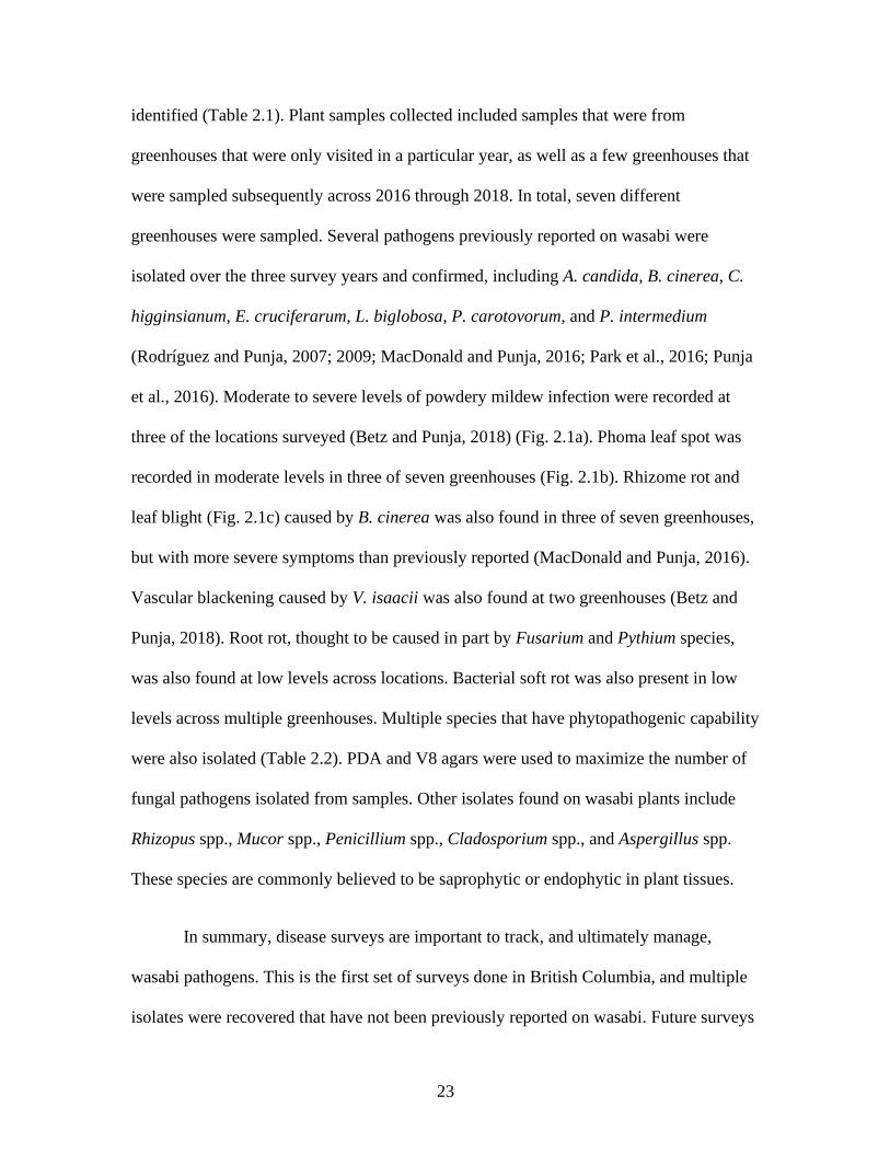
identified (Table 2.1). Plant samples collected included samples that were from
greenhouses that were only visited in a particular year, as well as a few greenhouses that
were sampled subsequently across 2016 through 2018. In total, seven different
greenhouses were sampled. Several pathogens previously reported on wasabi were
isolated over the three survey years and confirmed, including A. candida, B. cinerea, C.
higginsianum, E. cruciferarum, L. biglobosa, P. carotovorum, and P. intermedium
(Rodríguez and Punja, 2007; 2009; MacDonald and Punja, 2016; Park et al., 2016; Punja
et al., 2016). Moderate to severe levels of powdery mildew infection were recorded at
three of the locations surveyed (Betz and Punja, 2018) (Fig. 2.1a). Phoma leaf spot was
recorded in moderate levels in three of seven greenhouses (Fig. 2.1b). Rhizome rot and
leaf blight (Fig. 2.1c) caused by B. cinerea was also found in three of seven greenhouses,
but with more severe symptoms than previously reported (MacDonald and Punja, 2016).
Vascular blackening caused by V. isaacii was also found at two greenhouses (Betz and
Punja, 2018). Root rot, thought to be caused in part by Fusarium and Pythium species,
was also found at low levels across locations. Bacterial soft rot was also present in low
levels across multiple greenhouses. Multiple species that have phytopathogenic capability
were also isolated (Table 2.2). PDA and V8 agars were used to maximize the number of
fungal pathogens isolated from samples. Other isolates found on wasabi plants include
Rhizopus spp., Mucor spp., Penicillium spp., Cladosporium spp., and Aspergillus spp.
These species are commonly believed to be saprophytic or endophytic in plant tissues.
In summary, disease surveys are important to track, and ultimately manage,
wasabi pathogens. This is the first set of surveys done in British Columbia, and multiple
isolates were recovered that have not been previously reported on wasabi. Future surveys
24
should be continually conducted to keep track of the spread of prevalent and problematic
diseases.
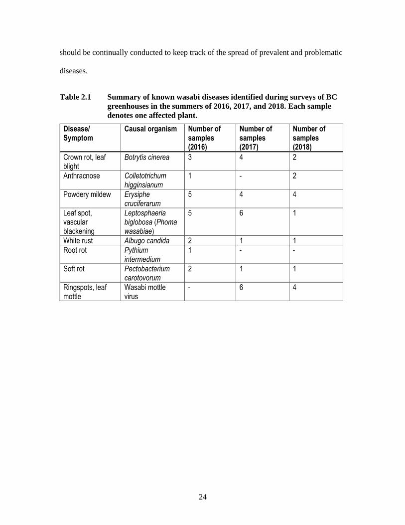
Table 2.1 Summary of known wasabi diseases identified during surveys of BC
greenhouses in the summers of 2016, 2017, and 2018. Each sample
denotes one affected plant.
Disease/ Symptom
Causal organism Number of samples (2016)
Number of samples (2017)
Number of samples (2018)
Crown rot, leaf blight
Botrytis cinerea 3 4 2
Anthracnose Colletotrichum higginsianum
1 - 2
Powdery mildew Erysiphe cruciferarum
5 4 4
Leaf spot, vascular blackening
Leptosphaeria biglobosa (Phoma wasabiae)
5 6 1
White rust Albugo candida 2 1 1
Root rot Pythium intermedium
1 - -
Soft rot Pectobacterium carotovorum
2 1 1
Ringspots, leaf mottle
Wasabi mottle virus
- 6 4
25
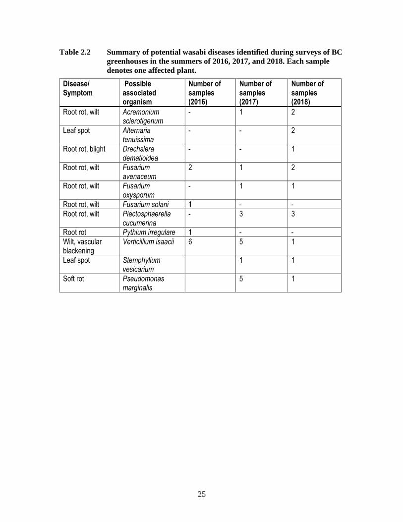
Table 2.2 Summary of potential wasabi diseases identified during surveys of BC
greenhouses in the summers of 2016, 2017, and 2018. Each sample
denotes one affected plant.
Disease/ Symptom
Possible associated organism
Number of samples (2016)
Number of samples (2017)
Number of samples (2018)
Root rot, wilt Acremonium sclerotigenum
- 1 2
Leaf spot Alternaria tenuissima
- - 2
Root rot, blight Drechslera dematioidea
- - 1
Root rot, wilt Fusarium avenaceum
2 1 2
Root rot, wilt Fusarium oxysporum
- 1 1
Root rot, wilt Fusarium solani 1 - -
Root rot, wilt Plectosphaerella cucumerina
- 3 3
Root rot Pythium irregulare 1 - -
Wilt, vascular blackening
Verticillium isaacii 6 5 1
Leaf spot Stemphylium vesicarium
1 1
Soft rot Pseudomonas marginalis
5 1
26
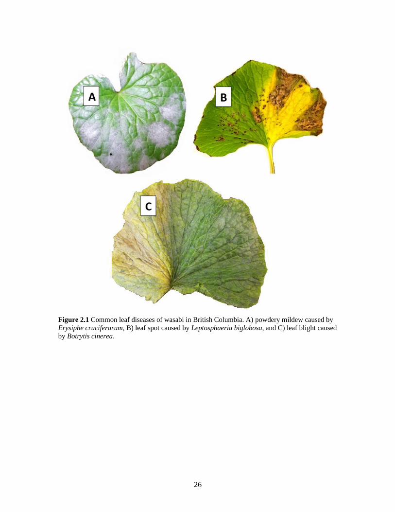
Figure 2.1 Common leaf diseases of wasabi in British Columbia. A) powdery mildew caused by
Erysiphe cruciferarum, B) leaf spot caused by Leptosphaeria biglobosa, and C) leaf blight caused
by Botrytis cinerea.

27
Chapter 3. First report of Verticillium isaacii causing
wilt and vascular blackening on wasabi (Wasabia japonica)
plants in Canada
3.1. Introduction
Wasabi (Wasabia japonica (Miq.) Matsumura, syn. Eutrema japonicum Matsum.)
is a plant in the Brassicaceae family primarily grown for its valuable rhizome, which is
traditionally ground into a paste and used as a condiment (Chadwick et al. 1993). Wasabi
was traditionally grown in Japan (Adachi, 1987), but it is now cultivated in several
countries including Taiwan (Lo and Wang, 2000), China (Weng et al., 2010), South
Korea (Choi et al., 2014), New Zealand (Palmer, 1990), the United States (Chadwick et
al., 1993), and Canada (Rodríguez and Punja, 2007; Macdonald and Punja, 2017). The
natural habitat of wasabi is that of shaded mountain streambeds or riverbanks in Japan
(Adachi, 1987). Wasabi prefers cool, moist environments with ideal growing
temperatures from 15-18 °C (Chadwick et al., 1993).
Wasabi in British Columbia (BC) is commercially grown in either double layer
polyethylene or glass greenhouses, most often using semi-hydroponic systems with river
rock as a growth substrate and overhead misting systems to provide fertilizer (Macdonald
and Punja, 2017). As this method creates high moisture conditions ideal for pathogen
growth, more growers are experimenting with other systems, such as drip irrigation or
hand watering, and various growing substrates including gravel, peat, and hydroton are
being tested. The three main cultivars grown in BC are ‘Daruma’, ‘Mazuma’, and ‘Green
Thumb’ (Macdonald and Punja, 2017). The slow-growing rhizomes are harvested after
28
12-18 months, and new plants are obtained by either vegetative cuttings or through tissue
culture ( Rodríguez and Punja, 2007; Macdonald and Punja, 2017).
Wasabi plants showing wilting symptoms and vascular blackening were observed
in one greenhouse in the Fraser Valley of B.C. in 2016. Initial isolations made from
symptomatic tissues suggested that a Verticillium species may be involved. Previously,
vascular blackening in wasabi was reported to be due to a physiological defense response
in which the xylem tracheid cells in the rhizome accumulated phenolic compounds within
damaged tissues (Rodríguez and Punja, 2009). This symptom was primarily associated
with infection by the pathogens Phoma wasabiae (Leptosphaeria biglobosa) and
Pectobacterium carotovorum (Lo et al., 2002; Rodríguez and Punja, 2009).
The objectives of this study were to isolate the organism causing the disease
symptoms on wasabi, identify the pathogen to species level, and complete Koch’s
postulates.
3.2. Materials and methods
3.2.1. Sample collection and pathogen isolation
Surveys were conducted for diseases of wasabi in seven B.C. greenhouses during
the summers of 2016-2018 (Betz and Punja, 2017a; Betz et al., 2019). Wasabi plants with
symptoms of wilting (Fig. 3.1a) were observed in one location in Abbotsford.
Additionally, in 2016, plants with leaf necrosis were recovered from a different
greenhouse on Vancouver Island (Fig. 3.1b). Infection by a Verticillium species was
confirmed (Betz and Punja, 2017b). Tissues from symptomatic plants were collected and
29
used for the isolation of the causal agent. Leaves, roots, and rhizomes were cut into 1 cm2
pieces and surface-sterilized in 0.37% sodium hypochlorite (5% commercial bleach)
solution for 20 s, followed by 70% ethanol for 20 s, and rinsed in sterile distilled water
(SDW) for 1 min. The tissues were then cut into smaller chunks (~25 mm2) and plated
onto potato dextrose agar (PDA) with 100 mg/ mL streptomycin sulphate. Plates were
incubated at 20 °C ± 2 °C for seven days under ambient light, and the resulting fungal
colonies were transferred to fresh PDA.
Figure 3.1 Natural progression of disease symptoms on greenhouse grown wasabi plants includes
wilt (A) and blackening of the vascular tissue (B, C). In one instance, necrotic lesions were
observed on leaves (D).
30
3.2.2. Identification of Verticillium isolates
Morphological criteria
A representative isolate NFIS4923 (National Fungal Identification Service isolate
4923, isolated at Simon Fraser University from a 3-month-old ‘Green Thumb’ wasabi
plant in 2016) was grown on potato dextrose agar at ambient room temperature (20 °C ±
2 °C) for 30 days. The isolate was identified as Verticillium sp. by examining colony
morphology, as well as characteristics of the conidia and conidiophores (Fig. 3.2). The
length of 15 phialides, and the length and width of 25 conidia were measured from a 14-
day old culture of isolate ECB1 (isolate information in Table 3.1).
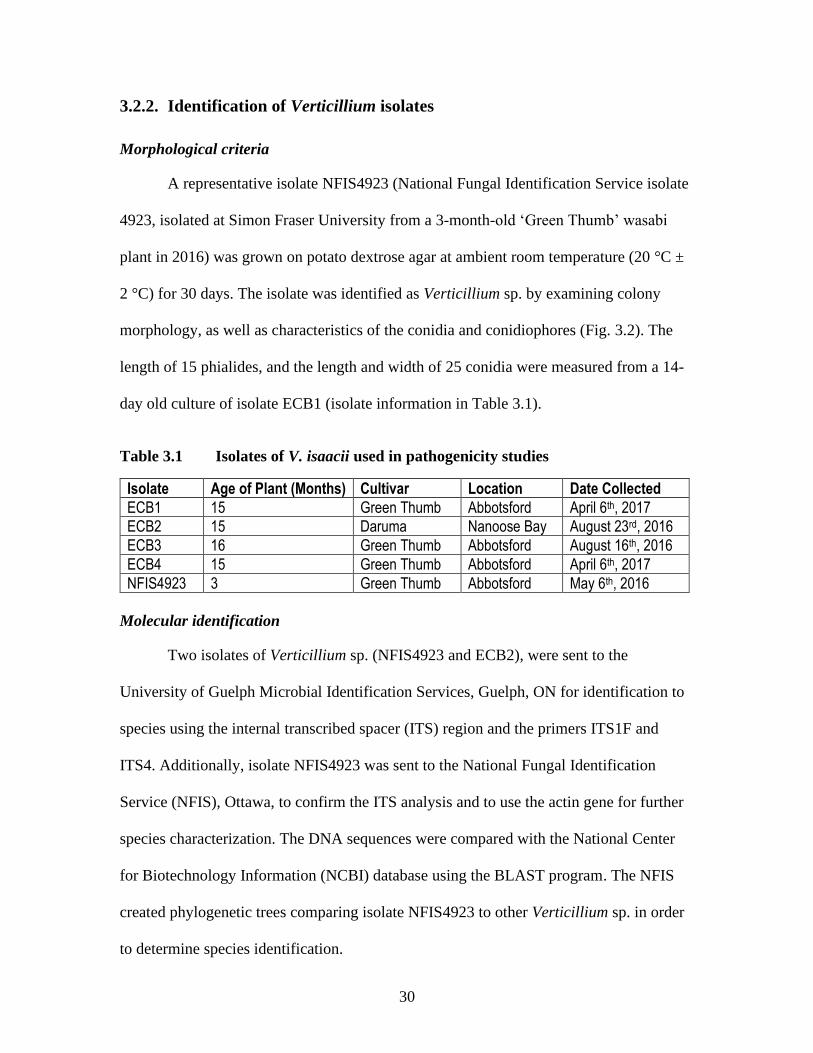
Table 3.1 Isolates of V. isaacii used in pathogenicity studies
Isolate Age of Plant (Months) Cultivar Location Date Collected
ECB1 15 Green Thumb Abbotsford April 6th, 2017
ECB2 15 Daruma Nanoose Bay August 23rd, 2016
ECB3 16 Green Thumb Abbotsford August 16th, 2016
ECB4 15 Green Thumb Abbotsford April 6th, 2017
NFIS4923 3 Green Thumb Abbotsford May 6th, 2016
Molecular identification
Two isolates of Verticillium sp. (NFIS4923 and ECB2), were sent to the
University of Guelph Microbial Identification Services, Guelph, ON for identification to
species using the internal transcribed spacer (ITS) region and the primers ITS1F and
ITS4. Additionally, isolate NFIS4923 was sent to the National Fungal Identification
Service (NFIS), Ottawa, to confirm the ITS analysis and to use the actin gene for further
species characterization. The DNA sequences were compared with the National Center
for Biotechnology Information (NCBI) database using the BLAST program. The NFIS
created phylogenetic trees comparing isolate NFIS4923 to other Verticillium sp. in order
to determine species identification.
31
3.2.3. Effect of temperature on pathogen growth
A 14-day old culture of isolate NFIS4923 was cut into 1 cm2 plugs and plated
onto fresh PDA plates with 100 mg/L streptomycin sulphate. Five plates each were
placed in growth chambers set at 5, 10, 15, 20, 25, 30, and 35 °C and left to incubate for
7 days. After 7 days, the diameter of the colony was measured. This experiment was
repeated using isolate ECB1.
3.2.4. Pathogenicity tests
Detached leaf, petiole, and rhizome inoculations
To determine pathogenicity of recovered isolates identified as V. isaacii (isolates
NFIS4923, ECB2, and ECB3), they were grown in 50 mL of potato dextrose broth (PDB)
for 3 weeks, and then homogenized (to a total volume of 150 mL) using a blender. This
suspension contained a mixture of mycelium, microsclerotia, and conidia. Five leaves,
five petioles, and two rhizomes were excised from healthy greenhouse-grown ‘Green
Thumb’ plants, surface-sterilized with 95% ethanol, wounded using a sterilized scalpel,
and inoculated with 20 mL of the suspension. The inoculated tissues were placed in
plastic containers lined with moist paper towels, and symptom development was assessed
after 7 days. Equal numbers of control plant tissues were wounded and received 20 mL of
sterile distilled water instead of a mycelial suspension.
Whole plant inoculations
In addition to detached tissue assays, pathogenicity tests were also conducted on
plants grown under greenhouse conditions. Five 6-month-old ‘Green Thumb’ plantlets
grown in 3.8 L pots containing PRO-MIX HP peat/perlite mix (Premier Horticulture,

32
Riviêre du Loup, QC) were wounded by inserting a scalpel at several locations through
the substrate to sever the roots. A mycelial suspension was prepared as above, and 50 mL
was poured into the substrate. Five control plants received sterile water. Plants were
grown in a greenhouse maintained at 20 °C ± 5 °C with a 16/8-hour day/night cycle, and
watered with a specialized fertilizer solution containing nitrogen, phosphate, potassium,
and micronutrients optimized for wasabi growth (Your Wasabi Farms Ltd, BC).
Symptoms were assessed approximately 6 months post infection by uprooting the plants,
rinsing the roots with tap water, and measuring the root lengths. The roots and rhizomes
were also cut open to examine for symptoms of vascular blackening.
To speed up the infection process, dip treatments were also used to inoculate
wasabi plants. Three 1-year-old ‘Green Thumb’ were uprooted from PRO-MIX HP
substrate and their roots were rinsed with tap water. The roots were trimmed to ~10 cm in
length using sterile scissors. The wounded roots were soaked in a mycelial suspension
(prepared as above, then diluted 5 times with sterile water) for 10 min and then repotted
in 3.8 L pots filled with coco coir (CANNA Canada Corp, ON) saturated with fertilizer
solution. Two control plants had their roots soaked in 5x-diluted PDB instead of mycelial
suspension. The plants were returned to the greenhouse and were assessed after
approximately 3 months for symptoms as above.
To assess if different V. isaacii isolates varied in pathogenic capability, twelve 15-
month-old ‘Green Thumb’ plants were inoculated as above and immersed in either a
mycelial suspension of each of 3 different isolates (NFIS4923, ECB1, and ECB2) or
diluted PDB (control). There were 3 plants per treatment. Plants were left to soak in
inoculum for 1 hour, followed by repotting in coco coir. Plants were placed in a Conviron

33
growth chamber set at 15 °C with a 16/8-hour day/night cycle and 80% RH for
approximately 4 months.
3.3. Results
3.3.1. Disease symptoms
Symptoms on naturally infected wasabi plants generally included wilting of the
leaves and petioles (Fig.1). Blackening of the vascular tissues of the rhizome occurred
with disease progression and in some cases, extended into the petiole (Fig. 1 B,C). Often,
plants also had symptoms of a secondary bacterial soft rot infection (Supplemental Fig. 1)
which was identified through MicroLog™ (BioLog Inc., Hayward, CA) as Pseudomonas
marginalis.
3.3.2. Species identification
Morphological criteria
Isolate NFIS4923 developed velvety, slow-growing, white-coloured colonies on
PDA (Fig. 3.2a). After 30 days, cultures darkened due to production of resting mycelium
and microsclerotia (Fig. 3.2b). Abundant amounts of colourless, oval-shaped conidia
were produced on phialides arranged in a verticillate pattern along conidiophores (Fig.
3.2d). These characteristics were used to identify the isolates as Verticillium sp.. Further
narrowing down the identification, some isolates (ECB4) secreted a yellow pigment in
culture (Fig. 3.2c) which is consistent with Verticillium sp. placed in clade Flavexudans
(Inderbitzin et al. 2011). The average length of lateral phialides was 23.6 µm (17.5 µm –
28 µm), and the average length and width of conidia were 7.4 µm (5 µm – 10 µm) and
6.14 µm (4µm – 10µm), respectively.
34
Figure 3.2 Colonies of Verticillium isaacii grown on PDA. A) Isolates form velvety, white
colonies after 7 days. B) Colonies darken to black as time progresses to 14 days. C) Some
isolates, such as ECB4, produce yellow pigments in culture after 7 days. (D-F) Morphology of
conidiophores and conidia of Verticillium isaacii. D) Verticilliate conidiophores with 3 to 5
phialides. E) Abundant hyaline conidia. F) Dark microsclerotia about 1 mm in diameter. Scale
bars = 25 µm.
Molecular identification
The ITS rDNA region of isolate NFIS4923 from wasabi was grouped with a
cluster of isolates comprised of 3 species: V. isaacii Inderb., Bostock, Davis & Subbarao,
V. klebahnii Inderb., Bostock, Davis & Subbarao, and V. tricorpus (Fig. 3.3a). It was not
possible to identify isolate NFIS4923 by PCR of the ITS region alone, but PCR with a
Verticillium-specific primer set targeting a portion of the actin gene shared a >99%

35
sequence identity with V. isaacii and the isolate clustered with a group of V. isaacii
isolates from other hosts (Fig. 3.3b).

36
A
37
Figure 3.3 Phylogenetic analysis performed by the NFIS of isolate NFIS4923 identified it as
Verticillium isaacii based on analyses using ITS (A) and actin (B) barcodes.
B
38
3.3.3. Effect of temperature on pathogen growth
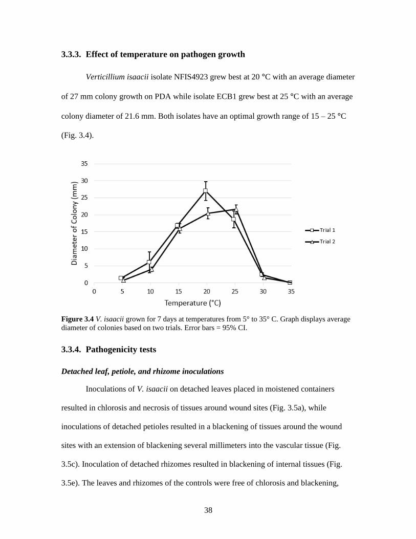
Verticillium isaacii isolate NFIS4923 grew best at 20 °C with an average diameter
of 27 mm colony growth on PDA while isolate ECB1 grew best at 25 °C with an average
colony diameter of 21.6 mm. Both isolates have an optimal growth range of 15 – 25 °C
(Fig. 3.4).
Figure 3.4 V. isaacii grown for 7 days at temperatures from 5° to 35° C. Graph displays average
diameter of colonies based on two trials. Error bars = 95% CI.
3.3.4. Pathogenicity tests
Detached leaf, petiole, and rhizome inoculations
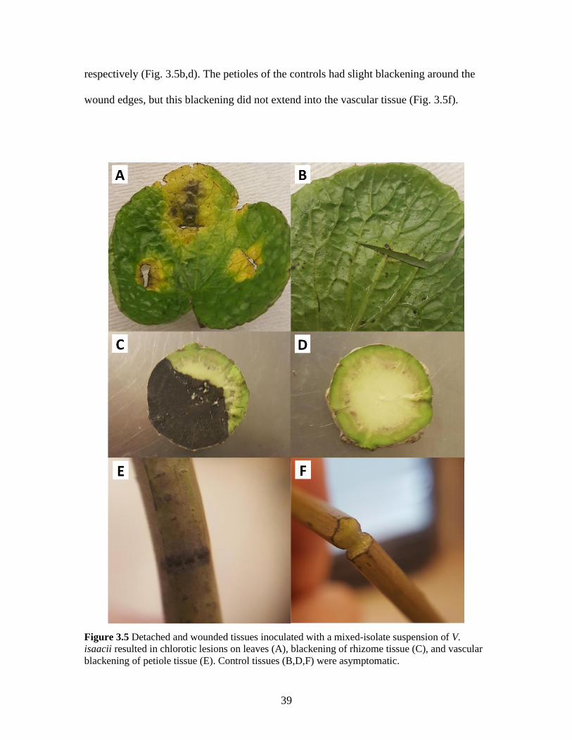
Inoculations of V. isaacii on detached leaves placed in moistened containers
resulted in chlorosis and necrosis of tissues around wound sites (Fig. 3.5a), while
inoculations of detached petioles resulted in a blackening of tissues around the wound
sites with an extension of blackening several millimeters into the vascular tissue (Fig.
3.5c). Inoculation of detached rhizomes resulted in blackening of internal tissues (Fig.
3.5e). The leaves and rhizomes of the controls were free of chlorosis and blackening,
39
respectively (Fig. 3.5b,d). The petioles of the controls had slight blackening around the
wound edges, but this blackening did not extend into the vascular tissue (Fig. 3.5f).
Figure 3.5 Detached and wounded tissues inoculated with a mixed-isolate suspension of V.
isaacii resulted in chlorotic lesions on leaves (A), blackening of rhizome tissue (C), and vascular
blackening of petiole tissue (E). Control tissues (B,D,F) were asymptomatic.
40
Whole plant inoculations
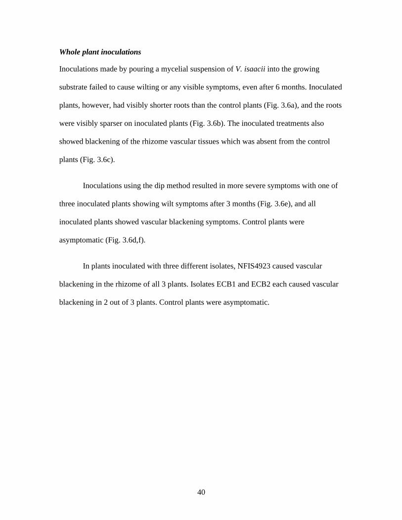
Inoculations made by pouring a mycelial suspension of V. isaacii into the growing
substrate failed to cause wilting or any visible symptoms, even after 6 months. Inoculated
plants, however, had visibly shorter roots than the control plants (Fig. 3.6a), and the roots
were visibly sparser on inoculated plants (Fig. 3.6b). The inoculated treatments also
showed blackening of the rhizome vascular tissues which was absent from the control
plants (Fig. 3.6c).
Inoculations using the dip method resulted in more severe symptoms with one of
three inoculated plants showing wilt symptoms after 3 months (Fig. 3.6e), and all
inoculated plants showed vascular blackening symptoms. Control plants were
asymptomatic (Fig. 3.6d,f).
In plants inoculated with three different isolates, NFIS4923 caused vascular
blackening in the rhizome of all 3 plants. Isolates ECB1 and ECB2 each caused vascular
blackening in 2 out of 3 plants. Control plants were asymptomatic.

41
Figure 3.6 After 3 months, plants inoculated with a mixed-isolate mycelial suspension of V.
isaacii show visibly sparser roots (A) compared to a control plant (B). All inoculated plants had
some degree of vascular blackening in their rhizome tissues (C), while controls were
asymptomatic (D). Wilting occurred in one of the three inoculated plants (E) while the controls
remained unwilted (F).
42
3.4. Discussion
There is much controversy surrounding species identification of Verticillium sp.
that infect brassica hosts (Hwang et al., 2017). For the clade of microsclerotia-producing
Verticillium sp., verticillium wilt on plants in the Brassicaceae family is attributed to V.
longisporum whereas wilt on plants outside the Brassicaceae family is attributed
primarily to V. dahliae (Karapapa et al., 1997). However, some short-spore (4-8 µm)
isolates of Verticillium can infect brassicas, and some long-spore (6-11 µm) isolates can
attack plants outside of brassicas (Fahleson et al., 2004; Inderbitzin et al., 2011; Novakazi
et al., 2015). Thus, some researchers have argued that V. longisporum should not be a
separate species from V. dahliae but be a subspecies (Barbara and Clewes, 2003;,Pegg
and Brady 2001). This creates major confusion when diseases caused by Verticillium are
reported as different researchers will use different identities for the same species
(Inderbitzin and Subbarao, 2014).
Verticillium isaacii was first described by Inderbitzin et al. (2011) from a
previously indistinguishable clade of V. isaacii, V. klebahnii, and V. tricorpus. These
species cannot be differentiated morphologically and furthermore cannot be identified
using ITS primers alone, as their ITS sequences are identical (Inderbitzin et. al, 2011).
Therefore, we used the actin region to distinguish between species in order to identify our
isolate. There are previous reports of V. tricorpus infecting wasabi in B.C. (Vippen Joshi,
Plant Diagnostic Pathologist, BC Ministry of Agriculture, pers. comm.) but this was
based on ITS sequences and the pathogen may in fact be V. isaacii as shown in the
present study.
43
Previous pathogenicity tests conducted with V. isaacii on various crops have
shown the isolates to be weakly pathogenic, with only 1 out of 4 of isolates causing
disease symptoms on artichoke and ‘Salinas’ lettuce after 10 weeks, but all isolates
caused wilt on strawberry plants (Gurung et al., 2015). Isolates tested on cauliflower were
asymptomatic (Gurung et al., 2015). In fact, a strain of V. isaacii has been shown to be a
preventative biocontrol agent in cauliflower against verticillium wilt (Tyvaert et al.,
2014). It endophytically colonized the plant and prevented V. longisporum from
colonizing through competition for space (Tyvaert et al., 2014). In sunflower and potato
plants, four out of five V. isaacii isolates tested were pathogenic and endophytic strains
were also reported (Wheeler and Johnson, 2019). These reports suggest that V. isaacii
can have both an endophytic and pathogenic lifestyle and is generally a weak pathogen.
Gurung et al. (2015) poured a conidial suspension onto the growing media in
order to inoculate artichoke, lettuce, strawberry and cauliflower plants, whilst Wheeler
and Johnson (2019) mixed microsclerotia suspended in sand into the growing medium to
inoculate sunflower and potato. On wasabi plants grown in the greenhouse, we observed
that conidial suspensions were not effective at producing symptoms (unpublished data).
The suspensions used in our experiments that produced symptoms contained a blend of
mycelium, conidia, and microsclerotia. Using these suspensions was faster than using
microsclerotia alone, as inoculum as conducted by other authors (Wheeler and Johnson,
2019).
On wasabi plants grown under commercial conditions, symptoms generally
appeared on 12-18 month-old plants, suggesting the pathogen could have infected at any
time during the preceding period and symptoms developed depending on the prevailing
44
moisture, temperature, and incubation times. Symptoms were most apparent during the
summer season, when ambient temperatures were over 30 °C, and were accompanied by
wilting and soft rot symptoms, believed to be caused by P. marginalis (unpublished data).
We believe that V. isaacii infects plants during colder months, but due to heat stress and
the prevalence of P. marginalis in the summer months, disease incidence was higher. The
pronounced symptoms seen in commercially grown plants were difficult to replicate
experimentally, as the temperature and moisture conditions, and microbe complex were
undetermined. In earlier studies, presence of P. carotovorum together with Pythium spp.
caused soft rot symptoms to develop (Rodríguez and Punja, 2007). The initial source of
inoculum of V. isaccii is unknown but could be from other Brassicaceae plants or from
the growing substrate.
Verticillium wilt is of increasing importance to wasabi growers, as it can be
difficult to eradicate, likely due to the presence of microsclerotia in the growing
substrate. During surveys, infected plants were found growing in hydroton, gravel, and
river rock substrates. Both the cultivars ‘Green Thumb’ and ‘Daruma’ were found to be
susceptible to verticillium wilt, but it is yet unknown if other B.C. cultivars are
susceptible. The wasabi industry has no registration to traditional pesticides, including
the soil fumigants traditionally used to control verticillium wilt. However, as V. isaacii is
a weak pathogen, some growers have observed that using organic substrates as a growth
substrate reduced disease severity, perhaps due to the resident microbial community
suppressing the pathogen. Since most growers do not use organic soils to grow wasabi,
verticillium wilt will remain problematic until adequate control options are identified.
45
Chapter 4. Management of powdery mildew, caused by
Erysiphe cruciferarum, on wasabi (Wasabia japonica) plants
in British Columbia
4.1. Introduction
Wasabi (Wasabia japonica (Miq.) Matsumura, syn. Eutrema japonicum Matsum.) is a
slow growing crop in the Brassicaceae family. It is grown for its valuable stem (generally
referred to as a rhizome), which is traditionally ground into a paste and used as a
condiment in Japanese cuisine (Chadwick et al., 1993). In B.C., wasabi is grown in glass
or double-polyethylene greenhouses using semi-hydroponic systems. The most popular
production systems use beds of river rock as a growing substrate and overhead misters to
provide high humidity and deliver fertilizers (Chadwick et al., 1993). Recently, however,
some growers are experimenting with drip irrigation and growing plants in other
substrates including hydroton, gravel, peat, and soil. The wasabi rhizome takes 12 to 18
months to reach a harvestable size, and due to the long periods between rhizome harvests,
growers are increasingly interested in marketing other parts of the wasabi plant, including
leaves. Wasabi leaves are edible and can be eaten as salad greens (Chadwick et al., 1993).
Additionally, some growers have been processing and selling the leaves for nutraceutical
supplements.
Notable root and rhizome-infecting pathogens in B.C. wasabi production facilities
include Leptosphaeria biglobosa Shoemaker & H. Brun, Botrytis cinerea Pers.,
Pectobacterium carotovorum (Jones) Waldee subsp. carotovorum (Jones) Bengey et al.,
as well as Pythium intermedium de Bary and Pythium dissotocum Dreschler (Macdonald
and Punja, 2017; Punja et al., 2017; Rodríguez and Punja, 2007, 2009). However, with

46
the increasing demand for high quality leaves, foliar pathogens are becoming of
increasing importance for wasabi producers. Previously reported foliar pathogens of
wasabi in B.C. include white blister rust caused by Albugo candida (Pers. ex Lev)
Kuntze, leaf spots caused by Colletotrichum higginsianum Sacc. and Leptosphaeria
biglobosa, leaf blight caused by Botrytis cinerea, and powdery mildew caused by
Erysiphe cruciferarum Opiz ex L. Junell (Joshi et al., 2014; Macdonald and Punja, 2017;
Punja et al., 2017).
Powdery mildew of wasabi has been previously reported from Japan, Taiwan, and
Korea, indicating E. cruciferarum to be the causal agent (Lo and Wang, 2000; Oku et al.,
1993; Park et al., 2016). The first report of E. cruciferarum causing powdery mildew on
wasabi in B.C. was in 2013, and powdery mildew has been consistently observed since
then (Joshi et al., 2014; Joshi and Jefferies, 2016; Betz and Punja, 2017; Betz et al.,
2019). In severe cases of powdery mildew infection, the leaf surfaces can be completely
covered by the fungal mycelium and chlorosis and defoliation may occur (Fig. 4.1).
Diseases continue to be a major barrier to successful wasabi production in
Canada, as the optimal growing environment for wasabi requires around 80% ambient
relative humidity (Chadwick et al., 1993). For disease management, there are four
biopesticides registered by the Pesticide Management Regulatory Agency (PMRA) for
use on wasabi in Canada (Health Canada, 2016). These include Rootshield® Plus WP
(BioWorks Inc.,Victor, NY) and BW240 WP (BioWorks Inc.,Victor, NY), both of which
contain a blend of Trichoderma harzianum Rifai strain KRL-AG2 and T. virens (J.H.
Mill., Giddens, & A.A. Foster) Arx strain G-4 as the active ingredients. Also registered
are Rhapsody® ASO™ (Bayer Cropscience Inc., Calgary, AB) and Cease® (BioWorks

47
Inc.,Victor, NY), which contain Bacillus subtilis (Ehrenberg) Cohn strain QST 713 as the
active ingredient. The published literature lacks any data on the comparative efficacy of
these biopesticides for management of wasabi diseases. There is increased demand for
disease management options that are both environmentally sustainable and allowed for
organic production, and producers are requesting efficacy data for additional products (S.
Sabaratnam, Plant Pathologist, BC Ministry of Agriculture, pers. comm.). Furthermore,
available fungicide products with varying active ingredients are necessary to help prevent
loss of product efficacy due to the pathogen developing resistance.
In this study, four commercially available products, with differing modes of
action, were tested for their ability to suppress powdery mildew development on wasabi.
The products tested were Actinovate® SP (Streptomyces lydicus strain WYEC 108;
Novozymes BioAg Inc., Brookfield, WI), Rhapsody® ASO™ (Bacillus subtilis strain QST
713), Cueva® (Copper Octanoate Soap; Certis USA L.L.C., Columbia, MD), and
Regalia® Maxx (Reynoutria sachalinensis extract; Marrone Bio Innovations Inc., Davis,
CA). Actinovate® and Rhapsody® are formulated with microbes that produce an array of
antimicrobial compounds involved as the primary mode of action ( Yuan and Crawford,
1995; Ongena and Jacques, 2008). Cueva® is a copper soap product which causes copper
toxicity in bacterial and fungal spores, inhibiting germination. Regalia® is a plant extract
of giant knotweed that induces systemic acquired resistance (Su et al., 2012). Induction
of polyphenolics involved in plant defense, including fungitoxic flavonoids, in plants
sprayed with this extract has been demonstrated (Daayf et al., 2000; Fofana et al., 2002).
These four products are approved for use in commercial crop production systems and
appear on the Organic Materials Review Institute (OMRI) Canada Products List© (2019).
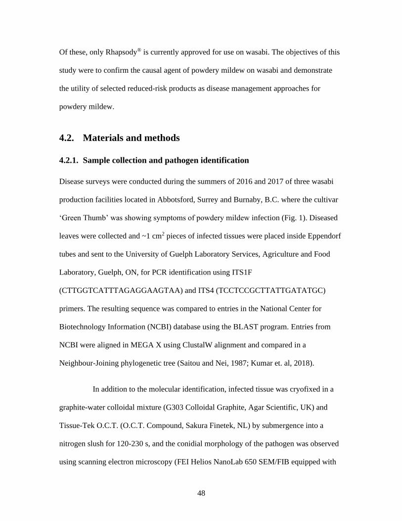
48
Of these, only Rhapsody® is currently approved for use on wasabi. The objectives of this
study were to confirm the causal agent of powdery mildew on wasabi and demonstrate
the utility of selected reduced-risk products as disease management approaches for
powdery mildew.
4.2. Materials and methods
4.2.1. Sample collection and pathogen identification
Disease surveys were conducted during the summers of 2016 and 2017 of three wasabi
production facilities located in Abbotsford, Surrey and Burnaby, B.C. where the cultivar
‘Green Thumb’ was showing symptoms of powdery mildew infection (Fig. 1). Diseased
leaves were collected and ~1 cm2 pieces of infected tissues were placed inside Eppendorf
tubes and sent to the University of Guelph Laboratory Services, Agriculture and Food
Laboratory, Guelph, ON, for PCR identification using ITS1F
(CTTGGTCATTTAGAGGAAGTAA) and ITS4 (TCCTCCGCTTATTGATATGC)
primers. The resulting sequence was compared to entries in the National Center for
Biotechnology Information (NCBI) database using the BLAST program. Entries from
NCBI were aligned in MEGA X using ClustalW alignment and compared in a
Neighbour-Joining phylogenetic tree (Saitou and Nei, 1987; Kumar et. al, 2018).
In addition to the molecular identification, infected tissue was cryofixed in a
graphite-water colloidal mixture (G303 Colloidal Graphite, Agar Scientific, UK) and
Tissue-Tek O.C.T. (O.C.T. Compound, Sakura Finetek, NL) by submergence into a
nitrogen slush for 120-230 s, and the conidial morphology of the pathogen was observed
using scanning electron microscopy (FEI Helios NanoLab 650 SEM/FIB equipped with

49
the Quorum PP3010T cryo system; 4D LABS, SFU, Burnaby, BC. Conducted by Darren
Sutton). Conidia were also observed using light microscopy in a Zeiss compound
microscope and the length and width of 25 conidia were measured.
Figure 4.1 Powdery mildew development on wasabi leaves caused by Erysiphe cruciferarum. (A)
Early development of disease, showing white colonies. (B) Advanced stages of infection. (C, D)
Severe powdery mildew infection that can cause yellowing.
4.2.2. Applications of environmentally sustainable products for disease
management
Mature wasabi ‘Green Thumb’ plants (between 14-16 months of age) growing in a
commercial greenhouse were treated with either Actinovate®, Rhapsody®, Cueva®,
Regalia®, or a water control using a backpack sprayer. The greenhouse was glass with
natural light, and the plants were grown in a hydroton substrate in individuals pots in 18
rows with approximately 200 plants per row. As the greenhouse is a controlled

50
environment, drift was assumed to be minimal. Sprays were made to run-off (with the
exception of Actinovate which was sprayed to wetness, according to label directions)
every two weeks over a total of 10 (in 2017) or 12 (in 2018) weeks. Applications were
made according to label rates of 1% (v/v) for Cueva® and Rhapsody®, 0.5% (v/v) for
Actinovate®, and 0.25% (v/v) for Regalia®. These rates were selected to represent the
middle of the recommended label rates, except for Regalia®, which was at the upper
range of recommended rates (Bayer CropScience Inc., 2017; Monsanto Canada ULC,
2018; Marrone Bio Innovations Inc., 2019; W. Neudorff GmbH KG, 2019). The trials
were conducted three times in the same greenhouse as described below.
Trial 1. May – August 2017
The trial was located within a row in the center of the greenhouse, with plants
sprayed with Actinovate® (n=20), Rhapsody® (n=20), or water (n=40) for a total of 80
plants. Plants within each treatment were divided into groups of five and were randomly
assigned positions along the greenhouse row. Each group had a minimum of four
untreated buffer plants between them. This made it practical to measure disease
development and to minimize differences in disease progression due to location.
Grouping plants together also helped to avoid drift and cross-contamination.
Trial 2. September – December 2017
The trial was located within a row in the centre of the greenhouse, with 20 plants
each sprayed with either Actinovate®, Cueva®, Rhapsody®, Regalia®, or water. Each
treatment had four groups of 5 (n=20/treatment), and each group was placed randomly
along the row, ensuring a buffer of at least four plants between treatment groups (Fig.
4.2). This trial was repeated once.

51
Trial 3. March – May 2018
The trial was located within a row that was towards the edge of the greenhouse,
with 20 plants each sprayed with Cueva®, Rhapsody®, Regalia®, or water. Actinovate®
was omitted from this trial. Each treatment had four groups of 5 (n=20/treatment), and
each group was placed randomly along the row, ensuring a buffer of at least four plants
between treatment groups. This trial was repeated once.
Figure 4.2 Experimental design for Trial 2. Treatments were divided into 4 groups/ treatment
with each group containing 5 plants. At least 4 untreated buffer plants were placed between
treated plants.
4.2.3. Assessment of disease development
Powdery mildew developed naturally from pre-existing inoculum in the greenhouse onto
wasabi plants. Once symptoms started to appear, plants were randomized (via random
number generation) into trials. Since the trials were situated within an existing
commercial operation, all plant pruning, management and other practices were conducted
according to industry recommendations. Plants in the trial were not sprayed with any
products other than the experimental treatments. Disease severity was assessed every two
weeks by using a custom scale (Trial 1) or the Horsfall-Barratt scale (Trials 2 and 3) to
measure the mean percentage of leaf surface area infected by mildew (Horsfall and
Barratt, 1945). The custom scale evaluated coverage on a 5-point scale, 1 = 1-10%, 2 =
11-25%, 3 = 26-50%, 4=51-75%, and 5=76-100%. The Horsfall-Barratt scale evaluated
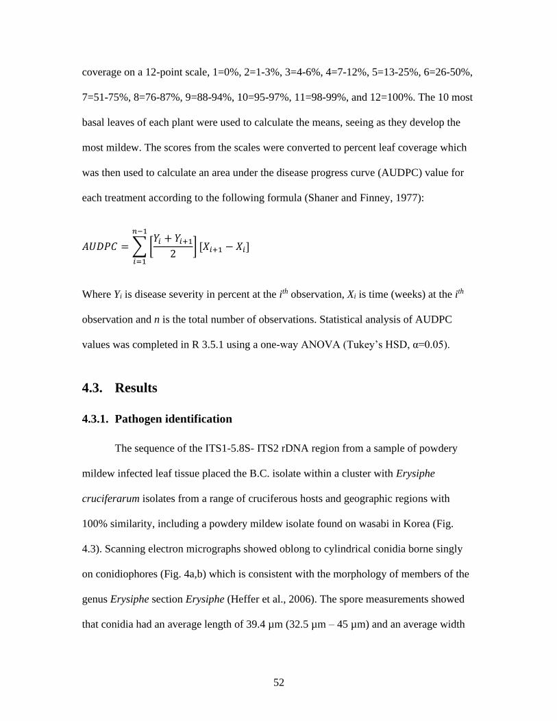
52
coverage on a 12-point scale, 1=0%, 2=1-3%, 3=4-6%, 4=7-12%, 5=13-25%, 6=26-50%,
7=51-75%, 8=76-87%, 9=88-94%, 10=95-97%, 11=98-99%, and 12=100%. The 10 most
basal leaves of each plant were used to calculate the means, seeing as they develop the
most mildew. The scores from the scales were converted to percent leaf coverage which
was then used to calculate an area under the disease progress curve (AUDPC) value for
each treatment according to the following formula (Shaner and Finney, 1977):
𝐴𝑈𝐷𝑃𝐶 = ∑[𝑌𝑖 + 𝑌𝑖+1
2] [𝑋𝑖+1 − 𝑋𝑖]
𝑛−1
𝑖=1
Where Yi is disease severity in percent at the ith observation, Xi is time (weeks) at the ith
observation and n is the total number of observations. Statistical analysis of AUDPC
values was completed in R 3.5.1 using a one-way ANOVA (Tukey’s HSD, α=0.05).
4.3. Results
4.3.1. Pathogen identification
The sequence of the ITS1-5.8S- ITS2 rDNA region from a sample of powdery
mildew infected leaf tissue placed the B.C. isolate within a cluster with Erysiphe
cruciferarum isolates from a range of cruciferous hosts and geographic regions with
100% similarity, including a powdery mildew isolate found on wasabi in Korea (Fig.
4.3). Scanning electron micrographs showed oblong to cylindrical conidia borne singly
on conidiophores (Fig. 4a,b) which is consistent with the morphology of members of the
genus Erysiphe section Erysiphe (Heffer et al., 2006). The spore measurements showed
that conidia had an average length of 39.4 µm (32.5 µm – 45 µm) and an average width

53
of 16.3 µm (12.5 µm – 20 µm) (Fig. 4.4c) which is consistent with other reports of E.
cruciferarum on wasabi (Oku et al., 1993; Park et al., 2016).
Figure 4.3 Phylogenetic analysis of powdery mildew isolates identified as E. cruciferarum from
several cruciferous hosts, including the wasabi powdery mildew from Korea (Δ). The sequence
from the BC wasabi isolate (▲) was subjected to NCBI Blast, and aligned in MEGA X using
ClustalW, and compared using a Neighbour-Joining tree.

54
Figure 4.4 Morphology of conidiophores and conidia of E. cruciferarum. A) Conidiophores and
hyphae growing across a wasabi leaf surface (scale bar = 25 µm). B) A close-up of a developing
conidium (scale bar = 2.5 µm). Note the lack of conidial chains. C) Conidia observed under light
microscopy (scale bar = 250 µm).
4.3.2. Assessment of disease development
Powdery mildew disease severity increased over time in all treatments in each of the
three trials (Fig. 4.5). After 10 weeks, plants treated with Cueva®, Regalia®, and
Rhapsody® had visibly lower development of disease. (Fig. 4.6). Actinovate® treated
plants had comparable mildew development to the water sprayed plants (Fig. 4.6).
AUDPC values calculated after 10 weeks showed that Actinovate® did not significantly
decrease the progression of powdery mildew in either of Trial 1 or Trial 2 (Fig. 4.7 a,b).
In contrast, both Regalia® and Cueva® significantly (p<0.05) reduced progression of
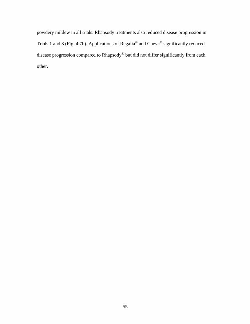
55
powdery mildew in all trials. Rhapsody treatments also reduced disease progression in
Trials 1 and 3 (Fig. 4.7b). Applications of Regalia® and Cueva® significantly reduced
disease progression compared to Rhapsody® but did not differ significantly from each
other.
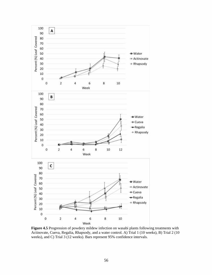
56
Figure 4.5 Progression of powdery mildew infection on wasabi plants following treatments with
Actinovate, Cueva, Regalia, Rhapsody, and a water control. A) Trial 1 (10 weeks), B) Trial 2 (10
weeks), and C) Trial 3 (12 weeks). Bars represent 95% confidence intervals.

57
Figure 4.6 Development of powdery mildew on wasabi plants, 10 weeks after treatments were
initiated. (A) Cueva and (B) Regalia applications reduced powdery mildew development
compared to a water control (E). (C) Rhapsody showed slight-to-moderate disease suppressive
capability. (D) Actinovate had no disease suppression compared to the water control (E,F).
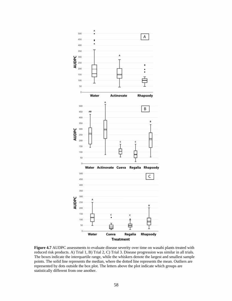
58
Figure 4.7 AUDPC assessments to evaluate disease severity over time on wasabi plants treated with
reduced risk products. A) Trial 1, B) Trial 2, C) Trial 3. Disease progression was similar in all trials.
The boxes indicate the interquartile range, while the whiskers denote the largest and smallest sample
points. The solid line represents the median, where the dotted line represents the mean. Outliers are
represented by dots outside the box plot. The letters above the plot indicate which groups are
statistically different from one another.
59
4.4. Discussion
Previous reports have shown that the pathogens affecting wasabi in B.C. have wide host
ranges that include other cruciferous plants (Joshi et al. 2014; Betz and Punja, 2017; Betz
et al., 2019). The powdery mildew pathogen, Erysiphe cruciferarum, can also infect other
members of the Brassicaceae family, including weed species (Koch and
Slusarenko, 1990; Enright and Cipollini, 2007), which may provide an additional
inoculum source. Powdery mildew can spread rapidly through a wasabi greenhouse by
production of large numbers of air-disseminated conidia, resulting in foliar infections that
can render diseased leaves unmarketable. The production system used to grow wasabi
may play a role in severity of powdery mildew. Plants receiving drip irrigation tended to
have higher levels of powdery mildew infection than those that receiving overhead
misting. This is likely due to the leaves remaining wetter for longer periods with misting,
limiting infection as powdery mildew fungi are favoured by drier conditions
(Schnathorst, 1960). However, higher moisture levels can enhance the incidence of other
diseases, including those caused by Botrytis, Colletotrichum, and Leptosphaeria (Punja et
al., 2017; MacDonald, 2018). The current strategies used to manage powdery mildew
development on wasabi include regular pruning of infected leaves and weekly sprays of
Rhapsody® during heavy infection (L. Benkrima, Lead Scientist, Your Wasabi Farms
Ltd., pers. comm.). While removal of diseased leaves is effective when infection levels
are low, it is less effective under high disease pressure and increases cost of production.
Mechanical removal of leaves can also provide entry points for other pathogens,
including B. cinerea, L. biglobosa and Wasabi mottle virus (Punja et al. 2017;
Macdonald et al., 2019).
60
Rootshield® and BW240 are registered on wasabi for control of root rot and
damping off due to Pythium sp., Rhizoctonia sp., Fusarium sp., and Phytophthora sp.
(BioWorks Inc., 2019a; 2019b); Rhapsody® and Cease® are registered for pythium root
rot (Pythium dissotocum and Pythium intermedium), phytophthora crown and root rot
(Phytophthora cryptogea Pethybr. & Laff.), and powdery mildew (E. cruciferarum)
(Bayer CropScience Inc., 2017; BioWorks Inc., 2016). Rhapsody® and Cease® are the
only products currently approved for use for management of powdery mildew on wasabi
in Canada and they have a common active ingredient, B. subtilis. The identification of
additional disease management products would provide wasabi growers with alternatives
for rotation programs. Both Cueva® and Regalia® Maxx were shown to suppress E.
cruciferarum development on wasabi in the present study. Copper-containing products
are known for their fungitoxic properties and inactivate enzymes and other proteins in
microbes (Lamichhane et al., 2018). They are widely used in organic agriculture and are
effective against multiple diseases on a range of crops, including powdery mildew on
cruciferous crops (Lamichhane et al., 2018). Cueva®’s formulation consists of copper
bound to octanoic acid (copper octanoate) (W. Neudorff GmbH KG, 2019), which
renders it more persistent on the leaf surface and reduces its uptake by the plant, thereby
reducing phytotoxicity (Marine et al., 2016; W. Neudorff GmbH KG, 2019). Applications
of copper octanoate were found to reduce powdery mildew on tomato and cucurbits
(Baysal-Gurel and Miller, 2015; Marine et al., 2016). Applications of Cueva® to wasabi
leaves resulted in higher levels of copper in leaf tissues (195 ppm in treated leaves
compared to 18 ppm in control leaves) based on tissue analysis, but the levels in the
rhizomes were unaffected (unpublished data). The majority of the copper adhered to the
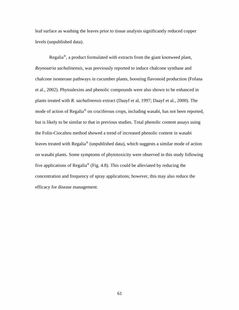
61
leaf surface as washing the leaves prior to tissue analysis significantly reduced copper
levels (unpublished data).
Regalia®, a product formulated with extracts from the giant knotweed plant,
Reynoutria sachalinensis, was previously reported to induce chalcone synthase and
chalcone isomerase pathways in cucumber plants, boosting flavonoid production (Fofana
et al., 2002). Phytoalexins and phenolic compounds were also shown to be enhanced in
plants treated with R. sachalinensis extract (Daayf et al, 1997; Daayf et al., 2000). The
mode of action of Regalia® on cruciferous crops, including wasabi, has not been reported,
but is likely to be similar to that in previous studies. Total phenolic content assays using
the Folin-Ciocalteu method showed a trend of increased phenolic content in wasabi
leaves treated with Regalia® (unpublished data), which suggests a similar mode of action
on wasabi plants. Some symptoms of phytotoxicity were observed in this study following
five applications of Regalia® (Fig. 4.8). This could be alleviated by reducing the
concentration and frequency of spray applications; however, this may also reduce the
efficacy for disease management.
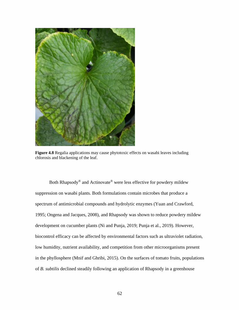
62
Figure 4.8 Regalia applications may cause phytotoxic effects on wasabi leaves including
chlorosis and blackening of the leaf.
Both Rhapsody® and Actinovate® were less effective for powdery mildew
suppression on wasabi plants. Both formulations contain microbes that produce a
spectrum of antimicrobial compounds and hydrolytic enzymes (Yuan and Crawford,
1995; Ongena and Jacques, 2008), and Rhapsody was shown to reduce powdery mildew
development on cucumber plants (Ni and Punja, 2019; Punja et al., 2019). However,
biocontrol efficacy can be affected by environmental factors such as ultraviolet radiation,
low humidity, nutrient availability, and competition from other microorganisms present
in the phyllosphere (Mnif and Ghribi, 2015). On the surfaces of tomato fruits, populations
of B. subtilis declined steadily following an application of Rhapsody in a greenhouse
63
environment, requiring multiple applications to maintain population levels (Punja et al.,
2016). Additionally, volatile compounds released from wasabi leaves can inactivate
entomopathogenic fungi such as Beauveria bassiana (Bals.-Criv.) Vuill. (Atsumi and
Saito, 2015) and may similarly influence survival of biopesticides, but this requires
further research.
In conclusion, Cueva® and Regalia® show potential for powdery mildew disease
management on wasabi but are not currently registered for use in Canada.
64
Chapter 5. General discussion and conclusions
5.1. Disease surveys
The identification of Verticillium isaacii as a new pathogen of wasabi brings the
current worldwide total of known wasabi diseases to 24. Along with the confirmed presence
of Erysiphe cruciferarum, these identifications bring the total of confirmed pathogens in B.C.
up to 11. The three years of surveys also identified 8 previously unreported disease-
associated microbes found on wasabi, including Fusarium sp. Additionally, the surveys
found Pseudomonas marginalis associated with soft rot, and Pythium irregulare associated
with root rot – both of which have been reported in literature (Goto and Matsumoto, 1986;
Joshi et al., 2014; Joshi et al., 2015) but lack the inoculation studies required to confirm
pathogenicity. This brings the total of disease-associated microbes found on wasabi
worldwide from nine to eighteen and from four to twelve in North America.
5.2. Control of wasabi diseases
As most wasabi diseases found in B.C. have broad host ranges that include other
brassica species, most of the primary inoculum sources are assumed to be other cruciferous
crops and weed species. Other possible inoculum sources include the growing substrate,
watering systems, and mechanical transmission from infected wasabi plants. In the case of
wasabi mottle virus, the inoculum source is suspected to be imported wasabi plants. It is also
possible that insect vectors play a role in the transmission of certain diseases, especially
viruses.
65
5.3. Future research
The disease surveys we conducted identified a multitude of microorganisms with
the potential to be pathogenic on wasabi. These microbes should be confirmed for
phytopathogenic capability and virulence on wasabi. This involves plant inoculation
experiments, and well as subsequent recovery of microbes from symptomatic plants. This
would help to further identify pathogens found on wasabi in the Pacific Northwest region
of B.C.
During the years surveying wasabi plants, cultivar was found to greatly influence
the presence of disease. ‘Green Thumb’ plants were observed to be more susceptible to a
variety of diseases than the other common grown cultivars. In fact, ‘Green Thumb’ plants
in the laboratory all developed a serious white rust infection while the ‘Daruma’ plants
sitting beside them on the bench were unaffected. These suspected cultivar differences
should be further investigated in a controlled setting.
As Regalia® and Cueva® were effective at slowing the progression of powdery
mildew on wasabi, it is possible that they would also be efficacious on other wasabi
pathogens. Therefore, experiments should be conducted on the ability of these, and other
reduced risk products, to supress commonly found wasabi diseases such as phoma leaf
spot and botrytis rhizome rot. It would also be useful to have data on the effects of
chemical pesticides on wasabi diseases, in case of severe disease outbreaks.
The lack of current disease control methods, as well as information on what
diseases are present, limit options for disease management. Without more management

66
options (such as products registered for use on wasabi) growers will continue to suffer
losses.
67
References
Adachi, S. ed. 1987. Wasabi sabai. Shizouka (Japan).
Agrios GN. 2005. Plant Pathology (5th Ed). Elsevier Academic Press: Oxford.
Alkooranee JT, Liu S, Aledan TR, Yin Y, and Li M. 2015. First report of powdery
mildew caused by Erysiphe cruciferarum on Brassica napus in China. Plant Dis.
99(11):1651.
Atsumi A, Saito T. 2015. Volatiles from wasabi inhibit entomopathogenic fungi:
implications for tritrophic interactions and biological control. J Plant Interact.
10:152–157.
Barbara, DJ, Clewes E. 2003. Plant pathogenic Verticillium species: how many of them
are there? Mol Plant Pathol. 4:297–305.
Betz EC, Punja ZK. 2017a. 2016 Survey of fungal diseases of wasabi in British Columbia
greenhouses. Can Plant Dis Surv. 97(2):227–228.
Betz EC, Punja ZK. 2017b. Occurrence of wilt, caused by Verticillium isaacii, and
powdery mildew, caused by Erysiphe cruciferarum, on wasabi in British
Columbia. Can J Plant Pathol (Abstr.) 40:154.
Betz EC, Roberts AJ, Punja ZK. 2019. 2017 and 2018 surveys of microbes associated
with wasabi diseases in British Columbia greenhouses. Can Plant Dis Surv.
99:239–240.

68
Bayer CropScience Inc. 2017. Rhapsody® ASO™ label. Retrieved from: http://pr-rp.hc-
sc.gc.ca/1_1/view_label?p_ukid=137994411
Baysal-Gurel F, Miller SA. 2015. Management of powdery mildew in greenhouse
tomato production with biorational products and fungicides. Acta Hortic.
1069:179–183.
BioWorks Inc. 2016. CEASE® biological fungicide label. Retrieved from: http://pr-rp.hc-
sc.gc.ca/1_1/view_label?p_ukid=137994522
BioWorks Inc. 2019a. BW240 WP biological fungicide label. Retrieved from: http://pr-
rp.hc-sc.gc.ca/1_1/view_label?p_ukid=137998149
BioWorks Inc. 2019b. ROOTSHIELD® PLUS WP biological fungicide label. Retrieved
from: http://pr-rp.hc-sc.gc.ca/1_1/view_label?p_ukid=137997637
Broadhurst PG, Wright PJ. 1998. Phoma disease of wasabi in New Zealand. In
O'Callaghan, M. (Ed.) Proceedings of the 51st New Zealand Plant Protection
Conference (1998). Hamilton, New Zealand, The New Zealand Plant Protection
Society Inc.
Chadwick CI, Lumpkin TA, Elberson LR. 1993. The botany, uses and production of
Wasabia japonica (Miq.) (Cruciferae) Matsum. Econ Bot. 47(2):113–135.
Chai AL, Xie XW, Shi YX, Li BJ. 2014. Research status of clubroot (Plasmodiophora
brassicae) on cruciferous crops in China. Can J Plant Pathol. 36:142–153.
Choi YJ, Han KS, Park YH, Shin HD. 2014. First report of white blister rust caused by
69
Albugo candida on wasabi in Korea. Plant Dis. 98:1006–1006.
Daayf F, Ongena M, Boulanger R, El Hadrami I, Bélanger RR. 2000. Induction of
phenolic compounds in two cultivars of cucumber by treatment of healthy and
powdery mildew-infected plants with extracts of Reynoutria sachalinensis. J Chem
Ecol. 26(7):1579–1593.
Daayf F, Schmitt A, Belanger RR. 1997. Evidence of phytoalexins in cucumber leaves
infected with powdery mildew following treatment with leaf extracts of Reynoutria
sachalinensis. Plant Physiol. 113(3):719–727.
Dutta BK. 1981. Effect of the chemical and physical condition of the soil on verticillium
wilt of antirrhinum. Plant Soil. 63(2):217–225.
Depotter JRL, Deketelaere S, Inderbitzin P, von Tiedemann A, Höfte M, Subbarao KV,
Wood TA, Thomma BP. 2016. Verticillium longisporum , the invisible threat to
oilseed rape and other brassicaceous plant hosts. Mol Plant Pathol. 17:1004–1016.
Enright SM, Cipollini D. 2007. Infection by powdery mildew Erysiphe cruciferarum
(Erysiphaceae) strongly affects growth and fitness of Alliaria petiolata
(Brassicaceae). Am J Bot. 94:1813–1820.
Fahleson J, Hu Q, Dixelius C. 2004. Phylogenetic analysis of Verticillium species based
on nuclear and mitochondrial sequences. Arch Microbiol. 181:435–442.
Fitt BDL, Brun H, Barbetti MJ, Rimmer SR. 2006. World-wide importance of phoma
stem canker (Leptosphaeria maculans and L. biglobosa) on oilseed rape (Brassica
70
napus). Eur J Plant Pathol. 114: 3–15.
Fletcher JD 1989. Additional hosts of alfalfa mosaic virus, cucumber mosaic virus, and
tobacco mosaic virus in New Zealand. New Zeal J Crop Hortic Sci. 17(4): 361–362.
Fofana B, McNally DJ, Labbé C, Boulanger R, Benhamou N, Séguin A, Bélanger RR
(2002). Milsana-induced resistance in powdery mildew-infected cucumber plants
correlates with the induction of chalcone synthase and chalcone isomerase. Physiol
Mol Plant Pathol. 6(2):121–132.
Goto M, Matsumoto K. 1986. Taxonomic study on soft rot bacteria isolated from
diseased rhizomes and roots of wasabi (Eutrema wasabi Maxim.). Ann Phytopathol
Soc Jpn. 52:69–77.
Glawe DA. 2006. First report of powdery mildew of Eschscholizia californica (california
poppy) caused by Erysiphe cruciferarum in North America. Plant Health Progress.
Online.
Glawe DA. 2008. The powdery mildews: A review of the world's most familiar (yet
poorly known) plant pathogens. Annu Rev Phytopathol. 46:27–51.
Granke LL, Harlan BR, Naegele RP, Hausbeck MK. 2012. First report of root and crown
rot of wasabi (Wasabia japonica Matsum.) caused by Phytophthora cryptogea in
Michigan. Plant Dis. 96(9):1379.
Gurung S, Short DPG, Hu X, Sandoya GV, Hayes RJ, Koike ST, Subbarao KV. 2015.
Host range of Verticillium isaacii and Verticillium klebahnii from artichoke,

71
spinach, and lettuce. Plant Dis. 99:933–938.
Heale JB, Karapapa VK. 1999. The verticillium threat to canada’s major oilseed crop:
canola. Can J Plant Pathol. 21:1–7.
Health Canada. 2016. Pesticide label search. Retrieved from Health Canada website:
http://pr-rp.hc-sc.gc.ca/ls-re/result-eng.php?p_search_label=wasabi
Heffer V, Johnson KB, Powelson ML, Shishkoff N. 2006. Identification of powdery
mildew fungi anno 2006. The Plant Health Instructor. PHI-I-2006-0706-01.
Horsfall JG, Barratt RW. 1945. An improved grading system for measuring plant
diseases. Phytopathology 35:655 (Abstr.).
Hwang SF, Strelkov SE, Ahmed HU, Zhou Q, Fu H, Fredua-Agyeman R, Turnbull JD.
2017. First report of Verticillium dahliae Kleb. causing wilt symptoms in canola (
Brassica napus L.) in North America. Can J Plant Pathol. 39:514–526.
Inderbitzin P, Bostock RM, Davis RM, Usami T, Platt HW, Subbarao KV. 2011.
Phylogenetics and taxonomy of the fungal vascular wilt pathogen Verticillium,
with the descriptions of five new species. ed. Idnurm A. PLoS One. 6:e28341.
Inderbitzin P, Davis RM, Bostock RM, Subbarao KV. 2013. Identification and
differentiation of Verticillium species and V. longisporum lineages by simplex and
multiplex PCR assays ed. Corradi N. PLoS One. 8:e65990.
Inderbitzin P, Subbarao KV. 2014. Verticillium systematics and evolution: how confusion
impedes verticillium wilt management and how to resolve it. Phytopathology.
72
104:564–574.
Joshi V, Jeffries M. 2010. Diseases diagnosed on commercial crops submitted to the
British Columbia Ministry of Agriculture and Lands (BCMAL) Plant Diagnostic
Laboratory in 2009. Can Plant Dis Surv. 90:7–15.
Joshi V, Jeffries M, Jesperson G. 2014. Diseases diagnosed on commercial crops
submitted to the British Columbia Ministry of Agriculture Plant Health
Laboratory in 2013. Can Plant Dis Surv. 94:8–18.
Joshi V, Jeffries M, Jesperson G. 2015. Diseases diagnosed on commercial crops
submitted to the British Columbia Ministry of Agriculture Plant Health
Laboratory in 2014. Can Plant Dis Surv. 95:7–16.
Joshi V, Jeffries M. 2016. Diseases diagnosed on commercial crops submitted to the
British Columbia Ministry of Agriculture Plant Health Laboratory in 2015. Can
Plant Dis Surv. 96:7–18.
Karapapa VK, Bainbridge BW, Heale JB. 1997. Morphological and molecular
characterization of Verticillium longisporum comb, nov., pathogenic to oilseed
rape. Mycol Res. 101:1281–1294.
Koch E, Slusarenko AJ. 1990. Fungal pathogens of Arabidopsis thaliana (L.)
Heyhn. Botanica Helvetica. 100: 257–268.
Koike ST, Gladders P, Paulus AO. 2007. Vegetable diseases: A color handbook.
Burlington, MA.
73
Kishi K. ed. 1988. Plant diseases in Japan. Tokyo, Zenkoku Noson Kyoiku Kyokai.
Kumar S, Stecher G, Li M, Knyaz C, Tamura K. 2018. MEGA X: Molecular
Evolutionary Genetics Analysis across computing platforms. Mol Biol Evol
35:1547–1549.
Lamichhane JR, Osdaghi E, Behlau F, Köhl J, Jones JB, Aubertot JN. 2018. Thirteen
decades of antimicrobial copper compounds applied in agriculture. A review.
Agron Sustain Dev. 38(3):28.
Lo CT, Wang KM, Hu MF, Wang CH. 2002. Integrated control of black rot disease of
wasabi, caused by Phoma wasabiae. Bot Bull Acad Sin. 43:219–225. .
Lo CT, Wang KM. 2000. Survey of fungal diseases on aboveground parts of wasabi in
Taiwan. Plant Pathol Bull. 9(1):17–22.
MacDonald JL, Betz EC, Punja ZK, Li YQ, Xiang Y, Bouthillier MJ, Deyoung RM,
Chen X, Bernardy MG. 2019. First report of ringspot and vein-clearing symptoms
on Wasabia japonica plants associated with Wasabi mottle virus in North
America. Can J Plant Pathol. 41(3):509.
MacDonald JL. 2018. Fungal Pathogens of Wasabi in British Columbia. Thesis: Master
of Pest Management. Simon Fraser University, Burnaby, BC.
MacDonald JL, Punja ZK. 2017. Occurrence of botrytis leaf blight, anthracnose leaf spot,
and white blister rust on Wasabia japonica in British Columbia. Can J Plant
Pathol. 39(1):60–71.
74
Marine SC, Newark MJ, Korir RC, Everts KL. 2016. Evaluation of rotational
biopesticide programs for disease management in organic cucurbit poduction.
Plant Dis. 100(11):2226–2233.
Marrone Bio Innovations Inc. 2019. Regalia® Maxx biofungicide label. Retrieved from:
http://pr-rp.hc-sc.gc.ca/1_1/view_label?p_ukid=139272003
Martin RJ, Deo B, Depree J. 2002. Wasabi yield and isothiocyanate responses to fertiliser
type. Agronomy NZ. 32:121–125.
Matsumoto K, Sugiyama M, Nakata E. 1985. Ring rot of wasabi (Eutrema wasabi
Maxim.) caused by Corynebacterium sp. Bull Yamaguchi Prefecture Agric Exp
Station. 37:99–114.
Miller PM, Stoddard EM. 1956. Hot-water treatment of fungi infecting strawberry roots.
Phytopathology. 46(12):694–696.
Minoshima A, Takeuchi J, Ono T, Kagiwada S, Horie H, Hirooka Y. 2017. First report of
phytophthora rot on Wasabia japonica caused by Phytophthora drechsleri in
Japan. J Gen Plant Pathol. 83:113–116.
Mnif I, Ghribi D. 2015. Potential of bacterial derived biopesticides in pest management.
Crop Prot. 77:52–64.
Monsanto Canada ULC. 2018. Actinovate® SP fungicide label. Retrieved from: http://pr-
rp.hc-sc.gc.ca/1_1/view_label?p_ukid=139270224
75
Nelson PE, Wilheim S. 1958. Thermal death range of Verticillium albo-atrum.
Phytopathology. 48(11):613–616.
Ni L, Punja ZK. 2019. Management of fungal diseases on cucumber (Cucumis sativus L.)
and tomato (Solanum lycopersicum L.) crops in greenhouses using Bacillus
subtilis. In: Islam MT, Rahman MM, Pandey P, Boehme MH, Haesaert G. Bacilli
and agrobiotechnology: phytostimulation and biocontrol: Vol 2. Cham,
Switzerland: Springer International Publishing.
Novakazi F, Inderbitzin P, Sandoya G, Hayes RJ, von Tiedemann A, Subbarao KV. 2015.
The three lineages of the diploid hybrid Verticillium longisporum differ in
virulence and pathogenicity. Phytopathology. 105:662–673.
Oku T, Arie M, Kishira H. 1993. Powdery mildew of wasabi (Eutrema wasabi Maxim.).
Jpn J Plant Pathol. 59(5):601–606.
Ongena M, Jacques P. 2008. Bacillus lipopeptides: versatile weapons for plant disease
biocontrol. Trends Microbiol. 16(3):115–125.
Organic Materials Review Institute. 2019. Canada Products List. Retrieved from:
https://www.omri.org/sites/default/files/opl_pdf/CompleteCompany-COR.pdf
Palmer J. 1990. Germination and growth of wasabi (Wasabia japonica (Miq.)
Matsumara). New Zeal J Crop Hortic Sci. 18:161–164.
Park YH, Moon YG, Cho SE, Shin HD. 2016. First report of powdery mildew caused by
Erysiphe cruciferarum on wasabi (Wasabia japonica) in Korea. Plant Dis.
76
100(2):530.
Pegg GF, Brady BL. 2001. Verticillium wilts. Oxford, UK: CAB International.
Powers E. 1995. Efficacy of the Ryu non-staining KOH technique for rapidly
determining gram reactions of food-borne and waterborne bacteria and yeasts.
Appl Environ Microbiol. 61(10):3756–3758.
Punja ZK, Chandanie WA, Chen X, Rodríguez G. 2017. Phoma leaf spot of wasabi
(Wasabia japonica) caused by Leptosphaeria biglobosa. Plant Pathol. 66(3):480–
489.
Punja ZK, Rodriguez G, Tirajoh A. 2016. Effects of Bacillus subtilis strain QST 713 and
storage temperatures on post-harvest disease development on greenhouse tomatoes.
Crop Prot. 84:98–104.
Punja ZK, Tirajoh A, Collyer D, Ni L. 2019. Efficacy of Bacillus subtilis strain QST 713
(Rhapsody) against four major diseases of greenhouse cucumbers. Crop Prot. 124:
104845.
Rodríguez G. 2007. Micropropagation of wasabi (Wasabia japonica) and identification of
pathogens affecting plant growth and quality. Thesis: Master of Science. Simon
Fraser University, Burnaby, BC.
Rodríguez G, Punja ZK. 2007. Root infection of wasabi (Wasabia japonica) by Pythium
species. Can J Plant Pathol. 29(1):79–83.

77
Rodríguez G, Punja ZK. 2009. Vascular blackening of wasabi rhizomes caused by
Pectobacterium carotovorum subsp. carotovorum. Eur J Plant Pathol.
124(3):483–493.
Saitou N, Nei M. 1987. The neighbor-joining method: A new method for reconstructing
phylogenetic trees. Mol Biol Evol. 4:406–425.
Shaner G, Finney RE. 1977. Effect of nitrogen-fertilization on expression of slow-
mildewing resistance in Knox wheat. Phytopathology 67:1051–1056.
Shimamoto I, Sonoda S, Vazquez P, Minaka N, Nishiguchi M. 1998. Nucleotide
sequence analysis of the 3′ terminal region of a wasabi strain of crucifer
tobamovirus genomic RNA: subgrouping of crucifer tobamoviruses. Arch Virol.
143:1801–1813.
Su H, Blair R, Johnson T, Marrone P. 2012. Regalia® Bioprotectant in plant disease
management. Outlook Pest Manage. 23(1):30–34.
Toth IK, Bell KS, Holeva MC, Birch PRJ. 2002. Soft rot erwiniae: from genes to
genomes. Mol Plant Pathol. 4:17–30.
Tyvaert L, França SC, Debode J, Höfte M. 2014. The endophyte Verticillium Vt305
protects cauliflower against verticillium wilt. J Appl Microbiol. 116:1563–1571.
W. Neudorff GmbH KG. 2019. Cueva® Commercial Copper Fungicide Label. Retrieved
from: http://pr-rp.hc-sc.gc.ca/1_1/view_label?p_ukid=139271629
78
Weng M, Han J, Bian X, Jiang W. 2010. Effects of shading on growth and photosynthetic
characteristics of wasabi seedlings. Southwest China J Agric Sci. 5: S649.
Williamson B, Tudzynski B, Tudzynski P, Van Kan JAL. 2007. Botrytis cinerea: the
cause of grey mould disease. Mol Plant Pathol. 8(5): 561–580.
Wilson CR. 1998. First report of cucumber mosaic virus Cucumovirus on wasabi in
Australia. Plant Pathol 82:590.
Wheeler D, Johnson DA. 2019. Verticillium isaacii is a pathogen and endophyte of
potato and sunflower in the Columbia Basin of Washington. Plant Dis. 103(12):
3150–3153.
Zhang F, Ning H, Zhang M, He W, Li G, He T. 2004. Biological characters of Phoma
wasabiae causing black rot of wasabi. Plant Prot. 30(4):45–48.
79
Appendix A.
Dual culture assays to assess in vitro pathogen inhibition by
biofungicides
A.1 Methods
Biocontrols were assessed in vitro (on solid media) for efficacy against several
wasabi pathogens including Leptosphaeria biglobosa, Verticillium sp., Fusarium
avenaceum, Fusarium oxysporum, Pythium intermedium, and Pythium irregulare.
We chose 5 commercially available biological control agents to test – Actinovate®
SP (Streptomyces lydicus strain WYEC 108), Rhapsody ® ASO (Bacillus subtilis strain
QST 713), Rootshield ® WP (Trichoderma harzianum strain T-22), Cueva® (Copper
octanoate soap), and Regalia® (Reynoutria sachalinensis extract).
For Actinovate, Rootshield, and Rhapsody, the active organism was isolated onto
either LB or V8 Agar plates. For Cueva and Regalia, the solutions were diluted to the
maximum rate recommended on the label (2% and 0.25%, respectively). For the
microbial controls, the organism was streaked (Actinovate and Rhapsody) or added as a
7mm plug (Rootshield) onto LB and PDA Agars. For Cueva and Regalia, 100μL was
pipetted into 7mm wells made on the plate. For each plate, a pathogen was added 24
hours later. The plates were assessed either 5 days (both Pythium sp.) or 3 weeks
(Fusarium, Phoma, and Verticillium) depending on rate of pathogen growth.
A.2 Results

80
There were obvious inhibition zones in all Rhapsody and Actinovate plates.
Rootshield also inhibited pathogen growth, but without the inhibition zones (Fig. A.1
shows an example). Both Cueva and Regalia showed little to no inhibition on plates.
When grown on PDA, all pathogens had significant growth reduction on Actinovate,
Rhapsody, and Rootshield plates (Fig. A.2).
The inhibition zones found in the Actinovate and Rhapsody plates are likely due
to the antimicrobials including antibiotics and chitinases. Rootshield works primarily
through competitive exclusion, so the reduction of pathogen growth without inhibition
zones is to be expected. Regalia works by inducing systemic immunity in plants, so it is
Figure A.1 Pythium intermedium dual culture assay plates showing pathogen
inhibition due to biocontrols. The pathogen was plated at the top of the plate, and after
24h, the following treatments were added: A) Water, B) Rhapsody, C) Actinovate, D)
Rootshield, E) Cueva, F) Regalia.
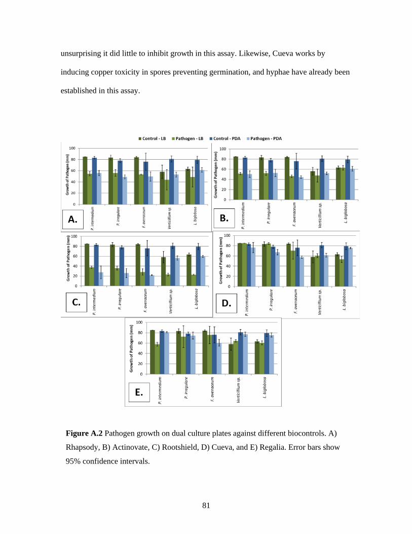
81
unsurprising it did little to inhibit growth in this assay. Likewise, Cueva works by
inducing copper toxicity in spores preventing germination, and hyphae have already been
established in this assay.
Figure A.2 Pathogen growth on dual culture plates against different biocontrols. A)
Rhapsody, B) Actinovate, C) Rootshield, D) Cueva, and E) Regalia. Error bars show
95% confidence intervals.
82
Appendix B.
Effect of temperature on pathogen growth in vitro
B.1 Methods
The growth of certain pathogens – Pythium intermedium, Fusarium avenaceum,
Fusarium oxysporum, Leptosphaeria biglobosa, and Verticillium sp. – were evaluated
under different temperature conditions. Five plates of each pathogen were sub-cultured
and placed in incubators at 5 °C, 10 °C, 15 °C, 20 °C, 30 °C, and 35 °C. Growth across
the plate was assessed by measuring the colony diameter after 7 days.
B.2 Results
Most pathogens grew best between 20 °C and 25 °C with growth declining on
both sides of this range (Figure B.1). The P. intermedium isolates reached the edge of the
petri dish before 1 week in all treatments from 10 °C to 25 °C. The F. oxysporum isolates
reached the edge of the petri dish before 1 week in the 25 °C treatments. For all
pathogens, except F. oxysporum, growth was completely inhibited at 35 °C. Most of
these pathogens grow best at the upper temperature range of wasabi, and therefore, heat
stress of the plants may play a role in pathogen outbreaks.
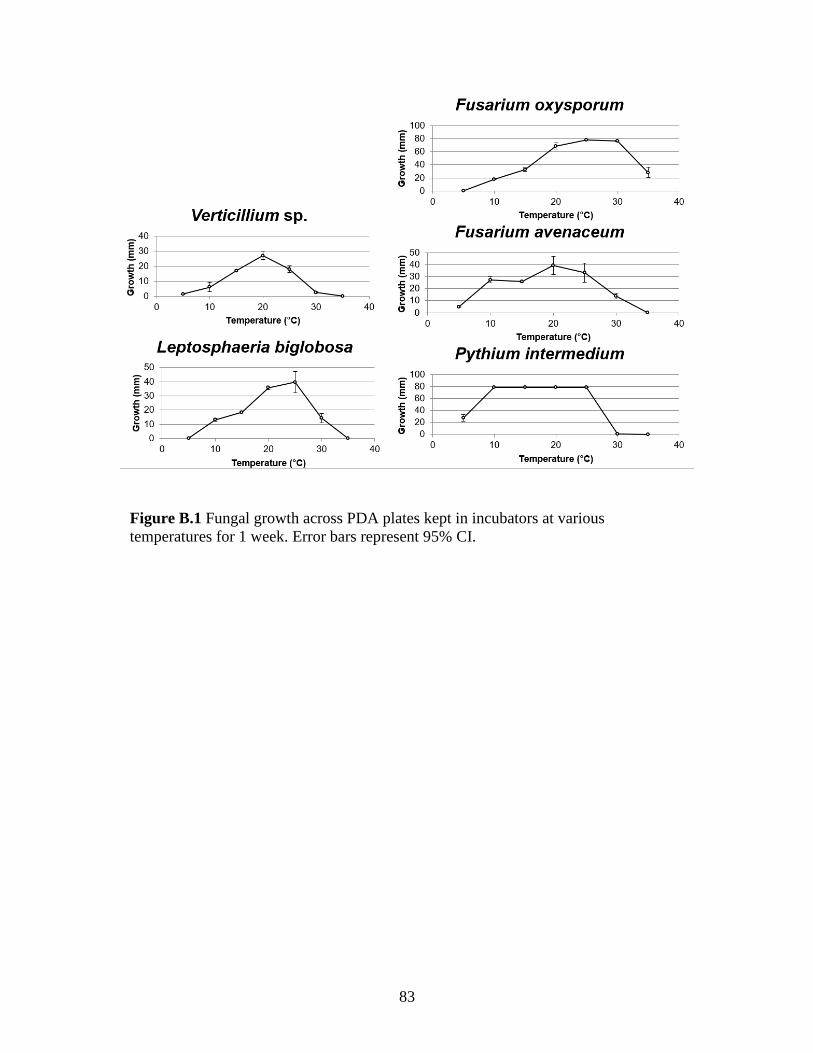
83
Figure B.1 Fungal growth across PDA plates kept in incubators at various
temperatures for 1 week. Error bars represent 95% CI.

84
Appendix C.
Fungitoxic effect of Cueva® on Verticillium isaacii
C.1 Methods
Flasks containing 30 mL of Potato Dextrose Agar with varying concentrations of
Cueva Copper Fungicide (n=5 for each treatment) were inoculated with plugs of
Verticillium isaacii. The cultures were left to incubate on a shaker for 7 days. After a
week, mycelium was filtered out and dried in an oven at 50 °C for 48 hours. The resulting
dry weights were measured and used to assess fungal growth. Statistical analysis was
performed using One-Way ANOVA and Tukey’s HSD Test.
C.2 Results
V. isaacii growth was significantly (p>0.05) reduced by about 50% in
concentrations of Cueva from 100 ppm – 300 ppm (Fig. C.1). At concentrations of 150
ppm and above ̧microsclerotial production was very reduced (Fig. C.2). No growth
occurred at concentrations of 500 ppm. As microsclerotia are the main source of
inoculum for Verticillium infection, Cueva may be efficacious in reducing spread of V.
isaacii.

85
Figure C.1 Growth of V. isaacii in PDB with varying concentrations of
Cueva Fungicide. Growth is measured in milligrams of dry weight.
Error bars are 95% confidence intervals. Letters denote which bars are
statistically different from one another.
Figure C.2 Growth of V. isaacii in PDB with varying concentrations of Cueva
Fungicide. Concentrations of Cueva from left to right are 0 ppm, 25 ppm, 50 ppm, 100
ppm, 150 ppm, 200 ppm, 250 ppm, and 300 ppm. Note the reduction of
microsclerotial production at concentrations of 150 ppm and above.