pathogens - MDPI
21
Citation: Kumar, D.; Downs, L.P.; Adegoke, A.; Machtinger, E.; Oggenfuss, K.; Ostfeld, R.S.; Embers, M.; Karim, S. An Exploratory Study on the Microbiome of Northern and Southern Populations of Ixodes scapularis Ticks Predicts Changes and Unique Bacterial Interactions. Pathogens 2022, 11, 130. https:// doi.org/10.3390/pathogens11020130 Academic Editor: Melissa J. Caimano Received: 16 December 2021 Accepted: 19 January 2022 Published: 21 January 2022 Publisher’s Note: MDPI stays neutral with regard to jurisdictional claims in published maps and institutional affil- iations. Copyright: © 2022 by the authors. Licensee MDPI, Basel, Switzerland. This article is an open access article distributed under the terms and conditions of the Creative Commons Attribution (CC BY) license (https:// creativecommons.org/licenses/by/ 4.0/). pathogens Article An Exploratory Study on the Microbiome of Northern and Southern Populations of Ixodes scapularis Ticks Predicts Changes and Unique Bacterial Interactions Deepak Kumar 1 , Latoyia P. Downs 1 , Abdulsalam Adegoke 1 , Erika Machtinger 2 , Kelly Oggenfuss 3 , Richard S. Ostfeld 3 , Monica Embers 4 and Shahid Karim 1,5, * 1 School of Biological, Environmental and Earth Sciences, The University of Southern Mississippi, Hattiesburg, MS 39406, USA; [email protected] (D.K.); [email protected] (L.P.D.); [email protected] (A.A.) 2 Department of Entomology, Pennsylvania State University, University Park, PA 16802, USA; [email protected] 3 Cary Institute of Ecosystem Studies, Millbrook, NY 12542, USA; [email protected] (K.O.); [email protected] (R.S.O.) 4 Division of Immunology, Tulane National Primate Research Center, 18703 Three Rivers Rd., Covington, LA 70433, USA; [email protected] 5 Center for Molecular and Cellular Biosciences, University of Southern Mississippi, Hattiesburg, MS 39406, USA * Correspondence: [email protected]; Tel.: +1-601-266-6232 Abstract: The black-legged tick (Ixodes scapularis) is the primary vector of Borrelia burgdorferi, the causative agent of Lyme disease in North America. However, the prevalence of Lyme borreliosis is clustered around the Northern States of the United States of America. This study utilized a metage- nomic sequencing approach to compare the microbial communities residing within Ix. scapularis populations from northern and southern geographic locations in the USA. Using a SparCC network construction model, we performed potential interactions between members of the microbial commu- nities from Borrelia burgdorferi–infected tissues of unfed and blood-fed ticks. A significant difference in bacterial composition and diversity was found between northern and southern tick populations. The network analysis predicted a potential antagonistic interaction between endosymbiont Rickettsia buchneri and Borrelia burgdorferi sensu lato. The network analysis, as expected, predicted significant positive and negative microbial interactions in ticks from these geographic regions, with the genus Rickettsia, Francisella, and Borreliella playing an essential role in the identified clusters. Interactions between Rickettsia buchneri and Borrelia burgdorferi sensu lato need more validation and understanding. Understanding the interplay between the microbiome and tick-borne pathogens within tick vectors may pave the way for new strategies to prevent tick-borne infections. Keywords: Ixodes scapularis; microbiome; salivary glands; midgut; ovaries; 16S rRNA sequencing; Borrelia burgdorferi; Rickettsia buchneri 1. Introduction Vector-borne diseases affect over one billion people every year and have been expand- ing alarmingly in recent years [1–3]. Among vector-borne diseases, Lyme disease is one of the most reported infectious diseases in the United States and corresponds to over 90% of vector-borne infections in North America [4,5]. Almost 300,000 cases of Lyme disease are reported every year in the United States, and Ixodes scapularis is known as its primary vector. Most Lyme disease cases are clustered in Northeastern and Upper Midwest States [6]. Ix. scapularis ticks are also prevalent in the Southern States, but the dearth of Lyme disease infections is linked with the restricted distribution and scarcity of the primary B. burgdorferi s.l. reservoir, namely white-footed mice, Peromyscus leucopus [7]. For B. burgdorferi s.l., an Pathogens 2022, 11, 130. https://doi.org/10.3390/pathogens11020130 https://www.mdpi.com/journal/pathogens
-
Upload
khangminh22 -
Category
Documents
-
view
3 -
download
0
Transcript of pathogens - MDPI

�����������������
Citation: Kumar, D.; Downs, L.P.;
Adegoke, A.; Machtinger, E.;
Oggenfuss, K.; Ostfeld, R.S.; Embers,
M.; Karim, S. An Exploratory Study
on the Microbiome of Northern and
Southern Populations of Ixodes
scapularis Ticks Predicts Changes and
Unique Bacterial Interactions.
Pathogens 2022, 11, 130. https://
doi.org/10.3390/pathogens11020130
Academic Editor: Melissa J. Caimano
Received: 16 December 2021
Accepted: 19 January 2022
Published: 21 January 2022
Publisher’s Note: MDPI stays neutral
with regard to jurisdictional claims in
published maps and institutional affil-
iations.
Copyright: © 2022 by the authors.
Licensee MDPI, Basel, Switzerland.
This article is an open access article
distributed under the terms and
conditions of the Creative Commons
Attribution (CC BY) license (https://
creativecommons.org/licenses/by/
4.0/).
pathogens
Article
An Exploratory Study on the Microbiome of Northern andSouthern Populations of Ixodes scapularis Ticks PredictsChanges and Unique Bacterial InteractionsDeepak Kumar 1, Latoyia P. Downs 1, Abdulsalam Adegoke 1 , Erika Machtinger 2, Kelly Oggenfuss 3,Richard S. Ostfeld 3, Monica Embers 4 and Shahid Karim 1,5,*
1 School of Biological, Environmental and Earth Sciences, The University of Southern Mississippi,Hattiesburg, MS 39406, USA; [email protected] (D.K.); [email protected] (L.P.D.);[email protected] (A.A.)
2 Department of Entomology, Pennsylvania State University, University Park, PA 16802, USA; [email protected] Cary Institute of Ecosystem Studies, Millbrook, NY 12542, USA; [email protected] (K.O.);
[email protected] (R.S.O.)4 Division of Immunology, Tulane National Primate Research Center, 18703 Three Rivers Rd.,
Covington, LA 70433, USA; [email protected] Center for Molecular and Cellular Biosciences, University of Southern Mississippi,
Hattiesburg, MS 39406, USA* Correspondence: [email protected]; Tel.: +1-601-266-6232
Abstract: The black-legged tick (Ixodes scapularis) is the primary vector of Borrelia burgdorferi, thecausative agent of Lyme disease in North America. However, the prevalence of Lyme borreliosis isclustered around the Northern States of the United States of America. This study utilized a metage-nomic sequencing approach to compare the microbial communities residing within Ix. scapularispopulations from northern and southern geographic locations in the USA. Using a SparCC networkconstruction model, we performed potential interactions between members of the microbial commu-nities from Borrelia burgdorferi–infected tissues of unfed and blood-fed ticks. A significant differencein bacterial composition and diversity was found between northern and southern tick populations.The network analysis predicted a potential antagonistic interaction between endosymbiont Rickettsiabuchneri and Borrelia burgdorferi sensu lato. The network analysis, as expected, predicted significantpositive and negative microbial interactions in ticks from these geographic regions, with the genusRickettsia, Francisella, and Borreliella playing an essential role in the identified clusters. Interactionsbetween Rickettsia buchneri and Borrelia burgdorferi sensu lato need more validation and understanding.Understanding the interplay between the microbiome and tick-borne pathogens within tick vectorsmay pave the way for new strategies to prevent tick-borne infections.
Keywords: Ixodes scapularis; microbiome; salivary glands; midgut; ovaries; 16S rRNA sequencing;Borrelia burgdorferi; Rickettsia buchneri
1. Introduction
Vector-borne diseases affect over one billion people every year and have been expand-ing alarmingly in recent years [1–3]. Among vector-borne diseases, Lyme disease is one ofthe most reported infectious diseases in the United States and corresponds to over 90% ofvector-borne infections in North America [4,5]. Almost 300,000 cases of Lyme disease arereported every year in the United States, and Ixodes scapularis is known as its primary vector.Most Lyme disease cases are clustered in Northeastern and Upper Midwest States [6]. Ix.scapularis ticks are also prevalent in the Southern States, but the dearth of Lyme diseaseinfections is linked with the restricted distribution and scarcity of the primary B. burgdorferis.l. reservoir, namely white-footed mice, Peromyscus leucopus [7]. For B. burgdorferi s.l., an
Pathogens 2022, 11, 130. https://doi.org/10.3390/pathogens11020130 https://www.mdpi.com/journal/pathogens

Pathogens 2022, 11, 130 2 of 21
infection rate of ~35–50% has been reported among ticks from the Northeastern states [8,9],whereas tick populations in the Southern states are more rarely infected [10–14].
Several studies have reported the microbial composition residing within ticks [15–22].Despite this work, limited information exists on which to base comparisons of the micro-biome residing within Ix. scapularis from the Lyme disease endemic and non-endemic areasor between different tissue types within ticks. Moreover, there is a dearth of informationon the interaction between recently identified tick endosymbionts or commensal microor-ganisms and B. burgdorferi s.l. or pathogenic microbes. Efforts have been made to identifycommensal and symbiotic microbes in the tick microbiome [8,23]. Significant variationsassociated with geography, species, and sex in Ixodes microbiota have been reported [14].The impact of microbiome communities on physiological processes in ixodid ticks, includ-ing reproductive fitness and vector competence, has been suggested [24–29]. In the UnitedStates, tick microbiome studies have shown variation in sex, species, and geography. InCanada, the Ix. scapularis microbiome from eastern and southern regions did not differsignificantly concerning the geographic origin, sex, or life stages (Clow et al., 2018). Thesevariable results motivate additional studies to generally detect whether geographic anddemographic patterns exist.
This study conducted a comparative analysis of the microbiome residing within the Ix.scapularis populations collected from Lyme-disease-endemic and -non-endemic areas in theUnited States, utilizing 16S rRNA sequencing. This study also provided an insight into themicrobiome composition in unfed and partially blood-fed tick tissues, including midgut,salivary glands, and ovaries.
2. Results2.1. Illumina MiSeq 16S V1–V3 Sequencing
Sequencing reads were acquired from a limited amount of tick samples (39 singleticks and 15 tick tissues). A minimum of three biological replicates for each sample andtissue type was used. The total number of reads obtained for whole-tick samples collectedfrom Louisiana, New York, Oklahoma, and Pennsylvania was 2,427,191, corresponding to2812 operational taxonomic units (OTUs), the minimum number of reads for a sample was36,358, and the maximum number of reads for a sample was 114,478. For tick tissues, thetotal number of reads was 546,024, corresponding to 1009 OTUs; the maximum numberof reads for a sample was 56,785; and the minimum number of reads was 17,258. In ouranalyses, each tissue sample was rarefied to 5000 sequences. In comparison, whole-ticksamples were rarefied to 16,000 sequences. The rarefaction curve (Supplementary Figure S1)plotted between the number of observed OTUs and the number of sequences, as mentionedabove, reached plateau, suggesting sufficient sample coverage for further analysis.
2.2. Comparative Analysis of Bacterial Communities Residing within Ix. scapularis
We observed substantial variation in bacterial profiles in tick populations from Louisiana,New York, Oklahoma, and Pennsylvania. Phyla Actinobacteria, Proteobacteria, Firmicutes,and Bacteroidetes cover for >90% reads for female ticks from these populations (Supplemen-tary Table S1). Proteobacteria were found to be the dominant phylum among all female tickpopulations, excluding the female Pennsylvania population, in which Bacteroidetes weredominant. A relative abundance of 36% Actinobacteria was present in female Oklahomaticks, but in other female ticks, its coverage was substantially low as 0.58%, 6.13%, and3.45% in Pennsylvania, Louisiana, and New York, respectively (Supplementary Table S1).Interestingly, female Pennsylvania ticks showed 60% relative abundance of Bacteroidetes,whereas this phylum was substantially less well represented in females from Oklahoma,New York, and Louisiana, at 0.07%, 7.5% and 0.49%, respectively (Supplementary Table S1).In male ticks, a significantly high level of Actinobacteria (88%) was found in Oklahomaticks, and levels were dramatically lower in Louisiana and New York males, at 3.5% and10%, respectively (Supplementary Table S1). New York male ticks were not included in thisstudy, due to unavailability. The level of Proteobacteria was also found to be much lower

Pathogens 2022, 11, 130 3 of 21
in Oklahoma males (6.19%) than Louisiana males (94.03%) and Pennsylvania males (50%)(Supplementary Table S1). Bacteroidetes (29%) and Tenericutes (10.4%) were substantiallyhigher in Pennsylvania males than other whole unfed male ticks, Oklahoma males (0.26%-Bacteroidetes), and Louisiana males (1.49%-Bacteroidetes), while Tenericutes were rare inother male ticks (Supplementary Table S1).
The bacterial profiling of mated and unmated tick populations was similar at thephylum level (Figure 1A–C). Among these ticks, the dominant phylum in both malesand females were Actinobacteria (~88%) and Proteobacteria (~56 to 60%), respectively(Figure 1A,B). Nonetheless, both male and female Oklahoma ticks contained ~2 to 5%Firmicutes, but other tick populations (NY and PA) contained either <1% or negligibleFirmicutes (Figure 1A,B). Further analysis of Oklahoma ticks at species level showedFirmicutes’ presence, including Jeotgalicoccus sp. and Staphylococcus lentus (Figure 1E).At the same time, Actinobacteria included Brevibacterium aureum, Yaniella halotolerans,Brevibacterium sp., and Streptomyces rochei species (Figure 1E). As expected, both New Yorkand Pennsylvania tick populations were infected with a significantly higher percentageof Spirochetes, ranging from 2% reads in New York (♀) and 0.14% in Pennsylvania (♂)respectively (Figure 1A). The prevalence of B. burgdorferi s. l. in Ix. scapularis ticks from NewYork and Pennsylvania ranges from 40 to 70% [8,9]. All the ticks and tick tissues, excludingthe ovary, were infected with B. burgdorferi s.l. The results presented here are a comparisonof percentage B. burgdorferi s.l. reads among all microbial reads in the sample. For example,ticks from Pennsylvania and New York are B. burgdorferi s.l.–infected, and 16S rRNAsequencing yielded a total of 100 reads from these ticks. Among those 100 reads, 2 and0.14 reads belong to B. burgdorferi s.l. from New York and Pennsylvania ticks, respectively.The rest of the reads belong to other microbes residing within these samples.
The Pennsylvania male and female ticks contained ~60% and 28.8% Bacteroidetes,respectively, and 7.48% in female ticks from New York (Figure 1A,B). Bacteroidetes in-cluded Sphingobacterium faecium, Flavobacterium sp., Sphingobacterium sp., Flexibacter sp.and Flavobacter spp. (Figure 1F). However, spirochetes were absent in both Louisiana andOklahoma tick populations, and only ~1.49% reads with Bacteroidetes were detected in theLouisiana female ticks (Figure 1 and Supplementary Table S1).
Pathogens 2022, 11, x FOR PEER REVIEW 3 of 22
(88%) was found in Oklahoma ticks, and levels were dramatically lower in Louisiana and New York males, at 3.5% and 10%, respectively (Supplementary Table S1). New York male ticks were not included in this study, due to unavailability. The level of Proteobacteria was also found to be much lower in Oklahoma males (6.19%) than Louisiana males (94.03%) and Pennsylvania males (50%) (Supplementary Table S1). Bacteroidetes (29%) and Tenericutes (10.4%) were substantially higher in Pennsylvania males than other whole unfed male ticks, Oklahoma males (0.26%-Bacteroidetes), and Louisiana males (1.49%-Bacteroidetes), while Tenericutes were rare in other male ticks (Supplementary Ta-ble S1).
The bacterial profiling of mated and unmated tick populations was similar at the phylum level (Figure 1A–C). Among these ticks, the dominant phylum in both males and females were Actinobacteria (~88%) and Proteobacteria (~56 to 60%), respectively (Figure 1A,B). Nonetheless, both male and female Oklahoma ticks contained ~2 to 5% Firmicutes, but other tick populations (NY and PA) contained either <1% or negligible Firmicutes (Figure 1A,B). Further analysis of Oklahoma ticks at species level showed Firmicutes’ presence, including Jeotgalicoccus sp. and Staphylococcus lentus (Figure 1E). At the same time, Actinobacteria included Brevibacterium aureum, Yaniella halotolerans, Brevibacterium sp., and Streptomyces rochei species (Figure 1E). As expected, both New York and Pennsyl-vania tick populations were infected with a significantly higher percentage of Spirochetes, ranging from 2% reads in New York (♀) and 0.14% in Pennsylvania (♂) respectively (Fig-ure 1A). The prevalence of B. burgdorferi s. l. in Ix. scapularis ticks from New York and Pennsylvania ranges from 40 to 70% [8,9]. All the ticks and tick tissues, excluding the ovary, were infected with B. burgdorferi s.l. The results presented here are a comparison of percentage B. burgdorferi s.l. reads among all microbial reads in the sample. For example, ticks from Pennsylvania and New York are B. burgdorferi s.l.–infected, and 16S rRNA se-quencing yielded a total of 100 reads from these ticks. Among those 100 reads, 2 and 0.14 reads belong to B. burgdorferi s.l. from New York and Pennsylvania ticks, respectively. The rest of the reads belong to other microbes residing within these samples.
Figure 1. Cont.
Pathogens 2022, 11, 130 4 of 21
Pathogens 2022, 11, x FOR PEER REVIEW 4 of 22
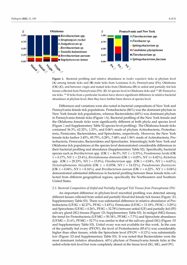
Figure 1. Bacterial profiling and relative abundance in Ixodes scapularis ticks at phylum level (A) among female ticks and (B) male ticks from Louisiana (LA), Pennsylvania (PA), Oklahoma (OK) (C), and between virgin and mated ticks from Oklahoma (D) in unfed and partially fed tick tissues col-lected from Pennsylvania (PA). (E) At species level in Oklahoma ticks and ** (F) Pennsylvania ticks. ** If ticks from a particular location have shown significant difference in relative bacterial abundance at phylum level, then they have further been shown at species level.
The Pennsylvania male and female ticks contained ~60% and 28.8% Bacteroidetes, respectively, and 7.48% in female ticks from New York (Figure 1A,B). Bacteroidetes in-cluded Sphingobacterium faecium, Flavobacterium sp., Sphingobacterium sp., Flexibacter sp. and Flavobacter spp. (Figure 1F). However, spirochetes were absent in both Louisiana and Oklahoma tick populations, and only ~1.49% reads with Bacteroidetes were detected in the Louisiana female ticks (Figure 1 and Supplementary Table S1).
Differences and variations were also noted in bacterial compositions of New York and Pennsylvania female tick populations. Proteobacteria (86%) was the dominant phy-lum in New York female tick populations, whereas Bacteroidetes (60%) was dominant phylum in Pennsylvania female ticks (Figure 1A). Bacterial profiling of the New York fe-male and the Oklahoma female ticks were significantly different at both phyla and species level (Figure 2 and Supplementary Table S2 species-level profiling). The Oklahoma female ticks contained 36.3%, 62.32%, 1.23%, and 0.06% reads of phylum Actinobacteria, Proteo-bacteria, Firmicutes, Bacteroidetes, and Spirochetes, respectively. However, the New York female ticks harbor 3.45%, 85.75%, 0.28%, 7.48% and 1.96% reads of Actinobacteria, Prote-obacteria, Firmicutes, Bacteroidetes and, Spirochaetes. Interestingly, both New York and Oklahoma tick populations at the species level demonstrated considerable differences in their bacterial profiling and abundance (Supplementary Table S2). Specifically, bacterial species such as Brevibacterium spp. (OK ♀ = 46.5%, NY ♀ = 0.33%), Pseudomonas lurida (OK ♀ = 0.17%, NY ♀ = 23.4%), Brevindumonas diminuta (OK ♀ = 0.05%, NY ♀= 8.42%), Rickettsia spp. (OK ♀ = 29.23%, NY ♀ = 15.8%), Flavobacterium spp. (OK ♀ = 0.04%, NY ♀ = 6.62%), Stenotrophomonas rhizophila (OK ♀ = 0.0358, NY ♀ = 14.52%), Pseudomonas fluorescens (OK ♀ = 0.04%, NY ♀ = 8.16%), and Brevibacterium aureum (OK ♀ = 6.22%. NY ♀ = 0.04%) demon-strated substantial differences in bacterial profiling between these female ticks collected from different geographical regions, specifically the Northeastern and Southern United States.
Figure 1. Bacterial profiling and relative abundance in Ixodes scapularis ticks at phylum level(A) among female ticks and (B) male ticks from Louisiana (LA), Pennsylvania (PA), Oklahoma(OK) (C), and between virgin and mated ticks from Oklahoma (D) in unfed and partially fed ticktissues collected from Pennsylvania (PA). (E) At species level in Oklahoma ticks and ** (F) Pennsylva-nia ticks. ** If ticks from a particular location have shown significant difference in relative bacterialabundance at phylum level, then they have further been shown at species level.
Differences and variations were also noted in bacterial compositions of New York andPennsylvania female tick populations. Proteobacteria (86%) was the dominant phylum inNew York female tick populations, whereas Bacteroidetes (60%) was dominant phylumin Pennsylvania female ticks (Figure 1A). Bacterial profiling of the New York female andthe Oklahoma female ticks were significantly different at both phyla and species level(Figure 2 and Supplementary Table S2 species-level profiling). The Oklahoma female tickscontained 36.3%, 62.32%, 1.23%, and 0.06% reads of phylum Actinobacteria, Proteobac-teria, Firmicutes, Bacteroidetes, and Spirochetes, respectively. However, the New Yorkfemale ticks harbor 3.45%, 85.75%, 0.28%, 7.48% and 1.96% reads of Actinobacteria, Pro-teobacteria, Firmicutes, Bacteroidetes and Spirochaetes. Interestingly, both New York andOklahoma tick populations at the species level demonstrated considerable differences intheir bacterial profiling and abundance (Supplementary Table S2). Specifically, bacterialspecies such as Brevibacterium spp. (OK ♀ = 46.5%, NY ♀ = 0.33%), Pseudomonas lurida (OK♀ = 0.17%, NY ♀ = 23.4%), Brevindumonas diminuta (OK ♀ = 0.05%, NY ♀= 8.42%), Rickettsiaspp. (OK ♀ = 29.23%, NY ♀ = 15.8%), Flavobacterium spp. (OK ♀ = 0.04%, NY ♀ = 6.62%),Stenotrophomonas rhizophila (OK ♀ = 0.0358, NY ♀ = 14.52%), Pseudomonas fluorescens(OK ♀ = 0.04%, NY ♀ = 8.16%), and Brevibacterium aureum (OK ♀ = 6.22%. NY ♀ = 0.04%)demonstrated substantial differences in bacterial profiling between these female ticks col-lected from different geographical regions, specifically the Northeastern and SouthernUnited States.
2.3. Bacterial Composition of Unfed and Partially Engorged Tick Tissues from Pennsylvania (PA)
An important difference in phylum-level microbial profiling was detected amongdifferent tissues collected from unfed and partially blood-fed female tick tissues (Figure 1D,Supplementary Table S3). There was substantial difference in relative abundance of Pro-teobacteria (UF.SG = 42.27%, PF.SG = 3.43%), Firmicutes (UF.SG = 11.18%, PF.SG = 3.29%)and Spirochetes (UF.SG = 0.26%, PF.SG = 32.78%) between unfed (UF) and partially fed (PF)salivary gland (SG) tissues (Figure 1D, Supplementary Table S3). In midgut (MG) tissues,the trend for Proteobacteria (UF.MG = 50.34%, PF.MG = 7.7%) and Spirochete abundance(UF.MG = 21.6%, PF.MG = 52.7%) was similar to that of the salivary gland (SG) (Figure 1Dand Supplementary Table S3). Unfed ovary was not available for this work. In the caseof the partially fed ovary (PF.OV), the level of Proteobacteria (85.6%) was considerablyhigher than other tissues, while the Spirochete level (PF.OV = 0.12%) was substantiallylow (Figure 1D and Supplementary Table S3). It was noted that Bacteroidetes were themost dominant (relative abundance, 60%) phylum of Pennsylvania female ticks at theunfed-whole-tick level but were completely absent at the tissue level (SG, MG, and OV).

Pathogens 2022, 11, 130 5 of 21
Pathogens 2022, 11, x FOR PEER REVIEW 5 of 22
Figure 2. Comparisons of bacterial alpha diversity among tick tissue samples (A,B) and ticks col-lected from different geographical locations (C,D). Plotted data represent alpha-diversity based on Faith’s phylogenetic distance (faith_pd) demonstrating relative richness among bacterial communi-ties, while Pielou’s index represent the evenness of the bacterial community among different sam-ples. Upon using the Benjamin and Hochberg correction, we found that all the adjusted-p-values (q-values) were insignificant. UF—unfed, PF—partially fed, SG—salivary gland, MG—midgut, OV—ovary, LA—Louisiana, PA—Pennsylvania, NY—New York, OK—Oklahoma.
2.3. Bacterial Composition of Unfed and Partially Engorged Tick Tissues from Pennsylvania (PA)
An important difference in phylum-level microbial profiling was detected among different tissues collected from unfed and partially blood-fed female tick tissues (Figure 1D, Supplementary Table S3). There was substantial difference in relative abundance of Proteobacteria (UF.SG = 42.27%, PF.SG = 3.43%), Firmicutes (UF.SG = 11.18%, PF.SG = 3.29%) and Spirochetes (UF.SG = 0.26%, PF.SG = 32.78%) between unfed (UF) and partially fed (PF) salivary gland (SG) tissues (Figure 1D, Supplementary Table S3). In midgut (MG) tissues, the trend for Proteobacteria (UF.MG = 50.34%, PF.MG = 7.7%) and Spirochete abundance (UF.MG = 21.6%, PF.MG = 52.7%) was similar to that of the salivary gland (SG) (Figure 1D and Supplementary Table S3). Unfed ovary was not available for this work. In the case of the partially fed ovary (PF.OV), the level of Proteobacteria (85.6%) was consid-erably higher than other tissues, while the Spirochete level (PF.OV = 0.12%) was substan-tially low (Figure 1D and Supplementary Table S3). It was noted that Bacteroidetes were the most dominant (relative abundance, 60%) phylum of Pennsylvania female ticks at the unfed-whole-tick level but were completely absent at the tissue level (SG, MG, and OV).
A considerable difference in microbial profiling at the phylum level was detected among different tissues collected from the Pennsylvania unfed and partially blood-fed female tick tissues (Figure 1D). Unfed salivary glands contained 42%, 42.27%, and ~11.2%, respectively, of Actinobacteria, Proteobacteria, and Firmicutes, whereas partially blood-
Figure 2. Comparisons of bacterial alpha diversity among tick tissue samples (A,B) and ticks collectedfrom different geographical locations (C,D). Plotted data represent alpha-diversity based on Faith’sphylogenetic distance (faith_pd) demonstrating relative richness among bacterial communities, whilePielou’s index represent the evenness of the bacterial community among different samples. Uponusing the Benjamin and Hochberg correction, we found that all the adjusted-p-values (q-values)were insignificant. UF—unfed, PF—partially fed, SG—salivary gland, MG—midgut, OV—ovary,LA—Louisiana, PA—Pennsylvania, NY—New York, OK—Oklahoma.
A considerable difference in microbial profiling at the phylum level was detectedamong different tissues collected from the Pennsylvania unfed and partially blood-fedfemale tick tissues (Figure 1D). Unfed salivary glands contained 42%, 42.27%, and ~11.2%,respectively, of Actinobacteria, Proteobacteria, and Firmicutes, whereas partially blood-fedsalivary glands harbored 42% Actinobacteria reads, followed by a substantially reducedlevel of Proteobacteria (3.43%) and Firmicutes (3.2%). Interestingly, both Spirochetesand Tenericutes levels increased from 0.26% to 32.77% and 3.37% to 17.66%, respectively(Figure 1D and Supplementary Table S3). Supplementary Table S3 shows the bacterialspecies abundance in tick tissues. Unfed midgut contained 50.34%, 21.61%, and 17.5%Proteobacteria, Spirochetes, and Actinobacteria, respectively; while partially engorgedmidgut of Proteobacteria decreases substantially to 7.7%, Spirochetes increases drastically to52.7%. The unfed ovary was unavailable, and the partially engorged ovary contained 85.56%of Proteobacteria, 8.11% of Actinobacteria, 5.24% of Tenericutes, and <1% of Firmicutes andSpirochetes (Supplementary Table S3).
Pathogens 2022, 11, 130 6 of 21
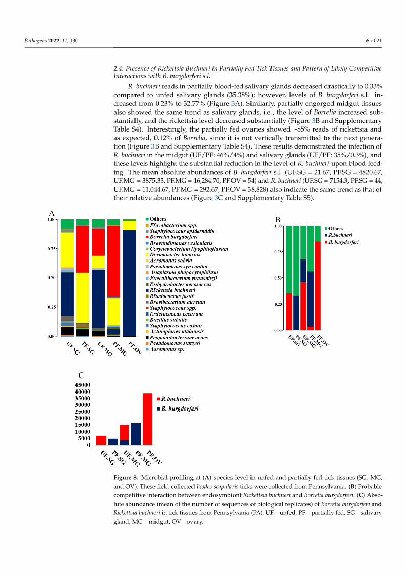
2.4. Presence of Rickettsia Buchneri in Partially Fed Tick Tissues and Pattern of Likely CompetitiveInteractions with B. burgdorferi s.l.
R. buchneri reads in partially blood-fed salivary glands decreased drastically to 0.33%compared to unfed salivary glands (35.38%); however, levels of B. burgdorferi s.l. in-creased from 0.23% to 32.77% (Figure 3A). Similarly, partially engorged midgut tissuesalso showed the same trend as salivary glands, i.e., the level of Borrelia increased sub-stantially, and the rickettsia level decreased substantially (Figure 3B and SupplementaryTable S4). Interestingly, the partially fed ovaries showed ~85% reads of rickettsia andas expected, 0.12% of Borrelia, since it is not vertically transmitted to the next genera-tion (Figure 3B and Supplementary Table S4). These results demonstrated the infection ofR. buchneri in the midgut (UF/PF: 46%/4%) and salivary glands (UF/PF: 35%/0.3%), andthese levels highlight the substantial reduction in the level of R. buchneri upon blood feed-ing. The mean absolute abundances of B. burgdorferi s.l. (UF.SG = 21.67, PF.SG = 4820.67,UF.MG = 3875.33, PF.MG = 16,284.70, PF.OV = 54) and R. buchneri (UF.SG = 7154.3, PF.SG = 44,UF.MG = 11,044.67, PF.MG = 292.67, PF.OV = 38,828) also indicate the same trend as that oftheir relative abundances (Figure 3C and Supplementary Table S5).
Pathogens 2022, 11, x FOR PEER REVIEW 7 of 23
Figure 3. Microbial profiling at (A) species level in unfed and partially fed tick tissues (SG, MG, and
OV). These field-collected Ixodes scapularis ticks were collected from Pennsylvania. (B) Probable
competitive interaction between endosymbiont Rickettsia buchneri and Borrelia burgdorferi. (C) Abso-
lute abundance (mean of the number of sequences of biological replicates) of Borrelia burgdorferi and
Rickettsia buchneri in tick tissues from Pennsylvania (PA). UF—unfed, PF—partially fed, SG—sali-
vary gland, MG—midgut, OV—ovary.
2.5. Diversity Analysis
Kruskal–Wallis non-parametric tests were performed to determine the effects of tick
geographic location (LA, PA, NY, and OK) on α-diversity metrics, using QIIME 2.
Faith_pd-diversity index, p-value, and adjusted-p-value (q-value) between the samples
are as follows: Louisiana and Pennsylvania female ticks (H = 5.4, p-value = 0.02, q-value =
0.101), Louisiana male ticks and Oklahoma mated male ticks (H = 3.85, p-value = 0.049, q-
value = 0.101), New York and Pennsylvania male and female ticks (H = 5.4, p-value = 0.02,
q-value = 0.101), Oklahoma and Pennsylvania female ticks (H = 5.4, p-value = 0.02, q-value
= 0.101), and Oklahoma and Pennsylvania virgin male (H = 4.5, p-value = 0.0338, q-value
= 0.101) (Figure 2C). Pennsylvania ticks were phylogenetically the least rich, while Okla-
homa virgin males were the richest in bacterial diversity among samples studied here. At
the tissue level, in the midgut from partially engorged ticks, we found the richest in di-
versity and ovaries were the least rich in bacterial diversity (Figure 2A). Tissue samples
Figure 3. Microbial profiling at (A) species level in unfed and partially fed tick tissues (SG, MG,and OV). These field-collected Ixodes scapularis ticks were collected from Pennsylvania. (B) Probablecompetitive interaction between endosymbiont Rickettsia buchneri and Borrelia burgdorferi. (C) Abso-lute abundance (mean of the number of sequences of biological replicates) of Borrelia burgdorferi andRickettsia buchneri in tick tissues from Pennsylvania (PA). UF—unfed, PF—partially fed, SG—salivarygland, MG—midgut, OV—ovary.

Pathogens 2022, 11, 130 7 of 21
2.5. Diversity Analysis
Kruskal–Wallis non-parametric tests were performed to determine the effects of tickgeographic location (LA, PA, NY, and OK) on α-diversity metrics, using QIIME 2. Faith_pd-diversity index, p-value, and adjusted-p-value (q-value) between the samples are as follows:Louisiana and Pennsylvania female ticks (H = 5.4, p-value = 0.02, q-value = 0.101), Louisianamale ticks and Oklahoma mated male ticks (H = 3.85, p-value = 0.049, q-value = 0.101),New York and Pennsylvania male and female ticks (H = 5.4, p-value = 0.02, q-value = 0.101),Oklahoma and Pennsylvania female ticks (H = 5.4, p-value = 0.02, q-value = 0.101), andOklahoma and Pennsylvania virgin male (H = 4.5, p-value = 0.0338, q-value = 0.101)(Figure 2C). Pennsylvania ticks were phylogenetically the least rich, while Oklahoma virginmales were the richest in bacterial diversity among samples studied here. At the tissue level,in the midgut from partially engorged ticks, we found the richest in diversity and ovarieswere the least rich in bacterial diversity (Figure 2A). Tissue samples did not demonstratestatistically significant evenness (pielou_e) in bacterial diversity (Figure 2B). Mated andvirgin Oklahoma male ticks contained similar bacterial profiles (Figure 2D).
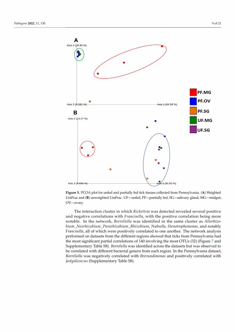
As it is clear from the Principal Coordinate Analysis (PCoA) plot of unweightedUniFrac distances, most of the tick samples from different locations (NY, OK, PA, andLA) contained different bacterial profiles (p = 0.001), as determined by PERMANOVA(Permutational multiple analysis of variance). The PCoA of unweighted UniFrac distancesof bacterial communities showed that the first two axes (Axis1 and Axis2) explained 31.14%and 8.5% of the variation in the data respectively (Figure 4B). PERMANOVA analysis ofUnweighted UniFrac distances, p-values and adjusted p-values (q-value) in several of thesamples when compared pairwise were as follows Louisiana and Pennsylvania female ticks(pseudo-F = 7.33, p-value = 0.011, q-value = 0.095625), Louisiana male and Pennsylvaniafemale ticks (pseudo-F = 6.10, p-value = 0.017, q-value = 0.095625), New York female andPennsylvania female (pseudo-F = 7.19, p-value = 0.013, q-value = 0.095625), Oklahomafemales and Pennsylvania females (pseudo-F = 10.80, p-value = 0.008, q-value = 0.095625),Oklahoma mated females and Pennsylvania males (pseudo-F = 2.99, p-value = 0.047,q-value = 0.147), Oklahoma mated males and Pennsylvania males (pseudo-F = 3.20, p-value = 0.03, q-value = 0.135), Oklahoma virgin females and Pennsylvania females (pseudo-F = 8.124, p-value = 0.017, q-value = 0.095625), and Oklahoma virgin males and Pennsyl-vania males (pseudo-F = 3.76, p-value = 0.027, q-value = 0.135) (Supplementary Table S6).For the unweighted UniFrac of whole-tick samples, Oklahoma tick samples (Mated ♀,Mated ♂, Virgin ♀, Virgin ♂), Louisiana ticks (♂, ♀), and Pennsylvania (♀, ♂) are separateclusters for the most part. Interestingly, it also showed a few outliers, such as Pennsylvaniamales and New York females (Figure 4A). Unweighted UniFrac PCoA plot (Figure 5B)showed that the partially fed midgut is distinct from unfed midgut at the tissue level. Inthis case, PCoA of unweighted UniFrac distances of bacterial communities in the first twoaxes (Axis1 and Axis2) explained 30.5% and 13.27% of the variation in the data. Again,in the weighted UniFrac PCoA plot (Figure 5A) also, the partially fed midgut (PF.MG)clustered more distinctly than the unfed midgut (UF.MG) and other tissues indicating thatthe dominant bacterial diversity in the partially fed midgut is distinct than other tissues.All other tissues, except the partially fed midgut, clustered together separately, indicatingcommon dominant bacteria.

Pathogens 2022, 11, 130 8 of 21
Pathogens 2022, 11, x FOR PEER REVIEW 8 of 22
Virgin ♀, Virgin ♂), Louisiana ticks (♂, ♀), and Pennsylvania (♀, ♂) are separate clusters for the most part. Interestingly, it also showed a few outliers, such as Pennsylvania males and New York females (Figure 4A). Unweighted UniFrac PCoA plot (Figure 5B) showed that the partially fed midgut is distinct from unfed midgut at the tissue level. In this case, PCoA of unweighted UniFrac distances of bacterial communities in the first two axes (Axis1 and Axis2) explained 30.5% and 13.27% of the variation in the data. Again, in the weighted UniFrac PCoA plot (Figure 5A) also, the partially fed midgut (PF.MG) clustered more distinctly than the unfed midgut (UF.MG) and other tissues indicating that the dom-inant bacterial diversity in the partially fed midgut is distinct than other tissues. All other tissues, except the partially fed midgut, clustered together separately, indicating common dominant bacteria.
Figure 4. PCoA plot for male and female ticks collected from OK, LA, NY, and PA. (A) Weighted UniFrac distance, the bacterial composition in female from Pennsylvania (PA.Female), New York (NY.Female) and male from Pennsylvania cluster together. (B) Unweighted UniFrac plot.
Figure 4. PCoA plot for male and female ticks collected from OK, LA, NY, and PA. (A) WeightedUniFrac distance, the bacterial composition in female from Pennsylvania (PA.Female), New York(NY.Female) and male from Pennsylvania cluster together. (B) Unweighted UniFrac plot.
2.6. Microbial Interactions
The network analysis of microbial interactions, using the SparCC permutations, re-vealed 726 interactions amongst 36 taxa when applied to the life-stage dataset from allregions, out of which 358 were positive interactions, while 360 were negative interactions(Figure 6 and Supplementary Table S7). All identified taxa belonged to the phylum Nanoar-chaeaeota (Unidentified genus), Proteobacteria (18), Spirochates (1), Actinobacteria (8),Bacteroidetes (6), and Firmicutes (2). Most interactions were seen with the bacteria in thegenus Flavobacterium, Pseudomonas, and Brevibacterium. Bacteria genus from pathogenicgroup belonging to Rickettsia and Borreliella were also identified in the network interactions,with Rickettsia interacting with more members compared to Borreliella.
Pathogens 2022, 11, 130 9 of 21Pathogens 2022, 11, x FOR PEER REVIEW 9 of 22
Figure 5. PCOA plot for unfed and partially fed tick tissues collected from Pennsylvania. (A) Weighted UniFrac and (B) unweighted UniFrac. UF—unfed, PF—partially fed, SG—salivary gland, MG—midgut, OV—ovary.
2.6. Microbial Interactions The network analysis of microbial interactions, using the SparCC permutations, re-
vealed 726 interactions amongst 36 taxa when applied to the life-stage dataset from all regions, out of which 358 were positive interactions, while 360 were negative interactions (Figure 6 and Supplementary Table S7). All identified taxa belonged to the phylum Nano-archaeaeota (Unidentified genus), Proteobacteria (18), Spirochates (1), Actinobacteria (8), Bacteroidetes (6), and Firmicutes (2). Most interactions were seen with the bacteria in the genus Flavobacterium, Pseudomonas, and Brevibacterium. Bacteria genus from pathogenic group belonging to Rickettsia and Borreliella were also identified in the network interac-tions, with Rickettsia interacting with more members compared to Borreliella.
Figure 5. PCOA plot for unfed and partially fed tick tissues collected from Pennsylvania. (A) WeightedUniFrac and (B) unweighted UniFrac. UF—unfed, PF—partially fed, SG—salivary gland, MG—midgut,OV—ovary.
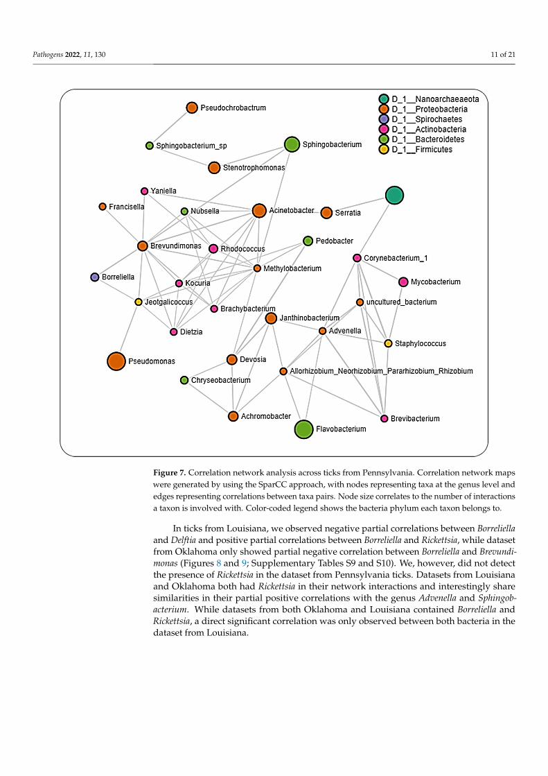
The interaction cluster in which Rickettsia was detected revealed several positiveand negative correlations with Francisella, with the positive correlation being morenotable. In the network, Borreliella was identified in the same cluster as Allorhizo-bium_Neorhizobium_Pararhizobium_Rhizobium, Nubsella, Stenotrophomonas, and notablyFrancisella, all of which were positively correlated to one another. The network analysisperformed on datasets from the different regions showed that ticks from Pennsylvania hadthe most significant partial correlations of 140 involving the most OTUs (32) (Figure 7 andSupplementary Table S8). Borreliella was identified across the datasets but was observed tobe correlated with different bacterial genera from each region. In the Pennsylvania dataset,Borreliella was negatively correlated with Brevundimonas and positively correlated withJeotgalicoccus (Supplementary Table S8).

Pathogens 2022, 11, 130 10 of 21Pathogens 2022, 11, x FOR PEER REVIEW 10 of 22
Figure 6. Correlation network analysis. Correlation network maps were generated by using the SparCC approach, with nodes representing taxa at the genus level and edges representing correla-tions between taxa pairs. Node size correlates to the number of interactions a taxon is involved with. Color-coded legend shows the bacteria phylum each taxon belongs to.
The interaction cluster in which Rickettsia was detected revealed several positive and negative correlations with Francisella, with the positive correlation being more notable. In the network, Borreliella was identified in the same cluster as Allorhizobium_Neorhizo-bium_Pararhizobium_Rhizobium, Nubsella, Stenotrophomonas, and notably Francisella, all of which were positively correlated to one another. The network analysis performed on da-tasets from the different regions showed that ticks from Pennsylvania had the most sig-nificant partial correlations of 140 involving the most OTUs (32) (Figure 7 and Supple-mentary Table S8). Borreliella was identified across the datasets but was observed to be correlated with different bacterial genera from each region. In the Pennsylvania dataset, Borreliella was negatively correlated with Brevundimonas and positively correlated with Jeotgalicoccus (Supplementary Table S8).
Figure 6. Correlation network analysis. Correlation network maps were generated by using theSparCC approach, with nodes representing taxa at the genus level and edges representing correlationsbetween taxa pairs. Node size correlates to the number of interactions a taxon is involved with.Color-coded legend shows the bacteria phylum each taxon belongs to.
Pathogens 2022, 11, 130 11 of 21Pathogens 2022, 11, x FOR PEER REVIEW 11 of 22
Figure 7. Correlation network analysis across ticks from Pennsylvania. Correlation network maps were generated by using the SparCC approach, with nodes representing taxa at the genus level and edges representing correlations between taxa pairs. Node size correlates to the number of interac-tions a taxon is involved with. Color-coded legend shows the bacteria phylum each taxon belongs to.
In ticks from Louisiana, we observed negative partial correlations between Borreliella and Delftia and positive partial correlations between Borreliella and Rickettsia, while da-taset from Oklahoma only showed partial negative correlation between Borreliella and Bre-vundimonas (Figures 8 and 9; Supplementary Tables S9 and S10). We, however, did not detect the presence of Rickettsia in the dataset from Pennsylvania ticks. Datasets from Lou-isiana and Oklahoma both had Rickettsia in their network interactions and interestingly share similarities in their partial positive correlations with the genus Advenella and Sphin-gobacterium. While datasets from both Oklahoma and Louisiana contained Borreliella and Rickettsia, a direct significant correlation was only observed between both bacteria in the dataset from Louisiana.
Figure 7. Correlation network analysis across ticks from Pennsylvania. Correlation network mapswere generated by using the SparCC approach, with nodes representing taxa at the genus level andedges representing correlations between taxa pairs. Node size correlates to the number of interactionsa taxon is involved with. Color-coded legend shows the bacteria phylum each taxon belongs to.
In ticks from Louisiana, we observed negative partial correlations between Borreliellaand Delftia and positive partial correlations between Borreliella and Rickettsia, while datasetfrom Oklahoma only showed partial negative correlation between Borreliella and Brevundi-monas (Figures 8 and 9; Supplementary Tables S9 and S10). We, however, did not detectthe presence of Rickettsia in the dataset from Pennsylvania ticks. Datasets from Louisianaand Oklahoma both had Rickettsia in their network interactions and interestingly sharesimilarities in their partial positive correlations with the genus Advenella and Sphingob-acterium. While datasets from both Oklahoma and Louisiana contained Borreliella andRickettsia, a direct significant correlation was only observed between both bacteria in thedataset from Louisiana.

Pathogens 2022, 11, 130 12 of 21Pathogens 2022, 11, x FOR PEER REVIEW 12 of 22
Figure 8. Correlation network analysis across ticks from Louisiana. Correlation network maps gen-erated by using the SparCC approach, with nodes representing taxa at the genus level and edges representing correlations between taxa pairs. Node size correlates to the number of interactions a taxon is involved with. Color-coded legend shows the bacteria phylum each taxon belongs to.
Figure 8. Correlation network analysis across ticks from Louisiana. Correlation network mapsgenerated by using the SparCC approach, with nodes representing taxa at the genus level and edgesrepresenting correlations between taxa pairs. Node size correlates to the number of interactions ataxon is involved with. Color-coded legend shows the bacteria phylum each taxon belongs to.

Pathogens 2022, 11, 130 13 of 21Pathogens 2022, 11, x FOR PEER REVIEW 13 of 22
Figure 9. Correlation network analysis across ticks from Oklahoma. Correlation network maps gen-erated by using the SparCC approach, with nodes representing taxa at the genus level and edges representing correlations between taxa pairs. Node size correlates to the number of interactions a taxon is involved with. Color-coded legend shows the bacteria phylum each taxon belongs to.
3. Discussion In this study, the microbiome of Ix. scapularis from Lyme-disease-endemic (NY and
PA) and -non-endemic (LA and OK) regions showed substantial variation by both geog-raphy and sex at the organismal and tissue levels. A variation in the microbiota of Ixodes ticks with regard to geography and sex has been suggested [14]. Our results are not in complete coherence with other studies [30–34], as those studies were conducted under a variety of different conditions to examine the tick’s microbiome. For example, Sperling et al. [34] analyzed the ticks collected off the cats and dogs from Alberta (Canada), whereas Kwan et al. [31] conducted their studies on Ixodes pacificus ticks. Several studies have also identified variation in microbiome among different tick species [35], sexes [36,37], and geographies [14,16]. It remains unclear what factors drive the microbiome’s natural vari-ation [33]. The impact of host and environmental drivers on the microbiome composition is still an under-investigated area [33]. By sequencing DNA from 39 single ticks and 15 individual tissues, our study showed the dominance of Proteobacteria in most of the tick samples except Pennsylvania female and Oklahoma male tick samples. Firmicutes were found only in Oklahoma ticks, and Bacteroidetes only in New York and Pennsylvania
Figure 9. Correlation network analysis across ticks from Oklahoma. Correlation network mapsgenerated by using the SparCC approach, with nodes representing taxa at the genus level and edgesrepresenting correlations between taxa pairs. Node size correlates to the number of interactions ataxon is involved with. Color-coded legend shows the bacteria phylum each taxon belongs to.
3. Discussion
In this study, the microbiome of Ix. scapularis from Lyme-disease-endemic (NY andPA) and -non-endemic (LA and OK) regions showed substantial variation by both geog-raphy and sex at the organismal and tissue levels. A variation in the microbiota of Ixodesticks with regard to geography and sex has been suggested [14]. Our results are not incomplete coherence with other studies [30–34], as those studies were conducted under avariety of different conditions to examine the tick’s microbiome. For example, Sperlinget al. [34] analyzed the ticks collected off the cats and dogs from Alberta (Canada), whereasKwan et al. [31] conducted their studies on Ixodes pacificus ticks. Several studies havealso identified variation in microbiome among different tick species [35], sexes [36,37],and geographies [14,16]. It remains unclear what factors drive the microbiome’s naturalvariation [33]. The impact of host and environmental drivers on the microbiome composi-tion is still an under-investigated area [33]. By sequencing DNA from 39 single ticks and15 individual tissues, our study showed the dominance of Proteobacteria in most of the ticksamples except Pennsylvania female and Oklahoma male tick samples. Firmicutes werefound only in Oklahoma ticks, and Bacteroidetes only in New York and Pennsylvania ticks.These results raised an important question of whether these ticks in different geographic

Pathogens 2022, 11, 130 14 of 21
regions maintain a distinct microbiome and whether these variations among the microbialcomposition and communities are driven by the host animals or even by soil bacteria [38].
Our data also showed comparatively higher B. burgdorferi s.l. reads (~2.5%) in theNew York and Pennsylvania female ticks compared to Louisiana and Oklahoma tickpopulations [4,6]. B. burgdorferi s.l. reads were also detected in the Pennsylvania maleticks (Figure 1B), and they were surprisingly higher than other male tick samples. Bac-teroidetes were dominant in the Pennsylvania female tick, whereas, in both unfed andpartially engorged tick tissues, the level was surprisingly low (Figure 1A,B,D). It suggestsbloodmeal-induced changes in the microbiome composition in tick tissues. Interestingly,studies pointed out that vertebrate hosts do not influence the bacterial composition withinadult flea and tick species (Dermacentor variabilis and Ix. scapularis) [35], but blood-feedingin immature developmental stages significantly impacts the bacterial community structur-ing [39]. Before and immediately after blood feeding of Ixodes persulcatus on rats, a reportshowed similar alpha-diversity but significantly different bacterial profiling of tick [37].
In the present study, the endosymbiont R. buchneri was found in the salivary gland,midgut, and ovary, and its level drops significantly when B. burgdorferi s.l. multiplies intick tissues upon blood-feeding (Figure 3B), suggesting a possible competitive interactionthat is purely speculative at this point and needs more work to support it. Moreover, theabsolute-abundance data of R. buchneri and B. burgdorferi s.l. (Figure 2 and SupplementaryTable S5) support this possible competitive interplay. Recent elegant work from Oliveret al. [40] has shown the growth dynamics of R. buchneri in Ix. scapularis ticks. This typeof competitive interaction has also been demonstrated between B. burgdorferi s.l. andgut microbiota in the midgut of Ix. scapularis [41] and by Rickettsia spp. in the ovary ofDermacentor andersoni [42]. The mechanism of co-existence of B. burgdorferi s.l. with othermicrobial pathogens inside ticks remain unknown [43] and is an area of active research.As B. burgdorferi s.l. is extracellular and R. buchneri is an intracellular microbe, they mightnot have physical interaction, but they may affect colonization by activating immuneand reactive oxygen species pathways [43–45]. The endosymbiont R. buchneri is alsoknown as “rickettsial endosymbiont of Ix. scapularis” [46]. The genome of R. buchneri issignificantly larger (>2 Mb) than that of pathogenic rickettsiae, and it also has the capabilityto synthesize vitamin B components, biotin, and de novo folate [46]. As indicated byprevious studies, one of the major roles of the symbionts from ticks and other obligatehematophagous arthropods is to provide vitamin B, which that ticks are deficient in, dueto their exclusively sanguineous diet [47,48]. The presence of R. buchneri was reportedto be restricted to tick ovaries [49,50], but a recent study has reported its colonizationinto tick salivary glands [48]. It might evolve as a pathogen, as it fulfills the prerequisitefor an endosymbiont to be transmitted to the vertebrate host by getting colonized intotick’s salivary glands [48]. Our data showed the colonization of this rickettsial speciesinto salivary glands (UF.SG = 35.38%, PF.SG = 0.33%) and the midgut (UF.MG = 46.09%,PF.MG = 3.52%) during the unfed stage, but the level of R. buchneri reduces significantlyafter blood feeding. It is not necessary that this process would lead to a condition ofpathogenicity, it might simply be a transmission route to other ticks through co-feeding.Nevertheless, a symbiont in the salivary gland might also be exposed to the host immunesystem, leading to an antibody response or can interact with pathogens in the salivarygland and may facilitate or impede their transmission [48]. Studies have shown thatseveral genera of bacteria, such as Rickettsia, Coxiella, Francisella, and Midichloria, persisttranstadially and later get transmitted transovarially as a regular process [47,51]. Theseendosymbionts might remain restricted to the arthropod host and sometimes may betransmitted to the vertebrate host or sometimes may cause disease [47,51]. The distributionof reads for R. buchneri in the various tissues is consistent with the rest of the literature [48].
Network analysis across all life-stage datasets from all regions revealed a relativelyequal number of positive (~49%) and negative (~51%) interactions to be present. Positiveinteractions between different bacteria taxa could indicate shared functionality, or even ashared niche within the host organism [52–54], whereas negative interactions would point

Pathogens 2022, 11, 130 15 of 21
toward an existing or potential competition between bacteria taxa. This would suggestthat the microbiome of Ix. scapularis favors a balanced distribution between bacteria withpotential synergistic and antagonistic interactions. This observation contrasts the detectionof greater than 97% positive interactions in the Ix. ricinus microbiota, as recently reportedby Lejal et al. [55], thus indicating differences exist in the microbial–microbial interactioneven between the same tick genera. Most of the interactions observed in the whole datasetwere driven by bacteria belonging to non-pathogenic genera, as indicated by the presenceof Flavobacterium, Pseudomonas, and Brevibacterium, further suggesting a contribution ofnon-symbiotic commensal microbes to the overall microbiome of the Ix. scapularis ticks.
Interestingly, we identified OTUs belonging to the pathogenic Rickettsia and Borreliellagenus from the network analysis, with Rickettsia observed to interact with more bacteriagenera compared to Borreliella. A positive correlation was seen to exist between Francisellaand Borreliella. The interaction between Rickettsia and Borreliella was also observed to varyacross different geographical locations. While our network analysis was carried out downto the genus level, the bacterial profile identified the Rickettsia identified to the species levelas R. buchneri, the major endosymbiont of Ix. scapularis. The genus Francisella, which isendosymbiont, was significantly correlated with Rickettsia, suggesting an indication of aco-dependency on two endosymbionts by Ix. scapularis. This hypothesis is in accordancewith a recent report of a dual endosymbiont dependency observed between Midichloria andFrancisella symbionts in Hyalomma marginatum ticks driven by a nutritional adaptation [56].It has also been shown that, although R. buchneri does possess the essential vitamin synthesisgenes, some Ix. scapularis harbor this endosymbiont that lack these vitamin synthesispathways, indicating a non-obligatory or facultative endosymbiotic relationship [57–59].This could explain the need to harbor a different class of endosymbiont in Francisella thatwould relieve the exclusive dependence on R. buchneri, as seen in this study.
The detection of multiple endosymbiont species has also been identified in other tickspecies, such as in the Amblyomma maculatum tick, although the functional contribution tothe tick biology has not been described [60]. Rickettsia and Borreliella were not identifiedwithin the same cluster in the network analysis of the whole dataset; however, bothbacteria genera interacted differently in ticks from different locations. A much surprisingobservation was the presence of Borreliella and corresponding absence of Rickettsia fromthe network analysis on datasets of ticks from Pennsylvania. While the small size of thetissue dataset from these regions prevented us from carrying out a network analysis, thisobservation was further supported by the exclusive presence of R. buchneri in unfed salivaryglands and partially fed ovarian tissues. A shared presence of both bacteria in partiallyfed salivary glands and midgut tissue suggests a tissue-driven microbial interaction. Thisobservation contrasts with the report of Aivelo et al., who reported positive correlationsbetween Lyme borreliois Borrelia group, Borrelia miyamotoi, and Rickettsiella in the sister tickIx. ricinus, suggesting a specific interaction dependent on the host tick specie.
4. Materials and Methods4.1. Ticks
Only adult ticks were field collected from Louisiana (LA), New York (NY), and Ok-lahoma (OK), and Pennsylvania (PA). Ticks from Covington, Louisiana [30.47671◦ N,−90.10517◦ E]; Stillwater, Oklahoma [36.110176◦ N, −97.05857◦ E]; and State College, Penn-sylvania [40.790703◦ N, −77.858795◦ E], were collected in spring 2019, and ticks fromMillbrook, New York [41.785538◦ N, −73.69046◦ E] were collected in winter of 2019. Unfedadult Ix. scapularis ticks mate before attachment on a vertebrate host. Ticks were identifiedby using standard morphological keys. To test the microbes’ transfer during mating ofmale and female, unfed ticks (♂and ♀) were used for these experiments. Unmated femaleand male ticks were kept in a vial for 48 h and allowed to mate with their partners. Un-fed adult ticks from Pennsylvania were blood-fed, as described earlier [61], and tissueswere dissected from partially engorged female ticks. The dissecting solution was ice cold100 mM 3-(N-Morpholino-propanesulfonic acid (MOPS) buffer containing 20 mM ethylene
Pathogens 2022, 11, 130 16 of 21
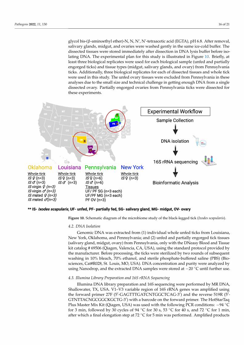
glycol bis-(β-aminoethyl ether)-N, N, N’, N’-tetraacetic acid (EGTA), pH 6.8. After removal,salivary glands, midgut, and ovaries were washed gently in the same ice-cold buffer. Thedissected tissues were stored immediately after dissection in DNA lysis buffer before iso-lating DNA. The experimental plan for this study is illustrated in Figure 10. Briefly, atleast three biological replicates were used for each biological sample (unfed and partiallyengorged ticks) and tissue types (midgut, salivary glands, and ovary) from Pennsylvaniaticks. Additionally, three biological replicates for each of dissected tissues and whole tickwere used in this study. The unfed ovary tissues were excluded from Pennsylvania in theseanalyses due to the small size and technical challenge in getting enough DNA from a singledissected ovary. Partially engorged ovaries from Pennsylvania ticks were dissected forthese experiments.
Pathogens 2022, 11, x FOR PEER REVIEW 16 of 22
male and female, unfed ticks (♂ and ♀) were used for these experiments. Unmated female and male ticks were kept in a vial for 48 h and allowed to mate with their partners. Unfed adult ticks from Pennsylvania were blood-fed, as described earlier [61], and tissues were dissected from partially engorged female ticks. The dissecting solution was ice cold 100 mM 3-(N-Morpholino-propanesulfonic acid (MOPS) buffer containing 20 mM ethylene glycol bis-(β-aminoethyl ether)-N, N, N’, N’-tetraacetic acid (EGTA), pH 6.8. After re-moval, salivary glands, midgut, and ovaries were washed gently in the same ice-cold buffer. The dissected tissues were stored immediately after dissection in DNA lysis buffer before isolating DNA. The experimental plan for this study is illustrated in Figure 10. Briefly, at least three biological replicates were used for each biological sample (unfed and partially engorged ticks) and tissue types (midgut, salivary glands, and ovary) from Penn-sylvania ticks. Additionally, three biological replicates for each of dissected tissues and whole tick were used in this study. The unfed ovary tissues were excluded from Pennsyl-vania in these analyses due to the small size and technical challenge in getting enough DNA from a single dissected ovary. Partially engorged ovaries from Pennsylvania ticks were dissected for these experiments.
Figure 10. Schematic diagram of the microbiome study of the black-legged tick (Ixodes scapularis).
4.2. DNA Isolation Genomic DNA was extracted from (1) individual whole unfed ticks from Louisiana,
New York, Oklahoma, and Pennsylvania; and (2) unfed and partially engorged tick tis-sues (salivary gland, midgut, ovary) from Pennsylvania, only with the DNeasy Blood and Tissue kit catalog # 69506 (Qiagen, Valencia, CA, USA), using the standard protocol pro-vided by the manufacturer. Before processing, the ticks were sterilized by two rounds of subsequent washing in 10% bleach, 70% ethanol, and sterile phosphate-buffered saline (PBS) (Biosciences, Cat#R028, St. Louis, MO, USA). DNA concentration and purity were analyzed by using Nanodrop, and the extracted DNA samples were stored at −20 °C until further use.
Figure 10. Schematic diagram of the microbiome study of the black-legged tick (Ixodes scapularis).
4.2. DNA Isolation
Genomic DNA was extracted from (1) individual whole unfed ticks from Louisiana,New York, Oklahoma, and Pennsylvania; and (2) unfed and partially engorged tick tissues(salivary gland, midgut, ovary) from Pennsylvania, only with the DNeasy Blood and Tissuekit catalog # 69506 (Qiagen, Valencia, CA, USA), using the standard protocol provided bythe manufacturer. Before processing, the ticks were sterilized by two rounds of subsequentwashing in 10% bleach, 70% ethanol, and sterile phosphate-buffered saline (PBS) (Bio-sciences, Cat#R028, St. Louis, MO, USA). DNA concentration and purity were analyzed byusing Nanodrop, and the extracted DNA samples were stored at −20 ◦C until further use.
4.3. Illumina Library Preparation and 16S rRNA Sequencing
Illumina DNA library preparation and 16S sequencing were performed by MR DNA,Shallowater, TX, USA. V1–V3 variable region of 16S rRNA genes was amplified usingthe forward primer 27F (5′-GAGTTTGATCNTGGCTCAG-3′) and the reverse 519R (5′-GTNTTACNGCGGCKGCTG-3′) with a barcode on the forward primer. The HotStarTaqPlus Master Mix Kit (Qiagen, USA) was used with the following PCR conditions: −94 ◦Cfor 3 min, followed by 30 cycles of 94 ◦C for 30 s, 53 ◦C for 40 s, and 72 ◦C for 1 min,after which a final elongation step at 72 ◦C for 5 min was performed. Amplified products

Pathogens 2022, 11, 130 17 of 21
were checked on 2% agarose gel to confirm the appropriate size and intensity of bands.On the basis of molecular weight and DNA concentrations, equal proportions of multiplesamples were pulled together and purified by using calibrated Ampure XP beads. Size ofthe DNA amplicons was determined by running on 2% agarose gel. Expected size of theDNA band was ~500 bps. Each sample was diluted to 5 nM, and 5 µLs of each sample wasadded to the pool. The quality and size of the DNA libraries was confirmed by lab-on-chipanalysis, using the Bioanalyzer (Agilent Technologies, Inc., Santa Clara, CA, USA). Thepooled sample was sent for sequencing. The mathematical formula used to convert ng/µLto nM is as follows:
(concentration in ng/µL)(660 g/mol x average library size)
× 106 = concentration in nM
The average molecular mass of one base-pair DNA is 660 g/mol.Purified PCR products were used to prepare Illumina DNA library. The quality of
the DNA libraries was confirmed by lab-on-chip analysis, using the Bioanalyzer (AgilentTechnologies, Inc., Santa Clara, CA, USA), and then 16S sequencing was performed onIllumina MiSeq platform at MR DNA, Shallowater, TX, USA. Three biological replicates ofeach of the controls were used. Controls used were DNA extraction blank control, negativecontrol (buffer), negative control (sterile water), no-template control, and positive-DNAextraction control [62,63] (commercially available Mock Microbial Community Standard,ZymoBIOMICS catalog # D6306).
4.4. Data Processing
Quantitative Insights into Microbial Ecology (QIIME 2, https://qiime2.org accessed on13 February 2021) was used for sequence analysis. Raw fastq files were processed by fastqprocessor available on the MR DNA website, which provided the files compatible withthe Earth microbiome project (EMP) paired-end format. Then “Atacama soil microbiome”tutorial (website link: https://docs.qiime2.org/2021.2/tutorials/atacama-soils/ (accessedon 13 February 2021)) and “moving pictures” tutorial (website link: https://docs.qiime2.org/2021.2/tutorials/moving-pictures/ (accessed on 13 February 2021) were followed toprocess the sequencing data. Among whole-tick samples, the maximum number of reads fora sample was 114,478, while the minimum number of reads for a sample was 36,358. For ticktissue samples, the maximum number of reads for a sample was 56,785, while the minimumnumber of reads was 17,258. The abovementioned ranges of sequences of each sample typewere available before the denoising step (DADA2 processing). DADA2 [64] was used fortrimming, primer sequence removal, sequence denoising, paired-end merging, filteringof chimeric sequences, singleton removal, and sequence dereplication. This step yielded5000 sequences from each tissue sample and 16,000 sequences from each of the whole-ticksamples for rarefaction curves. The rarefaction curve is getting leveled out, suggesting thatcollecting additional sequences beyond that sampling depth would not observe additionalreads. Minimum overlap of 50 bases was used for paired-end merging. Resultant sequencessets obtained after DADA2 processing were aligned by MAFFT (ver.7) [65], and then aphylogenetic tree was created by using FastTree (ver. 2.1) [66]. Greengenes 13_8 99%OTU database [67] was used to train the Naïve Bayes classifier, to which the representedsequences were compared and a 97% sequence similarity was put as a cutoff for taxonomicclassification. Network correlation maps were inferred based on the Sparse Correlations forCompositional data (SparCC) approach [68]. This approach uses the log-transformed valuesto carry out multiple iterations and subsequently identify taxa outliers to the correlationparameters [69]. Raw sequences were submitted to the NCBI read under the SRA databaseand obtained the accession number PRJNA663181.
4.5. Statistical Analysis
To measure α-diversity, different indices, such as Faith’s phylogenetic diversity(faith_pd) and Pielou’s community evenness (pielou_e), were used. Faith’s phylogenetic

Pathogens 2022, 11, 130 18 of 21
diversity (faith_pd) is an unweighted measure of phylogenetic distance of observed se-quences; and Pielou’s community evenness (pielou_e) measures how evenly bacterialspecies are distributed within a community. Kruskal–Wallis non-parametric tests (p ≤ 0.05)were performed to determine statistical significance of alpha-diversity metrics by usingQIIME 2. Weighted and unweighted UniFrac Metrics [70] were used for β-diversity anal-ysis. EMPeror [71] was used for visualization of principal coordinate analysis (PCoA)plot, and PERMANOVA tests (p ≤ 0.05) were used to test the statistical significance ofβ-diversity measurements.
5. Conclusions
This study has shown significant differences in the microbiome of Ix. scapularis tickscollected from Northeastern (New York and Pennsylvania) and Southern (Oklahoma andLouisiana) states. These results provide an insight into the microbiome of New York, Penn-sylvania, Oklahoma, and Louisiana tick populations and the interplay between pathogenicand endosymbiotic rickettsiae. The question remains: what are the drivers behind thesevariations among the microbiome composition and diversity? This question warrantsfurther investigation into issues such as why Oklahoma ticks contain comparatively muchhigher level of Firmicutes, while ticks from all other locations included in this study containalmost negligible Firmicutes. In further analysis at the species level, it was revealed thatthese Firmicutes from Oklahoma ticks possess bacterial species, such as Jeotgalicoccus sp.and Staphylococcus lentus. Do these species restrict B. burgdorferi s.l. in Oklahoma ticks?Similarly, the level of Bacteroidetes is much higher in Northeastern ticks when compared toSouthern ticks, whose levels are almost insignificant. Further analysis revealed that theseBacteroidetes possess bacterial species, such as Sphingobacterium faecium, Flavobacterium sp.,Sphingobacterium sp., Flexibacter sp., and Flavobacter spp. Do these Bacteroidetes interactwith spirochetes to colonize Lyme disease causative agent (i.e., B. burgdorferi s.l.) intoNortheastern ticks? Probable competitive interaction between R. buchneri and B. burgdorferis.l. surfaced in this study and is subject to be further investigated by using dysbiosisexperiments and a bigger sample size.
Supplementary Materials: The following supporting information can be downloaded at. https://www.mdpi.com/article/10.3390/pathogens11020130/s1. Figure S1: Rarefaction Curve. Table S1:Comparison of bacterial profiling and relative abundance (phylum level) between the ticks collectedfrom Northeastern and Southern regions of the United States. Table S2: Comparison of bacterialprofiling and relative abundance (species level) between the ticks collected from Northeastern andSouthern regions of the United States. Table S3: Comparison of bacterial profiling and relative abun-dance (phylum level) in unfed (UF) and partially fed (PF) tick tissues collected from Pennsylvania.Table S4: Relative abundance of Borrelia burgdorferi and Rickettsia buchneri in tick tissues fromPennsylvania (PA). Table S5: Absolute abundance (mean of number of sequences of biological repli-cates) of Borrelia burgdorferi and Rickettsia buchneri in tick tissues from Pennsylvania (PA). Table S6:Unweighted-Unifrac-pairwise PERMANOVA analysis (beta-diversity) for whole tick samples col-lected from Louisiana (LA), Oklahoma (OK), Pennsylvania and New York (NY). Table S7: Networkcorrelation summary across all datasets. Table S8: Network correlation summary showing significantinteractions between ticks from Pennsylvania. Table S9: Network correlation summary showingsignificant interactions between ticks from Louisiana. Table S10: Network correlation summaryshowing significant interactions between ticks from Oklahoma.
Author Contributions: Conceived and designed the experiments: D.K., L.P.D. and S.K. Performedthe experiments: D.K., L.P.D. and S.K. Analyzed the data: D.K., A.A. and S.K. Contributed reagents/materials/analysis tools: D.K., E.M., K.O., R.S.O., M.E. and S.K. Wrote the paper: D.K. and S.K. Allauthors have read and agreed to the published version of the manuscript.
Funding: This research was principally supported by a Pakistan–US Science and Technology Coop-eration Program award (US Department of State); the Mississippi INBRE (an institutional Award(IDeA) from the National Institute of General Medical Sciences of the National Institutes of Healthunder award P20GM103476). The funders played no role in the study design, data collection andanalysis, decision to publish, or preparation of the manuscript.

Pathogens 2022, 11, 130 19 of 21
Institutional Review Board Statement: All animal experiments were performed in strict accordancewith the recommendations in the Guide for the Care and Use of Laboratory Animals of the Na-tional Institutes of Health. The protocol for the blood-feeding of field-collected ticks was approvedby the Institutional Animal Care and Use Committee of the University of Southern Mississippi(protocol # 15101501.1).
Informed Consent Statement: Not applicable.
Data Availability Statement: Data supporting the conclusions of this article are included within thearticle and its additional files. The raw datasets used and analyzed for the present study are availablefrom the corresponding author upon reasonable request.
Acknowledgments: We thank Raima Sen for their technical assistance in extraction of genomic DNA.
Conflicts of Interest: The authors declare no conflict of interest.
References1. Harvell, C.D.; Mitchell, C.E.; Ward, J.R.; Altizer, S.; Dobson, A.P.; Ostfeld, R.S.; Samuel, M.D. Climate warming and disease risks
for terrestrial and marine biota. Science 2002, 296, 2158–2162. [CrossRef]2. Jones, B.A.; Grace, D.; Kock, R.; Alonso, S.; Rushton, J.; Said, M.Y.; McKeever, D.; Mutua, F.; Young, J.; McDermott, J.; et al.
Zoonosis emergence linked to agricultural intensification and environmental change. Proc. Natl. Acad. Sci. USA 2013, 110,8399–8404. [CrossRef]
3. Keesing, F.; Belden, L.K.; Daszak, P.; Dobson, A.; Harvell, C.D.; Holt, R.D.; Hudson, P.; Jolles, A.; Jones, K.E.; Mitchell, C.E.; et al.Impacts of biodiversity on the emergence and transmission of infectious diseases. Nature 2010, 468, 647–652. [CrossRef]
4. Kugeler, K.J.; Farley, G.M.; Forrester, J.D.; Mead, P.S. Geographic Distribution and Expansion of Human Lyme Disease, UnitedStates. Emerg. Infect. Dis. 2015, 21, 1455–1457. [CrossRef] [PubMed]
5. Radolf, J.D.; Caimano, M.J.; Stevenson, B.; Hu, L.T. Of ticks, mice and men: Understanding the dual-host lifestyle of Lyme diseasespirochaetes. Nat. Rev. Microbiol. 2012, 10, 87–99. [CrossRef] [PubMed]
6. Hinckley, A.F.; Connally, N.P.; Meek, J.I.; Johnson, B.J.; Kemperman, M.M.; Feldman, K.A.; White, J.L.; Mead, P.S. Lyme diseasetesting by large commercial laboratories in the United States. Clin. Infect. Dis. Off. Publ. Infect. Dis. Soc. Am. 2014, 59, 676–681.[CrossRef]
7. Roy-Dufresne, E.; Logan, T.; Simon, J.A.; Chmura, G.L.; Millien, V. Poleward expansion of the white-footed mouse (Peromyscusleucopus) under climate change: Implications for the spread of lyme disease. PLoS ONE 2013, 8, e80724. [CrossRef] [PubMed]
8. Moreno, C.X.; Moy, F.; Daniels, T.J.; Godfrey, H.P.; Cabello, F.C. Molecular analysis of microbial communities identified in differentdevelopmental stages of Ixodes scapularis ticks from Westchester and Dutchess Counties, New York. Environ. Microbiol. 2006, 8,761–772. [CrossRef] [PubMed]
9. Ostfeld, R.S.; Levi, T.; Keesing, F.; Oggenfuss, K.; Canham, C.D. Tick-borne disease risk in a forest food web. Ecology 2018, 99,1562–1573. [CrossRef] [PubMed]
10. Adelson, M.E.; Rao, R.V.; Tilton, R.C.; Cabets, K.; Eskow, E.; Fein, L.; Occi, J.L.; Mordechai, E. Prevalence of Borrelia burgdorferi,Bartonella spp., Babesia microti, and Anaplasma phagocytophila in Ixodes scapularis ticks collected in Northern New Jersey. J.Clin. Microbiol. 2004, 42, 2799–2801. [CrossRef] [PubMed]
11. Eisen, L.; Eisen, R.J.; Mun, J.; Salkeld, D.J.; Lane, R.S. Transmission cycles of Borrelia burgdorferi and B. bissettii in relation tohabitat type in northwestern California. J. Vector Ecol. J. Soc. Vector Ecol. 2009, 34, 81–91. [CrossRef]
12. Maggi, R.G.; Reichelt, S.; Toliver, M.; Engber, B. Borrelia species in Ixodes affinis and Ixodes scapularis ticks collected from thecoastal plain of North Carolina. Ticks Tick-Borne Dis. 2010, 1, 168–171. [CrossRef] [PubMed]
13. Clark, K.L.; Oliver, J.H.; James, A.M., Jr.; Durden, L.A.; Banks, C.W. Prevalence of borrelia burgdorferi sensu lato infection amongrodents and host-seeking ticks in South Carolina. J. Med. Entomol. 2002, 39, 198–206. [CrossRef] [PubMed]
14. Van Treuren, W.; Ponnusamy, L.; Brinkerhoff, R.J.; Gonzalez, A.; Parobek, C.M.; Juliano, J.J.; Andreadis, T.G.; Falco, R.C.; Ziegler,L.B.; Hathaway, N.; et al. Variation in the Microbiota of Ixodes Ticks with Regard to Geography, Species, and Sex. Appl. Environ.Microbiol. 2015, 81, 6200–6209. [CrossRef]
15. Clay, K.; Klyachko, O.; Grindle, N.; Civitello, D.; Oleske, D.; Fuqua, C. Microbial communities and interactions in the lone startick, Amblyomma americanum. Mol. Ecol. 2008, 17, 4371–4381. [CrossRef] [PubMed]
16. Carpi, G.; Cagnacci, F.; Wittekindt, N.E.; Zhao, F.; Qi, J.; Tomsho, L.P.; Drautz, D.I.; Rizzoli, A.; Schuster, S.C. Metagenomic profileof the bacterial communities associated with Ixodes ricinus ticks. PLoS ONE 2011, 6, e25604. [CrossRef]
17. Van Overbeek, L.; Gassner, F.; van der Plas, C.L.; Kastelein, P.; Nunes-da Rocha, U.; Takken, W. Diversity of Ixodes ricinustick-associated bacterial communities from different forests. FEMS Microbiol. Ecol. 2008, 66, 72–84. [CrossRef]
18. Clay, K.; Fuqua, C. The tick microbiome: Diversity, distribution and influence of the internal microbial community for a blood-feeding disease vector. In Critical Needs and Gaps in Understanding Prevention, Amelioration, and Resolution of Lyme and OtherTick-Borne Diseases: The Short-Term and Long-Term Outcomes. Workshop Report; National Academies Press: Washington, DC,USA, 2010.

Pathogens 2022, 11, 130 20 of 21
19. Nakao, R.; Abe, T.; Nijhof, A.M.; Yamamoto, S.; Jongejan, F.; Ikemura, T.; Sugimoto, C. A novel approach, based on BLSOMs(Batch Learning Self-Organizing Maps), to the microbiome analysis of ticks. ISME J. 2013, 5, 1003–1015. [CrossRef] [PubMed]
20. Budachetri, K.; Browning, R.E.; Adamson, S.W.; Dowd, S.E.; Chao, C.C.; Ching, W.M.; Karim, S. An insight into the microbiomeof the Amblyomma maculatum (Acari: Ixodidae). J. Med. Entomol. 2014, 51, 119–129. [CrossRef]
21. Ponnusamy, L.; Gonzalez, A.; Van Treuren, W.; Weiss, S.; Parobek, C.M.; Juliano, J.J.; Knight, R.; Roe, R.M.; Apperson, C.S.;Meshnick, S.R. Diversity of Rickettsiales in the microbiome of the lone star tick, Amblyomma americanum. Appl. Environ.Microbiol. 2014, 80, 354–359. [CrossRef]
22. Landesman, W.J.; Mulder, K.; Allan, B.F.; Bashor, L.A.; Keesing, F.; LoGiudice, K.; Ostfeld, R.S. Potential effects of blood meal hoston bacterial community composition in Ixodes scapularis nymphs. Ticks Tick-Borne Dis. 2019, 10, 523–527. [CrossRef]
23. Martin, P.A.; Schmidtmann, E.T. Isolation of aerobic microbes from Ixodes scapularis (Acari: Ixodidae), the vector of Lyme diseasein the eastern United States. J. Econ. Entomol. 1998, 91, 864–868. [CrossRef]
24. Narasimhan, S.; Fikrig, E. Tick microbiome: The force within. Trends Parasitol. 2015, 31, 315–323. [CrossRef]25. Ahantarig, A.; Trinachartvanit, W.; Baimai, V.; Grubhoffer, L. Hard ticks and their bacterial endosymbionts (or would be
pathogens). Folia Microbiol. 2013, 58, 419–428. [CrossRef]26. Gall, C.A.; Reif, K.E.; Scoles, G.A.; Mason, K.L.; Mousel, M.; Noh, S.M.; Brayton, K.A. The bacterial microbiome of Dermacentor
andersoni ticks influences pathogen susceptibility. ISME J. 2016, 10, 1846–1855. [CrossRef] [PubMed]27. Smith, T.A.; Driscoll, T.; Gillespie, J.J.; Raghavan, R. A Coxiella-like endosymbiont is a potential vitamin source for the Lone Star
tick. Genome Biol. Evol. 2015, 7, 831–838. [CrossRef] [PubMed]28. Zhong, J.; Jasinskas, A.; Barbour, A.G. Antibiotic treatment of the tick vector Amblyomma americanum reduced reproductive
fitness. PLoS ONE 2007, 2, e405. [CrossRef] [PubMed]29. Swei, A.; Kwan, J.Y. Tick microbiome and pathogen acquisition altered by host blood meal. ISME J. 2017, 11, 813–816. [CrossRef]
[PubMed]30. Clow, K.M.; Weese, J.S.; Rousseau, J.; Jardine, C.M. Microbiota of field-collected Ixodes scapularis and Dermacentor variabilis
from eastern and southern Ontario, Canada. Ticks Tick-Borne Dis. 2018, 9, 235–244. [CrossRef] [PubMed]31. Kwan, J.Y.; Griggs, R.; Chicana, B.; Miller, C.; Swei, A. Vertical vs. horizontal transmission of the microbiome in a key disease
vector, Ixodes pacificus. Mol. Ecol. 2017, 26, 6578–6589. [CrossRef]32. Thapa, S.; Zhang, Y.; Allen, M.S. Bacterial microbiomes of Ixodes scapularis ticks collected from Massachusetts and Texas, USA.
BMC Microbiol. 2019, 19, 138. [CrossRef] [PubMed]33. Brinkerhoff, R.J.; Clark, C.; Ocasio, K.; Gauthier, D.T.; Hynes, W.L. Factors affecting the microbiome of Ixodes scapularis and
Amblyomma americanum. PLoS ONE 2020, 15, e0232398. [CrossRef]34. Sperling, J.; Fitzgerald, D.; Sperling, F.; Magor, K.E. Microbiome Composition and Borrelia Detection in Ixodes scapularis Ticks at
the Northwestern Edge of Their Range. Trop. Med. Infect. Dis. 2020, 5, 173. [CrossRef] [PubMed]35. Hawlena, H.; Rynkiewicz, E.; Toh, E.; Alfred, A.; Durden, L.A.; Hastriter, M.W.; Nelson, D.E.; Rong, R.; Munro, D.; Dong, Q. The
arthropod, but not the vertebrate host or its environment, dictates bacterial community composition of fleas and ticks. ISME J.2013, 7, 221–223. [CrossRef]
36. Williams-Newkirk, A.J.; Rowe, L.A.; Mixson-Hayden, T.R.; Dasch, G.A. Characterization of the bacterial communities of lifestages of free living lone star ticks (Amblyomma americanum). PLoS ONE 2014, 9, e102130. [CrossRef]
37. Zhang, X.C.; Yang, Z.N.; Lu, B.; Ma, X.F.; Zhang, C.X.; Xu, H.J. The composition and transmission of microbiome in hard tick,Ixodes persulcatus, during blood meal. Ticks Tick-Borne Dis. 2014, 5, 864–870. [CrossRef]
38. Burtis, J.C.; Yavitt, J.B.; Fahey, T.J.; Ostfeld, R.S. Ticks as soildwelling arthropods: An intersection between disease and soil ecology.J. Med. Entomol. 2019, 56, 1555–1564. [CrossRef]
39. Rynkiewicz, E.C.; Hemmerich, C.; Rusch, D.B.; Fuqua, C.; Clay, K. Concordance of bacterial communities of two tick species andblood of their shared rodent host. Mol. Ecol. 2015, 24, 2566–2579. [CrossRef]
40. Oliver, J.D.; Price, L.D.; Burkhardt, N.Y.; Heu, C.C.; Khoo, B.S.; Thorpe, C.J.; Kurtti, T.J.; Munderloh, U.G. Growth Dynamics andAntibiotic Elimination of Symbiotic Rickettsia buchneri in the Tick Ixodes scapularis (Acari: Ixodidae). Appl. Environ. Microbiol.2021, 87, e01672-20. [CrossRef] [PubMed]
41. Narasimhan, S.; Rajeevan, N.; Liu, L.; Zhao, Y.O.; Heisig, J.; Pan, J.; Eppler-Epstein, R.; Deponte, K.; Fish, D.; Fikrig, E. Gutmicrobiota of the tick vector Ixodes scapularis modulate colonization of the Lyme disease spirochete. Cell Host Microbe 2014, 15,58–71. [CrossRef]
42. Macaluso, K.R.; Sonenshine, D.E.; Ceraul, S.M.; Azad, A.F. Rickettsial infection in Dermacentor variabilis (Acari: Ixodidae)inhibits transovarial transmission of a second Rickettsia. J. Med. Entomol. 2002, 39, 809–813. [CrossRef]
43. Ross, B.D.; Hayes, B.; Radey, M.C.; Lee, X.; Josek, T.; Bjork, J.; Mougous, J.D. Ixodes scapularis does not harbor a stable midgutmicro-biome. ISME J. 2018, 12, 2596–2607. [CrossRef]
44. Simhadri, R.K.; Fast, E.M.; Guo, R.; Schultz, M.J.; Vaisman, N.; Ortiz, L.; Bybee, J.; Slatko, B.E.; Frydman, H.M. The GutCommensal Microbiome of Drosophila melanogaster Is Modified by the Endosymbiont Wolbachia. mSphere 2017, 2, e00287-17.[CrossRef]
45. Kocan, K.M.; de la Fuente, J.; Blouin, E.F.; Coetzee, J.F.; Ewing, S.A. The natural history of Anaplasma marginale. Vet. Parasitol.2010, 167, 95–107. [CrossRef]

Pathogens 2022, 11, 130 21 of 21
46. Gillespie, J.J.; Joardar, V.; Williams, K.P.; Driscoll, T.; Hostetler, J.B.; Nordberg, E.; Shukla, M.; Walenz, B.; Hill, C.A.; Nene, V.M.;et al. A Rickettsia genome overrun by mobile genetic elements provides insight into the acquisition of genes characteristic of anobligate intracellular lifestyle. J. Bacteriol. 2012, 194, 376–394. [CrossRef]
47. Duron, O.; Binetruy, F.; Noel, V.; Cremaschi, J.; McCoy, K.D.; Arnathau, C.; Plantard, O.; Goolsby, J.; de Leon, A.A.P.; Heylen,D.J.A.; et al. Evolutionary changes in symbiont community structure in ticks. Mol. Ecol. 2017, 26, 2905–2921. [CrossRef]
48. Al-Khafaji, A.M.; Armstrong, S.D.; Varotto, B.I.; Gaiarsa, S.; Sinha, A.; Li, Z.; Sassera, D.; Carlow, C.K.S.; Epis, S.; Makepeace, B.L.Rickettsia buchneri, symbiont of the deer tick Ixodes scapularis, can colonise the salivary glands of its host. Ticks Tick Borne Dis.2020, 11, 101299. [CrossRef] [PubMed]
49. Munderloh, U.G.; Jauron, S.D.; Kurtti, T.J. The tick: A different kind of host for human pathogens. In Tick-Borne Diseases ofHumans; Goodman, J.L., Ed.; ASM Press: Washington, DC, USA, 2005; pp. 37–64.
50. Noda, H.; Munderloh, U.G.; Kurtti, T.J. Endosymbionts of ticks and their relationship to Wolbachia spp. and tick-borne pathogensof humans and animals. Appl. Environ. Microbiol. 1997, 63, 3926–3932. [CrossRef] [PubMed]
51. Angelakis, E.; Mediannikov, O.; Jos, S.L.; Berenger, J.M.; Parola, P.; Raoult, D. Candidatus Coxiella massiliensis infection. Emerg.Infect. Dis. 2016, 22, 285–288. [CrossRef] [PubMed]
52. Eiler, A.; Heinrich, F.; Bertilsson, S. Coherent dynamics and association networks among lake bacterioplankton taxa. ISME J. 2012,6, 330–342. [CrossRef]
53. Chow, C.-E.T.; Kim, D.Y.; Sachdeva, R.; Caron, D.A.; Fuhrman, J.A. Top-down controls on bacterial community structure:Microbial network analysis of bacteria, T4-like viruses and protists. ISME J. 2014, 8, 816–829. [CrossRef] [PubMed]
54. Berdjeb, L.; Parada, A.; Needham, D.M.; Fuhrman, J.A. Short-term dynamics and interactions of marine protist communitiesduring the spring–summer transition. ISME J. 2018, 12, 1907–1917. [CrossRef] [PubMed]
55. Lejal, E.; Moutailler, S.; Šimo, L.; Vayssier-Taussat, M.; Pollet, T. Tick-borne pathogen detection in midgut and salivary glands ofadult Ixodes ricinus. Parasites Vectors 2019, 12, 152. [CrossRef]
56. Buysse, M.; Floriano, A.M.; Gottlieb, Y.; Nardi, T.; Comandatore, F.; Olivieri, E.; Giannetto, A.; Urbina, A.M.P.; Makepeace,B.; Bazzocchi, C.; et al. A dual endosymbiosis drives nutritional adaptation to hematophagy in the invasive tick Hyalommamarginatum. bioRxiv 2021. [CrossRef]
57. Hunter, D.J.; Torkelson, J.L.; Bodnar, J.; Mortazavi, B.; Laurent, T.; Deason, J.; Thephavongsa, K.; Zhong, J. The Rickettsiaendosymbiont of Ixodes pacificus contains all the genes of de novo folate biosynthesis. PLoS ONE 2015, 10, e0144552. [CrossRef]
58. Steiner, F.E.; Pinger, R.R.; Vann, C.N.; Grindle, N.; Civitello, D.; Clay, K.; Fuqua, C. Infection and co-infection rates of Anaplasmaphagocytophilum variants, Babesia spp., Borrelia burgdorferi, and the rickettsial endosymbiont in Ixodes scapularis (Acari:Ixodidae) from sites in Indiana, Maine, Pennsylvania, and Wisconsin. J. Med. Entomol. 2008, 45, 289–297. [CrossRef]
59. Tokarz, R.; Tagliafierro, T.; Sameroff, S.; Cucura, D.M.; Oleynik, A.; Che, X.; Jain, K.; Lipkin, W.I. Microbiome analysis of Ixodesscapularis ticks from New York and Connecticut. Ticks Tick Borne Dis. 2019, 10, 894–900. [CrossRef]
60. Budachetri, K.; Kumar, D.; Crispell, G.; Beck, C.; Dasch, G.; Karim, S. The tick endosymbiont Candidatus Midichloria mitochondriiand selenoproteins are essential for the growth of Rickettsia parkeri in the Gulf Coast tick vector. Microbiome 2018, 6, 141.[CrossRef]
61. Karim, S.; Singh, P.; Ribeiro, J.M. A deep insight into the sialotranscriptome of the gulf coast tick, Amblyomma maculatum. PLoSONE 2011, 6, e28525. [CrossRef]
62. Kumar, D.; Raj Sharma, S.; Adegoke, A.; Kenedy, A.; Tuten, H.C.; Li, A.Y.; Karim, S. Recently Evolved Francisella-Like EndosymbiontOutcompetes an Ancient and Evolutionarily Associated Coxiella-Like Endosymbiont in the Lone Star Tick (Amblyomma americanum) Linkedto the Alpha-Gal; Research Square: Durham, NC, USA, 2021. [CrossRef]
63. Adegoke, A.; Kumar, D.; Budachetri, K.; Karim, S. Changes in microbial composition, diversity, and functionality in theAmblyomma maculatum microbiome following infection with Rickettsia parkeri. bioRxiv 2021. [CrossRef]
64. Callahan, B.J.; McMurdie, P.J.; Rosen, M.J.; Han, A.W.; Johnson, A.J.; Holmes, S.P. DADA2: High-resolution sample inferencefrom Illumina amplicon data. Nat. Methods 2016, 13, 581–583. [CrossRef] [PubMed]
65. Katoh, K.; Standley, D.M. MAFFT multiple sequence alignment software version 7: Improvements in performance and usability.Mol. Biol. Evol. 2013, 30, 772–780. [CrossRef] [PubMed]
66. Price, M.N.; Dehal, P.S.; Arkin, A.P. FastTree 2–approximately maximum-likelihood trees for large alignments. PLoS ONE 2010,5, e9490. [CrossRef]
67. McDonald, D.; Price, M.N.; Goodrich, J.; Nawrocki, E.P.; DeSantis, T.Z.; Probst, A.; Andersen, G.L.; Knight, R.; Hugenholtz, P. Animproved Greengenes taxonomy with explicit ranks for ecological and evolutionary analyses of bacteria and archaea. ISME J.2012, 6, 610–618. [CrossRef]
68. Friedman, J.; Alm, E.J. Inferring correlation networks from genomic survey data. PLoS Comput. Biol. 2012, 8, e1002687. [CrossRef]69. Chong, J.; Liu, P.; Zhou, G.; Xia, J. Using Microbiome Analyst for comprehensive statistical, functional, and meta-analysis of
microbiome data. Nat. Protoc. 2020, 15, 799–821. [CrossRef] [PubMed]70. Lozupone, C.; Knight, R. UniFrac: A new phylogenetic method for comparing microbial communities. Appl. Environ. Microbiol.
2005, 71, 8228–8235. [CrossRef]71. Vázquez-Baeza, Y.; Pirrung, M.; Gonzalez, A.; Knight, R. EMPeror: A tool for visualizing high-throughput microbial community
data. GigaScience 2013, 2, 16. [CrossRef]