Towards generating broad-spectrum resistance to pathogens ...
210
Towards generating broad-spectrum resistance to pathogens in plants: Studies on a down-stream signalling NB-LRR of tomato Daniela J. Sueldo
-
Upload
khangminh22 -
Category
Documents
-
view
2 -
download
0
Transcript of Towards generating broad-spectrum resistance to pathogens ...

Towards generating broad-spectrum resistance to pathogens in plants:
Studies on a down-stream signalling NB-LRR of tomato
Daniela J. Sueldo

Thesis committee
PromotorProf. Dr Pierre J.G.M de WitProfessor of PhytopathologyWageningen University
Co-promotorDr Matthieu H.A.J. JoostenAssociate professor, Laboratory of PhytopathologyWageningen University
Dr Wladimir I.L. TamelingExternal staff member, Laboratory of PhytopathologyWageningen University
Other membersProf. Dr Jaap Bakker, Wageningen UniversityProf. Dr Jane E. Parker, Max Planck Institute, Cologne, GermanyDr Jeroen R. van der Voort, Enza Zaden, Enkhuizen, the NetherlandsDr Frank van Breusegem, Ghent University, Belgium
The research was conducted under the auspices of the Graduate School of Experimental Plant Sciences (EPS).

Towards generating broad-spectrum resistance to pathogens in plants:
Studies on a down-stream signalling NB-LRR of tomato
Daniela J. Sueldo
Thesissubmitted in fulfilment of the requirements for the degree of doctor
at Wageningen University by the authority of the Rector Magnificus
Prof. Dr M.J. Kropff, in the presence of the
Thesis Committee appointed by the Academic Boardto be defended in public
on Wednesday 9 April 2014 at 4 p.m. in the Aula.

Daniela Jorgelina SueldoTowards generating broad-spectrum resistance to pathogens in plants: Studies on a down-stream signalling NB-LRR of tomato, 210 Pages.
Thesis, Wageningen University, Wageningen, NL (2014)With references, with summaries in Dutch, English and Spanish
ISBN: 978-90-6173-897-4

Table of ConTenTs
Chapter 1 General introduction 7
Chapter 2 Molecular handshakes: Going deep into the identification of interacting proteins of a down-stream signalling NB-LRR from tomato
29
Chapter 3 A mutation in the nucleotide-binding domain of a tomato down-stream signalling NB-LRR affects defence-induced changes in oligomerization
59
Chapter 4 Random mutagenesis of the nucleotide-binding region of NRC1, a down-stream NB-LRR from tomato, identifies gain-of-function mutations in the GLPL-motif
93
Chapter 5 Dynamic hydrolase activities precede hypersensitive cell death in tomato seedlings
123
Chapter 6 General discussion 151
Summary 191
Samenvatting 194
Resume 198
Acknowledgements 201
Curriculum Vitae 206
Education statement 207


Chapter 1
El cristal de MarteEn algún lugar de los vastos arenales de Marte, hay un
cristal muy pequeño y muy extraño.Si alzas el cristal y miras a través de él, veras el hueso detrás de tu ojo, y más adentro luces que se encienden y se apagan, luces enfermas que no
consiguen arder, son tus pensamientos. Si oprimes entonces el cristal en el sentido del eje medio, tus pensamientos adquirirán claridad y justeza
deslumbrantes, descubrirás de un golpe la clave del uinverso todo, sabrás por fin contestar hasta el último
por qué. En algún lugar de Marte se halla ese cristal.
Para encontrarlo hay que examinar grano por grano los inacabables arenales.
Sabemos también que, cuando lo encontremos y tratemos de recogerlo, el cristal se disgregará, sólo
nos quedará un poco de polvo entre los dedos.Sabemos todo eso, pero lo buscamos igual.
Héctor G. Oesterheld “Los argentinos en la Luna”
General Introduction
Engineering NB-LRRs for broad-spectrum resistance, one of the ways to Rome?

General introductionChapter 1
8

General introductionChapter 1
9
Introduction
Land plants are thought to have emerged around 450 million years ago, when their evolutionary lineage separated from that of animals. This has resulted in clear and well-studied differences between plants and animals, regarding their morphology, genetics, functioning and developmental programs. Nonetheless, biological processes exist in which plants and animals share striking resemblance. The innate immune system is one of them.
The ability to detect the presence of harmful pathogens is of vital importance, which explains the existence of immune systems in both animals and plants. Throughout evolution, animals have developed cell types especially devoted to the detection of pathogens and modified self-molecules. Altogether, immune responses in animals involve either the innate or adaptive immune systems, which are in turn interconnected (Williams et al., 2010; Eisenbarth et al., 2012; Hua and Hou, 2013). The innate immune system constitutes a first line of defence against microorganisms and is characterized by a lack of specificity, since it detects highly conserved microbial molecules. In the presence of adapted pathogens that are able to circumvent this first defence line, a second line of defence is provided by the adaptive immune system, only found in jawed vertebrates. This exquisite system, by which the organism adapts its response during an infection to improve recognition, is of high specificity and the response is retained over time, providing an immunological memory. Importantly, the adaptive immune system is activated by the innate immune responses (Williams et al., 2010; Eisenbarth et al., 2012). Unlike animals, plants do not have a circulatory system and in plants no specific immune cell types have been described so far. On the contrary, every plant cell has the potential to recognize the presence of a pathogen and defend itself. Thus, plants are deprived of an adaptive immune system and rely solely on their innate immune responses.
Animal and plant innate immunity: same actors, different plot?
Although the mechanisms that lead to innate immunity in animals and plants are different, the main actors triggering and orchestrating these responses are functionally conserved. In both phyla innate immunity involves Pattern-Recognition Receptors (PRRs), which recognize the presence of highly conserved microbial molecules known as Microbe-Associated Molecular Patterns (MAMPs) (Jones and Dangl, 2006; Elinav et al., 2011). Animal PRRs are either extracellular or intracellular proteins. Toll-Like Receptors (TLRs) are PRRs composed of an extracellular Leucine-Rich Repeat (LRR) domain and a cytoplasmic TIR (Toll/Interleukin-1 Receptor) domain (Sasai and Yamamoto, 2013). TLRs are devoted to the perception of extracellular molecules such as bacterial flagellin or danger-associated molecules, which are
General introductionChapter 1
10
released by the host at the event of pathogen ingress (Poltorak et al., 1998; Gottar et al., 2002; Millien et al., 2013). On the other hand, intracellular animal PRRs are represented by the Nucleotide-binding, leucine-rich repeat-Like Receptors or NLRs. As for TLRs, NLRs also recognize broadly conserved microbial molecules such as peptidoglycan, which is recognized by NOD1 and NOD2 (Girardin et al., 2003; Girardin et al., 2003), and bacterial flagellin that is perceived by NLRC4 (Franchi et al., 2006). Plants also possess PRRs, devoted to the perception of extracellular MAMPs. Examples of these are FLS2 (FLagellin Sensing 2) and EFR (Elongation Factor Receptor) both of which are Receptor-Like Kinases (RLKs) from Arabidopsis (Arabidopsis thaliana) that recognize bacterial flagellin and elongation factor Tu, respectively (Gómez-Gómez and Boller, 2000; Zipfel et al., 2006). Rice (Oryza sativa) CEBiP (Chitin-Elicitor Binding Protein) is a Receptor-Like Protein (RLP) capable of detecting fungal chitin in conjunction with the RLK CERK1 (Chitin-Elicitor Receptor Kinase 1) (Kaku et al., 2006; Miya et al., 2007; shimizu et al., 2010). As is the case for animal PRRs, these plant receptors do not discriminate between different kinds of bacteria or fungi and trigger an immune response that is sufficient to stop invasion by non-adapted pathogens.
Interestingly, plant genomes also code for NLR proteins, which are generally known as NB-LRRs (Nucleotide-Binding, Leucine-Rich Repeat proteins). Plant NB-LRRs and animal NLRs share a central nucleotide binding domain that evolved from a common ancestor (described in detail in the following section) and a variable number of LRRs at their C-terminal part. LRRs are generally involved in protein-protein interactions. In plant NB-LRRs, they mediate both elicitor recognition as well as auto-inhibition by means of intermolecular and intramolecular interactions, respectively. Swap experiments between LRR domains of closely related NB-LRR proteins resulted in altered recognition specificity, as has been demonstrated for the different alleles encoded by the L locus in flax (Linum usitatissimum) (Dodds et al., 2001; Dodds et al., 2006), the Mildew Locus A (MLA) in barley (Hordeum vulgare) (Shen et al., 2007) and also for tomato Mi-1 (Hwang and Williamson, 2003). Furthermore, deletion of the LRR region of some plant NB-LRRs, as well as animal NLRs, results in the spontaneous induction of defence responses and thereby demonstrates the auto-inhibition role of this domain (Ogura et al., 2001; Ade et al., 2007; Rairdan et al., 2008). The N-terminal region, however, is less conserved between different NLRs and NB-LRRs. In NLRs there is a fairly broad range of possible domains, based on which they are sub-grouped into various families (Ting et al., 2008). Typical examples are the Caspase Activation and Recruitment Domain (CARD) and the PYrin Domain (PYD). These domains have been related to recruitment of down-stream signalling partners, in a process that involves NLR self-association, which is referred to as homotypic protein interaction (Elinav et al., 2011). Plant NB-LRRs, on the other hand, possess either a Coiled-Coil (CC) or a TIR domain, which classifies them into

General introductionChapter 1
11
CC-NB-LRRs (CNLs) and TIR-NB-LRRs (TNLs). There are exceptions to this, wherein NB-LRRs possess additional and/or alternative domains at their N-terminus (Grant et al., 1995; Salmeron et al., 1996; Milligan et al., 1998; Collier and Moffett, 2009). It is worth noticing that TIR domains were mentioned above as components of the TLRs, once again reflecting the similarities between plant and animal immune systems. Similar to animal NLRs, the N-terminal domain of some plant NB-LRRs interacts with signalling partners (Sacco et al., 2007; Shen et al., 2007; Tameling and Baulcombe, 2007; Caplan et al., 2008). Moreover, although self-association is only just emerging in NB-LRR signalling, the N-terminal domain of a number of NB-LRRs participates in homotypic interactions (Mestre and Baulcombe, 2006; Ade et al., 2007; Bernoux et al., 2011; Maekawa et al., 2011). At least for the CNL MLA10 (Maekawa et al., 2011) and the TNL L6 (Bernoux et al., 2011), a link between homodimerization of the N-terminal domain and the induction of defence responses (e.g. cell death, see below) has been suggested.
Despite of their conserved protein structure, NLRs and NB-LRRs show major differences in their recognition specificity and in the signal transduction pathways acting down-stream of their activation. For example, unlike animal NLRs, plant NB-LRRs recognize race-specific pathogen-derived proteins termed effectors, while NLRs recognize MAMPs or modified-self molecules. Consequently, while NLR-mediated immunity in animals is non-specific at the level of signal perception, NB-LRR-dependent immunity in plants is highly specific. This clearly indicates that, although similar proteins have been recruited by both animals and plants to mediate innate immunity, their contribution to immune responses is different. The lack of a large overlap in the down-stream immune signalling components of NLRs and NB-LRRs, as well as the marked difference in the type of elicitors that activate them, has led to the proposition that animal NLRs and plant NB-LRRs do not share a common evolutionary origin. Instead, their existence in both phyla appears to be a consequence of convergent evolution (Ausubel, 2005; Maekawa et al., 2011). That evolution would independently recruit the same protein domains to participate in innate immunity probably points to biochemical constrains of the pathways, as already suggested by Ausubel (2005).
NLRs and NB-LRRs: STANDing at the centre of innate immunity
Animal NLRs and plant NB-LRRs belong to the STAND (Signal Transduction ATPases with Numerous Domains) NTPase protein family (Leipe et al., 2004). These modular proteins do not only occur in animals and plants, but are also found in archaea, bacteria and fungi, fulfilling highly diverse biological functions. STAND NTPases mainly act as molecular switches, regulating the activation of signal transduction pathways (Leipe et al., 2004). Essential for this function is their ability to reversibly bind nucleotides and hydrolyse them, which causes conformational changes that

General introductionChapter 1
12
modulate the activity of the molecular switch. Nucleotide binding in NB-LRRs is performed by a central domain known as NB-ARC for Nucleotide-Binding (NB) and domain found in the human Apoptotic Protease-Activating Factor 1 (Apaf-1), plant Resistance proteins and Caenorhabditis elegans CED-4 (Cell Death protein 4), that also regulate apoptosis. Animal NLRs possess an evolutionary related nucleotide-binding domain, known as NACHT (found in NAIP, CIITA, HET-E and TP1). The NB-ARC and NACHT domains probably evolved from a common ancestor (Leipe et al., 2004) and are characterized by the presence of highly conserved motifs, among which the most notable are the P-loop (or Walker A) and Walker B motifs (Leipe et al., 2004; Danot et al., 2009). The P-loop motif, with the consensus sequence GxxxxGKS/T (in which “x” indicates any amino acid residue), binds the β- and γ-phosphates of the nucleotide through the K residue, whereas the S/T residue interacts with a Mg+2 ion (Leipe et al., 2004; Takken et al., 2006; Danot et al., 2009). On the other hand, the Walker B motif (with consensus sequence hhhhDD/E, in which “h” is a hydrophobic amino acid) is important for coordination of a Mg+2 ion through the first invariant D, while the second D is the catalytic residue that mediates NTP hydrolysis (Leipe et al., 2004).
The NACHT and NB-ARC domains of NLRs and NB-LRRs bind either ATP or ADP. As mentioned earlier, it has been proposed that the bound nucleotide dictates the overall structure of the protein and therefore, its activation state. The first evidence for nucleotide binding by a plant NB-LRR came from work on the tomato immune receptor I-2, which confers resistance to Fusarium oxysporum f. sp. lycopersici (Tameling et al., 2002). After that, ATP binding was shown for other plant NB-LRRs (Ueda et al., 2006; Maekawa et al., 2011; Williams et al., 2011), as well as for animal NLRs (Duncan et al., 2007; Ye et al., 2008; Zurek et al., 2012). Moreover, replacement of the K residue of the above-mentioned P-loop motif impairs nucleotide binding and abrogates the ability of I-2 to induce defence responses (e.g. cell death, see below), clearly demonstrating that nucleotide binding is necessary for I-2 functioning (Tameling et al., 2002; Tameling et al., 2006). Requirement of a functional P-loop motif has also been shown for other NB-LRRs (DinesH-Kumar et al., 2000; Tao et al., 2000; Bendahmane et al., 2002; Tornero et al., 2002; Howles et al., 2005; Ade et al., 2007). Apart from the P-loop and Walker B motifs, NB-LRRs contain additional conserved motifs, namely hhGRExE, RNBS-A, RNBS-B, GLPL and MHD. The RNBS-B and GLPL motifs show a certain degree of conservation in animal NLRs too, where they are known as Sensor I and GxP, respectively (Meyers et al., 1999; Leipe et al., 2004; Takken et al., 2006; Proell et al., 2008; Danot et al., 2009). Moreover, the motif WH, present in the NACHT domain of NLRs and in Apaf-1, contains a highly conserved H residue that is also found in the MHD motif of plant NB-LRRs (Riedl et al., 2005; Proell et al., 2008). Although their function is not as clear as for the P-loop and the Walker B motifs, experimental evidence based on the identification of mutations
General introductionChapter 1
13
with gain or loss-of function phenotypes indicates that those motifs contribute to nucleotide binding as well (Neuwald et al., 1999; Iyer et al., 2004; Danot et al., 2009). Interestingly, analysis of the crystal structure of Apaf-1 revealed that conserved residues in the GxP and WH motifs are directly involved in binding of the nucleotide, demonstrating that some of these residues are not only conserved between NB-LRRs and NLRs but also amongst other STAND proteins (Riedl et al., 2005).
Extensive mutational analyses have been performed in several plant NB-LRRs, based on which a model for NB-LRR functioning has been proposed (Takken and Tameling, 2009; Takken and Goverse, 2012). In the absence of a pathogen, NB-LRRs are kept in an ADP-bound “off” state, due to intramolecular interactions between the LRR and NB-ARC domains. Perception of a pathogen-derived effector, either directly or indirectly, induces conformational changes whereby ADP is exchanged by ATP. Ultimately, ATP binding drives further conformational changes, likely exposing previously buried signalling domains. The NB-LRR protein is now in its “on” state, capable of initiating defence signalling. Finally, the return to a resting state is accomplished by ATP hydrolysis. In accordance with this model, a mutation in the catalytic D residue of the Walker B motif, inhibiting ATP hydrolysis, results in an auto-active protein that induces defence responses in the absence of pathogen recognition (Bendahmane et al., 2002; de la Fuente van Bentem et al., 2005; Tameling et al., 2006; Ade et al., 2007; Du et al., 2012).
NLRs and NB-LRRs: converging at the point of death?
Upon challenge by pathogens, a localised cell death can be an effective measure of creating a harsh environment for the invader. Programmed Cell Death (PCD) is a highly controlled process that occurs in both plants and animals during their development and upon activation of the immune response. The best studied form of PCD in animals is apoptosis, which, in addition to a role in development, has a fundamental role in immunity. The already mentioned protein Apaf-1 is crucial for triggering mitochondria-dependent apoptosis in a signalling cascade that involves the sequential activation of different cysteine proteases known as caspases (Zou et al., 1997). Necrosis is yet another cell death pathway that, though not as controlled and energetically expensive as apoptosis, elicits an inflammatory response (Zong and Thompson, 2006; Ting et al., 2008). Necrosis is mainly thought to occur through the release of cellular components, which can be perceived by neighbouring cells. Interestingly, the report of pyroptosis and necroptosis as additional cell death pathways that simultaneously share characteristics of both apoptosis and necrosis suggests that the dichotomy between these two forms of cell death is likely not so defined (Ting et al., 2008; Coll et al., 2011).
As mentioned above, PCD is also a defence mechanism in plant innate immunity. The Hypersensitive Response (HR) is a plant-specific type of PCD induced upon pathogen

General introductionChapter 1
14
recognition (Heath, 2000). Various genetic and biochemical experiments, aimed at deciphering the HR signalling cascade at a molecular level have revealed that the influx of Ca2+ and the efflux of K+ and Cl-, in addition to the production of Reactive Oxygen Species (ROS) and Nitric Oxide (NO) are the very early events occurring after initiation of the response leading to the HR (Melillo et al., 2006; Ma and Berkowitz, 2007; Ma et al., 2008). With respect to the actual executors of the HR, to date no close homologs of caspases have been identified in plants. Nonetheless, in plants various proteases with caspase-like activities have been identified to be involved in the HR (Hatsugai et al., 2004; Nakaune et al., 2005; Hatsugai et al., 2009; Coll et al., 2011).
As already mentioned, there is only limited overlap in the genetic components acting down-stream of NLR and NB-LRR activation. Although both immune responses involve massive transcriptional reprogramming, the signal transduction pathways that trigger these substantial transcriptional changes, as well as their final outcome are not conserved between animals and plants. However, the cell death response itself appears to be a point of convergence of NLR and NB-LRR signalling. Activation of NLR proteins involves the formation of higher-order protein complexes, termed inflammosomes, leading to an inflammatory response and effective defence. NLR activation is associated with the processing and secretion of InterLeukin (IL)-1β, involving a signalling cascade that depends on caspase-1 (Elinav et al., 2011). However, animal NLRs were found to also mediate pyroptosis and necroptosis, in response to several pathogenic stimuli in a caspase-dependent and -independent way, respectively (Brennan and Cookson, 2000; Cervantes et al., 2008). In plants, the HR is mainly associated with the activity of NB-LRRs. As of today, the exact mechanism by which NB-LRRs induce cell death is unknown. The triggering of cell death upon transient expression of these proteins in planta is a hallmark of their activity and has constituted an important tool for detailed studies on NB-LRRs (Bendahmane et al., 2002; de la Fuente van Bentem et al., 2005; Gabriëls et al., 2007). However, examples exist in which NB-LRR-mediated resistance to pathogens occurs independently of triggering cell death, indicating that NB-LRR-induced PCD and resistance to pathogens can be uncoupled (Bendahmane et al., 1995; Clough et al., 2000; Bulgarelli et al., 2010). An interesting point emerging from these observations is that NB-LRR activity might be manipulated to obtain disease resistance, without the potentially detrimental consequences of cell death.
Plants against pathogens; a continuation of ups and downs
Plant NB-LRR proteins recognize effector proteins from pathogens. Therefore, since they do not recognize MAMPs, they are not considered as PRRs but are known as Resistance (R) proteins, which provide immunity against pathogens expressing matching effector proteins. These effectors are virulence factors, as they facilitate
General introductionChapter 1
15
pathogens to cause disease, often compromising PRR-mediated defence. Virulence factors detected by R proteins are generally known as avirulence (Avr) factors, since their recognition results in avirulence of the pathogen. Most R proteins function according to the so-called “gene-for-gene” model, which states that for every dominant plant R gene there is a corresponding dominant Avr gene present in the pathogen (Flor, 1971). This dictates the outcome of a plant-pathogen encounter, such that only when matching R and Avr gene products meet each other, the plant is resistant to pathogen infection due to effector-recognition. The “gene-for-gene” model infers an interaction between resistance- and Avr proteins (Crute, 1994) and although direct interaction of R/Avr protein pairs has been shown (Dodds et al., 2006; Wang et al., 2007; Krasileva et al., 2010; Kanzaki et al., 2012), it does not appear to be the general mechanism by which R proteins sense invading pathogens. In contrast, many R proteins detect the presence of pathogen-derived effectors indirectly. The finding that R proteins can act as so called “guardians” of effector targets led to the postulation of the “Guard hypothesis”, which states that effector-induced modifications of host targets can activate R proteins and trigger defence responses (Van der Biezen and Jones, 1998; Dangl and Jones, 2001). Well-studied examples of NB-LRR proteins monitoring effector-driven modifications of host targets are Arabidopsis RPM1 (Resistance to Pseudomonas syringae pv. Maculicola-1) and RPS2 (Resistance to Pseudomonas Syringae-2), which are both guarding RIN4 (Rpm1-INteracting protein-4) (Axtell and Staskawicz, 2003; Mackey et al., 2003), and tomato Prf (Pseudomonas Resistance and Fention sensitivity) (Salmeron et al., 1994), that monitors the kinase Pto (Rathjen et al., 1999; Mucyn et al., 2006).
An adaptive immune system does not exist in plants and they solely rely on innate immunity to combat pathogens. In a comprehensive way, the “zigzag” model (Jones and Dangl, 2006) describes the plant innate immune system as consisting of two layers, one controlled by PRRs and the other by R proteins. PRRs, through the recognition of MAMPs such as flagellin and chitin, mediate MAMP-Triggered Immunity or MTI. This immune response is generally sufficient to stop most microbes from causing disease. However, adapted microbes become successful pathogens by delivery of effectors into plant tissues that inhibit MTI, rendering plants susceptible to these pathogens. As an evolutionary response to this susceptibility, plants evolved a second layer of defence, turning Effector-Triggered Susceptibility (ETS) into Effector-Triggered Immunity (ETI). R proteins lay at the frontline of ETI and evolved to detect the presence (or action) of pathogen-derived effectors in a highly specific manner. Although the strong resistance response associated with ETI is extremely effective, it can eventually be overcome if the pathogen succeeds in avoiding recognition. It can do so by either deleting or mutating the effector gene, turning the game again into its favour. From here on, it is predictable that plants will in turn deploy new R proteins to once again become resistant to the pathogen, and

General introductionChapter 1
16
thus pushing the pathogen to overcome this newly acquired resistance and causing this continuation of ups and downs.
PTI, ETI or something in between…?
PTI and ETI were previously considered as two discrete tiers of plant innate immunity (Jones and Dangl, 2006). However, a closer examination of the signalling cascades activated in both types of immunity revealed a lot more overlap than was previously anticipated (Mur et al., 2008). Both MTI and ETI involve the activation of MAPK cascades, changes in intracellular Ca2+ concentrations, synthesis of ROS, transcriptional reprogramming and even cell death (Taguchi et al., 2003; Naito et al., 2008), challenging the paradigm that PCD is a specific hallmark of ETI. Although experimental evidence still widely supports a difference between compounds derived from pathogens that actually trigger MTI and ETI (e.g. MAMPs vs. effectors), there are also examples of pathogen-derived elicitors and plant immune receptors that cannot simply be categorized into either MAMP/effector and PRR/R protein, respectively (Bolton et al., 2008; de Jonge et al., 2010; Fradin et al., 2011). Examples include the tomato immune receptors Cf-4 and Ve1, which confer resistance to the fungal pathogens Cladosporium fulvum and Verticillium dahliae by recognising the pathogen-derived proteins Avr4 and Ave1, respectively (Joosten et al., 1994; de Jonge et al., 2012). Although Avr4 and Ave1 cannot be considered as molecular patterns such as chitin or flagellin, their distribution amongst pathogens is more common than for other effectors (de Jonge et al., 2010; Stergiopoulos et al., 2010). Examples like this have led to the notion that rather than encompassing two discrete layers of immunity, MTI and ETI merge into one type of immune response, not only with respect to the elicitors and receptors involved, but also in relation to the signalling cascades that are activated (Thomma et al., 2011). In that respect, considering the wider distribution of Avr4 and Ave1, Ve1 and Cf-4 lay in-between the definitions of PRRs and R proteins.
If MTI and ETI responses are indeed a continuum, it is tempting to speculate that components of MTI and ETI can be manipulated in order to obtain immune responses that are as broad as MTI and as effective and fast as ETI. Interestingly, transfer of the PRR EFR from Arabidopsis to tomato resulted in tomato plants resistant to bacteria of different genera, based on the recognition of the elongation factor Tu (Lacombe et al., 2010). Further examples are provided by the transfer of Arabidopsis FLS2 to tomato (Chinchilla et al., 2006) and Ve1 from tomato to Arabidopsis (Fradin et al., 2011). These results not only show that defence signal transduction pathways are conserved between different plant families but also suggest that PRR receptors can be engineered and used in other plants to confer a broad-spectrum, and potentially durable, resistance in the field.

General introductionChapter 1
17
NB-LRRs have also been transferred between different plant species, which has resulted in a number of successful examples (Maekawa et al., 2012; Narusaka et al., 2013; Periyannan et al., 2013; Saintenac et al., 2013). From these studies, probably the most remarkable one is the finding that MLA1 from barley is functional in Arabidopsis plants that are partially compromised in their resistance to Blumeria graminis f. sp. hordei (Maekawa et al., 2012). The observation that an immune receptor from a monocotyledonous plant is functional in a dicotyledonous plant species indicates that a major conservation in plant innate immune signalling exists. Considering the compelling evidence for down-stream signalling conservation, a future challenge in NB-LRR research is to investigate whether these proteins can be exploited similarly as has been done with PRRs to obtain broad-spectrum, durable resistance.
Engineering NB-LRRs for broad-spectrum resistance: one of the ways to Rome…?
Broad-spectrum resistance can be defined as the ability of a single plant species to resist infection by two or more types of pathogen species or to the majority of races of the same pathogen (Kou and Wang, 2010). In this regard, MTI can be considered to provide broad-spectrum resistance. Moreover, as MTI is directed against conserved structural components of microbes, such as bacterial flagellin and fungal chitin that pathogens cannot spare or easily mutate to avoid recognition, it is also expected to be a durable type of resistance. However, MTI is not effective against host-adapted pathogens. Therefore, from the perspective of breeding, broad-spectrum resistance that relies on ETI to stop adapted pathogens is desirable. Although generally very effective in arresting pathogen proliferation, the race-specificity of ETI imposes a limit on the extent of its application, since it only provides resistance to a certain strain of a pathogen. Additionally, resistance based on recognition of race-specific effectors, which can be easily lost or mutated, is prone to a short life in the field.
Broad-spectrum and durable resistance is still a holy grail in plant breeding. For decades, the industry has based its strategies on the exploitation of R genes, mostly NB-LRRs. However, due to the above mentioned reasons, the resistances applied in monocultures were frequently overcome by new strains of the pathogen (Gassmann et al., 2000). Stacking several R genes, involved in the recognition of different effectors, in one plant genome (so-called pyramiding) became an interesting option. The chance that a pathogen can overcome multiple R genes by altering or deleting multiple effectors simultaneously is small and pyramiding can therefore provide a type of resistance that is expected to be durable. Overall, a more holistic approach that integrates commonly used breeding strategies with genome sequence information on the diversity of plant pathogens that is present, is likely to be the way to broad-spectrum and durable resistance in the field (Dangl et al., 2013).

General introductionChapter 1
18
An alternative approach to broad-spectrum, durable resistance is the exploitation of signalling components that act down-stream of R protein activation. Some down-stream signalling components are hubs into which several pathways, activated by different immune receptors, converge (Mukhtar et al., 2011). Due to their promiscuous nature, these molecular nods are involved in resistance against a broad spectrum of pathogens. Moreover, since they are not directly involved in effector-recognition themselves, enhanced signalling activity of such components in plants could make it harder for pathogens to overcome this resistance trait, thereby making it more durable. Recently it was discovered that some NB-LRRs act as down-stream components in defence signalling cascades, instead of being immune receptors per se. To differentiate between these two activities, the terms “sensor NB-LRR” and “helper NB-LRR” have been coined (Bonardi et al., 2011). In this respect, sensor NB-LRRs are involved in recognition of race-specific pathogen-derived effectors, as extensively elaborated on in this chapter. Helper NB-LRRs, on the other hand, though not directly involved in effector recognition, assist sensor NB-LRRs in their function, eventually contributing to the HR and disease resistance. The following proteins are examples of helper NB-LRRs. N-Requirement Gene-1 (NRG1) is required for N-mediated resistance to TMV (Tobacco Mosaic Virus) in N. benthamiana (Peart et al., 2005), whereas ADR1-L2 (Activated Disease Resistance-1-Like 2) (Grant et al., 2003) is involved in MTI, RPS2-mediated ETI and basal defence of susceptible Arabidopsis plants (Bonardi et al., 2011). Remarkably, these functions are independent of the nucleotide-binding capacity of ADR1-L2, suggesting that the helper function of this NB-LRR relies on a mechanism that does not involve nucleotide-driven conformational changes (Bonardi et al., 2011).
NRC1, a signalling hub of the tomato immune system
Cladosporium fulvum is a foliar, biotrophic fungal pathogen that colonises the extracellular space of tomato leaves (de Wit, 1977; Joosten and de Wit, 1999; Thomma et al., 2005). Tomato is the only host of C. fulvum, which has resulted in the acquisition by the plant of specific R proteins encoded by Cf genes, capable of recognising particular fungal effector (Avr) proteins. Recognition of Avr proteins from C. fulvum by the corresponding tomato Cf proteins activates a defence response in tomato (de Wit et al., 2002). Moreover, this recognition triggers a typical HR response. The tomato - C. fulvum system has been extensively studied, as the interaction between tomato Cf and fungal Avr genes provides very good examples for the “gene-for-gene” model and the guard hypothesis (Rooney et al., 2005) Additionally, transient expression of Cf proteins and their matching Avr proteins in N. benthamiana also induces a Cf-mediated defence response that includes an HR (van der Hoorn et al., 2000; Liebrand et al., 2012). To study the HR elicited in tomato upon effector-recognition, tomato plants expressing the R gene Cf-4 were crossed to plants expressing the corresponding effector of C. fulvum, Avr4. Since the seedlings

General introductionChapter 1
19
resulting from this cross express both the Cf-4 and Avr4 genes, these plants undergo a synchronised and systemic defence response which eventually culminates in the death of the tomato seedling, hence the name “Dying Seedlings” (DS) (de Jong et al., 2002; Gabriëls et al., 2006; Stulemeijer et al., 2009). Interestingly, the development of this form of PCD is temperature- and humidity-sensitive and can therefore be controlled by manipulating the growth conditions of the seedlings; turning the DS into a highly versatile system to study the various processes that are related to mounting the HR in plants (de Jong et al., 2002; Gabriëls et al., 2006; Stulemeijer et al., 2009; Etalo et al., 2013).
A cDNA AFLP analysis on the DS, followed by high-throughput Virus-Induced Gene Silencing (VIGS) of the identified set of differentially expressed genes, revealed the identity of several genes required for the Cf-4/Avr4-triggered HR. Amongst these genes was an NB-LRR-encoding gene, referred to as NRC1, for NB-LRR Required for hypersensitive response-associated Cell death-1 (Gabriëls et al., 2006; Gabriëls et al., 2007). An in-depth study revealed that NRC1 is necessary for resistance of tomato to the fungal pathogens C. fulvum and V. dahliae, mediated by Cf-4 and Ve1, respectively (Gabriëls et al., 2007; Fradin et al., 2009). Moreover, NRC1 is also involved in the down-stream signalling cascades leading to the HR in N. benthamiana activated by several intracellular (Rx, I-2, Mi-1 and Prf/Pto) and extracellular (Cf-4 and Cf-9) R proteins, as well as by the tomato PRR Eix2 (Gabriëls et al., 2007). Notably, the observation that NRC1 is involved in the resistance and HR mediated by both intracellular and extracellular R proteins, as well as by PRRs, suggests that this NB-LRR participates in both MTI and ETI-associated signalling cascades as was also reported for ADR1-L2 (Gabriëls et al., 2007) (Roberts et al., 2013). These findings further reinforce the idea that MTI and ETI are interconnected and overlap in the signalling pathways that they employ (Thomma et al., 2011).
Based on its genetic association with such a wide range of immune receptors, it was predicted that NRC1 likely acts as a central signalling switch in the tomato immune response. Studying how NRC1 is able to assist immune receptors involved in MTI and ETI can provide invaluable insight into the mechanism and regulation of NB-LRR functioning and plant innate immunity in general. Moreover, it can also deliver the basic knowledge required to eventually be applied into breeding programs that lead to broad-spectrum resistance. Therefore, this thesis is focussed on studying this down-stream signalling NB-LRR, with the aim to provide tools to eventually achieve broad-spectrum resistance.
General introductionChapter 1
20
Outline of this thesis
The research presented in this PhD thesis is aimed at understanding the mechanism by which the down-stream NB-LRR protein NRC1 mediates broad-spectrum resistance to pathogens in tomato. Fundamental questions on the biology of NB-LRR functioning are addressed, as well as the potential implementation of NRC1 to obtain broad-spectrum resistance to pathogens.
In Chapter 2 the search for NRC1 interactors in planta is described, in an attempt to unravel the signalling cascade that is induced after NRC1 activation. Immuno-precipitation assays, followed by mass spectrometry analysis revealed that the NB subdomain of NRC1 (NRC1-NB) and the full-length NRC1 protein (NRC1WT) interact with a Heat Shock Protein 70 (HSP70) in planta. Moreover, the identification of the signalling domain of NRC1, capable of triggering elicitor-independent cell death, is presented.
Chapter 3 focuses on the identification of mutations that affect the activity of NRC1. Employing an EMS-mutagenized tomato population as starting material, the NB-ARC domain of NRC1 was mined for the presence of mutations by means of high-throughput sequencing technology. Three mutations were identified in the NB subdomain of NRC1, all of which negatively affected the HR-signalling activity of the protein therefore representing loss-of-function mutations. Additionally, the effect of the mutations on effector-triggered changes in the oligomeric state of NRC1 was studied. Moreover, a 3D structural model of the NB-ARC domain of NRC1 is introduced.
Chapter 4 describes the generation of a library of NRC1 variants that contain mutations in the NB-ARC domain, aimed at the identification of mutants that cause elicitor-independent HR upon their transient expression in N. tabacum. The newly identified gain-of-function mutations were mapped on the 3D model of the NB-ARC domain of NRC1, providing insight into the mechanism by which they activate the NRC1 protein. Finally, the potential implementation of these NRC1 mutants in breeding programs to obtain broad-spectrum resistance to pathogens is discussed.
Employing Activity-Based Protein Profiling (ABPP), in Chapter 5 a study on the dynamics of the activation of several hydrolytic enzymes in tomato seedlings undergoing a synchronised HR is described. Changes in the activity of serine hydrolases and papain-like cysteine proteases are presented, and were confirmed to occur in the apoplast of infected tomato leaves. Moreover, the identity of the differentially activated serine hydrolases was determined by mass spectrometry. Finally, the participation of these enzymatic activities in the effector-triggered HR is discussed.

General introductionChapter 1
21
In Chapter 6 the results presented in this thesis are integrated into a model describing how NRC1 could be exploited to obtain broad-spectrum resistance in the field. A mechanism by which NRC1 mediates broad-spectrum resistance to pathogens is proposed and contrasted with notions from the animal field.

General introductionChapter 1
22
References
Ade J, DeYoung BJ, Golstein C, Innes RW (2007) Indirect activation of a plant nucleotide binding site-leucine-rich repeat protein by a bacterial protease. Proceedings of the National Academy of Sciences of the United States of America 104: 2531-2536
Ausubel FM (2005) Are innate immune signaling pathways in plants and animals conserved? Nature Immunology 6: 973-979
Axtell MJ, Staskawicz BJ (2003) Initiation of RPS2-specified disease resistance in Arabidopsis is coupled to the AvrRpt2-directed elimination of RIN4. Cell 112: 369-377
Bendahmane A, Farnham G, Moffett P, Baulcombe DC (2002) Constitutive gain-of-function mutants in a nucleotide binding site-leucine rich repeat protein encoded at the Rx locus of potato. Plant Journal 32: 195-204
Bendahmane A, Kohm BA, Dedi C, Baulcombe DC (1995) The coat protein of potato virus X is a strain-specific elicitor of Rx1-mediated virus resistance in potato. Plant Journal 8: 933-941
Bernoux M, Ve T, Williams S, Warren C, Hatters D, Valkov E, Zhang X, Ellis JG, Kobe B, Dodds PN (2011) Structural and functional analysis of a plant resistance protein TIR domain reveals interfaces for self-association, signaling, and autoregulation. Cell Host and Microbe 9: 200-211
Bolton MD, Van Esse HP, Vossen JH, De Jonge R, Stergiopoulos I, Stulemeijer IJE, Van Den Berg GCM, Borrás-Hidalgo O, Dekker HL, De Koster CG, De Wit PJGM, Joosten MHAJ, Thomma BPHJ (2008) The novel Cladosporium fulvum lysin motif effector Ecp6 is a virulence factor with orthologues in other fungal species. Molecular Microbiology 69: 119-136
Bonardi V, Tang S, Stallmann A, Roberts M, Cherkis K, Dangl JL (2011) Expanded functions for a family of plant intracellular immune receptors beyond specific recognition of pathogen effectors. Proceedings of the National Academy of Sciences of the United States of America 108: 16463-16468
Brennan MA, Cookson BT (2000) Salmonella induces macrophage death by caspase-1-dependent necrosis. Molecular Microbiology 38: 31-40
Bulgarelli D, Biselli C, Collins NC, Consonni G, Stanca AM, Schulze-Lefert P, Valè G (2010) The CC-NB-LRR-Type RDG2a resistance gene confers immunity to the seed-borne barley leaf stripe pathogen in the absence of hypersensitive cell death. PLoS ONE 5: 1-14
Caplan JL, Mamillapalli P, Burch-Smith TM, Czymmek K, Dinesh-Kumar SP (2008) Chloroplastic Protein NRIP1 Mediates Innate Immune Receptor Recognition of a Viral Effector. Cell 132: 449-462
Cervantes J, Nagata T, Uchijima M, Shibata K, Koide Y (2008) Intracytosolic Listeria monocytogenes induces cell death through caspase-1 activation in murine macrophages. Cellular Microbiology 10: 41-52
Chinchilla D, Bauer Z, Regenass M, Boller T, Felix G (2006) The Arabidopsis receptor kinase FLS2 binds flg22 and determines the specificity of flagellin perception. Plant Cell 18: 465-476
Clough SJ, Fengler KA, Yu IC, Lippok B, Smith Jr RK, Bent AF (2000) The Arabidopsis dnd1 “defense, no death” gene encodes a mutated cyclic nucleotide-gated ion channel. Proceedings of the National Academy of Sciences of the United States of America 97: 9323-9328
Coll NS, Epple P, Dangl JL (2011) Programmed cell death in the plant immune system. Cell Death and Differentiation 18: 1247-1256
Crute IR (1994) Gene-for-gene recognition in plant-pathogen interactions. Philosophical Transactions - Royal Society of London, B 346: 345-349
Dangl JL, Horvath DM, Staskawicz BJ (2013) Pivoting the plant immune system from dissection to deployment. Science 341: 746-751
Dangl JL, Jones JDG (2001) Plant pathogens and integrated defence responses to infection. Nature 411: 826-833
Danot O, Marquenet E, Vidal-Ingigliardi D, Richet E (2009) Wheel of Life, Wheel of Death: A Mechanistic Insight into Signaling by STAND Proteins. Structure 17: 172-182

General introductionChapter 1
23
De Jong CF, Takken FLW, Cai X, De Wit PJGM, Joosten MHAJ (2002) Attenuation of Cf-mediated defense responses at elevated temperatures correlates with a decrease in elicitor-binding sites. Molecular Plant-Microbe Interactions 15: 1040-1049
De Jonge R, Van Esse HP, Kombrink A, Shinya T, Desaki Y, Bours R, Van Der Krol S, Shibuya N, Joosten MHAJ, Thomma BPHJ (2010) Conserved fungal LysM effector Ecp6 prevents chitin-triggered immunity in plants. Science 329: 953-955
De La Fuente Van Bentem S, Vossen JH, De Vries KJ, Van Wees S, Tameling WIL, Dekker HL, De Koster CG, Haring MA, Takken FLW, Cornelissen BJC (2005) Heat Shock protein 90 and its co-chaperone protein phosphatase 5 interact with distinct regions of the tomato I-2 disease resistance protein. Plant Journal 43: 284-298
De Wit PJGM (1977) A light and scanning-electron microscopic study of infection of tomato plants by virulent and avirulent races of Cladosporium fulvum. Netherlands Journal of Plant Pathology 83: 109-122
De Wit PJGM, Brandwagt BF, Van den Burg HA, Cai X, Van der Hoorn RAL, De Jong CF, Van ‘t Klooster J, De Kock MJD, Kruijt M, Lindhout WH, Luderer R, Takken FLW, Westerink N, Vervoort JJM, Joosten MHAJ (2002) The molecular basis of co-evolution between Cladosporium fulvum and tomato. Antonie van Leeuwenhoek, International Journal of General and Molecular Microbiology 81: 409-412
Dinesh-Kumar SP, Tham WH, Baker BJ (2000) Structure-function analysis of the tobacco mosaic virus resistance gene N. Proceedings of the National Academy of Sciences of the United States of America 97: 14789-14794
Dodds PN, Lawrence GJ, Catanzariti AM, Teh T, Wang CIA, Ayliffe MA, Kobe B, Ellis JG (2006) Direct protein interaction underlies gene-for-gene specificity and coevolution of the flax resistance genes and flax rust avirulence genes. Proceedings of the National Academy of Sciences of the United States of America 103: 8888-8893
Dodds PN, Lawrence GJ, Ellis JG (2001) Six amino acid changes confined to the leucine-rich repeat β-strand/β-turn motif determine the difference between the P and P2 rust resistance specificities in flax. Plant Cell 13: 163-178
Du X, Miao M, Ma X, Liu Y, Kuhl JC, Martin GB, Xiao F (2012) Plant programmed cell death caused by an auto-active form of Prf is suppressed by co-expression of the Prf LRR domain. Molecular Plant 5: 1058-1067
Duncan JA, Bergstralh DT, Wang Y, Willingham SB, Ye Z, Zimmermann AG, Ting JPY (2007) Cryopyrin/NALP3 binds ATP/dATP, is an ATPase, and requires ATP binding to mediate inflammatory signaling. Proceedings of the National Academy of Sciences of the United States of America 104: 8041-8046
Eisenbarth SC, Williams A, Colegio OR, Meng H, Strowig T, Rongvaux A, Henao-Mejia J, Thaiss CA, Joly S, Gonzalez DG, Xu L, Zenewicz LA, Haberman AM, Elinav E, Kleinstein SH, Sutterwala FS, Flavell RA (2012) NLRP10 is a NOD-like receptor essential to initiate adaptive immunity by dendritic cells. Nature 484: 510-513
Elinav E, Strowig T, Henao-Mejia J, Flavell R (2011) Regulation of the Antimicrobial Response by NLR Proteins. Immunity 34: 665-679
Etalo DW, Stulemeijer IJE, Peter van Esse H, de Vos RCH, Bouwmeester HJ, Joosten MHAJ (2013) System-Wide hypersensitive response-associated transcriptome and metabolome reprogramming in tomato. Plant Physiology 162: 1599-1617
Flor HH (1971) CURRENT STATUS OF GENE-FOR-GENE CONCEPT. Annual Review of Phytopathology 9: 275-&
Fradin EF, Abd-El-Haliem A, Masini L, van den Berg GCM, Joosten MHAJ, Thomma BPHJ (2011) Interfamily transfer of tomato ve1 mediates Verticillium resistance in Arabidopsis. Plant Physiology 156: 2255-2265
Fradin EF, Zhang Z, Ayala JCJ, Castroverde CDM, Nazar RN, Robb J, Liu CM, Thomma BPHJ (2009) Genetic dissection of verticillium wilt resistance mediated by tomato ve1. Plant Physiology 150: 320-332

General introductionChapter 1
24
Franchi L, Amer A, Body-Malapel M, Kanneganti TD, Özören N, Jagirdar R, Inohara N, Vandenabeele P, Bertin J, Coyle A, Grant EP, Núñez G (2006) Cytosolic flagellin requires Ipaf for activation of caspase-1 and interleukin 1β in salmonella-infected macrophages. Nature Immunology 7: 576-582
Gabriëls SHEJ, Takken FLW, Vossen JH, De Jong CF, Liu Q, Turk SCHJ, Wachowski LK, Peters J, Witsenboer HMA, De Wit PJGM, Joosten MHAJ (2006) cDNA-AFLP combined with functional analysis reveals novel genes involved in the hypersensitive response. Molecular Plant-Microbe Interactions 19: 567-576
Gabriëls SHEJ, Vossen JH, Ekengren SK, Ooijen GV, Abd-El-Haliem AM, Berg GCMVD, Rainey DY, Martin GB, Takken FLW, Wit PJGMD, Joosten MHAJ (2007) An NB-LRR protein required for HR signalling mediated by both extra- and intracellular resistance proteins. Plant Journal 50: 14-28
Gassmann W, Dahlbeck D, Chesnokova O, Minsavage GV, Jones JB, Staskawicz BJ (2000) Molecular evolution of virulence in natural field strains of Xanthomonas campestris pv. vesicatoria. Journal of Bacteriology 182: 7053-7059
Girardin SE, Boneca IG, Carneiro LAM, Antignac A, Jéhanno M, Viala J, Tedin K, Taha MK, Labigne A, Zähringer U, Coyle AJ, DiStefano PS, Bertin J, Sansonetti PJ, Philpott DJ (2003) Nod1 detects a unique muropeptide from gram-negative bacterial peptidoglycan. Science 300: 1584-1587
Girardin SE, Boneca IG, Viala J, Chamaillard M, Labigne A, Thomas G, Philpott DJ, Sansonetti PJ (2003) Nod2 is a general sensor of peptidoglycan through muramyl dipeptide (MDP) detection. Journal of Biological Chemistry 278: 8869-8872
Gómez-Gómez L, Boller T (2000) FLS2: An LRR receptor-like kinase involved in the perception of the bacterial elicitor flagellin in Arabidopsis. Molecular Cell 5: 1003-1011
Gottar M, Gobert V, Michel T, Belvin M, Duyk G, Hoffmann JA, Ferrandon D, Royet J (2002) The Drosophila immune response against Gram-negative bacteria is mediated by a peptidoglycan recognition protein. Nature 416: 640-644
Grant JJ, Chini A, Basu D, Loake GJ (2003) Targeted activation tagging of the Arabidopsis NBS-LRR gene, ADR1, conveys resistance to virulent pathogens. Molecular Plant-Microbe Interactions 16: 669-680
Grant MR, Godiard L, Straube E, ASHfield T, Lewald J, Sattler A, Innes RW, Dangl JL (1995) Structure of the arabidopsis RPM1 gene enabling dual specificity disease resistance. Science 269: 843-846
Hatsugai N, Iwasaki S, Tamura K, Kondo M, Fuji K, Ogasawara K, Nishimura M, Hara-Nishimura I (2009) A novel membrane fusion-mediated plant immunity against bacterial pathogens. Genes and Development 23: 2496-2506
Hatsugai N, Kuroyanagi M, Yamada K, Meshi T, Tsuda S, Kondo M, Nishimura M, Hara-Nishimura I (2004) A plant vacuolar protease, VPE, mediates, virus-induced hypersensitive cell death. Science 305: 855-858
Heath MC (2000) Hypersensitive response-related death. Plant Molecular Biology 44: 321-334Howles P, Lawrence G, Finnegan J, McFadden H, Ayliffe M, Dodds P, Ellis J (2005) Auto-active alleles
of the flax L6 rust resistance gene induce non-race-specific rust resistance associated with the hypersensitive response. Molecular Plant-Microbe Interactions 18: 570-582
Hua Z, Hou B (2013) TLR signaling in B-cell development and activation. Cellular and Molecular Immunology 10: 103-106
Hwang CF, Williamson VM (2003) Leucine-rich repeat-mediated intramolecular interactions in nematode recognition and cell death signaling by the tomato resistance protein Mi. Plant Journal 34: 585-593
Iyer LM, Leipe DD, Koonin EV, Aravind L (2004) Evolutionary history and higher order classification of AAA+ ATPases. Journal of Structural Biology 146: 11-31
Jones JDG, Dangl JL (2006) The plant immune system. Nature 444: 323-329Joosten MHAJ, De Wit PJGM (1999) The tomato-Cladosporium fulvum interaction: A versatile
experimental system to study plant-pathogen interactions. Annual Review of Phytopathology 37: 335-367

General introductionChapter 1
25
Kaku H, Nishizawa Y, Ishii-Minami N, Akimoto-Tomiyama C, Dohmae N, Takio K, Minami E, Shibuya N (2006) Plant cells recognize chitin fragments for defense signaling through a plasma membrane receptor. Proceedings of the National Academy of Sciences of the United States of America 103: 11086-11091
Kanzaki H, Yoshida K, Saitoh H, Fujisaki K, Hirabuchi A, Alaux L, Fournier E, Tharreau D, Terauchi R (2012) Arms race co-evolution of Magnaporthe oryzae AVR-Pik and rice Pik genes driven by their physical interactions. Plant Journal 72: 894-907
Kou Y, Wang S (2010) Broad-spectrum and durability: understanding of quantitative disease resistance. Current Opinion in Plant Biology 13: 181-185
Krasileva KV, Dahlbeck D, Staskawicz BJ (2010) Activation of an Arabidopsis resistance protein is specified by the in planta association of its leucine-rich repeat domain with the cognate oomycete effector. Plant Cell 22: 2444-2458
Lacombe S, Rougon-Cardoso A, Sherwood E, Peeters N, Dahlbeck D, Van Esse HP, Smoker M, Rallapalli G, Thomma BPHJ, Staskawicz B, Jones JDG, Zipfel C (2010) Interfamily transfer of a plant pattern-recognition receptor confers broad-spectrum bacterial resistance. Nature Biotechnology 28: 365-369
Leipe DD, Koonin EV, Aravind L (2004) STAND, a class of P-loop NTPases including animal and plant regulators of programmed cell death: Multiple, complex domain architectures, unusual phyletic patterns, and evolution by horizontal gene transfer. Journal of Molecular Biology 343: 1-28
Ma W, Berkowitz GA (2007) The grateful dead: Calcium and cell death in plant innate immunity. Cellular Microbiology 9: 2571-2585
Ma W, Smigel A, Tsai YC, Braam J, Berkowitz GA (2008) Innate immunity signaling: Cytosolic Ca2+ elevation is linked to down-stream nitric oxide generation through the action of calmodulin or a calmodulin-like protein. Plant Physiology 148: 818-828
Mackey D, Belkhadir Y, Alonso JM, Ecker JR, Dangl JL (2003) Arabidopsis RIN4 is a target of the type III virulence effector AvrRpt2 and modulates RPS2-mediated resistance. Cell 112: 379-389
Maekawa T, Cheng W, Spiridon LN, Töller A, Lukasik E, Saijo Y, Liu P, SHen QH, Micluta MA, Somssich IE, Takken FLW, Petrescu AJ, Chai J, Schulze-Lefert P (2011) Coiled-coil domain-dependent homodimerization of intracellular barley immune receptors defines a minimal functional module for triggering cell death. Cell Host and Microbe 9: 187-199
Maekawa T, Kracher B, Vernaldi S, Van Themaat EVL, Schulze-Lefert P (2012) Conservation of NLR-triggered immunity across plant lineages. Proceedings of the National Academy of Sciences of the United States of America 109: 20119-20123
Maekawa T, Kufer TA, Schulze-Lefert P (2011) NLR functions in plant and animal immune systems: So far and yet so close. Nature Immunology 12: 818-826
Melillo MT, Leonetti P, Bongiovanni M, Castagnone-Sereno P, Bleve-Zacheo T (2006) Modulation of reactive oxygen species activities and H2O 2 accumulation during compatible and incompatible tomato-root-knot nematode interactions. New Phytologist 170: 501-512
Meng X, Zhang S (2013) MAPK cascades in plant disease resistance signaling. In Annual Review of Phytopathology, Vol 51, pp 245-266
Mestre P, Baulcombe DC (2006) Elicitor-mediated oligomerization of the tobacco N disease resistance protein. Plant Cell 18: 491-501
Meyers BC, Dickerman AW, Michelmore RW, SivaramakriSHnan S, Sobral BW, Young ND (1999) Plant disease resistance genes encode members of an ancient and diverse protein family within the nucleotide-binding superfamily. Plant Journal 20: 317-332
Millien VO, Lu W, SHaw J, Yuan X, Mak G, Roberts L, Song LZ, Knight JM, Creighton CJ, Luong A, Kheradmand F, Corry DB (2013) Cleavage of fibrinogen by proteinases elicits allergic responses through toll-like receptor 4. Science 341: 792-796
Milligan SB, Bodeau J, Yaghoobi J, KaloSHian I, Zabel P, Williamson VM (1998) The root knot nematode resistance gene Mi from tomato is a member of the leucine zipper, nucleotide binding, leucine-rich repeat family of plant genes. Plant Cell 10: 1307-1319

General introductionChapter 1
26
Miya A, Albert P, SHinya T, Desaki Y, Ichimura K, SHirasu K, Narusaka Y, Kawakami N, Kaku H, SHibuya N (2007) CERK1, a LysM receptor kinase, is essential for chitin elicitor signaling in Arabidopsis. Proceedings of the National Academy of Sciences of the United States of America 104: 19613-19618
Mucyn TS, Clemente A, Andriotis VME, Balmuth AL, Oldroyd GED, Staskawicz BJ, Rathjen JP (2006) The tomato NBARC-LRR protein Prf interacts with Pto kinase in vivo to regulate specific plant immunity. Plant Cell 18: 2792-2806
Mukhtar MS, Carvunis AR, Dreze M, Epple P, Steinbrenner J, Moore J, Tasan M, Galli M, Hao T, NiSHimura MT, Pevzner SJ, Donovan SE, Ghamsari L, Santhanam B, Romero V, Poulin MM, Gebreab F, Gutierrez BJ, Tam S, Monachello D, Boxem M, Harbort CJ, McDonald N, Gai L, Chen H, He Y, Vandenhaute J, Roth FP, Hill DE, Ecker JR, Vidal M, Beynon J, Braun P, Dangl JL, Cabral A, Van Den Ackerveken G, Bator J, Yatusevich R, Katou S, Parker J, Fabro G, Jones J, Coates M, Payne T (2011) Independently evolved virulence effectors converge onto hubs in a plant immune system network. Science 333: 596-601
Mur LAJ, Kenton P, Lloyd AJ, Ougham H, Prats E (2008) The hypersensitive response; The centenary is upon us but how much do we know? Journal of Experimental Botany 59: 501-520
Naito K, Taguchi F, Suzuki T, Inagaki Y, Toyoda K, Shiraishi T, Ichinose Y (2008) Amino acid sequence of bacterial microbe-associated molecular pattern flg22 is required for virulence. Molecular Plant-Microbe Interactions 21: 1165-1174
Nakaune S, Yamada K, Kondo M, Kato T, Tabata S, Nishimura M, Hara-Nishimura I (2005) A vacuolar processing enzyme, δVPE, is involved in seed coat formation at the early stage of seed development. Plant Cell 17: 876-887
Narusaka M, Kubo Y, Hatakeyama K, Imamura J, Ezura H, Nanasato Y, Tabei Y, Takano Y, Shirasu K, Narusaka Y (2013) Breaking restricted taxonomic functionality by dual resistance genes. Plant Signaling and Behavior 8: e24244-24241-e24244-24242
Neuwald AF, Aravind L, Spouge JL, Koonin EV (1999) AAA+: A class of chaperone-like ATPases associated with the assembly, operation, and disassembly of protein complexes. Genome Research 9: 27-43
Ogura Y, Inohara N, Benito A, Chen FF, Yamaoka S, Núñez G (2001) Nod2, a Nod1/Apaf-1 Family Member That Is Restricted to Monocytes and Activates NF-κB. Journal of Biological Chemistry 276: 4812-4818
Peart JR, Mestre P, Lu R, Malcuit I, Baulcombe DC (2005) NRG1, a CC-NB-LRR protein, together with N, a TIR-NB-LRR protein, mediates resistance against tobacco mosaic virus. Current Biology 15: 968-973
Periyannan S, Moore J, Ayliffe M, Bansal U, Wang X, Huang L, Deal K, Luo M, Kong X, Bariana H, Mago R, McIntosh R, Dodds P, Dvorak J, Lagudah E (2013) The gene Sr33, an ortholog of barley Mla genes, encodes resistance to wheat stem rust race Ug99. Science 341: 786-788
Poltorak A, He X, Smirnova I, Liu MY, Van Huffel C, Du X, Birdwell D, Alejos E, Silva M, Galanos C, Freudenberg M, Ricciardi-Castagnoli P, Layton B, Beutler B (1998) Defective LPS signaling in C3H/HeJ and C57BL/10ScCr mice: Mutations in Tlr4 gene. Science 282: 2085-2088
Proell M, Riedl SJ, Fritz JH, Rojas AM, Schwarzenbacher R (2008) The Nod-Like Receptor (NLR) family: A tale of similarities and differences. PLoS ONE 3
Rairdan GJ, Collier SM, Sacco MA, Baldwin TT, Boettrich T, Moffett P (2008) The coiled-coil and nucleotide binding domains of the potato Rx disease resistance protein function in pathogen recognition and signaling. Plant Cell 20: 739-751
Rathjen JP, Chang JH, Staskawicz BJ, Michelmore RW (1999) Constitutively active Pto induces a Prf-dependent hypersensitive response in the absence of avrPto. EMBO Journal 18: 3232-3240
Riedl SJ, Li W, Chao Y, Schwarzenbacher R, Shi Y (2005) Structure of the apoptotic protease-activating factor 1 bound to ADP. Nature 434: 926-933
Roberts M, Tang S, Stallmann A, Dangl JL, Bonardi V (2013) Genetic Requirements for Signaling from an Auto-active Plant NB-LRR Intracellular Innate Immune Receptor. PLoS Genetics 9

General introductionChapter 1
27
Sacco MA, Mansoor S, Moffett P (2007) A RanGAP protein physically interacts with the NB-LRR protein Rx, and is required for Rx-mediated viral resistance. Plant Journal 52: 82-93
Saintenac C, Zhang W, Salcedo A, Rouse MN, Trick HN, Akhunov E, Dubcovsky J (2013) Identification of wheat gene Sr35 that confers resistance to Ug99 stem rust race group. Science 341: 783-786
Salmeron JM, Oldroyd GED, Rommens CMT, Scofield SR, Kim HRS, Lavelle DT, Dahlbeck D, Staskawicz BJ (1996) Tomato Prf is a member of the leucine-rich repeat class of plant disease resistance genes and lies embedded within the Pto kinase gene cluster. Cell 86: 123-133
Sasai M, Yamamoto M (2013) Pathogen recognition receptors: Ligands and signaling pathways by toll-like receptors. International Reviews of Immunology 32: 116-133
Shen QH, Saijo Y, Mauch S, Biskup C, Bieri S, Keller B, Seki H, Ülker B, Somssich IE, Schulze-Lefert P (2007) Nuclear activity of MLA immune receptors links isolate-specific and basal disease-resistance responses. Science 315: 1098-1103
Shimizu T, Nakano T, Takamizawa D, Desaki Y, Ishii-Minami N, Nishizawa Y, Minami E, Okada K, Yamane H, Kaku H, Shibuya N (2010) Two LysM receptor molecules, CEBiP and OsCERK1, cooperatively regulate chitin elicitor signaling in rice. Plant Journal 64: 204-214
Stulemeijer IJE, Joosten MHAJ, Jensen ON (2009) Quantitative phosphoproteomics of tomato mounting a hypersensitive response reveals a swift suppression of photosynthetic activity and a differential role for Hrsp90 Isoforms. Journal of Proteome Research 8: 1168-1182
Taguchi F, Shimizu R, Inagaki Y, Toyoda K, Shiraishi T, Ichinose Y (2003) Post-translational modification of flagellin determines the specificity of HR induction. Plant and Cell Physiology 44: 342-349
Takken FL, Albrecht M, Tameling WI (2006) Resistance proteins: molecular switches of plant defence. Current Opinion in Plant Biology 9: 383-390
Takken FLW, Goverse A (2012) How to build a pathogen detector: Structural basis of NB-LRR function. Current Opinion in Plant Biology 15: 375-384
Takken FLW, Tameling WIL (2009) To nibble at plant resistance proteins. Science 324: 744-746Tameling WIL, Baulcombe DC (2007) Physical association of the NB-LRR resistance protein Rx with a
Ran GTPase-activating protein is required for extreme resistance to Potato virus X. Plant Cell 19: 1682-1694
Tameling WIL, Elzinga SDJ, Darmin PS, Vossen JH, Takken FLW, Haring MA, Cornelissen BJC (2002) The tomato R gene products i-2 and Mi-1 are functional ATP binding proteins with ATPase activity. Plant Cell 14: 2929-2939
Tameling WIL, Vossen JH, Albrecht M, Lengauer T, Berden JA, Haring MA, Cornelissen BJC, Takken FLW (2006) Mutations in the NB-ARC domain of I-2 that impair ATP hydrolysis cause autoactivation. Plant Physiology 140: 1233-1245
Tao Y, Yuan F, Leister RT, Ausubel FM, Katagiri F (2000) Mutational analysis of the Arabidopsis nucleotide binding site-leucine-rich repeat resistance gene RPS2. Plant Cell 12: 2541-2554
Thomma BPHJ, Nürnberger T, Joosten MHAJ (2011) Of PAMPs and effectors: The blurred PTI-ETI dichotomy. Plant Cell 23: 4-15
Thomma BPHJ, van Esse HP, Crous PW, De Wit PJGM (2005) Cladosporium fulvum (syn. Passalora fulva), a highly specialized plant pathogen as a model for functional studies on plant pathogenic Mycosphaerellaceae. Molecular Plant Pathology 6: 379-393
Ting JPY, Lovering RC, Alnemri ES, Bertin J, Boss JM, Davis BK, Flavell RA, Girardin SE, Godzik A, Harton JA, Hoffman HM, Hugot JP, Inohara N, MacKenzie A, Maltais LJ, Nunez G, Ogura Y, Otten LA, Philpott D, Reed JC, Reith W, Schreiber S, Steimle V, Ward PA (2008) The NLR Gene Family: A Standard Nomenclature. Immunity 28: 285-287
Ting JPY, Willingham SB, Bergstralh DT (2008) NLRs at the intersection of cell death and immunity. Nature Reviews Immunology 8: 372-379
Tornero P, Chao RA, Luthin WN, Goff SA, Dangl JL (2002) Large-scale structure-function analysis of the Arabidopsis RPM1 disease resistance protein. Plant Cell 14: 435-450

General introductionChapter 1
28
van der Biezen EA, Jones JDG (1998) Plant disease-resistance proteins and the gene-for-gene concept. Trends in Biochemical Sciences 23: 454-456
van der Hoorn RAL, Laurent F, Roth R, De Wit PJGM (2000) Agro-infiltration is a versatile tool that facilitates comparative analyses of Avr9/Cf-9-induced and Avr4/Cf-4-induced necrosis. Molecular Plant-Microbe Interactions 13: 439-446
Wang CIA, Gunčar G, Forwood JK, Teh T, Catanzariti AM, Lawrence GJ, Loughlin FE, Mackay JP, Schirra HJ, Anderson PA, Ellis JG, Dodds PN, Kobe B (2007) Crystal structures of flax rust avirulence proteins AvrL567-A and -D reveal details of the structural basis for flax disease resistance specificity. Plant Cell 19: 2898-2912
Williams A, Flavell RA, Eisenbarth SC (2010) The role of NOD-like Receptors in Shaping adaptive immunity. Current Opinion in Immunology 22: 34-40
Ye Z, Lich JD, Moore CB, Duncan JA, Williams KL, Ting JPY (2008) ATP binding by monarch-1/NLRP12 is critical for its inhibitory function. Molecular and Cellular Biology 28: 1841-1850
Zipfel C, Kunze G, Chinchilla D, Caniard A, Jones JDG, Boller T, Felix G (2006) Perception of the Bacterial PAMP EF-Tu by the Receptor EFR Restricts Agrobacterium-Mediated Transformation. Cell 125: 749-760
Zong WX, Thompson CB (2006) Necrotic death as a cell fate. Genes and Development 20: 1-15Zou H, Henzel WJ, Liu X, Lutschg A, Wang X (1997) Apaf-1, a human protein homologous to C. elegans
CED-4, participates in cytochrome c-dependent activation of caspase-3. Cell 90: 405-413Zurek B, Proell M, Wagner RN, Schwarzenbacher R, Kufer TA (2012) Mutational analysis of human
NOD1 and NOD2 NACHT domains reveals different modes of activation. Innate Immunity 18: 100-111

General introductionChapter 1
Chapter 2
“I don’t mind your thinking slowly, but I do mind your publishing faster than you think”
Wolfang Pauli
Molecular handshakes: Going deep into the identification of interacting proteins
of a down-stream signalling NB-LRR from tomato
Daniela J. Sueldo1, Henk Dekker2, Chris de Koster2, Matthieu Joosten1, Wladimir Tameling1
Affiliations:1 Laboratory for Phytopathology, Wageningen University, Wageningen, The Netherlands2 Molecular Plant Pathology, Swammerdam Institute for Life Sciences,
University of Amsterdam, Science Park 904, 1098 XH, Amsterdam, The Netherlands

Molecular handshakes - on the search for NRC1-interacting proteinsChapter 2
30
Abstract
Nucleotide-Binding, Leucine-Rich Repeat (NB-LRR) proteins comprise the biggest class of plant immune receptors, also called Resistance (R) proteins. They coordinate pathogen perception and subsequent activation of plant defence responses. Upon recognition of race-specific proteins of invading pathogens, called effectors or avirulence proteins (Avrs), NB-LRRs trigger a signalling cascade that generally is associated with the Hypersensitive Response (HR), a plant-specific type of Programmed Cell Death (PCD). NRC1 (NB-LRR Required for HR-associated Cell death-1) is an NB-LRR of tomato that participates in the defence signalling cascade down-stream of different classes of immune receptors. However, it is not involved in pathogen perception itself. Silencing with the NRC1 construct suppresses the HR triggered by transient co-expression of various R/Avr combinations in Nicotiana benthamiana, as well as R protein-mediated resistance to fungal pathogens in tomato. This promiscuity turns NRC1 into an interesting candidate to elucidate the molecular mechanisms by which down-stream signalling NB-LRRs induce cell death and resistance. Firstly, we show that the Nucleotide-Binding (NB) subdomain of NRC1 is the minimal region capable of triggering HR. Furthermore, to unravel the mechanisms underlying NB-LRR activation and subsequent signal initiation, we focused on the identification of in planta-interacting proteins of NRC1. Large scale immuno-precipitation assays, followed by Mass Spectrometry (MS) analysis to identify co-purifying proteins were performed for eGFP fusions of the NB subdomain of NRC1 (NRC1-NB), the full length protein (NRC1WT) and NRC1D481V, an auto-active mutant that induces an HR upon transient expression in N. benthamiana. We identified cytoplasmic HSP70 as an interactor of both NRC1-NB and NRC1WT and confirmed this interaction by in planta co-immuno-precipitation experiments. Furthermore, we describe possible complications that might arise from MS analyses on protein samples obtained from tissue undergoing cell death. Overall, we show the development of an immuno-precipitation assay for full length NB-LRRs which, in combination with proper controls, should allow the identification of additional in planta interactors.
Molecular handshakes - on the search for NRC1-interacting proteinsChapter 2
31
Introduction
Nucleotide-Binding, Leucine-Rich Repeat (NB-LRR) genes encode for the most extensive class of plant immune receptors. Along with Receptor-Like Proteins (RLPs), NB-LRRs are known as resistance (R) proteins and coordinate plant defence against invading pathogens. Upon recognition of race-specific proteins of pathogens, also referred to as effectors or avirulence proteins (Avrs), R proteins mediate a signalling cascade that generally is associated with the Hypersensitive Response (HR), which is a plant-specific type of Programmed Cell Death (PCD) (Heath, 2000).
NB-LRRs are modular proteins, composed of three major domains. The N-terminal domain either consists of a Coiled-Coil (CC) or has homology to the Toll/Interleukin-1 receptor (TIR). Based on the nature of this domain, NB-LRRs are subdivided into CC-NB-LRRs (CNLs) or TIR-NB-LRRs (TNLs) (Meyers et al., 1999). For some NB-LRRs, such as barley (Hordeum vulgare) MLA10 (Mildew Locus A 10), flax (Linum usitatissimum) L6, tobacco (Nicotiana tabacum) NRG1 (N-requirement gene 1), and Arabidopsis thaliana (Arabidopsis) ADR1 (Activated Disease Resistance 1), the N-terminal domain (which is either a CC or TIR domain) has been proposed to be the signalling moiety of the protein, as its individual transient expression in absence of the other domains triggers HR in plant cells (Bernoux et al., 2011; Maekawa et al., 2011; Collier et al., 2011). The central region or NB-ARC (for Nucleotide-Binding and domain found in Apaf-1, Resistance proteins and CED4) has the capacity to bind nucleotides, either ADP or ATP, and thereby regulates the activation state of the protein (Tameling et al., 2002; Tameling et al., 2006; Ueda et al., 2006; Maekawa et al., 2011; Williams et al., 2011). This domain can be divided into three subunits, namely the NB subdomain, the ARC1 and ARC2 domains. Interestingly, the NB subdomain of the NB-LRR Rx, which mediates resistance to Potato Virus X (PVX) in potato (Solanum tuberosum), is sufficient to induce HR upon transient expression in tobacco in absence of the other domains (Rairdan and Moffett, 2006). Moreover, the expression of the CC domain of Rx does not induce cell death, suggesting that the identity of the signalling domain might not be universal in NB-LRRs. Finally, the C-terminal part of NB-LRRs consists of a domain made up of a variable number of Leucine-Rich Repeats (LRRs). This domain participates in elicitor recognition (Rairdan and Moffett, 2006; Brunner et al., 2010; Qi et al., 2012; Ravensdale et al., 2012), as well as in auto-inhibition in order to avoid activation of defence responses in the absence of a pathogen (Moffett et al., 2002; Qi et al., 2012). The current model on NB-LRR signalling proposes that these proteins are molecular switches that in the absence of a pathogen are kept in an inactive ADP-bound (“off”) state, as a result of intra-molecular interactions between the LRR and NB-ARC domains (Moffett et al., 2002; Qi et al., 2012). Upon recognition of an effector of an invading pathogen (either directly or indirectly) conformational changes occur through which ADP is exchanged by ATP, turning the protein in an active (“on”) state. Ultimately, ATP

Molecular handshakes - on the search for NRC1-interacting proteinsChapter 2
32
binding causes additional conformational changes that likely allow interaction with signalling partners, thus constituting a platform for the activation of the signalling cascade that leads to plant defence (Takken and Tameling, 2009; Takken and Goverse, 2012).
Since their first discovery in 1994 (Bent et al., 1994; Mindrinos et al., 1994), studies have focused on elucidation of the mechanism by which NB-LRRs initiate plant defence signalling. However, characterisation of the events that occur down-stream of NB-LRR activation still remains a major challenge in plant pathology. Nevertheless, understanding the molecular mechanisms by which NB-LRRs initiate the plant immune response is of high importance, as this knowledge will likely be exploitable to generate crop plants with improved resistance to pathogens. Two main roads were taken to unravel NB-LRR-triggered signalling pathways. On one side, genetic approaches have proved to be successful in deciphering the events occurring after NB-LRR activation. Examples of these are the identification of EDS1 (Enhanced Disease Susceptibility 1) as a major component of the TNL-triggered signalling cascade (Parker et al., 1996), as well as of NDR1 (Non-specific Disease Resistance 1) for CNLs (Century et al., 1995; Aarts et al., 1998). Likewise, genetic screens have revealed the identity of signalling components required for both TNL- and CNL- mediated innate immunity, such as RAR1 (Required for Mla12 Resistance) (Liu et al., 2002; Takahashi et al., 2003; Bieri et al., 2004; Zhang et al., 2010). However, not every plant species is amenable to genetic studies. Indeed, most of the research has been performed in Arabidopsis, for which large genetic resources (e.g. genome sequences of various ecotypes, mapping populations and mutant libraries) are available. Although the importance and advantages of Arabidopsis as a model system are indisputable, still a substantial amount of research is conducted in crop plants. In that case, alternatives to the above-mentioned genetic resources have been developed, such as TILLING (Targeting Induced Local Lesions IN Genomes), which allows the identification of mutations based on DNA sequencing (Wang et al., 2012), and interference RNA (iRNA)-mediated gene silencing (Lin et al., 2013; Song et al., 2013).
In addition to a genetic approach, a biochemical approach to identify interacting partners of NB-LRRs has emerged as an alternative and complementary option to dive into the molecular mechanisms behind NB-LRR function. Yeast two-hybrid (Y2H) screenings of plant cDNA libraries performed with individual domains of NB-LRRs identified a very diverse set of interactors that have subsequently been confirmed in planta by additional techniques (e.g. co-immuno-precipitation and Bi-molecular Fluorescence Complementation BiFC). For example, the CC domain of MLA10 was shown to interact with a WRKY transcriptional regulator, which is required for MLA10-mediated resistance to Blumeria graminis f sp. hordei in barley (Shen et al., 2007). Y2H experiments also showed that RIN4 (RPM1-INteracting

Molecular handshakes - on the search for NRC1-interacting proteinsChapter 2
33
protein 4), which is a negative regulator of basal defence, interacts with the CC domain of the Arabidopsis NB-LRRs RPM1 (Resistance to Pseudomonas syringae pv. Maculicola 1) and RPS2 (Resistance to Pseudomonas Syringae-2) (Mackey et al., 2002; Mackey et al., 2003). In addition to the identification of signalling partners, Y2H experiments also revealed the interaction between NB-LRRs and proteins belonging to the cytoplasmic chaperone machinery (van der Biezen and Jones, 1998; de la Fuente van Bentem et al., 2005; van Ooijen et al., 2010). Additional experimental evidence indicates that proteins belonging to the cytoplasmic chaperone machinery are required to maintain the NB-LRR-containing protein complexes in a properly folded and signalling-competent state, as has been shown for Rx (Lu et al., 2003; Azevedo et al., 2006; Botër et al., 2007), the MLA locus (Bieri et al., 2004), RPM1 (Hubert et al., 2003; Hubert et al., 2009), N (Mestre and Baulcombe, 2006) and Prf (Kud et al., 2013). In planta immuno-precipitations (IPs) of epitope-tagged NB-LRRs are yet another option to study NB-LRR protein complexes. Due the relative low abundance of most NB-LRRs as complete proteins, many of these experiments have been conducted with one or two domains, as this often increased the protein accumulation. Apart from the obvious technical advantage of such an approach, the employment of single domains might allow the identification of proteins that only interact with either active or inactive states of NB-LRRs, as was proposed based on Y2H experiments using truncations and mutants of the NB-LRR I-2 of tomato (Lukasik-SHreepaathy et al., 2012). By performing affinity-purification of the CC domain of Rx from N. benthamiana plants, followed by Mass Spectrometry (MS) to identify co-purifying proteins, the interaction with a Ran GTPase-Activating Protein 2 (RanGAP2) was identified by Tameling and Baulcombe (2007), which mediates the nucleo-cytoplasmic partitioning of Rx by serving as a cytoplasmic retention factor for this NB-LRR (Tameling et al., 2010).
We previously reported the identification of tomato NRC1 (NB-LRR Required for hypersensitive response-associated Cell death-1), an NB-LRR required for resistance to the fungal pathogens Cladosporium fulvum and Verticillium dahliae, mediated by the R proteins Cf-4 and Ve1 in tomato, respectively (Gabriëls et al., 2007; Fradin et al., 2009). Moreover, NRC1 is also involved in the down-stream signalling cascades leading to HR in N. benthamiana, activated by several intracellular (Rx, I-2, Mi-1 and Prf/Pto) and extracellular (Cf-4 and Cf-9) R proteins (Gabriëls et al., 2007). Therefore, by assisting such a wide range of R proteins in their down-stream signalling, NRC1 likely acts as a central signalling switch in the tomato immune response. The broad range of immune responses for which NRC1 is required is contrasting with the recognition-specificity that characterizes all the above-mentioned NB-LRRs, which generally respond to the presence of one particular effector. This promiscuity, also described for the Arabidopsis ADR1 gene family (Bonardi et al., 2011; Roberts et al., 2013), turns NRC1 into an interesting candidate to exploit for elucidating the
Molecular handshakes - on the search for NRC1-interacting proteinsChapter 2
34
molecular mechanisms by which down-stream signalling NB-LRRs induce cell death and resistance. Moreover, proteins that are required for NRC1 function are likely appealing targets for breeding programs to improve disease resistance in plants, along with NRC1 itself. Here we report the finding that the NB subdomain of NRC1 (NRC1-NB) is sufficient to trigger elicitor-independent cell death in tobacco. In addition, we studied the composition of in planta NRC1-containing protein complexes by immuno-precipitation and subsequent MS analysis and identified heat-shock protein 70 as an endogenous interactor protein of NRC1-NB. This interaction was confirmed by in planta co-immuno-precipitation experiments with epitope-tagged versions of the proteins. Finally, we show the successful immuno-precipitation and MS analyses of protein complexes containing full-length NRC1.
Results
NRC1-NB is the signalling domain of NRC1
In a first attempt to decipher NRC1 functioning and identify which domain initiates the defence signalling cascade, we investigated which is the minimal region of the protein capable of inducing an elicitor-independent HR-associated cell death, upon its transient expression in planta. To this end, the CC, CC-NB, NB, NB-ARC and LRR domains of NRC1 (Fig. 1A) were fused to eGFP to ensure protein stability (Rairdan et al., 2008) and transiently expressed in tobacco by Agrobacterium-mediated transformation (agro-infiltration). In addition to the single domains, we also transiently expressed full-length non-tagged NRC1 (NRC1). Moreover, transient expression of the NB subdomain of Rx was included in the analysis as a positive control, since this domain has previously been reported to trigger elicitor-independent cell death upon expression in tobacco and N. benthamiana (Rairdan et al., 2008). Indeed, with Rx-NB cell death became already visible at 2 dpi (days post-inoculation). In contrast, with NRC1-NB a complete collapse of the infiltrated area appeared two days later (Fig. 1B). None of the other domains triggered elicitor-independent cell death (Fig. 1C), suggesting that the NB subdomain of NRC1 is the minimal signalling domain, similar to what was previously suggested for Rx (Rairdan et al., 2008). Also NRC1 did not trigger elicitor-independent HR-associated cell death under these conditions (Fig. 1C), which is in agreement with previous results obtained in N. benthamiana (Gabriëls et al., 2007). However, NRC1 is able to trigger cell death when the silencing suppressor from Tomato Stunted Bushy Virus (TSBV) P19 (Voinnet et al., 2003) is co-expressed (Chapter 3 and Chapter 4, this thesis) (Gabriëls et al., 2007). This suggests that the lack of HR in N. benthamiana and tobacco in absence of P19 is likely the consequence of a NRC1 protein accumulation level below the threshold required to induce this response. These data show that the NB subdomain of NRC1 is responsible for initiating signalling leading to HR-associated cell death.

Molecular handshakes - on the search for NRC1-interacting proteinsChapter 2
35
Figure 1: Identification of the signalling domain of NRC1
(A) Cartoon depicting the different domains of NRC1. CC, Coiled-Coil; NB, Nucleotide-Binding subdomain; ARC1 and ARC2, subdomains present in Apaf-1, R proteins and CED-4; LRR, Leucine-Rich Repeat domain.
(B and C) The NB subdomain is the signalling domain of NRC1. Different NRC1 domains, or combinations thereof, (CC, CC-NB, NB, NB-ARC and LRR) C-terminally fused to eGFP, in addition to the non-tagged full-length protein (NRC1), were transiently expressed in tobacco by agro-infiltration. Development of HR-associated cell death was followed and pictures were taken at 8dpi. The expression of Rx-NB and GUS were included as positive and negative control, respectively.
NRC1-NB interacts with HSP70 in planta
Considering that transient expression of the NB subdomain of NRC1 alone triggers cell death in the absence of a pathogenic effector (Fig. 1B), we reasoned that this likely simulates the active state of the protein. Therefore, we set out to identify the proteins that interact with NRC1-NB in planta, as this should provide insight into the mechanisms by which NRC1 induces cell death upon its activation. To this end, eGFP-fused NRC1-NB and free eGFP as a control, were transiently co-expressed with P19 in N. benthamiana by agro-infiltration, to increase the level of protein accumulation. To avoid the development of cell death, transient expression was performed in fully expanded leaves of N. benthamiana, where NRC1-NB does not trigger cell death (data not shown). At 3dpi leaf material was harvested, total proteins were extracted under native conditions, and the expressed proteins were subsequently immuno-precipitated with GFP-affinity agarose beads. Analysis

Molecular handshakes - on the search for NRC1-interacting proteinsChapter 2
36
of the purified proteins by SDS-PAGE and immunoblotting in combination with silver staining revealed that both NRC1-NB-eGFP and free eGFP were successfully expressed in planta and immuno-precipitated (Fig. 2A-B). Moreover, several proteins were found to co-purify with NRC1-NB-eGFP, as observed by the presence of silver-stained proteins of different molecular weight visible in the left panel of Fig. 2B. The bands of a lower molecular weight than 50 kDa were detected by the α-GFP antibody, which suggests that they correspond to degradation products of NRC1-NB-eGFP (Fig. 2A), whereas higher molecular weight bands were not detected by the α-GFP antibody (data not shown). Moreover, since these bands were not detected with free eGFP (Fig. 2B), we reasoned these could represent true NRC1-NB in planta interactors.
Figure 2: Cytosolic HSP70 interacts with NRC1-NB in planta
(A and B) Immuno-precipitation of NRC1-NB-eGFP and free eGFP from N. benthamiana. N. benthamiana was agro-infiltrated with binary vectors containing the CaMV 35S promoter to express NRC1-NB-eGFP and free eGFP. At 3dpi leaf material was harvested, proteins were extracted and eGFP- fusions were immuno-precipitated (IP) with α-GFP-coated agarose beads. Immuno-precipitated proteins were eluted from the beads and analysed by immunoblotting using α-GFP (A) or detected by silver staining (B). In (A) the input samples were included to confirm expression of the eGFP-tagged proteins.
In (B) the bait proteins (NRC1-NB-eGFP and eGFP) are present in the gel (see open arrowheads), whereas in the left lane proteins co-purifying with NRC1-NB-eGFP are visible as individual bands.
(C) Cytosolic HSP70 (Solyc10g086410) interacts with NRC1-NB in planta. The amino acid sequence of Solyc10g086410 is presented, showing matching protein coverage after MS analyses to identify NRC1-NB co-purifying proteins. Peptides depicted in green match more than one HSP70 isoform, whereas the peptide depicted in red is unique to Solyc10g086410.
Molecular handshakes - on the search for NRC1-interacting proteinsChapter 2
37
To determine the identity of the co-purifying proteins we performed in-gel trypsin digestion followed by MS. MS analysis revealed that a Heat-Shock Protein of 70 kDa (HSP70) was a major component of the purified NRC1-NB-eGFP-containing protein complex. In four independent MS analyses, a total of nine peptides were identified which matched to ten different HSP70 isoforms (Supplemental Table S1). Solyc10g086410 was the only isoform that matched with all identified peptides and its presence in the NRC1-NB-containing protein complex was further confirmed by the identification of one unique peptide (Fig. 2C, Supplemental Table S1), indicating that at least this particular isoform is interacting with NRC1-NB-eGFP in planta. Therefore, all further analyses were performed with reference to the amino acid sequence of this homolog. However, based on peptides that match the other isoforms, we cannot exclude that these are also present within the same protein complex. MS analyses also revealed that the large subunit of Ribulose-1, 5-BIphosphate Carboxylase/Oxygenase (RuBisCO) and chloroplastic GlycerAldehyde-Phosphate DeHydrogenase (GAPDH) co-purified with NRC1-NB. As these proteins were also identified in the IP of free eGFP we reasoned they do not represent true interactors and did not pursue them further.
To validate the interaction between NRC1-NB and HSP70, we performed co-IP experiments in which HSP70, epitope-tagged with 10xMyc, was co-expressed in fully expanded leaves of N. benthamiana with NRC1-NB, NRC1-CC, NRC1-LRR and full length NRC1, all fused to eGFP. At 3dpi, proteins were again immuno-precipitated with GFP-affinity beads and the presence of HSP70-10xMyc in the immuno-precipitated protein complexes was evaluated by immunoblotting, using α-Myc antibody. These co-IP experiments confirmed the interaction between NRC1-NB and HSP70 in planta (Fig. 3A). Interestingly, neither the CC nor full length NRC1 seemed to interact with HSP70, although we did detect a faint band corresponding to HSP70-10xMyc when NRC1-LRR was immuno-precipitated, suggesting a (weak) interaction of the LRR domain with HSP70. Additional evidence of the interaction between NRC1-NB-eGFP and HSP70 came from the finding that endogenous HSP70 is precipitated along with NRC1-NB, as shown by immunoblot analysis with a polyclonal α-HSP70 antibody (Fig. 3B). In this experiment, we did also detect an interaction between HSP70 and full length NRC1, as well as the NRC1 CC and LRR domains (Fig. 3B), in contrast with what was observed in the co-IP experiments with the epitope-tagged version of HSP70 (Fig. 3A). Endogenous HSP70 was not precipitated by untagged full length NRC1, indicating that the interaction is specific (Fig. 3B). Moreover, the NB subdomain of Rx as well as full length Rx interact with HSP70 (Fig. 3B), suggesting this interaction is not limited to NRC1.

Molecular handshakes - on the search for NRC1-interacting proteinsChapter 2
38
A
B
Figure 3: NRC1 and HSP70 interact in vivo
(A) NRC1-NB interacts with transiently expressed HSP70.
(B) Different domains and the full length NRC1WT interact with endogenous HSP70.
(A and B) Different domains or the full length NRC1WT were fused to eGFP and transiently expressed in N. benthamiana either with HSP70 epitope-tagged with 10xMyc (A) or alone (B). At 3dpi proteins were extracted under native conditions and eGFP-fusion proteins were immuno-precipitated with α-GFP-coated agarose beads. Immuno-precipitated proteins were eluted from the beads and analysed by immuno blotting using α-Myc (A) or α-GFP antibody (A and B). The asterisks (*) indicate the position of the different eGFP-fusion proteins.

Molecular handshakes - on the search for NRC1-interacting proteinsChapter 2
39
Virus-induced gene silencing of HSP70 causes a developmental phenotype in N. benthamiana
Different proteins of the HSP family have been related to NB-LRR signalling, where they most likely facilitate protein stabilization and assist in the formation of signalling-competent protein complexes. Based on this, we hypothesized that HSP70 might be required for accumulation of the NRC1 protein and that lack of HSP70 would result in a reduction of NRC1 protein levels, thereby causing compromised NRC1-mediated signalling resulting in a suppressed HR. Therefore, to gain insight into the functional relevance of the interaction between the various domains of NRC1 and HSP70, transcript levels of HSP70 were reduced by Virus-Induced Gene Silencing (VIGS) in N. benthamiana. A DNA fragment that comprised the last 253 nucleotides of the coding region and the first 37 nucleotides of the 3’-UTR of Solyc10g086410, was cloned into a Tobacco Rattle Virus (TRV) recombinant vector and the resulting TRV-HSP70 construct was used for VIGS using seven days-old N. benthamiana seedlings. TRV-PDS and TRV-SGT1 were included in the experiments as positive controls to monitor the progress of silencing. Silencing of PDS (Phytoene DeSaturase) results in photobleaching by which the plant tissue turns white (Kumagai et al., 1995), whereas SGT1 (Suppressor of the G2 allele of SKP1) is required for the HR mediated by several NB-LRRs, including NRC1 (Gabriëls et al., 2007). We intended to study NRC1 (full length and single domains) protein levels and their HR signalling capacity in HSP70-silenced plants. However, unfortunately, agro-infiltration of N. benthamiana with TRV-HSP70 resulted in plants that were extremely hampered in their development (Supplemental Fig. S1), making it impossible to test NRC1 protein levels upon HSP70 silencing.
IP of full length NRC1 and identification of potential in planta interactors
Preliminary IP experiments performed on a small scale indicated that full length NRC1 (NRC1WT) also interacts with HSP70 (Fig. 3B). Therefore, as a next step into deciphering the events occurring down-stream of NRC1 activation, we focused on the identification of additional proteins interacting with NRC1WT. To this end, we transiently expressed NRC1WT and the mutant NRC1D481V as eGFP fusions in N. benthamiana. NRC1D481V, which carries a single amino acid substitution in the aspartic acid residue of the broadly conserved MHD motif, triggers elicitor-independent HR upon its expression in N. benthamiana either with or without P19 (Gabriëls et al., 2007). Hence, NRC1WT and auto-active NRC1D481V represent the signalling-competent and active states of NRC1, respectively. Based on this difference, we speculated that comparing the sets of identified interacting proteins of NRC1WT and NRC1D481V should give insight into the mechanism underlying NRC1 activation and/or inhibition and possibly subsequent down-stream signalling. NRC1D481V was expressed in the presence of P19 and to avoid protein degradation due to the induced cell death, leaf material was harvested at 1dpi, before tissue collapse occurred. Because the cell death induced

Molecular handshakes - on the search for NRC1-interacting proteinsChapter 2
40
by NRC1WT when co-expressed with P19 is much slower, leaf material was collected at 3dpi to allow higher levels of protein accumulation. NRC1WT and free eGFP were successfully expressed and immuno-precipitated (Fig. 4A and Supplemental Fig. S2). In contrast to NRC1WT, only a very faint band at the expected height of NRC1D481V
was detected in the silver stained gel (Fig. 4A). However, no NRC1D481V-GFP band was observed on the α-GFP immunoblot, neither in the input sample nor in the immuno-precipitate (Supplemental Fig. S2). Probably this is a consequence of the early harvesting time (1dpi), in combination with a potential strong post-translational down regulation of NRC1D481V accumulation due to its auto-activity, as has previously been shown for RPM1 (Gao et al., 2011). Interestingly, in this small scale experiment various proteins co-purified with both NRC1WT and NRC1D481V, but not with free eGFP, indicative of specific binding to NRC1 (Fig. 4A). Therefore, we performed MS analyses to determine the identity of the potential NRC1 in planta interactors. NRC1WT, NRC1D481V and free GFP were transiently expressed in N. benthamiana and the immuno-precipitation protocol was up-scaled with more infiltrated leaf tissue as described in Materials and Methods. Immuno-precipitated proteins were in-gel digested with trypsin and the identity of the co-purifying proteins was assessed by MS.
A C
B
Figure 4: In planta transient expression and immuno-purification of NRC1WT and NRC1D481V
(A) Immuno-purification of NRC1WT and NRC1D481V. N. benthamiana was agro-infiltrated with binary vectors for co-expression of NRC1WT, NRC1D481V and free eGFP, with P19. At 1dpi (NRC1D481V) or 3dpi
Molecular handshakes - on the search for NRC1-interacting proteinsChapter 2
41
(NRC1WT and eGFP), leaf material was harvested and proteins were immuno-precipitated with α-GFP-coated agarose beads. Immuno-precipitated proteins were then eluted from the beads and analysed by SDS-PAGE followed by silver staining. Bait proteins (indicated with an asterisk) and co-purifying proteins are observed.
(B) Number of peptides identified for the bait proteins (NRC1WT and NRC1D481V) as revealed by mass spectrometry. Protein score is automatically assigned to a protein based on the number and quality of the spectra corresponding to the matching peptides. Only proteins identified with scores above 60 were considered as interesting candidates.
(C) The amino acid sequence of NRC1WT (Solyc01g090430), with shown in red the 45 peptides derived from the NRC1WT sample and in blue the only peptide derived from the NRC1D481V sample.
When NRC1WT-eGFP was immuno-precipitated, a total of 45 peptides uniquely matching to Solyc01g090430, which corresponds to NRC1 itself, were identified (Fig. 4B). Matching peptides were scattered throughout the complete NRC1 sequence and covered 60.2% of the protein (Fig. 4C). MS analysis of the NRC1D481V immuno-precipitation sample retrieved only one peptide matching the NRC1 protein (Fig. 4C), resulting in only a 4.6% coverage. This result confirms that the amount of immuno-precipitated NRC1D481V was very low (Fig. 4A, Supplemental Fig. S2). Based on the small scale immuno-precipitations we already concluded that HSP70 is a major component of NRC1WT protein complexes in planta (Table 1). Interestingly, all of the identified HSP70-derived peptides again matched to isoform Solyc10g086410, similar to what was observed for NRC1-NB (Fig. 2C and Supplemental Table S1). Furthermore, based on unique matching peptides, the presence of additional HSP70 isoforms could be confirmed, namely Solyc01g099660, Solyc03g082920, Solyc05g055160, Solyc06g076020 and Solyc09g010630 (Supplemental Table S2), indicating that different isoforms are able to bind to NRC1. Moreover, α and β-tubulin as well as a translation elongation factor and a protein chaperone (DNAJ) were also detected in our analysis of the co-purifying proteins of NRC1WT (Table 1). Importantly, none of the above-mentioned co-purifying proteins were identified in the free eGFP control.
On the other hand, a broad set of proteins appeared to be immuno-precipitated in the NRC1D481V sample, amongst which several dynamin homologs had the highest scores (Table 1). Furthermore, an α- and β-glucosidase were detected, amongst other proteins. Once again, none of these proteins were detected in the free eGFP control, which would suggest that they immuno-precipitate due to their specific interaction with NRC1D481V. However, this is very unlikely considering that the yield of the NRC1D481V purification was extremely low (see above, Figure 4).
Moreover, the observation that more proteins would be co-purified with NRC1D481V
than with NRC1WT and that these proteins were not identified in the NRC1WT sample is also remarkable. Overall, this result probably indicates that the co-purification of dynamins and other proteins in the NRC1D481V sample is the consequence of non-specific binding to the agarose beads and/or tubes, despite the finding that they are not identified in the free eGFP control. A plausible explanation for this discrepancy

Molecular handshakes - on the search for NRC1-interacting proteinsChapter 2
42
is that the HR induced upon expression of NRC1D481V is accompanied with massive changes in the cellular proteome, that could result in the relative accumulation of these proteins in this sample but not in the free eGFP control. To test this hypothesis, we repeated the experiment including a new control consisting of the co-expression of untagged NRC1D481V and free eGFP in N. benthamiana. This control was intended to induce a cellular proteome in the free eGFP sample that was similar to the one where eGFP-tagged NRC1D481V was expressed. When the above hypothesis were true, we expected to detect the same potentially non-specific proteins in the sample with untagged NRC1D481V + eGFP as in the sample with tagged NRC1D481V (Table 1). Thus, assuming that the proteins co-purifying with NRC1D481V-eGFP, NRC1D481V + eGFP or eGFP alone are non-specific, we were able to filter out the peptides derived from these samples from the peptide list of the NRC1WT sample. Consequently, the remaining hits were considered as potential true NRC1WT in planta interactors. Table 2 shows the proteins that fulfilled these criteria. Besides HSP70, a peroxidase (Solyc04g071900), a methyl pectinesterase (Solyc03g123630), DNAJ (Soluc11g006460) and β-tubulin (Solyc04g081490) were also identified as potential NRC1WT interactors (Table 2). Future interaction studies should reveal whether these are indeed true NRC1WT
interactors.
Table 1: MS analysis reveals a different set of proteins co-purifying with NRC1WT and NRC1D481V
NRC1WT-eGFP, NRC1D481V-eGFP and free eGFP were transiently expressed in N. benthamiana by agro-infiltration in the presence of P19. At 1dpi (NRC1D481V-eGFP) or 3dpi (NRC1WT-eGFP and free eGFP), leaf material was harvested and proteins were immuno-purified with α-GFP-coated agarose beads. Immuno-precipitated proteins were eluted from the beads and analysed by MS to determine the identity of co-purifying proteins. Only proteins that were identified either in the samples obtained from transiently expressed NRC1WT-eGFP or NRC1D481V-eGFP with a score higher than 100 are shown. (U) indicates unique peptides. Protein score is automatically assigned to a protein based on the number and quality of the spectra corresponding to the matching peptides. Only proteins identified with scores above 60 were considered as interesting candidates.
Annotation AccessionNumber of
peptides (U)Protein score
NRC1WT
HSP70 Solyc10g086410 23 (2) 793.98
Translation elongation factor Solyc03g112150 8 (2) 555.81
Tubulin beta-1 chain Solyc04g081490 3 (2) 391.38
Chaperone DnaJ Solyc05g055160 5 (1) 295.69
Tubulin alpha-3 chain Solyc04g077020 4 (0) 242.3
NRC1D481V
Dynamin-2A Solyc11g039650 14 (9) 720.34
Dynamin like protein Solyc01g088510 10 (4) 604.99
ATP synthase subunit beta chloroplastic Solyc01g007320 8 (5) 511.45
H+ATPase Solyc06g071100 5 (2) 437.86
Dynamin 2 Solyc01g095970 7 (6) 420.72

Molecular handshakes - on the search for NRC1-interacting proteinsChapter 2
43
Beta-D-glucosidase Solyc11g071640 6 (4) 271.98
Tetratricopeptide containing protein Solyc01g086750 4 (4) 210.53
Chaperone protein htpG Solyc04g081570 3 (3) 190.89
Peroxidase Solyc04g07190 2 (2) 135.63
Alpha-glucosidase Solyc05g009470 3 (1) 132.71
Table 2: Identity of proteins potentially interacting with NRC1WT in planta
NRC1WT-eGFP, NRC1D481V-eGFP, non-tagged NRC1D481V + eGFP, and free eGFP were transiently expressed in N. benthamiana by agro-infiltration. At 1dpi (NRC1D481V-eGFP and NRC1D481V + eGFP) or 3dpi (NRC1WT-eGFP and free eGFP), leaf material was harvested and proteins were immuno-purified with α-GFP-coated agarose beads. Immuno-precipitated proteins were eluted from the beads and analysed by MS to determine the identity of co-purifying proteins. The list of potential in planta interactors of NRC1WT was generated by only considering proteins co-purifying with NRC1WT-eGFP and not with the samples NRC1D481V-eGFP, NRC1D481V + eGFP and/or eGFP alone. Only proteins that were identified with a score higher than 100 are shown. (U) indicates unique peptides. Protein score is automatically assigned to a protein based on the number and quality of the spectra corresponding to the matching peptides. Only proteins identified with scores above 60 were considered as interesting candidates.
Annotation Number of peptides (U)a Protein score
Peroxidase 9 (7) 446.7
HSP70 7 (0) 386.5
α-tubulin 6 (0) 327.9
Pectin methylesterase 4 (1) 218.2
DNA J 5 (3) 211.3
Actin 4 (0) 179.4
β-tubulin 3 (1) 174.4
26S proteasome subunit 4 5 (5) 166.1
Histone 3 4 (0) 101.6
Discussion
NB-LRR proteins take position at the centre of plant innate immunity. Upon detecting the presence of race-specific effectors of intruding pathogens they are capable of inducing a set of defence responses that generally includes a plant-specific type of Programmed Cell Death (PCD). This PCD, known as the Hypersensitive Response (HR), is an active process thought to limit the proliferation of biothrophic pathogens in the host tissues. The general consensus is that the HR is required for resistance as this phenotype generally accompanies NB-LRR activation. However, there is evidence that NB-LRRs can also mediate resistance in the absence of cell death (Bendahmane et al., 1995; Yu et al., 1998; Clough et al., 2000; Bonardi et al., 2011). Being at the foundation of the plant immune system, understanding the signalling

Molecular handshakes - on the search for NRC1-interacting proteinsChapter 2
44
cascades activated by NB-LRRs is not only of great interest from an academic point of view, but also of high importance for possible applications in breeding programs. In this respect, not only NB-LRRs themselves could be exploited to improve the resistance of plants to pathogens, but also their interactors and auxiliary proteins.
This study was aimed at the identification of proteins interacting with NRC1, a tomato NB-LRR involved in down-stream signalling, as an approach to unravel the events occurring after activation of down-stream signalling NB-LRRs. By means of in planta immuno-precipitation (IP) assays and MS analyses, we identified HSP70 as an interactor of both the NB subdomain of NRC1, as well as of full length NRC1 (Fig. 2, Table 1 and Table 2). Co-IP assays with Myc-tagged HSP70 further confirmed the interaction with NRC1-NB but failed to detect it for NRC1WT and for the individual CC and LRR domains (Fig. 3A). However, by employing an α-HSP70 antibody we found that endogenous HSP70 not only associates with NRC1-NB and NRC1WT (the latter was confirmed by MS), but also with the CC and LRR domains of NRC1 (Fig. 3B). Lack of interaction between HSP70-Myc and the above-mentioned NRC1-eGFP variants might be associated to the detection sensitivity of the α-Myc antibody. Alternatively, the lack of interaction could be a consequence of competition with endogenous HSP70, a phenomena that is possibly only detected when the interaction with HSP70 is weak.
Cytoplasmic HSP70s form a highly conserved protein family amongst prokaryotes and eukaryotes and belong to the group of molecular chaperones (Young et al., 2003). In this function, HSP70s are involved in cellular processes related to protein folding, post-translational modifications and stability, primarily through the binding of non-native polypeptides in an ATP-dependent manner (Brodsky and Chiosis, 2006). Another well characterized chaperone family are the HRSP90s, which bind to already folded proteins and mediate the formation and activation of protein complexes (Pratt and Toft, 2003; Young et al., 2003). Previous studies have already reported the participation of the cellular heat-shock protein machinery in plant immunity (Shirasu, 2009), a function that in turn depends on the participation of the co-chaperones SGT1 and RAR1 (Required for MLA12 Resistance). In planta interactions between HSP90, RAR1 and SGT1 and their impact on NB-LRR-mediated resistance have been demonstrated in Arabidopsis (TakahaShi et al., 2003; Hubert et al., 2009) as well as in tobacco (Liu et al., 2002). Based on experiments that showed reduced NB-LRR protein stability upon disruption of SGT1 or RAR1 function, the current model proposes that these co-chaperones control NB-LRR steady-state protein levels, likely by assisting the chaperones HSP70 and HSP90 in their protein folding activity (Austin et al., 2002; Azevedo et al., 2002; Bieri et al., 2004). Indeed, HSP70 interacts in Arabidopsis with both SGT1 paralogs (SGT1a and SGT1b) (Noël et al., 2007) and an interaction was also detected for human SGT1 and HSP70 (Spiechowicz et al., 2007). These findings lead to the hypothesis that SGT1 functions as a scaffold protein, capable of linking the activities of HSP90 and HSP70 through the channelling

Molecular handshakes - on the search for NRC1-interacting proteinsChapter 2
45
of client proteins from one chaperone to the other (Stuttmann et al., 2008).
The requirement of HSP70 for INF1-mediated HR and non-host resistance to Pseudomonas chicorii in N. benthamiana (Kanzaki et al., 2003) also argues in favor of a link between HSP70 function and defence signalling. Moreover, Arabidopsis lines stably over-expressing one HSP70 isoform (HSC70-1) were compromised in RPM1-dependent resistance to avirulent Pseudomonas syringae pv. tomato (Pst) DC3000 expressing the effector AvrRpm1, as well as in their basal defence to the virulent isolates of Hyaloperonospora parasitica Noco2 and Cala2 and to virulent Pst DC3000, suggesting that an optimal level of HSP70 activity is required for both effective R protein-mediated and basal resistance (Noël et al., 2007). Interestingly, RPM1 protein levels were not affected in these HSP70-overexpressing lines, which lead the authors to propose that HSP70 likely does not participate in immune signalling by regulating NB-LRRs steady-state levels (Noël et al., 2007). van Ooijen et al (2010), on the other hand, reported that the HSP20 chaperone RSI2 (Required for Stability of I-2) is required for I-2-mediated HR and I-2 protein stability in N. benthamiana. However, HSP20 is an ATP-independent chaperone and likely functions in a mechanism different from that of the ATP-dependent chaperones. We observed that NRC1 and HSP70 co-exist in the same protein complex in planta, suggesting a modulation of NRC1 protein levels by HSP70 and thereby affecting its activity. Silencing of HSP70, intended at addressing the biological relevance of the interaction between NRC1 and HSP70, could not be carried out due to the developmental phenotype of the silenced plants (Supplementary Fig. S1), similarly to what was previously reported in Arabidopsis (Sung and Guy, 2003; Noël et al., 2007). When these experiments were performed the genome of N. benthamiana had not yet been sequenced and we used the tomato coding region of Solyc10g086410 in our VIGS experiments. However, this sequence likely targets multiple HSP70 paralogs, resulting in the strong developmental phenotype. The recent availability of the genome sequence of N. benthamiana (Bombarely et al., 2012) opens the possibility to target the various HSP70 paralogs individually and address the role of the individual HSP70s in NRC1 protein stability and/or signalling.
Experiments aimed at determining the identity of the signalling domain of NB-LRRs revealed that the N-terminal region is capable of triggering elicitor-independent cell death, for both CNLs (Collier et al., 2011; Maekawa et al., 2011) and TNLs (Peart et al., 2002; Bernoux et al., 2011). However, from the potato CNL Rx the NB subdomain (instead of the CC) triggers elicitor-independent cell death upon its transient expression in tobacco and N. benthamiana (Rairdan et al., 2008). This suggests that the minimal region capable of inducing cell death is not universal amongst the various NB-LRRs. In agreement with the situation observed for Rx, the signalling domain of NRC1 is also the NB subdomain (Fig. 1B). All the other NRC1 domains accumulated in planta to similar levels (Fig. 3), suggesting that the lack of an HR is not due to impaired stability of these domains upon their individual

Molecular handshakes - on the search for NRC1-interacting proteinsChapter 2
46
expression. Since the NB subdomain is the signalling moiety of NRC1, we decided to use it as a bait for the identification of endogenous interactors. We made this choice not only because these associated proteins might be involved in regulating the activity of NRC1 but also because they could be very conserved interactors, considering that the NB is amongst the most highly conserved regions within NB-LRRs. We found that HSP70 interacts not only with the NB domain of NRC1 but also with that of Rx (Fig. 3B). Therefore, the binding of HSP70 could be a specific feature for NB-subdomains that have a direct role in signalling. However, as mentioned before, further experiments are required to uncover the biological relevance of this interaction.
Parallel to NRC1-NB, we also searched for interactors of full-length NRC1 in planta. Therefore, we performed immuno-precipitations of NRC1WT, as well as of an auto-active mutant of the NB-LRR (NRC1D481V) that triggers elicitor-independent death cell upon its transient expression in N. benthamiana (Gabriëls et al., 2007). Only one peptide matching to this mutant was identified upon analysis of the immuno-precipitated proteins by MS, and we observed that the in planta expression levels of this protein were extremely low (Fig. 4A and Supplemental Fig. S2). Nonetheless, several potential NRC1D481V co-purifying proteins were identified that were not detected in the free eGFP control, arguing against the possibility that these proteins bind aspecifically to the beads or the eGFP tag (Table 1). However, MS analysis on an additional negative control, in which untagged NRC1D481V was co-expressed with free eGFP, indicate that most of these proteins are likely co-purified aspecifically. In this control defence signalling leading to cell death is induced by the untagged NRC1D481V, but only free eGFP, as negative control, is immuno-precipitated. This analysis suggests that transient expression of NRC1D481V probably induces massive changes in the cellular proteome that do not occur upon expression of free eGFP or NRC1WT, thereby inducing the accumulation of proteins that aspecifically bind to the agarose beads and/or eGFP. These results call for extreme care when analysing MS data obtained from the transient expression and immuno-precipitation of signalling proteins, especially in those cases where over-expression leads to a strong cellular reprogramming. Likely due to the post-translational control under which NB-LRRs appear to be, in order to prevent auto-activation of defence, the expression and immuno-precipitation of full-length NB-LRRs in quantities sufficiently high to allow MS analyses remains a big challenge in the field of plant pathology. To our knowledge there is no report on a direct MS-based identification of in planta-interacting partners of full-length NB-LRRs. Various gain-of-function mutants with a less strong auto-activity as compared to the MHD mutant have been identified for other NB-LRRs (Bendahmane et al., 2002; Tameling et al., 2002; van Ooijen et al., 2008). These mutations are interesting because they could result into an activated state of NRC1 without causing post-translational down-regulation that seems to occur at a large scale with NRC1D481V (Fig 3A). Therefore, such mutants may allow activated NRC1 to accumulate to higher levels enabling successful

Molecular handshakes - on the search for NRC1-interacting proteinsChapter 2
47
immuno-precipitation and subsequent MS analysis. In a similar approach, the characterization of proteins specifically interacting with loss-of-function mutants of NRC1, representing the inactive state, could also shed light onto the molecular mechanism of NB-LRR signalling. Moreover, identification of gain-of-function NB-LRR mutants that do cause auto-activation of defence signalling without triggering a fast HR, will be very useful for NB-LRR proteomics. Finally, we propose that an integrated approach, combining biochemical (proteomics), mutational and genetic experiments is likely the best strategy to study the molecular mechanism of NB-LRR signalling.
Materials and Methods
Plant material and growth conditions
Wild-type N. benthamiana were grown in climate chambers under 16 hrs light at 25°C and 8 hrs dark at 21°C and 75% relative humidity. Wild-type Nicotiana tabacum cv. SR1 plants were grown in the greenhouse under 16hrs of light at 21°C and 8hrs of dark at 21°C and 75% relative humidity.
Plasmid construction
The CC-, CC-NB-, NB-, NB-ARC- and LRR-encoding sequences of NRC1 were amplified from SOL2120 (pENTR/D-TOPO-NRC1-stop), cloned into pENTR/D-TOPO (Invitrogen) following the manufacturer’s instructions and sequenced. The eGFP C-terminal fusions of the various domains were obtained by performing an LR reaction with Gateway LR Clonase II (Invitrogen) to the binary destination vector SOL2095 (pBINKS-35S-GWY-eGFP-tNOS) containing the CaMV 35S promoter (Liebrand et al., 2012). The binary vector pBIN61-Rx-NB-eGFP (Rairdan et al., 2008), to which an HemAgglutinin (HA) was added on the C-terminal of the eGFP tag, was used for expression of Rx-NB. Moreover, construction of pBIN+-GFP-Rx is described elsewhere (Slootweg et al., 2010).
To clone Solyc10g086410 (HSP70), the mRNA was amplified from tomato (cv MoneyMaker) cDNA using the specific primers Do52 (5’- CACCCATATGATGGCCGGCAAGGGAGAAGGACCGGCGATCGG-3’) and Do53 (5’- GGATCCAGTCCACCTCCTCAATCTTAGGACCAGCACC-3’). The PCR product was cloned into pENTR/D-TOPO (Invitrogen) following the manufacturer’s instructions to obtain SOL1626, after which the construct was sequenced. For HSP70-10xMyc, SOL1626 was recombined into the binary Gateway-destination vector SOL2750 (pGWB20-35S-GWY-10Myc-tNOS) containing the CaMV 35S promoter (Nakagawa et al., 2007).
For Virus-induced Gene Silencing (VIGS) experiments, a 290 bp fragment of Solyc10g086410, was amplified from tomato cDNA with primers Do54

Molecular handshakes - on the search for NRC1-interacting proteinsChapter 2
48
(5’-TCTAGAtgagaagattgcatccaaactgtccgctg-3’) (XbaI site in upper case) and Do55 (5’-CCCGGGcaggtatctaataaaaaacaatataggaaccac-3’) (XmaI site in upper case). The fragment was cloned into Zero Blunt TOPO PCR (Invitrogen), sequenced and subcloned into XbaI/XmaI-linearized pTRV-RNA2 (pyl156) (Liu et al., 2002).
In all cases, positive clones were verified by digestion and transformed into Agrobacterium tumefaciens strain C58C1 containing helper plasmid pCH32 (Hamilton, 1997).
Transient expression in N. tabacum and HR assays
Binary constructs were expressed in leaves of four-week-old N. tabacum (tobacco) cv. SR1 plants by agro-infiltration, as adapted from van der Hoorn et al. (2000) (Tameling et al., 2010). For HR assays in tobacco, the binary constructs were agro-infiltrated at an OD600 = 2 and pictures were taken at 8dpi.
In planta immuno-precipitation assays, in-gel tryptic digestion and MS analyses
For protein production, binary vectors were co-expressed in N. benthamiana with the viral silencing suppressor P19 from Tomato Bushy Stunt Virus (TBSV) (Voinnet et al., 2003) in a 1:1 ratio and at a total OD600 = 2. To perform large scale immuno-precipitations (IPs) of NRC1-NB-eGFP, NRC1WT-eGFP, NRC1D481V-eGFP and free eGFP, leaf material was harvested at 3dpi for NRC1-NB-eGFP, NRC1WT-eGFP and free eGFP and at 1dpi for NRC1D481V-eGFP. Proteins were extracted under native conditions by grinding 5g of fresh weight of leaf material in liquid nitrogen. Frozen tissue powder was added to 12.5 mL of extraction buffer (25 mM Tris-HCl pH 7.5, 125 mM NaCl, 10% glycerol, 5 mM dithiothreitol [DTT], 0.1% Nonidet P-40), supplemented with protease inhibitor cocktail (complete, EDTA-free, Roche) and 2% polyvinylpolypirrolidone (Moffett et al., 2002). The extract was centrifuged at 14000 rpm for 30 minutes at 4°C and subsequently incubated head-over-head with 80 µl of GFP-TrapA agarose beads (Chromotek) for 1 hr at 4°C, to immuno-precipitate the GFP-fused proteins. Beads were washed five times with 10 mL of extraction buffer and the precipitated proteins were eluted from the beads by boiling in 50 µl of 1x Gel Loading Buffer (GLB) and 10% of the sample was analysed by resolving the proteins in a 9% SDS-PAGE gel and subsequent silver staining according to Tameling et al., (2007), to confirm successful IP. The remainder of the eluate was loaded in one slot of a pre-casted gel (uni-kD, Bio-Rad) and run shortly. The gel was then stained by submerging in a solution containing 0.2% Coomassie Brilliant Blue R250 in 50% methanol and 2% acetic acid in water for 1 hr, destained in 50% methanol and 2% acetic acid in water and washed in 1% acetic acid in water for 30 minutes. Subsequently, the protein bands were excised form the gel and tryptic in-gel digestion was performed according to Shevchenko et al. (1996) with some modifications. Briefly, before in-gel digestion a step of reductive alkylation was included. Gel pieces were covered with 100 mM DTT in 100 mM ammonium bicarbonate3 and incubated at 56°C for

Molecular handshakes - on the search for NRC1-interacting proteinsChapter 2
49
1 hr, after which the solution was replaced by 55 mM iodoacetamide in 100 mM ammonium bicarbonate. The tubes were kept in the dark for 45 minutes, after which the gel pieces were washed for ten minutes with 50-100 µL of 100 mM NH4CHCO3. Gel pieces were subsequently dehydrated in 50% acetonitrile (ACN) in water, the liquid phase was removed and gel pieces were rehydrated again by covering with 100 mM NH4CHCO3. This last step was repeated once more and gel pieces were finally subjected to in-gel tryptic digestion (Shevchenko et al., 1996). For MS analyses, dried peptides were dissolved in 6-8 µL of 50% ACN, 2% trifluoroacetic acid (TFA) and MS/MS spectra were obtained with a QTof1 (Waters, Manchester, UK). Data acquisition and database search were performed as described in (Brighenti et al., 2008). All identifications were above the calculated threshold of MASCOT, which was around a score of 60 for these settings.
To analyse the presence of endogenous HSP70 in NRC1-containing protein complexes by immunoblotting, eGFP-fused proteins were purified as explained above. The precipitated proteins were eluted from the beads by boiling in 1xGLB and resolved in a 9% SDS-PAGE gel, followed by blotting and immuno detection with polyclonal α-HSP70 antibody raised against spinach HSP70 (ADI-SPA-817, Enzo Life Sciences), with α-mouse-HRP (AmerSHam) as a secondary antibody.
In planta co-immuno-precipitation assays
To test for interaction between NRC1WT, or the different single domains of NRC1, with HSP70, NRC1WT or the different single domains, all fused to eGFP, were transiently co-expressed with the silencing suppressor P19 (Voinnet et al., 2003) and HSP70-10xMyc in N. benthamiana by agro-infiltration (OD600=0.6 for each construct). At 3dpi, leaf material was harvested and proteins were extracted under native conditions by grinding 1 g of leaf fresh weight in liquid nitrogen. Frozen tissue powder was added to 2.5 mL of extraction buffer and subsequently centrifuged at 14000 rpm for 10 minutes at 4ºC. Proteins were with 20 µL of GFP-TrapA agarose beads (Chromotek) for 1 hr at 4°C to immuno-precipitated eGFP-tagged proteins. The beads were washed five times with 1 mL of extraction buffer as described and the precipitated proteins were eluted from the beads by boiling in 1xGLB and resolved in a 9% SDS-PAGE gel, followed by blotting and immuno detection with either α-GFP antibody coupled to HRP (MACS Antibodies) or α-Myc antibody (cMyc 9E10, Santa Cruz) with α-mouse-HRP (Amersham) as a secondary antibody.
VIGS in N. benthamiana
VIGS was performed in N. benthamiana using pTRV-RNA1 and pTRV-RNA2 as described by Liu et al. (2002). Ten-days-old N. benthamiana seedlings were agro-infiltrated with a combination of pTRV-RNA1 and pTRV-RNA2-derived vectors as described in Gabriels et al. (2007). Apart from a VIGS construct for HSP70 (SOL1630), pTRV-SGT1 (Peart et al., 2002), pTRV-PDS (Gabriëls et al., 2007) and pTRV-GUS (Tameling and Baulcombe, 2007), were included in the VIGS experiment.

Molecular handshakes - on the search for NRC1-interacting proteinsChapter 2
50
References
Aarts N, Metz M, Holub E, Staskawicz BJ, Daniels MJ, Parker JE. 1998. Different requirements for EDS1 and NDR1 by disease resistance genes define at least two R gene-mediated signaling pathways in Arabidopsis. Proceedings of the National Academy of Sciences of the United States of America 95: 10306-10311.
Austin MJ, Muskett P, Kahn K, Feys BJ, Jones JDG, Parker JE. 2002. Regulatory role of SGT1 in early R gene-mediated plant defenses. Science 295: 2077-2080.
Azevedo C, Betsuyaku S, Peart J, Takahashi A, Noël L, Sadanandom A, Casais C, Parker J, Shirasu K. 2006. Role of SGT1 in resistance protein accumulation in plant immunity. EMBO Journal 25: 2007-2016.
Bendahmane A, Farnham G, Moffett P, Baulcombe DC. 2002. Constitutive gain-of-function mutants in a nucleotide binding site-leucine rich repeat protein encoded at the Rx locus of potato. Plant Journal 32: 195-204.
Bendahmane A, Kohm BA, Dedi C, Baulcombe DC. 1995. The coat protein of potato virus X is a strain-specific elicitor of Rx1-mediated virus resistance in potato. Plant Journal 8: 933-941.
Bent AF, Kunkel BN, Dahlbeck D, Brown KL, Schmidt R, Giraudat J, Leung J, Staskawicz BJ. 1994. RPS2 of Arabidopsis thaliana: A leucine-rich repeat class of plant disease resistance genes. Science 265: 1856-1860.
Bernoux M, Ve T, Williams S, Warren C, Hatters D, Valkov E, Zhang X, Ellis JG, Kobe B, Dodds PN. 2011. Structural and functional analysis of a plant resistance protein TIR domain reveals interfaces for self-association, signaling, and autoregulation. Cell Host and Microbe 9: 200-211.
Bieri S, Mauch S, Shen QH, Peart J, Devoto A, Casais C, Ceron F, Schulze S, Steinbiß HH, Shirasu K, Schulze-Lefert P. 2004. RAR1 positively controls steady state levels of barley MLA resistance proteins and enables sufficient MLA6 accumulation for effective resistance. Plant Cell 16: 3480-3495.
Bombarely A, Rosli HG, Vrebalov J, Moffett P, Mueller LA, Martin GB. 2012. A draft genome sequence of Nicotiana benthamiana to enhance molecular plant-microbe biology research. Molecular Plant-Microbe Interactions 25: 1523-1530.
Bonardi V, Tang S, Stallmann A, Roberts M, Cherkis K, Dangl JL. 2011. Expanded functions for a family of plant intracellular immune receptors beyond specific recognition of pathogen effectors. Proceedings of the National Academy of Sciences of the United States of America 108: 16463-16468.
Botër M, Amigues B, Peart J, Breuer C, Kadota Y, Casais C, Moore G, Kleanthous C, Ochrsenbein F, Shirasu K, Guerois R. 2007. Structural and functional analysis of SGT1 reveals that its interaction with HRSP90 is required for the accumulation of Rx, an R protein involved in plant immunity. Plant Cell 19: 3791-3804.
Brighenti FL, Luppens SBI, Delbem ACB, Deng DM, Hoogenkamp MA, Gaetti-Jardim Jr E, Dekker HL, Crielaard W, Ten Cate JM. 2008. Effect of psidium cattleianum leaf extract on streptococcus mutans viability, protein expression and acid production. Caries Research 42: 148-154.
Brodsky JL, Chiosis G. 2006. HSP70 molecular chaperones: Emerging roles in human disease and identification of small molecule modulators. Current Topics in Medicinal Chemistry 6: 1215-1225.
Brunner S, Hurni S, Streckeisen P, Mayr G, Albrecht M, Yahiaoui N, Keller B. 2010. Intragenic allele pyramiding combines different specificities of wheat Pm3 resistance alleles. Plant Journal 64: 433-445.
Century KS, Holub EB, Staskawicz BJ. 1995. NDR1, a locus of Arabidopsis thaliana that is required for disease resistance to both a bacterial and a fungal pathogen. Proceedings of the National Academy of Sciences of the United States of America 92: 6597-6601.
Clough SJ, Fengler KA, Yu IC, Lippok B, Smith Jr RK, Bent AF. 2000. The Arabidopsis dnd1 “defense, no death” gene encodes a mutated cyclic nucleotide-gated ion channel. Proceedings of the National Academy of Sciences of the United States of America 97: 9323-9328.

Molecular handshakes - on the search for NRC1-interacting proteinsChapter 2
51
Collier SM, Hamel LP, Moffett P. 2011. Cell death mediated by the N-terminal domains of a unique and highly conserved class of NB-LRR protein. Molecular Plant-Microbe Interactions 24: 918-931.
de la Fuente van Bentem S, Vossen JH, De Vries KJ, Van Wees S, Tameling WIL, Dekker HL, De Koster CG, Haring MA, Takken FLW, Cornelissen BJC. 2005. Heat Shock protein 90 and its co-chaperone protein phosphatase 5 interact with distinct regions of the tomato I-2 disease resistance protein. Plant Journal 43: 284-298.
Fradin EF, Zhang Z, Ayala JCJ, Castroverde CDM, Nazar RN, Robb J, Liu CM, Thomma BPHJ. 2009. Genetic dissection of verticillium wilt resistance mediated by tomato ve1. Plant Physiology 150: 320-332.
Gabriëls SHEJ, Vossen JH, Ekengren SK, van Ooijen G, Abd-El-Haliem AM, van den Berg GCM, Rainey DY, Martin GB, Takken FLW, de Wit PJGM, Joosten MHAJ. 2007a. An NB-LRR protein required for HR signalling mediated by both extra- and intracellular resistance proteins. Plant Journal 50: 14-28.
Gabriëls SHEJ, Vossen JH, Ekengren SK, van Ooijen G, Abd-El-Haliem AM, van der Berg GCM, Rainey DY, Martin GB, Takken FLW, de Wit PJGM, Joosten MHAJ. 2007b. An NB-LRR protein required for HR signalling mediated by both extra- and intracellular resistance proteins. Plant Journal 50: 14-28.
Gao Z, Chung EH, Eitas TK, Dangl JL. 2011. Plant intracellular innate immune receptor Resistance to Pseudomonas syringae pv. maculicola 1 (RPM1) is activated at, and functions on, the plasma membrane. Proceedings of the National Academy of Sciences of the United States of America 108: 7619-7624.
Hamilton CM. 1997. A binary-BAC system for plant transformation with high-molecular-weight DNA. Gene 200: 107-116.
Heath MC. 2000. Hypersensitive response-related death. Plant Molecular Biology 44: 321-334.Hubert DA, He Y, McNulty BC, Tornero P, Dangl JL. 2009. Specific Arabidopsis HRSP90.2 alleles
recapitulate RAR1 cochaperone function in plant NB-LRR disease resistance protein regulation. Proceedings of the National Academy of Sciences of the United States of America 106: 9556-9563.
Hubert DA, Tornero P, Belkhadir Y, Krishna P, Takahashi A, Shirasu K, Dangl JL. 2003. Cytosolic HRSP90 associates with and modulates the Arabidopsis RPM1 disease resistance protein. EMBO Journal 22: 5679-5689.
Kanzaki H, Saitoh H, Ito A, Fujisawa S, Kamoun S, Katou S, Yoshioka H, Terauchi R. 2003. Cytosolic HRSP90 and HSP70 are essential components of INF1-mediated hypersensitive response and non-host resistance to Pseudomonas cichorii in Nicotiana benthamiana. Molecular Plant Pathology 4: 383-391.
Kud J, Zhao Z, Du X, Liu Y, Zhao Y, Xiao F. 2013. SGT1 interacts with the Prf resistance protein and is required for Prf accumulation and Prf-mediated defense signaling. Biochemical and Biophysical Research Communications 431: 501-505.
Kumagai MH, Donson J, Della-Cioppa G, Harvey D, Hanley K, Grill LK. 1995. Cytoplasmic inhibition of carotenoid biosynthesis with virus-derived RNA. Proceedings of the National Academy of Sciences of the United States of America 92: 1679-1683.
Liebrand TWH, Smit P, Abd-El-Haliem A, de Jonge R, Cordewener JHG, America AHP, Sklenar J, Jones AME, Robatzek S, Thomma BPHJ, Tameling WIL, Joosten MHA. 2012. Endoplasmic reticulum-quality control chaperones facilitate the biogenesis of Cf receptor-like proteins involved in pathogen resistance of tomato. Plant Physiology 159: 1819-1833.
Lin K-Y, Hrsu Y-H, Chen H-C, Lin N-S. 2013. Transgenic resistance to Bamboo mosaic virus by expression of interfering satellite RNA. Molecular Plant Pathology 14: 693-707.
Liu Y, Schiff M, Marathe R, Dinesh-Kumar SP. 2002. Tobacco Rar1, EDS1 and NPR1/NIM1 like genes are required for N-mediated resistance to tobacco mosaic virus. Plant Journal 30: 415-429.
Lu R, Malcuit I, Moffett P, Ruiz MT, Peart J, Wu AJ, Rathjen JP, Bendahmane A, Day L, Baulcombe DC. 2003. High throughput virus-induced gene silencing implicates heat Shock protein 90 in plant disease resistance. EMBO Journal 22: 5690-5699.

Molecular handshakes - on the search for NRC1-interacting proteinsChapter 2
52
Lukasik-Shreepaathy E, Vossen JH, Tameling WIL, de Vroomen MJ, Cornelissen BJC, Takken FLW. 2012. Protein-protein interactions as a proxy to monitor conformational changes and activation states of the tomato resistance protein I-2. Journal of Experimental Botany 63: 3047-3060.
Mackey D, Belkhadir Y, Alonso JM, Ecker JR, Dangl JL. 2003. Arabidopsis RIN4 is a target of the type III virulence effector AvrRpt2 and modulates RPS2-mediated resistance. Cell 112: 379-389.
Mackey D, Holt Iii BF, Wiig A, Dangl JL. 2002. RIN4 interacts with Pseudomonas syringae type III effector molecules and is required for RPM1-mediated resistance in Arabidopsis. Cell 108: 743-754.
Maekawa T, Cheng W, Spiridon LN, Töller A, Lukasik E, Saijo Y, Liu P, Shen QH, Micluta MA, Somssich IE, Takken FLW, Petrescu AJ, Chai J, Schulze-Lefert P. 2011. Coiled-coil domain-dependent homodimerization of intracellular barley immune receptors defines a minimal functional module for triggering cell death. Cell Host and Microbe 9: 187-199.
Mestre P, Baulcombe DC. 2006. Elicitor-mediated oligomerization of the tobacco N disease resistance protein. Plant Cell 18: 491-501.
Meyers BC, Dickerman AW, Michelmore RW, Sivaramakrishnan S, Sobral BW, Young ND. 1999. Plant disease resistance genes encode members of an ancient and diverse protein family within the nucleotide-binding superfamily. Plant Journal 20: 317-332.
Mindrinos M, Katagiri F, Yu GL, Ausubel FM. 1994. The A. thaliana disease resistance gene RPS2 encodes a protein containing a nucleotide-binding site and leucine-rich repeats. Cell 78: 1089-1099.
Moffett P, Farnham G, Peart J, Baulcombe DC. 2002. Interaction between domains of a plant NBS-LRR protein in disease resistance-related cell death. EMBO Journal 21: 4511-4519.
Nakagawa T, Kurose T, Hino T, Tanaka K, Kawamukai M, Niwa Y, Toyooka K, Matsuoka K, Jinbo T, Kimura T. 2007. Development of series of gateway binary vectors, pGWBs, for realizing efficient construction of fusion genes for plant transformation. Journal of Bioscience and Bioengineering 104: 34-41.
Noël LD, Cagna G, Stuttmann J, Wirthmüller L, Betsuyaku S, Witte CP, Bhat R, Pochon N, Colby T, Parker JE. 2007. Interaction between SGT1 and cytosolic/nuclear HRSC70 chaperones regulates Arabidopsis immune responses. Plant Cell 19: 4061-4076.
Parker JE, Holub EB, Frost LN, Falk A, Gunn ND, Daniels MJ. 1996. Characterization of eds1, a mutation in Arabidopsis suppressing resistance to Peronospora parasitica specified by several different RPP genes. Plant Cell 8: 2033-2046.
Peart JR, Lu R, Sadanandom A, Malcuit I, Moffett P, Brice DC, Schauser L, Jaggard DAW, Xiao S, Coleman MJ, Dow M, Jones JDG, SHirasu K, Baulcombe DC. 2002. Ubiquitin ligase-associated protein SGT1 is required for host and nonhost disease resistance in plants. Proceedings of the National Academy of Sciences of the United States of America 99: 10865-10869.
Pratt WB, Toft DO. 2003. Regulation of signaling protein function and trafficking by the hrsp90/HSP70-based chaperone machinery. Experimental Biology and Medicine 228: 111-133.
Qi D, de Young BJ, Innes RW. 2012. Structure-function analysis of the coiled-coil and leucine-rich repeat domains of the RPS5 disease resistance protein. Plant Physiology 158: 1819-1832.
Rairdan GJ, Collier SM, Sacco MA, Baldwin TT, Boettrich T, Moffett P. 2008. The coiled-coil and nucleotide binding domains of the potato Rx disease resistance protein function in pathogen recognition and signaling. Plant Cell 20: 739-751.
Rairdan GJ, Moffett P. 2006. Distinct domains in the ARC region of the potato resistance protein Rx mediate LRR binding and inhibition of activation. Plant Cell 18: 2082-2093.
Ravensdale M, Bernoux M, Ve T, Kobe B, Thrall PH, Ellis JG, Dodds PN. 2012. Intramolecular
interaction influences binding of the Flax L5 and L6 resistance proteins to their AvrL567 ligands. PLoS Pathogens 8.
Roberts M, Tang S, Stallmann A, Dangl JL, Bonardi V. 2013. Genetic requirements for signaling from an auto-active plant NB-LRR intracellular innate immune receptor. PLoS Genetics 9.

Molecular handshakes - on the search for NRC1-interacting proteinsChapter 2
53
Shen QH, Saijo Y, Mauch S, Biskup C, Bieri S, Keller B, Seki H, Ülker B, Somssich IE, Schulze-Lefert P. 2007. Nuclear activity of MLA immune receptors links isolate-specific and basal disease-resistance responses. Science 315: 1098-1103.
Shevchenko A, Wilm M, Vorm O, Mann M. 1996. Mass spectrometric sequencing of proteins from silver-stained polyacrylamide gels. Analytical Chemistry 68: 850-858.
Shirasu K. The HRSP90-SGT1 chaperone complex for NLR immune sensors. In: Annual Review of Plant Biology, 2009, p. 139-164.
Slootweg E, Roosien J, Spiridon LN, Petrescu AJ, Tameling W, Joosten M, Pomp R, van Schaik C, Dees R, Borst JW, Smant G, Schots A, Bakker J, Goverse A. 2010. Nucleocytoplasmic distribution is required for activation of resistance by the potato NB-LRR receptor Rx1 and is balanced by its functional domains. Plant Cell 22: 4195-4215.
Song G-q, Sink KC, Walworth AE, Cook MA, Allison RF, Lang GA. 2013. Engineering cherry rootstocks with resistance to Prunus necrotic ring spot virus through RNAi-mediated silencing. Plant Biotechnology Journal 11: 702-708.
Spiechowicz M, Zylicz A, Bieganowski P, Kuznicki J, Filipek A. 2007. HSP70 is a new target of Sgt1-an interaction modulated by S100A6. Biochemical and Biophysical Research Communications 357: 1148-1153.
Stuttmann J, Parker JE, Noël LD. 2008. Staying in the fold: The SGT1/chaperone machinery in maintenance and evolution of leucine-rich repeat proteins. Plant Signaling and Behavior 3: 283-285.
Sung DY, Guy CL. 2003. Physiological and molecular assessment of altered expression of Hrsc70-1 in Arabidopsis. Evidence for pleiotropic consequences. Plant Physiology 132: 979-987.
Takahashi A, Casais C, Ichimura K, Shirasu K. 2003. HRSP90 interacts with RAR1 and SGT1 and is essential for RPS2-mediated disease resistance in Arabidopsis. Proceedings of the National Academy of Sciences of the United States of America 100: 11777-11782.
Takken FLW, Goverse A. 2012. How to build a pathogen detector: Structural basis of NB-LRR function. Current Opinion in Plant Biology 15: 375-384.
Takken FLW, Tameling WIL. 2009. To nibble at plant resistance proteins. Science 324: 744-746.Tameling WIL, Baulcombe DC. 2007. Physical association of the NB-LRR resistance protein Rx with a
Ran GTPase-activating protein is required for extreme resistance to Potato virus X. Plant Cell 19: 1682-1694.
Tameling WIL, Elzinga SDJ, Darmin PS, Vossen JH, Takken FLW, Haring MA, Cornelissen BJC. 2002. The tomato R gene products I-2 and Mi-1 are functional ATP binding proteins with ATPase activity. Plant Cell 14: 2929-2939.
Tameling WIL, Nooijen C, Ludwig N, Boter M, Slootweg E, Goverse A, Shirasu K, Joosten MHAJ. 2010. RanGAP2 mediates nucleocytoplasmic partitioning of the NB-LRR immune receptor Rx in the Solanaceae, thereby dictating Rx function. Plant Cell 22: 4176-4194.
Tameling WIL, Vossen JH, Albrecht M, Lengauer T, Berden JA, Haring MA, Cornelissen BJC, Takken FLW. 2006. Mutations in the NB-ARC domain of I-2 that impair ATP hydrolysis cause autoactivation. Plant Physiology 140: 1233-1245.
Ueda H, Yamaguchi Y, Sano H. 2006. Direct interaction between the tobacco mosaic virus helicase domain and the ATP-bound resistance protein, N factor during the hypersensitive response in tobacco plants. Plant Molecular Biology 61: 31-45.
van der Biezen EA, Jones JD. 1998. The NB-ARC domain: a novel signalling motif Shared by plant resistance gene products and regulators of cell death in animals. Current biology : CB 8: R226-227.
van Ooijen G, Lukasik E, van den Burg HA, Vossen JH, Cornelissen BJC, Takken FLW. 2010. The small heat Shock protein 20 RSI2 interacts with and is required for stability and function of tomato resistance protein I-2. Plant Journal 63: 563-572.

Molecular handshakes - on the search for NRC1-interacting proteinsChapter 2
54
van Ooijen G, Mayr G, Kasiem MMA, Albrecht M, Cornelissen BJC, Takken FLW. 2008. Structure-function analysis of the NB-ARC domain of plant disease resistance proteins. Journal of Experimental Botany 59: 1383-1397.
Voinnet O, Rivas S, Mestre P, Baulcombe D. 2003. An enhanced transient expression system in plants based on suppression of gene silencing by the p19 protein of tomato bushy stunt virus. Plant Journal 33: 949-956.
Wang TL, Uauy C, Robson F, Till B. 2012. TILLING in extremis. Plant Biotechnology Journal 10: 761-772.Williams SJ, Sornaraj P, De Courcy-Ireland E, Ian Menz R, Kobe B, Ellis JG, Dodds PN, Anderson PA.
2011. An auto-active mutant of the M flax rust resistance protein has a preference for binding ATP, whereas wild-type M protein binds ADP. Molecular Plant-Microbe Interactions 24: 897-906.
Young JC, Barral JM, Hartl FU. 2003. More than folding: Localized functions of cytosolic chaperones. Trends in Biochemical Sciences 28: 541-547.
Yu IC, Parker J, Bent AF. 1998. Gene-for-gene disease resistance without the hypersensitive response in Arabidopsis dnd1 mutant. Proceedings of the National Academy of Sciences of the United States of America 95: 7819-7824.
Zhang M, Kadota Y, Prodromou C, Shirasu K, Pearl LH. 2010. Structural basis for assembly of Hrsp90-Sgt1-CHORD protein complexes: Implications for chaperoning of NLR innate immunity receptors. Molecular Cell 39: 269-281.
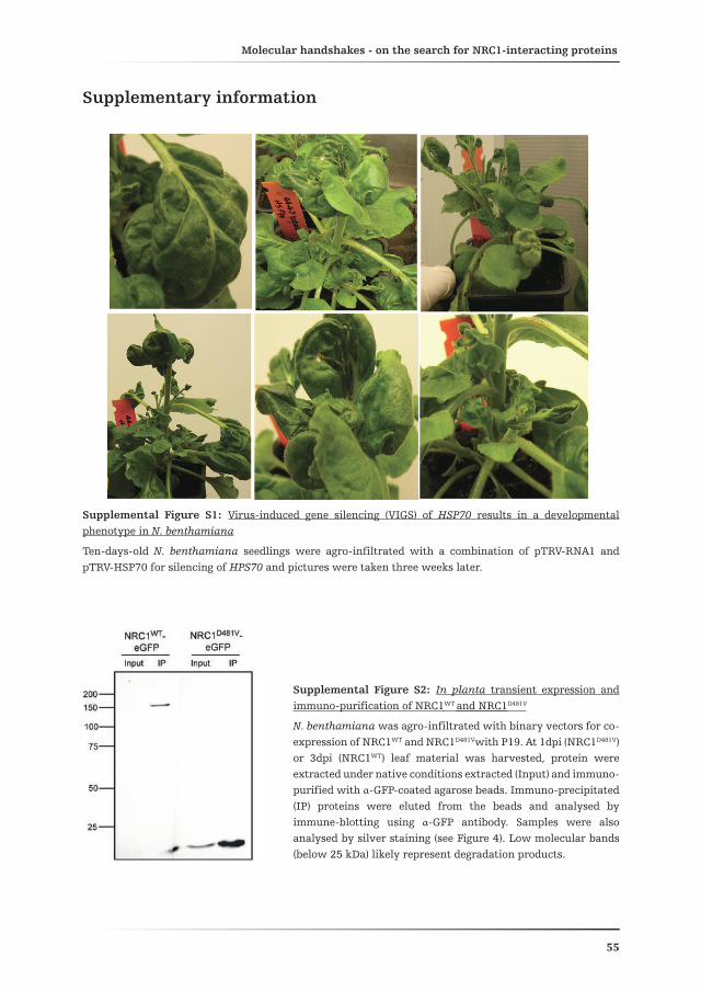
Molecular handshakes - on the search for NRC1-interacting proteinsChapter 2
55
Supplementary information
Supplemental Figure S1: Virus-induced gene silencing (VIGS) of HSP70 results in a developmental phenotype in N. benthamiana
Ten-days-old N. benthamiana seedlings were agro-infiltrated with a combination of pTRV-RNA1 and pTRV-HSP70 for silencing of HPS70 and pictures were taken three weeks later.
Supplemental Figure S2: In planta transient expression and immuno-purification of NRC1WT and NRC1D481V
N. benthamiana was agro-infiltrated with binary vectors for co-expression of NRC1WT and NRC1D481Vwith P19. At 1dpi (NRC1D481V) or 3dpi (NRC1WT) leaf material was harvested, protein were extracted under native conditions extracted (Input) and immuno-purified with α-GFP-coated agarose beads. Immuno-precipitated (IP) proteins were eluted from the beads and analysed by immune-blotting using α-GFP antibody. Samples were also analysed by silver staining (see Figure 4). Low molecular bands (below 25 kDa) likely represent degradation products.

Molecular handshakes - on the search for NRC1-interacting proteinsChapter 2
56
Sup
ple
men
tal
Tab
le S
1: H
SP70
isof
orm
Sol
yc10
g086
41 b
inds
to
NR
C1-
NB
in
pla
nta
Mat
rix
depi
ctin
g th
e se
quen
ce o
f al
l id
enti
fied
pep
tide
s co
rres
pon
din
g to
HSP
70 o
n t
he
left
an
d th
e te
n H
SP70
iso
form
s to
wh
ich
eac
h p
epti
de m
atch
es o
n t
he
righ
t. I
sofo
rm S
olyc
10g0
8641
(dep
icte
d in
bol
d) m
atch
es w
ith
all
pep
tide
s an
d is
als
o id
enti
fied
by
one
un
iqu
e pe
ptid
e.
Pep
tid
e se
qu
ence
HSP
70 i
sofo
rm
R.N
TT
IPT
KK
.ESo
lyc0
3g11
7630
Soly
c04g
0114
40So
lyc0
6g07
6020
Soly
c07g
0058
20So
lyc0
9g01
0630
Soly
c10g
0864
10So
lyc1
1g06
6060
Soly
c11g
0661
00
K.A
TAG
DT
HL
GG
ED
FD
NR
.MSo
lyc0
4g01
1440
Soly
c06g
0760
20So
lyc0
9g01
0630
Soly
c10g
0864
1So
lyc1
1g06
6060
Soly
c11g
0661
00
R.V
EII
AN
DQ
GN
R.T
Soly
c04g
0114
40So
lyc0
6g07
6020
Soly
c09g
0106
30So
lyc1
0g08
641
Soly
c11g
0660
60
Soly
c11g
0661
00
R.I
INE
PTA
AA
IAY
GL
DK
K.A
Soly
c06g
0760
20So
lyc0
7g00
5820
Soly
c09g
0106
30So
lyc1
0g08
641
Soly
c11g
0660
60
R.L
SKE
EIE
K.M
Soly
c06g
0760
20So
lyc0
9g01
0630
Soly
c10g
0864
1So
lyc1
1g06
6060
R.T
TP
SYV
GF
TD
SER
.LSo
lyc0
9g01
0630
Soly
c10g
0864
1So
lyc1
1g06
6060
K.E
QV
FST
YSD
NQ
PG
VL
IQV
YE
GE
R.T
Soly
c09g
0106
30So
lyc1
0g08
641
K.S
TV
HD
VV
LVG
GST
R.I
Soly
c06g
0760
20So
lyc1
0g08
641
K.E
IAE
AF
LG
TT
VK
.NSo
lyc1
0g08
641

Molecular handshakes - on the search for NRC1-interacting proteinsChapter 2
57
Pep
tid
e se
qu
ence
HSP
70 i
sofo
rm
R.I
INE
PTA
AA
IAY
GL
DK
.KSo
lyc0
1g06
0400
Soly
c01g
0996
60So
lyc0
3g08
2920
Soly
c03g
1176
30So
lyc0
4g01
1440
Soly
c06g
0760
20So
lyc0
7g00
5820
Soly
c08g
0828
20So
lyc0
9g01
0630
Soly
c10g
0864
10So
lyc1
1g06
6060
Soly
c11g
0661
00
K.I
TIT
ND
K.G
Soly
c03g
0829
20So
lyc0
3g11
7630
Soly
c04g
0114
40So
lyc0
6g05
2050
Soly
c06g
0760
20So
lyc0
7g00
5820
Soly
c08g
0828
20
Soly
c09g
0106
30So
lyc1
0g08
6410
Soly
c11g
0660
60So
lyc1
1g06
6100
K.N
AV
VT
VPA
YF
ND
SQR
.QSo
lyc0
3g11
7630
Soly
c04g
0114
40So
lyc0
6g07
6020
Soly
c07g
0058
20So
lyc0
9g01
063
Soly
c10g
0864
10So
lyc1
1g06
6060
Soly
c11g
0661
00
R.V
EII
AN
DQ
GN
R.T
Soly
c04g
0114
40So
lyc0
6g07
6020
Soly
c09g
0106
30So
lyc1
0g08
6410
Soly
c11g
0660
60So
lyc1
1g06
6100
K.A
TAG
DT
HL
GG
ED
FD
NR
.MSo
lyc0
4g01
1440
Soly
c06g
0760
20So
lyc0
9g01
0630
Soly
c10g
0864
10So
lyc1
1g06
6060
Soly
c11g
0661
00
K.N
QV
AM
NP
INT
VF
DA
K.R
Soly
c06g
0760
20So
lyc0
9g01
0630
Soly
c10g
0864
10So
lyc1
1g06
6060
K.E
LA
QAY
EV
LSD
PE
KR
.ESo
lyc0
5g05
0810
Soly
c05g
0551
60So
lyc1
1g00
6460
R.E
IYD
QY
GE
DA
LK
.ESo
lyc0
4g00
9770
Soly
c05g
0551
60So
lyc1
1g00
6460
K.S
QP
GE
VV
KP
DQ
FK
.ASo
lyc0
4g00
9770
Soly
c05g
0551
60So
lyc1
1g00
6460
K.E
LE
SLC
NP
IIA
K.M
Soly
c10g
0864
10So
lyc1
1g06
6060
K.E
IAE
AF
LG
STV
K.N
Soly
c06g
0760
20So
lyc1
1g06
6060
K.Q
FAA
EE
ISSM
VL
IK.M
Soly
c09g
0106
30
R.T
TP
SYV
GF
TD
TE
R.L
Soly
c06g
0760
20
K.Y
YE
ILG
VP
K.T
Soly
c05g
0551
60
K.N
QA
AV
NP
ER
.TSo
lyc0
3g08
2920
K.V
LL
KD
FF
DG
K.E
Soly
c01g
0996
60
R.F
SDA
SVQ
SDM
K.L
Soly
c10g
0864
10
K.E
IAE
AF
LG
TT
VK
.NSo
lyc1
0g08
6410
Sup
ple
men
tary
Tab
le S
2: S
ever
al H
SP70
iso
form
s in
tera
ct w
ith
NR
C1W
T i
n p
lan
ta
Mat
rix
depi
ctin
g th
e se
quen
ce o
f al
l id
enti
fied
pep
tide
s co
rres
pon
din
g to
HSP
70 o
n t
he
left
an
d th
e 14
HSP
70 i
sofo
rms
to w
hic
h e
ach
pep
tide
mat
ches
on
th
e ri
ght.
Iso
form
s u
nam
bigu
ousl
y id
enti
fied
by
the
pres
ence
of
a u
niq
ue
mat
chin
g pe
ptid
e ar
e at
th
e bo
ttom
of
the
Tabl
e.


Chapter 3
The tree of lifeI think that I shall never see
a thing so awesome as the Tree that links us all in paths of genes down into depths of time unseen;
Whose many branches spreading wide house wondrous creatures of the tide,
ocean deep and mountain tall, darkened cave and waterfall.
Among the branches we may find creatures there of every kind,
from microbe small to redwood vast, from fungus slow to cheetah fast.As glaciers move, strikes asteroid a branch may vanish in the void:
at Permian’s end and Tertiary’s door, the Tree was shaken to its core.
The leaves that fall are trapped in time
A mutation in the nucleotide-binding domain of a tomato down-stream signalling NB-LRR affects defence-induced
changes in oligomerization
Daniela J. Sueldo1, Ellen van Enckevort2, Laurentiu Spiridon3, Marcel Prins2, Matthieu Joosten1 , Wladimir Tameling1
Affiliations:1 Laboratory for Phytopathology, Wageningen University, Wageningen, The Netherlands2 Keygene, N V, Wageningen, The Netherlands3 Institute of Biochemistry of the Romanian Academy,
Department of Bioinformatics and Structural Biochemistry, Bucharest
beneath cold sheets of sand and lime; but new leaves sprout as mountains rise, breathing life anew ‘neath future skies.On one branch the leaves burst forth:
a jointed limb of firework growth. with inordinate fondness for splitting lines,
armored beetles formed myriad kinds.Wandering there among the leaves, in awe of variants Time conceived,
we ponder the shape of branching fates, and elusive origins of their traits.
Three billion years the Tree has grown from replicators’ first seed sown to branches rich with progeny:
the wonder of phylogeny.
David R. MaddisonSyst Biol. 2013 January; 62(1): 179

Chapter 3
60
Abstract
Plant disease Resistance (R) proteins confer immunity to pathogens possessing the corresponding avirulence (Avr) proteins. Activation of R proteins is often associated with the induction of the Hypersensitive Response (HR), a form of programmed cell death. NRC1 (NB-LRR required for HR-associated Cell death-1) is an NB-LRR from tomato that participates in the defence signalling cascade down-stream of different classes of R proteins. Silencing with the tomato NRC1 construct in Nicotiana benthamiana diminishes the HR triggered by various R/Avr combinations, as well as the R protein-mediated resistance to fungal pathogens in tomato. We conducted a TILLING (Targeting Induced Local Lesions In Genomes) analysis of an EMS-mutagenised tomato population of which the NRC1 region coding for the NB-ARC domain and the first four LRRs was sequenced in a high-throughput fashion. This approach yielded three loss-of-function mutations that caused a reduction in the HR-signalling activity of NRC1. However, they did not compromise Ve1-mediated resistance to Verticillium dahliae in tomato. Co-immuno-precipitation experiments demonstrated that NRC1 self-associates in planta in its pre-activation state and that the oligomer is disrupted upon induction of effector-triggered immunity. Moreover, one of the mutants exerts a dominant-negative effect on the defence signalling activity of wild-type NRC1 and shows a compromised disruption of the mixed oligomer upon activation. Our results confirm that NRC1 plays a role in effector-triggered immunity and that disruption of the NRC1 oligomer is required for the activation of defence signalling.

Dynamics of the NRC1 oligomer upon defence signallingChapter 3
61
Introduction
Throughout their life, plants encounter various organisms; some of which are pathogenic and can therefore cause disease. As disease is detrimental and has a negative impact on reproductive fitness, plants have evolved mechanisms to detect pathogens and defend themselves against pathogen attack. The current idea on how the plant immune system functions, as originally proposed by Jones and Dangl (2006) under the name “zigzag model”, states that plants possess two layers of induced defence; the first layer is triggered by conserved Microbe-Associated Molecular Patterns (MAMPs), whereas the second layer is activated upon the recognition of race-specific virulence proteins or so-called effectors of a pathogen. The first layer, referred to as MAMP-Triggered Immunity (MTI), results in the arrest of colonization of plant tissues by non-adapted pathogens and relies on the recognition of MAMPs by cell surface receptors termed Pattern Recognition Receptors (PRRs). Effector-Triggered Immunity (ETI) comes into the game when pathogens circumvent the first induced defence layer, for example by producing effectors that supress or overcome MTI. Some of these effectors are recognised by plant immune receptors named Resistance (R) proteins to trigger ETI. This process eventually results in resistant plants (Jones and Dangl, 2006). However, in an evolutionary perspective it is conceivable that in response to ETI, pathogens will either lose recognised effectors or will evolve effectors that in their turn block the ETI response, causing the battle to go on. Even though the “zigzag” model, illustrating this battle between plants and pathogens, is widely accepted within the plant pathology research community, recent evidence suggests that the boundaries between MTI and ETI are not always clear (de Jonge et al., 2010; Thomma et al., 2011).
Broad-spectrum resistance can be defined as the ability of a single plant species to resist infection by two or more pathogen species or is resistant to the majority of different races of the same pathogen (Kou and Wang, 2010). In this regard, MTI could be considered as a broad-spectrum resistance response. However, MTI is not effective against host-adapted pathogens and therefore a broad-spectrum resistance that relies on ETI to stop these pathogens is desirable. Although generally very effective in arresting pathogen proliferation, ETI is a pathogen race- and host genotype-specific type of immunity. This means that a specific R protein is only present in particular plant genotypes or accessions and provides resistance only against a specific race of a pathogen. Therefore, considering that plants are exposed to a plethora of pathogens, the question addressing how plants are able to successfully cope with such a variety of attackers, remains. Of equal importance is how we can actually exploit this extremely efficient defence mechanism to halt devastating pathogen out-breaks that often occur in the field. Stable over-expression of plant immune receptors can successfully lead to broad-spectrum resistance by inducing a constitutive defence response (Oldroyd and Staskawicz, 1998; Tang et
Chapter 3
62
al., 1999; Wang et al., 2013). However, constitutive defence is often detrimental to plant development and fitness. Alternatively, the transfer of immune receptors from one plant species to the other can broaden the resistance spectrum of a certain plant species (Lacombe et al., 2010; Fradin et al., 2011; Maekawa et al., 2012). Together this provides evidence that the plant immune system can be manipulated due to obvious conservation of defence signalling pathways. With this in mind, it should be possible to generate plants with defence mechanisms that combines the broad-spectrum characteristics of MTI and the efficiency of ETI.
The largest class of R proteins comprises the Nucleotide-Binding, Leucine-Rich Repeat (NB-LRR) proteins. NB-LRRs are intracellular immune receptors capable of binding nucleotides, either ATP or ADP, which modulates their activation state (Tameling et al., 2002; Tameling et al., 2006; Maekawa et al., 2011; Williams et al., 2011). In NB-LRRs, nucleotide binding occurs through the NB-ARC domain (Tameling et al., 2002; Williams et al., 2011). This central domain consists of three subunits, namely the NB (nucleotide binding), ARC1 and ARC2 (shared between the proteins Apaf-1, R proteins and CED-4) (van der Biezen and Jones, 1998; Takken et al., 2006). Moreover, based on their N-terminal domain they can be classified into two main subgroups: CC-NB-LRRs (CNLs) containing a coiled-coil domain and TIR-NB-LRRs (TNLs), having an N-terminal domain with homology to the Toll/interleukin-1 receptor (Meyers et al., 1999). Finally, the C-terminal part of NB-LRRs consists of a variable number of LRRs (Leucine-Rich Repeats). This domain may participate in elicitor recognition (Rairdan and Moffett, 2006; Brunner et al., 2010; Qi et al., 2012; Ravensdale et al., 2012), as well as in auto-inhibition, in order to avoid activation of defence responses in the absence of a pathogen (Moffett et al., 2002; Hwang&Williamson, 2003; Ade et al., 2007).
According to the current model, the ADP-bound conformation is the “off” state of NB-LRRs, whereas the ATP-bound conformation represents the “on” state (Takken and Tameling, 2009; Takken and Goverse, 2012). Upon pathogen ingress, effector recognition by the NB-LRR (either directly or indirectly) drives an increase in the amount of the ATP-bound form of the protein, which activates a signalling cascade that generally culminates in the Hypersensitive Response (HR), a form of Programmed Cell Death (PCD). Most of the data supporting this model come from the observation that point mutations in various highly conserved motifs of NB-LRRs involved in nucleotide binding and/or hydrolysis result in versions of the protein that trigger an elicitor-independent HR upon its transient expression in Nicotiana benthamiana (Bendahmane et al., 2002; Tameling et al., 2006; Ade et al., 2007; Kawano et al., 2010; Kawano et al., 2010; Du et al., 2012). Furthermore, such mutants constitutively activate defence responses when stably expressed in Arabidopsis thaliana (Eitas et al., 2008; Gao et al., 2011; Zhang et al., 2003; Roberts et al., 2013). These mutants are known as gain-of-function or auto-active, and generally contain substitutions in
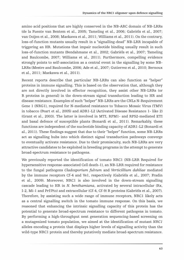
Dynamics of the NRC1 oligomer upon defence signallingChapter 3
63
amino acid positions that are highly conserved in the NB-ARC domain of NB-LRRs (de la Fuente van Bentem et al., 2005; Tameling et al., 2006; Gabriëls et al., 2007; van Ooijen et al., 2008; Maekawa et al., 2011; Williams et al., 2011). On the contrary, loss-of-function mutants usually result in a “signalling-dead” NB-LRR incapable of triggering an HR. Mutations that impair nucleotide binding usually result in such loss-of-function mutants (Bendahmane et al., 2002; Gabriëls et al., 2007; Tameling and Baulcombe, 2007; Williams et al., 2011). Furthermore, compelling evidence strongly points to self-association as a central event in the signalling by some NB-LRRs (Mestre and Baulcombe, 2006; Ade et al., 2007; Gutierrez et al., 2010; Bernoux et al., 2011; Maekawa et al., 2011).
Recent reports describe that particular NB-LRRs can also function as “helper” proteins in immune signalling. This is based on the observation that, although they are not directly involved in effector recognition, they assist other NB-LRRs (or R proteins in general) in the down-stream signal transduction leading to HR and disease resistance. Examples of such “helper” NB-LRRs are the CNLs N-Requirement Gene 1 (NRG1), required for N-mediated resistance to Tobacco Mosaic Virus (TMV) in tobacco (Peart et al., 2005) and ADR1-L2 (Activated Disease Resistance 1- like 2) (Grant et al., 2003). The latter is involved in MTI, RPM1- and RPS2-mediated ETI and basal defence of susceptible plants (Bonardi et al., 2011). Remarkably, these functions are independent of the nucleotide binding capacity of ADR1-L2 (Bonardi et al., 2011). These findings suggest that due to their “helper” function, some NB-LRRs act as signalling hubs into which distinct signal transduction pathways converge to eventually activate resistance. Due to their promiscuity, such NB-LRRs are very attractive candidates to be exploited in breeding programs in the attempt to generate broad-spectrum resistance to pathogens.
We previously reported the identification of tomato NRC1 (NB-LRR Required for hypersensitive response-associated Cell death-1), an NB-LRR required for resistance to the fungal pathogens Cladosporium fulvum and Verticillium dahliae mediated by the immune receptors Cf-4 and Ve1, respectively (Gabriëls et al., 2007; Fradin et al., 2009). Moreover, NRC1 is also involved in the down-stream signalling cascade leading to HR in N. benthamiana, activated by several intracellular (Rx, I-2, Mi-1 and Prf/Pto) and extracellular (Cf-4, Cf-9) R proteins (Gabriëls et al., 2007). Therefore, by assisting such a wide range of immune receptors, NRC1 likely acts as a central signalling switch in the tomato immune response. On this basis, we reasoned that enhancing the intrinsic signalling capacity of this protein has the potential to generate broad-spectrum resistance to different pathogens in tomato. By performing a high-throughput next generation sequencing-based screening on a mutagenized tomato population, we aimed at the identification of mutant NRC1 alleles encoding a protein that displays higher levels of signalling activity than the wild-type NRC1 protein and thereby putatively mediate broad-spectrum resistance.

Chapter 3
64
Here, we report the identification of three tomato lines carrying mutations in the NRC1 gene that negatively affect the HR-signalling activity of the encoded protein, but do not alter the capacity of NRC1 to mediate resistance to V. dahliae in tomato. Moreover, the finding that one of the mutations has a dominant-negative effect on the HR-signalling activity of NRC1 incited us to examine protein self-association as a requirement for NRC1-mediated signalling. Indeed, our results suggest that, as was found for other NB-LRRs, NRC1 has the capacity to constitutively self-associate in planta. However, unlike what has been reported for NB-LRRs so far, the NRC1 oligomer is disrupted upon the activation of defence signalling.
Results
Sequencing of the region encoding the NB-ARC domain of NRC1 in a mutagenized tomato population
In an attempt to identify in planta mutations that affect NRC1 signalling activity, we decided to follow a TILLING (Targeting Induced Local Lesions In Genomes) approach, combining an Ethyl MethaneSulfonate (EMS)-mutagenized tomato population, as described in Menda et al. (2004), and high-throughput sequencing technology. Genomic DNA was extracted from each individual of the M2 (second generation of mutated plants) population and 454-based massive parallel sequencing was performed as described in Materials and Methods. In total, we screened 15,040 mutant individuals, corresponding to five segregating M2 tomato plants belonging to 3008 M2 families. We limited the high-throughput sequencing and hence the search for mutations in NRC1 to the region coding for the central NB-ARC domain and first four LRRs, because almost all auto-active mutations have been found in these regions in other NB-LRRs (Tameling et al., 2006; Bendahmane et al., 2002; van Ooijen et al., 2008). This region chosen for mutational screening (NRC1-NB-ARC plus first four LRRs) was subdivided into two amplicons for efficient sequencing. Amplicon number 1 (741 bp) encompassed the coding sequence for the NB and ARC1 subdomains whereas amplicon number 2 (767 bp) contained the coding sequence for the ARC2 subdomain and the first four LRRs (Fig. 1A).
Sequencing of both amplicons identified a total of six mutations, from which two corresponded to missense mutations (G460A and G830A) resulting in the amino acid substitutions E154K and R277H, respectively (Fig. 1A). As expected from EMS mutagenesis, both identified mutations were transitions, from cytosine/guanidine to adenine/thymine (CG AT). A deeper sequencing analysis, searching not only for EMS-associated nucleotide substitutions but for any kind of substitution revealed the presence of one additional mutation, namely A774G, causing the amino acid substitution E258G. Moreover, we only observed missense mutations in the NB subdomain of NRC1.

Dynamics of the NRC1 oligomer upon defence signallingChapter 3
65
Figure 1: High-throughput sequencing of the region of NRC1 encoding the NB-ARC domain and first four N-terminal LRRs in an EMS-mutagenized tomato population, reveals three mutations in its NB subdomain
(A) Schematic representation of the NB-ARC domain and first four LRRs of NRC1 depicting highly conserved motifs (left). On the right the positions of the identified nucleotide mutations and resulting amino acid substitutions are indicated. The NB-ARC domain, with the first four LRRs of NRC1, was subdivided into two amplicons and sequenced in each individual plant of an EMS-mutagenised tomato population by means of 454-sequencing technology.
(B) Alignment of NRC1 and NB-LRRs from different plant species to illustrate the degree of conservation of the motifs in which the mutations in NRC1 were identified. The amino acid sequences of NRC1 and other selected NB-LRRs were aligned with ClustalW. Only the part of the alignment in which the mutations were identified in the NRC1 sequence is included. Highly conserved motifs are underlined in blue, mutations known to cause auto-activity in other NB-LRRs are depicted in blue and the newly identified mutations are shown in green.
As already mentioned, this subdomain is characterized by the presence of motifs that are highly conserved in NB-LRRs of different plant species. Several of these motifs

Chapter 3
66
contain residues proven or predicted to be required for nucleotide binding. None of the mutations identified were located in any of these motifs, although they did localise very close to two of them. As depicted in an alignment of the amino acid sequences of various NB-LRRs from different plant species (Fig. 1B), the E154K mutation is situated two amino acid positions upstream of the hhGRExE motif, which is involved in the positioning of the adenine base of the bound ATP or ADP (Meyers et al., 1999; Takken et al., 2006). The E258G and R277H mutations, on the other hand, are situated five and seven amino acid positions up and down-stream of the Walker B motif, respectively. This motif contains two highly conserved adjacent aspartic acid (D) residues of which the second (D268 in NRC1) has been shown to be involved in ATP hydrolysis by the CNL I-2 (Tameling et al., 2006). Finally, analysis of the alignment of NRC1 with other NB-LRRs (belonging to the CNL and TNL class) also revealed that the mutated residues themselves were not highly conserved within the NB-LRR protein family. Only the glutamic acid (E) at position 154 in NRC1 was conserved in I-2, Rx and RPM1.
A 3D model of the ADP-bound state of the NB-ARC domain of NRC1
To date, no crystal structure of the NB-ARC domain of a plant NB-LRR has been reported. However, the crystal structures of two proteins closely related to plant NB-LRRs have been resolved (Riedl et al., 2005; Yan et al., 2005). Apaf-1 (Apoptotic Protease pre-Activating Factor 1) and CED-4 (Caenorhabditis elegans Death-4) are proteins from mammals and C. elegans respectively, which are involved in PCD. The NB-ARC domain of NRC1 contains 357 amino acid residues (from amino acid position 151 to 508 in the full-length sequence) and this portion of the protein was used for structural 3D modelling. Both sequence homology and automated fold recognition software returned human Apaf-1 as the most reliable template in the 3D databases. Despite of the low sequence identity (23%), the similarity is substantial (42%) and both the secondary structure prediction and the position of the conserved motifs match to those in Apaf-1. The NB, ARC1 and ARC2 subdomains of Apaf-1 are also called the α/β-fold, helical domain I and winged-helix domain, respectively (Riedl et al., 2005). The final model generated for NRC1-NB-ARC deviates within only 3Å from the Apaf-1 template and retains the overall three subunit fold of the corresponding NB-ARC of Apaf-1 during in silico stability test simulations (Fig. 2A). All conserved motifs of the NB-ARC and the nucleotide-binding pocket are present in the NRC1-NB-ARC model. In addition, we could also identify amino acids required for nucleotide (ATP or ADP) binding and hydrolysis in the active site (Fig. 2A). Importantly, since Apaf-1 in the crystal was bound to ADP, the structure represents the closed, auto-inhibited conformation of the protein (Riedl et al., 2005). Due to the near perfect match, it is likely that our NRC1-NB-ARC 3D model also represents the conformation of the NB-ARC domain in the ADP-bound state of NRC1.
We then studied the identified mutations in the context of the 3D model of the NRC1-NB-ARC (Fig. 2A). As mentioned before, the three mutated residues localize strictly within the NB subdomain and showed a rather high degree of accessibility.

Dynamics of the NRC1 oligomer upon defence signallingChapter 3
67
In addition, the mutated residues are charged and hydrophilic; overall suggesting affinity for ligands rather than for solvent. In summary, based on their position and chemical nature, the E154K, E258G and R277H mutations are at the surface of the NB subdomain of NRC1 and are not likely to affect either the overall structure of the protein or its ADP-binding capacity. However, they could very well have an impact on the interaction of the NRC1-NB-ARC with other domains of NRC1, thereby possibly indirectly influencing the nucleotide-bound status of the protein due to the intimate interplay of these domains. Alternatively, the mutations could have an effect on the ability of NRC1 to interact with signalling partners.
Figure 2: Model of the 3D structure of the ADP-bound state of the NB-ARC domain of NRC1
(A) Ribbon cartoon depicting a model of the 3D structure of NRC1-NB-ARC. The amino acid residues 160 to 443 from the NRC1 full-length protein were modelled for its 3D structure, based on the crystal structure of Apaf-1 as described in Materials and Methods. Blue, conserved motifs of the NB-ARC; brown, amino acids directly involved in nucleotide binding; red, positions of E154, E258 and R277 amino acid residues; pink, ADP molecule. Side chains of important residues are depicted as sticks.
(B) Panels showing close-ups of the positions of the identified mutations. ADP, bound ADP molecule; D268, the invariant aspartic acid residue in the Walker B motif of the NB subdomain.

Chapter 3
68
Mutations E154K, E258G and R277H negatively affect the activity of NRC1
To determine whether the identified mutations cause elicitor-independent cell death (gain-of- function mutations) or have a negative impact on the activity of NRC1 (loss-of-function mutations) we introduced them individually in the region encoding the full-length NRC1 protein in our plant expression vector containing the CaMV 35S promoter. In order to properly discriminate between gain- and loss-of-function mutations, the mutations were introduced into the NRC1WT and NRC1D481V expression constructs and the mutant proteins were transiently expressed in N. benthamiana by Agrobacterium-mediated transformation. NRC1D481V carries a point mutation in the aspartic acid residue of the MHD motif, which is highly conserved in NB-LRRs. This mutation is known in both NRC1 and other proteins of this family to cause elicitor-independent cell death (Bendahmane et al., 2002; Tameling et al., 2006; Gabriëls et al., 2007; Gao et al., 2011; Roberts et al., 2013). NRC1WT, on the other hand, does not cause cell death when transiently expressed in N. benthamiana. To overcome this, and obtain a clear cell death phenotype, NRC1WT and NRC1 single mutants were co-expressed with the silencing suppressor P19 from Tomato Bushy Stunt Virus (TBSV) (Voinnet et al., 2003). This silencing suppressor increases the expression levels of the gene of interest, through inhibition of the plant RNA silencing machinery. Because of this phenomenon, the amount of NRC1WT protein that accumulates rises above the minimal signalling initiation threshold to trigger elicitor-independent cell death within 3 days post-infiltration (dpi) (Fig. 3A, upper panel, right leaf halves). This was not the case for NRC1E154K, NRC1E258G and NRC1R277H, as their expression in the presence of P19 did not cause an HR (Fig. 3A, upper panel, left leaf halves). This suggests that all of the three single mutations negatively affect the cell death-inducing activity of NRC1 and the mutants were therefore classified as loss-of-function mutants.
When transiently expressing the previously reported gain-of-function mutant NRC1D481V in N. benthamiana, the first symptoms of an HR where detectable at 1 dpi without the need of co-expression of P19 (Fig. 3A, lower panel; picture taken at 3 dpi, right leaf halves). In contrast to what we observed for the E154K and R277H single mutants (introduced in the NRC1WT background, Fig. 3A upper panel), NRC1E154K/D481V and NRC1 R277H/D481V double mutants retained their capacity to trigger cell death, similar to NRC1D481V itself. However, NRC1E258G/D481V did not induce an HR, suggesting that the E258G mutation has an epistatic effect over the D481V mutation. Alternatively, the combination of both mutations renders the NRC1 protein unstable, thereby explaining the lack of HR-inducing activity of NRC1E258G/D481V. With this in mind, we fused all NRC1 variants to eGFP (enhanced green fluorescent protein) to analyse their accumulation and stability. We also tested NRC1K191R, a previously reported loss-of-function mutant of NRC1 (Gabriëls et al., 2007). Substitution of this highly conserved lysine residue in the P-loop motif has been shown to abolish nucleotide binding, resulting in a signalling-dead mutant protein (Tameling et al., 2002; Tameling et al., 2006; Williams et al., 2011).

Dynamics of the NRC1 oligomer upon defence signallingChapter 3
69
Figure 3: E154K, E258G and R277H are loss-of-function mutations in NRC1
(A) The three identified mutations in the NB-ARC domain of NRC1 are loss-of-function mutations in the wild-type (WT) background of the protein and are not epistatic over the D481V gain-of-function mutation. The three mutations, E154K, E258G and R277H, were independently introduced in either the WT background of NRC1 (upper panel) or in the auto-active (NRC1D481V) background (lower panel), as depicted in the cartoon on the left. In the left halves of the leaf, the mutant proteins were transiently expressed with (upper panel) or without (lower panel) the silencing suppressor P19. In the right halves of the leaf, either NRC1WT (upper panel) or NRC1D481V (lower panel) were included as control. Note that NRC1WT does not induce an HR without co-expression of P19. Pictures were taken at 3 dpi.
(B) All mutant proteins, except for NRC1E258G/D481V, are stably expressed in N. benthamiana. All NRC1 variants were C-terminally fused to eGFP and transiently expressed in N. benthamiana by Agrobacterium-mediated transformation. At 3 dpi, leaves were harvested and the eGFP-fused proteins were immuno-precipitated with α-GFP-coated agarose beads. Immuno-precipitated proteins were eluted from the beads,
separated on SDS gels and analysed by western blotting using α-GFP.
The eGFP-fused proteins were transiently co-expressed with P19 in N. benthamiana and at 3 dpi, leaf material was harvested for protein extraction under native conditions. Due to the low accumulation levels of the NRC1WT protein an immuno-precipitation (IP) with α-GFP-agarose beads was performed in all cases to increase

Chapter 3
70
the concentration of the expressed proteins. NRC1WT, NRC1E154K, NRC1E258G and NRC1R277H could already be detected in the total protein extract (input of the IP), whereas NRC1K191R, NRC1E154K/D481V and NRC1R277H/D481V were only detected after their IP (Fig. 3B). This confirms that all mutant proteins are stable, except for NRC1E258G/
D481V, as for this mutant no protein could be detected (Fig. 3B). Together this clearly indicates that the loss-of-function phenotype of most of the mutants (except for NRC1E258G/D481V), is due to an effect on the signalling activity of NRC1 and is not caused by a decreased stability of the protein. Finally, although the expression of NRC1D481V was performed under control of the constitutive 35S promoter, attempts to immuno-precipitate and detect expression of this mutant were not successful (data not shown, see Chapter 2). Probably, this auto-active version of NRC1 is under strict post-translational regulation, preventing the NRC1D481V protein to accumulate to levels that allow detection on immunoblots. We can therefore conclude that the mutations E154K, E258G and R277H suppress the signalling activity of NRC1WT, implying that the replaced residues are important for proper functioning of the NRC1 protein.
Mutation E154K has a dominant-negative effect on the HR signalling activity of NRC1WT
Above we reported the effect of the E154K, E258G and R277H mutations on the activity of NRC1WT in cis. As a next step in the characterization of these mutations, we studied their influence on NRC1WT activity in trans. To this end, NRC1WT was transiently co-expressed in N. benthamiana with P19 and each of the mutants or GUS as a negative control (Fig. 4A), and the development of an HR was monitored. We also examined the production of polyphenolic compounds, which is a hallmark of the HR, by clearing the leaves from their chlorophyll in ethanol. This offers a way to detect subtle differences in HR strengths, as compared to merely looking at macroscopic cell death and tissue collapse. We anticipated that if any of the mutants would affect the activity of NRC1WT in trans, then both the HR and the production of polyphenolic compounds should be reduced as compared to co-expression of NRC1WT with GUS. At 3 dpi, a clear HR was observed when NRC1WT was co-expressed with either GUS or the mutants NRC1E258G and NRC1R277H, denoting that these mutants, despite of being loss-of-function in cis, do not exert a negative effect on the activity of NRC1WT in trans (Fig. 4B). In accordance with this, the accumulation of polyphenolic compounds also appeared equal when co-expressing NRC1WT with the aforementioned mutants or GUS. Remarkably, both the macroscopic cell death and the production of polyphenolic compounds normally induced by NRC1WT were visibly reduced upon its co-expression with NRC1E154K (Fig. 4B). This finding demonstrates that mutation E154K is acting in a dominant-negative manner, since it negatively affects the activity of NRC1WT not only in cis but also in trans. In conclusion, we

Dynamics of the NRC1 oligomer upon defence signallingChapter 3
71
have identified three new residues important for the HR- signalling activity of NRC1. Based on the finding that the mutations behave differently with respect to their effect in cis and in trans, we propose that they likely interfere with the activity of NRC1WT in different ways.
Figure 4: NRC1E154K is a dominant-negative mutant.
(A) Cartoon illustrating the experimental design. NRC1WT was transiently co-expressed in N. benthamiana by Agrobacterium-mediated transformation, either with each of the three NRC1 mutants or with GUS.
(B) NRC1E154K reduces the HR-inducing activity of NRC1WT when co-expressed in trans. Proteins were co-expressed as explained in (A) and development of an HR was monitored over time. Pictures were taken at 3 dpi and leaves were cleared with 70% ethanol to better visualise cell death.

Chapter 3
72
Mutation E154K likely blocks the NRC1 oligomer in a signalling-impaired state
Homo-dimerization, either as a pre- or post-activation event, is an emerging feature in NB-LRR signalling. A number of NB-LRR proteins have been shown to self-associate in higher order molecular complexes (Mestre and Baulcombe, 2006; Ade et al., 2007; Gutierrez et al., 2010; Bernoux et al., 2011; Maekawa et al., 2011). In the light of these findings, we interpreted the above-mentioned result which identify NRC1E154K as a dominant-negative mutant in trans, as a possible indication for NRC1 self-association. If indeed NRC1 forms oligomers, then the mutation E154K might lock the NRC1 oligomer in a signalling-incompetent state. We therefore first investigated whether NRC1 forms oligomers in planta. In addition, since NRC1 acts down-stream of the Cf-4/Avr4 recognition event (Gabriëls et al., 2007), we also examined NRC1 self-association upon activation of the Cf-4/Avr4-triggered signalling cascade as a proxy to study its self-association pre- and post-activation. To this end, we employed transgenic N. benthamiana:Cf-4 stably expressing the Cf-4 resistance gene from tomato (Gabriëls et al., 2006), to transiently co-express NRC1WT
fused to the Myc epitope tag with P19 and either NRC1WT itself or any of the single mutants fused to eGFP. At 2 dpi, the Cf-4/Avr4 signalling pathway was activated by transiently expressing Avr4 and, as a negative control GUS, instead of Avr4, was co-expressed. Finally, 24 hrs later (before cell death symptoms became apparent) leaf material was harvested and NRC1 self-association was tested by co-IP assays using α-GFP-coated agarose beads.
Interestingly, we observed that NRC1WT indeed forms an oligomer in planta before defence activation, as NRC1WT-Myc was co-immuno-precipitated by NRC1WT-eGFP, without the requirement of Cf-4/Avr4-triggered induction of defence (Fig. 5). Surprisingly, when the Cf-4 signalling cascade was activated by transient expression of Avr4, the amount of NRC1WT-Myc that was co-immuno-precipitated by NRC1WT-eGFP was clearly reduced (Fig. 5). Importantly, this is not due to degradation of NRC1WT-Myc since it is detected in the input sample at amounts similar to the experiment where no cell death is induced. Therefore, these results suggest that NRC1 is likely present as an oligomer in its pre-activation state, which is disrupted when NRC1 is activated, in this case is upon recognition of Avr4 by the matching Cf-4 resistance protein.
We also examined the behaviour of the NRC1WT–NRC1E154K mixed oligomer. Taking into consideration that NRC1WT oligomers are disrupted when Cf-4-dependent defence signalling is induced and that the E154K mutation is acting dominant-negative, we tested the possibility that the NRC1 proteins in the NRC1WT–NRC1E154K mixed oligomer show a reduced dissociation upon defence activation. Indeed, when Cf-4/Avr4-mediated defence signalling was induced, NRC1WT-Myc was still efficiently co-immuno-precipitated by NRC1E154K-eGFP, which suggests that the mixed oligomer
Dynamics of the NRC1 oligomer upon defence signallingChapter 3
73
was not disrupted or at least not to the same extent as the NRC1WT oligomer upon defence activation (Fig. 5). In conclusion, NRC1 self-associates in the pre-activated state and dissociates after defence activation in planta. Therefore, disruption of the oligomer occurs as a post-activation event. Additionally, by examining the behaviour of NRC1WT–NRC1E154K mixed oligomers before and after defence activation, we have provided a plausible explanation for the dominant-negative effect of the E154K mutation in trans on the signalling activity of NRC1WT.
Figure 5: NRC1E154K compromises disruption of an NRC1 oligomer upon activation of the Cf-4/Avr4-triggered HR.
NRC1WT forms an oligomer, which is disrupted upon activation of the Cf-4/Avr4-induced signalling cascade leading to the HR. NRC1WT–Myc was transiently co-expressed in N. benthamiana:Cf-4, with either NRC1WT-eGFP or NRC1E154K-eGFP by Agrobacterium-mediated transformation. Two days later, defence signalling was induced by expression of C. fulvum Avr4 (+), whereas expression of GUS was included as a negative control (-). After 24 hrs leaves were harvested, proteins were extracted under native conditions and eGFP fusions were immuno-precipitated using agarose beads coupled with α-GFP antibody. Co- immuno-precipitated proteins (IP) were analysed by immuno-blotting using either anti-GFP or anti-Myc antibody. The input shows the total amount of the various, transiently expressed proteins.
Tomato plants carrying an NRC1 loss-of-function mutation are not compromised in their resistance to V. dahliae
Having determined that the EMS-induced mutations identified by TILLING, E154K, E258G and R277H, impair the ability of NRC1 to trigger the HR, next their effect in the resistance of tomato to pathogens was examined. We have previously shown that NRC1 is required for resistance of tomato to C. fulvum and the soil-borne fungal pathogen V. dahliae (Gabriëls et al., 2007; Fradin et al., 2009). The tomato cultivar (M82) that was used to generate the EMS mutant population is resistant to race 1 of V. dahliae as it contains the Ve1 immune receptor. Therefore, we could test the tomato plants carrying the mutations in NRC1 for compromised resistance to this vascular pathogen. Per mutant a total of 12 M3 plants, homozygous for the mutation, were root-inoculated with a suspension of fungal spores or treated with water as a negative control. Two weeks later, plants were phenotypically screened for symptom development. Tomato cultivars Motelle and MoneyMaker were included as controls for resistance and susceptibility to V. dahliae, respectively. Susceptibility of tomato to V. dahliae is expressed mainly by plant stunting, meaning a reduction in plant height and canopy area (Fradin et al., 2009). Indeed, MoneyMaker plants showed

Chapter 3
74
a clear reduction in height and total canopy area two weeks after inoculation with V. dahliae, a phenotype that was not observed in the water-treated plants. Cultivar Motelle (carrying Ve1), on the other hand, displayed no symptom development upon inoculation with the pathogen (Fig. 6A and B). Similar to the resistant cultivar, none of the NRC1 mutants showed signs of compromised resistance, as in all cases spore-inoculated and water-treated plants looked equally healthy. This finding therefore suggests that despite the fact that the introduced mutations suppress the capacity of NRC1 to induce cell death, they do not show an impact on NRC1-mediated resistance to V. dahliae. Possibly, an NRC1-dependent signalling pathway leading to HR might branch off from the one leading to resistance to V. dahliae, at the point of NRC1 itself. Another explanation is that perhaps additional NRC1 homologs might have a redundant function.

Dynamics of the NRC1 oligomer upon defence signallingChapter 3
75
Figure 6: The mutations E154K, E258G and R277H in NRC1 do not compromise resistance of tomato carrying Ve1 to V. dahliae
(A) Tomato plants carrying Ve1 that are impaired in NRC1-HR signalling activity remain resistant to V. dahliae. M82 tomato, homozygous for the single EMS-induced mutations E154K, E258G and R277H and carrying Ve1 were root-inoculated with spores of V. dahliae or treated with water. Two weeks later, plants were photographed from the side and the top and phenotypically assessed for compromised resistance to the fungal pathogen. Tomato cultivars MoneyMaker (susceptible) and Motelle (resistant, carrying Ve1) were included as controls. For all mutant lines and controls, twelve plants were used, of which six were root-inoculated with V. dahliae and six were treated with water. Pictures show the phenotype of a representative plant from each line and treatment.
(B) Inoculation of the tomato NRC1E154K, NRC1E258G and NRC1R277H mutant lines with V. dahliae does not cause a reduction in either plant height or canopy area. Plant height and canopy area were determined for each individual plant, including the controls, as described in Materials and Methods. For each plant line, parameters are plotted as absolute values and error bars represent differences between individual plants. Different letters indicate significant differences with at α=0.05 as determined by a one-way ANOVA analysis.
The NRC1 gene family in tomato and other Solanaceous species
Virus-Induced Gene Silencing (VIGS) proved that NRC1 is required for resistance of Ve1-carrying tomato to V. dahliae (Fradin et al., 2009). However, as shown above, Ve1 tomato plants carrying an NRC1 loss-of-function mutation are not compromised in the resistance to this pathogen. As mentioned above, the mutations in NRC1 could

Chapter 3
76
have a specific effect on the cell death signalling pathway, without affecting the fungal resistance pathway. However, the observed discrepancy can also be explained if NRC1 belongs to a gene family in tomato. When there is functional redundancy amongst the different family members, this will account for the absence of a susceptibility phenotype in the NRC1 mutant plants. When NRC1 was discovered, it was reported to be a single copy gene, based on DNA gel blot analysis (Gabriëls et al., 2007). However, the recent availability of the sequenced tomato genome offered us the possibility to mine for potential NRC1 homologs encoded by the tomato genome. We used the NRC1 protein sequence to perform a BLASTp search against the translated full genome of tomato in the SOL Genomics Network database (Mueller et al., 2005), and retrieved protein sequences with an amino acid identity from 40%, up to 70% (Supplemental Fig. S1). Interestingly, the relatively high homology was wide-spread throughout the entire NRC1 sequence and not just restricted to the more highly conserved regions generally present in NB-LRRs, suggesting these can be considered as true NRC1 homologs in tomato. The highest amino acid sequence identity (67.8%) corresponded to a protein encoded on chromosome 10. This is clearly the closest homolog of NRC1 in tomato and we therefore decided to name this gene NRC2. Based on their sequence homology, we cannot exclude the possibility that functional gene redundancy is occurring between NRC1 and NRC2. If NRC2 is redundant, this explains why tomato plants carrying a loss-of-function mutation in NRC1 are still resistant to V. dahliae, as a functional wild-type copy of NRC2 is still present.
We were also interested in studying the existence of possible NRC1 orthologs in other Solanaceous species. For this, we used the NRC1 protein sequence from tomato to perform a BLASTp search against the translated potato genome and the translated draft genome sequence of N. benthamiana (SOL Genomics Network) (Mueller et al., 2005; Bombarely et al., 2012). The most closely related sequences from the three species were retrieved and aligned and a phylogenetic tree was constructed. As depicted in Fig. 7, we found two NRC1 orthologs in potato (Sotub01g029620 and Sotub01g029230), located in tandem on chromosome 1, similar to tomato NRC1. Interestingly, we did not identify an obvious NRC1 ortholog in N. benthamiana, despite the fact that it has been shown that tomato NRC1 is functional in this plant species and that VIGS employing an insert derived from tomato NRC1 compromises the HR induced by different classes of R proteins in N. benthamiana (Gabriëls et al., 2007). On the other hand, we found two potential NRC2 orthologs in N. benthamiana but none in potato, implying a separation of the NRC1 gene family within the Solanaceae. Furthermore, for the additional NRC1-likes identified in tomato (Fig. 7), we did not find orthologs in N. benthamiana and potato.

Dynamics of the NRC1 oligomer upon defence signallingChapter 3
77
Figure 7: The NRC1 and NRC1-like gene family of tomato, potato and N. benthamiana
NRC1 is a member of a gene family present also in other Solanaceous species. The NRC1 protein sequence was used to perform a BLASTp search against the translated tomato, potato and draft N. benthamiana genomes. Retrieved sequences were aligned and used to construct a phylogenetic tree as described in Materials and Methods. Protein sequences from the NB-LRRs Mi-1 and I-2 from tomato, Rx from potato and NRG1 from N. benthamiana were included in the alignment as out-groups. Tomato homologs are shown in green, potato orthologs in orange and the orthologs from N. benthamiana are depicted in purple.
Discussion
NRC1 is a signalling hub for different plant immune receptors (Gabriëls et al., 2007) and enhancing the signalling capacity of this protein has the potential to generate broad-spectrum resistance to different pathogens. Therefore, we aimed at the identification of mutant NRC1 alleles that cause higher levels of signalling activity of the encoded protein (gain-of-function) and thereby putatively mediate broad-spectrum resistance. Our approach, based on the principle of TILLING, combined an EMS-mutagenised tomato population (Menda et al., 2004) and high-throughput sequencing technology (Rigola et al., 2009). The direct amplification and sequencing of the region of NRC1 encoding the NB-ARC domain and the first four LRRs from 15,040 M2 individuals allowed us to identify three missense mutations that lead to the amino acid substitutions E154K, E258G and R277H, which are all located in

Chapter 3
78
the NB subdomain of the NRC1 protein (Fig. 1). Using the same methodology and tomato population, Rigola et al (2009) identified two mutations upon sequencing of an amplicon of 254 bp of the gene encoding the eukaryotic translation initiation factor 4E (SleIF4E). Overall, this represents a four times higher mutation density per kb in that gene than what we found for NRC1.
Several reports have shown that mutations in NB-LRR proteins can cause constitutive activation of defence responses and, in some cases, cause detrimental developmental phenotypes (Tang et al., 1999; Noutoshi et al., 2005; Gao et al., 2011; Tang et al., 2011; Gou and Hua, 2012). An interesting example is ADR1-L2, a CNL from Arabidopsis that, similar to NRC1, promotes ETI down-stream of several R proteins. ADR1-L2 is also involved in MAMP-triggered immunity (MTI) and basal resistance (Grant et al., 2003; Bonardi et al., 2011). Stable ectopic expression (under control of its own promoter) of ADR1-L2D484V, a mutant in the aspartic residue of the MHD motif that causes elicitor-independent HR, showed constitutive defence activation and a typical dwarfing phenotype (Roberts et al., 2013). Mutations in this highly conserved MHD motif have earlier been shown to have a strong effect on the signalling activity of NB-LRRs, as revealed by an extensive mutational analysis focussed specifically on the MHD motif of tomato I-2 (van Ooijen et al., 2008). In addition, several gain-of-function mutations that only cause a mild HR have also been described (Tameling et al., 2002; Bendahmane et al., 2002; van Ooijen et al., 2008). It is expected that such mild mutations would not cause a dwarfing phenotype when stably expressed in plants. We aimed for the identification of such milder mutations in NRC1, as these mutant alleles could potentially be implemented in breeding programs aimed at generating broad-spectrum resistance. However, as revealed by the functional analysis of the three TILLING mutants that were identified (Figs. 3 and 4), we only found loss-of-function mutations, possibly reflecting the stringent cellular control that immune receptors are subjected to. Hence, a possible explanation for our observations is that plants carrying NRC1 gain-of-function mutants, generated by the treatment with EMS, are not viable.
Tornero et al. (2002) performed a mutational screen on the Arabidopsis NB-LRR resistance protein RPM1 (Resistance to Pseudomonas syringae pv. Maculicola 1) and reported the identification of 95 mutants compromised in their ability to trigger an HR upon co-expression with the matching effector, AvrRpm1. Furthermore, the RPM1-mediated resistance response when challenged with avirulent Pseudomonas syringae pv. tomato, expressing AvrRpm1, was also suppressed in these mutants. In comparison with the other domains of RPM1, its NB-ARC was clearly enriched in mutations and rather than in a particular motif, mutations were scattered throughout the domain (Tornero et al., 2002). Similarly, the mutations that we identified in NRC1 were located close to highly conserved motifs of the NB subdomain, but not in the motifs themselves. A few additional extensive random mutational screens on
Dynamics of the NRC1 oligomer upon defence signallingChapter 3
79
NB-LRRs have been performed, either by performing EMS-induced mutagenesis in Arabidopsis (Axtell et al., 2001), or by the introduction of mutations through error-prone PCR gene amplification in the potato NB-LRR Rx (Bendahmane et al., 2002). Despite of being performed on a considerable scale, none of the above-mentioned screens reported loss-of-function mutations corresponding to the positions in the coding region that we found, suggesting that these particular mutations specifically affect the activity of NRC1. However, it should be noted that different from the above-mentioned studies, we first identified the mutations by sequencing the amplicons of interest and then tested their effect on the activity of NRC1. This might have allowed us to detect mild loss-of-function mutations that would have been missed if we had carried out our screen by directly assessing the HR phenotype.
The identified loss-of-function mutations affect NRC1-dependent HR but not NRC1-mediated resistance
As mentioned above, mutations in RPM1 that suppressed effector-induced HR also resulted in compromised resistance to avirulent bacteria expressing AvrRpm1 (Tornero et al., 2002), depicting a clear correlation between the loss of HR and compromised resistance. An interesting finding in our case was that, although NRC1 is required for Ve1-mediated resistance to V. dahliae (Fradin et al., 2009), Ve1 tomato plants homozygous for the mutant alleles of NRC1 were still resistant to the fungus (Fig. 6) and therefore did not show a positive correlation with the compromised HR phenotype (Fig. 3). Functional gene redundancy could be an explanation for this discrepancy and indeed mining of the tomato genome revealed the existence of several NRC1 homologs (Fig. 7), some of which are located in tandem as was previously reported for other NB-LRRs (Li et al., 2001; Zhang et al., 2003; Jupe et al., 2012). Based on nucleotide sequence alignment of the various NRC1 homologs, we anticipate that the inserts used for VIGS of NRC1 in tomato by Gabriëls et al. (2007) and (Fradin et al., 2009) very likely also target NRC2 as well, in addition to other NRC1-related sequences. An additional experiment in which only NRC1 was silenced by specifically targeting its 3’-UTR region confirmed that silencing of NRC1 alone is indeed sufficient to compromise resistance to C. fulvum (Gabriëls et al., 2007). However, this is not clear for V. dahliae resistance, as this particular VIGS assay has not been performed by Fradin et al., (2009). Furthermore, specific silencing of NRC2 only has never been performed and therefore we cannot assure that this homolog is also involved in the resistance response against these fungal pathogens. As depicted in Fig. 7, the genome of N. benthamiana codes for several potential NRC2 orthologs, whereas a true ortholog of tomato NRC1 appears to lack in the draft genome of N. benthamiana. This finding could explain the fact that various experimental approaches that have been undertaken to clone the NRC1 gene

Chapter 3
80
from N. benthamiana were unsuccessful (Vossen and Abd-El-Haliem, unpublished data). Nonetheless, the sequence similarity between the coding regions of tomato NRC1 and N. benthamiana NRC2 is sufficiently high for N. benthamiana NRC2 to be targeted by the VIGS fragment derived from tomato NRC1. Therefore, it is possible that in N. benthamiana NRC2, and not NRC1, is involved in the HR triggered by several intracellular and extracellular R proteins (Gabriëls et al., 2007). It should be noted though, that the genome of N. benthamiana has only recently been released (Bombarely et al., 2012). Therefore, completion is yet needed to properly address whether N. benthamiana possesses a true NRC1 tomato ortholog. Specific silencing of NRC1 in tomato, through a VIGS fragment targeting its 3’-UTR, caused a reduction of the Cf-4/Avr4 induced HR, clearly indicating that NRC1 is required for both the HR and resistance to C. fulvum. These findings indicate that likely different members of the NRC1 gene family are important for defence in at least two Solanaceous species.
If NRC1-dependent HR and resistance triggered by immune receptors would be based on two independent pathways that branch off at the level of NRC1 itself, mutations could arise that only affect the HR-inducing activity but not the resistance response. Moreover, if resistance to, for instance, V. dahliae does not depend on cell death, a mutant plant not capable of triggering an NRC1-dependent HR could still be resistant to the fungus. Rx is an NB-LRR from potato that confers resistance to Potato Virus X (PVX) in the absence of cell death (Bendahmane et al., 1995; Kang et al., 2005). In addition, actual requirement of the HR for resistance to V. dahliae in tomato has not been proven and whether NRC1 participates in Ve1-mediated resistance to V. dahliae through the activation of an HR pathway therefore remains elusive. Another possible explanation for the plants still being resistant to V. dahliae, is that the identified mutations are too mild to cause a detectable compromised resistance, especially considering that the presence of additional functional NRC1 homologs could mask the phenotype due to gene redundancy.
Changes in protein oligomerization as an emerging issue in NB-LRR functioning
The finding that the E154K mutation acts in a dominant-negative way on the activity of NRC1WT in trans (Fig. 4), led us to speculate that NRC1 forms oligomers. Indeed, co-immuno-precipitation experiments confirmed that NRC1WT self-associates in planta (Fig. 5). Self-association was detected in the absence of an HR, indicating it is a process that does not require NRC1 activation. Oligomerization has already been shown for the NB-LRRs N (Mestre and Baulcombe, 2006), Prf (Gutierrez et al., 2010), RPS5 (Ade et al., 2007), MLA10 (Maekawa et al., 2011) and L6 (Bernoux et al., 2011), indicating that NB-LRR self-association appears to be a common feature within the Plant Kingdom. However, the dynamics of the formed protein complexes do not seem to be conserved, as there appears to be a specific difference between CNLs and TNLs (Bonardi and Dangl, 2012). Both N and L6, which belong to the

Dynamics of the NRC1 oligomer upon defence signallingChapter 3
81
TNL subgroup, are monomers in their pre-activation state and upon perception of P50 and Avr567, respectively, undergo elicitor-induced oligomerization (Mestre and Baulcombe, 2006; Bernoux et al., 2011). For the CNLs RPS5, Prf and MLA10 the signalling platform is also an oligomer, though formed already prior to elicitor perception (Ade et al., 2007; Gutierrez et al., 2010; Maekawa et al., 2011). Being a CNL, NRC1 indeed also shows self-association prior to defence activation. However, unlike other CNLs reported so far, the NRC1 oligomer appears to dissociate upon activation of the HR by Avr4 in Cf-4-expressing N. benthamiana (Fig. 5). Therefore, for NRC1 the active state is the monomeric state, or at least a lower order oligomeric complex.
The current model for NB-LRR signalling suggests that in the absence of an avirulent pathogen, NB-LRRs cycle between relatively high levels of the “off” or pre-activation state that can either be a monomer (TNL) or a dimer/oligomer (CNL), and relatively low levels of the “on” state that is capable of defence signalling. These changes from “off” to “on” are accompanied by an exchange of the bound ADP by ATP, after which a return to the “off” state is accomplished by hydrolysis of the bound ATP to ADP. Upon perception of an effector of a pathogen (either directly or indirectly), the equilibrium between the “off” and “on” state is shifted towards the “on” state, and as a consequence the HR and other defence responses are induced. Based on what we observed concerning NRC1 functioning, we now introduce a new alternative to this model, in which NB-LRR activation is accompanied by dissociation of the constitutively present oligomer. If so, then NRC1D481V, which is an auto-active mutant, is expected to behave as a monomer. However, the strict post-transcriptional regulation this protein is subjected to made it impossible to test this hypothesis, since NRC1D481V protein does not accumulate to levels sufficient to be detected on western blots (see Chapter 2).
Considering the model for NRC1 signalling described above we reason that the loss-of-function NRC1E154K mutant protein, which stabilises the mixed NRC1 oligomer (Fig. 5), exerts its dominant-negative effect by sequestering NRC1WT into an inactive oligomer. This mutation would therefore impede the normal equilibrium shift of NRC1 from the “off” to the “on” state upon activation of the HR. Interestingly, NRC1K191R, a loss-of-function mutant in the P-loop motif that is required for nucleotide binding (Gabriëls et al., 2007), can still oligomerise, confirming that self-association of NRC1 is a pre-activation event and does not require nucleotide binding (data not shown). Overall, our mutational analysis and self-association studies on NRC1 have contributed to our knowledge on the diverse mechanisms underlying NB-LRR activation and subsequent down-stream signal initiation and suggest that commonalities are the exception rather than the rule when NB-LRR functioning is concerned.

Chapter 3
82
Materials and Methods
Plant material and growth conditions
Wild-type N. benthamiana and transgenic N. benthamiana:Cf-4 (Gabriëls et al., 2006) were grown in climate chambers under 16 hrs light at 25°C and 8 hrs dark at 21°C and 75% relative humidity.
Site-directed mutagenesis and plasmid construction
The NRC1 mutations G462A, A774G and G831A, corresponding to amino acids substitutions E154K, E258G and R277H respectively, were introduced in the high copy plasmids SOL77 (pRH80-NRC1WT) and SOL79 (pRH80-NRC1D481V) described in Gabriels et al. (2007). Mutations E154K and E258G were introduced by overlap extension PCR. Using SOL77 as template, primers NRC1-E154K-Fw (5’-CTGTGGTTGAGAAAGATGATGTG-3’) (mutation in bold) and NRC1-E154K-Rv (5’-CACATCATCTTTCTCAACCACAG-3’) (mutation in bold) were used in combination with primers P45 (5’-GGGATCCATGGTTGATGTAGGGGTTGA-3’) and NRC1-Rv (5’-AAATAGCCTCTGGGGAATGTACC-3’) to generate a 1.2 Kb fragment containing the mutation E154K. The PCR fragment was cloned in the Zero Blunt TOPO PCR vector (Invitrogen), sequenced and sub-cloned into SOL77 and SOL79 with SalI and SpeI restriction sites to generate pRH80-NRC1E154K and pRH80-NRC1E154K/D481V respectively. Similarly, using primers NRC1-E258G-Fw (5’-GATTTTGGAAGGAGGAGGAAAATAC-3’) (mutation in bold) and NRC1-E258G-Rv (5’-GTATTTTCCTCCTCCTTCCAAAATC-3’) (mutation in bold) along with P45 and NRC1-Rv (see above for sequences) a 1.2 Kb fragment containing the mutation E258G was generated, which was then sub-cloned into SOL77 and SOL79 to obtain pRH80-NRC1E258G and pRH80-NRC1E258G/D481V, respectively. For mutation R277H a 477bp PCR fragment was generated with the primers NRC1-R277H-SalI-Fw (5’- GGTCGACAGATGCTTGGGATCATATCAAGAT-3’) (mutation in bold and SalI restriction site underlined) and NRC1-R277H-Rv (5’-GGGATTAAATAGCCTCTGGGGAATGTACCG-3’). The PCR product was cloned into the Zero Blunt TOPO PCR Cloning Kit (Invitrogen) for sequencing and then sub-cloned into SalI and HindIII sites of SOL77 and SOL79 to generate pRH80-NRC1R277H and pRH80-NRC1R277H/D481V respectively. All six pRH80-based vectors were digested with KpnI and XbaI to transfer the entire expression cassette (35S promoter, NRC1 open reading frame and NOS terminator) into the binary vector pMOG800. All vectors were verified by digestion with restriction enzymes and transformed into Agrobacterium tumefaciens C58C1 containing helper plasmid pCH32 (Hamilton, 1997).
To generate GFP fusions of NRC1, mutations E154K and E258G were introduced into SalI- and SpeI-digested pENTR/D-TOPO plasmids SOL2116 and SOL2117; containing the coding sequence of NRC1 or NRC1D481V, both without stop codon, respectively. The

Dynamics of the NRC1 oligomer upon defence signallingChapter 3
83
mutation R277H was introduced into SalI- and ScaI- digested SOL2116 and SOL2117. The eGFP C-terminal NRC1 fusions were obtained by performing an LR reaction with Gateway LR Clonase II (Invitrogen) with SOL2095 (pBINKS-35S-GWY-eGFP-tNOS) as a destination vector following the manufacturer’s instructions. For NRC1WT-10xMyc, NRC1WT present in SOL2116 was recombined into the Gateway-compatible destination vector SOL2750 (pGWB20-35S-GWY-10Myc-tNOS)(Nakagawa et al., 2007).
3D model of the structure of the NBS domain of NRC1
The complete amino acid sequence of NRC1 was used to delineate protein domains with the domain linker predictor (Miyazaki et al., 2002), whereas motifs and patterns were assessed with Pfam (Finn et al., 2010) and Prosite (Sigrist et al., 2010). Overall, secondary structure prediction resulted in a consensus out of several top performance secondary structure prediction methods. Propensities of the three aspects of intrinsic disorder – coils, missing coordinates and high B-factor – were assessed with DisEMBL (Linding et al., 2003), IUPRED (Dosztányi et al., 2005) and DISOPRED (Ward et al., 2004) and translated into a disorder score for each amino acid position in the sequence. Sequence alignments and interactive threading were performed with MULTALIN (Corpet, 1988), ClustalW (Thompson et al., 1994) and SLIDE (Hanganu et al, 2010). Molecular modelling was performed on a Silicon Graphics Octane Station, using the InsightII software suite from Accelrys. In sequence-conserved regions (SCR) the model was generated by coordinate transfer and side chain reconstruction, while the sequence-variable regions (SVR) were randomly generated and filtered by steric constraints, followed by successive rounds of simulated annealing and energy minimization. Steric conflicts were further relieved by global simulated annealing with absolute positional harmonic restraints on backbone atoms found in definite secondary structure states (H, I, E), followed by energy minimization. Finally, model quality was evaluated globally with MetaMQAP (Pawlowski et al., 2008) and locally with PROCHECK V.3.4.4. (Laskowski et al, 1993) for crystallographic standards compliance.
Transient expression assays in N. benthamiana for HR induction and protein production
Binary vectors were expressed in leaves of four-week-old N. benthamiana plants by Agrobacterium-mediated transformation, as adapted from van der Hoorn et al., (2000) (Tameling et al., 2010). For HR assays, NRC1WT or the single mutants (NRC1E154K, NRC1E258G and NRC1R277H) were co-expressed with the silencing suppressor P19 from Tomato Bushy Stunt Virus (TBSV) (Voinnet et al., 2003) in a 1:1 ratio, whereas NRC1D481V and NRC1 double mutants (NRC1E154K/D481V, NRC1E258G/D481V and NRC1R277H/D481V) were expressed without P19. All bacterial suspensions were infiltrated at a final OD600
= 1. To test the effect of the mutations on the activity of NRC1 in trans, NRC1WT was

Chapter 3
84
transiently co-expressed in N. benthamiana with P19 and with either a single NRC1 mutant or GUS as a negative control in a 1:1:4 ratio (NRC1WT : P19 : NRC1MUT or GUS) at a final OD600 = 1. In all cases pictures were taken at 3 dpi and, where indicated, leaves were cleared with 70% ethanol to more clearly visualise the cell death response.
For protein extraction, the different NRC1 mutants fused to eGFP (generated as described above), were transiently co-expressed with P19 in a 1:1 ratio in four-week-old N. benthamiana leaves. At 3 dpi leaf material was harvested and proteins were extracted under native conditions by grinding 1 gr of leaf fresh weight in liquid nitrogen. Frozen powder was resuspended in 2.5 mL of extraction buffer [25 mM Tris-HCl pH 7.5, 125 mM NaCl, 10% glycerol, 5 mM dithiothreitol (DTT), 0.1% Nonidet P-40], supplemented with protease inhibitor cocktail (Roche) and 2% polyvinylpolypyrrolidone (Moffett et al., 2002). The protein extract was subsequently incubated head-over-head with 20 µl of GFP_TrapA agarose beads (Chromotek) for 1 hr at 4°C, to immuno-precipitate GFP-fused proteins as described by (Liebrand et al., 2012). The precipitated proteins were eluted from the beads by boiling in 1x gel loading buffer (GLB) and resolved in a 9% SDS-PAGE protein gel, followed by blotting and immune detection with α-GFP antibody coupled to Horse Radish Peroxidase (HRP) (MACS Antibodies).
To test NRC1 self-association in planta, NRC1WT-10xMyc was transiently co-expressed with NRC1WT or NRC1E154K fused to GFP in N. benthamiana stably expressing the tomato R gene Cf-4 (N. benthamiana:Cf-4) (Gabriëls et al., 2006). To trigger defence signalling, at 2 dpi a second infiltration was performed in the same leaf area of either the matching Avr4 effector gene from C. fulvum or GUS at an OD600 = 0.03 (Gabriëls et al., 2007). Leaf material (1 gr fresh weight) was harvested 24 hpi after the second infiltration, proteins were extracted and GFP fusions were immuno-precipitated as described above. The precipitated proteins were eluted from the beads by boiling in 1x GLB and resolved in a 9% SDS-PAGE protein gel, followed by blotting and immune detection with either α-GFP antibody coupled to HRP (MACS Antibodies) or α-Myc antibody (cMyc 9E10, Santa Cruz), in combination with α-mouse-HRP (Amersham) as a secondary antibody.
V. dahliae disease test
V. dahliae disease tests were performed according to Fradin et al. (2009). Twelve individual tomato plants for each M3 EMS mutant family were used in each assay. Essentially, four-week-old plants were either root-inoculated with spores of V. dahliae at a concentration of 1x10⁶ spores/ml or treated with water. Cultivars Motelle and MoneyMaker were included as resistant and susceptible controls, respectively. Two weeks after inoculation, plants were examined for signs of compromised resistance, namely reduction in plant height and canopy area. At the end of the disease assay, all plants were photographed and relative plant height and canopy area were measured digitally with ImageJ software (Santhanam and Thomma, 2013).
Dynamics of the NRC1 oligomer upon defence signallingChapter 3
85
Multiple sequence alignment and phylogenetic tree
The NRC1 protein sequence was used to perform a BLASTp search in the Sol Genomics Network (www.solgenomics.net) against the tomato predicted proteins (SL2.40), the potato predicted proteins (ST1.0) and the N. benthamiana predicted proteins based on the first genome draft (genome v0.4.4). The protein sequences corresponding to NRC1 homologs in tomato and orthologs in other Solanaceous species were used for a multiple sequence alignment with ClustalW. Subsequently, the multiple alignment was used to construct a phylogenetic tree using the web-based tool provided by the European Bioinformatics Institute (EBI, http://www.ebi.ac.uk/Tools/phylogeny/clustalw2_phylogeny/).
High-throughput sequencing of an EMS-mutagenized tomato population
The screening for mutations that compromise the activity of NRC1 was performed by following the KeyPoint® Technology (Rigola et al., 2009), with modifications. The sequencing protocol was updated to employ the GS Titanium platform (Roche). This modifications include the generation of 700 bp fragments, facilitating the 400 bp read length. In order to sequence the entire region of the NRC1 gene encoding the NB-ARC domain and the first four LRRs, two amplicons were generated. Amplicon 1 (741 bp), obtained with primers NRC1 amplicon 1 forward (5’-TAGTTGACCTATCCTTTTCTGCTG-3’) and NRC1 amplicon 1 reverse (5’-CCTTTACACTTCCTGGCTATGC-3’) encompasses the coding region for the NB and ARC1 subdomains. Amplicon 2 (767 bp) was amplified with primers NRC1 amplicon 2 forward (5’-CAATAAAGGAATGGGAGCAAGTGG-3’) and NRC1 amplicon 2 reverse (5’-AAGGTTGATCTGTTGTGTGTTGAC-3’) and encompasses the coding region for ARC2 and the first four LRRs. In total 15,040 mutant individuals, corresponding to five segregating M2 tomato plants belonging to 3008 M2 families were screened (Menda et al., 2004). After deconvolution, the position of each mutation in the super-pool was identified and based on these positions the M2 family number was retrieved and 22 seeds were germinated for each M2 family. DNA isolation and Sanger sequencing was performed for 19, 9 and 21 individuals for mutations E154K, E258G and R277H, respectively. For each family, several homozygous M2 plants were identified and used for harvesting of M3 seeds.

Chapter 3
86
References
Ade J, DeYoung BJ, Golstein C, Innes RW. 2007. Indirect activation of a plant nucleotide binding site-leucine-rich repeat protein by a bacterial protease. Proceedings of the National Academy of Sciences of the United States of America 104: 2531-2536.
Axtell MJ, McNellis TW, Mudgett MB, Hrsu CS, Staskawicz BJ. 2001. Mutational analysis of the Arabidopsis RPS2 disease resistance gene and the corresponding Pseudomonas syringae avrRpt2 avirulence gene. Molecular Plant-Microbe Interactions 14: 181-188.
Bendahmane A, Farnham G, Moffett P, Baulcombe DC. 2002. Constitutive gain-of-function mutants in a nucleotide binding site-leucine rich repeat protein encoded at the Rx locus of potato. Plant Journal 32: 195-204.
Bendahmane A, Kohm BA, Dedi C, Baulcombe DC. 1995. The coat protein of potato virus X is a strain-specific elicitor of Rx1-mediated virus resistance in potato. Plant Journal 8: 933-941.
Bernoux M, Ve T, Williams S, Warren C, Hatters D, Valkov E, Zhang X, Ellis JG, Kobe B, Dodds PN. 2011. Structural and functional analysis of a plant resistance protein TIR domain reveals interfaces for self-association, signaling, and autoregulation. Cell Host and Microbe 9: 200-211.
Bombarely A, Rosli HG, Vrebalov J, Moffett P, Mueller LA, Martin GB. 2012. A draft genome sequence of Nicotiana benthamiana to enhance molecular plant-microbe biology research. Molecular Plant-Microbe Interactions 25: 1523-1530.
Bonardi V, Dangl JL. 2012. How complex are intracellular immune receptor signaling complexes? Front Plant Sci 3: 237.
Bonardi V, Tang S, Stallmann A, Roberts M, Cherkis K, Dangl JL. 2011. Expanded functions for a family of plant intracellular immune receptors beyond specific recognition of pathogen effectors. Proceedings of the National Academy of Sciences of the United States of America 108: 16463-16468.
Brunner S, Hurni S, Streckeisen P, Mayr G, Albrecht M, Yahiaoui N, Keller B. 2010. Intragenic allele pyramiding combines different specificities of wheat Pm3 resistance alleles. Plant Journal 64: 433-445.
Corpet F. 1988. Multiple sequence alignment with hierarchical clustering. Nucleic Acids Research 16: 10881-10890.
de Jonge R, van Esse HP, Kombrink A, Shinya T, Desaki Y, Bours R, van der Krol S, Shibuya N, Joosten MHAJ, Thomma BPHJ. 2010. Conserved fungal LysM effector Ecp6 prevents chitin-triggered immunity in plants. Science 329: 953-955.
de la Fuente van Bentem S, Vossen JH, de Vries KJ, van Wees S, Tameling WIL, Dekker HL, de Koster CG, Haring MA, Takken FLW, Cornelissen BJC. 2005. Heat Shock protein 90 and its co-chaperone protein phosphatase 5 interact with distinct regions of the tomato I-2 disease resistance protein. Plant Journal 43: 284-298.
Dosztányi Z, Csizmok V, Tompa P, Simon I. 2005. IUPred: Web server for the prediction of intrinsically unstructured regions of proteins based on estimated energy content. Bioinformatics 21: 3433-3434.
Du X, Miao M, Ma X, Liu Y, Kuhl JC, Martin GB, Xiao F. 2012. Plant programmed cell death caused by an auto-active form of Prf is suppressed by co-expression of the Prf LRR domain. Molecular Plant 5: 1058-1067.
Eitas TK, Nimchuk ZL, Dangl JL. 2008. Arabidopsis TAO1 is a TIR-NB-LRR protein that contributes to disease resistance induced by the Pseudomonas syringae effector AvrB Proceedings of the National Academy of Sciences of the United States of America 105: 15219.
Finn RD, Mistry J, Tate J, Coggill P, Heger A, Pollington JE, Gavin OL, Gunasekaran P, Ceric G, Forslund K, Holm L, Sonnhammer ELL, Eddy SR, Bateman A. 2010. The Pfam protein families database. Nucleic Acids Research 38: D211-D222.

Dynamics of the NRC1 oligomer upon defence signallingChapter 3
87
Fradin EF, Abd-El-Haliem A, Masini L, van den Berg GCM, Joosten MHAJ, Thomma BPHJ. 2011. Interfamily transfer of tomato ve1 mediates Verticillium resistance in Arabidopsis. Plant Physiology 156: 2255-2265.
Fradin EF, Zhang Z, Ayala JCJ, Castroverde CDM, Nazar RN, Robb J, Liu CM, Thomma BPHJ. 2009. Genetic dissection of verticillium wilt resistance mediated by tomato ve1. Plant Physiology 150: 320-332.
Gabriëls SHEJ, Takken FLW, Vossen JH, de Jong CF, Liu Q, Turk SCHJ, Wachowski LK, Peters J, Witsenboer HMA, de Wit PJGM, Joosten MHAJ. 2006. cDNA-AFLP combined with functional analysis reveals novel genes involved in the hypersensitive response. Molecular Plant-Microbe Interactions 19: 567-576.
Gabriëls SHEJ, Vossen JH, Ekengren SK, van Ooijen G, Abd-El-Haliem AM, van den Berg GCM, Rainey DY, Martin GB, Takken FLW, de Wit PJGM, Joosten MHAJ. 2007. An NB-LRR protein required for HR signalling mediated by both extra- and intracellular resistance proteins. Plant Journal 50: 14-28.
Gao Z, Chung EH, Eitas TK, Dangl JL. 2011. Plant intracellular innate immune receptor resistance to Pseudomonas syringae pv. maculicola 1 (RPM1) is activated at, and functions on, the plasma membrane. Proceedings of the National Academy of Sciences of the United States of America 108: 8915.
Gou M, Hua J. 2012. Complex regulation of an R gene SNC1 revealed by auto-immune mutants. Plant signaling & behavior 7: 213-216.
Grant JJ, Chini A, Basu D, Loake GJ. 2003. Targeted activation tagging of the Arabidopsis NBS-LRR gene, ADR1, conveys resistance to virulent pathogens. Molecular Plant-Microbe Interactions 16: 669-680.
Gutierrez JR, Balmuth AL, Ntoukakis V, Mucyn TS, Gimenez-Ibanez S, Jones AME, Rathjen JP. 2010. Prf immune complexes of tomato are oligomeric and contain multiple Pto-like kinases that diversify effector recognition. Plant Journal 61: 507-518.
Hamilton CM. 1997. A binary-BAC system for plant transformation with high-molecular-weight DNA. Gene 200: 107-116.
Hwang CF, Williamson VM. 2003. Leucine-rich repeat-mediated intramolecular interactions in nematode recognition and cell death signaling by the tomato resistance protein Mi. Plant Journal 34: 585-593.
Jones JDG, Dangl JL. 2006. The plant immune system. Nature 444: 323-329.Jupe F, Pritchard L, Etherington GJ, MacKenzie K, Cock PJA, Wright F, Sharma SK, Bolser D, Bryan
GJ, Jones JDG, Hein I. 2012. Identification and localisation of the NB-LRR gene family within the potato genome. BMC Genomics 13.
Kang BC, Yeam I, Jahn MM. Genetics of plant virus resistance. In: Annual Review of Phytopathology, 2005, p. 581-621.
Kawano Y, Akamatsu A, Hayashi K, Housen Y, Okuda J, Yao A, Nakashima A, Takahashi H, Yoshida H, Wong HL, Kawasaki T, Shimamoto K. 2010. Activation of a Rac GTPase by the NLR family disease resistance protein Pit plays a critical role in rice innate immunity. Cell Host and Microbe 7: 362-375.
Kou Y, Wang S. 2010. Broad-spectrum and durability: understanding of quantitative disease resistance. Current Opinion in Plant Biology 13: 181-185.
Lacombe S, Rougon-Cardoso A, Sherwood E, Peeters N, Dahlbeck D, Van Esse HP, Smoker M, Rallapalli G, Thomma BPHJ, Staskawicz B, Jones JDG, Zipfel C. 2010. Interfamily transfer of a plant pattern-recognition receptor confers broad-spectrum bacterial resistance. Nature Biotechnology 28: 365-369.
Li X, Clarke JD, Zhang Y, Dong X. 2001. Activation of an EDS1-mediated R-gene pathway in the snc1 mutant leads to constitutive, NPR1-independent pathogen resistance. Molecular Plant-Microbe Interactions 14: 1131-1139.

Chapter 3
88
Liebrand TWH, Smit P, Abd-El-Haliem A, de Jonge R, Cordewener JHG, America AHP, Sklenar J, Jones AME, Robatzek S, Thomma BPHJ, Tameling WIL, Joosten MHA. 2012. Endoplasmic reticulum-quality control chaperones facilitate the biogenesis of Cf receptor-like proteins involved in pathogen resistance of tomato. Plant Physiology 159: 1819-1833.
Linding R, Jensen LJ, Diella F, Bork P, Gibson TJ, Russell RB. 2003. Protein disorder prediction: Implications for structural proteomics. Structure 11: 1453-1459.
Maekawa T, Cheng W, Spiridon LN, Töller A, Lukasik E, Saijo Y, Liu P, Shen QH, Micluta MA, Somssich IE, Takken FLW, Petrescu AJ, Chai J, Schulze-Lefert P. 2011. Coiled-coil domain-dependent homodimerization of intracellular barley immune receptors defines a minimal functional module for triggering cell death. Cell Host and Microbe 9: 187-199.
Maekawa T, Kracher B, Vernaldi S, van Themaat EVL, Schulze-Lefert P. 2012. Conservation of NLR-triggered immunity across plant lineages. Proceedings of the National Academy of Sciences of the United States of America 109: 20119-20123.
Menda N, Semel Y, Peled D, Eshed Y, Zamir D. 2004. In silico screening of a saturated mutation library of tomato. Plant Journal 38: 861-872.
Mestre P, Baulcombe DC. 2006. Elicitor-mediated oligomerization of the tobacco N disease resistance protein. Plant Cell 18: 491-501.
Meyers BC, Dickerman AW, Michelmore RW, Sivaramakrishnan S, Sobral BW, Young ND. 1999. Plant disease resistance genes encode members of an ancient and diverse protein family within the nucleotide-binding superfamily. Plant Journal 20: 317-332.
Miyazaki S, Kuroda Y, Yokoyama S. 2002. Characterization and prediction of linker sequences of multi-domain proteins by a neural network. Journal of Structural and Functional Genomics 2: 37-51.
Moffett P, Farnham G, Peart J, Baulcombe DC. 2002. Interaction between domains of a plant NBS-LRR protein in disease resistance-related cell death. EMBO Journal 21: 4511-4519.
Mueller LA, Solow TH, Taylor N, Skwarecki B, Buels R, Binns J, Lin C, Wright MH, Ahrens R, Wang Y, Herbst EV, Keyder ER, Menda N, Zamir D, Tanksley SD. 2005. The SOL Genomics Network. A comparative resource for Solanaceae biology and beyond. Plant Physiology 138: 1310-1317.
Nakagawa T, Kurose T, Hino T, Tanaka K, Kawamukai M, Niwa Y, Toyooka K, Matsuoka K, Jinbo T, Kimura T. 2007. Development of series of gateway binary vectors, pGWBs, for realizing efficient construction of fusion genes for plant transformation. Journal of Bioscience and Bioengineering 104: 34-41.
NoutoSHi Y, Ito T, Seki M, NakaSHita H, YoSHida S, Marco Y, SHirasu K, SHinozaki K. 2005. A single amino acid insertion in the WRKY domain of the Arabidopsis TIR-NBS-LRR-WRKY-type disease resistance protein SLH1 (sensitive to low humidity 1) causes activation of defense responses and hypersensitive cell death. Plant Journal 43: 873-888.
Oldroyd GED, Staskawicz BJ. 1998. Genetically engineered broad-spectrum disease resistance in tomato. Proceedings of the National Academy of Sciences of the United States of America 95: 10300-10305.
Pawlowski M, Gajda MJ, Matlak R, Bujnicki JM. 2008. MetaMQAP: A meta-server for the quality assessment of protein models. BMC Bioinformatics 9.
Peart JR, Mestre P, Lu R, Malcuit I, Baulcombe DC. 2005. NRG1, a CC-NB-LRR protein, together with N, a TIR-NB-LRR protein, mediates resistance against tobacco mosaic virus. Current Biology 15: 968-973.
Qi D, de Young BJ, Innes RW. 2012. Structure-function analysis of the coiled-coil and leucine-rich repeat domains of the RPS5 disease resistance protein. Plant Physiology 158: 1819-1832.
Rairdan GJ, Moffett P. 2006. Distinct domains in the ARC region of the potato resistance protein Rx mediate LRR binding and inhibition of activation. Plant Cell 18: 2082-2093.
Ravensdale M, Bernoux M, Ve T, Kobe B, Thrall PH, Ellis JG, Dodds PN. 2012. Intramolecular Interaction Influences Binding of the Flax L5 and L6 Resistance Proteins to their AvrL567 Ligands. PLoS Pathogens 8.
Riedl SJ, Li W, Chao Y, Schwarzenbacher R, Shi Y. 2005. Structure of the apoptotic protease-activating factor 1 bound to ADP. Nature 434: 926-933.

Dynamics of the NRC1 oligomer upon defence signallingChapter 3
89
Rigola D, van Oeveren J, Janssen A, Bonné A, Schneiders H, van der Poel HJA, van Orsouw NJ, Hogers RCJ, de Both MTJ, van Eijk MJT. 2009. High-throughput detection of induced mutations and natural variation using KeyPoint™ technology. PLoS ONE 4.
Roberts M, Tang S, Stallmann A, Dangl JL, Bonardi V. 2013. Genetic requirements for signaling from an auto-active plant NB-LRR intracellular innate immune receptor. PLoS Genetics 9.
Santhanam P, Thomma BPHJ. 2013. Verticillium dahliae sge1 differentially regulates expression of candidate effector genes. Molecular Plant-Microbe Interactions 26: 249-256.
Sigrist CJA, Cerutti L, de Castro E, Langendijk-Genevaux PS, Bulliard V, Bairoch A, Hulo N. 2010. PROSITE, a protein domain database for functional characterization and annotation. Nucleic Acids Research 38: D161-D166.
Takken FL, Albrecht M, Tameling WI. 2006. Resistance proteins: molecular switches of plant defence. Current Opinion in Plant Biology 9: 383-390.
Takken FLW, Goverse A. 2012. How to build a pathogen detector: Structural basis of NB-LRR function. Current Opinion in Plant Biology 15: 375-384.
Takken FLW, Tameling WIL. 2009. To nibble at plant resistance proteins. Science 324: 744-746.Tameling WIL, Baulcombe DC. 2007. Physical association of the NB-LRR resistance protein Rx with a
Ran GTPase-activating protein is required for extreme resistance to Potato virus X. Plant Cell 19: 1682-1694.
Tameling WIL, Elzinga SDJ, Darmin PS, Vossen JH, Takken FLW, Haring MA, Cornelissen BJC. 2002. The tomato R gene products I-2 and Mi-1 are functional ATP binding proteins with ATPase activity. Plant Cell 14: 2929-2939.
Tameling WIL, Nooijen C, Ludwig N, Boter M, Slootweg E, Goverse A, Shirasu K, Joosten MHAJ. 2010. RanGAP2 mediates nucleocytoplasmic partitioning of the NB-LRR immune receptor rx in the solanaceae, thereby dictating Rx function. Plant Cell 22: 4176-4194.
Tameling WIL, Vossen JH, Albrecht M, Lengauer T, Berden JA, Haring MA, Cornelissen BJC, Takken FLW. 2006. Mutations in the NB-ARC domain of I-2 that impair ATP hydrolysis cause autoactivation. Plant Physiology 140: 1233-1245.
Tang J, Zhu X, Wang Y, Liu L, Xu B, Li F, Fang J, Chu C. 2011. Semi-dominant mutations in the CC-NB-LRR-type R gene, NLS1, lead to constitutive activation of defense responses in rice. Plant Journal 66: 996-1007.
Tang X, Xie M, Kim YJ, Zhou J, Klessig DF, Martin GB. 1999. Overexpression of Pto activates defense responses and confers broad resistance. Plant Cell 11: 15-29.
Thomma BPHJ, Nürnberger T, Joosten MHAJ. 2011. Of PAMPs and effectors: The blurred PTI-ETI dichotomy. Plant Cell 23: 4-15.
Thompson JD, Higgins DG, Gibson TJ. 1994. CLUSTAL W: Improving the sensitivity of progressive multiple sequence alignment through sequence weighting, position-specific gap penalties and weight matrix choice. Nucleic Acids Research 22: 4673-4680.
Tornero P, Chao RA, Luthin WN, Goff SA, Dangl JL. 2002. Large-scale structure-function analysis of the Arabidopsis RPM1 disease resistance protein. Plant Cell 14: 435-450.
van der Biezen EA, Jones JD. 1998. The NB-ARC domain: a novel signalling motif Shared by plant resistance gene products and regulators of cell death in animals. Current biology : CB 8: R226-227.
van Ooijen G, Mayr G, Kasiem MMA, Albrecht M, Cornelissen BJC, Takken FLW. 2008a. Structure-function analysis of the NB-ARC domain of plant disease resistance proteins. Journal of Experimental Botany 59: 1383-1397.
van Ooijen G, van den Burg HA, Cornelissen BJC, Takken FLW. Structure and function of resistance proteins in solanaceous plants. In: Annual Review of Phytopathology, 2008b, p. 43-72.
Voinnet O, Rivas S, Mestre P, Baulcombe D. 2003. An enhanced transient expression system in plants based on suppression of gene silencing by the p19 protein of tomato bushy stunt virus. Plant Journal 33: 949-956.
Wang X, Chen J, Yang Y, Zhou J, Qiu Y, Yu C, Cheng Y, Yan C. 2013. Characterization of a novel NBS-LRR gene involved in bacterial blight resistance in rice. Plant Molecular Biology Reporter 31: 649-656.
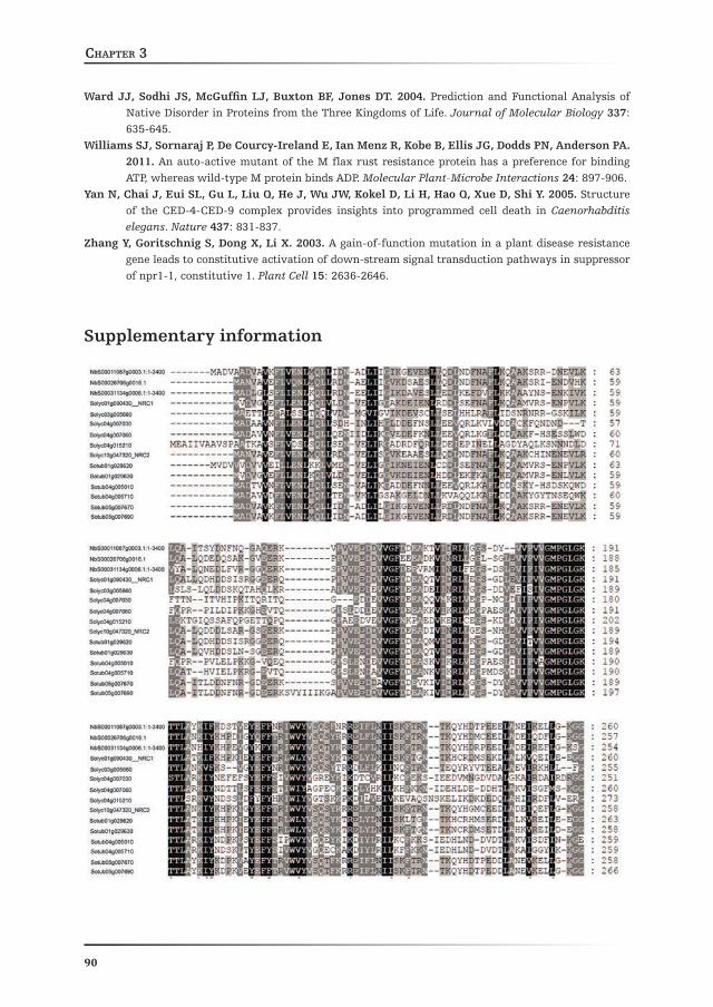
Chapter 3
90
Ward JJ, Sodhi JS, McGuffin LJ, Buxton BF, Jones DT. 2004. Prediction and Functional Analysis of Native Disorder in Proteins from the Three Kingdoms of Life. Journal of Molecular Biology 337: 635-645.
Williams SJ, Sornaraj P, De Courcy-Ireland E, Ian Menz R, Kobe B, Ellis JG, Dodds PN, Anderson PA. 2011. An auto-active mutant of the M flax rust resistance protein has a preference for binding ATP, whereas wild-type M protein binds ADP. Molecular Plant-Microbe Interactions 24: 897-906.
Yan N, Chai J, Eui SL, Gu L, Liu Q, He J, Wu JW, Kokel D, Li H, Hao Q, Xue D, Shi Y. 2005. Structure of the CED-4-CED-9 complex provides insights into programmed cell death in Caenorhabditis elegans. Nature 437: 831-837.
Zhang Y, Goritschnig S, Dong X, Li X. 2003. A gain-of-function mutation in a plant disease resistance gene leads to constitutive activation of down-stream signal transduction pathways in suppressor of npr1-1, constitutive 1. Plant Cell 15: 2636-2646.
Supplementary information

Dynamics of the NRC1 oligomer upon defence signallingChapter 3
91

Chapter 3
92
Supplementary Figure S1: Amino acid sequence alignment of the different NRC1 protein family members of tomatoThe NRC1 protein sequence was used to perform a BLASTp search against the translated tomato genome in the SOL Genomics database as described in Materials and Methods. Sequences were aligned with ClustalW.

Chapter 3
Chapter 4
Queda prohibido llorar sin aprender, levantarme un día sin saber qué hacer,
tener miedo a mis recuerdos, sentirme sólo alguna vez.
Queda prohibido no sonreír a los problemas, no luchar por lo que quiero,
abandonarlo todo por tener miedo, no convertir en realidad mis sueños.
Queda prohibido no demostrarte mi amor, hacer que pagues mis dudas y mi mal humor,
inventarme cosas que nunca ocurrieron, recordarte sólo cuando no te tengo.Queda prohibido dejar a mis amigos,
no intentar comprender lo que vivimos, llamarles sólo cuando les necesito,
no ver que también nosotros somos distintos.Queda prohibido no ser yo ante la gente,
fingir ante las personas que no me importan, hacerme el gracioso con tal de que me recuerden,
Random mutagenesis of the nucleotide-binding region of NRC1, a down-stream NB-LRR from tomato,
identifies gain-of-function mutations in the GLPL-motif
Daniela J. Sueldo1, Mahdere Shimels1*, Laurentiu Spiridon3, Matthieu Joosten1 , Wladimir Tameling1
Affiliations:1 Laboratory for Phytopathology, Wageningen University, Wageningen, The Netherlands2 Institute of Biochemistry of the Romanian Academy,
Department of Bioinformatics and Structural Biochemistry, Bucharest*Current address: Laboratory of Plant Physiology, Wageningen University,
Wageningen, The Netherlands
olvidar a toda la gente que me quiere.Queda prohibido no hacer las cosas por mí mismo,
no creer en mi Dios y hacer mi destino, tener miedo a la vida y a sus castigos,
no vivir cada día como si fuera un último suspiro.Queda prohibido echarte de menos sin alegrarme, olvidar los momentos que me hicieron quererte,
todo porque nuestros caminos han dejado de abrazarse, olvidar nuestro pasado y pagarlo con nuestro presente.
Queda prohibido no intentar comprender a las personas, pensar que sus vidas valen más que la mía,
no saber que cada uno tiene su camino y su dicha, pensar que con su falta el mundo se termina.
Queda prohibido no crear mi historia, dejar de dar las gracias a mi familia por mi vida,
no tener un momento para la gente que me necesita, no comprender que lo que la vida nos da, también nos lo quita.
Alfredo Cuervo Barrero

Random mutagenesis identifies gain-of-function mutations in the GLPL motif of NRC1Chapter 4
94
Abstract
Plant disease Resistance (R) proteins confer immunity to pathogens possessing the corresponding avirulence (Avr) proteins. The majority of R proteins belong to the Nucleotide-Binding, Leucine-Rich Repeat (NB-LRR) class. Activation of NB-LRRs is often associated with induction of the Hypersensitive Response (HR), a form of programmed cell death. NRC1 (NB-LRR required for HR-associated Cell death-1) is an NB-LRR from tomato that participates in the down-stream signalling cascade leading to resistance to the fungal pathogens Cladosporium fulvum and Verticillium dahliae. Silencing in Nicotiana benthamiana employing the tomato NRC1 coding sequence diminishes the HR triggered by various R/Avr combinations. This suggests that NRC1 is a down-stream signalling hub for different classes of immune receptors. To identify mutations in NRC1 that cause a higher signalling activity we generated a random mutant library restricted to the NB-ARC domain of this protein. The mutants were screened for their capacity to induce an elicitor-independent HR upon transient expression in N. tabacum. The screening of 1920 clones retrieved 12 mutants that showed auto-activity. In nine mutants a single amino acid substitution was responsible for this gain-of-function phenotype. Interestingly, five of these substitutions were located at the GLPL motif, suggesting that this motif is involved in modulating the signalling activity of NRC1. Moreover, we also identified a mutation in the highly conserved R residue in the RNBS-B motif. 3D modelling of the NB-ARC domain of NRC1 revealed that all nine targeted residues where centred around the bound nucleotide. Our mutational approach provides a wide set of novel gain-of-function mutations in the NB-ARC domain of NRC1 that were not identified in screens with other NB-LRRs. These data provides insight in how the activity of this NB-LRR may be regulated.

Random mutagenesis identifies gain-of-function mutations in the GLPL motif of NRC1Chapter 4
95
Introduction
STAND (Signal Transduction ATPase with Numerous Domains) proteins are a class of NTPases (Nucleoside TriPhosphatases) capable of binding and hydrolysing nucleotides (Leipe et al., 2004). Although they generally possess a variety of different domains depending on their function, STAND proteins are characterized by the presence of a P-loop NTPase domain, responsible for the hydrolysis of the γ-β phosphate bond of a Nucleotide TriPhosphate (NTP) (Saraste et al., 1990). Amongst the best studied STAND proteins are those involved in complex biological processes such as Programmed Cell Death (PCD) and disease resistance in both animals and plants: the Nucleotide-binding, Oligomerization Domain (NOD)-Like Receptor (NLR) and the Nucleotide-Binding, Leucine-Rich Repeat (NB-LRR) proteins of innate immunity in animals and plants, respectively (Damm et al., 2013; Hou et al., 2013). These immune receptors are constantly surveying for molecules that may betray the presence of pathogens or the cellular damage generated by them. From a functional point of view, animal NLRs are considered as Pattern Recognition Receptors (PRRs), as they detect the presence of Microbe-Associated Molecular Patterns (MAMPs), such as bacterial flagellin (Miao et al., 2006) or peptidoglycan (Fritz et al., 2006). Though plants also possess PRRs capable of recognizing bacterial flagellin (Gómez-Gómez and Boller, 2000) and elongation factor-Tu (Zipfel et al., 2006), as well as fungal chitin (Kaku et al., 2006; Miya et al., 2007), NB-LRRs are not considered PRRs since they recognize race-specific proteins (so-called effectors) from pathogens. Therefore, while the range of microbes perceived by NLRs is fairly broad, NB-LRR-dependent immunity in plants is highly specific. Moreover, NLR activation is often associated with an inflammatory response that does not necessarily culminate in cell death (Elinav et al., 2011). In contrast, a hallmark of plant NB-LRR activation is their ability to trigger the Hypersensitive Response (HR), a plant-specific type of PCD (Heath, 2000). However, also here examples exist in which NB-LRR-mediated resistance to pathogen occurs independently of cell death (Bendahmane et al., 1995; Zhang et al., 2013). Therefore, the question remains whether NB-LRR-mediated disease resistance requires the induction of the HR.
Animal NLRs and plant NB-LRRs share the presence of a central domain involved in nucleotide binding, known as NACHT (found in NAIP, CIITA, HET-E and TP1) in NLRs and NB-ARC (Nucleotide-Binding, found in Apaf-1, plant Resistance proteins and CED4) in NB-LRR proteins (van der Biezen and Jones, 1998; Koonin and Aravind, 2000; Ye and Ting, 2008; Takken and Goverse, 2012). This domain is also present in human Apaf-1 (Apoptotic Protease-Activating Factor-1) and its Caenorhabditis elegans homolog CED4 (CEll Death protein 4), both of which are involved in apoptosis (Chinnaiyan et al., 1997; Chinnaiyan et al., 1997; Seshagiri and Miller, 1997; Zou et al., 1997; Cecconi et al., 1998). Interestingly, in animal NLRs the NACHT domain

Random mutagenesis identifies gain-of-function mutations in the GLPL motif of NRC1Chapter 4
96
has been implicated in ligand-mediated oligomerization, a phenomenon required for activation of immune signalling (Damm et al., 2013). Moreover, the C-terminal part of NLRs and NB-LRRs is composed of a Leucine-Rich Repeat (LRR) region, of variable length. The N-terminal domain, however, is not conserved between NLRs and NB-LRRs (Ting et al., 2008). In NB-LRRs the N-terminus generally comprises either Coiled-Coil (CC) structures or a domain with homology to the Toll/Interleukin-1 Receptor (TIR), although some carry additional and/or alternative domains (Grant et al., 1995; Salmeron et al., 1996; Milligan et al., 1998).
Due to their modular nature, several studies on plant NB-LRRs have focused on one or more domains independently, based on which a certain signalling task division amongst the different domains has been proposed. The N-terminal domain, either CC or TIR, is generally associated with the recruitment of signalling partners. Examples of these are the interaction between the CC domain of the CC-NB-LRR (CNL) MLA10 from barley (Hordeum vulgare) and a WRKY transcription factor (SHen et al., 2007), the potato (Solanum tuberosum) CNL Rx and a Ran-GTPase-activating protein (RanGap2) (Sacco et al., 2007; Tameling and Baulcombe, 2007). In addition, in the presence of a pathogen the N-terminal domain of NB-LRRs can sense changes in the state or subcellular localization of other plant proteins, thereby activating defence responses. An example of this is the TIR-NB-LRR (TNL) N from tobacco (Nicotiana tabacum), which directly binds to a chloroplastic protein (N-Receptor Interacting Protein, NRIP) through its TIR domain (Caplan et al., 2008). Moreover, for some CNLs and TNLs the N-terminal domain, either CC or TIR, is sufficient to induce cell death upon their individual expression in the absence of the other domains and of a pathogen (Maekawa et al., 2011; Bernoux et al., 2011; Collier et al., 2011). This has led to the hypothesis that, at least for a number of NB-LRRs, the N-terminal domain is involved in initiating defence signalling.
In NB-LRRs the C-terminal LRR domain is the most variable region and plays a fundamental role in governing recognition specificity, as has been demonstrated for the L locus in flax (Linum usitatissimum) (Dodds et al., 2001; Dodds et al., 2006), for tomato Mi-1 (Hwang and Williamson, 2003) and for the polymorphic barley mildew A (MLA) R locus (SHen et al., 2007). In addition to its role in recognition, the LRR domain is thought to exert an auto-inhibitory effect on the activity of the NB-LRR protein through intra-molecular interactions with the region carrying the NB-ARC and CC domains. Auto-inhibition is required to avoid inappropriate activation of defence signalling (e.g. cell death) in the absence of a pathogen. Evidence supporting this notion comes from experiments wherein swapping the LRR domains between closely related NB-LRRs resulted in auto-activity. This is likely caused by the intra-molecular incompatibility in such chimeras resulting in an abrogated auto-inhibition (Hwang and Williamson, 2003; Howles et al., 2005; Rairdan and Moffett, 2006; Slootweg et al., 2013). Moreover, deletion of the entire LRR domain or parts

Random mutagenesis identifies gain-of-function mutations in the GLPL motif of NRC1Chapter 4
97
thereof, results in a mild constitutive activation of cell death and defence responses, at least for some NB-LRRs (Ade et al., 2007); (Rairdan et al., 2008), which confirms the role of the LRR domain in auto-inhibition.
As mentioned above, the central region of all NB-LRRs is represented by the NB-ARC domain, which has the ability to bind and hydrolyse ATP (Tameling et al., 2002). By this activity the NB-ARC domain is considered as a molecular switch that is thought to direct nucleotide-dependent conformational changes in the protein which, in turn, allow interaction with down-stream signalling partners to induce cell death and other defence responses (Tameling et al., 2006; Tameling and Takken, 2008; Takken and Goverse, 2012). The NB-ARC domain is subdivided into three subunits (NB, ARC1 and ARC2), which contain several highly conserved motifs many of which are directly required for nucleotide-binding and/or -hydrolysis. The best characterized motifs, namely the P-loop (or Walker A), Walker B and RNBS-B, are located in the NB subdomain. The P-loop motif is involved in interactions with the phosphates and Mg2+ ion of the ATP-Mg2+ complex (Saraste et al., 1990; Via et al., 2000; Riedl et al., 2005; Yan et al., 2005) and mutation of the invariant lysine residue within this motif abolishes nucleotide binding (Tameling et al., 2002; Williams et al., 2011), rendering the NB-LRR in a “signalling-dead” state (Bendahmane et al., 2002; Tameling et al., 2002; Gabriëls et al., 2007; Tameling and Baulcombe, 2007; Williams et al., 2011). The Walker B motif is involved in ATP hydrolysis through two conserved aspartic acid residues, of which the first orients the Mg2+ ion and the second serves as a catalytic base. Mutation of the second aspartic acid residue compromises the hydrolytic activity in the NB-LRR I-2 from tomato and results in a mild auto-active phenotype in this protein (Tameling et al., 2006) as well as in other NB-LRRs (Bendahmane et al., 2002; van Ooijen et al., 2008; Ade et al., 2007). Finally, the RNBS-B motif is involved in binding and possibly sensing the γ-phosphate of the nucleotide, similar to the sensor I motif of proteins belonging to the AAA+ superclass (Takken et al., 2006). Two other motifs are located in the NB subdomain, hhGRExE and RNBS-A, although with a lower degree of conservation. The ARC1 and ARC2 domains each contain two conserved motifs, namely RNBS-C and GLPL in ARC1 and RNBS-D and MHD in ARC2 (Takken et al., 2006). Although the exact function of these motifs remains elusive, mutations have been identified in these motifs that either cause elicitor-independent cell death, representing gain-of-function (de la Fuente van Bentem et al., 2005; Tameling et al., 2006; Gabriëls et al., 2007; van Ooijen et al., 2008a; Maekawa et al., 2011; Williams et al., 2011), or fully repress the capacity of the protein to induce an HR, representing loss-of-function (Dinesh-Kumar et al., 2000; Tao et al., 2000; Tameling et al., 2002). These findings indicate that these motifs regulate the signalling activity of NB-LRRs, either through direct involvement in binding and/or hydrolysis of the nucleotide, in auto-inhibition or in relaying conformational changes in the NB-ARC domain to other parts of the

Random mutagenesis identifies gain-of-function mutations in the GLPL motif of NRC1Chapter 4
98
protein. Although many studies have resulted in the successful identification of new mutations that modulate the activity of NB-LRRs, our knowledge on the mechanisms by which the NB-ARC domain regulates the activity of NB-LRRs is still limited.
We have previously reported the identification of tomato NRC1 (NB-LRR Required for hypersensitive response-associated Cell death-1), an NB-LRR required for resistance to the fungal pathogens Cladosporium fulvum and Verticillium dahliae mediated by the R proteins Cf-4 and Ve1, respectively (Gabriëls et al., 2007; Fradin et al., 2009). Moreover, NRC1 is also involved in the down-stream signalling cascades leading to HR in N. benthamiana activated by several intracellular (Rx, I-2, Mi-1 and Prf/Pto) and extracellular (Cf-4 and Cf-9) R proteins (Gabriëls et al., 2007). Therefore, by assisting such a wide range of R proteins, NRC1 likely acts as a central signalling switch in the tomato immune response. In order to gain more insight into the functioning of the NB-ARC domain of NB-LRRs, we performed a mutagenesis screen on the NB-ARC domain of tomato NRC1. Random mutations were incorporated by an error-prone PCR reaction and tested for their effect on the signalling activity in the context of full length NRC1 in plants. We identified novel gain-of-function mutations that allowed NRC1 to induce an elicitor-independent HR and that were not reported for other NB-LRRs. Moreover, we speculate on the possibility of implementing such mutations in breeding programs to obtain broad-spectrum resistance to pathogens.
Results
Random mutagenesis of the NB-ARC domain of NRC1
To identify mutations that affect the HR-signalling activity of NRC1, we screened for NRC1 mutant proteins capable of inducing cell death in an elicitor-independent manner. The mutants were generated with an error-prone PCR reaction that randomly incorporates mismatches in the nucleotide sequence of NRC1. Mutants were only generated in the region encoding the NB-ARC domain of NRC1, considering that most of the gain-of-function mutations identified in NB-LRRs localize in this domain (Bendahmane et al., 2002; Howles et al., 2005; Tameling et al., 2006; Ade et al., 2007; van Ooijen et al., 2008; Gao et al., 2011). The mutant library was sub-cloned into a binary vector for transient expression in planta. This resulted in the expression of chimeric NRC1 mutant proteins, in which the CC and LRR domains were Wild-Type (WT) sequences, whereas the central NB-ARC domain may carry random mutations (Fig. 1A). For in planta expression, the library was transferred to Agrobacterium tumefaciens and individual clones were assessed for their capacity to trigger elicitor-independent HR after agro-infiltration in tobacco (Nicotiana tabacum).

Random mutagenesis identifies gain-of-function mutations in the GLPL motif of NRC1Chapter 4
99
We have previously reported that NRC1WT induces an HR when agro-infiltrated in N. benthamiana in combination with the silencing suppressor of Tomato Bushy Stunt Virus (TBSV) P19, but not when expressed alone (Gabriëls et al., 2007)
(Chapter 3, this thesis). Likewise, transient expression of NRC1WT without P19 does not trigger cell death in tobacco (Fig. 1B). In contrast, NRC1D481V, which carries a point mutation in a highly conserved motif in the ARC2 subdomain, is capable of inducing a strong HR without the need of P19 both in N. benthamiana (Gabriëls et al., 2007) (Chapter 3, this thesis) and in tobacco (Fig. 1B). Upon screening of the mutant NRC1 library, we considered the signalling activity of NRC1WT, manifested as chlorosis of the infiltrated area, as the basal level and searched for NRC1 mutant proteins that induced a stronger response than the wild-type protein.
Figure 1: Transient expression of NRC1D481V, but not of wild-type NRC1,induces elicitor-independent cell death in Nicotiana tabacum
(A) Cartoon depicting the structure of the mutant NRC1 proteins expressed in tobacco by agro-infiltration used for screening for elicitor-independent HR. Only the sequence encoding the NB-ARC domain was targeted for mutation by error-prone PCR and the mutant library was sub-cloned in a binary plant expression vector.
(B) Unlike NRC1WT, NRC1D481V, carrying a single amino acid substitution in the MHD motif, activates elicitor-independent HR in tobacco. Agrobacterium cultures carrying binary vectors with NRC1WT or NRC1D481V were infiltrated into tobacco leaves at OD600 of 1, 0.5 and 0.1 (from left to right) and the development of cell death was monitored in time. Red dashed lines indicate an HR, whereas the white ones indicate a lack of the HR. The picture was taken at 4 dpi.

Random mutagenesis identifies gain-of-function mutations in the GLPL motif of NRC1Chapter 4
100
We also included NRC1D481V as a positive control, as this was the only gain-of-function mutant of NRC1 reported so far (Gabriëls et al., 2007). A total of 1920 individual clones were tested for their ability to trigger an elicitor-independent cell death in tobacco. Every clone that induced cell death faster and/or stronger than NRC1WT was considered to be an interesting candidate and was selected for further analysis. The development of an elicitor-independent HR induced by NRC1D481V was already visible at 24-26 hours post-infiltration (hpi). The HR induced by the expression of the various mutant clones appeared between 24 hpi and 7 days post infiltration (dpi) and was therefore of variable intensity (Fig. 2A).
Figure 2: Random mutagenesis screening of the NB-ARC domain of NRC1 yields several gain-of-function mutants
(A) Representative picture of tobacco leaves from the NRC1 mutant screening showing different clones that show an elicitor-independent HR. In each tobacco leaf, wild-type (WT) NRC1 (white dashed line) and NRC1D481V (red dashed line), in addition to six NRC1 mutants were transiently expressed by agro-infiltration. Mutant clones that induced cell death were considered for further analysis. The picture was taken at 7 dpi.
(B) Summary of the results obtained after screening the NRC1-NB-ARC mutant library in tobacco. 1920 Agrobacterium clones were tested for their ability to induce an elicitor-independent HR in tobacco upon transient agro-infiltration-mediated expression. Clones that consistently displayed an increased signalling activity (12) were sequenced to identify the mutation(s). The indicated characteristics of these clones were derived from the sequencing results.
Random mutagenesis identifies gain-of-function mutations in the GLPL motif of NRC1Chapter 4
101
Out the 1920 assessed clones, 12 consistently gave an elicitor-independent HR upon extensive re-testing and were selected for sequencing (Supplementary Table S1). Sequencing results revealed that the 12 mutant clones contained in total 40 mutations, of which 12 were silent and 28 were missense mutations that resulted in an amino acid substitution. We identified two clones (A and K) that carry only one (missense) mutation. The remaining 38 mutations were spread over the other ten clones, which resulted in an average of 3.3 mutations per clone, including both silent and missense mutations (Fig. 2B). We also examined the type of mutations at the DNA level and observed that the number of so-called transversions (A T, C G, A C and T G) was two times higher than that of transitions (A G andT C), consistent with the fact that transversions are twice as common as transitions. This indicates that there was no bias in the chemical nature of the introduced mutations in the library.
Identification of the gain-of-function mutations by site-directed mutagenesis of NRC1
Of the 28 identified missense mutations, two occurred individually in a clone, leading to the amino acid substitutions V356A and G350R in clones A and K, respectively. The mutants F and L also contained one (identical) missense mutation, leading to the R296Q substitution, but was combined with one and three unique silent mutations, respectively. Therefore, in each of these four clones (A, K, F and L) the single missense mutation is responsible for inducing the elicitor-independent HR and are classified as gain-of-function mutations. Since the remaining 24 missense mutations did not occur individually in the remaining eight clones, we introduced these as single missense mutations into the coding region of NRC1 by site-directed mutagenesis. These single mutants were transiently expressed in tobacco and the development of an elicitor-independent cell death was monitored. This analysis revealed that the single mutations L483W, I355N, V357M, F486I, I358N and V158E, were the responsible gain-of-function mutations in the progenitor clones C, E, G, H, I and J, respectively, as expression of these single mutants induced a swift elicitor-independent HR (Fig. 3; Supplementary Table S1). Interestingly, none of the missense mutations derived from mutant B (D155E, Y262F and G341R) induce an elicitor-independent HR as single mutants in tobacco (Supplementary Fig. S2). Therefore, the auto-activity of clone B is most likely caused by the cooperative effect of two or all three mutations.

Random mutagenesis identifies gain-of-function mutations in the GLPL motif of NRC1Chapter 4
102
Figure 3: Identification of the individual gain-of-function mutations in the NB-ARC domain of NRC1
For nine gain-of-function mutants identified in the screen (indicated by letters), a single mutation is responsible for the gain-of-function phenotype. The listed mutations were independently introduced into the coding region of NRC1WT by site-directed mutagenesis. Mutant proteins were transiently expressed in tobacco by agro-infiltration and the timing of the appearance of cell death in hours post infiltration (hpi) was recorded. At 3dpi leaves were photographed. Only images depicting the HR induced by the gain-of-function mutants are Shown. Mutant clones B and D, for which a single gain-of-function mutation was not identified, are not included in the picture. See Supplementary Figure S2 for pictures of the mutations that do not affect the activity of NRC1. Transient expression of NRC1WT and NRC1D481V was included as a control.
Remarkably, we identified mutation E258G in mutant D, of which we have previously shown that it is a loss-of-function mutation in NRC1 (Chapter 3, this thesis). Of the two other occurring missense mutations in mutant D, L395S did not result in auto-activity (Supplementary Figure S2), suggesting that the remaining mutation, V465E, likely causes the gain-of-function phenotype of clone D. Unfortunately, we were unable to obtain this single mutant by site-directed mutagenesis. Therefore, it cannot be concluded whether this mutation is individually or cooperatively with L395S responsible for the auto-activity of mutant D. In conclusion, in ten of the 12 clones we could determine that one single amino acid substitution is responsible for the induction of the elicitor-independent HR.
We observed that the NRC1 single mutants induced elicitor-independent HR at different timings. Indeed, the mutations R296Q, I355N and L483W induced an HR that appeared faster (21-24 hpi) than with the D481V mutant (24-26 hpi; Fig. 3). The V158E, V357M, I358N and F486I mutants showed a timing of the HR that was similar to the D481V mutant (Fig. 4). Only expression of NRC1 mutant V356A caused an HR that appeared slower (48 hpi) than the HR triggered by the D481V mutant (Fig. 3). To evaluate the intensity of the HR caused by the different mutants in more detail, dilution series with the Agrobacterium cultures containing the mutant constructs were infiltrated in tobacco (OD600 of 1, 0.5 or 0.1; Fig. 4). With
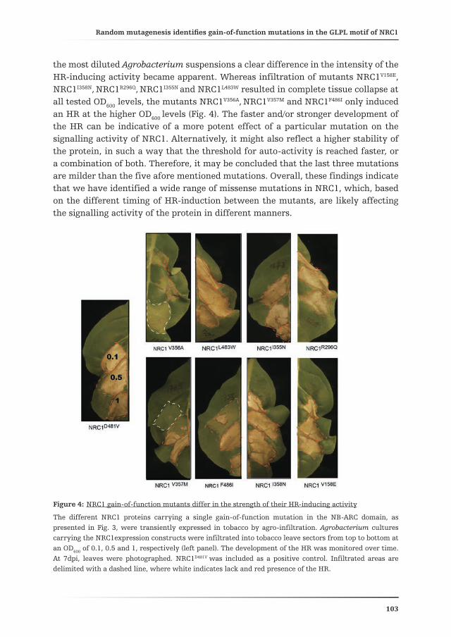
Random mutagenesis identifies gain-of-function mutations in the GLPL motif of NRC1Chapter 4
103
the most diluted Agrobacterium suspensions a clear difference in the intensity of the HR-inducing activity became apparent. Whereas infiltration of mutants NRC1V158E, NRC1I358N, NRC1R296Q, NRC1I355N and NRC1L483W resulted in complete tissue collapse at all tested OD600 levels, the mutants NRC1V356A, NRC1V357M and NRC1F486I only induced an HR at the higher OD600 levels (Fig. 4). The faster and/or stronger development of the HR can be indicative of a more potent effect of a particular mutation on the signalling activity of NRC1. Alternatively, it might also reflect a higher stability of the protein, in such a way that the threshold for auto-activity is reached faster, or a combination of both. Therefore, it may be concluded that the last three mutations are milder than the five afore mentioned mutations. Overall, these findings indicate that we have identified a wide range of missense mutations in NRC1, which, based on the different timing of HR-induction between the mutants, are likely affecting the signalling activity of the protein in different manners.
Figure 4: NRC1 gain-of-function mutants differ in the strength of their HR-inducing activity
The different NRC1 proteins carrying a single gain-of-function mutation in the NB-ARC domain, as presented in Fig. 3, were transiently expressed in tobacco by agro-infiltration. Agrobacterium cultures carrying the NRC1expression constructs were infiltrated into tobacco leave sectors from top to bottom at an OD600 of 0.1, 0.5 and 1, respectively (left panel). The development of the HR was monitored over time. At 7dpi, leaves were photographed. NRC1D481V was included as a positive control. Infiltrated areas are delimited with a dashed line, where white indicates lack and red presence of the HR.

Random mutagenesis identifies gain-of-function mutations in the GLPL motif of NRC1Chapter 4
104
Gain-of-function mutations in NRC1 are centred around the bound ADP in the structural 3D model of the NB-ARC domain
In order to investigate the degree of conservation of the amino acids that are mutated in NRC1 within the NB-LRR protein class, we aligned the protein sequence of the NB-ARC domain of NRC1 and other NB-LRR proteins (Fig. 5A and Supplementary Fig. S3). To this end, we selected a broad range of NB-LRRs, considering the following criteria: (1) they should originate from different plant species (tomato, potato, tobacco and Arabidopsis) and (2) they should be either immune receptor (Mi-1, I-2, Rx and RPM1), or down-stream signalling NB-LRR (ADR1-L2 and NRG1) (Debener et al., 1991; Bendahmane et al., 1995; Ori et al., 1997; Milligan et al., 1998; Simons et al., 1998; Peart et al., 2005; Bonardi et al., 2011). Moreover, for most of these NB-LRRs, various gain-of-function mutations have been described (Bendahmane et al., 2002; Gabriëls et al., 2007; van Ooijen et al., 2008; Bonardi et al., 2011; Gao et al., 2011). The protein sequence alignment, which also includes Apaf-1, revealed that some of the gain-of-function mutations identified in NRC1 correspond to highly conserved amino acids amongst the selected NB-LRR proteins. A clear example of this are mutations R296Q and G350R, which hit highly conserved residues in the RNBS-B and GLPL motifs, respectively, both in NB-LRRs as well as in Apaf-1. Other residues, like I355, I358 and L483, show a lower degree of conservation which, in some cases, was restricted to the selected NB-LRRs from the Solanaceous plants.
We then examined the orientation of the identified gain-of-function mutations in a structural 3D model of the NB-ARC domain of NRC1. This model, that represents the ADP-bound state of the NB-ARC domain of NRC1, was obtained based on the crystal structure of the mammalian apoptotic factor Apaf-1 (Riedl et al., 2005), as described in Materials and Methods (see also Chapter 3). Strikingly, all nine gain-of-function mutations targeted amino acid residues that seem to be part of or very close to the nucleotide-binding pocket and are all centred around the bound ADP (Fig. 5B and Supplementary Fig. S4). This suggests that the auto-activity might be related to alterations in nucleotide-binding affinity and/or hydrolysis. Alternatively, the mutations might affect the sensor function of the ARC domains, which generally communicate the nucleotide-binding state to other parts of the protein, thereby regulating its activity.
Our 3D model predicts that 16 residues of the NB-ARC domain of NRC1 are likely involved in binding to ADP (Supplementary Fig. S3), based on the crystal structure of Apaf-1. Of these, two (V158 and V356) were hit by the random mutagenesis screen and resulted in an elicitor-independent HR when mutated to an E and A, respectively. Indeed, both residues locate very close to the ADP molecule (Fig. 5B and Supplementary Fig. S4). Although G350 is extremely conserved between the selected NB-LRRs and Apaf-1 (Fig. 5A), it is likely not directly involved in nucleotide binding in NRC1, because the analogous residue in Apaf-1 is not either.

Random mutagenesis identifies gain-of-function mutations in the GLPL motif of NRC1Chapter 4
105
A
B
Figure 5: Gain-of-function mutations in the NB-ARC domain of NRC1 correspond to residues surrounding the ADP molecule, mostly in or very close to the GLPL motif
(A) Amino acid sequence alignment of the NB-ARC domain of NRC1, other NB-LRRs and Apaf-1, depicting the location and conservation of the residues that have been mutated in NRC1. The amino acid sequences of the NB-ARC domains of the different proteins were aligned with ClustalW. The hypothetical NRC1_MUT sequence carries all missense mutations identified in the random mutagenesis screen described here. Only parts of the alignment where the gain-of-function mutations were identified are shown. For complete alignment of the NB-ARC domain see Supplementary Figure S3. Blue, gain-of-function mutation; red, previously described gain-of-function mutations in other NB-LRRs (Bendahmane et al., 2002; Tameling et al., 2006; Gabriëls et al., 2007; Gao et al., 2011; Roberts et al., 2013). Conserved sequence motifs are indicated below the alignment with a light-blue bar. Numbers correspond to the amino acid position, considering the first amino acid of the full length protein as position one.
(B) 3D-model of the NB-ARC domain of NRC1 indicating the location of the targeted residues in the gain-of-function mutants. The amino acid residues 160 to 443 from the NRC1 full-length protein were modelled for its 3D structure, based on the crystal structure of Apaf-1 as described in Materials and Methods. Light-blue, conserved motifs of the NB-ARC; red, amino acids predicted to be directly involved in nucleotide binding; orange, positions corresponding to the nine identified gain-of-function mutations; blue, ADP molecule. Side chains of important residues are depicted as sticks. The dot projection (in orange) displays the solvent-accessible surface. The small panel shows a close-up of the mutations, indicated in orange.

Random mutagenesis identifies gain-of-function mutations in the GLPL motif of NRC1Chapter 4
106
Though, close examination of the 3D model revealed that this residue is directly in the vicinity of P352 (Fig. 5B and Supplementary Fig. S4), which is probably required for nucleotide-binding, similarly to the analogous P321 residue in Apaf-1 (Riedl et al., 2005). It is therefore tempting to speculate that the substitution with a bulky and charged R residue at the G350 position might affect the nucleotide binding and/or hydrolysis activity of NRC1. Residues I355, V357 and I358 are not predicted to participate in nucleotide-binding, based on the crystal structure of Apaf-1. However, their proximity to residues P352 and L353 suggests that mutations I355N, V357M and I358N are likely to interfere with the involvement of the GLPL motif in nucleotide-binding and/or hydrolysis (Fig. 5B and Supplementary Fig. S4). Moreover, residues L483 and F486 also do not appear to make direct contact with the nucleotide and are located further away from it than the other mutations. Therefore, speculation on how they affect the HR-signalling activity of NRC1 becomes more challenging. However, their location within the MHD motif suggests that mutations L483W and F486I might compromise the predicted nucleotide-binding by H480, which is analogous to H438 in Apaf-1 that contacts ADP (Riedl et al., 2005). Finally, based on the proximity of residue R296 to the ADP molecule, it is tempting to speculate that this residue is involved in nucleotide-binding and/or hydrolysis (Fig. 5B and Supplementary Fig. S4). In agreement with this hypothesis, is the observation that the analogous R273 residue of CED-4 contacts the γ-phosphate of the bound ATP in the crystal structure (Yan et al., 2005). Overall, our random mutagenesis screen revealed nine novel single gain-of-function mutations in the NB-ARC domain of NRC1. The finding that five of those mutations are in or close to the GLPL motif, surrounding the nucleotide molecule, suggests a participation of this motif in modulating the HR-signalling activity of NRC1.
Discussion
In this study we report the identification of gain-of-function mutants of NRC1 in a random mutagenesis screening. NRC1 is a defence signalling NB-LRR from tomato acting down-stream of R proteins. By means of an error-prone PCR, we generated random mutations in the NB-ARC domain of NRC1 and screened the mutants for their ability to induce elicitor-independent cell death upon transient expression in tobacco. Such auto-activity is indicative of an increased defence signalling activity of the generated NRC1 mutant.
Mutagenesis screens of NB-LRRs has been conducted before, although mostly aimed at the identification of loss-of-function mutations (Dinesh-Kumar et al., 2000; Tao et al., 2000; Axtell et al., 2001; Tornero et al., 2002). Using a similar approach as the one employed in this chapter, Bendahmane et al. (2002) reported the identification

Random mutagenesis identifies gain-of-function mutations in the GLPL motif of NRC1Chapter 4
107
of gain-of-function mutations in the potato NB-LRR Rx, which confers resistance to Potato Virus X (PVX). By error-prone PCR, the authors randomly mutagenized the entire coding region of Rx, and searched for mutations that would induce elicitor-independent HR upon expression in tobacco. The screening of 2,500 colonies resulted in the identification of eight gain-of-function mutants, resulting in a rate of one gain-of-function mutant every 312 clones (Bendahmane et al., 2002). This represents a lower success rate as compared to our NRC1 mutagenesis screen, in which one of every 160 screened clones gave an auto-active phenotype. However, it should be noted that the Rx mutants were transiently expressed in tobacco under control of their own regulatory sequences, which likely results in a lower expression level than would be provided by the constitutive Cauliflower Mosaic Virus (CaMV) 35S promoter that we employed. This could account for the fact that mild gain-of-function mutants were not identified in the screen performed by Bendahmane et al (2002). Moreover, the authors focused on the entire coding region of Rx, instead of just the NB-ARC domain (Bendahmane et al., 2002).
Interestingly, similar to Rx, we found that a single amino acid change was responsible for the constitutive gain-of-function phenotype of ten of the 12 mutants (Fig. 3). No overlap was found between the causal mutations in the gain-of-function mutants identified in both screens. Moreover, to our knowledge, none of the gain-of-function mutations in NRC1 have been reported for other NB-LRRs. This is remarkable, considering that some of them targeted amino acids that are extremely conserved amongst NB-LRRs and other STAND proteins, as described below.
Location of the gain-of-function mutations and their possible involvement in nucleotide-binding
Our 3D structural model of the NB-ARC domain of NRC1, which is based on the crystal structure of Apaf-1 (Riedl et al., 2005), predicts 16 residues that are involved in the interaction with the ADP molecule (Supplementary Figure S3). Interestingly, our mutational analysis hit one of these residues, V158, which corresponds to V127 in Apaf-1. This V residue is located in the hhGRExE motif, which determines the base specificity for adenine over guanine in Apaf-1 (Riedl et al., 2005). In NRC1, mutation V158E induced elicitor-independent HR at a similar speed/intensity as mutation D481V (Fig. 3, Fig. 4 and Fig. 5). Notably, we also reported that mutation E154K, located three amino acids towards the N-terminal of the hhGRExE motif, affects the HR-signalling activity of NRC1 both in cis and in trans, likely by affecting the oligomerization state of the protein (chapter 3, this Thesis).
The residue R296 is located in the RNBS-B motif of NB-LRRs, which has been suggested to have a similar function to the sensor I motif in proteins belonging to the AAA+ superclass (Leipe et al., 2004; Takken et al., 2006). The highly conserved R in the sensor I motif of AAA+ proteins is involved in sensing the presence or absence

Random mutagenesis identifies gain-of-function mutations in the GLPL motif of NRC1Chapter 4
108
of the γ-phosphate of the nucleotide and transmitting this information to the rest of the protein by mediating conformational changes (Takken et al., 2006). Accordingly, analysis of the crystal structure of CED-4 revealed that R273, which corresponds to R296 from NRC1, indeed contacts the γ-phosphate (Yan et al., 2005). Because of the strong conservation of this residue it is highly likely that the analogous R in NRC1 and other STAND proteins also plays a role in nucleotide binding and possibly as a γ-phosphate sensor. This notion is further reinforced by the observation that R296 is located close to the phosphates of the ADP molecule, according to our structural model (Fig. 5B and Supplementary Fig. S4). Requirement of an intact RNBS-B for NB-LRR functioning has been demonstrated by the identification of loss-of-function mutations in RPM1, N and Prf (Salmeron et al., 1996; Dinesh-Kumar et al., 2000; Tornero et al., 2002). However, no gain-of-function mutations in this motif have been reported. The R296Q mutation in NRC1 causes a strong auto-activity (Figure 3 and 4), which argues in favour of a predicted γ-phosphate sensor function of this residue. The substitution by the Q may mimic the presence of the γ-phosphate, thus ATP, and cause NRC1 to adopt the activated state.
We also identified gain-of-function mutations in (L483W) or close to (F486I) the MHD motif of NRC1 (Figs. 3, 4, 5 and 6) (van der Biezen and Jones, 1998), whose capacity to induce elicitor-independent HR has already been shown in NRC1 (Gabriëls et al., 2007) as well as in other NB-LRRs (de la Fuente van Bentem et al., 2005; Tameling et al., 2006; van Ooijen et al., 2008a; Maekawa et al., 2011; Williams et al., 2011). An extensive mutational analysis performed in tomato I-2 to address the role of the MHD motif in NB-LRR-triggered HR showed that mutation of not only the D but also the H residue results in an increased HR, with variations in intensity depending on the actual amino acid substitution (van Ooijen et al., 2008). In Apaf-1, the H of this motif (H438) binds to the β-phosphate and might replace the sensor II function in AAA+ proteins. The weak interaction of the ARC2 subdomain (winged-helix domain) with ADP indicates that it is prone to conformational changes (Riedl et al., 2005). This notion is further supported by recent crystallographic data on NLRC4, a mammalian NLR protein, where the corresponding H443 from the winged-helix domain also binds the β-phosphate. Interestingly mutation of this residue also cause auto-activity of the protein, likely by facilitating conformational changes and attenuation of ADP binding (Hu et al., 2013).
The GLPL motif as a modulator of the activity of STAND NTPases
The highly conserved GLPL motif is located in the ARC1 subdomain of NRC1 and five of the nine identified gain-of-function mutations were in (G350R and I355N) or very close to (V356A, V357M and I358N) this motif (van der Biezen and Jones, 1998; Pan et al., 2000), strongly indicating that it plays an important role in modulating the defence signalling activity of this protein (Fig. 5). The GLPL motif, also known

Random mutagenesis identifies gain-of-function mutations in the GLPL motif of NRC1Chapter 4
109
as the GxP motif, is highly conserved in STAND NTPases and has been suggested to function as a hinge, facilitating NTP-dependent movement of the flanking helices such that the ARC1 subdomain transmits these conformational changes to the rest of the protein (Leipe et al., 2004). This hypothesis is supported mainly by the characterization of loss-of-function mutants. For example, the HR induced by transient expression of WT Rx is abolished by the combined mutations G330A and P332A, targeting the G and P residues of the GLPL motif, respectively (Bendahmane et al., 2002). Similarly, a G to E substitution in the GLPL motif of L6, an NB-LRR from flax, compromises resistance to flax rust (Melampsora lini) (Dodds et al., 2001). Analysis of the crystal structures of the STAND ATPases Apaf-1 and CED-4 point out that the residues P321, L322 and S325 (P330, A331 and M334 in CED-4) of the GLPL motif, are involved in nucleotide binding, by stabilizing the adenine and ribose moieties through van der Waals contact (Riedl et al., 2005; Yan et al., 2005). Our structural 3D modelling also supports the involvement of the analogous residues (P352, L353 and V356) in NRC1 (Figure 5).
Despite of the data available on compromised NB-LRR functioning due to mutations in the GLPL motif, little is known about its potential to induce increased defence signalling activity. In our NRC1 screen, two mutant clones carrying either the mutations G350R or V356A in the GLPL motif induced an elicitor-independent HR (Fig. 3, Fig. 4 and Supplementary Table S1). It is interesting to note that mutation of the same amino acid residue in different NB-LRR proteins leads to completely opposite phenotypes. Substitution of the G residue by either an aliphatic one (A in Rx; Bendahmane et al., 2002) or acidic amino acid (E in L6; Dodds et al., 2001), resulted in compromised NB-LRR activity, whereas a mutation to a basic amino acid (R) caused constitutive auto-activity in NRC1. A plausible explanation for this is that while changes to a more bulky aliphatic or negatively charged amino acid might abrogate nucleotide binding, the substitution of the G residue by a positively charged amino acid likely results in a conformation that mimics the active ATP-bound state of NRC1. This idea is in line with the current model describing NB-LRR functioning, and which proposes that these proteins cycle between an “off” and “on” state, which is tightly regulated by the bound-nucleotide and associated conformational changes (Takken et al., 2006; Takken and Goverse, 2012). It would be of interest to determine whether mutation of the G residue to an A or an E will result in reduced signalling activity of NRC1, as observed for Rx and L6, respectively. Likewise, whether the gain-of-function phenotype caused by the G350R mutation can be transferred to Rx and L6 is also an interesting question to explore. In a recent publication, Harris et al. (2013) reported the identification of five mutations (N147D, T178A, M293L, V337A and G340R) in the NB-ARC domain of Rx that resulted in increased activation sensitivity of the RxM1 (RxN846D) mutant. RxN846D has previously been shown to have broader recognition specificity: unlike WT Rx, the RxM1 mutant recognises the Coat Protein (CP) of Poplar Mosaic Virus (PopMV). However, it only confers partial

Random mutagenesis identifies gain-of-function mutations in the GLPL motif of NRC1Chapter 4
110
resistance to this virus in transgenic N. benthamiana, resulting in a trailing necrosis (Farnham and Baulcombe, 2006). The secondary sensitising mutations introduced in the RxM1 mutant (N147D, T178A, M293L, V337A and G340R) turn the defence response induced by PoPMV recognition more effective, resulting in a complete resistance to this virus in transgenic N. benthamiana. Interestingly, transient over-expression of each mutant in tobacco resulted in the induction of a weak HR, indicating that they are actually mild auto-activating mutations (Harris et al., 2013). All five mutations map to the nucleotide-binding pocket of Rx and are also in or close to the conserved motifs, similarly to the mutations we identified in NRC1. Strikingly, two mutations also map very close to the GLPL motif and one of them (V337A) targets the same residue as in NRC1V357N. However, in the context of Rx the V337A mutation acts cooperatively with the N147D mutation in the hhGRExE motif (Harris et al., 2013). Overall, the finding that mutations in the GLPL motif can induce auto-activity in NRC1 (as well as in Rx) supports the idea that this motif transmits conformational changes associated with different nucleotide-binding states as previously suggested (Leipe et al., 2004). Moreover, they also indicate that, similarly to other highly conserved motifs in the NB-ARC domain of STAND proteins, the GLPL motif can be manipulated to induce elicitor-independent HR.
Potential implementation of the newly identified NRC1 gain-of-function mutations
Stable (over)-expression of gain-of-function mutants of NB-LRRs induces constitutive defence responses and can result in increased resistance to pathogens (Li et al., 2001; Grant et al., 2003; Zhang et al., 2003; Tang et al., 2011). However, this constitutive activation is often detrimental for plant development and fitness (Grant et al., 2003; Zhang et al., 2003; Igari et al., 2008). As already mentioned, mutations in the MHD motif are amongst the strongest ones identified in NB-LRRs, likely imposing a limit to the implementation of such mutations in plant breeding. Interestingly, site-directed mutagenesis in NRC1 revealed that NRC1 mutations V356A, V357M and F486I induced an elicitor-independent HR that was slower and less strong than the HR triggered by the D481V mutant, suggesting that these mutations cause a milder auto-activation than the mutation in the MHD motif. Hence, we anticipate that the potential developmental phenotypes associated with stable expression of these NRC1 mutants in tomato are likely not as detrimental as is the case for stronger mutations. Furthermore, the expression of these mutants from the native NRC1 regulatory sequences instead of the constitutive CaMV 35S promoter may also decrease the chance of an adverse effect on plant development. If so, we predict that these mutations might be of significant potential for exploiting NRC1 to obtain broad-spectrum resistance in tomato. However, a major challenge that still remains is to determine whether the increased signalling activity of NB-LRRs, as identified by a faster triggering of cell death, is indeed directly associated with increased resistance to pathogens.
Random mutagenesis identifies gain-of-function mutations in the GLPL motif of NRC1Chapter 4
111
Materials and Methods
Plant material, growth conditions and in planta expression assays
Wild-type Nicotiana tabacum cv. SR1 plants were grown in the greenhouse under 16hrs of light at 21°C and 8hrs of dark at 21°C and 75% relative humidity.
In planta transient expression of proteins was performed by agro-infiltration as modified from van der Hoorn (2000) (Tameling et al., 2010) unless specified differently. For all in planta expression assays, Agrobacterium tumefaciens C58C1 containing the helper plasmid pCH32 was used (Hamilton, 1997).
Synthesis of the NRC1-NB-ARC mutant library
The mutant library of the NB-ARC domain of NRC1 was synthesised with the GeneMorph II EZClone Domain Mutagenesis Kit (Agilent Technologies), following the manufacturer’s instructions. Using primers Do79 (5’- GAGAGAAATTCGGCAAAATAA GGCAC - 3’) and Do80 (5’- TCCACAACAGAGGAATGAATGCACAAGCG - 3’), a PCR reaction was performed to amplify the region encoding the NB-ARC domain of NRC1 from the SOL2120 plasmid (pENTR-D/TOPO-NRC1) with a blend of two error-prone DNA polymerases that randomly introduce nucleotide mismatches (Mutazyme II DNA polymerase, GeneMorph II EZClone Domain Mutagenesis Kit, Agilent Technologies). The conditions of the PCR reaction, as well as the amplification program, were selected to preferably obtain a low frequency of mutations (0 to 4.5 mutations per kb). 750 ng of the 1.1 kb fragment obtained in the first PCR reaction was subsequently used as a mega-primer in a second PCR reaction using 75 ng of SOL2120 as a template as indicated by the manufacturer. This resulted in the replacement of the region encoding the wild-type (WT) NB-ARC domain of NRC1 by the mutated ones, thereby generating chimeric NRC1 DNA molecules in a Gateway-compatible and high copy vector. To eliminate the template plasmid carrying wild-type NRC1 (SOL2120), the product of the second PCR reaction was digested with DpnI (New England BioLabs) for 2hrs at 37˚C, after which the DNA was purified with the GFX PCR DNA Purification Kit (GE, Healthcare). The purified library was transformed into electro-competent E. coli DH5α, 10% of the transformation mix was plated and the plasmid of 20 colonies were subsequently verified by digestion and sequenced to determine the mutation rate. The remainder of the transformation mixture was grown as an overnight liquid culture to amplify the mutated plasmid library. The plasmid library was isolated from this culture and then transferred to a Gateway-compatible binary destination vector (SOL2092: pBINKS-35S-GTW-Tnos) for transient expression in planta, by performing an LR reaction with LR Clonase enzyme mix II (Invitrogen) according to the manufacturer’s instructions. The LR reaction was then transformed into electro-competent E. coli DH5α. The binary mutant library was isolated and transferred to A. tumefaciens. After transformation,

Random mutagenesis identifies gain-of-function mutations in the GLPL motif of NRC1Chapter 4
112
plates were incubated for 48 hours at 28˚C, and 1920 colonies were picked and grown in 96 wells micro-titer plates containing 138 µl of L medium (kanamycin 50 mg/mL and tetracycline 25 mg/mL) at 28˚C and shaking at 200 rpm for 72 hours. Finally, 37.5 µl of 80% glycerol was added to each well and the mutant library was stored at -80˚C.
Screening of the NRC1-NB-ARC mutant library
Binary plasmids of the library containing the mutant NRC1 DNA sequences were expressed in leaves of four-weeks-old N. tabacum cv. SR1 plants by agro-infiltration. Individual A. tumefaciens colonies were grown by transferring 5 µl of the glycerol stocks into a new 96 wells micro-titer plate, containing 195 µl of L medium (kanamycin 50 mg/mL and tetracycline 25 mg/mL) and grown for 48 hours at 28˚C while shaking at 200 rpm. To prepare for agro-infiltration, the plates were centrifuged for 6 minutes at 3600 rpm in a swing-out rotor and the supernatant was decanted. After re-suspending the bacterial pellets in 100 µl MMAi (van der Hoorn et al., 2000) the OD600 of ten wells was measured, based on which the OD600 was adjusted for all wells with MMAi to a final value of 1. On each plate, NRC1WT (SOL1633, pBINKS-35S::NRC1WT-tNOS) and NRC1D481V (SOL1639, pBINKS-35S::NRC1D481V-tNOS) were included as controls and infiltrated at an OD600 of 1 and 0.1 respectively. A lower OD600 for NRC1D481V was chosen due to its strong auto-activity (Gabriëls et al., 2007). On average, 150-200 µl of bacterial culture were infiltrated in fully expanded tobacco leaves, in between two side veins. Each leaf typically included six mutant colonies and both of the above-mentioned controls. Development of cell death was monitored daily and pictures were taken at 7 days post-infiltration (dpi).
A. tumefaciens colonies carrying mutant NRC1 plasmids that upon in planta expression induced an elicitor-independent cell death were considered for further analysis. To confirm the phenotype, an individual colony was picked from the glycerol stock of each mutant after plating and grown as modified from van der Hoorn et al. (2000) (Tameling et al., 2010). Plasmid DNA was isolated from the A. tumefaciens colonies with a confirmed elicitor-independent cell death activity and transformed into electro-competent DH5α. Subsequently, plasmids were verified by digestion before proceeding with sequencing to identify the mutations.
Site-directed mutagenesis of NRC1-NB-ARC
To identify the mutations in the region of the DNA encoding the NB-ARC domain of NRC1 responsible for the auto-active phenotype, each nucleotide substitution was independently introduced in the open reading frame of NRC1WT using the QuickChange Site-Directed Mutagenesis Kit (Stratagene). For each mutation, complementary forward and reverse primers, which included the nucleotide mismatch, were designed following the manufacturer’s recommendations (Supplementary information).

Random mutagenesis identifies gain-of-function mutations in the GLPL motif of NRC1Chapter 4
113
Mutations were introduced individually into SOL2120 with a PCR reaction. Mutant clones were checked by digestion with restriction enzymes and the presence of the mutations was further confirmed by sequencing. Finally, binary vectors for in planta expression were obtained by performing an LR reaction with Gateway LR Clonase enzyme mix II (Invitrogen) using SOL2092 as a destination vector. Positive clones were transformed into A. tumefaciens and cell death-inducing activity was tested as explained above.
3D model of the structure of the NB-ARC domain of NRC1
The complete amino acid sequence of NRC1 was used to delineate protein domains with the domain linker predictor (Miyazaki et al., 2002), whereas motifs and patterns were assessed with Pfam (Finn et al., 2010) and Prosite (Sigrist et al., 2010). Overall, secondary structure prediction resulted in a consensus out of several top performance secondary structure prediction methods. Propensities of the three aspects of intrinsic disorder – coils, missing coordinates and high B-factor – were assessed with DisEMBL (Linding et al., 2003), IUPRED (Dosztányi et al., 2005) and DISOPRED (Ward et al., 2004) and translated into a disorder score for each amino acid position in the sequence. Sequence alignments and interactive threading were performed with MULTALIN (Corpet, 1988), ClustalW (Thompson et al., 1994) and SLIDE (Hanganu et al, 2010). Molecular modelling was performed on a Silicon Graphics Octane Station, using the InsightII software suite from Accelrys. In sequence-conserved regions (SCR) the model was generated by coordinate transfer and side chain reconstruction, while the sequence-variable regions (SVR) were randomly generated and filtered by steric constraints, followed by successive rounds of simulated annealing and energy minimization. Steric conflicts were further relieved by global simulated annealing with absolute positional harmonic restraints on backbone atoms found in definite secondary structure states (H, I, E), followed by energy minimization. Finally, model quality was evaluated globally with MetaMQAP (Pawlowski et al., 2008) and locally with PROCHECK V.3.4.4. (Laskowski et al, 1993) for crystallographic standards compliance.
Multiple sequence alignment
All identified amino acid mutations were introduced in silico into the sequence of the NB-ARC domain of NRC1 to generate NRC1MUT. Multiple sequence alignment of the NB-ARC domain of NRC1WT, NRC1MUT and selected NB-LRRs from diverse plant species was performed with ClustalW.

Random mutagenesis identifies gain-of-function mutations in the GLPL motif of NRC1Chapter 4
114
References
Ade J, DeYoung BJ, Golstein C, Innes RW (2007) Indirect activation of a plant nucleotide binding site-leucine-rich repeat protein by a bacterial protease. Proceedings of the National Academy of Sciences of the United States of America 104: 2531-2536
Axtell MJ, McNellis TW, Mudgett MB, Hrsu CS, Staskawicz BJ (2001) Mutational analysis of the Arabidopsis RPS2 disease resistance gene and the corresponding Pseudomonas syringae avrRpt2 avirulence gene. Molecular Plant-Microbe Interactions 14: 181-188
Bendahmane A, Farnham G, Moffett P, Baulcombe DC (2002) Constitutive gain-of-function mutants in a nucleotide binding site-leucine rich repeat protein encoded at the Rx locus of potato. Plant Journal 32: 195-204
Bendahmane A, Kohm BA, Dedi C, Baulcombe DC (1995) The coat protein of potato virus X is a strain-specific elicitor of Rx1-mediated virus resistance in potato. Plant Journal 8: 933-941
Bernoux M, Ve T, Williams S, Warren C, Hatters D, Valkov E, Zhang X, Ellis JG, Kobe B, Dodds PN (2011) Structural and functional analysis of a plant resistance protein TIR domain reveals interfaces for self-association, signaling, and autoregulation. Cell Host and Microbe 9: 200-211
Bonardi V, Tang S, Stallmann A, Roberts M, Cherkis K, Dangl JL (2011) Expanded functions for a family of plant intracellular immune receptors beyond specific recognition of pathogen effectors. Proceedings of the National Academy of Sciences of the United States of America 108: 16463-16468
Caplan JL, Mamillapalli P, Burch-Smith TM, Czymmek K, Dinesh-Kumar SP (2008) Chloroplastic protein NRIP1 mediates innate immune receptor recognition of a viral effector. Cell 132: 449-462
Cecconi F, Alvarez-Bolado G, Meyer BI, Roth KA, Gruss P (1998) Apaf1 (CED-4 homolog) regulates programmed cell death in mammalian development. Cell 94: 727-737
Chinnaiyan AM, Chaudhary D, O’Rourke K, Koonin EV, Dixit VM (1997) Role of CED-4 in the activation of CED-3 [9]. Nature 388: 728-729
Chinnaiyan AM, O’Rourke K, Lane BR, Dixit VM (1997) Interaction of CED-4 with CED-3 and CED-9: A molecular framework for cell death. Science 275: 1122-1126
Collier SM, Hamel LP, Moffett P (2011) Cell death mediated by the N-terminal domains of a unique and highly conserved class of NB-LRR protein. Molecular Plant-Microbe Interactions 24: 918-931
Corpet F (1988) Multiple sequence alignment with hierarchical clustering. Nucleic Acids Research 16: 10881-10890
Damm A, Lautz K, Kufer TA (2013) Roles of NLRP10 in innate and adaptive immunity. Microbes and Infection 15: 516-523
Debener T, Lehnackers H, Arnold M, Dangl JL (1991) Identification and molecular mapping of a single Arabidopsis thaliana locus determining resistance to a phytopathogenic Pseudomonas syringae isolate. Plant Journal 1: 289-302
Dinesh-Kumar SP, Tham WH, Baker BJ (2000) Structure-function analysis of the tobacco mosaic virus resistance gene N. Proceedings of the National Academy of Sciences of the United States of America 97: 14789-14794
Dodds PN, Lawrence GJ, Catanzariti AM, Teh T, Wang CIA, Ayliffe MA, Kobe B, Ellis JG (2006) Direct protein interaction underlies gene-for-gene specificity and coevolution of the flax resistance genes and flax rust avirulence genes. Proceedings of the National Academy of Sciences of the United States of America 103: 8888-8893
Dodds PN, Lawrence GJ, Ellis JG (2001) Six amino acid changes confined to the leucine-rich repeat β-strand/β-turn motif determine the difference between the P and P2 rust resistance specificities in flax. Plant Cell 13: 163-178
Dosztányi Z, Csizmok V, Tompa P, Simon I (2005) IUPred: Web server for the prediction of intrinsically unstructured regions of proteins based on estimated energy content. Bioinformatics 21: 3433-3434
Elinav E, Strowig T, Henao-Mejia J, Flavell R (2011) Regulation of the Antimicrobial Response by NLR Proteins. Immunity 34: 665-679
Farnham G, Baulcombe DC (2006) Artificial evolution extends the spectrum of viruses that are targeted by a disease-resistance gene from potato. Proceedings of the National Academy of Sciences of the United States of America 103: 18828-18833

Random mutagenesis identifies gain-of-function mutations in the GLPL motif of NRC1Chapter 4
115
Finn RD, Mistry J, Tate J, Coggill P, Heger A, Pollington JE, Gavin OL, Gunasekaran P, Ceric G, Forslund K, Holm L, Sonnhammer ELL, Eddy SR, Bateman A (2010) The Pfam protein families database. Nucleic Acids Research 38: D211-D222
Fradin EF, Zhang Z, Ayala JCJ, Castroverde CDM, Nazar RN, Robb J, Liu CM, Thomma BPHJ (2009) Genetic dissection of Verticillium wilt resistance mediated by tomato Ve1. Plant Physiology 150: 320-332
Fritz JH, Ferrero RL, Philpott DJ, Girardin SE (2006) Nod-like proteins in immunity, inflammation and disease. Nature Immunology 7: 1250-1257
Gabriëls SHEJ, Vossen JH, Ekengren SK, van Ooijen G, Abd-El-Haliem AM, van der Berg GCM, Rainey DY, Martin GB, Takken FLW, de Wit PJGM, Joosten MHAJ (2007) An NB-LRR protein required for HR signalling mediated by both extra- and intracellular resistance proteins. Plant Journal 50: 14-28
Gao Z, Chung EH, Eitas TK, Dangl JL (2011) Plant intracellular innate immune receptor Resistance to Pseudomonas syringae pv. maculicola 1 (RPM1) is activated at, and functions on, the plasma membrane. Proceedings of the National Academy of Sciences of the United States of America 108: 7619-7624
Gao Z, Chung EH, Eitas TK, Dangl JL (2011) Plant intracellular innate immune receptor resistance to pseudomonas syringae pv. maculicola 1 (RPM1) is activated at, and functions on, the plasma membrane (Proceedings of the National Academy of Sciences of the United States of America (2011) 108, 18 (7619-7624) DOI: 10.1073/pnas.1104410108). Proceedings of the National Academy of Sciences of the United States of America 108: 8915
Gómez-Gómez L, Boller T (2000) FLS2: An LRR receptor-like kinase involved in the perception of the bacterial elicitor flagellin in Arabidopsis. Molecular Cell 5: 1003-1011
Grant JJ, Chini A, Basu D, Loake GJ (2003) Targeted activation tagging of the Arabidopsis NBS-LRR gene, ADR1, conveys resistance to virulent pathogens. Molecular Plant-Microbe Interactions 16: 669-680
Grant MR, Godiard L, Straube E, Ashfield T, Lewald J, Sattler A, Innes RW, Dangl JL (1995) Structure of the arabidopsis RPM1 gene enabling dual specificity disease resistance. Science 269: 843-846
Heath MC (2000) Hypersensitive response-related death. Plant Molecular Biology 44: 321-334Hou S, Zhang C, Yang Y, Wu D (2013) Recent advances in plant immunity: Recognition, signaling, response,
and evolution. Biologia Plantarum 57: 11-25Howles P, Lawrence G, Finnegan J, McFadden H, Ayliffe M, Dodds P, Ellis J (2005) Auto-active alleles
of the flax L6 rust resistance gene induce non-race-specific rust resistance associated with the hypersensitive response. Molecular Plant-Microbe Interactions 18: 570-582
Hwang CF, Williamson VM (2003) Leucine-rich repeat-mediated intramolecular interactions in nematode recognition and cell death signaling by the tomato resistance protein Mi. Plant Journal 34: 585-593
Igari K, Endo S, Hibara KI, Aida M, Sakakibara H, Kawasaki T, Tasaka M (2008) Constitutive activation of a CC-NB-LRR protein alters morphogenesis through the cytokinin pathway in Arabidopsis. Plant Journal 55: 14-27
Kaku H, Nishizawa Y, Ishii-Minami N, Akimoto-Tomiyama C, Dohmae N, Takio K, Minami E, Shibuya N (2006) Plant cells recognize chitin fragments for defense signaling through a plasma membrane receptor. Proceedings of the National Academy of Sciences of the United States of America 103: 11086-11091
Leipe DD, Koonin EV, Aravind L (2004) STAND, a class of P-loop NTPases including animal and plant regulators of programmed cell death: Multiple, complex domain architectures, unusual phyletic patterns, and evolution by horizontal gene transfer. Journal of Molecular Biology 343: 1-28
Li X, Clarke JD, Zhang Y, Dong X (2001) Activation of an EDS1-mediated R-gene pathway in the snc1 mutant leads to constitutive, NPR1-independent pathogen resistance. Molecular Plant-Microbe Interactions 14: 1131-1139
Linding R, Jensen LJ, Diella F, Bork P, Gibson TJ, Russell RB (2003) Protein disorder prediction: Implications for structural proteomics. Structure 11: 1453-1459
Maekawa T, Cheng W, Spiridon LN, Töller A, Lukasik E, Saijo Y, Liu P, Shen QH, Micluta MA, Somssich IE, Takken FLW, Petrescu AJ, Chai J, Schulze-Lefert P (2011) Coiled-coil domain-dependent homodimerization of intracellular barley immune receptors defines a minimal functional module for triggering cell death. Cell Host and Microbe 9: 187-199

Random mutagenesis identifies gain-of-function mutations in the GLPL motif of NRC1Chapter 4
116
Miao EA, Alpuche-Aranda CM, Dors M, Clark AE, Bader MW, Miller SI, Aderem A (2006) Cytoplasmic flagellin activates caspase-1 and secretion of interleukin 1β via Ipaf. Nature Immunology 7: 569-575
Milligan SB, Bodeau J, Yaghoobi J, Kaloshian I, Zabel P, Williamson VM (1998) The root knot nematode resistance gene Mi from tomato is a member of the leucine zipper, nucleotide binding, leucine-rich repeat family of plant genes. Plant Cell 10: 1307-1319
Miya A, Albert P, Shinya T, Desaki Y, Ichimura K, Shirasu K, Narusaka Y, Kawakami N, Kaku H, Shibuya N (2007) CERK1, a LysM receptor kinase, is essential for chitin elicitor signaling in Arabidopsis. Proceedings of the National Academy of Sciences of the United States of America 104: 19613-19618
Miyazaki S, Kuroda Y, Yokoyama S (2002) Characterization and prediction of linker sequences of multi-domain proteins by a neural network. Journal of Structural and Functional Genomics 2: 37-51
Ori N, Eshed Y, Paran I, Presting G, Aviv D, Tanksley S, Zamir D, Fluhr R (1997) The l2c family from the wilt disease resistance locus l2 belongs to the nucleotide binding, leucine-rich repeat superfamily of plant resistance genes. Plant Cell 9: 521-532
Pawlowski M, Gajda MJ, Matlak R, Bujnicki JM (2008) MetaMQAP: A meta-server for the quality assessment of protein models. BMC Bioinformatics 9
Peart JR, Mestre P, Lu R, Malcuit I, Baulcombe DC (2005) NRG1, a CC-NB-LRR protein, together with N, a TIR-NB-LRR protein, mediates resistance against tobacco mosaic virus. Current Biology 15: 968-973
Rairdan GJ, Collier SM, Sacco MA, Baldwin TT, Boettrich T, Moffett P (2008) The coiled-coil and nucleotide binding domains of the potato Rx disease resistance protein function in pathogen recognition and signaling. Plant Cell 20: 739-751
Rairdan GJ, Moffett P (2006) Distinct domains in the ARC region of the potato resistance protein Rx mediate LRR binding and inhibition of activation. Plant Cell 18: 2082-2093
Riedl SJ, Li W, Chao Y, Schwarzenbacher R, Shi Y (2005) Structure of the apoptotic protease-activating factor 1 bound to ADP. Nature 434: 926-933
Roberts M, Tang S, Stallmann A, Dangl JL, Bonardi V (2013) Genetic requirements for signaling from an auto-active plant NB-LRR intracellular innate immune receptor. PLoS Genetics 9
Sacco MA, Mansoor S, Moffett P (2007) A RanGAP protein physically interacts with the NB-LRR protein Rx, and is required for Rx-mediated viral resistance. Plant Journal 52: 82-93
Salmeron JM, Oldroyd GED, Rommens CMT, Scofield SR, Kim HRS, Lavelle DT, Dahlbeck D, Staskawicz BJ (1996) Tomato Prf is a member of the leucine-rich repeat class of plant disease resistance genes and lies embedded within the Pto kinase gene cluster. Cell 86: 123-133
Saraste M, Sibbald PR, Wittinghofer A (1990) The P-loop - A common motif in ATP- and GTP-binding proteins. Trends in Biochemical Sciences 15: 430-434
Seshagiri S, Miller LK (1997) Caenorhabditis elegans CED-4 stimulates CED-3 processing and CED-3-induced apoptosis. Current Biology 7: 455-460
Shen QH, Saijo Y, Mauch S, Biskup C, Bieri S, Keller B, Seki H, Ülker B, Somssich IE, Schulze-Lefert P (2007) Nuclear activity of MLA immune receptors links isolate-specific and basal disease-resistance responses. Science 315: 1098-1103
Sigrist CJA, Cerutti L, De Castro E, Langendijk-Genevaux PS, Bulliard V, Bairoch A, Hulo N (2010) PROSITE, a protein domain database for functional characterization and annotation. Nucleic Acids Research 38: D161-D166
Simons G, Groenendijk J, Wijbrandi J, Reijans M, Groenen J, Diergaarde P, van der Lee T, Bleeker M, Onstenk J, De Both M, Haring M, Mes J, Cornelissen B, Zabeau M, Vos P (1998) Dissection of the fusarium I2 gene cluster in tomato reveals six homologs and one active gene copy. Plant Cell 10: 1055-1068
Slootweg EJ, Spiridon LN, Roosien J, Butterbach P, Pomp R, Westerhof L, Wilbers R, Bakker E, Bakker J, Petrescu A, Smant G, Goverse A (2013) Structural determinants at the interface of the ARC2 and leucine-rich repeat domains control the activation of the plant immune receptors Rx1 and Gpa2. Plant Physiology 162: 1510-1528
Takken FL, Albrecht M, Tameling WI (2006) Resistance proteins: molecular switches of plant defence. Current Opinion in Plant Biology 9: 383-390
Takken FLW, Goverse A (2012) How to build a pathogen detector: Structural basis of NB-LRR function. Current Opinion in Plant Biology 15: 375-384

Random mutagenesis identifies gain-of-function mutations in the GLPL motif of NRC1Chapter 4
117
Tameling WIL, Baulcombe DC (2007) Physical association of the NB-LRR resistance protein Rx with a Ran GTPase-activating protein is required for extreme resistance to Potato virus X. Plant Cell 19: 1682-1694
Tameling WIL, Elzinga SDJ, Darmin PS, Vossen JH, Takken FLW, Haring MA, Cornelissen BJC (2002) The tomato R gene products I-2 and Mi-1 are functional ATP binding proteins with ATPase activity. Plant Cell 14: 2929-2939
Tameling WIL, Nooijen C, Ludwig N, Boter M, Slootweg E, Goverse A, Shirasu K, Joosten MHAJ (2010) RanGAP2 mediates nucleocytoplasmic partitioning of the NB-LRR immune receptor rx in the solanaceae, thereby dictating Rx function. Plant Cell 22: 4176-4194
Tameling WIL, Takken FLW (2008) Resistance proteins: Scouts of the plant innate immune system. European Journal of Plant Pathology 121: 243-255
Tameling WIL, Vossen JH, Albrecht M, Lengauer T, Berden JA, Haring MA, Cornelissen BJC, Takken FLW (2006) Mutations in the NB-ARC domain of I-2 that impair ATP hydrolysis cause autoactivation. Plant Physiology 140: 1233-1245
Tang J, Zhu X, Wang Y, Liu L, Xu B, Li F, Fang J, Chu C (2011) Semi-dominant mutations in the CC-NB-LRR-type R gene, NLS1, lead to constitutive activation of defense responses in rice. Plant Journal 66: 996-1007
Tao Y, Yuan F, Leister RT, Ausubel FM, Katagiri F (2000) Mutational analysis of the Arabidopsis nucleotide binding site-leucine-rich repeat resistance gene RPS2. Plant Cell 12: 2541-2554
Thompson JD, Higgins DG, Gibson TJ (1994) CLUSTAL W: Improving the sensitivity of progressive multiple sequence alignment through sequence weighting, position-specific gap penalties and weight matrix choice. Nucleic Acids Research 22: 4673-4680
Ting JPY, Lovering RC, Alnemri ES, Bertin J, Boss JM, Davis BK, Flavell RA, Girardin SE, Godzik A, Harton JA, Hoffman HM, Hugot JP, Inohara N, MacKenzie A, Maltais LJ, Nunez G, Ogura Y, Otten LA, Philpott D, Reed JC, Reith W, Schreiber S, Steimle V, Ward PA (2008) The NLR gene family: A standard nomenclature. Immunity 28: 285-287
Tornero P, Chao RA, Luthin WN, Goff SA, Dangl JL (2002) Large-scale structure-function analysis of the Arabidopsis RPM1 disease resistance protein. Plant Cell 14: 435-450
van Ooijen G, Mayr G, Kasiem MMA, Albrecht M, Cornelissen BJC, Takken FLW (2008) Structure-function analysis of the NB-ARC domain of plant disease resistance proteins. Journal of Experimental Botany 59: 1383-1397
Via A, Ferrè F, Brannetti B, Valencia A, Helmer-Citterich M (2000) Three-dimensional view of the surface motif associated with the P-loop structure: Cis and trans of cases convergent evolution. Journal of Molecular Biology 303: 455-465
Ward JJ, Sodhi JS, McGuffin LJ, Buxton BF, Jones DT (2004) Prediction and Functional Analysis of Native Disorder in Proteins from the Three Kingdoms of Life. Journal of Molecular Biology 337: 635-645
Williams SJ, Sornaraj P, De Courcy-Ireland E, Ian Menz R, Kobe B, Ellis JG, Dodds PN, Anderson PA (2011) An auto-active mutant of the M flax rust resistance protein has a preference for binding ATP, whereas wild-type M protein binds ADP. Molecular Plant-Microbe Interactions 24: 897-906
Yan N, Chai J, Eui SL, Gu L, Liu Q, He J, Wu JW, Kokel D, Li H, Hao Q, Xue D, Shi Y (2005) Structure of the CED-4-CED-9 complex provides insights into programmed cell death in Caenorhabditis elegans. Nature 437: 831-837
Ye Z, Ting JPY (2008) NLR, the nucleotide-binding domain leucine-rich repeat containing gene family. Current Opinion in Immunology 20: 3-9
Zhang Y, Goritschnig S, Dong X, Li X (2003) A gain-of-function mutation in a plant disease resistance gene leads to constitutive activation of down-stream signal transduction pathways in suppressor of npr1-1, constitutive 1. Plant Cell 15: 2636-2646
Zipfel C, Kunze G, Chinchilla D, Caniard A, Jones JDG, Boller T, Felix G (2006) Perception of the bacterial PAMP EF-Tu by the receptor EFR restricts Agrobacterium-mediated transformation. Cell 125: 749-760
Zou H, Henzel WJ, Liu X, Lutschg A, Wang X (1997) Apaf-1, a human protein homologous to C. elegans CED-4, participates in cytochrome c-dependent activation of caspase-3. Cell 90: 405-413

Random mutagenesis identifies gain-of-function mutations in the GLPL motif of NRC1Chapter 4
118
Supplementary information
Mutant Nucleotide change Amino acid substitution
A T1067C V356A
B T465G D155E
A785T Y262F
G1021A G341R
C G583A A195T
A1181G K394R
T1448G L483W
D A762G E258G
T1184C L395S
A1394T V465E
E T1064A I355N
A1069G S366G
F G887A R296Q
G A953G E318G
C1069A V357M
H A785G Y262C
A996T K332N
T1228C C410R
A1358G Y453C
T1446A F486I
I A955G S319G
T1073A I358N
J T473A V158E
A709G T237A
T839C I280T
A1505C K502T
K G1048A G350R
L G1048A R296Q
Supplementary Table S1: Sequencing results of the region of NRC1 encoding the NB-ARC domain in the 12 mutant clones that induce an elicitor-independent HR upon transient expression in tobacco.
Out of 1920 NRC1 mutant clones, the 12 that consistently induced an elicitor-independent HR in tobacco were sequenced. Numbers correspond to the position in the full length gene or protein. Amino acid substitutions responsible for the gain-of-function phenotype are indicated in black.
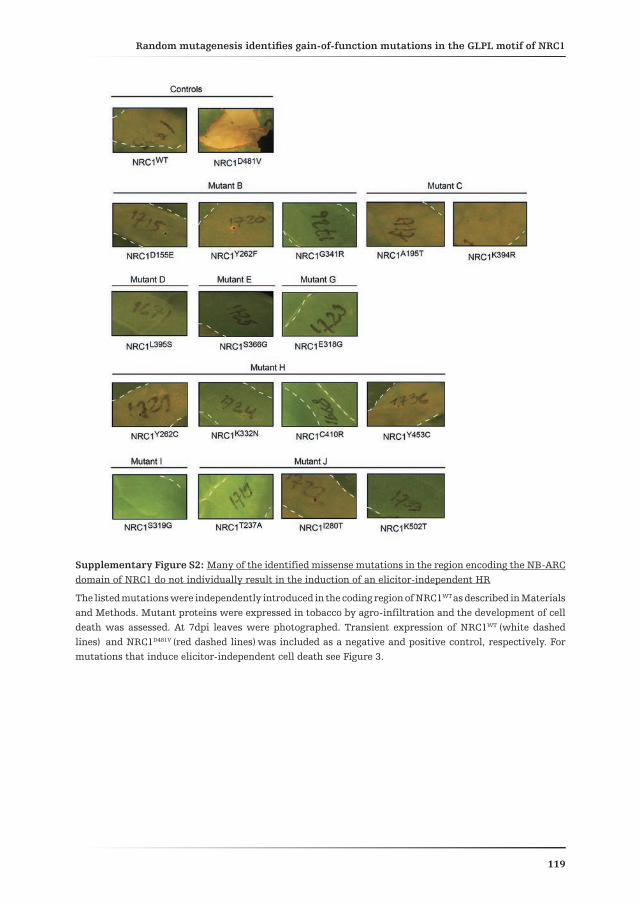
Random mutagenesis identifies gain-of-function mutations in the GLPL motif of NRC1Chapter 4
119
Supplementary Figure S2: Many of the identified missense mutations in the region encoding the NB-ARC domain of NRC1 do not individually result in the induction of an elicitor-independent HR
The listed mutations were independently introduced in the coding region of NRC1WT as described in Materials and Methods. Mutant proteins were expressed in tobacco by agro-infiltration and the development of cell death was assessed. At 7dpi leaves were photographed. Transient expression of NRC1WT (white dashed lines) and NRC1D481V (red dashed lines) was included as a negative and positive control, respectively. For mutations that induce elicitor-independent cell death see Figure 3.

Random mutagenesis identifies gain-of-function mutations in the GLPL motif of NRC1Chapter 4
120
Supplementary Fig. S3: Gain-of-function mutations in the NB-ARC domain of NRC1 primarily localize in and close to the GLPL motif and correspond to residues of a different degree of conservation within NB-LRRs

Random mutagenesis identifies gain-of-function mutations in the GLPL motif of NRC1Chapter 4
121
Amino acid alignment of the NB-ARC domain of NRC1 and other NB-LRRs, depicting the location and conservation of the residues that have been mutated in NRC1. The amino acid sequences of the NB-ARC domains of the different proteins were aligned with ClustalW. NRC1_MUT combines all missense mutations identified in the random mutagenesis screen. Blue, gain-of-function mutation; green, loss-of-function mutation; orange, mutations which increase the HR-inducing activity of NRC1 cooperatively but not individually (D155E, Y262F and G341R); red, previously reported gain-of-function mutations (Bendahmane et al., 2002; Tameling et al., 2006; Gabriëls et al., 2007; Gao et al., 2011; Roberts et al., 2013); purple, NRC1 mutation that is predicted to provide auto-activity (V365E). Amino acids predicted to be involved in nucleotide binding based on the crystal structure of Apaf-1 are indicated with a black star. Numbers correspond to the amino acid position considering the first amino acid of the full length protein as position one.
Supplementary Fig. S4: Location of the identified nine gain-of-function mutants in the NB-ARC domain of NRC1
3D-model of the NB-ARC domain of NRC1 indicating the location of the targeted residues in the gain-of-function mutants. The amino acid residues 160 to 443 from the NRC1 full-length protein were modelled for its 3D structure, based on the crystal structure of Apaf-1 as described in Materials and Methods. Light-blue, conserved motifs of the NB-ARC; red, positions corresponding to the nine identified gain-of-function mutations; pink, ADP molecule. Side chains of important residues are depicted as sticks. The dot projection (in red) displays the solvent-accessible surface.

Yo soy cordillera y marselva, pampa y desiertoríos, lagunas y montes
puna y salar…Soy ese país que siempre está
buscando el camino de la libertad
Mercedes Sosa(dedicado a “las señoritas”)
Chapter 5Dynamic hydrolase activities precede
hypersensitive cell death in tomato seedlings
Daniela J. Sueldo1, Ali Ahmed1 5, Johana Misas-Villamil2, Tom Colby4, Wladimir Tameling1, Matthieu H.A.J. Joosten1,
and Renier A.L. van der Hoorn2, 3
Affiliations:1 Laboratory for Phytopathology, Wageningen University, Wageningen, The Netherlands2 Plant Chemetics Laboratory, Department of Plant Sciences, University of Oxford,
South Parks Road, Oxford, United Kingdom3 Plant Chemetics Laboratory, Max Planck Institute for Plant Breeding Research,
Carl-von-Linne Weg 10, 50829 Cologne, Germany4 Mass Spectrometry group, Max Planck Institute for Plant Breeding Research,
Carl-von-Linne Weg 10, 50829 Cologne, Germany5 Department of Plant and Environmental Sciences, University of Copenhagen,
Thorvaldsensvej 40, 1871, Copenhagen, Denmark
Submitted for publication

Activity-based protein profiling of tomato seedlings mounting a synchronized defence responseChapter 5
124
Abstract
Hydrolases play important roles during Programmed Cell Death (PCD) in plants. Plant PCD is regulated by lipases, Vacuolar Processing Enzymes (VPEs), subtilases and the proteasome. Despite the importance of these enzymes, little is known about when these hydrolases are active during PCD. Here, we investigated hydrolase activities during PCD using Activity-Based Protein Profiling (ABPP). ABPP displays the active proteome using small molecule probes that react covalently with the active site of enzymes. We studied hydrolase activities in tomato seedlings undergoing a synchronized hypersensitive cell death by co-expressing the avirulence protein Avr4 from the extracellular tomato pathogen Cladosporium fulvum and the corresponding tomato resistance protein Cf-4. Hypersensitive cell death is blocked in Cf-4/Avr4 seedlings grown at high temperature and humidity, and is subsequently synchronously induced by lowering the temperature and humidity. ABPP on this synchronized PCD system revealed that VPEs and the proteasome are not differentially active, but that activities of papain-like cysteine proteases and several serine hydrolases, including the carboxyesterase Hsr203 and the subtilase P69B, dramatically increase upon transfer of the seedlings to the PCD-permissive conditions, whereas the activity of a carboxypeptidase-like enzyme is reduced. Similar dynamics were observed in the apoplast of resistant tomato challenged with C. fulvum, except that we now also detected the appearance of novel isoforms of secreted putative VPEs. Importantly, in the Cf-4/Avr4 seedlings differential hydrolase activity can be uncoupled from the cell death response at the condition of 100% relative humidity and 20 ºC, since the temperature drop by itself does not induce cell death as long as the humidity remains high. Under high humidity, the hydrolase activity profile alters completely, while the plants behave normally, demonstrating that changes in hydrolase activities precede hypersensitive cell death.

Activity-based protein profiling of tomato seedlings mounting a synchronized defence responseChapter 5
125
Introduction
Programmed Cell Death (PCD) is a highly controlled process that occurs in both plants and animals during developmental processes and the immune response. In the context of infection by pathogens, multiple types of PCD have been described in mammals; with apoptosis being the best characterized (Coll et al., 2011). The Hypersensitive Response (HR) involves a plant-specific type of PCD elicited in the event of pathogen recognition (Heath, 2000). A diverse spectrum of cytological changes are associated with HR, some of which are also shared by animal PCD, examples of which are cytoplasmic shrinkage, chromatin condensation and mitochondrial swelling (Mur et al., 2008). Processes such as vacuolization and chloroplast malfunctioning occur during the last stages of HR and are specific to plant PCD.
The HR is generally, but not always, part of a more complex defence response that leads to resistance to pathogens (Kacprzyk et al., 2011). This form of PCD is often thought to be the ultimate resource to stop invasion of biotrophic pathogens, which depend on living tissue to complete their life cycle. HR is initiated as the consequence of the recognition (either direct or indirect) of the matching pathogen-derived effector by a plant immune receptor. Such effectors are also known as avirulence (Avr) proteins or specific elicitors. Resistance (R) genes encode immune receptors and the R gene-mediated defence response in plants is often race-specific, meaning that the expression of a particular R gene confers resistance only against a pathogen species when it harbours the matching effector protein (Romeis et al., 1999; Dodds et al., 2006).
Cladosporium fulvum is a foliar, biotrophic fungal pathogen that colonises the extracellular space of tomato leaves (de Wit, 1977; Joosten and de Wit, 1999; Thomma et al., 2005). As a result of their co-evolution, tomato has acquired specific R proteins encoded by Cf genes, capable of recognising particular Avr proteins of C. fulvum, after which a defence response is activated. We have previously reported the development of a simple and robust synchronised system to study HR in tomato, which we have named “Dying Seedling” (DS) (de Jong et al., 2002; Gabriëls et al., 2006; Stulemeijer et al., 2009). The DS are tomato hybrid (F1) plants obtained from a cross between transgenic Parental Lines (PL) expressing either the tomato R gene Cf-4 (Hcr9-4D; Thomas, 1997) or the corresponding avirulence gene from C. fulvum, Avr4. Since the DS express the Cf-4/Avr4 gene pair in the same tissue, they undergo defence responses which will culminate in the death of the tomato seedling (de Jong et al., 2002). Importantly, activation of HR in these plants can be strictly controlled by manipulating the growth conditions, thereby turning the DS into an extremely versatile biological system to study defence-related hypersensitive cell death. The HR is inhibited when plants are grown at high temperature (33°C) and high relative humidity (100% RH) (de Jong et al., 2002; Gabriëls et al., 2006; Stulemeijer et al.,

Activity-based protein profiling of tomato seedlings mounting a synchronized defence responseChapter 5
126
2009). Therefore, at this non-inductive condition, seedlings remain alive and are phenotypically identical to the PLs. In previous studies with DS, the induction of defence was carried out by transferring the seedlings to a condition of 20°C and at 70% humidity. Under these conditions, plants would die within 8 hours (de Jong et al., 2002; Gabriëls et al., 2006; Stulemeijer et al., 2009). However, we recently discovered that the transfer from the standard non-inductive condition to a situation in which only the temperature is lowered (from 33°C to 20°C) does not induce cell death, as the plants remain phenotypically unaltered for several days (Etalo et al., 2013). It is the successive drop in the humidity (from 100% to 70% RH) that ultimately causes plants to die within 90 minutes (Etalo et al., 2013). In this study we characterized this synchronized HR, by studying proteome activities using Activity-Based Protein Profiling (ABPP).
ABPP displays the activity of different protein families and is based on the use of small molecule probes that react with the active site of proteins in an activity-dependent manner. Most probes consist of inhibitors to which a tag, such as biotin or a fluorescent reporter, has been added to facilitate detection (Cravatt et al., 2008; Kołodziejek and van der Hoorn, 2010). Upon incubation of the proteome with the probe, only proteins that contain an accessible active site residue will covalently bind to the warhead of the probe (the inhibitor itself) and become irreversibly labelled. ABPP is emerging as a powerful platform to identify the differential activities of proteins acting at the plant-pathogen interface (Kaschani et al., 2009; van der Linde et al., 2012; Lozano-Torres et al., 2012; Shindo et al., 2012).
The DS represent a highly sensitive and robust system to study plant PCD in the context of an immune response. Since tomato produces a wide variety of hydrolytic enzymes aimed at establishing a defence barrier against C. fulvum, and because some of these enzymes might be involved in the HR, we used ABPP to investigate the active proteome of tomato seedlings undergoing HR. Unexpectedly, these assays revealed that Serine (Ser) Hydrolases (SHs) and Cysteine (Cys) protease activities change before macroscopic cell death occurs. We speculate on the possible effects of temperature and humidity on the activation of Cf-4/Avr4-dependent PCD.
Results
Dynamics of protease activities in tomato seedlings undergoing synchronized HR
To induce a synchronised HR, three-weeks old tomato seedlings of the Parental Lines (PLs) and the Dying Seedlings (DS) (Fig. 1A) were transferred from the non-inductive conditions (33°C and 100% RH) to the inductive conditions (20°C and 70% RH) in two consecutive steps (Fig. 1B). First only the temperature was lowered from 33°C to 20°C and then three days later, the relative humidity was lowered from 100% to

Activity-based protein profiling of tomato seedlings mounting a synchronized defence responseChapter 5
127
70%. We have named these time points t01 (just before lowering the temperature) and t02 (at the moment of lowering the humidity). This final condition (20°C and 70% RH) causes a very rapid and synchronous systemic cell death in the DS within 90 minutes, while the phenotype of the PLs remains unaltered. In order to study enzyme activities during this synchronized PCD process, leaf material was harvested for the PLs and DS at t01, t02 and at 30, 60 and 90 minutes after t02 (Fig. 1B).
Figure 1: Experimental design for generating the synchronized hypersensitive response
(A) Parental lines (PLs) expressing the resistance gene Cf-4 (Hcr9-4D) or the Avr4 effector gene from C. fulvum are crossed to obtain F1 hybrid seeds. F1 seedlings are symptomless when grown at elevated temperature (33 ºC) and 100% relative humidity. Lowering only the temperature (to 20ºC) does not cause any symptoms, but a subsequent drop in humidity causes synchronized hypersensitive cell death, hence the name ‘Dying Seedlings’ (DS).
(B) Two-stage induction of synchronized cell death. Seedlings are grown at 33ºC and 100% humidity for three weeks. At time-point t01, the temperature is lowered to 20ºC and seedlings are kept for three days at 100% humidity. At time-point t02, the humidity is lowered to 70% and subsequently samples are taken at 30, 60, and 90 minutes. The DS are symptomless until t02 and collapse within 90 minutes after t02. The PLs, that do not develop any symptoms upon exposure to the same treatment, are taken as control.
Activities of the proteasome and Vacuolar Processing Enzymes (VPEs) are differentially regulated during defence signalling (Xia et al., 2004; Hara-Nishimura et al., 2005; Gu et al., 2010; Misas-Villamil et al., 2013). The proteasome is required for the HR induced in Arabidopsis by avirulent Pseudomonas syringae (Hatsugai et al., 2009), whereas VPEs are required for Tobacco Mosaic Virus (TMV)-induced HR in Nicotiana benthamiana (Hatsugai et al., 2004). We investigated whether these protease activities were also altered in our DS system. To this end, extracts from PLs and DS were prepared and separated on protein gel to show equal protein amounts (Fig. 2A). Next, these extracts were labelled with activity-based probes to display activities of the proteasome and VPEs. Proteasome activities are detected with MV151, a probe which contains vinyl sulfone as a reactive group and a Bodipy fluorophore for detection (Verdoes et al., 2006; Gu et al., 2010). Incubation of the proteome of both the PLs and DS with MV151 resulted in two 25 kDa signals (Fig. 2B). These signals represent the catalytic subunits (β1, β2 and β5) of the proteasome since pre-incubation with the proteasome selective inhibitor epoxomicin blocked labelling (Supplementary Fig. S1A) (Gu et al., 2010). Surprisingly, labelling was

Activity-based protein profiling of tomato seedlings mounting a synchronized defence responseChapter 5
128
uniform throughout the time course of the experiment, suggesting that no changes in proteasome activity occur upon activation of the Cf-4/Avr4-triggered HR.
Labelling of the seedling proteomes with the fluorescent probe AMS101, which targets VPEs (Misas-Villamil et al., 2013), caused two signals of about 40 kDa (Fig. 2C). The signals have been reported earlier for tomato leaf extracts and represent both the intermediate (i) and mature (m) isoforms of VPEs (Misas-Villamil et al., 2013). Labelling of these proteins was blocked upon pre-incubation with caspase-1 inhibitor YVAD (Supplementary Fig. S1B), which inhibits VPEs (Misas-Villamil et al., 2013), thus confirming that the signals represent VPEs. Unexpectedly, the labelling pattern of VPEs of DS was similar to that of PLs and did not change during the time course (Fig. 2E). Taken together, these observations indicate that the activity of neither the proteasome nor VPEs is differentially regulated in the DS.
Figure 2: Dying seedlings display unaltered vacuolar-processing enzymes (VPE) and proteasome activities and increased papain-like cysteine protease (PLCP) activity
Total protein was extracted from parental lines (PLs) or dying seedlings (DS) and immediately separated on protein gel (A) or labelled with 2µM MV151 (B), 2µM of AMS101 (C), or 1µM of DCG-04 (D). After labelling, proteins were separated in protein gels and analysed by coomassie staining (A,E), fluorescence scanning (B, C), or protein blotting using streptavidin-HRP (D).
(A) Equal protein levels upon extraction in PLs and DS.
(B) Activities of the proteasome are not up-regulated in DS. β1, 2 and 5 refers to the β-1, -2 and -5 subunits of the proteasome.
(C) Activities of VPEs are unaltered in DS. iVPE, intermediate VPE; mVPE, mature VPE.
(D) Increased activity of PLCPs in DS.
(E) Reduced protein accumulation in the DS during labelling reaction coincides with increased PLCP activity.
We next displayed activities of Papain-Like Cysteine Proteases (PLCPs) using DCG-04. This activity-based probe is a biotinylated version of E-64, an irreversible inhibitor of PLCPs (Greenbaum et al., 2000), and has been used to display the activities of PLCPs in plants (van der Hoorn et al., 2004; Richau et al., 2012). Labelling of protein extracts from the PLs and DS with DCG-04 revealed signals derived from three proteins of 25, 30 and 40 kDa (Fig. 2D). These signals were not detected upon pre-incubation with a saturating amount of the PLCP inhibitor E-64, which also covalently binds to the active site of PLCPs and therefore inhibits labelling with DCG-04. Based on

Activity-based protein profiling of tomato seedlings mounting a synchronized defence responseChapter 5
129
previous studies, we conclude that the 25 kDa signal represents PIP1 (Phytophthora-Inhibited Protein 1) and the 35 kDa signal represents C14 (Tian et al., 2007; Shabab et al., 2008; van Esse et al., 2008). Importantly, this assay revealed a strong induction of PIP1 in the DS; with a peak signal at 30 minutes (Fig. 2D). C14 followed a similar pattern of induction as PIP1, although its activity levels were much lower. Surprisingly, in both PLs and DS PIP1 activity was already increased at t02 (20°C and 100% RH) when compared to t01 (33°C and 100% RH) (Fig. 2D). Therefore, this activity must have been induced upon the temperature shift. This observation implies that some of the molecular changes related to HR already occur before lowering the humidity, even though the DS do not display any symptoms under these conditions.
Unexpectedly, after staining the protein membrane with coomassie to confirm equal protein loading, we detected a clear reduction in the amount of the large subunit of RuBisCO (RBCL) in the extracts that originated from the DS (Fig. 2E). This reduction was repeatedly observed in most of our experiments, despite the fact that equal protein amounts were loaded based on protein concentration measurements and when protein extracts were immediately separated on protein gels (Fig. 2A). Interestingly, the samples showing reduced RBCL levels were the same ones showing increased PLCP activity; namely the DS at t02 and the time points thereafter (Fig. 2E). We also noted that RBCL levels were lower in samples that required longer labelling times (data not shown), which suggests that degradation occurred after protein extraction, possibly by increased PLCP activities or other concomitantly induced proteases.
Dynamic changes in the activity of intracellular and extracellular serine hydrolases during HR
Serine Hydrolases (SHs) are another class of hydrolytic enzymes, whose activity has been shown to respond to infection in other plant-pathogen systems (Kaschani et al., 2009). SHs carry an active site S residue and include proteases, lipases, acyltransferases and esterases. To study SHs in the DS we used tri-functional nitrophenolphosphonate (TriNP), a probe containing a phosphonate warhead, a fluorophore for in-gel fluorescence visualization and biotin for affinity purification of labelled proteins (Nickel et al., 2012). Analysis of the activity profile of SHs in total protein extracts of the tomato DS revealed several signals that were absent in the no-probe control (Fig. 3A). Moreover, the drop in the temperature changed the activity profile dramatically. A 70 kDa signal (#1) appeared in both PLs and DS, but was much stronger in the DS (Fig. 3A). A 50 kDa signal (#2), which was present in both PLs and DS, remained constant in the PLs at the different time points, but was clearly reduced in the DS upon dropping of the temperature (t01) and remained low upon dropping the humidity as well (Fig. 3A). Furthermore, a 40 kDa signal (#3) increased upon the temperature shift in both the PLs and DS, but was again much

Activity-based protein profiling of tomato seedlings mounting a synchronized defence responseChapter 5
130
stronger in DS (Fig. 3A). Other low molecular weight signals (below 40 kDa) also appeared only in the DS samples at t02 and later time points (Fig. 3A). The activity profile of the DS at t01 was identical to the one of the PLs, indicating that there are no major differences in SH activity between PLs and DS at non-inductive conditions (33°C and 100% RH).These results demonstrate the involvement of differential SH activity in the DS.
Figure 3: Dying seedlings display dynamic alterations in the activities of Serine hydrolases
Total protein (A) or 20µl of apoplastic fluid (AF) (B) obtained from the parental lines (PLs) and dying seedlings (DS) at the indicated time points was incubated either with or without trifunctional nitrophosphonate (TriNP). Fluorescently-labelled proteins were detected by fluorescence scanning. Major differential signals are indicated by arrows and numbered (1-3).
(A) Differential SH activities in total protein extracts of the DS.
(B) Differential SH activities in the AF of the DS.
We next investigated whether some of the differential SH activities were extracellular proteins. To this end, we performed a labelling experiment on Apoplastic Fluid (AF) extracted from the PLs and DS at t01, t02 and 40 minutes after the induction of the HR. This early time point was chosen to avoid that leakage from the cytoplasm due to cell death could contaminate the AF with cytoplasmic proteins. Similar to the profiling of total extracts, the 70 kDa signal (#1) increased in intensity at t02 in the AF of both the PLs and DS (Fig 3B). However, this increase was much stronger in the DS, once again implying a specifically increased activity of this SH in the Cf-4/Avr4-triggered HR. In addition, the 50 kDa signal (#2) decreased in the DS at t02, showing the same behaviour as in total extracts (Fig 3A). The 40 kDa signal (#3) appeared to be reduced in AF as compared to total extracts, suggesting that the most abundant SH causing this signal is intracellular. Moreover, several signals, generated by proteins of lower molecular weight, appeared at t02 in the AF of the DS (Fig. 3B). To conclude, several differential SH activities detected upon the temperature shift are secreted proteins.

Activity-based protein profiling of tomato seedlings mounting a synchronized defence responseChapter 5
131
Identification of differentially active SHs
To identify the enzymes responsible for the differential SH activities, we affinity-purified TriNP-labelled proteins and identified them by Mass Spectrometry (MS). MS analysis of the purified proteins uncovered 12 SHs belonging to four main protein families. Most of these proteins were absent in the no-probe control and many were detected in two independent experiments (Fig. 4A-B). The 70 kDa signal #1 (Figs. 3 and 4A) comprised three S8 subtilisin-like proteases: Solyc08g079870 (P69B), Solyc08g079900 (P69C) and Solyc10g084320 (Fig. 4B). The increased labelling intensity of the 70 kDa signal in the DS confirms an increased P69 activity during the Cf-4-mediated defence responses. The 50 kDa signal (#2) (Figs. 3 and 4A) contained Serine CarboxyPeptidase-Like protein (SCPL), Solyc11g066250 (Fig. 4B). The decreased signal intensity of the 50 kDa signal indicates that the activity of this SCPL is down-regulated during immune responses in the DS (Fig. 3 and 4A). In addition, the 40 kDa signal (#3) (Figs 3A and 4A) included several CarboXylEsterases (CXEs) (Fig. 4B). The most abundantly detected CXE is the tomato ortholog of the tobacco Hsr203 (Pontier et al., 1994; Pontier et al., 1998), indicating that increased Hsr203 activity is causing increased labelling of the 40 kDa signal in the DS. Finally, in signal #4 (Fig 4A), we identified an alpha-hydroxynitrile lyase (AHL, Solyc03g044790) (Fig. 4B). Overall, these results revealed the up-regulated activity of P69 and CXEs dominated by Hsr203 and the down-regulation of a SCPL, preceding the hypersensitive cell death.
Figure 4: Differential Serine Hydrolase (SH) activities are dominated by P69, SCPL and HSR203 and are regulated at the transcriptional level
(A) Identification of labelled SH by mass spectrometry. Leaf extracts from the parental lines (PLs) and dying seedlings (DS) were harvested at 90 minutes after lowering the humidity and incubated either with (+) or without (-) the trifunctional nitrophosphonate (TriNP) probe. Labelled proteins were purified on streptavidin columns and separated on SDS gels. Fluorescent bands 1 to 4 were excised from all four lanes and proteins were identified by mass spectrometry.
(B) The experiment was repeated twice and spectral counts for each protein are summed up for both experiments (n) for the samples labelled with (NP+) or without (NP-) the probe.
(C) Fold changes in the transcript levels of the identified SHs at t01, t02 and 90 minutes time point. RNA was isolated at t01, t02 and 90 minutes after dropping the humidity and used for quantitative RT-PCR. The DS/PL ratio was calculated for three independent experiments and shown as an average. Errors represent standard deviation from three experiments. Note that due to high sequence identity, transcript levels of P69B and P69C were detected with the same primer set and can therefore not be distinguished from one another. See Fig.S2 for more details on transcript levels.
Activity-based protein profiling of tomato seedlings mounting a synchronized defence responseChapter 5
132
We next used real time RT-PCR to determine the relative transcript levels of the genes encoding the labelled identified proteins (Fig. 4B-C). For that purpose, relative expression levels were determined at 90 minutes after the humidity drop and at t01 and t02. Quantitative RT-PCR analysis revealed that the transcript levels of P69B and P69C increased progressively in the DS as compared to the PLs, with fold change values of 4.14 at t01 to 32.74 at t=90 minutes (Fig. 4C). This augmented expression of the P69s is in accordance with the increased labelling of the 70 kDa signal (#1) in the DS. Transcript levels of the SCPL in the DS were 33 and 6 times lower than in the PLs at t02 and t=90 minutes, respectively, which is consistent with the reduced fluorescent signal at 50 kDa (#2; Fig. 3A and Fig. 4A-B). Of the CXEs detected in the 40 kDa signal (#3), Hsr203 expression was 13-fold up-regulated at t02, but less at t=90 minutes. In contrast, late up-regulated transcript levels were detected for the CXEs Solyc01g108580, Solyc09g075670 and Solyc10g054780, although not as high as for Hsr203; whereas transcripts of the three remaining CXEs (Solyc01g108530, Solyc05g051660 and Solyc09g075680) were unaffected. Taken together, the regulation of gene expression seems to underlie the changes in these major protein activities. Interestingly, although both gene expression and protein activity patterns correlated, the dynamics of these changes were different. When considering transcriptional patterns, the changes occurred either progressively (as in the case of P69s) or transiently (for SCPL and Hsr203). However, SH labelling at these time points did not change accordingly (Fig. 3A). Therefore, since changes in transcript levels are not fully correlating with protein activity, P69s, SCPL and CXEs are probably regulated at additional levels than just gene expression alone.
Early and late hydrolase activities in the apoplast of infected tomato are responsive to inoculation with C. fulvum
To determine whether differential PLCP, VPE and SH activities also occur during infection by pathogens we studied their hydrolytic activities in the tomato-C. fulvum patho-system. To this end, susceptible (Cf0) and resistant (Cf-4) tomato plants were inoculated with C. fulvum producing Avr4. AF was extracted from the inoculated leaves at different time points and the proteins were labelled with the respective probes for PLCPs, VPEs and SHs. Detection of PLCPs in the AF of tomato plants was performed with the probe MV201, a fluorescent derivative of E-64 (Greenbaum et al., 2000) which, like DCG-04, specifically labels PLCPs (Richau et al., 2012). Labelling showed a 25 kDa signal in the AF of susceptible plants strongly increasing from 10 to 14 days post inoculation (dpi) with C. fulvum (Fig. 5A). The signal was not detected upon pre-incubation of a mix of all AF with a saturating amount of E-64, indicating that indeed PLCP activity is detected at the different time-points (Fig. 5B). In the AF of resistant plants this signal appeared earlier (6 dpi) than in susceptible plants and declined at 14 dpi (Fig. 5A, long exposure panel). The earlier appearance of the 25 kDa signal in resistant compared to susceptible plants resembles the behaviour

Activity-based protein profiling of tomato seedlings mounting a synchronized defence responseChapter 5
133
of Pathogenesis-Related (PR) proteins, whose accumulation increases earlier in incompatible interactions as compared to compatible ones (Joosten and de Wit, 1988). It is therefore evident that the defence response activated upon recognition of Avr4 by Cf-4 triggers an increase in the extracellular activity of PLCPs. Interestingly, no clear PLCP activity was detected in the AF of non-inoculated plants (susceptible and resistant, t=0 dpi, Fig. 5A), suggesting that pathogen challenge strongly induces PLCP activity. Based on its molecular weight and previous MS experiments (Tian et al., 2007; Shabab et al., 2008; van Esse et al., 2008), we speculate that the majority of the labelled PLCPs represents PIP1 activity. Importantly, the signal intensities resemble the signals observed when labelling total extracts of the DS (Fig. 2A), confirming that the DS system is a proper representation of the events occurring in a tomato plant upon challenge with C. fulvum. We also detected increased labelling of two bands of around 35 and 40 kDa which are likely the mature (mC14) and immature (imC14) isoforms of C14, respectively. As for PIP1, C14 labelling increased in time in both susceptible and resistant tomato plants. However, this occurred faster and stronger in the resistant plants, once again resembling the behaviour of PR proteins.
The AF of susceptible and resistant tomato plants inoculated with C. fulvum was also tested for VPE activity. Interestingly, we detected AMS101-labelling in the apoplast of tomato plants before inoculation (t=0 dpi) with the pathogen (Fig. 5C). These activities probably represent the putative intermediate (iVPE) and mature (mVPE) isoforms of VPE (Fig. 5C). Unexpectedly, new signals appeared upon inoculation with C. fulvum of both susceptible and resistant tomato. These signals were not detected upon pre-incubation of the protein sample with the VPE inhibitor YVAD, indicating that they represent VPE-like activity (Fig. 5D).
We also performed activity profiling for SHs in the AF of C. fulvum-inoculated tomato, using a rhodamine-tagged nitrophenol phosphonate probe (RhNP) (Nickel et al., 2012). Four clear signals (#1 to #4) were detected in the AF of susceptible plants (Fig. 5E). All signals increased in intensity after inoculation with C. fulvum (Fig. 5E), in both susceptible and resistant tomato plants. However, similar to the timing of the increase in PLCP activity (Fig 5A), the SH activity in the AF of resistant plants accumulated faster and stronger than in susceptible plants. Quantification of the fluorescence intensity confirmed that the intensity of all bands was nearly always higher in the resistant plants than in the susceptible plants (Fig. 5F). MS analysis of differentially labelled SH in DS suggests that the 70 kDa signal (#1) and the 50 kDa signal (#2) found in the AF of C. fulvum-inoculated tomato correspond to P69 and an SCPL respectively. Again, this similarity between the two assays confirms that the DS forms a proper biological model system to study immune responses in tomato.

Activity-based protein profiling of tomato seedlings mounting a synchronized defence responseChapter 5
134
Figure 5: Challenge of tomato plants with C. fulvum induces early and late tomato apoplastic hydrolase activities
(A and B) C. fulvum challenge induces activities of papain-like cysteine proteases (PLCPs). Proteins present in 100 µl of AF obtained from C. fulvum-inoculated susceptible and resistant tomato plants, harvested at 0, 4, 6, 10 and 14 days post inoculation (dpi), were incubated either with or without MV201 for three hours. In (B) all AFs obtained from susceptible and resistant plants were combined in equal amounts, pre-

Activity-based protein profiling of tomato seedlings mounting a synchronized defence responseChapter 5
135
incubated either with or without 20 µM E-64 for 30 minutes and labelled with MV201 as in (A). Labelled proteins were detected by in-gel fluorescence scanning. In panel (A) m indicates a mix of all AFs. iC14, immature C14.
(C and D) C. fulvum challenge alters secreted isoforms of putative vacuolar processing enzymes (VPEs). Proteins present in 100 µl of AF obtained from C. fulvum-inoculated susceptible and resistant tomato plants, harvested at 0, 4, 6, 10 and 14 days post inoculation (dpi), were incubated either with or without AMS101 for two hours. In (D) all AFs were combined in equal amounts, pre-incubated either with or without 50 µM YVAD (inhibitor) for 30 minutes and labelled with AMS101 as in (C). Labelled proteins were detected by in-gel fluorescence scanning. iVPE, intermediate VPE; mVPE, mature VPE
(E) C. fulvum challenge alters apoplastic serine hydrolase (SH) activities. Proteins present in 20 µl of AF obtained from C. fulvum-inoculated susceptible and resistant tomato plants, harvested at 0, 4, 6, 10 and 14 days post inoculation (dpi), were incubated either with or without RhNP for 1 hr. Labelled proteins were detected by in-gel fluorescence scanning. The numbers 1 to 4 indicate discrete signals that were quantified in (F). m, mix of all AFs.
(F) Quantification of differential SH activities upon challenge of susceptible or resistant tomato with C. fulvum. The intensities of signals 1 to 4, indicated in (E), were quantified and plotted against the dpi.
Lowering the temperature is the determining factor triggering differential hydrolase activity
One striking observation throughout our experiments was the fact that differences in the SH activity of the PLs and DS were already evident at t02, which is the time point where the temperature has already been lowered but the humidity is still 100% and symptoms are not apparent. In other words, the changes in the SH activity appear to precede hypersensitive cell death. To more precisely pinpoint the moment at which changes in the SH activity profile occur, we examined time points between t01 and t02 by investigating leaf samples taken at 24 and 48 hrs after the temperature drop (Fig. 6A). The labelling revealed that changes in the SH activities occurred as quickly as 24 hrs after the incubation temperature was lowered from 33°C to 20°C both in total extracts and in the AF (Fig. 6B and 6C). After another 24 hr of incubation at 20°C additional progressive changes were evident in the labelling profile of AF obtained from the DS. Most of the differentials are specific to the DS, but the 70 kDa signal was also up-regulated in PLs upon the temperature shift, albeit less strong than in DS (Fig. 6B and 6C). The finding that at 24 hrs after the temperature drop massive changes are already visible in the DS demonstrates that alterations in hydrolase activities precede the hypersensitive cell death response.

Activity-based protein profiling of tomato seedlings mounting a synchronized defence responseChapter 5
136
Fig. 6: Differential serine hydrolase (SH) activities in the apoplast precede hypersensitive cell death in the dying seedlings
20µg of protein or 20µl of apoplastic fluid (AF) was incubated either with or without FPRh for 1hr at room temperature. Labelled proteins were detected by in-gel fluorescence scanning.
(A) Seedlings were transferred to the lower temperature at t01 and samples were taken at 24 and 48 hrs after transfer. Subsequently, the relative humidity was dropped from 100% to 70% at t02.
(B) Differential SH activities in total extract of the parental lines (PLs) and dying seedlings (DS) upon lowering of the temperature.
(C) Differential SH activities in the AF obtained from the PLs and DS upon lowering of the temperature.

Activity-based protein profiling of tomato seedlings mounting a synchronized defence responseChapter 5
137
Discussion
This study revealed the dynamics of hydrolytic enzyme activities upon activation of the HR, which is a plant-specific form of PCD associated with innate immunity. We employed a robust biological model system to induce a systemic and synchronised Cf-4/Avr4-triggered HR in tomato and monitored the activities of Vacuolar Processing Enzymes (VPEs), the proteasome, Papain-Like Cysteine Proteases (PLCPs) and Serine Hydrolases (SHs) by Activity-Based Protein Profiling (ABPP). Our results indicate that changes in the activity of both PLCPs and SHs preceded the hypersensitive cell death and are differentially regulated in the apoplast of leaflets of C. fulvum-inoculated susceptible and resistant tomato plants.
VPEs activities are not altered during hypersensitive cell death but are responsive to pathogen challenge
PCD is an active and highly controlled process normally resulting in the death of a selected and limited group of cells. In animals, apoptosis is executed by aspartate-specific cysteine proteases, or caspases (Miura, 2012). To date, no close homologs of caspases have been identified in plants. Metacaspase 1 (MC1) and MC2 are two metacaspases involved in R protein-mediated PCD in Arabidopsis (Coll et al., 2010). Metacaspases are distantly related to caspases and share the same catalytic residues but have a different substrate specificity. Interestingly, MC1 is required for cell death development, whereas MC2 negatively regulates the pro-cell death activity of MC1 (Coll et al., 2010). Moreover, VPEs and the proteasome display caspase-like activity. VPEs have emerged as proteases involved in plant PCD (Nakaune et al., 2005). VPE is required for the HR triggered by TMV (Tobacco Mosaic Virus) in tobacco (Hatsugai et al., 2004). However, we did not detect an increase in VPE activity in the tomato DS upon activation of the HR (Fig. 2E). Our report is consistent with the finding that the HR is not compromised in a VPE null mutant of Arabidopsis upon inoculation with avirulent Pseudomonas syringae pv. tomato DC3000 (Hatsugai et al., 2009).
Interestingly, we detected altered VPE-like activity in the apoplast of tomato plants inoculated with C. fulvum, regardless whether the plants were susceptible or resistant to this fungus (Fig. 5C). This is a remarkable observation, since VPEs activity was so far only described in the vacuole. Our findings are in contrast with the increased VPE activity detected upon infection of Arabidopsis with virulent Hyaloperonospora arabidopsidis (Misas-Villamil et al., 2013) and suggest that participation of VPE in defence responses varies between plant-pathogen interactions. Moreover, we observed a conspicuous change in the VPE labelling pattern of AF of C. fulvum-inoculated tomato plants, either susceptible or resistant, as compared to non-inoculated plants (Fig. 5C). The detection of new signals might be the consequence of further processing of these enzymes in the apoplast of tomato leaflets. Alternatively,

Activity-based protein profiling of tomato seedlings mounting a synchronized defence responseChapter 5
138
new VPEs isoforms might have been induced upon inoculation with the fungus, as five VPE genes have been reported to be present in the tomato genome (Ariisumi et al., 2011). In any case, apoplastic VPE activity responded to C. fulvum challenge, although there was no difference between susceptible and resistant plants. This could reflect an involvement of VPEs in a basal response to pathogens. We propose that future studies should be focussed on the possible roles and processing of VPEs in the apoplast.
Our labelling experiments revealed that the activity of the proteasome is also not differentially regulated during mounting of the HR in tomato seedlings (Fig. 2D). This is unexpected since the DS contain large amounts of Salicylic Acid (SA) (Etalo et al., 2013) and SA is known to activate the proteasome in Arabidopsis (Gu et al., 2010). Furthermore, Hatsugai et al. (2009) showed that proteasome activity is required for RPM1-mediated HR in Arabidopsis. In addition, Pseudomonas syringae pv syringae produces a proteasome inhibitor that inhibits SA-dependent defence responses and delays hypersensitive cell death (Misas-Villamil et al., 2013). However, in the case of the DS, constitutive proteasome activity might be sufficient for the activation of the HR.
Differential activity of PIP1, P69B and a SCPL correlates between assay systems
We showed that PLCP activity is induced when the Cf-4/Avr4-activated HR is mounted in the DS (Fig. 2A). Moreover, labelling of AF of C. fulvum-inoculated tomato revealed that PLCP activity increased faster in resistant than in susceptible plants (Fig. 5A). Our data are consistent with previous reports showing the induction of PLCP activity in the apoplast of tomato upon BTH (a SA analogue) treatment (Shabab et al., 2008), which was predominantly caused by the PIP1 protease (Tian et al., 2007; Shabab et al., 2008). PIP1 is thought to play a role in plant defence, possibly by degrading effectors and other proteins produced by the pathogen. Our results show that the recognition of fungal Avr4 by tomato Cf-4 increases the activity of PIP1 in the apoplast. The increased PIP1 activity level is likely associated with increased amounts of PIP1 protein and PIP1 transcript levels, as has been described before (Shabab et al., 2008).
We identified three extracellular subtilisins that belong to the P69 protease family of tomato (Tornero et al., 1996), the activity of which was increased in the DS and in C. fulvum-inoculated tomato (Fig. 3, Fig. 4 and Fig. 5E and 5F). P69 was first identified upon inoculation of tomato with citrus exocortis viroid (Granell et al., 1987; Vera and Conejero, 1989). Further studies showed that some members from this protein family are specifically induced upon inoculation of Arabidopsis with virulent Pseudomonas syringae and upon treatment with SA (Jordá et al., 1999; Jorda et al., 2000). Little is known about their biological function, although increased expression of P69 proteins upon pathogen challenge suggests a role in defence against invading

Activity-based protein profiling of tomato seedlings mounting a synchronized defence responseChapter 5
139
microbes. Subtilisin proteases usually have a broad substrate specificity for peptide bonds (Markland Jr and Smith, 1971) and up-regulation of P69 expression and activity might lead to degradation of pathogen-derived virulence factors. Alternatively, subtilases might play a role in signalling. For example, phytaspase is a subtilisin-like protease from tobacco involved in the HR and the resistance response to TMV (Chichkova et al., 2010). Interestingly, phytaspase is also required for the PCD that is induced under abiotic stress.
In addition to P69, we confidently identified other SHs (Fig. 4A). Serine carboxypeptidase-like proteins (SCPLs) belong to the α/β hydrolase protein family (Breddam, 1986). Although annotated as peptidases, many SCPLs have other enzymatic activities, such as acyltransferase- or lyase-activity (Li and Steffens, 2000; Mugford et al., 2009). We found that the activity of an extracellular SCPL (Solyc11g066250) was reduced during defence in the DS (Fig. 3 and Fig. 4A). The expression of its encoding gene was also reduced (Fig. 4B), suggesting that the reduced enzyme activity is a consequence of transcriptional down-regulation.
Carboxylesterases (CXE) comprise another major group of the SHs that we identified; Hsr203 being the most abundantly detected CXE in our assays (Fig. 3A and Fig. 4A). Hsr203J is a CXE that was initially identified as a gene specifically induced in the incompatible interaction between tobacco and Ralstonia solanacearum (Pontier et al., 1994). Further studies demonstrated that expression of the tomato ortholog, Slhsr203, was immediately up-regulated upon transient expression of the Avr9 effector from C. fulvum in Cf-9-containing tomato plants (Pontier et al., 1998). Studies on the promoter of tobacco Hsr203 revealed that it is specifically activated upon the induction of the HR, several hours before the necrotic lesions appear. This is consistent with our results, because we detected an increase in the amount of Hsr203 transcripts and Hsr203 activity in the DS before the plants developed cell death (Fig. 3A and Fig. 4). Since its identification, Hsr203J transcript levels have been widely used as a hallmark for the activation of PCD (Radwan et al., 2011; MiSHra et al., 2011; Zhang et al., 2012). To our knowledge, we are the first to show activity of Hsr203 in the context of a plant-pathogen interaction with a simple and robust labelling assay. We did not detect activity of Hsr203 in C. fulvum-inoculated tomato, either susceptible or resistant, but this is probably because we only studied the AF of the inoculated plants and Hsr203 is not predicted to be secreted.
Differential hydrolase activity precedes hypersensitive cell death
Several of the hydrolytic activities we studied in the DS have been implicated in PCD before (Pontier et al., 1998). Surprisingly, we show that changes in the activities of PLCPs and SHs in the DS precede cell death development. Despite of the increased hydrolase activity, the DS did not display visible cell death until the humidity was decreased, suggesting that the activation of these hydrolases is a part of a complex

Activity-based protein profiling of tomato seedlings mounting a synchronized defence responseChapter 5
140
immune response. Therefore, we hypothesise that, although involved in the Cf-4/Avr4-triggered defence response, these hydrolytic activities likely do not participate in the signalling cascade leading to the hypersensitive cell death itself. Moreover, an elevated temperature was enough to repress changes in the hydrolytic activities of PLCPs and SHs. Wang et al (2005) concluded that both high temperature and 100% humidity synergistically repress Cf-4/Avr4 and Cf-9/Avr9 induced HR. In our experiments only lowering of the temperature was enough to trigger the changes in the activity of hydrolases but not to trigger cell death.
The temperature-controlled immune response is uncoupled from humidity-controlled hypersensitive cell death
The suppression of hypersensitive cell death by high humidity is not uncommon. Several Arabidopsis lesion mimic mutants show phenotypes that are repressed by high relative humidity (Hammond-Kosack et al., 1996; Yoshioka et al., 2001; Jambunathan et al., 2001; Noutoshi et al., 2005). The cpr22 Arabidopsis mutant, for example, displays constitutive expression of PR genes, spontaneous lesion formation and increased resistance to virulent pathogens, but some of these phenotypes were inhibited at high humidity (Yoshioka et al., 2001). To explain their observations, the authors proposed the existence of a “humidity-sensitive factor” that down-regulates the SA-dependent defence response at high humidity. The inhibition of effector-triggered PCD by environmental conditions observed in our system could be caused by the crosstalk between biotic and abiotic stress. Specifically, an antagonistic interplay between the ethylene (biotic stress) and ABA (abiotic stress) cascades has already been reported (Robert-Seilaniantz et al., 2011; Chen et al., 2013). It is tempting to speculate that when grown at high humidity, the DS are subjected simultaneously to two different stresses: an ethylene-dependent signalling cascade linked to the HR and an ABA-mediated response linked to the high humidity. In such a situation, the hypersensitive cell death would be (partially) repressed by the inhibitory effect of the abiotic signalling pathway. When the humidity is lowered, the pressure of the abiotic stress is released and so is its antagonistic effect on the biotic stress signalling. This would then result in the release of the brake on the cell death response. A metabolome and transcriptome study on the DS performed by Etalo et al. (2013) indeed indicated that when the temperature alone is lowered, the production of ethylene is highly induced. However, differential hydrolase activation, which precedes hypersensitive cell death, is apparently not under control of this crosstalk. Moreover, it also suggests that hypersensitive cell death can be uncoupled from other defence responses.

Activity-based protein profiling of tomato seedlings mounting a synchronized defence responseChapter 5
141
Materials and Methods
Plant Material and growth conditions
Tomato Cf-4/Avr4 Dying Seedlings (DS) were obtained from crossings between Cf-4 (Hcr9-4D) and Avr4-expressing tomato plants, as described elsewhere (de Jong et al., 2002). Sowing and germination of the seeds at 25°C and high relative humidity (100% RH) was performed as explained in Stulemeijer et al. (2009). After one week the seedlings were transferred to “non-inductive” conditions (33°C and 100% RH) and two weeks later the temperature was lowered to 20°C. Plant material (cotyledons and primary leaves) corresponding to t01 was immediately harvested and plants were kept under these growth conditions (20°C and 100% RH) for three more days. Finally, plants were shifted to the “inductive” condition by decreasing the humidity to 70% RH. Sampling of additional plant material was performed at the moment of lowering the humidity (t02) and at 30, 60 and 90 minutes after the plants had been transferred to “inductive” conditions. As a control, the parental lines (PLs) used for the cross to generate the DS were taken along and exposed to the same treatment. Tissue samples were stored at -80°C until protein extraction.
For plant inoculations, the PL of the tomato DS, expressing Avr4 (Cf0:Avr4; susceptible line) or Cf-4 (Cf0:Hcr9-4D; resistant line) were inoculated with C. fulvum race 5, expressing Avr4 (de Wit, 1977). At different time points after inoculation (0, 4, 6, 10 and 14 days post inoculation (dpi) leaf samples were taken for Apoplastic Fluid (AF) isolation as described below.
Protein extraction, apoplastic fluid isolation and labelling reactions
Cotyledons and primary leaves were ground to a fine powder with a mortar and pestle using liquid nitrogen. Frozen powder was resuspended in 1 mM DTT to extract proteins. The extract was centrifuged at 16,000xg for 5 minutes at 4°C and the supernatant was transferred into a new tube. The protein concentration was measured using the RC/RD protein assay (Bio-Rad) using Bovine Serum Albumin (BSA) as a standard. AF was isolated from cotyledons and primary leaves of the DS and PLs as well as from susceptible and resistant plants inoculated with C. fulvum according to Joosten (2012).
For PLCP labelling, 60 µg of total protein extract was incubated either with or without 1 µM DCG-04 (van der Hoorn et al., 2004) in 50 mM NaAc pH 6 and 1 mM DTT in a final volume of 0.5 mL. The labelling reaction was performed for 5 hrs at Room Temperature (RT) while rotating. Proteins were precipitated with 70% acetone and resuspended in 50 µl of 1x Gel Loading Buffer (GLB). Where indicated, protein samples were pre-incubated for 30 minutes with 20 µM E-64 before DCG-04 labelling. For detection of extracellular PLCPs, 100 µl of AF was incubated with 0.2 µM of MV201 in 50 mM NaAc pH5.5 and 10 mM DTT in a final volume of 0.5

Activity-based protein profiling of tomato seedlings mounting a synchronized defence responseChapter 5
142
mL. After labelling for 3hrs at RT, proteins were precipitated with 70% acetone and resuspended in 50 µl of 1xGLB. For inhibition assays equal volumes of the various AFs were mixed and pre-incubated with 20 µM E-64 for 30 minutes.
SH labelling was performed as described by Kaschani et al. (2009). 20 µg of total protein or 20 µl of tomato AF were labelled either with or without 1 µM of TriNP or RhNP (Nickel et al., 2012) in 50 mM Tris pH 8 (final volume 50 µl) for 1 hr at RT. The labelling reaction was stopped by boiling in 1xGLB and samples were further analysed as explained below.
For proteasome labelling, 60 µg of total protein was incubated either with or without 2 µM MV151 (Gu et al., 2010) in 50 mM Tris pH7 in a final volume of 50 µl. After incubating for 3.5 hrs at RT, the reaction was stopped by boiling in 1xGLB. For inhibition assays, equal amounts of all samples were combined and pre-incubated with 50 µM of epoxomicin for 30 minutes.
The activity of VPEs was detected by incubation of 60 µg of total protein extract or 100 µl of AF with 1 µM AMS101 (Misas-Villamil et al., 2013) in 50 mM sodium acetate pH 5 and 10 mM TCEP (Tris 2-carboxyethyl phosphine) for 2 hrs at RT. Competition assays were performed by pre-incubating the protein sample or a mixture of all AFs with 50 µM YVAD-CMK for 30 minutes. The labelling reaction was stopped by boiling in 1xGLB, including an earlier precipitation step with 70% acetone for the AF samples.
Protein samples were separated on 12% SDS-PAGE protein gels. Fluorescently labelled proteins were detected by fluorescence scanning of the gel using the Typhoon FLA9000. For DCG-04 labelling reactions, biotinylated proteins were analysed by protein blotting using streptavidin-conjugated HRP as described before (van der Hoorn et al., 2004).
Affinity purification and identification of SHs
Large scaled labelling and purification of biotinylated SHs was performed as described in Kaschani et al. (2012). 5 mg of total proteins was incubated either with or without 200 µM TriNP. After affinity-purification, biotinylated proteins were eluted from the beads by boiling in 1x GLB and resolved on a 12% SDS-PAGE protein gel. The gel was subsequently scanned, photographed and protein bands were automatically excised. In-gel trypsin digestion was performed according to Kaschani et al. (2012).
Data acquisition and processing
Peptide mixtures were analysed using a Thermo/Proxeon Easy nLC II mass spectrometer in a two column configuration (pre-column 3cm x 100um, 5um C18AQ medium, analytical column: 10cm x 75 µm, 3 µm C18AQ) coupled to a LTQ-Velos ion trap (Thermo Scientific). Peptides were separated over a 100 minute gradient

Activity-based protein profiling of tomato seedlings mounting a synchronized defence responseChapter 5
143
running from 5% - 25% acetonitrile in water containing 0.1% formic acid. MS/MS spectra were acquired in centroid mode on multiply-charged precursors with an m/z between 400 and 1600 Daltons using a Top20 method for with active exclusion for 60 seconds in a window from 0.2 Da below to 1.6 Da above the precursor mass. The resulting RAW files were de-noised (6 strongest peaks/ 100 Da window) and converted to mgf format using MSConvert from the Proteowizard package (http://www.proteowizard.org/).
Database searching
The sequence database was constructed by combining the ITAG2.3 tomato protein database (34727 sequences) from the Sol Genomics Network (www.solgenomics.net) supplemented with 1095 common artefact sequences and reversed copies of all sequences as decoys for False Discovery Rate (FDR) calculation. MS/MS spectra were searched against the described databases using MASCOT 2.3 (www.matrixscience.com) (see Supplemental Information). Reported proteins were identified on the basis of at least two peptides exceeding the MASCOT 95% certainty cut-off.
q-RT-PCR and gene expression analyses
Cotyledons and primary leaves were ground to a fine powder with mortar and pestle in liquid nitrogen and frozen powder was used for total RNA isolation following the manufacturers’ instructions (Macherey-Nagel RNA extraction kit). First strand cDNA was synthesised using Moloney-Murine Leukemia Virus (MMLV) reverse transcriptase and oligo dT (Promega) and was used as template for quantitative RT-PCR using the SensiMix SYBR-HiROX kit (Biolyne). Primers sequences for the different hydrolase-encoding sequences are provided in Supplemental Table S1. Tomato actin was used as a reference gene (Supplementary Table S1).
Acknowledgements
We thank Desalegn Etalo for providing the apoplastic fluid of C. fulvum-inoculated tomato plants. Christian Gu, Takayuki Shindo and Joji Villamor are thanked for helpful suggestions. This work was financially supported by the University of Oxford, the Max Planck Society and EU-funded COST program CM1004.

Activity-based protein profiling of tomato seedlings mounting a synchronized defence responseChapter 5
144
References
Breddam K (1986) Serine carboxypeptidases. A review. Carlsberg Research Communications 51: 83-128.Chen L, Zhang L, Li D, Wang F, Yu D (2013) WRKY8 transcription factor functions in the TMV-cg defense
response by mediating both abscisic acid and ethylene signaling in Arabidopsis. Proceedings of the National Academy of Sciences, USA 110: 1963-1971.
Chichkova NV, SHaw J, Galiullina RA, Drury GE, Tuzhikov AI, Kim SH, Kalkum M, Hong TB, GorSHkova EN, Torrance L, Vartapetian AB, Taliansky M (2010) Phytaspase, a relocalisable cell death promoting plant protease with caspase specificity. EMBO Journal 29: 1149-1161.
Coll NS, Epple P, Dangl JL (2011) Programmed cell death in the plant immune system. Cell Death & Differentiation 18: 1247-1256.
Coll NS, Vercammen D, Smidler A, Clover C, van Breusegem F, Dangl JL, Epple P (2010) Arabidopsis type I metacaspases control cell death. Science 330: 1393-1397.
Cravatt BF, Wright AT, Kozarich JW (2008) Activity-based protein profiling: From enzyme chemistry to proteomic chemistry. Annual Review of Biochemistry 77: 383-414.
de Jong CF, Takken FLW, Cai X, de Wit PJGM, Joosten MHAJ (2002) Attenuation of Cf-mediated defense responses at elevated temperatures correlates with a decrease in elicitor-binding sites. Molecular Plant-Microbe Interactions 15: 1040-1049.
de Wit PJGM (1977) A light and scanning-electron microscopic study of infection of tomato plants by virulent and avirulent races of Cladosporium fulvum. Netherlands Journal of Plant Pathology 83: 109-122.
Dodds PN, Lawrence GJ, Catanzariti AM, Teh T, Wang CIA, Ayliffe MA, Kobe B, Ellis JG (2006) Direct protein interaction underlies gene-for-gene specificity and coevolution of the flax resistance genes and flax rust avirulence genes. Proceedings of the National Academy of Sciences , USA 103: 8888-8893.
Etalo DW, Stulemeijer IJE, Peter van Esse H, de Vos RCH, Bouwmeester HJ, Joosten MHAJ (2013) System-wide hypersensitive response-associated transcriptome and metabolome reprogramming in tomato. Plant Physiology 162: 1599-1617.
Gabriëls SHEJ, Takken FLW, Vossen JH, de Jong CF, Liu Q, Turk SCHJ, Wachowski LK, Peters J, Witsenboer HMA, de Wit PJGM, Joosten MHAJ (2006) cDNA-AFLP combined with functional analysis reveals novel genes involved in the hypersensitive response. Molecular Plant-Microbe Interactions 19: 567-576.
Granell A, Bellés JM, Conejero V (1987) Induction of pathogenesis-related proteins in tomato by citrus exocortis viroid, silver ion and ethephon. Physiological and Molecular Plant Pathology 31: 83-90.
Greenbaum D, Medzihradszky KF, Burlingame A, Bogyo M (2000) Epoxide electrophiles as activity-dependent cysteine protease profiling and discovery tools. Chemistry and Biology 7: 569-581.
Gu C, Kolodziejek I, Misas-Villamil J, SHindo T, Colby T, Verdoes M, Richau KH, Schmidt J, Overkleeft HRS, van der Hoorn RAL (2010) Proteasome activity profiling: A simple, robust and versatile method revealing subunit-selective inhibitors and cytoplasmic, defense-induced proteasome activities. Plant Journal 62: 160-170.
Hammond-Kosack KE, Silverman P, Raskin I, Jones JDG (1996) Race-specific elicitors of Cladosporium fulvum induce changes in cell morphology and the synthesis of ethylene and salicylic acid in tomato plants carrying the corresponding Cf disease resistance gene. Plant Physiology 110: 1381-1394.
Hara-Nishimura I, Hatsugai N, Nakaune S, Kuroyanagi M, Nishimura M (2005) Vacuolar processing enzyme: An executor of plant cell death. Current Opinion in Plant Biology 8: 404-408.
Hatsugai N, Iwasaki S, Tamura K, Kondo M, Fuji K, Ogasawara K, Nishimura M, Hara-Nishimura I (2009) A novel membrane fusion-mediated plant immunity against bacterial pathogens. Genes and Development 23: 2496-2506.

Activity-based protein profiling of tomato seedlings mounting a synchronized defence responseChapter 5
145
Hatsugai N, Kuroyanagi M, Yamada K, MeSHi T, Tsuda S, Kondo M, Nishimura M, Hara-Nishimura I (2004) A plant vacuolar protease, VPE, mediates, virus-induced hypersensitive cell death. Science 305: 855-858.
Heath MC (2000) Hypersensitive response-related death. Plant Molecular Biology 44: 321-334.Jambunathan N, Siani JM, McNellis TW (2001) A humidity-sensitive Arabidopsis copine mutant exhibits
precocious cell death and increased disease resistance. Plant Cell 13: 2225-2240.Joosten MHAJ, de Wit PJGM (1988) Isolation, purification and preliminary characterization of a protein
specific for compatible Cladosporium fulvum (syn. Fulvia fulva)-tomato interactions. Physiological & Molecular Plant Pathology 33: 241-253.
Joosten MHAJ, De Wit PJGM (1999) The tomato-Cladosporium fulvum interaction: A versatile experimental system to study plant-pathogen interactions. Annual Review of Phytopathology 37: 335-367.
Jordá L, Coego A, Conejero V, Vera P (1999) A genomic cluster containing four differentially regulated subtilisin- like processing protease genes is in tomato plants. Journal of Biological Chemistry 274: 2360-2365.
Jorda L, Conejero V, Vera P (2000) Characterization of P69E and P69F, two differentially regulated genes encoding new members of the subtilisin-like proteinase family from tomato plants. Plant Physiology 122: 67-73.
Kacprzyk J, Daly CT, McCabe PF (2011) The botanical dance of death. Programmed cell death in plants. Advances in Botanical Research 60: 169-261.
Kaschani F, Gu C, Niessen S, Hoover H, Cravatt BF, van der Hoorn RAL (2009) Diversity of serine hydrolase activities of unchallenged and Botrytis-infected Arabidopsis thaliana. Molecular and Cellular Proteomics 8: 1082-1093.
Kołodziejek I, van der Hoorn RAL (2010) Mining the active proteome in plant science and biotechnology. Current Opinion in Biotechnology 21: 225-233.
Li AX, Steffens JC (2000) An acyltransferase catalyzing the formation of diacylglucose is a serine carboxypeptidase-like protein. Proceedings of the National Academy of Sciences, USA 97: 6902-6907.
Lozano-Torres JL, Wilbers RHP, Gawronski P, BoSHoven JC, Finkers-Tomczak A, Cordewener JHG, America AHP, Overmars HA, van ‘t Klooster JW, Baranowski L, Sobczak M, Ilyas M, van der Hoorn RAL, Schots A, de Wit PJGM, Bakker J, Goverse A, Smant G (2012) Dual disease resistance mediated by the immune receptor Cf-2 in tomato requires a common virulence target of a fungus and a nematode. Proceedings of the National Academy of Sciences, USA 109: 10119-10124.
Markland Jr FS, Smith EL (1971) 16 Subtilisins: Primary structure, chemical and physical properties. Enzymes 3: 561-608.
Misas-Villamil JC, Kolodziejek I, Crabill E, Kaschani F, Niessen S, SHindo T, Kaiser M, Alfano JR, van der Hoorn RAL (2013) Pseudomonas syringae pv. syringae uses proteasome inhibitor syringolin A to colonize from wound infection sites. PLoS Pathogens 9: e1003281.
Misas-Villamil JC, Toenges G, Kolodziejek I, Sadaghiani AM, Kaschani F, Colby T, Bogyo M, van der Hoorn RAL (2013) Activity profiling of vacuolar processing enzymes reveals a role for VPE during oomycete infection. Plant Journal 73: 689-700.
Mishra A, Pandey D, Singh M, Kumar A (2011) Involvement of hsr203J like gene homologue, protease and protease inhibitors in triggering differential defense response against Alternaria blight in Brassica. Australasian Plant Pathology 40: 461-470.
Miura M (2012) Apoptotic and nonapoptotic caspase functions in animal development. Cold Spring Harbor Perspectives in Biology 4.
Mugford ST, Qi X, Bakht S, Hill L, Wegel E, Hughes RK, Papadopoulou K, Melton R, Philo M, Sainsbury F, Lomonossoff GP, Roy AD, Goss RJM, Osbourn A (2009) A serine carboxypeptidase-like acyltransferase is required for synthesis of antimicrobial compounds and disease resistance in oats. Plant Cell 21: 2473-2484.

Activity-based protein profiling of tomato seedlings mounting a synchronized defence responseChapter 5
146
Mur LAJ, Kenton P, Lloyd AJ, Ougham H, Prats E (2008) The hypersensitive response; The centenary is upon us but how much do we know? Journal of Experimental Botany 59: 501-520.
Nakaune S, Yamada K, Kondo M, Kato T, Tabata S, Nishimura M, Hara-Nishimura I (2005) A vacuolar processing enzyme, δVPE, is involved in seed coat formation at the early stage of seed development. Plant Cell 17: 876-887.
Nickel S, Kaschani F, Colby T, van der Hoorn RAL, Kaiser M (2012) A para-nitrophenol phosphonate probe labels distinct serine hydrolases of Arabidopsis. Bioorganic and Medicinal Chemistry 20: 601-606.
Noutoshi Y, Ito T, Seki M, Nakashita H, YoSHida S, Marco Y, Shirasu K, Shinozaki K (2005) A single amino acid insertion in the WRKY domain of the Arabidopsis TIR-NBS-LRR-WRKY-type disease resistance protein SLH1 (sensitive to low humidity 1) causes activation of defense responses and hypersensitive cell death. Plant Journal 43: 873-888.
Pontier D, Godiard L, Marco Y, Roby D (1994) hsr203J, a tobacco gene whose activation is rapid, highly localized and specific for incompatible plant/pathogen interactions. Plant Journal 5: 507-521.
Pontier D, Tronchet M, Rogowsky P, Lam E, Roby D (1998) Activation of hsr203, a plant gene expressed during incompatible plant-pathogen interactions, is correlated with programmed cell death. Molecular Plant-Microbe Interactions 11: 544-554.
Radwan O, Bouzidi MF, Mouzeyar S (2011) Molecular characterization of two types of resistance in sunflower to Plasmopara halstedii, the causal agent of downy mildew. Phytopathology 101: 970-979.
Richau KH, Kaschani F, Verdoes M, Pansuriya TC, Niessen S, Stüber K, Colby T, Overkleeft HRS, Bogyo M, van der Hoorn RAL (2012) Subclassification and biochemical analysis of plant papain-like cysteine proteases displays subfamily-specific characteristics. Plant Physiology 158: 1583-1599.
Robert-Seilaniantz A, Grant M, Jones JDG (2011) Hormone crosstalk in plant disease and defense: More than just JASMONATE-SALICYLATE antagonism. Annual Review of Phytopathology 49: 317-343.
Romeis T, Piedras P, Zhang S, Klessig DF, Hirt H, Jones JDG (1999) Rapid Avr9- and Cf-9-dependent activation of MAP kinases in tobacco cell cultures and leaves: Convergence of resistance gene, elicitor, wound, and salicylate responses. Plant Cell 11: 273-287.
Shabab M, SHindo T, Gu C, Kaschani F, Pansuriya T, Chintha R, Harzen A, Colby T, Kamoun S, van der Hoorn RAL (2008) Fungal effector protein AVR2 targets diversifying defense-related cys proteases of tomato. Plant Cell 20: 1169-1183.
Shindo T, Misas-Villamil JC, Hörger AC, Song J, van der Hoorn RAL (2012) A role in immunity for arabidopsis cysteine protease RD21, the ortholog of the tomato immune protease C14. PLoS ONE 7: e29317.
Stulemeijer IJE, Joosten MHAJ, Jensen ON (2009) Quantitative phosphoproteomics of tomato mounting a hypersensitive response reveals a swift suppression of photosynthetic activity and a differential role for Hrsp90 isoforms. Journal of Proteome Research 8: 1168-1182.
Thomas CM, Jones DA, Parniske M, Harrison K, Balint-Kurti PJ, Hatzixanthis K, Jones JD. (1997) Characterization of the tomato Cf-4 gene for resistance to Cladosporium fulvum identifies sequences that determine recognitional specificity in Cf-4 and Cf-9. Plant Cell 9: 2209-2224.
Thomma BPHJ, van Esse HP, Crous PW, de Wit PJGM (2005) Cladosporium fulvum (syn. Passalora fulva), a highly specialized plant pathogen as a model for functional studies on plant pathogenic Mycosphaerellaceae. Molecular Plant Pathology 6: 379-393.
Tian M, Win J, Song J, van der Hoorn R, van der Knaap E, Kamoun S (2007) A Phytophthora infestans cystatin-like protein targets a novel tomato papain-like apoplastic protease. Plant Physiology 143: 364-377.
Tornero P, Conejero V, Vera P (1996) Primary structure and expression of a pathogen-induced protease (PR-P69) in tomato plants: Similarity of functional domains to subtilisin-like endoproteases. Proceedings of the National Academy of Sciences, USA 93: 6332-6337.
van der Hoorn RAL, Leeuwenburgh MA, Bogyo M, Joosten MHAJ, Peck SC (2004) Activity profiling of papain-like cysteine proteases in plants. Plant Physiology 135: 1170-1178.

Activity-based protein profiling of tomato seedlings mounting a synchronized defence responseChapter 5
147
van der Linde K, Hemetsberger C, Kastner C, Kaschani F, van der Hoorn RAL, Kumlehn J, Doehlemann G (2012) A maize cystatin suppresses host immunity by inhibiting apoplastic cysteine proteases. Plant Cell 24: 1285-1300.
Van Esse HP, Van’t Klooster JW, Bolton MD, Yadeta KA, Van Baarlen P, Boeren S, Vervoort J, Dewit PJGM, Thomma BPHJ (2008) The Cladosporium fulvum virulence protein Avr2 inhibits host proteases required for basal defense. Plant Cell 20: 1948-1963.
Vera P, Conejero V (1989) The induction and accumulation of the pathogenesis-related P69 proteinase in tomato during citrus exocortis viroid infection and in response to chemical treatments. Physiological & Molecular Plant Pathology 34: 323-334.
Verdoes M, Florea BI, Menendez-Benito V, Maynard CJ, Witte MD, van der Linden WA, van den Nieuwendijk AMCH, Hofmann T, Berkers CR, van Leeuwen FWB, Groothuis TA, Leeuwenburgh MA, Ovaa H, Neefjes JJ, Filippov DV, van der Marel GA, Dantuma NP, Overkleeft HRS (2006) A fluorescent broad-spectrum proteasome inhibitor for labeling proteasomes in vitro and in vivo. Chemistry & Biology 13: 1217-1226.
Xia Y, Suzuki H, Borevitz J, Blount J, Guo Z, Patel K, Dixon RA, Lamb C (2004) An extracellular aspartic protease functions in Arabidopsis disease resistance signaling. EMBO Journal 23: 980-988.
Yoshioka K, Kachroo P, Tsui F, Sharma SB, Shah J, Klessig DF (2001) Environmentally sensitive, SA-dependent defense responses in the cpr22 mutant of arabidopsis. Plant Journal 26: 447-459.
Zhang H, Wang W, Yin H, Zhao X, Du Y (2012) Oligochitosan induces programmed cell death in tobacco suspension cells. Carbohydrate Polymers 87: 2270-2278.
Supplementary information
Supplementary Figure S1: Inhibition of proteasome and vacuolar processing enzyme (VPE) activity in tomato dying seedlings
(A) Total proteins were extracted from parental lines (PLs) and dying seedlings (DS) at different time points as explained in Materials and Methods. Extracts were combined equally to a final amount of 60 µg of protein and labelled with 2 µM MV151 at room temperature for 2hrs. Where indicated, samples were pre-incubated with 50 µM of the proteasome inhibitor epoxomicin for 30 minutes.
(B) Total proteins were extracted from parental lines and dying seedlings at different time points as explained in Materials and Methods. Extracts were combined equally to a final amount of 60 µg of protein and labelled with 2 µM AMS101 at room temperature for 2hrs. Where indicated, samples were pre-incubated with 100 µM of the caspase-1 type cysteine protease inhibitor YVAD for 30 minutes.

Activity-based protein profiling of tomato seedlings mounting a synchronized defence responseChapter 5
148
Sup
ple
men
tary
Fig
ure
S2:
Rel
ativ
e tr
ansc
ript
lev
els
of d
etec
ted
seri
ne
hyd
rola
ses
Mes
sen
ger
RN
A w
as e
xtra
cted
fro
m p
aren
tal
lin
es (
PL
s) a
nd
dyin
g se
edli
ngs
(D
S) a
t t0
1, t
0 2 an
d t
= 9
0 m
inu
tes.
Fir
st s
tran
d cD
NA
was
syn
thes
ised
an
d ge
ne
expr
essi
on w
as a
nal
ysed
by
quan
tita
tive
RT-
PC
R u
sin
g ac
tin
as
a re
fere
nce
gen
e. V
alu
es r
efle
ct t
he
fold
ch
ange
s in
gen
e ex
pres
sion
in b
oth
PL
s an
d D
S, r
elat
ive
to t
hei
r re
spec
tive
exp
ress
ion
lev
el a
t t0
1. E
rror
bar
s re
pres
ent
stan
dard
dev
iati
on f
rom
th
ree
diff
eren
t ex
peri
men
ts.

Activity-based protein profiling of tomato seedlings mounting a synchronized defence responseChapter 5
149
Supplementary table S1: Primer sequences used for q-RT-PCR
Accession Name Primer sequence
Solyc08g079870/Solyc08079900 P69B/P69C Do119: Fw: 5’- GTTTGCAGCAGAACATGGGTGTATGGAA - 3’
Do120: Rv: 5’- CTCCTGCAGCTGTGCTTGCTGTGTGTGTAC - 3’
Solyc10g084320 S8 Do118: Fw: 5’- GGAATCTCAGCTGAGAGGTC - 3’
Do117: Rv: 5’- GAAGCCATGAAAGACATTGTC - 3’
Solyc11g066250 SCLP Do121: Fw: 5’- AGGCTGCACCATCTGTTTCT - 3’
Do122: Rv: 5’ - ACCGCTGAAGCATTTCAAGT -3’
Solyc02g069800 CXE Do123: Fw: 5’- TCCCGTCATTCTTCACTTCC - 3’
Do124: Rv: 5’- GTTGAAATCGGCGTATTCGT - 3’
Solyc09g075670 CXE Do129: Fw: 5’- CGGCGTCTCCTCAAAAGATA - 3’
Do130: Rv: 5’- ATTTGATTCGGCGACTATGC - 3’
Solyc09g075680 CXE Do131: Fw: 5’- CATCACCATCTCACCTCACG -3’
Do132: Rv: 5’- CGAAACAGCAACGACGTTTA - 3’
Solyc01g108530 CXE Do135: Fw: 5’- ATGATTATGCCCCTCCTTCC -3’
Do136: Rv: 5’- CCCCCATGGAAATACACAAG - 3’
Solyc01g108580 CXE Do127: Fw: 5’- CTTGTGGTAGCTTATTACACTATG -3’
Do128: Rv: 5’- CAAGTTGAAATATGATTATTTAG - 3’
Solyc05g051660 CXE Do141: Fw: 5’ - TCCGACAACAACAACGACAT - 3’
Do142: Rv: 5’ - CTCCGCCGTGGTAATACACT -3’
Solyc10g054780 CXE Do145: Fw: 5 ‘- GTATCTCAGCCCGGATACGA -3’
Do146: Rv: 5’- CACGGCTCTTCTCATTGTCA - 3’
Solyc03g044790 AHL Do137: Fw: 5’- CTAGAGAAGTGTTATCTAAATAATTCTTC - 3’
Do138: Rv: 5’- CAAGAACACTAACTTTGTGTCCTTC -3’
Solyc03g044790 actin To58: Fw: 5’ TATGGAAACATTGTGCTCAGTGG 3’
To59: Fw: 5’ CCAGATTCGTCATACTCTGCC 3’
Supplementary Material and Methods: Information for Mascot searchesPrecursor mass tolernace 0.3 Da.
Fragment mass tolerance 0.4 Da.
1 C13 permitted.
1 Tryptic miscleavage.
Fixed modifications – carbamidomethyl (C).
Variable modifications – oxidation (M).
Mascot 95% certainty peptide cut-off – 33.


Hay un tiempo en el que es necesario dejar las ropas usadas que adoptaron la forma de nuestro cuerpo y en el que debemos olvidar los caminos que nos han
llevado a los mismos lugares. Es ahora el tiempo de la travesia: y si nos animamos,
habremos quedado para siempre en el margen de nosotros mismos.
Fernando Pessoa
Chapter 6General Discussion
NB-LRRs and broad-spectrum resistance; do all roads really lead to Rome?

General discussionChapter 6
152

General discussionChapter 6
153
Introduction
The existence of broad-spectrum resistance is undoubtedly illustrated by the observation that in nature plant disease is an exception, rather than a rule. Despite of the wide variety of plant pathogens with regard to their lifestyles and infection strategies, millions of years of plant-pathogen co-evolution have shaped plants to efficiently recognise pathogens and arrest their proliferation, thereby restricting plant susceptibility to only a very limited subset of pathogenic microbes. Still, one of the major concerns in agriculture is the constant fight against plant pathogens and the social and economic consequences of the diseases they cause in crops. Considering the high complexity of disease resistance in plants, in combination with the often observed detrimental phenotypes and fitness trade-offs related to the constitutive activation of plant immunity, it is not surprising that generating broad-spectrum resistance still remains a challenge.
This chapter focuses on how different components of the plant innate immune system may be exploited to obtain broad-spectrum resistance to pathogens, with a special emphasis on the deployment of down-stream components of defence signalling pathways. Using the tomato (Solanum lycopersicum) NB-LRR (Nucleotide-Binding, Leucine-Rich Repeat) NRC1 (NB-LRR Required for hypersensitive-associated Cell death 1) as a case study, we will exemplify the search for a “perfect allele” with increased activity that potentially confers robust and broad-spectrum resistance, only upon pathogen recognition. Moreover, we will discuss the possibility of manipulating the NRC1-dependent immune response to uncouple disease resistance and cell death. We also propose a mechanism by which NRC1 might mediate broad-spectrum resistance to pathogens and finalize contrasting this idea with recent developments in the animal NLR (Nucleotide-binding, leucine-rich repeat-Like Receptors) field.
PRRs and R proteins: complications at the frontline
Broad-spectrum resistance defines the capacity of a plant species to resist tissue colonisation by multiple isolates or strains of a particular pathogen, or by several unrelated pathogens (Kou and Wang, 2010). The existence of broad-spectrum resistance indicates that throughout their co-evolution, plants have acquired the ability to recognize conserved microbial molecules that the pathogen simply cannot eliminate or change without facing a reduction in its fitness. Examples of these are flagellin and chitin, which are structural components of bacterial flagella and the fungal cell wall respectively and are generally referred to as Microbe-Associated Molecular Patterns (MAMPs) (Jones and Dangl, 2006). Recognition of flagellin and chitin by their corresponding plant immune receptors FLS2 (Flagellin-Sensing 2) (Gómez-Gómez and Boller, 2000) and CEBiP (Chitin-Elicitor Binding-Protein) (Kaku

General discussionChapter 6
154
et al., 2006; SHimizu et al., 2010) triggers a defence response, known as MAMP-Triggered Immunity (MTI), which is sufficiently effective to stop invasion by non-adapted pathogens (Jones and Dangl, 2006; Senthil-Kumar and Mysore, 2013). FLS2 and CEBiP, amongst others, are collectively known as Pattern Recognition Receptors (PRRs) and are conserved throughout the Plant Kingdom. In fact, PRRs can provide a novel resistance specificity when expressed in other plant species, as exemplified by the transfer of the PRR EFR (Elongation Factor Receptor) (Zipfel et al., 2006). These findings indicate that signal transduction down-stream of PRR-activation is likely conserved between different plant families and opened the possibility for manipulating PRRs in the search of broad-spectrum resistance. However, soon it was recognized that various bacterial pathogens are able to suppress the FLS2- and EFR-mediated immunity and were able to cause disease (Göhre et al., 2008; Shan et al., 2008; Nicaise et al., 2013). Therefore it became apparent that although being functional against a broad spectrum of pathogens, MTI is inoperative when adapted pathogens attack, whose cocktail of virulence factors successfully suppress defence responses and render the plant completely susceptible to infection. Still, identification of new PRRs and their subsequent exploitation might aid to obtain broad-spectrum resistance, although likely only when applied with other mechanisms of resistance, such as the combination of multiple PRRs derived from different plant species that are non-hosts for a particular pathogen.
Plants possess a second type of immune receptors known as Resistance (R) proteins, of which the biggest class are the intracellular NB-LRRs. These are multi-domain proteins, characterized by the presence of a central nucleotide-binding domain known as NB-ARC (Nucleotide-Binding and domain found in Apaf-1, R proteins and CED-4). This central domain is capable of binding nucleotides, either ATP or ADP, and thereby regulates the activation state of the protein (Tameling et al., 2002; Tameling et al., 2006; Ueda et al., 2006; Maekawa et al., 2011; Williams et al., 2011). Moreover, this domain can be divided into three subunits, namely the NB subdomain itself and the ARC1 and ARC2. At their N-terminus NB-LRRs either have a Coiled-Coil (CC) domain or a domain that has homology to the Toll/Interleukin-1 Receptor (TIR), and are therefore classified as CNL or TNL, respectively. There are exceptions to this, wherein NB-LRRs possess additional and/or alternative domains at their N-terminus (Grant et al., 1995; Salmeron et al., 1996; Milligan et al., 1998; Collier and Moffett, 2009). Finally, their C-terminal region consists of a variable number of leucine-rich repeats (Meyers et al., 1999). The N-terminal domain, either CC or TIR, is generally associated with the recruitment of down-stream signalling partners (Sacco et al., 2007; Shen et al., 2007; Tameling and Joosten, 2007; Caplan et al., 2008). The LRR domain, on the other hand, participates in elicitor recognition (Rairdan and Moffett, 2006; Brunner et al., 2010; Qi et al., 2012; Ravensdale et al., 2012), as well as in auto-inhibition in order to avoid activation of defence responses in the absence of a pathogen (Moffett et al., 2002; Qi et al., 2012).

General discussionChapter 6
155
Unlike PRRs, which have broad recognition specificity, NB-LRRs recognize race-specific effector (virulence) proteins of pathogens and activate effector-triggered immunity (ETI; Jones and Dangl, 2006). It is easy to imagine that triggering immune responses only upon perception of certain pathogen-derived proteins allows an efficient control of the onset of defence, preventing its activation when not needed. Unfortunately, this narrow specificity also imposes a limit on NB-LRR exploitation, as the incorporation of one specific NB-LRR gene in the genome of a host plant often only leads to immunity against a single race of a particular pathogen. A widely implemented strategy to overcome this limitation is R gene stacking, by which a certain crop is armed with different NB-LRRs, overall conferring resistance against a wide diversity of races of a particular pathogen (Dangl et al., 2013; Grant et al., 2013). Though successful in the field, it is difficult to predict whether a strategy that piles up numerous genes, might not have excessive energy costs. Furthermore, some NB-LRRs recognize proteins of pathogens that, although important for virulence, are also dispensable because of redundancy (Guo et al., 2009). Given the wide effector repertoire that pathogens generally exhibit, loss or mutation of effector proteins is generally associated with only a minor reduction in pathogen virulence, but allows evasion of recognition thereby turning the plant immune receptor obsolete (Joosten et al., 1994; Joosten et al., 1997; Vogt et al., 2013). Despite of the potential limitations of NB-LRR-mediated immunity in terms of its application, soon after their discovery (Bent et al., 1994; Mindrinos et al., 1994), stable over-expression of NB-LRRs was shown to confer broad-spectrum resistance to virulent pathogens in tomato (Oldroyd and Staskawicz, 1998). This finding clearly demonstrated that NB-LRRs can be exploited to confer increased resistance, although a change of gears in their implementation strategy might be required (see below).
NB-LRRs and broad-spectrum resistance: do all roads really lead to Rome?
Recent advances in the study of NB-LRR functioning provide valuable information to re-consider their potential to provide broad-spectrum resistance in the field. First of all, NB-LRR recognition specificity might not be as restricted as originally thought (Eitas and Dangl, 2010). RRS1 and RPS4 are examples of two NB-LRR proteins from Arabidopsis that collectively confer resistance to Pseudomonas syringae expressing AvrRps4 and Ralstonia solanacearum expressing Pop2, as well as to the fungal pathogen Colletotrichum higginsianum (Birker et al., 2009; Narusaka et al., 2009). Remarkably, the RRS1 and RPS4 genes were found to be physically linked in a head-to-head configuration in the Arabidopsis genome (Narusaka et al., 2009). This tandem arrangement has been suggested to allow coordinated gene expression and, therefore, balanced protein accumulation (Mukhtar, 2013). Interestingly, both NB-LRR genes have recently been transferred to two closely related Brassica species (Brassica rapa and Brassica napus), as well as to three more distantly related plant species; tomato (Solanum lycopersicum), Nicotiana benthamiana and cucumber (Cucumis sativus)

General discussionChapter 6
156
(Narusaka et al., 2013). This gene transfer resulted in successful protection of both the Brassicaceae as well as the Solanaceous species to R. solanacearum and P. syringae expressing Pop2 and AvrRps4, respectively. Moreover, the Brassica species and cucumber also displayed resistance towards C. higginsianum and C. orbiculare, respectively (Narusaka et al., 2013). These interesting findings not only illustrate a broader specificity of NB-LRRs, but also demonstrate that down-stream signalling components recruited by NB-LRRs are likely conserved in distantly related plants, allowing potential exploitation of these immune receptors.
Another example of dual specificity of NB-LRRs is provided by the RGA4/RGA5 gene pair, which together confer resistance to Magnaporthe grisea in rice (Oryza sativa) (Cesari et al., 2013). The encoded NB-LRRs, which are both required for resistance to this fungus, are activated upon recognition of two effectors that are not related in sequence but just as the R genes are also encoded by physically-linked genes, reinforcing the association between cooperative recognition specificity and gene clustering. The broader recognition specificity and the conservation of NB-LRR-triggered immunity throughout plant linages is further supported by several other recent publications, both for dicots as well as monocots (Tai et al., 1999; Goggin et al., 2006; Zhang et al., 2010; Horvath et al., 2012; Maekawa et al., 2012; Inoue et al., 2013; Periyannan et al., 2013; Saintenac et al., 2013). It will now be important to determine whether these findings can be taken to the next level, meaning that the NB-LRRs of interest are tested for generating broad-spectrum resistance in field trials.
NB-LRRs as down-stream signalling components
A strong body of evidence argues that NB-LRR proteins are not only involved in effector perception per se, but can also act as signalling components down-stream of other immune receptors. To distinguish between these different NB-LRR-associated activities, the terms “sensor” and “helper” NB-LRR were coined (Bonardi et al., 2011). A sensor NB-LRR participates in defence as bona fide immune receptor involved in effector recognition, whereas a helper NB-LRR assists the sensor NB-LRR, or another class of immune receptors, in activating its down-stream signalling cascade. Interestingly, helper NB-LRRs may not only assist sensor NB-LRRs and additional immune receptors (Peart et al., 2005; Bonardi et al., 2011), but may also participate in basal defence and MTI (Bonardi et al., 2011), suggesting that these proteins might constitute hubs where broad-spectrum- and race-specific defence signalling converge.
A screen of a randomly generated Virus-Induced Gene Silencing (VIGS) library conducted in N. benthamiana revealed that N Requirement Gene 1 (NRG1) is required for N-mediated resistance to Tobacco Mosaic Virus (TMV) (Peart et al., 2005). Both NRG1 and N are NB-LRR proteins and this was therefore the first report of an NB-

General discussionChapter 6
157
LRR protein acting as a signalling component, down-stream of another NB-LRR. Recently, the ADR1 gene family (Activated Disease Resistance-1) was implicated in RPS2- and RPP4-mediated Effector-Triggered Immunity (ETI), as well as in MTI and basal defence in Arabidopsis (Bonardi et al., 2011). Based on the sequence of their CC and NB-ARC domains, NRG1 and ADR1 form a separate subclass within the CNL protein class (Collier and Moffett, 2009; Collier et al., 2011). This ancient subclass of CNLs is characterized by the lack of the EDIVD motif in the CC domain, which is a hallmark of many other CNLs (Rairdan et al., 2008). Interestingly, this subclass is most similar to RPW8, a CC domain containing non-NB-LRR protein from Arabidopsis that confers broad-spectrum resistance against powdery mildew (Erysiphe spp.) (Xiao et al., 2001). It is noteworthy that the RPW8 locus codes for two genetically-linked genes, RPW8.1 and RPW8.2, both of which are required for resistance, similar to what was mentioned above for the RRS1/RPS4 and RGA4/RGA5 gene pairs.
We previously reported the identification of tomato NRC1 (NB-LRR Required for hypersensitive response-associated Cell death-1), an NB-LRR required for resistance to the fungal pathogens Cladosporium fulvum and Verticillium dahliae, mediated by the immune receptors Cf-4 and Ve1, respectively (Gabriëls et al., 2007; Fradin et al., 2009). Moreover, NRC1 is also involved in the down-stream signalling cascade leading to HR in N. benthamiana, activated by several intracellular (Rx, I-2, Mi-1 and Prf/Pto) and extracellular (Cf-4, Cf-9, Exi2) immune receptors (Gabriëls et al., 2007). Therefore, at least from a functional point of view, NRC1 can be considered as a helper NB-LRR that assists different kinds of immune receptors and participates in both race-specific and broad-spectrum immune responses. Importantly, there is yet no evidence suggesting that the sensor and helper functions of NB-LRRs are mutually exclusive. In that respect, although NRC1 assists several immune receptors, we cannot exclude that it is also involved in the recognition of a yet unidentified effector of a particular pathogen. A close inspection of the sequence of the CC domain of NRC1 revealed the presence of an EDIVD motif (EDAVD in NRC1, from amino acid 73 till 77) (Gabriëls et al., 2007), indicating that NRC1 does not belong to the same subclass as NRG1 or ADR1. However, based on functionality we consider NRC1 to be a “helper” NB-LRR and will describe in the next section how we envision this down-stream signalling component of the plant immune system can potentially be exploited to obtain broad-spectrum resistance in crops.
Searching for the “perfect allele”
The hallmark of NB-LRR activity is their capacity to trigger Programmed Cell Death (PCD), generally after effector recognition. This form of plant-specific PCD is known as the Hypersensitive Response (HR) (Heath, 2000) and has been considered to be the ultimate plant resource to stop invading pathogens. Interestingly, many point

General discussionChapter 6
158
mutations located in highly conserved motifs within the NB-ARC domain of NB-LRRs have been shown to result in a protein that induces an elicitor-independent HR (Bendahmane et al., 2002; de la Fuente van Bentem et al., 2005; Tameling et al., 2006; Ade et al., 2007; van Ooijen et al., 2008; Maekawa et al., 2011; Du et al., 2012). Such mutations are thought to mimic the active conformation of NB-LRRs, likely allowing interaction with down-stream signalling partners and thereby constitutively activating defence. Moreover, NB-LRR activity also triggers other immune responses that do not necessarily include cell death, such as the expression of Pathogenesis-Related (PR) genes and induction of defence-related hormones like Salicylic Acid (SA) (Zhang et al., 2003; Huang et al., 2010; Tang et al., 2011). The finding that NB-LRRs can be manipulated to allow their activation in the absence of a pathogen provides a strong starting point to develop breeding programs aimed at generating disease resistant crops. Moreover, if a down-stream NB-LRR involved in many defence responses, such as NRC1, is manipulated in such a way that it becomes more active, this could lead to broad-spectrum resistance.
The NRC1D481V mutant carries a point mutation in the D residue of the highly conserved MHD motif located in the NB-ARC domain. Transient expression of NRC1D481V in N. benthamiana induces a fast and strong HR (Gabriëls et al., 2007), as was also shown for other NB-LRRs (Bendahmane et al., 2002; de la Fuente van Bentem et al., 2005; Tameling et al., 2006; Gabriëls et al., 2007; Gao et al., 2011; Du et al., 2012; Roberts et al., 2013). This observation indicates that NRC1 can be manipulated to induce cell death, and likely other defence responses, in the absence of a pathogen. With this in mind, and considering that NRC1 is involved in immune responses of different amplitudes, triggered by different types of immune receptors, we hypothesized that it should be possible to manipulate the activity of NRC1 in such a way that this would allow a faster and stronger response of the plant against a broad range of pathogens, without the detrimental effects of cell death.
To increase the repertoire of mutations that induce elicitor-independent HR in NRC1 to various levels of severity, and potentially broad-spectrum resistance, we pursued two parallel and complementary approaches. On the one hand, we sequenced the region encoding the NB-ARC domain and the first four LRRs of NRC1 in an EMS-mutagenized tomato population (Chapter 3). Three missense mutations were identified in the NB subdomain coding region of NRC1 and subsequently functionally characterized (Chapter 3). On the other hand, we screened an NRC1 mutant library for gain-of-function variants, which were randomly generated by amplifying the sequence encoding the NB-ARC domain of NRC1 by error-prone PCR (Fig. 1) (Chapter 4). Nine novel NB-ARC mutations resulted in a protein inducing elicitor-independent HR upon its transient expression in tobacco, suggesting that these NRC1 mutant proteins have an increased signalling capacity as compared to NRC1WT (Fig. 1) (Chapter 4). Only from the error-prone PCR approach auto-active NRC1 variants with increased signalling activity were retrieved. Overall, we obtained NRC1 mutants

General discussionChapter 6
159
showing various levels of auto-activity, as some mutants induced cell death faster than others (Chapter 4). Therefore, this strategy provided us with an interesting pool of mutations as potential starting material for breeding programs.
Figure 1: Model summarizing all the findings described in this thesis
In its resting state, an NRC1 wild-type (WT; NRC1WT) oligomer exists in the cell that interacts with Heat-shock Protein 70 (Chapters 2 and 3). Recognition of the effector protein Avr4 from C. fulvum, either directly or indirectly, by the resistance protein Cf-4 from tomato disrupts the NRC1WT oligomer, inducing a signalling cascade that culminates in the HR and resistance to the fungal pathogen. The mutation E154K, located N-terminal of the hhGRExE motif in the nucleotide-binding domain of NRC1, hampers disruption of the NRC1-oligomer, thereby negatively affecting the HR-signalling activity of the WT protein (Chapter 3). The screening of randomly-generated NRC1 mutant proteins allowed the identification of nine amino acid substitutions that resulted in an increased signalling activity of NRC1, manifested as elicitor-independent HR upon transient expression of the mutant NRC1 proteins in tobacco (Chapter 4). Whether these mutants with increased signalling activity result in an exacerbated defence response upon their expression in tomato, either HR-dependent or -independent, is unknown. Recognition of Avr4 by Cf-4 also triggers differential changes in the activity of serine hydrolases (P69, SCLP and Hsr203) and papain-like cysteine proteases (like PIP1), located both in the cytoplasm (Hsr203) as well as in the apoplast of tomato leaves (P69, SCLP and PIP1). It remains to be unravelled whether these diverse hydrolase activities actually play a role in the HR-signalling pathway and in resistance to pathogens, in addition to whether there is a link between these hydrolases and NRC1 activation. Connections and pathways not addressed in this thesis are depicted with dashed arrows.

General discussionChapter 6
160
Having identified several mutations in NRC1 that increase its HR-inducing activity, the next step is to determine whether this increased auto-activity can result in broad-spectrum resistance in tomato. Of equal importance is to investigate whether stable expression of any of these NRC1 mutants induces developmental phenotypes. With this in mind, it is tempting to suggest that mutations that cause milder gain-of-function (auto-activity) phenotypes likely have more potential to be used in crops. Nevertheless, it should be considered that transient (over)-expression levels of NB-LRRs through agro-infiltration are not necessarily comparable to the levels of the same DNA construct reached in stable transformants. In the latter situation the number of T-DNA copies per cell is generally lower, which may result into lower gene expression levels and therefore lower levels of active protein. Although in most cases stable (over)-expression of NB-LRRs causes developmental phenotypes, there are also examples where transgenic plants NB-LRRs were phenotypically indistinguishable from non-transgenic plants, even when over-expression was under the control of the constitutive 35S promoter from Cauliflower Mosaic Virus (CaMV) (Oldroyd and Staskawicz, 1998).
As a proof-of-concept of our hypothesis that NRC1 can be implemented to confer broad-spectrum resistance to pathogens in tomato, we generated transgenic lines over-expressing either NRC1WT or NRC1D268E under control of the above mentioned constitutive promoter. Transient expression of NRC1WT in combination with the silencing suppressor P19 induces an HR in N. benthamiana (Gabriëls et al., 2007) (Chapter 3, this thesis), indicating that (over)-expression of NRC1WT might already induce defence responses. Furthermore, the mutation D268E in NRC1 targets a highly conserved D residue located in the Walker B motif of the NB subdomain. This residue has been shown to be in involved in ATP hydrolysis in the tomato NB-LRR I-2 (Tameling et al., 2006). The analogous mutation in this protein and in the NB-LRRs Mi-1 from tomato and RPS5 from Arabidopsis also induced auto-activity as revealed by transient expression in N. benthamiana (Tameling et al., 2006; Ade et al., 2007) (Supplementary Fig. 1A). NRC1WT and NRC1D268E were transformed into tomato and at least two T1 lines were selected for each NRC1 variant for further studies. To eliminate segregating, non-transgenic individuals the tomato seedlings were sprayed with kanamycin as soon as the cotyledons emerged (Weide et al., 1989). Although our results are preliminary, the analysis of the T1 tomato transgenic lines suggests that over-expression of either NRC1WT or its auto-active mutant NRC1D268E does not lead to any developmental phenotype (Supplementary Fig. 1B). Unfortunately, over-expression of either NRC1WT or the auto-active mutant does not result in an evidently increased resistance to a virulent pathogen either (see below). To evaluate this, NRC1WT and NRC1D268E were transformed to the tomato cultivar MoneyMaker, which does not carry a resistance gene against V. dahliae and is therefore susceptible to this pathogenic fungus (Fradin et al., 2009) (Chapter 3). Q-RT-PCR analysis confirmed the over-expression in the transgenic lines

General discussionChapter 6
161
(Supplementary Fig. 1B). We reasoned that if NRC1 is involved in the activation of various defence responses next to cell death, the over-expression of the wild-type and/or the D268E mutant might be enough to induce an increase in the basal defence to V. dahliae in tomato, independent of the presence of the immune receptor Ve1. However, our results suggest that this is not the case. One possibility is that the transgenes are not expressed to high enough levels to observe an effect on basal resistance. Alternatively, despite the potential of both alleles to cause an HR when over-expressed (with P19) in N. benthamiana, this auto-activity might not lead to constitutive signalling into basal resistance to V. dahliae. It is possible that both alleles might still depend on the activation of Ve1 as an upstream immune receptor to mediate resistance against this pathogen. This might suggest that there could be a difference with the ADR1 protein family, which consists of helper NB-LRRs that, as mentioned before, are involved in MTI and basal resistance in Arabidopsis (Bonardi et al., 2011; Collier et al., 2011). Nonetheless, additional experiments are required to properly assess the possible function of NRC1 in basal resistance, as we cannot exclude that it provides a minor contribution, which has been overlooked in our preliminary studies. Finally, considering that over-expression was performed under the control of the constitutive viral promoter 35S and that expression of the transgene could be detected by Q-RT-PCR (Supplementary Fig. 1C), we consider the lack of a detrimental developmental phenotype as encouraging preliminary data.
Overall, we envision a strategy in which an NRC1 allele, carrying a gain-of-function mutation that results in an increased signalling-capacity, is introduced in the already existing commercial elite lines of tomato, resulting in plants with a primed immune system (Fig. 2). However, the condition of “helper” NB-LRR implies that, despite of being auto-active, the NRC1 mutant still relies on an up-stream immune receptor to fully induce a more potent resistance response, allowing a better control of the activation of the defence responses. Given that multiple NRC1-dependent immune receptors would be combined with this engineered allele of NRC1, this strategy would eventually lead to broad-spectrum resistance. In line with this rational is the recent finding that Rx can be engineered to confer resistance against Poplar Mosaic Virus (PopMV) in a process involves two steps. In a first instance recognition specificity of the immune receptor is broaden by the artificial incorporation of a mutation in the LRR domain. This, in combination with mutations in the NB-ARC domain that sensitise the protein, allows a faster activation of the defence response and results in resistant plants (Harris et al., 2013). Importantly, these findings illustrate that NB-LRRs can be primed, in order to mediate activation of a stronger defence response only after recognition of their elicitor, as we propose for NRC1.
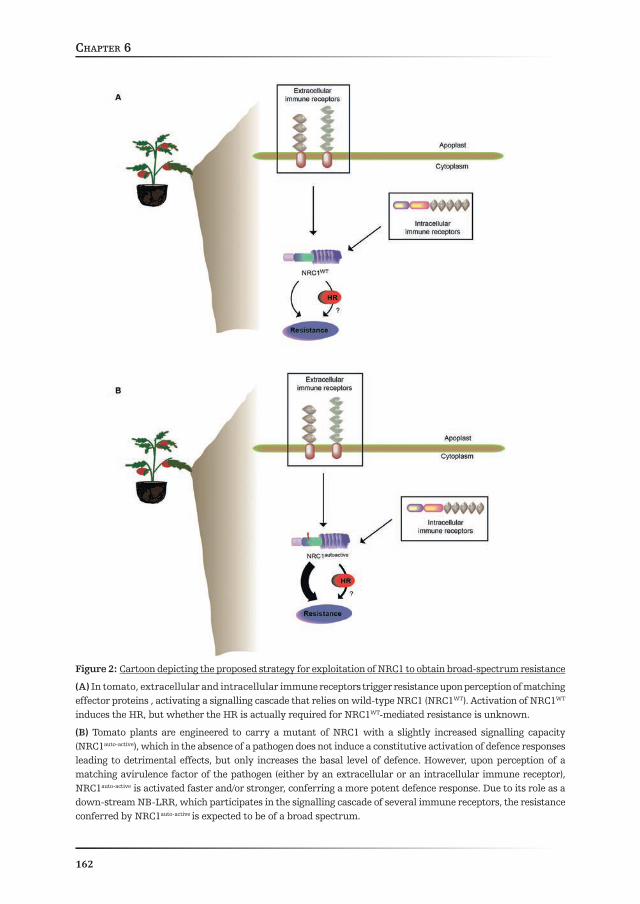
General discussionChapter 6
162
Figure 2: Cartoon depicting the proposed strategy for exploitation of NRC1 to obtain broad-spectrum resistance
(A) In tomato, extracellular and intracellular immune receptors trigger resistance upon perception of matching effector proteins , activating a signalling cascade that relies on wild-type NRC1 (NRC1WT). Activation of NRC1WT induces the HR, but whether the HR is actually required for NRC1WT-mediated resistance is unknown.
(B) Tomato plants are engineered to carry a mutant of NRC1 with a slightly increased signalling capacity (NRC1auto-active), which in the absence of a pathogen does not induce a constitutive activation of defence responses leading to detrimental effects, but only increases the basal level of defence. However, upon perception of a matching avirulence factor of the pathogen (either by an extracellular or an intracellular immune receptor), NRC1auto-active is activated faster and/or stronger, conferring a more potent defence response. Due to its role as a down-stream NB-LRR, which participates in the signalling cascade of several immune receptors, the resistance conferred by NRC1auto-active is expected to be of a broad spectrum.

General discussionChapter 6
163
Is death a path towards resistance…?
NB-LRR-induced HR needs nucleotide binding by the NB subdomain of the immune receptor and therefore depends on the presence of an intact P-loop motif in this subdomain. This is evidenced by the fact that transient over-expression of NB-LRRs carrying a single amino acid substitution in the P-loop motif prevents HR initiation (Dinesh-Kumar et al., 2000; Tao et al., 2000; Bendahmane et al., 2002; Tameling and Joosten, 2007; Williams et al., 2011). Remarkably, two NB-LRRs have been described that provide resistance independently of an intact P-loop motif. Arabidopsis ADR1-L2AAA, which belongs to the already mentioned ADR1 protein family and carries mutations in the three consecutive conserved residues of the P-loop motif, by which nucleotide binding should be fully abolished, is still capable of mediating ETI to avirulent pathogens as well as MTI and basal defence (Bonardi et al., 2011). Still, HR induced by transient expression of ADR1-L2 in N. benthamiana, as well as the dwarf phenotype associated with stable expression of the auto-active mutant ADR1-L2D484V in Arabidopsis, depend on an intact P-loop motif, suggesting that HR and resistance can be uncoupled for ADR1-L2 (Roberts et al., 2013). Rice Pb1 constitutes another example of an NB-LRR providing immunity in a non-canonical way. Pb1 is an atypical CNL protein that confers broad-spectrum resistance to several isolates of the rice blast fungus (M. grisea). Sequence analysis of Pb1 revealed that this NB-LRR does not contain an EDVID motif (HayaSHi et al., 2010), similar to what has been reported for ADR1-likes (Collier et al., 2011). Moreover, most of the motifs of the NB-ARC domain show high degeneration and a P-loop motif cannot be traced (Hayashi et al., 2010), suggesting that Pb1 functioning is different from other NB-LRRs. Whether Pb1 is capable of triggering an HR, either as a requirement for resistance against the blast fungus or not, has not been examined. Considering that Pb1 possesses an MHD motif as most other NB-LRRs, it would be of interest to examine whether a mutant in the D residue of this motif will constitutively induce HR.
Whether NB-LRR-dependent HR and resistance can be uncoupled has been a long-standing issue in plant pathology. In other words, is the HR required for resistance? Moreover, although NB-LRRs are generally studied in relation to their ability to induce an HR, is this really the mechanism by which they contribute to resistance? The above-mentioned examples illustrate that indeed the HR and resistance may be uncoupled for at least some NB-LRRs. Furthermore, the finding that an NB-LRR can induce the HR does not necessarily mean that this capacity is required for providing resistance, as has also been shown for other NB-LRRs such as Rx (Bendahmane et al., 1995). In relation to this, we have identified three missense mutations in the NB subdomain of NRC1 that, despite of negatively affecting the NRC1-dependent HR, they do not compromise Ve1-dependent resistance to V. dahliae of tomato (Chapter 3). Although we cannot exclude that gene redundancy within the NRC1 gene family accounts for this apparent discrepancy, it can also be speculated that NRC1-dependent HR and resistance are based on two pathways that are partly

General discussionChapter 6
164
independent, in such a way that one can be disturbed by a specific mutation without affecting the other.
Interestingly, resistance to V. dahliae is not the only NRC1-dependent defence response that does not rely on HR. Although the HR elicited by a gain-of-function mutant of Rx is dependent on NRC1 (Gabriëls et al., 2007), Rx-mediated resistance to Potato Virus X (PVX) is not dependent on an HR (Bendahmane et al., 1995). In agreement with the finding for Ve1, VIGS with the NRC1 construct in transgenic N. benthamiana plants containing the potato Rx gene were not affected in the resistance to PVX (Gabriels et al., 2007). Requirement of an HR has also never been proven for resistance to C. fulvum in tomato, although recognition of effectors from this fungus by the cognate tomato Cf proteins is known to trigger a strong HR (Joosten and de Wit, 1999; van der Hoorn et al., 2000). In fact, Stulemeijer et al. (2007) demonstrated that VIGS of the tomato gene encoding Mitogen-activated Protein Kinase 2 (SlMPK2) results in a compromised Cf-4/Avr4-triggered HR, whereas the Cf-4-dependent resistance to C. fulvum was not affected. On the other side, VIGS of SlMPK1 resulted in an opposite phenotype, since the Cf-4-mediated resistance to C. fulvum was affected but the Cf-4/Avr4-triggered HR was not compromised (Stulemeijer et al., 2007). Overall, these data indicate that in the tomato-C. fulvum interaction the HR and actual resistance to the pathogen can also be uncoupled. Furthermore, these observations are in line with the proposition that NRC1 might be involved in a signalling pathway leading to pathogen resistance that is independent of the signalling pathway leading to HR in which NRC1 also plays a role. To evaluate this, it would be of interest to investigate whether tomato plants carrying the NRC1 loss-of-function mutations, identified by TILLING (Chapter 3), are also compromised in their resistance to C. fulvum.
By exploiting a biological system that induces a synchronised and systemic Cf-4/Avr4-dependent defence response in tomato seedlings, we observed that the activities of Papain-Like Cysteine Proteases (PLCPs) and Serine Hydrolases (SHs) are differentially regulated, preceding the induction of the hypersensitive cell death (Fig. 1) (Chapter 5). The activity of both groups of proteins specifically increased in transgenic tomato seedlings co-expressing the tomato Cf-4 gene and its corresponding effector gene from C. fulvum, Avr4, when compared to control lines that expressed either only the receptor or the matching effector gene. Interestingly, PLCP- and SH activation preceded the appearance of the Cf-4/Avr4-triggered cell death. Mass spectrometry-based identification of the differentially regulated SHs revealed groups of proteins that have already been related to the HR and in some cases are even considered to be HR markers (Pontier et al., 1994; Pontier et al., 1998). However, we showed that changes in their activity are independent of the execution of the Cf-4/Avr4-triggered hypersensitive cell death, although the activation does depend on the actual perception of Avr4 by Cf-4. Overall, our findings suggest that the recognition of the fungal effector Avr4 by its corresponding immune receptor Cf-4 induces an array of defence responses, of which cell death constitutes only one.
General discussionChapter 6
165
It would be of interest to determine whether the above-mentioned changes in hydrolytic activities of PLCPs and SHs depend on NRC1 (Fig. 1), similarly to what has been shown for the Cf-4/Avr4-triggered PCD in tomato (Gabriëls et al., 2007). Moreover, further studies are required to determine how the change in the hydrolytic activity actually contributes to resistance to C. fulvum. Finally, since the changes in these hydrolytic activities precede the HR and are likely independent of it, they might be interesting targets for breeding programs.
As discussed above, the role of cell death in the NRC1-dependent defence responses against C. fulvum and V. dahliae remains elusive. Although preliminary results suggest that the HR might not be required for NRC1-mediated resistance (Chapter 3), further experiments are required to address this issue in more detail. As mentioned already, ADR1 knockout Arabidopsis plants stably expressing ADR1-L2AAA, which is predicted to be impaired in nucleotide binding and therefore cannot trigger HR, are still capable of mounting MTI and ETI (Bonardi et al., 2012). Similarly, it would be interesting to test the resistance response of tomato plants expressing NRC1K191R, which carries an analogous mutation to that of ADR1-L2AAA and, as such, is thought to be impaired in nucleotide binding and cannot trigger HR (Gabriëls et al., 2007). Introduction of this mutant in an NRC1/NRC2 knockout background, therefore targeting the two more closely related NRC-like sequences (Chapter 3, this thesis), would allow to properly evaluate the requirement of nucleotide binding for the NRC1-mediated defence response. However, such an experiment is still a major challenge when working with tomato, due to the limited genetic resources and the generation time to obtain transgenic plants. Additionally, since the NRC1 gene family is not present in Arabidopsis (Chapter 3), exploiting this model plant system is not an option. Recently developed techniques might allow replacement of the WT allele of NRC1 for that containing the K191R mutation (Nekrasov et al., 2013). This strategy, in combination with VIGS-mediated silencing of NRC2, should result in tomato lines useful to test the requirement of an intact P-loop motif for NRC1-dependent resistance to C. fulvum and/or V. dahliae.
How can a down-stream signalling hub be activated?
As already stated, NRC1 is considered to be a helper NB-LRR that participates in the signalling cascade leading to resistance to C. fulvum and V. dahliae in tomato, down-stream of the plant immune receptors Cf-4 and Ve1, respectively (Gabriëls et al., 2007; Fradin et al., 2009). Moreover, NRC1 is also required for the HR triggered upon the activation of highly diverse immune receptors in N. benthamiana (Gabriëls et al., 2007). A summary of the findings of this thesis is presented in Figure 1. We have shown that NRC1 interacts with HSP70, likely to maintain the protein in a properly-folded manner and ready to be activated (Chapter 2). Interestingly, NRC1-containing protein complexes probably harbour more than one NRC1 protein molecule, as
General discussionChapter 6
166
evidenced by the observation that this NB-LRR forms oligomers in planta (Chapter 3). Moreover, we showed that NRC1 oligomers are formed in the pre-activation state and are disrupted upon activation of the upstream Cf-4 immune receptor, reflecting the dynamics of the NRC1 protein complex. The observation that the oligomer represents the inactive state of NRC1 implies that this protein is inhibited in trans through its interaction with one or more additional NRC1 molecules. This hypothesis is further supported by the finding that the mutation E154K, which is a dominant loss-of-function mutation, blocks disruption of the oligomer upon recognition of Avr4 by Cf-4, thereby affecting the activity of NRC1 in trans. Finally, we identified a set of new mutations that positively affect the activity of NRC1, allowing effector-independent induction of the HR by the protein (Chapter 4).
A major, still to unravel, issue is the identification of the signal that is perceived by NRC1 and triggers its activation. Considering that NRC1 assists a diverse set of plant immune receptors, some of which recognize race-specific effectors (e.g. Rx, I-2, Prf/Pto), whereas others sense fairly conserved molecules (e.g. Cf-4, Ve1, Eix) (Gabriëls et al., 2007), it is very unlikely that NRC1 itself recognizes a molecule of a pathogen. A conserved molecule present in plants, either activated or induced as a general response upon pathogen perception, is a more likely candidate for the NRC1-activating molecule. For instance, ADR1-L2-dependent defence responses have been related to both SA and Reactive Oxygen Species (ROS) induction, suggesting that activation of non-canonical NB-LRR responses might rely on sensing changes in the concentration of endogenous signalling molecules. It would be interesting to investigate whether the NRC1-mediated defence response also depends on SA and ROS, and whether NRC1 is sensing changes in the concentration of any of these molecules.
Going back to animals… can we learn from them?
Animal NLRs are similar in structure and function to plant NB-LRRs, although they seem to have originated independently (Ausubel, 2005). As immune receptors, they trigger several defence pathways upon their activation, thereby orchestrating the animal innate immune response. A major difference between animal NLRs and plant NB-LRRs is that, while canonical NB-LRRs recognize race-specific molecules of pathogens, animal NLRs are considered to be PRRs. As such, NLRs detect conserved molecules as bacterial flagellin or peptidoglycan (Girardin et al., 2003; Girardin et al., 2003; Franchi et al., 2006).
NOD1 and NOD2 are involved in perception of different epitopes of peptidoglycan, a component of the bacterial cell wall (Chamaillard et al., 2003; Girardin et al., 2003; Girardin et al., 2003). Similarly to what has been mentioned for plant NB-LRRs in previous sections, swap experiments wherein the LRR regions of NOD1 and NOD2 (Nucleotide-Oligomerization Domain) were interchanged resulted in

General discussionChapter 6
167
altered recognition specificity, suggesting that the LRR domain of these animal NLRs participates in elicitor recognition (Girardin et al., 2005). Upon sensing of cytosolic bacterial flagellin, as well as components of the bacterial Type Three Secretion System (TTSS), NLRC4 (NLR family CARD domain-containing 4) forms higher-order protein complexes known as inflammasomes (Franchi et al., 2006; Miao et al., 2006; Miao et al., 2010). Remarkably, recent evidence indicates that different members of the NAIP (Neuronal Apoptosis Inhibitory Protein) NLR family are required for NLRC4 activation in mice (Kofoed and Vance, 2011). In fact, mice NAIP5 and NAIP6 are required for flagellin perception, whereas recognition of the rod protein, a component of the bacterial TTSS, relies on NAIP2 (Kofoed and Vance, 2011). This situation clearly resembles the sensor and helper distinction in the NB-LRR field.
An interesting case is that of NLRP3, which is activated by a wide range of signals and participates in immune responses activated upon infection with viral, bacterial and fungal pathogens (Elinav et al., 2011). Although direct interaction of NLRP3 with microbial activators might occur (Marina-García et al., 2008), a more general mechanism is envisioned for the activation of this NLR, involving internal danger-associated factors. Different molecules have been shown to induce the assembly of an NLRP3-containing inflammasome, such as extracellular ATP (eATP), pore-forming bacterial toxins, cathepsin B and other lysosomal enzymes, mitochondria-derived ROS, phospholipids and DNA as well as Endoplasmic Reticulum (ER) stress (Elinav et al., 2011; Monie, 2013; Wen et al., 2013). In addition to ROS, which have already been implicated in the activation of NB-LRRs (Bonardi et al., 2011), it would be interesting to study a possible link between perception of these stress signals and NB-LRR activation. eATP, for instance, has recently become an emerging topic in plant biology, and many publications have illustrated its involvement in developmental processes (Terrile et al., 2010; Clark and Roux, 2011; Hao et al., 2012), as well as in the response to stress (Chivasa et al., 2009; Chivasa et al., 2009; Sun et al., 2012) and mounting of PCD (Sun et al., 2012). Considering the above-mentioned promiscuity of both NRC1 and NLRP3, it is tempting to speculate that NRC1 might also sense disturbances of cellular homeostasis, similarly to what is generally accepted for NLRP3.
Perspective
The mind of a scientist, and likely that of all humans, tends to look for general patterns. Although this simplification is generally very useful to understand processes, in biology often exceptions to the general pattern are the rule. This is also the case in the NB-LRR research field. Extensive research has aimed at the identification of a general mechanism to explain the events occurring before, during and after NB-LRR activation. However, although there are points in common in NB-LRR function, such a general mechanism has so far not been found. In fact,

General discussionChapter 6
168
the diversity of the described signalling cascades suggests that such a common mechanism might not exist.
The crystal structure of some NB-LRR domains have become available over the last couple of years (Bernoux et al., 2011; Maekawa et al., 2011). Additionally, very recently the crystal structure of NLRC4, a mammalian NLR, has been resolved, which has provided insight into its auto-inhibition mechanism (Hu et al., 2013). A prediction is that in the coming years major findings will advance the field of the structural biology of NB-LRRs. This should allow answering how the conformation of NB-LRRs is in their pre- and activated state.
Clearly, the genetic resources needed to engineer plants in the search of durable and broad-spectrum resistance are available and await exploitation. Success in this respect is more likely to be achieved in the context of an integrated approach that not only involves the commercialization of newly generated breeding lines. Even more so, it will fully rely on the concerted efforts of both the scientific community and society.

General discussionChapter 6
169
References
Aarts N, Metz M, Holub E, Staskawicz BJ, Daniels MJ, Parker JE (1998) Different requirements for EDS1 and NDR1 by disease resistance genes define at least two R gene-mediated signaling pathways in Arabidopsis. Proceedings of the National Academy of Sciences of the United States of America 95: 10306-10311
Ade J, DeYoung BJ, Golstein C, Innes RW (2007) Indirect activation of a plant nucleotide binding site-leucine-rich repeat protein by a bacterial protease. Proceedings of the National Academy of Sciences of the United States of America 104: 2531-2536
Austin MJ, Muskett P, Kahn K, Feys BJ, Jones JDG, Parker JE (2002) Regulatory role of SGT1 in early R gene-mediated plant defenses. Science 295: 2077-2080
Ausubel FM (2005) Are innate immune signaling pathways in plants and animals conserved? Nature Immunology 6: 973-979
Axtell MJ, McNellis TW, Mudgett MB, Hrsu CS, Staskawicz BJ (2001) Mutational analysis of the Arabidopsis RPS2 disease resistance gene and the corresponding Pseudomonas syringae avrRpt2 avirulence gene. Molecular Plant-Microbe Interactions 14: 181-188
Axtell MJ, Staskawicz BJ (2003) Initiation of RPS2-specified disease resistance in Arabidopsis is coupled to the AvrRpt2-directed elimination of RIN4. Cell 112: 369-377
Azevedo C, Betsuyaku S, Peart J, Takahashi A, Noël L, Sadanandom A, Casais C, Parker J, SHirasu K (2006) Role of SGT1 in resistance protein accumulation in plant immunity. EMBO Journal 25: 2007-2016
Bendahmane A, Farnham G, Moffett P, Baulcombe DC (2002) Constitutive gain-of-function mutants in a nucleotide binding site-leucine rich repeat protein encoded at the Rx locus of potato. Plant Journal 32: 195-204
Bendahmane A, Kohm BA, Dedi C, Baulcombe DC (1995) The coat protein of potato virus X is a strain-specific elicitor of Rx1-mediated virus resistance in potato. Plant Journal 8: 933-941
Bent AF, Kunkel BN, Dahlbeck D, Brown KL, Schmidt R, Giraudat J, Leung J, Staskawicz BJ (1994) RPS2 of Arabidopsis thaliana: A leucine-rich repeat class of plant disease resistance genes. Science 265: 1856-1860
Bernoux M, Ve T, Williams S, Warren C, Hatters D, Valkov E, Zhang X, Ellis JG, Kobe B, Dodds PN (2011) Structural and functional analysis of a plant resistance protein TIR domain reveals interfaces for self-association, signaling, and autoregulation. Cell Host and Microbe 9: 200-211
Bieri S, Mauch S, SHen QH, Peart J, Devoto A, Casais C, Ceron F, Schulze S, Steinbiß HH, SHirasu K, Schulze-Lefert P (2004) RAR1 positively controls steady state levels of barley MLA resistance proteins and enables sufficient MLA6 accumulation for effective resistance. Plant Cell 16: 3480-3495
Birker D, Heidrich K, Takahara H, Narusaka M, Deslandes L, Narusaka Y, Reymond M, Parker JE, O’Connell R (2009) A locus conferring resistance to Colletotrichum higginsianum is SHared by four geographically distinct Arabidopsis accessions. Plant Journal 60: 602-613
Bolton MD, Van Esse HP, Vossen JH, De Jonge R, Stergiopoulos I, Stulemeijer IJE, Van Den Berg GCM, Borrás-Hidalgo O, Dekker HL, De Koster CG, De Wit PJGM, Joosten MHAJ, Thomma BPHJ (2008) The novel Cladosporium fulvum lysin motif effector Ecp6 is a virulence factor with orthologues in other fungal species. Molecular Microbiology 69: 119-136
Bombarely A, Rosli HG, Vrebalov J, Moffett P, Mueller LA, Martin GB (2012) A draft genome sequence of Nicotiana benthamiana to enhance molecular plant-microbe biology research. Molecular Plant-Microbe Interactions 25: 1523-1530
Bonardi V, Cherkis K, NiSHimura MT, Dangl JL (2012) A new eye on NLR proteins: Focused on clarity or diffused by complexity? Current Opinion in Immunology 24: 41-50
Bonardi V, Dangl JL (2012) How complex are intracellular immune receptor signaling complexes? Front Plant Sci 3: 237
General discussionChapter 6
170
Bonardi V, Tang S, Stallmann A, Roberts M, Cherkis K, Dangl JL (2011) Expanded functions for a family of plant intracellular immune receptors beyond specific recognition of pathogen effectors. Proceedings of the National Academy of Sciences of the United States of America 108: 16463-16468
Botër M, Amigues B, Peart J, Breuer C, Kadota Y, Casais C, Moore G, Kleanthous C, Ochrsenbein F, SHirasu K, Guerois R (2007) Structural and functional analysis of SGT1 reveals that its interaction with HRSP90 is required for the accumulation of Rx, an R protein involved in plant immunity. Plant Cell 19: 3791-3804
Breddam K (1986) Serine carboxypeptidases. A review. Carlsberg Research Communications 51: 83-128Brennan MA, Cookson BT (2000) Salmonella induces macrophage death by caspase-1-dependent necrosis.
Molecular Microbiology 38: 31-40Brighenti FL, Luppens SBI, Delbem ACB, Deng DM, Hoogenkamp MA, Gaetti-Jardim Jr E, Dekker
HL, Crielaard W, Ten Cate JM (2008) Effect of psidium cattleianum leaf extract on streptococcus mutans viability, protein expression and acid production. Caries Research 42: 148-154
Brodsky JL, Chiosis G (2006) HSP70 molecular chaperones: Emerging roles in human disease and identification of small molecule modulators. Current Topics in Medicinal Chemistry 6: 1215-1225
Brunner S, Hurni S, Streckeisen P, Mayr G, Albrecht M, Yahiaoui N, Keller B (2010) Intragenic allele pyramiding combines different specificities of wheat Pm3 resistance alleles. Plant Journal 64: 433-445
Bulgarelli D, Biselli C, Collins NC, Consonni G, Stanca AM, Schulze-Lefert P, Valè G (2010) The CC-NB-LRR-Type RDG2a resistance gene confers immunity to the seed-borne barley leaf stripe pathogen in the absence of hypersensitive cell death. PLoS ONE 5: 1-14
Caplan JL, Mamillapalli P, Burch-Smith TM, Czymmek K, DineSH-Kumar SP (2008) Chloroplastic protein NRIP1 mediates innate immune receptor recognition of a viral effector. Cell 132: 449-462
Cecconi F, Alvarez-Bolado G, Meyer BI, Roth KA, Gruss P (1998) Apaf1 (CED-4 homolog) regulates programmed cell death in mammalian development. Cell 94: 727-737
Century KS, Holub EB, Staskawicz BJ (1995) NDR1, a locus of Arabidopsis thaliana that is required for disease resistance to both a bacterial and a fungal pathogen. Proceedings of the National Academy of Sciences of the United States of America 92: 6597-6601
Cervantes J, Nagata T, Uchijima M, Shibata K, Koide Y (2008) Intracytosolic Listeria monocytogenes induces cell death through caspase-1 activation in murine macrophages. Cellular Microbiology 10: 41-52
Cesari S, Thilliez G, Ribot C, Chalvon V, Michel C, Jauneau A, Rivas S, Alaux L, Kanzaki H, Okuyama Y, Morel JB, Fournier E, Tharreau D, Terauchi R, Kroj T (2013) The rice resistance protein pair RGA4/RGA5 recognizes the Magnaporthe oryzae effectors AVR-Pia and AVR1-CO39 by direct binding. Plant Cell 25: 1463-1481
Chamaillard M, Hashimoto M, Horie Y, Masumoto J, Qiu S, Saab L, Ogura Y, Kawasaki A, Fukase K, Kusumoto S, Valvano MA, Foster SJ, Mak TW, Nuñez G, Inohara N (2003) An essential role for NOD1 in host recognition of bacterial peptidoglycan containing diaminopimelic acid. Nature Immunology 4: 702-707
Chen L, Zhang L, Li D, Wang F, Yu D (2013) WRKY8 transcription factor functions in the TMV-cg defense response by mediating both abscisic acid and ethylene signaling in Arabidopsis. Proceedings of the National Academy of Sciences of the United States of America 110: 1963-1971
Chichkova NV, SHaw J, Galiullina RA, Drury GE, Tuzhikov AI, Kim SH, Kalkum M, Hong TB, GorSHkova EN, Torrance L, Vartapetian AB, Taliansky M (2010) Phytaspase, a relocalisable cell death promoting plant protease with caspase specificity. EMBO Journal 29: 1149-1161
Chinchilla D, Bauer Z, Regenass M, Boller T, Felix G (2006) The Arabidopsis receptor kinase FLS2 binds flg22 and determines the specificity of flagellin perception. Plant Cell 18: 465-476
Chinnaiyan AM, Chaudhary D, O’Rourke K, Koonin EV, Dixit VM (1997) Role of CED-4 in the activation of CED-3 [9]. Nature 388: 728-729
Chinnaiyan AM, O’Rourke K, Lane BR, Dixit VM (1997) Interaction of CED-4 with CED-3 and CED-9: A molecular framework for cell death. Science 275: 1122-1126

General discussionChapter 6
171
Chivasa S, Murphy AM, Hamilton JM, Lindsey K, Carr JP, Slabas AR (2009) Extracellular ATP is a regulator of pathogen defence in plants. Plant Journal 60: 436-448
Chivasa S, Tomé DF, Murphy AM, Hamilton JM, Lindsey K, Carr JP (2009) Extracellular ATP: a modulator of cell death and pathogen defense in plants. Plant signaling & behavior 4: 1078-1080
Clark G, Roux SJ (2011) Apyrases, extracellular ATP and the regulation of growth. Current Opinion in Plant Biology 14: 700-706
Clough SJ, Fengler KA, Yu IC, Lippok B, Smith Jr RK, Bent AF (2000) The Arabidopsis dnd1 “defense, no death” gene encodes a mutated cyclic nucleotide-gated ion channel. Proceedings of the National Academy of Sciences of the United States of America 97: 9323-9328
Coll NS, Epple P, Dangl JL (2011) Programmed cell death in the plant immune system. Cell Death and Differentiation 18: 1247-1256
Coll NS, Vercammen D, Smidler A, Clover C, van Breusegem F, Dangl JL, Epple P (2010) Arabidopsis type I metacaspases control cell death. Science 330: 1393-1397
Collier SM, Hamel LP, Moffett P (2011) Cell death mediated by the N-terminal domains of a unique and highly conserved class of NB-LRR protein. Molecular Plant-Microbe Interactions 24: 918-931
Collier SM, Moffett P (2009) NB-LRRs work a “bait and switch” on pathogens. Trends in Plant Science 14: 521-529
Corpet F (1988) Multiple sequence alignment with hierarchical clustering. Nucleic Acids Research 16: 10881-10890
Cravatt BF, Wright AT, Kozarich JW (2008) Activity-based protein profiling: From enzyme chemistry to proteomic chemistry. In Annual Review of Biochemistry, Vol 77, pp 383-414
Crute IR (1994) Gene-for-gene recognition in plant-pathogen interactions. Philosophical Transactions - Royal Society of London, B 346: 345-349
Damm A, Lautz K, Kufer TA (2013) Roles of NLRP10 in innate and adaptive immunity. Microbes and Infection 15: 516-523
Dangl JL, Horvath DM, Staskawicz BJ (2013) Pivoting the plant immune system from dissection to deployment. Science 341: 746-751
Dangl JL, Jones JDG (2001) Plant pathogens and integrated defence responses to infection. Nature 411: 826-833
Danot O, Marquenet E, Vidal-Ingigliardi D, Richet E (2009) Wheel of life, wheel of death: A mechanistic insight into signaling by STAND proteins. Structure 17: 172-182
de Jong CF, Takken FLW, Cai X, de Wit PJGM, Joosten MHAJ (2002) Attenuation of Cf-mediated defense responses at elevated temperatures correlates with a decrease in elicitor-binding sites. Molecular Plant-Microbe Interactions 15: 1040-1049
de Jonge R, van Esse HP, Kombrink A, Shinya T, Desaki Y, Bours R, van der Krol S, Shibuya N, Joosten MHAJ, Thomma BPHJ (2010) Conserved fungal LysM effector Ecp6 prevents chitin-triggered immunity in plants. Science 329: 953-955
de Jonge R, van Esse HP, Maruthachalam K, Bolton MD, Santhanam P, Saber MK, Zhang Z, Usami T, Lievens B, Subbarao KV, Thomma BPHJ (2012) Tomato immune receptor Ve1 recognizes effector of multiple fungal pathogens uncovered by genome and RNA sequencing. Proceedings of the National Academy of Sciences of the United States of America 109: 5110-5115
de la Fuente van Bentem S, Vossen JH, de Vries KJ, van Wees S, Tameling WIL, Dekker HL, de Koster CG, Haring MA, Takken FLW, Cornelissen BJC (2005) Heat SHock protein 90 and its co-chaperone protein phosphatase 5 interact with distinct regions of the tomato I-2 disease resistance protein. Plant Journal 43: 284-298
de Wit PJGM (1977) A light and scanning-electron microscopic study of infection of tomato plants by virulent and avirulent races of Cladosporium fulvum. Netherlands Journal of Plant Pathology 83: 109-122
de Wit PJGM, Brandwagt BF, van den Burg HA, Cai X, van der Hoorn RAL, de Jong CF, van ‘t Klooster J, de Kock MJD, Kruijt M, Lindhout WH, Luderer R, Takken FLW, Westerink N, Vervoort JJM, Joosten MHAJ (2002) The molecular basis of co-evolution between Cladosporium fulvum and tomato. Antonie van Leeuwenhoek, International Journal of General and Molecular Microbiology 81: 409-412

General discussionChapter 6
172
Debener T, Lehnackers H, Arnold M, Dangl JL (1991) Identification and molecular mapping of a single Arabidopsis thaliana locus determining resistance to a phytopathogenic Pseudomonas syringae isolate. Plant Journal 1: 289-302
Dinesh-Kumar SP, Tham WH, Baker BJ (2000) Structure-function analysis of the tobacco mosaic virus resistance gene N. Proceedings of the National Academy of Sciences of the United States of America 97: 14789-14794
Dodds PN, Lawrence GJ, Catanzariti AM, Teh T, Wang CIA, Ayliffe MA, Kobe B, Ellis JG (2006) Direct protein interaction underlies gene-for-gene specificity and coevolution of the flax resistance genes and flax rust avirulence genes. Proceedings of the National Academy of Sciences of the United States of America 103: 8888-8893
Dodds PN, Lawrence GJ, Ellis JG (2001) Six amino acid changes confined to the leucine-rich repeat β-strand/β-turn motif determine the difference between the P and P2 rust resistance specificities in flax. Plant Cell 13: 163-178
Dosztányi Z, Csizmok V, Tompa P, Simon I (2005) IUPred: Web server for the prediction of intrinsically unstructured regions of proteins based on estimated energy content. Bioinformatics 21: 3433-3434
Du X, Miao M, Ma X, Liu Y, Kuhl JC, Martin GB, Xiao F (2012) Plant programmed cell death caused by an auto-active form of Prf is suppressed by co-expression of the Prf LRR domain. Molecular Plant 5: 1058-1067
Duncan JA, Bergstralh DT, Wang Y, Willingham SB, Ye Z, Zimmermann AG, Ting JPY (2007) Cryopyrin/NALP3 binds ATP/dATP, is an ATPase, and requires ATP binding to mediate inflammatory signaling. Proceedings of the National Academy of Sciences of the United States of America 104: 8041-8046
Eisenbarth SC, Williams A, Colegio OR, Meng H, Strowig T, Rongvaux A, Henao-Mejia J, Thaiss CA, Joly S, Gonzalez DG, Xu L, Zenewicz LA, Haberman AM, Elinav E, Kleinstein SH, Sutterwala FS, Flavell RA (2012) NLRP10 is a NOD-like receptor essential to initiate adaptive immunity by dendritic cells. Nature 484: 510-513
Eitas TK, Dangl JL (2010) NB-LRR proteins: Pairs, pieces, perception, partners, and pathways. Current Opinion in Plant Biology 13: 472-477
Eitas TK, Nimchuk ZL, Dangl JL (2008) Arabidopsis TAO1 is a TIR-NB-LRR protein that contributes to disease resistance induced by the Pseudomonas syringae effector AvrB Proceedings of the National Academy of Sciences of the United States of America 105: 15219
Elinav E, Strowig T, Henao-Mejia J, Flavell R (2011) Regulation of the Antimicrobial Response by NLR Proteins. Immunity 34: 665-679
Etalo DW, Stulemeijer IJE, Peter van Esse H, de Vos RCH, Bouwmeester HJ, Joosten MHAJ (2013) System-wide hypersensitive response-associated transcriptome and metabolome reprogramming in tomato. Plant Physiology 162: 1599-1617
Finn RD, Mistry J, Tate J, Coggill P, Heger A, Pollington JE, Gavin OL, Gunasekaran P, Ceric G, Forslund K, Holm L, Sonnhammer ELL, Eddy SR, Bateman A (2010) The Pfam protein families database. Nucleic Acids Research 38: D211-D222
Flor HH (1971) Current status of gene-for-gene concept. Annual Review of Phytopathology 9: 275-&Fradin EF, Abd-El-Haliem A, Masini L, van den Berg GCM, Joosten MHAJ, Thomma BPHJ (2011)
Interfamily transfer of tomato ve1 mediates Verticillium resistance in Arabidopsis. Plant Physiology 156: 2255-2265
Fradin EF, Zhang Z, Ayala JCJ, Castroverde CDM, Nazar RN, Robb J, Liu CM, Thomma BPHJ (2009) Genetic dissection of verticillium wilt resistance mediated by tomato Ve1. Plant Physiology 150: 320-332
Franchi L, Amer A, Body-Malapel M, Kanneganti TD, Özören N, Jagirdar R, Inohara N, Vandenabeele P, Bertin J, Coyle A, Grant EP, Núñez G (2006) Cytosolic flagellin requires Ipaf for activation of caspase-1 and interleukin 1β in Salmonella-infected macrophages. Nature Immunology 7: 576-582
Fritz JH, Ferrero RL, Philpott DJ, Girardin SE (2006) Nod-like proteins in immunity, inflammation and disease. Nature Immunology 7: 1250-1257

General discussionChapter 6
173
Gabriëls SHEJ, Takken FLW, Vossen JH, de Jong CF, Liu Q, Turk SCHJ, Wachowski LK, Peters J, Witsenboer HMA, de Wit PJGM, Joosten MHAJ (2006) cDNA-AFLP combined with functional analysis reveals novel genes involved in the hypersensitive response. Molecular Plant-Microbe Interactions 19: 567-576
Gabriëls SHEJ, Vossen JH, Ekengren SK, Ooijen GV, Abd-El-Haliem AM, Berg GCMVD, Rainey DY, Martin GB, Takken FLW, Wit PJGMD, Joosten MHAJ (2007) An NB-LRR protein required for HR signalling mediated by both extra- and intracellular resistance proteins. Plant Journal 50: 14-28
Gabriëls SHEJ, Vossen JH, Ekengren SK, van Ooijen G, Abd-El-Haliem AM, van den Berg GCM, Rainey DY, Martin GB, Takken FLW, de Wit PJGM, Joosten MHAJ (2007) An NB-LRR protein required for HR signalling mediated by both extra- and intracellular resistance proteins. Plant Journal 50: 14-28
Gao Z, Chung EH, Eitas TK, Dangl JL (2011) Plant intracellular innate immune receptor resistance to Pseudomonas syringae pv. maculicola 1 (RPM1) is activated at, and functions on, the plasma membrane. Proceedings of the National Academy of Sciences of the United States of America 108: 8915
Gao Z, Chung EH, Eitas TK, Dangl JL (2011) Plant intracellular innate immune receptor Resistance to Pseudomonas syringae pv. maculicola 1 (RPM1) is activated at, and functions on, the plasma membrane. Proceedings of the National Academy of Sciences of the United States of America 108: 7619-7624
Gao Z, Chung EH, Eitas TK, Dangl JL (2011) Plant intracellular innate immune receptor resistance to pseudomonas syringae pv. maculicola 1 (RPM1) is activated at, and functions on, the plasma membrane (Proceedings of the National Academy of Sciences of the United States of America (2011) 108, 18 (7619-7624) DOI: 10.1073/pnas.1104410108). Proceedings of the National Academy of Sciences of the United States of America 108: 8915
Gassmann W, Dahlbeck D, Chesnokova O, Minsavage GV, Jones JB, Staskawicz BJ (2000) Molecular evolution of virulence in natural field strains of Xanthomonas campestris pv. vesicatoria. Journal of Bacteriology 182: 7053-7059
Girardin SE, Boneca IG, Carneiro LAM, Antignac A, Jéhanno M, Viala J, Tedin K, Taha MK, Labigne A, Zähringer U, Coyle AJ, DiStefano PS, Bertin J, Sansonetti PJ, Philpott DJ (2003) Nod1 detects a unique muropeptide from gram-negative bacterial peptidoglycan. Science 300: 1584-1587
Girardin SE, Boneca IG, Viala J, Chamaillard M, Labigne A, Thomas G, Philpott DJ, Sansonetti PJ (2003) Nod2 is a general sensor of peptidoglycan through muramyl dipeptide (MDP) detection. Journal of Biological Chemistry 278: 8869-8872
Girardin SE, Jéhanno M, Mengin-Lecreulx D, Sansonetti PJ, Alzari PM, Philpott DJ (2005) Identification of the critical residues involved in peptidoglycan detection by Nod1. Journal of Biological Chemistry 280: 38648-38656
Goggin FL, Jia L, Shah G, Hebert S, Williamson VM, Ullman DE (2006) Heterologous expression of the Mi-1.2 gene from tomato confers resistance against nematodes but not aphids in eggplant. Molecular Plant-Microbe Interactions 19: 383-388
Göhre V, Spallek T, Häweker H, Mersmann S, Mentzel T, Boller T, de Torres M, Mansfield JW, Robatzek S (2008) Plant pattern-recognition receptor FLS2 is directed for degradation by the bacterial ubiquitin ligase AvrPtoB. Current Biology 18: 1824-1832
Gómez-Gómez L, Boller T (2000) FLS2: An LRR receptor-like kinase involved in the perception of the bacterial elicitor flagellin in Arabidopsis. Molecular Cell 5: 1003-1011
Gottar M, Gobert V, Michel T, Belvin M, Duyk G, Hoffmann JA, Ferrandon D, Royet J (2002) The Drosophila immune response against Gram-negative bacteria is mediated by a peptidoglycan recognition protein. Nature 416: 640-644
Gou M, Hua J (2012) Complex regulation of an R gene SNC1 revealed by auto-immune mutants. Plant signaling & behavior 7: 213-216

General discussionChapter 6
174
Granell A, Bellés JM, Conejero V (1987) Induction of pathogenesis-related proteins in tomato by citrus exocortis viroid, silver ion and ethephon. Physiological and Molecular Plant Pathology 31: 83-90
Grant JJ, Chini A, Basu D, Loake GJ (2003) Targeted activation tagging of the Arabidopsis NBS-LRR gene, ADR1, conveys resistance to virulent pathogens. Molecular Plant-Microbe Interactions 16: 669-680
Grant MR, Godiard L, Straube E, Ashfield T, Lewald J, Sattler A, Innes RW, Dangl JL (1995) Structure of the arabidopsis RPM1 gene enabling dual specificity disease resistance. Science 269: 843-846
Grant MR, Kazan K, Manners JM (2013) Exploiting pathogens’ tricks of the trade for engineering of plant disease resistance: Challenges and opportunities. Microbial Biotechnology 6: 212-222
Greenbaum D, Medzihradszky KF, Burlingame A, Bogyo M (2000) Epoxide electrophiles as activity-dependent cysteine protease profiling and discovery tools. Chemistry and Biology 7: 569-581
Gu C, Kolodziejek I, Misas-Villamil J, Shindo T, Colby T, Verdoes M, Richau KH, Schmidt J, Overkleeft HRS, van der Hoorn RAL (2010) Proteasome activity profiling: A simple, robust and versatile method revealing subunit-selective inhibitors and cytoplasmic, defense-induced proteasome activities. Plant Journal 62: 160-170
Guo M, Tian F, Wamboldt Y, Alfano JR (2009) The majority of the type III effector inventory of Pseudomonas syringae pv. tomato DC3000 can suppress plant immunity. Molecular Plant-Microbe Interactions 22: 1069-1080
Gutierrez JR, Balmuth AL, Ntoukakis V, Mucyn TS, Gimenez-Ibanez S, Jones AME, Rathjen JP (2010) Prf immune complexes of tomato are oligomeric and contain multiple Pto-like kinases that diversify effector recognition. Plant Journal 61: 507-518
Hamilton CM (1997) A binary-BAC system for plant transformation with high-molecular-weight DNA. Gene 200: 107-116
Hammond-Kosack KE, Silverman P, Raskin I, Jones JDG (1996) Race-specific elicitors of Cladosporium fulvum induce changes in cell morphology and the synthesis of ethylene and salicylic acid in tomato plants carrying the corresponding Cf disease resistance gene. Plant Physiology 110: 1381-1394
Hao LH, Wang WX, Chen C, Wang YF, Liu T, Li X, SHang ZL (2012) Extracellular ATP Promotes stomatal opening of Arabidopsis thaliana through heterotrimeric G protein α subunit and reactive oxygen species. Molecular Plant 5: 852-864
Hara-Nishimura I, Hatsugai N, Nakaune S, Kuroyanagi M, Nishimura M (2005) Vacuolar processing enzyme: An executor of plant cell death. Current Opinion in Plant Biology 8: 404-408
Harris CJ, Slootweg EJ, Goverse A, Baulcombe DC (2013) Stepwise artificial evolution of a plant disease resistance gene. Proceedings of the National Academy of Sciences
Hatsugai N, Iwasaki S, Tamura K, Kondo M, Fuji K, Ogasawara K, Nishimura M, Hara-Nishimura I (2009) A novel membrane fusion-mediated plant immunity against bacterial pathogens. Genes and Development 23: 2496-2506
Hatsugai N, Kuroyanagi M, Yamada K, MeSHi T, Tsuda S, Kondo M, Nishimura M, Hara-Nishimura I (2004) A plant vacuolar protease, VPE, mediates, virus-induced hypersensitive cell death. Science 305: 855-858
Hayashi N, Inoue H, Kato T, Funao T, Shirota M, Shimizu T, Kanamori H, Yamane H, Hayano-Saito Y, Matsumoto T, Yano M, Takatsuji H (2010) Durable panicle blast-resistance gene Pb1 encodes an atypical CC-NBS-LRR protein and was generated by acquiring a promoter through local genome duplication. Plant Journal 64: 498-510
Heath MC (2000) Hypersensitive response-related death. Plant Molecular Biology 44: 321-334Horvath DM, Stall RE, Jones JB, Pauly MH, Vallad GE, Dahlbeck D, Staskawicz BJ, Scott JW (2012)
Transgenic resistance confers effective field level control of bacterial spot disease in tomato. PLoS ONE 7
Hou S, Zhang C, Yang Y, Wu D (2013) Recent advances in plant immunity: Recognition, signaling, response, and evolution. Biologia Plantarum 57: 11-25
General discussionChapter 6
175
Howles P, Lawrence G, Finnegan J, McFadden H, Ayliffe M, Dodds P, Ellis J (2005) Auto-active alleles of the flax L6 rust resistance gene induce non-race-specific rust resistance associated with the hypersensitive response. Molecular Plant-Microbe Interactions 18: 570-582
Hu Z, Yan C, Liu P, Huang Z, Ma R, Zhang C, Wang R, Zhang Y, Martinon F, Miao D, Deng H, Wang J, Chang J, Chai J (2013) Crystal structure of NLRC4 reveals its autoinhibition mechanism. Science 341: 172-175
Hua Z, Hou B (2013) TLR signaling in B-cell development and activation. Cellular and Molecular Immunology 10: 103-106
Huang X, Li J, Bao F, Zhang X, Yang S (2010) A gain-of-function mutation in the Arabidopsis disease resistance gene RPP4 confers sensitivity to low temperature. Plant Physiology 154: 796-809
Hubert DA, He Y, McNulty BC, Tornero P, Dangl JL (2009) Specific Arabidopsis HRSP90.2 alleles recapitulate RAR1 cochaperone function in plant NB-LRR disease resistance protein regulation. Proceedings of the National Academy of Sciences of the United States of America 106: 9556-9563
Hubert DA, Tornero P, Belkhadir Y, KriSHna P, TakahaSHi A, SHirasu K, Dangl JL (2003) Cytosolic HRSP90 associates with and modulates the Arabidopsis RPM1 disease resistance protein. EMBO Journal 22: 5679-5689
Hwang CF, Williamson VM (2003) Leucine-rich repeat-mediated intramolecular interactions in nematode recognition and cell death signaling by the tomato resistance protein Mi. Plant Journal 34: 585-593
Igari K, Endo S, Hibara KI, Aida M, Sakakibara H, Kawasaki T, Tasaka M (2008) Constitutive activation of a CC-NB-LRR protein alters morphogenesis through the cytokinin pathway in Arabidopsis. Plant Journal 55: 14-27
Inoue H, HayaSHi N, MatsuSHita A, Xinqiong L, Nakayama A, Sugano S, Jiang CJ, Takatsuji H (2013) Blast resistance of CC-NB-LRR protein Pb1 is mediated by WRKY45 through protein-protein interaction. Proceedings of the National Academy of Sciences of the United States of America 110: 9577-9582
Iyer LM, Leipe DD, Koonin EV, Aravind L (2004) Evolutionary history and higher order classification of AAA+ ATPases. Journal of Structural Biology 146: 11-31
Jambunathan N, Siani JM, McNellis TW (2001) A humidity-sensitive Arabidopsis copine mutant exhibits precocious cell death and increased disease resistance. Plant Cell 13: 2225-2240
Jones JDG, Dangl JL (2006) The plant immune system. Nature 444: 323-329Joosten MHAJ, Cozijnsen TJ, de Wit PJGM (1994) Host resistance to a fungal tomato pathogen lost by a
single base-pair change in an avirulence gene. Nature 367: 384-386Joosten MHAJ, de Wit PJGM (1988) Isolation, purification and preliminary characterization of a protein
specific for compatible Cladosporium fulvum (syn. Fulvia fulva)-tomato interactions. Physiological and Molecular Plant Pathology 33: 241-253
Joosten MHAJ, De Wit PJGM (1999) The tomato-Cladosporium fulvum interaction: A versatile experimental system to study plant-pathogen interactions. Annual Review of Phytopathology 37: 335-367
Joosten MHAJ, Vogelsang R, Cozijnsen TJ, Verberne MC, de Wit PJGM (1997) The biotrophic fungus Cladosporium fulvum circumvents Cf-4-mediated resistance by producing unstable AVR4 elicitors. Plant Cell 9: 367-379
Jordá L, Coego A, Conejero V, Vera P (1999) A genomic cluster containing four differentially regulated subtilisin- like processing protease genes is in tomato plants. Journal of Biological Chemistry 274: 2360-2365
Jorda L, Conejero V, Vera P (2000) Characterization of P69E and P69F, two differentially regulated genes encoding new members of the subtilisin-like proteinase family from tomato plants. Plant Physiology 122: 67-73
Jupe F, Pritchard L, Etherington GJ, MacKenzie K, Cock PJA, Wright F, SHarma SK, Bolser D, Bryan GJ, Jones JDG, Hein I (2012) Identification and localisation of the NB-LRR gene family within the potato genome. BMC Genomics 13

General discussionChapter 6
176
Kacprzyk J, Daly CT, McCabe PF (2011) The botanical dance of death. Programmed cell death in plants. In Advances in Botanical Research, Vol 60, pp 169-261
Kaku H, NiSHizawa Y, ISHii-Minami N, Akimoto-Tomiyama C, Dohmae N, Takio K, Minami E, SHibuya N (2006) Plant cells recognize chitin fragments for defense signaling through a plasma membrane receptor. Proceedings of the National Academy of Sciences of the United States of America 103: 11086-11091
Kang BC, Yeam I, Jahn MM (2005) Genetics of plant virus resistance. In Annual Review of Phytopathology, Vol 43, pp 581-621
Kanzaki H, Saitoh H, Ito A, Fujisawa S, Kamoun S, Katou S, YoSHioka H, Terauchi R (2003) Cytosolic HRSP90 and HSP70 are essential components of INF1-mediated hypersensitive response and non-host resistance to Pseudomonas cichorii in Nicotiana benthamiana. Molecular Plant Pathology 4: 383-391
Kanzaki H, YoSHida K, Saitoh H, Fujisaki K, Hirabuchi A, Alaux L, Fournier E, Tharreau D, Terauchi R (2012) Arms race co-evolution of Magnaporthe oryzae AVR-Pik and rice Pik genes driven by their physical interactions. Plant Journal 72: 894-907
Kaschani F, Gu C, Niessen S, Hoover H, Cravatt BF, van der Hoorn RAL (2009) Diversity of serine hydrolase activities of unchallenged and Botrytis-infected Arabidopsis thaliana. Molecular and Cellular Proteomics 8: 1082-1093
Kawano Y, Akamatsu A, HayaSHi K, Housen Y, Okuda J, Yao A, NakaSHima A, TakahaSHi H, YoSHida H, Wong HL, Kawasaki T, SHimamoto K (2010) Activation of a Rac GTPase by the NLR family disease resistance protein Pit plays a critical role in rice innate immunity. Cell Host and Microbe 7: 362-375
Kofoed EM, Vance RE (2011) Innate immune recognition of bacterial ligands by NAIPs determines inflammasome specificity. Nature 477: 592-597
Kołodziejek I, van der Hoorn RAL (2010) Mining the active proteome in plant science and biotechnology. Current Opinion in Biotechnology 21: 225-233
Koonin EV, Aravind L (2000) The NACHT family - A new group of predicted NTPases implicated in apoptosis and MHC transcription activation. Trends in Biochemical Sciences 25: 223-224
Kou Y, Wang S (2010) Broad-spectrum and durability: understanding of quantitative disease resistance. Current Opinion in Plant Biology 13: 181-185
Krasileva KV, Dahlbeck D, Staskawicz BJ (2010) Activation of an Arabidopsis resistance protein is specified by the in planta association of its leucine-rich repeat domain with the cognate oomycete effector. Plant Cell 22: 2444-2458
Kud J, Zhao Z, Du X, Liu Y, Zhao Y, Xiao F (2013) SGT1 interacts with the Prf resistance protein and is required for Prf accumulation and Prf-mediated defense signaling. Biochemical and Biophysical Research Communications 431: 501-505
Kumagai MH, Donson J, Della-Cioppa G, Harvey D, Hanley K, Grill LK (1995) Cytoplasmic inhibition of carotenoid biosynthesis with virus-derived RNA. Proceedings of the National Academy of Sciences of the United States of America 92: 1679-1683
Lacombe S, Rougon-Cardoso A, SHerwood E, Peeters N, Dahlbeck D, Van Esse HP, Smoker M, Rallapalli G, Thomma BPHJ, Staskawicz B, Jones JDG, Zipfel C (2010) Interfamily transfer of a plant pattern-recognition receptor confers broad-spectrum bacterial resistance. Nature Biotechnology 28: 365-369
Leipe DD, Koonin EV, Aravind L (2004) STAND, a class of P-loop NTPases including animal and plant regulators of programmed cell death: Multiple, complex domain architectures, unusual phyletic patterns, and evolution by horizontal gene transfer. Journal of Molecular Biology 343: 1-28
Li AX, Steffens JC (2000) An acyltransferase catalyzing the formation of diacylglucose is a serine carboxypeptidase-like protein. Proceedings of the National Academy of Sciences of the United States of America 97: 6902-6907

General discussionChapter 6
177
Li X, Clarke JD, Zhang Y, Dong X (2001) Activation of an EDS1-mediated R-gene pathway in the snc1 mutant leads to constitutive, NPR1-independent pathogen resistance. Molecular Plant-Microbe Interactions 14: 1131-1139
Liebrand TWH, Smit P, Abd-El-Haliem A, de Jonge R, Cordewener JHG, America AHP, Sklenar J, Jones AME, Robatzek S, Thomma BPHJ, Tameling WIL, Joosten MHA (2012) Endoplasmic reticulum-quality control chaperones facilitate the biogenesis of Cf receptor-like proteins involved in pathogen resistance of tomato. Plant Physiology 159: 1819-1833
Lin K-Y, Hrsu Y-H, Chen H-C, Lin N-S (2013) Transgenic resistance to Bamboo mosaic virus by expression of interfering satellite RNA. Molecular Plant Pathology 14: 693-707
Linding R, Jensen LJ, Diella F, Bork P, Gibson TJ, Russell RB (2003) Protein disorder prediction: Implications for structural proteomics. Structure 11: 1453-1459
Liu Y, Schiff M, Marathe R, DineSH-Kumar SP (2002) Tobacco Rar1, EDS1 and NPR1/NIM1 like genes are required for N-mediated resistance to tobacco mosaic virus. Plant Journal 30: 415-429
Lozano-Torres JL, Wilbers RHP, Gawronski P, BoSHoven JC, Finkers-Tomczak A, Cordewener JHG, America AHP, Overmars HA, van ‘t Klooster JW, Baranowski L, Sobczak M, Ilyas M, van der Hoorn RAL, Schots A, de Wit PJGM, Bakker J, Goverse A, Smant G (2012) Dual disease resistance mediated by the immune receptor Cf-2 in tomato requires a common virulence target of a fungus and a nematode. Proceedings of the National Academy of Sciences of the United States of America 109: 10119-10124
Lu R, Malcuit I, Moffett P, Ruiz MT, Peart J, Wu AJ, Rathjen JP, Bendahmane A, Day L, Baulcombe DC (2003) High throughput virus-induced gene silencing implicates heat SHock protein 90 in plant disease resistance. EMBO Journal 22: 5690-5699
Lukasik-SHreepaathy E, Vossen JH, Tameling WIL, de Vroomen MJ, Cornelissen BJC, Takken FLW (2012) Protein-protein interactions as a proxy to monitor conformational changes and activation states of the tomato resistance protein I-2. Journal of Experimental Botany 63: 3047-3060
Ma W, Berkowitz GA (2007) The grateful dead: Calcium and cell death in plant innate immunity. Cellular Microbiology 9: 2571-2585
Ma W, Smigel A, Tsai YC, Braam J, Berkowitz GA (2008) Innate immunity signaling: Cytosolic Ca2+ elevation is linked to down-stream nitric oxide generation through the action of calmodulin or a calmodulin-like protein. Plant Physiology 148: 818-828
Mackey D, Belkhadir Y, Alonso JM, Ecker JR, Dangl JL (2003) Arabidopsis RIN4 is a target of the type III virulence effector AvrRpt2 and modulates RPS2-mediated resistance. Cell 112: 379-389
Mackey D, Holt Iii BF, Wiig A, Dangl JL (2002) RIN4 interacts with Pseudomonas syringae type III effector molecules and is required for RPM1-mediated resistance in Arabidopsis. Cell 108: 743-754
Maekawa T, Cheng W, Spiridon LN, Töller A, Lukasik E, Saijo Y, Liu P, SHen QH, Micluta MA, Somssich IE, Takken FLW, Petrescu AJ, Chai J, Schulze-Lefert P (2011) Coiled-coil domain-dependent homodimerization of intracellular barley immune receptors defines a minimal functional module for triggering cell death. Cell Host and Microbe 9: 187-199
Maekawa T, Kracher B, Vernaldi S, van Themaat EVL, Schulze-Lefert P (2012) Conservation of NLR-triggered immunity across plant lineages. Proceedings of the National Academy of Sciences of the United States of America 109: 20119-20123
Maekawa T, Kufer TA, Schulze-Lefert P (2011) NLR functions in plant and animal immune systems: So far and yet so close. Nature Immunology 12: 818-826
Marina-García N, Franchi L, Kim YG, Miller D, McDonald C, Boons GJ, Núñez G (2008) Pannexin-1-mediated intracellular delivery of muramyl dipeptide induces caspase-1 activation via cryopyrin/NLRP3 independently of Nod2. Journal of Immunology 180: 4050-4057
Markland Jr FS, Smith EL (1971) 16 Subtilisins: Primary structure, chemical and physical properties. In Enzymes, Vol 3, pp 561-608
Melillo MT, Leonetti P, Bongiovanni M, Castagnone-Sereno P, Bleve-Zacheo T (2006) Modulation of reactive oxygen species activities and H2O 2 accumulation during compatible and incompatible tomato-root-knot nematode interactions. New Phytologist 170: 501-512
General discussionChapter 6
178
Menda N, Semel Y, Peled D, ESHed Y, Zamir D (2004) In silico screening of a saturated mutation library of tomato. Plant Journal 38: 861-872
Mestre P, Baulcombe DC (2006) Elicitor-mediated oligomerization of the tobacco N disease resistance protein. Plant Cell 18: 491-501
Meyers BC, Dickerman AW, Michelmore RW, SivaramakriSHnan S, Sobral BW, Young ND (1999) Plant disease resistance genes encode members of an ancient and diverse protein family within the nucleotide-binding superfamily. Plant Journal 20: 317-332
Miao EA, Alpuche-Aranda CM, Dors M, Clark AE, Bader MW, Miller SI, Aderem A (2006) Cytoplasmic flagellin activates caspase-1 and secretion of interleukin 1β via Ipaf. Nature Immunology 7: 569-575
Miao EA, Mao DP, Yudkovsky N, Bonneau R, Lorang CG, Warren SE, Leaf IA, Aderem A (2010) Innate immune detection of the type III secretion apparatus through the NLRC4 inflammasome. Proceedings of the National Academy of Sciences of the United States of America 107: 3076-3080
Millien VO, Lu W, SHaw J, Yuan X, Mak G, Roberts L, Song LZ, Knight JM, Creighton CJ, Luong A, Kheradmand F, Corry DB (2013) Cleavage of fibrinogen by proteinases elicits allergic responses through toll-like receptor 4. Science 341: 792-796
Milligan SB, Bodeau J, Yaghoobi J, KaloSHian I, Zabel P, Williamson VM (1998) The root knot nematode resistance gene Mi from tomato is a member of the leucine zipper, nucleotide binding, leucine-rich repeat family of plant genes. Plant Cell 10: 1307-1319
Mindrinos M, Katagiri F, Yu GL, Ausubel FM (1994) The A. thaliana disease resistance gene RPS2 encodes a protein containing a nucleotide-binding site and leucine-rich repeats. Cell 78: 1089-1099
Misas-Villamil JC, Kolodziejek I, Crabill E, Kaschani F, Niessen S, SHindo T, Kaiser M, Alfano JR, van der Hoorn RA (2013) Pseudomonas syringae pv. syringae uses proteasome inhibitor syringolin A to colonize from wound infection sites. PLoS Pathog 9: e1003281
Misas-Villamil JC, Kolodziejek I, Crabill E, Kaschani F, Niessen S, SHindo T, Kaiser M, Alfano JR, van der Hoorn RAL (2013) Pseudomonas syringae pv. syringae Uses Proteasome Inhibitor Syringolin A to Colonize from Wound Infection Sites. PLoS Pathogens 9
Misas-Villamil JC, Toenges G, Kolodziejek I, Sadaghiani AM, Kaschani F, Colby T, Bogyo M, van der Hoorn RAL (2013) Activity profiling of vacuolar processing enzymes reveals a role for VPE during oomycete infection. Plant Journal 73: 689-700
MiSHra A, Pandey D, Singh M, Kumar A (2011) Involvement of hsr203J like gene homologue, protease and protease inhibitors in triggering differential defense response against Alternaria blight in Brassica. Australasian Plant Pathology 40: 461-470
Miura M (2012) Apoptotic and nonapoptotic caspase functions in animal development. Cold Spring Harbor Perspectives in Biology 4
Miya A, Albert P, SHinya T, Desaki Y, Ichimura K, SHirasu K, Narusaka Y, Kawakami N, Kaku H, SHibuya N (2007) CERK1, a LysM receptor kinase, is essential for chitin elicitor signaling in Arabidopsis. Proceedings of the National Academy of Sciences of the United States of America 104: 19613-19618
Miyazaki S, Kuroda Y, Yokoyama S (2002) Characterization and prediction of linker sequences of multi-domain proteins by a neural network. Journal of Structural and Functional Genomics 2: 37-51
Moffett P, Farnham G, Peart J, Baulcombe DC (2002) Interaction between domains of a plant NBS-LRR protein in disease resistance-related cell death. EMBO Journal 21: 4511-4519
Monie TP (2013) NLR activation takes a direct route. Trends in Biochemical Sciences 38: 131-139Mucyn TS, Clemente A, Andriotis VME, Balmuth AL, Oldroyd GED, Staskawicz BJ, Rathjen JP (2006)
The tomato NBARC-LRR protein Prf interacts with Pto kinase in vivo to regulate specific plant immunity. Plant Cell 18: 2792-2806
Mueller LA, Solow TH, Taylor N, Skwarecki B, Buels R, Binns J, Lin C, Wright MH, Ahrens R, Wang Y, Herbst EV, Keyder ER, Menda N, Zamir D, Tanksley SD (2005) The SOL Genomics Network. A comparative resource for Solanaceae biology and beyond. Plant Physiology 138: 1310-1317

General discussionChapter 6
179
Mugford ST, Qi X, Bakht S, Hill L, Wegel E, Hughes RK, Papadopoulou K, Melton R, Philo M, Sainsbury F, Lomonossoff GP, Roy AD, Goss RJM, Osbourn A (2009) A serine carboxypeptidase-like acyltransferase is required for synthesis of antimicrobial compounds and disease resistance in oats. Plant Cell 21: 2473-2484
Mukhtar MS (2013) Engineering NLR immune receptors for broad-spectrum disease resistance. Trends in Plant Science 18: 469-472
Mukhtar MS, Carvunis AR, Dreze M, Epple P, Steinbrenner J, Moore J, Tasan M, Galli M, Hao T, NiSHimura MT, Pevzner SJ, Donovan SE, Ghamsari L, Santhanam B, Romero V, Poulin MM, Gebreab F, Gutierrez BJ, Tam S, Monachello D, Boxem M, Harbort CJ, McDonald N, Gai L, Chen H, He Y, Vandenhaute J, Roth FP, Hill DE, Ecker JR, Vidal M, Beynon J, Braun P, Dangl JL, Cabral A, Van Den Ackerveken G, Bator J, Yatusevich R, Katou S, Parker J, Fabro G, Jones J, Coates M, Payne T (2011) Independently evolved virulence effectors converge onto hubs in a plant immune system network. Science 333: 596-601
Mur LAJ, Kenton P, Lloyd AJ, Ougham H, Prats E (2008) The hypersensitive response; The centenary is upon us but how much do we know? Journal of Experimental Botany 59: 501-520
Naito K, Taguchi F, Suzuki T, Inagaki Y, Toyoda K, SHiraiSHi T, Ichinose Y (2008) Amino acid sequence of bacterial microbe-associated molecular pattern flg22 is required for virulence. Molecular Plant-Microbe Interactions 21: 1165-1174
Nakagawa T, Kurose T, Hino T, Tanaka K, Kawamukai M, Niwa Y, Toyooka K, Matsuoka K, Jinbo T, Kimura T (2007) Development of series of gateway binary vectors, pGWBs, for realizing efficient construction of fusion genes for plant transformation. Journal of Bioscience and Bioengineering 104: 34-41
Nakaune S, Yamada K, Kondo M, Kato T, Tabata S, NiSHimura M, Hara-NiSHimura I (2005) A vacuolar processing enzyme, δVPE, is involved in seed coat formation at the early stage of seed development. Plant Cell 17: 876-887
Narusaka M, Kubo Y, Hatakeyama K, Imamura J, Ezura H, Nanasato Y, Tabei Y, Takano Y, SHirasu K, Narusaka Y (2013) Breaking restricted taxonomic functionality by dual resistance genes. Plant Signaling and Behavior 8: e24244-24241-e24244-24242
Narusaka M, Kubo Y, Hatakeyama K, Imamura J, Ezura H, Nanasato Y, Tabei Y, Takano Y, SHirasu K, Narusaka Y (2013) Interfamily transfer of dual NB-LRR genes confers resistance to multiple pathogens. PLoS ONE 8
Narusaka M, Kubo Y, SHiraiSHi T, Iwabuchi M, Narusaka Y (2009) A dual resistance gene system prevents infection by three distinct pathogens. Plant Signaling and Behavior 4: 954-955
Narusaka M, SHirasu K, NoutoSHi Y, Kubo Y, SHiraiSHi T, Iwabuchi M, Narusaka Y (2009) RRS1 and RPS4 provide a dual Resistance-gene system against fungal and bacterial pathogens. Plant Journal 60: 218-226
Nekrasov V, Staskawicz B, Weigel D, Jones JDG, Kamoun S (2013) Targeted mutagenesis in the model plant Nicotiana benthamiana using Cas9 RNA-guided endonuclease. Nature Biotechnology 31: 691-693
Neuwald AF, Aravind L, Spouge JL, Koonin EV (1999) AAA+: A class of chaperone-like ATPases associated with the assembly, operation, and disassembly of protein complexes. Genome Research 9: 27-43
Nicaise V, Joe A, Jeong BR, Korneli C, Boutrot F, Westedt I, Staiger D, Alfano JR, Zipfel C (2013) Pseudomonas HopU1 modulates plant immune receptor levels by blocking the interaction of their mRNAs with GRP7. EMBO Journal 32: 701-712
Nickel S, Kaschani F, Colby T, van der Hoorn RAL, Kaiser M (2012) A para-nitrophenol phosphonate probe labels distinct serine hydrolases of Arabidopsis. Bioorganic and Medicinal Chemistry 20: 601-606
Noël LD, Cagna G, Stuttmann J, Wirthmüller L, Betsuyaku S, Witte CP, Bhat R, Pochon N, Colby T, Parker JE (2007) Interaction between SGT1 and cytosolic/nuclear HRSC70 chaperones regulates Arabidopsis immune responses. Plant Cell 19: 4061-4076

General discussionChapter 6
180
NoutoSHi Y, Ito T, Seki M, NakaSHita H, YoSHida S, Marco Y, SHirasu K, SHinozaki K (2005) A single amino acid insertion in the WRKY domain of the Arabidopsis TIR-NBS-LRR-WRKY-type disease resistance protein SLH1 (sensitive to low humidity 1) causes activation of defense responses and hypersensitive cell death. Plant Journal 43: 873-888
Ogura Y, Inohara N, Benito A, Chen FF, Yamaoka S, Núñez G (2001) Nod2, a Nod1/Apaf-1 family member that Is Restricted to Monocytes and Activates NF-κB. Journal of Biological Chemistry 276: 4812-4818
Oldroyd GED, Staskawicz BJ (1998) Genetically engineered broad-spectrum disease resistance in tomato. Proceedings of the National Academy of Sciences of the United States of America 95: 10300-10305
Ori N, ESHed Y, Paran I, Presting G, Aviv D, Tanksley S, Zamir D, Fluhr R (1997) The l2c family from the wilt disease resistance locus l2 belongs to the nucleotide binding, leucine-rich repeat superfamily of plant resistance genes. Plant Cell 9: 521-532
Pan Q, Liu YS, Budai-Hadrian O, Sela M, Carmel-Goren L, Zamir D, Fluhr R (2000) Comparative genetics of nucleotide binding site-leucine rich repeat resistance gene homologues in the genomes of two dicotyledons: Tomato and arabidopsis. Genetics 155: 309-322
Parker JE, Holub EB, Frost LN, Falk A, Gunn ND, Daniels MJ (1996) Characterization of eds1, a mutation in Arabidopsis suppressing resistance to Peronospora parasitica specified by several different RPP genes. Plant Cell 8: 2033-2046
Pawlowski M, Gajda MJ, Matlak R, Bujnicki JM (2008) MetaMQAP: A meta-server for the quality assessment of protein models. BMC Bioinformatics 9
Peart JR, Lu R, Sadanandom A, Malcuit I, Moffett P, Brice DC, Schauser L, Jaggard DAW, Xiao S, Coleman MJ, Dow M, Jones JDG, SHirasu K, Baulcombe DC (2002) Ubiquitin ligase-associated protein SGT1 is required for host and nonhost disease resistance in plants. Proceedings of the National Academy of Sciences of the United States of America 99: 10865-10869
Peart JR, Mestre P, Lu R, Malcuit I, Baulcombe DC (2005) NRG1, a CC-NB-LRR protein, together with N, a TIR-NB-LRR protein, mediates resistance against tobacco mosaic virus. Current Biology 15: 968-973
Periyannan S, Moore J, Ayliffe M, Bansal U, Wang X, Huang L, Deal K, Luo M, Kong X, Bariana H, Mago R, McIntoSH R, Dodds P, Dvorak J, Lagudah E (2013) The gene Sr33, an ortholog of barley Mla genes, encodes resistance to wheat stem rust race Ug99. Science 341: 786-788
Poltorak A, He X, Smirnova I, Liu MY, Van Huffel C, Du X, Birdwell D, Alejos E, Silva M, Galanos C, Freudenberg M, Ricciardi-Castagnoli P, Layton B, Beutler B (1998) Defective LPS signaling in C3H/HeJ and C57BL/10ScCr mice: Mutations in Tlr4 gene. Science 282: 2085-2088
Pontier D, Godiard L, Marco Y, Roby D (1994) hsr203J, a tobacco gene whose activation is rapid, highly localized and specific for incompatible plant/pathogen interactions. Plant Journal 5: 507-521
Pontier D, Tronchet M, Rogowsky P, Lam E, Roby D (1998) Activation of hsr203, a plant gene expressed during incompatible plant-pathogen interactions, is correlated with programmed cell death. Molecular Plant-Microbe Interactions 11: 544-554
Pratt WB, Toft DO (2003) Regulation of signaling protein function and trafficking by the hrsp90/HSP70-based chaperone machinery. Experimental Biology and Medicine 228: 111-133
Proell M, Riedl SJ, Fritz JH, Rojas AM, Schwarzenbacher R (2008) The Nod-Like Receptor (NLR) family: A tale of similarities and differences. PLoS ONE 3
Qi D, de Young BJ, Innes RW (2012) Structure-function analysis of the coiled-coil and leucine-rich repeat domains of the RPS5 disease resistance protein. Plant Physiology 158: 1819-1832
Radwan O, Bouzidi MF, Mouzeyar S (2011) Molecular characterization of two types of resistance in sunflower to Plasmopara halstedii, the causal agent of downy mildew. Phytopathology 101: 970-979
Rairdan GJ, Collier SM, Sacco MA, Baldwin TT, Boettrich T, Moffett P (2008) The coiled-coil and nucleotide binding domains of the potato Rx disease resistance protein function in pathogen recognition and signaling. Plant Cell 20: 739-751
General discussionChapter 6
181
Rairdan GJ, Moffett P (2006) Distinct domains in the ARC region of the potato resistance protein Rx mediate LRR binding and inhibition of activation. Plant Cell 18: 2082-2093
Rathjen JP, Chang JH, Staskawicz BJ, Michelmore RW (1999) Constitutively active Pto induces a Prf-dependent hypersensitive response in the absence of avrPto. EMBO Journal 18: 3232-3240
Ravensdale M, Bernoux M, Ve T, Kobe B, Thrall PH, Ellis JG, Dodds PN (2012) Intramolecular Interaction Influences Binding of the Flax L5 and L6 Resistance Proteins to their AvrL567 Ligands. PLoS Pathogens 8
Richau KH, Kaschani F, Verdoes M, Pansuriya TC, Niessen S, Stüber K, Colby T, Overkleeft HRS, Bogyo M, van der Hoorn RAL (2012) Subclassification and biochemical analysis of plant papain-like cysteine proteases displays subfamily-specific characteristics. Plant Physiology 158: 1583-1599
Riedl SJ, Li W, Chao Y, Schwarzenbacher R, SHi Y (2005) Structure of the apoptotic protease-activating factor 1 bound to ADP. Nature 434: 926-933
Rigola D, van Oeveren J, Janssen A, Bonné A, Schneiders H, van der Poel HJA, van Orsouw NJ, Hogers RCJ, de Both MTJ, van Eijk MJT (2009) High-throughput detection of induced mutations and natural variation using KeyPoint™ technology. PLoS ONE 4
Robert-Seilaniantz A, Grant M, Jones JDG (2011) Hormone crosstalk in plant disease and defense: More than just JASMONATE-SALICYLATE antagonism. In Annual Review of Phytopathology, Vol 49, pp 317-343
Roberts M, Tang S, Stallmann A, Dangl JL, Bonardi V (2013) Genetic Requirements for Signaling from an Auto-active Plant NB-LRR Intracellular Innate Immune Receptor. PLoS Genetics 9
Romeis T, Piedras P, Zhang S, Klessig DF, Hirt H, Jones JDG (1999) Rapid Avr9- and Cf-9-dependent activation of MAP kinases in tobacco cell cultures and leaves: Convergence of resistance gene, elicitor, wound, and salicylate responses. Plant Cell 11: 273-287
Rooney HCE, Van’t Klooster JW, Van Der Hoorn RAL, Joosten MHAJ, Jones JDG, De Wit PJGM (2005) Plant science: Cladosporium Avr2 inhibits tomato Rcr3 protease required for Cf-2-dependent disease resistance. Science 308: 1783-1786
Sacco MA, Mansoor S, Moffett P (2007) A RanGAP protein physically interacts with the NB-LRR protein Rx, and is required for Rx-mediated viral resistance. Plant Journal 52: 82-93
Saintenac C, Zhang W, Salcedo A, Rouse MN, Trick HN, Akhunov E, Dubcovsky J (2013) Identification of wheat gene Sr35 that confers resistance to Ug99 stem rust race group. Science 341: 783-786
Salmeron JM, Barker SJ, Carland FM, Mehta AY, Staskawicz BJ (1994) Tomato mutants altered in bacterial disease resistance provide evidence for a new locus controlling pathogen recognition. Plant Cell 6: 511-520
Salmeron JM, Oldroyd GED, Rommens CMT, Scofield SR, Kim HRS, Lavelle DT, Dahlbeck D, Staskawicz BJ (1996) Tomato Prf is a member of the leucine-rich repeat class of plant disease resistance genes and lies embedded within the Pto kinase gene cluster. Cell 86: 123-133
Santhanam P, Thomma BPHJ (2013) Verticillium dahliae sge1 differentially regulates expression of candidate effector genes. Molecular Plant-Microbe Interactions 26: 249-256
Saraste M, Sibbald PR, Wittinghofer A (1990) The P-loop - A common motif in ATP- and GTP-binding proteins. Trends in Biochemical Sciences 15: 430-434
Sasai M, Yamamoto M (2013) Pathogen recognition receptors: Ligands and signaling pathways by toll-like receptors. International Reviews of Immunology 32: 116-133
Senthil-Kumar M, Mysore KS (2013) Nonhost resistance against bacterial pathogens: Retrospectives and prospects. In Annual Review of Phytopathology, Vol 51, pp 407-427
Seshagiri S, Miller LK (1997) Caenorhabditis elegans CED-4 stimulates CED-3 processing and CED-3-induced apoptosis. Current Biology 7: 455-460
Shabab M, Shindo T, Gu C, Kaschani F, Pansuriya T, Chintha R, Harzen A, Colby T, Kamoun S, van der Hoorn RAL (2008) Fungal effector protein AVR2 targets diversifying defense-related cys proteases of tomato. Plant Cell 20: 1169-1183

General discussionChapter 6
182
Shan L, He P, Li J, Heese A, Peck SC, Nürnberger T, Martin GB, Sheen J (2008) Bacterial effectors target the common signaling partner BAK1 to disrupt multiple MAMP receptor-signaling complexes and impede plant immunity. Cell Host and Microbe 4: 17-27
Shen QH, Saijo Y, Mauch S, Biskup C, Bieri S, Keller B, Seki H, Ülker B, Somssich IE, Schulze-Lefert P (2007) Nuclear activity of MLA immune receptors links isolate-specific and basal disease-resistance responses. Science 315: 1098-1103
Shevchenko A, Wilm M, Vorm O, Mann M (1996) Mass spectrometric sequencing of proteins from silver-stained polyacrylamide gels. Analytical Chemistry 68: 850-858
Shimizu T, Nakano T, Takamizawa D, Desaki Y, Ishii-Minami N, Nishizawa Y, Minami E, Okada K, Yamane H, Kaku H, Shibuya N (2010) Two LysM receptor molecules, CEBiP and OsCERK1, cooperatively regulate chitin elicitor signaling in rice. Plant Journal 64: 204-214
Shindo T, Misas-Villamil JC, Hörger AC, Song J, van der Hoorn RAL (2012) A role in immunity for arabidopsis cysteine protease RD21, the ortholog of the tomato immune protease C14. PLoS ONE 7
Shirasu K (2009) The HRSP90-SGT1 chaperone complex for NLR immune sensors. In Annual Review of Plant Biology, Vol 60, pp 139-164
Sigrist CJA, Cerutti L, de Castro E, Langendijk-Genevaux PS, Bulliard V, Bairoch A, Hulo N (2010) PROSITE, a protein domain database for functional characterization and annotation. Nucleic Acids Research 38: D161-D166
Simons G, Groenendijk J, Wijbrandi J, Reijans M, Groenen J, Diergaarde P, van der Lee T, Bleeker M, Onstenk J, De Both M, Haring M, Mes J, Cornelissen B, Zabeau M, Vos P (1998) Dissection of the fusarium I2 gene cluster in tomato reveals six homologs and one active gene copy. Plant Cell 10: 1055-1068
Slootweg E, Roosien J, Spiridon LN, Petrescu AJ, Tameling W, Joosten M, Pomp R, van Schaik C, Dees R, Borst JW, Smant G, Schots A, Bakker J, Goverse A (2010) Nucleocytoplasmic distribution is required for activation of resistance by the potato NB-LRR receptor Rx1 and is balanced by its functional domains. Plant Cell 22: 4195-4215
Slootweg EJ, Spiridon LN, Roosien J, Butterbach P, Pomp R, Westerhof L, Wilbers R, Bakker E, Bakker J, Petrescu A, Smant G, Goverse A (2013) Structural determinants at the interface of the ARC2 and leucine-rich repeat domains control the activation of the plant immune receptors Rx1 and Gpa2. Plant Physiology 162: 1510-1528
Song G-q, Sink KC, Walworth AE, Cook MA, Allison RF, Lang GA (2013) Engineering cherry rootstocks with resistance to Prunus necrotic ring spot virus through RNAi-mediated silencing. Plant Biotechnology Journal 11: 702-708
Spiechowicz M, Zylicz A, Bieganowski P, Kuznicki J, Filipek A (2007) HSP70 is a new target of Sgt1-an interaction modulated by S100A6. Biochemical and Biophysical Research Communications 357: 1148-1153
Stergiopoulos I, Van Den Burg HA, Ökmen B, Beenen HG, Van Liere S, Kema GHJ, De Wit PJGM (2010) Tomato Cf resistance proteins mediate recognition of cognate homologous effectors from fungi pathogenic on dicots and monocots. Proceedings of the National Academy of Sciences of the United States of America 107: 7610-7615
Stulemeijer IJE, Joosten MHAJ, Jensen ON (2009) Quantitative phosphoproteomics of tomato mounting a hypersensitive response reveals a swift suppression of photosynthetic activity and a differential role for Hrsp90 Isoforms. Journal of Proteome Research 8: 1168-1182
Stulemeijer IJE, Stratmann JW, Joosten MHAJ (2007) Tomato mitogen-activated protein kinases LeMPK1, LeMPK2, and LeMPK3 are activated during the Cf-4/Avr4-induced hypersensitive response and have distinct phosphorylation specificities. Plant Physiology 144: 1481-1494
Stuttmann J, Parker JE, Noël LD (2008) Staying in the fold: The SGT1/chaperone machinery in maintenance and evolution of leucine-rich repeat proteins. Plant Signaling and Behavior 3: 283-285

General discussionChapter 6
183
Sun J, Zhang CL, Deng SR, Lu CF, SHen X, Zhou XY, Zheng XJ, Hu ZM, Chen SL (2012) An ATP signalling pathway in plant cells: Extracellular ATP triggers programmed cell death in Populus euphratica. Plant, Cell and Environment 35: 893-916
Sun J, Zhang X, Deng S, Zhang C, Wang M, Ding M, Zhao R, SHen X, Zhou X, Lu C, Chen S (2012) Extracellular ATP signaling is mediated by H2O2 and cytosolic Ca2+ in the salt response of Populus euphratica cells. PLoS ONE 7
Sung DY, Guy CL (2003) Physiological and molecular assessment of altered expression of Hrsc70-1 in Arabidopsis. Evidence for pleiotropic consequences. Plant Physiology 132: 979-987
Taguchi F, Shimizu R, Inagaki Y, Toyoda K, Shiraishi T, Ichinose Y (2003) Post-translational modification of flagellin determines the specificity of HR induction. Plant and Cell Physiology 44: 342-349
Tai TH, Dahlbeck D, Clark ET, Gajiwala P, Pasion R, Whalen MC, Stall RE, Staskawicz BJ (1999) Expression of the Bs2 pepper gene confers resistance to bacterial spot disease in tomato. Proceedings of the National Academy of Sciences of the United States of America 96: 14153-14158
Takahashi A, Casais C, Ichimura K, Shirasu K (2003) HRSP90 interacts with RAR1 and SGT1 and is essential for RPS2-mediated disease resistance in Arabidopsis. Proceedings of the National Academy of Sciences of the United States of America 100: 11777-11782
Takken FL, Albrecht M, Tameling WI (2006) Resistance proteins: molecular switches of plant defence. Current Opinion in Plant Biology 9: 383-390
Takken FLW, Goverse A (2012) How to build a pathogen detector: Structural basis of NB-LRR function. Current Opinion in Plant Biology 15: 375-384
Takken FLW, Tameling WIL (2009) To nibble at plant resistance proteins. Science 324: 744-746Tameling WIL, Baulcombe DC (2007) Physical association of the NB-LRR resistance protein Rx with a
Ran GTPase-activating protein is required for extreme resistance to Potato virus X. Plant Cell 19: 1682-1694
Tameling WIL, Elzinga SDJ, Darmin PS, Vossen JH, Takken FLW, Haring MA, Cornelissen BJC (2002) The tomato R gene products I-2 and Mi-1 are functional ATP binding proteins with ATPase activity. Plant Cell 14: 2929-2939
Tameling WIL, Joosten MHAJ (2007) The diverse roles of NB-LRR proteins in plants. Physiological and Molecular Plant Pathology 71: 126-134
Tameling WIL, Nooijen C, Ludwig N, Boter M, Slootweg E, Goverse A, Shirasu K, Joosten MHAJ (2010) RanGAP2 mediates nucleocytoplasmic partitioning of the NB-LRR immune receptor rx in the solanaceae, thereby dictating Rx function. Plant Cell 22: 4176-4194
Tameling WIL, Takken FLW (2008) Resistance proteins: Scouts of the plant innate immune system. European Journal of Plant Pathology 121: 243-255
Tameling WIL, Vossen JH, Albrecht M, Lengauer T, Berden JA, Haring MA, Cornelissen BJC, Takken FLW (2006) Mutations in the NB-ARC domain of I-2 that impair ATP hydrolysis cause autoactivation. Plant Physiology 140: 1233-1245
Tang J, Zhu X, Wang Y, Liu L, Xu B, Li F, Fang J, Chu C (2011) Semi-dominant mutations in the CC-NB-LRR-type R gene, NLS1, lead to constitutive activation of defense responses in rice. Plant Journal 66: 996-1007
Tang X, Xie M, Kim YJ, Zhou J, Klessig DF, Martin GB (1999) Overexpression of Pto activates defense responses and confers broad resistance. Plant Cell 11: 15-29
Tao Y, Yuan F, Leister RT, Ausubel FM, Katagiri F (2000) Mutational analysis of the Arabidopsis nucleotide binding site-leucine-rich repeat resistance gene RPS2. Plant Cell 12: 2541-2554
Terrile MC, Tonoń CV, Iglesias MJ, Lamattina L, Casalongué CA (2010) Extracellular ATP and nitric oxide signaling pathways regulate redox-dependent responses associated to root hair growth in etiolated Arabidopsis seedlings. Plant Signaling and Behavior 5: 698-701
Thomas CM (1997) Characterization of the tomato Cf-4 gene for resistance to Cladosporium fulvum identifies sequences that determine recognitional specificity in Cf-4 and Cf-9. Plant Cell 9: 2209-2224

General discussionChapter 6
184
Thomma BPHJ, Nürnberger T, Joosten MHAJ (2011) Of PAMPs and effectors: The blurred PTI-ETI dichotomy. Plant Cell 23: 4-15
Thomma BPHJ, Van Esse HP, Crous PW, De Wit PJGM (2005) Cladosporium fulvum (syn. Passalora fulva), a highly specialized plant pathogen as a model for functional studies on plant pathogenic Mycosphaerellaceae. Molecular Plant Pathology 6: 379-393
Thompson JD, Higgins DG, Gibson TJ (1994) CLUSTAL W: Improving the sensitivity of progressive multiple sequence alignment through sequence weighting, position-specific gap penalties and weight matrix choice. Nucleic Acids Research 22: 4673-4680
Tian M, Win J, Song J, van der Hoorn R, van der Knaap E, Kamoun S (2007) A Phytophthora infestans cystatin-like protein targets a novel tomato papain-like apoplastic protease. Plant Physiology 143: 364-377
Ting JPY, Lovering RC, Alnemri ES, Bertin J, Boss JM, Davis BK, Flavell RA, Girardin SE, Godzik A, Harton JA, Hoffman HM, Hugot JP, Inohara N, MacKenzie A, Maltais LJ, Nunez G, Ogura Y, Otten LA, Philpott D, Reed JC, Reith W, Schreiber S, Steimle V, Ward PA (2008) The NLR gene family: A standard nomenclature. Immunity 28: 285-287
Ting JPY, Willingham SB, Bergstralh DT (2008) NLRs at the intersection of cell death and immunity. Nature Reviews Immunology 8: 372-379
Tornero P, Chao RA, Luthin WN, Goff SA, Dangl JL (2002) Large-scale structure-function analysis of the Arabidopsis RPM1 disease resistance protein. Plant Cell 14: 435-450
Tornero P, Conejero V, Vera P (1996) Primary structure and expression of a pathogen-induced protease (PR-P69) in tomato plants: Similarity of functional domains to subtilisin-like endoproteases. Proceedings of the National Academy of Sciences of the United States of America 93: 6332-6337
Ueda H, Yamaguchi Y, Sano H (2006) Direct interaction between the tobacco mosaic virus helicase domain and the ATP-bound resistance protein, N factor during the hypersensitive response in tobacco plants. Plant Molecular Biology 61: 31-45
van der Biezen EA, Jones JD (1998) The NB-ARC domain: a novel signalling motif SHared by plant resistance gene products and regulators of cell death in animals. Current biology : CB 8: R226-227
Van Der Biezen EA, Jones JDG (1998) Plant disease-resistance proteins and the gene-for-gene concept. Trends in Biochemical Sciences 23: 454-456
van der Hoorn RAL, Laurent F, Roth R, de Wit PJGM (2000) Agro-infiltration is a versatile tool that facilitates comparative analyses of Avr9/Cf-9-induced and Avr4/Cf-4-induced necrosis. Molecular Plant-Microbe Interactions 13: 439-446
van der Hoorn RAL, Leeuwenburgh MA, Bogyo M, Joosten MHAJ, Peck SC (2004) Activity profiling of papain-like cysteine proteases in plants. Plant Physiology 135: 1170-1178
van der Linde K, Hemetsberger C, Kastner C, Kaschani F, van der Hoorn RAL, Kumlehn J, Doehlemann G (2012) A maize cystatin suppresses host immunity by inhibiting apoplastic cysteine proteases. Plant Cell 24: 1285-1300
Van Esse HP, Van’t Klooster JW, Bolton MD, Yadeta KA, Van Baarlen P, Boeren S, Vervoort J, Dewit PJGM, Thomma BPHJ (2008) The Cladosporium fulvum virulence protein Avr2 inhibits host proteases required for basal defense. Plant Cell 20: 1948-1963
van Ooijen G, Lukasik E, van den Burg HA, Vossen JH, Cornelissen BJC, Takken FLW (2010) The small heat SHock protein 20 RSI2 interacts with and is required for stability and function of tomato resistance protein I-2. Plant Journal 63: 563-572
van Ooijen G, Mayr G, Kasiem MMA, Albrecht M, Cornelissen BJC, Takken FLW (2008) Structure-function analysis of the NB-ARC domain of plant disease resistance proteins. Journal of Experimental Botany 59: 1383-1397
van Ooijen G, van den Burg HA, Cornelissen BJC, Takken FLW (2008) Structure and function of resistance proteins in solanaceous plants. In Annual Review of Phytopathology, Vol 45, pp 43-72

General discussionChapter 6
185
Vera P, Conejero V (1989) The induction and accumulation of the pathogenesis-related P69 proteinase in tomato during citrus exocortis viroid infection and in response to chemical treatments. Physiological and Molecular Plant Pathology 34: 323-334
Verdoes M, Florea BI, Menendez-Benito V, Maynard CJ, Witte MD, van der Linden WA, van den Nieuwendijk AMCH, Hofmann T, Berkers CR, van Leeuwen FWB, Groothuis TA, Leeuwenburgh MA, Ovaa H, Neefjes JJ, Filippov DV, van der Marel GA, Dantuma NP, Overkleeft HRS (2006) A fluorescent broad-spectrum proteasome inhibitor for labeling proteasomes in vitro and in vivo. Chemistry and Biology 13: 1217-1226
Via A, Ferrè F, Brannetti B, Valencia A, Helmer-Citterich M (2000) Three-dimensional view of the surface motif associated with the P-loop structure: Cis and trans of cases convergent evolution. Journal of Molecular Biology 303: 455-465
Vogt I, Wöhner T, Richter K, Flachowsky H, Sundin GW, Wensing A, Savory EA, Geider K, Day B, Hanke MV, Peil A (2013) Gene-for-gene relationSHip in the host-pathogen system Malus × robusta 5-Erwinia amylovora. New Phytologist 197: 1262-1275
Voinnet O, Rivas S, Mestre P, Baulcombe D (2003) An enhanced transient expression system in plants based on suppression of gene silencing by the p19 protein of tomato buSHy stunt virus. Plant Journal 33: 949-956
Wang CIA, Gunčar G, Forwood JK, Teh T, Catanzariti AM, Lawrence GJ, Loughlin FE, Mackay JP, Schirra HJ, Anderson PA, Ellis JG, Dodds PN, Kobe B (2007) Crystal structures of flax rust avirulence proteins AvrL567-A and -D reveal details of the structural basis for flax disease resistance specificity. Plant Cell 19: 2898-2912
Wang TL, Uauy C, Robson F, Till B (2012) TILLING in extremis. Plant Biotechnology Journal 10: 761-772Wang X, Chen J, Yang Y, Zhou J, Qiu Y, Yu C, Cheng Y, Yan C (2013) Characterization of a novel NBS-LRR
gene involved in bacterial blight resistance in rice. Plant Molecular Biology Reporter 31: 649-656Ward JJ, Sodhi JS, McGuffin LJ, Buxton BF, Jones DT (2004) Prediction and Functional Analysis of
Native Disorder in Proteins from the Three Kingdoms of Life. Journal of Molecular Biology 337: 635-645
Weide R, Koornneef M, Zabel P (1989) A simple, nondestructive spraying assay for the detection of an active kanamycin resistance gene in transgenic tomato plants. Theoretical and Applied Genetics 78: 169-172
Wen H, Miao EA, Ting JPY (2013) Mechanisms of NOD-like receptor-associated inflammasome activation. Immunity 39: 432-441
Williams A, Flavell RA, Eisenbarth SC (2010) The role of NOD-like Receptors in SHaping adaptive immunity. Current Opinion in Immunology 22: 34-40
Williams SJ, Sornaraj P, De Courcy-Ireland E, Ian Menz R, Kobe B, Ellis JG, Dodds PN, Anderson PA (2011) An auto-active mutant of the M flax rust resistance protein has a preference for binding ATP, whereas wild-type M protein binds ADP. Molecular Plant-Microbe Interactions 24: 897-906
Xia Y, Suzuki H, Borevitz J, Blount J, Guo Z, Patel K, Dixon RA, Lamb C (2004) An extracellular aspartic protease functions in Arabidopsis disease resistance signaling. EMBO Journal 23: 980-988
Xiao S, Ellwood S, Calis O, Patrick E, Li T, Coleman M, Turner JG (2001) Broad-spectrum mildew resistance in Arabidopsis thaliana mediated by RPW8. Science 291: 118-120
Yan N, Chai J, Eui SL, Gu L, Liu Q, He J, Wu JW, Kokel D, Li H, Hao Q, Xue D, SHi Y (2005) Structure of the CED-4-CED-9 complex provides insights into programmed cell death in Caenorhabditis elegans. Nature 437: 831-837
Ye Z, Lich JD, Moore CB, Duncan JA, Williams KL, Ting JPY (2008) ATP binding by monarch-1/NLRP12 is critical for its inhibitory function. Molecular and Cellular Biology 28: 1841-1850
Ye Z, Ting JPY (2008) NLR, the nucleotide-binding domain leucine-rich repeat containing gene family. Current Opinion in Immunology 20: 3-9
Yoshioka K, Kachroo P, Tsui F, Sharma SB, Shah J, Klessig DF (2001) Environmentally sensitive, SA-dependent defense responses in the cpr22 mutant of arabidopsis. Plant Journal 26: 447-459

General discussionChapter 6
186
Young JC, Barral JM, Hartl FU (2003) More than folding: Localized functions of cytosolic chaperones. Trends in Biochemical Sciences 28: 541-547
Yu IC, Parker J, Bent AF (1998) Gene-for-gene disease resistance without the hypersensitive response in Arabidopsis dnd1 mutant. Proceedings of the National Academy of Sciences of the United States of America 95: 7819-7824
Zhang H, Wang W, Yin H, Zhao X, Du Y (2012) Oligochitosan induces programmed cell death in tobacco suspension cells. Carbohydrate Polymers 87: 2270-2278
Zhang LY, Zhang YY, Chen RG, Zhang JH, Wang TT, Li HX, Ye ZB (2010) Ectopic expression of the tomato Mi-1 gene confers resistance to root knot nematodes in lettuce (Lactuca sativa). Plant Molecular Biology Reporter 28: 204-211
Zhang M, Kadota Y, Prodromou C, Shirasu K, Pearl LH (2010) Structural basis for assembly of Hrsp90-Sgt1-CHORD protein complexes: Implications for chaperoning of NLR innate immunity receptors. Molecular Cell 39: 269-281
Zhang Y, Goritschnig S, Dong X, Li X (2003) A gain-of-function mutation in a plant disease resistance gene leads to constitutive activation of down-stream signal transduction pathways in suppressor of npr1-1, constitutive 1. Plant Cell 15: 2636-2646
Zhang Z, van Esse HP, van Damme M, Fradin EF, Liu CM, Thomma BPHJ (2013) Ve1-mediated resistance against Verticillium does not involve a hypersensitive response in Arabidopsis. Molecular Plant Pathology
Zipfel C, Kunze G, Chinchilla D, Caniard A, Jones JDG, Boller T, Felix G (2006) Perception of the bacterial PAMP EF-Tu by the receptor EFR restricts Agrobacterium-mediated transformation. Cell 125: 749-760
Zong WX, Thompson CB (2006) Necrotic death as a cell fate. Genes and Development 20: 1-15Zou H, Henzel WJ, Liu X, Lutschg A, Wang X (1997) Apaf-1, a human protein homologous to C. elegans
CED-4, participates in cytochrome c-dependent activation of caspase-3. Cell 90: 405-413Zurek B, Proell M, Wagner RN, Schwarzenbacher R, Kufer TA (2012) Mutational analysis of human
NOD1 and NOD2 NACHT domains reveals different modes of activation. Innate Immunity 18: 100-111

General discussionChapter 6
187
Supplementary information
Supplementary Figure S1: Transgenic tomato lines constitutively over-expressing NRC1WT or a mild auto-active mutant (NRC1D268E) show no developmental phenotype
(A) Mutation D268E induces mild auto-activity in NRC1 upon its transient expression in N. benthamiana. NRC1D268E and NRC1WT were transiently expressed in N. benthamiana with (+) or without (-) the silencing suppressor P19 at a final OD600 of either 0.1 (upper spots) or 0.5 (lower spots). At 7 dpi leaves were harvested and chlorophyll was washed out with 70% ethanol in water for proper visualization of cell death. Absence (black-circled spots) and presence (yellow-circled spots) of cell death is indicated.
General discussionChapter 6
188
(B) Expression levels of the NRC1 transgene in the various tomato lines. Transgenic lines constitutively over-expressing NRC1D268E or NRC1WT were generated by Agrobacterium-mediated transformation. Several independent lines were selected for each NRC1 variant and expression of the transgene in individual T1 plants was confirmed by q-RT-PCR. For each plant, transgene expression is shown relative to the expression level of endogenous NRC1. Values indicate the average transgene expression calculated from at least five plants per line. The letter ‘a’ indicates a significant statistical difference, as determined by a non-parametric ANOVA test (Kruskal-Wallis).
(C) Transgenic lines of tomato constitutively over-expressing NRC1D268E or NRC1WT do not show a developmental phenotype. Transgenic lines constitutively over-expressing NRC1D268E or NRC1WT were generated by Agrobacterium-mediated transformation. At least two independent T1 lines were selected for each NRC1 variant, seeds were sown and seedlings were sprayed with kanamycin to eliminate segregating, non-transgenic plants. The presence of the transgene was confirmed by PCR on genomic DNA. Plants that survived the kanamycin spray were grown for four to five weeks to evaluate possible developmental phenotypes. This procedure was repeated three times, with at least 15 plants per line. Pictures depict representative plants for each line. Non-transgenic MoneyMaker is included as control.

General discussion
Summary
Samenvatting
Resumen
Acknowledgments
Curriculum vitae
Education Statement

SummarySummary

SummarySummary
191
Summary
Plants possess an innate immune system that allows them to detect harmful pathogens and mount a defence response to block their proliferation. The ability to recognize pathogens relies on the presence of plant immune receptors, which are divided into two main groups: Pattern-Recognition Receptors (PRRs) and Resistance (R) proteins. PRRs recognise conserved structural components of pathogens, known as Microbe-Associated Molecular Patterns (MAMPs) and trigger MAMP-Triggered Immunity (MTI). In contrast, R proteins directly or indirectly recognize virulence factors from pathogens, also known as effectors, which are generally not conserved and subsequently trigger Effector-Triggered Immunity (ETI). Direct recognition involves physical interaction between the effector and the matching R protein, whereas indirect recognition implicates detection of modified plant proteins, so-called virulence targets, as a consequence of effector activity inside the host. ETI mainly involves the participation of intracellular R proteins, most of which are Nucleotide-Binding, Leucine-Rich Repeat proteins or NB-LRRs. NB-LRRs bind either ATP or ADP through their central nucleotide-binding domain (NB-ARC domain), which results in different conformations of the protein. The ATP-bound form of a NB-LRR represents the active state and the hallmark of NB-LRRs is their capacity to induce a plant-specific form of programmed cell death, known as the Hypersensitive Response (HR).
Although most NB-LRRs are associated with effector recognition, recent reports have demonstrated that they can also function as assistants of other immune receptors. Previously, the identification of tomato NRC1 (NB-LRR Required for HR-associated Cell death-1) was reported, which is an NB-LRR required for resistance to the fungal pathogens Cladosporium fulvum and Verticillium dahliae mediated by the extracellular immune receptors Cf-4 and Ve1, respectively. Moreover, NRC1 is also involved in the down-stream signalling cascades leading to the HR activated by several intracellular (Rx, I-2, Mi-1 and Prf/Pto) and extracellular (Cf-4 and Cf-9) R proteins.
The objective of this PhD project was to study the functioning of the down-stream NB-LRR protein NRC1, with a focus on exploring the ways by which this protein might be exploited to obtain broad-spectrum resistance to pathogens.
In Chapter 1, the plant innate immune system and its main players are introduced, with a major focus on the role and functioning of NB-LRR proteins. Moreover, the current knowledge on NB-LRR signalling in plants is contrasted with what is known about similar proteins functioning as immune receptors in mammals known as Nucleotide-binding, Leucine-rich repeat-like Receptors (NLRs). Finally, the need to obtain broad-spectrum resistance of plants to pathogens is raised and the possibility of achieving this by exploiting a down-stream signalling NB-LRR, such as NRC1, is considered.
SummarySummary
192
In order to gain insight into the mechanism by which NRC1 assists different plant immune receptors, Chapter 2 describes the search for NRC1 interactors in planta. For this, immuno-precipitation assays of eGFP fusions of the NB (Nucleotide-Binding) subdomain of NRC1 (NRC1-NB), the full-length wild-type (WT) protein (NRC1WT) and NRC1D481V, an auto-active mutant that induces an HR upon its transient expression in Nicotiana benthamiana, were performed. Mass spectrometry analysis revealed Heat Shock Protein 70 (HSP70) as an interactor of both NRC1-NB and NRC1WT, which was confirmed by in planta co-immuno-precipitation experiments.
As an alternative approach to study NRC1 functioning, the studies described in Chapter 3 were focused on the identification of mutations that potentially affect the signalling activity of NRC1. A TILLING (Targeting Induced Local Lesions IN Genomes) analysis in tomato was conducted, in which the region encoding the NB-ARC domain and the first four LRRs of the NRC1 gene were sequenced in a high-throughput fashion, in an EMS-mutagenized tomato population. Three loss-of-function mutations were identified that caused a reduction in the HR-signalling activity of NRC1 in cis. Interestingly, one of these mutations (E154K) has a dominant-negative effect on the activity of NRC1WT, as it reduced the HR-inducing activity of the protein in trans as well as in cis. By performing co-immuno-precipitation experiments, it was observed that NRC1 self-associates in planta, prior to activation of an upstream immune receptor. The NRC1 oligomer is disrupted upon defence activation, suggesting that the monomer is the active form of the protein. Moreover, mixed oligomers containing both NRC1WT and NRC1E154K are more stable, thereby providing a possible mechanism causing the dominant negative effect. Interestingly, tomato plants homozygous for any of the three mutations in NRC1 are still resistant to V. dahliae mediated by Ve-1. This indicates that although the mutations affect the NRC1-dependent HR, they do not compromise NRC1-dependent resistance to this fungal pathogen. Importantly, this finding illustrates the possibility that the HR and resistance are uncoupled in the NRC1-signalling pathway.
The observation that NRC1 is a signalling hub for different classes of immune receptors suggests that enhancing its signalling capacity in plants has the potential to generate broad-spectrum resistance to different pathogens. Therefore, to identify mutations in NRC1 that cause an increased signalling activity of the protein, a library of mutants in the NB-ARC domain of NRC1 was generated (Chapter 4). The random mutants were obtained by an error-prone PCR reaction and were screened for their capacity to induce an elicitor-independent HR upon their transient expression in N. tabacum. The screening of 1920 clones retrieved 12 mutants, in total containing 28 missense mutations, which induced an elicitor-independent HR. After deconvolution, the single amino acid substitutions responsible for the phenotypes of nine of the mutants were identified through site-directed mutagenesis. Five of the nine gain-of-function mutations were located at the conserved GLPL motif present in the ARC1

SummarySummary
193
subdomain of NRC1, suggesting that this motif is involved in modulating the activity of NRC1. The HR induced by the various NRC1 mutant proteins varied in timing and strength, indicating a versatile effect of the different gain-of-function mutations, thereby providing insight into the regulation of NRC1 signalling. The possibility of implementing these mutations in NRC1 in breeding programs to obtain resistance to a broad range of pathogens is discussed.
To deepen our knowledge on the molecular events during activation of an HR-signalling cascade, the activity of several hydrolytic enzymes in tomato seedlings undergoing a synchronized HR was monitored (Chapter 5). In these seedlings, the HR is initiated by the expression of the C. fulvum effector protein Avr4, which is recognized by the immune receptor Cf-4 that is also expressed in these plants. Using Activity-Based Protein Profiling (ABPP), which displays the active proteome by using small molecule probes that irreversibly react with the active site of specific enzymes, we found that the activity of Papain-Like Cysteine Proteases (PLCPs) and Serine Hydrolases (SHs) strongly changes upon activation of the HR. Induced activities comprise different subtilases and carboxylesterases, including respectively P69B and Hsr203. In contrast, serine carboxypeptidase activity was down-regulated during the HR. Interestingly, we observed that changes in the hydrolytic activities of these SHs preceded the HR, suggesting that despite of being modulated by the induction of HR they are likely not required for cell death per se.
Finally, in Chapter 6 the exploitation of NRC1 in obtaining broad-spectrum resistance against pathogens, based on the newly available data, is discussed. A mechanism that could explain NRC1-mediated broad-spectrum resistance against pathogens is proposed and this is compared to current insights obtained from studies on mammalian Nucleotide-binding, Leucine-rich repeat-like Receptors (NLRs).

SamenvattingSamenvatting
194
Samenvatting
Planten beschikken over een soort van immuunsysteem dat ze in staat stelt om schadelijke ziekteverwekkers, ook wel pathogenen genoemd, te detecteren en een afweerreactie te activeren waardoor de infectie wordt gestopt. Het vermogen van planten om pathogenen te herkennen is gebaseerd op de aanwezigheid van immuunreceptoren. Dit zijn eiwitten die kunnen worden onderverdeeld in twee hoofdgroepen; de zogenaamde “Pattern Recognition” Receptoren (PRRs) en Resistentie (R) eiwitten. PRRs herkennen relatief geconserveerde structurele componenten van ziekteverwekkers, die bekend staan als “Microbe-Associated Molecular Patterns” (MAMPs), en activeren vervolgens “MAMP-Triggered Immunity” (MTI). R eiwitten herkennen daarentegen specifieke virulentiefactoren van pathogenen en deze herkenning kan zowel direct als indirect plaatsvinden. Virulentiefactoren staan ook bekend als effectoren en deze eiwitten hebben doorgaans geen geconserveerde structurele domeinen. De herkenning van effectoren door R eiwitten leidt tot het activeren van “Effector-Triggered Immunity” (ETI). Directe herkenning vindt plaats als gevolg van een fysieke interactie tussen de effector en het bijbehorende R eiwit, terwijl bij indirecte herkenning het R eiwit de modificatie van een eiwit van de plant, het zogenaamde virulentie doelwit, als gevolg van effector-activiteit in de gastheer detecteert. Bij ETI zijn meestal intracellulaire R eiwitten betrokken, dit zijn de zogenaamde “Nucleotide-Binding, Leucine-Rich Repeat”(NB-LRR) eiwitten. Deze NB-LRRs binden ofwel ATP of ADP met behulp van het centrale nucleotide-bindende domein (het zogenaamde NB-ARC domein), waardoor het eiwit verschillende conformaties kan aannemen. De ATP gebonden vorm van een NB-LRR vertegenwoordigt de actieve toestand van het eiwit en in deze staat kunnen NB-LRRs een plant-specifieke vorm van geprogrammeerde celdood activeren. Deze vorm van celdood wordt ook wel de overgevoeligheidsreactie (“Hypersensitive Response” (HR)) genoemd.
Hoewel de meeste NB-LRRs worden geassocieerd met effector herkenning, hebben recente publicaties laten zien dat ze ook een soort van assisterende rol kunnen spelen en daarmee het functioneren van andere immuunreceptoren mogelijk maken. Recentelijk is de identificatie van tomaat NRC1 (“NB-LRR Required for HR-associated Cell death-1”) beschreven. NRC1 is een NB-LRR die een rol speelt in resistentie tegen de schimmels Cladosporium fulvum en Verticillium dahliae, welke respectievelijk wordt geactiveerd door de extracellulaire immuunreceptoren Cf-4 en Ve1. NRC1 is bovendien ook betrokken bij de HR die wordt geactiveerd door verschillende intracellulaire R eiwitten, zoals Rx, I-2, Mi-1 en Prf/Pto en de extracellulaire R eiwitten Cf-4 en Cf-9.
Dit promotieonderzoek had als doel het werkingsmechanisme van het NB-LRR eiwit NRC1 te begrijpen, en op grond daarvan de mogelijkheden te onderzoeken om dit

SamenvattingSamenvatting
195
eiwit te gebruiken om breed-spectrum resistentie van tomaat tegen pathogenen te verkrijgen.
In Hoofdstuk 1 worden het immuunsysteem van planten en de belangrijkste spelers hierin geïntroduceerd, met speciale aandacht voor de rol en functie van NB-LRR eiwitten in de resistentie van planten tegen ziekteverwekkers. Bovendien wordt de huidige kennis over NB-LRR signalering in planten vergeleken met wat reeds bekend is over vergelijkbare immuunreceptoren van zoogdieren. Tenslotte wordt de noodzaak om breed-spectrum resistentie van planten tegen pathogenen te verkrijgen besproken en wordt de mogelijkheid bediscussieerd om dit type resistentie te genereren door gebruik te maken van NB-LRRs, zoals NRC1.
Om inzicht te krijgen in het mechanisme waarmee NRC1 mogelijk andere NB-LRR immuunreceptoren bij hun functioneren assisteert, is er naar NRC1 interactoren in planta gezocht (Hoofdstuk 2). Hiervoor zijn immunoprecipitatie assays uitgevoerd met eGFP eiwitfusies van het nucleotide-bindende (NB) sub-domein van NRC1 (NRC1-NB), het volledige NRC1 wild-type (WT) eiwit (NRC1WT) en NRC1D481V. Deze laatste is een auto-actieve mutant van NRC1 die een HR induceert wanneer deze tot expressie wordt gebracht in de modelplant Nicotiana benthamiana. Analyses met behulp van massaspectrometrie resulteerden in de identificatie van “Heat Shock Protein 70” (HSP70) als een interactor van zowel NRC1-NB als NRC1WT, hetgeen werd bevestigd met behulp van in planta co-immunoprecipitatie experimenten.
In Hoofdstuk 3 zijn de studies beschreven die gericht zijn op de identificatie van mutaties in NRC1 die invloed hebben op de signalerende activiteit van dit NB-LRR eiwit. Dit onderzoek vormt een alternatieve benadering voor het bestuderen van de werking van NRC1. Er werd een TILLING (“Targeting Induced Local Lesions IN Genomes”) analyse in tomaat uitgevoerd, waarbij het deel van het NRC1 gen dat codeert voor het NB-ARC domein en de eerste vier LRRs van NRC1 in een EMS-gemutageniseerde populatie van tomaat werd geanalyseerd met behulp van “high-throughput” sequencing. Er werden drie mutaties geïdentificeerd die (in cis) leidden tot een vermindering van de signaleringsactiviteit van NRC1. Interessant was dat één van deze mutaties (E154K) een dominant-negatief effect op de activiteit van NRC1WT bleek te hebben, omdat zowel in trans als in cis de HR-inducerende activiteit van het NRC1 eiwit verminderd was. Met behulp van co-immunoprecipitatie experimenten werd waargenomen dat NRC1 in planta constitutief oligomeren vormt, dus zonder dat er een immuunreceptor is geactiveerd die zou leiden tot NRC1 activatie. De NRC1 oligomeer dissocieert wanneer de plantafweer wordt geactiveerd, hetgeen suggereert dat het NRC1 monomeer de actieve vorm van het eiwit is. Oligomeren bestaande uit zowel NRC1WT als NRC1E154K bleken stabieler te zijn en dit kan een mogelijke verklaring zijn voor het dominant negatieve effect van de NRC1 mutant. Een interessante waarneming was ook dat tomatenplanten die homozygoot zijn voor één van de drie EMS-geïnduceerde mutaties in NRC1 nog steeds resistent zijn

SamenvattingSamenvatting
196
tegen V. dahliae, wat aangeeft dat weliswaar de mutaties de NRC1-afhankelijke HR verminderen, maar deze geen effect hebben op de NRC1-afhankelijke resistentie tegen deze schimmel. Deze waarneming laat zien dat de HR en de resistentie mogelijk ontkoppeld zijn in de door NRC1 geactiveerde signaleringsroute.
NRC1 is een eiwit waar de diverse signaleringsroutes die worden geactiveerd door verschillende immuunreceptoren, behorende tot uiteenlopende klassen, bij elkaar lijken te komen. Dit suggereert dat een verhoging van de signaleringsactiviteit van NRC1 de potentie heeft om breed-spectrum resistentie tegen verschillende pathogenen te genereren. Daarom werden diverse mutaties in het NB-ARC domein van NRC1 aangebracht (Hoofdstuk 4), met als doel mutaties in NRC1 te identificeren die een verhoogde signalerende activiteit van het eiwit veroorzaken. De willekeurige mutanten werden verkregen door het uitvoeren van een voor fouten gevoelige PCR reactie en deze werden vervolgens getest op hun vermogen om een effector-onafhankelijke HR te induceren na hun expressie in N. tabacum. Screening van 1920 NRC1 mutanten resulteerde in de identificatie van 12 varianten, met daarin aanwezig een totaal van 28 mutaties, die een HR veroorzaken. Door middel van aminozuur codon-gerichte mutagenese werden, na deconvolutie, de aminozuursubstituties die verantwoordelijk zijn voor de fenotypen van negen van de 12 mutanten geïdentificeerd. Vijf van de negen mutaties die resulteerden in een auto-actieve vorm van NRC1 bleken te liggen in het geconserveerde GLPL motief dat aanwezig is in het ARC1 sub-domein van NRC1. Dit suggereert dat dit motief betrokken is bij het moduleren van de activiteit van NRC1. De HR die wordt geïnduceerd door de verschillende mutanten van NRC1 varieert in timing en sterkte, hetgeen wijst op een veelzijdig effect van de diverse mutaties op de activiteit van NRC1. Hierdoor kan inzicht worden verkregen in de manier waarop de signalering van NRC1 wordt gereguleerd. In dit hoofdstuk wordt tevens de mogelijkheid besproken om deze mutaties in NRC1 te gebruiken in veredelingsprogramma’s om resistentie te verkrijgen tegen een breed scala aan ziekteverwekkers.
Om meer te weten te komen over wat er exact plaatsvindt op moleculair niveau tijdens de activering van de signaleringsroute die leidt tot de HR, is de activiteit van diverse hydrolytische enzymen bepaald in zaailingen van tomaat die een gesynchroniseerde en systemische HR ondergaan (Hoofdstuk 5). In deze zaailingen wordt de HR geïnitieerd door de expressie van het C. fulvum effectoreiwit Avr4, dat wordt herkend door de immuunreceptor Cf-4, welke ook aanwezig is in deze zaailingen. Met behulp van “Activity-Based Protein Profiling” (ABPP) kan het actieve proteoom worden aangetoond door kleine moleculen te gebruiken die onomkeerbaar reageren met de katalytische site van specifieke enzymen. Met deze techniek is gevonden dat de activiteit van “Papain-Like” Cysteïne Proteasen (PLCPs) en bepaalde Serine Hydrolasen (SHs) significant veranderde bij de activering van de HR. Verschillende subtilasen en carboxylesterasen, waaronder respectievelijk P69B

SamenvattingSamenvatting
197
en Hsr203, werden sterk geactiveerd, terwijl de serine carboxypeptidase activiteit afnam tijdens het activeren van de HR. Interessant is dat de veranderingen in de hydrolytische activiteit van deze SHs voorafgingen aan de HR, wat suggereert dat ondanks dat hun activiteit wordt beïnvloed door de inductie van de HR, deze enzymen als zodanig waarschijnlijk niet nodig zijn voor de celdood op zich.
Tenslotte wordt in Hoofdstuk 6 het gebruik van NRC1 om breed-spectrum resistentie van planten tegen pathogenen te verkrijgen, op basis van de nieuwe beschikbare gegevens, besproken. Er wordt een mechanisme voorgesteld dat breed-spectrum resistentie tegen pathogenen als gevolg van NRC1 activiteit zou kunnen verklaren en dit wordt vergeleken met de huidige inzichten verkregen uit studies betreffende “Nucleotide-binding, Leucine-rich repeat-like Receptors” (NLRs) afkomstig van zoogdieren.

ResumenResumen
198
Resumen
Las plantas poseen un sistema inmune innato que les permite detectar patógenos y desencadenar una respuesta de defensa para bloquear su proliferación. La habilidad de reconocer dichos patógenos depende de la presencia de receptores inmunes, que se clasifican en dos grupos principales: Receptores de Reconocimiento de Patrones (del inglés Pattern-Recognition Receptors, PRRs) y proteínas de Resistencia (R). Los primeros reconocen estructuras altamente conservadas de la superficie microbiana, conocidos como Patrones Moleculares Asociados a Microbios (del inglés Microbial-Associated Molecular Patterns, MAMPs). En contraste, las proteínas de resistencia detectan factores de virulencia producidos por el patógeno, también conocidos como efectores patogénicos. Dichos efectores presentan generalmente una conservación limitada entre diferentes microorganismos y permiten al patógeno bloquear la respuesta de defensa de la planta; estableciendo un ambiente propicio para su proliferación. La mayoría de las proteínas de resistencia corresponde al grupo de las NB-LRRs (del inglés Nucleotide-Binding, Leucine-Rich Repeat). A través de su dominio central de unión a nucleótido (dominio NB-ARC) las NB-LRRs unen ATP o ADP, resultando en cambios en la conformación tridimensional de la proteína. La conformación unida a ATP representa el estado activo de la NB-LRR, cuyo sello principal es la capacidad de inducir la respuesta hipersensible (RH), una clase de muerte celular programada asociada a defensa.
Aunque la mayoría de las NB-LRRs están asociadas al reconocimiento de efectores patogénicos, publicaciones recientes han demostrado que también colaboran con otros receptores inmunes vegetales. Por ejemplo, la NB-LRR de tomate NRC1 (NB-LRR Requerida para la muerte Celular asociada a la respuesta hipersensible-1) es necesaria para la resistencia a los patógenos fúngicos Cladosporium fulvum y Verticillium dahliae mediada por los receptores inmunes Cf-4 y Ve1, respectivamente (Gabriëls et al, 2007; Fradin et al, 2009). Por otra parte, NRC1 también participa en las cascadas de señalización que conducen a la respuesta hipersensible activada por varios receptores inmunes intracelulares (Rx, I-2, Mi-1 y Prf/Pto) y extracelulares (Cf-4 y Cf-9) (Gabriëls et al ., 2007).
El objetivo de esta tesis doctoral fue estudiar el funcionamiento de NRC1, focalizando en los posibles mecanismos por medio de los cuales esta proteína podría ser explotada para obtener resistencia de amplio espectro a patógenos en tomate.
En el capítulo 1, se introducen el sistema inmune vegetal y sus principales actores, con un enfoque principal en la función de las proteínas NB-LRR. Por otra parte, se contrasta lo conocido acerca de receptores inmunes similares en animales con los conocimientos actuales sobre señalización mediada por NB-LRRs en plantas. Por último, este capítulo plantea la necesidad de obtener plantas con resistencia a un amplio espectro de patógenos; así como la posibilidad de lograr esto mediante la

ResumenResumen
199
explotación proteínas involucradas en la señalización de la respuesta inmune, tales como NRC1.
El capítulo 2 describe la búsqueda de proteínas que interactúan con NRC1 en la planta, con el fin de comprender mejor el mecanismo por el cual NRC1 asiste a diferentes receptores inmunes durante la respuesta inmune en tomate,. Para ello, se fusionó la proteína fluorescente eGFP al subdominio de unión a nucleótido (NRC1-NB) así como a la proteína completa (salvaje, NRC1WT) y a una versión mutante de NRC1 que es capaz de inducir respuesta hipersensible en ausencia del patógeno (NRC1D481V). Estas proteínas fueron producidas en tabaco salvaje (Nicotiana benthamiana) y posteriormente precipitadas utilizando anticuerpos dirigidos contra GFP. Finalmente, se detectó la presencia de proteínas interactoras in planta mediante espectrometría de masas. El análisis de los resultados reveló que la proteína HSP70 (del inglés Heat Shock Protein 70) interactúa con NRC1-NB y NRC1WT, lo cual fue posteriormente confirmado por ensayos de co-inmunoprecipitación.
Como un enfoque alternativo para estudiar el funcionamiento NRC1, los estudios descritos en el capítulo 3 se centraron en la identificación de mutaciones que potencialmente afectan la actividad de señalización de NRC1. Para ello, el dominio NB-ARC y las primeras cuatro repeticiones de leucina (LRRs) del gen NRC1 fueron secuenciados en una población de tomate mutagenizada con el agente metano sulfonato de etilo. Dicho método, conocido como TILLING (del inglés Targeting Induced Local Lesion IN the Genome), permitió la identificación de tres mutaciones en el subdominio de unión a nucleótido de NRC1 (E154K, R277H y E258G). Al ser introducidas en la secuencia proteica de NRC1 (en cis), todas las mutantes fueron incapaces inducir respuesta hipersensible, indicando que se trata de mutaciones de pérdida de función. Por otra parte, la mutación E154K también afectó la actividad de NRC1 salvaje (NRC1WT) en trans. Dicho efecto dominante negativo sobre la actividad de NRC1 WT sugiere que NRC1 es capaz de formar oligómeros in planta. Con el objetivo de evaluar esta posibilidad se realizaron experimentos de co-immunoprecipitación. Efectivamente, NRC1 forma oligómeros in planta, los cuales se desestabilizan tras la activación de la respuesta de defensa, sugiriendo que el monómero es la forma activa de la proteína. Por otra parte, los oligómeros mixtos que contienen NRC1WT y NRC1E154K son más estables, proporcionando de este modo un posible mecanismo por el cual NRC1E154K ejerce su efecto dominante negativo sobre la actividad de NRC1WT. Curiosamente, las plantas de tomate homocigotas para cualquiera de las tres mutaciones identificadas son resistente al hongo V. dahliae; indicando que aunque las mutaciones afectan la capacidad de NRC1 de inducir la respuesta hipersensible, no comprometen la respuesta de resistencia mediada por dicha proteína. Este hallazgo pone de manifiesto la posibilidad de que la respuesta hipersensible y la resistencia, ambas dependientes de NRC1, pueden ser desacopladas.

Acknowledgements Resumen
200
La observación de que NRC1 es un punto en el cual convergen las vías de señalización inducidas por diferentes clases de receptores inmunes sugiere que manipular su capacidad de señalización tiene el potencial de generar resistencia a un amplio espectro de patógenos. Por lo tanto, para identificar mutaciones en NRC1 que causen un aumento de la actividad de señalización de la proteína, permitiendo la inducción de respuesta hipersensible en la ausencia de patógenos, se generó una biblioteca de mutantes en el dominio NB- ARC de NRC1 (capítulo 4) . Los mutantes fueron obtenidos de manera aleatoria mediante una reacción de PCR que introduce errores en la secuencia de ADN y fueron seleccionados por su capacidad de inducir respuesta hipersensible al ser producidas de manera transitoria en tabaco (N. tabacum). El análisis de 1920 clones mutantes permitió la identificación de 12 mutantes que indujeron respuesta hipersensible en tabaco, los cuales presentaron en total 28 mutaciones. Mediante estudios de mutagénesis dirigida se identificaron las mutaciones responsables de los fenotipos de nueve de los 12 clones mutantes. Cinco de las nueve mutaciones se encuentran alrededor del motivo GLPL, ubicado en el subdominio ARC1 de NRC1, sugiriendo que este motivo estaría involucrado en modular la actividad de la proteína. Por otra parte, las distintas mutaciones afectaron la actividad de NRC1 de manera diferente en términos de tiempo e intensidad de la respuesta hipersensible inducida. Finalmente, se discute la posibilidad de implementar estas mutaciones para obtener resistencia a una amplia gama de patógenos.
Para profundizar en el conocimiento de los eventos moleculares desencadenados ante la activación de la respuesta hipersensible, el capítulo 5 está enfocado en el estudio de la actividad de varias enzimas hidrolíticas en plántulas de tomate sometidas a una respuesta hipersensible sincronizada. En estas plántulas la respuesta hipersensible es iniciada debido a la expresión simultanea del efector de C. fulvum Avr4 y del correspondiente receptor inmune de tomate, Cf-4. A fin de estudiar diferentes actividades enzimáticas se utilizó la técnica ABPP (del inglés, Activity-Based Protein Prifiling), la cual muestra el proteoma activo de un tejido utilizando pequeñas moléculas que reaccionan de manera irreversible con el sitio activo de enzimas específicas. La actividad de diferentes tipos de serin hidrolasas se vio afectada ante la activación de la respuesta hipersensible. Ejemplos de ello son subtilasas (P69) y carboxilesterasas (Hsr203); cuya actividad incrementó. Por el contrario, la actividad de una serin carboxipeptidasa disminuyó ante el desencadenamiento de la respuesta hipersensible. Curiosamente, los cambios observados ocurrieron antes de la aparición macroscópica de muerte celular.
Finalmente, en base a los resultados expuestos en esta tesis doctoral, el capítulo 6 discute la posibilidad de explotar NRC1 en la búsqueda de plantas resistentes a una amplia gama de patógenos. Asimismo, se propone un posible mecanismo por medio del cual NRC1 media la respuesta de resistencia en tomate; el cual también es contrastado con la información disponible sobre receptores inmunes similares en animales.

Acknowledgements Resumen
201
Acknowledgements
The work complied in this book is the result of a concerted effort and support from people on both sides of the Atlantic. The following words are directed at them…
After having worked for five years in the SOL-group, my first “Thank you” goes to my co-promotors Matthieu and Wladimir. From a professional point of view, both of you gave me freedom and independence. You always understood, better than me, that this was a difficult project and trusted my capacity to undertake the challenge. Had you not pushed at the right time, or understood my interest to “work abroad”, experimental chapters 4 and 5 would probably not be in this thesis now. I enjoyed my time working with you both (and still do). If I was given the chance to turn back time and decide on a PhD position, I would take this one again.
Matthieu, I still remember your talk in Mar del Plata, somewhere back in 2007. At that time, it never occurred to me that attending it would ship to Wageningen. I very much appreciate your interest for things in life, aside from science. As someone that just arrived in a foreign country, it is very comforting to find out that your supervisor expects you to enjoy this new life experience; not only by discussing science, but also by travelling and tasting the specialities of each region visited. I fully took every advice and can proudly say I was a good student in that respect… Thank you for inviting me to your excursions in the NL with Titia and Floortje, for always cooking fish for this Argentinian that does not like beef and pouring a good (Argentinian) Malbec in every SOL-BBQ at your place.
Wladimir, it is often said that after your PhD you should master your topic better than your supervisor. Well… that is a challenging task when it comes to you and your knowledge of the NB-LRR field. We are very different in terms of our working strategy. Basically, you are very organized; I am not. You understood that and never imposed your ways onto mine. Thank you for that. You are a “people’s person”. Easy to approach, always with a smile and very professional. It has been a real pleasure to be your first PhD student.
To the SOL (or SUN) Group; despite not being so big we could still manage to have interminable SOL-meetings; always with nice food though! Ahmed, Nora, Patrick, Thomas, Etalo, Guozhi, Laurens and many Msc students: thanks for the scientific (and non-scientific) discussions, for the SOL-drinks, SOL-trips and SOL-BBQs. But, most of all, for keeping the SOL-candy jar always filled with sweet happiness ;) I would especially like to thank Laurens: you’ve tolerated wonderfully my mood-swings during this last year of writing and stress. Moreover, your independence and enthusiasm in what you do allowed me to handle the “PhD thesis + postdoc” combo more easily. I hope you agree with me in that we make a good lab-team. Involved in this PhD thesis were two Msc students: Lan Pham and Mahdere Shimels. Both of

Acknowledgements Acknowledgements
202
you not only collaborated with data and knowledge on the functioning of NRC1, but also, and maybe even more importantly, you helped me in my first experiences as a supervisor. Thank you for the patience and for (literally) running behind me in the lab!
To my paranymphs Emilie and Nora. You are the first ladies I met upon my arrival to Wageningen and you’ve been a fundamental part of my life here ever since. For this reason I wanted you next to me on this important day. I enjoy our monthly meetings a lot, for which finding a day is a major challenge, especially considering Nora’s busy agenda. You always make fun of me, or should I say with me…? Anyway, I like that! Thank you!
Phytopathology is a friendly, integrated and welcoming department to which I am very happy to belong. I know that wherever I go next, it will be extremely difficult to find a bunch of people like this. Thank you all for generating a very good working environment, for the Fyto-drinks, Fyto-weekends, Fyto-ladies dinners and the Sinterklaas events. I would especially like to mention the people from the Phytophthora Lab, with whom we’ve shared the lab the last few years. My days of experiments were certainly more fun in that chaotic atmosphere. And although you might not accept it, I’m quite sure you miss us already! At this point my acknowledgements are extended to my promotor, Pierre. As a head of the department, I consider both you and Bart, as current chair holder, to be directly involved in promoting such an environment. Finally, Phytopathology would not be the department it is if it wasn’t for the effort performed by our secretary Ali and the technicians (Grardy, Henriek, Ester, Laurens, DJ and Rob). Thanks for putting up with people that are so focussed in their own experiments that they often do not collaborate as much as they should.
To the Fyto-PhD students: guys, it was really cool to have you as fellow-PhD students. I especially appreciate the discussions at the lunch-meetings and the trial defences. I will really miss them!
This PhD thesis was carried out in close collaboration with industry, represented by KeyGene and the BioSeed Partners. I am thankful to all partners for the concerted effort and discussions at the project meetings. In practice, collaboration with industry meant, for me, a close interaction with Ellen (van Eckenvort) and Marcel (Prins). You trusted Matthieu’s decision and accepted me as the PhD for the TTI-GG project and also for the STW currently running. You have invested a lot of time and energy in NRC1 and this book would now not have been possible without your participation. Thank you!
One of the chapters of my PhD thesis was performed at the Max Planck Institute in Cologne, mainly motivated by my desire to “work abroad” (yes, because the NL was apparently not abroad enough…). To Renier van der Hoorn and the Plant Chemetics

Acknowledgements Acknowledgements
203
group (Johana, Taka, Farnusch, Kirsten, Christian, Ilyas, Joji and Tram), thanks for taking me in and introducing me to the world of activity-based protein profiling. I am very satisfied with the outcome of my many stays and truly hope it results in a nice publication. Especially I would like to acknowledge Johana and Ali who directly contributed with experiments and the manuscript and the MS facilities of the MPIZ. Tom, our discussions on analysis of the MS data were very useful to me!
My stay(s) in Cologne not only allowed me to broaden my scientific network (as intended) but also my social one (unexpectedly…?). Nora, Benji and many other people at the MPIZ who made my stay memorable! It’s also because of you that I keep on coming back to Cologne! Y no puedo olvidarme de mi compatriota: Julieta. Juli, gracias por la buena onda, por alojarme en tu casa en cada una de mis escapadas a Colonia, por hacerme de banco cuando mi tarjeta se reusó a funcionar en Alemania (o le erré en el pin, muy probablemente…), por tu visita a mi pueblito… En fin, por tu amistad. Espero que disfrutes de la vuelta a tu querida La Plata, aunque reconozco (y de manera muy egoísta) que me hubiese gustado que te quedaras por estos lados… Cuando menos te los esperes te visito para tomarnos una cerveza. Si, si, ya se… sin alcohol!
Frank Takken, as an external supervisor, I would like to thank you for the discussions we had, especially at the beginning of my PhD. Also, I appreciate you always reminding me that I could contact you at any given time; and that contact would be strictly confidential. I am very glad I didn’t have to do, but it was always comforting to know that the option was there.
My PhD would not have been the same without a long list of PhD students from other departments at WUR as well as from other universities. I have very good memories from the various social events, dinners and especially those Friday evenings at De Zaaier (the place that many people dare to call “my second home in Wageningen”). Amongst those PhD students there is one that deserves a special mention: Charles. As the social person that you are, you bring people together. And like this, you included me in your (wide) social circle and drastically (for good and bad) expanded mine. Charlito, I am very proud of knowing I am your Argentinian friend. And I would like to publicly wish GOOD LUCK at the World Cup in Brazil…. jijijiij
To H8 people: Bram, Charel, Sander, Susann, Santosh, Maria, Angel, Etienne, Jeroen, Haimil, Bart, Victor, Ilse, Mandy, Ruth and Paolo. Thanks for sharing that old house with me. Susann, our friendship transcended you moving out from H8 all the way to the Nude… I am then certain it will also survive wherever we both go next Paolo, Mandy, Ilse and Victor: thanks for always having a portion of warm food for this housemate that does not cook, you’ve saved me from the salad numerous times! It is very nice to come home and hear that my “international family” is upstairs in the kitchen (o en tu cuarto, Victor)! You’ve really set the housemate standard very high

Acknowledgements Acknowledgements
204
Panam Parikh and Amaya Serrano: for the last couple of years I’ve been discovering Europe with you, in journeys that involved cultural activities, food and drink degustation, pictures (quite many), shopping (also quite some) and a lot of talking. I cannot wait to also discover India and Argentina with you. Thanks for being there every single time.
Natalia Carreño, mi líder espiritual, mi confesora… con una capacidad jamás igualada de aparecer y desaparecer de manera misteriosa… Gracias Carreño por esos “cafecitos” y caminatas en el laguito, luego de encontrarnos en la “puerta secreta”. Te deseo lo mejor en tu siguiente aventura, durante la cual espero nos veamos…
And now to the other side of the Atlantic…
Ana Laxalt: sin tu confianza en mí (que creo fue mayor que mi confianza en mi misma) no estaría acá hoy, escribiendo estos agradecimientos. Gracias por haberme recomendado a Matthieu y por mantenerme a flote cada vez que las cosas en el lab no iban bien. Te repito lo que te dije al irme para Holanda: quiero trabajar con vos otra vez! Mamá Claudia (Claudia Casalongué) y Gaby Gonorazky, gracias por recibirme todos los veranos en el IIB y en sus casas, impacientes de saber cómo iba mi vida profesional (y también la no profesional) en Holanda. Las tres representan modelos de mujer a seguir y agradezco profundamente su tiempo y dedicación como docentes y/o directoras.
A “las señoritas”: Temperoni (Brenda), Sal Moyano (Maria Paz), Mitton (Francesca) y Mandiola (Agustina). Como “jefa del grupo” (no auto-proclamada, obvio) estoy súper orgullosa de nuestra capacidad de mantener esta amistad más allá de la distancia física que nos separa. Casi me atrevería a decir que las distancia nos acercó: conocen a cada una de las personas que comparte mi vida en Wageningen, sin haber puesto un pie en Holanda jamás. Siguieron cada uno de mis pasos (los físicos y los más “filosóficos”) y estuvieron siempre al lado mio. Espero haber hecho lo mismo por Uds. Como Presidenta (elegida por unanimidad, recuerdan…?), me hace muy feliz haber compartido con Uds. los eventos que hoy justifican que me quieran destituir. Sin embargo les informo que soy: I-NA-MO-VI-BLE!
A “the others” (o the other others…?) y anexos: Rodriguez Colman, (Maria José, AKA “la Maja”), Salcedo (Florencia), Robuschi (Luciana), Martin (Victoria o “la Viky”), LuPut y Majo (Iglesias). El grupo loco y free-lance… ecléctico y auto-denominado “las peores amigas del mundo”. Bueno, permítanme discrepar. Nuestros retiros espirituales en “Los Laureles”, mojitos en la “Bodeguita del medio” y lecturas astrológicas usurpando la carpa de la Familia Robuschi están entre los mejores recuerdos de mis años y veranos en Mar del Plata. Y ni hablar de las largas charlas con la Dra. Viky… ;) A las Maria José: un placer haberlas recibido en Wageningen! Son las únicas que han tenido ese privilegio…
A todas (Señoritas + the others): porque cada vez que volví a la Argentina me hicieron sentir que nunca me fui. Las quiero!

Acknowledgements Acknowledgements
205
A mi familia. A los bahienses por su interés en mi vida en Holanda, por estar siempre a la expectativa de novedades y por tratar de alivianar ese calor bahiense con mates y asados. Rolando y Susana, aunque no haya sido fácil para Uds., gracias por entender mi decisión de hacer el doctorado en el exterior. Y aunque no siempre muestre el aprecio correspondiente, gracias por preocuparse por mis publicaciones, mis experimentos y mi futuro (ya sea aun en el exterior o en la Argentina…). Porque sé que están orgullosos y deberían estarlo; pero no de mi sino de Uds. Porque su influencia, ejemplo y la educación que recibí de Uds. marca cada una de mis decisiones. Gracias por recibirme y mimarme en cada regreso “a casa”, por los viajes a Villa La Angostura y por los momentos que vendrán. Martina, disfruté mucho de nuestro viaje juntas y espero que vengan muchos más. Estoy súper orgullosa de vos y sólo puedo decirte que hagas lo que te gusta… y que disfrutes de los años que están por venir; van a ser excelentes!
Well…, that was longer than I expected
Daniela
March 2014

Education statEmEntcurriculum vitaE
206
Curriculum vitae
Daniela J. Sueldo was born in Mar del Plata, province of Buenos Aires, Argentina, on June 7th 1984, where she also obtained a degree in Biology in 2007 from the Universidad Nacional de Mar del Plata (UNMdP, Mar del Plata National University). She specialized in Molecular Biology and Biochemistry and performed a thesis in the Group of Plant Biochemistry at the Instituto de Investigaciones Biológicas (IIB-CONICET, UNMdP), studying the participation of fungal and plant proteases at the interface of the Fusarium solani - potato interaction.
After obtaining her degree, Daniela joined the group of Molecular and Integrative Physiology at the IIB in Mar del Plata. She worked there for a year, under the supervision of Dra. Ana Laxalt, studying the cross-talk between Nitric Oxide (NO) and phospholipids upon perception of extracellular ATP (eATP) in tomato cell lines.
In June 2008, Daniela visited the group of Dr. Matthieu Joosten of the Department of Phytopathology of Wageningen University for a month, with the possibility of taking part in a PhD research project. Six months later, in January 2009, she started her PhD in the SOL group (Laboratory of Phytopathology, Wageningen University) under the daily supervision of Dr. Wladimir Tameling. Her PhD project, funded by the Technological Top Institute – Green Genetics (TTI-GG), focused on the study of NRC1, which is a down-stream signalling NB-LRR of tomato. The work was performed in close collaboration with industry (KeyGene and the BioSeed Partners). During her PhD studies, Daniela also visited the Max Planck Institute for Plant Breeding Research (MPIPZ, Cologne, Germany). Here, in the Plant Chemetics Group (which has now moved to Oxford University, UK) headed by Dr. Renier van der Hoorn, she performed the experiments of one of her experimental chapters. Since the beginning of 2013 she is involved in an STW project that focuses on the identification of susceptibility factors for viral diseases, together with Laurens Deurhof and Dr. Matthieu Joosten (SOL Group, Laboratory of Phytopathology, Wageningen University).

Education statEmEntcurriculum vitaE
207
Education Statement of the Graduate SchoolExperimental Plant Sciences
Issued to: Daniela Sueldo
Date: 9 April 2014
Group: Phytopathology, Wageningen University & Research Centre
1) Start-up phase date
► First presentation of your project
Biochemical and molecular studies on NRC1 to identify NRC1-interacting proteins Oct 02, 2009
► Writing or rewriting a project proposal Writing a review or book chapter ► MSc courses ► Laboratory use of isotopes
Subtotal Start-up Phase 1.5 credits*
2) Scientific Exposure date
► EPS PhD student days EPS PhD student day, Leiden University Mar 26, 2009
2nd European Retreat for PhD Students in Experimental Plant Sciences, Cologne, DE Apr 15-17, 2010
EPS PhD student day, Utrecht University Jun 01, 2010
EPS PhD student day, Wageningen University May 20, 2011
3rd European Retreat for PhD Students in Experimental Plant Sciences, Paris, FR Jul 05-08, 2011
4th European Retreat for PhD Students in Experimental Plant Sciences, Norwich, UK Aug 14-17, 2012
EPS PhD student day, University of Amsterdam Nov 30, 2012
► EPS theme symposia
EPS Theme 2 Symposium & WCS Day: Interactions between Plants and Biotic Agents (Utrecht) Jan 22, 2009
EPS Theme 3 Symposium: Metabolism and adaptation (Amsterdam) April 26, 2012
► NWO Lunteren days and other National Platforms ALW meeting ‘Experimental Plant Sciences’, Lunteren Apr 06-07, 2009
ALW meeting ‘Experimental Plant Sciences’, Lunteren Apr 19-20, 2010
ALW meeting ‘Experimental Plant Sciences’, Lunteren Apr 04-05, 2011
ALW meeting ‘Experimental Plant Sciences’, Lunteren Apr 02-03, 2012
ALW meeting ‘Experimental Plant Sciences’, Lunteren Apr 22-23, 2013
► Seminars (series), workshops and symposia TTI-GG Meeting Sep 22, 2009
TTI-GG Meeting Sep 22, 2010
TTI-GG Meeting Sep 21, 2011
TTI-GG Meeting Sep 19, 2012
Invited seminar Rays H. Y. Jiang Jun 10, 2009

Education statEmEntEducation statEmEnt
208
Invited seminars (Paul Birch, Brigitte Mauch-Mani, Felix Mauch, Naoto Shibuya) May-Oct, 2010
Invited seminar Rosie Bradshaw Aug 04, 2011
Rob Golbach Memorial Lecture: Plant versus virus: defense, counter defense and counter counter defense. Sir David Baulcome. Wageningen, The Netherlands Oct 10, 2012
EPS Flying Seminar Detlef Weigel Feb 27, 2013
Invited seminar Brian Staskawicz May 21, 2013
► Seminar plus ► International symposia and congresses Molecular Plant-Microbe Interaction (MPMI) Meeting, Quebec, Canada Jul 19-23, 2009
Gondon Conference, Plant Molecular Biology, New Hampshire, United States Jul 15-20, 2012
Keystone Symposia, Plant Immunity: Pathways and Translation, Montana, United States Apr 07-12, 2013
► Presentations Poster: MPMI Meeting, Quebec, Canada Jul 19-23, 2009
Oral: Instituto de Investigaciones Biológicas, UNMdP, Mar del Plata, Bs. As., Argentina Dec 28, 2009
Poster: TTI-GG Meeting Sep 22, 2010
Poster and oral: TTI-GG Networking meeting Sep 21, 2011
On-line presentation (webinar) “VI Biologos en Red” Mar del Plata, Buenos Aires, Argentina Oct 14, 2011
Science Slam, ALW meeting ‘Experimental Plant Sciences’ Apr 02-03, 2012
Poster: Gordon Conference, Plant Molecular Biology Jul 15-20, 2012
Oral: 4th Joint Plant Sciences PhD Retreat Aug 14-17, 2012
Oral: Phytopathology for the Director of Plant Sciences Group Oct 01, 2012
Oral: Instituto de Investigaciones Biológicas, UNMdP, Mar del Plata, Bs. As., Argentina Feb 11, 2013
Poster: Keystone Symposia, Plant Immunity: Pathways and Translation Apr 07-12, 2013
► IAB interview
Meeting with a member of the International Advisory Board of the Graduate School Feb 18, 2011
► Excursions
Subtotal Scientific Exposure 28.0 credits*
3) In-Depth Studies date
► EPS courses or other PhD courses PhD Summerschool: Environmental Signalling (Utrecht) Aug 24-26, 2009
Wellcome Trust Advance Course: Protein Interactions and Networks Dec 10-19, 2009
Advance PhD course: Comparative Proteomics Apr 21-23, 2010
Wellcome Trust Advance: Bioinfomatics Summer school Jun 20-24, 2011
Autumn School, “Host -Microbe Interactomics” Nov 01-03, 2011
► Journal club
member of literature discussion group at Phytopathology 2009-2012
► Individual research training Plant Chemetics Group, Max Planck Institute, Cologne, Germany Nov 01-21, 2010
Plant Chemetics Group, Max Planck Institute, Cologne, Germany Nov 01-Dec 20, 2011
Plant Chemetics Group, Max Planck Institute, Cologne, Germany Mar 15-30, 2012
Subtotal In-Depth Studies 11.7 credits*

Education statEmEntEducation statEmEnt
209
4) Personal development date
► Skill training courses Scientific Artwork Nov 07-08, 2011
PCDI Postdoc Retreat 2012 Mar 28-30, 2012
Course: InDesign May 08, 2013
STW career day Nov 11, 2012
Life Sciences Momentum 2012 Nov 20, 2012
► Organisation of PhD students day, course or conference Phytopathology Labouting Day May 27, 2010
► Membership of Board, Committee or PhD council
Subtotal Personal Development 3.9 credits*
TOTAL NUMBER OF CREDIT POINTS* 45.1
Herewith the Graduate School declares that the PhD candidate has complied with the educational requirements set by the Educational Committee of EPS which comprises of a minimum total of 30 ECTS credits
* A credit represents a normative study load of 28 hours of study.

This research was conducted at the Laboratory of Phytopathology of Wageningen University and was financially supported by the TTI-GG (Top Technological Institute-Green Genetics), KeyGene, Enza and Vco.
Cover and layout design: Agilecolor Design Studio/Atelier (www.agilecolor.com)
Printed by: Wöhrmann Print Service, Zutphen, NL