Increase of CD4 - PLOS
16
RESEARCH ARTICLE Increase of CD4 + CD25 high FoxP3 + cells impairs in vitro human microbicidal activity against Mycobacterium tuberculosis during latent and acute pulmonary tuberculosis Lorenzzo Lyrio Stringari ID 1 *, Luciana Polaco Covre ID 1 , Fla ´ via Dias Coelho da Silva ID 1 , Vivian Leite de Oliveira ID 1 , Maria Carolina Campana 1 , David Jamil Hadad 1 , Moise ´ s Palaci 1 , Padmini Salgame 2 , Reynaldo Dietze ID 1,3 , Daniel Cla ´ udio de Oliveira Gomes ID 1,4 , Rodrigo Ribeiro-Rodrigues ID 1 * 1 Nu ´ cleo de Doenc ¸as Infecciosas, Universidade Federal do Espı ´rito Santo, Vito ´ ria, Brazil, 2 Center for Emerging Pathogens, Rutgers-New Jersey Medical School, International Center for Public Health, Newark, New Jersey, United States of America, 3 Global Health & Tropical Medicine, Instituto de Higiene e Medicina Tropical, Universidade Nova de Lisboa, Lisbon, Portugal, 4 Nu ´ cleo de Biotecnologia, Universidade Federal do Espı ´rito Santo, Vito ´ ria, Brazil * [email protected] (LLS); [email protected] (RR-R) Abstract Background Regulatory T cells (Tregs) play a critical role during Mycobacterium tuberculosis (Mtb) infec- tion, modulating host responses while neutralizing excessive inflammation. However, their impact on regulating host protective immunity is not completely understood. Here, we dem- onstrate that Treg cells abrogate the in vitro microbicidal activity against Mtb. Methods We evaluated the in vitro microbicidal activity of peripheral blood mononuclear cells (PBMCs) from patients with active tuberculosis (TB), individuals with latent tuberculosis infection (LTBI, TST+/IGRA+) and healthy control (HC, TST-/IGRA-) volunteers. PBMCs, depleted or not of CD4 + CD25 + T-cells, were analyzed to determine frequency and influence on microbicidal activity during in vitro Mtb infection with four clinical isolates (S1, S5, R3, and R6) and one reference strain (H37Rv). Results The frequency of CD4 + CD25 high FoxP3 + cells were significantly higher in Mtb infected whole blood cultures from both TB patients and LTBI individuals when compared to HC. Data from CD4 + CD25 + T-cells depletion demonstrate that increase of CD4 + CD25 high FoxP3 + is associ- ated with an impairment of Th-1 responses and a diminished in vitro microbicidal activity of LTBI and TB groups. PLOS Neglected Tropical Diseases | https://doi.org/10.1371/journal.pntd.0009605 July 29, 2021 1 / 16 a1111111111 a1111111111 a1111111111 a1111111111 a1111111111 OPEN ACCESS Citation: Stringari LL, Covre LP, da Silva FDC, de Oliveira VL, Campana MC, Hadad DJ, et al. (2021) Increase of CD4 + CD25 high FoxP3 + cells impairs in vitro human microbicidal activity against Mycobacterium tuberculosis during latent and acute pulmonary tuberculosis. PLoS Negl Trop Dis 15(7): e0009605. https://doi.org/10.1371/journal. pntd.0009605 Editor: Franc ¸oise Mascart, Universite Libre de Bruxelles Faculte de Medecine, BELGIUM Received: July 5, 2020 Accepted: June 29, 2021 Published: July 29, 2021 Copyright: © 2021 Stringari et al. This is an open access article distributed under the terms of the Creative Commons Attribution License, which permits unrestricted use, distribution, and reproduction in any medium, provided the original author and source are credited. Data Availability Statement: All relevant data are within the manuscript and its Supporting Information files. Funding: This study was partially supported by the National Institute of Allergy and Infectious Diseases of the National Institutes of Health under award number R01AI116438 [RRR’ Fellowship Grant], by the Fundac ¸ão de Apoio à Pesquisa do Espı ´rito Santo [LLS Fellowship Grant #50204076]. The
-
Upload
khangminh22 -
Category
Documents
-
view
1 -
download
0
Transcript of Increase of CD4 - PLOS
RESEARCH ARTICLE
Increase of CD4+CD25highFoxP3+ cells impairs
in vitro human microbicidal activity against
Mycobacterium tuberculosis during latent and
acute pulmonary tuberculosis
Lorenzzo Lyrio StringariID1*, Luciana Polaco CovreID
1, Flavia Dias Coelho da SilvaID1,
Vivian Leite de OliveiraID1, Maria Carolina Campana1, David Jamil Hadad1, Moises Palaci1,
Padmini Salgame2, Reynaldo DietzeID1,3, Daniel Claudio de Oliveira GomesID
1,4,
Rodrigo Ribeiro-RodriguesID1*
1 Nucleo de Doencas Infecciosas, Universidade Federal do Espırito Santo, Vitoria, Brazil, 2 Center for
Emerging Pathogens, Rutgers-New Jersey Medical School, International Center for Public Health, Newark,
New Jersey, United States of America, 3 Global Health & Tropical Medicine, Instituto de Higiene e Medicina
Tropical, Universidade Nova de Lisboa, Lisbon, Portugal, 4 Nucleo de Biotecnologia, Universidade Federal
do Espırito Santo, Vitoria, Brazil
* [email protected] (LLS); [email protected] (RR-R)
Abstract
Background
Regulatory T cells (Tregs) play a critical role during Mycobacterium tuberculosis (Mtb) infec-
tion, modulating host responses while neutralizing excessive inflammation. However, their
impact on regulating host protective immunity is not completely understood. Here, we dem-
onstrate that Treg cells abrogate the in vitro microbicidal activity against Mtb.
Methods
We evaluated the in vitro microbicidal activity of peripheral blood mononuclear cells
(PBMCs) from patients with active tuberculosis (TB), individuals with latent tuberculosis
infection (LTBI, TST+/IGRA+) and healthy control (HC, TST-/IGRA-) volunteers. PBMCs,
depleted or not of CD4+CD25+ T-cells, were analyzed to determine frequency and influence
on microbicidal activity during in vitro Mtb infection with four clinical isolates (S1, S5, R3,
and R6) and one reference strain (H37Rv).
Results
The frequency of CD4+CD25highFoxP3+ cells were significantly higher in Mtb infected whole
blood cultures from both TB patients and LTBI individuals when compared to HC. Data from
CD4+CD25+ T-cells depletion demonstrate that increase of CD4+CD25highFoxP3+ is associ-
ated with an impairment of Th-1 responses and a diminished in vitro microbicidal activity of
LTBI and TB groups.
PLOS Neglected Tropical Diseases | https://doi.org/10.1371/journal.pntd.0009605 July 29, 2021 1 / 16
a1111111111
a1111111111
a1111111111
a1111111111
a1111111111
OPEN ACCESS
Citation: Stringari LL, Covre LP, da Silva FDC, de
Oliveira VL, Campana MC, Hadad DJ, et al. (2021)
Increase of CD4+CD25highFoxP3+ cells impairs in
vitro human microbicidal activity against
Mycobacterium tuberculosis during latent and
acute pulmonary tuberculosis. PLoS Negl Trop Dis
15(7): e0009605. https://doi.org/10.1371/journal.
pntd.0009605
Editor: Francoise Mascart, Universite Libre de
Bruxelles Faculte de Medecine, BELGIUM
Received: July 5, 2020
Accepted: June 29, 2021
Published: July 29, 2021
Copyright: © 2021 Stringari et al. This is an open
access article distributed under the terms of the
Creative Commons Attribution License, which
permits unrestricted use, distribution, and
reproduction in any medium, provided the original
author and source are credited.
Data Availability Statement: All relevant data are
within the manuscript and its Supporting
Information files.
Funding: This study was partially supported by the
National Institute of Allergy and Infectious Diseases
of the National Institutes of Health under award
number R01AI116438 [RRR’ Fellowship Grant], by
the Fundacão de Apoio à Pesquisa do Espırito
Santo [LLS Fellowship Grant #50204076]. The
Conclusions
Tregs restrict host anti-mycobacterial immunity during active disease and latent infection
and thereby may contribute to both disease progression and pathogen persistence.
Author summary
Our immune system has an enormous capacity of recognizing and responding to foreign
antigens and, likewise, presents an extremely efficient mechanism of controlling these
responses. Here, we investigated how a specific cell type with regulatory abilities can inter-
fere in the immunological response against tuberculosis bacillus. For this, we used blood
samples from individuals sensitized with the bacillus and patients with active pulmonary
tuberculosis to understand how these cells act and their impact on the host/parasite rela-
tionship in the development of the disease. We could observe the negative impact that
such regulatory cells cause during the immune response against Mycobacterium tuberculo-sis, decreasing the control/elimination of the bacillus in asymptomatic individuals and
patients with tuberculosis. We also observed a recovery in the immune response when
Treg cells were removed during in vitro challenge, restoring the capacity of Mtb clearance.
Thus, these regulatory cells, when present, may represent a possible facilitator of the
asymptomatic permanence of the bacillus, or even of the development of the disease itself.
These data allowed us to see latency and tuberculosis from a new angle and thus postulate
new approaches to fight tuberculosis.
Introduction
Although it has been estimated that one-fourth of the world’s population is latently infected
with Mycobacterium tuberculosis (Mtb), 90% of these individuals may never develop active dis-
ease, suggesting that, in most cases, the immune response can control the infection [1,2]. How-
ever, despite its ability to control the infection, host immunity is not capable of eliminating
Mtb bacilli and cumulative data suggest that T regulatory (Treg) cells in tuberculosis (TB) may
play a negative role in Mtb clearance [3,4]. Human Treg cells represent 5–10% of CD4+ T cell
population and can be characterized by the expression of Forkhead box P3 transcription factor
(FoxP3) [5]. These cells are known to down-regulate the activity of CD4+ and CD8+ T-cell
populations, avoiding collateral-tissue damage due to an excessive inflammation elicited dur-
ing the response to pathogens [6,7]. Although this mechanism is generally beneficial to the
host in the acute phase, it may be detrimental during chronic infection, when, despite the pres-
ence of active immune response, pathogen persistence is sustained, as reported during TB [8].
Tuberculosis patients present elevated frequencies of induced Treg cells when compared to
healthy individuals [3,9–14]. Higher level of Tregs, as well as activated CD4+ T cells, was also
determined in lymph nodes (LN) mononuclear cells of patients with LN TB when compared
to PBMCs [15]. Moreover, in TB patients, Treg cells have a pivotal role in depressing anti-Mtb
immunity, decreasing IFN-γ production, which is restored after Treg depletion [13,16,17].
Interestingly, the high frequency of Treg population is not restricted to active TB disease, but
also reported during latent TB infection (LTBI) when compared to healthy controls (HC) both
in PBMC [14,18,19] or in bronchoalveolar lavage [19].
It has been suggested that patients who had been treated and cured for TB present a higher
risk of developing pulmonary TB if re-infected [20]. Besides, TB-case contacts with a prior
PLOS NEGLECTED TROPICAL DISEASES CD25highFoxP3+ T cells hinder immunity in TB
PLOS Neglected Tropical Diseases | https://doi.org/10.1371/journal.pntd.0009605 July 29, 2021 2 / 16
funders had no role in study design, data collection
and analysis, decision to publish, or preparation of
the manuscript.
Competing interests: The authors have declared
that no competing interests exist.
positive TST response had an increased propensity to develop the active disease when com-
pared to individuals with a negative TST result [21,22]. Recently our group showed that TST-
positive individuals had an impaired in vitro microbicidal activity compared to TST-negative
[23]. Together these findings indicate that immune-competent individuals infected with Mtbare not protected against disease during subsequent reinfection [24–26].
In the present work, we investigated the ability of CD4+CD25highFoxP3+ cells to down-
modulate in vitro microbicidal activity against Mtb. Data presented here demonstrate that
Treg cells hinder anti-Mtb in vitro microbicidal activity, suggesting that these cells may inhibit
the establishment of protective immunity against Mtb, contributing to pathogen persistence
and/or disease progression in LTBI individuals and patients with active TB.
Materials and methods
Ethics statement
All study participants signed an informed consent form before their enrolment and before
sample collection. The study protocol was approved by the Health Science Center Internal
Review Board of the Universidade Federal do Espırito Santo and is registered under the num-
ber CEP/UFES 080/2011.
Human subjects
HIV-negative healthy volunteers were invited to participate in the present study at Universi-
dade Federal do Espırito Santo (UFES), Vitoria, Brazil, and divided according to their purified
protein derivative (TST) and whole blood IFN-γ release assay (IGRA) status in LTBI (TST
+/IGRA+) and healthy control (TST-/IGRA-) groups. Both LTBI and HC subjects underwent
a comprehensive clinical evaluation before their enrollment, which included a complete hemo-
gram, HIV test, thoracic X-ray, TST, and IGRA testing. Candidates invited to participate in
the present study were excluded if positive for other chronic diseases such as HIV, intestinal
helminths, hepatitis, and autoimmune diseases. HIV-negative active pulmonary TB patients
were enrolled at the Tuberculosis Clinic, Hospital Universitario Cassiano Antonio de Moraes,
Centro de Ciências da Saude, Universidade Federal do Espırito Santo. TB diagnosis was based
on chest X-ray, acid-fast bacilli staining of sputum smears (AFB smears), and mycobacterial
culture of sputum samples from all TB patients. Blood samples from TB patients were collected
before anti-TB therapy was initiated. Enrolled subjects/patients, who initiated anti-TB treat-
ment, or the use of anti-inflammatory drugs were also excluded.
Tuberculin skin test (TST)
TST involved an intradermal injection of 0.1mL (2 tuberculin units) of the purified protein
derivative RT23 (Statens Serum Institut, Copenhagen, Denmark) into the anterior surface of
the forearm with a standard tuberculin syringe. Reactions were evaluated 72h after the injec-
tion by two experts and defined as positive by induration of�10 mm in the transverse diame-
ter using the “Sokal ballpoint pen method” [27].
QuantiFERON TB-Gold In-Tube (QFT-IT)
Prior to the intradermal injection of PPD (tuberculin skin testing), blood samples for IGRA
testing were collected by venipuncture using vials provided by the manufacturer. Blood sam-
ples were used for whole blood IFN-γ release assay (QuantiFERON TB Gold In-Tube, Qiagen,
Valencia, CA, USA) according to the manufacturer’s instructions.
PLOS NEGLECTED TROPICAL DISEASES CD25highFoxP3+ T cells hinder immunity in TB
PLOS Neglected Tropical Diseases | https://doi.org/10.1371/journal.pntd.0009605 July 29, 2021 3 / 16
Mycobacterium tuberculosis reference strain and clinical isolates
The ATCC Mtb H37Rv reference strain (ATCC27294) and four clinical isolates with distinct
minimal inhibitory concentration (MIC) to first-line anti-Mtb drugs and IS6110 RFLP electro-
phoretic band pattern [28], stored at the Nucleo de Doencas Infecciosas (NDI)/UFES reposi-
tory, were used in the present study (S1 Fig and Table 1).
Cell cultures and in vitro microbicidal activity assay (Mtb killing assay)
Peripheral blood samples were collected by venipuncture in K3-EDTA-treated tubes (BD
Vacutainer Blood Collection Tube, Becton & Dickinson, USA). Peripheral blood mononuclear
cells (PBMC) were isolated by density gradient separation (Ficoll-Histopaque, SIGMA-AL-
DRICH, Missouri, USA). The in vitro microbicidal activity assay, a modified version of an ex-vivo model previously described [29], was performed using 300 μL of heparinized peripheral
whole blood or PBMC, depleted or not of Treg cells, dispensed into sterile microtubes (Sar-
stedt AG & Co, Numbrecht, Germany) and incubated at 37˚C, 5% CO2 in the presence of Mtb
suspension (10:1 MOI) for 72h. Non-stimulated control samples received complete RPMI
medium alone. After incubation, cultures were centrifuged (15000 x g for 10 min) and the
supernatants were collected and stored at -80˚C until further use. The cell pellet was lysed in
sterile Milli-Q water, followed by serial dilution (10−1 to 10−4) in PBS 0.25% Tween 80. Ali-
quots were plated on Middlebrook 7H11 medium (BD Difco, Detroit, Michigan, USA) supple-
mented with Middlebrook OADC (BD BBL, Maryland, USA) and incubated for 14–21 days at
37˚C in 5% CO2 when colony forming units (CFU) was determined. All experiments were
conducted in triplicates.
Depletion of CD4+CD25+ regulatory T cells
Depletion of Treg cells was accomplished by magnetic separation using MACS human CD4
CD25 regulatory T cell isolation kit according to manufacturer’s instructions (Miltenyi Biotec,
Germany). Briefly, CD4+ cells were first isolated by negative selection, then CD4+CD25+ cells
were isolated by positive selection, resulting in 95% purity based on FoxP3 expression as
reported previously [13] (S2A and S2B Fig).
Flow cytometry
Intra- and extracellular labeling were performed using monoclonal antibodies (MoAb) (BD
Immunocytometry, BD Biosciences, San Jose, CA, USA; Biolegend, California, USA) conju-
gated with fluorescein isothiocyanate (FITC); phycoerythrin (PE), peridinin chlorophyll
(PerCP), phycoerythrin-cyanine 7 (PE-Cy7). Cell surface markers were labeled using MoAb
specific for CD25 (clone BC96), CD4 (SK3) markers, isotype control IgG2a (eBM2a), IgG2b
(eBMG2b) and anti-IgG1 (P3). Cell surface markers were first labeled in FACS buffer (PBS
0.1% BSA 0.05% sodium azide) at 4˚C for 30 min. Then, cells were washed, fixed,
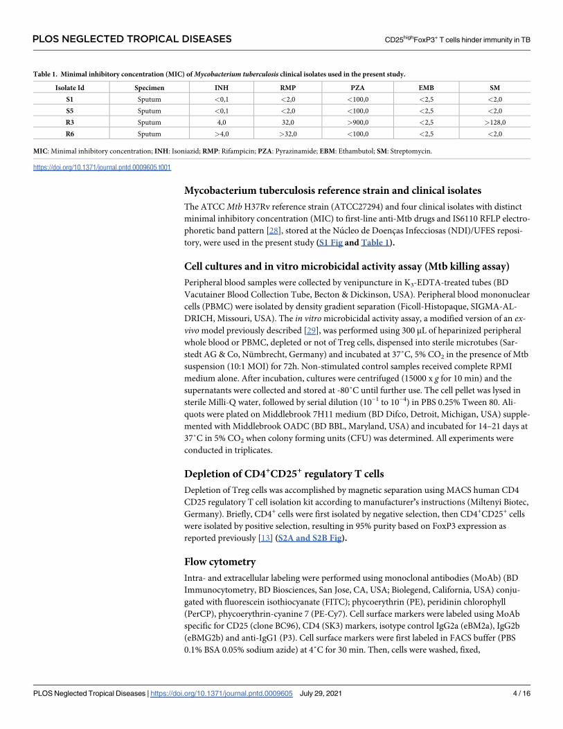
Table 1. Minimal inhibitory concentration (MIC) of Mycobacterium tuberculosis clinical isolates used in the present study.
Isolate Id Specimen INH RMP PZA EMB SM
S1 Sputum <0,1 <2,0 <100,0 <2,5 <2,0
S5 Sputum <0,1 <2,0 <100,0 <2,5 <2,0
R3 Sputum 4,0 32,0 >900,0 <2,5 >128,0
R6 Sputum >4,0 >32,0 <100,0 <2,5 <2,0
MIC: Minimal inhibitory concentration; INH: Isoniazid; RMP: Rifampicin; PZA: Pyrazinamide; EBM: Ethambutol; SM: Streptomycin.
https://doi.org/10.1371/journal.pntd.0009605.t001
PLOS NEGLECTED TROPICAL DISEASES CD25highFoxP3+ T cells hinder immunity in TB
PLOS Neglected Tropical Diseases | https://doi.org/10.1371/journal.pntd.0009605 July 29, 2021 4 / 16
permeabilized using FoxP3 Staining Buffer (eBioscience, CA, USA), and incubated at 4˚C for
30 min. for intracellular labeling of T-bet (a Th1 specific T-box transcription factor expressed
in T cell) (clone 4B10) and FoxP3 (Forkhead box P3 transcription factor) (clone 236A/E7).
Data was acquired with Attune NxT flow cytometer (Life Technologies, Carlsbad, CA, USA)
and analyzed using FlowJo software (TreeStar, San Carlos, CA, USA).
Detection of Soluble IFN-γSoluble IFN-γ concentration in culture supernatants was measured using a commercial kit
according to the manufacturer’s instructions (Human ELISA Ready-Set-Go kit; eBioscience
Inc. California, San Diego, USA). The absorbance was recorded at 570 nm and 450 nm on
SpectraMax M3 (Molecular Devices, Sunnyvale, CA, USA), and the detection limit was 4pg to
IFN-γ.
In vitro evaluation of phagocytic activity in differentiated human
macrophages
PBMCs (1×10⁶ cells/mL) were incubated at 37˚C/5%CO2 in 16-well tissue culture chamber
slides (Nalge Nunc International). Four hours after incubation, monolayers were washed three
times with RPMI 1640 medium supplemented with 10% inactivated fetal bovine serum (FBS)
to remove non-adherent cells. Adherent cells were cultivated with rGM-CSF (10μg/mL stock
R&D) for 5 days, followed by infection with either H37Rv strain or R6 Mtb isolate (MOI of
10:1) for 4h, then washed three times with 10% FBS RPMI 1640. Cells were divided in two
samples: one readily fixed and stained using the Kinyoun method as described previously [30],
and another group incubated at 37˚C for 24h, followed by staining. Slides were examined
using an optical microscope (Zeiss Axiostar) and intracellular bacteria load counted (number
of cells containing intracellular bacilli x 200 fields).
Statistical analyses
Normally distributed variables were compared using ANOVA analysis simple factorial test
and by one-way ANOVA-Tukey’s honestly significant difference (Tukey’s HSD) post-hoc
method. A 5% significance level (p<0.05) was used to determine statistical significance. Statis-
tical analysis was performed using GraphPad Prism software 7.0 (GraphPad Software, San
Diego, CA, EUA).
Results
Characteristics of the studied population
Thirty-seven HIV-negative subjects were enrolled and divided in three groups: a) Healthy
Controls (n = 13), b) LTBI (n = 13), and c) TB group (n = 11). Healthy control group com-
prised 3 males (median age ± SD = 31±10.6 years) and 10 females (26.5±5.1 years), the LTBI
group 4 males (29±6.2 years) and 9 females (30±9.8 years) and the TB group 6 males (39±8.6
years) and 5 females (28±6.6 years), as described in Table 2. BCG vaccination after birth is
mandatory in Brazil since 1960; therefore, all participants were vaccinated before enrollment.
Age and gender distribution did not differ significantly among the studied groups, patients
with helminthiasis were excluded. A few individuals with a discordant result between TST and
IGRA (TST-/IGRA+ or TST+/IGRA-) were excluded to prevent bias during further analyzes.
PLOS NEGLECTED TROPICAL DISEASES CD25highFoxP3+ T cells hinder immunity in TB
PLOS Neglected Tropical Diseases | https://doi.org/10.1371/journal.pntd.0009605 July 29, 2021 5 / 16
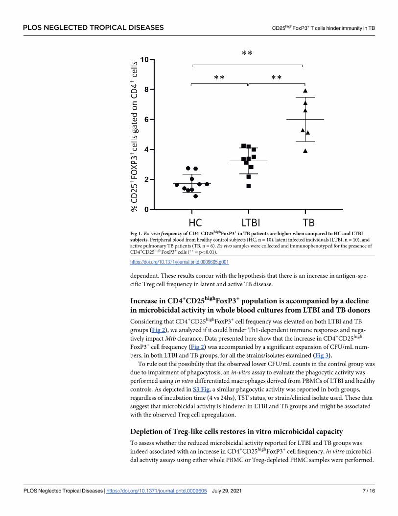
Ex-vivo frequency of CD4+CD25highFoxP3+ cells are high in TB patients
and LTBI individuals when compared to healthy controls
After venipuncture, whole blood samples were immediately stained for CD4+CD25highFoxP3+
and analyzed. A high frequency of CD4+CD25highFoxP3+ cells was observed in patients with
active pulmonary tuberculosis when compared to healthy control and LTBI subjects, and
when LTBI patients were compared to healthy controls (p<0.05) (Fig 1).
Frequency of CD4+CD25highFoxP3+ cells in LTBI individuals and TB
patients is significantly augmented in whole blood cultures after Mtb
infection
To investigate if CD4+CD25highFoxP3+ cell frequency differed among the studied groups,
whole peripheral blood samples from TB patients, healthy control and LTBI subjects were cul-
tured for 72h in the presence or absence of live Mtb H37Rv strain. After, in vitro stimulation,
CD4+CD25highFoxP3+ cell frequency was further increased, in both LTBI and TB groups,
upon exposure to live Mtb strain (H37Rv), or upon exposure to five different clinical isolates.
On the other hand, no significant change was observed in the healthy control group regardless
of the stimuli used (Fig 2).
To determine if the observed increase on CD4+CD25highFoxP3+ cell frequency was a strain-
dependent phenomenon, the experiment was also conducted with both multidrug-resistant
(R3 and R6) and drug-susceptible (S1 and S5) clinical isolates. The same profile observed for
H37Rv was also reported for all the isolates used, regardless of its drug susceptibility status
(Fig 2), suggesting that the expansion on the Treg population was neither strain/isolate
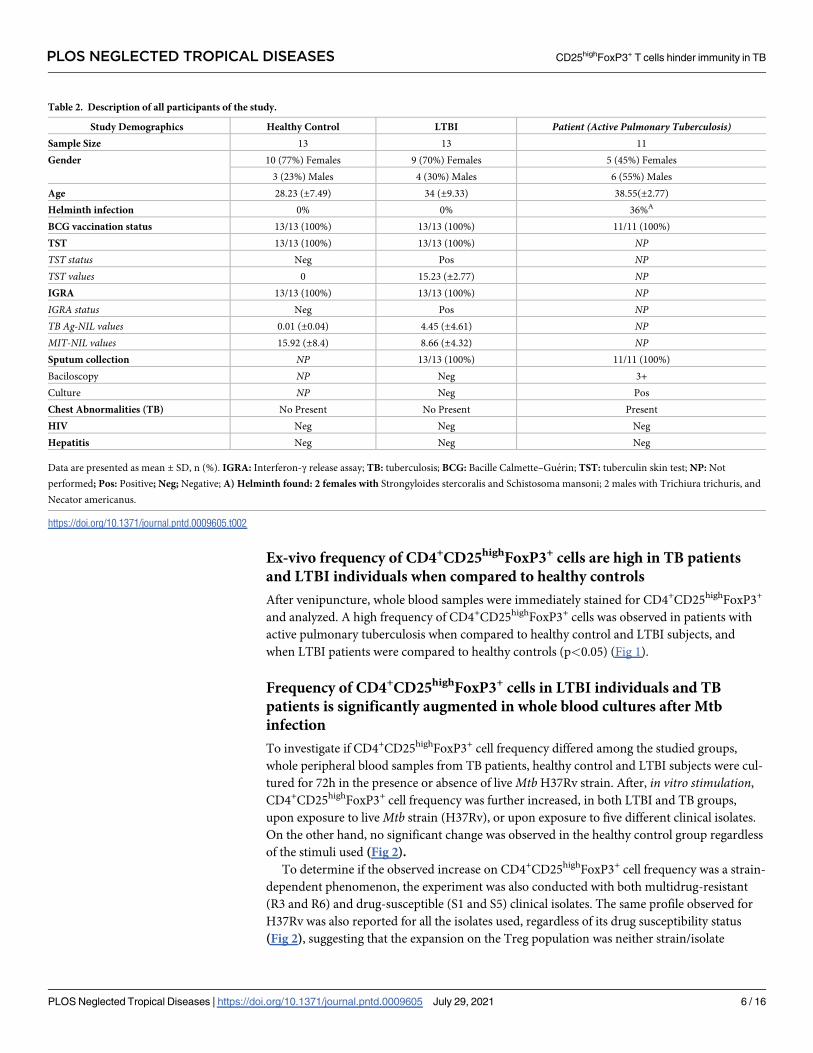
Table 2. Description of all participants of the study.
Study Demographics Healthy Control LTBI Patient (Active Pulmonary Tuberculosis)Sample Size 13 13 11
Gender 10 (77%) Females 9 (70%) Females 5 (45%) Females
3 (23%) Males 4 (30%) Males 6 (55%) Males
Age 28.23 (±7.49) 34 (±9.33) 38.55(±2.77)
Helminth infection 0% 0% 36%A
BCG vaccination status 13/13 (100%) 13/13 (100%) 11/11 (100%)
TST 13/13 (100%) 13/13 (100%) NPTST status Neg Pos NPTST values 0 15.23 (±2.77) NPIGRA 13/13 (100%) 13/13 (100%) NPIGRA status Neg Pos NPTB Ag-NIL values 0.01 (±0.04) 4.45 (±4.61) NPMIT-NIL values 15.92 (±8.4) 8.66 (±4.32) NPSputum collection NP 13/13 (100%) 11/11 (100%)
Baciloscopy NP Neg 3+
Culture NP Neg Pos
Chest Abnormalities (TB) No Present No Present Present
HIV Neg Neg Neg
Hepatitis Neg Neg Neg
Data are presented as mean ± SD, n (%). IGRA: Interferon-γ release assay; TB: tuberculosis; BCG: Bacille Calmette–Guerin; TST: tuberculin skin test; NP: Not
performed; Pos: Positive; Neg; Negative; A) Helminth found: 2 females with Strongyloides stercoralis and Schistosoma mansoni; 2 males with Trichiura trichuris, and
Necator americanus.
https://doi.org/10.1371/journal.pntd.0009605.t002
PLOS NEGLECTED TROPICAL DISEASES CD25highFoxP3+ T cells hinder immunity in TB
PLOS Neglected Tropical Diseases | https://doi.org/10.1371/journal.pntd.0009605 July 29, 2021 6 / 16
dependent. These results concur with the hypothesis that there is an increase in antigen-spe-
cific Treg cell frequency in latent and active TB disease.
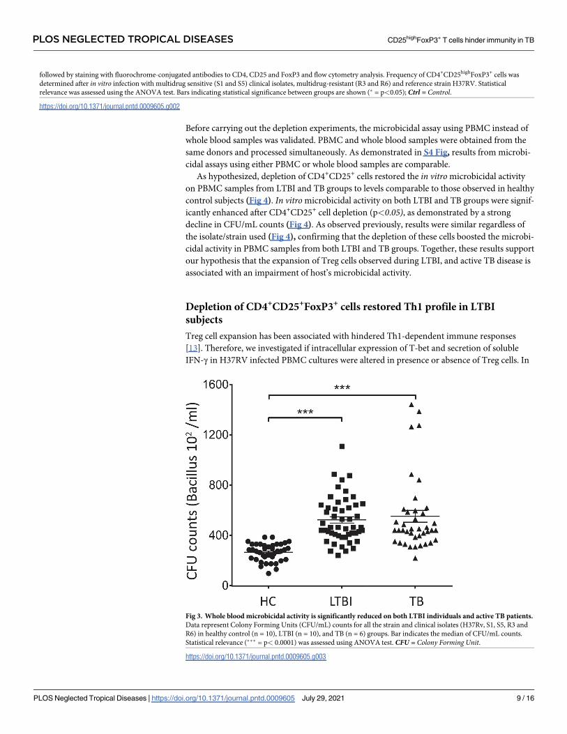
Increase in CD4+CD25highFoxP3+ population is accompanied by a decline
in microbicidal activity in whole blood cultures from LTBI and TB donors
Considering that CD4+CD25highFoxP3+ cell frequency was elevated on both LTBI and TB
groups (Fig 2), we analyzed if it could hinder Th1-dependent immune responses and nega-
tively impact Mtb clearance. Data presented here show that the increase in CD4+CD25high
FoxP3+ cell frequency (Fig 2) was accompanied by a significant expansion of CFU/mL num-
bers, in both LTBI and TB groups, for all the strains/isolates examined (Fig 3).
To rule out the possibility that the observed lower CFU/mL counts in the control group was
due to impairment of phagocytosis, an in-vitro assay to evaluate the phagocytic activity was
performed using in vitro differentiated macrophages derived from PBMCs of LTBI and healthy
controls. As depicted in S3 Fig, a similar phagocytic activity was reported in both groups,
regardless of incubation time (4 vs 24hs), TST status, or strain/clinical isolate used. These data
suggest that microbicidal activity is hindered in LTBI and TB groups and might be associated
with the observed Treg cell upregulation.
Depletion of Treg-like cells restores in vitro microbicidal capacity
To assess whether the reduced microbicidal activity reported for LTBI and TB groups was
indeed associated with an increase in CD4+CD25highFoxP3+ cell frequency, in vitro microbici-
dal activity assays using either whole PBMC or Treg-depleted PBMC samples were performed.
Fig 1. Ex-vivo frequency of CD4+CD25highFoxP3+ in TB patients are higher when compared to HC and LTBI
subjects. Peripheral blood from healthy control subjects (HC, n = 10), latent infected individuals (LTBI, n = 10), and
active pulmonary TB patients (TB, n = 6). Ex vivo samples were collected and immunophenotyped for the presence of
CD4+CD25highFoxP3+ cells (�� = p<0.01).
https://doi.org/10.1371/journal.pntd.0009605.g001
PLOS NEGLECTED TROPICAL DISEASES CD25highFoxP3+ T cells hinder immunity in TB
PLOS Neglected Tropical Diseases | https://doi.org/10.1371/journal.pntd.0009605 July 29, 2021 7 / 16

Fig 2. Frequency of CD4+CD25highFoxP3+ cells is increased in peripheral blood of LTBI and active TB patients. Peripheral blood from healthy control subjects (HC,
n = 10), latent infected individuals (LTBI, n = 10), and active pulmonary TB patients (TB, n = 6) were cultivated in the presence or absence of live Mtb by 72 hours,
PLOS NEGLECTED TROPICAL DISEASES CD25highFoxP3+ T cells hinder immunity in TB
PLOS Neglected Tropical Diseases | https://doi.org/10.1371/journal.pntd.0009605 July 29, 2021 8 / 16
Before carrying out the depletion experiments, the microbicidal assay using PBMC instead of
whole blood samples was validated. PBMC and whole blood samples were obtained from the
same donors and processed simultaneously. As demonstrated in S4 Fig, results from microbi-
cidal assays using either PBMC or whole blood samples are comparable.
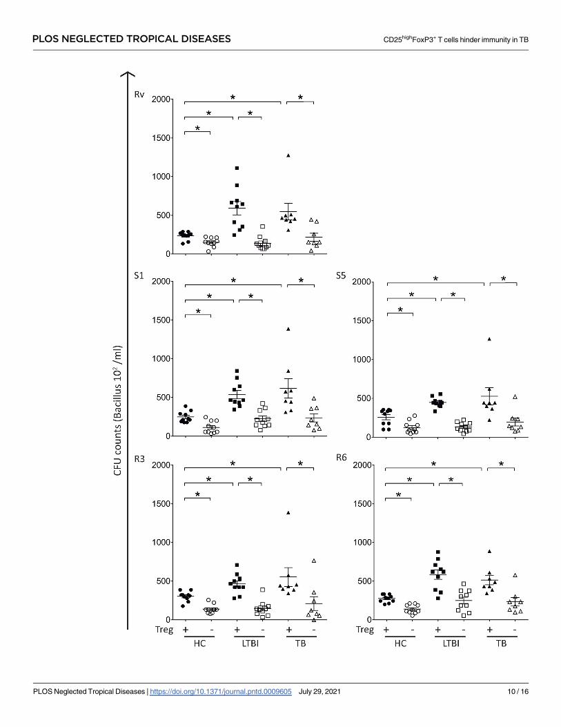
As hypothesized, depletion of CD4+CD25+ cells restored the in vitro microbicidal activity
on PBMC samples from LTBI and TB groups to levels comparable to those observed in healthy
control subjects (Fig 4). In vitro microbicidal activity on both LTBI and TB groups were signif-
icantly enhanced after CD4+CD25+ cell depletion (p<0.05), as demonstrated by a strong
decline in CFU/mL counts (Fig 4). As observed previously, results were similar regardless of
the isolate/strain used (Fig 4), confirming that the depletion of these cells boosted the microbi-
cidal activity in PBMC samples from both LTBI and TB groups. Together, these results support
our hypothesis that the expansion of Treg cells observed during LTBI, and active TB disease is
associated with an impairment of host’s microbicidal activity.
Depletion of CD4+CD25+FoxP3+ cells restored Th1 profile in LTBI
subjects
Treg cell expansion has been associated with hindered Th1-dependent immune responses
[13]. Therefore, we investigated if intracellular expression of T-bet and secretion of soluble
IFN-γ in H37RV infected PBMC cultures were altered in presence or absence of Treg cells. In
followed by staining with fluorochrome-conjugated antibodies to CD4, CD25 and FoxP3 and flow cytometry analysis. Frequency of CD4+CD25highFoxP3+ cells was
determined after in vitro infection with multidrug sensitive (S1 and S5) clinical isolates, multidrug-resistant (R3 and R6) and reference strain H37RV. Statistical
relevance was assessed using the ANOVA test. Bars indicating statistical significance between groups are shown (� = p<0.05); Ctrl = Control.
https://doi.org/10.1371/journal.pntd.0009605.g002
Fig 3. Whole blood microbicidal activity is significantly reduced on both LTBI individuals and active TB patients.
Data represent Colony Forming Units (CFU/mL) counts for all the strain and clinical isolates (H37Rv, S1, S5, R3 and
R6) in healthy control (n = 10), LTBI (n = 10), and TB (n = 6) groups. Bar indicates the median of CFU/mL counts.
Statistical relevance (��� = p< 0.0001) was assessed using ANOVA test. CFU = Colony Forming Unit.
https://doi.org/10.1371/journal.pntd.0009605.g003
PLOS NEGLECTED TROPICAL DISEASES CD25highFoxP3+ T cells hinder immunity in TB
PLOS Neglected Tropical Diseases | https://doi.org/10.1371/journal.pntd.0009605 July 29, 2021 9 / 16
PLOS NEGLECTED TROPICAL DISEASES CD25highFoxP3+ T cells hinder immunity in TB
PLOS Neglected Tropical Diseases | https://doi.org/10.1371/journal.pntd.0009605 July 29, 2021 10 / 16
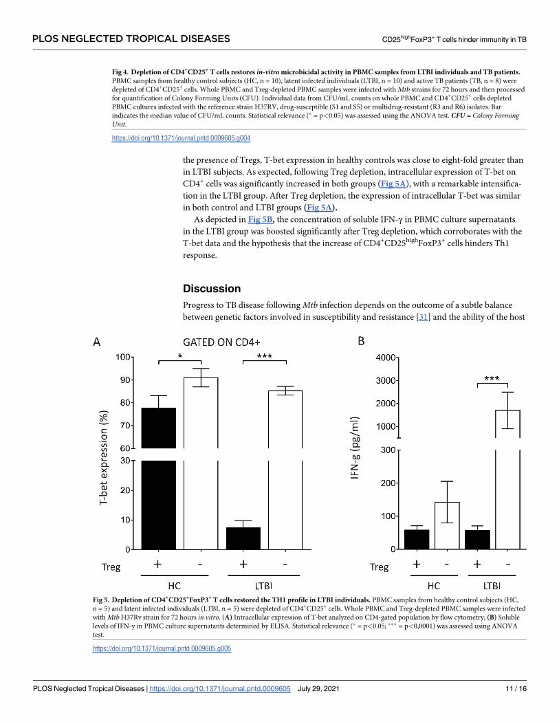
the presence of Tregs, T-bet expression in healthy controls was close to eight-fold greater than
in LTBI subjects. As expected, following Treg depletion, intracellular expression of T-bet on
CD4+ cells was significantly increased in both groups (Fig 5A), with a remarkable intensifica-
tion in the LTBI group. After Treg depletion, the expression of intracellular T-bet was similar
in both control and LTBI groups (Fig 5A).
As depicted in Fig 5B, the concentration of soluble IFN-γ in PBMC culture supernatants
in the LTBI group was boosted significantly after Treg depletion, which corroborates with the
T-bet data and the hypothesis that the increase of CD4+CD25highFoxP3+ cells hinders Th1
response.
Discussion
Progress to TB disease following Mtb infection depends on the outcome of a subtle balance
between genetic factors involved in susceptibility and resistance [31] and the ability of the host
Fig 4. Depletion of CD4+CD25+ T cells restores in-vitro microbicidal activity in PBMC samples from LTBI individuals and TB patients.
PBMC samples from healthy control subjects (HC, n = 10), latent infected individuals (LTBI, n = 10) and active TB patients (TB, n = 8) were
depleted of CD4+CD25+ cells. Whole PBMC and Treg-depleted PBMC samples were infected with Mtb strains for 72 hours and then processed
for quantification of Colony Forming Units (CFU). Individual data from CFU/mL counts on whole PBMC and CD4+CD25+ cells depleted
PBMC cultures infected with the reference strain H37RV, drug-susceptible (S1 and S5) or multidrug-resistant (R3 and R6) isolates. Bar
indicates the median value of CFU/mL counts. Statistical relevance (� = p<0.05) was assessed using the ANOVA test. CFU = Colony FormingUnit.
https://doi.org/10.1371/journal.pntd.0009605.g004
Fig 5. Depletion of CD4+CD25+FoxP3+ T cells restored the TH1 profile in LTBI individuals. PBMC samples from healthy control subjects (HC,
n = 5) and latent infected individuals (LTBI, n = 5) were depleted of CD4+CD25+ cells. Whole PBMC and Treg-depleted PBMC samples were infected
with Mtb H37Rv strain for 72 hours in vitro. (A) Intracellular expression of T-bet analyzed on CD4-gated population by flow cytometry; (B) Soluble
levels of IFN-y in PBMC culture supernatants determined by ELISA. Statistical relevance (� = p<0.05; ��� = p<0,0001) was assessed using ANOVA
test.
https://doi.org/10.1371/journal.pntd.0009605.g005
PLOS NEGLECTED TROPICAL DISEASES CD25highFoxP3+ T cells hinder immunity in TB
PLOS Neglected Tropical Diseases | https://doi.org/10.1371/journal.pntd.0009605 July 29, 2021 11 / 16

to mount an effective immune response [32–34]. Immune regulatory mechanisms, such as
Treg cells, are known to limit inflammatory immune responses [8,35,36] and have been associ-
ated with thwarted immune responses against Mtb which if overrepresented, may contribute
to the persistence and/or establishment of chronic infections [1,4]. Therefore, it is plausible to
expect that upregulation in the Treg cell population may lead to disease reactivation [37] or re-
infection [13,18,35,38,39]. Data presented here demonstrate that ex-vivo (Fig 1) and in vitro(Fig 2) Treg population is significantly augmented in LTBI subjects and patients with active
TB when compared to healthy controls. These findings are supported by previous studies
showing a higher frequency of CD4+CD25highFoxP3+ cells in both pulmonary TB patients and
LTBI subjects, when compared to other groups, suggesting a potential role of these cells in dis-
ease progression [3,10,11,13,14,16–18,35,40]. Although it is still not clear whether increased invitro Treg cell frequency correlates with progression of TB disease in humans, data exhibiting
a significant in vitro expansion of these cells on samples from TB patients after stimulation
[11,13,36,37,41–43] support this hypothesis. A possible explanation for the observed signifi-
cant increase in Treg cell frequency in LTBI individuals comes from data showing that CD4+
T cells expansion during exposure to Mtb could induce the upregulation of Treg cells specific
to Mtb antigens [13,44].
Other studies suggest that different Mtb strains induced Foxp3+ cells expansion distinc-
tively [45,46]. Data presented here demonstrated that the observed Treg frequencies associated
with drug-susceptible (DS strains, H37Rv, S1 and S5) and drug-resistant (DR strains, R3 and
R6) Mtb strains in whole blood cultures did not differ (Fig 2), concurring with Lim et al. [47].
These authors showed that frequencies of Treg cells in TB patients infected with either drug-
susceptible Mtb or multidrug-resistant Mtb were similar but significantly higher when com-
pared to healthy controls [47], corroborating with our findings.
A modified ex vivo whole blood bactericidal assay [29] allowed us to confirm that the upre-
gulation of CD4+CD25highFoxP3+ cells was associated with impaired microbicidal activity in
latent and active TB groups when compared to healthy control subjects, regardless of the strain
used. This difference was not due to alterations in bacterial phagocytosis assay (S3 Fig) but the
depletion of CD4+CD25highFoxP3+ cells in the culture restored the microbicidal activity in
LTBI and TB group regardless of Mtb strain/isolates used. This result corroborates previous
findings showing that peripheral blood Tregs from patients with pulmonary TB hindered the
ability of alveolar and monocyte-derived macrophages to restrict Mtb growth [48]. Data pre-
sented here also confirm previous results showing that CD4+CD25highFoxP3+ cells depletion
in PBMC cultures from LTBI subjects restores IFN-γ production and expression of T-bet,
enhancing the Th1-immune response (Fig 5A and 5B) [12,13].
Our work has limitations. First, since a large proportion of patients with pulmonary tuber-
culosis were under the use of anti-inflammatory drugs, which could hinder the assessment of
cytokines, cell phenotype, and the “in vitro” killing assay; enrolling TB patients who were not
using such medications to participate in the present study was a challenge, which limited the
number of enrolled TB patients. Second, frequently the final volume of collected blood samples
was smaller than required, impacting directly on the number of experiments that could be per-
formed per subject. Third, the great majority of enrolled TB patients refused to provide stool
samples for intestinal worms testing; therefore, the impact of the presence of intestinal worms
could not be assessed. Fourth, all enrolled patients with pulmonary tuberculosis presented a 3
+ or higher grade bacilloscopy result, which hindered the correlation of bacillary load and the
“in vitro” killing assay results. And finally, we were unable to enroll discordant cases sepa-
rately, which due to the necessary sampling size would require mass testing for both IGRA and
TST; therefore, analyzing discordant patients as a separate group was not possible. Limitations,
notwithstanding, in this study, we demonstrate that an increase in CD4+CD25highFoxP3+
PLOS NEGLECTED TROPICAL DISEASES CD25highFoxP3+ T cells hinder immunity in TB
PLOS Neglected Tropical Diseases | https://doi.org/10.1371/journal.pntd.0009605 July 29, 2021 12 / 16
population thwarts the host´s ability to restrict Mtb growth. Therefore, it may facilitate both
mycobacterial survival and intracellular persistence in vivo. Taken together, the results pre-
sented here may contribute to further our knowledge about the reason why the age-adjusted
incidence rate of TB reinfection after treatment was four times greater than new TB cases [20].
Additionally, it also may help to elucidate why case contacts with prior positive TST and peo-
ple who had TB previously present a significantly higher risk of developing active TB disease if
reinfected, when compared to healthy subjects [21,22]. Our findings support the hypothesis
that both latent and active Mtb infection led to an upregulation in Treg cell frequency, which
in turn may down-regulate the ability of the host to kill intracellular mycobacteria, signifi-
cantly increasing the risk of TB disease progression/reactivation in LTBI individuals or of rein-
fection of those treated and cured after a re-exposure to Mtb.
Supporting information
S1 Fig. IS6110 RFLP pattern for Mtb clinical isolates used in the study. RFLP patterns dem-
onstrate that selected clinical isolates are distinct from each other. Legend: S1 and S5 –drug-
susceptible isolates, R3 and R6—drug-resistant isolates, and CR–MT 14323 reference strain
used for IS6110 RFLP typing.
(TIF)
S2 Fig. Gating strategy. (A) Specific gating strategy was performed using CD4+, CD25high+
and FoxP3+ expression before and after depletion of CD4+CD25+ cells. (B) FoxP3+ expression
assessed by flow cytometry analysis of intracellular FOXP3+ on CD4+ cells before and after
CD4+CD25+ depletion assay (��� = p<0.0001).
(TIF)
S3 Fig. Evaluation of in vitro phagocytic activity in human differentiated macrophages
upon infection with live M. tuberculosis. Phagocytic activity was evaluated in vitro in PBMC
samples from healthy control subjects (HC) or latently infected individuals (LTBI) by quantifi-
cation of intracellular mycobacteria after 4h and 24h of exposure to live Mtb H37Rv strain and
R6 isolate, via Kinyoun acid-fast staining. Bars indicating statistical significance between
groups are shown (� = p<0.05).
(TIF)
S4 Fig. Validation of the in-vitro microbicidal activity assay using PBMC samples. Paired
whole blood (WB) and PBMC samples from healthy control subjects (n = 10), LTBI individu-
als (n = 10) and TB patients (n = 7) were infected with the reference strain H37RV or drug-sus-
ceptible (S1 and S5) or multidrug-resistant (R3 and R6) clinical isolates.
(TIF)
Acknowledgments
We thank physicians and clinical staff at UFES TB outpatient clinic who have kindly assisted
us in collecting blood samples from the study participants.
Author Contributions
Conceptualization: Lorenzzo Lyrio Stringari, Moises Palaci, Reynaldo Dietze, Daniel Claudio
de Oliveira Gomes, Rodrigo Ribeiro-Rodrigues.
Data curation: Moises Palaci.
PLOS NEGLECTED TROPICAL DISEASES CD25highFoxP3+ T cells hinder immunity in TB
PLOS Neglected Tropical Diseases | https://doi.org/10.1371/journal.pntd.0009605 July 29, 2021 13 / 16
http://journals.plos.org/plosntds/article/asset?unique&id=info:doi/10.1371/journal.pntd.0009605.s001
http://journals.plos.org/plosntds/article/asset?unique&id=info:doi/10.1371/journal.pntd.0009605.s002
http://journals.plos.org/plosntds/article/asset?unique&id=info:doi/10.1371/journal.pntd.0009605.s003

Formal analysis: Lorenzzo Lyrio Stringari, Flavia Dias Coelho da Silva, Vivian Leite de
Oliveira.
Funding acquisition: Rodrigo Ribeiro-Rodrigues.
Investigation: Lorenzzo Lyrio Stringari, Luciana Polaco Covre, David Jamil Hadad, Reynaldo
Dietze, Daniel Claudio de Oliveira Gomes, Rodrigo Ribeiro-Rodrigues.
Methodology: Lorenzzo Lyrio Stringari, Luciana Polaco Covre, Flavia Dias Coelho da Silva,
Maria Carolina Campana, Moises Palaci, Reynaldo Dietze, Daniel Claudio de Oliveira
Gomes, Rodrigo Ribeiro-Rodrigues.
Resources: Rodrigo Ribeiro-Rodrigues.
Supervision: Padmini Salgame.
Validation: Lorenzzo Lyrio Stringari, Maria Carolina Campana.
Visualization: David Jamil Hadad, Padmini Salgame, Rodrigo Ribeiro-Rodrigues.
Writing – original draft: Lorenzzo Lyrio Stringari, Padmini Salgame, Daniel Claudio de Oli-
veira Gomes, Rodrigo Ribeiro-Rodrigues.
Writing – review & editing: Lorenzzo Lyrio Stringari, Luciana Polaco Covre, Vivian Leite de
Oliveira, David Jamil Hadad, Moises Palaci, Padmini Salgame, Reynaldo Dietze, Daniel
Claudio de Oliveira Gomes, Rodrigo Ribeiro-Rodrigues.
References1. Kursar M, Koch M, Mittrucker HW, Nouailles G, Bonhagen K, Kamradt T, et al. Cutting Edge: Regula-
tory T cells prevent efficient clearance of Mycobacterium tuberculosis. J Immunol. 2007; 178(5):2661–
5. https://doi.org/10.4049/jimmunol.178.5.2661 PMID: 17312107
2. World_Health_Organization. Latent tuberculosis infection: updated and consolidated guidelines for pro-
grammatic management. Geneva: World Health Organization;. WHO; 2018.
3. Pandey P, Bhatnagar AK, Mohan A, Sachdeva KS, Vajpayee M, Das BK, et al. Insights in tuberculosis
immunology: Role of NKT and T regulatory cells. Int J Mycobacteriol. 2019; 8(4):333–40. https://doi.org/
10.4103/ijmy.ijmy_141_19 PMID: 31793502
4. Parkash O, Agrawal S, Madhan Kumar M. T regulatory cells: Achilles’ heel of Mycobacterium tuberculo-
sis infection? Immunol Res. 2015; 62(3):386–98. https://doi.org/10.1007/s12026-015-8654-0 PMID:
25948475
5. Hori S, Nomura T, Sakaguchi S. Control of regulatory T cell development by the transcription factor
Foxp3. Science. 2003; 299(5609):1057–61. https://doi.org/10.1126/science.1079490 PMID: 12522256
6. Chatenoud L. Natural and induced T CD4+CD25+FOXP3+ regulatory T cells. Methods Mol Biol. 2011;
677:3–13. https://doi.org/10.1007/978-1-60761-869-0_1 PMID: 20941599
7. Curotto de Lafaille MA, Lafaille JJ. Natural and adaptive foxp3+ regulatory T cells: more of the same or
a division of labor? Immunity. 2009; 30(5):626–35. https://doi.org/10.1016/j.immuni.2009.05.002 PMID:
19464985
8. Belkaid Y, Tarbell KV. Arming Treg cells at the inflammatory site. Immunity. 2009; 30(3):322–3. https://
doi.org/10.1016/j.immuni.2009.03.004 PMID: 19303386
9. de Almeida AS, Fiske CT, Sterling TR, Kalams SA. Increased frequency of regulatory T cells and T lym-
phocyte activation in persons with previously treated extrapulmonary tuberculosis. Clin Vaccine Immu-
nol. 2012; 19(1):45–52. https://doi.org/10.1128/CVI.05263-11 PMID: 22038848
10. Feruglio SL, Tonby K, Kvale D, Dyrhol-Riise AM. Early dynamics of T helper cell cytokines and T regula-
tory cells in response to treatment of active Mycobacterium tuberculosis infection. Clin Exp Immunol.
2015; 179(3):454–65. https://doi.org/10.1111/cei.12468 PMID: 25313008
11. Guyot-Revol V, Innes JA, Hackforth S, Hinks T, Lalvani A. Regulatory T cells are expanded in blood
and disease sites in patients with tuberculosis. Am J Respir Crit Care Med. 2006; 173(7):803–10.
https://doi.org/10.1164/rccm.200508-1294OC PMID: 16339919
PLOS NEGLECTED TROPICAL DISEASES CD25highFoxP3+ T cells hinder immunity in TB
PLOS Neglected Tropical Diseases | https://doi.org/10.1371/journal.pntd.0009605 July 29, 2021 14 / 16

12. Marin ND, Paris SC, Velez VM, Rojas CA, Rojas M, Garcia LF. Regulatory T cell frequency and modula-
tion of IFN-gamma and IL-17 in active and latent tuberculosis. Tuberculosis (Edinb). 2010; 90(4):252–
61. https://doi.org/10.1016/j.tube.2010.05.003 PMID: 20594914
13. Ribeiro-Rodrigues R, Resende Co T, Rojas R, Toossi Z, Dietze R, Boom WH, et al. A role for CD4
+CD25+ T cells in regulation of the immune response during human tuberculosis. Clin Exp Immunol.
2006; 144(1):25–34. https://doi.org/10.1111/j.1365-2249.2006.03027.x PMID: 16542361
14. Wergeland I, Assmus J, Dyrhol-Riise AM. T regulatory cells and immune activation in Mycobacterium
tuberculosis infection and the effect of preventive therapy. Scand J Immunol. 2011; 73(3):234–42.
https://doi.org/10.1111/j.1365-3083.2010.02496.x PMID: 21204895
15. Sahmoudi K, Abbassi H, Bouklata N, El Alami MN, Sadak A, Burant C, et al. Immune activation and reg-
ulatory T cells in Mycobacterium tuberculosis infected lymph nodes. BMC Immunol. 2018; 19(1):33.
https://doi.org/10.1186/s12865-018-0266-8 PMID: 30409122
16. Whittaker E, Nicol M, Zar HJ, Kampmann B. Regulatory T Cells and Pro-inflammatory Responses Pre-
dominate in Children with Tuberculosis. Front Immunol. 2017; 8:448. https://doi.org/10.3389/fimmu.
2017.00448 PMID: 28487695
17. Hougardy JM, Place S, Hildebrand M, Drowart A, Debrie AS, Locht C, et al. Regulatory T cells depress
immune responses to protective antigens in active tuberculosis. Am J Respir Crit Care Med. 2007; 176
(4):409–16. https://doi.org/10.1164/rccm.200701-084OC PMID: 17541018
18. Serrano CJ, Castaneda-Delgado JE, Trujillo-Ochoa JL, Gonzalez-Amaro R, Garcia-Hernandez MH,
Enciso-Moreno JA. Regulatory T-cell subsets in response to specific Mycobacterium tuberculosis anti-
gens in vitro distinguish among individuals with different QTF and TST reactivity. Clin Immunol. 2015;
157(2):145–55. https://doi.org/10.1016/j.clim.2015.02.008 PMID: 25728490
19. Herzmann C, Ernst M, Ehlers S, Stenger S, Maertzdorf J, Sotgiu G, et al. Increased frequencies of pul-
monary regulatory T-cells in latent Mycobacterium tuberculosis infection. Eur Respir J. 2012; 40
(6):1450–7. https://doi.org/10.1183/09031936.00214611 PMID: 22441737
20. Verver S, Warren RM, Beyers N, Richardson M, van der Spuy GD, Borgdorff MW, et al. Rate of reinfec-
tion tuberculosis after successful treatment is higher than rate of new tuberculosis. Am J Respir Crit
Care Med. 2005; 171(12):1430–5. https://doi.org/10.1164/rccm.200409-1200OC PMID: 15831840
21. Andrews JR, Noubary F, Walensky RP, Cerda R, Losina E, Horsburgh CR. Risk of progression to active
tuberculosis following reinfection with Mycobacterium tuberculosis. Clin Infect Dis. 2012; 54(6):784–91.
https://doi.org/10.1093/cid/cir951 PMID: 22267721
22. Gounder PP, Harris TG, Anger H, Trieu L, Meissner JS, Cadwell BL, et al. Risk for Tuberculosis Dis-
ease Among Contacts with Prior Positive Tuberculin Skin Test: A retrospective Cohort Study, New York
City. J Gen Intern Med. 2015; 30(6):742–8. https://doi.org/10.1007/s11606-015-3180-2 PMID:
25605533
23. Coelho da Silva FD, Covre LP, Stringari LL, Palaci M, Dietze R, Gomes DCO, et al. Toll-like receptors
blocking restores in vitro microbicidal activity in latent tuberculosis-infected subjects. Int J Tuberc Lung
Dis. 2019; 23(2):212–8. https://doi.org/10.5588/ijtld.18.0392 PMID: 30808454
24. Barnes PF, Cave MD. Molecular epidemiology of tuberculosis. N Engl J Med. 2003; 349(12):1149–56.
https://doi.org/10.1056/NEJMra021964 PMID: 13679530
25. Vynnycky E, Fine PE. The natural history of tuberculosis: the implications of age-dependent risks of dis-
ease and the role of reinfection. Epidemiol Infect. 1997; 119(2):183–201. https://doi.org/10.1017/
s0950268897007917 PMID: 9363017
26. Bjartveit K. Olaf Scheel and Johannes Heimbeck: their contribution to understanding the pathogenesis
and prevention of tuberculosis. Int J Tuberc Lung Dis. 2003; 7(4):306–11. PMID: 12729334
27. Sokal JE. Editorial: Measurement of delayed skin-test responses. N Engl J Med. 1975; 293(10):501–2.
https://doi.org/10.1056/NEJM197509042931013 PMID: 1152865
28. Ribeiro FK, Pan W, Bertolde A, Vinhas SA, Peres RL, Riley L, et al. Genotypic and Spatial Analysis of
Mycobacterium tuberculosis Transmission in a High-Incidence Urban Setting. Clin Infect Dis. 2015; 61
(5):758–66. https://doi.org/10.1093/cid/civ365 PMID: 25948063
29. Wallis RS, Palaci M, Vinhas S, Hise AG, Ribeiro FC, Landen K, et al. A whole blood bactericidal assay
for tuberculosis. J Infect Dis. 2001; 183(8):1300–3. https://doi.org/10.1086/319679 PMID: 11262217
30. Selvakumar N, Gomathi M, Rehman F, Narayanan PR. Evaluation of a two-reagent cold staining
method for detection of acid-fast bacilli. Int J Tuberc Lung Dis. 2002; 6(8):728–31. PMID: 12150486
31. Davies PD, Grange JM. Factors affecting susceptibility and resistance to tuberculosis. Thorax. 2001; 56
Suppl 2:ii23–9. PMID: 11514703
32. Geffner L, Yokobori N, Basile J, Schierloh P, Balboa L, Romero MM, et al. Patients with multidrug-resis-
tant tuberculosis display impaired Th1 responses and enhanced regulatory T-cell levels in response to
PLOS NEGLECTED TROPICAL DISEASES CD25highFoxP3+ T cells hinder immunity in TB
PLOS Neglected Tropical Diseases | https://doi.org/10.1371/journal.pntd.0009605 July 29, 2021 15 / 16
an outbreak of multidrug-resistant Mycobacterium tuberculosis M and Ra strains. Infect Immun. 2009;
77(11):5025–34. https://doi.org/10.1128/IAI.00224-09 PMID: 19720756
33. Manca C, Reed MB, Freeman S, Mathema B, Kreiswirth B, Barry CE 3rd, et al. Differential monocyte
activation underlies strain-specific Mycobacterium tuberculosis pathogenesis. Infect Immun. 2004; 72
(9):5511–4. https://doi.org/10.1128/IAI.72.9.5511-5514.2004 PMID: 15322056
34. Rajavelu P, Das SD. Th2-type immune response observed in healthy individuals to sonicate antigen
prepared from the most prevalent Mycobacterium tuberculosis strain with single copy of IS6110. FEMS
Immunol Med Microbiol. 2005; 45(1):95–102. https://doi.org/10.1016/j.femsim.2005.02.011 PMID:
15985228
35. Chen X, Zhou B, Li M, Deng Q, Wu X, Le X, et al. CD4(+)CD25(+)FoxP3(+) regulatory T cells suppress
Mycobacterium tuberculosis immunity in patients with active disease. Clin Immunol. 2007; 123(1):50–9.
https://doi.org/10.1016/j.clim.2006.11.009 PMID: 17234458
36. Chiacchio T, Casetti R, Butera O, Vanini V, Carrara S, Girardi E, et al. Characterization of regulatory T
cells identified as CD4(+)CD25(high)CD39(+) in patients with active tuberculosis. Clin Exp Immunol.
2009; 156(3):463–70. https://doi.org/10.1111/j.1365-2249.2009.03908.x PMID: 19438599
37. Hougardy JM, Verscheure V, Locht C, Mascart F. In vitro expansion of CD4+CD25highFOXP3
+CD127low/- regulatory T cells from peripheral blood lymphocytes of healthy Mycobacterium tuberculo-
sis-infected humans. Microbes Infect. 2007; 9(11):1325–32. https://doi.org/10.1016/j.micinf.2007.06.
004 PMID: 17890131
38. Luo J, Zhang M, Yan B, Zhang K, Chen M, Deng S. Imbalance of Th17 and Treg in peripheral blood
mononuclear cells of active tuberculosis patients. Braz J Infect Dis. 2017; 21(2):155–61. https://doi.org/
10.1016/j.bjid.2016.10.011 PMID: 27932286
39. Sterling TR, Dorman SE, Chaisson RE, Ding L, Hackman J, Moore K, et al. Human immunodeficiency
virus-seronegative adults with extrapulmonary tuberculosis have abnormal innate immune responses.
Clin Infect Dis. 2001; 33(7):976–82. https://doi.org/10.1086/322670 PMID: 11528568
40. Singh A, Dey AB, Mohan A, Sharma PK, Mitra DK. Foxp3+ regulatory T cells among tuberculosis
patients: impact on prognosis and restoration of antigen specific IFN-γ producing T cells. PLoS One.
2012; 7(9):e44728. https://doi.org/10.1371/journal.pone.0044728 PMID: 23028594
41. Henao-Tamayo MI, Obregon-Henao A, Arnett K, Shanley CA, Podell B, Orme IM, et al. Effect of bacillus
Calmette-Guerin vaccination on CD4+Foxp3+ T cells during acquired immune response to Mycobacte-
rium tuberculosis infection. J Leukoc Biol. 2016; 99(4):605–17. https://doi.org/10.1189/jlb.4A0614-
308RR PMID: 26590147
42. Li L, Lao SH, Wu CY. Increased frequency of CD4(+)CD25(high) Treg cells inhibit BCG-specific induc-
tion of IFN-gamma by CD4(+) T cells from TB patients. Tuberculosis (Edinb). 2007; 87(6):526–34.
https://doi.org/10.1016/j.tube.2007.07.004 PMID: 17851131
43. Orme IM, Robinson RT, Cooper AM. The balance between protective and pathogenic immune
responses in the TB-infected lung. Nat Immunol. 2015; 16(1):57–63. https://doi.org/10.1038/ni.3048
PMID: 25521685
44. Shafiani S, Tucker-Heard G, Kariyone A, Takatsu K, Urdahl KB. Pathogen-specific regulatory T cells
delay the arrival of effector T cells in the lung during early tuberculosis. J Exp Med. 2010; 207(7):1409–
20. https://doi.org/10.1084/jem.20091885 PMID: 20547826
45. Li N, Xie WP, Kong H, Min R, Hu CM, Zhou XB, et al. Enrichment of regulatory T-cells in blood of
patients with multidrug-resistant tuberculosis. Int J Tuberc Lung Dis. 2015; 19(10):1230–8. https://doi.
org/10.5588/ijtld.15.0148 PMID: 26459539
46. Ordway D, Henao-Tamayo M, Harton M, Palanisamy G, Troudt J, Shanley C, et al. The hypervirulent
Mycobacterium tuberculosis strain HN878 induces a potent TH1 response followed by rapid down-regu-
lation. J Immunol. 2007; 179(1):522–31. https://doi.org/10.4049/jimmunol.179.1.522 PMID: 17579073
47. Lim HJ, Park JS, Cho YJ, Yoon HI, Park KU, Lee CT, et al. CD4(+)FoxP3(+) T regulatory cells in drug-
susceptible and multidrug-resistant tuberculosis. Tuberculosis (Edinb). 2013; 93(5):523–8.
48. Semple PL, Binder AB, Davids M, Maredza A, van Zyl-Smit RN, Dheda K. Regulatory T cells attenuate
mycobacterial stasis in alveolar and blood-derived macrophages from patients with tuberculosis. Am J
Respir Crit Care Med. 2013; 187(11):1249–58. https://doi.org/10.1164/rccm.201210-1934OC PMID:
23590266
PLOS NEGLECTED TROPICAL DISEASES CD25highFoxP3+ T cells hinder immunity in TB
PLOS Neglected Tropical Diseases | https://doi.org/10.1371/journal.pntd.0009605 July 29, 2021 16 / 16