Unattended emotional faces elicit early lateralized amygdala–frontal and fusiform activations
Upload
independentCategory
view
5download
0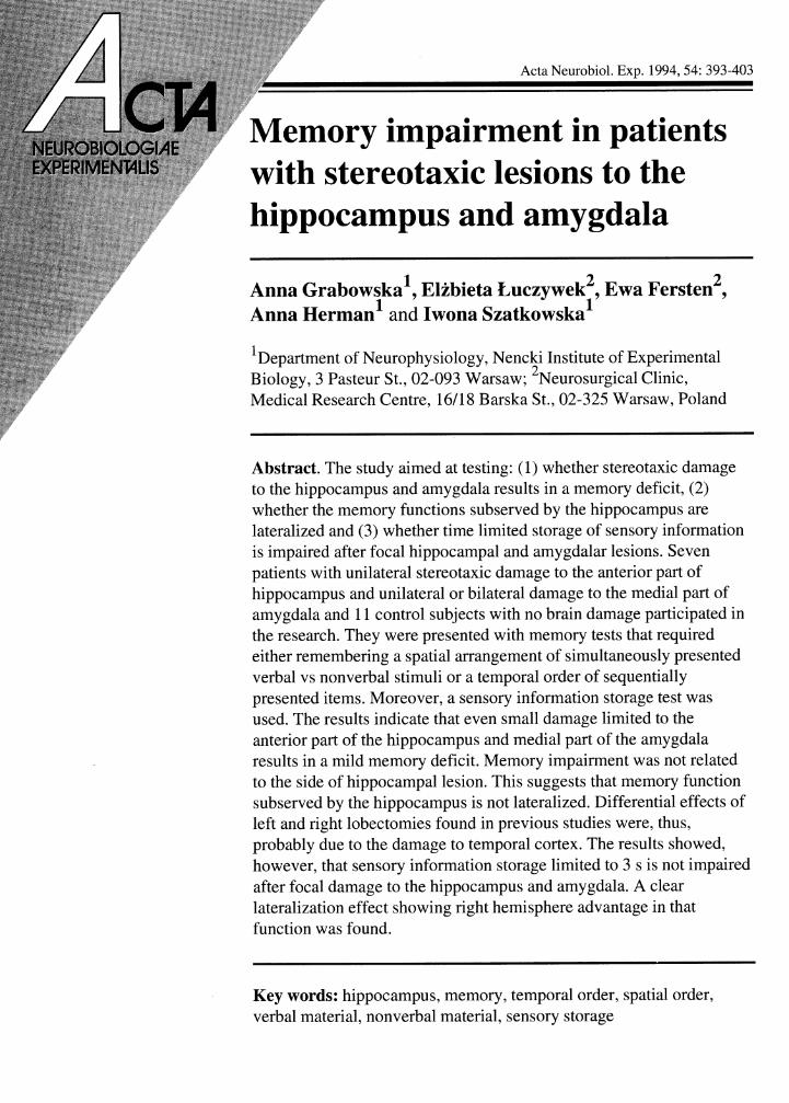
Memory impairment in patients with stereotaxic lesions to the hippocampus and amygdala
1 2 2 Anna Grabowska , Elzbieta Luczywek , Ewa Fersten , 1 Anna Herman and Iwona Szatkowska 1
' ~ e ~ a r t m e n t of Neurophysiology, Nencki Institute of Experimental Biology, 3 Pasteur St., 02-093 Warsaw; '~eurosur~ica l Clinic, Medical Research Centre, 1611 8 Barska St., 02-325 Warsaw, Poland
Abstract. The study aimed at testing: (1) whether stereotaxic damage to the hippocampus and amygdala results in a memory deficit, (2) whether the memory functions subserved by the hippocampus are lateralized and (3) whether time limited storage of sensory information is impaired after focal hippocampal and amygdalar lesions. Seven patients with unilateral stereotaxic damage to the anterior part of hippocampus and unilateral or bilateral damage to the medial part of amygdala and 11 control subjects with no brain damage participated in the research. They were presented with memory tests that required either remembering a spatial arrangement of simultaneously presented verbal vs nonverbal stimuli or a temporal order of sequentially presented items. Moreover, a sensory information storage test was used. The results indicate that even small damage limited to the anterior part of the hippocampus and medial part of the amygdala results in a mild memory deficit. Memory impairment was not related to the side of hippocampal lesion. This suggests that memory function subserved by the hippocampus is not lateralized. Differential effects of left and right lobectomies found in previous studies were, thus, probably due to the damage to temporal cortex. The results showed, however, that sensory information storage limited to 3 s is not impaired after focal damage to the hippocampus and amygdala. A clear lateralization effect showing right hemisphere advantage in that function was found.
Key words: hippocampus, memory, temporal order, spatial order, verbal material, nonverbal material, sensory storage
394 A. Grabowska et al.
INTRODUCTION
A severe impairment in memory following bilat- eral temporal lobectomy in a human subject was first reported by Scoville and Milner in 1957. Since that time numerous investigators have provided support for the notion that medial temporal lobe structures are essential for memory function (see Squire 1992 for recent review). An important step in understanding the role of these structures was the insight that they are involved only in a particular kind of memory, namely declarative or explicit memory (Cohen and Squire 1980, Mishkin and Appenzeller 1987, Squire 1992). Various types of memory abilities that are believed to belong to a wide class of nondeclarative memory (skills, habits, simple conditioning and priming) were found to be relatively spared after medial temporal lobe lesions (e.g. Milner 1962, Brooks and Baddeley 1976, Squire 1982, Cohen 1984, Squire and Zola-Morgan 1988).
Despite the very great progress that has recently been achieved in the field of memory research owing to animal studies intended to mimic human amnesic cases, there are still several problems which need further study. The first issue concerns the question which of the complex medial temporal structures actually contribute to memory. Earlier studies in monkeys seemed to suggest that com- bined lesions to the hippocampus and amygdala are necessary to produce a severe memory impairment (Mishkin 1978, Murray and Mishkin 1985). Recent studies, however, changed that opinion by implying that besides the hippocampus per se the underlying cortices (parahippocampal, entorhinal and perirhi- nal cortex) and not amygdala are important for dec- larative memory functions (Zola-Morgan et al. 1989a, b, Squire and Zola-Morgan 1991). Reexam- ination of the earlier studies showed that they were based on surgical groups in which the amygdala was removed together with underlying cortex (Zola-Morgan et al. 1982).
Although much is known as regarding the struc- tures, connections and functioning of the medial temporal lobe system in animals (especially in mon- key and rat) the relevant knowledge on human sub-
jects is limited. This results from the fact that most of the human research concerns the effect of tem- poral lobectomies which usually extend to a large portion of the temporal lobe. It is difficult, there- fore, to attribute memory deficits to particular struc- tures. Very few cases are now available which are exceptions to this generalization. One of them is pa- tient R.B. (Zola-Morgan et al. 1986) who suffered a moderately severe memory impairment following an ischemic event. Examination of R.B.'s brain after his death revealed bilateral damage within the CA1 region of the hippocampus. Another human study using magnetic resonance (MR) techniques (Squire et al. 1990) in which abnormalities in hip- pocampus were demonstrated in 4 patients with cir- cumscribed memory impairment, provided some additional evidence supporting the conclusion that the hippocampus proper is important for human memory. As focal lesions to the medial temporal lobe structures are very rare in human subjects we thought that testing patients who have undergone stereotaxic hippocampal and amygdalar lesions would be of great interest. The present paper pres- ents such data.
Studies in man show that the effects of temporal lobectomies are hemisphere related: left hemis- phere lesions result in an impairment in verbal memory function (Milner 1968a, Rubino 1970, Milner 1972, Ojeman and Dorill 1985), whereas right hemisphere damage disturbs the memory for visuo-spatial material (Kimura 1963, Milner 1965, 1968~1, b, de Renzi and Nichelli 1979, Smith and Milner 1981, Zatore 1985). It is a matter of discus- sion, however, whether these laterality effects result from functional asymmetry of temporal cortex or hippocampal formation per se. Studies of patients after temporal lobectomies do not provide a resol- ution to the question, as in most cases both these structures are being damaged. Our study addressed this issue by studying whether left- and right-side lesions limited to hippocampus and amygdala have similar effects on verbal and visual-spatial tests.
Several laterality studies suggest that the two he- mispheres are characterized by different processing modes: the left hemisphere processes the incoming
Memory deficit after stereotaxic lesions to the hippocampus in man 395
information in a sequential, analytic manner, whereas the right hemisphere uses a simultaneous, holistic style of processing (Bradshaw and Nettleton 198 1). It is believed therefore, that successive pres- entations of temporally ordered stimuli would fit the left hemisphere processing mode, whereas sim- ultaneous presentations of spatially organized stimuli would be adequate for the right hemisphere. In the present study we used memory tests that re- quired either remembering a spatial arrangement of simultaneously presented stimulus items or the tem- poral order of sequentially presented items. We as- sumed that if memory functions subserved by the hippocampus are lateralized, left- and right-side le- sion should differentially affect the two types of memory tests.
Studies in monkey using a delayed nonmatching to sample procedure have shown that the animals perform relatively well when the time between the presentation of the sample object and the choice is short and becomes poorer as the delay increases (Overman et al. 1990). In line with these finding are studies showing that in amnesic patients immediate memory is disproportionally spared in relation to a global memory deficit (Milner 1965, Baddeley 1982, Piggot and Milner 1993). It appears that rela- tively good performance in memory tests measured shortly after stimuli presentation might result from the support of time limited high capacity storage of sensory information (Czachowska-Sieszycka et al. 1985). Thus we were interested in whether patients with focal medial temporal lesions would show any deficit in a test requiring a comparison of sensory features (e.g., size) of two visual stimuli presented with a very short interstimulus interval.
To sum up, our study addressed 3 questions: 1. Do the stereotaxic lesions limited to the ante-
rior part of the hippocampus and the medial part of amygdala result in a memory deficit?
2. Are the two hippocampi (amygdalae) func- tionally differentiated i. e., do the left- and right- side lesions have differential effects on verbal vs. visual-spatial memory tests and on tests requiring remembering either the spatial arrangement or the temporal order of stimuli?
3. Is the storage of sensory information impaired after stereotaxic hippocampal lesions?
METHODS
Subjects
Seven patients, each of whom has undergone an operation which produced focal damage to the ante- rior hippocampal and medial amygdalar structures for the relief of pharmacologically intractable epi-
1 lepsy , and 11 control subjects with no brain dam- age were examined. The damage was performed by irreversible cooling of the anterior part of the hip- pocampus and medial part of the amygdala with liq- uid nitrogen at a temperature of -60 deg centigrade which was introduced stereotaxically with a 2 mm diameter canula. In 3 patients surgery involved bi- lateral lesions to the amygdala and a left side lesion to the hippocampus; in the other 3 patients there was a right side lesion to the hippocampus and amygda- la. In one patient there was only a lesion to the right hippocampus. Due to surgical intervention in 3 pa- tients epileptic seizures disappeared and in the re- maining 4 patients the number and severity of seizures were greatly reduced.
Patients were characterized by normal intellec- tual functions (IQ= 10 1 - 125 as measured with Wechsler-Bellevue scale). In most cases they were able to perform professional work and to maintain families. They were right-handed, aged 35-41 and on the average had reached a secondary school level of education. Control subjects matched patients as much as possible in all these respects.
Material and procedure
Subjects participated in two sets of memory tests. The first one consisted of four tasks which were modifications of tests developed originally by Maksymczuk (1973): verbal-successive, nonver-
'operations were performed by prof. E. Mempel at Neurosur- gical Clinic, Medical Research Centre, Warsaw. For more de- tails on the surgery procedure see Mempel 1971.
396 A. Grabowska et al.
Fig. 1. Examples of stimuli used in the simultaneous versions of the test.
BAR
CEL
KOS
bal-successive, verbal-simultaneous and nonver- bal-simultaneous. In the two simultaneous tests subjects were presented with nine words or with nine figures arranged in three rows in a matrix (Fig. 1). All nine items were exposed simultaneously for 18 s. After each presentation subjects were given all items on separate cards arranged in one row, and were asked to put them on an empty matrix accord- ing to the order they saw before. The trials were re- peated until two consecutive correct performances were reached. Each trial was preceded by an expo- sure of the matrix with correctly arranged items.
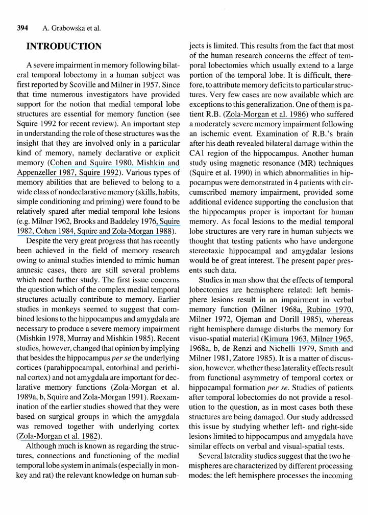
In the two successive versions of the test nine similar stimuli were presented sequentially one after another so that only one stimulus was presented at a time. The time of exposure was 2 sper item. After each presentation subjects were given all stimuli in one row and were asked to arrange them in the order of exposure (Fig. 2). Thus, the subjects were re-
quired to memorize and reproduce either the spatial or temporal order of the material having the con- tents continuously at their disposal.
The second set of tests, which we call sensory in- formation storage tests (Szatkowska et al. 1992), consisted of presentations of Vanderplas-type figures (Vanderplas and Garvin 1959). The stimuli were exposed for 100 ms in pairs, one after another, on a computer screen. The first stimulus was presented either 2 deg left or 2 deg right of the fix- ation point. The second stimulus was presented in the centre of the screen with a 50 ms, 500 ms or 3,000 ms delay. The subjects' task was to decide whether the second stimulus was smaller, bigger or the same size as the first one. They responded by pressing one of three buttons indicating their choice. The figures used in the study had two dif- ferent shapes and four different sizes (Fig. 3). Figures presented in a pair always had the same shape and either the same or different size. The first stimulus in a pair had the size indicated as 2 or 3 in Fig. 3. The second stimulus could be either the same, smaller or bigger. The three different delays were blocked into separate sessions, each consisting of 96 exposures. Each session was performed twice, once with the left and once with the right hand. The order of sessions was randomly generated by the computer. The order of trials in a series was pseu- dorandom: the same relation between the two stimuli (same, bigger and smaller) could not repeat consecutively more than three times and the lat- erally presented figure could not appear consecu-
EOM
LAS
SYN
- -
Fig. 2. Successive version of the test. tively more than three times in the same visual field.
MAK
WIR
POT
Memory deficit after stereotaxic lesions to the hippocampus in man 397
Fig. 3. Examples of stimuli used in the sensory information storage test. Four different sizes (1-4) and two different shapes (a,b) were used.
The experiment was preceded by training trials to acquaint the subjects with the procedure.
RESULTS
Temporal and spatial order memory tests
Two different measures were analyzed: (I) the accuracy scores indicating mean number of errors per trial made in reproducing either spatial or tem-
poral order of verbal and nonverbal stimuli and (2) number of trials required to reach the criterion of two consecutive correct performances.
COMPARISON OF PATIENTAND CONTROL GROUPS
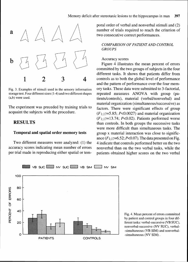
Accuracy scores Figure 4 illustrates the mean percent of errors
committed by the two groups of subjects in the four different tasks. It shows that patients differ from controls as to both the global level of performance and the pattern of performance over the four mem- ory tasks. These data were submitted to 3-factorial, repeated measures ANOVA with group (pa- tients/controls), material (verbal/nonverbal) and material organization (simultaneous/successive) as factors. There were significant effects of group (Fi; i7=5.85; P<0.0027) and material organization (Fi; i7= 13.74; P<0.02). Patients performed worse than controls. In both groups the successive tasks were more difficult than simultaneous tasks. The group x material interaction was close to signific- ance (F1;17=6.52; P<0.07). The data presented on Fig. 4 indicate that controls performed better on the two nonverbal than on the two verbal tasks, while the patients obtained higher scores on the two verbal
VB SUC NV SUC VB SIM NV SIM
0 PATIENTS CONTROLS
Fig. 4. Mean percent of errors committed by patient and control groups in four dif- ferent tasks: verbal-succesive (VB SUC), nonverbal-succesive (NV SUC), verbal- -simultaneous (VB SIM) and nonverbal- -simultaneous (NV SIM) .
398 A. Grabowska et al.
VB SUC NV SUC VB SIM @ NV SIM
2 0 PATIENTS CONTROLS
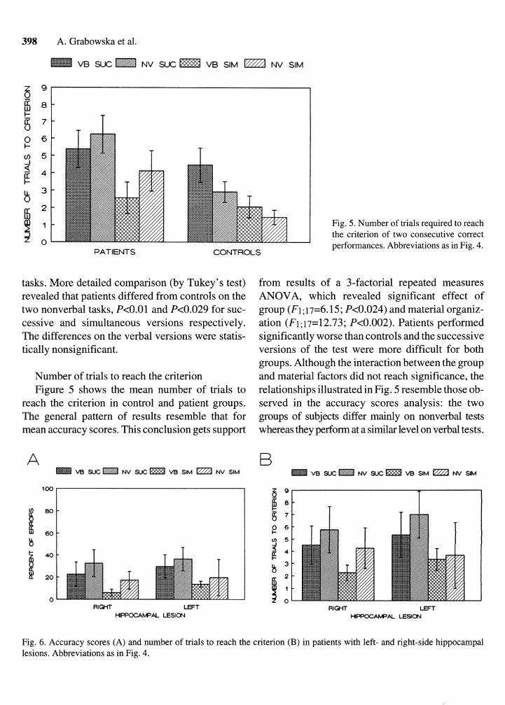
Fig. 5. Number of trials required to reach the criterion of two consecutive correct performances. Abbreviations as in Fig. 4.
tasks. More detailed comparison (by Tukey's test) revealed that patients differed from controls on the two nonverbal tasks, P<0.01 and P<0.029 for suc- cessive and simultaneous versions respectively. The differences on the verbal versions were statis- tically nonsignificant.
Number of trials to reach the criterion Figure 5 shows the mean number of trials to
reach the criterion in control and patient groups. The general pattern of results resemble that for mean accuracy scores. This conclusion gets support
from results of a 3-factorial repeated measures ANOVA, which revealed significant effect of group (Fi; 17=6.15; P<0.024) and material organiz- ation (F1;17=12.73; P<0.002). Patients performed significantly worse than controls and the successive versions of the test were more difficult for both groups. Although the interaction between the group and material factors did not reach significance, the relationships illustrated in Fig. 5 resemble those ob- served in the accuracy scores analysis: the two groups of subjects differ mainly on nonverbal tests whereas they perform at a similar level on verbal tests.
A VB S U C W NV S U C m V B SIM NV SIM
B VB S U C W NV S U C m V B SIM NV SlM
0 RIGHT LEFT
HIPPCCMAL LESION RIGHT LEFT
M!WOOA@AL LESlCN
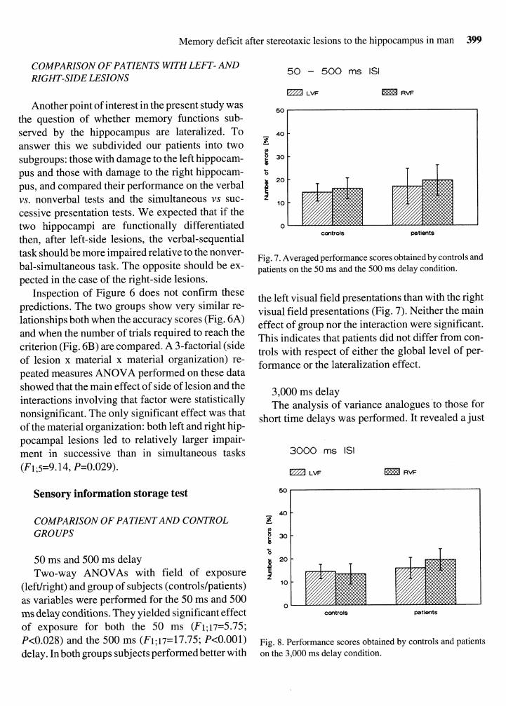
Fig. 6. Accuracy scores (A) and number of trials to reach the criterion (B) in patients with left- and right-side hippocampal lesions. Abbreviations as in Fig. 4.
Memory deficit after stereotaxic lesions to the hippocampus in man 399
COMPARISON OF PATIENTS WITH LEFT- AND RIGHT-SIDE LESIONS
Another point of interest in the present study was the question of whether memory functions sub- served by the hippocampus are lateralized. To answer this we subdivided our patients into two subgroups: those with damage to the left hippocam- pus and those with damage to the right hippocam- pus, and compared their performance on the verbal vs. nonverbal tests and the simultaneous vs suc- cessive presentation tests. We expected that if the two hippocampi are functionally differentiated then, after left-side lesions, the verbal-sequential task should be more impaired relative to the nonver- bal-simultaneous task. The opposite should be ex- pected in the case of the right-side lesions.
Inspection of Figure 6 does not confirm these predictions. The two groups show very similar re- lationships both when the accuracy scores (Fig. 6A) and when the number of trials required to reach the criterion (Fig. 6B) are compared. A 3-factorial (side of lesion x material x material organization) re- peated measures ANOVA performed on these data showed that the main effect of side of lesion and the interactions involving that factor were statistically nonsignificant. The only significant effect was that of the material organization: both left and right hip- pocampal lesions led to relatively larger impair- ment in successive than in simultaneous tasks (F1;5=9.14, P=0.029).
Sensory information storage test
COMPARISON OF PATIENTAND CONTROL GROUPS
50 ms and 500 ms delay Two-way ANOVAs with field of exposure
(leftlright) and group of subjects (controls/patients) as variables were performed for the 50 ms and 500 ms delay conditions. They yielded significant effect of exposure for both the 50 ms (Fi;i7=5.75; P<0.028) and the 500 ms (F1;17=17.75; P<0.001) delay. In both groups subjects performed better with
0 controls mtients
Fig. 7. Averaged performance scores obtained by controls and patients on the 50 ms and the 500 ms delay condition.
the left visual field presentations than with the right visual field presentations (Fig. 7). Neither the main effect of group nor the interaction were significant. This indicates that patients did not differ from con- trols with respect of either the global level of per- formance or the lateralization effect.
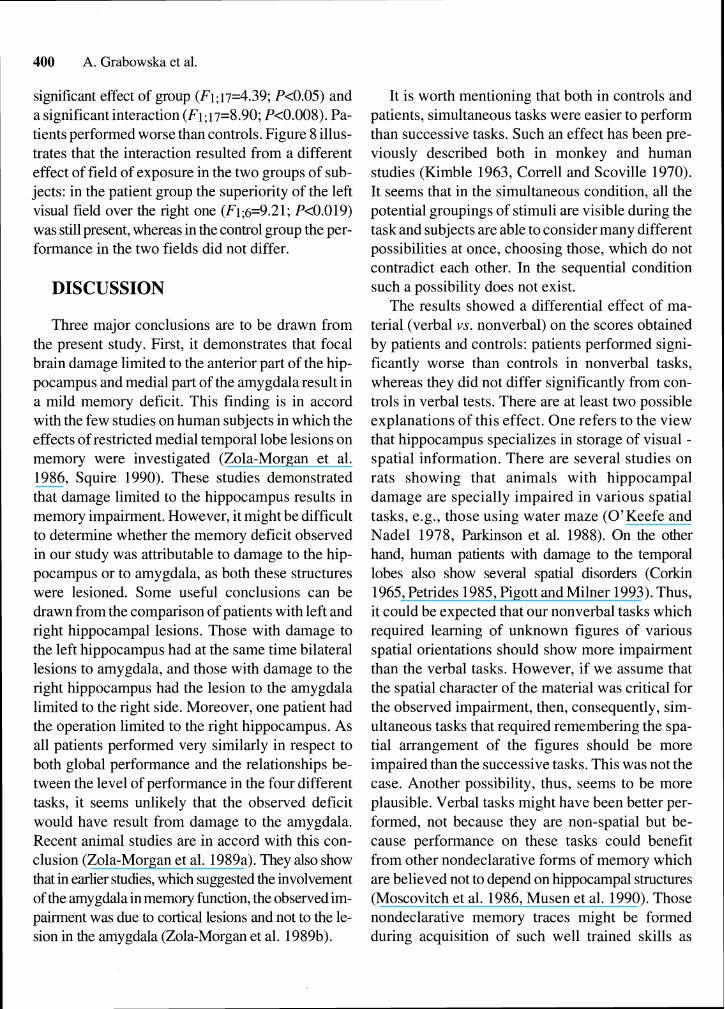
3,000 ms delay The analysis of variance analogues t o those for
short time delays was performed. It revealed a just
controls mtients
Fig. 8. Performance scores obtained by controls and patients on the 3,000 ms delay condition.
400 A. Grabowska et al.
significant effect of group (F1; 17=4.39; P<0.05) and a significant interaction (F1;17=8.90; P<0.008). Pa- tients performed worse than controls. Figure 8 illus- trates that the interaction resulted from a different effect of field of exposure in the two groups of sub- jects: in the patient group the superiority of the left visual field over the right one (F1;6=9.21; P<0.019) was still present, whereas in the control group the per- formance in the two fields did not differ.
DISCUSSION
Three major conclusions are to be drawn from the present study. First, it demonstrates that focal brain damage limited to the anterior part of the hip- pocampus and medial part of the amygdala result in a mild memory deficit. This finding is in accord with the few studies on human subjects in which the effects of restricted medial temporal lobe lesions on memory were investigated (Zola-Morgan et al. 1986, Squire 1990). These studies demonstrated that damage limited to the hippocampus results in memory impairment. However, it might be difficult to determine whether the memory deficit observed in our study was attributable to damage to the hip- pocampus or to amygdala, as both these structures were lesioned. Some useful conclusions can be drawn from the comparison of patients with left and right hippocampal lesions. Those with damage to the left hippocampus had at the same time bilateral lesions to amygdala, and those with damage to the right hippocampus had the lesion to the amygdala limited to the right side. Moreover, one patient had the operation limited to the right hippocampus. As all patients performed very similarly in respect to both global performance and the relationships be- tween the level of performance in the four different tasks, it seems unlikely that the observed deficit would have result from damage to the amygdala. Recent animal studies are in accord with this con- clusion (Zola-Morgan et al. 1989a). They also show that in earlier studies, which suggested the involvement of the amygdala in memory function, the observed im- pairment was due to cortical lesions and not to the le- sion in the amygdala (Zola-Morgan et al. 1989b).
It is worth mentioning that both in controls and patients, simultaneous tasks were easier to perform than successive tasks. Such an effect has been pre- viously described both in monkey and human studies (Kimble 1963, Correll and Scoville 1970). It seems that in the simultaneous condition, all the potential groupings of stimuli are visible during the task and subjects are able to consider many different possibilities at once, choosing those, which do not contradict each other. In the sequential condition such a possibility does not exist.
The results showed a differential effect of ma- terial (verbal vs. nonverbal) on the scores obtained by patients and controls: patients performed signi- ficantly worse than controls in nonverbal tasks, whereas they did not differ significantly from con- trols in verbal tests. There are at least two possible explanations of this effect. One refers to the view that hippocampus specializes in storage of visual - spatial information. There are several studies on rats showing that animals with hippocampal damage are specially impaired in various spatial tasks, e.g., those using water maze (O'Keefe and Nadel 1978, Parkinson et al. 1988). On the other hand, human patients with damage to the temporal lobes also show several spatial disorders (Corkin 1965, Petrides 1985, Pigott and Milner 1993). Thus, it could be expected that our nonverbal tasks which required learning of unknown figures of various spatial orientations should show more impairment than the verbal tasks. However, if we assume that the spatial character of the material was critical for the observed impairment, then, consequently, sim- ultaneous tasks that required remembering the spa- tial arrangement of the figures should be more impaired than the successive tasks. This was not the case. Another possibility, thus, seems to be more plausible. Verbal tasks might have been better per- formed, not because they are non-spatial but be- cause performance on these tasks could benefit from other nondeclarative forms of memory which are believed not to depend on hippocampal structures (Moscovitch et al. 1986, Musen et al. 1990). Those nondeclarative memory traces might be formed during acquisition of such well trained skills as
Memory deficit after stereotaxic lesions to the hippocampus in man 401
reading or writing. It has been shown, for example, that amnesic patients are able to acquire normally a reading skill for regularly repeating nonwords (Musen and Squire 199 1).
Second major finding of the present study is that the results did not show any hint of a differential in- fluence of left- and right-side hippocampal lesions on memory function. Memory impairment after left hippocampal lesions was comparable to that after right hippocampal lesions. Moreover, the influence of the type of material (verbal vs nonverbal) and ma- terial organization (simultaneous vs. successive) was also similar in the two groups. In both groups successive tasks were more difficult than simulta- neous and nonverbal tests were more difficult than verbal tests. All this suggests that memory function subserved by the hippocampus is not lateralized. Our results diverge from other human studies which show that lesions to the left medial temporal lobe structures result in verbal memory deficits ( Milner 1968a, Rubino 1970, Miller 1972, Ojeman and Dorill 1985), whereas lesions to the right-sided structures lead to impairment on visual-spatial tests (Kimura 1963, Milner 1965, 1968a, 1968b, Smith and Milner 1981, Jones-Gotman 1986). It should be stressed, however, that our study differed from the previous ones in that our patients had lesions limited to the hippocampus and amygdala, whereas the majority of previous studies tested lobectomy patients, in whom lesions extended to a large portion of cortex. That difference seems to be crucial for the observed discrepancy. It suggests that the lateralized effects observed in previous studies were due to damage to the cortex rather than to hippocampal formation per se.
The third aim of our research was to investigate whether lesions to the hippocampus and amygdala result in a sensory storage deficit. For that purpose we used a task that required size comparison of one visually presented stimulus to the memory trace of another stimulus. When very short (50 ms and 500 ms) interstimulus intervals were used, patients' per- formance did not differ significantly from controls'. Moreover, in both groups the left side presentations resulted in significantly higher performance than the right-side performance, showing a right hemis-
phere advantage. A difference between controls and patients appeared at the longer (3 s) interstimulus interval. Patients performed worse than controls and they differed from controls as to the laterality effect. In controls the left field superiority disap- peared suggesting that after 3 s of storage another memory mechanism was involved, one presumably based on categorical descriptions of the stimuli. On the other hand, patients still showed the left field su- periority. This might suggest that they continued to relay on sensory storage, having difficulties with switching to a more stable memory mechanism in which hippocampal structures are involved. It seems therefore that time limited storage of sensory information is not impaired after focal lesions of the hippocampus and amygdala. This suggestion finds support from a recent electrophysiological study (Nielson-Bohlman and Knight 1994) which shows that very short storage (for less than 4 s), in contrast to storage for a longer time, does not involve acti- vation of medial temporal structures.
Summing up our data we conclude that: - Focal brain damage limited to the anterior part
of hippocampus and medial part of amygdala result in a mild memory deficit.
- Memory impairment is not related to the side of hippocampal lesion. This suggests that memory function subserved by the hippocampus is not lat- eralized. Differential effects of left and right lobec- tomies found in previous studies were thus probably due to damage to temporal cortex.
- Sensory information storage is not impaired after focal damage to the hippocampus and amygdala. The storage is lateralized to the right he- misphere, at least when stimulus size is concerned.
ACKNOWLEDGEMENT
The study was supported by grants from the State Committee for Scientific Research 6 6330 92 03~103 and statutable to the Nencki Institute.
REFERENCES
Baddeley A. (1982) Implications o f neuropsychological evi- dence for theories o f normal memory. In: Philosophical transactions o f the Royal Society o f London (Eds. D. E.
402 A. Grabowska et al.
Broadbent and L. Weiskrantz). The Royal Society, Lon- don, 298: 59-72.
Bradshaw J. L., Nettleton N. C. (1981) The nature od hemis- pheric specialization in man. Behav. Brain Sci. 4: 51-63.
Brooks D. N., Baddeley A. (1976) What can amnestic patients learn? Neuropsychologia 14: 11 1-122.
Cohen N. J. (1984) Preserved learning capacity in amnesia: evidence for multiple memory systems. In: Neuropsycho- logy of memory (Eds. L. R. Squire and N. Butters). Guil- ford Press, New York, p. 83-101.
Cohen N., Squire L. R. (1980) Preserved learning and reten- tion of pattern analyzing skill in amnesia: dissociation of knowing how and knowing that. Science 210: 207- 209.
Corkin S. (1965) Tactually-guided maze-learning in man: ef- fects of unilateral cortical excision and bilateral hippo- campal lesion. Neuropsychologia 3: 339-35 1.
Correl R. E., Scoville W. B. (1970) Relationship of IT1 to ac- quisition of serial visual discriminations following tempo- ral rhinencephalic resection in monkeys. J. Comp. Physiol. Psychol. 70: 464-469.
Czachowska-Sieszycka B., tuczywek E., Sob6tka S., Budo- hoska W., Mempel E. (1985) Sensory memory in the two hemispheres in normals and epileptic patients with surgi- cal brain lesions. Polish Psychol. Bull. 16: 67-75.
De Renzi E., Nichelli (1975) Verbal and nonverbal short-term memory impairment following hemispheric damage. Cor- tex 11: 341-354.
Jones-Gotman M. (1986) Right hippocampal excision impairs learning and recall of a list of abstract designs. Neuropsy- chologia 24: 659-670.
Kimble D. P. (1963) The effects of bilateral hippocampal le- sions in rats. J. Comp. Physiol. Psychol. 56: 273-283.
Kimura D. (1963) Right temporal-lobe damage. Archs. Neu- rol. 8: 264-271.
Maksymczuk A. (1 973) Disturbances in memorizing structure of a serial material following unilateral temporal lobec- tomy (in Polish). Studia Psychologiczne 12: 127-165.
Mempel E. (1971) Stereotxic methods in neurosurgery (in Polish). Neur. Neurochir. Pol. 5 (21): 1-9.
Miller E. (1972) Clinical neuropsychology. Penguin Books. Milner B. (1962) Laterality effects in audition. In: Interhemis-
pheric relations and cerebral dominance (Ed. V. Mount- castle). Johns Hopkins Press, Baltimore, p. 177-195.
Milner B. (1965) Visually-guided maze learning in man: ef- fects of bilateral hippocampal, bilateral frontal, and unilat- eral cerebral lesions. Neuropsychologia 3: 3 17-338.
Milner B. (1968a) Visual recognition and recall after right tem- poral-lobe excision in man. Neuropsychologia 6: 19 1-209.
Milner B. (1968b) Disorders of memory after brain lesions in man. Material specific and generalized memory loss. Neu- ropsychologia 8: 175- 179.
Milner B. (1 972) Disorders of learning and memory after tem- poral lobe lesions in man. Clin. Neurosurg. 19: 421-466.
Mishkin M. (1978) Memory in monkeys severely impaired by combined but not separate removal of amygdala and hip- pocampus. Nature 273: 297-298.
Mishkin M., Appenzeller T. (1987) The anatomy of memory. Sci. Am. 225: 80-90.
Moscovitch M., Winocur G., McLachlan D. (1986) Memory as assessed by recognition and reading time in normal and memory impaired people with Alzheimer's disease and other neurological disorders. J. Exp. Psychol. General 115: 331-347
Murray E. A., Mishkin M. (1985) Amygdaloctomy impairs cross- modal associations in monkeys. Science 228: 604-606.
Musen G., Shimamura A. P., Squire L. R. (1990) Intact text- specific reading skill in amnesia. J. Exp. Psychol.: Learn. Memory Cogn. 6: 1068-1076.
Musen G., Squire L. R. (1991) Normal aquisition of novel ver- bal information in amnesia. J. Exp. Psychol.: Learn. Mem- ory Cogn. 17: 1095-1 104.
Nielsen-Bohlman L., Knight R. K. (1994) Electrophysiologi- cal dissociation of rapid memory mechanisms in humans. NeuroReport 5: 1517-1521.
Ojemann G. A., Dorill C. B. (1985) Verbal memory deficits after left temporal lobectomy for epilepsy. J. Neurosurg. 62: 101-107.
O'Keefe J., Nadel L. (1978) The hippocampus as a cognitive map. University Press, London.
Overman W. H., Ormsby G., Mishkin M. (1990) Picture rec- ognition vs. picture discrimination learning in monkeys withmedial temporal removals. Exp. Brain Res. 79: 18-24.
Parkinson J. K., Murray E., Mishkin M. (1988) A selective mnemonic role for the hippocampus in monkeys: Memory for the location of objects. J. Neurosci. 6: 1991-2003.
Petrides M. (1985) Deficits on conditional associative-learn- ing task after frontal- and temporal lobe lesions in man. Neuropsychologia 23: 60 1-6 14.
Pigott S., Milner B. (1993) Memory for different aspects of complex visual scenes after unilateral temporal- or fron- tal-lobe resection. Neuropsychologia 3 1: 1-15.
Rubino C. A. (1970) Hemispheric localisation of visual per- ception. Cortex 1: 102-120.
Scoville W. B., Milner B. (1957) Loss of recent memory after bilateralhippocampal lesions. J. Neurol. Neurosurg. Psy- chiatry. 20: 11-21.
Smith M. L., Milner B. (198 1) The role of the right hippocam- pus in the recall of spatial location. Neuropsychologia 19: 781-793.
Squire L. R. (1982) The neuropsychology of human memory, Ann. Rev. Neurosci. 5: 241-273.
Squire L. R. (1990) Closing remarks. In: The biology of mem- ory (Eds. L. R. Squire and E. Lindenlaub). F.K. Schattauer Verlag, Stuggart, 643-664.
Squire L. R. (1992) Memory and the hippocampus: a synthesis from findings with rats, monkeys and humans. Psychol. Rev. 99: 195-231.
Memory deficit after stereotaxic lesions to the hippocampus in man 403
Squire L. R., Amaral D. G., Press G. A. (1990) Magnetic resonance measurements of hippocampal formation and mammillary nuclei distinguish medial temporal lobe and diencephalic amnesia. J. Neurosci. 10: 3106-3 117.
Squire L. R., Zola-Morgan S. (1988) Memory: brain systems and behavior. Trends Neurosci. 11: 170-175.
Squire L. R., Zola-Morgan S. (1991) The medial temporal lobe memory system. Science 253: 1380-1386.
Szatkowska I., Grabowska A., Nowicka A. (1993) Hemis- pheric asymmetry in stimulus size evalutation. Acta Neur- obiol. Exp. 53: 257-262.
Vanderplas J.M., Garwin E.A. (1959) The association value of random shapes. J. Exp. Psychol. 57: 147-154.
Zatorre R. J. (1985) Discrimination and recognition of tonal melodies after unilateral celebral excision. Neuropsycho- logia 23: 31-41.
Zola-Morgan S., Squire L. R., Miskin M. (1982) The neu- roanatomy of amnesia: amygdala-hippocampus vs tempo- ral stem. Science 218: 1337-1339.
Zola-Morgan S., Squire L. R., Amaral D. G. (1986) Human amnesia and the medial temporal region: enduring mem- ory impairment following a bilateral lesion limited to field CA1 of the hippocampus. J. Neurosci 6: 2950-2967.
Zola-Morgan S., Squire L. R., Amaral D. G. (1989a) Lesions of the amygdala that spare adjacent cortical regions do not impair memory or exacerbate the impairment following lesions of the hippocampal formation. J. Neurosci. 9: 1922-1936.
Zola-Morgan S., Squire L. R., Amaral D. G., Suzuki W. A. (1 989b) Lesions of perirhinal and parahippocampal cortex that spare the amygdala and hippocampal formation pro- duce severe memory impairment. J. Neurosci. 9: 4355- 4370.
Received 5 October 1994, accepted 14 October 1994
Copyright © 2022 FDOKUMEN