Associative structure of fear memory after basolateral amygdala lesions in rats
Upload
independentCategory
view
3download
0
Prenatal thyroxine treatment disparately affectsperipheral and amygdala thyroid hormone levels
Pradeep K. Shukla 1, Laura J. Sittig 1, Brian M. Andrus, Daniel J. Schaffer,Kanchi K. Batra, Eva E. Redei *
Department of Psychiatry and Behavioral Sciences, The Asher Center, Feinberg School of Medicine, Northwestern University,Chicago, IL 60611, USA
Received 5 March 2009; received in revised form 25 September 2009; accepted 22 October 2009
Psychoneuroendocrinology (2010) 35, 791—797
KEYWORDSAmygdala;Anxiety;Defensive burying;Prenatal;Thyroxine;Wistar Kyoto
Summary A prenatal hypothyroid state is associated with behavioral abnormalities in adult-hood. Wistar Kyoto (WKY) rats exhibit hypothyroidism and increased depressive and anxiety-likebehaviors. Thus, the WKY could illuminate the mechanisms by which the reversal of develop-mental hypothyroidism in humans and animals results in adult behavioral improvement. Weexamined the outcome of maternal thyroxine (T4) treatment on thyroid hormone-regulatedfunctions and adult behavior of the WKYoffspring. Pregnant WKY dams completed gestation withand without T4 administration and their adult male offspring were tested. Measures includeddepressive and anxiety-like behaviors, and thyroid hormone (TH) concentrations in both plasmaand specific brain regions. In addition, the expression of two proteins affecting thyroid hormonetrafficking and metabolism, monocarboxylate transporter 8 (MCT-8) and iodothyronine deiodi-nase type III (Dio3), and of several behavior-altering molecules, glucocorticoid receptor (GR),prepro-thyrotropin releasing hormone (prepro-TRH) and corticotrophin releasing hormone (CRH),were determined in the hippocampus and amygdala of the offspring. Prenatal T4 treatment ofWKYs did not affect adult depressive behavior but increased anxiety-like behavior and decreasedplasma levels of THs. In the hippocampus of males treated with T4 in utero, Dio3 and MCT-8protein levels were increased, while in the amygdala, there were increases of free T4, MCT-8, GR,prepro-TRH protein and CRH mRNA levels. These results show that T4 administration in uteroprograms adult peripheral and amygdalar thyroid hormone levels divergently, and that theresulting upregulation of anxiety-related genes in the amygdala could be responsible for theexacerbated anxiety-like behavior seen in WKYs after prenatal T4 treatment.# 2009 Elsevier Ltd. All rights reserved.
ava i lab le at www.sc ienced i rect .com
journa l homepage: www.e l sev ie r.com/locate/psyneuen
* Corresponding author at: Department of Psychiatry and Beha-vioral Sciences, The Asher Center, Northwestern University FeinbergSchool of Medicine, Ward Building Room 9-198, 303 E. ChicagoAvenue, Chicago, IL 60611, USA. Tel.: +1 312 908 1791;fax: +1 312 503 0466.
E-mail address: [email protected] (E.E. Redei).1 These authors contributed equally to the work.
0306-4530/$ — see front matter # 2009 Elsevier Ltd. All rights reservedoi:10.1016/j.psyneuen.2009.10.019
1. Introduction
Early environmental disturbances are known to affect adultphysiology and behavior as proposed in the ‘‘fetal origins ofadult disease’’ concept of Barker (Barker et al., 2002).Prenatal thyroid function abnormalities can alter fetal brain
d.
792 P.K. Shukla et al.
development (Auso et al., 2004; Zoeller et al., 2004) leadingto impairments in neuropsychological development and cog-nitive function of the adult offspring (Haddow et al., 1999;Obregon et al., 2007). The Wistar Kyoto (WKY) rat strainexhibits hypothyroidism and behavioral abnormalities includ-ing depressive behavior in the forced swim test (FST), andincreased anxiety and passive coping in the defensive burying(DB) test (Ahmadiyeh et al., 2003, 2004, 2005; Baum et al.,2005; Nosek et al., 2008; Solberg et al., 2004). These phy-siological and behavioral phenotypes are determined bygenetic and environmental influences, of which the geneticcontribution has been explored extensively (Ahmadiyehet al., 2003, 2004, 2005; Baum et al., 2005; Nosek et al.,2008; Solberg et al., 2004). In the current study we used theWKY to investigate the contribution of the hypothyroid milieuduring prenatal development to adult behaviors.
Previously we have shown that depressive behavior inadulthood can result from prenatal exposure to alcohol-induced maternal hypothyroidism, since the depressivebehavior can be relieved by administering T4 to the alco-hol-consuming pregnant dam (Wilcoxon et al., 2005). Thus,administering T4 to pregnant hypothyroid WKYs could poten-tially ameliorate behavioral abnormalities in adult WKY off-spring even though the behaviors depend on significantgenetic contributions. In contrast, the WKY adult thyroidfunction should not be normalized because it is of geneticorigin and because administration of T4 to the alcohol-con-suming hypothyroid mother does not reverse adult hypothyr-oidism but instead suppresses adult thyroid function(Wilcoxon and Redei, 2004).
Our previous results indicate that the WKY has differingsensitivity to thyroid hormones (THs) in the periphery vs. thebrain. The WKY’s peripheral response to exogenous TH com-pares to that of the Wistar rat strain, while WKY freezingbehavior in the open field test of emotionality responds onlyto administration of high T3 dose, suggesting that WKY iscentrally resistant to thyroid hormones (Redei et al., 2001).This observation points to brain-specific mechanisms, such astransporter systems and enzymatic control, which could beresponsible for the divergence of brain and peripheralresponses to TH. Because adult WKYs are centrally resistantto TH administration and because a large dose of T4 is neededto affect offspring behavior (Wilcoxon et al., 2005), weadministered a supraphysiological dose of T4 to WKY dams.
2. Materials and methods
Animals: All animal experimentation was carried out inaccordance with the NIH guide for the care and use oflaboratory animals and approved by the Northwestern AnimalUse and Care Committee. Adult female WKY rats (Harlan,14—16 weeks of age) were mated with WKY males overnightand gestational day 1 (G1) was assigned by the presence ofsperm in vaginal smears. Thyroxine (T4, 20 mg/ml) was givento pregnant dams via drinking water on gestation days G8—G20 (n = 10, average body weight = 202 � 5 g on G8). Meanmaternal intake of T4 was 1.1 � 0.04 mg/day. Control dams(n = 11) were given plain drinking water ad libitum.
Male offspring (n = 8—12/group, one or two/litter/group)were weaned at 24 days of age, and tested in the DB and FSTstarting at 60 days of age between 09:00 and 12:00 h. Therewas a 2-week test period between the two tests and following
the FST. Animals were then decapitated, trunk blood wascollected into EDTA-coated tubes on ice, and plasma wasobtained by centrifugation. Whole brains were stored inRNAlater at �80 8C and subsequently dissected as describedpreviously (Wilcoxon et al., 2005).
2.1. Behavioral testing
The defensive burying (DB) test was originally developed as abehavioral test of anxiety (Treit et al., 1981). In the currentstudy, the DB test was administered as described previously(Ahmadiyeh et al., 2004). Briefly, animals were habituatedtogether with cagemates for 15 min in a Plexiglas chamberfor 3 consecutive days. On day 4, animals were placed intothe chamber individually with an electrified prod added.Upon contact, the prod delivered a 4.5 mA shock from agenerator (Lafayette Instruments, San Diego, California).Once the animal touched the prod, the 15 min videotapedtest period began. Behaviors, including latency to beginburying, total time spent burying (duration of burying),the number of approaches to the prod (defined as snoutwithin 1.0 cm of the prod), and time spent grooming, werescored.
The forced swim test (FST) of depressive behavior wasadministered 2 weeks after the DB test as previouslydescribed (Redei et al., 2001; Solberg et al., 2004). Briefly,animals were placed into a glass cylinder (30 cm diameter,45 cm depth) containing 22—24 8C tap water for 15 min. After24 h rats were again placed into the cylinder of water for5 min. All testing took place between 10:00 and 14:00 h.Activity during the second test was recorded for subsequentscoring using a technique in which behavior was scored asimmobility, climbing, or swimming every 5 s.
2.2. Radioimmunoassay
Plasma total T4, free T4, total T3, and free T3weremeasuredby RIA as previously described (Wilcoxon and Redei, 2004).Corticosterone concentrations were measured as describedpreviously (Wilcoxon and Redei, 2004) in plasma using 125I-labeled Corticosterone RIA (MP Biochemicals, NY). The assaysensitivity was 1.2 ng/ml. All samples were analyzed in oneassay for which the intra-assay CV was 2.4%.
2.3. T3 and T4 concentrations in hippocampusand amygdala
T3 and T4 were extracted as previously described (Campos-Barros et al., 1995) with some modifications as describedbelow. Tissue concentrations were determined by RIA, usingstandard curves prepared from T4 and T3 (Sigma, St. Louis,MO). Tissue was homogenized in 3.5 volumes of 100% metha-nol containing 1 mM propylthiouracil. The homogenates wereextracted with chloroform—methanol (2:1), eluted with 0.5%CaCl2, evaporated, and taken up in 500 ml RIA buffer. Sampleswere processed individually and each was assayed in dupli-cate. Recovery of T3 and T4 was determined by addition of[125I]-T4 or [125I]-T3 to each tissue sample during the initialextraction process. TH recovery averaged 80% for bothgroups. Concentrations are given as T4 or T3 ng/g wet tissueweight.
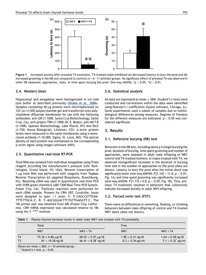
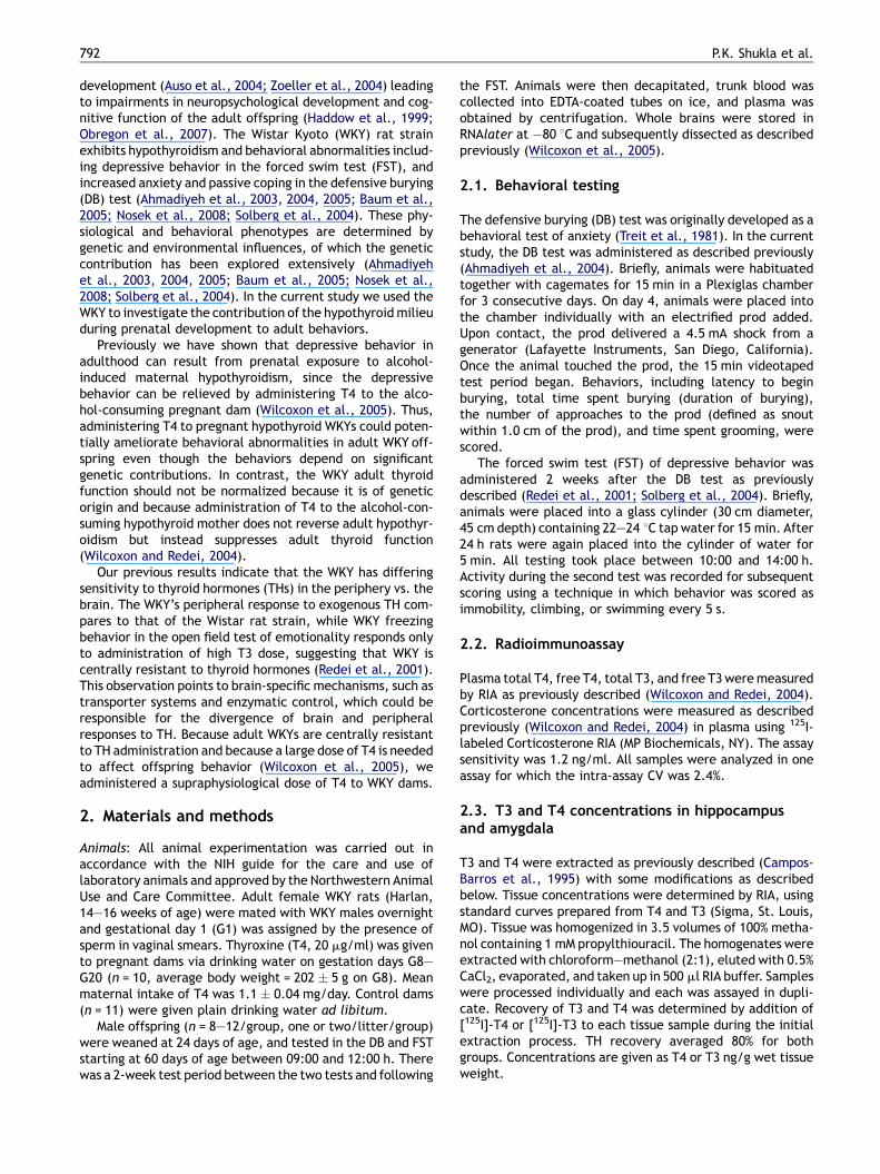
Figure 1 Increased anxiety after prenatal T4 treatment. T4-treated males exhibited (A) decreased latency to bury the prod and (B)increased grooming in the DB test compared to controls (n = 6—11 animals/group). No significant effect of prenatal T4 was observed inother DB measures: approaches, rears, or time spent burying the prod. One-way ANOVA, *p < 0.05, **p < 0.01.
Prenatal T4 affects brain thyroid hormone levels 793
2.4. Western blots
Hippocampi and amygdalae were homogenized in ice-coldlysis buffer as described previously (Shukla et al., 2006).Samples containing 40 mg protein were electrophoresed on12% (w/v) SDS polyacrylamide gel and transferred onto poly-vinylidene difluoride membranes for use with the followingantibodies: anti-GR (1:1000, Santa Cruz Biotechnology, SantaCruz, CA), anti-prepro-TRH (1:1000, Dr. E. Redei), anti-MCT-8(1:1000, Upstate Biotechnology, Lake Placid, NY) and Dio3(1:750, Novus Biologicals, Littleton, CO). b-Actin proteinlevels were measured in the same membranes using a mono-clonal antibody (1:10,000, Sigma, St. Louis, MO). The opticaldensity of each protein was normalized to the correspondingb-actin signal using ImageJ software (NIH).
2.5. Quantitative real-time RT-PCR
Total RNAwas isolated from individual amygdalae using Trizolreagent according the manufacturer’s protocol (Life Tech-nologies, Grand Island, NY, USA). Reverse transcription of1 mg total RNA was performed with reagents from TaqManReverse Transcription kit (Applied Biosystems, Branchburg,NJ). Resulting cDNA was used in quantitative real-time PCRwith SYBR green chemistry (ABI 7300 Real-Time PCR System,Foster City, CA). Triplicate reactions were performed foreach cDNA sample. Primers for CRH (IDT, Coralville, Iowa)were designed to span >1 exon: F: 50-CAGCCGTTGAA-TTTCTTGCA-30; R: 50-AGCGGGACTTCTGTTGAGGTT-30. The18s primer pair was obtained from ABI (Foster City, Califor-nia). CRH mRNA expression was calculated relative to 18susing the 2�DDCt method.
Table 1 Plasma thyroid hormone levels in adult male WKY rats
Total
WKY WKY + T4
T4 73.16 � 6.86 mg/dl 68.52 � 9.57 mg/dlT3 81 � 19.28 ng/dl 66.41 � 8.28* ng/dl
Values are mean � SEM. n = 10 animals/group.* Student’s t-test, p < 0.05.
2.6. Statistical analysis
All data are expressed as mean � SEM. Student’s t-tests wereconducted and correlations within the data were identifiedusing Pearson’s r coefficients (Systat software, Chicago, IL).Some experiments used a subset of samples due to metho-dological differences among measures. Degrees of freedomfor the different measures are indicated. p < 0.05 was con-sidered significant.
3. Results
3.1. Defensive burying (DB) test
Behaviors in the DB test, including latency to begin burying theprod, duration of burying, time spent grooming and number ofapproaches, were assessed in adult male offspring of WKYcontrol and T4-treated mothers. In males treated with T4, weobserved nonsignificant increases in the duration of buryingtime and in the number of approaches to the prod (data notshown). Latency to bury the prod after the initial shock wassignificantly lower (one-way ANOVA: F[1,14] = 11.0, p < 0.01,Fig. 1A) and time spent grooming was significantly increased(one-way ANOVA: F[1,17] = 4.8, p < 0.05, Fig. 1B). Thus, pre-natal T4 treatment resulted in behaviors that collectivelyindicate increased anxiety in adult WKY offspring.
3.2. Forced swim test (FST)
There were no differences in swimming, floating, or climbingbehaviors between male offspring of control and T4-treatedWKY dams (data not shown).
treated with T4 prenatally.
Free
WKY WKY + T4
1.85 � 0.31 ng/dl 1.64 � 0.28 ng/dl8.3 � 0.34 pg/ml 7.1 � 0.32* pg/ml

Figure 2 Brain region-specific effects of prenatal T4 on thyroidhormone levels in adult offspring. Free T4 and T3 concentrationswere both non-significantly decreased in the hippocampus (A),but in the amygdala, T4 was significantly increased and T3showed a similar trend (B). Student’s t-test, n = 6/group,**p < 0.01.
794 P.K. Shukla et al.
3.3. Corticosterone and thyroid hormone levels
There was no difference in plasma corticosterone levelsbetween WKY and WKY + T4 offspring (WKY, 93.5 �28.2 ng/ml; WKY + T4, 95.0 � 30.0 ng/ml, n = 10/group).Prenatal T4 treatment resulted in nonsignificant suppressionof total T4 (t(19) = 1.4, p = 0.24) and free T4 (t(21) = 2.6,p = 0.12) in the plasma, while total T3 (t(23) = 5.7, p < 0.05)and free T3 (t(23) = 6.6, p < 0.05) were significantly
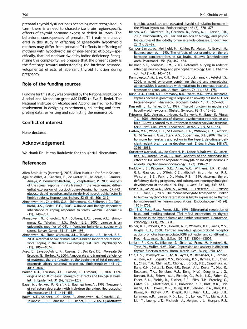
Figure 3 Prenatal T4 treatment alters protein levels in the hiptransporter monocarboxylate transporter 8 (MCT-8) were increasedelevated. (B) Amygdala: MCT-8 levels were increased along withhormone (prepro-TRH) levels. Student’s t-test, n = 4/group, *p < 0.
decreased (Table 1). In the brain, prenatal T4 treatmentdid not alter TH concentrations in the adult male hippocam-pus (Fig. 2A), whereas the amygdala of prenatally T4-treatedoffspring showed a significant increase in free T4 concentra-tion (t(11) = 15.9, p < 0.01, Fig. 2B) and a tendency ofincreased free T3 concentration (t(11) = 3.95, p = 0.07,Fig. 2B). While the active hormone T3 was not significantlyincreased in the amygdala of T4-treated WKYs, T3 levels inthis region correlated significantly with latency to bury theprod in the DB test (r = �0.58, p < 0.05).
3.4. Protein levels of MCT-8, Dio3, GR, andprepro-TRH
Protein levels of the MCT-8 TH transporter were significantlyincreased in both the hippocampus (t(7) = 9.4, p < 0.05,Fig. 3A) and amygdala (t(7) = 6.8, p < 0.05, Fig. 3B) of pre-natally T4-treated offspring. The TH-inactivating enzymeDio3 was also increased in the hippocampus of these offspring(t(7) = 9.2, p < 0.05, Fig. 3A), but in the amygdala, Dio3levels were very low as described elsewhere (Allen BrainAtlas, 2008; Lein et al., 2007), and were unchanged byprenatal T4 treatment (data not shown). In addition, therewere increases of GR (t(7) = 9.6, p < 0.05) and prepro-TRH(t(7) = 23.6, p < 0.01) protein in the amygdala, while GR andprepro-TRH were unchanged in the hippocampus (data notshown).
3.5. CRH mRNA expression
Hippocampal CRH mRNA levels, as measured by quantitativereal-time PCR, were approximately 50% of amygdala levelsand barely detectable as found previously (Aguilar-Valleset al., 2005). Hippocampal CRHmRNA levels were not alteredby prenatal T4 treatment (data not shown). In the amygdala,CRH mRNA levels were increased in offspring of T4-treateddams (WKY, 0.94 � 0.09; WKY + T4, 1.69 � 0.27; t(10) = 6.5,n = 6/group, p < 0.05). This increase in CRH transcriptexpression was positively correlated with amygdala T4(r = 0.76, p < 0.05) and T3 (r = 0.79, p < 0.05) levels.
pocampus and amygdala. (A) Hippocampus: levels of the THand the TH-inactivating enzyme deiodinase-III (Dio3) was alsoglucocorticoid receptor (GR) and prepro-thyrotropin-releasing05. Inserts are representative immunoblots.

Prenatal T4 affects brain thyroid hormone levels 795
4. Discussion
We intended to ameliorate adult depressive behavior withdevelopmental T4 administration based on previous evidencethat depressive behavior in adult offspring of alcohol-con-suming hypothyroid dams is reversed by supplementingmaternal T4 (Wilcoxon et al., 2005). That we did not obtainthis effect could be due to the genetic contribution todepressive behavior in this strain or to insufficiency of theT4 dose. Depressive-like behavior of adult WKYs can beameliorated by large doses of T3 (Redei et al., 2001), sug-gesting that a behavioral change in the WKY requires supra-physiological thyroid hormone levels in the brain. The latterfinding parallels human data wherein only supraphysiologicaldoses of thyroid hormones have depression-amelioratingeffects (Bauer et al., 1998).
Rather than affecting depressive behavior as expected,the prenatal T4 treatment effect pointed instead toincreased anxiety. Decreased latency to bury the prod inthe defensive burying paradigm is interpreted as increasedanxiety (De Boer and Koolhaas, 2003) as is increased groom-ing (Dunn et al., 1981) in a strain that is known for its lowbasal level of grooming (Nosek et al., 2008). In our studythese behaviors could be an indirect consequence of theadult WKY offspring’s exaggerated hypothyroid state, assubclinical and clinical hypothyroidism is known to increaseanxiety (Larisch et al., 2004; Sait Gonen et al., 2004).However, hypothyroid adult WKYs do not show increasedanxiety (Redei et al., 2001); thus, prenatal T4 administrationis a likely cause for the increased anxiety-related behaviorsobserved in the adult WKY offspring.
Our findings demonstrate for the first time that prenatalT4 treatment has opposite effects on peripheral and brainthyroid hormone levels: elevation in the amygdala and sup-pression in the periphery. Our previous findings suggest thatthe WKY strain may have exaggerated differences in respon-siveness between the periphery and brain to thyroid hor-mones (Redei et al., 2001), which we strongly corroboratehere. Further, prenatal T4 treatment suppressed peripheralTHs in the adult WKY similar to findings in another animalmodel (Wilcoxon and Redei, 2004). In both cases this effect isprobably attributable to elevated thyroid hormone levels atthe perinatal critical period, during which they determinethe set-point for adult thyroid function (Dussault and Fisher,1999; Pracyk et al., 1992; Walker and Courtin, 1985).
It was long assumed that the brain maintains its ownthyroid homeostasis that is rarely affected by peripheralthyroid dysfunction. Later, it was often assumed that per-ipheral T4 — but not T3 — enters the central nervous system.Our current finding of brain region-specific alterations of THlevels in the WKY is novel. Local conversion of T4 to T3 bydeiodinase II (Dio2) in astrocytes is estimated to produce asmuch as 80% of the T3 in the adult rat brain (Bianco et al.,2002). However, Dio2-deficient mice lacking local brain T3production show proper brain development and function(Galton et al., 2007), showing that T3 taken up from theperiphery can support the brain T3 need. The nonessentialnature of Dio2 also suggests that the primary regulators of T3action are the molecules MCT-8 and Dio3, which transport T3into the neuron, and metabolize it there (Heuer et al., 2005).
Our finding of increased T3 levels in the amygdala despitedecreased peripheral TH levels raises the question of how
local thyroid hormone levels are controlled in a brain region-specific manner. The amygdala-specific ‘‘hyperthyroid’’ THmilieu in T4-treated offspring is likely due to the increasedTH transport via MCT-8 into amygdalar neurons combinedwith the low level of Dio3 expression in this region (Leinet al., 2007). Themechanism by which prenatal T4 treatmentaugments adult MCT-8 expression has yet to be elucidated,but the increase of MCT-8 expression is of particular interestas MCT-8 mutation in humans results in neurodevelopmentaland global neurological impairment greater than thatobserved in cases of congenital hypothyroidism (Dumitrescuet al., 2004; Friesema et al., 2006; Schwartz and Stevenson,2007).
In T4-treated offspring, the ‘‘hyperthyroid’’ milieu of theamygdala correlates with a shorter latency to bury the prod,an indicator of anxiety. An intermediary in this relationshipcould be increased GR protein levels. We have shown pre-viously that amygdalar GR is decreased in the offspring ofhypothyroid dams, and prenatal T4 treatment reversed thisdecrease in GR expression (Wilcoxon et al., 2005). GR over-expression in mice causes increased anxiety-like behavior inseveral behavioral tests of anxiety, and targeted reduction ofGR function in the nervous system impairs stress responsesand reduces anxiety behavior (Tronche et al., 1999; Weiet al., 2004). These studies and our own suggest that thein utero environment programs adult expression of GR–—andin the adult, increased amygdalar GR is anxiogenic. In addi-tion, GR-bound glucocorticoids induce CRH expression in theamygdala (Kolber et al., 2008), which can lead to increasedanxiety (Yilmazer-Hanke et al., 2004). Elevated CRH mRNAlevels are found in the central amygdala of the WKY com-pared to F344 and Wistar rats in association with increasedanxiety behavior in the elevated plus maze (Shepard andMyers, 2008). In the current study, amygdalar CRH mRNAlevels were further elevated in WKY offspring of T4-treateddams concomitant with increased anxiety measures, with nochange in peripheral glucocorticoid levels.
In addition to elevated GR protein levels and CRH mRNAexpression, prepro-TRH protein levels were also higher in theamygdala of T4-treated offspring. The WKY has higher basalamygdala TRH peptide levels compared to the Wistar rat andTRHergic neurons in the amygdala are active in response toenvironmental stress including exposure to the DB paradigm(Gutierrez-Mariscal et al., 2008). TRH peptide levels are notsubject to classical hormonal feedback control in the amyg-dala, and T3 administration to adult rats did not alter TRHmRNA levels in limbic brain regions (Kim et al., 1996). Thus,the increase in amygdalar prepro-TRH levels we observedmay be a unique effect of in utero T4 treatment. Furtherinvestigations are needed to fully characterize the processingof prepro-TRH in the amygdala and therefore the amount ofTRH peptide that is present to influence anxiety-relatedbehavior. Specialized prepro-TRH processing may also relateto paradoxical evidence that administration of exogenousTRH intracerebroventricularly is proposed to have anxiolyticactions (Gutierrez-Mariscal et al., 2008).
Our study establishes that prenatal T4 treatment caninduce increased anxiety by altering brain region-specificexpression of genes whose function is to modulate localizedthyroid hormone availability. Thyroid hormones have aremarkable range of actions in the development and functionof the nervous system, and the spectrum of consequences of
796 P.K. Shukla et al.
prenatal thyroid dysfunction is becoming more recognized. Inturn, there is a need to characterize brain region-specificeffects of thyroid hormone excess or deficit in utero. Thebehavioral consequences of prenatal T4 treatment uncov-ered in this study in offspring of genetically hypothyroidmothers may differ from prenatal T4 effects in offspring ofmothers with hypothyroidism of non-genetic etiology–—spe-cifically, that induced worldwide by iodine deficiency. Recog-nizing this complexity, we propose that the present study isthe first step toward understanding the intricate neurode-velopmental effects of aberrant thyroid function duringpregnancy.
Role of the funding sources
Funding for this studywas providedby theNational InstituteonAlcohol and Alcoholism grant AA013452 to Eva E. Redei. TheNational Institute on Alcohol and Alcoholism had no furtherinvolvement in designing experiments, collecting and inter-preting data, or writing and submitting the manuscript.
Conflict of interest
None declared.
Acknowledgement
We thank Dr. Jelena Radulovic for thoughtful discussions.
References
Allen Brain Atlas [Internet], 2008. Allen Institute for Brain Science.Aguilar-Valles, A., Sanchez, E., de Gortari, P., Balderas, I., Ramirez-
Amaya, V., Bermudez-Rattoni, F., Joseph-Bravo, P., 2005. Analysisof the stress response in rats trained in the water-maze: differ-ential expression of corticotropin-releasing hormone, CRH-R1,glucocorticoid receptors and brain-derived neurotrophic factor inlimbic regions. Neuroendocrinology 82 (5—6), 306—319.
Ahmadiyeh, N., Churchill, G.A., Shimomura, K., Solberg, L.C., Taka-hashi, J.S., Redei, E.E., 2003. X-linked and lineage-dependentinheritance of coping responses to stress. Mamm. Genome 14(11), 748—757.
Ahmadiyeh, N., Churchill, G.A., Solberg, L.C., Baum, A.E., Shimo-mura, K., Takahashi, J.S., Redei, E.E., 2005. Lineage is anepigenetic modifier of QTL influencing behavioral coping withstress. Behav. Genet. 35 (2), 189—198.
Ahmadiyeh, N., Slone-Wilcoxon, J.L., Takahashi, J.S., Redei, E.E.,2004. Maternal behavior modulates X-linked inheritance of beha-vioral coping in the defensive burying test. Biol. Psychiatry 55(11), 1069—1074.
Auso, E., Lavado-Autric, R., Cuevas, E., Del Rey, F.E., Morreale DeEscobar, G., Berbel, P., 2004. Amoderate and transient deficiencyof maternal thyroid function at the beginning of fetal neocorti-cogenesis alters neuronal migration. Endocrinology 145 (9),4037—4047.
Barker, D.J., Eriksson, J.G., Forsen, T., Osmond, C., 2002. Fetalorigins of adult disease: strength of effects and biological basis.Int. J. Epidemiol. 31 (6), 1235—1239.
Bauer, M., Hellweg, R., Graf, K.J., Baumgartner, A., 1998. Treatmentof refractory depression with high-dose thyroxine. Neuropsycho-pharmacology 18 (6), 444—455.
Baum, A.E., Solberg, L.C., Kopp, P., Ahmadiyeh, N., Churchill, G.,Takahashi, J.S., Jameson, J.L., Redei, E.E., 2005. Quantitative
trait loci associatedwith elevated thyroid-stimulating hormone inthe Wistar Kyoto rat. Endocrinology 146 (2), 870—878.
Bianco, A.C., Salvatore, D., Gereben, B., Berry, M.J., Larsen, P.R.,2002. Biochemistry, cellular and molecular biology, and physio-logical roles of the iodothyronine selenodeiodinases. Endocr. Rev.23 (1), 38—89.
Campos-Barros, A., Meinhold, H., Kohler, R., Muller, F., Eravci, M.,Baumgartner, A., 1995. The effects of desipramine on thyroidhormone concentrations in rat brain. Naunyn SchmiedebergsArch. Pharmacol. 351 (5), 469—474.
De Boer, S.F., Koolhaas, J.M., 2003. Defensive burying in rodents:ethology, neurobiology and psychopharmacology. Eur. J. Pharma-col. 463 (1—3), 145—161.
Dumitrescu, A.M., Liao, X.H., Best, T.B., Brockmann, K., Refetoff, S.,2004. A novel syndrome combining thyroid and neurologicalabnormalities is associated with mutations in a monocarboxylatetransporter gene. Am. J. Hum. Genet. 74 (1), 168—175.
Dunn, A.J., Guild, A.L., Kramarcy, N.R., Ware, M.D., 1981. Benzodia-zepines decrease grooming in response to novelty but not ACTH orbeta-endorphin. Pharmacol. Biochem. Behav. 15 (4), 605—608.
Dussault, J.H., Fisher, D.A., 1999. Thyroid function in mothers ofhypothyroid newborns. Obstet. Gynecol. 93 (1), 15—20.
Friesema, E.C., Jansen, J., Heuer, H., Trajkovic, M., Bauer, K., Visser,T.J., 2006. Mechanisms of disease: psychomotor retardation andhigh T3 levels caused by mutations in monocarboxylate transpor-ter 8. Nat. Clin. Pract. Endocrinol. Metab. 2 (9), 512—523.
Galton, V.A., Wood, E.T., St Germain, E.A., Withrow, C.A., Aldrich,G., St Germain, G.M., Clark, A.S., St Germain, D.L., 2007. Thyroidhormone homeostasis and action in the type 2 deiodinase-defi-cient rodent brain during development. Endocrinology 148 (7),3080—3088.
Gutierrez-Mariscal, M., de Gortari, P., Lopez-Rubalcava, C., Marti-nez, A., Joseph-Bravo, P., 2008. Analysis of the anxiolytic-likeeffect of TRH and the response of amygdalar TRHergic neurons inanxiety. Psychoneuroendocrinology 33 (2), 198—213.
Haddow, J.E., Palomaki, G.E., Allan, W.C., Williams, J.R., Knight,G.J., Gagnon, J., O’Heir, C.E., Mitchell, M.L., Hermos, R.J.,Waisbren, S.E., Faix, J.D., Klein, R.Z., 1999. Maternal thyroiddeficiency during pregnancy and subsequent neuropsychologicaldevelopment of the child. N. Engl. J. Med. 341 (8), 549—555.
Heuer, H., Maier, M.K., Iden, S., Mittag, J., Friesema, E.C., Visser,T.J., Bauer, K., 2005. The monocarboxylate transporter 8 linkedto human psychomotor retardation is highly expressed in thyroidhormone-sensitive neuron populations. Endocrinology 146 (4),1701—1706.
Kim, S.Y., Post, R.M., Rosen, J.B., 1996. Differential regulation ofbasal and kindling-induced TRH mRNA expression by thyroidhormone in the hypothalamic and limbic structures. Neuroendo-crinology 63 (3), 297—304.
Kolber, B.J., Roberts, M.S., Howell, M.P., Wozniak, D.F., Sands, M.S.,Muglia, L.J., 2008. Central amygdala glucocorticoid receptoraction promotes fear-associated CRH activation and conditioning.Proc. Natl. Acad. Sci. U.S.A. 105 (33), 12004—12009.
Larisch, R., Kley, K., Nikolaus, S., Sitte, W., Franz, M., Hautzel, H.,Tress, W., Muller, H.W., 2004. Depression and anxiety in differentthyroid function states. Horm. Metab. Res. 36 (9), 650—653.
Lein, E.S., Hawrylycz, M.J., Ao, N., Ayres, M., Bensinger, A., Bernard,A., Boe, A.F., Boguski, M.S., Brockway, K.S., Byrnes, E.J., Chen,L., Chen, T.M., Chin, M.C., Chong, J., Crook, B.E., Czaplinska, A.,Dang, C.N., Datta, S., Dee, N.R., Desaki, A.L., Desta, T., Diep, E.,Dolbeare, T.A., Donelan, M.J., Dong, H.W., Dougherty, J.G.,Duncan, B.J., Ebbert, A.J., Eichele, G., Estin, L.K., Faber, C.,Facer, B.A., Fields, R., Fischer, S.R., Fliss, T.P., Frensley, C.,Gates, S.N., Glattfelder, K.J., Halverson, K.R., Hart, M.R., Hoh-mann, J.G., Howell, M.P., Jeung, D.P., Johnson, R.A., Karr, P.T.,Kawal, R., Kidney, J.M., Knapik, R.H., Kuan, C.L., Lake, J.H.,Laramee, A.R., Larsen, K.D., Lau, C., Lemon, T.A., Liang, A.J.,Liu, Y., Luong, L.T., Michaels, J., Morgan, J.J., Morgan, R.J.,

Prenatal T4 affects brain thyroid hormone levels 797
Mortrud, M.T., Mosqueda, N.F., Ng, L.L., Ng, R., Orta, G.J.,Overly, C.C., Pak, T.H., Parry, S.E., Pathak, S.D., Pearson,O.C., Puchalski, R.B., Riley, Z.L., Rockett, H.R., Rowland,S.A., Royall, J.J., Ruiz, M.J., Sarno, N.R., Schaffnit, K., Shapo-valova, N.V., Sivisay, T., Slaughterbeck, C.R., Smith, S.C., Smith,K.A., Smith, B.I., Sodt, A.J., Stewart, N.N., Stumpf, K.R., Sunkin,S.M., Sutram, M., Tam, A., Teemer, C.D., Thaller, C., Thompson,C.L., Varnam, L.R., Visel, A., Whitlock, R.M., Wohnoutka, P.E.,Wolkey, C.K., Wong, V.Y., Wood, M., Yaylaoglu, M.B., Young, R.C.,Youngstrom, B.L., Yuan, X.F., Zhang, B., Zwingman, T.A., Jones,A.R., 2007. Genome-wide atlas of gene expression in the adultmouse brain. Nature 445 (7124), 168—176.
Nosek, K., Dennis, K., Andrus, B.M., Ahmadiyeh, N., Baum, A.E.,Woods, L.C., Redei, E.E., 2008. Context and strain-dependentbehavioral response to stress. Behav. Brain Funct. 4, 23.
Obregon, M.J., Calvo, R.M., Del Rey, F.E., de Escobar, G.M., 2007.Ontogenesis of thyroid function and interactions with maternalfunction. Endocr. Dev. 10, 86—98.
Pracyk, J.B., Seidler, F.J., McCook, E.C., Slotkin, T.A., 1992. Pitui-tary-thyroid axis reactivity to hyper- and hypothyroidism in theperinatal period: ontogeny of regulation of regulation and long-term programming of responses. J. Dev. Physiol. 18 (3), 105—109.
Redei, E.E., Solberg, L.C., Kluczynski, J.M., Pare, W.P., 2001. Para-doxical hormonal and behavioral responses to hypothyroid andhyperthyroid states in the Wistar-Kyoto rat. Neuropsychophar-macology 24 (6), 632—639.
Sait Gonen, M., Kisakol, G., Savas Cilli, A., Dikbas, O., Gungor, K.,Inal, A., Kaya, A., 2004. Assessment of anxiety in subclinicalthyroid disorders. Endocr. J. 51 (3), 311—315.
Schwartz, C.E., Stevenson, R.E., 2007. The MCT8 thyroid hormonetransporter and Allan—Herndon—Dudley syndrome. Best Pract.Res. Clin. Endocrinol. Metab. 21 (2), 307—321.
Shepard, J.D., Myers, D.A., 2008. Strain differences in anxiety-likebehavior: association with corticotropin-releasing factor. Behav.Brain Res. 186 (2), 239—245.
Shukla, P.K., Tang, L., Wang, Z.J., 2006. Phosphorylation of neuro-granin, protein kinase C, and Ca2+/calmodulin dependent proteinkinase II in opioid tolerance and dependence. Neurosci. Lett. 404(3), 266—269.
Solberg, L.C., Baum, A.E., Ahmadiyeh, N., Shimomura, K., Li, R.,Turek, F.W., Churchill, G.A., Takahashi, J.S., Redei, E.E., 2004.Sex- and lineage-specific inheritance of depression-like behaviorin the rat. Mamm. Genome 15 (8), 648—662.
Treit, D., Pinel, J.P., Fibiger, H.C., 1981. Conditioned defensiveburying: a new paradigm for the study of anxiolytic agents.Pharmacol. Biochem. Behav. 15 (4), 619—626.
Tronche, F., Kellendonk, C., Kretz, O., Gass, P., Anlag, K., Orban,P.C., Bock, R., Klein, R., Schutz, G., 1999. Disruption of theglucocorticoid receptor gene in the nervous system results inreduced anxiety. Nat. Genet. 23 (1), 99—103.
Walker, P., Courtin, F., 1985. Transient neonatal hyperthyroidismresults in hypothyroidism in the adult rat. Endocrinology 116(6), 2246—2250.
Wei, Q., Lu, X.Y., Liu, L., Schafer, G., Shieh, K.R., Burke, S.,Robinson, T.E., Watson, S.J., Seasholtz, A.F., Akil, H., 2004.Glucocorticoid receptor overexpression in forebrain: a mousemodel of increased emotional lability. Proc. Natl. Acad. Sci.U.S.A. 101 (32), 11851—11856.
Wilcoxon, J.S., Kuo, A.G., Disterhoft, J.F., Redei, E.E., 2005. Beha-vioral deficits associated with fetal alcohol exposure are reversedby prenatal thyroid hormone treatment: a role for maternalthyroid hormone deficiency in FAE. Mol. Psychiatry 10 (10),961—971.
Wilcoxon, J.S., Redei, E.E., 2004. Prenatal programming of adultthyroid function by alcohol and thyroid hormones. Am. J. Physiol.Endocrinol. Metab. 287 (2), E318—E326.
Yilmazer-Hanke, D.M., Hantsch, M., Hanke, J., Schulz, C., Faber-Zuschratter, H., Schwegler, H., 2004. Neonatal thyroxine treat-ment: changes in the number of corticotropin-releasing-factor(CRF) and neuropeptide Y (NPY) containing neurons and density oftyrosine hydroxylase positive fibers (TH) in the amygdala corre-late with anxiety-related behavior of Wistar rats. Neuroscience124 (2), 283—297.
Zoeller, R.T., Bigelow, C., Rovet, J., 2004. Lack of a relation betweenhuman neonatal thyroxine and pediatric neurobehavioral disor-ders: neonatal total thyroxine is not a good proxy measure ofmaternal thyroid hormone insufficiency. Thyroid 14 (3), 239—241author reply 241—233.
Copyright © 2022 FDOKUMEN