Mutations in squirrel monkey glucocorticoid receptor impair nuclear translocation
Glucocorticoid receptor number predicts increase inamygdala activity after severe stress
Elbert Geuze a,b,*, Guido A. van Wingen c,d, Mirjam van Zuiden a,e,Arthur R. Rademaker a,b, Eric Vermetten a,b, Annemieke Kavelaars e,Guillen Fernandez d,f, Cobi J. Heijnen e
aResearch Centre, Military Mental Health, Ministry of Defense, Utrecht, The NetherlandsbDepartment of Psychiatry, Rudolf Magnus Institute of Neuroscience, Utrecht University Medical Center, Utrecht, The NetherlandscDepartment of Psychiatry, Academic Medical Center, University of Amsterdam, Amsterdam, The NetherlandsdDonders Institute for Brain, Cognition and Behaviour, Radboud University Nijmegen, Nijmegen, The Netherlandse Laboratory of Neuroimmunology and Developmental Origins of Disease (NIDOD), University Medical Center Utrecht, Utrecht, TheNetherlandsfDepartment for Cognitive Neuroscience, Radboud University Nijmegen Medical Centre, Nijmegen, The Netherlands
Received 9 November 2011; received in revised form 21 March 2012; accepted 21 March 2012
Psychoneuroendocrinology (2012) 37, 1837—1844
KEYWORDSStress;Combat;PTSD;Glucocorticoid receptor;fMRI;Amygdala
Summary
Introduction: Individuals who are exposed to a traumatic event are at increased risk ofdeveloping psychiatric disorders such as posttraumatic stress disorder (PTSD). Studies haveshown that increased amygdala activity is frequently found in patients with PTSD. In addition,pre-trauma glucocorticoid receptor (GR) number in peripheral blood mononuclear cells (PBMCs)has been found to be a significant predictor for the development of PTSD symptoms. Research inrodents has shown that the response of basolateral amygdala neurons to corticosterone ismediated by GR. However, to the best of our knowledge, no previous study has investigatedGR number in PBMCs and amygdala function in humans.Methods: To investigate whether peripheral GR number is related to amygdala functioning, weassessed GR number in PBMCs of healthy soldiers before their deployment to Afghanistan.Amygdala functioning was assessed with fMRI before and after deployment.Results: We found that pre-deployment GR number was significantly negatively correlated topre-deployment amygdala activity. More importantly, pre-deployment GR number predicted theincrease in amygdala activity by deployment.Discussion: Our results demonstrate that peripheral GR number is associated with amygdalafunctioning and predicts the increase in amygdala activity following military deployment in
* Corresponding author at: Department of Psychiatry, Utrecht University Medical Center, PO Box B.01.2.06, Heidelberglaan 100, 3584 CXUtrecht, The Netherlands. Tel.: +31 30 250 2588; fax: +31 30 250 2282.
E-mail address: [email protected] (E. Geuze).
Available online at www.sciencedirect.com
j our na l h omepa g e: www.e l se v ie r.c om/l oca te/ psyne ue n
0306-4530/$ — see front matter # 2012 Elsevier Ltd. All rights reserved.
http://dx.doi.org/10.1016/j.psyneuen.2012.03.017
healthy individuals who did not develop PTSD. It is uncertain how this relationship is mediatedmechanistically, but future studies should examine the relation of GR and amygdala activity todetermine whether this is part of a common pathway leading to increased vulnerability to stress-related disorders.# 2012 Elsevier Ltd. All rights reserved.
1838 E. Geuze et al.
1. Introduction
Individuals who are exposed to a traumatic event (such asnatural disasters, terrorism, assault, or military combat) areat riskof developing psychiatric disorders such as posttraumaticstress disorder (PTSD). In the last decades knowledge of thelasting consequences of traumatic stress on the neurobiology ofindividuals has increased rapidly. Functional neuroimagingstudies have revealed that patients with PTSD show exagger-ated amygdala responses and deficient prefrontal cortex func-tion, in particular in the anterior cingulate cortex andventromedial prefrontal cortex (Rauch et al., 2000; Shinet al., 2001, 2005). These neural alterations in PTSD probablyresult from both stress exposure and stress vulnerability(Admon et al., 2009; Shin et al., 2009). In a recent study, weinvestigated soldiers before and after their first deployment toa combat zone (van Wingen et al., 2011). Our results demon-strated that combat stress increases amygdala and insulareactivity to biologically salient stimuli, and that impairedprefrontal cortex—amygdala coupling was associated with per-ceived threat. Follow-up showed that amygdala responsivitynormalizes within 1.5 years after deployment in resilient indi-viduals that do not develop PTSD, but that perceived threat-dependent changes in amygdala coupling with the dorsal ante-rior cingulate cortex persist (van Wingen et al., 2012).
In addition to these neurobiological changes, numerousstudies have also shown that PTSD is associated with increasedsensitivity of the hypothalamic—pituitary—adrenal axis (HPA-axis) to negative feedback by glucocorticoids (GCs) (Yehudaet al., 1995, 2002; Golier et al., 2006; de Kloet et al., 2006,2007a,b). Recently, we reported that the pre-trauma gluco-corticoid receptor number in peripheral blood mononuclearcells (PBMCs) was higher within soldiers who developed a highlevel of PTSD symptoms after deployment to Afghanistancompared to matched controls who did not develop PTSDsymptoms (van Zuiden et al., 2011, 2012). Although the per-ipheral GR pathway is often considered to be an accessiblemodel for GR signaling in the brain, it remains unclear if andhow peripheral GR functioning is related to CNS activity.Preclinical research has shown that the response of basolateralamygdala neurons to corticosterone is mediated by GR (Karstet al., 2010; Arnett et al., 2011; Groeneweg et al., 2011).Whether GR number in PBMCs is a reliable reflection of GRnumber in the brain has not been studied extensively. Pre-clinical studies have shown that the affinity and specificity forGCs of neuronal and lymphoid GRs are similar (Lowy, 1989). Inaddition, GRs in both brain and peripheral immune tissues aredownregulated after chronic corticosterone administration(Spencer et al., 1991). However, to our knowledge, no previousstudy has investigated the relation between peripheral GR andamygdala function in humans. We hypothesized that GR num-ber in PBMCs would be related to amygdala functioning inhealthy individuals. In addition, we hypothesized that GR
number in PBMCs would be associated with the amygdalaresponse in reaction to severe stress.
To investigate whether peripheral GR is related to amyg-dala functioning, we assessed GR number in PBMCs in soldiersbefore deployment to Afghanistan. Amygdala functioningwas assessed with functional magnetic resonance imaging(fMRI) in the same group of soldiers before and 1.5 monthsafter deployment.
2. Methods
2.1. Participants
Participants included in this study consisted of twenty-fourhealthy soldiers who were deployed for 4 months to Afghani-stan as part of the NATO International Security Assistance Force(ISAF) peacekeeping operation. They were recruited from alarger prospective study on the development of stress-relateddisorders following military deployment in the Dutch armedforces. None of the participants had been deployed previously.Duties during deployment included combat patrols, clearing orsearching homes and buildings, participation in deminingoperations, and transportation across enemy territory. Theywere exposed to typical war-zone stressors such as enemy fire,armed combat, and seeing seriously injured fellow soldiers andcivilians (including women and children). The study was inaccordance with the declaration of Helsinki and institutionalguidelines of the local ethics committee (CMO Arnhem-Nijme-gen, the Netherlands and the Institutional Review Board of theUniversity Medical Center Utrecht, the Netherlands), and allparticipants provided written informed consent after writtenand oral description of the study.
2.2. Procedure
Participants were investigated several weeks prior to deploy-ment to Afghanistan. A venous blood sample was drawn forassessment of the number of GR binding sites in PBMCs.Participants filled in a number of paper-and-pencil question-naires. Since GR is associated with glucose regulation, andpolymorphisms of the GR gene are associated with metabolicparameters and body composition (van Rossum and Lam-berts, 2004), we also measured the body-mass index (BMI)prior to deployment. BMI was calculated from the self-reported weight and length of the participants. In addition,all participants received an fMRI scan before and 1.5 monthsafter deployment.
2.3. Questionnaires
A previously validated Dutch questionnaire, the Self-ratingInventory for PTSD, was used to assess self-reported PTSD

GR number and amygdala activity 1839
symptoms (Hovens et al., 1994). We included all healthysoldiers who were deployed for 4 months to Afghanistan.As it is quite plausible that GR concentrations and amygdalaactivity would be altered in those individuals who developPTSD both prior to deployment and after, one participant whoscored above the clinical threshold for PTSD symptoms afterdeployment was therefore excluded from the analyses. Expo-sure to potential traumatic experiences before the age of 18was assessed using the Dutch version of the short form self-report version of the Early Trauma Inventory (Bremner et al.,2007; Rademaker et al., 2008). This questionnaire consists of27 dichotomous items. The total score represents the numberof experienced events. To quantify stress exposure, wemeasured combat exposure during deployment using thecombat experience scale of the Deployment Risk and Resi-lience Inventory (Vogt et al., 2008).
2.4. Cortisol
Plasma cortisol levels were measured by electrochemilumi-nescence immunoassay on the Modular E170 (Roche Diagnos-tics, Mannheim, Germany). Lower limit of detection was3 nmol/l; interassay variation was <3%; and reference values(0700—1000 h) were 170—540 nmol/l.
2.5. GR binding sites in PBMCs
For determination of the capacity of PBMCs to bind glucocor-ticoids a validated whole cell single point binding assay wasused as described previously. This method provides a reliableestimate of Bmax as determined using a classical binding assaywith 3—200 nM 3H-dexamethasone (r2 = 0.92) (van Zuidenet al., 2009, 2011). Briefly, PBMCs were isolated from wholeblood using Ficoll-Paque (Pharmacia, Uppsala, Sweden) and107 cells were frozen in DMSO. After thawing and 60 minequilibration in culture medium, cells were washed twice,resuspended in assay buffer (RPMI-1640 with 5% FCS) andincubated in duplicate with 100 nM 3H-dexamethasone (Amer-sham, Buckinghamshire, UK) in the presence or absence ofexcess unlabeled dexamethasone (Sigma—Aldrich, Steinheim,Germany) for 1 h at 37 8C. Cell bound radioactivity was quan-tified by liquid scintillation analysis.
2.6. MRI data acquisition
MR data were acquired with a 1.5 T Siemens (Erlangen, Ger-many) Avanto MR scanner, equipped with a standard head coil.T2*-weighted BOLD images were acquired using EPI with anecho time of 35 ms to reduce signal dropout in the medialtemporal lobes. Each image volume consisted of 32 axial slices(3.5 mm, 0.35 mm slice-gap, TR = 2.340 s, 64 � 64 matrix,FOV = 212 mm, FA = 908). In addition, a high-resolution T1-weighted structural MR image with optimized gray/whitematter contrast was acquired (3D MP-RAGE, 1 � 1 � 1 mm3
voxels, TR = 2.730 s, TE = 2.95 ms, TI = 1000 ms, FOV =256 mm, FA = 78).
2.7. Behavioral task
The experimental paradigm that was performed during func-tional MRI scanning consisted of a blocked design, including a
condition with angry and fearful face stimuli (http://www.macbrain.org) and a visuo-motor control condition withscrambled ellipse stimuli, a task specifically designed to elicitamygdala activity (Hariri et al., 2002; van Wingen et al.,2011). Two blocks of angry and fearful faces were interleavedwith three control blocks, and each 30 s block consisted of six5 s trials. Each trial consisted of three simultaneously pre-sented stimuli, with the cue stimulus presented above thetarget and distractor. In the faces condition, an angry orfearful face was presented on top as cue, and participantshad to indicate by an appropriate button press which of thebottom two faces (one angry and one fearful) matched thecue in emotional expression. In the control condition, ahorizontally or vertically oriented ellipse was presented ascue above two ellipses (one vertical and one horizontal), andparticipants had to select the identically oriented ellipse.
2.8. Data analysis
Image analysis was performed with SPM5 (http://www.fil.io-n.ucl.ac.uk/spm). The first five EPI-volumes were discardedand the remaining images were realigned to the first volume.Images were then coregistered to the anatomical scan,corrected for differences in slice acquisition time, spatiallynormalized to the MNI T1 template, resampled into2 � 2 � 2 mm3 voxels, and spatially smoothed (8 mm FWHM).Statistical analysis was performed within the framework ofthe general linear model (Friston et al., 1994). To assess theinfluence of combat stress on neural responsivity, the twoexperimental conditions were modeled as box-car regressorsconvolved with the canonical hemodynamic response func-tion of SPM5. In addition, the realignment parameters wereincluded to model potential movement artifacts, as well as aconstant. Furthermore, a high-pass filter (cut-off 1/128 Hz)was included, and temporal autocorrelation was modeledwith an AR(1) process. Contrast images subtracting the visuo-motor control condition from the emotion condition wereobtained, and analyzed in subsequent random effects mod-els.
Voxel-wise statistical tests were family-wise error (FWE)rate corrected ( p < 0.05) for multiple comparisons acrossthe entire brain, or for the search volume for regions ofinterest using a small volume correction (SVC) (Worsleyet al., 1996). The search volume for the amygdala wasanatomically defined using the WFU Pickatlas toolbox imple-mented in SPM5 (Maldjian et al., 2003). For the correlationand regression analyses with GR number, the imaging datawere extracted from the amygdala mask (see Fig. 1). Pearsoncorrelation analyses and linear hierarchical regression ana-lysis were performed using PASW Statistics 18.0, p < 0.05(two-tailed) was considered significant. Data were assessedfor the presence of significant outliers. One of the partici-pants had a GR number which deviated >2 SD from the meanand was therefore excluded from the analyses.
3. Results
3.1. Demographics
The participants that were included for the analysis were22 male soldiers who had been deployed to Afghanistan.
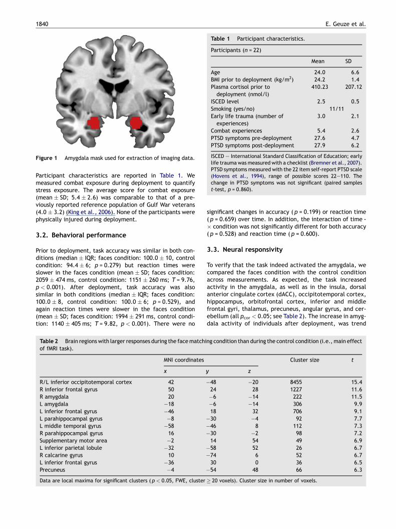
Figure 1 Amygdala mask used for extraction of imaging data.
Table 1 Participant characteristics.
Participants (n = 22)
Mean SD
Age 24.0 6.6BMI prior to deployment (kg/m2) 24.2 1.4Plasma cortisol prior todeployment (nmol/l)
410.23 207.12
ISCED level 2.5 0.5Smoking (yes/no) 11/11Early life trauma (number ofexperiences)
3.0 2.1
Combat experiences 5.4 2.6PTSD symptoms pre-deployment 27.6 4.7PTSD symptoms post-deployment 27.9 6.2
ISCED — International Standard Classification of Education; earlylife trauma was measured with a checklist (Bremner et al., 2007).PTSD symptoms measured with the 22 item self-report PTSD scale(Hovens et al., 1994), range of possible scores 22—110. Thechange in PTSD symptoms was not significant (paired samplest-test, p = 0.860).
1840 E. Geuze et al.
Participant characteristics are reported in Table 1. Wemeasured combat exposure during deployment to quantifystress exposure. The average score for combat exposure(mean � SD; 5.4 � 2.6) was comparable to that of a pre-viously reported reference population of Gulf War veterans(4.0 � 3.2) (King et al., 2006). None of the participants werephysically injured during deployment.
3.2. Behavioral performance
Prior to deployment, task accuracy was similar in both con-ditions (median � IQR; faces condition: 100.0 � 10, controlcondition: 94.4 � 6; p = 0.279) but reaction times wereslower in the faces condition (mean � SD; faces condition:2059 � 474 ms, control condition: 1151 � 260 ms; T = 9.76,p < 0.001). After deployment, task accuracy was alsosimilar in both conditions (median � IQR; faces condition:100.0 � 8, control condition: 100.0 � 6; p = 0.529), andagain reaction times were slower in the faces condition(mean � SD; faces condition: 1994 � 291 ms, control condi-tion: 1140 � 405 ms; T = 9.82, p < 0.001). There were no
Table 2 Brain regions with larger responses during the face matchiof fMRI task).
MNI coordinates
x
R/L inferior occipitotemporal cortex 42
R inferior frontal gyrus 50
R amygdala 20
L amygdala �18
L inferior frontal gyrus �46
L parahippocampal gyrus �8
L middle temporal gyrus �58
R parahippocampal gyrus 16
Supplementary motor area �2
L inferior parietal lobule �32
R calcarine gyrus 10
L inferior frontal gyrus �36
Precuneus �4
Data are local maxima for significant clusters ( p < 0.05, FWE, cluster
significant changes in accuracy ( p = 0.199) or reaction time( p = 0.659) over time. In addition, the interaction of time -� condition was not significantly different for both accuracy( p = 0.528) and reaction time ( p = 0.600).
3.3. Neural responsivity
To verify that the task indeed activated the amygdala, wecompared the faces condition with the control conditionacross measurements. As expected, the task increasedactivity in the amygdala, as well as in the insula, dorsalanterior cingulate cortex (dACC), occipitotemporal cortex,hippocampus, orbitofrontal cortex, inferior and middlefrontal gyri, thalamus, precuneus, angular gyrus, and cer-ebellum (all pcor < 0.05; see Table 2). The increase in amyg-dala activity of individuals after deployment, was trend
ng condition than during the control condition (i.e., main effect
Cluster size t
y z
�48 �20 8455 15.424 28 1227 11.6�6 �14 222 11.5�6 �14 306 9.918 32 706 9.1�30 �4 92 7.7�46 8 112 7.3�30 �2 98 7.214 54 49 6.9�58 52 26 6.7�74 6 52 6.730 0 36 6.5�54 48 66 6.3
� 20 voxels). Cluster size in number of voxels.

Figure 2 GR number in PBMCs prior to deployment is signifi-cantly correlated with amygdala activity prior to deployment (A)but not to amygdala activity after deployment (B). GR number isalso significantly associated with the increase in amygdala ac-tivity after deployment (C).
GR number and amygdala activity 1841
significant, after small volume correction (peak MontrealNeurological Institute coordinate (x, y, z); (26, �2, �26),Z = 2.9, pcor = 0.076).
3.4. GR number in PBMCs and amygdala activity
Pre-deployment GR number in PBMCs was significantlynegatively correlated to pre-deployment amygdala activity(Pearson’s R = �0.465, p = 0.029; see Fig. 2A), but not topost deployment amygdala activity (Pearson’s R = 0.213,p = 0.340). The difference between the correlations ofpre-deployment GR with pre- and post-deployment amygdalaactivation is significantly different (Steiger’s Z = 2.41,p = 0.016). The partial correlation of GR and baseline amyg-dala activation, controlling for BMI was Pearson’s R = �0.468,p = 0.033. More importantly, we also examined if GR numberin PBMCs was associated with the amygdala response inreaction to severe stress. Pre-deployment GR number inPBMCs was significantly correlated to the increase in amyg-dala activity after deployment (Pearson’s R = 0.525,p = 0.012; see Fig. 2B). In order to control for age, body-mass index (BMI), and combat experiences, we entered theseinto a linear hierarchical regression analysis with GR number.To illustrate the unique effects of the variables, these wereentered in separate blocks (see Table 3). GR number wasentered into the first block, age and BMI in the second blockand combat experiences in the third block. Linear regressionanalysis significantly predicted the increase in amygdalaactivity after deployment (linear regression, F = 3.931,p = 0.019, R2 = 0.480, adjusted R2 0.358; see Table 3). BothGR number (b = 0.571, p = 0.005) and BMI (b = 0.438,p = 0.034) were significant predictors of the increase inamygdala activity after deployment. As expected, PTSDsymptom severity change (within these healthy individualswith little variation in PTSD scores) was not correlated withamygdala activation change (Pearson’s R = 0.068, p = 0.794).
To assess the anatomical specificity of the associationsbetween GR number and amygdala activity, additionalvoxel-wise correlations were performed. Pre-deploymentGR number was negatively correlated to amygdala activity(MNI coordinates (x, y, z); left: (�18, �6, �20), Z = 3.3,pcor = 0.026; right: (22, �2, �22), Z = 3.0, pcor = 0.57).Furthermore, pre-deployment GR number was positivelycorrelated to the increase in amygdala activity after deploy-ment (left: (�20, �2, �16), Z = 2.9, pcor = 0.077; right: (20,�2, �20), Z = 3.6, pcor = 0.009). No significant correlationswith other brain regions were observed after correcting formultiple comparisons, suggesting that GR number primarilypredicts amygdala functioning.
4. Discussion
In this study, we investigated whether peripheral GR number,which has been shown to be a predictor for the developmentof PTSD symptoms after combat exposure (van Zuiden et al.,2011, 2012), was related to amygdala functioning in healthyindividuals exposed to severe stress. We assessed GR numberin PBMCs in soldiers before deployment to Afghanistan.Amygdala functioning was assessed with functional magneticresonance imaging (fMRI) in the same group of soldiers beforeand after deployment. The increase in amygdala activity

Table 3 Linear regression of age, BMI, and predeployment GR number in PBMCs on the increase in amygdala activity after militarydeployment (N = 22).
Block 1F1,21 = 7.613*
R2 = 0.276
Block 2F3,21 = 5.071**
R2 = 0.458
Block 3F4,21 = 3.931***
R2 = 0.480
Variable b t p b t p b t p
GR number 0.525 2.759 0.012 0.575 3.268 0.004 0.571 3.225 0.005Age �0.032 �0.171 0.866 �0.108 �0.516 0.613BMI 0.441 2.340 0.031 0.438 2.309 0.034Combat experiences �0.168 �0.857 0.403
BMI, body mass index; GR, glucocorticoid receptor; PBMCs, peripheral blood mononuclear cells; all variables were forced into entry in threeseparate blocks to illustrate the unique effects of the variables. GR number was entered into the first block, age and BMI in the second block,and combat experiences in the third block.* p = 0.012.** p = 0.010.*** p = 0.019.
1842 E. Geuze et al.
after deployment was trend significant ( p = 0.076). This wasdue to a lower number of participants in this study comparedto our previous study (van Wingen et al., 2011). Pre-deploy-ment GR number was significantly correlated with pre-deployment amygdala activity. More importantly, pre-deployment GR number significantly predicted a substantialproportion of variance in the increase in amygdala activityfollowing deployment. Thus it appears that high GR numberin PBMCs not only is a vulnerability factor for PTSD (vanZuiden et al., 2011, 2012), but may also lead to increasedamygdala activity after experiencing severe stress. Interest-ingly, a recent study in mice with high trait anxiety revealedincreased levels of hippocampal GR protein and mRNAexpression under non-stressed conditions, compared to micewith low trait anxiety (Jakovcevski et al., 2011). In addition,systemic injection of the glucocorticoid receptor antagonistmifepristone (RU486) decreased the stress-induced activa-tion of the HPA axis and the long-term anxiogenic effects ofstress observed in mice with high trait anxiety. This suggeststhat a central GR pathway dysregulation may indeed beassociated with increased vulnerability for the developmentof stress-related disorders.
It is well known that the amygdala plays an important rolein threat detection and is part of a larger salience network(Davis and Whalen, 2001; Phelps and LeDoux, 2005; LeDoux,2007). In a number of stress and anxiety related disorders,such as PTSD and panic disorder, amygdala hyperactivity isfrequently found (Rauch et al., 2000; Shin et al., 2005; Etkinand Wager, 2007). It is thus of particular interest that GRreceptor number (in PBMCs prior to deployment), which haspreviously been shown to be a predictor of PTSD symptoms(van Zuiden et al., 2011, 2012), also predicts the increase inamygdala activation after deployment. Earlier work byKukolja et al. (2008) has shown that elevation of norepi-nephrine and cortisol to moderate stress levels induces anamygdala response bias to fear equivalent to amygdalahyperactivity observed in patients with PTSD. Administrationof hydrocortisone sodium succinate (HCORT) led to increasedactivation of the amygdala in veterans with and without PTSD(Yehuda et al., 2009). However, in healthy individuals, theadministration of hydrocortisone actually reduces amygdalaresponses (Henckens et al., 2010). It may be proposed that
cortisol increases amygdala activity only in the presence ofsevere stress exposure, possibly through interaction with thenoradrenergic system (Valentino et al., 1983, 1993). Para-doxically, although pre-deployment GR number was nega-tively correlated with pre-deployment amygdala activity,pre-deployment GR number actually predicted the increasein amygdala activity after deployment. The significance ofthis relationship is still unknown, although it could be specu-lated that GR regulation of amygdala activity may be altereddue to exposure to severe stress. Assessment of post-deploy-ment GR number could have shed more light on this issue.Unfortunately we have no data available on post-deploymentGR number in these individuals.
Although it is unclear how pre-deployment GR number inPBMCs is involved in amygdala functioning, it may behypothesized that GR number in PBMCs is a reflection ofcentral GR function. Glucocorticoid receptors are foundthroughout the brain, particularly in limbic areas, such asthe amygdala (Meaney et al., 1985; Groeneweg et al., 2011).Studies in rodents have shown that GR within the CNS plays animportant role in fear conditioning (Kohda et al., 2007;Kolber et al., 2008) and extinction (Cai et al., 2006; Yanget al., 2006). Glucocorticoids thus appear to modulate theformation of emotional memories (see Rodrigues et al., 2009for a review). Inactivation of GR within the amygdala leads topersistent disruption of traumatic memories (possibly indi-cating a novel direction for prevention of PTSD) (Tronel andAlberini, 2007).
Mechanisms of central and peripheral GR regulation are stillpoorly understood. In this study we made use of a peripheralmeasure of GR number, which may not be a genuine reflectionof GR in CNS. However, research has shown that neuronal andlymphoid cytosolic GRs are similar in glucocorticoid affinityand specificity (Lowy, 1989). In addition, cytosolic GRs in bothbrain and peripheral immune tissue are down-regulated afteradministration of corticosterone following adrenalectomy(Lowy, 1991; Spencer et al., 1991). It is thus noteworthy thatthe current study demonstrates an association between per-ipheral GR and central brain function. The availability of asuitable radiotracer for GR would enable future studies toexamine central GR function using SPECT or PET (Steinigeret al., 2008; Wuest et al., 2009).

GR number and amygdala activity 1843
The current study illustrates the power of combiningdiverse neurobiological techniques, to gain a broader under-standing of mechanisms involved in exposure to severe stressand development of stress-related disorders. However, thereare several limitations, which should be addressed. Cautionshould be used in interpreting functionality from correla-tional analyses, such as used in this study. The results of thisstudy point to a potentially interesting mechanism in whichglucocorticoid functioning could influence amygdala func-tioning possibly through altered fear conditioning and thusincrease vulnerability for psychiatric disorders after expo-sure to severe stress. Future research should also examinethe functional implications of increased GR number by takinginto account functional assessment of the HPA axis (e.g. adexamethasone suppression test). Unfortunately, this studyexamined these effects in a limited number of participants,thus these results should be replicated in a larger study.Although, none of the participants were physically injuredduring deployment, we did not assess actual blast exposure,thus we cannot exclude the possibility that this could haveaffected the results. The current study’s strength, a homo-genous sample, is also its weakness, as it limits the general-izability of the results. We only examined (relatively young)males, who were exposed to a similar prolonged severelystressful period. Future studies should examine the relationof GR and amygdala activity in females, individuals exposedto other stressful and traumatic events, and for differinglengths of time. In addition, studies should examine how GRand amygdala activity are related to the development ofPTSD by assessing GR and amygdala activity in a largerprospective study of individuals exposed to trauma as wellas in patients with PTSD.
In conclusion, our results demonstrate that GR number inPBMCs is related to amygdala activation in humans, and thatGR number in PBMCs prior to exposure to severe stresspredicts the increase in amygdala activity after exposure.Although it remains unclear how this relationship ismediated, this study has shown that it is a research topicthat deserves further exploration.
Role of the funding source
This work was supported by grants (916.11.037 and918.66.613) from the Netherlands Organization for Scien-tific Research (NWO) and by the Dutch Ministry of Defense.The sponsors had no role in study design; in the collection,analysis and interpretation of data; in the writing of thereport; and in the decision to submit the paper for pub-lication.
Conflict of interest
The authors have no potential conflicts of interest.
Acknowledgments
We thank A. Beekman, M. Groenewald, A. Muilwijk, Cap. M.Baatenburg de Jong, J. Smulders, and Sgt. Maj. M. Derks forassistance with data collection and P. Gaalman for technicalsupport. We also thank M. Maas for excellent technicalassistance with the determination of GR.
References
Admon, R., Lubin, G., Stern, O., Rosenberg, K., Sela, L., Ben-Ami, H.,Hendler, T., 2009. Human vulnerability to stress depends onamygdala’s predisposition and hippocampal plasticity. Proc. Natl.Acad. Sci. U. S. A. 106, 14120—14125.
Arnett, M.G., Kolber, B.J., Boyle, M.P., Muglia, L.J., 2011. Behavioralinsights from mouse models of forebrain- and amygdala-specificglucocorticoid receptor genetic disruption. Mol. Cell. Endocrinol.336, 2—5.
Bremner, J.D., Bolus, R., Mayer, E.A., 2007. Psychometric propertiesof the early trauma inventory-self report. J. Nerv. Ment. Dis. 195,211—218.
Cai, W.H., Blundell, J., Han, J., Greene, R.W., Powell, C.M., 2006.Postreactivation glucocorticoids impair recall of established fearmemory. J. Neurosci. 26, 9560—9566.
Davis, M., Whalen, P.J., 2001. The amygdala: vigilance and emotion.Mol. Psychiatry 6, 13—34.
de Kloet, C.S., Vermetten, E., Geuze, E., Kavelaars, A., Heijnen,C.J., Westenberg, H.G., 2006. Assessment of HPA-axis function inposttraumatic stress disorder: pharmacological and non-pharma-cological challenge tests, a review. J. Psychiatr. Res. 40, 550—567.
de Kloet, C.S., Vermetten, E., Heijnen, C.J., Geuze, E., Lentjes,E.G., Westenberg, H.G., 2007a. Enhanced cortisol suppression inresponse to dexamethasone administration in traumatized veter-ans with and without posttraumatic stress disorder. Psychoneur-oendocrinology 32, 215—226.
de Kloet, C.S., Vermetten, E., Bikker, A., Meulman, E., Geuze, E.,Kavelaars, A., Westenberg, H.G., Heijnen, C.J., 2007b. Leuko-cyte glucocorticoid receptor expression and immunoregulation inveterans with and without post-traumatic stress disorder. Mol.Psychiatry 12, 443—453.
Etkin, A., Wager, T.D., 2007. Functional neuroimaging of anxiety: ameta-analysis of emotional processing in PTSD, social anxietydisorder, and specific phobia. Am. J. Psychiatry 164, 1476—1488.
Friston, K.J., Holmes, A.P., Worsley, K.J., Poline, J.-P., Frith, C.D.,Frackowiak, R.S.J., 1994. Statistical parametric maps in func-tional imaging: a general linear approach. Hum. Brain Mapp. 2,189—210.
Golier, J.A., Schmeidler, J., Legge, J., Yehuda, R., 2006. Enhancedcortisol suppression to dexamethasone associated with Gulf Wardeployment. Psychoneuroendocrinology 31, 1181—1189.
Groeneweg, F.L., Karst, H., de Kloet, E.R., Joels, M., 2011. Rapidnon-genomic effects of corticosteroids and their role in thecentral stress response. J. Endocrinol. 209, 153—167.
Hariri, A.R., Mattay, V.S., Tessitore, A., Kolachana, B., Fera, F.,Goldman, D., Egan, M.F., Weinberger, D.R., 2002. Serotonintransporter genetic variation and the response of the humanamygdala. Science 297, 400—403.
Henckens, M.J., van Wingen, G.A., Joels, M., Fernandez, G., 2010.Time-dependent effects of corticosteroids on human amygdalaprocessing. J. Neurosci. 30, 12725—12732.
Hovens, J.E., van der Ploeg, H.M., Bramsen, I., Klaarenbeek, M.T.,Schreuder, J.N., Rivero, V.V., 1994. The development of the Self-Rating Inventory for Posttraumatic Stress Disorder. Acta Psychiatr.Scand. 90, 172—183.
Jakovcevski, M., Schachner, M., Morellini, F., 2011. Susceptibility tothe long-term anxiogenic effects of an acute stressor is mediatedby the activation of the glucocorticoid receptors. Neuropharma-cology 61, 1297—1305.
Karst, H., Berger, S., Erdmann, G., Schutz, G., Joels, M., 2010.Metaplasticity of amygdalar responses to the stress hormonecorticosterone. Proc. Natl. Acad. Sci. U. S. A. 107, 14449—14454.
King, L.A., King, D.W., Vogt, D.S., Knight, J., Samper, R.E., 2006.Deployment risk and resilience inventory: a collection of mea-sures for studying deployment-related experiences of militarypersonnel and veterans. Mil. Psychol. 18, 89—120.
1844 E. Geuze et al.
Kohda, K., Harada, K., Kato, K., Hoshino, A., Motohashi, J., Yamaji,T., Morinobu, S., Matsuoka, N., Kato, N., 2007. Glucocorticoidreceptor activation is involved in producing abnormal phenotypesof single-prolonged stress rats: a putative post-traumatic stressdisorder model. Neuroscience 148, 22—33.
Kolber, B.J., Roberts, M.S., Howell, M.P., Wozniak, D.F., Sands, M.S.,Muglia, L.J., 2008. Central amygdala glucocorticoid receptoraction promotes fear-associated CRH activation and conditioning.Proc. Natl. Acad. Sci. U. S. A. 105, 12004—12009.
Kukolja, J., Schlapfer, T.E., Keysers, C., Klingmuller, D., Maier, W.,Fink, G.R., Hurlemann, R., 2008. Modeling a negative responsebias in the human amygdala by noradrenergic-glucocorticoidinteractions. J. Neurosci. 28, 12868—12876.
LeDoux, J., 2007. The amygdala. Curr. Biol. 17, R868—R874.Lowy, M.T., 1989. Quantification of type I and II adrenal steroid
receptors in neuronal, lymphoid and pituitary tissues. BrainRes. 503, 191—197.
Lowy, M.T., 1991. Corticosterone regulation of brain and lymphoidcorticosteroid receptors. J. Steroid Biochem. Mol. Biol. 39, 147—154.
Maldjian, J.A., Laurienti, P.J., Kraft, R.A., Burdette, J.H., 2003. Anautomated method for neuroanatomic and cytoarchitectonicatlas-based interrogation of fMRI data sets. Neuroimage 19,1233—1239.
Meaney, M.J., Sapolsky, R.M., McEwen, B.S., 1985. The developmentof the glucocorticoid receptor system in the rat limbic brain. I.Ontogeny and autoregulation. Brain Res. 350, 159—164.
Phelps, E.A., LeDoux, J.E., 2005. Contributions of the amygdala toemotion processing: from animal models to human behavior.Neuron 48, 175—187.
Rademaker, A.R., Vermetten, E., Geuze, E., Muilwijk, A., Kleber,R.J., 2008. Self-reported early trauma as a predictor of adultpersonality: a study in a military sample. J. Clin. Psychol. 64,863—875.
Rauch, S.L., Whalen, P.J., Shin, L.M., McInerney, S.C., Macklin, M.L.,Lasko, N.B., Orr, S.P., Pitman, R.K., 2000. Exaggerated amygdalaresponse to masked facial stimuli in posttraumatic stress disor-der: a functional MRI study. Biol. Psychiatry 47, 769—776.
Rodrigues, S.M., LeDoux, J.E., Sapolsky, R.M., 2009. The influence ofstress hormones on fear circuitry. Annu. Rev. Neurosci. 32, 289—313.
Shin, L.M., Whalen, P.J., Pitman, R.K., Bush, G., Macklin, M.L.,Lasko, N.B., Orr, S.P., McInerney, S.C., Rauch, S.L., 2001. AnfMRI study of anterior cingulate function in posttraumatic stressdisorder. Biol. Psychiatry 50, 932—942.
Shin, L.M., Lasko, N.B., Macklin, M.L., Karpf, R.D., Milad, M.R., Orr,S.P., Goetz, J.M., Fischman, A.J., Rauch, S.L., Pitman, R.K.,2009. Resting metabolic activity in the cingulate cortex andvulnerability to posttraumatic stress disorder. Arch. Gen. Psychi-atry 66, 1099—1107.
Shin, L.M., Wright, C.I., Cannistraro, P.A., Wedig, M.M., McMullin, K.,Martis, B., Macklin, M.L., Lasko, N.B., Cavanagh, S.R., Krangel,T.S., Orr, S.P., Pitman, R.K., Whalen, P.J., Rauch, S.L., 2005. Afunctional magnetic resonance imaging study of amygdala andmedial prefrontal cortex responses to overtly presented fearfulfaces in posttraumatic stress disorder. Arch. Gen. Psychiatry 62,273—281.
Spencer, R.L., Miller, A.H., Stein, M., McEwen, B.S., 1991. Cortico-sterone regulation of type I and type II adrenal steroid receptorsin brain, pituitary, and immune tissue. Brain Res. 549, 236—246.
Steiniger, B., Kniess, T., Bergmann, R., Pietzsch, J., Wuest, F.R.,2008. Radiolabeled glucocorticoids as molecular probes forimaging brain glucocorticoid receptors by means of positronemission tomography (PET). Mini Rev. Med. Chem. 8, 728—739.
Tronel, S., Alberini, C.M., 2007. Persistent disruption of a traumaticmemory by postretrieval inactivation of glucocorticoid receptorsin the amygdala. Biol. Psychiatry 62, 33—39.
Valentino, R.J., Foote, S.L., Aston-Jones, G., 1983. Corticotropin-releasing factor activates noradrenergic neurons of the locuscoeruleus. Brain Res. 270, 363—367.
Valentino, R.J., Foote, S.L., Page, M.E., 1993. The locus coeruleus asa site for integrating corticotropin-releasing factor and norad-renergic mediation of stress responses. Ann. N. Y. Acad. Sci. 697,173—188.
van Rossum, E.F., Lamberts, S.W., 2004. Polymorphisms in the glu-cocorticoid receptor gene and their associations with metabolicparameters and body composition. Recent Prog. Horm. Res. 59,333—357.
van Wingen, G.A., Geuze, E., Vermetten, E., Fernandez, G., 2011.Perceived threat predicts the neural sequelae of combat stress.Mol. Psychiatry 16, 664—671.
van Wingen, G.A., Geuze, E., Vermetten, E., Fernandez, G., 2012.The neural consequences of combat stress: long-term follow-up.Mol. Psychiatry 17, 116—118.
van Zuiden, M., Geuze, E., Willemen, H.L.D.M., Vermetten, E., Maas,M., Amarouchi, K., Kavelaars, A., Heijnen, C.J., 2012. Glucocor-ticoid receptor pathway components predict PTSD symptom de-velopment: a prospective study. Biol. Psychiatry 71, 309—316.
van Zuiden, M., Geuze, E., Maas, M., Vermetten, E., Heijnen, C.J.,Kavelaars, A., 2009. Deployment-related severe fatigue withdepressive symptoms is associated with increased glucocorticoidbinding to peripheral blood mononuclear cells. Brain Behav.Immun. 23, 1132—1139.
van Zuiden, M., Geuze, E., Willemen, H.L., Vermetten, E., Maas, M.,Heijnen, C.J., Kavelaars, A., 2011. Pre-existing high glucocorticoidreceptor number predicting development of posttraumatic stresssymptoms after military deployment. Am. J. Psychiatry 168, 89—96.
Vogt, D.S., Proctor, S.P., King, D.W., King, L.A., Vasterling, J.J., 2008.Validation of scales from the Deployment Risk and ResilienceInventory in a sample of Operation Iraqi Freedom veterans.Assessment 15, 391—403.
Worsley, K.J., Marrett, S., Neelin, P., Vandal, A.C., Friston, K.J.,Evans, A.C., 1996. A unified statistical approach for determiningsignificant signals in images of cerebral activation. Hum. BrainMapp. 4, 58—73.
Wuest, F., Kniess, T., Henry, B., Peeters, B.W., Wiegerinck, P.H.,Pietzsch, J., Bergmann, R., 2009. Radiosynthesis and radiophar-macological evaluation of [N-methyl-11C]Org 34850 as a gluco-corticoid receptor (GR)-binding radiotracer. Appl. Radiat. Isot.67, 308—312.
Yang, Y.L., Chao, P.K., Lu, K.T., 2006. Systemic and intra-amygdalaadministration of glucocorticoid agonist and antagonist modulateextinction of conditioned fear. Neuropsychopharmacology 31,912—924.
Yehuda, R., Boisoneau, D., Lowy, M.T., Giller Jr., E.L., 1995. Dose—response changes in plasma cortisol and lymphocyte glucocorti-coid receptors following dexamethasone administration in com-bat veterans with and without posttraumatic stress disorder.Arch. Gen. Psychiatry 52, 583—593.
Yehuda, R., Halligan, S.L., Grossman, R., Golier, J.A., Wong, C.,2002. The cortisol and glucocorticoid receptor response to lowdose dexamethasone administration in aging combat veteransand holocaust survivors with and without posttraumatic stressdisorder. Biol. Psychiatry 52, 393—403.
Yehuda, R., Harvey, P.D., Golier, J.A., Newmark, R.E., Bowie, C.R.,Wohltmann, J.J., Grossman, R.A., Schmeidler, J., Hazlett, E.A.,Buchsbaum, M.S., 2009. Changes in relative glucose metabolicrate following cortisol administration in aging veterans withposttraumatic stress disorder: an FDG-PET neuroimaging study.J. Neuropsychiatry Clin. Neurosci. 21, 132—143.
Copyright © 2022 FDOKUMEN