Tumor lymphangiogenesis predicts melanoma metastasis to sentinel lymph nodes
11
Tumor lymphangiogenesis predicts melanoma metastasis to sentinel lymph nodes Soheil S Dadras 1,2 , Bernhard Lange-Asschenfeldt 3 , Paula Velasco 3 , Lynh Nguyen 1 , Anish Vora 1 , Alona Muzikansky 4 , Katharina Jahnke 3 , Axel Hauschild 3 , Satoshi Hirakawa 1 , Martin C Mihm 2 and Michael Detmar 1 1 Cutaneous Biology Research Center and Department of Dermatology, Massachusetts General Hospital and Harvard Medical School, Charlestown, MA, USA; 2 Department of Pathology, Massachusetts General Hospital and Harvard Medical School, Boston, MA, USA; 3 Department of Dermatology, University of Kiel, Kiel, Germany and 4 Department of Biostatistics, Massachusetts General Hospital and Harvard Medical School, Boston, MA, USA Cutaneous melanoma is a common melanocytic neoplasm that can quickly metastasize to regional lymph nodes. Currently, prognosis is determined by measuring tumor thickness but more reliable markers for metastatic spread are urgently needed. We investigated whether the extent of tumor lymphangiogenesis can predict melanoma metastasis to sentinel lymph nodes. We quantified the extent of tumor lymphangiogenesis, as well as other factors, in excised primary tumors and in sentinel lymph node biopsy samples from 45 patients with primary cutaneous melanoma. The results were correlated with histological and clinical outcome. Primary melanomas from patients whose tumors had metastasized to the sentinel lymph nodes contained prominent ‘hot spots’ of increased lymphatic vessel density, compared to nonmetastatic tumors. Multivariate risk analysis revealed that the lymphatic vascular area of primary melanomas, an index of tumor lymphangiogenesis, was the most sensitive prognostic marker for sentinel lymph node metastasis, and was even able to more accurately predict which tumors were metastatic to sentinel lymph nodes than the currently used method of measuring tumor thickness. Highly lymphangiogenic melanomas maintained their lymphangiogenic activity after metastasis to the sentinel lymph node. The extent of tumor lymphangiogenesis is a highly sensitive (83%) and specific (89%) prognostic marker of lymph node metastasis. Assessment of lymphangiogenesis in primary melanomas may be a more effective approach than the currently used technique of measuring tumor thickness in selecting patients with early metastatic disease for aggressive therapy. Modern Pathology (2005) 18, 1232–1242. doi:10.1038/modpathol.3800410; published online 1 April 2005 Keywords: LYVE-1; lymphangiogenesis; angiogenesis; sentinel lymph node; VEGF-C and VEGF-D In 2003, it was estimated that 7700 patients in the US would die from cutaneous melanoma, 1 a mela- nocytic tumor with increasing worldwide incidence and mortality rates. 2 Melanoma is among the most common types of cancer in young adults, and death from melanoma occurs at a younger age than for any other common malignancy, 3 representing a substan- tial public health problem. The prognosis of patients with melanoma depends on the tumor stage at diagnosis, and is currently based on microstaging and clinical/radiologic evaluation for metastases. In addition to a complete surgical excision with wide margins, patients with primary melanomas 41.0 mm thick undergo intraoperative lymphatic mapping and sentinel lymph node (SLN) biopsy to identify the first deposition of microscopic meta- static cells. 4 Among several prognostic parameters, tumor thickness is currently the most sensitive parameter for predicting the metastatic risk of cutaneous melanoma. However, the prognosis for individual melanoma patients is still difficult to determine, since thin melanomas can also develop into lethal metastases. 5 More accurate prognostic indicators for melanoma metastasis are therefore urgently needed. Recently, studies in mouse tumor models have shown that some types of malignant tumors actively induce the formation of lymphatic vessels (lym- phangiogenesis), which promotes tumor spread to regional lymph nodes. 6,7 These findings strongly suggest that tumors can actively induce lymphan- Received 22 November 2004; revised 6 February 2005; accepted 7 February 2005; published online 1 April 2005 Correspondence: Dr SS Dadras, MD, PhD, Cutaneous Biology Research Center, Massachusetts General Hospital, Building 149, 13th Street, Charlestown, MA 02129, USA. E-mail: [email protected] Modern Pathology (2005) 18, 1232–1242 & 2005 USCAP, Inc All rights reserved 0893-3952/05 $30.00 www.modernpathology.org
-
Upload
independent -
Category
Documents
-
view
0 -
download
0
Transcript of Tumor lymphangiogenesis predicts melanoma metastasis to sentinel lymph nodes
Tumor lymphangiogenesis predicts melanomametastasis to sentinel lymph nodes
Soheil S Dadras1,2, Bernhard Lange-Asschenfeldt3, Paula Velasco3, Lynh Nguyen1,Anish Vora1, Alona Muzikansky4, Katharina Jahnke3, Axel Hauschild3, Satoshi Hirakawa1,Martin C Mihm2 and Michael Detmar1
1Cutaneous Biology Research Center and Department of Dermatology, Massachusetts General Hospital andHarvard Medical School, Charlestown, MA, USA; 2Department of Pathology, Massachusetts General Hospitaland Harvard Medical School, Boston, MA, USA; 3Department of Dermatology, University of Kiel, Kiel,Germany and 4Department of Biostatistics, Massachusetts General Hospital and Harvard Medical School,Boston, MA, USA
Cutaneous melanoma is a common melanocytic neoplasm that can quickly metastasize to regional lymphnodes. Currently, prognosis is determined by measuring tumor thickness but more reliable markers formetastatic spread are urgently needed. We investigated whether the extent of tumor lymphangiogenesis canpredict melanoma metastasis to sentinel lymph nodes. We quantified the extent of tumor lymphangiogenesis,as well as other factors, in excised primary tumors and in sentinel lymph node biopsy samples from 45 patientswith primary cutaneous melanoma. The results were correlated with histological and clinical outcome. Primarymelanomas from patients whose tumors had metastasized to the sentinel lymph nodes contained prominent‘hot spots’ of increased lymphatic vessel density, compared to nonmetastatic tumors. Multivariate risk analysisrevealed that the lymphatic vascular area of primary melanomas, an index of tumor lymphangiogenesis, was themost sensitive prognostic marker for sentinel lymph node metastasis, and was even able to more accuratelypredict which tumors were metastatic to sentinel lymph nodes than the currently used method of measuringtumor thickness. Highly lymphangiogenic melanomas maintained their lymphangiogenic activity aftermetastasis to the sentinel lymph node. The extent of tumor lymphangiogenesis is a highly sensitive (83%)and specific (89%) prognostic marker of lymph node metastasis. Assessment of lymphangiogenesis in primarymelanomas may be a more effective approach than the currently used technique of measuring tumor thicknessin selecting patients with early metastatic disease for aggressive therapy.Modern Pathology (2005) 18, 1232–1242. doi:10.1038/modpathol.3800410; published online 1 April 2005
Keywords: LYVE-1; lymphangiogenesis; angiogenesis; sentinel lymph node; VEGF-C and VEGF-D
In 2003, it was estimated that 7700 patients in theUS would die from cutaneous melanoma,1 a mela-nocytic tumor with increasing worldwide incidenceand mortality rates.2 Melanoma is among the mostcommon types of cancer in young adults, and deathfrom melanoma occurs at a younger age than for anyother common malignancy,3 representing a substan-tial public health problem. The prognosis of patientswith melanoma depends on the tumor stage atdiagnosis, and is currently based on microstagingand clinical/radiologic evaluation for metastases.In addition to a complete surgical excision with
wide margins, patients with primary melanomas41.0 mm thick undergo intraoperative lymphaticmapping and sentinel lymph node (SLN) biopsy toidentify the first deposition of microscopic meta-static cells.4 Among several prognostic parameters,tumor thickness is currently the most sensitiveparameter for predicting the metastatic risk ofcutaneous melanoma. However, the prognosis forindividual melanoma patients is still difficult todetermine, since thin melanomas can also developinto lethal metastases.5 More accurate prognosticindicators for melanoma metastasis are thereforeurgently needed.
Recently, studies in mouse tumor models haveshown that some types of malignant tumors activelyinduce the formation of lymphatic vessels (lym-phangiogenesis), which promotes tumor spread toregional lymph nodes.6,7 These findings stronglysuggest that tumors can actively induce lymphan-
Received 22 November 2004; revised 6 February 2005; accepted 7February 2005; published online 1 April 2005
Correspondence: Dr SS Dadras, MD, PhD, Cutaneous BiologyResearch Center, Massachusetts General Hospital, Building 149,13th Street, Charlestown, MA 02129, USA.E-mail: [email protected]
Modern Pathology (2005) 18, 1232–1242& 2005 USCAP, Inc All rights reserved 0893-3952/05 $30.00
www.modernpathology.org
giogenesis via production of lymphangiogenic fac-tors such as vascular endothelial growth factor-C(VEGF-C) and VEGF-D, and that the extent of tumorlymphangiogenesis can be reliably determined byimmunostains for the lymphatic endothelial hyalur-onan receptor-1 (LYVE-1), a specific marker forlymphatic endothelium.8 These findings also indi-cate that the degree of tumor-associated lymphan-giogenesis might serve as a novel predictor forlymph node metastasis. However, the relevance ofthese animal studies to human cancers has remainedunclear. Our recent retrospective analysis of clini-cally and histologically matched cohorts of patientswith cutaneous melanoma provided the first indica-tion that intra- and peritumoral lymphangiogenesiswas increased in melanomas that later metastasizedto lymph nodes, as compared with nonmetastaticmelanomas.9 Analysis of tumor lymphangiogenesismight therefore be a useful prognostic marker.
We performed a prospective investigation todetermine whether the level of tumor lymphangio-genesis might predict the risk of melanoma meta-stasis to SLN—the earliest form of microscopicmetastatic disease—using tissue samples obtainedfrom 45 consecutive patients with cutaneous mela-noma who were treated by surgical excision alongwith SLN biopsy.
Materials and methods
Patient Population and Histological Analyses
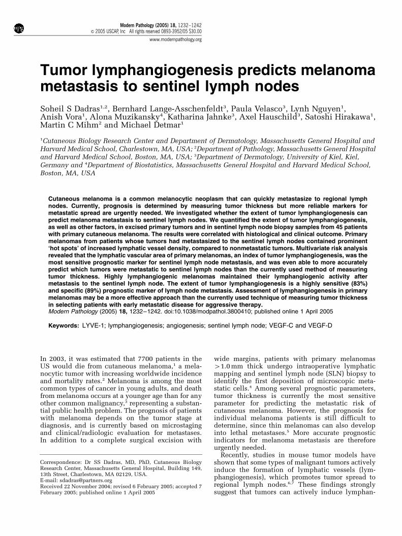
Between 2000 and 2003, 110 patients with cuta-neous melanoma were identified at the Departmentof Dermatology at the University of Kiel, Germanyand subjected to SLN biopsy, following the excisionof primary tumors that exceeded 1 mm in thickness,using lymphatic scintigraphy as described.4 A totalof 45 consecutive patients underwent SLN biopsies,27 were found to have SLNs that were free ofmelanoma metastases (mean age of 53.8 years; range19–76 years; termed ‘SLN-negative’), whereas thetumors were found to have metastasized to the SLNin the remaining 18 patients (mean age of 54 years;range 29–78 years; termed ‘SLN-positive’). Thesetwo groups were otherwise similar, in terms oftumor histologic type, presence and degree ofulceration, anatomic site, signs of regression andperitumoral inflammation (Table 1).
SLN-positive patients subsequently received ad-juvant systemic therapy with recombinant inter-feron alpha-2a (Roferons, Hoffmann La Roche,Switzerland) or interferon alpha-2b (Introns A,Essex, Switzerland) for up to 60 months. Comparedto six out of 18 SLN-positive, only two out of 27SLN-negative, patients developed metastases todistant lymph nodes or organs within 2 years. Thediagnosis of melanoma, the tumor thickness, thelevel of tumor invasion, and SLN pathologic statuswere reconfirmed by two pathologists (SSD andMCM). Additional parameters such as the frequency
of mitoses, tumor regression and ulceration werealso evaluated. Peritumoral inflammation was eval-uated as absent, present (non-brisk), or brisk.10
Immunostains
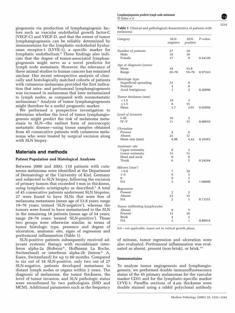
To analyze tumor angiogenesis and lymphangio-genesis, we performed double immunofluorescencestains of the 45 primary melanomas for the vascularmarker CD31 and for the lymphatic-specific markerLYVE-1. Paraffin sections of 4mm thickness weredouble stained using a rabbit polyclonal antibody
Table 1 Clinical and pathological characteristics of patients withmelanoma
Category SLN-negative
SLN-positive
P-value
Number of patients 27 18Male 10 10Female 17 8 0.24120
Age at diagnosis (years)Mean 54 53.8Range 29–78 19–76 0.97243
Histologic typeSuperficial spreading 22 8Nodular 4 8Acral lentiginous 1 2 0.20090
Tumor thickness (mm)o1.5 19 3Z1.5 8 15Mean 1.7 3.01 0.02850
Level of invasionI–III 16 3IV–V 11 15 0.00010
UlcerationPresent 4 6Absent 23 12Mean size (mm) 4.98 4.42 0.16563
Anatomic siteUpper extremity 8 2Lower extremity 13 11Head and neck 0 0Trunk 6 5 0.34244
Mitoses (mm2)46.0 7 101–5 9 50 9 3NA 2 0 1.00000
RegressionPresent 2 2Absent 23 15NA 2 1 0.71531
Tumor infiltrating lymphocytesAbsent 8 5Present 13 10Brisk 4 2NA 2 1 0.88914
NA¼not applicable, tumor not in vertical growth phase.
Lymphangiogenesis predicts lymph node metastasisSS Dadras et al
1233
Modern Pathology (2005) 18, 1232–1242
against human LYVE-18 and a mouse monoclonalantibody against human CD31 (1:40; DAKO), aspreviously described9,11 (Table 2). For additionallymphatic-specific stains, the antibody D2-40 (Sig-net, Dedham, MA) was used. We have recentlyshown that this antibody reliably and specificallyrecognizes human podoplanin, a mucin-type glyco-protein that is specifically expressed by lymphaticendothelium, but not by blood vascular endothe-lium.12,13 Cell nuclei were counterstained withHoechst bisbenzimide (Sigma) at 20 mg/ml. Addi-tional immunohistochemical analysis was per-formed using affinity-purified rabbit polyclonalantibodies against human vascular endothelialgrowth factor-C (VEGF-C) and VEGF-D (kindlyprovided by Dr M Achen).14 For specificity controls,the primary antibodies were omitted. The immu-nostained sections were scored semi-quantitativelyfor VEGF-C and VEGF-D expression by tumor cells,in comparison to tumor infiltrating macrophages(internal positive control, 3þ ) by researchers whowere blinded to the SLN status. The results werescored as follows: 1þ for faintly positive, 2þ formoderately positive, 3þ for strongly positive.
Computer-Assisted Morphometric Analysis
Paraffin sections were immunostained for CD31 andLYVE-1 as described above, and were examinedusing a Nikon E-600 microscope. Digital imageswere captured using a SPOT digital camera. Foreach section, three fields in the peritumoral areawith the highest lymphatic vascular density (‘hotspots’) were evaluated at � 100 magnification.Digital images of tumor-associated lymphatic ves-sels and blood vessels were captured in the samefield. Peritumoral lymphatic vessels were defined asLYVE-1-positive vessels within an area of 100 mmfrom the tumor border. Intratumoral lymphaticvessels were defined as LYVE-1-positive vesselslocated within the tumor mass and not confined byinvagination of normal tissue. Tumor borders weredetermined on serial sections, using Hoechst nucle-ar stains and hematoxylin and eosin stains. Morpho-metric analyses of lymphatic vessels and of bloodvessels were performed in the peritumoral areausing the IP-Lab software (Scanalytics, Fairfax, VA,USA) to determine the vessel number per mm2, thatis, lymphatic vessel density (LVD), and the blood
vessel density (BVD). The average vessel size (inmm2) and the relative tissue area occupied byvessels, that is, the lymphatic vessel area (LVA;expressed as %) and the blood vessel area (BVA),were quantified as previously described.9,11 Tumor-associated lymphatics and blood vessels wereanalyzed within and surrounding the primarymelanomas and also in metastatic SLNs usingdouble immunofluorescence with antibodies againstCD31 and LYVE-1.
Statistical Analyses
The unpaired Student’s t-test was used to determinethe statistical significance (P value) of the mean forall vascular parameters. In a univariate analysis, wetested the association between different risk factorsfor melanoma metastasis to SLN, using the Fisher’sexact test for dichotomous variables, the w2-test forcategorical variables and the Wilcoxon Rank-Sumtest for continuous or ordinal variables. A multi-variate logistic regression model was applied to therisk factors that were found to be significant in theunivariate analysis, and then a stepwise selectionmethod was used to select the significant predictorsfor SLN metastasis. Sensitivity was calculated basedon the number of true positives (patients who wereSLN-positive and had an LVA41.5%), divided bythe number of true positives plus the number of falsenegatives (patients who were SLN-positive but hadan LVAo1.5%). Specificity was calculated based onthe number of true negatives (patients who wereSLN-negative and had an LVAo1.5%) divided bythe number of true negatives plus the number offalse positives (patients who were SLN-negative buthad an LVA41.5%).
Results
Increased Tumor Lymphangiogenesis in PrimaryMelanomas of SLN-Positive Patients
We analyzed 45 consecutive patients with nonmeta-static (n¼ 27) or with metastatic (n¼ 18) primarycutaneous melanoma to the SLN. SLN-positivepatients had significantly thicker tumors (3.0170.52 mm) than SLN-negative patients (1.7070.31 mm, P¼ 0.0285) (Table 1). Histologic analysisrevealed that additional prognostic parameters such
Table 2 Antibodies used for immunofluorescent analysis
Antibody Staining for Source Type Dilution and conditionsa
LYVE-1 Lymphatics DG Jackson Polyclonal 1:600; O/N (41C)CD31 Panvascular DAKO Monoclonal, IgG1 1:50; 3 h (RT)D2-40 Lymphatics Signet Monoclonal, IgG1 1:100; O/N (41C)VEGF-C Melanoma cells, macrophages Zymed Polyclonal 1:100; O/N (41C)VEGF-D Melanoma cells, macrophages M Achen Monoclonal 1:100; O/N (41C)
aRT¼room temperature; O/N¼ over night.
Lymphangiogenesis predicts lymph node metastasisSS Dadras et al
1234
Modern Pathology (2005) 18, 1232–1242
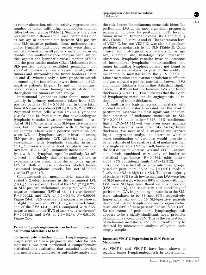
as tumor ulceration, mitotic activity, regression andnumber of tumor infiltrating lymphocytes did notdiffer between groups (Table 1). Similarly, there wasno significant difference in clinical parameters suchas sex, age or anatomic site between SLN-positiveand SLN-negative patients. Next, melanoma-asso-ciated lymphatic and blood vessels were simulta-neously visualized in all primary melanomas, usingdouble immunofluorescence analysis with antibo-dies against the lymphatic vessel marker LYVE-1and the panvascular marker CD31. Melanomas fromSLN-positive patients exhibited prominent ‘hotspots’ of increased lymphatic vessel density, withintumors and surrounding the tumor borders (Figure1b and d), whereas only a few lymphatic vesselssurrounding the tumor border were detected in SLN-negative patients (Figure 1a and c). In contrast,blood vessels were homogeneously distributedthroughout the tumors in both groups.
Intratumoral lymphatics were found more fre-quently in primary melanomas taken from SLN-positive patients (83.370.09%) than in those takenfrom SLN-negative patients (59.370.09%, P¼ 0.0960).Pigmented tumor cells within LYVE-1þ lymphaticvessels—that is, from tumors that have undergonelymphatic vascular invasion—were found in twoout of 18 (11%) patients with SLN-positive melano-mas, but not in any patients with SLN-negativemelanomas. There was a positive correlation bet-ween LVD and lymphatic vascular invasion amongSLN-positive patients (LVD 26.676.3 lymphaticvessels/mm2 with lymphatic vascular invasion,15.171.6 vessels/mm2 without lymphatic vascularinvasion; P¼ 0.0348). Immunohistochemical ana-lysis with the lymphatic-specific antibody D2-4013
showed a strikingly similar staining pattern asexperiments performed with the antibody againstLYVE-1. Both of these antibodies labeled tumor-associated lymphatic vessels, but not of bloodvessels (Figure 1f).
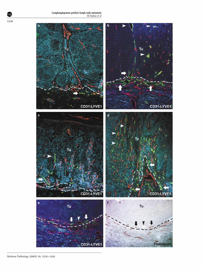
Computer-assisted morphometric analysis re-vealed a 2.4-fold increase in the peritumoral LVD(16.471.7 vessels/mm2) and of the LVA (2.270.2%)in SLN-positive melanomas, compared with SLN-negative melanomas (LVD of 7.071.1 vessels/mm2,P¼ 0.00022, and LVA of 0.970.1%, P¼ 0.00008;Figure 2d–f). SLN-positive melanomas also showeda slight increase of BVD (48.274.6 vessels/mm2)and of the BVA (4.370.4%), compared with SLN-negative melanomas (BVD of 36.374.1 vessels/mm2,P¼ 0.03392, and BVA of 2.970.2%, P¼ 0.01106;Figure 2a–c).
Extent of Lymphangiogenesis can be Used to PredictMelanoma Metastasis to SLN
To investigate whether tumor lymphangiogenesismight serve as a new prognostic indicator for SLNmetastasis, we next performed a comprehensivestatistical data evaluation that included univariateand multivariate analyses. A univariate analysis of
the risk factors for melanoma metastasis identifiedperitumoral LVA as the most significant prognosticparameter, followed by peritumoral LVD, level oftumor invasion, tumor thickness, BVD and finallyBVA (Table 3; Figure 2a and c). The expression levelof VEGF-C, but not VEGF-D, was also a significantpredictor of metastasis to the SLN (Table 3). Otherclinical and histological parameters, such as age,sex, anatomic site, histologic type, regression,ulceration, lymphatic vascular invasion, presenceof intratumoral lymphatics, microsatellites andtumor infiltrating lymphocytes were not found bythe univariate analysis to predict the ability ofmelanoma to metastasize to the SLN (Table 3).Linear regression and Pearson correlation coefficientanalyses showed a positive correlation between BVAand tumor thickness (borderline statistical signifi-cance; P¼ 0.0539) but not between LVA and tumorthickness (P¼ 0.1515). This indicates that the extentof lymphangiogenesis, unlike angiogenesis, is in-dependent of tumor thickness.
A multivariate logistic regression analysis withapplied selection criteria revealed that the level ofperitumoral LVA was the most significant, indepen-dent predictor of melanoma metastasis to SLN(P¼ 0.00037, odds ratio¼ 5.527, 95% confidencelimits 1.740–17.552)—it was even more accuratethan the currently used prognostic factor, tumorthickness. We next used a stepwise multivariatelogistic regression analysis to determine whethersome combination of variables would provide abetter estimate of the relative risk of metastasis thanany single variable. LVA by itself, however, providedthe best estimate, whereas LVA and VEGF-C expres-sion levels combined were only of borderlinestatistical significance (P¼ 0.0562, odds ratio¼4.369, 95% confidence limits 1.478–12.912).
We next classified all patients into three groups,based on peritumoral LVA: low (r1.0%), medium(1.0% r1.5%) or high (41.5%). The great majorityof patients (89%) with low to medium LVA were freeof SLN metastases, whereas 89% of those with highLVA were SLN-positive. Based on this threshold(LVA of 1.5%), the sensitivity and specificity ofperitumoral LVA in predicting metastasis to the SLNwere calculated to be 83 and 89%, respectively.Importantly, six out of 18 SLN-positive patientsdeveloped distant lymph node and/or organ metas-tases, and 83% of these patients had an LVA41.5%.So the extent of peritumoral lymphangiogenesisappears to be a highly significant, novel predictorof melanoma spread to SLN. This is the earliest formof melanoma metastasis, and can currently only bedetected by microscopic analysis of lymph nodebiopsy samples.
Increased VEGF-C Expression in SLN-PositiveMelanomas
As VEGF-C and VEGF-D have been shown toregulate tumor lymphangiogenesis in experimental
Lymphangiogenesis predicts lymph node metastasisSS Dadras et al
1235
Modern Pathology (2005) 18, 1232–1242
Lymphangiogenesis predicts lymph node metastasisSS Dadras et al
1236
Modern Pathology (2005) 18, 1232–1242
models, we analyzed the expression levels of theseproteins in all primary melanomas by immunohisto-chemistry. We detected moderate to high levels ofcytoplasmic VEGF-C in the majority of SLN-positivemelanomas but in a minority of the SLN-negative
melanomas (Figure 3a–d). In total, 88.2% of SLN-positive melanomas expressed VEGF-C protein,compared to only 44.0% of SLN-negative tumors(P¼ 0.00818; Table 3). Although the majority ofmelanoma cells examined contained VEGF-D in
Figure 2 Computer-assisted morphometric analysis of tumor-associated blood and lymphatic vessels in cutaneous melanoma patientswith SLN dissection. Panels a–c show the dramatic increase of peritumoral lymphatic vascular density and relative area occupied bylymphatics in SLN-positive melanomas (þ ; black bars, n¼17), compared with SLN-negative melanomas (�; white bars, n¼ 28). Nochange of the size of individual lymphatic vessels was detected. Panels d–f show a slight increase of BVD and relative area occupied byblood vessels detected in SLN-positive melanomas, compared to those patients with negative SLN. No change of the size of individualblood vessels was detected. Data are shown as mean7s.e.m.
Figure 1 Increased intra- and peritumoral lymphatic density in cutaneous melanomas that have undergone metastasis to SLN. Panels a–eshow sections of melanomas that were analyzed by immunofluorescence analysis using antibodies against LYVE-1 (green), which is alymphatic marker, and CD31 (red) which is panvascular marker. Panels a and c show that in two representative SLN-negativemelanomas, only a few lymphatic vessels surrounding (arrows) and inside (arrowhead) the tumor (Tu) border (dotted line). Panels b andd show, in comparison, that SLN-positive melanomas contain prominent ‘hot spots’, or areas of increased LVD, within (arrowheads) andsurrounding (arrows) the tumor border (dotted line). In contrast, blood vessels (red) are homogeneously distributed throughout thetumor. Panel e also shows the increased peritumoral LVD (arrows) at the border of a tumor removed from an SLN-positive patient. Ablood vessel, which is LYVE-1� but CD31þ , is indicated by the arrowhead. Panel f shows the results of immunohistochemical analysis ofa serial tumor section with the lymphatic-specific antibody D2-40. This antibody has a strikingly similar staining pattern to that of theanti-LYVE-1 antibody (e), labeling tumor-associated lymphatic vessels (arrows) but not blood vessels (arrowhead). Originalmagnifications: � 100.
Lymphangiogenesis predicts lymph node metastasisSS Dadras et al
1237
Modern Pathology (2005) 18, 1232–1242
focal to diffuse cytoplasmic patterns (Figure 3e, f),there was no correlation between VEGF-D levels andmetastasis to SLN (P¼ 1.00; Table 3). Overall, theseresults indicate that VEGF-C, but not VEGF-D,mediates tumor-associated lymphangiogenesis dur-ing melanoma metastasis.
Metastasized Melanoma Cells MaintainLymphangiogenic Activity in SLNs
To investigate whether the melanoma cells maintaintheir lymphangiogenic activity once they spread tothe SLNs, all tumor-negative (n¼ 27) and -positiveSLN (n¼ 18) sections were immunostained forLYVE-1/CD31. Immunofluorescence analysis of nor-mal lymph nodes revealed that the medullarysinuses and hilar efferent lymphatics were LYVE-1-positive, whereas the high endothelial venules wereLYVE-1-negative (Figure 4a, b). Importantly, 40% ofthe metastasized melanomas demonstrated promi-nent ‘hot spots’ of lymphatic vessel formation in theSLN (Figure 4d–f). The primary tumors in thesepatients had a significantly higher LVD (21.86vessels/mm2) than those that were not associatedwith lymphangiogenic metastases (13.76 vessels/mm2, P¼ 0.01202, Figure 4c). These results reveal apositive correlation between tumor lymphangiogen-esis in primary melanomas and the lymphangio-genic activity of SLN metastases.
Discussion
When patients are diagnosed with primary cuta-neous melanoma, determining their risk of develop-ing metastases poses a challenge to clinicians andpathologists alike. The early identification of meta-static disease is important, as it determines therequirement for adjuvant therapy and further man-agement.15 Prognosis is currently based on tumorthickness,16 but a considerable number of patientswith thin melanomas also go on to develop meta-static disease.5 SLN biopsy is also commonly used todetect metastases in patients with primary melano-ma.4 It can be a challenge, however, to detect SLNmetastases, as they are often microscopic, requiringexamination of multiple SLN sections. Dissection ofSLN can also result in mild to moderate degrees oflymphedema17 or sensory morbidity.18 Alternativeearly-stage prognostic factors are therefore urgentlyneeded.
The discovery of the lymphatic endothelial hya-luronan receptor-1 (LYVE-1) as a specific marker forboth normal8 and tumor-associated lymphatics,19
has facilitated analysis of the association betweentumor lymphangiogenesis and clinical outcome.Our study of 45 patients with cutaneous melanomareveals that quantification of primary tumor lym-phangiogenesis can be used to accurately predictwhich patients are most likely to develop SLNmetastases.
Evaluation of tumor lymphangiogenesis can alsobe used to predict the risk of tumor metastasisbeyond the SLN—six out of 18 SLN-positivepatients developed distant lymph node and/or organmetastases, and five of these patients (83%) had anLVA41.5%. Furthermore, in patients whose pri-mary melanoma lymphangiogenesis index (LVA)was above 2%, the incidence of melanoma meta-stasis to SLN was 100%. Another important findingof this study was that an increase in LVD in primarymelanomas was correlated with lymphangiogenesisin SLN metastases. Thus, metastatic primary tumorcells maintain their lymphangiogenic activity afterthey have spread to the lymph nodes. Takentogether, these results indicate that lymphangiogen-esis induction is an active part of human tumormetastasis,20,21 as opposed to the traditional view ofa passive role for the lymphatic endothelium duringtumor lymphatic invasion and metastasis.22
The fact that we detected significantly higherlevels of VEGF-C expression in SLN-positive mela-noma cells than SLN-negative tumor cells, and thatVEGF-C expression was also correlated with theextent of peritumoral lymphangiogenesis, indicatesthat this growth factor promotes melanoma lym-phangiogenesis and metastasis. In fact, tumor cellexpression of VEGF-C alone was shown by uni-variate analysis to predict metastasis to the SLNs. Acorrelation between VEGF-C mRNA levels andmelanoma metastasis to lymph nodes was pre-viously reported.23,24 However, the importance of
Table 3 Univariate analysis of different risk factors for melanomametastasis to SLN
Variable P-value
Wilcoxon rank testRelative lymphatic vascular area (%)a 0.00008Lymphatic vessels/mm2a 0.00022Level of invasion (IV–V)b 0.00010Tumor thickness (mm)a 0.00500Relative blood vascular area (%)a 0.01106Blood vessels/mm2a 0.03392Age (years)a 0.97243Lymphatic vessel size (mm2)a 0.68712Blood vessel size (mm2)a 0.62088
Fisher’s exact testVEGF-C protein expressionb 0.00818VEGF-D protein expressionb 1.00000Sexb 0.24120Ulceration (mm)b 0.16563Lymphatic vascular invasionb 0.15455Intratumoral lymphaticsb 0.11070Microsatellitesb 1.00000
w2
Anatomic sitea 0.34244Regressiona 0.7153Histologic typea 0.20090Tumor infiltrating lymphocytesa 0.88914
aCategorical variables.
bDichotomous variables.
Lymphangiogenesis predicts lymph node metastasisSS Dadras et al
1238
Modern Pathology (2005) 18, 1232–1242
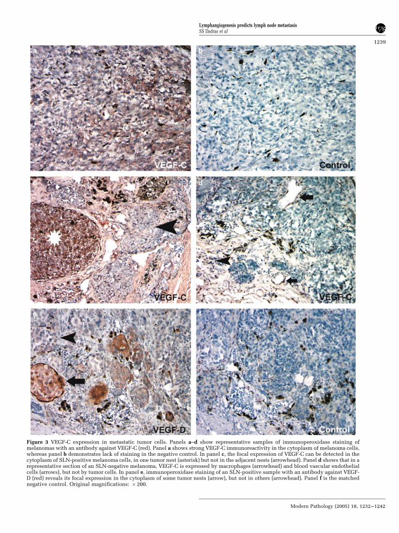
Figure 3 VEGF-C expression in metastatic tumor cells. Panels a–d show representative samples of immunoperoxidase staining ofmelanomas with an antibody against VEGF-C (red). Panel a shows strong VEGF-C immunoreactivity in the cytoplasm of melanoma cells,whereas panel b demonstrates lack of staining in the negative control. In panel c, the focal expression of VEGF-C can be detected in thecytoplasm of SLN-positive melanoma cells, in one tumor nest (asterisk) but not in the adjacent nests (arrowhead). Panel d shows that in arepresentative section of an SLN-negative melanoma, VEGF-C is expressed by macrophages (arrowhead) and blood vascular endothelialcells (arrows), but not by tumor cells. In panel e, immunoperoxidase staining of an SLN-positive sample with an antibody against VEGF-D (red) reveals its focal expression in the cytoplasm of some tumor nests (arrow), but not in others (arrowhead). Panel f is the matchednegative control. Original magnifications: � 200.
Lymphangiogenesis predicts lymph node metastasisSS Dadras et al
1239
Modern Pathology (2005) 18, 1232–1242
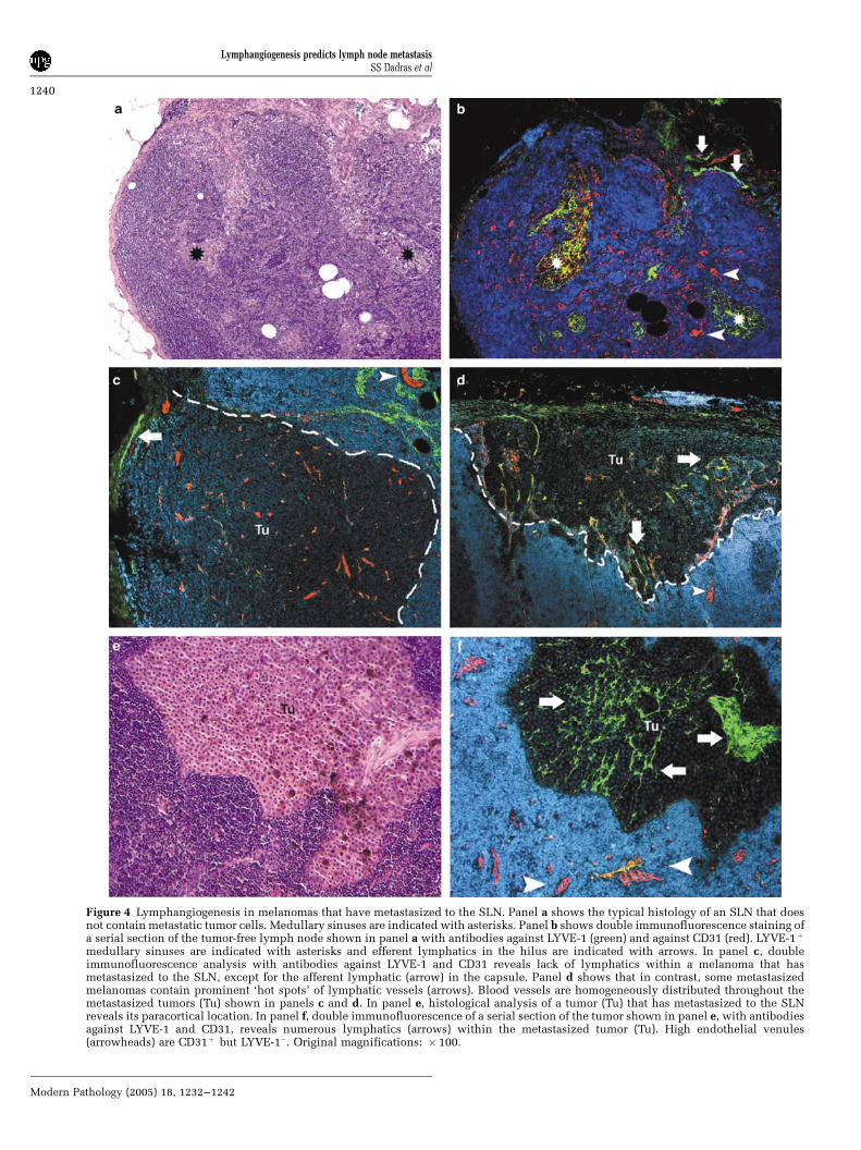
Figure 4 Lymphangiogenesis in melanomas that have metastasized to the SLN. Panel a shows the typical histology of an SLN that doesnot contain metastatic tumor cells. Medullary sinuses are indicated with asterisks. Panel b shows double immunofluorescence staining ofa serial section of the tumor-free lymph node shown in panel a with antibodies against LYVE-1 (green) and against CD31 (red). LYVE-1þ
medullary sinuses are indicated with asterisks and efferent lymphatics in the hilus are indicated with arrows. In panel c, doubleimmunofluorescence analysis with antibodies against LYVE-1 and CD31 reveals lack of lymphatics within a melanoma that hasmetastasized to the SLN, except for the afferent lymphatic (arrow) in the capsule. Panel d shows that in contrast, some metastasizedmelanomas contain prominent ‘hot spots’ of lymphatic vessels (arrows). Blood vessels are homogeneously distributed throughout themetastasized tumors (Tu) shown in panels c and d. In panel e, histological analysis of a tumor (Tu) that has metastasized to the SLNreveals its paracortical location. In panel f, double immunofluorescence of a serial section of the tumor shown in panel e, with antibodiesagainst LYVE-1 and CD31, reveals numerous lymphatics (arrows) within the metastasized tumor (Tu). High endothelial venules(arrowheads) are CD31þ but LYVE-1�. Original magnifications: �100.
Lymphangiogenesis predicts lymph node metastasisSS Dadras et al
1240
Modern Pathology (2005) 18, 1232–1242
tumor cell-derived VEGF-C vs VEGF-C produced bytumor-associated macrophages25,26 has remainedunclear. Our study shows that tumor cells them-selves make a significant contribution to VEGF-Cproduction and, therefore, to tumor lymphangiogen-esis. We also detected a slight but statisticallysignificant increase in levels of tumor angiogenesisin SLN-positive melanomas. Our finding that theextent of tumor angiogenesis, unlike lymphangio-genesis, was dependent on tumor thickness furthersupports the concept that angiogenesis, as demon-strated by other investigators,27 is required for tumorgrowth. However, our results indicate that angiogen-esis seems to play a lesser role in promotingmelanoma metastasis than does the formation oftumor-associated lymphatic vessels.
This is the first study to show that measuring theextent of tumor-associated lymphangiogenesis canserve as a powerful tool for identifying primarymelanomas that are likely to undergo metastasis—the presence of metastases can be determined at theearliest stages of tumor progression, when micro-scopic tumors have developed in the SLN. If theseresults will be corroborated in additional studies oflarger numbers of patients, this new method mightprovide a safe and accurate future alternative to SLNbiopsy analysis. Our results also demonstrate that itis possible to perform immunofluorescence analysisof CD31 and LYVE-1 levels on routine paraffinsections, thereby facilitating larger, prospective,multi-institutional studies. Evaluating the extent ofmelanoma lymphangiogenesis would provide on-cologists and dermatologists with a safer and moreeffective means of determining which patients withmelanoma require adjuvant therapy and furthermanagement, possibly including anti-lymphangio-genic therapy.
Acknowledgements
We thank Dr DG Jackson for the gift of anti-humanLYVE-1 antibody, Dr M Achen for the gift of anti-human VEGF-D antibody, M Constant and L Janesfor technical assistance, and V Schacht and RKunstfeld for helpful discussions. This work wassupported by NIH/NCI Grants CA69184, CA86410and CA91861 (MD), by the Susan G Komen BreastCancer Foundation (MD), by American CancerSociety Program Project Grant 99-23901 (MD), byan NIH Pathology Training Grant (SSD), by theWerner and Klara Kreitz Foundation (BL-A) and bythe Cutaneous Biology Research Center through theMassachusetts General Hospital/Shiseido Co. LtdAgreement (MD).
References
1 Greenlee R, Hill-Harmon M, Murray T, et al. CancerStatistics, 2001. CA Cancer J Clin 2001;51:15–36.
2 Hall HI, Miller DR, Rogers JD, et al. Update onthe incidence and mortality from melanoma inthe United States. J Am Acad Dermatol 1999;40:35–42.
3 Weinstock MA. Epidemiology, etiology, and control ofmelanoma. Med Health R I 2001;84:234–236.
4 Gershenwald JE, Thompson W, Mansfield PF, et al.Multi-institutional melanoma lymphatic mapping ex-perience: the prognostic value of sentinel lymph nodestatus in 612 stage I or II melanoma patients. J ClinOncol 1999;17:976–983.
5 Kalady MF, White RR, Johnson JL, et al. Thinmelanomas: predictive lethal characteristics from a30-year clinical experience. Ann Surg 2003;238:528–535; discussion 535–527.
6 Skobe M, Hawighorst T, Jackson DG, et al. Induction oftumor lymphangiogenesis by VEGF-C promotes breastcancer metastasis. Nat Med 2001;7:192–198.
7 Stacker SA, Caesar C, Baldwin ME, et al. VEGF-Dpromotes the metastatic spread of tumor cells via thelymphatics. Nat Med 2001;7:186–191.
8 Banerji S, Ni J, Wang SX, et al. LYVE-1, a newhomologue of the CD44 glycoprotein, is a lymph-specific receptor for hyaluronan. J Cell Biol 1999;144:789–801.
9 Dadras SS, Paul T, Bertoncini J, et al. Tumor lymphan-giogenesis: a novel prognostic indicator for cutaneousmelanoma metastasis and survival. Am J Pathol 2003;162:1951–1960.
10 Clemente CG, Mihm Jr MC, Bufalino R, et al.Prognostic value of tumor infiltrating lymphocytes inthe vertical growth phase of primary cutaneousmelanoma. Cancer 1996;77:1303–1310.
11 Detmar M, Velasco P, Richard L, et al. Expression ofvascular endothelial growth factor induces an invasivephenotype in human squamous cell carcinomas. Am JPathol 2000;156:159–167.
12 Kahn HJ, Marks A. A new monoclonal antibody, D2-40,for detection of lymphatic invasion in primary tumors.Lab Invest 2002;82:1255–1257.
13 Schacht V, Dadras SS, Johnson L, et al. Upregulation ofthe lymphatic marker podoplanin, a mucin-typetransmembrane glycoprotein, in human squamous cellcarcinomas and germ cell tumors. Am J Pathol 2005;166:913–921.
14 Achen MG, Williams RA, Minekus MP, et al. Localiza-tion of vascular endothelial growth factor-D in malig-nant melanoma suggests a role in tumor angiogenesis.J Pathol 2001;193:147–154.
15 Roberts AA, Cochran AJ. Pathologic analysis ofsentinel lymph nodes in melanoma patients: currentand future trends. J Surg Oncol 2004;85:152–161.
16 Balch CM, Buzaid AC, Soong SJ, et al. Final version ofthe American Joint Committee on Cancer stagingsystem for cutaneous melanoma. J Clin Oncol 2001;19:3635–3648.
17 Wrone DA, Tanabe KK, Cosimi AB, et al. Lymphedemaafter sentinel lymph node biopsy for cutaneousmelanoma: a report of 5 cases. Arch Dermatol 2000;136:511–514.
18 Temple LK, Baron R, Cody III HS. Morbidity aftersentinel lymph node biopsy and axillary dissection: aprospective study of 233 women. Ann Surg Oncol2002;9:654–662.
19 Beasley NJ, Prevo R, Banerji S, et al. Intratumorallymphangiogenesis and lymph node metastasis inhead and neck cancer. Cancer Res 2002;62:1315–1320.
Lymphangiogenesis predicts lymph node metastasisSS Dadras et al
1241
Modern Pathology (2005) 18, 1232–1242

20 Detmar M, Hirakawa S. The formation of lymphaticvessels and its importance in the setting of malignancy.J Exp Med 2002;6:713–718.
21 Dadras SS, Detmar M. Angiogenesis and lymphangio-genesis of skin cancers. Hematol Oncol Clin North Am2004;18(5):1059–1070.
22 Carmeliet P, Jain RK. Angiogenesis in cancer and otherdiseases. Nature 2000;407:249–257.
23 Goydos JS, Gorski DH. Vascular endothelial growthfactor C mRNA expression correlates with stage ofprogression in patients with melanoma. Clin CancerRes 2003;9:5962–5967.
24 Schietroma C, Cianfarani F, Lacal PM, et al. Vascularendothelial growth factor-C expression correlates with
lymph node localization of human melanoma meta-stases. Cancer 2003;98:789–797.
25 Skobe M, Hamberg LM, Hawighorst T, et al. Concur-rent induction of lymphangiogenesis, angiogenesis,and macrophage recruitment by vascular endothelialgrowth factor-C in melanoma. Am J Pathol 2001;159:893–903.
26 Schoppmann SF, Birner P, Stockl J, et al. Tumor-associated macrophages express lymphatic endo-thelial growth factors and are related to peri-tumoral lymphangiogenesis. Am J Pathol 2002;161:947–956.
27 Folkman J. Angiogenesis and tumor growth. N Engl JMed 1996;334:9219.
Lymphangiogenesis predicts lymph node metastasisSS Dadras et al
1242
Modern Pathology (2005) 18, 1232–1242