![The Arrival of Egyptian Taweret and Bes[et] on Minoan Crete: Contact and Choice](https://static.fdokumen.com/doc/165x107/6315e4e185333559270d5872/the-arrival-of-egyptian-taweret-and-beset-on-minoan-crete-contact-and-choice.jpg)
The Arrival of Egyptian Taweret and Bes[et] on Minoan Crete: Contact and Choice

Preparing the ‘‘Soil’’: The Primary Tumor Induces Vasculature
Reorganization in the Sentinel Lymph Node before the
Arrival of Metastatic Cancer Cells
Chao-Nan Qian,1,5Bree Berghuis,
2Galia Tsarfaty,
3MaryBeth Bruch,
3Eric J. Kort,
4
Jon Ditlev,1Ilan Tsarfaty,
3Eric Hudson,
2David G. Jackson,
6David Petillo,
1
Jindong Chen,1James H. Resau,
2and Bin Tean Teh
1
Laboratories of 1Cancer Genetics, 2Analytical, Cellular, and Molecular Microscopy, 3Molecular Oncology, and 4Molecular Epidemiology,Van Andel Research Institute, Grand Rapids, Michigan; 5Department of Nasopharyngeal Carcinoma, Sun Yat-sen University CancerCenter, Guangzhou, China; and 6Medical Research Council Human Immunology Unit, Weatherall Institute of MolecularMedicine, John Radcliffe Hospital, Oxford, United Kingdom
Abstract
Sentinel lymph node (SLN) metastasis is the first step in thespreading of cancer in many malignancies. Tumor-reactivelymphadenopathy in SLNs has been observed for decades, butalterations of the lymphatic channels and vasculature in thesenodes before the arrival of metastatic tumor cells remainunexplored. Using animal models, we show here that, beforethe establishment of metastasis in the SLN, there arereorganizations of the lymphatic channels and the vascula-ture. The node becomes a functional blood vessel–enrichedand lymph vessel/sinus-enriched organ before metastasis. Theenlargement of the lymph sinuses is correlated with theprimary tumor weight. The newly emerged functional bloodvessels develop from high endothelial venules (HEV), in whichthe proliferation rate of the endothelial cells is alsosignificantly increased. Similar alterations of the HEVs arealso characterized in the axillary lymph nodes from humanbreast cancer patients without the evidence of metastasis.These findings support the hypothesis that modification of themicroenvironment for a secondary tumor (i.e., vasculaturereorganization in the SLN) can be initiated by a primarytumor before and independent of the physical presence ofmetastatic cancer cells. (Cancer Res 2006; 66(21): 10365-76)
Introduction
The most important survival predictor in patients with head andneck cancers is cervical lymph node metastasis, regardless ofextension of the primary tumor (1). Detection of lymph nodemetastasis is therefore important for tumor staging and therapyplanning. Current imaging approaches to evaluate lymph nodemetastasis mainly rely on the size and the shape of the involvedlymph node, and controversial interpretations of these imagingresults remain due to the limited knowledge of the angiogenesisand lymphangiogenesis within the lymph node.
It has been known for decades that regional lymph nodes drainingtumor areas may be enlarged without evidence of metastasis. Theterm for this condition is tumor-reactive lymphadenopathy (2).However, the processes of lymphangiogenesis and angiogenesis intumor-reactive lymphadenopathy are not well understood. Angio-genesis is essential to the growth and metastasis of solid tumors (3).Recently, intratumoral lymphangiogenesis has been characterized inhead and neck carcinomas, related to regional cervical lymph nodemetastasis (4, 5). Increased expression of vascular endothelialgrowth factor (VEGF)-C, which can induce lymphangiogenesis, isalso correlated with regional lymph node metastasis of cancer cellsin both animal model and human tumors (6, 7). Expression ofplatelet-derived growth factor in murine fibrosarcoma cells caninduce tumor lymphangiogenesis, leading to enhancedmetastasis inlymph nodes (8). Lymphangiogenesis in the lymph nodes inducedby VEGF-A contributes to cancer metastasis (9).Nasopharyngeal carcinoma (NPC) has the highest incidence rate
of lymph node metastasis among head and neck cancers (10, 11). Inbreast cancer, axillary lymph node involvement is a significantprognostic factor (12, 13), although 20% to 30% of node-negativepatients will develop distant metastases within 10 years (14). Tocharacterize the process of lymph node metastasis, we establisheda spontaneous lymph node metastasis animal model by using ahuman, poorly differentiated NPC cell line, CNE-2 (15), and themurine breast cancer cell line DA3. The premetastatic alterationsof the sentinel lymph node (SLN), which receives direct lymphaticdrainage from the tumor lesion (16), were analyzed. Human axillarylymph nodes from breast cancer patients were used to confirm thefindings from the animal models.
Materials and Methods
Cell culture and clone selection. The human NPC cell line CNE-2 andits clones were maintained in DMEM supplemented with 10% fetal
bovine serum (FBS). The individual clones from the CNE-2 parental cell
line, each grown from a single cell, were isolated by limited dilution
methods.In vitro migration and invasion assays. For migration assays, 5.0 � 104
NPC cells in 500 AL serum-free DMEM was added to the cell culture inserts
with an 8-Am microporous filter without extracellular matrix coating
(Becton Dickinson Labware, Bedford, MA). DMEM containing 10% FBS wasadded to the bottom chamber. After 18 hours of incubation, the cells on the
lower surface of the filter were fixed and stained followed by microscopic
examination. The number of cells in five random optical fields (�200magnification) from triplicate filters was averaged. For in vitro invasion
assays, the inserts of the chambers to which the cells were seeded were
coated with Matrigel (Becton Dickinson Labware).
Note: Supplementary data for this article are available at Cancer Research Online(http://cancerres.aacrjournals.org/).Requests of reprints: Chao-Nan Qian, Laboratory of Cancer Genetics, Van Andel
Research Institute, 333 Bostwick Avenue Northeast, Grand Rapids, MI 49503.Phone: 616-234-5538; Fax: 616-234-5539; E-mail: [email protected] or Bin TeanTeh, Laboratory of Cancer Genetics, Van Andel Research Institute, 333 BostwickAvenue Northeast, Grand Rapids, MI 49503. Phone: 616-234-5296; Fax: 616-234-5297;E-mail: [email protected].
I2006 American Association for Cancer Research.doi:10.1158/0008-5472.CAN-06-2977
www.aacrjournals.org 10365 Cancer Res 2006; 66: (21). November 1, 2006
Research Article
Research. on March 13, 2016. © 2006 American Association for Cancercancerres.aacrjournals.org Downloaded from
Published OnlineFirst October 23, 2006; DOI: 10.1158/0008-5472.CAN-06-2977

Animals and spontaneous lymph node metastasis assay. FemaleBALB/c mice and athymic BALB/c nu/nu mice (5-6 weeks old) were
maintained in the vivarium of the Van Andel Research Institute (VARI; Grand
Rapids, MI). All of the animal studies were conducted under protocols
approved by the Institutional Animal Care and Use Committee of VARI.Tumor cells (8 � 104 in 20 AL Hank’s solution) were injected s.c. into the
footpad of left hind limb of each nude mouse. On the terminal day, the
primary tumor weight was calculated by subtracting the weight of the foot
without tumor from the weight of foot carrying the primary tumor. Thepopliteal lymph nodes from both hind limbs were isolated using a dissecting
microscope. The lymph node volumes were calculated as volume = width �length � height / 2.
To trace the lymph drainage, 10 AL Evan’s blue dye (0.4%) in PBS wasinjected s.c. at the end of the limb 15 minutes before sacrifice.
Real-time quantitative PCR. Single CNE-2 cells were identified by
limited dilution method in 96-well plates. Total RNA from cells and lymph
nodes was extracted using the High Pure RNA Tissue kit (Roche Applied
Science, Penzberg, Germany). To prove the validity of this method in
identifying a few cancer cells in the lymph node, varying numbers of cancer
cells were combined with a certain number of normal popliteal lymph nodes
for homogenization. Briefly, tumor cell suspension in culture medium was
plated into 96-well plates. Four hours later, the cell number was counted, and
100 AL lysis/binding buffer was added into each well. The lysate was then
transferred to a tube containing a certain number of fresh-frozen popliteal
lymph nodes from normal mice. Another 300 AL lysis/binding buffer was thenadded followed by homogenization. Specific primers for human hypoxanthine
phosphoribosyltransferase (HPRT) were used (17). The PCR products were
continuously measured with an ABI PRISM 7700 Sequence Detection System
(Applied Biosystems, Forster City, CA) during 50 cycles. The experiments were
done in duplicate, and the average of the two samples was calculated.
Ultrasonography and image analyses. Blood flow in the popliteal
lymph node was detected in vivo using the Acuson Sequoia 512 (Mountain
View, CA) instrument with a 15L8 (15 MHz) linear transducer (18). Micewere anesthetized using continuous inhalation of isoflurane. Ultrasonog-
raphy was done on the popliteal lymph node 6 to 26 days after tumor cell
inoculation into the footpad. No detectable popliteal lymph node was foundin the hind limbs of normal healthy mice or in the right hind limbs of the
mice carrying left hind footpad tumors. Any sentinel popliteal lymph nodes
with histologically verified metastases were excluded from the analyses.
The blood flow in large blood vessels of the popliteal lymph node wasevaluated by color Doppler ultrasound. The application of contrast
medium, which are air bubbles 2 Am in diameter, enables the detection
of blood flow in smaller vessels (40-70 Am in diameter). First-pass, low-
intensity, nondestructive contrast medium-enhanced ultrasound imagingwas done using Definity Perflutren Lipid Microspheres (Bristol-Myers
Squibb Medical Imaging, Inc.) by tail-vein injection at 15 Ag/mouse.Contrast medium ultrasound imaging analysis was done using UIA
dedicated functional molecular imaging analysis software (iLAB’s Israel).Histologic evaluation and immunohistochemical and immunofluo-
rescent staining. Paraffin-embedded human lymph node tissues were
obtained from the Shared Pathology Informatics Network at VARI (19).Mouse lymph nodes were fixed in 4% paraformaldehyde, embedded in
paraffin, and sectioned at 3 Am throughout the lymph node. To exclude
lymph nodes containing micrometastases, H&E staining and histologic
examination were done on all of the rest of the continuous tissue sectionsfrom a whole lymph node after several sections were selected for
immunohistochemical staining. To quantify the lymph vessel/sinus area,
the largest H&E-stained cross-section of each nonmetastatic lymph node
was used for image analyses. Rat anti-mouse CD31 monoclonal antibody(mAb; Research Diagnostics Inc., Concord, MA), the rabbit anti-mouse
LYVE-1 antibody (prepared as described; ref. 20), the purified rat anti-mouse
MECA-79 (PNAd) antibody (BD PharMingen), and the mouse anti-humanCD31 (JC70A, DAKO, Carpinteria, CA) mAb were used at a dilution of 1:50.
The mouse anti-human CD34 (QBEnd10, DAKO) mAb was used at a dilution
of 1:100. The anti-mouse MECA-79 antibody reacts with both mouse and
human PNAd carbohydrate antigen. The proteinase K (DAKO) antigenretrieval method was applied to the staining of CD31 in mouse lymph node.
The avidin-biotin complex method Elite kit (Vector Laboratories, Burlin-game, CA) was used to detect the primary antibodies followed by
3,3-diaminobenzidine substrate visualization (Vector Laboratories). For
immunofluorescent staining, the formalin-fixed, paraffin-embedded tissues
were sectioned at 3 Am. Immunoglobulins [FITC-conjugated donkey anti-rabbit, FITC-conjugated goat anti-rat, tetramethylrhodamine isothiocyanate
(TRITC)-conjugated donkey anti-rat, and TRITC-conjugated donkey anti-
rabbit] at 12 Ag/mL (Jackson ImmunoResearch Laboratories, West Grove,
PA) were used to detect the primary antibodies.Endotoxin-induced lymphadenopathy. Lipopolysaccharides from
Escherichia coli O127:B8 (Sigma, Saint Louis, MO) were dissolved in PBS(1 mg/mL) and injected into the left hind footpads of 10 BABL/c nude miceat the dosage of 20 Ag daily for 20 days. An equal volume of PBS wasinjected into another group of 10 mice as a vehicle control. After 20 days ofinjection, the mice were euthanized and the sentinel popliteal lymph nodeswere isolated for evaluation.
Proliferation rates of high endothelial venule endothelial cells.Rabbit anti-proliferating cell nuclear antigen (PCNA) polyclonal antibody(Abcam, Inc., Cambridge, MA) and rat anti-mouse MECA79 were applied at
1:100 dilution simultaneously onto the lymph node sections followed by
TRITC-conjugated donkey anti-rabbit and FITC-conjugated goat anti-rat
secondary antibodies. The nuclei were stained with 4¶,6-diamidino-2-phenylindole (DAPI). At least 200 high endothelial venule (HEV) endothelial
cells were randomly counted for each lymph node to calculate the percentage
of proliferating endothelial cells. For routine immunohistochemical staining,the rabbit anti-PCNA polyclonal antibody was used at 1:300 dilution.
Image analysis. Image analysis was done using interactive image analysissoftware developed by our laboratory. Regions positively stained with the
HEV marker MECA-79 within the images were identified by combining two
binary masks using a logical AND operator. The first mask was generated
by identifying pixels that were bright in the yellow image channel [after
converting the red, green, and blue (RGB) color image to cyan, magenta,
yellow, and black color space]. The second mask was generated by identifying
pixels that were dark in the green image channel ( from the RGB color space).
Pixels that were ‘‘bright’’ or ‘‘dark’’ were automatically identified using the
thresholding algorithm of Ridler and Calvard (1978; ref. 21). Small staining
artifacts (mainly resulting from nonstaining endothelial nuclei) were removed
by applying a two-pixel opening function. Vessels within the binary mask
were identified using connected component analysis (22). Several variables for
each connected component within the binary mask were then calculated:
1. The total number of pixels within each component, representing the
cross-sectional area of the HEV wall.
2. The length of edge of each component, representing the sum of the innerand outer circumferences of the vessel wall.
3. The number of pixels enclosed by the vessel wall, representing the
luminal area (if any).
4. The ‘‘longest chord’’ of the luminal area (if any). This variable was used tocorrect for nonperpendicular sectioning of the vessels. Once the longest
chord is known, the minor axis (i.e., the longest chord perpendicular to the
major axis) may be calculated. The area of an ellipse is defined in
Eq. 1, where a1 and a2 are half the major and minor axes, respectively[half for the same reason that half the diameter (i.e., the radius) is used
for calculating the area of a circle].
A ¼ pa1a2 ð1Þ
a2 ¼ mApa1
ð2Þ
A¶ ¼ pa22 ð3Þ
Because we measure the area directly (by enumerating pixels within the
lumen), we may rearrange this equation to solve for half the minor axis (a2),
Cancer Research
Cancer Res 2006; 66: (21). November 1, 2006 10366 www.aacrjournals.org
Research. on March 13, 2016. © 2006 American Association for Cancercancerres.aacrjournals.org Downloaded from
Published OnlineFirst October 23, 2006; DOI: 10.1158/0008-5472.CAN-06-2977

as given by Eq. 2. With the minor axis known, we may then reconstitute thetrue perpendicular cross-sectional luminal area (A ¶) using the equation forthe area of a circle (Eq. 3).
To calculate total lymph sinus area, the whole H&E-stained section was
digitally captured. The lymph sinus regions were identified by segmentingthe blue channel of the images of the lymph nodes using the automated
thresholding algorithm mentioned above. Artifactual gaps between cells
were eliminated using a five-pixel closing function. The number of pixels in
the ‘‘bright’’ regions corresponding to the lymph sinus areas wasenumerated.
Statistical analysis. The test for equality of proportions was used to
evaluate the equivalence of real-time PCR and histopathologic analyses in
detection of lymph node metastasis. A two-tailed Student’s t test wasused for comparing lymph node volumes and the proliferation rates of
endothelial cells. The data produced by the image analyses deviated
significantly from a normal distribution. Therefore, nonparametric testswere used, including Wilcoxon rank-sum tests for the comparison of means
and m2 tests for differences in proportion. Spearman’s correlation test was
used to assess the relation between tumor weight and lymph vessels/sinuses
area. All tests were two tailed. A number that follows the F sign is a SD.
Results
Spontaneous lymph node metastasis in a mouse model ofNPC. There is considerable evidence of heterogeneity in themetastatic population of tumor cells (23–25). We isolated 29 clonesfrom the parental cell line CNE-2 and screened for their migratoryand invasive abilities in vitro . Clone-18 statistically produced thegreatest migration and invasion when compared with the parentalcell line and other clones (Fig. 1A).To study the process of spontaneous lymph node metastasis, an
animal model was established by injecting CNE-2 tumor cells intothe left hind footpad of immunodeficient nude mice, resulting inmetastasis to the popliteal lymph node, which is the SLN for thismodel. Relative to the parental cell line and other clones, Clone-18had a significantly greater potential of metastasis (Fig. 1B and C),although the primary tumor growth rates were not differentamong these cell populations (Fig. 1D). Metastases of Clone-18were observed by day 20 and reached 100% metastatic incidenceon day 50 after inoculation of 8 � 104 cells into the hind footpad ofthe mouse (Fig. 1E). The footpad model was used because (a) thereis only a single popliteal lymph node in the anatomic structure ofthe murine rear leg and (b) the anatomic location of the popliteallymph node is constant, superficial, and easy to identify bypalpation, favoring noninvasive imaging by ultrasonography. Tostudy the histologic alteration before metastasis, we restricted ouranalyses to the lymph node that contained no CNE-2 micrometa-stasis as assessed by thorough histologic analysis (Fig. 1C). Weproved that the sensitivity of this conventional histopathologicapproach was equivalent to that of the real-time quantitative PCRmethod, which could identify single human cancer cell in a murinelymph node (Fig. 1F and G).The neovascularization in SLN before metastasis is func-
tional. To evaluate the baseline histology in popliteal lymph nodesbefore CNE-2 metastasis, the nodes from both hind limbs of thetumor-bearing animals were collected on days 10 to 26 after Clone-18 cells were inoculated into the left hind footpad. Tumor-reactivelymphadenopathy was found to be induced in most of the poplitealSLNs (Fig. 2A and B). Compared with the contralateral popliteallymph node and a normal (noninjected animal) popliteal lymphnode, the popliteal SLN was significantly enlarged by day 20 afterinoculation and before detectable metastasis. The mean volumesfor the fresh, unfixed normal (n = 10), contralateral (n = 10), and
sentinel (n = 10) popliteal lymph nodes were 2.5 F 0.8, 2.1 F 0.8,and 8.3 F 5.4 mm3, respectively, with statistical significance for thesentinel versus the normal popliteal lymph node (P = 0.008) and forthe sentinel versus the contralateral popliteal lymph node (P =0.005). No significant difference of volume was found between thenormal and contralateral lymph nodes.The enlarged popliteal lymph nodes were enriched in functional
blood vessels compared with contralateral lymph nodes (Fig. 2B).By using ultrasonographic, real-time blood flow measurements(26), we found that the blood flow in large blood vessels of theenlarged sentinel popliteal lymph node was increased 20 days afterClone-18 tumor cell inoculation; there was no detectable bloodflow in the contralateral popliteal lymph node of the same mouse(Fig. 2C). Moreover, increased blood flow in the popliteal SLN wasalso found in the immunocompetent BALB/c mice carryingfootpad syngeneic DA3 breast adenocarcinoma 15 days afterinoculation and before metastasis, again with undetectable bloodflow in the contralateral popliteal lymph node (Fig. 2D). Afteradministration of contrast medium, the blood flow in small bloodvessels in the popliteal SLN from nude mice carrying Clone-18 cellswas clearly detectable by ultrasonography as early as day 17 aftertumor cell inoculation, whereas there was no detectable blood flowin the contralateral popliteal lymph node in the same mouse(Fig. 2E). The increase of blood flow in the vessels draining the SLNwas also determined (data not shown), and it was consistent withthe finding from a s.c. xenograft tumor model in nude mice thatthe increase in regional blood flow may result from the mobili-zation of pre-existent latent vessels (27). These data suggestedthe integration of the SLN vasculature into the adjacent vascularsystem. All of these functional measurements indicated that thenewly emerging vasculature in sentinel popliteal lymph node wasfunctioning before metastasis.Lymphangiogenesis in SLNs before metastasis. An increase in
the number of lymph vessels/sinuses, as well as their level ofdilation, was evident in the popliteal SLNs by staining with Evan’sblue dye (Fig. 2B). To histologically evaluate the process oflymphangiogenesis, the anti-mouse LYVE-1 antibody was used toidentify the lymphatic endothelium (20, 28). The absence ofcolocalization of LYVE-1 and CD31 staining was evident in thelymph node tissue (Fig. 3A-D), confirming that LYVE-1 did notstain blood vessels in the lymph node. Normally, the lymph vessels/sinuses with an open lumen in the popliteal lymph node of youngadult mice are not common. The collapsed lymphatic endotheliumbranching from the hilum to the medulla and paracortex of thelymph node is usually observed (Fig. 3E). In the popliteal SLNs forCNE-2 tumor-bearing mice, these lymph vessels/sinuses wereclearly dilated (Figs. 3F and 4) before metastasis.To assess the correlation between lymph sinus dilation and
primary tumor weight, 12 nonmetastatic sentinel popliteal lymphnodes from the nude mice carrying CNE-2 Clone-18 tumors 30days after inoculation were studied. The largest cross-section ofeach lymph node stained with H&E was completely captured inseveral images (Supplementary Fig. S1A). The total lymph sinusarea was then objectively calculated by the computer program(Supplementary Fig. S1B). The total area of lymph vessels/sinuseswas significantly correlated with the primary tumor weight(Supplementary Fig. S1C), implying that the dilation of the lymphvessels/sinuses in SLN was induced by the primary tumor.The HEV is responsible for tumor-induced vascularization
in the lymph node. Histologic examination confirmed that theenlarged blood vessels in popliteal SLNs contained numerous RBC
Primary Tumor Reorganizes Vasculature in SLN
www.aacrjournals.org 10367 Cancer Res 2006; 66: (21). November 1, 2006
Research. on March 13, 2016. © 2006 American Association for Cancercancerres.aacrjournals.org Downloaded from
Published OnlineFirst October 23, 2006; DOI: 10.1158/0008-5472.CAN-06-2977

(Fig. 4), consistent with the functional ultrasonography analyses.Besides the typically thin, attenuated capillary vessels (<15 Am indiameter), HEVs (>20 Am in their largest diameter) wereconspicuous in the normal mouse popliteal lymph node (Figs. 4and 5A), with their characteristically cuboidal endothelial cellsexpressing peripheral node addressin, which could be recognizedby the MECA-79 antibody (Fig. 5B ; refs. 29, 30) as well as bytraditional blood vessel markers. Unlike lymph vessels/sinuses,which concentrate in and branch from the hilum to the medulla/paracortex, HEVs were evenly distributed in both medullary andparacortical regions in the mouse popliteal lymph node (Fig. 4).In the popliteal SLNs of mice bearing CNE-2 tumors, however,the HEVs were dilated, with increased lumenal diameter (Figs. 4and 5D) and the branching out of thin-walled blood vessels(Fig. 5C). Few HEV with flattened lumens (like those in normallymph nodes) were observed in the SLN 20 days after tumor cellinoculation.
The remodeling of HEV in SLN could be induced in nudemice carrying low-metastatic clones as well as in immuno-competent mice carrying syngeneic mouse breast cancer cells.To determine if the premetastatic lymphangiogenesis and vascu-larization observed in the murine SLNs are universal, we inoculatedlow-metastatic CNE-2 Clone-22 and Clone-26 cells into theimmunodeficient mice and mouse breast adenocarcinoma DA3cells into the immunocompetent BALB/c mice. The popliteal lymphnodes were collected 15 to 30 days after inoculation. The dilation oflymph sinuses/vessels was consistent with the findings from Clone-18 experiments (Fig. 3G). Computerized quantitative analyses weredone to measure the HEV number, the wall thickness, and theadjusted cross-sectional lumen area of HEV. The mean HEVsections per field (0.26 mm2) of the popliteal SLNs versus thecontralateral popliteal lymph node in the animal models of Clone-22, Clone-26, and DA3 tumors were 8.97 F 2.3 versus 20.95 F 5.75(P < 0.001), 8 F 2.1 versus 14.57 F 3.7 (P < 0.001), and 9.65 F 3.18
Figure 1. Characterization of CNE-2 cell migration and invasion in vitro and metastasis in vivo. A, the cells that migrated (no Matrigel layer) or invaded (passed throughthe Matrigel) into the lower chamber in the two-chamber assay were fixed and counted. Columns, mean; bars, SD. B, the lymph node (LN ) metastasis rates ofthe different CNE-2 populations 40 days after the same number of tumor cells (8 � 104 cells) were injected s.c. into the footpad of left hind limb of each nude mouse.Ps are from the comparison of the metastasis rates between the individual clones and the parental CNE-2 cells. C, histologic examination of an H&E-stainedsection showed a micrometastasis of a CNE-2 tumor embolus (inset ) in the subcapsular sinus of a sentinel popliteal lymph node. D, the average primary tumor weightswere not different among these cellular populations 40 days after inoculation. E, the lymph node metastasis rates of Clone-18 cells were further evaluated byusing the same footpad model at different time points, with 15 mice in each time point group. F, a real-time quantitative PCR method amplifying the human HPRT mRNAwas applied to detect human CNE-2 Clone-18 cells mixed with mouse normal popliteal lymph nodes. Curve, the listed formula obtained from normal mousepopliteal lymph nodes added with varying numbers of Clone-18 cells. This method was sensitive enough to detect single cancer cell in one mouse popliteal lymph node.G, a total of 52 sentinel popliteal lymph nodes were harvested 25 days after inoculation of Clone-18 cells into the left hind footpad of the nude mice. All of thelymph nodes were equally divided, with one half used for quantitative PCR and the other half for conventional, thorough, histopathologic evaluation. The test for equalityof proportions showed these two methods were equivalent in detection of metastasis.
Cancer Research
Cancer Res 2006; 66: (21). November 1, 2006 10368 www.aacrjournals.org
Research. on March 13, 2016. © 2006 American Association for Cancercancerres.aacrjournals.org Downloaded from
Published OnlineFirst October 23, 2006; DOI: 10.1158/0008-5472.CAN-06-2977

versus 14.9 F 5.68 (P < 0.001), respectively. In these mousemodels, the decrease of the HEV density in SLNs can be explainedby the dilation of the HEV lumen, the dilation of the lymph sinuses,and the proliferation of the lymphocytes during lymphadenopha-thy. In all of these groups, the wall thickness of HEVs wassignificantly decreased in the SLNs, whereas the adjusted cross-sectional lumen area of HEVs was significantly increased (Fig. 6Aand B). These results suggest that the premetastasis remodeling ofHEVs in SLNs is a common phenomenon in animal models and isindependent to the metastatic ability of cancer cells.Remodeling of HEVs in the axillary lymph nodes of human
breast cancer patients. To determine if the remodeling of HEVsalso occurred in human neoplasia, we examined a series ofarchived axillary lymph node tissues. The normal thick-walledHEVs devoid of RBC were easily identified in human axillary lymphnodes (Fig. 5E). However, in the nonmetastatic axillary lymphnodes of breast cancer patients, the HEVs had increased lumendiameters, contained RBC, and transitioned into thin-walledenlarged blood vessels (Fig. 5F-H). To quantitatively evaluate thesechanges, 10 axillary lymph nodes from 6 patients with noncancer-ous chronic inflammation were used to generate 28 images of theregions of greatest HEV density (‘‘hotspots’’) within the lymph nodesections for a noncancerous control (Fig. 6C). Fourteen metastaticand 11 nonmetastatic axillary lymph nodes from 7 breast cancerpatients were selected to generate 32 and 31 images of HEVhotspots, respectively (Fig. 6D-F).
The mean HEV sections per field (0.26 mm2) of metastatic,nonmetastatic, and noncancerous control lymph nodes were20.21 F 7.22, 24.44 F 10.25, and 36.69 F 15.94, respectively. Thedifference between the mean HEV numbers per field in metastaticversus nonmetastatic lymph nodes in breast cancer patients wasstatistically significant (P = 0.00004). The difference in the meanHEV numbers per field between metastatic versus control lymphnodes, as well as between nonmetastatic versus control lymphnodes, was likewise statistically significant (P = 0.0014 and 0.0315,respectively).The mean adjusted cross-sectional lumen areas for images of
metastatic, nonmetastatic, and noncancerous control lymph nodeswere 215.44 F 491.20, 156.84 F 301.41, and 34.70 F 30.52 Am2,respectively. The difference between the mean adjusted lumen areain metastatic versus nonmetastatic lymph node sections was notstatistically significant (P = 0.26). The difference in the meanadjusted lumen cross-sectional area between images of metastaticversus noncancerous control lymph node, as well as betweennonmetastatic versus noncancerous control lymph nodes, wasstatistically significant (P < 0.00001 for each).Of particular interest was the measurement of the emergence
of functional venules from the population of nascent HEVs. Weselected 80 Am2 as a minimum luminal cross-sectional area forfunctional vessels, corresponding to the minimum caliber ofphysiologic venules (31). Figure 6G compares the proportion ofHEVs having any measurable open lumen at all, as well as those
Figure 2. The newly emerged vasculaturein tumor-reactive lymphadenopathyfunctions as a blood flow carrier.A, autopsy was done after injection ofEvan’s blue dye. The pictures of the samemouse showed the enlargement of thesentinel popliteal lymph node with morefunctional blood vessels (arrow ) beforemetastasis. B, the popliteal lymph nodesfrom another mouse were isolated togetherwith the hilar tissues and transparentizedwith 80% glycerol. Enrichment of functionalblood vessels (red ; arrows ) andlymphatic vessels/sinuses (blue ;arrowheads ) was clearly evident inthe SLN before metastasis. C,ultrasonography images of the popliteallymph nodes from a nude mouse carryingCNE-2 Clone-18 cells. The blood flow(red and blue ) in the sentinel popliteallymph node (confined with white arrows )could be detected, whereas it wasundetectable in the contralateral popliteallymph node. The SLN was later provenhistologically to be cancer cell–free.D, ultrasonographic images of the popliteallymph nodes from an immunocompetentBALB/c mouse carrying a syngeneic DA3tumor in the footpad before metastasis.The blood flow (red and blue ) was evidentin the SLN (white arrows ), with nodetectable blood flow in the contralaterallymph node. E, ultrasound contrastmedium signal intensity map of a sentinelpopliteal lymph node and the contralateralpopliteal lymph node in the same mouse,showing the blood flow in small bloodvessels within the lymph node at 0, 1, 2,and 3 seconds after injection of thecontrast. Circles, area of popliteal lymphnode. Increased blood flow was evident inthe sentinel popliteal lymph node beforemetastasis.
Primary Tumor Reorganizes Vasculature in SLN
www.aacrjournals.org 10369 Cancer Res 2006; 66: (21). November 1, 2006
Research. on March 13, 2016. © 2006 American Association for Cancercancerres.aacrjournals.org Downloaded from
Published OnlineFirst October 23, 2006; DOI: 10.1158/0008-5472.CAN-06-2977

with open lumens of at least 80 Am2, among metastatic,nonmetastatic, and noncancerous control nodes. Significantlymore vessels had open lumens among the HEVs imaged inmetastatic lymph nodes (38%) relative to both nonmetastaticlymph node (27%) and noncancerous control lymph nodes (11%;P < 0.001 for each), as well as among nonmetastatic (27%) relativeto noncancerous control lymph node (11%; P < 0.001). Similarly,the proportion of HEVs with adjusted luminal areas of at least80 Am2 was significantly larger in metastatic lymph nodes (18%)compared with both nonmetastatic (13%) and noncancerouscontrol lymph nodes (0.8%; P < 0.001 for each), as well as amongnonmetastatic (13%) relative to noncancerous control lymphnodes (0.8%; P < 0.001).Comparing the proportion of vessels with any open lumen at all
which exceeded the theoretical minimum caliber for function
(80 Am2), there was no significant difference between themetastatic lymph node (47%) and nonmetastatic lymph node(45%; P = 0.8). However, the difference between each of thesegroups and the noncancerous control lymph node (8%) was highlysignificant (P < 0.00001 for both).Among HEVs with open lumens, the mean estimated vessel wall
thickness for HEVs imaged in noncancerous control, nonmeta-static, and metastatic lymph nodes was 11.96 F 3.93, 8.57 F 3.52,and 9.12 F 4.80 Am, respectively. Whereas there was no significantdifference between mean HEV wall thickness in metastatic andnonmetastatic nodes (P = 0.14), the mean wall thickness for eachgroup was significantly thinner than in noncancerous controlnodes (P < 0.00001 for both).Figure 6H compares the wall thicknesses of those HEVs that had
open lumens among noncancerous control, nonmetastatic, and
Figure 3. Identification of lymphatic channels inpopliteal lymph nodes by using anti-LYVE-1 antibody.A to C, immunofluorescent images of the same lymphnode section. A, staining with anti-LYVE-1 primaryantibody followed by FITC-conjugated secondary antibody(green fluorescence). B, staining with anti-CD31 primaryantibody followed by rhodamine-conjugated secondaryantibody (red fluorescence). C, staining with DAPI bluefluorescence to indicate the nucleus. D, merged image of(A-C ), showing no colocalization of LYVE-1 and bloodvessels. E to G, images of routine immunohistochemicalstaining using anti-LYVE-1 antibody. E, a section of anormal popliteal lymph node. Inset, the highermagnification of the framed area. F, a section of a sentinelpopliteal lymph node from a nude mouse 10 days afterCNE-2 Clone-18 cell inoculation; enlargement of the lymphsinuses is evident. G, a section of a sentinel popliteallymph node without metastasis from an immunocompetentBALB/c mouse 15 days after DA3 cell inoculation.
Cancer Research
Cancer Res 2006; 66: (21). November 1, 2006 10370 www.aacrjournals.org
Research. on March 13, 2016. © 2006 American Association for Cancercancerres.aacrjournals.org Downloaded from
Published OnlineFirst October 23, 2006; DOI: 10.1158/0008-5472.CAN-06-2977
metastatic lymph nodes. There was no difference in the proportionof HEVs with walls <5-Am thick between metastatic (9.8%) andnonmetastatic nodes (7.4%) or between nonmetastatic and controllymph nodes (2.5%), but there was a significantly larger proportionof HEVs with walls <5-Am thick when comparing metastatic tocontrol lymph nodes (P = 0.035). Furthermore, there was nodifference in the proportion of HEVs with walls 5- to 10-Am thick or10- to 15-Am thick when comparing metastatic (59% and 22%,respectively) and nonmetastatic lymph nodes (63% and 23%,respectively). However, each of these groups had a significantlygreater proportion of HEVs with walls 5- to 10-Am thick and asignificantly smaller proportion with walls 10- to 15-Am thickrelative to noncancerous controls (29% and 49%, respectively;P < 0.0001 for all).Finally, there was a significantly larger proportion of HEVs with
walls >15-Am thick in the control lymph node (18%) relative toeither the metastatic (8%; P = 0.006) or nonmetastatic lymph node(6%; P = 0.0018). There was an inverse correlation between lumensize and wall thickness for those vessels that have lumens
(Spearman’s U = �0.318; P < 0.0001), implying that the larger theHEV lumen, the thinner the vessel wall. All these results suggestedthat, in human axillary lymph nodes of breast cancer patients, theremodeling of HEV started before metastasis and continued afterthe arrival of metastatic cancer cells. The decrease of HEV densityin the cancerous condition can be explained by the dramaticdilation of the HEV lumen space.Proliferation of endothelial cells during the remodeling of
HEV. For the proliferation study in mouse lymph node, 10 normalmouse popliteal lymph nodes were collected at the age of 2 monthsold. Ten nonmetastatic and 10 metastatic mouse sentinel popliteallymph nodes were also collected from nude mice 30 days afterinoculation of CNE-2 Clone-18 cells into the left hind footpads. Thecombined fluorescent images of triple stained HEVs were used tocalculate the proliferation rates of HEV endothelial cells (Fig. 7A).The proliferation rate of HEV endothelial cells was significantlyincreased before metastasis (Fig. 7E), which is consistent with theprocess of HEV remodeling. The endothelial cell proliferation rateswere not different between nonmetastatic and metastatic lymph
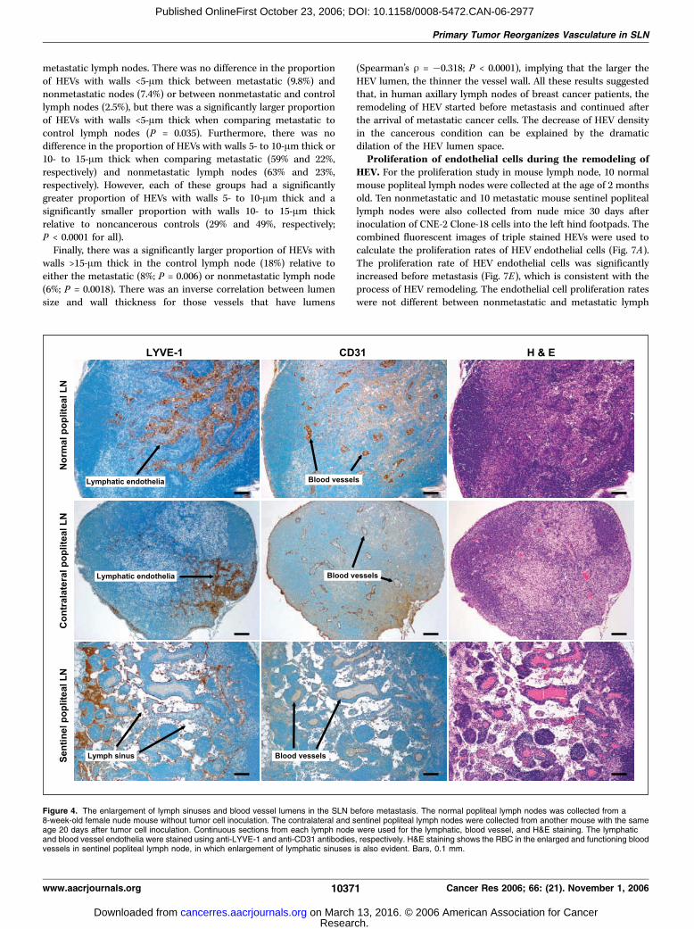
Figure 4. The enlargement of lymph sinuses and blood vessel lumens in the SLN before metastasis. The normal popliteal lymph nodes was collected from a8-week-old female nude mouse without tumor cell inoculation. The contralateral and sentinel popliteal lymph nodes were collected from another mouse with the sameage 20 days after tumor cell inoculation. Continuous sections from each lymph node were used for the lymphatic, blood vessel, and H&E staining. The lymphaticand blood vessel endothelia were stained using anti-LYVE-1 and anti-CD31 antibodies, respectively. H&E staining shows the RBC in the enlarged and functioning bloodvessels in sentinel popliteal lymph node, in which enlargement of lymphatic sinuses is also evident. Bars, 0.1 mm.
Primary Tumor Reorganizes Vasculature in SLN
www.aacrjournals.org 10371 Cancer Res 2006; 66: (21). November 1, 2006
Research. on March 13, 2016. © 2006 American Association for Cancercancerres.aacrjournals.org Downloaded from
Published OnlineFirst October 23, 2006; DOI: 10.1158/0008-5472.CAN-06-2977

nodes, implying that the proliferation of HEV endothelial cells wasmainly influenced by the duration of the cancerous condition.To confirm that proliferation of HEV endothelial cells is also
involved in the remodeling process of human HEV, the above-mentioned 10 noncancerous, 11 nonmetastatic, and 14 metastaticaxillary lymph nodes from patients were immunofluorescentlystained (Fig. 7B). The typical proliferating HEVendothelial cells in ametastatic lymph node detected by routine immunohistochemicalstaining are also shown in Fig. 7C and D . The proliferation rate ofthe HEV endothelial cells was significantly increased in thenonmetastatic lymph nodes (Fig. 7F), which was consistent withour results from animal models. Interestingly, the proliferation rateof HEV endothelial cells was significantly higher in the metastatichuman lymph nodes than in nonmetastatic lymph nodes, implying
that the lymph nodes with more robust HEV proliferation may beassociated with a higher metastasis possibility in human lymphnode.Characteristics of endotoxin-induced lymphadenopathy and
tumor-reactive lymphadenopathy. The use of athymic nudemice, which lack T cells, clearly excludes the possibility that nodeproliferation is due to the generation of a primary immuneresponse to micrometastases or to tumor antigens from theprimary lesion being presented by antigen-presenting cells arrivingvia the afferent lymphatics. The use of DA3 cells in the syngeneicBALB/c mice further excludes the involvement of immuneresponse. However, the injection of dead CNE-2 Clone-18 cellsfixed with 70% alcohol (8 � 104 cells twice with a 5-day interval),saline (30 AL daily for 20 days), or the serum from nude mice
Figure 5. Remodeling of HEVs in the SLN of animalmodels and in the regional lymph nodes of breast cancerpatients is evident. Normal popliteal lymph nodes inBALB/c nude mice, stained with anti-CD31 antibody(A ) and stained with anti-MECA79 antibody (B). PoplitealSLN before metastasis 10 days after Clone-18inoculation, stained with anti-CD31 antibody (C ) andstained with anti-MECA79 antibody (D ). Remodeled HEVsare seen to be dilated and are branching out intothin-walled, functional blood vessels containing RBC(asterisks in C ), which are typical of an angiogenicphenotype. E, human noncancerous axillary lymph nodewith a normal HEV (arrow ). F to H, nonmetastatic axillarylymph nodes from breast cancer patients. F, a dilatedHEV. G, a dilated and remodeled HEV with a part ofthe vessel wall transforming into thin wall (arrow ).H, another transforming HEV with the changing of the highendothelial cells into flat endothelial cells (arrow ).Staining is with anti-MECA79 antibody (E and F ) and H&E(G and H ).
Cancer Research
Cancer Res 2006; 66: (21). November 1, 2006 10372 www.aacrjournals.org
Research. on March 13, 2016. © 2006 American Association for Cancercancerres.aacrjournals.org Downloaded from
Published OnlineFirst October 23, 2006; DOI: 10.1158/0008-5472.CAN-06-2977

carrying CNE-2 tumors (30 AL daily for 20 days) all failed to induceany morphologic and functional alteration of the lymph channel orHEV vasculature in the SLNs (data not shown). Moreover, in any ofour footpad models, we found no morphologic alteration of thelymph channel or HEV vasculature in the second lymph nodestation (e.g., the inguinal lymph nodes; data not shown).It is unclear whether morphologic alterations of the lymph
channel and HEV vasculature share the same patterns ininflammatory lymphadenopathy and tumor-reactive lymphade-nopathy. In nude mice, the absence of T cells accounts for theresistance to endotoxin-induced inflammation (32, 33). Therefore,prolonged administration of endotoxin for 20 days was done toinduce significant lymphadenopathy in the sentinel popliteallymph node (Supplementary Fig. S1D). Staining for LYVE-1 showedthat the dilated lymph vessels/sinuses in endotoxin-inducedlymphadenopathy were full of lymphocytes (Supplementary
Fig. S1E). This was in contrast to the dilated lymph vessels/sinuses in tumor-reactive lymphadenopathy, which contained fewcellular structures, implying that it was carrying a large amount offluid rather than a large amount of lymphocytes. Interestingly, themorphology of HEVs was not altered at all in the endotoxin-induced lymphadenopathy (Supplementary Fig. S1E), suggestingthat the inflammatory reaction and cancerous reaction in the SLNare induced by different mechanisms.Premetastatic remodeled HEVs could integrate into the
postmetastatic tumor vasculature with further differentiation.The standard marker for HEVs is MECA-79. The MECA-79-reactive ligand is known as peripheral node addressin (PNAd).Anti-MECA-79 antibody specifically stains HEVs in lymph nodesand other secondary lymphoid organs in many species, includinghumans and mice (34). Within metastatic tumor nests in theaxillary lymph nodes from human breast cancer patients, some
Figure 6. Quantitative analyses of the remodeling of HEVs in both animal SLNs and human regional lymph nodes. A, the increase of the adjusted HEV lumen areain the sentinel popliteal lymph node before metastasis is statistically significant in nude mice carrying human cancers (CNE-2 Clone-22 and Clone-26) as well as inimmunocompetent BALB/c mice carrying mouse DA3 breast adenocarcinoma (P < 0.001). B, the decrease of the HEV wall thickness is statistically significant inClone-22 (P < 0.001), Clone-26 (P < 0.001), and DA3 (P = 0.035). C to F, immunohistochemical staining of HEVs using anti-MECA79 antibody in a humannoncancerous lymph node from a cancer-free patient (C ), a nonmetastatic lymph node from a breast cancer patient (D ), and a metastatic lymph node (E and F )showing the margin of a metastatic tumor lesion (T in E) and a lymphoid tissue area distant to the tumor lesion (F ). G, proportion of HEVs with lumens (any or >80 Am2)for control, nonmetastatic, and metastatic lymph nodes. *, P < 0.001 relative to control; **, P < 0.001 relative to nonmetastatic. H, proportion of open lumenHEVs with various wall thicknesses for control, nonmetastatic, and metastatic lymph nodes. *, P < 0.05 relative to control.
Primary Tumor Reorganizes Vasculature in SLN
www.aacrjournals.org 10373 Cancer Res 2006; 66: (21). November 1, 2006
Research. on March 13, 2016. © 2006 American Association for Cancercancerres.aacrjournals.org Downloaded from
Published OnlineFirst October 23, 2006; DOI: 10.1158/0008-5472.CAN-06-2977

indentified HEVs were full of RBC, implying fully functioningvessels (Supplementary Fig. S2A). Interestingly, the remodeledHEVs gradually lost their specific marker MECA-79 from thetumor margin to the central portion of the tumor nests(Supplementary Fig. S2B), implying further differentiation of theHEVs when they integrated into the metastatic tumor vasculature.Actually, most of the HEVs lost their specific marker MECA-79after integrating into the tumor vasculature, leaving thesurrounding remnant lymphoid tissue, indicating that they werepre-existing vessels (Supplementary Fig. S2C). These findingsimplied that the remodeled HEVs in the premetastatic phasecontinued to nurture the metastatic lesions after the arrival ofcancer cells and therefore played an important role in thesecondary tumor growth inside the involved lymph node.
Discussion
The lumens of the lymph sinuses/vessels were dilated in SLNbefore metastasis. The SLN is rebuilt by the primary tumor tobecome a functional blood vessel–enriched organ before andindependent of metastasis, with the morphologic and functionalalterations of the HEVs to become main blood flow carrier in thelymph node. The processes of vascularization in the SLN were
consistent in both animal model and human tissues. This studypresents functional and structural data that the primary tumor ismanipulating the ‘‘soil’’ to improve the ‘‘seeding’’ of subsequentmetastases.The extent of lymph sinus dilation in the SLN was significantly
correlated with the primary tumor weight, suggesting that the
lymph from the primary tumor induced the persistent alteration of
the lymph channel in SLN. These results are consistent with a
recent finding that, in contrast to angiogenesis, in which blood
flow proceeds only after the vessel develops, lymphangiogenesis
can be induced by interstitial fluid channeling (35). The alteration
of lymph channels in SLNs can also occur during an inflammatory
reaction, which facilitates the migration of inflammatory cells (36).
Interestingly, the dilated lymph vessels/sinuses in endotoxin-
induced lymphadenopathy were full of lymphocytes, but they
contained very few cellular structures in tumor-reactive lymph-
adenopathy, suggesting different roles of the SLN lymphatic
channels in different pathologic processes.It has been well characterized that the HEVs of lymph nodes
play an important role in recruiting lymphocytes for the generation
of immune responses. By expressing homing receptors on their
surface, which blood lymphocytes can recognize as they pass in
Figure 7. Proliferation of the HEVendothelial cells was involved in themorphologic and functional alterations ofHEVs. A and B, combined images of tripleimmunofluorescent staining on MECA79(green ), proliferating marker PCNA (red),and nuclei (blue ). Arrows, proliferatingendothelial cells in the HEVs. A, anonmetastatic sentinel popliteal lymphnode section from a nude mouse bearinga CNE-2 Clone-18 tumor. B, anonmetastatic axillary lymph node sectionfrom a breast cancer patient. C and D,the same area of two consecutive sectionsof a human metastatic lymph node tissuefrom a breast cancer patient. C,immunohistochemical staining of MECA79indicating HEVs. D, immunohistochemicalstaining of PCNA indicating proliferatingcells. Inset, enlarges one of the HEVs,showing two proliferating endothelialcells stained in brown (arrows ).E, proliferation rates of HEV endothelialcells in mouse lymph nodes.F, proliferating rate of HEV endothelialcells in the human lymph nodes. Columns,mean; bars, SD. *, P < 0.001 relative tocontrol; **, P < 0.001 relative to thenonmetastatic group.
Cancer Research
Cancer Res 2006; 66: (21). November 1, 2006 10374 www.aacrjournals.org
Research. on March 13, 2016. © 2006 American Association for Cancercancerres.aacrjournals.org Downloaded from
Published OnlineFirst October 23, 2006; DOI: 10.1158/0008-5472.CAN-06-2977

circulation, HEVs provide a unique location where naive lympho-cytes can enter the lymph node (30, 37, 38). In this study, we foundthat the role of HEV was shifted to become the main blood flowcarrier in the SLN before metastasis. Not only could the HEVmorphology change dramatically to carry more blood flow, but theproliferation rate of HEV endothelial cells was also increasedbefore metastasis. However, the HEV morphology did not alter atall in endotoxin-induced lymphadenopathy, implying a selectivereaction of HEV in the cancerous condition.The adaptive response of the vascular wall has been noted when
a vein segment is transposed as a bypass graft into the arterialcirculation, with an increase in wall thickness and the appearanceof multiple cellular layers in the venous endothelium (39, 40).Distinct, differentiated gene expression has also been reportedwhen the endothelial cells respond to the changing of theirmicroenvironment (41). In our own study, we found that thecellular morphology of the tall endothelial cells forming HEVschanged dramatically to become flat endothelial cells in cancerouscondition. As a consequence, the HEV was remodeled from athick-walled, endothelial vessel with a small lumen to a thin-walled, large-lumen vessel, shifting its function from recruitinglymphocytes to becoming a blood flow carrier. These facts suggestagain that the blood vessel endothelium has tremendous potentialto adapt its biomechanical environment.The confined lymph and blood channel alterations within the
SLN but not in the next station lymph node imply that an inducerfrom the primary tumor is functioning locally with the existence ofa liable primary tumor. VEGF-A has been found to be an inducer oflymphangiogenesis in SLNs (9). We also found that the serum levelof VEGF-A was elevated in patients with late-stage NPC (42).Because VEGF-A is a secreted protein that can travel via thebloodstream to other lymph nodes, our findings suggest that otherinducers besides VEGF-A may be involved.It is also of interest to explore the role of HEVs after the
establishment of a metastatic tumor nest. We found that theenlarged, remodeled HEVs could integrate into the metastatictumor vasculature with further differentiation, characterized bythe gradual loss of their specific marker MECA-79 from the tumormargin to the central part of the metastatic tumor nest. It has been
explained that, compared with primary tumors, the more rapidgrowth of metastatic lesions in the cervical lymph nodes of NPCpatients was due to clonal selection of the cancer cells duringmetastases, with highly proliferative clones disseminated to thecervical nodes. However, based on our findings, the metastatictumor vasculature in lymph nodes consists of many large bloodvessels derived from normal HEVs, suggesting that the efficiency ofnutrition and oxygen supplies could be better for the metastatictumor cells in the involved lymph node. The enrichment of theblood supply in the lymph node before and after metastasis mayfavor the growth of newly arriving metastatic cancer cells.Consequently, the involved regional lymph nodes may becomemanifest, whereas the primary tumors remain clinically occult foryears (43, 44). Moreover, the high density of functioning bloodvessels in lymph nodes may subsequently facilitate the metastasisof cancer cell to distant organs. It will be important to elucidate ifthe highly vascularized premetastatic SLN is associated with anincreased metastatic potential. Control of lymphatic fluid move-ment may also be a target to consider in preventing metastases.In conclusion, our study reveals the basis of lymphangiogesis
and angiogenesis within SLNs. The lumens of the lymph sinuses/vessels were dilated in SLNs before metastasis. The SLN was rebuiltby the primary tumor to become a functional blood vessel–enriched organ before and independent of metastasis, withmorphologic and functional alterations of the HEVs to becomethe main blood flow carrier in the lymph node. The processes ofvascularization in the SLN were consistent in both animal modeland human tissues. This is the evidence that tumor-inducedvascularization can be a regional process rather than purely a localevent.
Acknowledgments
Received 8/15/2006; accepted 8/18/2006.The costs of publication of this article were defrayed in part by the payment of page
charges. This article must therefore be hereby marked advertisement in accordancewith 18 U.S.C. Section 1734 solely to indicate this fact.
We thank Dawna Dylewski, Elissa Boguslawski, Bryn Eagleson, and the rest of theVARI vivarium staff for assistance in animal husbandry; JC Goolsby and the rest of theVARI Histology Core Facility for technical support; and David Nadziejka for criticallyreading this article.
References1. Johnson JT. A surgeon looks at cervical lymph nodes.Radiology 1990;175:607–10.2. Ioachim HLRH. Tumor-reactive lymphadenopathy. 3rded. Philadelphia, Baltimore, New York, London, BuenosAires, Hong Kong, Sydney, Tokyo: Lippincott Williamsand Wilkins; 2002. p. 254–8.3. Folkman J. Role of angiogenesis in tumor growth andmetastasis. Semin Oncol 2002;29:15–8.4. Beasley NJ, Prevo R, Banerji S, et al. Intratumorallymphangiogenesis and lymph node metastasis in headand neck cancer. Cancer Res 2002;62:1315–20.5. Maula SM, Luukkaa M, Grenman R, et al. Intratumorallymphatics are essential for the metastatic spread andprognosis in squamous cell carcinomas of the head andneck region. Cancer Res 2003;63:1920–6.6. Pepper MS, Tille JC, Nisato R, Skobe M. Lymphangio-genesis and tumor metastasis. Cell Tissue Res 2003;314:167–77.7. Skobe M, Hawighorst T, Jackson DG, et al. Induction oftumor lymphangiogenesis by VEGF-C promotes breastcancer metastasis. Nat Med 2001;7:192–8.8. Cao R, Bjorndahl MA, Religa P, et al. PDGF-BB induces
intratumoral lymphangiogenesis and promotes lym-phatic metastasis. Cancer Cell 2004;6:333–45.9. Hirakawa S, Kodama S, Kunstfeld R, et al. VEGF-Ainduces tumor and sentinel lymph node lymphangio-genesis and promotes lymphatic metastasis. J Exp Med2005;201:1089–99.10. Chan AT, Teo PM, Johnson PJ. Nasopharyngealcarcinoma. Ann Oncol 2002;13:1007–15.11. Sham JS, Choy D, Wei WI. Nasopharyngeal carcino-ma: orderly neck node spread. Int J Radiat Oncol BiolPhys 1990;19:929–33.12. Carlson RW, Anderson BO, Bensinger W, et al. NCCNpractice guidelines for breast cancer. Oncology (Hun-tingt) 2000;14:33–49.13. Goldhirsch A, Glick JH, Gelber RD, Coates AS,Senn HJ. Meeting highlights: International ConsensusPanel on the Treatment of Primary Breast Cancer.Seventh International Conference on Adjuvant Ther-apy of Primary Breast Cancer. J Clin Oncol 2001;19:3817–27.14. Rosen PP, Saigo PE, Braun DW, Jr., Weathers E,DePalo A. Predictors of recurrence in stage I (T1N0M0)breast carcinoma. Ann Surg 1981;193:15–25.15. Sizhong Z, Xiukung G, Yi Z. Cytogenetic studies on
an epithelial cell line derived from poorly differenti-ated nasopharyngeal carcinoma. Int J Cancer 1983;31:587–90.16. Keshtgar MW, Lakhani W, Ell S. P. Dosimetry andradiation protection. Berlin: Springer; 1999. p. 91–102.17. Muller A, Homey B, Soto H, et al. Involvement ofchemokine receptors in breast cancer metastasis.Nature 2001;410:50–6.18. Tsarfaty G, Stein GY, Moshitch-Moshkovitz S, et al.HGF/SF increases tumor blood volume: a novel tool forthe in vivo functional molecular imaging of Met.Neoplasia 2006;8:344–52.19. Kort EJ, Campbell B, Resau JH. A human tissue anddata resource: an overview of opportunities, challenges,and development of a provider/researcher partnershipmodel. Comput Methods Programs Biomed 2003;70:137–50.20. Banerji S, Ni J, Wang SX, et al. LYVE-1, a newhomologue of the CD44 glycoprotein, is a lymph-specific receptor for hyaluronan. J Cell Biol 1999;144:789–801.21. Ridler T, Calvard S. Picture thresholding using aniterative selection method. IEEE Transactions onSystems, Man, and Cybernet 1978;8:629–32.
Primary Tumor Reorganizes Vasculature in SLN
www.aacrjournals.org 10375 Cancer Res 2006; 66: (21). November 1, 2006
Research. on March 13, 2016. © 2006 American Association for Cancercancerres.aacrjournals.org Downloaded from
Published OnlineFirst October 23, 2006; DOI: 10.1158/0008-5472.CAN-06-2977

22. Kort EJ, Jones A, Daumbach M, et al. Quantifying cellscattering: the blob algorithm revisited. Cytometry. PartA: The Journal of the International Society for AnalyticalCytology 2003;51:119–26.23. Fidler IJ. The pathogenesis of cancer metastasis: the‘seed and soil’ hypothesis revisited. Nat Rev Cancer 2003;3:453–8.24. Fidler IJ, Kripke ML. Metastasis results from preex-isting variant cells within a malignant tumor. Science1977;197:893–5.25. Hynes RO. Metastatic potential: generic predisposi-tion of the primary tumor or rare, metastatic variants-orboth? Cell 2003;113:821–3.26. Yang WT, Chang J, Metreweli C. Patients with breastcancer: differences in color Doppler flow and gray-scaleU.S. features of benign and malignant axillary lymphnodes. Radiology 2000;215:568–73.27. Lu W, Schroit AJ. Vascularization of melanoma bymobilization and remodeling of preexisting latentvessels to patency. Cancer Res 2005;65:913–8.28. Jackson DG, Prevo R, Clasper S, Banerji S. LYVE-1,the lymphatic system and tumor lymphangiogenesis.Trends Immunol 2001;22:317–21.29. Hauff P, Reinhardt M, Briel A, Debus N, Schirner M.Molecular targeting of lymph nodes with L-selectinligand-specific US contrast agent: a feasibility study in
mice and dogs. Radiology 2004;231:667–73. Epub 2004Apr 29.30. Yeh JC, Hiraoka N, Petryniak B, et al. Novel sulfatedlymphocyte homing receptors and their control by aCore1 extension h 1,3-N-acetylglucosaminyltransferase.Cell 2001;105:957–69.31. Gartner LP, Hiatt JL. Color textbook of histology.2nd ed. Philadelphia (PA): WB Saunders Company;2001. p. 266.32. Huang JH, Lin YY, Lai YY, Hu SW. Lethal outcomecaused by Porphyromonas gingivalis A7436 in a mousechamber model is associated with elevated titers of hostserum interferon-g. Oral Microbiology and Immunology2006;21:100–6.33. Okamura H, Tsutsi H, Komatsu T, et al. Cloning of anew cytokine that induces IFN-g production by T cells.Nature 1995;378:88–91.34. Uchimura K, Gauguet JM, Singer MS, et al. A majorclass of L-selectin ligands is eliminated in mice deficientin two sulfotransferases expressed in high endothelialvenules. Nat Immunol 2005;6:1105–13.35. Boardman KC, Swartz MA. Interstitial flow as a guidefor lymphangiogenesis. Circ Res 2003;92:801–8.36. Halin C, Detmar M. An unexpected connection:lymph node lymphangiogenesis and dendritic cellmigration. Immunity 2006;24:129–31.
37. Sackstein R. Physiologic migration of lymphocytesto lymph nodes following bone marrow transplanta-tion: role in immune recovery. Semin Oncol 1993;20:34–9.38. von Andrian UH, M’Rini C. In situ analysis oflymphocyte migration to lymph nodes. Cell AdhesCommun 1998;6:85–96.39. Garrett HE, Diethrich EB, DeBakey ME. Myocardialrevascularization. Surg Clin North Am 1966;46:863–71.40. Kwei S, Stavrakis G, Takahas M, et al. Early adaptiveresponses of the vascular wall during venous arterial-ization in mice. Am J Pathol 2004;164:81–9.41. Chi JT, Chang HY, Haraldsen G, et al. Endothelialcell diversity revealed by global expression profiling.Proc Natl Acad Sci U S A 2003;100:10623–8. Epub 2003Sep 8.42. Qian CN, Zhang CQ, Guo X, et al. Elevation of serumvascular endothelial growth factor in male patients withmetastatic nasopharyngeal carcinoma. Cancer 2000;88:255–61.43. Medina-Franco H, Urist MM. Occult breast carcino-ma presenting with axillary lymph node metastases. RevInvest Clin 2002;54:204–8.44. Nieder C, Ang KK. Cervical lymph node metastasesfrom occult squamous cell carcinoma. Curr TreatOptions Oncol 2002;3:33–40.
Cancer Research
Cancer Res 2006; 66: (21). November 1, 2006 10376 www.aacrjournals.org
Research. on March 13, 2016. © 2006 American Association for Cancercancerres.aacrjournals.org Downloaded from
Published OnlineFirst October 23, 2006; DOI: 10.1158/0008-5472.CAN-06-2977

2006;66:10365-10376. Published OnlineFirst October 23, 2006.Cancer Res Chao-Nan Qian, Bree Berghuis, Galia Tsarfaty, et al. before the Arrival of Metastatic Cancer CellsVasculature Reorganization in the Sentinel Lymph Node Preparing the ''Soil'': The Primary Tumor Induces
Updated version
10.1158/0008-5472.CAN-06-2977doi:
Access the most recent version of this article at:
Material
Supplementary
ml
http://cancerres.aacrjournals.org/content/suppl/2006/11/07/0008-5472.CAN-06-2977.DC1.htAccess the most recent supplemental material at:
Cited articles
http://cancerres.aacrjournals.org/content/66/21/10365.full.html#ref-list-1
This article cites 39 articles, 10 of which you can access for free at:
Citing articles
http://cancerres.aacrjournals.org/content/66/21/10365.full.html#related-urls
This article has been cited by 26 HighWire-hosted articles. Access the articles at:
E-mail alerts related to this article or journal.Sign up to receive free email-alerts
Subscriptions
Reprints and
To order reprints of this article or to subscribe to the journal, contact the AACR Publications
Permissions
To request permission to re-use all or part of this article, contact the AACR Publications
Research. on March 13, 2016. © 2006 American Association for Cancercancerres.aacrjournals.org Downloaded from
Published OnlineFirst October 23, 2006; DOI: 10.1158/0008-5472.CAN-06-2977
Copyright © 2022 FDOKUMEN