Efficacy of memantine in treating patients with fibromyalgia
Upload
independentCategory
view
0download
0
© 2010 Ruggiero et al, publisher and licensee Dove Medical Press Ltd. This is an Open Access article which permits unrestricted noncommercial use, provided the original work is properly cited.
International Journal of Nanomedicine 2010:5 783–802
International Journal of Nanomedicine Dovepress
submit your manuscript | www.dovepress.com
Dovepress 783
O R I g I N A L R e s e A R c h
open access to scientific and medical research
Open Access Full Text Article
DOI: 10.2147/IJN.S13300
Imaging and treating tumor vasculature with targeted radiolabeled carbon nanotubes
Alessandro Ruggiero1*
carlos h Villa1*
Jason P holland1
shanna R sprinkle1
chad May2
Jason s Lewis1
David A scheinberg1
Michael R McDevitt1
1Departments of Medicine and Radiology, Pharmacology and Molecular Therapeutics, Memorial sloan-Kettering cancer center, New York, UsA; 2Imclone systems, New York, UsA; *Ruggiero and Villa contributed equally to this work
correspondence: Michael R McDevitt Departments of Medicine and Radiology, Memorial sloan-Kettering cancer center, 1275 York Ave, Box 231, New York, NY 10065, UsA Tel +1-646-888-2192 Fax +1-646-422-0640 email [email protected]
Abstract: Single wall carbon nanotube (SWCNT) constructs were covalently appended with
radiometal-ion chelates (1,4,7,10-tetraazacyclododecane-1,4,7,10-tetraacetic acid [DOTA]
or desferrioxamine B [DFO]) and the tumor neovascular-targeting antibody E4G10. The
E4G10 antibody specifically targeted the monomeric vascular endothelial-cadherin (VE-cad)
epitope expressed in the tumor angiogenic vessels. The construct specific activity and blood
compartment clearance kinetics were significantly improved relative to corresponding antibody-
alone constructs. We performed targeted radioimmunotherapy with a SWCNT-([225Ac]DOTA)
(E4G10) construct directed at the tumor vasculature in a murine xenograft model of human
colon adenocarcinoma (LS174T). The specific construct reduced tumor volume and improved
median survival relative to controls. We also performed positron emission tomographic (PET)
radioimmunoimaging of the tumor vessels with a SWCNT-([89Zr]DFO)(E4G10) construct in
the same murine LS174T xenograft model and compared the results to appropriate controls.
Dynamic and longitudinal PET imaging of LS174T tumor-bearing mice demonstrated rapid blood
clearance (,1 hour) and specific tumor accumulation of the specific construct. Incorporation
of the SWCNT scaffold into the construct design permitted us to amplify the specific activity to
improve the signal-to-noise ratio without detrimentally impacting the immunoreactivity of the
targeting antibody moiety. Furthermore, we were able to exploit the SWCNT pharmacokinetic
(PK) profile to favorably alter the blood clearance and provide an advantage for rapid imaging.
Near-infrared three-dimensional fluorescent-mediated tomography was used to image the
LS174T tumor model, collect antibody-alone PK data, and calculate the number of copies
of VE-cad epitope per cell. All of these studies were performed as a single administration of
construct and were found to be safe and well tolerated by the murine model. These data have
implications that support further imaging and radiotherapy studies using a SWCNT-based
platform and focusing on the tumor vessels as the target.
Keywords: actinium-225 (225Ac), zirconium-89 (89Zr), angiogenesis, vascular endothelial-
cadherin, radioimmunotherapy (RIT), radioimmunoPET
IntroductionAdvances in cancer diagnosis and therapy require improvements in the agents used to
image and treat disease. Valuable modifications include those that enable these agents
to specifically target disease, increase signal-to-noise, rapidly clear from the blood,
and incorporate multiple imaging and therapeutic modalities. Carbon nanomaterials
are being investigated as delivery platforms for diagnostic and therapeutic cargoes to
target disease.1,2 Pharmacokinetic (PK) studies of soluble, covalently-functionalized,
radiolabeled carbon nanotubes (CNTs) have demonstrated rapid blood compartment

International Journal of Nanomedicine 2010:5submit your manuscript | www.dovepress.com
Dovepress
Dovepress
784
Ruggiero et al
clearance, high specific activity (SA), multimodal imaging
capability, renal elimination, and tumor-specific accumulation
in vivo.3–6 Implementation of an imaging and therapeutic
drug construct that is designed to target, report location, and
irradiate the tumor vessels is a key strategic modification that
will take advantage of the single wall CNT (SWCNT)-based
construct’s chemical, radiochemical, and PK properties.
Many cancers are characterized by an extensive angio-
genic, aberrant vascular network that supports the tumor
proliferation and survival. The endothelial vessels in tumors
often do not exhibit the same organizational hierarchy of
arterioles, capillaries, and venules present in normal tissue.
Instead, tumor vessels are tortuous and have abnormal
component and structural composition. Endothelial cells
in these tumors are inefficiently and irregularly joined with
holes, gaps, and defects; pericytes are absent or loosely
associated with vessels; and basement membranes are
inefficiently applied relative to typical normal tissues. The
pore diameters of tumor typically range from 40 to 80 nm.
Large interendothelial junctions in cancerous tissue may be
as large as 500 nm, whereas in healthy tissue, these junc-
tions are ∼8 nm.7 Angiogenic endothelial cells express the
monomeric vascular endothelial-cadherin (VE-cad) epitope
on the cell surface that upon dimerizing with another mono-
meric copy of VE-cad on an adjoining cell surface leads to the
formation of tight adherens junctions between the cells.8–12
The antibody E4G10 binds only to the monomeric VE-cad
and not to the homodimeric form (the binding region is
masked in the homodimers that form the tight cell–cell con-
tacts), thus conferring the specificity for targeting angiogenic
and poorly joined endothelial cells in vivo while not binding
to normal endothelium or the LS174T tumor.
The alpha particle-emitting radionuclide actinium-225
(225Ac; t1/2
= 10 days) attached to monoclonal antibodies
(immunoglobulin [Ig]G) has been used as the therapeutic
effector moiety in a number of preclinical studies13–18 and
is currently in clinical use.19–21 Alpha particles are charged
helium nuclei that travel ∼50–80 µm, which is similar to the
dimensions of vessels within a tumor. The alpha particle track
lengths appropriately match the vessel dimensions, and a
single traversal through a cell of a high linear energy transfer
(LET) alpha particle can be acutely cytotoxic.22,23 Typical
tumor vasculature is ,100 µm in diameter, and smaller ves-
sels are ,10 µm in diameter. In addition, individual alpha
particles are able to kill a target cell due to their deposition
of 5–8 MeV in a short ionizing track that is several cell diam-
eters in length.23 This highly concentrated field of ionizing
radiation may also irradiate the cancer stem cell population
that can be found in the adjacent perivascular niche of some
tumors.24–26 Alpha particles are very potent cytotoxic agents in
proximity to the targeted tissue but will largely spare normal
tissue; it is this characteristic that offers clear advantages
to other known forms of radiation as a means of selective
cell killing. We have previously demonstrated the utility of 225Ac-radiolabeled E4G10 constructs to target and irradiate
tumor vascular endothelium in animal models, improve
survival, control tumor growth, and normalize the vessels27
and also the ability of the construct to target and eradicate
bone marrow-derived endothelial progenitors.28
Zirconium-89 (89Zr) is now being developed as one of the
most promising new immuno-positron emission tomography
(PET) agents for in vivo imaging of cancer.29–34 PET is based
on coincidence detection of a positron-emitting radionuclide
from 2 coincident 511 keV annihilation photons emitted
simultaneously in opposite directions.35 The clinical use
of PET has emerged as an important diagnostic imaging
modality for humans because it provides extremely sensitive,
quantitative, and functional information that is different from
the information obtainable with other largely anatomical
imaging modalities.35 The physical decay properties of 89Zr
(t1/2
= 78.41 hours; electron capture = 76.6%; β+ = 22.3%)
are well suited for use in the design of imaging agents with
extended PK profiles. Recently, reported improvements in
the separation chemistry of 89Zr from the yttrium-89 (89Y)
target material have yielded a facile process for production
of clinical-grade, high SA 89Zr using a small cyclotron.30
This process should lead to more widespread use of this
radionuclide.
The rationale for using SWCNTs as the scaffold in
our construct design is the following: SWCNTs are made
almost entirely of carbon, are nonimmunogenic, and can
be chemically modified.36–38 The commercially available
SWCNTs have a diameter of 1–2 nm and lengths of
100–1,000 nm. Thus, SWCNTs have extremely high aspect
ratios with estimated surface areas of 1,600 m2/g. On the
atomic level, SWCNTs have highly regular structures with
defined periodicity. For every 100 nm of length, a SWCNT
can have up to 12,200 carbon atoms and weighs 150,000 Da,
providing numerous sites (typically 1 in 95 carbons was modi-
fied) for chemical modifications and anchoring chemical,
biological, and radionuclide moieties.3–6 SWCNTs that have
been chemically functionalized with sidewall amino groups
exhibit aqueous solubility and reactivity towards a variety of
reagents under mild bioconjugation conditions.3–6,36–39 The
SWCNTs appended with amines and the metal-ion chelate
1,4,7,10-tetraazacyclododecane-1,4,7,10-tetraacetic acid

International Journal of Nanomedicine 2010:5 submit your manuscript | www.dovepress.com
Dovepress
Dovepress
785
Targeting angiogenic tumor vasculature with carbon nanotubes
(DOTA) were water soluble (∼20 g/L) and studied in vivo
in animal models.3–6 Our data showed that these SWCNTs
rapidly (t1/2
, 1 hour) cleared the blood and prototype
SWCNT constructs (diameter = ∼1 nm; length = ∼300 nm;
moleculer weight = ∼500 kD) were predominantly eliminated
intact within minutes via glomerular filtration through the
renal pathway. Renal excretion was found to be facilitated
by the high aspect ratio of SWCNT, which confers lower
rotational diffusivity; thus, the CNT tends to orient with the
blood flow allowing easy access to the glomerular pores.6
Other recent work described the enzymatic degradation
of functionalized SWCNT in vitro and in vivo, offering
the potential for metabolic decomposition of CNT drug
constructs.40–43 Further, toxicity studies have confirmed
the safe in vivo use of functionalized SWCNT.44–47 Taken
together, these data on renal elimination, enzymatic degrada-
tion, and lack of toxicity support continued investigation of
SWCNT as a platform in drug development.
We hypothesize that CNT constructs can be covalently
multifunctionalized with copies of targeting moieties and
different reporting and therapeutic radionuclides and, thereby,
increase signal-to-noise ratio and be used to image tumor
vessels and improve the therapeutic index. The underlying
rationale for this approach is that we are specifically target-
ing the aberrant and angiogenic tumor vessels and deliver-
ing a dose of short range (1–4 cell diameters), high energy
(several million electron volts) cytotoxic alpha particles to
the targeted area. In addition, we will report the location of
the drug construct. This approach is novel because it will
examine the attributes of targeted radioimmunotherapy (RIT)
with alpha particle generators, sensitive and quantitative PET
imaging, and the specificity of the targeting E4G10 antibody
in combination with the unique nanomaterial properties
of SWCNT.
Material and methodsLs174T xenograft model in nude miceAthymic nude mice (NCr nu/nu, male), 4–12 weeks of age,
were obtained from Taconic, Germantown, NY, USA. For all
in vivo experiments, housing and care were in accordance
with the Animal Welfare Act and the Guide for the Care
and Use of Laboratory Animals. The animal use protocols
were approved by the Institutional Animal Care and Use
Committee, Memorial Sloan-Kettering Cancer Center.
Human LS174T cells (human colon adenocarcinoma)
were expanded in Dulbecco–Vogt Modified Eagle’s media
supplemented with glucose, nonessential amino acids,
l-glutamine, and 10% fetal bovine serum in an atmosphere
of 5% CO2 and air at 37°C. Cells were harvested and mixed
with MatrigelTM (BD Biosciences, Palo Alto, CA, USA), and
0.1 mL of ∼2–3E6 cells were injected subcutaneously into
the right hind flank of each animal.
construct design and synthesesSWCNTs (Nanolab, Newton, MA, USA) were covalently
amine functionalized as described previously.3–6,36–39 The
SWCNT-NH2 product was purified from carbonaceous
impurities using a C18 Sep-Pak® (Waters, Milford, MA, USA)
and analyzed by high-performance liquid chromatography
(HPLC), Raman spectroscopy, and transmission electron
microscopy (TEM).3–6 Amine loading was determined using
the Sarin assay, whereas TEM and Raman spectroscopy were
performed as described previously.3–6 Table 1 provides a list
of the key drug constructs, the corresponding nomenclature,
studies performed, and the SA values for the therapeutic and
imaging studies from this work.
The RIT drug Construct I (Figure 1A) was designed
to specifically target the tumor vessels and deliver the
potent alpha particle-emitting 225Ac radionuclide gen-
erator.13 The key SWCNT precursor to Construct I
was assembled by first converting a fraction of the pri-
mary amines on the SWCNT-NH2 construct to reactive
hydrazinopyridine (HNH) moieties. Briefly, 0.5 g of SWCNT-
NH2 was dissolved in 1 mL of 100 mM sodium phosphate
(NaH2PO
4 and Na
3PO
4; Sigma-Aldrich, St Louis, MO,
USA)/150 mM sodium chloride (NaCl; Sigma-Aldrich), pH
7.8 buffer. Immediately before use, 5.5 mg of succinimidyl
4-hydrazinonicotinate acetone hydrazone (SANH; Solulink
Inc, San Diego, CA, USA) was dissolved in 0.2 mL of dry
N,N-dimethylformamide (DMF; Sigma-Aldrich). An aliquot
Table 1 Construct nomenclature, designation, study use, and specific activity
Construct nomenclature Designation Study High SA (Ci/g) Low SA (Ci/g)
sWcNT-([225Ac]DOTA)(e4g10) construct I RIT 23 0.05sWcNT-([225Ac]DOTA)(anti-KLh) control construct I RIT 23 NAsWcNT-([89Zr]DFO)(e4g10) construct II RII 16 NAsWcNT-([89Zr]DFO)(anti-KLh) control construct II RII 16 NA
Abbreviations: SA, specific activity; SWCNT, single wall carbon nanotube; 225Ac, actinium-225; DOTA, 1,4,7,10-tetraazacyclododecane-1,4,7,10-tetraacetic acid; RIT, radioimmunotherapy; anti-KLh, anti-keyhole limpet hemocyanin; 89Zr, zirconium-89 DFO, desferrioxamine B; RII, radioimmunoimaging.

International Journal of Nanomedicine 2010:5submit your manuscript | www.dovepress.com
Dovepress
Dovepress
786
Ruggiero et al
O
O
O
OO O
HOO
OO
SO
OO
O
OOO
O
H
OH N
NH
NH
NH
NN
NN
NN
N
HN H
N
225Ac
A
Figure 1 graphical representations of the key moieties that were appended to the water-soluble sWcNT-Nh2 by covalent-functionalization with radionuclides, DOTA, DFO, and antibodies (Note, not drawn to scale). A) Radioimmunotherapeutic drug construct I (sWcNT-([225Ac]DOTA)(e4g10)). B) Radioimmunoimaging drug construct II sWcNT-([89Zr]DFO)(e4g10).Abbreviations: sWcNT, single wall carbon nanotube; DOTA, 1,4,7,10-tetraazacyclododecane-1,4,7,10-tetraacetic acid; DFO, desferrioxamine B; 225Ac, actinium-225; 89Zr, zirconium-89.
O
O
H
OH N
N
O
O O O
O
O O
O
O
OOO
O
O O
O
O
NH
HN
NH
NH
NH
NH
NH
N
HO
N
NN
N
5
5
89Zr
B

International Journal of Nanomedicine 2010:5 submit your manuscript | www.dovepress.com
Dovepress
Dovepress
787
Targeting angiogenic tumor vasculature with carbon nanotubes
of SANH/DMF solution was added to the SWCNT-NH2 to
achieve a 0.25-fold mole ratio of SANH to primary amine.
The reaction proceeded at ambient temperature for 2–3 hours
at pH 7.6. The product, SWCNT-(HNH)(NH2), was purified
using size exclusion chromatography (SEC) with a 10 DG
gel permeation column (Bio-Rad Laboratories, Hercules, CA,
USA) as the stationary phase and metal-free water (MFW;
Purelab Plus System, US Filter Corp, Lowell, MA, USA)
as the mobile phase. The product was lyophilized to yield a
solid that was found to be the desired SWCNT-(HNH)(NH2)
construct.
The second step entailed covalently appending multiple
copies of 2-(p-isothiocyanatobenzyl)-DOTA (DOTA-NCS;
Macrocyclics, Inc, Dallas, TX, USA) to the remaining amines
on the water-soluble SWCNT-(HNH)(NH2) construct to yield
a SWCNT-(DOTA)(HNH) construct in metal-free conditions
at pH 9.5 (adjusted with 1 M metal-free carbonate solution)
for 40 minutes at room temperature at a stoichiometry of
10:1 (DOTA-NCS to amine).3–6 The product was purified
using a 10 DG gel permeation column with MFW as the
mobile phase. The 10 DG column was rendered metal free
by washing with 50 mL of 25 mM EDTA (Sigma-Aldrich)
followed by rinsing with 250 mL of MFW. The product was
lyophilized to yield a solid that was found to be the desired
SWCNT-(DOTA)(HNH) construct.
SWCNT-(DOTA)(HNH) + 225Ac → SWCNT-([225Ac]DOTA)(HNH) (1)
225Ac radionuclide was obtained from the US Department
of Energy’s Oak Ridge National Laboratory (Oak Ridge, TN,
USA). Methods for radiolabeling and purification of a MFW
solution of SWCNT-(DOTA)(HNH) with 225Ac at pH 5 are
similar to those described previously.48 Briefly, 0.18 mg of
SWCNT-(DOTA)(HNH) in 0.02 mL of MFW was reacted
with 0.005 mL of 225Ac in 50 mM optima grade hydrochloric
acid (HCl; Fisher Scientific, Pittsburgh, PA, USA) along with
0.02 mL of 150 g/L l-ascorbic acid (Sigma-Aldrich) and
0.2 mL of 3 M tetramethylammonium acetate (Fisher Scien-
tific) buffer, pH 5.5, at 60°C for 60 minutes (reaction 1). Puri-
fication was accomplished using SEC with a P6 gel stationary
phase and a phosphate buffer saline (PBS) mobile phase.
Chemicals used in the radiolabeling and purification steps
were of American Chemical Society reagent-grade or higher
purity. The labeling solutions were prepared and subsequently
rendered metal free with Chelex® 100 resin, 200–400 mesh,
sodium form (Bio-Rad Laboratories), and sterile filtered
through a 0.22 or 0.45 µm filter device. Solutions of 50 mM
diethylenetriaminepentaacetic acid (DTPA; Sigma-Aldrich)
were sterile filtered and used to quench the labeling reac-
tion prior to SEC. Human serum albumin (HSA; Swiss Red
Cross, Bern, Switzerland) and 0.9% NaCl (Abbott Labora-
tories, North Chicago, IL, USA) were used as received.225Ac
activity was measured with a Squibb CRC-17 Radioisotope
Calibrator (or equivalent model; E.R. Squibb and Sons, Inc,
Princeton, NJ, USA) set at 775 and multiplying the displayed
activity value by 5 to report the activity.
Instant thin layer chromatography using silica gel
impregnated paper (ITLC-SG; Gelman Science Inc, Ann
Arbor, MI, USA) was used to determine the labeling
efficiency of the reaction mixture and the purity of the
product. Briefly, a 0.001 mL aliquot was spotted onto
the paper strips and developed using 2 different mobile
phases.13,23,48 Mobile phase 1 was 10 mM EDTA and 2
was 9% NaCl/10 mM sodium hydroxide (NaOH; Sigma-
Aldrich). The Rf of the radiolabeled construct was 0 and any
free metal species and metal chelates were characterized by
Rf of 1.0 in mobile phase 1. In mobile phase 2, the radiola-
beled construct and free metal species were characterized
by Rf of 0 and the metal chelates by R
f of 1.0. The strips
were counted intact using a System 400 Imaging Scanner
(Bioscan Inc, Washington, USA).
E4G10 + SFB → E4G10-FB (2)
The next key step (reaction 2) was the conversion of the
VE-cad-specific IgG (E4G10; Imclone Systems, New York,
NY, USA) or the isotype control anti-keyhole limpet hemo-
cyanin (anti-KLH) IgG (R&D Systems, Minneapolis, MN,
USA) to the reactive arylaldehyde modified–IgG precursors.
Briefly, immediately before use, 15 mg of succinimidyl
4-formylbenzoate (SFB; Solulink Inc) was dissolved in
0.5 mL DMF. An aliquot of this modification solution was
added to 2.5 mg of E4G10 protein (5 g/L; or the isotype
control IgG) to achieve a 10–20 fold molar excess of the
reagent. The reaction mixture was incubated at 37°C for
2–3 hours. Purification of the arylaldehyde modified–IgG was
performed by SEC as described above. The modified proteins
were stored at 4°C. The stoichiometry of substitution (moles
of formylbenzoate [FB] per mole IgG) was determined first
by assaying the protein concentration using the bicinchoninic
acid (BCA) protein assay (Pierce, Rockford, IL, USA) and the
moles of FB per mole IgG using the 2-HNH-dihydrochloride
(2-HP; Solulink) quantification assay. Briefly, the addition of
a molar excess 2-HP to the FB moiety on E4G10 at pH 4.7
permitted the measurement of the molar substitution ratio by

International Journal of Nanomedicine 2010:5submit your manuscript | www.dovepress.com
Dovepress
Dovepress
788
Ruggiero et al
electronic absorption spectroscopy using the arylhydrazone
chromophore (maximum absorption [Absmax
] = 350 nm,
ε = 18,000 M−1 cm−1).
SWCNT-([225Ac]DOTA)(HNH) + E4G10-FB → SWCNT-([225Ac]DOTA)(E4G10) (3)
The E4G10-FB antibodies were covalently attached to the
SWCNT-([225Ac]DOTA)(HNH) by the reaction of 0.05 mg of
SWCNT-([225Ac]DOTA)(HNH) with 0.17 mg of E4G10-FB
in 1.2 mL of 100 mM sodium phosphate/150 mM NaCl,
pH 5.2 buffer (reaction 3). A similar chemical scheme was
used to append several of the anti-KLH-FB moieties to the
SWCNT-([225Ac]DOTA)(HNH) precursor to yield the control
Construct I. The reaction mixture was incubated at 37°C for
several hours, and the high and low SA SWCNT-([225Ac]
DOTA)(E4G10) (Construct I) and isotype control Construct
I products were stored at 4°C. The products were formulated
into 1% HSA for injection.
The radioimmunoimaging (RII) drug Construct II
(Figure 1B) was designed to specifically target the tumor
vessels and deliver the positron-emitting 89Zr radionuclide
for PET imaging. The key precursor to Construct II was
assembled first by appending multiple copies of the reactive
arylaldehyde, SFB. Briefly, the SFB was dissolved in 0.1 mL
DMF and a volume of this modification solution was added
to 0.5 mg of SWCNT-NH2 (3 g/L) to achieve a ratio of 5 SFB
per 100 amines. The reaction mixture was incubated at 37°C
for 2–3 hours. Purification of the arylaldehyde modified–
SWCNT was performed by SEC as described above. The
stoichiometry of substitution was determined (moles of FB
per gram SWCNT) using the 2-HP quantification assay as
described previously. Then, the remaining amines on the
SWCNT-(FB)(NH2) construct were covalently modified by
appending the desferrioxamine B (DFO) chelate. The details
of preparation of the reactive DFO intermediate reagent are
described below.
The synthesis of N-succinylDFO (N-succDFO) was
performed by the reaction of DFO mesylate (0.508 g,
0.77 mmol; Calbiochem, Spring Valley, CA, USA) dissolved
in 7.5 mL of pyridine (Sigma-Aldrich) with excess (1.704 g,
0.017 mol) succinic anhydride (Sigma-Aldrich) at room
temperature for 24 hours. The resulting white suspension
was then poured into an aqueous NaOH solution (120 mL,
0.015 M) and stirred at room temperature for 16 hours. The
colorless solution was adjusted to pH 2 by the addition of 12
M HCl and cooled with stirring at 4°C for 2 hours. The white
precipitate was collected by filtration, washed with copious
amounts of 0.01 M HCl and then water, and dried in a vacuum
to give the N-succDFO as a white microcrystalline solid
(0.306 g, 4.75 × 10⁻⁴ mol).
The preparation of ferric DFO-2,3,5,6-tetrafluorophenol (Fe[DFO-TFP]) was performed by reacting the activated
ester N-succDFO (9.0 mg, 0.014 mmol), suspended
in 3 mL of 0.9% sterile saline and the pH adjusted to
6.5, with 0.05–0.075 mL of 0.1 M sodium carbonate
(Na2CO
3; Sigma-Aldrich). A solution of ferric trichloride
hexahydrate (FeCl3 6H
2O [4 mg, 0.015 mmol, 0.3 mL of 0.1
M HCl]; Sigma-Aldrich) – was added to this N-succDFO
solution. Upon addition of the FeCl3, the reaction mixture
changed from colorless to deep orange due to the intense
electronic absorption band of Fe(DFO) with a peak at
430 nm (ε430
= 2,216 ± 49 M−1 cm−1). After stirring the
reaction mixture at room temperature for 1 hour, a 1.2 M
solution of TFP (0.3 mL, 0.036 mmol; Sigma-Aldrich)
in Chelex-purified acetonitrile (MeCN; Sigma-Aldrich)
was added to the reaction followed by the addition of
solid N-(3-dimethylaminopropyl)-N’-ethylcarbodiimide
hydrochloride (0120 mg, 0.63 mmol; Sigma Aldrich).
The reaction mixture (pH 6.5) was then stirred at room
temperature for 1 hour before purifying the Fe(DFO-TFP)
product using a C18 Light Sep-Pak cartridge (Waters). The
reaction mixture was loaded onto a preactivated (6 mL
MeCN and 10 mL H2O) C18 cartridge, washed with copi-
ous amounts of water (.40 mL), and eluted with 1.5 mL
MeCN. The final Fe(DFO-TFP) solution had a concentra-
tion of ∼9.8 mM. The Fe(DFO-TFP) solution was stored
at 4ºC.
The Fe(DFO-TFP) reagent was then reacted with the
remaining amines on the SWCNT-(FB)(NH2) construct
to introduce the DFO chelate onto the SWCNT precursor.
The Fe was removed by exposing the metallated precursor
to a 10-fold excess of EDTA (0.0674 M, 0.0137 mmol,
0.03 mL) with respect to Fe(N-succDFO-TFP). The reaction
was incubated in a water bath at 38°C for 1 hour. The
SWCNT-(DFO)(FB) was purified by SEC chromatography
to render it Fe free and was ready to be radiolabeled
(reaction 4).
SWCNT-(DFO)(FB) + 89Zr → SWCNT-([89Zr]DFO)(FB) (4)
89Zr was produced via the 89Y(p,n)89Zr transmutation
reaction on an EBCO TR19/9 variable-beam-energy
cyclotron (Ebco Industries Inc, Richmond, BC, Canada)
in accordance with previously reported methods.30 The

International Journal of Nanomedicine 2010:5 submit your manuscript | www.dovepress.com
Dovepress
Dovepress
789
Targeting angiogenic tumor vasculature with carbon nanotubes
89Zr-oxalate was isolated in high radionuclidic and
radiochemical purity (RCP) .99.9%, with an effective
SA of 195–497 TBq/g, (5,280–13,430 Ci/g).30 Methods
for radiolabeling and purification of a 10 g/L solution
of SWCNT-(DFO)(FB) in MFW with 89Zr at pH 5 are
similar to those described previously.30 Briefly, 0.1 mg of
SWCNT-(DFO)(FB) in 0.02 mL of MFW was reacted with
122.5 MBq (3.31 mCi) of 89Zr in 0.005 mL of 1 M oxalic
acid (Sigma-Aldrich), pH 6.5. The pH was adjusted to 8.1
with the addition of 0.17 mL of 1.0 M Na2CO
3. The reac-
tion was heated to 60°C for 60 minutes. Purification was
accomplished using SEC with a P6 gel stationary phase
and a PBS mobile phase.89 Zr activity was measured with
a Squibb CRC-17 Radioisotope Calibrator (or equivalent
model) set at 465. ITLC-SG was used to determine the
labeling efficiency of the reaction mixture and the purity of
the product. The strips were counted intact using a System
400 Imaging Scanner (or equivalent).
E4G10 + SANH → E4G10-HNH (5)
The next key step was the conversion of the E4G10
or the isotype control anti-KLH IgGs to the reactive
arylhydrazine modif ied–IgG precursors (reaction 5).
Briefly, 1 mg of IgG was dissolved in 0.2 mL of 100 mM
sodium phosphate/150 mM NaCl, pH 7.6 buffer. Immedi-
ately before use, 2–4 mg of SANH was dissolve in 0.1 mL
of dry DMF. A volume of SANH/DMF solution was added
to the IgG to achieve a 10–20 fold molar excess of the
SANH to antibody. The reaction proceeded at ambient
temperature for 2–3 hours at pH 7.6. The product, IgG-
HNH, was purified using SEC with a 10 DG gel column
as the stationary phase and 100 mM 2-(N-morpholino)
ethanesulfonic acid (MES; Sigma-Aldrich)/150 mM
NaCl conjugation buffer at pH 5.4 as the mobile phase.
The amount of HNH substituent per IgG was determined
first by assaying the protein concentration using the BCA
protein assay and the moles of arylhydrazine (HNH) per
mole IgG using the 4-nitrobenzaldehyde (4-NBA; Solulink)
quantification assay. Addition of a molar excess 4-NBA to
the HNH moiety on IgG at pH 4.7 permitted the measure-
ment of the molar substitution ratio of the chromophore
(Absmax
= 390 nm, ε = 24,000 M−1 cm−1).
SWCNT-([89Zr]DFO)(FB) + E4G10-HNH → SWCNT-([89Zr]DFO)(E4G10) (6)
The E4G10-HNH antibodies (or anti-KLH-HNH) were
covalently attached to the SWCNT-([89Zr]DFO)(FB) by the
reaction of 0.1 mg of SWCNT-([89Zr]DFO)(FB) with 0.3 mg
of E4G10-HNH in 0.25 mL of 100 mM MES/150 mM NaCl
conjugation buffer at pH 4.7 (reaction 6). The reaction mixture
was incubated at 37°C for several hours. The SWCNT-([89Zr]
DFO)(E4G10) (Construct II) and isotype control Construct
II products were stored at 4°C and were formulated into 1%
HSA for injection.
Amplification of construct specific activityA fixed mass of SWCNT-(DOTA)(HNH) precursor was
radiolabeled with varying amounts of 225Ac activity to
determine the reaction yields and specific activities. In
each of 5 radiolabeling reactions, the volume, pH, time,
temperature, and reagent concentrations were held con-
stant (see specific conditions described above), while only
the amount of radionuclide was varied. Briefly, 0.18 mg
of SWCNT-(DOTA)(HNH) was 225Ac-radiolabeled in
0.4 mL, at pH 5.5, at 60°C for 60 minutes in 5 different
reactions. In amplification reaction 1, the SWCNT-(DOTA)
(HNH) was labeled with 0.444 MBq (0.012 mCi); 2, 1.48
MBq (0.04 mCi) was used; 3, 2.26 MBq (0.061 mCi) was
used; 4, 21.1 MBq (0.57 mCi) was used; and 5, 193 MBq
(5.21 mCi) was used. An aliquot of each reaction was
assayed using ITLC-SG (see above), and then, the reaction
was quenched with the addition of DTPA. The reaction
mixture was then purified by SEC (see above) and the
purified product assayed by ITLC-SG and the recovered
activity measured.
Data for numerous preclinical radiolabeling prepara-
tions of 225Ac-E4G10 were also compiled for comparison.
The radiolabeling data using our published methods48
from 11-dose preparations that used 0.75 ± 0.13 mg
(mean ± standard deviation) of E4G10 and 93.6 ± 51.1
MBq (2.53 ± 1.38 mCi) of 225Ac per dose were used as
comparison to the results from the SWCNT-(DOTA)(HNH)
labeling study.
As a further demonstration of the consistency of our
published 2-step IgG radiolabeling methodology, lintuzumab
(Protein Design Labs, Inc, Mountain View, CA, USA), a
monoclonal IgG that targets CD33 on leukemia cells, was
routinely radiolabeled with 225Ac for a Phase I clinical
trial to produce 225Ac-lintuzumab. The radiolabeling data
from 17 clinical dose preparations that used 1.4 ± 0.5 mg
(mean ± standard deviation) of lintuzumab and 91 ± 55.1
MBq (2.46 ± 1.49 mCi) of 225Ac were also included as
comparison to the results from the SWCNT-(DOTA)(HNH)
labeling study.

International Journal of Nanomedicine 2010:5submit your manuscript | www.dovepress.com
Dovepress
Dovepress
790
Ruggiero et al
Three-dimensional fluorescent-mediated tomography imaging study to assess the PK of e4g10 and determine the number of Ve-cad monomer epitopes per cell in vivoThree-dimensional fluorescent-mediated tomography (FMT)
experiments were performed by using the FMT-2500 (VisEn
Medical, Boston, MA, USA) to determine the PK profile of
the of E4G10 (and anti-KLH isotype control) IgGs and the
number of binding sites per newly formed vascular endothe-
lial cell in the LS174T xenograft model.
The E4G10 and anti-KLH antibodies were reacted
with the succinimidyl ester of Alexa Fluor® 680 carboxylic
acid (AF680; Invitrogen, Carlsbad, CA, USA) per the
manufacturer’s instructions to prepare 2 antibody constructs
for an in vivo near-infrared (NIR) FMT imaging study.
Briefly, the constructs were prepared by the reaction of a
10–20 fold mole excess of the succinimidyl ester of the
AF680 dye per milligram of IgG at pH 8 for 2 hours at
ambient temperature. The dye-labeled constructs were puri-
fied by SEC chromatography as described above and charac-
terized by UV-visible spectroscopy (measured the absorbance
at 280 and 679 nm per the manufacturer’s instructions) and
SEC HPLC. The HPLC system used a Beckman Coulter Sys-
tem Gold Bioessential 125/168 diode array detection system
(Beckman Coulter, Fullerton, CA, USA) equipped with an
in-line Jasco FP-2020 fluorescence detector (Tokyo, Japan).
The stationary phase was a Tosoh Science G3000SWXL
column (300 mm × 7.8 mm, 5 µm; Fisher Scientific) and a
20 mM sodium acetate (Sigma-Aldrich), 150 mM NaCl, pH
6.4, mobile phase at 1 mL/min at ambient temperature.
Two groups of 5 nude mice with the LS174T tumor were
randomly assembled, and each mouse received 0.03 mg of
the construct in 0.1 mL in 1% HSA via intravenous (IV)
retro-orbital sinus injection. NIR FMT imaging was per-
formed every 24–48 hours over a 7-day time period by using
the specific 680 channel (excitation/emission [Ex/Em]:
680 nm/700 nm). The volume of interest (VOI) was drawn
over the whole tumor (as visualized by the 3-dimensional
photographic image acquisition), and fluorescence uptake was
quantified. Mice were maintained on a diet of low-fluorescence
chow (AIN76A; Harlan Teklad, Wisconsin) to minimize back-
ground noise. The FMT device was calibrated for use with
sample standards of the E4G10-AF680 and anti-KLH-AF680
constructs in accordance with the manufacturer’s guidelines.
The values obtained from the measurements of these standards
of the injected dose were entered into the TrueQuant software
(VisEn Medical) to allow for quantification.
To determine the number of VE-cad epitopes in these
tumors, we employed the data obtained from Hilmas and
Gillette49 that reported a morphometric analyses of tumor
microvasculature during growth. Their data described
changes in the tumor vascular volume, vessel diameter,
mean vessel length, and surface area per unit volume of
tumor tissue. Further, it was assumed that a VE cell has an
area50 of 1E-3 mm2 (0.141 mm × 0.007 mm) and that there
were 1E9 cells per gram of tumor. The data of Hilmas and
Gillette49 reported that a 500 mm3 tumor had a vascular
surface area per tumor volume of 13 mm2/mm3 and as the
tumor volume increased (up to 1,500 mm3), the ratio of
vascular surface area per tumor volume decreased and lev-
eled at 12 mm2/mm3.
RIT study to target and irradiate the tumor vasculature (survival and tumor regression)A RIT study was performed in the LS174T xenograft tumor
model with SWCNT-([225Ac]DOTA)(E4G10) vs appropriate
controls. Briefly, tumor cells were xenografted 13 days before
treatment (the mean ± standard deviation tumor volumes for
the animals in this study were 179 ± 112 mm3 at the time RIT
commenced). Mice were randomly separated into 4 groups
before treatment, and all mice received a single IV dose of
drug (or vehicle control) via the retro-orbital sinus except one
mouse in group 1 that received a single intraperitoneal injec-
tion to investigate tumor targeting by that administration route.
Group 1 mice (n = 6) each received a single dose of the high
SA = 851 GBq/g SWCNT (23 Ci/g) Construct I containing
16.1 kBq (435 nCi) 225Ac, 19 ng SWCNT, and 29 ng E4G10.
Group 2 mice (n = 5) each received a single dose of the high
SA = 851 GBq/g SWCNT (23 Ci/g) isotype control Construct I
containing 15.2 kBq (410 nCi) 225Ac, 18 ng SWCNT, and 27 ng
anti-KLH. Group 3 mice (n = 5) each received a single dose
of the low SA = 1.9 GBq/g SWCNT (0.05 Ci/g) Construct I
containing 0.037 kBq (1 nCi) 225Ac, 19 ng SWCNT, and 29 ng
E4G10. Group 4 mice (n = 4) each received a single dose of
normal saline and served as a growth control. Mice were
observed daily and tumor volumes measured and recorded. The
tumor volumes were assessed using calipers to measure the
diameters and the volume was calculated as V = a × b2 × 0.52,
where a is the longest diameter and b is the shortest diameter.
When tumor volumes reached 1,000 mm3 or greater, mice
were euthanized. Survival was analyzed as a function of time
from treatment using Kaplan–Meier analysis.
RII study of tumor vasculatureA RII study was performed in the LS174T xenograft tumor
model with SWCNT-([89Zr]DFO)(E4G10) vs appropriate

International Journal of Nanomedicine 2010:5 submit your manuscript | www.dovepress.com
Dovepress
Dovepress
791
Targeting angiogenic tumor vasculature with carbon nanotubes
controls. Briefly, tumor cells were xenografted 13 days before
treatment (the mean ± standard deviation tumor volumes for
the animals in this study were 558 ± 413 mm3 at the time RII
commenced). Mice were randomly separated into 3 groups
before treatment, and all mice received a single IV dose of
drug via the lateral tail vein. All the SWCNT-([89Zr]DFO)(IgG)
constructs were labeled to high SA (592 GBq/g SWCNT [16
Ci/g]). Group 1 mice (n = 4) received a single dose of Construct
II containing 4.18 MBq (0.113 mCi) 89Zr, 7,000 ng SWCNT,
and 15,700 ng E4G10. Group 2 mice (n = 3) received a single
IV 0.8 mg dose of unlabeled E4G10 (50-fold excess relative
to the construct-associated E4G10) 30 minutes before the
single dose of Construct II containing 4.18 MBq 89Zr, 7,000 ng
SWCNT, and 15,700 ng E4G10. This group served as a block-
ing control. Group 3 mice (n = 3) received a single dose of the
isotype control Construct II containing 3.08 MBq (0.083 mCi) 89Zr, 5,200 ng SWCNT, and 12,100 ng anti-KLH.
The PET study was performed with a microPET FocusTM
120 (CTI Molecular Imaging, Knoxville, TN, USA). Mice were
maintained under 2% isoflurane/oxygen anesthesia during the
scanning. Images were recorded at various time points between
0–96 hours after injection. The list-mode data were acquired
for between 10 and 30 minutes using a γ-ray energy window
of 350–750 keV and a coincidence timing window of 6 ns. For
all static images, scan time was adjusted to ensure a minimum
of 20-million coincident events recorded. Data were sorted
into 2-dimensional histograms by Fourier rebinning, and trans-
verse images were reconstructed by filtered back-projection
into a 128 × 128 × 63 (0.72 × 0.72 × 1.3 mm) matrix. The
reconstructed spatial resolution for 89Zr was 1.9 mm full width
at half maximum at the center of the field of view. The image
data were normalized to correct for nonuniformity of response
of the PET, dead-time count losses, positron-branching ratio,
and physical decay at the time of injection but no attenuation,
scatter, or partial volume-averaging correction was applied.
An empirically determined system calibration factor (in units
of [mCi/mL]/[cps/voxel]) for mice was used to convert voxel
count rates to activity concentrations. The resulting image data
were then normalized to the administered activity to parameter-
ize images in terms of %ID/g. Manually drawn 2-dimensional
region of interest (ROI) or 3-dimensional VOI were used to
determined the maximum and mean % ID/g (decay corrected to
the time of injection) in various tissues.6 Images were analyzed
by using ASIPro VM 5.0 software (Concorde Microsystems,
Knoxville, TN, USA).
characterization of e4g10 reactivityLS174T, Chinese hamster ovary (CHO), and CHO cells that
stably expressed human VE-cad were assessed for VE-cad
expression by flow cytometry. Cells were stained with E4G10
plus a secondary goat anti-rat phycoerythrin IgG-conjugated
antibody (R&D Systems) and then analyzed by flow cytometry
(FACSAria; Beckman Coulter). In addition, lysates from these
cell lines were tested for VE-cad expression by Western blot
analysis. The lysates were resolved on a 4%–12% NuPAGE
Bis–Tris gel (Invitrogen) and transferred to a polyvinylidene
difluoride membrane, and VE-cad was detected using E4G10
plus a goat anti-rat horseradish peroxidase IgG–conjugated
antibody (R&D Systems). In addition, glyceraldehyde 3-phos-
phate dehydrogenase (GAPDH) was included as a loading
control and was measured to evaluate protein loading using
an anti-GAPDH pAb (R&D Systems).
Data analysesThree-dimensional VOI analysis on PET images was
accomplished with ASIPro VM 5.0 software (Concorde
Microsystems). Statistical data were evaluated using
Graphpad Prism 5.0 (Graphpad Software Inc, La Jolla,
CA, USA). Analysis of NIR images used the VisEn FMT-
2500 instrument’s TrueQuant software (VisEn Medical).
Statistical comparison between 2 experimental groups was
performed using a t test (unpaired comparison); comparison
of multiple groups was performed with the one-way analysis
of variance using Bonferroni’s multiple comparison post hoc
analysis. All statistical comparisons were 2-sided, and the
level of statistical significance was set at P , 0.05.
ResultsConstruct syntheses and amplification of specific activityThe SWCNT-(DOTA)(HNH) precursor to Construct I was
assayed and found to contain 1 mmol DOTA and 0.06 mmol
HNH per gram of SWCNT. This precursor was radiolabeled
(reaction 1) with 5 different amounts of 225Ac activity, which
after SEC purification yielded 96% radiochemically pure
SWCNT-([225Ac]DOTA)(HNH). The SA for the 5 reaction
products was 2.15, 4.07, 12.1, 108, and 914 GBq/g (0.058, 0.11,
0.326, 2.91, and 24.7 Ci/g, respectively). The 2 antibody compo-
nents, E4G10-FB and the isotype control anti-KLH-FB, had 5–7
FB reactive groups appended per IgG (reaction 2). These IgG
precursors were in turn reacted (reaction 3) in 5-fold excess (per
100 nm of SWCNT) with the SWCNT-([225Ac]DOTA)(HNH)
(0.058 and 24.7 Ci/g) precursors to yield the RIT Construct I at
two different SA and the nontargeting high-SA isotype control
Construct I. Approximately 3 IgG per construct were appended
per SWCNT. The starting amount of radioactivity used in the
reaction is plotted vs the resultant SA (Figure 2).

International Journal of Nanomedicine 2010:5submit your manuscript | www.dovepress.com
Dovepress
Dovepress
792
Ruggiero et al
The preclinical radiolabeling data for 225Ac-E4G10
yielded SA of 6.7 ± 4.4 GBq/g (0.18 ± 0.12 Ci/g) with RCP
of 96.7% ± 2.6%. The radiolabeled clinical IgG, 225Ac-
lintuzumab, yielded SA of 6.7 ± 1.9 GBq/g (0.18 ± 0.05
Ci/g) with RCP of 97.2% ± 2.5%. A comparison of the
SWCNT-(DOTA)(HNH) precursor and the IgG construct
labeling results demonstrates that the increased amount of
DOTA per SWCNT yields almost a 2-log amplification of
SA vs the IgG-DOTA constructs (Figure 2).
The N-succDFO product was obtained in 62% yield. High
resolution mass spectrometry confirmed the product identity
(HRMS-ES+ Calculated for [C29
H52
N6O
11 + H+] = 661.3772;
found 661.3760 ([M + H+] = 100%).
The SWCNT-(DFO)(FB) precursor to Construct II was
assayed and found to contain 0.4 mmol DFO and 0.3 mmol
FB per gram of SWCNT. This precursor was radiolabeled
with 89Zr activity, which after SEC purification yielded 97%
radiochemically pure SWCNT-([89Zr]DFO)(FB) (reaction 4).
The SA was 592 GBq/g (16 Ci/g). To assemble Construct II, ∼11
HNH reactive groups were appended per IgG to yield E4G10-
HNH and the isotype control anti-KLH-HNH (reaction 5).
These antibody precursors were in turn reacted with the
SWCNT-([89Zr]DFO)(FB) precursor in 10-fold excess (to
SWCNT) to yield the RII Construct II and the nontargeting
isotype control Construct II (reaction 6). Approximately 3
IgG per construct were appended per SWCNT.
PK profile of E4G10 and the number of Ve-cad monomer epitopes per cellThe E4G10-AF680 and anti-KLH-AF680 constructs
prepared for the FMT tumor NIR imaging and PK studies
were prepared in 75% and 51% yield, respectively. Spec-
trophotometric analysis revealed that there were 5.5 AF680
appended per E4G10 and 10.2 AF680 per anti-KLH isotype
control. Both constructs were 99% pure as determined by
HPLC analysis. The amount of dye that accumulated in each
tumor was imaged and measured on days 1, 2, 3, 5, 6, and
7 by FMT imaging. NIR FMT images of 2 representative
mice demonstrate the differential targeting of the VE-cad
epitope in the LS174T tumor vessels with E4G10-AF680
vs a similarly prepared isotype anti-KLH-AF680 nontar-
geting control construct 7 days after injection (Figure 3).
There was 1.2 ± 1 pmol E4G10-AF680 (mean ± standard
deviation) vs 0.04 ± 0.06 pmol anti-KLH-AF680 per group
Sp
ecif
ic a
ctiv
ity
(Cl/g
)
Reaction activity (mCi)
100
10
1
0.1
0.010 1 2 3 4 5 6
Figure 2 A plot of the starting 225Ac activity used to radiolabel vs the final specific activity. The multiple copies of DOTA chelate covalently appended to the SWCNT scaffold (filled black squares) permit amplification of the amount of radioactivity that can be loaded onto the targeting construct relative to 2 different IgG-DOTA constructs (lintuzumab [filled red circles] and E4G10 [filled blue circles]). The nonlinear regression fitted curve (dashed black line) is shown for the SWCNT-DOTA labeling data.Abbreviations: 225Ac, actinium-225; DOTA, 1,4,7,10-tetraazacyclododecane-1,4,7,10-tetraacetic acid; sWcNT, single wall carbon nanotube; Igg, immunoglobulin g.

International Journal of Nanomedicine 2010:5 submit your manuscript | www.dovepress.com
Dovepress
Dovepress
793
Targeting angiogenic tumor vasculature with carbon nanotubes
a b50.0
37.5
25.0
12.5
0.0[nM]
Figure 3 Near-infrared three-dimensional fluorescent-mediated tomographic images of 2 representative mice showing the targeting of the monomeric VE-cad epitope in the subcutaneous Ls174T tumor with A) e4g10-AF680 vs B) anti-KLh-AF680, a similarly prepared nontargeting isotype control construct, 7 days after injection.Abbreviations: Ve-cad, vascular endothelial-cadherin; anti-KLh, anti-keyhole limpet hemocyanin.
(n = 5 mice per group) on day 7 representing a 30-fold excess
of signal-to-noise at this time.
A kinetic analysis of this data yielded the concentration
of IgG-AF680 per tumor per day (Figure 4), which demon-
strated a blood compartment clearance time of ∼3 days. The
tumor volumes (mean ± standard deviation) of the E4G10-
AF680 group were 1,208 ± 444 mm3 (n = 5), and the anti-
KLH-AF680 group had tumor volumes of 1,022 ± 667 mm3
(n = 5) on the second day of NIR FMT imaging study. This
PK data were also used to calculate an estimate of the num-
ber of VE-cad per VE cell. In the E4G10-AF680 group, the
mean tumor volume was 1,208 mm3, and 3.2 ± 0.08 pmol
of E4G10 was accumulated after the blood compartment
clearance. Using data from Hilmas and Gillette49 along with
the aforementioned assumptions, there were 1.44E7 VE cells
in the tumor with 1.33E5 VE-cad epitopes per VE cell. (The
isotype control group that received the anti-KLH-AF680 had
0.27 ± 0.25 pmol of antibody in the tumor.)
RIT targeted to the tumor vessels improved median survival and tumor regressionA single administration of the high SA Construct I to the
RIT group 1 mice significantly improved the median survival
relative to the RIT group 4 mice (the growth controls; 26 days
vs 12.5 days; P = 0.0334). RIT group 2 mice received the
high SA isotype control Construct I and RIT group 3 mice
received the low SA Construct I and had median survival
times of 13 and 14 days, respectively. The Kaplan–Meier
survival plot and the plot of the change in tumor volumes
from time of treatment showed the benefit of a single dose,
high SA Construct I relative to controls (Figure 5). It was
observed that on day 10 after treatment, there was a notice-
able transient decrease in tumor volume in RIT group 1 mice
compared with other groups. Photographic images (Figure 6)
of a representative mouse from RIT groups 1 and 3 are shown
at day 10 from treatment to illustrate the therapeutic effect
of the high SA drug construct control vs the low SA drug
construct control. The tumor volume was significantly less
in the RIT group 1 mouse with a lesion scar in the center of
the tumor area.
RII of tumor vasculatureA single IV administration of Construct II to the RII group
1 mice via the lateral tail vein showed rapid accumulation
by PET ROI analysis of signal in the tumor (∼0.36% ID/g
at 24 hours after injection) (Figure 7A). RII group 2 mice
(n = 3) received a single IV 0.8 mg dose of unlabeled E4G10

International Journal of Nanomedicine 2010:5submit your manuscript | www.dovepress.com
Dovepress
Dovepress
794
Ruggiero et al
(50-fold excess relative to the construct-associated E4G10)
30 minutes prior to a single dose of Construct II and did
not show accumulation of signal in the tumor (∼0.24% ID/g
at 24 hours after injection) (Figure 7B). RII group 3 mice
(n = 3) received a single dose of the isotype control Con-
struct II and did not show accumulation of signal in the
tumor (∼0.18% ID/g at 24 hours after injection) (Figure 7C).
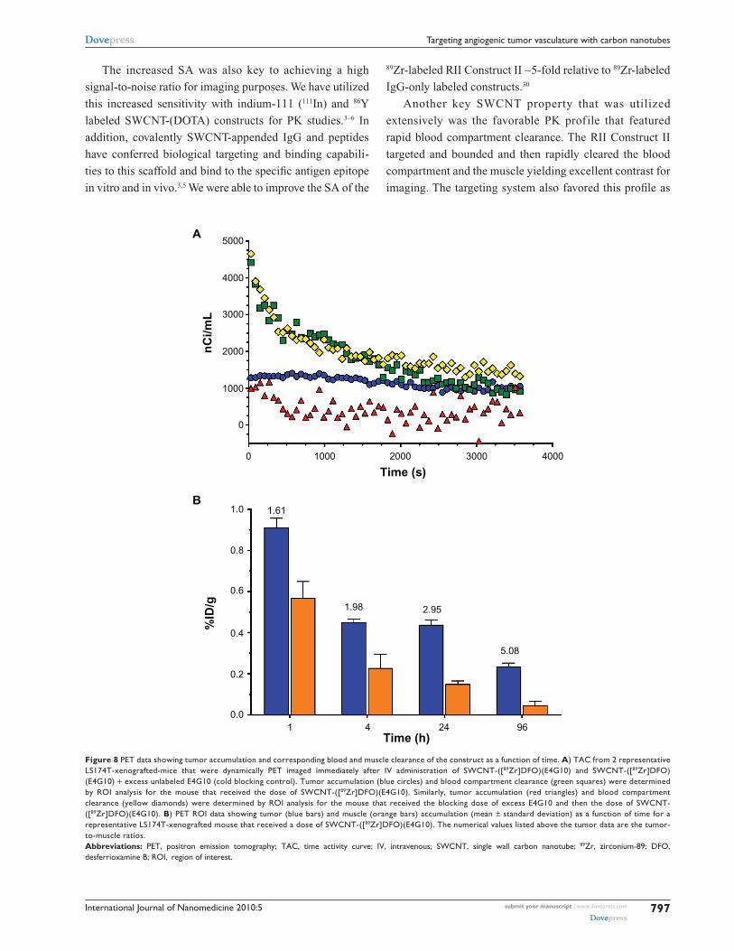
Other imaging data (Figure 8A) was taken from dynamic
PET measurements in the first hour after administration
which showed clearly the rapid blood compartment clearance
(t1/2
= 15 minutes). The tumor accumulation in a representa-
tive RII group 1 mouse showed rapid and persistent accumu-
lation of drug, whereas a representative RII group 3 control
mouse demonstrated decrease of tumor-associated activity
in the first 5 minutes that leveled off and appeared as noise.
Further PET ROI data (Figure 8B) from a representative
mouse from RII group 1 was plotted as a function of time at
1, 4, 24, and 96 hours and showed a tumor-to-muscle ratio of
1.61, 1.98, 2.95, and 5.08, respectively. The tumor-to-muscle
ratios were highest at 96 hours, but reasonable contrast
was apparent between 4 and 24 hours. This demonstrated
a significant improvement over the E4G10-alone imaging
agent that required ∼3–4 days for the blood compartment
activity to clear.
DiscussionThe concurrent processes of angiogenesis and tumor cell
proliferation are keys to tumor growth and dissemination
and are interrelated by a paracrine effect.51 Endothelial
cells will expand to produce a tortuous network of vessels
that supplies required nutrients, oxygen, cytokines, and
chemokines to tumor cells. Disrupting and damaging the
IgG
-AF
680
(pm
ol)
Time (h)0 24 48 72 96 120 144 168
P = 0.0043
20
15
10
5
0
Figure 4 Near-infrared three-dimensional fluorescent-mediated tomographic signal (normalized for moles of AF680 dye per IgG) in the LS174T tumor as a function of time after injection. e4g10-AF680 (red circles) vs a similarly prepared isotype anti-KLh-AF680 (blue triangles).Abbreviations: Igg, immunoglobulin g; anti-KLh, anti-keyhole limpet hemocyanin.

International Journal of Nanomedicine 2010:5 submit your manuscript | www.dovepress.com
Dovepress
Dovepress
795
Targeting angiogenic tumor vasculature with carbon nanotubes
vascular endothelial architecture associated with tumor tissue
have been recognized as a viable therapeutic strategy.51–57
Furthermore, imaging modalities that can specifically target
the tumor vessels would be of value in diagnosing disease
and following the progression or regression as a function
of treatment.58,59
We hypothesized that novel synthetic structures based
on hybrid molecules consisting of targeting biologics,
radionuclides, and CNTs will have emergent anticancer
properties. These molecular hybrids were designed to amplify
the intrinsic targeting, binding, imaging, and therapeutic
attributes of a drug construct and should, therefore, improve
Fra
ctio
n o
f m
ice
surv
ivin
gT
um
or
volu
me
(mm
3 )
Time from treatment (d)
Time from treatment (d)
1.0
0.5
0.00 10 20 30
0 10 20 30
1000
800
600
400
200
0
A
B
Figure 5 Kaplan–Meier survival plot of the fraction of Ls174T-xenografted mice surviving vs time following treatment with A) high sA sWcNT-([225Ac]DOTA)(e4g10) (solid green line); high sA sWcNT-([225Ac]DOTA)(anti-KLh) (solid red line); low sA sWcNT-([225Ac]DOTA)(e4g10) (solid blue line); and untreated growth control (dashed black line). B) Mean tumor volumes for each treatment group as a function of time from treatment (Note, the line colors and styles correspond to the data in panel A).Abbreviations: sA, specific activity; SWCNT, single wall carbon nanotube; 225Ac, actinium-225; DOTA, 1,4,7,10-tetraazacyclododecane-1,4,7,10-tetraacetic acid; anti-KLh, anti-keyhole limpet hemocyanin.

International Journal of Nanomedicine 2010:5submit your manuscript | www.dovepress.com
Dovepress
Dovepress
796
Ruggiero et al
potency, specificity, and efficacy relative to current drugs.
Irradiation of the vessels associated with tumor tissue
was effected by specifically targeting the high LET alpha
particle-emitting 225Ac in high SA to the VE-cad epitope;
however, imaging the tumor vessels was effected by specifi-
cally targeting the same epitope with the positron-emitting 89Zr in high SA.
We previously demonstrated that an alpha particle-
emitting, vascular-targeting antibody construct, 225Ac-E4G10,
could specifically irradiate prostate carcinoma vascular
endothelial cells27 and also their bone marrow-derived
endothelial progenitors,28 delaying tumor growth and
improving survival. We have also examined 225Ac-E4G10 in
vascular-targeting strategies to treat animal models of
glioblastoma mutiforme25,26 and the colon carcinoma
(LS174T) model of tumor vasculature.60 Others have
also used vascular-targeting, alpha particle-emitting RIT
approaches to treat animal models of disease61–64 or vascu-
lar/tumor epitope-targeting RII approaches to PET image
tumor.65–67
Two particular SWCNT properties, the high aspect
ratio and the periodic structure, have rendered this nano-
material amenable to being simultaneously appended with
multiple copies of reactive primary amines, radiometal-ion
chelates (DOTA or DFO), and IgGs. By amplifying the
number of chelates per SWCNT, we have demonstrated a
2-log increase of SA of radiolabeling of SWCNT-(DOTA)
relative to IgG constructs. Each of these SWCNT molecules
had ∼100 DOTA or DFO chelates appended per SWCNT.
In contrast, an IgG might only accommodate 5–10 DOTA
moieties per molecule before losing the ability to target
and bind efficiently.68
The radiotherapeutic SWCNT constructs, labeled with 225Ac, were also functionalized with multiple copies of E4G10
antibody and used to treat LS174T tumors vs low SA con-
trol, nontargeting high SA isotype IgG control, and growth
control. Survival of animals treated with high SA Construct
I was doubled after only one treatment relative to the control
groups and was significantly better than the growth controls.
Tumor growth was also arrested and regressed in the high
SA RIT group 1. The mice treated with the high SA, tumor
vascular-targeting Construct I showed significant tumor
regression while the low SA targeting analog did not control
tumor growth. The image of the regressed tumor lesion was
similar to the images obtained by Nilsson and Neri69 who
targeted the delivery of tissue factor to the ED-B domain
of fibronectin, a marker of angiogenesis, and mediated the
infarction of solid tumors in mice.
Co
ron
alT
ran
s. 1.5 %ID/g
0.0 %ID/g
Figure 7 PeT images of 3 representative subcutaneous Ls174T tumored mice that received an IV injection of A) sWcNT-([89Zr]DFO)(e4g10); B) low sA 89Zr-DFO-sWcNT-e4g10 (competitive inhibition or blocking control experiment); and C) nonspecific control construct SWCNT-([89Zr]DFO)(anti-KLh) recorded at 24 hours after injection. The top panel is the transverse image and the bottom panel is the coronal image. The notations T, L, Ki, and B indicate the tumor, liver, kidneys, and bladder, respectively.Abbreviations: PeT, positron emission tomography; IV, intravenous; sWcNT, single wall carbon nanotube; 89Zr, zirconium-89; DFO, desferrioxamine B; SA, specific activity; anti-KLh, anti-keyhole limpet hemocyanin; Trans, transverse.
Figure 6 Two representative Ls174T-xenografted mice from the radioimmuno- therapeutic study 10 days after treatment. A) A mouse treated with high sA sWcNT-([225Ac]DOTA)(e4g10). B) A mouse treated with low sA sWcNT-([225Ac]DOTA)(e4g10).Abbreviations: sA, specific activity; SWCNT, single wall carbon nanotube; 225Ac, actinium-225; DOTA, 1,4,7,10-tetraazacyclododecane-1,4,7,10-tetraacetic acid.
a b
International Journal of Nanomedicine 2010:5 submit your manuscript | www.dovepress.com
Dovepress
Dovepress
797
Targeting angiogenic tumor vasculature with carbon nanotubes
The increased SA was also key to achieving a high
signal-to-noise ratio for imaging purposes. We have utilized
this increased sensitivity with indium-111 (111In) and 86Y
labeled SWCNT-(DOTA) constructs for PK studies.3–6 In
addition, covalently SWCNT-appended IgG and peptides
have conferred biological targeting and binding capabili-
ties to this scaffold and bind to the specific antigen epitope
in vitro and in vivo.3,5 We were able to improve the SA of the
89Zr-labeled RII Construct II ∼5-fold relative to 89Zr-labeled
IgG-only labeled constructs.30
Another key SWCNT property that was utilized
extensively was the favorable PK profile that featured
rapid blood compartment clearance. The RII Construct II
targeted and bounded and then rapidly cleared the blood
compartment and the muscle yielding excellent contrast for
imaging. The targeting system also favored this profile as
nC
i/mL
%ID
/g
Time (s)
Time (h)
A
B
5000
4000
3000
3000 4000
2000
2000
1000
1000
0
0
1 4 24 96
5.08
2.951.98
1.611.0
0.8
0.6
0.4
0.2
0.0
Figure 8 PeT data showing tumor accumulation and corresponding blood and muscle clearance of the construct as a function of time. A) TAc from 2 representative Ls174T-xenografted-mice that were dynamically PeT imaged immediately after IV administration of sWcNT-([89Zr]DFO)(e4g10) and sWcNT-([89Zr]DFO)(e4g10) + excess unlabeled e4g10 (cold blocking control). Tumor accumulation (blue circles) and blood compartment clearance (green squares) were determined by ROI analysis for the mouse that received the dose of sWcNT-([89Zr]DFO)(e4g10). similarly, tumor accumulation (red triangles) and blood compartment clearance (yellow diamonds) were determined by ROI analysis for the mouse that received the blocking dose of excess e4g10 and then the dose of sWcNT-([89Zr]DFO)(e4g10). B) PeT ROI data showing tumor (blue bars) and muscle (orange bars) accumulation (mean ± standard deviation) as a function of time for a representative Ls174T-xenografted mouse that received a dose of sWcNT-([89Zr]DFO)(e4g10). The numerical values listed above the tumor data are the tumor-to-muscle ratios.Abbreviations: PeT, positron emission tomography; TAc, time activity curve; IV, intravenous; sWcNT, single wall carbon nanotube; 89Zr, zirconium-89; DFO, desferrioxamine B; ROI, region of interest.

International Journal of Nanomedicine 2010:5submit your manuscript | www.dovepress.com
Dovepress
Dovepress
798
Ruggiero et al
the murine VE-cad epitope is expressed in the vessel lumen
and, therefore, is readily accessible to the CNT construct.
The binding occurred rapidly, precluding any need to diffuse
into the solid tumor to target and bind. All of these studies
demonstrated that the administered constructs were safe
and well tolerated.
The SWCNT-E4G10 construct was tumor vessel specific
and targeted the murine VE-cad. E4G10 did not cross-react
with the LS174T tumor cells in flow cytometric or Western
blot analyses (Figure 9). Furthermore, the normal, resting
vasculature no longer exposes this epitope to the E4G10
IgG for binding,9 thus sparing normal vessels. An illustra-
tion of this VE-cad targeting concept using IV delivered
soluble, targeting, radiolabeled (for imaging or therapy)
constructs in patients with tumor is presented in Figure 10.
The construct rapidly accesses the tumor vasculature and
then can specifically bind to the monomeric VE-cad that is
expressed in the neovasculature but cannot bind to resting
vasculature with tight cell–cell contacts at the adherens
junctions.
This E4G10/VE-cad targeting system is unlike the
arginine-glycine-aspartic acid (RGD)-based agents that
target the αvβ
3 integrin, which is often expressed by both
the tumor and the vascular network. The RGD/αvβ
3 integrin
system lacks the vascular specificity that we designed into
our constructs. Targeting studies of vascular endothelial
growth factor (VEGF)-A that is expressed in the LS174T
human xenograft model with bevacizumab were complicated
not only by the relatively small numbers of copies of epitope
per tumor cell (12E3) but also by the imaging artifact that
was created by the lack of expression of human VEGF-A in
a mouse model.66
The NIR FMT imaging data in vivo yielded an
estimate of the number of VE-cad expressed per vascular
endothelial cell. Using the measured values of the moles
of E4G10 (3.2 pmol) per tumor (1,208 mm3) and published
vessel area values per tumor volume49 and VE cell area
(1E-3 mm2),50 there were 1.4E7 VE cells in the tumor with
1.3E5 VE-cad epitopes per VE cell. The VE-cad epitope
was not expressed by the LS174T tumor and is a murine
protein in the vessels of a mouse model. Baumgartner and
Drenckhahn70 reported 6E6 VE-cad dimers (12E6 VE-
cad monomers) for immortalized mouse microvascular
endothelial cells (MyEND) as determined in vitro using
affinity chromatography and trypsinization. Our value
was 90-fold lower than their value; however, it might be
safely assumed that most of the vascular endothelial cells
in our tumor were not newly formed angiogenic cells, and
thus, the epitope was hidden. Since our value was based
on total vascular endothelial cells, then if only 10% of the
vascular endothelial cells in the tumor were newly formed
or had irregularly or poorly connected adherens junctions,
A
B
Co
un
ts
Co
un
ts
Co
un
ts
Isotype + GAR-PE
CHO
PE-A PE-A PE-A
CHO VE-cad
CHO VE-cad
LS174T
LS174TCHO
120100
VE-cadherinIB:E4G10
GAPDH
020
4060
8010
0
020
4060
8010
0
010
2030
4050
6070
80
100 101 102 103 104 100 101 102 103 104 100 101 102 103 104
EAG10 + GAR-PE and
Figure 9 characterization of the e4g10 antibody. A) Western blot analysis of e4g10 binding to cell lysates from the (chO), Ve-cad-transfected chO, and Ls174T cells. gAPDh was included as a loading control. B) Flow cytometric analysis showed the binding characteristics of e4g10 with the chO, Ve-cad-transfected chO, and Ls174T cells. The Igg isotype control was the anti-KLh antibody. The secondary Igg was a goat anti-rat phycoerythrin Igg.Abbreviations: chO, chinese hamster ovary; Ve-cad, vascular endothelial-cadherin; gAPDh, glyceraldehyde 3-phosphate dehydrogenase; Igg, immunoglobulin g; anti-KLh, anti-keyhole limpet hemocyanin.

International Journal of Nanomedicine 2010:5 submit your manuscript | www.dovepress.com
Dovepress
Dovepress
799
Targeting angiogenic tumor vasculature with carbon nanotubes
then our estimate of the number of epitopes per cell would
increase 10-fold (1.3E6 VE-cad epitopes per VE cell). If
the number of newly formed VE cells was only 1% of the
total VE cell population in the tumor, then the value would
increase 100-fold (1.3E7 VE-cad epitopes per VE cell).
The latter assumption yielded a better correlation with the
Baumgartner data.
ConclusionSWCNT constructs were designed, constructed, and used
to deliver therapeutic and imaging radionuclide cargoes
specifically to the vessels of a solid tumor using a target
on VE-cad found only on new vascular endothelium. The
goal was to target the neovessels and irregular vessels
in a tumor with these novel nanoconstructs and image
Figure 10 A graphical illustration of the tumor vascular-targeting concept using the carbon nanotube constructs. The soluble, covalently functionalized, and radiolabeled constructs are delivered intravenously to the patient with tumor. The construct rapidly accesses the tumor vasculature and then can specifically bind to the monomeric Ve-cad that is expressed in the neovasculature but cannot bind to resting vasculature with tight cell–cell contacts at the adherens junctions. (Note, Figures are not drawn to scale.)

International Journal of Nanomedicine 2010:5submit your manuscript | www.dovepress.com
Dovepress
Dovepress
800
Ruggiero et al
accumulation and evaluate the therapeutic anti-angiogenic
effects. The construct design incorporated 100-fold ampli-
fied cargo delivery (relative to the gold standard for tar-
geted therapy – IgG) and was built to be multifunctional and
thus had therapeutic or imaging cargo, as well as targeting
capability conferred by the appended IgG. This proof of
concept design resulted in a construct with therapeutic
efficacy, good image contrast, and specificity for the target.
This amplified SA may prove important in delivering potent
enough therapy and sensitive enough diagnostic signals
simultaneously to the tumor. Our data also provided support
of the use of nanomaterials in vascular-targeting strategies.
These SWCNT construct doses were well tolerated and safe
in these animal models. The number of VE-cad epitopes per
tumor was measured and extrapolated to estimate a num-
ber of bound VE-cad epitopes per cell. These latter results
along with the PK profile will be of use in designing more
optimized therapeutic and imaging studies with these con-
structs. Moving forward, it is anticipated that a single con-
struct could be designed to incorporate both the imaging and
therapeutic cargoes onto the same platform. Furthermore,
because the construct targets an epitope expressed by the
tumor vascular network, a single agent could be employed
to image or treat a variety of different tumors.
Acknowledgments/disclosureFunded in part by the National Institutes of Health grants
R21 CA128406, R01 CA55349, R25T CA096945, R24
CA83084, P30 CA08748, P01 CA33049; the Memorial
Sloan-Kettering Brain Tumor Center; the Memorial Sloan-
Kettering Experimental Therapeutics Center; the Geoffrey
Beene Cancer Research Center of Memorial Sloan-Ketter-
ing Cancer Center; and the Office of Science (BER), US
Department of Energy (Award DE-SC0002456). We would
also like to thank Medactinium, Inc for the 225Ac; ImClone
Systems (a wholly-owned subsidiary of Eli Lilly and Com-
pany) for the E4G10 antibody; and Amy Carol McDevitt
for the graphic illustration and figure. Conflict of interest
statement: David A Scheinberg is a consultant for Enscyce;
Michael R McDevitt was a consultant for Medactinium; and
Chad May was employed by ImClone Systems at the time
of this study.
References1. Scheinberg DA, Villa CH, Escorcia FE, McDevitt MR. Conscripts of the
infinite armada: systemic cancer therapy using nanomaterials. Nat Rev Clin Oncol. 2010;7(5):266–276.
2. Kostarelos K, Bianco A, Prato M. Promises, facts and challenges for carbon nanotubes in imaging and therapeutics. Nat Nanotechnol. 2009;4(10):627–633.
3. McDevitt MR, Chattopadhyay D, Kappel BJ, et al. Tumor targeting with antibody-functionalized, radiolabeled carbon nanotubes. J Nucl Med. 2007:48(7);1180–1189.
4. McDevitt MR, Chattopadhyay D, Jaggi JS, et al. PET Imaging of soluble yttrium-86-labeled carbon nanotubes in mice. PLoS One. 2007; 2(9):e907.
5. Villa CH, McDevitt MR, Escorcia FE, et al. Synthesis and biodistribution of oligonucleotide-functionalized, tumor-targetable carbon nanotubes. Nano Lett. 2008;8(12):4221–4228.
6. Ruggiero A, Villa CH, Bander E, et al. Paradoxical glomerular filtra-tion of carbon nanotubes. Proc Natl Acad Sci U S A. 2010; 107(27): 12369–12374.
7. Fox ME, Szoka FC, Frechet JMJ. Soluble polymer carriers for the treat-ment of cancer: the importance of molecular architecture. Acc Chem Res. 2009;42(8):1141–1151.
8. Corada M, Liao F, Lindgren M, et al. Monoclonal antibodies directed to different regions of vascular endothelial cadherin extracellular domain affect adhesion and clustering of the protein and modulate endothelial permeability. Blood. 2001;97(6):1679–1684.
9. Liao F, Doody JF, Overholser J, et al. Selective targeting of angiogenic tumor vasculature by vascular endothelial-cadherin antibody inhibits tumor growth without affecting vascular permeability. Cancer Res. 2002;62(9):2567–2575.
10. Corada M, Zanetta L, Orsenigo F, et al. A monoclonal antibody to vascular endothelial-cadherin inhibits tumor angiogenesis without side effects on endothelial permeability. Blood. 2002;100(3):905–911.
11. May C, Doody JF, Abdullah R, et al. Identification of a transiently exposed VE-cadherin epitope that allows for specific targeting of an antibody to the tumor neovasculature. Blood. 2005;105(11): 4337–4344.
12. Lamszus K, Brockmann MA, Eckerich C, et al. Inhibition of glioblas-toma angiogenesis and invasion by combined treatments directed against vascular endothelial growth factor receptor-2, epidermal growth factor receptor, and vascular endothelial-cadherin. Clin Cancer Res. 2005; 11(13):4934–4940.
13. McDevitt MR, Ma D, Lai LT, et al. Tumor therapy with targeted atomic nano-generators. Science. 2001;294(5546):1537–1540.
14. Borchardt PE, Yuan RR, Miederer M, McDevitt MR, Scheinberg DA. Targeted actinium-225 in vivo generators for therapy of ovarian cancer. Cancer Res. 2003;63(16):5084–5090.
15. Miederer M, McDevitt MR, Sgouros G, Kramer K, Cheung NK, Scheinberg DA. Pharmacokinetics, dosimetry and toxicity of the targetable atomic generator, 225Ac-HuM195, in nonhuman primates. J Nucl Med. 2004;45(1):129–137.
16. Ballangrud ÅM, Yang WH, Palm S, et al. Alpha-particle emitting atomic generator (actinium-225)-labeled trastuzumab (Herceptin) targeting of breast cancer spheroids: efficacy versus HER2/neu expression. Clin Cancer Res. 2004;10(13):4489–4497.
17. Miederer M, McDevitt MR, Borchardt P, et al. Treatment of neuroblastoma meningeal carcinomatosis with intrathecal application of α-emitting atomic nanogenerators targeting disialo-ganglioside GD2. Clin Cancer Res. 2004;10(20):6985–6992.
18. Yuan RR, Wong P, McDevitt MR, et al. Targeted deletion of T-cell clones using alpha-emitting suicide MHC tetramers. Blood. 2004;10(8):2397–2402.
19. Jurcic JG, McDevitt MR, Divgi CR, et al. Alpha-particle immunotherapy for acute myeloid leukemia (AML) with bismuth-213 and actinium-225. Cancer Biother Radiopharm. 2006;221(4):396.
20. Rosenblat TL, McDevitt MR, Pandit-Taskar N, et al. Phase I trial of the targeted alpha-particle nano-generator actinium-225 (225Ac)-HuM195 (Anti-CD33) in acute myeloid leukemia (AML). Blood. 2007; 110(11):277A.

International Journal of Nanomedicine 2010:5 submit your manuscript | www.dovepress.com
Dovepress
Dovepress
801
Targeting angiogenic tumor vasculature with carbon nanotubes
21. Miederer M, Scheinberg DA, McDevitt MR. Realizing the poten-tial of the actinium-225 radionuclide generator in targeted alpha-particle therapy applications. Adv Drug Deliv Rev. 2008;60(12): 1371–1382.
22. McDevitt MR, Sgouros G, Finn RD, et al. Radioimmunotherapy with alpha-emitting radionuclides. Eur J Nucl Med. 1998;25(9): 1341–1351.
23. Nikula TN, McDevitt MR, Finn RD, et al. Alpha-emitting bismuth cyclohexylbenzyl DTPA constructs of recombinant humanized anti-CD33 antibodies: pharmacokinetics, bioactivity, toxicity and chemistry. J Nucl Med. 1999;40(1):166–176.
24. Calabrese C, Poppleton H, Kocak M, et al. A perivascular niche for brain tumor stem cells. Cancer Cell. 2007;11(1):69–82.
25. Hambardzumyan D, Squatrito M, Carbajal E, Holland EC. Glioma formation, cancer stem cells, and akt signaling. Stem Cell Rev. 2008; 4(3):203–210.
26. Hambardzumyan D, Becher OJ, Rosenblum MK, Pandolfi PP, Manova-Todorova K, Holland EC. PI3 K pathway regulates survival of cancer stem cells residing in the perivascular niche following radiation in medulloblastoma in vivo. Genes Dev. 2008;22(4):436–448.
27. Jaggi JS, Henke E, Seshan SV, et al. Selective alpha-particle mediated depletion of tumor vasculature with vascular normalization. PLoS One. 2007;2(3):e267.
28. Nolan DJ, Ciarrocchi A, Mellick AS, et al. Bone marrow-derived endothelial progenitor cells are a major determinant of nascent tumor neovascularization. Genes Dev. 2007;21(12):1546–1558.
29. Verel I, Visser GW, Boellaard R, Stigter-van Walsum M, Snow GB, van Dongen GA. 89Zr immuno-PET: comprehensive procedures for the production of 89Zr-labeled monoclonal antibodies. J Nucl Med. 2003; 44(8):1271–1281.
30. Holland JP, Sheh Y, Lewis JS. Standardized methods for the production of high specific-activity zirconium-89. Nucl Med Biol. 2009; 36(7):729–739.
31. Holland JP, Williamson MJ, Lewis JS. Unconventional nuclides for radiopharmaceuticals. Mol Imaging. 2010;9(1):1–20.
32. Holland JP, Caldas-Lopes E, Divilov V, et al. Measuring the pharmacodynamic effects of a novel Hsp90 inhibitor on HER2/neu expression in mice using Zr-DFO-trastuzumab. PLoS One. 2010;5(1): e8859.
33. Ruggiero A, Holland JP, Lewis JS, Grimm J. Cerenkov lumi-nescence imaging of medical isotopes. J Nucl Med. 2010;51(7): 1123–1130.
34. Holland JP, Divilov V, Bander NH, Smith-Jones PM, Larson SM, Lewis JS. 89Zr-DFO-J591 for immunoPET of prostate-specific membrane antigen expression in vivo. J Nucl Med. 2010;51(8):1293–1300.
35. von Schulthess GK, Steinert HC, Hany TF. Integrated PET/CT: current applications and future directions. Radiology. 2006;238(2):405–422.
36. Georgakilas V, Tagmatarchis N, Pantarotto D, Bianco A, Briand JP, Prato M. Amino acid functionalization of water soluble carbon nanotubes. Chem Commun (Camb). 2002;(24):3050–3051.
37. Tasis D, Tagmatarchis N, Bianco A, Prato M. Chemistry of carbon nanotubes. Chem Rev. 2006;106(3):1105–1136.
38. Singh P, Campidelli S, Giordani S, Bonifazi D, Bianco A, Prato M. Organic functionalisation and characterisation of single-walled carbon nanotubes. Chem Soc Rev. 2009;38(8):2214–2230.
39. Singh R, Pantarotto D, Lacerda L, et al. Tissue biodistribution and blood clearance rates of intravenously administered carbon nanotube radiotracers. Proc Natl Acad Sci U S A. 2006;103(9):3357–3362.
40. Allen BL, Kichambare PD, Gou P, et al. Biodegradation of single-walled carbon nanotubes through enzymatic catalysis. Nano Lett. 2008;8(11): 3899–3903.
41. Allen BL, Kotchey GP, Chen Y, et al. Mechanistic investiga-tions of horseradish peroxidase-catalyzed degradation of sin-gle-walled carbon nanotubes. J Am Chem Soc. 2009;131(47): 17194–17205.
42. Konduru NV, Tyurina YY, Feng W, et al. Phosphatidylserine targets single-walled carbon nanotubes to professional phagocytes in vitro and in vivo. PLoS One. 2009;4(2):e4398.
43. Kagan VE, Konduru NV, Feng W, et al. Carbon nanotubes degraded by neutrophil myeloperoxidase induce less pulmonary inflammation. Nat Nanotechnol. 2010;5(5):354–359.
44. Dumortier H, Lacotte S, Pastorin G, et al. Functionalized carbon nanotubes are non-cytotoxic and preserve the functionality of primary immune cells. Nano Lett. 2006;6(7):1522–1528.
45. Sayes CM, Liang F, Hudson JL, et al. Functionalization density dependence of single-walled carbon nanotubes cytotoxicity in vitro. Toxicol Lett. 2006;161(2):135–142.
46. Schipper ML, Nakayama-Ratchford N, Davis CR, et al. A pilot toxicology study of single-walled carbon nanotubes in a small sample of mice. Nat Nanotechnol. 2008;3(4):216–221.
47. Kostarelos K. The long and short of carbon nanotube toxicity. Nat Biotechnol. 2008;26(7):774–776.
48. McDevitt MR, Ma D, Simon J, Frank RK, Scheinberg DA. Design and synthesis of actinium-225 radioimmunopharmaceuticals. Appl Radiat Isot. 2002;57(6):841–847.
49. Hilmas DE, Gillette EL. Morphometric analyses of the microvasculature of tumors during growth and after x-irradiation. Cancer. 1974; 33(1):103–110.
50. Haas TL, Duling BR. Morphology favors an endothelial cell pathway for longitudinal conduction within arterioles. Microvasc Res. 1997;53(2): 113–120.
51. Folkman J. Tumor angiogenesis and tissue factor. Nat Med. 1996; 2(2):167–168.
52. Folkman J. Anti-angiogenesis: new concept for therapy of solid tumors. Ann Surg. 1972;175(3):409–416.
53. Denekamp J. Endothelial cell proliferation as a novel approach to targeting tumor therapy. Br J Cancer. 1982;45(1):136–139.
54. Denekamp J. Review article: angiogenesis, neovascular proliferation and vascular pathophysiology as targets for cancer therapy. Br J Radiol. 1993;66(783):181–196.
55. Burrrows FJ, Thorpe PE. Vascular targeting – a new approach to the therapy of solid tumors. Pharmacol Ther. 1994;64(1):155–174.
56. Folkman J. Addressing tumor blood vessels. Nat Biotechnol. 1997; 15(6):510.
57. Kerbel RS. Antiangiogenic therapy: a universal chemosensitization strategy for cancer? Science. 2006;312(5777):1171–1175.
58. Cai W, Chen X. Multimodality molecular imaging of tumor angiogenesis. J Nucl Med. 2008;49 Suppl 2:113S–128S.
59. Dobrucki LW, de Muinck ED, Lindner JR, Sinusas AJ. Approaches to multimodality imaging of angiogenesis. J Nucl Med. 2010;51 Suppl 1: 66S–79S.
60. Leunig M, Yuan F, Menger MD, et al. Angiogenesis, microvascular architecture, microhemodynamics, and interstitial fluid pressure during early growth of human adenocarcinoma LS174T in SCID mice. Cancer Res. 1992;52(23):6553–6560.
61. Kennel SJ, Mirzadeh S. Vascular targeted radioimmunotherapy with 213Bi-an alpha-particle emitter. Nucl Med Biol. 1998;25(3): 241–246.
62. Kennel SJ, Lankford TK, Foote LJ, Davis IA, Boll RA, Mirzadeh S. Combination vascular targeted and tumor targeted radioimmuno-therapy. Cancer Biother Radiopharm. 1999;14(5):371–379.
63. Kennel SJ, Chappell LL, Dadachova K, et al. Evaluation of 225Ac for vascular targeted radioimmunotherapy of lung tumors. Cancer Biother Radiopharm. 2000;15(3):235–244.
64. Kennel SJ, Mirzadeh S, Eckelman WC, et al. Vascular-targeted radioimmunotherapy with the alpha-particle emitter 211At. Radiat Res. 2002;157(6):633–641.
65. Nagengast WB, de Vries EG, Hospers GA, et al. In vivo VEGF imaging with radiolabeled bevacizumab in a human ovarian tumor xenograft. J Nucl Med. 2007;48(8):1313–1319.

International Journal of Nanomedicine
Publish your work in this journal
Submit your manuscript here: http://www.dovepress.com/international-journal-of-nanomedicine-journal
The International Journal of Nanomedicine is an international, peer-reviewed journal focusing on the application of nanotechnology in diagnostics, therapeutics, and drug delivery systems throughout the biomedical field. This journal is indexed on PubMed Central, MedLine, CAS, SciSearch®, Current Contents®/Clinical Medicine,
Journal Citation Reports/Science Edition, EMBase, Scopus and the Elsevier Bibliographic databases. The manuscript management system is completely online and includes a very quick and fair peer-review system, which is all easy to use. Visit http://www.dovepress.com/ testimonials.php to read real quotes from published authors.
International Journal of Nanomedicine 2010:5submit your manuscript | www.dovepress.com
Dovepress
Dovepress
Dovepress
802
Ruggiero et al
66. Stollman TH, Scheer MG, Leenders WP, et al. Specific imaging of VEGF-A expression with radiolabeled anti-VEGF monoclonal antibody. Int J Cancer. 2008;122(10):2310–2314.
67. Nayak TK, Garmestani K, Baidoo KE, Milenic DE, Brechbiel MW. PET imaging of tumor angiogenesis in mice with VEGF-A targeted (86)Y-CHX-A’’-DTPA-bevacizumab. Int J Cancer. In press 2010.
68. Nikula TK, Bocchia M, Curcio MJ, et al. Impact of the high tyrosine frac-tion in complementarity determining regions: measured and predicted effects of radioiodination on IgG immunoreactivity. Mol Immunol. 1995;32(12):865–872.
69. Nilsson F, Kosmehl H, Zardi L, Neri D. Targeted delivery of tissue factor to the ED-B domain of fibronectin, a marker of angiogenesis, mediates the infarction of solid tumors in mice. Cancer Res. 2001;61(2): 711–716.
70. Baumgartner W, Drenckhahn D. Plasmalemmal concentration and affin-ity of mouse vascular endothelial cadherin. Eur Biophys J. 2002;31(7): 532–538.
Copyright © 2022 FDOKUMEN


![[Treating frostbite injuries]](https://static.fdokumen.com/doc/165x107/633ff39332b09e4bae09a1b5/treating-frostbite-injuries.jpg)