Hot spots of retroviral integration in human CD34+ hematopoietic cells
Upload
independentCategory
view
0download
0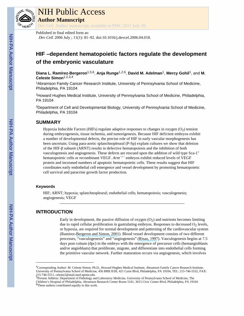
HIF –dependent hematopoietic factors regulate the developmentof the embryonic vasculature
Diana L. Ramírez-Bergeron1,5,6, Anja Runge1,2,6, David M. Adelman1, Mercy Gohil1, and M.Celeste Simon1,2,3,4
1Abramson Family Cancer Research Institute, University of Pennsylvania School of Medicine,Philadelphia, PA 191042Howard Hughes Medical Institute, University of Pennsylvania School of Medicine, Philadelphia,PA 191043Department of Cell and Developmental Biology, University of Pennsylvania School of Medicine,Philadelphia, PA 19104
SUMMARYHypoxia Inducible Factors (HIFs) regulate adaptive responses to changes in oxygen (O2) tensionduring embryogenesis, tissue ischemia, and tumorigenesis. Because HIF deficient embryos exhibita number of developmental defects, the precise role of HIF in early vascular morphogenesis hasbeen uncertain. Using para-aortic splanchnopleural (P-Sp) explant cultures we show that deletionof the HIF-β subunit (ARNT) results in defective hematopoiesis and the inhibition of bothvasculogenesis and angiogenesis. These defects are rescued upon the addition of wild type Sca-1+
hematopoietic cells or recombinant VEGF. Arnt−/− embryos exhibit reduced levels of VEGFprotein and increased numbers of apoptotic hematopoietic cells. These results suggest that HIFcoordinates early endothelial cell emergence and vessel development by promoting hematopoieticcell survival and paracrine growth factor production.
KeywordsHIF; ARNT; hypoxia; splanchnopleural; endothelial cells; hematopoiesis; vasculogenesis;angiogenesis; VEGF
INTRODUCTIONEarly in development, the passive diffusion of oxygen (O2) and nutrients becomes limitingdue to rapid cellular proliferation in gastrulating embryos. Responses to decreased O2 levels,or hypoxia, are required for normal development and patterning of the cardiovascular system(Ramirez-Bergeron and Simon, 2001). Blood vessel development consists of two differentprocesses, “vasculogenesis” and “angiogenesis” (Risau, 1997). Vasculogenesis begins at 7.5days post coitum (dpc) in the embryo with the emergence of precursor cells (hemangioblastsand/or angioblasts) that proliferate, migrate, and differentiate into endothelial cells formingthe primitive vascular network. Further maturation occurs via angiogenesis, which involves
4Corresponding Author: M. Celeste Simon, Ph.D., Howard Hughes Medical Institute, Abramson Family Cancer Research Institute,University of Pennsylvania School of Medicine, 456 BRB II/III, 421 Curie Blvd, Philadelphia, PA 19104, TEL: 215-746-5532, FAX:215-746-5511, [email protected] Address: Department of Pathology and Laboratory Medicine, University of Pennsylvania School of Medicine, TheChildren’s Hospital of Philadelphia, Abramson Research Center Room 5161, 3615 Civic Center Blvd, Philadelphia, PA 191046These authors contributed equally to this work.
NIH Public AccessAuthor ManuscriptDev Cell. Author manuscript; available in PMC 2011 July 28.
Published in final edited form as:Dev Cell. 2006 July ; 11(1): 81–92. doi:10.1016/j.devcel.2006.04.018.
NIH
-PA Author Manuscript
NIH
-PA Author Manuscript
NIH
-PA Author Manuscript

remodeling by pruning, branching, and sprouting of preexisting vessels. Ultimately,additional support cells (pericytes/smooth muscle cells) are recruited and extracellularmatrixes are deposited (Carmeliet, 2005; Coultas et al., 2005; Jain, 2003). Although themolecular mechanism(s) underlying hypoxic regulation of embryonic vascular developmentare not entirely clear, we and others have demonstrated that Hypoxia Inducible Factors(HIFs) are critical transcriptional regulators mediating these events (Ramirez-Bergeron andSimon, 2001: Maltepe and Simon, 1998).
HIF is a member of the basic helix-loop-helix (bHLH)-PAS family of transcription factorsthat regulate diverse biological processes such as O2 homeostasis, circadian rhythms,neurogenesis, and toxin metabolism. Under normoxic conditions (≥5% O2), the HIF-αproteins are hydroxylated and targeted for proteosomal degradation (Ivan et al., 2001;Jaakkola et al., 2001; Yu et al., 2001). Under low O2 (<5%), however, the α subunits arestabilized and dimerize with a related bHLH-PAS protein called HIF-β or ARNT (arylhydrocarbon receptor nuclear translocator) (Semenza, 1999). HIF is a master regulator of O2homeostasis and induces a network of genes important for angiogenesis, erythropoiesis, andglucose metabolism (Giaccia et al., 2004). Many genes involved in cardiovasculardifferentiation are directly or indirectly regulated by HIF, including vascular endothelialgrowth factor (VEGF), transforming growth factor β (TGF-β1), erythropoietin (EPO),transferrin, platelet derived growth factor β (PDGF-β), basic fibroblast growth factor(bFGF), and/or their receptors (Semenza, 1998; Semenza, 1999).
Mice lacking HIF activity develop extensive cardiovascular pathologies. HIF-1α isubiquitously expressed in all tissues and Hif-1α−/− embryos die by 10.5 dpc with variouscardiovascular defects including inadequate vessel formation (Iyer et al., 1998; Kotch et al.,1999; Ryan et al., 1998). In contrast, HIF-2α expression is more spatially restricted toendothelial cells, mesenchyme of the lung, and neural crest derivatives duringembryogenesis (Ema et al., 1997; Tian et al., 1997). Hif-1α−/− mice die by 9.5–13.5 dpc dueto vascular disorganization in the yolk sac and embryo (Peng et al., 2000) or later fromcardiorespiratory failure due to decreased lung surfactant or catecholamine production(Compernolle et al., 2002; Tian et al., 1998). Further demonstration of unique roles forHIF-1α and HIF-2α is their target gene specificity: glycolytic genes are regulatedexclusively by HIF-1α, while HIF-2α preferentially activates the stress responsive geneGADD45B and the stem cell factor Oct-4 (Covello et al., 2006; Hu et al., 2003; Wang etal.,2005).
ARNT expression is ubiquitous and Arnt−/− embryos also die by 10.5 dpc with similarcardiovascular anomalies to the Hif-1α−/− mice (Cowden Dahl et al., 2005; Maltepe et al.,1997). In particular, Arnt−/− yolk sac hematopoietic and angiogenic development isdisrupted with aberrant vascular remodeling (Adelman et al., 1999; Ramirez-Bergeron andSimon, 2001). Moreover, histological sections of Arnt−/− placentas demonstrate that fetalvessels are unable to invade the labyrinthine layer, and tetraploid chimeric embryoexperiments reveal an Arnt−/− cardiac phenotype (Adelman et al., 2000). It remainsuncertain, therefore, whether yolk sac, placental, and/or heart defects are the primary causeof vascular irregularities in Hif-1α −/−, Hif-2α −/− and Arnt−/− embryos.
Given the distinct expression and function(s) of the HIF-α subunits, we have focused on theArnt-null genetic model where absence of the β subunit completely eliminates all HIFactivity during cardiovascular differentiation. In this study, we demonstrate that Arnt−/−
embryos exhibit greatly reduced blood vessel numbers by 9.5 dpc. Since defects in multiplecardiovascular tissues confound interpretation of these observations, we performed ex vivopara-aortic splanchnopleural (P-Sp) explant assays that support both vascular andhematopoietic development (Takakura et al., 1998). Here, developmentally stage- matched
Ramírez-Bergeron et al. Page 2
Dev Cell. Author manuscript; available in PMC 2011 July 28.
NIH
-PA Author Manuscript
NIH
-PA Author Manuscript
NIH
-PA Author Manuscript
wild type and mutant embryos can be evaluated independently of deleterious effects causedby abnormal circulation or placentation. In this context, the Arnt−/− P-Sp explants exhibitedabnormal vasculogenesis, angiogenesis, and hematopoiesis. We determined that endothelialcell production and vascular morphogenesis are regulated by paracrine VEGF supplied byhematopoietic cells that are deficient in Arnt−/− embryos. Therefore, vascular anomaliesexhibited by Arnt−/− embryos are downstream of the intraembryonic hematopoietic defect.
RESULTSDisruption of vascular development in Arnt−/− tissues
We have reported previously that the Arnt−/− mutation results in extraembryonic vascularand hematopoietic defects by 10.5 dpc (Adelman et al., 1999; Maltepe et al., 1997). Furtheranalysis by immunohistochemical whole mount staining for the platelet-endothelial celladhesion molecule-1 (PECAM, CD31) demonstrated abnormal endothelial organization inboth extra- and intra- embryonic Arnt−/− tissues (Figure 1). At 9.5 dpc, extensive endothelialremodeling was defective in Arnt−/− yolk sacs, which displayed a disorganized network witha honeycomb-like plexus of similar sized capillaries (Figure 1G–J). In the embryo itself,Arnt−/− vessels were highly disorganized and exhibited a decrease in PECAM+ cellsthroughout (Figure 1A–F). The lack of large and small vessels was especially apparent in thecranial region and the P-Sp tissues of Arnt−/− embryos. Although the anterior cardinal veinswere present in mutant embryos, their remodeling was defective. Furthermore, thedeveloping heart and at least one of the branchial arches also exhibited decreased PECAMstaining. Similarly, the inter- and intra- somitic vessels branching from the dorsal aorta wereabsent (Figure 1D–F).
To further evaluate Arnt−/− vessel phenotypes, transverse sections from 9.5 dpc embryoswere immunostained for CD34, which is expressed on endothelial and hematopoietic stemcells. As shown in Figure 1K–P, Arnt−/− embryos were defective in CD34 expressioncompared to wild type embryos. Furthermore, the number of CD34+ cells outlining thedorsal aorta as well as the endocardium was significantly diminished (Figure IN and P). Ofnote, some CD34+ hematopoietic cells were apparent in the Arnt−/− embryos serving as apositive control for staining (Figure 1L). In aggregate, these results reflect endothelialdeficiencies in the absence of ARNT, suggesting that inadequate hypoxic responses result inimproper vessel formation, remodeling, and maturation throughout the embryo.
Examination of over 670 embryos generated from Arnt heterozygous intercrosses collectedat 9.5 dpc indicated a normal Mendel Ian ratio (1:2:1) of wild type, heterozygous, andhomozygous mutant embryos, respectively (Supplemental Table 1). However, furtherstaging of embryos by enumerating paired somite numbers revealed a disproportionatenumber of Arnt−/− embryos (50% compared to the expected 25%) with fewer than 15somites. In contrast only 11% of embryos with over 26 somites were Arnt−/− mice. Grossobservations revealed additional defects in Arnt−/− embryos including pericardial effusion,reduced numbers and sizes of the pharyngeal pouches, lack of chorio-allantoic fusion,hemorrhagic lesions, and failure of neural tube closure in some mutants. We concluded thatmany (but not all) Arnt−/− embryos are developmentally delayed by 9.5 dpc.
ARNT is required for appropriate vasculogenesis and angiogenesis in P-Sp explantsDuring development, the embryo resides in a hypoxic environment (Lee et al., 2001; Ryan etal., 1998) and HIF activates target genes that support vascular development. As describedabove, many Arnt−/− embryos are growth impaired by 9.5 dpc. Because the vascular defectscould be the result of general developmental delay, we employed an ex vivo approachutilizing the para-aortic splanchnopleural (P-Sp) explant assay which examines blood vessel
Ramírez-Bergeron et al. Page 3
Dev Cell. Author manuscript; available in PMC 2011 July 28.
NIH
-PA Author Manuscript
NIH
-PA Author Manuscript
NIH
-PA Author Manuscript

and hematopoietic development potential of a 9.5 dpc embryo (Takakura et al., 1998;Takakura et al., 2000). The P-Sp mesoderm consisting of the dorsal aorta, genital ridge/gonad, and pro/mesonephros (aorta-gonad-mesonephros, AGM) has been suggested to be asource of intraembryonic hematopoietic and endothelial progenitors (de Bruijn et al., 2002;Dieterlen-Lievre et al., 2002; North et al., 2002). P-Sp analyses can determine if the failedArnt−/− vessel maturation is independent of an embryonic developmental delay or otherconfounding factors such as yolk sac, cardiac, and placental defects which contribute tocirculatory abnormalities. Therefore, a direct role for HIF in intraembryonic vascular andhematopoietic development might be revealed by P-Sp explant assays.
We generated P-Sp explants from 9.5 dpc Arnt−/− and wild type embryos of similar sizesthat were matched for somite numbers ranging from 20–30 each. Importantly, we only usedwild type and mutant embryos that were morphologically indistinguishable in these assays(Figure 2A and B). Explants were cultured on OP9 stromal cells for 14 days, and PECAMstained to evaluate endothelial cell (EC) differentiation (Figure 2C–F). In these cultures,PECAM+ ECs initially form sheet-like structures (“vascular beds”) emulatingvasculogenesis, followed by migration, sprouting, and network formation of PECAM+ cellsin a ring around the explant (“vascular networks”) representing angiogenesis (Takakura,1998). Explants from Arnt+/+ embryos with 20–30 somite pairs generated visible vascularbeds by day 7 that were further remodeled into networks by day 12 of culture (Figure 2Cand D). In direct contrast, few if any vascular beds or networks were detected in Arnt−/− P-Sp explants (Figure 2E and F). Although rare, when vascular beds were observed in Arnt−/−
samples, branching vascular networks were reduced to small clusters of undersized vessellengths or sprouts (Figure 2E and F, and Figure 3B). By these analyses, it appeared that thenumbers of nascent EC progenitors (angioblasts) are significantly reduced and vesselmaturation is impaired in Arnt−/− embryos.
ARNT is required for hematopoiesis in P-Sp explantsDuring embryogenesis, hematopoietic cells (herein referred to as “HCs”) arise within twodistinct anatomic locations: primitive hematopoiesis occurs in the yolk sac while definitivehematopoiesis takes place in the AGM of the embryo proper (Medvinsky and Dzierzak,1996; Sanchez et al., 1996). We have previously characterized an ARNT- dependent yolksac hematopoietic defect (Adelman et al., 1999); therefore, we determined if definitiveembryonic hematopoiesis was also compromised in the Arnt−/− animals. We detected someHCs (primarily erythrocytes) migrating through the heart and vessels of 9.5 dpc Arnt−/−
embryos. To analyze definitive hematopoiesis further, we employed P-Sp cultures. Weobserved relatively few nonadherent HCs in the Arnt−/− specimens (Figure 2I and J). Indirect contrast, Arnt+/+ explants produced numerous round nonadherent HCs (Figure 2G andH). Nonadherent cells were harvested from wild type and mutant cultures and assayed forexpression of the pan-hematopoietic marker CD45 by flow cytometry. CD45+ cells wereconsistently detected in Arnt+/+ P-Sp cultures by day 6, whereas Arnt−/− P-Sp culturesexhibited less than 1% of wild type CD45+ cell numbers over the 14 day culture period (datanot shown), indicating a substantial defect in hematopoietic development.
Because of the dramatically reduced number of nonadherent cells detected in Arnt−/− P-Spcultures, we further analyzed hematopoietic potential by culturing the limited number ofcells in hematopoietic progenitor methylcellulose assays. As expected, cells from Arnt+/+ P-Sps produced a variety of colony-forming cells (CFCs) (primarily granulocyte/macrophage(GM) and erythroid (E)). In contrast, replated Arnt−/− nonadherent cells exclusivelyproduced mast cells. These data are consistent with the Arnt−/− extraembryonic defect whereyolk sac cells generate fewer hematopoietic colony-forming units (CFUs) compared to wildtype controls (Adelman et al., 1999). We concluded that both primitive and definitivehematopoiesis is significantly impaired in embryos lacking HIF activity.
Ramírez-Bergeron et al. Page 4
Dev Cell. Author manuscript; available in PMC 2011 July 28.
NIH
-PA Author Manuscript
NIH
-PA Author Manuscript
NIH
-PA Author Manuscript

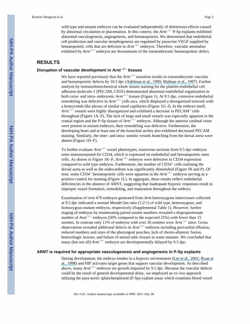
Arnt−/− P-Sp explants can be “rescued” with wild type Sca-1+ bone marrow- derived cellsUsing P-Sp explant cultures, Takakura et al. (2000) have previously reported thathematopoietic stem cells (HSCs) contribute to angiogenic vessel development by providingAngiopoietin-1 (Ang-1) during embryogenesis. We therefore asked to what extent doesangioblast production and differentiation require the presence of HCs? To determine if thevasculo-angiogenic defect exhibited by Arnt−/− P-Sp explants could be ameliorated by theaddition of wild type Sca-1+ HCs, Sca-1+ cells isolated from the bone marrow of wild typeGFP+ mice were added to the cultures. Remarkably, the addition of enriched Sca-1+GFP+
HCs completely rescued the vessel defect in Arnt−/− explants (Figure 3). The angiogenicpotential of these cultures was analyzed by measuring the lengths of sprouting vessels usingan ImageJ software program (see Experimental Procedures). While we were unable tomeasure vessel length of many unrescued Arnt−/− explants due to lack of PECAM staining,the few explants containing some vascular networks exhibited a mean vessel length of 0.43mm ± 0.31 (Figure 3B, asterisk; and 3G, number sign). In contrast, the mean vessel length ofArnt+/+ networks was 1.39 mm ± 0.11. In the rescue experiments, Arnt−/− cultures exhibitedvessel lengths (1.36 mm± 0.31) which were not significantly different from those detected inthe Arnt+/+ cultures (P< 0.93, Figure 3G). These results indicate that the Sca-1+ HCs canrescue the HIF vessel defect.
It has been reported previously that bone marrow stem or progenitor cells can contribute toendothelial cell populations during adult neovascularization (Kocher et al., 2001; Lyden etal., 2001; Rafii and Lyden, 2003). In order to determine if the wild type donor Sca-1+GFP+
cells were the source of endothelial cells observed in rescue experiments, P-Sp cultures weretreated with DiI-Ac-LDL red-fluorescent lipoprotein to label vascular endothelial cells. Anepifluorescence microscope was used to visualize and then overlay the red and greenfluorescence. Importantly, GFP+ cells failed to contribute to the EC population, as nooverlap between green fluorescence from the donor GFP+ HCs with red fluorescence fromthe DiI-Ac-LDL+ ECs was observed (Figure 3H and I). While HCs retained their greenfluorescence after 14 days of culture, the tracks of endothelial networks surrounding theexplant remained GFP (Figure 3J–M). Thus, donor Sca-1+ cells did not “transdifferentiate”into ECs and vascular ECs originated exclusively from P-Sp explant tissues.
To our surprise, a large number of nonadherent GFP− cells were also observed in therescued P-Sp cultures. Under higher magnification, we noted the presence of hematopoieticcolonies (Figure 3N and P) that were GFP− under green fluorescence (Figure 3O and Q). Todetermine if hematopoiesis was promoted by the addition of wild type Sca-1+GFP+ HCs tothe Arnt−/− P-Sp explants, HCs from these cultures were analyzed for CD45 expression andlack of GFP. Nonadherent cells from individual wild type and mutant cultures wereharvested, stained for CD45, and gated CD45+cells were evaluated for GFP expression byflow cytometry (Table 1). For example, one P-Sp explant generated from an 18-somiteArnt+/+ embryo produced nonadherent cells that were 89.3% CD45+, of which 44.4% wereGFP−. Similarly, an 18-somite Arnt−/− embryo produced 88.7% CD45+ nonadherent cells ofwhich 66.4% were GFP−. Indeed, the CD45+ population in each of the rescued cultures wascomposed of both CD45+GFP+ donor derived HCs and CD45+GFP− cells originating fromthe P-Sp explant tissue. Again, less than 1% CD45+ cells were detected in unrescued Arnt−/−
explants. As a control, we demonstrated that the original donor Sca-1+GFP+ bone marrowcells cultured on OP9 stroma for the 14 -day period under identical conditions did not losetheir fluorescence. The data confirm that both Arnt+/+ and Arnt−/− tissues contribute to theCD45+ hematopoietic population under these rescue conditions (Table 1). Therefore, weconcluded that bone marrow derived HCs contribute important growth factors for bothhematopoietic and vascular differentiation in P-Sp explants.
Ramírez-Bergeron et al. Page 5
Dev Cell. Author manuscript; available in PMC 2011 July 28.
NIH
-PA Author Manuscript
NIH
-PA Author Manuscript
NIH
-PA Author Manuscript

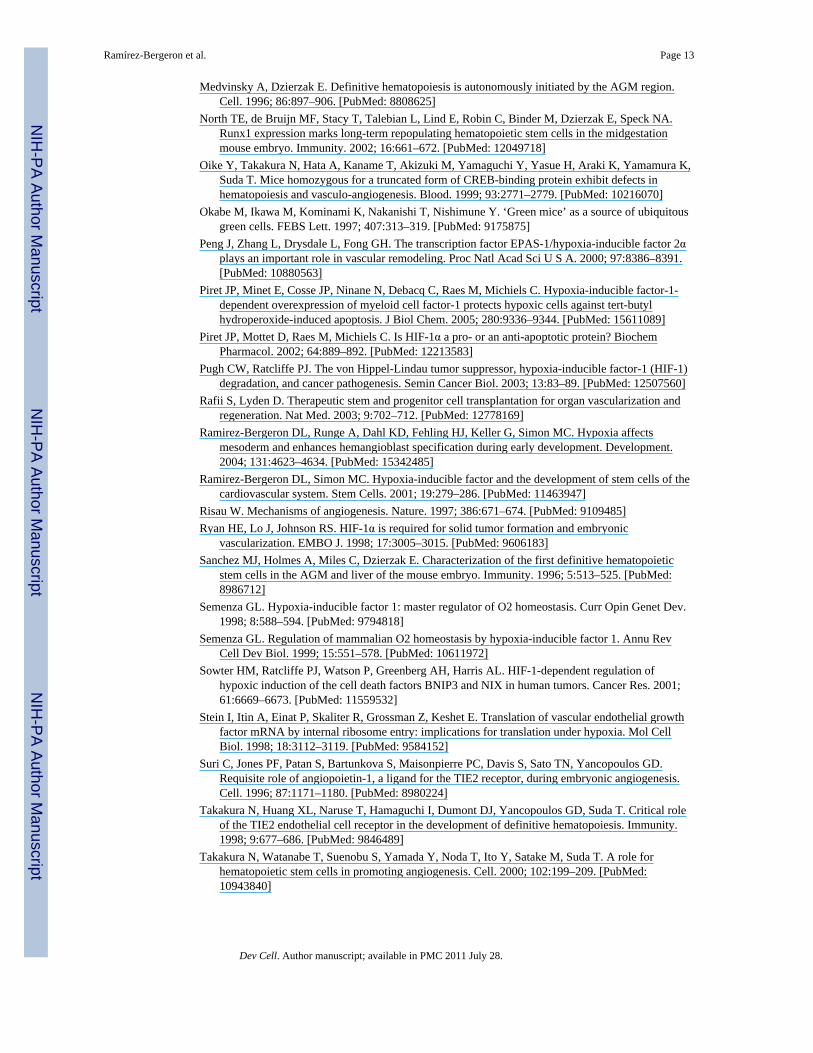
VEGF protein levels are reduced in Arnt−/− embryosAs a subunit of HIF, ARNT is critical for the induction of O2 regulated genes such as Pgk, aglycolytic enzyme, and Vegf, a critical endothelial growth and survival factor. It hadpreviously been reported that the vascular defects of Hif-1α −/− embryos were not due todecreased embryonic Vegf mRNA, but rather mesenchymal cell death (Kotch et al., 1999).Therefore, we analyzed Vegf mRNA levels in Arnt−/− embryos. In situ hybridizationperformed on sagittal sections of 9.5 dpc Arnt+/+ and Arnt−/− samples demonstrated nooverall differences in Pgk and Vegf mRNA abundance with the exception of cells in thecardiac region (Figure 4A, a–d). Furthermore, mRNA quantification by real time PCR ofHIF targets Vegf, Ang-1, Epo, bFgf, VE-cadherin, Tie-2, Flk-1, Fit-1, Aldolase, and Pgkfrom individual somite-matched whole embryos (9.5 dpc) failed to demonstrate anystatistically significant differences between Arnt+/+ and Arnt−/− samples (data not shown).We confirmed that ARNT2 (a related bHLH-PAS protein) is not expressed in HCs, makingARNT essential for HIF activity in these cells (Supplemental Figure 1A–D). Additionally,HIF-1α protein was detected in Arnt−/− embryo sections, but nuclear staining was reducedcompared to Arnt+/+ samples (Supplemental Figure 1E–H). No significant differences wereobserved between Arnt+/+ and Arnt−/− samples by immunostaining for the VEGF receptorFlk-1, Ang-1, or the Ang-1 receptor Tie-2 (Supplemental Figure 2). However, VEGF proteinlevels were clearly reduced in Arnt−/− embryonic sections, especially in the hematopoieticcompartment (Figure 4A, g and h). We have previously demonstrated that VEGF is post-transcriptionally regulated by HIF in subcutaneous tumors (Covello et al., 2005). Weconfirmed these observations by measuring VEGF protein levels in whole- cell orpostnuclear lysates obtained from somite-matched 9.5 dpc embryos by ELISA. Arnt−/−
embryos exhibited a 50% decrease in VEGF protein levels as compared to Arnt+/+ controls(Figure 4A, i). Therefore, while Vegf mRNA levels were similar in wild type and mutantsamples, VEGF protein was reduced in the absence of ARNT. The significance of theseresults is addressed in the discussion.
Arnt−/− embryos exhibit increased numbers of apoptotic hematopoietic cellsAs stated above, Hif-1α−/− embryos exhibit mesenchymal cell death (Kotch et al., 1999).While programmed cell death is essential for some developmental processes, apoptosis isalso associated with embryonic pathologies and lethality (Lindsten et al., 2000). BecauseHIF reportedly exerts pro- and/or anti- apoptotic effects (Piret et al., 2005; Piret et al., 2002;Sowter et al., 2001), we determined if Arnt deficiency is associated with changes in eitherproliferation or programmed cell death. Transverse and sagittal embryonic sections weresubjected to staining with Ki67 and terminal transferase-mediated dUTP nick end labeling(TUNEL) to evaluate proliferation and apoptosis, respectively (Figure 4B). No differenceswere noted in Ki67 staining (data not shown). Although cell death is a natural component ofembryogenesis, we detected a significant increase in the number of TUNEL+ cells in Arnt−/−
sections from 9.5 dpc embryos compared to wild type controls (Figure 4B). Of note,increased TUNEL+ cells were most significant in regions of the dorsal aorta, HCs, andsomitic vessels of the Arnt−/− embryos (Figure 4Bb, d and f). Statistical analysis revealed asignificant increase in the number of TUNEL+HCs in Arnt−/− sections compared to Arnt+/+
controls (Figure 4Bg). These results suggest that ARNT influences the survival but not theproliferation of cells in 9.5 dpc embryos, especially HCs.
To further evaluate the apoptotic defect in HCs of Arnt−/− embryos, we took advantage ofexplants where exogenous Ang-1 was added (see below). Under these culture conditions,hematopoiesis was observed in 7 out of 16 Arnt−/− explants, indicated by the presence ofCD45+ cells. The recovered nonadherent CD45+ Arnt−/− cells from these culture conditionswere analyzed for apoptosis by flow cytometry using Annexin V staining. Annexin V+ cellnumbers were increased in Arnt−/− explants (mean± S.E., 19.39%± 9.77), compared to wild
Ramírez-Bergeron et al. Page 6
Dev Cell. Author manuscript; available in PMC 2011 July 28.
NIH
-PA Author Manuscript
NIH
-PA Author Manuscript
NIH
-PA Author Manuscript

type levels (4.26%± 2.40) (Table 2). Of note, CD45+ nonadherent cell numbers in theremaining nine Arnt−/− explants were insufficient to analyze, suggesting that Ang-1 alonedoes not consistently rescue the hematopoietic Arnt−/− defect. The increased Annexin Vstaining exhibited by cells that can be recovered from Arnt−/− explants indicate thatdecreased survival likely accounts for the severely reduced numbers of HCs in Arnt−/−
cultures. Thus, it appears that vessel defects in Arnt−/− embryos are influenced by decreasedVEGF levels, and HCs are a likely paracrine VEGF source. Moreover, reduced VEGF levelsalso contribute to the intraembryonic hematopoietic defect in Arnt−/− embryos.
VEGF is sufficient to rescue the Arnt−/− P-Sp defectThe dramatic reduction in VEGF expression may be responsible for vascular defects notedin Arnt−/− embryos. Because VEGF is an important survival factor for multiple cell types,VEGF would also be indirectly necessary for normal vessel formation and development bypromoting HC viability. Furthermore, since VEGF is expressed in HCs, we hypothesizedthat VEGF could rescue the Arnt−/− vascular phenotype. To determine if exogenous VEGFcould overcome the Arnt−/− hematopoietic and vascular defects, VEGF 164 isoform wasadded to the P-Sp cultures (Figure 5Aa–d). Vessel lengths observed in the vascular networkswere enhanced in VEGF treated cultures and became indistinguishable between Arnt+/+ andArnt−/− explants (Figure 5Aa–d, 5B). To confirm the significance of VEGF for endothelialdevelopment in P-Sp explants, we analyzed the effect of blocking VEGF signaling bysupplementing the cultures with recombinant soluble Flt-1-Fc receptor. The development ofvascular beds and networks was significantly inhibited in both Arnt+/+; and Arnt−/− P-Spcultures (Figure 5Ai–l). While Flt-1-Fc did not reduce HC numbers after 14 days of culture,Flt-1-Fc treatment resulted in reduced numbers of PECAM+ cells (Figure 5Ai–l). VEGFrescued Arnt−/− P-Sp cultures also contained abundant numbers of nonadherent CD45+
hematopoietic cells, similar to Arnt+/+ controls (data not shown). We next examinedhematopoietic potential in the VEGF treated P-Sp cultures (Supplemental Table 2) andfound that although reduced, Arnt−/− P-Sp cultures generated CFCs more efficiently thanuntreated cultures. These data indicate that paracrine VEGF promotes both hematopoieticexpansion and vessel development in Arnt−/− P-Sp cultures.
Since HSCs are also an important source of Ang-1 (Takakura et al., 2000), we determined ifthe Arnt−/− P-Sp explants could be rescued with exogenous Ang-1 (Figure 5Ae–h). Thevascular bed size in Arnt+/+ and Arnt−/− explants was greatly increased by the addition ofAng-1 alone (Figure 5Ae–h, 5B) or in combination with VEGF (Supplemental Figure 3). Inboth conditions, a large mantle of cells adjacent to the original explants were PECAM+,suggesting that Ang-1 enhanced the proliferation of ECs. Treatment with both Ang-1 andVEGF increased vessel lengths in Arnt−/− networks (Supplemental Figure 3, asterisks),compared to VEGF treatment alone (Figure 5Ae–h, asterisks). In contrast, vessel lengthsremained small in networks within cultures treated with Ang-1 alone where stainingdemonstrated EC congestion (Arnt+/+: 0.755 mm ± 0.136, and Arnt−/−: 0.597mm ± 0.045,P< 0.397) and a decrease in the fine patterning of branches (Arnt+/+: 1.159 mm ± 0.090, andArnt−/−: 1.248mm ± 0.010, P< 0.224) (Figure 5Ag–h, 5B). It should be noted that only 37%of Arnt−/− explants demonstrated significant vessel differentiation upon exogenous Ang-1addition. Moreover, hematopoiesis was rescued in only one half of the Arnt−/− explantssupplemented with Ang-1. We concluded that Ang-1 is not sufficient to fully rescue theArnt−/− P-Sp vascular defect. In direct contrast, exogenous VEGF rescues vasculogenesis,angiogenesis and hematopoiesis in mutant explants.
DISCUSSIONIn this study, we determined that HIF is essential for vasculogenesis, angiogenesis, andhematopoiesis in the P-Sp/AGM. We employed a P-Sp ex vivo approach to analyze HIF’s
Ramírez-Bergeron et al. Page 7
Dev Cell. Author manuscript; available in PMC 2011 July 28.
NIH
-PA Author Manuscript
NIH
-PA Author Manuscript
NIH
-PA Author Manuscript

involvement in embryonic vessel development using Arnt−/− embryos to abrogate all HIFtranscriptional activity (Supplemental Table 3). The explant assay allows us to evaluatevasculogenesis, angiogenesis, and hematopoiesis in the intraembryonic P-Sp region withoutthe complication of other cardiovascular defects. The addition of wild type Sca-1+ HCspromoted both endothelial and hematopoietic differentiation of Arnt−/− explants. VEGF,whose protein levels are dramatically reduced in Arnt−/− embryos, fully rescued the P-Spexplant defects when added to the cultures. Reduced VEGF levels may be the cause ofincreased numbers of apototic TUNEL+ or Annexin V+ HCs observed in Arnt−/− embryonichistological sections or P-Sp explants. The decreased survival of and VEGF production byHCs in Arnt−/− embryos likely contribute to paracrine defects preceding abnormal vesseldevelopment, as shown in Figure 5C. Therefore, O2 sensing by HIF is critical forintraembryonic blood and vessel development, in part by promoting the production ofVEGF.
Our findings support the hypothesis that growth factors supplied in a paracrine fashion byHCs are important for vessel development. AML1−/− embryos have previously been shownto exhibit both hematopoietic and angiogenic defects (Takakura et al., 2000). The AML1(Runxl)-CBFβ transcription factor is required for the generation of hematopoieticprogenitors and AML1−/− embryos die by 12.5 dpc. In similar P-Sp experiments, addition ofHCs or exogenous Ang-1 rescued the AML1 angiogenic defect. In contrast, we show thatAng-1 only partially rescued the Arnt−/− vessel and hematopoietic defects. While theAML1−/− embryos survive to 11.5 dpc with intact large vessels, HIF deficiency results inmore severe vascular defects whereby Arnt−/− embryos are developmentally delayed withpoor vessel development by 9.5 dpc. Therefore, we show that HCs are also required forearlier vascular differentiation by secreting VEGF, a growth factor important for angioblastspecification and vasculogenesis (Weinstein, 1999). Because some PECAM+ vessels areobserved in 9.5 dpc Arnt−/− embryos, sufficient levels of VEGF may be available formesoderm differentiation into a limited number of hemangioblasts, hematopoietic precursorsand/or angioblasts. However, hypoxic induction of VEGF is required for extensiveproliferation, survival, differentiation, maturation, and/or migration of these cells (Figure5C). While circulating bone marrow derived endothelial progenitor cells (EPCs) contributeto adult “neovasculogenesis” (Ingram et al., 2005), HCs do not contribute to vesselsgenerated in the P-Sp explants. Therefore, the relationship between hematopoietic andendothelial cells during embryonic development and in adults remains a complicated,context dependent process.
Hypoxia and ischemia trigger multiple responses to overcome decreased O2 availability,including the expression of growth factors required for establishment of a functionalcirculatory system. While unable to detect a significant difference in Vegf transcript levelsbetween Arnt−/− and Arnt+/+ 9.5 dpc embryos, we clearly observed decreased VEGF proteinlevels in mutant samples. The transcription of Vegf, which contains Hypoxic ResponseElements (HREs) in 5′ and 3′ flanking regions of the gene, is strongly induced by HIF inresponse to low O2 tension (Forsythe et al., 1996; Semenza, 1999). However, VEGFregulation by hypoxia also includes increased mRNA translational efficiency, proteinstability, and biological activity (Chan et al., 2002; Claffey et al., 1998; Stein et al, 1998).
Decreased VEGF levels in HCs of 9.5 dpc Arnt−/− embryos may be linked to their increasedrate of apoptosis. VEGF is a known survival factor for various cell types, including HSCsvia a cell autonomous autocrine pathway (Brusselmans et al., 2005; Gerber et al., 2002).Embryonic inactivation of Vegf, or its receptor Flk-1, results in decreased numbers of bloodislands, endothelial cells, and major vessels (reviewed in Carmeliet and Collen, 2000). Arequirement for VEGF in P-Sp explant outgrowth has also been demonstrated by the partialrescue of similar hematopoietic and vascular defects in explants from mice lacking CREB-
Ramírez-Bergeron et al. Page 8
Dev Cell. Author manuscript; available in PMC 2011 July 28.
NIH
-PA Author Manuscript
NIH
-PA Author Manuscript
NIH
-PA Author Manuscript

binding protein (Oike et al., 1999). We show that inhibition of VEGF signaling using asoluble Flt-1-Fc receptor blocks EC development in P-Sp explant assays, supportingprevious work using an anti-Flk-1 antibody (Takakura, 1998). While some evidencesuggests Ang-1 is a hematopoietic mitogenic or survival factor (Arai et al., 2005; Kopp etal., 2005), we determined that Ang-1 is insufficient to rescue the Arnt−/− P-Sp explants. Incontrast to VEGF, Ang-1 protein levels were not reduced in Arnt−/− embryos. Moreover,Ang-1−/− embryos exhibit normal vascular differentiation prior to day 11.5 dpc (Suri et al.,1996) suggesting that the requirement for VEGF precedes Ang-1. Taken together, VEGFappears to be critical for the survival and subsequent maturation of early hematopoietic andendothelial cells. Furthermore, hypoxia in the early embryo controls VEGF levels duringthese events.
Recent studies have examined the role of HIF in ECs using a conditional Hif-1 α−/− allele(Tang et al., 2004). Although mice in which Hif-1α is deleted in the endothelial andhematopoietic compartments after day 9.5 dpc are viable, angiogenesis in adult mice iscompromised in these animals. In vitro, Hif-1α −/− ECs exhibit migration, proliferation, andsurvival defects. While these studies contribute to our understanding of hypoxic responses inEC biology, they may not be as informative about embryonic vascularization becauseimmortalized adult EC lines were used. Futhermore, hypoxic responses in Hif-1α −/− ECsare not completely abrogated as they maintain HIF-2α expression, making the analysis ofArnt−/− embryos important for determining the role of HIF in vessel development. Futurework will thoroughly evaluate the role of ARNT in EC behavior.
Our data indicate that HIF regulates cardiovascular development. In particular, it is requiredfor the generation of both the hematopoietic and vascular organ systems. Although therequirement for HIF in these processes has been established, the molecular nature of all therelevant signals involved in embryonic development remains unclear. We show that HCs arerequired for proper vessel emergence and development. Furthermore, we presently suggestthat HIF is important for cell survival during embryogenesis through mechanisms thatinclude the production of growth factors and protection from apoptosis.
EXPERIMENTAL PROCEDURESP-Sp Explants
All animal experiments were conducted in accordance with NIH Guidelines. Generation ofArnt−/− mice used in these experiments has been previously described (Maltepe et al., 1997).9.5 dpc embryos were harvested and staged by counting somites. P-Sp explants werecultured at 37°C in humidified 5 % CO2 on OP9 stromal cell layers as previously described(Takakura et al., 2000). Briefly, RPMI 1640 (GIBCO-BRL, Gaithersburg, MD) culturemedium contained 10% FCS (Hyclone), 10−5 M 2ME (Sigma, St Louis, MO), 50 ng/mlstem cell factor (SCF), 20 ng/ml interleukin-6 (IL-6), and 2 U/ml Erythropoietin (EPO). Forthe rescue or inhibition experiments, 300 ng/ml of recombinant Flt-1-Fc chimera protein,100 ng/ml VEGF and/or 300 ng/ml Ang-1 were added. All growth factors were purchasedfrom R&D Systems. Sca-1+ HCs from the bone marrow of GFP+ mice (Okabe et al., 1997)were selected using magnetic beads as described (Stem Cell Technologies, Vancouver BC,Canada) and 200–300 Sca-1+ cells were added to each well. After 14 days, cultures werestained for PECAM (see below). Hematopoietic cells were harvested and analyzed atdifferent time points for CD45 or Annexin V expression (see below).
Quantitative Analysis of angiogenic vascular networksAfter PECAM staining, photomicrographs of P-Sps were integrated using a LEICA DC 50camera and program. Angiogenic vessel lengths were measured by ImageJ analysis. Ten
Ramírez-Bergeron et al. Page 9
Dev Cell. Author manuscript; available in PMC 2011 July 28.
NIH
-PA Author Manuscript
NIH
-PA Author Manuscript
NIH
-PA Author Manuscript

individual measurements of five separate Arnt+/+ and Arnt−/− explants were used todetermine average vessel lengths and standard deviations of the mean. P values werecalculated by 2-tailed Student t test.
Methylcellulose Colony AssaysThe in vitro colony assays for hematopoietic progenitors were performed as recommendedfor M3434 media (Stem Cell Technologies, Vancouver BC, Canada) with the addition of 2×l03 nonadherent cells harvested from day 12 P-Sp explants. Cultures were performed intriplicate and colonies were enumerated at day 7.
Cell Preparation and Flow CytometryNonadherent hematopoietic cells were harvested from P-Sp cultures at the days indicated.Cells were incubated in 1:100 FCγIII/II receptor (Pharmingen, San Jose, CA) blockingbuffer for 20 minutes at 4°C. After washing, cells were incubated with PE- conjugatedCD45 or APC-conjugated Annexin V (Caltag, Buckingham UK) at 1:100 for 20 minutes at4°C, washed, and visualized by FACS Vantage (Becton and Dickinson, San Jose, CA).Results were analyzed by Ho-Jo (Tree-Star).
ImmunohistochemistryPECAM antibody (MEC13.3, Pharmingen) was used for whole mount staining as described(Oike et al., 1999). Tissue sections were incubated with anti-CD34, anti-Flk1, anti-Tie2(Pharmingen), anti-Ang-1 (Abcam) or anti-VEGF (NeoMarkers, Fremont, CA) anddeveloped with HRP-conjugated secondary antibodies followed by treatment withdiaminobenzidine (DAB, Vector Laboratories, Burlingame, CA). A BioVision (MountainView, Ca) Apo-BrdU-FITC In Situ DNA fragment kit was used to assay apoptosis inembryonic tissues. Apoptotic measurements were made by capturing sequential images ofFITC (BrdU staining) and DAPI (nuclear staining) channels of identical tissue sections withan epiflourescent microscope. Images were overlayed in Photoshop, quantified and resultswere expressed as numbers of apoptotic cells within dorsal aortic regions.
RNA in Situ Hybridization9.5 dpc embryos from Arnt+/+ matings were fixed in 4% paraformaldehyde for 48 hours.Genotypes were analyzed from yolk sac DNA by PCR. Subsequent tissue embedding,sectioning, and in situ hybridization were performed as previously described (Jain et al.,1998). Sense and antisense RNA probes were generated using T7 and Sp6 RNApolymerases and -[35S]thio-UTP. Pgk, and Vegf probes were generated by reversetranscription-PCR with the following primers: Pgk, 5′ 5′-CTTGGACTGTGGTACTGAGAGand 3′ 5′-CTGAATCTTGCGCTAACACCA; and Vegf, 5′ 5′-CCATGCAGATCATGCGGATC and 3′ 5′-CAAAGTTCTCCTCGAAGGATC.Hybridization to sense probes revealed only low levels of general background staining (datanot shown).
RNA and Protein QuantificationReal-time detection PCR (RTD-PCR) was performed as previously described (Ramirez-Bergeron et al., 2004). A VEGF ELISA kit was used to measure VEGF protein levels fromindividual whole embryo cell extracts (Biosource, Camarillo, CA). Briefly, stage matchedembryos were harvested and lysed. Protein concentrations of whole cell or postnuclearextracts were quantified and analyzed as suggested by the manufacturer.
Ramírez-Bergeron et al. Page 10
Dev Cell. Author manuscript; available in PMC 2011 July 28.
NIH
-PA Author Manuscript
NIH
-PA Author Manuscript
NIH
-PA Author Manuscript

Supplementary MaterialRefer to Web version on PubMed Central for supplementary material.
AcknowledgmentsWe thank Hongwei Yu for technical assistance. This research was supported by National Institutes of Health GrantsHL66130 (M.C.S.) and HL73153 (D.R.B) and the Abramson Family Cancer Research Institute. M.C.S. is aninvestigator of the Howard Hughes Medical Institute.
ReferencesAdelman DM, Gertsenstein M, Nagy A, Simon MC, Maltepe E. Placental cell fates are regulated in
vivo by HIF-mediated hypoxia responses. Genes Dev. 2000; 14:3191–3203. [PubMed: 11124810]Adelman DM, Maltepe E, Simon MC. Multilineage embryonic hematopoiesis requires hypoxic ARNT
activity. Genes Dev. 1999; 13:2478–2483. [PubMed: 10521392]Arai P, Hirao A, Suda T. Regulation of hematopoietic stem cells by the niche. Trends Cardiovasc Med.
2005; 15:75–79. [PubMed: 15885574]Brusselmans K, Bono F, Collen D, Herbert JM, Carmeliet P, Dewerchin M. A novel role for vascular
endothelial growth factor as an autocrine survival factor for embryonic stem cells during hypoxia. JBiol Chem. 2005; 280:3493–3499. [PubMed: 15572379]
Carmeliet P. Angiogenesis in life, disease and medicine. Nature. 2005; 438:932–936. [PubMed:16355210]
Carmeliet P, Collen D. Transgenic mouse models in angiogenesis and cardiovascular disease. J Pathol.2000; 190:387–405. [PubMed: 10685072]
Chan DA, Sutphin PD, Denko NC, Giaccia AJ. Role of prolyl hydroxylation in oncogenicallystabilized hypoxia-inducible factor-1 alpha. J Biol Chem. 2002; 277:40112–40117. [PubMed:12186875]
Claffey KP, Shih SC, Mullen A, Dziennis S, Cusick JL, Abrams KR, Lee SW, Detmar M.Identification of a human VPF/VEGF 3′ untranslated region mediating hypoxia-induced mRNAstability. Mol Biol Cell. 1998; 9:469–481. [PubMed: 9450968]
Compernolle V, Brusselmans K, Acker T, Hoet P, Tjwa M, Beck H, Plaisance S, Dor Y, Keshet E,Lupu F, et al. Loss of HIF-2α and inhibition of VEGF impair fetal lung maturation, whereastreatment with VEGF prevents fatal respiratory distress in premature mice. Nat Med. 2002; 8:702–710. [PubMed: 12053176]
Coultas L, Chawengsaksophak K, Rossant J. Endothelial cells and VEGF in vascular development.Nature. 2005; 438:937–945. [PubMed: 16355211]
Covello KL, Kehler J, Yu H, Gordan JD, Arsham AM, Hu CJ, Labosky PA, Simon MC, Keith B.HIF-2α regulates Oct-4: effects of hypoxia on stem cell function, embryonic development, andtumor growth. Genes Dev. 2006; 20:557–570. [PubMed: 16510872]
Covello KL, Simon MC, Keith B. Targeted replacement of hypoxia-inducible factor-lalpha by ahypoxia-inducible factor-2α knock-in allele promotes tumor growth. Cancer Res. 2005; 65:2277–2286. [PubMed: 15781641]
Cowden Dahl KD, Fryer BH, Mack FA, Compernolle V, Maltepe E, Adelman DM, Carmeliet P,Simon MC. Hypoxia-inducible factors 1α and 2α regulate trophoblast differentiation. Mol CellBiol. 2005; 25:10479–10491. [PubMed: 16287860]
de Bruijn ME, Ma X, Robin C, Ottersbach K, Sanchez MJ, Dzierzak E. Hematopoietic stem cellslocalize to the endothelial cell layer in the midgestation mouse aorta. Immunity. 2002; 76:673–683. [PubMed: 12049719]
Dieterlen-Lievre F, Pardanaud L, Bollerot K, Jaffredo T. Hemangioblasts and hemopoietic stem cellsduring ontogeny. C R Biol. 2002; 325:1013–1020. [PubMed: 12494498]
Ema M, Taya S, Yokotani N, Sogawa K, Matsuda Y, Fujii-Kuriyama Y. A novel bHLH-PAS factorwith close sequence similarity to hypoxia- inducible factor 1α regulates the VEGF expression andis potentially involved in lung and vascular development. Proc Natl Acad Sci U S A. 1997;94:4273–4278. [PubMed: 9113979]
Ramírez-Bergeron et al. Page 11
Dev Cell. Author manuscript; available in PMC 2011 July 28.
NIH
-PA Author Manuscript
NIH
-PA Author Manuscript
NIH
-PA Author Manuscript

Forsythe JA, Jiang BH, Iyer NV, Agani F, Leung SW, Koos RD, Semenza GL. Activation of vascularendothelial growth factor gene transcription by hypoxia-inducible factor 1. Mol Cell Biol. 1996;16:4604–4613. [PubMed: 8756616]
Gerber HP, Malik AK, Solar GP, Sherman D, Liang XH, Meng G, Hong K, Marsters JC, Ferrara N.VEGF regulates haematopoietic stem cell survival by an internal autocrine loop mechanism.Nature. 2002; 417:954–958. [PubMed: 12087404]
Giaccia AJ, Simon MC, Johnson R. The biology of hypoxia: the role of oxygen sensing indevelopment, normal function, and disease. Genes Dev. 2004; 18:2183–2194. [PubMed:15371333]
Hu CJ, Wang LY, Chodosh LA, Keith B, Simon MC. Differential roles of hypoxia-inducible factor 1alpha (HIF-1 alpha) and HIF-2alpha in hypoxic gene regulation. Mol Cell Biol. 2003; 23:9361–9374. [PubMed: 14645546]
Ingram DA, Caplice NM, Yoder MC. Unresolved questions, changing definitions, and novelparadigms for defining endothelial progenitor cells. Blood. 2005
Ivan M, Kondo K, Yang H, Kim W, Valiando J, Ohh M, Salic A, Asara JM, Lane WS, Kaelin WG Jr.HIFalpha targeted for VHL-mediated destruction by proline hydroxylation: implications for O2sensing. Science. 2001; 292:464–468. [PubMed: 11292862]
Iyer NV, Kotch LE, Agani F, Leung SW, Laughner E, Wenger RH, Gassmann M, Gearhart JD, LawlerAM, Yu AY, Semenza GL. Cellular and developmental control of O2 homeostasis by hypoxia-inducible factor 1 alpha. Genes Dev. 1998; 12:149–162. [PubMed: 9436976]
Jaakkola P, Mole DR, Tian YM, Wilson MI, Gielbert J, Gaskell SJ, Kriegsheim A, Hebestreit HF,Mukherji M, Schofield CJ, et al. Targeting of HIF-α to the von Hippel-Lindau ubiquitylationcomplex by O2-regulated prolyl hydroxylation. Science. 2001; 292:468–472. [PubMed:11292861]
Jain RK. Molecular regulation of vessel maturation. Nat Med. 2003; 9:685–693. [PubMed: 12778167]Jain S, Maltepe E, Lu MM, Simon C, Bradfield CA. Expression of ARNT, ARNT2, HIF1 α, HIF2 α
and Ah receptor mRNAs in the developing mouse. Mech Dev. 1998; 73:117–123. [PubMed:9545558]
Kocher AA, Schuster MD, Szabolcs MJ, Takuma S, Burkhoff D, Wang J, Homma S, Edwards NM,Itescu S. Neovascularization of ischemic myocardium by human bone-marrow-derived angioblastsprevents cardiomyocyte apoptosis, reduces remodeling and improves cardiac function. Nat Med.2001; 7:430–436. [PubMed: 11283669]
Kopp HG, Avecilla ST, Hooper AT, Shmelkov SV, Ramos CA, Zhang F, Rafii S. Tie2 activationcontributes to hemangiogenic regeneration after myelosuppression. Blood. 2005; 106:505–513.[PubMed: 15817675]
Kotch LE, Iyer NV, Laughner E, Semenza GL. Defective vascularization of HIF-1α-null embryos isnot associated with VEGF deficiency but with mesenchymal cell death. Dev Biol. 1999; 209:254–267. [PubMed: 10328919]
Lee YM, Jeong CH, Koo SY, Son MJ, Song HS, Bae SK, Raleigh JA, Chung HY, Yoo MA, Kim KW.Determination of hypoxic region by hypoxia marker in developing mouse embryos in vivo: apossible signal for vessel development. Dev Dyn. 2001; 220:175–186. [PubMed: 11169851]
Lindsten T, Ross AJ, King A, Zong WX, Rathmell JC, Shiels HA, Ulrich E, Waymire KG, Mahar P,Frauwirth K, et al. The combined functions of proapoptotic Bcl-2 family members bak and bax areessential for normal development of multiple tissues. Mol Cell. 2000; 6:1389–1399. [PubMed:11163212]
Lyden D, Hattori K, Dias S, Costa C, Blaikie P, Butros L, Chadburn A, Heissig B, Marks W, Witte L,et al. Impaired recruitment of bone-marrow-derived endothelial and hematopoietic precursor cellsblocks tumor angiogenesis and growth. Nat Med. 2001; 7:1194–1201. [PubMed: 11689883]
Maltepe E, Schmidt JV, Baunoch D, Bradfield CA, Simon MC. Abnormal angiogenesis and responsesto glucose and oxygen deprivation in mice lacking the protein ARNT. Nature. 1997; 386:403–407.[PubMed: 9121557]
Maltepe E, Simon MC. Oxygen, genes, and development: an analysis of the role of hypoxic generegulation during murine vascular development. J Mol Med. 1998; 76:391–401. [PubMed:9625296]
Ramírez-Bergeron et al. Page 12
Dev Cell. Author manuscript; available in PMC 2011 July 28.
NIH
-PA Author Manuscript
NIH
-PA Author Manuscript
NIH
-PA Author Manuscript
Medvinsky A, Dzierzak E. Definitive hematopoiesis is autonomously initiated by the AGM region.Cell. 1996; 86:897–906. [PubMed: 8808625]
North TE, de Bruijn MF, Stacy T, Talebian L, Lind E, Robin C, Binder M, Dzierzak E, Speck NA.Runx1 expression marks long-term repopulating hematopoietic stem cells in the midgestationmouse embryo. Immunity. 2002; 16:661–672. [PubMed: 12049718]
Oike Y, Takakura N, Hata A, Kaname T, Akizuki M, Yamaguchi Y, Yasue H, Araki K, Yamamura K,Suda T. Mice homozygous for a truncated form of CREB-binding protein exhibit defects inhematopoiesis and vasculo-angiogenesis. Blood. 1999; 93:2771–2779. [PubMed: 10216070]
Okabe M, Ikawa M, Kominami K, Nakanishi T, Nishimune Y. ‘Green mice’ as a source of ubiquitousgreen cells. FEBS Lett. 1997; 407:313–319. [PubMed: 9175875]
Peng J, Zhang L, Drysdale L, Fong GH. The transcription factor EPAS-1/hypoxia-inducible factor 2αplays an important role in vascular remodeling. Proc Natl Acad Sci U S A. 2000; 97:8386–8391.[PubMed: 10880563]
Piret JP, Minet E, Cosse JP, Ninane N, Debacq C, Raes M, Michiels C. Hypoxia-inducible factor-1-dependent overexpression of myeloid cell factor-1 protects hypoxic cells against tert-butylhydroperoxide-induced apoptosis. J Biol Chem. 2005; 280:9336–9344. [PubMed: 15611089]
Piret JP, Mottet D, Raes M, Michiels C. Is HIF-1α a pro- or an anti-apoptotic protein? BiochemPharmacol. 2002; 64:889–892. [PubMed: 12213583]
Pugh CW, Ratcliffe PJ. The von Hippel-Lindau tumor suppressor, hypoxia-inducible factor-1 (HIF-1)degradation, and cancer pathogenesis. Semin Cancer Biol. 2003; 13:83–89. [PubMed: 12507560]
Rafii S, Lyden D. Therapeutic stem and progenitor cell transplantation for organ vascularization andregeneration. Nat Med. 2003; 9:702–712. [PubMed: 12778169]
Ramirez-Bergeron DL, Runge A, Dahl KD, Fehling HJ, Keller G, Simon MC. Hypoxia affectsmesoderm and enhances hemangioblast specification during early development. Development.2004; 131:4623–4634. [PubMed: 15342485]
Ramirez-Bergeron DL, Simon MC. Hypoxia-inducible factor and the development of stem cells of thecardiovascular system. Stem Cells. 2001; 19:279–286. [PubMed: 11463947]
Risau W. Mechanisms of angiogenesis. Nature. 1997; 386:671–674. [PubMed: 9109485]Ryan HE, Lo J, Johnson RS. HIF-1α is required for solid tumor formation and embryonic
vascularization. EMBO J. 1998; 17:3005–3015. [PubMed: 9606183]Sanchez MJ, Holmes A, Miles C, Dzierzak E. Characterization of the first definitive hematopoietic
stem cells in the AGM and liver of the mouse embryo. Immunity. 1996; 5:513–525. [PubMed:8986712]
Semenza GL. Hypoxia-inducible factor 1: master regulator of O2 homeostasis. Curr Opin Genet Dev.1998; 8:588–594. [PubMed: 9794818]
Semenza GL. Regulation of mammalian O2 homeostasis by hypoxia-inducible factor 1. Annu RevCell Dev Biol. 1999; 15:551–578. [PubMed: 10611972]
Sowter HM, Ratcliffe PJ, Watson P, Greenberg AH, Harris AL. HIF-1-dependent regulation ofhypoxic induction of the cell death factors BNIP3 and NIX in human tumors. Cancer Res. 2001;61:6669–6673. [PubMed: 11559532]
Stein I, Itin A, Einat P, Skaliter R, Grossman Z, Keshet E. Translation of vascular endothelial growthfactor mRNA by internal ribosome entry: implications for translation under hypoxia. Mol CellBiol. 1998; 18:3112–3119. [PubMed: 9584152]
Suri C, Jones PF, Patan S, Bartunkova S, Maisonpierre PC, Davis S, Sato TN, Yancopoulos GD.Requisite role of angiopoietin-1, a ligand for the TIE2 receptor, during embryonic angiogenesis.Cell. 1996; 87:1171–1180. [PubMed: 8980224]
Takakura N, Huang XL, Naruse T, Hamaguchi I, Dumont DJ, Yancopoulos GD, Suda T. Critical roleof the TIE2 endothelial cell receptor in the development of definitive hematopoiesis. Immunity.1998; 9:677–686. [PubMed: 9846489]
Takakura N, Watanabe T, Suenobu S, Yamada Y, Noda T, Ito Y, Satake M, Suda T. A role forhematopoietic stem cells in promoting angiogenesis. Cell. 2000; 102:199–209. [PubMed:10943840]
Ramírez-Bergeron et al. Page 13
Dev Cell. Author manuscript; available in PMC 2011 July 28.
NIH
-PA Author Manuscript
NIH
-PA Author Manuscript
NIH
-PA Author Manuscript
Tang N, Wang L, Esko J, Giordano FJ, Huang Y, Gerber HP, Ferrara N, Johnson RS. Loss of HIF-1αin endothelial cells disrupts a hypoxia-driven VEGF autocrine loop necessary for tumorigenesis.Cancer Cell. 2004; 6:485–495. [PubMed: 15542432]
Tian H, Hammer RE, Matsumoto AM, Russell DW, McKnight SL. The hypoxia-responsivetranscription factor EPAS1 is essential for catecholamine homeostasis and protection against heartfailure during embryonic development. Genes Dev. 1998; 12:3320–3324. [PubMed: 9808618]
Tian H, McKnight SL, Russell DW. Endothelial PAS domain protein 1 (EPAS1), a transcription factorselectively expressed in endothelial cells. Genes Dev. 1997; 11:72–82. [PubMed: 9000051]
Wang V, Davis DA, Haque M, Huang LE, Yarchoan R. Differential gene up-regulation by hypoxia-inducible factor-1α and hypoxia-inducible factor-2α in HEK293T cells. Cancer Res. 2005;65:3299–3306. [PubMed: 15833863]
Weinstein BM. What guides early embryonic blood vessel formation? Dev Dyn. 1999; 215:2–11.[PubMed: 10340752]
Yu F, White SB, Zhao Q, Lee FS. HIF-1α binding to VHL is regulated by stimulus-sensitive prolinehydroxylation. Proc Natl Acad Sci U S A. 2001; 98:9630–9635. [PubMed: 11504942]
Ramírez-Bergeron et al. Page 14
Dev Cell. Author manuscript; available in PMC 2011 July 28.
NIH
-PA Author Manuscript
NIH
-PA Author Manuscript
NIH
-PA Author Manuscript

FIGURE 1. Disruption of vascular development in Arnt−/− yolk sacs and embryosWhole-mount PECAM stained wild type embryo (A,B,C) and yolk sac (G,H), and Arnt−/−
embryo (D,E,F) and yolk sac (I,J). The Arnt−/− embryo lacks proper staining of the cardinalvein (A,D arrows) and pharyngeal pouches as seen in the Arnt+/+ embryo (A,D dotted blacklines). Compared to wild type, the mutant embryo lacks proper inter- and intra- somitic(black arrows, C and F) and peripheral (arrowheads, B and E) PECAM staining. Strikingdecreased staining is observed in the AGM region of the Arnt−/− embryo (white dotted lines,A and B). Vascular networks, seen as abnormal branching of the vitelline artery in the yolksac, are deficient in the Arnt−/− embryo (white arrows, G–J). Immunohistochemical stainingwith anti-CD34 of Arnt+/+ (K,M,O) and Arnt−/− (L,N,P) 9.5 dpc embryos. The Arnt−/−
embryo lacks proper EC staining in the dorsal aorta (M,N, open arrows), and endocardium(O,P, arrows). Enlarged boxed area demonstrates the presence of CD34+ hematopoietic cellsin the Arnt−/− embryo (L). Magnifications: A,D, 120X; G,I 150X; B,C,E,F,H,J 200X; K,L100X; M–P 300X.
Ramírez-Bergeron et al. Page 15
Dev Cell. Author manuscript; available in PMC 2011 July 28.
NIH
-PA Author Manuscript
NIH
-PA Author Manuscript
NIH
-PA Author Manuscript

FIGURE 2. Impaired vascular network formation by Arnt−/− P-Sp explantsP-Sp regions (white dotted lines) from 9.5 dpc Arnt+/+ (A) and Arnt−/− (B) somite matchedembryos were removed and cultured on OP9 stromal cells and immunostained with anti-PECAM (C–F). P-Sp explants from Arnt+/+ embryos (C,D), but not Arnt−/− embryos (E,F),formed extensive PECAM+ vascular beds (white arrowheads) and networks (blackarrowheads). In contrast, representative Arnt−/− explants exhibit reduced PECAM stainingof tissue outgrowth (E) or a few random PECAM+ cells (F, open arrowhead). Wild-typeexplants (G,H) observed in phase contrast micrographs generated extensive non-adherentround hematopoietic cells (arrows) which are lacking in Arnt−/− P-Sp cultures (I,J).Magnifications: A,B 100X; C,E,G,I, scale bar 100 μm; D,F,J 100X; H, 200X.
Ramírez-Bergeron et al. Page 16
Dev Cell. Author manuscript; available in PMC 2011 July 28.
NIH
-PA Author Manuscript
NIH
-PA Author Manuscript
NIH
-PA Author Manuscript

FIGURE 3. Wild type Sca-1+GFP+ bone marrow cells rescue Arnt−/− P-Sp explantsSca-1+ cells isolated from the bone marrow of wild type GFP+ mice were added to P-Spexplant cultures and stained for PECAM after 14 days (C–F). As a control, representativeuntreated Arnt+/+ (A) and Arnt−/− (B) P-Sp cultures are shown. Rescued vascular networks(*) from Arnt−/− explants (C–F) were comparable to wild type controls (A). (G)Measurements of lengths from ten individual vessels extending from the vascular bedsexpressed as mean ± SEM distance from five independent wild type (grey) and mutant(white) rescued explants. As a control (#) the mean ± SEM from an untreated Arnt−/−
culture with minimal vessel staining (B) is shown. The horizontal lines represent averagevessel lengths, for the Arnt+/+ and Arnt−/− cultures (P≤ 0.98). (H) Dil-Ac-LDL uptake ofendothelial cells from a rescued P-Sp culture showing a peripheral ring of cells that includesthe vascular networks. (I) Higher magnification showing no overlap between the DiI-Ac-LDL+ ECs originating from the explant and the GFP+ hematopoietic donor cells in rescuedcultures. (J–M) Higher magnification phase contrast (J,L) and counterpart green fluorescent(K,M) micrographs demonstrating track of ECs (dooted white lines) that do not incorporateGFP+ cells. (N–Q) Higher magnification phase contrast (N,P) and green fluorescent (O,Q)micrographs demonstrating nonadherent hematopoietic colonies that are GFP− arising fromthe rescued explants. Magnifications: A–F scale bar 100 μm H, 100X; J–K, 400X; I, L–Q200X.
Ramírez-Bergeron et al. Page 17
Dev Cell. Author manuscript; available in PMC 2011 July 28.
NIH
-PA Author Manuscript
NIH
-PA Author Manuscript
NIH
-PA Author Manuscript

FIGURE 4.A. VEGF protein levels are reduced in Arnt−/− 9.5 dpc embryos. Transverse sections ofArnt+/+ (a,b,c) and Arnt−/− (d,e,f) embryos probed for Vegf(b,e) and Pgk (c,f) showing nooverall differences in expression. As a comparison, hematoxylin stained Arnt+/+ (a) andArnt−/− (d) serial sections are shown. VEGF immunohistochemistry of transverse sections of9.5 dpc (28 somites) Arnt+/+ (g) and Arnt−/− (h) embryos showing reduced proteinexpression in the mutant embryo. Expression is particularly reduced in the hematopoieticcompartment (higher magnification inlay) of the Arnt−/− embryo (arrow). Dorsal aorta (da);hematopoietic cells (HCS); heart (ht); neural tube (nt). (i) VEGF levels from whole cell orpostnuclear lysates of individual 9.5 dpc (21 somites) Arnt+/+ (black) and Arnt−/− (white)embryos analyzed by ELISA. Error bars indicate standard error of the mean (n=3, (*)P<0.005). B. Increased apoptosis in Arnt−/− embryos. TUNEL immunostaining (FITC,green) of transverse sections counterstained with hematoxylin (a,b) or nuclear DAPI (blue)(c,d), and hematoxylin stained sagittal sections (e,f) of Arnt+/+ (a,c,e) and Arnt−/− (b,d,f)embryos. An increase in TUNEL+ HCs was detected in the dorsal aorta (d, arrowheads) ofArnt−/− transverse sections compared to the modest TUNEL staining observed in theArnt+/+ control (c). Increased numbers of TUNEL+ cells in the dorsal aorta (f, arrows) andintrasomitic (f, dotted lines) regions are observed in Arnt−/− sagittal sections. Comparison ofapoptotic cells in the dorsal aortic region of Arnt+/+ (black) and Arnt−/− (white) transversesections (g). Numbers of apoptotic cells are expressed as total numbers of FITC+ cells (*)P<0.005. Magnifications: 50X A a–f, Ba,b,e,f; 100X A g–h, B c,d (inserts 400X).
Ramírez-Bergeron et al. Page 18
Dev Cell. Author manuscript; available in PMC 2011 July 28.
NIH
-PA Author Manuscript
NIH
-PA Author Manuscript
NIH
-PA Author Manuscript

Figure 5.A. Treatment of P-Sp explants with VEGF or Ang-1 promotes vessel growth Additionof 100 ng/ml VEGF to Arnt−/− cultures (c,d) promotes fine patterning and branching in thevascular networks (*). Arnt+/+ cultures (a,b) are shown as controls. Addition of 300 ng/mlAng-1 does not result in proper angiogenesis but promotes congestion (A) in the vascularnetworks in Arnt−/− cultures (g,h) as well as the Arnt+/+ P-Sps (e,f; P<0.722) However, thecongestion was worse in the Arnt−/− explants. Addition of 300 ng/ml of Flt-1-Fc (j–l)inhibited proper vessel development as compared to control (i) P-Sp explants. All explantswere stained for PECAM. Magnifications: scale bars 100 μm. B. Measurement of vesselareas from VEGF or Ang-1 treated P-Sp explants. Ten sprouting vessel measurementsfrom five independent Arnt−/− and Arnt+/+ P-Sp explants were analyzed. Results are shownas mean vessel lengths (±SEM) for wild type (black) and mutant (white) VEGF treated orAng-1 treated explants. Note that Ang-1 promoted vessel congestion. C. Proper vesseldevelopment requires HIF- dependent factors supplied by hematopoietic cells.Differentiation of hematopoietic cells and vasculature is HIF-dependent during threeseparate events. (1) HIF promotes the survival of hematopoietic cells. (2) Hematopoieticcells serve as a paracrine source of VEGF which induces vasculogenesis by increasing theproduction and proliferation of endothelial cells. (3) Hematopoietic cells are also a source ofAng-1, promoting angiogenesis, as measured by the remodeling and maturation of vessels(Takakura et al., 2000). Loss of Arnt results in HIF deficiency, increased numbers ofapoptotic cells in the hematopoietic compartment, and a reduction of VEGF production.
Ramírez-Bergeron et al. Page 19
Dev Cell. Author manuscript; available in PMC 2011 July 28.
NIH
-PA Author Manuscript
NIH
-PA Author Manuscript
NIH
-PA Author Manuscript

NIH
-PA Author Manuscript
NIH
-PA Author Manuscript
NIH
-PA Author Manuscript
Ramírez-Bergeron et al. Page 20
Table 1
Nonadherent hematopoietic cell numbers from P-Sp rescue experiment with wild type bone marrow cells
Genotype Somite number Total CD45+ CD45+GFP− (embryonic)
+/+ 23 86.9 55.8
+/+ 18 89.3 44.4
+/− 20 91.0 58.9
+/− 16 90.6 41.0
+/− 15 93.6 61.4
−/− 18 88.7 66.4
−/− 17 90.3 57.3
−/− 16 89.6 47.6
−/− 15 92.1 56.2
−/− 11 90.9 38.3
All P-Sp explants were supplemented with 200–300 Sca-1+GFP+ wild type bone marrow cells per culture. Nonadherent cells were harvested and
CD45+ cells were gated and further analyzed for GFP expression. Numbers are denoted as percents. Negative control; unrescued Arnt−/− explants
did not generate CD45+ cells. Positive control; isolated bone marrow cells remained GFK+ over the course of the 14 day experiment.
Dev Cell. Author manuscript; available in PMC 2011 July 28.

NIH
-PA Author Manuscript
NIH
-PA Author Manuscript
NIH
-PA Author Manuscript
Ramírez-Bergeron et al. Page 21
Table 2
Percent of apoptotic CD45+ cells obtained from P-Sp explants
Arnt+/+ Arnt−/−
%CD45+ (%AnnV+) %CD45+ (%AnnV+)
51.3 2.3 19.7 35.8
93.3 1.18 19.4 24.1
12.6 6.03 14 17.2
54.1 4.02 10.8 9.05
52.83∞ 3.38∞ 15.97∞ 21.54∞
28.54¶ 1.83¶ 3.75¶ 9.80
Nonadherent cells from 4 independent Arnt+/+ and Arnt−/− day 14 P-Sp cultures treated with 300 ng/ml Ang-1 were analyzed by flow cytometry.
Gated CD45+ cells were analyzed for numbers of apoptotic cells that stained for Annexin V and were expressed as percentages.
∞mean,
¶standard deviation of all samples above the dotted line; P<0.005.
Dev Cell. Author manuscript; available in PMC 2011 July 28.
Copyright © 2022 FDOKUMEN