Postnatal ontogenetic scaling of Nesodontine (Notoungulata, Toxodontidae) cranial morphology
Upload
khangminh22Category
view
3download
0
Blood Vessels are in Control: Vasculature
Regulates the Neural Niche in Cranial
Sensory Ganglia
Laura Taberner Pérez
TESI DOCTORAL UPF / 2019
Thesis supervisor
Berta Alsina i Español
Developmental Biology
Department of Experimental and Health Sciences


i
ACKNOWLEDGMENTS
Primer de tot vull agrair a la Berta Alsina haver cregut i confiat en mi per dur a terme un
doctorat. Gràcies a tu he aprés, he crescut i he format una nova confiança en mi mateixa.
Gràcies per totes les discussions sobre el projecte, per enviar-me a tots els cursos i
congressos possibles, per animar-me en els moments de decepció i multiplicar els
moments d’il·lusió i troballes. I gràcies també pel teu tracte personal més enllà de la
relació PI-estudiant.
También quiero agradecer a Fernando Giráldez y a Berta (otra vez) por abrirme las
puertas al mundo de la educación. Por cederme clases, compartir proyectos y por confiar
en que lo haría bien.
També a la Cristina Pujades, pel seu interès en el meu projecte i discussions.
A l’Aitor super-company de laboratori i un gran amic, per la seva energia inesgotable i
contagiosa, i els seus coneixements infinits. Sense els teus consells, ànims i soparets
post-labo la tesis no seria la que és. A la resta de companys de laboratori, la Cova, l’Ivan,
la Carla, la Carol, i la Laia per compartir i discutir, i per les nostres trobades de Das Kinis.
La Marta mereix un agraïment a part, pels centenars (milers?) de talls al criostat que
m’ha fet perquè pogués comptar cèl·lules, resultat estrella de la tesi. I també ex-
companys, l’Esteban, especialment pels seus consells tècnics, i a la Sara per la seva
energia i humor tant positius. També vull donar les gràcies al Xavi, del servei de
microscòpia, pel seu interès i preocupació perquè els experiments sortissin bé.
També vull mencionar a la biblioteca de la central, les seves parets càlides i finestrals
gegants, on he estudiat tantes hores i he escrit la majoria de les pàgines d’aquesta tesi.
En el camp personal vull donar les gràcies als Papes, per ser els millors cèl·lules Mare
i Pare del món. Per acompanyar-me en cada decisió de la meva vida, recolzar-me i
animar-me a donar el millor de mi en cada una d’elles. També, i de manera important en
quan a la realització d’aquesta tesi doctoral, per inculcar-me el seu amor per la biologia.
Per explicar-me com fusionava la cooperació entre els diferents organismes dels líquens
quan anàvem d’excursió, i per regalar-me un microscopi i un joc de fer experiments pels
Reixos. Per la preciosa llibreria del menjador amb l’enciclopèdia d’Història Natural del
Països Catalans i les revistes del National Geographic. De més gran, per escoltar-me i

ii
donar-me consells i ànims en forma de paraules o tuppers per dinar. També vull agrair
que la Mama m’insistís: “Per què no vas a parlar amb la Berta per si t’orienta sobre on
pots fer un doctorat?”. A l’Eduard per sempre fer-me riure i relativitzar les coses, i per la
seva ajuda artístico-tècnica.
A las Yayas Carmina y Obdulia por todo su amor y cariño a la vez de ser un modelo de
superación y de trabajo.
A tots els SanQ Powers, especialment a la Nora, la Pala, la Mar, el Conrad i la Vice, per
ser-hi sempre, pels sopars, dinars, sortides i riures esbojarrats, la vida amb tots vosaltres
és molt divertida.
A les nenes Argonautes, l’Alba, la Sandra, la Gemma, la Marta, la Tiri i la Sara. Per les
nostres converses infinites, riures i consells científics i morals. Sou meravelloses.
A la família del Roger, la Salut, el Josep i la Salut petita, per preguntar-me sempre pels
“peixets” i animar-me a seguir. Pel seu carinyo. Al Pau i la Mònica pels viatgets fantàstics
i partides de Party; a Patri y también a Ana y nuestros audios de minutos.
Per acabar, vull agrair al Roger. Pel seu amor incondicional, per animar-me a ser sempre
la millor versió de mi mateixa, per ajudar-me a convertir les pors en reptes, a superar-
me en el que creia que eren els meus límits i a somiar més lluny. Per compartir alegries
i fer-me riure fins i tot quan estic desanimada, gràcies al seu inexhaurible bon humor i
alegria. Per aprendre les tècniques que faig al labo per poder discutir amb mi els
experiments, i acompanyar-me en moto per la nit al PRBB quan ha calgut. Pels nostres
viatges i aventures, que són de somni. Ets el millor company de vida que podia haver
trobat.

iii
SUMMARY
Cranial sensory ganglia are groups of neurons located in the head of chordates outside
the central nervous system that allow individuals to sense and perceive information from
the outer world. An important step in the proper formation and functioning of these cranial
ganglia is the regulation of neural proliferation and differentiation. The control of stem
cells behaviour is carried out by signals provided by their niche, which is formed by other
cell types found around stem cells. Blood vessels have emerged as key components in
the adult neural stem cell niche. However, the putative function of vasculature on neural
behaviour has yet not been studied in the peripheral nervous system.
In the present work, I have described the anatomical relationship of cranial sensory
ganglia with vasculature during their development, focusing in the statoacoustic ganglion
and using zebrafish as a model system. Secondly, I have demonstrated that two
independent signalling mechanisms exist from vasculature to the developing sensory
neurons. Early in development, endothelial cells maintain neural cells’ quiescence via
Dll4/Notch1 signalling and cytoneme contacts. Later, blood flow onset is required for the
differentiation of sensory neurons. Blood flow initiation also produces transcriptional
changes in neural cells related to oxygen sensing and oxidative phosphorylation
metabolism. In conclusion, my work sheds light into the role of vasculature in sensory
neurogenesis that might be relevant to understand organ growth and their associated
diseases.

iv
RESUM
Els ganglis sensorials cranials són grups de neurones que es troben en el cap dels
cordats, fora del sistema nerviós central, que permeten captar i percebre informació del
món extern. Un pas important en la correcta formació i funcionament d’aquests ganglis
cranials és la regulació de la proliferació i la diferenciació. El control del comportament
de les cèl·lules mare és dut a terme per les senyals emeses en el seu nínxol, format pel
altres tipus cel·lulars que resideixen al voltant de les cèl·lules mare. Els vasos sanguinis
han esdevingut un component clau del nínxol de les cèl·lules mare neurals adultes.
Tanmateix, la possible funció dels vasos en la regulació del comportament neural encara
no s’ha estudiat en el sistema nerviós perifèric.
En aquest treball, he descrit la relació anatòmica del ganglis sensorials cranials amb la
vasculatura del cap durant el seu desenvolupament, centrant-me en el gangli
estaoacústic i utilitzant el peix zebra com a model animal. També he demostrat que,
durant el desenvolupament, existeixen dos mecanismes de senyalització independents
de la vasculatura a les neurones sensorials. En estadis inicials del desenvolupament,
les cèl·lules endotelials mantenen a les cèl·lules neurals en quiescència mitjançant la
senyalització Dll4/Notch1 i contactes per citonemes. En estadis més tardans del
desenvolupament, l’inici del flux sanguini és necessari per la diferenciació de les
neurones sensorials. L’inici del flux sanguini també produeix canvis transcripcionals en
les cèl·lules neurals relacionades amb la detecció de la presència d’oxigen i la
fosforilació oxidativa. En conclusió, la meva feina contribueix a la comprensió de la
funció de la vasculatura en la neurogènesis sensorial que pot ser rellevant per entendre
el creixement dels òrgans i les malalties que hi estan relacionades.

v
PREFACE
The term niche was first used in ecology by Grinnell in 1917 to describe the functional
role and position of an organism in its environment. Much later the term niche was
expanded to other contexts. For example, the adult neural stem cell (NSC) niche refers
to the specialized brain microenvironment in which NSCs reside.
The nervous system is composed by a high number and diversity of cell types that are
highly specialized and connected to conduct their function of sensing, integrating and
responding to inner and outer stimuli.
Neurogenesis is the process by which neural stem cells produce all the cells of the
nervous system. During neurogenesis, some neural cells must remain in quiescence,
while others will proliferate, migrate, differentiate and integrate into a network through
axogenesis. All these steps must be tightly regulated for their correct functioning at adult
stages. This control is done, in part, by the surrounding cells that conform the niche.
Blood vessels have been proven to belong to the NSC niche. Their communication with
neural cells to regulate neural development goes beyond the simple perfusion of oxygen
and nutrients.
While the vast majority of these studies have been conducted in the adult central nervous
system (CNS), the first articles on this interaction during embryonic stages have started
to appear recently. Additionally, these studies have focused in the CNS, while very little
is known about the NSCs niche in the peripheral nervous system.

vi

vii
TABLE OF CONTENTS
ACKNOWLEDGMENTS ............................................................................................. i
SUMMARY ................................................................................................................ iii
RESUM .................................................................................................................... iv
PREFACE ................................................................................................................. v
TABLE OF CONTENTS ........................................................................................... vii
I. INTRODUCTION ................................................................................................ 1
1. The stem cell concept and functions ............................................................... 1
2. The nervous system of vertebrates ................................................................. 2
2.1 Neurogenesis during development ............................................................... 3
2.1.1 Development and neurogenesis in the CNS........................................... 3
2.1.2 Development and neurogenesis in the cranial PNS ............................... 7
2.2 Neurogenesis during adulthood .................................................................. 16
1.2.1 Domains of the NSC niche ................................................................... 19
3. The vascular system ..................................................................................... 21
3.1 Vasculature functions and morphology ....................................................... 21
3.2 The development of the vasculature ........................................................... 22
3.2.1 Vasculogenesis .................................................................................... 22
3.2.2 Regulation of vascular formation .......................................................... 23
3.2.3 Arterial and venous specification .......................................................... 23
3.2.4 Angiogenesis ....................................................................................... 24
3.3 The development of the vasculature in the CNS ......................................... 27
3.4 The development of the vasculature in zebrafish ........................................ 28
4. The Neural and Vascular systems – Common features and functional
interactions .......................................................................................................... 34
4.1 Similar morphological structures ................................................................. 35
4.2 Neurovascular congruency ......................................................................... 37

viii
4.3 Neurons instruct Vessels formation ............................................................ 39
4.3.1 Neural metabolic requirements and cerebrovascular patterning by neural
activity .......................................................................................................... 39
4.4 Vessels instruct on neurons behaviour and functions ................................. 40
4.4.1 Vascular control of neuronal function during the developing CNS ........ 40
4.4.2 Vascular control of neuronal function during the adult CNS ................. 44
4.5 The role of hypoxia in neurogenesis and angiogenesis .............................. 54
4.5.1. Hypoxia regulating angiogenesis ........................................................ 55
4.5.2. Hypoxia regulating neurogenesis ........................................................ 55
4.5.3. Oxygen as a cell metabolism regulator ............................................... 56
5. Mechanisms of cell signalling: Filopodia and cytonemes .............................. 58
5.1 Filopodia .................................................................................................... 58
5.2 Cytonemes ................................................................................................. 60
5.2.1 Roles of cytonemes ............................................................................. 62
5.2.2 Signal trafficking by cytonemes ............................................................ 64
5.2.3 Signal delivery by cytonemes ............................................................... 64
5.2.4 Cytonemes as synaptic contacts .......................................................... 64
5.2.5 Cytonemes’ contacts establishment and regulation.............................. 65
II. AIMS OF THE THESIS ................................................................................. 71
III. RESULTS ..................................................................................................... 73
Article 1. Anatomical map of the cranial vasculature and sensory ganglia............ 73
Article 2. Sensory neurogenesis depends on vascular-neuronal cytoneme contacts
and blood flow ..................................................................................................... 85
IV. DISCUSSION ............................................................................................. 115
Description of cranial sensory ganglia in a vascular niche ................................. 115
Vasculature promotes neural quiescence in cranial sensory ganglia .................. 116
Cranial vasculature development does not require neural cells .......................... 117
Signalling Filopodia (Cytonemes) mediate neurovascular cross-talk .................. 118

ix
Dll4/Notch1 are required for maintenance of neural quiescence ........................ 119
Cytonemes carrying Dll4 and Notch1 ................................................................. 120
Blood flow requirement to trigger neuronal differentiation .................................. 121
Blood vessels-derived cues upon blood flow onset mediating neuronal
differentiation ..................................................................................................... 123
Vasculature as a key component of is part of the stem cell niche ...................... 124
Neurovascular linked diseases .......................................................................... 125
Neurovascular linked diseases in the Inner Ear ................................................. 127
Future perspectives ........................................................................................... 128
V. CONCLUSIONS .......................................................................................... 131
VI. APPENDIX ................................................................................................. 133
VII. REFERENCES ........................................................................................... 141


1
I. INTRODUCTION
One of the most fascinating questions in biology is to understand how, during
development, a single cell is capable of generating diverse functional tissues and organs.
For this to happen, cells with a broad potential known as stem or progenitor cells become
committed to specific cell fates. Cell-cell communication between these new cells is
crucial for generating structural diversity, regulating differentiation and growth, and
allowing tissues and organs to organize in 3D complex structures.
The nervous system is one of the most complex tissues due to its high number and
diversity of cell types, and also for the complexity of its cells connectivity, among them
(neuronal synapses) and with other cell types (muscles, glia, blood vessels (BV) and
sensory receptors).
In this thesis, I was interested in understanding the mechanisms that regulate the
development of the cranial sensory nervous system. Cranial sensory ganglia are clusters
of neurons that belong to the peripheral nervous system (PNS). These ganglia collect
information from the outer world through sensory receptors and transmit this information
to the central nervous system (CNS) for its processing. Specifically, I have studied the
interaction between sensory neural and endothelial cells (ECs) for the development of
the statoacoustic ganglion (SAG) of the inner ear and other sensory ganglia.
In order to better understand my results, I will first give a short overview on the
development of the nervous and vascular system, two different but also surprisingly
resembling organs; before fully explaining previous knowledge on the relationship
between neurons and vessels, the neurovascular crosstalk. Finally, the role of filopodia
as a signalling mechanism will also be introduced.
1. The stem cell concept and functions
Stem cells (SCs) are characterized by two intrinsic properties: self-renewal and potency.
Self-renewal is the capacity to generate an identical daughter cell, while potency is the
ability to generate a diverse array of differentiated cells (Spradling et al., 2001). Thanks
to these properties SCs contribute to tissue development during embryogenesis, and
their maintenance and regeneration after injury during adulthood. Depending on their
source, thus, two major types of SCs can be found: embryonic SCs and adult SCs.
INTRODUCTION

2
SCs can divide symmetrically producing two SCs, or asymmetrically, producing one SC
and another cell that will start to differentiate. They give rise to progenitor or transit-
amplifying cells (TACs), which are incapable of unlimited self-renewal but divide few
times before differentiating. These cells are committed to a particular cell lineage
(Seaberg and van der Kooy, 2003). Through this mechanism, amplification of mature
cells is achieved while minimizing the chances of DNA alterations during replication in
the long-living SCs (Reya et al., 2001).
In mammals, adult SCs where discovered in the 1960s in the bone marrow (Humphries
et al., 1979; Jurásková and Tkadlecek, 1965; Till and McCulloch, 1961), but now they
are known to be present in many other organs and scientists discovered epidermal, hair,
melanocyte, muscle, tooth, gut, germline and neural SCs (NSCs).
The ability of a cell to become a SC is determined by where it resides. Appropriate
signalling must exist to regulate the maintenance of SCs and the production of
differentiated cells. Schofield first introduced the concept of “stem cell niche” in 1978,
described as the existence of an anatomical microenvironment that provide signals to
maintain SCs undifferentiated, control survival, proliferation, fate specification and
differentiation (Schofield, 1978). It also ensures a structural and trophic support, temporal
and spatial information and physiological cues (Jones and Wagers, 2008). SCs
microenvironment regulate proliferation and differentiation by paracrine and juxtracrine
factors produced by the cells that make up the niche. As SCs leave the niche they no
longer can sense these signals and thus commit differentiation (Moore and Lemischka,
2006).
NSCs give rise to the whole nervous system during development and can replace lost
cells during adulthood in certain circumstances. Before going into the properties of the
NSC niche, I will briefly introduce the nervous system and the main mechanisms for
neuronal generation.
2. The nervous system of vertebrates
The vertebrate neural system is a complex highly organized structure responsible for
sensing, integrating, processing and sending information from all parts of the body and
the external environment. It is composed of multiple cell types: neurons, astrocytes,
oligodendrocytes and microglia that communicate with each other to ensure the proper
development and function of the system.

3
2.1 Neurogenesis during development
During the development of vertebrates, neurons are generated from three sources
deriving from the ectoderm: the neuroepithelium of the neural tube, the precursor of the
CNS; and the neural crest and ectodermal cranial placodes, which will generate, among
others, the PNS (see Fig 1).
Neurogenesis is the process by which NSCs produce all the cells of the nervous system:
neurons and macroglial cells (astrocytes and oligodendrocytes). During this process,
some NSCs remain in quiescence, while others proliferate to give rise to progenitors that
become specified, migrate, differentiate and integrate into a network through
axogenesis. All these steps must be tightly controlled for their correct functioning, and
thus, understanding how NSCs behaviour is regulated is of outstanding importance
(Bjornsson et al., 2015).
2.1.1 Development and neurogenesis in the CNS
The development of the vertebrate CNS begins early during embryogenesis. The neural
plate is specified by low levels of BMPs, FGFs and Wnts, and the expression of Sox
family transcription factors (SoxB1 genes, Sox1, 2 and 3) establish the neural plate cells
as neural precursors (Wilson and Edlund, 2001).
Then, the neural plate folds to form the neural tube (see Fig 1). The neural tube is formed
by a single layer of neuroepithelial cells (Manabe et al., 2002; Zhadanov et al., 1999).
Multipotent neuroepithelial cells are SCs that can undergo symmetric division, or
proliferative division -giving rise to two new SCs- or asymmetric division (Malatesta et
al., 2000). In asymmetric division one of the daughter cells will continue being attached
to the ventricular surface and thus remains as a NSC, while the other cell detaches,
migrates and differentiates (Gray et al., 1988; Hollyday, 2001; Huttner and Brand, 1997)
(see Fig 2). Once the neural tube is constituted, the neurogenic program starts from
multipotent neuroepithelial SCs that will become specialized into neurons, glial or
ependymal cells. This process can be divided into three steps: first, cells will commit to
a certain neural cell phenotype; secondly, they will become determined according to their
position (anteroposterior and dorsoventral); and finally, the developmental decision to
differentiate regulated by the time or birthday. Thus, combinatorial codes of position and
INTRODUCTION

4
temporal identity regulate the neural progenitor population differences in the different
regions of the neural tube that will eventually translate in the specific neuronal subtypes
at precise positions and developmental times (Osterfield et al., 2003; Panchision and
McKay, 2002; Temple, 2001).
Proneural genes of the homeodomain and basic helix-loop-helix (bHLH) family are the
molecular codes that determine the where and when the different classes of neural cell
are going to be generated. These transcription factors also promote the generation of
different cell types, linking patterning with cell specification. Proneural bHLH genes,
which include Mash1, Ngn1-3 and Math1, are necessary and sufficient to promote the
generation of differentiated neurons. In order to activate neurogenesis, they first inhibit
SoxB1 genes. They control the commitment of multipotent progenitors, the production of
a particular neuronal subtype, division arrest, migration and terminal differentiation,
through the activation of numerous downstream transcription factors. Homeodomain
genes participate in the subtype specific differentiation neural programs. Neural
differentiation bHLH genes, such as NeuroM (Neurod4) and NeuroD (Neurod1), are
induced by proneural proteins and contribute to neuronal differentiation (Bertrand et al.,
2002; Guillemot, 2007).

5
As the neural tube matures, the brain transforms into a more complex tissue with
numerous cell layers, and the multipotent neuroepithelial SCs become radial glial cells
(RGC), which are NSCs. RGC exhibit residual properties of the neuroepithelial cells but
also new astroglial properties, such as the expression of GFAP (glial fibrillary acidic
protein) (Götz, 2003; Huttner and Brand, 1997; Kenneth Campbell and Magdalena Götz,
2002). As their predecessors, RGC maintain the apical-basal polarity and contact with
both surfaces (Chenn et al., 1998; Wodarz and Huttner, 2003), but they are more fate-
restricted (Malatesta et al., 2000; Williams and Price, 1995). Most of the neurons in the
brain are derived from RGC (Anthony et al., 2004; Malatesta et al., 2003). RGC daughter
cells will become intermediate progenitor cells (IPCs) that can divide symmetrically
functioning as TACs (Lui et al., 2011; Noctor et al., 2004), and later undergo symmetric
differentiating division generating terminally differentiated postmitotic cells. Post-mitotic
neurons will use the pial connection of RGC to migrate, followed by glia-independent
migration, to attain their final position (Miyata et al., 2001; Rakic, 1971) (see Fig 2).
The resulting neurons will be composed by a neural cell body or soma from which two
different types of extensions grow. Fine and short branching extensions used to pick up
electric impulses from other neurons are called dendrites. These structures allow the
connection or synapse with other neurons. Some neurons have few, while other have
extensive dendritic arbors. On the other hand, axons can be very long and allow the
connection to far placed neurons or other cells and form the nerves. Nerve outgrowth is
led by the tip of the axon, called growth cone, which moves and senses the environment
to find their correct path thanks to the elongation and retraction of filopodia (Lamoureux
et al., 1989, and further developed in 3.1. Similar morphological structures).
Figure 1. Formation of the neural tube, neural crest and placodes.
In the ectoderm the neural plate, neural crest and pre-placodal region are specified. Neural
folds form along the embryo as parallel ridges, forming a neural groove in the centre. Neural
crest cells begin to form at the crest of the neural folds. The neural folds meet at the midline
and fuse forming the neural tube, and the neural crest cells disperse and leave the neural tube
separated from the epidermis. Mouse embryo shown of 21 days after fertilization. Cross
sections represent progressive closure of the neural tube.
Modified from Developmental Biology, 8th Edition.
INTRODUCTION

6
Figure 2. Schematic drawing of neurogenesis in the embryonic vertebrate brain.
The principal types of NPCs (NEC and RGC) are represented with their mode of division
(symmetric and asymmetric), and their progeny indicated in different colours: intermediate
progenitor cells, neurons and glia.
Modified from Paridaen and Huttner, 2014.

7
2.1.2 Development and neurogenesis in the cranial PNS
The PNS is composed of afferent sensory fibres from the sensory organs to the CNS
(brain, brainstem and spinal cord), where interneurons will modulate the information.
Thus, the PNS has the important role of mediating the relationship of the organism with
its environment.
The cranial PNS is composed of cranial nerves and their associated cranial sensory
ganglia, which relate to the paired sense organs. Cranial sensory ganglia are the
condensation where somas of the sensory neurons of the cranial nerves reside.
Some cranial sensory ganglia have a dual embryonic origin, the proximal parts arise from
neural crest cells (NCCs) and the distal portions from the ectodermal cranial placodes.
Others have exclusively a placodal origin (Baker and Bronner-Fraser, 2001; Blentic et
al., 2011; D’amico-Martel and Noden, 1983; Ladher et al., 2010; Schlosser, 2010).
Ectodermal Cranial Placodes
Ectodermal cranial placodes (or cranial placodes) are specified at the boundary between
the neuroectoderm and ectoderm, they are located lateral to the NCCs and receive
moderate levels of BMPs. Paired ectodermal thickenings are formed first in the pre-
placodal region (PPR), a U-shaped domain around the anterior neural plate (Schlosser,
2010; Streit, 2004), defined by the expression of transcription factors Six1 and Eya1. The
expression of this factors is maintained in some placodes (Kobayashi et al., 2000;
Schlosser and Ahrens, 2004). Upon inductive signals, the PPR will segregate and form
the placodes, which are specified for a unique sensory fate (Schlosser, 2005; Streit,
2004) (see Fig 3). The cranial placodes will contribute to non-neural cell types of the
paired sensory organs related to hearing, balance, vision and olfaction. They also form
the majority of the cranial sensory ganglia neurons, allowing the connection of sensory
organs and the CNS. Some placodes are only neurogenic (the trigeminal and the
epibranchial), while others will also give rise to a multitude of cell types, such as lens
fibres, hair cells (HC) or epithelial cells.
Cranial placodes produce sensory neuroblasts that delaminate, migrate and coalesce to
form ganglia (Begbie et al., 2002; Blentic et al., 2011; Graham et al., 2007; McCabe et
al., 2009). The distinct cranial sensory ganglia send projections to the neural tube at
INTRODUCTION

8
specific axial levels, coordinating the sensory input with the correct CNS region (Begbie
and Graham, 2001; Osborne et al., 2005).
From anterior to posterior, the olfactory placode produces migrating cells that enter the
forebrain to contribute to the neuroendocrine compartments (Tarozzo et al., 1995),
olfactory sensory cells and non-neuronal sustentacular cells of the olfactory epithelium.
It will transduce the odour and pheromone signals to the CNS (Croucher and Tickle,
1989). The lens placode forms the lens and is the only placode that does not form
neurons. The trigeminal placode forms the trigeminal ganglion, which functions as a relay
station for temperature, pain and touch of facial skin, jaws and teeth (Baker et al., 1999;
Begbie et al., 2002). The otic placode (OP) is one of the best known and widely studied
Figure 3. Cranial placodes form sensory neurons.
A. Fate map of the cranial placodes in the developing chick embryo at the neural plate (left)
and 8 somite (right) stages. B. PRR in a zebrafish embryo at 10.5 hpf (left), and their derivative
cranial placodes in the zebrafish embryo at 30 hpf (right). PRR, Pre-placodal Region.
Modified from Developmental Biology, 8th Edition and Kobayashi et al., 2000.

9
and will be developed further in the next section. It is located adjacent to r5 and r6 and
generates all the structures of the inner ear and neurons of the VIIIth ganglion (SAG) (for
review, see Brown et al., 2003; Maier et al., 2014; Torres and Giráldez, 1998; Whitfield,
2015). In fish and aquatic amphibians the OP is surrounded by the lateral line placodes,
which develop the lateral line organs in the head and trunk with mechano- and electro-
receptors and the neurons that innervate them (Northcutt et al., 1994, 1995; Schlosser,
2002; Schlosser and Northcutt, 2000). Finally, lateral to the OP, there are the three
epibranchial placodes -geniculate, petrosal and nodose- that contribute to the fascial,
glossopharyngeal and vagus ganglia, respectively. They innervate taste buds, visceral
organs and the heart (D’amico-Martel and Noden, 1983; Schlosser and Northcutt, 2000)
(see Fig 3).
2.1.2.1 The Inner Ear and Statoacoustic Ganglion
The vertebrate inner ear is a sensory organ with a complex morphology and high
sensitivity. It is responsible for the sound and equilibrium detection.
In mammals, the inner ear is composed of multiple compartments also known as the
labyrinth: the spiral cochlea is the auditory organ; the utricle and saccule sense linear
acceleration in the horizontal and vertical axis respectively, and also the gravity pull; and
three semicircular canals allow the sensing of head rotation (Cantos et al., 2000; Fekete
and Wu, 2002). The epithelial layer that specializes in sensory functions is located in
specific regions and is called sensory patches. They contain mechano-sensory receptors
– HC –, as well as supporting and secretory cells, associated with sensory neurons.
Different sensory patches are placed at each inner ear structure according to their
function: cristae are found in the semicircular canals, maculae in the saccule and utricle,
and the organ of Corti (mammals) or basilar papilla (birds) in the spiral cochlea. In
zebrafish, sacculus is the primary auditory organ as no cochlea is present, while the
maculae and cristae are very similar to those of vertebrates, including humans (Jiang
and Tierney, 1996; Whitfield et al., 1996) (see Fig 4).
HCs have a kinocilia and a bundle of stereocilia that sense mechanic perturbations,
causing the depolarization of HCs. This information will be collected by bipolar sensory
neurons of the VIII ganglion, which will transduce the information to the corresponding
nuclei in the hindbrain (Kelley, 2006; Rubel and Fritzsch, 2002). The inner ear is
suspended in endolymph, essential for HCs functioning (Li et al., 2013c).
INTRODUCTION

10
The VIIIth ganglion is also known as SAG, auditory vestibular ganglion and vestibular
and spiral ganglia, depending on the species. In zebrafish this ganglion is named as
SAG. While the organization of the vestibular region is very similar amongst vertebrates,
the auditory part is almost absent in aquatic vertebrates.
Zebrafish has become an important model for the study of the inner ear and SAG
development.
Inner ear development
Almost all the different cell types that compose the inner ear derive from the OP: epithelial
cells, supporting cells, HCs, secretory cells and sensory neurons. Sensory neurons of
some cranial ganglia derive both from NCCs and the cranial placodes (D’amico-Martel
and Noden, 1983), but the SAG is somewhat unique as neurons arise exclusively from
the OP (Breuskin et al., 2010; van Campenhout, 1935; D’amico-Martel and Noden,
1983), and NCCs only participate in glia formation (D’amico-Martel and Noden, 1983;
Harrison, 1924).
Otic placode development
After the PPR is determined, a common otic/epibranchial precursor domain (OEPD) is
formed, followed by the induction of the OP. Later, the OP segregates from the lateral
Figure 4. The adult inner ear in vertebrates.
Schematic drawings of adult inner ears in zebrafish, chick and mouse, their sensory patches
and VIIIth ganglion (SAG).
Modified from Maier et al., 2014; Neves et al., 2013.

11
line and epibranchial placodes (McCarroll et al., 2012; Steventon et al., 2012). After
induction, the OP forms the otic vesicle (OV) via invagination in amniotes (Sai et al.,
2014) and hollowing in fish (Hoijman et al., 2015) (see Fig 5). The NCCs also participate
in the ear development (Dutton et al., 2009; Freyer et al., 2011; Sandell et al., 2014;
Takano-Maruyama et al., 2012; Watari et al., 2001), forming the SAG glia (D’amico-
Martel and Noden, 1983; Hemond and Morest, 1991) and helping in the SAG projection
to the CNS (Sandell et al., 2014). The PNS glia consists of Schwann cells, supporting
neuronal axons and neurites, and satellite cells, which surround and myelinate ganglia
neurons.
Figure 5. Formation of the otic vesicle in chick and zebrafish.
The PRR is first specified to later split into larger placodal domains such as the
OEPD. This structure later segregates to form the OP, which appears as a thickening
in the chick. The OP invaginates by apical constriction forming an OC, which
eventually forms the OV. In zebrafish cells of the OEPD are not epithelialized. By 10
s, placodal precursors coalesce into an unorganised mass of cells next to the
posterior hindbrain. The OP appears by progressive epithelialisation from medial to
lateral positions. Subsequently, the OP undergoes a process of hollowing, in which
establishment of apicobasal polarity leads to the separation of the apical membranes
and emergence of intercellular spaces that will be fluid-filled and expanded to
generate the lumen.
PRR, pre-placodal region; OEPD, otic/epibranchial precursor domain; NT, neural
tube; OP, otic placode; OC, otic cup; OV, otic vesicle; s, somites; hpf, hours post
fertilization.
Modified from Alsina and Whitfield, 2017.
INTRODUCTION

12
Neurogenesis of the statoacoustic ganglion
Early in development the OP becomes subdivided in an anterior neurogenic domain and
a posterior non-neurogenic domain. The neurogenic domain gives rise to the neurons of
the SAG and partially overlaps with the sensory domain that will form sensory HCs in
chick and mouse, at later stages (Raft et al., 2007; Satoh and Fekete, 2005); while in
fish, sensory HCs arise both at the anterior and posterior poles of the OV, concomitantly
with neuronal precursor cells (Haddon and Lewis, 1996; Radosevic et al., 2014). The
non-neurogenic domain gives rise to support, secretory and epithelial cells. In zebrafish,
Foxi1 and Dlx3b/4b specify the neuronal and sensory competence respectively (Hans et
al., 2013). Tbx1 and her9, on the other hand, restrict the neurogenic domain (Radosevic
et al., 2011; Raft et al., 2004), in zebrafish and mice. Expression of Tbx1 is regulated by
external signals such as RA and Hedgehog (Hh) expressed in the surrounding
mesenchyme, in zebrafish and chick (Bok et al., 2011; Hammond et al., 2010; Radosevic
et al., 2011) (see Fig 6 A). In chick, members of the Notch signalling pathway also
participate in the neurogenic/non-neurogenic fate determination (Abelló et al., 2007) (see
BOX 1). Finally, BMPs also participate in the determination of the non-neurogenic
domain (Abelló et al., 2007).
Neurogenesis in the inner ear does not differ extensively from neurogenesis in the CNS.
Specification of neural precursors from Sox2/3 neuroepithelial cells occur under the
regulation of Fgf and RA gradients in mice and zebrafish (Maier and Whitfield, 2014;
Radosevic et al., 2011; Vemaraju et al., 2012). Specification of neuronal precursors
occurs in the anterior OV floor, by the activation of Neurog1 expression and Notch-
mediated lateral inhibition (Adam et al., 1998; Andermann et al., 2002; Haddon et al.,
1998; Ma et al., 1998). High levels of Neurog1/Delta1 induce the expression of other
bHLH genes such as NeuroD1 (Adam et al., 1998; Bell et al., 2008; Kim et al., 2001; Liu
et al., 2000). Neuroblasts expressing NeuroD soon delaminate to continue their
development outside of the inner ear, they migrate a short distance and enter a transit-
amplification phase that allows the precursor population to expand (Kim et al., 2001; Raft
et al., 2007). These precursor cells eventually differentiate; they exit cell cycle, lose
expression of NeuroD and express differentiation markers such as Islet1, POU4f1 and
the survival factor IGF1 (Camarero et al., 2001; Huang et al., 2001; Radde-Gallwitz et
al., 2004), to compose the SAG (see Fig 6 B and C). In zebrafish, it is thought that Fgf5-
dependent feedback inhibition from the differentiated neurons regulate both specification
and maturation of neuroblasts, regulating the final number of neurons composing the

13
Figure 6. Signals involved in the SAG neurogenesis.
A. Establishment of the neurogenic (blue) and non-neurogenic (grey) regions in the otic
placode and the signals and genetic networks involved in it. B. Neurogenic progenitors give
rise to neurons and sensory cells and the genetic network responsible of it. C. Schematic
representation of the different stages of the SAG development. Otic neurogenesis:
neuroblasts (NB, blue) are specified in the otic epithelium, delaminate and accumulate
beneath the otic vesicle and enter a transit amplifying (TA, green) phase.
(legend continued on next page)
INTRODUCTION

14
SAG (Vemaraju et al., 2012) (see Fig 6 B). Finally, other factors such as birthdate or
position will determine neuronal innervation of different HCs (Bell et al., 2008; Sapede
and Pujades, 2010; Zecca et al., 2015). In mouse, neuronal subtype identity of cochlear
versus vestibular fate is acquired by Gata3 expression being restricted to auditory
neurons (Jones and Warchol, 2009; Karis et al., 2001; Lawoko-Kerali et al., 2004; Lu et
al., 2011).
SAG neurons are bipolar, they extend one axon to the CNS and another one to innervate
the HC of the different sensory patches of the inner ear. It is not clear what determines
their innervation pattern, but it is known that sensory HCs secrete chemoatractants to
guide sensory axons. Some of the signals have identified in mice and chick. Examples
are neurotrophins (BDNF and NT3) and their receptors TrkB and C (Fariñas et al., 2001),
Semaphorin 3A (sema3A) and neuropilin (Nrp)/plexin receptors (Gu et al., 2003;
Katayama et al., 2013), and ephrin/Ephs (Allen-Sharpley et al., 2013; Bianchi and Gray,
2002; Kim et al., 2016; Zhou et al., 2011) (see Fig 6 D).
SAG degeneration and regeneration
Sensory HCs are lost through aging, noise and chemical exposure, for instance by
aminoglycoside antibiotics. In mammals, damaged HCs cannot be replaced and, upon
their loss, neurons innervating them also enter cell death (Schuknecht and Gacek, 1993;
Webster and Webster, 1981). Contrary, fish, amphibians, lizards and birds are capable
of HC regeneration thanks to supporting cells transdifferentiation (Avallone et al., 2003;
Liang et al., 2012; Roberson et al., 2004; Stone et al., 1999; Taylor and Forge, 2005).
In species where HC regeneration takes place, re-innervation also occurs (Hennig and
Cotanche, 1998; Zakir and Dickman, 2006). However, re-enervation does not always
correlate with functional recovery (Lawner et al., 1997; Strominger et al., 1995). Efforts
These cells finally differentiate into sensory neurons (orange), which innervate hair cells
(light green). D. Signals secreted by HCs to attract SAG axons, in mouse and chick.
RA, retinoic acid; BMP, bone morphogenetic protein; FGF, fibroblast growth factor; OV, otic
vesicle, HB, hindbrain; HC, hair cell, NB, neuroblast; TA, transit-amplifying, N, neuron.
Modified from Maier et al., 2014; Vemaraju et al., 2012.

15
have also been focused on trying to replace lost auditory neurons (Chen et al., 2012b;
Coleman et al., 2007; Corrales et al., 2006; Hu et al., 2005; Okano et al., 2005).
Further research needed
Thus, otic neurogenesis is achieved by a coordinated process of specification,
delamination, proliferative expansion and terminal differentiation. Some of the signals
regulating the sequential steps of SAG development have been identified, being those
mainly produced by the inner ear or SAG itself (Vemaraju et al., 2012).
However, it is not known whether other cell types apart from neurons regulate cranial
sensory neurons behaviour. It has not been studied before if cranial sensory NSCs
develop in a niche, which are the cells that constitute it nor the signals that regulate their
behaviour for the proper development of neurogenesis in the cranial sensory system.
INTRODUCTION

16
2.2 Neurogenesis during adulthood
During late development, RGCs are the primary precursors of neurons and glia (Anthony
et al., 2004; Malatesta et al., 2003; Miyata et al., 2001; Noctor et al., 2004). Postnatally,
RGCs differentiate into astrocytes (Alves et al., 2002; Schmechel and Rakic, 1979; Voigt,
1989), but some of them are retained as NSCs in the adult brain. Thus, neurogenesis
persists during adult stages (Merkle et al., 2004). In these cells, the embryonic pathways
must be conserved, despite additional complements of regulatory mechanisms may also
be active (Chichung Lie et al., 2004; Machold et al., 2003; Molofsky et al., 2003).
For many years it was thought that the brain, with its extraordinary structure, connectivity
and cell type diversity did not have SCs, and that neurogenesis could only occur during
development. This dogma was challenged in the 60s by Joseph Altman, who suggested
that new neurons could be added in the cortex, hippocampus and olfactory bulb (OB)
(Altman, 1962; Altman and Das, 1965). Direct demonstration of adult neurogenesis and
functional integration of new neurons came in the 80s in songbirds (Burd and Nottebohm,
1985; Paton and Nottebohm, 1984). The presence of NSCs in the mammalian brain was
demonstrated in vitro in the 90s (Gage et al., 1995; Lois and Alvarez-Buylla, 1993;
Reynolds and Weiss, 1992; Richards et al., 1992). Since then, neurogenesis and the
existence of NSCs in adult mammalian brain, including humans (Eriksson et al., 1998),
has been widely confirmed. Still, the possibility that other neurogenic regions exist and
the search for new sites of adult neurogenesis continues.
In the brain, adult NSCs only reside in a specialized brain microenvironment called NSC
niche. A continuous communication between SCs and their niche is required for
maintaining stemness, and to control their proliferation and differentiation. In the adult
brain there are two regions containing NSCs that will generate neurons and macroglia.
These two neurogenic regions are the Ventricular-Subventricular Zone (V-SVZ) and the
Subgranular Zone (SGZ). The V-SVZ is the largest neurogenic region in the adult brain,
found in the walls of the two lateral ventricles and 5-6 cells thick. The V-SVZ produces
neurons that migrate long distances through the rostral migratory stream (RMS) to the
OB (Luskin, 1993; Menn et al., 2006) and functionally integrate into the existing circuitry
(Belluzzi et al., 2003; Carleton et al., 2003). These new born neurons contribute to fine
odour detection and odour-reward discrimination (Grelat et al., 2018; Li et al., 2018;
Lledo and Saghatelyan, 2005; Shingo et al., 2003). V-SVZ neurogenesis increases upon
injury and migration is directed toward the injury site (Arvidsson et al., 2002). The second
neurogenic region is the SGZ, which is placed between the hilus and the dentate gyrus.

17
SGZ progenitors migrate short distances to the granule cell layer and produces excitatory
neurons (Seri et al., 2004). They are involved in learning, memory and pattern separation
(Clelland et al., 2009; Ming and Song, 2011; Sahay et al., 2011; Tronel et al., 2012).
Exercise and enriching environments increase hippocampal neurogenesis (Dranovsky
et al., 2011; Praag et al., 1999), while stress and social isolation reduce it (Dranovsky et
al., 2011; Goshen et al., 2008; McEwen, 1999). New neurons generation allow, therefore,
adaptative responses to environmental factors. Still, in adult mammals, NSCs are mainly
quiescent and only become activated under specific situations such as learning, exercise
or injury (Gould and Tanapat, 1997; Gould et al., 1999; Kempermann et al., 1997; van
Praag et al., 2005; Rochefort et al., 2002).
These two niches differ structurally but share some key features. In both cases NSCs
contact different regions of their niche, allowing the regulation of their behaviour, and the
interaction with BV. The V-SVZ niche s composed by different neural cell types: NSCs
(type B1 cells); IPCs or TACs (IPC/TAC, type C cells); and neuroblasts (type A cells)
(see Fig 7 A and B). B1 cells can be divided into quiescent and activated NSCs (Morizur
et al., 2018). These last ones produce IPC/TACs or C cells. C cells have a round
morphology, few processes and divide 3 to 4 times before generating migrating
interneurons or A cells. A cells will also divide and eventually group to enter the RMS to
reach the OB (Lois and Alvarez-Buylla, 1994; Lois et al., 1996; Luskin, 1993; Ponti et al.,
2013). It seems B1 cells can also produce oligodendrocytes in vitro, but it is not yet clear
if they are multipotent in vivo and can generate both neurons and glia (Casper &
McCarthy, 2006; Fogarty, 2005; Nait-Oumesmar et al., 1999; Menn et al., 2006) (see Fig
7 A and B).
The SGZ niche has at least two populations of astrocytes: radial astrocytes, which extend
a process into the granule layer, are nestin+ and proliferate; and horizontal astrocytes,
which extend a basal process under the granule cell layer and are nestin-. These two
types are also called B cells, which will give rise to intermediate precursors (type D cells).
Type D cells will progressively generate more differentiated progeny (type D1, D2 and
D3), which will eventually differentiate into granule neurons (type G) (Kronenberg et al.,
2003; Seri et al., 2004) (see Fig 7 A and C).
INTRODUCTION

18
Figure 7. Adult Neural Stem Cell Niches: the V-SVZ and the SGZ
A. Drawing of a lateral section of the adult mouse brain. The V-SVZ and the SGZ are
indicated. B. Drawing of a transverse section of the adult mouse brain showing the location
of the V-SVZ. B’. The cellular composition and the domains of the V-SVZ. B1 cells (blue) are
surrounded by E cells forming a pinwheel structure in the ventricular surface. B1 cells give
rise to C cells (green), the transit-amplifying cells that will give rise to the A cells (red). B1
cells can be divided in three domains. Domain I (proximal) contains the primary cilium and
contacts the CSF and the E cells (light green). Domain II (intermediate) is in close proximity
with C and A cells. Domain III (distal) comprises a specialized end-foot process that contacts
BV. C. Drawing of a transverse section of the adult mouse brain showing the location of the
SGZ.
(legand continued on next page)

19
1.2.1 Domains of the NSC niche
B1 cells of the V-SVZ, or NSC, have a very peculiar morphology that allows them to
interact with multiple environments. B1 cells have an apical-basal polarity and are
organized in clusters. A subpopulation of type B cells have an apical process called
primary cilium. The proximal domain of B1 cells is formed by this primary cilium, which
penetrates the cerebro-spinal fluid (CSF) that fills the ventricles to sense changes in
signals in this humoral compartment (Doetsch et al., 1999). Signals in the CSF can
promote NSC proliferation (Bauer and Patterson, 2006; Lehtinen et al., 2011). The
proximal part of B-cells is also surrounded by ependymal cells, which form a pinwheel
around the apical processes and secrete signals that regulate NSC stemness and
proliferation (Holmberg et al., 2005; Lim et al., 2000; Mirzadeh et al., 2008; Peretto et al.,
2004; Ramírez-Castillejo et al., 2006) (see Fig 7 B’).
The intermediate domain of B1 cells is in contact with IPCs and neuroblasts, allowing
the NSCs to receive feedback signalling from them, to regulate their proliferation and
stemness maintenance (Alfonso et al., 2012; Fernando et al., 2011; Kirby et al., 2015;
Liu et al., 2005) (see Fig 7 B’).
Finally, the distal domain is formed by a specialized end-food process that allows B1
cells to interact with BV (Kacem et al., 1998) (see Fig 7 B’ and 8). BV perfuse all the
body with oxygen, nutrients and signals. However, it is now clear that vasculature in the
NSC niche goes beyond the support function. Many studies have reached the conclusion
that local microvasculature bed is needed to direct NSC quiescence, proliferation, self-
renewal and differentiation, as well as for providing a cellular scaffolding for neuroblasts
migration. BV have been demonstrated to be such an important component for NSC
C’. The cellular composition and the domains of the SGZ. RA (blue) give rise to IPCs (green),
which differentiate into GCs. RA can be subdivided in three domains. Domain I (proximal)
faces the hilus, harbours a primary cilium and contacts BV and other RA. Domain II
(intermediate) contains the cell body and interacts with IPCs and GCs. Domain III (distal)
contacts other glial cells, axons and synaptic terminals in the IML.
V-SVZ, ventricular-subventricular zone; SGZ, sub-granular zone; E, ependymal cell; CSF,
cerebrospinal fluid; BV, blood vessels; RA, radial astrocytes; IPC, intermediate progenitor
cell; IGC, immature granule cell, GC, granule cell; IML, inner molecular layer; GCL, granule
cell layer.
Modified from Fuentealba et al., 2012.
INTRODUCTION

20
regulation that they can be considered a niche itself, named as “vascular niche”, and it
will be developed in a separated section (see 3.5. Vessels instruct on Neurons
behaviour and functions). Finally, fractons -a new structure of the extracellular matrix
(ECM)- have been found to be an extension of BV ECM that contact all the cells in the
V-SVZ niche and bind neurogenic growth factors (Douet et al., 2013; Kerever et al., 2007;
Mercier and Douet, 2014) (see Fig 7).
The integration of all these multiple signals, coming both from neural and nonneural cell
types, will regulate NSCs renewal and differentiation. There is an increasing interest on
understanding how the NSC microenvironment or niche both helps maintain the NSC
pool and facilitates neuroblast production.
Figure 8. NSC contacting a blood vessel through an
end-food process.
Confocal image of a whole mount showing projections of B
cells (mP2-mCherry+ cells, red) contacting blood vessels
(immunostained with laminin, cyan).
Modified from Codega et al., 2014.

21
3. The vascular system
Besides the neural cells, the other major cell type present in the nervous system/brain is
the vascular network.
3.1 Vasculature functions and morphology
The vascular system is composed of two hierarchically branched vessel network: the
blood and the lymphatic vasculatures. Despite the vascular system appeared later than
the nervous system evolutionary (Carmeliet and Tessier-Lavigne, 2005), vasculature is
one of the earliest organs to appear developmentally (Hirschi et al., 1998). Its principal
role is to establish a systemic circulation to allow gases, nutrients, hormones and cells
distribution, as well as metabolic waste removal (Hirschi et al., 1998; Wener Risau,
1997). Additionally, vascular ECs are highly metabolically active and display
physiological roles such as: regulating the proliferation and survival of surrounding cells,
establishing systemic innate and adaptative immunity, trafficking blood-born signals, and
controlling vascular tone and blood pressure (Aird, 2007).
BV organise as a tree-like structure. Blood travels from the heart to the aorta, the largest
axial artery, into smaller arteries and arterioles until distal capillary beds. These allow the
exchange between the blood stream and the surrounding tissue. Next, oxygen-depleted
blood is drained into small venules, veins and, finally, large axial cardinal veins. Blood is
carried via the pulmonary artery to the lungs for re-oxygenation. Functional features,
morphology and gene expression of arteries and veins are different (Rocha et al., 2009;
Swift and Weinstein, 2009). Arteries transport high pressure and speed blood, while
veins do it at low pressure, with valves preventing backflow. There are different types of
mural cells covering vessels: the vascular smooth muscle cells (vSMCs), pericytes and
hepatic stellate cells. Accordingly with their haemodynamic features, arteries are
enwrapped with multiple layers of vSMCs, which are contractile and confer stability to
the vessel by depositing matrix and elastic fibres. On the other hand, veins are enveloped
by fewer vSMCs. Capillaries are covered by their own specialized supporting cell, the
pericytes, which regulate vessel stability and transendothelial transport (Armulik et al.,
2005; Bergers and Song, 2005). The endothelium of both arteries and veins is
continuous, while in capillaries it can be continuous, fenestrated or discontinuous,
depending on the tissue.
INTRODUCTION

22
3.2 The development of the vasculature
The architecture of the vascular system is the result of the coordinated interaction
between the ECM (Haas and Madri, 1999), other cell types (Nguyen and D’Amore, 2001)
and growth factors (Adams and Klein, 2000; Jones and Dumont, 2000; Veikkola and
Alitalo, 1999). Good reviews on how the vasculature is formed have been written by
Potente, Carmeliet and colleagues (Carmeliet, 2000a; Potente and Mäkinen, 2017;
Potente et al., 2011).
3.2.1 Vasculogenesis
The assembly of a vascular tube, or BV, occurs through two main mechanisms:
vasculogenesis and angiogenesis. Vasculogenesis is the process by which BV are
formed de novo. In zebrafish, the neuronal PAS domain-containing protein 4-like protein
(Npas4l) is at the top of the gene regulatory network orchestrating the formation of the
endothelial and haematopoietic lineages (Reischauer et al., 2016). Multipotent cells in
the posterior and anterior lateral plate mesoderm differentiate into endothelial
progenitors known as angioblasts and assemble into the large axial and cranial vessels,
as well as the transient pharyngeal arch arteries (Carmeliet, 2000b; Etchevers et al.,
2002; Mikkola and Orkin, 2002; Paffett-Lugassy et al., 2013; Proulx et al., 2010;
Siekmann et al., 2009). This process will give rise to the first major artery and vein: the
dorsal aorta and the cardinal vein. It was first though that arterial and venous ECs derived
from a common precursor vessel, but recent studies suggest that in fact they derive from
different pools of angioblasts that arise at distinct locations (Kohli et al., 2013) (see Fig
9 A). In zebrafish, these precursors receive signals from the notochord (Shh) and the
ventral endoderm to become restricted to the aorta and trunk vein respectively (Fouquet
et al., 1997; Lawson and Weinstein, 2002a; Lawson et al., 2002; Sumoy et al., 1997;
Zhong et al., 2001). Shh produced by the notochord will induce the adjacent somites to
secrete VEGF, which in turn will drive artery fate in ECs through Notch signalling pathway
(Lawson and Weinstein, 2002b; Lawson et al., 2002) (see BOX 1).
Similarly, in mice the first ECs differentiate from mesodermal progenitors and coalesce
into primitive vessel networks at extra- and intra-embryonic sites. In this case, NPAS4,
the mammalian homologue for Npas4l, is only transiently expressed but dispensable for
vasculature development (Reischauer et al., 2016). During mice vasculogenesis, FGF2

23
and BMP4s control angioblasts specification and ECs differentiation, and VEGF has a
role on ECs propagation and survival (Marcelo et al., 2013).
3.2.2 Regulation of vascular formation
The vascular network uses signalling mechanisms to grow accordingly to tissue
architecture and demand. One key signal triggering the formation of new vessels is
hypoxia, which leads to the secretion of pro-angiogenic factors and cytokines. The most
prominent angiogenic factor is VEGF, which binds to VEGFRs. The most notable
VEGFRs are the tyrosine kinases VEGFR2 (Flk-1/Kdrl) and VEGFR3 (Flt4), which are
expressed in the surface of ECs. Upon ligand binding, VEGFRs trigger downstream
signalling including activation of mitogen-activated kinase pathway, phosphoinositide
Kinase 3 (PI3K), and Akt, phospholipase C γ, and small GTPases such as Rac1 (Olsson
et al., 2006; Zachary, 2005). Selective activation translates into biological responses as
diverse as proliferation and differentiation or angiogenesis and lymphogenesis, but the
underlying mechanisms are yet not fully understood. Differential receptors’ expression
might explain part of them. VEGFR3 is present in all vessels in the early embryo, but
gradually becomes restricted to lymphatic vessels (Kaipainen et al., 1995). VEGFR2, on
the other hand, is only expressed in vascular vessels. The VEGF coreceptor Nrp-1 is
only expressed in arteries, and the Nrp-2 in veins and, later in development, in lymphatic
vessels (Herzog et al., 2001; Moyon et al., 2001; Mukouyama et al., 2002).
3.2.3 Arterial and venous specification
Arteries and venous ECs are different at the molecular level (Adams and Alitalo, 2007;
Swift and Weinstein, 2009). Notch pathway is high in arteries and low in veins. The Notch
pathway regulates Eph/ephrin expression, which configures artery-vein boundary. Notch
upregulates ephrinB2a, which is a marker for arteries; while it downregulates EphB4,
which will be expressed in veins (Adams et al., 1999; Wang et al., 1998).
INTRODUCTION

24
3.2.4 Angiogenesis
Angiogenesis is the process by which a new vessel is formed from a pre-existing one
through EC delamination and migration. Angiogenesis appears to be the main processes
for the formation of most vessels during development, tissue repair and disease
processes. New sprouts are formed by two cell types: stalk and tip cells. Tip cells sense
the environment and guide the sprout until it eventually fuses to another vessel in a
process called anastomosis, establishing a network of perfused vasculature (Potente et
al., 2011; De Smet et al., 2009; Wacker and Gerhardt, 2011; Wälchli et al., 2015). Tip
cells are followed by stalk cells, which proliferate supporting the elongation of the new
vessel and forming the lumen, while tip cells almost do not proliferate (Carmeliet and
Jain, 2011; Hasan et al., 2017) (see Fig 9 B).
Recent studies suggest that the Notch pathway, which is well known for its roles in cell
fate determination and differentiation processes, regulates the tip-stalk cell decision in a
dynamic feed-back loop between VEGF-VEGFR and the Notch pathway (Hasan et al.,
2017; Phng and Gerhardt, 2009; Roca and Adams, 2007; Thurston and Kitajewski,
2008). When a new sprout has to grow, all the ECs from the pre-existing vessel are
stimulated with VEGF-A, this will make the EC to become activated and to up-regulate
the expression of VEGFR1-3 and Nrp-1 as well as the Notch ligand Delta-like protein 4
(Dll4) (Jakobsson et al., 2010). The EC that express more quickly higher amounts of Dll4
will take the competitive advantage of becoming the tip cell. Dll4 induces the
neighbouring ECs to become specified as stalk cell by activating Notch1 signalling, which
results in the down-regulation of VEGFR2, 3 and Nrp-1 and the up-regulation of VEGFR1
- a decoy receptor that sequesters VEGF -, altogether becoming less sensitive to VEGF
(Blanco and Gerhardt, 2013; Hellström et al., 2007; Suchting et al., 2007; Tammela et
al., 2008). Dll4-Notch signalling, thus, restricts the number of tip cells and the filopodia
of tip cells through cell-cell contact-dependent lateral inhibition. Alteration of this pathway
leads to hyper-sprouting and filopodia, increase in EC number and blood flow alterations
(Hasan et al., 2017; Leslie et al., 2007; Siekmann and Lawson, 2007). In contrast to Dll4,
the Notch ligand Jagged-1 (Jag1) is mainly expressed by stalk cells. Given that some
Dll4 is detectable in stalk cells, Jag1 helps maintain a differential Notch activity by
antagonizing Dll4, which signals back to tip cells (Eilken and Adams, 2010). The
described signalling pathway can be influenced by other signals such as BMP9 and 10
provided by blood flow (Larrivée et al., 2012; Moya et al., 2012) and ECM interactions
(Germain et al., 2010; Stenzel et al., 2011) (see Fig 9 C).
25
Compared to tip cells, stalk cells produce fewer filopodia, proliferate more and can
produce a lumen. They also establish junctions with neighbouring cells and produce a
basement membrane to ensure the integrity of the new vessel (Phng and Gerhardt,
2009). It is important to note that tip and stalk cell phenotype is a transient cell fate,
allowing dynamic position changes (Hasan et al., 2017; Jakobsson et al., 2010).
Tip cell guidance, anastomosis, lumen formation and vessel maturation
The vascular system must be properly patterned for optimal oxygen and nutrients
delivery. Emerging vessels will use tip cells to guide sprouts correctly. The new sprouts
pattern will be determined by the expression of receptors in tip cells and the attractive
and repulsive cues that the sprout will encounter while growing.
The establishment of the lumen occurs by different mechanisms that depend on the type
of vessel (Iruela-Arispe and Davis, 2009; Zeeb et al., 2010). One of these mechanisms
is cell hollowing, which occurs by the fusion of pinocytic vacuoles. Whereas in the chord
hollowing ECs adjust their shape, define an apico-basal polarity and rearrange their
junctions to open up a lumen (Strilić et al., 2009; Zeeb et al., 2010).
Eventually tip cells will fuse to other tip cells or vessels in a process called anastomosis.
Blood flow onset will also shape vessels connections, modifying EC transcriptome by
shear-stress responsive transcription factors such as Krüppel-like factor 2 in zebrafish
(Nicoli et al., 2010). Responses to physiological laminar shear include actin cytoskeleton
and focal adhesions reorganization (Baeyens et al., 2015). Perfusion will also supply with
oxygen and nutrients, reducing VEGF expression and EC oxygen sensors, altogether
shifting the endothelial behaviour towards a quiescent state. Otherwise, non-perfused
segments regress (Korn and Augustin, 2015).
The last steps of vascular mophogenesis will include pruning and remodelling of the
vascular network to meet the specific tissue needs (Baluk et al., 2004; Rocha et al.,
2009). After vascular networks are established, ECs enter quiescence, a reversible state
where ECs do not divide or migrate (Serra et al., 2015; Wilhelm et al., 2016). Pericytes
and vSMCs are thought to stabilize BV, together with the deposition of extracellular
matrix, and to promote a mature non-angiogenic state of vasculature (Armulik et al.,
2005; Bergers and Song, 2005; Betsholtz et al., 2005). Through iterative cycles of
angiogenesis, ECs progressively expand the primary vascular network.
INTRODUCTION

26
Figure 9. Hallmarks of vessel formation.
A. Vasculogenesis: endothelial progenitor cells, known as angioblasts, differentiate from
mesoderm, acquire arterial or venous fate, and assemble into the first embryonic blood
vessels: the dorsal aorta and the cardinal vein. B. Angiogenesis: i) tip/stalk cell selection, ii)
tip cell migration and stalk cell proliferation, iii) branching coordination, iv) stalk cell elongation,
tip cell fusion and lumen formation, v) perfusion and vessel maturation. C. Tip cell formation:
in response to VEGF and the Notch pathway ECs are selected as tip or stalk cells.
Modified from Potente and Mäkinen, 2017; Potente et al., 2011.

27
3.3 The development of the vasculature in the CNS
During mouse embryogenesis, the perineural vascular plexus (PNVP) forms around the
neural tube via vasculogenesis at embryonic 8.5 (E8.5) regulated by VEGF-A secreted
by the neural tube (Mancuso et al., 2008). In the brain, vascularization occurs mainly by
angiogenesis (Pardanaud et al., 1989), as a response to the metabolic demands of the
expanding nervous system (Tuor et al., 1994). The PNVP will give rise to the arteries
and veins of the leptomeninges. At E9.5, angiogenic sprouts will grow from the PNVP to
penetrate the CNS parenchyma towards the SVZ, under the regulation of VEGF-A-
VEGFR-Nrp-1, forming the intraneural vascular plexus (INVP) (Daneman et al., 2010;
Mackenzie and Ruhrberg, 2012). Research over the last years supports that
vascularization of the CNS is controlled by CNS-specific vascular cues (Ruhrberg and
Bautch, 2013). One of the key angiogenic factors in this process is VEGF (Hogan et al.,
2004; Mackenzie and Ruhrberg, 2012).
VEGF is synthesized and released by the ventricular neuroectoderm (Breier et al., 1992),
while VEGFR2 and Nrp-1 is expressed on invading ECs (Gu et al., 2003). Wnt7a and
7b, which are expressed by the neuroepithelium coincident with vascular invasion, and
the G protein-coupled receptor GPR124 also participate in the proper development of
INVP and CNS-specific properties in certain compartments of the CNS (e.g. hind- and
forebrain) (Anderson et al., 2011; Cullen et al., 2011; Daneman et al., 2009; Kuhnert et
al., 2010; Stenman et al., 2008). Wnt/β-catenin signalling has been demonstrated to
interact with the VEGF-VEGFR-Dll4-Jag-Notch pathways both in mice and zebrafish
(Corada et al., 2010; Phng et al., 2009). Other signals that participate in the sprouting
inside the CNS are VEGFs, Dll4-Notch, Angiopoietins, Integrins, Slits and Dr6/TROY, as
well as relative hypoxia (Jeansson et al., 2011; Mancuso et al., 2008; Stenzel et al.,
2011; Tam et al., 2012). At a cellular level, the newly formed ECs will interact with
neurons, astrocytes, pericytes and, postnatally, also with oligodendrocytes and NSCs
(Eichmann and Thomas, 2013; Mancuso et al., 2008; Quaegebeur et al., 2011).
In general, brain vascularization is completed entirely during development. However, the
CNS vasculature can continue to remodel postnatally, but the mechanisms governing
these angiogenic sprouting are poorly understood further from VEGF-A (Ogunshola et
al., 2000; Wälchli et al., 2015).
INTRODUCTION

28
Composition of blood vessels in the brain
In the head, BV are formed, in general terms, by two cell types: ECs form the inner
luminal lining, and mural cells form the outer contractile layer. An additional outer layer
exists in larger vessels, such as big arteries, and it is composed of fibroblast, ECM and
perivascular nerves (Hirschi et al., 1999).
Large cerebral arteries branch into smaller pial arteries and arterioles. The pial arterioles
travel through the surface of the brain in the subarachinoid space and produce the first
arteries and arterioles that will penetrate the brain. As arterioles progress into the brain
they lose the perivascular space that separate them from the brain, allowing direct
contact between astrocytic end-feet and vascular cells basement membrane. When
going even deeper into the brain, arterioles become capillaries (Girouard and Iadecola,
2005), they are variably surrounded by pericytes and ECM. The minimal composition of
capillaries allows for a unique interface for communication with the surrounding tissue
(Aird, 2007).
3.4 The development of the vasculature in zebrafish
For this study, we have used zebrafish embryos. Their external fertilization, small size,
rapid embryonic development and optical transparency, together with the amenability to
genetic and pharmacological manipulation, have made zebrafish a very popular animal
model to study organogenesis in vivo.
The vascular anatomy of zebrafish during development has been described in detail.
Early studies were performed by injection of small fluorescent microspheres (Isogai et
al., 2001), and thus missed non-lumenizated vessels. The later generation of vascular
specific reporter transgenic lines complemented previous studies and allowed the
observation of cellular activities such as cell migration, division and cytoskeletal
rearrangements as they occur during BV formation (Lawson and Weinstein, 2002b; Phng
et al., 2013; Ulrich et al., 2011). Also, it possesses a very interesting feature: zebrafish
embryos develop independent of the circulation for oxygen supply until 6 dpf, this makes
it a very suitable model to study the effect of endothelial vascular cells and blood cells in
cardiovascular mutant embryos, without inducing hypoxia.
One of the most broadly used avascular models in zebrafish is the cloche mutant (clo),
named after its bell-shaped heart. This mutation leads to the loss of most cells of the

29
endothelial and haematopoietic lineages (Liao et al., 1998, 1997, 2000; Stainier et al.,
1995), and an expansion of the cardiomyocytes number (Schoenebeck et al., 2007) (see
Fig 10 A). For this reason, it became very popular on the study of mesoderm
diversification and differentiation.
The gene defective in this mutant has just very recently been identified by the same
group that first described the clo mutant (Reischauer et al., 2016). The telomeric location
and transient early expression during embryogenesis made this discovery to be delayed
for 20 years. The novel gene has been named npas4-like (npas4l), because of the limited
homology shared with the mammalian gene NPAS4 transcription factor, which is
involved in the development of inhibitory synapses (Lin et al., 2008). Npas4l encodes for
a PAS-domain-containing bHLH transcription factor that functions upstream of the
earliest endothelial and haematopoietic transcription factors identified to date, etv2 and
tal1 respectively (Reischauer et al., 2016). Positioned at the top of the transcriptional
cascade, Cloche/Npas4l drives the commitment of the multipotent mesodermal cells to
the endothelial and haematopoietic lineages (see Fig 10 B).
Figure 10. The cloche mutation.
A. 30 hpf sibling and clo5 mutant
embryos with the Tg(kdrl:GFP)1a116
reporter of endothelial cells, which are
missing in the clo mutant. Yellow
arrow, kdrl:EGFP expression in
pharyngeal arch endoderm observed
in both wild-type and clo mutant
embryos. Scale bars, 200 μm. B.
Schematic illustration of
Cloche/Npas4l function;
cloche/npas4l is necessary and
sufficient for the expression of early
endothelial and haematopoietic
markers including etv2 and tal1.
Modified from Reischauer et al., 2016.
INTRODUCTION

30
The basic frameworks of vasculature development have been demonstrated to be
conserved among vertebrates, making it possible to assign homologies and compare the
formation of vessels in different species (Isogai et al., 2001). Not only this, but functional
studies of forward and reverse genetics have shown that the molecular mechanisms
governing vascular development are conserved between mammals and fish (Beis and
Stainier, 2006; Lawson and Weinstein, 2002b; Thisse and Zon, 2002). As an example,
transcription factors of the ETS, GATA and LMO families control the specification of the
haematopoietic and EC lineages both in mammals and in zebrafish (Detrich et al., 1995;
Feng and Roger, 2008; Thompson et al., 1998; Val et al., 2008; Zon et al., 1991).
Therefore, the zebrafish embryo has become a unique system where live imaging can
be combined with functional studies to gain a complete view on the molecular and
morphogenetic mechanisms regulating the vascular tree formation.
Description of the cranial vascular anatomy of the developing zebrafish
The primary circulatory loop forms after 1 day post fertilization (dpf) and is comprised by
a two-chamber heart connected to the aortic arches (AAs) that empty the blood into the
right and left lateral dorsal aortas (LDA), which run caudally until they coalesce into a
single dorsal aorta (DA). The DA continues into the tail and, at it most caudal end, turns
180º to empty into the posterior cardinal vein (PCV). The PCV splits into the bilateral
cardinal veins, and finally empties into the common cardinal veins (CCVs), which runs
across the yolk on both sides, coming together back to the heart.
Shortly after this first circulatory loop, a second one is built around 1.5 dpf. The AAs
connect caudally to the LDAs and rostrally to the primitive internal carotid arteries
(PICAs), each one will divide into two vessels one caudally and another rostrally. The
cranial one will empty into the primordial midbrain channel (PMBC), which will continue
caudally to the primordial hindbrain channel (PHBC). The PHBC is located medial to the
cranial nerve and the otic vesicle. From the PMBC-PHBC junction, the midcerebral veins
(MCeV) will take off extending along the midbrain-hindbrain boundary. In the caudal end
of the OV a sprout grows at 2.5 dpf from the PHBC building the primary head sinus
(PHS), which runs ventrolateral to the OV and will fuse to the rostral portion of the anterior
cardinal vein (ACV). Finally, at 2 dpf sprouts from the PHBC grow to form the central
arteries (CtAs), which will fuse with the basilar artery (BA) that extends caudally along

31
the base of the medulla oblongata (Isogai et al., 2001; Lawson and Weinstein, 2002b;
Ulrich et al., 2011) (see Fig 11).
Figure 11. Zebrafish vascular
system.
A. Drawing of active vessels in the
zebrafish embryo just after the
initiation of circulation, at
approximately 24 hpf. B. Angiogram
of a developing zebrafish at
approximately 1.5 dpf. Lateral views.
C. Circulation in the developing
zebrafish at approximately 2.5 dpf.
D. Maximum intensity confocal
projections of Tg(kdrl:GFP)1a116
embryos showing the vascular
development of the head.
Endothelium, green (GFP). Cell
outlines, red (β-catenin). Small white
arrows, CtAs. Dorsal view, ventral
level. D’ Lateral view. D’’ Transverse
cross-sections of the posterior
hindbrain at approximately the r5-r6
level.
Modified from Isogai et al., 2001;
Ulrich et al., 2011.
INTRODUCTION

32
Figure BOX 1.1. The core Notch pathway.
The signalling pathway is initiated when the receptor binds to one of its ligands
expressed in the surface of another cell. This binding produces the endocytosis of the
Notch-ligand complex by the ligand-expressing cell and the cleavage of the Notch
intracellular domain (NICD) by the γ-secretase complex. The NICD translocates to the
nucleus and assembles to a transcriptional activation complex that includes DNA
binding proteins, which ultimately will relieve the repression of Notch target genes, such
as the hairy and enhancer of split (HES). HES, in turn, negatively regulate the
expression or activity of differentiation factors such as MASH, MYOD and neurogenin.
Modified from Bray, 2006.
BOX 1. Notch/Delta signalling in neuronal and vascular development
The Notch signalling pathway is highly conserved among all metazoan species.
It governs the development of different cell types, including endothelial and
neural cells, through cell-cell contacts (Artavanis-Tsakonas et al., 1999) (see
Fig BOX 1.1). It regulates different cellular processes as diverse as cell fate
decision, quiescence, proliferation, apoptosis, migration and plasticity.

33
In mammals there are four Notch receptors (Notch1-4), which are a single-pass
transmembrane heterodimers, and five ligands that belong to the Delta-
Serrate-Lag-2 family and include Jagged1 and 2 (Jag1,2), and Delta-like 1, 3
and 4 (Dll1,3,4) (D’Souza et al., 2010).
The phenotype of Notch haploinsufficiency was one of the first described in
Drosophila, and results in the failure of the neurogenic ectoderm to segregate
neural and epidermal cell lineages and all cells become neuroblasts (Poulson,
1937). Further studies identified a role for Notch signalling in the regulation of
NSCs differentiation (Hitoshi et al., 2002; Lütolf et al., 2002; Pompa de la et al.,
1997; Yang et al., 2004; Yoon et al., 2004). In the adult brain, Notch signalling
also maintains adult NSCs promoting self-renewal and inhibiting cell-cycle exit
(Breunig et al., 2007; Givogri et al., 2006; Imayoshi et al., 2010; Irvin et al.,
2004; Ottone et al., 2014; Stump et al., 2002).
During angiogenesis Notch signalling is key to determine tip versus stalk cell
position, which will regulate migration, branching and lumen formation of new
sprouts. Dll4 will have an antiangiogenic function whereas Jag1 will be
proangiogenic (Hasan et al., 2017; Pitulescu et al., 2017; Siekmann and
Lawson, 2007).
INTRODUCTION

34
4. The Neural and Vascular systems – Common features and functional
interactions
500 years ago, the Belgian anatomist Andreas Versalius already realized that the
structure of the nervous system and the vascular system resemble each other (Fig 12 A
and B). Evolutionary speaking, albeit less complex, the vascular system arose later than
the nervous one. Despite being totally different systems, they have the same functional
aim: a transport means over long distances reaching every cell of the body – the nervous
system processes electric signals to transfer information, and the vascular system works
as a pathway for dissolved messenger molecules, oxygen, nutrients and leukocytes
traffic. Thus, it is not surprising that the vascular system co-opted for several of the
organizational principles and molecular signals of the nervous system to differentiate,
grow and navigate towards their target.
Figure 12. Parallels in vessel and nerve patterning.
A, B, Drawings by the Belgian anatomist Andreas Vesalius, illustrating the similarities in the
arborization of the vascular and nervous networks (Aristotle On the Parts of Animals
(eBooks@Adelaide, The University of Adelaide Library, University of Adelaide, 2004);
hhttp://etext.library.adelaide.edu.au/a/ aristotle/parts/index.htmli). C. Coalignment of nerves
and vessels in mouse skin. Whole-mount double heterozygous embryos showing arteries
(red, ephrinB2) aligned with peripheral sensory nerves (green, neurofilament).
Images taken from Melani and Weinstein, 2010.
35
4.1 Similar morphological structures
The vascular and nervous systems in vertebrates resemble each other at three different
levels. First, they are anatomically similar as they are hierarchically branched structures
that form a network that will reach almost every point of an organism. In each system it
can be identified an efferent and afferent network -motor and sensory nerves in the
nervous system, and veins and arteries in the vascular system. Besides, in the PNS they
are often patterned in parallel, a phenomenon called neurovascular congruency (Fig 12
C). This coupling can be explained by different reasons. One example would be for
metabolic needs, the nervous system is highly dependent on oxygen and nutrients and
thus has to ensure a proper supply from BV. This coordination can be achieved by the
two systems following the same signalling cue, or otherwise, by one system directly
signalling the other one. These mechanisms also provide for development efficiency:
following an already formed “path” can be time efficient and ensure proper patterning. In
some cases, axons will crawl on BV to reach a determined point (Makita et al., 2008) or
ECs will be called and grouped on top of an already formed axon (Li et al., 2013b).
Finally, both systems have a robust patterning and a conservation on their anatomical
architecture will be found.
They are also alike at the cellular level. Their most distal structure, being it the axonal
growth cone in nerves and the distal tip cell in vessels, are resembling. When neurons
are developing, they send a cable-like axon called “pioneer axon” that can travel long
distances to reach its target. The leading task of the pioneer axons is performed by the
growth cone. The growth cone is a highly motile structure at the axon tip that extends
filopodia and lamellipodia protrusions to sense the environment and select the correct
path (Goodman and Shatz, 1993; Lowery and Vactor, 2009; Tessier-Lavigne and
Goodman, 1996), which was first described by Ramón y Cajal a century ago (Ramon y
Cajal, 1890).
During angiogenesis new vessels sprout from already existing ones through the
delamination and migration of ECs to colonize avascular areas of the body. In the
forefront of this new growing vessels there is a specialized EC called “tip cells” (Gerhardt
et al., 2003). The tip cell of vessel sprouts shares many similarities with axonal growth
cones. They send and retract filopodia and lamellipodia with the aim to explore the
environment and sense signalling cues through the expression of receptors in the cell
membrane and respond via regulation of cytoskeleton remodelling (Dickson, 2002;
Gerhardt et al., 2003) (see Fig 13).
INTRODUCTION

36
As seen, the development and patterning of the nervous and the vascular system are
regulated by the presence of attractive and repulsive cues. Unexpectedly, in the late 90s
it was discovered that the molecular signals that they use for specification, differentiation
and patterning are in some cases shared, as it is for the case of ephrins, Semaphorins,
Netrins and Slit signalling pathways. These signals have also been called “angioneurins”
[reviewed in (Klagsbrun and Eichmann, 2005; Melani and Weinstein, 2010)].
Figure 13. Cellular and molecular similarities between the Neuronal Growth Cone and
the Vascular Endothelial Tip Cell.
A. The axonal growth cone at the leading edge of a growing axon is a specialized structure at
the tip of an extending neuron, usually far away from its cell body. Actin-based structures
(brown) such as lamellipodia and filopodia are used to sense and integrate guidance cues in
the local tissue microenvironment in order to guide the extending axon to its appropriate
target, where it forms a synapse. B. The endothelial tip cell is a specialized vascular
endothelial cell type at the tip of the newly forming blood vessel, followed by stalk cells,
another specialized cell type. The endothelial tip cell uses actin-based lamellipodia and
filopodia sens cues in the local tissue microenvironment. Thereby, the extending blood vessel
reaches its target, for example another developing blood vessel constituting a fusion partner
(anastomosis).
Modified from Wälchli et al., 2015.

37
Besides sharing the aforementioned guiding cues, morphogens and growth factors
typical of each system have been lately been described to play roles on the other system
as will be described in the following sections. This is the case for wingless-type proteins
(Wnts), Sonic Hedgehog (Shh) and Bone Morphogenic Protein (BMPs) (Charron and
Tessier-Lavigne, 2007; Zacchigna et al., 2007), as well as VEGF-A, FGF-2, Endothelin-
3 and artemin (Honma et al., 2002; Quaegebeur et al., 2011).
4.2 Neurovascular congruency
The congruence in the peripheral nerves and BV anatomical patterns observed in
vertebrate adults is established during embryogenesis (Bates et al., 2002). It has mainly
been studied in the forelimb of different vertebrates (Bates et al., 2003; Mukouyama et
al., 2005) and occurs through more or less direct cell-cell interactions that result in a
mutual crosstalk or co-patterning in which neurogenesis and angiogenesis occur
concomitantly, a phenomenon called “neurovascular congruency” (Martin and Lewis,
1989; Taylor et al., 1994). Through it, neural cells ensure a proper vascularization to
meet their high metabolic demands or one system uses the other as a scaffold for its
patterning. Neurovascular congruency can be achieved through two different
mechanisms: independently, in which both systems share the same patterning
mechanism; or co-ordinately, where either neurons or vessels pattern the other system.
Examples of independent neurovascular congruency occur when both nerves and
vessels respond to a common guidance cue, and can be found in the quail forelimb
(Bates et al., 2002, 2003) or mice whisker system (Oh and Gu, 2013).
In other cases, these systems follow a one-patterns-the-other model. In it, one of the two
systems precedes developmentally, and then instructs the second to form, using an
already established architecture as a template. There are cases where peripheral nerves
regulate vasculature formation, as happens in the mouse developing limb skin (Li et al.,
2013b; Mukouyama et al., 2002, 2005) (Fig 14 A). And also, there other cases in which
vessels express signals to attract and pattern axons as they project toward their final
target tissues (Honma et al., 2002; Kuruvilla et al., 2004; Makita et al., 2008) (Fig 14 B).
INTRODUCTION

38
In the PNS, only simple neurovascular interactions have been described, which result in
the formation of parallel structures. However, a much more sophisticated type of
communication has been described in the CNS, where, unlike in the periphery, the
microvascular topology does not match with the neural architecture (Blinder et al., 2013;
Lacoste et al., 2014; Tsai et al., 2009; Woolsey et al., 1996).
Figure 14. Coordinated patterning of
nerves and vessels.
A. Nerve-derived Cxcl12 and VEGF-A
control patterns of vascular branching and
arterial differentiation. B. Vascular smooth
muscle cells secrete artemin (ARTN) which
is a neurotropic guidance signal sensed by
sympathetic nerve axons, and guide the
axons to its final target.
Modified from Carmeliet, 2003.
39
4.3 Neurons instruct Vessels formation
4.3.1 Neural metabolic requirements and cerebrovascular patterning by neural
activity
The brain is highly demanding on oxygen and glucose supply from BV. The brain, despite
only representing the 2% of the body mass, consumes the 20% of the body energy at
rest. Therefore, an adequate matching between metabolic needs and blood supply is
needed (Attwell and Laughlin, 2001; Peters et al., 2004). In fact, the progress from G1
to S phase of NSC is regulated by a checkpoint dependent on nutrient and metabolite
support. In the V-SVZ of postnatal rats, NSC cell cycle entry is accompanied by a local
increase of blood flow to meet this metabolic requirement (Lacar et al., 2012a). To meet
this demand, neurons will instruct vessels growth at its proximities as well as cerebral
blood flow (CBF) according to its activity.
Greenough et al. were the first to suggest that the brain adapts the vasculature to its
increased metabolic demands postnatally (Black et al., 1987, 1990, 1991). It was
observed that requirements of neural tissues were influencing the maturation of the
underlying capillary networks (Black et al., 1987; Sirevaag et al., 1988) and that high
metabolic activity correlated with higher vascular density (Riddle et al., 1993). A recent
study found that vascular density and branching, as well as endothelial proliferation, were
decreased in the cortex when sensory information was decreased in mouse pups;
whereas increased sensory stimulation resulted in a vascular network with a higher
density and branching (Lacoste et al., 2014). These findings suggest that, additionally to
the genetic programs regulating vascular angiogenesis, sensory-related neural activity
also regulates vasculature refinement in the early brain postnatal development.
Neural cells can also regulate the vascular tone and CBF both during development and
at adult stages. Pyramidal neurons (excitatory), inhibitory interneurons and astrocytes
are recruited during sensory stimulation (Lecrux and Hamel, 2011) and release
vasoactive signals that control vascular tone and CBF (Cauli and Hamel, 2010; Drake
and Iadecola, 2007). Astrocytes are the favourite candidates to be the main players
regulating the coupling of neural activity to vascular growth because of their physical
position contacting neuronal synapses and cerebral microvessels. In fact, astrocytes
have already been proven to regulate CBF (Attwell et al., 2010; Iadecola and
Nedergaard, 2007; Lind et al., 2013), to secrete the proangiogenic signal VEGF in
INTRODUCTION

40
response to glutamate (Munzenmaier and Harder, 2000; Potente et al., 2003; Pozzi et
al., 2005; Zhang and Harder, 2002), and to be essential for the normal development of
postnatal cortical vasculature (Ma et al., 2012). Finally, NSPCs can also regulate CBF in
the postnatal V-SVZ (Lacar et al., 2012b). Adjustments of blood flow in response to
neuronal activity are known as “functional hyperemia” (Attwell et al., 2010).
4.4 Vessels instruct on neurons behaviour and functions
During growth and regeneration, SCs from different tissues -including the NSCs- reside
in close proximity to BV, which supply oxygen and nutrients to meet the high metabolic
demands of SCs. Additionally, BV can also exert a role on SCs’ properties by BV-derived
molecules (Rafii et al., 2016). While the potential of ECs to modulate NSCs has been
well described in adult neurogenesis (see 4.5.2 Vascular control of neuronal function
during the adult CNS), much less is known during development. In the following section,
the few studies addressing this topic will be explained.
4.4.1 Vascular control of neuronal function during the developing CNS
During embryo development, there are many cases where neurogenesis occurs in
association with vasculature. In fact, angiogenesis of the CNS coincides with the timing
when neurogenesis expands in the forebrain (Vasudevan et al., 2008), the hindbrain
(Tata et al., 2016; Ulrich et al., 2011) and the spinal cord (Hogan et al., 2004; Takahashi
et al., 2015). Beyond a coincidence in time, the first signalling mechanisms of vasculature
regulating neurogenesis have started to be described during development. Surprisingly,
there does not seem to be a characteristic mechanism, but rather it varies depending on
the system.
Vasculature regulates embryonic neuronal expansion
Early studies showed that IPCs divide near BV branch points, suggesting an importance
of BV for the creation of a neurogenic niche in mice embryonic cortex (Javaherian and
Kriegstein, 2009; Nie et al., 2010; Stubbs et al., 2009). Additionally, since these vessels
do not yet have lumen, the interaction of IPCs with vasculature is probably due to ECs
signals rather than factors supplied by the blood. Interestingly, the entrance of the first

41
sprouts in the mouse hindbrain is required for the neural progenitors to proliferate and to
hold up cell cycle exit to ensure a correct number of final neurons (Tata et al., 2016). IPC
in the ventral telencephalon, but not in the dorsal telencephalon, also depend on
vasculature to proliferate (Tan et al., 2016). In these two cases where vessels exert a
role on progenitors’ proliferation, neurons are physically contacting ECs -via integrin β1-
or enwrapping them. When this contact is prevented (by protein of anchorage mutation
or whole vascular system abrogation), cell proliferation is disrupted and, as a
consequence, there is a decrease in the descendant more differentiated cells (Tan et al.,
2016; Tata et al., 2016).
Another study showed that conditional deletion of vegf from ECs produces an
overexpansion of neural progenitors in abnormal places of the cerebral cortex, as well
as problems in migration and axonal tracks development. These observation
demonstrated that endothelial-derived VEGF works as a stop signal for neuronal
proliferation (Li et al., 2013a). These conditional mutant mice, however, also present
defects in the density of the brain vasculature and thus the participation of other signals
apart from VEGF should not be excluded (see Table 1).
Vasculature regulates embryonic neuronal differentiation
There are other examples where the vascular system is involved in neuronal
differentiation.
In zebrafish, sympathetic ganglia neurons differentiate into noradrenergic neurons when
they reach the vicinity of the DA, just at the same time when blood flow onset starts.
Blood flow onset is thought to produce the maturation of this artery and the recruitment
of vascular mural cells (VMC). VMCs are the ones that direct neuronal cell cycle exit
(Fortuna et al., 2015). VMC have, however, been demonstrated not to have a
hematopoietic or endothelial origin (Santoro et al., 2009).
Finally, there is an interesting study where the vascular system development regulates
the balance between precursors’ expansion and differentiation. In mouse cerebral cortex,
the lack of vessels produces a hypoxic condition that permits the expansion of neural
progenitors. After the ingrowth of BV, the oxygen supply will induce neuroblasts
differentiation, possibly through a change in cell metabolism to oxidative phosphorylation
(OxPhos). Thus, the ingression of the vascular system indicates the time for the
expansion-to-differentiation shift (Lange et al., 2016) (see Fig 15 and Table 1).
INTRODUCTION

42
Vasculature regulates embryonic neuronal migration
Finally, a role for BV in neuronal migration during developmental stages has also been
described. The angiogenic growth factor Angiopoietin-2 (Ang2) is required for IPCs to
migrate from the cortical ventricular zone to the cortical plate (Marteau et al., 2010),
following brain capillaries (Stubbs et al., 2009). Another neural cell type that will use BV
to move are the oligodendrocyte precursor cells, a glial cell type that gives rise to mature
oligodendrocyte. These cells have to migrate extensively through the compact and
developing brain and spinal cord to achieve a uniform distribution to develop their
Figure 15. The switch from RG expansion to neurogenesis depends on angiogenesis
and hypoxia relief in the mammalian cortical development.
At E10.5 the absence of BV produces a hypoxic environment that maintains high levels of
HIF-1α, a protein that prevents the premature differentiation of RG. At 13.5, however, the
ingrowth of BV produces the oxygenation of the developing cortex and thus the HIF-1α
degradation, relieving its inhibitory pressure on neurogenesis.
RG, radial glia; BP, basal progenitor; N, neuron; BV, blood vessel; O2, oxygen.
Modified from Lange et al., 2016.

43
function as myelinating cells of the CNS. To do so,
they use vasculature as a migrating scaffold via
Wnt7a and 7b and CXCR4 signalling (Tsai et al.,
2016) (see Table 1).
Table 1. Summary of vascular-to-neurons
interactions in the developing brain
VEGF, vascular endothelial growth factor; CXCR4,
C-X-C chemokine receptor type.
INTRODUCTION

44
The vascular-to-neurons communication is not exclusive to developmental stages but
continues in the adult brain. A lot of studies have investigated the vascular-to-neurons
communication in the adult brain and thus more information about the underlying
mechanisms is available.
4.4.2 Vascular control of neuronal function during the adult CNS
The neurovascular interaction was first discovered in the adult NSC niche, where
neurogenesis takes place outside the embryonic development. As previously seen in
section “1.2. Neurogenesis during adulthood”, adult neurogenic niches harbour a
unique population of NSCs that are intimately associated with BV. Their relationship has
been proven to be such important that it has been given a its own name as “the vascular
niche for neurogenesis”.
Special Physical relationship between adult NSC and BV
The vascular architecture in the V-SVZ is different from the one present in non-
neurogenic regions such as the cortex. In the V-SVZ an extensive planar network of
vessels runs parallel to the ventricles, while in the cortex seems to be disorganized
(Tavazoie et al., 2008). Also, it is more densely vascularized (Kazanis et al., 2010).
Finally, it has been demonstrated to present a heterogeneity at the level of laminin
(Kazanis et al., 2010; Tavazoie et al., 2008). Apart from the vasculature, the blood-brain-
barrier also presents differences in the neurogenic regions. In the V-SVZ BV are more
permeable due to a reduction in endothelial tight junctions and pericyte and astrocyte
coverage (Tavazoie et al., 2008). Finally, blood flow is slower close to V-SVZ (Culver et
al., 2013). It has been demonstrated that shear stress can alter ECs gene expression
(Chiu and Chien, 2011; Korn and Augustin, 2015; Nicoli et al., 2010; Weijts et al., 2018),
thus, changes in blood flow could be regulating NSCs. It is therefore tempting to think
that all these are adaptations to permit neurovascular communication.
Despite all these differences, a study that analysed isolated ECs and pericytes from the
V-SVZ and cortex could not find intrinsic differences between the vasculature in
neurogenic and not neurogenic regions. Suggesting that, if they are not inherently
different, external stimuli would be required to break the homogeneity (Crouch et al.,
2015).

45
The vasculature of the neurovascular niche is composed of cellular and acellular
components related to BV, which will be ECs, a basal lamina rich in laminin, collagen IV
and fibronectin (Kazanis et al., 2010; Mercier et al., 2002; Tavazoie et al., 2008), and
pericytes and astrocytes that enwrap and contact BV to regulate their permeability and
signalling (Abbott, 2002; Armulik et al., 2010). BV are capable to recruit or “home” NSC
through the expression of SDF1, which binds to its receptor CXCR4 in SVZ cells. This
will upregulate α6integrins in NSCs, thus promoting their binding to laminin-rich ECs
(Kokovay et al., 2010).
Besides, BV interact differently with the different NSCs. C-cells and B-cells lie adjacent
to BV, whereas A-cells (migrating NB) are more distantly found. C and B cells directly
contact BV that are not covered by astrocyte endfeet (40%) or/nor pericytes (70%).
These contacts occur through the neurons’ soma, long processes of neurons contact
laminin+ fractons extending from BV; both during homeostasis and regeneration. NSCs
are not only close to vessels but they have a greater access to signals coming from BV
than do cells outside the SVZ (Tavazoie et al., 2008). The first in vitro studies that
cultured V-SVZ explants with ECs showed increased growth and maturation of neurites
as well as neuronal migration, already suggesting a potential role for EC regulation of
neural cells (Leventhal et al., 1999). Later, clusters of dividing cells were found close to
vessels in the SGZ (Palmer et al., 2000). Finally, Shen et al. first demonstrated that EC-
secreted factors could promote V-SVZ progenitors’ self-renewal and differentiation in
vivo (Shen, 2004). Since these studies on, increasing literature has demonstrated that
vasculature has a function on neuronal behaviour.
As seen during development, in the adult brain, vasculature will also communicate with
NSCs through direct contacts, secreted factors and circulating hormones to regulate
NSC quiescence, proliferation, migration and differentiation.
Vasculature Regulates Adult NSCs Quiescence maintenance
One beautiful example on how vasculature signals the NSC was described by Ottone
and colleges. In this study they show -in vitro and in vivo- that anchorage of vasculature
exposes B1 cells to endothelial-expressed ephrinB2 and Jag1 ligands, which restrain
their proliferation and differentiation, maintaining NSCs in a quiescent state. When the
niche is activated, precursor cells loose contact with BV, terminating Eph and Notch
signalling and allowing these cells to progress through the lineage (Ottone et al., 2014).
INTRODUCTION

46
ECs in the V-SVZ -and CP capillaries- also maintain NSC quiescence through the
production and secretion of neurotrophin-3 (NT-3). NT-3 is bound to its receptor TrkC in
NSCs, activating eNOS enzyme and leading to NO production, which eventually acts as
a cytostatic signal in NSC (Delgado et al., 2014) (see Fig 16 and Table 2).
Vasculature Regulates Adult NSCs Renewal and Expansion
Several EC-derived factors have been shown to regulate NSC proliferation. Pigmented
epithelium-derived factor (PEDF; Serpinf1), secreted by endothelial and ependymal
cells, promotes the renewal of NSCs (or type-B cells) and induces them to an
undifferentiated state both in neurospheres culture and in vivo, probably through Notch,
Hes1, Hes5 and Sox2 expression (Andreu-Agulló et al., 2009; Gomez-Gaviro et al.,
2012; Ramírez-Castillejo et al., 2006).
Laminins and integrins are known EC-derived secreted ECM proteins that act as SC
anchors to increase proximity to vasculature-derived signals (Tanentzapf et al., 2007;
Del Zoppo and Milner, 2006). In the subependymal zone laminin is present all over the
niche (Mercier et al., 2002) but it is specially enriched in the surface of BV. When NSCs
enter cell cycle they will start expressing the laminin receptor β1-integrin. The interaction
between these two molecules will negatively control the levels of proliferation of NSCs
and progenitors (Kazanis et al., 2010). However, in the cerebellum cortex the binding of
β1-integrin of granule cell precursors to laminin seems to produce the opposite effect,
promoting proliferation and preventing premature differentiation, through the mediation
of Shh – although this study did not consider BV as the source of laminin (Blaess et al.,
2004).
Betacellulin is another factor secreted by BVs that promotes proliferation via
EGFR/ErbB4/ERK signalling, while inhibiting differentiation of neuroblasts (Gomez-
Gaviro et al., 2012). FGF-2 is a potent angiogenic factor (Biro et al., 1994) secreted by
BV to regulate ECs’ proliferation, migration and differentiation. But, at the same time,
type B cells also express the corresponding receptor (Doetsch et al., 2002; Zheng et al.,
2004), being able to respond to FGF-2 (Ciccolini and Svendsen, 1998; Gritti et al., 1996;
Kilpatrick and Bartlett, 1995; Palmer et al., 1997). It has been demonstrated that this
signal promotes neurogenesis and proliferation in cortical progenitors (Alzheimer and
Werner, 2002; Raballo et al., 2000; Vaccarino et al., 1999).

47
In the V-SVZ, BMP2 and -4 are expressed by astrocytic glia and ECs. Its expression
reduces NSPCs proliferation while promoting cell-cycle exit when EGF and FGF-2 are
also present (Mathieu et al., 2008), probably favouring astrocyte formation (Gross et al.,
1996; Lim et al., 2000).
VEGF, a part from being a well-known angiogenic factor, also promotes neurogenesis
when infused in the V-SVZ (Jin et al., 2002). Another study, however, pointed to a role
of VEGF-A promoting NSC survival rather than affecting the proliferation of these cells
(Wada et al., 2006). It is difficult to differentiate if these effects are a response to a larger
vasculature. However, VEGFR3 is only expressed in NSC -and not in EC- and its ligand
VEGF-C stimulates adult neurogenesis but not angiogenesis (Calvo et al., 2011) (see
Fig 16).
Other important angiogenic signals are Ang1 and Ang2, which bind to their receptor Tie2
(Maisonpierre et al., 1997; Suri et al., 1996). Tie2 was found to be expressed in NSCs
and TACs both in the V-SVZ and SGZ (Androutsellis-Theotokis et al., 2009), suggesting
that angiopoietins may have an effect in neurogenesis. Indeed, injections of Ang2
activate precursors in the adult brain (Androutsellis-Theotokis et al., 2009) and Ang-1 in
cell culture (Rosa et al., 2010).
Circulating factors can also regulate neurogenesis in special cases. One example was
found is pregnant mice females, in which the production of neural precursors is increased
through the effect of prolactin hormone in the V-SVZ. The corresponding increase in new
olfactory interneurons is thought to be important for maternal behaviour as mother will
recognize and rear their offspring thanks to odour discrimination (Shingo et al., 2003).
Finally, exercise-induced neurogenesis is mediated through vascular-derived signals.
Exercise stimulate IGF-1 uptake from bloodstream to the hippocampus, producing an
increase in hippocampal neurons proliferation and BDNF mRNA and protein (Ding et al.,
2006; Trejo et al., 2001; Vaynman et al., 2003) (see Table 2).
Vasculature Regulates Adult NSCs Differentiation
ECs in adult V-SVZ actively secrete BDNF to the local environment promoting neuronal
maturation, migration and survival in culture (Leventhal et al., 1999). However, B cells
would be protected from the differentiating signalling of BDNF as astrocytes express the
high affinity receptor TrkB, sequestering BDNF and making it inaccessible for NSCs
INTRODUCTION

48
(Klein et al., 1990; Leventhal et al., 1999). Additionally, B and C-cells only express the
low-affinity neurotrophin receptor p75 (p75NTR) (Young et al., 2007). It is not only until
neuroblast leave the niche and start migrating towards the OB (A cells) that they start
expressing the high-affinity BDNF receptor TrkB (Chiaramello et al., 2007; Galvao et al.,
2008). As such, BDNF is limited in areas where NSCs must remain as so, and gradually
becomes accessible as neuroblasts leave the niche and signalling for differentiation,
maturation and post-mitotic survival become necessary. Additionally, it has been
proposed that BDNF acts in a positive feed-back loop to reduce proliferation and
increase neuroblast differentiation through the release of NO by type B and C cells
(Cheng et al., 2003; Packer et al., 2003).
Collagen IV expressed in the outer surface of BV has been shown in vitro to inhibit
proliferation while promotes differentiation of NSPCs into neurons in rats (Ali et al., 1998).
Finally, a recent study demonstrated that BV are also involved in neuronal maturation
and functional integration, through plasticity remodelling. Sema3G secreted by ECs
positively regulates excitatory synapse density in hippocampal neurons through its
binding to Nrp2/PlexinA4 receptor complex. Besides, researchers have shown how
vasculature signalling have consequences on synaptic plasticity and cognitive behaviour
(Tan et al., 2019) (see Fig 16 and Table 2).
Vasculature Regulates Adult NSCs Migration
New-born neurons from the SVZ are continuously added to the OB. During development,
neuroblasts use RGC projections to reach their final destination, but RGCs are absent in
the adult brain. Neuroblasts, thus, need a different strategy to migrate. These cells
migrate radially using vessels as a scaffold and trophic support. It is hypothesised that it
works though a contact-mediated and humoral signals (Bovetti et al., 2007; Emsley and
Hagg, 2003; Kokovay et al., 2010; Snapyan et al., 2009).
Vessel-supported migration has been found to be part of neural progenitors movement
in the granular cell layer (Sun et al., 2015), along the RMS and within the OB (Bovetti et
al., 2007). Even, oligodendroglial progenitors use it to migrate from the V-SVZ to the
corpus callosum (Cayre et al., 2013).
ECs express SDF1, which chemoattracts CXCR4-expressing activated type B cells and
C cells on one hand, while on the other hand upregulates α6 integrin in NSCs (Kokovay

49
et al., 2010; Shen et al., 2008; Tavazoie et al., 2008). The expression of α6β1 integrin in
neuroblasts will favour their migration through the RMS, thanks to their binding to the
laminin present in the basement membrane of BV (Emsley and Hagg, 2003). Finally, as
neuroblast leave the niche, endothelial-derived BDNF will serve both as a
chemoattractant and survival factor for the migration of neuroblasts expressing cognate
p75NTR (Snapyan et al., 2009), which will eventually differentiate (see Fig 16).
Other candidate signals to play a role in RMS neuroblasts migration are EphB1-2/EphA4
and ephrin-B2/3. EphA4 is widely expressed in the endothelium at developing stages, its
expression becomes progressively restricted to the RMS and V-SVZ vasculature at early
post-natal live and finally to only V-SVZ vasculature at adult stages (Colín-Castelán et
al., 2011; Conover et al., 2000).
Finally, in the adult songbird higher vocal centre (HVC) neurogenesis proceeds
throughout life. Testosterone circulating in BV increases VEGF in neural cells and
VEGFR-2 in ECs, stimulating angiogenesis. Then, the expanded vasculature creates a
permissive environment for neurogenesis through the expression of BDNF, which
promotes the migration of neurons towards the HVC (Louissaint et al., 2002) (see Table
2).
INTRODUCTION

50
Figure 16. Major signals involved in the vascular interactions with NSCs in the V-SVZ.
As developed in the text, ECs secrete or express in their membrane different molecules that
promote quiescence, proliferation, differentiation and migration of the NSCs in the adult brain.
E, ependymal cell; B, B cell; C, C cell; A, A cell; RMS, rostral migratory stream; NT3,
neurotrophin-3; PEDF, pigmented epithelium-derived factor; FGF2, fibroblast growth factor 2;
BMP2/4, bone morphogenetic protein 2/4; VEGF-A, vascular endothelial growth factor A;
SDF-1, stromal cell-derived factor 1; O2, oxygen; BDNF, brain-derived neurotrophic factor;
Sema3, semaphorin 3.
Modified from Goldman and Chen, 2011.

51
INTRODUCTION

52

53
On summary, vasculature can signal NSCs through three different ways: direct contacts,
secreted diffusible cues, and hormones. Until now, there is only one study that has
demonstrated direct contact in vivo, in which ephrinB2 and Jag1 in EC maintain NSC
quiescence (Ottone et al., 2014). On the other hand, EC-secreted signals have a
diversity of effects, from promotion to reduction of both neurogenesis and neuronal
differentiation, as well as neuroblasts’ migration regulation. Finally, circulating factors
can also regulate NSC activity in certain cases, such as IGF-2 and prolactin favouring
neurogenesis (Ding et al., 2006; Shingo et al., 2003; Trejo et al., 2001; Vaynman et al.,
2003). Together these findings reveal an unexpected level of complexity in the system
and the presence of multiple co-existing regulatory mechanisms that jointly orchestrate
quiescence, survival, proliferation, migration and differentiation of NS and progenitor
cells. However, BV have never been reported to play a role on neuronal specification,
which matches with the hypothesis that vasculature becomes crucial for NSCs once they
are established in the niche. These findings on the vascular regulation of neural functions
and behaviour have led to a new appreciation of the structural plasticity in the adult
mammalian brain.
Further research needed
Most of the studies on vasculature-to-neurons communication have been conducted in
the adult CNS. However, the first articles on this interaction during embryonic stages
have started to appear recently and still little is known about the influence that BV have
on neurogenesis during development
Understanding how this communication occurs at developmental stages is also of great
interest since neurogenesis at adult and embryonic stages differ greatly, and thus, it is
likely that its regulation by BV also changes. Basically, at adult stages NSC proliferate
rarely and there is scant vasculogenesis. Whereas, at embryonic neurogenesis there is
a high rate of proliferation of progenitor cells, that will gradually loose potency, while at
the same time the vascular system is being formed.
Table 2. Summary of vascular-to-neurons interactions in the adult brain
VEGF, vascular endothelial growth factor; PEDF, pigment epithelium-derived factor; FGF,
fibroblast growth factor; BDNF, brain-derived neurotrophic factor; BMP, bone morphogenic
protein; IGF-1, insulin-like growth factor 1; SDF-1, stromal cell-derived factor.
INTRODUCTION
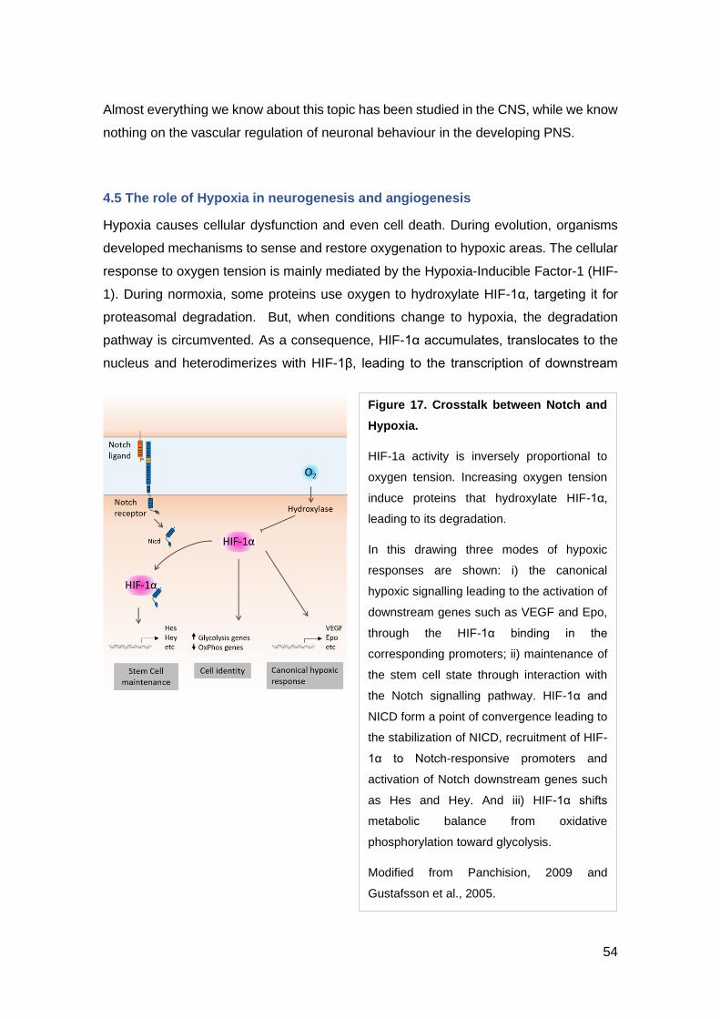
54
Almost everything we know about this topic has been studied in the CNS, while we know
nothing on the vascular regulation of neuronal behaviour in the developing PNS.
4.5 The role of Hypoxia in neurogenesis and angiogenesis
Hypoxia causes cellular dysfunction and even cell death. During evolution, organisms
developed mechanisms to sense and restore oxygenation to hypoxic areas. The cellular
response to oxygen tension is mainly mediated by the Hypoxia-Inducible Factor-1 (HIF-
1). During normoxia, some proteins use oxygen to hydroxylate HIF-1α, targeting it for
proteasomal degradation. But, when conditions change to hypoxia, the degradation
pathway is circumvented. As a consequence, HIF-1α accumulates, translocates to the
nucleus and heterodimerizes with HIF-1β, leading to the transcription of downstream
Figure 17. Crosstalk between Notch and
Hypoxia.
HIF-1a activity is inversely proportional to
oxygen tension. Increasing oxygen tension
induce proteins that hydroxylate HIF-1α,
leading to its degradation.
In this drawing three modes of hypoxic
responses are shown: i) the canonical
hypoxic signalling leading to the activation of
downstream genes such as VEGF and Epo,
through the HIF-1α binding in the
corresponding promoters; ii) maintenance of
the stem cell state through interaction with
the Notch signalling pathway. HIF-1α and
NICD form a point of convergence leading to
the stabilization of NICD, recruitment of HIF-
1α to Notch-responsive promoters and
activation of Notch downstream genes such
as Hes and Hey. And iii) HIF-1α shifts
metabolic balance from oxidative
phosphorylation toward glycolysis.
Modified from Panchision, 2009 and
Gustafsson et al., 2005.
55
genes. Its targets include genes that code for molecules that participate in vasomotor
control, angiogenesis, erythropoiesis, iron metabolism, cell proliferation and cell cycle
control, cell death, and energy metabolism (Semenza, 2006; Sharp and Bernaudin,
2004) (see Fig 17).
4.5.1. Hypoxia regulating angiogenesis
Initially, it was thought that the brain vasculature was fixed structurally at adulthood,
except for pathological deterioration. Surprisingly, upon hypoxic stimulus, CBF and
glucose consumption increase (Beck and Krieglstein, 1987; Sun and Reis, 1994), as well
as the capillary density (Lauro and LaManna, 1997). During the course of hypoxia-
induced angiogenesis, capillary density begins to increase after 1 week of hypoxia
exposure, and it completes after 2 or 3 weeks (Harik et al., 1996, 1995). Several pro-
angiogenic factors, including VEGF, have hypoxia responding elements in their
promoters, making hypoxia a strong inducer of angiogenesis (Chávez et al., 2000;
Pichiule and LaManna, 2002) (see Fig 17). Also, Ang2, which is not expressed in
normoxic conditions, will be induced in ECs following hypoxia (Pichiule and LaManna,
2002). In the presence of growth factors such as VEGF, angiogenesis will start,
otherwise capillaries will undergo apoptotic regression, a process known as de-
adaptation, in case hypoxia lasts for more than 3 weeks (Harik et al., 1996; Pichiule and
LaManna, 2002).
Therefore, there are physiological mechanisms for capillary density increase and
decrease in order to balance capillary density and structure with tissue oxygen balance
and activity.
4.5.2. Hypoxia regulating neurogenesis
Less is known about the effects of hypoxia on adult neurogenesis. Ischaemia-induced
brain damage can be caused by stroke, cerebral artery occlusion, cardiac arrest, or by
coronary artery occlusion. In these cases, NSCs both in the CNS and the PNS show
increased neuronal proliferation and differentiation in response to low oxygen tension
(Morrison et al., 2000; Studer et al., 2000a). In hypoxia conditions the body responds
increasing the expression of erythropoietin (EPO), which promotes the proliferation and
differentiation of erythroid progenitors as well as maturing erythroid cells survival
(Youssoufian et al., 1993). EPO receptors are also found in the developing CNS (Liu et
INTRODUCTION
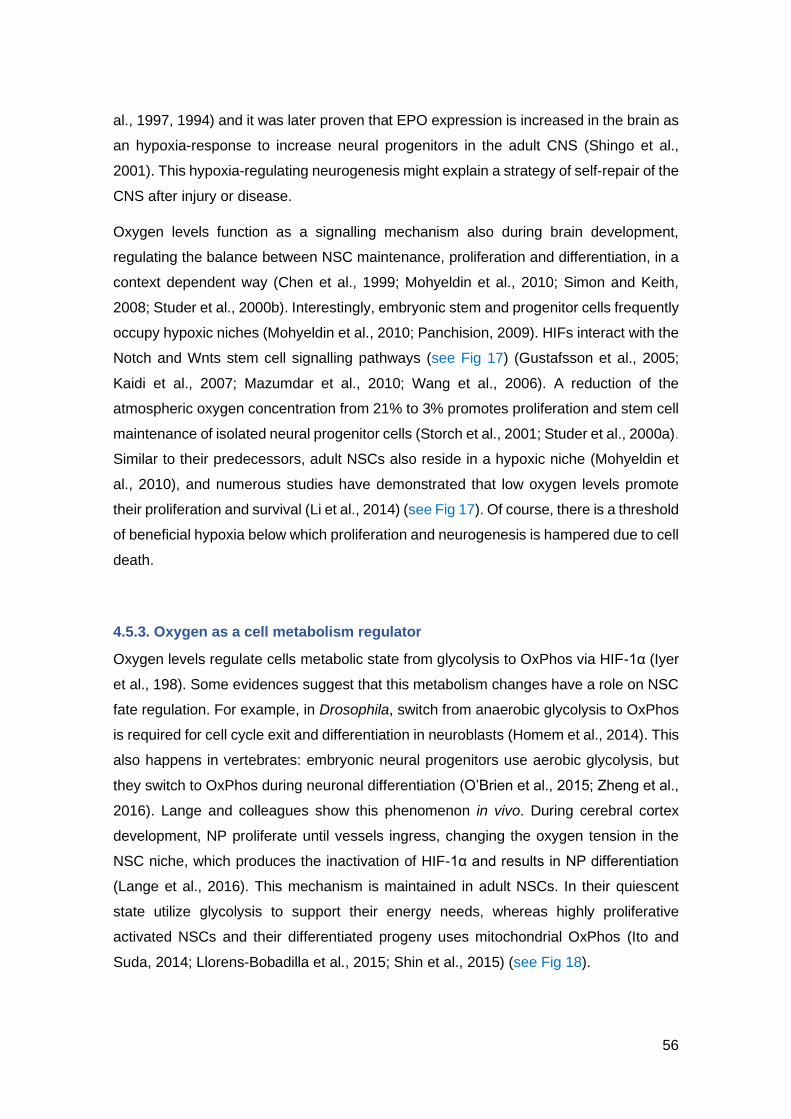
56
al., 1997, 1994) and it was later proven that EPO expression is increased in the brain as
an hypoxia-response to increase neural progenitors in the adult CNS (Shingo et al.,
2001). This hypoxia-regulating neurogenesis might explain a strategy of self-repair of the
CNS after injury or disease.
Oxygen levels function as a signalling mechanism also during brain development,
regulating the balance between NSC maintenance, proliferation and differentiation, in a
context dependent way (Chen et al., 1999; Mohyeldin et al., 2010; Simon and Keith,
2008; Studer et al., 2000b). Interestingly, embryonic stem and progenitor cells frequently
occupy hypoxic niches (Mohyeldin et al., 2010; Panchision, 2009). HIFs interact with the
Notch and Wnts stem cell signalling pathways (see Fig 17) (Gustafsson et al., 2005;
Kaidi et al., 2007; Mazumdar et al., 2010; Wang et al., 2006). A reduction of the
atmospheric oxygen concentration from 21% to 3% promotes proliferation and stem cell
maintenance of isolated neural progenitor cells (Storch et al., 2001; Studer et al., 2000a).
Similar to their predecessors, adult NSCs also reside in a hypoxic niche (Mohyeldin et
al., 2010), and numerous studies have demonstrated that low oxygen levels promote
their proliferation and survival (Li et al., 2014) (see Fig 17). Of course, there is a threshold
of beneficial hypoxia below which proliferation and neurogenesis is hampered due to cell
death.
4.5.3. Oxygen as a cell metabolism regulator
Oxygen levels regulate cells metabolic state from glycolysis to OxPhos via HIF-1α (Iyer
et al., 198). Some evidences suggest that this metabolism changes have a role on NSC
fate regulation. For example, in Drosophila, switch from anaerobic glycolysis to OxPhos
is required for cell cycle exit and differentiation in neuroblasts (Homem et al., 2014). This
also happens in vertebrates: embryonic neural progenitors use aerobic glycolysis, but
they switch to OxPhos during neuronal differentiation (O’Brien et al., 2015; Zheng et al.,
2016). Lange and colleagues show this phenomenon in vivo. During cerebral cortex
development, NP proliferate until vessels ingress, changing the oxygen tension in the
NSC niche, which produces the inactivation of HIF-1α and results in NP differentiation
(Lange et al., 2016). This mechanism is maintained in adult NSCs. In their quiescent
state utilize glycolysis to support their energy needs, whereas highly proliferative
activated NSCs and their differentiated progeny uses mitochondrial OxPhos (Ito and
Suda, 2014; Llorens-Bobadilla et al., 2015; Shin et al., 2015) (see Fig 18).

57
Figure 18. Metabolic pathways used by cells at different differentiation states.
A. Stem cells and differentiated cells use different metabolic pathways, the first preferentially use
glycolysis, while the latest use oxidative phosphorylation (OsPhos). B. Schematic representation of the
differences between OxPhos, anaerobic glycolysis, and aerobic glycolysis. When oxygen is present,
differentiated tissues first metabolize glucose to pyruvate via glycolysis and then completely oxidize
most of that pyruvate in the mitochondria to CO2 during the process of oxidative phosphorylation.
Because oxygen is required as the final electron acceptor to completely oxidize the glucose, oxygen is
essential for this process. When oxygen is limiting, cells can redirect the pyruvate generated by
glycolysis away from mitochondrial oxidative phosphorylation by generating lactate (anaerobic
glycolysis). This generation of lactate during anaerobic glycolysis allows glycolysis to continue (by
cycling NADH back to NAD+), but results in minimal ATP production when compared with oxidative
phosphorylation. Cancer cells tend to convert most glucose to lactate regardless of whether oxygen
is present (aerobic glycolysis). This property is shared by normal proliferative tissues. Mitochondria
remain functional and some oxidative phosphorylation continues in both cancer cells and normal
proliferating cells. Nevertheless, aerobic glycolysis is less efficient than oxidative phosphorylation for
generating ATP.
Modified from Hu et al., 2016 and Vander Heiden, 2009.
INTRODUCTION

58
5. Mechanisms of cell signalling: Filopodia and cytonemes
In the previous section, we have seen that ECs and neuronal cells communicate with
each other. Cell-cell communication can be achieved through different signalling
mechanism: via direct cell-cell contact or juxtracrine signalling, over relatively short
distances or paracrine signalling, and over large distances or endocrine signalling. Cell-
cell direct contacts is probably the most precise spatial and temporal mechanism for the
transference of signalling molecules.
Recently, some cell communication events thought to be occurring via diffusion of
signalling molecules have been demonstrated to be actually via direct cell-cell contacts.
It has been demonstrated, thanks to the advance of live imaging and staining and
reporter tools, that in some cases signals are driven by thin cytoplasmatic protrusions
called signalling filopodia or cytonemes.
5.1 Filopodia
Filopodia are thin, actin-made plasma membrane protrusions that function as antennae
to sense the mechanical and chemical environment of a cell. They have been described
in a multitude of cell types and to have a role on cell migration, ECM adhesion, neurite
outgrowth, tip cell guidance and wound healing.
Two different types of protrusive structures are found in the leading edge of motile cells:
lamellipodia and filopodia. While lamellipodia are sheet-like protrusions, filopodia are
finger-like structures, but both are thin (0.1-0.2 µm and 0.1-0.3 µm, respectively) and
filled with filamentous (F)-actin (Chhabra and Higgs, 2007; Pollard and Borisy, 2003).
Filopodia length ranges from 1 to 100 µm. The actin filaments that compose filopodia are
polarized, there is a rapidly growing end called barbed end and a slowly one called
pointed end. Filopodial protrusions grow and retract rapidly through the actin
polymerization at barbed ends and retrograde flow of the actin filament bundle
(Mallavarapu and Mitchison, 1999). Filopodia have been described in different
organisms and systems with different dynamics, lengths and positions (Welch and
Mullins, 2002).

59
Filopodia formation mechanisms
There are two models that explain how filopodia is formed. The “convergent elongation
model” propose that branched filaments from the lamellipodia cytoskeleton network form
filopodia via the ARP2/3 complex (Gupton and Gertler, 2007). The other model named
“de novo filament nucleation” proposes that filopodial actin filaments are newly formed
by formins (Schirenbeck et al., 2006). Most probably different mechanisms regulate the
formation of actin filaments to produce filopodia elongation, and the relative importance
of each mechanism varies depending on the organism or cell type (Yang et al., 2007).
Despite the growing list of proteins interacting with signalling pathways leading to the
formation of filopodia (Jacquemet et al., 2019), the exact sequence of events is far from
being fully unveiled.
Cellular roles of filopodia
For many years filopodia roles remained unproven because it was not possible to
selectively remove them without compromising the whole cell. Initially, filopodia were
described as protrusions sensing or exploring the environment, but the roles of filopodial
processes are diverse and yet not fully understood (see BOX 2).
Filopodial processes are formed during directed cell migration. Filopodia tips contain
receptors for signalling molecules and ECM molecules, probing the environment and
working as “sticky fingers” that will adhere to the ECM or other cells to migrate by cell
body translocation (Galbraith et al., 2007). Filopodia also allow cell-cell adhesion during
embryonic development and wound healing. Opposing cells send filopodia to facilitate
cell-cell matching, to align and adhere together, a process known as “zippering”, for
example during the closure of the neural tube (Millard and Martin, 2008; Vasioukhin et
al., 2000). Filopodia are part of neuronal growth cones and of endothelial tip cells to
guide axons and new sprouts to their proper targets respectively, thanks to the sensing
of chemoattractants and repulsive cues as previously described (see 3.1. Similar
morphological structures) (Dickson, 2002; Gerhardt et al., 2003). More recently
filopodia have been described to play a role on cell-cell signalling mediation. This
specialized filopodia or signalling filopodia have also been named “cytonemes”.
INTRODUCTION

60
5.2 Cytonemes
During the development of an organism, cell-cell communication through signals is
required to induce cellular behaviours and organize tissues. This can occur between
closely adjacent cells, but also between cells further separated by interstitial fluid, ECM
or other cells. The understanding of developmental patterning is based on positional
information that indicates cells to differentiate accordingly (Wolpert, 1969). The classical
morphogen model, explained by the French Flag model, proposes that cells (or
organizers) secrete diffusible signals creating gradients that are sensed by receiving
cells in a concentration-dependent manner (Crick, 1970; Mathison, 1952; Wolpert,
1969). In the last years morphogens have been intensely investigated and their
functional importance demonstrated (Driever and Nüsslein-Volhard, 1988; Green and
Smith, 1990; Rogers and Schier, 2011). However, increasing experimental evidences
also challenged this model (Kornberg and Roy, 2014; Wolpert, 2016). The morphogen
model cannot explain how signals spread through complex 3D structures, only some
BOX 2 – Methods used to inhibit filopodia formation
Different methods have been used to study cell membrane dynamics. Cytochalasin-
D is the classic drug, with an origin in fungi, used to inhibit actin polymerization by
many laboratories (Lidke et al., 2005). But it has been supplanted by a more effective,
specific and rapid drug, called Latrunculin (Spector et al., 1983). Latrunculin (A and
B) derive from a toxic secreted by a sea sponge that, upon binding to actin monomers
prevents polymerization in a reversible way (Morton et al., 2000). In zebrafish, low
concentrations of Latrunculin B (LatB) have been used to inhibit filopodia formation
without affecting cell morphology or proliferation (Phng et al., 2013). ML141 is
another drug that inhibits filopodia formation by blocking Cdc42/Rac1 GTPase
(Stanganello et al., 2015). Transient knockdown and overexpression of genes have
also been carried out for Cdc42, N-WASP, MyoX, IRSp53 and DN IRSp53 (Lee et
al., 2010; Millard et al., 2005; Nalbant et al., 2004; Stanganello et al., 2015). Finally,
optogenetic tools are starting to be developed (Wu et al., 2009). Of special
importance are the functional studies in vivo, as in in vitro studies cells are removed
from their physiological environment and lack features such as the ECM, mechanical
inputs and signals from the surrounding tissue.

61
cells are targeted by different signals acquiring different fates or physically separated by
other tissues.
Quite recently a new model has been proposed thanks to the advance of imaging
technology and fluorescent staining and reporter lines (see BOX 3). In this model, cell
communication is mediated by filopodia transporting morphogens and establishes
patterns of gene expression and cell differentiation (Ramírez-Weber and Kornberg,
1999). This signalling filopodia have also been called “cytonemes”, which can be
regarded as a subclass of canonical filopodia (Sanders et al., 2013).
Cytonemes were first observed in sea urchin gastrulation (Miller et al., 1995), but the
term “cytoneme” was coined for signalling filopodia in Drosophila imaginal discs
(Ramírez-Weber and Kornberg, 1999). This signalling mechanism has been
documented in flies (Bischoff et al., 2013; Callejo et al., 2011; Chen et al., 2017; Cohen
et al., 2010; Du et al., 2018; Fuwa et al., 2015; González-Méndez et al., 2017; Gradilla
et al., 2014; Huang et al., 2019; Huang and Kornberg, 2015; Inaba et al., 2015; Iwaki et
al., 2005; de Joussineau et al., 2003; Mandal et al., 2007; Peng et al., 2012; Ramírez-
Weber and Kornberg, 1999; Rojas-Ríos et al., 2012; Roy et al., 2014; Sohr et al., 2019),
chick (Sagar et al., 2015; Sanders et al., 2013), and zebrafish (Caneparo et al., 2011;
Eom et al., 2015; Hamada et al., 2014; Luz et al., 2014; Mattes et al., 2018; Stanganello
et al., 2015). Cytonemes are highly dynamic structures that allow spatially restricted
signalling. Different lengths have been observed depending on the distance to the target
cells, ranging from 1 to 380 µm (Caneparo et al., 2011; Luz et al., 2014; Rojas-Ríos et
al., 2012; Sagar et al., 2015; Sanders et al., 2013) (see Table 3). They also have different
velocities of extension and retraction, but also have been described as stable. This
opposite characteristic might be in fact two different states of the same structure.
Dynamic cytonemes correspond to protrusions that have not yet contacted the target
BOX 3 – Technical challenges of filopodia and cytonemes detection
Filopodia extensions are difficult to detect because most of them do not survive
fixation, and those that do, do not retain their normal shape. Antibody staining is thus
problematic. Filopodia can be visualized by cytoplasmatic fluorescence staining,
membrane-tethered GFP or fluorescent proteins fused to filopodia components such
as actin or signalling proteins (in the case of cytonemes). Besides, fluorescence
quenching, phototoxicity and 3D growth through tissues limit or prohibit real-time
imaging.
INTRODUCTION

62
cell, while stable cytonemes have established the contact and are actively signalling
(Sagar et al., 2015). Cytonemes are emerging as a general mechanism for intercellular
crosstalk, next to the classical diffusion models.
5.2.1 Roles of cytonemes
Cytonemes have been described as signalling structures in Drosophila and vertebrates
(amphibians, zebrafish, chick and mouse). They transport fundamental signalling
molecules such as Notch, EGF, FGF, BMP, Wnt and Shh. Additionally, cytonemes can
transport the ligand or the receptor, always achieving a spatially restricted distribution of
the signals. Cytonemes are a mechanism of cell signalling used during development that
allows the selective distribution of signalling molecules to create gradients and patterns
for cell differentiation, and maintaining SCs in their niches (see Fig 19 and Table 3). They
are also involved in processes such as tissue regeneration and cancer progression.
Figure 19. Cytonemes transport ligands
and receptors to create morphogen
gradients.
The signal-sending cell (red) and the signal-
receiving cell (blue) communicate with each
other through cytonemes. Graded distribution
of the signal is achieved by the number of
contacts and distance between cells.
Additionally, cytonemes allow for the
specialization of a single cell without affecting
neighboring cells. Different cytoneme contact
types are possible: A. Cytonemes extending
from the signal-producing cell contact with
specific receptors in the receiving cell. B.
Cytonemes carrying specific receptors extend
from receiving cells to capture the ligand of
the signal-producing cell. C. Cytonemes from
both the signal source and the receptor cells
contact each other for the signal transfer.
Modified from González-Méndez et al., 2019.

63
Cytonemes can produce a graded dispersion of morphogens depending on their length
and contact frequency. This function has been shown in the ligand delivery of Wnt during
neural plate patterning in zebrafish (Luz et al., 2014; Mattes et al., 2018; Stanganello et
al., 2015), in Hh in Drosophila and its homologue Shh in chick limb bud (Bischoff et al.,
2013; Chen et al., 2017; González-Méndez et al., 2017; Gradilla and Guerrero, 2013;
Sanders et al., 2013), and in the receptor delivery of Btl in Drosophila (FGF receptor
homologue) (Du et al., 2018) (see Fig 20 and Table 3).
Cytonemes can also generate spatially patterned cell fates. This role has been described
in the formation of bract cells (a type of thick hair) in Drosophila via Spitz/EGF distribution
(Peng et al., 2012), in the organization of the bristles in the notum via Notch signalling ,
also in Drosophila (Cohen et al., 2010; de Joussineau et al., 2003), in the colours pattern
of butterfly wings (Iwasaki et al., 2017; Ohno and Otaki, 2015), and the pigmented
stripped pattern of zebrafish through Delta/Notch signalling (Eom and Parichy, 2017;
Eom et al., 2015; Hamada et al., 2014) (see Table 3).
It has also been shown that cytonemes can coordinate morphogenesis bridging the
space of physically separated tissues. It is the case of the air sac primordium (ASP) in
Drosophila wing disc. Cells from the ASP send cytonemes to ligand-producing cells (Dpp
Figure 20. Cytonemes transporting signals of the Wnt/Frz pathway in different
organisms.
A. PAC2 zebrafish fibroblast transfected with Wnt8a-GFP and Evi-mCherry. Multiple filopodia
and Wnt8a at the distal tip (arrow) can be seen forming cell-cell contact with another PAC2
fibroblast (min:s). B. Myoblasts labeled with CD8-GFP express Frz-Cherry to contact Wnt-
producing air sac cells in Drosophila.
Modified from Stanganello and Scholpp, 2016; Stanganello et al., 2015.
INTRODUCTION

64
and FGF), to promote cell migration and patterning (Du et al., 2018; Huang and
Kornberg, 2015; Huang et al., 2019; Roy et al., 2014; Sohr et al., 2019) (see Table 3).
Finally, cytonemes have also been described in the maintenance of SC niche. For
example, in the ovary, testis and haematopoietic SCN of Drosophila signals carried by
cytonemes regulate undifferentiated cells (Fuwa et al., 2015; Inaba et al., 2015; Mandal
et al., 2007; Rojas-Ríos et al., 2012) (see Table 3).
5.2.2 Signal trafficking by cytonemes
Several membrane-bound signalling proteins have been observed travelling through
cytonemes, and an increasing number of studies are reporting that this transport of
morphogens is associated with vesicles such as argosomes (Greco et al., 2001),
lipoprotein particles (Panáková et al., 2005), exosomes (Beckett et al., 2013; Gradilla et
al., 2014; Koles et al., 2012) and exosome-like particles (Danilchik et al., 2013; Matusek
et al., 2014).
5.2.3 Signal delivery by cytonemes
Scientists are still trying to understand how cytonemes recognize and contact their target
cell for signal transfer. Two types of contacts have been identified: the first is the
cytoneme-cell body contact, for example found in cytonemes transporting the Wnt8a
ligand in the zebrafish embryo (Mattes et al., 2018; Stanganello et al., 2015) and the
cytonemes carrying the Fz receptor from the ASP of Drosophila wing disc (Huang and
Kornberg, 2015). The second mechanism is the cytoneme-cytoneme contact, for
examples cytonemes carrying receptors for Dpp and FGF in Drosophila wing disc (Du et
al., 2018; Roy et al., 2014), and myoblasts carrying Delta to contact the ASP (Huang and
Kornberg, 2015), in the Hh distribution within the Drosophila epithelia (González-Méndez
et al., 2017) and possibly in Shh signalling in the developing chick limb bud (Sanders et
al., 2013) (see Fig 21).
5.2.4 Cytonemes as synaptic contacts
A synapse is a mechanism of intracellular communication in which the secreting cell by
extending a process can, after crossing other intervening cells, reach the target cell in a

65
highly precise way. These two cells present pre- and postsynaptic in specific membrane
areas. Cytoneme signalling features resemble to synaptic contacts in some aspects.
Cytonemes allow for specific and direct contacts, saving distances between cells, the
signalling is directed from sending to receiving cells, the ligand presentation would be
the parallel to the presynaptic molecules, and the receptor to the postsynaptic molecules.
For these reasons, the new term “morphogenetic synapses” is been used to describe
cytonemes’ contacts. Also, the contact sites at membranes between cytonemes are
similar to the synaptic buttons, with membrane swellings and vesicles release (González-
Méndez et al., 2017; Huang et al., 2019), as well as the space between the cells (Roy et
al., 2014). Of course, differences must not be underestimated, as the complexity of
molecules involved, contact duration and the link to the propagating electric impulse
(reviewed in Kornberg, 2017).
5.2.5 Cytonemes’ contacts establishment and regulation
Finally, cytoneme signalling will depend on the frequency and the stability of these
contacts. Therefore, membrane bound proteins must regulate the establishment of
cytonemes (or cytoneme-cell body) contacts. Some of them have already been identified,
Figure 21. Types of cytoneme contacts.
A. Cytoneme-cell body contact. Single-plane image of a Ror2-mCherry/Wnt8a producing cell
leading to Lrp6-GFP clustering of the responding cell clone, showing the cytoneme contact
site. Scale bars 10 mm. B. Cytoneme-cytoneme contact of mesenchymal cells of the
developing limb bud in chick. Mosaic labelling through Cre-Lox system generates membrane-
palmitoylated mKate2 (a far-red fluorescent protein) or pmeGFP. Scale bar 3 mm.
Image modified from Mattes et al., 2018; Sanders et al., 2013.
INTRODUCTION

66
they are adhesion molecules such as integrins and synaptic adhesion molecules such
as Neuroglian that guide cytonemes sensing the ECM and cell membranes to recognize
and establish physical contacts with the correct cell (Bischoff et al., 2013; González-
Méndez et al., 2017; Huang and Kornberg, 2015; Huang et al., 2019; Roy et al., 2014).
The spatial and temporal establishment of cytonemes must be regulated. Some studies
propose a self-regulatory mechanism between the signalling pathway and the filopodia
formation. For example, FGF in the Drosophila ASP promotes protrusion enhancement,
while low levels of signalling repress protrusion extension (Du et al., 2018) and Wnt8a
cytoneme transport in zebrafish leads to the activation of actin polymerization process
(Mattes et al., 2018; Stanganello et al., 2015). Interestingly, these feed-back loops do
not always function the same way. In the Drosophila ovary stem cell niche cytonemes
grow until they reach the ligand, and impairment of ligand reception causes cytonemes
to grow further (Rojas-Ríos et al., 2012). However, autoregulatory feed-back loops are
not always present: In the wing imaginal disc and abdominal histoblasts (cuticle-
producing cells) cytonemes formation and dynamics are independent of the signalling by
Hh (González-Méndez et al., 2017). Thus, still further work is needed to fully understand
the mechanisms of cytonemes formation and the links to the signalling pathways.
Other cell protrusions with signalling capacities have been described as tunnelling
nanotubes or intracellular bridges (Caneparo et al., 2011; Gerdes et al., 2012).

67
INTRODUCTION

68

69
INTRODUCTION

70
Table 3. Table summarizing structural and functional characteristics of cytonemes
described in different organisms
ASP, air sac primordium; BMP, bone morphogenic protein; Bnl, branchless; Boc, brother of
CDO; BTl, breathless; Cdo, Cam-related/downregulated by oncogenes; Dpp,
decapentaplegic; EGF, epidermal growth factor; FGF, fibroblast growth factor; Fz, frizzled,
Hh, hedgehog, Ptc, patched; Shh, sonic hedgehog; Spi, spitz; Tkv, Thick-Vein; Wg, wingless.
Extra references not included in the text but present in the table: (Holzer et al., 2012)
Table modified from: González-Méndez et al., 2019; Pröls et al., 2016.
71
II. AIMS OF THE THESIS
Cells within an organism develop in association with other cell types around them, which
will constitute the so-called “niche” and with which they communicate to build functioning
organs and tissues. This communication also occurs in the development of neural cells.
As seen in the introduction, BV have emerged as key members of neurogenic niches.
However, only the CNS has been investigated yet.
The aim of this work was to explore the niche in which the cranial ganglia is formed,
focusing in the putative function of BV in neurogenesis, to shed light into the unknown
BV functional role in the developing PNS neurons. My investigations were mainly done
in the SAG, while principal results were also searched in other sensory ganglia.
The specific issues addressed were:
[1] To describe the anatomical relationship between the head vasculature and cranial
ganglia during their development.
[2] To explore a functional role of vasculature on cranial sensory neural development,
focusing in the SAG.
[3] To investigate a functional role of neurogenesis on vascular development.
[4] To search for the mechanisms involved in the neurovascular interaction.
[5] To identify the signalling cues responsible for the identified neurovascular
communication.
AIMS OF THE THESIS

72

73
III. RESULTS
Article 1. Anatomical map of the cranial vasculature and sensory ganglia
RESULTS
Taberner L, Bañón A, Alsina B. Anatomical map of the cranial vasculature and sensory ganglia. J Anat. 2018 Mar 1;232(3):431–9. DOI: 10.1111/joa.12762

85
Article 2. Sensory neurogenesis depends on vascular-neuronal cytoneme
contacts and blood flow
RESULTS
Taberner L, Bañón A, Alsina B. Sensory Neuroblast Quiescence Depends on Vascular Cytoneme Contacts and Sensory Neuronal Differentiation Requires Initiation of Blood Flow. Cell Rep. 2020 Jul 14;32(2). DOI: 10.1016/j.celrep.2020.107903
115
IV. DISCUSSION
During neurogenesis, both in embryo and adult, each of the NSCs behaviours –
quiescence, proliferation, self-renewal and differentiation– has to be tightly balanced.
Otherwise pathologies may arise. Increased self-renewal without differentiation could
result in brain cancer (Palm and Schwamborn, 2010). On the other hand, premature
differentiation at the expense of progenitors’ proliferation can deplete the NSC pool
producing phenotypes of aging.
The development of the nervous system highly depends on the developing vasculature.
The nascent vasculature regulates NSC behaviour and neurogenesis by providing
oxygen but also specific signalling cues. This relationship is maintained in adult
neurogenic regions. ECs and neural cells have evolved to be able to communicate and
ensure their proper development and maintenance.
Despite the importance of the neurovascular crosstalk, we are just beginning to
understand the underlaying mechanisms responsible of it and their implications.
Research on the neurovascular interaction has mainly focused in the adult, while the first
studies during development are starting to emerge. Still, in both cases reports are
focused in different regions of the CNS. Thus, previous studies left a totally unexplored
terrain: the possible regulation of ECs in neurons’ behaviour has remained totally elusive
in the developing PNS.
This thesis explores the vascular regulation of NSCs’ properties in the developmental
PNS, beyond the axonal patterning.
Description of cranial sensory ganglia in a vascular niche
My first work consisted of studying the relative position of sensory ganglia with respect
to head vessels, as other studies did before trying to understand any functional role
between the vascular and the neuronal system (Bates et al., 2002; Javaherian and
Kriegstein, 2009; Mukouyama et al., 2002; Tata et al., 2016). The development and
precise architecture of the zebrafish vasculature have been studied in detail (Isogai et
al., 2001; Ulrich et al., 2011), but whether they develop in close proximity with cranial
ganglia and how had not been addressed before.
DISCUSSION

116
Thanks to the use of reporter transgenic lines for cranial ganglia and vessels, we found
that all sensory ganglia develop in close proximity with at least one BV. We described
the complex 3D interactions in space and time (24, 30, 48 and 72 hpf) between these
two systems. The Tg develops on top of the PHBC in its most anterior part, the aLL and
the pLL relate with the PHBC in their dorso-medial edges, all the epibranchial ganglia
grow on top of an AAs, and the Xg follows the PHS. In particular, we found that the SAG
was in contact with a higher number of BV: two veins, the PHBC and the PHS, and one
artery, the LDA. The analysis of these interactions in 3D allowed us to identify different
domains in cranial ganglia depending on whether cells are in close contact or further
from BV, which might be translated to different cells’ behaviour – for instance more
proliferative. Finally, as we studied the physical interaction over time, we can state that
the contacts perdure, and thus, they might not be something coincidental at some time
points but actively sustained.
Discovering that cranial sensory ganglia were developing in a rich vascular environment
gave us the first hint that BV might be part of the sensory neuronal niche.
Vasculature promotes neural quiescence in cranial sensory ganglia
The function of a niche is to both maintain SCs in quiescent or undifferentiated state and
to control proliferation and differentiation into the correct lineages. Much effort is put,
therefore, on understanding how the same environment can conduct such opposite
effects on SCs.
To functionally evaluate the putative regulation of vasculature on sensory ganglia
development, the avascular clo mutant transgenic line has been used (Stainier et al.,
1995). We analysed the growth dynamics of the SAG in clo mutant versus control
embryos. The results of this project identified that the SAG and Tg volume and cell
number were increased in the avascular mutants compared to control embryos, meaning
that BV play a role in negatively regulating neuroblasts’ proliferation or promoting their
quiescence.
Interestingly, there does not seem to be a common mechanism by which BV regulate
neural behaviour. A good collection of reports demonstrate that BV are required for
neuronal precursors to proliferate, both in the adult and developing brain (Biro et al.,
1994; Gomez-Gaviro et al., 2012; Louissaint et al., 2002; Tan et al., 2016; Tata et al.,
2016). However, very few examples are found in literature where BV are required to

117
restrict neuronal growth, probably promoting quiescence (Delgado et al., 2014; Ottone
et al., 2014). These cases of quiescence promotion have only been described in adult
stages. Thus, our results are the first ones to demonstrate that during neuronal
development BV can also promote quiescence.
Out of these investigations, we also gained a detailed temporal information on the SAG
growth dynamics. These type of quantifications in the cranial ganglia had been hampered
due to their loss of an epithelial character and its complicated 3D organization. Here, by
quantifying overtime the number of neurod cells (neuroblasts) and also islet 2 cells
(postmitotic and differentiated neurons) we have drawn at high resolution the growth
dynamics of the SAG in zebrafish from 30 hpf to 96 hpf. We believe that the quantitative
data provided can be useful for other projects in which growth defects might be taking
place upon specific signalling pathways manipulations in the SAG.
Cranial vasculature development does not require neural cells
Neurovascular communication does not only occur from vessels to neurons, but also
from neurons to vessels. ECs have been reported to sense neural-derived signalling
cues for their patterning and maturation in the limb skin (Li et al., 2013b; Mukouyama et
al., 2002, 2005), and to vascularize the retina (Okabe et al., 2014), cortex (Ma et al.,
2012) and spinal cord (Himmels et al., 2017). In this project we also wanted to assess
the possible requirement of neural cells for the proper development of the cranial
vasculature, but our analysis demonstrated that it develops correctly independently of
the presence of cranial sensory neurons. Therefore, in the cranial system, neurovascular
communication occurs only in one direction, from vessels to neurons.
A possible reason why might be because of developing times: the PHBC is formed (two
initial sprouts merge) at 24 hpf, the time point at which neuroblasts peak in their
delamination from the OV to form the SAG. Thus, the development of the vasculature is
preceding the one of the ganglion. Also, cranial ganglia present complex 3D
morphologies, not “straight paths” that vascular sprouts could use to follow for a correct
patterning. Also, no small vessels penetrate the ganglia, so neural cells do not need to
promote the formation of new sprouts. Finally, and further speculating, if BV are going to
regulate fine decisions such how many neural cells will proliferate or differentiate it is
“safer” -evolutionary speaking- if the first system forms correctly independently of the
system they are going to signal to.
DISCUSSION

118
Signalling Filopodia (Cytonemes) mediate neurovascular cross-talk
When we took a closer look to our neurovascular interacting system, we found that,
despite endothelial and neural cells were separated by a small space, they establish
direct physical contacts through filopodia. Multiple studies have described the close
physical relationship between endothelial and neural cells both during development and
in adult neurogenic niches (Delgado et al., 2014; Ottone et al., 2014; Tan et al., 2016;
Tata et al., 2016; Tavazoie et al., 2008). In some cases, a small distance between BV
and dividing neurons is described and measured, which still makes a significant
difference with the further located non-diving neurons (Tavazoie et al., 2008). In these
cases, communication might occur through diffusible cues. Direct physical contacts had
been described before both in adult and developmental stages, consisting in end-food
projections of neurons enwrapping BV, (Kacem et al., 1998; Tan et al., 2016), but never
through filopodial protrusions.
Further, pharmacological inhibition of filopodia formation demonstrated that these
filopodia are required to transmit the signal that promotes neuroblasts’ quiescence in the
SAG. This signal is not secreted, because BV are still present in the filopodia inhibition
experiment. When filopodia formation is inhibited, the BV-derived signal cannot reach
the SAG. Thus, we demonstrate that these cytoplasmatic protrusions are indeed
signalling filopodia, also named as cytonemes (Ramírez-Weber and Kornberg, 1999).
Signalling filopodia (or cytonemes) between SCs and the cells of their niche participate
in the maintenance of SCs in an undifferentiated state in the ovaries, testis and
haematopoietic stem cell niche, but they have only been described in Drosophila (Fuwa
et al., 2015; Inaba et al., 2015; Mandal et al., 2007; Rojas-Ríos et al., 2012). Thus, our
study is the first one to describe cytonemes in a vertebrate niche. Specifically, we
observe cytonemes in a neurogenic niche between NSCs and ECs. Tavazoie and
colleagues observed that the contact sites between NSC and BV were often small, and
suggested that it might be occurring dynamically in vivo. This hypothesis could not be
assessed as their observations were based in fixed tissues whole mounted or sectioned
(Tavazoie et al., 2008). The use of zebrafish embryos, otherwise, allow for in vivo
imaging, which permitted cytonemes observation, opening the possibility that
neurovascular communication is also occurring through a cytoneme-based mechanism
in other organisms and contexts.

119
Dll4/Notch1 are required for maintenance of neural quiescence
Despite the growing number of reports describing functional roles for ECs on neurons,
the EC-derived molecular cues required for these interactions still remain elusive in many
cases.
Notch signalling pathway play key roles in balancing NSC quiescence and proliferation
(Borghese et al., 2010; Shimojo et al., 2008, 2011), where high Notch activation induces
growth arrest and low Notch cell proliferation, in adult NSC niches. On the other hand,
the two Notch ligands Dll4 and Jag1 are highly expressed in ECs (Benedito et al., 2009),
thus it could be expectable that ECs may also be signalling to Notch-expressing
embryonic and adult NSCs.
Only one study identified ECs regulating Notch in NSCs, through the expression of Jag1.
The Jag1-Notch ligand-receptor binding, together with the Eph-ephrinB2a interaction,
promotes NSC quiescence in the V-SVZ in adult mice (Ottone et al., 2014). In our study,
though, it is not Jag1 but Dll4 the EC ligand binding to Notch. Interestingly, EC-derived
Dll4 has already been linked to SC quiescence maintenance, but in the adult muscle SC
niche (Verma et al., 2018).
Dll4 has been widely studied in the formation and guidance of new angiogenic sprouts.
High amounts of this protein are expressed in tip cells, while low quantities are present
in stalk cells (Hasan et al., 2017). Signalling to otic sensory neuroblasts come from stalk
and not tip ECs. We propose that maybe, small quantities but spatially concentrated in
specialized regions of the cytoplasm -or filopodia tips- are sufficient to signal to another
cell type, the neuroblasts.
In summary, our results support the hypothesis that the Notch ligand Dll4 expressed in
ECs regulate NSCs quiescence, and suggests that, if it is true for adult NSCs in the CNS
and in the developing PNS, it might be a general signalling mechanism present in other
neurogenic regions and even other SC niches.
It is worth mentioning that neuroblasts have been proven to be less dependent on Notch
signalling than NSCs and more responsive to environmental cues for the regulation of
their proliferation (Ables et al., 2010). These findings match with previous studies carried
out in the SAG where other signals, such as IGFs, are also important to regulate
neuroblasts’ proliferation (Alsina et al., 2003; Camarero et al., 2003). Our results add one
piece of information in the canvas of all the signalling cues regulating the SAG
development, where vasculature is now a new player.
DISCUSSION

120
Cytonemes carrying Dll4 and Notch1
An increasing amount of reports show cytoneme signalling associated with vesicle
transport. In our study we did not investigate this feature in detail, but still it is worth
mentioning that rounded thickenings travelling to the distal edges of neuroblasts’
filopodia can be observed in some of the videos. This type of signal transportation has
been described in Drosophila (González-Méndez et al., 2017; Gradilla et al., 2014;
Matusek et al., 2014), chick (Sanders et al., 2013) and zebrafish (Eom and Parichy,
2017; Eom et al., 2015; Luz et al., 2014; Stanganello et al., 2015). Thus, it is not risky to
hypothesise that ECs and neuroblasts might use membranous vesicles containing Dll4
and Notch1 to reach each other.
Cytonemes deliver their signals to target cells through two different ways, some studies
state that cytonemes contact the cell body of their target cells (Huang and Kornberg,
2015; Stanganello et al., 2015), while in others cytoneme-cytoneme contacts have been
observed (Du et al., 2018; González-Méndez et al., 2017; Huang and Kornberg, 2015;
Roy et al., 2014; Sanders et al., 2013). In our particular system, we have been able to
identify cytoneme-cytoneme direct contacts thanks to the physical separation of both
tissues and the different colours of the two transgenic lines used. In other cases, these
direct contacts were not seen. These observations can be explained in different ways,
one possibility is that all cases are cytoneme-cytoneme contact but, in some cases, the
“missing cytoneme” is inside one of the tissues and cannot be seen as all the cells are
stained with the same fluorophore. The other possibility is that the two types of contacts
are occurring at the same time, enhancing the number of signalling contacts. Maybe,
single-cell labelling techniques could be used to solve this question in future research.
The signalling though cytonemes depends on their frequency and stability. In our system,
dynamic and stable filopodia have been observed, being the first probably those that
have not correctly contacted with their target cell yet, and the second ones those that
have and are actively signalling (Sagar et al., 2015). The establishment of cytonemes’
contacts is mediated by adhesion molecules (Bischoff et al., 2013; González-Méndez et
al., 2017; Huang and Kornberg, 2015; Huang et al., 2019; Roy et al., 2014). In some
cases the signalling pathways, the adhesion molecules and the the filopodia formation
machinery are connected via feedback loops in some cases but do not in others (Du et
al., 2018; González-Méndez et al., 2017; Mattes et al., 2018; Rojas-Ríos et al., 2012;
Stanganello et al., 2015). The adhesion molecules involved in the neuroblast-EC contact
in the SAG have not been explored, nor the filopodia formation machinery. However, we

121
can state that they function independently on the Dll4-Notch1 signalling, since filopodial
processes are still formed when Dll4 is downregulated, despite they not being signalling.
On the contrary, it is clear that some signal located in ECs is required for NB to form
filopodia, as absence of BV produces a lack of filopodia in neuroblasts. To explore
deeper in the interdependent mechanisms of the PHBC-SAG cytoneme contacts, the
dynamics/speed of filopodial processes could have been measured in the absence of
Dll4 and neuroblasts (neurog1 -/-), as well as performing gain and loss-of function
experiments for already described adhesion and filopodia formation molecules.
Some researchers state that cytonemes are a new type of “synapsis”, and called them
morphogenetic synapsis, due to their multiple common features such as cell polarization,
directed secretion for communication and membrane domains specialized with receptors
and adhesion molecules (Kornberg, 2017). Increasing evidence shows the relevance of
cytonemes, or signalling filopodia, as a cell-cell communication mechanism, which could
be a general feature of signalling processes in cells that allows a precisely spatio-
temporal regulation, and not only as a sophistication of neuronal synapses. The plasticity
of the cytoneme mode of signalling is especially important in our system: it probably
allows some neuroblasts to be maintained in quiescence, those specific cells that have
been contacted; while others are not touched by cytonemes, and thus can continue to
proliferate, achieving the correct SAG growth dynamics. This selective signalling could
not have been possible with other signalling mechanisms such as signal diffusion.
Blood flow requirement to trigger neuronal differentiation
We have shown that blood flow onset is required for neuronal differentiation in the SAG
at specific developmental stage (from 54 to 60 hpf). Only one previous study described
a similar observation. Fortuna et al. demonstrated that blood flow onset was required for
VMC to be recruited around BV, once there, SMCs drive neuronal differentiation through
PDGFR secretion (Fortuna et al., 2015). However, SMC are not ECs (they have a
different developmental origin) and are not present around the PHBC (Santoro et al.,
2009). Another mechanism must, therefore, be responsible of otic sensory neuronal
differentiation.
Upon blood flow onset, ECs lining the inner surface of BV sense two types of mechanical
stimuli: shear stress, the frictional force tangential to the vessel, and mechanical tension,
the force perpendicular to the flow direction. Shear stress can modify the patterning of
DISCUSSION

122
BV (Chen et al., 2012a), be detected by ECs through the mechanosensitive transcription
factor klf2a (Nicoli et al., 2010), and also produce changes in the ECs gene expression
through the activation of transcription factors, making ECs to “mature” (Chiu and Chien,
2011; Korn and Augustin, 2015; Nicoli et al., 2010; Weijts et al., 2018). Shear stress has
been described to activate Notch signalling in arterial ISVs (Weijts et al., 2018), and to
allow the temporal translocation of Yap1 into the EC nucleus in zebrafish (Nakajima et
al., 2017). Then, it is possible that the transcriptional changes happening on ECs after
blood flow onset promote the expression of a diffusible signalling cue that prompts
neuroblasts to exit cell cycle and terminally differentiate. A signalling mechanism in which
ECs translates blood flow and mechanotransduction into secreted signals for growth or
differentiation of another cell type has been described in a beautiful study in the mouse
liver. Liver ECs sense the blood flow mechanical forces through β-integrin and VEGFR3,
which in turn promote the secretion of growth factors that promote hepatocyte
proliferation (Lorenz et al., 2018).
On the other hand, blood flow onset will also transport multiple molecules that could also
have a signalling function on neuronal differentiation. Unfortunately, it is technically
difficult to separate these two factors in vivo.
Finally, still one last possibility should be noted. BV or blood flow-derived signals might
not be essential for instructing cell-fate or differentiation, but rather might just provide
survival cues for a certain type of new-born neuron (Kirschenbaum and Goldman, 1995;
Leventhal et al., 1999), a selection role that could give the illusion of an effect on
differentiation instruction. To discard this possibility, a cell death essay (TUNEL or
caspase 3) could be done from 54 to 72 hpf. If the EC-derived signal is a survival cue,
Nifedipine treated and avascular mutant embryos should present more apoptosis than
control embryos, while proliferation would be occurring in all conditions. However, we
consider this not to be very plausible as the neurod reporter (Tg(neurod:egfp) nl1) labels
also the differentiated population (Islet2+ cells are also GFP+) and we do not see this
population to decrease at 72 hpf or apoptotic bodies in the confocal images.
We conclude that a signal carried by or secreted upon blood flow is promoting neuronal
differentiation at late stages and that this mechanism is independent from the first one
as no filopodia is observed at late stages.

123
Blood vessels-derived cues upon blood flow onset mediating neuronal
differentiation
BV can exert their roles on NSC niches via angiocrine signalling or via delivery of
nutrients or oxygen (Cleaver and Dor, 2012; Ramasamy et al., 2015).
The transcriptome analysis of SAG neuroblasts in clo mutant vs control embryos allowed
us, first of all, to demonstrate that neuronal cells were actually sensing signals from BV
and that this was translated into changes in their transcriptional profile. More specifically,
what we observed is that neuroblasts of the avascular mutant embryos presented far
more downregulated than upregulated genes when compared to neuroblasts of control
embryos. This may indicate that a whole signalling cascade is being activated in the
control case, while it is not in the mutant, rather than different signalling pathways are
acting in each condition. This also matches with a cell differentiation arrest in the
avascular mutant.
Among the differentially expressed genes we identified some related to oxygen tension
and mitochondria metabolism. egln2 (Egl nine homolog 2) is a cellular oxygen sensor
involved in the degradation of HIF (Epstein et al., 2001), rdh8a (retinol dehydrogenase
8a) is involved in redox processes (Haller et al., 2010), and abat (4-aminobutyrate
aminotransferase) and rmnd1 (Required for meiotic nuclear division protein 1 homolog)
in mitochondrial metabolism, translation and OxPhos (Besse et al., 2015; Janer et al.,
2012, 2015). Therefore, this means that control embryos are sensing oxygen, high levels
of Egln2 are degrading proteins and a shift to OxPhos metabolism is occurring, while this
in not happening in the avascular mutants.
Our results provide evidences that, at 2 dpf, zebrafish larvae start to depend on BV for a
better oxygen perfusion to produce the correct number of otic sensory differentiated
neurons. Oxygen tension increase is required for cranial ganglia sensory neurons
differentiation, while reduced levels of oxygen -either by complete lack of vasculature or
by blood flow inhibition- prevent the switch to neuronal differentiation. Importantly, this
does not imply an increase in the neuroblasts’ proliferation at late stages, demonstrating
that the mechanisms for proliferation control and differentiation are independent in the
cranial sensory ganglia system. Also, as already discussed, it is not related to cell death,
as the number of neurod+ cells does not decrease at 72 hpf.
Current evidence has demonstrated that, in addition to growth factors, the metabolic
pathways of glycolysis and OxPhos provide important signals for stem-cell renewal and
DISCUSSION

124
differentiation, respectively. Oxygen tension impinges on the shift from glycolysis to
OxPhos, through the regulation of HIF-1α. Absence of OxPhos is a defining feature of
embryonic SCs, and it is required to maintain their pluripotency (Harvey et al., 2016;
Ryall et al., 2015). In fact, certain SCs reside in hypoxic niches (Mohyeldin et al., 2010;
Panchision, 2009). Some cases where the metabolic switch from aerobic glycolysis to
OxPhos regulates NSC differentiation have been described both in invertebrates and
vertebrates through the regulation of HIF-1α (Homem et al., 2014; Lange et al., 2016),
despite it is yet a rather unexplored terrain. Osteogenic and adipogenic differentiation of
mesenchymal SCs is also regulated by the loss of aerobic glycolysis and increase of
OxPhos and HIF-1α downregulation (Palomäki et al., 2013; Shum et al., 2015; Zhang et
al., 2013). In other cases, the switch is done by the regulation of epigenetics, redox and
reactive oxygen species, a part from HIF-1α (Hawkins et al., 2016; Kida et al., 2015;
Ryall et al., 2015). Thus, we think that a possible mechanism by which neuroblasts need
blood flow is for oxygen tension to increase and switch their metabolism to OxPhos,
whose transcriptional changes will ultimately lead to neuronal differentiation.
A direct demonstration of this mechanism could be gained with additional experiments
such as growing avascular mutant and control embryos in hypoxic or hyperoxia
chambers (Khaliullina-skultety et al., 2017; Rouhi et al., 2010), reproducing the
phenotype of differentiation arrest in control embryos and rescuing the phenotype with
egln2 overexpression in clo mutant embryos, respectively. Also, by doing loss-of-function
experiments of egln2 in WT embryos (by MO or CRISPR), or assessing the differential
transcriptional levels of HIF target genes involved in cell cycle control and energy
metabolism (such as IGF2, p21, or GLUT1) (Sharp and Bernaudin, 2004) between clo
mutant and control embryos.
Finally, our results do not exclude that BV also participate in the otic sensory neurons’
differentiation via angiocrine factors as suggested by previous work in vitro (Shen et al.,
2008), and in vivo in adult NSCs (Delgado et al., 2014; Ottone et al., 2014; Tan et al.,
2019).
Vasculature as a key component of is part of the stem cell niche
In the previous sections we have seen that vasculature, in addition to its well established
role of oxygen and nutrients transport, also regulates neural expansion and
differentiation. But this new feature is not exclusive in the nervous system, few studies

125
have started to demonstrate a role for vascular cells in stem cell niches of other organs
such as the liver, bone, kidney and pancreas during development (Bjarnegård et al.,
2004; Han et al., 2011; Lammert et al., 2001; Lorenz et al., 2018; Ramasamy et al., 2015;
Serluca et al., 2002; Yoshitomi and Zaret, 2004), and at adult stages in the lungs, skin,
spermatogonia and muscle (Paquet-Fifield et al., 2009; Rafii et al., 2015; Verma et al.,
2018; Yoshida et al., 2007).
The vascular system is, thus, emerging as a signalling centre with functions that involve
the hosting and regulation of stem and progenitor cells, in many different organs
systems.
Neurovascular linked diseases
As widely described in the introduction, the nervous and vascular system share
similarities at the morphological, cellular and molecular levels. Additionally, they are also
functionally interdependent. It is, therefore, not surprising that both neuronal and
vascular defects are also involved in the pathogenesis of neurodegenerative disorders.
Owing to the aging of the world population and lack of effective treatments,
neurodegenerative diseases are predicted to grow to epidemic proportions in the
following decades (Gammon, 2014).
Traditionally, neurological diseases have been thought to arise primarily from an intrinsic
malfunctioning of the neurons themselves. However, alterations of the neurovascular
crosstalk can also contribute to numerous neurological disorders (Zlokovic, 2008).
Conditions where vascular abnormalities affect the nervous system include stroke,
vascular dementia, hypertension and diabetes. Thus, there is a growing interest in
exploring the potential contribution of neurovascular dysfunction to neurodegeneration
(de la Torre, 2017).
Vascular defects also affect certain neurodegenerative diseases. Patients suffering
Alzheimer disease present profound changes in the cerebrovascular structure. A
hallmark of this disease is the deposition of β-amyloid peptide in the brain parenchyma
(Zlokovic, 2005). This protein also accumulates in BV producing vasoconstriction,
reduced blood flow, vascular resistance and EC damage increase (Kalaria, 2002).
Certain studies document that the vascular alterations are present before the disease
onset, suggesting that they might contribute to its initiation (Zlokovic, 2008).
DISCUSSION

126
Frontotemporal dementia is the leading cause of dementia in the middle age (Olney et
al., 2017). Pre-symptomatic patients present reduced CFB in the frontal, parietal and
temporal cortex (Dopper et al., 2016) as well as alterations in the BBB (Janelidze et al.,
2017). Another example is found in patients suffering Parkinson disease, who show
capillary damage and fragmentation (Guan et al., 2013), reduced CBF (Borghammer et
al., 2010) and increased VEGF in the CSF (Janelidze et al., 2017). Vascular alterations
are also present in dementia with Lewy bodies, where CBF is reduced in several parts
of the cortex (Galasko, 2017). These changes in the CBF appear just before the onset
of the disease (Roquet et al., 2016) and are probably due to reduced energy metabolism
(Ishii et al., 2015). Cerebral autosomal-dominant arteriopathy with subcortical infarcts
and leukoencephalopathy (CADASIL) is a syndrome that leads to cognitive decline and
dementia. This condition is due to a progressive degeneration of cerebral arterioles
which produces arterial narrowing and hypoperfusion, caused by mutations in Notch3
(Kalimo et al., 2002). Finally, in diabetic neuropathies, endoneural perfusion deficits and
chronic ischaemia is caused by several vascular abnormalities such as the thickening of
the basement membrane and EC hyperplasia.
Subtle mutations in the VEGF promoter have been identified to produce lower levels of
VEGF protein. As a consequence, people with these VEGF gene variations present
deficits in spinal cord perfusion and chronic ischaemia, this specially affects motor
neurons due to their active metabolism (Oosthuyse et al., 2001). They also have a higher
risk of suffering amyotrophic lateral sclerosis (ALS).
Other neurological disorders, such as brain tumours are characterized by excessive
vascularization. Some studies suggest the existence of a cancer stem cell-vascular niche
complex, where ECs promote brain tumour survival (Calabrese et al., 2007; Klagsbrun
et al., 1976).
Therapeutic implications of the neurovascular interaction
We are gaining evidence on the importance of BV in NSC behaviour control. Elucidating
the signals and mechanisms by which ECs participate in the regulation of neural cells
behaviour in vivo will shed light into the first tools to target neurovasculature
therapeutically and to build SC-based therapies to treat neural diseases and disorders.
However, we first need a solid understanding of the intrinsic and extrinsic interactions of
NSC regulation. For example, the realization that angioneurins have multitasking

127
properties in angiogenesis, but also in neuroregeneration and neuroprotection, has
created opportunities to explore novel therapeutic opportunities. Some examples of
clinical trials are the use of VEGF and EPO for neurotrophic support and angiogenesis
in ALS (Azzouz et al., 2004; Lambrechts et al., 2003; Storkebaum et al., 2004, 2005),
Parkinson’s Disease (Chen et al., 2007; Tian et al., 2007), diabetic neuropathies (Hobson
et al., 2000; Isner et al., 2001; Schratzberger et al., 2001; Sondell et al., 2000) and
ischemic injuries (Nishijima et al., 2007); and VEGF inhibitors for brain tumours (Loges
et al., 2009; Miletic et al., 2009).
Another therapeutic option is to take profit of the potential of NSCs to home into injured
brain regions (Imitola et al., 2004; Thored et al., 2006). The use of SCs to repair the
nervous system is of great potential, but being able to control the expansion and
differentiation of these NSC is still very challenging. Additionally, despite still
controversial, some studies claim to detect adult hippocampal neurogenesis (AHN) in
humans (Choi and Tanzi, 2019). Therefore, therapies focused on the promotion of AHN
will be useful for prevention and treatment of neurodegenerative diseases.
A fundamental understanding of neuronal and vascular interactions will have far-
reaching benefits in developing strategies to treat psychological and neurodegenerative
diseases, brain tumours and stroke. Understanding how these interactions occur during
development and adulthood in physiological circumstances will help to understand
whether they become reactivated or dysregulated during pathological conditions. With
this thesis I have participated in the expansion of the knowledge on the neurovascular
interaction, demonstrating that it is also required for the proper sensory cranial ganglia
development. Our work in the SAG might serve as a model system for a general
mechanism found in other regions of the nervous system.
Neurovascular linked diseases in the Inner Ear
Hearing loss, resulting from aging, genetic predisposition or environmental exposure to
noise, infections or ototoxic drugs, is one of the most prevalent chronic conditions
affecting older adults, affecting the 5% of the world population – or 466 million people–
according to the World Health Foundation (https://www.who.int/news-room/fact-
sheets/detail/deafness-and-hearing-loss). Hearing loss impacts in the individual ability to
communicate, with social consequences such as loneliness, isolation, and frustration,
particularly among older people; and this can be an important driver of morbidity and
DISCUSSION
DISCUSSION

128
mortality. Vertigo, on the other hand, affects between the 20 and 30% of the population,
and can cause nausea, vomiting, ataxia, and even anxiety and depression (Karatas,
2008).
Hearing loss and vertigo commonly reflect a loss of sensory HC and auditory synapse
degeneration (Kujawa and Liberman, 2009; Spoendlin, 1975). Loss of VIIIth ganglion
neurons also significantly contributes to hearing loss. Otic progenitor and SC
transplantation is a potential strategy to replace lost neurons (Chen et al., 2012b; Rivolta,
2015). Also, promising advances are being done in the development of inner ear
organoids (Koehler et al., 2017; Munnamalai and Fekete, 2017). Thus, understanding
the mechanisms and key signals that regulate otic sensory neurons’ development will
accelerate efforts to a future where replacement therapies are real. The work of this
thesis has contributed on it and has put BV derived signals in the scenario. More
specifically, this new knowledge may benefit some deficiencies in the auditory and
vestibular system that have been described to be due to neurovascular alterations
(Jensen et al., 2004; Lago et al., 2018; Paul et al., 2016).
Future perspectives
In summary, an array of cell-cell contact, diffusible and circulating cues from BV have
been demonstrated to regulate NSC quiescence, self-renewal, proliferation,
differentiation and migration. Key questions would be to understand how neural cells
integrate all these signals, especially when they can have overlapping or antagonistic
roles; and how BV signalling is integrated with the rest of the information coming from
other cells that conform the niche in which NSCs develop. It would also be of particular
interest to know how this is coupled in time, in the short term for temporal needs -for
instance damage- but also in the long term, since these interactions have been seen
from embryogenesis to adult stages. Additionally, ECs do not only regulate neural cells’
behaviour, but they also instruct on liver and pancreas formation. This positions the
vascular system as a new signalling system that may be responsible of many organs’
regulation.
After demonstrating the requirement of BV in the correct development of the SAG in
zebrafish, it would be interesting to elucidate whether this interaction is common in all
vertebrates and explore whether vasculature is also involved in the vestibular and
cochlear ganglion development in mice. Vascular interactions can be supressed by the

129
use of transgenic lines such as VEGF-A, VEGF-R2 and VEGF-R1 mutants (Carmeliet et
al., 1996; Ferrara et al., 1996; Fong et al., 1995; Shalaby et al., 1995), and blood flow
can be altered by the use of BDM and epinephrine (Lorenz et al., 2018).
Also, we have positioned zebrafish as an interesting model organism to study the
neurovascular interaction. A detailed spatiotemporal study has been carried out for the
cranial sensory ganglia found in the PNS. I think it would be interesting to continue
exploring the neurovascular interaction in the CNS, both during development and adult
stages in this animal model. Zebrafish show some interesting features that could
complement the knowledge gained in mouse studies. The vascularization has been
deeply studied and thus well known, besides, it is highly stereotypic (Gore et al., 2012;
Isogai et al., 2001). On the side of the adult neurogenesis, in contrast to mammals,
zebrafish present more NSC niches in the adult brain (Chapouton et al., 2007; Kaslin et
al., 2008, 2009). Thus, zebrafish provide invaluable comparative material for extracting
core mechanisms in vertebrate adult NSC niche, and to study the role of vasculature in
it.
Another interesting line of study could be to explore the possible contribution of BV other
organs different to the nervous system. As developed in the introduction, some studies
are demonstrating that BV are required for the proper development of kidney, pancreas
and liver (Han et al., 2011; Lammert et al., 2001; Serluca et al., 2002). Taking the optical
and imaging advantages zebrafish embryos offer, we could study whether
communication in these cases also occurs through cytonemes.
Finally, it would also be very exciting to study whether the signalling through Dll4 is also
taking place in other species such as the chick and the mouse embryo, where a good
Dll4 mutant line is present (Duarte et al., 2004), and thus elucidate if it is a general
neurovascular signalling mechanism.
With this thesis I hope to contribute to the knowledge on how sensory neurogenesis is
regulated positioning the vasculature as a new and important player.
DISCUSSION

130

131
V. CONCLUSIONS
1. The SAG and the rest of the cranial sensory ganglia develop in a vascular
environment.
2. The SAG is surrounded by three main vessels: two veins, the PHBC and the
PHS, and one artery, the LDA.
3. Vasculature is required to negatively control neuroblasts proliferation or to
maintain neuroblasts quiescent in the SAG, at early stages, and not to regulate
specification (neurog1+) or differentiation (Islet 2+).
4. Signalling from ECs to neuroblasts to negatively control proliferation or to
maintain quiescence is also required in other sensory ganglia such as the Tg, at
early stages.
5. Direct physical contacts between SAG neuroblasts and PHBC ECs are
established through filopodia growing from both cell types.
6. Some of these filopodia protrusions are dynamic while other are stable.
7. Some of these interactions are made through filopodia-filopodia contacts.
8. Neurovascular interaction through filopodia contacts is occurring in other sensory
ganglia such as the Tg and aLL.
9. Filopodia extending in PHBC ECs are not randomly exploring the environment,
but spatiotemporally regulated.
10. At early stages, cranial vasculature develops correctly independently of the
presence of sensory cranial ganglia neurons.
11. SAG neuroblasts need some signal coming from BV to form filopodia, whereas
ECs do not need signals from neuroblasts for filopodia formation.
CONCLUSIONS

132
12. Filopodial contacts are required to deliver the signal that prevents neuroblasts’
proliferation or promotes quiescence, and thus are cytonemes.
13. Dll4 is involved in the promotion of neuroblasts’ quiescence. This signalling is
specific to the presence of blood vessels, as Dll4-MO injected avascular mutants
do not present additional effects.
14. Dll4 is not required for filopodia formation in ECs nor in SAG neuroblasts.
15. Blood vessels are required at late stages to trigger Islet2+ sensory neuronal
differentiation in the SAG. This also affects the axonal density entering the
hindbrain.
16. At late stages, vasculature is required for sensory neurons’ differentiation, but not
for the formation of other cell types in the inner ear such as HC of the maculae
and cristae.
17. Blood vessels are also required for Islet2+ sensory neuronal differentiation in
other ganglia such as the Tg and the Xg, at late stages.
18. Blood flow is required in a specific time window to trigger Islet 2+ sensory neuronal
differentiation.
19. The neuroblasts transcriptome of avascular mutant and control embryos reveals
changes in oxygen sensing and metabolic status.

133
VI. APPENDIX
As a result of this thesis, other data and tools were generated which are not included in
the articles.
Table with the exact number of neurod+ and Islet2+ cells from 30 to 96 hpf
Table 4. Number of Neurod + and Islet2 + cells from 30 to 96 hpf. Tables showing the
number of quantified cells per embryo of the Neurod + cell population and Islet 2+ cell
population. Neurod + cells were counted using the TgBAC(neurod:egfp) nl1 and DAPI staining.
Islet2 + cells were counted using immunostaing with Islet 2 antibody, which is nuclear.
APPENDIX
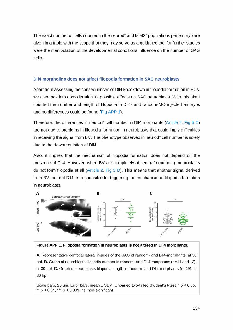
134
The exact number of cells counted in the neurod+ and Islet2+ populations per embryo are
given in a table with the scope that they may serve as a guidance tool for further studies
were the manipulation of the developmental conditions influence on the number of SAG
cells.
Dll4 morpholino does not affect filopodia formation in SAG neuroblasts
Apart from assessing the consequences of Dll4 knockdown in filopodia formation in ECs,
we also took into consideration its possible effects on SAG neuroblasts. With this aim I
counted the number and length of filopodia in Dll4- and random-MO injected embryos
and no differences could be found (Fig APP 1).
Therefore, the differences in neurod+ cell number in Dll4 morphants (Article 2, Fig 5 C)
are not due to problems in filopodia formation in neuroblasts that could imply difficulties
in receiving the signal from BV. The phenotype observed in neurod+ cell number is solely
due to the downregulation of Dll4.
Also, it implies that the mechanism of filopodia formation does not depend on the
presence of Dll4. However, when BV are completely absent (clo mutants), neuroblasts
do not form filopodia at all (Article 2, Fig 3 D). This means that another signal derived
from BV -but not Dll4- is responsible for triggering the mechanism of filopodia formation
in neuroblasts.
Figure APP 1. Filopodia formation in neuroblasts is not altered in Dll4 morphants.
A. Representative confocal lateral images of the SAG of random- and Dll4-morphants, at 30
hpf. B. Graph of neuroblasts filopodia number in random- and Dll4-morphants (n=11 and 13),
at 30 hpf. C. Graph of neuroblasts filopodia length in random- and Dll4-morphants (n=49), at
30 hpf.
Scale bars, 20 µm. Error bars, mean ± SEM. Unpaired two-tailed Student’s t-test. * p ˂ 0.05,
** p ˂ 0.01, *** p ˂ 0.001. ns, non-significant.

135
Development of the transgenic line Tg(UAS:DN Irsp53, IRES:nls-eGFP)
We generated a new transgenic line with the aim to inhibit filopodia formation in a tissue
specific way, through the expression of the dominant-negative (DN) form of the Irsp53
protein.
The Irsp53 (Insulin-receptor substrate p53) is a protein with multiple domains through
which it interacts with different proteins and thus develops different activities related to
filopodia formation. Through its I-BAR domain it interacts with plasma-membrane lipids
inducing membrane deformation (Choi et al., 2005; Henne et al., 2007; Mattila et al.,
2007). IRSp53 binds to CDC42, ENA/VASP and WAVE2 proteins via the SH3 domain
(Krugmann et al., 2001; Miki and Takenawa, 2002; Scita et al., 2008), and also to EPS8,
an actin filament capping protein (Disanza et al., 2006; Funato et al., 2004). The DN form
of this protein contains a 4K mutation where four lysines (142, 143, 146 and 147) are
altered. This mutation prevents the bundling of actin fibres due to the disruption of the
Irsp53 actin-binding sites (Millard et al., 2005).
The construct was built through the Tol2 Gateway system (Kwan et al., 2007). We first
amplified the full-sequence of the DN Irsp53 from a plasmid kindly provided by Erez Raz
(Meyen et al., 2015), with PRC primers 5’-
ATAGGTACCGCTTAGATCCACCATGTCTCG-3’ and 5’-
ATACTCGAGATCTCACTGTGCAAAGCCTGCCAT-3’. Then, we incorporated the
sequence of the DN Irsp53 in a pME-MCS (237), with KpnI and XhoI. Then, the resulting
plasmid was subcloned with the p5E-4xrnUAS (Akitake et al., 2011) and p3E-IRES-
nlsEGFPpA (391) in a pDestTol2CG2 (395), which contains a cmlc2:egfp transgenesis
marker. The final plasmid was co-injected with Tol2 mRNA into 1-cell-stage embryos in
the double TgBAC(neurod:EGFP) nl1;(Kdrl:ras-mCherry) s896 background (see Fig APP 2
A and B). A stable transgenic line was established.
In parallel, we grow in our fish facility the Tg(Flk1:Gal4) bw9, sent by Julien Bertrand (Kim
et al., 2014; Mahony et al., 2016). We expected that the later crossing of these two
transgenic lines would prevent the formation of filopodia specifically in ECs, to study their
requirement for the correct expansion of SAG neuroblasts (see Fig APP 2 B and C).
Unfortunately, the resulting embryos of the cross between Tg(Flk1:Gal4) bw9 and
Tg(UAS:DN Irsp53, IRES:nls-eGFP) showed a very weak expression of the construct
(see Fig APP 2 D), as observed by the few number of nuclei expressing eGFP and the
presence of filopodia in ECs.
APPENDIX

136
After discussion with Shigetomo Fukuhara, I learned that Tg(Flk1:Gal4) bw9 was not a
strong promoter of UAS transgenic lines (Wakayama et al., 2015) and asked for a better
one, Tg(fli1:Gal4ff) ubs3, to Heinz-Georg Belting (Paatero et al., 2018).
Regrettably, similar results were obtained when Tg(UAS:DN Irsp53, IRES:nls-eGFP)
was crossed to Tg(fli1:Gal4ff) ubs3 , which lead me to think that there must be additional
problems with the new transgenic line such as silencing.
As possible solutions the LR reaction can be repeated in a pDestTol2pA2 (394), which
contains insulators, to favour the construct expression, and the resulting plasmid will be
co-injected in with Tol2 mRNA into 1-cell-stage embryos in the double
TgBAC(neurod:EGFP) nl1; (fli1:Gal4ff) ubs3.
Figure APP 2. Generation of the Tg(UAS:DN Irsp53, IRES:nls-eGFP).
A. Plasmid generated with the Tol2 Gateway system containing the 4xUAS, the IRSp53-4K
and the IRES:nls-GFP. B. This construct was injected together with Tol2 mRNA in 1-cell
stage embryos of the TgBAC(neurod:gfp)nl1; (Kdrl:ras-mCherry)s896 background and grown
to adulthood. To be later crossed with Tg(Flk-1:Gal4) fish. C. Illustration of the expected
phenotype. Embryos Gal4+ and UAS+ express the marker of green nuclei specifically in BV
and ECs do not form filopodia. D. Lateral confocal image of an embryo resulting of the cross
illustrated in B, where some green nuclei can be observed.

137
Generation of mRNA DN Irsp53
Finally, the injection of DN Irsp53 mRNA could be used as a last solution to produce a
genetic alteration of the filopodia formation, to complement the results obtained with the
LatB treatment.
The full-length DN Irsp53 cDNA was amplified from the plasmid provided by Erez Raz
(Meyen et al., 2015) using the PCR primers 5’-
ATAGAATTCGCTTAGATCCACCATGTCTCG-3’ and 5’-
ATACTCGAGATCTCACTGTGCAAAGCCTGCCAT-3’ and subcloned into a pCS2+ with
EcoRI and XhoI (see Fig APP 3).
mRNA would transcribed from ApaI or KpnI or NotI -linearized template using SP6 RNA
polymerase and the mMessage mMachine kit (Ambion) and injected in
TgBAC(neurod:EGFP) nl1;(Kdrl:ras-mCherry) s896 embryos to visualize filopodia formation
and analyse the phenotype in SAG neuroblasts.
Generation of mRNA Dll4-eGFP
In other to visualize the location of Dll4 and assess if it is located in EC cytoneme tips,
we decided to generate a fusion protein of Dll4 with GFP.
The full-length Dll4 cDNA was first amplified from a plasmid provided by Stefan Schulte-
Merker (Hogan et al., 2009), using the PCR primers 5’-
ATTTGGTACCACCATGGCAGCTTGGCTCACCTT-3’ and 5’-
ATTTGGATCCGTACCTCAGTTGCTATGAC-3’ to eliminate the STOP codon and
Figure APP 3. Generation of mRNA
DN Irsp53.
Illustration of the generated plasmid
containing the IRSp53-4K inside the
pCS2+.
APPENDIX

138
cloned into the pEGFP-N2 vector to obtain a C-terminal eGFP fusion protein, with KpnI
and BamHI. Then, it was subcloned into the pCS2+ with KpnI and NotI (Fig APP 4).
mRNA will transcribed from XbaI-linearized template using SP6 RNA polymerase and
the mMessage mMachine kit (Ambion) and injected in TgBAC(neurod:EGFP)
nl1;(Kdrl:ras-mCherry) s896 embryos to visualize Dll4 expression and analyse the
phenotype in SAG neuroblasts.
Development of the transgenic line Tg(UAS:Dll4-eGFP)
We generated a new transgenic line that, after its cross with Tg(fli1:Gal4ff) ubs3 , would
allow us to i) visualize the expression Dll4 at a sub-cellular resolution, to ideally see its
presence in EC filopodia tips; and ii) produce a gain-of-function EC-specific experiment
to then analyse its effect in SAG neuroblasts.
The construct was generated through the Tol2 Gateway system (Kwan et al., 2007)
The full-length Dll4 cDNA was amplified from a plasmid provided by Stefan Schulte-
Merker (Hogan et al., 2009), using the PCR primers 5’-
GGGGACAAGTTTGTACAAAAAAGCAGGCTTCAACATGGCAGCTTGGCTCACCTTT
CTC-3’ and 5’-
GGGGACCACTTTGTACAAGAAAGCTGGGTGTACCTCAGTTGCTATGACACATTCA
CTC-3’ to eliminate the STOP codon and cloned into the pDNOR221 (218) vector, via
a BP reaction. Then, the resulting plasmid was subcloned with the p5E-4xrnUAS
(Akitake et al., 2011), p3E-EGFPpA (366) in a pDestTol2pA2 (394), which produced a
fusion protein of Dll4 with eGFP under the regulation of a UAS promoter (Fig APP 6).
Figure APP 4. Generation of mRNA
Dll4-eGFP.
Illustration of the generated plasmid
containing the Dll4-eGFP fusion
protein inside the pCS2+.

139
The final plasmid was co-injected with Tol2 mRNA into 1-cell-stage embryos in the
double TgBAC(neurod:EGFP) nl1; (fli1:Gal4ff) ubs3 background. The resulting embryos
are currently growing in the fish facility.
Development of the transgenic line Tg(Dll4 KO) with CRISPR technology
To complement the results obtained by the Dll4 knock-down experiment with MO, we
would like to study the effect of a Dll4 knock-out (KO) mutant in the SAG neuroblasts.
One dll4 mutant is already available in the zebrafish community, the dll4j16e1 (Leslie et al.,
2007). This line carries a dominant haplo-insufficient mutation that produces phenotypes
similar but less severe than the ones seen in Dll4 morphants (Hasan et al., 2017; Leslie
et al., 2007).
With the aim to generate a Dll4 mutation with a stronger phenotype I designed two
scRNA to target 2 of the 4 functional domains of Dll4: 5’-AGTATTCCCCCTTCCAGCCA-
3’ and 5’-TCTGTCGACCTGGCTACACC-3’ (Fig APP 7), and co-injected them with
trRNA and Cas9 protein in 1-cell-stage embryos in the double TgBAC(neurod:EGFP)
nl1;(Kdrl:ras-mCherry) s896 background. The resulting CRISPR embryos are currently
growing in the fish facility. Some of these embryos were used to obtain genomic DNA
Figure APP 6. Generation of the
transgenic line Tg(UAS:Dll4-eGFP).
Illustration of the generated plasmid
containing the 4xUAS and the Dll4-eGFP
fusion protein.
APPENDIX

140
and to assess the efficiency of the scRNA by PCR amplification of the targeted
sequences. In 3/5 embryos the scRNA target sequence was altered.
Figure APP 7. Dll4 functional domains and scRNA used to mutate Dll4.
Full sequence of Dll4 and its 4 functional domains: MNNL in blue, DSL in yellow, EGF1 in
pale green and EGF2 in pink. The 2 scRNA used to target the sequence of Dll4 with the
Cas9 proteins are highlighted in orange (targeting on the functional domain DSL) and in
green (targeting the functional domain EGF1). Their strands, FW and RV primers to amplify
and sequence the targeted region and its product size are also given.
Name scRNA Strand Fw Rv Product size
Dll4 scRNA D2 AGTATTCCCCCTTCCAGCCA - AAAGAAAGTTGGAAGTAGGGGC CAAAGCCTCACTCCTCAAGAAT 273
Dll4 scRNA D3 TCTGTCGACCTGGCTACACC + TTTTGCACACACCATAAACCAT CAATTACTCACCGTGCAAAGAC 173

141
VII. REFERENCES
Abbott, N.J. (2002). Astrocyte–endothelial interactions and blood–brain barrier
permeability. J. Anat 200, 629–638.
Abelló, G., Khatri, S., Giráldez, F., and Alsina, B. (2007). Early regionalization of the otic
placode and its regulation by the Notch signaling pathway. Mech. Dev. 124, 631–645.
Ables, J.L., Decarolis, N.A., Johnson, M.A., Rivera, P.D., Gao, Z., Cooper, D.C., Radtke,
F., Hsieh, J., and Eisch, A.J. (2010). Notch1 Is Required for Maintenance of the
Reservoir of Adult Hippocampal Stem Cells. J Neurosci 30, 10484–10492.
Adam, J., Myat, A., Le Roux, I., Eddison, M., Henrique, D., Ish-Horowicz, D., and Lewis,
J. (1998). Cell fate choices and the expression of Notch, Delta and Serrate homologues
in the chick inner ear: parallels with Drosophila sense-organ development. Development
125, 4645–4654.
Adams, R.H., and Alitalo, K. (2007). Molecular regulation of angiogenesis and
lymphangiogenesis. Nat. Rev. Mol. Cell Biol. 8, 464.
Adams, R.H., and Klein, R. (2000). Eph Receptors and Ephrin Ligands: Essential
Mediators of Vascular Development. Trends Cardiovasc. Med. 10, 183–188.
Adams, R.H., Wilkinson, G.A., Weiss, C., Diella, F., Gale, N.W., Deutsch, U., Risau, W.,
and Klein, R. (1999). Roles of ephrinB ligands and EphB receptors in cardiovascular
development: Demarcation of arterial/venous domains, vascular morphogenesis, and
sprouting angiogenesis. Genes Dev. 13, 295–306.
Aird, W.C. (2007). Phenotypic heterogeneity of the endothelium: I. Structure, function,
and mechanisms. Circ. Res. 100, 158–173.
Akitake, C.M., Macurak, M., Halpern, M.E., and Goll, M.G. (2011). Transgenerational
analysis of transcriptional silencing in zebrafish. Dev. Biol. 352, 191–201.
Alfonso, J., Le Magueresse, C., Zuccotti, A., Khodosevich, K., and Monyer, H. (2012).
Diazepam binding inhibitor promotes progenitor proliferation in the postnatal SVZ by
reducing GABA signaling. Cell Stem Cell 10, 76–87.
Ali, S.A., Pappas, I.S., and Parnavelas, J.G. (1998). Collagen type IV promotes the
differentiation of neuronal progenitors and inhibits astroglial differentiation in cortical cell
REFERENCES

142
cultures. Dev. Brain Res. 110, 31–38.
Allen-Sharpley, M.R., Tjia, M., and Cramer, K.S. (2013). Differential Roles for EphA and
EphB Signaling in Segregation and Patterning of Central Vestibulocochlear Nerve
Projections. PLoS One 8, e78658.
Alsina, B., and Whitfield, T.T. (2017). Sculpting the labyrinth: Morphogenesis of the
developing inner ear. Semin. Cell Dev. Biol. 65, 47–59.
Alsina, B., Giraldez, F., and Varela-Nieto, I. (2003). Growth Factors and Early
Development of Otic Neurons: Interactions between Intrinsic and Extrinsic Signals. Curr.
Top. Dev. Biol. 57, 177–206.
Altman, J. (1962). Are New Neurons Formed in the Brains of AduIt Mammals? Science.
135, 0–1.
Altman, J., and Das, G.D. (1965). Autoradiographic and histological evidence of
postnatal hippocampal neurogenesis in rats. J. Comp. Neurol. 124, 319–335.
Alves, J.A.J., Barone, P., Engelender, S., Fróes, M.M., and Menezes, J.R.L. (2002).
Initial stages of radial glia astrocytic transformation in the early postnatal anterior
subventricular zone. J. Neurobiol. 52, 251–265.
Alzheimer, C., and Werner, S. (2002). Fibroblast growth factors and neuroprotection.
Adv. Exp. Med. Biol. 513, 335–351.
Andermann, P., Ungos, J., and Raible, D.W. (2002). Neurogenin1 Defines Zebrafish
Cranial Sensory Ganglia Precursors. Dev. Biol. 251, 45–58.
Anderson, K.D., Pan, L., Yang, X., Hughes, V.C., Walls, J.R., Dominguez, M.G.,
Simmons, M. V, Burfeind, P., Xue, Y., Wei, Y., et al. (2011). Angiogenic sprouting into
neural tissue requires Gpr124, an orphan G protein-coupled receptor. Proc. Natl. Acad.
Sci. U. S. A. 108, 2807–2812.
Andreu-Agulló, C., Morante-Redolat, J.M., Delgado, A.C., and Farĩas, I. (2009). Vascular
niche factor PEDF modulates notch-dependent stemness in the adult subependymal
zone. Nat. Neurosci. 12, 1514–1523.
Androutsellis-Theotokis, A., Rueger, M.A., Park, D.M., Mkhikian, H., Korb, E., Poser,
S.W., Walbridge, S., Munasinghe, J., Koretsky, A.P., Lonser, R.R., et al. (2009).
Targeting neural precursors in the adult brain rescues injured dopamine neurons. Proc.
Natl. Acad. Sci. 106, 13570–13575.

143
Anthony, T.E., Klein, C., Fishell, G., and Heintz, N. (2004). Radial glia serve as neuronal
progenitors in all regions of the central nervous system. Neuron 41, 881-90.
Armulik, A., Abramsson, A., and Betsholtz, C. (2005). Endothelial / Pericyte Interactions.
Circ Res 97, 512–523.
Armulik, A., Genové, G., Mäe, M., Nisancioglu, M.H., Wallgard, E., Niaudet, C., He, L.,
Norlin, J., Lindblom, P., Strittmatter, K., et al. (2010). Pericytes regulate the blood–brain
barrier. Nature 468, 557–561.
Artavanis-Tsakonas, S., Rand, M.D., and Lake, R.J. (1999). Notch Signaling: Cell Fate
Control and Signal Integration in Development. Science. 284, 770–776.
Arvidsson, A., Collin, T., Kirk, D., Kokaia, Z., and O, L. (2002). Neuronal replacement
from endogenous precursors in the adult. Nat Med 8, 963–970.
Attwell, D., and Laughlin, S.B. (2001). An Energy Budget for Signaling in the Grey Matter
of the Brain. J. Cereb. Blood Flow Metab. 21, 1133–1145.
Attwell, D., Buchan, A.M., Charpak, S., Lauritzen, M., MacVicar, B.A., and Newman, E.A.
(2010). Glial and neuronal control of brain blood flow. Nature 468, 232–243.
Avallone, B., Porritiello, M., Esposito, D., Mutone, R., Balsamo, G., and Marmo, F.
(2003). Evidence for hair cell regeneration in the crista ampullaris of the lizard Podarcis
sicula. Hear. Res. 178, 79–88.
Azzouz, M., Ralph, G.S., Storkebaum, E., Walmsley, L.E., Mitrophanous, K.A.,
Kingsman, S.M., Carmeliet, P., and Mazarakis, N.D. (2004). VEGF delivery with
retrogradely transported lentivector prolongs survival in a mouse ALS model. Nature 429,
413–417.
Baeyens, N., Nicoli, S., Coon, B.G., Ross, T.D., Van den Dries, K., Han, J., Lauridsen,
H.M., Mejean, C.O., Eichmann, A., Thomas, J.-L., et al. (2015). Vascular remodeling is
governed by a VEGFR3-dependent fluid shear stress set point. Elife 4, e04645.
Baker, C.V.H., and Bronner-Fraser, M. (2001). Vertebrate Cranial Placodes I. Embryonic
Induction. Dev. Biol. 232, 1–61.
Baker, C. V, Stark, M.R., Marcelle, C., and Bronner-Fraser, M. (1999). Competence,
specification and induction of Pax-3 in the trigeminal placode. Development 126, 147–
156.
REFERENCES

144
Baluk, P., Lee, C.G., Link, H., Ator, E., Haskell, A., Elias, J.A., and Mcdonald, D.M.
(2004). Regulated Angiogenesis and Vascular Regression in Mice Overexpressing
Vascular Endothelial Growth Factor in Airways. Am. J. Pathol. 165, 1071–1085.
Bates, D., Taylor, G.I., and Newgreen, D.F. (2002). The pattern of neurovascular
development in the forelimb of the quail embryo. Dev. Biol. 249, 300–320.
Bates, D., Taylor, G.I., Minichiello, J., Farlie, P., Cichowitz, A., Watson, N., Klagsbrun,
M., Mamluk, R., and Newgreen, D.F. (2003). Neurovascular congruence results from a
shared patterning mechanism that utilizes Semaphorin3A and Neuropilin-1. Dev. Biol.
255, 77–98.
Bauer, S., and Patterson, P.H. (2006). Leukemia Inhibitory Factor Promotes Neural Stem
Cell Self-Renewal in the Adult Brain. J. Neurosci. 26, 12089–12099.
Beck, T., and Krieglstein, J. (1987). Cerebral circulation, metabolism, and blood-brain
barrier of rats in hypocapnic hypoxia. Am J Physiol 252, H504-12.
Beckett, K., Monier, S., Palmer, L., Alexandre, C., Green, H., Bonneil, E., Raposo, G.,
Thibault, P., Le Borgne, R., and Vincent, J.P. (2013). Drosophila S2 cells secrete
wingless on exosome-like vesicles but the wingless gradient forms independently of
exosomes. Traffic 14, 82–96.
Begbie, J., and Graham, A. (2001). Integration Between the Epibranchial Placodes and
the Hindbrain. Science. 294, 595–598.
Begbie, J., Ballivet, M., and Graham, A. (2002). Early Steps in the Production of Sensory
Neurons by the Neurogenic Placodes. Mol. Cell. Neurosci. 21, 502–511.
Beis, D., and Stainier, D.Y.R. (2006). In vivo cell biology: following the zebrafish trend.
Trends Cell Biol. 16, 105–112.
Bell, D., Streit, A., Gorospe, I., Varela-Nieto, I., Alsina, B., and Giraldez, F. (2008). Spatial
and temporal segregation of auditory and vestibular neurons in the otic placode. Dev.
Biol. 322, 109–120.
Belluzzi, O., Benedusi, M., Ackman, J., and LoTurco, J.J. (2003). Electrophysiological
differentiation of new neurons in the olfactory bulb. J. Neurosci. 23, 10411–10418.
Benedito, R., Roca, C., Sörensen, I., Adams, S., Gossler, A., Fruttiger, M., and Adams,
R.H. (2009). The Notch Ligands Dll4 and Jagged1 Have Opposing Effects on
Angiogenesis. Cell 137, 1124–1135.

145
Bergers, G., and Song, S. (2005). The role of pericytes in blood-vessel formation and
maintenance. Neuro Oncol. 7, 452-64.
Bertrand, N., Castro, D.S., and Guillemot, F. (2002). Proneural genes and the
specification of neural cell types. Nat. Rev. Neurosci. 3, 517–530.
Besse, A., Wu, P., Bruni, F., Donti, T., Graham, B.H., Craigen, W.J., McFarland, R.,
Moretti, P., Lalani, S., Scott, K.L., et al. (2015). The GABA Transaminase, ABAT, Is
Essential for Mitochondrial Nucleoside Metabolism. Cell Metab. 21, 417–427.
Betsholtz, C., Lindblom, P., and Gerhardt, H. (2005). Role of pericytes in vascular
morphogenesis. EXS 94, 115-25.
Bianchi, L.M., and Gray, N.A. (2002). EphB receptors influence growth of ephrin-B1-
positive statoacoustic nerve fibers. Eur. J. Neurosci. 16, 1499–1506.
Biro, S., Yu, Z.X., Fu, Y.M., Smale, G., Sasse, J., Sanchez, J., Ferrans, V.J., and
Casscells, W. (1994). Expression and subcellular distribution of basic fibroblast growth
factor are regulated during migration of endothelial cells. Circ. Res. 74, 485–494.
Bischoff, M., Gradilla, A.C., Seijo, I., Andrés, G., Rodríguez-Navas, C., González-
Méndez, L., and Guerrero, I. (2013). Cytonemes are required for the establishment of a
normal Hedgehog morphogen gradient in Drosophila epithelia. Nat. Cell Biol. 15, 1269–
1281.
Bjarnegård, M., Enge, M., Norlin, J., Gustafsdottir, S., Fredriksson, S., Abramsson, A.,
Takemoto, M., Gustafsson, E., Fässler, R., and Betsholtz, C. (2004). Endothelium-
specific ablation of PDGFB leads to pericyte loss and glomerular, cardiac and placental
abnormalities. Development 131, 1847–1857.
Bjornsson, C.S., Apostolopoulou, M., Tian, Y., and Temple, S. (2015). It takes a village:
Constructing the neurogenic niche. Dev. Cell 32, 435–446.
Black, J.E., Sirevaag, A.M., and Greenough, W.T. (1987). Complex experience promotes
capillary formation in young rat visual cortex. Neurosci. Lett. 83, 351–355.
Black, J.E., Isaacs, K.R., Anderson, B.J., Alcantara, A.A., and Greenough, W.T. (1990).
Learning causes synaptogenesis, whereas motor activity causes angiogenesis, in
cerebellar cortex of adult rats. Proc. Natl. Acad. Sci. U. S. A. 87, 5568–5572.
Black, J.E., Zelazny, A.M., and Greenough, W.T. (1991). Capillary and mitochondrial
support of neural plasticity in adult rat visual cortex. Exp. Neurol. 111, 204–209.
REFERENCES

146
Blaess, S., Graus-Porta, D., Belvindrah, R., Radakovits, R., Pons, S., Littlewood-Evans,
A., Senften, M., Guo, H., Li, Y., Miner, J., et al. (2004). β1-Integrins Are Critical for
Cerebellar Granule Cell Precursor Proliferation. J. Neurosci. 24, 3402–3412.
Blanco, R., and Gerhardt, H. (2013). VEGF and Notch in tip and stalk cell selection. Cold
Spring Harb. Perspect. Med. 3, 1–20.
Blentic, A., Chambers, D., Skinner, A., Begbie, J., and Graham, A. (2011). The formation
of the cranial ganglia by placodally-derived sensory neuronal precursors. Mol. Cell.
Neurosci. 46, 452–459.
Blinder, P., Tsai, P.S., Kaufhold, J.P., Knutsen, P.M., Suhl, H., and Kleinfeld, D. (2013).
The cortical angiome: an interconnected vascular network with noncolumnar patterns of
blood flow. Nat. Neurosci. 16, 889–897.
Bok, J., Raft, S., Kong, K.-A., Koo, S.K., Dräger, U.C., and Wu, D.K. (2011). Transient
retinoic acid signaling confers anterior-posterior polarity to the inner ear. Proc. Natl.
Acad. Sci. 108, 161–166.
Borghammer, P., Chakravarty, M., Jonsdottir, K.Y., Sato, N., Matsuda, H., Ito, K.,
Arahata, Y., Kato, T., and Gjedde, A. (2010). Cortical hypometabolism and
hypoperfusion in Parkinson’s disease is extensive: probably even at early disease
stages. Brain Struct. Funct. 214, 303–317.
Borghese, L., Dolezalova, D., Opitz, T., Haupt, S., Leinhaas, A., Steinfarz, B., Koch, P.,
Edenhofer, F., Hampl, A., and Brüstle, O. (2010). Inhibition of Notch Signaling in Human
Embryonic Stem Cell–Derived Neural Stem Cells Delays G1/S Phase Transition and
Accelerates Neuronal Differentiation In Vitro and In Vivo. Stem Cells 28, 955–964.
Bovetti, S., Hsieh, Y.-C., Bovolin, P., Perroteau, I., Kazunori, T., and Puche, A.C. (2007).
Blood Vessels Form a Scaffold for Neuroblast Migration in the Adult Olfactory Bulb. J.
Neurosci. 27, 5976–5980.
Bray, S.J. (2006). Notch signalling: A simple pathway becomes complex. Nat. Rev. Mol.
Cell Biol. 7, 678–689.
Breier, G., Albrecht, U., Sterrer, S., and Risau, W. (1992). Expression of vascular
endothelial growth factor during embryonic angiogenesis and endothelial cell
differentiation. Development 114, 521–532.
Breunig, J.J., Silbereis, J., Vaccarino, F.M., Šestan, N., and Rakic, P. (2007). Notch

147
regulates cell fate and dendrite morphology of newborn neurons in the postnatal dentate
gyrus. Proc. Natl. Acad. Sci. 104, 20558 – 20563.
Breuskin, I., Bodson, M., Thelen, N., Thiry, M., Borgs, L., Nguyen, L., Stolt, C., Wegner,
M., Lefebvre, P.P., and Malgrange, B. (2010). Glial but not neuronal development in the
cochleo-vestibular ganglion requires Sox10. J. Neurochem. 114, 1827–1839.
Brown, S.T., Martin, K., and Groves, A.K. (2003). Molecular Basis of Inner Ear Induction.
Curr Top Dev Biol. 57, 115–149.
Burd, G.D., and Nottebohm, F. (1985). Ultrastructural characterization of synaptic
terminals formed on newly generated neurons in a song control nucleus of the adult
canary forebrain. J. Comp. Neurol. 240, 143–152.
Calabrese, C., Poppleton, H., Kocak, M., Hogg, T.L., Fuller, C., Hamner, B., Oh, E.Y.,
Gaber, M.W., Finklestein, D., Allen, M., et al. (2007). A Perivascular Niche for Brain
Tumor Stem Cells. Cancer Cell 11, 69–82.
Callejo, A., Bilioni, A., Mollica, E., Gor, N., Andrés, G., Ibáñez, C., Torroja, C., Doglio, L.,
Sierra, J., and Guerrero, I. (2011). Dispatched mediates Hedgehog basolateral release
to form the long-range morphogenetic gradient in the Drosophila wing disk epithelium.
Proc Natl Acad Sci U S A 108, 12591–12598.
Calvo, C.F., Fontaine, R.H., Soueid, J., Tammela, T., Makinen, T., Alfaro-Cervello, C.,
Bonnaud, F., Miguez, A., Benhaim, L., Xu, Y., et al. (2011). Vascular endothelial growth
factor receptor 3 directly regulates murine neurogenesis. Genes Dev. 25, 831–844.
Camarero, G., Avendaño, C., Fernández-Moreno, C., Villar, A., Contreras, J., de Pablo,
F., Pichel, J.G., and Varela-Nieto, I. (2001). Delayed Inner Ear Maturation and Neuronal
Loss in Postnatal Igf-1-Deficient Mice. J. Neurosci. 21, 7630–7641.
Camarero, G., Leon, Y., Gorospe, I., De Pablo, F., Alsina, B., Giraldez, F., and Varela-
Nieto, I. (2003). Insulin-like growth factor 1 is required for survival of transit-amplifying
neuroblasts and differentiation of otic neurons. Dev. Biol. 262, 242–253.
van Campenhout, E. (1935). Experimental researches on the origin of the acoustic
ganglion in amphibian embryos. J. Exp. Zool. 72, 175–193.
Caneparo, L., Pantazis, P., Dempsey, W., and Fraser, S.E. (2011). Intercellular bridges
in vertebrate gastrulation. PLoS One 6.
Cantos, R., Cole, L.K., Acampora, D., Simeone, A., and Wu, D.K. (2000). Patterning of
REFERENCES

148
the mammalian cochlea. Proc. Natl. Acad. Sci. U. S. A. 97, 11707–11713.
Carleton, A., Petreanu, L.T., Lansford, R., Alvarez-Buylla, A., and Lledo, P.M. (2003).
Becoming a new neuron in the adult olfactory bulb. Nat. Neurosci. 6, 507–518.
Carmeliet, P. (2000a). Mechanisms of angiogenesis and arteriogenesis. Nat Med 6, 389–
395.
Carmeliet, P. (2000b). One cell, two fates. Nature 408, 43–45.
Carmeliet, P. (2003). Blood vessels and nerves: Common signals, pathways and
diseases. Nat. Rev. Genet. 4, 710–720.
Carmeliet, P., and Jain, R.K. (2011). Molecular mechanisms and clinical applications of
angiogenesis. Nature 473, 298–307.
Carmeliet, P., and Tessier-Lavigne, M. (2005). Common mechanisms of nerve and blood
vessel wiring. Nature 436, 193–200.
Carmeliet, P., Ferreira, V., Breier, G., Pollefeyt, S., Kieckens, L., Gertsenstein, M.,
Fahrig, M., Vandenhoeck, A., Harpal, K., Eberhardt, C., et al. (1996). Abnormal blood
vessel development and lethality in embryos lacking a single VEGF allele. Nature 380,
435–439.
Casper, K.B., and McCarthy, K.D. (2006). GFAP-positive progenitor cells produce
neurons and oligodendrocytes throughout the CNS. Mol. Cell. Neurosci. 31, 676–684.
Cauli, B., and Hamel, E. (2010). Revisiting the role of neurons in neurovascular coupling
. Front. Neuroenergetics 2, 9.
Cayre, M., Courtès, S., Martineau, F., Giordano, M., Arnaud, K., Zamaron, A., and
Durbec, P. (2013). Netrin 1 contributes to vascular remodeling in the subventricular zone
and promotes progenitor emigration after demyelination. Development 140, 3107–3117.
Chapouton, P., Jagasia, R., and Bally-Cuif, L. (2007). Adult neurogenesis in non-
mammalian vertebrates. BioEssays 29, 745–757.
Charron, F., and Tessier-Lavigne, M. (2007). The Hedgehog, TGF-beta/BMP and Wnt
families of morphogens in axon guidance. Adv Exp Med Biol. 621, 116–133.
Chávez, J.C., Agani, F., Pichiule, P., and LaManna, J.C. (2000). Expression of hypoxia-
inducible factor-1α in the brain of rats during chronic hypoxia. J. Appl. Physiol. 89, 1937–
1942.
149
Chen, E.Y., Fujinaga, M., and Giaccia, A.J. (1999). Hypoxic microenvironment within an
embryo induces apoptosis and is essential for proper morphological development.
Teratology 60, 215–225.
Chen, Q., Jiang, L., Li, C., Hu, D., Bu, J. wen, Cai, D., and Du, J. lin (2012a).
Haemodynamics-Driven Developmental Pruning of Brain Vasculature in Zebrafish. PLoS
Biol. 10.
Chen, W., Jongkamonwiwat, N., Abbas, L., Eshtan, S.J., Johnson, S.L., Kuhn, S., Milo,
M., Thurlow, J.K., Andrews, P.W., Marcotti, W., et al. (2012b). Restoration of auditory
evoked responses by human ES-cell-derived otic progenitors. Nature 490, 278.
Chen, W., Huang, H., Hatori, R., and Kornberg, T.B. (2017). Essential basal cytonemes
take up Hedgehog in the Drosophila wing imaginal disc . Development 144, 3134–3144.
Chen, Z., Asavaritikrai, P., Prchal, J.T., and Noguchi, C.T. (2007). Endogenous
Erythropoietin Signaling Is Required for Normal Neural Progenitor Cell Proliferation *. J.
Biol. Chem. 282, 25875–25883.
Cheng, A., Wang, S., Cai, J., Rao, M.S., and Mattson, M.P. (2003). Nitric oxide acts in a
positive feedback loop with BDNF to regulate neural progenitor cell proliferation and
differentiation in the mammalian brain. Dev. Biol. 258, 319–333.
Chenn, A., Zhang, Y.A., Chang, B.T., and McConnell, S.K. (1998). Intrinsic Polarity of
Mammalian Neuroepithelial Cells. Mol. Cell. Neurosci. 11, 183–193.
Chhabra, E.S., and Higgs, H.N. (2007). The many faces of actin: matching assembly
factors with cellular structures. Nat. Cell Biol. 9, 1110.
Chiaramello, S., Dalmasso, G., Bezin, L., Marcel, D., Jourdan, F., Peretto, P., Fasolo,
A., and De Marchis, S. (2007). BDNF/ TrkB interaction regulates migration of SVZ
precursor cells via PI3-K and MAP-K signalling pathways. Eur. J. Neurosci. 26, 1780–
1790.
Chichung Lie, D., Nakashima, K., Gage, F.H., Shi, Y., Yu, R.T., Taupin, P., Ray, J., and
Evans, R.M. (2004). Expression and function of orphan nuclear receptor TLX in adult
neural stem cells. Nature 427, 78–83.
Chiu, J.-J., and Chien, S. (2011). Effects of Disturbed Flow on Vascular Endothelium:
Pathophysiological Basis and Clinical Perspectives. Physiol. Rev. 91, 327–387.
Choi, S.H., and Tanzi, R.E. (2019). Is Alzheimer’s Disease a Neurogenesis Disorder?
REFERENCES

150
Cell Stem Cell 25, 7–8.
Choi, J., Ko, J., Racz, B., Burette, A., Lee, J.-R., Kim, S., Na, M., Lee, H.W., Kim, K.,
Weinberg, R.J., et al. (2005). Regulation of Dendritic Spine Morphogenesis by Insulin
Receptor Substrate 53, a Downstream Effector of Rac1 and Cdc42 Small GTPases. J.
Neurosci. 25, 869–879.
Ciccolini, F., and Svendsen, C.N. (1998). Fibroblast Growth Factor 2 ( FGF-2 ) Promotes
Acquisition of Epidermal Growth Factor ( EGF ) Responsiveness in Mouse Striatal
Precursor Cells : Identification of Neural Precursors Responding to both EGF and FGF-
2. 18, 7869–7880.
Cleaver, O., and Dor, Y. (2012). Vascular instruction of pancreas development. 2843,
2833–2843.
Clelland, C., Choi, M., Romberg, C., Clemenson Jr, G., Fragniere, A., Tyers, P.,
Jessberger, S., Saksida, L., Barker, R., Gage, F., et al. (2009). A functional role for adult
hippocampal neurogenesis in spatial pattern separation. J. Bus. Law 325, 210–213.
Codega, P., Silva-Vargas, V., Paul, A., Maldonado-Soto, A.R., DeLeo, A.M., Pastrana,
E., and Doetsch, F. (2014). Prospective Identification and Purification of Quiescent Adult
Neural Stem Cells from Their In Vivo Niche. Cell 82, 545–559.
Cohen, M., Georgiou, M., Stevenson, N.L., Miodownik, M., and Baum, B. (2010).
Dynamic Filopodia Transmit Intermittent Delta-Notch Signaling to Drive Pattern
Refinement during Lateral Inhibition. Dev. Cell 19, 78–89.
Coleman, B., Fallon, J.B., Pettingill, L.N., de Silva, M.G., and Shepherd, R.K. (2007).
Auditory hair cell explant co-cultures promote the differentiation of stem cells into bipolar
neurons. Exp. Cell Res. 313, 232–243.
Colín-Castelán, D., Phillips-Farfán, B. V., Gutiérrez-Ospina, G., Fuentes-Farias, A.L.,
Báez-Saldaña, A., Padilla-Cortés, P., and Meléndez-Herrera, E. (2011). EphB4 is
developmentally and differentially regulated in blood vessels throughout the forebrain
neurogenic niche in the mouse brain: Implications for vascular remodeling. Brain Res.
1383, 90–98.
Conover, J., Doetsch, F., Garcia-Verdugo, J., Gale, N., Yancopoulos, G., and Alvarez-
Buylla, A. (2000). Disruption of Eph / ephrin signaling affects migration and proliferation
in the adult subventricular zone. Nat Neurosci. 3, 1091–1097.

151
Corada, M., Nyqvist, D., Orsenigo, F., Caprini, A., Giampietro, C., Taketo, M.M., Iruela-
Arispe, M.L., Adams, R.H., and Dejana, E. (2010). The Wnt/β-catenin pathway
modulates vascular remodeling and specification by upregulating Dll4/notch signaling.
Dev. Cell 18, 938–949.
Corrales, C.E., Pan, L., Li, H., Liberman, M.C., Heller, S., and Edge, A.S.B. (2006).
Engraftment and differentiation of embryonic stem cell–derived neural progenitor cells in
the cochlear nerve trunk: Growth of processes into the organ of corti. J. Neurobiol. 66,
1489–1500.
Crick, F. (1970). Diffusion in Embryogenesis. Nature 225, 420–422.
Crouch, E.E., Liu, C., Silva-Vargas, V., and Doetsch, F. (2015). Regional and Stage-
Specific Effects of Prospectively Purified Vascular Cells on the Adult V-SVZ Neural Stem
Cell Lineage. J. Neurosci. 35, 4528–4539.
Croucher, S.J., and Tickle, C. (1989). Characterization of epithelial domains in the nasal
passages of chick embryos: spatial and temporal mapping of a range of extracellular
matrix and cell surface molecules during development of the nasal placode.
Development 106, 493–509.
Cullen, M., Elzarrad, M.K., Seaman, S., Zudaire, E., Stevens, J., Yang, M.Y., Li, X.,
Chaudhary, A., Xu, L., Hilton, M.B., et al. (2011). GPR124, an orphan G protein-coupled
receptor, is required for CNS-specific vascularization and establishment of the blood-
brain barrier. Proc. Natl. Acad. Sci. U. S. A. 108, 5759–5764.
Culver, J.C., Vadakkan, T.J., and Dickinson, M.E. (2013). A Specialized Microvascular
Domain in the Mouse Neural Stem Cell Niche. PLoS One 8, e53546.
D’amico-Martel, A., and Noden, D.M. (1983). Contributions of placodal and neural crest
cells to avian cranial peripheral ganglia. Am. J. Anat. 166, 445–468.
D’Souza, B., Meloty-Kapella, L., and Weinmaster, G. (2010). Canonical and Non-
Canonical Notch Ligands. In Curr Top Dev Biol., R.B.T.-C.T. in D.B. Kopan, ed.
(Academic Press), pp. 73–129.
Daneman, R., Agalliu, D., Zhou, L., Kuhnert, F., Kuo, C.J., and Barres, B.A. (2009).
Wnt/beta-catenin signaling is required for CNS, but not non-CNS, angiogenesis. Proc.
Natl. Acad. Sci. U. S. A. 106, 641–646.
Daneman, R., Zhou, L., Kebede, A.A., and Barres, B.A. (2010). Pericytes are required
REFERENCES

152
for blood–brain barrier integrity during embryogenesis. Nature 468, 562-6.
Danilchik, M., Williams, M., and Brown, E. (2013). Blastocoel-spanning filopodia in
cleavage-stage Xenopus laevis: Potential roles in morphogen distribution and detection.
Dev. Biol. 382, 70–81.
Delgado, A.C., Ferrón, S.R., Vicente, D., Porlan, E., Perez-Villalba, A., Trujillo, C.M.,
D’Ocón, P., and Fariñas, I. (2014). Endothelial NT-3 Delivered by Vasculature and CSF
Promotes Quiescence of Subependymal Neural Stem Cells through Nitric Oxide
Induction. Neuron 83, 572–585.
Detrich, H.W., Kieran, M.W., Chan, F.Y., Barone, L.M., Yee, K., Rundstadler, J.A., Pratt,
S., Ransom, D., and Zon, L.I. (1995). Intraembryonic hematopoietic cell migration during
vertebrate development. Proc. Natl. Acad. Sci. U. S. A. 92, 10713–10717.
Dickson, B.J. (2002). Molecular Mechanisms of Axon Guidance. Science. 298, 1959–
1964.
Ding, Q., Vaynman, S., Akhavan, M., Ying, Z., and Gomez-Pinilla, F. (2006). Insulin-like
growth factor I interfaces with brain-derived neurotrophic factor-mediated synaptic
plasticity to modulate aspects of exercise-induced cognitive function. Neuroscience 140,
823–833.
Disanza, A., Mantoani, S., Hertzog, M., Gerboth, S., Frittoli, E., Steffen, A., Berhoerster,
K., Kreienkamp, H.-J., Milanesi, F., Fiore, P.P. Di, et al. (2006). Regulation of cell shape
by Cdc42 is mediated by the synergic actin-bundling activity of the Eps8–IRSp53
complex. Nat. Cell Biol. 8, 1337–1347.
Doetsch, F., García-Verdugo, J.M., and Alvarez-Buylla, A. (1999). Regeneration of a
germinal layer in the adult mammalian brain. Proc. Natl. Acad. Sci. U. S. A. 96, 11619–
11624.
Doetsch, F., Petreanu, L., Caille, I., Garcia-Verdugo, J.-M., and Alvarez-Buylla, A.
(2002). EGF Converts Transit-Amplifying Neurogenic Precursors in the Adult Brain into
Multipotent Stem Cells. Neuron 36, 1021–1034.
Dopper, E.G.P., Chalos, V., Ghariq, E., den Heijer, T., Hafkemeijer, A., Jiskoot, L.C., de
Koning, I., Seelaar, H., van Minkelen, R., van Osch, M.J.P., et al. (2016). Cerebral blood
flow in presymptomatic MAPT and GRN mutation carriers: A longitudinal arterial spin
labeling study. NeuroImage Clin. 12, 460–465.

153
Douet, V., Kerever, A., Arikawa-Hirasawa, E., and Mercier, F. (2013). Fractone-heparan
sulphates mediate FGF-2 stimulation of cell proliferation in the adult subventricular zone.
Cell Prolif. 46, 137–145.
Drake, C.T., and Iadecola, C. (2007). The role of neuronal signaling in controlling
cerebral blood flow. Brain Lang. 102, 141–152.
Dranovsky, A., Picchini, A.M., Moadel, T., Sisti, A.C., Yamada, A., Kimura, S., Leonardo,
E.D., and Hen, R. (2011). Experience Dictates Stem Cell Fate in the Adult Hippocampus.
Neuron 70, 908–923.
Driever, W., and Nüsslein-Volhard, C. (1988). The bicoid protein determines position in
the Drosophila embryo in a concentration-dependent manner. Cell 54, 95–104.
Du, L., Sohr, A., Yan, G., and Roy, S. (2018). Feedback regulation of cytoneme-mediated
transport shapes a tissue-specific FGF morphogen gradient. Elife 7, 1–35.
Duarte, A., Hirashima, M., Benedito, R., Trindade, A., Diniz, P., Bekman, E., Costa, L.,
Henrique, D., and Rossant, J. (2004). Dosage-sensitive requirement for mouse Dll4 in
artery development. Genes Dev. 18, 2474–2478.
Dutton, K., Abbas, L., Spencer, J., Brannon, C., Mowbray, C., Nikaido, M., Kelsh, R.N.,
and Whitfield, T.T. (2009). A zebrafish model for Waardenburg syndrome type IV reveals
diverse roles for Sox10 in the otic vesicle. Dis Model Mech. 2, 68–83.
Eichmann, A., and Thomas, J.L. (2013). Molecular parallels between neural and vascular
development. Cold Spring Harb. Perspect. Med. 3, 1–15.
Eilken, H.M., and Adams, R.H. (2010). Dynamics of endothelial cell behavior in sprouting
angiogenesis. Curr. Opin. Cell Biol. 22, 617–625.
Emsley, J.G., and Hagg, T. (2003). Α6Β1 Integrin Directs Migration of Neuronal
Precursors in Adult Mouse Forebrain. Exp. Neurol. 183, 273–285.
Eom, D.S., and Parichy, D.M. (2017). A macrophage relay for long-distance signaling
during postembryonic tissue remodeling. Science. 355, 1317–1320.
Eom, D.S., Bain, E.J., Patterson, L.B., Grout, M.E., and Parichy, D.M. (2015). Long-
distance communication by specialized cellular projections during pigment pattern
development and evolution. Elife 4, 1–25.
Epstein, A., Gleadle, J., McNeill, L., Hewitson, K., O’Rourke, J., Mole, D., Mukherji, M.,
REFERENCES

154
Metzen, E., Wilson, M., Dhanda, A., et al. (2001). C . elegans EGL-9 and Mammalian
Homologs Define a Family of Dioxygenases that Regulate HIF by Prolyl Hydroxylation
The Henry Wellcome Building of Genomic Medicine. Cell 107, 43–54.
Eriksson, P., Perfilieva, E., Björk-Eriksson, T., Alborn, A., Nordborg, C., Peterson, D.,
and Gage, F. (1998). Neurogenesis in the adult human hippocampus. Nat. Med. 4, 1313–
1317.
Etchevers, H.C., Couly, G., and Le Douarin, N.M. (2002). Morphogenesis of the
Branchial Vascular Sector. Trends Cardiovasc. Med. 12, 299–304.
Fariñas, I., Jones, K.R., Tessarollo, L., Vigers, A.J., Huang, E., Kirstein, M., de Caprona,
D.C., Coppola, V., Backus, C., Reichardt, L.F., et al. (2001). Spatial Shaping of Cochlear
Innervation by Temporally Regulated Neurotrophin Expression. J. Neurosci. 21, 6170–
6180.
Fekete, D.M., and Wu, D.K. (2002). Revisiting cell fate specification in the inner ear. Curr.
Opin. Neurobiol. 12, 35–42.
Feng, L., and Roger, P. (2008). Genome-Wide Analysis of the Zebrafish ETS Family
Identifies Three Genes Required for Hemangioblast Differentiation or Angiogenesis.
Circ. Res. 103, 1147–1154.
Fernando, R.N., Eleuteri, B., Abdelhady, S., Nussenzweig, A., Andäng, M., and Ernfors,
P. (2011). Cell cycle restriction by histone H2AX limits proliferation of adult neural stem
cells. Proc. Natl. Acad. Sci. U. S. A. 108, 5837–5842.
Ferrara, N., Carver-Moore, K., Chen, H., Dowd, M., Lu, L., O’Shea, K.S., Powell-Braxton,
L., Hillan, K.J., and Moore, M.W. (1996). Heterozygous embryonic lethality induced by
targeted inactivation of the VEGF gene. Nature 380, 439–442.
Fogarty, M. (2005). A subset of oligodendrocytes generated from radial glia in the dorsal
spinal cord. Development 132, 1951–1959.
Fong, G.-H., Rossant, J., Gertsenstein, M., and Breitman, M.L. (1995). Role of the Flt-1
receptor tyrosine kinase in regulating the assembly of vascular endothelium. Nature 376,
66–70.
Fortuna, V., Pardanaud, L., Brunet, I., Ola, R., Ristori, E., Santoro, M.M., Nicoli, S., and
Eichmann, A. (2015). Vascular Mural Cells Promote Noradrenergic Differentiation of
Embryonic Sympathetic Neurons. Cell Rep. 11, 1786–1796.

155
Fouquet, B., Weinstein, B.M., Serluca, F.C., and Fishman, M.C. (1997). Vessel
Patterning in the Embryo of the Zebrafish: Guidance by Notochord. Dev. Biol. 183, 37–
48.
Freyer, L., Aggarwal, V., and Morrow, B.E. (2011). Dual embryonic origin of the
mammalian otic vesicle forming the inner ear. Development 138, 5403–5414.
Fuentealba, L.C., Obernier, K., and Alvarez-Buylla, A. (2012). Adult neural stem cells
bridge their niche. Cell Stem Cell 10, 698–708.
Funato, Y., Terabayashi, T., Suenaga, N., Seiki, M., Takenawa, T., and Miki, H. (2004).
IRSp53/Eps8 Complex Is Important for Positive Regulation of Rac and Cancer Cell
Motility/Invasiveness. Cancer Res. 64, 5237–5244.
Fuwa, T.J., Kinoshita, T., Nishida, H., and Nishihara, S. (2015). Reduction of T antigen
causes loss of hematopoietic progenitors in Drosophila through the inhibition of filopodial
extensions from the hematopoietic niche. Dev. Biol. 401, 206–219.
Gage, F.H., Coates, P.W., Palmer, T.D., Kuhn, H.G., Fisher, L.J., Suhonen, J.O.,
Peterson, D.A., Suhr, S.T., and Ray, J. (1995). Survival and differentiation of adult
neuronal progenitor cells transplanted to the adult brain. Proc. Natl. Acad. Sci. 92,
11879–11883.
Galasko, D. (2017). Lewy Body Disorders. Neurol. Clin. 35, 325–338.
Galbraith, C.G., Yamada, K.M., and Galbraith, J.A. (2007). Polymerizing Actin Fibers
Position Integrins Primed to Probe for Adhesion Sites. Science. 315, 992–995.
Galvao, R.P., Garcia-Verdugo, J.M., and Alvarez-Buylla, A. (2008). Brain-Derived
Neurotrophic Factor Signaling Does Not Stimulate Subventricular Zone Neurogenesis in
Adult Mice and Rats. J. Neurosci. 28, 13368–13383.
Gammon, K. (2014). Neurodegenerative disease: brain windfall. Nature 515, 299–300.
Gerdes, H., Rustom, A., and Wang, X. (2012). Tunneling nanotubes , an emerging
intercellular communication route in development. Mech. Dev. 130, 381–387.
Gerhardt, H., Golding, M., Fruttiger, M., Ruhrberg, C., Lundkvist, A., Abramsson, A.,
Jeltsch, M., Mitchell, C., Alitalo, K., Shima, D., et al. (2003). VEGF guides angiogenic
sprouting utilizing endothelial tip cell filopodia. J. Cell Biol. 161, 1163–1177.
Germain, Germain, S., Monnot, C., Muller, L., and Eichmann, A. (2010). Hypoxia-driven
REFERENCES

156
angiogenesis: role of tip cells and extracellular matrix scaffolding. Curr. Opin. Hematol.
17, 245–251.
Girouard, H., and Iadecola, C. (2005). Neurovascular coupling in the normal brain and in
hypertension, stroke, and Alzheimer disease. J. Appl. Physiol. 100, 328–335.
Givogri, M.I., de Planell, M., Galbiati, F., Superchi, D., Gritti, A., Vescovi, A., de Vellis,
J., and Bongarzone, E.R. (2006). Notch Signaling in Astrocytes and Neuroblasts of the
Adult Subventricular Zone in Health and after Cortical Injury. Dev. Neurosci. 28, 81–91.
Goldman, S.A., and Chen, Z. (2011). Perivascular instruction of cell genesis and fate in
the adult brain. Nat. Neurosci. 14, 1382–1389.
Gomez-Gaviro, M. V., Scott, C.E., Sesay, A.K., Matheu, A., Booth, S., Galichet, C., and
Lovell-Badge, R. (2012). Betacellulin promotes cell proliferation in the neural stem cell
niche and stimulates neurogenesis. Proc. Natl. Acad. Sci. 109, 1317–1322.
González-Méndez, L., Seijo-Barandiarán, I., and Guerrero, I. (2017). Cytoneme-
mediated cell-cell contacts for Hedgehog reception. Elife 6, 1–24.
González-Méndez, L., Gradilla, A.-C., and Guerrero, I. (2019). The cytoneme
connection: direct long-distance signal transfer during development. Development 146,
dev174607.
Goodman, C.S., and Shatz, C.J. (1993). Developmental mechanisms that generate
precise patterns of neuronal connectivity. Cell 72, 77–98.
Gore, A., Monzo, K., Cha, Y., Pan, W., and Weinstein, B. (2012). Vascular development
in zebrafish. Cold Spring Harb Perspect Med. 2, a006684.
Goshen, I., Kreisel, T., Ben-Menachem-Zidon, O., Licht, T., Weidenfeld, J., Ben-Hur, T.,
and Yirmiya, R. (2008). Brain interleukin-1 mediates chronic stress-induced depression
in mice via adrenocortical activation and hippocampal neurogenesis suppression. Mol.
Psychiatry 13, 717–728.
Götz, M. (2003). Glial Cells Generate Neurons—Master Control within CNS Regions:
Developmental Perspectives on Neural Stem Cells. Neurosci. 9, 379–397.
Gould, E., and Tanapat, P. (1997). Lesion-induced proliferation of neuronal progenitors
in the dentate gyrus of the adult rat. Neuroscience 80, 427–436.
Gould, E., Beylin, A., Tanapat, P., Reeves, A., and Shors J, T. (1999). Learning

157
enhances adult neurogenesis. Nat. Neurosci. 2, 260–265.
Gradilla, A.C., and Guerrero, I. (2013). Cytoneme-mediated cell-to-cell signaling during
development. Cell Tissue Res. 352, 59–66.
Gradilla, A.C., González, E., Seijo, I., Andrés, G., Bischoff, M., González-Mendez, L.,
Sánchez, V., Callejo, A., Ibáñez, C., Guerra, M., et al. (2014). Exosomes as Hedgehog
carriers in cytoneme-mediated transport and secretion. Nat. Commun. 5, 5649.
Graham, A., Blentic, A., Duque, S., and Begbie, J. (2007). Delamination of cells from
neurogenic placodes does not involve an epithelial-to-mesenchymal transition.
Development 134, 4141–4145.
Gray, G.E., Glover, J.C., Majors, J., and Sanes, J.R. (1988). Radial arrangement of
clonally related cells in the chicken optic tectum: lineage analysis with a recombinant
retrovirus. Proc. Natl. Acad. Sci. 85, 7356–7360.
Greco, V., Hannus, M., and Eaton, S. (2001). Argosomes: A Potential Vehicle for the
Spread of Morphogens through Epithelia. Cell 106, 633–645.
Green, J.B.A., and Smith, J.C. (1990). Graded changes in dose of a Xenopus activin A
homologue elicit stepwise transitions in embryonic cell fate. Nature 347, 391–394.
Grelat, A., Benoit, L., Moigneu, C., Lledo, P.-M., Alonso, M., and Wagner, S. (2018).
Adult-born neurons boost odor–reward association. Proc. Natl. Acad. Sci. 115, 2514–
2519.
Gritti, A., Parati, E., Cova, L., Frolichsthal, P., Galli, R., Wanke, E., Faravelli, L.,
Morassutti, D., Roisen, F., Nickel, D., et al. (1996). Multipotential Stem Cells from the
Adult Mouse and Self-Renew in Response to Basic Fibroblast Growth Factor. J.
Neurosci. 16, 1091–1100.
Gross, R.E., Mehler, M.F., Mabie, P.C., Zang, Z., Santschi, L., and Kessler, J.A. (1996).
Bone Morphogenetic Proteins Promote Astroglial Lineage Commitment by Mammalian
Subventricular Zone Progenitor Cells. Neuron 17, 595–606.
Gu, C., Rodriguez, E.R., Reimert, D. V, Shu, T., Fritzsch, B., Richards, L.J., Kolodkin,
A.L., and Ginty, D.D. (2003). Neuropilin-1 Conveys Semaphorin and VEGF Signaling
during Neural and Cardiovascular Development. Dev. Cell 5, 45–57.
Guan, J., Pavlovic, D., Dalkie, N., Waldvogel, H.J., O’Carroll, S.J., Green, C.R., and
Nicholson, L.F.B. (2013). Vascular Degeneration in Parkinson’s Disease. Brain Pathol.
REFERENCES

158
23, 154–164.
Guillemot, F. (2007). Spatial and temporal specification of neural fates by transcription
factor codes. Development 134, 3771–3780.
Gupton, S.L., and Gertler, F.B. (2007). Filopodia: the fingers that do the walking. Sci.
STKE 2007, 1–9.
Gustafsson, M. V., Zheng, X., Pereira, T., Gradin, K., Jin, S., Lundkvist, J., Ruas, J.L.,
Poellinger, L., Lendahl, U., and Bondesson, M. (2005). Hypoxia requires Notch signaling
to maintain the undifferentiated cell state. Dev. Cell 9, 617–628.
Haas, T.., and Madri, J.A. (1999). Extracellular Matrix-Driven Matrix Metalloproteinase
Production in Endothelial Cells: Implications for Angiogenesis. Trends Cardiovasc. Med.
9, 70–77.
Haddon, C., and Lewis, J. (1996). Early ear development in the embryo of the Zebrafish,
Danio rerio. J. Comp. Neurol. 365, 113–128.
Haddon, C., Jiang, Y., Smithers, L., and Lewis, J. (1998). Delta-Notch signalling and the
patterning of sensory cell differentiation in the zebrafish ear : evidence from the mind
bomb mutant. 4644, 4637–4644.
Haller, F., Moman, E., Hartmann, R.W., Adamski, J., and Mindnich, R. (2010). Molecular
Framework of Steroid/Retinoid Discrimination in 17β-Hydroxysteroid Dehydrogenase
Type 1 and Photoreceptor-associated Retinol Dehydrogenase. J. Mol. Biol. 399, 255–
267.
Hamada, H., Watanabe, M., Lau, H.E., Nishida, T., Hasegawa, T., Parichy, D.M., and
Kondo, S. (2014). Involvement of Delta/Notch signaling in zebrafish adult pigment stripe
patterning. Development 141, 318–324.
Hammond, K.L., van Eeden, F.J.M., and Whitfield, T.T. (2010). Repression of Hedgehog
signalling is required for the acquisition of dorsolateral cell fates in the zebrafish otic
vesicle. Development 137, 1361–1371.
Han, S., Dziedzic, N., Gadue, P., Keller, G.M., and Gouon-Evans, V. (2011). An
Endothelial Cell Niche Induces Hepatic Specification Through Dual Repression of Wnt
and Notch Signaling. Stem Cells 29, 217–228.
Hans, S., Irmscher, A., and Brand, M. (2013). Zebrafish Foxi1 provides a neuronal
ground state during inner ear induction preceding the Dlx3b/4b-regulated sensory
159
lineage. Development 140, 1936–1945.
Harik, N., Harik, S.I., Kuo, N.-T., Sakai, K., Przybylski, R.J., and LaManna, J.C. (1996).
Time-course and reversibility of the hypoxia-induced alterations in cerebral vascularity
and cerebral capillary glucose transporter density. Brain Res. 737, 335–338.
Harik, S.I., Hritz, M.A., and LaManna, J.C. (1995). Hypoxia‐induced brain angiogenesis
in the adult rat. J. Physiol. 485, 525–530.
Harrison, R.G. (1924). Neuroblast versus sheath cell in the development of peripheral
nerves. J. Comp. Neurol. 37, 123–205.
Harvey, A.J., Rathjen, J., and Gardner, D.K. (2016). Metaboloepigenetic Regulation of
Pluripotent Stem Cells. Stem Cells Int. 2016, 1816525.
Hasan, S.S., Tsaryk, R., Lange, M., Wisniewski, L., Moore, J.C., Lawson, N.D.,
Wojciechowska, K., Schnittler, H., and Siekmann, A.F. (2017). Endothelial Notch
signalling limits angiogenesis via control of artery formation. Nat. Cell Biol. 19, 928–940.
Hawkins, K., Joy, S., Delhove, J., Kotiadis, V., Fernandez, E., Fitzpatrick, L., Whiteford,
J., King, P., Bolanos, J., Duchen, M., et al. (2016). NRF2 Orchestrates the Metabolic
Shift during Induced Pluripotent Stem Cell Reprogramming Report NRF2 Orchestrates
the Metabolic Shift during Induced Pluripotent Stem Cell Reprogramming. Cell Rep. 14,
1883–1891.
Vander Heiden, M.G. (2009). Understanding the Warburg Effect : The Metabolic
Requirements of Cell Proliferation. Science. 324, 1029–1033.
Hellström, M., Phng, L.K., Hofmann, J.J., Wallgard, E., Coultas, L., Lindblom, P., Alva,
J., Nilsson, A.K., Karlsson, L., Gaiano, N., et al. (2007). Dll4 signalling through Notch1
regulates formation of tip cells during angiogenesis. Nature 445, 776–780.
Hemond, S., and Morest, D. (1991). Ganglion formation from the otic placode and the
otic crest in the chick embryo: Mitosis, migration, and the basal lamina. Anat Embryol
(Berl). 184, 1–13.
Henne, W.M., Kent, H.M., Ford, M.G.J., Hegde, B.G., Daumke, O., Butler, P.J.G., Mittal,
R., Langen, R., Evans, P.R., and McMahon, H.T. (2007). Structure and analysis of
FCHo2 F-BAR domain: a dimerizing and membrane recruitment module that effects
membrane curvature. Structure 15, 839–852.
Hennig, A.K., and Cotanche, D.A. (1998). Regeneration of Cochlear Efferent Nerve
REFERENCES

160
Terminals after Gentamycin Damage. J. Neurosci. 18, 3282–3296.
Herzog, Y., Kalcheim, C., Kahane, N., Reshef, R., and Neufeld, G. (2001). Differential
expression of neuropilin-1 and neuropilin-2 in arteries and veins. Mech. Dev. 109, 115–
119.
Himmels, P., Paredes, I., Adler, H., Karakatsani, A., Luck, R., Marti, H.H., Ermakova, O.,
Rempel, E., Stoeckli, E.T., and Ruiz De Almodóvar, C. (2017). Motor neurons control
blood vessel patterning in the developing spinal cord. Nat. Commun. 8, 1–16.
Hirschi, K., Rohovsky, S., Beck, L., Smith, S., and D’Amore, P. (1999). Endothelial cells
modulate the proliferation of mural cell precursors via platelet-derived growth factor-BB
and heterotypic cell contact. Circ Res 84, 298–305.
Hirschi, K.K., Rohovsky, S.A., and D’Amore, P.A. (1998). PDGF, TGF-B and heterotypic
cell-cell interactions mediate the recruitment and differentiation of 10T1/2 cells to a
smooth muscle cell fate. J Cell Biol 141, 805–814.
Hitoshi, S., Alexson, T., Tropepe, V., Donoviel, D., Elia, A.J., Nye, J.S., Conlon, R.A.,
Mak, T.W., Bernstein, A., and Kooy, D. Van Der (2002). Notch pathway molecules are
essential for the maintenance, but not the generation, of mammalian neural stem cells.
Genes Dev 16, 846–858.
Hobson, M.I., Green, C.J., and Terenghi, G. (2000). VEGF enhances intraneural
angiogenesis and improves nerve regeneration after axotomy. J. Anat. 197 Pt 4, 591–
605.
Hogan, B.M., Herpers, R., Witte, M., Helotera, H., Alitalo, K., Duckers, H.J., and Schulte-
Merker, S. (2009). Vegfc/Flt4 signalling is suppressed by Dll4 in developing zebrafish
intersegmental arteries. Development 136, 4001–4009.
Hogan, K.A., Ambler, C.A., Chapman, D.L., and Bautch, V.L. (2004). The neural tube
patterns vessels developmentally using the VEGF signaling pathway. Development 131,
1503-13.
Hoijman, E., Rubbini, D., Colombelli, J., and Alsina, B. (2015). Mitotic cell rounding and
epithelial thinning regulate lumen growth and shape. Nat. Commun. 6, 1–13.
Hollyday, M. (2001). Neurogenesis in the vertebrate neural tube. Int. J. Dev. Neurosci.
19, 161–173.
Holmberg, J., Armulik, A., Senti, K., Edoff, K., Spalding, K., Momma, S., Cassidy, R.,

161
Flanagan, J.G., and Frisén, J. (2005). Ephrin-A2 reverse signaling negatively regulates
neural progenitor proliferation and neurogenesis. Genes Dev. 19, 462–471.
Holzer, T., Liffers, K., Rahm, K., Trageser, B., Özbek, S., and Gradl, D. (2012). Live
imaging of active fluorophore labelled Wnt proteins. FEBS Lett. 586, 1638–1644.
Homem, C.C.F., Steinmann, V., Burkard, T.R., Jais, A., and Esterbauer, H. (2014).
Ecdysone and Mediator Change Energy Metabolism to Terminate Proliferation in
Drosophila Neural Stem Cells. Cell 158, 874–888.
Honma, Y., Araki, T., Gianino, S., Bruce, A., Heuckeroth, R.O., Johnson, E.M., and
Milbrandt, J. (2002). Artemin is a vascular-derived neurotropic factor for developing
sympathetic neurons. Neuron 35, 267–282.
Hu, C., Fan, L., Cen, P., Chen, E., Jiang, Z., and Li, L. (2016). Energy Metabolism Plays
a Critical Role in Stem Cell Maintenance and Differentiation. Int J Mol Sci. 17, 253.
Hu, Z., Andäng, M., Ni, D., and Ulfendahl, M. (2005). Neural cograft stimulates the
survival and differentiation of embryonic stem cells in the adult mammalian auditory
system. Brain Res. 1051, 137–144.
Huang, H., and Kornberg, T.B. (2015). Myoblast cytonemes mediate Wg signaling from
the wing imaginal disc and Delta-Notch signaling to the air sac primordium. Elife 4, 1–
22.
Huang, E.J., Liu, W., Fritzsch, B., Bianchi, L.M., Reichardt, L.F., and Xiang, M. (2001).
Brn3a is a transcriptional regulator of soma size, target field innervation and axon
pathfinding of inner ear sensory neurons. Development 128, 2421–2432.
Huang, H., Liu, S., and Kornberg, T.B. (2019). Glutamate signaling at cytoneme
synapses. Science. 363, 948–955.
Humphries, R.K., Eaves, A.C., and Eaves, C.J. (1979). Characterization of a primitive
erythropoietic progenitor found in mouse marrow before and after several weeks in
culture. Blood 53, 746–763.
Huttner, W.B., and Brand, M. (1997). Asymmetric division and polarity of neuroepithelial
cells. Curr. Opin. Neurobiol. 7, 29–39.
Iadecola, C., and Nedergaard, M. (2007). Glial regulation of the cerebral
microvasculature. Nat. Neurosci. 10, 1369-76.
REFERENCES
162
Imayoshi, I., Sakamoto, M., Yamaguchi, M., Mori, K., and Kageyama, R. (2010).
Essential Roles of Notch Signaling in Maintenance of Neural Stem Cells in Developing
and Adult Brains. J. Neurosci. 30, 3489–3498.
Imitola, J., Raddassi, K., Park, K.I., Mueller, F., Nieto, M., Teng, Y.D., Frenkel, D., Li, J.,
Sidman, R.L., Walsh, C.A., et al. (2004). Directed migration of neural stem cells to sites
of CNS injury by the stromal cell-derived factor 1α/CXC chemokine receptor 4 pathway.
Proc Natl Acad Sci U S A 101, 18117–18122.
Inaba, M., Buszczak, M., and Yamashita, Y.M. (2015). Nanotubes mediate niche-stem-
cell signalling in the Drosophila testis. Nature 523, 329–332.
Iruela-Arispe, M.L., and Davis, G.E. (2009). Cellular and Molecular Mechanisms of
Vascular Lumen Formation. Dev. Cell 16, 222–231.
Irvin, D.K., Nakano, I., Paucar, A., and Kornblum, H.I. (2004). Patterns of Jagged1,
Jagged2, Delta-like 1 and Delta-like 3 expression during late embryonic and postnatal
brain development suggest multiple functional roles in progenitors and differentiated
cells. J. Neurosci. Res. 75, 330–343.
Ishii, K., Hosokawa, C., Hyodo, T., Sakaguchi, K., Usami, K., Shimamoto, K., Hosono,
M., Yamazoe, Y., and Murakami, T. (2015). Regional glucose metabolic reduction in
dementia with Lewy bodies is independent of amyloid deposition. Ann. Nucl. Med. 29,
78–83.
Isner, J., Ropper, A., and Hirst, K. (2001). VEGF gene transfer for diabetic neuropathy.
Hum Gene Ther 12, 1593-4.
Isogai, S., Horiguchi, M., and Weinstein, B.M. (2001). The vascular anatomy of the
developing zebrafish: An atlas of embryonic and early larval development. Dev. Biol.
230, 278–301.
Ito, K., and Suda, T. (2014). Metabolic requirements for the maintenance of self-renewing
stem cells. Nat. Rev. Mol. Cell Biol. 15, 243–256.
Iwaki, D.D., Kornberg, T.B., and Hsiung, F. (2005). Dependence of Drosophila wing
imaginal disc cytonemes on Decapentaplegic. Nature 437, 8–11.
Iwasaki, M., Ohno, Y., and Otaki, J.M. (2017). Butterfly eyespot organiser: in vivo
imaging of the prospective focal cells in pupal wing tissues. Sci. Rep. 7, 1–10.
Iyer, N. V, Kotch, L.E., Agani, F., Leung, S.W., Laughner, E., Wenger, R.H., Gassmann,

163
M., Gearhart, J.D., Lawler, A.M., Yu, A.Y., et al. (1998). Cellular and developmental
control of O2 homeostasis by hypoxia-inducible factor 1 alpha. Genes Dev. 12, 149–162.
Jacquemet, G., Stubb, A., Saup, R., Miihkinen, M., Kremneva, E., Hamidi, H., and
Ivaska, J. (2019). Filopodome Mapping Identifies p130Cas as a Mechanosensitive
Regulator of Filopodia Stability. Curr. Biol. 0, 202-216.e7.
Jakobsson, L., Franco, C.A., Bentley, K., Collins, R.T., Ponsioen, B., Aspalter, I.M.,
Rosewell, I., Busse, M., Thurston, G., Medvinsky, A., et al. (2010). Endothelial cells
dynamically compete for the tip cell position during angiogenic sprouting. Nat. Cell Biol.
12, 943–953.
Janelidze, S., Hertze, J., Nägga, K., Nilsson, K., Nilsson, C., Wennström, M., van
Westen, D., Blennow, K., Zetterberg, H., and Hansson, O. (2017). Increased blood-brain
barrier permeability is associated with dementia and diabetes but not amyloid pathology
or APOE genotype. Neurobiol. Aging 51, 104–112.
Janer, A., Antonicka, H., Lalonde, E., Nishimura, T., Sasarman, F., Brown, G.K., Brown,
R.M., Majewski, J., and Shoubridge, E.A. (2012). An RMND1 Mutation causes
encephalopathy associated with multiple oxidative phosphorylation complex deficiencies
and a mitochondrial translation defect. Am. J. Hum. Genet. 91, 737–743.
Janer, A., Van Karnebeek, C.D.M., Sasarman, F., Antonicka, H., Al Ghamdi, M., Shyr,
C., Dunbar, M., Stockler-Ispiroglu, S., Ross, C.J., Vallance, H., et al. (2015). RMND1
deficiency associated with neonatal lactic acidosis, infantile onset renal failure, deafness,
and multiorgan involvement. Eur. J. Hum. Genet. 23, 1301–1307.
Javaherian, A., and Kriegstein, A. (2009). A stem cell niche for intermediate progenitor
cells of the embryonic cortex. Cereb. Cortex 19.
Jeansson, M., Gawlik, A., Anderson, G., Li, C., Kerjaschki, D., Henkelman, M., and
Quaggin, S.E. (2011). Angiopoietin-1 is essential in mouse vasculature during
development and in response to injury. J. Clin. Invest. 121, 2278–2289.
Jensen, R.W., Chuman, H., Trobe, J.D., and Deveikis, J.P. (2004). Facial and Trigeminal
Neuropathies in Cavernous Sinus Fistulas. J. Neuro-Ophthalmology 24, 34–38.
Jiang, Z.D., and Tierney, T.S. (1996). Binaural Interaction in Human Neonatal Auditory
Brainstem. Pediatr. Res. 39, 708–714.
Jin, K., Zhu, Y., Sun, Y., Mao, X.O., Xie, L., and Greenberg, D.A. (2002). Vascular
REFERENCES
164
endothelial growth factor (VEGF) stimulates neurogenesis in vitro and in vivo. Proc. Natl.
Acad. Sci. 99, 11946–11950.
Jones, D.L., and Wagers, A.J. (2008). No place like home: Anatomy and function of the
stem cell niche. Nat. Rev. Mol. Cell Biol. 9, 11–21.
Jones, J.M., and Warchol, M.E. (2009). Expression of the Gata3 transcription factor in
the acoustic ganglion of the developing avian inner ear. J. Comp. Neurol. 516, 507–518.
Jones, N., and Dumont, D. (2000). Tek/Tie2 signaling: new and old partners. Cancer
Metastasis Rev 19, 13–17.
de Joussineau, C., Soulé, J., Martin, M., Anguille, C., Montcourrier, P., and Alexandre,
D. (2003). Delta-promoted filopodia mediate long-range lateral inhibition in Drosophila.
Nature 426, 555–559.
Jurásková, V., and Tkadlecek, L. (1965). Character of primary and secondary colonies
of haematopoiesis in the spleen of irradiated mice. Nature. 206, 951–952.
Kacem, K., Lacombe, P., Seylaz, J., and Bonvento, G. (1998). Structural Organization
of the Perivascular Astrocyte Endfeet and Their Relationship With the Endothelial
Glucose Transporter : A Confocal Microscopy Study. Glia 23, 1–10.
Kaidi, A., Williams, A.C., and Paraskeva, C. (2007). Interaction between β-catenin and
HIF-1 promotes cellular adaptation to hypoxia. Nat. Cell Biol. 9, 210–217.
Kaipainen, A., Korhonen, J., Mustonen, T., Hinsbergh, V.W. van, Fang, G.H., Dumont,
D., Breitman, M., and Alitalo, K. (1995). Expression of the fms-like tyrosine kinase 4 gene
becomes restricted to lymphatic endothelium during development. Proc. Natl. Acad. Sci.
92, 3566–3570.
Kalaria, R.N. (2002). Small Vessel Disease and Alzheimer’s Dementia: Pathological
Considerations. Cerebrovasc. Dis. 13, 48–52.
Kalimo, H., Ruchoux, M., Viitanen, M., and Kalaria, R. (2002). CADASIL: a Common
Form of Hereditary Arteriopathy Causing Brain Infarcts and Dementia. Brain Pathol. 12,
371-84.
Karatas, M. (2008). Central vertigo and dizziness: epidemiology, differential diagnosis,
and common causes. Neurologist. 14, 355–364.
Karis, A., Pata, I., van Doorninck, J.H., Grosveld, F., de Zeeuw, C.I., de Caprona, D.,

165
and Fritzsch, B. (2001). Transcription factor GATA-3 alters pathway selection of
olivocochlear neurons and affects morphogenesis of the ear. J. Comp. Neurol. 429, 615–
630.
Kaslin, J., Ganz, J., and Brand, M. (2008). Proliferation, neurogenesis and regeneration
in the non-mammalian vertebrate brain. Philos Trans R Soc L. B Biol Sci 363, 101–122.
Kaslin, J., Ganz, J., Geffarth, M., Grandel, H., Hans, S., and Brand, M. (2009). Stem
Cells in the Adult Zebrafish Cerebellum: Initiation and Maintenance of a Novel Stem Cell
Niche. J. Neurosci. 29, 6142–6153.
Katayama, K. ichi, Imai, F., Suto, F., and Yoshida, Y. (2013). Deletion of Sema3a or
plexinA1/plexinA3 Causes Defects in Sensory Afferent Projections of Statoacoustic
Ganglion Neurons. PLoS One 8, 1–6.
Kazanis, I., Lathia, J.D., Vadakkan, T.J., Raborn, E., Wan, R., Mughal, M.R., Eckley,
D.M., Sasaki, T., Patton, B., Mattson, M.P., et al. (2010). Quiescence and Activation of
Stem and Precursor Cell Populations in the Subependymal Zone of the Mammalian Brain
Are Associated with Distinct Cellular and Extracellular Matrix Signals. J. Neurosci. 30,
9771–9781.
Kelley, M.W. (2006). Hair cell development: Commitment through differentiation. Brain
Res. 1091, 172–185.
Kempermann, G., Kuhn, H.G., and Gage, F.H. (1997). More hippocampal neurons in
adult mice living in an enriched environment. Nature 386, 493–495.
Kenneth Campbell, and Magdalena Götz (2002). Radial glia: multi-purpose cells for
vertebrate brain development. TRENDS Neurosci. 25, 235–238.
Kerever, A., Schnack, J., Vellinga, D., Ichikawa, N., Moon, C., Arikawa-Hirasawa, E.,
Efird, J.T., and Mercier, F. (2007). Novel Extracellular Matrix Structures in the Neural
Stem Cell Niche Capture the Neurogenic Factor Fibroblast Growth Factor 2 from the
Extracellular Milieu. Stem Cells 25, 2146–2157.
Khaliullina-skultety, H., Chao, N.Z., and Harris, W.A. (2017). Induction of Hypoxia in
Living Frog and Zebrafish Embryos. 1–10.
Kida, Y., Kawamura, T., Wei, Z., Sogo, T., Jacinto, S., Shigeno, A., Kushige, H.,
Yoshihara, E., Liddle, C., Ecker, J., et al. (2015). ERRs Mediate a Metabolic Switch
Required for Somatic Cell Reprogramming to Pluripotency. Stem Cell 16, 547–555.
REFERENCES

166
Kilpatrick, T.J., and Bartlett, P.F. (1995). Cloned multipotential precursors from the
mouse cerebrum require FGF-2, whereas glial restricted precursors are stimulated with
either FGF-2 or EGF. J. Neurosci. 15, 3653–3661.
Kim, A.D., Melick, C.H., Clements, W.K., Stachura, D.L., Distel, M., Panakova, D.,
MacRae, C., Mork, L.A., Crump, J.G., and Traver, D. (2014). Discrete Notch signaling
requirements in the specification of hematopoietic stem cells. EMBO J. 33, 2363–2373.
Kim, W.Y., Fritzsch, B., Serls, A., Bakel, L.A., Huang, E.J., Reichardt, L.F., Barth, D.S.,
and Lee, J.E. (2001). NeuroD-null mice are deaf due to a severe loss of the inner ear
sensory neurons during development. Development 128, 417–426.
Kim, Y.J., Ibrahim, L.A., Wang, S.Z., Yuan, W., Evgrafov, O. V., Knowles, J.A., Wang,
K., Tao, H.W., and Zhang, L.I. (2016). EphA7 regulates spiral ganglion innervation of
cochlear hair cells. Dev. Neurobiol. 76, 452–469.
Kirby, E.D., Kuwahara, A.A., Messer, R.L., and Wyss-Coray, T. (2015). Adult
hippocampal neural stem and progenitor cells regulate the neurogenic niche by secreting
VEGF. Proc. Natl. Acad. Sci. 112, 4128–4133.
Kirschenbaum, B., and Goldman, S.A. (1995). Brain-derived neurotrophic factor
promotes the survival of neurons arising from the adult rat forebrain subependymal zone.
Proc. Natl. Acad. Sci. 92, 210–214.
Klagsbrun, M., and Eichmann, A. (2005). A role for axon guidance receptors and ligands
in blood vessel development and tumor angiogenesis. Cytokine Growth Factor Rev. 16,
535–548.
Klagsbrun, M., Knighton, D., and Folkman, J. (1976). Tumor Angiogenesis Activity in
Cells Grown in Tissue Culture. Cancer Res. 36, 110–114.
Klein, R., Conway, D., Parada, L.F., and Barbacid, M. (1990). The trkB tyrosine protein
kinase gene codes for a second neurogenic receptor that lacks the catalytic kinase
domain. Cell 61, 647–656.
Kobayashi, M., Osanai, H., Kawakami, K., and Yamamoto, M. (2000). Expression of
three zebrafish Six4 genes in the cranial sensory placodes and the developing somites.
Mech. Dev. 98, 151–155.
Koehler, K.R., Nie, J., Longworth-Mills, E., Liu, X.-P., Lee, J., Holt, J.R., and Hashino, E.
(2017). Generation of inner ear organoids containing functional hair cells from human

167
pluripotent stem cells. Nat. Biotechnol. 35, 583–589.
Kohli, V., Schumacher, J.A., Desai, S.P., Rehn, K., and Sumanas, S. (2013). Arterial and
venous progenitors of the major axial vessels originate at distinct locations. Dev. Cell 25,
196–206.
Kokovay, E., Goderie, S., Wang, Y., Lotz, S., Lin, G., Sun, Y., Roysam, B., Shen, Q.,
and Temple, S. (2010). Adult svz lineage cells home to and leave the vascular niche via
differential responses to SDF1/CXCR4 signaling. Cell Stem Cell 7, 163–173.
Koles, K., Nunnari, J., Korkut, C., Barria, R., Brewer, C., Li, Y., Leszyk, J., Zhang, B.,
and Budnik, V. (2012). Mechanism of evenness interrupted (Evi)-exosome release at
synaptic boutons. J. Biol. Chem. 287, 16820–16834.
Korn, C., and Augustin, H.G. (2015). Mechanisms of Vessel Pruning and Regression.
Dev. Cell 34, 5–17.
Kornberg, T.B. (2017). Distributing signaling proteins in space and time: the province of
cytonemes. Curr. Opin. Genet. Dev. 45, 22–27.
Kornberg, T.B., and Roy, S. (2014). Cytonemes as specialized signaling filopodia.
Development 141, 729–736.
Kriegstein, A.R., and Götz, M. (2003). Radial glia diversity: A matter of cell fate. Glia 43,
37–43.
Kronenberg, G., Reuter, K., Steiner, B., Brandt, M.D., Jessberger, S., Yamaguchi, M.,
and Kempermann, G. (2003). Subpopulations of Proliferating Cells of the Adult
Hippocampus Respond Differently to Physiologic Neurogenic Stimuli. J. Comp. Neurol.
467, 455–463.
Krugmann, S., Jordens, I., Gevaert, K., Driessens, M., Vandekerckhove, J., and Hall, A.
(2001). Cdc42 induces filopodia by promoting the formation of an IRSp53:Mena complex.
Curr. Biol. 11, 1645–1655.
Kuhnert, F., Mancuso, M.R., Shamloo, A., Wang, H.-T., Choksi, V., Florek, M., Su, H.,
Fruttiger, M., Young, W.L., Heilshorn, S.C., et al. (2010). Essential Regulation of CNS
Angiogenesis by the Orphan G Protein–Coupled Receptor GPR124. Science. 330, 985–
989.
Kujawa, S.G., and Liberman, M.C. (2009). Adding Insult to Injury : Cochlear Nerve
Degeneration after “ Temporary ” Noise-Induced Hearing Loss. 29, 14077–14085.
REFERENCES

168
Kuruvilla, R., Zweifel, L.S., Glebova, N.O., Lonze, B.E., Valdez, G., Ye, H., and Ginty,
D.D. (2004). A neurotrophin signaling cascade coordinates sympathetic neuron
development through differential control of TrkA trafficking and retrograde signaling. Cell
118, 243–255.
Kwan, K.M., Fujimoto, E., Grabher, C., Mangum, B.D., Hardy, M.E., Campbell, D.S.,
Parant, J.M., Yost, H.J., Kanki, J.P., and Chien, C. Bin (2007). The Tol2kit: A multisite
gateway-based construction Kit for Tol2 transposon transgenesis constructs. Dev. Dyn.
236, 3088–3099.
de la Torre, J. (2017). Are Major Dementias Triggered by Poor Blood Flow to the Brain?
Theoretical Considerations. J Alzheimers Dis. 57, 353–371.
Lacar, B., Herman, P., Hartman, N.W., Hyder, F., and Bordey, A. (2012a). S phase entry
of neural progenitor cells correlates with increased blood flow in the young subventricular
zone. PLoS One 7, e31960.
Lacar, B., Herman, P., Platel, J.-C., Kubera, C., Hyder, F., and Bordey, A. (2012b).
Neural Progenitor Cells Regulate Capillary Blood Flow in the Postnatal Subventricular
Zone. J. Neurosci. 32, 16435–16448.
Lacoste, B., Comin, C.H., Ben-Zvi, A., Kaeser, P.S., Xu, X., Costa, L.F., and Gu, C.
(2014). Sensory-Related Neural Activity Regulates the Structure of Vascular Networks
in the Cerebral Cortex. Neuron 83, 1117–1130.
Ladher, R.K., O’Neill, P., and Begbie, J. (2010). From shared lineage to distinct functions:
the development of the inner ear and epibranchial placodes. Development 137, 1777–
1785.
Lago, M.R.R., Fernandes, L. da C., Lyra, I.M., Ramos, R.T., Teixeira, R., Salles, C., and
Ladeia, A.M.T. (2018). Sensorineural hearing loss in children with sickle cell anemia and
its association with endothelial dysfunction. Hematology 23, 849–855.
Lambrechts, D., Storkebaum, E., Morimoto, M., Del-Favero, J., Desmet, F., Marklund,
S.L., Wyns, S., Thijs, V., Andersson, J., van Marion, I., et al. (2003). VEGF is a modifier
of amyotrophic lateral sclerosis in mice and humans and protects motoneurons against
ischemic death. Nat. Genet. 34, 383–394.
Lammert, E., Cleaver, O., and Melton, D. (2001). Induction of Pancreatic Differentiation
by Signals from Blood Vessels. Science. 294, 564–567.

169
Lamoureux, P., Buxbaum, R.E., and Heidemann, S.R. (1989). Direct evidence that
growth cones pull. Nature 340, 159–162.
Lange, C., Turrero Garcia, M., Decimo, I., Bifari, F., Eelen, G., Quaegebeur, A., Boon,
R., Zhao, H., Boeckx, B., Chang, J., et al. (2016). Relief of hypoxia by angiogenesis
promotes neural stem cell differentiation by targeting glycolysis. EMBO J. 35, 924–941.
Larrivée, B., Prahst, C., Gordon, E., del Toro, R., Mathivet, T., Duarte, A., Simons, M.,
and Eichmann, A. (2012). ALK1 Signaling Inhibits Angiogenesis by Cooperating with the
Notch Pathway. Dev. Cell 22, 489–500.
Lauro, K., and LaManna, J. (1997). Adequacy of cerebral vascular remodeling following
three weeks of hypobaric hypoxia. Examined by an integrated composite analytical
model. Adv Exp Med Biol. 411, 369-76.
Lawner, B.E., Harding, G.W., and Bohne, B.A. (1997). Time course of nerve-fiber
regeneration in the noise-damaged mammalian cochlea. Int. J. Dev. Neurosci. 15, 601–
617.
Lawoko-Kerali, G., Rivolta, M.N., Lawlor, P., Cacciabue-Rivolta, D.I., Langton-Hewer,
C., Hikke van Doorninck, J., and Holley, M.C. (2004). GATA3 and NeuroD distinguish
auditory and vestibular neurons during development of the mammalian inner ear. Mech.
Dev. 121, 287–299.
Lawson, N.D., and Weinstein, B.M. (2002a). Arteries and veins: making a difference with
zebrafish. Nat. Rev. Genet. 3, 674–682.
Lawson, N.D., and Weinstein, B.M. (2002b). In vivo imaging of embryonic vascular
development using transgenic zebrafish. Dev. Biol. 248, 307–318.
Lawson, N.D., Vogel, A.M., and Weinstein, B.M. (2002). Sonic hedgehog and vascular
endothelial growth factor act upstream of the Notch pathway during arterial endothelial
differentiation. Dev. Cell 3, 127–136.
Lecrux, C., and Hamel, E. (2011). The neurovascular unit in brain function and disease.
Acta Physiol 203, 47–59.
Lee, K., Gallop, J.L., Rambani, K., and Kirschner, M.W. (2010). Self-Assembly of
Filopodia-Like Structures on Supported Lipid Bilayers. Science. 329, 1341–1345.
Lehtinen, M.K., Zappaterra, M.W., Chen, X., Yang, Y.J., Hill, A.D., Lun, M., Maynard, T.,
Gonzalez, D., Kim, S., Ye, P., et al. (2011). The Cerebrospinal Fluid Provides a
REFERENCES

170
Proliferative Niche for Neural Progenitor Cells. Neuron 69, 893–905.
Leslie, J.D., Ariza-McNaughton, L., Bermange, A.L., McAdow, R., Johnson, S.L., and
Lewis, J. (2007). Endothelial signalling by the Notch ligand Delta-like 4 restricts
angiogenesis. Development 134, 839–844.
Leventhal, C., Rafii, S., Rafii, D., Shahar, A., and Goldman, S.A. (1999). Endothelial
trophic support of neuronal production and recruitment from the adult mammalian
subependyma. Mol. Cell. Neurosci. 13, 450–464.
Li, L., Candelario, K.M., Thomas, K., Wang, R., Wright, K., Messier, A., and Cunningham,
L.A. (2014). Hypoxia Inducible Factor-1 (HIF-1 ) Is Required for Neural Stem Cell
Maintenance and Vascular Stability in the Adult Mouse SVZ. J. Neurosci. 34, 16713–
16719.
Li, S., Haigh, K., Haigh, J.J., and Vasudevan, A. (2013a). Endothelial VEGF Sculpts
Cortical Cytoarchitecture. J. Neurosci. 33, 14809–14815.
Li, W., Kohara, H., Uchida, Y., James, J.M., Soneji, K., Cronshaw, D.G., Zou, Y.R.,
Nagasawa, T., and Mukouyama, Y. suke (2013b). Peripheral nerve-derived CXCL12 and
VEGF-A regulate the patterning of arterial vessel branching in developing limb skin. Dev.
Cell 24, 359–371.
Li, W.L., Chu, M.W., Wu, A., Suzuki, Y., Imayoshi, I., and Komiyama, T. (2018). Adult-
born neurons facilitate olfactory bulb pattern separation during task engagement. Elife 7,
1–26.
Li, X., Sanneman, J.D., Harbidge, D.G., Zhou, F., Ito, T., Nelson, R., Picard, N.,
Chambrey, R., Eladari, D., Miesner, T., et al. (2013c). SLC26A4 Targeted to the
Endolymphatic Sac Rescues Hearing and Balance in Slc26a4 Mutant Mice. PLOS
Genet. 9, e1003641.
Liang, J., Wang, D., Renaud, G., Wolfsberg, T.G., Wilson, A.F., and Burgess, S.M.
(2012). The stat3/socs3a Pathway Is a Key Regulator of Hair Cell Regeneration in
Zebrafish stat3/socs3a Pathway: Regulator of Hair Cell Regeneration. J. Neurosci. 32,
10662–10673.
Liao, E.C., Paw, B.H., Oates, A.C., Pratt, S.J., Postlethwait, J.H., and Zon, L.I. (1998).
SCL/Tal-1 transcription factor acts downstream of cloche to specify hematopoietic and
vascular progenitors in zebrafish. Genes Dev. 12, 621–626.

171
Liao, W., Bisgrove, B., Sawyer, H., and Hug, B. (1997). The zebrafish gene cloche acts
upstream of a flk-1 homologue to regulate endothelial cell differentiation. Development
124, 381–389.
Liao, W., Ho, C.Y., Yan, Y.L., Postlethwait, J., and Stainier, D.Y. (2000). Hhex and scl
function in parallel to regulate early endothelial and blood differentiation in zebrafish.
Development 127, 4303–4313.
Lidke, D.S., Lidke, K.A., Rieger, B., Jovin, T.M., and Arndt-jovin, D.J. (2005). Reaching
out for signals: filopodia sense EGF and respond by directed retrograde transport of
activated receptors. J. Cell Biol. 170, 619–626.
Lim, D.A., Tramontin, A.D., Trevejo, J.M., Herrera, D.G., García-Verdugo, J.M., and
Alvarez-Buylla, A. (2000). Noggin antagonizes BMP signaling to create a niche for adult
neurogenesis. Neuron 28, 713–726.
Lin, Y., Bloodgood, B.L., Hauser, J.L., Lapan, A.D., Koon, A.C., Kim, T.-K., Hu, L.S.,
Malik, A.N., and Greenberg, M.E. (2008). Activity-dependent regulation of inhibitory
synapse development by Npas4. Nature 455, 1198–1204.
Lind, B., Brazhe, A., Jessen, S., Tan, F., and Lauritzen, M. (2013). Rapid stimulus-
evoked astrocyte Ca2+ elevations and hemodynamic responses in mouse
somatosensory cortex in vivo. Proc. Natl. Acad. Sci. U. S. A. 110, 4678–4687.
Liu, C., Shen, K., Liu, Z., and Noguchi, C.T. (1997). Regulated Human Erythropoietin
Receptor Expression in Mouse Brain. J Biol Chem 272, 32395–32400.
Liu, M., Pereira, F.A., Price, S.D., Chu, M.J., Shope, C., Himes, D., Eatock, R.A.,
Brownell, W.E., Lysakowski, A., and Tsai, M.J. (2000). Essential role of BETA2/NeuroD1
in development of the vestibular and auditory systems. Genes Dev. 14, 2839–2854.
Liu, X., Wang, Q., Haydar, T.F., and Bordey, A. (2005). Nonsynaptic GABA signaling in
postnatal subventricular zone controls proliferation of GFAP-expressing progenitors.
Nat. Neurosci. 8, 1179–1187.
Liu, Z.-Y., Chin, K., and Noguchi, C.T. (1994). Tissue Specific Expression of Human
Erythropoietin Receptor in Transgenic Mice. Dev. Biol. 166, 159–169.
Lledo, P.M., and Saghatelyan, A. (2005). Integrating new neurons into the adult olfactory
bulb: Joining the network, life-death decisions, and the effects of sensory experience.
Trends Neurosci. 28, 248–254.
REFERENCES

172
Llorens-Bobadilla, E., Zhao, S., Baser, A., Saiz-Castro, G., Zwadlo, K., and Martin-
Villalba, A. (2015). Single-Cell Transcriptomics Reveals a Population of Dormant Neural
Stem Cells that Become Activated upon Brain Injury. Cell Stem Cell 17, 329–340.
Loges, S., Mazzone, M., Hohensinner, P., and Carmeliet, P. (2009). Minireview Silencing
or Fueling Metastasis with VEGF Inhibitors : Antiangiogenesis Revisited. Cancer Cell 15,
167–170.
Lois, C., and Alvarez-Buylla, A. (1993). Proliferating subventricular zone cells in the adult
mammalian forebrain can differentiate into neurons and glia
(neurogenesis/subependymal zone/brain repair/stem cells). Proc. Natl. Acad. Sci. USA
90, 2074–2077.
Lois, C., and Alvarez-Buylla, A. (1994). Long-distance neuronal migration in the adult
mammalian brain. Science. 264, 1145–1148.
Lois, C., García-Verdugo, J.-M., and Alvarez-buylla, A. (1996). Chain Migration of
Neuronal Precursors. 271, 978–981.
Lorenz, L., Axnick, J., Buschmann, T., Henning, C., Urner, S., Fang, S., Nurmi, H.,
Eichhorst, N., Holtmeier, R., Bódis, K., et al. (2018). Mechanosensing by β1 integrin
induces angiocrine signals for liver growth and survival. Nature 562, 128–132.
Louissaint, A., Rao, S., Leventhal, C., and Goldman, S. (2002). Coordinated interaction
of neurogenesis and angiogenesis in the adult songbird brain. Neuron 34, 945–960.
Lowery, L.A., and Vactor, D. Van (2009). The trip of the tip : understanding the growth
cone machinery. Nat Rev Mol Cell Biol. 10, 332–343.
Lu, C.C., Appler, J.M., Houseman, E.A., and Goodrich, L. V (2011). Developmental
Profiling of Spiral Ganglion Neurons Reveals Insights into Auditory Circuit Assembly. J.
Neurosci. 31, 10903–10918.
Lui, J.H., Hansen, D. V, and Kriegstein, A.R. (2011). Development and evolution of the
human neocortex. Cell 146, 18–36.
Luskin, M.B. (1993). Restricted proliferation and migration of postnatally generated
neurons derived from the forebrain subventricular zone. Neuron 11, 173–189.
Lütolf, S., Radtke, F., Aguet, M., Suter, U., and Taylor, V. (2002). Notch1 is required for
neuronal and glial differentiation in the cerebellum. Development 129, 373–385.

173
Luz, M., Spannl-Müller, S., Özhan, G., Kagermeier-Schenk, B., Rhinn, M., Weidinger,
G., and Brand, M. (2014). Dynamic association with donor cell filopodia and lipid-
modification are essential features of Wnt8a during patterning of the zebrafish
neuroectoderm. PLoS One 9, e84922.
Ma, Q., Chen, Z., Barrantes, I. del B., Luis de la Pompa, J., and Anderson, D.J. (1998).
Neurogenin1 Is Essential for the Determination of Neuronal Precursors for Proximal
Cranial Sensory Ganglia. Neuron 20, 469–482.
Ma, S., Kwon, H.J., and Huang, Z. (2012). A Functional Requirement for Astroglia in
Promoting Blood Vessel Development in the Early Postnatal Brain. PLoS One 7, e48001.
Machold, R., Hayashi, S., Rutlin, M., Muzumdar, M.D., Nery, S., Corbin, J.G., Gritli-Linde,
A., Dellovade, T., Porter, J.A., Rubin, L.L., et al. (2003). Sonic Hedgehog Is Required for
Progenitor Cell Maintenance in Telencephalic Stem Cell Niches. Neuron 39, 937–950.
Mackenzie, F., and Ruhrberg, C. (2012). Diverse roles for VEGF-A in the nervous
system. Development 139, 1371–1380.
Mahony, C.B., Fish, R.J., Pasche, C., and Bertrand, J.Y. (2016). Tfec controls the
hematopoietic stem cell vascular niche during zebrafish embryogenesis. Blood 128,
1336–1345.
Maier, E.C., and Whitfield, T.T. (2014). RA and FGF Signalling Are Required in the
Zebrafish Otic Vesicle to Pattern and Maintain Ventral Otic Identities. PLoS Genet. 10,
e1004858.
Maier, E.C., Saxena, A., Alsina, B., Bronner, M.E., and Whitfield, T.T. (2014).
Sensational placodes: Neurogenesis in the otic and olfactory systems. Dev. Biol. 389,
50–67.
Maisonpierre, P.C., Suri, C., Jones, P.F., Bartunkova, S., Wiegand, S.J., Radziejewski,
C., Compton, D., McClain, J., Aldrich, T.H., Papadopoulos, N., et al. (1997).
Angiopoietin-2, a natural antagonist for Tie2 that disrupts in vivo angiogenesis. Science.
277, 55–60.
Makita, T., Sucov, H.M., Gariepy, C.E., Yanagisawa, M., and Ginty, D.D. (2008).
Endothelins are vascular-derived axonal guidance cues for developing sympathetic
neurons. Nature 452, 759–763.
Malatesta, P., Hartfuss, E., and Gotz, M. (2000). Isolation of radial glial cells by
REFERENCES

174
fluorescent-activated cell sorting reveals a neuronal lineage. Development 127, 5253–
5263.
Malatesta, P., Hack, M.A., Hartfuss, E., Kettenmann, H., Klinkert, W., Kirchhoff, F., and
Götz, M. (2003). Neuronal or glial progeny: regional differences in radial glia fate. Neuron
37, 751-64.
Mallavarapu, A., and Mitchison, T. (1999). Regulated Actin Cytoskeleton Assembly at
Filopodium Tips Controls Their Extension and Retraction. J. Cell Biol. 146, 1097–1106.
Manabe, N., Hirai, S.-I., Imai, F., Nakanishi, H., Takai, Y., and Ohno, S. (2002).
Association of ASIP/mPAR-3 with adherens junctions of mouse neuroepithelial cells.
Dev. Dyn. 225, 61–69.
Mancuso, M.R., Kuhnert, F., and Kuo, C.J. (2008). Developmental Angiogenesis of the
Central Nervous System. Lymphat. Res. Biol. 6, 173–180.
Mandal, L., Martinez-Agosto, J.A., Evans, C.J., Hartenstein, V., and Banerjee, U. (2007).
A Hedgehog- and Antennapedia-dependent niche maintains Drosophila haematopoietic
precursors. Nature 446, 320–324.
Marcelo, K.L., Goldie, L.C., and Hirschi, K.K. (2013). Regulation of Endothelial Cell
Differentiation and Specification. Circ. Res. 112, 1272–1287.
Marteau, L., Pacary, E., Valable, S., Bernaudin, M., Guillemot, F., and Petit, E. (2010).
Angiopoietin-2 Regulates Cortical Neurogenesis in the Developing Telencephalon.
Cereb. Cortex 21, 1695–1702.
Martin, P., and Lewis, J. (1989). Origins of the neurovascular bundle: interac- tions
between developing nerves and blood vessels in embryonic chick skin. Int. J. Dev. Biol.
33, 379–387.
Mathieu, C., Sii-Felice, K., Fouchet, P., Etienne, O., Haton, C., Mabondzo, A., Boussin,
F.D., and Mouthon, M.-A. (2008). Endothelial cell-derived bone mo rphogenetic proteins
control proliferation of neural stem/progenitor cells. Mol. Cell. Neurosci. 38, 569–577.
Mathison, T.A. (1952). The chemical basis of morphogenesis. Philos. Trans. R. Soc.
Lond. B. Biol. Sci. 237, 37–72.
Mattes, B., Dang, Y., Greicius, G., Kaufmann, L.T., Prunsche, B., Rosenbauer, J.,
Stegmaier, J., Mikut, R., Özbek, S., Nienhaus, G.U., et al. (2018). Wnt/PCP controls
spreading of Wnt/β-catenin signals by cytonemes in vertebrates. Elife 7, 1–28.

175
Mattila, P.K., Pykäläinen, A., Saarikangas, J., Paavilainen, V.O., Vihinen, H., Jokitalo,
E., and Lappalainen, P. (2007). Missing-in-metastasis and IRSp53 deform PI(4,5)P2-rich
membranes by an inverse BAR domain–like mechanism. J. Cell Biol. 176, 953–964.
Matusek, T., Wendler, F., Polès, S., Pizette, S., D’Angelo, G., Fürthauer, M., and
Therond, P.P. (2014). The ESCRT machinery regulates the secretion and long-range
activity of Hedgehog. Nature 516, 99–103.
Mazumdar, J., O’Brien, W.T., Johnson, R.S., LaManna, J.C., Chavez, J.C., Klein, P.S.,
and Simon, M.C. (2010). O2 regulates stem cells through Wnt/β-catenin signalling. Nat.
Cell Biol. 12, 1007–1013.
McCabe, K.L., Sechrist, J.W., and Bronner-Fraser, M. (2009). Birth of ophthalmic
trigeminal neurons initiates early in the placodal ectoderm. J. Comp. Neurol. 514, 161–
173.
McCarroll, M.N., Lewis, Z.R., Culbertson, M.D., Martin, B.L., Kimelman, D., and
Nechiporuk, A. V (2012). Graded levels of Pax2a and Pax8 regulate cell differentiation
during sensory placode formation. Development 139, 2740–2750.
McEwen, B.S. (1999). Stress and Hippocampal Plasticity. Annu. Rev. Neurosci. 22, 105–
122.
Melani, M., and Weinstein, B.M. (2010). Common Factors Regulating Patterning of the
Nervous and Vascular Systems*. Annu. Rev. Cell Dev. Biol. 26, 639–665.
Menn, B., Yaschine, C., Gonzalez-Perez, O., Alvarez-Buylla, A., Garcia-Verdugo, J.M.,
and Rowitch, D. (2006). Origin of Oligodendrocytes in the Subventricular Zone of the
Adult Brain. J. Neurosci. 26, 7907–7918.
Mercier, F., and Douet, V. (2014). Bone morphogenetic protein-4 inhibits adult
neurogenesis and is regulated by fractone-associated heparan sulfates in the
subventricular zone. J. Chem. Neuroanat. 57–58, 54–61.
Mercier, F., Kitasako, J.T., and Hatton, G.I. (2002). Anatomy of the brain neurogenic
zones revisited: Fractones and the fibroblast/macrophage network. J. Comp. Neurol.
451, 170–188.
Merkle, F.T., Tramontin, A.D., Garcia-Verdugo, J., and Alvarez-buylla, A. (2004). Radial
glia give rise to adult neural stem cells in the subventricular zone. Proc. Natl. Acad. Sci.
101, 17528–17532.
REFERENCES

176
Meyen, D., Tarbashevich, K., Banisch, T.U., Wittwer, C., Reichman-Fried, M., Maugis,
B., Grimaldi, C., Messerschmidt, E.M., and Raz, E. (2015). Dynamic filopodia are
required for chemokine-dependent intracellular polarization during guided cell migration
in vivo. Elife 2015, 1–25.
Miki, H., and Takenawa, T. (2002). WAVE2 serves a functional partner of IRSp53 by
regulating its interaction with Rac. Biochem. Biophys. Res. Commun. 293, 93–99.
Mikkola, H., and Orkin, S. (2002). The search for the hemangioblas. J Hematother Stem
Cell Res. 11, 9-17.
Miletic, H., Niclou, S., Johansson, M., and Bjerkvig, R. (2009). Anti-VEGF therapies for
malignant glioma: treatment effects and escape mechanisms. Expert Opin Ther Targets.
13, 455–468.
Millard, T.H., and Martin, P. (2008). Dynamic analysis of filopodial interactions during the
zippering phase of Drosophila dorsal closure. Development 135, 621–626.
Millard, T.H., Bompard, G., Heung, M.Y., Dafforn, T.R., Scott, D.J., Machesky, L.M., and
Fu, K. (2005). Structural basis of filopodia formation induced. EMBO J. 24, 240–250.
Miller, J.R., Fraser, S.E., and Mcclay, D.R. (1995). Dynamics of thin filopodia during sea
urchin gastrulation. Development 121, 2501–2511.
Ming, G. li, and Song, H. (2011). Adult Neurogenesis in the Mammalian Brain: Significant
Answers and Significant Questions. Neuron 70, 687–702.
Mirzadeh, Z., Merkle, F.T., Soriano-Navarro, M., Garcia-Verdugo, J.M., and Alvarez-
Buylla, A. (2008). Neural Stem Cells Confer Unique Pinwheel Architecture to the
Ventricular Surface in Neurogenic Regions of the Adult Brain. Cell Stem Cell 3, 265–278.
Miyata, T., Kawaguchi, A., Okano, H., and Ogawa, M. (2001). Asymmetric Inheritance of
Radial Glial Fibers by Cortical Neurons. Neuron 31, 727–741.
Mohyeldin, A., Garzón-Muvdi, T., and Quiñones-Hinojosa, A. (2010). Oxygen in stem
cell biology: A critical component of the stem cell niche. Cell Stem Cell 7, 150–161.
Molofsky, A. V, Pardal, R., Iwashita, T., Park, I., Clarke, M.F., and Morrison, S.J. (2003).
Bmi-1 dependence distinguishes neural stem cell self-renewal from progenitor
proliferation. Nature. 425, 962–967.
Moore, K.A., and Lemischka, I.R. (2006). Stem Cells and Their Niches. Science. 311,

177
1880–1885.
Morizur, L., Chicheportiche, A., Gauthier, L.R., Daynac, M., Boussin, F.D., and Mouthon,
M.A. (2018). Distinct Molecular Signatures of Quiescent and Activated Adult Neural Stem
Cells Reveal Specific Interactions with Their Microenvironment. Stem Cell Reports 11,
565–577.
Morrison, S.J., Csete, M., Groves, A.K., Melega, W., Wold, B., and Anderson, D.J.
(2000). Culture in Reduced Levels of Oxygen Promotes Clonogenic Sympathoadrenal
Differentiation by Isolated Neural Crest Stem Cells. 20, 7370–7376.
Morton, W.M., Ayscough, K.R., and McLaughlin, P.J. (2000). Latrunculin alters the actin-
monomer subunit interface to prevent polymerization. Nat. Cell Biol. 2, 376–378.
Moya, I.M., Umans, L., Maas, E., Pereira, P.N.G., Beets, K., Francis, A., Sents, W.,
Robertson, E.J., Mummery, C.L., Huylebroeck, D., et al. (2012). Stalk Cell Phenotype
Depends on Integration of Notch and Smad1/5 Signaling Cascades. Dev. Cell 22, 501–
514.
Moyon, D., Pardanaud, L., Yuan, L., Bréant, C., and Eichmann, A. (2001). Plasticity of
endothelial cells during arterial-venous differentiation in the avian embryo. Development
128, 3359–3370.
Mukouyama, Y., Shin, D., Britsch, S., Taniguchi, M., and Anderson, D.J. (2002). Sensory
Nerves Determine the Pattern of Arterial Differentiation and Blood Vessel Branching in
the Skin. Cell 109, 693–705.
Mukouyama, Y., Gerber, H.-P., Ferrara, N., Gu, C., and Anderson, D.J. (2005).
Peripheral nerve-derived VEGF promotes arterial differentiation via neuropilin 1-
mediated positive feedback. Development 132, 941–952.
Munnamalai, V., and Fekete, D.M. (2017). Building the human inner ear in an organoid.
Nat. Biotechnol. 35, 518–520.
Munzenmaier, D.H., and Harder, D.R. (2000). Cerebral microvascular endothelial cell
tube formation: role of astrocytic epoxyeicosatrienoic acid release. Am. J. Physiol. Circ.
Physiol. 278, H1163–H1167.
Nait-Oumesmar, B., Decker, L., Lachapelle, F., Avellana-Adalid, V., Bachelin, C., Baron-
Van Evercooren, A., Van Evercooren, A.B.-., Nait-Oumesmar, B., and Lachapelle, F.
(1999). Progenitor cells of the adult mouse subventricular zone proliferate, migrate and
REFERENCES

178
differentiate into oligodendrocytes after demyelination. Eur. J. Neurosci. 11, 4357–4366.
Nakajima, H., Yamamoto, K., Agarwala, S., Terai, K., Fukui, H., Fukuhara, S., Ando, K.,
Miyazaki, T., Yokota, Y., Schmelzer, E., et al. (2017). Flow-Dependent Endothelial YAP
Regulation Contributes to Vessel Maintenance. Dev. Cell 40, 523-536.e6.
Nalbant, P., Hodgson, L., Kraynov, V., Toutchkine, A., and Hahn, K.M. (2004). Activation
of Endogenous Cdc42 Visualized in Living Cells. Science. 305, 1615–1619.
Neves, J., Abelló, G., Petrovic, J., and Giraldez, F. (2013). Patterning and cell fate in the
inner ear: A case for Notch in the chicken embryo. Dev. Growth Differ. 55, 96–112.
Nguyen, L., and D’Amore, P. (2001). Cellular interactions in vascular growth and
differentiation. Int Rev Cytol. 204, 1–48.
Nicoli, S., Standley, C., Walker, P., Hurlstone, A., Fogarty, K.E., and Lawson, N.D.
(2010). MicroRNA-mediated integration of haemodynamics and Vegf signalling during
angiogenesis. Nature 464, 1196–1200.
Nie, K., Molnár, Z., and Szele, F.G. (2010). Proliferation but not migration is associated
with blood vessels during development of the rostral migratory stream. Dev. Neurosci.
32, 163–172.
Nishijima, K., Ng, Y.-S., Zhong, L., Bradley, J., Schubert, W., Jo, N., Akita, J.,
Samuelsson, S.J., Robinson, G.S., Adamis, A.P., et al. (2007). Vascular Endothelial
Growth Factor-A Is a Survival Factor for Retinal Neurons and a Critical Neuroprotectant
during the Adaptive Response to Ischemic Injury. Am. J. Pathol. 171, 53–67.
Noctor, S.C., Martinez-Cerdeño, V., Ivic, L., and Kriegstein, A.R. (2004). Cortical neurons
arise in symmetric and asymmetric division zones and migrate through specific phases.
Nat. Neurosci. 7, 136–144.
Northcutt, R.G., Catania, K.C., and Criley, B.B. (1994). Development of lateral line
organs in the axolotl. J. Comp. Neurol. 340, 480–514.
Northcutt, R.G., Brändle, K., and Fritzsch, B. (1995). Electroreceptors and
Mechanosensory Lateral Line Organs Arise from Single Placodes in Axolotls. Dev. Biol.
168, 358–373.
O’Brien, L.C., Keeney, P.M., and Bennett, J.P. (2015). Differentiation of Human Neural
Stem Cells into Motor Neurons Stimulates Mitochondrial Biogenesis and Decreases
Glycolytic Flux. Stem Cells Dev. 24, 1984–1994.

179
Ogunshola, O.O., Stewart, W.B., Mihalcik, V., Solli, T., Madri, J.A., and Ment, L.R.
(2000). Neuronal VEGF expression correlates with angiogenesis in postnatal developing
rat brain. Dev. Brain Res. 119, 139–153.
Oh, W.J., and Gu, C. (2013). Establishment of Neurovascular Congruency in the Mouse
Whisker System by an Independent Patterning Mechanism. Neuron 80, 458–469.
Ohno, Y., and Otaki, J.M. (2015). Live cell imaging of butterfly pupal and larval wings in
vivo. PLoS One 10, 1–21.
Okabe, K., Kobayashi, S., Yamada, T., Kurihara, T., Tai-Nagara, I., Miyamoto, T.,
Mukouyama, Y.S., Sato, T.N., Suda, T., Ema, M., et al. (2014). Neurons limit
angiogenesis by titrating VEGF in retina. Cell 159, 584–596.
Okano, Okano, T., Nakagawa, T., Endo, T., TS, K., Kita, T., Tamura, T., Matsumoto, M.,
Ohno, T., Sakamoto, T., et al. (2005). Engraftment of embryonic stem cell-derived
neurons into the cochlear modiolus. Neuroreport 16, 1919–1922.
Olney, N.T., Spina, S., and Miller, B.L. (2017). Frontotemporal Dementia. Neurol. Clin.
35, 339–374.
Olsson, A.-K., Dimberg, A., Kreuger, J., and Claesson-Welsh, L. (2006). VEGF receptor
signalling ? in control of vascular function. Nat. Rev. Mol. Cell Biol. 7, 359–371.
Oosthuyse, B., Moons, L., Storkebaum, E., Beck, H., Nuyens, D., Brusselmans, K.,
Dorpe, J. Van, Hellings, P., Gorselink, M., Heymans, S., et al. (2001). Deletion of the
hypoxia-response element in the vascular endothelial growth factor promoter causes
motor neuron degeneration. Nat. Genet. 28, 131–138.
Osborne, N.J., Begbie, J., Chilton, J.K., Schmidt, H., and Eickholt, B.J. (2005).
Semaphorin/neuropilin signaling influences the positioning of migratory neural crest cells
within the hindbrain region of the chick. Dev. Dyn. 232, 939–949.
Osterfield, M., Kirschner, M.W., and Flanagan, J.G. (2003). Graded positional
information: Interpretation for both fate and guidance. Cell 113, 425–428.
Ottone, C., Krusche, B., Whitby, A., Clements, M., Quadrato, G., Pitulescu, M.E., Adams,
R.H., and Parrinello, S. (2014). Direct cell-cell contact with the vascular niche maintains
quiescent neural stem cells. Nat. Cell Biol. 16, 1045–1056.
Paatero, I., Sauteur, L., Lee, M., Lagendijk, A.K., Heutschi, D., Wiesner, C., Guzmán, C.,
Bieli, D., Hogan, B.M., Affolter, M., et al. (2018). Junction-based lamellipodia drive
REFERENCES

180
endothelial cell rearrangements in vivo via a VE-cadherin-F-actin based oscillatory cell-
cell interaction. Nat. Commun. 9, 3545.
Packer, M.A., Stasiv, Y., Benraiss, A., Chmielnicki, E., Grinberg, A., Westphal, H.,
Goldman, S.A., and Enikolopov, G. (2003). Nitric oxide negatively regulates mammalian
adult neurogenesis. Proc. Natl. Acad. Sci. 100, 9566–9571.
Paffett-Lugassy, N., Singh, R., Nevis, K.R., Guner-Ataman, B., O’Loughlin, E., Jahangiri,
L., Harvey, R.P., Burns, C.G., and Burns, C.E. (2013). Heart field origin of great vessel
precursors relies on nkx2.5-mediated vasculogenesis. Nat. Cell Biol. 15, 1362–1369.
Palm, T., and Schwamborn, J. (2010). Brain tumor stem cells. Biol Chem 391, 607–617.
Palmer, T.D., Takahashi, J., and Gage, F.H. (1997). The Adult Rat Hippocampus
Contains Primordial Neural Stem Cells. Mol. Cell. Neurosci. 8, 389–404.
Palmer, T.D., Willhoite, A.R., and Gage, F.H. (2000). Vascular Niche for Adult
Hippocampal Neurogenesis. J. Comp. Neurolgy 494, 479–494.
Palomäki, S., Pietilä, M., Laitinen, S., Pesälä, J., Sormunen, R., Lehenkari, P., and
Koivunen, P. (2013). HIF-1α is upregulated in human mesenchymal stem cells. Stem
Cells 31, 1902–1909.
Panáková, D., Sprong, H., Marois, E., Thiele, C., and Eaton, S. (2005). Lipoprotein
particles are required for Hedgehog and Wingless signalling. Nature 435, 58–65.
Panchision, D.M. (2009). The Role of Oxygen in Regulating Neural Stem Cells in
Development and Disease. J Cell Physiol. 220, 562–568.
Panchision, D.M., and McKay, R.D.. (2002). The control of neural stem cells by
morphogenic signals. Curr. Opin. Genet. Dev. 12, 478–487.
Paquet-Fifield, S., Schlüter, H., Li, A., Aitken, T., Gangatirkar, P., Blashki, D., Koelmeyer,
R., Pouliot, N., Palatsides, M., Ellis, S., et al. (2009). A role for pericytes as
microenvironmental regulators of human skin tissue regeneration. J. Clin. Invest. 119,
2795–2806.
Pardanaud, L., Yassine, F., and Dieterlen-Lievre, F. (1989). Relationship between
vasculogenesis, angiogenesis and haemopoiesis during avian ontogeny. Development
105, 473–485.
Paridaen, J.T., and Huttner, W.B. (2014). Neurogenesis during development of the

181
vertebrate central nervous system. EMBO Rep. 15, 351–364.
Paton, J.A., and Nottebohm, F.N. (1984). Neurons generated in the adult brain are
recruited into functional circuits. Science. 225, 1046–1048.
Paul, A., Mazighi, M., Lenck, S., Bresson, D., Herman, P., and Hautefort, C. (2016).
Isolated intermittent bilateral hearing loss revealing a brain hemorrhage. J. Neurol. Sci.
370, 18–20.
Peng, Y., Han, C., and Axelrod, J.D. (2012). Planar Polarized Protrusions Break the
Symmetry of EGFR Signaling during Drosophila Bract Cell Fate Induction. Dev. Cell 23,
507–518.
Peretto, P., Dati, C., De Marchis, S., Kim, H.H., Ukhanova, M., Fasolo, A., and Margolis,
F.L. (2004). Expression of the secreted factors noggin and bone morphogenetic proteins
in the subependymal layer and olfactory bulb of the adult mouse brain. Neuroscience
128, 685–696.
Peters, A., Schweiger, U., Pellerin, L., Hubold, C., Oltmanns, K.M., Conrad, M., Schultes,
B., Born, J., and Fehm, H.L. (2004). The selfish brain: competition for energy resources.
Neurosci. Biobehav. Rev. 28, 143–180.
Phng, L., and Gerhardt, H. (2009). Review Angiogenesis : A Team Effort Coordinated by
Notch. Dev. Cell 16, 196–208.
Phng, L.-K., Stanchi, F., and Gerhardt, H. (2013). Filopodia are dispensable for
endothelial tip cell guidance. Development 140, 4031–4040.
Phng, L.K., Potente, M., Leslie, J.D., Babbage, J., Nyqvist, D., Lobov, I., Ondr, J.K., Rao,
S., Lang, R.A., Thurston, G., et al. (2009). Nrarp Coordinates Endothelial Notch and Wnt
Signaling to Control Vessel Density in Angiogenesis. Dev. Cell 16, 70–82.
Pichiule, P., and LaManna, J.C. (2002). Angiopoietin-2 and rat brain capillary remodeling
during adaptation and deadaptation to prolonged mild hypoxia. J. Appl. Physiol. 93,
1131–1139.
Pitulescu, M.E., Schmidt, I., Giaimo, B.D., Antoine, T., Berkenfeld, F., Ferrante, F., Park,
H., Ehling, M., Biljes, D., Rocha, S.F., et al. (2017). Dll4 and Notch signalling couples
sprouting angiogenesis and artery formation. Nat. Cell Biol. 19, 915–927.
Pollard, T.D., and Borisy, G.G. (2003). Cellular Motility Driven by Assembly and
Disassembly of Actin Filaments. Cell 112, 453–465.
REFERENCES

182
Pompa de la, J.L., Wakeham, A., Correia, K.M., Samper, E., Brown, S., Aguilera, R.J.,
Nakano, T., Honjo, T., Mak, T.W., Rossant, J., et al. (1997). Conservation of the Notch
signalling pathway in mammalian neurogenesis. Development 124, 1139–1148.
Ponti, G., Guinto, C., Alvarez-Buylla, A., Jose, L., Bonfanti, L., and Obernier, K. (2013).
Cell cycle and lineage progression of neural progenitors in the ventricular-subventricular
zones of adult mice. Proc. Natl. Acad. Sci. 110, E1045–E1054.
Potente, M., and Mäkinen, T. (2017). Vascular heterogeneity and specialization in
development and disease. Nat. Rev. Mol. Cell Biol. 18, 477–494.
Potente, M., Fisslthaler, B., Busse, R., and Fleming, I. (2003). 11,12-Epoxyeicosatrienoic
acid-induced inhibition of FOXO factors promotes endothelial proliferation by down-
regulating p27Kip1. J. Biol. Chem. 278, 29619–29625.
Potente, M., Gerhardt, H., and Carmeliet, P. (2011). Basic and therapeutic aspects of
angiogenesis. Cell 146, 873–887.
Poulson, D.F. (1937). Chromosomal Deficiencies and the Embryonic Development of
Drosophila Melanogaster. Proc. Natl. Acad. Sci. U. S. A. 23, 133–137.
Pozzi, A., Macias-Perez, I., Abair, T., Wei, S., Su, Y., Zent, R., Falck, J.R., and
Capdevila, J.H. (2005). Characterization of 5,6- and 8,9-epoxyeicosatrienoic acids (5,6-
and 8,9-EET) as potent in vivo angiogenic lipids. J. Biol. Chem. 280, 27138–27146.
Praag, H. Van, Kempermann, G., and Gage, F.H. (1999). Running increases cell
proliferation and neurogenesis in the adult mouse dendate gyrus. Nat. Neurosci. 2, 266–
270.
van Praag, H., Shubert, T., Zhao, C., and Gage, F.H. (2005). Exercise Enhances
Learning and Hippocampal Neurogenesis in Aged Mice. J. Neurosci. 25, 8680–8685.
Pröls, F., Sagar, and Scaal, M. (2016). Signaling filopodia in vertebrate embryonic
development. Cell. Mol. Life Sci. 73, 961–974.
Proulx, K., Lu, A., and Sumanas, S. (2010). Cranial vasculature in zebrafish forms by
angioblast cluster-derived angiogenesis. Dev. Biol. 348, 34–46.
Quaegebeur, A., Lange, C., and Carmeliet, P. (2011). The neurovascular link in health
and disease: Molecular mechanisms and therapeutic implications. Neuron 71, 406–424.
Raballo, R., Rhee, J., Lyn-cook, R., Leckman, J.F., Schwartz, M.L., and Vaccarino, F.M.

183
(2000). Basic Fibroblast Growth Factor (Fgf2) Is Necessary for Cell Proliferation and
Neurogenesis in the Developing Cerebral Cortex. J. Neurosci. 20, 5012–5023.
Radde-Gallwitz, K., Pan, L., Gan, L., Lin, X., Segil, N., and Chen, P. (2004). Expression
of Islet1 marks the sensory and neuronal lineages in the mammalian inner ear. J. Comp.
Neurol. 477, 412–421.
Radosevic, M., Robert-Moreno, A., Coolen, M., Bally-Cuif, L., Alsina, B., Robert-Moreno,
À., Coolen, M., Bally-Cuif, L., and Alsina, B. (2011). Her9 represses neurogenic fate
downstream of Tbx1 and retinoic acid signaling in the inner ear. Development 138, 397–
408.
Radosevic, M., Fargas, L., and Alsina, B. (2014). The Role of her4in Inner Ear
Development and Its Relationship with Proneural Genes and Notch Signalling. Chem.
Eng. News 9, e109860.
Rafii, S., Cao, Z., Lis, R., Siempos, I.I., Chavez, D., Shido, K., Rabbany, S.Y., and Ding,
B.-S. (2015). Platelet-derived SDF-1 primes the pulmonary capillary vascular niche to
drive lung alveolar regeneration. Nat. Cell Biol. 17, 123–136.
Rafii, S., Butler, J.M., and Ding, B. Sen (2016). Angiocrine functions of organ-specific
endothelial cells. Nature 529, 316–325.
Raft, S., Nowotschin, S., Liao, J., and Morrow, B.E. (2004). Suppression of neural fate
and control of inner ear morphogenesis by Tbx1. Development 131, 1801–1812.
Raft, S., Koundakjian, E.J., Quinones, H., Jayasena, C.S., Goodrich, L. V, Johnson, J.E.,
Segil, N., and Groves, A.K. (2007). Cross-regulation of Ngn1 and Math1 coordinates the
production of neurons and sensory hair cells during inner ear development. Development
134, 4405–4415.
Rakic, P. (1971). Neuron-glia relationship during granule cell migration in developing
cerebellar cortex. A Golgi and electonmicroscopic study in Macacus rhesus. J. Comp.
Neurol. 141, 283–312.
Ramasamy, S.K., Kusumbe, A.P., and Adams, R.H. (2015). Regulation of tissue
morphogenesis by endothelial cell-derived signals. Trends Cell Biol. 25, 148–157.
Ramírez-Castillejo, C., Sánchez-Sánchez, F., Andreu-Agulló, C., Ferrón, S.R., Aroca-
Aguilar, J.D., Sánchez, P., Mira, H., Escribano, J., and Fariñas, I. (2006). Pigment
epithelium-derived factor is a niche signal for neural stem cell renewal. Nat. Neurosci. 9,
REFERENCES

184
331–339.
Ramírez-Weber, F.-A., and Kornberg, T.B. (1999). Cytonemes: Cellular Processes that
Project to the Principal Signaling Center in Drosophila Imaginal Discs. Cell 97, 599–607.
Ramon y Cajal, S. (1890). Sur l’origine et les ramifications des fibres nerveuses de la
moelle embryonaire. Anat. Anz. 5, 609–613.
Reischauer, S., Stone, O.A., Villasenor, A., Chi, N., Jin, S.W., Martin, M., Lee, M.T.,
Fukuda, N., Marass, M., Witty, A., et al. (2016). Cloche is a bHLH-PAS transcription
factor that drives haemato-vascular specification. Nature 535, 294–298.
Reya, T., Morrison, S.J., Clarke, M.F., and Weissman, I.L. (2001). Stem cells, cancer,
and cancer stem cells. Nature 414, 105–111.
Reynolds, B.A., and Weiss, S. (1992). from Isolated Cells of the Adult Mammalian
Central Nervous System. Most.
Richards, L.J., Kilpatrick, T.J., and Bartlett, P.F. (1992). De novo generation of neuronal
cells from the adult mouse brain. Proc. Natl. Acad. Sci. 89, 8591–8595.
Riddle, D.R., Gutierrez, G., Zheng, D., White, L.E., Richards, A., and Purves, D. (1993).
Differential metabolic and electrical activity in the somatic sensory cortex of juvenile and
adult rats. J. Neurosci. 13, 4193–4213.
Rivolta, M.N. (2015). Developing a stem cell-based therapy for the treatment of hearing
loss MARCELO. Hear. Balanc. Commun. 13, 148–152.
Roberson, D.W., Alosi, J.A., and Cotanche, D.A. (2004). Direct transdifferentiation gives
rise to the earliest new hair cells in regenerating avian auditory epithelium. J. Neurosci.
Res. 78, 461–471.
Roca, C., and Adams, R.H. (2007). Regulation of vascular morphogenesis by Notch
signaling. 2511–2524.
Rocha, S.F., Adams, Æ.R.H., Vegf, N.Á., and Lymphatic, Á.B.Á. (2009). Molecular
differentiation and specialization of vascular beds. Angiogenesis 12, 139–147.
Rochefort, C., Gheusi, G., Vincent, J.-D., and Lledo, P.-M. (2002). Enriched odor
exposure increases the number of newborn neurons in the adult olfactory bulb and
improves odor memory. J. Neurosci. 22, 2679–2689.
Rogers, K.W., and Schier, A.F. (2011). Morphogen Gradients: From Generation to

185
Interpretation. Annu. Rev. Cell Dev. Biol. 27, 377–407.
Rojas-Ríos, P., Guerrero, I., and González-Reyes, A. (2012). Cytoneme-mediated
delivery of Hedgehog regulates the expression of bone morphogenetic proteins to
maintain germline stem cells in Drosophila. PLoS Biol. 10, e1001298.
Roquet, D., Sourty, M., Botzung, A., Armspach, J.-P., and Blanc, F. (2016). Brain
perfusion in dementia with Lewy bodies and Alzheimer’s disease: an arterial spin labeling
MRI study on prodromal and mild dementia stages. Alzheimers. Res. Ther. 8, 29.
Rosa, A.I., Goncalves, J., Cortes, L., Bernardino, L., Malva, J.O., and Agasse, F. (2010).
The Angiogenic Factor Angiopoietin-1 Is a Proneurogenic Peptide on Subventricular
Zone Stem/Progenitor Cells. J. Neurosci. 30, 4573–4584.
Rouhi, P., Jensen, L.D., Cao, Z., Hosaka, K., Länne, T., Wahlberg, E., Steffensen, J.F.,
and Cao, Y. (2010). Hypoxia-induced metastasis model in embryonic zebrafish. 5, 1911–
1918.
Roy, S., Huang, H., Liu, S., and Kornberg, T.B. (2014). Cytoneme-mediated contact-
dependent transport of the Drosophila decapentaplegic signaling protein. Science . 343,
1244624.
Rubel, E.W., and Fritzsch, B. (2002). Auditory System Development: Primary Auditory
Neurons and Their Targets. Annu. Rev. Neurosci. 25, 51–101.
Ruhrberg, C., and Bautch, V.L. (2013). Neurovascular development and links to disease.
Cell. Mol. Life Sci. 70, 1675–1684.
Ryall, J.G., Orso, S.D., Fulco, M., Sartorelli, V., Ryall, J.G., Orso, S.D., Derfoul, A., Juan,
A., Zare, H., Feng, X., et al. (2015). Metabolic Switch into Regulatory Epigenetics in
Skeletal Muscle Stem Cells Article The NAD + -Dependent SIRT1 Deacetylase
Translates a Metabolic Switch into Regulatory Epigenetics in Skeletal Muscle Stem
Cells. Stem Cell 16, 171–183.
Sagar, Prols, F., Wiegreffe, C., and Scaal, M. (2015). Communication between distant
epithelial cells by filopodia-like protrusions during embryonic development. Development
142, 665–671.
Sahay, A., Wilson, D.A., and Hen, R. (2011). Pattern separation: a common function for
new neurons in hippocampus and olfactory bulb. Neuron 70, 582–588.
Sai, X., Yonemura, S., and Ladher, R.K. (2014). Junctionally restricted RhoA activity is
REFERENCES

186
necessary for apical constriction during phase 2 inner ear placode invagination. Dev.
Biol. 394, 206–216.
Sandell, L.L., Butler Tjaden, N.E., Barlow, A.J., and Trainor, P.A. (2014).
Cochleovestibular nerve development is integrated with migratory neural crest cells.
Dev. Biol. 385, 200–210.
Sanders, T.A., Llagostera, E., and Barna, M. (2013). Specialized filopodia direct long-
range transport of SHH during vertebrate tissue patterning. Nature 497, 628–632.
Santoro, M.M., Pesce, G., and Stainier, D.Y. (2009). Characterization of vascular mural
cells during zebrafish development. Mech. Dev. 126, 638–649.
Sapede, D., and Pujades, C. (2010). Hedgehog Signaling Governs the Development of
Otic Sensory Epithelium and Its Associated Innervation in Zebrafish. J. Neurosci. 30,
3612–3623.
Satoh, T., and Fekete, D.M. (2005). Clonal analysis of the relationships between
mechanosensory cells and the neurons that innervate them in the chicken ear.
Development 132, 1687–1697.
Schirenbeck, A., Arasada, R., Bretschneider, T., Stradal, T.E.B., Schleicher, M., and
Faix, J. (2006). The bundling activity of vasodilator-stimulated phosphoprotein is required
for filopodium formation. Proc. Natl. Acad. Sci. U. S. A. 103, 7694–7699.
Schlosser, G. (2002). Development and evolution of lateral line placodes in amphibians
I. Development. Zoology 105, 119–146.
Schlosser, G. (2005). Evolutionary origins of vertebrate placodes: insights from
developmental studies and from comparisons with other deuterostomes. J. Exp. Zool.
Part B Mol. Dev. Evol. 304B, 347–399.
Schlosser, G. (2010). Making senses development of vertebrate cranial placodes. Int
Rev Cell Mol Biol 283, 129–234.
Schlosser, G., and Ahrens, K. (2004). Molecular anatomy of placode development in
Xenopus laevis. Dev. Biol. 271, 439–466.
Schlosser, G., and Northcutt, R.G. (2000). Development of neurogenic placodes in
Xenopus laevis. J. Comp. Neurol. 418, 121–146.
Schmechel, D., and Rakic, P. (1979). A Golgi study of radial glial cells in developing

187
monkey telencephalon: morphogenesis and transformation into astrocytes. Anat
Embryol (Berl). 156, 115–152.
Schoenebeck, J.J., Keegan, B.R., and Yelon, D. (2007). Vessel and Blood Specification
Override Cardiac Potential in Anterior Mesoderm. Dev. Cell 13, 254–267.
Schofield, R. (1978). The relationship between the spleen colony-forming cell and the
haemopoietic stem cell. Blood Cells 4, 7–25.
Schratzberger, P., Walter, D.H., Rittig, K., Bahlmann, F.H., Pola, R., Curry, C., Silver,
M., Krainin, J.G., Weinberg, D.H., Ropper, A.H., et al. (2001). Reversal of experimental
diabetic neuropathy by VEGF gene transfer. J. Clin. Invest. 107, 1083–1092.
Schuknecht, H., and Gacek, M. (1993). Cochlear pathology in presbycusis. Ann Otol
Rhinol Laryngol. 102, 1–16.
Scita, G., Confalonieri, S., Lappalainen, P., and Suetsugu, S. (2008). IRSp53: crossing
the road of membrane and actin dynamics in the formation of membrane protrusions.
Trends Cell Biol. 18, 52–60.
Seaberg, R.M., and van der Kooy, D. (2003). Stem and progenitor cells: the premature
desertion of rigorous definitions. Trends Neurosci. 26, 125–131.
Semenza, G.L. (2006). Regulation of physiological responses to continuous and
intermittent hypoxia by hypoxia-inducible factor 1. Exp. Physiol. 91, 803–806.
Seri, B., García-Verdugo, J.M., Collado-Morente, L., McEwen, B.S., and Alvarez-Buylla,
A. (2004). Cell types, lineage, and architecture of the germinal zone in the adult dentate
gyrus. J. Comp. Neurol. 478, 359–378.
Serluca, F.C., Drummond, I.A., and Fishman, M.C. (2002). Endothelial Signaling in
Kidney Morphogenesis: A Role for Hemodynamic Forces. Curr. Biol. 12, 492–497.
Serra, H., Chivite, I., Angulo-Urarte, A., Soler, A., Sutherland, J.D., Arruabarrena-
Aristorena, A., Ragab, A., Lim, R., Malumbres, M., Fruttiger, M., et al. (2015). PTEN
mediates Notch-dependent stalk cell arrest in angiogenesis. Nat. Commun. 6, 7935.
Shalaby, F., Rossant, J., Yamaguchi, T.P., Gertsenstein, M., Wu, X.-F., Breitman, M.L.,
and Schuh, A.C. (1995). Failure of blood-island formation and vasculogenesis in Flk-1-
deficient mice. Nature 376, 62–66.
Sharp, F.R., and Bernaudin, M. (2004). HIF1 and oxygen sensing in the brain. Nat. Rev.
REFERENCES

188
Neurosci. 5, 437–448.
Shen, Q. (2004). Endothelial Cells Stimulate Self-Renewal and Expand Neurogenesis of
Neural Stem Cells. Science. 304, 1338–1340.
Shen, Q., Wang, Y., Kokovay, E., Lin, G., Chuang, S.M., Goderie, S.K., Roysam, B., and
Temple, S. (2008). Adult SVZ Stem Cells Lie in a Vascular Niche: A Quantitative Analysis
of Niche Cell-Cell Interactions. Cell Stem Cell 3, 289–300.
Shimojo, H., Ohtsuka, T., and Kageyama, R. (2008). Oscillations in Notch Signaling
Regulate Maintenance of Neural Progenitors. Neuron 58, 52–64.
Shimojo, H., Ohtsuka, T., and Kageyama, R. (2011). Dynamic expression of Notch
signaling genes in neural stem/ progenitor cells. Front. Neurosci. 5, 1–7.
Shin, J., Berg, D.A., Zhu, Y., Shin, J.Y., Song, J., Bonaguidi, M.A., Enikolopov, G.,
Nauen, D.W., Christian, K.M., Ming, G., et al. (2015). Single-Cell RNA-Seq with Waterfall
Reveals Molecular Cascades underlying Adult Neurogenesis. Cell Stem Cell 17, 360–
372.
Shingo, T., Sorokan, S.T., Shimazaki, T., and Weiss, S. (2001). Erythropoietin regulates
the in vitro and in vivo production of neuronal progenitors by mammalian forebrain neural
stem cells. J. Neurosci. 21, 9733–9743.
Shingo, T., Gregg, C., Enwere, E., Fujikawa, H., Hassam, R., Geary, C., Cross, J.C., and
Weiss, S. (2003). Pregnancy-stimulated neurogenesis in the adult female forebrain
mediated by prolactin. Science. 299, 117–120.
Shum, L.C., White, N.S., Mills, B.N., de Mesy Bentley, K.L., and Eliseev, R.A. (2015).
Energy Metabolism in Mesenchymal Stem Cells During Osteogenic Differentiation. Stem
Cells Dev. 25, 114–122.
Siekmann, A.F., and Lawson, N.D. (2007). Notch signalling limits angiogenic cell
behaviour in developing zebrafish arteries. Nature 445, 781–784.
Siekmann, A.F., Standley, C., Fogarty, K.E., Wolfe, S.A., and Lawson, N.D. (2009).
Chemokine signaling guides regional patterning of the first embryonic artery. Genes Dev.
23, 2272–2277.
Simon, M.C., and Keith, B. (2008). The role of oxygen availability in embryonic
development and stem cell function. Nat. Rev. Mol. Cell Biol. 9, 285–296.

189
Sirevaag, A., Black, J., Shafron, D., and Greenough, W. (1988). Direct evidence that
complex experience increases capillary branching and surface area in visual cortex of
young rats. Brain Res 47, 299–304.
De Smet, F., Segura, I., De Bock, K., Hohensinner, P.J., and Carmeliet, P. (2009).
Mechanisms of vessel branching: Filopodia on endothelial tip cells lead the way.
Arterioscler. Thromb. Vasc. Biol. 29, 639–649.
Snapyan, M., Lemasson, M., Brill, M.S., Blais, M., Massouh, M., Ninkovic, J., Gravel, C.,
Berthod, F., Götz, M., Barker, P.A., et al. (2009). Vasculature Guides Migrating Neuronal
Precursors in the Adult Mammalian Forebrain via Brain-Derived Neurotrophic Factor
Signaling. J. Neurosci. 29, 4172–4188.
Sohr, A., Du, L., Wang, R., Lin, L., and Roy, S. (2019). Drosophila FGF cleavage is
required for efficient intracellular sorting and intercellular dispersal. J. Cell Biol. 218,
1653–1669.
Sondell, M., Sundler, F., and Kanje, M. (2000). Vascular endothelial growth factor is a
neurotrophic factor which stimulates axonal outgrowth through the flk-1 receptor. Eur J
Neurosci. 12, 4243-54.
Spector, I., Shochet, N., Kashman, Y., and Groweiss, A. (1983). Latrunculins: Novel
Marine Toxins That Disrupt Microfilament Organization in Cultured Cells. Science. 219,
493–495.
Spoendlin, H. (1975). Retrograde degeneration of the cochlear nerve. Acta Otolaryngol
79, 266–275.
Spradling, A., Drummond-Barbosa, D., and Kai, T. (2001). Stem cells find their niche.
Nature 414, 98–104.
Stainier, D.Y., Weinstein, B.M., Detrich, H.W., Zon, L.I., and Fishman, M.C. (1995).
Cloche, an early acting zebrafish gene, is required by both the endothelial and
hematopoietic lineages. Development 121, 3141–3150.
Stanganello, E., and Scholpp, S. (2016). Role of cytonemes in Wnt transport. J. Cell Sci.
129, 665–672.
Stanganello, E., Hagemann, A.I.H., Mattes, B., Sinner, C., Meyen, D., Weber, S., Schug,
A., Raz, E., and Scholpp, S. (2015). Filopodia-based Wnt transport during vertebrate
tissue patterning. Nat. Commun. 6, 1–14.
REFERENCES

190
Stenman, J.M., Rajagopal, J., Carroll, T.J., Ishibashi, M., McMahon, J., and McMahon,
A.P. (2008). Canonical Wnt Signaling Regulates Organ-Specific Assembly and
Differentiation of CNS Vasculature. Science. 322, 1247–1250.
Stenzel, D., Franco, C.A., Estrach, S., Mettouchi, A., Sauvaget, D., Rosewell, I., Schertel,
A., Armer, H., Domogatskaya, A., Rodin, S., et al. (2011). Endothelial basement
membrane limits tip cell formation by inducing Dll4/Notch signalling in vivo. EMBO Rep.
12, 1135–1143.
Steventon, B., Mayor, R., and Streit, A. (2012). Mutual repression between Gbx2 and
Otx2 in sensory placodes reveals a general mechanism for ectodermal patterning. Dev.
Biol. 367, 55–65.
Stone, J.S., Choi, Y.-S., Woolley, S.M.N., Yamashita, H., and Rubel, E.W. (1999).
Progenitor cell cycling during hair cell regeneration in the vestibular and auditory epithelia
of the chick. J. Neurocytol. 28, 863–876.
Storch, A., Paul, G., Csete, M., Boehm, B.O., Carvey, P.M., Kupsch, A., and Schwarz,
J. (2001). Long-Term Proliferation and Dopaminergic Differentiation of Human
Mesencephalic Neural Precursor Cells. Exp. Neurol. 170, 317–325.
Storkebaum, E., Lambrechts, D., and Carmeliet, P. (2004). VEGF: once regarded as a
specific angiogenic factor, now implicated in neuroprotection. BioEssays 26, 943–954.
Storkebaum, E., Lambrechts, D., Dewerchin, M., Moreno-Murciano, M.-P., Appelmans,
S., Oh, H., Van Damme, P., Rutten, B., Man, W.Y., De Mol, M., et al. (2005). Treatment
of motoneuron degeneration by intracerebroventricular delivery of VEGF in a rat model
of ALS. Nat. Neurosci. 8, 85–92.
Streit, A. (2004). Early development of the cranial sensory nervous system: From a
common field to individual placodes. Dev. Biol. 276, 1–15.
Strilić, B., Kučera, T., Eglinger, J., Hughes, M.R., McNagny, K.M., Tsukita, S., Dejana,
E., Ferrara, N., and Lammert, E. (2009). The Molecular Basis of Vascular Lumen
Formation in the Developing Mouse Aorta. Dev. Cell 17, 505–515.
Strominger, R.N., Bohne, B.A., and Harding, G.W. (1995). Regenerated nerve fibers in
the noise-damaged chinchilla cochlea are not efferent. Hear. Res. 92, 52–62.
Stubbs, D., Deproto, J., Nie, K., Englund, C., Mahmud, I., Hevner, R., and Molnár, Z.
(2009). Neurovascular congruence during cerebral cortical development. Cereb. Cortex

191
19, i32-41.
Studer, L., Csete, M., Lee, S.H., Kabbani, N., Walikonis, J., Wold, B., and McKay, R.
(2000a). Enhanced proliferation, survival, and dopaminergic differentiation of CNS
precursors in lowered oxygen. J. Neurosci. 20, 7377–7383.
Studer, L., Csete, M., Lee, S.-H., Kabbani, N., Walikonis, J., Wold, B., and McKay, R.
(2000b). Enhanced Proliferation, Survival, and Dopaminergic Differentiation of CNS
Precursors in Lowered Oxygen. J. Neurosci. 20, 7377–7383.
Stump, G., Durrer, A., Klein, A.-L., Lütolf, S., Suter, U., and Taylor, V. (2002). Notch1
and its ligands Delta-like and Jagged are expressed and active in distinct cell populations
in the postnatal mouse brain. Mech. Dev. 114, 153–159.
Suchting, S., Freitas, C., le Noble, F., Benedito, R., Breant, C., Duarte, A., and
Eichmann, A. (2007). The Notch ligand Delta-like 4 negatively regulates endothelial tip
cell formation and vessel branching. Proc. Natl. Acad. Sci. 104, 3225–3230.
Sumoy, L., Bennett Keasey, J., Dittman, T.D., and Kimelman, D. (1997). A role for
notochord in axial vascular development revealed by analysis of phenotype and the
expression of VEGR-2 in zebrafish flh and ntl mutant embryos. Mech. Dev. 63, 15–27.
Sun, M.-K., and Reis, D.J. (1994). Central neural mechanisms mediating excitation of
sympathetic neurons by hypoxia. Prog. Neurobiol. 44, 197–219.
Sun, G.J., Zhou, Y., Stadel, R.P., Moss, J., Yong, J.H.A., Ito, S., Kawasaki, N.K., Phan,
A.T., Oh, J.H., Modak, N., et al. (2015). Tangential migration of neuronal precursors of
glutamatergic neurons in the adult mammalian brain. Proc. Natl. Acad. Sci. 112, 9484–
9489.
Suri, C., Jones, P.F., Patan, S., Bartunkova, S., Maisonpierre, P.C., Davis, S., Sato, T.N.,
and Yancopoulos, G.D. (1996). Requisite role of angiopoietin-1, a ligand for the TIE2
receptor, during embryonic angiogenesis. Cell 87, 1171–1180.
Swift, M.R., and Weinstein, B.M. (2009). Arterial – Venous Specification During
Development. Circ Res 104, 573–579.
Takahashi, T., Takase, Y., Yoshino, T., Saito, D., Tadokoro, R., and Takahashi, Y.
(2015). Angiogenesis in the developing spinal cord: Blood vessel exclusion from neural
progenitor region is mediated by VEGF and its antagonists. PLoS One 10, 1–20.
Takano-Maruyama, M., Chen, Y., and Gaufo, G.O. (2012). Differential contribution of
REFERENCES

192
Neurog1 and Neurog2 on the formation of cranial ganglia along the anterior-posterior
axis. Dev. Dyn. 241, 229–241.
Tam, S.J., Richmond, D.L., Kaminker, J.S., Modrusan, Z., Martin-McNulty, B., Cao, T.C.,
Weimer, R.M., Carano, R.A.D., vanBruggen, N., and Watts, R.J. (2012). Death
Receptors DR6 and TROY Regulate Brain Vascular Development. Dev. Cell 22, 403–
417.
Tammela, T., Zarkada, G., Wallgard, E., Murtomäki, A., Suchting, S., Wirzenius, M.,
Waltari, M., Hellström, M., Schomber, T., Peltonen, R., et al. (2008). Blocking VEGFR-3
suppresses angiogenic sprouting and vascular network formation. Nature 454, 656–660.
Tan, C., Lu, N.-N., Wang, C.-K., Chen, D.-Y., Sun, N.-H., Lyu, H., Körbelin, J., Shi, W.-
X., Fukunaga, K., Lu, Y.-M., et al. (2019). Endothelium-Derived Semaphorin 3G
Regulates Hippocampal Synaptic Structure and Plasticity via Neuropilin-2/PlexinA4.
Neuron 920–937.
Tan, X., Liu, W.A., Zhang, X.J., Shi, W., Ren, S.Q., Li, Z., Brown, K.N., and Shi, S.H.
(2016). Vascular Influence on Ventral Telencephalic Progenitors and Neocortical
Interneuron Production. Dev. Cell 36, 624–638.
Tanentzapf, G., Devenport, D., Godt, D., and Brown, N.H. (2007). Integrin-dependent
anchoring of a stem-cell niche. Nat. Cell Biol. 9, 1413–1418.
Tarozzo, G., Peretto, P., and Fasolo, A. (1995). Cell migration from the olfactory placode
and the ontogeny of the neuroendocrine compartments. Zool. Sci. 12, 367-83.
Tata, M., Wall, I., Joyce, A., Vieira, J.M., Kessaris, N., and Ruhrberg, C. (2016).
Regulation of embryonic neurogenesis by germinal zone vasculature. Proc. Natl. Acad.
Sci. 113, 13414–13419.
Tavazoie, M., Van der Veken, L., Silva-Vargas, V., Louissaint, M., Colonna, L., Zaidi, B.,
Garcia-Verdugo, J.M., and Doetsch, F. (2008). A Specialized Vascular Niche for Adult
Neural Stem Cells. Cell Stem Cell 3, 279–288.
Taylor, R.R., and Forge, A. (2005). Hair cell regeneration in sensory epithelia from the
inner ear of a urodele amphibian. J. Comp. Neurol. 484, 105–120.
Taylor, G.I., Gianoutsos, M.P., and Morris, S.F. (1994). The neurovascular territories of
the skin and muscles: anatomic study and clinical implications. Plast. Reconstr. Surg.
94, 1–36.

193
Temple, S. (2001). The development of neural stem cells. Nature 414, 112–117.
Tessier-Lavigne, M., and Goodman, C. (1996). The molecular biology of axon guidance.
Science. 274, 1123-33.
Thisse, C., and Zon, L.I. (2002). Organogenesis--Heart and Blood Formation from the
Zebrafish Point of View. Science. 295, 457–462.
Thompson, M.A., Ransom, D.G., Pratt, S.J., MacLennan, H., Kieran, M.W., Detrich,
H.W., Vail, B., Huber, T.L., Paw, B., Brownlie, A.J., et al. (1998). The cloche and
spadetail Genes Differentially Affect Hematopoiesis and Vasculogenesis. Dev. Biol. 197,
248–269.
Thored, P., Arvidsson, A., Cacci, E., Ahlenius, H., Kallur, T., Darsalia, V., Ekdahl, C.T.,
Kokaia, Z., and Lindvall, O. (2006). Persistent Production of Neurons from Adult Brain
Stem Cells During Recovery after Stroke. Stem Cells 24, 739–747.
Thurston, G., and Kitajewski, J. (2008). VEGF and Delta-Notch : interacting signalling
pathways in tumour angiogenesis. Br. J. Cancer 99, 1204–1209.
Tian, Y., Tang, C.-J., Wang, J., Feng, Y., Chen, X., Wang, L., Qiao, X., and Sun, S.
(2007). Favorable effects of VEGF gene transfer on a rat model of Parkinson disease
using adeno-associated viral vectors. Neurosci. Lett. 421, 239–244.
Till, J., and McCulloch, E. (1961). A direct measurement of the radiation sensitivity of
normal mouse bone marrow cells. Radiat Res. 14, 213–222.
Torres, M., and Giráldez, F. (1998). Development of the Vertebrate Inner Ear. Mech Dev.
71, 5–21.
Trejo, J.L., Carro, E., and Torres-Alemán, I. (2001). Circulating Insulin-Like Growth
Factor I Mediates Exercise-Induced Increases in the Number of New Neurons in the
Adult Hippocampus. J. Neurosci. 21, 1628–1634.
Tronel, S., Belnoue, L., Grosjean, N., Revest, J.M., Piazza, P.V., Koehl, M., and Abrous,
D.N. (2012). Adult-born neurons are necessary for extended contextual discrimination.
Hippocampus 22, 292–298.
Tsai, H.H., Niu, J., Munji, R., Davalos, D., Chang, J., Zhang, H., Tien, A.C., Kuo, C.J.,
Chan, J.R., Daneman, R., et al. (2016). Oligodendrocyte precursors migrate along
vasculature in the developing nervous system. Science. 351, 379–384.
REFERENCES

194
Tsai, P.S., Kaufhold, J.P., Blinder, P., Friedman, B., Drew, P.J., Karten, H.J., Lyden,
P.D., and Kleinfeld, D. (2009). Correlations of Neuronal and Microvascular Densities in
Murine Cortex Revealed by Direct Counting and Colocalization of Nuclei and Vessels. J.
Neurosci. 29, 14553–14570.
Tuor, U., Kurpita, G., and Simone, C. (1994). Correlation of local changes in cerebral
blood flow, capillary density, and cytochrome oxidase during development. J Comp
Neurol. 342, 439–448.
Ulrich, F., Ma, L.H., Baker, R.G., and Torres-Vázquez, J. (2011). Neurovascular
development in the embryonic zebrafish hindbrain. Dev. Biol. 357, 134–151.
Vaccarino, F.M., Schwartz, M.L., Raballo, R., Nilsen, J., Rhee, J., Zhou, M.,
Doetschman, T., Coffin, J.D., Wyland, J.J., and Hung, Y.E. (1999). Changes in cerebral
cortex size are governed by fibroblast growth factor during embryogenesis. Nat Neurosci.
2, 246-53.
Val, S. De, Chi, N.C., Meadows, S.M., Minovitsky, S., Anderson, J.P., Harris, I.S., Ehlers,
M.L., Agarwal, P., Visel, A., Xu, S.-M., et al. (2008). Combinatorial Regulation of
Endothelial Gene Expression by Ets and Forkhead Transcription Factors. Cell 135,
1053–1064.
Vasioukhin, V., Bauer, C., Yin, M., and Fuchs, E. (2000). Directed Actin Polymerization
Is the Driving Force for Epithelial Cell-Cell Adhesion. Cell 100, 209–219.
Vasudevan, A., Long, J.E., Crandall, J.E., Rubenstein, J.L.R., and Bhide, P.G. (2008).
Compartment-specific transcription factors orchestrate angiogenesis gradients in the
embryonic brain. Nat. Neurosci. 11, 429–439.
Vaynman, S., Ying, Z., and Gomez-Pinilla, F. (2003). Interplay between brain-derived
neurotrophic factor and signal transduction modulators in the regulation of the effects of
exercise on synaptic-plasticity. Neuroscience 122, 647–657.
Veikkola, T., and Alitalo, K. (1999). VEGFs, receptors and angiogenesis. Semin. Cancer
Biol. 9, 211–220.
Vemaraju, S., Kantarci, H., Padanad, M.S., and Riley, B.B. (2012). A Spatial and
Temporal Gradient of Fgf Differentially Regulates Distinct Stages of Neural Development
in the Zebrafish Inner Ear. PLoS Genet. 8, e1003068.
Verma, M., Asakura, Y., Murakonda, B., Pengo, T., Latroche, C., Chazaud, B., McLoon,

195
L.K., and Asakura, A. (2018). Muscle Satellite Cell Cross-Talk with a Vascular Niche
Maintains Quiescence via VEGF and Notch Signaling. Stem Cell 23, 530-543.e9.
Voigt, T. (1989). Development of glial cells in the cerebral wall of ferrets: Direct tracing
of their transformation from radial glia into astrocytes. J Comp Neurol. 289, 74–88.
Wacker, A., and Gerhardt, H. (2011). Endothelial development taking shape. Curr. Opin.
Cell Biol. 23, 676–685.
Wada, T., Haigh, J.J., Ema, M., Hitoshi, S., Chaddah, R., Rossant, J., Nagy, A., and van
der Kooy, D. (2006). Vascular Endothelial Growth Factor Directly Inhibits Primitive Neural
Stem Cell Survival But Promotes Definitive Neural Stem Cell Survival. J. Neurosci. 26,
6803–6812.
Wakayama, Y., Fukuhara, S., Ando, K., Matsuda, M., and Mochizuki, N. (2015). Article
Cdc42 Mediates Bmp-Induced Sprouting Angiogenesis through Fmnl3-Driven Assembly
of Endothelial Filopodia in Zebrafish. Dev. Cell 32, 109–122.
Wälchli, T., Wacker, A., Frei, K., Regli, L., Schwab, M.E., Hoerstrup, S.P., Gerhardt, H.,
and Engelhardt, B. (2015). Wiring the Vascular Network with Neural Cues: A CNS
Perspective. Neuron 87, 271–296.
Wang, H.U., Chen, Z.-F., and Anderson, D.J. (1998). Molecular Distinction and
Angiogenic Interaction between Embryonic Arteries and Veins Revealed by ephrin-B2
and Its Receptor Eph-B4. Cell 93, 741–753.
Wang, R., Zhang, Y., Zhang, X., Liu, R., Zhang, X., Hong, S., Xia, K., Xia, J., Zhang, Z.,
and Xu, H. (2006). Transcriptional regulation of APH-1A and increased γ-secretase
cleavage of APP and Notch by HIF-1 and hypoxia. FASEB J. 20, 1275–1277.
Watari, N., Kameda, Y., Takeichi, M., and Chisaka, O. (2001). Hoxa3 Regulates
Integration of Glossopharyngeal Nerve Precursor Cells. Dev. Biol. 240, 15–31.
Webster, M., and Webster, D.B. (1981). Spiral ganglion neuron loss following organ of
corti loss: A quantitative study. Brain Res. 212, 17–30.
Weijts, B., Gutierrez, E., Saikin, S.K., Ablooglu, A.J., Traver, D., Groisman, A., and
Tkachenko, E. (2018). Blood flow-induced Notch activation and endothelial migration
enable vascular remodeling in zebrafish embryos. Nat. Commun. 9, 5314.
Welch, M.D., and Mullins, R.D. (2002). Cellular Control of Actin Nucleation. Annu. Rev.
Cell Dev. Biol. 18, 247–288.
REFERENCES

196
Wener Risau (1997). Mechanisms of angiogenesis. Nature 386, 671–674.
Whitfield, T.T. (2015). Development of the inner ear. Curr. Opin. Genet. Dev. 32, 112–
118.
Whitfield, T.T., Granato, M., van Eeden, F.J., Schach, U., Brand, M., Furutani-Seiki, M.,
Haffter, P., Hammerschmidt, M., Heisenberg, C.P., Jiang, Y.J., et al. (1996). Mutations
affecting development of the zebrafish inner ear and lateral line. Development 123, 241–
254.
Wilhelm, K., Happel, K., Eelen, G., Schoors, S., Oellerich, M.F., Lim, R., Zimmermann,
B., Aspalter, I.M., Franco, C.A., Boettger, T., et al. (2016). FOXO1 couples metabolic
activity and growth state in the vascular endothelium. Nature 529, 216–220.
Williams, B.P., and Price, J. (1995). Evidence for multiple precursor cell types in the
embryonic rat cerebral cortex. Neuron 14, 1181–1188.
Wilson, S.I., and Edlund, T. (2001). Neural induction: Toward a unifying mechanism. Nat.
Neurosci. 4, 1161–1168.
Wodarz, A., and Huttner, W.B. (2003). Asymmetric cell division during neurogenesis in
Drosophila and vertebrates. Mech. Dev. 120, 1297–1309.
Wolpert, L. (1969). Positional information and the spatial pattern of cellular differentiation.
J. Theor. Biol. 25, 1–47.
Wolpert, L. (2016). Positional Information and Pattern Formation (Elsevier Inc.).
Woolsey, T.A., Rovainen, C.M., Cox, S.B., Henegar, M.H., Liang, G.E., Liu, D.,
Moskalenko, Y.E., Sui, J., and Wei, L. (1996). Neuronal Units Linked to Microvascular
Modules in Cerebral Cortex: Response Elements for Imaging the Brain. Cereb. Cortex
6, 647–660.
Wu, Y.I., Frey, D., Lungu, O.I., Jaehrig, A., Schlichting, I., Kuhlman, B., and Hahn, K.M.
(2009). A genetically encoded photoactivatable Rac controls the motility of living cells.
Nature. 461, 104–108.
Yang, C., Czech, L., Gerboth, S., Kojima, S., Scita, G., and Svitkina, T. (2007). Novel
Roles of Formin mDia2 in Lamellipodia and Filopodia Formation in Motile Cells. PLOS
Biol. 5, e317.
Yang, X., Klein, R., Tian, X., Cheng, H.-T., Kopan, R., and Shen, J. (2004). Notch

197
activation induces apoptosis in neural progenitor cells through a p53-dependent
pathway. Dev. Biol. 269, 81–94.
Yoon, K., Nery, S., Rutlin, M.L., Radtke, F., Fishell, G., and Gaiano, N. (2004). Fibroblast
Growth Factor Receptor Signaling Promotes Radial Glial Identity and Interacts with
Notch1 Signaling in Telencephalic Progenitors. J. Neurosci. 24, 9497–9506.
Yoshida, S., Sukeno, M., and Nabeshima, Y.I. (2007). A vasculature-associated niche
for undifferentiated spermatogonia in the mouse testis. Science. 317, 1722–1726.
Yoshitomi, H., and Zaret, K.S. (2004). Endothelial cell interactions initiate dorsal
pancreas development by selectively inducing the transcription factor Ptf1a.
Development 131, 807–817.
Young, K.M., Merson, T.D., Sotthibundhu, A., Coulson, E.J., and Bartlett, P.F. (2007).
p75 Neurotrophin Receptor Expression Defines a Population of BDNF-Responsive
Neurogenic Precursor Cells. J. Neurosci. 27, 5146–5155.
Youssoufian, H., Longmore, G., Neumann, D., Yoshimura, A., and Lodish, H. (1993).
Structure, function, and activation of the erythropoietin receptor. Blood 81, 2223-36.
Zacchigna, S., Almodovar, C.R. de, and Carmeliet, P. (2007). Similarities Between
Angiogenesis and Neural Development: What Small Animal Models Can Tell Us. Curr.
Top. Dev. Biol. 80, 1–55.
Zachary, I. (2005). Signal transduction in angiogenesis. EXS 94, 267-300.
Zakir, M., and Dickman, J.D. (2006). Regeneration of Vestibular Otolith Afferents after
Ototoxic Damage. J. Neurosci. 26, 2881–2893.
Zecca, A., Dyballa, S., Voltes, A., Bradley, R., and Pujades, C. (2015). The Order and
Place of Neuronal Differentiation Establish the Topography of Sensory Projections and
the Entry Points within the Hindbrain. J. Neurosci. 35, 7475–7486.
Zeeb, M., Strilic, B., and Lammert, E. (2010). Resolving cell–cell junctions: lumen
formation in blood vessels. Curr. Opin. Cell Biol. 22, 626–632.
Zhadanov, A.B., Provance, D.W., Speer, C.A., Coffin, J.D., Goss, D., Blixt, J.A., Reichert,
C.M., and Mercer, J.A. (1999). Absence of the tight junctional protein AF-6 disrupts
epithelial cell-cell junctions and cell polarity during mouse development. Curr. Biol. 9,
880–888.
REFERENCES

198
Zhang, C., and Harder, D. (2002). Cerebral capillary endothelial cell mitogenesis and
morphogenesis induced by astrocytic epoxyeicosatrienoic Acid. Stroke 33, 2957-64.
Zhang, Y., Marsboom, G., Toth, P.T., and Rehman, J. (2013). Mitochondrial Respiration
Regulates Adipogenic Differentiation of Human Mesenchymal Stem Cells. PLoS One 8,
e77077.
Zheng, W., Nowakowski, R.S., and Vaccarino, F.M. (2004). Fibroblast Growth Factor 2
Is Required for Maintaining the Neural Stem Cell Pool in the Mouse Brain Subventricular
Zone. Dev. Neurosci. 26, 181–196.
Zheng, X., Boyer, L., Jin, M., Mertens, J., Kim, Y., Ma, L., Ma, L., Hamm, M., Gage, F.H.,
and Hunter, T. (2016). Metabolic reprogramming during neuronal differentiation from
aerobic glycolysis to neuronal oxidative phosphorylation. 1–25.
Zhong, T.P., Childs, S., Leu, J.P., and Fishman, M.C. (2001). Gridlock signalling pathway
fashions the first embryonic artery. Nature 414, 216–220.
Zhou, C.Q., Lee, J., Henkemeyer, M.J., and Lee, K.H. (2011). Disruption of ephrin B/Eph
B interaction results in abnormal cochlear innervation patterns. Laryngoscope 121,
1541–1547.
Zlokovic, B. V (2005). Neurovascular mechanisms of Alzheimer’s neurodegeneration.
Trends Neurosci. 28, 202–208.
Zlokovic, B. V (2008). The blood-brain barrier in health and chronic neurodegenerative
disorders. Neuron 57, 178–201.
Zon, L.I., Mather, C., Burgess, S., Bolce, M.E., Harland, R.M., and Orkin, S.H. (1991).
Expression of GATA-binding proteins during embryonic development in Xenopus laevis.
Proc. Natl. Acad. Sci. 88, 10642–10646.
Del Zoppo, G.J., and Milner, R. (2006). Integrin-matrix interactions in the cerebral
microvasculature. Arterioscler. Thromb. Vasc. Biol. 26, 1966–1975.
Copyright © 2022 FDOKUMEN