Cranial biomechanics and feeding performance of sharks
252
University of South Florida Scholar Commons Graduate eses and Dissertations Graduate School 2006 Cranial biomechanics and feeding performance of sharks Daniel Robert Huber University of South Florida Follow this and additional works at: hp://scholarcommons.usf.edu/etd Part of the American Studies Commons is Dissertation is brought to you for free and open access by the Graduate School at Scholar Commons. It has been accepted for inclusion in Graduate eses and Dissertations by an authorized administrator of Scholar Commons. For more information, please contact [email protected]. Scholar Commons Citation Huber, Daniel Robert, "Cranial biomechanics and feeding performance of sharks" (2006). Graduate eses and Dissertations. hp://scholarcommons.usf.edu/etd/2566
-
Upload
khangminh22 -
Category
Documents
-
view
0 -
download
0
Transcript of Cranial biomechanics and feeding performance of sharks

University of South FloridaScholar Commons
Graduate Theses and Dissertations Graduate School
2006
Cranial biomechanics and feeding performance ofsharksDaniel Robert HuberUniversity of South Florida
Follow this and additional works at: http://scholarcommons.usf.edu/etd
Part of the American Studies Commons
This Dissertation is brought to you for free and open access by the Graduate School at Scholar Commons. It has been accepted for inclusion inGraduate Theses and Dissertations by an authorized administrator of Scholar Commons. For more information, please [email protected].
Scholar Commons CitationHuber, Daniel Robert, "Cranial biomechanics and feeding performance of sharks" (2006). Graduate Theses and Dissertations.http://scholarcommons.usf.edu/etd/2566

Cranial Biomechanics and Feeding Performance of Sharks
by
Daniel Robert Huber
A dissertation submitted in partial fulfillment of the requirements for the degree of
Doctor of Philosophy Department of Biology
College of Arts and Sciences University of South Florida
Major Professor: Philip J. Motta, Ph.D. Robert E. Hueter, Ph.D. Thomas J. Koob, Ph.D.
Florence I. Thomas, Ph.D. James R. Garey, Ph.D.
Date of Approval: June 2, 2006
Keywords: elasmobranch, functional morphology, aquatic prey capture, bite force, jaw suspension
© Copyright 2006 , Daniel Robert Huber

Dedication
I dedicate this work to my parents, Mark, Geri, Joann, and Steve, for fostering a
kid’s bizarre fascination with dangerous animals. Their foresight and unwavering support
over the past twenty-seven years has been the foundation of the perseverance and
integrity that has led to the completion of my doctorate. I am forever indebted to them,
and hope that I have made them proud.

Acknowledgements
I would first like to acknowledge the contributions of my mentor and friend Dr.
Philip J. Motta. His wisdom in all things shark and otherwise has been a guiding force in
my life over the past six years and I cannot imagine having found a more insightful and
cheerful person to call “boss”. Secondly, I would like to acknowledge Christina
Weggelaar, whose confidence in and patience with me have been crucial to my sanity and
success. Many others have contributed to the work herein, including Drs. Thomas Eason,
James Garey, Robert Hueter, Thomas Koob, Adam Summers, and Florence Thomas.
Years full of thought provoking conversation and brainstorming on this and other projects
were provided by Angela Collins, Eric Cross, Mason Dean, Mark Driscoll, Dayv Lowry,
Kyle Mara, Mike Matott, Samantha Mulvany, Justin Schaefer, Lisa Whitenack, and Alpa
Wintzer. Experimental assistance was provided by Dan Conklin, Coral Gehrke, Wayne
Giordano, Terry Hall, Eric Hovland, Kristen Mathews, Sean Moore, Jack Morris, Wes
Pratt, Chris Schreiber, and John Tyminski. The use of facilities and specimens was
provided by California State University at Long Beach, Florida Museum of Natural
History, Florida Aquarium, Hokkaido University, Mote Marine Laboratory Center for
Shark Research, Occidental College, SeaWorld Entertainment Parks, Texas A&M
University, University of Miami, University of South Florida, and University of
Washington Friday Harbor Laboratories. Funding for this project was provided by the
American Elasmobranch Society, American Society of Ichthyologists and Herpetologists,
Mote Marine Laboratory, PADI Project A.W.A.R.E. Foundation, Porter Family
Foundation, Society for Integrative and Comparative Biology, University of South
Florida Department of Biology, and University of South Florida Foundation.

i
Table of Contents List of Tables ......................................................................................................................iv List of Figures ................................................................................................................... vii Abstract .............................................................................................................................. xi Chapter One. Prey Capture Biomechanics and Feeding Performance of the Horn Shark Heterodontus francisci .........................................................................................1 Introduction..............................................................................................................3 Materials and Methods.............................................................................................6 Experimental Animals .................................................................................6 Morphological Analysis...............................................................................6 Theoretical Force Generation ....................................................................10 In Situ Bite Performance Measurements....................................................12 Restrained and Stimulated Bite Force Measurements ...............................14 Statistical Analysis.....................................................................................15 Results....................................................................................................................17 Biomechanical Modeling ...........................................................................17 Performance Measurements.......................................................................22 Methodological Comparison......................................................................27 Bite Forces Among Vertebrates.................................................................28 Discussion..............................................................................................................30 Functional Morphology .............................................................................30 Methodological Comparison......................................................................37 Feeding Performance .................................................................................39 Feeding Ecology ........................................................................................42 Conclusions............................................................................................................44 Chapter Two. Prey Capture Biomechanics and Feeding Performance of Juvenile Lemon Sharks Negaprion brevisostris..........................................................................46 Introduction............................................................................................................48 Materials and Methods...........................................................................................52 Experimental Animals ...............................................................................52 Cranial Morphology...................................................................................52 Theoretical Biomechanical Analysis .........................................................55 Bite Performance Measurements ...............................................................58 Statistical Analysis.....................................................................................60
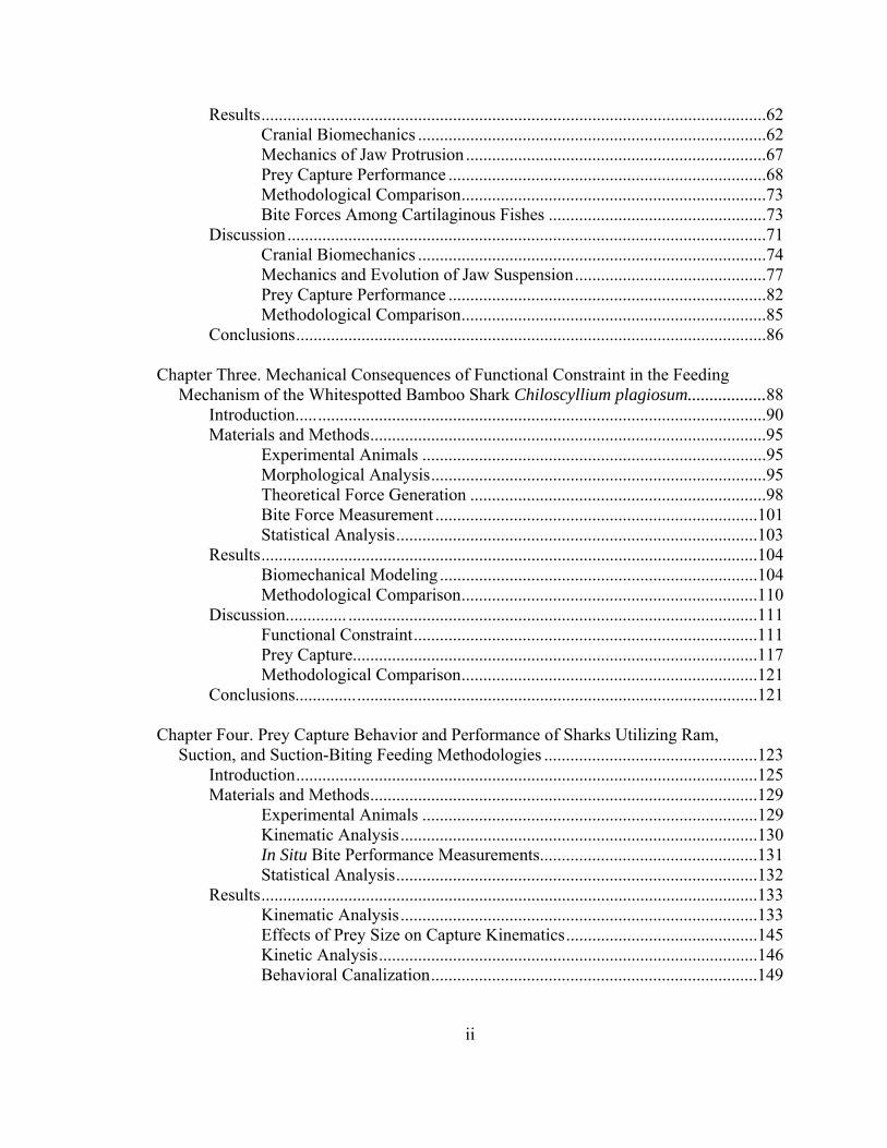
ii
Results....................................................................................................................62 Cranial Biomechanics ................................................................................62 Mechanics of Jaw Protrusion .....................................................................67 Prey Capture Performance .........................................................................68 Methodological Comparison......................................................................73 Bite Forces Among Cartilaginous Fishes ..................................................73 Discussion..............................................................................................................71 Cranial Biomechanics ................................................................................74 Mechanics and Evolution of Jaw Suspension............................................77 Prey Capture Performance .........................................................................82 Methodological Comparison......................................................................85 Conclusions............................................................................................................86 Chapter Three. Mechanical Consequences of Functional Constraint in the Feeding Mechanism of the Whitespotted Bamboo Shark Chiloscyllium plagiosum..................88 Introduction............................................................................................................90 Materials and Methods...........................................................................................95 Experimental Animals ...............................................................................95 Morphological Analysis.............................................................................95 Theoretical Force Generation ....................................................................98 Bite Force Measurement ..........................................................................101 Statistical Analysis...................................................................................103 Results..................................................................................................................104 Biomechanical Modeling .........................................................................104 Methodological Comparison....................................................................110 Discussion.............. ..............................................................................................111 Functional Constraint...............................................................................111 Prey Capture.............................................................................................117 Methodological Comparison....................................................................121 Conclusions..........................................................................................................121 Chapter Four. Prey Capture Behavior and Performance of Sharks Utilizing Ram, Suction, and Suction-Biting Feeding Methodologies .................................................123 Introduction..........................................................................................................125 Materials and Methods.........................................................................................129 Experimental Animals .............................................................................129 Kinematic Analysis..................................................................................130 In Situ Bite Performance Measurements..................................................131 Statistical Analysis...................................................................................132 Results..................................................................................................................133 Kinematic Analysis..................................................................................133 Effects of Prey Size on Capture Kinematics............................................145 Kinetic Analysis.......................................................................................146 Behavioral Canalization...........................................................................149

iii
Discussion............................................................................................................150 Ram Feeding ............................................................................................151 Suction Feeding .......................................................................................157 Suction-Biting ..........................................................................................160 Variability in Prey Capture Behavior ......................................................163 Conclusions..........................................................................................................165 Chapter Five. Comparative Prey Capture Biomechanics of Sharks: Implications for the Evolution of Jaw Suspension Mechanisms .....................................................167 Introduction..........................................................................................................169 Materials and Methods.........................................................................................176 Species Descriptions ................................................................................176 Morphological Analysis...........................................................................177 Theoretical Force Generation ..................................................................179 Statistical Analysis...................................................................................182 Results..................................................................................................................183 Discussion............................................................................................................192 Prey Capture Biomechanics.....................................................................192 Jaw Suspension Mechanics......................................................................199 Evolution of Jaw Suspension ...................................................................206 Jaw Suspension Mechanics in Non-Elasmobranch Fishes ......................209 Conclusions..........................................................................................................212 Literature Cited ................................................................................................................213 Appendices.......................................................................................................................231 Appendix I: Mass-Specific Bite Forces of Vertebrates .......................................232 About the Author ................................................................................................... End Page

iv
List of Tables Table 1 Theoretical maximum forces generated by the cranial musculature active during the gape cycle in H. francisci ........................................18 Table 2 Resultant bilateral muscle and jaw forces occurring during prey capture in H. francisci broken into their principal components ..........19 Table 3 Unilateral mechanical loadings at articulation points in H. francisci’s feeding mechanism broken into their principal components ..........................................................................................20 Table 4 In situ bite performance data for H. francisci biting at the tips of its jaws......................................................................................................23 Table 5 Principal component loadings of performance and kinematic variables from bite performance trials of H. francisci ........................25 Table 6 Results of one-way ANOVA on different methods of determining bite force at the tips of the jaws in H. francisci...................................28 Table 7 Resultant forces occurring during prey capture broken into their principal components in N. brevirostris ..............................................63 Table 8 Mechanical loadings at articulation points in the feeding mechanism of N. brevirostris broken into their principal force components .........................................................................................66 Table 9 In situ bite performance data for N. brevirostris........................................69 Table 10 Principal component loadings of performance and kinematic variables from bite performance trials of N. brevirostris ....................71 Table 11 Theoretical maximum forces generated by the cranial musculature during the gape cycle in C. plagiosum ..............................................106 Table 12 Resultant forces occurring during prey capture in C. plagiosum broken into their principal components .............................................106

v
Table 13 Mechanical loadings at articulation points in the feeding mechanism of C. plagiosum broken into their principal force components .......................................................................................107 Table 14 Results of one-way ANOVA on different methods of determining bite force at the tips of the jaws in C. plagiosum ..............................110 Table 15 Principal component loadings of kinematic variables from 0.5W feeding trials of H. francisci, N. brevirostris, and C. plagiosum ......136 Table 16 Kinematics data from 0.5W feeding trials of H. francisci, N. brevirostris, and C. plagiosum ..........................................................137 Table 17 Principal component loadings of kinematic variables from 1W feeding trials of H. francisci, N. brevirostris, and C. plagiosum ......141 Table 18 Kinetic data from bite performance trials of H. francisci, N. brevirostris, and C. plagiosum ..........................................................142 Table 19 Mass-specific kinetic data from bite performance trials of H. francisci, N. brevirostris, and C. plagiosum......................................146 Table 20 Principal component loadings of bite performance variables of H. francisci, N. brevirostris, and C. plagiosum......................................147 Table 21 Mean coefficients of variation for kinematic and kinetic variable groups in H. francisci, N. brevirostris, and C. plagiosum.................148 Table 22 Kinetic data from bite performance trials of H. francisci, N. brevirostris, and C. plagiosum ..........................................................150 Table 23 Bilateral forces produced by the cranial muscles during the expansion and compression of the feeding mechanisms of H. francisci, N. brevirostris, C. plagiosum, and H. perlo ......................184 Table 24 Mechanical advantages and force distributions (N) during feeding in H. francisci, N. brevirostris, C. plagiosum, and H. perlo .............185 Table 25 Resultant jaw adducting forces (N) of H. francisci, N. brevirostris, C. plagiosum, and H. perlo broken into their principal components ......................................................................................186 Table 26 Results of principal components analysis of jaw adducting and mechanical loading variables in H. francisci, N. brevirostris, C. plagiosum, and H. perlo ....................................................................190

vi
Table 27 Hypothesized suspensorial loading regime occurring in the feeding mechanisms of the chondrichthyan fishes based upon mechanical modeling of the feeding mechanisms of H. francisci, N. brevirostris, C. plagiosum, and H. perlo ......................202

vii
List of Figures Figure 1. Lateral views of representative elasmobranch jaw suspensions ..................5 Figure 2. Right lateral (A) and ventral (B) views of the cranial and branchial musculature of a 63 cm male H. francisci ..............................................7 Figure 3. Coordinate system for three-dimensional vector analysis and schematic diagram of the jaws of H. francisci indicating variables for mechanical lever-ratio analysis .........................................9 Figure 4. Forces involved in the static equilibrium calculations of the lower and upper jaws of H. francisci..............................................................11 Figure 5. Theoretical maximum bite force (N) of five male H. francisci .................19 Figure 6. Bite force waveforms from bite performance trials of three male H. francisci ................................................................................................23 Figure 7. Maximum in situ bite force (N) from five male H. francisci plotted against (A) impulse (kg m s-1) and (B) force duration (ms) on logarithmic axes...............................................................................26 Figure 8. Bite forces (N) of various vertebrates plotted against mass (g) .................29 Figure 9. Left lateral view of the cranium, jaws, and hyoid arch of N. brevirostris, with the skin and muscles removed .................................49 Figure 10. (A) Left lateral and (B) ventral views of the cranial musculature of N. brevirostris.......................................................................................53 Figure 11. Forces involved in the static equilibrium calculations of the lower and upper jaws of N. brevirostris .........................................................56 Figure 12. Theoretical forces produced by the muscles involved in (A) abduction and (B) adduction of the feeding mechanism of N. brevirostris.......................................................................................63

viii
Figure 13. Diagrammatic explanation of local versus global forces acting at articulations within the feeding mechanism of N. brevirostris ...........................................................................................65 Figure 14. Linear regressions of log10-transformed bite performance variables of N. brevirostris ..................................................................................70 Figure 15. Lateral Left lateral views of the feeding mechanisms of (A) the sharpnose sevengill shark Heptranchias perlo (amphistyly); (B) whitespotted bamboo shark Chiloscyllium plagiosum (hyostyly); (C) lesser electric ray Narcine brasiliensis (euhyostyly)..........................................................................................92 Figure 16. Right lateral view of the cranium of C. plagiosum illustrating the constrained linkage between the orbital groove of the cranium and the orbital process of the upper jaw.................................94 Figure 17. Left lateral (A) and ventral (B) views of the cranial and branchial musculature of C. plagiosum ................................................................97 Figure 18. Schematic diagram of the jaws of C. plagiosum indicating (A) variables for lever ratio analysis and (B) the forces involved in static equilibrium calculations of the upper and lower jaws ..........................................................................................100 Figure 19. Diagrammatic explanation of local versus global forces acting at articulations within the feeding mechanism of C. plagiosum using the jaw joint as a model ............................................................108 Figure 20. Right lateral view of the feeding mechanism of C. plagiosum indicating the balance of forces acting on the jaws (A) while the ethmoidal articulation remains intact due to the functional constraint imposed by the association of the orbital process and orbital groove on the upper jaw and cranium respectively (see Materials and methods for description), and (B) while the functional constraint imposed by the orbital process and orbital groove is theoretically released, allowing the upper jaw to dissociate from the cranium during protrusion of the upper jaw.....................................................113 Figure 21. Principal components analysis of expansive and compressive phase kinematic variables from capture of 0.5W sized food by H. francisci, C. plagiosum, and N. brevirostris.......................................135

ix
Figure 22. Principal components analysis of palatoquadrate protrusion kinematic variables from capture of 0.5W sized food by H. francisci, C. plagiosum, and N. brevirostris.......................................138 Figure 23. Principal components analysis of head depression kinematic variables from capture of 0.5W sized food by H. francisci, C. plagiosum, and N. brevirostris.......................................................140 Figure 24. Principal components analysis of expansive and compressive phase kinematic variables from capture of 1.0W sized food by H. francisci, C. plagiosum, and N. brevirostris.......................................143 Figure 25. Principal components analysis of palatoquadrate protrusion kinematic variables from capture of 1.0W sized food by H. francisci, C. plagiosum, and N. brevirostris.......................................145 Figure 26. Principal components analysis of kinetic variables from bite performance trials of H. francisci, C. plagiosum, and N. brevirostris .........................................................................................147 Figure 27. Representation of kinematic and kinetic behavioral transitions associated with capturing prey via ram, suction, and suction-biting feeding methodologies ................................................152 Figure 28. Right lateral views of the feeding mechanisms of elasmobranchs with different jaw suspensions ...........................................................170 Figure 29. Dorsal views of the neurocrania of the A) cladodont shark Cladodus, B) sharpnose sevengill shark Heptranchias perlo, and C) shortfin mako shark Isurus oxyrinchus, illustrating the reduction of the postorbital processes during the evolutionary transition from amphistyly to hyostyly ..............................................171 Figure 30. Coordinate system for three-dimensional vector analysis of the forces generated by the cranial musculature represented by H. francisci..............................................................................................178 Figure 31. Schematic diagram of the jaws of N. brevirostris indicating (A) variables for mechanical lever-ratio analysis, (B) forces involved in the static equilibrium calculations of the lower and upper jaws, and (C) the disarticulation of the upper jaw from the cranium at maximum upper jaw protrusion .................................181

x
Figure 32. Diagrammatic explanation of local versus global forces acting at articulations within the feeding mechanism, using the jaw joint of N. brevirostris as a model ..............................................................187 Figure 33. Principal components analysis of jaw adducting muscle forces, mechanical advantages, and the resulting force distributions throughout the jaws and their articulations with the cranium in Heterodontus francisci, Chiloscyllium plagiosum, Negaprion brevirostris, and Heptranchias perlo .................................................190 Figure 34. Regression analyses of reaction forces (N) occurring within the feeding mechanisms of H. francisci, C. plagiosum, N. brevirostris, and H. perlo during prey capture...................................191 Figure 35. Right lateral views of the feeding mechanisms of (A) C. plagiosum and (B) N. brevirostris indicating the net forces acting on the jaws and their articulations with the cranium during biting ...............201

xi
Cranial Biomechanics and Feeding Performance of Sharks
Daniel Robert Huber
ABSTRACT
The elasmobranch fishes possess a remarkable diversity of feeding mechanisms
for a group containing relatively few species (~1200). The three most prevalent of these
mechanisms involve prey capture during which the predator overtakes its prey (ram),
prey is drawn into the mouth of the predator (suction), and relatively stationary
consumption of sessile or substrate affixed prey (biting). Biomechanical modeling of
cranial force distributions, in situ bite performance trials, and kinematic analysis of prey
capture behaviors were employed to identify morphological and behavioral
specializations and constraints associated with these feeding mechanisms in lemon
Negaprion brevirostris (ram), whitespotted bamboo Chiloscyllium plagiosum (suction),
and horn Heterodontus francisci (biting) sharks. Biomechanical modeling of the forces
generated by the cranial musculature was used to theoretically estimate the maximum bite
force and mechanical loadings occurring throughout the hyostylic jaw suspension
mechanisms of each species, characterized by suspensory hyomandibular cartilages
between the back of the jaws and cranium and anterior ligamentous attachments. To
assess the mechanical factors involved in the evolution of elasmobranch jaw suspension
mechanisms, the feeding mechanism of the sharpnose sevengill shark Heptranchias perlo
was modeled as well. Heptranchias perlo possesses an ancestral amphistylic jaw
xii
suspension mechanism including non-suspensory hyomandibular cartilages, a large post-
orbital articulation between the jaws and cranium, and anterior ligamentous attachments.
Theoretical estimates of maximum bite force were compared to voluntary bite forces
measured during in situ bite performance trials. Voluntary bite force measurements
allowed the quantification of discrete behavioral attributes of bite force application in
each species. To further assess the behavioral specializations associated with these
feeding mechanisms, high-speed digital videography was used to analyze the prey
capture cranial kinematics of species. Collectively, these analyses have developed a
morphological and behavioral basis from which to understand the functional diversity of
the ram, suction, and biting feeding mechanisms in elasmobranchs.

1
Chapter 1: Prey Capture Biomechanics and Feeding Performance of the Horn Shark
Heterodontus francisci
Abstract
Three-dimensional static equilibrium analysis of the forces generated by the jaw
musculature of the horn shark Heterodontus francisci was used to theoretically estimate
the maximum force distributions and loadings on its jaws and suspensorium during
biting. Theoretical maximum bite force was then compared to bite forces measured: (1)
voluntarily in situ; (2) in restrained animals; and (3) during electrical stimulation of the
jaw adductor musculature of anesthetized sharks. Maximum theoretical bite force ranged
from 128 N at the anterior-most cuspidate teeth, to 338 N at the posterior-most
molariform teeth. The hyomandibula, which connects the posterior margin of the jaws to
the base of the chondrocranium, is loaded in tension during biting. Conversely, the
ethmoidal articulation between the palatal region of the upper jaw and the
chondrocranium is loaded in compression, even during upper jaw protrusion because H.
francisci’s upper jaw does not disarticulate from the chondrocranium during prey capture.
Maximum in situ bite force averaged 95 N for free-swimming H. francisci, with a
maximum of 133 N. Time to maximum force averaged 322 ms and was significantly
longer than time away from maximum force (212 ms). Bite force measurements from
restrained individuals (187 N) were significantly greater than those from free-swimming
individuals (95 N), but equivalent to those from both theoretical (128 N) and electrically

2
stimulated measurements (132 N). The mean mass specific bite of H. francisci was
greater than that of many other vertebrates and highest of the cartilaginous fishes that
have been studied. Measuring bite force on restrained sharks appears to be the best
indicator of maximum bite force. The large bite forces and robust molariform dentition of
H. francisci correspond to its consumption of hard prey.

3
Introduction
The elasmobranch fishes (sharks, skates, and rays) possess highly diverse feeding
mechanisms composed of few kinetic elements, making them an ideal group in which to
investigate feeding biomechanics and patterns of diversity in cranial morphology, feeding
behavior, and ecology. Elasmobranchs inhabit nearly all marine environments and have
evolved ram, suction, biting, and filter feeding mechanisms to exploit prey ranging from
plankton to marine mammals (Motta, 2004). Among the diverse feeding mechanisms
found in extant elasmobranch taxa are those adapted for durophagy, the consumption of
hard prey. While “hard” prey of some sort is found in the diets of elasmobranchs from
approximately thirteen families, it does not comprise a substantial portion of the diet in
many of these groups. Genuine durophagy has convergently evolved in the bullhead
(Heterodontidae), hammerhead (Sphyrnidae), zebra (Stegostomatidae), and hound sharks
(Triakidae), as well as eagle rays (Myliobatidae) (Compagno, 1984a, 1984b, 2001;
Summers et al., 2004).
The heterodontid sharks are the only family of elasmobranchs in which every
species is ecologically and functionally specialized for durophagy (Taylor, 1972;
Compagno, 1984a, 1999). The suite of morphological characters associated with
durophagy in the heterodontid sharks includes robust jaws capable of resisting
dorsoventral flexion under high loading, molariform teeth, and hypertrophied jaw
adductor muscles (Reif, 1976; Nobiling, 1977; Summers et al., 2004). To date, the
concept of durophagy in the heterodontid sharks has mostly been examined qualitatively
(but see Summers et al. (2004)). Neither the bite forces they are capable of producing, nor
the subsequent loadings on the various articulations within their feeding mechanisms,
4
have been quantified in any manner. Bite force is particularly informative in regard to
linking morphological, ecological, and behavioral variables associated with prey capture
because biting capacity is dictated by cranial morphology and is known to affect resource
partitioning (Wiersma, 2001; Verwaijen et al., 2002), dietary diversity (Wainwright,
1988; Clifton and Motta, 1998), and ontogenetic changes in feeding ecology (Hernandez
and Motta, 1997).
Like most modern elasmobranchs, the heterodontid sharks possess a hyostylic jaw
suspension in which the mandibular arch indirectly articulates with the chondrocranium
via the hyomandibular cartilages, and the palatal region of the upper jaw is suspended
from the ethmoid region of the chondrocranium via ligamentous connections (Fig. 1A).
However, a number of variants on this arrangement exist, primarily in the superorder
Squalea (Gregory, 1904; Shirai, 1996; Wilga, 2002). The hexanchiform sharks possess an
orbitostylic jaw suspension in which the upper jaw articulates with the ethmoidal, orbital,
and postorbital regions of the chondrocranium and the hyomandibula contributes little
support to the jaws (Fig. 1B). Conversely, the only suspensorial element in the batoids is
the hyomandibula (euhyostyly, Fig. 1C) (Gregory, 1904; Maisey, 1980; Wilga, 2002).
These highly divergent morphologies constitute independent mechanical systems,
perhaps with comparably divergent cranial loading regimes occurring during feeding.
Determining these loading regimes will help to establish the link, if any, between
elasmobranch jaw suspension and the functional diversity of their feeding mechanisms.
The purpose of this study was therefore to determine the biomechanical basis of
durophagy in the heterodontid sharks, as represented by the horn shark Heterodontus

Figure 1. Left lateral views of representative elasmobranch jaw suspensions. (A), Heterodontus, Heterodontiformes (hyostyly); (B), Heptranchias, Hexanchiformes (amphistyly); (C), Rhinobatos, Batoidea (euhyostyly). Articulation points are marked with arrows. C, ceratohyal; E, ethmoidal; H, hyomandibula; L, lower jaw; O, orbital; P, postorbital; U, upper jaw. Reproduced from Wilga (2002) with permission from Blackwell Publishing.
francisci, a primarily shallow water, nocturnal forager of molluscs, echinoderms, and
benthic crustaceans (Strong Jr., 1989; Segura-Zarzosa et al., 1997). Heterodontus
francisci uses suction to capture prey, which is grasped by the anterior, cuspidate teeth
and then crushed by the posterior molariform teeth, effectively combining both suction
and biting feeding mechanisms (Edmonds et al., 2001; Summers et al., 2004). Through in
situ bite performance measurements and theoretical modeling of the forces generated by
5
6
the cranial musculature of H. francisci, the specific goals of this study were to: 1)
theoretically determine the forces generated by each of the cranial muscles active during
the gape cycle; 2) determine the distribution of forces throughout the jaws and
suspensorium, and discuss the implications of these loadings for jaw suspension; 3)
compare theoretical bite force from anatomical measures to those obtained during
voluntary unrestrained feeding, restrained biting, and electrical stimulation of the jaw
adductors; 4) relate its bite performance to feeding ecology; and 5) compare the bite force
of H. francisci to those of other vertebrates.
Materials and Methods
Experimental Animals
Five horn sharks Heterodontus francisci Girard (63 cm – 74 cm TL) were housed
at the University of South Florida in Tampa, FL in accordance with the guidelines of the
Institutional Animal Care and Use Committee (IACUC #1882). Individuals were
maintained at 20°C in a 1,500 l semicircular tank on a diet of thread herring Opisthonema
oglinum and squid (Loligo spp.). The planar face of the tank held a window for viewing.
Five additional H. francisci (55 cm – 68 cm TL) obtained as fisheries bycatch off the
coast of Los Angeles, CA were frozen until used for morphological analyses.
Morphological Analysis
A theoretical model of the feeding mechanism of H. francisci was designed by
investigating the forces produced by the nine cranial muscles involved in the abduction
(coracomandibularis, coracohyoideus, coracoarcualis, and coracobranchiales), adduction
(adductor mandibulae complex consisting of the quadratomandibularis-preorbitalis

7
Figure 2. Right lateral (A) and ventral (B) views of the cranial and branchial musculature of a 63 cm male H. francisci. CC, coracoarcualis; CH, coracohyoideus; CHD, dorsal hyoid constrictor; CHV, ventral hyoid constrictor; CM, coracomandibularis; CO, coracoid bar; HM, hyomandibulo-mandibularis; IMD, intermandibularis; LH, levator hyomandibularis; LJ, lower jaw; LP, levator palatoquadrati; QM-PO complex, quadratomandibularis-preorbitalis complex; QM-γ, quadratomandibularis- γ; PO-α, preorbitalis-α; UJ, upper jaw; VSBC, ventral superficial branchial constrictor. The intermandibularis (IMD) has been partially removed to reveal the ventral musculature. The coracobranchiales (not shown) are located deep to the coracoarcualis (CC).

8
complex, quadratomandibularis-γ, and preorbitalis-α) and retraction (levator
palatoquadrati and levator hyomandibularis) of the jaws and hyobranchial region (Fig. 2).
The quadratomandibularis-preorbitalis complex consists of six individual heads of the
adductor mandibulae complex (Nobiling, 1977). Difficulty in mechanically separating
these heads led to their analysis as a group. Using the tip of the snout as the center of a
three-dimensional coordinate system, the three-dimensional position of the origin and
insertion of each muscle were determined by measuring the distance of these points from
the respective X, Y, and Z planes intersecting the tip of the snout (Fig. 3A). Each muscle
was then excised (unilaterally where applicable), bisected through its center of mass
perpendicular to the principal fiber direction, and digital images of the cross-sections
were taken (JVC DVL9800 camera). Cross-sectional areas were measured from these
images using Sigma Scan Pro 4.01 (SPSS, Inc.). Center of mass was estimated by
suspending the muscle from a pin and tracing a vertical line down the muscle. After
repeating this from another point, the intersection of the two line-tracings indicated the
center of mass of the muscle.
The three-dimensional coordinates of the center of rotation of the dual (lateral and
medial (Nobiling, 1977)) quadratomandibular jaw articulation (hereafter referred to as
“jaw joint”), ethmoidal articulation, and the lateral and medial articulations of the
hyomandibula with the jaws and chondrocranium respectively were determined with
respect to the right side of the head of each individual. Points corresponding to 0, 25, 50,
75, and 100% of the distance along the functional tooth row on the lower jaw from the
posterior-most molariform tooth were also determined; 100% is the anterior-most
cuspidate tooth. The in-lever for jaw abduction from the center of rotation of the jaw joint
9
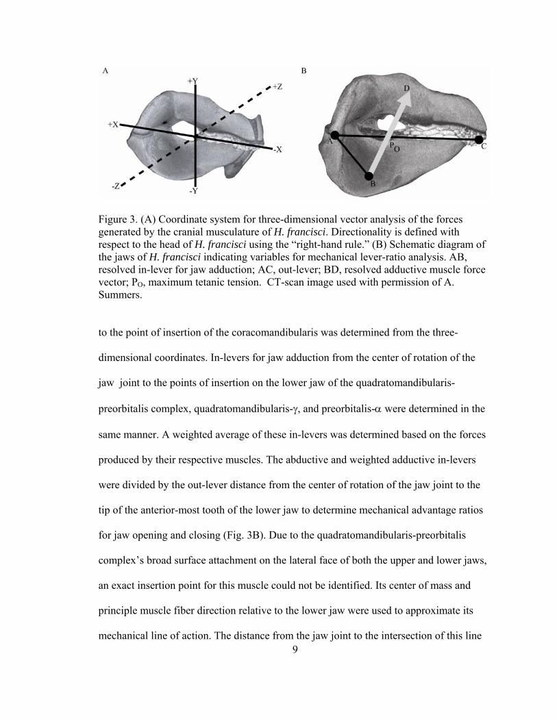
Figure 3. (A) Coordinate system for three-dimensional vector analysis of the forces generated by the cranial musculature of H. francisci. Directionality is defined with respect to the head of H. francisci using the “right-hand rule.” (B) Schematic diagram of the jaws of H. francisci indicating variables for mechanical lever-ratio analysis. AB, resolved in-lever for jaw adduction; AC, out-lever; BD, resolved adductive muscle force vector; PBOB, maximum tetanic tension. CT-scan image used with permission of A. Summers.
to the point of insertion of the coracomandibularis was determined from the three-
dimensional coordinates. In-levers for jaw adduction from the center of rotation of the
jaw joint to the points of insertion on the lower jaw of the quadratomandibularis-
preorbitalis complex, quadratomandibularis-γ, and preorbitalis-α were determined in the
same manner. A weighted average of these in-levers was determined based on the forces
produced by their respective muscles. The abductive and weighted adductive in-levers
were divided by the out-lever distance from the center of rotation of the jaw joint to the
tip of the anterior-most tooth of the lower jaw to determine mechanical advantage ratios
for jaw opening and closing (Fig. 3B). Due to the quadratomandibularis-preorbitalis
complex’s broad surface attachment on the lateral face of both the upper and lower jaws,
an exact insertion point for this muscle could not be identified. Its center of mass and
principle muscle fiber direction relative to the lower jaw were used to approximate its
mechanical line of action. The distance from the jaw joint to the intersection of this line

10
of action with the lower jaw served as the in-lever for this muscle. Anatomical
nomenclature is based on Daniel (1915), Motta and Wilga (1995, 1999), and Nobiling
(1977).
Theoretical Force Generation
Anatomical cross-sectional area (CSA) measurements of the nine parallel fibered
muscles were multiplied by the specific tension of elasmobranch white muscle (289
kN/m2 (Lou et al., 2002)) to determine their theoretical maximum tetanic forces (PO):
PO = CSA * specific tension
Anatomical cross-sectional area was used in this analysis because theoretical estimates of
maximum bite force based on the anatomical cross-sectional area of the parallel fibered
jaw adducting musculature of the spiny dogfish Squalus acanthias best approximated bite
forces measured during tetanic stimulation of the jaw adducting musculature (Huber and
Motta, 2004). Force vectors for each muscle were constructed from their maximum
tetanic forces and the three-dimensional coordinates of their origins and insertions. The
force vectors of muscles excised unilaterally were reflected about the Y-plane to
represent the forces generated by the musculature on the other side of the head.
Mathcad 11.1 software (Mathsoft, Inc.) was used to generate a three-dimensional
model of the static forces acting on the jaws of H. francisci during prey capture.
Summation of the three dimensional moments acting on the lower jaw about the jaw
joints (left and right) determined the theoretical maximum bite force for each individual,
and the average maximum bite force for all individuals (FB, Fig. 4). Maximum bite force
was modeled at points 0, 25, 50, 75, and 100% of the distance along the functional tooth

11
Figure 4. Forces involved in the static equilibrium calculations of the lower and upper jaws of H. francisci. FBB B, bite reaction force; FBEB, reaction force at the ethmoidal articulation; FBHB, reaction force at the hyomandibular articulation; FBJR B, jaw joint reaction force; FBPO-α B, force generated by the preorbitalis-B Bα; FBQM-PO B, force generated by the quadratomandibularis-preorbitalis complex; FBQM-γ B, force generated by the quadratomandibularis-γ; FBR B, resultant adductive force; α, angle of incidence of FBEB relative to the articular surface of the upper jaw at the ethmoidal articulation. Arrow size does not indicate force magnitude and angles of force vectors are approximate. CT-scan image used with permission of A. Summers.
row from the posterior-most tooth to determine a bite force gradient along the lower jaw.
Additionally, the reaction force acting on the jaw joints during bites occurring at 0 and
100% of the distance along the functional tooth row was determined (FBJR B, Fig. 4).
Loadings were determined at the ethmoidal and hyomandibular articulations of
the upper jaw with the chondrocranium and hyomandibula, respectively (Figs 1A, 4). For
bites occurring at 0% (posterior-most molariform tooth) and 100% (anterior-most
cuspidate tooth) of the distance along the functional tooth row, the moments acting on the

12
upper jaw about the ethmoidal articulation were summed to determine the forces acting at
the hyomandibular articulation (FBHB, Fig. 4). In these analyses the hyomandibula was
modeled as a two-force member, moveable about its articulations with both the upper jaw
and chondrocranium (Hibbeler, 2004). Static equilibrium analysis of the forces acting on
the upper jaw was then used to determine the forces acting at the ethmoidal articulation
(FBEB, Fig. 4). Static equilibrium conditions for the forces acting on the lower (FBLJ B ) and
upper jaws (FBUJ B) were:
∑FBLJ B = FBJR B + FBQM-PO B + FBQM-γ B + FBPO-α B + FBB B= 0
∑FBUJ B = FBJR B + FBHB + FBQM-PO B + FBEB + FBB B= 0
where FBB B is the bite reaction force from a prey item, FBEB is the force at the ethmoidal
articulation, FBHB is the force at the hyomandibular articulation, FBJR B is the jaw joint reaction
force, FBPO-α B is the force generated by the preorbitalis-B Bα, FBQM-PO B is the force generated by
the quadratomandibularis-preorbitalis complex, and FBQM-γ B is the force generated by the
quadratomandibularis-γ. Forces generated by the preorbitalis-B Bα and
quadratomandibularis-γ are isolated to the lower jaw because they originate on the
chondrocranium and insert only upon the lower jaw (Figs 2A, 4). Joint reaction forces
maintain the static equilibrium of feeding mechanisms by balancing the moments acting
upon the jaws via their associated musculature and contact with prey items. The moment
acting on the lower jaw during jaw opening via the coracomandibularis muscle was used
to determine the theoretical maximum jaw opening force of H. francisci.
In Situ Bite Performance Measurements
Bite performance measurements were performed using a modified single point
load cell (Amcells Corp., Carlsbad, CA, USA) with custom designed stainless steel lever

13
arms, which was calibrated using a series of known weights. Free-swimming H. francisci
were trained to voluntarily bite the transducer by wrapping the device in squid and
presenting it to them after several days of food deprivation. A P-3500 strain indicator
(Vishay Measurements Group, Raleigh, NC, USA) was used for transducer excitation and
signal conditioning. Data were acquired with a 6020E data acquisition board and
LabVIEW 6.0 software (National Instruments Corp., Austin, TX, USA). Fifteen
measurements of bite force were taken from each animal. Only events in which the
transducer was bitten between the tips of their jaws were kept for analysis. The five
largest bite force measurements for each individual were analyzed for the following
performance variables, as well as used in the multivariate statistical analyses described
below: maximum force (N), duration of force production (ms), time to maximum force
(ms), rising slope of force-time curve (N s P
-1P), duration at maximum force (ms), time from
maximum force to end of force production (hereafter referred to as “time away from
maximum force” (ms)), falling slope of force-time curve (N sP
-1P), and impulse (I), which is
the integrated area under the force-time curve (kg m sP
-1P) from the initiation of force
generation to its cessation:
I = ∫ F dt
The impulse of a force is the extent to which that force changes the momentum of another
body, in this case being the force transducer, and therefore has the units of momentum
(kg m s P
-1P). For each individual, the single largest bite force and its associated
performance measurements were used to create a profile of maximum bite performance
for H. francisci, to compare the dynamics of the ascending and descending portions of the
bite performance waveforms, and to compare the maximum bite forces obtained from the

14
theoretical, in situ, restrained, and stimulated methods of determining bite force (see
below).
In situ bite performance measurements were simultaneously filmed with a
Redlake PCI-1000 digital video system (Redlake MASD, San Diego, CA, USA) at 250
frames per second to verify that bites on the transducer occurred between the tips of the
jaws (hereafter referred to as “transducer bites”). The modified single point load cell used
in this study averages the signals generated by four strain gages in a full Wheatstone
bridge such that the transducer is insensitive to the position on the lever arms at which the
bite is applied. Therefore, the point at which a shark bit the lever arms of the transducer
did not need to be determined from the digital video sequences for appropriate
calibration. To identify any behavioral artifacts associated with biting a stainless steel
transducer, H. francisci were also filmed while consuming pieces of O. oglinum cut to the
same size as the biting surface of the force transducer (hereafter referred to as “fish
bites”). The following kinematic variables were quantified from transducer and fish bites
using Motionscope 2.01 (Redlake MASD) and Sigma Scan Pro 4.01 (SPSS, Inc.)
software: distance, duration, velocity, and acceleration of lower jaw depression, lower
jaw elevation, upper jaw protrusion, and head depression; maximum gape; time to
maximum gape; time to onset of lower jaw elevation; time to onset of head depression;
cranial elevation angle. All kinematic variables were quantified using discrete cranial
landmarks as reference points (Edmonds et al., 2001).
Restrained and Stimulated Bite Performance Measurements
At least one week after the in situ bite performance measurements, four of the
previous H. francisci were individually removed from the experimental tank and

15
restrained on a table. Once they had opened their jaws an adequate distance, the
transducer was placed between the anterior teeth, which elicited an aggressive bite.
Following a recovery period of approximately 10-15 minutes, the shark was again
removed from the tank and anaesthetized with MS-222 (0.133 g/l). The
quadratomandibularis-preorbitalis complex, quadratomandibularis-γ, and preorbitalis-α
were implanted with stainless steel 23 gauge hypodermic needles connected to a SD9
stimulator (Grass Telefactor, West Warwick, RI, USA) and tetanic fusion of these
muscles was accomplished via stimulation (10V, 100 Hz, 0.02 ms delay, 3ms pulse
width) while the bite force transducer was placed between the tips of the anterior teeth.
Three measurements were taken from each individual in both of these experimental
protocols. Individuals were ventilated with aerated seawater between measurements
during muscle stimulation experiments. Maximum bite force, time to maximum force,
and time away from maximum force were quantified from all restrained and stimulated
bites.
Statistical Analysis
All bite performance and kinematic variables were LogB10B transformed and linearly
regressed against body mass to remove the effects of size. Studentized residuals were
saved from each regression for subsequent analysis (Quinn and Keough, 2002). Principal
components analyses (PCA) based on correlation matrices were then used to 1) identify
covariation in bite performance variables and reduce these variables to a series of non-
correlated principal components, which were subsequently analyzed to assess the extent
of individual variability in these parameters; 2) identify covariation in performance and
kinematic variables from in situ bite performance trials; and 3) identify covariation in

16
kinematic variables from “fish” and “transducer” bites and reduce these variables to a
series of non-correlated principal components, which were subsequently analyzed to
determine whether there were any behavioral artifacts associated with biting the steel
transducer. Variables were considered to load strongly on a given principal component
(PC) if their factor scores were greater than 0.6. Non-rotated axes described the greatest
amount of variability in each PCA. For analyses 1 and 3, multivariate analysis of variance
(MANOVA) was used to compare the factor scores for the PCs with eigenvalues greater
than 1.0. To determine whether “fish” and “transducer” bites kinematically differed, a
two-way, mixed-model MANOVA was performed on the PCs from PCA 3 with
individual as the random effect and prey type as the fixed effect, which was tested over
the interaction mean square. Kinematic data from four individuals were included in this
analysis because a complete data set was lacking for one individual. To determine the
extent of individual variability within the bite performance variables, a one-way
MANOVA was performed on the PCs from PCA 1.
To determine whether the kinematic variables associated with biting the
transducer were predictive of biting performance in H. francisci, stepwise (forward)
multiple regressions were performed with kinematic variables measured from transducer
bites as the multiple independent factors, and the eight bite performance variables as the
individual dependent factors. Data from four individuals were included in this analysis
because a complete kinematic data set was lacking for one individual. One-way ANOVA
on studentized residuals was used to identify significant differences among the
theoretical, in situ, restrained, and electrically stimulated methods of determining
maximum bite force. A Student's t-test was used to identify differences between time to

17
maximum force and time away from maximum force, and the rising and falling slopes of
the force-time curves for in situ biting trials. One-way ANOVA was used to compare
time to maximum force and time away from maximum force within and among in situ,
restrained, and electrically stimulated bite forces. Lastly, bite forces at the anterior jaw
(fish, reptiles, and birds) or canine teeth (mammals) and body masses for various
vertebrates were compiled from the available literature and grouped according to major
taxonomic level. These bite forces, along with those of the horn sharks investigated in
this study, were linearly regressed against body mass. Studentized residuals from this
regression were then coded according to taxonomic level and compared with a one-way
ANOVA. All significant differences were investigated post-hoc with Tukey’s pairwise
comparisons test. Linear regressions were performed in SigmaStat 2.03 (SPSS Inc.) in
order to obtain studentized residuals. All other statistical analyses were performed in
SYSTAT 10 (SPSS, Inc.) with a p-value of 0.05.
Results
Biomechanical Modeling
The quadratomandibularis-preorbitalis complex, which is the primary jaw
adductor, generated the greatest force of all muscles investigated (242 N, Table 1). Of the
muscles active during jaw and hyobranchial abduction, the coracobranchiales generated
the greatest force (107 N, Table 1). The levator hyomandibularis generated more force
during the retractive phase (33 N) than the levator palatoquadrati (20 N, Table 1). After
resolving the force generated by the adductor musculature into its principal components,
the majority of force was directed dorsally (294 N) and anteriorly (128 N). The Z-axis

18
Table 1. Theoretical maximum forces generated by the cranial musculature active during the gape cycle in H. francisci
Action Muscle Theoretical Max Force (N) P
§P
Coracomandibularis 31 +/- 5 Coracohyoideus 57 +/- 4 Coracoarcualis 87 +/- 4*
Jaw & Hyobranchial Abduction
Coracobranchiales 107 +/- 8* Quadratomandibularis-γ 44 +/- 2*
Preorbitalis-α 52 +/- 5* Jaw Adduction QM-PO Complex 242 +/- 11*
Levator Palatoquadrati 20 +/- 1* Jaw & Hyobranchial Retraction Levator Hyomandibularis 33 +/- 1*
§ mean +/- S.E. * bilateral muscle force for paired muscles
component of this force (19 N per side) were directed laterally on either side of the head,
and negate each other during jaw adduction (Table 2, Fig. 3A). Thus, the resultant
adductive force along the z-axis was 0 N. The large anterodorsally directed component of
this adductive bite force (FBR B, Fig. 4) drives the lower jaw towards the upper jaw, which is
itself driven into the ethmoid region of the chondrocranium (FBEB, Fig. 4).
Summation of the moments acting on the lower jaw determined that the maximum
theoretical bite force of H. francisci ranged from 128 N at the anterior teeth to 338 N at
the posterior-most molariform teeth (Fig 5, Table 2). The bite force at the posterior-most
molariform teeth exceeded the resultant force generated by the adductive musculature
(Table 2) because the mechanical advantage at this point along the jaw was 1.06. The
resultant jaw closing mechanical advantage at the anterior teeth was 0.51, resulting in a
dramatically lower bite force at this point.
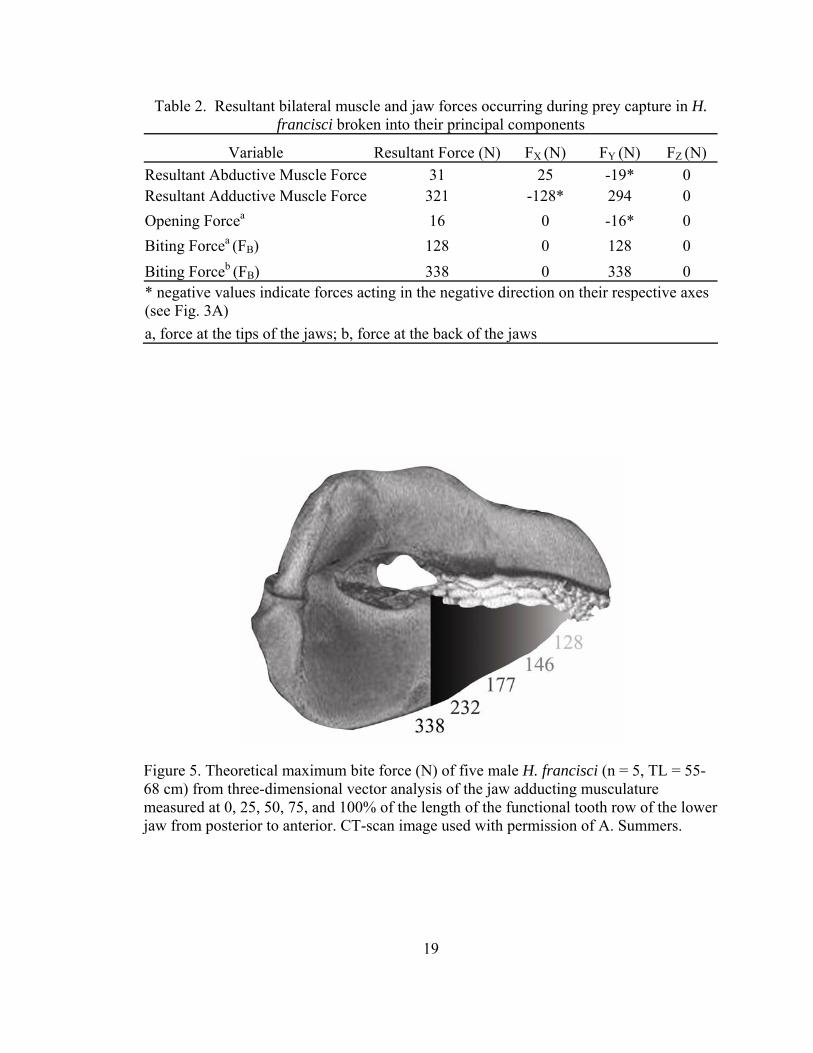
Table 2. Resultant bilateral muscle and jaw forces occurring during prey capture in H. francisci broken into their principal components
Variable Resultant Force (N) FX (N) FY (N) FZ (N) Resultant Abductive Muscle Force 31 25 -19* 0 Resultant Adductive Muscle Force 321 -128* 294 0 Opening Forcea 16 0 -16* 0 Biting Forcea (FB) 128 0 128 0 Biting Forceb (FB) 338 0 338 0 * negative values indicate forces acting in the negative direction on their respective axes (see Fig. 3A) a, force at the tips of the jaws; b, force at the back of the jaws
Figure 5. Theoretical maximum bite force (N) of five male H. francisci (n = 5, TL = 55-68 cm) from three-dimensional vector analysis of the jaw adducting musculature measured at 0, 25, 50, 75, and 100% of the length of the functional tooth row of the lower jaw from posterior to anterior. CT-scan image used with permission of A. Summers.
19

20
Table 3. Unilateral mechanical loadings at articulation points in the feeding mechanism of H. francisci broken into their principal components
Variable Resultant Force (N) FBX B(N) FBY B(N) FBZ B(N)Joint Reaction ForceP
aP (FBJR B) 106 69 -80* 0
Joint Reaction ForceP
bP (FBJR B) 73 69 25 0
Loading at Ethmoidal Artic.P
aP (FBEB) 59 10 -59* 0
Loading at Ethmoidal Artic.P
bP (FBEB) 59 10 -59* 0
Loading at Hyomandibular Artic.P
aP (FBHB) 36 10 22 27
Loading at Hyomandibular Artic.P
bP (FBHB) 36 10 22 27
* negative values indicate forces acting in the negative direction on their respective axes relative to the right side of H. francisci’s head (see Fig. 3A) a, force at the tips of the jaws; b, force at the back of the jaws
The jaw joint reaction forces (FBJR B, Fig. 4) occurring when prey is captured at the
anterior teeth and crushed at the posterior teeth by H. francisci were 106 N and 73 N per
side, respectively (Table 3). This force was oriented posteroventrally relative to the
articular surface of the lower jaw joint for anterior biting, and consequently oriented
anterodorsally relative to the articular surface of the upper jaw joint. The local/internal
loadings on the joint between the upper and lower jaws indicate that the jaw joint is
globally in compression (Hibbeler, 2004) when prey is bitten at the tips of the jaws.
When prey is crushed between the posterior molariform teeth the orientation of the
vertical component of the joint reaction force relative to the lower jaw (25 N) was
opposite that for the lower jaw joint during anterior biting (-80 N), indicating tensile
loading of the jaw joint during posterior prey capture (Table 3).
The ethmoidal articulation of H. francisci received a loading of 59 N per side
during biting, regardless of whether biting occurred at the anterior or posterior margin of
the jaws (FBEB, Fig. 4). The angle of incidence of this force relative to the articular surface
of the upper jaw at the ethmoidal articulation was 80° (α, Fig. 4). For both anterior and
21
posterior biting, the majority of loading was directed ventrally into the upper jaw,
indicating compression between the ethmoid region of the chondrocranium and the
palatal region of the upper jaw (Table 3).
The magnitude of loading at the hyomandibular articulation (36 N) was
independent of bite point as well (FBHB, Fig. 4). The lower jaw was loaded posterodorsally
and medially at its articulation with the hyomandibula during both anterior and posterior
biting (Table 3). The reaction forces acting on the distal ends of the hyomandibula are
equal to and opposite the forces acting at the jaws’ articulation with the hyomandibula.
Therefore, during biting the hyomandibula was loaded anteroventrally and laterally.
These local/internal loadings between the jaws and hyomandibula indicate that the
hyomandibula is globally in tension. Modeling the hyomandibula as a two-force member
assumed that the line of action of the force acting on the hyomandibula passed through its
articulation with the jaws and chondrocranium. The hyomandibula is therefore loaded in
pure tension and the angle of incidence of the hyomandibular force cannot be determined.
The only muscle involved in abduction of the lower jaw is the
coracomandibularis, which was capable of generating 31 N of force (Table 1). This
muscle inserts on the caudal aspect of the lower jaw symphysis at 37° below the
longitudinal axis of this jaw, and has a mechanical advantage of 0.89. Despite this high
mechanical advantage, indicative of force amplification in a class III lever system, its
acute insertion angle caused the muscular force generating motion about the lower jaw
(force component perpendicular to the lower jaw) to be 19 N (Table 2). After accounting
for mechanical advantage, the resultant abductive force at the tip of the lower jaw was 16
N (Table 2). The abductive force lacks a component along the Z-axis because the

22
coracomandibularis runs parallel to the longitudinal axis of the body. The other muscles
involved in the expansive phase of the gape cycle generated considerably greater forces
than the coracomandibularis (Table 1).
Performance Measurements
In situ measurements: H. francisci approached and bit the force transducer in an
attempt to remove the attached food. In most cases biting continued until the food was
removed from the transducer. PCA 1 reduced the performance variables for each
individual to three PCs (89.7% of variance explained), each of which indicated
considerable overlap among individuals. MANOVA subsequently demonstrated no
differences among individuals for bite performance variables using size-corrected data
(Wilk’s Lambda = 0.51, F12,47 = 1.157, p = 0.340). The average maximum in situ bite
force measured at the anterior teeth was 95 N, with an absolute maximum force of 133 N
(66 cm male H. francisci). Heterodontus francisci took approximately 322 ms to reach
maximum bite force, which was held for 41 ms, and released after an additional 212 ms
(Table 4). The average duration of force application was 535 ms. Time to maximum bite
force was longer than the time away from maximum bite force (p = 0.049). The mean
rising slope of the force-time curve was 300 N s-1, and was lower than the average falling
slope of 457 N s-1 (p = 0.048). The average impulse generated from the beginning of
force application until its cessation was 25 kg m s-1, but measured as high as 44 kg m s-1.
The majority of bite force waveforms consisted of single peaks associated with single
bites. However, in 32% of the bites multiple peaks occurred indicating a repetitive
crushing behavior during force application (Fig. 6).

Table 4. In situ bite performance data for H. francisci biting at the tips of its jaws Variable Minimum Maximum Mean +/- S.E.
Maximum Force (N) 60 133 95 +/- 13 Force Duration (ms) 400 721 535 +/- 60 Time to Maximum Force (ms) 241 428 322 +/- 33 Time at Maximum Force (ms) 31 55 41 +/- 4 Time away from Maximum Force (ms) 146 303 212 +/- 35 Impulse (kg m s-1) 11 44 25 +/- 6 Rising Slope of Force-Time Curve (N s-1) 200 400 300 +/- 34 Falling Slope of Force-Time Curve (N s-1) 305 696 457 +/- 65
Figure 6. Bite force waveforms from bite performance trials of three male H. francisci (TL = 66-70 cm) illustrating in situ voluntary bites with single (black) and double (light gray) force peaks, and a bite from a restrained individual (dark gray).
23

24
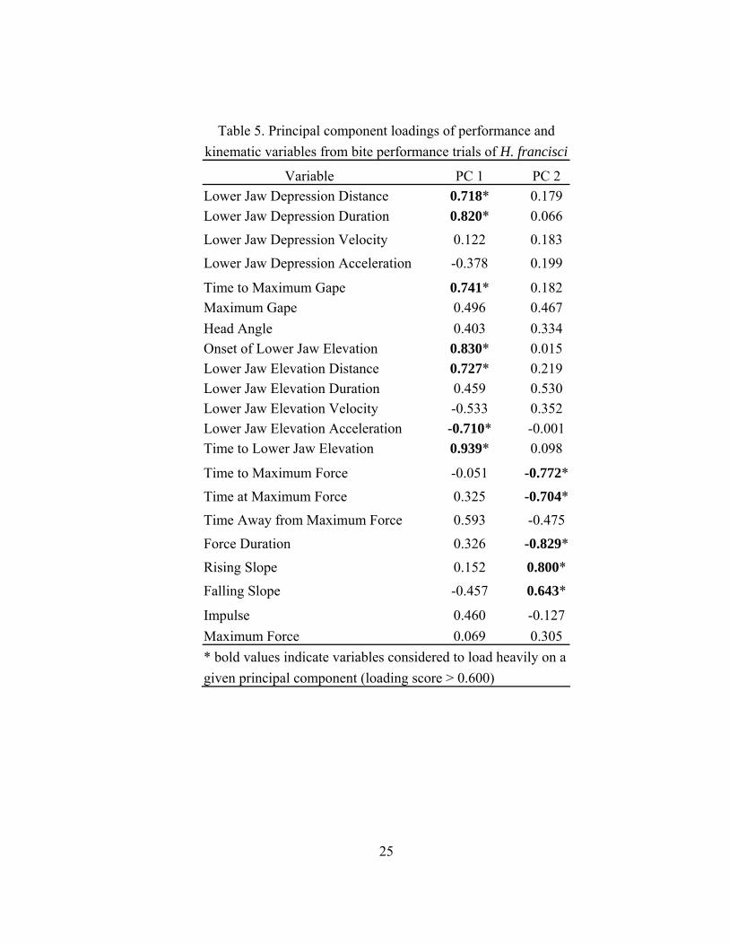
PCA 2 of performance and kinematic variables yielded 6 PCs with eigenvalues
greater than 1.0, which collectively explained 86.7% of the variance. All of the variables
that loaded heavily on the first PC (30.5% of variance explained) were kinematic
measurements (Table 5). These variables primarily demonstrated covariance in the
timings and excursions of lower jaw depression and elevation. Performance measures
were the only variables to load heavily on the second PC (19.5% of variance explained),
indicating covariance between rates and durations of force application (Table 5).
Maximum bite force did not load heavily until the fifth PC (7.8% of variance explained)
and impulse did not load heavily on any of the PCs.
Stepwise multiple regressions yielded similar results to PCA 2 on kinematic and
performance data. Only three of the bite performance variables were significantly related
to individual kinematic variables. Force duration was significantly, though poorly, related
to lower jaw elevation velocity (R2 = 0.226, F1,18 = 5.268, p = 0.034). Similarly, time to
maximum force (R2 = 0.389, F1,18 = 11.471, p = 0.003) and the rising slope of the force
time curve (R2 = 0.410, F1,18 = 12.523, p = 0.002) were significantly related to lower jaw
elevation distance. Inclusion of additional kinematic variables did not improve the
predictive ability of these regression models. The two variables indicative of the
magnitude of bite force generated (maximum force, impulse) could not accurately be
predicted by any combination of kinematic variables. Although kinematic variables were
not predictive of bite performance variables, PCA 1 used to assess individual variability
(see above) identified notable covariance in performance measures. Maximum in situ bite
force exhibited a strong linear relationship with impulse (R2 = 0.758), and moderate
25
Table 5. Principal component loadings of performance and kinematic variables from bite performance trials of H. francisci
Variable PC 1 PC 2 Lower Jaw Depression Distance 0.718* 0.179 Lower Jaw Depression Duration 0.820* 0.066
Lower Jaw Depression Velocity 0.122 0.183
Lower Jaw Depression Acceleration -0.378 0.199
Time to Maximum Gape 0.741* 0.182 Maximum Gape 0.496 0.467 Head Angle 0.403 0.334 Onset of Lower Jaw Elevation 0.830* 0.015 Lower Jaw Elevation Distance 0.727* 0.219 Lower Jaw Elevation Duration 0.459 0.530 Lower Jaw Elevation Velocity -0.533 0.352 Lower Jaw Elevation Acceleration -0.710* -0.001 Time to Lower Jaw Elevation 0.939* 0.098
Time to Maximum Force -0.051 -0.772* Time at Maximum Force 0.325 -0.704*
Time Away from Maximum Force 0.593 -0.475
Force Duration 0.326 -0.829*
Rising Slope 0.152 0.800*
Falling Slope -0.457 0.643*
Impulse 0.460 -0.127 Maximum Force 0.069 0.305 * bold values indicate variables considered to load heavily on a given principal component (loading score > 0.600)

Figure 7. Maximum in situ bite force (N) from five male H. francisci (n = 5, TL = 63-74 cm) plotted against (A) impulse (kg m s-1) and (B) force duration (ms) on logarithmic axes.
26

27
linear relationships with force duration (R2 = 0.450) and time to maximum force (R2 =
0.489) (Fig. 7).
PCA 3 reduced the set of kinematic variables measured from fish and transducer
bites to a series of four PCs (73.3% of variance explained). MANOVA indicated no
significant differences between the prey capture kinematics of H. francisci while
bitingfish or the transducer on any of the PCs for all individuals (Wilk’s Lambda = 1.0,
F4,29 = 0.0, p = 1.0). However, a single individual was found to differ from two other
individuals on the first PC (F3,32 = 4.646, p = 0.008). Variables that loaded heavily on the
first PC were durations and distances of lower jaw depression and elevation, times to
maximum gape, onset of lower jaw elevation, and completion of lower jaw elevation, and
maximum gape distance. The acceleration of lower jaw elevation loaded heavily, but
negatively on the first PC.
Methodological Comparison
In situ measurement of maximum bite force was a reasonably good indicator of
the maximum bite force of H. francisci. Using size-corrected data, a single difference was
found among the four methods of determining maximum bite force (F3,14 = 4.358, p =
0.023). Restrained bite force (159-206 N) was significantly greater than in situ bite force
(60-133 N) (p = 0.013). In situ bite force was, however, equivalent to theoretical (107-
163 N) and electrically stimulated (62-189 N) bite forces. Restrained, electrically
stimulated, and theoretical bite forces were equivalent (Table 6). During restrained bites,
time to maximum force (522 ms) was greater than time away from maximum force (339
ms) (t4 = 2.848, p = 0.046). Time to maximum force (285 ms) was shorter than time away
from maximum force (556 ms) for electrically stimulated bites (t8 = -5.476, p < 0.001).

28
Table 6. Results of one-way ANOVA on different methods of determining bite force at the tips of the jaws in H. francisci*
in situ Theoretical Stimulated Restrained **Avg. Max. +/- S.E. (N) 95 +/- 13 128 +/- 10 132 +/- 24 187 +/- 14
*Statistically similar values are underlined **Average of the single largest bite force values from each individual
Significant differences were detected between the in situ, restrained, and electrically
stimulated methods for time to maximum force (F2,10 = 4.996, p = 0.031) and time away
from maximum force (F2,10 = 58.290, p < 0.001). Time to maximum force was greater for
restrained bites than electrically stimulated bites (p = 0.030), both of which were
equivalent to the time to maximum force of in situ bites. Time away from maximum
force was greater for electrically stimulated bites than restrained bites (p = 0.001), which
was greater than that of in situ bites (p = 0.005).
Bite Forces Among Vertebrates
Bite forces and body masses were compiled for 113 species of vertebrates
(including H. francisci) from the available literature (Ringqvist, 1972; Robins, 1977;
Thomason et al., 1990; Cleuren et al., 1995; Hernandez and Motta, 1997; Clifton and
Motta, 1998; Herrel et al., 1999, 2001, 2002; Binder and Van Valkenburgh, 2000;
Thompson et al., 2003; Erickson et al., 2004; Huber and Motta, 2004; Korff and
Wainwright, 2004; van der Meij and Bout, 2004; Wroe et al., 2005; Huber et al., 2006, in
prep) (Appendix I). Collectively, bite force scaled to body mass with a coefficient of
0.60, which is below the isometric scaling coefficient of 0.67 (Fig. 8). When the
mammalian bite forces from Wroe et al. (2005) were excluded from this analysis bite

29
Figure 8. (A) Bite forces (N) of various vertebrates plotted against mass (g). (B) Residuals from regression analysis of Log B10 B bite force versus LogB10B mass plotted against LogB10B mass (g). Dashed lines indicate ± 1 standard deviation about the residual mean.

30
force scaled with a coefficient of 0.66, approximating isometry. This discrepancy is likely
due to Wroe et al. (2005) having used the dry-skull method of estimating muscle CSA,
which can underestimate CSA by 1.3-1.5X (Thomason et al., 1991).
Fishes collectively had the highest mass specific bite force of the four vertebrate
groups, followed by reptiles, mammals, and birds respectively (FB3,130B = 6.357, p < 0.001).
Mass specific bite force of the fishes was greater than those of the birds (p = 0.002) and
mammals (p = 0.013), while reptilian mass specific bite force was greater than that of the
birds (p = 0.009). The striped burrfish Chilomycterus schoepfi had the highest mass
specific bite force, followed by the Canary Island lizard Gallottia galloti, and the
American alligator Alligator mississipiensis (Herrel et al., 1999; Erickson et al., 2004;
Korff and Wainwright, 2004). The hogfish Lachnolaimus maximus had the second
highest mass specific bite force, but for biting with the pharyngeal jaws, not the oral jaws
(Clifton and Motta, 1998). The three lowest mass specific bite forces were those of the
red-bellied short-necked turtle Emydura subglobosa, mata mata turtle Chelus fimbriatus,
and twist-necked turtle Platemys platycephala (Herrel et al., 2002) (Fig. 8). Of the
cartilaginous fishes in this analysis, the mean mass specific bite force of H. francisci was
greater than those of S. acanthias and the blacktip shark Carcharhinus limbatus, but less
than that of the white-spotted ratfish Hydrolagus colliei.
Discussion
Functional Morphology
The jaw adducting cranial musculature (QM-PO complex, QM-γ, PO-α on Fig. 2)
of H. francisci generates more force during prey capture than either the jaw and

31
hyobranchial abducting or retracting musculature. The mechanical advantage of H.
francisci’s jaw adducting mechanisms ranges from 0.51 at the tip of the jaws to 1.06 at
the posterior margin of the functional tooth row. In class III lever systems such as shark
jaws, a mechanical advantage greater than 1.0 indicates that the point at which force is
being applied to a prey item is closer to the jaw joint than the point at which muscular
force is being applied to the jaw, resulting in an amplification of the muscular force.
Subsequently, the theoretical maximum bite force at the posterior margin of the
functional tooth row exceeds the resultant force generated by H. francisci’s adductor
musculature. This amplification of muscular force is advantageous for the processing of
hard prey such as the molluscs, echinoderms, and benthic crustaceans consumed by H.
francisci (Strong Jr., 1989; Segura-Zarzosa et al., 1997).
The jaw closing mechanical advantage at the anterior teeth of H. francisci is
greater than that of the only other elasmobranch for which values have been published, S.
acanthias (0.28 (Huber and Motta, 2004)), which utilizes a combination of ram and
suction feeding to consume soft-bodied prey (Wilga and Motta, 1998a). Its jaw closing
mechanical advantage is greater than those at the anterior teeth of nearly every
actinopterygian fish investigated (~150), which include prey from plankton to hard-
shelled species (Turingan et al., 1995; Durie and Turingan, 2001; Wainwright et al.,
2004; Westneat, 2004). The durophagous species among these taxa do, however, have the
highest jaw adducting mechanical advantages. The durophagous parrot fishes (Scaridae)
are the only actinopterygian fishes with jaw adducting mechanical advantages
comparable to that of H. francisci (Wainwright et al., 2004; Westneat, 2004). Thus, there

32
is extensive evolutionary convergence on high leverage jaw adducting mechanisms in
fishes that consume hard prey.
The jaws of H. francisci are elliptical in transverse-section, with their major axis
oriented vertically, in-line with the compressive stresses associated with feeding. Calcium
reinforcement in the jaw cortex increases posteriorly as the dentition becomes more
molariform, and is greatest at the jaw joints (Summers et al., 2004). Calcification and
elliptical geometry increase the second moment of area of the jaws with respect to the
compressive loading of prey capture and processing, which augments the jaws’ ability to
resist dorsoventral flexion (Summers et al., 2004). The resolved force vector for jaw
adduction also occurs approximately in the region of the most robust molariform teeth of
H. francisci, where it can generate upwards of 338 N of bite force. Therefore, maximum
bite force is produced where both the dentition and jaw cartilages are best able to resist
compressive stresses.
Despite the high mechanical advantage (0.89) of the coracomandibularis muscle
in the lower jaw depression mechanism of H. francisci, its acute insertion angle relative
to the lower jaw causes most of its force to be directed posteriorly, into the jaw joints
(Table 2.) This high mechanical advantage is due the insertion of the coracomandibularis
on the posterior margin of the mandibular symphysis, which is synapomorphic for
chondrichthyans (Wilga et al., 2000). Although this mechanism is suited for force
production, velocity production is desirable for inertial suction feeding. Heterodontus
francisci nonetheless effectively uses suction to initially capture and reorient prey
(Edmonds et al., 2001), which may be due in part to its powerful hyoid and branchial
abductors (Table 1). These muscles rapidly expand the floor of the buccopharyngeal

33
cavity, which is critical to suction feeding in elasmobranchs (Motta et al., 2002;
Svanback et al., 2002). As in the nurse shark Ginglymostoma cirratum (Motta et al.,
2002), the large labial cartilages of H. francisci considerably occlude its lateral gape,
theoretically augmenting suction ability (Muller and Osse, 1984; Van Leeuwen and
Muller, 1984).
Three-dimensional resolution of the forces generated during jaw adduction may
reveal the mechanical basis of upper jaw protrusion in H. francisci. The force driving the
upper jaw into the ethmoidal articulation has both dorsal and anterior components,
causing the upper jaw to slide through the anteroventrally sloping palatal fossa of the
chondrocranium and protrude (Fig. 4, Table 2). This proposed mechanism is based on the
resolved force vector for all muscles involved in jaw adduction. Differential activity of
the heads of this complex may facilitate modulation of protrusion. The
quadratomandibularis-γ is the likely candidate for control over protrusion because its
acute insertion angle relative to the lower jaw and anterior insertion point give it high
leverage over anterior motion (Fig. 2).
Activity of the quadratomandibularis-preorbitalis complex alone, which has a
broad insertion on the lateral face of both the upper and lower jaws, may contribute to
protrusion of the upper jaw as well. After the lower jaw has been depressed, contraction
of this muscle complex may simultaneously raise the lower jaw and pull the upper jaw
away from the skull. This mechanism has been proposed for upper jaw protrusion in S.
acanthias, G. cirratum, and the lemon shark Negaprion brevirostris (Moss, 1977; Motta
et al., 1997; Wilga and Motta, 1998a). Protrusion by H. francisci, which may be used to
chisel away at attached benthic prey, occurs after the lower jaw has been depressed

34
(Edmonds et al., 2001), corroborating the role of the quadratomandibularis-preorbitalis
complex in this behavior.
Extensive calcification near the jaw joints of H. francisci (Summers et al., 2004)
would apparently indicate high joint reaction forces during prey capture. Joint reaction
forces can exceed bite forces at the tip of the jaw depending on the mechanical advantage
of the given feeding mechanism and the force produced by the associated musculature,
which has been identified in numerous reptiles (3-4 times greater) (Cleuren et al., 1995;
Herrel et al., 1998). Although joint reaction force was greater than anterior bite force in
H. francisci, the ratio of these values (1.65) is substantially lower than those found for
reptiles. The ratio of joint reaction force to posterior bite force in H. francisci was 0.43.
Low ratios of joint reaction force to bite force in H. francisci are due to its high
mechanical advantage jaw adducting mechanism. Humans, which share this
characteristic, have correspondingly low ratios of joint reaction force to bite force
(Koolstra et al., 1988). Although some damping will occur in the connective tissue
associated with the jaw joint, loading occurring at the joint will be transmitted to adjacent
skeletal elements. Therefore, low ratios of joint reaction force to bite force may be
adaptive in H. francisci, and elasmobranchs in general, because the posterior region of
their jaws is suspended from the cranium by mobile hyomandibulae, not a stable jaw
articulation as in other vertebrates. Minimizing loading at this articulation may stabilize
the feeding mechanism during prey capture and processing.
In heterodontiform sharks, the cranial stresses associated with prey capture can be
isolated to the ethmoidal and hyomandibular articulations. Unlike carcharhinid sharks
(Motta and Wilga, 1995), the upper jaw of H. francisci does not disarticulate from the

35
chondrocranium during feeding, even during upper jaw protrusion (Maisey, 1980).
Therefore, in carcharhinid sharks the hyomandibulae may receive all of the suspensorial
loading occurring during prey capture. Optimal loading at the ethmoidal articulation
would entail forces directed perpendicularly into the articular surface of the upper jaw
because cartilage is strongest in axial compression (Carter and Wong, 2003). The
estimated forces at this articulation deviated from optimal orientation by only 10° during
anterior and posterior biting. The ethmoidal articulation of H. francisci appears well
designed for withstanding this nearly axial compressive loading because the upper jaw
calcifies at this articulation early in ontogeny (Summers et al., 2004) and the ethmoid
region of the chondrocranium is one of the thickest parts of this structure (Daniel, 1915).
Additionally, maintenance of contact between the upper jaw and chondrocranium in H.
francisci will distribute stresses from the repetitive loading associated with processing
hard prey.
Although it is well known that the hyomandibulae support the posterior margin of
the jaws, the nature of the loading they receive has been a matter of speculation. This
mechanical analysis indicates that the hyomandibulae of H. francisci are tensile elements
as suggested by Moss (1972) and Frazzetta (1994). Consequently, the hyomandibulae
may regulate anterior movement of the jaws during feeding, such as would occur during
jaw protrusion. In this regulatory role, activity of the levator hyomandibularis could
hypothetically modulate resistance to anterior motion of the jaws. Electromyographic
analysis of the feeding musculature of H. francisci would be required to verify this
hypothesis.

36
The ligamentous attachments between the hyoid arch and the posterior end of the
jaws stabilize this articulation against the tensile stresses caused by biting. The internal
hyomandibular palatoquadrate and hyoideo-mandibulare ligaments resist dorsoventral
translation between the hyomandibula and jaws, while the hyomandibuloceratohyal
ligament prevents lateral translation between these elements. The two slips of the median
ligament (Daniel, 1915) stabilize against dorsoventral and lateral translation respectively.
Although this analysis makes the assumption that the hyomandibulae are loaded as two-
force members in axial tension, they likely experience a more diverse loading pattern in
nature, necessitating this multidirectional support.
Increased hyomandibular loading may have played a role in the transition from
amphistylic to hyostylic jaw suspensions in modern elasmobranchs. As the number and
size of the articulations between the jaws and chondrocranium was reduced, the
hyomandibulae took on a greater role in suspending the jaws (Schaeffer, 1967; Maisey,
1980; Carroll, 1988). Concomitant with these changes in articulation, the hyomandibulae
became shorter and more mobile (Schaeffer, 1967; Moy-Thomas and Miles, 1971;
Maisey, 1985; Cappetta, 1987), oriented more orthogonally to the chondrocranium (Stahl,
1988; Wilga, 2002), and had more extensive ligamentous attachments with the jaws and
chondrocranium (Gadow, 1888). Collectively, these changes may have been associated
with a shifting of the force of jaw adduction to a more posterior region of the jaws. This
may have resulted in greater hyomandibular loading as well as a “freeing-up” of the
anterior margin of the jaws such that upper jaw kinesis was increased, facilitating jaw
protrusion and prey gouging (Moss, 1977; Maisey, 1980; Wilga, 2002).

37
Although the jaw suspension mechanism of H. francisci is classified as hyostylic
(Gregory, 1904; Wilga, 2002), this analysis indicates that it is functionally amphistylic.
Heterodontus francisci exhibits considerable upper jaw kinesis (reduces maximum gape
by 39%), similar to other hyostylic carcharhiniform sharks (Edmonds et al., 2001; Wilga
et al., 2001). Despite this functional similarity, the upper jaw does not disarticulate from
the chondrocranium during protrusion in H. francisci. Furthermore, in contrast to the
hypotheses regarding hyomandibular evolution (see above), H. francisci has considerable
loading at both the hyomandibular (tensile) and ethmoidal (compressive) articulations.
The term ‘hyostyly’ should therefore be reserved for taxa in which the upper jaw
disarticulates from the chondrocranium during protrusion such that hyomandibulae are
the primary means of support, and the ethmopalatine ligaments are loaded in tension.
Therefore, contemporary definitions of jaw suspension should incorporate functional
interpretations of loadings at the various articulations between the jaws and cranium, as
well as the relationship between suspension type and upper jaw protrusion.
Methodological Comparison
Although no differences were found between theoretical, restrained, and
electrically stimulated bite force measurements using size corrected data, the absolute
maximum bite force for each individual occurred during restrained bite force
measurements. No differences were found between theoretical and electrically stimulated
bite force measurements of S. acanthias either (Huber and Motta, 2004). Therefore,
restrained measurements appear to be the best method of obtaining maximum bite force
measurements from live elasmobranchs. Small sample size might, however, account for
the lack of statistical significance found between different methods of determining bite

38
force. Nonetheless, the results of both this analysis and that of bite force production in S.
acanthias (Huber and Motta, 2004) indicate that theoretical estimates of bite force in
sharks are accurate in predicting maximum bite forces. This is fortunate given the
logistical problems associated with obtaining bite force measurements from live
elasmobranchs. Given the appropriate resources, however, maximum bite force can be
obtained through in situ methods as indicated by the equivalence of theoretical,
electrically stimulated, and in situ bite forces in this study. In situ measurements enable
the quantification of biting dynamics as well, which is informative regarding feeding
performance and ecology (see below).
Static estimates of force production based on muscle architecture may
underestimate actual force production because active stretching of the jaw adductors
during the expansive phase of the gape cycle can increase force production (Askew and
Marsh, 1997; Josephson, 1999). Furthermore, by modeling the primary jaw adductor as
the quadratomandibularis-preorbitalis complex instead of delineating the individual heads
of this complex, variations in muscle architecture of these heads such as pinnate insertion
points may have been overlooked. If this were the case, a theoretical model of force
production based on morphological cross-sectional area alone could underestimate
maximum force production.
The ratios of time to and away from maximum force for in situ (1.52) and
restrained (1.54) bites suggest that the application of bite force by H. francisci takes
longer than its release. However, the opposite relationship for these variables occurred
during electrically stimulated bites. The ratio of time to and away from maximum force
during electrically stimulated biting (0.51) approximates the ratio of time for twitch

39
tension development to relaxation (0.42) for pectoral fin muscle of the cuckoo ray Raja
naevus (Johnston, 1980). This suggests that force generation during voluntary or
stimulated biting is a function of the rate at which the adductor muscles reach tetanic
fusion. Gradual summation of motor unit recruitment during voluntary biting results in a
prolonged time to maximum force, whereas manual, high-frequency electrical stimulation
of the adductor muscles causes more rapid tetani and subsequently shorter times to
maximum force. Time away from maximum force was longer for restrained
measurements than in situ measurements perhaps indicating motivational differences
between these two presentation methods.
Feeding Performance
Several bite performance variables demonstrated patterns consistent with the
durophagous diet of H. francisci. The time to maximum bite force application by H.
francisci was longer than time away from maximum force, the rising slope of the force-
time curve was lower than the falling slope, and maximum bite force was positively
related to the time to maximum force. These performance characteristics indicate that the
application of bite force is a slower, more deliberate action than its release by H.
francisci. Linear relationships of maximum bite force with impulse and force duration
further indicate that higher bite forces are associated with slower, more deliberate closing
of the jaws by H. francisci (Fig. 7).
The impulse generated upon impact between two bodies is a measure of
momentum transfer, and can be interpreted as the ‘effort’ that each body exerts on the
other (Nauwelaerts and Aerts, 2003). Because momentum is conserved during impact,
larger impulses generated during biting transfer greater quantities of kinetic energy from

40
the jaws to the prey. Optimizing impulse by maximizing bite force output per unit time
will increase the amount of energy contributing to the rupture/fracture of a prey item.
Heterodontus francisci capitalizes upon this when consuming hard prey with composite
exoskeletons. Sustained loading after a high-velocity initial impact is effective at
fracturing composite structures such as sea urchin exoskeletons (calcite ossicles linked by
collagenous ligaments) because composites harden to a saturation point upon initial
compression, after which crack nucleation occurs, followed by structural failure (Provan
and Zhai, 1985; Christoforou et al., 1989; Strong Jr., 1989; Ellers et al., 1998).
The prevalence of multiple force peaks within a compressive waveform of a
single bite (32% of in situ bites) also indicates H. francisci’s behavioral specialization for
exploiting hard prey (Fig. 6). This behavior maximizes the damage inflicted upon prey
items during a given bite by ramping up the applied force multiple times, especially when
there are multiple bites during a feeding event. The rate at which the strength of a
composite structure degrades is a power function of both the strain rate and number of
strain cycles (Hwang and Han, 1989). Multiple force peaks within a given bite indicate
that H. francisci may have evolved motor patterns specialized for durophagy as well.
High-frequency bursts of electrical activity associated with rhythmic compression of prey
items occur in the jaw adductor musculature of the lungfish Lepidosiren paradoxa
(Bemis and Lauder, 1986). Prolonged jaw adductor activity occurs in the queen
triggerfish Balistes vetula (Turingan and Wainwright, 1993) and bonnethead shark
Sphyrna tiburo, which also uses repeated compressions of the jaws to process prey
(Wilga and Motta, 2000). All of these fish include hard prey in their diets (Turingan and
Wainwright, 1993; Wilga and Motta, 2000; Berra, 2001). These behavioral attributes

41
demonstrate that the way in which force is applied to prey items, and not just the
magnitude of force, is likely a determinant of feeding success.
Although covariation in several performance measures appears related to the
consumption of hard prey by H. francisci, covariation was lacking between kinematic and
performance variables from the in situ bite performance trials. Both principal components
and multiple regression analyses demonstrated the inability of kinematic measures to
predict bite performance measures with any accuracy (Table 5). These findings beg the
question, how are two series of sequential behaviors so unrelated? One would assume
that at least the kinematics of lower jaw elevation (e.g. velocity, acceleration) would be
predictive of biting performance (e.g. maximum force, impulse). This lack of covariation
is likely due to the instantaneous position of the jaw adducting muscles of H. francisci on
the force-velocity curve relating muscle tension to contraction velocity (Aidley, 1998).
Based on this principle, when the adductor musculature is elevating the lower jaw, it is
contracting with high velocity and low force. However, once contact is made with the
bite force transducer, movement of the lower jaw is impeded and the jaw adductors shift
to the low velocity, high force region of the force-velocity curve. In addition to high
force, maximum muscle power is generated at low velocity as well (Askew and Marsh,
1997). Because of the dramatic differences in muscle function at either end of the force-
velocity curve, jaw kinematics and biting performance may vary conversely and possibly
be modulated independently. A predictive relationship between cranial kinesis and
performance kinetics is more likely to be found for behaviors such as suction feeding in
which kinesis and performance occur simultaneously (cranial expansion and suction

42
generation) (Sanford and Wainwright, 2002; Svanback et al., 2002), not sequentially as is
the case in biting performance (jaw adduction and bite force application).
An additional behavior that may augment the biting performance of H. francisci is
the use of upper jaw protrusion to dislodge and chisel away at hard prey, as was
suggested by Edmonds et al. (2001). While the restrained bite force measurements of H.
francisci indicate that they can consume prey capable of resisting over 200 N using this
behavior, in situ bite force measurements suggest they would consume smaller, less
durable prey. An analysis of the forces necessary to crush various sizes of hard prey items
found in H. francisci’s diet is needed to delineate the prey it is theoretically capable of
consuming (potential niche) from that which it actually consumes (realized niche).
Functioning at maximum capacity would typically be an unnecessary expenditure of
energy, especially when feeding occurs in a niche such as durophagy that is relatively
inaccessible to sympatric taxa.
Feeding Ecology
The cranial architecture and prey capture behavior of H. francisci enable it to
exploit hard prey, which is a relatively untapped ecological niche for aquatic vertebrates.
In fishes, durophagy has been associated with high bite forces and low dietary diversity
(Wainwright, 1988; Clifton and Motta, 1998). Species capable of consuming hard prey
are morphologically segregated by relative differences in bite force and ecologically
segregated by the hardness of the prey they can consume (Kiltie, 1982; Aguirre et al.,
2003). Therefore, durophagy appears to result in niche specialization and competition
reduction. This is the case in H. francisci because hard prey (molluscs, echinoderms,
benthic crustaceans) comprises approximately 95% of its diet (Strong Jr., 1989; Segura-

43
Zarzosa et al., 1997). However, Summers et al. (2004) suggested that H. francisci goes
through on ontogenetic shift to durophagy due to biomechanical changes in its jaw
cartilages. It remains to be seen if H. francisci undergoes a reduction in dietary diversity
and increased niche specialization over ontogeny, with associated changes in feeding
behavior and performance. A more detailed dietary analysis of neonate and juvenile H.
francisci would be needed to determine whether these changes occur.
Biomechanical modeling and performance testing provide a morphological and
behavioral basis from which to interpret differences in organismal ecology. These
analyses determined that H. francisci is capable of generating bite forces an order of
magnitude higher than comparably sized S. acanthias (Huber and Motta, 2004), and that
H. francisci applies bite force in a way suited for processing hard prey. Differences in the
feeding performance of H. francisci and S. acanthias directly coincide with the different
feeding niches they occupy (durophagy and piscivory respectively) (Segura-Zarzosa et
al., 1997; Alonso et al., 2002). Therefore, these analyses are of utility for understanding
the diversity of elasmobranch feeding mechanisms at numerous organismal levels
(morphology, behavior, ecology), as well as the selective pressures involved in the
evolution of these mechanisms.
Heterodontus francisci has the second highest mass-specific bite force of the
cartilaginous fishes in which bite force has been measured or estimated (Huber and
Motta, 2004; Huber et al., 2006, in prep). Relative to body mass, the hardest biting
cartilaginous fish studied thus far is H. colliei, which is also durophagous (Johnson, 1967;
Ebert, 2003). Neither H. francisci nor H. colliei were comparable in biting ability to the
durophagous teleost fishes C. schoepfi, L. maximus, and the sheepshead Archosargus

44
probatocephalus. The mass specific bite forces of these teleost fishes, which possess a
battery of anatomical specializations associated with durophagy respectively (Hernandez
and Motta, 1997; Clifton and Motta, 1998; Korff and Wainwright, 2004), were
considerably higher than those of the durophagous cartilaginous fishes (Appendix I).
Comparative materials testing of the hard prey items in the diets of these cartilaginous
and teleost fishes would be required to determine the ecological relevance of these
differences in bite force. Nonetheless, the bite forces of these fishes collectively indicate
that high biting performance, in addition to anatomical specialization, are associated with
the consumption of hard prey.
Conclusions
The heterodontiform sharks, as represented by the horn shark H. francisci, possess
a unique combination of morphological and behavioral characteristics that enable them to
consume hard prey. Although H. francisci bites harder than the average vertebrate of
comparable size, on a mass specific basis it is not the most powerful biter in the animal
kingdom (Fig. 8). Reptiles, mammals, other fishes, and even some birds are capable of
performing as well as or better than H. francisci when body mass is accounted for. This
data suggests that factors other than bite force magnitude play a significant role in prey
capture and processing ability. For H. francisci these factors are molariform teeth, robust
jaws, a high leverage jaw-adducting mechanism, and long duration, cyclically applied
bite forces. The durophagous feeding behavior of H. francisci is reflected in its extensive
ethmoidal articulation bracing the anterior portion of the upper jaw against the
chondrocranium during prey capture and processing. Although in situ bite force

45
measurements provided valuable information regarding its feeding behavior and ecology,
theoretical estimates and restrained bite force measurements were the most effective
means of estimating maximum bite force depending on the availability of deceased
specimens and live individuals. Because only a few investigations of biting performance
in cartilaginous fishes have been made (Snodgrass and Gilbert, 1967; Evans and Gilbert,
1971; Huber and Motta, 2004; Huber et al., 2005, 2006, in prep), little is known about the
role that bite force plays in the ecological and evolutionary success of sharks. Combining
theoretical and performance analyses provides the basis for an in-depth understanding of
the link between morphology, behavior, and ecology in sharks, and the role that
biomechanics plays in the form and function of shark feeding mechanisms.

46
Chapter 2: Prey Capture Biomechanics and Feeding Performance of Juvenile Lemon
Sharks Negaprion brevirostris
Abstract
The bite performance of juvenile lemon sharks Negaprion brevirostris was
investigated via three-dimensional static equilibrium analyses of the jaw adducting
musculature and in situ performance trials. Maximum bite force was determined from
theoretical analyses involving protrusion of the upper jaw and ranged from 69-217 N
from the anterior to posterior-most tooth in the functional row. Equilibrium analyses were
also used to model the loadings occurring throughout the jaw suspension mechanism.
During bites without upper jaw protrusion, the ethmoidal articulation is under negligible
tension (1 N) while the hyomandibula is under negligible compression (2 N). Bites
involving upper jaw protrusion, in which the ethmoidal articulation is disengaged,
substantially increased compression of the hyomandibula (60 N), and suggest that the
evolution of upper jaw protrusion in elasmobranchs was associated with increased
hyomandibular loading, resulting in increased kinesis at the anterior margin of the jaws.
Voluntary biting by N. brevirostris involved low magnitude forces (13 N) applied over a
short duration (114 ms). The five-fold discrepancy between voluntary and theoretical bite
force measurements indicates that the effectiveness with which the teeth of N.
brevirostris cut prey plays a large role in its predatory successes. The mass-specific bite

47
force of N. brevirostris is intermediate among the cartilaginous fishes in which bite force
has been investigated.

48
Introduction
The emergent functional properties of a biomechanical system are best diagnosed
in two ways: 1) detailed mechanical analysis of the components of the system; and 2)
measurement of the performance of the system. Through these two approaches, the
morphological basis of mechanical performance can be identified and performance
constraints on a system can be assessed. The elegance of such a research program is in its
applicability to all mechanical systems, man-made or biological (Aerts and DeVree,
1993; Long et al., 2004). In recent time the utility of mechanical analyses has been
realized in studies of the ecology and evolution of vertebrate feeding, and has set a
precedent for the continued study of vertebrate feeding mechanisms (Lauder, 1991).
The feeding mechanisms of the elasmobranch fishes (sharks, skates, and rays) are
highly diverse in their functional properties, which is largely due to the coupling and
decoupling of skeletal elements and muscle systems throughout evolution (Wilga et al.,
2001; Dean and Motta, 2004a). Although the basic components of elasmobranch feeding
mechanisms are homologous, they have differentiated in form and function to garner prey
resources ranging from plankton to marine mammals (Sims, 1999; Klimley et al., 2001).
The functional and ecological diversity of elasmobranch feeding mechanisms is
particularly interesting given the small number of kinetic morphological elements on
which they are based (Motta, 2004). Elasmobranch feeding mechanisms are essentially
composed of upper and lower jaws that articulate indirectly with the chondrocranium via
mobile hyomandibular cartilages (Fig. 9). In most taxa the upper and lower jaws are
movable independently of each other and the chondrocranium (but see Dean and Motta
(2004a)). An extensive range of prey capture behaviors (ram, suction, biting, filter

Fig. 9. Left lateral view of the cranium, jaws, and hyoid arch of N. brevirostris, with the skin and muscles removed. Tendons and ligaments are indicated. C, ceratohyal; ECN, ectethmoid condyle; HMD, hyomandibula; LCP, ethmopalatine ligament; LHME, external hyoid-mandibular ligament; LHMM, medial hyoid-mandibular ligament; LHPE, external hyomandibula-palatoquadrate ligament; LHPI, internal hyomandibula-palatoquadrate ligament; LPI, postspiracularis ligament; MC, Meckel's cartilage (lower jaw); MR, medial rostral cartilage; NC, nasal capsule; OP, orbital process of palatoquadrate; OT, otic capsule; PMTS, palatoquadrate-mandibular connective tissue sheath; PR, preorbital process; PT, postbital process; SL, suborbital ledge; SS, suborbital shelf; TCHD, constrictor hyoideus dorsalis tendon (modified with permission from Motta and Wilga (1995)).
feeding) mirrors the morphological diversity found among elasmobranch feeding
mechanisms (Le Boeuf et al., 1987; Klimley et al., 1996; Motta et al., 1997, 2002; Sims,
1999; Huber et al., 2005).
Although recent studies of elasmobranch feeding have incorporated functional
interpretations of anatomical structures and behaviors (see Motta and Wilga (2001) and
Motta (2004) for review), the mechanical basis of functional diversity has largely been
treated qualitatively. Kinematic measurements and anatomical descriptions stop short of
determining the actual forces exerted by organisms in the environment, which may
49

50
ultimately dictate ecological success under competition for finite resources. For this
reason it is necessary to quantify performance measures such as bite force, which has
been shown to affect resource partitioning (Kiltie, 1982; Herrel et al., 2004a), ontogenetic
changes in feeding ecology (Hernandez and Motta, 1997), dietary diversity (Wainwright,
1988; Clifton and Motta, 1998; Herrel et al., 2004b), and prey handling efficiency
(Verwaijen et al., 2002; Van der Meij et al., 2004). Measurement of these forces provides
greater resolution of the means by which morphologically and behaviorally diverse
elasmobranchs procure equally diverse prey resources.
Considerable advances have been made in the understanding of the morphology,
behavior, and ecology of shark feeding due to studies involving the lemon shark
Negaprion brevirostris, a ram-feeding trophic generalist that routinely uses jaw
protrusion while capturing prey (Moss, 1972; Wetherbee et al., 1990; Motta and Wilga,
1995; Motta et al., 1997; Sundstrom et al., 2001). These studies have determined the
anatomical and physiological basis of cranial kinesis, described the prey capture
methodology of N. brevirostris (ram), and elucidated the manner in which prey selection
is related to habitat use. However, the direct link between morphology, feeding
performance, and ecology has not been established.
Two additional questions that remain unanswered in elasmobranch feeding regard
the mechanics of jaw protrusion and the manner in which cranial force generation has
influenced the evolution of jaw suspension mechanisms. Jaw protrusion has been
hypothesized in part to increase prey capture and manipulation efficiency by augmenting
the cutting ability of teeth, reducing jaw closure time and distance, and enabling greater
precision when grasping prey from the substrate (Springer, 1961; Tricas and McCosker,

51
1984; Frazzetta and Prange, 1987; Motta et al., 1997; Wilga et al., 2001). Protrusion
reduces jaw closure distance by 26% in N. brevirostris (Motta et al., 1997). Like most
modern elasmobranchs, N. brevirostris possesses a hyostylic jaw suspension in which the
jaws articulate with the chondrocranium posteriorly via hyomandibular cartilages and
anteriorly via ligamentous attachments between the ethmoid region of the
chondrocranium and the palatal region of the upper jaw (Gregory, 1904; Wilga, 2002)
(Fig. 9). Although this is the typical arrangement found among elasmobranchs, numerous
taxa possess variations of the hyostylic jaw suspension mechanism involving either fewer
(euhyostyly) or greater (amphistyly) numbers of articulations between these elements
(Maisey, 1980; Shirai, 1996; Wilga, 2002). Morphological and behavioral investigations
have suggested a link between the evolution of upper jaw protrusion and jaw suspension
in elasmobranchs such that the shift to hyostyly, wherein most of the jaw support is borne
by the hyomandibula, resulted in “freeing up” of the anterior margin of the jaws,
facilitating greater protrusion (Schaeffer, 1967; Wilga, 2002; Huber et al., 2005).
In order to develop a more comprehensive understanding of ram feeding and the
evolution of jaw suspension in elasmobranchs, the goals of this study were to: 1) measure
the biting performance of N. brevirostris during voluntary feeding and restrained biting,
and compare these measures to theoretical estimates of bite force from modeling of the
cranial musculature; 2) determine the loading regimes occurring throughout the jaws and
suspensorium, and discuss the implications of these loadings for the hyostylic jaw
suspension mechanism of N. brevirostris; 3) validate previously stated hypotheses
regarding the mechanical basis of jaw protrusion; and 4) place the bite performance of N.

52
brevirostris in an ecological and functional perspective with respect to other cartilaginous
fishes.
Materials and Methods
Experimental Animals
Five juvenile N. brevirostris (59 cm – 69 cm TL) were housed at Mote Marine
Laboratory’s Center for Tropical Research on Summerland Key, FL in accordance with
the guidelines of the Institutional Animal Care and Use Committees of Mote Marine
Laboratory and the University of South Florida (IACUC #1882). Individuals were
maintained at 27°C in a 12,000 l circular tank on a diet of thread herring Opisthonema
oglinum and squid Loligo spp. One side of the tank held a window for viewing. Four
additional juvenile N. brevirostris (60 – 69 cm TL) obtained off Miami, FL, were frozen
until used for morphological analyses.
Cranial Morphology
Theoretical force generation was modeled in eight of the cranial muscles involved
in the expansion (coracomandibularis, coracohyoideus, coracoarcualis, and
coracobranchiales), compression (quadratomandibularis dorsal (1-4) and ventral
divisions, preorbitalis dorsal and ventral divisions, and levator palatoquadrati), and
retraction (levator hyomandibularis) of the feeding mechanism of N. brevirostris (Fig.
10). Each muscle was excised, unilaterally where applicable. Using the tip of the snout as
the origin of a three-dimensional coordinate system, the positions of the jaw joint and the
origins and insertions of each muscle were taken by measuring the distances of these
points from the X, Y, and Z planes intersecting the tip of the snout. From this point, the

Fig. 10. (A) Left lateral and (B) ventral views of the cranial musculature of N. brevirostris. BA, branchial arches; CC, coracoarcualis; CH, coracohyoideus; CHD, dorsal hyoid constrictor; CHV, ventral hyoid constrictor; CM, coracomandibularis; EP, epaxialis; FA, fin adductor; GR, gill rays; HN, hyomandibular nerve; HYP, hypaxialis; IMD, intermandibularis; LH, levator hyomandibulae; LHPE, external hyomandibula-palatoquadrate ligament; LHPI, internal hyomandibula-palatoquadrate ligament; LP, levator palatoquadrati; LPN, levator palpebrae nictitantis; MC, Meckel’s cartilage (lower jaw); MN, mandibular branch of trigeminal nerve; NC, nasal capsule; NI, nictitating membrane; OR, orbit; PD, dorsal preorbitalis; PV, ventral preorbitalis; PQ, alatoquadrate (upper jaw); QD, dorsal quadratomandibularis; QV, ventral quadratomandibularis; VSBC, ventral superficial branchial constrictor. The coracobranchiales is located deep to the coracoarcualis (modified with permission from Motta and Wilga (1995)). 53

54
X-axis was positive moving caudally, the Y-axis moving dorsally, and the Z-axis moving
to the left side of the head. For the purposes of this analysis the jaw joint was considered
to be the center of rotation of the dual (lateral and medial (Motta and Wilga, 1995))
quadratomandibular jaw articulation. Each muscle was then bisected through its center of
mass perpendicular to the principal fiber direction and digital images of the cross-sections
were taken (JVC DVL9800 camera). Cross-sectional areas were measured from these
images using Sigma Scan Pro 4.01 (SYSTAT Software, Inc., Point Richmond, CA,
USA).
Three-dimensional positions were also measured for bite points at 0, 25, 50, 75,
and 100% of the length along the functional tooth row from the posterior-most tooth.
Out-levers were then determined from the three-dimensional coordinates of these points
and those of the jaw joint. In-levers were determined for each lower jaw adductor
(quadratomandibularis dorsal (1-4) and ventral divisions, preorbitalis dorsal and ventral
divisions) from the positions of the jaw joint and each muscle’s insertion on the lower
jaw. Because the ventral quadratomandibularis broadly attaches to the lateral face of both
the upper and lower jaws, its center of mass and average muscle fiber direction relative to
the lower jaw were used to approximate its mechanical line of action and insertion point.
A resultant in-lever for lower jaw adduction was determined by taking a weighted
average of the in-levers based on the force produced by their respective muscles. An in-
lever for jaw abduction was determined from the three-dimensional coordinates of the
jaw joint and the insertion of the coracomandibularis on the lower jaw. In-lever distances
for jaw abduction and adduction (resultant) were divided by out-lever distances to the
anterior and posterior-most teeth of the lower jaw to determine mechanical advantage

55
ratios for jaw opening and closing. Anatomical nomenclature is based on Motta and
Wilga (1995).
Theoretical Biomechanical Analysis
Anatomical cross-sectional area (ACS) measurements of each muscle were
multiplied by the specific tension of elasmobranch white muscle (TSP; 289 kN m-2 (Lou et
al., 2002)) to determine the maximum tetanic force (PO) of each:
PO = ACS * TSP
Force vectors were then created for each muscle from the three-dimensional coordinates
of their origins and insertions and their respective maximum tetanic forces. The force
vectors of muscles excised unilaterally were reflected about the Y-plane to represent the
forces generated by the musculature on the other side of the head.
Three-dimensional static equilibrium analyses of the feeding mechanism of N.
brevirostris during prey capture were performed with Mathcad 11.1 software (Mathsoft,
Inc., Cambridge, MA, USA). Summation of the three-dimensional moments acting on
the lower jaw about the jaw joints (left and right) determined theoretical maximum bite
force at points corresponding to 0, 25, 50, 75, and 100% of the distance along the
functional tooth row from the posterior-most tooth. Jaw joint reaction forces were
determined for bites occurring at 0 and 100% of the distance along the functional tooth
row. Additionally, the mechanical loadings on the suspensorium of N. brevirostris were
determined at the ethmoidal and hyomandibular articulations of the jaws with the
chondrocranium and hyomandibula respectively.
Two separate equilibrium analyses were performed for N. brevirostris, the first of
which involved prey capture without upper jaw protrusion, i.e. the ethmoidal articulation

56
Fig. 11. Forces involved in the static equilibrium calculations of the lower and upper jaws of N. brevirostris (A) during bites without protrusion and (B) during bites with protrusion. FB, bite reaction force; FE, reaction force at the ethmoidal articulation; FH, reaction force at the hyomandibular articulation; FJR, jaw joint reaction force; FLP, force generated by the levator palatoquadrati; FPD, force generated by the dorsal preorbitalis; FPV, force generated by the ventral preorbitalis; FQD, force generated by the dorsal quadratomandibularis; FQV, force generated by the ventral quadratomandibularis; α, angle of incidence of FE relative to the articular surface of the upper jaw at the ethmoidal articulation. Arrow size does not indicate force magnitude and angles of force vectors are approximate.
remains intact. For prey capture without protrusion, the moments acting on the upper jaw
about the ethmoidal articulation were summed to determine the forces acting at the
hyomandibular articulation. This was performed for bites occurring at 0 and 100% of the
distance along the functional tooth row. The forces acting at the ethmoidal articulation
were then determined via static equilibrium analysis of the upper jaw (Fig. 11a). Static
equilibrium conditions for the forces acting on the lower (FBLJ B ) and upper jaws (FBUJ B)
during prey capture without upper jaw protrusion were:
∑FBLJ B = FBJR B + FBQD (1,2,3,4)B + FBQV B + FBPDB + FBB B= 0
∑FBUJ B = FBHB + FBJR B + FBQD (1,2,3,4)B + FBQV B + FBPDB + FBEB + FBB B= 0

57
where FBB B is the bite reaction force from a prey item, FBEB is the force at the ethmoidal
articulation, FBHB is the force at the hyomandibular articulation, FBJR B is the jaw joint reaction
force, FBQV B is the force generated by the ventral quadratomandibularis, FBQD (1,2,3,4) Bis the
force generated by the divisions of the dorsal quadratomandibularis, and FBPDB is the force
generated by the dorsal preorbitalis. This analysis makes the assumption that the ventral
preorbitalis and levator palatoquadrati, which are the primary actuators of upper jaw
protrusion (Motta et al., 1997), are not active.
Protrusion of the upper jaw during prey capture by N. brevirostris involves the
disarticulation of the upper jaw from the chondrocranium, precluding compression at the
ethmoidal articulation (Motta and Wilga, 1995). Loading at the hyomandibular
articulation is therefore determined from the static equilibrium of the upper jaw. During
protrusion, both the ventral preorbitalis and levator palatoquadrati are active (Motta et al.,
1997). The ventral preorbitalis inserts upon the mid-ventral raphe of the
quadratomandibularis, which spans the gap between the upper and lower jaws (Fig. 10)
(Motta and Wilga, 1995). The force generated by the ventral preorbitalis (F BPVB) is
therefore included in the equilibrium conditions for both the lower and upper jaws. Static
equilibrium conditions for the forces acting on the lower and upper jaws during prey
capture with upper jaw protrusion were (Fig. 11b):
∑FBLJ B = FBJR B + FBQD (1,2,4,3)B + FBQV B + FBPDB + FBPVB + FBB B= 0
∑FBUJ B = FBHB + FBJR B + FBQD (1,2,3,4)B + FBQV B + FBLP B + FBPDB + FBPVB + FBB B= 0
In addition to the aforementioned force variables, FBLP B is the force generated by the levator
palatoquadrati. This static model of bite force did not account for changes in position of
the included elements associated with the antero-ventral rotation of the upper jaw away

58
from the chondrocranium that occurs during protrusion. Upper jaw kinesis will slightly
modify the relative three-dimensional orientation of components in the feeding
mechanism, which can affect the estimated maximum bite force. In analyses both with
and without protrusion the hyomandibula was analyzed as a two-force member, moveable
about its articulations with the upper jaw and chondrocranium (Hibbeler, 2004). The
moment acting on the lower jaw during jaw opening via the coracomandibularis muscle
was used to determine the theoretical maximum jaw opening force of N. brevirostris as
well.
To investigate the mechanical basis of upper jaw protrusion in N. brevirostris, and
the extent to which variable muscle activity can affect protrusion, static equilibrium
models of the upper and lower jaws were compared with and without activity of the
ventral preorbitalis, levator palatoquadrati, and coracomandibularis. Contraction of the
ventral preorbitalis and levator palatoquadrati mediates anterior motion of the jaws
during protrusion (Moss, 1972; Wilga et al., 2001). Moss (1972) and Wilga et al. (2001)
proposed that contraction of the quadratomandibularis and dorsal preorbitalis while the
lower jaw was held in the depressed state via activity of the coracomandibularis or from
resistance provided by a prey item would cause the upper jaw to be depressed as well.
The quadratomandibularis and dorsal preorbitalis span the upper and lower jaws such that
their contractile force will cause the jaws to move towards each other in the dorso-ventral
plane.
Bite Performance Measurements
Bite performance measurements were performed with a single point load cell
(Amcells Corp., Carlsbad, CA, USA) to which custom designed stainless steel lever arms

59
were attached. A P-3500 strain indicator (Vishay Measurements Group, Raleigh, NC,
USA) provided transducer excitation and signal conditioning, and data were acquired via
a 6020E data acquisition board and LabVIEW 6.0 software (National Instruments Corp.,
Austin, TX, USA). Free-swimming N. brevirostris were trained to voluntarily bite the
transducer by wrapping it in squid and presenting it to the sharks after they were starved
for several days. Fifteen measurements during which the transducer was bitten between
the tips of the sharks’ jaws were taken from each animal. Placement of the transducer in
the mouth was verified via high-speed videography (250 fps) of the bite performance
trials with a Redlake PCI-1000 digital video system (Redlake MASD, San Diego, CA,
USA). The modified single-point load cell averages the signals generated by four strain
gages in a full Wheatstone bridge such that the device is insensitive to the position on the
lever arms at which force is applied. Therefore, calibration of the device did not require
knowledge of the point at which a shark bit the lever arms. The following variables were
quantified from the trials eliciting the five highest bite forces: maximum force (N),
duration of force application (ms), time to maximum force (ms), rising slope of force-
time curve (N sP
-1P), duration at maximum force (ms), time from maximum force to end of
force production (“time away from maximum force” (ms)), falling slope of force-time
curve (N sP
-1P), and impulse (I), which is the integrated area under the force-time curve (kg
m s P
-1P) from the initiation of force generation to its cessation:
I = ∫ F dt
To determine whether any behavioral artifacts were associated with biting a steel
transducer, N. brevirostris were also filmed (250 fps) while eating pieces of O. oglinum
cut to the same size as the bite force transducer (“fish bites”). A series of kinematic

60
variables were then quantified from these video sequences, as well as those from the in
situ bite performance trials (“transducer bites”). Using Redlake Motionscope 2.01
(Redlake, MASD, San Diego, CA, USA) and Sigma Scan Pro 4.01 (SYSTAT Software,
Inc., Point Richmond, CA, USA) software, the following variables were measured from
both fish and transducer bites: distance, duration, velocity, and acceleration of lower jaw
depression, lower jaw elevation, upper jaw protrusion, and head depression, maximum
gape, time to maximum gape, time to onset of lower jaw elevation, time to onset of head
depression, and cranial elevation angle. All kinematic variables were quantified using
discrete cranial landmarks as reference points (Motta et al., 1997).
Restrained bite performance measurements were obtained by removing the
experimental animals one at a time from the holding tank and restraining them on a table.
Once they had opened their jaws an adequate distance the transducer was placed between
the tips of their jaws, which elicited an aggressive bite. Three measurements were
acquired from each individual in this way. Maximum bite force, time to maximum force,
and time away from maximum force were quantified from restrained bites.
Statistical Analysis
All bite performance and kinematic variables were log10-transformed and linearly
regressed against body mass to remove the effects of size. Studentized residuals were
saved from each regression for subsequent analysis (Quinn and Keough, 2002). The
single maximum in situ bite force and its associated performance measurements from
each individual were used to create a profile of maximum bite performance for N.
brevirostris. These individual maximum performance values were also used to compare
the maximum bite forces from theoretical, in situ, and restrained methods of determining

61
bite force via one-way ANOVA, and to compare the dynamics of the ascending and
descending portions of the bite performance waveforms. Student's t-tests were used to
identify differences between times to and away from maximum force within and between
in situ and restrained biting trials, as well as to compare the rising and falling slopes of
the force-time curves for in situ biting trials.
Principal components analyses (PCA) based on correlation matrices of the
studentized residuals from the five highest bite force values and their respective
performance measures, as well as the kinematic variables from fish and transducer bites,
were used to identify 1) covariation in bite performance variables among individuals; 2)
covariation in bite performance and kinematic variables from in situ bite performance
trials; and 3) covariation in kinematic variables from fish and transducer bites to
determine whether biting a steel transducer induced behavioral abnormalities. Non-
rotated axes explained the greatest amount of variability in each PCA. Variables with
factor scores greater than 0.6 were considered to load “heavily” on their respective
principal components (PCs). Kinematic analyses of transducer bites generally did not
involve head depression, and upper jaw protrusion was generally obscured by the bite
force transducer, resulting in the exclusion of variables associated with these behaviors
from the statistical analyses. For analyses 1 and 3, multivariate analysis of variance
(MANOVA) was used to compare the factor scores for PCs with eigenvalues greater than
1.0. A one-way MANOVA was performed on the PCs from PCA 1 to determine whether
individuals differed in performance measurements. A two-way, mixed-model MANOVA
was performed on the PCs from PCA 3 with individual as the random effect and prey
type as the fixed effect to determine whether fish and transducer bites kinematically

62
differed. The random and fixed effects were tested over the residual and interaction mean
squares respectively in this analysis.
In addition to PCA 2, stepwise (forward) multiple regressions were used to
determine the predictive relationship between prey capture kinematics and bite
performance variables from in situ biting trials. In these analyses, the kinematic variables
were the multiple independent variables and bite performance variables were individually
used as the dependent variable. Lastly, mass-specific bite forces from N. brevirostris
were compared to the mass-specific bite forces for the other cartilaginous fishes, the horn
shark Heterodontus francisci, blacktip shark Carcharhinus limbatus, spiny dogfish
Squalus acanthias, and white-spotted ratfish Hydrolagus colliei (Huber and Motta, 2004;
Huber et al., 2005, 2006, in prep). Sigmastat 2.03 (SYSTAT Software, Inc., Point
Richmond, CA, USA) was used to determine studentized residuals. All other statistical
analyses were performed in SYSTAT 10 (SYSTAT Software, Inc., Point Richmond, CA,
USA) with a p-value of 0.05. All significant differences were investigated post-hoc with
Tukey’s pairwise comparisons test.
Results
Cranial Biomechanics
The quadratomandibularis (dorsal and ventral divisions), which is the primary jaw
adductor, generated more force than any other muscle in the feeding mechanism of N.
brevirostris (183 N, Fig. 12). The coracobranchiales generated the most force of the
muscles involved in jaw and hyobranchial abduction (148 N, Fig. 12). The levator
hyomandibularis, which is the only muscle involved in the retraction of the feeding

Fig. 12. Theoretical forces produced by the muscles involved in (A) abduction and (B) adduction of the feeding mechanism of N. brevirostris. CB, coracobranchiales; CC, coracoarcualis; CH, coracohyoideus; CM, coracomandibularis; LP, levator palatoquadrati; PD, dorsal preorbitalis; PV, ventral preorbitalis; QD, dorsal quadratomandibularis; QV, ventral quadratomandibularis.
Table 7. Resultant forces occurring during prey capture broken into their principal components in N. brevirostris
Variable Resultant (N) FX (N) FY (N) FZ (N) Resultant Abductive Muscle Force 13 12 -6 0 Resultant Adductive Muscle Force 167 -150 74 0 Resultant Adductive Muscle Force (w/ Prot.) 193 -172 88 0
Opening Forcea -5 0 -5 0
Biting Forcea 56 0 56 0
Biting Force (w/ Prot.)a 69 0 69 0
Biting Forceb 168 0 168 0
Biting Force (w/ Prot.)b 217 0 217 0 * negative values indicate forces acting in the negative direction on their respective axes relative to the right side of the head of N. brevirostris a, biting at the tips of the jaws; b, biting at the back of the jaws; Prot., Protrusion
63

64
mechanism, generated 83 N. During bites in which protrusion does not occur, the
principal component of the muscular force adducting the lower jaw was oriented in the -
X direction (150 N), or anteriorly with respect to the head (Table 7). The second principal
component of the adductive force in bites without protrusion was oriented dorsally in the
+Y direction (74 N, Table 7). The resolved adductive vector of these forces pulls the
lower jaw anterodorsally. The mechanical advantage of the jaw adducting mechanism
during bites without protrusion ranged from 0.33 - 1.06 for anterior and posterior biting,
respectively. The theoretical maximum bite force of N. brevirostris ranged from 56-168
N between the anterior and posterior-most teeth in the functional tooth row (Table 7).
When lower jaw static equilibrium calculations included activity of the ventral
preorbitalis as would be the case during upper jaw protrusion, the principal components
of the resultant muscular force were oriented anteriorly (172 N) and dorsally (88 N) with
respect to the head (Table 7). Activity of the ventral preorbitalis had a negligible effect on
the mechanical advantage of the jaw adducting mechanism, which ranged from 0.34-1.07
for anterior and posterior biting when protrusion occurred. The theoretical maximum bite
force during bites with protrusion ranged from 69-217 N for anterior and posterior biting,
respectively (Table 7).
Jaw joint reaction forces were greater for posterior biting (105 N, 89 N) than
anterior biting (88 N, 76 N) for bites with and without upper jaw protrusion, respectively
(Table 8). Anterior biting in both situations placed the jaw joint globally in compression
by virtue of local/internal forces oriented posteroventrally relative to the articular surface
of the joint on the lower jaw, and anterodorsally relative to that of the upper jaw
(Hibbeler, 2004) (Fig. 13). During biting at the posterior margin of the functional tooth

65
Fig. 13. Diagrammatic explanation of local versus global forces acting at articulations within the feeding mechanism of N. brevirostris, using the jaw joint as a model. Arrows within the joint represent equilibrium reaction forces relative to the articular surfaces of skeletal elements (local forces). Arrows acting on skeletal elements represent forces causing kinesis of those skeletal elements (global forces).
row both with and without protrusion, joint reaction forces were oriented posterodorsally
relative to the articular surface of the lower jaw and anteroventrally relative to that of the
upper jaw, placing the jaw joint in tension (Table 8, Fig. 13).
Because disarticulation of the upper jaw from the chondrocranium occurs only
during protrusion (Motta and Wilga, 1995) it was assumed that compression at the
ethmoidal articulation of N. brevirostris would occur only during bites without
protrusion. During these bites the ethmoidal articulation received a negligible loading of
1 N, oriented anterodorsally relative to the upper jaw (18° relative to the X-axis). This
orientation indicates that the upper jaw is slightly pulled away from the chondrocranium
during bites without protrusion, perhaps placing the ethmopalatine ligament in tension.
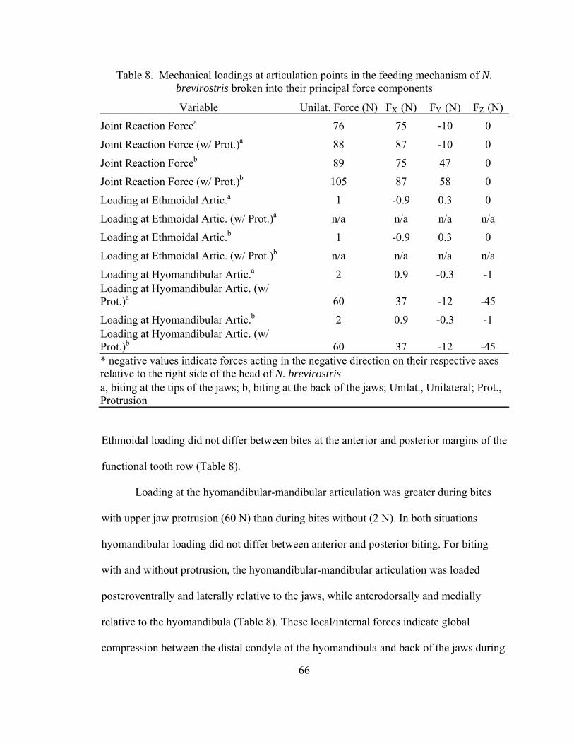
66
Table 8. Mechanical loadings at articulation points in the feeding mechanism of N. brevirostris broken into their principal force components
Variable Unilat. Force (N) FX (N) FY (N) FZ (N) Joint Reaction Forcea 76 75 -10 0
Joint Reaction Force (w/ Prot.)a 88 87 -10 0
Joint Reaction Forceb 89 75 47 0
Joint Reaction Force (w/ Prot.)b 105 87 58 0
Loading at Ethmoidal Artic.a 1 -0.9 0.3 0
Loading at Ethmoidal Artic. (w/ Prot.)a n/a n/a n/a n/a
Loading at Ethmoidal Artic.b 1 -0.9 0.3 0
Loading at Ethmoidal Artic. (w/ Prot.)b n/a n/a n/a n/a
Loading at Hyomandibular Artic.a 2 0.9 -0.3 -1 Loading at Hyomandibular Artic. (w/ Prot.)a 60 37 -12 -45 Loading at Hyomandibular Artic.b 2 0.9 -0.3 -1 Loading at Hyomandibular Artic. (w/ Prot.)b 60 37 -12 -45 * negative values indicate forces acting in the negative direction on their respective axes relative to the right side of the head of N. brevirostris a, biting at the tips of the jaws; b, biting at the back of the jaws; Unilat., Unilateral; Prot., Protrusion
Ethmoidal loading did not differ between bites at the anterior and posterior margins of the
functional tooth row (Table 8).
Loading at the hyomandibular-mandibular articulation was greater during bites
with upper jaw protrusion (60 N) than during bites without (2 N). In both situations
hyomandibular loading did not differ between anterior and posterior biting. For biting
with and without protrusion, the hyomandibular-mandibular articulation was loaded
posteroventrally and laterally relative to the jaws, while anterodorsally and medially
relative to the hyomandibula (Table 8). These local/internal forces indicate global
compression between the distal condyle of the hyomandibula and back of the jaws during

67
biting both with and without upper jaw protrusion. Modeling the hyomandibula as a two-
force member assumes either purely compressive or purely tensile loading. Deviations
from this assumption, as would be likely during dynamic loading, cannot be determined
from these analyses.
Although a cumulative force of 275 N is generated by the muscles involved in the
abduction of the feeding mechanism of N. brevirostris, the only muscle actuating
depression of the lower jaw is the coracomandibularis, which was capable of generating
13 N of force. Accounting for its acute insertion on the posterior margin of the lower jaw
symphysis (26°) and the mechanical advantage of the jaw abducting mechanism (0.86),
the realized jaw opening force at the anterior margin of the functional tooth row was 5 N
(Table 7).
Mechanics of Jaw Protrusion
Static equilibrium calculations on the lower jaw of N. brevirostris indicated that
the jaw is elevated with greater force in the anterodorsal direction when the ventral
preorbitalis is active (194 N) than when it is not (167 N). This finding accounts for the
difference between bite forces with and without protrusion. Activity of the
coracomandibularis in absence of the ventral preorbitalis decreased the lower jaw
adducting force by 15% (141 N), whereas simultaneous activity of these muscles resulted
in a marginal increase in the anterodorsally directed adductive force (168 N). The
adductive force pulling the upper jaw posteroventrally (167 N) decreased by 17% (140
N) during activity of the ventral preorbitalis, 38% (103 N) during activity of the levator
palatoquadrati, and 55% (76 N) during simultaneous activity of these two muscles.

68
The bulk of the adductor musculature of N. brevirostris spans the gap between the
upper and lower jaws and act to draw the jaws together. Forces acting externally to this
system (coracomandibularis, ventral preorbitalis, levator palatoquadrati (Fig. 10))
therefore actuate movement of the jaw system as a whole. Activity of the
coracomandibularis added a posteroventrally directed force of 13 N to the jaw system,
while activity of the ventral preorbitalis and levator palatoquadrati contributed 27 and 65
N of anterodorsally directed force, respectively. Simultaneous activation of the ventral
preorbitalis and levator palatoquadrati added 92 N of anterodorsally directed force to the
jaws, whereas simultaneous activity of all of three muscles generated a net force of 79 N
in the anterodorsal direction. When all three muscles were active, 2.5 times more force
was generated anteriorly than dorsally.
Prey Capture Performance
The mean bite force for all individuals during in situ bite performance trials was
13 N, and was applied to the transducer for approximately 114 ms (Table 9). It took
juvenile N. brevirostris an average of 46 ms to reach maximum bite force at a rate of 470
N s-1. Maximum bite force was sustained for 13 ms and released over the subsequent 55
ms. Times to and away from maximum bite force were statistically equivalent. Maximum
bite force was released at 550 N s-1, which was statistically equivalent to the rising slope
of the force-time curve. The average impulse generated during biting was 1 kg m s-1
(Table 9). Although the majority of bites analyzed showed single force peaks associated
with single bites, double force peaks separated by an average of 31 ms were observed in
association with a single bite in 12% of the trials. Additionally, head-shaking was
observed in 46% of the in situ bite performance trials.

69
Table 9. In situ bite performance data for N. brevirostris Variable Minimum Maximum Mean +/- S.E.Maximum Force (N) 6 31 13 +/- 4 Force Duration (ms) 24 282 114 +/- 45 Time to Maximum Force (ms) 11 155 46 +/- 27 Time at Maximum Force (ms) 2 31 13 +/- 7 Time away from Maximum Force (ms) 3 110 55 +/- 19 Impulse (kg m s-1) 0.1 4 1 +/- 0.8 Rising Slope of Force-Time Curve (N s-1) 172 1083 470 +/- 166
Falling Slope of Force-Time Curve (N s-1) 78 2156 550 +/- 402
PCA 1 reduced the performance variables into two PCs (82.4% of variance
explained). MANOVA indicated no differences in performance measures among
individuals using size-corrected data (Wilk’s Lambda = 0.742, F8,38 = 0.764, p = 0.636).
PC1 revealed substantial covariation in performance measures for all individuals. Every
performance variable other than maximum bite force loaded heavily upon this axis.
Maximum bite force was significantly, though weakly, related to impulse only (p =
0.011, R2 = 0.251). Impulse had a strong positive relationship with force duration (p <
0.001, R2 = 0.862) and negative relationships with the rising (p = 0.001, R2 = 0.406) and
falling slopes (p = 0.001, R2 = 0.402) of the force-time curve. Force duration had
significant negative relationships with both the rising (p < 0.001, R2 = 0.507) and falling
(p < 0.001, R2 = 0.698) slopes as well (Fig. 14).
PCA 2 on kinematic and performance data yielded six PCs with eigenvalues
greater than 1.0 (89.9% of the variability explained). Seven of the ten variables that
loaded heavily on PC 1 were performance variables (all but maximum force). Force
duration, time to maximum force, time at maximum force, time away from maximum

Fig. 14. Linear regressions of log10-transformed bite performance variables of N. brevirostris. (A) Force duration (ms) versus impulse (kg m s-1). (B) Slope of the force-time curve (N s-1) versus impulse (kg m s-1). Black circles represent the rising slope; gray triangles represent the falling slope; regression lines fall on top of each other. (C) Slope of the force-time curve (N s-1) versus force duration (ms). Black circles represent the rising slope; gray triangles represent the falling slope.
70

71
Table 10. Principal component loadings of performance and kinematic variables from bite performance trials of N. brevirostris
Variable PC 1 PC 2 PC 3 Lower Jaw Depression Distance 0.137 0.650 0.632 Lower Jaw Depression Duration 0.434 0.716 0.209 Lower Jaw Depression Velocity 0.111 0.347 0.652 Lower Jaw Depression Acceleration 0.251 0.047 0.505 Time to Maximum Gape 0.651 0.590 0.157 Maximum Gape 0.456 0.297 0.009 Onset of Lower Jaw Elevation 0.640 0.402 0.181 Lower Jaw Elevation Distance 0.529 0.448 0.263 Lower Jaw Elevation Duration 0.579 0.283 0.469 Lower Jaw Elevation Velocity 0.138 0.229 0.471 Lower Jaw Elevation Acceleration 0.468 0.092 0.587 Time to Lower Jaw Elevation 0.749 0.469 0.396 Time to Maximum Force 0.882 0.312 0.040 Time at Maximum Force 0.725 0.470 0.071 Time away from Maximum Force 0.614 0.227 0.581 Force Duration 0.824 0.418 0.350 Rising Slope -0.810 0.178 0.030 Falling Slope -0.658 0.392 0.271 Impulse 0.815 0.380 0.277 Maximum Force 0.122 0.128 0.251
Bold values indicate variables considered to load heavily on a given principal component (loading score > 0.600)
force, and impulse all loaded positively on PC 1, while the rising and falling slopes of the
force-time curve loaded negatively. Times to maximum gape, onset of lower jaw
elevation, and completion of lower jaw elevation were the only kinematic variables that
loaded heavily on PC 1 (Table 10). Maximum force did not load heavily until PC 5 (8.0%
of variance explained), and was the only variable to do so on this axis. Performance
variables did not load heavily upon any other axes. A general lack of kinematic

72
covariation on all principal components is indicative of highly variable feeding behavior
by N. brevirostris (Table 10).
Stepwise multiple regression analyses demonstrated more predictive relationships
between kinematic and performance variables than PCA 2. The four performance
variables quantifying durations were all predicted by kinematics. Force duration was
significantly positively related to the time to maximum gape and negatively related to the
duration of lower jaw depression (R2 = 0.382, F2,22 = 6.812, p = 0.005). Time to
maximum force was positively related to the time to onset and negatively related to the
acceleration of lower jaw elevation (R2 = 0.346, F2,22 = 5.816, p = 0.009). Most notably,
time at maximum force was positively related to the acceleration of lower jaw depression,
time to maximum gape, and duration of lower jaw elevation, and negatively related to the
duration of lower jaw depression and distance of lower jaw elevation (R2 = 0.581, F5,19 =
10.72, p < 0.001). Time away from maximum force had a weak, but positive relationship
to the time to maximum gape (R2 = 0.191, F1,23 = 5.445, p = 0.029). Lastly, the rising
slope of the force-time curve was positively related to the acceleration of lower jaw
elevation and negatively related to the time to maximum gape (R2 = 0.394, F2,22 = 7.153,
p = 0.004). Neither of the performance variables indicative of the magnitude of loading
generated during a bite (maximum force, impulse) were predicted by regression models
of kinematic variables.
PCA 3 of kinematic variables from fish and transducer bites yielded four PCs
with eigenvalues greater than 1.0 (76.4% of variance explained). MANOVA indicated no
significant differences between prey capture kinematics from the two presentation
methods (Wilk’s Lambda = 1.0, F4,37 = 0.0, p = 1.0) or between individuals (Wilk’s

73
Lambda = 0.710, FB16,113B = 0.841, p = 0.637). Additionally, there were no significant
interactions between presentation method and individual (Wilk’s Lambda = 0.697, FB16,113B
= 0.892, p = 0.580).
Methodological Comparison
After controlling for the effect of body size, theoretical maximum bite force (69 ±
5 N) was significantly greater than in situ (13 ± 4 N) and restrained (19 ± 5 N) bite forces
(FB2,10B = 12.699, p = 0.002). In situ and restrained bite forces were equivalent. Times to
and away from maximum bite force were equivalent within in situ (46, 55 ms) and
restrained (215, 134 ms) bite force measurements. Times to (t B7 B = -2.786, p = 0.027) and
away (tB7 B = -2.646, p = 0.033) from maximum force were significantly longer during
restrained bites than during in situ bites.
Bite Forces among Cartilaginous Fishes
Bite forces among the cartilaginous fishes collectively scaled to body mass with a
coefficient of 0.66, approximating the isometric scaling coefficient of 0.67. Hydrolagus
colliei and H. francisci, the two durophagous cartilaginous fishes in this analysis, had the
highest mass-specific bite forces (1.08, 0.69 respectively). Negaprion brevirostris was
intermediate among the cartilaginous fishes (0.01), with C. limbatus and S. acanthias
having the lowest mass-specific bite forces (-0.71, -1.39 respectively) (Huber and Motta,
2004; Huber et al., 2005, 2006, in prep).

74
Discussion
Cranial Biomechanics
The maximum bite force of juvenile N. brevirostris ranged from 69-217 N along
the lower jaw. This range was estimated from bites with protrusion of the upper jaw, and
was 23-29% higher than bites not involving protrusion. This finding is attributed to the
ventral preorbitalis, which plays a dual role in the adduction and protrusion of the jaws
(Motta et al., 1997). Increased bite force should therefore be added to the list of
hypothetical selective pressures involved in the evolution of upper jaw protrusion in
elasmobranchs, including increased prey capture and manipulation efficiency due to
enhanced cutting ability of teeth, more rapid jaw closure, more precise handling of
benthic prey, and the allowance of a hydrodynamic sub-terminal mouth (Springer, 1961;
Moss, 1972; Tricas and McCosker, 1984; Frazzetta and Prange, 1987; Motta et al., 1997;
Wilga et al., 2001). Subdivision of the preorbitalis muscle in carcharhiniform sharks
created a dorsal division serving as a supplementary jaw adductor and a ventral division
that both adducts the lower jaw and translates the upper jaw anteroventrally (Wilga et al.,
2001). Although protrusion may augment bite force, juvenile N. brevirostris do not bite
particularly hard with respect to other cartilaginous fishes, suggesting that factors other
than bite force (e.g. tooth cutting mechanics, head-shaking) figure prominently in its prey
capture ability.
The hypothesized functions of the dorsal and ventral preorbitalis, levator
palatoquadrati, dorsal and ventral quadratomandibularis, and coracomandibularis muscles
in upper jaw protrusion were supported by this study (Moss, 1972; Motta et al., 1997;
Wilga et al., 2001). During protrusion, the ventral preorbitalis and levator palatoquadrati

75
contributed an anteriorly directed force of 86 N and a dorsally directed force of 34 N to
the jaws. The anteriorly directed force is the impetus behind anterior motion of the upper
jaw. Ventral motion of the upper jaw is accomplished via multiple mechanisms.
Staggered firing of the jaw adducting (dorsal and ventral quadratomandibularis and
dorsal preorbitalis) and jaw protruding muscles (levator palatoquadrati and ventral
preorbitalis) (Motta et al., 1997) allows the adductors to pull the upper jaw away from the
chondrocranium, after which it is drawn forward by the protruding muscles. Ventral
movement of the upper jaw frees its ethmoid process from the orbital notch in the ventral
surface of the chondrocranium. Once the upper jaw is clear of the orbital notch, the
anteriorly directed force of the jaw protruding muscles forces the upper jaw along the
anteroventrally sloping palatal fossa of the chondrocranium. Therefore, both the jaw
adducting and protruding muscles contribute to ventral movement of the upper jaw
(Motta et al., 1997).
Firing of the coracomandibularis during jaw adduction will facilitate ventral
movement of the upper as well. This would create a clockwise moment about the jaws
relative to the right side of the head such that the upper jaw is pulled down with 18%
more force than the lower jaw is pulled up with. Should the coracomandibularis be
inactive at this time, as noted by Motta et al. (1997), the inertia of a prey item resisting
lower jaw elevation could provide the necessary imbalance to cause this clockwise
moment about the jaws. Additional activity from the ventral preorbitalis and levator
palatoquadrati after the upper jaw has rotated ventrally (Motta et al., 1997) will augment
this clockwise moment as well as pull the upper jaw anteriorly.

76
In contrast to the anteriorly directed adductive force facilitating protrusion in N.
brevirostris (Table 7), the principal component of the jaw adducting force of H. francisci
is oriented vertically (Huber et al., 2005), illustrating the disparity in selective pressures
between elasmobranchs with different feeding mechanisms. Ram feeding by N.
brevirostris involves anterior movement of the upper jaw during protrusion that will
assist in capturing elusive prey (Wainwright et al., 2001). Conversely, the processing of
hard prey by H. francisci is enhanced by adductive forces generated orthogonal to the
occlusal surface of its molariform teeth.
Upper jaw protrusion along with rapid elevation of the lower jaw facilitates rapid
closure of the jaws upon prey by decreasing both gape distance and the time required to
close the jaws (Wilga et al., 2001). Kinematic analyses of transducer and fish bites
determined the average velocity of lower jaw elevation by N. brevirostris to be 43.6 cm/s,
which is approximately as quick as any elasmobranch for which data is available (Ferry-
Graham, 1998a; Wilga and Motta, 1998a, 2000; Edmonds et al., 2001). Despite the high
speed of lower jaw elevation by N. brevirostris, its jaw adducting mechanical advantage
(0.34) is higher than those of most ram-feeding teleosts that feed upon elusive prey
(Wainwright and Richard, 1995; Wainwright et al., 2004; Westneat, 2004). By teleost
standards, the adductive mechanical advantage of N. brevirostris is better suited for force
transmission than velocity transmission. This mechanical advantage is comparable to
those of S. acanthias (0.28) and C. limbatus (0.34), which also rely upon rapid jaw
kinesis to capture elusive prey (Castro, 1996; Alonso et al., 2002; Huber and Motta,
2004; Huber et al., 2006). However, the adductive mechanical advantage of N.
brevirostris is much lower than those of H. francisci (0.51) and H. colliei (0.57), both of

77
which consume hard prey (Johnson and Horton, 1972; Segura-Zarzosa et al., 1997; Huber
et al., 2005, in prep).
Anterior biting placed the jaw joints of N. brevirostris in compression while
posterior biting placed the jaw joints in tension. Although the ellipsoidal geometry of the
lateral jaw articulation will resist compressive joint reaction forces at a variety of gape
angles (Motta and Wilga, 1995), tensile joint reaction forces present a larger problem for
the mechanical stability of the feeding mechanism (Greaves, 1988, 2000). Tensile joint
reaction forces arise when food is bitten between the jaw joint and the resultant jaw-
adducting force vector (mechanical advantage > 1.0). In such instances the prey item
becomes a temporary fulcrum about which the lower jaw rotates, placing the jaw joints in
tension (Greaves, 1988, 2000). In N. brevirostris, stabilization of the jaw joints and the
jaws’ articulations with the hyoid arch against tensile loading is accomplished through an
extensive set of ligaments. Lateral translation between the upper and lower jaws, as
would be associated with lateral head-shaking, is resisted via the ellipsoidal concavity of
the lateral quadratomandibular articulation and the sagittal orientation of the medial
quadratomandibular articulation as well (Motta and Wilga, 1995). Lateral head-shaking is
commonly used by N. brevirostris to draw its teeth across prey items (Frazzetta and
Prange, 1987; Motta et al., 1997).
Mechanics and Evolution of Jaw Suspension
Analyses of bites without protrusion revealed a potential mechanical role of the
ethmoidal articulation in elasmobranch feeding mechanisms. During such bites in N.
brevirostris the ethmoidal articulation was in negligible tension (1 N) while the
hyomandibula was compressed (2 N) between the posterior margin of the jaws and the
78
chondrocranium. These respective loadings are the result of a clockwise moment about
the upper jaw relative to the right side of the head, and are contingent upon force being
generated by the ventral quadratomandibularis from a position posterior to the jaw joint
(Figs. 9, 10). The magnitude of hyomandibular loading is low due to the approximately
equal moments generated on either side of the ethmoidal articulation by muscle, joint
reaction, and bite reaction forces counteracting each other. The minimal tensile loading at
the ethmoidal articulation is readily transferred to compression by moving the origin of
the resultant adductive force vector anteriorly along the lower jaw. This modification
increases mechanical advantage and orients the adductive force vector more
perpendicularly relative to the lower jaw, causing a counterclockwise moment to be
generated about the upper jaw relative to the right side of the head. This
counterclockwise moment subsequently places the hyomandibula in tension and the
ethmoidal articulation in compression (D.R. Huber, unpub. data).
Simultaneous hyomandibular tension and ethmoidal compression are found in H.
francisci due to an anatomical arrangement similar to that hypothesized to cause these
loadings in N. brevirostris. These findings demonstrate that the ethmoidal articulation
likely acts as a pivot point in the feeding mechanisms of elasmobranchs in which the
upper jaw does not disarticulate from the chondrocranium during biting (Huber et al.,
2005). Maintenance of the ethmoidal articulation during biting, high mechanical
advantage (0.51), and perpendicularly arranged jaw adducting muscles give H. francisci
an ethmoidal loading over 2500% greater than that of N. brevirostris relative to their
respective bite forces. The higher mechanical advantage of H. francisci also causes a
greater imbalance of moments cranial and caudal to the ethmoidal articulation, resulting

79
in nearly 800% more hyomandibular loading than N. brevirostris relative to their
respective bite forces. Thus, it is probable that the hyomandibula acts as a tensile element
in elasmobranchs in which the ethmoidal articulation is maintained during biting. As
such, the structural and material properties of hyomandibulae in these taxa may reflect
their tensile role.
Protrusion of the upper jaw during biting by N. brevirostris creates an antithetical
loading regime to that explained above for elasmobranchs in which the ethmoidal
articulation remains intact. During protrusion, disengagement of the upper jaw from the
chondrocranium precludes the generation of bending moments on either side of the
ethmoidal articulation and causes the muscular and reaction forces acting on the upper
jaw to be linearly transmitted to the jaws’ articulation with the hyomandibula. The result
of this transmission is a substantial increase in compression of the hyomandibula (60 N).
During bites involving protrusion, the hyomandibulae of N. brevirostris are loaded
approximately 300% greater relative to bite force than those of H. francisci, in which the
ethmoidal articulation remains intact (Huber et al., 2005).
The posteroventrally and laterally directed compressive force on the jaws at their
hyomandibular articulation will create a laterally directed bending moment and torsion
about the jaws in the coronal plane. Lateral deflection and axial torsion of the posterior
region of the jaws is most likely resisted due to the geometric properties afforded by the
large mandibular knob and sustentaculum of the lower jaw. The lower jaw is deepest and
widest at these structures and therefore best able to resist flexion along the Y and Z-axes
in this region due to increased second moments of area. The interaction of the second

80
moments of area along the Y and Z-planes increases the polar second moment of area of
the jaws, increasing their ability to resist torsion (Hibbeler, 2004).
Increased compressive hyomandibular loading associated with upper jaw
protrusion may have been related to a freeing-up of the anterior margin of the jaws, and
been the mechanical impetus behind evolutionary changes in elasmobranch jaw
suspension. Albeit only in two taxa, evidence from N. brevirostris and H. francisci
indicate that disengagement of the ethmoidal articulation, which is associated with
extensive protrusion of the upper jaw, causes a shifting from tensile to compressive
loading on the hyomandibula, and concentrates the forces associated with prey capture to
the posterior region of the feeding mechanism. This transition may hypothetically be
correlated with the evolutionary shift from amphistylic to hyostylic jaw suspension
mechanisms and increased upper jaw kinesis.
The primary supportive structures in an amphistylic jaw suspension are
ligamentous attachments between the anterior margin of the upper jaw and
chondrocranium. The posterior margins of the upper and lower jaws articulate with the
chondrocranium via hyomandibular cartilages that contribute little support and a post-
orbital articulation between the upper jaw and chondrocranium may or may not be
present. Subsequently, the hyomandibulae of early amphistylic sharks were relatively
small (Xenacanthida, Palaeospinax) (Moy-Thomas and Miles, 1971; Wilga, 2002). In
hyostyly the hyomandibula is the primary supportive element in the feeding mechanism,
which is accompanied by reduced ethmoidal or orbital articulations between the upper
jaw and chondrocranium. Elasmobranchs possessing a suspensory hyomandibula and an
orbital articulation are said to posses an orbitostylic variety of the hyostylic jaw

81
suspension (Gregory, 1904; Maisey, 1980; Wilga, 2002). The evolution of hyostyly in
elasmobranchs was associated with the reduction of the number (loss of post-orbital
articulation) and size of the articulations between the jaws and chondrocranium and
shorter, more mobile hyomandibulae oriented more orthogonal relative to the
chondrocranium during biting (Schaeffer, 1967; Moy-Thomas and Miles, 1971; Maisey,
1980, 1985; Cappetta, 1987; Carroll, 1988; Stahl, 1988; Wilga, 2002). These changes are
indicative of enhanced jaw kinesis and the ability to withstand compression by the
hyomandibula. Therefore, hyomandibular morphology (as indicative of load-bearing
ability) may vary concomitantly with protrusion ability in elasmobranchs.
The morphological diversity of extant elasmobranch feeding mechanisms
represents a continuum within the aforementioned jaw suspension mechanisms.
Variability in these mechanisms qualitatively supports the hypothesized relationships
between hyomandibular loading, jaw kinesis, and suspension mechanisms. The
hexanchid sharks (sixgill and sevengill) are the only extant elasmobranchs to retain an
amphistylic jaw suspension, including ethmoidal, orbital, and post-orbital articulations
(Maisey, 1980; Wilga, 2002). Although the post-orbital articulation of the hexanchid
sharks can disarticulate during jaw protrusion, the orbital articulation remains intact
(Compagno, 1977; Maisey, 1983; Wilga, 2002). Given this morphological arrangement
and the suspensorial loading results from both N. brevirostris and H. francisci, the
hyomandibulae of hexanchid sharks are hypothesized to experience low magnitude
tensile loading. This is supported by the fact that their hyomandibulae are long, thin,
posteriorly directed, and “non-suspensory” (Wilga, 2002). Presumably a similar loading
regime occurs in orbitostylic sharks such as S. acanthias, in which the orbital articulation
82
remains intact at maximum upper jaw protrusion as well (Wilga, 2002). In contrast, the
hyomandibula is the only articulation between the jaws and chondrocranium in batoid
feeding mechanisms (euhyostyly (Wilga, 2002)). Based on the present body of
knowledge it would be parsimonious to infer that the batoid hyomandibula is a
compressive element. This assertion is strongly supported by the lesser electric ray
Narcine brasiliensis, in which medial translation of the hyomandibulae protrudes the
jaws. At maximum protrusion the longitudinal axes of the hyomandibulae line up with
those of the jaws, such that the jaws are axially buttressed as they are protruded into the
sediment in search of benthic prey. Trabecular reinforcement in both the jaws and
hyomandibulae of N. brasiliensis are arranged to resist buckling associated with axial
compression (Dean and Motta, 2004a, 2004b; Dean et al., 2005a).
Prey Capture Performance
Voluntary biting by N. brevirostris involved the rapid application of low
magnitude forces (114 ms, 13 N), indicating that maintenance of bite force is not critical
to prey capture. The brief duration of low magnitude force, equivalence of the times to
and away from maximum force, and equivalence of the rising and falling slopes of the
force-time curve demonstrate that biting by N. brevirostris is characterized by quick,
snapping bites during which the application of force is no more important than its release.
The rapid application of low magnitude forces minimizes the impulse imparted to prey
items by N. brevirostris (1 kg m s-1). Impulse, which is representative of momentum
transfer, is a measure of the ‘effort’ exerted upon one body by another (Nauwelaerts and
Aerts, 2003). Because momentum is conserved in inelastic collisions, impulse is a proxy
for kinetic energy transfer. Low impulse generation by N. brevirostris therefore indicates

83
that little kinetic energy is needed for successful predation using ram feeding with
extensive protrusion to gouge prey.
The bite performance profile of N. brevirostris is in stark contrast to that of H.
francisci, the only other elasmobranch in which voluntary bite performance has been
measured. Heterodontus francisci bites harder (95 N) and longer (535 ms) than N.
brevirostris, which translates into a biting impulse 25 times greater than that of N.
brevirostris. The dramatic differences in kinetic energy transfer to prey items between N.
brevirostris and H. francisci echo the different physical requirements associated with
capturing and consuming prey in their respective ecological niches. Heterodontus
francisci utilizes deliberate, cyclical, high-magnitude force application to crush molluscs,
echinoderms, and benthic crustaceans (Strong Jr., 1989; Segura-Zarzosa et al., 1997;
Huber et al., 2005). Negaprion brevirostris is a trophic generalist that relies on upper jaw
protrusion and a piercing dentition to slash prey into pieces small enough to consume
(Frazzetta and Prange, 1987; Wetherbee et al., 1990; Motta et al., 1997).
Although the interplay between dental morphology and the cutting of compliant
materials has received negligible quantitative attention, limited empirical evidence and
physical theory regarding cutting devices has established a fundamental basis from which
to interpret the effectiveness with which the teeth of N. brevirostris cut prey (Frazzetta,
1988; Abler, 1992). The teeth of the lower jaw are elliptical in cross-section and sharply
pointed, making them effective at puncturing compliant materials through pressure
concentration. Bite force applied along the longitudinal axes of the teeth is concentrated
at the minute area of their tips. Having initially penetrated, friction between a tooth’s
surface and the cutting substrate will shear the substrate, creating stress concentrations

84
that lead to material rupture and further penetration of the teeth (Frazzetta, 1988; Martin
et al., 1998). The teeth of the lower jaw are the first to contact a prey item (D.R. Huber,
pers. obs.) and likely serve to impale the prey item until the teeth of the upper jaw can
descend.
The triangular teeth of the upper jaw in N. brevirostris possess serrations that
gradually enlarge from a tooth’s tip to its base (Frazzetta, 1988). Initial penetration of
these teeth will occur through pressure concentration. Once a tooth tip has penetrated and
its apical edges encounter the prey item, the cutting mechanism changes from pressure
concentration to friction-based draw cutting (Frazzetta, 1988). As the apical edges of
triangular teeth are forced down through the prey item, frictional and reaction forces
between the teeth and the prey item will cause shearing and rupture. As the teeth descend
further their serrations will come into contact with the compliant substrate. The substrate
will bulge between serrations, converting the draw force acting parallel to the apical edge
into a reaction force between the bulged material and the edges of the serrations, further
augmenting rupture of the prey item (Frazzetta, 1988; Motta, 2004). The triangular form
of the upper jaw’s teeth also allows successive serrations to continually encounter and
sever new substrate, ever widening the cut (Abler, 1992).
Lateral head shaking augments the cutting ability of the teeth of N. brevirostris as
well. Its effect will primarily be realized at the anterior teeth that are oriented in the
transverse plane, in line with the lateral motion of the head. This movement will cause
the somewhat laterally oriented cusps of the teeth in the upper jaw to sink into the prey
item, as well as direct material into the lateral notches of the teeth (Fig. 11). Stress
concentrations within the cutting substrate increase substantially when they reach this

85
notch, promoting the rupture of more durable connective tissues (Motta, 2004). The
ability of tiger sharks Galeocerdo cuvier to bite through the shells of large cheloniid sea
turtles is primarily attributed to the use of lateral head shaking and the presence of
exaggerated lateral notches on the serrated teeth of its upper jaw (Witzell, 1987; Motta,
2004).
Notable covariance was identified within and among several bite performance and
kinematic variables. Positive relationships between impulse and all force durations, and
negative relationships between impulse and the rising and falling slopes of the force-time
curve, indicate that larger kinetic energy transfers to prey items are associated with
slower, more deliberate biting. This relationship was also found in H. francisci (Huber et
al., 2005). The dependence of kinetic energy transfer on duration was also corroborated
by PCA 2, in which the only kinematic variables that covaried with performance
variables were durations (Table 10). The kinematic variables found to be predictive of
biting performance from multiple regression analyses supported the notion that slower,
more deliberate behaviors are associated with greater force production as well, with time
to maximum gape and the acceleration of lower jaw elevation being the most predictive
of biting performance.
Methodological Comparison
The theoretical bite force of juvenile N. brevirostris was substantially greater than
the highest in situ bite force measurements obtained, as was found for H. francisci (Huber
et al., 2005). While in situ bite force was not indicative of maximum biting ability in
either shark, obtaining voluntary bite force measurements yielded valuable behavioral
information in both cases. In contrast to H. francisci, restrained bite force measurements
86
of N. brevirostris were not comparable to theoretical estimates. This discrepancy is again
attributed to these animals’ contrasting predatory strategies and perhaps to behavioral
motivation under restrained conditions. Under restrained circumstances, N. brevirostris
shook its head violently from side to side. Effort was concentrated on using the teeth as
cutting devices instead of applying bite force. Conversely, H. francisci did not employ
head-shaking, but instead bit the force transducer vigorously, as it would have bitten and
processed a hard prey item common to its durophagous diet (Segura-Zarzosa et al., 1997).
Restrained circumstances did apparently motivate more aggressive behavior in N.
brevirostris, as indicated by the longer times to and away from maximum bite force.
Collectively, of the various methods used to determine bite force in N. brevirostris, H.
francisci, and S. acanthias, theoretical estimates are the most reliable indicator of
maximum performance (Huber and Motta, 2004; Huber et al., 2005).
Conclusions
The jaw adducting musculature of N. brevirostris generates an anteriorly directed
force motivating protrusion of the upper jaw, due in part to the action of the ventral
preorbitalis. Previous hypotheses regarding the different roles of the adductive muscles in
upper jaw protrusion were corroborated in this study and the ventral preorbitalis was
found to increase bite force during protrusion due to its dual role in adducting the lower
jaw and protruding the upper jaw. Mechanical analyses revealed that the hyomandibula
of N. brevirostris is a compressive element regardless of whether or not the upper jaw is
protruded, and that compression does not occur at the ethmoidal articulation between the
jaws and chondrocranium. This loading regime may be indicative of a trend in the

87
evolution of elasmobranch feeding mechanisms, whereby a posterior shifting of the
loadings occurring within the jaw suspension and compressive loading on the
hyomandibula results in a freeing-up of the anterior margin of the jaws, allowing greater
jaw kinesis. The benefit of a highly kinetic feeding mechanism may come at the cost of
having a force-inefficient feeding mechanism however, as bite force is fairly low in
juvenile N. brevirostris. The very low voluntary in situ bite forces generated by juvenile
N. brevirostris suggest that the effectiveness with which its teeth cut prey is very
important to its predatory success.

88
Chapter 3: Mechanical consequences of functional constraint in the feeding mechanism
of the whitespotted bamboo shark Chiloscyllium plagiosum
Abstract
The evolution of elasmobranch jaw suspension mechanisms has involved the
decoupling of skeletal elements and has resulted in enhanced kinesis of the jaws relative
to the cranium. The whitespotted bamboo shark Chiloscyllium plagiosum has a hyostylic
jaw suspension in which the upper jaw is incapable of dissociating from the cranium
during protrusion, and as such is constrained in its kinetic abilities. Biomechanical
modeling of the feeding mechanism determined that this linkage causes compression
between the anterior margin of the jaws and cranium, while placing the hyomandibular
cartilages in tension during biting. Theoretically releasing this constraint caused a
transition to compressive hyomandibular loading during biting. The release of this
constraint occurred during the evolutionary transition from a hyostylic to euhyostylic jaw
suspension mechanism in the batoid elasmobranchs (skates and rays), resulting in
increased jaw kinesis and presumably the opening of new feeding niches. These results
suggest that the decoupling of the anterior margin of the jaws from the cranium, which
allows increased jaw kinesis, is associated with a transition from tensile to compressive
hyomandibular loading. Biomechanical modeling of jaw mechanics in C. plagiosum also
predicted a theoretical maximum bite force of 69 N at the anterior tips of the jaws. To
validate this, bite forces were measured in live C. plagiosum through voluntary in situ

89
biting performance trials (29 N), tetanic stimulation of the jaw adducting musculature (53
N), and measurements on restrained individuals (51 N). Theoretical, stimulated, and
restrained bite force measurements were statistically equivalent and all greater than
measurements from in situ trials, indicating that 1) C. plagiosum can regulate its bite
force and 2) restrained and tetanically stimulated bite force measurements are an accurate
proxy for maximum bite force in C. plagiosum.

90
Introduction
Functional constraints in musculoskeletal systems influence organismal behavior
on a number of levels. Variation in muscle geometry and fiber type delimit the temporal
dynamics and forces generated during muscular contraction (Gans and Gaunt, 1991;
Curtin et al., 1998; Lou et al., 2002). The mechanical properties of both intra- and
extramuscular (tendon) connective tissues influence the transmission of these forces to
skeletal elements (Alexander, 2002; Purslow, 2002). The relative dimensions of
articulated segments within the skeleton then dictate the rate and force with which
muscular contractions are transmitted via these elements to the external environment
(Westneat, 1994; Carrano, 1999), while the degrees of freedom between these elements
determines the paths of motion along which these transmissions occur. Developmental
constraints further limit organismal function by canalizing the ontogenetic trajectories
allowed in these parameters (Sears, 2004). Ultimately, interactions among these
musculoskeletal variables yield a specific range of behaviors that a given organism is
capable of performing, often representing trade-offs between mechanical stability and
skeletal kinesis (Biewener, 1998).
The degrees of freedom between adjacent skeletal elements determine the range
of motion allowed between those elements and subsequently influence the nature of the
mechanical loadings they are subject to. Physical constraints limit freedom of motion,
thereby imposing directionally specific loading patterns on skeletal elements. Such
patterns are readily observed in the structural (cortical and trabecular) reinforcement
patterns of these elements (Currey, 2002; Lieberman et al., 2003; Dean et al., 2005a;
Pontzer et al., 2006). Conversely, the decoupling of skeletal elements allows for greater

91
freedom of motion and consequently more variable loading regimes as these elements are
subjected to diverse internal and external forces (Herrel et al., 2000).
The elasmobranch fishes (sharks, skates, and rays) are capable of remarkable jaw
protrusion (> 100% head length (Dean and Motta, 2004b)), which is historically due to a
series of decouplings between skeletal elements and muscular diversification in their
feeding mechanisms (Wilga et al., 2001; Wilga, 2002, 2005). Basal elasmobranchs
possessed an amphistylic jaw suspension mechanism in which the large otic flange of the
upper jaw articulated with the postorbital region of the cranium and the palatine process
of the upper jaw articulated with the ethmoidal region of the cranium, supplemented by
ligamentous attachments (Fig. 15). The jaws also articulated with the cranium indirectly
via a pair of hyomandibular cartilages extending from the medial face of the jaws
posterior to the jaw joint to the otic region of the cranium. The hyomandibular cartilages
are presumed to have contributed little mechanical support to the jaws in these
elasmobranchs (Gregory, 1904; Wilga, 2002). These amphistylic sharks are believed to
have used ram feeding, over-swimming and grasping prey between the jaws (Schaeffer,
1967; Carroll, 1988).
Amphistyly gave rise to the hyostylic and orbitostylic jaw suspensions of modern
elasmobranchs, although the hexanchiform sharks retain the amphistylic condition with
an orbital, not ethmoidal cranio-palatine articulation (Maisey, 1980). In hyostylic and
orbitostylic elasmobranchs, the hyomandibular cartilages are the primary supportive
elements between the jaws and cranium. Hyostyly and orbitostyly differ in that the orbital
process of the upper jaw articulates with either the ethmoidal or orbital region of the
cranium respectively (Fig. 15) (Maisey, 1980; Wilga, 2002). The first decoupling event

Figure 15. Left lateral views of the feeding mechanisms of (A) Cladodont level Cladodus (amphistyly); (B) the sharpnose sevengill shark Heptranchias perlo Hexanchiformes (amphistyly); (C) whitespotted bamboo shark Chiloscyllium plagiosum (hyostyly); (D) lesser electric ray Narcine brasiliensis (euhyostyly). Heptranchias perlo is also representative of the orbitostylic jaw suspension mechanism due to its multiple cranio-palatine articulations. Articulation points are marked with arrows. C, ceratohyal; CR, cranium; E, ethmoidal; H, hyomandibula; L, lower jaw; O, orbital; P, postorbital; U, upper jaw. The feeding mechanism of Cladodus is based upon Schaeffer (1967) and that of N. brasiliensis is based upon Dean and Motta (2004a).
involved in the evolution of hyostyly and orbitostyly from amphistyly involved the loss
of the postorbital articulation between the upper jaw and cranium due to reductions of the
postorbital process and otic flange of the cranium and upper jaw respectively (Maisey,
1980; Wilga, 2002). The second decoupling event involved the evolution of euhyostyly in
the batoid elasmobranchs (skates and rays), in which all direct connections between the
upper jaw and cranium were lost. The elements of the hyoid arch are dissociated in these
fishes, leaving the hyomandibula as the only supportive structure between the jaws and
92
93
cranium (Fig. 15) (Gregory, 1904; Miyake and McEachran, 1991; Dean and Motta,
2004a). Due to the unresolved phylogenetic position of the batoids, it is unknown
whether euhyostyly evolved from hyostyly or orbitostyly (Shirai, 1996; Douady et al.,
2003; Winchell et al., 2004). Nonetheless, the enhanced jaw kinesis afforded by these
changes in jaw suspension is believed to have increased the functional versatility of
elasmobranch feeding mechanisms, resulting in the ram, suction, biting, and filter feeding
mechanisms of modern elasmobranchs (Moss, 1977; Motta, 2004).
Although there is mixed sentiment regarding the magnitude of functional
consequence associated with these transitions based upon kinematic data of upper jaw
protrusion in a limited number of species (Moss, 1977; Wilga, 2002; Dean and Motta,
2004b), there is no doubt that these decoupling events have resulted in progressively
greater kinesis of the jaws relative to the cranium. The diversification of ligamentous
attachments and the muscles involved in jaw kinesis have played an integral role in this
evolutionary progression towards enhanced jaw kinesis as well (Wilga et al., 2001;
Wilga, 2002, 2005). To date, however, only a single study has taken a quantitative
approach to studying the biomechanics of elasmobranch jaw suspensions (Huber et al.,
2005). The primary objective of the present study was to investigate the loading regime
occurring throughout the jaws and their articulations with the cranium in the hyostylic
condition, and to speculate regarding the mechanical consequences of the transition from
hyostyly to euhyostyly. The upper jaw of the whitespotted bamboo shark Chiloscyllium
plagiosum maintains permanent contact with the ethmoidal region of the cranium during
the full range of motion exhibited during feeding because the orbital process of the upper
jaw cannot dissociate from the orbital groove of the cranium. The path of motion allowed

94
Figure 16. Right lateral view of the cranium of C. plagiosum illustrating the constrained linkage between the orbital groove of the cranium and the orbital process of the upper jaw. The upper jaw is shown in medial view, reflected 180° about its longitudinal axis. The orbital process is a medially directed prominence that extends from the lingual face of the upper jaw into the orbital groove of the cranium. The orbital groove is an anteroventrally sloping concavity extending laterally from the antorbital process of the cranium. During protrusion in C. plagiosum, the orbital process of the upper jaw slides along the ventral surface of the orbital groove. The anteroventral slope and caudally directed anterior enclosure of the orbital groove constrain both the trajectory and magnitude of kinesis of the upper jaw, which cannot disarticulate from the orbital groove. CR, cranium; HF, articular facets for the hyomandibular cartilages; OG, orbital groove; OP, orbital process; UJ, upper jaw.
during upper jaw protrusion is subsequently confined to the antero-ventral slope of the
orbital groove (Fig. 16). This condition contrasts that of other hyostylic sharks in which
the upper jaw can be protruded far enough that the ethmoidal articulation disengages
(Motta and Wilga, 1995). Through biomechanical modeling of the feeding mechanism of
C. plagiosum, the effects of maintaining and releasing this constraint on jaw suspension
mechanics were determined. Chiloscyllium plagiosum is an obligate suction feeder
known to consume a variety to teleost and crustacean prey (Compagno, 2001; Lowry,

95
2005). Therefore, the secondary objective of this study was to compare the biomechanics
involved in the expansion, compression, and retraction of the suction feeding mechanism,
which is believed to have arisen following the loss of the amphistylic jaw suspension
mechanism.
Materials and Methods
Experimental Animals
Four whitespotted bamboo sharks Chiloscyllium plagiosum Bennett (63 – 74 cm
TL) were obtained from SeaWorld Adventure Park in Orlando, FL and housed at the
University of South Florida in Tampa, FL in accordance with the guidelines of the
Institutional Animal Care and Use Committee (IACUC #1882). Individuals were
maintained at 27°C in a 1,500 l semicircular tank on a diet of thread herring Opisthonema
oglinum and squid (Loligo spp.). The planar face of the tank held a window for viewing.
Four additional deceased C. plagiosum specimens (55 – 68 cm TL) obtained from
SeaWorld Adventure Park in Orlando, FL were frozen until used for morphological
analyses.
Morphological Analysis
Using the tip of the snout as the origin of a three-dimensional coordinate system,
the positional coordinates of the origins and insertions of eight muscles involved in the
expansion (coracomandibularis, coracohyoideus, coracoarcualis, and coracobranchiales),
compression (quadratomandibularis and preorbitalis), and retraction (levator
palatoquadrati and levator hyomandibularis) of the feeding mechanism of C. plagiosum
were measured with a Polhemus Patriot Digital Tracker (Polhemus, Colchester, VT,

96
USA) (Fig. 17). Each muscle was then excised (unilaterally where applicable), bisected
through its center of mass perpendicular to the principal fiber direction, and
photographed in cross-section with a Nikon Coolpix 4300 digital camera. Cross-sectional
areas were measured from these images using Sigma Scan Pro 4.01 (SYSTAT Software
Inc., Point Richmond, CA, USA). Center of mass was estimated by suspending the
muscle from a pin and tracing a vertical line down the muscle. After repeating this from
another point, the intersection of the two line-tracings indicated the center of mass of the
muscle.
The three-dimensional coordinates of the center of rotation of the
quadratomandibular jaw articulation, ethmoidal articulation between the palatal region of
the upper jaw and cranium, and the lateral and medial articulations of the hyomandibula
with the jaws and cranium respectively were determined with respect to the right side of
the head of each individual. Points corresponding to 0 and 100% of the distance along the
functional tooth row on the lower jaw from the posterior-most tooth were also
determined; 100% is the anterior-most tooth. In-levers and out-levers for lower jaw
abduction and adduction were determined from the three-dimensional coordinates of the
muscles and points on the jaws in order to estimate mechanical advantage ratios for the
opening and closing of the jaws. The in-lever for jaw abduction was the distance from the
center of rotation of the jaw joint to the insertion of the coracomandibularis on the lower
jaw. In-levers for each jaw adducting muscle (quadratomandibularis and preorbitalis)
were the distances from the center of rotation of the jaw joint to the origin of each muscle
on the lower jaw. A weighted average of the adductive in-levers was determined based on
the forces produced by their respective muscles. The abductive and weighted adductive

Figure 17. Left lateral (A) and ventral (B) views of the cranial and branchial musculature of C. plagiosum. AMH, adductor mandibulohyoideus; CC, coracoarcualis; CH, coracohyoideus; CHD, dorsal hyoid constrictor; CHV, ventral hyoid constrictor; CM, coracomandibularis; CO, coracoid bar; CR, cranium; CU, cucullaris; EP, epaxialis; HMD, hyomandibular cartilage; IH, interhyoideus; IM, intermandibularis; LH, levator hyomandibularis; LJ, lower jaw; LP, levator palatoquadrati; NC, nasal capsule; PO, preorbitalis; QM, quadratomandibularis; SP, spiracularis; UJ, upper jaw; VSBC, ventral superficial branchial constrictor. The intermandibularis (IMD) and interhyoideus (IH) have been partially removed to reveal the ventral musculature. The coracobranchiales (not shown) are located deep to the coracoarcualis (CC). 97
98
in-levers were divided by the out-lever distance from the center of rotation of the jaw
joint to the tip of the anterior-most tooth of the lower jaw to determine mechanical
advantage ratios for jaw abduction and adduction (Fig 18a). A mechanical advantage
ratio for jaw adduction at the posterior margin of the functional tooth row was determined
in this way as well. Anatomical nomenclature is based on Motta and Wilga (1999) and
Goto (2001).
Theoretical Force Generation
Cross-sectional area (ACS) measurements of the eight muscles were multiplied by
the specific tension (TSP) of elasmobranch white muscle (289 kN/m2 (Lou et al., 2002)) to
determine the theoretical maximum tetanic force (PO) of each:
PO = ACS * TSP
Force vectors for each muscle were then constructed from their maximum tetanic forces
and the three-dimensional coordinates of their origins and insertions. The force vectors of
muscles excised unilaterally were reflected about the Y-plane to represent the forces
generated by the musculature on the other side of the head. Mathcad 11.1 software
(Mathsoft, Inc., Cambridge, MA, USA) was used to calculate a static equilibrium model
of the forces acting on the jaws of C. plagiosum during prey capture. The moments
generated by the adductive musculature about the jaw joints were used to determine the
theoretical maximum bite force and resulting jaw joint reaction forces for each individual
(FB, FJR, Fig. 18b). Maximum bite force was modeled at points 0 and 100% of the
distance along the functional tooth row from the posterior-most tooth.
Manipulation of fresh dead specimens revealed that the upper jaw does not disarticulate
from the cranium during jaw abduction and adduction (D.R. Huber, pers. obs.). The

99
upper jaw is functionally constrained to an anteroventrally path of motion due to its
articulation with the orbital groove of the cranium and its abutment with the ventral
surface of the nasal capsule. The orbital process of the upper jaw is a medially directed
prominence extending from the lingual face of the palatal region of the upper jaw. At
rest, the orbital process sits within the caudal portion of the orbital groove on the lateral
edge of the antorbital process of the cranium (Motta and Wilga, 1999) (Fig. 16). During
manual protrusion of the upper jaw, the orbital process slides anteriorly and somewhat
ventrally along the orbital groove, never losing contact between the articular surfaces.
This articulation is maintained even at maximum protrusion when the ligamentous
attachments between the upper jaw and cranium are pulled taught because the rostral end
of the orbital groove is enclosed by a caudally directed cup-shaped lateral expansion of
the antorbital process.
Provided that during biting the upper jaw remains in contact with the ethmoid
region of the cranium anteriorly and the hyomandibula posteriorly, the mechanical
loadings at these articular points can be calculated. Loadings at the ethmoidal and
hyomandibular articulations were determined for bites occurring at 0% and 100% of the
distance along the functional tooth row. Summation of moments from muscular forces
acting on the upper jaw about the ethmoidal articulation was used to determine the force
acting at the hyomandibular articulation (FH, Fig. 18b). The hyomandibula was modeled
as a two-force member, moveable about its articulations with both the upper jaw and
cranium (Hibbeler, 2004). The force acting through the hyomandibula was then
determined from its three-dimensional orientation and the force acting at its articulation
with the jaws. The force acting at the ethmoidal articulation was subsequently determined

100
Figure 18. Schematic diagram of the jaws of C. plagiosum indicating (A) variables for lever ratio analysis and (B) the forces involved in static equilibrium calculations of the upper and lower jaws. AB, resolved in-lever for jaw adduction; AC, out-lever; BD, resolved adductive muscle force vector; PBOB, maximum tetanic tension; FBB B, bite reaction force; FBEB, reaction force at the ethmoidal articulation; FBHB, reaction force at the hyomandibular articulation; FBJR B, jaw joint reaction force; F BPOB, force generated by the preorbitalis; FBQMB, force generated by the quadratomandibularis; Arrow size does not indicate force magnitude and angles of force vectors are approximate.

101
from static equilibrium calculations of the upper jaw (FBEB, Fig. 18b). Static equilibrium
conditions for forces acting on the lower (FBLJ B ) and upper jaws (FBUJ B) were:
∑FBLJ B = FBJR B + FBQMB + FBPOB + FBB B= 0
∑FBUJ B = FBJR B + FBHB + FBQMB + FBEB + FBB B= 0
where FBB B is the bite reaction force from a prey item, FBEB is the force at the ethmoidal
articulation, FBHB is the force at the hyomandibular articulation, FBJR B is the jaw joint reaction
force, FBPOB is the force generated by the preorbitalis, and FBQMB is the force generated by the
quadratomandibularis. The moment acting on the lower jaw during jaw opening via the
coracomandibularis muscle was used to determine the theoretical maximum jaw opening
force of C. plagiosum as well. If the upper jaw was permitted the freedom of motion to
disarticulate from the cranium during protrusion, as is found in the lemon shark
Negaprion brevirostris (Motta and Wilga, 1995), the hyomandibula would be the only
element in the jaw suspension mechanism of C. plagiosum to receive loading during
biting. To identify the loading on the hyomandibula during this hypothetical situation a
second static equilibrium model of the upper jaw was developed, which lacked an
ethmoidal force:
∑FBUJ B = FBJR B + FBHB + FBQMB + FBB B= 0
Bite Force Measurement
For comparison with theoretical estimates of maximum bite force in C.
plagiosum, bite force measurements were acquired from four individuals through
voluntary in situ bite force trials, measurements while the animals were physically
restrained, and through tetanic stimulation of the jaw adducting musculature. Previous
studies have found the restrained and stimulated methods of measuring bite force to be

102
accurate indicators of maximum bite force (Huber and Motta, 2004; Huber et al., 2005).
All bite force measurements were made using a single point load cell (Amcells Corp.,
Carlsbad, CA, USA) with stainless steel lever arms. Free-swimming C. plagiosum were
trained to voluntarily bite the transducer by wrapping the device in squid and presenting
it to them after several days of food deprivation. A P-3500 strain indicator (Vishay
Measurements Group, Raleigh, NC, USA) was used for transducer excitation and signal
conditioning. Data were acquired with a 6020E data acquisition board and LabVIEW 6.0
software (National Instruments Corp., Austin, TX, USA). Fifteen measurements during
which the transducer was bitten between the tips of the jaws were taken from each
animal, and the single largest bite force from each shark was used for statistical
comparisons.
In situ bite force measurements were filmed with a Redlake PCI-1000 digital
video system (Redlake MASD, San Diego, CA, USA) at 250 frames per second to verify
that bites on the transducer occurred between the tips of the jaws. The position at which a
shark bit the lever arms did not need to be determined for calibration of the transducer
because the single point load cell used in this study averages the signals generated by
four strain gages in a full Wheatstone bridge such that the transducer is insensitive to the
position of force application. To identify any behavioral artifacts associated with biting
the device, C. plagiosum were also filmed while consuming pieces of O. oglinum cut to
the same size as the biting surface of the force transducer (hereafter referred to as “fish
bites”). The following kinematic variables were then quantified from transducer and fish
bites using Motionscope 2.01 (Redlake MASD, San Diego, CA, USA) and Sigma Scan
Pro 4.01 software: distance, duration, velocity, and acceleration of lower jaw depression,

103
lower jaw elevation, and head depression; times to onset and completion of hyoid
depression; maximum gape; time to maximum gape; time to onset of lower jaw elevation;
time to onset of head depression; cranial elevation angle.
Restrained bite force measurements were made by individually removing each C.
plagiosum from the experimental tank and restraining it on a table. Once the animals
opened their jaws an adequate distance, the transducer was placed between the anterior
teeth, which elicited an aggressive bite. Following a recovery period of approximately
10-15 minutes, the shark was again removed from the tank and anaesthetized with MS-
222 (0.133 g/l). The quadratomandibularis and preorbitalis muscles were implanted with
stainless steel 22 gauge hypodermic needles connected to a SD9 stimulator (Grass
Telefactor, West Warwick, RI, USA) and tetanic fusion of these muscles was
accomplished via stimulation (10V, 100 Hz, 0.02 ms delay, 3ms pulse width) while the
bite force transducer was placed between the anterior tips of the jaws. Three
measurements were taken from each individual in both of these experimental protocols.
Individuals were ventilated with aerated seawater for 2 – 3 minutes between
measurements during muscle stimulation experiments.
Statistical Analysis
Bite force and kinematic variables were log10 transformed and linearly regressed
against body mass to remove the effects of size. Studentized residuals were saved from
each regression for subsequent analysis (Quinn and Keough, 2002). Principal components
analyses (PCA) based on a correlation matrix was then used to identify covariation in
kinematic variables from “fish” and “transducer” bites and reduce these variables to a
series of non-correlated principal components (PCs), which were subsequently analyzed

104
to determine whether there were any behavioral artifacts associated with biting the steel
transducer. Due to an unbalanced kinematic sampling, one PCA was performed on all
kinematic variables describing lower jaw and hyoid movements, and a second PCA was
performed on kinematic variables describing head depression. To determine whether
“fish” and “transducer” bite kinematics differed, two-way, mixed-model multivariate
analysis of variance (MANOVA) was used to compare the factor scores of PCs with
eigenvalues greater than 1.0. In these analyses individual was used as the random effect
and prey type as the fixed effect, which was tested over the interaction mean square. One-
way ANOVA on studentized residuals was used to identify significant differences among
the theoretical, in situ, restrained, and electrically stimulated methods of determining
maximum bite force. Sigmastat 2.03 (SYSTAT Software, Inc., Point Richmond, CA,
USA) was used to determine studentized residuals. All other statistical analyses were
performed in SYSTAT 10 (SYSTAT Software, Inc., Point Richmond, CA, USA) with a
p-value of 0.05. All significant differences were investigated post-hoc with Tukey’s
pairwise comparisons test.
Results
Biomechanical Modeling
The quadratomandibularis, which is the primary adductor of the lower jaw,
generated the largest force (134 N) of the eight cranial muscles involved in moving the
jaws and hyobranchial apparatus during feeding (Table 11). Of the muscles involved in
jaw and hyobranchial abduction, the coracoarcualis and coracohyoideus generated the
largest forces (97 N, 58 N respectively). The coracomandibularis (mandibular abductor)
105
generated the least force (23 N) of the abductor musculature. Comparatively, these forces
indicate that abduction of the hyoid arch by the coracoarcualis and coracohyoideus may
be of greater importance to buccopharyngeal expansion and suction generation than
mandibular abduction. Relatively small forces were generated by the muscles involved in
returning the jaws and hyoid arch to their resting positions at the end of a bite (levator
palatoquadrati (23 N), levator hyomandibularis (36 N)).
Due to its acute insertion angle on the lower jaw (38°), only 61% of the force
generated by the coracomandibularis initiated abduction of the lower jaw (14 N). After
accounting for the mechanical advantage of the jaw abducting mechanism (0.84), the
lower jaw was depressed with a force of 12 N (Table 12). The coracohyoideus (58 N)
inserted onto the hyoid arch at 56°, resulting in a force of 48 N initiating hyoid arch
abduction. The mechanical advantage over hyoid arch abduction is approximately 1.0
because the coracohyoideus inserts onto the distal tips of the ceratohyal cartilages, the
lower lever arms of the hyoid arch. Nearly four times more force is dedicated to initiating
hyoid arch abduction than lower jaw abduction, corroborating the role of the hyoid arch
in suction generation.
The resultant adductive force generated about the lower jaw (169 N) had its
principal component oriented vertically along the Y-axis (140 N), with the secondary
component oriented anteriorly on the X-axis (-94 N). This orientation indicates that the
adductive muscular force primarily elevates the lower jaw, but also forces the jaw
apparatus anteriorly, facilitating jaw protrusion during adduction. The mechanical
advantage of the jaw-adducting mechanism ranged from 0.43 at the anterior tip of the
lower jaw to 0.83 at the posterior margin of the functional tooth row. The combination of

106
Table 11. Theoretical maximum forces generated by the cranial musculature during the gape cycle in C. plagiosum
Action Muscle Theoretical Max. Force (N) Jaw/Hyobranchial Expansion Coracomandibularis 23 Coracohyoideus 58 Coracoarcualis 97* Coracobranchiales 36* Jaw Adduction Quadratomandibularis 134 Preorbitalis 56* Jaw/Hyobranchial Retraction Levator palatoquadrati 23* Levator hyomandibularis 36* Values are means ± S.E.M. *Bilateral muscle force for paired muscles
Table 12. Resultant forces occurring during prey capture in C. plagiosum broken into their principal components
Variable Resultant (N) FX (N) FY (N) FZ (N) Resultant Abductive Muscle Force 23 18 -14* 0 Resultant Adductive Muscle Force 169 -94* 140 0 Opening Forcea 12 0 -12* 0 Biting Forcea 69 0 -69* 0 Biting Forceb 127 0 -127* 0 * negative values indicate forces acting in the negative direction on their respective axes a, biting at the tips of the jaws; b, biting at the back of the jaws

107
a vertically oriented adductive muscular force and relatively high mechanical advantage
yielded a theoretical maximum bite force ranging from 69 to 127 N between the anterior
and posterior bite points (Table 12).
Static equilibrium calculations determined that the jaw joints of C. plagiosum are
in compression during both anterior (59 N) and posterior (47 N) biting by virtue of
posteroventrally and anterodorsally directed forces relative to the articular surfaces of the
lower and upper jaws respectively (Table 13, see Fig. 19 for description of local versus
global forces). Due to large X-axis components, the joint reaction forces for anterior and
Table 13. Mechanical loadings at articulation points in the feeding mechanism of C. plagiosum broken into their principal force components
Variable Unilateral Force (N) FX (N) FY (N) FZ (N) Joint Reaction Forcea 59c 47 -35* 0
Joint Reaction Forceb 47c 47 -6* 0 Ethmoidal Artic. Loading (constrained)a 49c 18 -46* 0
Ethmoidal Artic. Loading (constrained)b 49c 18 -46* 0
Ethmoidal Artic. Loading (released)a 0 0 0 0
Ethmoidal Artic. Loading (released)b 0 0 0 0
Hyomandibular Artic. Loading (constrained)a 29t 12 15 21
Hyomandibular Artic. Loading (constrained)b 29t 12 15 21
Hyomandibular Artic. Loading (released)a 26c -12* -15* -18*
Hyomandibular Artic. Loading (released)b 26c -12* -15* -18*
* negative values indicate forces acting in the negative direction on their respective axes relative to the right side of the head of C. plagiosum a, biting at the tips of the jaws; b, biting at the back of the jaws; c, compressive loading; t, tensile loading

108
Fig. 19. Diagrammatic explanation of local versus global forces acting at articulations within the feeding mechanism of C. plagiosum using the jaw joint as a model. Arrows within the joint represent equilibrium reaction forces relative to the articular surfaces of skeletal elements (local forces). Arrows acting on skeletal elements represent forces causing kinesis of those skeletal elements (global forces).
posterior biting were oriented 37° and 10° above the horizontal plane relative to the lower
jaw, respectively. The latter of these forces will promote shearing within the jaw joints
via posterior translation of the upper jaw relative to the lower jaw. Any shearing forces in
the jaw joints must be resisted by the ligamentous array binding the mandibular and
hyoid arches together because the lower jaw of C. plagiosum lacks an ascending process
posterior to the quadratomandibular articulation to impede the aforementioned translation
of the upper jaw (Fig. 15). Ratios of joint reaction force to bite force for anterior and
posterior biting were 1.71 and 0.74 respectively.
Loading at the ethmoidal articulation between the orbital process of the upper jaw
and ethmoidal region of the cranium was oriented posteroventrally relative to the upper

109
jaw indicating compressive loading at this articulation as well (49 N). Compression
between the upper jaw and cranium at the ethmoidal articulation will necessarily invoke
frictional forces between these elements, impeding anteroventral translation of the upper
jaw during protrusion. Ratios of ethmoidal loading to bite force for anterior and posterior
biting were 1.42 and 0.77. Ethmoidal loading was the same for biting at the anterior and
posterior bite points.
Loading at the articulation between the upper jaw and hyomandibula during bites
with the ethmoidal articulation intact was oriented posterodorsally and medially relative
to the upper jaw, and anteroventrally and laterally relative to the hyomandibula. This
orientation indicates that the hyomandibula is in tension during biting and that the
hyomandibular cartilages act as restrictive elements inhibiting the forward translation of
the jaws of C. plagiosum. As with ethmoidal loading, hyomandibular loading (29 N) was
equivalent for anterior and posterior biting. The ratios of hyomandibular loading to bite
force for anterior and posterior biting with the ethmoidal articulation intact were 0.84 and
0.46 (Table 13). Under the hypothetical situation in which the orbital process of the upper
jaw is capable of disarticulating from the orbital groove of the cranium during protrusion,
similar to N. brevirostris (see Theoretical Force Generation in Materials and Methods),
the hyomandibular cartilages would be loaded in compression by virtue of an
anteroventrally and laterally directed force relative to the upper jaw and a posterodorsally
and medially directed force relative to the hyomandibular cartilages. For this scenario,
hyomandibular loading was equivalent for anterior and posterior biting (26 N) and the
ratios of hyomandibular force to bite force for anterior and posterior biting were 0.75 and
0.41 respectively (Table 13). Theoretical dissociation of the upper jaw from its only

110
direct articulation with the cranium would cause a reversal of loading on the
hyomandibular cartilages such that they act as compressive elements buttressing the jaws
against the cranium.
Methodological Comparison
Theoretical maximum bite forces (69 ± 11 N) from mechanical modeling of jaw
adduction in C. plagiosum were equivalent to bite force measurements obtained from the
stimulated (53 ± 2 N) and restrained (51 ± 5 N) methods. However, bite forces from these
three methods were all significantly greater than voluntary in situ bite force
measurements (29 ± 1 N) (F3,14 = 15.353, p < 0.001) (Table 14). PCA on lower jaw
depression and elevation kinematic data from fish and transducer bites yielded six axes
with eigenvectors greater than 1.0 (89.9% of variance explained), while PCA on head
depression kinematic data from fish and transducer bites yielded three axes with
eigenvectors greater than 1.0 (97.4% of variance explained). MANOVA on these
principal components found no significant differences in kinematics between fish and
transducer bites, indicating that biting the force transducer did not induce any behavioral
abnormalities in C. plagiosum.
Table 14. Results of one-way ANOVA on different methods of determining bite force at the tips of the jaws in C. plagiosum
Theoretical Stimulated Restrained In situ Mean max. ± S.E.M. (N) 69 ± 11a 53 ± 2a 51 ± 5a 29 ± 1b
Statistically similar values are represented by the same lower case letters. Values are means of the single largest bite force values from each individual.

111
Discussion
Functional Constraint
The hyostylic jaw suspension of modern elasmobranchs provides enhanced jaw
kinesis relative to the amphistylic condition due to the loss of the postorbital articulation
between the jaws and cranium (Schaeffer, 1967; Maisey, 1980; Wilga, 2002). This
enhanced kinesis is believed to have increased the functional versatility of the feeding
mechanism, resulting in the diversification of feeding modes in elasmobranchs (Moss,
1977; Motta, 2004). However, diversity within the hyostylic mechanism has been shown
to influence the nature of the loading regime occurring throughout these feeding
mechanisms. Chiloscyllium plagiosum has a hyostylic jaw suspension that differs from
those of carcharhinid sharks like N. brevirostris in that its upper jaw is constantly
buttressed against the ethmoidal region of the cranium, even at maximum protrusion, due
to the association of the orbital process and orbital groove on these respective structures
(Fig. 16). Conversely, the upper jaw of N. brevirostris lacks a constrained linkage to the
cranium and can be protruded far enough that the ethmoidal articulation disengages
(Motta and Wilga, 1995). This functional constraint in C. plagiosum limits the range of
motion of the upper jaw and imposes a directionally specific loading pattern such that the
upper jaw is compressed against the ethmoidal region of the cranium and the
hyomandibular cartilages are loaded in tension regardless of the position on the jaws at
which biting occurs. Compressive ethmoidal and tensile hyomandibular loading have also
been identified in the horn shark Heterodontus francisci (Huber et al., 2005). Unlike C.
plagiosum, the orbital process on the upper jaw of H. francisci is not confined to lie
within the orbital groove of the cranium. However, the upper jaw of H. francisci is

112
closely associated with the anteroventrally sloping palatal fossa of the cranium and is
tightly bound to the cranium by ligamentous attachments such that it can only move
within the palatal fossa during protrusion and subsequently remains in constant contact
with the cranium during the full range of motion exhibited by the jaws (Huber et al.,
2005).
Chiloscyllium plagiosum and H. francisci both have a disequilibrium of muscular
forces acting between the upper and lower jaws. The preorbitalis muscles of both sharks
span from the anterior portion of the lower jaw to the cranium posterior to the nasal
capsule, but anterior to the ethmoidal articulation. As such, the force produced by the
preorbitalis adducts the lower jaw but has no effect on the upper jaw. The
quadratomandibularis, which spans the upper and lower jaws, does not act between the
jaws and cranium. Taking all other muscular and reaction forces to be equal, the force
produced by the preorbitalis causes a net upward translation of the jaws, compressing
them into and pivoting them around the ethmoidal articulation. Due to the fact that the
origin of the preorbitalis on the cranium is anterior to the ethmoidal articulation, the
resulting moment arm of this muscle causes a counterclockwise rotation of the jaws about
the ethmoidal articulation relative to the right side of the head. This causes an
anteroventral rotation of the posterior margin of the jaws and pulls the hyomandibular
cartilages in tension (Fig. 20). Based upon this loading regime, the hyomandibular
cartilages probably limit anterior and ventral movement of the jaws during biting, in
addition to suspending the jaws from the cranium at rest. This finding may be a general
characteristic of all hyostylic sharks in which the ethmoidal articulation remains intact
during biting, and in orbitostylic sharks like the spiny dogfish Squalus acanthias in which

Fig. 20. Right lateral view of the feeding mechanism of C. plagiosum indicating the balance of forces acting on the jaws (A) while the ethmoidal articulation remains intact due to the functional constraint imposed by the association of the orbital process and orbital groove on the upper jaw and cranium respectively (see Materials and Methods for description), and (B) while the functional constraint imposed by the orbital process and orbital groove is theoretically released, allowing the upper jaw to dissociate from the cranium during protrusion of the upper jaw. Black arrows with ‘C’ indicate compressive loading, black arrows with ‘T’ indicate tensile loading, and white arrows with black outlining indicate the direction of motion of the jaws due to the net muscular forces acting on the feeding mechanism. During functionally constrained biting (A) the jaws translate upwards, compressing and pivoting about the ethmoidal articulation. The resultant rotation of the jaws pulls the hyomandibular cartilages in tension. When this constraint is theoretically released (B), allowing the jaws to protrude far enough away from the cranium to disengage the ethmoidal articulation, upward translation of the jaws occurs in a linear manner, compressing the hyomandibular cartilages against the cranium.
contact at the orbital articulation is maintained during jaw protrusion as well (Wilga and
Motta, 1998a; Wilga, 2002). Additionally, in C. plagiosum the hyomandibular cartilages
articulate with the chondrocranium via two robust condyles that lie in ellipsoidal fossae
and permit antero-ventral movement (Motta and Wilga, 1995; Goto, 2001). The degrees
of freedom afforded by this articulation should allow the distal hyomandibular cartilages
to be drawn antero-ventrally during pivoting about the ethmoidal articulation without
inducing bending strain in these cartilages.
113

114
Separation of the jaws from the hyomandibular cartilages under tensile loading in
C. plagiosum is resisted by a complex set of ligaments attaching the hyoid arch to the jaw
cartilages (Motta and Wilga, 1995; Goto, 2001). Despite any force-damping due to elastic
energy storage in these ligaments, the hyomandibular cartilages are rapidly loaded in
tension over the course of approximately 430 ms (D.R. Huber, unpub. data). Although the
biochemical constituents of hyaline cartilage afford it substantial compressive strength, it
has a high tendency to strain under tensile and shear loads (Carter and Beaupre, 2001;
Summers and Long Jr., 2006). For this reason the prismatically calcified cortex of the
hyomandibular cartilages may play a key role in resisting deformation. This mineralized
cortex is the interface between the aforementioned array of ligaments and the hyaline
core of the hyomandibular cartilages, and may serve to diminish strain transfer from the
ligaments to this core.
Theoretically releasing the upper jaw from its functional constraint created an
antithetical loading regime to that occurring under normal conditions. Under these
hypothetical circumstances, dissociation of the upper jaw from the cranium during biting
would place the hyomandibular cartilages in compression, again due to the force
produced by the preorbitalis. Net upward translation of the jaws in the absence of
ethmoidal contact precludes pivoting of the jaws about this articulation, resulting in an
upward, linear displacement of the jaws. Because the hyomandibular cartilages lie
between the jaws and cranium, upward linear displacement of the jaws compresses the
hyomandibular cartilages against the skull (Fig. 20). Although this dissociation is not
possible in C. plagiosum due to the articulation of the orbital process with the orbital
groove, other sharks such as N. brevirostris are capable of extensive jaw protrusion such

115
that the ethmoidal articulation disengages (Motta and Wilga, 1995). Therefore, in taxa
lacking a constrained linkage between the upper jaw and cranium the hyomandibular
cartilages may act as both tensile and compressive elements depending upon the degree
of upper jaw protrusion (i.e. whether or not the ethmoidal articulation remains intact).
The freedom of motion associated with decoupling skeletal elements like the upper jaws
and crania of elasmobranchs may cause highly variable loading regimes (Herrel et al.,
2000), and the conflicting demands of tensile and compressive forces may limit the extent
to which skeletal elements can be specialized to handle either.
The theorized effects of decoupling the ethmoidal articulation in both C.
plagiosum and N. brevirostris (Ch. 2) demonstrate the potential role of the hyomandibula
as a compressive element. Based upon these findings it would be logical to infer that the
hyomandibula acts as a compressive element in the euhyostylic batoid elasmobranchs
(skates and rays), in which all anterior articulations between the jaws and cranium have
been lost (Wilga, 2002; Dean and Motta, 2004a). This permanent decoupling is
responsible for the extreme upper jaw protrusion observed in batoids (> 100% head
length), as well as their capacity to protrude the jaws asymmetrically (Wilga and Motta,
1998b; Dean and Motta, 2004b). Unlike C. plagiosum and H. francisci, upper jaw
protrusion in batoids is not inhibited by frictional forces resulting from compression at
the anterior cranio-palatine articulation either. Although no studies have modeled jaw
suspension mechanics in a euhyostylic elasmobranch, anatomical data from the lesser
electric ray Narcine brasiliensis supports the role of the batoid hyomandibula as a
compressive element. Medial translation of the hyomandibular cartilages protrudes the
jaws of N. brasiliensis. At maximum protrusion the longitudinal axes of the

116
hyomandibular cartilages are aligned with those of the jaws, such that the jaws are axially
buttressed as they are protruded. Trabecular reinforcement in both the jaws and
hyomandibular cartilages of N. brasiliensis are arranged to resist buckling associated with
axial compression (Dean and Motta, 2004b; Dean et al., 2005a). Based upon these
findings, it appears that the evolution of euhyostyly from hyostyly and the resulting
increase in jaw kinesis are associated with a transition from tensile to compressive
hyomandibular loading.
Skeletal decoupling of the upper jaw and cranium may have provided the
necessary degrees of freedom for increased jaw kinesis throughout elasmobranch
evolution. However, modifications to the cranial musculature and skeleton were required
to actuate and support this kinesis. The diversification and reorganization of cranial
muscles in carcharhiniform and lamniform sharks is responsible for the increased jaw
kinesis observed in these taxa (Compagno, 1988; Wilga et al., 2001; Wilga, 2005),
although the extent of protrusion still appears to be limited in part by the length of the
ethmopalatine ligament connecting the upper jaw to the cranium (Wilga, 2002). The
extreme jaw protrusion observed in batoids is made possible by the lack of inhibitory
cranio-palatine ligaments, the derivation of novel cranial muscles relative to sharks, and
the diversification of existing cranial muscles (Miyake et al., 1992; Wilga and Motta,
1998b; Dean and Motta, 2004a). Precise control of jaw protrusion may be facilitated by
hyomandibular compression as well. In the absence of cranio-palatine contact,
compression at the cranio-hyomandibular articulation would confer stability to the
feeding mechanism and allow the associated musculature to pivot the jaws and
hyomandibular cartilages about the cranium. Such an arrangement would provide the
117
bracing required for asymmetric protrusion and repeated protrusion within a single gape
cycle (Dean and Motta, 2004b; Wilga, 2005).
The increased load-bearing role of the hyomandibular cartilages would have
necessitated structural changes to these elements to avoid catastrophic strain magnitudes.
The hyomandibular cartilages of ancestral amphistylic sharks, which are presumed to
have contributed little support to the jaws, were thin, poorly calcified elements lying at
acute angles relative to the cranium and were more similar in appearance to the branchial
arches from which they were derived than to the hyomandibular cartilages of extant
sharks (Gregory, 1904; Zangerl and Williams, 1975; Wilga, 2002). The transition from
amphistyly to hyostyly, orbitostyly, and euhyostyly in modern elasmobranchs was
associated with the shortening, thickening, increased calcification, and more orthogonal
orientation of the hyomandibular cartilages relative to the cranium, all indicative of a
greater capacity to receive and distribute loading (Schaeffer, 1967; Moy-Thomas and
Miles, 1971; Zangerl and Williams, 1975; Maisey, 1985; Cappetta, 1987; Stahl, 1988;
Wilga, 2002).
Prey Capture
Although numerous studies have alluded to the relative hypertrophication of the
jaw and hyobranchial abducting musculature in suction feeding elasmobranchs,
particularly of the coracohyoideus and coracobranchiales muscles (Moss, 1965, 1977;
Motta and Wilga, 1999, 2001; Motta et al., 2002; Motta, 2004), quantitative data in
support of this assertion is lacking. While the quadratomandibularis generated the most
force in C. plagiosum, the coracoarcualis and coracohyoideus muscles both generated
large abductive forces, more so than the coracomandibularis and coracobranchiales. A

118
similar force distribution was identified in comparably sized H. francisci, which uses a
combination of suction and crushing to capture prey (Edmonds et al., 2001; Huber et al.,
2005). Despite previous assertions, the primary difference in force distribution of these
suction feeders and comparably sized ram-feeding N. brevirostris lies in the force
produced by the coracoarcualis, which was nearly 50% greater in the suction feeders
(Motta et al., 1997; Huber et al., 2005; Ch. 2).
The larger forces produced by the coracoarcualis muscles of the suction feeders
relative to N. brevirostris suggests that differences in the kinetics of hyoid arch abduction
play a key role in the ability of the former to generate inertial suction. The coracoarcualis
inserts onto the caudal fascia of the coracohyoideus and electromyography studies of N.
brevirostris, S. acanthias, and the nurse shark Ginglymostoma cirratum demonstrate
overlap in the activity of these muscles during hyoid arch abduction (Motta et al., 1997;
Wilga and Motta, 1998a; Matott et al., 2005). The adjacent position and simultaneous
activity of these muscles potentially allows them to act as a serially contractile muscle
unit during force transmission, and the larger forces produced by the coracoarcualis of C.
plagiosum and H. francisci indicates that the hyoid arches of these suction feeders are
abducted more forcefully than that of the ram feeding N. brevirostris. The magnitude of
suction pressure and flow velocity induced in front of the mouth are functions of the rate
and magnitude of buccopharyngeal expansion (Muller et al., 1982; Sanford and
Wainwright, 2002; Svanback et al., 2002; Day et al., 2005), both of which are augmented
by powerful jaw and hyoid abducting muscles (Van Wassenbergh et al., 2005). As no
differences are apparent in the forces produced by the coracomandibularis of C.

119
plagiosum, H. francisci, and N. brevirostris, the kinetics of hyoid arch abduction appear
to the primary determinant of suction generation in these sharks.
The onset of suction pressure generation is coincident with the onset of hyoid
depression in largemouth bass Micropterus salmoides, although peak pressure was most
coincident with peak gape in both M. salmoides and bluegill sunfish Lepomis
macrochirus (Sanford and Wainwright, 2002; Svanback et al., 2002; Day et al., 2005).
Motta et al. (in prep) also found a relationship between hyoid depression and the onset of
suction generation in G. cirratum. However, there was a significant phase lag between
lower jaw depression and sub-ambient pressure drop within the buccopharyngeal cavity
suggesting that hyoid arch abduction is the primary actuator of suction generation in G.
cirratum, not the depression of the lower jaw (Motta et al., in prep). Another key
difference among suction feeding sharks such as C. plagiosum and ram-feeding sharks
such as N. brevirostris that allows the former to generate suction pressure is the
protraction of the large labial cartilages of the suction feeders during the expansive phase
of the gape cycle (Wilga and Motta, 1998a; Edmonds et al., 2001; Motta et al., 2002;
Lowry, 2005). Labial cartilage extension laterally occludes the gape, thereby augmented
flow velocity into the mouth; for a given rate of volumetric buccopharyngeal expansion,
flow velocity into the mouth is inversely proportional to the size of the mouth aperture
(Lauder, 1979; Day et al., 2005).
Despite the fact that orectolobiform sharks are generally classified as suction
feeders (Moss, 1977; Motta et al., 2002), the jaw adducting mechanical advantage of C.
plagiosum (0.43) is comparable to those of numerous bony fishes considered to be hard
prey specialists (mechanical advantage > 0.35 (Westneat, 2004)). Chiloscyllium

120
plagiosum consumes a variety of exoskeletonized crustacean prey (Compagno, 2001), yet
these quarry do not rival the hardness of the prey of durophagous teleosts and
elasmobranchs (Wainwright et al., 1976; Currey, 1980; Korff and Wainwright, 2004).
Regardless of the extent to which C. plagiosum uses suction or jaw prehension to capture
prey, its force-efficient jaw adducting mechanism is partially attributed to the derived
anterior placement and vertical orientation of the preorbitalis muscle in orectolobiform
and heterodontiform sharks (Compagno, 1977). The jaw adducting mechanical advantage
of the durophagous heterodontiform shark H. francisci (0.51) is the highest reported for
any elasmobranch (Huber et al., 2005).
Like H. francisci, C. plagiosum had low ratios of joint and suspensorial loading to
bite force. Low ratios of articular reaction forces to bite force are characteristic of high
mechanical advantage lever systems, which are inherently more stable than low
mechanical advantage systems due to a low potential for force transmission to adjacent
skeletal elements (Koolstra et al., 1988; Huber et al., 2005). For example, a high leverage
jaw adducting mechanism transmits a relatively higher proportion of the available
muscular force to the object being bitten, resulting in a relatively lower proportion of the
muscular force being balanced by jaw joint reaction forces. Any departures from the
static conditions of this analysis via asymmetries in musculoskeletal function could result
in the displacement at articular points throughout the feeding mechanism, which must be
resisted by adjacent skeletal elements. Therefore, low ratios of articular reaction forces to
bite force represent relatively lower forces that must potentially be resisted by these
adjacent elements. This type of skeletal force distribution may be adaptive in
elasmobranchs because their jaws are supported by mobile hyomandibular cartilages.
121
Conversely, the transmission of force to adjacent skeletal elements may be less
problematic in gnathostomes with well-braced feeding mechanisms like the holostylic
holocephalans and autostylic tetrapods in which the upper jaw is fused to the cranium
(Grogan and Lund, 2000; Liem et al., 2001). Fusion of the upper jaw and cranium
precludes the displacement of elements in the jaw suspension mechanism during
asymmetric musculoskeletal function.
Methodological Comparison
As has been found in previous studies (Huber and Motta, 2004; Huber et al.,
2005), theoretical estimates of maximum bite force in C. plagiosum were statistically
equivalent to bite forces obtained from restrained and tetanically stimulated methods.
Any of these methods can be used to accurately estimate maximum bite performance
depending upon the availability of live individuals and morphological specimens.
Voluntary in situ bite force measurements were substantially lower than those of the
previous three methods and are not a good proxy for maximum bite performance.
Voluntary bite performance measurements do, however, possess a wealth of information
regarding biting behavior and kinetic energy transfer from predator to prey (Huber et al.,
2005; Ch. 4), and highlight the ability of these sharks to voluntarily modulate bite force.
Conclusions
By limiting the degrees of freedom in mechanical systems, functional constraints
influence both the kinematic and kinetic properties of skeletal elements. The nature of the
ethmoidal articulation in C. plagiosum specifies the trajectory and extent of upper jaw
protrusion, and in doing so, generates a predictable loading regime of ethmoidal

122
compression and hyomandibular tension. However, theoretically releasing this constraint
demonstrated that the decoupling of skeletal elements results in more variable loading
patterns and that the evolutionary change from a hyostylic to euhyostylic jaw suspension
mechanism in elasmobranchs most likely resulted in a transition to compressive
hyomandibular loading and concomitant structural modifications to this element. The
feeding mechanism of C. plagiosum exhibits characteristics associated with prey capture
via suction and biting. Hypertrophication of the muscles involved in abducting the hyoid
arch indicates that hyobranchial expansion is a critical factor in the ability of C.
plagiosum to generate suction. Its high leverage jaw adducting mechanism, comparable
to many durophagous bony and cartilaginous fishes, indicates an ability to capture and
process hard prey as well. However, its relatively low bite force may exclude C.
plagiosum from consuming durophagous prey. Additionally, the theoretical model of bite
force in C. plagiosum was validated by both tetanically stimulated and restrained
measurements of bite force, indicating that these methods are accurate in estimating
maximum bite force in lieu of conducting complex biomechanical analyses.

123
Chapter 4: Prey Capture Behavior and Performance of Sharks Utilizing Ram, Suction,
and Suction-Biting Feeding Methodologies
Abstract
Different prey capture behaviors often result in functional and behavioral trade-
offs that may constrain feeding performance. Ram, suction, and suction-biting feeding in
lemon Negaprion brevirostris, whitespotted bamboo Chiloscyllium plagiosum, and horn
sharks Heterodontus francisci were investigated using high-speed digital videography
and in situ bite performance measurements to identify attributes unique to each mode of
prey capture and constraints that govern shark feeding. Ram feeding by N. brevirostris
was characterized by relatively slow jaw movements that were highly variable during
both the expansive and compressive phases of the gape cycle, accompanied by low bite
forces. Suction feeding by C. plagiosum involved extremely rapid jaw movements with
low variability during all feeding behaviors. Kinematic events were among the fastest
recorded for any elasmobranch to date, and the mass-specific bite force of C. plagiosum
was the highest among the three species. Prey capture in H. francisci involves suction to
initially capture hard prey, followed by crushing. Expansive phase variables in H.
francisci were similar to, although slower than, those of C. plagiosum. The compressive
phase of H. francisci was relatively fast given its 1) need for high bite forces to crush
hard prey, and 2) the trade-off between force and velocity in mechanical lever systems.
Nonetheless, H. francisci had the highest absolute bite force. Ram, suction, and suction-

124
biting represent three functionally disparate feeding behaviors in sharks characterized by
trade-offs in the quickness and forcefulness of cranial movements, as well as gross
morphological differences. Comparative analyses of these feeding behaviors provide a
window into the selective regimes involved in the evolution and diversification of shark
feeding mechanisms.

125
Introduction
Resource acquisition is largely determined by behavior acting within the confines
of morphological and functional constraints. These behaviors are under selective pressure
to increase the efficiency with which anatomical structures are used in the environment,
and in some cases apparently supersede such constraints (Liem, 1978; Norton, 1991;
Nemeth, 1997a). Feeding specialization can occur along a variety of evolutionary
trajectories resulting in behavioral polarization among related taxa (Lauder, 1983; Norton
and Brainerd, 1993; Ferry-Graham et al., 2002a; Wainwright et al., 2004). The
polarization of feeding behaviors in the aquatic environment has led to three predominant
prey capture methodologies: ram, suction, and biting. Ram feeding involves the predator
over-taking its prey and either seizing the prey between its jaws or engulfing the prey in
the oropharyngeal cavity. Prey may be overtaken either by movements of the entire body
or jaws alone (Motta, 1984; Wainwright et al., 2001). Suction feeding involves the
inertial transport of water and prey into the mouth, and occurs due to a subambient
pressure generated within the oropharyngeal cavity caused by the rapid expansion of the
mouth and hyobranchial apparatus. In biting, prey are seized by the oral jaws while the
predator and prey are essentially stationary (Liem, 1980; Wainwright, 1999). Ram and
suction are generally associated with the capture of elusive prey, whereas biting is
generally used to capture substrate-affixed and/or large, hard prey requiring considerable
reduction prior to deglutition (Turingan and Wainwright, 1993; Wainwright, 1999; Alfaro
et al., 2001). Implicit in the term behavioral polarization is the notion that these feeding
methodologies exist in adaptive isolation, when in fact most taxa use a combination of
them (Liem, 1980; Norton, 1991).

126
The contrasting behavioral requirements associated with capturing prey by ram,
suction, and biting can initially be understood by examining the relative importance of
the different phases of the gape cycle. A typical prey capture attempt involves an
expansive phase in which the jaws and hyobranchial apparatus are abducted from their
resting positions, followed by a compressive phase in which these elements are retracted,
culminating in jaw closure. Lastly, a recovery phase occurs in which these elements are
returned to their resting positions. A preparatory phase may be present initially during
which the oropharyngeal cavity is compressed (Liem, 1978; Lauder, 1985). The
preparatory phase is generally associated with inertial suction feeding because initial
compression of the feeding mechanism maximizes volumetric expansion of the
oropharyngeal cavity, the magnitude of which is a key determinant of suction generation
(Van Leeuwen and Muller, 1984; Wainwright et al., 2001; Sanford and Wainwright,
2002). Suction feeding is contingent upon a rapid expansive phase to generate the sub-
ambient pressure required to draw water and prey into the mouth. In both ram and biting,
the compressive phase takes precedence because predatory success is generally
contingent upon the rapid or forceful closure of the jaws on or around the prey item (see
Clark and Nelson (1997) and Sims (1999) for exceptions).
The different behaviors associated with prey capture using ram, suction, and
biting extend beyond the relative importance of the gape cycle phases and are
intrinsically related to the biomechanics of the feeding apparatus. Because force and
velocity are inversely proportional in mechanical lever systems (Westneat, 1994),
behaviors that require forceful movements, such as the crushing of hard prey, necessitate
high mechanical advantage mandibular lever systems that maximize the transmission of

127
force from the cranial musculature to prey items. Conversely, systems in which the
velocity of jaw movement is of greater importance have low mechanical advantage
mandibular lever systems. For a given muscular input, the transmission of force and
velocity cannot simultaneously be maximized, such that an animal lying at one end of
this continuum is precluded from enhanced performance at the other end (Westneat,
1994; Wainwright and Shaw, 1999). For example, labrid fishes with low mechanical
advantage jaw abducting mechanisms depress their lower jaws at higher angular
velocities than those with high mechanical advantage lever systems (Westneat, 1994).
Assessing the relative importance of the gape cycle phases in light of the trade-off
between force and velocity in lever systems reveals the mechanical basis of performance
using ram, suction, and biting. Both the rate and magnitude of volumetric expansion
determine the magnitude of the suction pressure and flow velocity generated during
suction feeding (Muller et al., 1982; Day et al., 2005). Investigations of suction
generation and cranial kinematics have demonstrated that rapid lower jaw depression and
hyobranchial expansion, and the close temporal succession of these events respectively
are the behavioral keys to effective suction generation (Lauder, 1980; Nemeth, 1997b;
Motta et al., 2002; Sanford and Wainwright, 2002; Svanback et al., 2002; Day et al.,
2005). Experimental analyses have also demonstrated that manipulating water movement
during suction feeding requires a remarkable synchrony of muscle activity characterized
by considerable overlap in activity of antagonist muscles in the feeding mechanism
(Liem, 1978; Lauder, 1980; Alfaro et al., 2001; Matott et al., 2005). Additionally, suction
pressure and velocity are augmented by a small, round oral aperture, which minimizes the
cross-sectional area through which flow is generated (Lauder, 1979).

128
Although ram feeding and biting both rely upon the compressive phase for prey
capture, they do so in opposed manners. Ram feeders rely upon rapid adduction of the
lower jaw to secure prey either between the jaws or within the oropharyngeal cavity
(Porter and Motta, 2004). Unlike suction feeders, the compressive phase is generally
faster than the expansive phase in ram feeding sharks (Ferry-Graham, 1998a; Wilga and
Motta, 1998a, 2000; Edmonds et al., 2001; Motta et al., 2002). Ram feeders, which are
not subject to the same hydrodynamic constraints as suction feeders, generally have large
oral apertures allowing them to consume larger prey and may use lateral head-shaking to
reduce large prey prior to deglutition (Motta and Wilga, 2001). Conversely, forceful jaw
adduction may be used during biting to either fracture hard prey or dislodge substrate-
affixed prey (Hernandez and Motta, 1997; Korff and Wainwright, 2004; Wainwright et
al., 2004; Westneat, 2004; Huber et al., 2005). Given the trade-off between force and
velocity in lever systems, fish utilizing biting should have slower rates of jaw adduction
for a given magnitude of adductive force.
To determine whether ram, suction, and biting occupy different ends of a
biomechanical and functional continuum based on these theoretical and empirical
grounds, prey capture kinematics and biting performance were investigated in lemon
Negaprion brevirostris, whitespotted bamboo Chiloscyllium plagiosum, and horn sharks
Heterodontus francisci. Negaprion brevirostris is a ram-feeding trophic generalist that
routinely uses jaw protrusion while capturing prey (Wetherbee et al., 1990; Motta et al.,
1997). Chiloscyllium plagiosum is a suction feeder that consumes a variety of bony fishes
and crustaceans (Compagno, 2001; Lowry, 2005). Heterodontus francisci is a nocturnal
forager of molluscs, echinoderms, and benthic crustaceans that uses suction and biting to

129
capture prey, which is grasped by the anterior cuspidate teeth and then crushed by the
posterior molariform teeth (Strong Jr., 1989; Segura-Zarzosa et al., 1997; Edmonds et al.,
2001; Huber et al., 2005).
Materials and Methods
Experimental Animals
Five horn Heterodontus francisci, five lemon Negaprion brevirostris, and eight
whitespotted bamboo sharks Chiloscyllium plagiosum were maintained in accordance
with the guidelines of the Institutional Animal Care and Use Committee of the University
of South Florida (IACUC #1882). Heterodontus francisci (63 cm – 74 cm TL) were
housed at the University of South Florida (Tampa, FL) at 20°C and N. brevirostris (59
cm – 69 cm TL) at Mote Marine Laboratory’s Florida Keys Tropical Research Center
(Summerland Key, FL) at 28°C. Kinematic analyses of were performed on four C.
plagiosum (49 cm – 60 cm TL) at SeaWorld Entertainment Park (Orlando, FL), while in
situ bite performance trials were performed on four C. plagiosum (63 cm – 74 cm TL) at
the University of South Florida, all of which were maintained at 27°C. All specimens
housed at the University of South Florida were kept in a 1,500 l semicircular tank.
Negaprion brevirostris were kept in a 12,000 l circular tank at Mote Marine Laboratory
and C. plagiosum filmed at SeaWorld Entertainment Park were kept in a 600 l
rectangular tank. All tanks held windows through which videography was performed and
all individuals were maintained on thread herring Opisthonema oglinum and squid
(Loligo spp.).

130
Kinematic Analyses
Individuals of all three species were filmed with a Redlake PCI-1000 digital video
system (Redlake MASD, San Diego, CA, USA) at 250 fps. Videography was conducted
in the holding tanks for each species by isolating an individual to a region of the tank
large enough for them to swim freely. A mirror was placed in the tank at 45° to the
camera to provide simultaneous lateral and ventral views. Individuals were filmed while
capturing O. oglinum that was cut to either half of their mouth width (0.5W) or their
entire mouth width (1W). Fifteen kinematic sequences were recorded for each individual
on each type of prey type, which were presented haphazardly.
Kinematic variables were quantified using Motionscope 2.01 (Redlake MASD)
and Sigma Scan Pro 4.01 (SYSTAT Software Inc., Point Richmond, CA, USA) software.
These variables described the expansive and compressive phases of the gape cycle.
Variables describing the expansive phase of the gape cycle were: (1-4) distance, duration,
velocity, and acceleration of lower jaw depression; (5) time from the onset of lower jaw
depression until maximum gape; (6) maximum gape distance; (7-8) times to onset of and
maximum hyoid depression; and (9) cranial elevation angle. Variables describing the
compressive phase were: (10-13) distance, duration, velocity, and acceleration of lower
jaw elevation; and (14-15) times to the onset and completion of lower jaw elevation.
During the compressive phase, variables describing palatoquadrate protrusion were: (16-
19) distance, duration, velocity, and acceleration of palatoquadrate protrusion; (20-21)
times to the onset of and maximum palatoquadrate protrusion; and (22) percent by which
palatoquadrate protrusion reduced maximum gape. Lastly, variables describing head
depression during the compressive phase were: (23-26) distance, duration, velocity, and

131
acceleration of head depression; and (27-28) times to the onset and completion of head
depression.
In Situ Biting Performance Measurements
Bite performance measurements were performed using a single point load cell
(SPA series, Amcells Corp., Carlsbad, CA, USA) with stainless steel lever arms, which
was calibrated using a digital scale. Free-swimming individuals were trained to
voluntarily bite the transducer by wrapping the device in squid (Loligo spp.) and
presenting it to them after several days of starvation. A P-3500 strain indicator (Vishay
Measurements Group, Raleigh, NC, USA) was used for transducer excitation and signal
conditioning. Data were acquired with a 6020E data acquisition board and LabVIEW 6.0
software (National Instruments Corp., Austin, TX, USA). Biting performance trials were
filmed with a Redlake PCI-1000 digital video system at 250 fps to verify that the
transducer was bitten between the tips of their jaws. Fifteen measurements of bite force in
which this was the case were recorded from each animal. The five largest bite force
measurements for each were analyzed for eight performance variables: maximum force
(N), duration of force production (ms), time to maximum force (ms), rising slope of
force-time curve (N sP
-1P), duration at maximum force (ms), time away from maximum
force (ms), falling slope of force-time curve (N sP
-1P), and impulse (I), which is the
integrated area under the force-time curve (kg m s P
-1P):
I = ∫ F dt
The impulse of a force is the extent to which that force changes the momentum of another
body, in this case the force transducer, and therefore has the units of momentum (kg m sP
-
1P). The five largest bite forces and their associated performance measures from each

132
individual were used in a multivariate comparison of biting performance in the three
species (see below). The single largest bite force and its associated performance
measurements from each individual were used to create a profile of maximum biting
performance for each species. The single point load cell used in this study averages the
signals generated by four strain gages in a full Wheatstone bridge such that the transducer
is insensitive to the position on the lever arms where the bite is applied. Therefore, the
point at which a shark bit the transducer did not need to be determined from the video
sequences.
Statistical Analysis
All kinematic and bite performance variables were Log10 transformed and linearly
regressed against body mass to remove the effects of size. These regressions were
performed to avoid the confounding effects of body size on the scaling of cranial
kinematics and force production (Wainwright and Shaw, 1999). Studentized residuals
were saved from each regression for subsequent analysis. Both kinematic and bite
performance data sets were analyzed in the same manner. To obtain a balanced statistical
design for kinematic analysis, the kinematic data had to be divided into three data subsets
comprised of expansive and compressive phase data, palatoquadrate protrusion data, and
head depression data. Palatoquadrate protrusion data were not collected for C. plagiosum
because its large labial cartilages obscure the palatoquadrate in lateral view and
movement of the palatoquadrate is not visible in ventral view.
Principal components analyses (PCA) based on correlation matrices of
studentized residuals were performed to 1) identify covariation among species in each
kinematic data set and 2) identify the effects of prey size on prey capture kinematics

133
within each species. In each PCA, variables with factor scores greater than 0.6 were
considered to load “heavily” on their principal components (PCs). Analysis of variance
with individuals nested within species was used to compare the factor scores for each
species on PCs with eigenvalues greater than 1.0. Student’s t-tests were used to compare
the rising and falling slopes of the force-time curves and the times to and away from
maximum bite force within each species. Coefficients of variation (CV) of each variable
for each species were calculated to determine whether behavioral polarization in each
species is associated with canalization of prey capture behavior:
CV = (Standard Deviation / Mean) * 100
From these data, mean coefficients of variation were calculated for the expansive phase,
compressive phase, palatoquadrate protrusion, and head depression variable groupings.
Model I linear regressions were performed in SigmaStat 2.03 (SYSTAT Software Inc.) in
order to obtain studentized residuals. All other statistical analyses were performed in
SYSTAT 10 (SYSTAT Software Inc.) with a p-value of 0.05. All significant differences
were investigated post-hoc with Tukey’s pairwise comparisons test.
Results
Kinematic Analysis
Negaprion brevirostris captured food using ram feeding exclusively. Strikes were
characterized by an increase in swimming velocity, followed by lower jaw depression
and head elevation. The mouth was held agape for a relatively long period of time, and
lower jaw elevation, upper jaw protrusion, and head depression all began just prior to the
food entering the gape. Chiloscyllium plagiosum approached the food apparently using

134
chemosensory cues. Upon reaching the food, C. plagiosum made an abrupt stop,
reoriented its mouth above the food, and rapidly sucked it into the mouth. The feeding
mechanism was expanded and compressed very quickly, with labial cartilage extension
occurring during the expansive phase. Head elevation was essentially non-existent. Food
was generally captured and transported through the oropharyngeal cavity in a single
suction attempt. Like C. plagiosum, H. francisci apparently used chemotaxis to locate
food, which was sucked into the mouth via rapid expansion of the feeding mechanism,
including labial cartilage extension. Unlike C. plagiosum, food was generally grasped
between the teeth and processed with additional compressions of the jaws prior to being
transported through the oropharyngeal cavity.
0.5W Feeding Trials - PCA on expansive and compressive phase kinematic data
yielded four axes with eigenvectors greater than 1.0 (73.2% of variance explained).
Significant differences were found among species and individuals on the first three PCs.
All three species were significantly different on PC1 (F2,186 = 45.953, p < 0.001) with the
factor scores of N. brevirostris greater than those of H. francisci, which were greater than
those of C. plagiosum (Fig 21). The variables loading heavily on PC1 indicate that the
durations and relative onsets on kinematic events during the expansion and compression
of the feeding mechanism take the longest amounts of time, occur later, and at the lowest
accelerations in N. brevirostris. Conversely, these events take the shortest amounts of
time, occur relatively earlier, and at the highest accelerations in C. plagiosum (Tables 15,
16). Although there were numerous differences among individuals within and among
species on PC1 (F11,186 = 9.344, p < 0.001), no intraspecific differences were found in N.
brevirostris.

Figure 21. Principal components analysis of expansive and compressive phase kinematic variables from capture of 0.5W sized food by H. francisci, C. plagiosum, and N. brevirostris. Variables loading heavily on PC 1 included timings, durations, and accelerations of lower jaw depression, hyoid depression, and lower jaw elevation. Variables loading heavily on PC 2 included the magnitudes of displacement and velocities of lower jaw depression and elevation.
When capturing small food items, the lower jaw of H. francisci moved greater
distances and at higher velocities of depression and elevation than that of C. plagiosum
(PC2), both of which were equivalent to N. brevirostris (F2,186 = 7.218, p = 0.001) (Table
15, 16; Fig. 21). However, species means indicated that C. plagiosum depressed its lower
jaw at a higher velocity than H. francisci. The discrepancy was due to a single H.
francisci with extremely rapid lower jaw depression (F11,186 = 4.272, p < 0.001).
Heterodontus francisci and C. plagiosum both had significantly higher factor scores than
135

136
Table 15. Principal component loadings of kinematic variables from 0.5W feeding trials of H. francisci, N. brevirostris, and C. plagiosum
Expansive & Compressive Variables PC 1 PC 2 PC 3 PC 4 Lower Jaw Depression Distance 0.136 0.856 0.056 0.271 Lower Jaw Depression Duration 0.830 0.298 0.259 -0.067 Lower Jaw Depression Velocity -0.543 0.669 -0.156 0.345 Lower Jaw Depression Acceleration -0.748 0.314 -0.159 0.259 Time to Maximum Gape 0.902 0.213 0.152 -0.171 Maximum Gape 0.455 0.589 -0.279 0.059 Onset of Hyoid Depression 0.306 0.154 0.661 0.245 Time to Maximum Hyoid Depression 0.798 0.069 0.412 0.108 Onset of Lower Jaw Elevation 0.836 -0.130 0.117 -0.072 Lower Jaw Elevation Distance 0.203 0.788 -0.158 -0.092 Lower Jaw Elevation Duration 0.702 0.021 -0.370 0.428 Lower Jaw Elevation Velocity -0.300 0.617 0.186 -0.453 Lower Jaw Elevation Acceleration -0.600 0.312 0.387 -0.512 Time to Lower Jaw Elevation 0.922 -0.094 -0.081 0.145 Head Angle 0.422 0.054 -0.445 -0.216
Protrusion Variables PC 1 PC 2 PC 3 Onset of Protrusion 0.801 0.460 -0.013 Time to Maximum Protrusion 0.920 0.179 0.142 Protrusion Distance -0.547 0.135 0.713 Protrusion Duration 0.492 -0.631 0.502 Protrusion Velocity -0.770 0.562 0.169 Protrusion Acceleration -0.651 0.588 -0.102 % Gape Reduced -0.233 -0.068 0.747
Head Depression Variables PC 1 PC 2 Onset of Head Depression 0.679 0.465 Time to Maximum Head Depression 0.920 0.128 Head Depression Distance 0.281 -0.938 Head Depression Duration 0.823 -0.526 Head Depression Velocity -0.671 -0.567 Head Depression Acceleration -0.856 0.137 Bold values indicate variables considered to load heavily on a given principal component (loading score > 0.600)

137
Table 16. Kinematic data 0.5W feeding trials of H. francisci, N. brevirostris, and C. Plagiosum
Expansive & Compressive Variables H. francisci N. brevirostris C. plagiosum Lower Jaw Depression Dist. (cm) 1.4 ± 0.1 1.2 ± 0.1 0.8 ± 0.0 Lower Jaw Depression Dur. (ms) 51.2 ± 2.2 53.1 ± 2.6 26.6 ± 0.9 Lower Jaw Depression Vel. (cm s-1) 27.5 ± 1.7 23.5 ± 1.4 30.6 ± 1.3 Lower Jaw Depression Acc. (cm s-2) 799.0 ± 84.0 543.1 ± 46.5 1351.5 ± 87.5 Time to Max Gape (ms) 58.6 ± 2.4 61.0 ± 2.8 27.5 ± 0.9 Max Gape (cm) 2.9 ± 0.1 3.2 ± 0.1 1.8 ± 0.0 Onset of Hyoid Depression (ms) 29.2 ± 1.9 23.8 ± 1.6 21.5 ± 0.6 Time to Max Hyoid Depression (ms) 69.5 ± 2.7 69.2 ± 2.8 44.7 ± 1.0 Onset of Lower Jaw Elevation (ms) 76.2 ± 2.7 92.4 ± 4.2 51.8 ± 2.7 Lower Jaw Elevation Dist. (cm) 1.5 ± 0.1 1.2 ± 0.1 0.8 ± 0.0 Lower Jaw Elevation Dur. (ms) 42.9 ± 2.6 48.9 ± 2.1 29.0 ± 1.2 Lower Jaw Elevation Vel. (cm s-1) 40.5 ± 2.7 26.7 ± 1.3 29.1 ± 1.8 Lower Jaw Elevation Acc. (cm s-2) 1541.0 ± 223.7 615.6 ± 58.0 1318.9 ± 113.2 Time to Lower Jaw Elevation (ms) 119.1 ± 4.2 141.3 ± 4.7 80.7 ± 3.3 Head Angle (°) 1.8 ± 0.2 7.1 ± 0.6 1.2 ± 0.3
Protrusion Variables H. francisci N. brevirostris C. plagiosum Onset of Protrusion (ms) 87.4 ± 3.4 87.9 ± 5.0 - Time to Max Protrusion (ms) 112.1 ± 3.7 121.6 ± 5.9 - Protrusion Dist. (cm) 0.8 ± 0.0 0.5 ± 0.0 - Protrusion Dur. (ms) 24.7 ± 1.2 33.7 ± 2.5 - Protrusion Vel. (cm s-1) 37.9 ± 2.4 16.7 ± 1.2 - Protrusion Acc. (cm s-2) 1557.5 ± 204.2 673.2 ± 82.6 - % Gape Reduced 27.7 ± 1.3 15.5 ± 0.9 -
Head Depression Variables H. francisci N. brevirostris C. plagiosum Onset of Head Depression (ms) 80.6 ± 5.4 86.0 ± 5.2 64.0 ± 3.7 Time to Max Head Depression (ms) 116.4 ± 7.0 119.0 ± 6.6 87.0 ± 6.6 Head Depression Dist. (cm) 0.9 ± 0.1 0.8 ± 0.1 0.7 ± 0.1 Head Depression Dur. (ms) 35.8 ± 3.2 33.0 ± 3.1 23.0 ± 4.4 Head Depression Vel. (cm s-1) 27.0 ± 2.0 23.5 ± 1.6 31.0 ± 3.7 Head Depression Acc. (cm s-2) 1115.0 ± 251.7 974.4 ± 134.3 1456.5 ± 241.1 *Values are means ± one standard error Acc., Acceleration; Dist., Distance; Dur., Duration; Vel., Velocity

Figure 22. Principal components analysis of palatoquadrate protrusion kinematic variables from capture of 0.5W sized food by H. francisci, C. plagiosum, and N. brevirostris. Variables loading heavily on PC 1 included timings, velocity, and acceleration of palatoquadrate protrusion. Palatoquadrate protrusion duration was the only variable to load heavily on PC 2.
N. brevirostris on PC3 (F2,186 = 15.512, p < 0.001), on which the only variable to load
heavily was time to onset of hyoid depression. However, species means demonstrate that
the time to onset of hyoid depression in N. brevirostris (24 ms) was intermediate to those
of H. francisci (29 ms) and C. plagiosum (22 ms) (Tables 15, 16). The comparable times
to onset of hyoid depression among the three species coupled with the relatively early
time to maximum hyoid depression in C. plagiosum (Tables 15, 16) indicates that hyoid
depression occurs most rapidly in this suction feeding species.
PCA on palatoquadrate protrusion kinematics for small food items yielded three
axes with eigenvectors greater than 1.0 (82.4% of variance explained). Significant
138
139
differences were found among species and individuals on all three PCs. Significant
differences between N. brevirostris and H. francisci on PC1 (F1,102 = 14.660, p < 0.001)
and PC2 (F1,102 = 43.950, p < 0.001) indicated that palatoquadrate protrusion occurred
over a longer period of time and at a slower rate in N. brevirostris (Tables 15, 16; Fig.
22). Heterodontus francisci had significantly higher factor loadings than N. brevirostris
on PC3 (F1,102 = 11.493, p = 0.001) indicating that its palatoquadrate is protruded a
greater distance and reduces maximum gape to a greater extent than in N. brevirostris
(Tables 15, 16). Significant intraspecific variability was found within H. francisci on all
three axes (PC1, F8, 102 = 4.611, p < 0.001; PC2, F8,102 = 6.000, p < 0.001; PC3, F8,102 =
3.596, p = 0.001). PCA on head depression kinematics for small food items yielded two
axes with eigenvectors greater than 1.0 (82.9% of variance explained). No significant
differences were found on either PC (Fig. 23).
1W Feeding Trials - PCA on expansive and compressive phase kinematics for
large food items yielded five axes with eigenvectors greater than 1.0 (81.1% of variance
explained). Significant differences were found among species and individuals on PCs 1,
2, and 5, while significant differences were found among species on PC3. Both H.
francisci and N. brevirostris had significantly higher factor scores than C. plagiosum on
PC1 (F2,179 = 26.818, p < 0.001). As was found for small food, variable loadings indicate
that in general the durations and relative onsets on kinematic events during the expansion
and compression of the feeding mechanism take longer, and occur later and at lower
accelerations in N. brevirostris as compared to C. plagiosum (Tables 17, 18). Unlike
small food, however, the capture of large food by H. francisci was equivalent to that of N.

Figure 23. Principal components analysis of head depression kinematic variables from capture of 0.5W sized food by H. francisci, C. plagiosum, and N. brevirostris. Variables loading heavily on PC 1 included timings, duration, velocity, and acceleration of head depression. Head depression distance was the only variable to load heavily on PC 2.
brevirostris in the duration and acceleration of lower jaw depression and the times to
maximum gape, maximum hyoid depression, onset of lower jaw elevation, and
completion of lower jaw elevation. Although there was considerable individual
variability both within and among species (F11,179 = 8.910, p < 0.001), no intraspecific
differences were found within N. brevirostris.
Variables loading heavily on PC2 for expansive and compressive phase
kinematics were the same as those loading heavily for small food items (distances and
velocities of lower jaw depression and elevation) (Tables 15, 17). However, the lower
jaw of H. francisci moved significantly greater distances and at higher velocities than
140

141
Table 17. Principal component loadings of kinematic variables from 1W feeding trials of H. francisci, N. brevirostris, and C. plagiosum
Expansive & Compressive Variables PC 1 PC 2 PC 3 PC 4 PC 5 Lower Jaw Depression Distance 0.340 0.768 -0.046 0.239 0.305 Lower Jaw Depression Duration 0.849 0.145 0.262 0.06 0.002 Lower Jaw Depression Velocity -0.309 0.744 -0.265 0.22 0.34 Lower Jaw Depression Acceleration -0.699 0.382 -0.326 0.157 0.196 Time to Maximum Gape 0.920 0.124 0.191 -0.06 0.026 Maximum Gape 0.550 0.484 -0.167 0.198 -0.304 Onset of Hyoid Depression 0.271 -0.067 0.268 0.491 0.518 Time to Maximum Hyoid Depression 0.770 -0.037 0.405 0.203 0.054 Onset of Lower Jaw Elevation 0.768 -0.303 0.189 -0.136 0.094 Lower Jaw Elevation Distance 0.383 0.705 -0.145 0.175 -0.324 Lower Jaw Elevation Duration 0.565 -0.037 -0.628 0.383 -0.115 Lower Jaw Elevation Velocity -0.036 0.825 0.357 -0.118 -0.271 Lower Jaw Elevation Acceleration -0.347 0.594 0.593 -0.248 -0.132 Time to Lower Jaw Elevation 0.874 -0.265 -0.170 0.086 0.015 Head Angle 0.204 -0.061 -0.372 -0.055 -0.462
Protrusion Variables PC 1 PC 2 PC 3 Onset of Protrusion 0.813 0.472 -0.009 Time to Maximum Protrusion 0.863 0.327 0.242 Protrusion Distance -0.607 0.467 0.593 Protrusion Duration 0.301 -0.363 0.828 Protrusion Velocity -0.722 0.642 -0.018 Protrusion Acceleration -0.507 0.561 -0.445 % Gape Reduced -0.544 0.451 0.575
Head Depression Variables PC 1 PC 2 PC 3 Onset of Head Depression -0.794 -0.067 0.575 Time to Maximum Head Depression -0.834 0.390 0.381 Head Depression Distance 0.358 0.912 0.175 Head Depression Duration -0.264 0.861 -0.291 Head Depression Velocity 0.702 0.427 0.476 Head Depression Acceleration 0.414 -0.306 0.726 Bold values indicate variables considered to load heavily on a given principal component (loading score > 0.600)

142
Table 18. Kinematic data 1W feeding trials of H. francisci, N. brevirostris, and C. Plagiosum
Expansive & Compressive Variables H. francisci N. brevirostris C. plagiosum Lower Jaw Depression Dist. (cm) 1.5 ± 0.1 1.1 ± 0.1 0.8 ± 0.0 Lower Jaw Depression Dur. (ms) 52.3 ± 2.0 45.9 ± 1.9 26.6 ± 0.8 Lower Jaw Depression Vel. (cm s-1) 29.3 ± 1.3 24.4 ± 1.3 32.5 ± 1.6 Lower Jaw Depression Acc. (cm s-2) 687.9 ± 54.9 587.7 ± 48.6 1331.1 ± 87.0 Time to Max Gape (ms) 57.3 ± 2.4 53.9 ± 2.2 28.6 ± 0.8 Max Gape (cm) 3.4 ± 0.1 3.3 ± 0.1 2.0 ± 0.1 Onset of Hyoid Depression (ms) 26.6 ± 1.5 20.1 ± 1.3 19.9 ± 0.7 Time to Max Hyoid Depression (ms) 62.7 ± 2.2 64.9 ± 2.2 46.3 ± 1.1 Onset of Lower Jaw Elevation (ms) 73.1 ± 2.3 87.9 ± 4.2 52.1 ± 2.4 Lower Jaw Elevation Dist. (cm) 1.6 ± 0.1 1.2 ± 0.1 0.7 0.1 Lower Jaw Elevation Dur. (ms) 44.1 ± 2.2 46.9 ± 1.9 29.4 ± 1.5 Lower Jaw Elevation Vel. (cm s-1) 41.5 ± 2.6 26.1 ± 1.1 25.7 ± 1.4 Lower Jaw Elevation Acc. (cm s-2) 1454.4 ± 176.6 663.1 ± 45.7 1143.5 ± 89.9 Time to Lower Jaw Elevation (ms) 117.2 ± 3.7 134.9 ± 4.6 81.4 ± 2.5 Head Angle (°) 2.2 ± 0.2 6.6 ± 0.5 1.4 ± 0.3
Protrusion Variables H. francisci N. brevirostris C. plagiosum Onset of Protrusion (ms) 77.9 ± 2.8 90.6 ± 6.6 - Time to Max Protrusion (ms) 106.8 ± 3.3 118.6 ± 6.7 - Protrusion Dist. (cm) 0.8 ± 0.0 0.5 ± 0.0 - Protrusion Dur. (ms) 28.9 ± 1.4 28.1 ± 1.2 - Protrusion Vel. (cm s-1) 32.7 ± 1.9 18.2 ± 1.2 - Protrusion Acc. (cm s-2) 1158.2 ± 122.4 765.6 ± 71.7 - % Gape Reduced 25.6 ± 1.3 14.3 ± 0.9 -
Head Depression Variables H. francisci N. brevirostris C. plagiosum Onset of Head Depression (ms) 79.2 ± 3.8 92.2 ± 6.2 63.0 ± 2.8 Time to Max Head Depression (ms) 109.1 ± 4.1 125.5 ± 6.5 90.0 ± 3.3 Head Depression Dist. (cm) 1.1 ± 0.1 0.8 ± 0.1 0.8 ± 0.1 Head Depression Dur. (ms) 29.9 ± 2.8 33.3 ± 2.7 27.0 ± 1.3 Head Depression Vel. (cm s-1) 36.8 ± 2.6 23.8 ± 2.0 27.4 ± 1.8 Head Depression Acc. (cm s-2) 1437.4 ± 166.3 1743.0 ± 376.9 1119.9 ± 102.6 *Values are means ± one standard error Acc., Acceleration; Dist., Distance; Dur., Duration; Vel., Velocity

Figure 24. Principal components analysis of expansive and compressive phase kinematic variables from capture of 1.0W sized food by H. francisci, C. plagiosum, and N. brevirostris. Variables loading heavily on PC 1 included timings and durations of lower jaw depression, hyoid depression, and lower jaw elevation, and the acceleration of lower jaw depression. Variables loading heavily on PC 2 included the magnitudes of displacement and velocities of lower jaw depression and elevation.
those of both N. brevirostris and C. plagiosum while capturing large food (F2,179 =
19.948, p < 0.001) (Fig. 24, Table 18). PC3 indicated that lower jaw elevation took
significantly less time in H. francisci than in N. brevirostris, both of which were
equivalent to C. plagiosum (F2,179 = 4.088, p = 0.018; Table 17). Although the mean
duration of lower jaw elevation by C. plagiosum (29 ms) was lower than those of H.
francisci (44 ms) and N. brevirostris (47 ms) (Table 18), considerable intraspecific
variability within C. plagiosum precluded its differentiation from H. francisci and N.
brevirostris on PC3. Although H. francisci and C. plagiosum had significantly higher
143

144
factor scores than N. brevirostris on PC5 (F2,179 = 16.410, p < 0.001), no variables loaded
heavily on this axis (Table 17).
PCA on palatoquadrate protrusion kinematics for large food items yielded three
axes with eigenvectors greater than 1.0 (86.6% of variance explained). Significant
differences were found among species on PCs 1, 2, and 3, and between individuals on
PCs 1 and 3. PC1 (F1,103 = 17.427, p < 0.001) and PC2 (F1,103 = 12.995, p < 0.001)
indicate that palatoquadrate protrusion by N. brevirostris began later, and moved a
shorter distance at a lower velocity than in H. francisci (Tables 17, 18; Fig. 25). The only
variable that loaded heavily on PC3, on which H. francisci was greater than N.
brevirostris (F1,103 = 8.451, p = 0.004), was protrusion duration (Table 17). This disagrees
with the results from small food items, which indicated longer protrusion durations in N.
brevirostris. This discrepancy is due to a single H. francisci having significantly higher
factor scores than four of five N. brevirostris on PC3 (F8,103 = 3.464, p = 0.001).
Although significant intraspecific variability was found on PC1 (F8,103 = 4.100, p <
0.001) and PC3 (F8,103 = 3.464, p = 0.001) within H. francisci, no intraspecific variability
was observed in N. brevirostris.
PCA on head depression kinematics yielded three axes with eigenvectors greater
than 1.0 (92.3% of variance explained). Head depression occurred earlier and at a higher
velocity in H. francisci compared to N. brevirostris, both of which were equivalent to C.
plagiosum (F2,49 = 6.169, p = 0.004; Tables 17, 18). Head depression distance and
duration loaded heavily on PC2, while head depression acceleration loaded heavily on
PC3 (Table 17).

Figure 25. Principal components analysis of palatoquadrate protrusion kinematic variables from capture of 1.0W sized food by H. francisci, C. plagiosum, and N. brevirostris. Variables loading heavily on PC 1 included timings, duration, and velocity of palatoquadrate protrusion. Palatoquadrate protrusion was the only variable to load heavily on PC 2.
Effects of Prey Size on Capture Kinematics
Significant differences in prey capture kinematics based on prey size were found
in H. francisci. PCA on expansive and compressive phase variables for small and large
prey yielded five axes with eigenvalues greater than 1.0 (79.6% of variance explained), of
which a significant difference was found on PC2 (F5, 132 = 2.404, p = 0.040). PCA on
palatoquadrate protrusion and head depression data both yielded three axes with
eigenvalues greater than 1.0, explaining 80.2% and 97.3% of the variance respectively. A
significant prey size effect was found on PC2 for palatoquadrate protrusion data (F3, 128 =
5.963, p = 0.001) and no differences were found for head depression data. Collectively
145

146
these differences indicated that the lower jaw of H. francisci is depressed farther and at a
higher velocity when capturing large prey, resulting in a larger maximum gape, and that
the palatoquadrate is protruded for a longer period of time. No prey size effects were
found in either N. brevirostris or C. plagiosum.
Kinetic Analysis
The highest absolute in situ bite force was generated by H. francisci (95 N),
followed by C. plagiosum (29 N) and N. brevirostris (13 N). Absolute force duration,
times to and away from maximum force, and impulse followed the same pattern (Table
19). However, mass-specific bite force and impulse were highest in C. plagiosum (Table
20). Chiloscyllium plagiosum spent the longest amount of time at maximum force (66
ms), followed by H. francisci (44 ms) and N. brevirostris (13 ms). Bite force was applied
and released most rapidly by N. brevirostris (470, 550 N s-1), followed by H. francisci
(300, 457 N s-1) and C. plagiosum (249, 208 N s-1) (Table 19). The rising slope of the
force-time curve was significantly lower than the falling slope in H. francisci (p = 0.048),
Table 19. Kinetic data from bite performance trials of H. francisci, N. brevirostris, and C. Plagiosum
Variable H. francisci N. brevirostris C. plagiosum Max Force (N) 95 ± 13 13 ± 4 29 ± 1 Force Duration (ms) 535 ± 60 114 ± 45 428 ± 189 Time to Max Force (ms) 322 ± 33 46 ± 27 245 ± 118 Time at Max Force (ms) 41 ± 4 13 ± 7 66 ± 37 Time away from Max Force (ms) 212 ± 35 55 ± 19 117 ±35 Impulse (kg m s-1) 25 ± 6 1 ± 1 8 ± 4 Rising Slope (N s-1) 300 ± 34 470 ± 166 249 ± 136 Falling Slope (N s-1) 457 ± 65 550 ± 402 208 ±31 *Values are means ± one standard error

Table 20. Mass-specific kinetic data from bite performance trials of H. francisci, N. brevirostris, and C. Plagiosum
Variable H. francisci N. brevirostris C. plagiosum Max Force (N) 0.41 -1.04 0.79 Force Duration (ms) 0.34 -0.59 0.31 Time to Max Force (ms) 0.33 -0.46 0.18 Time at Max Force (ms) 0.20 -0.55 0.44 Time away from Max Force (ms) 0.31 -0.59 0.36 Impulse (kg m s-1) 0.39 -0.90 0.63 Rising Slope (N s-1) -0.05 -0.29 0.42 Falling Slope (N s-1) 0.12 -0.48 0.45
Figure 26. Principal components analysis of kinetic variables from bite performance trials of H. francisci, C. plagiosum, and N. brevirostris. Variables loading heavily on PC 1 included magnitudes and durations of bite force application. Variables loading heavily on PC 2 included maximum bite force and rates of bite force application and release.
147

148
Table 21. Principal component loadings of bite performance variables of H. francisci, N. brevirostris, and C. plagiosum
Variable PC 1 PC 2 Maximum Force (N) 0.705 -0.703 Force Duration (ms) 0.983 0.066 Time to Maximum Force (ms) 0.918 0.130 Time at Maximum Force (ms) 0.824 0.074 Time away from Maximum Force (ms) 0.840 0.004 Impulse (kg m s-1) 0.940 -0.310 Rising Slope of Force-Time Curve (N s-1) -0.497 -0.711 Falling Slope of Force-Time Curve (N s-1) -0.220 -0.835 Bold values indicate variables considered to load heavily on a given principal component (loading score > 0.600)
and equivalent in N. brevirostris and C. plagiosum. Time to maximum force was
significantly longer than time away from maximum force in H. francisci (P = 0.049), and
equivalent in N. brevirostris and C. plagiosum.
PCA on bite performance data yielded two axes with eigenvectors greater than 1.0
(83.6% of variance explained). Significant differences were found among species on PCs
1 and 2, and between individuals on PC1. Variables that loaded heavily on PC1 were
maximum force, force duration, the times to, at, and away from maximum force, and
impulse (Table 21). Heterodontus francisci and C. plagiosum both had significantly
higher factor scores on PC1 than N. brevirostris (F2,56 = 16.831, p < 0.001), indicating
that more force is applied over a longer period of time by these sharks, resulting in a
greater transfer of kinetic energy (impulse) (Table 19, Fig. 26). Intraspecific differences
were found in C. plagiosum on PC1 were (F11,56 = 3.903, p < 0.001). Negaprion
brevirostris had significantly higher factor scores than H. francisci and C. plagiosum on
PC2 (F2,56 = 25.401, p < 0.001), upon which maximum force, and the rising and falling

149
slopes of the force-time curve loaded negatively (Fig. 26, Table 21). Although species’
means indicated that bite force was applied and released at the highest rate by N.
brevirostris, mass-specific analyses indicated that N. brevirostris performed these
behaviors at the lowest rates.
Behavioral Canalization
Chiloscyllium plagiosum demonstrated the least amount of variability in
expansive phase variables for small and large food items (26.0%, 25.0%). Expansive
phase variability was approximately equal in H. francisci (40.6%, 33.1%) and N.
brevirostris (41.8%, 40.9%). Chiloscyllium plagiosum also exhibited the least amount of
variability in compressive phase variables for small and large food items (32.8%, 35.1%),
followed by N. brevirostris (39.0%, 37.3%) and H. francisci (47.4%, 46.4%).
Coefficients of variation for palatoquadrate protrusion variables were similar for H.
francisci (42.7%, 39.4%) and N. brevirostris (41.6%, 40.2%). Head depression variables
again showed that C. plagiosum exhibited the least variable behavior (25.7%, 21.5%).
Head depression was variability was comparable in N. brevirostris (39.6%) and H.
francisci (40.6%) for small food items, but much higher in N. brevirostris for large food
items (57.2%, 34.0% respectively). The biting performance of H. francisci showed the
least variability (32.0%), followed by C. plagiosum (57.1%) and N. brevirostris (87.0%,
Table 22).

150
Table 22. Mean coefficients of variation for kinematic and kinetic variable groups in H. francisci, N. brevirostris, and C. Plagiosum
H. francisci N. brevirostris C. plagiosum 0.5W 1.0W 0.5W 1.0W 0.5W 1.0W Expansive Phase 40.6 33.1 41.8 40.9 26.0 25.0 Compressive Phase 47.4 46.4 39.0 37.3 32.8 35.1 Palatoquadrate Protrusion 42.7 39.4 41.6 40.2 n/a n/a Head Depression 40.6 34.0 39.6 57.2 25.7 21.5 Biting Performance 32.0 87.0 57.1
Discussion
Behavioral constraints can afford enhanced performance in accomplishing a
subset of ecological tasks while limiting performance in others. The contrasting
requirements of ram, suction, and biting in aquatic feeding are manifested in disparate
behaviors often rooted in divergent morphologies (Van Leeuwen and Muller, 1984;
Norton, 1991; Norton and Brainerd, 1993). Subsequently, numerous kinematic and
kinetic differences were identified among N. brevirostris, C. plagiosum, and H. francisci,
sharks that exhibited markedly different prey capture behaviors. These data supported
predictions of the timings and durations of events during the expansion and compression
of the feeding mechanism based upon the relative importance of the different phases of
the gape cycle in ram, suction, and biting predators. However, numerous pieces of
evidence called into question the validity of behavioral predictions based on feeding
ecology and the mechanics of jaw movement. The kinematic behaviors of N. brevirostris
were highly variable and relatively slow in all aspects of the gape cycle despite the fact
that a rapid compressive phase was hypothesized to be an important characteristic of ram-

151
feeding. The mass-specific bite force of N. brevirostris was very low, suggesting that
factors other than bite force magnitude play a role in its predatory success. Suction
feeding in C. plagiosum involved very rapid kinematic behaviors with low variability in
all aspects of the gape cycle even though the expansive phase is the primary determinant
of suction feeding performance (Sanford and Wainwright, 2002; Svanback et al., 2002;
Day et al., 2005). The mass-specific bite force of C. plagiosum was also surprisingly
high, although its absolute bite force was relatively low. Heterodontus francisci, which
captures prey by suction and biting (Edmonds et al., 2001), demonstrated rapid
oropharyngeal expansion similar to, though slower than, C. plagiosum. The compressive
phase kinematics of H. francisci were faster than hypothesized based on the trade-off
between force and velocity in mechanical lever systems (Westneat, 1994), and its
absolute bite force was much greater than those of N. brevirostris or C. plagiosum,
indicative of its consumption of hard prey (Fig. 27) (Strong Jr., 1989; Segura-Zarzosa et
al., 1997).
Ram Feeding
Ram feeding by N. brevirostris was characterized by kinematic events that
occurred relatively later and slower than those in the suction or suction-biting
mechanisms. These findings are intuitive for the expansive phase given its lesser
importance to ram feeding. Although N. brevirostris depressed its lower jaw faster than
many other ram feeding sharks (Tricas and McCosker, 1984; Ferry-Graham, 1997,
1998a; Motta et al., 1997; Wilga and Motta, 1998a, 2000; Motta and Wilga, 2001), as
predicted, this behavior was performed slower than in numerous suction feeding sharks

Figure 27. Representation of kinematic and kinetic behavioral transitions associated with capturing prey via ram, suction, and suction-biting feeding methodologies. Ram is represented by N. brevirostris (upper apex), suction is represented by C. plagiosum (left apex), and suction-biting is represented by H. francisci (right apex). Kinematic changes between each feeding methodology are indicated outside the graded transitional arrow, while kinetic changes are indicated within the graded transitional arrows.
152

153
(Edmonds et al., 2001; Motta et al., 2002), teleosts (Ferry-Graham et al., 2002b; Sanford
and Wainwright, 2002; Day et al., 2005; Gibb and Ferry-Graham, 2005), amphibians
(Lauder and Shaffer, 1985; Reilly and Lauder, 1992), and a reptile (Lemell et al., 2002).
Relatively long durations, low velocities, and low accelerations of lower jaw depression
indicate low inertial forces imparted to the surrounding water, which may account for a
lack of suction generation in N. brevirostris (Muller et al., 1982; Motta et al., 1997; Day
et al., 2005; D.R. Huber, pers. obs.). Therefore, the slow expansive phase of N.
brevirostris may behaviorally constrain it from using suction to effectively capture prey.
Additionally, suction feeding is only effective if the predator is close to its prey because
suction flow velocity decays exponentially with distance from the mouth (distance-3)
(Muller et al., 1982; Day et al., 2005). Consequently, suction is more effective for
capturing relatively immobile (easily accessible) prey (Ferry-Graham et al., 2002b;
Carroll et al., 2004; Gibb and Ferry-Graham, 2005), whereas ram is more commonly used
to capture elusive prey (Norton, 1991; Wainwright et al., 2001). Negaprion brevirostris is
an active predator that routinely ram feeds on elusive teleost prey (Cortes and Gruber,
1990; Sundstrom et al., 2001), and as such, its behavior is not suited for suction prey
capture.
Contrary to predictions for ram feeding, the compressive phase of N. brevirostris
involved relatively late onsets of kinematic events and slow lower jaw elevation.
However, hypotheses regarding the compressive phase of ram feeders are based upon the
pursuit of elusive prey, which was not presented in this study. Assuming that N.
brevirostris can modulate its prey capture behavior based on prey elusivity (Liem, 1978;
Motta et al., 1991; Norton, 1991; Nemeth, 1997a, 1997b; Ferry-Graham, 1998a; Lowry,

154
2005), the appropriate prey stimuli may elicit a rapid compressive phase. Dietary
analyses have shown that N. brevirostris does effectively pursue and capture elusive prey
in the wild (Cortes and Gruber, 1990). Additionally, postulating that the compressive
phase of N. brevirostris is slow relative to H. francisci and C. plagiosum may be an
unjust comparison because suction feeders generally have a more rapid feeding sequence
due to synchrony of antagonistic muscle activation (Liem, 1978; Lauder, 1980; Alfaro et
al., 2001). Nonetheless, lower jaw elevation duration in N. brevirostris was shorter than
in other ram feeding sharks (Tricas and McCosker, 1984; Ferry-Graham, 1997, 1998a;
Motta et al., 1997; Wilga and Motta, 1998a, 2000; Motta and Wilga, 2001) and the
snapping turtle Chelydra serpentine (Lauder and Prendergast, 1992). Additionally, data
from numerous teleosts suggests that strike velocity is an important determinant of ram
feeding success; a hydrodynamic body plan and rapid approach to prey may be as
important as rapid jaw closure in ram feeding (Webb, 1984; Ferry-Graham et al., 2001a;
Wainwright et al., 2001).
Although upper jaw protrusion occurred slowly in N. brevirostris relative to H.
francisci, it occurred more quickly than in other ram feeding sharks (Tricas and
McCosker, 1984; Ferry-Graham, 1997; 1998a; Motta et al., 1997; Wilga and Motta,
1998a, 2000; Motta and Wilga, 2001). Upper jaw protrusion in sharks generally occurs
during the compressive phase and significantly reduces lower jaw elevation distance,
thereby shortening the compressive phase (Motta et al., 1997; Wilga and Motta, 1998a,
1998b; Edmonds et al., 2001; Dean and Motta, 2004b). Protrusion also reorients the jaws
from their hydrodynamic subterminal position, exposing the teeth and allowing for
effective biting and manipulating of prey (Springer, 1961; Tricas and McCosker, 1984;

155
Frazzetta and Prange, 1987; Wilga et al., 2001). In contrast, teleosts exhibit expansive
phase jaw protrusion due to mechanical coupling of the upper jaw to the lower jaw
depression mechanism (Alexander, 1967). Consequently, protrusion mechanisms in
sharks generally assist in grasping prey, whereas those of teleosts generally contribute to
expansion of the feeding mechanism during suction feeding (Van Leeuwen and Muller,
1984), although some taxa have capitalized upon expansive phase protrusion for ram
feeding (Westneat and Wainwright, 1989; Ferry-Graham et al., 2001a; Konow and
Bellwood, 2005).
Lower jaw elevation occurred at low accelerations indicating the transmission of
relatively little bite force from N. brevirostris to its food, as was found in the biting
performance trials. The brief duration of low magnitude force (114 ms, 13 N) applied by
N. brevirostris and equivalence of the times to and away from maximum force and the
rising and falling slopes of the force-time curve demonstrate that its biting is
characterized by quick, snapping bites. Impulse, as representative of kinetic energy
transfer, is minimized by rapidly applying low magnitude forces (1 kg m s-1). Low
impulse generation and relatively slow lower jaw elevation will prevent N. brevirostris
from consuming hard-shelled benthic prey, the composite exoskeletons of which require
rapid impact and sustained, high magnitude loading to fracture (Provan and Zhai, 1985;
Christoforou et al., 1989). Apparently little kinetic energy transfer is required for
predation by N. brevirostris, perhaps indicating that tooth cutting mechanics play a
critical role. However, N. brevirostris likely generates larger bite forces during natural
predation because empirically verified modeling analyses have shown that 60-70 cm TL
N. brevirostris and closely related, ecologically similar, ram feeding blacktip sharks

156
Carcharhinus limbatus generate bite forces of 30-70 N (Frazzetta and Prange, 1987;
Hoffmayer and Parsons, 2003; Huber et al., 2006; Ch. 2). Offering pieces of squid
undoubtedly did not elicit maximum bite forces.
Physical theory regarding cutting devices and limited empirical evidence provide
a basis for interpreting the manner in which the teeth of N. brevirostris cut prey
(Frazzetta, 1988; Abler, 1992). The teeth of the lower jaw are sharply pointed and
effective at puncturing compliant materials through pressure concentration. Once the tips
of the teeth have penetrated, friction between the teeth and the prey will shear the
substrate, creating stress concentrations that lead to material rupture (Frazzetta, 1988;
Martin et al., 1998; Motta, 2004). The triangular teeth of the upper jaw will also penetrate
via pressure concentration, after which friction-based draw cutting takes over (Frazzetta,
1988). As the apical edges of these teeth are forced into the prey, frictional and reaction
forces between the teeth and prey cause shearing and rupture. Further penetration of the
teeth causes the substrate to bulge between serrations, converting the draw force acting
parallel to the apical edge into a reaction force between the bulged material and the edges
of the serrations, further augmenting material rupture (Frazzetta, 1988; Motta, 2004). The
cutting of prey by N. brevirostris is augmented by lateral head shaking as well, which
involves the transmission of force from trunk to the head via a series of sinusoidal
muscular waves passed up the body (Frazzetta and Prange, 1987; Motta et al., 1997).
Swinging the head from side to side causes the laterally oriented cusps of the anterior
teeth to dig into the prey item, as well as direct material into the lateral notches of the
teeth. Durable connective tissues are severed when they reach this notch because of
increased stress concentration (Motta, 2004). This interaction between feeding behavior

157
and dental geometry is why tiger sharks Galeocerdo cuvier are thought to be able to bite
through the shells of large sea turtles (Witzell, 1987).
Suction Feeding
Suction feeding by C. plagiosum was characterized by an extremely rapid
kinematic sequence that was 30-40% shorter than those of H. francisci and N.
brevirostris (Fig. 27). The expansive phase of the gape cycle met with predictions in that
C. plagiosum exhibited the shortest duration of lower jaw depression, earliest times to
maximum gape and maximum hyoid depression, and high lower jaw depression
velocities and accelerations. Lower jaw depression duration (27 ms), time to maximum
gape (28 ms), and time to maximum hyoid depression (46 ms) occurred as quickly or
quicker than in any other shark studied to date, and closely matched the timings of
kinematic measurements in the obligate suction feeding nurse shark Ginglymostoma
cirratum (Tricas and McCosker, 1984; Frazzetta and Prange, 1987; Ferry-Graham, 1997,
1998a, 1998b; Motta et al., 1997, 2002; Wilga and Motta, 1998a, 1998b, 2000; Fouts and
Nelson, 1999; Edmonds et al., 2001). Maximum hyoid depression occurs 17 ms after
maximum gape indicating a rapid posteriorly-directed expansion of the feeding
mechanism as well. These expansive phase variables corroborate other studies on suction
feeding which indicate that the rate of oropharyngeal expansion is a key determinant of
suction performance in fishes, and as such, has been a primary selective force in the
evolution of inertial suction feeding (Lauder, 1980; Muller and Osse, 1984; Van Leeuwen
and Muller, 1984; Sanford and Wainwright, 2002; Svanback et al., 2002; Day et al.,
2005).

158
Suction feeding in C. plagiosum is augmented by two sets of tripartite labial
cartilages that encircle its mouth. Shortly after the beginning of lower jaw depression the
labial cartilages swing forward to laterally occlude the gape, making the oral aperture
approximately circular. The lower jaw is depressed a short distance and cranial elevation
is relatively non-existent, giving C. plagiosum the smallest maximum gape of the species
investigated (Tables 16, 18). Uniformly encircling a relatively small gape induces high
mainstream velocities in the parcel of water being inertially transported (Lauder, 1979;
Van Leeuwen and Muller, 1984; Norton and Brainerd, 1993). Similar labial
configurations and behaviors are found in other suction feeding sharks including H.
francisci, G. cirratum, leopard T. semifasciata, spiny dogfish Squalus acanthias, and
spotted wobbegong sharks Orectolobus maculatus (Wu, 1994; Ferry-Graham, 1998a;
Wilga and Motta, 1998a; Motta and Wilga, 1999; Edmonds et al., 2001; Motta et al.,
2002).
Anterior maxillary rotation and premaxillary translation is an analogous
mechanism of lateral gape occlusion found in suction feeding teleosts (Alexander, 1967;
Lauder, 1979; Muller and Osse, 1984). Unlike teleosts, sharks lack a coupling between
the lower jaw depression and upper jaw protrusion mechanisms, which prevents upper
jaw protrusion from contributing to suction generation (see Dean and Motta (2004b) for
exceptions). The hydrodynamic constraints of inducing flow in a viscous medium
necessitate lateral occlusion of the gape (Lauder, 1979; Van Leeuwen and Muller, 1984).
Thus, suction feeding sharks have evolved the compensatory mechanism of labial
cartilage extension. Although some teleosts have co-opted their upper jaw protrusion
mechanism for ram feeding (Westneat and Wainwright, 1989; Ferry-Graham et al.,

159
2001a), the gape occlusion mechanisms of teleosts and sharks are generally not found
among true ram feeding taxa (Compagno, 1988; Porter and Motta, 2004) or teleosts that
use a biting mechanism to excavate substrate borne food (Turingan and Wainwright,
1993; Hernandez and Motta, 1997). However, numerous teleosts and sharks that use a
combination of ram and suction do possess gape occlusion mechanisms (Compagno,
1988; Ferry-Graham, 1998a; Wilga and Motta, 1998a; Wainwright et al., 2001).
Unlike H. francisci, G. cirratum and O. maculatus, C. plagiosum does not have a
terminal mouth. Its labial cartilages are extended parallel to the anteroventrally oriented
jaws, focusing the suction force approximately 45° to the ventral body surface. Because
flow velocity induced by suction generation is inversely proportional to the distance away
from the mouth (distance-3) (Muller et al., 1982; Day et al., 2005), the near-field nature of
suction feeding and orientation of the jaws require that C. plagiosum be close to and
above its prey, representing both physical and morphological constraints on behavior.
Capturing substrate borne prey may facilitate the effectiveness of suction feeding by
reducing the effective volume of water influenced by pressure generation (Gibb and
Ferry-Graham, 2005). However, the lack of a terminal mouth and small gape will inhibit
C. plagiosum from successfully ram feeding, except perhaps on small elusive benthic
prey.
Food was generally captured and transported to the esophagus in a single gape
cycle by C. plagiosum, supporting the assertion that the compressive phase is less
important to C. plagiosum than to non-suction feeders. Therefore, it would not be
surprising to find a protracted compressive phase in C. plagiosum. Conversely, the onset
and completion of lower jaw elevation occurred earliest in C. plagiosum, which had the

160
shortest durations of lower jaw elevation and total bite time of any shark studied (Tricas
and McCosker, 1984; Frazzetta and Prange, 1987; Ferry-Graham, 1997, 1998a, 1998b;
Motta et al., 1997, 2002; Wilga and Motta, 1998a, 1998b, 2000; Fouts and Nelson, 1999;
Edmonds et al., 2001). Rapid compression of the feeding mechanism may prevent the
escape of elusive prey and is probably due to the rapid synchrony of firing in antagonistic
muscle groups in its feeding mechanism, as has been found in numerous suction feeding
fishes (Liem, 1978; Lauder, 1980; Alfaro et al., 2001; Matott et al., 2005).
The high acceleration of lower jaw elevation indicates a kinematic correlate to the
high mass-specific bite force and impulse of C. plagiosum, which were greater than those
of the other species (Table 19, 20). Impulse is maximized by high bite forces and long
durations of force application (Huber et al., 2005), both of which were observed in C.
plagiosum. These kinetic behaviors may be associated with its consumption of benthic
crustaceans with a variety of exoskeletal armaments (Compagno, 2001). However, the
larger absolute bite forces of H. francisci allow it to consume harder prey including
mollusks and echinoderms (Fig. 27) (Strong Jr., 1989; Segura-Zarzosa et al., 1997).
Suction-Biting
Despite having a high bite force (Taylor, 1972; Summers et al., 2004; Huber et
al., 2005), similar to other predators of sessile benthic invertebrates H. francisci initially
captures prey using inertial suction (Edmonds et al., 2001; Ferry-Graham et al., 2002b;
Carroll et al., 2004; Gibb and Ferry-Graham, 2005). Several characteristics of its
expansive phase consequently mirror those of C. plagiosum. Heterodontus francisci
generally had intermediate values in the variables describing the expansive phase of the
gape cycle (Fig. 27). The lower jaw was depressed at a high velocity and maximum hyoid

161
depression followed maximum gape by only 5-11 ms, indicating a rapid, posteriorly
directed abduction of the feeding mechanism. Unlike C. plagiosum, maximum hyoid
depression occurred late in the kinematic sequence indicating that hyoid-mediated
expansion of the feeding mechanism occurred at a slower rate in H. francisci. Large
lower jaw depression gave H. francisci a maximum gape comparable to N. brevirostris
without a major contribution from cranial elevation. A large, terminal gape affords H.
francisci the ability to capture large prey items such as sea urchins, which may be
dislodged from the substrate via rapid upper jaw protrusion, as hypothesized by Edmonds
et al. (2001). However, a large gape may reduce its suction pressure because the
generated force is distributed over a large buccal area. The expansive phase of H.
francisci exhibits characteristics that both augment (high lower jaw depression velocity,
labial cartilage extension) and detract from (low rate of hyoid depression, large gape)
suction generation (Muller et al., 1982; Motta et al., 2002; Day et al., 2005). These
morphological and behavioral constraints represent functional compromises between
suction and biting.
Given that the consumption of hard prey requires large bite forces and that force
and velocity are inversely proportional in mechanical lever systems (Westneat, 1994;
Wainwright and Shaw, 1999), the compressive phase of H. francisci was hypothesized to
be relatively slow. Contrarily, lower jaw adduction and palatoquadrate protrusion
occurred relatively early and rapidly. Unexpectedly rapid biting kinematics have recently
been observed in several labrid fishes as well (Gibb and Ferry-Graham, 2005). Times to
the onset, completion of, and duration of lower jaw elevation in H. francisci were
intermediate amongst the three species, while velocities and accelerations of lower jaw

162
elevation and upper jaw protrusion were the highest. Although the rapid nature of these
events may represent the synchrony of antagonist muscle activity found among suction
feeders (Liem, 1978; Lauder, 1980; Alfaro et al., 2001; Matott et al., 2005), the high
velocities and accelerations are indicative of high kinetic energy transfer from predator to
prey. The majority of prey consumed by H. francisci are sessile (Strong Jr., 1989;
Segura-Zarzosa et al., 1997), ruling out the role of entrapping elusive prey within the
jaws as a motivating factor for rapid compressive phase kinematics. Palatoquadrate
protrusion and lower jaw elevation reach their maximum excursions within 7-10 ms of
each other, causing a nearly simultaneous impact of both jaws on a prey item. This will
induce large stresses in prey because movement of the item due to impact with one jaw is
resisted contralaterally by the other jaw. These findings support the assertion of Edmonds
et al. (2001), who proposed that the upper jaw of H. francisci may be used as a tool to
chisel at substrate-affixed prey.
The high velocities and accelerations of the compressive phase translated into
large kinetic energy transfer by H. francisci, which had the highest absolute bite force
and impulse. Applying high magnitude, long duration forces maximizes impulse
generation by increasing bite force output per unit time. These kinetic behaviors increase
the amount of energy contributing to the fracture of prey with composite exoskeletons
like sea urchins, which are composed of calcite ossicles linked by collagenous ligaments
(Ellers et al., 1998). Sustained loading after a high velocity impact effectively fractures
composites because they harden to a saturation point upon initial compression. Sustained
loading after this initial compression induces crack nucleation, followed by structural
failure (Provan and Zhai, 1985; Christoforou et al., 1989). Cyclical bite force application

163
observed in H. francisci will further augment the fracture of composite exoskeletons
because the rate at which the strength of a composite structure degrades is a power
function of the number of strain cycles to which it is subjected (Hwang and Han, 1989;
Huber et al., 2005). Multiple force peaks within a given bite indicate that H. francisci
may have evolved motor patterns specialized for consuming hard prey, which have also
been found in other durophagous fishes including the bonnethead shark Sphyrna tiburo
(Bemis and Lauder, 1986; Turingan and Wainwright, 1993; Wilga and Motta, 2000).
Variability in Prey Capture Behavior
The occurrence of inter- and intraspecific variability has been a hallmark of
comparative studies on vertebrate feeding (Wainwright and Lauder, 1986). While
interspecific variability is expected among taxa utilizing different methodologies for a
given task, the breadth of variability within a species is indicative of behavioral
constraints on performance. Chiloscyllium plagiosum exhibited lower variability in all
kinematic behaviors than H. francisci and N. brevirostris. Because suction generation is
contingent upon rapid, posteriorly directed abduction of the feeding mechanism,
variability in these behaviors would likely compromise performance (Sanford and
Wainwright, 2002; Svanback et al., 2002). Both kinematic and electromyographic
analyses have demonstrated limited variability in the obligate suction feeder G. cirratum
as well (Motta et al., 2002; Matott et al., 2005). It was therefore surprising that the
expansive phase of H. francisci, which initially uses suction to capture prey (Edmonds et
al., 2001), was relatively variable. The bimodal prey capture behavior of H. francisci may
constrain its suction behavior because its feeding mechanism is primarily designed for
biting (high mechanical advantage, hypertrophied jaw adductors, robust jaws, molariform

164
teeth) (Nobiling, 1977; Summers et al., 2004; Huber et al., 2005). Heterodonts francisci
was surprisingly variable in compressive phase kinematics in light of the contingency of
hard prey consumption upon jaw adduction. The shear magnitude of its bite force and the
lack of elusive prey in its diet may afford it this behavioral flexibility. The breadth of
expansive and compressive phase behaviors exhibited by N. brevirostris in this study
(high coefficients of variability but no intraspecific differences) may allow it to
effectively encounter and apprehend a wide range of prey.
Variability in biting performance told a different story than that of cranial
kinematics. Coefficients of variability were very low in H. francisci relative to C.
plagiosum and N. brevirostris. This low variability is associated with consistent kinetic
energy transfer and, in general, an effective bite force delivery mechanism. Conversely,
the extremely variable biting performance of N. brevirostris may be attributed in part to
the importance of dental cutting mechanics to its predatory behavior, not the magnitude
of kinetic energy transfer from its jaws to prey. Behavioral motivation may have played a
role in the low kinetic energy magnitudes measured however. Provided that low
variability in kinetic parameters and consistent kinetic energy transfer are valuable
commodities in eating hard prey, the intermediate variability of C. plagiosum parallels its
consumption of prey of varying hardness, ranging from bony fishes to crustaceans
(Compagno, 2001). Collectively, these patterns of kinematic and kinetic variability
demonstrate that taxa using the jaws to perform specialized tasks (suction generation,
crushing hard prey) exhibit less variability in the behavioral variables most relevant to
those tasks (i.e. increased precision (Ferry-Graham et al., 2002a)).

165
Conclusions
Behavioral constraints, which are often a manifestation of morphological
diversity, are a primary determinant of ecological breadth and performance. Given a
constraint, the subset of possible behaviors at an organism’s disposal determines its
proficiency at a range of ecologically relevant tasks and likely influences the decision to
engage in certain tasks. The cranial kinematics and bite performance of N. brevirostris,
C. plagiosum, and H. francisci demonstrated numerous attributes that both augment and
constrain their abilities to perform ram, suction, and biting. The large gape, slicing
dentition, and extensive upper jaw protrusion of N. brevirostris are ideal for using ram
feeding to capture elusive prey. However, slow jaw movements and low bite forces will
limit its ability to generate suction and crush hard prey. Although the extremely rapid
expansion of the feeding mechanism, labial cartilage extension, and small,
anteroventrally directed gape of C. plagiosum will facilitate benthic suction feeding, the
small gape, a consequence of both behavioral and morphological constraints, will also
inhibit it from capturing large elusive prey. Despite having the highest mass-specific bite
force of the three sharks, the absolute bite force of C. plagiosum will ultimately limit its
ability to consume hard prey as well. Lastly, the large gape, high bite force, molariform
teeth, and repeated compressions of the jaws prior to deglutition will assist H. francisci in
crushing large, hard prey items. Its relatively slower expansion of the oropharyngeal
region and large, terminal gape of H. francisci may limit the magnitude of suction
pressure it can generate, however. Collectively, this investigation has provided evidence
for the manner in which behavior can influence feeding ecology and has elucidated
specific behavioral attributes and constraints associated with ram, suction, and biting in

166
sharks, from which the general selective pressures influencing their evolution can be
inferred.

167
Chapter 5: Comparative Prey Capture Biomechanics of Sharks: Implications for the
Evolution of Jaw Suspension Mechanisms
Abstract
The major trend in the evolution of elasmobranch feeding mechanisms has been
progressively enhanced kinesis of the jaws relative to the cranium. This enhanced kinesis
has been facilitated by reduction of the size and number of the articulations between the
jaws and cranium during the evolutionary transition from amphistylic to hyostylic,
orbitostylic, and euhyostylic jaw suspension mechanisms. Subsequent to this transition,
greater protractility of the jaws brought about the diversification of elasmobranch feeding
mechanisms into the ram, suction, biting, and filter feeding mechanisms of extant species.
The mechanical consequences of this evolutionary transition were investigated through
biomechanical modeling of the feeding mechanisms of an amphistylic species (sharpnose
sevengill Heptranchias perlo) and three hyostylic species (lemon Negaprion brevirostris,
whitespotted bamboo Chiloscyllium plagiosum, and horn Heterodontus francisci)
representing the ram, suction, and biting mechanisms. The results indicate that the
ancestral suspensorial loading pattern involved compression at the anterior cranio-
palatine articulation and tension at the posterior hyomandibular articulation. Suspensorial
loading increased in magnitude with the development of muscular forces (preorbitalis)
acting between the jaws and cranium. Additionally, reversal of the ancestral loading
regime occurred when the jaws became protrusible enough to dissociate from the anterior

168
cranio-palatine articulation, as in N. brevirostris and the batoid elasmobranchs.
Mechanical modeling was used to identify trends in the functional diversification of the
feeding mechanisms of these sharks as well. Modest correlations were identified between
the mechanics of jaw and hyobranchial abduction and the ability to generate suction
during feeding. The mechanics of jaw adduction and bite forces were more indicative of
the feeding ecology of the four species. Theoretical maximum bite force ranged from 92
– 245 N in H. perlo, 128 – 321 N in H. francisci, 69 – 127 N in C. plagiosum, and 69 –
217 N in N. brevirostris.

169
Introduction
The elasmobranch fishes (sharks, skates, and rays) are a highly diverse group of
primarily marine predators that first arose in the late Ordovician period, approximately
450 million years ago (Janvier, 1996; Turner, 2004). These basal cladodont level
elasmobranchs underwent several major radiations including the evolution of the
hybodontoids (late Carboniferous/early Permian period, approx. 300 m.y.a.), which were
the dominant elasmobranchs of the early Mesozoic, and culminating in the evolution of
modern neoselachian elasmobranchs (late Permian/early Triassic period, approx. 250
m.y.a.), which became dominant in the late Mesozoic and remain so today (Moy-Thomas
and Miles, 1971; Carroll, 1988). A major trend in the evolution of elasmobranch feeding
mechanisms has been the movement of the mouth from a terminal to a sub-terminal
position and enhanced kinesis of the jaws relative to the cranium. The success of these
fishes is believed to be predicated, in part, upon the highly protractile nature of their jaws,
which can be protruded up to 100% of an animal’s head length in modern forms
(Schaeffer, 1967; Dean and Motta, 2004b). This remarkable kinesis is due to the
independent nature of the musculoskeletal attachments between their jaws and cranium.
Unlike their sister-taxa the holocephalans, the upper jaws of elasmobranchs do not fuse to
the cranium (Grogan et al., 1999). Rather, the posterior margins of the jaws articulate
indirectly with the otic region of the cranium via hyomandibular cartilages and
postorbital, orbital, or ethmoidal articulations between the upper jaw and cranium may be
present as well (Fig. 28) (Gregory, 1904; Maisey, 1980; Wilga, 2002). The mechanical
consequences of anatomical changes that have led to this remarkable performance have
rarely been investigated quantitatively however (Huber et al., 2005).

Figure 28. Right lateral views of the feeding mechanisms of elasmobranchs with different jaw suspensions. A) Cladodont level Cladodus (amphistyly); B) Hybodont level Hybodus (amphistyly); C) sharpnose sevengill shark Heptranchias perlo (amphistyly); D) horn shark Heterodontus francisci (hyostyly); E) whitespotted bamboo shark Chiloscyllium plagiosum (hyostyly); F) lemon shark Negaprion brevirostris (hyostyly); G) lesser electric ray Narcine brasiliensis (euhyostyly). C, ceratohyal; CR, cranium; E, ethmoidal articulation; H, hyomandibula; LJ, lower jaw; O, orbital articulation; PO, postorbital articulation; UJ, upper jaw. Cladodus, Hybodus, and N. brasiliensis were redrawn from Schaeffer (1967), Maisey (1982), and Dean and Motta (2004a).
170

Figure 29. Dorsal views of the neurocrania of the A) cladodont shark Cladodus, B) sharpnose sevengill shark Heptranchias perlo, and C) shortfin mako shark Isurus oxyrinchus, illustrating the reduction of the postorbital processes during the evolutionary transition from amphistyly to hyostyly. PO, postorbital process. Cladodus, H. perlo, and I. oxyrinchus were redrawn from Schaeffer (1967) and Daniel (1934).
All cladodont level elasmobranchs had an amphistylic jaw suspension in which
the large otic process of the upper jaw articulated with, and was ligamentously suspended
from, the laterally expanded postorbital process of the cranium (Figs. 28, 29). This
anatomical constraint severely limited kinesis of the upper jaw relative to the cranium
(Schaeffer, 1967; Maisey, 1980; Wilga, 2002). The palatine portion of the upper jaw was
buttressed against and ligamentously suspended from the ethmoid region of the cranium
and a pair of long hyomandibular cartilages was present as well. The hyomandibular
cartilages of these sharks were non-suspensory, reflecting their ancestry from a post-
mandibular visceral arch (Moy-Thomas and Miles, 1971; Zangerl and Williams, 1975;
Maisey, 1980; Mallatt, 1996). Early hybodont level elasmobranchs retained an
amphistylic jaw suspension (Schaeffer, 1967; Moy-Thomas and Miles, 1971), although
the otic process of the upper jaw was significantly reduced in later taxa, such that it no
171

172
longer articulated with the postorbital process of the cranium (Asteracanthus, Hybodus,
Tribodus (Peyer, 1946; Maisey, 1980, 1982, 1987; Maisey and de Carvalho, 1997)). Loss
of the postorbital articulation was associated with an enlargement of the ethmoidal
articulation and an increase in the size and suspensorial role of the hyomandibular
cartilages in these late hybodontoids. Collectively these changes permitted enhanced jaw
kinesis relative to cladodont and early hybodont elasmobranchs (Maisey, 1982, 1987;
Maisey and de Carvalho, 1997).
The hexanchiform (sixgill, sevengill, and frill) sharks are the only extant
elasmobranchs to have retained an amphistylic jaw suspension, featuring an orbital, not
ethmoidal articulation between the palatine process of the upper jaw and the orbital
region of the cranium. The hyomandibular cartilages of these sharks are also non-
suspensory (Fig. 28) (Daniel, 1934; Compagno, 1977; Wilga, 2002). All other
neoselachians evolved one of three variants of the hyostylic jaw suspension in which the
hyomandibular cartilages became the primary supportive elements between the jaws and
cranium. This was accomplished by reductions of the otic and postorbital processes of the
upper jaw and cranium respectively, such that these structures no longer contacted, and
enlargement of the hyomandibulae (Fig. 28) (Gregory, 1904; Maisey, 1980; Cappetta,
1987; Wilga, 2002). Enlargement of the hyomandibulae, presumably indicative of
enhanced load-bearing ability, and reduction of the otic process convergently evolved in
late hybodontoids and neoselachians (Maisey and de Carvalho, 1997). However, the late
hybodontoids were functionally amphistylic due to retained contact between the upper
jaw and postorbital region of the cranium (Schaeffer, 1967; Maisey, 1982).

173
Hyostyly in the traditional sense evolved in the galeomorph sharks
(heterodontoids, orectoloboids, lamnoids, and carcharhinoids) and is characterized by the
presence of a suspensory hyomandibula and an ethmoidal articulation (Compagno, 1977;
Maisey, 1980; Wilga, 2002). Orbitostyly is a variant of hyostyly found in the
squalomorph (hexanchoids, squaloids, and pristiophoroids) and squatinomorph
(squatinoids) sharks, in which the palatine process of the upper jaw articulates with the
orbital region of the cranium, not the ethmoid region (Compagno, 1977; Maisey, 1980;
Wilga, 2002). Lastly, euhyostyly is found in the batoids (skates and rays) and is
characterized by the lack of anterior cranio-palatine connections such that the
hyomandibular cartilages are the sole structures suspending the jaws from the cranium
(Gregory, 1904; Wilga, 2002). Euhyostyly generally involves the dissociation of the
hyomandibular cartilages from the other elements of the hyoid arch, creating a
mechanism in which the jaws and hyomandibular cartilages can be freely pivoted about
the cranio-hyomandibular articulation (Fig. 28) (Miyake and McEachran, 1991; Dean and
Motta, 2004a).
The transitions from amphistyly to hyostyly and orbitostyly, and from
hyostyly/orbitostyly to euhyostyly involved losses of the postorbital articulation and
eventually the anterior ligamentous attachment between the upper jaw and cranium. The
phylogenetic position of the batoids is unresolved due to contrasting morphological and
molecular analyses (Shirai, 1992, 1996; Douady et al., 2003; Winchell et al., 2004), so it
cannot be determined whether euhyostyly arose from hyostyly or orbitostyly.
Nonetheless, decoupling of skeletal elements increases the degrees of freedom in a
system, consequently increasing the diversity of forces to which those elements are

174
subject (Herrel et al., 2000). However, there is mixed sentiment regarding the degree of
functional consequence that changes in jaw suspension had on jaw kinesis because of
widely varying magnitudes of upper jaw protrusion within taxa sharing the same jaw
suspension type (Wilga, 2002). This lack of a predictive relationship between jaw
suspension type and the extent of upper jaw protrusion in confounded by the geometry
and activity patterns of the muscles involved in jaw kinesis as well (Wilga et al., 2001;
Wilga, 2005). In fact, the expectation that all elasmobranchs with a given jaw suspension
will have comparable upper jaw protrusion is unwarranted due to this muscular diversity.
Disregarding the nuances of protrusion within individual species, it appears that the
evolutionary progression towards increased kinesis of the jaws relative to the cranium has
been associated with both a reduction in the size and number of articulations between the
upper jaw and cranium, and diversification in the size and number of muscles actuating
protrusion (Schaeffer, 1967; Moss, 1972; Maisey, 1980; Liem et al., 2001; Wilga, 2002,
2005).
Although some descriptions have alleged that the postorbital articulation of
amphistylic hexanchiform sharks will preclude upper jaw kinesis (Schaeffer, 1967;
Zangerl and Williams, 1975; Wolfram, 1984), limited protrusion has been observed in the
broadnose sevengill shark Notorynchus cepedianus (Wilga, 2002). Short ethmopalatine
ligaments and the inability of the upper jaw to disengage from its orbital articulation with
the cranium are thought to limit protrusion in these sharks (Compagno, 1977; Wilga,
2002). In contrast, the jaws of numerous hyostylic species are highly protrusible and can
significantly reduce the time and distance required to close the jaws (Tricas and
McCosker, 1984; Wu, 1994; Motta et al., 1997; Ferry-Graham, 1998a). However, the

175
tight association between the upper jaw and cranium in some hyostylic heterodontiform
and orectolobiform sharks apparently limits protrusion (Wu, 1994; Edmonds et al., 2001).
Large protrusion has also been observed in S. acanthias, the only orbitostylic shark in
which protrusion has been measured (Wilga and Motta, 1998a). The greatest degree of
upper jaw protrusion is afforded by the batoid euhyostylic jaw suspension due to the lack
of anterior cranio-palatine attachments. The lesser electric ray Narcine brasiliensis is
capable of protruding its jaws 100% of its head length, as well as asymmetric protrusion
(Dean and Motta, 2004b).
The enhanced jaw kinesis afforded by changes in jaw suspension is believed, in
part, to have increased the functional versatility of the feeding mechanism, resulting in
the ram, suction, biting, and filter feeding mechanisms of modern elasmobranchs (Moss,
1977; Motta, 2004). This diversification is indicated throughout the fossil record by
dentitions specialized for grasping, tearing, slicing, crushing, and grinding, and is evident
in the gross cranial morphology of modern forms (Zangerl, 1981; Cappetta, 1987; Motta,
2004). Based upon the historic transformations that have occurred in elasmobranch jaw
suspension mechanisms, the primary objective of the present study was to identify the
mechanical consequences of the transition from amphistyly to hyostyly by modeling the
forces occurring throughout the jaws and their articulations with the cranium in sharks
representing amphistyly (sharpnose sevengill Heptranchias perlo), hyostyly with a non-
disarticulating upper jaw (horn Heterodontus francisci, whitespotted bamboo
Chiloscyllium plagiosum), and hyostyly with a disarticulating upper jaw (lemon N.
brevirostris) (Daniel, 1934; Motta and Wilga, 1995; Wilga, 2002). At maximum
protrusion the upper jaw of N. brevirostris rotates far enough away from the cranium that

176
contact at the ethmoidal articulation is lost (Motta and Wilga, 1995). An orbitostylic
species was not included in this analysis because the orbital process is not capable of
disarticulating from the cranium in the species that have been investigated (Compagno,
1977; Wilga and Motta, 1998a). Therefore, orbitostyly is considered functionally
analogous to hyostyly with a non-disarticulating upper jaw. To investigate the
diversification of elasmobranch feeding mechanisms associated with the increased
functional versatility of their jaw suspensions, the secondary objective of this study was
to relate the magnitudes and orientations of the forces produced by the cranial
musculature and the loadings acting on cranial elements in H. francisci, N. brevirostris,
C. plagiosum, and H. perlo to the prey capture methodologies of these sharks.
Materials and Methods
Species Descriptions
Heterodontus francisci uses suction and/or biting to capture prey, which is
grasped by the anterior cuspidate teeth and then crushed by the posterior molariform teeth
(Strong Jr., 1989; Edmonds et al., 2001; Huber et al., 2005). Negaprion brevirostris is a
ram feeder with piercing and slicing teeth that routinely uses jaw protrusion and head-
shaking to bite and slice through elusive prey (Frazzetta and Prange, 1987; Cortes and
Gruber, 1990; Motta et al., 1997). Chiloscyllium plagiosum is a suction feeder with small,
cuspidate teeth that consumes a variety of bony fishes and crustaceans (Compagno, 2001;
Lowry, 2005). Although the feeding behavior of H. perlo has not been directly
quantified, limited dietary analyses as well as dental and cranial morphology suggest that
it ram captures elusive prey, which it pierces with fang-like upper teeth and cuts with

177
multi-cuspidate lower teeth (Bigelow and Schroeder, 1948; Schaeffer, 1967; Compagno,
1984a).
Theoretical maximum force generation in the cranial musculature of H. francisci
(µ = 63 cm TL, n = 5), N. brevirostris (µ = 66 cm TL, n = 4), C. plagiosum (µ = 66 cm
TL, n = 4), and H. perlo sharks (µ = 77 cm TL, n = 4) was determined in order to develop
biomechanical models of prey capture. Specimens of approximately the same length were
used to minimize size effects. Fresh dead specimens of H. francisci and N. brevirostris
were obtained off the coasts of Los Angeles, CA and Miami, FL respectively, while C.
plagiosum was obtained from SeaWorld Adventure Park in Orlando, FL. Preserved
specimens of H. perlo were acquired from the Florida Museum of Natural History at the
University of Florida (catalog numbers UF 38553, UF 78000, and UF 112211).
Morphological Analysis
The positions of the origins and insertions of cranial muscles involved in
expanding and compressing the feeding mechanisms were estimated using the tip of the
snout as the origin of a three-dimensional coordinate system (Table 23, Fig. 30). Muscles
with multiple heads were separated into their constituent portions and positional data was
collected for individual muscle heads. Difficulty in separating the six heads of the
quadratomandibularis-preorbitalis complex in H. francisci led to their analysis as a group.
Positional data were collected by measuring the distance of the origins and insertions
from the respective X, Y, and Z planes intersecting the tip of the snout. Each muscle was
then excised (unilaterally where applicable), bisected through its center of mass
perpendicular to the principal fiber direction, and digital images of the cross-sections
were taken. Cross-sectional areas were measured from these images using Sigma Scan

Figure 30. Coordinate system for three-dimensional vector analysis of the forces generated by the cranial musculature represented by H. francisci. Directionality is defined with respect to the head using the “right-hand rule.”
Pro 4.01 (SYSTAT Software Inc., Point Richmond, CA, USA). Center of mass was
estimated by suspending the muscle from a pin and tracing a vertical line down the
muscle. After repeating this from another point, the intersection of the two line-tracings
indicated the center of mass, through which the muscle was bisected. Due to stipulations
of the museum loan, data were not collected for muscles involved in the expansion of the
feeding mechanism of H. perlo.
The three-dimensional coordinates of the center of rotation of the
quadratomandibular jaw articulation, ethmoidal articulation between the palatal region of
the upper jaw and chondrocranium, and the lateral and medial articulations of the
hyomandibula with the jaws and chondrocranium respectively were determined with
respect to the right side of the head of each individual. Points corresponding to 0 and
178
179
100% of the distance along the functional tooth row on the lower jaw from the posterior-
most tooth were also determined; 100% is the anterior-most tooth. In-levers and out-
levers for lower jaw abduction and adduction were determined from the three-
dimensional coordinates of the muscles and points on the jaws in order to estimate
mechanical advantage ratios for the opening and closing of the jaws. The in-lever for jaw
abduction was the distance from the center of rotation of the jaw joint to the insertion of
the coracomandibularis on the lower jaw. In-levers for each jaw adducting muscle were
the distances from the center of rotation of the jaw joint to the origin of each muscle on
the lower jaw. A weighted average of the adductive in-levers was determined based on
the forces produced by their respective muscles. The abductive and weighted adductive
in-levers were divided by the out-lever distance from the center of rotation of the jaw
joint to the tip of the anterior-most tooth of the lower jaw to determine mechanical
advantage ratios for jaw abduction and adduction (Fig 31a). Mechanical advantage ratios
indicate the trade-off between force and velocity in lever systems, with high values
indicating force efficient systems and low values indicating velocity efficient systems
(Westneat, 1994). A mechanical advantage ratio for jaw adduction at the posterior margin
of the functional tooth row was determined in this way as well. Anatomical nomenclature
is based on Daniel (1915, 1934), Goto (2001), Motta and Wilga (1995, 1999), and
Nobiling (1977).
Theoretical Force Generation
Cross-sectional area (ACS) measurements of the muscles were multiplied by the
specific tension (TSP) of elasmobranch white muscle (289 kN/m2 (Lou et al., 2002)) to
determine the theoretical maximum tetanic force (PO) of each:

180
PO = ACS * TSP
Force vectors for each muscle were then constructed from their maximum tetanic forces
and the three-dimensional coordinates of their origins and insertions. The force vectors of
muscles excised unilaterally were reflected about the Y-plane to represent the forces
generated by the musculature on the other side of the head. Mathcad 11.1 software
(Mathsoft, Inc., Cambridge, MA, USA) was used to model the static equilibrium of the
feeding mechanisms during jaw abduction and adduction in each species. The moments
generated by the adductive musculature about the jaw joints were used to determine the
theoretical maximum bite force and resulting jaw joint reaction forces for each individual
(FB, FJR, Fig. 31b). Maximum bite force was modeled at points 0 and 100% of the
distance along the functional tooth row from the posterior-most tooth. Theoretical
estimates of maximum bite force have been shown to be accurate predictors of tetanically
stimulated maximum bite force in H. francisci, C. plagiosum, and the spiny dogfish
Squalus acanthias (Huber and Motta, 2004; Huber et al., 2005; Ch. 3).
Mechanical loading at the ethmoidal and hyomandibular articulations of the jaws
with the chondrocranium and hyomandibular cartilages respectively were determined for
bites occurring at 0% and 100% of the distance along the functional tooth row.
Summation of moments from muscular forces acting on the upper jaw about the
ethmoidal articulation was used to determine the force acting at the hyomandibular
articulation (FH, Fig. 31b). The hyomandibula was modeled as a two-force member,
moveable about its articulations with both the upper jaw and chondrocranium (Hibbeler,
2004). The force acting through the hyomandibula was then determined from its three-
dimensional orientation and the force acting at its articulation with the jaws. The force

Figure 31. Schematic diagram of the jaws of N. brevirostris indicating (A) variables for mechanical lever-ratio analysis, (B) forces involved in the static equilibrium calculations of the lower and upper jaws, and (C) the disarticulation of the upper jaw from the cranium at maximum upper jaw protrusion (redrawn from Motta and Wilga (1995)). A–B, resolved in-lever for jaw adduction; A–C, out-lever; B–D, resolved adductive muscle force vector; FB, bite reaction force; FE, reaction force at the ethmoidal articulation; FH, reaction force at the hyomandibular articulation; FJR, jaw joint reaction force; FPO, force generated by the preorbitalis; FQM, force generated by the quadratomandibularis; P0, maximum tetanic tension. Arrow size does not indicate force magnitude.
181

182
acting at the ethmoidal articulation was subsequently determined from static equilibrium
calculations of the upper jaw (FE, Fig. 31b). The postorbital articulation between the
upper jaw and cranium in H. perlo readily disengages during protrusion (Compagno,
1977; Wilga, 2002), and so contact at this point was not included in the modeling
analysis. Additionally, the moment acting on the lower jaw during jaw opening via the
coracomandibularis muscle was used to determine theoretical maximum jaw opening
forces in H. francisci, N. brevirostris, and C. plagiosum.
Protrusion of the upper jaw during prey capture by N. brevirostris involves the
disarticulation of the upper jaw from the chondrocranium, precluding loading at the
ethmoidal articulation (Fig. 31c) (Motta and Wilga, 1995). To account for this condition,
a second model of the upper jaw was developed in which loading at the hyomandibular
articulation is determined directly from the static equilibrium of the upper jaw. This
model included the force generated by the levator palatoquadrati muscle in N.
brevirostris, which is associated with the derived upper jaw protrusion mechanism of
carcharhinid sharks (Compagno, 1988; Wilga et al., 2001). Changes in the positions of
the musculoskeletal elements during jaw protrusion were not accounted for in the static
equilibrium models. Upper jaw kinesis will slightly modify the relative three-dimensional
orientation of components in the feeding mechanism, which can affect the estimated
maximum bite force (Herrel et al., 2000).
Statistical Analyses
All mechanical advantage ratios, muscle forces, and bite forces were compared
among species using one-way ANOVA (p = 0.05) and all significant differences were
investigated post-hoc with Tukey’s pairwise comparisons test. Principal components

183
analysis (PCA) based on a correlation matrix of jaw adducting muscle forces, mechanical
advantages, and the resulting force distributions throughout the jaws and their
articulations with the cranium was used to identify covariation in biomechanical
variables. Variables with factor scores greater than 0.6 were considered to load “heavily”
on their respective principal components (PCs). All statistical analyses were performed in
SYSTAT 10 (SYSTAT Software Inc., Point Richmond, CA, USA).
Results
In general, the jaw and hyoid abducting musculature generated more force in
suction feeding H. francisci and C. plagiosum than in the ram feeding N. brevirostris,
although these differences were only partially supported by statistical analyses (Table
23). The coracomandibularis of H. francisci generated significantly more force than that
of N. brevirostris (F2,10 = 4.792, p = 0.035), while the coracoarcualis of C. plagiosum
generated significantly more force than that of N. brevirostris (F2,10 = 5.803, p = 0.028).
Surprisingly, no statistical differences were found in the force produced by the primary
hyoid arch abductor, the coracohyoideus. The coracobranchiales of both H. francisci and
N. brevirostris generated more force than that of C. plagiosum (F2,10 = 18.223, p < 0.001).
The jaw opening mechanical advantages of H. francisci, N. brevirostris, and C.
plagiosum were equivalent as well (Tables 23, 24). Due to the larger force produced by
the coracomandibularis of H. francisci and the equivalent jaw opening mechanical
advantages of H. francisci and N. brevirostris, the resultant jaw opening force of H.
francisci was greater than that of N. brevirostris (F2,10 = 8.948, p = 0.006) (Table 24).

184
Table 23. Bilateral forces (N) produced by cranial muscles during the expansion and compression of the feeding mechanisms of H. francisci, N. brevirostris, C. plagiosum,
and H. perlo Muscle H. francisci N. brevirostris C. plagiosum H. perlo
Coracoarcualis (E) 87 ± 4ab 63 ± 7b 96 ± 17a Coracobranchiales (E) 107 ± 8a 148 ± 11a 36 ± 7b Coracohyoideus* (E) 57 ± 4a 51 ± 6a 58 ± 8a Coracomandibularis* (E) 31 ± 5a 13 ± 2b 23 ± 4ab Preorbitalis (C) 52 ± 5a 50 ± 4ab 56 ± 7a 22 ± 4b
Quadratomandibularis (C) 324 ± 20a 182 ± 4b 134 ± 21b 309 ± 36a
Levator Palatoquadrati (C) 89 ± 8 exp - active during expansive phase; comp - active during compressive phase Superscript letters denote significant differences from statistical analyses * Unpaired muscles along longitudinal axis do not produce force "bilaterally"
Forces produced by the jaw adducting musculature were more indicative of the
prey capture methodologies of H. francisci, N. brevirostris, C. plagiosum, and H. perlo
than those produced by the abducting musculature. The quadratomandibularis of H.
francisci and H. perlo, which rely upon crushing sedentary and grasping elusive prey
between their jaws respectively, generated significantly more force than those of N.
brevirostris and C. plagiosum, which bite/slash and suction capture their respective prey
(F3,12 = 20.069, p < 0.001). The forces generated by the preorbitalis of H. francisci and C.
plagiosum were greater than that of H. perlo (F3,12 = 5.044, p < 0.022), all of which were
equivalent to N. brevirostris (Table 23). The principal component of the adductive force
vector was oriented vertically in H. perlo, H. francisci, and C. plagiosum, and
represented 91%, 70%, and 60% of the generated adductive force respectively.
Conversely, the principal component of the adductive force vector in N. brevirostris was
oriented anteriorly, with the vertical component accounting for only 34% of the adductive

185
Table 24. Mechanical advantages and force distributions (N) during feeding in H. francisci, N. brevirostris, C. plagiosum, and H. perlo
Variable H. francisci N. brevirostris C. plagiosum H. perlo Opening M.A. 0.89 P
aP 0.86P
aP 0.84P
aP n/a
Closing M.A. (ant) 0.51P
aP 0.34P
cP 0.43P
bP 0.31P
cP
Closing M.A. (post) 1.06 P
abP 1.07P
aP 0.83P
bP 0.82P
bP
Opening Force 16 P
aP 5P
bP 12P
abP n/a
Bite Force (ant) 128P
aP 69P
bP 69P
bP 92P
abP
(x, y, z) (0, 128, 0) (0, 69, 0) (0, 69, 0) (0, 92, 0) Bite Force (post) 321P
aP 217P
bcP 127P
cP 245P
abP
(x, y, z) (0, 321, 0) (0, 217, 0) (0, 127, 0) (0, 245, 0) Joint Force (ant) 106P
cP 88P
cP 59P
cP 109P
cP
(x, y, z) (69, -80*, 0) (87, -10*, 0) (47, -35*, 0) (15, -107*, 0) α 49° 7° 37° 82°
Joint Force (post) 73P
tP 105P
tP 47P
cP 35P
cP
(x, y, z) (69, 25, 0) (87, 58, 0) (47, -6*, 0) (15, -31*, 0) α 340° 326° 7° 64°
Eth. Force (ant) 59P
cP 1P
tP 51P
cP 5P
cP
(x, y, z) (10, -59*, 0) (-0.9*, 0.3, 0) (18, -46*, 0) (5, -0.4*, 0) 0P
♦P
(x, y, z) (0, 0, 0)P
♦P
Eth. Force (post) 59P
cP 1P
tP 51P
cP 5P
cP
(x, y, z) (10, -59*, 0) (-0.9*, 0.3, 0) (18, -46*, 0) (5, -0.4*, 0) 0P
♦P
(x, y, z) (0, 0, 0)P
♦P
Hyom. Force (ant) 36P
tP 2P
cP 29P
tP 1P
tP
(x, y, z) (10, 22, 27) (0.9, -0.3, -1) (12, 15, 21) (-0.8*, 0.7, 0.3) 60P
c♦P
(x, y, z) (37, -12, -45) P
♦P
Hyom. Force (post) 36P
tP 2P
cP 29P
tP 1P
tP
(x, y, z) (10, 22, 27) (0.9, -0.3, -1) (12, 15, 21) (-0.8*, 0.7, 0.3) 60P
c♦P
(x, y, z) (37, -12, -45) P
♦P
ant, anterior biting; c, compression; Eth, Ethmoidal; Hyom, Hyomandibular; M.A., mechanical advantage; post, posterior biting; t, tension; α, angle of the joint reaction force relative to the X-axis Superscript letters denote significant differences from statistical analyses P
♦ PForces occurring when the ethmoidal articulation disengages during protrusion
* Negative values indicate forces acting in the negative direction on their respective axes relative to the right side of the head (see Fig. 30)

186
Table 25. Resultant jaw adducting forces (N) of H. francisci, N. brevirostris, C. plagiosum, and H. perlo broken into their principal components
Species Resultant Force (N) Fx (N) Fy (N) Fz (N) H. francisci 321 -128* 294 0 N. brevirostris 193 -172* 88 0 C. plagiosum 169 -94* 140 0 H. perlo 308 -30* 306 0 * Negative values indicate forces acting in the negative direction on their respective axes (see Fig. 30)
force (Table 25). The vertical component of the adductive force vector is the primary
determinant of the bite force that can be generated for a given muscular force. Therefore,
the vertical orientation of the adductive force vectors in H. perlo, H. francisci, and C.
plagiosum maximize bite force, whereas the horizontal orientation of the adductive force
vector in N. brevirostris will promote the anterior translation of the jaws.
Jaw adducting mechanical advantage for biting at the anterior margin of the jaws
was highest in the durophagous H. francisci, followed by C. plagiosum, which was
greater than both N. brevirostris and H. perlo (F3,12 = 35.769, p < 0.001). Negaprion
brevirostris had higher mechanical advantage ratios for biting at the posterior margin of
the functional tooth row than C. plagiosum and H. perlo, all of which were equivalent to
H. francisci (F3,12 = 35.769, p < 0.001) (Table 24). Through the combination of high
mechanical advantage ratios and a large, vertically oriented adductive force, H. francisci
had the highest anterior (128 N) and posterior (338 N) bite forces. The anterior bite force
of H. francisci was significantly greater than those of N. brevirostris (69 N) and C.
plagiosum (69 N) (F3,12 = 10.972, p = 0.001), all of which were equivalent to H. perlo (92
N). The posterior bite force of H. francisci was also greater than those of N. brevirostris

Figure 32. Diagrammatic explanation of local versus global forces acting at articulations within the feeding mechanism, using the jaw joint of N. brevirostris as a model. Arrows within the joint represent equilibrium reaction forces relative to the articular surfaces of skeletal elements (local forces). Arrows acting on skeletal elements represent forces causing kinesis of those skeletal elements (global forces).
(217 N) and C. plagiosum (127 N), although the posterior bite force of H. perlo (245 N)
was greater than that of C. plagiosum (F3,12 = 13.044, p < 0.001) (Table 24).
The palatoquadrate-mandibular jaw joints of all four species were loaded in
compression during anterior biting by virtue of posteroventrally directed local reaction
forces relative to the lower jaw (see Fig. 32 for explanation of local versus global reaction
forces). Joint reaction forces were highest in H. francisci and H. perlo, which also had the
highest anterior bite forces (Table 24). Ratios of joint reaction force to anterior bite force,
which are inversely proportional to the relative stability of the fulcrum in a lever system,
were highest in N. brevirostris (2.58), followed by H. perlo (2.35), C. plagiosum (1.71),
and H. francisci (1.65). Additionally, the large x-axis component of the joint reaction
187

188
force in N. brevirostris indicates the potential for shearing within its jaw joints. The jaw
joints of H. francisci and C. plagiosum experience considerably lower shear forces due to
a more equitable balance between the horizontal and vertical components of the joint
reaction forces, while those of H. perlo are loaded nearly in pure compression (Table 24).
Compressive loading was identified in the jaw joints of C. plagiosum and H. perlo
during posterior biting as well. However, the jaw joints of H. francisci and N. brevirostris
exhibited a transition to tensile loading during posterior biting due to their posterior jaw
adducting mechanical advantages, both of which exceed 1.0. When the mechanical
advantage of a class III lever system exceeds 1.0, the item being bitten is located between
the jaw joint and the resultant adductive force vector. At such times the prey item
becomes a temporary fulcrum, about which the adductive muscular forces apply a torque,
resulting in tension on the jaw joint (class I lever system). Ratios of joint reaction force to
bite force were again highest in N. brevirostris (1.02), followed by C. plagiosum (0.74),
H. francisci (0.43), and H. perlo (0.27). The orientation of the joint reaction force during
posterior biting indicates the potential for joint shearing in C. plagiosum as well (Table
24).
The ethmoidal articulations of H. francisci, C. plagiosum, and H. perlo were
loaded in compression during both anterior and posterior biting due to posteroventrally
directed local forces relative to the upper jaw. During biting without upper jaw
protrusion, when the ethmoidal articulation of N. brevirostris remains intact, minimal
tensile loading occurred (1 N) at this articulation by virtue of anterodorsally directed
local forces relative to the upper jaw. When the upper jaw of N. brevirostris is maximally
protruded it disengages from the chondrocranium, precluding loading at the ethmoidal

189
articulation. The position at which bite force was applied did not change the magnitude or
orientation of ethmoidal loading in any of the four species (Table 24).
The hyomandibular cartilages of H. francisci, C. plagiosum, and H. perlo were all loaded
in tension to varying extents during anterior and posterior biting. The local forces acting
at the hyomandibular articulation relative to the upper jaw were oriented posterodorsally
and medially in H. francisci and C. plagiosum, indicating that the hyomandibular
cartilages are pulled along their longitudinal axes during biting (Table 24). Negligible
hyomandibular loading (1 N) was oriented anterodorsally and medially in H. perlo.
Unlike H. francisci, N. brevirostris, and C. plagiosum, the hyomandibular cartilages of
H. perlo are directed anteriorly (Fig. 28). Thus, the orientation of this force places the
hyomandibular cartilages of H. perlo in tension as well. Conversely, the hyomandibular
cartilages of N. brevirostris were loaded in compression during bites both with (62 N)
and without (2 N) upper jaw protrusion, with much greater loading occurring during bites
with protrusion. In both situations the local hyomandibular force was oriented
posteroventrally and laterally relative to the jaws, and anterodorsally and medially
relative to the hyomandibula (Table 24). Hyomandibular loading did not vary with bite
position in any of the species.
PCA on the mechanics of jaw adduction and the resulting force distributions
yielded four axes with eigenvectors greater than 1.0 (93.3% of variance explained). PC1
indicated that the force produced by the quadratomandibularis was highly correlated with
both anterior and posterior bite force, all of which were correlated with the reaction
forces occurring at the jaw joints, ethmoidal articulation, and hyomandibular articulation
(Fig. 34, Table 26). Hyomandibular force loaded negatively on PC1 indicating a

Figure 33. Principal components analysis of jaw adducting muscle forces, mechanical advantages, and the resulting force distributions throughout the jaws and their articulations with the cranium in Heterodontus francisci, Chiloscyllium plagiosum, Negaprion brevirostris, and Heptranchias perlo. Variables loading heavily on PC 1 were the force produced by the quadratomandibularis, anterior bite force, posterior bite force, anterior joint reaction force, force at the ethmoidal articulation, and force at the hyomandibular articulation, which loaded negatively. Variables loading heavily on PC 2 were the force produced by the preorbitalis, mechanical advantage for anterior biting, and force at the ethmoidal articulation.
Table 26. Results of Principal Components Analysis of jaw adducting and mechanical loading variables in H. francisci, N. brevirostris, C. plagiosum, and H. perlo
Variable PC1 PC2 PC3 Preorbitalis Force (N) 0.255 0.782 0.228 Quadratomandibularis Force (N) 0.909 -0.372 -0.143 Anterior Mechanical Adv. 0.398 0.615 0.085 Posterior Mechanical Adv. 0.268 0.055 0.846 Anterior Bite Force (N) 0.957 0.039 -0.049 Posterior Bite Force (N) 0.860 -0.281 0.303 Anterior Joint Reaction Force (N) 0.844 -0.410 -0.016 Posterior Joint Reaction Force (N) 0.214 0.073 0.885 Ethmoidal Force (N) 0.640 0.700 -0.243 Hyomandibular Force (N) -0.549 -0.450 0.45
190
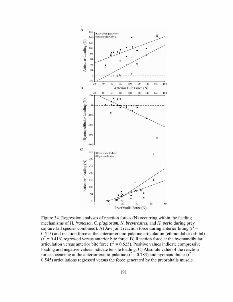
Figure 34. Regression analyses of reaction forces (N) occurring within the feeding mechanisms of H. francisci, C. plagiosum, N. brevirostris, and H. perlo during prey capture (all species combined). A) Jaw joint reaction force during anterior biting (r2 = 0.515) and reaction force at the anterior cranio-palatine articulation (ethmoidal or orbital) (r2 = 0.416) regressed versus anterior bite force. B) Reaction force at the hyomandibular articulation versus anterior bite force (r2 = 0.525). Positive values indicate compressive loading and negative values indicate tensile loading. C) Absolute value of the reaction forces occurring at the anterior cranio-palatine (r2 = 0.783) and hyomandibular (r2 = 0.545) articulations regressed versus the force generated by the preorbitalis muscle. 191

192
relationship between jaw adducting and bite forces and the magnitude of tensile
hyomandibular loading (Figs. 33, 34). The forces generated by the preorbitalis, anterior
mechanical advantage, and force at the ethmoidal articulation all loaded heavily on PC2
(Table 26). The preorbitalis is the anterior-most jaw adducting muscle in all four species,
placing it in a position to exert a strong influence over mechanical advantage. In all four
species the preorbitalis inserts onto the chondrocranium anterior to the ethmoidal
articulation as well, giving this muscle high leverage over ethmoidal loading. Posterior
mechanical advantage and the joint reaction force for posterior biting loaded heavily on
PC3, indicating a correlation between the ability of a lever system to transmit force and
the resulting reaction forces at the fulcrum of that lever system.
Discussion
Prey Capture Biomechanics
Differences in the forces produced by the muscles involved in the expansion of
the feeding mechanisms of H. francisci, N. brevirostris, and C. plagiosum were did not
appear directly related to the use of ram and suction feeding by these sharks. Ram feeding
involves the predator over-taking its prey and either seizing the prey between its jaws or
engulfing the prey in the buccopharyngeal cavity (Liem, 1980). While rapid jaw
adduction is key to capturing elusive prey (Westneat, 1994; Ferry-Graham et al., 2002b;
Porter and Motta, 2004), expansion of the feeding mechanism is theoretically less
important to ram feeding. Conversely, aquatic suction feeding involves the rapid
expansion of the feeding mechanism, which generates a sub-ambient pressure within the
buccopharyngeal cavity, causing water and prey to flow into the mouth (Sanford and

193
Wainwright, 2002; Day et al., 2005). Presumably, the rapid production of large forces by
the muscles involved in abducting the lower jaw and hyobranchial elements would
increase the rate of buccopharyngeal expansion (Van Wassenbergh et al., 2005).
However, the abductive muscles of the suction feeding H. francisci and C. plagiosum
showed no uniform differences from those of the ram feeding N. brevirostris. Although
the species means indicated greater force production in the coracomandibularis (lower
jaw depressor) and coracoarcualis (hyoid arch depressor) of H. francisci and C.
plagiosum relative to N. brevirostris, these differences were only partially supported by
statistical analysis (Table 23). While hypertrophication of the primary hyoid
(coracohyoideus) and branchial arch (coracobranchiales) abductors in suction feeding
elasmobranchs has been alluded to in numerous studies (Moss, 1965, 1977; Motta and
Wilga, 1999; Motta et al., 2002; Motta, 2004), no support for this assertion was found
within these species.
Rapid expansion of the feeding mechanism in suction feeding teleosts is generally
associate with low (velocity efficient) jaw abducting mechanical advantage ratios
(Wainwright and Richard, 1995; Westneat, 1995). However, differences in the ability of
H. francisci, N. brevirostris, and C. plagiosum to generate suction cannot be attributed to
the leverage of their jaw abducting mechanisms either (Table 24). All elasmobranchs
exhibit the synapomorphic lower jaw depression mechanism of chondrichthyans in which
the coracomandibularis inserts onto the posterior aspect of the mandibular symphysis
(Wilga et al., 2000). This developmental constraint gives all elasmobranchs high leverage
(force efficient) jaw abducting mechanisms, especially in comparison to teleosts (M.A. <
0.35) (Wainwright and Richard, 1995; Westneat, 1995). Based upon the present data, the
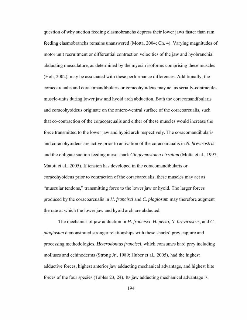
194
question of why suction feeding elasmobranchs depress their lower jaws faster than ram
feeding elasmobranchs remains unanswered (Motta, 2004; Ch. 4). Varying magnitudes of
motor unit recruitment or differential contraction velocities of the jaw and hyobranchial
abducting musculature, as determined by the myosin isoforms comprising these muscles
(Hoh, 2002), may be associated with these performance differences. Additionally, the
coracoarcualis and coracomandibularis or coracohyoideus may act as serially-contractile-
muscle-units during lower jaw and hyoid arch abduction. Both the coracomandibularis
and coracohyoideus originate on the antero-ventral surface of the coracoarcualis, such
that co-contraction of the coracoarcualis and either of these muscles would increase the
force transmitted to the lower jaw and hyoid arch respectively. The coracomandibularis
and coracohyoideus are active prior to activation of the coracoarcualis in N. brevirostris
and the obligate suction feeding nurse shark Ginglymostoma cirratum (Motta et al., 1997;
Matott et al., 2005). If tension has developed in the coracomandibularis or
coracohyoideus prior to contraction of the coracoarcualis, these muscles may act as
“muscular tendons,” transmitting force to the lower jaw or hyoid. The larger forces
produced by the coracoarcualis in H. francisci and C. plagiosum may therefore augment
the rate at which the lower jaw and hyoid arch are abducted.
The mechanics of jaw adduction in H. francisci, H. perlo, N. brevirostris, and C.
plagiosum demonstrated stronger relationships with these sharks’ prey capture and
processing methodologies. Heterodontus francisci, which consumes hard prey including
molluscs and echinoderms (Strong Jr., 1989; Huber et al., 2005), had the highest
adductive forces, highest anterior jaw adducting mechanical advantage, and highest bite
forces of the four species (Tables 23, 24). Its jaw adducting mechanical advantage is

195
higher than those of all other cartilaginous fishes, with the exception of the durophagous
spotted ratfish Hydrolagus colliei (Huber et al., in prep), and nearly all actinopterygian
fishes with the exception of the durophagous parrot fishes (Scaridae) (Turingan et al.,
1995; Durie and Turingan, 2001; Huber and Motta, 2004; Wainwright et al., 2004;
Westneat, 2004; Huber et al., 2006). This high-leverage mechanism is due, in part, to the
derived anterior placement and vertical orientation of the preorbitalis muscle in
heterodontiform and orectolobiform sharks (Compagno, 1977). Through the combination
of high magnitude, cyclically applied bite forces, robust jaws, and molariform teeth, H.
francisci is well suited for crushing the composite exoskeletons of benthic invertebrate
prey (Summers et al., 2004; Huber et al., 2005).
Although the adductive musculature of H. perlo can generate forces comparable
to that of H. francisci, its low jaw adducting mechanical advantage ultimately yielded
intermediate bite forces. This force-inefficient mechanism is apparently related to
constructional constraints associated with the ancestral cranial morphotype of sharks
(Compagno, 1977). The cleaver-shaped upper jaw of extinct cladodont and hybodont
sharks, and extant hexanchiform (sixgill, sevengill, frill) sharks, is characterized by a
large otic process with a deep fossa in the quadrate region, posterior to the orbit. The
anterior portion of the upper jaw is a narrow palatine ramus that extends beneath the
extremely large orbit to approximately the terminus of the head (Fig. 28C) (Allis, 1923;
Daniel, 1934; Schaeffer, 1967; Compagno, 1977). The quadrate fossa is paired with a
mandibular fossa, both of which are occupied by the large quadratomandibularis muscle.
As such, the vast majority of the jaw adducting musculature (93% in H. perlo) is located
posterior to the orbit, resulting in the poor leverage of the jaw adducting mechanism. The

196
need for large eyes (visual sensitivity) in the light depauperate deep sea environment may
be the selective pressure maintaining this anatomical arrangement in H. perlo, which can
be found as deep as 1000 m (Compagno, 1984a). Visual constraints on suspensorial
anatomy have been identified in cichlid fishes as well (Barel et al., 1989). Given this
constraint, the vertical orientation of the quadratomandibularis maximizes the bite force
of H. perlo by producing muscular force nearly orthogonal to the lower jaw. This
configuration will help H. perlo grasp and retain elusive prey with its fang-like upper
teeth, and saw through prey with its multicuspid lower teeth (Bigelow and Schroeder,
1948; Schaeffer, 1967).
Comparing the cranial anatomy of H. perlo to that of N. brevirostris and C.
plagiosum demonstrates the general evolutionary pattern of the shortening and
subterminal relocation of shark jaws (Fig. 28). Shortening of the jaws (out-levers of jaw
adducting mechanism) has generally been hypothesized to increase the leverage over jaw
adduction and subsequently the bite force of sharks, as well as allow for a
hydrodynamically efficient subterminal mouth (Schaeffer, 1967). Mechanically, this
interpretation of shark jaw evolution ignores changes to the moment arm of jaw
adduction (in-lever) via muscular reorganization. The relatively low anterior mechanical
advantage of N. brevirostris demonstrates concomitant selection on both muscular and
skeletal elements of the feeding mechanism in that the shortening and repositioning of the
jaws was met with a reorganization of the adductor musculature, allowing for a
subterminal and velocity efficient feeding mechanism. Consequently, lower jaw elevation
occurs faster in N. brevirostris than in other ram feeding sharks (Tricas and McCosker,
1984; Ferry-Graham, 1997, 1998a; Motta et al., 1997; Wilga and Motta, 1998a, 2000;

197
Motta and Wilga, 2001) and even the ram feeding snapping turtle Chelydra serpentine
(Lauder and Prendergast, 1992). High velocity jaw adduction comes at the cost of bite
force however, which was low in N. brevirostris. In fact, the voluntary bite force of N.
brevirostris (13 N) is significantly lower than the present theoretical estimates of
maximum bite force (Ch. 2, 4). Low bite force in N. brevirostris is accommodated for by
its sharp, triangular teeth and the routine use of upper jaw protrusion and head shaking to
gouge and slice through its prey (Frazzetta and Prange, 1987; Motta et al., 1997).
The bite forces of N. brevirostris and C. plagiosum were equivalent despite
considerable differences in the mechanisms producing those forces. The adductor
musculature of C. plagiosum produced nearly 20% less force than that of N. brevirostris.
However, this force was applied to the lower jaw more orthogonally and at a significantly
higher mechanical advantage. Despite being classified as a suction feeder (Lowry, 2005),
the jaw adducting mechanical advantage of C. plagiosum (0.43) was comparable to those
of durophagous teleosts considered to be hard prey specialists (Westneat, 2004). The
derived anterior placement and vertical orientation of the preorbitalis muscle in
orectolobiform and heterodontiform sharks accounts for the high mechanical advantage
of C. plagiosum (Compagno, 1977). While C. plagiosum consumes a variety of
crustacean prey (Compagno, 2001), these quarry do not rival the hardness of the prey of
durophagous teleosts and elasmobranchs such as H. francisci (Wainwright et al., 1976;
Currey, 1980; Korff and Wainwright, 2004). It is interesting to note that the jaw
adducting mechanical advantage of every shark that has been studied, irrespective of their
prey capture methodology (ram, suction, biting), is similar to those of durophagous,
198
biting teleosts considered to have high leverage jaw adducting mechanisms (M.A. > 0.35)
(Huber and Motta, 2004; Westneat, 2004; Huber et al., 2005, 2006).
Lever systems with high mechanical advantage are inherently more stable than
low leverage systems because a greater percentage of the muscular force actuating
movement is transmitted along the out-lever, leaving a smaller percentage of that force to
be balanced by reaction forces within the joint. Accordingly, those species with the
highest anterior mechanical advantages (H. francisci, C. plagiosum) had the lowest ratios
of joint reaction force to bite force for anterior biting. The high ratio of joint reaction
force to bite force for anterior biting in H. perlo won’t necessarily compromise joint
function because the compressive reaction force is oriented nearly orthogonal to the
articular surface of the joint cartilages, which are strongest in axial compression
(Summers and Long Jr., 2006). However, the acute orientation of the reaction force
during anterior biting in N. brevirostris will induce shear stresses at these articular
surfaces, and may explain the presence of a large caudal prominence on the lateral
quadratomandibular jaw joint in N. brevirostris (Fig. 28). This vertical extension of the
joint’s articular surface may accommodate reaction forces occurring over a wide range of
gape angles. The extent to which the jaw joints are loaded in axial or shear compression
is determined by the orientation of the resultant adductive force vector, which was
vertically directed in H. perlo and anteriorly directed in N. brevirostris.
Compression of the jaw joints during posterior biting occurred in H. perlo and C.
plagiosum because their posterior mechanical advantage ratios did not exceed 1.0.
Although the joint reaction forces of H. perlo were approximately orthogonal to the
articular surface, those of C. plagiosum were acutely oriented. Unlike N. brevirostris, the

199
articular surface of the quadratomandibular jaw joint in C. plagiosum lies completely in
the frontal plane, with no caudal prominence to resist shearing within the joint (Fig. 28).
Potentially greater joint instability was found during posterior biting in H. francisci and
N. brevirostris, in which mechanical advantage ratios greater than 1.0 caused tensile joint
loading because the prey becomes a temporary fulcrum between the jaw joint and
resultant adductive force vector. Separation or slippage within the jaw joints due to
tensile and shear loading must be prevented by the complex ligamentous arrays binding
the mandibular and hyoid arches together in H. francisci, N. brevirostris, and C.
plagiosum (Daniel, 1915; Motta and Wilga, 1999; Goto, 2001).
Jaw Suspension Mechanics
Heptranchias perlo retains the amphistylic jaw suspension of its cladodont
predecessors, while H. francisci, C. plagiosum, and N. brevirostris evolved the hyostylic
jaw suspension mechanism characteristic of galeomorph neoselachians (Daniel, 1934;
Compagno, 1977). Despite having considerably different cranial anatomies, the jaw
suspensions of H. perlo, H. francisci, and C. plagiosum are functionally analogous in that
the anterior cranio-palatine articulation (ethmoidal or orbital) remains intact during the
full range of motion exhibited by the jaws (Compagno, 1977; Maisey, 1980; Motta et al.,
1997; Huber et al., 2005). Subsequently, the common suspensorial loading pattern
observed in these sharks involved compression at the anterior cranio-palatine articulation
(ethmoidal or orbital) and tension at the posterior hyomandibular articulation.
Conversely, the jaw suspension of N. brevirostris experienced negligible tension at its
ethmoidal articulation and compression at the hyomandibular articulation during bites
without protrusion, and substantial hyomandibular compression during bites with

200
protrusion. Negaprion brevirostris is capable of protruding the upper jaw far enough that
it loses contact with the ethmoid region of the cranium (Motta and Wilga, 1995). The
common denominators underlying these differences in suspensorial loading are the state
of the anterior cranio-palatine articulation (intact or disarticulated), and the presence or
absence of a force vector acting between the jaws and cranium (preorbitalis or levator
palatoquadrati muscles).
The preorbitalis muscles of H. francisci and C. plagiosum originate on the
anterior portion of the lower jaw and insert on the cranium posterior to the nasal capsule,
while that of H. perlo originates more posteriorly on the median raphe of the
quadratomandibularis muscle and inserts on the cranium posterior to the nasal capsule.
As such, the forces produced by the preorbitalis are included in the static equilibrium
calculations of the lower jaw, and not the upper jaw. Taking all other muscular and
reaction forces to be equal, the inequality of the forces acting on the lower and upper
jaws due to the preorbitalis results in the net upward translation of the mandibular arch.
The nature of this upward translation is dictated by the presence or absence of contact at
the anterior cranio-palatine articulation (ethmoidal or orbital) and the position of the
preorbitalis’ insertion relative to this articulation. In H. perlo, H. francisci, and C.
plagiosum the preorbitalis inserts anterior to the articulation. The resulting moment arm
of the preorbitalis compresses the upper jaw into the articulation and generates a counter-
clockwise torque about this point relative to the right side of the head (Fig. 35). As a
result, the posterior region of the upper jaw rotates anteroventrally, pulling the
hyomandibular cartilages in tension (Fig. 35, Table 27). This loading regime was

Figure 35. Right lateral views of the feeding mechanisms of (A) C. plagiosum and (B) N. brevirostris indicating the net forces acting on the jaws and their articulations with the cranium during biting. Black arrows with ‘C’ indicate compressive loading, black arrows with ‘T’ indicate tensile loading, and white arrows with black outlining indicate the direction of motion of the jaws due to the net muscular force acting on the feeding mechanism. (A) During biting in C. plagiosum (representative of H. francisci and H. perlo) the ethmoidal articulation remains intact. As the jaws translate upwards via muscular force acting between the jaws and cranium, they compress and pivot about the ethmoidal articulation. The resultant rotation of the jaws pulls the hyomandibular cartilages in tension. (B) During biting at maximum protrusion in N. brevirostris the upper jaw disarticulates from the cranium at the ethmoidal articulation. Unable to pivot about the ethmoidal articulation, upward translation of the jaws occurs in a linear manner, compressing the hyomandibular cartilages against the cranium.
supported by the PCA, which indicated strong correlations between the force generated
by the preorbitalis, the magnitude of compression at the anterior cranio-palatine
articulation, and the magnitude of tension at the hyomandibular articulation (Fig. 34,
Table 26).
The primary muscles actuating upper jaw protrusion in N. brevirostris are the
ventral division of the preorbitalis and levator palatoquadrati, each of which generate
force between the jaws and cranium (Motta and Wilga, 1995; Motta et al., 1997). Biting
without protrusion was modeled assuming inactivity in these muscles, and the resulting
201

equilibrium conditions lacked a force vector between the jaws and cranium. Despite the
fact that the anterior cranio-palatine articulation remains intact during bites without
202
protrusion, the resultant suspensorial loading showed negligible tension at this
articulation (1 ± 1 N) and negligible compression on the hyomandibular cartilages (2 ± 1
N). Taking the respective standard error estimates into account, the suspensorium of N.
brevirostris effectively experiences no loading during bites without protrusion because all
muscular and reaction forces are equally applied to both the lower and upper jaws (no net
translation of the jaws).
During protrusion in N. brevirostris, disengagement of the upper jaw from the
cranium precludes the generation of torque about the anterior cranio-palatine articulation.

203
With no
erior
to
rbitalis (D.R.
drati
per
the anterior cranio-
palatine
rlo;
e of
pivot point, activity in the ventral preorbitalis and levator palatoquadrati causes a
net upward translation of the mandibular arch resulting in compression on the
hyomandibulae (60 N), which are located between the jaws and cranium (Fig. 35, Table
27). If these muscles were active prior to protrusion and dissociation of the ant
cranio-palatine articulation, a torque about the articulation would still favor
hyomandibular compression because the levator palatoquadrati inserts posterodorsal
the articulation and generates a larger bending moment than the ventral preo
Huber, unpub. data). This scenario is specific to carcharhinid sharks, the only group in
which the levator palatoquadrati is oriented anterodorsally allowing it to participate in
upper jaw protrusion (Compagno, 1988; Wilga et al., 2001). Regardless, the motor
activity pattern during jaw adduction in N. brevirostris suggests that activity of the
quadratomandibularis pulls the upper jaw ventrally, disengaging the anterior cranio-
palatine articulation, prior to activity in the ventral preorbitalis and levator palatoqua
(Motta et al., 1997). In other elasmobranchs the levator palatoquadrati retracts the up
jaw during the recovery phase of the gape cycle (Wilga, 2005).
Presuming that suspensorial loading is determined by the position of muscular
force vectors acting between the jaws and cranium and whether
articulation is intact or dissociated, the nature of suspensorial loading can be
hypothesized in other groups of elasmobranchs (Table 27). The configuration of the
preorbitalis in orbitostylic squalean sharks such as S. acanthias is the same as in H. pe
the preorbitalis is a single headed, horizontal muscle extending from the median raph
the quadratomandibularis muscle to the posterior aspect of the nasal capsule (Wilga and
Motta, 1998a). The orbital process of the upper jaw maintains contact with the cranium

204
the
goblin
s
derived
at
r jaw is
e
throughout the full range of motion exhibited by the jaws as well (Compagno, 1977;
Wilga and Motta, 1998a). Therefore it is hypothesized that S. acanthias and other
orbitostylic sharks in which the orbital articulation remains intact during feeding will
exhibit compression at the orbital articulation and tension on the hyomandibulae.
Wilga (2005) recently identified additional variants of the traditional hyostylic
jaw suspension in lamniform sharks. The jaw suspension of basal lamnoids such as
Mitsukurina owstoni, sandtiger Carcharias taurus, and common thresher sharks
Alopias vulpinnis is characterized by suspensory hyomandibular cartilages and
ligamentous connections between the upper jaw and both the ethmoidal and nasal region
of the cranium. Derived lamnoids such as the porbeagle Lamna nasus, white
Carcharodon carcharias, and shortfin mako sharks Isurus oxyrinchus have suspensory
hyomandibulae and have lost the ethmopalatine ligaments while retaining the
palatonasal ligament. All of these sharks are capable of extensive jaw protrusion such th
the upper jaw can disarticulate from the cranium (Tricas and McCosker, 1984; Wilga,
2005). Mitsukurina owstoni and A. vulpinnis are have preorbitalis muscles extending
from the jaws to the cranium, while the preorbitalis of C. carcharias, C. taurus, I.
oxyrinchus, and L. nasus lie between the lower and upper jaws (Wilga, 2005). Like N.
brevirostris during bites with protrusion, activity of the preorbitalis while the uppe
dissociated from the cranium in M. owstoni and A. vulpinnis will hypothetically place th
hyomandibular cartilages in compression. The lack of a preorbitalis force vector between
the jaws and cranium in C. carcharias, C. taurus, I. oxyrinchus, and L. nasus apparently
indicates negligible suspensorial loading in these sharks. However, the novel insertion of
the levator hyomandibularis muscle onto the upper jaw in derived lamnid sharks (I.
205
ape
(euhyostyly)
(Wilga role in
n.
rce
sm
es
oxyrinchus, L. nasus, C. carcharias) is suggestive of hyomandibular compression as well
(Table 27). From this insertion point, the levator hyomandibularis can direct precise
movements of the upper jaw by bracing it against the cranium via the hyomandibular
cartilages, perhaps allowing for repeated protrusion of the upper jaw during a single g
cycle (Tricas and McCosker, 1984; Wilga, 2005). Additionally, all lamniform sharks
possess a sesamoid-like cartilage in their novel palatonasal ligaments, suggestive of
tensile forces at the anterior cranio-palatine articulation (Wilga, 2005).
The batoids lack an anterior cranio-palatine articulation, making the
hyomandibular cartilages the sole support of the jaws against the cranium
, 2002). The quadratomandibularis and preorbitalis play a synergistic
protruding the jaws anteroventrally, with the preorbitalis exerting force between the
lower jaw and the cranium (Wilga and Motta, 1998b). This mechanism is again
analogous to the suspensorial mechanics of N. brevirostris during bites with protrusio
The lack of anterior cranio-palatine contact coupled with an upwardly directed fo
between the jaws and cranium will hypothetically result in hyomandibular compression
(Table 27). Additionally, batoids possess several novel lower jaw and hyomandibular
depressor muscles that appear to pivot (compress) the jaws and hyomandibulae about the
cranium allowing for high-precision, asymmetrical movements of the feeding mechani
(Miyake et al., 1992; Wilga and Motta, 1998b; Dean and Motta, 2004a; Dean et al.,
2005b). Although a modeling analysis has not been performed on a batoid, trabecular
reinforcement in the jaws and hyomandibular cartilages of N. brasiliensis corroborat
the role of the hyomandibulae as compressive elements. Medial translation of the
hyomandibular cartilages protrudes the jaws of N. brasiliensis such that at maximum

206
s.
,
e
H.
perlo are suggestive of a broad evolutionary pattern in elasmobranch feeding
mechan
he
r
lar
ylic
ig. 29) (Schaeffer, 1967; Carroll, 1988). Due to the
abutme
o
.
protrusion the longitudinal axes of the hyomandibulae line up with those of the jaw
Trabeculae in the jaws and hyomandibular cartilages are arranged to resist buckling as
the jaws are axially buttressed against the cranium during protrusion (Dean and Motta
2004a, 2004b; Dean et al., 2005a). Collectively, enhanced maneuverability of the jaws
appears to be associated with hyomandibular compression in elasmobranchs in which th
anterior cranio-palatine articulation is either permanently or temporarily disengaged.
Evolution of Jaw Suspension
The suspensorial mechanics of H. francisci, C. plagiosum, N. brevirostris, and
isms. The available evidence indicates that the amphistylic jaw suspension was
characterized by negligible suspensorial loading because the vast majority of t
adductive force acted between the jaws (93% in H. perlo), not between the jaws and
cranium. Under these conditions there would have been little selective pressure fo
structural changes to the long, thin, poorly calcified, posteriorly directed hyomandibu
cartilages of extinct (cladodont, early hybodont) and extant (hexanchiform) amphist
sharks (Zangerl and Williams, 1975; Maisey, 1980). Thus, the hyoid arch retained the
appearance of the post-mandibular visceral arch from which it was derived (Zangerl and
Williams, 1975; Mallatt, 1996).
An obvious characteristic of the cranium in these sharks are the large, laterally
expanded postorbital processes (F
nt of the otic process of the upper jaw against this protuberance, it would have
impeded anterior and perhaps even ventral translation of the jaws. Therefore, there als
would have been little selective pressure for the preorbitalis to diversify in these sharks

207
ard,
avity or
de of
aeffer, 1967;
Maisey e
of
Provided that the postorbital articulation did not prevent ventral translation of the upper
jaw, protrusion would have occurred in the vertical plane when the lower jaw was
depressed and contraction of the quadratomandibularis adducted the upper jaw towards
the lower jaw. Contraction of the preorbitalis would have drawn the upper jaw forw
compressing it into the postorbital articulation and generating a small counter-clockwise
torque about this articulation relative to the right side of the head, analogous to
compression at the orbital articulation in H. perlo. This mechanism would have braced
the upper jaw against the cranium and possibly expanded the buccopharyngeal c
assisted in retracting the protruded upper jaw (Schaeffer, 1967). Therefore, the
plesiomorphic suspensorial loading regime associated with the amphistylic jaw
suspension probably involved compression between the upper jaw and cranium at the
postorbital articulation and tension on the hyomandibular cartilages, the magnitu
which was dictated by the force of the preorbitalis (Fig. 34, Table 27).
Once the postorbital articulation was convergently lost via reduction of the otic
process of the upper jaw in the late hybodontoids and neoselachians (Sch
, 1982; Carroll, 1988; Maisey and de Carvalho, 1997), architectural changes to th
preorbitalis (enlargement, subdivision, reorientation) would have facilitated anterior
movement of the jaws, in addition to the existing ventral motion. Anterior kinesis would
have been further facilitated by the reduction of the postorbital process on the cranium
neoselachians as well (Schaeffer, 1967; Carroll, 1988). As this modeling analysis
indicates, increased force production by the preorbitalis causes increased loading at the
anterior cranio-palatine and hyomandibular articulations, providing the impetus for the
evolution of the hyostylic jaw suspension mechanism. Subsequently, the hyomandibular

208
;
Fig.
e
e evolution of hyostyly appears to have been capitalized upon by carcharhinid,
lamnoi
ilga
,
cartilages of the neoselachians became shorter, thicker, rotated anteriorly into a more
orthogonal position relative to the cranium, and developed deep articular facets against
the cranium facilitating directionally specific motion (Schaeffer, 1967; Cappetta, 1987
Wilga, 2002). These characteristics are indicative of the hyomandibular cartilages being
drawn forward under a repeatable loading regime, and suggest that these elements play a
passive role in bracing the jaws during protrusion. The hyomandibular cartilages of the
late hybodontoids also increased in size concomitant with the loss of the postorbital
articulation. However, these hyomandibulae were located dorsal to the upper jaw and
remained posteriorly directed, suggesting that they were less suited for load-bearing (
28) (Maisey, 1980, 1982, 1987). Additionally, the upper jaws of these sharks abutted th
postorbital region of the cranium, making them functionally amphistylic (Schaeffer,
1967).
The general increase in load-bearing ability of the hyomandibular cartilages
during th
d, and batoid elasmobranchs. In each of these groups, diversification of the
muscles controlling jaw and hyomandibular movement has led to increased magnitude
and precision of jaw kinesis (Moss, 1977; Compagno, 1988; Miyake et al., 1992; W
and Motta, 1998b; Wilga et al., 2001; Dean and Motta, 2004b; Wilga, 2005). Enhanced
jaw kinesis is associated with the dissociation of the upper jaw from the cranium and a
subsequent transition from tensile to compressive hyomandibular loading, allowing the
jaws and hyomandibulae to be pivoted about the cranium. This pivoting is believed to
permit repeated protrusion during a single gape cycle in lamniform sharks and
asymmetric strikes at prey in batoids (Dean and Motta, 2004b; Wilga, 2005). Therefore
209
alatine
roperties
echanical principles elucidated by this study allow inferences to be
drawn regarding of fish through
evoluti to
d in
the
hyomandibular compression probably preceded the loss of the anterior cranio-p
articulation during the evolution of euhyostyly in batoids. Because the nature of
hyomandibular loading is determined by the type of jaw suspension and the extent of
muscular control over jaw protrusion, hyomandibular morphology and material p
(as indicative of load-bearing ability) may vary concomitantly with these parameters in
elasmobranchs.
Jaw Suspension Mechanics in Non-Elasmobranch Fishes
The biom
the jaw suspension mechanisms of other major radiations
onary history. The extinct placoderms are believed to be either the sister-group
chondrichthyans (Janvier, 1996; Liem et al., 2001) or the sister-group to all gnathostomes
(Goujet, 2001). Most placoderms had a rudimentary holostylic jaw suspension in which
the quadrate portion of the upper jaw was fused to the robust dermal skeleton (Moy-
Thomas and Miles, 1971; Carroll, 1988). Among fishes, only the holocephalans (sister-
taxon to the elasmobranchs) have a truly holostylic jaw suspension characterized by
complete fusion of the upper jaw to the cranium and a non-suspensory, intact hyoid arch
(Grogan et al., 1999). This mechanism is distinct from the autostylic mechanism foun
sarcopterygian fishes and tetrapods, in which the upper jaw is fused to the cranium and
hyoid arch is non-suspensory and broken-up or modified (Liem et al., 2001; Wilga,
2002). Although the hyomandibula was located between the jaws and cranium in
placoderms, the upper jaw was akinetic in most taxa and could not induce loading on
hyomandibula (Moy-Thomas and Miles, 1971). The only load-bearing activity the
hyomandibula may have performed in these fishes was during cranial elevation via their

210
cted
s. These fish possessed an orbital cranio-palatine articulation and long,
thin hy iles,
fore, it cannot
e
ison,
1979; L
al
ontribution
novel cranio-thoracic joint in the dermal armor because the dermal armor was conne
to the upper jaw, which was in turn connected to the hyomandibula (Moy-Thomas and
Miles, 1971).
The derived rhenanid placoderms were the only group known to possess
protrusible jaw
omandibular cartilages that were clearly suspensory (Moy-Thomas and M
1971; Carroll, 1988). Protrusion would have occurred in the vertical plane as the upper
and lower jaws were adducted towards each other via contraction of the
quadratomandibularis. However, the presence or absence of an additional muscle acting
between the jaws and cranium such as the preorbitalis is unknown. There
be determined whether the hyomandibular cartilages of rhenanid placoderms were
actually load-bearing. The general lack of calcification in these cartilages suggests that
they were not load-bearing (Moy-Thomas and Miles, 1971; Wilga, 2002).
The acanthodians, sister-group to the osteichthyan fishes, also had hyomandibula
articulating with the upper jaw and cranium (Moy-Thomas and Miles, 1971; Den
iem et al., 2001). As with the placoderms, the existence of cranio-palatine muscle
forces in these fish is unknown and the nature of hyomandibular loading cannot be
determined. Hyomandibular loading is believed to have been negligible, if present at all,
because the upper jaw had prominent articulations with both the basal and postorbit
regions of the cranium. Therefore, the hyomandibulae of acanthodians are believed to
have been non-suspensory (Moy-Thomas and Miles, 1971; Denison, 1979).
The lower jaw of the osteichthyan fishes is braced against the cranium via the
suspensorial series of bones, with the quadrate forming the suspensorium’s c

211
to the l
f
talized
ous
ing it
ower jaw joint distally and the hyomandibula articulating with the neurocranium
proximally. As such, the hyomandibula is directly in series with the jaw joint. The lower
jaw adducting mechanism of the osteichthyans is a class III lever system, with the bulk o
the adductor musculature originating on and around the suspensorium and inserting onto
the lower jaw (Winterbottom, 1974). Given this musculoskeletal geometry, the jaw joint
will be loaded in compression during biting provided that bite force is applied anterior to
the resultant adductive force vector. This compressive joint reaction force will be
transmitted up the suspensorium, compressing the hyomandibula into the cranium.
Independence of the upper jaw from the lower jaw adducting mechanism was capi
upon by the actinopterygian radiation of osteichthyan fishes, which developed numer
mechanisms of protruding the upper jaw (Schaeffer and Rosen, 1961; Alexander, 1967;
Motta, 1984). Protrusibility of the upper jaw is considered a key innovation in the
adaptive radiation of modern actinopterygian fishes, as it permitted the diversification of
a myriad of feeding specializations (Schaeffer and Rosen, 1961). The evolutionary
progression towards enhanced cranial kinesis in these fishes has been associated with the
shortening, thickening, and more orthogonal orientation of the hyomandibulae, all
suggestive of an increase in load-bearing activity (Schaeffer and Rosen, 1961). Some
modern actinopterygian fishes have evolved kinesis within the suspensorium, allow
to pivot independently of the skull and protrude the lower jaw as well (Westneat and
Wainwright, 1989; Ferry-Graham et al., 2001b).

212
Conclusions
In the absence of load-bearing activity skeletal elements lack the mechanical
impetus needed to adaptively diversify is believed to be the cranial archetype
for elas due
tion
anical
of the elasmobranch feeding mechanism increased
dramat in
way
ss with
. Given what
mobranchs, the amphistylic jaw suspension mechanism was not load-bearing
to small forces produced by the muscles acting between the jaws and cranium. As a
result, the hyoid arch retained the structure of a post-mandibular visceral arch. Given this
mechanical context for skeletal diversification, the most likely scenario for the evolu
of hyostyly from amphistyly involved augmentation of the force produced by the
preorbitalis and concomitant loss of the postorbital articulation, followed by
hyomandibular diversification.
Once the hyomandibula was better able to receive and distribute mech
loading, the evolutionary lability
ically. Enhanced jaw kinesis likely increased the effectiveness of ram feeding
elasmobranchs by providing quicker gape closure and the ability to extend the jaws a
from the head to excise large chunks of prey. By freeing the posterior margin of the jaws
from the cranium, hyostyly permitted shortening and subterminal placement of the jaws.
Barring reorganization of the adductor musculature, shortening of the jaws increased the
leverage of the feeding mechanism and the ability of benthic elasmobranchs to consume
hard prey. Lastly, increased load-bearing ability of the hyomandibula would have
allowed for forceful, rapid movements of the hyoid arch as required during suction
feeding. Collectively, these changes illustrate that kinesis increased the effectivene
which the jaws could be used as tools for prey capture, and that elasmobranchs
capitalized upon this by radically diversifying the feeding mechanism in modern forms.

213
Literatu Cited
Abler, W.L. (1992). The serrated teeth of tyrannosaurid dinosaurs, and biting structures in other animals. Paleobiology 18, 161-183.
Aerts, P. and DeVree, F. (1993). Feeding performance and muscular constraints in fish. J.
guirre, L.F., Herrel, A., Van Damme, R. and Matthysen, E. (2003). The implications of
idley, D.J., (1998). Physiology of Excitable Cells. Cambridge University Press,
lexander, R.M. (1967). The functions and mechanisms of the protrusible upper jaws of
lexander, R.M. (2002). Tendon elasticity and muscle function. Comp. Biochem.
lfaro, M.E., Janovetz, J. and Westneat, M.W. (2001). Motor control across trophic
1,
gica
lonso, M.K., Crespo, E.A., Garcia, N.A., Pedraza, S.N., Mariotti, P.A. and Mora, N.J.
anthias, in Patagonian waters, Argentina. Environ. Biol. Fishes 63, 193-202.
Askew
arel, C.D.N., Anker, G.C., Witte, F., Hoogerhoud, R.J.C. and Goldschmidt, T. (1989).
.
re
Exp. Biol. 177, 129-147.
Afood hardness for diet in bats. Funct. Ecol. 17, 201-212.
ACambridge.
Asome acanthopterygian fish. Zool. J. Linn. Soc. 151, 43-64.
APhysiol. A. 133, 1001-1011.
Astrategies: muscle activity of biting and suction feeding fishes. Am. Zool. 41266-1279.
Allis, E.P. (1923). The cranial anatomy of Chlamydoselachus anguineus. Acta Zoolo
4, 1-100.
A(2002). Fishery and ontogenetic driven changes in the diet of the spiny dogfish, Squalus ac
, G.N. and Marsh, R.L. (1997). The effects of length trajectory on the mechanicalpower output of mouse skeletal muscles. J. Exp. Biol. 200, 3119-3131.
BConstructional morphology and its ecomorphological implications. Acta MorpholNeerl.-Scand. 27, 83-109.

214
s
erra, T. (2001). Freshwater Fish Distribution. Academic Press, San Diego, CA.
Biewen r elastic energy savings versus muscles designed to generate mechanical power.
inder, W.J. and Van Valkenburgh, B.V. (2000). Development of bite strength and
stav
arrano, M.T. (1999). What, if anything, is a cursor? Categories versus continua for
4).
7, 3873-3881.
arter, D.R. and Beaupre, G.S. (2001). Skeletal Function and Form: Mechanobiology of
ess,
astro, J.I. (1996). Biology of the blacktip shark, Carcharhinus limbatus, off the
hristoforou, A.P., Swanson, S.R. and Beckwith, S.W. (1989). Lateral impact of
ting of and Fracture.
Cincinnati, OH, pp. 373-386.
Bemis, W.E. and Lauder, G.V. (1986). Morphology and function of the feeding apparatuof the lungfish, Lepidosiren paradoxa (Dipnoi). J. Morphol. 187, 81-108.
B
er, A.A. (1998). Muscle function in vivo: A comparison of muscles used fo
Am. Zool. 38, 703-717. Bigelow, H.B. and Schroeder, W.C. (1948). Fishes of the western north Atlantic:
Lancelets, cyclostomes, and sharks. Mem. Sears Found. Mar. Res. 1, 1-576.
Bfeeding behavior in juvenile spotted hyenas (Crocuta crocuta). J. Zool. (Lond.) 252, 273-283.
Cappetta, H. (1987). Chondrichthyes II: Mesozoic and Cenozoic Elasmobranchii. Gu
Fischer Verlag, Stuttgart.
Cdetermining locomotor habit in mammals and dinosaurs. J. Zool. Soc. Lond. 247, 29-42.
Carroll, A.M., Wainwright, P.C., Huskey, S.H., Collar, D.C. and Turingan, R.G. (200
Morphology predicts suction feeding performance in centrarchid fishes. J. Exp. Biol. 20
Carroll, R.L. (1988). Vertebrate Paleontology and Evolution. W.H. Freeman and
Company, New York.
CSkeletal Development, Aging, and Regeneration. Cambridge University PrCambridge.
Carter, D.R. and Wong, M. (2003). Modelling cartilage mechanobiology. Philos. Trans.
R. Soc. Lond. B. Biol. Sci. 358, 1461-1471.
Csoutheastern United States. Bull. Mar. Sci. 59, 508-522.
Ccomposite cylinders. In: Lagace, P.A. (Ed.), Procedings of the Annual Meethe Second Symposium on Composite Materials: Fatigue

215
Clark, E
leuren, J., Aerts, P. and De Vree, F. (1995). Bite and joint force analysis in Caiman
lifton, K.B. and Motta, P.J. (1998). Feeding morphology, diet, and ecomorphological
53-
,
ompagno, L.J.V. (1984a). FAO Species Catalogue. Sharks of the World. An Annotated
ormes to Lamniformes. Food and Agriculture Organization of the United Nations, Rome.
Compa An Annotated
and Agriculture Organization of the United Nations, Rome.
Compa on University
ompagno, L.J.V. (1999). Checklist of living elasmobranchs. In: Hamlett, W.C. (Ed.),
imore, MD, 471-498.
f
tolobiformes). Food and Agriculture Organization of the United Nations, Rome.
Cortes,
urrey, J.D. (1980). Mechancial properties of mollusc shell. In: Vincent, J.F.V. and
. and Nelson, D.R. (1997). Young whale sharks, Rhincodon typus, feeding on a copepod bloom near La Paz, Mexico. Environ. Biol. Fishes 50, 63-73.
Ccrocodilus. Belg. J. Zool. 125, 79-94.
Crelationships among five Caribbean labrids (Teleostei, Labridae). Copeia (4), 9966.
Compagno, L.J.V. (1977). Phyletic relationships of living sharks and rays. Am. Zool. 17
303-322.
Cand Illustrated Catalogue of Shark Species Known to Date. Part 1: Hexanchif
gno, L.J.V. (1984b). FAO Species Catalogue. Sharks of the World. and Illustrated Catalogue of Shark Species Known to Date. Part 2: Carcharhiniformes. Food
gno, L.J.V. (1988). Sharks of the Order Carcharhiniformes. PrincetPress, Princeton, NJ.
CSharks, skates, and rays: The Biology of Elasmobranch Fishes. Johns Hopkins University Press, Balt
Compagno, L.J.V. (2001). Sharks of the World. An annotated and illustrated catalogue o
shark species known to date. Volume 2. Bullhead, mackerel, and carpet sharks (Heterodontiformes, Lamniformes, and Orec
E. and Gruber, S.H. (1990). Diet, feeding habits and estimates of daily ration ofyoung lemon sharks, Negaprion brevirostris (Poey). Copeia (1), 204-218.
CCurrey, J.D. (Eds.), The Mechanical Properties of Biological Materials. Press Syndicate of the University of Cambridge, Cambridge, 75-98.
Currey, J.D. (2002). Bones: Structure and Mechanics. Princeton University Press,
Princeton, NJ.

216
urtin, N.A., Gardner-Medwin, A.R. and Woledge, R.C. (1998). Predictions of the time f
iol. 201, 103-114.
aniel, J.F. (1934). The Elasmobranch Fishes. University of California Press, Berkeley,
ay, S.W., Higham, T.E., Cheer, A.Y. and Wainwright, P.C. (2005). Spatial and
ochirus) by Particle Image Velocimetry. J. Exp. Biol. 208, 2661-2671.
ulation doi:
62, 462-483.
c 89.
ean, M.N., Wilga, C.D. and Summers, A.P. (2005b). Eating without hands or tongue:
Douad . (2003). Molecular
phylogenetic evidence refuting the hypothesis of Batoidea (rays and skates) as
eeding
ia
dmonds, M.A., Motta, P.J. and Hueter, R.E. (2001). Food capture kinematics of the
Ccourse of force and power output by dogfish white muscle fibers during brietetani. J. Exp. B
Daniel, J.F. (1915). The anatomy of Heterodontus francisci II: The endoskeleton. J.
Morphol. 26, 447-493.
DCA.
Dtemporal flow patterns during suction feeding of bluegill sunfish (Lepomis macr
Dean, M., Huber, D. and Nance, H. (2005a). Functional morphology of jaw trabec
in the lesser electric ray Narcine brasiliensis. J. Morphol. Published online (10.1002/jmor.10302).
Dean, M. and Motta, P.J. (2004a). Anatomy and functional morphology of the feeding
apparatus of the lesser electric ray, Narcine basiliensis (Elasmobranchii: Batoidea). J. Morphol. 2
Dean, M. and Motta, P.J. (2004b). Feeding behavior and kinematics of the lesser electri
ray, Narcine brasiliensis (Elasmobranchii: Batoidea). Zoology 107, 171-1
Dspecialization, elaboration and the evolution of prey processing mechanisms in cartilaginous fishes. Biology Letters 1, 357-361.
Denison, R. (1979). Acanthodii. Gustav Fischer Verlag, Stuttgart.
y, C.J., Dosay, M., Shivji, M.S. and Stanhope, M.J
derived sharks. Mol. Phylogenet. Evol. 26, 215-221. Durie, C.J. and Turingan, R.G. (2001). Relationship between durophagy and f
biomechanics in gray triggerfish, Balistes capriscus: Intraspecific variation in ecological morphology. Fla. Sci. 64, 20-28.
Ebert, D.A. (2003). Sharks, Rays, and Chimaeras of California. University of Californ
Press, Berkeley.
Esuction feeding horn shark Heterodontus francisci. Environ. Biol. Fishes 62, 415-427.

217
llers, O., Johnson, A.S. and Moberg, P.E. (1998). Structural strengthening of urchin
Erickson, G.M., Lappin, A.K., Parker, T. and Vliet, K.A. (2004). Comparison of bite
n Diego, 1-12.
haloscyllium
erry-Graham, L.A. (1998a). Effects of prey size and mobility on prey-capture
erry-Graham, L.A. (1998b). Feeding kinematics of hatchling swellsharks,
r size.
ctional
. Biol. Ecol. 256, 167-184.
). iol. 141, 819-
12.
Eskeletons by collagenous sutural ligaments. Biol. Bull. 195, 136-144.
performance between long-term captive and wild American alligators Alligatormississippiensis. J. Zool. 262, 21-28.
Evans, W.R. and Gilbert, P.W. (1971). The force of bites by the silky shark
(Carcharhinus falciformis) measured under field conditions. Naval Undersea Research and Development Center, Sa
Ferry-Graham, L.A. (1997). Feeding kinematics in juvenile swellsharks, Cep
ventriosum. J. Exp. Biol. 200, 1255-1269.
Fkinematics in leopard sharks Triakis semifasciata. J. Exp. Biol. 201, 2433-2444.
FCephaloscyllium ventriosum (Scyliorhinidae): The importance of predatoMar. Biol. 131, 703-718.
Ferry-Graham, L.A., Bolnick, D.I. and Wainwright, P.C. (2002a). Using fun
morphology to examine the ecology and evolution of specialization. Integr. Comp. Biol. 42, 265-277.
Ferry-Graham, L.A., Wainwright, P.C. and Bellwood, D.R. (2001a). Prey capture in
long-jawed butterfly fishes (Chaetodontidae): the functional basis of novel feeding habits. J. Exp. Mar
Ferry-Graham, L.A., Wainwright, P.C., Hulsey, C.D. and Bellwood, D.R. (2001b).
Evolution and mechanics of long jaws in butterfly fishes (Family Chaetodontidae). J. Morphol. 248, 120-143.
Ferry-Graham, L.A., Wainwright, P.C., Westneat, M.W. and Bellwood, D.R. (2002b
Mechanisms of benthic prey capture in wrasses (Labridae). Mar. B830.
Fouts, W.R. and Nelson, D.R. (1999). Prey capture by the Pacific angel shark, Squatina
californica: Visually mediated strikes and ambush-site charactersitics. Copeia (2), 304-3
Frazzetta, T.H. (1988). The mechanics of cutting and the form of shark teeth
(Chondrichthyes, Elasmobranchii). Zoomorphology 108, 93-107.

218
s. Adv.
razzetta, T.H. and Prange, C.D. (1987). Movements of cephalic components during 4),
iol. Sci. 179, 451-485.
ch.
ibb, A.C. and Ferry-Graham, L.A. (2005). Cranial movements during suction feeding in
Hokkaido Univ. 48, 1-100.
(Ed.), M 2.
reaves, W.S. (1988). The maximum average bite force for a given jaw length. J. Zool.
reaves, W.S. (2000). Location of the vector of jaw muscle force in mammals. J.
regory, W.K. (1904). The relations of the anterior visceral arches to the
rogan, E.D. and Lund, R. (2000). Debeerius ellefseni (Fam. Nov., Gen. Nov, Spec.,
r Gulch nships of the Chondrichthyes, and
comments on gnathostome evolution. J. Morphol. 243, 219-245.
Grogan nd its
Frazzetta, T.H. (1994). Feeding mechanisms in sharks and other elasmobranchComp. Environ. Physiol. 18, 31-57.
F
feeding in some requiem sharks (Carcharhiniformes: Carcharhinidae). Copeia (979-993.
Gadow, H. (1888). On the modifications of the first and second visceral arches, with
special reference to the homologies of the auditory ossicles. Philos. Trans. R. Soc. Lond. B. B
Gans, C. and Gaunt, A.S. (1991). Muscle architecture in relation to function. J. Biome
24, 53-65.
Gteleost fishes: Are they modified to enhance suction production? Zoology 108, 141-153.
Goto, T. (2001). Comparative anatomy, phylogeny, and cladistic classification of the
order Orectolobiformes (Chondrichthyes, Elasmobranchii). Mem. Grad. Sch. Fish. Sci.
Goujet, D. (2001). Placoderms and basal gnathostome apomorphies. In: Ahlberg, P.E.
ajor Events in Early Vertebrate Evolution. Taylor & Francis, London, 209-22
G(Lond.) 214, 295-306.
G
Morphol. 243, 293-299.
Gchondrocranium. Biol. Bull. 7, 55-69.
GNov.), an autodiastylic Chondrichthyan from the Mississippian Bealimetone of Montana (USA), the relatio
, E.D., Lund, R. and Didier, D.A. (1999). Description of the chimaerid jaw aphylogenetic origins. J. Morphol. 239, 45-59.

219
ernandez, L.P. and Motta, P.J. (1997). Trophic consequences of differential performance: ontogeny of oral jaw-crushing performance in the sheepshead, Archosargus probatocephalus (Teleostei, Sparidae). J. Zool. (Lond.) 243, 737-756.
Herrel,
errel, A., Aerts, P. and DeVree, F. (1998). Ecomorphology of the lizard feeding
errel, A., Grauw, E. and Lemos-Espinal, J.A. (2001). Head shape and bite performance
errel, A., O'Reilly, J.C. and Richmond, A.M. (2002). Evolution of bite performance in
errel, A., Spithoven, L., Van Damme, R. and De Vree, F. (1999). Sexual dimorphism of
89-297.
errel, A., Vanhooydonck, B. and Van Damme, R. (2004b). Omnivory in lacertid lizards:
ibbeler, R.C. (2004). Engineering Mechanics: Statics (10th Edition). Prentice Hall,
offmayer, E.R. and Parsons, G.R. (2003). Food habits of three shark species from the
oh, J.F.Y. (2002). 'Superfast' or masticatory myosin and the evolution of jaw-closing
uber, D.R., Dean, M.N. and Summers, A.P. (in prep). Hard prey, soft jaws, and the
uber, D.R., Eason, T.G., Hueter, R.E. and Motta, P.J. (2005). Analysis of the bite force
rk
bite force in the spiny dogfish Squalus acanthias. J. Exp. Zool. 301A, 26-37.
H
A., Aerts, P. and De Vree, F. (2000). Cranial kinesis in geckoes: functional implications. J. Exp. Biol. 203, 1415-1423.
Happaratus: a modeling approach. Neth. J. Zool. 48, 1-25.
Hin xenosaurid lizards. J. Exp. Zool. 290, 101-107.
Hturtles. J. Evol. Biol. 15, 1083-1094.
Hhead size in Gallotia galloti: Testing the niche divergence hypothesis by functional analyses. Funct. Ecol. 13, 2
Herrel, A., Vanhooydonck, B., Joachim, R. and Irschick, D.J. (2004a). Frugivory in
polychrotid lizards: effects of body size. Oecologia 140, 160-168.
Hadaptive evolution or constraint. J. Evol. Biol. 17, 974-984.
HUpper Saddle River, NJ.
HMississippi Sound in the northern Gulf of Mexico. Southeast. Nat. 2, 271-280.
Hmuscles of vertebrates. J. Exp. Biol. 205, 2203-2210.
Hontogeny of feeding mechanics in the spotted ratfish Hydrolagus colliei.
Hand mechanical design of the feeding mechanism of the durophagous horn shaHeterodontus francisci. J. Exp. Biol. 208, 3553-3571.
Huber, D.R. and Motta, P.J. (2004). Comparative analysis of methods for determining

220
Huber,
e
Johnso
n State University, Corvallis, OR.
habits, parasites, and sex and age determination of the ratfish, Hydrolagus colliei (Lay
hnston, I..A. (1980). Contractile properties of fish fast muscle fibers. Mar. Biol. Lett. 1,
Josephs er output. J. Exp. Biol. 202, 3369-3375.
peccari u tajacu and T. pecari). Biotropica 14, 188-195.
S. and Kelly, J.T. (2001). The hunting strategy of white sharks (Carcharodon
limley, A.P., Pyle, P. and Anderson, S.D. (1996). The behavior of white sharks and
oolstra, J.H., Van Eijden, T.M.G.J., Weijs, W.A. and Naeije, M. (1988). A three
orff, W.L. and Wainwright, P.C. (2004). Motor pattern control for increasing crushing
6.
D.R., Weggelaar, C.L. and Motta, P.J. (2006). Scaling of bite force in the blacktipshark Carcharhinus limbatus. Zoology 109, 109-119.
Hwang, W. and Han, K.S. (1989). Fatigue of composite materials: Damage model and
life prediction. In: Lagace, P.A. (Ed.), Procedings of the Annual Meeting of thSecond Symposium on Composite Materials: Fatigue and Fracture. Cincinnati, OH. 87-102.
Janvier, P. (1996). Early Vertebrates. Oxford University Press, Oxford.
n, A.G. (1967). Biology of the ratfish, Hydrolagus colliei (Lay and Bennett). MSThesis. Orego
Johnson, A.G. and Horton, H.F. (1972). Length-weight relationship, food
and Bennett). Fish. Bull. 70, 421-429.
Jo323-328.
on, R.K. (1999). Dissecting muscle pow Kiltie, R.A. (1982). Bite force as a basis for niche diversification between rain forest
es (Tayass Klimley, A.P., Le Boeuf, B.J., Cautara, K.M., Richert, J.E., Davis, S.F., Van Sommeran,
carcharias) near a seal colony. Mar. Biol. 138, 617-636. K
their pinniped prey during predatory attacks. In: Klimley, A.P. and Ainley, D.G. (Eds.), Great White Sharks: The biology of Carcharodon carcharias. Academic Press, San Diego.
Konow, N. and Bellwood, D.R. (2005). Prey-capture in Pomacanthus semicirculatus
(Teleostei, Pomacanthidae): functional implications of intramandibular joints in marine angelfishes. J. Exp. Biol. 208, 1421-1433.
K-dimensional mathematical model of the human masticatory system predictingmaximum possible bite forces. J. Biomech. 21, 563-576.
Kforce in the striped burrfish (Chilomycterus schoepfi). Zoology 107, 335-34
221
Lauderisosteus, and Amia. J.
Morphol. 163, 283-317.
Laudersunfishes (Teleostei, Centrarchidae). J. Morphol. 178, 1-21.
Lauder ds.), Functional Vertebrate Morphology. Harvard
University Press, Cambridge, MA, 210-229.
Lauder cal and historical biology in the study of complex systems. In: Rayner, J.M.V. and Wootton, R.J.
Lauder of aquatic prey capture in the
snapping turtle Chelydra serpentina. J. Exp. Biol. 164, 55-78.
Lauder m
rph
387-392.
fimbriatus (Pleurodira: Chelidae). J. Exp. Biol. 205, 1495-1506.
Lieberm . Optimization of bone growth and remodeling in response to loading in tapered
Liem, K oire of the feeding
mechanism in cichlid fishes. J. Morphol. 158, 323-360.
Liem, Ksiology of Fishes.
Plenum, New York, 299-334.
Liem, K ). Functional Anatomy of the Vertebrates: An Evolutionary Perspective. Harcourt College Publishers, New
, G.V. (1980). Evolution of the feeding mechanism in primitive actinopterygian fishes: a functional anatomical analysis of Polypterus, Lep
, G.V. (1983). Functional and morphological bases of trophic specialization in
, G.V. (1985). Aquatic feeding in lower vertebrates. In: Hildebrand, M., Bramble,D.M., and Wake, D.B. (E
, G.V. (1991). Biomechanics and evolution: Integrating physi
(Eds.), Biomechanics in Evolution. Cambridge University Press, Cambridge, 1-18.
, G.V. and Prendergast, T. (1992). Kinematics
, G.V. and Shaffer, H.B. (1985). Functional morphology of the feeding mechanisin aquatic ambystomatid salamanders. J. Morphol. 185, 297-326.
Lauder, G.V. (1979). Feeding mechanisms in primitive teleosts and in the halecomo
fish Amia calva. J. Zool. (Lond.) 187, 543-578. Le Boeuf, B.J., McCosker, J.E. and Hewitt, J. (1987). Crater wounds on northern
elephant seals: the cookiecutter shark strikes again. Fish. Bull. 85, Lemell, P., Lemell, C., Snelderwaard, P., Gumpenberger, M., Wocheslander, R. and
Weisgram, J. (2002). Feeding patterns of Chelus
an, D.F., Pearson, O.M., Polk, J.D., Demes, B. and Crompton, A.W. (2003)
mammalian limbs. J. Exp. Biol. 206, 3125-3138.
.F. (1978). Modulatory multiplicity in the functional repert
.F. (1980). Acquisition of energy by teleosts: adaptive mechanisms and
evolutionary patterns. In: Ali, M.A. (Ed.), Environmental Phy
.F., Bemis, W.E., Walker, J., W.F. and Grande, L. (2001
York.

222
Long, J d gational primitive: biorobotic implementation of
cycloptic helical klinotaxis in planar motion. IEEE J. Ocean Eng. 29, 795-806.
Lou, F.ance of red muscle fibers from the dogfish Scyliorhinus canicula. J. Exp.
Lowry, l
aspects of morphology, behavior, and performance. Ph.D. Dissertation. University
Maisey, J.G. (1980). An evaluation of jaw suspension in sharks. Amer. Mus. Novit. 2703,
1-17.
Maisey.
ous of England. Amer. Mus. Novit. 2758, 1-17.
2804, 1-28.
s ybodontid systematics.
Amer. Mus. Novit. 2878, 1-39.
Maisey ok at old sharks. Nature 385, 779 -780.
Mallatt .
g, New York.
shark Ginglymostoma cirratum. Environ. Biol. Fishes 74, 163-174.
Miyake, T. and McEachran, J.D. (1991). The morphology and evolution of the ventral gill arch skeleton in batoid fishes (Chondrichthyes: Batoidea). Zool. J. Linn. Soc.
.H., Jr., Lammert, A.C., Pell, C.A., Kemp, M., Strother, J.A., Crenshaw, H.C. anMcHenry, M.J. (2004). A navi
, Curtin, N.A. and Woledge, R.C. (2002). Isometric and isovelocity contractile performBiol. 205, 1585-1595.
D.C. (2005). The early ontogeny of feeding in two shark species: Developmenta
of South Florida, Tampa, FL.
, J.G. (1982). The anatomy and interrelationships of Mesozoic hybodont sharks. Amer. Mus. Novit. 2724, 1-48
Maisey, J.G. (1983). Cranial anatomy of Hybodus basanus Egerton from the lower
cretace Maisey, J.G. (1985). Cranial morphology of the fossil elasmobranch Synechodus
dubrisiensis. Amer. Mus. Novit. Maisey, J.G. (1987). Cranial anatomy of the lower Jurassic shark Hybodus reticulatu
(Chondrichthyes: Elasmobranchii), with comments on h
, J.G. and de Carvalho, M.R. (1997). A new lo
, J. (1996). Ventilation and the origin of jawed vertebrates: a new mouth. Zool. JLinn. Soc. 117, 329-404.
Martin, R.B., Burr, D.B. and Sharkey, N.A. (1998). Skeletal Tissue Mechanics. Springer
-Verla Matott, M.P., Motta, P.J. and Hueter, R.E. (2005). Modulation in feeding kinematics and
motor pattern of the nurse
102, 75-100.

223
Miyakepment with an analysis of muscles in the ventral gill arch region of
batoid fishes (Chondrichthyes: Batoidea). J. Morphol. 212, 213-256.
Moss, S gaprion brevirostris (Poey), and Ginglymostoma cirratum
(Bonaterre). Ph.D. Dissertation. Cornell University, Ithaca, NY.
Moss, S
Motta, shes: a review.
Copeia (1), 1-18.
Motta, P.J. (2004). Prey capture behavior and feeding mechanics of elasmobranchs. In: Carrier, J., Musick, J. and Heithaus, M. (Eds.), Biology of Sharks and Their
lemon shark, Negaprion brevirostris: Functional and evolutionary implications. J. Morphol. 210, 55-69.
Motta, s of cirratum (Orectolobiformes,
Ginglymostomatidae). Copeia (1), 24-38.
Motta, prep). Functional
morphology and suction performance in the nurse shark Ginglymostoma cirratum.
Motta, he lemon shark Negaprion brevirostris
(Chondrichthyes, Carcharhinidae). J. Exp. Biol. 200, 2765-2780.
Motta, lemon
k,
otta, P.J. and Wilga, C.D. (2001). Advances in the study of feeding behaviors,
, T., McEachran, J.D. and Hall, B.K. (1992). Edgeworth's legacy of cranial muscle develo
.A. (1965). The feeding mechanisms of three sharks: Galeocerdo cuvier (Peron &Le Sueur), Ne
.A. (1972). The feeding mechanism of sharks of the family Carcharhinidae. J. Zool. (Lond.) 167, 423-436.
Moss, S.A. (1977). Feeding mechanisms in sharks. Am. Zool. 17, 355-364.
P.J. (1984). Mechanics and function of jaw protrusion in teleost fi
Relatives. CRC Press LLC, Boca Raton, 165-202. Motta, P.J., Hueter, R.E. and Tricas, T.C. 1991. An electromyographic analysis of the
biting mechanism of the
P.J., Hueter, R.E., Tricas, T.C. and Summers, A.P. (2002). Kinematic analysisuction feeding in the nurse shark, Ginglymostoma
P.J., Hueter, R.E., Tricas, T.C., Summers, A.P., Lowry, D., Matott, M.P., Whitenack, L.B., Wintzer, A.P. and Huber, D.R. (in
P.J., Tricas, T.C., Hueter, R.E. and Summers, A.P. (1997). Feeding mechanism and functional morphology of the jaws of t
P.J. and Wilga, C.A.D. (1995). Anatomy of the feeding apparatus of theshark, Negaprion brevirostris. J. Morphol. 226, 309-329.
Motta, P.J. and Wilga, C.D. (1999). Anatomy of the feeding apparatus of the nurse shar
Ginglymostoma cirratum. J. Morphol. 241, 33-60.
Mmechanisms, and mechanics of sharks. Environ. Biol. Fishes 60, 131-156.

224
oy-Thomas, J.A. and Miles, R.S. (1971). Paleozoic Fishes. Chapman and Hall, Ltd.,
uller, M. and Osse, J.W.M. (1984). Hydrodynamics of suction feeding in fish. Trans.
uller, M., Osse, J.W.M. and Verhagen, J.H.G. (1982). A quantitative hydrodynamic
auwelaerts, S. and Aerts, P. (2003). Propulsive impulse as a covarying performance
agrammus. J. Exp. Biol. 200, 2155-2164.
Nemethidae). J. Exp. Biol. 200,
2145-2154.
Nobilincksoni = Heterodontus philippi). Adv. Anat. Embryol. Cell
Biol. 52, 1-52.
Nortonand attack kinematics. Ecology 72, 1807-1819.
of
.
weiz.
. and
ges in load orientation. J. Exp. Biol. 209, 57-65.
s: Florida gar (Lepisosteus platyrhincus), redfin needlefish (Strongylura notata), and great barracuda (Sphyraena
MLondon.
MZool. Soc. Lond. 37, 51-135.
Mmodel of suction feeding in fish. J. Theor. Biol. 95, 49-79.
Nmeasure in the comparison of the kinematics of swimming and jumping in frogs.J. Exp. Biol. 206, 4341-4351.
Nemeth, D.H. (1997a). Modulation of attack behavior and its effect on feeding
performance in a trophic generalist fish, Hexagrammos dec
, D.H. (1997b). Modulation of buccal pressure during prey capture in Hexagrammos decagrammus (Teleostei: Hexagramm
g, G. (1977). Die Biomechanik des Kiefferapparates beim Stierkpfhai (Heterodontus portusja
, S.F. (1991). Capture success and diet of cottid fishes: the role of predator morphology
Norton, S.F. and Brainerd, E.L. (1993). Convergence in the feeding mechanics
ecomorphologically similar species in the Centrarchidae and Cichlidae. J. Exp. Biol. 176, 11-29
Peyer, B. (1946). Die schweizerischen funde von Asteracanthus (Strophodus). Sch
Palaont. Abhandl. Band 64, 1-101. Pontzer, H., Lieberman, D.E., Momin, E., Devlin, M.J., Polk, J.D., Hallgrimsson, B
Cooper, D.M.L. (2006). Trabecular bone in the bird knee responds with high sensitivity to chan
Porter, H.T. and Motta, P.J. (2004). A comparison of strike and prey capture kinematics
of three species of piscivorous fishe
barracuda). Mar. Biol. 145, 989-1000.

225
rovan, J.W. and Zhai, Z.H. (1985). A review of fatigue crack initiation. In: Krausz, A.S.
Purslow al significance of variations in the
connective tissue within muscle. Comp. Biochem. Physiol. A 133, 947-966.
eif, W.E. (1976). Morphogenesis, pattern formation, and function of the dentition of
eilly, S.M. and Lauder, G.V. (1992). Morphology, behavior, and evolution:
40,
Biol. 22,
anford, C.P. and Wainwright, P.C. (2002). Use of sonomicrometry demonstrates the link
xp.
son, R.F. and Rall, D.P. (Eds.), Sharks, Skates, and Rays. Johns Hopkins Press, Baltimore, 3-35.
Schaeff mechanism. Am. Zool. 1, 187-204.
oulder
egura-Zarzosa, J.C., Abitia-Cardenas, L.A. and Galvan-Magana, F. (1997).
d on, Baja California
Sur, Mexico. Ciencias Marinas 23, 111-128.
Shirai, ew framework of "squaloid" sharks and related taxa. Hokkaido University Press, Sapporo.
P(Ed.), Time-Dependent Fracture. Martinus Nijhoff Publishers, Dordrecht, The Netherlands, 201-212.
, P.P. (2002). The structure and function
Quinn, G.P. and Keough, M.J. (2002). Experimental Design and Data Analysis for
Biologists. Cambridge University Press, Cambridge.
R Heterodontus (Selachii). Zoomorphologie 83, 1-47.
RComparative kinematics of aquatic feeding in salamanders. Brain Beh. Evol.182-196.
Ringqvist, M. (1972). Isometric bite force and its relation to dimensions of the facial
skeleton. Acta Odont. Scand. 31, 35-42. Robins, M.W. (1977). Biting loads generated by the laboratory rat. Arch. Oral
43-47.
Sbetween prey capture kinematics and suction pressure in largemouth bass. J. EBiol. 205, 3445-3457.
Schaeffer, B. (1967). Comments on elasmobranch evolution. In: Gilbert, P.W.,
Mathew
er, B. and Rosen, D.E. (1961). Major adaptive levels in the evolution of the actinopterygian feeding
Sears, K.E. (2004). Constraints on the morphological evolution of marsupiall sh
girdles. Evolution 58, 2353-2370.
SObservations on the feeding habits of the shark Heterodontus francisci Girar1854 (Chondrichthyes: Heterodontidae), in San Ignacio Lago
S. (1992). Squalean phylogeny: A n
226
Shirai,
San Diego, 9-34.
life Biol. Sci. 266, 1437-1433.
pkins
,
trong Jr., W.R. (1989). Behavioral ecology of horn sharks, Heterodontus francisci, at
. ia State University at Long Beach, Long Beach, CA.
rn
hard prey crusher. J. Morphol. 260, 1-12.
Summeer, G.V.
(Eds.), Fish Biomechanics. Elsevier Inc., Amsterdam, 141-172.
Sundstren, L. and Oliveira, M.T. (2001).
Review of elasmobranch behavioral studies using ultrasonic telemetry with i
vanback, R., Wainwright, P.C. and Ferry-Graham, L.A. (2002). Linking cranial
ss.
Taylor,
n Diego, CA.
and
S. (1996). Phylogenetic interrelationships of neoselachians (Chondrichthyes: Euselachii). In: Stiassny, M.L.J., Parenti, L.R., and Johnson, G.D. (Eds.), Interrelationships of Fishes. Academic Press,
Sims, D.W. (1999). Threshod foraging behavior of basking sharks on zooplankton:
on an energetic knife-edge? Proc. R. Soc. Lond. B. Snodgrass, J.M. and Gilbert, P.W. (1967). A shark-bite meter. In: Gilbert, P.W.,
Mathewson, R.F. and Rall, D.P. (Eds.), Sharks, Skates, and Rays. Johns HoPress, Baltimore, 331-337.
Springer, S. (1961). Dynamics of the feeding mechanism of large galeoid sharks. Am.
Zool. 1, 183-185. Stahl, B.J. (1988). Reconstruction of the head skeleton of the fossil elasmobranch
Phoebodus heslerorum (Pisces: Chondrichthyes). Copeia (4), 827-1183.
SSanta Catalina Island, California, with emphasis on patterns of space utilizationMS thesis. Californ
Summers, A.P., Ketcham, R. and Rowe, T. (2004). Structure and function of the ho
shark (Heterodontus francisci) cranium through ontogeny: the development of a
rs, A.P. and Long Jr., J.H. (2006). Skin and bones, sinew and gristle: The mechanical behavior of fish skeletal tissues. In: Shadwick, R.E. and Laud
om, L.F., Gruber, S.H., Clermont, S.M., Correia, J.P.S., de Marignac, J.R.C., Morrissey, J.F., Lowrance, C.R., Thomass
special reference to the lemon shark, Negaprion brevirostris, around BiminIslands, Bahamas. Environ. Biol. Fishes 60, 225-250.
Skinematics, buccal pressure, and suction feeding performance in largemouth baPhysiol. Biochem. Zool. 75, 532-543.
L.R. (1972). A revision of the sharks of the family Heterodontidae. Ph.D. Dissertation. University of California at San Diego, Sa
Thomason, J.J., Russell, A.P. and Morgeli, M. (1990). Forces of biting, body size,
masticatory muscle tension in the opossum Dedelphis virginiana. Can. J. Zool. 68, 318-324.

227
homason, J.J., Russell, A.P. and Morgeli, M. (1991). Cranial strength in relation to
hompson, E.N., Biknevicus, A.R. and German, R.Z. (2003). Ontogeny of feeding
Tricas, (Carcharodon carcharias), with notes on its biology. Proc. Cal. Acad. Sci. 43,
Turinga
hagy in the queen triggerfish, Balistes vetula (Pisces, Tetradontiformes). J. Morphol. 215, 101-118.
Turinga in nd feeding biomechanics in Caribbean triggerfishes. Oecologia 102,
296-304.
Turner,tier, R. (Eds.), Recent Advances in the Origin and Early
Radiation of Vertebrates. Verlag Dr. Friedrich Pfeil, Munich, 65-94.
Van dein finches. J. Exp. Biol. 207, 2745-2753.
an Leeuwen, J.L. and Muller, M. (1984). Optimum sucking techniques for predatory
an Wassenbergh, S., Aerts, P. and Herrel, A. (2005). Scaling of suction feeding
Biol.
, in two sympatric lacertid lizards.
Funct. Ecol. 16, 842-850.
Wainwibbean labrid fishes. Ecology 69, 635-645.
Testimated biting forces in some mammals. Can. J. Zool. 69, 2326-2333.
Tfunction in the gray short-tailed opossum Monodelphis domestica: empirical support for the constrained model of jaw biomechanics. J. Exp. Biol. 206, 923-932.
T.C. and McCosker, J.E. (1984). Predatory behavior of the white shark
221-238.
n, R.G. and Wainwright, P.C. (1993). Morphological and functional bases of durop
n, R.G., Wainwright, P.C. and Hensley, D.A. (1995). Interpopulation variation prey use a
S. (2004). Early vertebrates: analysis from microfossil evidence. In: Arratia, G., Wilson, M.V.H. and Clou
r Meij, M.A.A. and Bout, R.G. (2004). Scaling of jaw muscle size and maximal bite force
Van der Meij, M.A.A., Griekspoor, M. and Bout, R.G. (2004). The effect of seed
hardness on husking time in finches. Animal Biol. 54, 195-205.
Vfish. Trans. Zool. Soc. Lond. 37, 137-169.
Vkinematics and dynamics in the African catfish Clarias gariepinus. J. Exp.208, 2103-2114.
Verwaijen, D., Van Damme, R. and Herrel, A. (2002). Relationships between head size
bite force, prey handling efficiency and diet
right, P.C. (1988). Morphology and ecology: Functional basis of feeding constraints in Car

228
Wainw d.), l Research. Jiwaji University Press, Gwalior, India,
375-387.
Wainw d Hoey, A.S. (2004). A functional morphospace for the skull of labrid fishes: patterns of diversity in a complex biomechanical system. Biol. J. Linn. Soc. Lond. 82, 1-25.
Wainw .R. (2001). Evaluating the use of ram and suction during prey capture
by cichlid fishes. J. Exp. Biol. 204, 3039-3051.
Wainw f
ainwright, P.C. and Shaw, S.S. (1999). Morphological basis of kinematic diversity in
ainwright, S.A., Biggs, W.D., Currey, J.D. and Gosline, J.M. (1976). Mechanical
ebb, P.W. (1984). Body and fin form and strike tactics of four teleost predators
s of 3-118.
sidiator
right, P.C. (1999). Ecomorphology of prey capture in fishes. In: Saksena, E. (EAdvances in Ichthyologica
right, P.C., Bellwood, D.R., Westneat, M.W., Grubich, J.R. an
right, P.C., Ferry-Graham, L.A., Waltzek, T.B., Carroll, A.M., Hulsey, C.D. andGrubich, J
right, P.C. and Lauder, G.V. (1986). Feeding biology of sunfishes: patterns ovariation in the feeding mechanism. Zool. J. Linn. Soc. 88, 217-228.
Wainwright, P.C. and Richard, B.A. (1995). Predicting patterns of prey use from
morphology in fishes. Environ. Biol. Fishes 44, 97-113.
Wfeeding sunfishes. J. Exp. Biol. 202, 3101-3110.
WDesign in Organisms. Princeton University Press, Princeton.
Wattacking fathead minnow (Pimephales promelas) prey. Can. J. Fish. Aq. Sci. 41,157-165.
Westneat, M.W. (1994). Transmission of force and velocity in the feeding mechanism
labrid fishes (Teleostei, Perciformes). Zoomorphology 114, 10 Westneat, M.W. (1995). Feeding, function, and phylogeny: analysis of historical
biomechanics in labrid fishes using comparative methods. Systematic Biol. 44, 361-383.
Westneat, M.W. (2004). Evolution of levers and linkages in the feeding mechanisms of
fishes. Integr. Comp. Biol. 44, 278-289. Westneat, M.W. and Wainwright, P.C. (1989). Feeding mechanism of Epibulus in
(Labridae, Teleostei): evolution of a novel functional system. J. Morphol. 202, 129-150.

229
is. In: Pratt, H.L., Gruber, S.G. and Taniuchi, T. (Eds.), Elasmobranchs as a Living Resource: Advances in the Biology, Ecology, Systematics, and the Status of the Fisheries. NOAA Tech. Report NMFS no. 90, 29-48.
iersma, J.H. (2001). Maximum estimated bite force, skull morphology, and primary
Wilga,
ilga, C.D. (2002). A functional analysis of jaw suspension in elasmobranchs.
ilga, C.D. (2005). Morphology and evolution of the jaw suspension in lamniform
ilga, C.D., Hueter, R.E., Wainwright, P.C. and Motta, P.J. (2001). Evolution of upper
ilga, C.D. and Motta, P.J. (1998a). Conservation and variation in the feeding 358.
ilga, C.D. and Motta, P.J. (1998b). Feeding mechanism of the Atlantic guitarfish
ion
Soc. 71, 165-185.
Winche on al RNA genes. Mol. Phylogenet. Evol. 31, 214-224.
itzell, W.N. (1987). Selective predation on large cheloniid sea turtles by tiger sharks
olfram, K.E. (1984). The functional anatomy of the jaw suspension of Notorynchus
S
Wetherbee, B.M., Gruber, S.G. and Cortes, E. (1990). Diet, feeding habits, digestion, and consumption in sharks, with special reference to the lemon shark, Negaprion brevirostr
W
prey size in North American carnivores. MS thesis. Laurentian University.
C.A.D. and Motta, P.J. (2000). Durophagy in sharks: feeding mechanics of the hammerhead Sphyrna tiburo. J. Exp. Biol. 203, 2781-2796.
WBiol. J. Linn. Soc. 75, 483-502.
Wsharks. J. Morphol. 265, 102-119.
Wjaw protrusion mechanisms in elasmobranchs. Am. Zool. 41, 1248-1257.
Wmechanism of the spiny dogfish Squalus acanthias. J. Exp. Biol. 201, 1345-1
WRhinobatus lentiginosus: Modulation of kinematic and motor activity? J. Exp. Biol. 201, 3167-3183.
Wilga, C.D., Wainwright, P.C. and Motta, P.J. (2000). Evolution of jaw depress
mechanics in aquatic vertebrates: insights from Chondrichthyes. Biol. J. Linn.
ll, C.J., Martin, A.P. and Mallatt, J. (2004). Phylogeny of elasmobranchs basedLSU and SSU ribosom
Winterbottom, R. (1974). A descriptive synonymy of the striated muscles of the
Teleostei. Proc. Acad. Nat. Sci. Phil. 125, 225-317.
W(Galeocerdo cuvier). Jap. J. Herpetol. 12, 22-29.
Wcepedianus (Chondrichthyes: Selachii), with application to fossil forms. MThesis. University of Nebraska, Lincoln, NB.

230
g ehavior in fossil taxa. Proc. R.
Soc. Lond. B. Biol. Sci. 272, 619-625.
Wu, E. w s. J. Morphol. 222, 175-190.
angerl, R. and Williams, M.W. (1975). New evidence on the nature of the jaw
Wroe, S., McHenry, C. and Thomason, J. (2005). Bite club: comparative bite force in bibiting mammals and the prediction of predatory b
H. (1994). Kinematic analysis of jaw protrusion in orectolobiform sharks: a nemechanism for jaw protrusion in elasmobranch
Zangerl, R. (1981). Chondrichthyes I. Paleozoic Elasmobranchii. Gustav Fisher Verlag,
Stuttgart.
Zsuspension in Paleozoic anacanthous sharks. Paleobiology 18, 333-341.

231
Appendices

232
Appendix I: Mass-Specific Bite Forces of Vertebrates
Vertebrate Group Source Specific Name Common Name Anterior Bite Force (N) Mass (g) Residual Bite Force Mammals Ringqvist (1972) Homo sapiens human 294 55000 -1.21 Robins (1977) Rattus norvegicus Norway rat 47 555 0.06 Thomason et al. (1990) Didelphis virginiana North American opposum 442 5000 1.13 Binder and Van Valkenburg (2000)* Crocuta crocuta spotted hyena 242 20700 -0.74 Crocuta crocuta spotted hyena 2195 292000 0.05 Thompson et al. (2003) Monodelphis domestica short-tailed opposum 21 90 0.32 Wroe et al. (2005) Acinonyx jubatus cheetah 472 29500 -0.15 Alopex lagopus Arctic fox 178 8200 -0.40 Canis alpinus
dhole 314 16500 -0.21 Canis aureus golden jackal 165 7700 -0.45 Canis latrans coyote 275 19800 -0.53 Canis lupus dingo dingo 313 17500 -0.25 Canis lupus hallstromi singing dog 235 12300 -0.36 Canis lupus lupus grey wolf 593 34700 0.01 Dasyurus maculatus spotted-tailed quoll 153 3000 0.15 Dasyurus viverrinus Eastern Quoll 65 870 0.02
Felis concolor cougar 472 34500 -0.28 Felis sylvestris wild cat 56 2800 -1.04 Felis yagouaroundi jaguarundi 127 7100 -0.73 Gennetta tigrinum striped genet 73 6200 -1.32 Hyaena hyaena brown hyena 545 40800 -0.20 Lycaon pictus African hunting dog
428 18900 0.06
Lynx rufus bobcat 98 2900 -0.37 Meles meles European badger 244 11400 -0.25 Neofelis nebulosa clouded leopard
595 34400 0.01
Panthera leo lion 1768 294600 -0.22 Panthera onca jaguar 1014 83200 0.05 Panthera pardus leopard 467 43100 -0.43 Panthera tigris tiger 1525 186900 -0.07 Proteles cristatus aardwolf 151 9300 -0.70 Sarcophilus harrisii Tasmanian devil 418 12000 0.38 Thylacinus cynocephalus Tasmanian wolf 808 41700 0.28

233
Appendix I (Continued)
Ursus americanus black bear 751 128800 -0.67 Ursus americanus black bear 751 128800 -0.67 Ursus arctos brown bear 312 77200 -1.42 Ursus thibetanus Asiatic bear 244 11400 -0.25 Vulpes vulpes red fox 164 8100 -0.51 Reptiles Cleuren et al. (1995) Caiman crocodilus spectacled caiman 70 1500 -0.28 Herrel et al. (1999) Gallotia galloti
Canary Island lizard 109 58 2.73 Herrel et al. (2001) Xenosaurus grandis knob-scaled lizard 12 17 1.09 Xenosaurus newmanorum crevice-dwelling lizard 19 27 1.27 Xenosaurus platyceps crocodile lizard 20 25 0.91 Herrel et al. (2002) Amyda cartilaginea Asian softshell turtle 210 937 1.44 Apalone ferox Florida softshell turtle 42 114 0.99 Apalone spinifera spiny softshell turtle 12 260 -1.17 Callagur borneoensis painted terrapin
147 10065 -0.79
Chelus fimbriatus mata mata 5 405 -2.62 Chelydra serpentina snapping turtle 209 3940 0.35 Chinemys reevesii Reeve's turtle 20 137 -0.07 Dogania subplana Malayan softshell turtle 37 328 0.05 Elseya novaeguineae New Guinea snapping turtle 35 743 -0.64 Emydura subglobosa red-bellied short-necked turtle 2 119 -2.86 Geoemyda spengleri black breasted leaf turtle 12 126 -0.64 Heosemys grandis giant Asian pond turtle 102 2866 -0.31 Kinosternon scorpioides scorpion mud turtle 38 214 0.41 Kinosternon subrubrum Mississippi mud Turtle 35 133 0.66 Macrochelys temminckii alligator snapping turtle 158 388 1.75 Orlitia borneensis Malaysian giant turtle 117 3818 -0.34 Pelodiscus sinensis Chinese softshell turtle 59 305 0.70 Pelomedusa subrufa African helmeted turtle 8 224 -1.59 Phrynops nasutus common toad-headed turtle 432 1752 1.89 Platemys platycephala twist-necked turtle 7 245 -1.80 Platysternon megacephalum big-headed turtle 42 137 0.86 Staurotypus salvinii Pacific coast giant musk turtle 252 743 1.84 Staurotypus triporcatus Mexican giant musk turtle 139 600 1.25
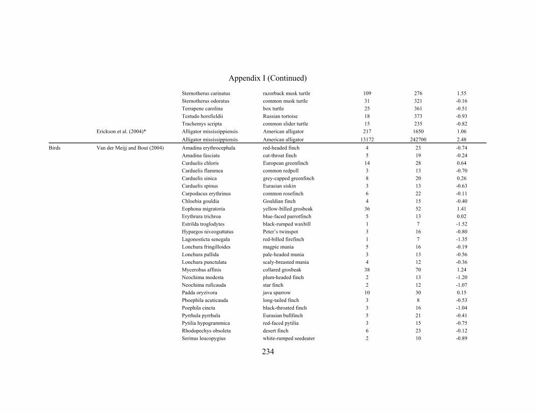
234
Appendix I (Continued)
Sternotherus carinatus razorback musk turtle 109 276 1.55 Sternotherus odoratus common musk turtle 31 321 -0.16 Terrapene carolina box turtle 25 361 -0.51 Testudo horsfieldii Russian tortoise 18 373 -0.93 Trachemys scripta common slider turtle 15 235 -0.82 Erickson et al. (2004)* Alligator mississippiensis American alligator 217 1650 1.06 Alligator mississippiensis American alligator 13172 242700 2.48 Birds Van der Meijj and Bout (2004) Amadina erythrocephala red-headed finch 4 23 -0.74 Amadina fasciata cut-throat finch 5 19 -0.24 Carduelis chloris European greenfinch 14 28 0.64 Carduelis flammea common redpoll 3 13 -0.70 Carduelis sinica grey-capped greenfinch 8 20 0.26 Carduelis spinus Eurasian siskin 3 13 -0.63 Carpodacus erythrinus common rosefinch 6 22 -0.11 Chloebia gouldia Gouldian finch 4 15 -0.40 Eophona migratoria
yellow-billed grosbeak 36 52 1.41 Erythrura trichroa blue-faced parrotfinch 5 13 0.02 Estrilda troglodytes black-rumped waxbill 1 7 -1.52 Hypargos niveoguttatus
Peter’s twinspot 3 16 -0.80
Lagonosticta senegala red-billed firefinch 1 7 -1.35 Lonchura fringilloides
magpie munia 5 16 -0.19
Lonchura pallida pale-headed munia 3 13 -0.56 Lonchura punctulata scaly-breasted munia 4 12 -0.36 Mycerobas affinis collared grosbeak 38 70 1.24 Neochima modesta plum-headed finch 2 13 -1.20 Neochima ruficauda star finch 2 12 -1.07 Padda oryzivora java sparrow 10 30 0.15 Phoephila acuticauda
long-tailed finch 3 8 -0.53
Poephila cincta black-throated finch 3 16 -1.04 Pyrrhula pyrrhula Eurasian bullfinch 5 21 -0.41 Pytilia hypogrammica red-faced pytilia 3 15 -0.75 Rhodopechys obsoleta desert finch 6 23 -0.12 Serinus leucopygius white-rumped seedeater 2 10 -0.89

235
Appendix I (Continued)
Serinus mozambicus yellow-fronted canary 3 12 -0.66 Serinus sulphuratus brimstone canary 12 18 0.80 Taeniopygia bichenovi double-barred finch 2 10 -1.03 Taenopygia guttata zebra finch 4 23 -0.77 Uraeginthus bengalus red-cheeked cordonblue 1 10 -1.54 Fish Hernandez and Motta (1997) Archosargus probatocephalus sheepshead 309 998 1.88 Clifton and Motta (1998) Halichoeres bivittatus slippery dick
5 19 -0.35 Halichoeres garnoti yellowhead wrasse
10 21 0.49
Halichoeres maculipinna
clown wrasse 11 18 0.71 Lachnolaimus maximus hogfish 290 209 2.97 Thalassoma bifasciatum bluehead wrasse
5 7 0.47
Huber and Motta (2004) Squalus acanthias spiny dogfish 20 501 -1.09 Huber et al. (in prep) Hydrolagus colliei white-spotted ratfish 87 870 0.96 Huber et al. (2005) Heterodontus francisci horn shark 206 2948 0.54 Korff and Wainwright (2004) Chilomycterus schoepfi striped burrfish
380 180 3.42
Huber et al. (2006)* Carcharhinus limbatus blacktip shark 32 1274 -0.04 Carcharhinus limbatus blacktip shark 423 22092 -1.15 *Two values are given for studies in which specimen body masses ranged over more than one order of magnitude

About the Author
Born in West Hempstead, NY on December 7, 1978, Daniel Huber spent much of
his childhood in and around the Atlantic Ocean. At age eight a relative of Dan’s was
bitten by a shark, beginning his lifelong fascination with these amazing animals. After
graduating from Chaminade High School in 1996, Dan attended Duke University and
received his B.S. in Biology in 2000. Dan then began his doctorate at the University of
South Florida as an inaugural recipient of the university’s Presidential Fellowship, where
he worked under Dr. Philip Motta on the biomechanics and functional morphology of
shark feeding. He has taught courses in Comparative Vertebrate Anatomy and Human
Anatomy and Physiology, and has appeared in numerous Discovery Channel productions.
At the completion of his doctorate, Dan had authored six papers and in the fall of 2006
will be joining the faculty in the Biology Department at the University of Tampa.