Cranial anatomy of Andinodelphys cochabambensis, a stem ...
145
2020 42 30 geodiversitas
-
Upload
khangminh22 -
Category
Documents
-
view
1 -
download
0
Transcript of Cranial anatomy of Andinodelphys cochabambensis, a stem ...

2020 42 30
geodiversitas
Geodiversitas est une revue en flux continu publiée par les Publications scientifiques du Muséum, ParisGeodiversitas is a fast track journal published by the Museum Science Press, Paris
Les Publications scientifiques du Muséum publient aussi / The Museum Science Press also publish: Adansonia, Zoosystema, Anthropozoologica, European Journal of Taxonomy, Naturae, Cryptogamie sous-sections Algologie, Bryologie, Mycologie, Comptes Rendus Palevol
Diffusion – Publications scientifiques Muséum national d’Histoire naturelle CP 41 – 57 rue Cuvier F-75231 Paris cedex 05 (France) Tél. : 33 (0)1 40 79 48 05 / Fax : 33 (0)1 40 79 38 40 [email protected] / http://sciencepress.mnhn.fr
© Publications scientifiques du Muséum national d’Histoire naturelle, Paris, 2020ISSN (imprimé / print) : 1280-9659/ ISSN (électronique / electronic) : 1638-9395
Directeur De la publication / Publication director : Bruno David,Président du Muséum national d’Histoire naturelle
réDacteur en chef / editor-in-chief : Didier Merle
assistant De réDaction / assistant editor : Emmanuel Côtez ([email protected])
Mise en page / Page layout : Emmanuel Côtez, Audrina Neveu
coMité scientifique / scientific board :Christine Argot (Muséum national d’Histoire naturelle, Paris)Beatrix Azanza (Museo Nacional de Ciencias Naturales, Madrid)Raymond L. Bernor (Howard University, Washington DC)Alain Blieck (chercheur CNRS retraité, Haubourdin)Henning Blom (Uppsala University)Jean Broutin (Sorbonne Université, Paris, retraité)Gaël Clément (Muséum national d’Histoire naturelle, Paris)Ted Daeschler (Academy of Natural Sciences, Philadelphie)Bruno David (Muséum national d’Histoire naturelle, Paris)Gregory D. Edgecombe (The Natural History Museum, Londres)Ursula Göhlich (Natural History Museum Vienna)Jin Meng (American Museum of Natural History, New York)Brigitte Meyer-Berthaud (CIRAD, Montpellier)Zhu Min (Chinese Academy of Sciences, Pékin)Isabelle Rouget (Muséum national d’Histoire naturelle, Paris)Sevket Sen (Muséum national d’Histoire naturelle, Paris, retraité)Stanislav Štamberg (Museum of Eastern Bohemia, Hradec Králové)Paul Taylor (The Natural History Museum, Londres, retraité)
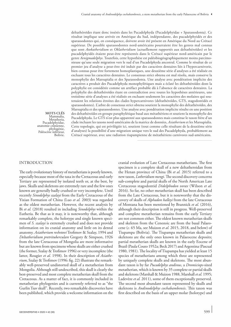
couverture / cover : Life reconstruction of Andinodelphys cochabambensis by Charlène Letenneur: on the left is a complete animal (natural size) grasping on a branch and on the right, on the foreground, is a lateral view of the head.
Geodiversitas est indexé dans / Geodiversitas is indexed in:– Science Citation Index Expanded (SciSearch®)– ISI Alerting Services®
– Current Contents® / Physical, Chemical, and Earth Sciences®– Scopus®
Geodiversitas est distribué en version électronique par / Geodiversitas is distributed electronically by:– BioOne® (http://www.bioone.org)
Les articles ainsi que les nouveautés nomenclaturales publiés dans Geodiversitas sont référencés par / Articles and nomenclatural novelties published in Geodiversitas are referenced by:
– ZooBank® (http://zoobank.org)
597GEODIVERSITAS • 2020 • 42 (30) © Publications scientifiques du Muséum national d’Histoire naturelle, Paris. www.geodiversitas.com
urn:lsid:zoobank.org:pub:DA30F7AF-FA14-4007-8999-F61BBDDD6772
Muizon C. de & Ladevèze S. 2020. — Cranial anatomy of Andinodelphys cochabambensis, a stem metatherian from the early Palaeocene of Bolivia. Geodiversitas 42 (30): 597-739. https://doi.org/10.5252/geodiversitas2020v42a30. http://geodiversitas.com/42/30
ABSTRACTAndinodelphys cochabambensis Marshall & Muizon, 1988 is one of the best preserved metatherian species from the early Palaeocene fauna of Tiupampa (Bolivia). It is represented by five almost complete skulls, three of them being securely associated to sub-complete to partial skeleton. Four skulls could be extracted from a block including several intermingled skeletons. The present paper provides a thorough description of the dental, cranial, and dentary anatomy of A. cochabambensis. The cranial anatomy of A. cochabambensis is similar to that of Pucadelphys andinus. The skull of Andinodelphys however differs from that of Pucadelphys in its larger size and proportionally longer rostrum. Other differences include the presence, in Andinodelphys, of large anteriorly protruding I1s, small palatal vacuities, a transverse canal, and a small hypotympanic sinus. Andinodelphys has the same dental formula as Pucadelphys (I 5/4, C 1/1, P 3/3, M4/4), the plesiomorphic condition for metatherians. Furthermore, both genera share the lack a tympanic process of the alisphenoid, a deep groove for the internal carotid artery at the anterior apex of the promontorium, a small prootic canal perforating the lateral edge of the petrosal and opening laterally in the deep sul-cus for the prootic sinus, and a vestigial anterior lamina of the petrosal. Dentally Andinodelphys closely resembles Pucadelphys, the two genera differing in the larger size of the former and in the inconstant presence in the former of a twinned stylar cusp C. Although 25% smaller, the cheek teeth of Andinodelphys closely resemble those of Itaboraidelphys camposi from the early Eocene of Itaboraí (Brazil). As far as dental morphology is concerned, both genera are likely to have diverged from a direct common ancestor, probably Andinodelphys-like, with Itaboraidelphys displaying more derived dental structures. Two isolated petrosal from Itaboraí (Type 2 petrosals) are morphologically close to those of Andinodelphys but distinctly larger. In this paper, a previous interpretation including the teeth of Itaboraidelphys and these petrosals in the same taxon is fol-lowed. A phylogenetic analysis retrieved Itaboraidelphys as a sister taxon of the clade Pucadelphys + Andinodelphys, thus lending support to inclusion of the former in the Pucadelphyidae. Three sets of parsimony analyses were performed. A first set of analyses (with all characters) retrieved a strict
Christian de MUIZON Sandrine LADEVÈZE
CR2P (CNRS, MNHN, Sorbonne Université),Département Origines et Évolution, Muséum national d’Histoire naturelle,
case postale 38, 57 rue Cuvier, F-75231 Paris cedex 05 (France)
Submitted on 25 November 2019 | accepted on 31 March 2020 | published on 31 December 2020
Cranial anatomy of Andinodelphys cochabambensis, a stem metatherian from the early Palaeocene of Bolivia
598 GEODIVERSITAS • 2020 • 42 (30)
Muizon C. de & Ladevèze S.
consensus tree with a clade as follows: (pucadelphyids, (deltatheroidans (stagodontids, Gurlin Tsav skull-GTS), sparassodonts)). An implied weighting analysis with the same data matrix placed the stagodontids in an early diverging position but retained a clade (pucadelphyids, (deltatheroidans, (GTS, sparassodonts))), the deltatheroidans, being therefore inserted in the pucadelphydans. This result implies an independent arrival of pucadelphyids and sparassodonts to South America, which consequently must have been present in North America in the Late Cretaceous. Possible North American sparassodonts could be the poorly known genera Atokatheridium and Olklath-eridium (currently referred to deltatheroidans) and the pucadelphyids may have been present in the Late Cretaceous of North America with the genus Aenigmadelphys. However, this hypothesis is less parsimonious (with regard to palaeobiogeography) than a single southward migration of an ancestral Pucadelphyda (Pucadelphyidae + Sparassodonta). Because the result of this first set of analyses may have been induced by heavily homoplastic dental characters related to hypercarnivory, a second set of analyses was performed excluding all the dental characters. The strict consensus is poorly resolved but retains monophyletic Marsupialia and Sparassodonta. An implied weight-ing analysis retrieved a monophyletic Pucadelphyda but split the deltatheroidans, the polyphyly of which is regarded as a possible artefact related to the lack of dental characters. The GTS is sister taxon to Pucadelphyda. Because the polyphyly of deltatheroidans contradicts all previous hypotheses, a third set of analyses has been performed excluding only those molar characters that supported the close relationships of the hypercarnivorous clades (deltatheroids, stagodontids, and sparassodonts). The strict consensus tree retrieved monophyletic deltatheroidans, Marsupialia and sparassodonts. An implied weighting analysis resulted in deltatheroidans forming a paraphyletic stem assemblage of Metatheria and monophyletic Pucadelphyda. The GTS was no longer related to sparassodonts but was the sister taxon of a clade including the North American taxa of the data matrix, Asiatherium, and Marsupialia. This topology, which is favoured here, supports (as well as that of the second set of analyses) a single pucadelphydan southward migration, probably in the Late Cretaceous, with a Tiupampian radiation of South American carnivorous metatherians.
RÉSUMÉAnatomie cranienne d’Andinodelphys cochabambensis, un stem-métathérien du Paléocène inférieur de Bolivie.Andinodelphys cochabambensis Marshall & Muizon, 1988 est l’une des espèces de métathériens les mieux conservées de la faune du Paléocène inférieur de Tiupampa (Bolivie). Elle est représentée par cinq crânes presque complets, trois d’entre eux étant associés à des squelettes sub-complets ou partiels. Le présent travail fournit une description détaillée de l’anatomie des dents, du crâne et du dentaire d’Andinodelphys cochabambensis. L’anatomie crânienne d’A. cochabambensis est semblable à celle de Pucadelphys andinus. Toutefois, le crâne d’Andinodelphys diffère de celui de Pucadelphys par sa taille plus grande et son rostre proportionnellement plus long. D’autre dif-férences incluent la présence, chez Andinodelphys, de I1s plus grandes que les autres incisives et projetées antérieurement, de petites fenêtres maxillopalatines, d’un canal transverse et d’un sinus hypotympanique. Andinodelphys a la même formule dentaire que Pucadelphys (I 5/4, C 1/1, P 3/3, M4/4), qui constitue la condition plésiomorphe pour les métathériens. De plus, les deux genres partagent l’absence de processus tympanique de l’alisphénoïde, un profond sillon pour la carotide interne à l’apex antérieur du promontoire, un petit canal prootique perforant le bord latéral du pétreux et s’ouvrant latéralement dans le profond sillon pour le sinus prootique et une lame antérieure vestigiale du pétreux. Sur le plan dentaire Andinodelphys ressemble étroitement à Pucadelphys, les deux genres différant par la taille plus grande du premier et par la présence inconstante, chez le premier, d’une cuspide stylaire C dédoublée. Bien que 25% plus petites, les dents jugales d’Andinodelphys ressemblent beaucoup à celles d’Itaboraidelphys camposi de l’Eocène inférieur d’Itaboraí (Brésil). Du point de vue de leur morphologie dentaire, les deux genres ont très probablement divergé d’un morphotype ancestral de type Andinodelphys, avec Itaboraidelphys présentant une condition plus dérivée. Deux pétreux isolés d’Itaboraí (pétreux de type 2) sont morphologiquement très semblables à ceux d’Andinodelphys, mais nettement plus grands. Dans ce travail, une interprétation suggérée antérieurement, incluant les dents d’Itaboraidelphys et ces deux pétreux dans le même taxon, est entérinée. Une analyse phylogénétique résulte en un place-ment d’Itaboraidelphys comme taxon frère du clade Andinodelphys + Pucadelphys, supportant de ce fait le placement du premier parmi les Pucadelphyidae. Trois séries d’analyses de parcimonie ont été réalisées. Une première série d’analyses (avec tous les caractères) a produit un consensus strict avec un clade comme suit : (pucadelphyidés (deltathéroïdes (stagodontidés, crâne de Gur-lin Tsav – GTS) sparassodontes)). Une analyse avec pondération implicite (Implied Wheighting) des caractères utilisant la même matrice de données a fait diverger les stagodontidés plus tôt sur l’arbre mais a maintenu le clade (pucadelphyidés, (deltathéroïdes (GTS, (sparassodontes))), les
KEY WORDSMammalia,Metatheria,
Pucadelphyidae,cranial anatomy,
phylogeny,Early Palaeocene,
Bolivia.
599
Cranial anatomy of Andinodelphys cochabambensis, a stem metatherian from the early Palaeocene of Bolivia
GEODIVERSITAS • 2020 • 42 (30)
deltathéroïdes étant donc insérés dans les Pucadelphyda (Pucadelphyidae + Sparassodonta). Ce résultat implique une arrivée en Amérique du Sud, indépendante, des pucadelphyidés et des sparassodontes qui, en conséquence, doivent avoir été présent en Amérique du Nord au Crétacé supérieur. De possible sparassodontes nord-américains pourraient être les genres mal connus que sont Atokatheridium et Oklatheridium (actuellement rapportés aux deltathéroïdes) et les pucadelphyidés étaient peut-être représentés dans le Crétacé supérieur nord-américain par le genre Aenigmadelphys. Toutefois, cette hypothèse est paléobiogéographiquement moins parcimo-nieuse qu’une seule migration vers le sud d’un Pucadelphyda ancestral. Comme le résultat de ce premier jeu d’analyse a peut-être été induit par des caractères dentaires liés à l’hypercarnivorie, bien connus pour être fortement homoplastiques, une deuxième série d’analyses a été réalisée en excluant tous les caractères dentaires. Le consensus strict obtenu est mal résolu, mais conserve la monophylie des Marsupialia et des Sparassodonta. Une analyse avec pondération implicite des caractères a produit des Pucadelphyda monophylétiques mais a éclaté les deltathéroïdes dont la polyphylie est considérée comme un artéfact probable dû à l’absence de caractères dentaires. La polyphylie des deltathéroïdes étant en contradiction avec toutes les hypothèses antérieures, une troisième série d’analyses a été réalisée en excluant seulement les caractères des molaires qui sou-tenaient les relations étroites des clades hypercarnivores (deltathéroïdes, GTS, stagodontidés et sparassodontes). L’arbre de consensus strict obtenu soutient la monophylie des deltathéroïdes, des Marsupialia et des sparassodontes. Une analyse avec pondération implicite résulte en une position des deltathéroïdes en groupe paraphylétique basal aux métathériens et soutient la monophylie des Pucadelphyda. Le GTS n’est plus apparenté aux sparassodontes mais constitue le taxon frère d’un clade incluant les taxons nord-américains de la matrice de données, Asiatherium et les Marsupialia. Cette topologie, qui est privilégiée ici, soutient (tout comme celle résultant de la deuxième série d’analyses) la possibilité d’une migration unique vers le sud des Pucadelphyda, probablement au Crétacé supérieur, avec une radiation tiupampienne de métathériens carnivores sud-américains.
INTRODUCTION
The early evolutionary history of metatherians is poorly known, especially because most of the taxa in the Cretaceous and early Tertiary are represented by isolated teeth or, at the best, by jaws. Skulls and skeletons are extremely rare and the few ones known are generally badly crushed or very incomplete. Until recently Sinodelphys szalayi from the Early Cretaceous of the Yixian Formation of China (Luo et al. 2003) was regarded as the oldest metatherian. However, the recent analysis by Bi et al. (2018) resulted in including Sinodelphys within the Eutheria. Be that as it may, it is noteworthy that, although remarkably complete, the holotype and single known speci-men of S. szalayi is extremely crushed and does not provide information on its cranial anatomy and little on its dental anatomy. Asiatherium reshetovi Trofimov & Szalay, 1994 and Deltatheridium pretrituberculare Gregory & Simpson, 1926 from the late Cretaceous of Mongolia are more informative but are known from specimens whose skulls are either crushed (the former, Szalay & Trofimov 1996) or very incomplete (the latter, Rougier et al. 1998). In their description of Asiathe-rium, Szalay & Trofimov (1996: fig. 22) illustrate the remark-ably well-preserved undistorted skull of a metatherian from Mongolia. Although still undescribed, this skull is clearly the best-preserved and most complete metatherian skull from the Cretaceous. As a matter of fact, it is commonly included in metatherian phylogenies and is currently referred to as “the Gurlin Tsav skull”. Recently, two remarkable discoveries have been published, which provide a welcome information on the
cranial evolution of Late Cretaceous metatherians. The first specimen is a complete skull of a new deltatheroidan from the Henan province of China (Bi et al. 2015) referred to a new taxon, Lotheridium mengi. The second discovery concerns sub-complete and partial skulls of the North American Late Cretaceous stagodontid Didelphodon vorax (Wilson et al. 2016). So far, no other metatherian skull has been described from the Late Cretaceous, but it is noteworthy that the dis-covery of skulls of Alphadon halleyi from the late Cretaceous of Montana has been mentioned by Brannick et al. (2016), although their description is still in progress. Well-preserved and complete metatherian remains from the early Tertiary are not common either. The oldest known metatherian skulls and skeleton from the Cenozoic are from the basal Palaeo-cene (c. 65 Ma, see Muizon et al. 2015, 2018, and below) of Tiupampa (Bolivia). The Tiupampa metatherian skulls and skeletons are the only ones known in Palaeocene deposits; partial metatherian skulls are known in the early Eocene of Brazil (Paula Couto 1952a; Beck 2017) and Argentina (Pascual 1980, 1981). The locality of Tiupampa has yielded at least 15 species of metatherians among which three are represented by uniquely complete skulls and skeletons. The most abun-dant taxon is by far Pucadelphys andinus, a Dromiciops-sized metatherian, which is known by 35 complete or partial skulls and skeletons (Marshall & Muizon 1988, Marshall et al. 1995; Ladevèze et al. 2011), some of them exceptionally preserved. The second most abundant taxon represented by skulls and skeletons is Andinodelphys cochabambensis. This taxon was first described on the basis of an upper molar (holotype) and
MOTS CLÉSMammalia,Metatheria,
Pucadelphyidae,anatomie crânienne,
phylogénie,Paléocène inférieur,
Bolivie.
600 GEODIVERSITAS • 2020 • 42 (30)
Muizon C. de & Ladevèze S.
a referred lower molar (Marshall & Muizon 1988) (Fig. 1), but during the 1996 field season at Tiupampa, five partial or sub-complete skulls and skeletons have been recovered (Mui-zon et al. 1997) (Figs 2, 3). A third taxon, Mayulestes ferox, is an early sparassodont known by an almost complete (but dorsoventrally crushed) skull associated to a partial skeleton (Muizon 1994, 1998). Some other taxa are known by nearly complete or partial skulls such as the sparassodont Allqoki-rus australis, that has been recently described (Muizon et al. 2018), and five other specimens referred to Mizquedelphys pilpinensis Marshall & Muizon, 1988 (two sub-complete skulls and two partial skulls) and Incadelphys antiquus Marshall & Muizon, 1988 (one partial skull) currently under study by the authors. The other metatherians of Tiupampa are known by jaws and teeth only (Muizon 1992). The anatomy of the skull and postcranial skeleton of Pucadelphys andinus, May-ulestes ferox, as well as that of the skull of Allqokirus australis have been thoroughly described in earlier works (Marshall & Muizon 1995; Muizon 1998; Muizon et al. 2018). In con-trast, only a preliminary description of the skulls of Andino-delphys cochabambensis has been provided by Muizon et al. (1997), although isolated petrosals have been described by Ladevèze & Muizon (2007). In the present paper, we intend to comprehensively describe the skull and dentition of Andi-nodelphys cochabambensis and to perform parsimony analyses in order to evaluate its phylogenetic affinities and to address the relationships of the major groups of American carnivorous metatherians. In particular, potential close relationships of Andinodelphys with the Itaboraian genus Itaboraidelphys, as suggested by Marshall & Muizon (1988) and Muizon et al. (2018), will be investigated.
AGE OF THE TIUPAMPA MAMMAL FAUNA
The Tiupampa fauna was discovered in 1982 by a French-US-Bolivian paleontological expedition in the Vila-Vila outcrop, a large exposure of red-beds in Mizque Province (Cochabamba Department, Bolivia). These red beds were initially referred to the El Molino Formation, which was correlated to the Late Cretaceous (Marshall et al. 1983; Muizon et al. 1983). Consequently, the mammals collected at Tiupampa were initially given a Late Cretaceous age (Maastrichtian) (Mar-shall et al. 1983, 1985; Marshall & Muizon 1988; Muizon et al. 1984). Subsequent studies brought to light the fact that the Vila Vila outcrop also included beds of the Santa Lucía Formation, which are considered to be Palaeocene (Sempere et al. 1997) and which yielded the Tiupampa mammal fauna. Although a Palaeocene age is now commonly accepted for the Tiupampa fauna, interpretations diverge on the age of the fauna within the Palaeocene. Marshall et al. (1997) and Sempere et al. (1997), on the basis of sedimentological and palaeontological (but see below) arguments, concluded that the Tiupampa mammal bearing beds correspond to the middle to late Santa Lucía Formation, and are included in a single reversed magnetostratigraphic series, which they correlated to Chron 26r. This Chron corresponds to the early Selandian
(c. 58 Ma to 60 Ma), i.e. the early middle Palaeocene. Sem-pere et al. (1997) also reported the mammal fauna of Punta Peligro (Chubut Province, Patagonia, Argentina) to be older than that of Tiupampa and referred the former to the earliest Selandian or latest Danian (c. 62 to 60 Ma). This interpretation was followed by Pascual & Ortiz-Jaureguizar (2007: table 1), although these authors implicitly concluded that the Punta Peligro therians feature a South American cachet, whereas the Tiupampa fauna mainly retains a North American hallmark.
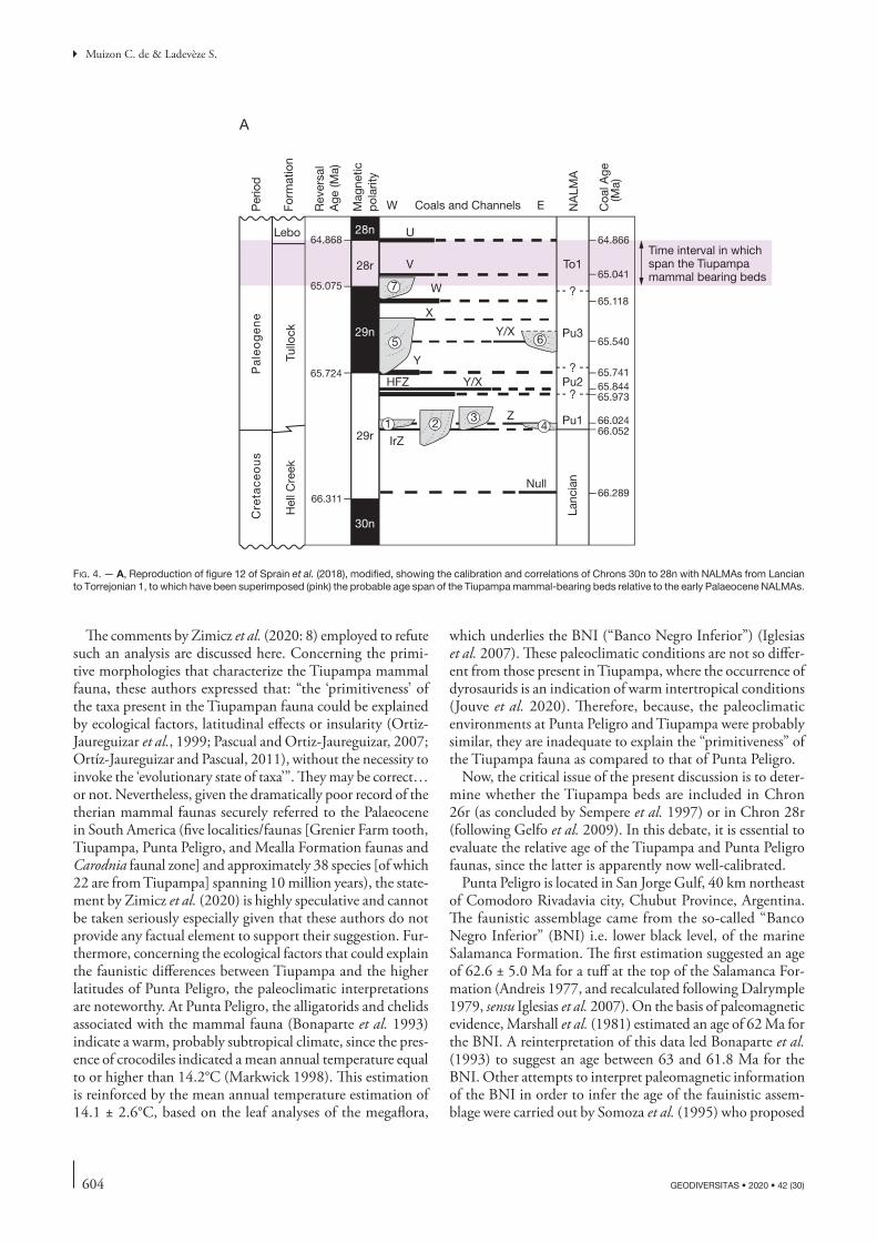
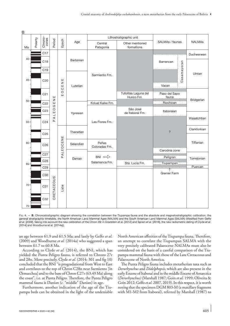
A different interpretation was given by Gelfo et al. (2009), Woodburne et al. (2014a, b), and Muizon et al. (2015, 2018), who considered the Tiupampa mammal fauna to be early Palaeocene (Danian) and older than the Punta Peligro fauna. Gelfo et al. (2009) employed two different meth-ods, a cluster analysis and a parsimony analysis based on the Parsimony Analysis of Endemicity (PAE; Rosen 1988) and the Cladistics Analysis of Distribution and Endemism (CADE; Porzecanski & Cracraft 2005). Gelfo et al. (2009) concluded that the Tiupampa mammal fauna predated the Punta Peligro assemblage. Because the Tiupampa beds are included in a single reversed magnetostratigraphic series (fol-lowing Sempere et al. 1997), Gelfo et al. (2009) correlated the Tiupampa beds with polarity Chron 28r, equivalent to the Puercan 3 (c. 64 to 64.5 Ma) of North America at that time (see Gelfo et al. [2009] and below for discussion on the referral of the Tiupampa mammal-bearing beds to Chron 28r rather than to Chron 26r as suggested by Sempere et al. [1997]). However, new calibration of the early Palaeocene NALMAs (Sprain et al. 2015, 2018) modified the age and position of Chron 28r relative to the NALMAs. According to Sprain et al. (2018: fig. 12) the lower limit of Chron 28r is bracketed between 65.075 Ma and 64.868 Ma (Fig. 4). The Torrejonian 1 spans the period from 65.041 Ma (Sprain et al. 2018) to c. 63.5 Ma (Vandenberghe et al. 2012; Flynn et al. 2020). On the basis of the calibrations of Sprain et al. (2015), which are very similar to those of Sprain et al. (2018), Muizon et al. (2015) correlated the mammal-bearing beds at Tiupampa, to the base of Torrejonian 1 and evaluated an age of approximately 65 Ma for the Tiupampa mammals, an interpretation followed here. Therefore, the longevity of the Tiupampa mammals probably does not exceed 0.2 Ma (the approximate duration of Chron 28r according to Sprain et al. 2018; see Fig. 4). Consequently, the time span of the Tiupampan in Woodburne et al. (2014a and b: fig. 2) and in Goin et al. (2016: fig. 7.2) should be significantly shortened.
Nevertheless, the age of the Tiupampa mammal fauna is still considered to be controversial by Eldridge et al. (2019), although these authors do not provide arguments in sup-port of their conclusion. Eldridge et al. (2019) accepted the Danian age of the Punta Peligro fauna (Argentina) suggested by Sempere et al. (1997) but did not discuss the evidence provided by others (Gelfo et al. 2009; Muizon et al. 2015, 2018), who consider that the Tiupampa mammal fauna is older than that of Punta Peligro (therefore, early Danian in age). It is noteworthy that the latter interpretation is now commonly accepted by most authors (e.g., Rose 2010; Goin et al. 2012; Oliveira & Goin 2012; Woodburne et al.

601
Cranial anatomy of Andinodelphys cochabambensis, a stem metatherian from the early Palaeocene of Bolivia
GEODIVERSITAS • 2020 • 42 (30)
2014a, b; Reguero et al. 2014; Forasiepi et al. 2015; Goin et al. 2016; Babot et al. 2017; Carneiro 2018; Carneiro et al. 2018; Bauzá et al. 2019), who were not cited, however, by Eldridge et al. (2019). Croft et al. (2020) also followed the same interpretation.
Recently, Zimicz et al. (2020) went back to the paleo-magnetic correlation arguments of Sempere et al. (1997), whom they followed, considering that the Tiupampa beds and fauna correlate with Chron 26r. Moreover, Zimicz et al. (2020: 8) further stated that there is a “[...] large amount of recent geological evidence reinforcing the Sempere’s correla-tion of Santa Lucía beds with Chron 26r (e.g. Horton et al. 2001; DeCelles & Horton 2003; Demouy et al. 2012; Rak et al. 2017; Calle et al. 2018).” However, it is noteworthy that, most of these papers did not discuss directly the age of the Tiupampa fauna. Horton et al. (2001) and DeCelles & Horton (2003) mentioned the age of the Santa Lucía For-mation, but without a critical reconsideration. Horton et al. (2001) did not provide new data on the problem or did not present arguments different from those of Gelfo et al. (2009). They simply cited Sempere et al. (1997) as the source of the referral of the Santa Lucía Formation to Chron 26r. Furthermore, considering the lithostratigraphic correlations established by Sempere et al. (1997) to justify their interpre-tation of the age of the Santa Lucía Formation at Tiupampa, Horton et al. (2001: 1389) stated that: “Despite successful dating of the Santa Lucía in several Bolivian locales, regional lithostratigraphic correlations over hundreds of kilometers remain tentative”. DeCelles & Horton (2003) do not provide either new evidence concerning the age of the Santa Lucía Formation and its referral to Chron 26r, but only refer, in this respect, to Sempere et al. (1997). However, interestingly
DeCelles & Horton (2003: fig. 13) illustrate the stratigraphic extension of the Santa Lucía Formation as approximately spanning the period 59 to 65 Ma, thereby, extending the lower limit of the formation to the lower Danian. Demouy et al. (2012) did not even mention the Santa Lucía Forma-tion nor any polarity Chron for it and Rak et al. (2017) and Calle et al. (2018), did not comment on the correlation of Santa Lucía beds to Chron 26r. In fact, the “large amount of geological evidences” mentioned by Zimicz et al. (2020) are simply references to the interpretation of Sempere et al. (1997) with no critical discussion or new data. Therefore, in essence, the new interpretation of Zimicz et al. (2020) is based only on the argument of Sempere et al. (1997) to suggest a younger age for the Tiupampa beds, because the five geologi-cal references they mentioned are inappropriate to support their argument. Furthermore, Zimicz et al. (2020) provided neither new geological data nor critical discussion based on new paleontological arguments (see below for comments on their interpretation of the presence of Peradectes at Tiu-pampa and on the primitive morphologies that characterize the Tiupampa fauna). In this context, it is worth recalling that Sempere et al. (1997) based the main evidence justifying their assignment and their conclusions on: 1) paleontological arguments and 2) correlation of the Tiupampa profile to the one at La Palca, where the El Molino and Santa Lucía units are better exposed. However, as is concluded by Sempere et al. (1997: 718, 719): “A simple one-to-one correlation of the La Palca polarity zonation to the geomagnetic polarity time scale is not evident (Fig. 10). The correlation shown in Figures 10 and 11 is our preferred interpretation because it is corroborated by the paleontologic data and agrees with a number of facts discussed below.” Therefore, Sempere et al.
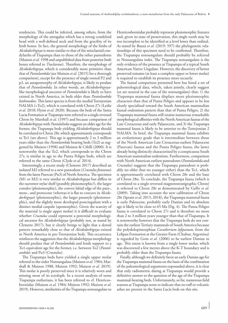
fig. 1. — Andinodelphys cochabambensis (YPFB Pal 6192). SEM stereophotograph of the holotype: a right M2. Scale bar: 1 mm.
602 GEODIVERSITAS • 2020 • 42 (30)
Muizon C. de & Ladevèze S.
(1997) implicitly admitted that there are alternative inter-pretations. It is therefore unclear why Zimicz et al. (2020) dismissed most of the paleontological evidence provided by Gelfo et al. (2009) (but see comments below), which was, in fact, the same kind of evidence (but with a larger faunal sample per locality) that led Sempere et al. (1997), in partial view of the knowledge of the Tiupampa fauna by that time, to assign Santa Lucía and the upper part of the El Molino Formation to Chron 26r.
To conclude, because of the controversy mentioned by Eldridge et al. (2019) and the comments of Zimicz et al. (2020), and because paleontological data were regarded by Sempere et al. (1997) as partly supporting their interpretation, we consider it useful to review here the paleontological data that can, in the light of the new paleontological discoveries and studies since the publication of the Sempere et al. (1997) paper, support the ante-riority of the Tiupampa mammal fauna relative to that of Punta Peligro and its correlation to the early Danian as advocated here.
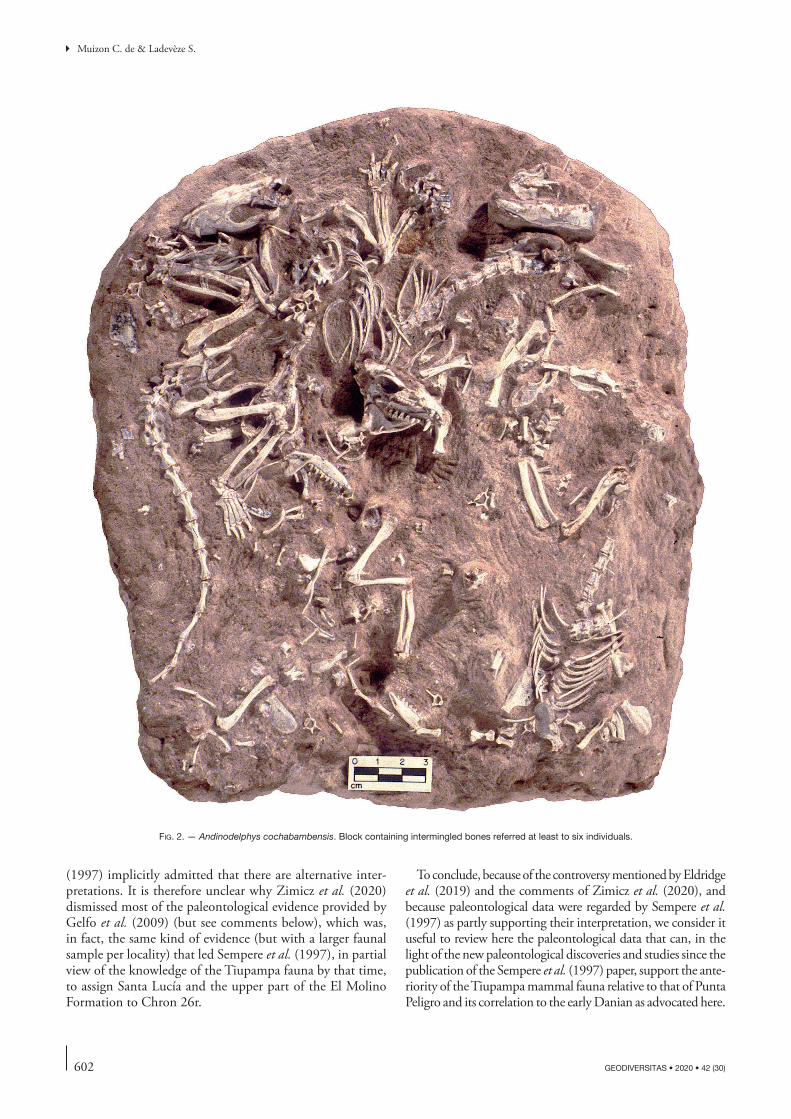
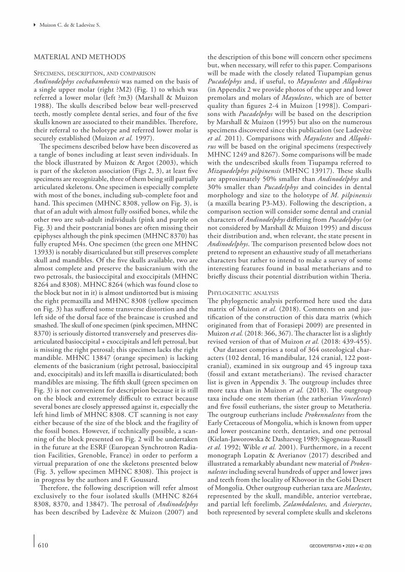
fig. 2. — Andinodelphys cochabambensis. Block containing intermingled bones referred at least to six individuals.
603
Cranial anatomy of Andinodelphys cochabambensis, a stem metatherian from the early Palaeocene of Bolivia
GEODIVERSITAS • 2020 • 42 (30)
Because no direct radiometric dates are available at Tiu-pampa or at Punta Peligro and because magnetostratigraphic studies are inconclusive (Tiupampa beds are included in a single reversed magnetostratigraphic series), comparison of the evolutionary grades of the two faunas is the only criterion on which to base their relative ages and provide informa-tion on their absolute age (Gelfo et al. 2009). It is clear that the conclusions of such a comparison in the case of a small
number of taxa could be questioned. However, in the case of taxonomically abundant faunas, if all (or most of ) the species provide the same convergent information, the conclusions are obviously more reliable. Therefore, the Tiupampa mammal fauna (exclusively therians) with 22 described species (plus two undescribed ones) and the Punta Peligro fauna, which includes at least 9 therian species, can be compared with some degree of confidence.
MHNC 13925
MHNC 13933
Skull MHNC 8308(yellow specimen)
Skull MHNC 13847(orange specimen)
Skull MHNC 8370(pink specimen)
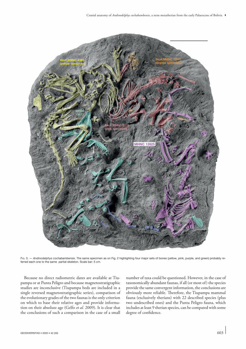
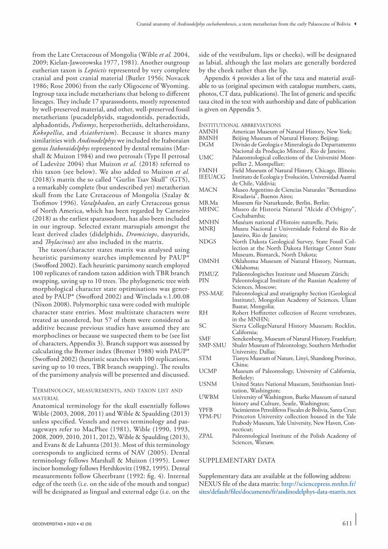
fig. 3. — Andinodelphys cochabambensis. The same specimen as on Fig. 2 highlighting four major sets of bones (yellow, pink, purple, and green) probably re-ferred each one to the same partial skeleton. Scale bar: 5 cm.
604 GEODIVERSITAS • 2020 • 42 (30)
Muizon C. de & Ladevèze S.
The comments by Zimicz et al. (2020: 8) employed to refute such an analysis are discussed here. Concerning the primi-tive morphologies that characterize the Tiupampa mammal fauna, these authors expressed that: “the ‘primitiveness’ of the taxa present in the Tiupampan fauna could be explained by ecological factors, latitudinal effects or insularity (Ortiz-Jaureguizar et al., 1999; Pascual and Ortiz-Jaureguizar, 2007; Ortíz-Jaureguizar and Pascual, 2011), without the necessity to invoke the ‘evolutionary state of taxa’”. They may be correct… or not. Nevertheless, given the dramatically poor record of the therian mammal faunas securely referred to the Palaeocene in South America (five localities/faunas [Grenier Farm tooth, Tiupampa, Punta Peligro, and Mealla Formation faunas and Carodnia faunal zone] and approximately 38 species [of which 22 are from Tiupampa] spanning 10 million years), the state-ment by Zimicz et al. (2020) is highly speculative and cannot be taken seriously especially given that these authors do not provide any factual element to support their suggestion. Fur-thermore, concerning the ecological factors that could explain the faunistic differences between Tiupampa and the higher latitudes of Punta Peligro, the paleoclimatic interpretations are noteworthy. At Punta Peligro, the alligatorids and chelids associated with the mammal fauna (Bonaparte et al. 1993) indicate a warm, probably subtropical climate, since the pres-ence of crocodiles indicated a mean annual temperature equal to or higher than 14.2°C (Markwick 1998). This estimation is reinforced by the mean annual temperature estimation of 14.1 ± 2.6°C, based on the leaf analyses of the megaflora,
which underlies the BNI (“Banco Negro Inferior”) (Iglesias et al. 2007). These paleoclimatic conditions are not so differ-ent from those present in Tiupampa, where the occurrence of dyrosaurids is an indication of warm intertropical conditions (Jouve et al. 2020). Therefore, because, the paleoclimatic environments at Punta Peligro and Tiupampa were probably similar, they are inadequate to explain the “primitiveness” of the Tiupampa fauna as compared to that of Punta Peligro.
Now, the critical issue of the present discussion is to deter-mine whether the Tiupampa beds are included in Chron 26r (as concluded by Sempere et al. 1997) or in Chron 28r (following Gelfo et al. 2009). In this debate, it is essential to evaluate the relative age of the Tiupampa and Punta Peligro faunas, since the latter is apparently now well-calibrated.
Punta Peligro is located in San Jorge Gulf, 40 km northeast of Comodoro Rivadavia city, Chubut Province, Argentina. The faunistic assemblage came from the so-called “Banco Negro Inferior” (BNI) i.e. lower black level, of the marine Salamanca Formation. The first estimation suggested an age of 62.6 ± 5.0 Ma for a tuff at the top of the Salamanca For-mation (Andreis 1977, and recalculated following Dalrymple 1979, sensu Iglesias et al. 2007). On the basis of paleomagnetic evidence, Marshall et al. (1981) estimated an age of 62 Ma for the BNI. A reinterpretation of this data led Bonaparte et al. (1993) to suggest an age between 63 and 61.8 Ma for the BNI. Other attempts to interpret paleomagnetic information of the BNI in order to infer the age of the fauinistic assem-blage were carried out by Somoza et al. (1995) who proposed
1 2 34
5 6
7 ?
?
?
To1
Pu3
Pu1
Pu2
Lanc
ian
Per
iod
Form
atio
n
NA
LMA
Mag
netic
pol
arity
Rev
ersa
lA
ge (M
a)
Coa
l Age
(M
a)
Coals and ChannelsW E
Cre
tac
eo
us
Hel
l Cre
ek
Pa
leo
ge
ne
Tullo
ck
Lebo64.868
65.075
65.724
66.311 66.289
66.05266.024
65.97365.84465.741
65.540
65.118
65.041
64.866
28r
29r
30n
29n
28n U
V
W
X
Y/X
Y/X
Y
HFZ
Z
IrZ
Null
Time interval in whichspan the Tiupampamammal bearing beds
A
fig. 4. — A, Reproduction of figure 12 of Sprain et al. (2018), modified, showing the calibration and correlations of Chrons 30n to 28n with NALMAs from Lancian to Torrejonian 1, to which have been superimposed (pink) the probable age span of the Tiupampa mammal-bearing beds relative to the early Palaeocene NALMAs.
605
Cranial anatomy of Andinodelphys cochabambensis, a stem metatherian from the early Palaeocene of Bolivia
GEODIVERSITAS • 2020 • 42 (30)
an age between 61.9 and 61.5 Ma; and lastly by Gelfo et al. (2009) and Woodburne et al. (2014a) who suggested a span between 61.7 to 60.8 Ma.
According to Clyde et al. (2014), the BNI, which has yielded the Punta Peligro fauna, is referred to Chrons 27r and 28n. More precisely, Clyde et al. (2014: 301 and fig 10) concluded that the BNI “is progradational from West to East and correlates to the top of Chron C28n near Sarmiento [in Ormaechea] and to the base of Chron C27r (63.49 Ma) along the coast”, i.e. at Punta Peligro. Therefore, the Punta Peligro mammal fauna is Danian (c. “middle” Danian) in age.
Furthermore, another indication of the age of the Tiu-pampa beds can be obtained in the light of the undeniable
North American affinities of the Tiupampa fauna. Therefore, an attempt to correlate the Tiupampan SALMA with the very precisely calibrated Palaeocene NALMAs must also be considered on the basis of a careful comparison of the Tiu-pampa mammal fauna with those of the Late Cretaceous and Palaeocene of North America.
The Punta Peligro fauna includes metatherian taxa such as Derorhynchus and Didelphopsis, which are also present in the early Eocene of Itaboraí and in the middle Eocene of Antarctica (Derorhynchus) (Marshall 1987; Goin et al. 1999; Oliveira & Goin 2012; Gelfo et al. 2007, 2019). In this respect, it is worth noting that the specimen DGM 803-M (a maxillary fragment with M1-M2 from Itaboraí), referred by Marshall (1987) to
Ma
40
45
50
55
60
65
70
75
Pol
arity
Per
iod
Ep
och
Chr
ono-
zone
sAge
Lithostratigraphic unit
CentralPatagonia
Other mentionedformations
SALMAs / faunas NALMAs
C17
C18
C19
C20
C21
C22
C23
C24
C25
C26
C27
C28
C29
C30
C31
C32
PA
LE
OG
EN
E
EO
CE
NE
PA
LE
OC
EN
EL
ate
CR
ET
AC
EO
US
Bartonian
Lutetian
Ypresian
Thanetian
Selandian
Danian
Sarmiento Fm.
Koluel Kaike Fm.
Las Flores Fm.
PeñasColoradas Fm.
Salamanca Fm.
BNI
Tufolitas Laguna delHunco Fm.
São Joséde Itaborai Fm.
Sta. Lucía Fm.
Barrancan
Vacan
Ca
sa
ma
yo
ran
Duchesnean
Uintan
Paso del Sapofauna
Riochican
Itaboraian
?
Bridgerian
Wasatchtian
Clarkforkian
Tiffanian
Carodnia zone
Peligran
Tiupampan
Grenier Farm
Torrejonian
Puercan
B
fig. 4. — B, Chronostratigraphic diagram showing the correlation between the Tiupampa fauna and the absolute and magnetostratigraphic calibration, the general stratigraphic timetable, the North American Land Mammal Ages (NALMA) and the South American Land Mammal Ages (SALMA) (Modified from Gelfo et al. [2009], taking into account the new calibration of the Chron 28r in Gradstein et al. [2012] and Sprain et al. [2018], the new radiometric dating of Clyde et al. [2014] and Woodburne et al. [2014a]).
606 GEODIVERSITAS • 2020 • 42 (30)
Muizon C. de & Ladevèze S.
Carolopaulacoutoia itaboraiensis, has been referred by Oliveira & Goin (2012) to Derorhynchus singularis, an interpretation fol-lowed here. Furthermore, bonapartheriid polydolopimorphs are present at Punta Peligro and are also recorded in the early Eocene of the Lumbrera Formation and in the middle Eocene of the Geste Formation of Northwestern Argentina (Pascual 1980, 1981; Goin et al. 1998; Pascual & Ortiz-Jaureguizar 2007; Gelfo et al. 2007; Woodburne et al. 2014a; Babot et al. 2017) as well as in the early Eocene of Brazil at Itaboraí (Beck 2017). These taxa from Punta Peligro (Derorhynchus, Didel-phopsis, Bonapartheriidae) are relevant to the debate because the groups to which they belong (“Didelphimorphia” and Polydolopimorphia) are represented at Tiupampa by species that clearly exhibit more plesiomorphic features. The derived robust crushing molars, the large P3 (based on alveoli) and the large and inflated p3 of Didelphopsis are absent in any of the Tiupampa metatherians. Furthermore, several dental characters of Derorhynchus are more derived than in any of the Tiupampa “didelphimorphs”. They are, for instance, in Derorhynchus, the greater size difference between the paracone and metacone, the lack of ectoflexus, the greater size of the stylar cusp C, the lower trigonid, the talonid of M2-3 wider than the trigonid and the larger entoconid, which is higher than the hypoconid. Furthermore, it is noteworthy that Gelfo et al. (2007) referred the Derorhynchidae recorded at Punta Peligro to Derorhynchus aff. D. minutus, a species present in the middle Eocene of Antarctica (Goin et al. 1999; Gelfo et al. 2019).
Polydolopimorphians are represented at Tiupampa by the basal polydolopiform Roberthoffstetteria, which is unknown in any other faunal assemblage. As noted by Goin et al. (2003) and Case et al. (2005), Roberthoffstetteria more closely resembles Ectocentrocristus from the Late Cretaceous of North America than any other South American polydolopimorphians. However, Chornogubsky & Goin (2015) have re-evaluated this statement providing a new interpretation of the molar morphology of the polydolopimorph Sillustania quechuense, from the late Palaeocene-early Eocene of Laguna Umayo (Peru). The holotype of S. quechuense (UMC-CHU33) is an isolated, incomplete, heavily worn, and somewhat eroded left M2. We can agree with their new interpretation of the identification of the cusps of S. quechuense, which follows that of Goin et al. (2003) concerning Roberthoffstetteria. In their phylogenetic analysis, Chornogubsky & Goin (2015) retrieved a sister group relationship of Sillustania and Rob-erthoffstteteria, which we provisionally accept. However, given these relationships we question the attribution to Sillustania of the m1 (UMC-CHU34) described by Crochet & Sigé (1996) because of the position of the paraconid, anteriorly projected and well-separated from the metaconid, which differs strongly from the condition in Robersthoffstetteria, in which these cusps are closely appressed one against the other and the paraconid does not project anteriorly. The morphology of the paraconid of UMC-CHU34 is also barely compatible with the very large metaconule, which has the size and position of a pseudohypocone, since, as stated by Jernvall (1995), the presence of well-developed hypocone or pseudohypocone is
generally concomitant with a reduced paraconid appressed against the metaconid or a totally absent paraconid. Therefore, we consider here that the hypodigm of Sillustania quechuense should restricted to its holotype only (the M2). The similari-ties between the M2 of Sillustania and Roberthoffstetteria are noteworthy, but do not favour the possible contemporane-ity of these taxa. On the contrary, as far as can be observed on the poorly preserved holotype of Sillustania, this taxon appears more derived than Roberthoffstetteria in, for instance, the larger stylar cusp C, the straight labial edge of the tooth (lack of ectoflexus), the much smaller paraconule, and the functional pseudohypocone (i.e. the metaconule) larger and more distinctly separated from the protocone, a condition that indicates an increased specialization in the crushing function of the cusp. In other respects, given the large dimensions and position of the metaconule, conspicuously separated from the protocone, relationships with the Caenolestoidea should be investigated. Furthermore, it is noteworthy that the holotype of Sillustania does not feature the labiolingual compression of the protocone observed in Roberthoffstetteria and Ectocentroc-ristus. Unfortunately, Roberthoffstetteria-like or Sillustania-like species are absent at Punta Peligro, which prevents compari-son with these taxa. To conclude, even if similarities exist between Roberthoffstetteria and Sillustania, it is noteworthy that the poor preservation of the single upper molar known of Sillustania (the holotype) requires extreme caution in any character comparison with other morphologically similar taxa and casts doubts on any conclusion retrieved from such a comparison. The discovery of more complete and better preserved specimens of Sillustania (especially with premolars) is required to establish a reliable comparison. Considering the presence of enlarged sectorial premolars in polidolopimorphs (except Roberthoffstetteria), the discovery of such teeth in Sillustania is a critical issue. Be that as it may, Roberthoffstet-teria, the single polydolopimorphian known at Tiupampa, retains more plesiomorphic dental characters than all other South American polydolopimorphs and in particular, does not present the enlarged sectorial P2 and/or P3-p3 observed in all the other taxa of the order. Furthermore, because of the transverse compression of its protocone combined with an alignment of this cusp with the conules, it strongly resembles Ectocentrocristus of North America, although it is more derived in the bunoid morphology of its cusps (Case et al. 2005). It is noteworthy that other studies (Williamson et al. 2012, 2014 and Eberle et al. 2019) recovered a sister group relationship of Roberhoffstetteria with Glasbius, whereas Ectocentrocris-tus is included in the Herpetotheriidae (but see Goin et al. 2016: 215). The results of Williamson et al. (2012, 2014 and Eberle et al. 2019) would therefore support a close relation-ship between Glasbius and Roberthoffstetteria as suggested by Goin et al. (2003). Therefore, both interpretations (Case et al. [2005] on the one hand and Williamson et al. [2012, 2014] and Eberle et al. [2019] on the other) suggest close affinities between Roberthoffstetteria and some North American taxa.
Polydolopimorphians recorded at Punta Peligro are referred to a new genus and species of Bonapartheriidae and to a new genus and species of an indeterminate polydolopimorphian
607
Cranial anatomy of Andinodelphys cochabambensis, a stem metatherian from the early Palaeocene of Bolivia
GEODIVERSITAS • 2020 • 42 (30)
(Gelfo et al. 2007; Woodburne et al. 2014a; Babot et al. 2017). Bonapartheriidae are much more derived than the Tiupampa polydolopimorph, Roberthoffstetteria, from which they differ, for example, in the remarkable increase in size of the P3 and or P2 and p3 (which are sectorial), in the loss of the plesiomorphic metatherian molars pattern (which is clearly recognizable in Roberthoffstetteria in spite of the bunoid morphology of the cusps), and in the extreme reduction of P2 and p2 (absent in Bonapartherium) as well as M4 and m4. Bonapartheriidae are absent at Tiupampa but are present (in addition to Punta Peligro) in the early Eocene of Itaboraí (Beck 2017), and in Eocene Formations of Argentina (Lumbrera, Mealla, and Geste formations) (Goin et al. 1998; Woodburne et al. 2014a).
Although pucadelphyids are unknown at Punta Peligro, the Tiupampa pucadelphyids provide interesting elements for this debate because, as stated by Case et al. (2005), they represent an excellent morphological intermediate between a basal Late Cretaceous North American “peradectoid” stock and the more advanced Itaboraí opossum-like metatherians. The problem of the origin of the Tiupampa pucadelphyids is discussed below and we suggest that the North American Aenigmadelphys archeri from the Campanian of the Kaiparow-its Formation is probably morphologically the closest North American metatherian to Pucadelphys and Andinodelphys (see below – p. 694 – for characters that support this statement). Furthermore, in our phylogenetic analyses below, the Tiu-pampa pucadelphyids (e.g., Pucadelphys, Andinodelphys) have been regarded as closely related to the Itaboraian pucadel-phyid, Itaboraidelphys camposi (as suggested by Muizon et al. 2018). However, Pucadelphys and Andinodelphys are more plesiomorphic than Itaboraidelphys in, for instance, the more pronounced ectoflexus (almost absent in Itaboraidelphys), the shorter labial edge of M2-3 as related to labiolingual width, the smaller size difference (in volume and height) between paracone and metacone, the less V-shaped centrocrista, the larger stylar cusp C, the mesiodistally broader protocone, the much smaller size (in length and height) of p3 as compared to m1, the slightly more mesially projected trigonid, the smaller size difference (in volume and height) between paraconid and metaconid, the slightly shorter talonid as compared to trigonid, especially on m4, the smaller entoconid, and possibly their much smaller size. Therefore, the Tiupampa pucadelphyids are morphologically intermediate between the North American Aenigmadelphys and the Itaboraian Itaboraidelphys.
Furthermore, the discovery of a partial skull of Incadelphys antiquus (currently under study by the authors) has revealed the complete dental anatomy of this taxon. It appears that I. antiquus features remarkable similarities with pucadelphyids but also with Aenigmadelphys archeri from which it differs significantly only in the relative size of the paracone and metacone. The paracone is larger than the metacone in Aenigmadelphys and the condi-tion is reversed in Incadelphys. The notable similarity between the two taxa is probably related to close phylogenetic affinities.
Zimicz et al. (2020: 8) have questioned that the presence of Peradectes (a genus also present in the NALMA Puercan3, included in Chrons 29n and 28r) at Tiupampa could be an argument to support correlation of the Tiupampa beds to the
Chron28r (as advocated by Gelfo et al. [2009]), since, as sug-gested by Williamson et al. (2012), this genus is likely para-phyletic. Therefore, Zimicz et al. (2020) regard the affinities of the Tiupampa “Peradectes” as uncertain. Given the result of Williamson et al. (2012), we agree that the single upper molar from Tiupampa referred to Peradectes cf. austrinum may not belong to the genus Peradectes. However, as expressed below (p. 693), paraphyly is a very unstable condition especially in morphological matrices including a majority of taxa known only by dental characters, which are well-known to be highly homoplastic, because of strong selective pressures for feeding (Muizon & Lange-Badré 1997; Springer et al. 2007; Solé & Ladevèze 2017). As a matter of fact, Williamson et al. (2012: 632) mention that the paraphyly of Peradectidae retrieved in the strict consensus of their analysis is “a labile result” and state that “future studies may likely recover a more inclu-sive phylogenetically-defined Peradectidae (which may also include many of the species that fall into the polytomy in our strict consensus, many of which do group with P. elegans and P. californicus in many of the individual most parsimonious trees)”. Be that as it may, the tooth from Tiupampa referred to Peradectes cf. austrinum by Muizon (1992) presents greater similarities with many of the paraphyletic peradectids of Williamson et al. (2012) (e.g., P. elegans and P. californicus) than with any other South American metatherian except the holotype of Peradectes austrinum from Laguna Umayo (Peru). Similarities between the Tiupampa and Laguna Umayo speci-mens strongly suggest that they belong to the same genus, but because of the size and age difference existing between the Tiupampa specimen and the holotype from Laguna Umayo, Muizon (1992) considered that the two specimens may belong to different species. However, because of the poor preservation of the two specimens and the scarcity of the material (two incomplete upper molars), Muizon (1992: 580), expressed reluctance to refer the Tiupampa specimen to a new species based on a single incomplete molar. Therefore, Muizon (1992) cautiously referred the Peradectes tooth from Tiupampa to P. cf. austrinum (contra Marshall & Muizon 1988), stating that the affinities of the two specimens had to be confirmed by the discovery of better preserved and more complete fossils.
The Tiupampa fauna includes a microbiotherian, Khasia cordillerensis, which is known from upper and lower molars and partial upper jaws. Khasia presents characteristic features of the Microbiotheriidae such as a reduced stylar shelf, stylar cusps B, C, and D very reduced to absent, a straight centro-crista, a preparacrista that ends at the anterolabial angle of the tooth, reaching stylar cusp A, and very reduced to absent cingula/cingulids (Marshall & Muizon 1988; Muizon 1992). It is noteworthy that Khasia has been referred by Goin et al. (2016) to the North American family Pediomyidae. However, pediomyids differ from Khasia in having: 1) well-developed cingulids and a precingulum, whereas they are reduced to almost absent in Khasia; 2) a trigon basin that is generally larger and more broadly excavated than in Khasia; 3) a well-developed posterior part of the stylar shelf (the anterior is absent) with large styles (especially StD), whereas Khasia
608 GEODIVERSITAS • 2020 • 42 (30)
Muizon C. de & Ladevèze S.
has a very narrow posterior stylar shelf, if any (the anterior is absent) and is lacking StB, C and D; 4) a postprotocrista and a postmetaconule crista that extend labial to the meta-cone and reach the posterolabial angle of the molar forming a true metacingulum, whereas in Khasia the postmetaconule crista remains above the lingual edge of the metacone. The condition of these four characters in Khasia is similar to that observed in microbiotherians. As a matter of fact, it is note-worthy that the upper molars of Khasia are more similar to those of Dromiciops than to those of the other fossil micro-biotherians known by upper molars (e.g. Microbiotherium, Woodburnodon). Therefore, we do not fully endorse the inter-pretation of Goin et al. (2016) and rather suggest that Khasia should probably be retained as an early member of the South American family Microbiotheriidae. However, as expressed below, (p. 703) this debate may simply reveal an indication of close relationships (even possibly sister group relationships) between microbiotherians and pediomyoids (as suggested by Marshall 1987 and Muizon 1992). However, the latter are unknown in the Punta Peligro fauna and, therefore, do not provide an indication on the relative age of the Peligran and Tiupampan faunas. In other respect, although Carneiro et al. (2018) have recently described a pediomyoid from Brazil, we do not follow their interpretation and consider that, so far, representatives of this superfamily are absent in South America (see comments in Appendix 1). Therefore, if our interpreta-tion is correct, the fact that the Tiupampa microbiotherian, Khasia, presents similarities with pediomyids (and possibly close phylogenetic relationships) could indicate that mico-biotherians may have originated in North America from a pediomyid stock and recently (i. e. early in the Palaeocene) dispersed to South America. This hypothesis however appears plausible only if microbiotherians are sister group to all Aus-tralasian marsupials (= Eomarsupialia), following the latest robust molecular analysis of Duchêne et al. (2018), (see also Mitchell et al. 2014 and Nilsson et al. 2010), a hypothesis which implies one single dispersal event of australidelphian marsupials from western Gondwana to Australasia.
Therefore, as already stated by Gelfo et al. (2009) and Goin et al. (2016: 219), the metatherian fauna of Punta Peligro exhibits clearly more derived morphologies than the Tiu-pampa fauna, and “compare(s) well with Eocene taxa even at generic level”. Furthermore, although inconclusive as far as the relatives ages of the Tiupampa and Punta Peligro faunas are concerned, the fact that several metatherians from Tiu-pampa (pucadelphyids, Incadelphys, Roberthoffstetteria, Khasia) feature morphological similarities with North American taxa from the Late Cretaceous is probably an indication of a short period of differentiation in South America due to the recent arrival in the subcontinent, thus suggesting a relatively basal age within the Palaeocene for these taxa.
The “condylarths” of Tiupampa (the Kollpaniinae) are related to the North American mioclaenid stock and differ from the endemic South American didolodontids and Notonychopidae, which are present at Punta Peligro. Although the mioclaenid affinities of the kollpaniines have been questioned by William-son & Carr (2007), it is noteworthy that the strict consensus
trees in their phylogenetic analyses are strongly unresolved and provide little information about the phylogenetic affinities of the kollpaniines. Similarly, the position of Molinodus retrieved by Halliday et al. (2017) is irrelevant to the mioclaenid affini-ties of Molinodus, since no other mioclaenid is considered in their analysis. In contrast, the Peligran “condylarths” are related to the South American family didolodontids and are represented by the genera Escribania and Raulvacia (Bonaparte et al. 1993; Gelfo 2007; Gelfo et al. 2007). Much smaller than the latter, the Tiupampa kollpaniines retain a more primitive morphology than the Punta Peligro didolodontids in lacking a conspicuous hypocone, a structure well-developed in the latter. However, as brought to light by Muizon et al. (2019), in Molinodus, a pseudohypocone is clearly in process of indi-vidualization by duplication of the protocone, a process also present in Raulcaccia but at a more advanced stage.
The endemic South American order Litopterna has not been discovered at Tiupampa but is undoubtedly represented at Punta Peligro by the notonychopid Requisia vidmari (Bona-parte & Morales 1997).
The orders “Proteutheria” and Pantodonta are abundant in North America and have not been recorded elsewhere than Tiupampa in South America.
The “proteutherian” Cimolestes is represented at Tiupampa by a lower m2 or m3 and is recorded in North America from the Maastrichtian to the late Puercan NALMA (Pu3) (Lof-gren et al. 2004). “Proteutherians” have not been recorded elsewhere in South America.
Comparison of the Tiupampa pantodont, Alcidedorbignya inopinata, to North American taxa allows better estimation of the absolute age of the Tiupampa fauna. As mentioned by Gelfo et al. (2009) and Muizon et al. (2015), the morphology of A. inopinata is compatible with an evolutionary grade that clearly predates Pantolambda bathmodon from the Torrejonian 2 of New Mexico (Lofgren et al. 2004). A. inopinata is approxi-mately 60% smaller than P. bathmodon and is, with Crustulus fontanus, from the Puercan 3 of Montana (Clemens 2017), the smallest American pantodont. A. inopinata is dentally less derived than P. bathmodon in lacking a mesostyle (formed by a marked labial inflexion of the centrocrista), a structure pre-sent in all the post-Puercan North American pantodonts. The description of the remarkably complete pantodont specimens from Tiupampa (Muizon & Marshall 1992; Muizon et al. 2015) clearly indicates that the entire skeletal and dental anatomy of A. inopinata more closely resembles that of P. bathmodon than that of any other North American pantodont (but see below for comparison with the recently described upper molar of Crustulus). However, it is distinctly more generalized than Pantolambda in all its cranial and postcranial morphology and could represent an almost perfect morphological ances-tor for this taxon. The skull of Alcidedorbignya shows a set of primitive characters with respect to Pantolambda, such as the presence of a lower dorsal edge of the narial opening, posteriorly wider nasals bones, and angular process smaller and hook-like. Furthermore, in contrast to Pantolambda and all other North American pantodonts, Alcidedorbignya shows a primitive postcranial skeleton with no trace of graviportal
609
Cranial anatomy of Andinodelphys cochabambensis, a stem metatherian from the early Palaeocene of Bolivia
GEODIVERSITAS • 2020 • 42 (30)
tendencies. This could be inferred, among others, from the morphology of the astragalus which has a strong condyloid head with a well-defined neck and from the gracility of its limb bones. In fact, the general morphology of the limbs of Alcidedorbignya is more similar to that of the mioclaenid con-dylarths of Tiupampa than to those of the other pantodonts (Muizon et al. 1998 and unpublished data from posterior limb bones referred to Tiuclaenus). Therefore, the morphology of Alcidedorbignya, which is considerably more primitive than that of Pantolambda (see Muizon et al. [2015] for a thorough comparison), except for the presence of single rooted P2 and p2, an autapomorphy of Alcidedorbignya, is likely to predate that of Pantolambda. In other words, an Alcidedorbignya-like morphological ancestor of Pantolambda is likely to have existed in North America, in beds older than Pantolambda bathmodon. This latter species is from the medial Torrejonian NALMA (=To2), which is correlated with Chron 27r (Leslie et al. 2018; Flynn et al. 2020). Because the beds of the Santa Lucia Formation at Tiupampa were referred to a single reversed Chron by Marshall et al. (1997) and because comparison of Alcidedorbignya with Pantolalmba suggests an older age for the former, the Tiupampa beds yielding Alcidedorbignya should be correlated to Chron 28r, which approximately corresponds to To1 (see above). This age is approximately 2 to 3 million years older than the Pantolambda bearing beds (To2) as sug-gested by Muizon (1998) and Muizon & Cifelli (2000). It is noteworthy that the To2, which corresponds to the Chron 27r, is similar in age to the Punta Peligro beds, which are referred to the same Chron (Clyde et al. 2014).
Interestingly, a recent study (Clemens 2017) described an isolated M2 referred to a new pantodont (Crustulus fontanus) from the latest Puercan (Pu3) of North America. The specimen (M1 or M2) is very similar to Alcidedorbignya but differs in the narrower stylar shelf (possibly plesiomorphic?), the larger conules (plesiomorphic), the convex labial edge of the para-, meta-, and protocone (whereas it is flat to concave in Alcide-dorbignya) (plesiomorphic), the larger parastyle (plesiomor-phic), and the slightly more developed postcingulum with a distinct medial cuspule (apomorphic). Given the scarcity of the material (a single upper molar) it is difficult to evaluate whether Crustulus could represent a potential morphologi-cal ancestor for Alcidedorbignya (probably not, as stated by Clemens 2017), but it clearly brings to light that a dental pattern remarkably close to that of Alcidedorbignya existed in North America in pre-Torrejonian beds. This occurrence reinforces the suggestion that the Alcidedorbignya morphology should predate that of Pantolambda and lends support to a To1-equivalent age for the former, i.e. between To2 (Pantol-ambda) and Pu3 (Crustulus).
The Tiupampa beds have yielded a single upper molar referred to the order Notoungulata (Muizon et al. 1984; Mar-shall & Muizon 1988, Muizon 1992; Muizon et al. 2019). This molar is poorly preserved since it is relatively worn and missing most of its ectoloph. In a recent analysis of some Tiupampa eutherians, it has been referred to cf. Henricos-borniidae (Muizon et al. 1984; Muizon 1992; Muizon et al. 2019). However, similarities of the Tiupampa notoungulate to
Henricosborniidae probably represent plesiomorphic features and, given its state of preservation, this single tooth may be too incomplete to be identified at a level lower than ordinal. As stated by Bauzá et al. (2019: 597) the phylogenetic rela-tionships of this specimen need to be confirmed. Therefore, the Tiupampa notoungulate should probably be referred to Notoungulata indet. The Tiupampa notoungulate is the only evidence of the presence at Tiupampa of a typical South American Native Ungulate. However, the discovery of better preserved remains (at least a complete upper or lower molar) is required to establish its presence more securely.
The faunal comparison presented here has listed a set of paleontological data, which, taken jointly, clearly suggest (or are neutral in the case of the notoungulate) that: 1) the Tiupampa mammal fauna displays more plesiomorphic characters than that of Punta Peligro and appears to be less clearly specialised toward the South American mammalian faunal endemism pattern than that of Punta Peligro, 2) the Tiupampa mammal fauna still retains numerous remarkable morphological affinities with the North American faunas of the Late Cretaceous and early Palaeocene, and 3) the Tiupampa mammal fauna is likely to be anterior to the Torrejonian 2 NALMA. In brief, the Tiupampa mammal fauna exhibits an evolutionary grade that is intermediate between those of the North American Late Cretaceous-earliest Palaeocene (Puercan) faunas and the Punta Peligro fauna, the latter already being distinctly characterized by a conspicuous South American mammalian endemism. Furthermore, comparison with North American earliest pantodonts (Pantolambda and Crustulus) suggests that the Tiupampa pantodont is prob-ably no older (but no younger either) than the To1, which is approximately correlated with Chron 28r and the base of Chron 28n. To conclude, the Tiupampa fauna (which is correlated to a single reversed magnetostratigraphic Chron) is referred to Chron 28r as demonstrated by Gelfo et al. (2009). Taking into account the new calibration of Chron 28r (Sprain et al. 2015, 2018), the Tiupampa mammal fauna is early Paleocene, probably early Danian and its absolute age is likely to be close to 65 Ma (Fig. 4). The Punta Peligro fauna is correlated to Chron 27r and is therefore no more than 2 to 3 million years younger than that of Tiupampa. It is noteworthy however that the Tiupampa beds do not con-tain the earliest Tertiary mammals of South America because the polydolopimorphian Cocatherium lefipanum, from the Lefipan Formation at the Grenier Farm (Chubut, Argentina) is regarded by Goin et al. (2006) to be earliest Danian in age. This taxon is known from a single lower molar, which was discovered a few metres above the K-T boundary and is probably older than the Tiupampa fauna.
Finally, although we definitely favor an early Danian age for the Tiupampa mammal fauna on the basis of the combination of the paleontological arguments expounded above, it is clear that only radiometric dating at Tiupampa would provide a definitive answer to the question of the age of the Tiupampa mammal-bearing beds. Unfortunately, so far, numerous field seasons at Tiupampa seem to indicate that no tuff or volcanic ashes are present in the Santa Lucía beds on this site.
610 GEODIVERSITAS • 2020 • 42 (30)
Muizon C. de & Ladevèze S.
MATERIAL AND METHODS
SpecimenS, deScription, and compariSon
Andinodelphys cochabambensis was named on the basis of a single upper molar (right ?M2) (Fig. 1) to which was referred a lower molar (left ?m3) (Marshall & Muizon 1988). The skulls described below bear well-preserved teeth, mostly complete dental series, and four of the five skulls known are associated to their mandibles. Therefore, their referral to the holotype and referred lower molar is securely established (Muizon et al. 1997).
The specimens described below have been discovered as a tangle of bones including at least seven individuals. In the block illustrated by Muizon & Argot (2003), which is part of the skeleton association (Figs 2, 3), at least five specimens are recognizable, three of them being still partially articulated skeletons. One specimen is especially complete with most of the bones, including sub-complete foot and hand. This specimen (MHNC 8308, yellow on Fig. 3), is that of an adult with almost fully ossified bones, while the other two are sub-adult individuals (pink and purple on Fig. 3) and their postcranial bones are often missing their epiphyses although the pink specimen (MHNC 8370) has fully erupted M4s. One specimen (the green one MHNC 13933) is notably disarticulated but still preserves complete skull and mandibles. Of the five skulls available, two are almost complete and preserve the basicranium with the two petrosals, the basioccipital and exoccipitals (MHNC 8264 and 8308). MHNC 8264 (which was found close to the block but not in it) is almost undistorted but is missing the right premaxilla and MHNC 8308 (yellow specimen on Fig. 3) has suffered some transverse distortion and the left side of the dorsal face of the braincase is crushed and smashed. The skull of one specimen (pink specimen, MHNC 8370) is seriously distorted transversely and preserves dis-articulated basioccipital + exoccipitals and left petrosal, but is missing the right petrosal; this specimen lacks the right mandible. MHNC 13847 (orange specimen) is lacking elements of the basicranium (right petrosal, basioccipital and, exoccipitals) and its left maxilla is disarticulated; both mandibles are missing. The fifth skull (green specimen on Fig. 3) is not convenient for description because it is still on the block and extremely difficult to extract because several bones are closely appressed against it, especially the left hind limb of MHNC 8308. CT scanning is not easy either because of the size of the block and the fragility of the fossil bones. However, if technically possible, a scan-ning of the block presented on Fig. 2 will be undertaken in the future at the ESRF (European Synchrotron Radia-tion Facilities, Grenoble, France) in order to perform a virtual preparation of one the skeletons presented below (Fig. 3, yellow specimen MHNC 8308). This project is in progress by the authors and F. Goussard.
Therefore, the following description will refer almost exclusively to the four isolated skulls (MHNC 8264 8308, 8370, and 13847). The petrosal of Andinodelphys has been described by Ladevèze & Muizon (2007) and
the description of this bone will concern other specimens but, when necessary, will refer to this paper. Comparisons will be made with the closely related Tiupampian genus Pucadelphys and, if useful, to Mayulestes and Allqokirus (in Appendix 2 we provide photos of the upper and lower premolars and molars of Mayulestes, which are of better quality than figures 2-4 in Muizon [1998]). Compari-sons with Pucadelphys will be based on the description by Marshall & Muizon (1995) but also on the numerous specimens discovered since this publication (see Ladevèze et al. 2011). Comparisons with Mayulestes and Allqoki-rus will be based on the original specimens (respectively MHNC 1249 and 8267). Some comparisons will be made with the undescribed skulls from Tiupampa referred to Mizquedelphys pilpinensis (MHNC 13917). These skulls are approximately 50% smaller than Andinodelphys and 30% smaller than Pucadelphys and coincides in dental morphology and size to the holotype of M. pilpinensis (a maxilla bearing P3-M3). Following the description, a comparison section will consider some dental and cranial characters of Andinodelphys differing from Pucadelphys (or not considered by Marshall & Muizon 1995) and discuss their distribution and, when relevant, the state present in Andinodelphys. The comparison presented below does not pretend to represent an exhaustive study of all metatherians characters but rather to intend to make a survey of some interesting features found in basal metatherians and to briefly discuss their potential distribution within Theria.
phylogenetic analySiS
The phylogenetic analysis performed here used the data matrix of Muizon et al. (2018). Comments on and jus-tification of the construction of this data matrix (which originated from that of Forasiepi 2009) are presented in Muizon et al. (2018: 366, 367). The character list is a slightly revised version of that of Muizon et al. (2018: 439-455).
Our dataset comprises a total of 364 osteological char-acters (102 dental, 16 mandibular, 124 cranial, 122 post-cranial), examined in six outgroup and 45 ingroup taxa (fossil and extant metatherians). The revised character list is given in Appendix 3. The outgroup includes three more taxa than in Muizon et al. (2018). The outgroup taxa include one stem therian (the zatherian Vincelestes) and five fossil eutherians, the sister group to Metatheria. The outgroup eutherians include Prokennalestes from the Early Cretaceous of Mongolia, which is known from upper and lower postcanine teeth, dentaries, and one petrosal (Kielan-Jaworowska & Dashzeveg 1989; Sigogneau-Russell et al. 1992; Wible et al. 2001). Furthermore, in a recent monograph Lopatin & Averianov (2017) described and illustrated a remarkably abundant new material of Proken-nalestes including several hundreds of upper and lower jaws and teeth from the locality of Khovoor in the Gobi Desert of Mongolia. Other outgroup eutherian taxa are Maelestes, represented by the skull, mandible, anterior vertebrae, and partial left forelimb, Zalambdalestes, and Asioryctes, both represented by several complete skulls and skeletons
611
Cranial anatomy of Andinodelphys cochabambensis, a stem metatherian from the early Palaeocene of Bolivia
GEODIVERSITAS • 2020 • 42 (30)
from the Late Cretaceous of Mongolia (Wible et al. 2004, 2009; Kielan-Jaworowska 1977, 1981). Another outgroup eutherian taxon is Leptictis represented by very complete cranial and post cranial material (Butler 1956; Novacek 1986; Rose 2006) from the early Oligocene of Wyoming. Ingroup taxa include metatherians that belong to different lineages. They include 17 sparassodonts, mostly represented by well-preserved material, and other, well-preserved fossil metatherians (pucadelphyids, stagodontids, peradectids, alphadontids, Pediomys, herpetotheriids, deltatheroidans, Kokopellia, and Asiatherium). Because it shares many similarities with Andinodelphys we included the Itaboraian genus Itaboraidelphys represented by dental remains (Mar-shall & Muizon 1984) and two petrosals (Type II petrosal of Ladevèze 2004) that Muizon et al. (2018) referred to this taxon (see below). We also added to Muizon et al. (2018)’s matrix the so called “Gurlin Tsav Skull” (GTS), a remarkably complete (but undescribed yet) metatherian skull from the Late Cretaceous of Mongolia (Szalay & Trofimov 1996). Varalphadon, an early Cretaceous genus of North America, which has been regarded by Carneiro (2018) as the earliest sparassodont, has also been included in our ingroup. Selected extant marsupials amongst the least derived clades (didelphids, Dromiciops, dasyurids, and Thylacinus) are also included in the matrix.
The taxon/character states matrix was analysed using heuristic parsimony searches implemented by PAUP* (Swofford 2002). Each heuristic parsimony search employed 100 replicates of random taxon addition with TBR branch swapping, saving up to 10 trees. The phylogenetic tree with morphological character state optimisations was gener-ated by PAUP* (Swofford 2002) and Winclada v.1.00.08 (Nixon 2008). Polymorphic taxa were coded with multiple character state entries. Most multistate characters were treated as unordered, but 57 of them were considered as additive because previous studies have assumed they are morphoclines or because we suspected them to be (see list of characters, Appendix 3). Branch support was assessed by calculating the Bremer index (Bremer 1988) with PAUP* (Swofford 2002) (heuristic searches with 100 replications, saving up to 10 trees, TBR branch swapping). The results of the parsimony analysis will be presented and discussed.
terminology, meaSurementS, and taxon liSt and material
Anatomical terminology for the skull essentially follows Wible (2003, 2008, 2011) and Wible & Spaulding (2013) unless specified. Vessels and nerves terminology and pas-sageways refer to MacPhee (1981), Wible (1990, 1993, 2008, 2009, 2010, 2011, 2012), Wible & Spaulding (2013), and Evans & de Lahunta (2013). Most of this terminology corresponds to anglicized terms of NAV (2005). Dental terminology follows Marshall & Muizon (1995). Lower incisor homology follows Hershkovitz (1982, 1995). Dental measurements follow Gheerbrant (1992: fig. 4). Internal edge of the teeth (i.e. on the side of the mouth and tongue) will be designated as lingual and external edge (i.e. on the
side of the vestibulum, lips or cheeks), will be designated as labial, although the last molars are generally bordered by the cheek rather than the lip.
Appendix 4 provides a list of the taxa and material avail-able to us (original specimen with catalogue numbers, casts, photos, CT data, publications). The list of generic and specific taxa cited in the text with authorship and date of publication is given on Appendix 5.
inStitutional abbreviationSAMNH American Museum of Natural History, New York;BMNH Beijing Museum of Natural History, Beijing;DGM Divisão de Geología e Mineralogía do Departamento
Nacional da Produção Mineral , Rio de Janeiro;UMC Palaeontological collections of the Université Mont-
pellier 2, Montpellier;FMNH Field Museum of Natural History, Chicago, Illinois;IEEUACG Instituto de Ecología y Evolución, Universidad Austral
de Chile, Valdivia;MACN Museo Argentino de Ciencias Naturales “Bernardino
Rivadavia”, Buenos Aires;MB.Ma Museum für Naturkunde, Berlin, Berlin;MHNC Museo de Historia Natural “Alcide d’Orbigny”,
Cochabamba;MNHN Muséum national d’Histoire naturelle, Paris;MNRJ Museu Nacional e Universidade Federal do Rio de
Janeiro, Rio de Janeiro;NDGS North Dakota Geological Survey, State Fossil Col-
lection at the North Dakota Heritage Center State Museum, Bismarck, North Dakota;
OMNH Oklahoma Museum of Natural History, Norman, Oklahoma;
PIMUZ Paläontologisches Institute und Museum Zürich;PIN Paleontological Institute of the Russian Academy of
Sciences, Moscow;PSS-MAE Paleontological and stratigraphy Section (Geological
Institute), Mongolian Academy of Sciences, Ulaan Baatar, Mongolia;
RH Robert Hoffstetter collection of Recent vertebrates, in the MNHN;
SC Sierra CollegeNatural History Museum; Rocklin, California;
SMF Senckenberg, Museum of Natural History, Frankfurt;SMP-SMU Shuler Museum of Paleontology, Southern Methodist
University, Dallas;STM Tianyu Museum of Nature, Linyi, Shandong Province,
China;UCMP Museum of Paleontology, University of California,
Berkeley;USNM United States National Museum, Smithsonian Insti-
tution, Washington;UWBM University of Washington, Burke Museum of natural
history and Culture, Seatle, Washington;YPFB Yacimientos Petrolíferos Fiscales de Bolivia, Santa Cruz;YPM-PU Princeton University collection housed in the Yale
Peabody Museum, Yale University, New Haven, Con-necticut;
ZPAL Paleontological Institute of the Polish Academy of Sciences, Warsaw.
SUPPLEMENTARY DATA
Supplementary data are available at the following address:NEXUS file of the data matrix: http://sciencepress.mnhn.fr/sites/default/files/documents/fr/andinodelphys-data-matrix.nex
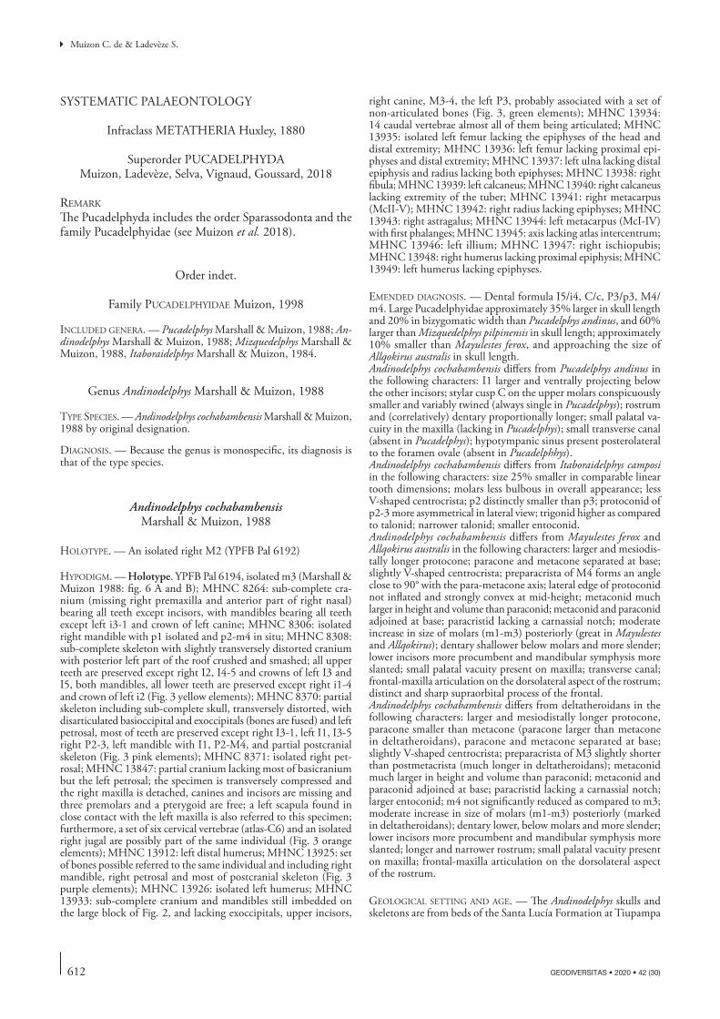
612 GEODIVERSITAS • 2020 • 42 (30)
Muizon C. de & Ladevèze S.
SYSTEMATIC PALAEONTOLOGY
Infraclass METATHERIA Huxley, 1880
Superorder PUCADELPHYDA Muizon, Ladevèze, Selva, Vignaud, Goussard, 2018
remark
The Pucadelphyda includes the order Sparassodonta and the family Pucadelphyidae (see Muizon et al. 2018).
Order indet.
Family pucadelphyidae Muizon, 1998
included genera. — Pucadelphys Marshall & Muizon, 1988; An-dinodelphys Marshall & Muizon, 1988; Mizquedelphys Marshall & Muizon, 1988, Itaboraidelphys Marshall & Muizon, 1984.
Genus Andinodelphys Marshall & Muizon, 1988
type SpecieS. — Andinodelphys cochabambensis Marshall & Muizon, 1988 by original designation.
diagnoSiS. — Because the genus is monospecific, its diagnosis is that of the type species.
Andinodelphys cochabambensis Marshall & Muizon, 1988
holotype. — An isolated right M2 (YPFB Pal 6192)
hypodigm. — Holotype. YPFB Pal 6194, isolated m3 (Marshall & Muizon 1988: fig. 6 A and B); MHNC 8264: sub-complete cra-nium (missing right premaxilla and anterior part of right nasal) bearing all teeth except incisors, with mandibles bearing all teeth except left i3-1 and crown of left canine; MHNC 8306: isolated right mandible with p1 isolated and p2-m4 in situ; MHNC 8308: sub-complete skeleton with slightly transversely distorted cranium with posterior left part of the roof crushed and smashed; all upper teeth are preserved except right I2, I4-5 and crowns of left I3 and I5, both mandibles, all lower teeth are preserved except right i1-4 and crown of left i2 (Fig. 3 yellow elements); MHNC 8370: partial skeleton including sub-complete skull, transversely distorted, with disarticulated basioccipital and exoccipitals (bones are fused) and left petrosal, most of teeth are preserved except right I3-1, left I1, I3-5 right P2-3, left mandible with I1, P2-M4, and partial postcranial skeleton (Fig. 3 pink elements); MHNC 8371: isolated right pet-rosal; MHNC 13847: partial cranium lacking most of basicranium but the left petrosal; the specimen is transversely compressed and the right maxilla is detached, canines and incisors are missing and three premolars and a pterygoid are free; a left scapula found in close contact with the left maxilla is also referred to this specimen; furthermore, a set of six cervical vertebrae (atlas-C6) and an isolated right jugal are possibly part of the same individual (Fig. 3 orange elements); MHNC 13912: left distal humerus; MHNC 13925: set of bones possible referred to the same individual and including right mandible, right petrosal and most of postcranial skeleton (Fig. 3 purple elements); MHNC 13926: isolated left humerus; MHNC 13933: sub-complete cranium and mandibles still imbedded on the large block of Fig. 2, and lacking exoccipitals, upper incisors,
right canine, M3-4, the left P3, probably associated with a set of non-articulated bones (Fig. 3, green elements); MHNC 13934: 14 caudal vertebrae almost all of them being articulated; MHNC 13935: isolated left femur lacking the epiphyses of the head and distal extremity; MHNC 13936: left femur lacking proximal epi-physes and distal extremity; MHNC 13937: left ulna lacking distal epiphysis and radius lacking both epiphyses; MHNC 13938: right fibula; MHNC 13939: left calcaneus; MHNC 13940: right calcaneus lacking extremity of the tuber; MHNC 13941: right metacarpus (McII-V); MHNC 13942: right radius lacking epiphyses; MHNC 13943: right astragalus; MHNC 13944: left metacarpus (McI-IV) with first phalanges; MHNC 13945: axis lacking atlas intercentrum; MHNC 13946: left illium; MHNC 13947: right ischiopubis; MHNC 13948: right humerus lacking proximal epiphysis; MHNC 13949: left humerus lacking epiphyses.
emended diagnoSiS. — Dental formula I5/i4, C/c, P3/p3, M4/m4. Large Pucadelphyidae approximately 35% larger in skull length and 20% in bizygomatic width than Pucadelphys andinus, and 60% larger than Mizquedelphys pilpinensis in skull length; approximately 10% smaller than Mayulestes ferox, and approaching the size of Allqokirus australis in skull length.Andinodelphys cochabambensis differs from Pucadelphys andinus in the following characters: I1 larger and ventrally projecting below the other incisors; stylar cusp C on the upper molars conspicuously smaller and variably twined (always single in Pucadelphys); rostrum and (correlatively) dentary proportionally longer; small palatal va-cuity in the maxilla (lacking in Pucadelphys); small transverse canal (absent in Pucadelphys); hypotympanic sinus present posterolateral to the foramen ovale (absent in Pucadelphhys).Andinodelphys cochabambensis differs from Itaboraidelphys camposi in the following characters: size 25% smaller in comparable linear tooth dimensions; molars less bulbous in overall appearance; less V-shaped centrocrista; p2 distinctly smaller than p3; protoconid of p2-3 more asymmetrical in lateral view; trigonid higher as compared to talonid; narrower talonid; smaller entoconid.Andinodelphys cochabambensis differs from Mayulestes ferox and Allqokirus australis in the following characters: larger and mesiodis-tally longer protocone; paracone and metacone separated at base; slightly V-shaped centrocrista; preparacrista of M4 forms an angle close to 90° with the para-metacone axis; lateral edge of protoconid not inflated and strongly convex at mid-height; metaconid much larger in height and volume than paraconid; metaconid and paraconid adjoined at base; paracristid lacking a carnassial notch; moderate increase in size of molars (m1-m3) posteriorly (great in Mayulestes and Allqokirus); dentary shallower below molars and more slender; lower incisors more procumbent and mandibular symphysis more slanted; small palatal vacuity present on maxilla; transverse canal; frontal-maxilla articulation on the dorsolateral aspect of the rostrum; distinct and sharp supraorbital process of the frontal. Andinodelphys cochabambensis differs from deltatheroidans in the following characters: larger and mesiodistally longer protocone, paracone smaller than metacone (paracone larger than metacone in deltatheroidans), paracone and metacone separated at base; slightly V-shaped centrocrista; preparacrista of M3 slightly shorter than postmetacrista (much longer in deltatheroidans); metaconid much larger in height and volume than paraconid; metaconid and paraconid adjoined at base; paracristid lacking a carnassial notch; larger entoconid; m4 not significantly reduced as compared to m3; moderate increase in size of molars (m1-m3) posteriorly (marked in deltatheroidans); dentary lower, below molars and more slender; lower incisors more procumbent and mandibular symphysis more slanted; longer and narrower rostrum; small palatal vacuity present on maxilla; frontal-maxilla articulation on the dorsolateral aspect of the rostrum.
geological Setting and age. — The Andinodelphys skulls and skeletons are from beds of the Santa Lucía Formation at Tiupampa
613
Cranial anatomy of Andinodelphys cochabambensis, a stem metatherian from the early Palaeocene of Bolivia
GEODIVERSITAS • 2020 • 42 (30)
and have been discovered in 1996 in the locality called “the Quarry” by Gayet et al. (1992) and Marshall & Muizon (1995). As discussed above (p. 600-609) and elsewhere (Gelfo et al. 2009; Muizon et al. 2015, 2018), the age of the Tiupampa beds is regarded as early Danian in age (c. 65 Ma).
COMPARATIVE DESCRIPTION
The description below is based on the skulls of the specimens included in the block of intermingled skeletons illustrated in Figures 2 and 3. All the specimens included in the hypodigm, except the holotype, the referred m3 (YPFB Pal 6194), and MHNC 13912, have been discovered in, or close to, this block. These individuals were probably buried in a single catastrophic event as has also been suggested in the case of the accumulation of nearly complete skulls and skeletons of Pucadelphys andinus from the same locality, which have been regarded as belonging to the same population (Ladevèze et al. 2011). Consequently, because the specimens described here have undergone similar taphonomic conditions, they are also regarded as belonging to the same population of Andinodel-phys cochabambensis. Therefore, the morphological differences observed between the specimens, especially concerning dental morphology, are regarded here as related to individual varia-tion within the species A. cochabambensis. They could also be related to sexual dimorphism, although our sample does not present morphological arguments favouring this hypothesis.
dentition
As mentioned above two almost complete skulls are known (Figs 5, 6), but all the teeth of Andinodelphys are well preserved on the four prepared skulls and the two isolated mandibles, which provides an excellent knowledge of the dental anatomy. The dental formula is the plesiomorphic pattern for metathe-rians: I5/i4, C1/c1, P3/p3, M4/m4 (Kielan-Jaworowska et al. 2004). Upper dentition will be described first, followed by lower dentition.
Upper dentitionUpper Incisors. (Fig. 7) Both I1 are preserved on MHNC 8308. They are relatively slender with a conical crown, which is at least twice as high as wide, although it may be more because the boundary between enamel and dentine is dif-ficult to observe precisely. The crown is elevated relative to the other upper incisors and the part of the root external to the alveolus is approximately 50% to 100% higher than on the other teeth. As a consequence, the I1 projects farther ventrally than the other incisors. The I1s are closely appressed one against each other at their apices and diverge at their base. Because of this layout, they appear slightly oriented medially. The relative proportion and layout of the I1 clearly resemble the condition observed in Recent didelphids but also in Mayulestes as mentioned by Muizon et al. (1997). As observed on MHNC 8308, I2 is much smaller than I1 and approaches the size of I5 (preserved only on the right premax-illa of MHNC 8370). The crown of I2 is much lower than that of I1. It is triangular, approximately as wide as high and
slightly compressed labiolingually. I3 and I4 are morphologi-cally similar to I2 but are larger, I3 being in turn larger than I4. The three teeth have a clearly lower crown than that of I1. I5 is not preserved on MHNC 8308. On MHNC 8370, I5 is a tiny tooth with a peg-like crown that is not distinctly triangular as on I2-4 and not compressed labiolingually. The edges of the alveoli of I1-3 are on the same level, while that of I4 is slightly more dorsal. The alveolus of I5 is much more dorsal, facing distinctly posteroventrally at the anterior edge of the paracanine fossa for the lower canine. A small diastema separates I1 and I2 as is observed in Recent didelphids and a large one separates I5 from the canine.
Upper Canine. (Figs 7-10) The upper canine is a large tooth similar in size (as compared to the skull length) to the con-dition observed in Didelphis and Caluromys. It is slightly larger proportionally than in Philander and Marmosa and much larger than in Thylamys and Metachirus (Table 1). As compared to the other Tiupampa metatherians, the upper canine of Andinodelphys is slightly smaller than that of the only known specimen of Mayulestes but falls within the range of the variation observed in Pucadelphys. In the latter genus, the wide range of variation of the size of the upper canine (Table 1) is related to sexual dimorphism (Ladevèze et al. 2011). In the available sample for Andinodelphys (3 specimens) no such variation is observed since the maximum variation of the ratio canine height/skull length is 0.006 whereas in Pucadelphys it is eight times as much (0.048). Therefore, the size variation of the upper canine, which has been regarded as related to sexual dimorphism in Pucadelphys, may be only related to individual variation in Andinodelphys. However, it is also possible that, because of its small size, our sample of Andinodelphys skulls may not include specimens allowing identification of sexual dimorphism. The upper canine of Andinodelphys is pointed and arcs posteriorly so that the apex of the crown is approximately ventral to the posterior edge of the crown base. The cross section of the canine is oval-shaped without anterior or posterior carina and the edges of the tooth are slightly flattened transversely. At the base of the crown the section of the tooth is more than twice longer than wide (2.93 mm x 1.28 mm on MHNC 8308).
Upper Premolars. (Figs 8-11) The three upper premolars are double-rooted and distinctly increase in size from P1 to P3. The crown of P2 is distinctly higher than that of P1 but is only slightly lower than that of P3. The same observation is made for the mesiodistal length and transverse width as observed on the means calculated on Table 2 for the three measurements. The three premolars are single cusped with anterior and posterior heels. P1 is closely appressed against the canine. On the four skulls, the anterior root is distinctly lateral to the posterolateral edge of the canine. As a conse-quence, the tooth is set obliquely in the tooth row its axis being oriented anterolaterally-posteromedially. The crown of P1 is lower than long and its apex is ventral to the posterior edge of the anterior root. As a consequence, the anterior keel of the main cusp is much shorter than the posterior one. The
614 GEODIVERSITAS • 2020 • 42 (30)
Muizon C. de & Ladevèze S.
anterior base of the crown bears a small heel, which is lower than that observable at the posterior angle of the crown. In occlusal view the crown is ovoid, the lingual edge being more
convex than the labial one. P2 is approximately twice as large as P1 but morphologically similar. Its crown is higher and its apex is less anterior than that of P1. In labial view the crown
A
B
fig. 5. — A, B, Andinodelphys cochabambensis (MHNC 8264) stereophotos of the cranium: A, dorsal view; B, ventral view. Scale bar: 10 mm.

615
Cranial anatomy of Andinodelphys cochabambensis, a stem metatherian from the early Palaeocene of Bolivia
GEODIVERSITAS • 2020 • 42 (30)
appears less asymmetrical than P1 and the anterior edge of the main cusp is only slightly shorter than the posterior one. The crown is roughly triangular and slightly lower than long. Mesially, a small cuspule is present at the base of the crown and distally a significant heel extends the tooth posteriorly. In occlusal view the lingual edge of the crown is inflated above the anterior edge of the posterior root. Consequently, in occlusal view the crown forms a low triangle with the apex in a distolingual position. The P3 is elevated and robust being much larger than P2. It is only slightly longer than P2 but 33% higher (mean in Table 2). It is approximately 50% higher than long. In labial view the tooth is slightly asymmetrical and the apex of the main cusp is shifted posteriorly being ventral to the anterior edge of the posterior root. As a consequence, the mesial edge of the crown is longer than the distal one in contrast with the condition on P1 and P2. At the mesial base of the crown the condition varies. On MHNC 8370 a small
cuspule, larger than on P2, is present. On MHNC 8264, there is no real cuspule as on P2 but a small cingulum surrounds the mesial edge of the crown base and on MHNC 8308 the mesial edge of the crown passes smoothly to the mesial root. Lingually the distolingual inflation is more accentuated than on P2 and is ventral to the whole length of the distal root. At the distal base of the crown a well-cuspated heel is present. Distally this cusp closely contacts the stylar cusp A of M1. The mesial edge of the main cusp is a thick and rounded ridge from base to apex, whereas the distal edge of the main cusp is a relatively sharp crista, straight or slightly concave distally.
Upper Molars. (Figs 8-11) The upper molar anatomy of Andi-nodelphys has been described by Marshall & Muizon (1988) on the basis of the holotype of A. cochabambensis, an M2 or M3 (more likely an M2). The measurements of the holotype (L = 2.9 mm; W = 3.2 mm) better fit the measurements range
fig. 5. — C, D, Andinodelphys cochabambensis (MHNC 8264) stereophotos of the cranium: C, lateral view; D, occipital view. Scale bar: 10 mm.
C
D
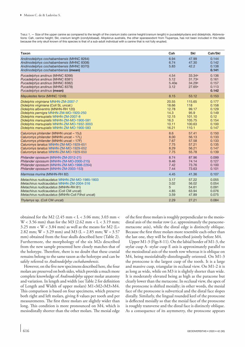
616 GEODIVERSITAS • 2020 • 42 (30)
Muizon C. de & Ladevèze S.
obtained for the M2 (2.45 mm < L < 3.06 mm; 3.03 mm < W < 3.56 mm) than for the M3 (2.62 mm < L < 3.19 mm; 3.25 mm < W < 3.84 mm) as well as the means for M2 (L= 2.82 mm; W = 3.29 mm) and M3 (L = 2.85 mm; W = 3.57 mm) obtained from the four skulls described here (Table 2). Furthermore, the morphology of the six M2s described from the new sample presented here closely matches that of the holotype. Therefore, there is no doubt that these cranial remains belong to the same taxon as the holotype and can be safely referred to Andinodelphys cochabambensis.
However, on the five new specimens described here, the four molars are preserved on both sides, which provide a much more complete knowledge of Andinodelphys upper molar anatomy and variation. In length and width (see Table 2 for definition of Length and Width of upper molars) M1<M2<M3>M4. This comparison is based on four specimens, which preserve both right and left molars, giving 8 values per tooth and per measurements. The first three molars are slightly wider than long. This condition is more pronounced on M4, which is mesiodistally shorter than the other molars. The mesial edge
of the first three molars is roughly perpendicular to the mesio-distal axis of the molar row (i.e. approximately the paracone-metacone axis), while the distal edge is distinctly oblique. Because the first three molars more resemble each other than the last one, they will be first described jointly before M4.
Upper M1-3 (Figs 8-11). On the labial border of M1-3, the stylar cusp A- stylar cusp E axis is approximately parallel to the mesiodistal axis of the tooth row whereas it is oblique on M4, being mesiolabially-distolingually oriented. On M1-3 the protocone is the largest cusp of the tooth. It is a large and massive cusp, triangular in occlusal view. On M1-2 it is as long as wide, while on M3 it is slightly shorter than wide. It is moderately elevated being as high as the paracone but clearly lower than the metacone. In occlusal view, the apex of the protocone is shifted mesially; in other words, the mesial face of the protocone is subvertical and the distal face slopes distally. Similarly, the lingual rounded keel of the protocone is deflected mesially so that the mesial face of the protocone is roughly transverse and the distal face is distinctly oblique. As a consequence of its asymmetry, the protocone appears
table 1. — Size of the upper canine as compared to the length of the cranium (ratio canine height/cranium length) in pucadelphydans and didelphids. Abbrevia-tions: Cah, canine height; Skl, cranium length (condylobasal). Allqokirus australis, the other sparassodont from Tiupampa, has not been included in this table because the only skull known of this species is that of a sub-adult individual with a canine that is not fully erupted.
Taxon Cah Skl Cah/Skl
Andinodelphys cochabambensis (MHNC 8264) 6.94 47.99 0.144Andinodelphys cochabambensis (MHNC 8308) 6.74 47.30 0.142Andinodelphys cochabambensis (MHNC 8370) 5.83 42.2 0.138Andinodelphys cochabambensis (mean) 0.141
Pucadelphys andinus (MHNC 8266) 4.54 33.34e 0.136Pucadelphys andinus (MHNC 8381) 5.12 31.73e 0.161Pucadelphys andinus (MHNC 8382) 5.40e 34.29e 0.157Pucadelphys andinus (MHNC 8378) 3.12 27.65e 0.113Pucadelphys andinus (mean) 0.142
Mayulestes ferox (MHNC 1249) 8.15 53.12 0.153
Didelphis virginiana MNHN-ZM-2007-7 20.55 115.65 0.177Didelphis virginiana (Coll SL uncat.) 18.66 118 0.158Didelphis albiventris (MNHN-RH 120) 12.78 99.17 0.129Didelphis pernigra MNHN-ZM-MO-1920-250 14.3 95.9 0.149Didelphis marsupialis MNHN-ZM-2007-8 12.15 101.10 0.12Didelphis marsupialis MNHN-ZM-MO-1900-581 16.3 105.75 0.154Didelphis marsupialis MNHN-ZM-MO-1932-3003 10.11 100.63 0.100Didelphis marsupialis MNHN-ZM-MO-1900-583 16.21 110.1 0.147
Caluromys philander (MNHN uncat – 15J) 8.6 57.41 0.150Caluromys philander (MNHN uncat – 17L) 8.00 56.13 0.133Caluromys philander (MNHN uncat – 17F) 7.67 57.58 0.133Caluromys lanatus MNHN-ZM-MO-1929-651 7.75 57.21 0.135Caluromys lanatus MNHN-ZM-MO-1929-652 8.29 56.21 0.147Caluromys lanatus MNHN-ZM-MO-1929-650 7.75 55.78 0.139
Philander opossum (MNHN-ZM-2012-21) 8.74 87.96 0.099Philander opossum (MNHN-ZM-MO-2000-215) 9.46 74.14 0.127Philander opossum (MNHN-ZM-MO-1998-2264) 7.42 73.76 0.100Philander opossum (MNHN-ZM-2003-153) 7.44 73.63 0.101
Marmosa murina (MNHN-RH 82) 4.45 41.36 0.107
Metachirus nudicaudatus MNHN-ZM-MO-1985-1803 3.17 57.22 0.055Metachirus nudicaudatus MNHN-ZM-2004-316 3.02 56.02 0.054Metachirus nudicaudatus (MNHN-RH 81) 5 54.61 0.091Metachirus nudicaudatus (Coll CM uncat) 4.85 63.94 0.076Metachirus nudicaudatus (MNHN-Coll Filhol uncat) 3.59 47.89 0.075
Thylamys sp. (Coll CM uncat) 2.29 27.21 0.084
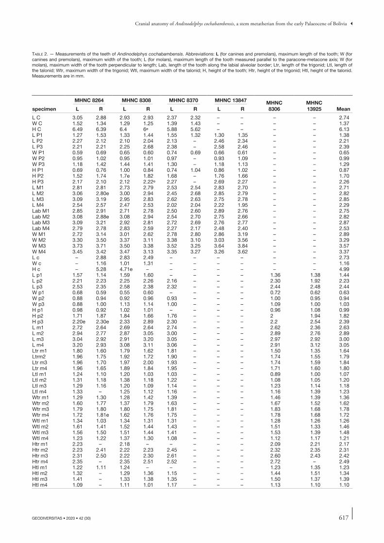
617
Cranial anatomy of Andinodelphys cochabambensis, a stem metatherian from the early Palaeocene of Bolivia
GEODIVERSITAS • 2020 • 42 (30)
table 2. — Measurements of the teeth of Andinodelphys cochabambensis. Abbreviations: L (for canines and premolars), maximum length of the tooth; W (for canines and premolars), maximum width of the tooth; L (for molars), maximum length of the tooth measured parallel to the paracone-metacone axis; W (for molars), maximum width of the tooth perpendicular to length; Lab, length of the tooth along the labial alveolar border; Ltr, length of the trigonid; Ltl, length of the talonid; Wtr, maximum width of the trigonid; Wtl, maximum width of the talonid; H, height of the tooth; Htr, height of the trigonid; Htl, height of the talonid. Measurements are in mm.
specimen
MHNC 8264 MHNC 8308 MHNC 8370 MHNC 13847 MHNC8306
MHNC 13925 MeanL R L R L R L R
L C 3.05 2.88 2.93 2.93 2.37 2.32 – – – – 2.74W C 1.52 1.34 1.29 1.25 1.39 1.43 – – – – 1.37H C 6.49 6.39 6.4 6e 5.88 5.62 – – – – 6.13L P1 1.27 1.53 1.33 1.44 1.55 1.32 1.30 1.35 – – 1.38L P2 2.27 2.12 2.10 2.04 2.13 – 2.46 2.34 – – 2.21L P3 2.21 2.21 2.25 2.68 2.38 – 2.58 2.46 – – 2.39W P1 0.59 0.69 0.65 0.60 0.74 0.69 0.66 0.61 – – 0.65W P2 0.95 1.02 0.95 1.01 0.97 – 0.93 1.09 – – 0.99W P3 1.18 1.42 1.44 1.41 1.30 – 1.18 1.13 – – 1.29H P1 0.69 0.76 1.00 0.84 0.74 1.04 0.86 1.02 – – 0.87H P2 1.52 1.74 1.7e 1.82 1.68 – 1.76 1.66 – – 1.70H P3 2.17 2.10 2.12 2.22e 2.27 – 2.69 2.27 – – 2.26L M1 2.81 2.81 2.73 2.79 2.53 2.54 2.83 2.70 – – 2.71L M2 3.06 2.80e 3.00 2.94 2.45 2.68 2.85 2.79 – – 2.82L M3 3.09 3.19 2.95 2.83 2.62 2.63 2.75 2.78 – – 2.85L M4 2.54 2.57 2.47 2.53 2.02 2.04 2.22 1.95 – – 2.29Lab M1 2.85 2.91 2.71 2.78 2.50 2.60 2.89 2.76 – – 2.75Lab M2 3.08 2.88e 3.08 2.94 2.54 2.70 2.75 2.66 – – 2.82Lab M3 3.09 3.21 2.92 2.81 2.72 2.69 2.76 2.77 – – 2.87Lab M4 2.79 2.78 2.83 2.59 2.27 2.17 2.48 2.40 – – 2.53W M1 2.72 3.14 3.01 2.62 2.78 2.80 2.86 3.19 – – 2.89W M2 3.30 3.50 3.37 3.11 3.38 3.10 3.03 3.56 – – 3.29W M3 3.73 3.71 3.50 3.38 3.52 3.25 3.64 3.84 – – 3.57W M4 3.45 3.42 3.47 3.13 3.35 3.27 3.26 3.62 – – 3.37L c – 2.88 2.83 2.49 – – – – – – 2.73W c – 1.16 1.01 1.31 – – – – – – 1.16H c – 5.28 4.71e – – – – – – – 4.99L p1 1.57 1.14 1.59 1.60 – – – – 1.36 1.38 1.44L p2 2.21 2.23 2.25 2.26 2.16 – – – 2.30 1.92 2.23L p3 2.53 2.35 2.58 2.38 2.32 – – – 2.44 2.48 2.44W p1 0.68 0.59 0.55 0.60 – – – – 0.72 0.62 0.63W p2 0.88 0.94 0.92 0.96 0.93 – – – 1.00 0.95 0.94W p3 0.88 1.00 1.13 1.14 1.00 – – – 1.09 1.00 1.03H p1 0.98 0.92 1.02 1.01 – – – – 0.96 1.08 0.99H p2 1.71 1.87 1.84 1.66 1.76 – – – 2 1.94 1.82H p3 2.20e 2.30e 2.33 2.89 2.30 – – – 2.2 2.54 2.39L m1 2.72 2.64 2.69 2.64 2.74 – – – 2.62 2.36 2.63L m2 2.94 2.77 2.87 3.05 3.00 – – – 2.89 2.76 2.89L m3 3.04 2.92 2.91 3.20 3.05 – – – 2.97 2.92 3.00L m4 3.20 2.93 3.08 3.11 3.06 – – – 2.91 3.12 3.05Ltr m1 1.80 1.60 1.79 1.62 1.81 – – – 1.50 1.35 1.64Ltrm2 1.96 1.75 1.92 1.72 1.90 – – – 1.74 1.55 1.79Ltr m3 1.96 1.70 1.97 2.00 1.93 – – – 1.74 1.59 1.84Ltr m4 1.96 1.65 1.89 1.84 1.95 – – – 1.71 1.60 1.80Ltl m1 1.24 1.10 1.20 1.03 1.03 – – – 0.89 1.00 1.07Ltl m2 1.31 1.18 1.38 1.18 1.22 – – – 1.08 1.05 1.20Ltl m3 1.29 1.16 1.20 1.09 1.14 – – – 1.23 1.14 1.18Ltl m4 1.33 – 1.25 1.12 1.16 – – – 1.16 1.39 1.23Wtr m1 1.29 1.30 1.28 1.42 1.39 – – – 1.46 1.39 1.36Wtr m2 1.60 1.77 1.37 1.79 1.63 – – – 1.67 1.52 1.62Wtr m3 1.79 1.80 1.80 1.75 1.81 – – – 1.83 1.68 1.78Wtr m4 1.72 1.81e 1.62 1.76 1.75 – – – 1.78 1.68 1.72Wtl m1 1.34 1.03 1.34 1.31 1.31 – – – 1.28 1.26 1.26Wtl m2 1.61 1.41 1.52 1.44 1.43 – – – 1.51 1.33 1.46Wtl m3 1.56 1.50 1.51 1.44 1.41 – – – 1.53 1.39 1.48Wtl m4 1.23 1.22 1.37 1.30 1.08 – – – 1.12 1.17 1.21Htr m1 2.23 – 2.18 – – – – – 2.09 2.21 2.17Htr m2 2.23 2.41 2.22 2.23 2.45 – – – 2.32 2.35 2.31Htr m3 2.31 2.50 2.22 2.30 2.61 – – – 2.60 2.43 2.42Htr m4 2.35 – 2.35 2.51 2.52 – – – 2.72 – 2.49Htl m1 1.22 1.11 1.24 – – – – – 1.23 1.35 1.23Htl m2 1.32 – 1.29 1.36 1.15 – – – 1.44 1.51 1.34Htl m3 1.41 – 1.33 1.38 1.35 – – – 1.50 1.37 1.39Htl m4 1.09 – 1.11 1.01 1.17 – – – 1.13 1.10 1.10
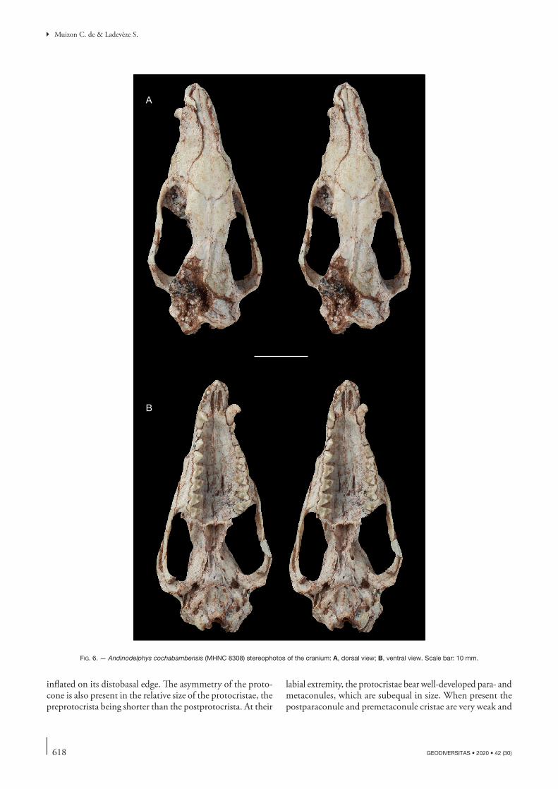
618 GEODIVERSITAS • 2020 • 42 (30)
Muizon C. de & Ladevèze S.
inflated on its distobasal edge. The asymmetry of the proto-cone is also present in the relative size of the protocristae, the preprotocrista being shorter than the postprotocrista. At their
labial extremity, the protocristae bear well-developed para- and metaconules, which are subequal in size. When present the postparaconule and premetaconule cristae are very weak and
A
B
fig. 6. — Andinodelphys cochabambensis (MHNC 8308) stereophotos of the cranium: A, dorsal view; B, ventral view. Scale bar: 10 mm.
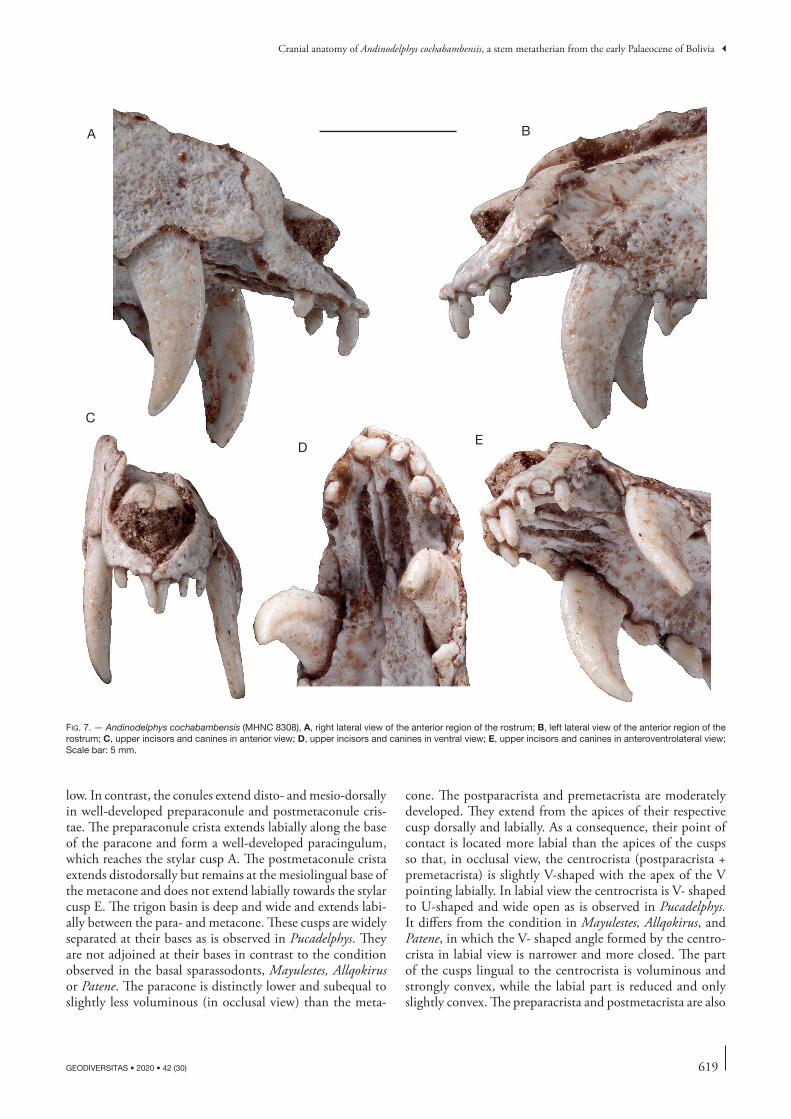
619
Cranial anatomy of Andinodelphys cochabambensis, a stem metatherian from the early Palaeocene of Bolivia
GEODIVERSITAS • 2020 • 42 (30)
low. In contrast, the conules extend disto- and mesio-dorsally in well-developed preparaconule and postmetaconule cris-tae. The preparaconule crista extends labially along the base of the paracone and form a well-developed paracingulum, which reaches the stylar cusp A. The postmetaconule crista extends distodorsally but remains at the mesiolingual base of the metacone and does not extend labially towards the stylar cusp E. The trigon basin is deep and wide and extends labi-ally between the para- and metacone. These cusps are widely separated at their bases as is observed in Pucadelphys. They are not adjoined at their bases in contrast to the condition observed in the basal sparassodonts, Mayulestes, Allqokirus or Patene. The paracone is distinctly lower and subequal to slightly less voluminous (in occlusal view) than the meta-
cone. The postparacrista and premetacrista are moderately developed. They extend from the apices of their respective cusp dorsally and labially. As a consequence, their point of contact is located more labial than the apices of the cusps so that, in occlusal view, the centrocrista (postparacrista + premetacrista) is slightly V-shaped with the apex of the V pointing labially. In labial view the centrocrista is V- shaped to U-shaped and wide open as is observed in Pucadelphys. It differs from the condition in Mayulestes, Allqokirus, and Patene, in which the V- shaped angle formed by the centro-crista in labial view is narrower and more closed. The part of the cusps lingual to the centrocrista is voluminous and strongly convex, while the labial part is reduced and only slightly convex. The preparacrista and postmetacrista are also
A B
C
D E
fig. 7. — Andinodelphys cochabambensis (MHNC 8308), A, right lateral view of the anterior region of the rostrum; B, left lateral view of the anterior region of the rostrum; C, upper incisors and canines in anterior view; D, upper incisors and canines in ventral view; E, upper incisors and canines in anteroventrolateral view; Scale bar: 5 mm.
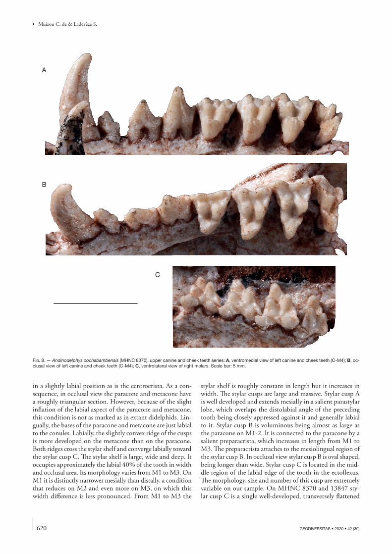
620 GEODIVERSITAS • 2020 • 42 (30)
Muizon C. de & Ladevèze S.
in a slightly labial position as is the centrocrista. As a con-sequence, in occlusal view the paracone and metacone have a roughly triangular section. However, because of the slight inflation of the labial aspect of the paracone and metacone, this condition is not as marked as in extant didelphids. Lin-gually, the bases of the paracone and metacone are just labial to the conules. Labially, the slightly convex ridge of the cusps is more developed on the metacone than on the paracone. Both ridges cross the stylar shelf and converge labially toward the stylar cusp C. The stylar shelf is large, wide and deep. It occupies approximately the labial 40% of the tooth in width and occlusal area. Its morphology varies from M1 to M3. On M1 it is distinctly narrower mesially than distally, a condition that reduces on M2 and even more on M3, on which this width difference is less pronounced. From M1 to M3 the
stylar shelf is roughly constant in length but it increases in width. The stylar cusps are large and massive. Stylar cusp A is well developed and extends mesially in a salient parastylar lobe, which overlaps the distolabial angle of the preceding tooth being closely appressed against it and generally labial to it. Stylar cusp B is voluminous being almost as large as the paracone on M1-2. It is connected to the paracone by a salient preparacrista, which increases in length from M1 to M3. The preparacrista attaches to the mesiolingual region of the stylar cusp B. In occlusal view stylar cusp B is oval shaped, being longer than wide. Stylar cusp C is located in the mid-dle region of the labial edge of the tooth in the ectoflexus. The morphology, size and number of this cusp are extremely variable on our sample. On MHNC 8370 and 13847 sty-lar cusp C is a single well-developed, transversely flattened
A
B
C
fig. 8. — Andinodelphys cochabambensis (MHNC 8370), upper canine and cheek teeth series: A, ventromedial view of left canine and cheek teeth (C-M4); B, oc-clusal view of left canine and cheek teeth (C-M4); C, ventrolateral view of right molars. Scale bar: 5 mm.
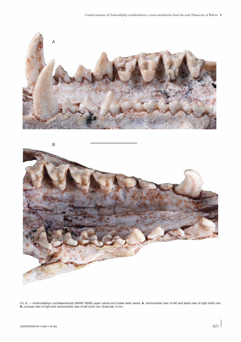
621
Cranial anatomy of Andinodelphys cochabambensis, a stem metatherian from the early Palaeocene of Bolivia
GEODIVERSITAS • 2020 • 42 (30)
A
B
fig. 9. — Andinodelphys cochabambensis (MHNC 8308) upper canine and cheek teeth series: A, ventromedial view of left and labial view of right tooth row; B, occlusal view of right and ventromedial view of left tooth row. Scale bar: 5 mm.
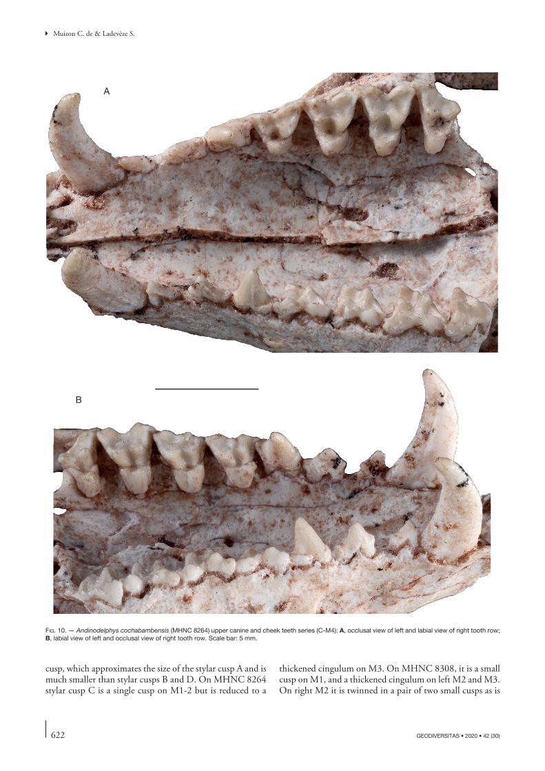
622 GEODIVERSITAS • 2020 • 42 (30)
Muizon C. de & Ladevèze S.
cusp, which approximates the size of the stylar cusp A and is much smaller than stylar cusps B and D. On MHNC 8264 stylar cusp C is a single cusp on M1-2 but is reduced to a
thickened cingulum on M3. On MHNC 8308, it is a small cusp on M1, and a thickened cingulum on left M2 and M3. On right M2 it is twinned in a pair of two small cusps as is
A
B
fig. 10. — Andinodelphys cochabambensis (MHNC 8264) upper canine and cheek teeth series (C-M4): A, occlusal view of left and labial view of right tooth row; B, labial view of left and occlusal view of right tooth row. Scale bar: 5 mm.

623
Cranial anatomy of Andinodelphys cochabambensis, a stem metatherian from the early Palaeocene of Bolivia
GEODIVERSITAS • 2020 • 42 (30)
observed on the holotype (YPFB Pal 6192). Furthermore, on MHNC 8370 stylar cusp C is twinned on the right M2 but is single cusp on the left M2. The same condition is also present on MHNC 13847 but reversed (left stylar cusp C of M2 only is twinned). Therefore, the twinned nature of stylar cusp C observed on the holotype is apparently highly variable within our sample. Stylar cusp D is slightly smaller than stylar cusp B (but sometimes subequal to it) and trans-versely compressed. Stylar cusp D is slightly posterolabial to the metacone. Stylar cusp E is distinctly present but is the smallest cusp of the tooth. It is sometimes connate to stylar cusp D and linked to the metacone by a long mesially concave postmetacrista. The ectoflexus is very shallow to absent on M1 but increase in depth from M1 to M3. The first three upper molars have a similar pattern but differ in their proportions and relative size of some of their elements. From M1 to M3 they increase in overall size and width. The preparacrista increases in length correlatively to the increase in width of the stylar shelf. Stylar cusp A, B, and D slightly increase in size from M1 to M3. The ectoflexus deepens from M1 to M3. No pre- or postcingula are present.
Upper M4 (Figs 8-11). The M4 is significantly different from the other three anterior molars, which justifies a separate description. The major differences of M4 relatively to M1-3 result from the strong reduction of the metacone, which is much lower and less voluminous. The metacone is also clearly smaller in height and volume than the paracone. As a conse-quence, the premetacrista is almost sub-horizontal in some specimens and the postmetacrista is greatly shortened. The paracone is similar in size to that of M3 and the preparacrista is longer. The protocone and conules are slightly smaller than on the preceding molars. The stylar cusp A is slightly larger than on M3. In contrast, stylar cusp B is much smaller to barely present. Stylar cusp C is tiny (sometimes twinned) and stylar cusps D and E are absent. As a consequence, the dis-tolabial angle of the tooth is greatly reduced, the stylar shelf strongly narrows distally and the labial alveolar edge of M4 is strongly oblique being oriented distolingually-mesiolabially. In contrast, the parastylar lobe is greatly developed being slightly larger than on M3. As on the other molars, the preparacrista is markedly transverse and forms an angle close to 90°-100° with the centrocrista (or paracone-metacone axis). This con-
fig. 11. — Andinodelphys cochabambensis (MHNC 13847): stereophotographs of the left upper premolar (P1-P2) and molar (M1-M4) series. Scale bar: 5 mm.
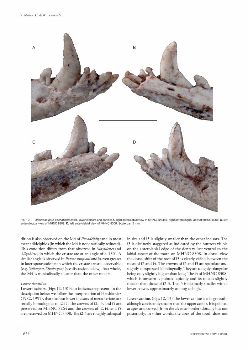
624 GEODIVERSITAS • 2020 • 42 (30)
Muizon C. de & Ladevèze S.
dition is also observed on the M4 of Pucadelphys and in most extant didelphids (in which the M4 is not drastically reduced). This condition differs from that observed in Mayulestes and Allqokirus, in which the cristae are at an angle of c. 130°. A similar angle is observed in Patene simpsoni and is even greater in later sparassodonts in which the cristae are still observable (e.g. Sallacyon, Sipalocyon) (see discussion below). As a whole, the M4 is mesiodistally shorter than the other molars.
Lower dentitionLower incisors. (Figs 12, 13) Four incisors are present. In the description below, we follow the interpretation of Hershkovitz (1982, 1995), that the four lower incisors of metatherians are serially homologous to i2-i5. The crowns of i2, i3, and i5 are preserved on MHNC 8264 and the crowns of i2, i4, and i5 are preserved on MHNC 8308. The i2-4 are roughly subequal
in size and i5 is slightly smaller than the other incisors. The i3 is distinctly staggered as indicated by the buttress visible on the anterolabial edge of the dentary just ventral to the labial aspect of the tooth on MHNC 8308. In dorsal view the dorsal shift of the root of i3 is clearly visible between the roots of i2 and i4. The crowns of i2 and i3 are spatulate and slightly compressed labiolingually. They are roughly triangular being only slightly higher than long. The i4 of MHNC 8308, which is unworn is pointed apically and its root is slightly thicker than those of i2-3. The i5 is distinctly smaller with a lower crown, approximately as long as high.
Lower canine. (Figs 12, 13) The lower canine is a large tooth, although consistently smaller than the upper canine. It is pointed at apex and curved (from the alveolar border) dorsally but not posteriorly. In other words, the apex of the tooth does not
A B
C D
fig. 12. — Andinodelphys cochabambensis, lower incisors and canine: A, right anterolabial view of MHNC 8264; B, right anterolingual view of MHNC 8264; C, left anterolingual view of MHNC 8308; D, left anterolabial view of MHNC 8308. Scale bar: 5 mm.
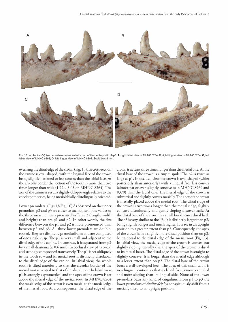
625
Cranial anatomy of Andinodelphys cochabambensis, a stem metatherian from the early Palaeocene of Bolivia
GEODIVERSITAS • 2020 • 42 (30)
overhang the distal edge of the crown (Fig. 13). In cross-section the canine is oval-shaped, with the lingual face of the crown being slightly flattened or less convex than the labial face. At the alveolar border the section of the tooth is more than two times longer than wide (1.22 × 3.03 on MHNC 8264). The axis of the canine is set at a slightly oblique angle relative to the cheek tooth series, being mesiolabially-distolingually oriented.
Lower premolars. (Figs 13-Fig. 16) As observed on the upper premolars, p2 and p3 are closer to each other in the values of the three measurements presented in Table 2 (length, width and height) than are p1 and p2. In other words, the size difference between the p1 and p2 is more pronounced than between p2 and p3. All three lower premolars are double-rooted. They are distinctly premolariform and are composed of one single cusp. The p1 is very small and adjacent to the distal edge of the canine. In contrast, it is separated from p2 by a small diastema (c. 0.6 mm). In occlusal view p1 is ovoid and strongly compressed transversely. The p1 is set obliquely in the tooth row and its mesial root is distinctly distolabial to the distal edge of the canine. In labial view, the whole tooth is tilted anteriorly so that the alveolar border of the mesial root is ventral to that of the distal root. In labial view p1 is strongly asymmetrical and the apex of the crown is set above the mesial edge of the mesial root. In MHNC 8264 the mesial edge of the crown is even mesial to the mesial edge of the mesial root. As a consequence, the distal edge of the
crown is at least three times longer than the mesial one. At the distal base of the crown is a tiny cuspule. The p2 is twice as large as p1. In occlusal view the crown is oval-shaped (wider posteriorly than anteriorly) with a lingual face less convex (almost flat or even slightly concave as in MHNC 8264 and 8370) than the labial one. The mesial edge of the crown is subvertical and slightly convex mesially. The apex of the crown is mesially placed above the mesial root. The distal ridge of the crown is two times longer than the mesial ridge, slightly concave distodorsally and gently sloping distoventrally. At the distal base of the crown is a small but distinct distal heel. The p3 is very similar to the P3. It is distinctly larger than p2, being slightly longer and much higher. It is set in an upright position to a greater extent than p2. Consequently, the apex of the crown is in a slightly more distal position than on p2, being dorsal to the distal edge of the mesial root (Fig. 13). In labial view, the mesial edge of the crown is convex but slightly sloping mesially (i.e. the apex of the crown is distal to its mesial base). The distal edge of the crown is straight to slightly concave. It is longer than the mesial edge although to a lesser extent than on p2. The distal base of the crown bears a well-developed heel. The apex of this small talon is in a lingual position so that its labial face is more extended and more sloping than its lingual side. None of the lower premolars bears any kind of cingulum. From p1 to p3 the lower premolars of Andinodelphys conspicuously shift from a mesially tilted to an upright position.
A B
CD
fig. 13. — Andinodelphys cochabambensis anterior part of the dentary with i1-p3: A, right labial view of MHNC 8264; B, right lingual view of MHNC 8264; C, left labial view of MHNC 8308; D, left lingual view of MHNC 8308. Scale bar: 5 mm.

626 GEODIVERSITAS • 2020 • 42 (30)
Muizon C. de & Ladevèze S.
A CB
fig. 14. — Andinodelphys cochabambensis (MHNC 8264) left lower premolars (p2-p3) and molars: A, labial view; B, occlusal view (stereophotos); C, lingual view. Scale bar: 5 mm.
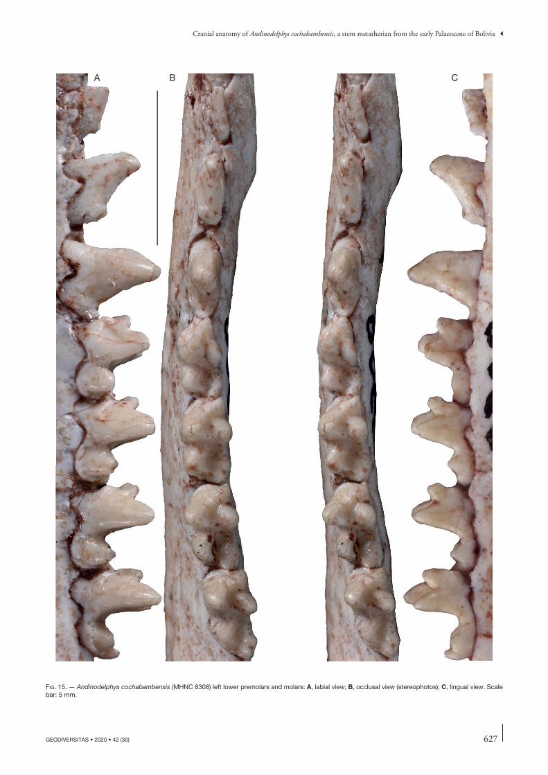
627
Cranial anatomy of Andinodelphys cochabambensis, a stem metatherian from the early Palaeocene of Bolivia
GEODIVERSITAS • 2020 • 42 (30)
B CA
fig. 15. — Andinodelphys cochabambensis (MHNC 8308) left lower premolars and molars: A, labial view; B, occlusal view (stereophotos); C, lingual view. Scale bar: 5 mm.
628 GEODIVERSITAS • 2020 • 42 (30)
Muizon C. de & Ladevèze S.
Lower molars. (Figs 14-16) The four lower molars slightly increase in length distally (m1<m2<m3<m4). This comparison is based on the mean of five specimens, two of them includ-ing right and left molars (MHNC 8264, 8308), and therefore seven measurements. The trigonid of m1 is slightly lower than on the other molars but m2-m4 are subequal in trigo-nid height. The trigonid is slightly narrower than the talonid on m1, subequal on m2-3, and the trigonid is consistently wider than the talonid on m4. The trigonid of m1 is relatively long with a paraconid set mesiolabially (as compared to the other molars) so that the angle between the paracristid and protocristid is more open lingually. As a consequence, if the length of the trigonid is measured at the apices of paraconid and metaconid, the trigonid of m1 is distinctly longer than wide, while it is wider than long on m2-4. In contrast, if the length of the trigonid is measured from mesialmost point of the molar to distobasalmost point of the metaconid, the trigo-nid is longer than wide on the four molars. The protoconid is the highest of the trigonid cusps, but it is only slightly higher than the metaconid. In width, it occupies approximately half of the talonid width. In occlusal view, the protoconid is tri-angular in section, while the metaconid is roughly ovoid and less voluminous. In lingual view, the metaconid is subvertical and slightly increases in height distally. The paraconid is the smallest cusp of the trigonid. In lingual view, it is consistently tilted mesially and also increases in height distally. Paraconid and metaconid are connate at base and the vallid between them is higher than half of the metaconid height (measured from the lingual alveolar border). Because of this condition, the trigo-nid basin is elevated, well-excavated, and well-circumscribed lingually at least on m2-4. On these molars, the deepest point of the basin is at the level of, or lower than the uppermost point of the paraconid-metaconid vallid. This condition is similar to that observed in Pucadelphys but differs distinctly from that observed in the Tiupampa sparassodonts, Mayulestes and Allqokirus. In these taxa, the paraconid and metaconid are broadly separated and the vallid between them is wider and extends almost as far as the base of the crown lingually. As a consequence, the trigonid basin is broadly open lingually and is more a slope on the lingual side of the protoconid than a true depression. The paraconid of Andinodelphys is roughly triangular in occlusal section and its mesiolingual angle bears a salient paraconid ridge, which projects mesially. Labial to this ridge the mesial face of the paraconid is excavated by a shallow hypoconulid notch. The ridge and notch interlock with the hypoconulid of the preceding molar. When unworn, paracristid and protocristid are sharp but no carnassial notch is present on the paracristid, whereas a weak notch is present on the protocristid. The median point of the cristids (point of contact of the cusps) is slightly lower on the paracristid than on the protocristid, this condition being more marked on the anterior molars. The paracristid is strongly oblique relative to the axis of the tooth row, whereas the protocristid is distinctly transverse. The angle of the paracristid with the axis of the tooth row increases from m1 to m4 (Figs 14B-16B).
The talonid is distinctly basined. On m1-3, the hypoconid is the largest cusp in height and volume; the entoconid and the
hypoconulid are subequal in height and volume. On m4 the hypoconulid is the highest cusp of the talonid. The hypoconid is large and occupies approximately half of the talonid volume on m1-2 and slightly less on m3-4. It is conspicuously larger than the other cusps of the talonid. In occusal view, it is tri-angular to V-shaped in appearance. At the mesial edge of the hypoconid, the cristid obliqua is well-developed and sharp. It extends mesiolingually and connects to the trigonid at the distolingual edge of the protoconid (i.e. slightly labial to the protocristid notch). On the distolingual edge of the hypoco-nid a strong posthypocristid connects to the hypoconulid. It is distolabially concave and deeply notched between the two cusps. The posthypocristid notch is located approximately at midline of the talonid. At the distolingual angle of the talonid the hypoconulid and entoconid are clearly approximated and twinned (i.e. set closer to each other than to the hypoconid). They are not distinctly connate. The hypoconulid is lingual to the midline of the talonid. The entoconid is a small but well-individualized cusp. It is somehow compressed trans-versely and bears, on its mesial edge, a marked entocristid, which connects the distal base of the metaconid. Therefore, the talonid is relatively well enclosed lingually. On m4 the hypoconulid is enlarged and elevated above the other talonid cusps. The entoconid is reduced and partially fused at base of the hypoconulid. A well-developed precingulid is present at the mesial base of the protoconid and extends on the mesiolabial base of the paraconid. A thick postcingulid is present on distal edge of m1-3 as a shelf extending ventrolabially from the apex of the hypoconulid to the distal edge of the hypoconid as far as its labial side. A postcingulid is absent on m4.
bony Skull
General features (Figs 6, 17-20) Three complete skulls of Andinodelphys are known. MHNC 8264 is little distorted and measurements of the skull provide an acceptable approximation of its proportions. MHNC 8308 is slightly compressed transversely and the left side of the skull appears to have been more affected by transverse distortion than the right. Therefore, the measurement of the bizygomatic width of the skull can be estimated in multiplying by 2 the measurement taken for the right half of the skull width (see table below). MHNC 8370 is strongly distorted, with the right side more affected by the distortion and the right zygomatic arch completely crushed. The deformation of this specimen is severe and prevents to estimate rigorously its dimensions.
In general size and when compared to the length of the five Pucadelphys skulls measured in Table 3, the skull length of Andinodelphys is 28% (minimum length of Andinodelphys vs. maximum length of Pucadelphys) to 42% (maximum length of Andinodelphys vs. minimum length of Pucadelphys) larger (in absolute value) than that of Pucadelphys. Therefore, the skull of Andinodelphys is approximately one third longer than that of Pucadelphys (28 + 42/2 = 35). When comparing the means of the bizygomatic width of the two genera, the skull of Andino-delphys is 19.6% wider than that of Pucadelphys. Therefore, the significant difference observed in the two measurements shows that the skull of Andinodelphys is proportionally longer than

629
Cranial anatomy of Andinodelphys cochabambensis, a stem metatherian from the early Palaeocene of Bolivia
GEODIVERSITAS • 2020 • 42 (30)
that of Pucadelphys. Comparison of the mean of the width/length ratio obtained from the two complete skulls of Andi-nodelphys (0.520 + 0.497/2 = 0.508) with the mean obtained with 5 skulls of Pucadelphys (X̅ = 0.632) clearly indicates that the skull of the former is proportionally c. 24% longer than
that of the latter. Comparison of the relative length of the palate and rostrum indicates that it is proportionally slightly longer in Andinodelphys than in Pucadelphys. In contrast, the relative length of the orbitotemporal fossa of Andinodelphys is not significantly larger (proportionally) than in Pucadelphys.
A B C
fig. 16. — Andinodelphys cochabambensis (MHNC 8370) left lower premolars (p2-p3) and molars: A, labial view; B, occlusal view; C, lingual view. Scale bar: 5 mm.
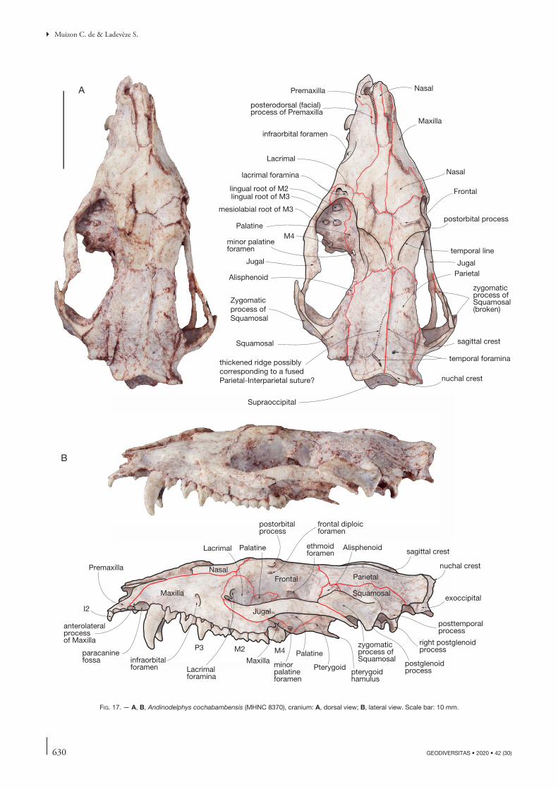
630 GEODIVERSITAS • 2020 • 42 (30)
Muizon C. de & Ladevèze S.
Maxilla
Premaxilla Nasal
infraorbital foramen
postorbital process
M4
lacrimal foramina
Lacrimal
Jugal
Nasal
Zygomaticprocess of Squamosal
Squamosal
Parietal
temporal lineminor palatineforamen
Palatine
lingual root of M2lingual root of M3
mesiolabial root of M3
temporal foramina
sagittal crest
nuchal crest
Supraoccipital
Frontal
sagittal crest
nuchal crest
ParietalFrontal
Maxilla
Nasal
Jugal
Squamosal
P3 M2 M4
Lacrimal Palatine ethmoidforamen
Alisphenoid
postorbitalprocess
frontal diploicforamen
infraorbitalforamen Lacrimal
foraminapterygoidhamulus
Pterygoidminorpalatineforamen
posttemporalprocess
exoccipital
right postglenoidprocess
postglenoidprocess
zygomaticprocess ofSquamosal
PalatineMaxilla
paracaninefossa
Premaxilla
anterolateralprocessof Maxilla
B
A
I2
thickened ridge possiblycorresponding to a fused Parietal-Interparietal suture?
Jugal
zygomaticprocess of Squamosal(broken)
Alisphenoid
posterodorsal (facial)process of Premaxilla
fig. 17. — A, B, Andinodelphys cochabambensis (MHNC 8370), cranium: A, dorsal view; B, lateral view. Scale bar: 10 mm.
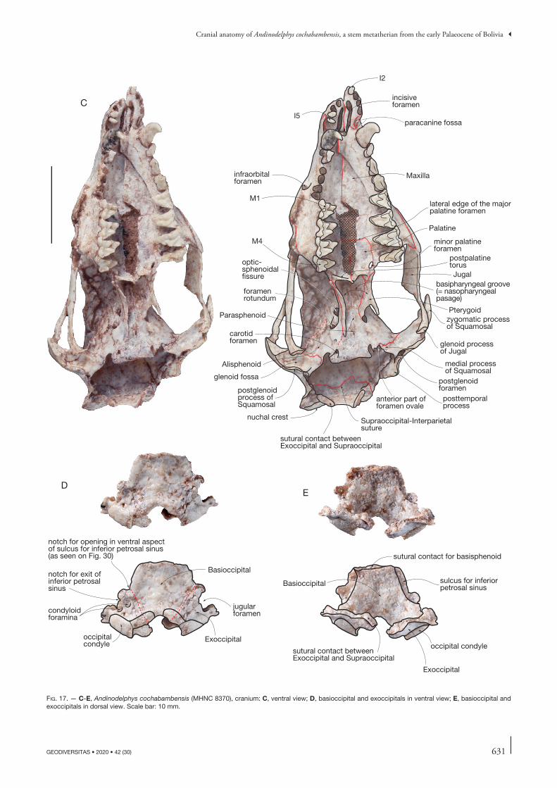
631
Cranial anatomy of Andinodelphys cochabambensis, a stem metatherian from the early Palaeocene of Bolivia
GEODIVERSITAS • 2020 • 42 (30)
fig. 17. — C-E, Andinodelphys cochabambensis (MHNC 8370), cranium: C, ventral view; D, basioccipital and exoccipitals in ventral view; E, basioccipital and exoccipitals in dorsal view. Scale bar: 10 mm.
Palatine
Maxilla
Supraoccipital-Interparietalsuture
Alisphenoid
posttemporalprocess
C
D
sutural contact for basisphenoid
Jugal
Exoccipital
Basioccipital
Pterygoid
postglenoidprocess ofSquamosal
glenoid processof Jugal
zygomatic processof Squamosal
postglenoidforamen
sutural contact betweenExoccipital and Supraoccipital
nuchal crest
anterior part offoramen ovale
carotid foramen
foramenrotundum
optic-sphenoidalfissure
minor palatineforamen
postpalatinetorus
infraorbital foramen
incisiveforamen
I2
I5paracanine fossa
medial processof Squamosal
occipitalcondyle
condyloidforamina
jugularforamen
Basioccipital
Exoccipital
notch for exit ofinferior petrosal sinus
glenoid fossa
M1
M4
sutural contact betweenExoccipital and Supraoccipital
occipital condyle
sulcus for inferior petrosal sinus
notch for opening in ventral aspect of sulcus for inferior petrosal sinus(as seen on Fig. 30)
E
Parasphenoid
lateral edge of the majorpalatine foramen
basipharyngeal groove (= nasopharyngeal pasage)
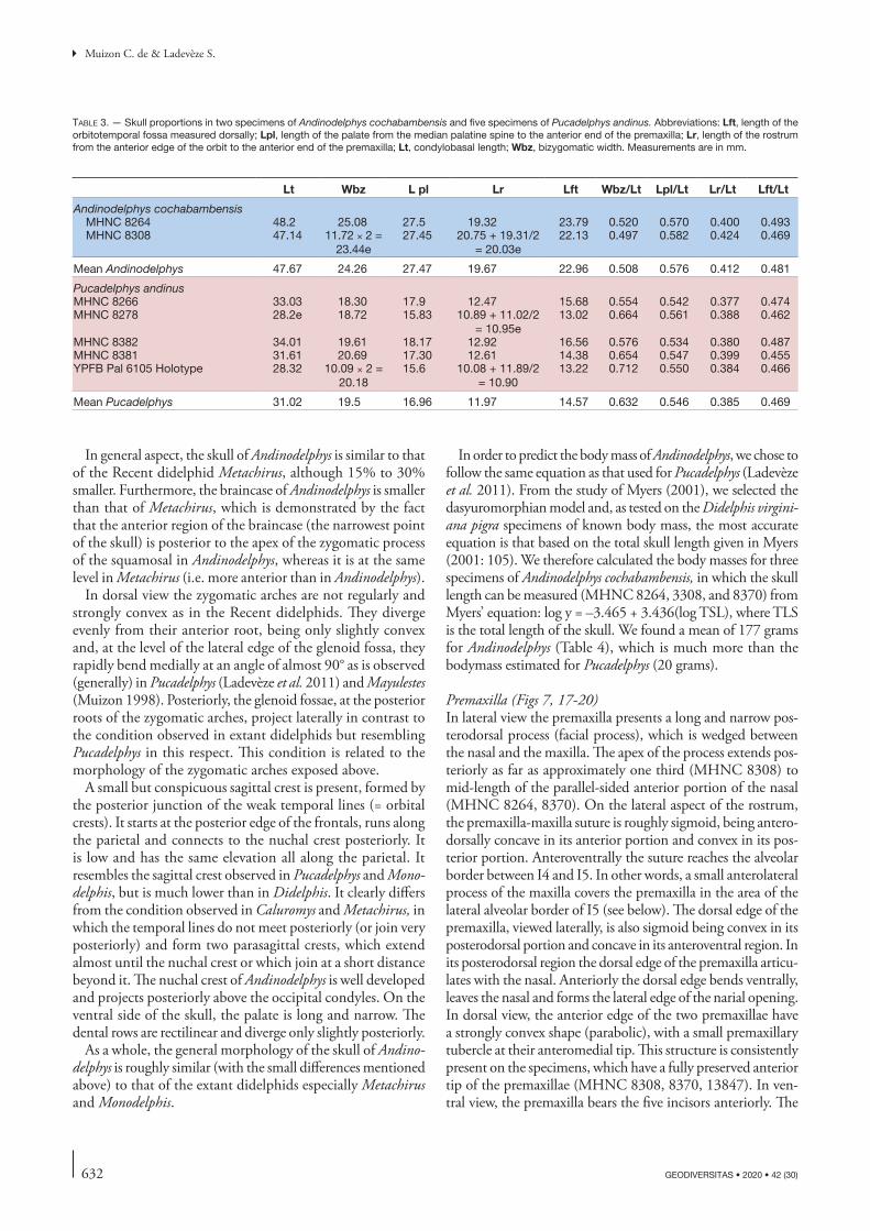
632 GEODIVERSITAS • 2020 • 42 (30)
Muizon C. de & Ladevèze S.
In general aspect, the skull of Andinodelphys is similar to that of the Recent didelphid Metachirus, although 15% to 30% smaller. Furthermore, the braincase of Andinodelphys is smaller than that of Metachirus, which is demonstrated by the fact that the anterior region of the braincase (the narrowest point of the skull) is posterior to the apex of the zygomatic process of the squamosal in Andinodelphys, whereas it is at the same level in Metachirus (i.e. more anterior than in Andinodelphys).
In dorsal view the zygomatic arches are not regularly and strongly convex as in the Recent didelphids. They diverge evenly from their anterior root, being only slightly convex and, at the level of the lateral edge of the glenoid fossa, they rapidly bend medially at an angle of almost 90° as is observed (generally) in Pucadelphys (Ladevèze et al. 2011) and Mayulestes (Muizon 1998). Posteriorly, the glenoid fossae, at the posterior roots of the zygomatic arches, project laterally in contrast to the condition observed in extant didelphids but resembling Pucadelphys in this respect. This condition is related to the morphology of the zygomatic arches exposed above.
A small but conspicuous sagittal crest is present, formed by the posterior junction of the weak temporal lines (= orbital crests). It starts at the posterior edge of the frontals, runs along the parietal and connects to the nuchal crest posteriorly. It is low and has the same elevation all along the parietal. It resembles the sagittal crest observed in Pucadelphys and Mono-delphis, but is much lower than in Didelphis. It clearly differs from the condition observed in Caluromys and Metachirus, in which the temporal lines do not meet posteriorly (or join very posteriorly) and form two parasagittal crests, which extend almost until the nuchal crest or which join at a short distance beyond it. The nuchal crest of Andinodelphys is well developed and projects posteriorly above the occipital condyles. On the ventral side of the skull, the palate is long and narrow. The dental rows are rectilinear and diverge only slightly posteriorly.
As a whole, the general morphology of the skull of Andino-delphys is roughly similar (with the small differences mentioned above) to that of the extant didelphids especially Metachirus and Monodelphis.
In order to predict the body mass of Andinodelphys, we chose to follow the same equation as that used for Pucadelphys (Ladevèze et al. 2011). From the study of Myers (2001), we selected the dasyuromorphian model and, as tested on the Didelphis virgini-ana pigra specimens of known body mass, the most accurate equation is that based on the total skull length given in Myers (2001: 105). We therefore calculated the body masses for three specimens of Andinodelphys cochabambensis, in which the skull length can be measured (MHNC 8264, 3308, and 8370) from Myers’ equation: log y = –3.465 + 3.436(log TSL), where TLS is the total length of the skull. We found a mean of 177 grams for Andinodelphys (Table 4), which is much more than the bodymass estimated for Pucadelphys (20 grams).
Premaxilla (Figs 7, 17-20) In lateral view the premaxilla presents a long and narrow pos-terodorsal process (facial process), which is wedged between the nasal and the maxilla. The apex of the process extends pos-teriorly as far as approximately one third (MHNC 8308) to mid-length of the parallel-sided anterior portion of the nasal (MHNC 8264, 8370). On the lateral aspect of the rostrum, the premaxilla-maxilla suture is roughly sigmoid, being antero-dorsally concave in its anterior portion and convex in its pos-terior portion. Anteroventrally the suture reaches the alveolar border between I4 and I5. In other words, a small anterolateral process of the maxilla covers the premaxilla in the area of the lateral alveolar border of I5 (see below). The dorsal edge of the premaxilla, viewed laterally, is also sigmoid being convex in its posterodorsal portion and concave in its anteroventral region. In its posterodorsal region the dorsal edge of the premaxilla articu-lates with the nasal. Anteriorly the dorsal edge bends ventrally, leaves the nasal and forms the lateral edge of the narial opening. In dorsal view, the anterior edge of the two premaxillae have a strongly convex shape (parabolic), with a small premaxillary tubercle at their anteromedial tip. This structure is consistently present on the specimens, which have a fully preserved anterior tip of the premaxillae (MHNC 8308, 8370, 13847). In ven-tral view, the premaxilla bears the five incisors anteriorly. The
table 3. — Skull proportions in two specimens of Andinodelphys cochabambensis and five specimens of Pucadelphys andinus. Abbreviations: Lft, length of the orbitotemporal fossa measured dorsally; Lpl, length of the palate from the median palatine spine to the anterior end of the premaxilla; Lr, length of the rostrum from the anterior edge of the orbit to the anterior end of the premaxilla; Lt, condylobasal length; Wbz, bizygomatic width. Measurements are in mm.
Lt Wbz L pl Lr Lft Wbz/Lt Lpl/Lt Lr/Lt Lft/Lt
Andinodelphys cochabambensisMHNC 8264 48.2 25.08 27.5 19.32 23.79 0.520 0.570 0.400 0.493MHNC 8308 47.14 11.72 × 2 =
23.44e27.45 20.75 + 19.31/2
= 20.03e22.13 0.497 0.582 0.424 0.469
Mean Andinodelphys 47.67 24.26 27.47 19.67 22.96 0.508 0.576 0.412 0.481
Pucadelphys andinusMHNC 8266 33.03 18.30 17.9 12.47 15.68 0.554 0.542 0.377 0.474MHNC 8278 28.2e 18.72 15.83 10.89 + 11.02/2
= 10.95e 13.02 0.664 0.561 0.388 0.462
MHNC 8382 34.01 19.61 18.17 12.92 16.56 0.576 0.534 0.380 0.487MHNC 8381 31.61 20.69 17.30 12.61 14.38 0.654 0.547 0.399 0.455YPFB Pal 6105 Holotype 28.32 10.09 × 2 =
20.1815.6 10.08 + 11.89/2
= 10.9013.22 0.712 0.550 0.384 0.466
Mean Pucadelphys 31.02 19.5 16.96 11.97 14.57 0.632 0.546 0.385 0.469
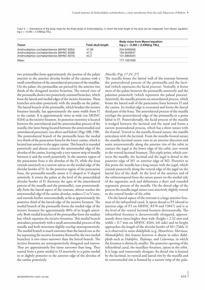
633
Cranial anatomy of Andinodelphys cochabambensis, a stem metatherian from the early Palaeocene of Bolivia
GEODIVERSITAS • 2020 • 42 (30)
two premaxillae form approximately the portion of the palate anterior to the anterior alveolar border of the canines with a small contribution of the anterolateral processes of the maxillae. On the palate, the premaxillae are pierced by the anterior two thirds of the elongated incisive foramina. The ventral view of the premaxilla shows two posteriorly oriented branches, which form the lateral and medial edges of the incisive foramina. These branches articulate posteriorly with the maxilla on the palate. The lateral branch of the premaxilla, which borders the incisive foramen laterally, has approximately the same width from I1 to the canine. It is approximately twice as wide (on MHNC 8264) as the incisive foramen. Its posterior extremity is located between the anterolateral and the anteromedian process of the maxilla (the latter being located between the anteromedial and anterolateral processes); (see above and below) (Figs 18B; 19B). The posterolateral branch of the premaxilla bears the medial two thirds of the paracanine fossa for the lower canine, which is located just anterior to the upper canine. This branch is rounded posteriorly and almost contacts the anteromedial edge of the alveolus of the canine, leaving only a very narrow strip of maxilla between it and the tooth posteriorly. In the anterior region of the paracanine fossa is the alveolus of the I5, while the fossa extends anteriorly as a narrow sulcus on the medial edge of the alveolar border of I5. In the posterior region of the paracanine fossa, the premaxilla-maxilla suture is U-shaped to V-shaped anteriorly. It enters the palate at the level of the posterolabial alveolar border of I5 (between the apex of the anterolateral process of the maxilla and the premaxilla), runs posteromedi-ally from the lateral aspect of the rostrum, almost reaches the anteromedial edge of the canine alveolus, makes a U to V-turn, and extends further anteromedially as far as approximately the posterior third of the lateral edge of the incisive foramen. The medial branch of the premaxilla forms the medial edge of the incisive foramen for approximately 80% of its length anteri-orly. Both medial branches of the premaxillae form the median bar, which separates the incisive foramina. This medial branch articulates posteriorly with a small anteromedial process of the maxilla and both structures slightly overlap anteroposteriorly. The medial branch is much narrower than the lateral one as the bar separating the incisive foramina (formed by the two medial branches) is two times narrower than the lateral branch. The incisive foramina are anteroposteriorly elongated and narrow. They are approximately five times narrower than long. They extend from a point medial to I3 anteriorly to a point medial to or slightly posterior to the anterior edge of the alveolus of the canine posteriorly.
Maxilla (Figs 17-24, 27) The maxilla forms the lateral wall of the rostrum between the posterodorsal process of the premaxilla and the lacri-mal (which represents the facial process). Ventrally, it forms most of the palate between the premaxilla anteriorly and the palatines posteriorly (which represents the palatal process). Anteriorly, the maxilla presents an anterolateral process, which forms the lateral wall of the paracanine fossa between I5 and the canine. Its medial edge is excavated and forms the lateral third part of the fossa. The anterolateral process of the maxilla overlaps the posterolateral edge of the premaxilla to a point labial to I5. Posterodorsally, the facial process of the maxilla is wedged between the lacrimal and the nasal and forms a narrow posterodorsal process, which has a short suture with the frontal. Ventral to the maxilla-frontal suture, the maxilla articulates with the lacrimal. From the maxilla-frontal suture the maxilla-lacrimal suture runs in an anterior direction and turns anteroventrally along the anterior rim of the orbit to contact the jugal at the lower edge of the orbit, just ventral to the ventral lacrimal foramen. This triple point, which con-nects the maxilla, the lacrimal and the jugal is dorsal to the posterior edge of M1 or anterior edge of M2. Posterior to this point the maxilla has a long suture with the jugal, which extends posteriorly along the ventral edge of the jugal on the lateral face of the skull. At the level of the anterior end of the orbitotemporal fossa the suture passes on the medial side of the zygomatic arch and delimitates a short and rounded zygomatic process of the maxilla. On the dorsal edge of the process the maxilla-jugal suture runs anteriorly slightly ventral to the ventral border of the orbit.
On the lateral aspect of the rostrum is a large anterior fora-men of the infraorbital canal. It opens dorsal to P3 (dorsal to anterior edge of P3 on MHNC 8370 and 13847) and is at the level of the ventral lacrimal foramen dorsoventrally. The infraorbital foramen is dorsoventrally elongated, approxi-mately three times higher than wide (height = 2.32 mm and width = 0.7 mm on MHNC 8264, left side) and its height approaches the length of the alveolar border of M1 (Table 2) as is observed in some didelphids (e.g. Metachirus, Marmosa, Monodelphis); this feature however is absent in other didel-phids such as Didelphis, Thylamys, and Caluromys, in which the foramen is distinctly smaller. The posterior opening of the infraorbital canal, the maxillary foramen, opens in the orbit. It is large and transversely elongate. Its dorsal rim is formed by the lacrimal, its ventral and lateral rim by the maxilla and its ventromedial rim is formed by a narrow strip of the pala-
table 4. — Estimations of the body mass for the three skulls of Andinodelphys, in which the total length of the skull can be measured, from Myers’ equation: log y = –3.465 + 3.436(log TSL).
Taxon Total skull lengthBody mass from Myers’equation:log y = –3.465 + 3.436(log TSL)
Andinodelphys cochabambensis (MHNC 8264) 47.99 204.8490606Andinodelphys cochabambensis (MHNC 8308) 47.3 194.9049547Andinodelphys cochabambensis (MHNC 8370) 42.2 131.6963024
Mean 177.1501059
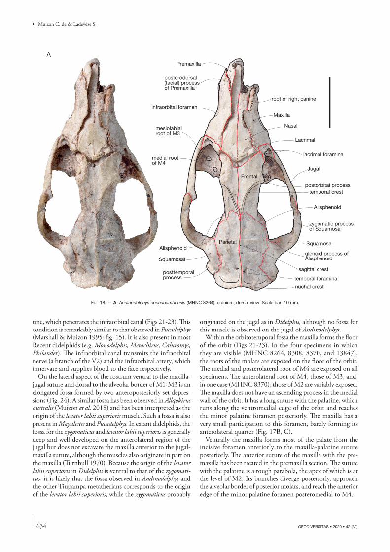
634 GEODIVERSITAS • 2020 • 42 (30)
Muizon C. de & Ladevèze S.
tine, which penetrates the infraorbital canal (Figs 21-23). This condition is remarkably similar to that observed in Pucadelphys (Marshall & Muizon 1995: fig. 15). It is also present in most Recent didelphids (e.g. Monodelphis, Metachirus, Caluromys, Philander). The infraorbital canal transmits the infraorbital nerve (a branch of the V2) and the infraorbital artery, which innervate and supplies blood to the face respectively.
On the lateral aspect of the rostrum ventral to the maxilla-jugal suture and dorsal to the alveolar border of M1-M3 is an elongated fossa formed by two anteroposteriorly set depres-sions (Fig. 24). A similar fossa has been observed in Allqokirus australis (Muizon et al. 2018) and has been interpreted as the origin of the levator labii superioris muscle. Such a fossa is also present in Mayulestes and Pucadelphys. In extant didelphids, the fossa for the zygomaticus and levator labii superioris is generally deep and well developed on the anterolateral region of the jugal but does not excavate the maxilla anterior to the jugal-maxilla suture, although the muscles also originate in part on the maxilla (Turnbull 1970). Because the origin of the levator labii superioris in Didelphis is ventral to that of the zygomati-cus, it is likely that the fossa observed in Andinodelphys and the other Tiupampa metatherians corresponds to the origin of the levator labii superioris, while the zygomaticus probably
originated on the jugal as in Didelphis, although no fossa for this muscle is observed on the jugal of Andinodelphys.
Within the orbitotemporal fossa the maxilla forms the floor of the orbit (Figs 21-23). In the four specimens in which they are visible (MHNC 8264, 8308, 8370, and 13847), the roots of the molars are exposed on the floor of the orbit. The medial and posterolateral root of M4 are exposed on all specimens. The anterolateral root of M4, those of M3, and, in one case (MHNC 8370), those of M2 are variably exposed. The maxilla does not have an ascending process in the medial wall of the orbit. It has a long suture with the palatine, which runs along the ventromedial edge of the orbit and reaches the minor palatine foramen posteriorly. The maxilla has a very small participation to this foramen, barely forming its anterolateral quarter (Fig. 17B, C).
Ventrally the maxilla forms most of the palate from the incisive foramen anteriorly to the maxilla-palatine suture posteriorly. The anterior suture of the maxilla with the pre-maxilla has been treated in the premaxilla section. The suture with the palatine is a rough parabola, the apex of which is at the level of M2. Its branches diverge posteriorly, approach the alveolar border of posterior molars, and reach the anterior edge of the minor palatine foramen posteromedial to M4.
mesiolabial root of M3
Parietal
Frontal
Nasal
Maxilla
Squamosal
Jugal
Lacrimal
lacrimal foramina
temporal crestpostorbital process
infraorbital foramen
Premaxilla
root of right canine
zygomatic processof Squamosal
posttemporal process
medial root of M4
temporal foramina
nuchal crest
sagittal crest
Alisphenoid
posterodorsal(facial) process of Premaxilla
Alisphenoidglenoid process ofAlisphenoid
A
Squamosal
fig. 18. — A, Andinodelphys cochabambensis (MHNC 8264), cranium, dorsal view. Scale bar: 10 mm.
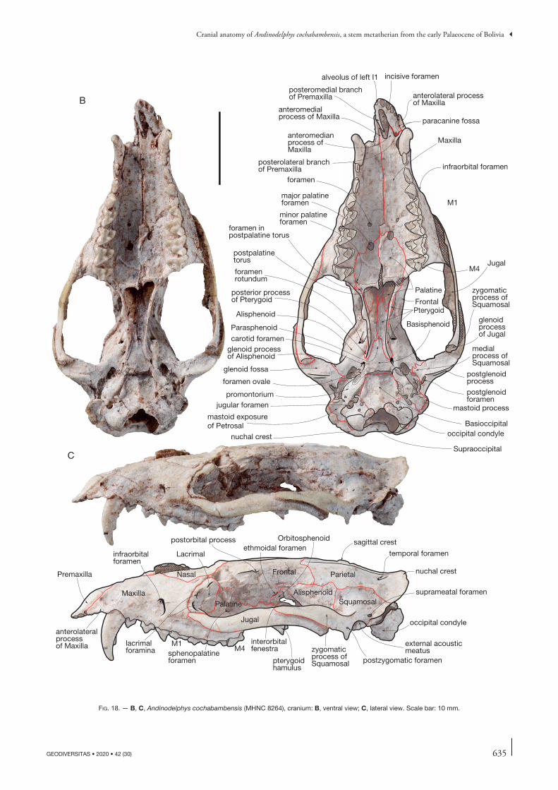
635
Cranial anatomy of Andinodelphys cochabambensis, a stem metatherian from the early Palaeocene of Bolivia
GEODIVERSITAS • 2020 • 42 (30)
fig. 18. — B, C, Andinodelphys cochabambensis (MHNC 8264), cranium: B, ventral view; C, lateral view. Scale bar: 10 mm.
anterolateral processof Maxilla
paracanine fossa
alveolus of left I1 incisive foramen
anteromedial process of Maxilla
anteromedianprocess of Maxilla
posteromedial branchof Premaxilla
posterolateral branchof Premaxilla
major palatineforamen M1
M4
minor palatine foramen
postpalatinetorus
Palatine
Pterygoid
Jugal
glenoidprocessof Jugal
posterior processof Pterygoid
foramenrotundum
glenoid processof Alisphenoid
postglenoidprocess
postglenoidforamen
medial process ofSquamosal
glenoid fossa
foramen ovale
carotid foramen
Basioccipital
mastoid processjugular foramenpromontorium
nuchal crest occipital condyle
Supraoccipital
zygomaticprocess ofSquamosal
infraorbital foramen
foramen
foramen in postpalatine torus
sagittal crest
temporal foramen
nuchal crest
suprameatal foramen
occipital condyle
ParietalFrontal
Squamosal
Jugal
Maxilla
Nasal
postorbital process
infraorbitalforamen
Premaxilla
Lacrimal
sphenopalatineforamen
Palatine
ethmoidal foramenOrbitosphenoid
Alisphenoid
interorbitalfenestra
pterygoidhamulus
zygomatic process of Squamosal
M4M1lacrimal
foramina
anterolateralprocessof Maxilla external acoustic
meatuspostzygomatic foramen
Basisphenoid
mastoid exposureof Petrosal
Alisphenoid
C
B
Frontal
Maxilla
Parasphenoid

636 GEODIVERSITAS • 2020 • 42 (30)
Muizon C. de & Ladevèze S.
Perforation of the palate varies. In MHNC 8264 a large major palatine foramen is present at the maxilla-palatine suture. This foramen is oval-shaped being slightly elongated anteroposteriorly. It is located at the level of the embrasure between M2 and M3 on the medial third of the palate width. On the right palatal process of the maxilla, well anterior to the major palatine foramen (at the level of the posterior edge of M1), is a small circular foramen approximately three times smaller than major palatine foramen. This foramen is present on the left side at the level of the embrasure between M1 and M2, but it is slightly compressed transversely because of post-mortem distortion. The anteroposterior position of this foramen corresponds to that of the anterior end of the maxillopalatal fenestra of the other specimens; it is labelled “foramen” on Fig. 18B. In MHNC 13847, the major pala-tine foramen is an elongated slit approximately four times longer than wide and which extends from the level of the metacone of M2 posteriorly to the level of the protocone of M1 anteriorly (Fig. 11). The foramen notches the palatine posteriorly along less than one fifth of its length, and the maxilla anteriorly on most of its length. A similar condition is observed in MHNC 8308 (Fig. 19B) and on MHNC
13933 (the partially unprepared skull on the block of Fig. 3). In MHNC 8370 although the palate is partly damaged it is possible to observe the lateral edge of an elongated palatine foramen on the left maxilla (Fig. 17C), and it is therefore likely that it was similar to the condition observed in MHNC 13847, and 8308. It is unclear whether or not this elongated major palatine foramen should be regarded as an incipiently developed palatal vacuity. The condition in MHNC 8264, in which the major palatine foramen is not strongly elongated but in which another small foramen is present in a position similar to the anterior end of the elongated palatine foramen in the other two specimens is noteworthy. It could represent a morphological stage leading from a single major palatine foramen to an elongated foramen, which in turn could rep-resent a step toward a larger palatal vacuity as observed in some extant didelphids. In fact, the condition observed in Andinodelphys is reminiscent of the condition observed in some extant didelphids. However, it is noteworthy that the presence and morphology of the palatal vacuities are extremely variable in extant didelphids (Voss & Jansa 2009). They are gener-ally absent in Caluromys and Caluromysiops (but see Voss & Jansa 2009: 94). They form an elongated slit at the maxilla-
Parietal
Frontal
Nasal
Maxilla
Squamosal
Jugal
Lacrimal
lacrimal foramina
temporalcrest
postorbital process
infraorbital foramen
Premaxilla
zygomatic processof Squamosal
posttemporal process
medial root of M4
nuchal crest
sagittal crest
glenoidprocess ofAlisphenoid
posterodorsal(facial) process of Premaxilla
medial rootof M3
A
fig. 19. — A, Andinodelphys cochabambensis (MHNC 8308), cranium, dorsal view. Scale bar: 10 mm.
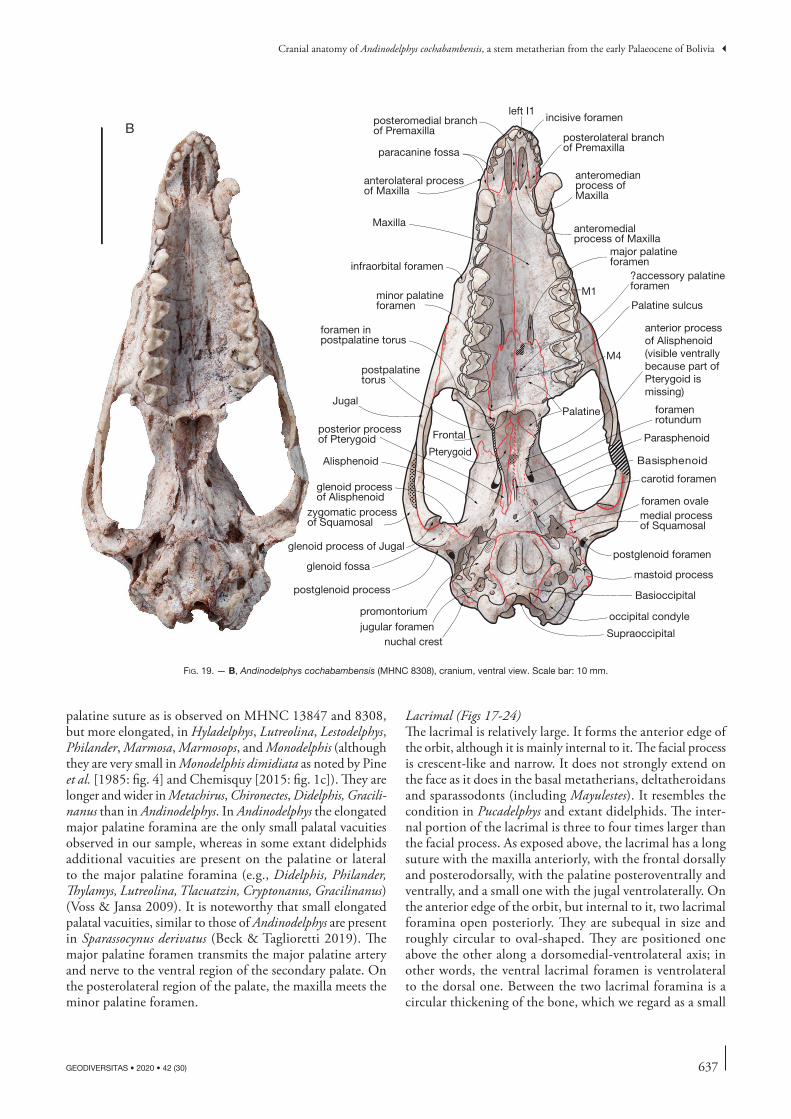
637
Cranial anatomy of Andinodelphys cochabambensis, a stem metatherian from the early Palaeocene of Bolivia
GEODIVERSITAS • 2020 • 42 (30)
palatine suture as is observed on MHNC 13847 and 8308, but more elongated, in Hyladelphys, Lutreolina, Lestodelphys, Philander, Marmosa, Marmosops, and Monodelphis (although they are very small in Monodelphis dimidiata as noted by Pine et al. [1985: fig. 4] and Chemisquy [2015: fig. 1c]). They are longer and wider in Metachirus, Chironectes, Didelphis, Gracili-nanus than in Andinodelphys. In Andinodelphys the elongated major palatine foramina are the only small palatal vacuities observed in our sample, whereas in some extant didelphids additional vacuities are present on the palatine or lateral to the major palatine foramina (e.g., Didelphis, Philander, Thylamys, Lutreolina, Tlacuatzin, Cryptonanus, Gracilinanus) (Voss & Jansa 2009). It is noteworthy that small elongated palatal vacuities, similar to those of Andinodelphys are present in Sparassocynus derivatus (Beck & Taglioretti 2019). The major palatine foramen transmits the major palatine artery and nerve to the ventral region of the secondary palate. On the posterolateral region of the palate, the maxilla meets the minor palatine foramen.
Lacrimal (Figs 17-24)The lacrimal is relatively large. It forms the anterior edge of the orbit, although it is mainly internal to it. The facial process is crescent-like and narrow. It does not strongly extend on the face as it does in the basal metatherians, deltatheroidans and sparassodonts (including Mayulestes). It resembles the condition in Pucadelphys and extant didelphids. The inter-nal portion of the lacrimal is three to four times larger than the facial process. As exposed above, the lacrimal has a long suture with the maxilla anteriorly, with the frontal dorsally and posterodorsally, with the palatine posteroventrally and ventrally, and a small one with the jugal ventrolaterally. On the anterior edge of the orbit, but internal to it, two lacrimal foramina open posteriorly. They are subequal in size and roughly circular to oval-shaped. They are positioned one above the other along a dorsomedial-ventrolateral axis; in other words, the ventral lacrimal foramen is ventrolateral to the dorsal one. Between the two lacrimal foramina is a circular thickening of the bone, which we regard as a small
fig. 19. — B, Andinodelphys cochabambensis (MHNC 8308), cranium, ventral view. Scale bar: 10 mm.
anterolateral processof Maxilla
paracanine fossa
left I1incisive foramen
anteromedial process of Maxilla
anteromedianprocess of Maxilla
posteromedial branchof Premaxilla
posterolateral branchof Premaxilla
major palatineforamen
M1
M4
minor palatine foramen
postpalatinetorus
Palatine
Pterygoid
Jugal
glenoid process of Jugal
posterior processof Pterygoid
foramenrotundum
glenoid processof Alisphenoid
postglenoid process
postglenoid foramen
medial processof Squamosal
glenoid fossa
foramen ovale
carotid foramen
Basioccipital
mastoid process
jugular foramenpromontorium
nuchal crest
occipital condyle
Supraoccipital
zygomatic processof Squamosal
infraorbital foramen
foramen in postpalatine torus
Basisphenoid
Maxilla
Frontal
anterior processof Alisphenoid(visible ventrallybecause part of Pterygoid ismissing)
Alisphenoid
Parasphenoid
B
Palatine sulcus
?accessory palatine foramen
638 GEODIVERSITAS • 2020 • 42 (30)
Muizon C. de & Ladevèze S.
lacrimal tubercle. This tubercle is clearly present on the two skulls of fully adult individuals (MHNC 8264 and 8308), while it is not really developed on the other two skulls of young adults (MHNC 8370 and 13847). A lacrimal tubercle similar to that of Andinodelphys is variably present in some extant didelphids (e.g., Caluromys, Thylamys – personal observations). It is absent in Pucadelphys and Mayulestes. Within the orbit, the lacrimal forms the dorsal edge of the maxillary foramen.
Nasal (Figs 17-Fig. 19)The nasals are elongated bones, which are approximately 45% the condylobasal length. They are narrow with sub-parallel edges in their anterior two thirds and strongly widen in their posterior third. The anterior apices of the nasals are preserved on the four fully prepared skulls (the left only on MHNC 8264). The anterior edge of the two nasals protrudes anteri-orly being distinctly rounded (semicircular to parabolic) and strongly convex anteriorly. Therefore, the medial side of the apex of each nasal is well anterior to its lateral side. Anteriorly, the nasals extend beyond the anteriormost point of the nasal-premaxilla suture and overhang the narial opening on more than half of its length: on MHNC 8308 the anteriormost point of the nasal is at the level of the posterior edge of I3. The nasal-maxilla suture is anteroposteriorly oriented up to the level of the infraorbital foramen. There, it turns laterally at approximately 45° and turns posteriorly just before contact-ing the maxilla-frontal small suture. Posterior to this suture the nasal contacts the frontal and the nasal-frontal suture turns medially at approximately 45° and meets the sagittal plane. The posterior suture of the nasals, with the frontals, is markedly convex posteriorly. It is not distinctly W-shaped as in Pucadelphys and in most didelphids (e.g., Hyladelphys, Marmosa, Monodelphis, Metachirus, Didelphis, Lutreolina, Cryptonanus, Lestodelphys, Thylamys). This posterior flared portion of the nasals is roughly square to diamond-shaped with one of the diagonals oriented anteroposteriorly. It is roughly as long as wide and differs from the condition in extant didelphids, in which this part of the nasals is distinctly longer than wide. Interestingly, this region of the nasals is conspicuously wider than long in Deltatheridium, a late Cre-taceous metatherian from Mongolia (Rougier et al. 1998). In its lateralmost region the nasal is closely approximated to the lacrimal but does not contact it, in contrast to the condition in sparassodonts and deltatheroidans, in which the bones have a distinct suture. Posteriorly, the nasals extend approximately as far as mid-length of the orbit and remain well anterior to the supraorbital processes.
Jugal (Figs 20, 21)Complete jugals are preserved on the four prepared skulls. It is a relatively slender bone as compared to extant didel-phids but similar in this respect to that of Pucadelphys. In lateral view, it is consistently convex laterally and concave medially. It is gently sigmoid being dorsally concave ante-riorly and convex posteriorly. Its anterior dorsally concave part forms the ventral edge of the orbit. In lateral view at
its anterodorsal apex it has a short anterior suture with the lacrimal and a long (c. one third of its total length) and oblique anterior suture with the maxilla along its ventral edge as far as the anterior edge of the orbitotemporal fossa (in ventral view). As mentioned above, it receives the short zygomatic process of the maxilla on the medial aspect of its anterior region. The posterior dorsally convex portion of the jugal has a long dorsal suture with the zygomatic process of the squamosal on approximately 45% of its total length. In lateral view, the free part of the jugal (i.e. with no suture with the maxilla nor with the squamosal) is approximately 25% of the length of the bone. It very slightly increases in height posteriorly, the bone having its greatest depth just anterior to the jugal-squamosal suture. However, in this region of the jugal of Andinodelphys there is no conspicuous frontal process of the jugal as is observed in Recent didelphids and dasyurids, in which it marks the posterior limit of the orbit (Wible 2003; Voss & Jansa 2009). However, a very small elevation of the dorsal edge of the jugal, posterior to the concave anterior part, demarcates the posterior limit of the orbit and could be regarded as an incipient frontal process. The condition of Andinodelphys is apparently intermediate between that of extant didelphids and that of Pucadelphys, Allqokirus and Mayulestes, in which the transition between the ventral edge of the orbit and the posterior part of the jugal is notably smooth. This incipient frontal process of Andinodelphys is slightly posterior to level of the postorbi-tal process and well anterior to the narrowest part of the intertemporal constriction. In extant didelphids the most elevated region of the jugal (the frontal process) is generally at the level of the postorbital process or slightly posterior to it. In some taxa, however, it is more posteriorly located, at the level of the intertemporal constriction (e.g. Lestodel-phys, Thylamys).
However, the condition of Andinodelphys differs strongly from that in extant didelphids and dasyurids, in which the posterodorsal angle of the jugal extends posteriorly above the zygomatic process of the squamosal. As a consequence, the jugal-squamosal suture is bifurcated, a condition which is never observed in Andinodelphys nor in the other Tiupampa pucadelphydans (Pucadelphys and Mizquedelphys). The conditions observed in Figures 21-23 are artefacts due to a slight displacement of the two bones, which has modified the external aspect of the jugal-squamosal suture, a condi-tion that has been enhanced by the oblique nature of the photographs. Figures 18C and 20 provide a better render-ing of the actual jugal-squamosal suture in Andinodelphys.
From the anterior end of the jugal-squamosal suture, the jugal underlaps the zygomatic process of the squamosal, reducing in height posteriorly and thickening again at its posterior extremity. The posterior end of the jugal bears a conspicuous glenoid process, which articulates with the anterolateral edge of the glenoid fossa. This process bears a small ovoid articular facet for the mandibular condyle. It faces posteroventrally and its transverse width is approxi-mately one third to one fourth the total width of the glenoid fossa of the squamosal.
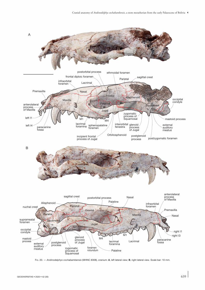
639
Cranial anatomy of Andinodelphys cochabambensis, a stem metatherian from the early Palaeocene of Bolivia
GEODIVERSITAS • 2020 • 42 (30)
sagittal crest
nuchal crest
suprameatalforamen
occipital condyle
Parietal
Frontal
Squamosal
Jugal
Maxilla
Nasal
postorbital process
infraorbitalforamen
Premaxilla
Lacrimal
sphenopalatineforamen
Palatine
ethmoidal foramen
Orbitosphenoid
Alisphenoid
interorbitalfenestra
zygomatic process of Squamosal
M4M1
lacrimalforamina
anterolateralprocessof Maxilla
external auditorymeatus
postzygomatic foramen
mastoid process
postglenoidprocess
glenoid processof Jugal
paracaninefossa
sagittal crest
occipitalcondyle
Parietal
Frontal
SquamosalJugal
Maxilla
Nasalpostorbital process
Premaxilla
Lacrimal
PalatineAlisphenoid
zygomatic process of Squamosal
M4M1
lacrimalforamina
anterolateralprocessof Maxilla
externalauditorymeatus
infraorbitalforamen
glenoid processof Jugal
foramenrotundum
mastoidprocess postglenoid
process
Palatine
Parietal
A
B
paracaninefossa
incipient frontalprocess of Jugal
frontal diploic foramen
left I1
left I4
right I1
right I3
Nasal
fig. 20. — Andinodelphys cochabambensis (MHNC 8308), cranium: A, left lateral view; B, right lateral view. Scale bar: 10 mm.
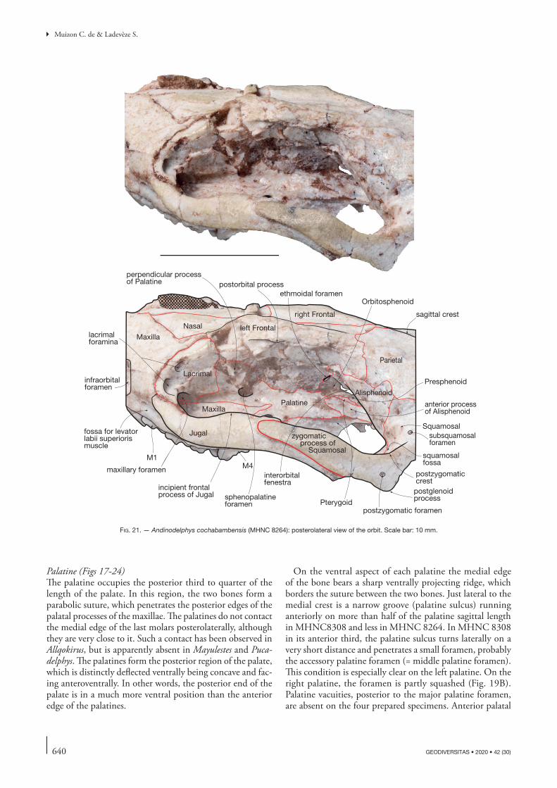
640 GEODIVERSITAS • 2020 • 42 (30)
Muizon C. de & Ladevèze S.
Palatine (Figs 17-24)The palatine occupies the posterior third to quarter of the length of the palate. In this region, the two bones form a parabolic suture, which penetrates the posterior edges of the palatal processes of the maxillae. The palatines do not contact the medial edge of the last molars posterolaterally, although they are very close to it. Such a contact has been observed in Allqokirus, but is apparently absent in Mayulestes and Puca-delphys. The palatines form the posterior region of the palate, which is distinctly deflected ventrally being concave and fac-ing anteroventrally. In other words, the posterior end of the palate is in a much more ventral position than the anterior edge of the palatines.
On the ventral aspect of each palatine the medial edge of the bone bears a sharp ventrally projecting ridge, which borders the suture between the two bones. Just lateral to the medial crest is a narrow groove (palatine sulcus) running anteriorly on more than half of the palatine sagittal length in MHNC8308 and less in MHNC 8264. In MHNC 8308 in its anterior third, the palatine sulcus turns laterally on a very short distance and penetrates a small foramen, probably the accessory palatine foramen (= middle palatine foramen). This condition is especially clear on the left palatine. On the right palatine, the foramen is partly squashed (Fig. 19B). Palatine vacuities, posterior to the major palatine foramen, are absent on the four prepared specimens. Anterior palatal
fig. 21. — Andinodelphys cochabambensis (MHNC 8264): posterolateral view of the orbit. Scale bar: 10 mm.
sagittal crest
Parietal
left Frontal
SquamosalJugal
Maxilla
Nasal
postorbital process
infraorbitalforamen
Lacrimal
sphenopalatineforamen
Palatine
ethmoidal foramenOrbitosphenoid
Alisphenoid
interorbitalfenestra
zygomatic process of Squamosal
M4M1
lacrimalforamina
postzygomatic foramen
postglenoidprocess
right Frontal
Maxillaanterior processof Alisphenoid
subsquamosalforamen
maxillary foramen
fossa for levatorlabii superiorismuscle
perpendicular processof Palatine
incipient frontalprocess of Jugal
Pterygoid
Presphenoid
squamosalfossa
postzygomaticcrest
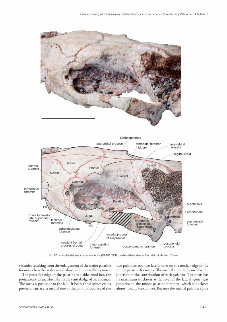
641
Cranial anatomy of Andinodelphys cochabambensis, a stem metatherian from the early Palaeocene of Bolivia
GEODIVERSITAS • 2020 • 42 (30)
vacuities resulting from the enlargement of the major palatine foramina have been discussed above in the maxilla section.
The posterior edge of the palatine is a thickened bar, the postpalatine torus, which forms the ventral edge of the choanae. The torus is posterior to the M4. It bears three spines on its posterior surface, a medial one at the point of contact of the
two palatines and two lateral ones on the medial edge of the minor palatine foramina. The medial spine is formed by the junction of the contribution of each palatine. The torus has its maximum thickness at the level of the lateral spine, just posterior to the minor palatine foramen, which it encloses almost totally (see above). Because the medial palatine spine
ethmoidal foramen(hidden)
sagittal crest
Frontal
Squamosal
Parietal
Jugal
Maxilla
Nasal
postorbital process
infraorbitalforamen
Lacrimal
sphenopalatineforamen
Palatine
Orbitosphenoid
Alisphenoid
interorbitalfenestra
zygomatic process of SquamosalM4
P3
lacrimalforamina
postzygomatic foramenpostglenoidprocess
Frontal
Maxilla
anterior processof Alisphenoid
suprameatalforamen
minor palatineforamen
lacrimaltubercle
fossa for levatorlabii superiorismuscle
incipient frontalprocess of Jugal
Presphenoid
fig. 22. — Andinodelphys cochabambensis (MHNC 8308): posterolateral view of the orbit. Scale bar: 10 mm.
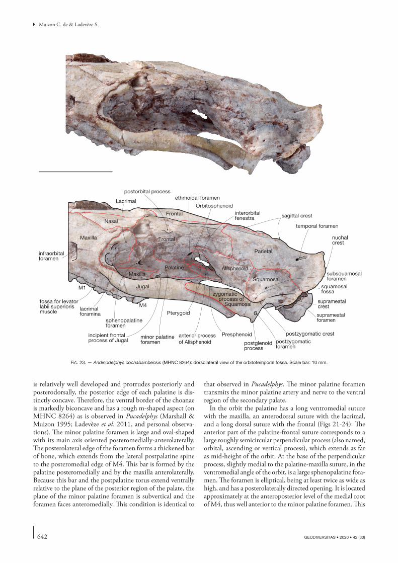
642 GEODIVERSITAS • 2020 • 42 (30)
Muizon C. de & Ladevèze S.
is relatively well developed and protrudes posteriorly and posterodorsally, the posterior edge of each palatine is dis-tinctly concave. Therefore, the ventral border of the choanae is markedly biconcave and has a rough m-shaped aspect (on MHNC 8264) as is observed in Pucadelphys (Marshall & Muizon 1995; Ladevèze et al. 2011, and personal observa-tions). The minor palatine foramen is large and oval-shaped with its main axis oriented posteromedially-anterolaterally. The posterolateral edge of the foramen forms a thickened bar of bone, which extends from the lateral postpalatine spine to the posteromedial edge of M4. This bar is formed by the palatine posteromedially and by the maxilla anterolaterally. Because this bar and the postpalatine torus extend ventrally relative to the plane of the posterior region of the palate, the plane of the minor palatine foramen is subvertical and the foramen faces anteromedially. This condition is identical to
that observed in Pucadelphys. The minor palatine foramen transmits the minor palatine artery and nerve to the ventral region of the secondary palate.
In the orbit the palatine has a long ventromedial suture with the maxilla, an anterodorsal suture with the lacrimal, and a long dorsal suture with the frontal (Figs 21-24). The anterior part of the palatine-frontal suture corresponds to a large roughly semicircular perpendicular process (also named, orbital, ascending or vertical process), which extends as far as mid-height of the orbit. At the base of the perpendicular process, slightly medial to the palatine-maxilla suture, in the ventromedial angle of the orbit, is a large sphenopalatine fora-men. The foramen is elliptical, being at least twice as wide as high, and has a posterolaterally directed opening. It is located approximately at the anteroposterior level of the medial root of M4, thus well anterior to the minor palatine foramen. This
sagittal crestFrontal
SquamosalJugal
Maxilla
Nasal
postorbital process
infraorbitalforamen
Lacrimal
sphenopalatineforamen
Palatine
ethmoidal foramen
Orbitosphenoid
Alisphenoid
interorbitalfenestra
zygomatic process of SquamosalM4
M1
lacrimalforamina
postzygomaticforamen
postglenoidprocess
FrontalMaxilla
anterior processof Alisphenoid
suprameatalforamen
minor palatineforamen
Pterygoid
temporal foramen
Presphenoid
Parietal
nuchalcrest
fossa for levatorlabii superiorismuscle
incipient frontalprocess of Jugal
postzygomatic crest
squamosalfossa
suprameatalcrest
subsquamosalforamen
fig. 23. — Andinodelphys cochabambensis (MHNC 8264): dorsolateral view of the orbitotemporal fossa. Scale bar: 10 mm.
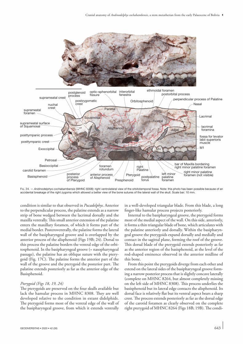
643
Cranial anatomy of Andinodelphys cochabambensis, a stem metatherian from the early Palaeocene of Bolivia
GEODIVERSITAS • 2020 • 42 (30)
condition is similar to that observed in Pucadelphys. Anterior to the perpendicular process, the palatine extends as a narrow strip of bone wedged between the lacrimal dorsally and the maxilla ventrally. This small anterior extension of the palatine enters the maxillary foramen, of which it forms part of the medial border. Posteroventrally, the palatine forms the lateral wall of the basipharyngeal groove and is overlapped by the anterior process of the alisphenoid (Figs 19B; 24). Dorsal to this process the palatine borders the ventral edge of the orbi-tosphenoid. In the basipharyngeal groove (= nasopharyngeal passage), the palatine has an oblique suture with the ptery-goid (Fig. 17C). The palatine forms the anterior part of the wall of the groove and the pterygoid the posterior part. The palatine extends posteriorly as far as the anterior edge of the Basisphenoid.
Pterygoid (Figs 18, 19, 24)The pterygoids are preserved on the four skulls available but lack the hamular process in MHNC 8308. They are well developed relative to the condition in extant didelphids. The pterygoid forms most of the ventral edge of the wall of the basipharyngeal groove, from which it extends ventrally
in a well-developed triangular blade. From this blade, a long finger-like hamular process projects posteriorly.
Internal to the basipharyngeal groove, the pterygoid forms most of the medial aspect of the wall. On this side, anteriorly, it forms a thin triangular blade of bone, which articulates with the palatine anteriorly and dorsally. Within the basipharyn-geal groove the pterygoids expand dorsally and medially and contact in the sagittal plane, forming the roof of the groove. This dorsal blade of the pterygoid extends posteriorly as far as the anterior region of the basisphenoid, at the level of the rod-shaped eminence observed in the anterior midline of this bone.
From this point the pterygoids diverge from each other and extend on the lateral sides of the basipharyngeal groove form-ing a narrow posterior process that is slightly concave laterally (complete on MHNC 8264, but almost completely missing on the left side of MHNC 8308). This process underlies the basisphenoid but its lateral edge contacts the alisphenoid. Its dorsal face is relatively flat but its ventral aspect bears a sharp crest. The process extends posteriorly as far as the dorsal edge of the carotid foramen as clearly observed on the complete right pterygoid of MHNC 8264 (Figs 18B; 19B). The condi-
Squamosal
JugalMaxilla
Nasal
postorbital process
Lacrimal
rightPalatine
ethmoidal foramen
Orbitosphenoid
Alisphenoid
interorbitalfenestra
M4 M1
lacrimalforamina
postglenoidprocess
Frontal
anterior processof Alisphenoid
suprameatalforamen
left minor palatine foramen
Pterygoid
Presphenoid
Parietalnuchalcrest
Basioccipital
Basisphenoid
Petrosal
Exoccipital
optic-sphenorbitalfissure
foramenrotundum
fossa for levatorlabii superiorismuscle
posteriorprocessof Pterygoid
right minor palatineforamen (not visible)
bar of Maxilla borderingright minor palatine foramen
suprameatal crest
suprameatal surfaceof Squamosal
posttympanic process
posttympanic crest
postzygomaticcrest
perpendicular process of Palatine
postpalatinetorus
carotid foramen
fig. 24. — Andinodelphys cochabambensis (MHNC 8308): right ventrolateral view of the orbitotemporal fossa. Note: this photo has been possible because of an accidental breakage of the right zygoma which allowed a better view of the bone sutures of the lateral wall of the skull. Scale bar: 10 mm.
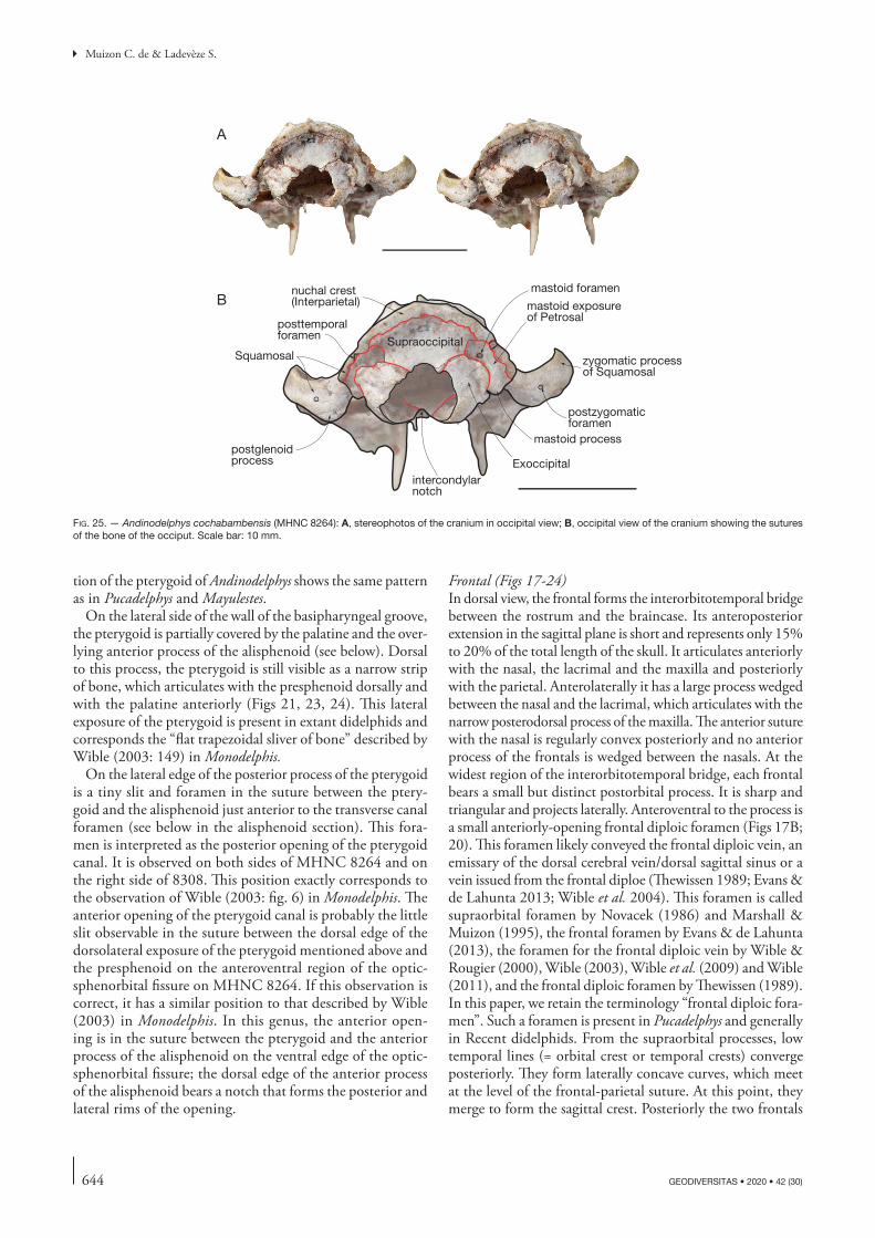
644 GEODIVERSITAS • 2020 • 42 (30)
Muizon C. de & Ladevèze S.
tion of the pterygoid of Andinodelphys shows the same pattern as in Pucadelphys and Mayulestes.
On the lateral side of the wall of the basipharyngeal groove, the pterygoid is partially covered by the palatine and the over-lying anterior process of the alisphenoid (see below). Dorsal to this process, the pterygoid is still visible as a narrow strip of bone, which articulates with the presphenoid dorsally and with the palatine anteriorly (Figs 21, 23, 24). This lateral exposure of the pterygoid is present in extant didelphids and corresponds the “flat trapezoidal sliver of bone” described by Wible (2003: 149) in Monodelphis.
On the lateral edge of the posterior process of the pterygoid is a tiny slit and foramen in the suture between the ptery-goid and the alisphenoid just anterior to the transverse canal foramen (see below in the alisphenoid section). This fora-men is interpreted as the posterior opening of the pterygoid canal. It is observed on both sides of MHNC 8264 and on the right side of 8308. This position exactly corresponds to the observation of Wible (2003: fig. 6) in Monodelphis. The anterior opening of the pterygoid canal is probably the little slit observable in the suture between the dorsal edge of the dorsolateral exposure of the pterygoid mentioned above and the presphenoid on the anteroventral region of the optic-sphenorbital fissure on MHNC 8264. If this observation is correct, it has a similar position to that described by Wible (2003) in Monodelphis. In this genus, the anterior open-ing is in the suture between the pterygoid and the anterior process of the alisphenoid on the ventral edge of the optic-sphenorbital fissure; the dorsal edge of the anterior process of the alisphenoid bears a notch that forms the posterior and lateral rims of the opening.
Frontal (Figs 17-24)In dorsal view, the frontal forms the interorbitotemporal bridge between the rostrum and the braincase. Its anteroposterior extension in the sagittal plane is short and represents only 15% to 20% of the total length of the skull. It articulates anteriorly with the nasal, the lacrimal and the maxilla and posteriorly with the parietal. Anterolaterally it has a large process wedged between the nasal and the lacrimal, which articulates with the narrow posterodorsal process of the maxilla. The anterior suture with the nasal is regularly convex posteriorly and no anterior process of the frontals is wedged between the nasals. At the widest region of the interorbitotemporal bridge, each frontal bears a small but distinct postorbital process. It is sharp and triangular and projects laterally. Anteroventral to the process is a small anteriorly-opening frontal diploic foramen (Figs 17B; 20). This foramen likely conveyed the frontal diploic vein, an emissary of the dorsal cerebral vein/dorsal sagittal sinus or a vein issued from the frontal diploe (Thewissen 1989; Evans & de Lahunta 2013; Wible et al. 2004). This foramen is called supraorbital foramen by Novacek (1986) and Marshall & Muizon (1995), the frontal foramen by Evans & de Lahunta (2013), the foramen for the frontal diploic vein by Wible & Rougier (2000), Wible (2003), Wible et al. (2009) and Wible (2011), and the frontal diploic foramen by Thewissen (1989). In this paper, we retain the terminology “frontal diploic fora-men”. Such a foramen is present in Pucadelphys and generally in Recent didelphids. From the supraorbital processes, low temporal lines (= orbital crest or temporal crests) converge posteriorly. They form laterally concave curves, which meet at the level of the frontal-parietal suture. At this point, they merge to form the sagittal crest. Posteriorly the two frontals
zygomatic processof Squamosal
postzygomaticforamen
mastoid exposureof Petrosal
Supraoccipital
nuchal crest(Interparietal)
posttemporalforamen
Exoccipitalpostglenoidprocess
mastoid process
A
Bmastoid foramen
Squamosal
intercondylarnotch
fig. 25. — Andinodelphys cochabambensis (MHNC 8264): A, stereophotos of the cranium in occipital view; B, occipital view of the cranium showing the sutures of the bone of the occiput. Scale bar: 10 mm.
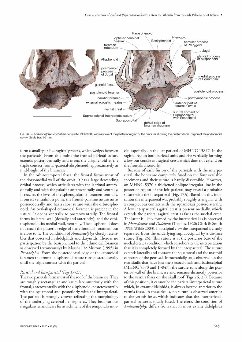
645
Cranial anatomy of Andinodelphys cochabambensis, a stem metatherian from the early Palaeocene of Bolivia
GEODIVERSITAS • 2020 • 42 (30)
form a small spur-like sagittal process, which wedges between the parietals. From this point the frontal-parietal suture extends posteroventrally and meets the alisphenoid at the triple contact frontal-parietal-alisphenoid, approximately at mid-height of the braincase.
In the orbitotemporal fossa, the frontal forms most of the dorsomedial wall of the orbit. It has a large descending orbital process, which articulates with the lacrimal antero-dorsally and with the palatine anteroventrally and ventrally. It reaches the level of the sphenopalatine foramen ventrally. From its ventralmost point, the frontal-palatine suture turns posterodorsally and has a short suture with the orbitosphe-noid. An oval-shaped ethmoidal foramen is present in the suture. It opens ventrally to posteroventrally. The frontal forms its lateral wall (dorsally and anteriorly), and the orbi-tosphenoid, its medial wall, ventrally. The alisphenoid does not reach the posterior edge of the ethmoidal foramen, but is close to it. The condition of Andinodelphys closely resem-bles that observed in didelphids and dasyurids. There is no participation by the basisphenoid to the ethmoidal foramen as observed (erroneously) by Marshall & Muizon (1995) in Pucadelphys. From the posterodorsal edge of the ethmoidal foramen the frontal-alisphenoid suture runs posterodorsally until the triple contact with the parietal.
Parietal and Interparietal (Figs 17-27)The two parietals form most of the roof of the braincase. They are roughly rectangular and articulate anteriorly with the frontal, anteroventrally with the alisphenoid, posteroventrally with the squamosal and posteriorly with the interparietal. The parietal is strongly convex reflecting the morphology of the underlying cerebral hemispheres. They bear various irregularities and scars for attachment of the temporalis mus-
cle, especially on the left parietal of MHNC 13847. In the sagittal region both parietal unite and rise vertically forming a low but consistent sagittal crest, which does not extend on the frontals anteriorly.
Because of early fusion of the parietals with the interpa-rietal, the bones are completely fused on the four available specimens and their suture is hardly discernible. However, on MHNC 8370 a thickened oblique irregular line in the posterior region of the left parietal may reveal a probable suture with the interparietal (Fig. 17A). Based on this indi-cation the interparietal was probably roughly triangular with a conspicuous contact with the squamosals posterolaterally. A low interparietal sagittal crest is present medially, which extends the parietal sagittal crest as far as the nuchal crest. The latter is likely formed by the interparietal as is observed in Monodelphis and Didelphis (Toeplitz 1920; Clark & Smith 1993; Wible 2003). In occipital view the interparietal is clearly separated from the underlying supraoccipital by a distinct suture (Fig. 25). This suture is at the posterior base of the nuchal crest, a condition which corroborates the interpretation that it is completely formed by the interparietal. The suture extends laterally and contacts the squamosal and the mastoid exposure of the petrosal. Intracranially, as is observed on the two skulls that have lost their exoccipitals and basioccipital (MHNC 8370 and 13847), the suture runs along the pos-terior wall of the braincase and remains distinctly posterior to the vermis fossa on the skull roof (Figs 26, 27). Because of this position, it cannot be the parietal-interparietal suture which, in extant didelphids, is always located anterior to the vermis fossa. In these skulls, no suture is observed anterior to the vermis fossa, which indicates that the interparietal-parietal suture is totally fused. Therefore, the condition of Andinodelphys differs from that in most extant didelphids
foramenrotundum
carotid foramen
anterior part offoramen ovale
Alisphenoid
Supraoccipital-Interparietal suture
dorsal edge offoramen magnum
sutural contact ofSupraoccipital with Exoccipital
Supraoccipital
postglenoid processpostglenoid foramen
external acoustic meatus
medial processof Squamosal
glenoid processof Alisphenoid
glenoid fossa
Basisphenoid
nuchal crest
postglenoidprocessof Jugal
optic-sphenoidalfissure
posttympanic process
hamular processof Pterygoid
Pterygoid
Jugal
Parasphenoid
fig. 26. — Andinodelphys cochabambensis (MHNC 8370): ventral view of the posterior region of the cranium showing the posterodorsal region of the endocranial cavity. Scale bar: 10 mm.
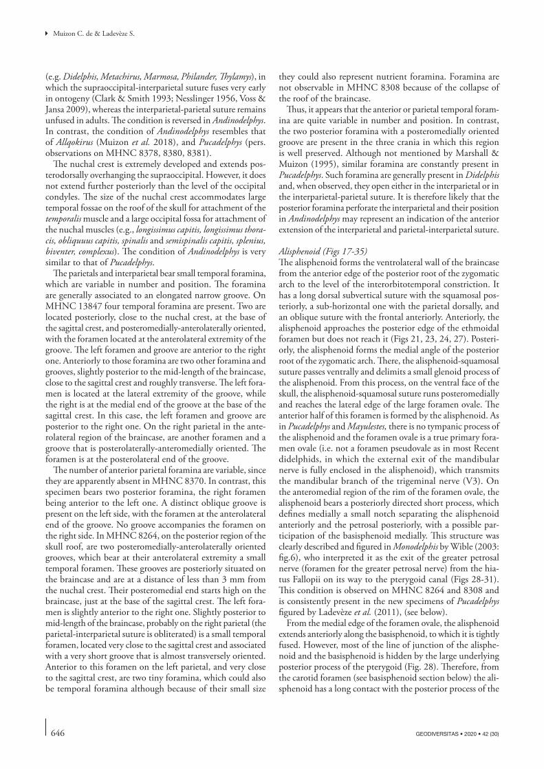
646 GEODIVERSITAS • 2020 • 42 (30)
Muizon C. de & Ladevèze S.
(e.g. Didelphis, Metachirus, Marmosa, Philander, Thylamys), in which the supraoccipital-interparietal suture fuses very early in ontogeny (Clark & Smith 1993; Nesslinger 1956, Voss & Jansa 2009), whereas the interparietal-parietal suture remains unfused in adults. The condition is reversed in Andinodelphys. In contrast, the condition of Andinodelphys resembles that of Allqokirus (Muizon et al. 2018), and Pucadelphys (pers. observations on MHNC 8378, 8380, 8381).
The nuchal crest is extremely developed and extends pos-terodorsally overhanging the supraoccipital. However, it does not extend further posteriorly than the level of the occipital condyles. The size of the nuchal crest accommodates large temporal fossae on the roof of the skull for attachment of the temporalis muscle and a large occipital fossa for attachment of the nuchal muscles (e.g., longissimus capitis, longissimus thora-cis, obliquuus capitis, spinalis and semispinalis capitis, splenius, biventer, complexus). The condition of Andinodelphys is very similar to that of Pucadelphys.
The parietals and interparietal bear small temporal foramina, which are variable in number and position. The foramina are generally associated to an elongated narrow groove. On MHNC 13847 four temporal foramina are present. Two are located posteriorly, close to the nuchal crest, at the base of the sagittal crest, and posteromedially-anterolaterally oriented, with the foramen located at the anterolateral extremity of the groove. The left foramen and groove are anterior to the right one. Anteriorly to those foramina are two other foramina and grooves, slightly posterior to the mid-length of the braincase, close to the sagittal crest and roughly transverse. The left fora-men is located at the lateral extremity of the groove, while the right is at the medial end of the groove at the base of the sagittal crest. In this case, the left foramen and groove are posterior to the right one. On the right parietal in the ante-rolateral region of the braincase, are another foramen and a groove that is posterolaterally-anteromedially oriented. The foramen is at the posterolateral end of the groove.
The number of anterior parietal foramina are variable, since they are apparently absent in MHNC 8370. In contrast, this specimen bears two posterior foramina, the right foramen being anterior to the left one. A distinct oblique groove is present on the left side, with the foramen at the anterolateral end of the groove. No groove accompanies the foramen on the right side. In MHNC 8264, on the posterior region of the skull roof, are two posteromedially-anterolaterally oriented grooves, which bear at their anterolateral extremity a small temporal foramen. These grooves are posteriorly situated on the braincase and are at a distance of less than 3 mm from the nuchal crest. Their posteromedial end starts high on the braincase, just at the base of the sagittal crest. The left fora-men is slightly anterior to the right one. Slightly posterior to mid-length of the braincase, probably on the right parietal (the parietal-interparietal suture is obliterated) is a small temporal foramen, located very close to the sagittal crest and associated with a very short groove that is almost transversely oriented. Anterior to this foramen on the left parietal, and very close to the sagittal crest, are two tiny foramina, which could also be temporal foramina although because of their small size
they could also represent nutrient foramina. Foramina are not observable in MHNC 8308 because of the collapse of the roof of the braincase.
Thus, it appears that the anterior or parietal temporal foram-ina are quite variable in number and position. In contrast, the two posterior foramina with a posteromedially oriented groove are present in the three crania in which this region is well preserved. Although not mentioned by Marshall & Muizon (1995), similar foramina are constantly present in Pucadelphys. Such foramina are generally present in Didelphis and, when observed, they open either in the interparietal or in the interparietal-parietal suture. It is therefore likely that the posterior foramina perforate the interparietal and their position in Andinodelphys may represent an indication of the anterior extension of the interparietal and parietal-interparietal suture.
Alisphenoid (Figs 17-35)The alisphenoid forms the ventrolateral wall of the braincase from the anterior edge of the posterior root of the zygomatic arch to the level of the interorbitotemporal constriction. It has a long dorsal subvertical suture with the squamosal pos-teriorly, a sub-horizontal one with the parietal dorsally, and an oblique suture with the frontal anteriorly. Anteriorly, the alisphenoid approaches the posterior edge of the ethmoidal foramen but does not reach it (Figs 21, 23, 24, 27). Posteri-orly, the alisphenoid forms the medial angle of the posterior root of the zygomatic arch. There, the alisphenoid-squamosal suture passes ventrally and delimits a small glenoid process of the alisphenoid. From this process, on the ventral face of the skull, the alisphenoid-squamosal suture runs posteromedially and reaches the lateral edge of the large foramen ovale. The anterior half of this foramen is formed by the alisphenoid. As in Pucadelphys and Mayulestes, there is no tympanic process of the alisphenoid and the foramen ovale is a true primary fora-men ovale (i.e. not a foramen pseudovale as in most Recent didelphids, in which the external exit of the mandibular nerve is fully enclosed in the alisphenoid), which transmits the mandibular branch of the trigeminal nerve (V3). On the anteromedial region of the rim of the foramen ovale, the alisphenoid bears a posteriorly directed short process, which defines medially a small notch separating the alisphenoid anteriorly and the petrosal posteriorly, with a possible par-ticipation of the basisphenoid medially. This structure was clearly described and figured in Monodelphis by Wible (2003: fig.6), who interpreted it as the exit of the greater petrosal nerve (foramen for the greater petrosal nerve) from the hia-tus Fallopii on its way to the pterygoid canal (Figs 28-31). This condition is observed on MHNC 8264 and 8308 and is consistently present in the new specimens of Pucadelphys figured by Ladevèze et al. (2011), (see below).
From the medial edge of the foramen ovale, the alisphenoid extends anteriorly along the basisphenoid, to which it is tightly fused. However, most of the line of junction of the alisphe-noid and the basisphenoid is hidden by the large underlying posterior process of the pterygoid (Fig. 28). Therefore, from the carotid foramen (see basisphenoid section below) the ali-sphenoid has a long contact with the posterior process of the
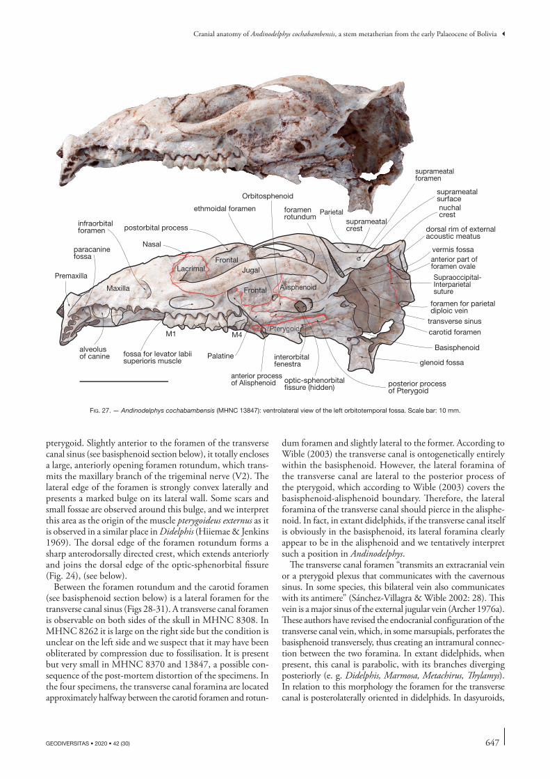
647
Cranial anatomy of Andinodelphys cochabambensis, a stem metatherian from the early Palaeocene of Bolivia
GEODIVERSITAS • 2020 • 42 (30)
pterygoid. Slightly anterior to the foramen of the transverse canal sinus (see basisphenoid section below), it totally encloses a large, anteriorly opening foramen rotundum, which trans-mits the maxillary branch of the trigeminal nerve (V2). The lateral edge of the foramen is strongly convex laterally and presents a marked bulge on its lateral wall. Some scars and small fossae are observed around this bulge, and we interpret this area as the origin of the muscle pterygoideus externus as it is observed in a similar place in Didelphis (Hiiemae & Jenkins 1969). The dorsal edge of the foramen rotundum forms a sharp anterodorsally directed crest, which extends anteriorly and joins the dorsal edge of the optic-sphenorbital fissure (Fig. 24), (see below).
Between the foramen rotundum and the carotid foramen (see basisphenoid section below) is a lateral foramen for the transverse canal sinus (Figs 28-31). A transverse canal foramen is observable on both sides of the skull in MHNC 8308. In MHNC 8262 it is large on the right side but the condition is unclear on the left side and we suspect that it may have been obliterated by compression due to fossilisation. It is present but very small in MHNC 8370 and 13847, a possible con-sequence of the post-mortem distortion of the specimens. In the four specimens, the transverse canal foramina are located approximately halfway between the carotid foramen and rotun-
dum foramen and slightly lateral to the former. According to Wible (2003) the transverse canal is ontogenetically entirely within the basisphenoid. However, the lateral foramina of the transverse canal are lateral to the posterior process of the pterygoid, which according to Wible (2003) covers the basisphenoid-alisphenoid boundary. Therefore, the lateral foramina of the transverse canal should pierce in the alisphe-noid. In fact, in extant didelphids, if the transverse canal itself is obviously in the basisphenoid, its lateral foramina clearly appear to be in the alisphenoid and we tentatively interpret such a position in Andinodelphys.
The transverse canal foramen “transmits an extracranial vein or a pterygoid plexus that communicates with the cavernous sinus. In some species, this bilateral vein also communicates with its antimere” (Sánchez-Villagra & Wible 2002: 28). This vein is a major sinus of the external jugular vein (Archer 1976a). These authors have revised the endocranial configuration of the transverse canal vein, which, in some marsupials, perforates the basisphenoid transversely, thus creating an intramural connec-tion between the two foramina. In extant didelphids, when present, this canal is parabolic, with its branches diverging posteriorly (e. g. Didelphis, Marmosa, Metachirus, Thylamys). In relation to this morphology the foramen for the transverse canal is posterolaterally oriented in didelphids. In dasyuroids,
Frontal
Jugal
postorbital processinfraorbitalforamen
Lacrimal
Palatine
ethmoidal foramen
Orbitosphenoid
Alisphenoid
interorbitalfenestra
M4M1
Frontal
Maxilla
anterior processof Alisphenoid
suprameatalforamen
Pteterygrygoidoid
Parietalnuchalcrest
transverse sinus
foramen for parietaldiploic vein
foramenrotundum
Basisphenoid
carotid foramen
posterior processof Pterygoid
Nasal
Supraoccipital-Interparietalsuture
anterior part offoramen ovale
Premaxilla
vermis fossa
optic-sphenorbital fissure (hidden)
fossa for levator labiisuperioris muscle
suprameatalcrest
suprameatalsurface
dorsal rim of externalacoustic meatus
paracaninefossa
alveolus of canine
glenoid fossa
fig. 27. — Andinodelphys cochabambensis (MHNC 13847): ventrolateral view of the left orbitotemporal fossa. Scale bar: 10 mm.
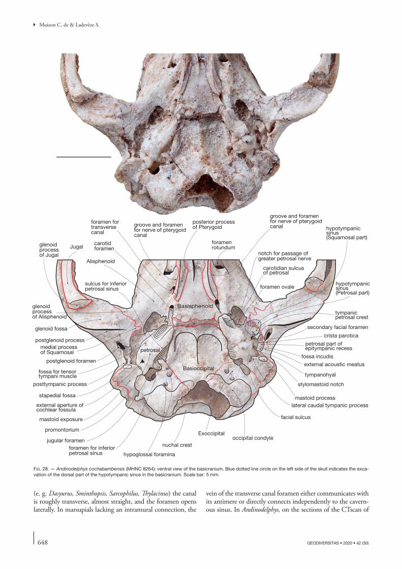
648 GEODIVERSITAS • 2020 • 42 (30)
Muizon C. de & Ladevèze S.
(e. g. Dasyurus, Sminthopsis, Sarcophilus, Thylacinus) the canal is roughly transverse, almost straight, and the foramen opens laterally. In marsupials lacking an intramural connection, the
vein of the transverse canal foramen either communicates with its antimere or directly connects independently to the cavern-ous sinus. In Andinodelphys, on the sections of the CTscan of
Jugalglenoidprocessof Jugal
posterior processof Pterygoid
foramenrotundum
glenoidprocess of Alisphenoid
postglenoid process
postglenoid foramen
medial processof Squamosal
glenoid fossa
foramen ovale
carotidforamen
Basioccipital
mastoid process
jugular foramen
promontorium
occipital condyle
Basisphenoid
Alisphenoid
facial sulcus
carotidian sulcusof petrosal
external aperture ofcochlear fossula
fossa incudis
tympanicpetrosal crest
fossa for tensor tympani muscle
crista parotica
tympanohyal
secondary facial foramen
hypotympanicsinus(Petrosal part)
lateral caudal tympanic process
notch for passage of greater petrosal nerve
external acoustic meatus
stapedial fossa
hypoglossal foraminaforamen for inferior petrosal sinus
Exoccipital
petrosal part of epitympanic recess
mastoid exposure
hypotympanicsinus (Squamosal part)
petrosal
stylomastoid notch
foramen for transversecanal
nuchal crest
posttympanic process
sulcus for inferiorpetrosal sinus
groove and foramen for nerve of pterygoidcanal
groove and foramen for nerve of pterygoidcanal
fig. 28. — Andinodelphys cochabambensis (MHNC 8264): ventral view of the basicranium. Blue dotted line circle on the left side of the skull indicates the exca-vation of the dorsal part of the hypotympanic sinus in the basicranium. Scale bar: 5 mm.
649
Cranial anatomy of Andinodelphys cochabambensis, a stem metatherian from the early Palaeocene of Bolivia
GEODIVERSITAS • 2020 • 42 (30)
MHNC 8264 (Fig. 32), an intramural canal pierces the basis-phenoid from right to left foramina of the transverse canal. It is better observed on the right side of the skull since the left trans-verse canal has likely suffered some post-mortem compression. The foramina open posteriorly and the canal extends anteriorly, thus being parabolic as is observed in Didelphis. A different condition is observed on MHNC 8308. In this specimen, the foramen of the transverse canal opens within the braincase on the lateral wall of the internal sulcus for the internal carotid, on the lateral edges of the pituitary fossa. There is no intramu-ral canal within the basisphenoid. This condition is observed in the CTscan sections (Fig. 33). An intramural connection between the transverse canal foramina is present in most, but not all, dasyurids since Sánchez-Villagra & Wible (2002: 30) observed that “in three specimens of Dasyurus viverrinus the foramen simply opens endocranially”. Variation is also observed in Thylacinus (Archer 1976a). Therefore, the variation observed in Andinodelphys is also present in extant marsupials.
Anteromedial to the foramen rotundum, the alisphenoid has an elongated anterior process, which is anteroposteriorly subhorizontal and slightly sloping laterally. This process is a narrow and thin blade of bone, which extends anteriorly as far as the level of the hamular process of the pterygoid as observed on MHNC 8264. It does not reach the minor pala-tine foramen anteriorly but it is slightly anterior to the level of the ethmoidal foramen. A similar condition is observed in Recent didelphids. In its posterior part the anterior process overlies the part of the pterygoid that forms the lateral wall of the basipharyngeal groove. Because the anterior process of the alisphenoid is narrower than this wall, the pterygoid is visible on the dorsal and ventral edges of the process, as is clearly observed in Monodelphis (Wible 2003: fig. 4) (Figs 21-24). In its anterior region, the anterior process of the alisphenoid contacts the posterior blade of the palatine, which forms the lateral wall of the basipharyngeal groove. The alisphenoid forms the lateral wall of the optic-sphenorbital fissure. The
A
B
fig. 29. — Andinodelphys cochabambensis (MHNC 8264): A, stereophotos of the right auditory region; B, stereophotos of the left auditory region. Scale bar: 5 mm.
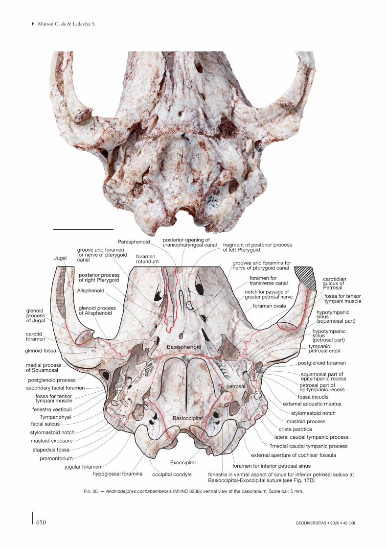
650 GEODIVERSITAS • 2020 • 42 (30)
Muizon C. de & Ladevèze S.
Jugal
glenoidprocessof Jugal
stylomastoid notch
posterior processof right Pterygoid
foramenrotundum
glenoid process of Alisphenoid
postglenoid process
postglenoid foramenmedial processof Squamosal
glenoid fossa
foramen ovale
carotidforamen
Basioccipitalmastoid process
jugular foramen
promontorium
occipital condyle
Basisphenoid
Alisphenoid
facial sulcus
carotidiansulcus ofPetrosal
external aperture of cochlear fossula
squamosal part of epitympanic recess
fossa incudis
tympanicpetrosal crest
fenestra vestibuli
fossa for tensor tympani muscle
crista parotica
Tympanohyal
secondary facial foramen
hypotympanicsinus(petrosal part)
lateral caudal tympanic process
notch for passage of greater petrosal nerve
external acoustic meatus
stapedius fossa
hypoglossal foramina
foramen for inferior petrosal sinusExoccipital
petrosal part of epitympanic recess
mastoid exposure
fenestra in ventral aspect of sinus for inferior petrosal sulcus at Basioccipital-Exoccipital suture (see Fig. 17D)
foramen fortransverse canal
hypotympanicsinus (squamosal part)
petrosal
stylomastoid notch
groove and foramen for nerve of pterygoidcanal
fragment of posterior processof left Pterygoid
grooves and foramina fornerve of pterygoid canal
fossa for tensor tympani muscle
?medial caudal tympanic process
Parasphenoid posterior opening of craniopharyngeal canal
fig. 30. — Andinodelphys cochabambensis (MHNC 8308): ventral view of the basicranium. Scale bar: 5 mm.
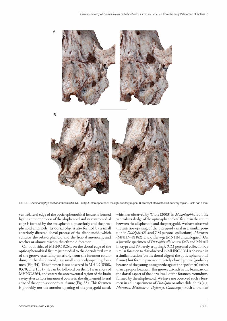
651
Cranial anatomy of Andinodelphys cochabambensis, a stem metatherian from the early Palaeocene of Bolivia
GEODIVERSITAS • 2020 • 42 (30)
ventrolateral edge of the optic-sphenorbital fissure is formed by the anterior process of the alisphenoid and its ventromedial edge is formed by the basisphenoid posteriorly and the pres-phenoid anteriorly. Its dorsal edge is also formed by a small anteriorly directed dorsal process of the alisphenoid, which contacts the orbitosphenoid and the frontal anteriorly, and reaches or almost reaches the ethmoid foramen.
On both sides of MHNC 8264, on the dorsal edge of the optic-sphenorbital fissure just medial to the dorsolateral crest of the groove extending anteriorly from the foramen rotun-dum, in the alisphenoid, is a small anteriorly-opening fora-men (Fig. 34). This foramen is not observed in MHNC 8308, 8370, and 13847. It can be followed on the CTscan slices of MHNC 8264, and enters the anteroventral region of the brain cavity after a short intramural course in the alisphenoid lateral edge of the optic-sphenorbital fissure (Fig. 35). This foramen is probably not the anterior opening of the pterygoid canal,
which, as observed by Wible (2003) in Monodelphis, is on the ventrolateral edge of the optic-sphenorbital fissure in the suture between the alisphenoid and the pterygoid. We have observed the anterior opening of the pterygoid canal in a similar posi-tion in Didelphis (SL and CM personal collections), Marmosa (MNHN-RH82), and Caluromys (MNHN uncatalogued). On a juvenile specimen of Didelphis albiventris (M3 and M4 still in crypt and P3 barely erupting), (CM personal collection), a similar foramen to that observed in MHNC 8264 is observed in a similar location (on the dorsal edge of the optic-sphenorbital fissure) but forming an incompletely closed groove (probably because of the young ontogenetic age of the specimen) rather than a proper foramen. This groove extends in the braincase on the dorsal aspect of the dorsal wall of the foramen rotundum, formed by the alisphenoid. We have not observed such a fora-men in adult specimens of Didelphis or other didelphids (e.g. Marmosa, Metachirus, Thylamys, Caluromys). Such a foramen
A
B
fig. 31. — Andinodelphys cochabambensis (MHNC 8308): A, stereophotos of the right auditory region; B, stereophotos of the left auditory region. Scale bar: 5 mm.
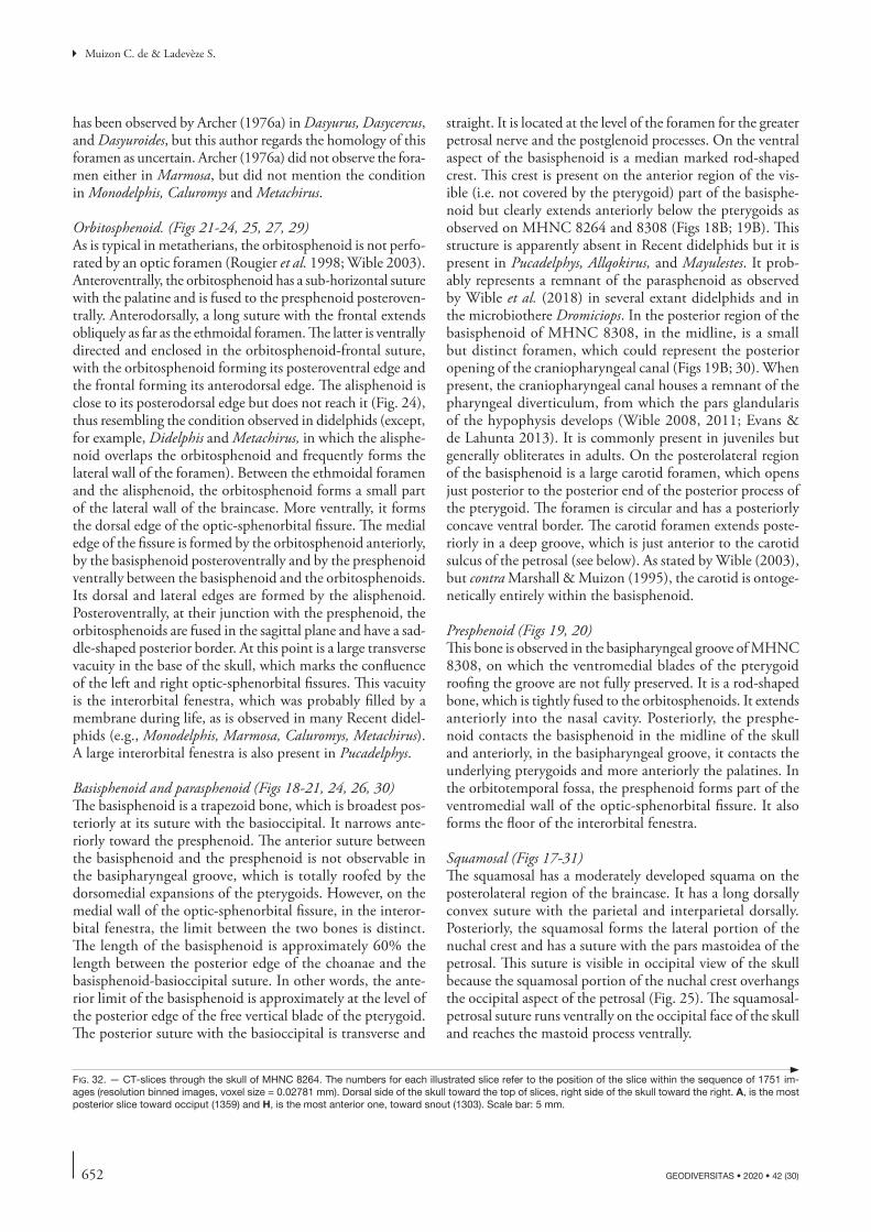
652 GEODIVERSITAS • 2020 • 42 (30)
Muizon C. de & Ladevèze S.
has been observed by Archer (1976a) in Dasyurus, Dasycercus, and Dasyuroides, but this author regards the homology of this foramen as uncertain. Archer (1976a) did not observe the fora-men either in Marmosa, but did not mention the condition in Monodelphis, Caluromys and Metachirus.
Orbitosphenoid. (Figs 21-24, 25, 27, 29)As is typical in metatherians, the orbitosphenoid is not perfo-rated by an optic foramen (Rougier et al. 1998; Wible 2003). Anteroventrally, the orbitosphenoid has a sub-horizontal suture with the palatine and is fused to the presphenoid posteroven-trally. Anterodorsally, a long suture with the frontal extends obliquely as far as the ethmoidal foramen. The latter is ventrally directed and enclosed in the orbitosphenoid-frontal suture, with the orbitosphenoid forming its posteroventral edge and the frontal forming its anterodorsal edge. The alisphenoid is close to its posterodorsal edge but does not reach it (Fig. 24), thus resembling the condition observed in didelphids (except, for example, Didelphis and Metachirus, in which the alisphe-noid overlaps the orbitosphenoid and frequently forms the lateral wall of the foramen). Between the ethmoidal foramen and the alisphenoid, the orbitosphenoid forms a small part of the lateral wall of the braincase. More ventrally, it forms the dorsal edge of the optic-sphenorbital fissure. The medial edge of the fissure is formed by the orbitosphenoid anteriorly, by the basisphenoid posteroventrally and by the presphenoid ventrally between the basisphenoid and the orbitosphenoids. Its dorsal and lateral edges are formed by the alisphenoid. Posteroventrally, at their junction with the presphenoid, the orbitosphenoids are fused in the sagittal plane and have a sad-dle-shaped posterior border. At this point is a large transverse vacuity in the base of the skull, which marks the confluence of the left and right optic-sphenorbital fissures. This vacuity is the interorbital fenestra, which was probably filled by a membrane during life, as is observed in many Recent didel-phids (e.g., Monodelphis, Marmosa, Caluromys, Metachirus). A large interorbital fenestra is also present in Pucadelphys.
Basisphenoid and parasphenoid (Figs 18-21, 24, 26, 30)The basisphenoid is a trapezoid bone, which is broadest pos-teriorly at its suture with the basioccipital. It narrows ante-riorly toward the presphenoid. The anterior suture between the basisphenoid and the presphenoid is not observable in the basipharyngeal groove, which is totally roofed by the dorsomedial expansions of the pterygoids. However, on the medial wall of the optic-sphenorbital fissure, in the interor-bital fenestra, the limit between the two bones is distinct. The length of the basisphenoid is approximately 60% the length between the posterior edge of the choanae and the basisphenoid-basioccipital suture. In other words, the ante-rior limit of the basisphenoid is approximately at the level of the posterior edge of the free vertical blade of the pterygoid. The posterior suture with the basioccipital is transverse and
straight. It is located at the level of the foramen for the greater petrosal nerve and the postglenoid processes. On the ventral aspect of the basisphenoid is a median marked rod-shaped crest. This crest is present on the anterior region of the vis-ible (i.e. not covered by the pterygoid) part of the basisphe-noid but clearly extends anteriorly below the pterygoids as observed on MHNC 8264 and 8308 (Figs 18B; 19B). This structure is apparently absent in Recent didelphids but it is present in Pucadelphys, Allqokirus, and Mayulestes. It prob-ably represents a remnant of the parasphenoid as observed by Wible et al. (2018) in several extant didelphids and in the microbiothere Dromiciops. In the posterior region of the basisphenoid of MHNC 8308, in the midline, is a small but distinct foramen, which could represent the posterior opening of the craniopharyngeal canal (Figs 19B; 30). When present, the craniopharyngeal canal houses a remnant of the pharyngeal diverticulum, from which the pars glandularis of the hypophysis develops (Wible 2008, 2011; Evans & de Lahunta 2013). It is commonly present in juveniles but generally obliterates in adults. On the posterolateral region of the basisphenoid is a large carotid foramen, which opens just posterior to the posterior end of the posterior process of the pterygoid. The foramen is circular and has a posteriorly concave ventral border. The carotid foramen extends poste-riorly in a deep groove, which is just anterior to the carotid sulcus of the petrosal (see below). As stated by Wible (2003), but contra Marshall & Muizon (1995), the carotid is ontoge-netically entirely within the basisphenoid.
Presphenoid (Figs 19, 20)This bone is observed in the basipharyngeal groove of MHNC 8308, on which the ventromedial blades of the pterygoid roofing the groove are not fully preserved. It is a rod-shaped bone, which is tightly fused to the orbitosphenoids. It extends anteriorly into the nasal cavity. Posteriorly, the presphe-noid contacts the basisphenoid in the midline of the skull and anteriorly, in the basipharyngeal groove, it contacts the underlying pterygoids and more anteriorly the palatines. In the orbitotemporal fossa, the presphenoid forms part of the ventromedial wall of the optic-sphenorbital fissure. It also forms the floor of the interorbital fenestra.
Squamosal (Figs 17-31)The squamosal has a moderately developed squama on the posterolateral region of the braincase. It has a long dorsally convex suture with the parietal and interparietal dorsally. Posteriorly, the squamosal forms the lateral portion of the nuchal crest and has a suture with the pars mastoidea of the petrosal. This suture is visible in occipital view of the skull because the squamosal portion of the nuchal crest overhangs the occipital aspect of the petrosal (Fig. 25). The squamosal-petrosal suture runs ventrally on the occipital face of the skull and reaches the mastoid process ventrally.
fig. 32. — CT-slices through the skull of MHNC 8264. The numbers for each illustrated slice refer to the position of the slice within the sequence of 1751 im-ages (resolution binned images, voxel size = 0.02781 mm). Dorsal side of the skull toward the top of slices, right side of the skull toward the right. A, is the most posterior slice toward occiput (1359) and H, is the most anterior one, toward snout (1303). Scale bar: 5 mm.
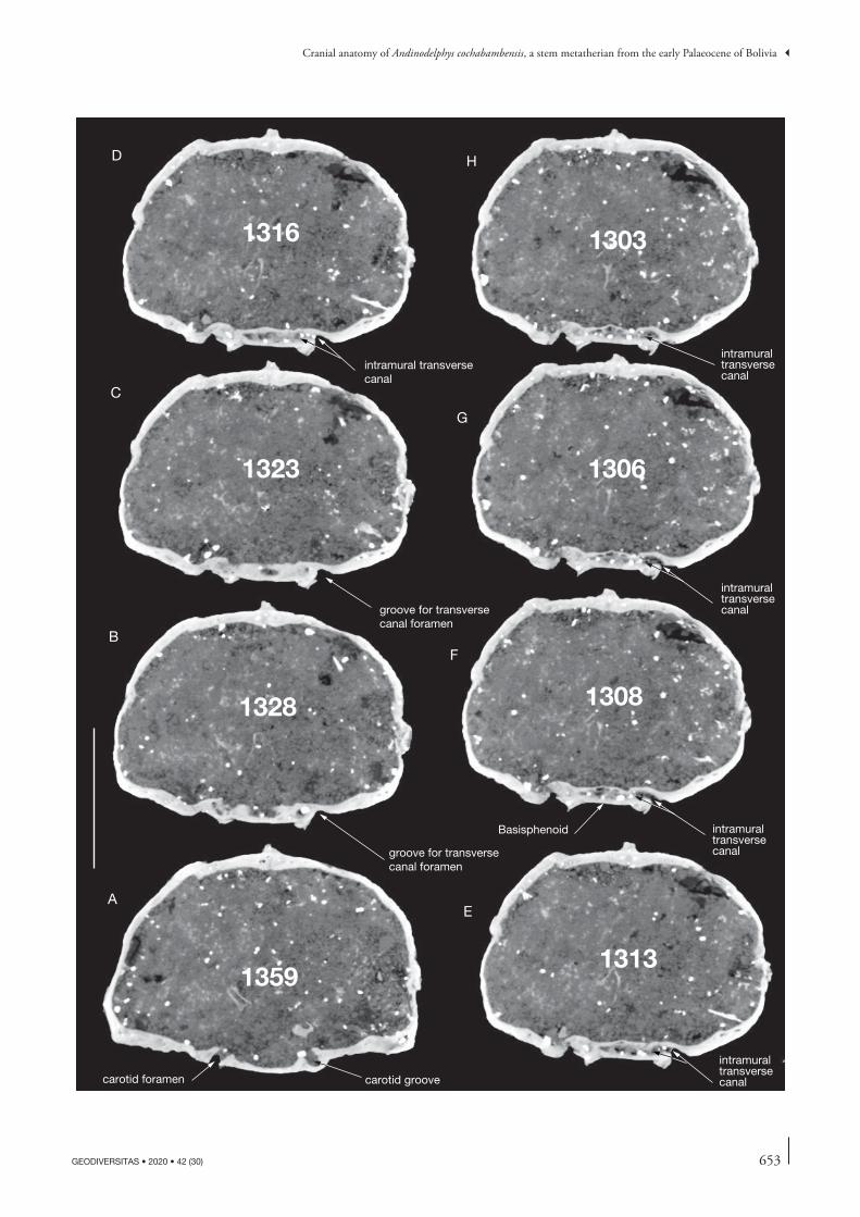
653
Cranial anatomy of Andinodelphys cochabambensis, a stem metatherian from the early Palaeocene of Bolivia
GEODIVERSITAS • 2020 • 42 (30)
carotid foramen carotid groove
groove for transversecanal foramen
groove for transversecanal foramen
intramural transversecanal
intramuraltransversecanal
intramural transversecanal
intramural transversecanal
intramuraltransversecanal
1359
1328
1323
1316
1313
1308
1306
1303
A
B
C
D
E
F
G
H
Basisphenoid
654 GEODIVERSITAS • 2020 • 42 (30)
Muizon C. de & Ladevèze S.
Anteriorly, on the roof of the braincase, the squamosal meets the alisphenoid at the level of the anterior edge of the posterior root of the zygoma and the suture turns ventrally at almost right angle. The squamosal-alisphenoid suture enters the dorsal face of the posterior zygomatic root in its anteromedial angle, where it delimitates a small glenoid process of the alisphenoid. On the anteromedial angle of the glenoid fossa the squamosal-alisphenoid suture runs medially towards the lateral edge of the foramen ovale. In this area, the relationships of the squamosal, alisphenoid and petrosal vary, but in all specimens the squamosal presents a medial process, which reaches or almost reaches the lateral rim of the foramen ovale. In MHNC 8308, this medial process of the squamosal is wedged between the alisphenoid anteriorly and the petrosal posteriorly (Fig. 30). It is triangular and its medial apex is pointed and forms a tiny portion of the lateral rim of the foramen ovale. In MHNC 8264, the medial process of the squamosal does not reach the lateral edge of the foramen ovale (Fig. 28). On this specimen, medial to the process, the rim of the foramen ovale is formed by a posterior extension of the alisphenoid, which wedges between the medial process of the squamosal anteriorly and the petrosal posteriorly. Both conditions are present in MHNC 8370. Apparently, on the left side of the skull the squamosal reaches the rim of the foramen ovale, while on the right side it does not and the alisphenoid forms the anterolateral rim of the foramen ovale (Fig. 26). The condition of MHNC 8308 is similar to that observed in Pucadelphys and that of MHNC 8264 resembles that of Mayulestes. The posteromedial edge of the medial process of the squamosal slopes posteromedially toward a small cupula. This shallow depression in the roof of the anterior region of the middle ear is posterolateral to the foramen ovale. It is formed posteromedially by the petrosal, anterolaterally by the medial process of the squamosal, and posterolaterally by the anteromedial face of the petrosal crest (see petrosal section below), which is distinctly concave (Fig. 28). The constitution of this depression is identical to that observed in Mayulestes (Muizon 1998) and forms what is regarded as an incipiently developed hypotympanic sinus (sensu Klaauw 1931: 19 and Archer 1976a: 227). The suture posterior to the medial process of the squamosal, is with the petrosal. It runs along the lateral border of the petrosal portion of the epitympanic recess and fossa incudis. In fact, the squamosal-petrosal suture passes within the epitympanic recess, which is divided into a medial petrosal portion (see petrosal section below) and a lateral squamosal portion. As is observed in MHNC 8308, in which this region is well-preserved, the two portions of the epitympanic recess are roughly similar in size and the medial wall of the squamosal in this region is slightly excavated. At the posterior end of the epitympanic recess, the squamosal also participates in the formation of the lateral part of the fossa incudis (see petrosal section below). The crest, which forms the ventral edge of the squamosal portion of the epitympanic recess, is the dorsal edge of the external acoustic meatus. Dorsal to it and on the lateral side of the squamosal is a subvertical area, which is ventral to the suprameatal crest (see below) and posterodorsal to the posterior face of the postglenoid process
(Fig. 24). This area corresponds to the suprameatal surface of the squamosal (Novacek 1986). Posterior to the fossa incudis the squamosal laterally borders the crista parotica and the tympanohyal of the petrosal, and reaches the lateral edge of the mastoid process, from where it runs dorsally along the anterodorsal edge of the pars mastoidea of the petrosal and forms the ventrolateral portion of the nuchal crest (see above). Between the fossa incudis anteriorly and the lateral edge of the mastoid process, the ventral rim of the squamosal protrudes ventrally in a rounded to V-shaped (in lateral view) but flat (in ventral view) process, which extends the dorsal rim of the external acoustic meatus (Fig. 24). This process laterally borders the tympanohyal and is slightly anterior to the mas-toid process. This structure corresponds to the posttympanic crest as defined by Wible (2003) and Wible et al. (2004). Lateral to the posttympanic crest and posteriorly bordering the suprameatal surface is the posttympanic process, which is weakly developed in Andinodelphys. In fact, the posttympanic process of Andinodelphys corresponds to the posterior edge of the suprameatal surface, which is slightly bent laterally at its contact with the pars mastoidea of the petrosal. In Andinodel-phys, the distinction between posttympanic crest and process is subtle, a condition that is also present in Pucadelphys and Mayulestes. As a matter of fact, the posttympanic crest (sensu Wible 2003) of Pucadelphys and Mayulestes has been identi-fied as posttympanic process by Marshall & Muizon (1995) and Muizon (1998) respectively.
Lateral to the medial process of the squamosal is the trans-versely elongated glenoid fossa for articulation of the dentary. The main axes of the two fossae are not exactly parallel and transverse. They are slightly oblique being oriented on an anterolateral-posteromedial axis. The glenoid fossa is approxi-mately two times wider than long, and deeply concave. In lateral view, it has a section forming approximately a quarter of circle, the dorsal part of the fossa being sub-horizontal and the posterior wall being sub-vertical. The anterior edge of the cavity is roughly straight being distinctly oblique in dorsal view and oriented posteromedially-anterolaterally (especially in MHNC 8264 and 13847). The postglenoid process is wide and high. In posterior view, it is roughly semicircular and extends transversely for the whole width of the glenoid fossa. In posterior view, it is weakly asymmetrical, its ventralmost point being slightly shifted medially to the middle of the gle-noid fossa. In MHNC 8308 the glenoid fossa and postglenoid process are slightly less expanded transversely than in the other three specimens. However, this condition could be the result of the post-mortem transverse compression, which affected this specimen. The preglenoid process of the jugal is small but distinct and located at the anterolateral angle of the glenoid fossa. On the posterolateral aspect of the postglenoid process is a small postzygomatic foramen (Fig. 25). According to Archer (1976a), in dasyurids, this foramen transmits a vein from the posterior root of the zygomatic arch to the postglenoid vein. This foramen is present in Pucadelphys but apparently absent in Mayulestes. Posterior to the medial edge of the postglenoid process is a large postglenoid foramen. This foramen transmits the sphenoparietal emissary vein (which externally becomes
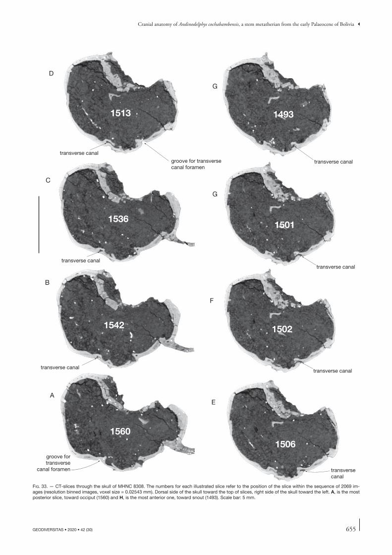
655
Cranial anatomy of Andinodelphys cochabambensis, a stem metatherian from the early Palaeocene of Bolivia
GEODIVERSITAS • 2020 • 42 (30)
fig. 33. — CT-slices through the skull of MHNC 8308. The numbers for each illustrated slice refer to the position of the slice within the sequence of 2069 im-ages (resolution binned images, voxel size = 0.02543 mm). Dorsal side of the skull toward the top of slices, right side of the skull toward the left. A, is the most posterior slice, toward occiput (1560) and H, is the most anterior one, toward snout (1493). Scale bar: 5 mm.
1560
1542
1536
1513
1506
1502
1501
1493
A
B
C
D
E
F
G
G
transverse canal
transverse canal
transversecanal
groove fortransverse
canal foramen
transverse canal
transverse canal
transverse canal
groove for transversecanal foramen
transverse canal
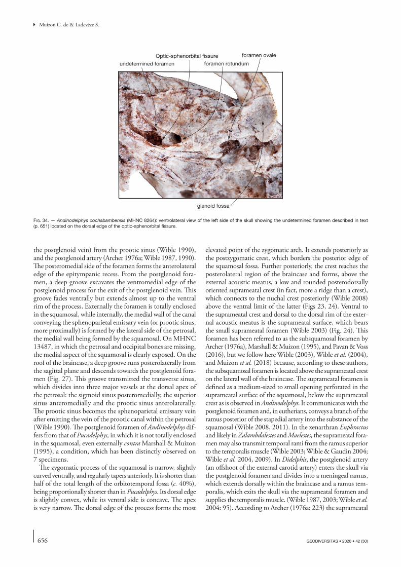
656 GEODIVERSITAS • 2020 • 42 (30)
Muizon C. de & Ladevèze S.
the postglenoid vein) from the prootic sinus (Wible 1990), and the postglenoid artery (Archer 1976a; Wible 1987, 1990). The posteromedial side of the foramen forms the anterolateral edge of the epitympanic recess. From the postglenoid fora-men, a deep groove excavates the ventromedial edge of the postglenoid process for the exit of the postglenoid vein. This groove fades ventrally but extends almost up to the ventral rim of the process. Externally the foramen is totally enclosed in the squamosal, while internally, the medial wall of the canal conveying the sphenoparietal emissary vein (or prootic sinus, more proximally) is formed by the lateral side of the petrosal, the medial wall being formed by the squamosal. On MHNC 13487, in which the petrosal and occipital bones are missing, the medial aspect of the squamosal is clearly exposed. On the roof of the braincase, a deep groove runs posterolaterally from the sagittal plane and descends towards the postglenoid fora-men (Fig. 27). This groove transmitted the transverse sinus, which divides into three major vessels at the dorsal apex of the petrosal: the sigmoid sinus posteromedially, the superior sinus anteromedially and the prootic sinus anterolaterally. The prootic sinus becomes the sphenoparietal emissary vein after emitting the vein of the prootic canal within the petrosal (Wible 1990). The postglenoid foramen of Andinodelphys dif-fers from that of Pucadelphys, in which it is not totally enclosed in the squamosal, even externally contra Marshall & Muizon (1995), a condition, which has been distinctly observed on 7 specimens.
The zygomatic process of the squamosal is narrow, slightly curved ventrally, and regularly tapers anteriorly. It is shorter than half of the total length of the orbitotemporal fossa (c. 40%), being proportionally shorter than in Pucadelphys. Its dorsal edge is slightly convex, while its ventral side is concave. The apex is very narrow. The dorsal edge of the process forms the most
elevated point of the zygomatic arch. It extends posteriorly as the postzygomatic crest, which borders the posterior edge of the squamosal fossa. Further posteriorly, the crest reaches the posterolateral region of the braincase and forms, above the external acoustic meatus, a low and rounded posterodorsally oriented suprameatal crest (in fact, more a ridge than a crest), which connects to the nuchal crest posteriorly (Wible 2008) above the ventral limit of the latter (Figs 23, 24). Ventral to the suprameatal crest and dorsal to the dorsal rim of the exter-nal acoustic meatus is the suprameatal surface, which bears the small suprameatal foramen (Wible 2003) (Fig. 24). This foramen has been referred to as the subsquamosal foramen by Archer (1976a), Marshall & Muizon (1995), and Pavan & Voss (2016), but we follow here Wible (2003), Wible et al. (2004), and Muizon et al. (2018) because, according to these authors, the subsquamosal foramen is located above the suprameatal crest on the lateral wall of the braincase. The suprameatal foramen is defined as a medium-sized to small opening perforated in the suprameatal surface of the squamosal, below the suprameatal crest as is observed in Andinodelphys. It communicates with the postglenoid foramen and, in eutherians, conveys a branch of the ramus posterior of the stapedial artery into the substance of the squamosal (Wible 2008, 2011). In the xenarthran Euphractus and likely in Zalambdalestes and Maelestes, the suprameatal fora-men may also transmit temporal rami from the ramus superior to the temporalis muscle (Wible 2003; Wible & Gaudin 2004; Wible et al. 2004, 2009). In Didelphis, the postglenoid artery (an offshoot of the external carotid artery) enters the skull via the postglenoid foramen and divides into a meningeal ramus, which extends dorsally within the braincase and a ramus tem-poralis, which exits the skull via the suprameatal foramen and supplies the temporalis muscle. (Wible 1987, 2003; Wible et al. 2004: 95). According to Archer (1976a: 223) the suprameatal
foramen rotundumOptic-sphenorbital fissure
undetermined foramen
foramen ovale
glenoid fossa
fig. 34. — Andinodelphys cochabambensis (MHNC 8264): ventrolateral view of the left side of the skull showing the undetermined foramen described in text (p. 651) located on the dorsal edge of the optic-sphenorbital fissure.
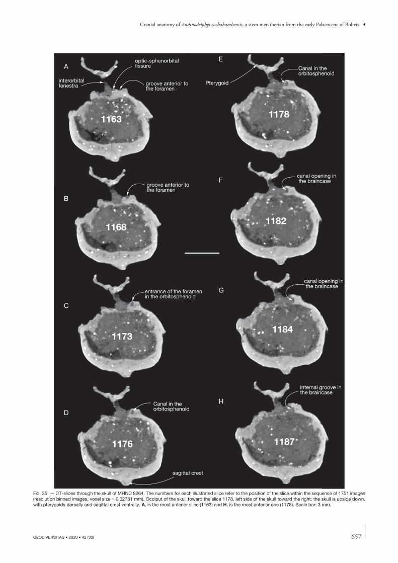
657
Cranial anatomy of Andinodelphys cochabambensis, a stem metatherian from the early Palaeocene of Bolivia
GEODIVERSITAS • 2020 • 42 (30)
sagittal crest
interorbitalfenestra
1168
1163
1173
1176 1187
1184
1182
1178
groove anterior tothe foramen
optic-sphenorbitalfissure
groove anterior tothe foramen
entrance of the foramenin the orbitosphenoid
Canal in theorbitosphenoid
Canal in theorbitosphenoid
canal opening in the braincase
canal opening in the braincase
internal groove inthe braincase
Pterygoid
A
B
C
D
E
F
G
H
fig. 35. — CT-slices through the skull of MHNC 8264. The numbers for each illustrated slice refer to the position of the slice within the sequence of 1751 images (resolution binned images, voxel size = 0.02781 mm). Occiput of the skull toward the slice 1178, left side of the skull toward the right; the skull is upside down, with pterygoids dorsally and sagittal crest ventrally. A, is the most anterior slice (1163) and H, is the most anterior one (1178). Scale bar: 3 mm.
658 GEODIVERSITAS • 2020 • 42 (30)
Muizon C. de & Ladevèze S.
foramen (his subsquamosal foramen) “carries postglenoid artery out onto parietal area of cranium from postglenoid foramen. Also carries vein from parietal area to postglenoid foramen”. The suprameatal foramen of Andinodelphys varies in position, number and size in the four skulls available. In MHNC 8264 there is one single, medium-sized, and posteriorly located fora-men on the right side, whereas there are two foramina on the left side, one large and posterior (just anterior to nuchal crest) and another small, slightly anterior to it. In MHNC 8308 a single medium-sized foramen is present, located in the anterior part of the suprameatal surface on both sides. In MHNC 8370 two foramina are present; one, large, in the posterior region of the surface and the other, medium-sized in the anterior half of the surface (observation of the two foramina is possible on the right side only). In MHNC 13847, two small foramina are observed on the left side (one anterior and one posterior), whereas two posteriorly located foramina are present on the right side, one ventral and small and the other dorsally placed (on the suprameatal crest) and medium-sized. The significant variation observed in Andinodelphys differs from the condition in most Pucadelphys specimens and in most Recent didelphids (e.g., Didelphis, Monodelphis, Marmosa, Philander, Metachirus, Thylamys), in which the suprameatal foramen is consistently single, distinctly larger than the large foramen in Andinodel-phys and in a median to anterior location on the suprameatal surface. In Andinodelphys, the suprameatal foramen (foramina) is (are) located more posteriorly than in Pucadelphys and the Recent didelphids mentioned above. Interestingly, however, in two specimens of Pucadelphys (MHNC 8266 and 8379, two out of ten specimens in which this character can be observed), two foramina are present. One is anterior and has the large size and the anterior position of the suprameatal foramen of the didelphid genera mentioned above, and the other is small and posterior as is generally observed in Andinodelphys. Fur-thermore, variation also exists within Recent didelphids since a single, small and posteriorly located foramen is present in Caluromys, thus resembling the condition of Andinodelphys. A similar condition is observed in Allqokirus, while in Mayulestes a small foramen is present but anteriorly situated.
On the squama of the squamosal, just above the posterior root of the zygoma, is a small foramen; another smaller fora-men is observed at the same dorsoventral level, slightly anterior to it. This condition is observed on MHNC 8264, 8308, and 13847. In MHNC 8370, observation of a foramen is uncertain due to the preservation of the specimen. This (these) foramen (foramina) is (are) likely to represent the subsquamosal fora-men (foramina) (Figs 21, 23). They convey the temporal rami emitted by the arteria diploëtica magna (Wible et al. 2004) to the temporalis muscles. Subsquamosal foramina are absent in Pucadelphys (10 specimens), Mayulestes (one specimen) and Allqokirus (one specimen). They are either absent or tiny in didelphids. Wible (2003) observed subsquamosal foramina in most specimens of Monodelphis studied and in one specimen of Didelphis. We have not observed subsquamosal foramina on the squama of the squamosal of five specimens of Didelphis (out of six), five Metachirus, Seven Caluromys, four Marmosa, and two Thylamys.
Petrosal and auditory region (Figs 28-31, 36-39).Although the petrosal of Andinodelphys has been described by Ladevèze & Muizon (2007), we consider useful to pro-vide a further description of the auditory region of the skulls MHNC 8264 and 8308 in order to rectify some anatomical and nomenclatural errors of the description in Ladevèze & Muizon (2007). Furthermore, some newly discovered petrosals in the skeleton block (Fig. 3) belonging to the unprepared specimen MHNC 13933 and to MHNC 13847 (Figs 36-39) provide a useful complement to Ladevèze & Muizon (2007)’s description. The best-preserved auditory region of Andino-delphys is the left side of MHNC 8308 (Fig. 31A), which has suffered only a slight transversal compression. The right side of this specimen is also reasonably preserved, but the squamosal is slightly separated from the petrosal, and the petrosal is dis-placed dorsomedially against the basioccipital and, so, does not articulate correctly with the basisphenoid anteriorly, being slightly dorsal to it (Fig. 31B). On MHNC 8264, the lateral side of the left promontorium has been severely distorted in the region of the fenestra vestibuli by the articular condyle of the left dentary, but the lateral region of the tympanic cavity is still quite informative (Fig. 29B). On the right side of the specimen, in contrast, the promontorium is well-preserved but lateral part of the bone in the region of the epitympanic recess and fossa incudis is damaged (Fig. 29A). Therefore, the description below will essentially refer to the left side of MHNC 8308 but will be complemented, when necessary, with the well-preserved parts of the other auditory regions. In other respect, because the petrosal shares structures with most of its surrounding bone, for a better understanding of the whole auditory region, its description will refer to these structures as such and, therefore, will have to consider these other bones in spite of some redundancy with parts of the description above.
Tympanic view. (Figs 28-31; 36A) The petrosal of Andino-delphys is tightly articulated to the surrounding bones (squa-mosal, basisphenoid, basioccipital, exoccipital). No vacuity is observed in the auditory region except the large foramen ovale in the anterolateral region of the petrosal. This foramen is an oval-shaped to triangular vacuity limited posteriorly by the anterolateral region of the petrosal (see below) and ante-riorly by the alisphenoid (see above). This vacuity presents on its anteromedial edge at the anteroposterior level of the basisphenoid-basioccipital suture, a small diverticulum (in fact an opened foramen), partly separated from the foramen ovale. This foramen is for the passage of the greater petrosal nerve (which exits the petrosal from the hiatus Fallopii on the dorsal face of the bone) on its way to the posterior opening of the pterygoid canal (Wible 2003). On the lateral side of the foramen ovale, the medial process of the squamosal may also provide a very small contribution to the foramen (see above).
The petrosal is composed of two major divisions, the pars cochlearis, which consists of the promontorium and the pars canalicularis consisting of the rest of the petrosal. The pars cochlearis houses the cochlear duct and saccule of the inner ear, and the pars canalicularis houses the semicircular
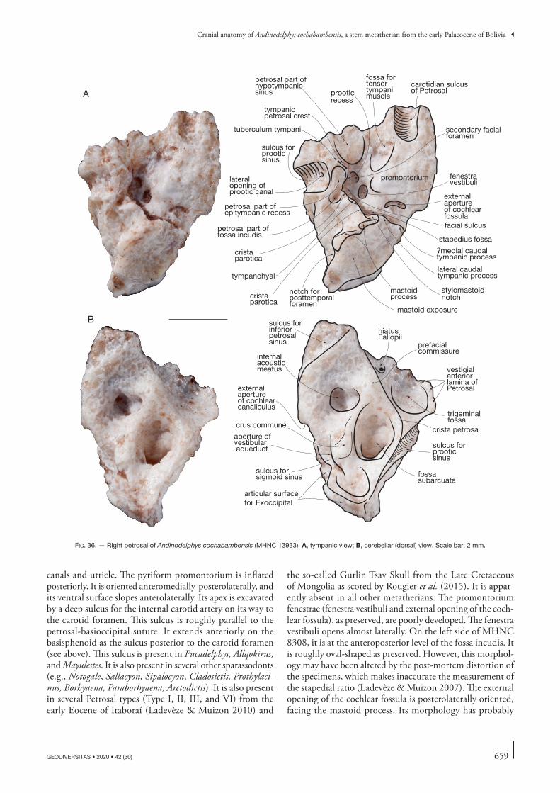
659
Cranial anatomy of Andinodelphys cochabambensis, a stem metatherian from the early Palaeocene of Bolivia
GEODIVERSITAS • 2020 • 42 (30)
canals and utricle. The pyriform promontorium is inflated posteriorly. It is oriented anteromedially-posterolaterally, and its ventral surface slopes anterolaterally. Its apex is excavated by a deep sulcus for the internal carotid artery on its way to the carotid foramen. This sulcus is roughly parallel to the petrosal-basioccipital suture. It extends anteriorly on the basisphenoid as the sulcus posterior to the carotid foramen (see above). This sulcus is present in Pucadelphys, Allqokirus, and Mayulestes. It is also present in several other sparassodonts (e.g., Notogale, Sallacyon, Sipalocyon, Cladosictis, Prothylaci-nus, Borhyaena, Paraborhyaena, Arctodictis). It is also present in several Petrosal types (Type I, II, III, and VI) from the early Eocene of Itaboraí (Ladevèze & Muizon 2010) and
the so-called Gurlin Tsav Skull from the Late Cretaceous of Mongolia as scored by Rougier et al. (2015). It is appar-ently absent in all other metatherians. The promontorium fenestrae (fenestra vestibuli and external opening of the coch-lear fossula), as preserved, are poorly developed. The fenestra vestibuli opens almost laterally. On the left side of MHNC 8308, it is at the anteroposterior level of the fossa incudis. It is roughly oval-shaped as preserved. However, this morphol-ogy may have been altered by the post-mortem distortion of the specimens, which makes inaccurate the measurement of the stapedial ratio (Ladevèze & Muizon 2007). The external opening of the cochlear fossula is posterolaterally oriented, facing the mastoid process. Its morphology has probably
facial sulcus
carotidian sulcusof Petrosal
promontorium
external apertureof cochlearfossula
sulcus forprooticsinus
lateralopening ofprootic canal
petrosal part ofepitympanic recess
petrosal part offossa incudis
tympanicpetrosal crest
mastoidprocess
fenestravestibuli
fossa fortensortympanimuscle
cristaparotica
tympanohyal
secondary facialforamen
petrosal part of hypotympanicsinus
notch for posttemporal foramen
lateral caudal tympanic process
vestigialanterior lamina ofPetrosal
sulcus forprooticsinus
crus commune
hiatus Fallopii
trigeminal fossa
crista petrosa
prefacialcommissure
fossa subarcuata
external apertureof cochlearcanaliculus
sulcus for inferiorpetrosalsinus
internalacousticmeatus
sulcus for sigmoid sinus
aperture of vestibular aqueduct
stapedius fossa
stylomastoidnotch
tuberculum tympani
articular surfacefor Exoccipital
mastoid exposure
A
B
?medial caudal tympanic process
prooticrecess
cristaparotica
fig. 36. — Right petrosal of Andinodelphys cochabambensis (MHNC 13933): A, tympanic view; B, cerebellar (dorsal) view. Scale bar: 2 mm.
660 GEODIVERSITAS • 2020 • 42 (30)
Muizon C. de & Ladevèze S.
also been altered by distortion. The promontorium does not present a well-developed rostral tympanic process in contrast to the condition observed in the Recent didelphids, but resembles Pucadelphys in this respect. However, as mentioned by Ladevèze & Muizon (2007) a distinct swelling is present ventral to the external opening of the cochlear fossula in the same place as the rostral process of Recent didelphids. It may represent an incipient development of a rostral process in Andinodelphys. A similar condition is observed in Allqokirus and Mayulestes, although more developed in this latter taxon. The portion of the petrosal lateral to the promontorium is a wide area extending from the posterior edge of the foramen ovale to the mastoid process. This region houses the hypo-tympanic sinus (sensu Klaauw 1931 and Archer 1976a), the facial nerve, the auditory ossicles, and the stapedial muscle. The anterior region of this area is as wide as the anterior aspect of the promontorium. Anteriorly, on the posterolateral edge of the foramen ovale, the hypotympanic sinus is a small cupula described above in the squamosal section (Fig. 28: blue dotted circle). It is roughly circular and composed of the petrosal posteromedially and the squamosal anterolaterally with a variable participation of the alisphenoid. A distinct hypotympanic sinus is absent in Pucadelphys but it is present in Mayulestes and Allqokirus.
The posterolateral edge of the hypotympanic sinus is formed by a robust but short, rounded ridge (Fig. 36A). This ridge is oblique being oriented anterolaterally-posteromedially. The anteromedial face of this ridge is strongly concave and forms part of the hypotympanic sinus. The posterolateral face is also concave and forms the anteromedial limit of the epitympanic recess (see below). The lateral extremity of the ridge is just medial to the postglenoid foramen and its medial extremity is lateral to the secondary facial foramen (see below) (Figs 28, 30). Following Archer (1976a), Muizon (1998) called a similar ridge in Mayulestes the petrosal crest. However, because this term could be confused with the crista petrosa a structure of the dorsal aspect of the petrosal (Fig. 36B), Ladevèze & Muizon (2010) proposed the term tympanic petrosal crest for this structure. A similar short and rounded tympanic petrosal crest is present in Pucadelphys and Mayulestes. It is also pre-sent, but much sharper, in most sparassodonts (e.g. Notogale, Sipalocyon), in didelphids, and in dasyurids. In Recent didel-phids, the lateral extremity of the tympanic petrosal crest is greatly enlarged and forms a prominent spur-like process, the tuberculum tympani (Wible 2003), which projects ventrally and abuts against the medial edge of the postglenoid fora-men. According to Toeplitz (1920), the tuberculum tympani could be regarded as homologous to the tegmen tympani of eutherians (see also Wible 2003), a statement that could perhaps be extended to the whole tympanic petrosal crest. In Andinodelphys, the lateral edge of the petrosal tympanic crest forms a small knob-like tuberculum tympani, which is barely more salient than the medial edge. A similar condi-tion is observed in Pucadelphys, Allqokirus, and Mayulestes. In contrast, the tuberculum tympani is large and spur-like also in several other sparassodonts such as Notogale and Sipalocyon (Muizon et al. 2018: 369, supplementary data, files 6 and 7).
It is noteworthy that the spur-like tympanohyal identified by Forasiepi et al. (2019: fig. 20A) in Sipalocyon is in fact the tuberculum tympani; the tympanohyal is located on the crista parotica, posteromedially to the fossa incudis, as in observed, for instance, in didelphids (Wible 1990, 2003) and Allqokirus (Muizon et al. 2018: fig. 24A); see also Fig. 36A. The medial edge of the tympanic petrosal crest of Andinodelphys is con-spicuously ventral to the facial sulcus and is excavated by a deep recess, which is regarded here as probably homologous to the lateral trough of the non-therian mammals (Fig. 36A). This recess has been formally termed the prootic recess by Muizon et al. (2018: 406 and fig. 24A). In this trough is the tympanic aperture of the prootic canal (see below). A deep prootic recess is also present in Pucadelphys (Marshall & Mui-zon 1995), and Allqokirus (Muizon et al. 2018).
Bordering the posterior rim of the foramen ovale, between the carotid sulcus medially and the hypotympanic sinus later-ally, is an elongated shallow fossa for the origin of the tensor tympani muscle. It is oriented roughly in an anteromedial-posterolateral direction. On the posterior border of this fossa is a marked pit, which is likely to be also part of the origin of the tensor tympani muscle. This pit is present on all the petrosals known and is therefore unlikely to be the result of distortion. As usual in therians, this fossa is on the lateral edge of the anterior region of the promontorium, but extends later-ally on the ventral aspect of what we interpret as the vestigial anterior lamina of the petrosal.
In Andinodelphys, the anterior lamina of the petrosal is a blade-like anterior extension of the bone, which forms a sig-nificant part of the posteroventral lateral wall of the braincase (Figs 36B; 37A; 38). Part of the anterior lamina is appressed against the internal side of the squamosal, posterior to the alisphenoid. In non-therian mammals, the anterior lamina forms part of the lateral wall of the braincase between the ali-sphenoid anteriorly and the squamosal posteriorly. It encloses the foramen for the mandibular branch (V3) of the trigeminal nerve and in some cases the maxillary branch (V2) (see Wible [1990]; Hopson & Rougier [1993], and Rougier & Wible [2006] for a review). The anterior lamina greatly reduces in metatherians and eutherians (Wible 1990: 200) and disappears in most extant taxa; the mandibular branch of the trigeminal nerve exits the skull either between the alisphenoid and the petrosal (foramen ovale of metatherians) or the foramen is totally enclosed in the alisphenoid (eutherians). However, in basal metatherians and eutherians, a vestigial anterior lamina is still present although the pattern of exit of the V3 is that discussed above. This anterior lamina is partly ventral (i.e. the posterior rim of the foramen ovale) and partly lateral, this por-tion being covered by the squamosal laterally. In dorsal view of the petrosal both ventral and lateral parts of the anterior lamina are clearly visible (Ladevèze & Muizon 2007) and form a deep basin which houses the trigeminal ganglion, the trigeminal fossa (Muizon et al. 2018) (Fig. 38). It is notewor-thy that the trigeminal ganglion is also housed in the medial face of the anterior lamina of non-therian mammals. The structure designated here and elsewhere (Muizon et al. 2018) as trigeminal fossa is the petrosal contribution to the cavum

661
Cranial anatomy of Andinodelphys cochabambensis, a stem metatherian from the early Palaeocene of Bolivia
GEODIVERSITAS • 2020 • 42 (30)
epiptericum (Wilson et al. 2016). A vestigial anterior lamina has been described in the metatherians Pucadelphys (Marshall & Muizon 1995), Andinodelphys (Ladevèze & Muizon 2007),
and Allqokirus (Muizon et al. 2018), in some herpetotheriids (Peratherium cuvieri, P. elegans [very reduced], Amphiperath-erium minutum [more expanded but in a juvenile individual],
articular surface for Squamosal
external aperture of cochlearcanaliculus
articular surface for exoccipital
sulcus for sigmoidsinus
mastoid processexternal apertureof cochlear fossula
sulcus for inferior petrosalsinus
mastoid exposure
promontorium
articular surface for Exoccipital
mastoid exposure
insertion fossa for sternocephalicusmuscle pars mastoidea
mastoid process
aperture of vestibular aqueduct
suclus for sigmoid sinus
sulcus for inferiorpetrosal sinus
external aperture of cochlear canaliculus
mastoidprocess
vestigial anterior lamina of Petrosal
trigeminal fossa
hiatus Fallopii
prefacial commissure
crista petrosa
notch for posttemporalforamen
lateral openning of prootic canal
sulcus for prootic sinus
lateral openning of prootic canal
sulcus for prootic sinus
sulcus for diploic vessels
insertion fossa for sternocephalicusmuscle pars mastoidea
crus commune
notch forposttemporalforamen
sulcus for diploic vessels
articular surface for Squamosal
A
B
C mastoid foramen
fig. 37. — Right petrosal of Andinodelphys cochabambensis (MHNC 13933): A, squamosal view; B, medial view; C, occipital view. Yellow area indicates mastoid external exposure. Scale bar: 2 mm.
662 GEODIVERSITAS • 2020 • 42 (30)
Muizon C. de & Ladevèze S.
see (Selva & Ladevèze 2017; Ladevèze et al. 2020). It has also been decribed in the eutherians Alcidedorbignya (Muizon et al. 2015), Prokennalestes (Wible et al. 2001), and in isolated “zhelestid” petrosals (Ekdale et al. 2004). This interpretation of the occurrence of a vestigial anterior lamina in metatheri-ans confirms Wible’s suggestions that “the anterior lamina … may not be totally absent in all marsupials” and that “a greatly reduced anterior lamina is a synapomorphy of marsupials and eutherians” (Wible 1990: 200 L and R). This vestigial ante-rior lamina of Pucadelphys and Andinodelphys is not exposed on the lateral wall of the braincase, as is the case, for instance, in Vincelestes (Hopson & Rougier 1993). In this genus, the anterior lamina separates the alisphenoid anteriorly and the squamosal posteriorly and the two bones have no contact. In therians, the anterior lamina has reduced and is overlapped laterally by the squamosal, which extends anteriorly, contacts the alisphenoid, and excludes internally the anterior lamina from the lateral wall of the braincase. A condition similar to that of Pucadelphys and Andinodelphys is also present in Didel-phodon as observed on Supplementary fig. 8b of Wilson et al. (2016); see comment to character 168 in Appendix 3.
Rougier & Wible (2006: 277) have refuted the interpreta-tion of a vestigial anterior lamina in Pucadelphys. In order to avoid duplication, we simply refer here to Marshall & Muizon (1995: 78-82) and Muizon et al. (2018: 408) for a discus-sion on the occurrence of an anterior lamina in Pucadelphys and Allqokirus respectively. Following these discussions, we persist in our interpretation of the structure described above, which houses the trigeminal ganglion in Andinodelphys, as a vestigial anterior lamina, and confirm our interpretation in Pucadelphys (Ladevèze & Muizon 2007) and Allqokirus (Muizon et al. 2018).
Posterior to the hypotympanic sinus and medial to the tympanic petrosal crest is the secondary facial foramen, which conveys the hyomandibular ramus of the facial nerve poste-riorly. This foramen is distinctly anterolateral to the fenestra vestibuli. On the posterolateral edge of the tympanic petrosal crest is an elongated cavity located lateral to the fenestra ves-tibuli and separated from it by the facial sulcus. This cavity is the petrosal part of the epitympanic recess. It is bordered laterally by the squamosal, which forms its lateral half (see above), the petrosal-squamosal suture passing in the middle of the recess. On the posterior region of the epitympanic recess is a deep pit, the fossa incudis, which receives the crus breve of the incus. The epitympanic recess is the cavity, which receives the articulation between the incus and the malleus. Because the epitympanic recess is posterior to the ventral petrosal crest, it is also posterior to the prootic canal, which pierces the crest dorsally. This condition is present in Pucadelphys, Mayulestes, Allqokirus, and all Recent didelphids. Therefore, the depression designated by Wible (1990: fig. 2F and fig. 4C) as the epit-ympanic recess, which is anterior to the ventral petrosal crest cannot be this structure. As a matter of fact, it is too anterior to the fenestra vestibuli to possibly receive the malleus-incus articulation. As stated by Muizon (1998), it is the posterior periotic portion of the hypotympanic sinus. Furthermore, the ventrally directed trihedron identified by Wible as the
lateral wall of the epitympanic recess (Wible 1990: fig. 4C, lw) is the lateral extremity of the ventral petrosal crest, which separates the epitympanic recess posteriorly from the hypo-tympanic sinus anteriorly. This trihedron corresponds to the tuberculum tympani, a term, which we retain here following Wible (2003). We prefer not to use the term “lateral wall of the epitympanic recess” because, strictly speaking, the lateral wall of the epitympanic recess, in early therians is formed by the squamosal. The epitympanic recess is a structure, which is carried by both the petrosal and the squamosal and what is generally called epitympanic recess on the petrosal is in fact the “petrosal part of the epitympanic recess”. The same is true for the fossa incudis.
The medial edge of the epitympanic recess is a thin crest, which almost reaches the level of the facial sulcus dorsally: it is the crista parotica. In the posterior region of the epit-ympanic recess and medial to the fossa incudis, the crista parotica extends ventrally and slightly thickens. In this area, it contacts the posttympanic process of the squamosal and is fused to the tympanohyal. The tympanohyal is an ossification of Reichert’s cartilage, which fuses to the petrosal at the crista parotica. This is why it is considered that the crista parotica also extends posterior to the tympanohyal (MacPhee 1981; Wible 2008, 2012). The portion of the crista parotica pos-terior to the tympanohyal is short and forms the lateral edge of the stylomastoid notch. The latter forms a saddle-shaped sulcus for the exit of the facial nerve from the middle ear on the anteromedial edge of the mastoid process. The latter is moderately developed but distinct and forms a small elon-gated knob at the ventral extremity of the nuchal crest in the posterior region of the external acoustic meatus.
On the medial wall of the stylomastoid notch and extending posteromedioventrally from the mastoid process is a conspicu-ous caudal tympanic process. It is a relatively thick ridge, markedly convex anteriorly, which projects anteromedially towards the external aperture of the cochlear fossula. This process has been interpreted by Muizon et al. (2015: fig. 36, 38) as homologous to the lateral caudal tympanic process of early eutherians. Medially the lateral caudal tympanic pro-cess bears a faint saddle-shaped notch with a tiny tubercle medial to this notch (Figs 30; 36A). This tubercle is limited medially by the petrosal-exoccipital suture and is facing (but not adjoined to) another small tubercle on the exoccipital. This medial tubercle of the petrosal of Andinodelphys is in the position of a medial caudal tympanic process (= “tym-panic process” of Wible et al. 2004, 2009) as is observed in early eutherians (Muizon et al. 2015). This tubercle could tentatively be interpreted as a small medial caudal tympanic process. This process is present on the three skulls of Andino-delphys (although weakly developed on MHNC 8264) that preserve a petrosal and, apparently, on the isolated petrosal MHNC 8371. We have observed this process and notch is some Recent didelphids (e.g. Didelphis, Philander, Marmosa). It is noteworthy however that the position of this possible medial tympanic process is unusual since in eutherians it generally faces the lateral caudal tympanic process and tympanohyal; furthermore, according to MacPhee (1981:
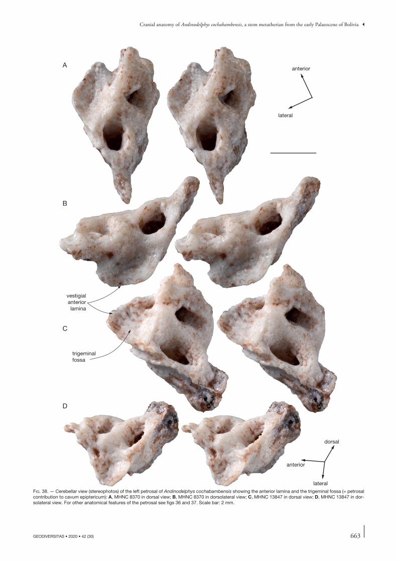
663
Cranial anatomy of Andinodelphys cochabambensis, a stem metatherian from the early Palaeocene of Bolivia
GEODIVERSITAS • 2020 • 42 (30)
trigeminalfossa
vestigialanteriorlamina
C
A
B
D
anterior
lateral
dorsal
anterior
lateralfig. 38. — Cerebellar view (stereophotos) of the left petrosal of Andinodelphys cochabambensis showing the anterior lamina and the trigeminal fossa (= petrosal contribution to cavum epiptericum): A, MHNC 8370 in dorsal view; B, MHNC 8370 in dorsolateral view; C, MHNC 13847 in dorsal view; D, MHNC 13847 in dor-solateral view. For other anatomical features of the petrosal see figs 36 and 37. Scale bar: 2 mm.
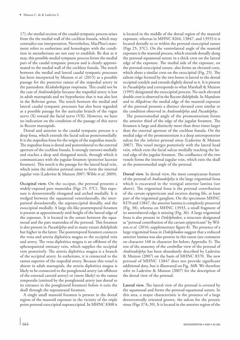
664 GEODIVERSITAS • 2020 • 42 (30)
Muizon C. de & Ladevèze S.
17), the medial section of the caudal tympanic process arises from the the medial wall of the cochlear fossula, which may contradict our interpretation. Nevertheless, MacPhee’s state-ment refers to eutherians and homologies with the condi-tion in metatherians are not easy to establish. Be that as it may, this possible medial tympanic process forms the medial part of the caudal tympanic process and is clearly approxi-mated to the medial wall of the cochlear fossula. The notch between the medial and lateral caudal tympanic processes has been interpreted by Muizon et al. (2015) as a possible passage for the posterior ramus of the stapedial artery in the pantodont Alcidedorbignya inopinata. This could not be the case of Andinodelphys because the stapedial artery is lost in adult marsupials and we hypothesize that it was also lost in the Bolivian genus. The notch between the medial and lateral caudal tympanic processes has also been regarded as a possible passage for the auricular branch of the vagus nerve (X) toward the facial nerve (VII). However, we have no indication on the condition of the passage of this nerve in Recent marsupials.
Dorsal and anterior to the caudal tympanic process is a deep fossa, which extends the facial sulcus posteromedially. It is the stapedius fossa for the origin of the stapedius muscle. The stapedius fossa is dorsal and posterolateral to the external aperture of the cochlear fossula. It strongly narrows medially and reaches a deep and elongated notch, through which it communicates with the jugular foramen (posterior lacerate foramen). This notch is the passage for the lateral head vein, which joins the inferior petrosal sinus to form the internal jugular vein (Ladevèze & Muizon 2007; Wible et al. 2009).
Occipital view. On the occiput, the petrosal presents a widely-exposed pars mastoidea (Figs 25; 37C). This expo-sure is dorsoventrally elongated and arched medially. It is wedged between the squamosal ventrolaterally, the inter-parietal dorsolaterally, the supraoccipital dorsally, and the exoccipital medially. A large slit-like posttemporal foramen is present at approximately mid-height of the lateral edge of the exposure. It is located in the suture between the squa-mosal and the pars mastoidea of the petrosal. This foramen is also present in Pucadelphys and in many extant didelphids but higher in the latter. The posttemporal foramen connects the vena and arteria diploëtica magna to the occipital vein and artery. The vena diploëtica magna is an offshoot of the sphenoparietal emissary vein, which supplies the occipital vein posteriorly. The arteria diploëtica magna is a branch of the occipital artery. In eutherians, it is connected to the ramus superior of the stapedial artery. Because this vessel is absent in adult marsupials, the arteria diploëtica magna is likely to be connected to the postglenoid artery (an offshoot of the external carotid artery) or (more likely) to the ramus temporalis (emitted by the postglenoid artery just dorsal to its entrance in the postglenoid foramen) before it exits the skull through the suprameatal foramen.
A single small mastoid foramen is present in the dorsal region of the mastoid exposure in the vicinity of the triple point petrosal-exoccipital-supraoccipital. In MHNC 8308 it
is located in the middle of the dorsal region of the mastoid exposure, whereas in MHNC 8264, 13847, and 13933 it is located dorsally to or within the petrosal-exoccipital suture (Figs 25; 37C). On the ventrolateral angle of the mastoid exposure is the mastoid process, which extends dorsally along the petrosal-squamosal suture in a thick crest on the lateral edge of the exposure. The medial side of the exposure, on the petrosal-exoccipital suture, also forms an elevated crest, which abuts a similar crest on the exoccipital (Fig. 25). The salient ridge formed by the two bones is lateral to the dorsal occipital condyle and extends slightly dorsal to it. It is present in Pucadelphys and corresponds to what Marshall & Muizon (1995) designated the exoccipital process. No such elevated double crest is observed in the Recent didelphids. In Mayulestes and in Allqokirus the medial edge of the mastoid exposure of the petrosal presents a distinct elevated crest similar to the condition observed in Andinodelphys and Pucadelphys.
The posteromedial angle of the promontorium forms the anterior third of the edge of the jugular foramen. The foramen is large and distinctly more than three times larger than the external aperture of the cochlear fossula. On the medial edge of the promontorium is a deep anteroposterior sulcus for the inferior petrosal sinus (Ladevèze & Muizon 2007). This vessel merges posteriorly with the lateral head vein, which exits the facial sulcus medially reaching the lat-eral edge of the jugular foramen. The confluence of the two vessels forms the internal jugular vein, which exits the skull at the posteromedial angle of the petrosal.
Dorsal view. In dorsal view, the most conspicuous feature of the petrosal of Andinodelphys is the large trigeminal fossa which is excavated in the vestigial anterior lamina (see above). The trigeminal fossa is the petrosal contribution of the cavum epiptericum and receives during life, at least part of the trigeminal ganglion. On the specimens MHNC 8370 and 13847, the anterior lamina is completely preserved (Fig. 38), whereas on MHNC 13933, a small fragment of its anterolateral edge is missing (Fig. 36). A large trigeminal fossa is also present in Didelphodon, a structure designated as “petrosal contribution of the cavum epiptericum” by Wil-son et al. (2016: supplementary figure 8). The presence of a large trigeminal fossa in Didelphodon suggest that a reduced anterior lamina was also present in this taxon (see comment on character 168 in character list below; Appendix 3). The rest of the anatomy of the cerebellar view of the petrosal of Andinodelphys has been abundantly described by Ladevèze & Muizon (2007) on the basis of MHNC 8370. The new petrosal of MHNC 13847 does not provide significant additional data, but is illustrated on Fig. 36B. We therefore refer to Ladevèze & Muizon (2007) for the description of the dorsal view of the petrosal.
Lateral view. The lateral view of the petrosal is covered by the squamosal and forms the petrosal-squamosal suture. In this area, a major characteristic is the presence of a large dorsoventrally oriented groove, the sulcus for the prootic sinus (Figs 37A; 39). It is located in the anterior region of the

665
Cranial anatomy of Andinodelphys cochabambensis, a stem metatherian from the early Palaeocene of Bolivia
GEODIVERSITAS • 2020 • 42 (30)
suture, just posterior to the posterior edge of the trigeminal fossa. The sulcus is slightly concave posteriorly and widens ventrally. In its ventral end is a small foramen, which rep-resents the lateral opening of the prootic canal, which runs in the substance of the petrosal above the petrosal tympanic crest and reaches the facial sulcus medially. The prootic canal conveys the prootic canal vein, which becomes the lateral head vein in the facial sulcus, and merge to the inferior petrosal sinus at the posteromedial angle of the petrosal. In the ventral part of the sulcus for the prootic sinus, on
its posterior edge, at the level of the lateral foramen of the prootic canal, is a small notch from which a faint groove extends posteriorly. This groove is twice narrower than the prootic sulcus and slightly sinuous. It is subhorizontal and ends posteriorly at the level of the notch for the posttemporal foramen (Fig. 37A). This sulcus likely represents the passage of the diploic vessels, the arteria diploëtica magna and vena diploëtica magna, which respectively enter and exit the skull via the posttemporal foramen from the occipital artery and to the occipital vein respectively.
A
B
inferiorpetrosalsinus
transversesinus
sigmoid sinusprooticsinus
superior petrosal sinus
vein of the prootic canalinternal carotid artery
lateral headvein
prooticsinus
fig. 39. — Reconstruction of the major vessels of the auditory region of Andinodelphys cochabambensis: A, tympanic view; B, cerebellar (dorsal) view. Scale bar: 5 mm.
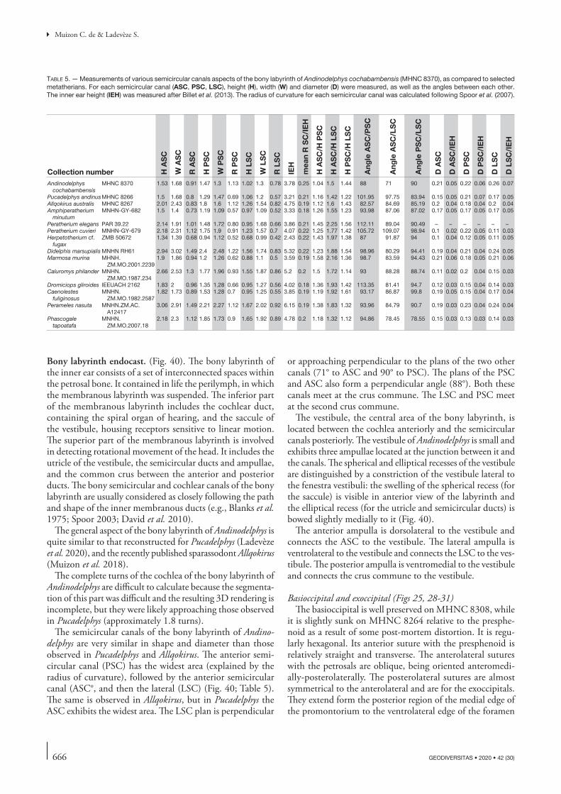
666 GEODIVERSITAS • 2020 • 42 (30)
Muizon C. de & Ladevèze S.
Bony labyrinth endocast. (Fig. 40). The bony labyrinth of the inner ear consists of a set of interconnected spaces within the petrosal bone. It contained in life the perilymph, in which the membranous labyrinth was suspended. The inferior part of the membranous labyrinth includes the cochlear duct, containing the spiral organ of hearing, and the saccule of the vestibule, housing receptors sensitive to linear motion. The superior part of the membranous labyrinth is involved in detecting rotational movement of the head. It includes the utricle of the vestibule, the semicircular ducts and ampullae, and the common crus between the anterior and posterior ducts. The bony semicircular and cochlear canals of the bony labyrinth are usually considered as closely following the path and shape of the inner membranous ducts (e.g., Blanks et al. 1975; Spoor 2003; David et al. 2010).
The general aspect of the bony labyrinth of Andinodelphys is quite similar to that reconstructed for Pucadelphys (Ladevèze et al. 2020), and the recently published sparassodont Allqokirus (Muizon et al. 2018).
The complete turns of the cochlea of the bony labyrinth of Andinodelphys are difficult to calculate because the segmenta-tion of this part was difficult and the resulting 3D rendering is incomplete, but they were likely approaching those observed in Pucadelphys (approximately 1.8 turns).
The semicircular canals of the bony labyrinth of Andino-delphys are very similar in shape and diameter than those observed in Pucadelphys and Allqokirus. The anterior semi-circular canal (PSC) has the widest area (explained by the radius of curvature), followed by the anterior semicircular canal (ASC°, and then the lateral (LSC) (Fig. 40; Table 5). The same is observed in Allqokirus, but in Pucadelphys the ASC exhibits the widest area. The LSC plan is perpendicular
or approaching perpendicular to the plans of the two other canals (71° to ASC and 90° to PSC). The plans of the PSC and ASC also form a perpendicular angle (88°). Both these canals meet at the crus commune. The LSC and PSC meet at the second crus commune.
The vestibule, the central area of the bony labyrinth, is located between the cochlea anteriorly and the semicircular canals posteriorly. The vestibule of Andinodelphys is small and exhibits three ampullae located at the junction between it and the canals. The spherical and elliptical recesses of the vestibule are distinguished by a constriction of the vestibule lateral to the fenestra vestibuli: the swelling of the spherical recess (for the saccule) is visible in anterior view of the labyrinth and the elliptical recess (for the utricle and semicircular ducts) is bowed slightly medially to it (Fig. 40).
The anterior ampulla is dorsolateral to the vestibule and connects the ASC to the vestibule. The lateral ampulla is ventrolateral to the vestibule and connects the LSC to the ves-tibule. The posterior ampulla is ventromedial to the vestibule and connects the crus commune to the vestibule.
Basioccipital and exoccipital (Figs 25, 28-31) The basioccipital is well preserved on MHNC 8308, while
it is slightly sunk on MHNC 8264 relative to the presphe-noid as a result of some post-mortem distortion. It is regu-larly hexagonal. Its anterior suture with the presphenoid is relatively straight and transverse. The anterolateral sutures with the petrosals are oblique, being oriented anteromedi-ally-posterolaterally. The posterolateral sutures are almost symmetrical to the anterolateral and are for the exoccipitals. They extend form the posterior region of the medial edge of the promontorium to the ventrolateral edge of the foramen
table 5. — Measurements of various semicircular canals aspects of the bony labyrinth of Andinodelphys cochabambensis (MHNC 8370), as compared to selected metatherians. For each semicircular canal (ASC, PSC, LSC), height (H), width (W) and diameter (D) were measured, as well as the angles between each other. The inner ear height (IEH) was measured after Billet et al. (2013). The radius of curvature for each semicircular canal was calculated following Spoor et al. (2007).
Collection number H A
SC
W A
SC
R A
SC
H P
SC
W P
SC
R P
SC
H L
SC
W L
SC
R L
SC
IEH
mea
n R
SC
/IE
H
H A
SC
/H P
SC
H A
SC
/H L
SC
H P
SC
/H L
SC
Ang
le A
SC
/PS
C
Ang
le A
SC
/LS
C
Ang
le P
SC
/LS
C
D A
SC
D A
SC
/IE
H
D P
SC
D P
SC
/IE
H
D L
SC
D L
SC
/IE
H
Andinodelphys cochabambensis
MHNC 8370 1.53 1.68 0.91 1.47 1.3 1.13 1.02 1.3 0.78 3.78 0.25 1.04 1.5 1.44 88 71 90 0.21 0.05 0.22 0.06 0.26 0.07
Pucadelphys andinusMHNC 8266 1.5 1.68 0.8 1.29 1.47 0.69 1.06 1.2 0.57 3.21 0.21 1.16 1.42 1.22 101.95 97.75 83.94 0.15 0.05 0.21 0.07 0.17 0.05Allqokirus australis MHNC 8267 2.01 2.43 0.83 1.8 1.6 1.12 1.26 1.54 0.82 4.75 0.19 1.12 1.6 1.43 82.57 84.69 85.19 0.2 0.04 0.18 0.04 0.2 0.04Amphiperatherium
minutumMNHN-GY-682 1.5 1.4 0.73 1.19 1.09 0.57 0.97 1.09 0.52 3.33 0.18 1.26 1.55 1.23 93.98 87.06 87.02 0.17 0.05 0.17 0.05 0.17 0.05
Peratherium elegans PAR 39.22 2.14 1.91 1.01 1.48 1.72 0.80 0.95 1.68 0.66 3.86 0.21 1.45 2.25 1.56 112.11 89.04 90.49 – – – – – –Peratherium cuvieri MNHN-GY-679 2.18 2.31 1.12 1.75 1.9 0.91 1.23 1.57 0.7 4.07 0.22 1.25 1.77 1.42 105.72 109.07 98.94 0.1 0.02 0.22 0.05 0.11 0.03Herpetotherium cf.
fugaxZMB 50672 1.34 1.39 0.68 0.94 1.12 0.52 0.68 0.99 0.42 2.43 0.22 1.43 1.97 1.38 87 91.87 94 0.1 0.04 0.12 0.05 0.11 0.05
Didelphis marsupialis MNHN RH61 2.94 3.02 1.49 2.4 2.48 1.22 1.56 1.74 0.83 5.32 0.22 1.23 1.88 1.54 98.96 80.29 94.41 0.19 0.04 0.21 0.04 0.24 0.05Marmosa murina MHNH.
ZM.MO.2001.22391.9 1.86 0.94 1.2 1.26 0.62 0.88 1.1 0.5 3.59 0.19 1.58 2.16 1.36 98.7 83.59 94.43 0.21 0.06 0.18 0.05 0.21 0.06
Caluromys philander MNHN.ZM.MO.1987.234
2.66 2.53 1.3 1.77 1.96 0.93 1.55 1.87 0.86 5.2 0.2 1.5 1.72 1.14 93 88.28 88.74 0.11 0.02 0.2 0.04 0.15 0.03
Dromiciops gliroides IEEUACH 2162 1.83 2 0.96 1.35 1.28 0.66 0.95 1.27 0.56 4.02 0.18 1.36 1.93 1.42 113.35 81.41 94.7 0.12 0.03 0.15 0.04 0.14 0.03Caenolestes
fuliginosusMNHN.
ZM.MO.1982.25871.82 1.73 0.89 1.53 1.28 0.7 0.95 1.25 0.55 3.85 0.19 1.19 1.92 1.61 93.17 86.87 99.8 0.19 0.05 0.15 0.04 0.17 0.04
Perameles nasuta MNHN.ZM.AC.A12417
3.06 2.91 1.49 2.21 2.27 1.12 1.67 2.02 0.92 6.15 0.19 1.38 1.83 1.32 93.96 84.79 90.7 0.19 0.03 0.23 0.04 0.24 0.04
Phascogale tapoatafa
MNHN.ZM.MO.2007.18
2.18 2.3 1.12 1.85 1.73 0.9 1.65 1.92 0.89 4.78 0.2 1.18 1.32 1.12 94.86 78.45 78.55 0.15 0.03 0.13 0.03 0.14 0.03
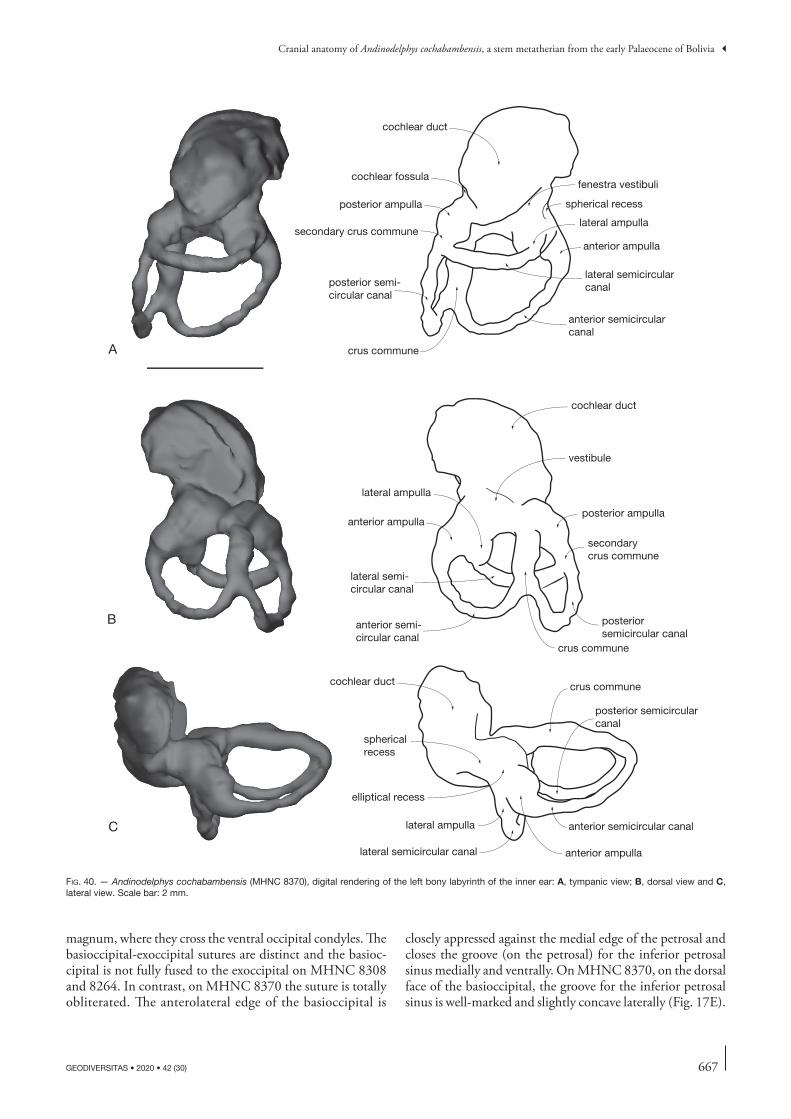
667
Cranial anatomy of Andinodelphys cochabambensis, a stem metatherian from the early Palaeocene of Bolivia
GEODIVERSITAS • 2020 • 42 (30)
magnum, where they cross the ventral occipital condyles. The basioccipital-exoccipital sutures are distinct and the basioc-cipital is not fully fused to the exoccipital on MHNC 8308 and 8264. In contrast, on MHNC 8370 the suture is totally obliterated. The anterolateral edge of the basioccipital is
closely appressed against the medial edge of the petrosal and closes the groove (on the petrosal) for the inferior petrosal sinus medially and ventrally. On MHNC 8370, on the dorsal face of the basioccipital, the groove for the inferior petrosal sinus is well-marked and slightly concave laterally (Fig. 17E).
cochlear duct
cochlear fossula
posterior ampulla
secondary crus commune
posterior semi-circular canal
crus commune
anterior semicircular canal
lateral semicircularcanal
anterior ampulla
lateral ampulla
spherical recess
fenestra vestibuli
cochlear duct
vestibule
posterior ampulla
secondarycrus commune
posteriorsemicircular canal
crus commune
anterior semi-circular canal
lateral semi-circular canal
anterior ampulla
lateral ampulla
A
B
C
cochlear duct
sphericalrecess
elliptical recess
lateral ampulla
lateral semicircular canal anterior ampulla
anterior semicircular canal
crus commune
posterior semicircularcanal
fig. 40. — Andinodelphys cochabambensis (MHNC 8370), digital rendering of the left bony labyrinth of the inner ear: A, tympanic view; B, dorsal view and C, lateral view. Scale bar: 2 mm.
668 GEODIVERSITAS • 2020 • 42 (30)
Muizon C. de & Ladevèze S.
The canal for the inferior petrosal sinus opens posteriorly at the level of the posterior edge of the promontorium, just anterior to the jugular foramen and is fully separated from it. The posterior opening of the canal, the foramen for the inferior petrosal sinus, is well posterior to the basioccipital-exoccipital suture and is formed dorsolaterally (c. one third) by the petrosal and ventromedially (c. two thirds) by the exoc-cipital. The jugular foramen is much larger than the foramen for the inferior petrosal sinus. It is limited anterolaterally by the posteromedial angle of the promontorium (c. one third) and posteromedially (c. two thirds) by the exoccipital. It is distinctly larger than the external aperture of the cochlear fossula. The jugular foramen transmits nerves IX, X, and XI and “occasionally conducts very small branch of sigmoid sinus of transverse venous system to internal jugular vein” (Archer 1976a: 221).
On MHNC 8308, at the lateral end of the left basioccip-ital-exoccipital suture is a triangular opening located at the basioccipital-exoccipital-petrosal triple point (Fig. 30). This fenestra opens dorsally in the canal for the inferior petrosal sinus. It is present on both sides of the specimen. It was prob-ably present on the right side of MHNC 8264 but it is not observed on the left side because of the preservation of the specimen. It was probably present also in MHNC 8370, as distinct notches are observed on the anterolateral edges of the basioccipital of this specimen (Fig. 17D). A similar condition may be present in four specimens of Pucadelphys (MHNC 8377, 8378, 8379, 8380), but it is apparently absent in other specimens (MHNC 8266, 8376, 8381). We have not observed this foramen in other metatherians, fossil or extant. In Andinodelphys we hypothesize that it may have permit-ted exit of a branch of the inferior petrosal sinus. The latter would have had two branches exiting the skull, the posterior one forming the internal jugular vein with the sigmoid sinus, and the anterior one joining the vertebral vein. This possible interpretation is based on comparison with the condition in the dog (Evans & de Lahunta 2013).
The ventral surface of the basioccipital bears an elevated medial keel, which extends from the middle of the intercon-dylar fossa to the anterior suture with the basisphenoid. The lateral edge of the basioccipital bears a conspicuous cylindrical elevation, parallel to the medial edge of the petrosal, which corresponds to the floor of the canal of the inferior petrosal sinus. Between the median keel and the lateral elevation is a deep fossa for the rectus capitis muscle.
On the ventral side of the exoccipital posteromedial to the basioccipital-exoccipital suture, posterolateral to the jugular foramen and anteromedial to the dorsal occipital condyle, are two well-developed hypoglossal foramina. The posterior one is the smaller and is located close to the anteroventral edge of the dorsal occipital condyle. The anterior one is anterior to anteromedial to the other and is close to the basioccipital-exoccipital suture. It is one third (in MHNC 8264) to two times larger (in MHNC 8308) than the posterior one. In MHNC 8308 the anterior foramen is in fact a fossa, which receives two foramina a small anterior one and a larger posterior one. In MHNC
8370, three foramina are observed on the left side with one of them being placed very close to the medial edge of the jugular foramen. On the internal face of this exoccipital two foramina only are observed. The external condition of the right exoccipital of MHNC 8370 is very similar to that observed in some specimens of Pucadelphys (MHNC 8380). Therefore, some variation exists in the number of hypoglossal foramina. A similar variation is also observed in Pucadelphys, as three foramina are observed in MHNC 8266, 8377, and 8380, while two are present in MHNC 8379, 8381, and 8382. In Didelphis virginiana and Monodel-phis domestica (Wible 2003: 175) “these foramina transmit parts of the hypoglossal nerve and accompanying arteries and veins with the arteries ultimately being branches of the vertebral artery”. The hypoglossal foramina may also transmit branches of the sigmoid sinus to the internal jugular vein (Archer 1976a: fig. 2B).
The posterior edge of the basioccipital forms the ventral border of the foramen magnum. It presents a deep U-shaped intercondylar fossa, bordered laterally by the ventral occipital condyles. These condyles extend posterolaterally on the pos-teromedial edge of the exoccipitals, and are therefore crossed by the basioccipital-exoccipital suture. The ventral occipital condyles are elongated and oval-shaped. They are oriented anteromedially-posterolaterally, and form the ventral edge of the foramen magnum, being separated by the intercondylar fossa. Posterodorsolateral to the ventral exoccipital condyles are the large dorsal exoccipital condyles, which form the lat-eral edges of the foramen magnum. They contact the ventral condyles ventrally and extend dorsally onto the occiput. They are sub-circular to roughly quadrate in posterolateral view, being approximately as long as wide in contrast to Pucadel-phys, in which they are distinctly ovoid and elongate. They project posteriorly forming the posteriormost point of the skull. They bear the atlantal articular surface. In lateral view the anterior edge of this surface is distinctly concave. Dorsal to the dorsal occipital condyles, no conspicuous dorsal atlan-tal facets are observed as described by Marshall & Muizon (1995) in Pucadelphys.
In posterior view, the exoccipital encloses the foramen magnum, being separated dorsally by a short notch of the supraoccipital and ventrally by the narrow intercon-dylar notch of the basioccipital (Fig. 25). The foramen magnum carries the vertebral arteries into the skull, all or part of the sigmoid sinus to the vertebral sinus and cranial nerves, which are likely the spinal roots of the accessory nerves (Archer 1976a; Wible 2003). The dorsal suture with the supraoccipital is distinctly observed on the three specimens, which preserve the exoccipital. It is smoothly convex dorsally and extends laterally to the dorsal region of the mastoid exposure of the petrosal, but not to its top. There, the suture is with the petrosal and turns regularly ventrally. The whole lateral edge of the exoccipital forms a roughly semi-circular suture from its intersection with the dorsal edge of the foramen magnum to the ventralmost point on the lateral edge of the jugular foramen. There, the paroccipital process of the exoccipital is poorly developed
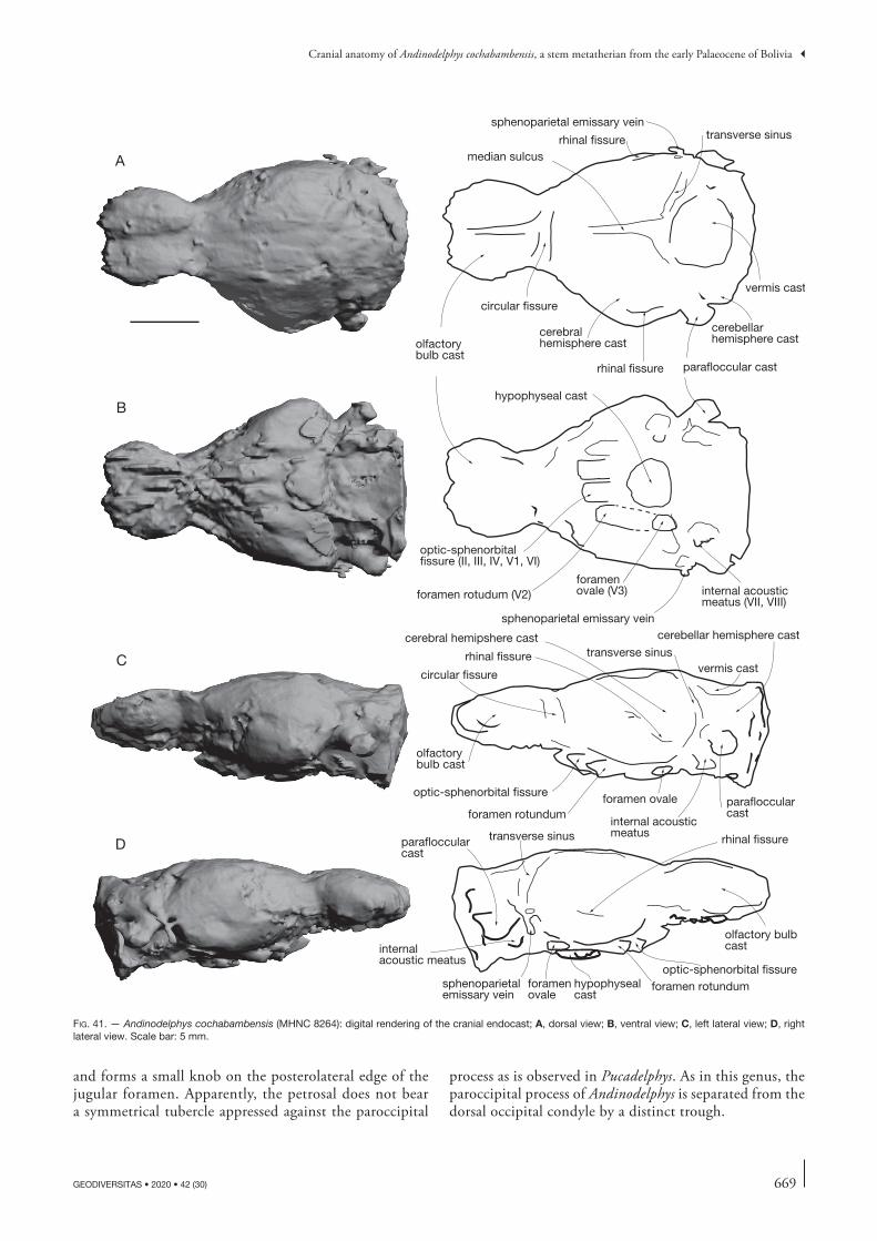
669
Cranial anatomy of Andinodelphys cochabambensis, a stem metatherian from the early Palaeocene of Bolivia
GEODIVERSITAS • 2020 • 42 (30)
and forms a small knob on the posterolateral edge of the jugular foramen. Apparently, the petrosal does not bear a symmetrical tubercle appressed against the paroccipital
process as is observed in Pucadelphys. As in this genus, the paroccipital process of Andinodelphys is separated from the dorsal occipital condyle by a distinct trough.
optic-sphenorbital fissure (II, III, IV, V1, VI)
foramen rotudum (V2)foramen ovale (V3)
sphenoparietal emissary vein
internal acousticmeatus (VII, VIII)
hypophyseal cast
circular fissure
rhinal fissure
cerebral hemipshere cast
vermis cast
cerebellar hemisphere cast
olfactorybulb cast
optic-sphenorbital fissure
foramen rotundumforamen ovale
internal acoustic meatus
parafloccularcast
transverse sinus
parafloccularcast
sphenoparietal emissary vein
foramen ovale
hypophysealcast
foramen rotundumoptic-sphenorbital fissure
transverse sinus rhinal fissure
internalacoustic meatus
olfactory bulb cast
olfactorybulb cast
circular fissure
median sulcusrhinal fissure transverse sinus
cerebral hemisphere cast
rhinal fissure parafloccular cast
cerebellar hemisphere cast
vermis cast
sphenoparietal emissary vein
A
B
C
D
fig. 41. — Andinodelphys cochabambensis (MHNC 8264): digital rendering of the cranial endocast; A, dorsal view; B, ventral view; C, left lateral view; D, right lateral view. Scale bar: 5 mm.
670 GEODIVERSITAS • 2020 • 42 (30)
Muizon C. de & Ladevèze S.
Supraoccipital (Fig. 25) This bone occupies the central dorsal surface of the occiput between the nuchal crest dorsally and the exoccipitals ven-trally. It forms the median region of the dorsal edge of the foramen magnum, where it is distinctly notched. Lateral to this notch, it has a dorsally convex suture with the exoc-cipital and laterally with the dorsal region of the mastoid exposure of the petrosal. Dorsally, it has a suture with the interparietal at the posterior base of the nuchal crest (the interparietal). The surface of the supraoccipital shield is smooth and no vertical ridge or crest is observed medially. Above the foramen magnum, medially the bone is flat and no bulge is observed as that present in Monodelphis for the vermis of the cerebellum (Wible 2003). A small foramen is present dorsomedially, between the supraoccipital and the interparietal, below the nuchal crest at its point of contact with the sagittal crest. A pair of small foramina is also pre-sent on the dorsolateral regions of the supraoccipital, as is observed in Pucadelphys (Marshall & Muizon 1995). These foramina are not clearly observed on MHNC 8370 and 13847, a condition which may be due to the distortion of the specimens.
Cranial endocast (Fig. 41)Only one specimen (MHNC 8264) of our set of Andinodelphys skulls is well preserved enough to allow a poorly distorted digital reconstruction of the cranial endocast. Comparisons with Recent didelphids were possible thanks to published descriptions of metatherian brains (e.g., Owen 1837; Haight 1988; Macrini et al. 2007a). We also used the measurements of the proportions of endocranial casts of several Recent didelphids and australidelphian marsupials provided by Macrini et al. (2007b), (e.g., Monodelphis domestica, Didelphis virginiana, Dasyurus hallucatus, Dromiciops gliroides, Vombatus ursinus, Phascolarctos cinereus). Comparisons with fossil south american metatherians were limited to the descrip-tion of the Pucadelphys andinus endocast (Macrini et al. 2007b).
The overall morphology of the cranial endocast of Andinodelphys cochabambensis (Fig. 41) is very similar to that of Pucadelphys andinus (Macrini et al. 2007b) and to the Recent didelphid marsupials Monodelphis domestica (Macrini et al. 2007a) and Didelphis marsupialis (Dom et al. 1970; Macrini et al. 2007a). It is elongate and narrow with large, spherical olfactory bulb casts on its anterior terminus (Fig. 41). In length, the braincase occupies almost half of the length of the skull, as it is the case in Pucadelphys (43 vs 49%). In lateral view, the endocast displays a flexure of 27° around the hypophyseal cast, which is very similar to the 26° flexure observed in Pucadelphys (Table 6). The width/length endocast aspect ratio is 0.59, the height/length ratio is 0.36, and the height/width ratio is 0.62. The endocast of the brain of Andinodelphys and Pucadelphys extend rather anteriorly, its anteriormost end reaching the orbit at the level of M3.
All the measurements and volumes are provided in Table 6 for Andinodelphys and Pucadelphys.
Olfactory bulbs. The olfactory bulb casts are elliptical and relatively large (Fig. 41), together composing about 12% of the total endocranial space. They are very similar to those of Pucadelphys, which represent 11.69% of the total endocranial space. The olfactory bulb casts of both Andinodelphys and Pucadelphys are relatively larger than those of the Recent mar-supials for which this structure is published (see Macrini et al. 2007b for the compared taxa and measurements). However, they are quite similar in relative volume than those of Didel-phis virginiana (approximately 11%). The olfactory bulb casts of both Andinodelphys and Pucadelphys are are much larger than in the other marsupials of comparison (Monodelphis domestica, Dasyurus hallucatus, Dromiciops gliroides, Vomba-tus ursinus, Phascolarctos cinereus) (Macrini et al. 2007b). The large relative size of the olfactory bulb casts in Andinodelphys and Pucadelphys suggests that these animals displayed a large endoturbinal area and an acute sense of olfaction, similar to that observed in Recent opossums (Rowe et al. 2005).
The olfactory bulb casts are separated from the rest of the endocranial cast by a well-developed circular fissure (sensu Loo 1930; Rowe 1996a, b; transverse fissure of Krause & Kielan-Jaworowska 1993), as in Pucadelphys. Such a deep and well-developed circular fissure is found in the extant marsupials considered here except for Phascolarctos (Macrini et al. 2007b).
On the ventral surface of the endocast, there is no trace of olfactory tracts from the bulbs to the telencephalon (Fig. 41).
table 6. — Endocast measurements for Andinodelphys cochabambensis (MHNC 8264) and Pucadelphys andinus (MHNC 8266).
Andinodelphys cochabambensis
Pucadelphys andinus
Specimen number MHNC 8264 MHNC 8266Source This study This study and
Macrini et al. (2007b)
Endocast flexure (°) 27 26Endocast anteroposterior length
(mm)20.9 16.04
Endocast maximum width (mm) 12.3 9.32Endocast maximim height (mm) 7.6 7.0Cerebrum anteroposterior
length (mm)10.3 8.11
Cerebrum maximal width (mm) 12.3 9.06Olfactory bulbs cast
anteroposterior length (mm)6.3 3.98
Olfactory bulbs cast maximum width (mm)
8.9 4.94
Olfactory bulbs cast maximum height (mm)
3.6 3.7
Cerebellum anteroposterior length (mm)
7.1 3.6
Cerebellum maximal width (mm) 12.1 6.6Cerebellum maximal height (mm) 7.7 3.9Hypophyseal fossa cast
anteroposterior length (mm)2.8 2.67
Hypophyseal fossa cast maximum width (mm)
3.1 2.95
Hypophyseal fossa cast maximum height (mm)
0.23 0.7
Total endocast volume (mm3) 758.301 311.506Olfactory bulb volume (mm3) 91.61 36.41Olfactory bulbs/endocast
volume (%)12% 11.7
Skull maximal length (mm) 48.9 32.28Endocast length/skull length 43% 49%Overall skull volume (mm3) 2524.04 921,95Endocast vol./Skull vol. 30% 34%
671
Cranial anatomy of Andinodelphys cochabambensis, a stem metatherian from the early Palaeocene of Bolivia
GEODIVERSITAS • 2020 • 42 (30)
Cerebrum and midbrain. Immediately posterior to the circular fissure, in dorsal view, are cast of the cerebral hemispheres. They are elongated and ovoid in shape. The surface of the cerebral hemispheres is smooth with no gyri and no sulci (= lissencephalic). The relatively small endocranial cavity of Andinodelphys suggests that the lissencephalic endocast had contained a lissencephalic brain because the latter was less likely to have been convoluted (see Edinger 1955; Tobias 1971; Holloway et al. 2004).
The two cerebral hemispheres are separated by the median or sagittal sinus, which is only slightly marked, as compared to the deep sinus observed on the endocast of Pucadelphys (Macrini et al. 2007b). In dorsal view, the two hemispheres are very similar to those of the endocast of Pucadelphys: they are triangular in shape and they do not expand much later-ally, clearly less so than Monodelphis, in which they extend beyond the lateral extent of the parafloccular casts of the cer-ebellum. At their posterior end, the two cerebral hemispheres diverge from each other in the part anterior to the cast of the vermis. In lateral view (Fig. 41), the cerebral hemispheres are more elevated than the olfactory bulbs, and their ventral part is expanded in a piriform lobe, which does not protrude strongly ventrally.
The height/length aspect ratio of the cerebral hemisphere casts of Andinodelphys is 0.74, which is closer to the values obtained for Monodelphis (range 0.74-0.86) than that obtained for Pucadelphys (0.62). The width/length ratio of the cerebral hemisphere casts of Andinodelphys is 1.19, which is just between the ratios calculated for Pucadelphys (1.12) and Monodelphis (range 1.24-1.38; mean 1.29). The cerebral hemisphere cast length/total endocast length ratios are quite similar between Andinodelphys (0.49), Pucadelphys (0.51) and Monodelphis (range 0.42-0.50; mean 0.47).
The posterior portion of the rhinal fissure is visible on the lateral surface of the endocast, and is very similar to the rhinal fissure observed in Didelphis virginiana (Macrini et al. 2007a). However, a rhinal fissure is nowhere visible on the cerebral hemispheres of Pucadelphys (Macrini et al. 2007b). In Monodelphis domestica, the rhinal fissure is not visible on any of the endocasts, but it is clearly visible on dissected brains (Macrini et al. 2007a). The rhinal fissure marks the boundary between the isocortex (=neocortex) and the piriform lobe of the cerebrum (Jerison 1991).
Cerebellum. The midbrain of the endocast of Andinodelphys does not show dorsal exposure. It is the same in Pucadelphys and other marsupial endocasts used here as comparison. Lack of midbrain exposure in mammals can be explained by a cover by blood sinus, posterior expansion of the cerebral hemispheres, anterior expansion of the cerebellum, or any combination of these (Edinger 1964). It has been shown that the venous sinus system covers the midbrain in Monodelphis, Didelphis, Dromiciops, Dasyurus, and Phascolarctos (Macrini et al. 2007a), and the same can be assumed for Andinodelphys.
On the posterodorsal surface of the endocast, a large bulge, interpreted as the cast of the vermis of the cerebellum, covers the midbrain (Fig. 41). As in Pucadelphys, the vermis cast of
Andinodelphys extends anterior to the casts of the paraflocculi. Cerebellar hemisphere casts are also visible on the endocast, but the imprints of the superior (dorsal) sagittal, transverse, and sigmoid sinuses are not visible. The parafloccular casts of the cerebellum are as prominent as those of the cerebellum of Pucadelphys and also Didelphis, Monodelphis, and Phasco-larctos, but clearly less than those observed in Dromiciops and Dasyurus (Macrini et al. 2007a). The paired paraflocculi are a lateral extension of the cerebellum housed in the subarcuate fossa of the petrosal bone, and are involved with coordination, balance, and vestibular sensory acquisition.
At the ventro-central surface of the endocast of Andinodelphys, a conspicuous, circular, shallow hypophyseal cast contained in life the pituitary gland. It is very difficult to provide reliable measurements of the hypophyseal cast (especially its depth) but it is estimated as having a width/length aspect ratio of 1.11, as in Pucadelphys. Here, the hypophyseal fossa of Andi-nodelphys is quite small as compared to the values calculated for Pucadelphys (Macrini et al. 2007b), but its height may well be underestimated.
The pons and medulla oblongata are not visible on the ventral surface of Andinodelphys. They are not visible either on the endocasts of Pucadelphys and the extant marsupials of comparison. These structures may have been obscured by meninges of the brain and/ or blood sinuses.
Cranial nerves and blood vessels. Anterior to the hypophy-seal cast are the paired casts of the canals that transmitted the maxillary branches of the trigeminal nerve (V2) (Fig. 41B). The paired canals for the V2 are more posterior and lateral to the hypophyseal cast in Pucadelphys. The right and left casts for the exit of the V2, the foramen rotundum, are widely separated from each other. The optic-sphenorbital fissure (optic-orbital foramen) is located anteromedial to the fora-men rotundum and transmitted the cranial nerves II, III, IV, V1, VI, the ophthalmic artery, and a vein (Marshall & Muizon 1995). The right and left casts for the exit of these nerves in the endocast are close to each other but are not confluent. Posterolateral to the hypophyseal cast and antero-medial to the internal auditory meatus is the foramen ovale for the mandibular branch of the trigeminal nerve (V3). As in Pucadelphys and most metatherians (Wible 1990; Macrini et al. 2007b), the cavum epyptericum, the space at the level with the sphenoparietal fenestra which houses the trigeminal ganglion (V), is not confluent with the cavum supracochle-are for the geniculate ganglion (VII). The cast of the internal auditory meatus for passage of cranial nerves VII and VIII is visible on the right side of the endocast.
On the dorsal and right lateral surface of the endocast (Fig. 41A, D), the pathway and ramification of the transverse sinus is well visible. The transverse sinus sulcus runs just between the cerebral hemisphere and the vermis and then divides into three branches: superior petrosal sinus, sigmoid sinus, and prootic sinus, the latter anastomoses with the sphe-noparietal emissary vein, the cast of which is visible on the endocranial cast and exits the skull through the postglenoid foramen (Wible 1990, 2003).
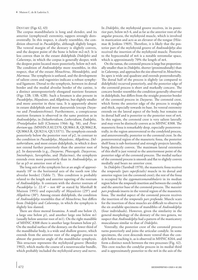
672 GEODIVERSITAS • 2020 • 42 (30)
Muizon C. de & Ladevèze S.
dentary (Figs 42, 43) The corpus mandibularis is long and slender, and its anterior (symphyseal) extremity, tappers strongly dors-oventrally. In this respect, it clearly resembles, the con-dition observed in Pucadelphys, although slightly longer. The ventral margin of the dentary is slightly convex, and the deepest point of the bone is below m2-m3. It is less convex than in the extant didelphids Didelphis and Caluromys, in which the corpus is generally deeper, with the deepest point located more posteriorly, below m3-m4. The condition of Andinodelphys more resembles, in this respect, that of the extant Metachirus, Monodelphis, and Marmosa. The symphysis is unfused, and the development of salient crests and rugosities indicate a robust symphy-seal ligament. Dorsal to the symphysis, between its dorsal border and the medial alveolar border of the canine, is a distinct anteroposteriorly elongated nutrient foramen (Figs 12B; 13B; 42B). Such a foramen is also present in Pucadelphys, Mayulestes, and Allqokirus, but slightly smaller and more anterior in these taxa. It is apparently absent in extant didelphids and most dasyuroids (except Dasyu-rus and Pseudantechinus). Furthermore, a conspicuous nutrient foramen is observed in the same position as in Andinodelphys, in Deltatheridium, Lotheridium, Eodelphis, ?Protalphadon lulli (Clemens 1966: fig. 58), and Pera-therium spp. (MNHN.F.AU2370, QU8061.R, QU8062.R, QU8063.R, QU8214, QU13371). The symphysis extends posteriorly below the posterior root of p2, in contrast to the condition in Pucadelphys, Mayulestes, Allqokirus, Del-tatheridium, and most extant didelphids, in which it does not extend further posteriorly than the anterior root of p2. In dasyuroids (e.g., Dasyurus, Phascogale, Dasycercus, Sminthopsis, Sarcophilus, and Thylacinus), the symphysis extends even more posteriorly than in Andinodelphys, as far as p3 or anterior root of m1.
The long axis of the symphysis lies at an angle of approxi-mately 18° to the horizontal axis of the tooth row (the alveolar border) (Table 7). This condition is probably related to the length and anterior tapering of the rostrum of Andinodelphys. It contrasts with the shorter rostrum of Pucadelphys (c. 22.4° – not 40° as stated by Marshall & Muizon 1995) and especially of Mayulestes (29°) and Allqokirus (30°). Among extant didelphids, the condition of Andinodelphys resembles that of Metachirus, but differs from Didelphis and Caluromys, in which the symphysis is slightly less slanted.
There are three mental foramina. A small one below i3, a large one below p1, and another large one below m1 (usually below anterior root of m1). On the right mandible of MHNC 8308 there is another large foramen below p2. On the medial surface of the dentary, on the lower third of the mandibular body, is a wide and shallow groove, which extends from the anterior root of the angular process to almost the posterior angle of the mandibular symphysis. This structure represents the mylohyoid groove (Bensley 1902), which marks the course of a neurovascular bundle, which probably included the mylohyoid artery and nerve.
In Didelphis, the mylohyoid groove receives, in its poste-rior part, below m3-4, and as far as the anterior root of the angular process, the mylohyoid muscle, which is involved in mastication and acts as an elevator of the tongue (Hiie-mae & Jenkins 1969). Therefore, it is likely that the pos-terior part of the mylohyoid groove of Andinodelphys also received the insertion of the mylohyoid muscle. Posterior to the hypoconulid of m4 is a notable retromolar space, which is approximately 70% the length of m4.
On the ramus, the coronoid process is large but proportion-ally smaller than in Didelphis, shorter (proximodistally) than in Caluromys, and approaches the size observed in Metachirus. Its apex is wide and quadrate and extends posterodorsally. The dorsal half of the process is slightly (as compared to didelphids) recurved posteriorly, and the posterior edge of the coronoid process is short and markedly concave. The concave border resembles the condition generally observed in didelphids, but differs from the straight posterior border of the coronoid process in Dasyurus. The coronoid crest, which forms the anterior edge of the process is straight and thick, especially towards its base. Its ventral extremity extends on the lateral aspect of the body, but remains on its dorsal half and is posterior to the posterior root of m4. In this region, the coronoid crest is very salient laterally and may even be distinctly convex as in MHNC 8308. The masseteric fossa is remarkably deep, especially posteroven-trally, in the region anteroventral to the condyloid process, and anteroventrally, posterior to the coronoid crest. In the posteroventral region of the masseteric fossa, the posterior shelf fossa is sub-horizontal and strongly projects laterally, being distinctly convex. The maximum lateral extension of this shelf is just ventral to the anteriormost point of the posterior edge of the coronoid process. The medial surface of the coronoid process is smooth and flat to slightly convex medially and bears no anterior crest.
In Didelphis (Turnbull 1970), the masseteric fossa receives the temporalis (pars superficialis) muscle in its dorsal and anterior region (on the coronoid crest); the rest of the fossa is occupied by the zygomaticomandibularis in its median region below the temporalis insertion and between the condyle and the anterior base of the coronoid process. The masseter pars profunda inserts in the ventral region of the masseteric fossa. The medial aspect of the coronoid process receives the insertion of the temporalis pars profunda. Muscle scars for the insertion of these muscles are difficult to observe in the six available specimens of mandibles of Andinodelphys (four individuals). However, given the similarity in the general morphology of the dentary of the two genera, we suspect that Andinodelphys had a pattern of the masticatory musculature similar to that of Didelphis.
Ventrally, the posterior crest of the coronoid process turns posteriorly and joins the articular condyle. In some specimens, the crest descends more ventral than the con-dyle before reaching it, as is observed in MHNC 8264, and form a distinct notch between the two processes (Fig. 42). This crest reaches the condylar process in its medial third and is approximately posterior to the m4 in the axis of the
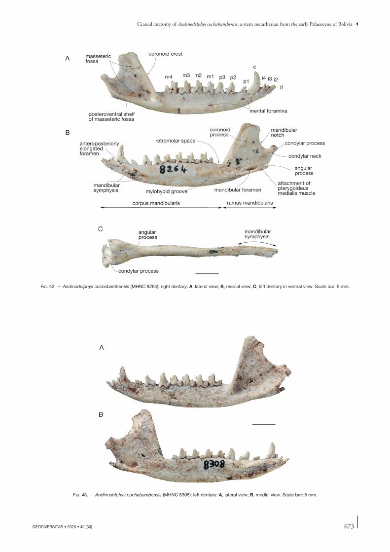
673
Cranial anatomy of Andinodelphys cochabambensis, a stem metatherian from the early Palaeocene of Bolivia
GEODIVERSITAS • 2020 • 42 (30)
corpus mandibularis ramus mandibularis
mylohyoid groove mandibular foramen
angularprocess
mandibular symphysis
coronoidprocess
attachment of pterygoideus medialis muscle
condylar process
condylar neck
mandibularnotch
coronoid crestmasseteric fossa
mental foraminaposteroventral shelfof masseteric fossa
c
p1p2p3m1m2m3m4
i1
i2i3i4
A
B
C angularprocess
condylar process
mandibular symphysis
retromolar spaceanteroposteriorlyelongated foramen
fig. 42. — Andinodelphys cochabambensis (MHNC 8264): right dentary: A, lateral view; B, medial view; C, left dentary in ventral view. Scale bar: 5 mm.
A
B
fig. 43. — Andinodelphys cochabambensis (MHNC 8308): left dentary: A, lateral view; B, medial view. Scale bar: 5 mm.
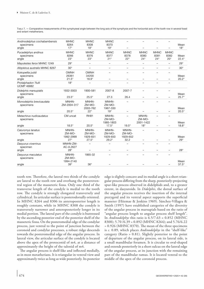
674 GEODIVERSITAS • 2020 • 42 (30)
Muizon C. de & Ladevèze S.
tooth row. Therefore, the lateral two thirds of the condyle are lateral to the tooth row and overhang the posteroven-tral region of the masseteric fossa. Only one third of the transverse length of the condyle is medial to the tooth row. The condyle is strongly elongated transversely and cylindrical. Its articular surface is posterodorsally oriented. In MHNC 8264 and 8306 its anteroposterior length is roughly constant, while in MHNC 8308 the condyle is transversely narrower and anteroposteriorly longer in its medial portion. The lateral part of the condyle is buttressed by the ascending posterior end of the posterior shelf of the masseteric fossa. On the posteromedial edge of the condylar process, just ventral to the point of junction between the coronoid and condylar processes, a robust ridge descends towards the posteromedial edge of the angular process. In lateral view, the articular surface of the condyle is located above the apex of the protoconid of m4, at a distance of approximately the height of the talonid of m4.
The angular process is shelf-like and inflected medially, as in most metatherians. It is triangular in ventral view and approximately twice as long as wide posteriorly. Its posterior
edge is slightly concave and its medial angle is a short trian-gular process differing from the sharp, posteriorly-projecting spur-like process observed in didelphids and, to a greater extent, in dasyuroids. In Didelphis, the dorsal surface of the angular process receives the insertion of the internal pterygoid and its ventral aspect supports the superficial masseter (Hiiemae & Jenkins 1969). Sánchez-Villagra & Smith (1997) have established categories of the diversity of the angular process in marsupials based on the ratio of “angular process length to angular process shelf length”. In Andinodelphys this ratio is 6.5/7.63 = 0.852 (MHNC 8308); 5.70 /6.39 = 0.892 (MHNC 8264); and 5.76/6.22 = 0.926 (MHNC 8370). The mean of the three specimens is c. 0.89, which places Andinodelphys in the “shelf-like” category (Ratio > 0.81). Slightly posterior to the point of departure of the angular process, on its lateral side, is a small mandibular foramen. It is circular to oval-shaped and extends posteriorly in a short sulcus on the lateral edge of the angular process, at its junction with the remaining part of the mandibular ramus. It is located ventral to the middle of the apex of the coronoid process.
table 7. — Comparative measurements of the symphyseal angle between the long axis of the symphysis and the horizontal axis of the tooth row in several fossil and extant metatherians.
Andinodelphys cochabambensis specimens
MHNC 8264
MHNC8308
MHNC8370
– – – –Mean
angle 17° 18° 19° – – – – 18°
Pucadelphys andinus specimens
MHNC8266
MHNC8376
MHNC8377
MHNC8378
MHNC8380
MHNC8381
MHNC8382 Mean
angle 23° 23° 21° 22° 24° 24° 20° 22.4°
Mayulestes ferox MHNC 1249 29° – – – – – – 29°
Allqokirus australis MHNC 8267 30° – – – – – – 30°
Kokopellia juddi specimens
OMNH26361
OMNH34200
– – – – –Mean
angle 21.5° 18.9° – – – – – 20.2°Protalphadon ?lulliUCMP 46882
18° – – – – – –
Didelphis marsupialis specimens
1932-3003 1900-581 2007-8 2007-7 – – –Mean
angle 23.5° 25.5° 27.3 26.4 – – – 25.7°
Monodelphis brevicaudata specimens
MNHN- ZM-2004-317
MNHN-ZM-MO- 2003-762
MNHN-ZM-MO- 1967-330
– – – –
Meanangle 20.5° 22° 19° – – – – 20.5°
Metachirus nudicaudatus specimens
CM uncat RH81 MNHN-ZM-MO-
1985-1803
– MNHN-ZM-MO-
2001-1422
– –
Meanangle 18.5° 20.5° 17.5° 19.5° 18° – – 18.8°
Caluromys lanatus specimens
MNHN-ZM-MO-
1992-2999
MNHN-ZM-MO- 1929-651
MNHN-ZM-MO- 1929-650
MNHN-ZM-MO- 1929-652
– – –
Meanangle 27° 27.5° 29.2° 32.2 – – – 29°Dasyurus viverrinus
specimenMNHN-ZM-AC-A.2627
– – – – – –
angle 28° – – – – – – 28°
Dasyurus maculatus specimens
MNHN-ZM-MO-
1994-2140
1865-32 – – – – –
Meanangle 39° 36° – – – – – 37.5°
675
Cranial anatomy of Andinodelphys cochabambensis, a stem metatherian from the early Palaeocene of Bolivia
GEODIVERSITAS • 2020 • 42 (30)
COMPARISON AND DISCUSSION
In the following discussion, because Andinodelphys is morpho-logically very similar to Pucadelphys, concerning the characters that are identical in the two genera, we refer to discussion of Pucadelphys characters in Marshall & Muizon (1995), except when new discoveries, observations, or publications require further discussion on a given feature.
general cranial morphology The general morphology of the skull of Andinodelphys cocha-bambensis is similar to that of Pucadelphys andinus from the same locality but significantly larger. The skull of Andinodelphys is approximately 35% longer (in absolute value) than that of Pucadelphys with a rostrum proportionally 24% longer. Andinodelphys approaches the size of Mayulestes and Allqokirus (although slightly smaller) and is among the largest known metatherian taxa of the Tiupampa fauna. Beside these size and skull proportion differences and a few cranial features discussed below, most of the cranial anatomy of Andinodel-phys does not significantly depart from that of Pucadelphys. The comparative life reconstructions of Andinodelphys and Pucadelphys presented in Fig. 44 intent to bring to light over-all resemblance existing between the two Tiupampan taxa.
dental anatomy
The dental anatomy of Andinodelphys is also quite similar to that of Pucadelphys. Both genera have the same dental formula. In both genera, P3 is the highest tooth of the upper cheek tooth series, the protocone is massive and anteroposteriorly expanded, the centrocrista is slightly V-shaped, the stylar shelf is transversely narrower than half of the width of the molars (M1-3), the stylar cusps B and D are the largest of the sty-lar shelf, the metaconid is large, being conspicuously larger than the paraconid and barely smaller than the protoconid, and the paraconid and metaconid are adjoined at their bases. Dentally, except for its larger size, Andinodelphys differs from Pucadelphys in minor features such as the enlarged I1 and the smaller stylar cusp C. As in Pucadelphys, Andinodelphys dif-fers from Mayulestes and Allqokirus in the following features: paracone and metacone well separated at their base (fused at their bases in the latter two); postmetacrista on upper molars (especially M3) not extended distolabially and not significantly more expanded than the mesiolabial angle of the tooth; angle between the paracone-metacone axis and the preparacrista on M4 not widely open and close to 90° (≥ to 130° in the latter two); metaconid on m3, twice as large in height and volume than the paraconid (subequal in size to smaller in the two latter), paraconid and metaconid adjoined at base (widely separated in the latter two); paracristid lacking a carnassial notch (conspicuous in the latter two); relatively straight labial edge of the protoconid in anterior view, (expanded labially at mid-height in the latter two).
Size and position of the I1As is observed in didelphids, the I1 of Andinodelphys is larger and extends further ventrally than the other upper incisors,
because a larger part of the root (as compared to other inci-sors) is external to the alveolus. Furthermore, I1 is separated from I2 by a small but distinct diastema, whereas the other incisors are not separated by diastemata. In anterior view, the crowns of the I1 converge one toward the other and are in contact medially, whereas their roots diverge dorsally. A similar condition is present in Mayulestes. In Pucadelphys, I1 is smaller than the other incisors and does not protrude ven-trally more than the other incisors, as is observed in MHNC 8378, the only specimen that preserves all the upper incisors (Fig. 45). However, in this specimen the extra-alveolar portion of the root is proportionally larger than in the other incisors. Comparison with other fossil taxa is difficult because the pre-maxillae and incisors are almost never preserved. A similar condition, however, is present in Didelphodon, although in this taxon, both I1 “do not clearly angle each other or make contact with each other at their crown apices” (Wilson et al. 2016, supplementary notes: 56). Furthermore, in Didelpho-don no diastema separates I1 and I2. As observed from the size and position of the upper incisors alveoli, the condition in the Gurlin Tsav skull resembles that of Andinodelphys in the larger size of the alveolus of I1 as compared to that of I2, but it differs in the probable absence of diastema. Apparently Lotheridium, a deltatheroidan from the Late Cretaceous of China, lacks an enlarged I1 (Bi et al. 2015). The I1 condition of Andinodelphys and didelphids is present in Dromiciops and in several dasyurids (e.g. Barinya, Sminthopsis, Dasycercus, Phascogale). In sparassodonts other than Mayulestes (in the latter the condition is similar to that of Andinodelphys) the I1 is not enlarged in the very few specimens preserving all the upper incisors (e.g., Prothylacynus, Borhyaena, Cladosictis, Arctodictis), as well as in thylacinids (e.g. Thylacinus, Nimbaci-nus). However, the condition in these taxa may be derived as possibly related to hypercarnivory.
Morphology of the M4. As discussed in the description above, the M4 of Andino-delphys and Pucadelphys differs from that of Mayulestes and Allqokirus in the angle formed by the preparacrista and the centrocrista (or the paracone-metacone axis if the centrocrista is strongly V-shaped), which is close to c. 90°-100° in the former two genera and c. 130° in the latter two genera. The Mayulestes and Allqokirus condition is even more pronounced (approaching in some case 180°) in later sparassodonts, in which cristae can still be observed and have not been oblit-erated by the reduction of the M4 (Fig. 46). In sparassodonts, the opening of the angle is related to the labiolingual posi-tion of the metacone (more labial than in Pucadelphys and Andinodelphys) and to the length of the labial edge of the tooth (in the case of the M4 it is the posterolateral maxillary alveolar border), which on the M4 is not strictly speaking labial, but corresponds to the labial edge in anterior molars. Because of this lingual displacement of the metacone, the posterior edge of the protocone (i.e. from the posterolingual edge of the metacone to the lingual side of the protocone) is shortened, and is generally shorter than the labial edge of the tooth. The condition is reversed in Andinodelphys and
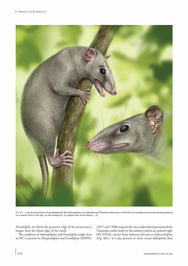
676 GEODIVERSITAS • 2020 • 42 (30)
Muizon C. de & Ladevèze S.
Pucadelphys, in which the posterior edge of the protocone is longer than the labial edge of the tooth.
The condition of Andinodelphys and Pucadelphys (angle close to 90°) is present in Mizquedelphys and Incadelphys (MHNC
13917 and 13906 respectively, two undescribed specimens from Tiupampa under study by the authors) and in an isolated right M4 (DGM, uncat) from Itaboraí referred to Itaboraidelphys (Fig. 46C). It is also present in most extant didelphids (but
fig. 44. — Life reconstructions of pucadelphyids: A, Andinodelphys cochabambensis (Charlène Letenneur); on the left is a complete animal (natural size) grasping on a branch and on the right, on the foreground, is a lateral view of the head (c. × 2).

677
Cranial anatomy of Andinodelphys cochabambensis, a stem metatherian from the early Palaeocene of Bolivia
GEODIVERSITAS • 2020 • 42 (30)
B
C
fig. 44. — Life reconstructions of pucadelphyids: B, positioning of the silhouette of 3D digital rendering of the skull (MHNC 8264), on which the life reconstruc-tions have been based; on the right is the head of the complete animal of fig. A and on the right is the head of the foregroung of fig. A (drawings of A and B are at the same scale); C, Pucadelphys andinus (Sophie Fernandez), scene of several individuals (natural size) on the ground. Scale bars: 1 cm.
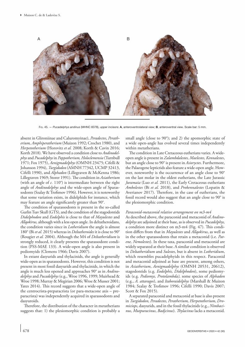
678 GEODIVERSITAS • 2020 • 42 (30)
Muizon C. de & Ladevèze S.
absent in Glironiinae and Caluromyinae), Peradectes, Perath-erium, Amphiperatherium (Muizon 1992; Crochet 1980), and Herpetotherium (Horovitz et al. 2008; Korth & Cavin 2016; Korth 2018). We have observed a condition close to Andinodel-phys and Pucadelphys in Pappotherium, Holoclemensia (Turnbull 1971; Fox 1975), Aenigmadelphys (OMNH 23475; Cifelli & Johanson 1994), Turgidodon (AMNH 77342, UCMP 32413, Cifelli 1990), and Alphadon (Lillegraven & McKenna 1986; Lillegraven 1969; Storer 1991). The condition in Asiatherium (with an angle of c. 110°) is intermediate between the right angle of Andinodelphys and the wide-open angle of Sparas-sodonts (Szalay & Trofimov 1996). However, it is noteworthy that some variation exists, in didelphids for instance, which may feature an angle significantly greater than 90°.
The condition of sparassodonts is present in the so-called Gurlin Tsav Skull (GTS), and the condition of the stagodontids Didelphodon and Eodelphis is close to that of Mayulestes and Allqokirus, although with a less open angle. In deltatheroidans, the condition varies since in Lotheridium the angle is almost 180° (Bi et al. 2015) whereas in Deltatheroides it is close to 90° (Rougier et al. 2004). Although the M4 of Deltatheridium is strongly reduced, it clearly presents the sparassodont condi-tion (PSS-MAE 133). A wide-open angle is also present in pediomyids (Clemens 1966; Davis 2007).
In extant dasyurids and thylacinids, the angle is generally wide-open as in sparassodonts. However, this condition is not present in most fossil dasyurids and thylacinids, in which the angle is much less opened and approaches 90° as in Andino-delphys and Pucadelphys (e.g., Wroe 1996, 1999; Muirhead & Wroe 1998; Murray & Megirian 2006; Wroe & Musser 2001; Yates 2014). This record suggests that a wide-open angle of the centrocrista-preparacrista (or para-metacone axis – pre-paracrista) was independently acquired in sparassodonts and dasyuroids.
Therefore, the distribution of the character in metatherians suggests that: 1) the plesiomorphic condition is probably a
small angle (close to 90°); and 2) the apomorphic state of a wide open-angle has evolved several times independently within metatherians.
The condition in Late Cretaceous eutherians varies. A wide-open angle is present in Zalambdalestes, Maelestes, Kennalestes, but an angle close to 90° is present in Asioryctes. Furthermore, the Palaeogene leptictids also feature a wide-open angle. How-ever, noteworthy is the occurrence of an angle close to 90° on the last molar in the oldest eutherians, the Late Jurassic Juramaia (Luo et al. 2011), the Early Cretaceous eutherians Ambolestes (Bi et al. 2018), and Prokennalestes (Lopatin & Averianov 2017). Therefore, in the case of eutherians, the fossil record would also suggest that an angle close to 90° is the plesiomorphic condition.
Paraconid-metaconid relative arrangement on m3-m4As described above, the paraconid and metaconid of Andino-delphys are adjoined at their base, as is observed in Pucadelphys, a condition more distinct on m3-m4 (Fig. 47). This condi-tion differs from that in Mayulestes and Allqokirus, as well as in the other sparassodonts that retain a metaconid (i.e. Pat-ene, Nemolestes). In these taxa, paraconid and metaconid are widely separated at their base. A similar condition is observed in Deltatheridium and Sulestes, but is absent in Lotheridium, which resembles pucadelphyids in this respect. Paraconid and metaconid adjoined at base are present, among others, in Asiatherium, Aenigmadelphys (OMNH 20531, 20612), stagodontids (e.g. Eodelphis, Didelphodon), some pediomy-ids (e.g. Pediomys, Protolambda), some species of Alphadon (e.g., A. attaragos), and Itaboraidelphys (Marshall & Muizon 1984; Szalay & Trofimov 1996; Cifelli 1990; Davis 2007; Scott & Fox 2015).
A separated paraconid and metaconid at base is also present in Turgidodon, Peradectes, Peratherium, Herpetotherium, Dro-miciops, dasyurids, and in the fossil thylacinids (e.g., Nimbaci-nus, Mutpuracinus, Badjcinus). Thylacinus lacks a metaconid.
A B
fig. 45. — Pucadelphys andinus (MHNC 8378), upper incisors: A, anteroventrolateral view; B, anteroventral view. Scale bar: 5 mm.
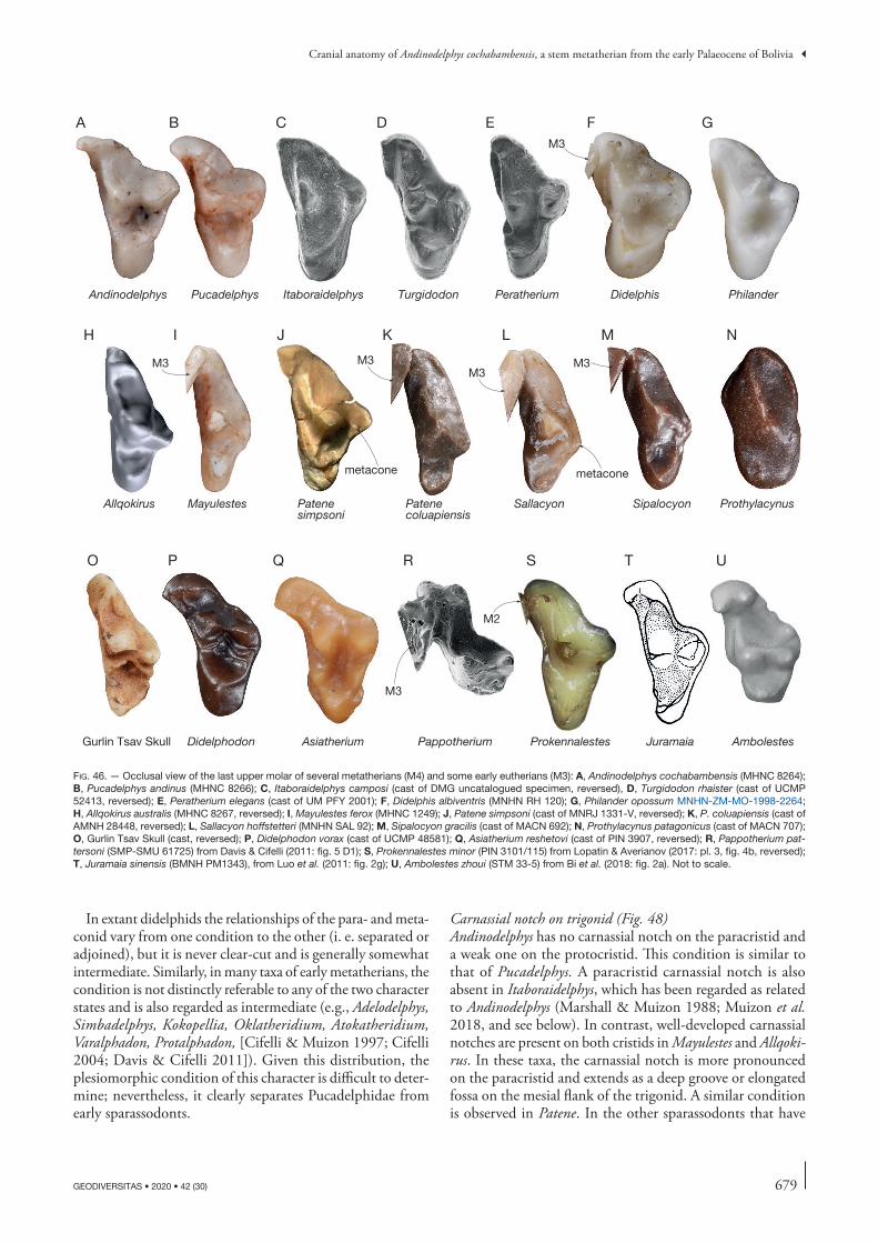
679
Cranial anatomy of Andinodelphys cochabambensis, a stem metatherian from the early Palaeocene of Bolivia
GEODIVERSITAS • 2020 • 42 (30)
In extant didelphids the relationships of the para- and meta-conid vary from one condition to the other (i. e. separated or adjoined), but it is never clear-cut and is generally somewhat intermediate. Similarly, in many taxa of early metatherians, the condition is not distinctly referable to any of the two character states and is also regarded as intermediate (e.g., Adelodelphys, Simbadelphys, Kokopellia, Oklatheridium, Atokatheridium, Varalphadon, Protalphadon, [Cifelli & Muizon 1997; Cifelli 2004; Davis & Cifelli 2011]). Given this distribution, the plesiomorphic condition of this character is difficult to deter-mine; nevertheless, it clearly separates Pucadelphidae from early sparassodonts.
Carnassial notch on trigonid (Fig. 48)Andinodelphys has no carnassial notch on the paracristid and a weak one on the protocristid. This condition is similar to that of Pucadelphys. A paracristid carnassial notch is also absent in Itaboraidelphys, which has been regarded as related to Andinodelphys (Marshall & Muizon 1988; Muizon et al. 2018, and see below). In contrast, well-developed carnassial notches are present on both cristids in Mayulestes and Allqoki-rus. In these taxa, the carnassial notch is more pronounced on the paracristid and extends as a deep groove or elongated fossa on the mesial flank of the trigonid. A similar condition is observed in Patene. In the other sparassodonts that have
Didelphis Philander
Gurlin Tsav Skull Didelphodon Asiatherium Prokennalestes Juramaia AmbolestesPappotherium
Andinodelphys
A
Pucadelphys
B
Itaboraidelphys
C
Turgidodon
D
Peratherium
E F G
Allqokirus
H
Mayulestes
I
Patenesimpsoni
J
Patenecoluapiensis
K
Sallacyon
L
Sipalocyon
M
Prothylacynus
N
O P Q R T US
metacone metacone
M3
M3M3
M3M3
M3
M2
fig. 46. — Occlusal view of the last upper molar of several metatherians (M4) and some early eutherians (M3): A, Andinodelphys cochabambensis (MHNC 8264); B, Pucadelphys andinus (MHNC 8266); C, Itaboraidelphys camposi (cast of DMG uncatalogued specimen, reversed), D, Turgidodon rhaister (cast of UCMP 52413, reversed); E, Peratherium elegans (cast of UM PFY 2001); F, Didelphis albiventris (MNHN RH 120); G, Philander opossum MNHN-ZM-MO-1998-2264; H, Allqokirus australis (MHNC 8267, reversed); I, Mayulestes ferox (MHNC 1249); J, Patene simpsoni (cast of MNRJ 1331-V, reversed); K, P. coluapiensis (cast of AMNH 28448, reversed); L, Sallacyon hoffstetteri (MNHN SAL 92); M, Sipalocyon gracilis (cast of MACN 692); N, Prothylacynus patagonicus (cast of MACN 707); O, Gurlin Tsav Skull (cast, reversed); P, Didelphodon vorax (cast of UCMP 48581); Q, Asiatherium reshetovi (cast of PIN 3907, reversed); R, Pappotherium pat-tersoni (SMP-SMU 61725) from Davis & Cifelli (2011: fig. 5 D1); S, Prokennalestes minor (PIN 3101/115) from Lopatin & Averianov (2017: pl. 3, fig. 4b, reversed); T, Juramaia sinensis (BMNH PM1343), from Luo et al. (2011: fig. 2g); U, Ambolestes zhoui (STM 33-5) from Bi et al. (2018: fig. 2a). Not to scale.
680 GEODIVERSITAS • 2020 • 42 (30)
Muizon C. de & Ladevèze S.
lost the metaconid, there is no protocristid, but the paracris-tid is strongly developed and also features a deep paracristid carnassial notch (Fig. 48). Among marsupials, the paracristid carnassial notch is well developed in dasyuroids and weak in extant didelphids and Dromiciops. A conspicuous paracristid carnassial notch is also present in several stem metatherians, such as Kokopellia, Asiatherium, deltatheroidans, stagodontids, Varalphadon, Turgidodon, Protalphadon, Alphadon, pediomy-ids, Peratherium, Herpetotherium, and peradectids, although, in some cases, it is variably developed.
Therefore, the distribution of a paracristid carnassial notch among metatherians suggests that it likely represents a plesio-morphic condition. Here again, the distribution of the states of this character within the Tiupampa metatherian fauna separates Pucadelphyidae and sparassodonts.
bony Skull
LacrimalThe lacrimal of Andinodelphys has a small facial process external to the orbit, which extends on the dorsal base of the rostrum. It is moderately expanded and does not form a large facial wing as observed, for instance, in cynodonts, morganucodonts (Kermack et al. 1981), deltatheroidans (Kielan-Jaworowska 1975b; Bi et al. 2015); the Gurlin Tsav skull (Szalay & Tro-fimov 1996) and sparassodonts (Muizon et al. 2018). The facial process of Andinodelphys is crescent-like, as is observed in Pucadelphys (contra Marshall & Muizon 1995) and extant didelphids, rather than wing-like. However, some variation is worth mentioning in Pucadelphys since one specimen (MHNC 8381) has a facial process significantly larger than in didelphids and other pucadelphyids, and approaches the wing morphology of deltatheroidans and sparassodonts. Be that as it may, Andinodelphys and Pucadelphys exhibit a derived condition within metatherians.
As in Pucadelphys and extant didelphids, the lacrimal of Andinodelphys has no contact with the nasal which is a derived character state. A broad lacrimal-nasal contact is a plesiomorphic condition within mammals and is present in multitubercu-lates (Wible & Rougier 2000), Morganucodon (Kermack et al. 1981), Vincelestes (Bonaparte & Rougier 1987), deltatheroidans (Rougier et al. 1998; Bi et al. 2015), sparassodonts (Muizon 1999), and Wynyardia (Gregory 1920; Jones 1930). This characteristic is also commonly present as an individual vari-ation in Trichosurus and Phalanger (Jones 1930) and given the fact that Wynyardia bassiana is apparently known by a single specimen, this condition may possible also represent an individual variation in this taxon. As in Pucadelphys, the condition of Andinodelphys is apomorphic within Metatheria.
Maxillopalatine vacuityOn the bony skull, one notable difference between Andino-delphys and Pucadelphys lies in the size of the major palatine foramen of the former, which is anteroposteriorly elongated and extends on the maxilla, forming a small maxillopalatine vacuity. However, this feature is not constant, since it is present in only three of the four skulls, on which it can be observed. On the fourth one (MHNC 8264), the major palatine fora-
men is large but not anteroposteriorly elongated; however, on this specimen a supplementary foramen perforates the maxilla anterior to the major palatine foramen (Fig. 18C). In the other three specimens, the anteroposterior enlargement of the major palatine foramen of Andinodelphys creates a small palatal vacuity in the maxilla. Palatal vacuities are absent in all the Pucadelphys skulls of our sample.
Palatal vacuities are present in the great majority of metathe-rians, in which they can perforate the palatine (palatine vacuities), and/or the maxillary (maxillary vacuities) or both (maxillopalatine vacuities). Palatal vacuities are absent in very few metatherian taxa. Among didelphids, they are totally missing in Caluromys and Caluromysiops, whereas they are present in all the other members of the family (Voss & Jansa 2009). They are poorly developed in Marmosa and Micoureus, in which they have the size observed in Andinodelphys. In Didelphis and Philander, maxillopalatine vacuities are large and are associated with more rounded posterior palatal (palatine) vacuities which pierce the palatine only. In other taxa, such as Metachirus and Chironectes, the maxillopalatine vacuities extend posteriorly and pierces a large part of the palatine.
Almost all Recent australidelphians have maxillopalatine vacuities. In dasyuroids, small anteroposteriorly elongate maxillopalatine vacuities are generally present in Dasyurus, but a double set of fenestrae is also present with greatly enlarged maxillopalatine fenestra in, for instance, Phascogale, Sminthopsis, Dasyuroides, Pseudantechinus, and Thylacinus. Maxillopalatine fenestrae are vestigial or absent, for instance, in Myrmecobius and Notoryctes (Marshall 1979a). In Myr-mecobius, we have observed a small but distinct elongated maxillary vacuity anterior to the major palatine foramen in AMNH 155328, but in MNHN-ZM-AC-A2564 this fora-men is absent. In Notoryctes, (MNHN-ZM-AC-1931-717, MNHN-ZM-1892.1243A, AMNH 202107), a palatal foramen is observed in the maxilla anterior to and similar in size to the major palatine foramen, a condition resembling that of Andinodelphys in MHNC 8264. Therefore, palatal vacuities in these two genera are reduced or vestigial in some individuals. They are absent in the petaurids Dactylopsila and Petaurus. In the latter, however, they are vestigial in some individuals, being present as anteroposteriorly enlarged major palatine foramina. Furthermore, it is noteworthy that intra- and interspecific variation exists in the size and number of the vacuities as observed, for instance, in Planigale (Archer 1976b) and Sminthopsis (Archer 1981).
Among fossil metatherians, palatal vacuities are present in most Late Cretaceous metatherian taxa in which they can be observed: Eodelphis (Fox 1981), Didelphodon (Wilson et al. 2016), Pediomys (Rougier et al. 1998, Luo et al. 2003, Forasiepi 2009), Alphadon (Rougier et al. 1998; Forasiepi 2009). They are large (c. 42% the palate length) in the ?deltatheroidan commonly referred to as the Gurlin Tsav skull from the Late Cretaceous of Mongolia (Szalay & Tro-fimov 1996). In Asiatherium, although Szalay & Trofimov (1996) regard the presence of maxillopalatal vacuities as uncertain, they have been coded as present in data matrices by Rougier et al. (1998), Luo et al. (2003), and Forasiepi
681
Cranial anatomy of Andinodelphys cochabambensis, a stem metatherian from the early Palaeocene of Bolivia
GEODIVERSITAS • 2020 • 42 (30)
Andinodelphys Pucadelphys
ItaboraidelphysAenigmadelphys
Mayulestes
Allqokirus
Patene Dasyurus
Didelphis
Philander
paraconid and metaconidadjoined at base
m4
m3 m2
m4m3 m2
m4 m3 m2
m4 m3m3m2
m3 m2
paraconid and metaconidpartly separated(intermediate)
m4 m3
m4 m3
paraconid and metaconid adjoined at base
paraconid and metaconid adjoined at base
paraconid andmetaconidseparated
paraconid andmetaconidseparated
metaconidparaconid
metaconid
paraconid
paraconid
metaconid
protoconid
A
B
C D
EF
G
H
I
J
fig. 47. — Medial view of the lower molars in several metatherian. A, Andinodelphys cochabambensis (m3-m4, MHNC 8264); B, Pucadelphys andinus (m2-m4 MHNC 8266, reversed); C, Aenigmadelphys archeri (m3, cast of OMNH 20531, reversed); D, Itaboraidelphys camposi (m2-3, cast of, DGM 804-M, reversed); E, Allqoki-rus australis (m2-m3, MHNC 8267); F, Mayulestes ferox (m3-m4, MHNC 1249, reversed); G, Patene simpsoni (m3 cast of DGM 798-M); H, Dasyurus hallucatus (m2-m4, MNHN-ZM-AC-1871-284); I, Didelphis albiventris (m2-m4, MNHN-RH 161); J, Philander opossum (m2-m4, MNHN-ZM-MO-1998-2264). Not to scale.

682 GEODIVERSITAS • 2020 • 42 (30)
Muizon C. de & Ladevèze S.
(2009). Palatal vacuities are absent in the deltatheroidans Deltatheridium and Lotheridium (in the latter, absence is definitive on the palatine, but probable only on the max-illa), (Bi et al. 2015). They are present in Herpetotherium (Sánchez-Villagra et al. 2007; Horovitz et al. 2008), but the condition in Peradectes, Mimoperadectes, and Peratherium is unknown. Within the Tiupampa metatherian fauna, they are absent in Pucadelphys, Mayulestes, Allqokirus, Szalinia, and Mizquedelphys. They are present (with the variation described above) in Andinodelphys and, when present, they are small. Small slot-like maxillopalatine fenestrae are pre-sent in Sparassocynus, a carnivorous didelphoid from the Plio-Pleistocene of Argentina (Beck & Taglioretti 2019) and they appear to have been probably also present, but small, in Epidolops (Beck 2017). They are absent in sparas-sodonts, which have been regarded as the sister group of the Pucadelphyidae by Muizon et al. (2018).
The absence of maxillopalatal fenestrae in Pucadelphys has been regarded by Muizon (1992) and Marshall & Muizon (1995) as a plesiomorphic feature. This interpretation was based on the fact that ontogenetic studies (Parker 1886: 260) have shown that the palatal plates of the maxillae and palatines of the marsupial skull are solid in early stages of development and become fenestrated in later stages by bone resorption (see also Marshall 1979a; Marshall & Muizon 1995). Interestingly, observations in Dromiciops confirm Parker’s studies. In this taxon, the maxillopalatine vacuities are very large in adults but they are barely more developed than the major palatine foramina in a juvenile skull, as figured by Giannini et al. (2004: fig. 2). Similar observations have been made on skulls of juvenile and adult Lutreolina and Didelphis (Flores et al. 2003).
Palatal vacuities are not common in non-metatherian mam-mals. They are present only in some multituberculates, and in some eutherians (e.g., rabbits, some rodents, macroscelidans, hedgehogs), (Marshall 1979a and references therein). It is therefore tempting to follow Marshall (1979a) in considering that a solid palate is likely to be the plesiomorphic condition among therians and that palatal vacuities were independently acquired in several lineages of multituberculates and eutherians as well as in metatherians.
However, the distribution of this character in Late Cretaceous metatherians, would suggests that the presence of maxillopala-tine vacuities may rather be the plesiomorphic character state for metatherians. Furthermore, their absence in a few taxa of didelphids and dasyurids while they are present in most others would confirm this interpretation being an indication that they probably disappeared in some lineages of these families. Be that as it may, considering the great variability of the pres-ence (and absence) and number of palatal fenestrae observed in the metatherian fossil record as early as the Late Cretaceous in North America and Asia, it is likely that this this feature had a complex evolution within metatherians with multiple disappearances and/or appearances.
Given the diverse distribution of palatal vacuities within Metatheria, their absence in some taxa and clades may be interpreted as a derived condition, which independently occurred in several lineages probably by paedomorphic reten-
tion of the unperforated condition of the palate in later developmental stages. In this study, we therefore rather favour the hypothesis that the lack of maxillopalatine vacuities in Pucadelphyda might be a synapomorphous loss, the condi-tion of Andinodelphys rather indicating a vestigial condition than an incipient development.
Transverse canalAndinodelphys possesses a transverse canal (Figs 28, 30, 32, 33). This structure has been recorded as absent in Pucadel-phys by Marshall & Muizon (1995), a statement based on the observation of one specimen only (the three other speci-mens described by these authors are either not fully prepared or not well preserved enough to allow observation). In the large sample of Pucadelphys skulls reported by Ladevèze et al. (2011), six skulls are well preserved enough to undoubtedly allow observation of this region and, among them, two speci-mens clearly present a small transverse canal (MHNC 8377 and 8380). Therefore, this character is clearly polymorphic in Pucadelphys.
A transverse canal is variably present in mammals. It is absent in monotremes (Luo et al. 2007), Morganucodon (Kermack et al. 1981), Megazostrodon (Gow 1986; Luo et al. 2007), Haldanodon (Lillagraven & Krusat 1991), Gobiconodon (Luo et al. 2003), and Repenomanus (Hu et al. 2005). It is absent in most multituberculates (Kielan-Jaworowska et al. 1986) but present in Kyroptobaatar (Wible & Rougier 2000). It is absent in the symmetrodont Zhangheotherium (Hu et al. 1997) and in the zatherian Vincelestes (Hopson & Rougier 1993). A transverse canal is generally absent in eutherians but it has been observed in Solenodon, Dasypus, Rattus, Nandinia, Gen-etta, and Felis (Wible & Gaudin 2004; Wible 2012; O’Leary et al. 2013; Wible & Spaulding 2013).
In contrast, a transverse canal is present in most metatheri-ans: Asiatherium, Andinodelphys, Mizquedelphys (undescribed specimen MHNC 13917), some specimens of Pucadelphys, some sparassodonts (e.g., Sipalocyon, Notogale, Prothyla-cynus, Lycopsis – Forasiepi 2009), (but see Archer 1976a who regards the transverse canal of sparassodonts – when present – as not clearly homologous with the transverse canal of didelphids), most didelphids, Mimoperadectes, Peratherium, caenolestids, most dasyurids, thylacinids, microbitheres, peramelemorphians, macropodoids, acro-batids, petaurids, phascolarctids and vombatids (Sánchez-Villagra & Wible 2002; Horovitz et al. 2009). However, some extinct metatherians lack a transverse canal such as the Gurlin Tsav skull (personal observation), some speci-mens of Pucadelphys (Marshall & Muizon 1995), and most sparassodonts (Muizon et al. 2018). Among the latter, the canal is absent for example in early taxa such as Mayulestes, Allqokirus, and Callistoe, but also in Neogene taxa such as Borhyaena, Arctodictis, Australohyaena, Hondadelphis, and Thylacosmilus. The transverse canal is exceptionally absent in some didelphids (e.g., Caluromys) and some dasyurids (e.g., Planigale) (Archer 1976a). The condition in Del-tatheridium and Didelphodon is unknown (Forasiepi 2009; Wilson et al. 2016) and uncertain in Epidolops (Beck 2017).
683
Cranial anatomy of Andinodelphys cochabambensis, a stem metatherian from the early Palaeocene of Bolivia
GEODIVERSITAS • 2020 • 42 (30)
Therefore, it appears that a transverse canal is generally absent in non-metatherian mammals (with some exceptions, cited above), whereas its presence is the most common state in metatherians. We therefore follow Marshall & Muizon (1995), who stated that the absence of a transverse canal is probably the plesiomorphic state for mammals. Because of the distribution of this character among metatherians, we also agree with these authors that this structure is likely to have evolved multiple times within metatherians. The distribu-tion of this character among Pucadelphyda (Pucadelphyidae + Sparassodonta) would lend support to this interpretation.
Tympanic process of alisphenoidA characteristic feature of most metatherians is the presence of large posteriorly projected wing of the alisphenoid, which borders part of the middle ear anteriorly and/or ventrally. This tympanic process of the alisphenoid forms an alisphe-noid bulla, which is present, but more or less developed, in all extant marsupials (except in some vombatids [Vombatus and Lasiorhinus] and some acrobatids [Acrobates and Distoe-churus]) and in many fossil taxa. Andinodelphys lacks a tym-panic process of the alisphenoid, a condition observed in all the other Tiupampa metatherians for which this part of the
no carnassial notch
small carnassialnotch
very small notch:but not really carnassial notch
carnassial notch
carnassialnotch
no carnassialnotch
carnassial notch
protoconid
paracristid
m2m3
m4
m2 m3 m4
m3 m4
m2
m3m3
m4
m3
m4
Andinodelphys Andinodelphys Pucadelphys
Philander AenigmadelphysItaboraidelphys
Allqokirus Mayulestes
Patene Sipalocyon
A
BC
D
EF
G
H
I
J
paracristid
paraconid
fig. 48. — Mesiolabial view of posterior lower molars in several metatherians. A, Andinodelphys cochabambensis (m2-m4, MHNC 8306, reversed); B, Andino-delphys cochabambensis (m4, cast of MHNC 8307); C, Pucadelphys andinus (m2-m3, MHNC 8384); D, Itaboraidelphys camposi (m3-m4, cast of DGM 817-M, reversed); E, Philander opossum (m3-m4, MNHN-ZM-MO-1998-2264) Aenigmadelphys archeri (m3, cast of OMNH 20531, reversed); F, Allqokirus australis (M2-m3, MHNC 8267); G, Mayulestes ferox (m2-m3, MHNC 1249, reversed), H, Patene simpsoni (m3, cast of DGM 798-M); I, Sipalocyon gracilis (m3-m4, cast of YPM-PU 15375, reversed). Not to scale.
684 GEODIVERSITAS • 2020 • 42 (30)
Muizon C. de & Ladevèze S.
skull is known (i.e. Pucadelphys, Mizquedelphys, Mayulestes, and Allqokirus).
Marshall & Muizon (1995) and Muizon (1994, 1998) regarded this condition as plesiomorphic in Pucadelphys and Mayulestes respectively. However, a tympanic process of the alisphenoid has been observed in several Cretaceous metatherians. It is present in Asiatherium and in the Gurlin Tsav skull (Szalay & Trofimov 1996), and in Didelphodon (Wilson et al. 2016). The fact that it is present in several Late Cretaceous taxa in which it can be observed, would suggest that a tympanic process of the alisphenoid is the plesiomorphic condition for metatherians. A tympanic pro-cess of the alisphenoid has also been observed in the early Eocene metatherians, Peratherium cuvieri (Selva & Ladevèze 2017), Herpetotherium and Mimoperadectes (Horovitz et al. [2009]; but see Jansa et al. [2014] for a different interpre-tation), but it is absent in the Tiupampa taxa mentioned above. It is absent in most sparassodonts (including the early Palaeocene Mayulestes and Allqokirus and the early Eocene Callistoe) except in hathliacynids (e.g., Notogale, Sipalocyon, Cladosictis) and thylacosmilids, in which it prob-ably represents a derived condition (reversal?) (Marshall & Muizon 1995). This feature is also apparently absent in the early Eocene Epidolops (Beck 2017) and in the Late Cretaceous Deltatheridium (Forasiepi 2009: 143; personal communication of G. Rougier to A. Forasiepi); condition in Lotheridium is unclear because of the poor preservation of the alisphenoid in the holotype of L. mengi (ZMNH M9032), (Bi personal communication to CM). Therefore, the distribution of this character in early metatherians is variable and diverse, suggesting that it may have appeared and disappeared several times during early metatherian evolution. Therefore, it is difficult to know if the absence of a tympanic process of the alisphenoid in the Tiupampa metatherians is apomorphic or plesiomorphic within metatherians. However, it likely represents a plesiomorphic condition within Pucadelphyda (Pucadelphyidae + Spar-assodonta), since the five Tiupampa pucadelphydans for which the basicranium is known lack a tympanic process of the alisphenoid.
Hypotympanic sinus Andinodelphys features a small circular cavity in the roof of the middle ear, located between the foramen ovale antero-medially and the tympanic petrosal crest posterolaterally (Fig. 28). This depression excavates the petrosal posterome-dially and the squamosal and alisphenoid anterolaterally. As pointed out by Beck (2017: 391), this structure, houses the dorsal part of the hypotympanic sinus (contra Ladevèze & Muizon 2007), an interpretatation that we follow here. In Andinodelphys, it is similar to that observed in Mayulestes. It is absent in Pucadelphys. Auditory sinuses are common in extant therians. They are air cavities (other than the epitympanic recess) within or between bones of the middle ear. Within metatherians, the most common sinuses are the hypotympanic and epitympanic sinuses. The former is located anterior to the epitympanic recess, anterolateral
to the petrosal and medial to the glenoid fossa. In most extant taxa, it excavates the alisphenoid only, most of it being housed in the tympanic process of the alisphenoid, which project ventrally below the tympanum. Therefore, most of the sinus is actually ventral to the tympanum and, hence, hypotympanic. However, the dorsal part of the sinus excavates the roof of the middle ear cavity, which is slightly dorsal to the tympanum. In Andinodelphys and Mayulestes, which lack a tympanic process of the alisphenoid, the small cavity in the roof of the middle ear is the bony materializa-tion of the hypotympanic sinus, which represent the part of the sinus located above the tympanum. The remaining part of the sinus is likely to have extended ventrally below the tympanum during life being limited ventrally by the fibrous membrane of the tympanic cavity (in the absence of any kind of bony tympanic bulla) (MacPhee 1981). To conclude, although what is referred to, here, (in Andinodel-phys and Mayulestes) as the hypotympanic sinus is actually dorsal to the tympanum it corresponds to part of a sinus that was during life mainly ventral to the tympanum: the hypotympanic sinus (see Beck & Taglioretti [2019] for dis-cussion on the position of the hypotympanic sinus relative to the tympanum). In taxa that lack a tympanic process of the alisphenoid, the hypotympanic sinus is excavated mostly in the petrosal and squamosal, with, in some taxa (e.g., Mayulestes), a small participation of the alisphenoid between them. The epitympanic sinus is located postero-laterally to the epitympanic recess and excavates the squa-mosal and/or the pars mastoidea of the petrosal. Because this sinus extends mostly dorsally, it is generally almost totally dorsal to the tympanum, thus epitympanic. Such a sinus is absent in Andinodelphys. The fossa erroneously identified by Muizon (1999) as the mastoid epitympanic sinus in the posterior region of the petrosal of Notogale, is in fact the stapedial fossa.
Auditory sinuses in general are absent in Morganucodon (Kermack et al. 1981); Gobiconodon, Sinoconodon (Cromp-ton & Sun 1985; Crompton & Luo 1993), Repenomanus (Hu et al. 2005), monotremes (Watson 1916), Vincelestes (Rougier et al. 1992), Late Cretaceous eutherians (Asioryctes, Barunlestes, Kennalestes, Zalambdalestes, Maelestes; Kielan-Jaworowska 1981, 1984; Kielan-Jaworowska & Trofimov 1980; Wible et al. 2004; Wible et al. 2009) and generalized extant eutherians (Van Kampen 1905; Novacek 1977). A hypotympanic sinus occurs in the great majority of extinct and extant metatherians except Deltatheridium (Rougier et al. 1998) Pucadelphys (Marshall & Muizon 1995), and Mizquedelphys (MHNC 13917). Condition in Lotheridium is unknown (Bi et al. 2015). In regard of the distribution, the absence of auditory sinuses likely represents the plesio-morphic condition for mammals. Therefore, Andinodelphys is more derived than Pucadelphys in this respect.
Inferior petrosal sinus and foramenAs mentioned above a conspicuous foramen is observed at the junction of the basioccipital, exoccipital and petrosal (Fig. 30). This foramen opens the canal for the inferior
685
Cranial anatomy of Andinodelphys cochabambensis, a stem metatherian from the early Palaeocene of Bolivia
GEODIVERSITAS • 2020 • 42 (30)
petrosal sinus ventrally, anterior to its posterior end. This foramen may have conveyed a branch of the inferior pet-rosal sinus which may have joined the vertebral vein. A similar condition may have been present in some specimens of Pucadelphys, although it cannot be ascertained because of the preservation of the specimens. The condition in Mayulestes and Allqokirus is unknown. The condition of Andinodelphys appears to be unique among metatherian (apart from a possible presence in Pucadelphys).
The other characters discussed by Marshall & Muizon (1995: 64-82) are identical in Pucadelphys and Andinodelphys and will not be further discussed here. They are: number of incisors, structure of lower incisors, number of molar and premolars, inflected angular process of the dentary, mylohyoid groove, labial mandibular foramen, orbit large and confluent with temporal fossa, nasal-lacrimal contact, alisphenoid-parietal contact, preglenoid process of the jugal, foramen ovale, subsquamosal foramen (= suprameatal foramen, in this paper), Orientation of ectotympanic and tympanic membrane, pars mastoidea contribution to occiput, mastoid and paroccipital process, shape of fenestra vestibuli and stapedial footplate, stapedial artery, course of internal carotid artery, sulcus for facial nerve, anterior lamina of petrosal.
Characters of dentition of Andinodelphys are very similar to those of Pucadelphys except the size and relative posi-tion of the I1
Specific comparison with Itaboraidelphys and phylogenetic analysisMarshall & Muizon (1988) and Muizon (1992) have regarded the molars of Andinodelphys cochabambensis as representing an adequate dental structural ancestor for Itaboraidelphys camposi from the late Palaeocene-early Eocene of Itaboraí (Brazil). However, Andinodelphys dif-fers from Itaboraidelphys in its smaller size (25% smaller in linear tooth dimensions), more gracile molars with sharper crests and cusps and deeper basins, upper molars with a less V-shaped centrocrista, paracone and metacone with a distinct vertical ridge on their labial edges, p2 distinctly smaller than p3 (subequal in size to p2 slightly larger than p3 in Itaboraidelphys) lower molars with a higher trigonid and smaller entoconid, and trigonid of m3 slightly wider than talonid (slightly narrower in Itaboraidelphys). Andi-nodelphys and Itaboraidelphys share the same large size of stylar cusps B and D, with a small stylar cusp C, the latter being twinned in some specimens, the posterior edge of the protocone distolingually expanded, which displaces its apex anteriorly, the paracone conspicuously smaller than the metacone, the angle para-metacone axis-preparacrista on M4 close to 90°, and the adjoined paraconid and meta-conid. In spite of the small differences mentioned above, which are likely to represent plesiomorphic conditions in Andinodelphys, the latter compares best with Itaboraidelphys among the metatherian taxa of the Itaboraí fauna.
Ladevèze & Muizon (2010) have performed a parsi-mony analysis including several isolated petrosals from
the late Palaeocene-early Eocene of Itaboraí. This analysis resulted in a sister group relationship of Type II petrosals (MNRJ 6728-V and MNRJ 6729-V) with pucadelphy-ids (Pucadelphys and Andinodelphys). In order to test this relationship, Muizon et al. (2018: 422-423) compared the area of the promontorium relative to the molar size in Andinodelphys and Itaboraidelphys. Assuming that the promontorium area relative to the length of the molars was similar in both taxa these authors calculated an estima-tion of the promontorium area in Itaboraidelphys on the basis of the upper and lower molar proportions. The two calculated estimated values (12.1 mm2 and 12.9 mm2) felt seemed perfectly within the range of the promonto-rium area measured on the two Type II petrosals (11.1 mm2 and 13.96 mm2). Their result provided a mean promontorium area of Andinodelphys (9.42 mm2) 24,8% smaller than in Itaboraidelphys (12.53 mm2), a difference that matches the size difference evaluated by Marshall & Muizon (1988) between the teeth of Andinodelphys and Itaboraidelphys, the former being approximately 25% smaller than the latter. Consequently, Muizon et al. (2018) concluded that, on the basis of both dental and petrosal anatomy, the Type II petrosals of Itaboraí could plausibly be referred to Itaboraidelphys. As a consequence, in the parsimony analysis below we tentatively referred dental remains of Itaboraidelphys camposi and Type II petrosals to the same taxon (contra Beck [2017], who referred the Type II petrosal to Epidolops, a hypothesis that has been challenged by Muizon et al. [2018]). The phylogenetic analysis presented here uses the data matrix of Muizon et al. (2018). However, after re-examination of casts, specimens, and literature we changed the scorings of some characters for some taxa. These changes are mentioned in the character list below (Appendix 3). Furthermore, in order to complement the data on deltatheroidans we added two Asiatic taxa of this group of metatherians (Sul-estes and Lotheridium) and three North American ones (Atokatheridium, Oklatheridium, and Nanocuris). Scor-ing of these taxa in our matrix is respectively based on the descriptions, illustrations, and scorings of Averianov et al. (2010) and Bi et al. (2015) on the one hand and Kielan-Jaworowska & Cifelli (2001), Fox et al. (2007), Davis et al. (2008), Wilson & Riedel (2010), Davis & Cifelli (2011) on the other hand. Because of the condi-tion of the stylar shelf of upper molars and postglenoid foramen in Lotheridium, we added a fifth state to character 35 (4, width of stylar shelf reduced labial to metacone on penultimate molar) and a third state to character 177 (2, postglenoid foramen located anterior to postglenoid process). Three other characters of Lotheridium were coded thanks to personal communication of Bi to one of us (CM); they are characters 71, 185, and 210. Four other in-group taxa (Alphadon-Turgidodon-Protalphadon, Varalphadon, Pediomys, and the so-called Gurlin Tsav skull (GTS) figured by Szalay & Trofimov 1996) were also added. Furthermore, we added three supplementary out-group taxa: Vincelestes, Zalambdalestes, and Leptictis.
686 GEODIVERSITAS • 2020 • 42 (30)
Muizon C. de & Ladevèze S.
RESULTS OF THE PHYLOGENETIC ANALYSES
We have performed three sets of parsimony analyses, each set including one analysis with unweighted characters and one analysis with implied weighting (Goloboff k = 3). The first set of analyses takes into account the whole of our character list (364). Then, given the possible bias related to dental charac-ters (e.g. related to hypercarnivory), which are well known to be strongly homoplastic (e.g. Muizon & Lange-Badré 1997; Ladevèze et al. 2011; Sansom et al. 2017; Solé & Ladevèze 2017), we have performed a second set of analyses excluding all dental characters and, consequently, excluding the taxa known by teeth only or teeth and partial jaws. The third set of analyses was performed with the exclusion of some dental characters only, which are regarded more specifically related to hypercarnivory (see p. 695), in the light of the preceding analyses.
reSultS of the firSt analySeS with all characterS
The analysis of this significantly modified matrix (as a conse-quence of the additional taxa and changes mentioned above and in the character list and scorings; 364 characters and 51 taxa) with PAUP* resulted in 6 shortest trees (L = 1577, CI = 0.305, RI = 0.667). As seen in the strict consensus tree (L = 1584, CI = 0.306, RI = 0.665) (Fig. 49A) the relationships of Itaboraidelphys are the same as those retrieved by Ladevèze & Muizon (2010). Itaboraidelphys is the sister group of the clade Pucadelphys + Andinodelphys and, as such, is included here in Pucadelphyidae.
The relationships of Stagodontidae and Deltatheroida resulting from this analysis are different from those retrieved by Muizon et al. (2018). The Gurlin Tsav Skull (GTS) forms a clade with Stagodontidae (Eodelphis and Didelphodon) and this clade is sister-group to Sparassodonta. Deltatheroida and Pucadelphyidae are successive sister groups to this (Sta-godontidae-GTS + Sparassodonta) clade. This result signifi-cantly differs from that of Muizon et al. (2018), in which the deltatheroidans and stagodontids were early diverging groups in the metatherian clade. The present result also differs from most other studies, in which deltatheroidans are almost always in a basal position in the Metatheria tree (e. g. Averianov et al. 2010; Rougier et al. 2015; Bi et al. 2015; Forasiepi et al. 2015; Wilson et al. 2016; Beck 2017). Moreover, stagodontids, although generally less basal than deltatheroidans, are, in these analyses, generally retrieved closer to North American metatherians than to sparassodont, with one exception how-ever (Engleman & Croft 2014), in which they were also the sister group of sparassodonts. As mentioned by these authors, although stagodontids have been regarded as closely related to sparassodonts by Marshall et al. (1990), this interpretation has been seriously questioned by Fox & Naylor (1995), but also by Muizon & Lange-Badré (1997), and Muizon (1999) because it is based almost exclusively on heavily homoplastic dental characters related to hypercarnivory. This statement may also apply in the case of this analysis. Therefore, this statement essentially concerns the dental synapomorphies (non-ambiguous and ambiguous) supporting 1) the clade
(deltatheroidans (GTS, stagodontids) sparassodonts)): 33(1), 48(1), 57(1), 70(0), 71(0), 80(1), 84(2), 85(2), 43(0), 47(0), 50(0), 95(1), 96(0); and 2) those supporting the clade ((GTS, stagodontids) sparassodonts): 6(1), 12(1), 23(0), 24(0), 30(1), 38(1), 46 (2), 50(0), 55(1), 64(1), 72(1).
In order to overcome this bias, we have performed a new analysis with downweighted homoplastic characters with Goloboff constant value = 3. The use of higher Goloboff constant values (as recommended by Goloboff et al. 2017) retrieved consensus trees almost identical to the strict con-sensus and did not downwheight homoplastic characters, which justifies the selection of a low value of the Goloboff constant (3) in the present study. In the single tree obtained (CI = 0.303, RI = 0.6637; Fig. 49B), stagodontids are placed in a more basal position, similar to that retrieved in the tree provided by Muizon et al. (2018: fig. 29B) after an analy-sis with downweighted homoplastic characters. However, the GTS and deltatheroidans remain closely related, basal to sparassodonts. The change in the position of the stago-dontids in our new tree likely reveals the elevated degree of homoplasy of the characters (mostly dental) that placed them in a sister group relationship to the sparassodonts, and therefore the weakness of this topology. The position of GTS, which is now a direct sister-group of sparassodonts, approaches the results of Rougier et al. (2015) and Wilson et al. (2016: fig. 3), and the position of the deltatheroidans as the sister-group of the GTS + sparassodonts is not so discrepant with that retrieved by these authors, in which deltatheroidans are only one node more basal to their clade including GTS + pucadelphydans (i.e. the first diverging clade of Marsupialiaformes) (see below p. 693). The analy-ses of Rougier et al. (2015) and Wilson et al. (2016) also resulted in a position of the stagodontids more closely nested within North American taxa but not in a basal position in the tree as in Forasiepi et al. (2015) and Muizon et al. (2018). Furthermore, it is noteworthy that the topologies obtained here and those retrieved by Rougier et al. (2015) and Wilson et al. (2016) resulted from different character lists (although most of characters of these authors are included in our list) and in the fact that two ingroup Operational Taxonomic Units (OTU) are considered by these authors at family level (borhyaenoids, and dasyurids) in contrast to ours, what may explain the slight differences in our results concerning the deltatheroidans and GTS + pucadelphydans relationships. In our new analysis, the clade Pucadelphyda is retained, but according to this topology, should now include deltatheroidans and the GTS. In this context, our discussion below will focus on the new tree obtained with downweighted homoplastic characters (Fig. 49B).
Another interesting result is the position of Varalphadon, a North American genus that has been regarded as a sparas-sodont by Carneiro (2018). Although the position of Varal-phadon as a sister group of Pediomys could perhaps be argued, it confirms Muizon et al. (2018: 430)’s conclusions that this taxon is very probably not closely related to sparassodonts.
Below we provide the unambiguous synapomorphies of the following clades resulting from the implied weighted analy-
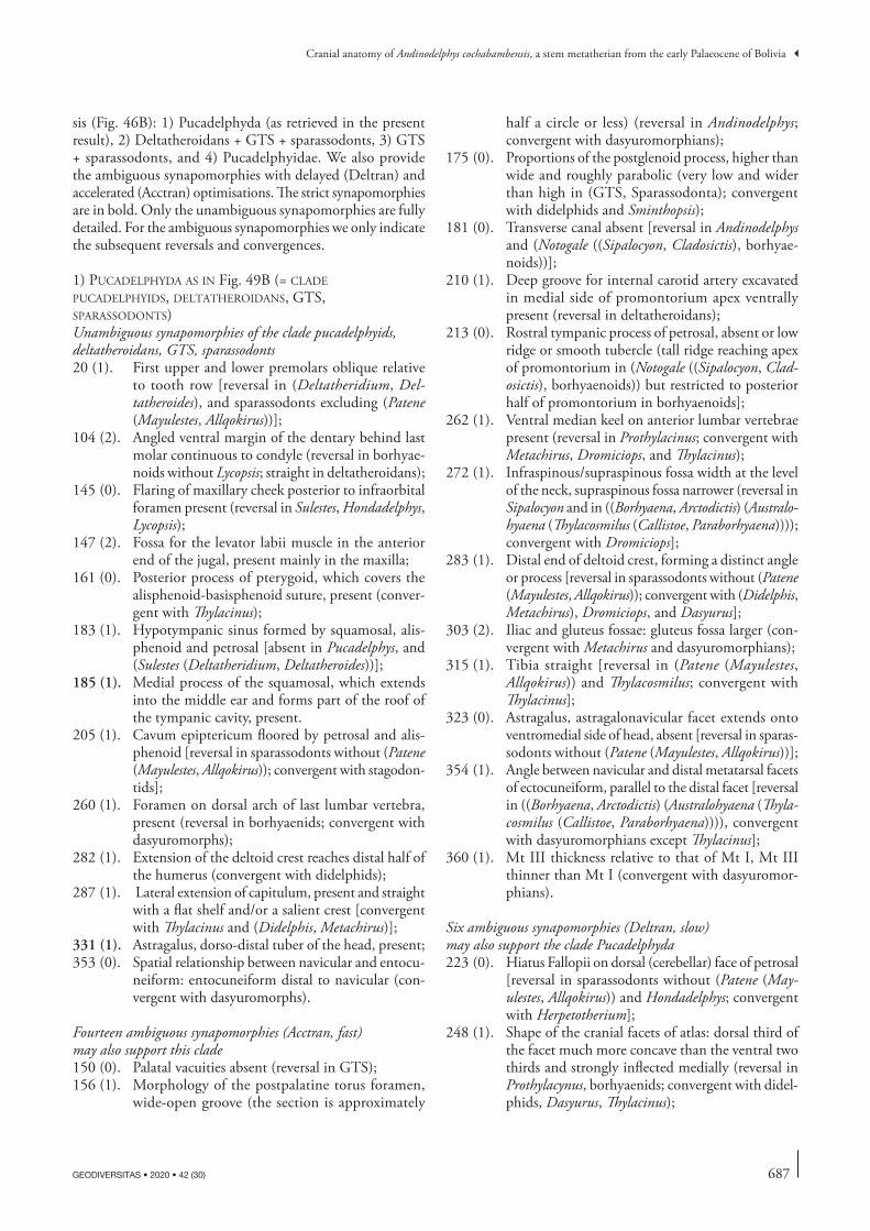
687
Cranial anatomy of Andinodelphys cochabambensis, a stem metatherian from the early Palaeocene of Bolivia
GEODIVERSITAS • 2020 • 42 (30)
sis (Fig. 46B): 1) Pucadelphyda (as retrieved in the present result), 2) Deltatheroidans + GTS + sparassodonts, 3) GTS + sparassodonts, and 4) Pucadelphyidae. We also provide the ambiguous synapomorphies with delayed (Deltran) and accelerated (Acctran) optimisations. The strict synapomorphies are in bold. Only the unambiguous synapomorphies are fully detailed. For the ambiguous synapomorphies we only indicate the subsequent reversals and convergences.
1) pucadelphyda aS in Fig. 49b (= clade pucadelphyidS, deltatheroidanS, gtS, SparaSSodontS)Unambiguous synapomorphies of the clade pucadelphyids, deltatheroidans, GTS, sparassodonts20 (1). First upper and lower premolars oblique relative
to tooth row [reversal in (Deltatheridium, Del-tatheroides), and sparassodonts excluding (Patene (Mayulestes, Allqokirus))];
104 (2). Angled ventral margin of the dentary behind last molar continuous to condyle (reversal in borhyae-noids without Lycopsis; straight in deltatheroidans);
145 (0). Flaring of maxillary cheek posterior to infraorbital foramen present (reversal in Sulestes, Hondadelphys, Lycopsis);
147 (2). Fossa for the levator labii muscle in the anterior end of the jugal, present mainly in the maxilla;
161 (0). Posterior process of pterygoid, which covers the alisphenoid-basisphenoid suture, present (conver-gent with Thylacinus);
183 (1). Hypotympanic sinus formed by squamosal, alis-phenoid and petrosal [absent in Pucadelphys, and (Sulestes (Deltatheridium, Deltatheroides))];
185 (1). Medial process of the squamosal, which extends into the middle ear and forms part of the roof of the tympanic cavity, present.
205 (1). Cavum epiptericum floored by petrosal and alis-phenoid [reversal in sparassodonts without (Patene (Mayulestes, Allqokirus)); convergent with stagodon-tids];
260 (1). Foramen on dorsal arch of last lumbar vertebra, present (reversal in borhyaenids; convergent with dasyuromorphs);
282 (1). Extension of the deltoid crest reaches distal half of the humerus (convergent with didelphids);
287 (1). Lateral extension of capitulum, present and straight with a flat shelf and/or a salient crest [convergent with Thylacinus and (Didelphis, Metachirus)];
331 (1). Astragalus, dorso-distal tuber of the head, present;353 (0). Spatial relationship between navicular and entocu-
neiform: entocuneiform distal to navicular (con-vergent with dasyuromorphs).
Fourteen ambiguous synapomorphies (Acctran, fast) may also support this clade 150 (0). Palatal vacuities absent (reversal in GTS);156 (1). Morphology of the postpalatine torus foramen,
wide-open groove (the section is approximately
half a circle or less) (reversal in Andinodelphys; convergent with dasyuromorphians);
175 (0). Proportions of the postglenoid process, higher than wide and roughly parabolic (very low and wider than high in (GTS, Sparassodonta); convergent with didelphids and Sminthopsis);
181 (0). Transverse canal absent [reversal in Andinodelphys and (Notogale ((Sipalocyon, Cladosictis), borhyae-noids))];
210 (1). Deep groove for internal carotid artery excavated in medial side of promontorium apex ventrally present (reversal in deltatheroidans);
213 (0). Rostral tympanic process of petrosal, absent or low ridge or smooth tubercle (tall ridge reaching apex of promontorium in (Notogale ((Sipalocyon, Clad-osictis), borhyaenoids)) but restricted to posterior half of promontorium in borhyaenoids];
262 (1). Ventral median keel on anterior lumbar vertebrae present (reversal in Prothylacinus; convergent with Metachirus, Dromiciops, and Thylacinus);
272 (1). Infraspinous/supraspinous fossa width at the level of the neck, supraspinous fossa narrower (reversal in Sipalocyon and in ((Borhyaena, Arctodictis) (Australo-hyaena (Thylacosmilus (Callistoe, Paraborhyaena)))); convergent with Dromiciops];
283 (1). Distal end of deltoid crest, forming a distinct angle or process [reversal in sparassodonts without (Patene (Mayulestes, Allqokirus)); convergent with (Didelphis, Metachirus), Dromiciops, and Dasyurus];
303 (2). Iliac and gluteus fossae: gluteus fossa larger (con-vergent with Metachirus and dasyuromorphians);
315 (1). Tibia straight [reversal in (Patene (Mayulestes, Allqokirus)) and Thylacosmilus; convergent with Thylacinus];
323 (0). Astragalus, astragalonavicular facet extends onto ventromedial side of head, absent [reversal in sparas-sodonts without (Patene (Mayulestes, Allqokirus))];
354 (1). Angle between navicular and distal metatarsal facets of ectocuneiform, parallel to the distal facet [reversal in ((Borhyaena, Arctodictis) (Australohyaena (Thyla-cosmilus (Callistoe, Paraborhyaena)))), convergent with dasyuromorphians except Thylacinus];
360 (1). Mt III thickness relative to that of Mt I, Mt III thinner than Mt I (convergent with dasyuromor-phians).
Six ambiguous synapomorphies (Deltran, slow) may also support the clade Pucadelphyda 223 (0). Hiatus Fallopii on dorsal (cerebellar) face of petrosal
[reversal in sparassodonts without (Patene (May-ulestes, Allqokirus)) and Hondadelphys; convergent with Herpetotherium];
248 (1). Shape of the cranial facets of atlas: dorsal third of the facet much more concave than the ventral two thirds and strongly inflected medially (reversal in Prothylacynus, borhyaenids; convergent with didel-phids, Dasyurus, Thylacinus);
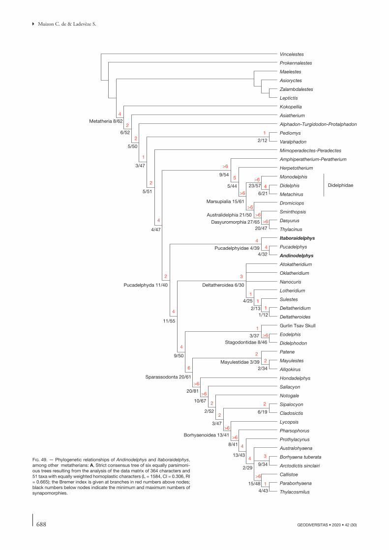
688 GEODIVERSITAS • 2020 • 42 (30)
Muizon C. de & Ladevèze S.
Vincelestes
Prokennalestes
Maelestes
Asioryctes
Zalambdalestes
Leptictis
Kokopellia
Asiatherium
Alphadon-Turgidodon-Protalphadon
Pediomys
Varalphadon
Mimoperadectes-Peradectes
Amphiperatherium-Peratherium
Herpetotherium
Monodelphis
Didelphis
Metachirus
Dromiciops
Sminthopsis
Dasyurus
Thylacinus
Itaboraidelphys
Pucadelphys
Andinodelphys
Atokatheridium
Oklatheridium
Nanocuris
Lotheridium
Sulestes
Deltatheridium
Deltatheroides
Gurlin Tsav Skull
Eodelphis
Didelphodon
Patene
Mayulestes
Allqokirus
Hondadelphys
Sallacyon
Notogale
Sipalocyon
Cladosictis
Lycopsis
Pharsophorus
Prothylacynus
Australohyaena
Borhyaena tuberata
Arctodictis sinclairi
Callistoe
Paraborhyaena
Thylacosmilus
2/12
5/51
4/47
Pucadelphyda 11/40
11/55
9/50
Sparassodonta 20/61
20/81
10/67
2/52
3/47
Borhyaenoidea 13/41
8/41
13/43
3/47
5/50
6/52
Metatheria 8/62
9/54
5/44
Marsupialia 15/61
Australidelphia 21/50
Dasyuromorphia 27/6520/47
23/57
6/21
Pucadelphyidae 4/394/32
Deltatheroidea 6/30
4/25
2/131/12
3/37Stagodontidae 8/46
Mayulestidae 3/392/34
6/19
2/29
15/48
4/43
9/34
Didelphidae
1
4
2
2
1
2
4
2
4
4
6
>6
2
2
>6
>6
>6
4
4
>6
1
3
2
5
4
>6
>6
>6
>6
>6>6
44
3
1
11
1>6
22
fig. 49. — Phylogenetic relationships of Andinodelphys and Itaboraidelphys, among other metatherians: A, Strict consensus tree of six equally parsimoni-ous trees resulting from the analysis of the data matrix of 364 characters and 51 taxa with equally weighted homoplastic characters (L = 1584, CI = 0.306, RI = 0.665); the Bremer index is given at branches in red numbers above nodes; black numbers below nodes indicate the minimum and maximum numbers of synapomorphies.
689
Cranial anatomy of Andinodelphys cochabambensis, a stem metatherian from the early Palaeocene of Bolivia
GEODIVERSITAS • 2020 • 42 (30)
fig. 49. — Phylogenetic relationships of Andinodelphys and Itaboraidelphys, among other metatherians: B, Single tree resulting from the analysis with down-weighted homoplastic characters (k = 3); black numbers below nodes indicate the minimum and maximum numbers of synapomorphies.
Vincelestes
Prokennalestes
Maelestes
Asioryctes
Zalambdalestes
Leptictis
Kokopellia
Asiatherium
Alphadon-Turgidodon-Protalphadon
Pediomys
Varalphadon
Eodelphis
Didelphodon
Mimoperadectes-Peradectes
Amphiperatherium-Peratherium
Herpetotherium
Monodelphis
Didelphis
Metachirus
Dromiciops
Sminthopsis
Dasyurus
Thylacinus
Itaboraidelphys
Pucadelphys
Andinodelphys
Oklatheridium
Atokatheridium
Nanocuris
Lotheridium
Sulestes
Deltatheridium
Deltatheroides
Gurlin Tsav Skull
Patene
Mayulestes
Allqokirus
Hondadelphys
Sallacyon
Notogale
Sipalocyon
Cladosictis
Lycopsis
Pharsophorus
Prothylacynus
Borhyaena tuberata
Arctodictis sinclairi
Australohyaena
Thylacosmilus
Callistoe
Paraborhyaena
Metatheria 9/67
7/57
3/55
6/54
3/44
2/46
Pucadelphyda 13/40
11/54
Deltatheroidea 4/28
1/22
1/25
2/26
1/121/13
5/56
Sparassodonta 12/57
Mayulestidae 3/411/36
22/81
10/65
2/52
3/46
6/19
Borhyaenoidea 14/41
8/41
7/33
15/43
8/30
2/29
8/463/32
Pucadelphyidae 4/394/33
9/54
5/45
Marsupialia 15/62
24/58
6/20
Australidelphia 22/49
Dasyuromorphia 26/6420/46
Didelphidae
Stagodontidae 20/52
3/102/13

690 GEODIVERSITAS • 2020 • 42 (30)
Muizon C. de & Ladevèze S.
262 (1). Ventral median keel on anterior lumbar vertebrae, present (reversal in Prothylacynus; convergent with Metachirus, Dromiciops, and Thylacinus);
272 (1). Infraspinous/supraspinous fossa width at the level of the neck: supraspinous fossa narrower (reversal in Sipalocyon, Arctodictis, Callistoe; convergent with Dromiciops);
303 (2). Iliac and gluteus fossae: gluteus fossa larger;360 (1). Mt III thickness relative to that of Mt I: Mt III thin-
ner than Mt I (convergent with dasyuromorphs).
2) clade deltatheroida + gtS + SparaSSodonta
Unambiguous synapomorphies supporting the clade Deltatheroida + GTS + Sparassodonta:33 (1). Size of molars (upper and lower) increases posteriorly:
marked [convergent with (Dasyurus, Thylacinus)];48 (1). Metacone and paracone adjoined at base (on, at
least, half of their height) (adjoined on most of their height; apices of cusps only are separated in borhyaenoids but Lycopsis; convergent with Thyl-acinus);
57 (1). Protocone size (on penultimate or antepenultimate molar): small and anteroposteriorly narrow (reversal in Hondadelphys, absent or very small in borhyae-noids but Lycopsis; convergent with Thylacinus);
70 (0). Trigonid vs talonid width (on penultimate or ante-penultimate molar): wider than talonid (reversal in Nanocuris; convergent with Andinodelphys);
71 (0). Protoconid labial expansion; the labial edge of the protoconid at mid-height is strongly convex, protruding labially and overhangs the base of the crown: present (reversal in Lotheridium);
80 (1). Paraconid elongated with anterior projection of the paraconid keel (ridge) mostly in the ventral half of the cusp: present [reversal in Lotheridium, (Callistoe, Paraborhyaena); convergent with Thylacinus];
84 (2). Height of metaconid vs paraconid (on penultimate molar): lower [subequal in (Patene (Mayulestes, Allqokirus)); convergent with stagodontids];
85 (2). Volume of the metaconid vs paraconid (on penulti-mate molar), in occlusal and lingual views: smaller [convergent with (Dasyurus, Thylacinus), and sta-godontids];
139 (0). Naso-lacrimal contact, present (reversal in Thyla-cosmilus);
149 (2). Number of palatal pits independently of their size: two between ultimate, penultimate and antepenul-timate molars [two pits between first, antepenulti-mate, and penultimate molars in Deltatheriidae-but reversal in Deltatheroides, one pit in borhyaenids; convergent with (Dasyurus, Thylacinus)].
Thirty-one ambiguous synapomorphies (Acctran, fast) may also support the clade Deltatheroida + GTS + Sparassodonta 1 (1). Number of upper incisors: four [reversal in (Patene
(Mayulestes, Allqokirus)); convergent with dasyuro-morphians];
2 (1). Size of extra-alveolar portion of root of I1 as long as on other incisors and as long as crown [reversal in (Patene (Mayulestes, Allqokirus)); convergent with Australidelphia];
3 (0). Implantation of the first upper incisors in anterior view: incisors parallel [reversal in (Patene (Mayul-estes, Allqokirus))];
10 (1). Number of lower incisors: three (reversal in May-ulestidae, Hondadelphys; convergent with dasyuro-morphians and stagodontids);
43 (0). Stylar cusp C, absent or tiny (convergent with Australidelphia);
47 (0). Shape of paracone and metacone: conical (conver-gent with Thylacinus);
50 (0). Centrocrista, straight (convergent with Dromiciops, Thylacinus);
95 (1). Shape of entoconid: transversely compressed (con-vergent with didelphids and dasyuromorphians);
96 (0). Relative height of entoconid: lower than hypoconid [reversal in Patene, Hondadelphys, (Sipalocyon, Clad-osictis); convergent with Herpetotherium, Thylacinus, and stagodontids];
106 (1). Anteroventral edge of mandibular symphysis: less slanting, forming an angle > 25° (reversal in Nano-curis; convergent with Thylacinus, and stagodontids);
118 (0). Condyle vertical position vs tooth row: approxi-mately at the same level or slightly below [reversal in (Patene (Mayulestes, Allqokirus)), Sipalocyon; convergent with Thylacinus];
120 (0). Length of the rostrum: less than (or equal to) 1/3 total length of the skull (reversal in sparassodonts; convergent with Dromiciops, Dasyurus, Thylacinus);
141 (0). Lacrimal tubercle: present, knob-like (reversal in Allqokirus; convergent with Herpetotherium);
154 (0). Posterior palatine spine: weak or absent and pos-terior edge of the palate concave posteriorly;
159 (0). Anteroposterior position of the sphenopalatine foramen relatively to the molars: above penultimate or antepenultimate molar [reversal in ((Borhyaena, Arctodictis) (Australohyaena (Thylacosmilus (Callistoe, Paraborhyaena)))); convergent with Monodelphis and (Dasyurus, Thylacinus)];
228 (2). Tuberculum tympani: expanded in a sharp anter-oventrally directed process (triangular in lateral view) [reversal in (Patene (Mayulestes, Allqokirus)); convergent with (Herpetotherium, Marsupialia)];
251 (1). Axis, posterior extension of the neural process: dis-tinctly longer than anterior extension [convergent with (Dasyurus, Thylacinus)];
252 (1). Ventral sagittal crest of axis: distinctly concave because of the development of a robust ventral process posteriorly (convergent with dasyuromor-phians);
256 (1). C6 spinous process: lamina [convergent with (Didel-phis, Metachirus), and (Dasyurus, Thylacinus)];
258 (2). First thoracic vertebra with a (relative to other vertebrae) tall spinous process: T3;

691
Cranial anatomy of Andinodelphys cochabambensis, a stem metatherian from the early Palaeocene of Bolivia
GEODIVERSITAS • 2020 • 42 (30)
273 (1). Scapular notch: between 90 and 130° (convergent with Metachirus, and dasyuromorphians);
274 (1). Clavicle: absent;293 (0). Shape of articular facet for the humerus on the
radius: anteroposteriorly compressed (convergent with Sminthopsis, and Asiatherium);
301 (1). Length of the iliac neck: short, less than 15% the total pelvis length;
304 (1). Epipubic bones: absent (convergent with Thylacinus);306 (1). Torsion between proximal and distal epiphyses of
femur: absent [reversal in Lycopsis, and (Australo-hyaena (Thylacosmilus (Callistoe, Paraborhyaena))); convergent with didelphids];
310 (1). Ossified patella: present (convergent with Amphi-peratherium-Peratherium); (personal observation of SL in Peratherium cuvieri, MNHN.F.GY679A);
320 (0). Anteroposterior length of the of the medial malleolus of the tibia at base relative to the greatest anteropos-terior length of the distal epiphysis (as seen in distal view): subequal (convergent with Sminthopsis);
327 (1). Astragalus, medial extent of sustentacular facet: reaches the medial edge of the neck (reversal in Borhyaenoidea);
329 (1). Astragalus, width of astragalar neck: neck narrower or as wide as head;
334 (1). Calcaneus, longest dimension of ectal facet: anter-oposterior.
One ambiguous synapomorphies (Deltran, slow) may also support the Deltatheroida + GTS + Sparassodonta clade 178 (2). Suprameatal foramen: at the same level or above
postzygomatic crest (reversal in Mayulestidae, Cal-listoe; convergent with Dromiciops, Sminthopsis).
3) clade gtS + SparaSSodonta
Unambiguous synapomorphies of the GTS + Sparassodonta clade:38 (1). Postmetacrista (on antepenultimate or penultimate
molar): distinctly longer than preparacrista [conver-gent with Deltatheridium, Nanocuris, didelphids, (Dasyurus, Thylacinus), Amphiperatherium-Perath-erium, stagodontids, (Pediomys, Varalphadon)];
54 (0). Conules wing-like cristae: present [convergent with (Dasyurus, Thylacinus)];
167 (1). Foramina for temporal rami: absent [reversal in Mayulestes, Notogale, (Thylacosmilus (Callistoe, Paraborhyaena)); convergent with (herpetoriids, Marsupialia), and stagodontids];
170 (0). Sagittal crest: extending to frontals (convergent with Didelphis);
175 (2). Proportions of the postglenoid process: very low and wider than high.
Twenty-two ambiguous synapomorphies may also support the clade GTS + Sparassodonta (Acctran, fast):6 (1). Size of I3 vs I2: I3=I2 [reversal in (Patene (May-
ulestes, Allqokirus)), in (Thylacosmilus (Callistoe, Paraborhyaena))); convergent with Australidelphia and stagodontids];
12 (1). Lower incisors procumbent: absent [convergent with (Dasyurus, Thylacinus), and Didelphodon];
23 (0). Diastema posterior to first upper premolar: absent [reversal in sparassodonts excluding (Patene (Mayul-estes, Allqokirus)) but diastema lost in borhyaenoids excluding Lycopsis and reacquired in Prothylacinus];
24 (0). Diastema posterior to first lower premolar: absent (reversal in sparassodonts excluding (Patene (May-ulestes, Allqokirus)) and Hondadelphys but diastema lost in borhyaenoids without Lycopsis; convergent with Sulestes);
45 (0). Stylar cusp E: present and distinct [reversal in sparas-sodonts excluding (Patene (Mayulestes, Allqokirus)); convergent with pucadelphyids and Alphadon-Turgidodon-Protalphadon];
55 (1). Conules labiolingual position (on penultimate or antepenultimate molar): closer to para/metacone [reversal in sparassodonts excluding (Patene (May-ulestes, Allqokirus)); convergent with stagodontids];
64 (1). Size of ultimate lower molar: larger in height and/or length than penultimate molar [convergent with (Dasyurus, Thylacinus), and stagodontids];
66 (0). Lingual opening of trigonid basin on penultimate molar when possible: wide open: angle between protocristid (or postprotocristid when metaconid is lost) and paracristid more than 45° (convergent with Nanocuris and Thylacinus);
67 (1). Trigonid basin floor: sloping lingually;68 (2). Trigonid proportions: longer than wide;72 (1). Trigonid vs talonid length (on penultimate or ante-
penultimate molar): longer than talonid [convergent with (Sulestes (Deltatheridium, Deltatheroides)), (Dasyurus, Thylacinus), and stagodontids];
82 (1). Height of protoconid (Hpo) relative to length of ultimate or penultimate lower molar (Lm): Hpo/Lm > 0.9 [reversal in Hondadelphys; convergent with (Deltatheridium, Deltatheroides), and Dasyurus];
89 (1). Relative lengths of para- and protocristid: paracristid > protocristid (convergent with (Nanocuris (Lother-idium (Sulestes (Deltatheridium, Deltatheroides)))), and (herpetotheriids, Marsupialia)];
93 (1). Hypoconulid of last molar: taller than other talonid cusps [convergent with (Pucadelphys, Andinodelphys), and Sminthopsis];
102 (1). Postcingulid on last molar: absent [reversal in Australohyaena; convergent with (Deltatheridium, Deltatheroides), and (herpetotheriids, Marsupialia)];
103 (1). Depth/length of the dentary below molars: inter-mediate (between 1,5 and 2) (convergent with Didelphodon);
130 (1). Dorsoventral position of the medial palatal process of the premaxilla: with posterior end shifted dorsally, forming an incisive fossa (reversal in Sipalocyon, Thylacosmilus; convergent with dasyuromorphians);
160 (1). Development of pterygoids: well developed and expanded on medial side but no midline contact (reversal in Mayulestes, Arctodictis);

692 GEODIVERSITAS • 2020 • 42 (30)
Muizon C. de & Ladevèze S.
221 (1). Diameter of stapedius fossa: distinctly less than twice the size of that of the fenestra vestibuli (=small and shallow) [convergent with (Herpetotherium, Marsupialia) and stagodontids];
234 (1). Posttemporal sulcus on squamosal surface of pet-rosal: absent [reversal in Sipalocyon; convergent with (herpetotheriids, Marsupialia)].
Four ambiguous synapomorphies may also support the GTS + Sparassodonta clade (Deltran, slow):159 (0). Anteroposterior position of the sphenopalatine
foramen relative to the molars: above penultimate or antepenultimate molar [reversal in ((Borhyaena, Arctodictis) (Australohyaena (Thylacosmilus (Callistoe, Paraborhyaena)))); convergent with Monodelphis and Thylacinus];
181 (0). Transverse canal: absent [reversal in (Notogale ((Sipalocyon, Cladosictis), borhyaenoids)), but lost in ((Borhyaena, Arctodictis) (Australohyaena (Thylacosmilus (Callistoe, Paraborhyaena)))); convergent with Pucadelphys];
210 (1). Deep groove for internal carotid artery excavated in medial side of promontorium apex ventrally: present (convergent with pucadelphyids);
228 (1). Tuberculum tympani: moderately developed (lateral end of tympanic petrosal crest larger than medial one) (reversal in Mayulestes).
4) pucadelphyidae
Unambiguous synapomorphies supporting Pucadelphyidae:41 (1). Stylar cusp B subequal to or approaching the size
of the paracone (convergent with GTS and stago-dontids);
57 (3). Protocone size (on penultimate or antepenultimate molar), expanded posteriorly with posterolingual projection (convergent with Marsupialia);
85 (0). Metaconid larger than paraconid (on penultimate molar), in occlusal and lingual views;
214 (1). Tensor tympani fossa: distinct elongated fossa at anterolateral edge of promontorium (convergent with sparassodonts and Thylacinus).
Fifteen ambiguous synapomorphies (Acctran, fast) may also support the Pucadelphyidae 137 (2). Postorbital process: conspicuous protruding pro-
cess [convergent with Lotheridium, sparassodonts (excluding Patene (Mayulestes, Allqokirus)), Didelphis, Mimoperadectes-Peradectes];
148 (0). Facial process of the lacrimal, large triangular and pointed anteriorly (convergent with GTS);
178 (1). Suprameatal foramen below postzygomatic crest [convergent with (Patene (Mayulestes, Allqokirus)), Callistoe, didelphids, (Dasyurus, Thylacinus)];
182 (0). Tympanic process of the alisphenoid, absent [con-vergent with (Patene (Mayulestes, Allqokirus)) and borhyaenoids];
193 (0). Secondary foramen ovale totally enclosed in the alis-phenoid, absent [convergent with (Patene (Mayulestes,
Allqokirus)), ((Borhyaena, Arctodictis) (Australohy-aena (Thylacosmilus (Callistoe, Paraborhyaena)))), Monodelphis, Dromiciops, and Dasyurus];
228 (0). Tuberculum tympani: Absent (medial and lateral ends of tympanic petrosal crest equally developed) (convergent with Mayulestes);
245 (0). Ventral foramen at the base of the transverse process of the atlas, absent (convergent with dasyuromor-phians);
263 (1). Mammillary processes (metapophyses) in third lumbar vertebra anterior to the last lumbar, present (convergent with dasyuromorphs);
296 (0). Distolateral process of scaphoid, absent;311 (1). Parafibula, absent (convergent with didelphids);314 (1). Proximal dimensions of tibia, subequal mesdi-
olaterally than anteroposteriorly (convergent with dasyuromorphs);
325 (1). Astragalus, visibility of medial plantar tuberosity of the astragalus in dorsal view, visible [convergent with Marsupialia, (Callistoe, Paraborhyaena), and Eodelphis];
350 (1). Ventral curvature of the tuber calcis, absent (con-vergent with Sipalocyon, and Marsupialia);
357 (1). Proximal ends of metatarsal II and III, Mt II extends more proximally than Mt III (convergent with dasyuromorphs);
363 (1). Ungual phalange, cleft [convergent with (Borhyaena, Arctodictis)].
Five ambiguous synapomorphies (Deltran, slow) may also support Pucadelphyidae (Deltran, slow)43 (1). Stylar cusp C present [convergent with (herpeto-
theriids, Marsupialia), and Alphadon-Turgidodon-Protalphadon];
47 (1). Shape of paracone and metacone, triangular (flat labial face) [convergent with (herpetotheriids, Mar-supialia)];
50 (1). Centrocrista V-shaped [convergent with (herpeto-theriids, Marsupialia)];
95 (0). Entoconid conical (convergent with Dromiciops, and Amphiperatherium-Peratherium);
210 (1). Deep groove for internal carotid artery excavated in medial side of promontorium apex ventrally: absent [convergent with (GTS, sparassodonts)].
diScuSSion on the firSt Set of analySeS
The close relationships of deltatheroidans with sparassodonts, retrieved in this analysis (Fig. 49B, analysis with implied weighting), is not a true novelty, since Marshall & Kielan-Jaworowska (1992) proposed a phylogeny of metatherians, in which the deltatheroidans are the sister-group to all the other metatherians (i.e. the clade “c” on their figure 4 group-ing Borhyaenoidea, Stagodontidae, and other Marsupialia). This result is similar to that of Wilson et al. (2016: fig. 3), in which the clade grouping deltatheroidans and Pappotherium is the sister-group of the Marsupialiaformes, the latter corre-sponding to the clade “f ” of Marshall & Kielan-Jaworowska
693
Cranial anatomy of Andinodelphys cochabambensis, a stem metatherian from the early Palaeocene of Bolivia
GEODIVERSITAS • 2020 • 42 (30)
(1992: fig. 4). Given the taxa taken into account by Marshall & Kielan-Jaworowska (1992), their hand-generated tree is almost identical to that of Wilson et al. (2016) retrieved with TNT.
However, the result retrieved here (Fig. 49B) differs from all other hypotheses in placing the deltatheroidans as a sister-group of a GTS + Sparassodonta clade. This new position of the deltatheroidans has been induced by the inclusion of new taxa in the original matrix of Muizon et al. (2018) such as Itaboraidelphys and several taxa of Asiatic and North American deltatheroidans. In contrast, the addition of the Operational Taxonomic Unit (OTU), Alphadon-Turgidodon-Protalphadon and Pediomys likely had little effect on the topology of our strict consensus tree. The difference between our tree and that of Wilson et al. (2016) probably results from our character list, which, following Forasiepi et al. (2015), is “sparassodont oriented” (with 17 sparassodont taxa and characters more representative of the order), whereas Wilson et al. (2016), following Rougier et al. (2015), treated the order as a single OTU, “borhyaenids”. This choice probably created a bias in their analysis at the level of the relationships of various taxa or clades close to the sparassodonts. Be that as it may, the close relationships of deltatheroidans (retrieved by Wilson et al. 2016) with a clade placing the GTS as a sister group of the sparassodonts and Pucadelphyidae – i.e. their clade (GTS (borhyaenoids (Mayulestes (Jaskhadelphys (Andinodelphys Pucadelphys))))) – is not so far from an alternative grouping in which deltatheroidans would be included in the Puca-delphyda, as it is the case in our analysis (see above p. 686). In other words, the group including deltatheroidans and GTS + pucadelphydans retrieved by Wilson et al. (2016) is only paraphyletic, not polyphyletic and given the numerous biases (character definition, scoring, missing data, functional complexes and associated characters, homoplasies…) of phy-logenetic analyses, in some cases, one single modification in character scoring or addition of a new character may induce displacement of loss of one node, a modification which may turn paraphyly into monophyly.
The new position of the deltatheroidans, and the direct sister-group relationship of the GTS (from the Late Creta-ceous of Mongolia) and the South American sparassodonts brings strong support to Forasiepi (2009)’s hypothesis that sparassodonts may have their origin in the Late Cretaceous. Furthermore, the Laurasian nature of the deltatheroidans (Asia and North America) and GTS (Mongolia), implies that the Pucadelphyidae and Sparassodonta appeared either in Asia or in North America and migrated separately to South America. Although perfectly possible, this alternative is clearly less par-simonious than a single southward migration of a common ancestor of the clade Pucadelphyidae + Sparassodonta ( = Pucadelphyda) and a South American radiation of the Puca-delphyda as proposed previously by Muizon et al. (2018). If the palaeobiogeographical scenario resulting from this analysis is correct (although less parsimonious), then Pucadelphyi-dae and Sparassodonta were very probably present in North America at least during the Late Cretaceous. So far, none of them have been recognized yet in this subcontinent. An intent of identifying sparassodonts in North America has been pro-
posed by Carneiro (2018), who described Varalphadon janetae from the early Late Cretaceous of Utah and suggested that the genus Varalphadon could be included in the order. However, and unfortunately, because neither V. janetae, nor the other species of Varalphadon (V. creber, V. wahweapensis) exhibit any of the sparassodont dental features (the three species are known by dental remains only), this hypothesis was regarded as doubtful by Muizon et al. (2018: 430). As discussed above, our present analysis confirms this interpretation.
It is noteworthy, however, that deltatheroidans are present in North America. Deltatheroides is possibly present in the Late Cretaceous of Alberta (Fox 1974), and several taxa referred to deltatheroidans have been recently recorded in North Amer-ica, such as Atokatheridium, Oklatheridium, and Nanocuris (Kielan-Jaworowska & Cifelli 2001; Fox et al. 2007; Davis et al. 2008; Wilson & Riedel 2010). They are represented by fragmentary and scarce dental remains and have been included in our data matrix in spite of their incompleteness. However, they present characters shared by deltatheroidans and sparassodonts such as the reduced metaconid, the labi-ally expanded (strongly convex at mid-height) protoconid, the para- and metaconid widely separated and not adjoined at base, the paracone and metacone adjoined at base, and the well-developed postmetacrista. Nevertheless, because of the available material (teeth only), no cranial characters are taken into account to support their inclusion within the del-tatheroidans or their non-inclusion within the sparassodonts. Therefore, given the poor data available for these taxa, it is perfectly conceivable that, if some more complete specimens were discovered (especially including some cranial remains), they may very well prove to be referable to sparassodonts rather than deltatheroidans. In the case of Nanocuris, the reduction of the last lower molar (Wilson & Riedel 2010: fig. 2) is shared with deltatheroidans but not with sparassodonts, which would preclude this taxon from being included in sparassodonts, but Atokatheridium and Oklatheridium, which feature dental characters shared by deltatheroidans and sparas-sodonts are potential candidate for being regarded as North American early derived sparassodonts. As a matter of fact, the basal position of these two genera in the deltatheroidan clade of our tree, could be an indication that they may not be so distant from the sparassodonts. The position of the GTS, placed between deltatheroidans and sparassodonts, is also significant. This remarkably preserved skull is probably one of the best-preserved therian skull ever discovered in the Late Cretaceous and, even though, still remains to be thoroughly described. Therefore, a better knowledge of the anatomy of this specimen may either confirm its sister-group relationship with the sparassodonts or may result in a capture of its branch by the deltatheroidans. In this context, we are reluctant to take a formal decision on the taxonomic position of the GTS although its upper molars morphology certainly better fits that of sparassodonts than that of deltatheroidans. Neverthe-less, it is noteworthy that the sister group relationship of the GTS with stagodontids retrieved in the strict consensus tree (although poorly supported here) is that recovered by Rougier et al. (1998 and 2004).
694 GEODIVERSITAS • 2020 • 42 (30)
Muizon C. de & Ladevèze S.
The search for Pucadelphyidae (or pucadelphyid relatives) in North America is not simple, but a potential candidate exists in the Campanian genus Aenigmadelphys. A. archeri is known by isolated upper (M1, M2, M3, and M4) and lower (m3 and m4) molars from the Kaiparowits Formation of Utah (Cifelli & Johanson 1994). Although it exhibits more primitive dental features than the Tiupampa Pucadel-phyidae, Aenigmadelphys matches relatively well a possible pucadelphyid ancestral morphotype. It is clearly smaller than Pucadelphys and Andinodelphys but approaches the size (although still slightly smaller) of Mizquedelphys. In general shape, the teeth of Aenigmadelphys are more gracile than the robust teeth of Pucadelphys and Andinodelphys, but approach the slenderness of those of Mizquedelphys. The upper molars differ from pucadelphyids in having a paracone larger than the metacone (it is smaller in pucadel-phyids), a mesiodistally shorter protocone, and a slightly deeper ectoflexus. The upper molars resemble each other in the stylar cusp C absent or small (although more devel-oped in pucadelphyids) and the generally well-developed stylar cusps B and D, the former being always larger than the latter. They also resemble each other in their slightly V-shaped centrocrista (a feature slightly more pronounced, however, in pucadelphyids) and in the lingually placed conules (i.e. in occlusal, view placed closer to the apex of the protocone than to those of the paracone and metacone). On the lower molars (referred specimens OMNH 20531, 20612, Cifelli 1990: fig. 2H-M, see Kielan-Jaworowska et al. 2004: 454L), the major difference between Aenigmadelphys and pucadelphyids is the smaller size difference between the paraconid and the metaconid, in the former than in than in the latter (i.e. the paraconid is much smaller relative to the metaconid in pucadelphyids than in Aenigmadelphys). This possible relationship will be analized in a work in progress by the authors on the partial skull of Incadelphys (MHNC 13906) mentioned above.
reSultS of Second Set of analySeS excluding dental characterS As mentioned above, because several clades considered in this study include highly carnivorous taxa (sparassodont, stagodontids, deltatheroidans, and dasyuroids) it is possi-ble that the results of our analyses with all characters may be partly forced by the dental characters related to this ecology, which are well known to be highly homoplastic (e.g., Muizon & Lange-Badré 1997; Ladevèze et al. 2011; Sansom et al. 2017; Solé & Ladevèze 2017). For instance, some of the synapomorphies shared by deltatheroidans, GTS, and sparassodonts are actually dental convergences with stagodontids. These are linked to carnivorous adapta-tions: metaconid lower than paraconid [84 (2)], metaconid smaller in volume than paraconid [85 (2)]. Likewise, the GTS shares dental features with sparassodonts, which are convergences with stagodontids: postmetacrista distinctly longer than preparacrista [38 (1)], ultimate lower molar larger in height and/or length than penultimate molar [64 (1)], trigonid longer than talonid [72 (1)].
Furthermore, studies of Sansom et al. (2017: 819) have shown that “dental morphology is found to convey a phylo-genetic signal that is different to that derived from osteology and is comparatively less consistent with molecular data”. These authors concluded that dental data alone are generally found to be less likely to reconstruct phylogenetic relation-ships accurately given their incongruence with osteological and molecular characters.
In our present case, we suspect that dental characters conveyed a misleading phylogenetical signal in our first set of analyses (combining dental and osteological characters). It is however difficult to securely evaluate all those that are clearly related to hypercarnivory, because dental characters are diverse and many of them probably interrelated. There-fore, in a second set of analyses, we decided, in a first step, to remove all our dental characters (1-102), retaining only the dentary, cranial, and postcranial characters, and thus not following recommendation by Sansom et al. (2017: 819) (but see below our third set of analyses). Consequently, we also had to remove all the taxa known by teeth only or teeth with incomplete dentaries (i.e. Kokopellia, Deltatheroides, Atokatheridium, Oklatheridium, Nanocuris, “Alphadon-Turgidodon-Protalphadon”, Varalphadon, and Patene).
A heuristic analysis of the 262 remaining characters and the 37 ingroup taxa resulted in 2181 equally parsimoni-ous trees with the following indices: L = 999, CI = 0.331, RI = 0.645 and a strict consensus tree (Fig. 50A) with the following indices: L = 1131, CI = 0.293, RI = 0.575. This tree is poorly resolved, but two major clades are preserved, the sparassodonts and the clade (Amphiperatherium-Per-atherium (Herpetotherium, Marsupialia)). These two clades have unresolved relationships with the other ingroup taxa.
In order to obtained a better resolution of our tree we performed another analysis with downweighted homoplas-tic characters (Goloboff k = 3). The consensus tree (CI = 0.331, RI = 0.644) of the nine equally parsimonious trees obtained is provided in Fig. 50B. The clade Pucadelphyda sensu Muizon et al. (2018) is retrieved, and Deltatheridium and Asiatherium are basal within Metatheria. The stagodon-tids (Didelphodon + Eodelphis) are sister group of Pediomys, and this clade is part of a larger clade including the marsu-pials, Mimoperadectes, Amphiperatherium-Peratherium, and Herpetotherium. Interesting novelty (as compared to the first analyses) is the presence of the GTS as a sister group of the Pucadelphyda (not of the sparassodonts as in the first analysis of Fig. 49B). A clade comprising Lotheridium and Sulestes is separate from Deltatheridium (i.e. not included in deltatheroidans) and is the sister group of the GTS + Pucadelphyda clade. However, we suspect that the polyphyly of deltatheroidans, retrieved in this analysis, could be an artefact because most of the deltatheroidan synapomorphies are dental, and therefore not taken into account here. As in the previous analyses with the complete character set, Itaboraidelphys is included in Pucadelphyidae, although its relationships with Andinodelphys and Pucadelphys are unresolved. Hathliacynidae is monophyletic, whereas it was paraphyletic in the previous analyses with all characters.
695
Cranial anatomy of Andinodelphys cochabambensis, a stem metatherian from the early Palaeocene of Bolivia
GEODIVERSITAS • 2020 • 42 (30)
In spite of the splitting of deltatheroidans (probably an artefact), which represents a small incongruence with most earlier phylogenies, this analysis sheds light on the results of the first analyses. The position of Stagodontidae retrieved in the second analyses (Fig. 50A, B) indicates that its position in the consensus of the first set of analyses (Fig. 49A), as a sister group (with the GTS) of the sparassodonts, was controlled by dental (molar) characters (38, 46, 54, 55, 64, 72) prob-ably (or possibly) related to hypercarnivory, which are well known to be highly homoplastic. Similarly, deltatheroidans (Deltatheridium on the one hand and Lotheridium-Sulestes on the other) and the GTS, which are now successive sister groups of Pucadelphyda were, in the first set of analyses, retrieved placed between the Pucadelphyidae and Sparas-sodonta (thus splitting Pucadelphyda sensu Muizon et al. 2018) (Fig. 49A). This topology was also induced by dental (molar) characters (33, 39, 47, 48, 50, 57, 70, 71, 80, 84, 85, 95, 97), which are also probably or possibly related to hypercarnivory. Here (Fig. 50B), the position of the stago-dontids and that of the GTS and deltatheroidans are sup-ported by cranial, dentary, and postcranial characters only. It is noteworthy that the branching of the GTS, as the sister group of Pucadelphyda, is congruent with that retrieved by Wilson et al. (2016).
Below we provide the unambiguous synapomorphies of the following clades resulting from the second analysis (without dental characters, implied weighting, Fig. 50B): 1) GTS + Pucadelphyda, and 2) Pucadelphyda (as retrieved in this sec-ond analysis), We also provide the ambiguous synapomorphies with both Acctran and Deltran optimisations. The strict syna-pomorphies are in bold. Only the unambiguous synapomor-phies are fully detailed. For the ambiguous synapomorphies we only indicate the subsequent reversals and convergences.
Unambiguous synapomorphies supporting the GTS + Pucadelphyda clade: 210 (1). Deep groove for internal carotid artery excavated
in medial side of promontorium apex ventrally: present.
Three ambiguous synapomorphies (Acctran, fast) also support the GTS + Pucadelphyda clade104 (2). Ventral margin of the dentary behind last molar
continuous to condyle, straight; 148 (0). Anterior medial and lateral palatal processes of the
maxilla approximately of the same size (reversal in sparassodonts);
170 (0). Sagittal crest extending to frontals [reversal in pucadelphyids; convergent with (Pediomys, stago-dontids), and Didelphis).
One ambiguous synapomorphy (Deltran, slow) also supports the GTS + Pucadelphyda clade:135 (0). Naso-frontal suture, acute W- or V-shaped [reversal
in sparassodonts without Allqokirus and Mayulestes; convergent with ((Pediomys, stagodontids), (herpe-totheriids, Marsupialia))].
Unambiguous synapomorphies supporting Pucadelphyda:120 (1). Length of the rostrum between 1/3 and 1/2 total length
of the skull [reversal in borhyaenoids but reversed back in Lycospis and (Callistoe (Paraborhyaena, Thylacosmilus)); convergent with (herpetotheriids, Marsupialia)];
178 (2). Suprameatal foramen at the same level or above postzygomatic crest [reversal in sparassodonts with-out Allqokirus and Mayulestes, but reverse in Cal-listoe; convergent with didelphids, and (Dasyurus, Thylacinus)];
182 (0). Tympanic process of the alisphenoid, absent [reversal in (Hondadelphys (Sallacyon (Notogale, Sipalocton, Cladosictis)))];
193 (0). Secondary foramen ovale totally enclosed in the alisphenoid, absent [reversal in sparassodonts with-out Allqokirus and Mayulestes, but reversed back in ((Borhyaena, Arctodictis), (Australohyaena (Callistoe (Paraborhyaena, Thylacosmilus)))); convergent with Monodelphis, Dromiciops, and Dasyurus];
214 (1). Tensor tympani fossa distinct elongated at ante-rolateral edge of promontorium (convergent with Thylacinus).
No ambiguous synapomorphy (Acctran, fast) supports Pucadelphyda but fifteen ambiguous synapomorphies (Deltran, slow) may also support Pucadelphyda: 104 (2). Ventral margin of the dentary behind last molar
continuous to condyle, straight [reversal in ((Borhy-aena, Arctodictis), (Australohyaena (Callistoe (Parabo-rhyaena, Thylacosmilus))))];
155 (1). Size of foramen or groove in lateral edge of post-palatine torus, present and small (less than half the diameter of the minor palatine foramen or groove) [convergent with ((Pediomys, stagodontids), (her-petotheriids, Marsupialia))];
161 (0). Posterior process of pterygoids, which covers the alisphenoid-basisphenoid suture: present (conver-gent with Thylacinus);
198 (1). Size of jugular foramen relative to fenestra coch-leae: at least three times larger (convergent with Herpetotherium, Didelphis, and dasyuromorphs);
248 (1). Shape of the cranial facets of atlas: dorsal third of the facet much more concave than the ventral two thirds and strongly inflected medially [reversal in (Borhyaena, Arctodictis), Prothylacinus; convergent with didelphids and (Dasyurus, Thylacinus)];
260 (1). Foramen on dorsal arch of last lumbar vertebra, present [reversal in Arctodictis; convergent with (Dasyurus, Thylacinus)];
262 (1). Ventral median keel on anterior lumbar vertebrae, present (reversal in Prothylacinus; convergent with Metachirus, Dromiciops, Thylacinus);
272 (1). Infraspinous/supraspinous fossa width at the level of the neck: supraspinous fossa narrower reversal in Sipalocyon, Arctodictis, Callistoe; convergent with Dromiciops);
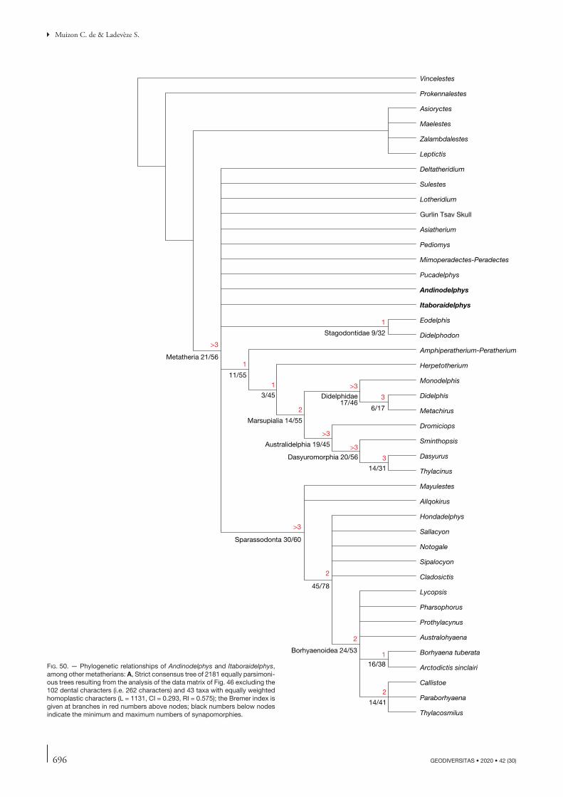
696 GEODIVERSITAS • 2020 • 42 (30)
Muizon C. de & Ladevèze S.
Vincelestes
Prokennalestes
Asioryctes
Maelestes
Zalambdalestes
Leptictis
Deltatheridium
Sulestes
Lotheridium
Gurlin Tsav Skull
Asiatherium
Pediomys
Mimoperadectes-Peradectes
Pucadelphys
Andinodelphys
Itaboraidelphys
Eodelphis
Didelphodon
Amphiperatherium-Peratherium
Herpetotherium
Monodelphis
Didelphis
Metachirus
Dromiciops
Sminthopsis
Dasyurus
Thylacinus
Mayulestes
Allqokirus
Hondadelphys
Sallacyon
Notogale
Sipalocyon
Cladosictis
Lycopsis
Pharsophorus
Prothylacynus
Australohyaena
Borhyaena tuberata
Arctodictis sinclairi
Callistoe
Paraborhyaena
Thylacosmilus
Metatheria 21/56
Stagodontidae 9/32
11/55
3/45
Marsupialia 14/55
Didelphidae17/46
6/17
Australidelphia 19/45
Dasyuromorphia 20/56
14/31
Sparassodonta 30/60
45/78
Borhyaenoidea 24/53
16/38
14/41
1
2
2
2
2
3
3
>3
>3
>3
>3
>3
1
1
1
fig. 50. — Phylogenetic relationships of Andinodelphys and Itaboraidelphys, among other metatherians: A, Strict consensus tree of 2181 equally parsimoni-ous trees resulting from the analysis of the data matrix of Fig. 46 excluding the 102 dental characters (i.e. 262 characters) and 43 taxa with equally weighted homoplastic characters (L = 1131, CI = 0.293, RI = 0.575); the Bremer index is given at branches in red numbers above nodes; black numbers below nodes indicate the minimum and maximum numbers of synapomorphies.
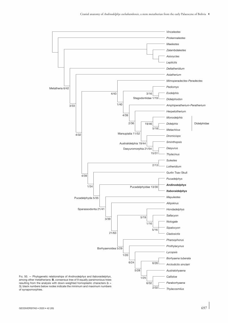
697
Cranial anatomy of Andinodelphys cochabambensis, a stem metatherian from the early Palaeocene of Bolivia
GEODIVERSITAS • 2020 • 42 (30)
fig. 50. — Phylogenetic relationships of Andinodelphys and Itaboraidelphys, among other metatherians: B, consensus tree of 9 equally parsimonious trees resulting from the analysis with down-weighted homoplastic characters (k = 3); black numbers below nodes indicate the minimum and maximum numbers of synapomorphies.
Vincelestes
Prokennalestes
Maelestes
Zalambdalestes
Asioryctes
Leptictis
Deltatheridium
Asiatherium
Mimoperadectes-Peradectes
Pediomys
Eodelphis
Didelphodon
Amphiperatherium-Peratherium
Herpetotherium
Monodelphis
Didelphis
Metachirus
Dromiciops
Sminthopsis
Dasyurus
Thylacinus
Sulestes
Lotheridium
Gurlin Tsav Skull
Pucadelphys
Andinodelphys
Itaboraidelphys
Mayulestes
Allqokirus
Hondadelphys
Sallacyon
Notogale
Sipalocyon
Cladosictis
Pharsophorus
Prothylacynus
Lycopsis
Borhyaena tuberata
Arctodictis sinclairi
Australohyaena
Callistoe
Paraborhyaena
Thylacosmilus
Metatheria 6/42
3/33
4/32
4/39
2/13
1/34
Pucadelphyda 5/35
Pucadelphyidae 13/30
Sparassodonta 21/41
3/39
21/63
5/19
1/16
5/16
Borhyaenoidea 5/29
1/20
6/24
5/28
6/26
1/25
6/32
2/32
4/43
1/40
3/16
Stagodontidae 1/19
4/39
2/36
Marsupialia 11/52
19/46
5/18
Didelphidae
Australidelphia 19/44
Dasyuromorphia 21/54
15/31
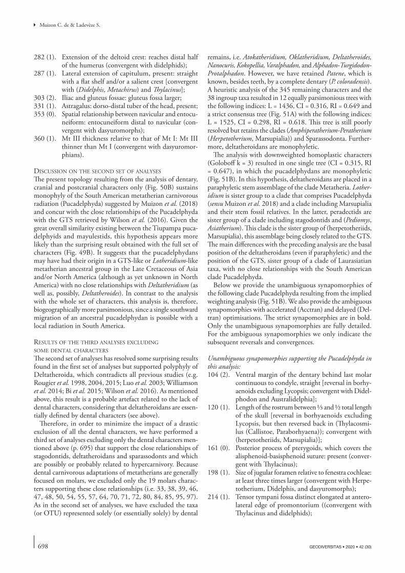
698 GEODIVERSITAS • 2020 • 42 (30)
Muizon C. de & Ladevèze S.
282 (1). Extension of the deltoid crest: reaches distal half of the humerus (convergent with didelphids);
287 (1). Lateral extension of capitulum, present: straight with a flat shelf and/or a salient crest [convergent with (Didelphis, Metachirus) and Thylacinus];
303 (2). Iliac and gluteus fossae: gluteus fossa larger;331 (1). Astragalus: dorso-distal tuber of the head, present;353 (0). Spatial relationship between navicular and entocu-
neiform: entocuneiform distal to navicular (con-vergent with dasyuromorphs);
360 (1). Mt III thickness relative to that of Mt I: Mt III thinner than Mt I (convergent with dasyuromor-phians).
diScuSSion on the Second Set of analySeS
The present topology resulting from the analysis of dentary, cranial and postcranial characters only (Fig. 50B) sustains monophyly of the South American metatherian carnivorous radiation (Pucadelphyda) suggested by Muizon et al. (2018) and concur with the close relationships of the Pucadelphyda with the GTS retrieved by Wilson et al. (2016). Given the great overall similarity existing between the Tiupampa puca-delphyids and mayulestids, this hypothesis appears more likely than the surprising result obtained with the full set of characters (Fig. 49B). It suggests that the pucadelphydans may have had their origin in a GTS-like or Lotheridium-like metatherian ancestral group in the Late Cretaceous of Asia and/or North America (although as yet unknown in North America) with no close relationships with Deltatheridium (as well as, possibly, Deltatheroides). In contrast to the analysis with the whole set of characters, this analysis is, therefore, biogeographically more parsimonious, since a single southward migration of an ancestral pucadelphydan is possible with a local radiation in South America.
reSultS of the third analySeS excluding Some dental characterS
The second set of analyses has resolved some surprising results found in the first set of analyses but supported polyphyly of Deltatheroida, which contradicts all previous studies (e.g. Rougier et al. 1998, 2004, 2015; Luo et al. 2003; Williamson et al. 2014; Bi et al. 2015; Wilson et al. 2016). As mentioned above, this result is a probable artefact related to the lack of dental characters, considering that deltatheroidans are essen-tially defined by dental characters (see above).
Therefore, in order to minimize the impact of a drastic exclusion of all the dental characters, we have performed a third set of analyses excluding only the dental characters men-tioned above (p. 695) that support the close relationships of stagodontids, deltatheroidans and sparassodonts and which are possibly or probably related to hypercarnivory. Because dental carnivorous adaptations of metatherians are generally focused on molars, we excluded only the 19 molars charac-ters supporting these close relationships (i.e. 33, 38, 39, 46, 47, 48, 50, 54, 55, 57, 64, 70, 71, 72, 80, 84, 85, 95, 97). As in the second set of analyses, we have excluded the taxa (or OTU) represented solely (or essentially solely) by dental
remains, i.e. Atokatheridium, Oklatheridium, Deltatheroides, Nanocuris, Kokopellia, Varalphadon, and Alphadon-Turgidodon-Protalphadon. However, we have retained Patene, which is known, besides teeth, by a complete dentary (P. coloradensis). A heuristic analysis of the 345 remaining characters and the 38 ingroup taxa resulted in 12 equally parsimonious trees with the following indices: L = 1436, CI = 0.316, RI = 0.649 and a strict consensus tree (Fig. 51A) with the following indices: L = 1525, CI = 0.298, RI = 0.618. This tree is still poorly resolved but retains the clades (Amphiperatherium-Peratherium (Herpetotherium, Marsupialia)) and Sparassodonta. Further-more, deltatheroidans are monophyletic.
The analysis with downweighted homoplastic characters (Goloboff k = 3) resulted in one single tree (CI = 0.315, RI = 0.647), in which the pucadelphydans are monophyletic (Fig. 51B). In this hypothesis, deltatheroidans are placed in a paraphyletic stem assemblage of the clade Metatheria. Lother-idium is sister group to a clade that comprises Pucadelphyda (sensu Muizon et al. 2018) and a clade including Marsupialia and their stem fossil relatives. In the latter, peradectids are sister group of a clade including stagodontids and (Pediomys, Asiatherium). This clade is the sister group of (herpetotheriids, Marsupialia), this assemblage being closely related to the GTS. The main differences with the preceding analysis are the basal position of the deltatheroidans (even if paraphyletic) and the position of the GTS, sister group of a clade of Laurasiatian taxa, with no close relationships with the South American clade Pucadelphyda.
Below we provide the unambiguous synapomorphies of the following clade Pucadelphyda resulting from the implied weighting analysis (Fig. 51B). We also provide the ambiguous synapomorphies with accelerated (Acctran) and delayed (Del-tran) optimisations. The strict synapomorphies are in bold. Only the unambiguous synapomorphies are fully detailed. For the ambiguous synapomorphies we only indicate the subsequent reversals and convergences.
Unambiguous synapomorphies supporting the Pucadelphyda in this analysis:104 (2). Ventral margin of the dentary behind last molar
continuous to condyle, straight [reversal in borhy-aenoids excluding Lycopsis; convergent with Didel-phodon and Australidelphia];
120 (1). Length of the rostrum between 1/3 and 1/2 total length of the skull [reversal in borhyaenoids excluding Lycopsis, but then reversed back in (Thylacosmi-lus (Callistoe, Paraborhyaena)); convergent with (herpetotheriids, Marsupialia)];
161 (0). Posterior process of pterygoids, which covers the alisphenoid-basisphenoid suture: present (conver-gent with Thylacinus);
198 (1). Size of jugular foramen relative to fenestra cochleae: at least three times larger (convergent with Herpe-totherium, Didelphis, and dasyuromorphs);
214 (1). Tensor tympani fossa distinct elongated at antero-lateral edge of promontorium ((convergent with Thylacinus and didelphids);

699
Cranial anatomy of Andinodelphys cochabambensis, a stem metatherian from the early Palaeocene of Bolivia
GEODIVERSITAS • 2020 • 42 (30)
260 (1). Foramen on dorsal arch of last lumbar vertebra, present [reversal in (Callistoe, Paraborhyaena); convergent with dasyuromorphs];
262 (1). Ventral median keel on anterior lumbar vertebrae, present (reversal in Prothylacinus; convergent with Metachirus, Dromiciops, and Thylacinus);
272 (1). Supraspinous fossa narrower than infraspinatus fossa at the level of the scapular neck [reversal in Sipalo-cyon and ((Borhyaena, Arctodictis), (Australohy-aena (Thylacosmilus (Callistoe, Paraborhyaena)))); convergent with Dromiciops];
282 (1). Extension of the deltoid crest: reaches distal half of the humerus (convergent with didelphids);
287 (1). Lateral extension of capitulum, present: straight with a flat shelf and/or a salient crest [convergent with (Didelphis, Metachirus) and Thylacinus];
303 (2). Iliac and gluteal fossae: gluteal fossa larger (conver-gent with Metachirus and dasyuromorphians);
331 (1). Astragalus: dorso-distal tuber of the head, present;353 (0). Spatial relationship between navicular and entocu-
neiform: entocuneiform distal to navicular (con-vergent with dasyuromorphs);
360 (1). Mt III thickness relative to that of Mt I: Mt III thin-ner than Mt I (convergent with dasyuromorphs).
Ten ambiguous synapomorphies (Acctran, fast) may also support the Pucadelphyda in this analysis: 1 (0). Number of upper incisors, five [reversal in Sparas-
sodonta excluding Mayulestidae; convergent with (herpetotheriids, Marsupialia)];
3 (1). I1s roots diverging dorsally [reversal in Sparas-sodonta excluding Mayulestidae; convergent with (herpetotheriids, Marsupialia)];
154 (1). Posterior palatine spine, absent and posterior edge of the palate straight [reversal in Mayulestidae; convergent with (herpetotheriids, Marsupialia)];
156 (1). Morphology of the postpalatine torus foramen, wide-open groove (the section is approximately half a circle or less) (reversal in Andinodelphys; convergent with dasyuromorphs);
178 (1). Suprameatal foramen, below postzygomatic crest [reversal in Sparassodonta excluding Mayulestidae; convergent with didelphids and (Dasyurus, Thyl-acinus)];
283 (1). Distal end of deltoid crest, forming a distinct angle or process [reversal in Sparassodonta excluding May-ulestidae; convergent with Dromiciops, Dasyurus, and (Didelphis, Metachirus)];
315 (1). Tibia shape, straight (reversal in Mayulestidae; convergent with Thylacinus);
331 (1). Astragalus: dorso-distal tuber of the head, present;353 (0). Entocuneiform distal to navicular (convergent with
dasyuromorphs);354 (1). Navicular facet of ectocuneiform parallel to the
distal metatarsal facet [reversal in ((Borhyaena, Arctodictis), (Australohyaena (Thylacosmilus (Cal-listoe, Paraborhyaena))))];
Four ambiguous synapomorphies (Deltran, slow) may also support Pucadelphyda in this analysis: 155 (1). Size of foramen or groove in lateral edge of post-
palatine torus, present and small (less than half the diameter of the minor palatine foramen or groove) [convergent with ((peradectids (((Asiatherium, Pediomys), stagodontids)), (herpetotheriids, Mar-supialia)))];
210 (1). Deep groove for internal carotid artery excavated in medial side of promontorium apex ventrally, present (convergent with GTS)
248 (1). Shape of the cranial facets of atlas: dorsal third of the facet much more concave than the ventral two thirds and strongly inflected medially [reversal in Prothylacinus, and (Borhyaena, Arctodictis); conver-gent with didelphids and (Thylacinus, Dasyurus)];
300 (1). Tuberosity for the rectus femoris muscle, protuber-ance (convergent with dasyuromorphs);
diScuSSion on the third Set of analySeS
The result of the third set of analyses (implied weighting analysis – Figs 51B; 52) is favoured here. They concord with those of Rangel et al. (2019) as far as the Tiupampian taxa are concerned. Andinodelphys and Pucadelphys are sister taxa and both are included in a clade (Pucadelphyidae) that is sister to Sparassodonta. Mayulestes and Allqokirus are the earliest known sparassodonts. The relationships within the sparassodonts retrieved by Rangel et al. (2019) is very similar to those of Muizon et al. (2018) and to those obtained in this paper. Several minor discrepancies still remain however. The Itaboraian genus Itaboraidelphys is included by Rangel et al. (2019) in Notometatheria, as the sister taxon of Didelphopsis, both taxa forming the sister group of Herpetotheriidae. Our analyses place Itaboraidelphys within Pucadelphyidae as the sister taxon to the clade (Andinodelphys, Pucadelphys). In fact, this difference is obviously related to our hypothesis to refer the two Type II Itaboraí petrosals to Itaboraidelphys (fol-lowing Muizon et al. [2018]), what Rangel et al. (2019) did not. Another difference is in the non-recognition of the taxon Mayulestidae sensu Muizon et al. (2018) that is including Allqokirus, Mayulestes, and Patene. However, it is noteworthy that Rangel et al. (2019) retrieved a para-phyletic Mayulestidae, which, in our point of view, is not drastically different from a monophyletic family. In fact, given the elevated number of dental characters of their data matrix and the total absence of cranial characters for Patene, it is possible that, if relevant cranial remains of Patene (or a closely related taxon) are discovered, given the phylogenetic closeness of the three genera, they could very well provide essential data in favour of a monophyletic Mayulestidae sensu Muizon et al. (2018). Furthermore, surprisingly, Rangel et al. (2019) restricted the family Mayulestidae to the genus Mayulestes, instead of retaining a paraphyletic “Mayulestidae” including the three genera as defined by Muizon et al. (2018), whereas they retained a paraphyletic family “Hathliacynidae” (represented in
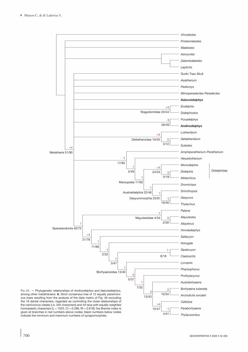
700 GEODIVERSITAS • 2020 • 42 (30)
Muizon C. de & Ladevèze S.
Vincelestes
Prokennalestes
Maelestes
Asioryctes
Zalambdalestes
Leptictis
Gurlin Tsav Skull
Asiatherium
Pediomys
Mimoperadectes-Peradectes
Itaboraidelphys
Eodelphis
Didelphodon
Pucadelphys
Andinodelphys
Lotheridium
Deltatheridium
Sulestes
Amphiperatherium-Peratherium
Herpetotherium
Monodelphis
Didelphis
Metachirus
Dromiciops
Sminthopsis
Dasyurus
Thylacinus
Patene
Mayulestes
Allqokirus
Hondadelphys
Sallacyon
Notogale
Sipalocyon
Cladosictis
Lycopsis
Pharsophorus
Prothylacynus
Australohyaena
Borhyaena tuberata
Arctodictis sinclairi
Callistoe
Paraborhyaena
Thylacosmilus
Metatheria 51/90
Deltatheroidea 19/30
3/12
Stagodontidae 20/44
17/65
5/49
Marsupialia 17/62
24/53
5/19
Didelphidae
Australidelphia 22/48
Dasyuromorphia 25/6119/40
28/49
Sparassodonta 40/72
Mayulestidae 4/34
2/30
21/78
11/66
2/52
3/47
6/18
Borhyaenoidea 13/40
5/37
7/32
13/4310/34
15/47
4/41
>4
>4
>4
>4
>4
>4
1
1
1
1
1
1
1
1
1
1
1
1
1 1
3
2
3
3
3
3
4
1
fig. 51. — Phylogenetic relationships of Andinodelphys and Itaboraidelphys, among other metatherians: A, Strict consensus tree of 12 equally parsimoni-ous trees resulting from the analysis of the data matrix of Fig. 46 excluding the 19 dental characters, regarded as controlling the close relationships of the carnivorous clades (i.e. 345 characters) and 44 taxa with equally weighted homoplastic characters (L = 1525, CI = 0.298, RI = 0.618); the Bremer index is given at branches in red numbers above nodes; black numbers below nodes indicate the minimum and maximum numbers of synapomorphies.

701
Cranial anatomy of Andinodelphys cochabambensis, a stem metatherian from the early Palaeocene of Bolivia
GEODIVERSITAS • 2020 • 42 (30)
fig. 51. — Phylogenetic relationships of Andinodelphys and Itaboraidelphys, among other metatherians: B, single tree trees resulting from the analysis with down-weighted homoplastic characters (k = 3); black numbers below nodes indicate the minimum and maximum numbers of synapomorphies.
Vincelestes
Prokennalestes
Maelestes
Asioryctes
Zalambdalestes
Leptictis
Deltatheridium
Sulestes
Lotheridium
Gurlin Tsav Skull
Mimoperadectes-Peradectes
Asiatherium
Pediomys
Eodelphis
Didelphodon
Amphiperatherium-Peratherium
Herpetotherium
Monodelphis
Didelphis
Metachirus
Dromiciops
Sminthopsis
Dasyurus
Thylacinus
Itaboraidelphys
Pucadelphys
Andinodelphys
Patene
Mayulestes
Allqokirus
Hondadelphys
Sallacyon
Notogale
Sipalocyon
Cladosictis
Lycopsis
Pharsophorus
Prothylacynus
Borhyaena tuberata
Arctodictis sinclairi
Australohyaena
Thylacosmilus
Callistoe
Paraborhyaena
Metatheria 18/65
2/43
2/42
14/50
3/45
8/47
4/30
4/29
5/23
Stagodontidae 9/34
11/54
4/45
Marsupialia 14/61
23/55
5/20
Didelphidae
Australidelphia 22/49
Dasyuromorphia 26/62
19/40
Pucadelphyda 10/41
Pucadelphyidae 3/38
2/37
Sparassodonta 26/54
Mayulestidae 2/34
2/30
24/84
11/67
2/54
3/49
6/19
Borhyaenoidea 14/41
5/37
7/32
15/43
8/30
2/28
8/46
3/30
702 GEODIVERSITAS • 2020 • 42 (30)
Muizon C. de & Ladevèze S.
their matrix by the genera Sipalocyon, Notogale, Acyon, and Cladosictis). Because Hathliacynus is a junior synonym of Cladosictis (Marshall 1981), following the treatment of the Mayulestidae by Rangel et al. (2019) and according to their result, the family Hathlyacynidae should be restricted to the clade (Acyon, Cladosictis), which they retrieved in their fig. 12. Be that as it may, it is noteworthy that the paraphyly of “Hathliacynidae” is another similarity of our analysis to that of Rangel et al. (2019).
The superorder Pucadelphyda was erected by Muizon et al. (2018) to include Pucadelphyidae and Sparassodonta, but it is noteworthy that the only non-sparassodont taxa they included in the superorder are Pucadelphys and Andi-nodelphys. The analysis performed by Rangel et al. (2019) also included in the Pucadelphyda a clade comprising two other Tiupampian taxa (Jaskhadelphys and Szalinia) as well as Minusculodelphys (from Itaboraí) and Kiruwamaq (from Santa Rosa, Peru). Jaskhadelphys and Szalinia, which were not included in Muizon et al. (2018)’s analysis, since it was essentially focussed on sparassodonts, will be considered in a work in progress by the authors on a skull of Mizquedelphys and a partial skull of Incadelphys. Furthermore, Rangel et al. (2019: 286) also included in the Pucadelphyda, the Tiupampian genera Mizquedelphys, Tiulordia, and Incadelphys (not included in their analy-sis). Muizon et al. (2018) have included Mizquedelphys in Pucadelphyidae, and, therefore, in Pucadelphyda; Tiu-lordia and Incadelphys were regarded by these authors as “Ameridelphia” incertae sedis together with Jaskhadelphys and Szalinia. Pending further discoveries and studies based on osteological remains (and/or more complete dental remains), we can provisionally accept the hypothesis of Rangel et al. (2019) to include these four genera within the Pucadelphyda. It is noteworthy, however, that the consensus tree of Rangel et al. (2019: fig. 12) indicates a Bremer value of 3 for the clade (Pucadelphyidae, Sparas-sodonta), whereas it is only of 1 for their Pucadelphyda. This means that their clade Pucadelphyda ((Szalinia, Jaskhadelphydae), (Pucadelphyidae, Sparassodonta)) is not strongly supported and, because Jaskhadelphys minutus, the only known jaskhadelphyid species is very incom-pletely known, (two upper molars only), the discovery of some new character or better preserved specimens, could easily significantly modify their topology concerning the (Szalinia, Jaskhadelphyidae) clade. To conclude, we consider, nevertheless, that the decision of Rangel et al. (2019) to include in the Pucadelphyda nine out of the twelve metatherian genera recorded at Tiupampa as well as Kiruwamaq (one single isolated ?M3) and Minusculo-delphis (dental remains only) is to be taken with caution concerning the taxa represented by dental remains only (or almost exclusively).
Another interesting concordant result with Rangel et al. (2019), obtained in our first set of analyses (analysis with all characters and implied weighting Fig. 49B), is the posi-tion of Varalphadon, which, as evidenced by Muizon et al. (2018), has no close relationships with the Sparassodonta.
CONCLUSIONS
Two hypotheses are here advanced as regard to the palaeobio-geographic history of the Sparassodonta. In a first scenario, based on a complete set of morphological characters, sparas-sodonts probably originated outside South America and not as an endemic carnivorous radiation including pucadelphy-ids and sparassodonts, as suggested by Muizon et al. (2018). In this hypothesis (resulting from the first set of analyses), sparassodonts may have originated from basal taxa of the still poorly known North American deltatheroidan radiation (e.g. Atokatheridium or Oklatheridium), but are unlikely to have been related to the Cenomanian North American genus Varalphadon as shown by the result of this analysis (Fig. 49).
An alternate scenario, resulting from the second and third sets of analyses, the former excluding all dental characters, and the latter excluding selected dental characters possibly or probably related to hypercarnivory, supported the mono-phyly of Pucadelphyda (Pucadelphyidae and Sparassodonta) and therefore advocates for a common biogeographic and phylogenetic origin of the two pucadelphydan clades. If an independent southward migration of pucadelphyids and sparassodonts is required by the first analysis (which renders the evolutionary history of the Pucadelphyda more complex but still quite possible) it is unlikely, unnecessary, and more parsimonious, as far as palaeobiogeography is concerned, in the case of the second and third ones.
This result also raises the problem of the North American origin of the Pucadelphyda in the case of a single southward dispersal of this clade. As discussed above (p. 694), great dental similarities exist between pucadelphyids and the late Campanian (Judithian) North American genus Aenigmadel-phys. Striking similarities also exist between this latter taxon and the Tiupampian Incadelphys, Jaskhadelphys, and Szalinia, which would lend support to Rangel et al.’s (2019) hypothesis for their inclusion in the Pucadelphyda. Such a similarity is especially true for Incadelphys and Mizquedelphys, for which the teeth of the partial skulls mentioned in this study are so similar to the teeth Aenigmadelphys that, if found in North America, they could easily have been referred to a species of this genus. Nevertheless, further study is needed to address this problem in detail. Be that as it may, it is therefore pos-sible that Aenigmadelphys could represent an ancestral North American morphotype for the South American pucadelphy-dan radiation.
Furthermore, as exposed by Goin et al. (2016), the great diversity of the Tiupampa mammalian fauna and its early Palaeocene age, as well as the absence of therians in the diverse and slightly older Campanian fauna of Los Alamitos (Argentina), strongly supports the independent southward migration of several groups of metatherians and eutherians, which already encompassed a radiation in North America and further diversified or became extinct in South America (e.g., Muizon & Cifelli 2000; Case et al. 2005, Muizon et al. 2015; Clemens 2017). As far as metatherians are concerned, this hypothesis is well established in the case of the Polydolopi-morphia which are present in the Late Cretaceous of North
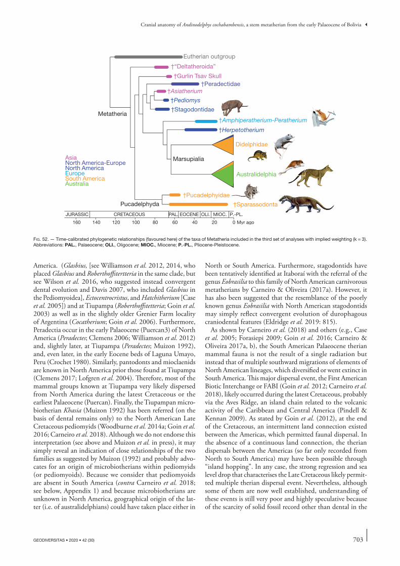
703
Cranial anatomy of Andinodelphys cochabambensis, a stem metatherian from the early Palaeocene of Bolivia
GEODIVERSITAS • 2020 • 42 (30)
America. (Glasbius, [see Williamson et al. 2012, 2014, who placed Glasbius and Roberthoffstertteria in the same clade, but see Wilson et al. 2016, who suggested instead convergent dental evolution and Davis 2007, who included Glasbius in the Pediomyoidea], Ectocentrocristus, and Hatchitherium [Case et al. 2005]) and at Tiupampa (Roberthoffstetteria; Goin et al. 2003) as well as in the slightly older Grenier Farm locality of Argentina (Cocatherium; Goin et al. 2006). Furthermore, Peradectia occur in the early Palaeocene (Puercan3) of North America (Peradectes; Clemens 2006; Williamson et al. 2012) and, slightly later, at Tiupampa (Peradectes; Muizon 1992), and, even later, in the early Eocene beds of Laguna Umayo, Peru (Crochet 1980). Similarly, pantodonts and mioclaenids are known in North America prior those found at Tiupampa (Clemens 2017; Lofgren et al. 2004). Therefore, most of the mammal groups known at Tiupampa very likely dispersed from North America during the latest Cretaceous or the earliest Palaeocene (Puercan). Finally, the Tiupampan micro-biotherian Khasia (Muizon 1992) has been referred (on the basis of dental remains only) to the North American Late Cretaceous pediomyids (Woodburne et al. 2014a; Goin et al. 2016; Carneiro et al. 2018). Although we do not endorse this interpretation (see above and Muizon et al. in press), it may simply reveal an indication of close relationships of the two families as suggested by Muizon (1992) and probably advo-cates for an origin of microbiotherians within pediomyids (or pediomyoids). Because we consider that pediomyoids are absent in South America (contra Carneiro et al. 2018; see below, Appendix 1) and because microbiotherians are unknown in North America, geographical origin of the lat-ter (i.e. of australidelphians) could have taken place either in
North or South America. Furthermore, stagodontids have been tentatively identified at Itaboraí with the referral of the genus Eobrasilia to this family of North American carnivorous metatherians by Carneiro & Oliveira (2017a). However, it has also been suggested that the resemblance of the poorly known genus Eobrasilia with North American stagodontids may simply reflect convergent evolution of durophagous craniodental features (Eldridge et al. 2019: 815).
As shown by Carneiro et al. (2018) and others (e.g., Case et al. 2005; Forasiepi 2009; Goin et al. 2016; Carneiro & Oliveira 2017a, b), the South American Palaeocene therian mammal fauna is not the result of a single radiation but instead that of multiple southward migrations of elements of North American lineages, which diversified or went extinct in South America. This major dispersal event, the First American Biotic Interchange or FABI (Goin et al. 2012; Carneiro et al. 2018), likely occurred during the latest Cretaceous, probably via the Aves Ridge, an island chain related to the volcanic activity of the Caribbean and Central America (Pindell & Kennan 2009). As stated by Goin et al. (2012), at the end of the Cretaceous, an intermittent land connection existed between the Americas, which permitted faunal dispersal. In the absence of a continuous land connection, the therian dispersals between the Americas (so far only recorded from North to South America) may have been possible through “island hopping”. In any case, the strong regression and sea level drop that characterises the Late Cretaceous likely permit-ted multiple therian dispersal event. Nevertheless, although some of them are now well established, understanding of these events is still very poor and highly speculative because of the scarcity of solid fossil record other than dental in the
020406080100120140160 Myr ago
JURASSIC CRETACEOUS PAL. OLI.EOCENE MIOC. P.-PL.
AsiaNorth America-Europe
EuropeSouth AmericaAustralia
North America
†Herpetotherium
Didelphidae
†Stagodontidae
Marsupialia
Metatheria
Pucadelphyda
†“Deltatheroida”
†Pucadelphyidae
†Sparassodonta
Australidelphia
†Asiatherium†Peradectidae
†Gurlin Tsav Skull
Eutherian outgroup
†Amphiperatherium-Peratherium
†Pediomys
fig. 52. — Time-calibrated phylogenetic relationships (favoured here) of the taxa of Metatheria included in the third set of analyses with implied weighting (k = 3). Abbreviations: PAL., Palaeocene; OLI., Oligocene; MIOC., Miocene; P.-PL., Pliocene-Pleistocene.
704 GEODIVERSITAS • 2020 • 42 (30)
Muizon C. de & Ladevèze S.
Late Cretaceous of North America and in the early Palaeocene of South America. Establishing phylogenetic affinities based almost exclusively on dental remains (in some cases fragmen-tary) is extremely questionable since dental characters have been shown to be strongly homoplastic and subject to major individual variation (e.g., Muizon & Lange-Badré 1997; Ladevèze et al. 2011; Sansom et al. 2017; Solé & Ladevèze 2017). In fact, given the poorness of the fossil record, the discovery of well-preserved therian cranial remains in the Late Cretaceous of North America may drastically modify (or corroborate) the current hypotheses on the evolutionary histories of South American Palaeogene metatherians and eutherians. A similar major migratory event occurred much later between the Americas in the late Cenozoic-early Pleis-tocene (the GABI or Great American Biotic Interchange) and was also characterised by multiple individual migrations of different groups (Stehli & Webb 1985). However, study of the GABI is much easier than in the case of the FABI since, in the late Cenozoic, the fossil record is much better in terms of abundance and completeness and, since the groups have encompassed a long evolutionary history, they are therefore more easily recognizable.
AcknowledgementsThe locality of Tiupampa was discovered in 1982 during a field expedition funded by National Geographic Society (grant N°2467/82). The specimens described in this paper were dis-covered in 1996 during a field season at Tiupampa funded by the Institut Français d’Études Andines and the CNRS. Fos-sil collecting was carried out under the auspices of research agreements between the Fundación para las Ciencias (Bolivia) and the Muséum national d’Histoire naturelle (France). The specimens are the property of the MHNC and have been provided on loan to the MNHN for curation and study. Field expedition has benefited of logistical support from the IRD (Institut de Recherche pour le Développement) in Bolivia. We thank our Bolivian colleagues (Ricardo Céspedes-Paz, Ramiro Suárez-Sorruco, Mario Suárez-Riglos) for their collaboration and logistic support during the field season. Special thanks are due to Céline Bens, Aurélie Verguin, Géraldine Véron, and Christine Lefèvre, (MNHN), who provided access to MNHN specimens under their care. Warm thanks to Guillermo Rou-gier who provided casts of Deltatheridium pretribuberculare (specimens PSS MAE 132 and 133), to Richard Cifelli for generously providing hundreds of casts of North American Cretaceous metatherians, to William Clemens who provided excellent photographs (taken by David Strauss) of the holotypes of Lycopsis longirostris and Hondadelphys fieldsi and a cast of the holotype of Crustulus fontanus, to Steve Wroe for providing stl files of the skulls of Thylacinus cynocephalus and Nimbacinus dicksoni. Special thanks also to Gregory Wilson for provid-ing access to the CT data of the skulls of Didelphodon vorax NDGS 431 and UW BM 94084. This work has benefited from fruitful discussions with Shundong Bi (who provided unpublished data on Lotheridium), Guillaume Billet, Richard Cifelli, Brian Davis, and Javier Gelfo. Special thanks are due
to the reviewers, Robin Beck and Gregory Wilson, whose invaluable comments significantly improved the manuscript. Photographs were made by Philippe Loubry and Lilian Cazes. CT scanning of the specimens was performed by Miguel Garcia at the AST-RX technical platform of scientific access to RX tomography (UMS 2700 “Acquisition et Analyse de Don-nées pour l’Histoire Naturelle”), CNRS-MNHN, Paris) Data processing was undertaken by Florent Goussard and Nathalie Poulet at the 3D imaging facilities Lab of the UMR 7207 CR2P (MNHN CNRS SU). The artistic reconstructions depicting the past life of Andinodelphys and Pucadelphys in Tiupampa were realised respectively by Charlene Letenneur and Sophie Fernandez (CR2P). Lastly, our warmest thanks to Emmanuel Côtez for his extremely careful edition of the manuscript. The MNHN gives access to the collections in the framework of the RECOLNAT national Research Infrastructure.
REFERENCES
andreiS r. r. 1977. — Geología del área de Cañadón Hondo, Depto. Escalante, provincia del Chubut, República Argentina. Revista del Museo de La Plata, Obra del Centenario, Geología 4: 77-102.
archer m. 1976a. — The basicranial region of marsupicarnivores (Marsupialia), interrelationships of carnivorous marsupials, and affinities of the insectivorous marsupial peramelids. Zoo-logical Journal of the Linnean Society 59: 217-322. https://doi.org/10.1111/j.1096-3642.1976.tb01016.x
archer m. 1976b. — Revision of the marsupial genus Planigale Throughton (Dasyuridae). Memoirs of the Queensland Museum 17 (3): 341-365. https://www.biodiversitylibrary.org/page/48804334
archer m. 1981. — Results of the Archbold expeditions NO. 104. Systematic revision of the marsupial dasyurid genus Sminthopsis Thomas. Bulletin of the American Museum of Natural History 168 (2): 65-223. http://hdl.handle.net/2246/1050
argot c. 2003. — Postcranial functional adaptations in the South American Miocene borhyaenoids (Mammalia, Metatheria): Cladosictis, Pseudonotictis, and Sipalocyon. Alcheringa: An Australasian Journal of Palaeontology 27: 303-356. https://doi.org/10.1080/03115510308619110
averianov a. o., archibald J. d. & e. g. ekdale 2010. — New material of the Late Cretaceous deltatheroidans mammal Sulestes from Uzbekistan and phylogenetic reassessment of the metatherian-eutherian dichotomy. Journal of Systematic Paleontology 8 (3): 301-330. https://doi.org/10.1080/14772011003603499
babot m. J., powell J. e. & muizon c. de 2002. — Callistoe vincei, a new Proborhyaenidae (Borhyaenoidea, Metatheria, Mam-malia) from the early Eocene of Argentina. Geobios 35: 615-629. https://doi.org/10.1016/S0016-6995(02)00073-6
babot J., garcía-lópez d., deraco v., herrera c. m. & del papa c. 2017. — Mamíferos paleógenos del subtrópico de Argentina: síntesis de estudios estratigráficos, cronológicos y taxonómicos, in muruaga c. m. & groSSe p. (eds), Ciencias de la Tierra y Recursos Naturales del NOA. Relatorio del XX Con-greso Geológico Argentino, San Miguel de Tucumán: 730-753.
bauzá n., gelfo J. n., & lópez g. m. 2019. — Early steps in the radiation of notoungulate mammals in southern South America: A new henricosborniid from the Eocene of Patagonia. Acta Pal-aeontologica Polonica 64 (3): 597-207. https://doi.org/10.4202/app.00565.2018
beck r. m. 2017. — The skull of Epidolops ameghinoi from the early Eocene Itaboraí fauna, Southern Brazil, and the affinities of the extinct marsupialiform order Polydolopimorphia. Journal of Mammalian Evolution 24 (4) 373-414. https://doi.org/10.1007/s10914-016-9357-6.

705
Cranial anatomy of Andinodelphys cochabambensis, a stem metatherian from the early Palaeocene of Bolivia
GEODIVERSITAS • 2020 • 42 (30)
beck r. m. & taglioretti m. l. 2019. — A nearly complete juvenile skull of the marsupial Sparassocynus derivatus from the Pliocene of Argentina, the affinities of “sparassocynids”, and the diversification of opossums (Marsupialia; Didelphimorphia; Didelphidae). Journal of Mammalian Evolution. https://doi.org/10.1007/s10914-019-09471-y
benSley b. a. 1902. — On the identification of the Meckelian and mylohyoid grooves in the jaws of Mesozoic and Recent mammals. University of Toronto Studies, Biological Series 3: 75-82. https://doi.org/10.5962/bhl.title.66541
bi S., Jin x., li S. & du t. 2015. — A new Cretaceous metathe-rian mammal from Henan, China. PeerJ 3: e896. https://doi.org/10.7717/peerj.896.
bi S., zheng x., wang x., cignetti n.e., yang S. & wible J. r. 2018. — An Early Cretaceous eutherian and the placen-tal-marsupial dichotomy. Nature 558: 390-395. https://doi.org/10.1038/s41586-018-0210-3
billet g., germain d., ruf i., muizon c. de & hautier l. 2013. — The inner ear of Megatherium and the evolution of the vestibular system in sloths. Journal of Anatomy 223: 557-567. https://doi.org/10.1111/joa.12114
blankS r. h. i., curthoyS i. S. & markham c. h. 1975. — Planar relationships of the semicircular canals in man. Acta of Oto-Laryngol-ogy 80: 185-296. https://doi.org/10.3109/00016487509121318
bonaparte J. & moraleS J. 1997. — Un primitivo Notonych-opidae (Litopterna) del Paleoceno Inferior de Punta Peligro, Chubut, Argentina. Estudios Geológicos 53: 183-186.
bonaparte J. & rougier g. 1987. — Mamíferos del Cretácico Inferior de Patagonia, in IV Congreso Latinoamericano de Pale-ontología. Santa Cruz, Bolivia 1: 343-359.
bonaparte J. J., van valen l. m., & kramarz a. 1993. — La fauna local de Punta Peligro, Paleoceno Inferior, de la provin-cia de Chubut, Patagonia, Argentina. Evolutionary Monographs 14: 1-21.
brannick a. l., wilSon g. p., varricchio d. J. & ekdale e. g. 2016. — Nestled among dinosaur eggs: new Alphadon specimens from Egg Mountain and their implications for metatherian evolution [abstract], in the Society of Vertebrate Paleontology 76th Annual Meeting; 2016, Oct 26-29; Salt Lake City, UT. http://vertpaleo.org/PDFS/2016/SVP-2016-Program-Book-v10-with-covers.aspx (last consultation on 9/12/2020).
bremer k. 1988. — The limits of amino acid sequence data in angiosperm phylogenetic reconstruction. Evolution 42 (4) 795-803. https://doi.org/10.2307/2408870
butler p. m. 1956. — The skull of Ictops and the classification of Insectivora. Proceedings of the Zoological Society of London 126: 453-481. https://doi.org/10.1111/j.1096-3642.1956.tb00449.x
cabrera a. 1927. — Datos para el conocimiento de los dasyuroid-eos fósiles argentinos. Revista del Museo de la Plata 30: 271-315.
calle a. z., horton b. k., limachi r., Stockli d. f., uze-da-orellana g. v., anderSon r. b. & long, S. p. 2018. — Cenozoic provenance and depositional record of the Subandean foreland basin during growth of the central Andean fold-thrust belt, southern Bolivia, in zamora valcarce g., mcclay & k., ramoS v. a. (eds), Petroleum Basins and Hydrocarbon Potential of the Andes of Peru and Bolivia. American Association of Petroleum Geologists, Memoir 117: 483-530. https://doi.org/10.1306/13622132M1173777
carneiro l. m. & oliveira e. v. 2017a. — Systematic affinities of the extinct metatherian Eobrasilia coutoi Simpson, 1947, a South American early Eocene Stagodontidae: implications for “Eobrasiliinae”. Revista Brasileira de Paleontologia 20: 355-372. https://doi.org/10.4072/rbp.2017.3.07
carneiro l. m. & oliveira e. v. 2017b. — The Eocene South American metatherian Zeusdelphys complicatus is not a proto-didelphid but a hatcheriform: paleobiogeographic implica-tions. Acta Palaeontologica Polonica 62: 497-207. https://doi.org/10.4202/app.00351.2017
carneiro l. m. 2018. — A new species of Varalphadon (Mamma-lia, Metatheria, Sparassodonta) from the upper Cenomanian of southern Utah, North America: Phylogenetic and biogeographic insights. Cretaceous Research 84: 88-96. https://doi.org/10.1016/j.cretres.2017.11.004
carneiro l. m., oliveira e. v. & goin f. J. 2018. — Austrope-diomys marshalli gen. et sp. nov., a new pediomyoidea (Mammalia, Metatheria) from the Paleogene of Brazil: Paleobiogeographic implications. Revista Brasileira de Paleontologia 21 (2): 120-131. https://doi.org/10.4072/rbp.2018.2.03
caSe J. a., goin f. J. & woodburne m. o. 2005. — “South American” marsupials from the Late Cretaceous of North America and the origin of marsupial cohorts. Journal of Mammalian Evolu-tion 12: 461-494. https://doi.org/10.1007/s10914-005-7329-3
chemiSquy m. a. 2015. — Peramorphic males and extreme sexual dimorphism in Monodelphis dimidiata (Didelphidae). Zoomorphol-ogy 134: 587-599. https://doi.org/10.1007/s00435-015-0274-7
chornogubSky l. & goin f. J. 2015. — A review of the molar morphology and phylogenetic affinities of Sillustania quechuense (Metatheria, Polydolopimorphia, Sillustaniidae) from the early Paleocene of Laguna Umayo, South eastern Peru. Journal of Vertebrate Paleontology 35 (6): e983238. https://doi.org/10.1080/02724634.2015.983238
cifelli r. l. 1990. — Cretaceous mammals of Southern Utah. I. Marsupials from the Kaiparowits Formation (Judithian). Journal of Vertebrate Paleontology 10 (3): 295-319. https://doi.org/10.1080/02724634.1990.10011816
cifelli r. l. 1993. — Early Cretaceous mammal from North America, and the evolution of marsupial dental characters. Pro-ceedings of the National Academy of Sciences USA 90: 9413-9416. https://doi.org/10.1073/pnas.90.20.9413
cifelli r. l. 2004. — Marsupial mammals from the Albian–Cenomanian (Early-Late Cretaceous) boundary, Utah. Bulletin of the American Museum of Natural History 285: 62-79. https://doi.org/cpgs94
cifelli r. l. & JohanSon z. 1994. — New mammal from the upper Cretaceous of Utah. Journal of Vertebrate Paleontology 14 (2): 292-295. https://doi.org/10.1080/02724634.1994.10011558
cifelli r. l. & muizon c. de 1997. — Dentition and jaw of Kokopellia juddi, a primitive marsupial or near-marsupial from the Medial Cretaceous of Utah. Journal of Mammalian Evolution 4 (4): 241-258. https://doi.org/10.1023/A:1027394430433
cifelli r. l. & muizon c. de 1998. — Marsupial mammal from the upper Cretaceous North Horn Formation, central Utah. Journal of Paleontology 72 (3): 532-537. https://doi.org/10.1017/S0022336000024306
clark c. t. & Smith k. k. 1993. — Cranial osteogenesis in Monodelphis domestica (Didelphidae) and Macropus eugenii (Macropodidae). Journal of Morphology 215:119-249. https://doi.org/10.1002/jmor.1052150203
clemenS w. a. 1966. — Fossil mammals from the type Lance For-mation, Wyoming. Part II. Marsupialia. University of California Publications in Geological Sciences 62: 1-122.
clemenS w. a. 1968. — A mandible of Didelphodon vorax (Mar-supialia, Mammalia). Contribution in Science 133: 1-11. https://www.biodiversitylibrary.org/page/52117669
clemenS w. a. 2006. — Early Paleocene (Puercan) peradectid marsupials from northeastern Montana, North American Western Interior. Palaeontographica, Abteilung A 277: 19-31.
clemenS w. a. 2017. — A pantodont (Mammalia) from the latest Puercan North American Land Mammal age (earliest Paleocene) of the Western interior, USA. Historical Biology 30 (1-2): 183-188. https://doi.org/10.1080/08912963.2016.1276178
clyde w. c., wilf p., igleSiaS a., Slingerland r. l., barnum t., biJl p. k., bralower t. J., brinkhuiS h., comer e. e., huber b. t., ibañez-meJia m., Jicha b. r., krauSe J. m., Schueth J. d., Singer b. S., raigemborn m. S., Schmitz m. d., SluiJS a., zamaloa m. c. 2014. — New age constraints
706 GEODIVERSITAS • 2020 • 42 (30)
Muizon C. de & Ladevèze S.
for the Salamanca Formation and lower Río Chico Group in the western San Jorge Basin, Patagonia, Argentina: Implica-tions for Cretaceous-Paleogene extinction recovery and land mammal age correlations. Geological Society of America Bul-letin 126 (3-4): 289-306. https://doi.org/10.1130/B30915.1
croft d. a., gelfo J. n., & lópez g. m. 2020. — Splendid innovation: the extinct South American native ungulates. Annual Review of Earth and Planetary Sciences 48: 259-290. https://doi.org/10.1146/annurev-earth-072619-060126
crochet J. y. 1980. — Les marsupiaux du Tertiaire d’Europe. Éditions de la Fondation Singer Polignac, Paris. 279 p. https://gallica.bnf.fr/ark:/12148/bpt6k3326526p
crochet J.-y. & Sigé b. 1996. — Un marsupial ancien (tran-sition Crétacé – Tertiaire) à denture évoluée en Amérique du Sud (Chulpas, Formation Umayo, Pérou). Neues Jahrbuch für Geologie und Paläontologie, Monatshefte 1996 (10): 622-634. https://doi.org/10.1127/njgpm/1996/1996/622
crompton a. w. & Sun a.-l. 1985. — Cranial structure and relationships of the Liassic mammal Sinoconodon. Zoo-logical Journal of the Linnean Society 85: 99-119. https://doi.org/10.1111/j.1096-3642.1985.tb01500.x
crompton a. w. & luo z. 1993. — Relationships of early mammals. Sinoconodon, Morganucodon oehleri, and Din-netherium, in Szalay f. S., novacek m. J. & mckenna M. C. (eds), Mammal Phylogeny. Vol. 1: Mesozoic Differentiation, Multituberculates, Monotremes Early Therians and Marsupials. Springer-Verlag, New York: 30-44. https://doi.org/10.1007/978-1-4613-9249-1_4
dalrymple g. b. 1979. — Critical tables for conversion of K-Ar ages from old to new constants. Geology 7: 558-260. https://doi.org/dbcc9m
david r., droulez J., allain r., berthoz a., Janvier p. & bennequin d. 2010. — Motion from the past. A new method to infer vestibular capacities of extinct species. Compte Rendus Palevol 9: 397-410. https://doi.org/10.1016/j.crpv.2010.07.012
daviS b. m. 2007. — A revision of “pediomyid” marsupials from the Late Cretaceous of North America. Acta Palaeontologica Polonica 52: 217-256.
daviS b. m. & cifelli r. l. 2011. — Reappraisal of the tribos-phenidan mammals from the Trinity Group (Aptian-Albian) of Texas and Oklahoma. Acta Palaeontologica Polonica 56 (3): 441-462. https://doi.org/10.4202/app.2011.0037
daviS b. m., cifelli r. l. & kielan-JaworowSka z. 2008. — Earliest evidence of Deltatheroida (Mammalia: Metatheria) from the Early Cretaceous of North America, in SargiS e. J. & dagoSto m. (eds), Mammalian Evolutionary Morphology, a tribute to Frederick Szalay. Springer Verlag, New York: 3-24. https://doi.org/10.1007/978-1-4020-6997-0_1
decelleS p. & horton b. k. 2003. — Early to Middle Ter-tiary foreland development and the history of Andean crustal shortening in Bolivia. Geological Society of America Bulletin 115: 58-27. https://doi.org/b6mh8k
demouy S., paquette J. l., Saint blanquat m. de, benoit m., belouSova e. a., o’reilly S. y., garcía f., teJada l. c., gallegoS r. & Sempere t. 2012. — Spatial and temporal evolution of Liassic to Paleocene arc activity in southern Peru unraveled by zircon U-Pb and Hf in-situ data on plu-tonic rocks. Lithos 155: 183-200. https://doi.org/10.1016/j.lithos.2012.09.001
dom r., fiSher b. l., & martin g. f. 1970. — The venous system of the head and neck of the opossum (Didelphis vir-giniana). Journal of Morphology 132 (4): 487-496. https://doi.org/10.1002/jmor.1051320408
duchêne d. a., bragg J. g., duchêne S., neaveS l. e., potter S., moritz c., JohnSon r. n., ho S. y. w., & eldridge m. d. b. 2018. — Analysis of phylogenomic tree space resolves relationships among marsupial families. Systematic Biology 67 (3): 400-412. https://doi.org/10.1093/sysbio/syx076
eaton J. g. 1993. — Therian mammals of the Cenomanian (Late Cretaceous) Dakota Formation, southwestern Utah. Journal of Vertebrate Paleontology 13: 105-224. https://doi.org/10.1080/02724634.1993.10011491
eberle J. J., clemenS w. a., mccarthy p. J., fiorillo a. r., erickSon g. r. & drukenmiller p. S. 2019. — Northermem-ost record of the Metatheria: a new Late Cretaceous pediomyid from the North slope of Alaska. Journal of Systematic Palaeon-tology 17 (21): 1805-1824. https://doi.org/10.1080/14772019.2018.1560369
edinger t. 1964. — Midbrain exposure and overlap in mammals. American Zoologist 4: 5-19. https://doi.org/10.1093/icb/4.1.5
edinger t. 1955. — Hearing and smell in cetacean history. Monatsschrift für Psychiatrie und Neurologie 129: 37-58. https://doi.org/10.1159/000139733
ekdale e. g., archibald d. J. & averianov a. o. 2004. — Petrosal bones of placental mammals from the Late Cretaceous of Uzbekistan. Acta Palaeontologica Polonica 49 (1): 161-176.
eldridge m. d. b., beck r. m., croft d. a., travouillon k. J. & fox b. J. 2019. — An emerging consensus in the evolution, phylogeny, and systematics of marsupials and their fossil relatives (Metatherian). Journal of Mammalogy 100 (3): 802-837. https://doi.org/10.1093/jmammal/gyz018
engelman r. k. & croft d. a. 2014. — A new species of small-bodied sparassodont (Mammalia: Metatheria) from the Middle Miocene locality of Quebrada Honda, Bolivia. Journal of Ver-tebrate Paleontology 34: 672-688. https://doi.org/10.1080/02724634.2013.827118
evanS h. e. & de lahunta a. 2013. — Miller’s Anatomy of the Dog. Saunders, St Louis, 850 p.
floreS d. a., giannini n. p. & abdala f. a. 2003. — Cranial ontogeny of Lutreolina crassicaudata (Didelphidae): a comparison with Didelphis albiventris. Acta Theriologica 48 (1): 1-2. https://doi.org/10.1007/BF03194261
flynn a. g., daviS a. J., williamSon t. e., heizler m., fenley iv w., leSlie c., Secord r., bruSatte S. l., & peppe d. J. 2020. — Early Eocene Magnetostratigraphy and revised bio-stratigraphy of the Ojo Alamo Sandstone and lower Nacimiento Formation, San Juan Basin, New Mexico, USA. Geological Society of Anerica Bulletin. https://doi.org/10.1130/B35481.1
foraSiepi a. m. 2009. — Osteology of Arctodictis sinclairi (Mam-malia, Metatheria, Sparassodonta) and phylogeny of Cenozoic metatherian carnivores from South America. Monografías del Museo Argentino de Ciencias Naturales 6: 1-174.
foraSiepi a. m., babot m. J & zimicz n. 2015. — Australohyaena antiqua (Mammalia, Metatheria, Sparassodonta), a large predator from the late Oligocene of Patagonia. Journal of Systematic Palae-ontology 13 (6): 503-525. Published online June 2014. https://doi.org/10.1080/14772019.2014.926403
foraSiepi a. m., macphee r. d. e. & hernández del pino S. 2019. — Caudal Cranium of Thylacosmilus atrox (Mammalia, Metatheria, Sparassodonta), a South American Predaceous Sabertooth. Bulletin of the American Museum of Natural History 2019 (433): 1-66. https://doi.org/10.1206/0003-0090.433.1.1
fox r. c. 1971. — Marsupial mammals from the early Campanian Milk River Formation, Alberta, Canada. Zoological Journal of the Linnean Society 50: 147-164.
fox r. c. 1974. — Deltatheroides-like mammals from the Upper Cretaceous of North America. Nature 249: 392. https://doi.org/10.1038/249392a0
fox r. c. 1975. — Molar structure and function in the Early Cretaceous mammal Pappotherium: evolutionary implications for Mesozoic Theria. Canadian Journal of Earth Sciences 12 (3): 412-442. https://doi.org/10.1139/e75-037
fox r. c. 1981. — Mammals from the Upper Cretaceous Oldman Formation, Alberta. V Eodelphis Matthew, and the evolution of the Stagodontidae. Canadian Journal of Earth Sciences 18 (2): 350-365. https://doi.org/10.1139/e81-027
707
Cranial anatomy of Andinodelphys cochabambensis, a stem metatherian from the early Palaeocene of Bolivia
GEODIVERSITAS • 2020 • 42 (30)
fox r. c. 1983. — Notes on the North American Tertiary marsu-pials Herpetotherium and Peradectes. Canadian Journal of Earth Sciences 20 (10): 1565-1578. https://doi.org/10.1139/e83-146
fox r. c. 1987. — Palaeontology and the early evolution of mar-supials, in archer m. (ed.), Possums and Opossums: Studies in Evolution. Surrey Beatty & Sons Pty Ltd and the Royal Zoological Society of New South Wales, Sydney: 161-169.
fox r. c. & naylor b. g. 1986. — A new species of Didelpho-don Marsh (Marsupialia) from the Upper Cretaceous of Alberta, Canada: paleobiology and phylogeny. Neues Jahrbuch für Geologie und Paläontologie 172 (3): 357-380.
fox r. c. & naylor b. g. 1995. — The relationships of the Stagodontidae, primitive North American Late Cretaceous mammals, in Sun a. & wang y. (eds), Sixth Symposium on Mesozoic Terrestrial Ecosystems and Biota. China Ocean Press, Beijing: 247-250.
fox r. c. & naylor b. g. 2006. — Stagodontid marsupials from the Late Cretaceous of Canada and their systematic and func-tional implications. Acta Palaeontologica Polonica 51 (1) 13-36.
fox r. c., Scott c. S. & bryant h. n. 2007. — A new, unu-sual therian mammal from the Upper Cretaceous of Saskatch-ewan, Canada. Cretaceous Research 28: 821-229. https://doi.org/10.1016/j.cretres.2006.12.005
gabbert S. l. 1998. — Basicranial anatomy of Herpetotherium (Marsupialia: Didelphimorphia) from the Eocene of Wyoming. American Museum Novitates 3235: 1-13. http://hdl.handle.net/2246/3244
gayet m., marShall l. g. & Sempere t. 1992. — The Meso-zoic and Paleocene Vertebrates of Bolivia and their stratigraphic context: a review, in Suarez-Soruco R. (ed.), Fósiles y Facies de Bolivia. Vol. 1. Vertebrados. Revista Técnica de Yacimientos Petrolíferos Fiscales de Bolivia 12 (3-4): 393-434.
geiSler J. h. 2001. — New morphological evidence for the phy-logeny of Artiodactyla, Cetacea, and Mesonychidae. American Museum Novitates 3344: 1-53. http://hdl.handle.net/2246/2897
gelfo J. n. 2007. — The “condylarth” Raulvaccia peligrensis (Mammalia: Didolodontidae) from the Paleocene of Patagonia, Argentina. Journal of Vertebrate Paleontology 27 (3): 651-660. https://doi.org/fg25pj
gelfo J. n., ortiz-Jaureguizar e. & rougier g. w. 2007. — New remains and species of the “Condylarth” genus Escribania from the Paleocene of Patagonia, Argentina. Earth and Environ-mental Science Transactions of the Royal Society of Edinburgh 98: 127-138. https://doi.org/10.1017/S1755691007006081
gelfo J., goin f., woodburne m. o. & muizon c. de 2009. — Biochronological relationships of South American Paleogene mammalian faunas. Palaeontology 52: 251-269. https://doi.org/10.1111/j.1475-4983.2008.00835.x
gelfo J., goin f., bauzá n. & reguero m. 2019. — The fos-sil record of Antarctic land mammals: commented review and hypotheses for future research. Advances in Polar Studies 30 (3): 274-292. https://doi.org/10.13679/j.advps.2019.0021
gheerbrant e. 1992. — Les mammifères paléocènes du bassin d’Ouarzazate (Maroc), I. Introduction générale et Palaeoryctidae. Palaeontographica (A) 224: 67-132.
giannini n. p., abdala f. & floreS d. a. 2004. — Compara-tive ontogeny of the skull in Dromiciops gliroides (Marsupialia: Microbiotheriidae). American Museum Novitates 3460: 1-17. https://doi.org/fg33mg
goin f. J. & paScual r. 1987. — News on the biology and taxonomy of the marsupials Thylacosmilidae (late Tertiary of Argentina). Anales de la Academia Nacional de Ciencias Exactas y Naturales, Buenos Aires 39: 219-246.
goin f. J., palma r. m., paScual r. & powell J. e. 1986. — Per-sistencia de un primitivo Borhyanenidae (Mammalia, Marsupialia) en el Eoceno temprano de Salta (FM. Lumbrera, Argentina). Aspectos geologicos y paleoambientales relacionados. Ameghin-iana 23 (1-2) 47-56.
goin f., candela a. & lopez g. 1998. — Middle Eocene marsupials from Antofagasta de la Sierra Northwestern Argentina. Geobios 31 (1): 75-85. https://doi.org/10.1016/S0016-6995(98)80098-3
goin f. J., caSe J. a., woodburne m. o., vizcaíno S. f. & reguero m. a. 1999. — New discoveries of “opossum-like” marsupials from Antarctica (Seymour Island, Medial Eocene). Journal of Mammalian Evolution 6: 335-365. https://doi.org/10.1023/A:1027357927460
goin f., candela a. & muizon c. de 2003. — The affinities of Roberthoffstetteria nationalgeographica (Marsupialia) and the origin of the polydolopine molar pattern. Journal of Vertebrate Paleontology 23: 869-876. https://doi.org/10.1671/2383-11
goin f. J., paScual r., teJedor m., gelfo J. n., woodburne m., caSe J., reguero m., bond m., cione a., udrizar Sau-thier d., balarino l., ScaSSo r., medina f. a. & ubaldón m. c. 2006. — The earliest Tertiary therian mammal from South America. Journal of Vertebrate Paleontology 26 (2): 505-510. https://doi.org/crwn2q
goin f. J., gelfo n. f., chornogubSky l., woodburne m. o. & martin t. 2012. — Origins, radiations, and distribution of South American mammals: from greenhouse to icehouse worlds, in patterSon b. d. & coSta L. P. (eds), Bones, Clones, and Biomes: An 80-Million Year History of Recent Neotropical Mam-mals. University of Chicago Press, Chicago: 20-50. https://doi.org/10.7208/chicago/9780226649214.003.0003
goin f. J., woodburne m. o., zimicz a. n., martin g. m. & chornogubSky l. 2016. — A Brief History of South American Metatherians. Evolutionary Contexts and Intercontinental Dispersals. Springer, Dordrecht, 237 p. https://10.1007/978-94-017-7420-8
goin f. J., vieyteS e. c., gelfo J. n., chornogubSky l., zimicz a. n. & reguero m. a. 2018. — New metatherian mammal from the Early Eocene of Antarctica. Journal of Mammalian Evo-lution. Pp. 1-20. https://doi.org/10.1007/s10914-018-9449-6
goloboff p. a., torreS a. & ariaS S. 2017. — Weighted par-simony outperforms other methods of phylogenetic inference under models appropriate for morphology. Cladistics 34: 407-437. https://doi.org/10.1111/cla.12205
gow c. e. 1986. — A new skull of Megazostrodon (Mammalia, Triconodonta) from the Elliot Formation (Lower Jurassic) of Southern Africa. Palaeontologia Africana 26 (2): 13-23. http://hdl.handle.net/10539/16138
gradStein f. m., ogg J. g., Schmitz m. d., & ogg g. m. 2012. — The Geologic Time Scale 2012. Elsevier, Oxford 1144 p.
gregory w. k. 1920. — Studies in comparative myology and osteology; NIV. A review of the evolution of the lacrymal bone of vertebrates with special references to that of mammals. Bul-letin of the American Museum of Natural History 42 (2): 95-263. http://hdl.handle.net/2246/1227
gregory w. k. & SimpSon g. g. 1926. — Cretaceous mammal skulls from Mongolia. American Museum Novitates 225: 1-20. http://hdl.handle.net/2246/3193
haight J. r. 1988. — Marsupials, Nervous System, in Irwin L. N. (ed.), Comparative Neuroscience and Neurobiology. Birkhäuser, Boston, Massachusetts: 63-68. https://doi.org/10.1007/978-1-4899-6776-3_28
halliday t. J. d., upchurch p. & goSwami a. 2017. — Resolv-ing the relationships of Paleocene placental mammals. Biological Reviews 92: 521-550. https://doi.org/10.1111/brv.12242
herShkovitz p. 1982. — The staggered marsupial lower third incisor (i3). Geobios, Mémoire Spécial 6: 191-200.
herShkovitz p. 1995. — The staggered marsupial third lower inci-sor: hallmark of Cohort Didelphimorphia, and description of a new genus and species with staggered i3 from the Albian (Lower Cretaceous) of Texas. Bonner zoologische Beiträge 45: 153-169.
hiiemae k. & JenkinS f. a. 1969. — The anatomy and internal architecture of the muscles of mastication in Didelphis marsu-pialis. Postilla 140: 1-49. https://www.biodiversitylibrary.org/page/10598728
708 GEODIVERSITAS • 2020 • 42 (30)
Muizon C. de & Ladevèze S.
holloway r. l., broadfield d. c. & yuan m. S. 2004. — The Human Fossil Record. Volume 3. Brain Endocasts; the Paleoneu-rological Evidence. New York: Wiley-Liss, 315 p. https://doi.org/10.1002/0471663573
hopSon J. a. & rougier g. w. 1993. — Braincase structure in the oldest skull of a therian mammal: implications for mammalian systematic and cranial evolution. American Journal of Science 293: 268-299. https://doi.org/10.2475/ajs.293.A.268
horovitz i. & Sánchez-villagra m. r. 2003. — A morphological analysis of marsupial mammal higher-level phylogenetic relationships. Cladistics 19: 181-212. https://doi.org/10.1111/j.1096-0031.2003.tb00363.x
horovitz i., ladevèze S., argot c., macrini t. e., martin t., hooker J. J., kurz c., muizon c. de & Sánchez-villagra m. r. 2008. — The anatomy of Herpetotherium cf. fugax Cope 1873, a metatherian from the Oligocene of North America. Palaeontographica 284 (4-6): 109-141. https://doi.org/10.1127/pala/284/2008/109
horovitz i., martin t., bloch J., ladevèze S., kurz c. & Sánchez-villagra m. r. 2009. — Cranial anatomy of the ear-liest marsupials and the origin of opossums. PlosOne 4 (12): 1-9. https://doi.org/10.1371/journal.pone.0008278
horton b. k., hampton b. a. & waanderS g. 2001. — Paleogene synorogenic sedimentation in the Altiplano plateau and implica-tions for initial mountain building in the central Andes: Geological Society of America Bulletin 113: 1387-2400. https://doi.org/b9vmx6
hu y., wang y., luo z.-x. & li c. 1997. — A new symmetro-dont mammal from China and its implications for mammalian evolution. Nature 390: 137-142. https://doi.org/10.1038/36505
hu y., meng J. & li c. 2005. — Large Mesozoic mammals fed on young dinosaurs. Nature 433: 149-152. https://doi.org/10.1038/nature03102
igleSiaS a., wilf p., JohnSon k. r., zamuner a. b., cúneo n. r., matheoS S. d. & Singer b. S. 2007. — A Paleocene lowland macroflora from Patagonia reveals significantly greater richness than North American analogs. Geology 35: 947-950. https://doi.org/10.1130/G23889A.1
JanSa S. a., barker f. k. & voSS r. S. 2014. — The early diver-sification history of didelphid marsupials: a window into South America’s “splendid Isolation”. Evolution 68 (3): 684-695. https://doi.org/10.1111/evo. 12290
JeriSon h. J. 1991. — Fossil brains and the evolution of the neo-cortex, in finlay b. l., innocenti g. & Scheich H. (eds), The Neocortex: Ontogeny and Phylogeny. NATO Advanced Science Insti-tutes Series A: Life Sciences. Vol. 200, Plenum Press, New York, New York: 5-29. https://doi.org/10.1007/978-1-4899-0652-6_2
Jernvall J. 1995. — Mammalian molar cusp pattern: developmental mechanisms of diversity. Acta Zoologica Fennica 198:1-61.
JohanSon z. 1996. — New marsupial from the Fort Union For-mation, Swain Quarry, Wyoming. Journal of Paleontology 70 (6): 1023-1031. https://doi.org/10.1017/S0022336000038725
JoneS w. f. 1930. — A re-examination of the skeletal characters of Wynyardia bassiana, an extinct Tasmanian marsupial. Papers and Proceedings of the Royal Society of Tasmania 1930: 96-115.
Jouve S., muizon c. de, céSpedeS-paz r., SoSSa-Sorruco v. & knoll S. 2020. — The longirostrine crocodyliforms from Bolivia and their evolutionthrough the Cretaceous-Palaeogene bound-ary. Zoological Journal of the Linnean Society: zlaa081. https://doi.org/10.1093/zoolinnean/zlaa081
kermack k. a., muSSett f. & rigney h. w. 1981. — The skull of Morganucodon. Zoological Journal of the Linnean Society 71: 1-158. https://doi.org/10.1111/j.1096-3642.1981.tb01127.x
kielan-JaworowSka z. 1969. — Preliminary data on the Upper Cretaceous eutherian mammals from Bayn Dzack, Gobi Desert. Palaeontologia Polonica 19: 171-191.
kielan-JaworowSka z. 1975a. — Preliminary description of two new eutherian genera from the Late Cretaceous of Mongolia. Palaeontologia Polonica 33: 5-16.
kielan-JaworowSka z. 1975b. — Evolution of the therian mam-mals in the Late Cretaceous of Asia. Part I. Deltatheridiidae. Palaeontologia Polonica 33: 103-132.
kielan-JaworowSka z. 1977. — Evolution of the therian mam-mals in the Late Cretaceous of Asia. Part II. Postcranial skeleton in Kennalestes and Asioryctes. Palaeontologia Polonica 37: 65-83.
kielan-JaworowSka z. 1981. — Evolution of the therian mam-mals in the Late Cretaceous of Asia. Part IV. Skull structure of Kennalestes and Asioryctes. Palaeontologia Polonica 42: 157-174.
kielan-JaworowSka z. 1984. — Evolution of the therian mam-mals in the Late Cretaceous of Asia. Part V. Skull structure in Zalambdalestidae. Palaeontologia Polonica 46: 107-117.
kielan-JaworowSka z. & cifelli r. l. 2001. — Primitive bore-osphenidan mammal (?Deltatheroida) from the Early Cretaceous of Oklahoma. Acta Palaeontologica Polonica 46: 377-291.
kielan-JaworowSka z. & daShzeveg d. 1989. — Eutherian mammals from the Early Cretaceous of Mongolia. Zoologica Scripta 18: 347-355. https://doi.org/10.1111/j.1463-6409.1989.tb00460.x
kielan-JaworowSka z. & neSSov l. a. 1990. — On the metathe-rian nature of the Deltatheroida, a sister group of the Marsupialia. Lethaia 23: 1-10. https://doi.org/10.1111/j.1502-3931.1990.tb01776.x
kielan-JaworowSka z. & trofimov b. a. 1980. — Cranial morphology of the Cretaceous eutherian mammal Barunlestes. Acta Palaeontologica Polonica 25:167-185.
kielan-JaworowSka z., preSley r. & poplin c. 1986. — The cranial vascular system in taeniolabidoid multituberculate mam-mals. Philosophical Transactions of the Royal Society B 313: 525-602. https://doi.org/10.1098/rstb.1986.0055
kielan-JaworowSka z., cifelli r. l. & luo z.-x. 2004. — Mammals from the Age of Dinosaurs. Origins, Evolution, and Structure. Columbia University Press, New York: 1-630. https://doi.org/10.7312/kiel11918
klaauw J. c. van der 1931. — The auditory bulla in some fossil mammals with a general introduction to this region of the skull. Bulletin of the American Museum of Natural History 62: 1-352. http://hdl.handle.net/2246/353
korth w. w. 2018. — Review of the marsupials (Mammalia: Metatheria) from the late Paleogene (Chadronian–Arikareean: late Eocene–late Oligocene) of North America. Paläontologische Zeitschrift 92 (3) 499- 523. https://doi.org/10.1007/s12542-017-0396-y
korth w. w. & cavin J. 2016. — New material of the marsupial (Mammalia, Metatheria) Herpetotherium merriami (Stock and Furlong, 1922) from the John Day Formation, late Oligocene, Oregon, USA. Journal of Paleontology 90 (6): 1225-2232. https://doi.org/10.1017/jpa.2016.117
krauSe d. w. & kielan-JaworowSka z. 1993. — The endocranial cast and encephalization quotient of Ptilodus (Multituberculata, Mammalia). Palaeovertebrata 22: 99-212.
kuhn h.-J. & zeller u. 1987. — The cavum epiptericum in monotremes and therian mammals, in kuhn h.-J. & zeller U. (eds), Morphogenesis of the Mammalian Skull. Verlag Paul Parey, Hamburg: 51-70.
ladevèze S. 2004. — Metatherian petrosals from the late Pale-ocene of Itaboraí, Brazil, and their phylogenetic implications. Journal of Vertebrate Paleontology 24 (1): 202-213. https://doi.org/10.1671/16
ladevèze S. & muizon c. de 2007. — The auditory region of early Palaeocene Pucadelphydae (Mammalia, Metatheria) from Tiupampa with phylogenetic implications. Palaeontology 50 (5): 1123-1154. https://doi.org/10.1111/j.1475-4983.2007.00703.x
ladevèze S. & muizon c. de 2010. — Evidence of early evolution of Australidelphia (Metatheria Mammalia) in South America: phylogenetic relationships of the metatherians from the Late Palaeocene of Itaboraí (Brazil) based on teeth and petrosal bones. Zoological Journal of the Linnean Society 159 (2): 746-784. https://doi.org/10.1111/j.1096-3642.2009.00577.x
709
Cranial anatomy of Andinodelphys cochabambensis, a stem metatherian from the early Palaeocene of Bolivia
GEODIVERSITAS • 2020 • 42 (30)
ladevèze S., muizon c. de, beck r, germain d. & céSpedeS-paz r. 2011. — Earliest evidence of mammalian social behaviour in the basal Tertiary of Bolivia. Nature 474: 83-86. https://doi.org/10.1038/nature09987
ladevèze S., Smith r. & Smith t. 2012. — Reassessment of the morphology and taxonomic status of the earliest herpetotheriid marsupials of Europe. Journal of Mammalian Evolution 19: 249-261. https://doi.org/10.1007/s10914-012-9195-0
ladevèze S., Selva c. & muizon c. de 2020. — What are “opossum-like” fossils? The phylogeny of herpetotheriid and peradectids metatherians, based on new features from the petrosal anatomy. Journal of Systematic Palaeontology 18 (17): 1463-1479, https://doi.org/10.1080/14772019.2020.1772387
leSlie c., peppe d., williamSon t., bilardello d., heitzler m., Secord r., & leggett t. 2018. — High-resolution mag-netostratigraphy of the upper Nacimiento Formation, San Juan Basin, New Mexico, USA: implications for basin evolution and mammalian turnover. American Journal of Science 318: 300-334. https://doi.org/10.2475/03.2018.02
lillegraven J. a. 1969. — Latest Cretaceous mammals of upper part of Edmonton Formation of Alberta, Canada, and review of marsupial-placental dichotomy in mammalian evolution. The University of Kansas Paleontological Contributions, article 50 (Vertebrata 12): 1-122. http://hdl.handle.net/1808/3825
lillagraven J. a. & kruSat g. 1991. — Cranio-mandibular anatomy of Haldanodon exspectatus (Docodonta, Mammalia) from the late Jurassic of Portugal and its implications on the evolution of mammalian characters. Contribution to Geology, University of Wyoming 28: 39-138.
lillegraven J. a. & mckenna m. c. 1986. — Fossil mammals from the “Mesaverde” Formation (Late Cretaceous, Judithian) of the Bighorn and Wind River Basins, Wyoming, with definitions of Late Cretaceous North American Land-mammal “ages”. American Museum Novitates 2840: 1-68. http://hdl.handle.net/2246/3575
lofgren d. l., lillegraven J. a. clemenS w. a. gingerich p. d. and williamSon t. e. 2004. — Paleocene Biochronology: The Puercan through Clarkforkian Land Mammal Ages, in wood-burne M. O. (ed.), Late Cretaceous and Cenozoic Mammals of North America. Biostratigraphy and Geochronology. Columbia University Press, New York: 43-105. https://doi.org/10.7312/wood13040-005
loo y. t. 1930. — The forebrain of the opossum, Didelphis vir-giniana. Part I. Gross anatomy. Journal of Comparative Neurology 51: 13-64. https://doi.org/10.1002/cne.900510103
lopatin a. v. & averianov a. o. 2017. — The stem placental mammal Prokennalestes from the Early Cretaceous of Mon-golia Paleontological Journal 51 (12): 1293-1374. https://doi.org/10.1134/S0031030117120048
luo z.-x., Ji q., wible J. r. & yuan c.-x. 2003. — An Early Cretaceous tribosphenic mammal and metatherian evolution. Sci-ence 302: 1934-1940. https://doi.org/10.1126/science.1090718
luo z.-x., chen p., li g. & chen m. 2007. — A new eutricono-dont mammal and evolutionary development in early mammals. Nature 446: 288-293. https://doi.org/10.1038/nature05627
luo z.-x., yuan c.-x., meng q.-J. & Ji q. 2011. — A Jurassic eutherian mammal and divergence of marsupials and placentals. Nature 476: 442-445. https://doi.org/10.1038/nature10291
macrini t. e., rowe t. & vandeberg J. l. 2007a. — Cranial endocasts from a growth series of Monodelphis domestica (Didel-phidae, Marsupialia): a study of individual and ontogenetic variation. Journal of morphology 268 (10): 844-865. https://doi.org/10.1002/jmor.10556
macrini t. e., muizon c. de, cifelli r. l. & rowe t. 2007b. — Digital cranial endocast of Pucadelphys andinus, a Paleocene metatherian. Journal of Vertebrate Paleontology 27 (1): 99-107. https://doi.org/c5c959
macphee r. d. 1981. — Auditory regions of primates and euthe-rian insectivores: Morphology, ontogeny, and character analysis. Contribution in Primatology 18:1-282.
marShall l. g. 1976. — New didelphine marsupials from the La Venta fauna (Miocene) of Colombia, South America. Journal of Paleontology 50 (3): 402-418. https://www.jstor.org/stable/1303521
marShall l. g. 1977. — A new species of Lycopsis (Borhyaenidae: Marsupialia) from the La Venta fauna (late Miocene) of Colombia, South America. Journal of Paleontology 51 (3): 633-642. https://www.jstor.org/stable/1303691
marShall l. g. 1979a. — Evolution of metatherian and eutherian (mammalian) characters: a review based on cladistics methodology. Zoological Journal of the Linnean Society 66: 369-410. https://doi.org/10.1111/j.1096-3642.1979.tb01914.x
marShall l. g. 1979b. — Review of the Prothylacininae, an extinct subfamily of South American “dog-like” marsupials. Fieldiana Geology, new series 3: 1-50. https://www.biodiversitylibrary.org/page/2842762
marShall l. g. 1981. — Review of the Hathlyacyninae, an extinct subfamily of South American “dog-like” marsupials. Fieldiana Geology, new series 7: 1-120. https://doi.org/10.5962/bhl.title.3520
marShall l. g. 1987. — Systematics of Itaboraian (middle Pale-ocene) age ‘opossum-like’ marsupials from the limestone quarry at Sao José de Itaborai, Brasil, in archer M. (ed.), Possums and Oppossums: Studies in Evolution. Vol. 1. Surrey Beatty & Sons Pty Limited and The Royal Zoological Society of New South Wales, Sydney: 91-160.
marShall l. g. & kielan-JaworowSka z. 1992. — Relation-ships of the dog-like marsupials, deltatheroidans and early tribosphenic mammals. Lethaia 25: 361-374. https://doi.org/10.1111/j.1502-3931.1992.tb01639.x
marShall l. g. & muizon c. de 1984. — Un nouveau Marsupial didelphidé (Itaboraidelphys camposi nov. gen. nov. sp.) du Paléocène moyen (Itaboraïen) de Sao José de Itaboraí (Brésil). Comptes Rendus hebdomadaires des Séances de l’Académie des Sciences, Paris, sér. II, 299 (18): 1297-1300.
marShall l. g. & muizon c. de 1988. — The dawn of the age of mammals in South America. National Geographic Research 4 (1): 23-55.
marShall l. g. & muizon c. de 1995. — Part II: The skull, in muizon C. de (ed.), Pucadelphys andinus (Marsupialia, Mamma-lia) from the early Paleocene of Bolivia. Muséum national d’Histoire naturelle, Paris: 21-90 (Mémoires du Muséum national d’Histoire naturelle; 165).
marShall l. g., butler r. f., drake r. e. & curtiS d. h. 1981. — Calibration of the beginning of the Age of Mammals in Patagonia. Science 212: 43-45. https://doi.org/10.1126/science.212.4490.43
marShall l.g., muizon c. de & Sigé b. 1983. — Late Cretaceous mammals (Marsupialia) from Bolivia. Geobios 16 (6): 739-745. https://doi.org/10.1016/S0016-6995(83)80090-4
marShall l. g., muizon c. de, gayet m., lavenu a. & Sigé b. 1985. — The “Rosetta Stone” for mammalian evolution in South America. National Geographic Research 1: 274-288.
marShall l. g., caSe J. a. & woodburne m. o. 1990. — Phylo-genetic relationships of the families of marsupials, in genowayS H. H. (ed.), Current Mammalogy. Plenum Press, New York: 433-505.
marShall l. g., muizon c. de & Sigogneau-ruSSell d. 1995. — Part I: The locality of Tiupampa: age, taphonomy and mammalian fauna, in muizon c. de (ed.), Pucadelphys andinus (Marsupialia, Mammalia) from the early Paleocene of Bolivia. Muséum national d’Histoire naturelle, Paris: 11-20 (Mémoires du Muséum national d’Histoire naturelle; 165).
marShall l. g., Sempere t. & butler r. f. 1997. — Chron-ostratigraphy of the mammal-bearing Palaeocene of South America. Journal of South American Earth Sciences 10: 49-70. https://doi.org/10.1016/S0895-9811(97)00005-9
markwick p. J. 1998. — Fossil crocodilians as indicators of Late Cretaceous and Cenozoic climates: implications for using palae-ontological data in reconstructing palaeoclimate. Palaeogeogra-phy Palaeoclimatology, Palaeoecology 137: 205-271. https://doi.org/10.1016/S0031-0182(97)00108-9

710 GEODIVERSITAS • 2020 • 42 (30)
Muizon C. de & Ladevèze S.
matthew w. d. 1916. — A marsupial from the Belly River Cre-taceous. Bulletin of the American Museum of Natural History 35: 477-500. http://hdl.handle.net/2246/1386
mitchell k. J., pratt r. c., watSon l. n., gibb g. c., llamaS b., kaSper m., edSon J., hopwood b., male d., amStrong k. n., meyer m., hofreiter m., auStin J., donnellan m. S., lee m. S. y., phillipS m. J. & cooper a. 2014. — Molec-ular phylogeny, biogeography, and habitat preference evolution of marsupials. Molecular Biology and Evolution 31: 2322-2330. https://doi.org/10.1093/molbev/msu176
muirhead J. & wroe S. 1998. — A new genus and species, Bad-jcinus turnbulli (Thylacinidae: Marsupialia), from the late Oli-gocene of Riversleigh, Northern Australia, and an investigation of thylacinid phylogeny. Journal of Vertebrate Paleontology 18 (3): 612-626. https://doi.org/10.1080/02724634.1998.10011088
muizon c. de 1992. — La fauna de mamíferos de Tiupampa (Paleoceno inferior, Formación Santa Lucía), Bolivia, in Suarez-Soruco r. (ed.), Fósiles y facies de Bolivia. Vol. I. Vertebrados. Revista Técnica de Yacimientos Petrolíferos y Fiscales de Bolivia 12 (3-4): 575-624, December 1991, Santa Cruz, Bolivia.
muizon c. de 1994. — A new carnivorous marsupial from the Palaeocene of Bolivia and the problem of marsupial monophyly. Nature 370: 208-211. https://doi.org/10.1038/370208a0
muizon c. de 1998. — Mayulestes ferox, a borhyaenoid (Metatheria, Mammalia) from the early Palaeocene of Bolivia. Phylogenetic and paleobiologic implications. Geodiversitas 20: 19-142.
muizon c. de 1999. — Marsupial skulls from the Deseadan (late Oligocene) of Bolivia and phylogenetic analysis of the Borhy-aenoidea (Marsupialia, Mammalia). Geobios 32 (3): 483-509. https://doi.org/10.1016/S0016-6995(99)80022-9
muizon c. de & argot c. 2003. — Comparative anatomy of the didelphimorph marsupials from the early Palaeocene of Bolivia (Pucadelphys, Andinodelphys, and Mayulestes). Palaeobiologic implications, in JoneS m., dickman c. & archer M. (eds), Predators with Pouches: the Biology of Carnivorous Marsupials. Surrey Beatty & Sons, Sydney: 42-63.
muizon c. de & cifelli r. l. 2000. — The “condylarths” (archaic Ungulata, Mammalia) from the early Palaeocene of Tiupampa (Bolivia): implications on the origin of the South American ungulates. Geodiversitas 22 (1): 47-150.
muizon c. de & lange-badré b. 1997. — Carnivorous dental adaptations in marsupials and placentals and phylo-genetical reconstruction. Lethaia 30: 351-366. https://doi.org/10.1111/j.1502-3931.1997.tb00481.x
muizon c. de & marShall l. g. 1992. — Alcidedorbignya inopinata (Mammalia: Pantodonta) from the early Paleocene of Bolivia: Phylogenetic and Paleobiogeographic implications. Journal of Paleontology 66 (3): 499-520. https://doi.org/10.1017/S002233600003403X
muizon c. de, gayet m., lavenu a., marShall l.g., Sigé b. & villarroel c. 1983. — Late Cretaceous vertebrates including mammals from Bolivia. Geobios 16 (6): 747-753. https://doi.org/10.1016/S0016-6995(83)80091-6
muizon c. de, marShall l. g. & Sigé b. 1984. — The mammal fauna from the El Molino Formation (Late Cretaceous, Maestrich-tian) at Tiupampa South Central Bolivia. Bulletin du Muséum national d’Histoire naturelle, 4ème série, Section C, Sciences de la terre, paléontologie, géologie, minéralogie 6 (4): 327-351. https://www.biodiversitylibrary.org/page/55710289
muizon c. de, cifelli r. l. & céSpedeS-paz r. 1997. — The origin of the dog-like borhyaenoid marsupials of South America. Nature 389: 486-489. https://doi.org/10.1038/39029
muizon c. de, cifelli r. l. & bergqviSt l. 1998. — Eutherian tarsals from the early Paleocene of Bolivia. Journal of Vertebrate Paleontology 18 (3): 655-663. https://doi.org/10.1080/02724634.1998.10011092
muizon c. de, billet g., argot c., ladevèze S. & gouSSard f. 2015. — Alcidedorbignya inopinata, a basal pantodont (Euthe-
ria, Mammalia) from the early Palaeocene of Bolivia: anatomy, phylogeny, and palaeobiology. Geodiversitas 37 (4): 397-634. https://doi.org/10.5252/g2015n4a1
muizon c. de, ladevèze S., Selva c., vignaud r. & gouS-Sard f. 2018 – Allqokirus australis (Sparassodonta, Metatheria) from the early Palaeocene of Tiupampa (Bolivia) and the rise of the metatherian carnivorous radiation in South America. Geodiversitas 40 (16): 363-459. https://doi.org/10.5252/geodi-versitas2018v40a16
muizon c. de, billet g. & ladevèze S. 2019. — New remains of kollpaniine “condylarths” (Panameriungulata) from the early Palaeocene of Bolivia shed light on hypocone origins and molar proportions among ungulate-like placentals. Geodiversitas. 41 (25): 841-874. https://doi.org/10.5252/geodiversitas2019v41a25. http://geodiversitas.com/41/25
murray p. f. & megirian d. 2006. — Cranial morphology of the Miocene thylacinid Mutpuracinus.archibaldi (Thylacinidae, Marsupialia) and relationships within the Dasyuromorphia, Alcheringa: An Australasian Journal of Palaeontology 30: 229-276. https://doi.org/10.1080/03115510609506865
myerS t. J. 2001. — Marsupial body mass prediction. Australian Journal of Zoology 49: 99 -118. https://doi.org/10.1071/ZO01009
neSSlinger c. l. 1956. — Ossification centers and skeletal develop-ment in the postnatal Virginia opossum. Journal of Mammalogy, 37: 382-294. https://doi.org/10.2307/1376739
nilSSon m. a., churakov g., Sommer m., van tran n., zemann a., broSiuS J., Schmitz J. 2010. — Tracking marsupial evolu-tion using archaic genomic retroposon insertions. PLoS Biology 8:e1000436. https://doi.org/10.1371/journal.pbio.1000436
nixon k. 2008. — WinClada, version 1.00. 08. Computer pro-gram published by the author, Ithaca, New York, United States.
novacek m. J. 1977. — Aspects of the problem of variation, origin, and evolution of the eutherian auditory bulla. Mammal Review 7: 131-149. https://doi.org/10.1111/j.1365-2907.1977.tb00366.x
novacek m. J. 1986. — The skull of leptictid insectivorans and the higher-level classification of eutherian mammals. Bulletin of the American Museum of Natural History 183 (1): 1-112. http://hdl.handle.net/2246/1628
nomina anatomica veterinaria (nav) 2005. — 5th edition. Edi-torial Committee, Hannover, Columbia, Ghent, Sapporo, 1-166.
oliveira e. v. & goin f. J. 2012. — Marsupiais do Início do Paleógeno no Brasil: Diversidade e afinidades, in cácereS N. C. (ed.), Os marsupiais do Brasil: biologia, ecologia e conservação. UFMS, Campo Grande: 275-307,
oliveira e. v., zimicz n. & goin f. J. 2016. — Taxonomy, affinities, and paleobiology of the tiny metatherian mammal Minusculodelphis, from the early Eocene of South America. The Science of Nature 103: 6: 1-11. https://doi.org/10.1007/s00114-015-1331-2
o’leary m. a., bloch J. i., flynn J. J., gaudin t. J., giallom-bardo a., giannini n. p., goldberg S. l., kraatz b. p., luo z .-x., meng J., ni x., novacek m. J., perini f. a., randall z. J., rougier g. w., SargiS e. J., Silcox m. t., Simm onS n. b., Spaulding m., velazco p. m., wekSler m., wible J. r. & cirranello a. l. 2013. — The placental mammal ancestor and the post-K-Pg radiation of placentals. Science 339: 662-667. https://doi.org/10.1126/science.1229237
ortiz-Jaureguizar e. & paScual r. 2011. — The tectonic set-ting of the Caribbean region and the K/T turnover of the South American land-mammal fauna. Boletín Geológico y Minero 122: 333-344. http://hdl.handle.net/11336/98171
ortiz-Jaureguizar e., cladera g. & giallombardo a. 1999. — Relaciones de similitud entre las faunas del lapso Cretácico superior-Paleoceno superior en América del Sur. Temas Geológicos Mineros 26: 280-283.
owen r. 1837. — On the structure of the brain in marsupial animals. Philosophical Transactions of the Royal Society of London 127: 87-92. https://www.biodiversitylibrary.org/page/52046207
711
Cranial anatomy of Andinodelphys cochabambensis, a stem metatherian from the early Palaeocene of Bolivia
GEODIVERSITAS • 2020 • 42 (30)
parker w. k. 1886. — On the structure and development of the skull in the Mammalia. Philosophical Transactions of the Royal Society of Lon-don 176: 1-275. https://www.biodiversitylibrary.org/page/55201970
paScual r. 1980. — Nuevos y singulares tipos ecológicos de marsu-piales extinguidos de América del Sur (Paleoceno tardío o Eoceno temprano) del Noroeste argentino. Actas II Congreso Argentino Paleontología y Bioestratigrafía y I Congreso Latinoamericano Pale-ontología 2: 151-173, Buenos Aires.
paScual r. 1981. — Adiciones al conocimiento de Bonapartherium hinakusijum (Marsupialia, Bonapartheriidae) del Eoceno tem-prano del Noroeste argentino. Anais II Congreso Latinoamericano de Paleontología 2: 507-520, Porto Alegre.
paScual r. & ortiz-Jaureguizar e. 2007. — The Gondwanan and South American episodes: two major and unrelated moments in the history of the South American mammals. Journal of Mammalian Evolution 14: 75-137. https://doi.org/10.1007/s10914-007-9039-5
patterSon b. 1965. — The auditory region of the borhyaenid marsupial Cladosictis. Breviora 217: 1-9. https://www.biodiver-sitylibrary.org/page/4312642
paula couto c. de 1952a. — Fossil mammals from the begin-ning of the Cenozoic in Brazil. Marsupialia: Polydolopidae and Borhyaenidae. American Museum Novitates 1559: 1-27. http://hdl.handle.net/2246/2386
paula couto c. de 1952b. — Fossil mammals from the beginning of the Cenozoic in Brazil. Marsupialia: Didelphidae. American Museum Novitates 1567: 1-26. http://hdl.handle.net/2246/2389
pavan S. e. & voSS r. S. 2016. — A revised subgeneric classifica-tionof short-tailed opossum (Didelphidae: Monodelphis). American Museum Novitates 3868: 1-44. https://doi.org/10.1206/3868.1
petter g. & hoffStetter r. 1983. — Les marsupiaux du Déséadien (Oligocène supérieur) de Salla (Bolivie). Annales de Paléontologie 69 (3): 175-234.
pine r. h., dalby p. l., & maStSon J. o. 1985. — Ecology, postnatal development, morphometrics, and taxonomic status of the short-tailed opossum Monodelphis dimidiata, an apparently semelparous annual marsupial. Annals of the Carnegie Museum 54: 195-231. https://www.biodiversitylibrary.org/page/52403409
pindell J. l. & kennan l. J. g. 2009. — Kinematic evolution of the Gulf of Mexico, the Caribbean, and Northern South America in the mantle reference frame: an update, in JameS k., lorente m. a. & pindell J. (eds), The Origin and Evolution of the Caribbean Plate. Geological Society of London, Special publication 328: 1-60. https://doi.org/10.1144/SP328.1
porzecanSki a. l. & cracraft J. 2005. — Cladistic analysis of distributions and endemism (CADE): using raw distributions of birds to unravel the biogeography of the South American aridlands. Journal of Biogeography 32: 261-275. https://doi.org/10.1111/j.1365-2699.2004.01138.x
rak a. J., mcquarrie n., ehlerS & t. a. 2017. — Kinemat-ics, exhumation, and sedimentation of the north central Andes (Bolivia): An integrated thermochronometer and thermokinematic modeling approach. Tectonics 36 (11): 2524-2554. https://doi.org/10.1002/2016TC004440
rangel c. c., carneiro l. m., bergqviSt l. p., oliveria e. v., goin f. J. & babot m. J. 2019. — Diversity, affinities, and adaptations of the basal sparassodont Patene Simpson, 1935 (Mammalia, Metatheria). Ameghiniana 56 (4): https://doi.org/10.5710/AMGH.06.05.2019.3222
reguero m. a., gelfo J. n., lopez g. m., bond m., abello a., Santillana S. n. & marenSi S. a. 2014. — Final Gondwana breakup: The Paleogene South American native ungulates and the demise of the South America-Antarctica land connection. Global and Planetary Changes 123, part B: 400-413. https://doi.org/10.1016/j.gloplacha.2014.07.016
reig o. a. & SimpSon g. g. 1972. — Sparassocynus (Marsu-pialia Didelphidae), a peculiar mammal from the late Cenozoic from Argentina. Journal of Zoology 167: 511-539. https://doi.org/10.1111/j.1469-7998.1972.tb01742.x
riggS e. S. 1934. — A new marsupial saber-tooth from the Plio-cene of Argentina and its relationships to other South American predacious marsupials. Transactions of the American Philosophical Society N.S. 24 (1): 1-32.
roSe k. d. 1999. — Postcranial skeleton of Eocene Leptictidae (Mammalia) and its implications for behavior and relationships. Journal of Vertebrate Paleontology 19: 355-372. https://doi.org/10.1080/02724634.1999.10011147
roSe k. d. 2006. — The postcranial skeleton of early Oligocene Leptictis (Mammalia: Leptictida), with a preliminary com-parison to Leptictidium from the middle Eocene of Messel. Palaeontographica (A) 278: 37-56. https://doi.org/10.1127/pala/278/2006/37
roSe k. d. 2010. — New marsupial from the early Eocene of Virginia. Journal of Paleontology 84: 561-565. https://doi.org/10.1666/09-140.1
roSen b. r. 1988. — From fossils to earth history: applied historical biogeography, in myerS a. a. & giller P. S. (eds), Analytical Biogeography: an Integrated Approach to the Study of Animal and Plant Distributions. Chapman and Hall, London: 437-481. https://doi.org/10.1007/978-94-009-1199-4_17
rougier g. w. & wible J. r. 2006. — Major changes in the mammalian ear region and basicranium, in carrano m. t., gaudin t. J., blob r. w. & wible J. R. (eds), Amniote Paleobi-ology: Perspective on the Evolution of Mammals, Birds, and Reptiles. University of Chicago Press, Chicago: 269-311.
rougier g. w., wible J. r. & hopSon J. 1992. — Reconstruction of the cranial vessels in the Early Cretaceous mammal Vincelestes neuquenianus: implications for the evolution of the mammalian cranial vascular system. Journal of Vertebrate Paleontology 12 (2): 188-216. https://doi.org/10.1080/02724634.1992.10011449
rougier g. w., wible J. r. & novacek m. J. 1998. — Implica-tions of Deltatheridium specimens for early marsupial history. Nature 396: 459-463. https://doi.org/10.1038/24856
rougier g. w., wible J. r. & novacek m. J. 2004. — New specimens of Deltatheroides cretacicus (Metatheria, Deltatheroida) from the Late Cretaceous of Mongolia. Bulletin of the Carnegie Museum of Natural History 36: 245-266. https://doi.org/cgbprc
rougier g. w., daviS b. m. & novacek m. J. 2015. — A del-tatheroidan mammal from the upper Cretaceous Baynshiree Formation, eastern Mongolia. Cretaceous Research 52:167-177. https://doi.org/10.1016/j.cretres.2014.09.009
rowe t. 1996a. — Brain heterochrony and the evolution of the mammalian middle ear, in ghiSelin m. & pinna G. (eds), New Perspectives on the History of Life. Memoir 20. California Academy of Sciences, San Francisco: 71-95.
rowe t. 1996b. — Coevolution of the mammalian middle ear and neocortex. Science 273: 651-654. https://doi.org/10.1126/science.273.5275.651
rowe t. b., eiting t. p., macrini t. e. & ketcham r. a. 2005. — Organisation of the olfactory and respiratory skeleton in the nose of the Gray short-tailed opossum Monodelphis domes-tica. Journal of Mammalian Evolution 12: 303-336. https://doi.org/10.1007/s10914-005-5731-5
SanSom r. S., willS m. a. & williamS t. 2017. — Dental data perform relatively poorly in reconstructing mammal phy-logenies: morphological partitions evaluated with molecular benchmarks. Systematic Biology 66 (5): 813-822. https://doi.org/10.1093/sysbio/syw116
Sánchez-villagra m. r. & Smith k. k. 1997. — Diversity and evolution of the marsupial mandibular angular process. Journal of Mammalian Evolution 4: 119-144. https://doi.org/10.1023/A:1027318213347
Sánchez-villagra m. r. & wible J. r. 2002. — Patterns of evolu-tionary transformation in the petrosal bone and some basicranial features in marsupial mammals, with special reference to didel-phids. Journal of Zoological Systematic and Evolutionary Research 40: 26-45. https://doi.org/10.1046/j.1439-0469.2002.00173.x
712 GEODIVERSITAS • 2020 • 42 (30)
Muizon C. de & Ladevèze S.
Sánchez-villagra m. r., ladevèze S., horovitz i., argot c., hooker J. J., macrini t. e., martin t., moore-fay S., mui-zon c. de, Schmelzle t, & aSher r. J. 2007. — Exceptionally preserved North American Paleogene metatherians and new infer-ences about their evolutionary relationships and lifestyle. Biology Letters 3 (3): 318-322. https://doi.org/10.1098/rsbl.2007.0090
Scott c. S. & fox r. c. 2015. — Review of the Stagodontidae (Mammalia, Marsupialia) from the Judithian (Late Cretaceous) Belly River Group of southeastern Alberta, Canada. Canadian Journal of Earth Sciences 52: 682-695. https://doi.org/10.1139/cjes-2014-0170
Segall w. 1970. — Morphological parallelisms of the bulla and auditory ossicles in some insectivores and marsupials. Fieldiana Zoology 51: 169-205. https://doi.org/10.5962/bhl.title.2899
Selva c. & ladevèze S. 2017. — Computed microtomography investigation of the skull of Cuvier’s famous ‘opossum’ (Marsu-pialiformes, Herpetotheriidae) from the Eocene of Montmartre. Zoological Journal of the Linnean Society 180 (3): 672-693. https://doi.org/10.1111/zoj.12495
Sempere t., butler r. l. & marShall l. g. 1997. — Stratigraphy and chronology of the Upper Cretaceous-Lower Paleogene strata in Bolivia and northwest Argentina. Geological Society of America, Bulletin 109 (6): 709-727. https://doi.org/czzndb
Sigogneau-ruSSell d., daShzeveg d. & ruSSell d. e. 1992. — Further data on Prokennalestes (Mammalia, Eutheria inc. sed.) from the Early Cretaceous of Mongolia. Zoologica Scripta 21: 205-209. https://doi.org/10.1111/j.1463-6409.1992.tb00322.x
Sinclair w. J. 1906. — Mammalia of the Santa Cruz Beds: Marsu-pialia. Reports of the Princeton University expeditions to Patagonia 4 (3): 333-460. https://www.biodiversitylibrary.org/page/51838575
Solé f. & ladevèze S. 2017. — Evolution of the hypercarnivorous dentition in mammals (Metatheria, Eutheria) and its bearing on the development of tribosphenic molars. Evolution and Develop-ment 12 (2): 56-68. https://doi.org/10.1111/ede.12219
Somoza r., cladera g. & archangelSky S. 1995. — Una nueva tafoflora paleocena de Chubut, Patagonia. Su edad y ambiente de depositación. VI Congreso Argentino de Paleontología y Bioes-tratigrafía, Actas: 265-269.
Spoor f. 2003. — The semicircular canal system and locomotor behaviour, with special reference to hominin evolution. Courier-Forschungsinstitut Senckenberg 243: 93-104.
Spoor f., garland t., krovitz g., ryan t. m., Silcox m. t. & walker a. 2007. — The primate semicircular canal system and locomotion. Proceedings of the National Academy of Sciences 104 (26): 10808-10812. https://doi.org/10.1073/pnas.0704250104
Sprain c. J., renne p. r., wilSon g. p. & clemenS w. a. 2015. — High-resolution chronostratigraphy of the terrestrial Cretaceous-Paleogene transition and recovery interval in the Hell Creek region, Montana. Geological Society of America Bulletin 127 (3-4): 393-409. https://doi.org/10.1130/B31076.1
Sprain c. J., renne p. r., clemenS w. a. & wilSon g. p. 2018. — Calibration of C29r: new high-precision geochrono-logic and paleomagnetic constraints from the Hell Creek region, Montana. https://doi.org/10.1130/B31890.1
Springer m. S., burk-herrick a., meredith r., eizirik e., teeling e., o’brien S. J. & murphy w. J. 2007. — The adequacy of morphology for reconstructing the early history of placental mammals. Systematic Biology 56 (4): 673-684 https://doi.org/10.1080/10635150701491149
Stehli f. g. & webb S. d. (edS) 1985. — The Great American Biotic Interchange. Plenum Press, New York: 1-532.
Storer J. e. 1991. — The mammals of the Gryde local fauna, Frenchman Formation (Maastrichtian: Lancian), Saskatchewan. Journal of Vertebrate Paleontology 11 (3): 350-369. https://doi.org/10.1080/02724634.1991.10011403
Swofford d. l. 2002. — PAUP*: Phylogenetic analysis using parsimony (and other methods), version 4.0b1.0. Sunderland, Massachusetts: Sinauer.
Szalay f. S. & trofimov b. a. 1996. — The Mongolian Late Cretaceous Asiatherium, and the early phylogeny and paleobio-geography of Metatheria. Journal of Vertebrate Paleontology 16 (3) 474-509. https://www.jstor.org/stable/4523738
thewiSSen J. g. m. 1989. — Mammalian frontal diploic vein and the human foramen caecum. The Anatomical Record 223: 242-244. https://doi.org/10.1002/ar.1092230217
tobiaS p. v. 1971. — The Brain in Hominid Evolution. Columbia University Press, New York, 170 p. https://doi.org/10.5962/bhl.title.15880
toeplitz c. 1920. — Bau und Entwicklung des Knorpelschädels von Didelphis marsupialis. Zoologica 27: 1-24.
turnbull w. d. 1970. — Mammalian masticatory apparatus. Fieldiana: Geology 18 (2): 149-356. https://doi.org/10.5962/bhl.title.5442
turnbull w. d. 1971. — The trinity therians: their bearing on evolution in marsupials and other therians, in dahlberg A. A. (ed.), Dental Morphology and Evolution. University of Chicago Press, Chicago: 151-179.
turnbull w. d. & Segall w. 1984. — The ear region of the marsupial sabretooth, Thylacosmilus: influence of the sabretooth lifestyle upon it, and convergence with placental sabertooths. Journal of Morphology 181: 239-270. https://doi.org/10.1002/jmor.1051810302
vandenbergh n., hilgen f. J. & SpeiJer r. 2012. — The Paleogene period, in gradStein f. m., ogg J. g., Schmitz m. d. & ogg G. M. (eds), The Geologic Time Scale 2012. Elsevier, Oxford: 855-921. https://doi.org/10.1016/B978-0-444-59425-9.00028-7
van kampen p. n. 1905. — Die Tympanalegend des Säugetier-schädels. Morphologische Jahrbuch 34: 321-722.
voSS r. S. & JanSa S. a. 2009. — Phylogenetic relationships and classification of didelphid marsupials, an extant radiation of New World metatherian mammals. Bulletin of the American Museum of Natural History 322: 1-177. https://doi.org/10.1206/322.1
watSon m. S. 1916. — The monotreme skull: a contribution to mammalian morphogenesis. Philosophical Transactions of the Royal Society B 207: 335-347. https://doi.org/10.1098/rstb.1916.0007
wible J. r. 1987. — The eutherian stapedial artery: character analysis and implications for superordinal relationships. Zoo-logical Journal of the Linnean Society 91: 107-135. https://doi.org/10.1111/j.1096-3642.1987.tb01725.x
wible J. r. 1990. — Petrosals of Late Cretaceous marsupials from North America, and a cladistic analysis of the petrosal in therian mammals. Journal of Vertebrate Paleontology 10 (2): 183-205. https://doi.org/10.1080/02724634.1990.10011807
wible J. r. 1993. — Cranial circulation and relationships of the colugo Cynocephalus (Dermoptera, Mammalia). American Museum Novitates 3072: 1-27. http://hdl.handle.net/2246/4969
wible J. r. 2003. — On the cranial osteology of the short-tailed opossum Monodelphis brevicaudata (Marsupialia, Didelphidae). Annals of the Carnegie Museum 72 (3): 1-66. https://www.bio-diversitylibrary.org/page/52469617
wible J. r. 2008. — On the cranial osteology of the Hispaniolan solenodon, Solenodon paradoxus, Brandt, 1833 (Mammalia, Lipotyphla, Solenodontidae). Annals of the Carnegie Museum 77 (3): 321-402. https://doi.org/10.2992/0097-4463-77.3.321
wible J. r. 2009. — The ear region of the pen-tailed treeshrew, Ptilocercus lowii Gray 1848 (Placentalia, Scandentia, Ptilocerci-dae). Journal of Mammalian Evolution 16 (3): 199-234. https://doi.org/10.1007/s10914-009-9116-z
wible J. r. 2010. — Petrosal anatomy of the nine-banded armadillo Dasypus novemcinctus Linnaeus, 1758 (Mammalia, Xenarthra, Dasypodidae). Annals of the Carnegie Museum 79 (1): 1-28. https://doi.org/10.2992/007.079.0101
wible J. r. 2011. — On the treeshrew skull (Mammalia, Placentalia, Scandentia). Annals of the Carnegie Museum 79 (3): 149-230. https://doi.org/10.2992/007.079.0301
713
Cranial anatomy of Andinodelphys cochabambensis, a stem metatherian from the early Palaeocene of Bolivia
GEODIVERSITAS • 2020 • 42 (30)
wible J. r. 2012. — The ear region of the aardvark, Orycteropus afer (Pallas, 1766) (Mammalia, Placentalia, Tubulidentata). Annals of the Carnegie Museum 80 (2): 115-146. https://doi.org/10.2992/007.080.0202
wible J. r. & gaudin t. J. 2004. — On the cranial osteology of the yellow armadillo Euphractus sexcinctus (Dasypodidae, Xenarthra, Placentalia). Annals of Carnegie Museum 73: 117-196. https://www.biodiversitylibrary.org/page/52457341
wible J. r. & hopSon J. a. 1993. — Basicranial evidence for early mammal phylogeny, in Szalay f. S., novacek m. J. & mckenna m. C. (eds), Mammal Phylogeny: Mesozoic Differentiation, Multituber-culates, Monotremes, Early Therians, and Marsupials. Springer-Verlag, New York: 45-62. https://doi.org/10.1007/978-1-4613-9249-1_5
wible J. r. & hopSon J. a. 1995. — Homologies of the prootic canal in mammals and nonmammalian cynodonts. Journal of Vertebrate Paleontology 15: 331-356. https://doi.org/10.1080/02724634.1995.10011233
wible J. r. & rougier g. w. 2000. — Cranial anatomy of Kryptobaatar dashzevegi (Mammalia, Multituberculata), and its bearing on the evolution of mammalian characrters. Bulletin of the American Museum of Natural History 247: 1-124. https://doi.org/dkpqq3
wible J. r. & Spaulding m. 2013. — On the cranial osteology of the African palm civet, Nandinia binotata (Gray, 1830) (Mammalia, Carnivora, Feliformia). Annals of the Carnegie Museum of Natural History 82 (1): 1-114. https://doi.org/10.2992/007.082.0101
wible J. r., rougier g. w., novacek m. J. & mckenna m. c. 2001. — Earliest eutherian ear region: a petrosal of Prokennal-estes from the Early Cretaceous of Mongolia. American Museum Novitates 3322: 1-44. https://doi.org/ckv3s3
wible J. r., novacek m. J. & rougier g. w. 2004. — New data on the skull and dentition in the Mongolian Cretaceous euthe-rian mammal Zalambdalestes. Bulletin of the American Museum of Natural History 281: 1-144. https://doi.org/c4wpk4
wible J. r., rougier g. w., novacek m. J. & aSher r. J. 2009. — The eutherian mammal Maelestes gobiensis from the Late Cretaceous of Mongolia and the phylogeny of Cretaceous Eutheria. Bulletin of the American Museum of Natural History 327: 1-123. https://doi.org/10.1206/623.1
wible J. r., Shelley S. l. & rougier g. w. 2018. — The mammalian parasphenoid: its occurrence in marsupials. Annals of the Carnegie Museum 85 (2): 113-164. https://doi.org/10.2992/007.085.0202
williamSon l. a. & carr t. d. 2007. — Bomburia and Ellip-sodon (Mammalia: Mioclaenidae) from the early Paleocene of New Mexico. Journal of Paleontology 81 (5): 966-985. https://doi.org/10.1666/pleo05-116.1
williamSon t. e., bruSatte S. l., carr t. d., weil a. & Standhardt b. r. 2012. — The phylogeny and evolution of Cretaceous-Palaeogene metatherians: cladistic analysis and descrip-tion of new early Palaeocene specimens from the Nacimiento formation, New Mexico. Journal of Systematic Palaeontology 10 (4): 625-651. https://doi.org/10.1080/14772019.2011.631592
williamSon t. e., bruSatte S. l. & wilSon g. p. 2014. — The origin and early evolution of metatherian mammals: the Cretaceous record. Zookeys 465: 1-76. https://doi.org/10.3897/zookeys.465.8178
wilSon g. p. & riedel J. a. 2010. — New specimen reveals deltatheroidan affinities of the North American Late Cretaceous mammal Nanocuris. Journal of Vertebrate Paleontology 30 (3): 872-884. https://doi.org/10.1080/02724631003762948
wilSon g. p., ekdale e. g., hoganSon J. w., calede J. J. & vander linden a. 2016. — A large carnivorous mammal from the Late Cretaceous and the North American origin of marsupials. Nature Communications 7: 13734. https://doi.org/10.1038/ncomms13734
woodburne m. o., goin f. J., bond m., carlini a. a., gelfo J. n., lopez g. m. igleSiaS a. & zimicz a. n. 2014a. — Paleogene land mammal faunas of South America; a response to global climatic changes and indigenous floral diversity. Journal of Mammalian Evolution 21: 1-73 https://doi.org/10.1007/s10914-012-9222-1
woodburne m. o., goin f. J., raigemborn m. S., heizler m., gelfo J. n. & oliveira e. v. 2014b. — Revised timing of the South American Early Paleogene land mammal ages. Jour-nal of South American Earth Sciences 54: 109-219. https://doi.org/10.1016/j.jsames.2014.05.003
wroe S. 1996. — Muribacinus gadiyuli, (Thylacinidae: Marsu-pialia), a very plesiomorphic thylacinid from the Miocene of Riversleigh, Northwestern Queensland, and the problem of paraphyly for the dasyuridae (Marsupialia). Journal of Verte-brate Paleontology 70 (6): 1032-144. https://doi.org/10.1017/S0022336000038737
wroe S. 1999. — The geologically oldest dasyurid, from the Miocene of Riversleigh, North-West Queensland. Palaeontology 42 (3): 501-527. https://doi.org/10.1111/1475-4983.00082
wroe S. & muSSer a. 2001. — The skull of Nimbacinus dicksoni (Thylacinidae: Marsupialia). Australian Journal of Zoology 49: 487-514. https://doi.org/10.1071/ZO00032
wroe S., ebach m., Shane a., muizon c. de & muirhead J. 2000. — Cladistic analysis of dasyuromorphian (Marsupialia) phylogeny using cranial and dental characters. Journal of Mam-malogy 81: 1008-1024. https://doi.org/df6t89
yateS a. m. 2014. — New craniodental remains of Thylacinus potens (Dasyuromorphia: Thylacinidae), a carnivorous marsupial from the late Miocene Alcoota Local Fauna of central Australia. PeerJ 2: e547; https://doi.org/10.7717/peerj.547
zimicz a. n., fernández m., bond m., chornogubSky l., arnal m., cárdenaS m., & fernicola J. c. 2020. — Archaeogaia macachaae gen. et sp. nov., one of the oldest Notoungulata Roth, 1903 from the early-middle Paleocene Mealla Formation (Central Andes, Argentina) with insights into the Paleocene-Eocene south American biochronology. Journal of South American Earth Sciences: 102772. https://doi.org/10.1016/j.jsames.2020.102772
Submitted on 25 November 2019; accepted on 31 March 2020;
published on 31 December 2020.
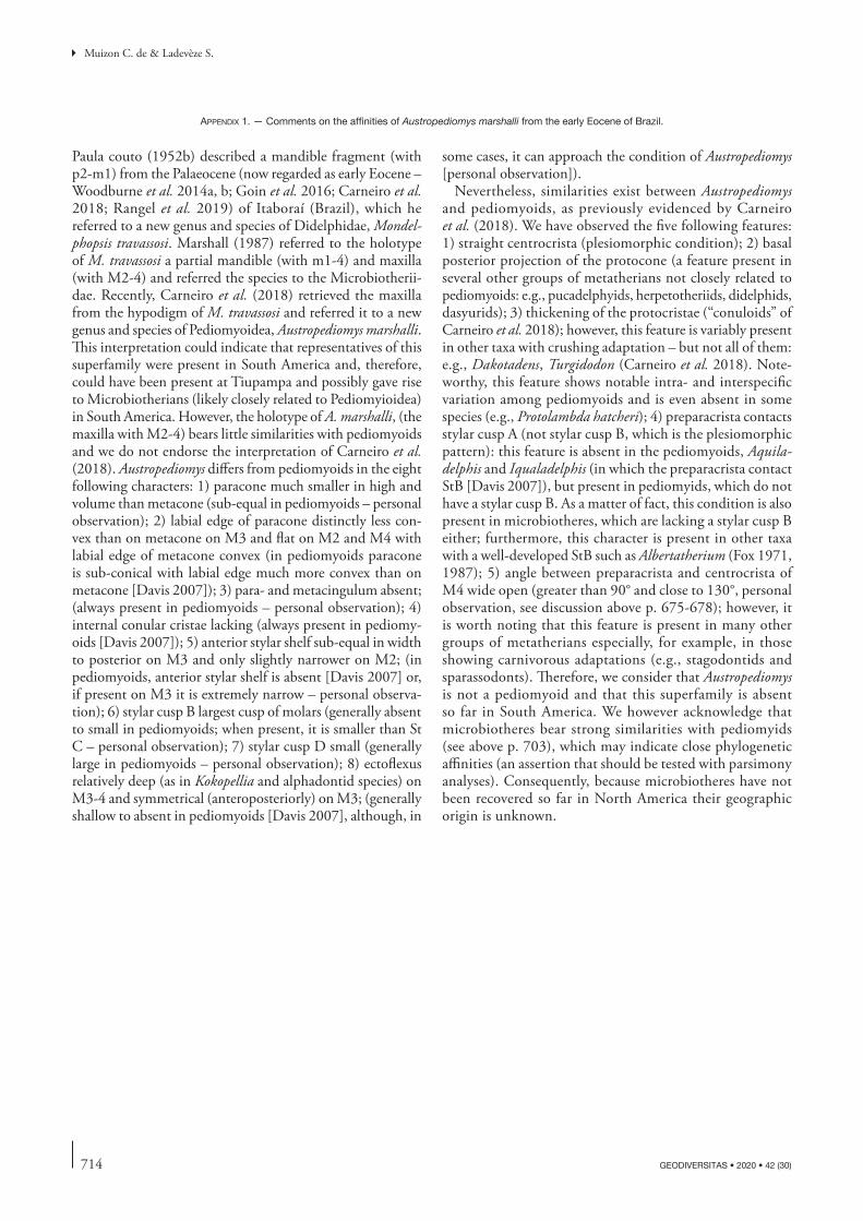
714 GEODIVERSITAS • 2020 • 42 (30)
Muizon C. de & Ladevèze S.
some cases, it can approach the condition of Austropediomys [personal observation]).
Nevertheless, similarities exist between Austropediomys and pediomyoids, as previously evidenced by Carneiro et al. (2018). We have observed the five following features: 1) straight centrocrista (plesiomorphic condition); 2) basal posterior projection of the protocone (a feature present in several other groups of metatherians not closely related to pediomyoids: e.g., pucadelphyids, herpetotheriids, didelphids, dasyurids); 3) thickening of the protocristae (“conuloids” of Carneiro et al. 2018); however, this feature is variably present in other taxa with crushing adaptation – but not all of them: e.g., Dakotadens, Turgidodon (Carneiro et al. 2018). Note-worthy, this feature shows notable intra- and interspecific variation among pediomyoids and is even absent in some species (e.g., Protolambda hatcheri); 4) preparacrista contacts stylar cusp A (not stylar cusp B, which is the plesiomorphic pattern): this feature is absent in the pediomyoids, Aquila-delphis and Iqualadelphis (in which the preparacrista contact StB [Davis 2007]), but present in pediomyids, which do not have a stylar cusp B. As a matter of fact, this condition is also present in microbiotheres, which are lacking a stylar cusp B either; furthermore, this character is present in other taxa with a well-developed StB such as Albertatherium (Fox 1971, 1987); 5) angle between preparacrista and centrocrista of M4 wide open (greater than 90° and close to 130°, personal observation, see discussion above p. 675-678); however, it is worth noting that this feature is present in many other groups of metatherians especially, for example, in those showing carnivorous adaptations (e.g., stagodontids and sparassodonts). Therefore, we consider that Austropediomys is not a pediomyoid and that this superfamily is absent so far in South America. We however acknowledge that microbiotheres bear strong similarities with pediomyids (see above p. 703), which may indicate close phylogenetic affinities (an assertion that should be tested with parsimony analyses). Consequently, because microbiotheres have not been recovered so far in North America their geographic origin is unknown.
Paula couto (1952b) described a mandible fragment (with p2-m1) from the Palaeocene (now regarded as early Eocene – Woodburne et al. 2014a, b; Goin et al. 2016; Carneiro et al. 2018; Rangel et al. 2019) of Itaboraí (Brazil), which he referred to a new genus and species of Didelphidae, Mondel-phopsis travassosi. Marshall (1987) referred to the holotype of M. travassosi a partial mandible (with m1-4) and maxilla (with M2-4) and referred the species to the Microbiotherii-dae. Recently, Carneiro et al. (2018) retrieved the maxilla from the hypodigm of M. travassosi and referred it to a new genus and species of Pediomyoidea, Austropediomys marshalli. This interpretation could indicate that representatives of this superfamily were present in South America and, therefore, could have been present at Tiupampa and possibly gave rise to Microbiotherians (likely closely related to Pediomyioidea) in South America. However, the holotype of A. marshalli, (the maxilla with M2-4) bears little similarities with pediomyoids and we do not endorse the interpretation of Carneiro et al. (2018). Austropediomys differs from pediomyoids in the eight following characters: 1) paracone much smaller in high and volume than metacone (sub-equal in pediomyoids – personal observation); 2) labial edge of paracone distinctly less con-vex than on metacone on M3 and flat on M2 and M4 with labial edge of metacone convex (in pediomyoids paracone is sub-conical with labial edge much more convex than on metacone [Davis 2007]); 3) para- and metacingulum absent; (always present in pediomyoids – personal observation); 4) internal conular cristae lacking (always present in pediomy-oids [Davis 2007]); 5) anterior stylar shelf sub-equal in width to posterior on M3 and only slightly narrower on M2; (in pediomyoids, anterior stylar shelf is absent [Davis 2007] or, if present on M3 it is extremely narrow – personal observa-tion); 6) stylar cusp B largest cusp of molars (generally absent to small in pediomyoids; when present, it is smaller than St C – personal observation); 7) stylar cusp D small (generally large in pediomyoids – personal observation); 8) ectoflexus relatively deep (as in Kokopellia and alphadontid species) on M3-4 and symmetrical (anteroposteriorly) on M3; (generally shallow to absent in pediomyoids [Davis 2007], although, in
appenDix 1. — Comments on the affinities of Austropediomys marshalli from the early Eocene of Brazil.
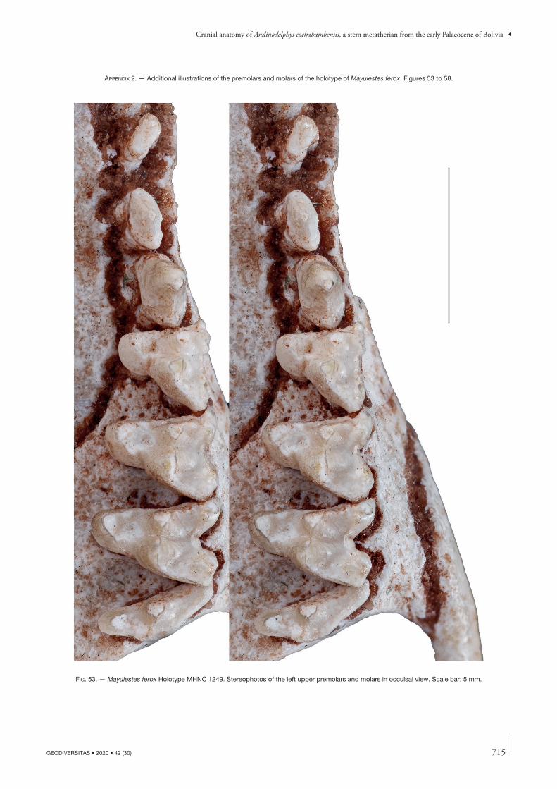
715
Cranial anatomy of Andinodelphys cochabambensis, a stem metatherian from the early Palaeocene of Bolivia
GEODIVERSITAS • 2020 • 42 (30)
appenDix 2. — Additional illustrations of the premolars and molars of the holotype of Mayulestes ferox. Figures 53 to 58.
fig. 53. — Mayulestes ferox Holotype MHNC 1249. Stereophotos of the left upper premolars and molars in occulsal view. Scale bar: 5 mm.
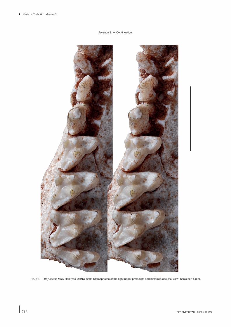
716 GEODIVERSITAS • 2020 • 42 (30)
Muizon C. de & Ladevèze S.
fig. 54. — Mayulestes ferox Holotype MHNC 1249. Stereophotos of the right upper premolars and molars in occulsal view. Scale bar: 5 mm.
appenDix 2. — Continuation.
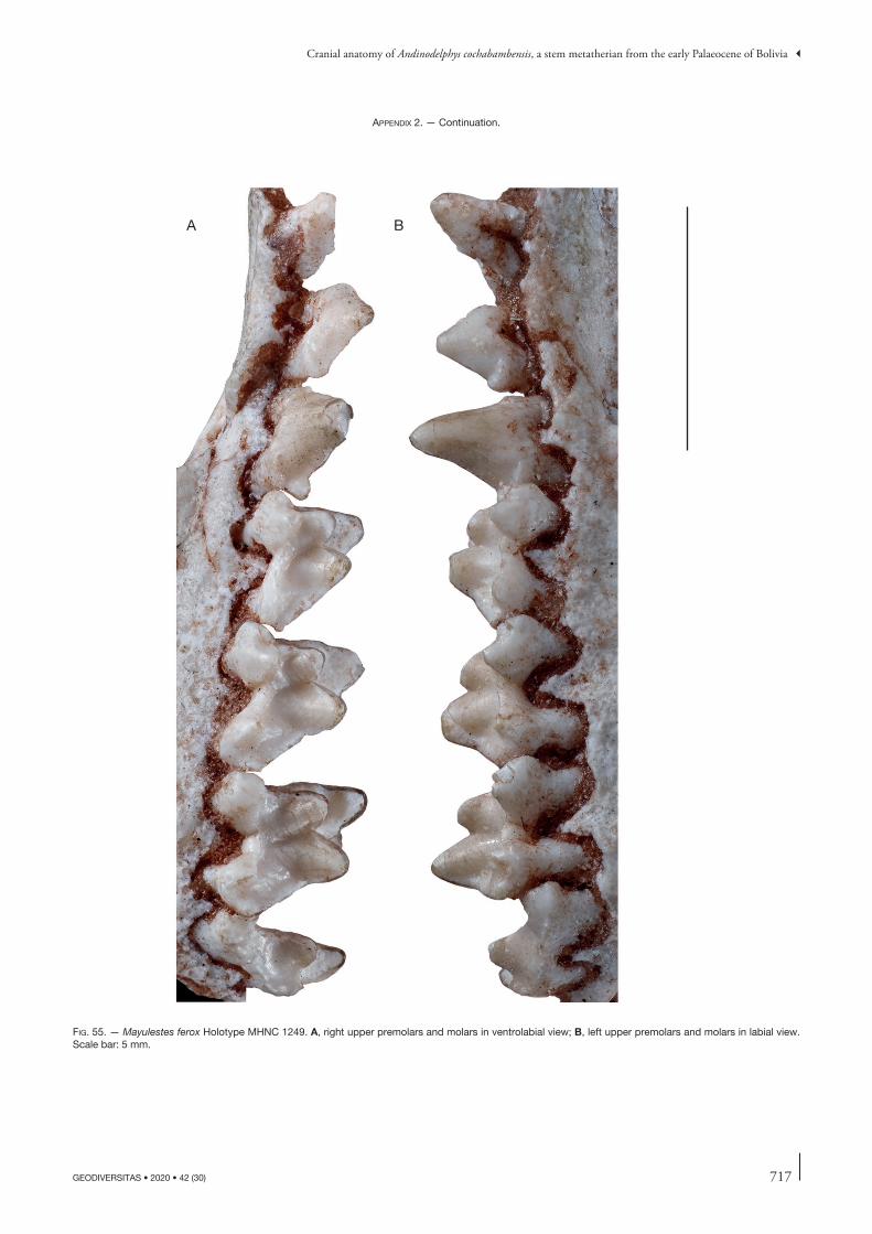
717
Cranial anatomy of Andinodelphys cochabambensis, a stem metatherian from the early Palaeocene of Bolivia
GEODIVERSITAS • 2020 • 42 (30)
A B
fig. 55. — Mayulestes ferox Holotype MHNC 1249. A, right upper premolars and molars in ventrolabial view; B, left upper premolars and molars in labial view. Scale bar: 5 mm.
appenDix 2. — Continuation.
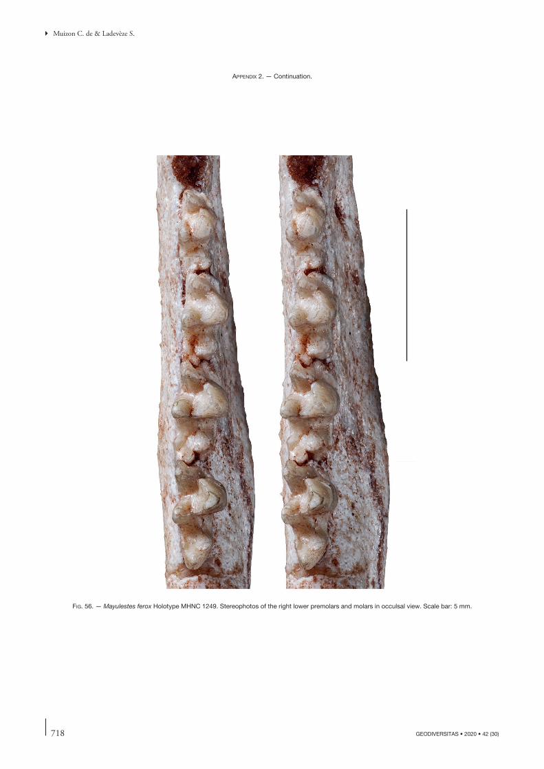
718 GEODIVERSITAS • 2020 • 42 (30)
Muizon C. de & Ladevèze S.
fig. 56. — Mayulestes ferox Holotype MHNC 1249. Stereophotos of the right lower premolars and molars in occulsal view. Scale bar: 5 mm.
appenDix 2. — Continuation.
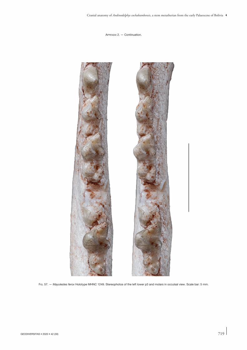
719
Cranial anatomy of Andinodelphys cochabambensis, a stem metatherian from the early Palaeocene of Bolivia
GEODIVERSITAS • 2020 • 42 (30)
fig. 57. — Mayulestes ferox Holotype MHNC 1249. Stereophotos of the left lower p3 and molars in occulsal view. Scale bar: 5 mm.
appenDix 2. — Continuation.
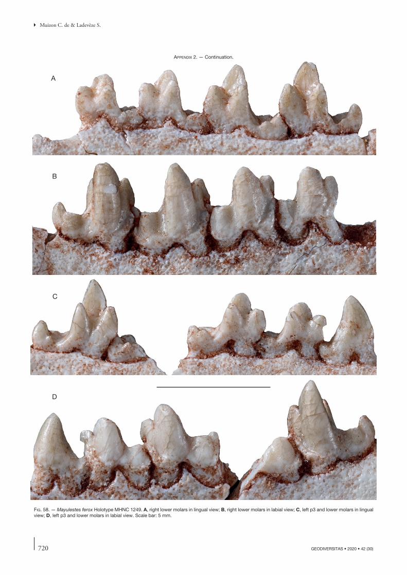
720 GEODIVERSITAS • 2020 • 42 (30)
Muizon C. de & Ladevèze S.
A
B
C
D
fig. 58. — Mayulestes ferox Holotype MHNC 1249. A, right lower molars in lingual view; B, right lower molars in labial view; C, left p3 and lower molars in lingual view; D, left p3 and lower molars in labial view. Scale bar: 5 mm.
appenDix 2. — Continuation.
721
Cranial anatomy of Andinodelphys cochabambensis, a stem metatherian from the early Palaeocene of Bolivia
GEODIVERSITAS • 2020 • 42 (30)
Dental characters 1 to 102Mandibular characters 103 to118Cranial characters: 119 to 242Post-cranial characters: 243 to 364Dental characters 1. Number of upper incisors (F108): five (0); four (1);
three (2); two or less (3). Changes in scorings of Muizon et al. (2018): Deltatheridium: ? > 1. Ordered.
Comment: in sparassodonts and dasyuromorphians with four incisors, it is probable that the I5 has been lost (Forasiepi 2009; Hershkovitz 1995). Sparassodonts with three upper incisors probably also lost the I1 (see Forasiepi 2009: 149, 150 and ref-erences therein for a discussion on the homologies of metatherian upper incisors).
2. Size of extra-alveolar portion of root of I1 (F109, modi-fied): longer than on others incisors and longer than crown; (the length of the extra-alveolar root is greater than that of the crown) (0); as long as on other incisors and as long as crown (1).
Comment: This character corresponds in part to the character formulated by several authors as “size (or shape) of first upper incisor” (Muizon et al. 1997; Rougier et al. 1998; Wroe et al. 2000; Luo et al. 2003.). However, we consider that this formu-lation is imprecise and often erroneous in stating that the I1 is enlarged. In fact, in most cases the crown of I1 is not significantly larger than that of the other incisors, but in some cases, it is only slightly longer. The most characteristic feature is the length of the extra-alveolar portion of the root which is notably long. The sparassodonts with three incisors are scored inapplicable because the I1 is assumed to be missing (see character 1).
3. Implantation of the first upper incisors in anterior view: incisors parallel (0); roots diverging dorsally (1).
4. Diastema between 11 and I2: Absent (0); Present (1). 5. Size of I4 vs I3 (F110): I4 subequal to smaller than I3
(0); I4 larger (1). 6. Size of I3 vs I2: I3>I2 (0); I3=I2 (1); I3<I2 (2). Ordered. 7. Size of I5 vs I4 (F111): I5 subequal to or larger than
I4 (0); I5 smaller than I4 (1); I5 lost (2). Ordered. 8. Shape of upper incisors: peg-shaped (0); spatulate (1). 9. Upper incisor arcade (F112 modified): parabolic (0);
slightly convex anteriorly (1); transverse (2); not an arcade, compressed transversely with incisors telescoped (3). Ordered.
10. Number of lower incisors (F113): four (0); three (1); less than three (2). Changes in scorings of Muizon et al. (2018): Deltatheridium, Deltatheroides: ? > 1. Ordered.
11. Staggered i3 (F114): absent (0); present (1). Changes in scorings of Muizon et al. (2018): Deltatheridium: ? > 1.
12. Lower incisors procumbent: present (0); absent (1) 13. Lower canine procumbent: absent (0); present (1). 14. Shape of canines (F115): small: upper canine lower
than twice the height of P3, lower canine same height or lower than apex of molars protoconid (0); large: upper canine as high as or taller than twice the height of P3, lower canine taller than apex of molars protoconid (1); saber-like upper canines (2). Ordered.
15. Pulp cavity of canines (F116): closed in adults (0); open in upper canines only (1); open in upper and lower canines (2). Ordered.
16. Roots of canine (F117): smooth (0); corrugated (1). 17. Number of roots of upper canine: two (0); one (1)
(Rougier et al. 1998). 18. Number of postcanine tooth family: eight or more
(0); seven or fewer (1) (Rougier et al. 1998, modi-fied). Changes in scorings of Muizon et al. (2018): Deltatheroides: ? > 1.
19. Number of premolars (F118): five (0); four (1); three (2), two or fewer (3) (Rougier et al. 1998). Changes in scorings of Muizon et al. (2018): Deltatheroides: ? > 2. .Ordered.
20. Orientation of first upper and lower premolars relative to tooth row (F119, modified): parallel (0); oblique (1); transverse (2). Changes in scorings of Muizon et al. (2018): Eodelphis: 0 > 3; Pucadelphys: 0&1 > 0. Ordered.
21. First upper premolar strongly asymmetrical in lateral view: absent or weak (0); distinctly present (1). Changes in scorings of Muizon et al. (2018): Deltatheridium: 0 > 1; Eodelphis: ? > 1.
Comment: in state (0), the apex of the tooth is roughly above the space between the two roots, or the middle of the crown when the tooth is single-rooted. In state (1), the apex of the tooth is above the anterior root, or above anterior edge of the crown when the tooth is single-rooted.
22. Diastema anterior to first upper premolar (F120): absent (0); present (1). Changes in scorings of Muizon et al. (2018): Deltatheridium: 0 > 0&1; Eodelphis: ? > 0; Didelphis, Monodelphis, Metachirus: 1 > 0.
23. Diastema posterior to first upper premolar (F121): absent (0); present (1). Changes in scorings of Muizon et al. (2018): Eodelphis: ? > 0;
24. Diastema posterior to first lower premolar (F122): absent (0); present (1).
appenDix 3. — character list: This character list is that of Muizon et al. (2018) indicating the changes in some scorings and characters definitions as compared to the earlier version of this matrix provided by those authors. The data matrix and character list of Muizon et al. (2018: 439-455) has been established mainly on the basis of that of Forasiepi et al. (2015) which was significantly modified with scoring changes and modification and addition of characters. Therefore, this character list is significantly different from that of Forasiepi et al. (2015) that was used as a starting point. When a character is identical to that of Forasiepi et al. (2015), it bears the number of Forasiepi et al.’s list as follows: (F000). When the character states have been modified or when some rewording has been neces-sary, it is indicated as follows: (F000, modified). We refer to Forasiepi et al. (2015) and to Forasiepi (2009) for the credit on the original mention of these characters. Scoring changes (from Forasiepi et al. 2015) mentioned in Muizon et al. (2018) are not reproduced here.
722 GEODIVERSITAS • 2020 • 42 (30)
Muizon C. de & Ladevèze S.
25. Shape of premolars (F123): transversely compressed and uninflated (0); bulbous (1)
26. Ultimate premolar protocone: absent (0); small pos-terolingual bulge or cusp (1); distinct protocone with basin (2). Ordered.
27. Size of penultimate lower premolar (F125 modified): smaller than ultimate premolar (0); subequal to ultimate premolar (1); larger than ultimate premolar (2). Ordered.
28. Change in height of the lower premolars (F126): abrupt change in size between antepenultimate premolar and penultimate-ultimate premolar (0); abrupt change in size between antepenultimate-penultimate premolar and ultimate premolar (1).
29. Roots of lower premolars (F127): not inflated, as wide as crown (0); bulbous on only one premolar (1); bulbous on all premolars and some molars (2). Ordered.
30. Anterobasal cusp or cingulid on penultimate premolar (F128 modified): absent (0); present (1). Changes in scorings of Muizon et al. (2018): Deltatheroides: ? > 0; Eodelphis 0 > 1.
31. Ultimate lower premolar symmetry of main cusp (F129): absent: anterior edge of cusp more convex than posterior edge (0); present: both edges similar in curva-ture (1). Changes in scorings of Muizon et al. (2018): Deltatheroides: ? > 0.
32. Timing of eruption of ultimate upper and lower premo-lars relative to ultimate or penultimate upper and lower molars (F130): ultimate upper and lower premolars with penultimate or antepenultimate upper and lower molars (0); ultimate upper and lower premolars with ultimate upper and lower molars (1).
33. Size of molars (upper and lower) increases posteriorly (F132): moderate (0); marked (1). Changes in scorings of Muizon et al. (2018): Mayulestes, Allqokirus, Patene, Hondadelphys: 0 > 1.
Comment: concerning the upper molars, the ultimate molar is not considered in the size increase. Furthermore, in the case of deltatheroidans, the last lower molar is not considered either. The changes in the scorings of the four sparassodont genera above is justified by the fact that their posterior molar increase is at the least as pronounced as in deltatheroidans, which are scored 1.
34. Width (labiolingual) of ultimate relative to penultimate upper molar (F133 modified): narrower (0); subequal or wider (1). Changes in scorings of Muizon et al. (2018): Mayulestes: 1 > 0.
Comment: Contra Muizon et al. (2018) the M4 of Mayulestes is in fact slightly narrower than the M3 and therefore this taxon is scored 0 for this character.
35. Width of stylar shelf (on penultimate molar), (F151, modified): uniform in width (0); slightly reduced labial
to paracone (1); strongly reduced labial to paracone (2); vestigial to absent (3); reduced labial to metacone (4). Changes in scorings of Muizon et al. (2018): Deltather-idium, Deltatheroides: 0 > 4. Ordered.
Comment: a fifth state has been added to this character to take into account the condition in deltatheroidans, in which the stylar shelf of penultimate molar is distinctly reduced labial to metacone.
36. Stylar shelf vs tooth width (on penultimate or antepe-nultimate molar), (F151, modified): 50% of transverse width or more (0); less than 50% of transverse width (1).
37. Deep ectoflexus (F152, modified): present on one or more teeth (0); absent on any tooth (1). See Ladevèze & Muizon (2007 ch. 20) for comments on the definition of this character as defined here vs. that of Rougier et al. (1998) and Forasiepi et al. (2015).
Comment: the depth of the ectoflexus has been shown to be highly variable for the same dental locus in some fossil and extant metatherians (e.g., Pucadelphys andinus, Peratherium elegans, Marmosa murina; see Ladevèze et al. 2011, 2012).
38. Postmetacrista (on antepenultimate or penultimate molar), (F148 modified): subequal or shorter than preparacrista (0); distinctly longer than preparacrista (1).
39. Carnassial notch in postmetacrista at posterolabial base of metacone: Absent (0); Present (1).
40. Stylar cusp A (F153): distinct but smaller than B (0); subequal to larger than B (1).
41. Stylar cusp B (F154, modified): present but smaller than paracone (0); subequal to or approaching the size of the paracone (1); Vestigial to absent (2).
42. Anterolabial cingulum on upper molars: absent (0); present (1).
Comment: stylar cusps A and B are present but poorly individual-ized and integrated in a variably developed anterolabial cingulum.
43. Stylar cusp C (F155): absent or tiny (0); present (1).
Comment: this feature has been evidenced as highly variable in some fossil and extant metatherians (e.g., Pucadelphys andinus, Peratherium elegans, Marmosa murina; see Ladevèze et al. 2011, 2012).
44. Relative size of Stylar cusp B and D: B smaller or sub-equal to D (0); B larger than D (1). Changes in scorings of Muizon et al. (2018): Deltatheroides: 0 > 1.
45. Stylar cusp E (F157): present and distinct (0); indis-tinct or tiny (1). Changes in scorings of Muizon et al. (2018): Deltatheroides: ? > 1.
appenDix 3. — Continuation.
723
Cranial anatomy of Andinodelphys cochabambensis, a stem metatherian from the early Palaeocene of Bolivia
GEODIVERSITAS • 2020 • 42 (30)
46. Relative size of paracone vs metacone (on penultimate or antepenultimate molar), (F134 modified): subequal or slightly larger (0); slightly smaller in volume and height (c. 10%) (1); distinctly smaller in volume and height (c. 30% smaller); (2); much smaller (c. 50% or less) (3); absent or simple bulge on the anterior edge of metacone (4). Ordered.
47. Shape of paracone and metacone (F136): conical (0); triangular (flat labial face) (1).
48. Metacone and paracone contact (F137 modified): separated – i.e. point of junction of premetacrista and postparacrista approximately at the level of the floor of trigon and/or stylar shelf basin (0); adjoined at base (on, at least, half of their height) – i.e. point of junction of premetacrista and postparacrista well below the level of the floor of the trigon and stylar shelf basin (1); adjoined on most of their height; apices of cusps only are separated (2). Ordered.
49. Metacone on last molar (F139): present and distinct (0); extremely reduced (1), absent (2). Changes in scorings of Muizon et al. (2018): Deltatheridium: 0 > 1. Ordered.
50. Centrocrista (F138): straight (0); V-shaped (1). 51. Relative lengths of preparacristae of penultimate and
ultimate molars (F147): preparacrista of ultimate molar shorter than that of penultimate molar (0); preparacrista of ultimate molar subequal to longer than that of penul-timate molar (1).
52. Orientation of preparacrista (on penultimate or ante-penultimate molar), (F146 modified): transverse (0); anterolabial (1).
53. Conules (F143): present (0); absent (1). 54. Conules wing-like cristae (F144): present (0); absent
(1). 55. Conules labiolingual position (on penultimate or ante-
penultimate molar), (F145): at or lingual to mid-point between protocone and para/metacone (0); closer to para/metacone (1).
56. Trigon basin: absent (0); present (1). 57. Protocone size (on penultimate or antepenultimate molar),
(F 141, modified): absent or very small (0); small and anteroposteriorly narrow (1); anteroposteriorly expanded (2); with posterolingual projection (3). Ordered.
58. Protocone height (F142, modified): approximately half the height of metacone or less (0); only slightly lower than metacone (1); almost the size of metacone or higher (2). ordered
59. Roots on ultimate molar (F140): 3 (0); 2 or 1 (1). 60. Paracingulum (on penultimate or antepenultimate
molar), (F149): continuous between stylar margin and paraconule (0); interrupted between stylar margin and paraconule (1); absent (2). Ordered
61. Postprotocrista labial extension: does not extend labi-ally past the base of the metacone (0); extends labially past the base of metacone (1).
62. Precingulum (on penultimate or antepenultimate molar): absent or weak (0); present (1).
63. Postcingulum (on penultimate or antepenultimate molar), (F150): absent or weak (0); present (1).
64. Size of ultimate lower molar (F158): subequal or smaller in height and/or length than penultimate molar (0); larger in height and/or length than penultimate molar (1).
65. Ventral extent of the enamel above posterior root lower than above anterior root on labial side of the crown of lower molars (F159): absent (0); present only on first and second molars and weakly developed (1); on the first three molars and strongly developed (2). Changes in scorings of Muizon et al. (2018): Deltatheroides: ? > 0.
66. Lingual opening of trigonid basin on penultimate molar when possible (162, modified): wide open: angle between protocristid (or postprotocristid when metaconid is lost) and paracristid more than 45°; (0); less open: angle proto-paracristid between c. 45° and 15° (1); anteroposteriorly compressed: cristids subpar-allel (2). Changes in scorings of Muizon et al. (2018): Deltatheroides: 0 > 1. Ordered.
67. Trigonid basin floor: sub-horizontal (0); sloping lingually (1); lost (2).
68. Trigonid proportions: wider than long (0); as wide as long (1); longer than wide (2). Changes in scorings of Muizon et al. (2018): Deltatheroides: ? > 1. Ordered.
69. Trigonid height (on penultimate or antepenultimate molar), (F167, modified): much higher than talonid (more than twice), (0); moderately higher than talonid (twice or less), (1).
Comment: When the talonid is extremely reduced or lost, this character is scored as inapplicable.
70. Trigonid vs talonid width (on penultimate or antepe-nultimate molar), (F166): wider than talonid (0); as wide as talonid (1); narrower than talonid (2).
Comment: taxa with incomplete trigonid (lack of metaconid) are scored as inapplicable.
71. Protoconid labial expansion: the labial edge of the protoconid at mid-height is strongly convex, protrud-ing labially and overhangs the base of the crown; as a consequence, the apex and base of the trigonid are more lingual than the mid-height region of the cusp and the trigonid is therefore wider at mid-height than at its base (see Fig. 14); present (0); absent (1). Changes in scorings of Muizon et al. (2018): Deltatheroides: ? > 0.
Comments: state (1) corresponds to a protoconid regularly widen-ing towards the base of the cusp, which is therefore wider at its base than at mid-height.
appenDix 3. — Continuation.
724 GEODIVERSITAS • 2020 • 42 (30)
Muizon C. de & Ladevèze S.
Lotheridium is scored absent for this character, contra Bi et al. (2015) on the basis of a personal communication of Shundong Bi to one of us (CM).
72. Trigonid vs talonid length (on penultimate or antepe-nultimate molar), (F165): subequal in length to talonid (0); longer than talonid (1); shorter than talonid (2). Changes in scorings of Muizon et al. (2018): Del-tatheroides: ? > 1. Ordered.
73. Talonid: small heel (0); multicusped basin (1). Changes in scorings of Muizon et al. (2018): Deltatheroides: ? > 1.
74. Proportions of talonid basin (F175): longer than wide (0); as long as wide (1); wider than long (2). Ordered.
75. Relative size and shape of talonid of ultimate vs penul-timate molar (F160): talonid of ultimate molar similar to anterior tooth (0); talonid of ultimate molar reduced and narrower than on anterior tooth (1). Changes in scorings of Muizon et al. (2018): Deltatheroides: ? > 0.
76. Paraconid position (on penultimate or antepenulti-mate molar), (F163, modified): at anterior edge of the trigonid (0); at anterolingual angle of trigonid (1).
Comment: the wording of this character considers the position of the paraconid on the trigonid rather than the metaconid as in Character 163 of Forasiepi et al. (2015). Because, implicitly, it is the relative position of the two cusps, which is considered, this formulation gives identical results in term of scoring. We prefer to refer to the paraconid because the transverse position of this cusp varies considerably in therian lower molar evolution, while that of the metaconid is less variable. Furthermore, the position of the lingual edge of the metaconid is displaced labially when the metaconid is reduced, a bias which is avoided if considering the paraconid.
77. number of cusps on last molar talonid: three (0); two (1); one (2). Ordered.
78. Vertical keel (ridge) at anterolingual angle of paraco-nid labially bordered by hypoconulid notch (vertical sulcus at lingual end of precingulid) (F172): absent (0); present (1).
79. Orientation of protocristid (or postprotocristid) vs anteroposterior axis of the molar (on penultimate or antepenultimate molar), (F164, modified): transverse or slightly oblique (i.e., less than 45°) (0); strongly oblique (i.e., more than 45°) or parallel (1).
80. Paraconid elongated with anterior projection of the paraconid keel (ridge) mostly in the ventral half of the cusp: absent (0); present (1); (Marshall et al. 1990, modified).
Comment: as a consequence of the anterior projection of the paraconid keel, the anterior edge of the cusp is markedly convex. 81. Precingulid (F173): on protoconid and paraconid (0);
on paraconid (1); absent (2). Ordered
82. Height of protoconid (Hpo) relative to length of ulti-mate or penultimate lower molar (Lm): Hpo/Lm < 0.9 (0); Hpo/Lm > 0.9 (1). Changes in scorings of Muizon et al. (2018): Deltatheroides: ? > 1.
83. Protoconid height in trigonid (F171): tallest cusp of trigonid (0); sub-equal to para- and/or metaconid (1).
84. Height of metaconid vs paraconid (on penultimate molar) (F170, modified): higher (0); subequal (1); lower (2). Ordered.
Comment: taxa in which the metaconid is lost are scored inap-plicable.
85. Volume of the metaconid vs paraconid (on penulti-mate molar), in occlusal and lingual views: larger (0); subequal (1); smaller (2).
Comment: taxa in which the metaconid is lost are scored inap-plicable. 86. Metaconid on first molar (F168): present (0); absent
(1). 87. Metaconid on molars between the first and last molar
(F169 modified): Present (0); Present, but extremely reduced as a tiny cuspule at the posterior base of the protoconid (1); Absent (2).
88. Metaconid on ultimate lower molar: present (0); ves-tigial or absent (1) Changes in scorings of Muizon et al. (2018): Deltatheroides: ? > 0.
89. Relative lengths of para- and protocristid: subequal (0); paracristid > protocristid (1); protocristid > paracristid (2).
90. Anterior end of cristid obliqua (F184): beneath car-nassial notch (0); lingual to carnassial notch (1); labial to carnassial notch (2). Changes in scorings of Muizon et al. (2018): Deltatheroides: ? > 1.
Comment: taxa which lack a metaconid also lack a carnassial notch and are scored as inapplicable. 91. Carnassial notch in cristid obliqua (F186): absent (0);
present (1). 92. Hypoconulid position (F180, modified): in median
position (0); shifted lingually, close to entoconid (1); absent (2).
93. Hypoconulid of last molar (F181, modified): as tall as or lower than other talonid cusps (0); taller than other talonid cusps (1); absent (2).
94. Hypoconid position (F176): middle of labial margin of tooth (0); posterolabial angle of tooth (1); absent (2). Changes in scorings of Muizon et al. (2018): Del-tatheroides: ? > 0.
95. Shape of entoconid (F177): conical (0); transversely compressed (1); vestigial to absent (2).
96. Relative height of entoconid (F178): lower than hypoco-nid (0); subequal to taller than hypoconid (1); absent (2).
97. Position of entoconid relative to trigonid (on antepe-nultimate or penultimate molar) (F179, modified):
appenDix 3. — Continuation.
725
Cranial anatomy of Andinodelphys cochabambensis, a stem metatherian from the early Palaeocene of Bolivia
GEODIVERSITAS • 2020 • 42 (30)
at posterolingual corner of tooth (in posterior half of the distance between the posterior base of trigonid and posterior margin of tooth) (0); between posterior edge of trigonid and posterior margin of tooth (mid-way or in anterior half of the distance between the posterior base of trigonid and posterior margin of tooth) (1).
98. Entocristid (F182, modified): absent or weakly devel-oped (0); well-developed and sharp (1).
99. Posthypocristid: oblique relative to long axis of tooth (0); transverse relative to long axis of tooth (1).
100. Hypoflexid (F185): deep (c. 40-50% of talonid width) (0); shallow or absent (less than 40% of talonid width) (1).
101. Labial postcingulid (F187): absent (0); present (1). 102. Postcingulid on last molar: present (0); absent (1).
Mandibular characters 103. Depth/length of the dentary below molars (F97): shal-
low (less than 0,15) (0); intermediate (between 1,5 and 2) (1); elevated (more than 2) (2). Ordered.
Comment: In the case of Mayulestes, because the ramus man-dibularis (vertical ramus) is missing in the only specimen known, the length of the dentary has been estimated on the basis of the position of the postglenoid process and articular condyle, when the lower tooth row of the dentary is in occlusion with the upper tooth row.
104. Ventral margin of the dentary behind last molar con-tinuous to condyle (F98): straight (0) or gently curved (1); angled (2).
105. Mandibular symphysis (F99): unfused (0); fused (1). 106. Anteroventral edge of mandibular symphysis: slanting
and forming a small angle with tooth row (< 25°) (0); less slanting, forming an angle > 25° (1).
107. Posteriormost mental foramen (F100): below last pre-molar or anterior (0); at last premolar/m1 embrasure (1); below first molar (2); posterior to first molar (3). Ordered.
108. Masseteric fossa: restricted dorsally by crest reaching condyle (0); extended ventral to lower margin of dentary (1), (Rougier et al. 1998: ch. 67).
109. Posterior shelf of masseteric fossa: absent (0); present (1), (Rougier et al. 1998: ch. 68).
110. Labial mandibular foramen (F102): present (0); absent (1).
111. “Coronoid” facet: present (0); absent (1), (Rougier et al. 1998: ch. 76).
112. Angle between coronoid process and tooth row (F104): subvertical between 95° and 105° (0); Between 106° and 125° (1); more than 125° (2). Ordered.
Comment: The condition in Patene has been measured on the left mandible of IML-PV 2618, in which the ramus mandibularis
is in correct position relative to the corpus mandibularis. On the right dentary, the ramus is not in anatomical position and, in medial view, it is slightly rotated clockwise. Because of this incorrect position of the ramus, the angle of the coronoid process of the right dentary is distinctly greater than on the left dentary.
113. Retromolar space (F101): absent (0); present (1). 114. Angular process medially inflected: absent (0); present
(1), (Rougier et al. 1998: ch. 73, modified). 115. Morphology of medially inflected angular process
(F103): shelf-like (ASL/AL>0.81) (0); intermediate (0.72<ASL/AL <0.81) (1); rod-like (ASL/AL < 0.72) (2). ASL: angular process shelf length, AL: angular process length. Ordered
116. Anteroposterior position of mandibular foramen (F105): posterior to mid-point of coronoid process (0); at mid-point of coronoid process (1); anterior to mid-point of coronoid process (2). Ordered.
117. Morphology of mandibular condyle (F106): sub-spherical (0); cylindrical (1).
118. Condyle vertical position vs tooth row (F107): approxi-mately at the same level or slightly below (0); above (1).
Comment: The condition in Patene has been scored on the left mandible of IML-PV 2618, in which the ramus mandibularis is in correct position relative to the corpus mandibularis. On the right dentary, the ramus is not in anatomical position and, in medial view, it is slightly rotated clockwise. Because of this incorrect position of the ramus, the condyle appears to be almost at the level of the tooth row, as stated by Goin et al. (1986). On the left dentary, in which the position of the ramus is correct, the condyle is located well above the tooth row.
Cranial characters 119. Length of the skull (F1): short (less than twice width
at the level of zygomatic arch), (0); long (more than twice width at level of zygomatic arch), (1).
120. Length of the rostrum (F2): less than (or equal to) 1/3 total length of the skull (0); between 1/3 and 1/2 total length of the skull (1); more than 1/2 total length of the skull (2).
Comment: The length of the rostrum of Allqokirus has been estimated because the only skull known (MHNC 8267) is that of a subadult individual in which the M4 and m 4 are not fully erupted. The correction applied is the length of the m4 (c. 3.5 mm). With this correction, Allqokirus is scored 1 for this character.
121. Expanded apex of the rostrum with concave tooth rows and lateral edges of the rostrum between canine and infraorbital foramen: absent (0); present (1).
122. Width of the braincase versus maximum postorbital width (F3): braincase at base of zygomatic arches wider than maximum postorbital width (0); braincase nar-rower than maximum postorbital width (1).
appenDix 3. — Continuation.
726 GEODIVERSITAS • 2020 • 42 (30)
Muizon C. de & Ladevèze S.
123. Proportions of the braincase (F4 modified): almost as wide as long (0); distinctly wider than long (1); longer than wide (2).
124. Level of the palate relative to basicranium (F 5): palate lower than basicranium (0); palate and basicranium at the same level (1).
125. Anterolateral process of the maxilla in the paracanine fossa (F6 modified): present: the anterolateral process of the maxilla closes the fossa laterally; the paracanine fossa is composed of the maxilla and premaxilla (0); absent: the fossa is opened laterally; the paracanine fossa is composed of the premaxilla only (1).
126. Paracanine fossa morphology: not extended dorsally: longer (anteroposteriorly) than high (dorsoventrally) (0); extended dorsally: higher dorsoventrally than (or as high as) long (anteroposteriorly) (1).
127. Precanine notch (F7): absent (0); present (1).
Comment: in dorsal or ventral view, the presence of a precanine notch results in a transverse constriction of the rostrum just anterior to the canine. The precanine notch is scored present in Allqokirus and Mayulestes because the weak but distinct concavity observed anterior to the canine is regarded as an incipient development of this character (see Figs 3 and 4).
128. Lateral palatal process of the premaxilla (F8 modified): anterior to anterior border of canine alveolus (0); just reaches anterior border of canine alveolus (1); posterior to anterior border of canine alveolus (2). Ordered.
129. Posterior border of incisive foramen (F9): anterior to or just reaches anterior border of canine alveolus (0); posterior to anterior border of canine alveolus (1).
130. Dorsoventral position of the medial palatal process of the premaxilla (F10): horizontal (0); with posterior end shifted dorsally, forming an incisive fossa (1).
131. Distinct narial tubercle or process at the anterodor-somedial angle of the premaxilla (F11): absent (0); present and small (narrower than I1) (1); present and very large (wider than I1) (2). Changes in scorings of Muizon et al. (2018): Didelphodon 1 > 2; Pharsophorus ? > 1; Australohyaena: 1 > 0;
132. Posterior end of the facial (posterodorsal) process of the premaxilla (F12 Modified): above canine or anterior (0); above P1 or P2 (1); posterior to P2 (2). Ordered.
133. Anterior parabolic protrusion of nasals, which over-hangs nasal aperture (F13): present (anterior protru-sion is approximately as long as width of the nasal at the level of anteriormost point of the nasal-premaxilla suture) (0); absent (1).
Comment: Mayulestes has been scored 1 for this character in spite of statement by Muizon (1998: 34) that these “bones extend anteriorly beyond the anteriormost point of the nasal-premaxilla suture”. In fact, this statement is biased by the distor-
tion (dorsoventral compression) of the specimen. The distortion has pushed the nasal ventrally and the maxillae dorsally. More important, the distortion also shows a slight anteroposterior component, which has pushed the maxillae and premaxillae posteriorly. This condition is evidenced on the posterior part of the Na-Mx suture, where the maxillae clearly overlap the nasals (in the anterolateral portion of the posterior expanded region of the nasals). In this respect, the reconstruction of Muizon (1998: fig 6A) is clearly inaccurate. Given that, although the anterior edge of the nasals of Mayulestes is slightly convex anteriorly, their condition is clearly more similar to those of Thylacinus and Arctodictis than to those of Didelphis and Pucadelphys. In these taxa, the anterior extension of the nasal is at least as large as the width of the nasal at the level of the anterior end of the nasal-premaxilla suture. This is certainly not the case in Mayulestes. In contrast, Callistoe, in which the nasals distinctly protrude anteriorly, has been scored 0.
134. Anterior end of the nasals: V-shaped (or parabolic) (0); transverse or slightly convex anteriorly (1); notched (V-shaped posteriorly) (2). Ordered.
135. Shape of naso-frontal suture (F14, modified): acute
W- or V-shaped (0); wide open W or posteriorly convex (1).
136. Nasofrontal suture with medial process of the frontals wedged between the nasals: Present (0); Absent (1), (Rougier et al. 1998: ch. 84).
137. Postorbital process (F15, modified): absent (0); weak tuberosity (1); conspicuous protruding process (2). Ordered.
138. Orbital crests (F35): absent (0); present (1). 139. Naso-lacrimal contact (F16): present (0); absent (1). 140. Facial process of the lacrimal (F30, modified): large
triangular and pointed anteriorly (0); small crescentic and rounded anteriorly (1).
141. Lacrimal tubercle (F31 modified): present, knob-like (0); present, crest-like (1); absent (2).
142. Lateral wall of ventral or lateral lacrimal foramen (F32, modified): distinctly anterior to medial wall (foramen exposed on face and faces laterally or posterolaterally) (0); at the same anteroposterior level as the medial wall (foramen not exposed on face and faces posteriorly) (1); intermediate (one foramen on face, the other in the orbit) (2).
143. Number of lacrimal foramina (F33): two (0); one (1). 144. Infraorbital foramen position (F18, modified): above
penultimate premolar (0); above ultimate premolar (1); above M1 or posterior (2). Changes in scorings of Muizon et al. (2018): Eodelphis: ? > 1. Ordered.
145. Flaring of maxillary cheek posterior to infraorbital foramen (F19): present (0); absent (1).
appenDix 3. — Continuation.

727
Cranial anatomy of Andinodelphys cochabambensis, a stem metatherian from the early Palaeocene of Bolivia
GEODIVERSITAS • 2020 • 42 (30)
Comment: Callistoe has been scored 0 contra Forasiepi et al. (2015), because the holotype in ventral view presents a small but distinct flaring of the maxillae behind the infraorbital foramina. Furthermore, it is noteworthy that the skull has suffered some transverse (mediolateral) compression, which suggests that the flaring was even more pronounced than the condition actually observed on the holotype.
146. Angle (posterior angle) between maxilla-jugal suture and plane of the tooth row in lateral view (F17): more than 140° (0); between 140° and 95° (1).
147. Fossa for the levator labii muscle in the anterior end of the jugal: absent (0); present mainly in the jugal (1); present mainly in the maxilla (2).
148. Anterior medial and lateral palatal processes of the maxilla: both processes approximately of the same size (0); medial process much smaller than the lateral or absent (1).
149. Number of palatal pits independently of their size (F21, modified): none (0); one (between ultimate and penultimate molar) (1); two between ultimate, penulti-mate and antepenultimate molars (2); two pits between first, antepenultimate, and penultimate molars. (3), three between each molar (4). Changes in scorings of Muizon et al. (2018): Eodelphis: ? > 4. Unordered.
150. Palatal vacuities (F22): absent (0); present (1). Changes in scorings of Muizon et al. (2018): Eodelphis: ? > 1;
151. Major palatine foramen (F23): well individualied, within maxilla and/or palatine (0); multiple small foramina in palatine and/or maxilla (1).
152. Minor palatine foramen (F24, modified): present and complete (closed posteriorly) (0); incomplete (open posteriorly) or absent (1).
153. Palatal expansion with regard to ultimate molar (F25, modified): posterior (0); even with (1); anterior (2). Ordered.
154. Posterior palatine spine (F26, modified): weak or absent and posterior edge of the palate concave posteriorly (0); absent and posterior edge of the palate straight (1); prominent and posterior edge of the palate double-arched (2), (see Wible et al. 2009: ch. 192).
155. Size of foramen or groove in lateral edge of postpala-tine torus: absent (0); present and small (less than half the diameter of the minor palatine foramen or groove), (1); present and large (approximately half the diameter of the minor palatine foramen or more (2). ordered
Comment: state (1) present in didelphids corresponds to the post-palatine torus foramen of Wible (2003: fig. 9 p. 144).
156. Morphology of the postpalatine torus foramen: totally or almost totally enclosed in the palatine (0); wide-open groove (the section is approximately half a circle or less) (1).
157. Palatine enters the posterior opening of the infraorbital canal (maxillary foramen) (F27): present (0); absent (1).
158. Position of sphenopalatine foramen (F28): posterior to the level of the posterior border of lacrimal (0); anterior to the level of the posterior border of lacrimal (1).
159. Anteroposterior position of the sphenopalatine fora-men relatively to the molars: above penultimate or antepenultimate molar (0); above ultimate molar or posterior (1).
160. Development of pterygoids (F29): well-developed and expanded on medial side with midline contact (0); well developed and expanded on medial side but no midline contact (1); reduced, not expanded on medial side (2). Ordered.
161. Posterior process of pterygoids, which covers the alisphenoid-basisphenoid suture: present (0); absent (1).
162. Orbitotemporal canal: present (0); absent (1), (Rougier et al. 1998: ch. 103).
163. Optic foramen (F48): well separated from sphenorbital fissure (0); confluent with sphenorbital fissure (1).
164. Interparietal (F36): present (0); absent (or fused to parietals) (1).
165. Frontoparietal suture (F37): with posterior wedge of the frontals (0); with anterior wedge of the parietals (1); no wedge, roughly straight (2).
166. Parietal-alisphenoid or fronto-squamosal contact (F38): parietal-alisphenoid (0); fronto-squamosal (1).
167. Foramina for temporal rami (F95 modified): present (0); absent (1).
168. Anterior lamina of the petrosal: present with large expo-sure on lateral braincase wall to (0); rudimentary with small exposure on lateral braincase wall (1); rudimentary without exposure on lateral braincase wall (2); absent and totally lost (Marshall & Muizon, 1995; Ladevèze & Muizon, 2007: ch. 123; Rougier et al. 2015). Changes in scorings of Wilson et al. (2016): Didelphodon: 2 > 1. Ordered.
Comments: The addition of Vincelestes, which has a well-developed anterior lamina exposed on the lateral braincase wall required the addition of an additionalcharacter state. Furthermore, we reconsidered this character to take into account not only the size of the anterior lamina but also its participation to the braincase wall as discussed in the formulation of this character in Rougier et al. (2015) matrix (Character 108: Anterior lamina exposure on lateral braincase wall). Vincelestes and Prokennalestes are respectively scored 0 and 1for this character.
On the digital rendering of the petrosal of Didelphodon pro-vided by Wilson et al. (2016: supplementary figure 8 a, b) the fossa on the dorsolateral aspect of the petrosal, labelled “petrosal contribution to cavum epiptericum” (cep) (named trigeminal
appenDix 3. — Continuation.
728 GEODIVERSITAS • 2020 • 42 (30)
Muizon C. de & Ladevèze S.
fossa in Muizon et al. 2018 and in this paper) is identical to the condition observed in Allqokirus and Andinodelphys and is regarded here as a rudimentary anterior lamina. Fur-thermore, on supplementary fig 8 b of Wilson et al. (2016) the dorsal edge of the figure is totally straight, thus indicating some missing part of the lamina probably due to difficulty in the segmentation of the petrosal. We estimate that the rudimen-tary anterior lamina of Didelphodon was probably at least as large as in Allqokirus and Andinodelphys. Therefore, in our matrix, we scored this character 2 (rudimentary without exposure on lateral braincase wall) rather than “absent” as in Wilson et al. (2016).
The scoring of Pediomys follows here that of Rougier et al. (2015), because we have not seen the specimen FMNH PM 53907, on which it is based. However, because this isolated petrosal is very slightly damaged, we strongly suspect that the anterior region of the “part of petrosal in middle cranial fossa” (mcf ) of Wible (1990), which we regard as an equivalent of the “petrosal contribution to cavum epiptericum” (cep) of Wilson et al. (2016: suppl. fig. 8 a, b) and of our trigeminal fossa (Muizon et al. 2018 and this paper) bore a vestigial anterior lamina as is observed in the Tiupampan fossils and that such a delicate structure has been broken off during fossilisation.
169. Nuchal crest (F73): at or posterior to the level of the condyles (0); anterior to condyles (1).
170. Sagittal crest (F72): extending to frontals (0); restricted to parietals (1); absent (2). Ordered.
171. Glenoid process of the jugal (F34): with articular facet (0); without articular facet (1).
172. Postorbital (frontal) process of the jugal: absent (0); present (1).
173. Width of the glenoid fossa (F39): less than twice anter-oposterior length (0); more than twice anteroposterior length (1), (Rougier et al. 1998: ch. 115, modified).
174. Preglenoid process of the squamosal (F40): absent (0); present (1).
175. Proportions of the postglenoid process (F41, modified): higher than wide and roughly parabolic (0); as wide as high (1); very low and wider than high (2). Ordered
176. Width of the postglenoid process: as wide as glenoid fossa (0); narrower than glenoid fossa (1).
177. Location of postglenoid foramen (F42): posterior to postglenoid process (0); medial to postglenoid process (1); anterior to postglenoid process (2); absent (3).
Comment: because of the condition in Vincelestes, which lacks a postglenoid foramen, we added a new state to this character: absent (2).
178. Suprameatal foramen (F43 modified): absent (0); below postzygomatic crest (1); at the same level or above postzygomatic crest (2).
Comment: Dromiciops is scored as “non-applicable” because the postzygomatic crest does not reach the level of the suprameatal foramen posteriorly.
179. Paroccipital process of exoccipital: low tubercle or absent (0); long process with diameter at apex smaller than proximodistal length (1).
180. Glenoid process of alisphenoid (F47): absent (0); pre-sent, does not contribute to glenoid fossa (1); present, contributes to glenoid fossa.
Comment: A conspicuous glenoid process is generally absent in sparassodonts other than Mayulestes and Allqokirus, although a small projection of the alisphenoid toward the glenoid fossa is observed in Prothylacynus, Arctodictis, Borhyaena, and Lycop-sis. This condition is interpreted here as a vestigial (or incipiently developed) glenoid process of the alisphenoid.
181. Transverse canal (F49): absent (0); present (1). 182. Tympanic process of the alisphenoid (F50): absent (0);
present (1). 183. Hypotympanic sinus (F51): absent (0); formed by
squamosal, alisphenoid and petrosal (1); formed by alisphenoid alone or alisphenoid and petrosal (2).
Comment: The Gurlin Tsav Skull is scored 1 for this character following Rougier et al. (2015).
184. Dorsal epitympanic expansion of the hypotympanic sinus above the glenoid fossa: absent (0); present (1).
185. Medial process of the squamosal, which extends into the middle ear and forms part of the roof of the tym-panic cavity (F52): absent (0); present (1). Changes in scorings of Muizon et al. (2018): Eodelphis: ? > 0.
Comments:
1) When a hypotympanic sinus is present, the medial process of the squamosal is part of the cavity (generally with the alisphenoid and the petrosal).
2) In the data matrix, we have changed the scoring of Forasiepi et al. (2015) for Prothylacinus, Arctodictis, and Australohyaena, because in these three taxa the squamosal-petrosal suture distinctly enters the hypotympanic sinus. Therefore, the squamosal is part of the sinus (with the petrosal and alisphenoid) and consequently projects medially within the middle ear cavity. As a matter of fact, this condition is clearly mentioned by Forasiepi el al (2015: 510) in Australohyaena, although the authors scored the medial process of the squamosal absent in this genus.
3) We also observed a medial process of the squamosal in Borhyaena (YPM-PU 15120). However, because we have not observed all the available skulls of this genus, we cautiously
appenDix 3. — Continuation.
729
Cranial anatomy of Andinodelphys cochabambensis, a stem metatherian from the early Palaeocene of Bolivia
GEODIVERSITAS • 2020 • 42 (30)
follow Forasiepi et al. (2015), who scored this character as polymorphic for this taxon.
4) Lotheridium is scored present for this character, contra Bi et al. (2015) on the basis of a personal communication of Shundong Bi to one of us (CM).
5) The Gurlin Tsav Skull is scored 1 for this character following Rougier et al. (2015).
186. Intratympanic sinus excavated in the exoccipital: absent (0); present (1).
187. Intratympanic sinus in the pars mastoidea: Absent (0); present (1)
188. Anterior expansion of the middle ear sinus within the lateral wall of the braincase: absent (0); present (1), (Muizon 1999: ch. 14; Babot et al. 2002: ch. 13).
Comment: in Paraborhyaena, Thylacosmilus and Callistoe, in addition to the spherical dorsal expansion, the hypotympanic sinus has a narrow and pointed anterior extension (not dorsal as stated by Muizon 1999), which inserts in the lateral wall of the braincase, affecting the squamosal and alisphenoid.
189. Squamosal extratympanic sinus excavated in the roof of the external acoustic meatus: absent (0); present (1).
190. Pneumatization of the squamosal with air cells present at the posterior base of the zygomatic process (F55): absent (0); present (1).
191. Notch or foramen in alisphenoid for greater petrosal nerve (F59): present (0); absent (1).
192. Primary foramen ovale (F57, modified): between pet-rosal and alisphenoid (0); In alisphenoid or between alisphenoid and squamosal (1); in petrosal (2). Ordered
Comment: Didelphodon is coded (?) contra Wilson et al. (2016) because the foramen ovale coded by Wilson is in fact a second-ary foramen ovale, which is bordered ventrally by the tympanic process of the alisphenoid. The primary foramen ovale is hidden by the tympanic process of alisphenoid and is not observable on Wilson’s digital renderings.
Because of the condition in Vincelestes we added a new state to this character: “in petrosal (2)”. 193. Secondary foramen ovale totally enclosed in the alis-
phenoid (F58): absent (0); present (1). Comment: Didelphodon is coded present 194. Carotid foramen position (F60): well anterior to
basioccipital-basisphenoid suture (0); in basisphenoid at the level of basioccipital-basisphenoid suture (1).
195. Hypoglossal foramen number (F61, modified): con-fluent with jugular foramen (0); two or more (1); one (2). Ordered.
196. Groove between hypoglossal foramina and foramen for inferior petrosal sinus (F62): absent or faint (0); present and deep (1).
197. Jugular foramen vs inferior petrosal sinus: separated from foramen for inferior petrosal sinus (0); confluent with foramen for inferior petrosal sinus, (Rougier et al. 1998: ch. 150).
198. Size of jugular foramen relative to fenestra cochleae: subequal or slightly larger (0); much larger, at least three times larger (1). (Ladevèze & Muizon 2007).
199. Median keel on basioccipital (F65): absent (0); present (1).
200. Midline rod-shaped eminence on basisphenoid (F66, modified): absent (0); present (1), (Wible et al. 2009: ch. 240).
201. Dorsoventral position of petrosal (F76): at the level of the basioccipital (0); distinctly dorsal to the basioc-cipital (1).
202. Orientation of the pars cochlearis of the petrosal: plane defined by the apex of the promontorium-fenestra vestibuli-fenestra cochleae: sub-horizontal or slightly oblique (0); sub-vertical (1), (Muizon 1999: ch. 20, modified; Babot et al. 2002: ch. 18, modified).
Comment: In this character, the axis of rotation of the pars coch-learis is oriented anteromedially-posterolaterally but remains in a roughly horizontal plane. The rotation is clockwise on the left side.
203. Orientation of the major axis of the petrosal (axis sub-arcuate fossa- internal acoustic meatus): subhorizontal to slightly oblique (0); oblique to subvertical (1); verti-cal (2), (Muizon 1999: ch. 20, modified; Babot et al. 2002: ch. 18, modified). ordered
Comment: in this character, the axis of rotation is oriented anterolaterally-posteromedially, approximately horizontal, i.e. at 90° to the axis of rotation of character 202.
204. Anteromedial flange (= epitympanic wing) (F83): present (0); absent (1).
Comment: the epitympanic wing, or anteromedial flange of Wible (2003), is a medially expanded crest which borders the anterior and anteromedial edge of the promontorium and which contacts the basioccipital medially. It forms the floor of the groove for the inferior petrosal sinus.
205. Cavum epiptericum (F79, modified): floored by pet-rosal alone (0); floored by petrosal and alisphenoid (1); floored primarily or exclusively by alisphenoid (2); Open as a pyriform fenestra (3). Ordered.
Comment: Didelphodon is coded (1) taking into account that the petrosal contributes to the floor of the cavum epiptericum,
appenDix 3. — Continuation.

730 GEODIVERSITAS • 2020 • 42 (30)
Muizon C. de & Ladevèze S.
as indicated in the supplementary figures 8 a and b of Wilson et al. (2016).
Regarding Pediomys it is noteworthy that, in Wible (1990: fig 2E, G), the region labelled mcf (part of petrosal in middle cranial fossa) is likely an equivalent of our trigeminal fossa or of the cep (petrosal contribution to cavum epiptericum) of Wilson et al. (2016: suppl. fig. 8 a, b). Therefore, it is probable that the cavum epiptericum of the petrosal described by Wible (1990) and commonly accepted to be referred to Pediomys, was actually floored by, both, the petrosal and the alisphenoid. We therefore question the scoring of Rougier et al. (2015) (2, floored primarily or exclusively by the alisphenoid). However, because we have not seen the specimen FMNH PM 53907, we follow their scoring here.
Furthermore, because this petrosal is very slightly damaged, it is likely that the anterior region of the “mcf-cep-trigeminal fossa” bore a vestigial anterior lamina as in observed in Tiupampa fos-sils, and that such a delicate structure has been broken off during fossilisation. See comment above on character 168.
206. Medial expansion of the crista petrosa that forms a thin and straight lamina covering the anterolateral part of the fossa subarcuata: absent (0); present (1), (Ladevèze & Muizon 2007: ch. 122)
207. Internal acoustic meatus (F80): deep with thick prefacial commissure (0); shallow with thin prefacial commissure (1).
208. Internal acoustic meatus (IAM) and subarcuate fossa: subequal and separated by sharp wall (0); IAM narrower than the opening of the subarcuate fossa and separated by a thick wall (1) (Ladevèze & Muizon 2007: ch. 125).
209. Fossa subarcuata (F81, modified): deep: greatest diam-eter larger than aperture (spherical) (0); intermediate: greatest diameter subequal to aperture (cylindrical) (1); wide open: greatest diameter is the aperture (conical) (2); very shallow or absent: just a depression (3). Ordered.
210. Deep groove for internal carotid artery excavated in medial side of promontorium apex ventrally (F82): absent (0); present (1).
Comment: in some taxa with a subvertical promontorium, the groove is located at the anterior end of the apex of the promon-torium (i.e. Borhyaena, Pharsophorus, Arctodictis,) rather than on the ventral aspect of the apex as in taxa in which the promontorium is not subvertical (i.e. Notogale, Cladosictis, Sipalocyon). In Arctodictis, the structure referred by Forasiepi (2009) as a possible fossa for the tensor tympani muscle is iden-tified here as the carotid groove. Because of the function of the tensor tympani muscle, this fossa is generally located on the lat-eral edge of the promontorium, anterior to the secondary facial foramen. Given the morphology of the petrosal of Arctodictis, a position of the tensor tympani fossa at the anteromedial apex of the promontorium is unlikely.
4) Lotheridium is scored absent for this character contra Bi et al. (2015) on the basis of a personal communication of Shundong Bi to one of us (CM).
211. Prootic canal (F84): present (0); absent (1). Changes in scorings of Muizon et al. (2018): Herpetotherium: 1 > 0.
Comment: the prootic canal of Mayulestes, Notogale, Siplaocyon and Borhyaena has been observed on CT data (see Muizon et al. 2018). The scoring of Thylacinus and Sminthopsis follows Archer (1976a) and Wroe et al. (2000: 1024, see comment on character 77).
A careful re-examination of the CT data of Herpetotherium has allowed to observed a small prootic canal in Herpetotherium, contra Horovitz et al. (2008)
212. Prootic canal morphology: large with endocranial opening (0); reduced with intramural opening (1), (Ladevèze & Muizon 2007: ch. 148). Changes in scor-ings of Muizon et al. (2018): Herpetotherium: 1 > 0.
213. Rostral tympanic process of petrosal (F85, modified): absent or low ridge of smooth tubercle (0); erected process (1); distinct ridge restricted to posterior half of promontorium (2); tall ridge reaching apex of prom-ontorium (3).
Comment: two supplementary states have been added to this character following states 1 and 2 of character 85 of Forasiepi et al. (2015).
214. Tensor tympani fossa: smooth and shallow area (0); distinct elongated fossa at anterolateral edge of prom-ontorium (1); long and narrow groove bordering the anterolateral edge of promontorium (2) (Geisler 2001: ch 14; Ladevèze & Muizon 2007: ch. 128).
215. Eustachian foramen in posteroventromedial petrosal sinus (F56, modified): absent (0); present (1).
Comment: This foramen is observed at the junction of the rostral tympanic process and the tympanic process of the alisphenoid. The rostral tympanic process greatly develops, ventrally and laterally and floors the whole promontorium (e.g., in Dasyurus) anter-oventrally and forms a ventromedial sinus, closed ventrally by its contact with the alisphenoid process. The exit of the Eustachian tube from the middle ear is through a foramen anteriorly at the junction of the alisphenoid and rostral tympanic processes.
216. Rostral tympanic process anterolaterally directed wing: absent (0); wing restricted to posterior region of promontorium (1); wing extend over the whole length of promontorium and contacts the alisphenoid tympanic process anteriorly (2), (Ladevèze & Muizon 2007: ch. 134). Ordered.
appenDix 3. — Continuation.

731
Cranial anatomy of Andinodelphys cochabambensis, a stem metatherian from the early Palaeocene of Bolivia
GEODIVERSITAS • 2020 • 42 (30)
217. Petrosal plate: absent (0); present (1), (MacPhee 1981; Ladevèze & Muizon 2007: ch. 142).
Comment: The petrosal plate is formed by the ventral junction of the rostral and caudal tympanic processes of the petrosal.
218. Caudal tympanic process anterior extension: absent (0); present (1), (Ladevèze & Muizon 2007: ch. 141).
Comment: In state 1, the caudal tympanic process extends ante-riorly and totally floors the postpromontorial sinus.
219. Relative size of posttympanic and paroccipital processes (F45): paroccipital process longer (0); both processes are similar in size (1).
220. Orientation of the posttympanic and/or paroccipital processes (F46): ventrally projected (0); anteroventrally projected (1).
Comment: when the processes are anteroventrally projected, they tend either to contact the tympanic process of the alisphenoid when present (Notogale, Cladosictis, and Sipalocyon) or to contact the medial region of the posterior wall of the postglenoid process or the anterior wall of the external auditory meatus (Lycopsis, Paraborhyaena). An extreme condition is found in Thylacosmilus, in which the postmeatal and paroccipital processes contact the alisphenoid and squamosal anteriorly, medial to the glenoid fossa. Since Thylacosmi-lus has no tympanic process of the alisphenoid, the tympanic cavity is floored ventrally by the postmeatal and paroccipital processes only.
221. Diameter of stapedius fossa (F94): approximately twice the size of that of fenestra vestibuli (0); distinctly less than twice the size of that of the fenestra vestibuli (=small and shallow) (1).
222. Mastoid process (= paroccipital process of Rougier et al. 1998); (F86, modified): proximodistally long and vertical (0); small nodular tubercle on posterolateral border of stylomastoid notch (1); indistinct or absent (2), (Ladevèze & Muizon 2007: ch. 139).
223. Hiatus Fallopii (F87, modified): on dorsal (cerebellar) face of petrosal (0); intermediary (i.e. neither visible in dorsal nor in ventral views) (1); ventral (tympanic) face of petrosal (2). Ordered.
224. Exit of facial nerve (F88): stylomastoid notch (0); sty-lomastoid foramen (1).
225. Floor of cavum supracochleare (F89): absent (0); pre-sent (1).
226. Roof of cavum supracochleare: Open dorsally in cavum epiptericum (0); Closed (1), (Rougier et al. 1998: ch. 128).
227. Tympanic petrosal crest (= petrosal crest of Archer, 1976 and Muizon, 1999), (F93): absent (0); present (1). Changes in scorings of Muizon et al. (2018): Didel-phodon: ? > 1.
228. Tuberculum tympani: Absent (medial and lateral ends of tympanic petrosal crest equally developed) (0); Mod-erately developed (lateral end of tympanic petrosal crest larger than medial one) (1); expanded in a sharp anter-oventrally directed process (triangular in lateral view (2).
Comment: an additional state has been added in order to better define this character and to better exemplify the development of the lateral end of the tympanic petrosal crest, which forms the tuberculum tympani.
229 – Contribution of squamosal to epitympanic recess (F91, modified): squamosal part as large as petrosal one (0); squamosal part distinctly smaller than petrosal one (1); squa-mosal part distinctly larger than petrosal one (2); squamosal contribution absent (3).
Comment: an additional state (absent) has been added to take into account the condition of Vincelestes.
230. Fossa incudis and epitympanic recess (F92): continu-ous (0); separated by distinct ridge (1).
231. Stapedial ratio (F90): subcircular (less than 1.8) (0); elliptical (more than 1.8) (1).
Comment: Measurements taken in extant marsupials (Segall 1970; Horovitz et al. 2008) show very variable values within a genus or even a species. For instance, Monodelphis exhibits values of stapedial ratio from 1.25 to 2.00 and Philander from 1.23 to 1.88 (Horovitz et al. 2008). The stapedial ratio in Andi-nodelphys and Mayulestes are scored “?” because the fenestra vestibuli has suffered post-mortem distortion, being generally dorsoventrally compressed.
232. Mastoid exposure (F77, modified): large on occiput (0); narrow (1); mastoid internal, wedged between the squamosal and exoccipital (2). Ordered.
233. Posttemporal notch or foramen (F96): present (0); absent (1).
234. Posttemporal sulcus on squamosal surface of petrosal: present (0); absent (1), (Ladevèze & Muizon 2007: ch. 152). Changes in the scorings of Muizon et al. (2018): Pucadelphys and Andinodelphys: 1 > 0 & 1.
235. Stapes (F74): collumelliform (not perforated) (0); bicrurate (perforated) (1).
236. Ectotympanic shape (F75): ring-shaped (0); transversely expanded (1)
237. Ectotympanic attachment to the skull: loose connection (no marked ridges and grooves on the skull) (0); tight attachment with marked ridges and grooves (1); fused to rostral tympanic process of petrosal (2), (Muizon, 1999: ch. 4; Babot et al. 2002: ch. 3). Ordered.
238. Squamosal at external acoustic meatus: squamosal not thickened at meatus (0); squamosal thickened at mea-
appenDix 3. — Continuation.
732 GEODIVERSITAS • 2020 • 42 (30)
Muizon C. de & Ladevèze S.
tus with mediolateral width mediolateral width shorter than anteroposterior length (1); squamosal thickened at meatus with mediolateral width mediolateral width longer than anteroposterior length (2), (Muizon, 1999: ch. 16; Babot et al. 2002: ch. 15). Ordered.
239. Ascending canal: present (0); absent (1), (Rougier et al. 1998: ch. 152).
240. Contribution of squamosal to occiput: absent or small (0); large (1), (Muizon 1999: ch. 21; Babot et al. 2002 ch. 19).
241. Dorsal margin of the foramen magnum (F67): formed by exoccipitals, which contact medially (0); formed by the supraoccipital medially and exoccipitals, which do not contact medially (1).
242. Connection between condylar articular facets on ventral edge of foramen magnum (F69): absent (0); present (1).
Post-cranial characters: 243. Atlantal foramen (F188): absent (0); present (1). 244. Atlas transverse foramen (F189): absent (0); present
(1). 245. Ventral foramen at the base of the transverse process
of the atlas (F190): absent (0); present (1). 246. Posterior extent of transverse processes of the atlas
(F191): anterior or just reach caudal facets for axis (0); extend caudally beyond level of caudal facet for axis (1).
247. Shape of the transverse processes of the atlas: proxi-modistaly short and roughly circular or quadrate (0); distinctly elongated (proximodistal length longer than transverse width) (1).
248. Shape of the cranial facets of atlas (F193): concavity is roughly constant along the whole height of the facet (0); dorsal third of the facet much more concave than the ventral two thirds and strongly inflected medially (1).
249. Atlas and intercentrum (F194): unfused (0); fused (1). 250. Axis transverse foramen (F195): absent represented by
a notch (0); present, enclosed (1). 251. Axis, posterior extension of the neural process (F196):
shorter or subequal than anterior extension (0); distinctly longer than anterior extension (1).
Comment: in state (1) the spinous process bears a sharp finger-like posterior process, which overhangs the C3 posteriorly and in some cases almost reaches the posterior edge of the centrum of C3. 252. Ventral sagittal crest of axis: roughly straight (0); dis-
tinctly concave because of the development of a robust ventral process posteriorly (1).
253. C3-C4 ventral sagittal process (F197): absent (0); present (1).
254. C5 transverse process heads in lateral view (F198 modi-fied): ventral head enlarged in a ventral lamina with the dorsal head as a tubercle, dorsal to the posterior part of the ventral lamina (0); ventral (anteroventral) head
triangular to rod-like and anteriorly projected with the dorsal (posterodorsal), head rod-like, posteriorly elon-gated, and widely separated from the anterior head; the posterodorsal head is either almost at the same level as the anteroventral one or slightly dorsal (in lateral view) and never overhangs the ventral head (1).
Comment: The two states of this character, as reformulated, more clearly describe the states originally defined by Horovitz & Sánchez-Villagra (2003). Note that state 0 is state 1 of Horo-vitz & Sánchez-Villagra (2003) and vice versa.
255. C5 and T1 body length (F199): C5 subequal or longer than T1 (0); C5 shorter than T1.
256. C6 spinous process (F200): absent or protuberance (0); lamina (1).
257. C7 transverse foramen (F201): absent (0); represented by a notch (1); complete foramen (2). Ordered.
258. First thoracic vertebra with a (relative to other verte-brae) tall spinous process (F203): T1 (0); T2 (1); T3 (2).
259. Anticlinal vertebra (F204): on lumbar (0); on thoracic (1); no anticlinal vertebra (2).
260. Foramen on dorsal arch of last lumbar vertebra (F205): absent (0); present (1).
261. Mammillary processes (metapophyses) in third lumbar vertebra anterior to the last lumbar (F206): absent or weak (0); present (1).
262. Ventral median keel on anterior lumbar vertebrae (F207, modified): absent (0); present (1).
263. Auricular process of the sacrum (F208): developed on two sacral vertebrae (0); developed on one sacral vertebra (1).
264. Size of sacral spinous process (F209): shorter than last lumbar (0); taller than last lumbar (1).
265. Length of the tail (F210): shorter than twice the length of the precaudal vertebral column (0); longer than twice the length of the precaudal vertebral column (1).
266. Angle between scapular spine and dorsal border of infraspinatus fossa (F211): acute or almost right (between 80° and 95°) (0); obtuse (more than 95°) (1).
267. Coracoid process (F212): large (extends beyond medial border of glenoid fossa) (0); small (just reaches medial border of glenoid fossa) (1).
268. Distal extension of acromion process (F213): extends distally beyond glenoid fossa (0); does not extend dis-tally beyond glenoid fossa (1).
269. Anterior extension of the acromion process: posterior to anterior edge of glenoid fossa (0); anterior or just lateral to anterior edge of glenoid fossa (1).
270. Width of infraspinatus fossa (F214): less than 1/4 its length (0); more than 1/4 its length (1).
271. Width of the acromion process at the level of the neck (F215): wider than infraspinatus fossa (0); narrower (1).
appenDix 3. — Continuation.
733
Cranial anatomy of Andinodelphys cochabambensis, a stem metatherian from the early Palaeocene of Bolivia
GEODIVERSITAS • 2020 • 42 (30)
272. Infraspinous/supraspinous fossa width at the level of the neck (F216): supraspinous fossa subequal of wider (0); supraspinous fossa narrower (1).
273. Scapular notch (F217): more than 130° (0); between 90 and 130° (1).
274. Clavicle (F218): present (0); absent (1). 275. Medial process or rugosity for the teres major on
humeral diaphysis (F219): absent (0); present (1). 276. Tricipital line of humerus (F220): absent (0); ridge or
small crest (1); massive crest continuous with deltopec-toral crest (2).
277. Capitulum for radius on humerus (F221): spherical (0); cylindrical (1).
278. Entepicondylar foramen (F222): present (0); absent (1).
279. Laminar supinator crest (ectepicondylar crest) (F224): large: proximolateral angle expanded (0); intermediate: proximolateral angle not expanded (1); absent (2).
Comment: In state 0, the angle between the proximal and lateral margins of the supinator crest is subequal or inferior to 130°. In state 1, the angle is superior to 130°.
280. Greater tuberosity height relative to humeral head height (F225): subequal or lower (0); higher (1).
281. Development of greater tuberosity in proximal view (F226): small (less than half the anteroposterior length of head) (0); large (greater than or equal to half the anteroposterior length of the head) (1).
282. Extension of the deltoid crest (F227): restricted to proximal half of the humerus (0); reaches distal half of the humerus (1).
283. Distal end of deltoid crest (F228): merging with dia-physis (0); forming a distinct angle or process (1).
284. Relative heights of the trochlea and capitulum in anterior view (F229, modified): proximal extension of capitulum longer (0); subequal (1); proximal extension of trochlea longer (2).
285. Humerus medial epicondyle size (F230): large (0); small (1).
286. Humerus distal end size (F231): large (0); small (1).
Comment: this character is based on the ratio of transverse width of distal epiphysis without entepicondyle/transverse width of proximal epiphysis. A ratio > 0.9 is large (state 0); a ratio < 0.9 is small (state 1) (Horovitz & Sánchez-Villagra, 2003; Forasiepi, 2009).
287. Lateral extension of capitulum (F232, modified): absent: capitulum rounded and regularly sloping laterally (0); present: straight with a flat shelf and/or a salient crest (1).
288. Depth of humeral trochlea (intercondylar notch) (F233): wide and relatively shallow concave (0); narrow and concave posteriorly (1).
289. Curvature of posterior border of the humeral shaft (F234): curved (0); straight (1).
290. Medial development of ulnar anconeal process (F235): does not protrude beyond medial border of olecranon process (0); medially protruding (1).
291. Medial curvature of the ulna (F236, modified): present (0); absent (1).
292. Posterior border of the proximal half of the ulna (F237): convex posteriorly (0); straight or concave posteriorly (1).
293. Shape of articular facet for the humerus on the radius (F238): anteroposteriorly compressed (0); circular (1).
294. Distal shaft of the radius (F239): oval (wider than long) (0); rounded (almost as wide as long) (1).
295. Prepollex (F240): absent (0); present (1). 296. Distolateral process of scaphoid (F241): absent (0);
present but does not separate lunate from magnum (1); present but separates lunate from magnum (2).
297. Number of plantar tubercles (distal heads) on trape-zium (F242): two (0); one (1).
298. Angle between transverse axis of proximal and distal epiphyses of metacarpal V (F243): absent (0); present (1).
299. Orientation of ilium relative to ischium in lateral view (F244): deflected dorsally (0); aligned with ischium (1).
300. Tuberosity for the rectus femoris muscle (F245): absent (0); protuberance (1); depression (2).
301. Length of the iliac neck (F246): long, 15% or more the total pelvis length (0); short, less than 15% the total pelvis length (1).
302. Greater sciatic notch (F247): obtuse angle (0); right or acute angle (1).
303. Iliac and gluteus fossae (F248): no fossa (0); two fossae subequal in size (1); gluteous fossa larger (2).
304. Epipubic bones (F249): present (0); absent (1). 305. Proximal size of epipubic bones (F250): short (0); long
(1). 306. Torsion between proximal and distal epiphyses of femur
(F251): present (0); absent (1).
Comment: The scoring of Mayulestes is not simple because the distal epiphysis of the left femur is incomplete and the right is known by its distal epiphysis only. Furthermore, in distal view, the elevation of the lateral trochlear crest is misleading because it is distinctly higher than the medial one. Therefore, a reference line for the distal epiphy-sis could be the line joining the attachment area of the lateral and medial femorotibial ligaments. The attachment of the femorotibial ligament is not preserved on the left femur of the holotype, but it is present on the distal extremity of the right bone. Its position can thus be estimated on the left femur. Given that, there is apparently no significant torsion in Mayulestes, which is scored 1.
307. Height of greater trochanter relative to femoral head (F252): lower or subequal in height (0); higher (1).
appenDix 3. — Continuation.
734 GEODIVERSITAS • 2020 • 42 (30)
Muizon C. de & Ladevèze S.
308. Lesser trochanter of femur (F253): present, large and blade-like (0); knob-like or absent (1).
309. Femoral condyles (F254): Lateral condyle wider than medial (0); subequal (1); medial condyle wider than lateral (2). Ordered
310. Ossified patella (F255): absent (0); present (1). 311. Parafibula (F256): present (0); absent (1). 312. Femorofibular articulation (F257): present (0); absent
(1). 313. Tibial length relative to femur length (F258): subequal
to longer (0); shorter (1). 314. Proximal dimensions of tibia (F259): larger mesdiolat-
erally than anteroposteriorly (0); subequal (1); larger anteroposteriorly than mediolaterally (2). Ordered.
315. Tibia shape (F260): sigmoid (0); straight (1). 316. Torsion between proximal and distal epiphyses of tibia
(F261): present (0); absent (1). 317. Tibia, distal articulation type (F 262): sagittal (0); spiral
(1).
Comments: in state 0, the lateral tibioastragalar facet is curved medi-ally and turns around the medial malleolus. Such a condition allows for some rotation of the pes during flexion-extension movements of the ankle, which tend to give a medial orientation of the plantar face of the pes. In state 1, the lateral tibioastraglar facet is anteroposteriorly oriented and allows for flexion-extension movements in sagittal plane only and the plantar face of the pes faces plantarly or posteroplantarly.
318. Posterior extension of the lateral astragalotibial facet of tibia in distal view (F263): does not extend posteri-orly beyond the medial astragalotibial articulation (0); extends posteriorly beyond the medial astragalotibial articulation (1).
319. Orientation of the lateral edge of the lateral astra-galotibial articular facet (in lateral view): parallel to epiphyseal suture of the tibia (0); oblique to epiphyseal suture of the tibia (1).
320. Anteroposterior length of the of the medial malleolus of the tibia at base relative to the greatest anteropos-terior length of the distal epiphysis (as seen in distal view): subequal (0); medial malleolus much shorter (1).
321. Distal malleolus of tibia (F264): absent or weak (0); distinct (1).
322. Astragalus, angle between medial and lateral astraga-lotibial facets (F265): 90° (0); intermediate (1); 180° (2). Ordered.
323. Astragalus, astragalonavicular facet extends onto ven-tromedial side of head (F266): absent (0); present (1).
324. Astragalus, width and height of navicular facet in distal view (F267): transverse diameter > dorsoplantar diameter (0); transverse diameter < dorsoplantar diameter (1).
325. Astragalus, visibility of medial plantar tuberosity of the astragalus in dorsal view (F268): not visible (0); visible (1).
326. Astragalus, angle between lateral tibial and fibular facets (F269): no angle (0); with angle (1).
327. Astragalus, medial extent of sustentacular facet (F270): does not reach the medial edge of the neck (0); reaches the medial edge of the neck (1).
328. Astragalus, astragalar canal (F271): present (0); absent (1).
329. Astragalus, width of astragalar neck (F272): neck wider than head (0); neck narrower or as wide as head (1).
330. Malleolar shelf of the astragalus (F274): absent (0); present (1).
331. Astragalus, dorso-distal tuber of the head (F275): absent (0); present (1).
Comments: this structure was termed “astragalo-distal tuber” by Forasiepi (2009). We instead use here the term “dorso-distal tuber of the head”, which we regard as more descriptive. A dor-sal tuber is present on an astragalus (MHNC 8398) referred to Mayulestes ferox because its size and articular calcanear facets perfectly fit the condition oberved on the calcaneus of the holotype (CM, personal observations). Furthermore, a distinct dorsal tuber is also observed in Pucadelphys and Andinodelphys.
332. Astragalus, connection between astragalo-navicular (or cuboid) facet and sustentacular facet (F276): present (0); absent (1).
333. Astragalo-cuboid facet on astragalar head: present (0); absent (1).
334. Calcaneus, longest dimension of ectal facet (F277, modified): anteromedial-posterolateral (0); anteropos-terior (1); transverse (2); anterolateral-posteromedial (3).
335. Calcaneus, orientation of the calcaneoastragalar (ectal) facet (F278): mostly medial (0); Intermediate (1); mostly dorsal (2). Ordered.
336. Calcaneal peroneal process (F279, modified): present as a salient and dorsoplantarly thick protuberance (0); present but reduced to a thin crest (1) absent or faint (2). Ordered.
337. Calcaneus, position of the peroneal process (F280): at the level of the edge of the cuboid facet (0); proximal to the level of the edge of the cuboid facet (1).
338. Calcaneal peroneal groove for the peroneous longus (F281): indistinct or weakly developed (0); distinct and deeply grooved (1).
339. Calcaneus, position of the sustentaculum (F282): reaches anterior end of calcaneus (0); subterminal (1).
340. Calcaneus, outline of sustentacular process (F283): triangular or rounded (0); rectangular (1).
341. Calcaneus, orientation of sustentacular facet in distal view (F284): mostly medial (0); mostly dorsal (1).
342. Calcaneus, orientation of sustentacular facet in medial view (F285): fully dorsal (0); c. 45° dorsodistally (1).
appenDix 3. — Continuation.
735
Cranial anatomy of Andinodelphys cochabambensis, a stem metatherian from the early Palaeocene of Bolivia
GEODIVERSITAS • 2020 • 42 (30)
Comment: Contra Horovitz & Sánchez-Villagra (2003), we consider that taxa scored with a medial orientation of the sus-tentacular facet (state 0) can be scored on the anteroposterior orientation of the sustentacular facet.
343. Calcaneus, sustentacular facet morphology (F286): slightly concave or flat (0); posteriorly convex (1).
344. Calcaneus, secondary distal calcaneoastragalar facet (F287): absent (0); present (1).
345. Calcaneus, sustentacular and posterior calcaneoastra-galar facets (F288): separated (0); merged (1).
346. Calcaneal facet for fibula (F289): present (0); absent (1). 347. Orientation of the calcaneal facet for the fibula (F290):
dorsal (0); lateral (1). 348. Length of the tuber calcis (F291): longer than the body
(0); as long as or shorter than the body (1). 349. Medial curvature of the tuber calci (F292): present (0);
absent (1). 350. Ventral curvature of the tuber calcis (F293): present
(0); absent (1). 351. Calcaneus, Notch for cuboid pivot on calcaneocuboid
facet (F294): absent (0); present (1). 352. Calcaneus, angle between proximal and distal areas
of calcaneocuboid facet (F295): no angle (0); oblique facet (1).
353. Spatial relationship between navicular and entocu-neiform (F296): entocuneiform distal to navicular (0);
entocuneiform extends proximally and medial to the distal area of the navicular (1).
354. Angle between navicular and distal metatarsal facets of ectocuneiform (F297): oblique (0); parallel to the distal facet (1).
355. Prehallux (F298): absent (0); present (1). 356. Metatarsal V proximal process (F299): does not extend
ventral to cuboid (0); extends ventral to cuboid (1). 357. Proximal ends of metatarsal II and III (F300): subequal
in length (0); Mt II extends more proximally than Mt III (1).
358. Ridge on proximal articular facet of metatarsal I (F301): absent (0); present (1).
359. Mt III thickness relative to that of Mt IV (F302): Mt III thicker or subequal to Mt IV (0); Mt III thinner (1).
360. Mt III thickness relative to that of Mt I (F303): Mt I thicker or subequal than Mt III (0); Mt III thinner than Mt I (1).
361. Median keel on palmar/plantar surface of metapodials (F304): sharp (0); blunt (1).
362. Foot ungual phalanx of digit IV in proximal view (F305): larger dorsoventrally than mediolaterally (0); larger mediolaterally than dorsoventrally (1).
363. Ungual phalange (F306): uncleft (0); cleft (1). 364. Dorsal border of ungual phalanges (F307): crest-like
(0); rounded (1).
appenDix 3. — Continuation.
736 GEODIVERSITAS • 2020 • 42 (30)
Muizon C. de & Ladevèze S.
Aenigmadelphys archeri; casts of OMNH 20120, 20160, 20531, 20612, 22898, 23321, 23375, 23460, 26169; Cifelli & Johanson (1994);
Alcidedorbignya inopinata: MHNC 1210, 8282, 8287, 8290, 8295, 8298, 8300, 8315, 8359-8363, 8371, 8372, 8373, 8399-8463, 13830-13845;
Allqokirus australis: YPFB pal 6188, 6189, 6190, MHNC 8267; Marshall & Muizon (1988), Muizon (1992);
Alphadon spp: Eaton (1993);Alphadon: A. eatoni: Cifelli & Muizon 1998; A. marshi (casts
of UCMP 47464, 47497, 51385, 51428, 51585, 52502, 52506); A. wilsoni (casts of UCMP 46403, 46885, 52767);
Andinodelphys cochabambensis: MHNC 8264, 8306, 8308, 8370, 13847, 13925 13927, 13928 (Five sub-complete skulls and four subcomplete to partial skeletons); Mar-shall & Muizon (1988), Muizon (1992), Muizon et al. (1997), Muizon & Argot (2003).
Arctodictis sinclairi: MLP 85-VII-3-1; Forasiepi (2009);Asiatherium reshetovi: cast and original specimen of PIN 3907;
Szalay & Trofimov (1996);Asioryctes nemegtensis: cast of ZPAL MgM-I/56 and I/98;
Kielan-Jaworowska (1975a, 1977, 1981, 1984); Horovitz & Sánchez-Villagra (2003); Wible et al. (2004, 2009);
Atokatheridium boreni: Kielan-Jaworowska & Cifelli (2001); Davis et al. (2008); Davis & Cifelli (2011);
Australohyaena antiqua: Forasiepi et al. (2015);Austropediomys marshalli : Cast of DGM 808-M; Carneiro
et al. (2018);Barinya wangala: Wroe (1999);Barunlestes butleri: Kielan-Jaworowska & Trofimov (1980);Borhyaena tuberata: YPM-PU 15120, 15701, MACN 2074;
Sinclair (1906) Cabrera (1927), Marshall (1981);Callistoe vincei: PVL 4187; Babot et al. (2002);Caluromys lanatus: MNHN-ZM-MO-1929-650; MNHN-
ZM-MO-1929-651; MNHN-ZM-MO-1929-652; MNHN-ZM-MO-1932-2999;
Caluromys philander: MNHN-ZM-MO-1986-140; MNHN-ZM-MO-1986-142; MNHN-ZM-MO-1986-143;
Caluromysiops irrupta: Voss & Jansa (2009);Chironectes minimus: MNHN-ZM-MO-1995-202, MNHN-
ZM-MO-1932-2860; Voss & Jansa (2009);Cladosictis patagonica: MNHN.F.SCZ143, SCZ146, YPM-
PU15170, YPM-PU15702, YPM-PU15705, YPM-PU15046. Sinclair (1906), Cabrera (1927), Patterson (1965), Marshall (1981), Argot (2003);
Cryptonanus spp: Voss & Jansa (2009);Dasycercus byrnei: MNHN-ZM-MO-1897-1489;Dasyurus hallucatus: MNHN-ZO-MO-1854-99, ZO-MO-
1880-1019, ZO-MO-1871-84;Dasyurus maculatus: MNHN-ZO-AC-A3295, ZM-MO-
1994-2140, ZM-MO-1865-32; FMHN 119806, 119805, 119804, 119803;
Dasyurus viverrinus: MNHN-ZM-AC-A2626, ZM-AC-A2627, ZM-AC-A3315, ZM-MO-1882-563, ZM-MO-1883-1537;
Deltatheridium praetrituberculare: casts of ZPAL MgM-I/102, ZPAL MgM-I/91, PSS-MAE 132 and 133; Kielan-Jaworowska (1975b); Kielan-Jaworowska & Nessov (1990); Marshall & Kielan-Jaworowska (1992); Rougier et al. (1998);
Deltatheroides cretacicus: Gregory & Simpson (1926); Kielan-Jaworowska (1975b), Rougier et al. (2004);
Didelphis albiventris: MNHN-RH24, RH120, RH161, MNHN-RH uncat.;
Didelphis virginiana SL uncat., MNHN-ZM-2007-7, MNHN-ZM-2007-12;
Didelphis marsupialis: MNHN-ZM-MO-1900-581, MNHN-ZM-MO-1900-583, MNHN-ZM-MO-1932-3003, MNHN-ZM-2007-8;
Didelphodon vorax: cast of USNM 2136, UCMP 52326, 46946, 52342, 51419, 46962, 48189, 48581, 52290, 47304, 53181, 52289. CT data of NDGS 431; UWBM 94500, 94084; SCNHM VMMa 20. Clemens (1966, 1968), Fox & Naylor (1986); Wilson et al. (2016);
Dromiciops gliroides: IEEUACG 2162, IEEUACG 2167; FMNH 22675, FMNH 134556;
Eodelphis browni: cast of AMNH 14169; Matthew (1916), Fox (1981); Fox & Naylor (2006); Scott & Fox (2015);
Epidolops ameghinoi: Beck (2017);Gobiconodon ostromi: Luo et al. (2003);Gracilinanus spp: Voss & Jansa (2009);Haldanodon: Lillegraven & Krusat (1991);Herpetotherium cf. fugax: PIMUZ 2613, MB.Ma 50671,
50672; SMF 2000/168, 2000/169; Gabbert (1998); Horo-vitz et al. (2008, 2009);
Herpetotherium fugax: AMNH 5254; FMNH P25654, P25653, P15329; Fox (1983), Gabbert (1998);
Hondadelphys fieldsi: Photos of UCMP 37960; Marshall (1976b);Hyladelphys kalinowskii: Voss & Jansa (2009);Incadelphys antiquus: YPFB Pal 6251;Itaboraidelphys camposi: casts of DGM 804-M, 814-M, 817-
M, 923-M, 926-M; MNRJ 2878-V a and b; Marshall & Muizon (1984);
Kennalestes gobiensis: Kielan-Jaworowska (1969, 1977, 1981); Wible et al. (2009), casts of ZPALMgM-I/2 and I/3;
Kokopellia juddi: OMNH 26361, 34200, 33248, 33243, 27639. Cifelli (1993); Cifelli & Muizon (1997);
Kyrptobaatar dashzevegi: Wible & Rougier (2000); Leptictis dakotensis: AMNH 80213; AMNH FAM 87458;
cast of AMNH 108194; Novacek (1986); Rose (1999); Rose (2006); Wible et al. (2009);
Lestodelphys halli: Voss & Jansa (2009);Lotheridium mengi: Bi et al. (2015); Bi, personnal commu-
nications to CMLutreolina crassicaudata: Voss & Jansa (2009);Lycopsis longirostris: Photos of UCMP 38061; Marshall (1977);Maelestes gobiensis: PPS-MAE-607, Wible et al. (2009);Marmosa murina: MNHN-ZM-MO-2001-1428, MNHN-
ZM-MO-2001-1464, MNHN-ZM-MO-2001-1966, MNHN-ZM-MO-2001-1967;
appenDix 4. — Taxon list, references, and specimens
737
Cranial anatomy of Andinodelphys cochabambensis, a stem metatherian from the early Palaeocene of Bolivia
GEODIVERSITAS • 2020 • 42 (30)
Marmosops spp: Voss & Jansa (2009);Mayulestes ferox: MHNC 1249 (holotype);Megazostrodon rudnerae: Gow (1986), Luo et al. (2007);Metachirus nudicaudatus: MNHN-RH16, RH81,
MNHN-RH uncat., MNHN-ZM-AC-2175, MNHN-ZM-2004-316, MNHN-ZM-MO-2001-1422, MNHN-ZM-MO-1985-1803;
Micoureus demerarae: MNHN-ZM-MO-2001-1481;Mimoperadectes houdei: USNM 482355. Horovitz et al. (2009);
M. labros: UM 66144Mizquedelphys pilpinensis: YPFB Pal 6196, MHNC 13917;Monodelphis brevicaudata: MNHN-ZM-AC-258-M, MNHN-
ZM-2004-317, MNHN-ZM-MO-1995-3216, ZM-MO-1967-330. Wible (2003);
Monodelphopsis travassosi: casts of M.N.R.J. No. 1365-V and DGM 807-M;
Morganucodon spp: Kermack et al. (1981);Myrmecobius fasciatus: AMNH 155328, MNHN-ZM-AC-
A2564;Nanocuris improvida: Fox et al. (2007), Wilson & Riedel (2010);Nimbacinus dicksoni: stl files of QMF36357; Wroe & Musser (2001);Notogale mitis: MNHN.F.SAL94, SAL95, SAL97, SAL271;
Petter & Hoffstetter (1983), Muizon (1999);Notoryctes spp: MNHN-ZM-MO-1962-2587, MNHN-
ZM-AC-1931-717; AMNH 202107;Oklatheridium szalayi: Davis et al. (2008), Davis & Cifelli
(2011);Paraborhyaena boliviana: MNHN.F.SAL51. Petter & Hoff-
stetter (1983);Patene simpsoni: MNHN.F.ITB1, cast of AMNH 49805,
DGM 324-M, DGM 654-M, DGM 655-M, 657-M, DGM 659-M, 656-M, MNRJ 1331-V, MNRJ 1332-V, MNRJ 1341-V, MNRJ 1351-V, photos of PVL 2618. Casts of uncatalogued Itaboraí specimens of DGM, with MNHN catalogue numbers: MNHN.F.ITB8, ITB9, ITB11, ITB63, ITB64. Paula Couto (1952a), Marshall (1981), Goin et al. (1986);
Patene coloradensis: Photos of PLV 2618, Goin et al. (1986); Rangel et al. (2019);
Patene coluapiensis: cast of AMNH 28448; Marshall (1981);Pediomys spp: Rougier et al. (1998), Luo et al. (2003), Forasiepi
(2009);Pediomyids: Pediomys elegans: cast of UCMP 51406, 47049,
47527, 52719, 47478, 47035, 50293; cast of AMNH 58809, 58768, 58748; Protolambda hatcheri: cast of UCMP 46881, 48157, 46847, 53205, 53206; Protolambda floren-cae: cast of UCMP 48384, 47283, 47433, 46344, 51440; Leptalestes krejcii: UCMP 47039, 46412, 51390, 51368, 51373; Leptalestes cooki: cast of UCMP 47738, 46400, 51613, 51434, AMNH 58756;
Peradectes elegans: cast of AMNH17383; P. chesteri: cast of UM71663; Crochet 1980;
Peratherium spp: MNHN.F.AU2370, QU8061.R, QU8062.R, QU8063.R, QU8214, QU13371; Crochet (1980);
Philander opossum: MNHN-ZM-MO-1986-485, MNHN-ZM-MO-2000-215, MNHN-ZM-MO-1998-2264 MNHN-ZM-2012-21;
Planigale spp: Archer (1976b);Prokennalestes trofimovi: PSS-MAE 136; Kielan-Jaworowska &
Dashzeveg (1989); Sigogneau-Russell et al. (1992); Wible et al. (2001); Lopatin & Averianov (2017)
Protaplphadon lulli: (casts of UCMP 46882, 47047, 47475; AMNH 58758); Clemens (1966);
Prothylacynus patagonicus: MACN 705, 707 (same individual), MACN 642, YPM-PU 15700, MACN 5931; Sinclair (1906), Cabrera (1927), Marshall (1979b);
Pseudantechinus macdonnelensis: MNHN-ZM-MO-1897-1492;Pucadelphys andinus: MHNC 8265, 8266, 8365, 8376-8395
(all specimens are complete or partial skulls and mandible);Repenomanus robustus: Hu et al. (2005);Sallacyon hoffstetteri: MNHN.F.SAL92, 93. Petter & Hoff-
stetter (1983);Sarcophilus spp: MNHN-ZM-MO-1874-291, MNHN-
ZM-2007-15;Sillustania quechuense: cast of CHU 33, 34;Sipalocyon gracilis: YPM PU 15373, 15154, 15029, AMNH
9254, MACN 691, 692, Sinclair (1906), Cabrera (1927), Marshall (1981);
Sminthopsis crassicaudata: MNHN-ZM-2007-18, FMNH 60116; FMNH 104788;
Sminthopsis sp.: MNHN-ZO-AC-1919-30, 1892-660;Sparassocynus heterotopicus: Reig & Simpson (1972);Sulestes karakshi and Sulestes sp.: Kielan-Jaworowska & Nessov
(1990), Averianov et al. (2010);Szalinia gracilis: MHNC 8350;Thylacinus cynocephalus: MNHN-ZM-AC-A2620, ZM-AC-
A2621, ZM-AC-A3299, ZM-AC-A3298, ZM-AC-A12413;Thylacosmilus atrox: cast of FMNH P 14531. Riggs (1934),
Turnbull & Segall (1984), Goin & Pascual (1987);Thylamys elegans: MNHN-ZM-MO-1971-1040, MNHN-
ZM-MO-1971-1041, MNHN-ZM-MO-1971-1042, MNHN-ZM-MO-1971-1043;
Tlacuatzin canescens: Voss & Jansa (2009);Turgidodon spp: T. rhaister (casts of UCMP 48221, 50282,
521346, 52413, 52461, 52483); T. madseni (casts of OMNH 20538, 20589); T. lillegraveni (casts of OMNH 20530, 20540); Cifelli (1990);
Varalphadon spp: V. wahweapensis (casts of OMNH 20109, 20115, 20123, 20536, 20587, V4516, V4574); V. janetae: Carneiro (2018);
Vincelestes neuquenianus: Cast of MACN-N04; photographs the ear region of this specimen, MACN-N05, and MACN-N09 kindly provided by G. Rougier; Rougier et al. (1992); Bonaparte & Rougier (1987);
Wynyardia bassiana: Jones (1930);Zalambdalestes lechei: PSS-MAE 108, 130, Kielan-Jaworowska
(1984); Kielan-Jaworowska & Trofimov (1980); Wible et al. (2004, 2009).
appenDix 4. — Continuation.
738 GEODIVERSITAS • 2020 • 42 (30)
Muizon C. de & Ladevèze S.
Zhangheotherium quinquecuspidens: Hu et al. (1997).Aenigmadelphys Cifelli & Johanson, 1994Aenigmadelphys archeri Cifelli & Johanson, 1994Alcidedorbignya Muizon & Marshall, 1987Alcidedorbignya inopinata Muizon & Marshall, 1987Allqokirus Marshall & Muizon, 1988Allqokirus australis Marshall & Muizon, 1988Alphadon Simpson, 1927Alphadon lulli Clemens, 1966Alphadon eatoni Cifelli & Muizon, 1998 Ambolestes Bi, Zheng, Cignetti, Yang, & Wible, 2018Amphiperatherium Filhol, 1879Andinodelphys Marshall & Muizon, 1988Andinodelphys cochabambensis Marshall & Muizon, 1988Aquiladelphis Fox, 1971Arctodictis Mercerat, 1891Arctodictis sinclairi Marshall, 1978Asiatherium Trofimov & Szalay, 1994Asiatherium reshetovi Trofimov & Szalay, 1994Asioryctes Kielan-Jaworowska, 1975Asioryctes nemegtensis Kielan-Jaworowska, 1975Atokatheridium Kielan-Jaworowska & Cifelli, 2001Australohyaena Forasiepi, Babot, & Zimicz, 2015Australohyaena antiqua Forasiepi, Babot, & Zimicz, 2015Austropediomys Carneiro, Oliveira & Goin, 2018Austropediomys marshalli Carneiro, Oliveira & Goin, 2018Barinya Wroe, 1999Barinya wangala Wroe, 1999Borhyaena Ameghino, 1887Borhyaena tuberata Ameghino, 1887Callistoe Babot, Powell & Muizon, 2002Caluromys Allen, 1900Caluromys lanatus Olfers, 1818Caluromys philander Linnaeus, 1758Caluromysiops Sanborn, 1951Chironectes Illiger, 1811Chironectes minimus (Zimmermann, 1780)Cladosictis Ameghino, 1887Cryptonanus Voss, Lunde & Jansa, 2005Dakotadens Eaton, 1993Dasycercus Petters, 1875Dasyuroides Spencer, 1896Dasyuroides byrnei Spencer, 1896Dasyurus Geoffroy Saint-Hilaire, 1796 Dasyurus hallucatus Gould, 1842Dasyurus maculatus (Kerr, 1792)Dasyurus viverrinus (Shaw, 1800)Deltatheridium Gregory & Simpson, 1926Deltatheridium pretrituberculare Gregory & Simpson, 1926Deltatheroides Gregory & Simpson, 1926Deltatheroides cretacicus Gregory & Simpson, 1926Didelphis Linnaeus, 1758Didelphis albiventris Lund, 1840Didelphis marsupialis Linnaeus, 1753
Didelphis virginiana Kerr, 1792Didelphodon Marsh, 1899Didelphodon vorax Marsh, 1899Dromiciops Thomas, 1894Dromiciops gliroides Thomas, 1894Eodelphis Matthew, 1916Eodelphis browni Matthew, 1916Epidolops Paula Couto, 1952aEpidolops ameghinoi Paula Couto, 1952aGobiconodon Trofimov, 1978Haldanodon Kühne & Krusat, 1972Herpetotherium Cope, 1873Herpetotherium fugax Cope, 1873Hondadelphys Marshall, 1976Hyladelphys Voss, Lunde & Simmons, 2001Iqualadelphis Fox, 1987Incadelphys antiquus Marshall & Muizon, 1988Itaboraidelphys Marshall & Muizon, 1984Itaboraidelphys camposi Marshall & Muizon, 1984Juramaia Luo, Yuan, Meng, & Ji, 2011Kennalestes Kielan-Jaworowska, 1968Kennalestes gobiensis Kielan-Jaworowska, 1968Kokopellia Cifelli, 1993Kokopellia juddi Cifelli, 1993Kryptobaatar Kielan-Jaworowska, 1969Lestodelphys Tate, 1934Leptictis Leidy, 1868Lotheridium Bi, Jin, LI & Du, 2015Lotheridium mengi Bi, Jin, LI & Du, 2015Lutreolina Thomas, 1910Lycopsis Cabrera, 1927Lycopsis longirostris Marshall, 1976Maelestes Wible, Rougier, Novacek, & Asher, 2007Maelestes gobiensis Wible, Rougier, Novacek, & Asher, 2007Marmosa Gray, 1821 Marmosa murina Linnaeus, 1758Marmosops Matschie, 1916Mayulestes Muizon, 1994Mayulestes ferox Muizon, 1994Megazostrodon Crompton & Jenkins, 1968Metachirus Burmeister, 1854Metachirus nudicaudatus Geoffroy Saint-Hilaire, 1803Micoureus Lesson, 1842Micoureus demerarae (Thomas, 1905)Mimoperadectes Bown & Rose, 1979Mimoperadectes houdei Horovitz, Martin, Bloch, Ladevèze,
Kurz & Sánchez-Villagra, 2009Mimoperadectes labrus Bown & Rose, 1979Mizquedelphys Marshall & Muizon, 1988Mizquedelphys pilpinensis Marshall & Muizon, 1988Monodelphis Burnett, 1830Monodelphis brevicaudata (Erxleben, 1777)Monodelphopsis, Paula Couto, 1952Monodelphopsis travassosi, Paula, Couto 1952
appenDix 5. — List of genus and species names cited in the text with authorship and year.
739
Cranial anatomy of Andinodelphys cochabambensis, a stem metatherian from the early Palaeocene of Bolivia
GEODIVERSITAS • 2020 • 42 (30)
Morganucodon Kühne, 1949Myrmecobius Waterhouse, 1836Myrmecobius fasciatus Waterhouse, 1836Nanocuris Fox, Scott & Bryant, 2007Nimbacinus Muirhead & Archer, 1990Nimbacinus dicksoni Muirhead & Archer, 1990Notogale Loomis, 1914Notogale mitis (Ameghino, 1897)Notoryctes Stirling, 1891Oklatheridium Davis, Cifelli & Kielan-Jaworowska, 2008Pappotherium Slaughter, 1965Paraborhyaena Hoffstetter & Petter, 1983Paraborhyaena boliviana Hoffstetter & Petter, 1983Patene Simpson, 1935Patene coluapiensis Simpson, 1935Patene simpsoni Paula Couto, 1952aPediomys Marsh, 1889Peradectes Matthew & Granger, 1921Peratherium Aymard, 1850Philander Brisson, 1762Philander opossum (Linnaeus, 1758)Planigale Troughton, 1928Prokennalestes Kielan-Jaworowska & Dashzeveg, 1989Prokennalestes trofimovi Kielan-Jaworowska & Dashzeveg, 1989Prothylacynus Ameghino, 1891Prothylacynus patagonicus Ameghino, 1891Protolambda Davis, 2007Protolambda hatcheri (Osborn, 1898)Pseudantechinus Spencer, 1896Pseudantechinus macdonnelensis Spencer, 1896Pucadelphys Marshall & Muizon, 1988Pucadelphys andinus Marshall & Muizon, 1988Repenomanus Hu, Meng & Li, 2005Sallacyon Villarroel & Marshall, 1982
Sallacyon hoffsteteri Villarroel & Marshall, 1982Sarcophilus Geoffroy Saint-Hilaire & Cuvier, 1837Sipalocyon Ameghino, 1887Sipalocyon gracilis Ameghino, 1887Sminthopsis Thomas, 1887Sminthopsis crassicaudata (Gould, 1844)Solenodon Brandt, 1833Solenodon paradoxus Brandt, 1833Sulestes Nessov, 1985Sulestes karakshi Nessov, 1985Sparassocynus Reig & Simpson, 1972Swaindelphys: Johanson, 1996Swaindelphys cifellii: Johanson, 1996Szalinia Muizon & Cifelli, 2001Szalinia gracilis Muizon & Cifelli, 2001Thylacinus Temminck, 1824Thylacinus cynocephalus (Harris, 1808)Thylacosmilus Riggs, 1934Thylacosmilus atrox Riggs, 1934Thylamys Gray, 1843Thylamys elegans Waterhouse, 1839Tlacuatzin Voss & Jansa, 2003Turgidodon Cifelli, 1990Varalphadon Johanson, 1996Varalphadon creber Fox, 1971Varalphadon janetae Carniero, 2018Varalphadon wahweapensisVincelestes Bonaparte, 1986Vincelestes neuquenianus Bonaparte, 1986Wynyardia Spencer, 1901Wynyardia bassiana Spencer, 1901Zalambdalestes Gregory & Simpson, 1926Zalambdalestes lechei Gregory & Simpson, 1926Zhangheotherium Hu, Wang, Luo & Li, 1997
appenDix 5. — Continuation.