ANATOMY Urinary system
28
ANATOMY Urinary system 1 st Prof Pharm.D
-
Upload
khangminh22 -
Category
Documents
-
view
1 -
download
0
Transcript of ANATOMY Urinary system

ANATOMY
Urinary system1st Prof
Pharm.D
Composition of urinary system:
The urinary system consists of
❑ two kidneys,
❑two ureters,
❑the urinary bladder,
❑the urethra.
Urinary system

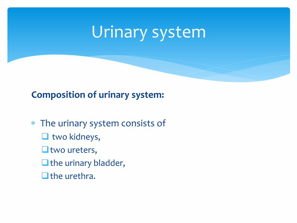
Kidneys are paired, reddish, bean–shaped organsresponsible for formation of urine by blood filtration
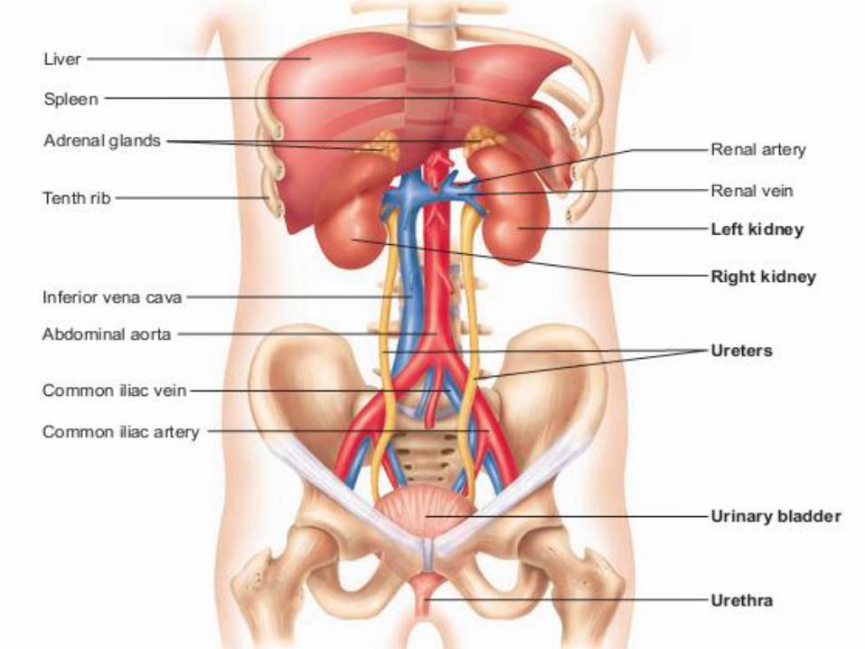
Location Kidneys are positioned against the posterior wall of the
abdominal cavity (retroperitoneal) between the levels of thetwelfth thoracic and the third lumbar vertebrae
The right kidney is usually 1.5 to 2.0 cm lower than the leftbecause of the large area occupied by the liver on the rightside.
Kidneys

Each adult kidney is a bean-shaped organ about 11.25 cm (4 in.) long, 5.5 to 7.7 cm(2–3 in.) wide, and 2.5 cm (1 in.) thick.
Each kidney is embedded in a fatty fibrous pouch consisting of three layers:
❑ The renal capsule - layer of dense fibrous connective tissue surrounding eachkidney, protects kidney from trauma and infection and maintain its shape
❑ Renal adipose capsule – layer of adipose tissue surrounding the renal capsulefunctions as a shock absorber, cushioning the kidneys against mechanical shock.
❑ Renal fascia - thin layer of connective tissue surrounding the adipose tissue,anchors the kidney to the posterior abdominal wall
* The lateral border of each kidney is convex, whereas the medial border isstrongly concave
* On the medial border is hilum, a depression through which renal artery, vein andnerves enter and leave kidney
* The superior border of each kidney is capped by the adrenal gland
External anatomy of kidneys

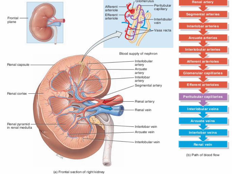
The coronal section of the kidney show two distinct regions and a cavity.
The outer light red coloured is called cortex, the inner dark red-brown coloured called medulla and cavity is called as renal sinus
The medulla has distinctive conical structures called renal pyramids
The renal pyramids has a base and an apex. The apex is called renal papillae and they point towards renal sinus
The base is directed towards the cortex (outside). The base of pyramids makes border between cortex and medulla
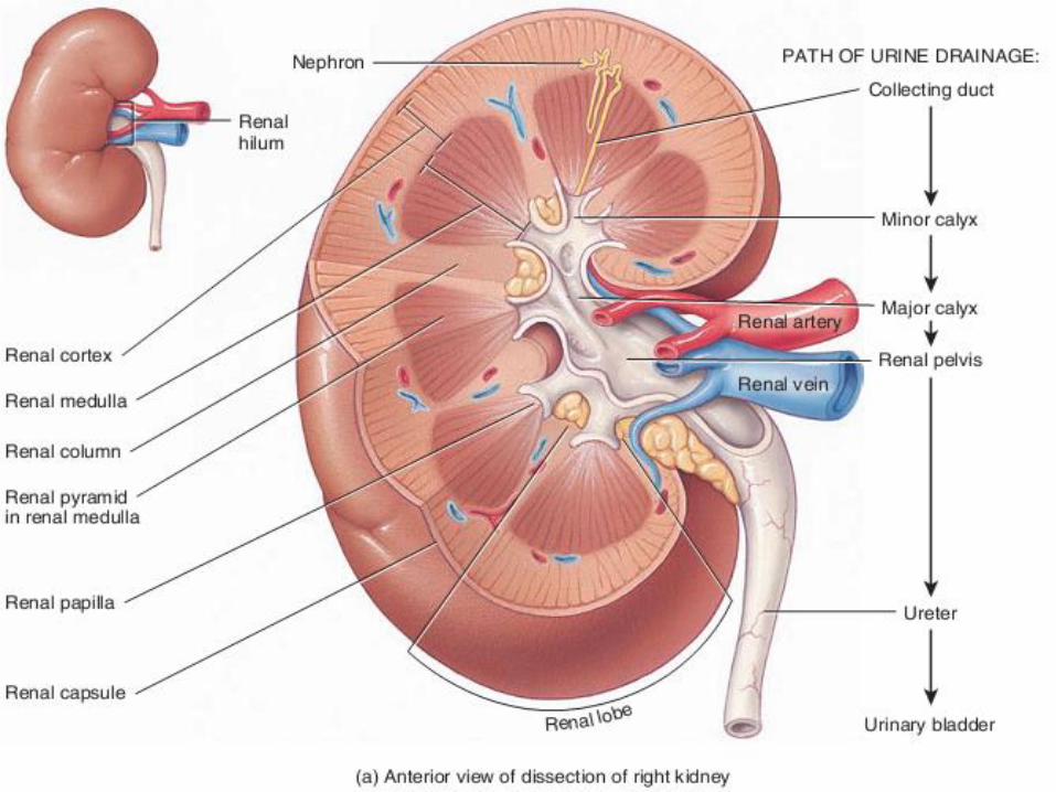
Internal anatomy of kidneys

The cortex occupies outer portion of the kidney and it inviginates deep into the medulla in between the renal pyramids. These extensions of cortex into medulla are called renal columns
The cavity of the kidney collects and transports urine from the kidney to the ureter. It is divided into several portions.
The papilla of a renal pyramid projects into a small depression in the renal sinus called the minor calyx.
Several minor calyces unite to form a major calyx. In turn, the major calyces join to form the funnel-shaped renal pelvis.
The renal pelvis collects urine from the calyces and transports it to the ureter.
A human kidney is divided into 8 to 15 renal lobes. A renal lobe consists of a medullary pyramid and some cortical substance from the renal columns adjacent to it on either side, as well as the cortex external to the pyramid base.
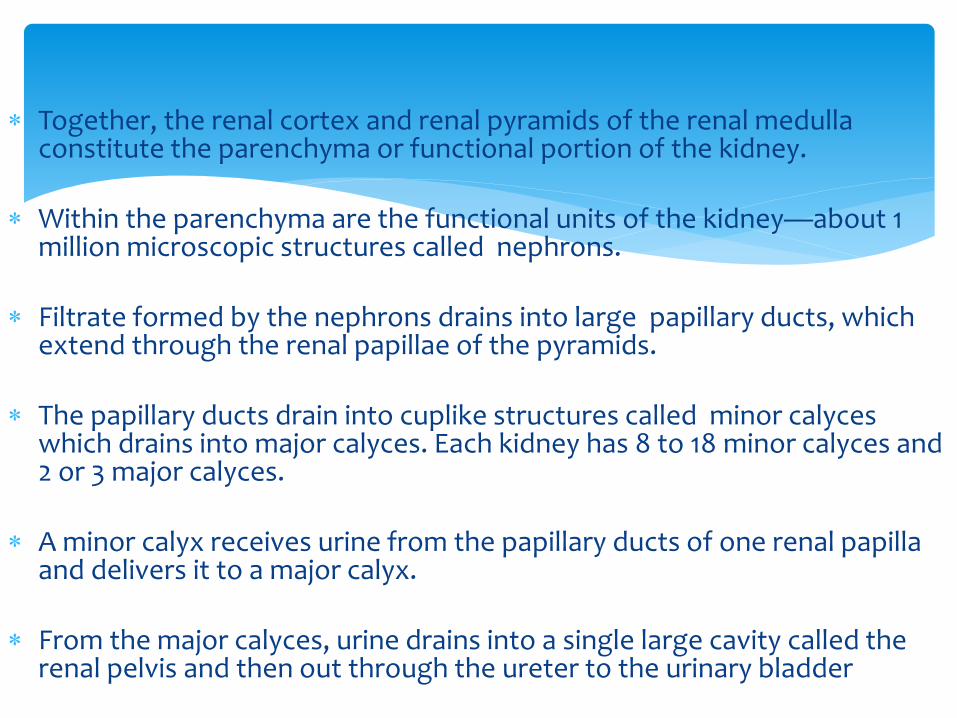
Together, the renal cortex and renal pyramids of the renal medulla constitute the parenchyma or functional portion of the kidney.
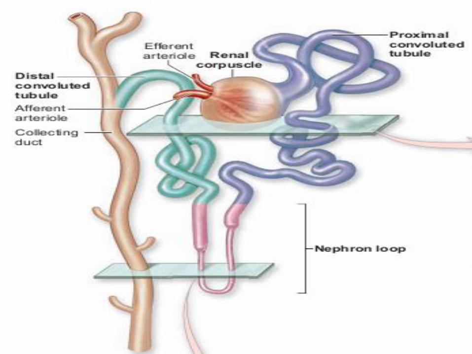
Within the parenchyma are the functional units of the kidney—about 1 million microscopic structures called nephrons.
Filtrate formed by the nephrons drains into large papillary ducts, which extend through the renal papillae of the pyramids.
The papillary ducts drain into cuplike structures called minor calyces which drains into major calyces. Each kidney has 8 to 18 minor calyces and 2 or 3 major calyces.
A minor calyx receives urine from the papillary ducts of one renal papilla and delivers it to a major calyx.
From the major calyces, urine drains into a single large cavity called the renal pelvis and then out through the ureter to the urinary bladder

The ureters are long, fibromuscular tubes that conduct urine fromthe kidneys to the urinary bladder.
Each tube averages 25 centimeters in length and is retroperitoneal
The ureters originate at the renal pelvis as it exits the hilum of thekidney, and then extend inferiorly .
At the level of base of urinary bladder they curve medially to enterthe posterolateral wall of the of the urinary bladder inferiorly
Ureters
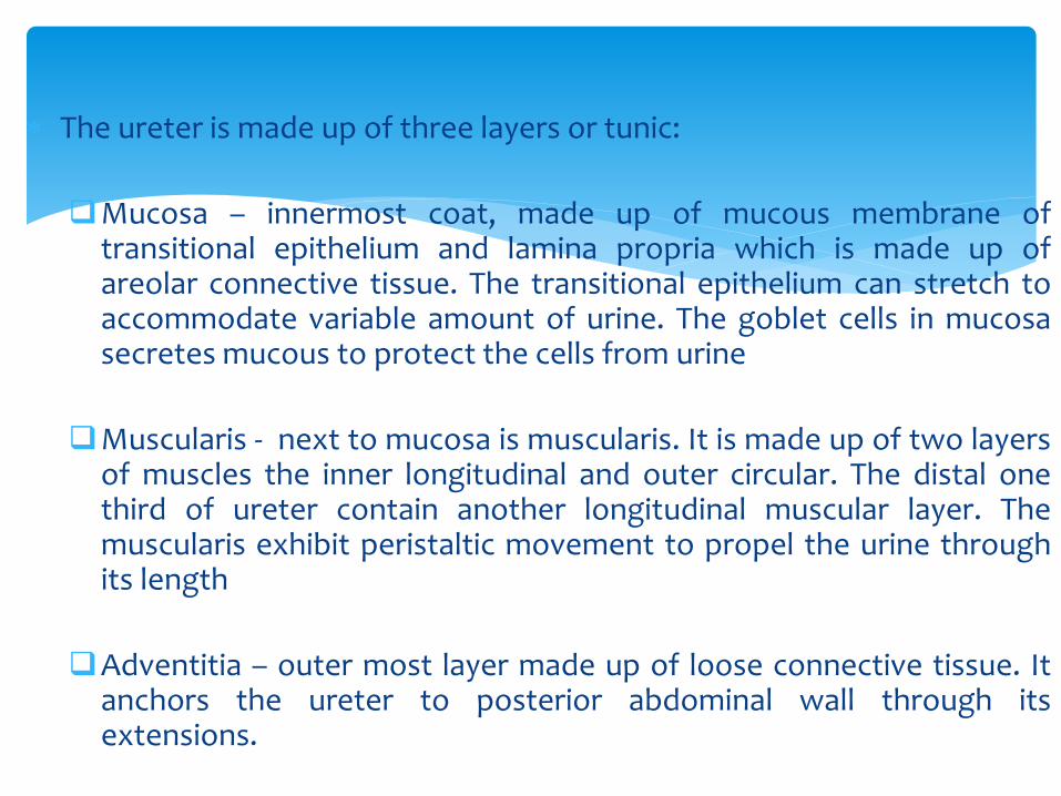
The ureter is made up of three layers or tunic:
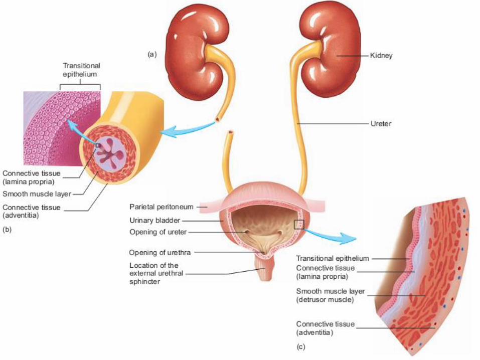
❑Mucosa – innermost coat, made up of mucous membrane oftransitional epithelium and lamina propria which is made up ofareolar connective tissue. The transitional epithelium can stretch toaccommodate variable amount of urine. The goblet cells in mucosasecretes mucous to protect the cells from urine
❑Muscularis - next to mucosa is muscularis. It is made up of two layersof muscles the inner longitudinal and outer circular. The distal onethird of ureter contain another longitudinal muscular layer. Themuscularis exhibit peristaltic movement to propel the urine throughits length
❑Adventitia – outer most layer made up of loose connective tissue. Itanchors the ureter to posterior abdominal wall through itsextensions.

The urinary bladder is a saccular organ for storage of urine
The urinary bladder is a retroperitoneal organ, since only its superior surface is covered with peritoneum
The urinary bladder is positioned immediately posterior to the pubic symphysis
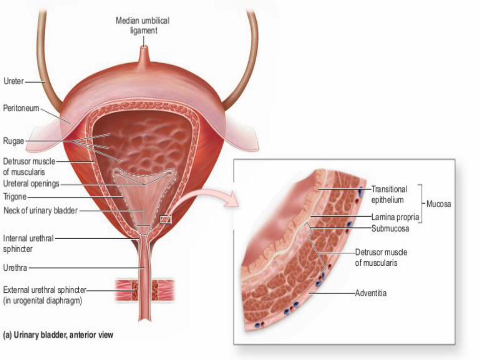
The shape of the urinary bladder is determined by the volume of urine it contains. An empty urinary bladder is pyramidal. As it fills, it becomes ovoid and bulges upward into the abdominal cavity
A fibrous, cordlike median umbilical ligament extends toward the umbilicus from the anterosuperior border of the urinary bladder.
The base of the urinary bladder receives the ureters, and the urethra exits at the inferior angle, or apex. The region surrounding the urethral opening is known as the neck of the urinary bladder
Urinary bladder

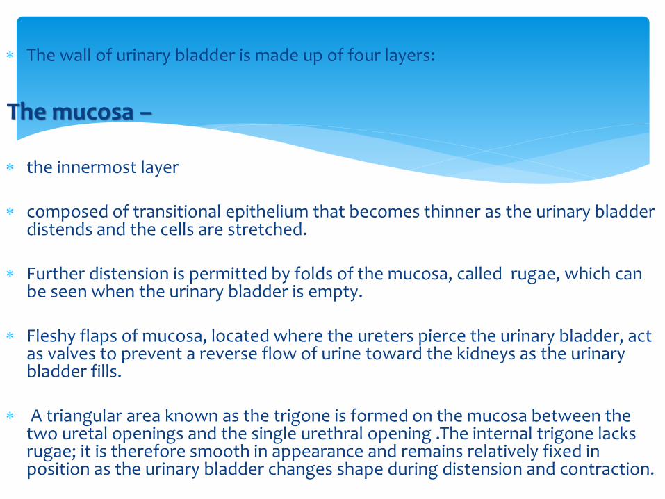
The wall of urinary bladder is made up of four layers:
The mucosa –
the innermost layer
composed of transitional epithelium that becomes thinner as the urinary bladder distends and the cells are stretched.
Further distension is permitted by folds of the mucosa, called rugae, which can be seen when the urinary bladder is empty.
Fleshy flaps of mucosa, located where the ureters pierce the urinary bladder, act as valves to prevent a reverse flow of urine toward the kidneys as the urinary bladder fills.
A triangular area known as the trigone is formed on the mucosa between the two uretal openings and the single urethral opening .The internal trigone lacks rugae; it is therefore smooth in appearance and remains relatively fixed in position as the urinary bladder changes shape during distension and contraction.
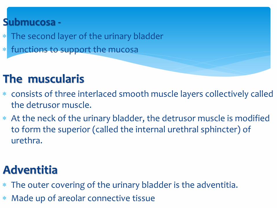
Submucosa -
The second layer of the urinary bladder
functions to support the mucosa
The muscularis consists of three interlaced smooth muscle layers collectively called
the detrusor muscle.
At the neck of the urinary bladder, the detrusor muscle is modified to form the superior (called the internal urethral sphincter) of urethra.
Adventitia The outer covering of the urinary bladder is the adventitia.
Made up of areolar connective tissue

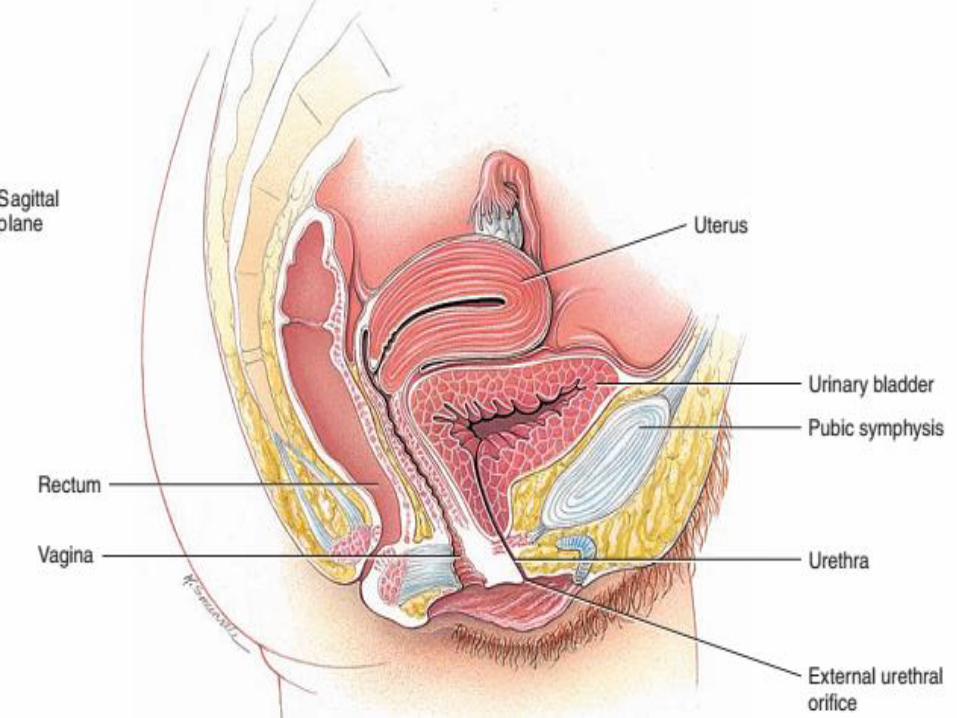
The urethra is a fibromuscular tube that originates at the neck of the urinary bladderand conducts urine to the exterior of the body
The male urethra is different from female urethra as male urethra also performreproductive function.
Four basic anatomic feature common in male and female urethra :
❑ Innermost mucous membrane lining releasing mucus
❑ Layer of smooth muscle next to mucus membrane oriented longitudinally
❑ Urethral glands embedded in urethral wall releasing mucus
❑ Two muscular sphincters;
❖The internal urethral sphincter – involuntary sphincter made up by the detrusormuscles of bladder
❖The external urethral sphincter – voluntary sphincter made up of skeletalmuscles
Urethra
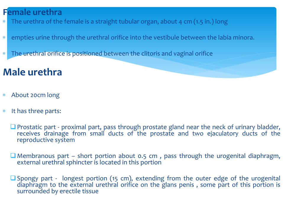
Female urethra The urethra of the female is a straight tubular organ, about 4 cm (1.5 in.) long
empties urine through the urethral orifice into the vestibule between the labia minora.
The urethral orifice is positioned between the clitoris and vaginal orifice
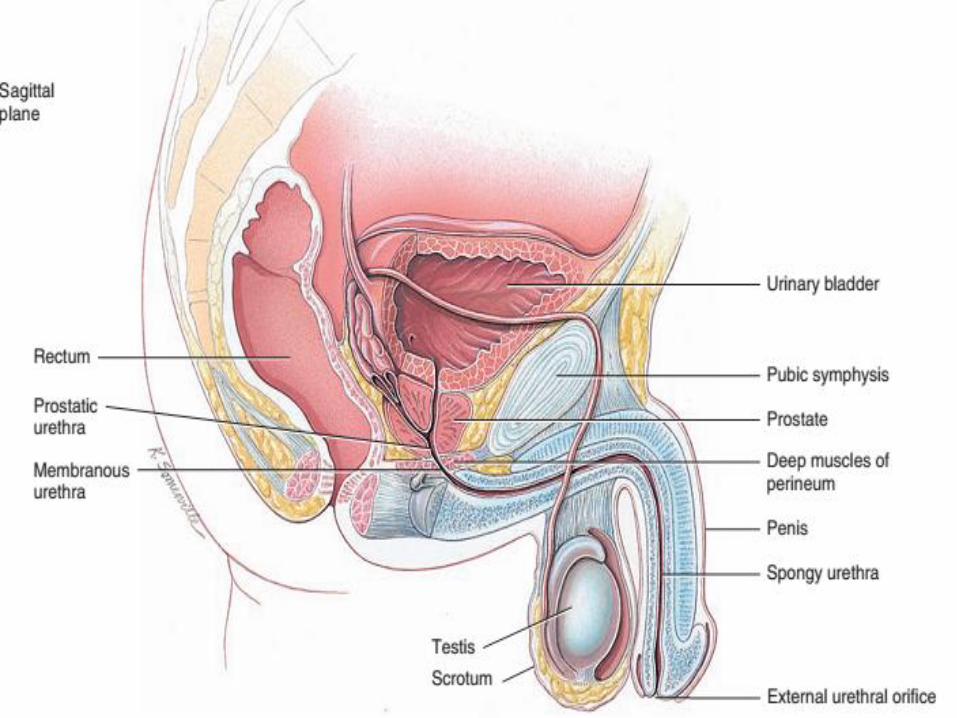
Male urethra
About 20cm long
It has three parts:
❑Prostatic part - proximal part, pass through prostate gland near the neck of urinary bladder,receives drainage from small ducts of the prostate and two ejaculatory ducts of thereproductive system
❑Membranous part – short portion about 0.5 cm , pass through the urogenital diaphragm,external urethral sphincter is located in this portion
❑Spongy part - longest portion (15 cm), extending from the outer edge of the urogenitaldiaphragm to the external urethral orifice on the glans penis , some part of this portion issurrounded by erectile tissue