FACIAL ANATOMY
26
FACIAL ANATOMY 1
-
Upload
khangminh22 -
Category
Documents
-
view
0 -
download
0
Transcript of FACIAL ANATOMY

FACIAL ANATOMY
1

SCALP FACIAL ANATOMY
Layer of dense connective tissue:- scalp is characterized by a layer of dense connective tissue deep to skin- contains profuse neurovasculature- dense connective tissue promotes bleeding following scalp injuries- skin, connective tissue & aponeurosis are tightly joined (first 3 layers): “scalp proper”
(flaps)
Harvesting oftemporal fascia
2

SCALP FACIAL ANATOMY
Aponeurotic layer:- deep to dense connective tissue, & adherent to overlying skin- consists chiefly of occipitofrontalis muscle - anterior & posterior muscle bellies linked by aponeurosis - moves scalp, contracts forehead, elevates eyebrows - innervated by temporal branch of facial nerve anteriorly and posterior auricular branch posteriorly
3

INNERVATION OF SCALP FACIAL ANATOMY
Cranial nerves (CN 5 branches)- anterior to ears & vertex of head: - supratrochlear - supra-orbital - auriculotemporal - zygomaticotemporal
Cervical nerves (spinal nervesfrom cervical vertebrae)- posterior to ears & vertex (C2, C3): - great auricular - lesser occipital - greater occipital - third occipital
http://img.medscape.com/pi/emed/ckb/clinical_procedures/834279-834808-1745.jpghttp://www.touchpoint.dk/xdoc/209/images/occipitaleXnerverXny.jpg
4
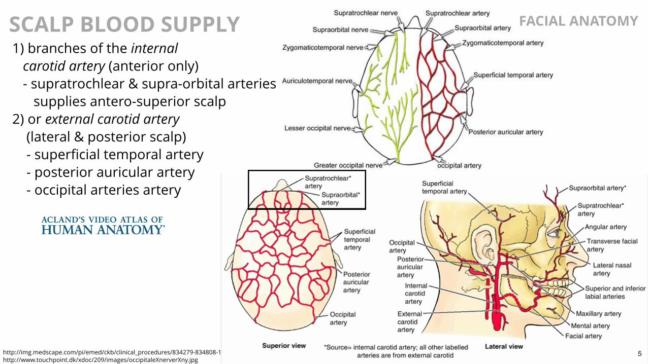
SCALP BLOOD SUPPLY 1) branches of the internal carotid artery (anterior only) - supratrochlear & supra-orbital arteries supplies antero-superior scalp2) or external carotid artery (lateral & posterior scalp) - superficial temporal artery - posterior auricular artery - occipital arteries artery
http://img.medscape.com/pi/emed/ckb/clinical_procedures/834279-834808-1745.jpghttp://www.touchpoint.dk/xdoc/209/images/occipitaleXnerverXny.jpg
FACIAL ANATOMY
5

MUSCLES OF HEAD FACIAL ANATOMY
Functional ClassificationExtraocular Muscles:- move eyeball and open upper eyelidMiddle Ear Muscles:- regulate movement of middle earFacial Muscles:- facial movements and expressionMastication Muscles:- move jaw at temporomandibular jointSoft Palate Muscles:- elevate & lower palateTongue Muscles:- move tongue
http://cnx.org/resources/92104ea27d9dc1e7c8342578d3b7ab5f00cf1367/1109_Muscles_that_Move_the_Tongue.jpg6
FACIAL MUSCLES FACIAL ANATOMY
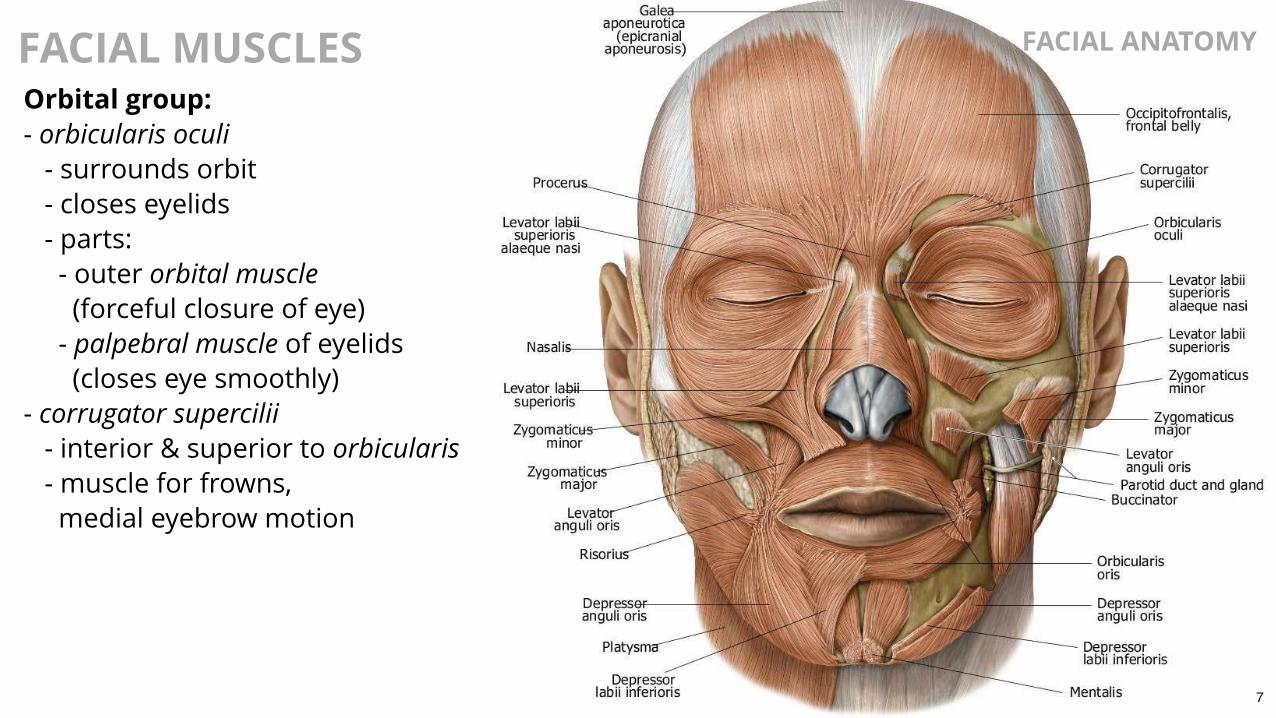
Orbital group:- orbicularis oculi - surrounds orbit - closes eyelids - parts: - outer orbital muscle (forceful closure of eye) - palpebral muscle of eyelids (closes eye smoothly)- corrugator supercilii - interior & superior to orbicularis - muscle for frowns, medial eyebrow motion
7

FACIAL MUSCLES FACIAL ANATOMY
Nasal group:nasalis - largest, modulates nares opening - transverse & alar parts procerus - nasal bone into skin between eyebrows - used for frowning, pulls medial eyebrows downward depressor septi nasi - also expands nares & draws nose inferiorly
8

FACIAL MUSCLESOral group (move lips & cheek): Orbicularis oris - surrounds mouth to move lips - fibers originate from medial maxilla, buccinator, etc. Buccinator - muscles of cheek, deep muscle between mandible & maxilla - buccinator fibers insert into lips to merge with orbicularis oris - contributes to movement of lips & brings cheek against teeth - facilitates mastication & forcing of air from cheek Lower group - depressor anguli oris (frowning, lowers corners of mouth) - depressor labii inferioris (depresses & lateral movements of lower lip) - mentalis (deep muscle; protrudes lower lip) 9

FACIAL MUSCLES FACIAL ANATOMY
Occipitofrontalis- interconnects frontal & occipital regions- frontal region covers forehead to eyebrows- adjusts scalp & wrinkles forehead
Platysma- broad, thin sheet of muscle in superficial fascia of neck- from clavicle to mandible (some fibers extending to mouth)
Upper group - risorius (thin superficial muscle, for grinning, mouth corners up & out) - zygomaticus major (superficial, to corner of mouth for smiling) - zygomaticus minor (inserts into upper lip medial to mouth corner) - levator labii superioris (moves corner of mouth relative to nose) - levator labii superioris alaeque nasi (between nose & skin of upper lip) - levator anguli oris (deep, elevates corner of mouth)
10

FACIAL MUSCLES FACIAL ANATOMY
Muscles of Mastication Masseter Temporalis Lateral Pterygoid Medial Pterygoid
11
FACIAL INNERVATION FACIAL ANATOMY
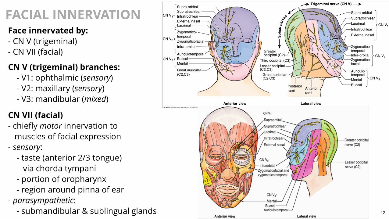
Face innervated by:- CN V (trigeminal)- CN VII (facial)
CN V (trigeminal) branches: - V1: ophthalmic (sensory) - V2: maxillary (sensory) - V3: mandibular (mixed)
CN VII (facial)- chiefly motor innervation to muscles of facial expression- sensory: - taste (anterior 2/3 tongue) via chorda tympani - portion of oropharynx - region around pinna of ear- parasympathetic: - submandibular & sublingual glands 12
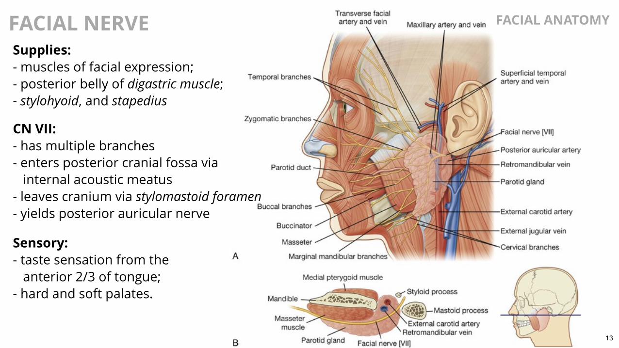
FACIAL NERVE
Supplies:- muscles of facial expression;- posterior belly of digastric muscle;- stylohyoid, and stapedius
CN VII:- has multiple branches- enters posterior cranial fossa via internal acoustic meatus- leaves cranium via stylomastoid foramen- yields posterior auricular nerve
Sensory:- taste sensation from the anterior 2/3 of tongue;- hard and soft palates.
FACIAL ANATOMY
13

FACIAL NERVE Extracranial Branches:- temporal - marginal mandibular- zygomatic - cervical- buccal
FACIAL ANATOMY
Note in illustration: - motor component - sensory component - autonomic branches
http://accessmedicine.mhmedical.com/data/Books/waxm27/waxm27_c008f013.png
14

TRIGEMINAL NERVE V1 branches innervating face:- supra-orbital- supratrochlear- infratrochlear (upper eyelid, side of nose, etc.)- lacrimal (lateral half of upper eyelid & skin of lateral angle)- external nasal (anterior nose)
FACIAL ANATOMYV2 branches innervating face:- zygomaticotemporal branch (supplies region near temple above zygomatic arch)- zygomaticofacial branch (skin over zygomatic bone)- infraorbital nerve (branches from infraorbital foramen to innervate lower eyelid, cheek, side of nose & upper lip)
V3 branches innervating face:- auriculotemporal nerve (external acoustic meatus, tympanic membrane & temple)- buccal nerve (cheek)- mental nerve (branches from mental foramen to innervate skin of lower lip & chin)
15

TRIGEMINAL NERVE FACIAL ANATOMY
Trigeminal Ganglion (Semilunar)- contains pseudo-unipolar soma of sensory neurons innervating the face (cavum trigeminale) - trigeminal cave filled with CSF- Fibers in each of 3 branches: - ophthalmic: 26,000 - maxillary: 50,000 - mandibular: 78,000- Proprioceptive fibers from extrinsic eye muscles, muscles of mastication & probably facial muscles terminate in cranial nuclei
Muscles innervated by motor root (in V3): - muscles of mastication - tensor tympani - tensor veli palatini - mylohyoid - anterior digastric
http://www.yourarticlelibrary.com/biology/human-beings/trigeminal-nerve-the-largest-of-all-cranial-nerves-1166-words/9555/16

BLOOD SUPPLY TO FACE FACIAL ANATOMY
Arteries supplying face arise chieflyfrom external carotid artery- facial artery terminates as angular artery at medial margin of eye- branches of facial a. include superior & inferior labial arteries- transverse facial artery is technically a branch of superficial temporal artery- mental a. is one of five branches of maxillary artery
17

BLOOD SUPPLY TO FACE FACIAL ANATOMY
Maxillary artery- larger of two terminal branches of external carotid artery
Maxillary a. gives rise to:- infra-orbital artery: lower eyelid, upper lip, etc.- buccal artery: cheek region- mental artery: via mental foramen; supplies chin
18
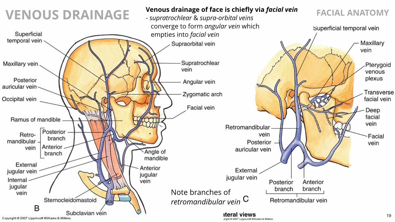
VENOUS DRAINAGE FACIAL ANATOMYVenous drainage of face is chiefly via facial vein- supratrochlear & supra-orbital veins converge to form angular vein which empties into facial vein
Note branches ofretromandibular vein
19

VENOUS DRAINAGE FACIAL ANATOMY
Facial vein:- receives small vessels draining eyelids, nose, cheek, etc.
- collects blood from deeper regions that communicate with intracranial cavernous sinus through emissary veins - facial infections may be transmitted intracranially by such pathways
https://clairenmp.files.wordpress.com/2014/04/8f0acb4a22bf484cbdd443c61df98545.jpg http://c0018305.cdn1.cloudfiles.rackspacecloud.com/SB593l.jpg20

LYMPHATIC DRAINAGE FACIAL ANATOMY
The lymphatic system is important forimmunological defense and for regulation ofbody fluid compartments.- lymph consists of fluid, cellular debris, protein, etc.- and is returned to the venous circulatory system
Superficial lymphatics draintoward ring system in neck.Deep lymphatics drain into leftand right jugular lymphatic trunks.
Enlarged lymph nodes may be indicative ofpathology (e.g. infection, cancer, etc.).
21

PAROTID GLAND FACIAL ANATOMY
Location and morphology:- largest of 3 major salivary glands- anterior & inferior to lower ear- extends from zygomatic arch to lower margin of mandible- extends over anterior sternocleidomastoid muscle to partially cover the masseter muscle- sensory innervation via auriculotemporal nerve (branch of mandibular n.)- multiple structures extend through the parotid gland
Parotid duct:- enters buccinator m. from anterior margin of gland- enters interior of mouth near second upper molarBuccal
fat pad
22

PAROTID GLAND FACIAL ANATOMY
Surgery of the parotid requiresspecial precautions to avoiddamaging the facial nerve.
The facial nerve passesthrough the parotid gland:- after passing through skull via the stylomastoid foramen- the nerve branches at least once at the level of the parotid gland
superficial temporal artery & vein
facial nerve
23

PAROTID GLAND FACIAL ANATOMY
External carotid- deep to lower margin of gland- runs superiorly yielding: - posterior auricular artery - maxillary artery - superficial temporal artery
superficial temporal artery & vein
facial nerve
Retromandibular vein- union of superficial temporal & maxillary veins- runs inferiorly through parotid- dividing into anterior & posterior branches below gland
24

PAROTID GLAND FACIAL ANATOMY
Benign tumors often necessitate removal only of superficial lobe.Malignant tumors usually require that entire gland be excised.
Rarely, secretory cells of the parotidsgive rise to tumors.- acinar cells are most commonly affected- most tumors are benign: pleomorphic adenoma, adenolymphoma- care must be taken to spare facial nerve during surgical procedures
Parotid surgery often causes damage to facial branches- impairing movement of facial muscles- various techniques have been proposed to spare the facial nerve during parotid surgery- devices that monitor integrity of facial nerve (stimulators) are often used during surgical procedures in this region
25
TRANSPLANT FACIAL ANATOMY
https://www.youtube.com/embed/B7OcH1YkWRI?enablejsapi=1
26