Quain's Elements of anatomy.
210
COLUMBIA LIBRARIES 0^^b m-^xi-i^-Kf^k ''K^>v;i'i,,.. 111111111111 msm'ff' HX00017671 PjSJt-mrv UAITsJS An ATOM
-
Upload
khangminh22 -
Category
Documents
-
view
2 -
download
0
Transcript of Quain's Elements of anatomy.
COLUMBIA LIBRARIES 0^^bm-^xi-i^-Kf^k ''K^>v;i'i,,..
111111111111 msm'ff'HX00017671
PjSJt-mrv
UAITsJS AnATOM

I' ''^!-'i^'^!^fc
,.\
'!^i'<}y^ .V

^m̂-:.
^>v:i
v-f. I.:

THE LIBRARIES

aXXx
OUAIN'S
ELEMENTS OF ANATOMY
EDITED BY
EDWARD ALBERT SCHAFER, LL.D., F.R.S.
PBOKh:SSOR OF PUVSIOLOGV AND HISTOLOOV IX USIVEU31TY COLt.EOE, LON'DOK,
GEORGE DANCER THANE,PROFESSOR OF AXATOMV IX UXIVEP^ITV COLLEGE, LOXDOX.
IN THREE VOLUMES.
. . VOL. I — PART L
EMBRYOLOGY.
By PEOFESSOE SCHAFEE.
illustrated by 200 engravings, many of which are coloured.
l^tiu en it I on.
LONGMAXS, GREEN, AND CO.
39 PATEEXO.STER ROW, LONDONNEW YORK AND BOMBAY
1898
[AU rights rescrixd.]
BIBLIOGRAPHICAL NOTE.
Ninth Edition, 2 Fo/s., 8to. November, 1882; FoL 7. Reprinted Mm-cli, 1884;
Odoher, 1887. FoZ. 7/. Reprinted Decemler, 1883 ; ^pn7, 1887.
Teni/i Edition, edited hy E. A. ScJidfer and G. D. Thane, in 8 separately issued
Parts and an Appendix, 1890-6.
Vol. I., Parti. {Embryology), first sepjarate issue, October, 1890; Reprinted, October,
1892 ; also, tvith additions and emendations, January, 1896, and again, with
further additions, November, 1898.
CONTENTS OF PART I.
Intuoductiox :
General CoNsiDEnAiioxsPlan of Oiganisalion
The Vertebrate typo .
PAGESegmentation of the BodyHomology .
Symmetry of Form.Descriptive terms
PACK2
4. 4. 4
EMBRYOLOGY.
6, 99
II
12
12
14
14
ft
Btkucttke of Ovakian Ovvm ;Matuka
TioN OF Ovum ....Formation of Polar Globules
Fertilization
Cleaning of Polar Globules.
Theory of Minot . . . •
Theory of AVeissmann
Recent Literature of the OvumSegmentatiox of the Ovvm ; Forma-
tion OF THE Blastoderm . . i6, 17
Gastrula Condition of the Vertebrate
Ovum • • ^^
Views concerning the Gastrulation of
A^'ertebrates 22
Inversion of Blastodemiic.Laj-ersin some
Mammals . . . • • •
"~
Historical view-of Blastoderm . . .
Characters of the Blastodermic Layers .
Parablast theory of His....Mesenchyme theory of Hertwig , . .
Kecent Literature of Blastoderm .
Early Changes in Blastoderm . .
Neural CanalNotochord ......Separation of the EmbryoCleavage of Mesoblast . . . -
Formation of Body Cavity
Formation of Mesoblastic Somites . .
Cerebral Vesicles
Heart and Vascular System . . .
Kecent Literature of Early Changes in
Blastoderm . . . . •
Development of the Fcetal ]\Iem-
ERANES ; Attachment of Ovl'm to
Uterl'sFormation of the Amnion and Chorion .
Formation of the Allantois .
Changes in the Uterus and mode of
attachment of Ovum to Uterus .
The Placenta .
Separation of the Decidua at birth, andregeneration of the Uterine MucousMembrane......
Keceut Literature of the Decidua , .
Development of the XERVors SystemOf the SpinalCord ....Of the Brain
23242526
27303032
3436363637
41
4242
43
46
53
5555575761
Further details regarding the develop-
ment of special parts of the Brain
The fifth cerebral vesicle : Bulbar vesicle
or MetencephalonThe fourth cerebral vesicle : Cerebellar
vesicle or Epencephalon. . . •
The third cerebral vesicle : Mesen-
cephalon : Mid-brain....The second cerebral vesicle : Thalamen-
cephalon ....••The fii-st cerebral vesicle : Prosencephalon
The Olfactory Lobes . . . • .
Formation ofthe FissuresandConvolutions
Development of the NervesSpinal nerves . . ... • .
Cranial nerves .....Optic nerves ......Olfactory lobe
Sympathetic nerves and ganglia . .
Kecent Literature of the development of
the Nervous System .
Development of the EyeOf the Ketina
Of the LensCapsule of the LensVitreous humourCorneal epithelium
Sclerotic ....Choroid coat .
Accessor}" structures .
LachrjTnal gland .
Lachrymal canals and ducts . . .
Development of the Ear ; The Laby-
rinthAccessory parts of the Organ of Hearing
External and Middle Ear .
Develop.ment of the Nose . . •
Kecent Literature of the development of
the Sense Organs ....Development of the Ali.mentary Canal
Of the Mouth and parts in connection
with it
PharynxTongue(Esophagus, Stomach, and Intestines
The MesenteryThe Spleen
63
63
:f68 ^71 -771 ^ I
'^7979 .
Si ^
Si
S38686S7S7
8788S8
898989
S9
9395
9899
99lOI
102
103
104108
S:v

IV CONTENTS OF PAET I.
Formation of the Anus . . .
FOKMATION OF THE GlANDS OF THE AlIMENTAKY CaNAL ...
The Lungs , . . . .
The Trachea and LarynxThe Thyroid Body ....The Thymus . .
The Liver
The Pancreas .....Eecent Literature of the development of
the Alimentary Canal and Glands .
Development of the Urixaey andGenekative Oegans
The Wolffian duct and hodySupra-renal Capsules
The permanent Kidnej^s ,
The Urinary BladderThe Miillerian duct .
The Germinal EpitheliumDevelopment of the Ovary.
Of the Testicles ,
Descent of the Testicles .
The External OrgansTable of Generative OrgansRecent Literature of the development of
the Urinary and Generative OrgansFormation of the Vascular SystemDevelopment of the HeartPeculiarities of the Foetal HeartDevelopment of the principal Arteries
page108
109109nonoIII
112
"3
113
115117120122122122
124124125126
127
130
132
134134146
146
PAGKDestination of tlie fourth and fifth Arte-
rial ArchesDevelopment of the principal Veins.
Peculiarities of the foetal Organs of Circulation
The Foramen Ovale .
The Eustachian Valve .
The Ductus Arteriosus
The Umbilical Vessels .
Course of the blood in the Foetus
Changes in the Circulation at Birth
The Lymphatic SystemEecent Literature of the development of
the Vascular System .
Deat:lopment of the Serous CavitiesANT) OF the Muscles and Skeleton
The Serous Cavities
Development of the MusclesFormation of the head Muscles and evi
deuces of head segmentation .
Development of the Vertebral ColumnRibs and Sternum ....The Limbs .....The Cranium .....Formation of the visceral skeleton of the
Head ; cartilaginous bars of the visceral
arches ......Formation of the Auditory Ossicles . .
Recent Literature ofthe development ofthe
Serous Cavities, Muscles, and Skeleton 169
150151
155
155155156156156
157
157
158
159159159
161
162
163
163
165
166
167
ELEMENTS OF ANATOMY.
INTPtODUCTION.
Anatomy, in its most extended seuse, is the science which deals with the
structure of organized bodies. It is divided into departments according to its
subjects ; such as Human Anatomy ; Comparative Anatomy, or the study of the
structure of different animals ; and Vegetable Anatomy, comprehending the
structure of plants.
On examining the structure of an organized body, we lind that it is made up of
members or organs, by means of which its functions are executed, such as the root
stem and leaves of a plant, and the heart, brain, stomach and limbs of an animal;
and ftirther, that these organs are themselves made up of certain constituent
materials named tissues or textures, such as the cellular, woody, and vascular tissues
of the vegetable, or the osseous, muscular, connective, vascular, nervous, and other
tissues, which form the animal organs.
Most of the tissues occur in more than one organ, and some of them indeed, as
the connective and vascular, in nearly all, so that a multitude of organs, and these
greatly diversified, are constructed out of a small number of constituent tissues;
and pai'ts of the body, differing widely in form, construction, and uses, may agi-ee in
the nature of their component materials. Again, as the same tissue possesses the
-same essential characters in whatever organ or region it is found, it is obvious that
the structure and properties of each tissue may be made the subject of investigation
apart from the organs into whose formation it enters.
The foregoing considerations have led to the subdivision of anatomy into twobranches, the one of which, under the name " General Anatomy," or " Histology,"
treats of the minute structure of the component tissues of the body ; the other,
named " Special or Descriptive Anatomy," treats of its several organs, members, andregions, describing the outward form and internal structure of the parts, their
relative situation and mutual connection, and the successive conditions which they
present in the progress of their formation or development.
To the description of the origin and formation of organs in the embryo, a special
chapter is devoted in this work, under the name Embryology.
The study of anatomy may be viewed in two different aspects ; viz., the physio-
logical and the morphological. In the former, anatomy supplies the materials
relating to structure from which an explanation is sought of the uses or functions
of organs by the physiologist ; and for this purpose the study of histology is of
particular service. In its morphological aspect, anatomy investigates and combinesthe facts relating to the structure and relations of organs, from which may be
deduced general principles as to the construction of the human body or that ofVOL, I. B
2 INTRODUCTION.
animals. In the determination of these general principles, or laws of morphology;-
it is necessary to combine the knowledge of the anatomy and development of
animals with that of man.
PLAN OF ORGANIZATION.
Vertebrate type.—The general plan of construction of the human body agrees
closely with that which prevails in a certain number of animals, viz., mammals,
birds, reptiles, amphibia, and fishes, and is known as the vertebrate type of organi-
zation. The main featm-e of that type, and that from which its name is derived,,
belongs to the internal skeleton, and consists in the existence of a median longi-
tudinal column, which extends through the whole trunk, and is composed in the fully
developed state of a series of bones termed vertelirce. This vertebral column is formed
in the early embryo around a simple rod-like structure, the primitive skeletal axis,
which is called the notochord, and which in most vertebrate animals disappears to a
greater or less extent in the course of development. The more solid portions of the
vertebra immediately surrounding the notochord are known as the todies or centra
(figs. 2 and 3), and constitute a pillar around which the other parts are grouped
with a certain regularity of structm-e. At one extremity of this pillar is situated the
head, snowing in almost all the animals formed upon this type a greater development of
its constituent parts ; and at the other the tail in which an opposite character or that
of diminution prevails ; while on the sides of the main part or trunic, there project,
in relation with some of the vertebral elements, two pairs of symmetrical limis.
The head and trunk contain the organs or viscera most important to life, such as-
the alimentary canal and the great central organs of the vascular and nervous
systems, while the limbs, from which such principal organs are absent, are very
variable and diifer widely in the degree of their development among the various
animals formed upon the vertebrate type. In man and the higher animals the trunk
is divisible into neck, chest, abdomen, and pelvis.
The vertebrate form of skeleton is invariably accompanied by a determinate and
conformable disposition of the other most important organs of the body, viz. :
—
firstly, the existence on the dorsal aspect of the vertebral axis of an elongated cavity
or canal which contains the brain and spinal cord, or central organs of the nervous
system ; and secondly, the existence on the ventral aspect of the vertebral axis of a
larger cavity, the visceral cavity, body cavity or ccelom, in which are contained the
principal viscera connected with nutrition and reproduction, such as the alimentary
canal, the heart and lungs, the great blood-vessels, and the urinary and generative
organs.
The general disposition of the parts of the body and of the more important
viscera in their relation to the vertebral axis are shown in the accompanying
diagrams of the external form and longitudinal and transverse sections of the humanembryo at an early period of its existence.
Segmentation of the body.—The vertebrate type of organisation in the repetition of
similar structural elements iu a longitadinal series, has a segmented character, especially in
the axial portion of the body, and this segmentation affects more or less, not merely the
skeletal parts of its structure, but also, to some extent, its other component organs.
A segmented plan of construction is by no means restricted to vertebrate animals, but exists
in several other classes of the animal kingdom, as is most conspicuously seen in the Arthropoda,
such as insects and Crustacea, and in the Annelida or worms. These animals, however, although
showing a serial repetition of parts of like structure, are not considered to belong to the verte-
brate type of organi2;ation.
In the human embryo, as in that of aU vertebrate animals, the segmentation is most marked
in the muscular system, the nervous and osseous systems becoming for the 'most part corres-
pondingly marked off : in the adult the osseous and nervous systems retain in great measuie
the segmentation which has thus been produced, although in the muscular system it has

PLAN UF OUGAiNlZATIoX,
^^S- 1-
—
Diagram of an early hciian embryo. (Allen Thomson.)s, s, indications of the vertebral divisions along the line of the back ; r, m, upper limb ; t, f, lowerlimb
;u, umbilical cord In the cranial part the divisions of the brain are indicated, together withtne eye, and au, the auditory vesicle
; near b, the visceral arches and clefts of the head, forming /h/c/-alia the rudiments of the upper and lower jaws.
Fig, 2.—Semi-diagramjiatic view of a longitudinal section of the embryo represented inFIGURE 1; SHOWING THE RELATIONS OF THE PRINCIPAL SYSTEMS AND ORGANS. (Allen ThomSOn.
)
1, 2 3, 4, 5 primary divisions of the brain in the cranial pai-t of the neural canal : n, n, spinalcord in the vertebral part of the canal ; s, spinous process of one of the vertebra ; ch, chorda dorealis run-ning through the axis ol the vertebral centra : ch', the same extending into the base of the cranium • adoreal aorta
; p, pharyngeal cavity ; /, i, alimeufcvry canal : h, ventricular part of the heart, ^nthwhich the arterial bulb is seen jouiiug the aorta by arches ; b, visceral arches of head ; I, Hver • vwoltlian body
;v, urinary vesicle or allantois, joining the intestine in the cloaca, cl ; u, u\ umbilicus.
'
Fig. 3.—Transverse section (diagrammatic) of the trunk of the embryo through the upperLIMBS. (Allen Thomson.)
m spinal cord;
n, neural or dorsal arch, including bone, muscle, skin, roots of the nerves, kc. :cA chorda dorsahs, surrounded by the vertebral body or centrum ; v, ventral or visceral arch, or waU01 tne body
; p, p, body cavity; i, alimentary canal ; h, heart ; 7, I, the rudimentary Hmbs.
Fig. i.—First dorsal vertebra with the first rib and upper part of the sterkuji, seen fromABOVE. |.
C, centi-um; N, neural cavity ; F, cavity of the chest, visceral cavity.
B 2

4 INTRODUCTION.
become greatly obscured. To the original segments in the embryo the terms protovcvtebrcs,
mesoMastic somites or myotomes have been applied ; those segments or metameres which are
traceable in the adult are often spoken of as vertelral segments. In the limbs, although there
is strong reason for believing that they have originated as outgrowths of certain segments of
the trunk, the repetition of such vertebral elements, and their primitive connection, are
greatly obscured.
Homology.—A certain agreement in structure, situation and connection of
parts or organs constitutes what is called homology, and this term is generally
employed to indicate the morphological identity of representative parts in different
animals, which may be considered to have its cause in community of origin {homo-
geny, Lankester), while the anatomical correspondence of parts which are repeated
in the same animal may be more exactly distinguished as serial homology {homo-
dynamy, Gegenbaur). Thus the arm-bone or humerus of a man is homologous
(homogenetic) with the upper bone of the fore limb of a quadruped, or of the wing
of a bird, while it is at the same time serially homologous (hemodynamic) with the
thigh bone of man himself, or any other vertebrate animal. It has farther been
found convenient to express by the word analogy that kind of resemblance amongthe organs of animals which depends upon similarity of function, and although it
may be accompanied by considerable agreement in structure, yet is not rendered
complete by anatomical relation and connection : for example, the gills of a fish, of
a crab, and of a mussel, serving the same function, are analogous organs, but in no
sense homologous, as all morphological correspondence, or genetic relation, is wanting
between them. Thus also, the upper limb of a man, the fore limb of a quadruped,
the wing of a bird, and the pectoral fin of a fish are homologous but not analogous
structures, the wing of a bat and the wing of a bird are both homologous and
analogous, while the last is analogous to but not homologous with the wing of an
insect.
Symmetry of form.—A remarkable regularity of form pervades the organi-
zation of certain parts of the body, especially the whole of the limbs, the head and
neck, and the framework, at least, and external walls of the trunk of the body.
Thus, if we conceive the body to be divided equally by a plane which passes from its
dorsal to its ventral aspect {median pkine), the two halves, in so far as regards the
parts previously mentioned, correspond almost exactly with each other, excepting by
their lateral transposition,—and the human body thus shows in a marked manner
the character of lilateral symmetry. There is, however, a departure from this
symmetrical form in the developed condition of certain of the internal organs, such
as the alimentary canal from the stomach downwards, the heart and first part of the
great blood-vessels, the liver, spleen, and some other viscera.
Descriptive terms.—In the description of parts so numerous, so various in
form, and so complex in their connections as those composing the human body, there
is diJB&culty in finding terms which shall indicate with sufficient precision their
actual position and their relation to the rest of the organism. This difficulty is
farther increased by the exceptional erect attitude in which the trunk of the human
body is placed as compared with the horizontal position in animals. Hence, a
number of terms have long been in use in human anatomy which are understood in
a technical or restricted sense. For example, the median plane, already referred to,
being that by which the body might be divided into right and left lateral halves,
and the middle or median line being that in which the median plane meets the surface
of the body, the words internal and external are used to denote relative nearness to
and distance from this plane on either side, and may be replaced by mesial and
lateral. The terms sagittal, frontal, and coronal, are also used in indication of
direction within the body : sagittal denoting a dorso-ventral direction in or parallel
to the median plane, frontal or coronal a transverse direction perpendicular to that

DESCKIPTIVE TERMS.
plane. The words anterior and /mterior, superior and vtfenor, and several othersiudicatiiig position, are employed in human anatomy strictly with reference to theerect posture of the body. But now that the more extended study of comparativeanatomy and embryonic development is largely applied to the elucidation of thehuman structure, it is very desirable that descriptive terms should be sought whichmay without ambiguity indicate position and relation in the organism at once in
man and animals. Such terms as dorsal and ventral, neural and visceral, cephalic
and caudal, central and peripheral, proximal and distal, axial and aprpendicxdar,pre-
axial and postaxial, are of this kind, and ought, whenever this may be done con-sistently with sufficient clearness of description, to take the place of those which areonly applicable to the peculiar attitude of the human body, so as to brin"- thelanguage of human and comparative anatomy as much as possible into conformity.In many instances, also, precision may be obtained by reference to certain fixed
relations of parts, such as the vertebral and sternal aspects, the radial and ulnar, an^the tibial aud fibular borders, the fiexor and cilcn.^or surfaces of the limbs, andsimilarly in other parts of the body.

EMBRYOLOGY/By E. A. SCHAFER.
GENERAL DEVELOPMENT.
FORMATION" OF THE BLASTODERM.
STRUCTURE OF THE OVUM AND CHANGES PRIOR TO SEGMENTATION.
The human body with all its tissues and organs is the product of the development
of a single nucleated cell, the egg-cell, germ-cell, or ovum, which is formed within
the principal reproductive organ of the female or ovary. The commencement of
development is preceded by certain changes in the ovum, which usually occur soon
after its discbarge from the ovary, and consist (1) in the emission of certain
constituents of the nucleus which form the so-called polar globules ; (2) in the
accession of the nucleus of a sperm-cell or spermatozoon, which is formed within the
reproductive organ of the male (testicle), and which, blending with the remaining
part of the nucleus of the ovum, appears to take the place of the part which was
discharged in the form of the polar globules.
An account of the structure of the ovum, and of the manner in which the above
changes are effected, may therefore appropriately precede the description of the
actual course of development of the ovum.
Structure of the ovarian ovum.—The human ovum resembles that of all other
mammals (with the exception of monotremes) in its minute size. Immediately
before the time of its discharge from the Graafian foUicle of the ovary in which it
has been formed, it is a small spherical vesicle measuring about yig-th inch {'2 mm.)in diameter, and is just visible as a clear speck to the naked eye. When it is
examined with the microscope, it is found to be invested by a comparatively thick,
clear covering. This, when the centre of the ovum is exactly focussed, has the
appearance in optical section of a clear girdle or zone encircling the ovum (fig. 5),
and was hence named zona pellucida by von Baer (1827). But on more careful
examination with higher magnifying powers, and especially by the examination of
sections, there is not much difficulty in making out the existence of strise passing
radially through the membrane (fig. 6, z^f). On this account, and especially since a
similar radially striated membrane forms a characteristic part of the investment of
the ovum in many animals belonging to widely different classes, it is more convenient,
in place of the name zona pellucida, which has been exclusively used to designate
this investment in mammals, to employ the more general term zona radiata, or to
gpeak of it simply as the striated membrane of the ovum."
The zona radiata of the mammalian ovum is sufficiently tough to prevent the
escape of the contents of the ovum, even when subjected to a considerable amountof pressure. If, however, the pressure be excessive, the tunic splits, and the soft
' It is mainly owing to the researches of His, published principally in the important monograph" Anatomic nienschlicher Embryonen " (Leipzig, 1880-1885), that our knowledge of the development of
Ihe human embryo is now far more complete than was the case when the last edition of this work wasandertaken, and we are therefore able to keep more closely than was before possible to the humanspecies in following the course of derelopment of the ovum. For the elucidation, however, of many of
the details of development, especially in its earlier stages, it will still be necessary to refer continually
to facts which have been made out only from the study of the embryology of other mammals, as well as
birds, reptiles, fishes, and even invertebrata.

STiturnuH (»r thk ovim. 7
contents are extruded (Hir. "i, b). The striiK in tbu membrane are believed to
be minute purcs, and aie supposed, while the ovum is yet within the (Jraafian
tblhcle, to permit tlie passjige of grannies of nutrient material into the interi<jr of
the ovum. After the ovum is disehar'^ed from the follicle, the spermatozoa mayperhaps find their way into the ovum throuj^h these pores. AccoTdlng to Ketzius
the protoi)lasm of the ovum is united with the fullicle-cells by fibres which pats
through the pores of tiie zona.
Inimedialoly surroundinj,'- tlie zona ladiata. as the ovum lies within the mature Graafiiiii
follicle, is a thin stratum of ,'ianular substJince. probably deposited upon the exterior of theovum by the iuneiouost cells of the discus prolijrerus. which immediately encircle the ovumwithin the follicle. When the Graafian follicle bursts and the ovum is set free, this ;;ranular
material ajipears to imbil)e water, and. as is .-rpecially noticeable in the ovum of the rabbit,
swells uji into a clear ^'elatinous envelope, which has been termed, from a possible homolo-rywith the white of the bird's ej-'t-'. the till/m/irn. But in the mammal this structure ha.s notthe nutritive importance to the embryo which is possesseil by the corresponding fonnationin the bird, and it disiippears during the pa-ssage of the ovum down the Fallopian tulje.
The substance of the ovum within the tunica radiata is known as the vitellus
or ijoJk (fig. G, vi). It is a soft semi-fluid substance, composed mainly of proto-
plasm, winch is filled with globides and gninules (yolk-granules) of different
Fig. T>.—Ovarian ovum of a mammifek. (Alien Thomson.
)
a, the entire ovum, viewed under pressure ; the granular cells have been removed from the outer
•surface, the germinal vesicle is seen in the yolk substance within ; h, the external coat or zona burst by
increasetl pressiue, the yolk protoplasm and the genninal vesicle having escaped from within ;c, germi-
nal vesicle more freed from the volk substance. In all of them the macula is seen.
Fig. 6.
—
Ovum of the cat ; highly magnified, semi-diagrammatic. (E. A. S.)
zp, zona pelliicida, showing mdiated structure ; ci, vitellus, round which a delicate membrane is
seen : ;/'•, germinal vesicle ; fjs, germinal spot.
sizes, but all small, and possessing a high index of refraction. Examined in the
fresh condition, the protoplasm between the granules looks perfectly clear and
structureless, but after treatment with suitable reagents, it may be seen to consist of
a fine reticulum, which is especially fine and close near the periphery of the ovum,
and also aroimd the germinal vesicle, at which places the yolk granules are in less
amount than elsewhere. The substances which occur ^vithin an ovum other than
the nucleus and protoplasm, may, as in cells generally, be collectively designated^' deutoplasm "
; they are regarded as funiishiug a supply of nutrient matter to the
protopla.sm dm-ing the earlier stages of development.
Embedded in the protoplasmic vitellus, usually eccentrically, is a large spherical

8 STRUCTURE OF THE OVARIAN OVUM.
nucleus, which was termed by its discoverer, Purkinje, the gmninal vesicle.^ This^
which is about -g^o^h inch in diameter, has all the characters of the nucleus of a
cell. It consists of a nuclear membrane enclosing a clear material or matrix,
embedded within which may be seen strands of karyoplasm, enclosing one or morewell-marked nucleoli (fig. 6, gv). Frequently there is but one nucleolus, which is
then large and prominent, and has received the name of germinal spot {macula ger-
ininaiiva, Wagner, 1805).
There is some doubt whether, before fertilization, there is another membrane (vitelline
membrane) enclosing- the vitellns within the zona radiata. The evidence of the presence of
i>uch a membrane is by no means clear, although its existence has been maintained by very
competent observers (v. Beneden, Balfour).
The mammalian ovum (that of monotremes alone excepted) differs from that of other
vertebrates in the relatively small amount of nutritive material (yolk granules, deutoplasm)which is embedded in its protoplasm. In fishes, amphibia, reptiles, and especially in birds,
the amount of such nutritive material is vastly g'reater than that of the protoplasm itself, so
that the very existence of the latter is obscured in most parts of the ovum, and it is only in
the immediate neighbourhood of the germinal vesicle that the protoplasm can be distinctly
Fig. 7.
—
Diagram of a holoblastic (alecithal) ovum (A) and of a mekoblastic (telolecithal}
OVUM (B). (E. A. S.
)
Only a small part of the latter is represented. The yolk or food material is represented in both hjclear globules, which in B are seen vastly to preponderate, except in the immediate neighbourhood of
the germinal vesicle.
recognized (fig. 7, B). It is here also that, after fertilization, the more active changes in the-
ovum occur, and it is this part alone in which in the bird and most other oviparous vertebrate?
the process of division or segmentation of the yolk and consequent formation of embryonic cells-
proceeds. Hence these ova are said to undergo a process of incomplete segmentation, only a
l^art of the ovum appearing to undergo development, and they are accordingly termed mero-
hla^tic to distinguish them from those (like the mammalian ova) in which the yolk or nutri-
tive material is everywhere in relatively small proportion to the protoplasm, the ivhole of
which undergoes division after fertilization, and jjarticipates in the formation of the embryo{Jioloblastic ova). This small amount of nutritive material in the mammal is obviousl,y
related to the fact that the mammalian ovum early acquires an attachment to the maternal
system from which it is then able directly to derive its nutriment, whereas the meroblastic
Dvum of oviparous vertebrata necessarily contains all the nutriment required by the developing
bird, reptile, or fish, until it is sufficiently advanced in development to emerge from the egg
and obtain food independently. Although, however, the mammalian ovum is holoblastic, it
is none the less clear, from a comparison of the early stages of its develojpment with that of
the bird, that the ancestors of the mammalia must have had ova of the meroblastic type.
Balfour has further conveniently distinguished between those ova in which there is a g-reat
accumulation of nutritive or 3'Olk material at one pole (telolecithal ova. as in the bird,
reptile, and fish amongst vertebrates), those in which the accumulation of yolk is in the middle
of the ovum {ccntrolemtlial ova, as in arthropods), and those in which it is scattered pretty
equally in small amount throughout the protoplasm without any very marked accumulation
^ Purkinje discovered the germinal vesicle in the bird's ovum in 1825 ; that of mammals was first
noticed by Coste in 1833.

MA'rL'l.'ATKiN oK TllK OVUM,
(iilrritliiil ova. as in iiiainnial>. Aiiipliioxiis. (rliiiinilcini.-). It is clear tliat those conditions
of arranjjcnu'Ut of tiic proto- and tlciito-iilasni wiiiiin tin- ovum air llic main factors in doter-
niininj^ variations in the [irocoss of scynu'ntatioii.
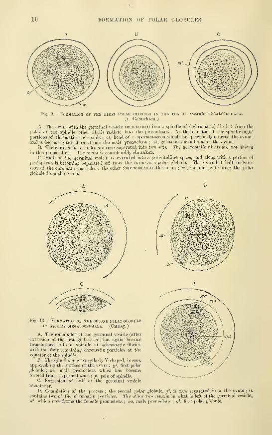
Maturation of the ovum. Formation of polar globules.— llillicr liefore
its c'sciipc IVoiii the (iriiatijiii Inlliclr, dr iiiiiiR'(li;iU'ly iil'tcr, I lie oviuii undci'^foos ii
[n'culiiir clianov. pi\'i»iU'iit(irv to, luii nevcrlliclcss iiltoj^cthcr iiid('i)C'iident ol" fci'ti liga-
tion, wiiii'h consists of a process of iiiir(|iial cell-division or jicnnination, and I'osults
in the extrusion from the vitellus of iwo minute sjtherical bodies (fig. 8), which have
Kili. S. —Ovi'M 1)1.- THi: UAHBIT IMIOM TIIK I'AI.I.hI'IAN TIIIK, TWKI.VKi
llullis AKTKK lMPIU'.(iNAl'InN. ( 1 iisrhdtt'.)
On tiie zona «, spermatozda are seen, and (itliers in tlje pciivitoliiiie
space ; b, the puiar yloljuk's.
been termed the jioliir iilohnlcs or ilircrlli'c (,)r/ii/sr/cs,
from a supposition that their presence deterniincs the
jidle at which the first seomentation will take place
should the ovum become fertilized. It is, however,
uncertain whether there is any coustant relationship of
this kind, but it is none the less clear that the extrusion
of the polar globules is an event of the highest importance for the due development
(»f the ovum, since until this has happened the ovum ai)pears to be incapable of
complete fertilization and segmentation. ^ AVhat is actually extruded is a small part
of the nucleus of the ovum, or, to speak more precisely, two small parts of its nucleus
in succession, probably surrounded by a very thin investment of protoplasm. Prior
to this extrusion, the germinal vesicle approaches the periphery of the vitellus. loses
its distinctness of outline, and after passing through phases which are charac-
teristic of a nucleus which is about to divide, does actually undergo a division into
two, the one part being extruded into a space (pcrivitelline), which has become
formed in consequence of the shrinking or contraction of the ovum, and the other
part remaining in the vitellus, only, however, to repeat the process of division, and
to form a second extruded globule. The remainder of the germinal vesicle, which is
now termed the feniale pronudpiis, leaves the periphery of the vitellus for a situation
nearer to the centre, where, if fertilization should supervene, it awaits the advent of
the male pronucleus, which is formed from a spermatozoon. After the two pro-
nuclei have come together, a new and complete nucleus is formed by their conju-
gation.
The actual formation of polar globules has not hitherto been observed in the human ovum,althoug'h there is no doubt whatever that it takes place. In the rabbit various stag-es in the
process have been traced by E. v. Beneden and Rein, and it has also been noticed in othermammals. But the details of the process have been made outmost preciselj' (by Fol. Hertwij^'.
and others) in the transparent own of echinoderms, and more recently and minutely (byE. V. Beneden, Carnoy. Boveri. Zacharias. and others) in Ascaris meyalocephala, a thread-worm]>arasitic in the horse, in which all the changes can be followed in one and the same ovum,or the various phases fixed by means of reagents in different ova. and these maj' aftenvards bestained and studied with the utmost minuteness. The successive changes in such ova arerepresented in figs. !) and 10. The polar globules remain visible for a time in the pcri-
vitelline tinid. and are even .seen, should the ovum become fertilized, during the early stages of
segmentation, but they ultimately disapjiear and are not known to take any further part in
the subsequent changes which the ovum undergoes.
The fact that throughout the whole animal kingdom the extrusion of ])olar globules froni
the ovum as it becomes mature is almost universal, and that a similar process has also beenobserved to occur in plants indicates the grea,t importance of the phenomenon. The signifi-
cance will be further discussed after the process of fertilization of the ovum has beendescribed.
' The ovuui may, however, receive a spermatozoon before the eonipletion of tlie formation of polar
globules.
10 FORMATION OF POLAR GLOBULES.
B C
Fis. 9. -FoRilATIOJI OF THE FIRST POLAR GLOBULE IN THE EGG OF ASCAKIS 3IEGAL0CEPHALA.
(v. Gehuchten.
)
A. The ovnm with the germinal vesicle transfovmed into a spindle of (achromatic) fibrils ; from the
poles of the spindle other fibrils radiate into the protoplasm. At the equator of the spindle eight
portions of chromatin are visible ; cs, head of a spermatozoon which has previously entered the ovum,and is becoming transformed into the male pronucleus ; in, gelatinous membrane of the ovum.
B. The chromatin particles are seen separated into two sets. The achromatic fibrils are not shownin this preparation. The ovum is considerably shrunken.
C. Half of the germinal vesicle is extruded into a perivitelline space, and along with a portion of
protoplasm is becoming separated otf from the ovum as a polar globule. The extruded half includfs
tour of the chromatin particles ; the other four remain in the ovum ; m', membrane dividing the polar
globule from the ovum.
B
Fia 10.
—
Formation of the second polar globuleIN ASCAKIS megalocephala. (Cai'noy.)
A. The remainder of the germinal vesicle (after
extrusion of the first giobule, g^) has again becometransformed into a spindle of achromatic fibrils,
with the four remaining chromatin particles at the
equator of the spinrl'e.
B. The sijindle. now irregularly Y-shaped, is seen
approaching the surface of the ovum ; (?', first polar
globule ; ns, male pronucleus which has becomeformed from a spermatozoon
; p. pole of spindle.
C. Extrusion of half of the germinal vesicle
remainder.
D. Completion of the jjrocess ; the second polar globule, g-, is now separated from the ovum ; it
contains two of the chromatin particles. The other two remain in what is left of the germinal vesicle,
ii^ which now forms the female pronucleus ; ns, male pronucleus ;rj^, fii-sfc polar globule.

FKjrrH.lZATlON. 11
Fertilization.—Tho ovum, after its fxpiilsitiii IVuiii Uie CJraafian lullicli; is
rccoivi'il u]inn the fiinliriiiteil end ol'tlie Fiilloi>iaii tube. The linibriic arc covered by
a ])roloiigati()n ol'tho ciUateil linini:- of the tul)e. and the acti<ju ol" the elHa serves to
jiropcl the minute ovum into and aloii^- i he tube towards the uterus. In tliis
jjassaije it may. if impretiiiation have oeeurred. meet with the spermatozoa, one or
more of which may penetrate the zona peUncida, and fertih'zc the ovum. It is
possible in some instances for fertihzation to occur on tlie findjriated extremity of the
tube, or in tlie body of the utei'us, but it is ])robable that in most ca.ses it happens
in the tube itself.
It is probable that normally only a single spennatozoon enters the vitellus. If it should
happen that two or more enter, normal development does not as a i-ule occur. Exceptions to
this rule have, however, been recorded.
The changes in the ovum which accompany fertilization have, like those which
result in the formation of the polar globules, been studied most satisfactorily in the
transparent ova ofechinoderms and in Ascaris. In the former (fig. 11) the si)ermatozoa
f.^r.iqn:
Fig. 11.
—
Fertilization op the ovum of an echinoderm. (Selenka.
)
s, spermatozoon ; m.pr, male pronucleus; f.pr, female pronucleus.
1. Accession of a spermatozoon to the periphery of the vitellus ; 2. Its penetration, and the radial
disposition of the vitelline granules ; 3. Transformation of the head of the spermatozoon into the male
pronucleus ; 4, 5. Blending of the male and female pronuclei.
may be seen to penetrate the gelatinous investment which here takes the place of a
zona pellucida, and the head, of one only as a rule, to imbed itself in the ]ieriphery
of the ovum, which becomes slightly protruded at the point of contact. According
to V. Beneden's account, the spermatozoon always enters in Ascaris at a particular
part of the ovum (polar disc), at which part there is an aperture in the vitelline
membrane (micropyle). When once it has passed into the ovum, this aperture
becomes closed, and the head of the spermatozoon rapidly increases in size, and
acquires the appearance of a nucleus which, in contra-distinction to the remains of
the germinal vesicle, or female pronucleus, is termed the 7nale promicleus. Soon
it leaves the periphery, and passes towards the centre of the ovum in the direction
of the female pronucleus. In its passage through the protoplasm it appears to
exercise a peculiar attraction upon the granules in that substance, for these become
arranged in its vicinity in radiating lines. The tail of the spermatozoon has
in the meantime disappeared, whether by being cast off or by blending with the
protoplasm of the ovum has not certainly been made out. As the male pronucleus
approaches the female pronucleus, the latter moves somewhat to meet it, and pre-
sently the two pronuclei come into contact and together fonn a new nucleus, com-

vz FEKTILIZATIOX.
plete in all its structure and functions. AVitb the blending of tlie two pronuclei
the act of fertilization is completed, and the ovum is now capable of formingnew cells by division. Since the head of the spermatozoon is formed from the
nucleus of a seminal cell, part of which appears to be thrown off prior to the com-plete maturation of the spermatozoon (Renson, Brown), and the female pronucleus is
the nucleus of an egg or germ-cell, part of which has been removed in the form of the
polar globules, the process of fertilization may be described as consisting essentially
of the conjunction of part of the nucleoplasm of a sperm cell with part of the nucleo-
plasm of a germ cell, the result being the production of a complete nucleus end<;iwed
with active properties of division and reproduction.
Although, as has been already stated, the chang-es which have just been described are mostclearly to be seen, and have been most completely studied, ia the ova of echinodei-ms andAscaris. similar processes have been found to occur in most if not ia all animals, and haveeven been made out, although not very distinctly, ia mammals (in the rabbit by v. Beneden).There is no doubt, therefore, that the phenomena, of fertilization are essentially the samethroiig-hout the whole animal kingdom. As to the exact details of the process there is still
much discrepancy in the accounts given by recent observers. Of all those that given byV. Beneden of the process of fertilization, and of the subsequent division of the resulting
nucleus in Ascaris, is the most explicit, and appears to negative the idea of a complete fusion
taking place between the elements of the pronuclei, at least so far as the chromatin is concerned.
According to this account (v. fig. 12). each of the two pronuclei is seen to possess, previous to their
conjunction, two short chromatin rods {elinnnosoinc-s) imbedded in clear nuclear matrix. Theserods undergo various changes, resulting in the formation of a skein within each pronucleus
(II.. III.), but eventually the skein resolves itself into two Y-shaped loops or filaments (IT.. Y.).
On conjitnction the matrix of the two nuclei may appear to blend, although it is doubtful if
they actually fuse together, but the chi-omatin filaments retata their distinct indiyiduality.
The nucleus which is thus formed hj the conjunction, contains, therefore, four similar Y-s)iaped
chromatin filaments, which now split longitudinally (YL. YII.). and after being aiTanged for
a time at the equator of the now spindle-shajDed nucleus (^^III.). four of the resulting filaments
pass towards the one pole, and form CA-entually the chromatin of the one daughter nucleus,
and foxu- toAvards the other pole, eventually forming the chi-omatin of the other daughter
nucleus (XI.. XII.). It is stated by v. Beneden that of each set of chromatin iUaments. or
chromosomes, which thus separate from one another, one half the number is derived from the
male and the other from the female pronucleus. If this is the case, and if it should further
be shown that in every subseqitent process of division of the resitlting cells, the chromatin
filaments of the daughter cells are derived half from male chromatin filaments and half
from female, it necessarily follows that eA-ery cell nucleus must be regarded as containing
both male and female morphological elements.
Meaning' of the polar globules.—Theory ofMinot.—The question of the hennaphroditism
of cells Avas first raised by C. S. Minot in connection Avith the separation of the polar globules.
According to the A'iew advocated by Minot. every cell which results from the division of a
fertilized ovum is hermaphrodite, for the fertilized ovum is formed by the union of both male
Fig. 12. FORMATIOX AND CONJCGATIOX OF THE PRONUCLEI IN ASCARIS jrEGALOCEIHAIA.
(E. V. Beneden.)
/, female pronucleus ; m, male pronucleus; ^), one of the polar globules.
I. The second polar globule has just been extruded ; both female and male pronuclei contain tAvo
chromatin particles ; those of the male pronucleus are beccming transformed into a skein.
II. The chromatin in both pronuclei now forms a skein.
Ilrt. The skein in the pronuclei is more distinct. Two attraction -splieres, each with a central
jjarticle, united by a spindle of achromatic fibres, have made their appearance near the pronuclei. Themale pronucleus has the remains of the body of the speiinatozoon adhering to it.
III. The pronuclei are enlarged ; the skein formation of the chromatin is complete.

III«.
MEANING Ob' THE I'OLAH GLOBULES. 13
III/; r-. '77.
Illrt. Ouly the female pronucleus is sliown in this figuie. The skein of this is contracted andthickened. The attraction-spheres are near one side of the ovum, and ;ire connected with its peripheryby a cone of fibres forming a polar circle p. c. ; c.c. equatorial circle.
Illi. The pronuclei have approached one another, and the spindle-system is now aiTanged across
their common axis.
IV. Conti-action of the skein and formation of two V-shaped chromatin filaments in each pronucleus.
V. The V-shaped chromatin filaments are now quite distinct ; the male and female pronuclei are in
close contact.
VI., VII. The V-shaped filaments are splitting longitudinally ; their structui-e of fine granules of
chromatin is apparent in VII., which is more highly magnified. The conjugation of the pronuclei is
apparently complete in these figures, hut according to v. Beneden's description, the outlines of both
can, under favourable conditions, be still made out. The attraction spheres and achromatic spindle,
although present, are not shown in IV., V., VI. and VII.
VIII. Equatorial arrangement of the four cliromatin loops in the middle of the now elongated ovum :
the achromatic substance forming a spindle-shaped system of granules with fibrils radiating from the
poles of the spindle (attraction-spheres) into the protoplasm ; commencing division of the ovum into
two cells.
IX. Shows diagrammatically the commencing separation of the chromatin filaments of the con-
jugated nuclei, and the system of fibres radiating from the attraction-spheres. p.c. polar ciicl^ :
f.c. equatorial circle ; c.c. central particle.
X. Further separation of the chromatin filaments. Each of the central particles of the attraction-
spheres has divided into t)vo.
XI. The chromatin filaments are becoming developed into the skeins of the daughter nuclei. Theseare still united by achromatic fibres. The protoplasm of the ovum is becoming divided.
XII. The daughter nuclei exhibit a chromatin network. Each of the attraction-spheres has divided
into two, which are joined by achromatic fibres, and are connected with the periphery of the cell in
the same manner as the parent sphere shown in Ilia.

14 liECJ^NT LITEKATUllE UF THE OVUM.
and female elements, and every one of its descendants must also contain a certain proportion
of each. For the sexual conjuffation of two cells it is assumed to be necessary that the on&should get rid of the male elements, and retain only the female, and that the other should beexclusively male. This is effected in the one case, according to Minot, by the extrusion of the
polar globules, which, in this view, represent the male element of the originally hermaphroditegenerative cell, so that when they are extruded this remains v^hoUy female ; in the other case
there is also a separation, and the separated part becomes disii tegrated, leaving only the maleportion, or spermatozoon—the separated part in this case represents, therefore, the femaleelement of the generative cell.
Theory of Weismann.—Minot's theory was adopted by Balfour, who looked upon theformation of polar cells as having been acquired by the ovum for the expi-ess purpose of pre-
venting parthenogenesis. According to this viev no polar globules should be formed in parthe-
nogenetic ova, and it was believed by both Minot and Balfour that they would not be foundto occur. It has, however, since been discovered by Weismann and Blochmann that parthe-
nogenetic ova do extrude one polar globule, although the ordinary ova of the same animalextrude two.
It is clear that this fact renders a modification necessa.ry in the view advocated by Minotand Balfour. Such modification, or substitute, as it may perhaps more appropriately betermed, has been furnished by Weismann in his theory of heredity ( Ve7-erhungstheorie). Thistheory assumes that every animal and vegetable cell contains two different kinds of living
substance. These are termed by Weismann the iiuclear plasma and the nutritive 2>lasma. Theformer is endowed, with germinative, directing and hereditary functions, the latter with,
assimilation of food and the more purely physical functions (contraction, nerve-conduction,
secretion, &c.), but these functions are assumed to be carried out under the direction of the
nuclear plasma. The nuclear plasma is further supposed by Weismann to consist of two sub-
stances, viz., a germinal 2)la.sma which is the primitive form, and which alone is endowed withheredity, and a Idstogenetic jilasma which has been derived from the germinal plasma, and which,
controls the division, growth, and differentiation of the cell. Fertilization consists in thebringing to the ovum of a certain amount of germinal plasma from a different individual,,
and Weismann assumes that it is necessary for the ovum, prior to fertilization and develop-
ment, to get rid both of its old histogenetic plasma and of so much germinal plasma as maybe brought to it by the spermatozoon, and that it effects this by the extrusion (1) of one(histogenetic) polar globule, (2) of the other (germiual) globule. If this is what happens, theprimitive or germinal plasma is never wholly eliminated from the ovum, so that it may belooked upon as transmitting all the accumulated ancestral characters which have been derived
from the vast number of its predecessors. A portion is, however, got rid of in the form of
the second polar globule, and what remains is not necessarily of quite the same constitution in
every case, nor is the portion of germ plasma brought by the spermatozoon necessarily alwayssimilar : these differences in the germinal plasma of the fertilized ovum may account, accord-
ing to Weismann, for the individual differences which occur in the progeny.'
Weismann and Ischikawa have shown that in some animals the segmentation of the ovummay have advanced through one or two stages before the entry of a spermatozoon. In this
case the spermatozoon (male pro-nucleus) blends with the nucleus of only one of the cells
which have resulted from the segmentation. Probably the sexual cells are the ultimate result
of this conjugation.
BECENT LITERATURE.
Beneden, E. v., Recherches sur la maturation de I'oeuf et la fecondation. Arch, de biolog., iv.,.
1884 ; Fertilization and Segmentation in Ascaris Megalocephala, Journal of Microscopic Science, 1888(Bulletin de racademie r. des sciences de Belgique, 1887, t. xiv.) ; Sur la fecondation chez I'ascaride
m.egaloc6phale, Anatomischer Anzeiger. Jahrg. , iii., 1888.
Beneden, E. v. et Neyt, A., Nouvelles recherches sur la fecondation et la division mitosique
chez I'ascaride megalocephale (Bullet, de I'acad. royale des sciences de Belgique, 3 ser. t. xiv., 1887).
Blochmann, F., t/eber die Richtungskorper hei Insekteneiern, Morphol. Jalirbuch, Bd. xii., 1887.
Also in Morph. Jahrb. xv., 1889. and Verhandl. d. naturhist. med. Vereins zu Heidelberg, 1888.
Boveri, T., Zellenstudien, H. 1, Die Bildung der Richstungskorper bei Ascaris megalocephala undAscaris lumhricoides, Jenaische Zeitschr, f. Naturwiss., Bd. xiv., 1887 ; Zellenstudien. U. 2. DieBefruchtung und Theilung des Eies von Ascaris megalocephala, Jena. Zeitschr., 1888 ; Zellenstudien.
H. 3. TJeber das Verhal.ien der chromatischen Kernsubstanz bei der Bildung der Richtungskorper «.
bei der Befruchtung, Jena. Zeitschr., 1890,Biitschli, O., Gedanken ilber die morphologisehe Bedeutung der sogenannten Richtungskorperehetiy
Biolog. Centralbl., Bd. iv., 1884.
^ It is difficult to do any justice to Weismann's theory in a short space, and the above is to be tatem
as only furnishing a rough sketch of its general outline. For a complete account the reader is
referred to Weismann's publications upon the subject (see Literature).

KKCENT LITERATUKIi OF THE UVUM. 15
Caldwell, W. H., The Embryology of Monotremata and MarmjiiaUa, part i., PhiJosopliical
Transactions of tlie Uoyal Society of London for tho year 1887.
Carnoy, J. B., Les ijlohes polaires de Vascaris clavata. La Cellule, t. iii., 1887 ; La v^siculr
germinatire ft les rilnhes jv>laires cluz ijuchiues uimatoden. La Cellule, t. iii., 1887 ; La visictile gennina-tive et les iitobults polaires de I'Ascaris viegalocepholn, La Cellule, t. ii., 18S7 ; Some Remarks on the
recent Restarc/ies of Zuchar'ms and Boveri upon the Fraindation of Ascaris megaloceplmUi. Report ol
the .^>7tli meeting of the British' Association for the Advancement of Science at Manchester, 1887.
Cunningrham, J. T., E. v. Benedens Researches on the Maturation ami Fecundation of the Ovum,QiMit. .'ourn. of iMicrosc. Science, Ji\n., 1885.
Qehuchten, A. v., Nouvclles observations sur la visicide germinative et les globules polaires deI'Ascarii inegnhicephala, Anat. Anz., No. 25, 1887.
Qriitzner, P., Physioltgische Unter&uchungen ilber die Zeugung, Deutsche medic. Wochenschr.,1884.
Henking*, H., Vcber Rcductionsteilung dcr Chromosomen in den Samenzellen von Insccten.
Monthly International Journal of Anatomy and Physiology, vol. vii., 1890.
Hertwig-, Oscar, und Richard, Ueber den Befruchtungs- und Theilungsvorgaruj des thierischen
Eies untcr dem Einjluss dusserer Agentien, Jena. Zeitschr. f. Naturwiss., Bd. xx., 1887.
Hertwig, R., L'eber den Einfluss des Cldoralhydrats auf die inneren Befruchtungserscheinungen,Anatoin. Anzeiger, 1886.
Kolliker, A. v.. Das Karyoplasma und die Vererbung, eine Kritik der Weisrnann'schen Theorievon der Continuit'dt des Keimplasma, Zeitschr. f. wissensch. Zoologie, Bd. xliv., 1886.
Kultschitzky, N., Die Befruchtungsvcrrgdnge bci Ascaris megalocephala, Arch. f. microsc. Aoat.,
Bd. xxxi. , 18SS ; Ueber die Eireifung und die Befruchtungsvorgdnge bei Ascaris marginata, Ebendas.,
Bd. xxxii., ISSS.
Kupffer, C. , Die Befruchtung des Forelleneies, Bayerische Fischerei-Zeitung, 1886.
Minot, Ch. S., Theorie der Oojioblasten, Biol. Centralbl., Bd. ii., 1882.
Mondino, C, e Sala, L., Sur les phdnom&nes de maturation et de fdcondation dans les oeufs des
ascarides. Arch. ital. de biol. xii., 1SS9.
Nagel, W., Das menschliche M, Arch. f. micr. Anat., Bd. xxxi., 1888.
Nussbaum, M., Ueber die Verdnderungender Geschlechtsprodiictebis zur Eifurchung. Ein Beitrag
tur Lehre der Vererbung, Arch. f. mikrosk. Anat., Bd. xxiii., 1884 ; Bildungu. Anza/d d. Richtungsk.
bei Cirripedicn, Zool. Anzeiger, 1889.
Platner, G., Ueber die Befruchtung bei Avion empiricorum, Archiv f. mikr. Anat., Bd. xxvii.,
18S6 ; Die Bildung d. ersten Richtungspindel im Ei von Aulastomum gulo. Arch. f. mikrosk. Anat.,
xxxiii., 1889.
Rein, G-., Beitrdge zur Kenntniss der Reifungserscheinungen und Befruchtungsvorgdnge a?»
Saugethierei, Archiv f. mikr. Anat., 1883.
Retzius, Gr., Zur Kenntniss v. Bau d. Eierstockseies, die, Hygiea, Festband, 1889.
Schultze, O., Ueber Reifung und Befruchtung des Amphibieneies, Acatom. Anz., N'o. 6, 1886.
Sehlen, D. v. , Beitrag zur Frage nach der Mikropyle des Sdugethiereies, Archiv. fiir Anat. a.
Physiol., Anat. Abth., 1882.
Selenka, E., Zur Befruchtung des thierischen Eies, Biol. Centralb. Y., No. 1, 1885.
Sheldon, L., The Maturation of the Ovum in Peripatus, Quart. Joum. of Microscopical Science,
Tol. xxi., 1889.
Tafani, La ficondation et la segmentation etudiees dans les oeufs des rats, Arch. ital. de biologic,
xi., 1SS9.
Thomson, A., Recent Researches on Oogenesis, Quart. Joum. of Micr. Science, vol. ixvi., June,
1886.
Waldeyer, Karyokinesis and its Relation to the Process of Fertilization (Translation), Quarterly
Journal of ]\[icroscopical Science, xxi., 1889 (contains an account of recent researches on these subjects).
Weismann, A., Ueber die Vererbung, Jena, 1883 ; Die Continuitdt des Keimplasmas als Grand-lage einer Theorie der Vererbung, Jena, 1885 ; Rlchtungskorper bei parthenogenetischen Eiern, Zoolog.
Anz., No. 233, 1886; Ueber die Zahl der Richtungskorper U7uiiiber ihre Bedeutung fiir die Vererbung,Jena, Fischer, 1887 ; Essays upon heredity and kindred biological problems (translations)—Oxford,
Clarendon Press, 1889 ; Ueber die Paracopulation im Daphnidenei, etc., 1889.
"Weismann, A,, n. Ischikawa, C, Ueber die Bildung der Rlchtungskorper bei thierischen
Eiern, Berichte der naturf. Gesellsch. zu Freiburg i. Br., Bd. iii., 1887; also Zool. Jahrb., Bd. ir.,
1889-90 ; Ueber imrtidle Befruchtung, Freiburg i. Br., 1888 ; Biol. Centralbl., Bd. viii., 1888.Zacharias, O., Neue Untersuchungen iiber die Copulation der Geschlechtsproducte und den
Befruchtungsvorgang bei Ascaris megalocephala, Arch. f. mikrosk. Anat., Bd. ixx., 1887 ; Ueber die
Bildung der Richtungskorper bei thierischer Eiern, Biol. Centralb., Bd. viii., 1888 ; Ueber Abweichungenvom Typus der Conjugation der Geschlechtskerne, Anat. Anzeiger, iii., 1888.
Ziegler, E., Konnen erworhene pathologische Eigenschaften vererbt werden und wie entstehen
erbliche Krankheiten und Missbildungent Beitrage zur pathoL. Anat. u. Physiol., herausgegeben von^egler and Nauwerk, 1886.

16 SEGMENTATION' OF THE OVUM.
EAELY CHANGES IN THE OVUM CONSEQUENT ON FERTILIZATION—SEGMENTATIONFORMATION OP THE BLASTODERM—THE PRIMITIVE STREAK AND GROOVE.
Segmentation of the ovum.—Immediately after the completion of the process
of fertilization, the ovum begins to show signs of division into two cells or segments.
The division is preceded by the formation of a spindle-shaped system of achromatic
Fig, 13.
—
First .stages
OF SEGMENTATION OF AMAMMALIAN OVUM:SEMI-I>IAGRAMMAT IC.
(Drawn by Allen Thom-son after E. v. Beue-
den's description.)
z.p, zona pellucida ;
'p.gl, polar globules ; a,
division into two .seg-
ments ; ect, larger andcl earer segment ; ent,smaller, more granular seg-
ment ; b, stage of four seg-
ments ; c, eight segments,
the ectomeres partially
enclosing the eutomeres;
d.e, succeeding stages of
segmentation showing the
more rapid division of the
clearer segments and the
enclosnre of the darker
segments by them.
fibres and by changes in the nucleus which are similar to those which take place in
the division of an ordinary cell (v. Histology). According to v, Beneden's observa-
tions in Ascaris these changes occur in each of the two pro-nuclei (fig. 12), and
one-half the number of resulting Y-shaped filaments then passes from each to form
€ach daughter nucleus, which thus contains male and female chromatin elements in
equal amount. Each of the two segments which are thus formed speedily again divides
in the same manner, so that four cells or segments now occupy the interior of the ovum.
By a further process of binary division eight cells are formed, then sixteen, thirty-two,
and so on until the originally simple ovum is eventually subdivided into a large
number of small segments, each of which is a nucleated cell, which are aggregated
into a solid spherical mass, not much larger than the original ovum, and known as
the mulberry mass. The cells are not similar throughout, for those at the surface
are clearer and less granular than those which occupy the interior of the mass.
According to v. Beneden's observations in the rabbit and bat, this difference in the
appearance of the cells is traceable even in the first pair of daughter cells, one of
which is larger and clearer than the other, which is darker and more granular (fig.
13, a)} The cells or segments which result from the division of each of these retain
their respective characters, and since the clearer cells divide somewhat more rapidly
than the darker ones, there are for a time at certain stages of the process of segmen-
tation more of the clear cells ; thus, at one stage there are eight clear cells, and only
four darker ones, the latter having not yet undergone division, and later there are
sixteen clear cells and only eight darker cells, for the same reason. Further, it is
found that as the segmentation proceeds the clearer cells occupy the superficial part
of the ovum and almost entirely enclose the granular cells which fill the interior
(fig. 14, a).'
' This statement has been denied by Kolliker and other observers, who assert that there is no
difference in the size and appearance of the first segments.- Numerous experiments have been made in recent years with the view of determining whether the
first cells which result from the division of the ovum have specific characters and give vise to specific

FORMATION OF THE BLASTObEKM. 17
Tlio ovum next undergoes a rapid increase in size owin^ to the segregation of
fluid between the clear suix'rficial layer of cells and the enclosed granular segments,
which thus become separated from one another except at one part (fig. 14, h). At the
same time the superficial cells multiply, and, becoming flattened out like a pavement
epithelium, form a membrane enclosing the contained fluid. The ovum is now a
thin-walled transi)arcnt sac, occupied by fluid and enclosed by two memljranes, one
Fig. 14.
—
Sections of the ovcm of the rabbit
PCRIXO THE LATER STAGES OF SEGMENTATION,
SHOWING THE FORMATION OF THE BLASTODER-
MIC VESICLE. (E. V. Beneden.
)
rt. Section showing the enclosure of darker cells,
ent, by clearer cells, tct ; h, more advanced stage
in which fluid is beginning to accumulate be-
tween the inner and outer cells, the former com-
pletely enclosed ; c, the fluid has much increased,
so that a large space separates inner from outer
cells except at one part ; d, blastodermic vesicle, its
wall formed of a layer of flattened cells, with a
patch of dark granular cells adhering to it at one
part ; z.p., zona pellucida.
being the thinned-out zona pellucida and
the other the epithelial membrane just
mentioned. Adherent to one part of the
inner surface of this membrane is the
little mass of dark granular cells which formerly occupied the whole interior of the
mulberry mass, and these cells give to the part of the ovum where they occur a
darker appearance, when it is viewed by transmitted light. At this stage of
development the ovum has been termed the iI(j^Jodernncj.'eside (fig. 14, c, d),
although the actual Ijlastodemi is not yet formed.
Formation of the blastoderm.—Soon the granular cells are found to be nolonger accumulated into a small mass but to be spreading out in the form of a
lenticular patch over the inner surface of the vesicle. As this extension proceeds
the innermost cells separate off as a distinct layer, the separation starting from
the centre and progressing outwards.
A section through the middle of the ovum now shows three layers (fig. 15) : an
outer, which is the epithelial membrane of the blastodennic vesicle (Rauber's layer)
;
an inner, which may be termed the pnmiJu-e enUJdJ'rm, from the fact that it becomes
the innermost layer of the blastoderm, and an ill-defined middle stratum of somewhat
portions of the organism or not. The general result of these has been to show that, although undercertain conditions one of the two cells resulting from the division of the ovum may on the destructionof the other develop into a semi-embryo (this in the frog by Eoux), nevertheless" completely isolatedcells can develop into full embrj-os (Driescn in Echinus, Wilson in AmpMoxus, 0. Schidtze' in frog).
For a detailed account and literature of this subject see 0. Hertwig, Lehrb. d. Allg. Anatomic °u.
Physiol. 1898, Bd. ii.
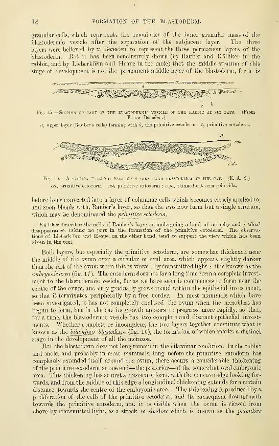
18 FORMATION OF THE BLASTODERM.
granular cells, which represents the remainder of the inner granular mass of the
blastodermic vesicle after the separation of the subjacent layer. The three
layers were believed by v. Beneden to represent the three permanent layers of the
blastoderm. But it has been conclusively shown (by Rauber and Kolliker in the
rabbit, and by Lieberkiihn and Heape in the mole) that the middle stratum of this
stage of development is not the permanent middle layer of the blastoderm, for it is
Fig. L5.
—
Section of part of the blastodermic a'^esicle op the rabbit at six days. (FromE. van Beueden.
)
a, upper layer (Raiiber's cells) forming with h, the primitive ectoderm ; c, primitive entoderm.
Fig. IG.—-A section through part of a bilajiinar blastoderm of the cat. (E. a. S.)
cct, primitive ectoderm ; ent, primitive entoderm ; z.p., thinned-out zona jjcllucida.
before long converted into a layer of columnar cells which becomes closely applied to,
and soon blends with, Rauber's layer, so that the two now form but a single stratum,
which may be denominated the jjrimiiive ectoderm.
Kolliker describes the cells of Rauber's layer as undergoing- a kind of atrophy and gradual
disappearance, taking no part in the fonnation of the isrimitive ectoderm. The observa-
tions of Lieberkiihn and Heape, on the other hand, tend to support the view -which has been
given in the text.
Both layers, but especially the primitive ectoderm, are somewhat thickened near
the middle of the ovum over a circular or oval area, which appears slightly darker
than the rest of the ovum when this is viewed by transmitted light : it is known as the
emtryonic area (fig. 17). The entoderm does not for a long time form a complete invest-
ment to the blastodermic vesicle, for as we have seen it commences to form near the
centre of the ovum, and only gradually grows round within the epithelial investment,
so that it terminates peripherally by a free border. In most mammals which have
been investigated, it has not completely enclosed the ovum when the mesoblast has
begun to form, but in the cat its growth appears to progress more rapidly, so that,
for a time, the blastodermic vesicle has two complete and distinct epithelial invest-
ments. Whether complete or incomplete, the two layers together constitute what is
known as the lUanmuir Masiods&ii (fig. 16), the formation of which marks a distinct
stage in the development of all the metazoa.
But the blastoderm does not long remain in the bilaminar condition. In the rabbit
and mole, and probably in most mammals, long before the primitive entoderm has
completely extended itself around the ovum, there occurs a considerable thickening
of the primitive ectoderm at one end—the posterior—of the somewhat oval embryonic
area. This thickening has at first a crescentic form, with the concave edge looking for-
wards, and from the middle of this edge a longitudinal thickening extends for a certain
distance towards the centre of the embryonic area. The thickening is produced by a
proliferation of the cells of the primitive ectoderm, and its consequent downgrowth
towards the primitive entoderm, and it is visible when the ovum is viewed from
above by transmitted light, as a streak or shadow which is known as the p-imitive

FOIIMATION OF THK BLASTODERM. 19
fifnak (f[»i. 1 S, 1 <)). Almost as soon as the i)riiiiitive streak has heconie fully formed, it
may be observed to be Sfored alon^- its leiiirth, except at the anterior end which is that
directed towards the centre of the embryonic area, by a narrow groove—the
/in'mifire (/rooi'e. The proliferating primitive ectoderm comes into close relationship
below this groove with the primitive entoderm, and t!ie two may be partly or
'^•S
ilit: . V
Fig. 17.
—
Embryosic area of mole immediately prior to appearance ur primitive streak andFORMED OF TWO LAYERS ONLY,
Fig. IS.— Embryonic area of the mole showing the primitive streak and groove endingPOSTERIORLY IN A CRESCENTIC THICKENING.
The area is bilaminar in front, trilaminar in the posterior half.
Fig. 19.— A SOMEWHAT LATER STAGE IX WHICH THE PRIMITIVE STREAK REACHES TWO-THIRDS OP THBLENGTH OF THE EMBRYONIC AREA, AND ENDS BEHIND IN A KNOB OR THICKENING.
Figs. 17, 18, and 19 are copied from Heape. They are magnified 49 times.
entirely blended, but the union is closest at the anterior end of the primitive streak
where a continuous column of cells unites the primitive ectoderm and entoderm,
so that the two layers are here indistinguishable.
The proliferation of the cells of the primitive streak subsequently proceeds chiefly
at the sides of the primitive gi'oove, and the cells which are produced by this proli-
feration extend themselves laterally between the ectoderm and entoderm to form a
Fig. 20, A. and B.
—
Views of the embryonic area of the rabbit showing two stages is thbEXTENSION OF THE 3IES0BLAST. (Kolliker.)
In A. the mesoblast extends on either side of the primitive streak over the posterior part of thecmhryonic area and also behind the primitive streak bevond the limits of that area.
In B. the mesoblast extends over a circular area which surrounds the embryonic area. The em-bryonic area is also ti-ilaminaiv except in the middle line in front of the primitive streak.

20 FOEMATION OF THE BLASTODERM.
third or intermediate layer. This is mainly derived, as was first pointed out by
KoUiker, from the primitive ectoderm of the groove, but since in this situation the
two primary layers become eventually more or less blended, it is probable that the
primitive entoderm cells also take part in its formation, although this part is in the
mammal evidently a subordinate one. It is further maintained by many embry-
Fig. 21.— Section across the posterior end of the embryonic area_ of a rabbit at the time
OF THE first SIGN OF A PRIMITIVE STREAK. (KolUker.)
ep, epiblast ; ax, its axial part undergoing proliferation (this is shown by the karyokinetic figures,.
k) ; me, mesoblast becoming derived from the proliferating axial epiblast ; hy, hypoblast.
Yia, 22. Longitudinal section through the middle line op part of an embryonic area (mole) in-
which the primitive streak has begun to form. (Heape.)
The blastoderm is perforated in front of the (short) primitive streak (? blastopore, hlxi) ; a few meso-
blast cells are seen anterior to the perforation ; ep, epiblast ; hy, hypoblast ;p.sk, primitive streak.
Fig. 23.—Two sections across the embryonic area of a blastoderm at the stage shown in
fig. 19. (Heape.)
A. Section across the anterior end of the primitive streak and groove.
B. Section across the posterior enlargement of the primitive streak. The epiblast and hypoblast
are seen to be united along the primitive streak, p.sk ; laterally the mesoblast, m.. the cells of which
have grown out from the uniting column of axial cells, separates the two primary layers.
p.gr, primitive groove ; ep, epiblast ; Jiy, hypoblast ; m, mesoblast.

GASTRULAlloX OF VERTEBRATES. 21
Pig. 2-t.
—
Four stages IX the development op AMPHIOXUS illustrating the FORMiTION OF TUEGASTRULA. (Hatscbek.
)
I. Spherical blastodenn ; tlie cells at the lowei- pole are larger thau the others, and filled with
granules.
II. Invagination of the lower pole producing a cupping of the vesicle.
III. Completion of the invagination ; the blastoderm is now bilaminar, and form.s a cup with
marrowed mouth, the blastopore, hi, and a double wall of epiblast, cp, and hyijoblast, hij (or primitive
ectoderm and primitive entoderm).
IV. The ovum is now elongated ; the cavity of the gastrula forms a primitive alimentary canal, the
orifice of which is the blastopore, which is directed dorsally. Extending from this along the dorsal
surface (right in the figure) a shallow groove is seen in optical section : this is the rudiment of the
nervous system.
oiogists that cells from the lateral parts of both primary layers are added to the
intermediate layer, and assist in its extension. According to the observations of
Bonnet in the sheep, there is an addition to the middle layer from the peripheral
(thickened) portion of the hypoblast ; this has been long held to be the case with
the blastoderm of the bird, and the cells thns derived (parablastic) have l)een
considered to have the special function of forming the connective tissues and blood.
AYhether, however, this is actually so, must be i-egarded as at present undecided.
However produced, the appearance of a middle layer causes the originally bilaminar
blastoderm to be trilaminar, and its three layers have received the names of ectoderm,
mesoderm, and entoderm, or epiblast, mesoblast, and hy}ioljlast.
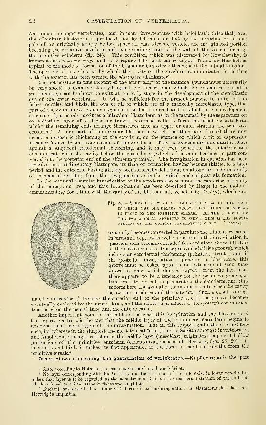
The gastrula condition of the vertebrate ovum.—It Avill be observed that in the
mammal the two primary layers of the blastolerm. at least their principal pai-t. are formed bya separation into two strata of the cells of the inner granular mass which occupies the
interior of the ovum after segmentation. The bilaminar condition may therefore be .said to
result from a process of deiamination in an originally simjile mass or stratum. But in
22 GASTEULATIO^s^ OF VERTEBRATES.
Amphioxus among'st vertebrates,' and in many inverteljrates with holoblastlc (alecithal) ova,
the bilaminar blastoderm is produced, not by delamination, but by the invagination of one
pole of an orig-inally simple hollow spherical blastodermic vesicle, the invaginated portion
becoming' the primitive entoderm and the remaining part of the wall of the vesicle forming
the primitive ectoderm (fig. 24), This condition, which was discovered by Kowalewsky, is
known as the gastrula Htarje, and it is regarded by most embiyologists, following Haeckel, as
typical of the mode of formation of the bilaminar blastoderm throughout the animal kingdom.
The aperture of invagination by which the cavity of the entoderm communicates for a time
with the exterior has been termed the Mastojxire (Lankester).
It is not possible in this account of the embryology of the mammal (which must necessarily
be very short) to examine at any length the evidence upon which the opinion rests that a
gastrula stage can be shown to exist at an early stage in the development of the meroblastic
ova of the lower vertebrata. It will be sufficient for the present purpose to state that in
fishes, reptiles, and birds, the ova of all of which are of a markedly meroblastic type, that
part of the ovum in which alone segmentation has occurred, and in which active development
subsequently proceeds, produces a bilaminar blastoderm as in the mammal by the separation off
as a distinct layer of a lower or inner stratum of cells to form the primitive entoderm,
whilst the remaining cells arrange themselves into an upper or outer stratum, the primitive
ectoderm. 2 At one part of the circular blastoderm which has thus been formed there nowoccurs a crescentic thickening of the ectoderm, on the surface of which a pit or depression
becomes formed by an invagination of the ectoderm. This pit extends inwards until it abuts
against a subjacent entodermal thickening, and it may even penetrate the entoderm andcommunicate with the cavity below the blastoderm (which afterwards becomes in part con-
verted into the posterior end of the alimentary canal). The invagiuation in question has been
regarded as a rudimentary blastopore, its time of formation having become shifted to a later
period, and the entoderm having already been formed by delamination altogether independently
of, in place of resulting from, the invagination, as in the typical mode of gastrula formation.
In the mammal a similar invagination of the ectoderm also occrus at the posterior extremity
of the embryonic area, and this invagination has been described by Heape in the mole as
communicating for a time with the cavity of the blastodermic vesicle (fig. 22, JZj;), which sub-
— - -.^ Fig. 25.— Surface view op ajt embryonic area of the mole"^^ IN WHICH THE MEDULLARY GROOVE HAS BE&UN TO APPEAR
IN FRONT OF THE PRIMITIVE STREAK. At THE JUNCTION OF
THE TWO A SMALL APERTURE IS SEEN : THIS IS THE DORSAL
/ OPENING OF THE OBLIQUE NEURENTERIC CANAL. (Heape.)
i \sequently becomes converted in part into the alimentary canal.
^-\ In birds and reptiles as well as mammals the invagination in
f,
"
,' question soon becomes extended forward along the middle line
'( of the blastoderm as a linear groove (primitive groove), which
t indents an ectodermal thickening (primitive streak), and if
\ the posterior invagination represents a blastopore, this
V groove must be looked upon as an extension of such blas-
topore, a view which derives support from the fact that
^' there appears to be a tendency for the primitive groove, at
\ ^ least its anterior end, to penetrate to the entoderm, and thus
to form here also a canal of communication between the cavity
below the entoderm and the exterior. Such a canal is desig-
nated " neurenteric," because the anterior end of the primitive streak and groove becomes
eventually enclosed by the neural tube, and the canal then effects a (temporary) communica-
tion between the neural tube and the enteric canal.
Another important point of resemblance between this invagination and the blastopore of
the typical gastrula is the fact that the middle layer of the trilaminar blastoderm begins to
develope from the margins of the invagination. But in this respect again there is a differ-
ence, for whereas in the simplest and most typical forms, such as Sagitta amongst invertebrates,
and Amphioxus amongst vertebrates, the middle layer (mesoblast) originates as a pair of hollow
protrusions of the primitive entoderm (coelom-invaginations of Hertwig, figs. 28, 29) ;in
mammals and birds it makes its first appearance in the form of solid outgrowths from the
primitive streak.-*
Other views concerning' the gastrulation of vertebrates.—Kupffer regards the part
' Also, according to Hofmann, to some extent in elasmobrancli fislies.
2 No layer corresponding with Rauber's layer of the mammal is known to exist in lower vertebrates,
unless that layer is to be regarded as the homologue of the external (corneous) stratum of the epiblast,
which is found at a later stage in fishes and amphibia.2 Riickert has described an imperfect form of ccelom-invagination in elasmobranch fishes, and
Hertwig in amphibia.

HISTOKY OF BLASTODERM. iic
•which has hten above alluded to as iiivaginated ectoderm (primitive groove) as the homologuo
of part of the entoderm of more typical forms. If this view Ije correct, many of tiiedilKculties
in the way of re^ardintr tlie aperture of the inva^rination as the blastopore, and in explaining,'
the differences in the mode of origin of the mesoblast are removed ; but, on the other hand,
other ditliculties are introduced, and the subject is left by no means clear.
Another view, which wjxs formerly extensively held, regards the blastopore of the nierolilastic
vertebrate ovum as bounded by the thickened edge of the bilaminar blastoderm (Haeckel).
According- to this view, the cavity of the gastrula is entirely filled up by a mass of
unsi'gmcnted or but partially segmented yolk, which also projects for a considerable distance
through the blastopore, forming in fact the great mass of the ovum. The primitive groove
is regarded as a linear prolongation of this thickened edge of the blastoderm towards the
centre of the blastoderm (Balfour), so that the embryo, which developes in front of the
primitive str(.ak, thus comes to have a i)seudo-central position in the blastoderm instead
of developing altogether from its margin as in the lower vertebrata and in inverte-
brates. In conformity with this idea, it may be noted that at the thickened rim of the
blastoderm of these meroblastic ova, the two primary Layers are continuous with one another
as in the primitive streak. In elasmobranchs an intermediate condition is observed, viz.. a
short groove, the margins of which are freely continuous with the margin of the blastoderm.
If. as His and others have described (ji-ide Infra), the mesoblast is in part (vascular
and connective tissue part) derived from the thickened rim of the blastoderm, this would
fm-nish another point of resemblance between the primitive streak and that margin.
Ed. V. Beneden has promulgated an entirely different opinion as to the mammalian blasto-
pore from those above described. He regards the condition of the ovum, after the completion
of segmentation and before the formation of a blastodermic vesicle, as representing the
gastrula stage, and looks upon the point where the granular inner mass of cells comes to the
surface between the clear cells which form the outer investment as the blastopore (fig. 14, «).
In confoi-mity with this view he considers the layer of clear cells to represent the whole of the
primitive ectoderm, and the granular inner mass the primitive entoderm. But since all the
more recent observations upon early mammalian ova agree in affirming the formation of
the three blastodermic layers from the granular inner mass, and that Eauber's laj-er either
takes no pai-t at all, or only a subordinate part in the formation of the ectoderm of the
embryonic area, v. Beneden's view, in spite of the superficial resemblance of the ovum at
this stage to certain gastrula forms, has not met with general acceptance from embryologists.
Inversion of the blastodermic layers in some mammals.— In the guinea-pig
(Bischoff), rat and mouse (Fraser, Selenka), and in some other rodents, an inversion of the
usual position of the blastodermic layers is found to occur, the epiblast being innermost,
the hypoblast outermost. The foundation of this inversion is laid early by a process of
invagination and formation of a central cavity in the mass of entomeres, so that when the
blastoderm is differentiated, the innermost cells which are next the (secondary) cavity thus
formed become the epiblast, and the outermost the hypoblast, the mesoblast subsequently
forming between the two by proliferation of epiblast at the primitive groove, as in other
mammals. (For details as to this process of invagination, the student is referred to the papers
by Selenka.)
Historical.—The existence of several laminse in the germinal substance of the eg^ was
first suggested by C. F. Wolff in his celebrated work Theoria Generationis. published in 1759,
and in his later Memoir On the DevdojJinent of the Intestine, &:st published in Nov. Comment.
Acad. Petropol. in 1767 and republished in German by J. F. Meckel in 1812. It is, however, to
the researches of Pander, conducted under the dii-ection of Dollinger of "Wiii-zburg, and pub-
lished in 1817, and those of v. Baer (182G-lSo7), tliat we owe the first consistent attempt to
connect the development of the several organs and systems of the embryo with the different
constituent parts or layers of the blastodeim. Pander recognised a trilaminar structure of the
blastoderm and distinguished the three layers composing it, in their order from above down-
wards, or from without inwards in the eg'^, as the serous, vascular, and mucous layers.
In lS.oO-."j-l a further important advance was made in the knowledge of the constitution of
the blastodennic layers, by the discovery by Remak that the greater part of the middle layer
soon after its formation comes to be divided into two laminaj, separated by a space which
coiTesponds to the perivisceral cavity {calom')—a fact which had been partially stated by von
Baer. So marked a division of the middle layer and distinction of the parts which are
afterwards developed from its two laminae, has seemed sufficient to some authors to warrant
the recognition of four distinct layers in the blastoderm ; but it will be found on the whole
more convenient to consider the fundamental layers as only three, to which, following the
nomenclature of Foster and Balfour, the designations of epiblast, mesoblast, and hypoblast are
applied, terms which are synonymous with those of ectoderm, mesoderm, and entoderm,
employed by many authors.
The terms ectoderm and entoderm were first applied to the two fundamental layers, shown
by Huxley in 1849 to constitute the whole body of coelenterates. and which were correctly
regarded by him as homologous with the two layers of the bilaminar blastoderm, to which w"»

24 CHARACTERS OF BLASTODERMIC LAYERS.
have apiDlied the terms primitive ectoderm and primitive entoderm. Since the middle layer
is developed from one or both of these primitive layers their permanent representatives are
morphologically different, having- lost the elements -w^hich go to form the middle layer, and it
is therefore convenient to accentuate this distinction by the adoption of different terms to
represent the permanent layers.
The generalisation that the formation of a bilaminar blastoderm is typically produced bythe invagination of a hoUov^r spherical unilaminar blastodermic vesicle is due to Haeckel, andwas based largely upon the important researches of Kowalevsky, especially those on Sagitta
and Ampjhioxus. The process of delamination which in some animals produces the twoprimary layers was originally regarded by Ray Lankester as the typical mode of formation,
but is now generally admitted to be a secondary modification. Finally, it has been shoAATi
(Balfour, Lankester, R. and 0. Hertwig), as is set forth below, that the coelom or body cavity
is typicatty developed, not by a process of splitting of the mesoblast (although in someanimals this may occur as a secondary modification), but as hollow protrusions from the
primitive alimentary cavity, the cells which bound these protrusions forming the mesoblast.
Thus from an originally single blastodermic layer by successive processes of invagiuation
or folding, the three permanent laminae are ultimately produced.
Such folds may be regarded as formed mechanically by local hypertrophic multiplication
of the cells of the laminEe, an increased surface being thus found for the increased number of
cells. In analogous manner the folds which accompany the formation and separation of the
body and the development of the several organs, e.g., the nervous system, alimentary canal,
amnion, may also be regarded as resulting mechanically from cell-multiplication. This
mechanical theory of development was first enunciated by Pander, and has of late years beenapplied extensively by several embryologists, notably by His QEntwickl. d. Iluhnchens, 1868,
and Un-wre Korperform. 1874), and Rauber.
Characters of the blastodermic layers.—The three layers of the blastodenn
show from the first distinctive characters (fig. 2G). The outer layer, or epiblast, is
epithelial in nature and consists of somewhat irregularly columnar cells closely set
Fig. 26.
—
Transverse section through the front end of the prijiitive streak ani>
BLASTODERM OP THE CHICK. (From Balfour.
)
fr, primitive groove ; m, mesoblast ; cp, epiblast ; liy, hypoblast.
side by side, forming a single stratum for the most part, except near the middle
line, and becoming thinner and flatter towards the margins of the embryonic area.
The inner layer or hypoblast is also epithelial, but the cells are at first all
flattened, and appear therefore quite thin and linear in sections of the blastoderm.
At a later stage, the hypoblast cells become markedly columnar and enlarged, so
that they considerably exceed the epiblast cells in size.
The middle layer, or mesoblast,^ which diff"ers, as we have seen, in its mode of
arigin, being formed secondarily from one or both of the primary layers, also diflTers
Irom them entirely in its appearance and structure. Instead of consisting of cells
closely joined together into a continuous membrane after the manner of an
epithelium, the mesoblast is at first composed of cells which are not thus closely
arranged, but have, on the contrary, a considerable amount of intercellular fluid
between them. They are most irregular m shape, and are often branched and
united with one another, so that much of the mesoblast early resembles an
embryonic connective tissue.
i The mesoblast was by von Baer described as being formed of two layers, one derived by splitting
from each of the two primary layers, and although this mode of origin can no longer be maintamed,
the two strata of mesoblast separated bv the pleuroperitoneal cavity or ccelom are still by some
embryologists considered to be distinct layers of the blastoderm, which is thus described as quadri-
laminar (see p. 26).

I'AKAliLAST THEOKY OF HIS. 25
S
b^
Before proceedinir tu desfiibe the commencinjr tk-vclojiiuent of the embryo it will
be instructive to enmnerate the jiarts which are forniL*d res])L'ctively from tlie three
blastodermic layers. The following; is the relation giveu in tabular form :
—
' The whole of the nervous system, including not only the central oryans (brain andspinal cord), but also the peripheral nerves and sympatlictic.
Thf epithelial structures of the organs of special sense.
The epidermis and its appendajres. includin^r the hair and nails.
The epithelium of all the ;,'lands openin;r upon the surface of the skin, includingthe mammary- irlands. the sweat ;rlands. and the sebaceous glands.
I
The muscular fibres of the sweat irlands.
I The epithelium of the mouth (except that covering,' the ton^'ue and the adjacentI posterior part of the floor of the mouth, which is derived from hypoblast;, and that of
I
the trlands openin<r into it. The hypophysis cerebri. The enamel of the teeth.
The epithelium of the nasal passajres, of the adjacent upp^r part of the pharynx,and of all the cavities and glands opening into the nasal passages.^ The epithelium of the anus and immediately adjacent pai-t of the rectum.
[ The epithelium of the vagina and of the urethra.
The urinary and generative organs (except the epithelium of the urinary bladderand urethra).
All the voluntary and involuntaiy muscles of the body (except the muscular fibres
of the sweat glands).
The whole of the vascular and lymphatic system, including the serous membranesand spleen.
The skeleton and all the connective tissue structures of the body.
The epithelium of the alimentaiy canal from the back of the mouth almost to the
anu<. and that of all the glands which open into this part of the alimentary tube.
The epithelium of the Eustachian tube and tympanum.The epithelium of the bronchial tubes and air sacs of the lungs.
The epithelium lining the vesicles of the thjToid body.
The epithelial nests of the thymus.The epithelium of the urinary bladder.
PAHAELAST THEORY OF HIS. MESENCHYME THEORY OF HERTWIG. ^'^-^J
The observations of His upon the development of the blood and connective tissuesin the bird led him to regard these tissues as originating, not from the mesoblast whichin the chick ^tows out from the sides of the primitive groove, but from cells which,
Fig. 27.
—
Vertical section THKoroH thl blastoderm of a hex's egg taken near thePERiPHEKT. (Strieker.)
E, epiUast : JI. hypoblast, passing at the periphery' into an undifferentiated mass of yolk, A,containing large cells filled with yolk granules ; M (towards the centre of the blastoderm), mesoblast
;M ('nearer the periphery), granular cells, apparently derived from A, and lying between the epiblast
and hypoblast.
originating either in the yolk or in the thickened rim of the spreading blastoderm, wander in
centripetally ';etween the primaiy layers and fill up all the interstices of the centrifugaily-
growing true mesoblast. These in-wandering cells being derived, not lie the other cells of
the embryonic area from the more active primarily differentiated central parts of the blasto-
derm, but from the peripheral non-embryonic portion, were collectively named by Hisjiara blast, and the tissues (blood and blood-vessels, and all the connective tissues) supposed to
be fonned from them were termed jMii-aJAa.^tic (all the other tissues of the embryo beingtermed, in contra-distinction. archlhlaiitic').
Hiss theory was enunciated as long ago as 1S68. although he afterwards introducedinto it certain modifications. For a considerable time it met with little acceptance, but of

26 MESENCHYME THEORY OF HERTWIG.
late years it has olDtained, in a somewhat modified fonn, the adherence of many embiyologists,
and especially of R. and 0. Hertwig, Kupffer, Kollmann, and Waldeyer. R. and 0. Hertwig-
have given the name of mesenchyme to the embryonic tissue which gives rise to the connective
tissues and vascular endothelium, while retaining the designation of middle germinal layer
or mesoderm for the rest of the mesoblast ; but the derivation of the mesenchyme is
admitted by 0. Hertwig to be directly from mesoblast, from which it differs (1) in its
structure, consisting of loosely arranged wandering cells, as distinguished from the
epithelium-like lamellse, of which according to their description the rest of the mesoderm is
composed; (2) in its derivation, arising as separate cells from the entoderm instead of in the
form of a coherent layer ; and (3) in its further development and destination, giving origin
to the connective tissues and blood-vessels, and jierhaps to the plain muscular tissue, whereasthe mesoderm proper gives origin to the skeletal muscles and to the epithelium of the serous
cavities, and of the genital and urinary organs. They describe the true mesoderm as
consisting of two epithelial lamellcE, which form distinct layers of the blastoderm, so that
according to this view the complete blastoderm Avould consist otfonr layers (epiblast, outer
or somatic mesoblast, inner or splanchnic mesoblast and hypoblast) besides the mesenchyme :
to which must be added a median strand of cells set aside for the formation of the notochord,
and derived from the hypoblast.
It appears evident from the researches of Kowalevsky in Sagitta and Amphioxus that whatis to be regarded as the typical orig'in of the mesoblast in Metazoa^ takes the form of a pair of
diverticula from the primitive archenteric cavity (fig. 28, la ; fig. 29, I), which hollow
diverticula become pinched off from the remainder of that cavity, and, their cavities becoming
Fig. 28.
—
Formation of mesoblastic somites ix amphioxus, shows in longitudinal optical
SECTION. (Hatschek.
)
la. dorsal view of an embryo in which the mesoblast is beginning to form as two longitudinal folds
of the hypoblast which are becoming subdivided from before back by cou&trictions into separate
somites. I., the same viewed in profile, showing the anterior three somites of one side, with their
cavities in free communication with the enteric cavity. The neural canal, n.c, is continued
posteriorly by a neurenteric canal into the enteric cavity; ej), epiblast; hij, hypoblast. II., dorsal
view of a more advanced embryo. The somites are more numerous and are completely separate. In
all but the most anterior pair the communication with the enteric cavity is still seen. III., dorsal
view at a still later stage. The somite cavities are now completely closed. The cellular rod, ch, shown
running along the middle of the embryo is the notochord.
compressed laterally, are converted into the coelom or body-cavity (serous cavities of
vertebrata), the two walls of this cavity on either side forming respectively the inner and
outer mesodermic layers of R. and 0. Hertwig. In Sagitta. the diverticula occur in the
neighbourhood of the blastopore, which is also the typical seat of origin of the mesoblast, but
in Amphioxus they are formed by longitudinal folds of the wall of the archenteric cavity,
which grow from before backwards, and become separated up into segments in theii- progress.
In Elasmobranchs the myotomes or protovertebraj are at an early period hollow, and the
1 Except the Coelenterata which have only the two primary layers.
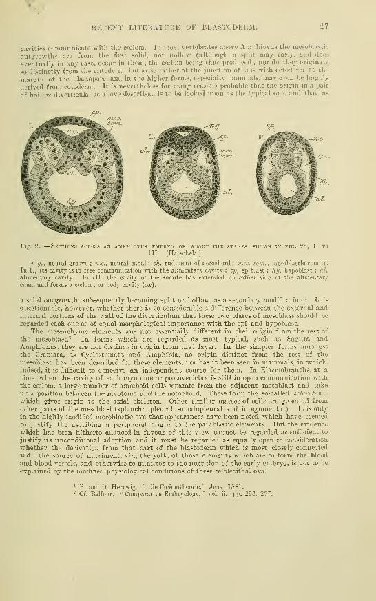
RECENT LITERATURE OF BLASTODEIIM. 27
cavities communicate witli the ca'loni. In most vertebrates above Ampliioxus the mesoblastic
outt,'-ro\vths are from the first solid, not noUow (althougli a split may early, and does
eventually in any case, occur in them, the ccelom being thu.s i)roduced;, nor do they originate
so distinctly from tiie entoderm, but arise rather at the junction of this with ecto'lerm at the
margin of the blastopore, and in the higher forms, especially mammals, may even be largely
derived from ectoderm. It is nevertheless for many reasons probable that the origin in a pair
of hollow diverticula, as above described, is to be looked upon as the typical one, and that as
Fig. 29.
—
Skctions across ax amphiuxvs embryo of about the stages shown in fig. 28, I. to
III. (Hatscbek.)
71. y., neural groove ; n.c, neural canal ; ch, rudiment of notochord; mcs. som., mesoblastic somite.
In I., its cavity is in free communication w-ith the alimentary cavity ; C2), epiblast ; hi/, hypoblast ; al,
alimentary cavity. In III. the cavity of the somite has extended on either side of tlie alimentary
canal and forms a ccelom, or body cavity (cce).
a soUd outgrowth, subsequently becoming split or hollow, as a secondary modification.' It is
questionable, however, whether there is so considerable a difference between the external andinternal portions of the wall of the diverticulum that these two plates of mesoblast shoidd be
regarded each one as of equal morphological importance with the epi- and hypoblast.
The mesenchyme elements are not essentially different in their origin from the rest of
the mesoblast.- In forms which are regarded as most typical, such as Sagitta andAmphiox;us, they are not distinct in origin from that layer. In the simpler forms amongstthe Craniata, as Cyclostomata and Amphibia, no origin distinct from the rest of the
mesoblast has been described for these elements, nor has it been seen in mammals, in which,
indeed, it is difficult to conceive an independent source for them. In Elasmobranchs, at a
time when the cavity of each myotome or protovertebra is still in open communication withthe coelom. a large number of amceboid cells separate from the adjacent mesoblast and take
up a position between the myotome and the notochord. These form the so-called sclerotome.
which gives origin to the axial skeleton. Other similar masses of cells are given off fromother parts of the mesoblast (splanchnopleural. somatopleural and integumental). It is only
in the highly modified meroblastic ova that appeai-ances have been noted which have seemedto justify the ascribing a peripheral origin to the parablastic elements. But the evidence
which has been hitherto adduced in favour of this view cannot be regarded, as sufficient to
justify its unconditional adoption, and it must be regardei as equally open to consideration
whether the derivation from that part of the blastoderm which is most closely connected
with the source of nutriment, viz., the yolk, of those elements which are to form the blood
and blood-vessels, and otherwise to minister to the nutrition of the early embryo, is not to be
explained by the modified physiological conditions of these telolecithal ova.
' R. and 0. Hertwig, "Die Ccelomtheorie," Jena, ISSl.- Cf. Balfour, "Comparative Embryology," vol. ii., pp. 296, 297.

28 EECENT LITEPtATUEE OF BLASTODERM.
RECENT LITERATURE.
1
Balfour, F. MC., On the structure and homologies of the germinal layers of the emh-ijo. Quart.
Journ. Microsc. Sc, vol. xx., 1880.
Balfour, F. M. and F. Deigliton, A reneioed study of the germinal layers of the chicle
Quart. Journ. Microsc. Scienc, vol. xxii.. 1882.
Beneden, Ed. van, Becherches sur Vembryologie des mammiferes. La formation des feuillets
chez le lapin. Archiv. de biologie, t. i. 1880 ; Sur I'dvolution de la ligne primitive, la formationde la notocorde et du caned cordal chez les mammiferes {lapin et murin). Bulletin de racademieroyale de Belgique. Ann. v., s^r. iii., t. xii., 1886.
Beneden, Ed. van und Ch. Julin, Oiservations sur la maturation, la fecondation et la
segmentation de I'cenf chez les cheiropiteres. Arch, de biol. , t. i., 1880.
Bonnet, K., Ueher den Primitivstreifen und die Chorda der Wiederkauer. Sitzungsber. d.
Gesellscliaft f . Morphologie u. Physiologic zu Mtinchen, 18S6 ; Beitrdgezur Emhryologieder Wicderkduer.
Arch, fur Anat., 1884, 1889.
Boveri, T., Ueber Differenzirung der Zellkerne wdhrend der Furchung des Eies von Ascaria
fuegalocephala. Anat. Anzeig., 1887.
Blitsclili, O., Beitrdge zur Gastndatheorie. Morpholog. Jahrbuch, Bd. ix., H. 3, 1884.
Caldwell, "W. H., The embryology vf Monotremata and Marsupialia. Philosophical Trans.,
vol. clxxviii., 1888.
Durham, H., Note on the presence of a neurenteric canal in liana. Quarterly Journal of
Microscopical Science, N. Ser., vol. 26, 1886.
Duval, M., Be la formation du ilastoderme dans Vceaf d'oiseau. Annales des sciences
naturelles, 6 ser. t. xviii., No. 1—3, 1884.
Fleiscliniann, A., Zur Entwickclungsgeschichte der Raubthiere. Biolog. Central bl., Bd. vii.,
1887.
Gasser, Der Parablast u. der Keimwall der Vogelkeimscheibe. Sitzungsber. d. naturw. Gesellsch.
EU ^[arburg, 1883.
G-erlach, L., Ueber die entoclermale Entstehung der Chorda. Biol. Centralbl., 1881.
Giacomini, C, Sul canale neurenterico et sul canale anale nclle vesicole blastodermiche di
coniglio. Giorn. della r. accademia di medic, di Torino, No. 4, 5, 1888.
Haddon, A,, Note on the blastodermic vesicle of mammals. Proceedings of the Royal Dublin
Society, N. Ser., vol. iv., 1385.
Haeckel, Bie Gastrceatheorie. Jena Zeitschrift, Bd. viii. ; Nachtrdge zur Gastrceathcorie.
Ibid., Bd. xi. ; Ursprung u. Entivickl. d. thierischen Geiueben. Ibid., Bd. xi.
Hatschek, B., Studien iiber Entwicklung des Amphioxus. Arbeiten a. d. zool. Instit. zu "Wien,
Bd, iv. , 1881 ; Ueber die Entioicklung des Amphioxus. Biolog. Centralbl., Bd. vi., 1887.
Heape, W., The development of the mole (Talpa europcea). The formation of the germinal
layers, and early development of the medullary groove and notochord. Quart. Journ. of IVticrosc.
Sc, N. S., xci., 1883. Also in Studies from the Morphological Laboratory in the University of
Cambridge, vol. iii.
Hensen, Ueber die Ableitung der Vmkehr der Keimbldtter des Meersehiveinch^ns. Verhand-
lungen des physiologisclien Vereins in Kiel, 1881.
IIert\s*g-, O., Bie Bntwicklung des mittleren Keimblattes der Wirbelthiere. Jena Zeitschrift fiir
Naturw., Bd. xvi., 1882.
Hertwig, O. und E., Die Cblomtheorie. Versuch einer Erkldrung des mittleren Keimblattes.
Jena, 1881 ; Studien zur Bldttertheorie. Jena, 1883.
His, "W., Der Keimwall des Hiihnereies u. d. Entstehung der parablastischen Zdlen. Zeitsch. f.
Anat. u. Physiol., Anat. Abth., 1876 ; Die Lehre vom Bindesubstanzkeim (Parablast), Ruckblick nebst
critischer Besprechung einiger neuerer entwickiungsgeschichtlicher Arbeiten. Archiv fiir Anat. u.
Pijy.'^inl., Anatom. Ab'tlieil., 1882.
Hoffmann, C. K., Die Bildung des Mesoderms, die Anlage der Chorda dorsalis und die
Entivickelung des Canalis neurentericus bei Vogelembryonen. Amsterdam, 1883.
Hubrecht, A. A. "W., Bie erste Anlage des IJypoblastcs bei den Sdugethieren. Eine Erwiderung
an Herrn Prof. Bd. van Beneden. Anatomischer Anzeiger., iii. Jahrg., No. 30, 1888.
Johnson, Alice, On the fate of the blastopore and the presence of a primitive streak in the
NeiDt [Triton cristatus). Quart. Journ. of Microsc. Science, N. S., No. xcvi., 1884.
Keibel, F., Van Beneden's Blastoporus und die Rauher'sche Deckschicht. Anatomisch. Anz.,
1887 ; Die Enticickclungsvorgdnge am hinteren Ende des Meerschiceinchencmbryos. Arch. f. Anat.
u. Physiol., Anat. Abth., H. 5 u. 6, 1888.
KoUer, C, Untersuchungen iiber die Bldtterbildung im Hilhnerkeim. Arch. f. mikr. Anat.,
1881 ; Beitrdr/e zur Kenntniss des Hiihnerheims im Beginne der Bebriitung. Wiener Sitzungsber.,
Bd. Ixxx., 1881.
Kolliker, Die Entioicklung der Keimbldtter des Kaninchens. Wurzburg Festschrift. Leipzig,
1882; Die embryonalen Keimbldtter u. d. Gewebe. Zeitschr. f. wiss. Zool. xl., 1884; Ueber die
Nichtexistenz eines embryonalen Bindegewebskeims [Parablasts). Sitzungsber. d. phys. medic. Ges.
zu Wurzburg, 1884.
1 For an extended bibliography of the maturation, fertilization and segmentation of the ovum, see
Sobotta, " Die Furchung des Wirbelthiereies " in Ergebnisse der Anatomie u. Entwickelungsgeschichte
von Merkel u. Bonnet, 1896.

RECENT LITEKATUltE OF BLASTODKll.M. 29
Kollmann, J., Ikr Mtsuhhttt und die Entwirkelung der Oewehe bei WirbeUhieren. Biolo;^.
Ceiitnilbhitt, UJ. iii., 1SS4; Jkr Unndundst u. d. Uriprutuj d. Slutz»ub»tanz. Arch. f. Aii.it. ii.
rhysiol.. Aiiat. Abtli. 1884.
Kowalevsky, J-Jntirickhtnf/sricsc/iir/itr dir f^uijillu. St. rettTMburg Memoir.s, xvi , 1S71 ;
J:'ntwirk-linii//!;iiscli. d. Amji/iio.fiu, dr. Arcli. f. inikr. Aiiat., IM. .xiii., 1877 ; lebcr die erstfit
Eidwickiluwi.ii'rocissf dir Knuchcnfisrhe. Zeitschr. f. wisseiiscbiift. Zooloxie, H<1. xliii , H. .'i, 188G.Kupflfer. C, l>iis Ei von Arn'oihi arrali.i und die verincintliilii- Umkihr der Kehiihtiitlir an
demsclbtii. .MiiiK-hener Sitzung.sbericlite, II. 5, 1882 ; Die Gantrulalion an den menblnstischinEiern dtr U'irbdthicrc xind die Bci'teutun;/ des Priinitirstni/.^. Arcliiv f. Anat. utid Physiol., Aunt.Abtheil., 18S2 ; I'e. d. Can<dig neurentiricus der Wirbclthicre. Sitzungsberichte der (ie.sellscli. f.
Morpliol. 11. Pliysiologie zu Jltincheii, 1887.
Lankester, On (he prim it ire cclldai/era of the embri/o, dr. Annals and Mag. of Nat. Hist., xi.,
18":!:
Xii'i.t "II I inl>riii>l'"/ij and r/aii.^itication. Quarterly Journ. of Miir. Science, xvii., 1877.Mitsukuri, K., and Ishikawa, C. On the formation of the f/erminal lai/crs in Chelonia.
Journal ot tlie College of Science, Ini|ierial University, Japan, vol. i. 1888.
Morg-an, Sotes on thr fate of the amphibian blastopore, John Hopkins University Circulars, 1880.Perenyi, 1., Die Enticickl. der KeimbUitter u. d. Chorda in neuer Beleuchtuny, Anat. An'zeiyer, 4.
Piatt, Studie.f on the 2)rimitire axial segmentation of the chick, Bulletin of tiie Zool. Museum atHarvard Culk-e, 1889.
Sabl. C, fiber die BUdung des Mesoderms. Anatomis:^hcr Anzeiger, iii. Juhrg., No. 23—25, 1888.Bauber, Ucber d. Ursprung des Blutes u. der Bindesubatanzen. Sitzuugsb. d. Naturf. Gesellsch.
ru Leipzig, 1877 ; Die Entwickelung der Gewebe des Sdugethierkorpers und die histologischen Systeme.Ber. der Naturf. Ges. zu Leipzig, 1883.
Bavn, E., Uebcr die mesodermfreie Stelle in der Keimscheibe des Iliihneremhryo. Archiv f.
Anatomic u. Physiologic, Anatom. Abth., 1886.Repiachoff, W., Bema-kungen iiber die Keim,Udtter der Wirbelthiere. Zool. Anzeiger, vi., 1883.Romiti, Gr., Sur Vorigine da misoderme et ses rapports avec le vitellus. Arch. ital. de biologie,
t. ii., 1SS2.
Roux, W., Beitrdge zur Entioickelungsmechanik des Embryo. Ueber die kUnstliche £ferror-bringung halbcr Embryonen durch eine der beiden ersten Purchungslcugeln, d-c. Virchow's Archiv,Bd. cxiv., 1888.
Riickert, Ueber die Gastrulaiion der Selachier. Biologisches Centralblatt, Bd. vi.
Ryder, The inversion of the blastodermic layers in Hesperornys. American Naturalist,vol. xxi.
Schanz, F., Das Schicksal des Blastoporus bei den Amphibien. Jenaische Zeitschr. f. Natur-wissensch., Bd. xiv., 1887.
Selenka, Emil, Keimhldtter und Primitivorgane der Maus. Wiesbaden, 1883 ; Die Bldtterum-kehrung im Ei der Xagethiere. Wiesbaden, 1884 ; Ueber die Gastrulation der Knochenfische und'Arnnioten. Biolog. Centralbl., 1887.
Shipley, A. E. , On the formation of the mesoblast, and the persistence of the blastopore in the
lamprey. Proceedings of the Royal Society, 1886 ; On some points in the Development of Petromyzonfluviatilis. Quarterly Journal of Micros. Science, No. 107, 1887.
Solger, Studien z. Entwicklungsgesch. des Cceloms u. des Ccelomepithels der Amp>hibien. Moi-ph.Jahrb., x.
Spee, P., Beitrag zur Entwirkelungsgeschichte der fruheren Sto.dien des Meerschweinchens bis zurVollendung dei- Keimblase. Arch, fiir Anat. u. Physiol., Anat. Abth., Heft. i. u. ii., 1883.
Spencer, On the fate of the blastopore in Rana temporaria. Zool. Anzeiger, 1885.Uskow, N. , Die Bhitgefdsskeime u. deren Entwickelung bei einem HUhnerembryo, M^ia. de
I'acad. de St. Petersb., xxxv., 1888.
Waldeyer, W., Arckiblast und, Parablast. Arch. f. mikr. Anat. 1883 ; Die neueren Forschungenim gebiet der Keimblaitlehre. Berliner klinische Wochenschrift, No. 17, 1885.
Wolff, W., Die beiden Keimbldtter und der Mittelkeim. Archiv f. mikroscopische Anatomic
.
Bd. xiviii., H. 4, 1886.
Zumstein, J. J., Ueber das mesoderm der Vogelkeimscheibe, Dissert., Bern, 1S87.

so EARLY CHANGES IN THE BLASTODERM.
EARLY CHANGES IW THE BLASTODERM, RESULTING INTHE FORMATION OF THE EMBRYO.
FIRST APPEARANCE OF THE EMBRYO; FORMATION OF THE NEURAL GROOVEAND MBSOBLASTIC SOMITES.
Henral canal.—The blastoderm in mammals, as we have seen, eventually
completely encloses the cavity of the ovum, and even in the large telolecithal ovumof the bird, it gradually extends so as to cover a large part of the yolk. But only
a small portion of this membrane takes part in the formation of the body of the
•embryo, that portion namely which lies immediately in front of the primitive groove,
and it is here that what may be regarded as the first trace of the embryo makes its
appearance—very soon after the outgrowth of mesoblast from the sides of that
Fig. 30.
—
Ejibrtonic area, with outline of part of the vascular, area, FROir A rabbit's
OVUM OF SEVEN DAYS. "f.(From Kolliker.)
ag, embr3'omc area;^r, primitive streak and groove ; rf, medullary groove.
groove—in the form of a shallow furrow (fig. 30, rf), wide behind where it
embraces the anterior end of the primitive streak, and at first narrow in front, and
.bounded on either side and anteriorly by a fold of epiblast ; which folds, in fact,
Fig. 31.
—
Section across the anterior part of the medullary groove op an early ejibryo
OF THE guinea PIG. (E. A. S.)
c^5, folds of epiblast rising tip on either side of the middle line, and thus bounding the medullary•groove ; mff, middle of medullary groove ; hy, hypoblast, -which is in contact with the medullaryepiblast at the middle of the groove, but is elsewhere separated from it by mesoblast, m, which hasburrowed forwards between the two primary layers. A cleft is seen in the mesoblast on either side
;
this is the commencement of the anterior part of the ccelom.
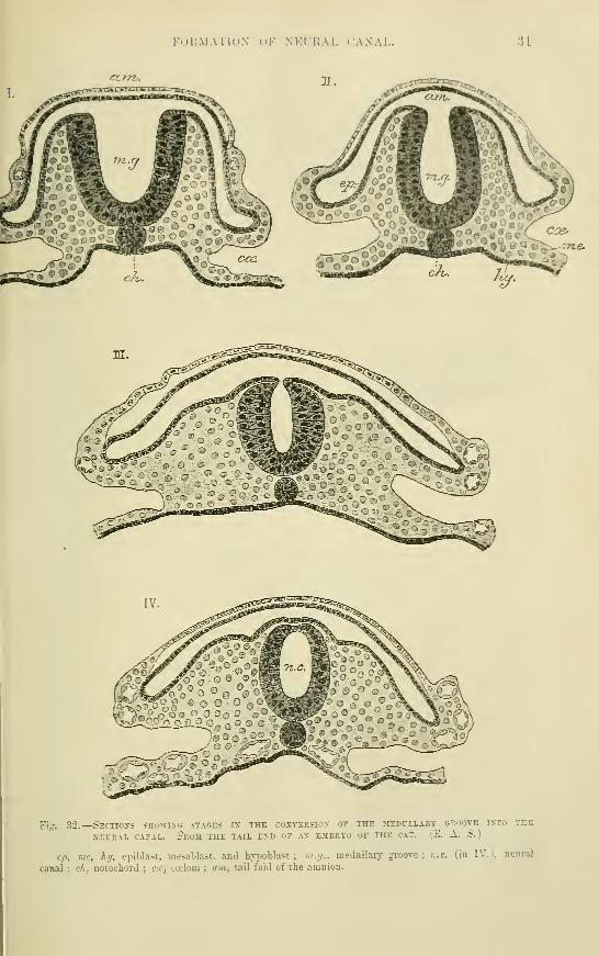
FOK.MATKi.N oK NEL'EAL CANAL. U
ccrn-.
Fig. 32.—Sections showing stages in the conversion of the ^lEDCLLAsr^ groove into the
NEURAL CANAL. FrOM THE TAIL END OF .VN EMBRTO OF THK CAT. (,K. A. S.
)
ep, me, hy, epiblast, mesoblast, and hypoblast; ra.fj., medullary groove; »;.c. (in IV.), neui-al
canal ; ch, notochord ; c<t, ccelom ; am, tail fold of the amnion.

33 THE NOTOCHORD.
produce the groove which is enclosed between them. The groove which is thus
early formed in front of, but not, as was formerly supposed, in continuity with the
primitive groove (Dursy), is no less than the rudiment of the whole central nervous
system, and it is accordingly known as the neural or medullary groove, the folds which
bound it being termed the medullary folds. By the liirLe""that Tfie neural groove is
formed, the mesoblast has ' generally extended forwards from either side of the
primitive streak, burrowing between the epi- and hypoblast, and as the folds become
developed, this mesoblast fills up the space below the epiblast, triangular in section,
which each fold encloses, so that on either side of the neural groove there is now a
longitudinal thickening of mesoblast, entirely separated from its fellow of the
opposite side by the meeting of epi- and hypoblast at the bottom of the neural
groove (fig. 31), and gradually thinning off laterally into what is known as the
Fig. 33.
—
Middle of the section shown in fig. 31,
MAGNIFIED TO SHOW THE EETAILS OF ITS STRUCTURE.
(E. A. S.)
ep, me, hy, m.g. . as above ; nch, notocliordal thickening
of median hyi^oblast.
lateral plate of mesoblast. These two longi-
tudinal thickenings of mesoblast give origin to
most of the muscular and skeletal tissues of the
body ; they form what may be termed the^
Tic^- paraxial as distinguished from the lateral meso-
blast. Somewhat later the medullary folds:
become bent over the neural groove, and meet one another in the middle line
(fig. 32). Here they blend together, and the groove becomes converted into a.
canal—the neural canal. Of the two layers of epiblast which are formed from
the folds, one is now the roof of the canal, the other is the epiblast of the dorsal
surface of the embryo. The layers are at first in contact with one another, but
subsequently mesoblast passes between them (forming the menibrana reuniens
superior of Remak). The closure of the neural groove begins in the posterior
cephalic region, and thence extends forwards and backwards.
Nqtochord.—Running along the bottom of the neural groove there may soon
be seen, when the blastoderm is viewed fi-om above, a linear shading, which appears
to start from the anterior end of the primitive streak and passing forwards becomes
gradually lost towards the anterior end of the neural groove. Transverse sections
across the latter show that this appearance of shading is due to a longitudinal
thickening of the hypoblast along the middle line (fig. 33) ; the central cells of
this layer becoming enlarged and gradually separating themselves off to form a rod-
like column, which lies between epi- and hypoblast just below the neural groove
(fig. 32, ch). When so separated, the column is known as the cliorda^ dorsalis, or
notochprd, a structure which, along the middle line of the early em'Bryo, replaces the
mesoblast, and which is at first, as before said, continuous with the united epi- and
hypoblast at the anterior end of the primitive streak. The actual separation of the
notochordal cells from the hypoblast occurs first a little behind the anterior end of
neural gToove, and progresses backwards, although the hypoblastic thickening occurs
first at the posterior end of the neural groove and is in fact directly continuous with
the united column of epiblast and hypoblast which forms the anterior end of the
primitive streak. The neurenteric canal passes through the thickened posterior
extremity of the notochord, where this is continuous with the anterior end of the
primitive streak (see fig. 34). It is continued in mammals a short distance along the
notochord as a canal (prolonged forwards into a groove) (fig. di, b), which has been

THE NOTOCHOKD. 38
Fig. 34.—A SERIES OF TRANSVERSE SECTIONS THKOCGH THE NECREXTERIC AND NOTOCHORDAL CANALOF A MOLE EMBRYO. (Heape.)
The embryo wa.s slightly more advanced than the one represented in Fig. 1:k The dorsal openingis shown in I., continued into the primitive groove : the canal passes thence tliiough the column ofcells which unites the epiblast and hypoblast at the front of the primitive streak (II. ), into the noto-chordal thickening of the hypoblast (III.), along which it extends for some distance (IV.), andeventually opens ventrally (V.) into a median groove, which is formed in the notochordal thicktniiig(n. ch. c. )
ep, me, hj, epiblast, mesobiast, hyi>oblast; p. (jr. (in 1. and II.), j.rimitive groove ; c, neureuteric
or notochordal canal ; vi.gr. (in III., IV., and V.), meduUary groove.
VOL. I. „

34 SEPARATION OF THE EMBRYO.
described undei' the uame of the nolocJiordal ranaJ, and corresponds with the hypo-
blastic invagination, which in Amphioxns (fig-. 20, 1., di) also forms the first stage in
the development of the notochord.
A flattening out and even eventually a duplication of this canal occurs in the chick and in
various mammals at a somewhat later stage than that given in fig. 34. According to Spee,
its cleft-like cavity may pass laterally into the commencing mesoblastic cleavage (ccelom-
invagination .').
The notochord is essentially an embryonic strQcture in mammals, although it does
not completely disappear, for traces of it are to be found throughout Hfe in the
centre of the intervertebral discs. When fully developed it is a cylindrical rod
composed of clear epithelium-like cells, enclosed within a special sheath of homo-
geneous substance. These cells, although they may become considerably enlarged
and vacuolated, undergo no marked histogenetic change and tjike no part in_ the
formation of any tissue or organ of the adult.
~ Separation of the enilt)ry;o from the blastoderiii.—The embryonic rudiment
which thus first makes its appearance in the blastoderm, and which consists
coeLoiro
Fis. 35.
—
Mesial sagittal sectioi; through the anterior end of
showing the commencing formation of the fore-gut.
i.N EARLY SHEEP(Bonuet.
)
cp, epiblast ; liy, hypoblast; /..'/., foregut, formed by folding over of layers ; am, amnion (head fold).
Below the foregut the cephalic ccelom is becoming formed as clefts in the niesoblast.
essentially of neural, ^groove, mesoblastic thij3keniuos, and notochord, becomes
at a very early period marked off in front by a dipping down omTe1)lastodermic
layers immediately in front of the anterior end of the neural groove, so as to form a
transverse curvilinear sulcus—the ankvioj\UmiliRg. Srulcvs. This is at first wide
and shallow, but soon deepens and narrows, and takes at the same time an oblique
direcfioh, curving downwards and backwards under the front end of the neural
groove. The sulcus is really due to a growth forwards of the anterior end of the
eiiibi'yonic rudiment, over the part of the blastoderm immediately in front of it, so
that this anterior end now projects as a distinct head (fig. 35).
All the three layers are involved in the forward growth and overfolding which
produces the head, so that a prolongation from the blastodermic cavity, which is of
course lined by hypoblast, becomes included in the head, and the anterior part of
the primitive alimentary canal, oifore-gut (f.g.) is thereby produced. Formed in this
way its front end is necessarily blind, and for a long while there is no mouth nor any
communication between the fore-gut and the exterior of the embryo. The niQuth.
becomes formed later by invagiimtion from the exterior.
In the rabbit, and also in the chick, the blastoderm at this time is still bilaminar in andnear the middle line in front of the embryo,, for the growth of the mesoblast has not yet

FORMATIOX OF I'lUJ-AMNIONT. ;35
exteniled to this part. The head tml of the einbryo arrows forward over this hibiininar portion,
and since the embryo, as it becomes <lill"erentiated, tends to sink below the treneral surface of
the bhistoderm. the head which now overlies the bibiniinar purt proiluces a depression of this
part towards the interior of tho vesicle, so that the head of the embryo becomes enclosed bythe bilamiiiar wall uf the depression (fi;r. IJfi)- The enclosing,' membrane, which is well markedin the rabbit, has been termeil by v. Heneden the pro-am n'lon : l>y an extension of mesoblast andof the mesoblastic cleavaiie between its layers, it afterwards becomes split into .-^omatoiileure
and splanchnoi)leure. and the former l)ecomes continnous with tlie true amnion (see p. 42).
The stage of pru-amniou, if it exists at all, must disappear very early in the human embryo.
Soon after the appoanince of the anterior limiting sulcus, byp lateral limiting
sulci are seen ruiinin<;- external and parallel to the medullary fulds ; these lateral
sulci, as they dip down, mark ott" the body of the embryo from the rest of the
Fig. 36.—Diagrammatic loxgitudixal sections through the embryo of the rabbit. TheSECTIONS SHOW THE MAXXER IX WHKH THE PRO-AMXIOX IS FORMED BY A DIPPIXG DOWN OF
THE HEAT) AND AXTERIOR PART OF THE BODr IXTO A DEPRESSION OF THE BLASTODERM, WHICHAT THIS PART IS FORMED OF KPIBLAST AXD HYPOBLAST ONLY. ThE DIAGRAMS ALSO ILLCSTRATE
THE MODE OF FORMATION OF THE ALLANTOIS AXD OF THE TAILFOLD OF THE AMNION IN THIS
ANIMAL, (v. Beneden and Julin.
)
ejt, epiblast ; luj, hypoblast ; me, mesoblast ; cce, parts of the cwlom ; coe', pericardial ccelom, the
heart not being represented ; 'pr.a., pro-amnion;pi, seat of formation of the placenta ; all, allantois
;
am, amnion.
blastoderm, but they do not for some time progress fiir in development, the middle part
of the future alimentary tract long remaining in free continuity with the cavity of the
blastodermic vesicle (fig. 45, and fig. 40, d), but becoming gradually more pinched off
from it. That part of the cavity of the original blastodermic vesicle which does not
form a part of the alimentary canal, but remains connected with it by a wide neck
of communication, is known as the ijolk-sac. At a later stage, when the body walls
are formed, and the yolk-sac, relatively greatly diminished in size, lies altogether
outside the body of the foetus, it is merely connected by a long narrow duct, which runs
D 2

36 CLEAVAGE OF THE MESOBLAST.
along the umbilical cord, with the intestine. This is termed the umhilkcd duct, and the
yolk-sac itself has received (in mammals) the name oiumhilica l vesicle (figs. 50, 51).
Lastly, at the tail end of the embryo a hinchgid is produced. In the humanembryo this appears to be formed by a protrusion from the posterior blind end of the
enteric groove, and after the formation of the allantoic tube, but in most mammals
Fig. 37.
—
Transverse section of the tail end of. an embrto chick of the latter half ofTHE second DAT, AT THE PLACE WHERE THE VERTEBRAL SOMITES CEASE. \^ (From Kolliker.
)
m.f., medullary folds, the neural canal beginning to close;jy-m., paraxial mesoblast ; l.in., lateral
mesoblast ; ep. , epiblast ; hy, hypoblast ; ao, primitive aorta ; cJi, notochord; p, ccelomic cleavage of
lateral plate of mesoblast.
and in birds it is produced by a folding over of the tail end of the embryo like that
which occurs at the head end to enclose the fore-gut (figs. 45 to 47). Tliiejiincl-gut
Fig. 38.
—
Embryo chick at the end of the second day, seen fromBELOW, f. (From Kolliker.)
V7i, forebrain ; A!), primary ocular vesicles ; CJi, notocliord ; //, tubular
heart ; om, vitelline veins ; Vd, entrance to the forepart of the alimentary
canal within the cephalic fold ; in the middle part of the embi-yo, the
protovertebral somites are seen (to the number of thirteen pairs) on each side
of the canal of the spinal maiTOw and notochord.
remains for a considerabje time blind, until the anus becomes
formed by invagmation from the ezterior.
Cleavage of mesoblast. Formation of body cavity.—At a very early period, soon indeed after the formation of the
neural groove, two important changes begin in the mesoblast.
One of these is the cleavage of the lateral mesoblast (which is at
first a continuous sheet) into two plates, one of which clings to
the epiblast, and the other to the hypoblast. The cleft is at first
small (fig. 37, p), but accumulation of fluid within it soon con-
verts it into a cavity, which gradually spreads until the separation
is very extensive (fig. 39, j^.^J.)- The layer of mesoblast which
clings to the epiblast eventually forms part of the body-wall,
and is known as the somafopleiiral mesoblast; that which clings
to the hypoblast forms eventually part of the wall of the
alimentary tract, and is known as the sj^lanch/wpleural mesohlast. The cavity
between these, which is formed by enlargement of the original cleft, is the ccclom or
lody cavity fplenro-peritoneal canity of authors).
Formation of mesoblastic somites.—The other change occurs not in the
lateral but in the paraxial mesoblast, and consists in the occurrence at regular
Intervals transversely along the mass, of a process of thinning which produces its
complete separation into distinct segments, so that when the embryo is viewed fromabove or below, these segments appear on either side of the neural groove as a
linear series of small quadrangular masses (fig. 38), which were originally termed
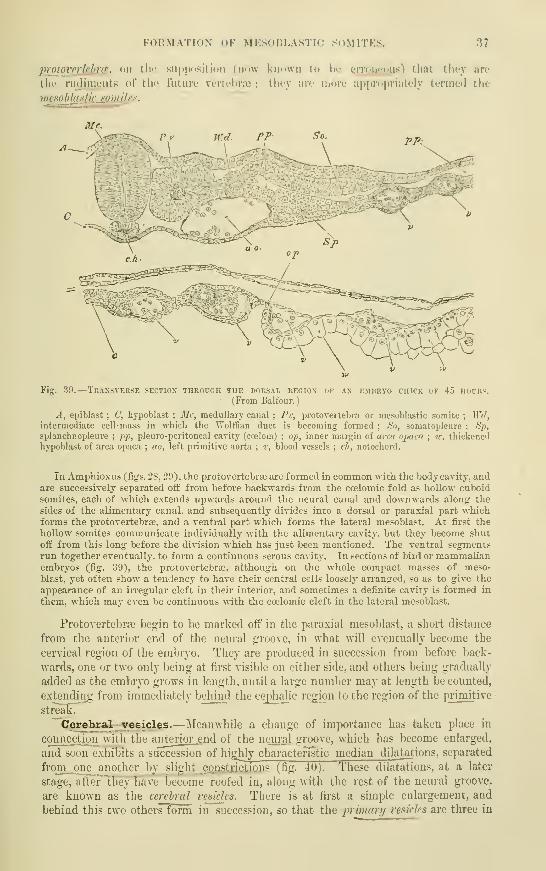
FORMATION OF MESOBLASTIC SOMITES. 37
proiovnishrfSi on tlic supposition (now known to hu yroncous) that they iire
the rudiments of the future vcrtebroc ; they arc more appropriately termed the
mesohlastis^mjlrs."~
Fig. 30.
—
Transverse section through the dorsal region of an embryo chick op 45 hours.(From Balfour.)
A, epiblast ; C, hypoblast ; Mc, medullary canal ; Pv, protovertebra or mesoblastic somite ; Wd,intermediate cell-mass in which the Wolffian duct is becoming formed ; <S'o, somatoplenre ; Sp,
splanchnopleure; pp, pleuro-peritoneal cavity (ca;lom) ; op, inner margin of arcxi opaca ; w. thickened
hypoblast of area opaca ; ao, left primitive aorta ; v, blood vessels ; ch, notochord.
In AmphioxHs (figs. 28, 20), the protovertebra3 are formed in common with the body cavity, andare successively separated off from before baclrwards from the coelomic fold as hollow cuboid
somites, each of which extends upwards around the neural canal and downwards along the
sides of the alimentary canal, and sub.sequently divides into a dorsal or paraxial part whichforms the protovertebrte, and a ventral part which forms the lateral mesoblast. At first the
hollow somites communicate individually with the alimentary cavity, but they Ijecome shut
off from this long before the division which has just been mentioned. The ventral segmentsrun together eventually, to fonn a continuous serous cavitj'. In sections of bird or mammalianembryos (fig. 30), the protovertebra^. although on the whole compact masses of meso-
blast, yet often show a tendency to have their central cells loosely an-anged, so as to give the
appearance of an irregular cleft in their interior, and sometimes a definite cavity is formed in
them, which may even be continuous with the coelomic cleft in the lateral mesoblast.
Protovertebras begin to be marked off in the paraxial mesoblast, a short distance
from the anterior end of the neural groove, in what will eventually become the
cervical region of the embryo. They are produced in succession from before back-
wards, one or two only being at first visible on either side, and others being gradually
added as the embryo gi'ows in length, until a large number may at length be counted,
extending from immediately behind the cephalic region to the region of the primitive
streak.
Cerebral vesicles.—Meanwhile a change of importance has taken place in
connection with the antei'ior .end of the neural groove, which has become enlarged,
and S' I >\\ exhibits a succession of highly charactefistic median dilatations, separated
from ont- another by slight constrictions (fig. 40). These dilatations, at a later
stage. ulUr thoy Iiuac become roofed in, along with the rest of the neural groove,
are known as the cerebral resides. There is at first a simple enlargement, and
behind this two others form in succeasion, so that the p/;««;v/ resides are tlu-ee in

38 CEREBRAL VESICLES.
number, but subsequently the most antexlor {fore-brain) and posterior (Jnnd-hrain)
e^chjforms two yesicles, whereas the middle vesicle (mid-irain) remains permanently
undivided._ Five vesicles are therefore theiTtoTe'seen, and these giv_e rise eventually
Fis 40. —Surface view of an early embryo op the guinea pig showing the commencement op
the three primary cerebral a'esicles (1, 2, 3) as enlargements of the medullary groote.
Semi-diagrammatic.
2}r, primitive streak and groove.
Fig. 41.— Rabbit embryo of the 9th day, from the surface. -J. (Kolliker.)
Tlie medullary groove is enlarged anteriorly and the primary optic vesicles are growing out from
the first cerebral enlargement. On either side of the head, the (double) tubular heart is seen.
l^airs of j)rotovertebrse are formed.
Eight
to the five fmidamental divisions of the brain, while from the sid^s of the fore-brain
the rudiments of the optic" nerves and retinse grow out as hollow protrusions.
HeaiSr and vascular system.—While this change is progressing in the
neural canal, and the proto\ertebr9e are becoming formed in the paraxial mesoblast,
the first sign of a vascular system is beginning to make its appearance in the meso-
blast on either side of the head in the form of a simple tubular vessel (fig. 41), Avhich
becomes developed in the splanchnopleure in this srEfeiiroiT." Xs. the splanchnopleural
mesoblast and its accompauying hypoblast fold round on either side under the
head to form the fore-gut, these two simple tubes necessarily come together in the
middle line, and they then fuse together Ion gitjjdin ally to form a sjngle^tube, the
priinitive heart (fig. 38) ; this tube runs for a short distance in the mesoblast immediately
under tlie fore-gub, and then divides into two branches, Avhich pass laterally, so as to
partially encircle the fore-gut, and thence course backAvards along the body of the
embryo on either side of the notochord. These two vessels form the primitive
arteries (primU'ive aortcc), the part of each which encircles the fore-gut as it passes
dorsalwards being known as Vaefi'nt aortic arch. On the other hand, the posterior

HKAUr AM) VASCULAR SVSTKM. 39
cud of tlie single tubular lifiirt bifurcates at an obtuse anji^lc to form two lar<je
venous roots {j>n'tti_ilire I'ci/i.s), wlit-h receive the blood from tb.e vascular area on tlie
yolk-sac when this is developed, and juiss it on to the heart, 'i'hese two primitive
veins become the ritcUine reins.
The heart begins to beat very soon after its ajipearanc.'!', e\L'n whilst still (illed
only with a colourless fiuid, and before receiviuL;- blood from the va-scuiar area.
Fig. 4-2.
—
Vascular area of ihe rabbit of 10 days. (v. Beneden and Julin.)
The arteries and arterial capillaries are represented red. the venous capillaries and veins blue.
Afterwards, when receiving and propelling the red blood from that area, and
especially after it has become elongated and bent upon itself, it is one of the most
prominent objects seen on examining the embryo ;projecting as it does freely into
the yet widely open coeloni immediately behind and beneath the cephalic region of
the body.
The first vessels to be developed are formed in mesoblast altogether outside the
body and ^yithin a circular area (vascular area), which sun-ounds the devek»ping
embryo for a certain distance. The first_appearance of red blood occurs in the form
of isolated red points (?>/oor/-/67f///^/6- of Pander), which are scattered about within
this area, and are especially numerous at its circumference, where they form an
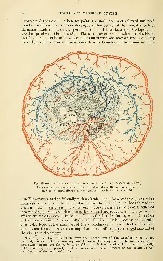
40 HEART AND VASCULAR SYSTEM.
almost continuous chain. These red points are small groups of colom'ed nucleated
blood corpuscles which have been developed within certain of the mesoblast cells in
the manner explained in another portion of this work (see Histology, Development of
blood-corpuscles and blood-vessels). The mesoblast cells in question form the blood-
vessels of the vascular area by becoming united with one another into a capillary
network, which becomes connected mesially with branches of the primitive aortfe
Fig. 43.
—
Vascular area op the rabbit of 11 days. (v. Beneden and Julin.
)
The arteries are represented red, the reins Lhie ; the capillaries are not shown.
In both the stages illustrated, the terminal sinus is seen to he arterial.
(vitelline arteries), and peripherally with a circular vessel {termincd sinus), arterial in
mammals but venous in the chick, which forms the circumferential boundaiy of the
vascular area. From the capillary network of the vascular area the blood is collected
into two vitelline veins, which course backwards and inwards to carry the blood of the
area to the venous roots of the_ heart. This is the first circulation, or the circulation
of the vasculaflirea. It is also called the Vitelline circulation, because the vascular
area is developed in the mesoblast of the splanchnopleural layer which encloses the
vitellus, and its capillaries are an important means of bringing the food material of
the vitellus to the embryo.^—
The origin of the cells which form the endothelium of the vascular system is not
definitely known. It has been supposed by some that they are in the first, instance of
hypoblastic origin, but the evidence on this point is insufficient, and it is more generally
held that they are specially modified mesoblastic cells. Regarding the origin of the
endothelium of the heart, see p. 136.
RECENT LITERATURE. 41
RECENT LITERATURE.*
Beneden, v., and Julin, Recherchea sur Ja formation dea annexes falales rluz let mammifirea{Ijopin et L'heiropteres). Arch, tie biologic, t. v., 1884.
Carius, Ueber die Enturicklung der Chorda u. dtr primitiven Raclttnkaut bet Meertdiweinchenund Kauinclien. Inaug. Diss., Warburg, 1888 : Vein- den Kopfforlsatz dea Kaninehena. MarburgSitzungsbcr, 1887 ; Ueler die AualAlduiuj dcs hinttren Korperendes lei Cavia. Ibid., 1888.
Chiarug^i, Anatomic d'un embryon humain de la lowjueur de mm. 2 6 en ligne droUe. Arch,ital. de biologic XII.
Ehlers, E., Nebendarm und Chorda dorsalia. Nachrichten d. kgl. Gesellscb. d. Wissensch. zaGottingen, 1885.
Fol, H., Deacription d'un embryon humuin de cinq millimetrea et six dixiimu. RecueilZool. Suisse, 1884 ; Rccherches sur le ddveloppcment dta protoverUbrea chez Vembryon du pouUt.Archives des sciences physiques et naturelles, 1884.
Giacomini, Sul canale neiLrentcrico et sul canale anale, <Lc. Giom. de r. accad. di medic, di
Torino, 1SS8.
Keibel, F., Zur EntwicUungsgesch. der Chorda bei Sdugem. Arch. f. Anat. u. Physiol., Anat.Abth., 18S9.
Eolliker, v., Ueber die Chordahohle und die Bildung der Chorda beim Kaninchen. Sitznngsber.
der physiciil.-med. Ges. in Wiirzburg, 1883.
liieberkulm, N., Ueber die Chorda bei Sdugethieren. Arch, fiir Anat. und Physiol., Anat.Abth., 1884.
Perenyi, J. v., Entuncklung des Am,nion, des Wolff^schen Ganges und der AllanUns bei den Repti-lien. Zool. Anzeiger, No. 274, 1888.
Eavn, Ueber die mesoderm/reie SteUe in der Keimschcibe des Hiihnerembryo. Arch. f. Anat.,ISS6.
Homiti, G., Be Vextremite antirieur de la corde dor-:ale et de son rapport avec la poche hypophy-taire ou de Eathke chez Pembryon du poulet. Archives italiennes de biol., t. VII., 1886.
Shore and Pickering', The pro-am,nion and amnion in the chick: Journal of Anatomy andPhysiology, 1SS9.
Solger, B., Studien zur Entwicklungsgc^chichte des Ccdom^ und des Ccelomepithels der Amphibien.Morphol. Jahrbuch X., 18S5.
Spee, F. v., Ueber die Entwicklungsvorgdnge vom Knoten aus in Sdugethicrkeim^cheiben. Anat.Anzeiger iii., 1888 ; Beobachtungen an einer menschlichen Keimscheibe mit offener Medullarinne u.
Canalis neureutericus. Arch. f. Anat. u. Physiol., Anat. Abth., 1889.Strahl, H., Ueber die Enticickelung des Canalis myelo-entericus und der Allantois der EidecJise.
Archiv fiir Anat. und Physiol., Anat. AbtheU., 1881 and 1883.
"Works dealing with the general subject of Embryonic Formation and Development.
Balfour, A Treatise on Comparative Embryology. London, 1880.Foster and Balfotir, The Elements of Embryology, Second Edition, 1883. Edited by
Sedgwick & Heape.Haddon. An introduction to the study of Embryology. London, 1887.Hert-wig, O., Lehrbuch der Entvncklungsgeschichte des Menschen u. d. Wirbelthiere. 2te
Auflage, Jena, 1888.
His, "W., Uniersuckungen ue. d. erste Anlage d. Wirbelthierleibes. Die erste Enticicklung deaEiihnchens im Ei. Leipzig, 1868 ; Unsere Korpeiform u. das physiol. Problem ihrer Enstehung,1871 ; Anatomie menschlicher embryonen, 1881—1885.
Kolliker, v., Eniwicklungsgeschichte des Menschen u. d. hoheren Thicre. 2te Auflage. Leipzig,
1879.
Eauber, PormhUdung «. Formstorung in der Enticicklung von Wirbelthieren. Morph. Jalirb.
vi., 1880.
* The early changes in the blastodenri*; layers are dealt with in several of the papers enuracrateoon pp. 27 to 29.

4S DEVELOPMENT OF THE FCETAL MEMBRANES.
DEVELOPMENT OF THE PCETAL MEMBRANES; ATTACHMENTOF OVUM TO UTERUS.
Having thus sketched out the manner in which the principal organs of the
body first make their appearance, we may briefly consider the formation of certain
structures which have a purely embryonic existence, and are concerned either with
the nutrition of the foetus and its attachment to the mucous membrane of the
uterus (chorion, allantois, placenta), or serve the purpose of protecting the embryo
against mechanical injuries by suspension in a bag of fluid (amnion).
Formation of the amnion and chorion.—The amnion, which is only found
in reptiles, birds, and mammals (amniota), is a membranous bag occupied by a clear
albuminous fluid, and covers the whole of the embryo. It is developed from folds of
false ocmjvtcn' or chorlOTi^
•viliL of
Fig. 44.
—
Diagram of a transverse section of a mammalian embryo showing the mode of
FORMATION OF THE AMNION. ThE AMNIOTIC FOLDS HAVE NEARLY UNITED IN THE MIDDLE LINE.
Epiblast, blue ; mesoblast, red ; hypoblast and notochord, black.
somatopleure, which are reflected from the head and t^iiLends and lateraL boundaries
ofThe embryo at an early stage.^ With the sinking of the embryo into the blasto-
dermic vesicle or yolk, these folds grow up over the back (fig. 44) until they meet and
coalesce with one another along the middle line, in such a manner as to form two
distinct membranes, one of which, the inner, is the amnion (true amnion), while the
outer membrane (termed the false, amnion) becomes applied to the greatly thinned
remnant of the zona pellucida, and eventually forms a complete external covering
to the ovum and its contents. This external^covering of the ovum has been long-
known as the chorion—a name which iias7however, been applied by some embry-
ologists to othSF-itructures.2 It is fixed to the uterus by vilh, which are
^ The head fold is preceded at a yet earlier stage by the bilamiiiar pi'O-amnion, the formation of
which has been already alluded to {]). 35)." The term " chorion" has been applied to various structures by embryologists. Originally used to
denote the external covering of the developing ovum, it was emi^loyed successively for the zona pellucida
(" primitive chorion "), the epithelial enclosing membrane of the blastodermic vesicle, and finally for
the external amniotic fold or false amnion, when this becomes formed. Lately it has been used to
express the external albuminous enveloije of the undeveloped ovum, so that it is probable that
much confusion may arise unless the meaning of the term be in each case clearly defined. It
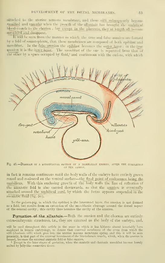
l>KVKLur.\lKXT OF THE FaMAL .MKMHUANKS. 43
iittaclit'd to the utoriiit' inucous lueinbraue, aiul these vijlj^siiljscmiciitly becomenijiiilied and vascular when the {rrowth of the aUantuis has broujfht the uiubJJicalblood-vessels to the chorion ; but cxc^UiLJH the Dhtccn ta, they at len^'th all becomeatroi>riTecI and disuppcar.
It will i)e seen from the manner in which the true and false amnion arc formedby a fold of somato|)leure, that these membranes are composed of both epiblast andmesoblast. In the falsejiuinion the epUilast becomes the oiiterja^er ; in the truei\""iion it is the innei-jb^i;. The mes(.blast of the one is sei)anited from that ofthe other by a space occupied by fluid,' and continuous with the ccelom, with which
fcn'&-atvt
Fig. 45.
—
Diagram of a longitudinal section of a mammalian embryo, after the completionOF THE AMNION.
in fact it remains continuous until the body walls of the embijo have entirely gi'own
round and coalesced on the ventral surface—the_ final point of coalescence being the
umbilicus. With this enclosing growth of the l^ody walls the line of reflection of
the ainniotic fold is also carried downwards, so that the amnion is eventually
attached around the umbilical..cord, by which the fcctns appeai-s suspended in the
anmiotic^iiTdXEg. oO).
In the guinea-pig. in wMcli the epiblast is the innermost layer, the amnion is not formedas a fold, but results from an extension of the mesoblastic cleavage around the dorsal a.spect
of the central cavity ; this cavity thus becomes the cavity of the amnion.
Fqrm.€.ticuio£ the allantois.—Both the amnion and the chorion are entirely
extra-embryonic structures, i.e., they are external to the body of the embryo, and,
\vill be used throughout this article in the sense in which it has hitherto almost invariably been
employed in human embryology, to denote that external membrane of the ovum from which the
villi (chorionic villi) which grow into the uterine mucous membrane spring, and this it will be seen
presently, is that part of the external investment of the blastodermic vesicle, v.hich, when the amnion is
formed, becomes the external amniotic fold or false amnion.^ Except in the later stages of gestation, when the amniotic and chorionic mesoblast become loosely
united by jelly-like connective tissue.
44 FORMATION OF THE ALLANTOIS.
although they minister to its protection and nutrition, take nqjwfc in the formation
of any of its organs. But tEe~case is different with the structure next to be
described, viz., the allantois, a part of whicii does in fact eventually becomeconverted into portions of ithe urinary and generative systems, although the greater
part is also extra-embryonic, its function being to minister, through its accompanyingblood-vessels, to the nutritive and^'respiratory exchanges of the foetus.
Tlie time of development of the allantois seems to vary much in mammals, andthere is reason to believe that it is found in the human embryo at a very early
period—indeed the earliest human embryos that have hitherto been described
already possess an allantois. In most animals in which its development has been
studied the allantois has been found to begin as a hollow prolongation of the posterior
end of the primitive alimentary canal (fig. 46). It soon, however, becomes relatively
Fig. 46.
—
Longitudinal section through the posterior end of an embryo rabbit, showing theOUTGROWTH OP THE ALLANTOIS. (KolHker.
)
h, epiblast of trunk ; del, hypoblast ; m, medullary or neural canal ; ch, notocliord ; hd,
commencing hind-gut, which is becoming formed by a folding-over of the tail end of the embryonicblastoderm, v ; ed, blind end of hind gut ; al, allantois gi'owing out from hind gut into aw, mesoblastic
thickening ; e, epithelium of yolk sac ; df, splanchnopleure ; hp, somato-pleure passing superiorly into
am, tail fold of amnion ; s, is placed within the cavity of the amnion, and denotes the tail end of the
embryo.
shifted in position so as to come off from the ventral wall of the hind-gut, growing
into the posterior extension of the mesoblastic cleft and eventually into the space
between the false and true amnion, and carrying along with it its mesoblastic covering
(figs. 36, 45), It is therefore composed eventually of two parts, viz., (jj a hypo-
blastic sac which communicates, at fu'st widely but afterwards by a narrowed orifice,
with the hind-gut, and (2) an investment of mesoblast. This last is usually greatly
thickened and very vascTilar, and is directly supplied with blood by two arteries
(allantoic or umbilical arteries), which appear at first as a direct continuation of
the primitive aortee. As the allantoic vesicle expands into the cavity of the false
amnion, it carries the vascular mesoblast along with it, so that this mesoblast is thus
brought to the inner surface of the chorion, over which its blood-vessels then spread
so as to convert this hitherto non-vascular membrane into one which is richly
supplied with blood-vessels. The chorion has grown in the form of ramified
villi into the substance of the uterine mucous membrane or decidua even
before this advent of the vascular tissue of the allantois, but the chorionic villi
now receive blood-vessels and thus become vascularized, the interchanges between
the foetal and maternal vascular systems, which are afterwards confined to one region
only of the chorion and decidua—that which forms the placenta^occurring in the
first instance over the whole superficies of the ovum.
FORMATION OF THE ALLANTOIC. -1-5
Like the amnion, an allantois is only formed in the embryo of reptilcH, birtlH, and mammals.'It varies chiefly in tho extent to wliicli its liypoblastic part liceomcH developed. In reptiles,
Fig. 47.
—
Longitudinal section at a slightly later stage than that suuwn in kiu. 4ti.
The allantoic protrusion now springs frnin the ventral wall of the hind gut. Lettering as in fig. 4(5.
(FroniKolliker.)
Fig. 48.
—
Early human embryo. (From His after Coste.)
The embryo is enclosed within the amnion am, and is attached at its caudal end by the allantoic
stalk, all, to the chorion. The yolk sac, ys, is still distinct from the allantoic stalk.
Fig. 49.—Dl.VGUAM OF longitudinal sections THUO0GH THE HUMAN (JVUM AT SUCCESSIVE STAGES
SHOWING THE DIPPING DOWN Of THE EMBUVO-RUDIMEXT INTO THE BL.VSTODERMIC VESICLE, ANDTHE FORMATION OF THE FOKEGUT, AMNION, AND STALK OF THE ALLANTOIS BY THE FOLDING OF
THE BLASTODER.M. (His.)
Am, head fold of the amnion (pro-amnion in b and c) ; .Y^, yolk .sac. a and d are conditions of the
embryo which have been seen and described ; b and c are intended to show how the conditions found
in (I may be brought about, and especially how the stalk of the allantt)is nuiy be regarded as a direct
continuation of the posterior end of the embryo, which accoi'ding to His does not lose at any time its
connection with the chorion or villous external membrane of the ovum. The curved dotted lines in c
indicate the formation of the amnion and false aniniou by the upgrowing of lateral folds (which have
not as yet met in the median line).
' For a discussion of the origin and meaning of the amnion and allantois, see Balfour, " Comparative
Embryology," H., p. 256.

46 CHANGES IN THE UTERUS.
in birds, and in some mammals, e.g. ruminants, this portion forms a large sac which occupies
the greater part of the cavity of the false amnion, and is filled by fluid (allantoic fluid) in
which many urinary products can be recognized. (The ducts of the embryonic renal organs openinto the pedicle of the allantois.) But in most mammals the development of the hypoblastic
sac is far less extensive, and in some, including the human embryo, the allantois is mainlyrepresented by a large mesoblastic outgrowth carrying the allantoic (umbilical) vessels to the
chorion. There is also considerable variation in the period at which the allantois begins to
develop. In the human embryo it is certainly formed at a very early period, probably evenbefore the amnion is completed. In the guinea-pig, also, it appears early, although after the
amnion. In this animal it is first developed as a solid outgrowth of mesoblast which projects
from the line of junction of the hinder end of the amniotic bag with the blastoderm, andbefore the formation of a hind gut or of any part of the alimentary tube, a hypoblastic
diverticulum being altogether wanting. In the earliest human ova in which the allantois
has been investigated, it is already a tube of hypoblast which forms a direct prolongation of
the posterior end of the primitive alimentary canal (fig. 49, d), and is enclosed in a short
stalk of mesoblast, by which the posterior end of the embryo is attached to the chorion, andthrough which, by the allantoic (umbilical) blood-vessels, the chorionic villi are freely
supplied with blood. From the attachment of this stalk to the placenta (chorionic part),
the hinder extremity of the amnion is reflected. The stalk in question is not the um-bilical cord, since it does not include the stalk of the yolk sac (vitelline duct), which only
later becomes bound up with it. It is termed by His the abdominal stalk (Bauchstiel),
the term allantois being by him restricted to the hypoblastic diverticulum. It is further
considered by His to be probable that the human embryo never becomes completely detached
from the chorion, but that it always retains its attachment to the outer membraneof the ovum at the hinder end, this abdominal stalk being regarded as a direct prolongation
of the tail end of the embryo (fig. 49). If this should prove to be the case, the
human ovum would foi-m an exception to the usual rule of a complete separation of embryofrom chorion at the formation of the amnion, and subsequent re-attachment by outgrowth of
allantois.
Changes in the uterus. Mode of attachment of ovum to uterus.^—The mucous, membrane of the pregnant uterus is known as the cUcidua. It is
thicker and more pulpy than in the ordinary non-pregnant condition, and the
glands are longer in proportion, but it is otherwise of similar structure except in
the part where the placenta is about to be formed ; here it undergoes important
modifications. The ovum, wliich has been fertilized and has passed through the
first stages of development in the Fallopian tube, although considerably larger than
the undeveloped ovum, is still an extremely minute object when it reaches the
uterus. Here it speedily becomes imbedded in the soft and thickened mucous
membrane, and this is soon reflected over and completely encloses the ovum, which
thus comes to lie in a cavity within the decidua which is altogether shut oflP by the
reflected part from the true cavity of the uterus. Different names have been given
to these parts of the uterine mucous membrane which immediately enclose the ovumto distinguish them from that which lines the original cavity of the uterus. Thus
the layer of membrane which has grown around the ovum is known as the decidua
reflexa ; the part where the ovum first becomes attached to the uterus and where the
placenta is afterwards formed, is the decidua ser_otina, while the membrane lining
the true cavity of the uterus, is termed deddua vera (figs. 50, 51).
With the subsequent growth and consequent expansion of the ovum the enclosing
decidua reflexa expands also pari passu, encroaching more and more upon the true
cavity of the uterus and coming into contact everywhere with the decidua vera.
Eventually it blends entirely with the decidua vera, so that the two layers are indis-
tinguishable and the original cavity of the uterus is obliterated (except at the
cervix uteri).
1 The following account of the formation of the deciduse and of the placenta is confined as much as
possible to what has been observed in the human subject. In other mammals important variations in
the mode of attachment of the ovum and in the formation and structure of the placenta are found to
Dccur.

CHAN(JF,s IN iiii: rri-.ins. 47
Both tlu- .Uri.hiii v,r:i iin.l tlu" (l.'fi.lua ivflexa (.rinimilly cntaiii tubular an.l somewhat
tortuous -lan.ls, which wciv .lisc.v.r.-.l by Sharpey. and w.t.. by lii.n suppos^^l to mimstfr. in
thofir<t instance, both to th.- nutrition an.l to th.- aitadnn.-nt ..f the ovum, the latter by
affordin- depressions for the ehorionic villi to penetrate into tin- substance of the dec.dua. It
has however since been shown that these villi do not .lirectly pass into the ^'landn, but
rather ten.l to become attache.l to the inter-landular surface, an.l in.leed at tin, de.;idua
sen.tina, where subsequently th.- main attnchment of the chorionic villi occurs the trland-
inmina mav become almost entirely obliterate.1 before the villi are here formed. But the
.n-catlv enl:ir-e.l -lands of the decidua vera very probably furnish a secretion to af^sist the
nourishment of the ovum previously to the full establishment of the placenUl circulation.
Fig. 50.—DlAGUAMJIATIC SECTIOS OF THE PREGNANT HUMAN UTERUS AT THE SEVENTH OR
EIGHTH WEEK. (AllcU ThomSOD. )
c, c, opeDings of Fallopian tubes into the cavity of the nterus : c', cervix, filled by a plug of mucijs^
thele te?s^andc'are placed within the original ca^^ty of the "terus '• ^r, decidua vera ;c/, de dinv
retlexa; ds, decidua serotina ; c/., choriou with its villi growuig intof^^/'^^'f^^Vfinr in a e
serotina : in the former the ^-illi are becoming atrophied ;«, umbd.ca cord, thef;"'^^^''
J*-„^„ "^^bloodvessels within it; «/. allantois ; >/, yolk sac (u.nbdi«il vesicle); y, its stalk, passing in the
umbilical cord and connected with the intestine of the embryo, ( ; am, amnion.
The decidua undergoes remarkable structural changes during the early months ot
pregnancy, some of these changes being common to all three parts of the membrane,

48 CHANGES IN THE UTERUS,
whilst others are special to that part (cl. serofcina) which enters into the construction
of the placenta. The following is a brief account of these changes.
^
With the supervention of pregnancy the mucous membrane lining the uterus
becomes thickened and the tubular glands become both dilated and greatly elongated.
Fig. 51.
—
Antero-posterior section of the gravid
UTERUS AND OVUM OF FIVE WEEKS. (Semi-clia-
grammatic. )(Allen Thomson.
)
a, anterior; p, jposterior uterine wall ; m, mus-
cular substance ; u, placet! in the cavity of the uterus;
ff,the glandular layer of the decidua vera ; r, the
decidua reflexa ; s, decidua serotina ; c, cervix uteri;
ch, chorion with its villi, which are more highly deve-
loped on the placenta] side ; e, the embryo enclosed
in the amnion, with the allantoic vessels passing along
a short allantoic stalk into the placenta, and the
umbilical vesicle lying free in the space between
amnion and chorion.7?P.
Fig. 51*.
—
Diagrammatic sections of the uterine mucous membrane, showing the changes whichTHE GLANDS UNDERGO WITH THE SUPERVENTION OF PREGNANCY (from Kundrat and Eogelmann).
A, Diagram of the glands of the non-pregnant uterus ; vi, muscular layer ; B, condition of the
glands at the beginning of pregnancy ; c, compact layer near free surface of decidua ; the glands are
here somewhat enlarged but not very tortuous, and the mucous membrane is rendered comj)act byhypertrophy of the interglandular tissue ; sj?, spongy layer, containing the middle portion of the
glands greatly enlarged and tortuous, producing a spongy condition in the mucous membrane ; d, deepest
portion of the glands, elongated and tortuous, but not much enlarged.
This thickening of the membrane and enlargement of the glands goes on until the
fifth month, so that by this time the decidua vera is nearly half an inch in thickness
and its glands have undergone so considerable an elongation that they now no longer
pass nearly straight through the membrane but run in a tortuous manner from the
1 For a more complete account of the changes in the uterus, and of the placenta, the reader is
referred to the list of papers at the end of this section, but especially to the works of Kundrat and
Engelmann, Leopold, and Minot, and for the comparative structure of the placenta to the classical
investigations of Turner.

CHANGJ;S IN THE UTERIS. 40
inner surface to the vascular layei", so lliat a verl ical seel ion of t lie iMciiilirano oxliil)its
tlieni cut (piitc as often ohliijuely or trunsvcrsely as loiii;iiU(liniilly. They are also
genenilly dilated, bnt the dihitation is hy far most marked at the mouths of the
glands, whicii come thus to have u I'uniiel-like sliajie, and in the deeper part of the
membrane, where the dilatations look in sections like a series of cavities, lined hy
cubical or flattened epithelium, ami separated from one another by a relatively
small amount of interulandular substance. 'J'his gives a spongy api)earan(.-e to the
part in question, and it has been accordingly ternied the .sLmltiiJi apo/it/iosiiiii of
the decidua (tig. 51*, B, sp). The deepest part of the glands, that, namely, which is
in contact with and is imbedded in the sui)erficial portion of the nniscular coat, does
not share in this dilatation, and its e])ithelium also retains the columnar (character.
The i)art of each gland between the funnel-shaped mouth and the dilatations aliove
described, also becomes enlarged, but not to so great an extent, the hypertrophy of
the mucous membrane being here chiefly confined to the interglandnlar tissue, which
becomes filled with large epithelinm-likc cells {decidual cells of Friedlander) andwith numerous and large ca])ilhiry blood-vessels. This layer of the decidua has
been termed the >^liJSLifJl^ fP'OL'S"'^"^^^ '" (^'o'ltradistinction to the stratum spongiosum
external to it (tig. ol*, B, c).
After the fifth month, by which time the great increase in size of the ovum with
its contained embryo has brought the decidua reflcxa into close contact with the
decidua A'cra, the latter begins to undergo an atrophic process, the result to all
appearance of the compression and distension to whicli it is thus subjected. Its
tissue becomes thinner and less vascular, and both the funnel-shaped mouths of the
glands and those ]iarts of the glands which run through the stratum compactumbecome gradually ol)litcrated, so that eventually hardly any trace remains. In the
Fig. l)'2.—DlAllK.VMMATIi; SKOTKiN TllKCiUGH THE UECIDU.K
AT THE EDCE Hi' THi; I'LACEN'TA (froiii Kundrat andEngeluiann).
c, sp, 111, as in fiy. 51*, W ; d r, (](.'ci(]n;i vera ; (/ .'•, decidua
serotina ; (/ /, dce-idiia icllcxa.
stratum spongiosiun the spaces which have
resulted from the dilatation of the gland tu1)es
lose the lining epithelium, and become flattened
out conformably to the surface, so that they nowappear as a layer of compressed lacumu, sepa-
rated by thin lilirous tra])ccuky (tig. 'r2, s/>.).
Similar changes occur in the decidua refiexa.
That this is truly a fold of mucous membrane is
evidenced by the fact that gland tubes can be
seen to open upon both its surfaces. These gland
tubes early become enlarged, and acquire an
oblique or tortuous course, with dilatations in their deeper parts, i.e., in the middle
of the thickness of the d. reflexa, so as to form here also a sort of s}>ongy tissue.
But the decidua reflexa sooner becomes ex[)anded by the growing ovum into a
relatively thin meml)rane, and tlie atrophic changes in the glands occur at an
earlier stage, so that by the time that it has coalesced with the decidua vera hardly
any traces of them can be discerned.
In the decidua serotina (placental decidua) similar changes have been described
in the glands, the final result being the formation of a spongy layer, with irregular
clefts flattened out conformalily to the sin'face. and from which the ei>itlu'liiun has
entirely disajipeared, accompanied by complete atrojihy and disappearance of all the
parts of the glands which are superficial to this layer, the only jiortions which
remain nearly unaltered licing the deepest parts of the tubes, which are jiartly
VOL. r. -E
d.s

50 CHANGES IN THE UTERUS.
Fig. 53.—Section through a normal placenta of seven months in situ (Minot).
Am, amnion ; CJio, chorion ; Vi, root of a villus ; vi, sections of the ramifications of villi in the
intervillous spaces, the larger bloodvessels within them are represented black :X», deep layer ot tne
decidua, showing flattened remnants of enlarged glands in spongy stratum ; Ve, uterine vein [i artery)
opening out of placental sinus ; jUc, muscular wall of uterus.

CHANGES IN THR UTERUS. 51
imbcdiled in tlic muscular coat of tlie uterus, and rotaiu thoir opithclium. After
scpai'ation of the placenta IVcni tlic uterine wall at parturition, the uterine mucousmembrane, with its cpillichuui and glands, becomes renewed from this deepest portion
of the decidua serotina.
The most important changes of structure occur in the superficial part of the
placental decidua, after the disappearance of the glands. The exact manner in
which these changes take place has not been folhjwed out, but the ultimate result is
the replacement of the whole of this poi'tioii of the decidua, with the excejjtion of a
Fig. 54.
—
Sections illustkating the structure of the placexta (.Minot).
A, vertical section througli the margin of a placenta at full term ; D, D, deep layer of decidua
;
Ft, chorionic villi variously cut, their bloodvessels injected ; Si, marginal space of the placenta,
neai-ly free from villi ; ri, aborted villi beyond the placenta ; Fih, canalized fibrine of Langhans,
produced, according to llinot, by transformation of the superficial layer of the chorionic epiblast.
B, decidual tissue from a i^lacenta at full term ; v, a bloodvessel ; d, d', decidual cells ; the latter
with several nuclei.
comparatively narrow basal kujer next to the spongy structure, into a series of inter-
communicating vascular sinuses, which together constitute an immense flattened
space (intervillous space), bounded internally (toward the uterine wall) by the basal
layer just referred to, and externally by the chorion ; also, according to some
authors, by a thin layer of decidua, the suhcJiorionic membrane of Turner, which is
described as lying immediately under the chorion of the o%'um but so intimately
incorporated with it as to be with difficulty demonstrable as a separate stratum
except at the edge of the placenta.
From the basal layer, partitions of fibrous decidual tissue pass towards the
chorionic surface, and serve to partially sub-divide the labyrinth of vascular spaces or
sinuses into a number of locidi (cotyledons). Each of these loculi is occupied by an
K 2
52 ATTACHMENT OF THE OVUM TO THE DECIDUA.
arborescent tuft of villi continuous with the fffital chorion, and traversed by blood-
vessels which are supplied from the branches of the umbilical arteries. These
blood-vessels form a capillary loop in each villus, and these capillary loops are
separated from the maternal blood in the placental sinuses not only by the capillary
walls and the connective tissue of the villus, but also by a double layer of flattened
epithelium-like cells derived either fi-om the chorionic epithelium (Minot),i or from
the decidual tissue, and, perhaps, in part representing an endothelial membrane
belonging to the placental sinuses, which, according to Waldeyer, are lined by endo-
thelium prolonged from that of the uterine vessels.
Some of the chorionic villi are attached (1) by comparatively stout bands of
fibrous tissue to the basal stratum of the decidua, (2) by finer bands of similar
substance both to one another and to the
septal prolongations of the decidua ; others amTucn choncTh
hang freely into the placental sinuses.
Thes3 sinuses are supplied directly with
arterial blood from tortuous branches of
the uterine arteries which pass through the
e' r
Fig. 55.
—
Diagram showing the tissues coveringTHE AaLLI IN THE HUMAN PLACENTA, ANDTHEIR RELATION TO THE DECIDUA ACCORDING
TO Turner.
F, fcetal tissue ; M, raaternal tissue ; d, blood-
vessels in villus ; d' , blood in ijlacsntal sinus;
e, layer of cells covering villus ; x. basement mem-brane covering villus (? endothelium) continued fromuterine vessels ; da, decidua serotina ; t, tissue
imiting villus to decidua ; ca, up, uterine vessels
opening into sinus.
Fig. 56.
—
Diagram of the placenta (E. A. S. ).
s, placental sinus ; ds, decidua serotina ; sp,
spongy layer ; m, muscularis ; a, v, uterine
artery and vein opening into placental sinus.
spongy stratum of the decidua serotina and through the basal stratum of the
placenta to open into the sinuses without the intervention of capillaries. Fromthe sinuses veins, which run very obliquely through the decidua, carry off' the blood,
and eventually pass into the veins of the muscular wall. The foetal villi are thus
bathed by slowly flowing maternal arterial blood, and respiratory and nutritive
exchanges may occur between the two kinds of blood, but there is no actual mixing
^ Minot describes both the villi and the sinuses of the placenta as bounded by the chorionic epithelium.
This wouM agree very well with recent observations in the bat, hedgehog, and other mammals, whichshow that these spaces are originally developed in hypertrophied epiblast (see next page).

FORMATION AN'I) 8TRUCTUKK nl' IIIM I'l.ACKN'lA. )3
of the two tluids, iidi' is it possiMc U> inject tlie fd'tiis IVdiM iIk,' imillier iliruiij^ii the
placenta, or the mother IVoiii the lu'liis.
The question, whiuh liiis hern iiuiic Hum uiiw niiscil. wlifilicr tli<! intcrvilluiis spaccB
nonnally contain blood, may bo now n';.'-anl(Ml as settled in tlio aflirniativc. It has lieen
usually held that they reiiresent capillaries or veins of tlie decidua, which have become
dilated and fused to^'etlier to sucli an extent as to occupy the whole thickness of the
placental jiart of that membrane, with atrophy of the intervening,-- decidual tissue, which
merely remains as a coverin;;- to the villi. But since the placental sinuses ajjpear to be
bounded superficially by the chorionic covering of the ovum, an<l it is in most jjlaces not
possible to detect any decidual tissue between them and the chorion, it has been conjectured by
some writers (Kiilliker. Lani^hans) that they have become formed by extravasation of bloofl into
a space between the ovum and the decidua, into and across which sjiace the chorionic villi
have ji-rown. Altliouyh the development of these structures is insulliciently known in Primates,
it has been shown in various nuimmals (Selenka, Duval, Ilubrecht, JIasius) that the first
attachment of the blastodermic vesicle to the uterine wall ia effected by the external layer of
the epiblast, which sometimes splits off over the embryonic area as a distinct layer, and
winch, in some animals (r.//., hed;iehoti'), becomes greatly thickened, and is connected Ijy
epiblastic villi to the decidua. This external layer of epiblast. for which Ilubrecht has pro-
posed the general name of troj)]ioh](i>it. causes the absorption of the uteiinc epithelium both
of tlie surface and of the glands (where this epithelium has not previously been cast off)
and comes directly in contact with the enlarging decidual vessels, the endothelium of which
is actively proliferating. AVithin the thickened trophoblast clefts now make their appear-
ance and are presently found to be occupied by maternal blood, which is derived from the
vessels of the adjacent hypertrophied decidua. This blood flows therefore into spaces in the
trophoblast. which are only bounded by foetal epiblast. and this prinuiry jihirrufal cirriilati(ni
may be formed before any foetal blood-vessels have reached the chorion. Subsequently, whenthe vascular mesoblastic villi become formed they extend into theee spaces, pushing before
them the epiblast ; by this layer they remain permanently covered and it also lines the
enlarged spaces into which they have extended.
The placenta is composed of two parts, one foetal, composed of chorion with
its villi ; the other maternal, formed from decidua serotina. h\ its completely
Fig. r>7.
—
Transversk section op a villus from
A PLACENTA OF SEVEN MONTHS (Miliot).
Three blood-vessels are seen within the villus,
imbedded in a jelly-like connective tissue containing
cells and fibres ; a, a, cell-layer covering villus
(epiblast according to Minot ; according to others
of decidual origin); /, a thickened portion of this
cell-layer, which has undergone a fibrinous transfor-
mation (canalised fibrin).
developed condition, it is a circular discoid
mass, weighing about a pound, 7 or 8 inches
in diameter, thickest at the centre (1-^ inch),
and thinning away towards the edges,
which are continuous with the compara-
tively thin coalesced decidufB and chorion.
Its inner surface is smooth and concave,
and is closely covered by the amnion as by
a serous membrane ; under this the larger branches of the umbilical vessels course
before dipping into the substance of the j^lacenta. From near the centre of the
organ the umbilical cord passes off to the foetus. Its outer surface is incorporated
with the uterine wall, but when detached from this by tearing through the spongy
tissue of which the deeper part of the decidua is formed (as occurs in parturition at
the expulsion of the foetus), the outer surface appears ragged and irregular, in
striking contrast to the smooth amnion-covered inner surface. Examined under
the microscope, the chorionic tissue (villi) of the placenta is found to be composed
of jelly-like connective tissue, with brauched and anastomosing cells (fig. 57) : in
some parts of the larger stems white fibres are seen. What remains of the decidual

54 THE PLACENTA.
tissue has a fibrous appearance, with very numerous decidual cells, which frequently
obscure the fibres (figo 54, b). In and after the fifth month of pregnancy, a number
Fig. 58. —Front and side views of an early human ovumFOUR Ti^ES THE NATURAL SIZE (from Eelchert).
This ovnm is supposed to be of thirteen days after impreg-
nation. The surface bare of villi is that next the wall of the
uterus, showing at c, the opacity produced by the thickened
embryonic disc. The villi covered chiefly the marginal parts of
the surface.
of large multinucleated giant cells are found scat-
tered about in the tissue. They occur most abun-
dantly in the outermost layer of the decidua
serotina, and have been described by Friedlander and by Leopold as growing at a
later sta'o-e (eio-hth or ninth month) into the veins which pass through this layer, so
Fig. 59.—YiEW OF TEE INTERIOR OF THE HUMAN GRAVID UTERUS AT THE TWENTY-FIFTH DAT
(from Farre after Coste).
a uterine wall ; o, ovum with villous chorion ; dv, decidua vera ; dr, decidua reflexa, divided round
the margin of the ovum, and turned down so as to expose its pitted surface, which has been removea
from the ovum. The right ovary is divided, and shows in section the plicated condition ot tJie eauy
corpus luteum.
SEPARATION OF THE DECIDUA AT BiRTH, y.^)
as to produce a partial blockage of these veins, preparatory to the detachment of the
phicenta from the layer.
The villi do not at first cover the wlidle surface of the ovum, but are deficient at
the embryonic, and perhaps also at the opposite, pole. In the earliest human ovumNvhieh has hitherto heen described,
that of Reichert (fig. bS), the villi,
which arc quite simple, occur in a
broad zone around the circumference
of the ovum, leaving the (somewhat
flattened) poles smooth and free
from villi, and on one of these poles
a thickening of the wall of the
vesicle could be detected, which was
probably the embryonic area. But
iu all other early human ova whieh
have been noticed, the chorion,
which is now formed by the false
amnion, is covered with ramified
villi (shaggy chorion), and these are
already vascularized from the allan-
tois, and have grown into the sub-
stance of the decidua reflexa and the
decidua serotina, the formation of
the placenta having already begun.
Separation of the decidua
uterine mucous membrane.—In
Fig. 60.
—
Portion of an injected vilixs from aPLACENTA OF ABODT FIVE MONTHS (Minot).
at birth, and regeneration of the
parturition, the pressure of the contracting
muscular walls upon the uterine contents, and especially npon the amniotic fluid,
causes a bulging of the membranes (consisting of the combined deciduas, the
chorion, and the amnion) through the os uteri. "When the membranes are ruptured,
the amniotic fluid first escapes, and subsequently the foetus is expelled. With
further contraction of the uterus, the placenta becomes detached from the uterine
wall, separating along the plane of the dilated parts of the glands (stratum spongio-
num of the decidua serotina), and as it is expelled, the separation extends around the
decidua lining the rest of the uterus, which appears in the " after-birth " along with
the chorion and amnion as a thin membranous skirt to the edge of the placenta. The
deepest part of the decidua containing the bases of the uterine glands is everywhere
left in connection with the muscular tissue, and from these basal portions of the
glands, fii"st the whole of the uterine glands, and subsequently the lining epithelium
of the uterus become gradually regenerated.
KECENT LITERATURE.
Allen, W., Omphalo-mesenteric remains in mammals. Joum. Anat. and Physiol., xvii., 1883.
Beneden, Ed. van, De la fixation du hlastocyste a la mitqucuse uterine chez le murin (Vesper-
tilio murinus). Bullet, de I'Acad. roy. de Belgique, Ser. ill.. T. xv. ; De la formation ct de la conditu-
tion du jilacenta chcz le murin [Vespcrtilio murinus). Bulletins de I'Acad. roy. de Belgique, T. xv.,
1888.
Beneden, E. v , et S-alin., Rechcrches sur la fornuition dcs annexes foitales (hezles mammijercs.
Arch, de biologie, v. 1S84.
Bonnet, R., Die I't.rinmilch und Hire Bedeutung fiir die Frucht. Beitriija zur Biologie, 1882 ;
Die Eihciutc dcs Pfcrdcs. Verhandl. d. anat. Gesellschaft, 1889.Bumm, Zur Kcnntniss der Utcroplaccntarjcfasse. Archiv f. Gyniikologie, xxxv., xxxvi'. ; Zur
Anat. d. Placenta. Wiirzburg Sitzuugsb., 1889.
Cadiat, L'allantoide. Gaz. med. de Paris, 4 ser. \'i. , 1887.Cald-well, W. H., On the arranf/c?7ient of the embryonic memhranes in marsupial animals.
Quarterly Journal of Microsc. Science, xxiv., 1SS4.Colucci, G., Di alcuni novi dati di struUura della placenta umana, 1887.
56 RECENT LITERATURE.
Duval, M., Les placentas discoides en r/eneral, d propos du plucenta des roiy/eiirs. Comptearendus de la societe de biologie, Ser. viii., T. v., 1888 ; Le placenta des ronr/eurs. Journal de I'anat.
et de la physio!., xxv.
Ercolani, Cr. B., Nuove ricerche di anatomia normale e patologica sulla placenta dei mammi-feri e della donna. Mem. d. accademia d. scienz. d. Bologna, 1883. (French in Archives italiennea
de biologie, iv.)
Fleischmann, Ueher die erste Anlage der Placenta bei den RauhtMercn. Sitzb. der physik.
medic. Soc. zu Erlartgen, 1886 ; Emhryolorjische Untersachungen. Heft i. Vntersuchungen iiber
einheimisehe jRaubthiere. Wiesbaden, 1888 ; Mittelblatt und Amnion der Katze. Erlangen, 1888.
Frommel, R., Entw. d. Placenta v. Myotiis murinus. Wiesbaden, 1888 (Ccntralbl. f. Gynak.,
1889).
Hart, B., The mechanism of the separation of the jdacenta, dbc. Proc. Roy. Soc. of Edinburgh,
XV., 1889.
Heinz, R., Untersuchungen iiber den Bau und die EntwicTcelimg der menschlichen Placenta,
Inaug.-Diss. Breslau, 1888.
Heinricius, G-., Die Entwickl. der Ilunde-Placenta. Sitzung.sb. d. Berlin. Akad., 1889; Arch. f.
mikr. Anat. 33, 1889.
HofCmaun, C. K., Vehcr das Amnion des zweihldttrigen Keimes. Archiv f. mikrosk. Anatomic,
xxiii., 1884.
Hofmeier, Zur Anatomic d. Placenta. Wiirzburg Sitzungsb., 1889.
Hubrecht, The placentation of Erinaceus Earopceus, with remarks on the phylogeny of the
placenta. Quarterly Journal of Micr. Science, 1889.
Kastschenko, N., Das menschliche Chorionepithel und dessen Rolle bei der Placenta. Arch. f.
Anat. u. Physiol., Anat. Abth., 1885.
Keibel, F., Zur EntivicHungsgesch. der menschlichen Placenta. Anat. Anzeiger, iv., 1889.
Klein, Ueber die Entstehung der Placenta marginata. Arch. f. Gynak. . xxxvi.
Kundrat und Engelmann, Untersuchungen iiber die Uteruschleinihaut. Strieker's med.
Jahrb.. 1873.
Kuppfer, C, Die Entstehung der Allantois und die Gastrvda der Wirbelthiere. Zoologischer
Anzeiger, ii. , 1879 ; Decidua und Ei des Menschen am Ende des ersten Monats. Miinchener Wochen-schrift, 1888.
Langhans, Die Losung der miitterlichen Eihdute. Archiv fiir Gynak ol., viii., 1876 ; Ueber die
Zellschicht des menschlichen Chorions. Henle's Festgabe, 1882.
Leopold, Studien iiber die Uterusschleimhaut wdlirend Menstruation, Schwangerschaft undWochenbett. Archiv f. Gyniikol., xi., 1877 ; Ueber den Ban der Placenta. Archiv f. Gynakologie,
XXXV., 1889.
Lieberkiihn, N., Der griine Saum der Ilandeplacenta. Arch, f, Anat. u. Physiol., Anat. Abth.
1889.
Marius, J., De la genese du placenta chcz le lapin. Arch, de biologie, ix., 1889.
Masquelin et Swaen, Premieres phases du d6veloppement du placenta maternal chez le lapiin.
Arch, de biol., i., 1880.
Minot, Uterus and embryo. Journal of Morphology, Vol. ii., 1889 (includes a tolerably full
Bibliography of the subject).
Nitabuch, R., Beitrdge zur Kenntniss der menschlichen Placenta. Inaug.-Diss. Bern, 1888.
Osborn, H. F., Observations upon the foetal membranes of the Opossum and other Ma,rsupials.
Quarterly Journal of Microsc. Science, xxiii., 1883.
Rohr, K., Die Beziehungen d. miltterl. Gefdsse z. d. intervilldsen Jiawnen, dec. Virchow's Archiv,
Rug-e, K., in Schroder, K., Der schwangere u. kreissende Uterus. Bonn, 1886.
Ryder, J. A., The origin of the Amnion. American Naturalist, xx., 1886 ; A theory of the
oi'igin of placental types, and on certain vestigiary structures in the placentce of the mouse, rat, andfield-mouse. American Naturalist, xxi., 1887.
Strahl, H., Die Allantois von Lacerta viriclis. Sitzungsb. d. Marburger Gesellsch., 1883; Ueber
den Bau der Placenta; Die Anlagerung des Eies an die Uterusivand. Arch. f. Anat. u. Physiol.,
Anat. Abth., 1889 ; Zur vergleichenden Anat. d. Placenta. Verhandl. d. anat. Gesellschaft, 1889;
Placenta von Putorius fur0. Anat. Anz., iv., 1889.Tafani, A., La circulation dans le placenta de quelques mammifh-es. Archives italiennes de
biol., viii., 1887.
Turner, "Wm., Lectures on the comparative anatomy of the placenta. Edinburgh (Black), 1876 ;
Some general observations on the placenta, with especial reference to the theory of evolution. Joum. of
Anat. and Physiol., xii., 1877 ; On the placentation of the Apes, with a comparison of the structure oftheir placenta with that of the human female, Phil. Trans., Ixix., 1879 ; An additional contribution
to the placentation of the Lemurs. Proceedings of the Royal Society, Vol. xliv., 1888.
Waldeyer, W., Ueber den Placentarkreislauf des Menschen. Sitzb. d. k. Akad. d. Wissensch.
zu Berlin, vi., 1887 ; Die Placenta von Inuusnemestrinu-^. Ibid., 1889 ; Menschen- und Affcn-placcnta,
Ai-chiv f. mikr. Anat., Bd. 35, 1890.

DEVELOPMENT OF THE Sl'INAL roni). 57
DKVKLOP.MKNT (»b' TIIK SKVKUAL OllLlANS < H' TIIK lloDV.
DEVELOPMENT OF THE NERVOUS SYSTEM.
As has been alreaily ck-scribed, the whole of the central nervous system takes origin
from the thickened walls of a dorsally situated axial gro(ne, subsequently converted
into a canal, which runs forwards in front of the primitive streak, and the anterior
end of which becomes enlarged and converted by constrictions into three successive
vesicles, around which the several ])arts of the brain are formed, and which are
known as the primary cerebral vesicles. The remainder of the neural canal is of
nearly uniform diameter, and its walls become converted into the substance of the
spinal cord, while the cavity itself becomes eventually tlic central canal of the cord.
The walls of the neural groove are of course composed of epiblast, and it tlierefore
follows that the whole structure of the central nervous system is laid down in
epiblast, and consists in the main of more or less modified epiblastic elements, except
where mesoblastic tissues subsequently penetrate into it, conveying blood-vessels
into its snbstance. As was shown by Balfour, the same is in all probability true for
all the nerves of the body, cranial and spinal, which either, as with the fibres of the
anterior roots of the spinal nerves, grow directly out from the neural epiblast, or, as
with the fibres of the posterior roots, are formed and gi'ow from masses of epiblast
cells, which are separated off at the junction of the neural and general epiblast to
form the ganglia, from which the posterior root fibres appear to take origin (His).
An exception must, however, be recorded for the olfactory tracts and bulbs and optic
tracts and nerves, which, although derived from the neural epiblast, yet have a different
mode of origin from all other nerves, both cranial and spinal, since they arise not as
solid outgrowths of that epiblast, but as hollow protrusions from the brain, which
only become solid at a later stage of development. We have then to consider the
manner in which are developed (1) the spinal cord ; (2) the several parts of the
brain ; and (3) the spinal and cranial nerves and their ganglia, as well as the ganglia
and nerves of the so-called sympathetic nervous system.
DEVELOPMENT OF THE SPINAL COED.
Soon after the neural canal is closed (fig. 32, p. 31), it takes the form, along the
greater part of the length of what is afterwards to become spinal cord, of a cleft-
like cavity, with thick sides, and a relatively thin dorsal and ventral boundary (roof
and floor). The parietes of the canal are wholly composed of long columnar epithe-
lium cells, whose free borders, which are at first smooth, but later become ciliated,
hne the cavity, and whose attached extremities rest upon a homogeneous limiting
membrane which early makes its appearance, bounding the embryonic cord, and
separating it from the surrounding structures. These cells, therefore, extend at
first through the whole thickness of the embiyonic cord, and they have the closely-
set, palisade-like character, with the nuclei at different depths, such as it is usual
to find in long columnar epithelium.
After a time, it is found that the cells (which have become always longer with
the increasing thickness of the wall of the neural canal) show a tendency to branch
and to unite with the branches of neighbouring cells. In this way a network or
spongework is produced, which extends throughout the gi-eater part of the thickness
of the embryonic cord ; at the same time the inner parts of the cells which immedi-
ately line the canal retain their palisade-like arrangement, while the external or
attached ends often exhibit a radiating disposition, which gives a characteristic
radial character to the external layer of the reticular structure. The reticulum is
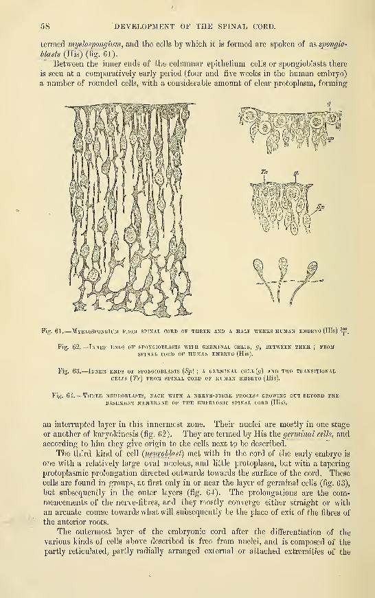
58 DEVELOPMENT OF THE SPINAL COED.
termed myelospongium, and the cells by which it is formed are spoken of as spongio-
blasts (His) (fig. 61).
Between the inner ends of the columnar epithelium cells or spongioblasts there
is seen at a comparatively early period (four and five weeks in the human embryo)
a number of rounded cells, with a considerable amount of clear protoplasm, forming
Fig. 61.
—
Myelospongium from spinal cord of three and a half weeks human embryo (His) ^i?
Fig. 62.
—
Inner ends of spongioblasts with germinal cells, g, between them ; fromSPINAL cord of human EMBRYO (His).
Fig. 63.
—
Inner ends of spongioblasts [Sp] ; a gfrminal cELL(f/) and two transitional
CELLS {Tr) from spinal cord of human embryo (His).
Fig. 64.— Three neuroblasts, each with a nerve-fibre process growing out beyond thebasement membrane of the embryonic spinal cord (His).
an interrupted layer in this innermost zone. Their nuclei are mostly in one stage
or another of karyokinesis (fig. 62). They are termed by His the germinal cells, and
according to him they give origin to the cells next to be described.
The third kind of cell {neurohlast) met with in the cord of the early embryo is
one with a relatively large oval nucleus, and little protoplasm, but with a taperiug
protoplasmic prolongation directed outwards towards the surface of the cord. These
cells are found in groups, at first only in or near the layer of germinal cells (fig. 63),
but subsequently in the outer layers (fig. 64). The prolongations are the com-mencements of the nerve-fibres, and they mostly converge either straight or with
an arcuate course towards what will subsequently be the place of exit of the fibres of
the anterior roots.
The outermost layer of the embryonic cord after the differentiation of the
various kinds of cells above described is free from nuclei, and is composed of the
partly reticulated, partly radially arranged external or attached extremities of the

DEVELOPMENT OF THE SPINAL CORD. 59
spongioblasts. This iiuiy l)e tiila-n to roi>rcsciit the white matter of the cord at this
stage (all the rest representing grey substance) ; ijut there arc at first no nerve fibres
in it, the only structures whieh can be at all compared to nerve fibres being the
prolongations of the neuroblasts, and these lie cither as arcuate fibres altogether in
the outer part of the grey substance, or are passing out of the cord as the beginningsof the anterior roots from a mass of nenrol)lasts which forms the rudiment of the
anterior cornu of grey matter (fig. G5). This mass constitutes in the human embryoof six weeks (fig. (JG) the chief portion of each half of the cord. It forms a con-siderable projection which laterally almost reaches the surfoce, but ventrally is sepa-
rated fn)m it liy a thickening of the external or radial zone, due to the appearance oflongitudinally coursing nerve fibres within it : this is the begiuninj; of the anterior
Fig. 65.
—
Section of spinal cord of four -weeks human embryo (His).
The posterior roots are continued -witliin the cord into a small longitudinal bundle which is the
rudiment of the posterior white column. The anterior roots are formed by ihe convergence of the
processes of the neuroblasts. The latter, along with the elongated cells of the niyelospongium composethe grey matter. The external layer of the cord is traversed by radiating fibres which are the outer ends
of the spongioblasts. The anterior commissure is beginning to appear. This figure is much moremagnified than the next one.
Fig. 66.
—
Transverse section of the cervical part of the spinal cord of a hujianEMBRYO of six WEEKS (from KoUiker). ^f
c, central canal ; e, its epithelial lining ; at e' (superiorly), the original place of closure of the canal;
a, the white substance of the anterior columns; [/, grey substance of antero-lateral horn
; ^;, posterior
column ; ur, anterior roots ; pr, jrosterior roots.
white column (o). By this time, also, although to a rather less extent, the posterior
white columns have, simultaneously with the posterior roots, begun to make their
appearance on either side of the narrow dorsal part of the neural canal (;;). There is,
however, only a relatively thin layer of grey matter (neuroblasts) separating the
posterior white columns from the palisade-like lining of the canal, and as yet no
sign of nerve fibres in the situation of the lateral columns, which are only repre-
sented by a thin layer of the radial myelospongium. The roof and floor of the
canal are also quite thin and undeveloped.
At this period there is still no sign of either anterior or posterior (dorsal or
ventral) fissures of the cord. These become formed as the cornua of the grey matter
grow out from the central mass, and as the anterior and posterior white columns

60 DEVELOPMENT OF THE SPINAL COED.
increase in exfcent. The anterior fissure is simply a cleft left between the enlarging
lateral halves of the cord ; the anterior commissure is formed across the bottom of
the cleft, which is thereby separated from the central canal. As for the posterior
fissure, it is uncertain whether it is in part formed from the dorsal portion of the
constricted canal, which has become occupied by an ingrowth of pia mater, and
converted into a mere septum of connective tissue, or whether this fissure with its
connective tissue septum becomes formed independently of the central canal, which,
as the fissure extends, gradually atrophies until it is eventually conveited into the
rudimentary epithelial tube which is persistent during life.
In the sacral region of birds, the central canal expands into the rhomboidal sinus, and in
the filum terminale of the human cord it remains relatively large. An open enlargement
analogous to the rhomboidal sinus of birds, although relatively smaller, has been described by
Tiedemann in a nine-week human foetus.
The cord is at first oblong oval in section, with an angular depression in each
side which serves to mark off the situation of the future posterior columns and
their coiTesponding grey matter from the antero-lateral region. These two parts of
the lateral neural epiblast may be distinguished as the dorso-lateral (alar) and the
venfcro-lateral (basal) laminse ; with the former, the aflFerent nerve-fibres become
Fig. 67.
—
Brain and spinal coud exposed from behind in a fcetus of
THREE MONTHS (from KoUiker).
/(, the hemispheres ; m, the mesencephalic vesicle or corpora quadrigemiua;
c, the cerebellum ; below this are the medulla oblongata, mo, and fourth ven-
tricle, with remains of the membrana obturatoria. The spinal cord, s, extends to
the lower end of the sacral canal, and shows brachial and crural enlargements.
connected, whilst from the latter the efferent fibres take origin
(His), In the human embryo of six weeks, they are well marked
off from one another, and their respective connections with the
posterior and anterior nerve-roots are very distinct (fig. 66). In
the upper part of the cord, the lateral nerve-roots (spinal accessory)
also arise from the basal lamina. The characteristic cylindrical
form of the cord is only attained with the development of the
lateral columns. The cervical and lumbar enlargements are mani-
fest at the end of the third month.
Up to the fourth month, the cord and the vertebral canal
increase in length pari passu,, but the vertebral column then
begins to grow more rapidly than the cord, so that by the time of
birth the coccygeal end of the cord is opposite the third lumbar
vertebra, while in the adult its limit is the lower end of the first lumbar. Along with
this relative shifting of the cord and its containing tube, the lower nerve-roots lose
their regular rectangular course, and become oblique. They alone, with the
filum terminaU, occupy the lower end of the neural canal, where they form the
Cauda equina.
The nerve fibres of the white columns are at first entirely non-medullated, and
the white substance has a greyish transparent appearance. The medullary sheath
is not formed simultaneously in all parts, but appears at different times in diflferent
parts corresponding with the tracts of conduction ; the last of these tracts to become
medullated are the pyramidal tracts.
The membranes are formed from mesoblast of the protovertebrse, which extends
over and under the cord, and becomes enclosed along with that structure within
the developing vertebral canal. The septa of connective tissue which are seen
penetrating into the substance of the cord from the pia mater grow in from this
mesoblast, carrying blood-vessels amongst the nervous elements. The neuroglia or

DEVELOPMENT OF THE BRAIN. 61
j^enoral snstentacular substance of both white and grey matter is probably derived
from the spongioblasts, and is therefore, like the nerve-cells themselves, of epiblastic
origin.
DEVEIiOPMENT OP THE BRAIN.
We have already traced the development of the cephalic part of the neural tube
as far as the formation of the primary cerebral vesicles. Those, which are at first
three in number (fig. 40), become subdivided so as to form five in all, which may
be termed in succession from before back, the first, second, third, fourth, and fifth
secondary vesicles. Of these five parts the first two, which represent the cerebral and
thalamic parts of the future brain (third ventricle), are derived from the first primary
vesicle, and the last two, the cerebellar and bulbar parts (fourth ventricle), from the
third primary vesicle, while the third, middle, or quadrigeminal part, represents the
undivided second primary vesicle (Sylvian aqueduct). These relationships, as well
as the several parts of the brain which are eventually respectively formed in connec-
tion with the vesicles, are shown in the subjoined table.
' Anterior end of tMrd ventricle, fora-
mina of Monro, lateral ventri-' First secondary vesicle \ cles, cerebral hemispheres, olfac-
(^jJvosenceiJhalon) tory bulbs and tracts, corpora
I. Anterior primary vesi- J I striata, corpus callosum, fornix,
cle or fore-brain|
„ , J -, i Third ventricle, optic nerve andSecond secondary vesicle
),. ,. ,, , • -^ -.
V ,,, , T 7 ^ 1 retina, optic thalami, pituitary(^thalamencephalon)
^ and pineal bodies.
II. Middle primary vesicle / Third secondary vesicle i Aqueduct of Sylvius, coi-pora quad-
or mid-brain \ {mesencephalon) \ rigemina, crura cerebri.
III. Posterior primary vesi-
cle or hind-brain
(Fourth secondary vesicle / / Cerebellum. Pons.
{epencephalon') \ Fourth ven- i
j tricle J
Fifth secondary vesicle \ \ Medulla oblongata.
(jnetcncephalon)
The first and most striking change which occurs in the primary brain is the
outgi'owth on either side of the first primary vesicle of a hollow protrusion {27rim.ary
optic vesicle), which becomes developed eventually into optic nerve and retina
(fig. 68). The changes which it undergoes in the formation of these structures will
be considered when the development of the eye is dealt with ; suffice it for the
present to say that the free hollow communication (optic stalk), which at first exists
between the forebrain and optic vesicle, becomes gradually narrowed and at length
obliterated, and that as development proceeds, the connection of the optic stalk
becomes relatively shifted backwards, so that when the anterior part of the fore-brain
is distinct from the posterior part, or thalamencephalon, the optic vesicle is connected
wholly Avith the latter, a relationship which is maintained permanently, although
partially obscured afterwards by the later connection which is formed between the
optic tract and the mid-brain. Subsequently another pair of hollow outgrowths,
sprouts from the fore-brain, and these rapidly extend forwards, laterally and back-
wards ; they form the vesicles of the cerebral hemispheres. From the roof of the fore-
brain (second vesicle) a median hollow protrusion grows upwards and forwards for a
certain distance towards the vertex, and from the floor of the same vesicle another
somewhat similar protrusion passes downwards and backwards towards the roof of
the mouth. The former is the rudiment of the pineal gland, the latter of the
infimdiiulum, which becomes involved in the formation of the pituitary body.
The principal parts of the brain appear as thickenings in different parts of the

62 DEVELOPMENT OF THE BRAIN.
walls of the vesicles. Thus the corpora striata are formed in the floor of the hemi-sphere vesicles, whilst the principal mass of each hemisphere is formed from theroof and sides (mantle) of those vesicles, and the olfactory lobes are hollow out-
Fig. 68.
—
Fore-part op the embryo shown in fig. 38,VIEWED from the DORSAL SIDE,
'f,(From Kblliker.).
i^, fore-brain ; e, ocular vesicles ; M', mid-brain ; //, hind-brain ; h, part of the heart seen bulging to the right side
;
Voin, omphalo-mesenteric or vitelline veins entering theheart posteriorly ; Mr, medullary canal, spinal part ;
p, protovertebral somites.
growths from them. The cavities of the hemi-
sphere-vesicles become the lateral ventricles, andthe cavity of the part of the fore-brain (or first
secondary vesicle) from which they spring, forms
the anterior extremity of the third ventricle.
The optic thalamus is formed by a thickening
of the lateral wall of the second vesicle, the
cavity of which comes to be the main part of
the third ventricle ; the corpora quadrigemina
are thickenings in the roof, and the crura cerebri
thickenings of the sides and floor of the third
vesicle, which becomes the aqueduct of Sylvius;
the cerebellum and pons are respectively thick-
enings of the roof and floor and the crura cere-
belli of the sides of the fourth vesicle (anterior
part of hind-brain), the cavity of which becomes
the anterior (superior) part of the fourth ven-
tricle ; and finally, the medulla oblongata is
developed as a thickening of the wall of the
fifth vesicle, the cavity of which expands from
the central canal of the spinal cord to form the calamus scriptorius of the fourth
ventricle.
On the other hand, certain parts of the walls of the vesicles become thin andgreatly expanded, and even eventually project into the cavities as folds of epithelium
Fig. 69. — Outline of a longitudinalSECTION THROUGH THE BRAIN OF ACHICK OF TEN DAYS (after Mihalkovics).
/;, cerebral hemisphere ; olf, olfactory lobe
and nerve ; st, corpus striatum ; Iv, lateral
ventricle : ac, anterior commissure ; It, laraina
terminalis; ope, optic commissure;pit, pitui-
tary gland ; inf, infundibulum ; cai, internal
carotid artery ; v^, third ventricle ; dfi, cho-
roid jjlexus of third ventricle;
2^^''^) pineal
gland ; bg, corpora bigemina ; aniv, anterior
medullary velum ; below which two last
references are the aqueduct of Sylvius andcrura cerebri; cbl, cerebellum; v^, fourth
ha, basilar artery : ps, pons Varolii ; cA'*, choroid plexus of the fourth ventricle ; obi, medulla
; r, roof of fourth ventricle.
ventricle;
oblongata
covering ramified vascular expansions of pia mater (choroid plexuses). These
vascular expansions occur along the lower border of the mesial surface of each
hemisphere-vesicle (choroid plexuses of lateral ventricles; ; along the roof of the
second vesicle (choroid plexus of third ventricle), and in the roof of the fifth vesicle
(choroid plexus of fourth ventricle).

DEVELOPMENT OF SPECIAL PARTS OF THE BRAIN. 68
Whilu tliese chaiii^cs are j^oini,^ on in iLs walls the embryonic brain docs not
remain straijj:hfc as at first, with its axis in a line with tliat of tlic spinal cord, but
undergoes certain flexures (fig. 70), the general result of which is to bend the
anterior end towards the ventral surface. The first of these flexures to makeits appearance is a sharp bend opposite the base of the mid-brain and around the ante-
rior end of the notochord. The result of this flexure, which produces a complete
doubling round of the anterior part of the brain, is that the mid-brain is for a time
the most prominent part of the encci)halon. liatcr, the growth of the cerebral
vesicles, and of the thalamencephalon, brings these parts again into prominence, andtends to obscure the flexure, which is, however, never actually obliterated. The second
cerebral flexure, which is also very sharp and well marked, occurs in the region of
the hind-brain (pons Varolii). It is in the opposite direction to the first one, its
concavity being directed towards the dorsum of the embryo, and it produces the
appearance of a deep depression at the part of the brain where it occurs. The third
flexure is a more gradual one. It occurs at the junction of the hind-brain with the
cord, the embryonic medulla oblongata being bent ventralwards fi'om the line of
direction of the medulla spinalis.
The result of these flexures is that the axis of the embryonic brain takes a crook-
shape, passing from the end of the spinal axis at first ventral, then dorsal, and then
again ventral, finally bending sharply backwards towards its termination at the
foramen of Monro.
The second and third flexm-es become eventually almost entirely obliterated with
the further growth of the brain.
FtTBTHER DETAILS EEaARDINa THE DEVELOPMENT OF SPECIAL PARTS OFTHE BRAIN.
The fifth cerebral vesicle: bulbar vesicle, or meteucephalon.—This
part of the embryonic brain, afterwards to become the medulla oblongata, often
shows at its first appearance—especially in the chick—a series of slight constrictions
(fig. 68), which have by some been taken to indicate a segmentation of the neural tube.
But even where they occur they are quite temporary, and the fifth vesicle soon becomesa well marked dilatation opening out from the anterior end of the embryonic spinal
cord. Its wall, like that of all the other cerebral vesicles, is composed of cells
similar to those of the rest of the neural tube, and the histogenetic changes whichoccur to form the nervous tissue are also entirely similar.
Sections across this part of the neural tube are of a compressed oval outline in
the lower part (fig. 71, A,'b), but in the upper part, which afterwards becomes the
lower part of the fourth ventricle, the thinning out and lateral expansion of the
dorsal wall of the tube gives to sections of this and the next (fourth) vesicle the
shape of an irregular triangle, or shield, the base of the triangle being directed
towards the dorsum (roof) and the sides bent more or less sharply inwards about
their middle to unite with one another ventrally at the apex of the triangle (figs.
72, 73). This bend serves to mark a division of each side of the tube into
two parts, a dorso-lateral and a ventro-lateral, which correspond, both in their
situation and in their relationship to afferent and efferent nerves, with the alar
and basal laminse of the embryonic cord (p. GO), with which they are in fact
continuous. The thinning out and lateral expansion of the roof in the region
of the fourth ventricle tends to open up the angle which the ventral laminiB
form with one another, and to throw the dorsal laminte more to the side, so that
what were previously the lateral boundaries of the neural tube come to occupy the
so-called floor of the fourth ventricle, and since in this region the roof becomes

NK
i^^ji^Kiw^%r\ y
Fig. 70. PllOFILE iEVEKAL STAGES,i'lEWS OF THE BRAIN OF HUMAN EMBRYOS AT THREEKECON.STRUCTBD FROM SECTIONS (His).
A. Brain of an emlnyo of about 15 clays (the embryo itself is shown in fig. ]17) magnified 35diameters.
B. Brain of an embryo about three and a half weeks old. The optic vesicle has been cut away.C. Brain of an embryo about seven and a half weeks old. The optic stalk is cut through.A, optic vesicle
; //, vesicle of cerebral hemisphere, first secondary vesicle ; Z, thalamencephalon,second secondary vesicle ; M, mid-brain ; /, isthmus between mid- and hind-brain ; HK fourthsecondary vesicle
;TV, fifth secondary vesicle ; Gh, otic vesicle ; Rf, fourth ventricle ; Nl\ neck
curvature;Br, pons curvature
;Pm, mammilary process ; Tr, infundibulum ; Hp (in B), outline of
Jiypophysis-fold of buccal epi blast ; 111, olfactory lobe. In C the basilar artery is represented along its
whole course.

DEVELOPMENT ol'^ TIIK .MKDI I.LA oULo.Nd A TA. 65
reduced to a thin laycT of tlaitciied t']iiLlii'Iiiiiii, tlie substance of this part of the
medulla ul)l()n,u;ata is wholly lunnrd liya thickcniii}^ of tin- shifted lateral buiiiidarics.
In these, the lieiid luarkiiij;- the disiiiictioii between the ventral anddnrsal laniiiuc
—
now by change of jiosition mesial and external—eontiiuies to l)e evident, and is in
fact recognizable even in sections of the fnlly-develoijcd brain.
Of the longitudinal columns of the medulla oblongata the restiform bodies first bc-comeprominent (third month in the human embryo). Tlie (ant(inor) pyramids arc oljvious in the fifth
month, and the olivary tubercle about the sixtli. liut belore any of these, and indeed with
Fig. 71.
—
Sections across the region op thb calamus scriptorius of the br^in representedIN FIG. 70, A. (His.)
A, region of the glossopharyngeal ganglion.
B, of the auditory-facial ganglion.
Fig. 72.
—
Sections across the fourth ventricle op a somewhat older embryo (His.)
A, section taken through the lower part.
B, across the widest ijaii- (trigeminus region).
C, through upper part (cerebellar region).
r, roof of neural canal : al, alar lamina ; hi, basal lamina ; v, ventral border.
Fig. 73.
—
Sections across the lower half of the fourth ventricle of a still older embryo,
SHOWING GRADUAL OPENING OUT OP THE NEURAL CANAL AND THE COMMENCING FOLDING OVER OK
THE ALAR LAMINA (at/).
1% ventral border ; t, tiEnia ; ot, otic vesicle ; rl, recessus labyrinthi.
In the succeeding stage (not here represented) the angle at c has almost disappeared, the fold / has
extended over the alar lamina, and the two thickened halves are in the same horizoutal plane, covered
by a gi-eatly expanded and thinned out roof.
VOL. I. F
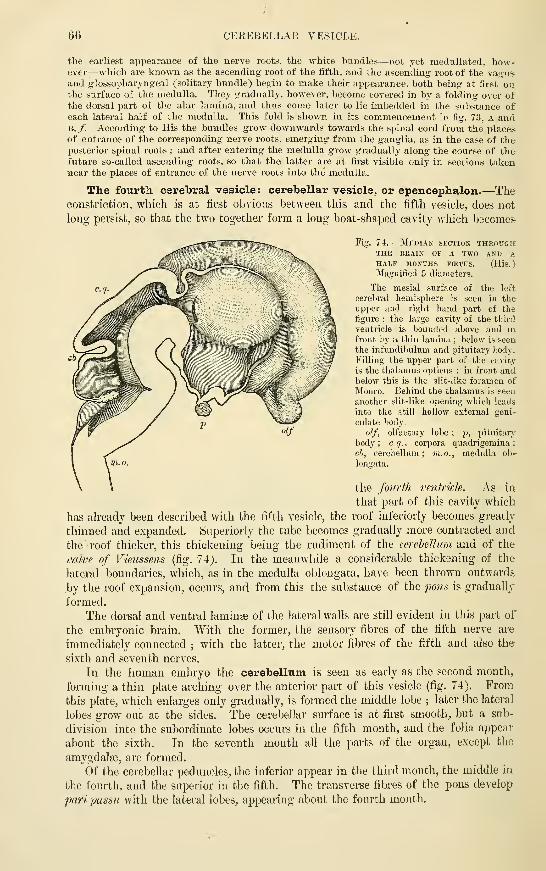
66 OEEEBELLAK VESICLE.
the earliest appearance of the nerve roots, the white bundles—not yet medullated, how-ever-—which are known as the ascending root of the fifth, and the ascending root of the vas'usand glossopharyngeal (solitary bundle) begin to make their appearance, both being at first onthe surface of the medulla. They gradually, however, become covered in by a folding over ofthe dorsal part of the alar lamina, and thus come later to lie imbedded in the substance of
each lateral half of the medulla. This fold is shown in its commencement in fig. 73, A andB,/. According to His the bundles grow downwards towards the spinal cord from the placesof entrance of the corresponding nerve roots, emerging from the ganglia, as in the case of theposterior spinal roots ; and after entering the medulla grow gradually along the course of thefuture so-called ascending roots, so that the latter are at first visible only in sections takennear the places of entrance of the nerve roots into the medulla.
The fourth cerebral vesicle : cerebellar vesicle, or epencephalon.—Theconstriction, which is at first obvions between this and the fifth vesicle, does not
long persist, so that the two together form a long boat-shaped cavity which becomes-
Fig. 7-4. —Median section throughTHE BRAIN OF A TWO AND A,
HALF MONTHS FCETUS. (His.)
Magnified 5 diameters.
The mesial surface of the left
cerebral hemisphere is seen in theupper and right hand part of thefigure ; the large cavity of the third
ventricle is bounded above and in
front bj' a thin lamina ; below is seen
the infundibukim and pituitai-y body.
Filling the upper part of the cavity
is the thalamus opticus ; in front andbelow this is the slit-like foramen of
Monro. Behind the thalamus is seen,
another slit-like opening which leads
into the still hollow external geni-
culate body,
olf, olfactory lobe; f, pituitary
body ; c q., corpora quadrigeraina;
cb, cerebellum; m.o., medulla ob-
longata.
the fourth vmtricU. As in
that part of this cavity which
has already been described with the fifth vesicle, the roof inferioiiy becomes greatly
thinned and expanded. Superiorly the tube becomes gradually more contracted and
the roof thicker, this thiGkening being the rudiment of the cerebellum and of the
valve of Vieussens (fig. 74). In the meanwhile a considerable thickening of the
lateral boundaries, which, as in the medulla oblongata, have been thrown outwards
by the roof expansion, occurs, and from this the substance of the fons is gradually
formed.
The dorsal and ventral laminge of the lateral walls are still evident in this part of
the embryonic brain. With the former, the sensory fibres of the fifth nerve are
immediately connected ; with the latter, the motor fibres of the fifth and also the
sixth and seventh nerves.
In the human embryo the cerebellum is seen as early as the second month,
forming a thin plate arching over the anterior part of this vesicle (fig. 74), Fromthis plate, which enlarges only gradually, is formed the middle lobe ; later the lateral
lobes grow out at the sides. The cerebellar surface is at first smooth, but a sub-
division into the subordinate lobes occurs in the fifth month, and the folia appear
about the sixth. In the seventh month all the parts of the organ, except the
amygdalte, are formed.
Of the cerebellar peduncles, the inferior appear in the third month, the middle in
the fourth, and the superior in the fifth. The transverse fibres of the pons develop
paripassu with the lateral lobes, appearing about the fourth month.
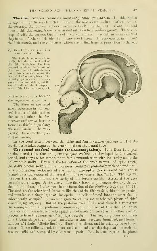
THE Sl'X'OND AM» IIIIKI) ( KltKliKA I. N'ESICLKS. f!7
The third cerebral vesicle : mesencephalon: mid-brain.— Ii: this rc;;iou
no cxpiiiisiiui of tlio Vesicle \vi til tliimiint; nrtlie loof (jcciirs, as in the others, ljiit,uii
the contrary, the roof uiuleruoes coiisideral)le thickening (tig. 74). About the third
month, this thickening becomes separated into two by a median groove. These cor-
respond with the corpora bigcmina of lower vertel)rates ; it is only in mammals that
they become further subdivided by a transverse furrow. This appears in nian about
the fifth month, and the eminences, which are at first large in proportion to the size
¥i<2 7i>.— F(KTAL IIR.MN-
TiiniD MONTH. (Hi
The brain is repiesenteil in
profile, but tlie external ".yall of
the right hemisphere has been
removed to show the interior of
the lateral ventricle with tlie cor-
pus striatum curving round the
bend of the fossa of Sylvius. Thecurved projections above tiie cor-
pus striatum are infoldings of the
mesial wall of the hemisphere
vesicle. The lettering as in tig. 74.
of the brain, thus become
the corpora quailrujemina.
The fibres of the third
nerve originate in the ven-
tral lamina of this part of
the neural tube : the teg-
meniinn and rrusta become "i-o-
formed &s thickenings along
the same lamina : the vesi-
cle itself becomes the aqtie-
duct of Sylvius.
In the constriction between the third and fourth vesicles {isthmus of His) the
fourth nerve takes origin in the ventral plate of the neural tube.
The second cerebral vesicle (thalamencephalon).— It is from this part
of the neural tube that the prir)iar]j optic resides are developed in the earliest
period, and they are for some time in free communication with its cavity along the
hollow optic stalks. But with the formation of the optic nerves and optic tracts,
the stalks become solid, and are. moreover, connected posteriorly with the mid-brain
by a prolongation backwards of the tracts. The optic thalamus of each side is
formed by a thickening of the lateral wall of the vesicle (figs. 74, 78). The interval
between the thalami forms the cavity of the third ventricle. Across it the gi'ey
commissure subsequently stretches. The floor becomes prolonged downwards into
the infundibulum, and takes part in the formation of iha pituitary hody (figs. 69, 74).
The roof, on the other hand, becomes like that of the fifth vesicle, thin and expanded,
and remains as a single layer of flat epithelium cells inflected into the ventricle and
subsequently occupied by vascular growtlis of pia mater (choroid plexus of third
ventricle, fig. 69, ch^). But at the posterior part of the roof there is a transverse
thickening to form the posterior commissure, and in front of this the roof grows
upwards and forwards, but subsequently backwards (in man) as a hollow median
process to form the jnneal yland (epiphysis cerebri). The median process soon takes
on a tubular shape (fig. 6'J, pin), and, after a time, becomes branched, and forms a
number of tubidar follicles lined by ciliated epithelium, and invested by vascular pia
mater. These follicles tend, in man and mammals, as development proceeds, to
become solid and occupied by calcareous deposit. But in some reptiles the pineal
F 2
68 THE FIRST CEREBRAL VESICLE.
tube remains single, and appears as the long stalk—partly hollow, and partly solid
—of a rudimentary median or parietal eye, which occupies an aperture in the middleline of the skull (de Graaf, Baldwin Spencer). The pituitary body (hypophysiscerebri) is chiefly formed by a diverticulum of the buccal epiblast (diverticulum ofRathke) which grows upwards towards the base of the second cerebral vesicle, anddilates into a flask-shaped expansion which is at first simple, but subsequently growsout to form a small mass of epithehal tubes, the lumen of which becomes eventuallyobliterated. Against the posterior wall of this flask-shaped dilatation the infun-dibulum grows down from the floor of the second vesicle, and its extremity becomes
Fig. 76.
—
Median sagittal section of the head in early embryos of thk rabbit. Magnified.
(From Milialkovics.
)
A. From an embryo five millimetres long.
B. From an embryo six millimetres long.
In A, the faucial opening is still closed ; in B, the septum is perforated at /; c, anterior cerebral
vesicle ; mc, mesencephalon ; mo, medulla oblongata ; m, medullary ei^iblast ; ?/ (in B), infundibulum;
spc, spheno-ethmoidal, be, sphenoidal, and spo, spheno-occipital parts of the l^asis cranii ; i, foregut
;
ch, notochord; py, buccal pituitary involution ; am, amnion : h, heart.
Fig. 77.
—
Median sagittal section of the infundibulum and pituitary diverticulum in aRABBIT EMBRYO, AFTER THE OPENING OF THE FAUCES. (From MihalkovicS.
)
he, basis cranii with basilar artery ; if, infundibulum ; tha, floor of thalamencephalon; py, pituitaiy
diverticulum, now closed;
p', stalk of original communication with the mouth;
pJi, pharynx ;
ch, notochord in the spheno-occipital part of the cranial basis.
intimately connected with the dilatation, but without communicating with its
cavity, although bound up together by the same vascular connective tissue. Inconnection with this extension of the infundibulum, nerve cells and fibres becomeformed ; in lower vertebrates they persist and retain their connection with the
brain. The notochord extends in the basis cranii as far as the pituitary body. Just
before reaching this, it bends ventralwards towards Eathke's diverticulum, and here
blends with the buccal epiblast (Bonnet).
Dohrn has shovs^n that in Petromyzon the hypophysis developes as a separate mediandiverticulum of the external epiblast which is formed between the nasal pit in front and thebuccal invagination (stomodasum) behind, and grows straight backwards as a canal of somelength towards the point of the notochord. where follicles develope from it and become con-nected with the infundibulum. Later, its orifice is found to open in common with that of thenasal pit.
The first cerebral vesicle : prosencephalon.—This is represented by the
common point at the front of the third ventricle, whence the hemisphere vesicles
diverge through the foramina of Monro, and by these vesicles themselves. Theoriginal vesicle is therefore relatively small, although its lateral outgrowths form by

Till': FIRST OEKEBRAL VESICLK. GO
far till' largest pcirt lull of tlu' hraiii in hij^hcr vertebrates. Tlic corpora striata
appear as tliickfiiiiijis (if tlif tiour of tlifs hemisphere vesicles, and oiitsitlc them tLu
Fig. 78.
—
Three sections through the foke-brain of a four and a half weeks embryo. (HLs.)
A. Througli the lower .interior part of tbe fore-braiu ; S, fal.\ : Sf. fold of roof pa.ssing below falx
towards the third ventricle ; Bf, fold forming the sulcu.s amuionis ; v. HI, k.I!/., anterior and posterior
parts of olfactory lobe ; Cs, corpus striatum ; 0. JF, groove continuous with optic stalk ; P.s, pai"s
subthalamica ; 7'.c, tuber cinereum.
B. Section a little further back. Sf in replaced by a less prominent but broader fold of the roof.
Ad, which subsequently receives the choroidal vessels, and is therefore the choroidal fold ; i/.y,
hemisphere vesicle ; Th, thalamus ; S.M, sulcus of Monro, below ami behind the thalamus.
C. Still further back : ..If^ choroidal fold here projecting into lateral ventricles, but still free from
mesoblast and bloodvessels ; Ma, mammillary tubercle. The other lettering as before.

70 THE FIRST CEREBKAL VESICLE.
grey and white matter of the island of Reil becomes differeutiated. The rest of the
wall of the hemisphere vesicle {imniJe of Eeichert), although remaining for a time
thinner than the floor, eventually thickens to form the whole of the grey and white
matter of the hemisphere, except along the line where the mesoblast, which is to
form the choroid plexus of the lateral ventricle, passes into the choroidal fold of the
neural epil^last, which becomes thinned out over the invading mesoblast and con-
verted into the epithelial covering of the plexus.
The growth of the hemispheres takes place gradually. They extend at first some-
what forwards and upwards, separated by a thin layer of mesoblast which forms the
f hh
Fig. 79. — Tka^syerse sectio>-s
THROrcH THE BRAIN OF Asheep's E3IBRT0 OF 2 '7 CM. IN
LE^'GTH. (From Kolliker.
)
In A the section passes through
the foramina of !Monro, in B through
the third and lateral ventricles some-
what further back, st, corpus stria-
tum ; th, optic thalamus ; t, third
ventricle ; r. c', rudiment of internal
capsule and corona radiata ; ', lateral
ventricle with choroid jilexus, pi
;
h, hippocampus major; /, primitive
falx ; a, orbito-sphenoid ; sa, pre-
sphenoid : p, pharynx ; ch, chiasma;
o, optic nerve ; m, m, foramina of
]\Ionro ; to, optic tract ; mk, ^Meckel's
cartilage.
falx, but soon begin to pass
jDrogressively backwards, so
that by the end of the third
month they have covered the
region of the optic thalami, by
the fourth month they have
reached the corpora quadri-
gemina, and by the sixth they
cover not only the corpora
quadrigemina, but also a great
part of the cerebellum, iDro-
jecting even beyond this by
the end of the seventh month.
In front, and for some dis-
tance backward over the roof
of the third ventricle, wdiere
the vesicles are not separated
by the falx, their mesial sur-
faces come into contact, and,
during the third month, partly
grow together, but in such a
manner as to leave anteriorly
just in front of the third
ventricle, a triangular ares
free in the middle, but completely surrounded at its periphery by the uuited parts.
Thus is formed the cavity which is known as the ffth ventricle or ventricle of the
septum Jucuhim, which at no time has any communication with the vesicles of the
cerebral hemispheres, nor with any other of the cerebral vesicles.
st-
(
^-A
THK FlIIST CHJlKr.KAL VRSICLE 71
The eoiiunissures of the cerel)ral hemispheres are also formed in tlie united portion
of the mesial walls of the vt'sicles ; the anterior is the earliest to apjxjai-, thuscoincidinii- with the early appearance of the corpora striata, which it unites in trout.
The anterior part of the fornix, with its pillars, and the (•ori)Us albicans (which is
at first single and median) are next formed, followed at a later ]>eriod by the
jiosterior pillars, which are seen runnins: backwards on each side into the comuummonis as soon as this structure becomes distinct. The corjuis callosum is the
last of the commissures to be formed, its anterior part apjjcars first, but as the
hemispheres extend l)ackwards the formation of the commissure accompanies the
l)ackward extension.
The olfactory lobes arc formed as hollow outgrowths fi'om the lower andlateral parts of the hemisphere vesicles (figs. 70, 7-4). In man thoy soon show a
division into two parts, an anterior and posterior ; of which the latter remains in
close connection with the hemisphere vesicle, while the anterior grows out towai'ds
Fig. 80.
—
The surface of the F(£Tal brain at six months. (K. "Wagner.)
This figure shows the foi-mation of the principal fissures. A, from above ; B, from the left side.
F, frontal lobe ; P, parietal ; 0, occipital ; T, temporal ; a, a, a, slight appearance of sulci in thefrontal lobe ;
.>•, Sylvian fissure : *•', its anterior division; witliin it, C, the central lobe ; j; Rolandic
sulcus; p, parieto-occipital fissure.
Fig. 81.
—
View of the inner surface of theRIGHT half of THE F(I:TAL BRAIN OF ABOUTSIX MONTHS. (Reichert.)
F, frontal lobe ; P. parietal ; 0, occipital
;
T, temporal ; /, olfactory bulb ; //, optic
nerve; /}), callo.so- marginal fissure
; ii,p', parts
•of the pai'ieto- occipital fissure ; h, calcarine
fissure; g, g, gyrus fornicatus ; c, c, coi-pus
•cftllosum ; s, septum hicidum ; /, placed be-
tween the middle conimissure and the foramenof ^Innro ; v, in the upper part of the third
ventricle ; v', in the back part of the third
ventricle ; v", in the lower part of the third
ventricle above the infumliljulum ; r, rec^ssus
pinealis;pr, pons Varolii ; C'c. cerebelhmi.
the olfactory area of the external epiblast. After the first month these lobes are
relatively small in size and their cavities become gradually obliterated, but in someanimals, as in the horse, they are large, and their cavities are permanently in
communication with the anterior eornua of the lateral ventricles. Their fiuther
development is given subsequently (p. 7!»).
Formation of the fissures and convolutions. — The enlargement of thecranium does not always keep pace with the growth m extent of the walls of the
t. ^l-

THE FIRST CEREBRAL VESICLE.
hemisphere vesicles, so that it happens that the former are thrown into folclt
separated by sulci, and the surface loses its smooth appearance. Such relatively
Tapid growth occurs during the second and third mouth in the human embryo,
resulting in the production of a number of infoldings of the surface, which are
mostly transverse to the (bent) axis of the brain, although one or cwo on the mesial
surface run parallel to that axis. These infoldings of the surface, which may be
termed temporary ov primitive sulci, necessarily have a corresponding projection into-
the cavity of the thin-walled hemisphere vesicle (fig. 75). During the fourth month,
probably owing to a relatively more rapid expansion of the cranium, most of these
primitive sulci become obliterated, and the cerebral surface is again almost smooth.
Three, however, of the primitive sulci remain as permanent fissures of the brain,^
and since the fissure of Sylvius is also now formed, although in a somewhat different
manner, the hemisphere of the human foetus at the beginning of the fifth month is
marked by four well characterised sulci having corresponding projections into the
interior of its cavity. These permanent primitive sulci are the following :
—
1. The hippocampal sulcus, corresponding with the projection of the cornu
ammonis (hippocampus) into the lateral ventricle.
^nc.pt
pew. OCC.
Fig. 82.
—
Foetal brain op the beginning of the eighth mojith. (Milialkovics.
)
A., from above ; B., from the side ; C, mesial surface.
i?o, Rolandic sulcus : Sy, Sylvian fissure; iMr.occ, parieto-occipital ; calc, calearine
;p)'.c, i^vecentral
;
•pll, parallel ; int.par, intraparietal ; call. mar, calloso-marginal ; unc, iincus.
2. The parieto-occipital sulcus, corresponding with the bend of the posterior
cornu of that ventricle.
3. The calearine sulcus, corresponding with the projection of the calcar avis.
4. The Sylvian fissure, con*esponding with the curve of the lateral ventricle.
To these may be reckoned the longitudinal infolding of the mesial wall of the
^ It is, however, uncertain whether the temporary sulci develope into or whether they are replaced
by corresponding permanent sulci. See on this subject a paper by D. J. Cunningham in the Journal of
Anatomy, Aprjl, 1890.

DEN Kl.oI'MKNT Ul' THK NKIIVES. T6
(icmisphciv just l»eK>\v the liii»iKic-;iiiii»al siiliiis, wliicli i> (.•aiisL(l liv the iujrrowth of
the clioruid plexus of the lateral veiitriele.
AccordinK' to Eoker. the fiMsure of Sylvius is the fii-st of the primitive sulci to ai)pear. It Ls
vi.-ible l)£'fort' the end of the third month, as a wide, shallow depression, which divides the lower
inarjrin <'f thf hemisphere into two nearly e<iual portions, anil at the Ijottom of which is seen
the thiekeninj: of the Hour of the vesic! • from which tlie corjtu^ striatum and the islanrl of
Keil are developed. This fissure ajipears to \)v fonuetl by a curvin;r of the still thin-walleil
hemisphere vesicle over that thickening, around which the vesicle Ijends ; and iU anterior ami
j)osterior parts ultimately meet alony: the line which marks the posterior liinb of the
Sylvian fissure in the develojied brain. The anterior limb is profluced much Iat«r by a further
folding over of that jiart of the mantle which i.s in front of the fossa Sylvii. The fissure
remains until nearly the end of fietal life as a widely open depression, at the bottom of
vhich the island of Keil is readily visible. It closes <,Tadually from behind forwards.
The other sulci are distiniruished from the four aliove-numerated in the faet that
they aiv dejn-es.sious of the surface merely, and lutt infoldinirs of the whole thickness
of the wall of the hemisphere vesicle.^ They begin to apjx-ar about the end of the
•fifth month, the fissure of Rolando being the first to show (figs. 8<J and 81).
By the end of the sixth month the preceutml and inferior frontal sulci, the
intraparietal. the superior occipital, the parallel, the inferior temporal, the callo.so-
marginal, and the collateral fissures have become visilile, as well as the anterior
limb of the Sylvian fissure.
Hy the end of the seventh month (see fig. 82) most of the remaining principal
convolutions and fissures have appeared, and those which were previously pre.sent
have increased both in length and depth. They are, however, all comparatively
short and simple. During the eighth mouth, they (.ontinue to increa.se in length
and depth, and the remaining sulci become gradually developed, but even in the
ninth month there are none of the accessory or secondary fmTows which add so muchto the complexity of the developed brain. The last of the principal sulci to maketheir appearance are the iuferica- occipito-temporal.
DEVELOPMENT OF THE NERVES.
Spinal nerves.—At an early period of development, in some cases even before
the closure of the neural gi'oove, in others during or shortly after that event, there
Fig. S3.
—
Traxsverse section throcgh the trcsk of ax embryoSHARK, TO SHOW THE NEURAL CREST. (BalfoUT.)
nc, neural canal : ]jr, ganglion rudiment running from neural
crest ; ar, sub- notochordal red ; ao, aorta : sc, parietal mesoblast : sp,
visceral mesoblast : n^p, muscle plate ; mp', portion of muscle plate
converted into muscle ; J'v, portion of protoverteV)ra wliicL v,h\ give
rise to the vertebra ; al, alimentaiy canai.mp
grows out bilaterally from the angle of junction of the
neural with the general epiblast (fig. 89), and conse-
quently at the dorsal aspect of the neural tube a,
continuous ridge or crest of epiblast. "which was first de-
scribed l)y Balfour in elasmobranch fishes : this is termed
the neural crest. At intervals along the sides of the
neural crest, corresponding with the middle of each
mesoblastic somite or protovertebra, special clavate
enlargements or outgrowths of the neural crest occur
(fig. 83, ^>;*). These grow downwards along the dorso-lateral aspect of the neural canal,
between the protovertebra and the canal. They remain foi- a time attached above
' An exception must, however, be made for the collateral fi.«sure, which corresponds with the col-
lateral eminence within the ventricle.
74 DEVELOPMENT OF THE NEKVES.
to the dorsal aspect of the neural tube, but that attachment becomes subsequently-
lost, and they then form completely isolated portions of epiblast, composed of oval
cells, and lying at the side of the embryonic cord between it and the muscle plates
of the protovertebrge (fig. 84). These are the rudiments of the posterior-root
ganglia. The remainder of the neural crest disappears : at least in most vertebrata.
Some little time after the sepai-ation of these ganglion-rudiments, the ventral or
anterior roots of the spinal nerves begin to grow out from the ventro-lateral aspect of
the nearal tube. They Avere originally described by Balfour in elasmobranchs, as
forming bud-like outgrowths from the neural epiblast, the outgrowths being com-
posed of spindle-shaped cells (fig. 84, ar). But according to the recent and extended
Fig. 84.
—
Section through the doksal part of the trunk op a torpedo embryo. (Balfour.)
pr, rj, n, spinal ganglion rudiment ; ar, anteiior root ; c7i, notochord ; nc, neural canal ; mp, muscle-
plate.
Fig. 85.
—
Section of the ventro-lateral angle of the spinal cord of a pristiurus embryo
showing the outgrowth of an anterior root-rudiment. (His.)
«.?•, axis cylinder processes of neuroblasts, forming the anterior root ; </, germinal cells in innennost
part of wall of neural canal.
observations of His in various classes of vertebrates, what actually grow out to form
the anterior roots, are the fibrous prolongations (axis-cylinder processes) of neuro-
blasts (v. antea, p. 58), which processes converge to the point of exit of the root
and penetrate gradually into the adjoining mesoblast (fig. 85, a. r), where they
come into close contact with the previously formed ganglion rudiments of the pos-
terior roots.i
The fibres of the posterior roots are developed, according to His, as processes
from the oval cells of the ganglion rudiment. These cells are in fact neuroblasts,
and from either end of each cell, which is thus rendered bipolar (fig. 86), a process
becoming eventually the axis-cylinder of a nerve-fibre grows out, one towards the
central organ, the other towards the periphery. The centrally directed processes
soon reach and grow into the em.bryonic cord at its dorso-lateral aspect, where they
are presently seen in sections occupying an oval area near the periphery of the cord ;
1 The place at which the anterior roots spring from the cord is not opposite to the corresponding
posterior root, but midway between that root and the succeeding one.

("KANIAI- NKKVES. 75
this area is the hoiiinning of thu jfosteriur white cohimn (fig. K7) : the further course
and attaehinents of tlie ingmwinj^ fihres in the cord are not a<-curately known, but
they appear to bifurcate and extend both upwards and downwards (Ilamon y (*ajal).
Tht' peripherally directed fibres urow downwai'ds and join the liundli; of fibres of the
anterior root, to foi-ni tctoethi'r with them the mixed s])injil nerve (fij^. KS),
The axis-cyhnder jn'ocesses whicli are to furni the fibres of the anterior I'oots
Fig. 86.
—
Bipolar cell froji si'inal ganglion of a 4i weeks embryo. (His.) "fo
Fig. 87.
—
Section of spinal cori> of four weeks hcman embrvc (His.)
The posterior roots are continued within the cord into a small longitudinal Lundle which is the
rudiment of the posterior white column. Tlie anterior roots are formed by the convergence of the
processes of the neuroblasts. The latter, along with the elongated cells of the myelospongium compose
the grey matter. The exteraal layer of the cord is travei-sed by radiating fibres which are the outer ends
of the spongioblasts. The anterior commissure is beginning to appear.
begin to make their appearance about the beginning of the fourth week in the humanembiyo (His). Their gi'owth towards the periphery is slow ; eTen by the end
of the second month they hare not reached the tips of the fingers and toes.
Cranial nerves.—The neural crest is continuous along the dorsal aspect of the
cerebral part of the neural titlje as far as, and even beyond the mid-brain. As in
the spinal part, clavate enlargements occur here also, but at somewhat ii-regular
intervals, and form ganglion rudiments which become separated from the dorsal aspect
of the tube, and acquire a new attachment on the lateral and ventral aspect. Such
ganglion rudiments have been described for the third, fifth, seventh, eighth, ninth,
and tenth nerves.
In the chick the ganglion rudiments belonging to the cranial nerves ajijiear as a
thickening of the cephalic epiblast, just where it is folding round into the as yet
unclosed neural canal (fig. 89, vg). This thickening, according to Golowine, is
continuous laterally with a modified portion of the external epiblast (sensory
epiblast), and soon becomes subdivided into three ganglionic groups, and these, later,
into separate ganglions. There is a corresponding subdivision of the sensory

76 CRANIAL NERVES.
epiblast into rudiments of special sense organs, which become gradually shifted
downwards and outwards away from the ganglion rudiments, and form the so-called
Fig. 88.
—
Traksyerse section through the AMEPaoR p.\rt of the trunk of ak embryo of
ScYLLiUM. (Balfour.)
sp.c, spinal cord : sp.g, ganglion of ijo.sterior root ; ar, anterior root; dn, dorsal; sp.n, Yentral
branch 'of spinal nerve ; nvp, muscle plate ; wj/, part of mnscle plate already converted into muscle ;
miy.l, part of muscle plate extending into the limb ; inl, nen'us lateralis ;ao. aorta ; cli, notochord ;
s;-'.'/, 'sympathetic ganglion ; ca.v, cardinal vein ; sd, segmental duct; at, segmental tube;du, duode-
num ; Tip.d, junction of hepatic duct with it;pan, rudiment of pancieas connected with another part
of duodenum ; umc, opening of ximbilical canal (vitelline duct).
" branchial sense-organs " of Beard. They become connected subsequently with
outgrowths from the posterior nerve-rudiments.
These rudimentary sense organs have been described in mammals also (by Froriep). They
appear to represent the special sense organs of the gill clefts of fishes, which Avere first de-
scribed by Leydig (18.50). Beard is of opinion that the nose and ear are also specialised
branchial sense organs, the only ones that are persistent in higher vertebrates. This
differentiation of a special sensory portion of epiblast into rudiments of special sense-
organs, occurs according to Golowine's observations in the chick, not only in the head but also
in the trunk, -^vhere they in all probability represent rudiments of " organs of the lateral line,"
such as are seen in fishes.
Various observers have described the ganglion rudiments as actually becoming fonned at

OlIANIAL NKltVES. 77
least in part at the expense of the •.eusory fpil)la<tic thickenin^rs. In connection with thisquestion it is worthy of note that from the sensory thicki-ninjr which forms the olfactory areaa }ranjrlionic rudiment becomes formed which joins the olfactory lobe of the brain, and ;fivei>
origin to the olfactory nerve-fibres (His).
Even in the adult, jus \va.s shown by Thorasen. traces of pre-existent ganglionic structurecan be found in the root of the third nerve, and similar traces of ganglionic structure have
Fig. 89.
—
Tk.wsvekse sectios
THROIOH THE POSTERIOR
PAKr OF THE IIE.\I> OF ANEMBKYO CHICK <>F :30 HOURS.
(From Ualfoiir. 'i
hh, hiiul-bniiii ; vj, vagus
nerve ; cp, epiplast ; ch, noto-
clionl ; X, sub-notochordal rod;
id, throat ; ht, heart; />/), body-
cavity ; so, parietal mesoblast ;
»f, visceral mesobla.st ; hy, hypo-
blast.
also been described by Gaskell
in the roots of the fourth
nerve, in the motor root of the
fifth, and in the root of the
seventh nerve. If these, as
Oaskell supposes, indicate the
pre-existence of sensory ele-
ments in the roots, it is pro-
bable that these nerves andganglia have all been origi-
nally developed like the posterior gangliated roots of the spinal nerves, as ontgfrowths fromthe neural crest. "VMiether they aie joined by outgi'owths corresponding to the efferent fibres
of the spinal nerves, or whether they originally contain the elements of the efferent fibres,
and thus resemble the spinal nerves of Amphioxus. in which there are no anterior roots,
but both sensory and motor nerves are contained in the posterior roots, is not at present
known. A\Tiat is liowever clear is that the ganglion cells and afferent fibres in the roots of
the third, fourth, motor of fifth and seventh nerves, eventually entirely disappear, the efferent
fibres alone remaining, while in the roots of the sixth, eleventh, and twelfth nerves efferent
fibres only are found, and ganglionic rudiments are not develope<l at alL
As is shown in another part of this work (Neurology; the nuclei of origin of the efferent
cranial nerves are disposed in two longitudinal series. One of these series comprises the nuclei
of origfin of the somatic efferent nerves of Gaskell. which conespond with the largest fibres
of a typical anterior spinal root, and the series of nuclei is a continuation of the cell-columnof the anterior horn ; the nuclei of this series are those of the byjwglossal, or twelfth, thesixth, fourth, and third. The other series comprises the nuclei of origin of the splanchnicefferent nerves of Gaskell. which corresjxjnd vdth the medium-sized and smallest fibres of atj-pical anterior spinal root, and the series is a continuation mainly of the cells of the lateral
comu or intermediolateral tract, and partly, perhaps, of the cells of the base of the posterior
horn ; the nuclei of this series are those of the spinal accessory, those of the efferent fibres
of the vagus and glosso-pharyngeal. the facial, and the motor nucleus of the fifth.
According to the observations of His, the distinction into an anterior or ventral
(somatic) and a lateral (splanchnic) group of efferent fibres is well marked in the
embrjo by the fact that the two kinds of efferent fibres take origin from entirely
different parts of the basal lamina of the neural tube, those which correspond with
the somatic efferent fibres originating from groups of neuroljlasts near the middle
line, while the others take their rise near the junction of the basal with the alar
lamina (see fig. 90). "With the alar lamina itself the afferent fibres are con-
nected, but they do not arise from groups of cells within it, as do the efferent fibres
Avithin the basal lamina : on the contrary, they effect their connection with the lamina
by growing into it from a ganglion in the manner already described for the posterior
roots of the spinal neiwes, and they then appear in most cases to grow downwardsin the direction of the spinal cord. This is stated by His to be the case with the
78 CRANIAL NERVES.
sensory root of the trigeminus, which thus forms the white bundle known as the
ascending root of the fifth, but which in its actual growth is descending ; and also'
with the afferent fibres of the glosso-pharyngeal and vagus which grow downwardsin the medulla to form the so-called solitary bundle.^ These are traceable in the
adult as far down as the middle and lower cervical region respectively, bnt in the
embryo are at first quite short, and hmited to near the place of attachment of the
Fig. 90, A and B.
—
Sectioxs across the hixd-braix of a human embryo, 10 mm. long. (His.) *'.
In A, the origin of tlie spinal accessory and hypoglossal nerves is shown, the fibres of both arising
from gi'oups of neuroblasts in the basal lamina of the neural tube. In B, one of the roots of the hyi^o-
glossal is still seen, and in addition the root of the vagus nerve. Tliis is represented as in part arising
lilie that of the spinal accessory in A, from a group of neuroblasts in the basal lamina, and in part froma bundle of longitudinally coursing fibres placed at the perijjhery of the alar lamina, and corresponding-
in situation to the commencing posterior white columns shown in fig. 87.
nerve roots, becoming gradually longer as development proceeds. The solitary bundle
is at first superficial, like the ascending root of the fifth, but it subsequently becomes
covered in by the bending over of the alar plate, and the formation of nervous sub-
stance in this. The ganglion-rudiments from which the ingrowth of these afferent
fibres takes place, become the Gasserian ganglion of the fifth, and the jugular
ganglia of the glosso-pharyngeal and vagus.
The auditory nerve-roots appear also to be formed by an ingrowth from the cells
of its ganglion-rudiment into the alar plate. Subsequently the ganglion-rudiment
becomes subdivided into three parts, one forming an intracranial ganglion, and the
others giving rise to the branches of the nerve to the cochlea and vestibule respectively
(fig. 90, C). The part belonging to the cochlea (ganglion cochleae) forms ultimately
the spiral ganglion ; while the one on the vestibular branch forms the gangiiform
swelling of Scarpa. Prom a separated part of the ganglion cochlese the nerve to the
posterior semicircular canal passes, as well as that to the macula of the saccule ;
from the vestibular ganglion the nerves to the other ampulla3 and to the utricle, are
derived. The geniculated ganglion of the facial is derived from the same ganglionic
1 These bundles, therefore, are homologous with the rudiments of the posterior white columnswhich are derived from the ingrowing fibres of the spinal ganglia (v, antea, p. 74).
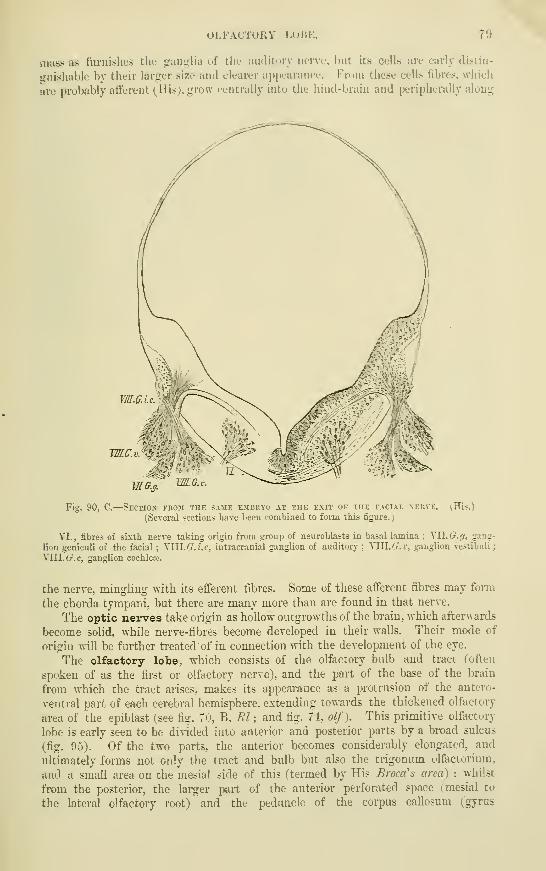
OLFACTORY LoBK. 7»
mass as furnishes the ganu;lia of thu audituiy nerve, but its cells are early distin-
guishable by their larger size and clearer ai)iiL'aranef. From these cells fibres, which
uve probably aticrent (^His), grow centrally into the hind-brain and peripherally along
m.G.
WLC.v.
WG.s.
Fig. 90, C.
—
Section from the same embryo at the exit of the facial xerve, (His.)
(Several sections have been combined to form this figure.
)
VI., fibres of sixth nerve taking origin fi-om group of neuroblasts in basal lamina ; XW.G.'j, gang-
lion geniculi of the facial ; Ylll./r.i.c, intracranial ganglion of auditory ; VlU.Cr.r, ganglion vestibuli j
VIII. 6^.0, ganglion cochleae.
the nerve, minghng with its efferent fibres. Some of these afferent fibres may form
the chorda tympani. but there are many more than are found in that nerve.
The optic nerves take origin as hollow outgrowths of the brain, which afterwards
become soHd, while nerve-fibres become developed in their walls. Their mode of
origin will be further treated of in connection with the development of the eye.
The olfactory lobe, which consists of the olfactory bulb and tract (often
spoken of as the fii-st or olfactory nerve), and the part of the base of the brain
from which the tract arises, makes its appearance as a protrusion of the antero-
ventral part of each cerebral hemisphere, extending towards the thickened olfactory,
area of the epiblast (see fig. 7u, B, Fd; and fig. 74, olf). This primitive olfactory
lobe is early seen to be divided into anterior and posterior parts by a broad sulcus
(fig. 95). Of the two parts, the anterior becomes considerably elongated, and
ultimately forms not only the tract and bulb but also the trigonum olfactoriimi,
and a small area on the mesial side of this (termed by His BroctCs area) ; whilst
from the posterior, the lai-ger part of the anterior perforated space (mesial to
the lateral olfactory root) and the pedimcle of the corpus callosum (gyms

Fig. 91.
—
Ckaihial nerves of a HU^[A^• embryo, 10.2 mm. long. (His.) -J*.
The cranial nerves are inflicated by Roman, the spinal nerves by Arabic numerals.
€./(, cerebral hemisphere ; th, thalamencephalon ; m.h, mid-brain; Mx, maxillary ijrocess ; Mn,mandibular arch ; Hy, hyoid arch ; the facial nerve is seen to send a branch (chorda tympani) aci'oss
-the hyoniandibular cleft ; (?.</, Gasserian ganglion ; eg, ciliary ganglion ; v, vestibular, and c, cochlear
pai-t of auditory ; rj.p, ganglion petrosum of glosso-pharyngeal; y.j, ganglion jugulare of vagus ; an
anastomosis is seen between these; g. tr, ganglion trunci of vagus ; F, ganglion described by Froriep as
belonging to the hypoglossal ; r.d, ramus descendens of hypoglossal • ot otic vesicle. The eye Ls also
represented, and a part of the heart.
fig. 92.
—
Diagram showing the centripetal and centrifugal roots of the cranial nerves of
THE same embryo. (His.
)
The places of exit of the nerves are marked by dotted circles or ovals. The efferent nerves (///.,
IV, mV, VI, VII, part of IX, XI, and XII), are seen to arise within the nerve centre from groupsof
neuroblasts ; the afferent fibres (V.s, VIII, v and c, most of IX, and X), pass a cei-tain distance in-
wards, and for the most part also caudalwards in the nerve-centre, and there end. The ganglion rudi-
ments from which they have grown are not shown here. They will be found in the preceding figure.
SYMPATHETIC NERVES. 81
snbcallosus of Zuckerkandl) are developed. These parts are separated even in tlie
ailult by a sulcus, along which the mesial or internal olfactory root runs.
The olfactory nerve fibres arise, according to His, from neuroblasts whichbecome formed within the thickened epiblast of the olfactory area (see p. 95).
This epiblast at a certain period of development resembles the neural epiblast, andwhilst some of the cells become spongioblasts, others become pear-shaped, or spindle-
shaped, and their processes grow as nerve fibres towards the olfactory lobe. Notonly, however, do these fibres emerge from the olfactory epiblast, but some of the
neuroblasts themselves also pass out, and these form a ganglion which lies between
the olfactory lobe and the olfactory area. Subsequently this ganglion, the cells
of which are prolonged at either end into nerve fibre processes, becomes attached
to and partially invests the olfactoiy bulb, with which it ultimately blends, forming
the part whence the olfactory nerve fibres pass to the Schneiderian membrane(layer of olfactory-glomeruli and nerve fibres), whilst bands of fibres on the other
hand grow centripetally and become the olfactory roots. These are in fact
comparable to the centripetal (so-called " ascending ") roots of the trigeminus,
glosso-pharyngeal, and vagns.
It is not until the third month that the part of the olfactory lobe which formsthe bulb, begins to grow forwards away from the trigonum, and thus to form the
olfactory tract.
The cranial nerves, except the optic and olfactory, and the relations they bear to oneanother and to the visceral arches of the head, are shown in &g. 91 as they occur in the humanembryo of about four weeks. Fig. 92 distinguishes diagrammatically the nerves whi'jh growinto the nerve centres (centripetal or afferent nerves) from those which grow out from thecentres (centrifugal or efferent nerves), and the extent of growth inwards of the former in the€ame embryo.
The S3rinpatlietic nerves and ganglia.—That these are merely outgrowths
of the cerebrospinal nervous system, nearly all recent observations, both morpho-logical and physiological, clearly show. But even before this fact had come to begenerally recognized, it was known that they are developed in connection with the
«pinal nerves (Balfour), and indeed as offshoots from the posterior spinal ganglia
(Schenk and Birdsell, Onodi). They appear for a time as enlargements upon the
main stem of each spinal nerve, but afterwards become connected A\'ith this by a
short branch (r. communicans) (fig. 88), and with one another by a longitudinal
commissure. The branch in question contains the splanchnic fibres of the spinal
nerve, and the sympathetic ganglia are its splanchnic or vagrant ganglia (Gaskell).
The splanchnic ganglia of the cranial nerves are probably formed in a similar way,
but their mode of development has not as yet been worked out.
The ciliary ganglion appears to be formed as an outgrowth of the Gasserian ganglion (fig.
91, e.g.) much in the same way as the sympathetic trunk ganglia are formed as offshoots of theposterior spinal ganglia. In elasmobranchs it is derived from the ophthalmicus profundusganglion, itself an offshoot of the Gasserian (Ewart).
Paterson has recently described the sympathetic chain of ganglia as developing in mammals(rodents) from a continuous rod of mesoblast lying on either side of the aorta, and as becomingonly secondarily segmented and connected %vith the cerebro-spinal nei-ves. But observationsupon earlier embiyos than were used by Paterson are necessary before the mesoblastic originof the rod can be admitted.
RECENT LITERATURE.
Ahlbom, Uiler die Bedeutung der Zirhddriise. Zeitschr. f. -wissensch. Zoologie. Bd. xl., 1884.Barnes, Will., On the development of the posterior fissure of the spinal cord and the reduction of
the central canal in the pig. Proc. Americ. Acad. Arts and Sciences, 1884.Beard, J., On the cranial ganglia and segmental sense organs of fishes. Zoolog. Anzeiger, 1885
;
The system of branchial sense organs and their associated ganglia in Ichthyopsida. Quarterly Joximalof Micr. Science, 1885 ; The development of the peripheral nerxom system of vertebrates. QuarterlyJournal of Micr. Science, Oct., 1888.
Bedot, III., Recherches sur le ddveloppement des nerfs spinaux chez les Tritons. Recueil zoolog.
Suisse, i,, 2, 1884.
82 RECENT LITERATURE.
Beer, B., On the development of the Sylvian fissure in the human emhryo. Journal of Anatomy
BBd Physiology, 1890.
Beraneck, E.j Recherches sur le developpement des nerfs crdniens chez les lizards. Recueil"
zoolog. Suisse, 1884 ; ^tude sur les replis midullaires du poidet. Recueil zoolog. Suisse, iv., 1887.
Chiarugi, Sidlo sviluppo di alcuni nervl cerehrali e spinali. Anat. Anzeiger, 1889.
Cunningham, D. J., The complete fissures of the human cerebrum, and their sif/nificance in^
connection with the growth of the hemisphere and the appearance of the occipital lobe. Journal of
Anatomy, April, 1890.
Dohrn, A., Die Entstehung dei' Hypophysis bei Petromyzon Planeri. Mittheil. aus der zoolog.
Station zu Neapel, iv. , i. , 1883; Ueber die erste Anlage und Entwicklung der motorischen Kuchen-
marksnerven bei den Selachiern, Mittheil. aus der zoolog. Station zu Neapel, Bd. viii., 1888.
E-wart, J. C, On the development of the ciliary or-motor oculi ganglion. Proc. Roy. Soc. xlvii.,
1890.Froriep, A., Ueber ein Ganglion des Hypoglossus u. Wirbelanlagen in der Occipitalregion,
Arch. f. Anat. u. Physiol., Anat. Abth.j 1882 ; Ueber Anlagen von Sinnesorganen am Facialis, Glosso-
pharyngeus und Vagus und iiber die genetische Stellung des Vagus zum Hyjvjglossus und iiber die
Iferkunft der Zungenmusculatnr, Arch. f. Anat. u. Physiolog., Anat. Abthl., 1885.
G-oette, A., Ueber die Entstehung und die Homologien des Hirnanhangs, Zool. Anzeiger, 1883.
Grolo-wine, E., Sur le developpement du systeme ganglionnaire chez le poulet, Anat. Anzeiger, 1890
Graaf, H. W. de, Bijdrage tot de kennis van den bouw en de ontwilckeling der epiphyse bij
Amphibien en Eeptilien. Proefschrift, Leiden, 1886.
His, W., Ueher das Auftreten der weissen Substanz und der Wurzelfasern, am RiicTcermiarlc-
mensehlicher Embryonen, Archiv f. Anat. u. Phys., Anat. Abth., 1883; Zur Geschichte des mensch-
lichen Rilckenmarks und der Nervenwurzeln, Abhandl. d. math. -phys. Kl. d. kgl. Sachs. Gesellsch. d.
Wissensch., Bd. xiii., No. 6, 1886 ; Die Entwicklung der ersten Nervenbahnen beim menschlichen
Embryo. Uebersichtliche Darstellung, Archiv f. Anat. u. Physiol., Anat. Abth., 1888 ; Zur Geschichte-
des Gehirns soivie der ventralen und peripherischen Nervenbahnen beim menschlichen Embryo, Abhandl.
d. math. -phys. Kl. d. kgl. Siichs. Gesellsch. der Wissensch., Bd. xiv., 1888 ; Die Formentivicklung des:
menschlichen Vorderhirns, Abhandl. d. konigl. Sachsischen Gesellschaft, Bd. xv., 1889 ; Die Neurohlnsten
u. dfren Entstehung im embryonalen Mark, Abhandl. d. konigl. Sachs. Gesellschaft, Bd. xv., 1889.
His, "W., jun., Zur Entwicklungsgesch, d. Acustico-facialis-gebietes beim Menschen, Arch. f. Anat.
ti. Physiol., Anat. Abth. 1889.
Hoffmann, C. K. , Ueber die Metamerie des Nachhirns u. Hinterhirns u. ihre Beziehung z. d.
segmevtalen Kopfnerven bei Reptilienembryonen, Zool. Anzeiger, xii.
Johnson, A., and Sheldon, Lilian, On the development of the cranial nerves of the neivt, Pro-
ceed, of the Royal Society, vol. xl., 1887.
Kaczander, J., Ueber die Beziehmgen des Medullarrohrs zu dem Primitivstreifen, Wiener-
Medicin. .Tahrb., 1886.
Kraushaar, E., Entwicklung der Hypophysis und Epiphysis bei Nagethieren, Zeitschi-. f.
wissensch. Zoologie, 1884.
Kupffer, Primdre Metamerie der Neuralrohrs der Vertehraten, Miinchen. Sitzungsb., Bd. xv.
McClure, The primitive segmentation of the vertebrate brain, Zool. Anzeiger, xii.
Marshall, A. Milnes, On the early stages of development of the nerves in birds, Journal of Anatomy
and Physiology, 1877 ; The development of the cranial nerves in the chick. Quarterly Journal of Microsc.
Science, 1878 ; On the head cavities and associated nerves of elasmobranchs. Quarterly Journal of-
MioTosc. Science, 1881.
Mihalkovics, v., Wirbelsaite u. Himanhang, Archiv f. mikr. Anatomic, 1875 ; Entwicklungs-
geschichte des Gehirns, Leipzig, 1877.
Onodi, A. D., Ueber die Entivicklung der Spinalganglien und der Nervenwurzeln, Internat.
Monatsschr. f. Anat. u. Histologic, i., 3, 1884 ; Ueber die Entwicklung des sympathischen Nerven-
systems, Arch. f. mikr. Anat., Bd. xxvi., 1885.
Orr, H., Note on the development of Amphibians, chiefly concerning the central nervous system, d-c.
Quarterly Journal of Micr. Science, xxix., 1889.
Osbom, The origin of the corpus callosum, Morph. Jahrbuch, 1887.
Paterson, A. M., On the fate of the muscle-plate, and the development of the spinal nerves and'
limb pilexuses in birds and mammals. Quarterly Journal of Micr. Science, Aug., 1887 ; The development
of the sympathetic nervous system in mammals. Proc. Roy. Soc, April, 1890.
Rabl, C, Bemerkung iiber die Segmentirung des Hirns, Zoolog. Anz. , 1885.
Rabl-Ruckard, Gehirn der Knochenfische, Arch. f. Anat. u. Physiol., Anat. Abth. 1882 und
1883.
Robinson, A., On the development of the posterior columns, of the posterior fissure, and of the
central canal of the spinal cord. Owens' College Studies, 1890.
Rudingrer, Ueber die BUdung der Augenblasen, Sitzungsb. der Gesellsch. f. Moi-phol. zu Miinchen,
1889.
Spencer, B., On the presence and structure of the pineal eye in LacertUia, Quarterly Journal of
Micr. Science, 1886.
Strahl, H., und Martin, E., Die Entwicklung des Parietalauges bei Anguis fragUis und Zacerta-
vivipara, Arch. f. Anat. u. Phys., Anat. Abth. 1888.
Vigrnal, W., Sur le developpement des eliments de la moelle des mammifhres, Archives de physiol.
1884 ; Recherches sur le developpement de la, substance corticale du cerveau et du cervelet. Archives de
physiologie, 1888.
"Wijhe, J. "W. van, Ueber Somiten und Nerven im Kopfe von Vogel- und Reptilienembryonen,
Zoolog. Anzeiger, 1886,
Zuckerkandl, E., Ueber das Riechcentrum, Stuttgart, 1887.

DEVELOPMENT OF THE EYE. 8a
DEVELOPMENT OF THE EYE.
The first devektpiucnt of the eye occurs us a lioUow protrusion of the antj)dor
cerebraJ..vesicle — primaiy.QpiiiJ.^'esicle—in the manner ah-eady mentioned (see
fio-. 08, Pj'and fij;. 0:5). The vesicle thus formed abuts externnliy against the
e^enUjJUpihliist of the side of the head (fi,u-. OG) ; and this external epibl.ast opposite
the most proniineiit point of the priuuay oi)tLc_vesk;le, becomes tiiicla-ned and in-
O.k.
oi/:
Fig. 94.—SlUF. VIEW OF ANTF.RIOR
PART (IF DilAIN OF SIORK A1>VAXCKD
IIU.MAX KMBKYO, SHOWING THBI'lilMAllY Ul'TIC VESICLK FOLDED
AND CUI'PKI). (His.)
c./(, cereliml lieniisi^liere (part of);
olj., uifactory lobe ; opt., optic cup.
Fig. 93, A,
—
Braik op chick op 2nd day,
VIEWED FROM BELOW, TO SHOW THE FORMA-TION OF THE OPTIC VESICLES BY OUTGROWTHOF THE SIDE OF THE FORE-BRAIN, AND ATTHE SAME TIME BY THE FOLDING OVER OFTHE ENLARGED PART, THE PRODUCTION OP
A GROOVING OR CUPPING OF THE VESICLES.
(His.)
f.h'., m.br., h.br., fore-, mid-, and hind-
brain ; opt., optic vesicle ; i, infundibulum.
Fig. 03, B.— Brain of human embryo opTHREE WEEKS, SHOWING THE PRIMARYOPTIC VESICLES AS OUT-GROWTHS FROJI THEFOREBRAIN. (His.)
Fig. 95.
—
Side view op the same part opTHE brain IN A STILL MORE ADVANCEDEMBRYO, THE EYE HAVING BEEN CUT AWAY.(His.)
opt., cut end of optic stalk, .showing the
manner in which it is folded ; i, infundibu-
lum ; olf.p. ,posterior part of olfactory lobe ;
olf.a., anterior part of the same ; c.h., cere-
bral hemisphere ; t.c, tubes cinereum.
vag'inated, so as to fonn at first a holloF cup-shaped depression with thickened walls
(figs. 97, 98), and subsequently by the closing in of the epiblast at the mouth of the
cup, a hollow island of epithelial cells (fig. 99). This island, which is the mdiiae-ntary
lens, lies l^etween, but is entirely distinct from the external epiblast on the one han"d,
aha' the neural epiblast of the primary, optic vesicle on the other hand. Its forma--
tion is accompanied by a cupping in of the primary optic vesicW figs. 94, 97, 98),
which is invaginated before~lf7'ahd this invagination is increased by an ingrowth
of Eoesoblast, which occurs between the lens and the cupped opUc _y^icle, and
which sulDsequently forms the vitreous^humour, Invaginated in this way the
cavity of the original optic vesicle becomes almost entirely oljliterated, and appears
merely as a cleft between the two layers which form the wall of the so-called " optic
cup." The inner of these two layers is from the first thicker than the outer, and in
it are developed all the parts of the future retina from the membrana limitans interna
G 2
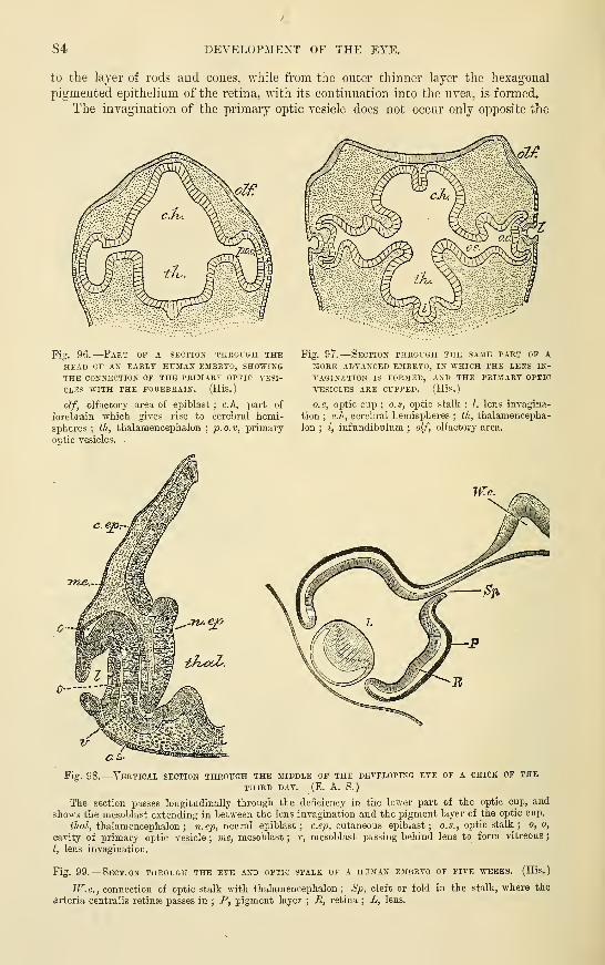
84 DEVELOPMENT OF THE EYE.
to the layer of rods and cones, Avhile from the outer thinner layer the hexagonal
pigmented epithelium of the retina, with its continuation into the uvea, is foimed.
The invagination of the primary optic vesicle does not occur only opposite the
Fig. 96.
—
Part of a section throcgh theHEAD OF AN EARLY HUMAN EMBRYO, SHOWINGTHE CONNECTION OF THE PRIIFARY OPTIC A'ESI-
OLES "SVITH THE FOREBRAIN. (His.)
olf, olfactory area of epiblast ; c.A, part of
forebrain which gives rise to cerebral hemi-
spheres ; ih, thalamencephalon;p.o.v, primaij
optic vesicles.
Fig. 97.
—
Section through the same part op aMORE ADVANCED EMBRYO, IN WHICH THE LENS IN-
VAGINATION IS FORMED, AND THE PRIMARY OPTIC
VESICLES ARE CUPPED. (His.
)
o.c, optic cup ; o.s, optic stalk ; I, lens invagina-
tion ; c.h, cerebral hemispheres ; th, thalamencepha-
lon ; i, infundibulum ; olf, olfactory area.
Tn^.
0-
C.&
Fig. 98.
—
Vertical section thkough the jiiddle of the developing eye of a chick of the
THIRD DAY. (E. A. S.
)
The section passes longitudinally through the deficiency in the lower part of the optic cup, and
shows the mesoblast extending in between the lens invagination and the pigment layer of the optic cup.
</taZ, thalamencephalon ; to. cp, neural epiblast; c.qj. cutaneous epiblast; o.s., optic stalk ;o, o,
cavity of primary optic vesicle ; me, mesoblast ; v, mesoblast passing behind lens to form vitreous
;
I, lens invagination.
Fig. 99.
—
Section through the eye and optic stalk of a human embryo of five weeks. (His.)
W.C., connection of optic stalk with thalamencephalon ; .Sj3, cleft or fold in the stalk, where the
drteria centralis retinse passes in ; P, pigment layer ; R, retina ; L, lens.
DEVELOPMEXT OF THE EYE. 85
place where the lens is becoming involuted, but also below, or ventral, to that place,
so that a section exactly throu_<;-li the middle of thi- ojitic cup at rijrlit au<i:les to the
axis of this part of tlif head, shows a gap iu the boundary of the cup through wliich
the mesoblast is passing into the space between the lens and the invaginated optic
vesicle (fig. \)S, r). This gap or cleft soon becomes closed, but the suture or line of
closure long remains apinu'cnt from the fact that when pigment Iiegiiis to be de-
posited in the eye, tiiis so-called dwroidal^ssure remains for soinfi tiine uiipigmented
(until the sixtli week in man)." ~~
The ventral invadiuition is in mammals continued for a c(»nsiderable distance
into the stalk of the optic_ vesicle (fig. 95), and the simultaneous inclosure of
mesoblastic tissue leads to the introduction of the central bl()od=TPsstriB of the retina
withni"tEeo£tigjp.vye. In birds no such infolding of the stalk occurs.
The lower invagination of the optic cup serves not only to peiTnit of the passage of meso-blast behind the lens for the formation of vitreous humour, but also to establish a direct
connection between the nerve-fibres which are formed along the course of the optic stalk
(future optic nerve) and the centre of the inner laj-er of the optic cup (future retina) (0.Hertwig).
The malformation termed colohoma iridin is attributed to a persistence of the choroidal
cleft, which extends behind the iris along with the retinal pigment or uvea, as far as themargin of the pupil.
Fig. 100.
—
Horizontal section throcgh the eyeOF AN EMBRYO RABBIT OF TWELVE DAYS AND SIX
HOURS. '-°. (Kolliker.
)
o, optic stalk ; h', remains of the ca^-ity of the
primaiy optic vesicle ; p, proximal lamella of the
optic cup (pigmentum nigrum) ; ?•, distill lamella
(retina) ; I, lens invagination, ^^•idely open at ol;
V, papillar elevation in the bottom of the lens
vesicle ; m, mesoblast; y, mesoblast of vitreous
;
r, a blood-vessel at the anterior border of the optic
cup ; e, cutaneous epiblast.
Fig. 101.— Eyeball of a human embryo opFOUR weeks cut ACROSS, AND THE ANTERIORHALF REPRi-SENTED FROM BEHIND. (KoUiker. )
^2°,
fv, the remains of the cavity of the primary
optic vesicle; p, outer layer forming the retinal
pigment ; r, the thickened inner part giving rise
to the columnar and other structures of the retina;
V, commencing vitreous humour within the optic
cup ; v', the cleft throuj;h which a vascular loop,
a, projects from below ; /, the lens with a central
cavity.
The hollow optic stalks are at first freely in communication with the thaiam-
encephalon. or third ventricle. Xerve-fibres grow along their walls, from neuroblasts
which develop iu the retinal epiblast, and pass towards the nerve-centre (His), and
the cavities of the stalks become thereby gradually obliterated, the radially striated
epithelial-like arrangement of the wall being, however, long evident. A new con-
nection becomes subsequently established between the posterior part of the optic
stalks (optic tracts) and the mesencephalon, whilst the middle parts become united
with one another to form the chiasma.

86 DEVELOPMENT OF THE EYE.
The development of the retina from the inner layer of the optic cup, has not
been fullyl\^orked out. In its earlier stages it closely resembles in structure the
wall of the cerebral vesicles, consisting of elongated ..epithelium-like cells, apparently
arranged in several interlocking layers. Of these celjs some become developed into
nerve-fibres and nerve-cells (inner granules and ganglionic layer), others into susten-
tacular tissue, similar to the neuroglia of the central nervous system (molecular
layers, MiiUerian fibres), whilst the outermost layer forms the sense- epithelium (W.
Miiller), or layer of outer granules, which is sharply marked oflF against the
layer of hexagonal pigment cells by the membrana limitans externa, as is the nerve-
fibre layer from the vitreous humour by the membrana limitans interna. For a long
time there is no trace of the rods and cones. These begin to appear some little time
before birth in man and most animals, but in animals which are born blind, such as
kittens, not until after birth (M. Schultze), in the shape of small protuberances of
the sense-epithelium cells growing beyond the limitans externa, and forming at first
the inner segments of the rods and cones, and subsequently the outer segments also.
The latter as they are developed become imbedded in the inner surface of the
bexagonal pigment cells, which have become developed from the outer layer of the
optic cup.
The anterior third of the optic cup does not undergo the changes above
af.n--
,.''!^ 6.6
Fig. 102.
—
Section through the eye of a babbit embryo, mobe advanced in development thait
THAT SHOWN IN FIG. 98. (BalfoUP.)
c, e-pithelium of cornea; I, lens; me.c, mesoblast growing in to form the substantia propria of the
cornea ; o/'. «, optic ner\'e ; rt, retina ; a.c.r, mesoblast for the formation of the vitreous humour, and
the arteria centralis retinffi.
described. Its two layers become here developed into the comparatively simple pars
ciliaris retinse, and in front of the ciliary region they extend forwards and inwards
in front of the lens and in close contact with the back of the iris, where they form
the thickly pigmented epitheliiun, which is known as the uvea and terminates at the
margin of the pupil.
rurther development of the lens.—The hollos: epiblastic vgsicle from
which the lens developes is composed of a thick posterior and a thin anterior layer
which pass into one another at the equator of the lens, and enclose a clear fluid.
In mammals, the vesicle when first formed also contains a small mass of epithelium
cells which have become separated ofP from the posterior wall (fig. 100), but these
THK VITREOUS HUMOUli. 87
afterwards disappear. The thin anterior layer remains throughout life as a simple
layer of cubical cells, and forms the so-called lens-epithelium ; but the cells of the
posterior layer grow forwards into the cavity of the lens-vesicle as the lens-fibres :
those in the middle being the longest and straiglit, while the rest are slightly curved
with their concavity towards the equator, and become gradually shorter towards the
circumference, where they pass through gradually shortening columnar cells {transi-
tional zone) into continuity with the anterior epithelium. By the growth of these
fibres the cavity of the lens-vesicle becomes obliterated.
In this manner the central part of the lens is formed, and it consists in the main
of fibres whicii pass in an antero-posterior direction. The remainder of the lens is
Ibrnifd of fibres which are so disposed as to curve round its margin and over the ends of
the first formed fibres ; they are, moreover, deposited in successive layers and in
three (or more) separate sections, so that their ends abut against one another in front
and behind along tri-radiate (or multi-radiate) lines, such as may be seen in the
macerated lens. These later deposited fibres are all formed at the equator (at the
transitional zone), where chiefly cell-multiplication takes place, and they grow hence
meridionally backwards over the ends of the already developed antero-posteriorly dis-
posed fibres of the central part of the lens.
The capsTile of the lens is early visible as a thin homogeneous. membrane, the
origin of which is still undetermined. According to some observers (Lieberkiihn,
Arnold, Lowe) it is derived from a thin^layer of mesoblast, which passes in between
the lens and the optic cup ; according to others (K6lliker,T^essler, Balfour), it
appeal's before any mesoblast has passed in, and they therefore regard it as a
cuticular deposit fi'om the lens cells. In the human embryo, His figures mesoblast
as existing from the first between the lens invagination and the optic cup (v. fig. 97).
In connection vnth. this question it must be remembered that the substantia propria of the
•cornea (see below), which is formed of connective tissue, and is therefore mesoblastic in
nature, also at first makes its appearance as a homogeneous deposit before any mesoblast cella
have passed in behind the corneal epithelium.^ Its chemical natuie, and its continuity at
the equator with the suspensory ligament and hyaloid membrane, certainly point to the lena
capsule as being a connective tissue, i.e. a mesoblastic structure.
Although the foetal lens like that of the adult is itself non-vascular, it is nevertheless
externally freely supplied with blood-capillaries, which form a vascular tunic completely
surrounding it outside the capsule. These caxjillaries are supplied by a branch of the artcrla
centralis retina; which passes forwards tkrough the centre of the vitreous humour ; in
front, at the margin of the pupil, they come into continuity v/ith the vessels of the iris. Themost anterior part of this vascular tunic forms a membrane which closes the aperture of the
pupil in the middle periods of foetal life. In the human eye the w^hole tunic, together with
the artery which supi^lies its vessels, becomes atrophied and is lost sight of before birth, but
in some animals the^y^^^iZZa?-?/ moiibrane remains apparent for a few daj'S after biith.
The vitreoTis iLumour appears to be fornied from the mesoblastic tissue which
has passed in between the lens and the inner layer of the optic cup by a gradual
formation of a larire quantity of ground-substance, whilst the cells of the tissue
almost entirely disappear. The development of the hyaloid membrane has not been
fully traced out, and the same may be said with regard to the zonule of Zinn. They
are probably both formed by part of the same mesoblast as forms the vitreous humour
(Lieberkiihn, Angelucci).
The corneo-sclerotic coat, the choroid coat, and the iris are all derived from the
mesoblast surrounding the optic cup.
The corneal epithelium is a portion of the external epiblast, which originally
rests against the front of the lens rudiment. The substantia propria comeae
first appears in the chick as a thin homogeneous layer lying immediately within
1 Kessler, however, looks upon this homogeneous deposit as being alBO a cuticular deposit formed by
the epithelial cells.

DEVELOPMENT OF THE EYE.
this epithelium. Into this homogeneous layer mesoblast cells pass fi'om the margin^greatly thickening it and producing eventually the regular layers of fibrous tissue!
which are characteristic of the cornea. Xo cells pass into the most anterior orinto the most posterior stratum, which remain homogeneous (anterior and posterior
Fig. 103. —• Horizontal sectioit
THROUGH THE EYE OF AN IJIBRYORABBIT OF 18 DAYS. ^o
/J£j3j.
liker.)
0, optic nerve; p, hexagonal pig-
ment layei- ; r, retina ; re, ciliaiy
part of the retina; x>\ foi-epart of the
Optic cuj) (rudiment of the iris pig-
mfent); g, vitreous, shrunk away from
the retina, except where the vessels
from the arteria centralis retinje enterit ; i, iris ; mj), membrana pupillaris ;
c, cornea with epithelium e ; pp, pta,
palpebrte ; I, lens ; V, lens epithe-
lium : /, sclerotic ; 7ii, recti muscles.
homogeneous lamellae of Bow-man). The epithelium of the
posterior homogeneous lamella,
or membrane of Descemet, is
derived from mesoblast cells
which grow in like the cor-
neal corpuscles from the mar-
gin and spread themselves
over the posterior surface of
the cornea, thus separating
this from the iris and anterior
surface of the lens. For a
long while, however, there is no anterior chamber ; this eventually appears as a
deft-like space between the cornea and the structures immediately behind it.
In mammals, all the above stages of formation have not been described. Acomplete layer of mesoblast is early visible lying between the corneal epiblast and
the lens epiblast, and continuous around the margin of the lens with the mesoblast
of the vitreous chamber. In this mesoblast a cleft makes its appearance, separating
it into two parts, one of which adheres to the corneal epiblast, where it forms the
substance of the cornea, the other to the lens capsule forming the pupillary membrane.
This cleft is the rudiment of the anterior chamber. It does not become actually
distended with fluid until a short time before birth (Kolliker).
The sclerotic is formed entirely from mesoblast around the optic cup, probably
continuous with that which forms the cornea, although it is only later that the
cornea and sclerotic come to be completely amalgamated.
The choroidjcoat is formed from the mgsoblast which is immediately in contact
Avith the outer layer of the optic cup, and tEeTTonVard growth of the middle tunic
closely follows that of the margin of the cup. The latter ceases at first at the
margin of the lens, but subsequently grows forwards over the front of the lens as a
thin double layer, which is closely covered externally with a continuation of the
choroidal mesoblast. This is the iris, over the back of which both the layers of the
cup-margin eventually acquire pigment and remain permanently as the uvea. Theciliary body is formed by a kind of hypertrophy of the optic cup,' which developes
radial folds, enclosing thin portions of mesoblastic choroidal tissue, in which, as in
the rest of the choroid, numerous blood-vessels and branched pigment-cells become
formed.

DEVELOPMENT OF THE EAR. 89
Accessoxy structures.—Tlie eyelids make their appearance gradually as folds
of integument, subsequently to the formation of the eyeball (fi<^. 103). About the
third month of foetal life the two folds, one forming the upper and the other the
lower lid, meet and unite by a gi-owth together of the epithelium at the margins of
the folds, so as to cut off the conjunctival sac from the exterior. A short time
before birth they again become disunited.
A third fold (of the conjunctiva) appears at the inner canthus, and in many
vertebrates developos into a well-marked third eyelid, the memhrana nicUlans. In
man it remains rudimentary, forming the plica semilunaris.
The glands, hairs, and other structures belonging to the eyelids, are deve-
loped in the same way as the oorresponding structures in the rest of the
integument.
The lachrymal gland is developed in the third month as a number of out-
growths from the deeper layer of the epithelium, at the upper and outer part of the
conjunctival sac. The outgrowths are at first solid, and branch into the surrounding
connective tissue as with other racemose glands, subsequently becoming hollowed
out and differentiated into ducts and acini.
The lachrymal canals and ducts are usually described as being directly
developed by the enclosure of the fissure which separates the lateral nasal process
from the maxillary process (see Development of Nose, p. 95, and figs. Ill, 112),
and which passes in the early embryo from the eye to the upper part of the naso-
buccal cavity (lachrymal fissure). But it has been shown, chiefly by the researches
of Born, that in most animals the canal is at first formed as a thickening of the
rete mucosum of the epidermis, which sinks into the corium along the line of that
fissure. The thickening subsequently becomes separated from the rest of the
epidermis, and hollowed out to form an epithelial tube, which leads from the
conjunctiva into the nasal cavity.
The bifurcation of the duct where it opens on the conjunctiva is produced, according to
Ewetsky, by a broadening out of the epithelial cord at the inner canthus, and its subsequent
separation into two parts by an ingrowth, of connective tissue in its middle, the two parts
developing into the upper and lower lachrymal canals.
DEVELOPMENT OF THE EAH.
The essential part of the ear, viz., the epithelial lining of the labyrinth, is
developed in much the same way as the crystalline lens, as an invagination of the
external epiblast, which at first appears as a pit of thickened epithelium {auditory
jot7, fig. 104, A.), but is^radually converted by a growing together of the margins of
the pit into a hollow island of epiblast, the auditory or otic vesicle (&g. 104, B).
This process occurs somewhat after the fonnation of the eye is laid, and at quite a
different part of the head, viz., on either side of the hind-brain just over the upper
end of the first post-oral visceral cleft. The vesicle comes at first into close contact
with the hind-brain, except where the gangHonic rudiment of the auditory nerve
projects between them, but it subsequently becomes entirely surrounded by meso-
blast, which separates it from both the neural and external epiblast.
The hind-brain does not send out a hollow process towards the otic vesicle
con*esponding to the optic processes of the fore-brain, but the _auditory nerve
developes from a solid outgrowth of the neural crest in the same way as the
posterior roots of the spinal nerves and parts of many other of the cranial nerves
(see p. 78).
The otic_vesiclejs at first flask-shaped, with the somewhat elongated mouth of
the flask directed externalTy towar3s the original point of connection with the

90 DEVELOPMENT OF THE EAE.
exterior. In elasmobranch fishes this connection is never closed, but remains
throughout Hfe in the form of a small duct-like tube which passes up through the
cranial wall and opens on the epidermis. In other vertebrates the connectjon with
the exterior becomes closed—in the chick during the third day—and'what remains
Fig. 104.—Sections through region op the hind-brain of human embryos, showing threeSTAGES IN the DEVELOPMENT OF THE OTIC VESICLE.
A, auditory pits ; B, simple auditory vesicles ; C, auditory vesicles beginning to be fashioned into
parts of the membranous labyrinth ; ot, otic vesicles ; rl, recessus labyrinth! ; v, ventral surface of
hind brain ; r, its roof; /, lateral recess.
Fig. 105.-
—
Outline op the right labyrinth of a SJWEEKS human EMBRYO, TO SHOW ITS RKLATIONS TO
THE PARTS OF THE AUDITORY NERVE. (W. HlS, jllU.)
a.h, section of hind brain;
g.v, ganglion vestibuli in
contact with the upper part of the labyrinth;g.c, ganglion
cochleae in contact with the lower part. The fibres of the
corresponding parts of the auditory nerve which have grown
from these ganglia into the hind brain, are seen to cross
one another; /, facial nerve ; r.l, recessus labyrinthi.
of the original mouth, or canal of connection
with the exterior, is visible as a distinct but
small process from t"fe~Trppef and iiiner angle
of ~Be vesicle, and is known as the recess of
fhe-'Iadyrinth (fig. 104 c, r.l). Evenlually^ it
dereWJ)es'"mto a long epithelial tube, which
passes through the petrous bone, with an expanded end lying within the skull
underneath the dura mater: "This tube and its expanded termination forpa respec-
tively the endoli/mphat'ic canal and sccccule (fig. 106).
In the meantime the auditory^vesicle becomes elongated and begms to be
irregidar. Its ventral end projects as a distinct hollow process, at first straight,
but^soon becoming curved ; this is the rudiment of the epithehal amdgfjhe cochlea.

DK\ i:l()|'mknt of the ear. 91
OthgjiJldlfflaLprojections appear near the dorsal end of the vesicle ; these form
the two superior semicircular canals ; the horizontal camd-ttppeftm-a litj^^eJatAj.-.
The mode of formation of the canals is somewhat pocnliar. They first appear as liattcneil
•semicircular hollow pnitnisious <>f tlic wall of the vesicle. Their sides then come to^.'-cither
and coalesce, except near the circumf('rencc of the semicircle, which now forms a tube con-
nectetl at both ends with the vesicle. Subseciuently a separation or lireach of continuity
occurs over the area of coalescence, so that the rest of the tube is free. One of the endHbecomes dilated into an ampulla and connected with a branch of the auditory nerve.
"Whilst these processes are occnrviiiu' at the dorsal and ventral ends of tlie nowelongated vesicle, a fold, or constriction, of the wall is beginning' to make its
appearance about the middle, and thus the posterior part which is connected with the
semicircular canals becomes gradually separated (as the utricle) from the anterior
part, which forms the saccule, and is connected with the cochlea. This fold extends
into the beginning of the recess of the labyrinth, and separates it longitudinally for a
p.s.c.
Fig. 106.
—
Stages in the development op the membranous labyrinth. (W. His, juu.)
A. Left labyrinth of a human embryo of about four weeks, viewed from the outer side, v, vestibu-
lar part ; c, cochlear part ; r.l, recessus labyrinthi (aquaaductus vestibuli).
B. Left labyrinth with parts of the facial and auditoiy nerves of a human embryo of about 4^
weeks. 6, surface of the hind brain; m, utricular ; s, saccular part of labyrinth; a.s.c, x>-S.c.,
c.s c, rudimentary folds representing the two vertical and the horizontal semicircular canals;
r.l, upper
part of recessus labjTinthi becoming enlarged into the endolymphatic saccule ;c.c, rudiment of cochlea ;
n.v, vestibular branch of auditoiy nerve;g.v, vestibular ganglion (ganglion of Scarpa)
;g.c, cochlear
ganglion ; n.f, facial nerve, with geniculate ganglion, r/.g.
C. Left labyrinth of a human embryo of about five weeks, viewed from without and below. Letter-
ing as before. The horizontal canal is still only a fold. The ampulhe are beginning to be visible on the
two vertical canals.
short distance into two tubes, one of which opens into the utricle, and the other
into the saccule, forming the only permanent means of communication between
their contents. Another fold, or constriction, appears presently, somewhat lower
down, and converts the connection between the saccule and the cochlea rudiment
into the narrow duct of Hensen {canalls re-unims).
In the meantime the cochlea-rudiment at the ventral end of the now labyrinthic
vesicle, becomes elongated into a tube, which, as it grows, becomes coiled upon
itself in such a manner as to produce the spiral structure of this pai-t of the auditory

92 DEVELOPMENT OF THE EAR.
Fig. 107.
—
Transverse and slightly oblique section of the head or a fcetal sheep, in the
REGION of the HIND BRAIN. (From Foster and Balfoui' after Boettcher.
)
HE, innei surface of the thickened vails of the hind brain ; RV, recess of the vestibule ; TB,
commencing vertical semicircular canal ; CC, canal of the cochlea ; GC, cochlear ganglion of the right
side; on the left side. G', the ganglion, and N, the auditory nerve connected ^vith the hind brain.
Fig. 108.
—
Transverse section of
THE HEAD OF A FOETAL SHEEP OF
four-fifths of an inch in LENGTH.
(From Foster and Balfour after
Boettcher.
)
EV, recessus vestibuli ; VB, vertical
semicircular canal ; CC, cochlear
canal ; G, cochlear ganglion ; HB,horizontal canal.
organ. This coiling, however,
only occurs in mammals ; in
birds, the cochlea is a short
straight blind tube.
All these parts of the laby-
rinth are, when first_ formed,
simple epithelial tubes sur-
rouiicTed by and imbedded in
embryonic' lionnective tissue.
As development proceeds, and
the skull begins to form, a
cartilaginous capsule becomes
developed around the several
parts of the labyrinth, and
this at length becomes ossified.
The cartilaginous capsule does
not closely invest tlie epithelial structures ; they are immediately surrounded by
embryonic connective tissue, which forms an internal periosteal lining to the capsiile
and a special covering to the epithelial tube. These two connective tissue membranes
are everywhere separated from one another by gelatinous connective tissue, composed

EXTERNAL AND MIDDLE EAR. 93
of semi-fluid oround substance and branching corpuscles, except along one border,
where they are in continuity. But in the cochlea the gelatinous tissue is above andbelow the epithelial tube, the place of the modiolus being occupied by embryonictissue which is not gelatinous, and is connected with that lining the capsule by
similar non-gelatinous tissue separating the turns of the cochlea from one another,
and also running in the position of the future spiral lamina.
The bone, which is formed by 03sification of the cartilaginous capsule, is of a spongjnature, but it becomes coated internally by layers of compact bone deposited by the periosteal
lining. The modiolus and septa of the cochlea, as well as the osseous spiral lamina, areformed wholly in connective tissue without any preformation in cartilage.
The perilymphatic spaces throughout the whole labyrinth are produced by a
gradual vacuolation and disappearance of the gelatinous tissue which surrounds the
membranous labyrinth. In the cochlea this conversion into perilymph begins in the
proximal turn of the spiral and extends hence towards the distal end. It is only
with the development of these perilymph-spaces (scalae) that the cochlear tube,
which was previously oval in section, acquires the characteristic triangular section
which we see in the fully-formed organ.
The auditory nerve is large and early becomes separated into its two main divisions, vesti-
bular and cochlear. Each division has a large ganglion upon it (fig. 105), which extendsto the anterior wall of the epithelial vesicle, and as the ventral end of the vesicle elongates
and assumes the spiral disposition, the cochlear nerve and ganglion extend along with it andtake the same coiled or spiral form.
The cells which form the wall of the epithelial tube become variously modified in different
parts of the labyrinth to produce the characteristic structures which there occur, viz. : thehaii"-cells, the rods of Corti, the sustentacular cells of Deiters and the epithelium lining thelabyrinth. The membrana tectoria appears as a cuticular deposit over the columnar cells
which are becoming developed into the organ of Corti.
ACCESSORY PARTS OF THE ORGAN OF HEARING. EXTERNAL AND MIDDLE EAR.
While the epithelium of the internal ear is formed by an involution of cutaneous
epiblast in the manner which has just been explained, the middle ear with the
Eustachian tube, and the external auditory meatus with the pinna are formed from
the remains of the first visceral cleft",'and from the parts of the mandibular and
hyoideaB!"afcTi^"wIncF immediaEely iDound the cleft. This cleft at an early period
forms an almost complete communication between the pharynx and the exterior,^ but
the broad cleft becomes gradually converted into a flattened tube, and this is presently
found to be closed, both by the epiblast and hypoblast, which are from the first
in contact at the bottom of the cleft, and also by an ingrowth of mesoblast, the
rudiment of the membrana tympani being thus formed. There is at first no enlarge-
ment of the flattened tube to represent the tympanic cavity, and the ossicles are
developed not within, but altogether outside the tube, in a mass of gelatinous con-
nective tissue, which is continuous with that forming the embryonic membranatympani ; they are formed for the most part by ossification of parts of the carti-
laginous bars, which extend from the otic capsule into the mandibular and hyoidean
visceral arches (see Development of Skeleton). As the tympanic cavity becomes
formed by a gradual enlargement of the blind end of the closed hyomandibular cleft,
the gelatinous tissue retires before it, and as this tissue disappears, the ossicles and
the chorda tympani which were previously entirely enveloped by it, are left projecting
into the tympanic cavity, covered only by thin mucous membrane. The process of
formation of that cavity is not, in fact, completed until after birth, when air becomes
admitted into it thi'ough the Eustachian tube.
* Vide footnote on p. 102,
94 DEVELOPMENT OF THE EAR.
The embryonic tympanic membrane is at first close to the exterior, the external
meatus being scarcely existent, although the several parts of the external ear are very
Fig. 109.
—
Sketches showing the gradual development of the parts op the external earFROM PROMINENCES UPON THE MANDIBULAR AND HTOIDEAN VISCERAL ARCHES. (His. ) Variouslymagnified.
F is an outline sketch showing the several parts of a well-developed adult ear, frd natural size.
1, 2, prominences on the mandibular arch; 3, prominence between the two arches, immediatelyover the cleft, prolonged posteriorly into c, behind the hyoidean arch'; 4, 5, and 6, prominences on thehyoidean arch ; L, in B, otic vesicle (seen also in A) ; K, lower jaw.
Of the prominences enumerated 1 forms the tragus ; 2, 3, and 3c, the helix ; 4, the antihelix
;
5, the antitragus ; and 6, the lobule {vide F).

DEVELOPMENT OF THE NOSE. 95
early distinguishable as slight protnberaaces upon the margins of the shallow cleft-
like (lepivssion which is all that represents the meatus at this stage (fig. 1 10, a). But
as the boily wall becnmes thicker, the cleft becomes deei»ened and more tubular, and
the protuberauces upon the mandil»ular and hyoideau arches Itecotne gradually so
tniusformed and arranged amuud the external orifice as to Ije recognizable as the
several parts of the future pinna. The transformations may readily be understood
from the study of the accompanying series of sketches from IIi.s, which show these
piirts in gradually advancing stages in the human embryo (fig. lu'jj,
DEVELOPMENT OP THE NOSE.
The olfactory organ arises in all verte-brates at an early period of embryonic life
AS a depression of external epiblust {olfaciory pit) on eithej- side of the fore-brain.
The epi blast in this regioiiDecomes thickened, forming an olfactorij area, and a de-
iJt-.*
-pr.gloK
Fig. 110. fROFILE riKW OF TffE HEAD OF AHUMAN EMBRYO OF NEARLY FOrR WEEKS. (His.)
olf,' olfactory depression passing posteriorly
into a deep pit, the rudiment of Jacobson's
organ ; mi, maxillary process : »in, mandibulararch ; liy, hyoidean arch ; 6r', hifi, first andsecond branchial arches.
Fig. 111.
—
Head of ax embryo more adtascei^
IX bevelopmejtt thah that suowx is fig.
110, FKOM BEFORE. (His.)
'pr.f/lohf globular extremity of the mesial nasal
process. The other letters as in fig. 111.
pression then forms in this area surrounded by a raised margin (figs. 96, 97, olf).
The depression soon appeal's pyriform, the smaller end extending as a groove towards
the storaodoeum or buccal invagination (see fig, 110, olf) ; near this end a special
pit is early visible, and becomes developed into Jacobson's organ.
The thickened b^oundaries of each ulfaituiy pit and g.roove ar? formed by the so-
called imsial and lateral nasal processes (tigs. Ill, 112). The mesial nasal processes
are iinT^d' at 'their base by a depressed median pare of the fronto-nasal process, but
are at first separated below, where they terminate in distinct tubercles, termed by
His the (/lobular processes. As development proceeds they extend backwards along
the roof of the emb^onic^mouth, forming the nasal lamTnTT. Eventually the
globular processes coalesce in the middle Hne'tb form^ the fntermaxillary process and
the middle part of the lip, while from the depressed surface between them the
lower part of the nasal septum and the philtnmi are fonued, and by a coalescence of
the nasal laminae the rest of the nasal septum is produced. In rodents a notch leads
from the nasal septum through the upper lip to the mouth, and represents an im-
perfect union of the globular processes.
Above the depressed surface just referred to, is a triangular part of the fronto-
nasal process whidr'fofmg' aii aiigle^wriEE it. This angle eventually becomes the

96 DEVELOPMENT OF THE NOSE.
-^nalcR
frv.vn
jjr. o.l.
Fig. 112.
—
Head of ax ejibryo still more advanced in development. (His.)
A, from above ; B, roof of mouth after removal of lower jaw ; i.m, placed on the fronto-nasal
process and just above its intermediate depressed part; l.n.pr, lateral nasal process; m.n.pr, mesialnasal process ; the other letters as before. The nasal lamince of the globular processes and thepalatine projections of the maxillary processes are seen in B.
fy.#
wVJ''
T'ig. 113.
—
Head of an embryo of about sevenweeks. (His.)
The external nasal processes have united with the
maxillary and globular processes to shut off the
olfactory pit from the orifice of the mouth.
Fig. 114.
—
Head of an embryo more advancedIN development, with the parts of theNOSE AND MOUTH BEGINNING TO ASSUME THEIRPERMANENT RELATIONSHIPS. (His.)
point ofjhe nose, and the triangular surface above it the bridge ; the alse nasi are
formed by the lateral nasal jirocesses. These pfo'cfe'ses'a^^ prominent than the
mesial (fig, 1] 2).~They curve round the olfactory depressions, and meet the maxillaiy.
DEVELOPMENT OP THE XOSE. 97
processes ; between tlif two pnxu'ssi's (lateral nasal ami m ixillary) th(! lachrymal
<i;;ro()ve parses from the eye to the nose (ti^s. HI, 112). 'I'hc maxillary pritecsses
also abut in front against the outwardly curving ends of the processus globulares,
which together form as just mentioned, an intermaxillary process, and the three
eventually coalesce to form the upper l)oundaiy of tlu^ mouth, which is thus shut
off from the anterior (irifice of the nasal fossa) (tig. 11;)). But further back the
Fig. ll'l.—OUTLINK OF A TRANSVKHSK VKKTICAI. SECTION TilKOL'GH THK XOSK AND UI'I'KR JAWS OF A
SHKKI-'S KMBKYO WITH OPEN I'AI.ATE. (Froill Kolliker.)
The lower j;iw ami tongue are removed ; d, dental germs; p, the palate plates approaching each
other in the mitUlie ; /, the nasal fossie ; c, nasal cartilage ; s, septal cartilage; j, the two organs of
Jacohson with their cartilages internally.
olfactory depressions, which are now developed into cleft-like cavities, and are in-
creasing in complexity by the development of the projections which are to form the
turbinate bones, are still freely in communication with the buccal cavity, and it is
only by the growth of the palatine processes of the maxilke (fig. 115,jt;) and their
coalescence in the middle line with one another, and with the lower part of the nasal
septum, that the nasal cavities are cut off from the mouth and from one another,
and now only open posteriorly into the upper part of the pharynx by the posterior
nares {(•Iioamc).
The median or septal part of the external nose, with its columella below, is
formed, as'aljove'statFd^'tif'the co'tdesced mesial^n processes, the alte nasi being
developed from the lateral nasal processes. The sepjum is at first broad and de-
pressed, so that the nostrils are widely separated from one another (fig. 114), a con-
dition which remains to a certain extent permanent amongst some of the dark races
of mankind.
From the above description it will be seen that the olfactory organs are at first altoj^ether
distinct from the mouth, that they subsequently pass backwards, as >;Tooves. deepening into
distinct clefts, along the roof of the mouth and forming in fact the upper part of the embryonicbuccal cavity, and that finally they are again gradually separated from that cavity Ijj- thegrowth of a horizontal septum to form at fir.st the hard and afterwards the soft palate.
The median union of the palate begins in front about the eighth week and reaches theback i^arc and is completed about the tenth week. Imperfect coalescence of these parts pro-
duces the malformations of hare-lip and cleft palate in their various degrees. Usually, how-ever, in man the coalescence is completed at a comparatively early period of foetal life,
although a vestige of the original separation may be found in front at the junction of themaxillary processes with the coalesced globular processes (intermaxillary), as the naso-palatinecanal or incisor foramen, which is occupied by connective tissue, blood-vessels, and a branchof the fifth nerve. In many mammals, however, an actual communication remains through-out life between the nostrils and mouth in this situation.
The organ of .Jacobson is early visible on either side of the nasal septum at its lower part
in the form of a narrow tube, oval in section, running horizontally in the substance of theseptum and opening anteriorly near the upper orifice of the naso-palatine canal. When the
cartilage of the septum becomes formed, a special curved plate of cartilage is seen partially
enclosing this organ ; but both the organ itself and the cartilage are less con.spicuous in manthan in most mammals. According to Geirenbaiu' the rudiment which has been described in
98 RECENT LITERATURE.
the human, embryo as the organ of Jacobson is not really that structure, but represents a
special g-land which occurs in some lemurs in the lower part of the nasal septum.
The epiblast of the olfactory area early becomes thickened, and resembles in structure the
neural epiblast (His). As in the latter, some of the cells (neuroblasts) become pyriform and
nerve-fibre processes grow out from them, whilst the remainder form long sustentacular
columns, which partially anastomose to form a spongework. The neuroblasts subsequently
pass out towards the olfactory lobe as ah-eady described (p. 81).
All the complexities of the nasal fossae (and they are far more complex and labyrinthic in
many animals than in man) are produced by folds and outgrowths of the original simple
depressions, and the thickened epithelium of these depressions extends over all parts of the
cavities which are thus formed. But it is only in the upper part of the nasal fossEe that the
connexion by nerve-fibres with the olfactory lobe becomes established, and it is in this part
only that the true sense-epithelium becomes developed. In the lower, or respiratory part
of the fossse the epithelium remains relatively thin and becomes ciliated.
BECENT LITERATURE.
Eye and Nose.
Born, G-., Die NasenTioIde u. der Thrdnennasenr/anr/ der amnioten Wirhelthiere, Morpt. Jahrb.,
1879 u. 1883.
Disss, J., Die Aushildung der Nasenhohle nach der Geburt, Arch. f. Anat. u. Physiol., Anat.
Abth. 1889.
Ewetzky, Th., Zur EntwicTcelungsgesckicTite des Thrdnennasenganges leim Menschen, Archiv f.
Ophthalmol., Bd. xxxiv., 1888.
G-ottschau, Zur Entioickelung der Sdugethierlinse, Anat. Anzeiger, 18S6.
His, W., AnatomiemenseUicherEmbryonen, Leipzig, 1880—85 ; Unsere Eorperform, tfcc, Leip-
zig, 1875 ; Die FormentwicJcdung des menschlichen Vorderhirns, Abhandl. d. k. Sachsischen Gesell-
schaft, 1889.
Keibel, F., Zur Entwickelung des Glaskorpers, Arch. f. Anat. u. Phys., Anat. Abth., 1886.
Kessler, Zur Entwickelung des Auges der Wirhelthiere, Leipzig, 1877.
KcUiker, A., Zur Entivickclung des Auges und Geruchsorganes menschlicher Emhryonen, Fest-
schrift der Schweizer. Universitat Zurich gewidmet, Wiirzburg, 1883.
Koranyi, A., Beitrdge zur Entwickelung der KrystalUnse lei den WirheUMeren, Intemat. Mo-
natsschr. fiir Anatomie u. Physiologic, 1886.
Legral, Die Nasenhohle u. der Thrdnennasengang, Morph. Jahrb., 1883.
Ear.
Baginsky, B., Zur Entwickelung der Gehorschnecke, Verhandl. d. physiol. Gesellschaft za Berlin,
1885-86.
Beard, J., On the segmental sense organs of the lateral line and on the morphology of the vertebrate
auditory organ, Zool. Anzeiger, No. 161, 1884.
Boettcher, A., Ueber Entwickelung u. Bau des Gehorlabyrinths, Verhandl. d. kaiserl. Leop.
Carol. Acad., Dresden, Bd. 35.
His, "W., jun., Zur Entwickelungsgeschichte des Aeustico-facialis-Gebietes beim Menschen. Arch,
f. Anat. u. Physiol., Anat. Abth., 1889, Supplement Bd.
Hoffmann, C. K., Ueber die Beziehung der ersten Kiementasche su der Anlage der Tuba Eustachii
und des Cavum tympani, Archiv f. mikrosk. Anat. xxiii., 1884.
Noorden, v.. Die Entwickelung des Labyrinths bei Knochenfischen, Arch. f. Anat. u. Physiol.,
Anat. Abth., 1883.
Rudinger, N., Zur Entwickehmg der hdutigen Bogengdnge der inneren Ohres, Sitzungsber. d.
Akademie d. Wissensch. zu Miinchen, Bd. xviii., 1888.
Tuttle, Alb. H., The relation of the external m.eatus, tympanum and Eustachian tube to the first
visceral cleft, Proc. American Academy of Arts and Sciences, 1884.
Vassaux, Recherches sur les premieres pjhases du developpement de Vceil chez le lapin Arch
a'ox)hthalm., 1888.

DEVELOPMENT OF THIC MoLTIl. <.JD
DEVELOPMENT OF THE ALIMENTARY CANAL.
The early development of the primitive alimentary canal has already been briefly
described in treating of the first formation of the embryo (pp. :U, :-j;")), and it was there
explained how the dipping downwards and inwards of the blastodermic layers on
either side of the embryo tends to separate or pinch off the part of the blastodermic
vesicle which is immediately underneath the body of the embryo as a distinct tube
(mid-gut) from the remainder of the vesicle, which is now known as the yolk-sac, while
at the same time similar changes occurring in ft'ont and behind produce the blind
anterior and posterior extremities of the tube which are known as the fore- and hind-
gut respectively. Although the downfolding in question eventually involves all the
layers of the embryonic blastoderm, the epiblast and the part of the mesoblast which
adheres to it, and together with it forms the somatopleure, do nob participate in
the process until afoer the formation of the amnion, so that the alimentary canal for
some time after its formation is enclosed only by the liypbblast and Jis adherent
mesobkst~(^splanciinqpleure), and projects freely into the wide coelom or space
between the splanchnopleure and somatopleure. The mid;:gut also remains for a
time in free communication with the yolk.sac, although the communication becomes
gradually narrowed into the vitelline duct. As the somatopleure afterwards grows
dowiTon eilKer side of the alfmentafy"canal, and becomes pinched in around the
vitelline duct and stalk of the allantois, which are thus united into the umbilical
cord, that part of the coelom which is within the body and around the alimentaiy
canal becomes shut off as the pleuroperitoneal cavity from the remainder, which lies
altogether outside the body, and forms the cavity of the false amnion.
Development of the mouth and of the parts iu connection with it.—The fore-guc terminates blindly at first underneath the head iu the region of the
Fig. 116.
—
Froxtal view of the tpper part of a humaxEMBRTO OF ABOUT FIFTEEN' DAYS, RECOXSTRCCTED FROMSERIAL SECTIONS. (His.) *'
The pericardium is opened to show the heart : between this
and the fore-brain is seen the primitive buccal cavity. A de-
scription of this figure is given on p. 138.
mn.
hind-brain, and the notochord, with the fore- and ,
. o.a.
mid-brain, curve downwards over the blind extre-
mity, the fore-brain thus causing a rounded pro-
minence in fi'ont of and ventral to the extremity of '"•
the alimentary tube (see fig. 4.5). With the develop-
ment of the heart another prominence becomes "l^formed on the ventral side of the fore-gut, a little
''•"•
further back. Between the two prominences, the
one caused by the projection of the fore-brain and
the other of the heart, a wide, shallow pit is enclosed
(fig. lie), at the bottom of which the ejDJblast
which lines it is in contact with the hypoblast of thefore-gut, and. the -two- layere
fuse to" form an epithelial raembnT.ne, which now forms a septum between the
primitive buccal epiblastic involution or stomoda^um and the fore-gut (fig. 117,
p.v.). This stage is met with in the human embryo before the twelfth day (His),
in the rabbit embryo at about the ninth day (Mihalkovics), and in the chick on the
fourth day.
H 2

100 DEVELOPMENT OF THE MOUTH.
The stomodseum deepens at its upper and anterior part, where it forms a
pocket-like protrusion, which grows a certain distance into the angle formed by the
sharp bend which the hinder part of the fore-brain now makes with the mid-brain.
enoi.
-t^i.v-.
u.a-r ^'^^ccZi,.
Fig. 117.
—
Pkofile view of a human embryo of about 15 days, with the alimentary canalSHOWN IN longitudinal SECTION. (His.)
Fig. 118.
—
Similar a'iew of a somewhat older embryo. (His.)
1, 2, -3, 4, 5, are opposite the respective secondary cerebral vesicles ; from the side of the fore-brain
the primary optic vesicle is seen projecting ; ot. otic vesicle ; "p-v., septum between mouth and pharynx
(primitive velum) ; I, commencing liver in septum transversum ; v, vitelline stalk ; all, allantois enclosed
within allantoic stalk; j v., jugular vein ; c.v., cardinal vein ; s.r., sinus venosus within septum trans-
versum ; u.a., umbilical (allnntoic) artery; l.u.v., left umbilical vein ; end., endothelial tube of heart.
The sharp curve of the truidc of the cmbi-yo towards the yolk-sac is normal at this stage.
In fig. 117 the otic vesicle is still oiDen, and there are only two aortic arches ; in fig. 118 the otic
vesicle is clcsed ; there are now five aortic arches. The primitive velum has disijiijeared.
Tliis.^gocket (Eathke) is the iM/PMllMW. cmybri, or pituitary involution of the buccal
epiblast, and comes presently into connection with the infundibular protrusion of the
neural epiblast, the two together forming the pituitary body (see p. G8). It lies
just above and in front of the pharyngeal septum.
The remains of this septum (when it has become broken through to allow of a communica-tion between stomodgeum and fore-gut), have been termed the 2}rimitii-e vehim, hut the septumhas nothing whatever to do with the formation of the permanent velum palati, or with the
isthmus of the fauces. The plane of the septum forms in fact an angle with the plane of the
future isthmus faucium, so that the primitive mouth or stomodEeum does not by any meanscorrespond with the permanent mouth. In fact the floor of the mouth, including the tongue,
is developed hehind the septum, and therefore in connection with the fore-gut rather than withthe stomodEeum, whereas the uppermost part of the pharynX; including the choanas, is in front
of the septum, and therefore belongs to the stoinoda3um.
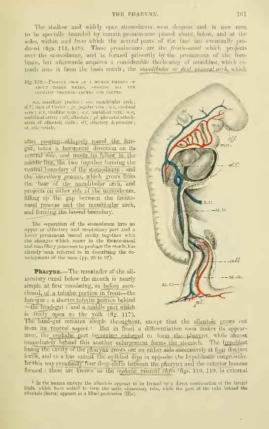
THE PHARYNX, 101
The shallow and widely open stumodajiim 6(jon deepens and is now seen
to be specially bounded by certain prominences placed above, below, and at the
sides, within and from which the several parts of the face arc eventually pro-
duced (fif^s. Ill, 111>). These pruminences arc {\\q fronlo-nasal which projects
over the stomoda^um, and is formed |)rimarily by tlie i)rumiiience of the fore-
brain, but afterwards acfjuires a considerable thickeninir of mesoblast, which ex-
tends into it from the basis cranii ; the mandibular orf[rs^ j;yirfrnl nrrh wliirh
Fig. 119.—PKotMLK VIKW UK A HIM.VN KMURYO OK
AIU'IT THKKK WKKKS, SHuWIXO ALL THECKl'lIALIC VISCKKAL AKCHKS AND CLEFTS.
wx, maxillary process ; mn, niandiltular arch;
d.C, duct of Ciivicr ; ji\ jugular vein ; c.r, cardinal
vein ; r.v, vitelline vein : u.v, umbilical vein ; u.a,
umbilical artery ; all, allantois ;jtl, placentjil attiich-
ment of allantoic stalk ; olf, olfactory depression;
ot, otic vesicle.
aftev_j2S§§ifl^—^^^lil^J~I2-^yL^Jii^ &*^'^'
gut, takes a horizontal^ direction on its
vcntftil side, and m cc,t,^_lts JteIlQw7jjQ_ tlie
middle line, the two together forming the
ventral boundary of the stpmodffilimT"a5id
the maxillarij irracess^ which grows from
the base of the mandibular arch, "and
in'ojects on either side of the stomodfeum,
filling up the gap between the fronto-
nasal j2i"oce8S and the maiidibular arch,
and forming, the lateral boundaiy.
The separation of the stomodaeum into anupper or olfactory and respii-atoiy part and a
lower permanent buccal ca\-ity. together withthe changes which occur in the fronto-nasal
and maxillaiy processes to produce the result, has
already been referred to in describing the de-
velopment of the nose (pp. 9.5 to 97).
Fliar3rnz.—The remainder of the ali-
mentary canal below the mouth is nearly
simple, at first consisting, as Jjefore nien-
tioned,_of a tuljular portion in front-—the
fore-gut : a shorte_r^tubular portion behind
—thelimcl-oiit : and a middle part whichis freely open to the yolk (fig. 117).
The"Tiirid-gut remains simple throughout, except that the allantois grows out
from its ventral aspect.^ But in front a differentiation soon~makes its appear-
ance, the j;ephaljjL-Part bgiiiUiiag^,enJarged to fm-m t,hp phpiypv while almost
immediately behind this another enlargement forms the stomach. The lij:pi)lilast
lining tlir ia\ ity of the pharynx grows out on either side successively at foui" distinct
levels, and to a less extent flie epiblast dips in opposite the hypoblastic outgrowths.
In-this way eventualTy^four deep cleTts Ijetween the pharynx and the exterior become
formed ; these are Yvi()V,'\V^WQ 'ce£ha^^^ defis (fi^. 110, 110, in external
' In the human embryo the allantois appears to be formed by a direct continuation of the lateral
folds, which have united to form the main alimentary tube, while the part of the tube behind the
allantois (bursa) appears as a blind protrusion (His).

102 DEVELOPMENT OF THE TONGUE.
appearance ; f5gs. 117, 118, seen from within). i Between them, and also in front of
the first cleft, the pharyngeal wall is greatly thickened so as to exhibit the appear-
ance of curved bars bounding the clefts ; these bars are known as the cephalic
visceral arcltes, and are five in number, viz. : the Jir^stjii.Jmndthidar, 'in front of the
first'Visceral cleft, "b'etween it and the mouth : this is the seat of formation of the
lowerjaw ; the second or Jiyoid arch, between the first and second clefts ; the third
<yv~thyro-hiJoid "arch, between tlie second and third clefts, which in fishes and
amphibia develops gill-plates, and is therefore also known as the first branchial
arch ; \X\^ fourth l^etween the third and fourth clefts, corresponding with the second
branchial arch of fishes, but small and inconspicuous in man and mammals ; and the
p.fth or second Iranchial still smaller and more inconspicuous, forming the posterior
boundary of the fourth cleft, and hardly recognizable as a distinct bar in man.
After the fourth week, and with the increasing flexure of the head, the arches become
somewhat shifted over one another, so that the fourth arch is concealed by the third,
and the third by the second.
The mandibular arches early become united on the ventral aspect ; from the fifth weektheir union is complete (fig. Ill, 7/;//) and in man shows eventually no sign of a median groove.
The other arches do not at first reach the middle line (His), the space between their central
ends being occupied by the heart and ijericardium ; as these shift backwards a smooth infra-
mandibular surface is left externally.
Development of the tongue.—Within the pharynx, the second and third
arches of the two sides are separated by a foT-kecf elevation {furcuia) with a median
Fig. 120.
—
Posterior aspect of the visceral arches op the embryoSHOWN IN figs. 116, 117, AS SEEN FROM THE INTERIOR OF THEPHARYNX. (His.) ^f
The first or mandibular pair of arches join in the middle line ; the
second arches are separated by a rounded j)rominence (tuberculum
impar). Behind (below) this is the forked prominence (furcula)
bounding a median groove which will become the laryngeal orifice. In
the sections of each of the first two arches, the included artery is seen.
The Roman numerals are opposite the corresponding arches.
groove, in front of which is..a I'ouaided. tubercle {t. impar,
His), which arTses, in ,_ the;. ,arLg.ular space l^etween the
first and seQpnd arches (fig. 120). The' groove around
the furcula {sinus arcuatiis. His) passes laterally into
the visceral clefts. The second and third arches afterwards unite between the
furcula and tuberculum impar (fig. 121, A). Thus united, the junction forms an
X-shaped mass.
From these conjoined ej.tiieniities of the second and t]hjx.d arches on either side, the
root of the tongue grows upwards and forwards as two prominences, which diverge'
in a V-shaped manner to embrace the anterior or papillary part of the organ which
is developed from the tuberculum impar (fig. 121, B). At^tlje angle of the V is a
deep depression {foramen ccecum) ; this leads into a diverticulum, which forms the
median rudiment of"the"t"Hyroid body. When the parts of the tongue are united,
there is still for a considerable time a V'Sliaped groove marking the line of union
(fig. 122), and even in the adult there is often a distinct trace of this groove (sulcus
terminalis, His). Parallel to this, and somewhat in front of it, the papilla vallatse
are developed, and in front of these the other hngual papilla make their appear-
ance (about the end of the second month).
^ According to His, who is confirmed by Born and by Kolliker, these clefts are not as a rule developed
into complete apertures in birds or mammals ; although the membi'anes which close them are composedonly of juxtaposed epi- and hypoblast, the mesoblast having disappeared.
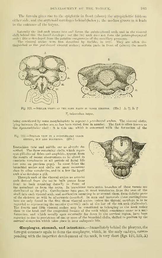
1>K\ KLOI'MKN'I" OK TIIK 'l'()N< I UK. 103
'Vhv fiirciila p^ivcs riso to llir cpii^Icittis in front (above) tin- aryepiirlottic folds on
cither sitk', and the aryti'noid cjirtdiiL^'es beliind (below) ; the median jjroove iu it loads
to the entrance of the larynx.
Laterally the 2ncl arch passes into and forms the palato-trlossal arch, and in the visceral
cleft behind this the tonsil develops ; hut the ;!rd arch docs not form the palato-pharyntrcal
arch ; this is dovcluiicd from the i)alatiuc out',M-o\vtlis of the maxillary processes.
The visceral arches were first described by Hathkc, in isi'.'». Tliey are often dis-
tinjjuished as the pod -buccal viscei'al arches ; certain pai'ts in front of (above; the mouth
Fig. 121. —Similar vikw> of thk samk parts in older embryos. (His.) A. Y, B. 'f
T, tuberculum impr.r.
beinpr considered by some morpholog'ists to represent pirr-hnccal arches. The visceral clefts,
lying between the arches are. as has been stated, four in number. The first is often known as
the Injomandiljuhir cleft : it is this one which is concerned with the formation of the
Fig. 122.
—
Similar view in a considerably olderEMBRYO, BUT LESS MAGNIFIED. (Hi.S.
)
Eustachian tube and middle ear as already de-
scribed. The three remaining clefts, which repre-
sent gill-slits of fishes and amiihibia. appear, fromthe results of recent observations, to be closed in
amniotic vertebrates at all periods of foetal life
(see note on previous page). In some fishes the
branchial arches and clefts are more numerousthan in other vertebrates, and in a few the hyoid
arch also develops a gill.
Through each of the visceral arches an arterial
arch derived from the aoi-tic bulb passes fromfront to back reuniting dorsally in front of
the notochord to form the aorta. In branchiate vertebrates, branches of these vessels are
distributed to the gills. Cartilaginous bars pass, iu most vertebrates, from the base of the
skull into each visceral arch, and ossification occui-ring in or around them, form definite parts
of the skeleton as will be afterwards described. In man and mammals these cartilaginous
Vjars are only found in the first three visceral arches, unless the thyroid cartilage is to be
regarded as representing the anterior (ventral) ends of the bar of the fth arch (Callender).
The fourth and fifth visceral arches may be considered as belonging to the neck rather
than to the head, and the congenital fissures of the neck which sometimes occur as a mal-
formation, and which usually open externally far down in the cervical region, have been
regarded as due to persistence of one or more of the branchial clefts, shifted in position by the
cervical elongation which takes place in later emljr3'onic life.
Olsophagus, stomach, and intestines.—Immediately behind the pharynx, the
fore-gut contracts again to form the oesophagus, which, in the early emljryo, corres-
ponding with the imperfect development of the neck, is very short (figs. 123, 125, A)

104
i
(ESOPHAGUS. STOMACH, AND INTESTINES.
and gradually widens out into the dilatation which represents the stomach. ^ This
organ, which is at first nearly straight (fig. 125, A, 3Ig), soon begins to show the con-
Fig. 123.
—
Sketch of a longitudinal section throughTHE alimentary CANAL OF A HUMAN EMBRYO,SOON AFTER THE DISAPPEARANCE OF THE PRIMITIVE
VELUM. (His.) *f
The alimentary canal is shaded throughout ; U. K,section of mandibular arch ; R. T, hypophysis ; behind
it the I'emains of the pharyngeal septum ; Lrj, com-mencing lung, the future orifice of the lai'jnx being
opposite K ; Mg, stomach ; Lb, liver ; Nb, yolk stalk;
W, Wolffian duct ; B, blind portion of hind gut
;
all, allantois.
vexity of the greater curvature on the side next
the vertebral column, and the concavity of the
lesser curvature on the opposite border (fig,
125, B, Mg), while the pyloric end becomes
tilted away from the vertebral column, pro-
ducing the duodenal loop (fig. 125, C, D_).
Finally the organ becomes turned over on
W'hat was previously its right side, which now^
becomes the posterior surface, and the pyloric
extreuiity being also tilted over, the duodenal
loop is thus thrown over to the right side of the
abdomen (fig. 126). The small intestine is also
at first quite short and straight, with a wide
aperture to the yolk-sac (fig. 125, A, Nh),
but gradually lengthens as the communication
with the yolk-sac becomes more contracted,
and (besides the loop formed by the tilting of
the pylorus) develops a long V^sbaped loop
opposite the attachment of the vitelline duct
(fig. 125, C, t), and fig. 127).
The loop of intestine to which the vitelline ductis attached passes, for a time, into the umbilical
cord, close to its attachment, enclosed in a protrusion of the peritoneal cavity (fig-. 124). Itoccasionally remains in this situation until late in foetal life.
Fig. 12'1.
—
Sketch of the human embryo of theTENTH WEEK, SHOWING THE COIL OF INTESTINE IN
THE UMBILICAL CORD. (Allen Thomson.)
The amnion and villous chorion have been opened and,
the embryo drawn aside from them ; 'v, umbilical vesicle,
connected with the coil of intestine, i, by a small, almost
linear tube. The figure at the side represents the first
part of the umbilical cord magnified ; i, coil of intes-
tine ; vi, vitelline-intestinal duct, alongside of whichare seen omphaJo-mesenteric blood-vessels.
The mesentery is developed bya thinning
out and extension of the mesoblastic tissue
W'hich lies between the intestine and the
vertebral column. It forms a continuous membrane along the wdiole length of the
alimentary canal from the stomach to the rectum, although the part attached to the
1 It has been shown (Balfour, Meuron) in most vertebrates—mammals excepted—that at a certain
period of development the lumen of the oesophagus becomes for a time completely obliterated at its upperextremity.

(KSorilAdUS, STOMACH, AND I.XTESTINRS. 105
Fig. 125.
—
Profile sketches of successive stages in the development of the alimextary canalIN the human embryo. (His.)
Ch, notochord : Sd (in B), median rudiment of thyroid ; P, pancreas ; Lhf), bile duct ; Ds, vitelline
duct;Zfj (in C and 1)), tongue ; N, permanent kidney ; d (in D), cloaca ; An, anus in coui-se of form-
ation ; .SV/, sexual prominence ; St, tail ; Cc, cceciim coli ; Tr, ti-acbea ; A', larynx. The other letter-
ing as in tig. 123.

106 CESOPHAGUS, STOxMACH, AND INTESTINES.
stomach is known as mcsogastriuni and the parts attached to the future colon and-
rectum are termed respectively mesocolon and mesorectum. The stomach begins to
assume its characteristic shape while still lying with its longitudinal axis in the
Fig. 126.
—
Front view of alimextary cakal, rather LEf^s adyaxced inDEVELOPMENT THAN THAT SHOWN IN PIG. 12.5, D. (His.) V)
The pharynx and upper part of cesophagus, and termination of the largeintestine are not represented. Lettering as in fig. 125.
median plane of the body ; it is then seen that the mesogastrium
passes to its greater curvature (fig. 127), which, therefore, is
that corresponding to the mesenteric border of the intestine.
And as the pyloric extremity of the stomach and lesser curva-
ture are tilted forwards and upwards, and at the same time
the whole organ turns over on its right side, the mesogastrium
becomes proportionally lengthened to permit of this change of
position, and the right surface of the stomach (now posterior)
rests against the anterior surface of what was previously
the right side of the mesogastrium, the mesogastrium thus
coming to form the posterior boundary of the omental sac (fig.
128). From near its attachment to the stomach a free fold
subsequently grows over the intestines, and becomes the great
omentum.
The gastro-liepatic omentum is formed by the gradual thinning of a mass of
mesoblastic tissue which from the first connects the ventral wall of the stomach with
the anterior wall of the abdomen, and mthin which the hypoblastic outgrowth
ZZy.
Fig. 127.—DlAGEA3I OF THE MESENTERY, STOMACH AND INTESTINE OF A HUJIAN EMBRYO OF SIX
WEEKS. (Toldt.
)
&t, stomachL;
y.c, greater curvature; l.c, smaller curvature; mg, mesogastrium ;_ sj:!?, spleen;
p, pancreas ; c, CEecum ; r, rectum ; me, mesentery ; ao, aorta ; cl, cceliac axis ; s.me&.a, i.mes.a, supe-
rior and inferior mesenteric arteries.
Fio-. 128.
—
Diagram of a section across the abdomen of a human embryo of the third
MONTH. (Toldt.)
I, I, liver ; k, kidneys ;g.o, gi-eat omentum ; f/.o', omental sac : s.o, small omentum, (The dotted
lino has not lieen carried quite far enough.) The other letters as in fig. 127,
which forms the liver becomes developed. The part of this mesoblastic connexion
which lies between the liver and stomach becomes the gastro-hepatic or lesser
omentum, and its free border which was at first directed downwards (caudalwards)
becomes with the descent of the stomach directed anteriorly (ventrally), and eventu-
ally with the turning of that organ laterally it also is directed towards the right, and
thus comes to form the anterior boundary of the entrance into the omental sac.
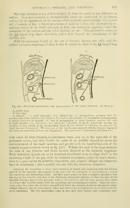
(KSol'llACfS, STOMACH, AND IN'IKSTI N KS. 107
The large intestine is not at first marked oil' from the small \>y any dill'erenee in
calibre. Its commencement is distin<,'uishal)le about the sixth week in the human
emltryo by the appearance of the caecum, whieli jrradually j,fro\vs out (Hgs. \2'>, D, and
lL^7), terming at first a lateral pn^trusion of uniform calibn-, but sul)SL(juently re-
maining narrow at its blind extremity to form ihe vermiform appendix, while the
i-emainder of the caecum and the colon increase in size. This protrusion occurs on
the [J"'^'>"P^''^ '""P '"I'jtjve deserilx'd, and a little beyond the attachment of the
vitelline duet.
AVith the iiicreasini:- length of the gut it becomes thrown into coils, and the
earliest and most important of these is that by which the limb of the [J -''J^'ip*-''^ ^^^V
d'l^^^^^^^^ dicif^f^r,^^
'/lancreas
cdenun
testine.
Fig. 129.
—
Diagrams illcstkatixg the i^eveloioiext of the great ojie>tl-3I. (0. Hertwig.)
A, earlier stage.
B. later stage.
St, stomach ; s.o, small omentum : s'.o', omental sac : o', mcsogastrium, springing from the
posterior wall of the abdomen, near which in A it encloses the pancreas ; o-, attachment of mesogastrium
to greater ciu-vature cf stomach ; (/\ fold of mesogastrium or gi-eat omentum growing over coils
of small intestine; me, mesentery; m.c, transverse mesocolon: o"* (in B), dotted line showing the
situation of that lamella of the mesogastrium which at first assisted in enclosing the pancreas but
which has now disappeared. The next part of this lamella has coalesced with the adjacent lamella of
the transverse mesocolon, and has also disajJijeared. The coalescence is indicated by the black line.
with which the large intestine is continuous turns over on to the right side of the
peritoneal cayity, and thus throws the colon in an archlike disposition across the
commencement of the small intestine, and parallel with the longitudinal axis of the
stomach (commencement shown in fig. 12G). Within this arch of the large intestine
the coils of the jejuniun and ileum become disposed as the intestine lengthens.
Their mesentery spreads out at its intestinal attachment so as to adapt itself to the
increasing length of the gut, Avhile its Tcrtebral attachment, relatively much shorter,
loses to a great extent its primitive disposition, and acquires oblique and transverse
lines of attachment ; this is notably the case Avith the transverse mesocolon.
Althougrh. the mesentery in most parts increases in lengrh and expansion with the further-
growth of the intestine, the contrary is the case -nith the mesenterj' of the duodenum, and of
the ascending- and descendinfr colon. All these parts possess at fiist a complete mesentery like
the rest of the intestine, but that of the duodenum disappears entirel}-, so that this part of the
intestine becomes fixed to the posterior wall of the abdomen, and the same process takes place
to a lesser and variable extent with the ascending and descending mesocolon. Since the trans-
verse colon lies across the abdomen immediately below the stomach, it and its mesentery, trans-
versely disposed, also lie immediately below and behind the mesogastrium (now folded into the
great omentum). The two membranes come in fact into close contact, and eventually com-

108 FORMATION OF THE ANUS.
pletely adhere (4th month and onwards) : and this causes the panci'eas to appear to lie alto-
gether behind the peritoneal cavity, in place of being- situated between the two layers of the
mesogastrium as is at first the case in the human embryo, and as is frequently found in other
mammals during- life.
The free or iioating part of the great omentum is formed by an extension of that part of
the omental fold which turns upwards towards the greater curvature of the stomach fromthe surface of the transverse colon. The fold is at first clearly double, and in some animals
remains so, but in man its two layers coalesce a little while after birth, and after a year or
two can no longer be separated. It extends gradually, first over the transverse colon (third
month), later over the coils of the small intestine.
The spleen becomes formed within the substance of the mesogastrium (fig.
127,s2jI). It is developed \Yholly from mesoblast, and in close connection with the
pancreas. It appears during the second month in the human embiyo, and grows
slowly during faial life, the Malpighian corpuscles being the last parts to appear.
Formation of the anus.—The anal invagination of the epiblast, which eventu-
ally by absorption of the septum between it and the hypoblast of the hind-gut opens
into the alimentary tube, is termed the procfodceum. The junction with the hind-
gut occurs at a little distance from the posterior extremity, so that there is a blindly
terminating post-anal or subcaudal portion of the gut beyond the junction with the
profitodgeum ; this, however, shrinks and disappears even before the absorption
of the sejDtum. This part of the hind-gut represents a cloaca, since it receives
through the allantois the ducts of the urinary and genital organs. The sepa-
ration of the permanent anus from the urogenital orifice, which occurs in all
mammals above monotremes, is the result of a later process of development (see
p. 127).
In mammals the actual amount of proctodasal invagination is very small. Theseptum between the hind-gut and the exterior (anal membrane) is throughout formed
Fig. 130. LONGITTJDIKAL SECTION THROUGH THE POSTEEIOR END OF A SHEEp's EMBRYO, SHOWING THEANAL MEMBRANE. (Bonnet.)
cp, epiblast ; hi/, bypoblast ; mes, mesoblast ; Ji.g, hindgut ; a7n, amnion ; an, anal membrane ;
p.s, prindtive streak ; all, allantois-rudiment.
by two epithelial layers only, viz., hypoblast and epiblast, Avhich here are in contact
with one another without the intervention of mesoblast (fig. 130, an). This con-
dition of juxtaposition of the two layers is in fact directly derived fr'om the union
of the two layers which occurs at the primitive streak and groove, and if the latter
be looked upon as representing the blastopore, the anus may in a sense be considered
to be formed from a part of that aperture. In some lower vertebrates the anus has
been shown to be directly produced from the blastopore.

THE LUNGS. 109
FORMATION OF THE GliANDS OF THE ALIMENTARY CANAL.
Under this licatl may lie iiicliidiMl not, only tliose orj^iuis which an- ordinarily so
termed, but also the lungs, and the ihyiuus and thyroid Ixtdics, since the early
developiiK-nt of these three orj^ans resenibles that of the true scei'eting <,dands.
All the organs above enumerated are formed as epithelial involutions, either solid
at fii-st and afterwards becoming hollowed out, or hollow from the first. As these
epithelial buds grow into the mesoblast, they may either bifurcate or give off lateral
branches, and in this manner all the ramifications of the ducts of the compoundracemose glands are produced. The blind extremities generally end eventually in
enlarged tubular or saccular dilatations. All the epithelium of the gland-saccules
and ducts is derived from the original epithelial sprout, while the basementmembranes and connective tissue and blood-vessels of the gland are derived from thesurrounding mesoblast. The salivary glands and most other glands of the mouth,and part of the hypophysis, which must also be reckoned as a glandular development,are formed in this way by involution of the buccal or stomodseal epiblast ; while thelungs, liver, pancreas, thyroid, thymus, and all the small glands of the rest of thealimentary canal are formed of involutions of the hypoblast. The development ofthe teeth, which also first make their appearance as involutions of stomodajalepi blast
(enamel germs), will be described after their structure has been dealt with (in thepart of this work which is devoted to Splanchnology).
The lungs.—The lungs begin to develope from the ventral part of the pharynxat its junction with the cesophagus, in the beginning of the third day in the chick.
Fig. 131. LCNG RrDIMF.NTS OF HUMAN EMBRYO OF ABOUT 4 WEEKS, SHOAVING THE BUD-LIKE
EXLARGEMENTS WHICH REPRESENT THE LOBES OP THE FUTURE LUNGS. (His.
)
Three buds are seen on the right side, two on the left.
Fig. 132.
—
Lungs of a human embryo muke akvanced in development. (His.)
and in the human embryo at a coiTespondingly early period (fig. 123, Lg). The
lung rudiment is at first single and median, and takes the form of an elongated
vertical diverticulum of the fore-gut, communicating freely with that tube, and of
course lined by hypoblast. Soon the diverticulum sprouts out at its lower extremity
in the form of two tubes which grow downwards on either side behind and on either
side of the heart, into a mass of mesoblastic tissue, which keeps pace in its growth
with the hypoblast ic lung rudiment, and from which the connective tissues of the
future lung become ultimately developed. The extremities of the tubes in question
are early seen to be dilated and lobulated (fig. 131), three lobules being present on
the right tiibe, and two on the left, the division of the lungs into their lobes being
thus early indicated.
The further outgro^vth of the lobulations produces the rudiments of the principal branches
of the bronchi, one for each future pulmonai-y lobe, and each of these branches then gradually
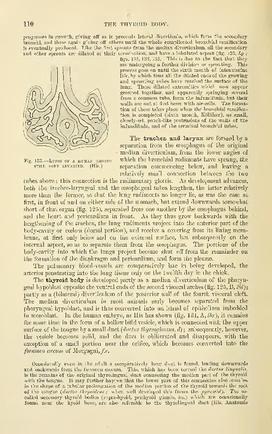
110 THE THYEOID BODY.
Fi 133.—LtJNGS OF A HUMAN EMBRYOSTILL MORE ADVANCED. (His.
)
progresses in growth, gciving off as it proceeds lateral diverticula, which form the secondaiy
bronchi, and these again giving off others until the whole complicated bronchial ramification
is eventually produced. Like the first sprouts from the median diverticulum, all the secondary
and other sprouts are dilated at their termination, and have a lobulated aspect (fig. 125, Lrj ;
figs. 131, 132, 133. This is due to the fact that they
are undergoing a further division or sprouting. This
process goes on until the sixth month of intrauterine
life, by which time all the dilated ends of the growingand sprouting tubes have reached the surface of the
lung. These dilated extremities which now appear
grouped together, and apparently springing several
from a common tube, form the infundibula, but their
walls are not at first beset with air-cells. The forma-
tion of these takes place when the bronchial ramifica-
tion is completed (sixth month, Kolliker), as small,
closely-set, pouch-like protrusions of the walls of the
infundibula, and of the terminal bronchial tubes.
The trachea and larynx are formed by a
separation from the oesophagus of the original
median diverticulum, from the lower angles of
which the bronchial rudiments have sprung, the
separation commencing below, and leaving a
relatively small connection between the two
tubes above : this connection is the rudimentary glottis. As development advances,
both the tracheo-laryngeal and the oesophageal tubes lengthen, the latter relatively
more than the former, so that the lung rudiments no longer lie, as was the case at
first, in front of and on either side of the stomach, but extend downwards somewhat
short of thai organ (fig. 125), separated from one another by the oesophagus behind,
and the heart and pericardium in front. As they thus grow backwards with the
lengthening of the trachea, the lung rudiments project into the anterior part of the
body-cavity or coelom (dorsal portion), and receive a covering fi-om its lining mem-brane, at first only below and on the external surface, but subsequently on the
internal aspect, so as to separate them from the oesophagus. The portions of the
body-cavity into Avhich the lungs project become shut off from the remainder on
the formation of the diaphragm and pericardium, and form the pleura.
The pulmonary blood-vessels are comparatively late in being developed, the
arteries penetrating into the lung tissue only on the twelfth day in the chick.
The thyroid body is developed partly as a median diverticulum of the pharyn-
geal hypoblast opposite the ventral ends of the second visceral arches (fig, ] 25, B, Sd);
partly as a (bilateral) diverticulum of the posterior wail of the fourth visceral cleft.
The median diverticulum in most animals early becomes separated from the
pharyngeal hypoblast, and is thus converted into an island of epithelium imbedded
in mesoblast. In the human embryo, as His has shown (fig. 134, A, thr), it remains
for some time in the form of a hollow bifid vesicle, which is connected with fhe upper
surface of the tongue by a small duct {ductus thyreogloss^is, d) ; subsequently, however,
the vesicle becomes solid, and the duct is obliterated and disappears, with the
exception of a small portion near the orifice, which becomes converted into the
foramen ccpxum of Morgagni, fx.
Occasionally even in the adult a comparatively long duct is found, leading downwardsand backwards from the foramen csecum. This, which has been termed the ductus linr/imlis,
is the remains of the original thyrolmgual duct connecting the median part of the thj^roid
with the tongue. It may further happen that the lower part of this connection also remains
in the shape of a tubular prolongation of the median portion of the thyroid towards the root
of the tongue Qdnctus thnjoideus ; when well developed this forms the 2)yram'id^. The so-
called accessory thyroid bodies (supra-hyoid, prEehyoid glands, &c.) which are occasionally
found near the hyoid bone, are also referable to the thyrolingual duct (His, Anatomie

THK THYMUS. HI
menschlichpv Emhrvonen, iii., p. inl, wlioro roferonco to the literature of these acccpsory
thyroi<ls may be loiind).
The hihitcval diverticulii, which assist in tlie formation of the thyroid body, spring
from the fourth visceral cleft (Born) (Hi;-. 1;!."), ///;•'). They have at first the a])j)earance
of simi)le saccuhir j,dands partially encircling the developing larynx (fig. l;M, ////').
In front of this they come into connection with the median rudiment, and eventually
hlend with it. Tjikc thab rudiment, they become entirely se))arated from the hypo-
blastic surface from which they have taken origin, their cavity disappears, and they
Fig. 134.
—
Sketches snowixo the condition op the thyroid and thymcs gland.s in a humanEMBRYO or ABOUT FIVE WEEKS. (Hls.)
A, profile sketch fiom the left side.
B, frontal sketch from behind.
t, tongue; fc, foramen ctecum ; d, ductus thyreoglo.ssus ; ep, epiglottis ; opposite I, larj'nx ; tr,
trachea ; ce, oesophagus ; thr, median rudiment of thyroid ; thr', lateral rudiment of thyroid ; thm,
developing thymus, seen on the left side of B to be connected with a visceral cleft ; ao (in B), ascending
aorta ; ao', descending aorta ; c, cai'otid.
are converted into ramifying and anastomosing cell-cyhnders, between which vascular
connective tissue becomes developed. The cell-cylinders subsequently become hol-
lowed out, and finally are subdivided by growth of the connective tisstie into small
vesicles, which gradually become larger from accumulation of colloid in their interior.
In most Vertebrates, the lateral and median parts of the thyroid remain distinct ; the
former are the organs known as snpra-jjcrirardial boflir.t in elasmobranchs, and as arrrxsory
tlnjrokls in other animals. Only in mammals do they become united into one organ as
in man.
The thynms is also developed as a growth of the epithelium (hypoblast) of some
of the visceral clefts ; in birds from the third and fourth (fig. 13-"), fh>/i)i), in reptiles
from the second, third, and fourth, and in lower Vertebrates from several clefts (de
Fig. 13.5.
—
Diagram sho'svinc; the visceral clefts from which theTHYMUS and lateral PARTS OF THE THYROID ARE DEVELOPED IN
THE chick, (de Jleurori.
)
1, 2, 3, 4, indicate the corresfionding visceral clefts ; thyni, rudiments of
thymus ; thr, median rudiment of thyroid ; thr', lateral rudiments of
thyroid.
IMeuron). In mammals the thymus appears as a (bilateral)
tubular prolongation backwards of the extremity of the
third visceral cleft (Kolliker), receiving, according to de
IMeuron, an accession from the hypoblast of the fourth cleft,
as in birds. The tube, which has a narrow lumen, and
comparatively thick epithelial lining, is surrounded by vascular connective tissue,
within which numerous lymphoid cells become accumulated, and grows downwards
along the side and in front of the trachea, where, in mammals, it generally unites

112 THE LIVER.
with its fellow to form a median organ. Its lower end then gives off solid, bud-like
excrescences, and lateral buds come off again from these, so that this part of the
organ acquires a ramified, lobulated appearance hke an acinous gland. The acini are,
however, solid, and remain so, although the upper end of the tube still has a narrow
lumen.
The lymphoid cells next invade the epithelium, growing into every part of the
tubular gland, and converting it into a mass of adenoid tissue. In this process the
epithelium becomes broken up into small isolated portions, some of which remain in
the medullary portion of the lobules as the epithelial nests which are seen in sections
of the fully developed organ, and are known as the concentric corpuscles of Hassall.
The liver.—This organ arises in the form of two diverticula of hypoblast, which
grow from the ventral wall of the duodenum immediately beyond the stomach (figs.
117, 118, 1, 123, Lh). They extend into a mass of mesoblastic tissue which connects
the stomach and duodenum with the anterior wall of the abdomen, and which (with
the mesentery, with which it is continuous round the gut) separates the body-cavity
here into a right and left half. In this tissue is the omphalomeseraic or vitelline
vein (and later the umbilical vein) proceeding on either side to the sinus venosus,
and the liver diverticula grow into the mesoblast above and in front of these veins.
Here they ramify, giving off solid, buds of cells which groAV into columns or cylinders,
and these again give off lateral diverticula of the same kind. So far the development
of the liver resembles that of a compound tubular or acino-tubular gland, except that
the ramifications of the original gland diverticula are from the first solid instead of
hollow. But soon an important difference appears in the fact that the cylinders
unite and anastomose with one another everywhere to form a close network, and
from the cords of this network solid sprouts are again constantly being given off to
form fresh cylinders, thus producing a yet closer and more intricate network. In
the meantime, capillary blood-vessels are formed in the mesoblastic tissue in which
this formation of cell-cylinders of hypoblast is going on, and these vessels, which
form a network interlocking with that of the anastomosing cell-cylinders, become
connected with branches of the vitelline vein on the one hand {vence adveJientes),
and on the other with veins (vence, revehcntes) which pass towards the sinus venosus,
and eventually are found opening as the hepatic veins into the inferior vena cava.
The two original hollow diverticula are the rudiments of the right and left hepatic ducts.
The common bile duct is formed later by a protrusion of that part of the duodenal wall with
which the original diverticula are connected. This protrusion also eventually receives the
duct of the pancreas, which becomes shifted towards it. As the common bile duct lengthens,
the liver becomes separated from the duodenum, with which it was at first in close connection.
The portal and interlobular bile ducts are formed by the hollowing out of some of the
anastomosing cell-cylinders, so that a lumen is produced within them surrounded by hepatic
cells, which lose their original polyhedral character, and become changed into the columnar
epithelium of the ducts, the anastomoses between the ceU-cylinders here disappearing.
The remaining cylinders form the secreting substance of the liver. The biliary canaliculi
appear as minute passages between the cells, and come into continuity with the bile ducts.
With a further development of the connective tissue of the organ, the glandular substance of
the liver, which was at fii'st continuous throughout, becomes separated into lobules, and the
network of cell-cylinders tends with multiplication of their cells to become fused into a con-
tinuous mass within each lobule, the bile canaliculi forming by numerous lateral junctions and
anastomoses a close network of intercellular passages within the lobule.
The gall bladder and cystic duct are formed by a diverticulum from the common bile
duct, which appears in the second month.
In the elasmobranch fishes, and in amphibia, there is only a single hepatic diverticulum.
The anastomosing cell-cylinders which sprout from this are not solid, but hollow, with a
narrow lumen, and the liver has from the first and retains permanently the character of a
compound gland formed of anastomosing tubules. In reptiles the cylinders also have from
the first a narrow lumen. In birds and mammals the cylinders are solid, as in man.
As the liver grows, it projects on either side into the pleuroperitoneal cavity. The
mesoblast which unites it to the antc-rior wall of the abdomen, becomes thinned out to form

THE PANCREAS. 113
the suspensory ligament. Tliat wliicli unites it to the ventral wall of the stomach and duo-denum also becomes thinned out
;it forms the small or },'astro-hepatic omentum. The liver is
at first an exactl}' symmetrical or;,'an, the rif,'-ht and left loljes being etjual in size andBymmetrically jdaced. After tlie fourth month the ri{,'ht lobe bet,'-ins relatively to increase in
size, and at birtli the proportion of this to the left lobe is as I'G to 1. The liver also at first
grows very rapidly, so that by the second month it nearly fills the abdomen, and causes awell marked prominence on the ventral aspect of the embryo. At this time it is calculated to
constitute m-arlj' one half the weight of the body. Tlic proportion, liowever, graduallydecreases, until at term the relative weight of the liver to the whole bodj' is as 1 to 18. Thefurther changes which the blood-vessels which pass to the liver undergo will be considered
with the development of the venous system.
The pancreas is developed as a hollow hypoblastic diverticulum from the dorsal
wall of the duodenum opposite the hepatic diverticula, and somewhat later than these
(fig. 125, B, C, D, P). This hollow process grows into the mesogastrium or gastro-
duodonal mesentery, which at this time is well developed, and ramifies within this,
producing by its oli-shoots the ducts and alveoli as with other compound acinous
glands. As the duodenal loop becomes formed, and this and the pyloric end of the
stomach are turned over towards the right side, the pancreas loses its median sym-metrical position, and with the mesentery which encloses it now lies across the back
of the abdomen. This is the condition in which the gland is found in most mammals.But in man, with the fusion of the mesogastrium (great omentum) to the transverse
mesocolon, the posterior layer of the mesenteric fold which encloses the pancreas
becomes absorbed (Toldt), and the gland becomes fixed across the back of the
abdomen, and is now apparently altogether behind the peritoneum (see fig. 129).
RECENT LITERATURE,
Bemmelen, J. F. van, Entwilckeling en metamorphose der kiemv of viceral-spalten en der aorta-bogen bij embryonen van Tropidonotus natrix en LaceHa muralis, Kin. Akad. v. Wet. Amsterd. Afd.Natuusk., 1885 ; Die Visceraltaschen u. Aortenbogen bei Reptilien u. Vogeln, Zool. Anzeiger, 1886
;
Die Halsgegend der Reptilien, Zool. Anz., 1887.Bonnet, R., Ueber die Entivicldung der Allantois und die Bildung des Afters bei den Wieder-
Icauern und iiber die Bedeutung der Primitivrinne und des Primitivstreifs bei den Embryonen derSdugethiere, Anat. Anzeiger, 1888.
Born, G., Ueber die Derivate der embryonalen Schlundbogen und Schlundspcdten, Arcbiv f.
mikr. Anat., Bd. xxii., 1883.
Cadiat, Dm developi2)ement desfentes et arcs brancliiaux chez I'embryon, Journal de I'anat., kz.,
1883.
Chievitz, J. C, Beitrdge zur EntwicUungsgeschichte der Speicheldriisen, Arch. f. Anat. u.
Physiol., Anat. Abtheil., 1885.
Demon, F., Developpement de laportion sousdiap>liragmatique du tube digestif. Lille. 1SS4.Dohrn, A., Vie Thyroidea bei Petromyzon, Amphioxus u. Timicaten, Mitth. aus der zool.
Station z. Neapel, 1886.
Fischelis, Ph., Beitrdge zur Kenntniss der EntwicklungsgescJiichte der Gl. thyreoidea u. Gl.
thymus, Arch. f. mikr. Anat., Bd. xxv., 1885.His, "W., Ueber den Sinus pyrcKcervicalis und die Thymusanlage, Archiv f. Anat. u. Physiol.,
Anat. Abth., 1886 ; Zur Bildungs(jcschichte der Lungen beim mensehlichen Embryo, Archiv f. Anat.und Physiol., Anat. Abtheilung, 1887 ; Schlundspalten u. Thymusanlage {Brief an F. Mall), Arch,f. Anat. u. Physiol., Anat. Abth., 1889.
Kastschenko, N., Das Schicksal der embryonalen Schlundspalten bei Sdugethieren, Archiv f.
niikrosk. Anat., Bd. xxx., 1887 ; Das Sc/dundsjjaltengebiet des Hiiknchens, Arch. f. Anat. u. Phys.AnuL. Abth., 1887,
Liiessner, E., Ein Beitrag zur Kenntniss der Kiemenspalten und ihrer Anlagen bei amniotenWirbelthieren, MorphoJog. Jahrbuch, Bd. xiii., 1888.
Mall, F. P., Entwicklung der Branchialbogen und Spalten des Hiihnchens, Arch. f. Anat. ii.
Physiol., Anat. Abth., 1887 ; The branchial clefts of the dog, with special reference to the origin ofthe thymus gland. Studies from the Biol. Laboratory of John Hopkins University, iv. , 1888.
Meuron, P. de, Recherches sur le developpement du thymus et de la glande thyroide, Recueilzool. Suisse, iii., 1886 ; Sur le developpement de l' ossophage, Compt. rend., 1886.
Minot, Ch. S., Evolution of the Lungs, Proceed, of the Zoolog. Society of Loudon, 1886.Ostroumoff, A. , Ueber den Blastoporus u. d. Schwanzdarm bei Eidechsen u, Sclachiern, Zool.
Anzeiger, 1889.
Philip, E. W., Beitrdge zur Lehre ilber die Entwicklung der Trachea, Mitth. aus d. embryol.lust. d. Univers. Wien, Bd. ii., 1883.

114 RECENT LITERATURE.
Piersol, Gr. A. , Ueber die EntwicMung der embryoncUen ScMundspalten und ihre Dcrivate bei
Saugethieren, Zeitschr. f. wiss. Zool, Bd. xlvii., 1888.
ilabl, C, Zur Bildungngeschichte des Halses, Prager medic. Woclienschr. , 1886 u. 1887.
Retterer, E. , Du developpement de la rdgion anale des mammiferes, G. r. de la society de biologic,
1890.Bobinson, A., Observations on the earlier stages in the development of the lum,gs of rats and
mice, Journal of Anatomy and Physiology, 1889.
Sch-wink, T., Ueber den Zwischenkiefer und seine Nachbarorgane bei Saugethieren, 1888.
Stieda, Untersuchungen ueber die Entwickl. der GlanduLa thymus, Glandula thyoidea und GlandvZa
earotica. Leipzig, 1881.
Swartz, D., Untersuchungen des Schwanzendes bei den Embryonen der Wirbelthiere, Zeitsch. f.
wiss. Zool. xlviii., 1889.
Toldt, C, Bail u. Wachsthumsverdnderungen der Gekrose des menschlichen Darmhanales,
Wiener Denkscliriften, 1879 ; Die Barmgekrose u. Netze im gesetzmdssigen u. im gesetzwidrigen
Zustand. Ibid., 1889.
Usko-w, N. , Bemerkungen zur Entwicklunqsgeschichte der Leber und der Lungen, Archiv f.
mikrosk. Anatomie, Bd, xxii., 1883.
Wolfler, A., Ueber die EntwicU. u. den Bau der Schildrilse. Berlin, 1880.

DEVELOPMENT OF 'JHE L'KINAUY AND GENERATIVE ORGANS. 115
DEVELOPMENT OF THE URINARY AND GENERATIVE ORGANS.
Tlie urinary aiul u-encratiw ui'uans origiiiate in cuiiiiccti»in witli the i/ilcnnet/iale
.ccll-nuis><, a jiortion of mesublast wliich is secu in sections of the early embryo lying
Fig. 136.
—
Part of a transverse section of a chick embryo of 2 pats, 6 HorRs. (Kolliker.) -f*
itw, protovertebra ; mp, lateral mesohlast ; dfp, splancbnopleuric mesoUast ; Iqj. .somatopleuricmesoblast : ]), pleuro-peritoneal cleft (ccelom) ; u-g, 'Wolffian duct ; wk, part of intermediate cell- massfrom which AVolffian bodj' will become developed.
between the paraxial mesoblast and the pleuro-peritoneal cleft, and abuttino- against
the external epiblast (fig. 39, p. 37).
Fig. 137.
—
Section through an external GLOMERrLUsOF THE pronephros FROJI A CHICK OF ABOUT 4 DAYS'
INCUBATION. (Balfour.
)
gl, glomerulus;ge, peritoneal epitl>elium ; Wd, Wolffian
duct ; (10, aorta ; vie, mesentery.
Some of the cells of this intermediate cell-mass
become differentiated into a longitudinally run-
ning cord, which subsequently acquires a lumen,
and is then known as the Wolffian dud (from its
discoverer, Caspar Friedrich Wolff) (fig. 136, icg).
Posteriorly the duct opens into the cloaca. Theanterior part of the duct becomes connected with
inyaginations of the peritoneal epithelium, between which vascular glomeruli project
freely into the peritoneal cavity (fig. 137). These glomeruli constitute the head
kidney, fore-kuhiey or j^i'onepliros} Along its inner side, somewhat fui'ther back-
^ Hertwig. According to Balfour and Sedgwick, these glomeruli form the ant-erior part of the
Wolffian body, and the head kidney is represented by the MuHerian invaginations referred to later on(seep. 122 and fig. 145).
I 2

116 DEVELOPMENT OF THE URINARY AND GENERATIVE ORGANS.
wards, a series of transversely coursing tubes becomes developed in the intermediate-
cell-mass. These tubes are connected for a time with other involutions of the
peritoneal epithehum (fig. 141), but subsequently lose their connection with that.
Fig. 138.
—
Diagrams of the ARRANGEiiENi of the urinary and genital organs in elasmobranchs.
(Balfour.)
A.
—
Diagram of the PRiiiiTivE condition of the kidney in an elasmobeanch embryo.
pd, segmental duct ; opening at o, into the body cavity and at its other extremity into the cloaca;
X, line of separation between the Wolffian duct above and the Mldlerian duct below ; st, segmental tubes,
opening at one end into the body cavity and at the other into the segmental duct.
B.
—
Diagram of the arrangement of the trino-genital organs in an adult femaleelasmobranch.
m.d, Mllllerian duct ; w.d, Wolffian duct : s.t, segmental tubes ; five of them are represented witli
ipenings into the body cavity, and five posteriorly correspond to the metanephros ; ov, the ovary ; cZ,
ureter.
C.
—
Diagram of the arrangement of the urino-genital organs in an adult male elasmobranch.
m.d, rudiment of Mldlerian duct ; ic.d. Wolffian duct, serving at vd as vas deferens ; s.t, segmental
tubes, two rei>resented with openings into the body cavity ; d, ureter ; t, testis ; nt, canal at the base
of the testis ; V.E, vas efiferentia ; Ic, longitudinal canal of the Wolffian body.
epithelium, and acquiring glomeruli at one part, at another part open into the
Wolffian duct. They form the mid-kidmy, Wolffian hocly or mesonephros, which
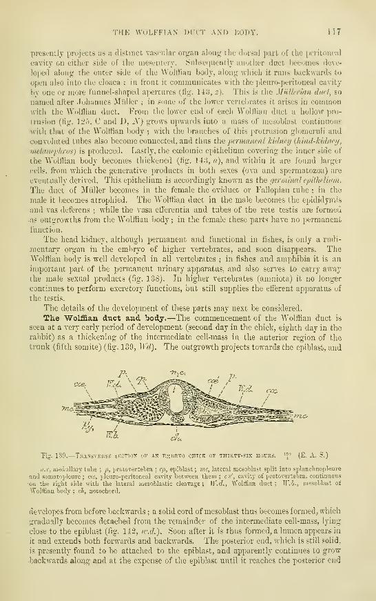
THE WOLFFIAN' DUCT AND BODY. 117
presondy projects as a distinct vascular organ along the dorsal pirt of the peritoneal
cavity on either side uf the mesentery. 8ubse(iuently anotlier duct becomes deve-
loped along the outer side of the Wolttian body, along which it runs backwards to
open also into the cloaca : in fiont it communicates with the pleuro-peritoneal cavity
by one or more funnel-shaped ai)ertures (tig. 143, z). This is the MiiUeiian dud, so
named after Jnhannes Midler ; in some of the lower vertebrates it arises in commonwith the "WoUHan duct. From the lower end of each Wolttian duct a hollow pro-
trusion (tig. 12."), C and D, N) gi-ows upwards into a mass of mesoblast continuous
with that of the Wolttian body ; with the branches of this protrusion glomeruli and
convoluted tubes also become connected, and thus the permanent kidneij {hind-hUhiei/,
metitncplirvs) is produced. Lastly, the coelomic epithelium covering the inner side of
the AVolttiau body becomes thickened (tig. 14:3, <'/), and within it are found larger
cells, from which the generative products in both sexes (ova and spermatozoa) are
eventually derived. This epithelium is accordingly known as the fjerminalepUheUum.
The duct of Miiller becomes in the female the oviduct or Fallopian tube ; in the
male it becomes atrophied. The AVolttian duct in the male becomes the epididymis*
and vas deferens ; while the vasa efferentia and tubes of the rete testis are formed
as outgrowths fi"om the Wolfiian body ; in the female these parts have no ijermanenL
tiinction.
The head kidney, although permanent and functional in fishes, is only a rudi-
jnentary organ in the embryo of higher vertebrates, and soon disappears. TheAVolttiau body is well developed in all vertebrates ; in fishes and amphibia it is an
important part of the permanent urinary apparatus, and also serves to cany awaythe male sexual products (tig. 138). In higher vertebrates (amniota) it no longer
continues to perform excretory functions, but still supplies the efferent apparatus of
the testis.
The details of the development of these parts may next be considered.
The Wolffian duct and body.—The commencement of the AVolttian duct is
seen at a very early period of development (second day in the chick, eighth day in the
rabbit) as a thickening of the intermediate cell-mass in the anterior region of the
trunk (fifth somite) {?ig. 139, ^Yd). The outgrowth projects towards the epiblast, and
yric^^±^£^/T2/>
Tig. 139.
—
Trassvekse section of ax embryo chick of thikty-six hours. ^\^ (E. A. S.)
n.c, medullary tube; p, protovertebra ; ep, epiblast; vie, lateral mesoblast split into splanchnopleure
and somatopleure ; ae, pleuro-peiitoneal cavity between them : ca', cavity of protovertebra. continuous
on the right side with the lateral mesoblastic cleavage; W.d., Wolffian duct; W.h., mesoblast ot
Wolffian body ; ch, notochord.
developes from before backwards ; a solid cord of mesoblast thus becomes formed, which
.gradually becomes detached from the remainder of the intermediate cell-mass, lying
close to the epiblast (fig. 142, ic.d.). Soon after it is thus formed, a lumen appears in
it and extends both forwards and backwards. The posterior end, which is still solid,
is presently found to be attached to the epiblast, and apparently continues to grow
backwards along and at the expense of the epiblast until it reaches the posterior end

118 THE WOLFFIAN DUCT AND BODY.
of the body, where it becomes detached from the epiblast, and is connected with and'
opens into the hind-gut (cloaca).
I have here followed what has appeared to me the most probable account of the orio-in of
the duct (Martin, Strahl), but it is right to state that in the opinion of some observers
(Hensen, Spee, Flemming) the formation and growth of the duct in connection with the
epiblast is primary, especially in mammals, and the duct is originally formed by a longitudinal
thickening and involution of the epiblast, which only secondarily becomes connected with the
intermediate cell-mass. Compare also Haddon, Origin of segmental duct, Proc. Roy. Dublin
Society, Vol. V.
In teleosteans (Rosenburg) and amphibia (Gotte) the Wolffian duct has been described as
developing in the form of a longitudinal groove-like invagination of the somatopleural meso-
blast (Balfour, Comp. Emb., vol. ii., pp. .580, .582), but more recent researches appear to indi-
cate that in these animals also the epiblast may be concerned in its formation.
The Wolffian body developes in the intermediate cell-mass between the Wolffian
duct and the body-cavity as a series of transverse tubes which lie at right angles to
the course of the Wolffian duct, and open into it at regular intervals. The usual
Fig. 140.—Transverse sec-
tion OF THE TRUNK OF Acat embryo, showing the.
vesicular stage of the,
avolffian tubules.
(E. A. S.).
m.p., muscle plate ; ao,
aorta ; m.ff., mid-gut ; am,.
amnion ; w, vesicle of Wolffian
body ; w.cl., Wolffian duct ;.
cos, ccelom.
mode of formation of
these tubes—which are
sometimes termed seg-
mental tubes— appears
to consist in the accu-
mulation at regular intervals, corresponding with the somites, of rounded massesof mesoblast on the mesial or ventral side of the Wolffian duct (fig. 142, iv.h.),
which masses become afterwards hollowed out so as to form small vesicles, at
first isolated, but afterwards growing towards and opening into the Wolffian
duct (fig. 140).i Corresponding with these vesicles there become formed invagi-
nations of the epithelium of the body-cavity (fig. 141, st), which is thickened
along the inner side of the Wolffian projection, and grows at regular intervals
towards the vesicles. These ingrowths may at first communicate by funnel-shaped
openings, which in some lower vertebrates are lined by ciliated epithelium, with
the body-cavity, but the openings in higher vertebrates become dosed again before
communication with the Wolffian duct is established. Finally, the connection
between the Wolffian tubes and the peritoneal epithelium is completely severed, andthe condition of simple or curved transverse tubes, blind at their inner ends andopening at their outer ends into the Wolffian duct, is produced (fig. 142, B). After
a time the blind extremities are seen to be enlarged and spoon-shaped, and glomeruli
1 According to v. Wijhe the hollow condition is the primary one in elasmobrancbs, and the cavity of
each vesicle represents an intermediate part of the ccelom of the segment (meso-coelom), the dorsal
coelom being represented by the cavity of the proto-vertebra and the ventral coelom by the pleuroperi-
toneal space. I have myself observed this condition of a hollow intermediate cell-mass communicating:on the one hand with the cavity of the protovertebra and on the other with the cleft of the lateral
mesoblast, in a chick of 36 hours (see fig. 139).
In mammals the Wolffian vesicles are more numerous than the segments.
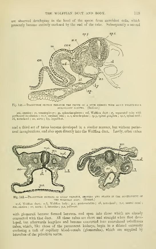
THE WOLFFIAN DUOT AND iiuDY. 119
are observed developiuLj: in the bowl of the spoon from mesoblast cells, which
presently become entirely enclosed by the end of the tube. Subsequently a secund
Fig. 141.
—
Traxsverse section through the trunk of a duck embryo vhte. about twenty-focbMESOBLASTIC SOMITES. (BalfoUF.)
am, amnion ; so, somatopleure ; sp, splanclinopleure ; wd, "Wolfl&an duct ; st, segmental tube with
peritoneal involution ; ca.v, cardinal vein ; ni.s, muscle-plate ; s^p.ff, spinal ganglion ; sj^.c, spinal cord;
ch, notochord ; ao, aorta ; hi/, hypoblast.
and a third set of tubes become developed in a similar manner, but without perito-
neal invaginations, and also open directly into the Wolffian duct. Lastly, other tubes
cofn^t^^-
Fig. 142.
—
Transverse sections of sheep embryos, showing two stages in the development of
THE WOLFFIAN BODY. (BonUCt.
)
w.d, Wolffian duct; v:.h, Wolffian body; -p.v, protovertebra ; cl>, notochord; a.c, neural canal;
am,, amnion ; ao, aorta ; i, intestine; y.s, yolk-sac.
with glomeruli become formed between, and open into those which are already
connected with that duct. All these tubes are short and straight when first deve-
loped, but afterwards lengthen and become converted into convoluted uriniferous
tubes, which, like those of the permanent kidneys, begin in a dilated extremity
enclosing a tuft of capillary blood-vessels (glomerulus), which are supplied by
branches of the primitive aortre.

120 THE WOLFFIAN DUCT AND BODY.
When completely formed, the Wolffian bodies are seen on opening the abdomen
of the embryo as long prominent vascular organs projecting into the peritoneal
cavity on either side of the intestine, and showing in section numerous Malpighian
corpuscles and uriniferous tubules variously cut (fig. 143).
Soon after having attained its complete condition of development, the Wolffian
body begins to undergo atrophic changes. These proceed much further in the
-c'
Fig. 143. — Transverse section of theWOLFFIAN BODY OF THE CHICK ON THEFOURTH DAY. (Waldejer.
)
VI, mesentery ; Z, hody wall ; a', thickeLedepithelium from whicii the involution of theanterior part of the illillerian duct 2, is
taking place ; a, thickened germinal epithe-
lium in which are seen primitive ova,;
E, modified raesoblast which will form the
stroma of the ovary ; WK, tubules of Wolffian
body variously cut; y, "Wolffian duct. Two
glomeruli are shown in the Wolffian body.
female sex than in the male, but the
tubules of the organ do not entirely
disappear in either sex. In the
female they form the rudimentary
organ which is known as the par-
ovariicm {ejioophoron of Waldeyer),
while the main tube of that struc-
ture represents a remnant of the
Wolffian duct. But in many animals,
e.g., the sow, the Wolffian duct re-
mains as the duct of Gartner, a
strong, slightly undulated tube, which is traceable, at first free in the broad ligament
of the uterus, and lower down becoming incorporated with the wall of the uterus
and vagina, upon which last it becomes lost. Traces of this tube can sometimes be
seen in sections across the body or cervix of the adult human uterus, and even lying
in the Avail of the vagina.
In the male the Wolffian duct forms the tube of the epididymis, the vas deferens,
and the ejaculatory duct ; the seminal vesicle being formed as a diverticulum fromits lower part. The coni vasculosi and tubuli eflFerentes are in all probabihty formed
by the persistence of some of the tubules of the Wolffian body. The Malpighian
corpuscles of these tubules have long disappeared, but j)revious to their disappearance
solid columns of epithelial cells, afterwards becoming tubules, grow from the walls of
those corpascles towards the germinal epithelium (fig. 153), where, in the male, they
become continuous with and enclose cells derived from that epithelium (which
subsequently form the epithelium of the seminiferous tubes), and thus produce the
walls of the seminiferous tubules and the rete testis. In the female sex there is also
a growth of solid cellular columns towards the germinal epithelium, but no connec-
tion becomes established between them, and the columns do not become tubular.
The organ of Giraldes and the vasa aberrantia of Haller are probably the remains
of one or more Wolffian tubules.
Suprarenal capsules.—These org-ans are intimately connected in their development withthe Wolffian bodies. According- to the observations of Welclon some of the cellular columnswhich grow from the Malpighian corpuscles of the upper part of the Wolffian body towardsthe germinal epithelium give offsets which pass njDwards towards the inferior vena cava, andthere become developed into the cortical substance of the suprarenal capsules. (Mihalkovics,
on the other hand, states that the strands of cells which grow from the upper part of the

SUPRARENAL CAPSULES. 121
"Wolffian lK)»ly to take part in the formation of the suprarenal capsules have been derived byproliferation from the -rerminal tpithelium.j It had lon;r been believed that the two parts of
these or-jans. cortical and medullary, are sejiarate in ori;rin : the former being derived, as wasthou^rht, fronx cells which are of mesoblastic oritrin. the latter ^>eing• developetl in connection
with the sympathetic j^anglia. In elasmobrauchd and Kome other lower vertebraiei*, they
Pig. 144.— Internal organs of a fkmale hcmax foetis of 3^ inches long, or about 14 weeks.Macnified (from Waldeyer).
o, the ovary full of primordial ova ; c, tubes of the upper part of the Wolffian body forming the
epoophoron (parovarium of Kobelt) ; W, the lower part of the Wolffian body forming the paroophoron
of His and "\Valdeyer ; w, the "Wolffian duct ; JI, the Miillerian duct, with its upper opening already
timbriated.
Fig. 145.
—
Internal genital organs of a male HtJSiAN embrto of 3^ isches losg (from Waldeyer).
t, body of the testicle with seminal canals formed ; e, epididymis, or upper part of Wolffian body
;
IT, Wolffian body, lower part, becoming paradidymis or organ of Giraldes ; w', Wolffian duct, becoming
vas deferens; ff,
gubernaculum.
B
Fig. 146.—Two figures exhibiting a comparison between parts of the generative organs 15
THE TWO sexes (fiom FarTc, after Kobelt).
A. AdCLT OVARY, PAROVARIUM AND FALLOPIAN TUBE.
rt, «, Epoophoron (parovarium) formed from the upper part of the Wolffian body ; b, remains of the
uppermost tubes, sometimes forming hydatids ; c, middle set of tubes : d. some lower atrophied tubes ;
c, atrophied remains of the Wolffian duct : /. the terminal bulb or hydatid ; h, the Fallopian tube,
originally the duct of iMidler ; (, hydatid attached to the extremity ; I, the ovary.
B.
—
The adult testis and epididymis.
rt, a, convoluted tubes in the head of the epididymis developed from the upper part of the Wolffian
body ; b and /. hydatids in the head of the epididymis ; c, coni vasculosi i d, vasa aberrantia : h,
remains of the duct of iliiller with i, the hydatid of Slorgagni, at its upper end ; I, body of the testis.

132 THE Mt)LLERIAN DUCT.
consist throughout life of two separate portions, one median and single, the other, dei-iTed'from the sympathetic ganglia, paired
; in birds, reptiles, and mammals these distinct 'portion^are combined into the two paii-ed organs (Balfour). But in these also, as has been shown byMitsukuri for mammals, the medullary or nervous part is at first distinct and outside the-cortical, into which it gradually insinuates itself, retaining, however, its connection with the-neighbouring sympathetic ganglia.
The permanent kidneys arise (1) as protrusions from the posterior end of theWolffian ducts (see fig. 125, C and D, i\^), which grow forwards towards the lower partof the Wolffian bodies, and form the ureters and the collecting tubules of the kidney
;
(2) from a portion of the intermediate cell-mass situated posterior to the Wolffianbody, and within which convoluted tubes and Malpighian corpuscles, and eventuallythe remaining parts of the uriniferous tubules become developed. But before thesechanges occur in this intermediate cell-mass, it shifts its position relatively to theWolffian body, eventually coming to lie above and behind that organ. The con-voluted tubes, with their Malpighian corpuscles, appear to be developed independentlyof the ureter and collecting tubes, as in the case of the Woh!ian tubules and the
Wolffian duct, a communication between them being only subsequently established.
The glomeruli are apparent in the eighth week in the human foetus. In thethird month the papillas are formed, and in the fourth month the loops of Henle are
seen. The tubes are wider in the foetus than in the adult ; the expansion of thekidney as growth advances must therefore be due mainly to an increase in length of
the tubules, since new tubules and glomeruli do not appear to be formed. Thehuman kidney is at first lobulated, the lobules corresponding in number to the
Malpighian pyramids, but by the end of the first year after birth, the kidneys haveusually nearly lost their lobulated appearance.
The nrinary bladder is formed by a spindle-shaped dilatation of the stalk ofthe allantois (second month). The upper pole of the spindle extends as the urachiis-
into the umbilical cord ; it not unfreqnently remains hollow for some length within
the cord (Luschka). The lower pole of the spindle which passes towards the cloaca
becomes the first part of the urethra of the male, and the whole of the urethra
of the female. The rest of the male urethra is formed and enclosed by the folds of
integument which produce the penis (see p. 128). The ureters, which are originally
prolonged from and open into the Wolffian ducts, subsequently become shifted in
position, so as eventually to open into the enlargement of the allantoic stalk, fromwhich the bladder is formed.
The Mnllerian duct.—In lower vertebrates, as was shown by Balfour for
elasmobranchs, this duct takes origin by the splitting off of the ventral part of a.
longitudinal segmental or Wolffian duct, the dorsal part remaining as the Wolffian
duct proper, and receiving the segmental and uriniferous tubes, while the ventral part
retains the funnel-shaped orifice, by which the segmental duct communicated ante-
riorly with the body cavity, and comes to open posteriorly into the cloaca by anorifice distinct from that of the Wolffian duct (fig. 138 and fig. 147). In amniotic
vertebrates, the process of formation of a Miillerian duct is somewhat different.
It arises on the outer side of the already fairly well developed Wolffian body, andsome little distance from the anterior end of that organ, as a thickening of the
peritoneal epithelium (fig. 143, a'), which thickening becomes invaginated towards
the adjacent Wolffian duct, in the form of three successive funnel-shaped depressions
(fig. 148), somewhat similar to those which are connected with the previously formedWolffian segmental tubes. The invaginations are connected together by a con-
tinuous epithelial ridge, forming a cord which becomes disconnected from the
peritoneal cavity except at the anterior invagination, and subsequently acquires
ft lumen. The short tube which is thus formed, soon begins to grow backwardas a solid rod of cells, which comes in close contact as it proceeds with the Wolffian'

THE MCLLERIAN DUCT. 125
duct (fiff. 141)). To this duct it presently adheres intimately, and thf-n continues
Fig. 147.—Four sections thkuich the antkkkiu i-akt ok tiik
SEGMENTAL IiUCT OP A SCYLLIUM EMBUYo. (littlfour. )
The tij:i:re shows how the sppiicntiil duct hecomes siilit into tlic
Wolrtian iluct (l(irs;illy and the Mullorian duct or oviduct ventrally ;
Wil, Wolrtian duct; od, Muilciian duct or oviduct; »(/ (iu D),
segiuental duct.
to grow backwarcis for a certain distance as a thicken-
ing of the epithelium of that tube, the thickening
becoming gradually separated oft' from before back-
wards, and the lumen passing along it. Further back
it ceases to grow thus in connection with the AVoltiiau
duct, but is prolonged as an independent cellular cord,
which lies in a groove along the side of the Wolffian
duct (Balfour and Sedgwick),
Entering the f/c/ii/al cord} the two Miillerian ducts
lie at first on the mesial side of the corresponding
"Wtilffian ducts, but lower down pass behind them ;
they finally come again between these ducts, lying
close together, and, according to Mihalkovics, approach
close to the sinus urogenitalis, which by this time is
formed out of the ventral part of the cloaca (see p. 128)
without actually opening into it for some time. The Miillerian ducts fuse together
below into a single tube (second month); the fusion begins not at the lower end,.
Fig. 148.
—
Sections from the chick showing two of the peritoneal invaginations which give
RISE to the anterior PART OF THE MCLLERIAN DUCT. (Balfour and Sedgwick.)
gr", sccoud invagination;
gi-^, third invagination ; r-, epithelial ridge lietween them ; Wd,"Wolffian duct. These structures form the pronephros of Balfour and Sedgwick (see note, p. 115).
Fig. 149.
—
Two sections from the chick
SHOWING THE JUNCTION OF THE TER-
MINAL SOLID PORTION OF THE ^McL-
LERIAN DUCT WITH THE ^YoLFFIAN
DUCT. (Balfour and Sedgwick.)
In A, the terminal portion of the duct
is quite distinct ; in B it has united
with the wall of the Wolffian duct, md,Miillerian duct ; Wd, Wolffian duct.
but a short distance away from
this (fig. 150, 3), and proceeds
both downwards towards the
future orifice and upwards for a
^ A name given to the thickened mass of tissue which surrounds the Wolffian ducts as they coursetogether to the cloaca behind the stalk of the allantois (afterwards the base of the bladder).
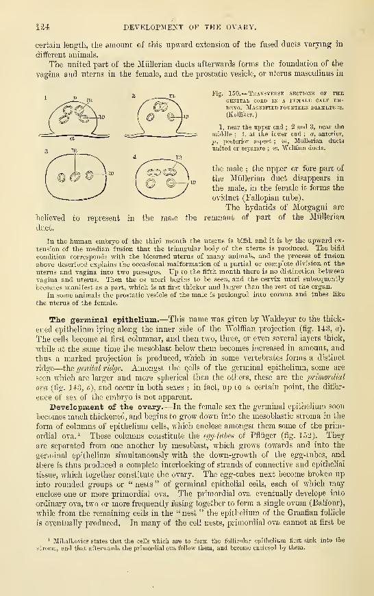
124 DEVELOPMENT OF THE OVARY.
certain leugtli, the amount of this upward extension of the fused ducts varying in
different animals.
The united part of the Miillerian ducts afterwards forms the foundation of the
vagina and uterus in the female, and the prostatic vesicle, or uterus masculinus in
Fig. 150.
—
Transverse sections of theGENITAL CORD IN A FEMALE CALF EM-
BRYO. Magnified fourteen diameters.
(KoUiker.
)
1, near tlie upper end ; 2 and 3, near the
middle ; 4, at the lower end ; a, anterior,
p, posterior asjject ; m, Miillerian ducts
united or separate ; iv. Wolffian ducts.
the male ; the upper or fore part of
the Miillerian duct disappears in
the male, in the female it forms the
oviduct (Fallopian tube).
The hydatids of Morgagni are
remnant of part of the Miillerianbelieved
dnct.
to represent in the ma^e the
In the human embiyo of the thii-d month the uterus is bifid, and it is by the upward ex-
tension of the median fusion that the triangular body of the uterus is produced. The bifid
condition corresponds with the bicomed uterus of many animals, and the process of fusion
above described explains the occasional malformation of a partial or complete division of the
uterus and vagina into two passages. Up to the fifth month there is no distinction between
vagina and uterus. Then the os uteri begins to be seen, and the cervix uteri subsequently
becomes manifest as a part, which is at fii-st thicker and larger than the rest of the organ.
In some animals the prostatic vesicle of the male is prolonged into cornua and tubes like
the uterus of the female.
The germinal epithelium.—This name was given by "Waldeyer to the thick-
-eued epithelium lying along the inner side of the Wolffian projection (fig. 143, a).
The cells become at first columnar, and then two, three, or even several layers thick,
while at the same time the mesoblast below them becomes increased in amount, and
thus a marked projection is produced, which in some vertebrates forms a distinct
ridge—the genital ridge. Amongst the (jells of the germinal epithelium, some are
seen which are larger and more spherical than the others, these are the primordial
ova (fig. 143, o), and occur in both sexes ; in fact, up to a certain point, the differ-
ence of sex of the embryo is not apparent.
Development of the ovary.—In the female sex the germinal epithelium soon
becomes much thickened, and begins to grow down into the mesoblastic stroma in the
form of columns of epithelium cells, which enclose amongst them some of the prim-
ordial ova.^ These columns constitute the egg-tahes of Pfliiger (fig. 152). They
are separated from one another by mesoblast, which grows towards and into the
germinal epithelium simultaneously with the down-growth of the egg-tubes, and
there is thus produced a complete interlocking of strands of connective and epithelial
tissue, which together constitute the ovary. The egg-tubes next become broken up
into rounded groups or " nests " of germinal epithelial cells, each of which mayenclose one or more primordial ova. The primordial ova eventually develope into
ordinary ova, two or more frequently fusing together to form a single ovum (Balfour),
while from the remaining cells in the "nest " the epithelium of the Graafian follicle
is eventually produced. In many of the cell nests, primordial ova cannot at first be
1 :Mihalkovics states that the cells which are to form the follicular epithelium first sink into the
stroma, and that afterwards the primordial ova follow them, and become enclosed by them.

DEVELOPMENT OF THE TESTICLE. J25
distinguished, but become formed subsequently by an increase in size of one or moreof tlie cells. Tiie further changes which take place in the Graafian follicle are
described with the structure of the ovary (r. Splanchnology). The remainder of
Fig. 151.
—
Traxsvekse section throcgh the ovart of ax embryo shark (scvllicm;, showing thcGERJI-EPITHELIUM F0R3IIXG PRIMITIVE OVA. iBtllfour.)
At po, the germ-epithelium and primitive ova ; the lightly-shaded part is the ovarian stroma,covered elsewhere by flattened epitheJium.
the germinal epithelium which is left covering the surface after the formation ofthe egg-tubes, constitutes the permanent epithelium of the ovary.
Most, if not all. of the permanent ova are produced, at least in the human .subject, longbefore birth.. In the human ovary the nests of cells which are to form the Gmafian follicle's-
Fig. 152.
—
Section of the ovary of aNEWLY-BORN CHILD. HiGHLY ilAG-
kified. (Waldeyer.)
a, Germinal epithelium dipping in at
b, to form an ovarian tube ; c, c, prim-
ordial ova lying in the germ-epithelium :
d, d, longer tube becoming constricted so
as to form nests of cells : e, c, larger ne.=!ts ;
/, distinctly formed follicle with ovum;.nd ei^ithelium
; g, (f, blood-vessels.
are more eciually diffused through the
substance of the ovary than in mostanimals, in many of which the young-
follicles remain fomiing a stratumnear the surface. In the humanembryo of from four months up to
the ijeriod of birth, the ovary seemsto be foiTQed of little else than a massof young' ova. closely surrounded byflattened cells of the germinal epithe-
lium and constituting thus minuteGraafian follicles : the amount of
stroma being at this time relatively small. It has been calcixlat«d that the ovaries mavat this stage contain as many as 70,000 primordial ova.
Development of the Testicle.—The germinal epithelium does not undergo somarked an hypertrophy in tlie male as in the female. But it becomes thickened,and enlarged ceils, corresponding to the primordial ova in the female, are found mit. Further, small strands of the epithelium dip down into the subjacent mesoblast,.
;, '-^^^'p

126 DESCENT OF THE TESTICLES.
which grows simultaneously into the epithelium, and eventually cell-nests are sepa-
rated and included in the mesoblastic tissue. Whether these nests are derived from
the division of the primordial ova only, or whether they also include other cells of the
germinal epithelium is not clear. It would appear that from these cell-nests the
epithelium of the seminiferous tubules is developed, although all stages of the
process have not been observed. The cell-nests eventually become connected with
the outgrowths from the Wolffian bodies (fig. 153, st), which as already mentioned,
Tig. 153. —Section of the germInal epithelium and adjacent strojia in a male chick embryo.(Semon.
)
g.(p, germinal epithelium forming a tliickenecl ridge-like projection ; ifyr.ov, primitive ova of various
sizes, some in the germinal epithelium and others somewhat beyond the limit of this epithelium ; st,
strands of cells which have grown from the Wolffian body towards the germinal epithelium, and one of
which appears connected -with an enlarged primitive ovum.
form the rete testis and the efferent tubes of the testicle. The reproductive gland is
in both sexes at first attached directly to the Wolffian body (fig. 156, A, oi), which
itself is attached by a fold of peritoneum to the back of the abdominal cavity.
This fold becomes the mesovarium or mesorchium as the case may be. K band also
passes from the Wolffian body upwards to the diaphragm, and another fold contain-
ing involuntary muscular fibres—the plka guhernatrix—runs down towards the
groin from the lower part of the AVolffian body and the duct. This band, as the
Wolffian body becomes atrophied, is found to be attached to the reproductive organ,
constituting the guhernaculum testis in the male, and the round ligament of the ovarij
ia the female (fig. 15C, g).
Descent of the Testicles.—The testicles originally lie in the lumbar region of
the abdomen. From this j)art they become shifted, at first to the internal
abdominal ring, opposite w^hich they are found in the sixth month, and which thev
•enter in the seventh month, then down the inguinal canal into the scrotum, which
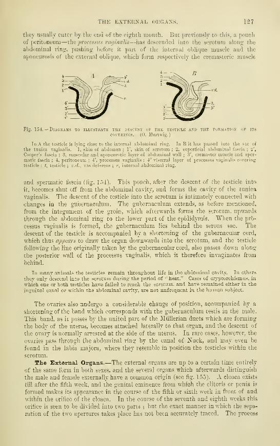
THE EXTERNAL ORGANS. 127
they usually enter by the eud of the eighth month. But previously to this, a pouchof peritoneum—the processus vayinalis—has descended into the scrotum along the
abdominal ring, pushing before it part of the internal olilique muscle and the
aponeurosis of the external oblique, which form respectively the cremasteric muscle
Fig. 154.
—
Diagrams to illustrate the descent of the testicle axd the f^rmatiux op its
covERiXGS. (0. Hertwig.)
In A the testicle is lying close to the internal abdominal ring. In B it has passed into the sic of
the tunica v;iginalis. 1, skin of abdomen ;1', skin of scrotum ; 2, superficial abdominal fascia ;
2',
Cooper's fascia : 3, muscular and aponeurotic layer of abdominal wall ;3', cremaster muscle and sper-
matic fascia ; 4, peritoneum ; 4', processus vaginalis ;4" visceral layer of processus vaginalis covering
testicle ; t, testicle ; v.d., vas deferens ; r, internal abdominal ring.
and spermatic fascia (tig. 154). This pouch, after the descent of the testicle into
it, becomes shut off from the abdominal cavity, and forms the cavity of the tunica
vaginahs. The descent of the testicle into the scrotum is intimately connected with
changes in the gubernaculum. The gul^ernaculum extends, as before mentioned,
from the integument of the groin, which aftei-^'ards forms the scrotum, upwards
through the abdominal ring to the lower part of the epididymis. When the pr?)-
cessus vaginalis is formed, the gubernaculum lies behind the serous sac. The
descent of the testicle is accompanied by a shortening of the gubernacuiar cord,
which thus appears to draw the organ downwards into the scrotum, and the testicle
following the line originally taken by the gubernacuiar cord, also passes down along
the posterior wall of the processus vaginalis, which it therefore invaginates from
behind.
In manv animals the testicles remain throuofhout life in the abdominal cavity. In others
thev only descend into the scrotum dtiring" the period of " heat." Cases of cryptorchismus. in
which one or both testicles have failed to reach the scrotum, and have remarued either in the
ingTiinal canal or -n-ithin the abdominal cavity, are not unfrequent in the human subject.
The ovaries also undergo a considerable change of position, accompanied by a
shortening of the band which corresponds with the gubernaculum testis in the male.
This band, as it passes by the united part of the Mlillerian ducts which are forming
the body of the uterus, becomes attached laterally to that organ, and the descent of
the ovary is normally arrested at the side of the uterus. In rare cases, however, the
ovaries pass through the abdominal ring by the canal of Xuck, and may even Ije
found in the labia majora, where they resemble in position the testicles within the
scrotum.
The External Organs.—The external organs are up to a certain time entirely
of the same form in both sexes, and the several ors-ans which afterwards distinguish
the male and female externally have a common origin (see fig. 155). A cloaca exists
till after the fifth week, and the genital eminence from which the clitoris or penis is
formed makes its appearance in the course of the fifth or sixth week in front of and
within the orifice of the cloaca. In the course of the seventh and eighth weeks this
orifice is seen to be divided into two parts ; but the exact manner in which the sepa-
ration of the two apertures takes place has not been accurately traced. The process
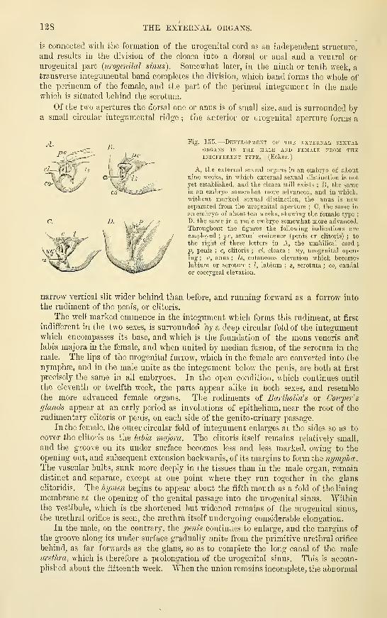
12S THE EXTERNAL ORGANS.
is connected with the formation of the urogenital cord as an independent structure,
and results in the division of the cloaca into a dorsal or anal and a ventral or
urogenital part {urogenital sinus). Somewhat later, in the ninth or tenth week, a
transverse integumental band completes the division, which band forms the whole ofthe perineum of the female, and the part of the perineal integument in the malewhich is situated behind the scrotum.
Of the two apertures the dorsal one or anus is of small size, and is surrounded bya small circular integumental ridge ; the anterior or urogenital aperture forms a
Fig. 155.
—
Developjiext of the external sexualORGANS IN THE MALE AND FEJIALE FK03I THEINDIFFERENT TYPE. (Ecker.
)
A, the external sexual organs in an embryo of aboutnine weeks, in which external sexual distinction is not
yet established, and the cloaca still exists ; B, the samein an embryo somewhat more advanced, and in which,without marked sexual distinction, the anus is nowseparated from the urogenital aperture ; C, the same in
an embryo of about ten weeks, showing the female type;
D, the same in a male embi-yo somewhat more advanced.Throughout the figures the following indications are
employed;^c, sexual eminence (penis or clitoris) ; to
the right of these letters in A, the umbilical cord ;.
'p, penis ; c, clitoris ; cl, cloaca ; iig, ui'ogenital open-
ing ; a, anus ; Is, cutaneous elevation which becomeslabium or scrotum ; I, labium ; s, scrotum ; co, caudalor coccygeal elevation.
nan-ow vertical slit wider behind than before, and running forward as a furrow into
the rudim.ent of the penis, or clitoris.
The well marked emmence in the integument which forms this rudiment, at first
indifferent in the two sexes, is surrounded by a deep circular fold of the integumentwhich encompasses its base, and which is the foundation of the mons veneris andlabia majora in the female, and when united by median fusion, of the scrotum in the
male. The lips of the urogenital furrow, which in the female are converted into the
nymphffi, and in the male unite as the integument below the penis, are both at first
precisely the same in all embryoes. In the open condition, which continues until
the eleventh or twelfth week, the parts appear alike in both sexes, and resemble
the more advanced female organs. The rudiments of Bartholin's or Coivper's
glands appear at an early period as involutions of epitheKum, near the root of the
rudimentary clitoris or penis, on each side of the genito-urinary passage.
In the female, the outer circular fold of integument enlarges at the sides so as to
cover the clitoris as the labia majora. The clitoris itself remains relatively small,
and the groove on its under surface becomes less and less marked, owing to the
opening out, and subsequent extension backwards, of its margins to form the nympha^.
The vascular bulbs, sunk more deeply in the tissues than in the male organ, remaindistinct and separate, except at one point where they run together in the gians
clitoridis. The hymen begins to appear about the fifth month as a fold of the lining
membrane at the opening of the genital passage into the urogenital sinus. Withinthe vestibule, which is the shortened but widened remains of the urogenital sinus,
the urethral orifice is seen, the urethra itself undergoing considerable elongation.
In the male, on the contrary, the ^;e;^^s continues to enlarge, and the margins of
the groove along its under surface gradually unite fi'om the primitive urethral orifice
behind, as far forwards as the glans, so as to complete the long canal of the maleurethra, which is therefore a prolongation of the urogenital sinus. This is accom-plished about the fifteenth week. When tlie union remains incomplete, the abnormal

THE EXTERNAL ORGAXS. 129
condition named hypospadias is produced. In the meantime the prepuce is formed,and, moreover, the lateral cutaneous folds also unite from behind forwards, along themiddle line or raphe, and thus complete the scrotum, into which the testicles descendin the course of the eighth month of foetal life, as before described.
The corpora cavernosa, which are at first separate, become united in their distal
portions in both sexes ; but the corpus spongiosum urethrje which is also originallv
divided in all embryos, and in the female remains so in the greater part of its
extent, becomes enlarged in the male in the glans penis, and its two parts becomeunited mesially both above and below the urethra, so as to enclose the whole of that
tube from the bulb forwards to the glans.
The following Table and Diagrams exhibit the correspouding parts of the
urino-generative organs in the two sexes :
—

180 GENITO-UPJNARY OI^GANS OF THE TWO SEXEa.
V'
o
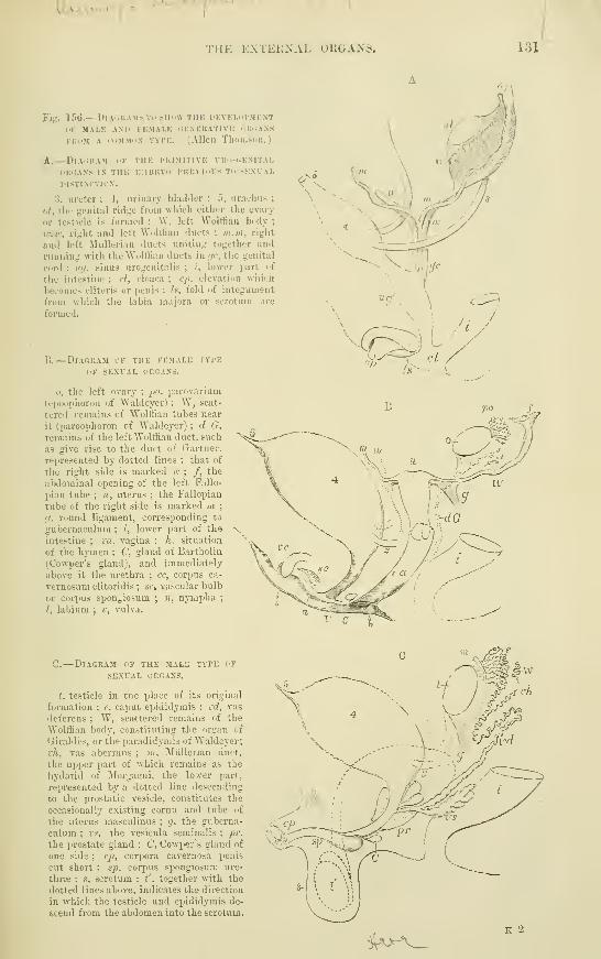
ILma-*--l-H«. V-1
TlIK KXTEiiNAL UllGANS. 131
Fig. 156.
—
Diagrams TO snow TiiK develhi-mknt
OK MALE AN1> FEMALE GENERATIVE oUiiANS
KuoM A COMMON TYi'K. (Allen Thomsoii.
)
A.— Diai;kam w the pkimitive uro-cjemtal
liRiiANS IN the embryo PREVIOUS TO SEXVAL
mSTINCTION.
3, ureter ; 4, urinary bladder ; 5, urachus ;
ot, the genital ridge from which either the ovary
or testicle is formed ; W, left Wolffian body;
w.w, right and left Wolffian ducts ; m.m, right
and left ^lullerian ducts uniting together and
ninuing with the Wolffian ducts in ijc, the genital
cord ; luj, .sinus urogenitixlis ; /, lower jiart of
the intestine ; cl, cloaca ; cp. elevation which
becomes clitoris or penis ; h, fold of integument
from which the labia majora or scrotum are
formed.
B.
—
Diagram of the female type
OF sexual organs.
o, the left ovary ; po, parovarium
(epoophoron of Waldeyer) ; W, scat-
tered remains of Wolffian tubes near
it (paroophoron of Waldeyer) ; d G.
remains of the left Wolffian duct, such
as give rise to the duet of Gartner,
represented by dotted lines ; that of
the right side is marked n: ; /, the
abdominal opening of the left Fallo-
jiian tube ; u, uterus ; the Fallopian
tube of the right side is marked m;
(J,round ligament, corresponding to
gubernaculum ; i, lower part of the
intestine ; va, vagina ; h. situation
of the hymen ; C, gland of Bartholin
(Cowper's gland), and immediately
above it the urethra ; cc, corpus ca-
vernosum clitoridis ; sc, vascular bulb
or corpus spongiosum ; n, nympha;
/, labium ; v, vulva.
<;;.
—
Diagram of the male type op
SEXUAL OPvGAXS.
t. testicle in the place of its original
formation ; e, caput epididymis ; vd, vas
deferens ; W, scattered remains of the
Wolffian body, constituting the organ of
(jiraldes, or the paradidymis of Waldeyer;
vh, vas aberrans ; m, Miillerian duct,
the upper part of which remains as the
hydatid of Morgagni. the lover part,
represented by a dotted line descending
to the prostatic vesicle, constitutes the
occasionally existing coma and tube of
the uterus masculinus ; g, the guberna-
culum ; VH, the vesicula seminalis ; pr.
the prost;tte gland ; C, Cowper's gland of
one side ; cp, corpora cavernosa penis
cut short : gp, corpus spongiosum ure-
thrte : s, scrotum ; t' , together with the
dotted lines above, indicates the direction
in which the testicle and epididymis de-
scend from the abdomen into the scrotum.
^t^^-^v^
132 EECENT LITERATURE.
RECENT LITERATURE.
Ackeren, F. v., Beitr. sur Entwickelunr/sgeschichte d. weihlichen Sexualorgane des Menschen,Zeitschr. f. wiss. ZooL, 1889.
Beard, J,, The origin of the segmental duct in Elasmohranchs, Anat. Anzeiger, 1887.Benda, C, Die Entivicld. des Sdugethierhodens, Verhandl. der anatom. Gesellschaft, 1889.Bierfreund, "M.., Ue. die Einmundungsweise der Milller'schen Gdnge in d. Sinus urogenitalis hei
dern vienschliclien Embryo, Zeitsch. f. Geburtshlilfe u. Gynak, xvii.
Bonnet, E.., Ueber die ektodermale Entstehung des Wolffsschen Ganges iei den Sdugethieren,
Miinchener med. Wocbensclir., 1887 ; Erabryologie der Wiederlcduer, Arch. f. Anat. u. Physiol.,
Anat. Abth., 1889.
Bramann, F., Beitrag zur Lehre von dem Descensus testieulorum, tfcc. Arch. f. Anat. u. Physiol.
,
Anat. Abth., 1884.
Brandt, Ue. d. ZusammenJiang der Glandula suprarenalis rait dem Parovarium, d-c, Biol.
CentralbJ. Ix.
Cadiat, 0., M&moire sur l\d6rus et les trompes (developpement). Joum. de ranatomie,
1884 ; Du developpement du canal de I'urethre et des organes genitaux de Vembryon, Journ. de
I'anatomie, 1884.
Doh.rn, Ueber die Gartner'schen Kandle heim Weibe, Arch. f. Gynakologie , xxi., 1883.
Emery, C. , Recherches embryologiques sur le rein des mammiferes, Archives italiennes de biologies
t. iv., 1883.
Flemmingr, W., Die eTctoblastische Anlage des Urogenitalsystems beim, Kaninchen, Archiv. f.
Anat. u. Physiol., Anat. Abth., 1886.
G-asser, Embryonalreste avi indnnlichen Genitalapparat, Marburg. Sitzungsb., 1882.
Gottsch.au, M., StruJctur u. embryonale EntwicH. der Nebennieren hei Sdugethieren, Arch, f,
Anat. u. Physiol, Anat. Abth., 1883.
Haddon, Suggestion res2Jecting the epiblastic origin of the segmental duct, Proceed, of the Royal
Dublin Society, Feb. 16, 1887.
Hoffmann, C. K., Zur Entwicklungsgeschichte der Urogenitalorgane bei den Anamnia, Zeitschr.
f. wiss. ZooL, Bd. xliv., 1886 ; Zur Entwickl. der Urogenitalorgane bei den Bejytilien. Zeitschr. f. wiss.
Zool. xlviii.
Janosik, I., Bemerhungen iiber die Enttvicklung der Nebenniere, Archiv. f. mikrosk. Anatomic, Bd,
xxii., 18S3 ; Histologisch-embryologische Untersuchungen ilber das Urogenital-system, Sitzungsber. d.
Wiener Akad., 1885.
Kallay, A., Die Niere im friihen Stadium des Embryonallebens, Mitth. aus d, embryol. Inst. d.
Univers. Wien, 1885.
Kocks, Ueber die Gartner schen Gdnge beim Weibe, Arch, f. Gynakologie, xx., 1883.
Kollmann, Ueber die Verbindung zwischen Colom u. Nephridium, Festschrift. Basel, 1882.
Lock-wood, C. B., The development and transition of the testis, normal and abnormal. Journal of
Anat. and Phys. , vols. xxi. and xxii., 1887 and 1888.
Martin, E., Ueber die Anlage der Urniere beim Kaninchen, Archiv. f. Anat. u. Physiol., Anat.
Abth., 1888.Martin, P., Zur Entwickl. der cavernosen Korper, <&c., Deutsche Zeitschr. f. Thiermed., xvi.
Milialkovics, Gr. v., Untersuchungen iiber die Entwicklung des Ham- und Geschlechts-apparatof
der Amnioten, Intern. Monatsschr. f. Anat. u. Histol., 1885, 1886.
Mitsukuri, K., The ectoblastic origin of the Wolffian duct in Chelonia, Zool. Anzeiger,
1888.
Nag-el, Ueher den Wolff'schen Korper des menschl. Embryo, Zeitschr. f. Geburtshulfe u. Gynak.,
1889 ; Ueber die Entwickl. des Urogenitalsystems des Menschen, Arch. f. mikr. Anat., xxxiv., 1889 ;
Ue. das Vorkommen von Primordialeiern ausserhalb der Keimdrilsenanlage beim Menschen,
Anatomischer Anzeiger, iv.
Perenyi, J. v., Die ektoblastische Anlage des Urogenitalsysteins bei Eana esculenta und Lacerta
viridis, Zoolog. Anzeiger, 1887.
Benson, G-., Contributions h Vembryologie des organes d'eoccritions des oiseaux et des mammiferes.
These. Bruxelles, 1883.
Both, Ueber einige Urnierenreste beim Menschen, Festschrift. Basel, 1882.
Btickert, J., Ueber die Entstehung der Excretionsorgane bei Selachiern, Arch. f. Anat. u. Physiol.,
Anat. Abth., 1888.
Schmieg-elo-w, E., Studien ueber die Entwickl. des Hodens u. Nebenliodens, Arch. f. Anat. u.
Physiol., Anat. Abth., 1882.
Sedg-wick, A. , On the early development of the anterior part of the Wolffian duct and body in the
c7«c^, <£-c.. Quarterly Journal of Microsc. Science, 1881.
Sem.on, E,., Die indifferente Anlage der KeimdrUsen beim Hilhnchen und ihre Differenzirung
zum Hoden, Jenaische Zeitschr. f. Naturw., Bd. xxi., 1887.
Spee, P., Ueher directe Betheiligung des Ektoderms an der Bildung der Urnierenanlage des
Meerschweinchens, Archiv f. Anat. u. Physiol., Anat. Abth., 1884; Ueber weitere Befunde zur Ent-
wicklung der Urniere, Mittheil. f. d. Verein Schleswig-Holstein, 1886.
Strahl, H., Zur Bildung der Kloake des Kaninchenemhryo, Archiv f. Anat. u. Phys., Anat. Abth.,
1886 ; Ueher den Wolff'schen Gang und die Segmentalbldschen bei Lacerta, Marburger Sitzungsberichte,
1886.

RECENT LITERATURE. 133
Tourneux, R. Sur les premiers developpements du doaque, du tvbercule giniial et de Vanuichez
Vembruon dTmouon, Journal de lanatomie. 1888 ; SvLr U divdoppemcnt du tubercle gimtal chez le
fZTf^rnainll-c., J^urn. de lanatomie. xxv. ; Sur le mode de formation du j^imnie chcz lembryon
tiu mou^o/i, Coiui.t. rend, de la Socidte de Biologic, 1890. ,„.,., t^„^ j-Tourneux, F., et Legay, Ch., Mdmoire sur U ddvehppement de Cutirus et du xwjxn, Joum. de
''""weidon'^W. F. R., Note on the ori'jin of the suprarenal bodies of vertebrates Proceed of the
aoyal Society, vol. 37, 1885 ; On the suprarenal bodies of vertebrata, Quarterly Journal of Micr.
^"Wieger.^a., Ueber die Entstehung u. Entwickl. der Bander des weiUichen Genitalapparates, «tc.,
\rch f Vnat. u. Phvsiol., Anat. Abtb., 1885., „ .
Wijhe, J. W. v., Die Bethtiliyung des Ektoderms an der Entwickluny des Vornurenganges,
2ool. Anz., ISSti.

134 DEVELOPMENT OF THE HEART.
FORMATIOlSr OF THE VASCULAR SYSTEM.
DEVELOPMENT OP THE HEART.
In mammals, the heart appears in the form of two tnbes lying in the cephalic
region, one on either side of the embryo. These are seen at a very early period,
prior, in fact, to the separation of any part of the alimentary canal from the yolk
sac, and to the closure of the neural groove. This bilateral condition was first
observed by Hensen in the rabbit ; it has been seen by His in the humanembryo.
The situation and mode of formation of the bi-tubular heart are well illustrated
by the accompanying figures from KoUiker. They exhibit the condition in the
Fig. 157.
—
Rabbit embeyo of the 9th day, feoji theSURFACE. -J-. (Kolliker.)
The medullary groove is enlarged anteriorly and the primaiy
optic vesicles are growing out from the first cerebral enlarge-
ment. On either side of the head, the bilateral tubular heart
is seen. Eight pairs of protovertebraj are formed.
rabbit embryo of about eight or nine days—the
time when the heart first makes its appearance.
Fig. 157 shows such an embryo in surface view.
The neural groove, as also the sections show, is
widely open, although the rudiments of the cere-
bral eulargements are apparent in it, and also the
enlargements for the primary optic vesicles. There
are eight pairs of protovertebras, the paraxial
mesoblast in front of these and on either side of
the cerebral enlargements being undivided. Out-
side this undivided cephalic mesoblast is a short
tube dijiping in front into it, and passing behind
into a venous trunk, the vitelline or omphalo-
meseraic vein of the same side. The tube lies
within and is immediately surrounded by a clear
space, which is continued forwards beyond it on
either side of the fore-brain ; this space is pro-
longed from the mesoblastic cleft or pleuro-peri-/
toneal cavity (coelom).
The two short tubes form the double rudiment of the heart. The situation
which they occupy becomes, when the lateral walls fold over to form the foregut,
the ventral wall of the pharynx, and the two tubes are thus brought together in the
middle line underneath the head part of the ahmentary canal. Here they soon
become fused together to form a single median tube, the hinder end of which is still
continuous with the two vitelline veins, while the anterior end bifurcates near the
anterior end of the foregut into two branches which arch dorsalwards on either side
of that tube, and then" pass backwards on each side of the notochord as the two
primitive aortee.
These changes in the position of the primitive heart are partly shown in surface
view in figs. 158, 159, but they can only properly be appreciated by the study of
transverse sections. Fig. 160 is a transverse section through the anterior head
region of the embryo shown in fig. 157. This is anterior to the heart region, but
shows the commencing folding over of the splanchnopleure to form the foregut.
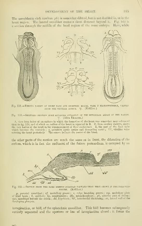
DEVKLOl'MENT OF THE HEART. 135
The mesoblastic cleft (cceloin, ;V/ ) is somewhat dilated, but is not doubled in, as in the
heart re<j;ion. The lateral niesoblast ceases a short distance beyond it. Fig. IGl is
a section throiiirh the middle of the head region of the same embryo. Here, while
/
C(\
Fin 158.
—
EjIBRYO rabbit of eight days AJTD eighteen hours, with 9 PROTOTERTEBR.i:, VIEWED
FROJI THE VENTRAL ASPECT. "{. (Kolliker.
)
Fig. 159.
—
Sketches showing more advanced condition of the bitubular heart of the rabbit.
-f . (Allen Thomson.
)
A, view from below of an embiyo in which the formation of the heart was somewhat more advanced
tlian in fig. 158, and of which an outline of the heart is repeated in B. C, from another embryo, shows
the two halves of the heart in the commencement of their coalescence. A, the part of the bent tube
which becomes the ventricle ; a, primitive aortic arches and descending aortte ; VV, vitelline veins
entering the heart posteriorly The arrows indicate the course of the blood.
the other parts of the section are much the same as in front, the dilatation of the
ccelom, which is in fact the rudiment of the future pericardium, is occupied by an
^dd'
dd ^' jih
Fig. 160.
—
Section from the same embryo further forward than that shown in the following
figure. (Kolliker.)
J),paraxial mesoblast ; rf, medullary groove ; r, ridge bounding groove ; mjp, medullary plate
of hind brain ; h, epiblast ; Ji-p^ somatopleure ; dfj>, splanchnopleure ; ph, anterior part of ccelom ;
mes, mesoblast beyond the ccelom ; dd, hypoblast ; dd' , notochordal thickening ; sv:, lateral wall of the
developing pharj-nx.
invagination, or fold, of the splanchnic mesoblast. This fold becomes subsequently
entirely separated and the aperture or line of invagination closed ; it forms the

136 DEVELOPMENT OF THE HEAET.
muscular wall of the heart. It encloses a second tube composed of flattened
epithelium cells ; this so-called endothelial tuie (His) becomes the lining epithelium
of the endocardium.
A
IMS fi
iliJi \f'^i^''
Fig. 161.—A, Transverse section through the head of an embryo rabbit op eight days andFOURTEEN HOURS, WITH A PART OP THE PERIPHERAL BLASTODERM. *f.
(KollikeV.
)
lih, rudiments of the heart ; &r, pharyngeal groove, \vith iiotochordal thickening of hypoblast.
B.
—
Part of the same more highly magnified,^'f-.
(Kolliker.)
Lettering as in fig. 160. In addition :
—
alili, fold of splanchnopleure to form wall of heart
;
ihli, endothelial tube of heart.
There is some doubt as to the source of this endothelial tube of the heart. In the preceding
edition of this work it was stated that it is•
' derived from the deeper part of the visceral meso-
blast ;" this statement being apparently founded upon the statements and figures given by
Kolliker. His ascribes it, like the endothelium of the blood-vessels, to an ingrowth from the
vascular area. The appearance of the section shown in fig. 161 B, seems to lend colour to the
belief that the invagination which has taken jslace to form the heart is not the splanchnic
mesoblast only, but has included also the hypoblastic layer of the splanchnopleure ; the notcli
which is seen in the hypoblast near .vx\ appearing to indicate an inteiTupted connection with
the endocardial tube. Should future investigations show that this is actually the mode of
formation of the tube, the mammalian heart would be developed in essentially the samemanner as has been shown by Ktickert to occur in Pristiurus (an Elasmobranch), where this
organ, which, as in all vertebrates below mammals, is formed only after the foregut is com-IDleted, is developed as a median outgrowth or thickening of the ventral wall of the foregut.
A similar mode of formation has also been noticed in Cyclostomata, Ganoids, and Amphibia. Inreptiles and birds the first appearance of the heart is as a bilateral tube, but it becomes visible
only after the foregut is formed, and the two tubes lie from the first close together, and fromthe surface appear as a single median tube.
Sections at a somewhat later period (fig. 162) show the two tubes lying in con-
tact on the ventral side of the now completed foregut. The septum which divides
them at this period has nothing whatever to do with the permanent intra-cardiac
septum, but soon becomes completely absorbed, so that by the fusion of the two
lateral tubes a single median tube is the result (fig. 163). This median tube remains
attached by a susp)ensory membrane resembling a mesentery (rnesocardium ])o&terms)
{m}), fig. 163) to the ventral Avail of the pharynx, but the rnesocardium anterius,
Avhich also at first results from the fusion, disap'iears, except at the lower end', and
otherwise the tube becomes free, except where the vitelline veins pass to it fi'om the
yolk sac, a lateral attachment to the body wall being here subsequently formed on
each side {rnesocardium laterale of Kolliker). After it is thus formed, the heart is
for a time median in position and symmetrical (fig. 164, A), but already in the
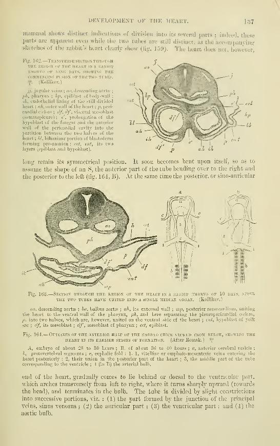
DEVELOPMENT OF THE ilKAKT. 137
mammal shows distinct imlications of division into its seveml parts ; indeed, theseparts are apparent even while the two tubes are still distinct, as the accompanyin<?sketches of the rabbit's heart clearly show (fig. li)'J). The heart does not, howevei"
Fig. lt>2.
—
Tkansvkusk section TuuorcnTHK IIKOIOX 1>K THK HKAKT IX A KAltlllT
KMUKYo I'F MSK HAYS. SHOWIMi THKCOMMEXCIXi: KISION OF TUK TWO TlUE:!.
y. (Kolliker.)
jj, jugular veins ; ao, desccinling aortic;
pit, pliarynx ; hp, cpiblast of loily-wall
;
ih, endothelial lining of the still iliviileil
heart : a/i, outer wall irt" the heart ; ji, jieri-
eanlial c.iloin ; df, df, visceral niesoljlast
(soniatopleurc) ; e', prolongation of theliyjiol'last of the foregut and the anterior
will of the pericardial cavity into the
l>artition hetween the two halves of the
heart; W, bilaminar portion of blastodermforming pro-aninion ; I'ct, ent, its twolayers (epiblast and hypoblast). a It in
long retain its symmetrical position. It soon becomes bent upon itself, so as to
assume the shape of an 8, the anterior part of the tube bending over to the right and
the posterior to the left (tig. 164, B). At the same time the posterior, or sino-auricular
Fig. 163.
—
Section THBorcH the segion of the heart in a rabbit embryo of 10 days, after
THE TWO TUBES HAVE UNITED IXTO A SINGLE MEDIAN ORGAN, (KoUikcr.
)
rto, descending aoiiie ; ha, bulbns aortre ; ah, its external wall ; mp, posterior mesocardiiim, uniting
the heart to the ventral wall of the pharynx, ph, and here separating the ijleuropericardial ccelcm,
p. into two halves, which are, however, united on the ventral side of the heaii; ; cut, hyiioblast of yolk
sac ; df, its mesoblast ; df , mesoblast of pharynx ; ect, epiblast.
Fig. 164.
—
Outlines of the anterior half of the embryo chick viewed from below, shewing theHEART IN ITS EARLIER STAGES OF FORMATION. (After Kcmak. ) -^^
A, embryo of about 28 to 30 honi-s ; B. of about 36 to 40 hours ; «, anterior cerebral vesicle;
h, protovertebral segments ; c, cephalic fold ; 1, 1, vitelline or omphalo-mesenteric veins entering the
heart jjosteriorly : '2. their union in the posterior part of the heart ; 3, the middle part of the tnle
coiTesponding to the ventricle ; 4 (in B) the arterial bulb.
end of the heart, gradually comes to lie behind or dorsal to the ventricular part,
which arches transversely from left to right, -where it turns shanply upward (towards
the head), and terminates in the bulb. The tube is divided by slight constrictions
into successive portions, viz. : (1) the part formed by the jimction of the principal
veins, sinus venosus; (2) the auricular part
; (3) the ventricular part ; and (4) the
aortic bulb.

138 DEVELOPMENT OF THE HEAET.
The sinus venosus may be described as consisting of two lateral enlargements or
horns, and of a transverse part connecting these horns. The veins which it at this
time receives are the nmbilical, the vitelline, and the ducts of Cuvier (formed
a. OM. '1(1.1).
Fig. 165.
—
Condition of the heakt in the human embkto of about fifteen days, reconstkuctedFROM serial sections. (His.) *^
A, from before, showing external appearance of heart ; B, the same with the muscular substance of
heart removed showing the endothelial tube ; C. from behind.
mil, mandibular arch with maxillary process; liy, hyoidean arch ; 6. a, bulbus aortse ; v, right
ventricle ; v' , left ventricle ; au, auricular part of heart ; c.a., canalis auricularis ; s.r, horn of sinus
venosus with umbilical vein {u.v), superior vena cava {v.c.s), and vitelline vein entering it;
d, diaphragm ; m j), mesocardium posteriiis ; I, liver ; h.d, bile duct.
by the junction of the primitive jugular from the head and the cardinal from
the trunk). The three veins are nearly symmetrical on the two sides, and enter the
Fig. 166.
—
Heart OP A somewhat MORE advanced HUMAN EMBRYO. (His.) \"
A, from befoi-e ; B, from behind.
r.v, right ventricle ; l.v, left ventricle ; h.a, bulbus aortse ; r.au, right auricle ; l.au, left auricle ;
v.c.s, vena cava superior ; -w.r, umbilical vein ; v.v, vitelline vein ; d, diaphragm.
corresponding horn of the sinus (fig. 168). The sinus is at first in free communi-
cation with the common auricular cavity, but the junction presently becomes
narrowed, and the resulting aperture, which eventually acquires a slit-like character,,
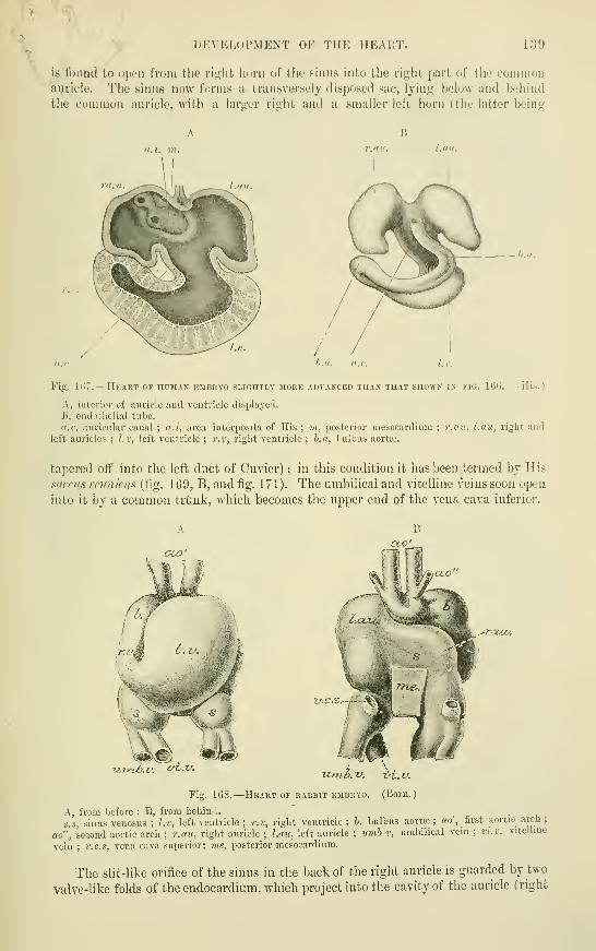
DEVELOPMENT OF THE IIKAUT. 1:39
is fdimd to oix'ii rroiu the ri^iit linni of the sinus into tho right part <ii" the commouauticle. Tlie sinus now forms a transversely disposed sac, lying below and beiiind
the common auricle, with a larger right and a smaller left liorn (the latter being
II
Lull.
f.aii.
-^h.,1.
Fig. Id".
—
Hkart of noMAN kmbryo slightly moue advanced than that shown in fig. 166. (His.)
A, interior of auricle aiul ventricle displayed.
B, endothelial tube.
«.c, auriculai' canal ; a.i, area interposita of His ; m, posterior mesocai'dium ; r.av, l.au, right andleft auricles ; l.v, left ventricle ; r.r, right ventricle ; h.a, lailbus aorta-.
ta])ered off into the left duct of Cuvier) ; in this condition it has been termed by His
sacms rciiniens (fig. 169, B, and fig. 171). The umbilical and vitelline veins soon open
into it by a common trimk, which becomes the upper end of the vena cava inferior.
oco'
V.C.S.-~,^--S
Fig. 168.
—
Heakt OF rabbit EMBRYO. (Lioru.
;
A, from before ; B, from behind.^
.
s.s, sinus venosus ; l.r, left ventricle ; r.r, right ventricle ; b, bulbus aorta; ; ao ,first aortic arch ;
ao", second aortic arch ; r.au, right auricle ; l.au, left auricle ; itmb.v, umbilical vein ;vi.v, vitelline
vein ; v.c.s, vena cava superior; me, posterior mesocardiura.
The slit-like orifice of the sinus in the back of the right auricle is guarded by two
valve-like folds of the endocardium, which project into the cavity of the auricle (right

140 DEVELOPMENT OF THE HEART,
LS.r.
vunh. v.Uvi.v. c/^.v.
Fie. 169.—A^'TEKioR and posterior aspect of the heart of a so^fewhat older rabbit embryo.vBorn.)
p.a, pulmonai-y arteiy ; s.r, s.l, s.tr, right and left horns and transverse part of sinus respectively;
r.s.c, l.s.c, right and left superior cavse : ^je, apertuj-e of pulmonary vein; h.v, hepatic veins; d.v,
ductus venosus ; me, mesocardium posterius.
The other letters as in iis. 16S.
oci^
.;K'kT^l£?^'^-^•
Fiw. 170.
—
Section through the heart of a rabbit EiiBRTO at the stage sno'wy ix fig. 169.
(Born.)
r.s, l.s, right and left horns of sinus receiving from above the respective sujierior vens cava : r.au,
l.au, right and left auricles ; r.v, l.v, right and left parts of the ventricle ; r.v.v, l.f.v, right and left
valves guarding the orifice from the right horn of the sinus into the right auricle; au.v.c. one of the
t>vo endocardial cushions which are beginning to subdivide the common amiculo-ventricular aperture.
The dotted line encloses the extent of the endocardial thickening ; s', first septum superior growing
down between ths auricles and prolonged below by a thickening of endocardium. Close to this septumin the left auricle is seen the opening of the pulmonary vein ; s. inf, inferior septum of the ventricles.
Fig. 171.
—
View FROii behind of the heart of a hfjian embryo of about 4 weeks, magnified. (His.)
The two sux^erior cavje, right and left, and the inferior cava are seen opening separately into the
sinus which is a transversely elongated sac communicating only by a narrow orifice with the right
auricle.

DEVELOPMENT UF THE HEART. 141
and left venous valves) (fi<?. 170, r.v.v., Lv.v.). These pass above into a muscular fold
of the auricular wall, whicli extends over the roof of the auricle heart parallel to the
septum atriurum, and is known as the septum spHrimn (Hi,'. 1 7o, B).^ It disajipears at
length, probably by uniting with the septum atriorum. Subse<|uently the venous orifice
opens out, and the right horn of the sinus, which is now seen to receive all the great
veins except the left duct of Cuviei', becomes gradually incorporated with the cavity
of the auricle. The transverse part of the sinus and its left, horn are continuous
with the left duct of Cuvier (tig. 171), and eventually tiie transvei-se part Ibrins the
coronary sinus. From the right venous valve the Eustachian valve is formed, andt'.ie development of the Thebesian valve is also connected with its lower end(Schmidt). The left venous valve distippears.
The trausN'crsely placed \'eutricular part of the heart receives at first at its left
end the orifice of the common auricle, which opens iuto its posterior wall (fig.
f.cuo.-v:^
S.ir^J.
S.T.-n.f
Fis. i: -Diagram to show the formation of the septum of the ventricles and bi'lb, and theMODE of division OP THE COMMON AURICULO-VENTRICULAR APERTURE. (Born.
)
au.v.c (in A and B), auriculo-ventricular aperture, partially divided into two by endocanlial cushions;
r.au.v, l.au.w right and left auriculo-ventricular apertures which have resulted from the division of thecommon aperture; ;•. r, l.v, right and left ventricles ; 6, bulbus aortae, replaced in C, by i^.a and a.o,
pulmonary artery aud aorta ; s.h, septum bulbi ; s. ('«/, septum inferius ventriculorum ; o (in A), orifice
between the two ventricles.
172, A, a.v.c.). At its right end it turns sharply upwards into the aortic bulb, into
which it gradually tapers, although there is at a certain point a constriction of the
endothelial tube, where the semilunar valves are subsequently formed {freium Halleri).
Soon the right aud left halves of the ventricle are separated externally by a
groove which extends from below, partially encircling the tube (fig. 1G9). If the
interior of ihe heart is examined at this stage, it is seen that a muscular septum,
corresponding internally to this groove, is growing upwards and backwards fi'om
the antero-inferior part of the tube, and is gradually separating it into two parts,
which become the right and left ventricles respectively (fig. 170, s.inf). This
iseptum {septum inferius of His) is placed obliquely to the long axis of the tube,
and extends eventually nearly to the level of the auriculo-ventricular orifice, which
has by this time become shifted along the posterior wall of the tube, so as to
open into it about its middle instead of at the left end, as was previously the case
(ng. 172, B). The septum of the ventricles remains incomplete for some time,
a communication between the two ventricles being maintained above it. Even-tually the septum inferius unites with prolongations, (1) from the endocardial
cushions which divide the common auriculo-ventricular orifice into right and left
^ This muscular prolongation maj', as Born suggests, be of use in assisting the action of the valves,
and in preventing their being forced backwards into the sinus when the auiicle contracts.

143 DEVELOPMENT OF THE HEART.
orifices; (2) from the endocardial aortic septum, which divides the bulb into aorta
and pulmonary artery. Thus the septum of the ventricles is completed by endo-
cardial connective tissue, a fact which is indicated even in the adult heart by the
existence of the thin septum membranaceum which forms the uppermost part of
the interventricular septum.
The common auricle in the meantime becomes shifted relatively upwards over
the back of the ventricles, carrying the sinus along with it, but it still lies behind
rather than over the ventricles, and the aperture of communication passes from
behind forwards, from the left part of the auricle into the corresponding half of the
ventricle. This constricted aperture soon becomes elongated into a short canal,
which is known as the auricular canal. Its orifice into the ventricle is from the
first somewhat flattened, and bounded by two lips, an upper and a lower. As deve-
lopment proceeds, it broadens out towards the median plane of the ventricular tube,
and becomes gradually shifted, first towards, and eventually over the line of constric-
tion which marks off the future right and left ventricles from one another (fig. 172).
The ventricular septum has by this time extended almost up to the transversely
elongated slit-like orifice, and its lips, still upper and lower in relative position,
become greatly thickened by the formation of cushions of endocardium, which grow
towards one another in the middle of the slit, and presently fuse into a median
thickening which converts the single I—l-shaped aperture into two triangular
openings, leading one into each ventricle (fig. 172, C). Meanwhile, the septum of
the ventricles growing towards the base abuts against, and at length comes into direct
continuity with the fused endocardial cushions, but this connection is nearer to the
right than to the left auriculo-ventricular aperture.^ There is still, as above stated,
a small orifice of direct communication between the left and right ventricles above
the free edge of the ventricular septum, and this is not closed until the descent of the
septum of the bulb, and its union with the septum of the ventricles, completes the
interventricular septum.
The above account of the division of the auricular canal is based upon that given
by Born for the rabbit, and in some respects differs from the description which was
given by His from an examination of human embryos. According to His, the endo-
cardial cushions, which by their union subdivide the auricular canal, are preceded by
and connected with a growth of endocardial tissue, which springs from the posterior
auricular wall, and they together form a septal prolongation {septum intermedium),
which projects like a stopper into the auricular canal, and divides the latter into the
two auriculo-ventricular orifices, and also grows down beyond that canal to meet the
uprising ventricular septum (fig. 173). The shortening of this canal is in part effected
by a kind of intussusception which takes place, and which causes its wall to be
folded into the ventricular cavity ; these folds, with probably some thickening of
endocardium, form the bases of the lateral flaps of the auriculo-ventricular valves
(fig. 175). The bases of the mesial or septal flaps are formed by a downward growth
of the edges of the endocardial septum between the two orifices. Both lateral and
mesial flaps become continuous Avith the spongy muscular substance which at this
time occupies most of the cavity of the ventricles (fig. 175). As development
proceeds, the flaps, which are at first thick and soft, become thin and membranous,
and become free fi'om muscular substance except near their free edges. These
muscular bands become tendinous near their insertion into the valves, and thus form
the chordcB tendinea, ; the parts which are not thus transformed become the impillary
muscles.
The septum of the auricles appears at the upper and back part of the auricular
2 Hence the riglit auriculo-ventricular orifice lies close to the ventricular septum, but the left orifice
is separated from it by an interval, into which the root of the aorta becomes continued (Born).
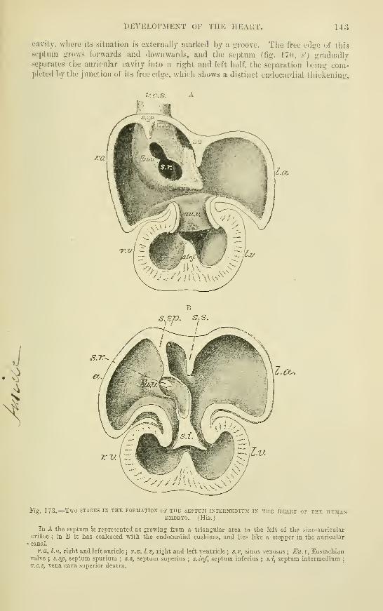
DEVELOPMENT OF THE HEAHT. 143
cavity, where its situation is externally marked by a j^roovc. The free eds^e of this
septum throws forwards and downwards, and the septum (W<f. 17<», .s') j;radually
separates the aiii'icular cavity into a right and left half, the separation being com-pleted l)y the junction of its free edge, which shows a distinct endocardial thickening,
/-«"
s.sp. s.s.
l.cu
Fig. 173.—Two STAGES IX the FOKIIATIOX OF THE SEPTUM I^•TERMEDI^^I IX THE HEART OF THE HCMAXEMBEYO. (His.)
In A the septum is represented as growing from a triangular area to the left of tte sino-auricular
orifice ; in B it has coalesced with the endocardial cushions, and lies like a stopper in the auricularcanal.
r.a, La, right and left auricle ; r.v, l.v, right and left ventricle; s.r, sinus venosus; Eu.v, Eustachianvalve ; s.sp, septum spuriura ; s.s, septum superius ; s.inf, septum inferius ; s.i, septum intermedium
;
v.c.s, vena cava superior dextra.

144 DEVELOPMENT OF THE HEART.
with the fused cushion-like thickenings which are subdividing the common auricular
orifice. But before the originally free communication between the two auricles is thus
closed, a new aperture makes its appearance above and at the back of this septum,
and gradually enlarges, so that a passage is thus re-established, but in a different
situation. This new orifice is the foramen ovale. It becomes closed by a second
septum, which also starts from the superior auricular wall, a little to the right of the
original attachment of the first septum, and gradually grows forwards and downwardsover the orifice. This second septum becomes the limdus Vieussenii, the first one
forms the so-called valve of the foramen ovale (Born).
According to His' account of the process in the human embryo, the septum atriorum is
formed by an anterior, or lower, and a posterior, or upper, sickle-shaped projection, which betweenthem enclose the foramen ovale, and form respectively the limbus Vieussenii and the valve of
the foramen ovale ; the connective tissue growth which he describes as growing- from the
posterior aiiricular wall towards the auriculo-ventricular orifice takes an important part in
the formation of the lower septal projection (septum intermedium). There is reason, how-ever, to believe that the process, as above described by Born for the rabbit, is materially the
same in all the higher vertebrates, including man, and that the successive growth of both
septa from the upper and posterior auricular wall was not noticed by His on account of the
lack of a series of human embryos sufficiently complete to show all the stages of growth.
Somewhat late in the course of development (after the appearance of the auricular
septum), the pulmonary veins are seen entering the left auricle. Before reaching the
auricle they have united to form a single vessel, and this opens into the auricle near
the septum (fig. 170, p.v.). In some animals, as the rabbit, this represents the
permanent mode of termination of the pulmonary veins, but in man the right and
left veins come to open separately into the auricular cavity, either by division of the
common trunk (His), or by opening out of the common trunk, and its absorption
into the auricle in the same way as the right horn of the venous sinus is absorbed
into the right auricle (Born). The two resulting vessels may again divide, so that
four pulmonary veins ultimately terminate in the left auricle.
The aortic bulb becomes subdivided into two vessels, the ascending aorta and
the pulmonary artery. The division is produced by a septum which arises as two
longitudinal thickenings of the lining membrane (endocardium). These grow from
opposite sides, and gradually meeting, fuse together in the middle of the bulb. The
folds take an oblique course down the bulb, for above they are anterior and posterior,
but below are right and left, hence the resulting vessels after separation are anterior
and posterior below and right and left above. The endocardial thickenings extend
somewhat below the origin of the bulb, and unite with one another and with the
septum of the ventricles, which they complete, and of which they form the mem-
branous part. The ventricular part of the heart is now completely divided into two,
each communicating with the corresponding division of the arterial bulb. There are
at first no semilunar valves, the soft thickened endocardial tissue of the bulb appear-
ing to exercise a sort of valvular action. The valves are formed as three projec-
tions of this tissue at the base of each vessel, at first thick and soft, but subsequently
becoming thinner and membranous. The common aortic trunk has four such
thickenings at the lower end, and the septum of the bulb as it descends is prolonged
into the right and left of these, so that the dumb-bell-shaped orifice is divided into
two triangular apertures, the bulging sides of which are formed by the endocardial
cushions and become developed into the semilunar valves (fig. 174).
The aortic septum begins between the fourth and fifth aortic arches, and is so dis-
posed, that the fourth arch continues the aortic half of the bulb, the fifth the pul-
monary half. After the completion of the septum, an external groove makes its
appearance along the line of the endocardial thickenings, and deepening gradually,
splits the bulb into two separate vessels.
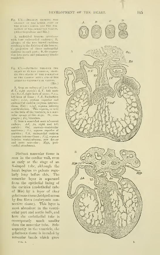
DEVELOPMENT OF THE HEART. U5
Fig. 17t.— Pl.VIJUAM SHI)\VIN<i TlIK
DIVISION OK TUK LOWKK TAUT OK
THK IIUI.IU'S AOKT.*;, AND THK KOK-
MATIOM <IK THK SKMIl.LNAK VAI.VKS.
(After (rogciibiiur ami His.)
A, iindividctl truncii.^ arteriosus
with fo\ir (Miiiocariliai cusiiions; 1!,
advance of the two lateiiil cusiiidiis
resulting in the division of tiie liinifii ;
C, projection of tiiroe endocardi.il
cushions in each juirl ; D, the sejiara-
tion into aorta and imUnonary trunks
completed.
Fig. IT'i.—Sections Tiinoi'iiii Tin:
HKAKT OK lU'.MAN EMHKVuS, SHoW-IXt! TWO STAdES IX THE FORMATION
OK THE CAUniAC SEPTA AND OK THEAURICULO-VENTKICULAK VALVES.
(His.)
A, from an embryo of n or 6 week.s.
Ji. V, riglit auricle; L. V, left auri-
cle ; S.r.il, right horn of sinus ; S.r.)<,
left horn of sinus ; V.A\ Eustachian
valve ; .i.int, septum superior andendocardial cushion (septum intei'me-
diuni, His) ; s.inf, septum infcrius
ventriculorum. This septum, as well
as the bulk of the ventricle, is a mu.s-
cular sponge at this stage. Oe, oeso-
phagus ; JJi; bronchus.
B, from a somewhat more advancedembryo. Ad, As, right and left
auricle ; Ost, auriculo-ventricular
apertures ; S.s. septum superior of
auricles ; S.!t, endocai'dial cushion
(septum intermedium) ; S.if, septuminferius ventriculorum, now denser
and more muscular ; ^^pi^, peri-
cardial attachment.
Distinct muscular tissue is
seen in the cardiac wall, even
as early as the stage of an
S-shaped tube, although the
heart begins to pulsate regu-
larly long before this. Themuscular layer is separated
from- the epithelial lining of
the cavities (endothelial tube
of His) by a layer of clear
gelatinous tissue,bridged across
by fine fibres (embryonic con-
nective tissue). This layer is
most abundant in the ventri-
cular part and aortic bulb, and
here the endothelial tube is
consequently much smaller
than the muscular tube. Sub-
sequently, in the ventricle, the
gelatinous tissue is invaded by
muscular bands which grow

146 DEVELOPiMENT OF THE PRINCIPAL ARTERIES.
into it from the compact outer layer of muscle, and unite with one another to form
a spongework of muscular trabeculse, while the endothelium of the cavity becomes
depressed between and over these trabecuisB, and lines all the spaces between them,
which thus communicate with the cavity of the ventricle. The ventricles are therefore
now in the same condition in Avhich they are found permanently in many of the
lower vertebrates (e.g. frog).
Ultimately the compact outer layer ofmuscle becomes greatly increased in thick-
ness, and the spongework of trabeculge occupies a relatively much smaller portion of
the cavity, being developed in part into the columnge carnese of the adult heart.
Peculiarities of the foetal heart.—Besides the peculiarities of structure,
which have been above descril)ed, tlie IVjetal heart differs in position, in relative size,
and in the thickness of its several pares, from the organ after birth. Thus it is at
first placed immediately under the head, but subsequently, with the development of
the neck, it gradually assumes a position farther back. In early foetal life it is
much larger in proportion to the size of the body than at a later period, and at birth
it is still proportionally large. The walls of both ventricles are of equal thickness
during foetal life, a peculiarity which is evidently connected with the fact that in
consequence of the communication of the pulmonary artery, through the ductus
arteriosus, with the aorta, the blood pi'essure which they have to overcome is the
same.
DEVELOPMENT OP THE PPaNCIPAL AKTERIES.
From the point of insertion of the aortic bulb into the ventral wall of the foregut,
first one, and then in snccession four other arterial arches, become formed, and pass
on either side, one into each visceral arch. Half encircling this part of the
Fig. 176 — Diagrammatic out-
lines OP THE HEART AKD PRI-
MITIVE VESSELS OF THE EMBRYOCHICK AS SEEN FROM BELOW ANDENLARGED. (A. T.
)
A, soon after the first establish-
ment of the circulation ; B, C, at
a somewhat later period ; 1 , ]
,
the veins returning from the vas-
cular area ; 2, 3, 4, the heart,
now in the form of a notched
tube ; 5, 5 (upper), the two primi-
tive aortic arches ; 5, 5 (lower),
the primitive double aorta ; a, the
single or united aorta ; 5', 5', the
continuation of the double aortse
beyond the origin of the large
omphalo-mesenteric arteries, 6, 6.
The division above 4 is repre-
sented as caiTied rather too far
down.
alimentary canal, they are continued above it into two descendinij or primitwe aorke.
These two vessels run down the trunk on either side of the notochord, yielding, as
they descend, lateral oflFsets to the body walls and to the yolk sac. Finally they give
off", at the lower or posterior extremity, two large vessels, which accompany the
allantois, and furnish blood to the foetal part of the placenta (tmiMUcal or allantoic
arteries).
The primitive aortas do not long remain double. As was first shown by means
of sections by Allen Thomson, they unite in the middle line, the union beginning
in the dorsal region and extending forwards and backwards ; in the latter direction
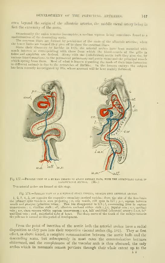
DEVELOPMENT oF THE PRINCIPAL ARTEKIES. 147
even Ix^yoml tlie (.rigiii of the allantoic arteries, the middle sacral artery boinf; infact the extremity of the aorta.
Occasionally the imion ivmaiiis incoinpleto, a median Foptum bein^' sometimes found as amalformation of the di'suendinjr aorta.
Tlie com.non iliacs are formed by persistence of the roots of the allantoic arteries ; whenthe lower limbs are formed thoy pive otF to these the external iliacs.
Since their discovery by Kathke in 182:,. the arterial arches have been regarded withmuch interest as corresponding with those from which the bloo<l-vessols of the gills infishes and amphibia are derived. Alon- with the (subdivided) aortic bulb they give rise byvarious tninsl urinations, to the permanent pulmonary and aortic stems and the principal ves-selswJiich spring from them. IMost of what is known regarding the mode of their transformationin different animals is due to the researches of Itathke. In the human embryo the subjecthas been recently investigated by His. whose account will be here mainly followed.
vo-co. VCV-
-cUZ.
Fig. 177.
—
Profile view of a human embrto of about fifteen days, with the alimextakt caxal in
LONGITUDINAL SECTION. (His.)
T'vo arterial arches are formed at this stage.
Fig. 17S.—Similar view of a sojiewuat older embrvo, showing five arterial arches.
2, 3, 4, 5, are opposite the respective secondai-y cerebral vesicles ; from the side itf the fore-brain
the primary optic vesicle is seen projecting ; ot, otic vesicle, still open in 177 ;js.i", septum between
mouth and pharynx (primitive velum). This has disappeared in 178 ; I, commencing liver in septumtransversum ; \\ vitelline stalk ; all, allantois enclosed within stalk
; j.v, jugular vein ; c.v, cardinal
vein; s.r, sinus venosus within septum transversum ; v..a, left umbilical (allantoic) artery; l.v.v, lef*
umbilical vein ; end., endothelial tube of heart. The sharp curve of the trunk of the embryo towards
tbe yolk-sac is normal at this period cf development.
From the point of insertion of the aortic bulb the arterial arches have a radial
disposition as they pass into their respective visceral arches (fig. 181). They at first
effect, as above stated, a complete communication between the aortic bulb and thedescending aorta, but subsequently iu most cases the communication becomesobliterated, and the completeness of the vascular arch is thus obscured, the onlyarches which in mammals remain pervious through their whole extent up to the
L 2
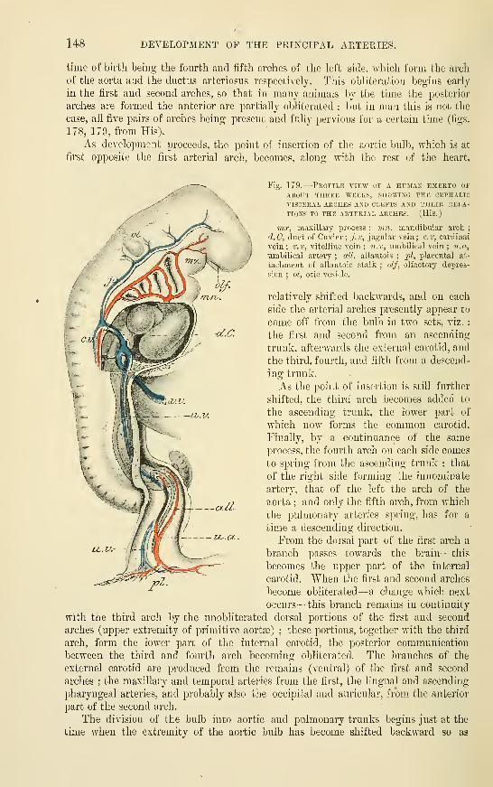
148 DEVELOPMENT OF THE PRINCIPAL ARTERIE,"^.
time of birth being the fourth and fifth arches of the left side, which form the arch
of tlie aorta and the ductus arteriosus respectively. This obliteration begins early
in the first and second arches, so that in many animals by the time the posterior
arches are formed the anterior are partially obliterated ; but in man this is not the
case, all five pairs of arches being present and fully pervious for a certain time (figs.
178, 179, fi-om His).
As development proceeds, the point of insertion of the aortic Ijulb, which is at
first opposite the first arterial arch, becomes, along with the rest of the heart,
Fig. 179.
—
-Profile view of a human embryo of
ABOUT THREE WEEKS, SHOWING THE CEPHALIC
A'ISCERAL ARCHES AND CLEFTS AND THEIR RELA-
TIONS TO THE ARTERIAL ARCHES. (His.)
mx, maxillary process ; tnn. mandibular arch;
d.C, duct of Guvier; j.v, jugular vein; c.v, cardinal
vein; v.t, vitelline vein ; u.v, umbilical vein ; u.a,
umbilical artery ; all, allantois; ^j/, placental at-
tachment of allantoic stalk ; olf, olfactory depres-
sion ; ot, otic vesicle.
relatively shifted backwards, and on each
side the arterial arches presently appear to
come off from the bulb in two sets, viz.
:
the first and second from an ascending
trunk, afterwards the external carotid, and
the third, fourth, and fifth from a descend-
ing trunk.
:As the point of insertion is still further
shifted, the third arch becomes added to
the ascending trunk, the lower part of
Avhich now forms the common carotid.
Finally, by a continuance of the same
process, the fourth arch on each side comes
to spring from the ascending trunk : that
of the right side forming the innominate
artery, that of the left the arch of the
aorta ; and only the fifth arch, from which
the pulmonary arteries spring, has for a
time a descending direction.
From the dorsal part of the first arch a
branch passes towards the brain—this
becomes the upper part of the internal
carotid. "When the first and second arches
become obliterated— a change which next
occurs—this branch remains in continuity
with the third arch l)y the unobliterated dorsal portions of the first and second
arches (upper extremity of primitive aort^) : these portions, together with the third
arch, form the lower part of the internal carotid, the posterior communication
between the third and fourth arch becoming obliterated. The branches of the
external carotid are produced from the remains (ventral) of the first and second
arches ; the maxillary and temporal arteries from the first, the lingual and ascending
pharyngeal arteries, and probably also the occipital and auricular, from the anterior
part of the second arch.
The division of the bulb into aortic and pulmonary trunks begins just at the
time when the extremity of the aortic bulb has become shifted backward so as

DEVELOPMENT OF THE IMIIXCII'AL AltTElllES. 149
to ho opposite the jwint of junction hctwuen the I'ouilh and fifih pairs of arches, so
tliat now all the arches above this jioint become se])arated oil" in connexion with the
trunk of tlie aorta (ascending aorta) ; the one below it remaining ia connexion with
Fi^. 180.
—
^Profile view of a human emrryo of about 3 or 1 weeks, suowing the principal arteriesAND veins. (His.)
1, 2, 3, 4, 5, the secondary cerebral vesicles ; hyp, hypophysis ; ot, otic vesicle ; run, mandibulararch ; bj. lung rudiment ; st, stomach ; Wd, Wolffian duct opening into cloaca ; /, //, III, IV, V,
the arterial arches', springing from b.a, bulbus arteriosus; p, pulmonary artery ; d.C, duct of Cuvier ;
ch., notoi;urd.
the pulmonary trunk. After the separation, the aortic bulb, now a double tube,
becomes still further shifted back, and with it the fourth and fifth arches. Since
Fig. 181.
—
View from behind of the anterior partOF THE MOUTH AND PHARYNX OF A HUMAN EMBRYOOF 3i- WEEKS. SHOWING THE ARTERIAL ARCHESRADIATING FROM THE ATTACHMENT OF THE AORTIC
BULB. (His.)
ao, point of attachment of aortic bulb in the anterior
wall of the pharynx ; ?hh, In/, ftr', hr", tirst four visceral
arches ; /, //, ///, IV, the corresponding arterial
arches ; V, fifth arterial arch giving off the pulmonaryartery, p.
the infei'ior laryui^^eal nerve passes under the
latter, this nerve must also become shifted
downwards along with these arciies. Toallow for this shifting of the bulb, the
common carotids become proportionally
lengthened.
From the descending primitive aorta3 on either side a series of inter-segmental
arteries pass ; the uppermost of these become united to form the vertebral arteries
(which subsequently unite superiorly in the middle line to produce the basilar), the
lower form intercostal arteries. A branch for the upper extremity comes ofif from

150 DEVELOPMENT OF THE FOURTH AND FIFTH ARCHES.
the commencement of the vertebral ; it subsequently far exceeds the parent vessel
in size, and forms the subclavian, this name being extended to what was originally
the commencement of the vertebral from the descending aorta.
Destination of the fourth and fifth arches.—As was above stated, the
aortic trunk, connected below with the left ventricle, is connected above with the
four superior arches, which spring from its two ascending rami ; of these rami the
right is much the smaller, and its root forms the innominate ; the root of the left,
which is much larger, represents the part of the arch of the aorta between the
Fig. 182 —Diagram to showTHE DESTINATION OP THEARTERIAL ARCHES IN MANAND MAMMALS. (Modified
from Ratlike.
)
The truncus arteriosus, andthe five arterial arches spring-
ing from it are represented in
outline only, the permanentvessels in colours—those be-
longing to the aortic system
red, to the pulmonary system
blue.
siwclcovictft
O/acerhdvriq/cLordco
—I'a^us Tierve
eoGtemaZ-
,verteBrat
otrcJi. of
sii^clcwimz
ct/rteriosns
sceruvirwf cwrtco
innominate and the left
common carotid. Onthe right side the
whole of the fourth
arch remains, and forms ~
the common subclavo-
vertebral trunk ; on tlie
left side the whole arch
persists and forms the
remainder of the arch of
the aorta.
The fifth arch of the
right side only persists
as far as the origin of
the branch to the right
lung, the remainder of
the arch disappears. The fifth arch of the left side jDcrsists throughout its whole
length during foetal life, and joins the fourth arch as the ductus arteriosus. After
birth, the part of the arch beyond the branch to the corresponding lung becomes
impervious on this side also, and is converted into the Ugamentum arteriosum.
The upper part of the descending primitive aorta disappears entirely on the
right side ; that of the left side forms the commencement of the permanent
descending aorta.
Rathke described both pulmonary arteries as being given off from the fifth left arch in
mammals, although admitting that in birds and reptiles each is formed from the corresponding
arch. In birds the permanent aortic arch is the fourth arch of the right side, and not of the
left side as in mammals, and in reptiles both aortic arches remain pervious.
Many of the abnormalities which are observed in the disposition of these arteries, may beexplained by regarding them as a persistence of embryonic conditions.
The develoi^ment of the arterial arches of the bird has been recently again examined byMackay, whose account differs in important particulars from that of Rathke, and indeed of
nearly all previous investigators. He describes the subclavian artery as arising from the third
arch, not from the fourth (this was also given as its origin by Sabatier), springing from the
ventral part of the arch, and running outwards superficial to the pneumogastric nerve and jugular
vein. The third arch and its dorsal upward prolongation, form the common carotid, not the
DEVELOPMENT OF THE PKINUIFAL VEINS. 151
internal carotid, as stated by llatlike, and the ventral prolongation of the truncus arterioauH
forms, not the external earotid, but a small brancli from the subolavian, or innominateartery, to the front of tlie trachea. These observations have not been as yet extended to
manunals.Zimmermann lias described, both in the niVjbit and in the human (imbryo, an arterial arch
between the aortic and pulmonary arches. If this is of constant occurrence it must be
reckoned as the fifth arch, and the {>ulmonary will become the sixth.
The histogcnctic changes involved in the development of the blood-vessels are described in
the chapter on Histology.
The tirst vessels appear, as has been already stated, in the mesoblast of the vascular area;
the lamina of mesoblast in which they are formed is sometimes distinguished as the vascular
lamina. They are said (by His and others) to grow inwards from tlie va.scular area, but the
manner in which the principal arteries and veins of the body are first developed is not clear,
beyond the fact that they are at first merely endothelial tubes. The muscular tissue of the
primitive aortas is derived from the lower part of the protovertebra (I']. Miiller).
DEVELOPMENT OF THE PRINCIPAL VEINS.
In this subject ulso the description given by His of the condition and changes
of the veins in the human embryo will be followed, although it differs in certain
particulars from that which has usually been received.
In the early embryo, before the development of the allantois, two vUellirui or
ompJmlo-nwseraic veins, right and left, bring back the blood from the vascular area
upon the yolk sac, and unite to form a common trunk, which is continued as the
sinus venosus into the auricular extremity of the rudimentary heart.
At the commencement of the placental or allantoic circulation (fourth week in
man) two umhilical veins are seen coming from the placenta and opening into the
sinus near the vitelUne veins (fig. 168). Into this also opens on either side a
transverse vein, the dud of Cuvier or superior vena cava, which is formed by the junc-
tion of the 'primitive jugular vein, bringing blood from the head, and the cardinal
vein, which returns the blood from the Wolffian bodies, tbe vertebral column and
the body walls (figs. 179, 180). The trunk or sinus into which all these veins pour
their blood is now transversely disposed, immediately below the diaplu-agm, and
forms the saccus reuniens of His, which has been already alluded to (p. 139).
The vitelline or jomphalomeseraic veins enter the abdomen along the vitelline
duct and ascend at first along the front of the alimentary canal, but higher up they
are seen on either side of that tube (duodenum and stomach). Here transverse
communications form between the two veins, two in front of and one behind the
duodenum, so that this is encircled by two vascular rings (figs. 180, 183). Above
these venous circles the direct communication with the sinus becomes lost, the inter-
mediate venous vessel or either side becoming broken up within the substance of the
liver (which has by this time developed around them) into a vascular network, the
middle part of which becomes capillary.
The vessels which pass from the upper venous ring to the capillary network are
known as venc& advehentes, they become the branches of the portal vein ; those
which pass from it into the sinus are the vcnce revehcnlcs, they become the hepatic
veins.
The lower communication between the vitelline veins takes the form of a com-
plete longitudinal fusion of the two vessels, at least for some distance. This fused
part receives veins from the intestine and stomach, and becomes the commencementof the portal vein.
The umbilical veins are for a long time double within the abdomen, although
they have fused within the umbilical cord into a single trunk: They diverge from
this and pass up to the sinus on either side in the somatopleure, just where this is
becoming bent round into the amnion. After a time, however, it is found that this
direct commuuication with the sinus is partially interrupted by the development of a

153 DEVELOPMEISTT OF THE PEINCIPAL VEINS.
vascular network, and that on the left side a fresh communication has become esta-
blished with the upper venous circle of the vitelline veins. The interruption subse-
quently becomes complete on both sides (fig. 183), and on the right side the greater
V:u.ij
VV^om.
Fig. 183.-
—
Venous trunks of a human embryo ofABOUT THREE-AND-A-HALF WEEKS. (His.)
v.c.d, v.c.s, superior vense cavK, right and left;
r.j, v.c, iJrimitiA'e jugular and cardinal vein
;
T.v.d, v.u.s, umbilical veins, right and left ; v.u",
v.u", upper detached portions of umbilical veins;
v.v.om, omphalomeseraic or vitelline veins forming
the vena portse. The permanent veins are coloured
blue.
part" of the vein becomes atrophied (on
both sides the part which originally opened
into the sinus reuniens remains evident
for a time). The left vein, on the other
hand, increases in bulk with the develop-
ment of the placental circulation. For a
short time the whole of its blood, as well
as that of the vitelline vein, passes through
the capillaries of the liver. But a branchis soon seen passing from the upper venous circle direct into the right hepatic
vein, near its entrance into the sinus. This forms the ductus venosus or vena
asce7idens, and it now carries most of the blood of the umbilical vein direct to the
heart. Subsequently the direct communication of the left hepatic vein with the
Fig. 184.
—
Under surface op the fcetal liver,
WITH ITS great BLOOD-TESSELS, AT THE FULLPERIOD.
a, the umbilical vein, lying in the umbilical
fissure, and turning to the right side, at the trans-
verse fissure (o), to join the vena i3ort8o( jj) ; d, the
ductus venosus, continuing straight on to join the
vena cava inferior (c) ; some branches of the umbili-
cal vein pass from a into the substance of the liver;
g, the gall-bladder, cut.
sinus becomes obliterated, and a new com-
munication becomes established with the
ductus venosus ; and, finally, when, with
the growth of the lower hmbs and of the
other abdominal and pelvic organs,' the inferior vena cava becomes developed, this
also joins the upper end of the ductus venosus.
The lower part of the portal vein is formed, as we have seen, by the united
vitelhne veins. The upper part is formed as a single trunk out of the double venous
annulus by atrophy of the right half of the lower ring and the left half of the upper
(fig. 183). The spiral turn around the duodenum is thus produced, and thus it is
also that the portal vein at first appears more directly connected with the right vense
advehentes than with the left.
Most of these embryonic veins are at first of relatively large size and have an
irregular sinus-hke character, which disappears at a later stage of development.
At the time of commencement of the j)lacental circulation, two short transverse
venous trunks, the ducts of Guvier, open, as has been above stated, one on each side,
into the sinus venosus of the heart. Each is formed by the union of a superior and
an inferior vein, named respectively ih.Q primitive jugular and the cardincd.

l)KVKlA>l'.Mi:\T OF THK l'i:i.\( 1 1'.\ I, VKIXS. 153
'V\\v pritiiitirv jiKjiilid- vein ivtTive'S tlu; hlood from the (•niiiial caviLy by cliiuiiiels
ill fiont (tf tlio ear, wliicli are sultseqiiontly obliterated : in the ;;Tealer part of its
extent it l)ec(inies the external jnuular vein ; and near its lower end it receives small
branches, which <;row to be the internal jii;ruhir and sul)clavian veins (fig. IHij).
The cardinal reins are tiie primitive vessels which retnrn the blood from the
Wolffian bodies, the vertebral column, and the parietes of the trunk. Their lower
extremities, which become developed later into the internal iliac veins, receive, cs
ImU. lSr>.—SCIIKMK OK TIIK IiKVKLOl'MKNT uK Tlir. f'Hrr.K VKIXSOF TIIK HUDV. ((t. D. T. )
TIic primitive venous trunks are imlicateil liy black outlines,
and their names are enclosed witliin parentheses-. The delinitive
veins are represented blue.
the lower limb becomes developed, the sciatic veins,
and later the external iliac ; hi^-her up, with the
development of the kidneys, the renal veins, with
the spermatic and suprarenal, also open into them.
The bilaterally symmetrical arrano-emeut which at
first prevails becomes lost, owinii;- to the obliteration
of a large part of the left cartlinal vein, and the
formation of a transverse communication {trans-
verse iliac vein) between its lower extremity andthe right cardinal, a little above the place where
the right exterual iliac joins it. The greater part
of the left common iliac becomes formed by this
communicating vessel, only a small part being
formed by cardinal vein, while the right is wholly
formed by cardinal vein.
Above the transverse iliac vein the fate of the
cardinal veins on the two sides is veiy different.
That of the left side becomes obliterated for some
distance, whereas that of the right side undergoes
an increased development, receiving as it now does
the blood from the whole of the lower limbs and
pelvis : it forms the greater part of the inferior
vena cava. But the hepatic part of the vena cava
has become developed independently of the cardinal
veins as a median vessel which opens above into the
tnmk formed by the junction of the ductus venosus
with the vitelline veins. A commmiicating branch
is soon formed on either side between the lower
end of this median vessel and the cardinal veins
at the place where they receive the renal veins,
and on the obliteration of the lower part of the left cardinal the left renal vein
opens by this communicating branch directly into the vena cava. On the right side
the communication becomes the continuation of the main trunk, for the right
cardinal also becomes obliterated for a short distance above the renal vein. (See
also Vol. II., Part 2, " Morphology of the Venous System,") ^
As development proceeds, the direction of the ducts of Cuvicr is altered by the
descent of the heart from the cervical into the thoracic region, and becomes the same
^ Advantage lias been taken of the necessity of reprinting this volume to alter the account of thedevelopment of the veins of the trunk, the former description having been based upon the earlier re-
searches of Rathke, which have been show-n by Hochstetter to require moditication (November, 1895).
154 DEVELOPMENT OF THE PEINCIPAL VEINS.
as that, of tlie primitive jugular yeins. A communicating branch (which may betermed the transverse jugular) makes its appearance, directed transversely from the
junction of the left subclavian and jugular veins, across the middle line to the right
jugular; and further down in the thoracic region between the cardinal veins, a
communicating branch {transverse cardinal) passes obliquely across the middle line
from right to left. The communicating branch betw^een the primitive jugular veins
is converted into the left brachiocephalic or innominate vein. The portion of vessel
between the right subclavian vein and the termination of the transverse jugular
becomes the riglit brachiocephalic vein. The portion of the primitive jugular vein
below the transverse jugular, together with the right duct of Cuvier, forms the venacava superior, while the cardinal vein opening into it is the extremity of the great
Fig. 186.—A and B.
—
Diagrammatic outlines of the vestige of the left superior cava and of
A CASE of its persistence. (Sketched after Marshall.) I.
The views are supposed to be from before, the parts of the heart beiog removed or seen through.
1, 1', internal jugular veins ; 2, 2', subclavian veins ; 3, right innominate ;3', right or regular
superior cava ; 4, left innominate, normal in A, rudimentary in B ; 5, in A, the opening of the superior
intercostal vein into the innominate ;5', vestige of the left superior cava or duct of Cuvier ; 5, 5', in B,
the left vena cava supeiior abnormally persistent ; 6, coronary sinus ;6', coronary veins ; 7, superior
intercostal trunk of the left side (left cardinal vein) ; 8, the jjrincipal azygos (right cardinal vein)
;
7', 8', some of the upper intercostal veins ; 9, the opening of the inferior vena cava, vith the Eustachian
valve.
vena azygos. On the left side, the portion of the primitive jugular vein placed
below the transverse jugular may still be represented by the left superior intercostal
vein, but the duct of Cuvier becomes obliterated (with the exception of the part
which forms the coronary sinus) as does also usually the uppermost part of the
cardinal vein. The remaining part of the lefc cardinal, however, which receives most
of the left intercostal veins, becomes, with the transverse cardinal, converted into the
left azygos veins. The variability in the adult arrangement of the veins depends
on the different extent to which the originally continuous vessels are developed or
atrophied at one point or another. The left duct of Cuvier is obliterated. But even
in the adult, traces of this vessel can ahvays be recognised in the form of a fibrous
band, or sometimes a narrow vein, which descends obliquely over the left auricle ;
and in front of the root of the left lung there remains an indication of its former
presence in the form of a small fold of the serous membrane of the pericardium, the
rECULlAi:iTIi:s Ul' TUK F(KTAL organs of CIIICLILATIoX. lo.j
vest if/ id I fold of ^[arsliall, to wliom is due tlic first full elucidation of the nature and
relations of the left j)riniitive vena cava superior.
The left duct of Cuvier lias been ol)ser\ed jiersistent as a small vessel in the
adult. Jiess frequently a ri^ht and a left innominate vein open separately into the
Fig. 187.— Vii:w ok thi: kiktai. iikakt ani> <;iikat
vks8ki.s, krom tuk i.kkt s1i>k, to slkiw thk vksthik
OK THK I.KKT Sll'KUIoK VKNA CAVA IN SITU. (TluH
ligure is iiliiniR'd after one of MjhsIimU's. )
a, rijjht nmiclo ; h, left auricle and imliiioiijiry veins;
c, the coiius arteriosus of tiic ri^'ht ventricle ; </, the left
ventricle ; c, descending aorta ; I , vestigial fold of the
pericardium; /, arcli of the aorta, with a part of the
pericardium rcmuininj< aliove; (f, main pulmonary artery
.and ductus arteriosus;
;/', left pulmonary artery ; 1, T,
right and left internal jugular veins ; li, 2', suhchivian
veins ; 3, .'!', riglit innondnate and superior vena cava ;
1, left iniuiminate ; 5, ;")', remains of the left superior
cava anil duct of Cuvier, passing at + in the vestigial
fold of the peric-.irdium, joining the coronary sinus, 0,
below, and receiving above the superior intercostal vein,
7 ;7', 7', the upper and lower intercostal vein.
n\2;ht auricle, an arraugcmcnt which is also met with in bii'ds and in certain
maiuinals, and which results from the vessels of the left side beiuf;' developed
similarly to those of the ri,o-ht, while the cross branch remains small or absent.
A ease is recorded by GrubL'r in which the left vena azygos opened into the
coronary sinus, and was met by a small vein descending from the union of the
subclavian and jugular. Here, then, the jugular veins had been developed in the
usual manner, while the left vena azygos continued to pour its blood into the duct
of Cuvier.
PECULIARITIES OF THE FCETAL ORGANS OF CIRCUIiATION.
It may be useful here to recapitulate shortly the peculiarities of structure exist-
ing in the advanced stage of the formation of the foetal organs of circulation, with
reference to their influence in determining the course of the blood during intra-
uterine life, and the changes w^hich occur in them upon the establishment of pulmo-
nary respiration at birth.
The foramen ovale retains the form of a free oval opening in the septum atri-
oruni up to tlie fourth month, but in the course of that month and the next the
growth of the valvular plate which fills np the floor of the fossa ovalis, becomes com-
plete, so that in the last three and a half mouths the blood can only pass from the
right into the left auricle, not in a contrary direction.
The Eustachian valve constitutes a crescentic fold of the lining structure of
the heart, which is so situated as to direct the blood entering the auricle by the in-
ferior cava towards the opening of the foramen ovale.
The ductus arteriosus establishes a communication lietwecn the main pul-
monary artery and the aorta, by which the blood from the right ventricle is carried
mainly into the dorsal aorta.
The two large hypogastric oi- umbilical arteries, prolonged from the iliac
arteries, passing out (jf the body of the fcetus, proceed along the umbilical cord, to
be distributed in the ttetal portion of the placenta. From the placenta the blood is
returned by the umbilical vein, which, after entering the abdomen, communicates
by one branch with the portal vein, and is continued by another, named ductus
vmosus, into one of the hepatic veins, through which it joins the main stem of the
vena cava inferior.
Course of the blood in the fcetus.—The riuht auricle of the foetal heart

156 COURSE OF THE BLOOD IN THE FCETUS.
V?
receives blood from the two vena cavse and the coronary sinus. The blood brought
by the superior cava is simply the venous blood returned from the head and upper
half of the body ; whilst the inferior cava, which is considerably larger than the
superior, conveys not only the blood from the lower half of the body, but also that
which is returned from the placenta and the liver. This latter stream of blood
reaches the vena cava inferior, partly by a direct passage—the ductus venosus—and
partly by the hepatic veins, which bring to the vena cava inferior aU the blood
Fig. 188.
—
Diagrammatic outline op theORGANS OF CIRCULATION IN THE PffiTUS
OF SIX MONTHS. (Allen Thomson. )i
RA, right auricle of the heai't ; EV, right
ventricle ; LA, left auricle ; E^^, Eustachian
valve ; LV, left ventricle ; L, liver ; K, left
kidney ; I, portion of small intestine ; a,
arch of the aorta ; a', its dorsal part ; a",
lower end ; vcs, superior vena cava ; vci, in-
ferior vena v/here it joins the right auricle;
i'ci', its lower end ; s, subclavian vessels;
_/, right jugular vein ; c, common carotid
arteries ; four curved dotted arrow lines are
carried through the aortic and pulmonary
opening, and the auriculo-veutricular orifices
;
da, opposite to the one passing through the
pulmonary artery, marks the place of the
ductus arteriosus ; a similar arrow line is
shown passing from the vena cava inferior
through the fossa ovalis of the right auricle,
and the foramen ovale into the left auricle ;
liv, the hepatic veins ; wp, vena portae ; x to
vol, the ductus venosus ; ilv, the umbilical
vein ; im, umbilical arteries ; mc, umbilical
cord cut short ; I i', iliac vessels.
circulating through the liver,
whether derived from the supply
of placental blood entering that
organ by the umbilical vein, or
proceeding from the vena portte or
hepatic artery.
The blood of the superior vena
cava is believed to pass through the
right auricle into the right ventricle,
whence it is propelled into the trunk
of the pulmonary artery, A small
part is distributed through the
branches of that vessel to the lungs,
and returns by the pulmonary veins
to the left auricle ; but, as these
vessels remain small up to the time
of birth, by far the larger part passes
through the ductus arteriosus into tne descending aorta, and is thence distributed in
part to the lower half of the body and the viscera, and in part along the umbilical
arteries to the placenta. From these several organs it is returned by the vena cava
inferior, the vena port^, and the umbiUcal vein ; and, as already noticed, reaches
the right auricle through the trunk of the inferior cava.
Of the blood entering the heart by the inferior vena cava, it is supposed that
only a smaU part is mingled with that of the superior cava, so as to pass into the
1 The red and blue coloration of this diagram represents the fact that the vessels coloured are
arteries and veins respectively, and not the quality of blood which they convey.
CUANGKS IN THE CIRCULATION AT BlltTlI. l5?
riglit ventricle; by far Llie lari;cr poilioii is tliuui;lit to be direeted by the Eustachian
valve tlirouf!:h the foramen ovale into the left auricle, and thence, together with the
small quantity of blood returned from tlie luniks by the pulmonary veins, to pass
into the left ventricle, whence it is sent into the arch of tlu! aorta, to be distributed
almost entirely to the head and upper liinl)s.
Sabatier wasthe lirst tocall attention particularly to the action of the Eustachian
valve in separatiuj;: the currents of blood entering the right auricle by the superior
and inferior vena; c;\\x. Tiiis scjiaration, as well as that occurring between the
currents passing through the aortic arch and the ductus arteriosus into the descend-
ing aorta, was illustrated experimentally l)y John Reid. A striking confirmation of
the extent to which the last mentioned division of the two currents of the foetal
blood may take place, without disturbance of the circulation up to the time of birth,
is afforded by the examples of malformation in which a complete obliteration has
existed in the aortic trunk immediately before the place of the union of the ductus
arteriosus with the posterior part of the aortic arch.
In earlier stages of development than those above described, it is certain that
there is little or no separation of the two kinds of blood, for both the umbilical
veins from the placenta and the veins from the yolk sac and body generally, pour
their blood together into the sinus vonosus, and the mixed blood is then forced
through a single somewhat narrowed orifice {poria vei^tilinli of Ilk) into the auricle.
CHANGES IN THE CIRCULATION AT BIRTH.
The changes which occur in the organs of circulation and respiration at birth,
and which lead to the establishment of their permanent condition, are more immedi-
ately determined by the inflation of the lungs with air in the first respiration, the
accompanying rapid dilatation of the pulmonary blood-vessels with a greater quantity
of blood, and the interruption to the passage of blood through the placental circula-
tion. These changes are speedily followed by shrinking and obliteration of the
ductus arteriosus, and of the hypogastric arteries fi'om the iliac trunk to the place of
their is^ue from the body by the umbilical cord ; by the cessation of the passage of
blood through the foramen ovale, and somewhat later by the closure of that foramen,
and by the ol)literation of the uml)ilical vein as far as its entrance into the hver, and
of the ductus venosns behiiid that organ.
The process of obliteration of the arteries appears to depend at first mainly on
the contraction of their coats, but this is very soon followed by a considerable
thickening of their substance, reducing rapidly their internal passage to a narrow
tube, and leading in a short time to final closure, even although the vessel may not
present externally any considerable diminution of its diameter. It commences at
birth, and is perceptible after a few respirations have occurred. It makes rapid
progress in the first and second, days, and. by the third or fourth day the passage
through the umbilical arteries is usually completely interrupted. The ductus
arteriosus is rarely found open after the eighth or tenth day, and by three weeks it
has in almost all instances become completely impervious.
The process of closure in the veins is slower ; but they remain empty of blood
and collapsed, and by the sixth or seventh day are generally closed.
Although blood ceases at once to pass through the foramen ovale from the
moment of birth, or as soon as the left auricle becomes filled with the blood return-
ing from the lungs, and the pressure within the two auricles tends to be more
equalised during their diastole, yet the actual closure of the foramen is more tardy
than any of the other changes now referred to. It is gradually effected by the union
of the forepart of the valve of the fossa ovalis with the margin of the limbus of
Yieussens on the left side j but the crescentic margin is generally perceptible in the

158 THE LYMPHATIC SYSTEM.
left auricle as a free border beyond tbe place of union, and not unfrequently the
union remains incomplete, so that a probe may be passed through the reduced
aperture. In many cases a wider aperture remains for more or less of the first year
of infancy, and in certain instances there is such a failure of the union of the valve
as to allow of the continued passage of venous blood, especially when the circulation
is disturbed by over-exertion, from the right to the left auricle, as occurs in the
malformation attending the morbus coeruleus.
THE LYMPHATIC SYSTEM.
Tlie clevelojjment of the lymphatic system has been studied in the chick by Budg-e. Here
there exist a network of lymphatics agreeing in their general distribution with the blood-
vessels in the vascular area. This is the first lymphatic circulation ; a second one is formed
later, corresponding with the allantoic circulation. The lymphatics of both systems communi-
cate with the coelom, but only those of the second circulation communicate with veins. The
lymphatics lie in the vascular area above and close to the blood-vessels, but are separated
near the embryo by a layer of meso blast continuous with the splanchnopleure.
The method of development of lymphatic vessels and lymphatic glands is dealt with in
the chapter on Histology.
RECENT LITERATURE,
Blaschek, A., UntersucTiung ilher Eerz, Pericard, Endocard und Pericardialhohle, Mittheil.
aus dem embryol. Institut der Universitat Wien, 1885.
Born, Gr., Entu-icUung des Sdugethier-herzens, Archiv f. mikr. Anat., Bd. 33, 1889.
Budg-e, A. , Untersuchungen ilher die Entwickelung des Lymjplisystems beirn Huhneremhryo, Arch,
f. Anat. u. Physiol., Anat. Abtheil., 1887.
Fiirstig-, J., Untersuchungen iiher die Entioiclcelung der primitiven Aorten mit hesondcrer
Berilchsichtigung der Beziehungen derselhen zu den Anlagen des Herzens, Schriften herausgegeben von
der Naturf. Gesellsch. bei der Univers. Dorpat, i., 1884.
G-reenfield, W. S., Case of malformation of the heart, djc, .Journal of Anat. and Physiol., 1890.
Hochstetter, F., Beitrage zur Enticiclcclungsgeschichte des Venensysterns der Amnioten, Morpholog.
Jahrbuch, Bd. xiii., 1888.
Mackay, J. Yale, The development of the branchial arterial arches in birds, with special refer-
ence to the origin of the subclavians and carotids, Philosophical Transactions, B, 1888.
Marius, J., Quelques notes sur le diveloppement du cceur chez le poulet, Arch, de biol., 1889.
Mayer, P., Ucber die EntwicJcelung des Herzens und der grossen Gefdssstdmme bei den SeUuhiern,
Mitth. d. zool. Station zu Neapel, Bd. vii., 1887.
Miiller, Erik, Studien iiber den Ursprung der Gefdssmushulatur, Archiv. f. Anat. u. Physiol.
Anat. Abth., 1888.
Rabl, C, Ueher die Bildung des Herzens der Amphibien, Morphol. Jahrbnch, 1886 ; Veber die
Bildung der Herzanlage, Wiener Medicin. Presse, 1886.
Rose, C, Beitrage zur EntvAclcclungsgeschichte des Herzens, Dissert. Heidelberg, 1888, and Morphol
Jahrb. xv.
Kiickert, J., Veber den Ursprung des Herzendothels, Anat. Anzeiger. 1887 ;Ueber die Entstehung
der endothelialen Anlagen des Herzens und der ersten Gefdssstdmme bei Selachier-Ernbryonen, Biolog.
Centralb., Bd. viii., 1888.
Sckimkewitscli, "W., Ueber die Identitdt der Herzbildwng bei den Wirbel- und wirhellosen
Thieren, Zoolog. Anz., 1885.
Sch-wink, F., Ueber die Entwickelung der Herzendothels der Amphibien, Anat. Anzeiger, 1890.
Tiirstig-, J., Mittheilungen iiber die Entwickelung der primitiven Aorten ruich Untersuchungen an
Huhnerembryonen, Dissert. Dorpat., 1886.
Van Bemmelen, J. F., Die Visceraltaschen und Aortenhogen bei Reptilien und Vogdn, Zool.
Anzeiger, 1886.
Zimmermann, W., Ueber ein zwischen Aorten- «. Pubnonalbogen gdegenen Kiemeaarterienbogen
beim Kaninchen, Anat. Anzeiger, 1889.

DEVELOPMENT OF THE MUSCLES. 139
DEVELOPMENT OF THE SEROUS CAVITIES AND OP THEMUSCLES AND SKELETON.
The seroxis cavities—peritoneum, pleiine, pericardium—arc derived from the
original split or cleavage of the mesoblast, which constitutes the coelom or general
body cavity (pleurn-peritoncal cavity of older authors). This cleft is formed in the
head as well as in tlie trunk, and wlien the heait is formed jus a double tube, each
half is enclosed within a portion of that cavity, which hiter on, when the body walls
bend round and meet to enclose tlie fore-gut, comes, like the heart itself, to occupy a
position on the ventral aspect of the alimentary tube. The part of the ccelom which
thus contains the heart is not for some time entirely distinct, but communicates
dorsally by two comparatively narrow channels with the anterior part of the general
body cavity, here separated into lateral halves, which ultimately become the pleura3,'
by the alimentary can:il, Subse(|uently these communications become obliterated,
and the heart-coelom separated as a distinct cavity (pericardial cavity). Below,
where the great veins enter the heart, they pass into a mass of mesoblastic tissue,
which is connected with the anterior body wall (where it receives the umbilical
and vitelline veins), and also with the lateral wall (where it receives the ducts of
Cuvier), and which, as the heart bends, so that the venoiLS end passes behind the
ventricle and bulb, is carried along with the veins up behind that organ, and thus
forms an obliquely placed thick septum, at first incomplete, but subsequently becoming
entirely closed, which separates the heart within the pericardial part of the body
cavity in front and above from the stomach and alimentary canal within the peri-
toneal part of that cavity behind and below. The thick septum, besides containing
the saccus reuniens and the portions of the great veins (vitelline, umbilical, ducts of
Cuvier) which open into that cavity, also contains the rudiments of a part of the
diaphragm and the mesoblastic part of the liver, into which the hypoblastic part
'grows from the adjacent duodenum ; it has been termed by His the transverse septum
(see figs. 177, 178). As development proceeds, the septum becomes gradually difie-
rentiated into its several parts. The great veins become still further shifted behind
the heart, and the saccus reuniens becomes incorporated with that organ. The
liver, which is at first contained entirely within the septum, becomes split ott* from
its upper layer, which now forms the thin portion of the diaphragm, while the cavity
of the peritoneum extends from cither side, and separates them from one another,
except along the attachment of the broad ligaments.
The diaphragm is completed by a growth of mesoblast which occurs on
each side, and cuts off the antero-dorsal portions of the body-cavity into whichthe lungs are invaginated (recessus pulmonales) from the posterior or peritoneal
part.
The serous membranes are formed by differentiation of the lining mesoblast of
the coelom.
The formation of the omenta, and the changes which the mesenteric folds of
peritoneum undergo, have been already mentioned in connection with the develop-
ment of the abdominal viscera.
Development of the muscles.—The muscles of the trunk are formed from
the protovertebrse. These are at first, as previously described, separate masses of
mesoblast, the cells of which have at the periphery of the mass a tendency to a
radial disposition (fig. 189, A), whilst toward the centre they are loosely arranged,
' The manner in which the pleurae are invaginated by the growing lungs has already been alluded to
(p. 110).

160 DEVELOPMENT OF THE MUSCLES.
and may even leave a more or less distinct space unoccupied l)y cells (protovertebral
cavity). This cavity has occasionally been noticed (Lockwooi, Bonnet) to be
continuous laterally with the mesoblastic (coelomic) cleavage (fig. 189, B ; see also
Fig. 189.—Two SECTIONS of a sheepEMBKYO. (Bonnet.)
A, shows the cavity within the pi'oto-
vertebrte. In B, the protovertebra on
the loft side of the section is united with
the lateral mesoblast ; on the right side its
cavityalsois continuous \rith the ccelomic
cleft in that mesoblast. am, amnion;
n.c, neural canal;
p.v, protovertebra;
ao, aorta ; 'jp.'p, pleuro-peritoneal space
(ccelom).
fig. 139, p. llTjj and it is
probably the morphological equi-
valent of the coelom in this part
of the mesoblast. Wliether there
be originally a cavity or not in it,
the protovertebra presently be-
comes filled up with cells and
then forms a fairly compact mass
of cells which are mostly iiTegu-
larly aiTanged, but externally
(next to the cutaneous epiblast)
become regularly disposed into
an epithelium-like plate of co-
lumnar cells. This is known as
the muscle plafe, and when the
inner part of the protovertebra becomes broken up as a distinct mass and joins
with the neighbouring protovertebrte to form the membranous vertebral column
(see below), the muscle plates still remain distinct : in them therefore the original
Fig. 190.
—
Teans^'erse section
OF THE TRUXK OF A CAT EMBRYO,SHOWING MUSCLE PLATES.
(E. A. S.)
\ ?)z.p. , muscle plate : ao, aorta ;
m.g. , mid-gut ; am, amnion ; z';,
ve.sicle of Wolffian body ; w.d.,
Wolffian duct.
mesoblastic segmentation
continues to be exhibited.
They do not long remain
as a single epithelium-like
layer, for the extremities
of this layer fold sharply
round and become continu-
ous with a cell-stratum,
Avhich immediately lines the interna, surface of the columnar layer and forms an
i/ine)- muscle-jjlate (fig. 190). It is uncertain whether the cells of this inner muscle-
plate are derived from part of the columnar layer which has folded over, or whether
they spring from other cells of the protovertebra. Soon after their appearance as a
distinct layer of the muscle-plate they begin to elongate in the sagittal (antero-
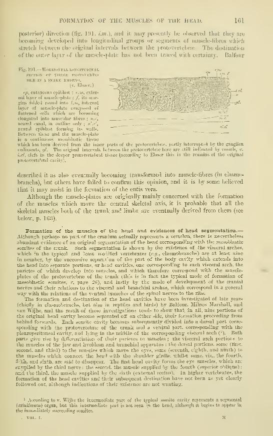
FORMATION OF THE MUSCLKS OF TIIK UKAI). IGl
posterior) diroctittn (liij;. I'.'l, /.///.), and it may pivst'iitly be observed thuc they are
becoming developed into longitudinal groups or segments of muscle-fibres which
stretch between the original intervals between the protovertebrae. The destinatJDii
of the onto?- layer of the musele-iilate has not been traced with certainty. Ikdluur
^^aetf^^l^ibliljafetHjji:' i,' J.t^iy '̂
Fig. 1!>1.— HoKI/.ONTAI, I.O.NdlTl'DINAL
SKCTION OF THKKK I'lKiTOVKUTK-
BR.K IN A SNAKK KMIIUYO.
(v. Ebner.
)
tp, cutaneous epiblast ; e. m, exter-
nal layer of luusdc-plate ; /, its mar-gins folJeil round into l.hi, internal
layer of niuscic-jilate coniiiosed of
flattened cells which are Ijccoiiiing
elongated into muscular fibres ; n.c,
neural civnal, in outline only ; n'.c',
neural epiblast forming its walls.
Between these and the muscle-plate
is a continuous mcsoblastic tissue
which has been derived from the inner ]iarts of the protovei-tcbrrc, partly interrupted by the ganglion
ludiments, ///. The original intervals between the protovcrteluaj here are ;:till indicated by vessels, v.
i.cl, cleft in the deeper protovertebral tissue (according to Ebner this is the remains of the original
protovertebral cavity).
described it as also eventually becoming transformed into mnscle-libres (in elasmn-
braiichs), l)ut others have failed to confirm this opinion, and it is by some believed
that it may assist in the formation of the cutis vera.
Although the muscle-plates are originally mainly concerned with the foiination
of the muscles which move the central skeletal axis, it is probable that all the
skeletal muscles l)oth of the trunk and limbs are eventually derived from them (see
below, p. IGo).
Formation of the muscles of the head and evidences of head segmentation.
—
Althoui!'h perhaps no part of the cranium actually represents a vertebra, there is nevertheless
abundant evidence of an orig-inal segmentation of the head corresponding- with the mesoblastic
somites of the trunk. Such segmentation is shown by the existence of the visceral arches,
which in the typical and least modified vertebrates (c.//., elasmobranchs) are at least nine
in number, by the successive separat'on of the part of the body cavity which extends into
the head into separate portions, or head cavities, one corresponding' to each visceral arch, the
parietes of which develop into muscles, and which therefore correspond with the muscle-
plates of the protovertebral of the trunk (this is in fact the typical mode of formation of
mesoblastic somites, r. page 26), and lastly by the mode of development of the cranial
nerves and their relations to the visceral and branchial arches, which correspond in a general
way with the relations of the ventral branches of the spinal nerves to the ribs.
The formation and destination of the head cavities have been investigated of late years
(chiefly in elasmobranchs, but also iu reptiles and biixls) by Balfour. Milnes Mar.<hall. andvan Wijhe, and the result of these investigations tends to show that, in all, nine portions of
the original head cavity become separated off on either side, their formation proceeding frombehind forwards. Each somite cavity becomes subsequently divided into a dorsal part corre-
sponding with the protovertebrte of the trunk and a ventral part, corresponding with the
pleuroperitoneal cavity, and lying in the middle of the corresponding visceral arch (). Bothpai-ts give rise by differentiation of their parietes to muscles ; the visceral arch portions to
the muscles of the jaw and hyoidean and branchial apparatus ; the doj-sal portions, some (first,
second, and third) to the muscles which move the eyes, some (seventh, eighth, and ninth) to
the muscles which connect the head with the shoulder gii-dle. v.hilst some, viz., the foui-th,
fiith, and sixth, are said to disappear. The fii'st head cavity forms the eye muscles, which are
supplied by the third nerve : the second, the muscle supplied by the fourth (superior oblique) :
and the third, the muscle supplied by the sixth (external rectus). In higher vertebrates, the
formation of the head cavities and thau- subsequent destination have not been as yet clearly
followed out, although indications of their existence are not wanting.
' According to v. Wijhe the intermediate part of the typical somite cavity represents a segmental
(uriniferous) organ, but this intermediate part is not scea iw. the head, although it begins to appear in
the immediately succeeding somites.
. VOL. I. M

163 DEVELOPMENT OF THE VERTEBRAL COLUMN.
Development of tlie vertebral column.—The vertebral column is developed
around the notochord, except at the anterior end of that structure, which is imbedded
in the basis cranii. It is formed from protovertebral mesoblast. The outer part of
each protovertebra is transformed into a muscle-plate, and thus the original meso-
blastic segmentation is maintained. The inner parts of the protovertebrse do not,
however, remain distinct, but blend with one another on each side of the neural
canal to form a longitudinal mass, which extends to the side of and subsequently
encloses the notochord, and finally sends dorsal prolongations over the neural canal,
so that this also receives a continuous mesoblastic investment, forming the membrana
reuniens superior of Eemak. The investment is only incomplete opposite the points
where the nerve roots are connected with the spinal cord, and in it there is no sign
of any differentiation into vertebrae. It is continuous with a similar investment
within the cranium, which extends in front of the notochord into the fronto-nasal
process. The mass of mesoblast, which thus encloses the notochord and neural
canal, is often spoken of as the membranous vertebral column and cranium, but it
represents much more than the cartilaginous and bony structures of those parts, all
the investing membranes of the cord and brain and the ligaments of the vertebrse
being also derived from it. From it septa pass between the muscle-plates and serve
to give attachment to the developing muscle-fibres.
The first appearance of the permanent vertebra is in the form of cartilage, which
becomes formed in this mesoblastic investment on either side of the neural canal,
nearly opposite the interval between each two muscle-plates, to form the neural
arch. This part of the vertebra therefore alternates with the original mesoblastic
somites as represented by the muscle-plates.
According to Froriep, the lateral halves of each cartilaginous neural arch become
united below the notochord before the appearance of the rudiments of the cartilagi-
nous bodies, and the latter appear as median accumulations of cartilage, immediately
posterior to the hypochordal part of the cartilaginous arch. In most of the vertebra
this hypochordal part of the arch soon disappears as a distinct structure, but in the
atlas vertebra the primitive condition is maintained.
The serial arrangement of the musculature represents phylogenetically the original
segmentation of the vertebrate body. The segmentation of the vertebral column,
on the other hand, has been arrived at later, and has been carried out in depen-
dence upon the muscular segmentation.^
The cartilage makes its appearance on the fom'th day in the chick, on the
eleventh or twelfth day in the rabbit, and in the fom-th or fifth week in man(Kolliker). It is completed by the sixth or seventh week, soon after which ossifica-
tion commences. To form the intervertebral discs, the mesoblast between the
bodies of the vertebree acquires a fibro-cartilaginous character, while at the same
time the notochord, which gradually elsewhere becomes reduced in size and eventu-
ally disappears,- here undergoes enlargement, and its cells form an irregular network
in the central intervertebral pulp. Its remains are found at aU periods of life in
the middle of the discs (Luschka).
In the osseous fishes there is an intervertebral dilatation of the notochord, the growth of
which proceeds to s-uch a considerable extent as to give rise to a mass of soft gelatinous
substance, which occupies the conical hollows of the biconcave vertebral bodies. But in
birds, reptiles, and amphibia, where synovial articulations are developed between the vertebral
bodies, the notochord soon disappears from the intervertebral spaces, although its remains are
seen for some time in the bodies themselves (Gegenbaur).
In mammals, the notochord is constricted within the cartilaginous vertebral body, but
dilated in the intermediate parts of this rod, the whole cord being moniliform. At a somewhat
later period small dilatations are also to be seen in the epiphysial cartilages (fig. 103).
1 A. Froriep, " Zur Entwickl. der Wirbelsaule, 8z;c.," Archiv f. Anatomie 1883 and 1S86,

THE LIMBS^. 1G3
Ribs and Sternum.—The ribs are foi-ined l)y separate cartilau^inous traiisfor-
niarioii ill fxicnsimis dl" the protoverteliral luesohlast l)et\veen the muselc-phites.
Aceordiiiy to some, they grow out from tlie cartilaginous vertelirae, Init become separate
before ossification begins. Simihir deposits are formed in connexion with the other
vertebrte (except the coccygeal in man), but they here become united by ossification
with and fonu parts of tiie vertebrae (see Osteology, Vol. 11.). At their \entral
Fig. 192.
—
Sections op the vertebral column op a Hrr-iAN fcetus op eight weeks.
(From Kolliker.
)
A, transverse longitudinal section of several vertebrne. 1, 1, clionla dorsalis, its remains tliicker
opposite the intervertebral discs ; 2, is placed on one of the bodies of the permanent vertcbne ; 3, on
one of the intervertebral discs.
B, transverse horizontal section through a part of one dorsal vertebra. 1, remains of the chorda
doi-salis in the middle of the body ; 2, arch of the vertebra ; 3, head of a rib.
Fig. 193.
—
Sagittal section of a dorsal intervertebral ligament of an advancedsheep's embryo. (Kolliker.)
La, l.p, anterior and posterior ligaments ; l.i, intervertebral ligament ; Ic, Jc', cartilaginous ends of
two vertebral bodies, w, w' ; c, enlargement of notochord in the ligament ; c', c", enlargements in the
cartilaginous ends of the vertebrae.
extremities the first seven (thoracic) cartilaginous ribs become united on either side
into a longitudinal cartilaginous plate, and this afterwards joins its fellow of the
opposite side to form the sternum (nianubrimu and body). The xiphoid i.s of later
formation (Parker). This mode of development of the sternum explains many of
the malformations in the shape of fissures of the sternum of different gi'adation
which have been observed.
TTie Limbs.—The limbs arise as outgrowths from the lateral part of the
trunk in the thoracic and pelvic regions in the third day in the chick and in the
third and fourth week in the human embryo. They appear as flattened semilunar
thickenings of the parietal mesoblast covered by epiblast, budding out from a lateral
ridge which is seen in the early embryo near the line of cleavage of the mesoblast
and close to the outer margins of the muscle-plates, and several of which subsequently
send prolongations into each liml) (i); they are therefore connected with several
mesoblastic somites, as is also indicated by their nerve supply.
* This is the case in elasmobvanchs (see fig. 194, from Balfour), but, according to Paterson.

164 'rSE LIMBS.
Each limb consists of a part which is sunk in the substance of the lateral ridge,
and in ^Yhich the thoracic or pelvic girdle becomes developed, and of a free or
sp-c
•vbtnc,
Fig. 194.
—
Tkaxsverse sectiox theoitgh an anterior part of the trunk of an embryo of
ScrLLirii. (Balfour.;
si^.c, spinal cord; sp.^; ganglion of posterior root; ar, anterior root; dn, dorsal; s^;. ;*. ventralbranch of spinal nerve ; mp, muscle plate ; m2j, part of muscle plate already converted into muscle
;
mjy.l; part of muscle plate extending into the limb ; nl, nei-vus lateralis ; ao, aorta ; ch, notochord
;
sy-ff, sympathetic ganglion; ca.v, cardinal vein; sr2, segmental duct; st, segmental tube; du, duode-num; hji.d, junction of hepatic duct with it; pan, rudiment of pancreas connected with another partof duodenum ; v./itc. opening of iimbilical canal (vitelline duct).
projecting part (fig. 195). This is at first quite simple, and represents the distal
segment of the limb (hand or foot). The other two segments (forearm and leg;
arm and thigh) are successively marked off between it and the girdle by the
development of transverse furrows representing the joints (fifth and sixth week).
At about the same time four notches appear in the flattened distal extremity,
marking off the Intervals between the fingers and toes, and the middle segment(fore-arm or leg) begins to be flexed upon the proximal (arm or thigh), the
the limbs do not receive such prolongations fi-om the muscle-plates in bii-ds and mammals ; the mr.sclesdevelop in situ from previously indifferent mesoblast.

THE ClJANIUxM. 1G5
Concavity looking forwards in tiie upper limli and Iiackwards in the lower limb.
The liinljs also come to he folded ventrally against the hody of the enil>ryo.
From tiie manner in whieh the flattened limh-hud grows (ut from the lateral
ridge, it is obvious that its surl'aces must at lirst be dorsal and ventral. Thedorsal surface afterwards becomes extensor and the ventral flexor. The anterior
edge is respectively the radial and tibial ; the posterior, the ulnar and filtular. Xa
Fig. 19r>.— OlTLIXKS <iK TllK A.NTKlili'U KXTIIKJUTIKS
OK niMAS EMUUYOS AT IiIFKKUK.NT A«ES. (After
His.)
>/, at four weeks; /?, at five weeks ; C, at seven weeks;
D, at nine or ten weeks.
development proceeds, a half rotation occurs in
opposite directions in the two limbs, resulting
in the middle flexure (elbow, knee) being directed
forwards in the ujiper, backwards in the lower
limb.
The bones of the limbs are laid down as
cartilages which ajjpear as separate differentia-
tions of the more centrally placed mesoblast,
a portion of mesoblast remaining for a time undifferentiated opposite each synovial
articulation. "Within this a cleft subsequently appears, and enlarges to form the
synovial cavity, the mesoblast which bounds the cleft developing eventually into
the synovial membrane and capsular ligaments of the joint.
The cranitira.—In the head the notochord extends as far forwards as the
mid-brain. Here also it is invested by a continuous mass of mesoblast, which sends
lateral prolongations over the neural canal as in the trunk (membrana reuniens).
^:Et/t.
Fig. 196.
—
Diagrams of the cartilagixocs craxium. (Wiedei-sbeim.)
A, First stage.
C7(, notochord ; Tr, trabeculse cranii ; P.ch, parachordal cartilages ; P, situation of pituitarj' body-;
A', E, 0, situations of olfactory, visual and auditory organs.
B, Second stage.
B, basilar cartilage (investing mass of Eatbke) ; .9, nasal septum and ethmoidal cartilage ; Etli,
Eth', prolongations of ethmoidal around olfactory organ, completing the nasal capsule ; 01, foramina
for passage of olfactory nerve-fibres ; N, E, 0, C/i, Tr, as before.
The main difference in development between the cranium and vertebral column
consists in the fact that no separate cartilaginous deposits to form vertebrae occur
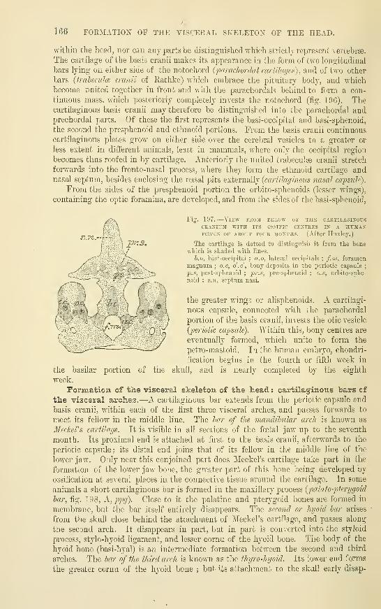
166 FORMATION OF THE VISCEEAL SKELETON OF THE HEAD.
withiu the head, nor caa any parts be distinguished which strictly represent vertebrse.
The cartilage of the basis cranii makes its appearance in the form of two longitudinal
bars lying on either side of the notochord {paracJiordal cartilages), and of two other
bars (fraieculce cranii of Rathke) which embi'ace the pituitary body, and whichbecome united together in front and with the parachordals behind to form a con-
tinuous mass, which posteriorly completely invests the notochord (fig. 196). Thecartilaginous basis cranii may therefore be distinguished into the parachordal andprechordal parts. Of these the first represents the basi-occipital and basi-sphenoid,
the second the presphenoid and ethmoid portions. From the basis cranii continuous
cartilaginous plates grow on either side over the cerebral vesicles to a greater or
less extent in different animals, least in mammals, where only the occipital region
becomes thus roofed in by cartilage. Anteriorly the united trabeculse cranii stretch
forwards into the fronto-nasal process, where they form the ethmoid cartilage andnasal septum, besides enclosing the nasal pits Q^t^LWoWj {cartilaginous nasal capsule).
From the sides of the presphenoid portion the orbito-sphenoids (lesser wings),
containing the optic foramina, are developed, and from the sides of the basi-sphenoid,
Fig. 197.
—
View frou below of the cartilagixousCBASIiJJI WITH ITS OSSIFIC CENTRES IX A HUMAXFffiTcs OF ABOUT FOUR 3I0STHS. (After Huxley.)
The cartilage is dotted to distinguish it from the bonewhich is shaded with lines.
h.o, basi-occipital ; a.o, lateral occipitals; f.ni, foramen
/" - .X magnum; o.c, o'.c', bony deposits in the periotic capsule;
(--'
v;,c * ' ' ~~-5 2'-*% post-sphenoid;
jpr.s, pre-sfihenoid ; o.s, orbito-sphe-
^ ^—
' ; ' ', noid ; s.n, septum nasi,
\'
'
[ (
'
'•
{ 5J - r... j :__^
' the greater wings or alisphenoids. A cartilagi-
*i,T:?r.\ , -:
J;.
*j,
nous capsule, connected with the parachordal
y' '
.: ,, Li,.^ - / portion of the basis cranii, invests the otic vesicle
{periotic capsule). TViDhin this, bony centres are
eventually formed, which unite to form the
petro-mastoid. In the human embryo, chondri-
fication begins in the fom'th or fifth week in
the basilar portion of the skull, and is nearly completed by the eighth
week.
Formation of tlie visceral skeleton of the head. : cartilaginous bars of
the visceral arches.—A cartilaginous bar extends from the periotic capsule and
basis cranii, within each of the first three visceral arches, and passes forwards to
meet its fellow in the middle line. The har of the mcmclibv.lar arch is known as
3IecJceVs cartilage. It is visible in all sections of the foetal jaw up to the seventh
month. Its proximal end is attached at first to the basis cranii, afterwards to the
periotic capsule; its distal end joins that of its fellow in the middle line of the
lower jaw. Only near this conjoined part does Meckel's cartilage take part in the
formation of the lower jaw bone, the greater part of this bone being developed by
ossification at several places in the connective tissue around the cartilage. In some
animals a short cartilaginous bar is formed in the maxillary process {palato-pterggoid
har, fig. 198, K,ppg). Close to it the palatine and pterygoid bones are formed in
membrane, but the bar itself entirely disappears. The second or hyoid bar arises
from the skull close behind the attachment of Meckel's cartilage, and passes along
the second arch. It disappears in part, but in part is converted into the styloid
process, stylo-hyoid ligament, and lesser cornu of the hyoid bone. The body of the
hyoid bone (basi-hyal) is an intermediate formation between the second and thu'd
arches. The bar of the third arch is known as the ihyro-hyoid. Its lower end forms
the greater cornu of the hyoid bone ; bu*'- its attachment to the skull early disap-

FORMATION OF THE AUDITORY OSSICLES. 10?
eiA
]i.y wj
jia.ck nc
Fig. 198, A.
—
Elements of tiie skull of an embryo pig, ?,-incii lonc, viewed from below :
sEMi-i>iAGRAMMATic. (From Baifour after Parker.)
pa.ch, parachordals ; nc, notot-horcl ; ait, otic capsule ; pn, pituitary ; tr, trabecule ; c.tr, cornu
of tlie trahecula;pn, prenasal cartilage; c.n, external uaies ; ol, olfactory region ; 7>.;>^, ijalato-
pterygoid bar enclosed in the luaxillo-pterygoid process ; run, mandibular bar ; hy, hyoidean bar : th.h,
thyrohyoid bar ; la, aperture for facial nerve ; 8rt, for glossopharyngeal ; 86, for vagus ; 9, for hypoglossal.
Fig. 198, I).—Side view of the siandibilar and hyoid arches in an embryo pig of 1^ inch
,in length. (From Balfour, after Parker.
)
tg, tongue ; ink, Meckel's cartilage ; ml, body of malleus ; inh, its manubrium or handle ; t.tii,
tegmen tyuipaui ; /, incus ; M, stapes ; i.h;i, inter-hyal ligament ; st.h, stylo-hyal cartilage ; h.h,
hypohyal ; bh basibranchial ; th.h, rudiment of first branchial arch ; 7a, facial nerve.
£?^2*f?;
X •' c-ct/^tilaxr&^
Jiyo>
cctrtttcujfc:
shofi
in.cvLS T'^^^^^^^^ Meckel's oca-Ula^e.
7i.U'jz<Z cccrtiZccae'
^uli'nioUiccLs
Meckel (xxt-itla^ey
ccofttlct(J& '-^^iSs^
fig. 1&9. prepi.rations showing three stages of theclrtilaginous bars of the 1st and 2nd arches inTHE embryo uf THE SHEEP. (Salcnsky.
)
pears, or in some animals may not
be found at all. This bar is in
all cases of less importance than
the first two, from the proximal
parts of which important struc-
tures connected in lower verte-
brates with the suspensory appa-
ratus of the lower jaw, in the
higher vertebrates with the ap-
paratus for sound transmission
to the internal ear are developed.
Formation of the auditory
ossicles.—If the embryonic de-
velopment of the bars is studied
in man and other mammals, it is
found that at a certain period of
fcetal life Meckel's cartilage is
directly continuous with the car-
tilaginous malleus, above which,
and at first in direct continuity
with it, is the cartilaginous incus
(fig. 199) ; these two ossicles
therefore appear as the enlarged
and modified proximal end of
Meckel's cartilage. Somewhat
later the incus becomes detached
from the malleus, but the latter
long remains in continuity with
Meckel's cartilage (fig. iOCi).
168 FORMATION OF THE AUDITORY OSSICLES.
The most obvious interpretation of these appearances would seem to be that both themalleus and incus are developed from the cartilaginous bar of the first or mandibular arch.
This view is not, however, universally held. For, as the several illustrations show, the secondor nyoid bar is also connected to the developing incus, through which it is joined to the periotic
capsule. Hence it has been inferred by some that the incus belongs to the hyoid bar, and notto the mandibular (A. Eraser). Others have looked upon it as representing a hyomandibularcartilage, like that which in Sauropsida forms a common suspensory apparatus for bothmandibular and hyoidean apparatus (Huxley). This hyomandibula itself, however, may repre-
Fig. 200.
—
Condition of Meckel's car-tilage AND THE HYOID BAR IN THEHUMAN FCETUS OF ABOUT 18 WEEKS,(KoUiker.
)
B, is an enlarged sketch by AllenThomson, showing the relationship of
the several parts better than in A.z, zygomatic arch ; ma, mastoid pro-
cess ; Ttii, portions of the lower jaw left
in situ, the rest having been cut away;
M, Meckel's cartilage of the right side,
continued at s, the symphysis, into thatof the left side 31' , of which only a small
part is shown ; T, tympanic ring ; m,malleus ; i, incus ; s, stapes ; sta, stape-
dius ; st, styloid process; p, h, g, stylo-
pharyngeus, stylohyoid, and styloglossus
muscles ; stl, stylohyoid ligament attached
to the lesser cornuof the hyoid bone, hy;ill, thyroid cartilage.
sent an upward prolongation of
Meckel's cartilage, and would thenbelong to the 1st visceral arch(Peters). Lastly, yet another solu-
tion of the question has been offered,
viz., that both incus and malleus are
hyomandibular (Albrecht, G-adow).
The last mentioned opinion is based
mainly ujoon considerations of com-parative anatomy, which can hardlybe left out of account in dealing withthe morphology of these structures.
In lower Vertebrata the suspensory
apparatus of the lower jaw comprises
besides the hyomandibula, commonto it and to the hyoid apparatus, a
large bone, known as the quadrate,
by means of which, either directly or
with the intercalation of an os arti-
culare, the lower jaw is united withthe basis cranii and periotic capsule.
Reichert looked upon the incus as the
homologue of the quadrate, and the malleus as that of the os articulare ;and the same view
was taken by Gegenbaur. Huxley, on the other hand, came to the conclusion that the
homologue of the quadrate bone of reptiles and birds is to be found in the malleus, and that
the incus represents a portion of the hyomandibular bar, which, as above stated, is common to
both first and second arches. Various other observers have concluded that the quadrate of
lower vertebrates is represented in mammals by the zygomatic process of the squamosal.
Gadow, however, looks upon it as represented by the tympanic ring of mammals.The stapes has been variously described as representing : 1 , a part of the hyoid arch
(Reichert) ; 2, a part of the periotic capsule which has become detached (Parker) ; 3, in part
or wholly, the hyomandibula of lower vertebrates (G-egenbaur, Huxley, Albrecht, G-adow)
;
i, hyomandibula and detached periotic cartilage conjoined (Gradsnigo) ; 5, as an independent
circular deposit of cartilage around the stapedial artery ^ (Salensky, Fraser). It is at any rate
closely connected with the hyoid bar, which forms from above clown the tympano-hyal and
This artery disappears in man, but is persistent in many mammals.

RECENT LITERATURE. 169
styloid processes, the stylo-hyoid lijjainont ami iho lessor cunma of iIr- liyoiil l)oiic (curato-
The reiuaiiiinjf bones of the visceral skeleton of tlu; lioad. viz.. tlie nia.xi'lary, malar,
jialatiue, ptcry^oiil, vomer, nasal, and laohrj'mal, are all formed in membrane. An account of
their dcvulopmuut is given in the Usteoloyy (Vol. II.).
RECENT LITERATURE.
Ahlborn, Ucbcr die Segmentation des WirhiUhii rklirpers, Zcitschr. f. wiss. Zool., xl.
Albrecht, .S'lo- la ralcnr morpkolor/ifjuc dc l'articulation mandihulairc, du cartilage de Mrel-eXtt
des ossihts de Vuuie, Bruxelles, 1883 ; Sur la vahur morpholoyique da la trompe d Eustackv, dkc,
HruxuUes, 1884.
Balfour, On the development of the skeleton of the paireel fins of Elasmohranchii, dc., Proc. Zool.
Soc, 18S1.
Baiir, (hi the quadrate in the Mammalia, Quarterly Journal of Microsc. Science, August, 1887.
Bemmelen, v., Ueber die llerkunft dcr Extrcmitdten u. Zungenmusc-idatur bei Eidedisen, Anal.
Anzeiger, lS8i».
Braun, M., l/eber den Schivanz bei Sdugcthieremhryonen, Deutsche Zeitsclir. f. Thicrmedicin, ix.,
ISS:?.
Cadiat, Du dtveloppimcnt de la partic ccphalo-thoracique de Vimbryon, ele la formation du dia-
I'liruyma, etc., Journal de I'anatomie, T. xiv. , 1S78.
Dohrn, Bcmerkumjcn ii. d. niucsten Versueh einer Losung eles Wirbilthierkopf-Problems, Anat.
Auzeiuer. 1890. Also numerous papers in Mitthcihingen el. znol. Station zu A\a2)cL
Dollo, Oil the luallcus of the Lacertdia and the malar and quadrate bones of Mammed ia, Quarterly
Juurnal of ?iIicrosc. Science, 1883.
Ebuer, V. v., Urwirbd und Neugliedcrung dcr Wirbdisiiule, Sitzungsb. der Wiener Akadein.,
1888.
Fraser, On the development of the ossicuUi audilus in the higher Mammalia, Phil. Trans., 1882.
Froriep, A.., Zur Entieicklungsgcschichte dcr Wirhelsdule, insbesondcre des Atlas und Epistro-
phiu.-< und d(r Occipitalngion, Arch. f. Anat. u. Entwicklung.sgesch., 1883 and 1886.
Gadow, On the modifications of the first and second visceral arches, with especial reference to the
homolo'iiis of the auditory ossicles, Philos. Trans., 1888.
Geg-enbaur, Die Metamerie des Kopfes und die IVirbcUhiorie des Kopfskelets, Morphol. Jahrb.,
xiii., 1887.
Gradenig-o, G., Die embryonale Anlage des Mittelohres : die morphologische Bidiutung derGehorknochelchen, Wiener med. Jahrbiicher, 1887.
His, W., Mittheilungen zur Embryologie der Sdugethiere u. des Menschen, Aicbiv f. Anat. u.
Physiol., Anat. Abth., 1881.
HofiEmann, C. K., Ue. die morphologische Bedeutung des Gehorknochelchens bei d. Reptilien,
Zool. Anzeiger, xii.
Kostlin, Der Bau des knochemen Kopfes in den vier Klasse7i der Wirbellhiere, Stuttgart,
1884.
Lockwood, C. B., The early development of the pencardium, diaphragm, and great veins.
Proceed, of the Royal Society, 1887.
Noorden, W. v., Beitrag zur Anatomic der knoi-peligen Schddclbasis menschlirher Embryonen,Arch. f. Anat. u. Phys., Anat. Abth., 1887.
Parker, W. K., Various important papers on the structure and development of the skull in the
Philosophical Transactions of the Royal Society and the Transactions of the Zoological Society.
Rabl, C, Theorie des Mesoderms, Morphol. Jahrb., xv.
Ravn, E., Ue. die Bildung dcr Schcidcwand zwischcn Brust- und Bauch-hohle in Sdugethierembryonen. Arch. f. Anat. u. Physiol., Anat. Abth., 1889 ; Vntersuehungen ue. d. Entivickl. des
Diaphragmas, <i;c.. Arch. f. Anat. u. Physiol., Anat. Abth., 1889.
Salensky, Beitrdge zur Entwicklungsgeschichte der knorpeligen Gehorknochelchen bei Sdugethicren,Morphol. Jahrb., vi.
Strahl u. Carius, Beitr. zur Entwickl. des Herzens u. d. KorperJiohlcn, Arch. f. Anat. u.
Physiol., Auat. Abth., 1889.
Strazza, G., Zur Lehre uber die Entwicklung der Keldkopfmuskeln, Wiener medic. Jahrbiicher,
'
TJskow, N., Ueber die Entwicklung des Zwcrchfclls, des Pcricardiums und des Coloms, Archie
f. luikrosk. Anatomic, Bd. 22, 1883.
Wijhe, J. W. van, Ueber die Kopfsegmcnte und die Phylogenic des Geruchsorganes dcr Wirbd-thieri, Zoolog. Anzeiger, 1SS6 ; Die Kopfregion der Cranioien beim Amphioxus, Anat. Anzeiger, iv.
tul.. I.

170 OUTLINES OF EAKLY HUMAN EMBRYOS.
The following outlines and measurements will be found useful to assist in
determining the ages of early human embryos :
—
12 Z A
(about 10 to l:j days) (15 days) (21 days)-inim. 2*5ram. 2'6niiu. 4mm.
5

INDEX OF PART I.
Abdominal stalk, 46Accessory thyroids, 1 1
1
Alee nasi, 96Albrecht on anditory ossicles, 168Alecithal ova (a, not, \-IJKvdoi, oil-bottle), defi-
nition, 9invagination of, 22
Alimentary canal, development of, 99Allantoic arteries. See U.mdilical.
Huid, 46tube, 36
Allantois (aAAny, sausage, uSos, form), formation
of, 43first appearance of, 44structiu'e 01", 44in what animals fonnd. 45hypoblastic sac of, 40pedicle of, 46variation of period of development, 46connection, with alimentary canal, loi, n.
with bladder, 122Amnion (afivioy, a bowl in which victim's blood
was caught, the caul), earliest appear-ance of in human embryo, 46
false, 42formation of, 42in guinea pig, 43, 46resulting from invagination, 24true, 42
Amniota, 42Amphibia, liver in, 112
Wolffian duct in, iiS .
heart in, 136Amphioxus {au<pi, double, o^vs, a point), ale-
cithal ova of, 9invagination of blastoderm of, 22, 24origin of mesoblast in, 22, 26niesenchjTne in, 27notochord in, 34mesoblastic somites in, 37
Amygdalre (oyu^SaATj, almond}, 66Anal membrane, the, loSAnalogy, di/o, according to, Aoyos, ratio), defi-
nition of, 4examples of, 4
Anatomy, definition of, i
departments of, i
Angelucci on zonule of Zinn, S7Annelida {anndlus, little ring), 2Anterior perforated space, 79Anus, formation of, 108, 12SAorta {aopTT], aeipw, to lift), inimitive, formation
of, 38descending primitive, 146arch of, 148, 150bulb of, 144, 147, 148descending permanent, 150
Aortic arches. See Aetekial Arches.
Acjueduct of Sylvius, 62, 67Aichenteric cavity, primitive {apxv, beginning,
ivrepov, intestine), 26Archiblastic cells of His, 25-Ajteria centralis retina?, 85, 87Arterial (aoitic) arches, formation of, 38, 146
division of aortic septum by, 144origin of principal arteries from, 146, 147,
148destination of fourth and fifth, 150
Arteries {dpTi)pia, afpu, to lift), development of,
146from arterial arches, 148, 149from arches in birds, 150ascending phar}iigeal, 148auricular, 148basilar, 149carotid, 148, 150common iliacs, 147innominate, 150intercostal, 149inter-segmental, 149lingual, 148maxillary, 148middle sacral, 147occipital, 148pulmonary, 148, 150subclavian, 150temporal, 148umbilical, 44, 146, 155vertebral, 149vitelline, 40
Arthropoda {&pdpov, a joint, ttovs, a foot), seg-
mental plan of, 2
ceuti'olecithal ova of, 8Aryepiglottic folds, 103Arytenoid cartilages [apinawa, a pitcher), 103Ascaris megalocephala, polar globules observed
in, 9fertilisation in, 11, 12
segmentation of, 16
Auditory pit, 89vesicle, 89nerve, 78, 89, 93ossicles, 167, 168
Auricular canal, 142Axis, primitive skeletal, 2
Baer, vox, zona pellucida of, 6on bilaniination of middle layer of blas-
toderm, 23Balfour, F. M., on amnion and allantois, 45, n.
blastodermic layers, nomenclature of, 23coelom, tj'pical development of, 24head cavities, 161
limbs, 163 n.
lens-capsule, 87
11 IXDEX OF PART I.
Balfour, F. M.
—
continued.
Miillerian duct, 122, 123muscle plate, outer, 161
nerves, epiblastic, origin of, 57spinal, anterior roots of, 74
neural crest, 73oesophagus, obliteration of lumen of, 104, n.
ova, formation of, 124parablast, 27, n.
primitive groove, 23polar globules, tlieory of, 14suprarenals, 122
sympathetic sj'stem. Si
Wolffian body, 115, n.
duct, 118vitelline membrane, 8
Bars, cartilaginous, of mandibular arch, 166, 168
palato-pterj'goid, 166
hyoid, 166, 168
thyro-hyoid, 166
Bartholin, glands of, 128
Basal layer of placental decidua, 5
1
attachment of villi to, 52Basis cranii, 166
Bat, placental sinuses in, 52Bauchstiel (abdominal stalk), 46Beard on branchial sense-organs, 76Beneden, v., vitelline membrane, 8
formation of polar globules, 9entrance of spermatozoon in Ascaris, 1
1
division of nucleus in Ascaris, 12, 16
blastodermic layers, 18
blastopore, theory of, 23pro-amnion of, 35
Bile ducts, 112
Birdsell on sympathetic nerves, 81
Bischoff on inversion of blastodermic layers, 23Bladder, urinary, 122
Blastoderm {fiKa(TT6s, a bud, 5f'p(Lia, skin), for-
mation of, 17, I
S
three layers of, 1
7
gastrula condition of, 21
stage typical, 22bilaminar, 21
layers, inversion of, 23historically considered, 23, 24characters of layers of, 24table of development from layers of, 25four layers of, 26separation of embrj^o from, 34
Blastodermic vesicle, definition of, 17
growth in the cat of, 18
invagination of, 22, 23, 24as yolk-sac, 35first attachment of to uterus, 53
Blastopore (ySAao-ros, a bud, Tr6pos, a passage),
discussion of in meroblastic ova, 22, 23diverticula, near to in Sagitta, 26
connection of anus with, 108
Blochmann on extrusion of one polar globule byparthenogenetic ova, 14
Blood, origin from parablastic cells, 21, 25corpuscles, 40
Blood islands of Pander, 39Blood vessels, formation of, 40Body, segmentation of, 2
Body-cavity. See CcelomBonnet on middle blastodermic layer, 21
on connection of uotochord with buccal epi-
blast, 68protovertebral cavity, 160
Born on the lachrymal canal, 89
Born
—
continued.
cephalic clefts, 102, n.
thyroid, iii
septum spurium, 141, n.
auricular canal, 142, 144ventricular septum, 142, n.
foramen ovale, valve of, 144pulmonary veins, 144
Boveri on polar gloljules in Ascaris, 9Bowman, anterior and posterior homogeneous
lamellte of, 88Brain, development of, 61
flexures of, 63fissures and convolutions of, 71
Branchial sense-organs of Beard, 76Bronchi, 109Brown, H. H., on extrusion of part of nucleus
of seminal cell, 12
Budge on Ijonphatic system, 158
C^ciTM (blind), 107Calamus scriptorius (writing pen), 62Callender on 4th visceral arch, 103Canalis re-unieus, 91Capsule of lens, 87
whence derived, 87Capsule, cartilaginous nasal, 166
periotic, 166
Carnoy on polar globules in Ascaris, 9Cat, blastodermic vesicle of, 18
Cauda equina, 60Centra [nii/rpuv, a point), of vertebrte, 2
Centrolecithal {.tevrpov, centre, XijKvdos, oil-
bottle), ova in arthropods, 8
Cerebellar peduncles, 66Cerebellum, 62
formation of, 66amj^gdahe of, 66
Cerebral vesicles, 37table of development from, 61fifth, or bulbar, 63fourth or cerebellar, 66third, mid-brain, 67 '
second, 67first, 68
Chick, pro-amnion in, 34rudiments of cranial nerve ganglia in, 75
Choanse {xiayos, a pit for melting metal in), 97,100
Chorda dorsalis, 32. See jSTotochoed.
Chorda tympani, 79Chordae tendineee, 142
Chorion {xopwv, skin), definition, 42 and n.
junction of allantois with, 44, 46Choroid coat, 87, 88
Choroid plexuses, 62, 67, 70Choroidal fissure, 85Chromatin, filaments of, 12, 16
Chromosomes {xpoiiixa, colour, craj^a, body), 12
Ciliary body, 88
Circulation, fcetal peculiarities of, 155course of, 155changes of at birth, 157
Cloaca, 108, 122, 123, 127
Cochlea (/coxAtay, a snail with a spii'al shell),
canal of, 90ganglion of, 78in birds, 92modiolus of, 93bone of, 93spiral lamina of, 93
INDEX OF PART I. Ill
Ccelenterates {ko7\os, hollow, ?fT«poj', intestine),
23Cceloni (koTAos, hollow), layers ot, 2, 23
tj'pical origin from invagination, 24, 26formation of, 36, 99connection of niesoblastic somites with, 37,
160formation of pleurte from, no
of perioardium from, 135 I
serous cavities from, 159Coelom-invaginations, 22connection of, with notochordal invagina-
tion, 34Colohoma iridis (»co\i5j3uj,ui, a part taken away
in mutilaiiou), S5Columns of cord, antei'ior white, first rudiment
of, 59posterior white, 59, 75, 7S, n.
structure of, 60Commissure, anterior of cord, 60
i^'ri'V, 67posterior, 67
Commissures of cerebral hemispheres, 71
Congenital fissures of neck, 103Connective tissue, origin from parablastic cells,
21, 25Cornea (horny), substantia propria of, 87
epithelium of, 87Cornu (horn), anterior, first rudiment of, 59Cornu Ammonis(from resemblance to the liorus
of the statue of Zeus-Ammon), 72Corpora quadrigemina (four-fold bodies), 62. 67
striata, 62, 69, 73Corpus albicans (white body), 71
Corjius callosum (the thick body), 71
peduncle of, 79Corti, rods of, 93Coste, discoverj' of germinal vesicle by, 8, n.
Cotyledons of placenta ((coruA??, anything hollow),
51Cowper, glands of, 12SCraniata, 27Cranium, 165Crura cerebri {criis, leg), 62Crusta (crust or rind), 67Cryptorchismus {Kpv:rr6s, hidden, upxis, testicle),
127Cunningham, D. F. , on sulci of brain, 72, n.
Cutis vera, formation of from outer muscle plate,161'
Cuvier, ducts of, 13S, 139, 141, 151, 152. 153vestiges of, 154
Cyclostomata {kvk\os, a circle, a-rdfj-a, mouth),parablastic elements in blastoderm of, 27
heart in, 136Cystic duct (kuo-tis, the bladder), 112
De^idua [dcddere, to fall oif), 44structm'e of, 46reflexa, 46serotina, 46vera, 46function of glands of, 47thickness of vera, 48stratum spongiosum of, 49stratum compactum of, 49atrophy of glands of, 49placental, 51giant cells in serotina, 54separation of at birth, 55
Decidual cells of Friedlander, 49Definition of anatomical terms, 4, 5
mesial,
lateral,
frontal,
sagittal {safjilta, an arrow),
coronal (corona, a crown),dorsal (dorsum, a liack),
vuntral {venter, belly),
neural (vtvpov, a cord),
visceral,
cephalic (KecpdAi), head),
caudal (cauda, tail),
axial,
Deiters, sustentacular cells of, 93Delamination, process of in blastoderm, 21
non-occurrence in invertebrata, 22Lankester on, 24
Descemet, membrane of, 88Descriptive terms, 4Deutoplasm, 7, 8Diujiluagm [oid, tiirough, (ppdyfia, a fence,)
159Directive corpuscles.. 9. See Polak GLObULES.Discus proligerus, albumen deposited on ovum
by, 7Dohrn on pituitary body, 68Dollinger, researches of Pander under, 23Ductus arteriosus, 146, 150, 155
lingnalis, nothyreoglossus, nothyroideus, novenosus, 152. 155
Duodenal loop, 104Dursy on contiuuitj' of piimitive with neural
groove, 32Duval on epiblastic connection of blastodermic
vesicle to uterine wall, 53
Ear, a specialised branchial sense-organ, 76development of, 89accessory parts, 93
Echiuoderms yix^^o^, urchin, 5ep,ua, skin), im-pregnation in, II, 12
Ecker on the Sylvian fissure, 73Ectoderm {sktos, outside, Sep,aa, skin), primitive,
18, 19, 24invagination of, 22, 23
Egg tubes, 124Eiasmobranchs (riKdafj.a, lamina, Ppayx'a, gills),
coelom invagination in, 22, n.
primitive groove in, 23neural crest in, 73spinal nerves, anterior roots in, 74origin of ciliary ganglion in, Si
auditory vesicle in, 90supra-pericardial bodies in, inliver in, 112Wolflian vesicles in, 118, n.
suprarenal capsules in, 121
lluUerian duct in, 122
heart in, 136muscle plate, outer, in, 161
visceral arches in, 161
head cavities in, 161
limbs in, 163, n.
EmbiTo, human, segmentation where mostmarked, 2
Embryo, separation of, from blasto'lerm, 34Embryology (e]uj8(>i;ov, a thing newlv born.Ao-vo?,
word), definition. I
iV INDEX OF PAE.T I.
Embryonic area, i8
Endocardium {hSov, within, nap^ia, heart), 136Endolymphatic canal, 90Engelmaun on placenta, 48, n.
Enteric canal (evrepov, intestine), 22groove, 36
Entoderm (ivr6s, inside, Sepfia, skin), primitive,
17, 18, 19, 24blastopore, relation to, 22, 23perforation of by primitive groove, 22
Entomeres, mass of {iyT6s, within, /J-epos, part),
23, , .
Epencephalon [iiri, over, iyKe(pa,\os, brain), 66Epiblast (eVi, over, ISAaards, germ), 21
homolog}^ with Eauber's layer, 22, n.
character of cells, 24formation of medullary folds by, 32formation of ganglia by, 73, 75sensory epiblast, 76formation of lens from, 83
Epiglottis {eni, upon, yAcarTis, mouth of the
windpipe), 103Epiphvsis cerebri {iwKpvw, to gi'ow upon, cere-
brum, brain), 6"]
Epoophoron (eTri, u])on, w6v, egg, (p^pa, to carry)
ofWaldeyer, 120
Eustachian tube, 93valve, 141, 155
Ewetsky on bifurcation of lachrymal canal, 89External amniotic fold, 43, n.
Eye, development of, S3
retina, 86hyaloid membrane of, 87corneo-sclerotic coat of, 87choroid coat of, 87, SS
iris of, 87, 88anterior chamber of, SS
Eyelids, 89
Fallopian tube, 7fimbriae of, 1
1
development of, 117, 124
False amnion, 42, 43, n.
Falx cerebri (a sickle), 70Fertilization, 11
Filum terminale (terminal thread), 60Fissure of Sylvius, 72, 73
of Eolando, 73riemming on Wolffian duct, 118
Fol on polar globules in echinoderms, 9Foramen caecum ofMorgagni (blind opening), 102,
noForamen of Monro, 63, 68
ovale, 144closure of, 155, 157
Fore brain, 38Fore-gut, first formation of, 34Fornix (an arch), 71
Foster on nomenclature of the blastodermic
layers, 23Fraser on inversion of blastodermic layers, 23
airditory ossicles, 168
Fretum Halleri (a strait or channel), 141
Friedlander, decidual cells of, 49on giant cells in decidua serotina, 54
Froriep on branchial sense-organs in mammals,
76vertebral column, 162
Furcula, the (forked prop), 102, 103
Gadow on auditory ossicles, 168Gall bladder, 112Ganglia {yayyMov, a swelling), posterior-root, 74
rudiments of, in cranial nerves, 75, 76Gasserian, 78, 81jugular, 78cochleae, 78spiral, 78geniculate, 78sympathetic, 81
ciliary. Si
ophthalmicus profundus of elasraobranchs,
81
Ganoids {yavos, bright, elSoy, form), heart in, 136Gartner, duct of, 120Gaskell on cranial ganglia, 77
nuclei of cranial nerves, 77sympathetic ganglia, 81
Gastrula {yavTrip, belly), 21views of formation, 22, 23
Gegenbaur on homodynamy, 4Jacobson's gland, 97remains of notochord, 162auditory ossicles, 168
Generative organs, male, 117, 120, 125external, 127female, 124table of, 130
Genital cord, 123ridge, 124
Germ cell, 12
Germinal cells, 58Germinal epithelium, 117
connection with Wolffian tubules, 120formation of ovary from, 124
of testicle from, 125Germinal spot, 8
Germinal vesicle, structure of, 8Giant cells of placenta, 54Gill slits, 103Giraldes, organ of, 120Girdle, thoracic and pelvic, 164Globular processes of His, 95Glomeruli ((/ZomMS, ball of thread), 119, 122Golowiue on ganglion rudiments, 75
special sense-organ rudiments, 76Gotte on Wolffian duct, 11
8
Graaf, de, on pineal e}"e, 68Graafian follicle, 6
union of cells of, with ovum, 7changes prior to escape of ovum, 9formation of, 124, 125
Gradenigo on the stapes, 168Gruber on a case of venous abnormality, 155Gubernaculum testis (a helm), 126
changes of in descent of testicle, 127Guinea-pig, formation of amnion in, 43
allantois in, 46Cyrus subcallosus of Zuckerkandl, 81
Hadden, on origin of segmental duct, 118
Haeckel, on gastrula stage and gastrulisation,
22, 23, 24Haller, vasa aberrautia of, 120
Hare lip, 97Hassal, concentric corpuscles of, 112
Head, first appearance of, 34muscles of, 161
evidences of .segmentation of, 161
skeleton of, 166vi'seeral skeleton of, 166
INDEX OF PART I.
Heape, on blastodermic layers in the mole, i8
rudimentary blastopore in the mole, 22
Heart, formation of, 38, 136bej^inning of beat, 39endothelial tube of, 134folding and division of, 137, 141, 142saccus reuniens, 139septum spuriuui of, 141
valves of, 141, 142fretum Halleri of, 14I
septum inferius of, 14Iauricnlar canal of, 142septum intermedium of, 142chordoa tendinece of, 142foramen ovule of, 144limbus Vieussenii of, 144muscle of, 145columme curiRfe of, 146foetal, peculiaiities of, 146
Hedgehog, placental sinuses in, 52, n.
epiblastic attachment to uterus in, 53Henle, loops of, 122Hensen, canalis re-uniens of, 91
on Wolffian duct, 118heart, bilateral, 134
Hermaphroditism ['Ep,urjs, ' AcppoSiTT), a male andfemale god), 12
Hertwig, 0. and E. , on polar globules in
echinoderms, 9coelom-invaginations of, 22coelom, typical development of, 24mesodermic laj'ers of, 26, 27, n.
mesenchyme of, 26Hind brain, 38Hind gut, ibrmation of, 36
connection with allantois, 46with Wolffian duct, 118
His, "\V. , abdominal stalk of, 46on accessory thyroid bodies, 1 10
allantois, loi, n.
arterial arches, 147ascending roots in medulla, 66auricular canal, 142axis cylinders, growtli of, 75blood-vessels, 151Broca's area, 79cephalic clefts, 102, n.
endothelial tube of heart, 136on epiblastic origin of ganglia and posterior
roots, 57germinal cells of, 58on geniculate ganglion, 79globular processes of, 95on heart, bilateral, 134isthmus of, 67on laminae of cord, 60lens capsule, 87mechanical theory of development, 24mesoblast, origin of in part from thickenedrim of blastoderm, 23
nerve fibres, somatic and splanchnic, 77olfactory epiblast, 98
nerve fibres, origin of, 77, 81
parablast, theory of, 25, 26on pinna, 95porta restibuli of, 157on pulmonary veins, 144researches of, 6, n.
saccus reuniens of, 139septiim atriorum, 144
inferius of, 141between stomodaeum and fore-gut, 99
His
—
amtinued.septum, transverse of, 159sinus arcuatus, 102
on spinal nerves, roots of, 74spongioblasts of, 58sulcus terminalis, 102
thyroid, noon tuberculum impar, 102
visceral arches, 102
Histologj' [icrrov, a web; K6yos, word), defini-
tion, I
Hofmann, gastrulation in elasmobranchs, 22, n.
Holoblaslic ova (8aos, whole ; /3Aost«{j, a germ),
definition of, 8
invagination of, 22
Homodynamy (<5m<5s, the same ; SwSjUis, power), 4Homogeny {.bixos, the same
;yivos, descent), 4
Homologj' (6/u(Js,the same ; Kdyos, word),definition
of, 4serial homology, 4examples of, 4
Hubrecht, on epiblastic connection of blasto-
dermic vesicle with uterus, 53Huxley, on two layers of ccelenterates, 23
auditory ossicles, 16S
Hymen, the (the god of maniage), 128
Hyoid, the (T el5os, the form of the Greek letter
T), 166, 169Hypoblast (vTt6, under ;
0\a(rT6s, genu), 21
characters of cells of, 24formation of notochord from, 32
allantois from, 44, 46Hypophysis cerebri {inro(pva},to gi-ow from below),
68, 100. And see Pituitapa' Body.
Hj'pospadias {vtt6, under ; crndw, to tear), 129
Impregnation, ii
Incisor foramen, 97Incus (an anvil), 167, 168
InfundLibulum, the (a funneD, 61, 67, 68
of lung, noIntermediate cell mass, the formation of
Wolffian duct from, 115
of pprmanent kidney from, 122
Intermaxillary process, 95Intervertebral disc, 162
Intervillous space, 51question of blood in, 53
Intestines, 104large, 106
Invagination in holoblastic ova, 22
aperture of, 22resemblance to blastopore, 22
Inversion of blastodermic layers, 23Iris, 86, 87, 88Ischikawa on segmentation of ovum before fer-
tilization, 14Island of Eeil, 70. 73Isthmus faucium. 100
of His, 67
JaCobsox's organ, 95, 97
KAEYOKiifESis {Kapvov, a kernel ; Klvr](ns, move-ment) of germinal cells, 58
Karyoplasm (Kapvov, a kernel ; irXaaua, a mould)of germinal vesicle, 8
Kessler, on lens capsule, 87substantia propria of cornea, 87, n.

VI INDEX OF PART I.
Kidney, head or fore, 115, 117
mid, 116
hind or permanent, 117, 122
KoUiker on Eaiiber's layer, 18
intermediate layer of blastoderm, 20placenta, 53lens capsule, 87the anterior chamber, 88cephalic clefts, 102, n.
air cells of lung, 1 10
thymus, iii
heart, 134endothelial tube of heart, 136mesocardium laterale of, 136cartilage of vertebice, 162
Kollmann, parablastic theory of His, adherence
to, 26Kowalevsky, researches on Sagitta and Amphi-
osus, 24, 26Kiindrat on placenta, 48, u.
Kupffer, gastrulation, view of, 22
His' parablastic theory, adherence to, 26
Labia majora (greater lips), 128
Labyrinth, the, the recess of, 90perilymphatic spaces of, 93
Lachrymal canals and ducts, 89, 97fissure, 89gland, 89
Laminae of cord, alar, basal (dorso-lateral)
(ventro-lateral), 60, 63, 65, 66, 77Langhans on placental sinuses, 53Lankester, on homogeny, 4
blastopore, 22
primary layers of blastoderm produced bydelamination, 24
Larynx, noLemurs [lemur, ghost), nasal gland in, 98Lens, 83, 86
vesicle of, 87transitional zone of, 87capsule of, 87
Leopold on placenta, 48, u.
giant cells in decidua serotina, 54Leydig on special sense organs of gill clefts, 76
Lieberkiihn on blastodermic layers in mole, 18
on lens capsule, 87zonule of Zinn, 87
Ligamentum arteriosum, 150Limbs, 4, 163
segments of, 164rotation of, 165bones of, 165
Limbus (a band) Vieussenii, 144, 157Literature, recent, of the OAiim, 14
blastoderm, 27embryo, formation of, 41
general subject of embryonic formation anddevelopment, 41
decidua, 55nervous system, development of, 81
sense organs, 98alimentary canal and glands, lungs, 113
m'inary and generative organs, 132
blood system, 158seroiis cavities, muscles and skeleton, 169
Liver, 106, 112weight of, 113formation of veins of, 151, 152
Lockwood, on protovertebral cavity, 160
Lungs, development of, 109Luschka, on urachus, 122
notochord, remains of, 162LjTnpihatic system, 158
Mackay on arterial arches of the bird, 150Macula germinativa (germinal spot), 8
Malleus (a hammer), 167, 168Malpighian corpuscles of spleen, 108
of Wolffian body, 120of permanent kidney, 122
Marshal], vestigial fold of, 155Milnes, on head cavities, 161
Martin on Wolfiian duct, 118Masius on epiblastic connection of blastodermio
vesicle with uterus, 53Mechanical theory of development, 24Meckel, J. F., 23Meckel's cartilage, 166, 167, 168Medulla oblongata, 62
formation of, 63Medullary folds, fonnation of, 30, 32
groove. See ISTeukal groove,Membrana limitans of retina, externa, 86
interna, 83, 86nictitans, 89reuniens, superior of Eemak, 32, 162, 165tectoria, 93tympani, 93
Membranes of cord, development of, 60Meroblastic ova (juepoy, a part, ^Aaaros, a germ),
definition of, 8
gastrular stage in, 22Haeckel, view concerning blastopore in, 23mesenchyme in, 27
Mesenchjone {jxiaos, middle, X'^Mo^j juice) 26,
27Mesencephalon {/xtaos, middle, eyKe(pa\os, brain,
67connection with optic stalks, 85
Mesentery, the {ixfaos, middle, evrepov, intestine),
104, 107Mesoblast, formation of, 20, 21
origin, varieties of, 22His' view of origin, 23character of cells, 24connection with mesenchj-me, 26paraxial and lateral, 32cleavage of, 36formation of vessels in, 39growth of with allantois, 44
Mesoblastic somites, 37, 159, 162
cavity in, 37, 160
Mesocardium, anterius, posterius, lateralis, 136Mesocolon [koXov, great intestine), 106
Mesogastiium {yaaTrip. belly), 106, 107Mesonephros (vecppos, kidney), 116ilesorchinm (opxis, testicle), 126Mesorectum, 106Mesovarium, 126
Metameres (/uera, following, fj-spos, a i^art), 4Metanephros {iJ-iTo., behind, pe^pos, kidney),
117Metazoa {iJ-ird, after, C'^ov, animal), 18, 26Metencephalon {fniTo., behind, iyK€cpa\os, brain),
63Meuron, de, on obliteration of human cesopha-
gus, 104, n.
thjrmus. III
Micropyle [jxiKpos, small, -nvKrt, opening), 11
Mid brain, 38
INDEX OF PART I. Vll
M ihalkovics on stonioiloeum in rabbit, 99snpra-rcnul capsules, 120
Mulleiiau ducts, 123
ovary, 124, n.
Minot, polar f;lobules, theory of, 12, 14
on placenta, 4S n.
chorionic epithelium, bounding placental
sinuses, 52, n.
Mitsukuri on supra-renals, 122
Jlodiolus, the (nave of a wheel), 93Mole, primitive ectoderm in, iS
blastopore in, 22
Monotremes (/xovos, single, rprjixa, a hole), 6
amount of yolk of, 8
cloaca in, loS
Jlorbus cceruleus (the blue disease), 158
Jlorgagni, hydatids of, 124
Jlorpholog}' (juop<j)7J, form, \6yos, word), deter-
mination of, 2
Mouth, formation of, 99non-correspondence of primitive with per-
manent, 100
Mulberry mass, 1
6
Miiller, E., on muscular tissue of primitive
aorlre, 151Johannes, [Miillerian duct named after,
117W., sense epithelium of, 86
Miilleriau duct, 117, 122, 123, 124, 127
invaginations, 115, u.
Miillerian tibres, 86Muscle plate, 160
inner, 160outer, 161
Muscles, 159Myelospougium {(ive\6s, marrow, 0-770770$,
sponge), 58ilyotomes {fivos, a muscle, re^vco, I cut), 4
Kasal processes, 95laminfe, 95septum, 95
Ifaso-palatine canal, 97IJerves, iifth ascending, root of, 66, 78
vagus and glosso-pharvngeal, ascending root
of, 66, 78iifth sensory fibres of, 66, 78
motor fibres of, 66sixth, seventh, 66thii-d, fourth, 67spinal, formation of, 73roots of, 74axis, cylinders of, 75traces of ganglia in cranial, 77cranial. 75auditory, 78, 89, 93sympathetic, 81
nuclei of origin of cranial, 77inferior laiyngeal, shifting of, 149
Iserve fibres, origin from neuroblasts, 58, 59origin of somatic and splanchnic, 77oltactory. Si
Kervous system, general view of development, 57Iseural canal, 32
parietes of, structure of, 57crest, 73, 75groove, formation and closure of, 30, 32formation of cerebral vesicles from, 37
Keurenteric canal {fevpov, nerve, ivTepov, in-
testine), 22piercing of notochord by, 32
Neuroblasts {vivpov, nerve, ^\aaj6s, germ), 58,
59, 74, 9S. . ,
Neuroglia {vtvpov, nerve, y\la, glue), origin of,
60Nose, a specialised branchial sense organ, 76
formation of, 95Notochord (i/aiToy, the back, x^P^Vt ^ string of
gut), 2
formation of, 32an emltryonic structure, 34structure of, 34connection w ith buccal epiblast, 68formation of vcrtebrai round, 162
connection with cranium, 165Notochordal canal, 34Xuck, canal of, 127
Nuclei of origin of cranial nerves, 77Nucleolus of ovum, 8Nucleoplasm of germ and sperm cells, 1
2
Nucleus (a kernel) of ovum, changes in, 6, 9chromatin filaments of, 12
Nymphffi, 128
QiSOPHAOL'S (o'itraj = (pfpci), tO cariT, (payuv, to
eat), 103, 104, n.
Olfactorv {oJjacio, to smell) area, 95bulb. Si
epithelium, 98glomeruli, 81
pit, 95lobes, 62, 71junction of ganglionic rudiment with, 77formation of, 79
Olivary tubercle, 65Omental sac, 106
Omentum, great, 106, loS
gastro-hepatic, or small, 106, 113Omphalomeseraic vein {dfj.(pa\6s, navel, fj.e<Tapaioi;
mesenter3\ See Vitelline A'eix.
Onodi on sympathetic nerves, 81
Optic chiasma {x'^aana, two lines placed cross-
wise), 85cup, 83nerves, origin from fore-brain, 38, 67, 79stalks, formation of, 85thalamus, 62, 67tracts, 67, 85vesicles, primarj", 61, 67, S3
Organ, definition, i
Organization, plan of, 2' • Organs of the lateral line, " 76Os artieulare (joint bone), 168
Otic vesicle (o5s, wtikos, ear, of the ear), 89Ovary, 6, 124, 125
round ligament of, 126
change of position of, 127Oviparous {ovum, egg, pano, to produce) verte-
brata, 8
Ovum (egg), alecitlial, 9attachment of to uterus, 46centrolecithal, 8
deutoplasm of, 7development, changes prior to, 6gastrula of, 21
germinal vesicle of, 8holobiastic, 8
human, exce])tion to rule of separation fromchorion, 46
human, villi in earliest described, 55
Till INDEX OF PART I.
vuin
—
continn ed.
fertilization of, 1
1
niammalian, descent of, 8
maturation of, 9mp.roblastic, 8permanent, 125polar globules, extrusion fi'om, 9primordial, 124segmentation of, 16
size of, 6striae of, 6
structure of, 6yolk of, 7zona pellucida or zona radiata of, 6
Palate, the, 97cleft, 97
Pancreas {ttuu, all, Kpsas, flesli), loS, 113Pander, researches on blastoderm, 23
mechanical theory of, 24blood islands of, 39
Papillary nrascles, 142Parablastic cells [irapd, beside, ^KacxTos, germ),
21, 25, 27Parachordal cartilages {irapd, beside, x°P^^h
string ; near the notochord), 166Parker, on formation of xiphoid, 163
stapes, 168Parovarium {irapd, beside ; ovarium, ovary), 120Pars ciliaris retina, 86Parthenogenesis {irapdevos, virgin
;yei'eais,
origin), polar globules in, 14Parturition, separation of membranes in, 55Paterson on origin of sympathetic system, 81
limb muscles, 163, n.
Penis, 128, 129Pericardium {^ep'-, about ; KapBia, heart), 135, 159Peritoneum (Trepi, about ; reiVco, to stretch), 159Perivitelline space {vitelhis, yolk), 9Peters on hyomandibula, 168Petromyzon {irerpos, a stone ; fiv^w, to suck
;
lamprey), pituitary body in, 68Pfliiger, egg tubes of, 124Pharyngeal septum, 100Pharynx, loi
Pliiltrum {cpiATpov, loveliness), 95Pineal gland {pinea, a fir-cone), 61, 67
as rudimentary eye, 68Pinna, the (a feather), 95Pituitary body (pituita, phlegm), 61, 6j
formation ot, 68in Petromj^zon, 68connection with stomodseum 100
Placenta (a cake), 44formation of, 49, 51basal layer of, 5
1
subchorionic membrane of, 51loculi or cotyledons of, 51structure of sinuses of. 52intervillous spaces of, 51, 53weight and size of, 53microscopical sti'ucture of, 53, 54
Placental decidua, 5
1
sinuses, structure of, 52Plane, median, 4Plasmas of Weismann, nuclear, nutritive, ger-
minal, histogenetic, 14Pleurae {vXevpa, a rib), 1 10, 159Pleuro-peritoneal cavity. See Ccelom
Plica gubernatrix (guiding fold), 126-
semilunaris, 89Polar disc, 1
1
globules, 6
formation of, 9, 12
where observed, 9of Ascaris msgalocephala, 9of plants, 9Minot's theory of, 12
"Weismaun's theory of, 14Pons (bridge), 62
formation of, 66Porta vestibuli (gate of the vestibule), 157Prse-hyoid glands, noPregnancy, changes of uterus in, 48Primary placental circulation, 53Primitive groove, formation of, 19
as extension of blastopore, 22as homologue of part of entoderm,
23Baltour's view of, 23connection with neural groove,
3°, 32.
streak, formation of, 19an ectodermal thickening in-
dented by blastopore, 22origin of mesoblast from, 22connection with notochord, 32
velum [velum, a curtain), 100Pristiurus, heart in, 136Proamnion, 35, 42, n.
Process, nasal, 95fronto-nasal, 95, loi
maxillarj', 97, loi
globular, 95tympano-hyal, 16Sstyloid, 169
Processus vaginalis, 127Proctodseum {irpuicTos, the anus), 108Pronephros (tt/j^, before; ve<pp6s, kidney), 115Pronucleus, female, male, 9, 11, 12
chromatin filaments, in, 16
Prosencephalon (irpos, besides; ey/ce'^aAos, brain),
68Prostatic vesicle, 124Protovertebrse {irpSiTos, first ; vertebra, I turn),.
37See Mesoblastic somites.
Protovertebral cavitj^, 37, 160Pulmonary blood-vessels, noPupillary membrane, 87, 88Purkinje, germinal vesicle of, 8
Pyramidal tracts, the last to be medullated, 60Pyramids, anterior, 65
QtlADEATE, the, 168
Rabbit, polar globules observed in, 9pro-amnion in, 35stomodaeum in, 99
Ramon y Cajal, on posterior roots of spinal
nerves, 75Rathke, diverticulum of, 68
on hypophysis cerebri, 100visceral arches, 103arterial arches, 147, 150
trabeculje cranii of, 166Rauber, layer of, 17, 18, 22, n., 23
on the mechanical theory of developmeutj.
24

INDEX OF PART I. IX
Recessus pulmonales (recesses ot the lungs),
159Reichert on villi in earliest ovum, 55
mantle of, 70on incus and stapes, 168
Reid, John, on foetal circulation, 157Rein on the polar globules in the rabbit, 9Remak on bilaniination of middle blastodermic
layer, 23membrana rcuniens superior of, 32, 162
Renal organs, embryonic ducts of, opening into
allantoic pedicle, 46Rensou on extrusion of part of nncleas of seminal
cell, 12
Restiform bodies, 65Retina (nte, a net), origin from fore-brain, 38
development of. 83, 86membranffi limitantes of, S^, 86rods and cones of, 84, 86hexagonal pigmented epithelium of, 84,
86central blood-vessels of, S$, 87ganglionic layer, 86sense epithelium of, 86
Ketzius on the zona radiata, 7Rhomboidal sinus, 60Ribs, development of, 163Rodents, inversion of blastodermic layers in,
23Paterson on sympathetic in, 81
nasal septum in, 95Rosenbuig on "Wolffian duct, 118Round ligament, 126
Ruckeit on ccelom invagination in elasmo-
branchs, 22, n.
heart in Pristiurus, 136
Sabatiee, on origin of subclavian artery,
150Eustachian valve, action of, 157
Saccule, 78, 90Saccus reuniens, 139, 151Sagitta, origin of mesoblast in, 22
Kowalevsky on, 24, 26mesenchyme in, 27
Salensky on the stapes, 16SSauropsida {aavpa, a lizard ; oi^is, look), hyo-maudibular arch in, 168
Rcalie (scala, a stair) of cochlea, 93Scarpa, gangliform swelling of, 78Schenk, on sj-mpathetic nerves, 81
Schmidt, on Thebesian valve, 141Schneiderian membrane, 81
Schultze, M. , on rods and cones, 86Sclerotic {(TK\-r]p6s, hard), 88Scrotum, 127, 128
Sedgwick, on "Wolffian body, 115, n,
Miillerian duct, 123Segmental tubes, 118
Segmentation of ovum, complete, incomplete, 8main factors of, 9before fertilization, 14after fertilization, 16
Selenka, on inversion of blastodermic layers in
rat and mouse, 23epiblastic connection of blastodermic vesicle
with uterus, 53Semicircular canals, 78, 91Seminal cell, 12
Septum lucidiim (transparent partition), ventriclftof, 70
spurinm, 141inferius, 141membranaceum, 142intermedium, 142, 144transverse, the, 159
Serous cavities, 159Sexual cells, origin of, 14
conjugation of, Minot's theory of, 14Sinus arcuatus (arched hollow), 102
venosus, 112, 138, 151urogcnitalis, 123, 128
Skull, formation of, 166chondrification of, 166
Somatopleure {awfia, boily ; irXivpa, the side),
35formation of, 36, 99origin of amnion from, 42
Somites (<raj/ua, body), mesoblastic, 4Spec, on connection of mesoblastic cleavage with
notochordal canal, 34epiblastic origin of "Wolffian duct, 118
Spencer, Baldwin, on pineal eye, 68Sperm cell, 12
Spennatozoon (airepfia, seed ; ^wou, animal),accession of, to ovum, 6, 9
fertilization by, 1
1
fate of tail of, 1
1
maturation of, 12
Spinal accessory, origin from basal lamina,60
Spinal cord, development of, 57outermost layer of, 58anterior comu of, first rudiment of. 59columns of, 59fissures of, 59anterior commissure of, 60dorso-lateral and ventro-lateral laminae of
60filum teiminale of, 60Cauda eqiuna of, 60growth of, pari passu with vei-tebral canal,
60membranes of, 60
Splanchnopleure {ffrr\dyx''oy, the inwards;
irAevpd, the side), 35formation of, 36, 99
Spleen, 108Spongioblasts {(xirSyyos, sponge ; ^XaarSs, £:erm),
58origin of neuroglia from, 61
Stapes (a stirnip), 168
Sternum, 163Stomach, 104Stomodaeum {arSna, mouth), 68, 99
connection with nose, 95Strahl, on Wolffian duct, 1 1
8
Stratum compactum, 49spongiosum, 49, 55
Subchorionic membrane of Turner, 51Sulci of brain, primitive, 72
principal, 73Sulcus, anterior limiting and lateral limiting,
34, 35.terminalis, 102
Supra-hyoid glands, no-pericardial bodies, ill
Suprarenal capsules, 120Suspensory ligament, 113Symmetry of form, 4
bilateral, 4

X INDEX OF PAE.T I.
Sympathetic ganglia, 8
1
connection of, with supra-renal capsules,
122
Teeth, 109Tegmentum (a cover), 67Teleosteans {reXeos, complete, oaTiov, bone),
Wolffian duct in, 118
Telolecithal ova {reXos, end, and \7]kv9os, oil-
bottle), 8, 27, 30Terminal sinus, 40Testicle, 117, 125
descent of, 126, 127
Testis. See Testicle.
Textures, general, 1
Thalamencephalon {ddXafios, chamber, 4yK4(paKos,
brain), 61, 67connection with optic stalks, 85
Thebesian valve, 141
Thomsen on ganglionic traces in third nerve, 77Thomson, Allen, on primitive aortse, 146
Th3'mus {6x101, to offer as a sacrifice). III
Th3'roid (Svpeo's, a shield), 102
formation of, nopyramid of, no
Tiedemann on rhomboidal sinus, 60
Tissues, general, i
Toldt on mesentery of pancreas, 113
Tongue, 100
formation of, 102
foramen ceecum of, 102
papillse of, 102
sulcus terminalis of, 102
Trabeculse cranii [trahs, a beam, diminutive of),
166
Trachea (rpaxus, rough), noTrigonum olfactorium {Tpl-y&vos, triangular), 79Trophoblast {rpocp-n, nourishment, fi\a<Tr6s,
germ), of Hubrecht, 53Truncus arteriosus, 151
Tuberculum impar (unp)aired tubercle), 102
Turner on placenta, 48, n.
subchorionic membrane of, 51
Type, vertebrate, main features of, 2
Umbilical cord, 36, 43, 104, 122, 151
Umbilical duct, 36Umbilical vesicle, 36Umbilicus (the navel), 43Urachus (ovpov, urine ; ex'^) to hold), 122
Ureter [ovpeoo, to make water), 122
Urethra, 122, 128
Uriniferous tubules, 119
Uterus, mode of attachment of ovum to, 46changes in pregnancy, 48—55regeneration of after parturition, 55formation of, 124bifid stage of, 124OS and cervix of, 124
Uterus masculinus, 124
Utricle {utricidus, a little womb), 91
Uvea {uva, a cluster of grapes), 84, 85, 86, <
Vagixa (a sheath), 124Valve of Vieussens, 66Vasa aberrantia of Haller,
Vascular area, 39, 151lamina, 151
Veins, azj'^gos, 154cardinal, 151, 152, 153connection of, with heart, 138, 139iliac, transverse, 153inferior cava, 152, 153innominate, 154intercostal, su]}erior, 154jugular, transverse, 154jugular, primitive, 151, 152, 153primitive, formation of, 39portal and hepatic, 151, 152pulmonary, 144subclavian, 154superior cava, 151, 154umbilical, 138, 151, 152, 155vitelline, 39, 40, 112, 138, 151, 152
Velum palati (curtain of the palate), 100
Vena ascendens, 152Vense, advehentes and revehentes, 112, 151,
152Venous annulus of duodenum, 151, 152Ventricles, lateral, 62
third, 62, 67fourth, 62, 66fifth, 70
Vererbungstheorie of Weismann, 14Vermiform appendix, 107Vertebra {verto, I turn), 2
permanent, 162
Vertebral column, development, 2, 162bodies, 2segments, 4
Vertebrate type, 2Vesicles of the cerebral hemispheres, 61
Vestibule, 78Villi {villus, shaggy hair) of chorion, 43
vascularisation of, 44, 52connection with glands of decidua, 47zone of, 55
Visceral arches, formation of ossicles of ear from,
93first, or mandibular, loi, 102
cephalic, 102
second or hj'oid, 102
third or thyro-hyoid, 103palato-pharyngeal, 103post- and i^ree-buccal, 103blood supply of, 103cartilaginous bars of, 103, 166
Visceral clefts, 103first formation of middle ear from, 93cephalic, loi
hyoniandibular, 103fourth, formation of thyroid from, no, ni
Vitelline arteries, 40'
circulation, 40duct, 46, 104, 151membrane, 8
veins, 39, 40, 112, 138, 151, 152Vitellus, structure of, 7. See Yolk.Vitreous humour {vitrum, glass), 8^, 85, 87
"WAG^'EK, macula germinativa of, 8
"Waldeyer on His' parablastic thcorj^, 26placental sinuses, 52epoophoron of, 120germinal epithelium of, 124
Weismann, theory of, 14Weldon on supra-renal capsules, 120
Wijhe, v., on Wolffian vesicles, 118, n.
head cavities, 161
typical somite cavity, 161, n.
INDEX OF PART I. XI
WolfiF, C. F., first suggestion of laminsB in blasto-
derm by, 23duct of, 115
"Wolffian body, 115, n., 116, 117, llS, 122
atrophic changes of, 120
connection with testis, 120, 126
veins of, 151
Wolffian duct, 115, Il6, 117, iiS
remnant of, in parovarium, 120
as duct of Gartner, 120
male generative oi'gans, 120
pemianent kidney, 122
connection with Jliillerian duct, 123
Wolffian vesicles, 118. iiS, n.
Yolk, structure of, 7sac, 3";, 99. 104vitelline circulation round, 40
Zacharias, polar globules in Ascaris, 9Zimmermann on arterial arches, 151
Zona pellucida (transparent zone), 6, 7penetration of, by spermatozoon, 1
1
Zonule of Zinn, 87Zuckerkaudl, gyrus subcallosus of, 81
.JjOVM^J
.i.LbL
END OF PART L
m -
^^i
J\P^,
BRACEURT, ACyKW, & CO. LD., PRINTERS.. LONDOS AKD TOKBRITCE


A LIST OF WORKS ON
MEDICINE, SURGERYAND
GENERAL SCIENCE,PUBLISHED BY
LONGMANS, GREEN & CO,LONDON, NEW YORK, AND DOMBAY.
ASH BY. NOTES ON PHYSIOLOGY FOR THE USE OFSTUDENTS PREPARING FOR EXAMINATION. By
HENRY ASHBY, M.D. Lond., F.R.C.B., Physician to the General Hospital
for Sick Children, Manchester ; formerly Demonstrator of Physiology, Liver-
pool School of Medicine. Sixth Edition, thoroughly revised. With 141
Illustrations. Fcap. 8vo, price 5^.
ASHBY AND WRIGHT. THE DISEASES OF CHILDREN,MEDICAL AND SURGICAL. By HENRY ASHBY, M.D.
Lond., F.R.C.P., Physician to the General Hospital for Sick Children, Man-
chester; Lecturer and Examiner in Diseases of Children in the Victoria
University; and G. A. WRIGHT, B.A., M.B. Oxon., F.R.C.S. Eng:..
Assistant Surgeon to the IManchester Royal Infirmary and Surgeon to tlie
Children's Hospital; formerly Examiner in Surgery in the University of Oxford.
Enlarged and Improved Edition. With 192 lilustralions. 8vo, price 2$s.
BENNETT.— frOTPA-S- dy WILLIAM H. BENNETT, F.R.C.S..Surgeon to St. George's Hospital; Member of the Board of Examiners, Royal College ofSurgeons ofEngland.
CLINICAL LECTURES ON VARICOSE VEINS OF THELOWER EXTREMITIES. Wiih 3 Plates. 8vo. 65.
ON VARICOCELE: A PRACTICAL TREATISE. ^Vith 4 Tables
and a Diagram. Svo. 5.;.
CLINICAL LECTURES ON ABDOMINAL HERNIA :chiefly in
relation to Treatment, including the Radical Cure. With 12 Diagrams in the
Text. Svo. S.f. ed.
BENTLEY. A TEXT-BOOK OF ORGANIC IMATERIAMEDICA. Comprising a Description of the Vegetable and Animal Drugs
of the British Pharmacopoeia, with some others in common use. Arranged
Systematically, and Especially Designed for Students. By ROBERTBENTLEY, M.R.C.S. Eng., F.L.S. With 62 Illustrations on Wood.
Crown Svo, price Ts. 6d.
BRODIE. THE ESSENTIALS OF EXPERIMENTAL PHY-SIOLOGY. For the Use of Students. By T. G. BRODIE, M.D.,
Lecturer on Physiology, St. Thomas's Hospital Medical School. With 2 Plates-
and 177 Illustrations. Svo. 6s. 6J.
WORKS ON MEDICINE, SURGERY &-c.
CABOT. A GUIDE TO THE CLINICAL EXAMINATIONOF THE BLOOD FOR DIAGNOSTIC PURPOSES. ByRICHARD C. CABOT, M.D. With 3 Coloured Plates and 28 Illustrations
in the Text. 8vo, i6s.
CLARKE.— WORKS by /. JACKSON CLARKE, M.B. {Lond.),
F.R.C.S., Assistant-Surgeon to the North-West London Hospital, Pathologist and Curator
0/ tlie Museum at St. lilary's Hospital.
POST-]\IORTEM EXAMINATIONS IN MEDICO-LEGALAND ORDINARY CASES. With Special Chapters on the Legal
Aspects of Post-Mortems, and on Certificates of Death. Fcp. 8vo, 2s. 6d.
SURGICAL PATHOLOGY AND PRINCIPLES. With 194
Illustrations. Crown 8vo. ioj. 6d.
COATS. A MANUAL OF PATHOLOGY. By JOSEPH COATS,M.D., Professor of Pathology in the University of Glasgow. Third Edition.
Revised throughout. With 507 Illustrations. 8vo, 3ii-. 6d.
OQOK.E.—WORKS by THOMAS COOKE, F.R.C.S.Eng., B.A., B.Sc,M.B. Paris, Senior Assistant Surgeon to the Westminster Hospital.
TABLETS OF ANATOMY. Being a Synopsis of Demonstrations given
in the Westminster Hospital Medical School. Eleventh Edition in ThreeParts, thoroughly brought up to date, and with over 700 Illustrations from all
the best sources, British and Foreign. Post 4to. Part I. The Bones, "js. 6d.
net; Part II. Limbs, Abdomen, Pelvis, ioj-. 6d. net; Part HI. Head andNeck, Thorax, Brain, ioj-. 6d. net.
APHORISMS IN APPLIED ANATOMY AND OPERATIVESURGERY. Including 100 Typical vivd voce Questions on Surface
Marking, &c. Crown 8vo, 3^. 6d.
DISSECTION GUIDES. Aiming at Extending and Facilitating suchPractical Work in Anatomy as will be specially useful in connection with anordinary Hospital Curriculum. 8vo, \os. 6d.
DAKIN. A MANUAL OF MIDWIFERY. By WILLIAM RAD-FORD DAKIN, M.D., F.R.C.P., Obstetrical Physician and Lecturer onMidwifery at St. George's Hospital, Examiner in Midwifery and Diseases ofWomen on the Conjoint Board of the Royal Colleges of Physicians andSurgeons in England, &c. With 393 Illustrations. 8vo, 185.
DICKINSON.— ;^(9/^^5 by W. HOWSHIP DICKINSON, M.D.Cantab., F.R.C.P., Physician to, and Lecturer on Medicine at, St. George's Hospital
;
Consultijig Physician to the Hospital/or Sick Children.
ON RENAL AND URINARY AFFECTIONS. Complete in ThreeParts, 8vo, with 12 Plates and 122 Woodcuts. Price ^^3 4J. dd.
THE TONGUE AS AN INDICATION OF DISEASE; being the
Lumleian Lectures delivered at the Royal College of Physicians in jNlarch, 1888.8vo, price "js. 6d.
THE HARVEIAN ORATION ON HARVEY IN ANCIENTAND MODERN MEDICINE. Crown 8vo, 2s. 6d.
OCCASIONAL PAPERS ON MEDICAL SUBJECTS, 1855-1896.8vO, 12S.

PUBLISHED BY LONGMANS, GREEN &> CO.
DUCKWORTH. THE SEQUELS OF DISEASE: being the
i.uinlcian LccUires delivered in the Royal College of Physicians, 1896.
Tonellier with Observations on Trognosis in Disease. lly Sir DYCEDUCKWORTH, M.D., LL.D., Kellow and Treasurer of the Ro)al College
of Physicians, i^c. Svo, los. bd.
ERICHSEN. THE SCIENCE AND ART OF SURGERY : ATREATISE ON SURGICAL INJURIES, DISEASES, ANDOPERATIONS. By Sir JOHN ERIC ERICHSEN, Bart., F.R.S.,
LL.D. (Edin.), Hon. M. Ch. and F.R.C.S. (Ireland), late Surgeon E.\tra-
ordinary to 11. M. the Queen ; President of University College, London;
Eellow and Ex-President of the Royal College of Surgeons of England ;
Emeritus Professor of Surgery in University College; Consulting- Surgeon to
University College Hospital, and to many other Medical Cliarities. TenthEdition. Revised by the late MARCUS BECK, M.S. ik. M.B. (Lond.),
F.R.C.S., Surgeon to University College Hospital, and Professor of Surgeryin University College, London; and by RAYMOND JOHNSON, M.B. &B.S. (Lond.), P'.R.C. S., Assistant Surgeon to University College Hospital, &c.Illustrated by nearly 1,000 Engravings on Wood. 2 Vols, royal 8vo, 48^.
FOWLER AND GODLEE. THE DISEASES OF THE LUNGS.By JAMES KINGSTON FOWLER, M.A., M.D., F.R.C.P., Physician
to the Middlesex Hospital, and to the Hospital for Consumption and Diseases
of the Chest, Brompton ; late Examiner in Medicine at the University of
Cambridge, and on the Conjoint Examining Board in England ; and RICK-MAN JOHN GODLEE, M.S., F.R.C.S., Fellow and Professor of Clinical
Surgery, University College, London; Surgeon to University College Hospital
and to the Hospital for Consumption and Diseases of the Chest, Brompton;
Surgeon in Ordinary to Her Majesty's Household. With 160 Illustrations.
Svo, 25.*-.
FRANKLAND. MICRO-ORGANISMS IN WATER, THEIRSIGNIFICANCE, IDENTIFICATION, AND REMOVAL.Together with an Account of the Bacteriological Methods Involved in their
Investigation. Specially Designed for the Use of those connected with the
Sanitary Aspects of W'ater Supply, By Professor PERCY FRANKLAND,Ph.D., B.Sc. (Lond.), F.R.S., Fellow of the Chemical Society; andMrs. PERCY^ FRANKLAND, Joint Author of "Studies on Some NewMicro-Organisms Obtained from Air." With 2 Plates and numerous Diagrams.Svo. i6.s-. net.
Q^IK^^OX^.—WORKS by Sir ALFRED BARING GARROD, M.D.,F.R.S., 6-"c. ; Physician Extraordinary to H.M. the Qiiccn; Consulting Physician to
King's College Hospitai ; late Vice-President of the Royal College of Physicians.
A TREATISE ON GOUT AND RHEUMATIC GOUT(RHEUMATOID ARTHRITIS). Third Edition, thoroughly revised
and enlarged; with 6 Plates, comprising 21 Figures (14 Coloured), and 27Illustrations engraved on Wood. Svo, price 2\s.
THE ESSENTIALS OF MATERIA MEDICA AND THERA-PEUTICS. The Fourteenth Edition, revised and edited, under the super-
vision of the Author, by NESTOR TIRARD, M.D. Lend., F.R.C.P.,Professor of Materia Medica and Therapeutics in King's College, London, &c.Crown Svo, price lis. 6d,

WORKS ON MEDICINE, SURGERY &^c.
GRAY. ANATOMY, DESCRIPTIVE AND SURGICAL. ByHENRY GRAY, F.R.S., late Lecturer on Anatomy at St. George's
Hospital. The Fourteenth Edition, re-edited by T. PICKERING PICK,Surgeon to St. George's Hospital ; Inspector of Anatomy in England and
Wales ; late Member of the Court of Examiners, Royal College of Surgeons
of England. With 705 large Woodcut Illustrations, a large proportion of
which are Coloured, the Arteries being coloured red, the Veins blue, and the
Nerves yellow. The attachments of the muscles to the bones, in the section
on Osteology, are also shown in coloured outline. Royal 8vo, price 361.
HALFORD. THE LIFE OF SIR HENRY HALFORD, "Bart.,
G.C.H., M.D., F.R.S., President of the Royal College of Physicians, Physician
to George III., George IV., William IV., and to Her Majesty Queen Victoria.
By Wilham MUNK, M.D., F.S.A., Fellow and late Vice-President of the
Royal College of Physicians of London. With 2 Portraits. 8vo, 12s. 6d.
HtkLUBURTOH.— WORKS by W. D. HALLIBURTON, M.D.,F.R.S., M.R.C.P., Professor of Physiology in King's College, London; Lecturer on Phy-
siology at the London School ofMedicinefor Women.
A TEXT-BOOK OF CHEMICAL PHYSIOLOGY ANDPATHOLOGY. W^ith 104 illustrations. 8vo, 285.
ESSENTIALS OF CHEMICAL PHYSIOLOGY. Second Edition.
8vo, 5J.
*^* This is a book suitable for medical students. It treats of the
subject in the same way as Prof. Schafer's " Essentials" treats of Histology,
It contains a number of elementary and advanced practical lessons, followed in
each case by a brief descriptive account of the facts related to the exercises
which are intended to be performed by each member of the class.
LANG. THE METHODICAL EXAMINATION OF THEEYE. Being Part I. of a Guide to the Practice of Ophthalmology for Students
and Practitioners. By WILLIAM LANG, F.R.C.S. Eng., Surgeon to the
Royal London Ophthalmic Hospital, Moorfields, &c. With 15 Illustrations,
Crown 8vo, 3?, dd.
LEUMANN. NOTES ON MICRO-ORGANISMS PATHO-GENIC TO MAN. By Surgeon-Cai'TAIN B. H. S. LEUMANN,M.B., Indian Medical Servica 8vo, 3J.

PUBLISHED BY LONGMANS, GREEN &^ CO. 5
LIVEING. HANDBOOK ON DISEASES OF THE SKIN.With especial reference to Diagnosis and Treatment. By ROBERTLIVEING, M.A. & M.D. Cantab., F.R.C.P. Lond., &c., Physician to the
Department for Diseases of the Skin at the Middlesex Hospital, &c. Fifth
Edition, revised and enlarged. Fcap. Svo, price 5^.
LUFF. TEXT -BOOK OF FORENSIC MEDICINE ANDTOXICOLOGY. By ARTHUR P. LUFF, M.D., B.Sc. (Lond.),
Physician in Charge of Out-P.itients and Lecturer on Medical Jurisprudence
and Toxicology in St. Mary's Hospital ; Examiner in Forensic Medicine in
the University of London ; External Examiner in Forensic Medicine in the
Victoria University ; Ofticial Analyst to the Home Office. With numerous
Illustrations. 2 vols., crown Svo, 24^.
NEWMAN. ON THE DISEASES OF THE KIDNEYAIMENABLE TO SURGICAL TREATMENT. Lectures to
Practitioners. By DAVID NEWMAN, M.D., Surgeon to the WesternInfirmary Out-Door Department ; Pathologist and Lecturer on Pathology at
the Glasgow Royal Infirmary ; Examiner in Pathology in the University of
Glasgow ; Vice-President Glasgow Pathological and Clinical Society. Svo,
price Ss.
NOTTER AND FIRTH. HYGIENE. By j. L. xotter, m.a.,
IM.D., Fellow and Member of Council of the Sanitary Institute of Great Britain,
Examiner in Hygiene, Science and Art Department, etc. ; and R. H. "FIRTH,F.R.C. S., Assistant Examiner in Hygiene, Science and Art Department.
With 93 Illustrations. Crown Svo, 3^-. 6d.
OWEN. A MANUAL OF ANATOMY FOR SENIOR STUDENTS.By EDMUND OWEN, M.B., F.R.S.C, Senior Surgeon to the Hospital
for Sick Children, Great Ormond Street, Surgeon to St. Mary's Hospital,
London, and co-Lecturer on Surgery, late Lecturer on Anatomy in its Medic?]
School. With 210 Illustrations. Crown Svo, price I2s. 6d.
POOLE. COOKERY FOR THE DIABETIC. By w. H. and
Mrs. POOLE. W'ith Preface by Dr. PAW. Fcap. Svo. 2s. 6</.
POORE.— IVOBKS by GEORGE VIVIAN POORE, M.D., E.R.C.P.
ESSAYS ON RURAL HYGIENE. Crown Svo, 60. 6,/.
THE DWELLING HOUSE. With 36 Illustrations. Crown Svo, y. 6d.
6 WORKS ON MEDICINE, SURGERY &^c.
QUAIN. A DICTIONARY OF MEDICINE; Including General
Pathology, General Therapeutics, Hygiene, and the Diseases of Womenand Children. By Various Writers. Edited by RICHARD QUAIN, Bart.,
M.D.Lond., LL.D.Edin. (Hon.) F.R.S., Physician Extraordinary to H.M. the
Queen, President of the General Medical Council, Member of the Senate of the
University of London, &c. Assisted by FREDERICK THOMAS ROBERTS,M.D.Lond., B.Sc, Fellow of the Royal College of Physicians, Fellow of
University College, Professor of Materia Medica and Therapeutics, University
College, &c. ; and J. MITCHELL BRUCE, M.A.Abdn., M.D.Lond., Fellow
of the Royal College of Physicians of London, Physician and Lecturer on the
Principles and Practice of Medicine, Charing Cross Hospital, &c. NewEdition, Revised throughout and Enlarged. In 2 Vols, medium 8vo, cloth,
red edges, price 40^-. net.
QUAIN. QUAIN'S (JONES) ELEMENTS OF ANATOMY,New Edition. Edited by EDWARD ALBERT SCHAFER, F.R.S.,
Professor of Physiology and Histology in University College, London ; and
GEORGE DANCER THANE, Professor of Anatomy in University College,
London. In 3 Vols.
*^* The several parts of this work form complete Text-books of theirRESPECTIVE SUBJECTS. They can be obtained separately as follows :
—
Vol. I., Part I. EMBRYOLOGY. By E. A. SCHAFER, F.R.S. With
200 Illustrations. Royal 8vo, gs.
VOL. I., Part II. GENERAL ANATOMY OR HISTOLOGY.By E. A. SCHAFER, F.R.S. With 291 Illustrations. Royal 8vo, 125. 6d,
Vol. II., Part I. OSTEOLOGY, By G. D. THANE. W^ith 168
Illustrations. Royal 8vo, gs.
Vol. II., Part II. ARTHROLOGY — MYOLOGY — ANGEIO-LOGY. By G. D. THANE. With 255 Illustrations. Royal 8vo, i8o%
Vol. IIL, Part I. THE SPINAL CORD AND BRAIN. By E. A.
SCHAFER, F.R.S. With 139 Illustrations. Royal 8vo, 12s, 6d.
Vol. IIL, Part IL THE NERVES. By G. D. THANE. With 102
Illustrations. Royal 8vo, ()s.
Vol. IIL, Part IIL THE ORGANS OF THE SENSES. By E. A.
SCHAFER, F.R.S. With 178 Illustrations. Royal 8vo, 9^-.
Vol. IIL, Part IV. SPLANCHNOLOGY. By E. A. SCHAFER,F.R.S., and JOHNSON SYMINGTON, M.D. With 337 Illustrations.
Royal 8vo, i6s.
Appendix. SUPERFICIAL AND SURGICAL ANATOMY. ByProfessor G. D. THANE and Professor R. J. GODLEE, M.S. With 29
Illustrations. Royal 8vo, 6s. 6d.
RICHARDSON. VITA MEDICA: Chapters of Medical Life and
Work. By Sir B. W. RICHARDSON, M.A., LL.D., F.R.S. 8vo. i6s.

PUBLISHED BY LONGMANS, GREEN ^ CO.
SCHAFER. THE ESSENTIALS OF HISTOLOGY: Descrip-
tive and Practical. For the Use of Stiulcnts. By E. A. ScuAfer, LL.D.,
F.R.S., Jodrcll Professor of Physiology in University College, London;Editor of the Histological Portion of Qiiain's "Anatomy." Illustrated bynearly 400 P'igures. Fifth Edition, Revised and Enlarged. 8vo, 8j. ;
Interleaved, io.>. 6;/.
SCHENK. ]\L\NUAL OF BACTERIOLOGY. For Practitioners and
Students. \Vith especial reference to Practical Methods. By Dr. S. L.
SCIIENK, Professor (Extraordinary) in the University of Vienna. Trans-
lated from the German, with an Appendix, by W. R. DAWSON, B.A.,
M.D., Univ. Dub. ; late University Travelling Prizeman in Medicine. With100 Illustrations, some of which are coloured. 8vo, lO.f. net.
SCHREINER. THE ANGORA GOAT (published under the
auspices of the South African Angora Goat Breeders' Associa-
tion) ; and A PAPER ON THE OSTRICH (reprinted from
the Zoologist for March, 1897). By S. C. CRONWRIGHTSCHREINER. With 26 Illustrations from Photographs. Svo, \os. 6d.
SMALE AND COLYER. DISEASES AND INJURIES OFTHE TEETH, including Pathology and Treatment : a Manual of Practical
Dentistry for Students and Practitioners. By MORTON SMALE, M.R.C.S.,
L.S.A., L.D.S., Dental Surgeon to St. Mary's Hospital, Dean of the School,
Dental Hospital of London, &c. ; and J. F. COLYER, L.R.C.P., M.R.C.S.,
L.D.S., Assistant Dental Surgeon to Charing Cross Hospital, and Assistant
Dental Surgeon to the Dental Hospital of London. With 334 Illustrations.
Large Crown Svo, i^s.
SMITH (H. F.). THE HANDBOOK FOR MIDWIVES. By
HENRY FLY SMITH, B.A,, M.B. Oxon., M.R.C.S. Second Edition.
With 41 Woodcuts. Crown Svo, price 5^-.
STEEL.— WORKS by JOHN HENRY STEEL, E.R.C.V.S., F.Z.S.,
A.V.D., late Professor of Veterinary Science and Principal ofBombay Veterinary College.
A TREATISE ON THE DISEASES OF THE DOG; being a
Manual of Canine Pathology. Especially adapted for the use of Veterinary
Practitioners and Students. S8 Illustrations. Svo, 105. bd,
A TREATISE ON THE DISEASES OF THE OX; being a
Manual of Bovine Pathology. Especially adapted for the use of Veterinary
Practitioners and Students. 2 Plates and 117 Woodcuts. Svo, 15J.
A TREATISE ON THE DISEASES OF THE SHEEP; being
a Manual of Ovine Pathology for the use of Veterinary Practitioners and
Students. With Coloured Plate, and 99 Woodcuts. Svo, \2s.
OUTLINES OF EQUINE ANATOMY; a Manual for the use of
Veterinary Students in the Dissecting Room. Crown Svo, 75. (id.

WORKS ON MEDICINE, SURGERY &^c.
STEVENSON. WOUNDS IN WAR: the Mechanism of their
Production and their Treatment. By Surgeon-Colonel W. F. STEVENSON(Army Medical Staff), A.B., M.B., M.Ch. Dublin University; Professor of
Military Surgery, Army Medical School, Net ley. With 86 Illustrations. 8vo
price iSs.
"STONEHENGE." THE DOG IN HEALTH AND DISEASE.By " STONEHENGE." With 84 Wood Engravings. Square crown 8vo, Is. 6d.
TAPPEINER. INTRODUCTION TO CHEMICAL METHODSOF CLINICAL DIAGNOSIS. By Dr. H. TAPPEINER, Pro-
fessor of Pharmacolog}- and Principal of the Pharmacological Institute of the
University of Munich. Translated from the Sixth German Edition, with an
Appendix on Micro-Biological Methods of Diagnosis, by EDMOND J.
McWEENEV, M.A., M.D. (Royal Univ. of Ireland), L.R.C.P.I. &c. With22 Illustrations. Crown 8vo. -xs. 6d.
THORNTON. HUMAN PHYSIOLOGY. By JOHN THORNTON,M.A., Author of "Elementary Physiography," "Advanced Physiography,"
&c. With 267 Illustrations, some of which are Coloured. Crown 8vo, 6s,
TIRARD. DIPHTHERIA AND ANTITOXIN. By NESTORTIRARD, M.D. Lond., Fellow of the Royal College of Physicians ; Fellowof King's College, London ; Professor of Materia Medica and Therapeutics at
King's College ; Physician to King's College Hospital ; and Senior Physician
to the Evelina Hospital for Sick Children. 8vo, 7^-. 6d.
WAKLEY. THE LIFE AND TIMES OF THOMAS WAKLEY,Founder and First Editor of the Lancet, Member of Parliament for Finsbury,
and Coroner for West Middlesex. By S. SQUIRE SPRIGGE, M,B.Cantab. With 2 Portraits. 8vo, iSs.
WALLER.— /^Ci?^6- by AUGUSTUS D. WALLER, M.D., Lecttirer
on Physiology at St. Marys Hospital Medical School, London; late External Examinerat the Victorian University.
AN INTRODUCTION TO HUMAN PHYSIOLOGY. ThirdEdition, Revised. W^ith 314 Illustrations. 8vo, i8j-.
EXERCISES IN PRACTICAL PHYSIOLOGY,Part I,
—
Elementary Physiological Che^nistry. By Augustus D.Waller and W. Legge Symes. 8vo, sewed, is. net.
Part II.
—
In the Press.
Part III.
—
Physiology of the Nervous System ; Electro-Physiology.8vo, 2s. 6d, net.
LECTURES ON PHYSIOLOGY.First Series.—On Animal Electricity. 8vo, 5^. net.

PUBLISHED BY LONGMANS, GREEN &- CO.
WEICHSELBAUM. THE ELEMENTS OF PATHOLOGICALHISTOLOGY, with Special Reference to Practical Methods.
By Dr. ANTON WEICHSELBAUM, rrofcssor of Pathology in the
University of Vienna. Translated by W. R. DAWSON, M.D. (Dub.),
Demonstrator of Pathology in the Royal College of Surgeons, Ireland, late
Medical Travelling Prizeman of Dublin University, &c. With 221 Figures,
partly in Colours, a Chromo-lithographic Plate, and 7 Photographic Plates.
Royal 8vo, 2i.f. net.
WILKS AND MOXON. LECTURES ON PATHOLOGICALANATOMY. By Sir SAMUEL WILKS, Bart., M.D., F.R.S., President
of the Royal College of Physicians, and Physician Extraordinary to H. M, the
Queen, and the late WALTER MOXON, M.D., F.R.C.P., Physician to,
and some time Lecturer on Pathology at, Guy's Hospital. Third Edition,
thoroughly Re\-ised. By Sir SAMUEL WILKS, Bart., M.D., LL.D.,
F.R.S. 8vo, 18^.
^OUPkYT.—WORKS by WILLIAM YOUATT.
THE HORSE. Revised and Enlarged by W. WATSON, M.R.C.V.S.
With 52 Woodcuts. 8vo, "js. 6d.
THE DOG. Revised and Enlarged. With 33 Woodcuts. 8vo, 6s.
\tntxvil ^txnxiiiu Morks.
BENNETT AND MURRAY. A HANDBOOK OF CRYP-TOGAMIC BOTANY. By A. W. BENNETT, M.A., B.Sc, F.L.S.,
and GEORGE R. MILNE MURRAY, F.L.S. With 378 Illustrations.
8vo, i6.r.
CLERKE. THE SYSTEM OF THE STARS. By AGNES M.
CLERKE, Author of " A History of Astronomy during the Nineteenth
Century." With 6 Plates and Numerous Illustrations. 8vo, 2ij-.
GENERAL SCIENTIFIC WORKS
CLODD.— WORKS by EDWARD CLODD, Author of' The Childhood
of tJie World," &^c.
THE STORY OF CREATION. A Plain Account of Evolution.
With ']'] Illustrations. Crown 8vo, 3^-. dd.
A PRIMER OF EVOLUTION : being a Popular Abridged Edition of
"The Story of Creation." ^Yith Illustrations. Fcp. 8vo, li-. 61/.
CROOKES. SELECT METHODS IN CHEMICAL ANALYSIS(chiefly Inorganic). By Sir W. CROOKES, F.R.S., V.P.C.S., Editor of
-"The Chemical News." Third Edition, re- written and enlarged. Illus-
trated with 67 Woodcuts. 8vo, lis. net.
CURTIS. A TEXT-BOOK OF GENERAL BOTANY. By
CARLTON C. CURTIS, A.M. Ph.D. Tutor in Botany in Columbia
University. With 87 Illustrations. Royal 8vo. I2s. net.
DU BO!S. THE MAGNETIC CIRCUIT IN THEORY ANDPRACTICE. By Dr. H. DU BOIS, Privat-docent in the University of
Berlin. Translated from the German by Dr. E. ATKINSON. With
94 Illustrations. 8vo, 12s. net.
EBERT. MAGNETIC FIELDS OF FORCE: An Exposition of
the Phenomena of IMagnetism, Electromagnetism, and Induction, based on
the Conception of Lines of Force. By H. EBERT, Professor of Physics
in the University of Kiel. Translated by C. V. BURTON, D.Sc. Part LAVith 93 Illustrations. 8vo, \os. 6d. net.
GANOT. ELEMENTARY TREATISE ON PHYSICS;Experimental and Applied, for the use of Colleges and Schools. Translated
and edited from Ganot's Ele7nents de Physique (with the Author's sanction)
by E, ATKINSON, Ph.D., F.C.S., formerly Professor of Experimental
Science, Staff College, Sandhurst. Fifteenth Edition, revised and enlarged,
with 9 Coloured Plates and 1,057 Woodcuts. Large crown 8vo, 155-.
NATUR.\L PHILOSOPHY FOR GENERAL READERSAND YOUNG PERSONS ; Translated and Edited firom Ganot's
Cours Eleinentaire de PJiysiqtie (with the Author's sanction) by E. ATKIN-SON, Ph.D., F.C.S. Eighth Edition, carefully revised; with 7 Plates,
624 Woodcuts, and an Appendix of Questions. Crown 8vo, *]$. 6d.

PUBLISHED B Y LONGMANS, GREEN &* CO. n
QOODE\/E.—WORKS by T. M. GOODEVE, M.A., Barrister-at-
La:u : /o>iin-rly Professor of Mccluinics at the .Xormal School of Science and the Royal
School of Mines.
PRINCIPLES OF MECHANICS. New Edition, re-written and
enlarged. With 253 Woodcuts and numerous Examples. Crown 8vo, 6s.
THE ELEMENTS OF MECHANISM. New Edition, re-written
and enlarged. With 357 Woodcuts. Crown 8vo, 6^.
A MANUAL OF MECHANICS : an Elementary Text-Book for
Students of Applied Mechanics. W^ith 138 Illustrations and Diagrams, and
141 Examples taken from the .Science Department Examination Papers, with
Answers. Fcp. 8vo, 2s. 6d.
HELMHOUZ.—WORKS by HERMANN L. F. HELMHOLTZ,M.D., laic Professor ofPhysics in the University of Berlin.
ON THE SENSATIONS OF TONE AS A PHYSIOLOGICALBASIS FOR THE THEORY OF MUSIC. Second English
Edition ; with numerous additional Notes, and a new Additional Appendix,
bringing down information to 1885, and specially adapted to the use of
Musical Students. By ALEXANDER J. ELLIS, B.A., F.R.S., F.S.A.,
&c., formerly Scholar of Trinity College, Cambridge. With 68 Figures
engraved on Wood, and 42 Passages in Musical Notes. Royal 8vo, 285.
POPULAR LECTURES ON SCIENTIFIC SUBJECTS. With
68 Woodcuts. 2 Vols, crown 8vo, 3^. dd. each.
HERSCHEL. OUTLINES OF ASTRONOMY. By Sir JOHN F.
W. HERSCHEL, Bart., K.H., &c,, Member of the Institute of France.
Twelfth Edition, with 9 Plates, and numerous Diagrams. 8vo, \2s.
HOFF. THE ARRANGEMENT OF ATOMS IN SPACE. By
J. H. VAN "T HOFF. Second, Revised, and Enlarged Edition. With a
Preface by JOHANNES WISLICENUS, Professor of Chemistry at the
University of Leipzig ; and an Appendix, " Stereochemistry among Inorganic
Substances," by ALFRED WERNER, Professor of Chemistry at the Uni-
versity of Zurich. Translated and Edited by ARNOLD EILOART.Crown 8vo. 6j. Gd.
HUDSON AND GOSSE. THE ROTIFERA OR 'WHEELANIMALCULES.' By C. T. HUDSON, LL.D., and P. H. GOSSE,F. R. S. With 30 Coloured and 4 Uncoloured Plates. In 6 Parts. 4to, price
I05-. dd. each ; Supplement, 125. dd. Complete in Two Volumes, with
Supplement, 4to, £/if a^s.
*,* The Plates in the Supplement contain figures of almost all the ForeignSpecies, as well as of the British Species, that have been discovered since the
original publication of Vols. I. and II.
GENERAL SCIENTIFIC WORKS
JOUBERT. ELEMENTARY TREATISE ON ELECTRICITYAND MAGNETISM, Founded on Joubert's " Traiti Elementaire
(TElectricite.'''' By G. C. FOSTER, F.R.S., Quain Professor of Physics in
University College, London; and E. ATKINSON, Ph.D., formerly Pro-
fessor of Experimental Science in the Staff College. With 381 Illustrations.
Crown 8vo, 75. (^d.
KOLBE. A SHORT TEXT-BOOK OF INORGANIC CHE-MISTRY. By Dr. HERMANN KOLBE, late Professor of Chemistry
in the University of Leipzig. Translated and Edited by T. S. HUM-PIDGE, Ph.D., B.Sc. (Lond.), late Professor of Chemistry and Physics in
the University College of Wales, Aberystwyth. New Edition. Revised byII. Lloyd-Snape, Ph.D., D.Sc. (Lond.), Professor of Chemistry in the Uni-versity College of Wales, Aberystwyth. With a Coloured Table of Spectra
and 66 Woodcuts. Crown 8vo, %s. 6d.
LARDEN. ELECTRICITY FOR PUBLIC SCHOOLS ANDCOLLEGES. With numerous Questions and Examples with Answers,
and 214 Illustrations and Diagrams. By W. LARDEN, M.A. Crown 8vo, 6s.
LEWIS. PAPERS AND NOTES ON THE GENESIS ANDMATRIX OF THE DIAIMOND. By the late HENRY CAR-VILL LEWIS, M.A., F.G.S., Professor of Mineralogy in the Academy ofNatural Sciences, Philadelphia, Professor of Geology in Haverford College,
U.S.A. Edited from his unpublished MSS. by Professor T. G. BONNEY,D.Sc, LL.D., F.R.S., &c. With 2 Plates and 35 Illustrations in the Text.
8vo, 'js. 6d.
LINDLEY AND MOORE. THE TREASURY OF BOTANY,OR POPULAR DICTIONARY OF THE VEGETABLEKINGDOM : with which is incorporated a Glossary of Botanical Terms,
Edited by J. LINDLEY, M.D., F.R.S., and T. MOORE, F.L.S. With20 Steel Plates, and numerous Woodcuts. 2 Parts, fcp. 8vo, price 12s.
LOWELL, MARS. By PERCIVAL LOWELL, Fellow American Academy,
Member Royal Asiatic Society, Great Britain and Ireland, &:c. W'ith 24
Plates. 8vo, 12s. 6d.
*^* The book is written in a style suitable for the general reader, and the
most recent speculations as to the planet being inhabited, the possible canals,
oases, &c., are discussed.

PUBLISHED BY LONGMANS, GREEN &- CO. 13
MARTIN. NAVIGATION AND NAUTICAL ASTRONOMY.Compiled by StafT-Commander W. K. MARTIN, R.N., Instructor in
Surveying, Navigation, and Compass Adjustment ; Lecturer on Meteorologyat the Royal Naval College, Greenwich. Sanctioned for use in the RoyalNavy by the Lords Commissioners of the Admiralty. Royal 8vo, iS^.
MENDELEEFF. THE PRINCIPLES OF CHEMISTRY. ByD. MENDELEEFF. Translated from the Russian (Sixth Edition) byGEORGE KAMENSKY, A.R.S.M. of the Imperial Mint, St. Petersburg,and Edited by T. A. LAWSON, B.Sc, Ph.D., Fellow of the Institute ol
Chemistry. With 96 Diagrams and Illustrations. 2 Vols. 8vo, 361.
MEYER. OUTLINES OF THEORETICAL CHEMISTRY.By LOTHAR MEYER, Professor of Chemistry in the University of Tubin-gen. Translated by Professors P. PHILLIPS BEDSON, D.Sc, and \V.
CARLETON \VILLIAMS, B.Sc. 8vo, 9.'.
MORGAN. ANIMAL BIOLOGY. An Elementary Text-Book. ByC. LLOYD MORGAN, Principal of University College, Bristol. Withnumerous Illustrations. Crown 8vo, Ss. 6d.
03TWALD. SOLUTIONS. By W. OSTWALD, Professor of Chemistry
in the University of Leipzig. Being the Fourth Book, with some additions,
of the Second Edition of Ostwald's " Lehrbuch der Allgemeinen Chemie."Translated by M. M. PATTISON MUIR, Professor of Gonville and CaiusCollege, Cambridge. 8vo, loj. 6d.
PROOTOR.—WORKS by RICHARD A. PROCTOR.
OLD AND NEW ASTRO- THE ORBS AROUND US ; aNOMY. By RiCH.\RD A. Series of Essays on the Moon andProctor and A. Cowper Planets, Meteors, and Comets.
Ranv.\rd. With 31 Plates and With Chart and Diagrams. Crown472 Illustrations. Text. 410, 21s. 8vo, y. 6d.
LIGHT SCIENCE FOR OTHER WORLDS THANLEISURE HOURS ; Familiar OURS ; The Plurality of WorldsEssays on Scientific Subjects, Studied under the Light of RecentNatural Phenomena, &c. 3 Vols. Scientific Researches. With 14Crown 8vo, 5^. each. Vol. I., Illustrations. Crown 8vo, y. 6d.Cheap Edition. Crown, t,s. 6d. {Continued.

14 GENERAL SCIENTIFIC WORKS
PROO'XOR.—WORKS by RICHARD A. PROCTOR—continued.
THE MOON ; her Motions, As-
pects, Scenery, and Physical
Condition. With Plates, Charts,
Woodcuts, and Lunar Photo-
graphs. Crown 8vo, 3^. 6d.
LARGER STAR ATLAS for
the Library, in 12 Circular Maps,
with Introduction and 2 Index
Pages. Folio, 15J'., or Mapsonly, I2J-. dd.
NEV/ STAR ATLAS for the
Library, the School, and the Ob-
servatory, in 12 Circular Maps(with 2 Index Plates). Crown8vo, 5J-.
OTHER SUNS THAN OURS :
a Series of Essays on Suns—Old,
Young, and Dead. With other
Science Gleanings, Two Essays
on Whist, and Correspondence
with Sir John Herschel. With
9 Star-Maps and Diagrams.
Crown 8vo, 3^. 6a'.
HALF-HOURS WITH THETELESCOPE : a Popular Guide
to the Use of the Telescope as a
Means of Amusement and Instruc-
tion. With 7 Plates. Fcap. 8vo,
2s. 6d.
THE SOUTHERN SKIES:a Plain and Easy Guide to the
Constellations of the Southern
Hemisphere, Showing in 12
Maps the position of the principal
Star-Groups night after night
throughout the year. With an
Introduction and a separate Ex-
planation of each Map. True for
every Year. 4to, 5^.
MYTHS AND MARVELS OFASTRONOMY, Crown 8vo,
y. 6d.
HALF-HOURS WITH THESTARS : a Plain and Easy Guide
to the Knowledge of the Con-
stellations. Showing in 12 Mapsthe position of the principal Star-
Groups night after night through-
out the Year. With Introduction
and a separate Explanation of
each Map. True for every Year.
4to, 35. 6d.
THE STARS IN THEIR SEA-SONS. An Easy Guide to a
Knowledge of the Star Groups, in
12 Large Maps. Imperial 8vo, ^s.
OUR PLACE AMONG IN-FINITIES : a Series of Essays
contrasting our Little Abode in
Space and Time with the Infini-
ties around Us. Crown 8vo, 35. 6d.
ROUGH WAYS MADESMOOTH. Familiar Essays
on Scientific Subjects. Crown8vo, y. 6d.
THE EXPANSE OF HEAVEN.Essays on the Wonders of the
Firmament. Crown 8vo, 3^, 6d.
PLEASANT WAYS INSCIENCE. Crown 8vo, y. 6d.
NATURE STUDIES. ByGrant Allen, A. Wilson,
T. Foster, E. Clodd, and R.A.
Proctor. Crown 8vo, 3?. 6d.
LEISURE READINGS. ByE.Clodd, A. Wilson, T. Foster,
A. C. RUNYARD, and R. A.
Proctor. Crown 8vo, y. 6d.
STRENGTH: How to get Strong
and keep Strong, with Chapters
on Rowing and Swimming, Fat,
Age, and the Waist. With 9 Il-
lustrations. Crown 8vo, 2s.

PUBLISHED BY LONGMANS, GREEN ^ CO.
REYNOLDS. EXPERIMENTAL CHEMISTRY for Junior Students.
By J. EMERSON REYNOLDS, M.D., F.R.S., Professor of Chemistry, Univ.
of Dublin. Fcp. 8vo, with numerous Woodcuts.
Part I.
—
Introductory, \s. 6d. Part III.—Metals a7id Allied Bodies, y. (>d.
Part II.
—
Non-Metals, is. bd. Part IV.
—
Chemistry of Carbon Compounds, 4s.
ROWlANES.— l^'OR^S by GEORGE JOHN ROMANES, M.A.,LL.D., F.R.S.
DARWIN, AND AFTER DARWIN : an Exposition on the Darwinian
Theory, and a Discussion on Post-Darwinian Questions. Part I. The Dar-winian Theory. With Portrait of Darwin and 125 Illustrations. Crown 8vo,
loj. bd. Part II. Post-Darwinian Questions : Ileredity and Utility.
With Portrait of the Author and 5 Illustrations. Crown 8vo, 10^. bd.
Part III. Post-Darwinian Questions : Isolation and PhysiologicalSelection. Crown Svo, 55.
:
AN EXAMINATION OF WEISMANNISM. Cr. Svo, 6..
ESSAYS. Edited by C. LLOYD MORGAN, Principal of University Col-
lege, Bristol. Crown Svo, ds.
Contents : Primitive Natural History—The Darwinian Theory of Instinct—Man and Brute— JNlind in Men and Animals—Origin of Human FacultyMental Difterences between Men and Women—What is the Object of Life?—Recreation—Plypnotism—Hydrophobia and the Muzzling Order.
SLINGO AND BROOKER. ELECTRICAL ENGINEERINGFOR ELECTRIC-LIGHT ARTISANS AND STUDENTS.(Embracing those branches prescribed in the Syllabus issued by the City andGuilds Technical Institute.) By W. SLINGO and A. BROOKER. With346 Illustrations. Crown Svo, 12s.
SORAUER. A POPULAR TREATISE ON THE PHYSIOLOGYOF PLANTS. For the Use of Gardeners, or for Students of Horticulture
and of Agriculture. By Dr. PAUL SORAUER, Director of the Experimental
Station at the Royal Pomological Institute in Proskau (Silesia). Translated
by F. E. WEISS, B.Sc, F.L.S., Professor of Botany at the Owens College,
Manchester. With 33 Illustrations. Svo, 9.r. net.
THORPE. A DICTIONARY OF APPLIED CHEMISTRY.By T. E. THORPE, B.Sc. (Vict.), Ph.D., F.R.S., Treas. C.S., Professor of
Chemistry in the Royal College of Science, London. Assisted by EminentContributors. To be published in 3 vols. Svo. Vols. I. and II. £2 2s. eacn.
Vol. III. £3 3s-
TUBEUF. DISEASES OF PLANTS DUE TO CRYPTO-GAjNIIC parasites. Translated from the German of Dr. CARLFREIHERR VON TUBEUF, of the University of Munich, by WILLIAMG. SMITH, B.Sc, Ph.D., Lecturer on Plant Physiology to the University of
Edinburgh. With 330 Illustrations. Royal Svo, iHs. net.

^ 1
6
GENERAL SCIENTIFIC WORKS.
'J^HDNlL.—WORKS by JOHN TYNDALL, F.R.S., ^c.
FRAGMENTS OF SCIENCE. 2 Vols. Crown 8vo, i6j.
NEW FRAGMENTS. Crown 8vo, lOJ. dd.
HEAT A ]\.IODE OF MOTION. Crown 8vo, 125.
SOUND. With 204 Woodcuts. Crown 8vo, ioj. 6^.
RESEARCHES ON DIAMAGNETISM AND MAGNE-CRYS-TALLIC ACTION, including the question of Diamagnetic Polarity.
Crown 8vo, 125.
ESSAYS ON THE FLOATING-MATTER OF THE AIRin relation to Putrefaction and Infection. With 24 Woodcuts. Crown 8vo,
is. 6d.
LECTURES ON LIGHT, delivered in America in 1872 and 1873-
With 57 Diagrams. Crown 8vo, 55.
LESSONS IN ELECTRICITY AT THE ROYAL INSTITU-
TION, 1875-76. With 58 Woodcuts. Crown 8vo, 2s. 6d.
NOTES OF A COURSE OF SEVEN LECTURES ONELECTRICAL PHENOMENA AND THEORIES, delivered at
the Royal Institution. Crown 8vo, is. 6d.
NOTES OF A COURSE OF NINE LECTURES ON LIGHT,delivered at the Royal Institution. Crown 8vo, is. 6d.
P^ARADAY AS A DISCOVERER. Crown 8vo, zs. ed.
THE GLACIERS OF THE ALPS : bemg a Narrative of Excursions
and Ascents. An Account of the Origin and Phenomena of Glaciers, and anExposition of the Physical Principles to which they are related. Withnumerous Illustrations. Crown 8vo, 6s. 6d. net.
\NPkTlS' DICTIONARY OF CHEMISTRY. Revised and entirely
Re-written by H. FORSTER MORLEY, M.A., D.Sc, Fellow of, and lately
Assistant- Professor of Chemistry in, University College, London ; and M. M.PATTISON MUIR, M.A., F.R.S.E., Fellow, and Prselector in Chemistry,
of Gonville and Caius College, Cambridge. Assisted by Eminent Contributors.
To be Published in 4 Vols. 8vo. Vols. I. & II. 425. each. Vol. III. 50J.
Vol. IV. 6y.
WEBB. CELESTIAL OBJECTS FOR COMMON TELESCOPES.By the Rev. T. W. WEBB, M.A., F.R.A.S., Vicar of Hardwick, Hereford-
shire. Fifth Edition, Revised and greatly Enlarged by the Rev. T. E.
ESPIN, M.A., F.R.A.S. (Two Volumes.)
Vol. I. With Portrait and a Reminiscence of the Author, 2 Plates, and
numerous Illustrations. Crown 8vo, 6^.
Vol, II. With Illustrations and Map of Star Spectra. Crown 8vo, Cs. 6d.
WRIGHT. OPTICAL PROJECTION : A Treatise on the Use of the
Lantern in Exhibition and Scientific Demonstration. By LEWIS WRIGHT,Author of "Light: a Course of Experimental Optics." With 232 Illustra-
tions.' Crown 8vo, 6s.
5000.9 58. BRADBURY, AGNEW, & CO. LD., PRINTERS, LONDON AND TONBRIDGE.

U-"^
UUrt^
y xaaja^j^a.

COLUMBIA UNIVERSITY LIBRARIES
This book is due on the date indicated below, or at the I
expiration of a definite period after the date of borrowing, as
provided by the library rules or by special arrangement with|
the Librarian in charge.
DATE BORROWED
C2S ( 10-53) 100M
DATE BORROWED
'ivr'^;v-;>,
.

0M23Q2 1898C.1v.1.pM
V. r1898
Quain, Jones
Elements of anatomy 12th ed.
-.;i^^^i