anatomy of flowering plants - Studiestoday
44
ANATOMY OF FLOWERING PLANTS (I) PRIMARY STRUCTURE OF PLANTS PLANT ANATOMY :: It is the branch of Botany which deals with study of internal structures and organization of plants by the section cutting is called Plant anatomy. Anatomy is a Greek Word. Ana asunder & temnein to cut. Plant anatomy is also called as Internal Morphology. N.Grew is known as father of plant anatomy. K.A. Chaudhary is known as father of Indian plant Anatomy. PLANT TISSUE :: An organized group of cells which is having similar or dissimilar in shape, having a common origin and usually performing a common function is called tissue. The term tissue was coined by Nehemiah Grew. Tissues Meristematic Tissues Permanent Tissues Simple Tissues Complex Tissue Secretory Tissue Parenchyma Collenchyma Sclerenchyma Chlorenchyma Aerenchyma Prosenchyma Stellate Idioblast Mucilagenous Lamellar Tubular Angular Fibres Sclereids Xylem Tracheids Vessels Wood Parenchyma Wood fibres Phloem Sieve tubes Companion cell Phloem parenchyma Phloem fibres Transfer cells Glandur Laticiferous External glands Internal Glands Latex cells Latex vessels Glandular Hairs Nectaries Digestive glands Stinging Hair Mucilage glands Oil glands Resin glands Hydathodes https://www.studiestoday.com Downloaded from https:// www.studiestoday.com Downloaded from https:// www.studiestoday.com
-
Upload
khangminh22 -
Category
Documents
-
view
0 -
download
0
Transcript of anatomy of flowering plants - Studiestoday

CAREER POINT : CP Tower, IPIA, Road No.1, Kota (Raj.), Ph: 0744-3040000 Anatomy of Flowering Plants 4
PRE-MEDICAL CAREER POINT .
ANATOMY OF FLOWERING PLANTS
(I) PRIMARY STRUCTURE OF PLANTS PLANT ANATOMY ::
It is the branch of Botany which deals with study of internal structures and organization of plants by the section cutting is called Plant anatomy.
Anatomy is a Greek Word. Ana asunder & temnein to cut. Plant anatomy is also called as Internal Morphology.
N.Grew is known as father of plant anatomy.
K.A. Chaudhary is known as father of Indian plant Anatomy.
PLANT TISSUE ::
An organized group of cells which is having similar or dissimilar in shape, having a common origin and usually performing a common function is called tissue.
The term tissue was coined by Nehemiah Grew.
Tissues
Meristematic Tissues Permanent Tissues
Simple Tissues Complex Tissue Secretory Tissue
Parenchyma Collenchyma SclerenchymaChlorenchyma Aerenchyma Prosenchyma Stellate Idioblast Mucilagenous
LamellarTubular Angular
Fibres Sclereids
Xylem Tracheids Vessels Wood Parenchyma Wood fibres
Phloem Sieve tubes Companion cell Phloem parenchyma Phloem fibres Transfer cells
Glandur Laticiferous
External glands Internal Glands Latex cells Latex vessels
Glandular Hairs Nectaries Digestive glands Stinging Hair
Mucilage glands Oil glands Resin glands Hydathodes
https
://www.st
udies
today
.com
Downloaded from https:// www.studiestoday.com
Downloaded from https:// www.studiestoday.com

CAREER POINT : CP Tower, IPIA, Road No.1, Kota (Raj.), Ph: 0744-3040000 Anatomy of Flowering Plants 4
PRE-MEDICAL CAREER POINT .
MERISTEMATIC TISSUE :
Term given by Nageli.
Meristem : Growth in plants is largely restricted to specialised regions of active cell division called meristerm./A meristem is a localized region in which actual cell division occurs.
CHARACTERISTICS OF MERISTEMATIC TISSUES :: It is an undifferentiated tissue.
Cell cycle of meristem is in continuous state of division. It means they have the capacity to divide. So meristematic tissue is composed of immature cells.
Meristematic cells have only primary cell wall which is thin and flexible (elastic) and made up of cellulose. Secondary cell wall is absent.
Cells of meristem are small and isodiametric.
They have dense cytoplasm.
Normally vacuoles are absent in meristematic cells but if present then small.
They have prominent and large nucleus.
Meristematic cells are metabolically highly active so lack of reserve food occur in these cells.
Plastids are absent in meristems. If they are present, then only in the proplastid stage ER is poorly developed.
They do not have intercellular spaces. Cells are closely fitted (Packed) together. So it is a compact tissue.
CLASSIFICATION OF MERISTEMATIC TISSUE :
[A] MERISTEMATIC TISSUE BASED ON ORIGIN AND DEVELOPMENT :: On the basis of origin and development meristems can be divided into following three types :
(i) Promeristem/Embryonic Meristem/Primordial Meristem :
This meristem develops in begining during embryonic stage.
They divide and give rise to primary meristem.
(ii) Primary meristem :
Meristematic cell developed from promeristem are known as primary meristem.
These cells are always in division phase and form primary permanent tissue.
They are present below the promeristem at shoot and root apices, at the apex of leaves and in intercalary parts.
(iii) Secondary meristem :
These are the meristems developed from primary permanent tissues. They are not present in the embryonic stage of the plant. These are present in mature region of root and stem of many plants particularly those that produce woody axis.
https
://www.st
udies
today
.com
Downloaded from https:// www.studiestoday.com
Downloaded from https:// www.studiestoday.com

CAREER POINT : CP Tower, IPIA, Road No.1, Kota (Raj.), Ph: 0744-3040000 Anatomy of Flowering Plants 5
CAREER POINT . PRE-MEDICAL
Some of the cells of primary permanent tissues become meristematic and constitute secondary meristem.
By the activity of secondary meristems, secondary growth takes place.
Cork cambium, Interfascicular cambium & root cambium are excellent examples of secondary meristems.
Note : Formation of meristem from any permanent tissue is called dedifferentiation.
or
Formation of undifferentiated tissue from differentiated tissues is called dedifferentiation.
Promeristem Primary meristem Permanent tissue Secondary meristem
[B] MERISTEMATIC TISSUES BASED ON LOCATION (POSITION) IN PLANT BODY :: On the basis of position, meristematic tissues are divided into three types :
(i) Apical Meristem :
The meristems which occur at the tips of roots and shoots and produce primary tissues are called apical meristems. They are responsible for increase in the length of plant organs. Example : Root apex, Shoot apex. They are responsible for primary growth.
Central Cylinder
Cortex
Protoderm
Initials of central cylinder
and cortex
Initials of cap root
Root cap
Root apical meristem
Leaf primordium
Shoot apical Meristematic zone
Auxillary bud
Differentiating vascular tissue
Fig.: Apical Meristem : (A) Root (B) Shoot
During formation of leaves and elongation of stem, some cells left behind, they form auxillary bud and from new branches or a flowers.
Apical meristem in shoot and root is terminal and subterminal respectively.
(ii) Intercalary Meristem :
The meristem which occurs between mature tissues.
This is the separated region of apical meristem.
By the activity of this meristem length of the plant organs increases.
They are present in some plants stem.
They are responsible for regeneration of parts removed by grazing herbivores in grasses.
https
://www.st
udies
today
.com
Downloaded from https:// www.studiestoday.com
Downloaded from https:// www.studiestoday.com

CAREER POINT : CP Tower, IPIA, Road No.1, Kota (Raj.), Ph: 0744-3040000 Anatomy of Flowering Plants 6
PRE-MEDICAL CAREER POINT . They may be present either at the base of internode e.g., grasses, bamboo and Equisetum etc.
or at the base of node e.g., Mint. They are also present at the base of leaves e.g., Pinus. By the activity of this meristem, length of leaves increases.
Note : They are short lived and convert into permanent tissue.
Both apical meristem & intercalary meristems are primary meristem because they appear early in the life of a plant and contribute to the formation of primary plant body.
(iii) Lateral Meristem :
Lateral meristem occurs in lateral side of plant organs or parallel to the longitudinal axis (Tangential plane) of plant organs. They are cyllindrical meristem.
Activity of lateral meristem increases the girth of plant organ, so it is responsible for secondary growth and produce secondary tissue.
Lateral meristems are both primary and secondary in origin (mostly secondary in origin). There are two examples of primary lateral meristem.
1. Marginal meristem :- It occurs at the margin of leaf. Its activity increases the width of leaf so total growth of leaf is called intercalary marginal growth.
2. Intra fascicular cambium or fascicular cambium :- This cambium occurs inside the
vascular bundle of the stem. Except intra fascicular cambium all cambia are secondary in origin.
[C] CLASSIFICATION BASED ON PLANE OF DIVISION ::
(i) Rib-Meristem/File Meristem :
Meristem in which anticlinal division occurs in one plane. For example, tunica is a type of rib-meristem. Formation of some cells of cortex and pith takes place by this meristem.
(ii) Plate-Meristem :
Meristem which divides anticlinally into two plane at right angle to each other. By this division a plate like structure is formed. Formation of leaf blade takes place by the activity of this meristem.
(ii) Mass-Meristem :
Meristem which divides in all possible planes resulting in the increase in the volume of plant body (organ). Example : The formation of embryo and endosperm takes place by this kind of meristem.
https
://www.st
udies
today
.com
Downloaded from https:// www.studiestoday.com
Downloaded from https:// www.studiestoday.com

CAREER POINT : CP Tower, IPIA, Road No.1, Kota (Raj.), Ph: 0744-3040000 Anatomy of Flowering Plants 7
CAREER POINT . PRE-MEDICAL
[D] CLASSIFICATION BASED ON RATE OF DIVISION ::
According to Foster, meristem is classified into two region on the basis of rate of division : -
(i) Summit (ii) Flank
(i) Summit : The rate of division is slow in this region. This region is located at the apex.
(ii) Flank : The rate of division is very fast in this region. This region lies behind the summit and leaf primordia are formed by this region.
Time period between initiations of two successive leaf primordia is called "Plastochron".
Growth of leaf primordium is First apical then marginal.
During reproductive phase i.e., at the time of flowering, vegetative shoot apex transforms into reproductive shoot apex. This change in shoot apex is induced by florigen & light.
Summit zone of reproductive shoot apex is more active i.e., rate of cell division is greater and it forms stamens & Carpels and flank zone is less active in reproductive shoot apex and it forms sepals and petals.
[E] ON THE BASIS OF FUNCTION ::
On the basis of function, Haberlandt divided meristem into three group.
(i) Protoderm :- It is the outer most layer of eumeristem. By the activity of protoderm epidermal tissue system is formed. It includes Epidermis, Root hair, Stem hair etc.
(ii) Procambium : These cells are long and it gives rise to the vascular tissue system. It includes Xylem and phloem.
(iii) Ground Meristem : The cells of this region are large, thin walled and isodiametric. Ground tissue system is formed by the acitivity of these cells. It includes hypodermis, cortex, endodermis, pericycle, pith-rays and pith.
COMPOSITION OF APICAL MERISTEM IN DIFFERENT PLANTS
Apical meristem is absent in lower Algae and Fungi. All the cells of these plants are divisible, So they do not show apical growth. Thus such type of growth in these plants is called diffused growth.
Apical meristem in higher algae (eg., Fucus, Dictyota & Sargassum), Bryophytes and Some Pteridophytes (eg., Selaginella) is consist of single cell. This cell is known as apical cell.
Apical meristem in Ferns, Gymnosperms and Angiosperms consist of many cells.
https
://www.st
udies
today
.com
Downloaded from https:// www.studiestoday.com
Downloaded from https:// www.studiestoday.com
CAREER POINT : CP Tower, IPIA, Road No.1, Kota (Raj.), Ph: 0744-3040000 Anatomy of Flowering Plants 8
PRE-MEDICAL CAREER POINT .
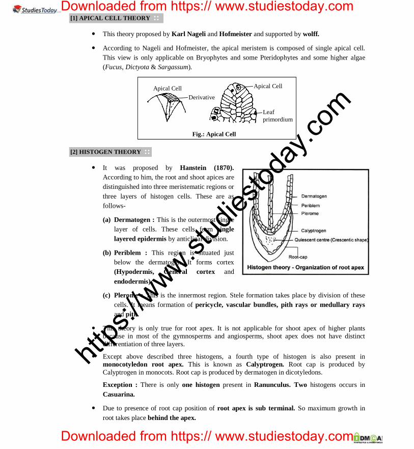
[1] APICAL CELL THEORY ::
This theory proposed by Karl Nageli and Hofmeister and supported by wolff.
According to Nageli and Hofmeister, the apical meristem is composed of single apical cell. This view is only applicable on Bryophytes and some Pteridophytes and some higher algae (Fucus, Dictyota & Sargassum).
Apical Cell Derivative
Apical Cell
Leaf primordium
Fig.: Apical Cell
[2] HISTOGEN THEORY ::
It was proposed by Hanstein (1870). According to him, the root and shoot apices are distinguished into three meristematic regions or three layers of histogen cells. These are as follows-
(a) Dermatogen : This is the outermost single layer of cells. These cells form single layered epidermis by anticlinal division.
(b) Periblem : This region is situated just below the dermatogen. It forms cortex (Hypodermis, General cortex and endodermis).
(c) Plerome : This is the innermost region. Stele formation takes place by division of these cells. It means formation of pericycle, vascular bundles, pith rays or medullary rays and pith.
This theory is only true for root apex. It is not applicable for shoot apex of higher plants because in most of the gymnosperms and angiosperms, shoot apex does not have distinct differentiation of three layers.
Except above described three histogens, a fourth type of histogen is also present in monocotyledon root apex. This is known as Calyptrogen. Root cap is produced by Calyptrogen in monocots. Root cap is produced by dermatogen in dicotyledons.
Exception : There is only one histogen present in Ranunculus. Two histogens occurs in Casuarina.
Due to presence of root cap position of root apex is sub terminal. So maximum growth in root takes place behind the apex.
https
://www.st
udies
today
.com
Downloaded from https:// www.studiestoday.com
Downloaded from https:// www.studiestoday.com

CAREER POINT : CP Tower, IPIA, Road No.1, Kota (Raj.), Ph: 0744-3040000 Anatomy of Flowering Plants 9
CAREER POINT . PRE-MEDICAL
Note : 1. In hydrophytes root cap is absent. 2. Generally root cap is single layered but in Pandanus (Screw pine) root cap is multilayered. 3. Root cap contains large number of Golgibody which secrete mucilage which make the root slimy.
QUIESCENT CENTRE
A group of inactive or less active cells present between the dermatogen and calyptrogen is called quiescent centre.
These cells contain less amount of DNA, light cytoplasm, small nuclei and synthesis of protein is also less. Quiescent centre name coined by "Clowes ".
Quiescent centre was discovered in Maize root with the help of autoradiography.
If calyptrogen get damaged, this zone becomes active to form new cells of calyptrogen.
[3] TUNICA CORPUS THEORY :: This theory was proposed by Schmidt (1924). This theory is applicable on shoot apex. According to
this theory two types of layeres are found in the shoot apex.
(a) Tunica :
This is peripheral layer, epidermis is formed by this layer. In tunica cells, anticlinal division takes place only in one plane.
Anticlinal division occurs at right angle to longitudinal axis (tangential plane) of cell.
When division occurs in single anticlinal plane they do not increase the number of layers.
Generally, tunica is single layered, but some times it is multilayered, then the outer most layer forms the epidermis and remaining layers form rest types of the tissue system with the association of corpus.
(b) Corpus : The mass of cells present below the tunica is called Corpus. The cells of this zone divide in all direction (many planes) due to which, volume increases. It forms rest of the tissue system.
[4] MANTLE CORE THEORY :: This theory was proposed by "Popham and Chan". They compared the mantle to the tunica and core
with the corpus. Mantle forms epidermis.
According to them corpus or core is distinguished into three zones.
(a) Sub-apical Meristem :- This present just below the mantle. It's function is re-establishment of mantle if mantle gets damaged. It is similar to quiescent centre of root apical meristem.
(b) Central-zone Meristem : This is the inner most zone. This zone is responsible only for formation of pith.
https
://www.st
udies
today
.com
Downloaded from https:// www.studiestoday.com
Downloaded from https:// www.studiestoday.com

CAREER POINT : CP Tower, IPIA, Road No.1, Kota (Raj.), Ph: 0744-3040000 Anatomy of Flowering Plants 10
PRE-MEDICAL CAREER POINT . (c) Peripheral Meristem : This region is only responsible for the formation of cortex, pericycle and
vascular tissues.
[5] NEWMAN'S THEORY ::
According to this theory meristematic tissues of shoot apex are three types :-
(i) Monoplex : Such type of shoot apex in which meristematc cells are arranged in group in place of meristematic layers. Such type of shoot apex is found in Ferns. They do not form special structure.
(ii) Simplex : Such type of shoot-apex is formed by single layer of meristematic cells. It is found in Gymnosperms. (Except – Gnetales)
(iii) Duplex : Such type of shoot apex is formed by two layers of meristematic cells. Peripheral layer is called tunica and inner layer is called corpus. Such type of shoot apex is found in Gnetales and Angiosperms.
[6] KORPER-KAPPE THEORY ::
It was proposed by Schuepp (1917). According to this theory, the cells of central and peripheral part of the root apex exhibit differences in planes of cell divisions.
In peripheral region each cell first divides transversely and there after the lower daughter cell divides longitudinally thus forming the shape of 'T'. Such divisions are called the Kappe divisions. In the central region T is inverted () as the second division takes place in the upper daughter cell. Such divisions are called the Korper divisions. As a result of these 'T' or '' divisions, the cells in root apex remain arranged in rows.
Korper form the body of organism while Kappe form the covering or cap..
PERMANENT TISSUES ::
Following division of cells in meristem the newly formed cells become structurally and functionally specialized and lose the division ability and they form permanent tissue. They are formed by division and differentiation of meristematic tissues.
They are present either in permanent G0 stage or in arrested G1 stage.
Their cells may be living or dead.
(A) SIMPLE TISSUES
https
://www.st
udies
today
.com
Downloaded from https:// www.studiestoday.com
Downloaded from https:// www.studiestoday.com

CAREER POINT : CP Tower, IPIA, Road No.1, Kota (Raj.), Ph: 0744-3040000 Anatomy of Flowering Plants 11
CAREER POINT . PRE-MEDICAL
These tissues are made up of similar type of cells that perform a common function and have common origin. Simple tissues are of three types :-
(I) Parenchyma (II) Collenchyma (III) Sclerenchyma
(I) PARENCHYMA : It is very primitive type of tissue. It is first evolved tissue. Remaining all different type of tissues are derived from this tissue. So it is also called as fundamental tissue.
It is a universal tissue and a major component of internal plant organs.
Parenchyma name coined by Grew.
Characteristic Features :
1. It is a living tissue.
2. Tissue first to be differentiated from meristem is parenchyma.
3. All the cells of parenchyma are thin walled. Cell wall is made up of pectocellulose. (Mainly cellulose). So parenchyma is a soft tissue.
4. Each cell containing large central vacuole.
5. Inter cellular spaces are present between cells of this tissue, it is a loose tissue. Intercellular spaces are schizogenous in origin.
6. Body of Bryophyte is mainly composed of parenchyma.
7. Flesh of a fruit is mainly composed of parenchyma.
8. The cells are isodiametric. The cells of parenchyma are spherical, oval or polygonal in shape. Each parenchymatous cell contains 14 planes of lateral line, which are maximum possible plane in a cell. These are known as tetrakaidecahedron.
Modification of Parenchyma :
(a) Prosenchyma : The cells of this parenchyma are long with pointed ends. This parenchyma forms the Pericycle of roots.
(b) Aerenchyma : This parenchyma is made up of rounded cells. These cells surround the large air chambers. Air chambers are lysigenous in origin. It is found in cortex region. It provides buoyancy to hydrophyte plants.
https
://www.st
udies
today
.com
Downloaded from https:// www.studiestoday.com
Downloaded from https:// www.studiestoday.com

CAREER POINT : CP Tower, IPIA, Road No.1, Kota (Raj.), Ph: 0744-3040000 Anatomy of Flowering Plants 12
PRE-MEDICAL CAREER POINT .(c) Stellate parenchyma : The cells of this tissue are stellate and branched. Air spaces are also
present but they are less developed. Main function of this parenchyma is to provide mechanical support.
It is found in the leaf bases of banana and canna. It provides strength to leaf bases.
(d) Chlorenchyma : Such type of parenchyma in which abundant quantity of chloroplasts are found. Two types of chlorenchyma are present in dorsiventral leaves :-
(i) Palisade tissues :- Inter cellular spaces are absent. Their cells are tightly fitted together. They are present towards adaxial/ventral/upper side of leaf. Numbers of chloroplasts are more in palisade tissue as compare to spongy tissue. So upper surface of a leaf appears more green as compared to lower surface.
(ii) Spongy tissues :- Large intercellular spaces are present. So they facilitate transpiration and gaseous exchange. They are present towards abaxial/dorsal/lower side of leaf.
(e) Mucilage Parenchyma : In the mucilage parenchyma large vacuoles and Mucilage will be found. eg., Succulent xerophytic plants. e.g., Aloe. Function –storage of water.
(f) Idioblast : In this type of parenchyma non-living ergastic substances like tannins, oils, crystals etc. are present.
Functions of parenchyma :
The main function of this tissue is storage of food, photosynthesis and secretion.
(II) COLLENCHYMA : Term coined by Schleiden.
Main characteristics :
Collenchyma is a living mechanical tissue.
It is made up of elongated (oval, spherical or polygonal shape in section) cells.
Localized deposition of pectocellulose (mainly pectin) & hemi cellulose is the characteristics feature of collenchyma.
Vacuolated cytoplasm is found in the cells of collenchyma. Intercellular spaces are not present. These cell assimilate food when they contain chloroplast.
Origin of collenchyma :- Collenchyma originates from ground meristem.
Occurance :
It is found in the stems of herbaceous dicotyledons.
Collenchyma is absent in woody plant parts, root and monocotyledons.
Collenchyma forms the hypodermis of dicotyledon stems. It is found either as a homogenous layer or in patches.
Collenchyma is absent in plants after the secondary growth because plant becomes woody.
Lamina margins of leaves also bear collenchyma. This protects the cracking of lamina margin due to the action of wind.
They are present in leaf petiole.
Type of Collenchyma :
https
://www.st
udies
today
.com
Downloaded from https:// www.studiestoday.com
Downloaded from https:// www.studiestoday.com

CAREER POINT : CP Tower, IPIA, Road No.1, Kota (Raj.), Ph: 0744-3040000 Anatomy of Flowering Plants 13
CAREER POINT . PRE-MEDICAL
On the basis of place of deposition, it is classify into three types by Majumdar :-
(1) Lamellar / plate collenchyma : The cells of collenchyma arranged in lamellar forms. The cell have thickening on the tangential walls. Due to such type of deposition, cell looks like a lamellar or plates. Ex. Sunflower stem.
(2) Angular collenchyma : This type of collenchyma abundantly found in plants. The cells of this tissue are angular. The depositions of pectocellulose occur at the corner of cell. eg., Stem of Datura, Solanum and Tomato.
(3) Lacunar collenchyma/tubular collenchyma : Large intercellular spaces are present in the cells of this tissue. Deposition of pectocellulose on the wall of intercellular spaces. Intercellular spaces of collenchyma are thickened. e.g., Cucurbita stem and aerial roots of Monstera.
Functions :
Mechanical as well as Physiological.
They provide mechanical support to growing parts of plant such as young stem and petiole of leaf.
Due to the presence of chloroplast, it is also participates in the process of photosynthesis.
(III) SCLERENCHYMA : Name coined by Mattenius.
Main features :
Sclerenchyma is the main mechanical tissue.
These cells are long, narrow, thick walled and dead.
Cell wall is thick and lignified and have different types of pit.
Function : It provide mechanical support/mechanical strength to plants.
Type of sclerenchyma :
On the basis of variation in form, structure, origin and development, sclerenchyma cells are of two types.
(1) Sclereids
(2) Sclerenchymatous fibres
https
://www.st
udies
today
.com
Downloaded from https:// www.studiestoday.com
Downloaded from https:// www.studiestoday.com

CAREER POINT : CP Tower, IPIA, Road No.1, Kota (Raj.), Ph: 0744-3040000 Anatomy of Flowering Plants 14
PRE-MEDICAL CAREER POINT .(1) Sclereids : These cells are small, extremely thick walled and their ends are not pointed. Sclereids
are isodiametric or irregular in shape. Sclereids cells have more pits and lumen is almost very narrow. There pit cavity is branched. Sclereids are classsify by Tschierch, on the basis of their shapes :
(a) Stone cells or Brachysclereids or Grit cells : These cells are spherical or oval in shape. They are found in endocarp of drupe fruits, so endocarp becomes hard.
They are present in endocarp of Coconut, Mango, Almond, and Walnut etc.
Brachysclereids are also present in fleshy (edible) part of pear (Pyrus), Guava and Sapota.
(b) Trichosclereids : These are also known as internal hairs. They are spines like, bifurcated cells. These are found in floating leaves.
Also present in aerial roots of monstera.
(c) Astro Sclereids or Stellate sclerenchyma : These cells are stellate (star) shaped. They are found in floating leaves.
Astrosclereids are also found in tea leaves.
Example : Both Astro and Tricho sclereids are present in floating leaves.
Victoria, Nelumbo (Lotus) and Nymphaea petiole.
(d) Macro-sclereids or Rod cells or Malpighi cells :
They are small and rod like cells. They are present in seed coats.
Example :
They form part of seed coat in legume plants. Due to their presence seed coat becomes hard and dormancy is present in legume seeds.
In leguminous plants hardest seed coat is found in French bean.
In plant kingdom, hardest Seed coat is found in lotus.
(e) Osteo-Sclereids or Bone cell :
These are known as prop-cells. These are pillar like cells. Both end of pillar like cells spreads to form bone like structure.
Example : These cells are found in leaves of Hakea and Osmanthus.
(2) Sclerenchymatous Fibres :
https
://www.st
udies
today
.com
Downloaded from https:// www.studiestoday.com
Downloaded from https:// www.studiestoday.com

CAREER POINT : CP Tower, IPIA, Road No.1, Kota (Raj.), Ph: 0744-3040000 Anatomy of Flowering Plants 15
CAREER POINT . PRE-MEDICAL
These cells are fibrous. They are longest cells in plant body.Their both ends are pointed (tapering). Due to thick wall, lumen is reduced. Fibers are generally occurring in groups.
Their cell wall contains simple and bordered pits.
On the basis of structure fibres are classified into two groups :
(a) Libriform fibres : They are thickened long fibres. They posses simple pits and narrow lumen. Libriform fibres are found in phloem, xylem, pericycle and hypodermis (Maximum in phloem).
(b) Fibre Tracheids : They are also highly thickened. Bordered pits are present in these fibres and lumen is broad. They are only found in xylem.
On the basis of position, fibres are divided into three types :
A. Surface fibres : They are present on the surface of plant. These fibres are also called as filling fibres.
(i) Seed surface fibres –
Example 1 : Cotton fibres : Cotton fibres are formed by the out growth of seed coat. They are not any type of tissue or cell.
Cotton fibres are composed of cellulose these fibres are non-lignified. So cotton fibres are not true fibres. Two types of fibres are found in cotton. Long fibres are called 'lint' and small fibres are known as 'fuzz'. Lint fibres are used in cloth industry. Fuzz are filling fibre. Cotton fibres are most pure form of cellulose in nature.
Cotton fibres are not an example of any type of cell because these fibres are formed by out growth of testa.
Example 2 : Red silk cotton (Semal fibre)- Obtained from Salmalia malabaricum.
Example 3 : White silk cotton (Kapok) – Obtained from Ceiba pentendra.
(Both red and white silk cotton fibres are not true fibres and they are also an example of seed surface fibre.)
(ii) Coir of coconut is also a type of surface fibre. They are derived from the mesocarp. These are true fibres.
https
://www.st
udies
today
.com
Downloaded from https:// www.studiestoday.com
Downloaded from https:// www.studiestoday.com

CAREER POINT : CP Tower, IPIA, Road No.1, Kota (Raj.), Ph: 0744-3040000 Anatomy of Flowering Plants 16
PRE-MEDICAL CAREER POINT . B. Xylary or wood fibres : These are hard fibres. These fibres are not flexible. They can not be
knitted (weaved) easily so they are not useful. These are found in xylem. Ex. Munj fibre (Saccharum munja)
C. Bast fibres / Extra xylary fibres / Phloem fibre : These are known as commercial fibres. These fibres are flexible and can be knitted (weaved) easily. They have great economic value.
These fibres are obtained from the phloem and pericycle of plants.
The bast fibres of Corchorus capsularis (Jute), Crotalaria juncea (Sunn hemp) and Hibiscus sabdariffa (patua) are obtained from the secondary phloem of stem.
The bast fibres of hemp (Cannabis sativa) and Linum usitatissimum (flax) are obtained from the pericycle. Fibres which are obtained from pericycle are called perivascular fibres.
Leaf fibres Manila hemp (Musa textilis) and agave hemp (Agave sislana) : These are obtained from sclerenchymatous bundle sheath.
Special Points :
Fibres are longest plant cell. Longest fibres occur in Boehmeria nivea (Ramie fibre) length – 55 cm.
In plant kingdom hardest seed coat is found in Nelumbo (Lotus).
In plant kingdom largest leaves are found in Victoria regia.
Longest leaves are found in Raphia vinifera. Length 10-15 m.
Longest commercial fibres – Jute fibres.
Living sclerenchymatous fibres are present in Tamarix.
(B) COMPLEX PERMANENT TISSUES
The complex tissues are made up of more than one type of cells and these work as a unit. Complex tissue are heterogenous.
Complex tissues are of two types : (a) Xylem (b) Phloem.
During vascularisation in plants differentiation of procambium followed by the formation of primary phloem and primary xylem simultaneously.
Complex tissues are absent in gametophytes.
(a) Xylem :
The term 'Xylem' is coined by Nageli. (Greek xyles – wood.)
The function of xylem is to conduct water and mineral salts upwards form the root to stem and leaves and to give mechanical strength to the plant body.
For conduction of water, death of protoplasm is must. Dead tissues are more develop in water scares condition.
On the basis of origin, xylem is divided into primary xylem and secondary xylem.
1. Primary xylem originates form procambium.
On the basis of development primary xylem divided into two parts.
https
://www.st
udies
today
.com
Downloaded from https:// www.studiestoday.com
Downloaded from https:// www.studiestoday.com
CAREER POINT : CP Tower, IPIA, Road No.1, Kota (Raj.), Ph: 0744-3040000 Anatomy of Flowering Plants 17
CAREER POINT . PRE-MEDICAL
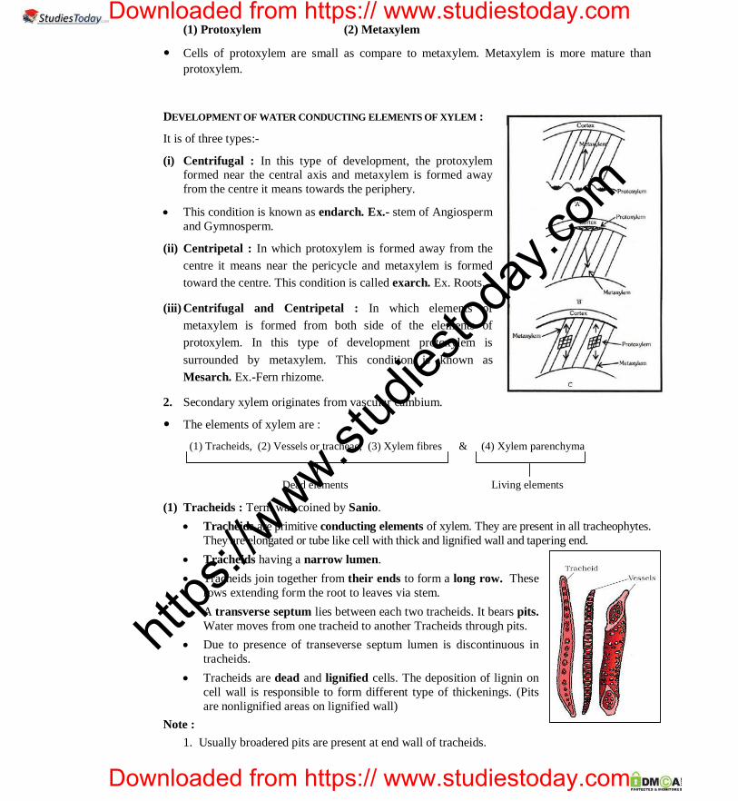
(1) Protoxylem (2) Metaxylem
Cells of protoxylem are small as compare to metaxylem. Metaxylem is more mature than protoxylem.
DEVELOPMENT OF WATER CONDUCTING ELEMENTS OF XYLEM :
It is of three types:-
(i) Centrifugal : In this type of development, the protoxylem formed near the central axis and metaxylem is formed away from the centre it means towards the periphery.
This condition is known as endarch. Ex.- stem of Angiosperm and Gymnosperm.
(ii) Centripetal : In which protoxylem is formed away from the centre it means near the pericycle and metaxylem is formed toward the centre. This condition is called exarch. Ex. Roots.
(iii) Centrifugal and Centripetal : In which elements of metaxylem is formed from both side of the elements of protoxylem. In this type of development protoxylem is surrounded by metaxylem. This condition is known as Mesarch. Ex.-Fern rhizome.
2. Secondary xylem originates from vascular cambium.
The elements of xylem are :
(1) Tracheids, (2) Vessels or tracheae, (3) Xylem fibres & (4) Xylem parenchyma
Dead elements Living elements (1) Tracheids : Term was coined by Sanio.
Tracheids are primitive conducting elements of xylem. They are present in all tracheophytes. They are elongated or tube like cell with thick and lignified wall and tapering end.
Tracheids having a narrow lumen. Tracheids join together from their ends to form a long row. These
rows extending form the root to leaves via stem. A transverse septum lies between each two tracheids. It bears pits.
Water moves from one tracheid to another Tracheids through pits. Due to presence of transeverse septum lumen is discontinuous in
tracheids. Tracheids are dead and lignified cells. The deposition of lignin on
cell wall is responsible to form different type of thickenings. (Pits are nonlignified areas on lignified wall)
Note : 1. Usually broadered pits are present at end wall of tracheids.
https
://www.st
udies
today
.com
Downloaded from https:// www.studiestoday.com
Downloaded from https:// www.studiestoday.com

CAREER POINT : CP Tower, IPIA, Road No.1, Kota (Raj.), Ph: 0744-3040000 Anatomy of Flowering Plants 18
PRE-MEDICAL CAREER POINT . 2. The maximum bordered pits are found in the tracheids of Gymnosperm plants. 3. Maximum deposition of lignin is found in pitted type of thickening. 4. Annular and Spiral type of thickening of lignin is found in protoxylem. 5. Reticulate and Pitted (mainly) type of thickening of lignin is found in metaxylem. 6. In metaxylem tracheids of pteridophytes scalariform type of thickening is found.
Reticulate
Simple pit
Annular Spiral Scalariform Different lignified thickening in xylem (tracheids and vessels)
(2) Vessels :
It is advance water conducting element of xylem. It is a long cylindrical tube like structure made up of many cell called vessel members each with lignified walls and large central cavity.
The lumen of vessels is wider than tracheids and end wall is perforated (Transverse septum is absent between two vessel elements. If present then porous.) Thus vessles are more capable for conduction of water than tracheids. Due to presence of perforated end, vessels work as a pipe line during conduction of water.
Vessels contain usually simple pits at their lateral wall. Thickening type of wall is the same as tracheids.
Note :
1. Vessels are only found in xylem of angiosperm but exceptionally it is also present in some Gymnosperms like Ephedra, Gnetum and Welwitschia.
2. Vessels are absent in some Angiospermic plants such as Dracaena, Yucca, Dazinaria, Drimys. There are some angiosperms families in which vesselless angiosperms are include. eg., Winteraceae, Tetracentronaceae and Trochodendronaceae.
3. Vessels are example of dead syncyte.
Syncyte : Cell which is formed by fusion of cells, called as syncyte.
(3) Xylem fibres :
Xylem fibres provides strength to the tracheids and vessels. Mainly these fibres provide strength to the vessels. They have highly thickened walls and obliterated central lumen.
They are present more abundantly in secondary xylem.
(4) Xylem Parenchyma :
https
://www.st
udies
today
.com
Downloaded from https:// www.studiestoday.com
Downloaded from https:// www.studiestoday.com

CAREER POINT : CP Tower, IPIA, Road No.1, Kota (Raj.), Ph: 0744-3040000 Anatomy of Flowering Plants 19
CAREER POINT . PRE-MEDICAL
It's cell wall is made up of cellulose. It store starch, fats and tannin etc.
The radial conduction of water is the function of xylem parenchyma. (It conducts water to peripheral part of plant organs).
They store food material in the form of starch, fat and other substance.
Their wall possesses pits.
Hadrom : Tracheids and Vessels are collectively known as water conducting elements or "Hadrom".
Hadrom term was proposed by Haberlandt.
(b) Phloem : The term 'Phloem' is coined by Nageli. The main function of the phloem is to conduct food materials, usually from the leaf to other plant parts
(eg., storage organ and growing regions.) On the basis of origin, phloem is classified into two categories primary and secondary phloem.
Primary phloem originates from procambium and secondary phloem originates from vascular cambium.
On the basis of development primary phloem categorised into protophloem and metaphloem. The protophloem has narrow sieve tubes whereas metaphloem has bigger sieve tubes.
Phloem remains active for less duration as compared to xylem.
Phloem consist of 4 types of cells.
1. Sieve cell / Sieve tube
In Gymnosperms In Angiosperms
and pteridophytes
Sieve elements was discovered by Hartig.
Sieve cell/sieve tube element are living and thin walled.
Mature sieve tube elements are enucleated living cells.
Central vacuole is present in each sieve cells/sieve tube element.
In Angiosperm plants sieve tube elements are arranged with their ends and form sieve tube.
Sieve plate (oblique transverse perforated septa) is present between the two sieve tube elements. Materials are transported through these pores.
Callose deposited on the radius of pores during dropping season (autumn) of leaves, to form a thick layer. This is called Callus pad.
Sieve plate is protected by callus pad. It is also prevented from bacterial infection and drought.
Callose dissolves during spring season by callase enzyme. Callose is a -1-3 glucan.
In Gymnosperms and pteridophytes sieve cells are arranged irregularly. Sieve cell have sieve plates on their lateral walls. Thus conduction of food takes place in zig-zag manner.
In Angiosperms food conduction is erect and efficient.
https
://www.st
udies
today
.com
Downloaded from https:// www.studiestoday.com
Downloaded from https:// www.studiestoday.com

CAREER POINT : CP Tower, IPIA, Road No.1, Kota (Raj.), Ph: 0744-3040000 Anatomy of Flowering Plants 20
PRE-MEDICAL CAREER POINT . Sieve elements contain special type of protein P-protein (p-phloem)
Note : 1. Food conduction is bidirectional in sieve tube.
2. Sieve tube is an example of living enucleated syncyte.
3. Most likely function of p-protein is sealing mechanism on wounding.
2. Companion cell :
The companion cells are specialized parenchymatous cell which are closely associated with sieve tube element. The sieve tube element and companion cells are connected by pits field present between their common longitudinal walls.
Sieve tube element and companion cell originates together. Both of them originates from a single mother cell. So called as sister cells.
Companion cell maintain pressure gradient in sieve tube. Functions of sieve tube are regulated by companion cell.
Companion cells are only found in Angiosperms. (Exception – Austrobaileya is angiosperm plant but companion cells are absent).
Special type of cells attached with the sieve cells in gymnosperm and pteridophytes in place of companion cells. These cells are called as albuminous cells/strassburger cell. It is analogous to companion cell.
3. Phloem fibres : Fibres which are present in phloem are called bast fibres. These fibres are generally not found
in primary phloem.
These fibres provide mechanical support to the conducting elements (sieve cells and sieve tube.)
4. Phloem Parenchyma : It is also known as bast parenchyma. It is made up of elongated tapering cylindrical cells
which have dense cytoplasm and nucleus and connected through Plasmodesmata.
It's cells are living and thin walled. It store various material. eg., Resin, Latex, Mucilage etc.
The main function of phloem parenchyma is conduction of food in radial direction and storage of food. The food conducting element of phloem is called Leptom. Leptom
includes Sieve cell
Sieve tubes
Leptom term was proposed by Haberlandt. Note : 1. Phloem parenchyma is absent in the stems of monocotyledon plants and in primary phloem of
dicot plant. 2. Phloem parenchyma is absent in the stems of Ranunculaceae plants. (dicot family). e.g., Thalictrum.
Fig. L.S. of Phloem Tissue
https
://www.st
udies
today
.com
Downloaded from https:// www.studiestoday.com
Downloaded from https:// www.studiestoday.com

CAREER POINT : CP Tower, IPIA, Road No.1, Kota (Raj.), Ph: 0744-3040000 Anatomy of Flowering Plants 21
CAREER POINT . PRE-MEDICAL SPECIAL TISSUES OR SECRETORY TISSUE ::
The cells of this tissue have secretory function. The substances get isolated from cytoplasm and get deposited or aggregated in vacuole of living cells, in dead cells or cavities and canals.
These tissues are of two main types -
(I) Laticiferous tissues (II) Glandular tissues
(I) Laticiferous tissue :
These are made up of long, highly branched and thin walled cells. These cells are filled with milky juice, called as Latex.
Latex is the mixture of saccharides, starch granules, alkaloids, minerals and waste materials.
Starch granules present in latex are dumble shaped.
(a) Function :
Latex provide protection to the plant.
It prevents the plants from infection of bacteria and fungus. In laticiferous tissue there are two types of cells-
1. Latex vessels 2. Latex cells
1. Latex vessels : These are articulated vassels. Latex vessels are formed due to dissolve of cell walls of meristematic cells. Thus these are syncyte, coenocytic cell (multinucleated). Example : Latex vessels are present in Hevea, Ficus, Papaver, Papaya, Argemone and Sonchus.
Highly developed latex vessels are found in the fruit wall of unripe fruit (capsule) of Poppy.
Opium is obtained from the latex of Papaver somniferum. It contains an alkaloid named as morphine.
An enzyme papain is obtained from the latex of papaya (Carica papaya). Indian rubber is obtained from Ficus elastica and para rubber is obtain from Hevea
brasiliensis. Chewing gum is obtained from Latex of Achras sapota.
Mostly latex is white in colour but in some plants latex is coloured. Ex. Papaver – Dark brown.
Argemone
Sonchus Yellow colour
In some plants latex is colourless. Ex. Banana. 2. Latex cells : These are non articulated latex ducts/tubes. These are long, branched and
multinucleated cells (coenocytic cells).
https
://www.st
udies
today
.com
Downloaded from https:// www.studiestoday.com
Downloaded from https:// www.studiestoday.com

CAREER POINT : CP Tower, IPIA, Road No.1, Kota (Raj.), Ph: 0744-3040000 Anatomy of Flowering Plants 22
PRE-MEDICAL CAREER POINT . Example – Latex cells are found in Calotropis, Euphorbia and Nerium. M.Calvin coined
petroplant term for Latex producing plants. Note : Latex vessels and latex cells are found in cortex.
(II) Glandular tissue :
As the name indicates that this tissue is made up of glands. These glands contain secretory or excretory materials.
Glandular tissues have two types of glands :
(1) Unicellular. Urtica-dioica. These cell are present on the surface of the leaves. These are spiny glands in which formic acid is filled. It protects the plants from grazing animals. It is commonly known as Bichchuvati.
(2) Multicellular : Multicellular glands are of two types. I. External Glands : These are located on the surface of the plants and arising as an
outgrowth from the epidermis. These glands are of various types :- Glandular hairs : They secrete gum-like sticky substance in tobacco and Plumbago,
digestive juicy substance in Drosera. Nectar Glands : These glands secrete sugar solution. These are found in floral parts
mainly in thalamus. Exeption In passiflora, nectar glands are found in leaves. II. Internal Glands : These glands are embedded in the tissues. Internal glands are of
following types. Digestive glands : Digestive glands are found in insectivorous plants. These are
found in Utricularia, Drosera, Dionea etc. They secrete proteolytic juice. Mucous secreting glands : These glands secretes mucous.These are found in the
leaves of betel. Oil glands : These glands are found in fruits and leaves of lemon and orange. Mostly, Oil glands are lysigenous but in sunflower these glands are schizogenous.
Note : Oil glands which secrete volatile oil are called osmophores. Osmophores develop fragrance in flowers. Oils which are obtained from Eucalyptus leaves are used in medicines.
Maximum resin glands are found in Pinus.
Resin ducts are schizogenous. Gum glands are found in Acacia.
Water glands / Hydathode : These glands are related with guttation. Hydathodes are present in Tomato, Pistia and Eichhornia etc.
TISSUE SYSTEM ::
In higher plants several tissues work together in form of a unit to perform a particular function. These tissues have the same origin. Such tissues form a system which is called tissue system. On the basis of their structure and location tissue categorized by Sachs into three different system.
1. EPIDERMAL TISSUE SYSTEM : The epidermal tissue system forms the outermost covering of the plant body. It is made up of elongated compactly arranged cells which form a continuous layer. Epidermal cells are Parenchymatous cells.This system includes epidermis and its related structures. eg., Root hairs, trichomes, stomata and bulliform cells etc. It is developed from protoderm.
https
://www.st
udies
today
.com
Downloaded from https:// www.studiestoday.com
Downloaded from https:// www.studiestoday.com

CAREER POINT : CP Tower, IPIA, Road No.1, Kota (Raj.), Ph: 0744-3040000 Anatomy of Flowering Plants 23
CAREER POINT . PRE-MEDICAL
The epidermis (Greek, Epi = upon ; Derma = skin) of most of plant organs is uniseriate, i.e. composed of single layer of epidermal cells but in some cases it may be multilayered e.g., Ficus , Nerium, Peperomia.
Each cell has a large central vacuole & peripheral thin cytoplasm. They may contain anthocyanin pigments, tannins, oils and crystals etc.
The outside of the epidermis is often covered with waxy thick layer cuticle.
Cuticle is absent in roots.
Stomata : Stomata are minute apertures in the epidermis. Each aperture is bounded by two kidney/ bean shaped cells, called as guard cells. Dumbell shaped guard cell are present in grasses. Guard cell contains chloroplasts. Inner wall of guard cell is thickened and outer wall are thin. There are different numbers of cells of variable size in the epidermis around the guard cells. These
are called as subsidiary cells. Stomatal appertus = Guard cell + Stomatal pore + Subsidiary cell. Stomata are absent in roots, underground parts and submerged hydrophytes. Stomata regulate the process of transpiration and gaseous exchange.
Fig.: Diagrammatic representation : (a) Stomata with bean-shaped guard cells.
(b) Stomata with dumb-bell shaped guard cell. Trichomes : On the stem the epidermal hairs are called Trichome.These Trichome are usually multicellular. They may be branched or unbranched and soft and stiff. Function : The trichomes help in protection, dispersal of seeds and fruits and preventing water loss due to transpiration. Root hair : The root hairs are unicellular elongation of the epidermal cells. The thin wall is made up of cellulose and pectic materials. Root hairs are endogenous in origin. Function : Root hairs play an important role in anchoring the plant body in the soil besides absorbing water and mineral solution from it.
2. GROUND TISSUE CULURE ::
It is the largest tissue system. All the tissues except epidermis and vascular bundle form the ground tissue system. It includes hypodermis, general cortex, endodermis, pericycle pith and medullary rays (pith rays). It is also called as fundamental tissue system. In leaves ground tissue consist of chloroplast containing mesophyll.
VASCULAR BUNDLES / VASCULAR TISSUE SYSTEM :: This tissue system originates from pro-cambium.
Xylem and phloem are collectively termed as Vascular bundles or Vascular tissues system. Type of Vascular Bundles : On the basis of arrangement of different parts, vascular bundles are divided
into three categories.
Radial vascular bundle
https
://www.st
udies
today
.com
Downloaded from https:// www.studiestoday.com
Downloaded from https:// www.studiestoday.com

CAREER POINT : CP Tower, IPIA, Road No.1, Kota (Raj.), Ph: 0744-3040000 Anatomy of Flowering Plants 24
PRE-MEDICAL CAREER POINT .
Xylem
Amphivasal
Phloem
Amphicribral
Concentric
I. Radial vascular bundles : When the xylem and phloem are present separately on different radii in alternate manner. Such
vascular bundles are called radial vascular bundle. The order of development of xylem in these vascular bundles is centripetal. Thus, these
vascular bundles are called exarch. Example : Most of the roots. Exception :- In Radish, Carrot, Turnip, Sugarbeet Conjoint-callateral, Vascular bundle are present.
II. Conjoint vascular bundles : In this type of vascular bundle xylem and phloem are present on the same radius. These are of
two types - (1) Conjoint collateral : In this type of vascular bundle xylem and phloem are present on the
same radius and phloem present towards the periphery. These are two types :
(i) Open – If the cambium is present between the xylem and phloem, It is known as open
vascular bundle. Ex. Open vascular bundle is found in stem of dicotyledons and gymnosperm.
(ii) Close – When cambium is absent between the xylem and phloem, in conjoint vascular bundle, it is called as closed vascular bundle. Ex.- Closed vascular bundles are found in monocotyledons stem.
In this type of vascular bundle, order of development of xylem is centrifugal. So endarch condition is found in xylem.
(2) Conjoint bicollateral and Open Vascular bundle – There are two patches of phloem, one on each side of xylem, are found. There are two strips of cambium (outer and inner), one on each side of xylem, are found. Such types of vascular bundles are known as conjoint, bicollateral and open vascular bundle.
Order of development of xylem is centrifugal so endarch condition is found. Ex.- Stem of family Cucurbitaceae, Apocynaceae and Solanaceae.
III. Concentric vascular bundles :
In this type of vascular bundle either xylem surrounds the phloem or phloem surrounds the xylem. Concentric vascular bundles are always closed. They are of two types -
(a) Amphicribral or Hadrocentric :
In this type of vascular bundle xylem is completely surrounded by phloem. It means xylem is present in the centre of vascular bundle. Such type of vascular bundle is termed as amphicribral.
https
://www.st
udies
today
.com
Downloaded from https:// www.studiestoday.com
Downloaded from https:// www.studiestoday.com

CAREER POINT : CP Tower, IPIA, Road No.1, Kota (Raj.), Ph: 0744-3040000 Anatomy of Flowering Plants 25
CAREER POINT . PRE-MEDICAL
The order of development of xylem in these vascular bundles both centripetal and centrifugal manner. In this type of vascular bundle protoxylem surrounded by metaxylem. These are known as mesarch vascular bundle.
Such types of vascular bundles are found ferns rhizomes.
(b) Amphivasal or Leptocentric :
In this type of vascular bundle phloem is completely surrounded by xylem. It means phloem is present in the centre of the vascular bundle.
In this type of vascular bundle, xylem is endarch. Eg., Stem of Dracaena, Yucca etc.
STELE ::
The stele is the whole central mass of vascular tissue (vascular cylinder) with or withouth pith surrounded by endodermis. Van Tieghem and Douliot put forward the hypothesis about stele. Stele surrounded by endodermis but endodermis is originally the part of cortex. It is not a part of stele. All the tissues inside endodermis is known as stele.
According to him stele is the central part or core of the axis of the plant which includes the vascular system and its related structures.
The tissues which lies inside the stele is called intrastelar tissues and the tissues which lies out side the stele is known as extra stellar tissues.
Presence of stele is a pteridophycean feature (Primitive) which later evolved to vascular bundles in higher plants.
TYPE OF STELE ::
Protostele or Mono stele or solid stele :
Protostele is the most primitive and simplest type of stele.
It consists of a solid mass of xylem which is completely surrounded by phloem.
Such type of stele devoid of pith, in place of pith xylem is present in centre.
Solid stele is of following types (on the basis of shape of xylem) :-
a. Haplostele : In this stele, xylem surrounded by a smooth layer of phloem. Central xylem is cylindrical, but circular in T.S.
Example :Rhynia, Selaginella, Selaginoides etc.
b. Actinostele : Actinostele is that stele in which the central xylem has radiating ribs and assume a star shaped appearance.
Example : Psilotum, Isoetes, Lycopodium serratum
c. Plecto Stele : Such type of solid stele in which the xylem divides into number of separate plates which lie parallel to one another.
Example : Most of the species of Lycopodium. (L.clavatum)
https
://www.st
udies
today
.com
Downloaded from https:// www.studiestoday.com
Downloaded from https:// www.studiestoday.com

CAREER POINT : CP Tower, IPIA, Road No.1, Kota (Raj.), Ph: 0744-3040000 Anatomy of Flowering Plants 26
PRE-MEDICAL CAREER POINT .d. Mixed protostele : Some times the solid xylem core of the protostele is broken into small group of
tracheids which remain embedded in the phloem. Such a protostele is known as mixed protostele. Ex. Lycopodium crenum.
Siphonostele : Siphonostele is the stele in which the pith is present in the centre of vascular cylinder. Siphonostele is of following two types :
Ectophloic siphonostele : In vascular tissue of such type of stele, phloem always present out side of the xylem.
Ex. Equisetum, Osmunda. Amphiphloic siphonostele : In vascular tissue of such type of stele, xylem is surrounded by
phloem on the both side. Ex. Adiantum, Marsilea.
Solenostele : When a megaphyllous leaf develops on stem vascular cylinder of plant organ (stem) breaks from one
side and a gap is formed. It is called as leaf gap. Xylem and Phloem are absent in leaf gap and it is filled with parenchyma. Vascular supply divert in from stem to leaf is called leaf trace. Due to the formation of one leaf gap
stele becomes horse shoe shaped called solenostele. Solenostele also may be Ectophloic or Amphiphloic. Dictyostele or Polystele : Due to production of many leaf gaps in siphonostele, main vascular cylinder, break into many
fragments, then such type of siphonostele is called Dictostele. Each divided fragment (piece) is called meristele. Each meristele has its own separate endodermis and
pericycle. Pith is absent in meristele. Meristele is complete stele so dictyostele is well developed type of stele in
Pteridophytes. Example – Pteridium, Pteris, Dryopteris Eustele : In this type of stele, vascular bundles are arranged in a ring. Medullary rays are present between vascular bundle. Such type of stele is found in stem of gymnosperm and dicotyledon plants. Atactostel : Many vascular bundles are distributed in ground tissue. Such type of stele is called atactostele. Endodermis and pericycle are absent in atactostele. This is highly developed type of stele.
INTERNAL STRUCTURE OF STEMS, ROOTS & LEAVES :: INTERNAL STRUCTURE OF DICOT STEM :
Internal structure of a typical dicot stem show following features :
1. Epidermis : Epidermis is the outermost layer of the stem. It is single layered. Multicellular hair (trichomes) and stomata are found on epidermis. Outer side of epidermis, a layer is present which is made up of cutin is called cuticle.
https
://www.st
udies
today
.com
Downloaded from https:// www.studiestoday.com
Downloaded from https:// www.studiestoday.com

CAREER POINT : CP Tower, IPIA, Road No.1, Kota (Raj.), Ph: 0744-3040000 Anatomy of Flowering Plants 27
CAREER POINT . PRE-MEDICAL
Epidermis plays a significant role in protection.
2. Cortex : In dicotyledon stem cortex divided into three parts :
(a) Hypodermis (b) General cortex (c) Endodermis
(a) Hypodermis : It is present just below the epidermis. It provides mechanical support to young stem. This layer is composed of collenchyma and their cells contain chloroplast. So hypodermis is green and photosynthetic.
(b) General cortex : This part is composed of parenchyma. Storage of food is the main function of the cortex. Resin canal/mucilage canal are present in it. These are schizogenous in origin.
(c) Endodermis : It is single celled thick layer. The cells of endodermis are barrel shaped. These cells accumulate starch in stem of dicot. Thus, it is known as "starch sheath".
3. Pericycle : Pericycle is present on the inner side of the endodermis and above the phloem in the form of semi lunar patches of sclerenchyma.
Note : In sunflower stem, pericycle is made of alternate bands of parenchymatous and sclerenchymatous cells. In which pericycle which is present in front of the vascular bundle is made up of sclerenchyma and remaining is composed of parenchyma. Part of pericycle which is situated in front of vascular bundle is known as Bundle cap. Pericycle is heterogenous in sunflower stem.
Fig.: T.S. of Stem
4. Vascular Bundle : Large number of vascular bundles (wedge shaped) are arranged in a ring. Each vascular bundle is conjoint, collateral and open. Each vascular bundle is made of phloem, cambium and xylem. Eustele is present in dicotyledon stems.
5. Pith : This is well developed region, spreading from ring of vascular bundle to the centre. The cells of this region mainly made up of parenchyma.
https
://www.st
udies
today
.com
Downloaded from https:// www.studiestoday.com
Downloaded from https:// www.studiestoday.com

CAREER POINT : CP Tower, IPIA, Road No.1, Kota (Raj.), Ph: 0744-3040000 Anatomy of Flowering Plants 28
PRE-MEDICAL CAREER POINT . Function of pith – Storage of water and food.
Note : The part of pith which is radially arranged between the vascular bundles, called pith rays or medullary rays. The main function of pith rays is radial conduction of food and water.
INTERNAL STRUCTURE OF MONOCOTYLEDON STEM ::
1. Epidermis : Epidermis is the outer most single celled thick layer. It is covered with thick cuticle. Multicellular hair are absent and stomata are also less.
2. Hypodermis : Hypodermis of monocotyledon is made up of sclerenchyma. It is 2-3 layered. It provides mechanical support to plant.
3.Ground tissue : The entire mass of parenchyma cells next to hypodermis and extending to the centre is called ground tissue. There is no differentiation of ground tissue in monocotyledon stem.
4. Vascular Bundle : Many vascular bundle each surrounded by sclerenchymatous bundle sheath are scattered in the ground tissue and V.B. are generally oval shape. Each vascular bundle is conjoint collateral and closed. Peripheral vascular bundle are generally smaller than centrally located ones.
(a) Xylem : In xylem number of vessels is less. In metaxylem there are two large vessels while in protoxylem there are one or two small vessels. Vessels are arranged in V or Y shape. Just beneath protoxylem vessels, there are a water cavity which is schizolysigenous in origin but major part of water cavity is lysigenous. This cavity is formed by disintegration of the element present below the protoxylem and neighbouring parenchyma.
Exception : In Asparagus water cavity and bundle sheath are absent.
(b) Phloem : It consist of sieve tube elements and companion cells. Phloem parenchyma is absent.
Stele : Atactostele is found in monocotyledon. This is the most developed stele.
https
://www.st
udies
today
.com
Downloaded from https:// www.studiestoday.com
Downloaded from https:// www.studiestoday.com

CAREER POINT : CP Tower, IPIA, Road No.1, Kota (Raj.), Ph: 0744-3040000 Anatomy of Flowering Plants 29
CAREER POINT . PRE-MEDICAL
S. No. Monocot stem Dicot stem 1. 2 3. 4. 5. 6. 7. 8. 9.
Epidermis with generally comparatively smaller cells. Hairs are generally absent. Hypodermis is sclerenchymatous. Cortex is generally absent, but from hypodermis to centre of stem there is ground tissue present. Endodermis is absent. Pericycle is absent. Medullary rays are absent. Pith is absent. Vascular bundles : (a) Scattered V.B. (b) V.B. are conjoint, collateral and closed. (c) There is differences in the size of V.B. in
the centre and at periphery, i.e., V.B. in centre are larger in size and towards periphery are smaller.
(d) Bundle sheath is present around vascular bundle in monocot stem
(e) Oval vascular bundles. (f) Phloem parenchyma is absent. (g) Xylem vessels are 'Y' or 'V' shaped.
Epidermis is made of comparatively larger cells. Multicellular hairs are present. Hypodermis is collenchymatous. Cortex is made of many layered parenchymatous
cells. Endodermis is present but usually poorly
developed. Pericycle is made of one or many layers of cells. Medullary rays are present between vascular
bundles. Pith is present. (a) V.B. are arranged in a ring. (b) V.B. are conjoint, collateral and open (c) V.B. are of same size.
(d) Bundle sheath is absent.
(e) Wedge shaped vascular bundles. (f) Phloem parenchyma is present. (g) Xylem vessels are radial.
https
://www.st
udies
today
.com
Downloaded from https:// www.studiestoday.com
Downloaded from https:// www.studiestoday.com

CAREER POINT : CP Tower, IPIA, Road No.1, Kota (Raj.), Ph: 0744-3040000 Anatomy of Flowering Plants 30
PRE-MEDICAL CAREER POINT . INTERNAL STRUCTURE OF TYPICAL DICOTYLEDON - ROOT :: Internal structure of a typical dicotyledon root shows following features : -
1. Epidermis : - It is uniseriate outermost layer. It comprising tubular living components. Cuticle and stomata are absent. Unicellular root hairs are formed due to elongation of some cells of epidermis.
2. Cortex : - It is made up of parenchymatous cells with intercellular space.
Note : The cells of outer part of cortex are suberized in old root. It is called exodermis.
Exodermis found in some dicotyledon roots and most of the monocotyledon roots.
3. Endodermis : Inner most layer of cortex is known as endodermis. Casparain strips are present on radial and tangential wall of endodermis. These strips are made up of suberin. Casparian strips are discovered by Caspari.
The cells of endodermis which are situated in front of protoxylem cells lack of casparain strips.
These are called passage cells.
The number of passage cells is equivalent to the protoxylem cells and number of rows of root hair equivalent to protoylem cells.
Passage cells provide path to absorbed water from cortex to pericycle.
Note : (1) Root hairs are linearly arranged on root apex. (2) Casparian bands and passage cells are well developed in monocot root. (3) Endodermis acts as a water tight jacket and prevents radial conduction of water
https
://www.st
udies
today
.com
Downloaded from https:// www.studiestoday.com
Downloaded from https:// www.studiestoday.com

CAREER POINT : CP Tower, IPIA, Road No.1, Kota (Raj.), Ph: 0744-3040000 Anatomy of Flowering Plants 31
CAREER POINT . PRE-MEDICAL
FIG.: T.S. OF TYPICAL DICOTYLEDON ROOT
4. Pericycle : It is few thick layered. It is composed of prosenchyma. Lateral roots are originated from the part of pericycle which is lying opposite to protoxylem. Thus
lateral root are endogenous in origin. A few mature cells of pericycle usually opposite to protoxylem, become meristematic. these cells
divide by periclinal divisions and form some layers of cells. these divisions are followed by anticlinal divisions forming a primordium which grows to form a lateral root.
Note : Adventitious root are also endogenous. Because these are originated from stellar region. Some part of vascular cambium in root is originated from pericycle. 5. Vascular Bundles : - Vascular bundles are radial and exarch. Xylem and phloems are separate and
equal in number. The numbers of xylem bundles are usually two to four (diarch to tetrarch upto hexarch).
But exceptionally, Ficus (Banyan tree) root is polyarch. Parenchyma which is found between xylem and phloem, called Conjunctive tissue. Vascular cambium is developed from it. 6. Pith : - In dicot root pith is small or inconspicous.
INTERNAL STRUCTURE OF MONOCOTYLEDON - ROOT :: The internal structure of a typical monocotyledon root is similar to dicotyledon root. But (1) Number of xylem bundles are more than six (Polyarch) in monocotyledon root (exceptionally the
number of xylem bundles are two to six in onion). (2) Pith is well developed in monocotyledon root (3) Only lateral roots are originated from pericycle.
https
://www.st
udies
today
.com
Downloaded from https:// www.studiestoday.com
Downloaded from https:// www.studiestoday.com

CAREER POINT : CP Tower, IPIA, Road No.1, Kota (Raj.), Ph: 0744-3040000 Anatomy of Flowering Plants 32
PRE-MEDICAL CAREER POINT .
Fig.: T.S. of Monocotyledon Root
Difference between dicot root and monocot root . S.No. Character Dicot root Monocot root 1 Pericycle Gives rise to secondary
roots and lateral meristem Gives rise to lateral roots only.
2 Vascular bundles
Diarch to hexarch Polyarch
3 Cambium Develops at the time of secondary growth
Absent
4 Pith Absent or poorly developed
Fully developed
INTERNAL STRUCTURE OF ORCHID ROOT ::
Velamen : - These are found in aerial or hanging roots of some epiphytes (eg. orchid)
These are examples of multilayered epidermis.
These are present outside the exodermis.
These are made up of protoderm and are dead.
Spiral thickening of colloids is found in velamen cells.
Passage cells are found in both exodermis and endodermis in hanging roots of orchids.
https
://www.st
udies
today
.com
Downloaded from https:// www.studiestoday.com
Downloaded from https:// www.studiestoday.com

CAREER POINT : CP Tower, IPIA, Road No.1, Kota (Raj.), Ph: 0744-3040000 Anatomy of Flowering Plants 33
CAREER POINT . PRE-MEDICAL
INTERNAL STRUCTURE OF LEAF ::
Generally leaves divided into two categories – Dorsiventral leaves and isobilateral leaves. The differences
in between them as follows :
Dorsiventral or Bi-facial Iso-bilateral or Equifacial
1. Present at right angle to stem
2. Upper surface of leaf receive more sun light as
compared to lower surface, so there are
difference between internal structure of upper
and lower surface of leaf.
Examples : - Dicots
Exception – Eucalyptus, Nerium.
1. Arranged parallel to stem.
2. Both surface of leaf receive equal
amount of sun light so there are no
difference between internal structure of
upper & lower surfaces.
Example : - Monocots
Exception – Lilium longiflorum
https
://www.st
udies
today
.com
Downloaded from https:// www.studiestoday.com
Downloaded from https:// www.studiestoday.com

CAREER POINT : CP Tower, IPIA, Road No.1, Kota (Raj.), Ph: 0744-3040000 Anatomy of Flowering Plants 34
PRE-MEDICAL CAREER POINT .
DIFFERENCE BETWEEN DICOT LEAF (DORSIVENTRAL) & MONOCOT (ISOBILATERAL) ::
Character Dicot leaf Monocot leaf
(i) Leaf Dorsiventral Isobilateral
(ii) Stomata Usually more on lower epidermis (Hypostomatic)
Equal on lower and upper epidermis (Amphistomatic)
(iii) Mesophyll Made up of two types of tissues (A) Palisade parenchyma (B) Spongy Parenchyma with large
intercellular spaces
Only spongy parenchyma is present which has very small spaces.
(iv) Bundle sheath Made up of parenchyma just above and below vascular bundle. Some parenchyma cells or collenchymatous cells are present upto epidermis
Just above and below the vascular bundles sclerenchymatous cells (up to epidermis) are found.
(v) Bulliform Cells Absent Present
INTERNAL STRUCTURE OF DORSIVENTRAL LEAVES :: Cuticle is present on both surfaces but cuticle of upper surface is thicker. Dorsiventral leaves are mostly
hypostomatic i.e. stomata present on lower surface.
Note : In amphistomatic dorsiventral leaves stomata are more on lower surface.
Mesophyll of these leaves is divided into two regions – Palisade tissue and spongy tissue.
Palisade tissue is found towards upper surface. These cells have more chloroplasts. Spongy tissues is found towards lower surface and have large intercellular space.
INTERNAL STRUCTURE OF ISOBILATERAL LEAVES ::
The thickness of cuticle on the both surface is equal.
Distribution of stomata on both surface's are equal.
Note : Isobilateral leaves are Amphistomatic i.e. stomata present on both sides.
Mesophyll of isobilateral leaves is not differentiated into palisade and spongy tissues. It is completely made up of spongy tissues. Palisade tissues are absent.
https
://www.st
udies
today
.com
Downloaded from https:// www.studiestoday.com
Downloaded from https:// www.studiestoday.com

CAREER POINT : CP Tower, IPIA, Road No.1, Kota (Raj.), Ph: 0744-3040000 Anatomy of Flowering Plants 35
CAREER POINT . PRE-MEDICAL
Bulliform cells : - Large cells are found in the epidermis of psammophytic (desert) grasses which are filled by liquid or empty (mostly) and colourless are called bulliform cells or motor cells. When the bulliform cells in the leaves have absorb water and become turgid the leaf surface is exposed. When they are flaccid due to water stress they make the leaf curl and minimize water loss.
Example : - Ammophila, Poa, Empectra and Agropyron etc. are Psammophytic grasses
VASCULAR BUNDLES OF LEAVES ::
Similar types of vascular bundles are found in both dorsiventral and isobilateral leaves. Vascular bundles of leaves are conjoint, collateral and closed.
Protoxylem is situated towards the adaxial surface and protophloem towards the abaxial surface in the vascular bundle.
The sizes of the vascular bundle are dependant on the size of vein. The veins vary in thickness in the reticulate venation. Thus different size vascular bundles are present in dicot while in parallel venation similar size vascular bundle are present.
Vascular bundles are surrounded by a bundle sheath. Bundle sheath is chlorenchymatous in C-4 plants.
Epidermis of Nerium (both upper & lower) and Ficus (only upper epidermis) becomes multilayered. This is an adaptation to reduce transpiration.
SPECIAL POINTS :: 1. Xerophytes with isobilateral leaves contain palisade on both sides of leaf.
Examples : - Eucalyptus & Nerium.
2. Desert grasses contain palisade like spongy tissue
3. Unifacial or cylindrical leaf : - In these leaves there are no differentitation of upper surface and lower surface. Example : - Onion, Garlic
4. Albascent leaf : - Palisade tissue is restricted in half part of leaf so half part appears more green and other half appears less green. Example : Abutilon
ANOMALOUS PRIMARY STRUCTURE ::
[1] ANOMALOUS STRUCTURE IN DICOTYLEDON STEM
I. Scattered Vascular Bundles : In some of dicotyledon stem, vascular bundles are not arranged in a ring, they are Scattered in the cortex.
Example : - Thalictrum, Nymphaea.
II. Phloem on innermost radius : Anomalously in some plants, the position of phloem is towards the inner side of xylem. Such type of phloem is called Internal or intraxylary phloem. Because, this phloem lies towards the pith, so it is also known as medullary phloem.
Example : Calotropis, Capsicum, Leptadaenia, etc.
III. Medullary vascular Bundle : - In some plants vascular bundles are present in pith. These are found in addition to normal ring of vascular bundles. These are called medulllary vascular bundles.
Example : Amaranthus, Boerhaavia, Chenopodium, Mirabilis, Achyranthes, Bougainvillea, Raphanus sativus.
https
://www.st
udies
today
.com
Downloaded from https:// www.studiestoday.com
Downloaded from https:// www.studiestoday.com

CAREER POINT : CP Tower, IPIA, Road No.1, Kota (Raj.), Ph: 0744-3040000 Anatomy of Flowering Plants 36
PRE-MEDICAL CAREER POINT . IV. Cortical Vascular Bundles : - Some of the vascular bundles are also present in the cortex of
plants except the ordinary ring of vascular bundles. They are known as cortical vascular bundles.
Example : Casuarina, Nyctanthes and Lathyrus etc.
V. Polystelic condition : - Each vascular bundle is surrounded by a separate endodermis and pericycle in some plants. Hence, each vascular bundle is a stele. It is the normal situation in pteriodphytes but in some dicotyledons it is present abnormally.
Example : - Primula, Dianthera
VI. Exclusively xylem vascular Bundle : - Abnormally, some vascular bundles are only formed by xylem except the normal vascular bundles. Phloem is not present in these vascular bundles.
Example : - Paonia
VII. Exclusively phloem vascular Bundle : - Abnormally, some vascular bundles are only formed by phloem except normal vascular bundles in some plants. Xylem is not present in these vascular bundle.
Examples : - Cuscuta & Ricinus communis
[2] Anomalous strucute in monocot stem
Vascular bundle situated in Ring : - Normally vascular bundles are found in monocotyledon stem in scattered form but in the stem of some monocotyledon plants vascular bundles are arranged in ring. Such as Triticum, Secale, Hordeum, Avena, Oryza etc. Members of family Gramineae.
SPECIAL POINT :
1. Monarch condition in Trapa root 2. Triarch condition in Pisum root 3. Tetracrch condition in Helianthus annus and Cicer arietenum root 4. Waiting meristem concept : This concept was given by Buvat. According to this, there is an inactive
centre in the shoot apex which is known as waiting meristem and it acts as reservoir of active initials and on induction it give rise to reproductive apex.
5. Tannin is found in latex of banana. When it comes in contact with air it gets oxidized and becomes reddish brown in colour.
6. Tannin glands are found in Camellia. These glands are schizogenous in origin. 7. Salt glands are found in Tamarix which secretes sodium chloride. 8. Chalk glands are found in plants of plumbaginaceae family which secretes calcium carbonate. 9. Maltilayered (14 to 15 layers) epidermis is found in Peperomia leaves. 10. The most durable wood is Tectona grandis. 11. Tracheids are the chief water transporting elements in gymnosperms. 12. Phloem is embedded into the secondary xylem in some plants. Such phloem is called included
phloem or interxylary phloem. This is secondary anomalous structure. Example. : Leptadaenia, Salvadora etc. dicot stem.
13. Pericycle is absent in roots and stem of some aquatic plants. 14. In some monocotyledonae roots, pith is sclerenchymatous. Ex. Canna. 15. A nectar secreting glands cell contains granular cytoplasm and a large conspicuous nucleus.
https
://www.st
udies
today
.com
Downloaded from https:// www.studiestoday.com
Downloaded from https:// www.studiestoday.com

CAREER POINT : CP Tower, IPIA, Road No.1, Kota (Raj.), Ph: 0744-3040000 Anatomy of Flowering Plants 37
CAREER POINT . PRE-MEDICAL (II) ANATOMY – SECONDARY GROWTH :: Secondary Growth :
By the activity of lateral meristems, increase in the circumference/girth of the plant organs due to the formation of secondary tissues in stelar & extra stelar regions called secondary growth.
Normally secondary growth takes place in roots and stem of dicotyledons & Gymnosperms.
Due to lack of cambium in monocotyledons, secondary growth is absent. But exceptionally secondary growth takes place in some monocotyledons. Such as- Palm, Yucca, Dracaena, Smilax, Agave, Coconut etc.
Pteridophytes and monocots have only primary structure but gymnosperm and dicots soon start undergoing secondary growth.
SECONDARY GROWTH IN DICOT STEM :: [A] Secondary growth in stelar region or secondary growth in vascular region
Secondary growth in stelar region begins earlier than the extra stelar region.
I. Formation of ring of vascular cambium : - A cambium which is present inside the vascular bundle is called intrafascicular – cambium. This is a type of primary meristem.
First of all, cells of medullary rays become meristematic to form interfascicular cambium which is secondary lateral meristem.
Intrafascicular and interfascicular cambium are collectively known as vascular cambium. Vascular cambium is formed in the form of a complete ring which is made up of single layer of cells.
In dicot stem some part of vascular cambium is primary and some part is secondary.
Two types of cells are found in the ring of this vascular cambium.
(i) Fusiform initials (ii) Ray initials.
Fusiform initials are long with pointed ends, while ray initials are spherical (oval).
Amount of fusiform initials is more in vascular cambium.
II. Activity of vascular fusiform initials :
(a) Activity of fusiform initials :
Continuous periclinal divisions or tangential division takes place in fusiform initials .
This type of activity few cells are formed towards the periphery and these cells differentiate into secondary phloem or bast and some of the cells are formed towards the central axis and these cells are differentiated into secondary xylem or wood.
Normally more secondary xylem is formed as compared to secondary phloem due to unequal distribution of hormones.
(Secondary xylem is formed 8-10 times more as compared to sec. phloem).
By the pressure of newly formed secondary phloem, primary phloem and initially formed secondary phloem is pushed towards the outside and get crushed.
The primary xylem however remain less or more intact in or around centre.
By the pressure of secondary xylem, all the primary tissues-such as primary xylem, pith etc and old secondary xylem degenerates in the centre of the stem. Due to this, central part of the stem becomes woody. These activities are going on continuously in plants throughout the life.
https
://www.st
udies
today
.com
Downloaded from https:// www.studiestoday.com
Downloaded from https:// www.studiestoday.com

CAREER POINT : CP Tower, IPIA, Road No.1, Kota (Raj.), Ph: 0744-3040000 Anatomy of Flowering Plants 38
PRE-MEDICAL CAREER POINT . Secondary xylem forms in plant regularly and primary tissues degenerate continuously. This
new secondary xylem also degenerate the old secondary xylem. Waste materials are formed in the stem such as lignin, suberin, tannin, resin, gums etc. due to degeneration of the cells. All these waste materials are filled in the lumen (cavity) of tracheids and vessels of secondary xylem. Because of this, wood in the central region of the stem becomes dark coloured (Black brown). It is called Heart wood or Duramen. The peripheral or outer wood which looks light in colour is known as Sap wood or Alburnum. As a result of growth of secondary xylem, the diameter of heart wood increases.
Fig.: Secondary growth in a dicot stem (diagrammatic) – stages in transverse views
Physiologically active wood is sapwood. The main function of sap wood is water conduction. Heart wood provides maximum mechanical strength to stem.
Note : Conduction of water is not carried out by heart wood because : -
1. Cavities of tracheids and vessels are progressively filled by waste materials.
2. The bladder like ingrowth of parenchyma cells, which enter the lumen of vessels (mainly) & tracheids through the pits in their wall. Such bladders like ingrowth are called as tyloses. Tyloses blocks the conduction of water.
Note: In gymnosperms tylosoids are formed in place of tyloses.
https
://www.st
udies
today
.com
Downloaded from https:// www.studiestoday.com
Downloaded from https:// www.studiestoday.com

CAREER POINT : CP Tower, IPIA, Road No.1, Kota (Raj.), Ph: 0744-3040000 Anatomy of Flowering Plants 39
CAREER POINT . PRE-MEDICAL
SPECIAL POINT ::
1. If the heart wood is destroyed in any stem, then there will be no effect on plants (any vital function is not effected), but if the sap wood is destroyed, then the plant will die because conduction of water will be blocked.
2. Study of wood is known as Xylotomy.
3. If a wood is exposed freely in air then decomposition of sap wood takes place rapidly.
4. Position of youngest layer of secondary phloem is just outside the vascular cambium.
5. Position of oldest layer of secondary phloem is just inside the primary phloem.
6. Position of youngest layer of secondary xylem is just inside the vascular cambium.
7. Position of oldest layer of secondary xylem is just outside the primary xylem.
8. If xylem is blocked then shoot will die first.
Activity of Ray initial : - Periclinal divisions are also going on continuously in ray intials of vascular cambium. Some of the cells are formed towards inside and some of the cells are formed towards outside through these divisions and these cells are made up of parenchyma. Radial lines of cells of parenchyma are formed in the stem. These are called Vascular rays or Sec. medullary rays. They conduct water and food in radial direction. Order of development of vascular rays is both centripetal and centrifugal manner. They pass through secondary xylem to secondary phloem in radial manner.
III. Formation of Annual Rings (Spring wood and autumn wood)
Annual rings are formed due to unequal activity of vascular cambium.
The activity of cambium does not remain same, it is changeable through out whole year. Activity of vascular cambium is affected by physiological and environmental factors.
In winter or autumn season the activity of the cambium is less and forms fewer xylary elements. Cells formed during this period, are small, thick walled darker with higher density and have narrow lumens. This is called autumn wood or late wood.
The vascular cambium is highly active in spring or summer season and produce a large number of xylary element and cells of secondary xylem are larger, thin walled lighter in colour with a lower density and have wider lumen. This wood is known as spring wood or early wood.
https
://www.st
udies
today
.com
Downloaded from https:// www.studiestoday.com
Downloaded from https:// www.studiestoday.com
CAREER POINT : CP Tower, IPIA, Road No.1, Kota (Raj.), Ph: 0744-3040000 Anatomy of Flowering Plants 40
PRE-MEDICAL CAREER POINT . (1) The autumn and spring wood are formed in the form of rings. The ring of any type of wood is
called growth ring. Thus two growth rings are formed in one year and they collectively constitute annual ring.
(2) The numbers of annual rings, formed in a tree give the idea of the age of the tree. The study of determination of age of the plant by this technique is called Dendrochronology.
(3) The annual rings are counted from the base of the stem because basal part has maximum annual rings and upper part has less. Therefore counting from the basal region can give the correct idea.
The width of annual ring is less in unfavorable conditions and more in favorable conditions. Thus the annual rings are also indicative of environment period, the tree has encountered perviously.
(4) A piece is taken from the stem from the base of stem with the help of increment borer instrument. The annual rings are counted from that piece and again inserted (fitted) into the same stem at the same place.
(5) More distinct annual rings are formed in that region where climatic variations are sharp. (6) More distinct annual rings are formed in temperate plants. (7) Distinct annual rings are not formed in tropical plants. (8) Distinct annual rings are not formed in India except Himalayan regions. (9) Least distinct annual rings are formed in seashore regions because the climate remains same
through out the year. (10) More clear annual rings are formed in deciduous plants as compared to evergreen
plants. (In temperate region) (11) In deserts annual rings are less distinct. [B] Secondary Growth in Extra Stelar Region (Cork Cambium) : Secondary growth takes place in extra stelar region due to the activity of cork cambium. Cork
cambium is also known as Phellogen or Extrastelar cambium. Cork cambium arises from the hypodermis or from the outer layer of cortex because they become
meristematic. Cork cambium forms some cells towards the outside (epidermis) and some cells towards the inside
(cortex). Those cells formed towards outside, their middle lamella is suberized. Due to this, these cells become dead. These cells are known as Cork or Phellem. Those cells formed towards the inside, are differentiated into parenchyma and may contain chloroplasts. These are called secondary cortex or Phelloderm.
Phellem (cork) + Phellogen (cork cambium) + Phelloderm (secondary cortex) = Periderm Cork is formed in more quantity and secondary cortex is in less quantity because activity of Cork
cambium is more towards outside. The maximum activity of cork cambium is in winter (Autumn) season.
Bark : All the tissues situated out side the vascular cambium is called Bark. According to modern view bark
includes both living and dead tissues. Bark has two parts. Outer layer of bark is called Rhytidome and inner layer of bark is secondary phloem. Bark that is formed early in the season is called early or soft Bark. Towards the end of season late or
hard bark is formed.
https
://www.st
udies
today
.com
Downloaded from https:// www.studiestoday.com
Downloaded from https:// www.studiestoday.com

CAREER POINT : CP Tower, IPIA, Road No.1, Kota (Raj.), Ph: 0744-3040000 Anatomy of Flowering Plants 41
CAREER POINT . PRE-MEDICAL
KINDS OF BARK :
1. Ring Bark : - Continuous bark of equal thickening is called ring bark. It is formed around the stem in the form of a complete ring. In ring bark cork cambium is continuous.
Example : - Bhojpatra (Betula) – A complete distinct ring bark is formed in this plant. Its bark was used as a writing material/as a paper in ancient period. Ring bark is also formed in Eucalyptus.
2. Scaly Bark : - Discontinuous bark of unequal thickening is called scaly bark. This bark is formed around the stem in the form of pieces or fragments. Highly obvious scaly bark is formed in Psidium guajava. Except this scaly bark is also formed in Neem (Azadirachta), Mango (Mangifera indica) and Tamarindus etc plants.
If complete bark of tree is removed then plant becomes dead due to excessive water loss.
If bark is removed in the form of ring at the base of main stem then root will die first due to lack of food.
LENTICELS : At certain region the phellogen cuts off closely arranged parenchymatous cell on the outer side of the cork cell instead of cork cell (complementary cell). These cell rupture and form lens shaped opening which is known as Lenticels.
Function :
1. The main function of lenticels is exchange of gases between plant and atmosphere.
2. Transpiration also takes place through the lenticles, is known as Lenticular transpiration.
3. Adventitious roots on cutting originate from the living cells of lenticels in vegetative reproduction.
Note :
Lenticels are found in most of the woody trees but absent in woody climbers.
Lenticles are present all over the plant body. They are also present on fruits.They are never found on leaves.
https
://www.st
udies
today
.com
Downloaded from https:// www.studiestoday.com
Downloaded from https:// www.studiestoday.com

CAREER POINT : CP Tower, IPIA, Road No.1, Kota (Raj.), Ph: 0744-3040000 Anatomy of Flowering Plants 42
PRE-MEDICAL CAREER POINT . SECONDARY GROWTH IN DICOT ROOT –
First of all, conjunctive tissue becomes meristematic during the secondary growth in a dicotyledon root and form separate curved strips of vasular cambium below phloem bundles. Then after, the cells of pericycle lying opposite to protoxylem also become meristematic to form additional strips of cambium. In this way a complete ring of vascular cambium is formed.
The portion of vascular cambium formed by pericycle is less. The main portion of vascular cambium is formed by conjunctive tissue. Vascular cambiums in roots are secondary in origin.
The activity of vascular cambium of root is the same as the activity of vascular cambium of stem. Secondary xylem is formed towards the inner side and secondary phloem is formed towards the outer side by vascular cambium. The portion of vascular cambium which is formed by pericycle is responsible for the formation of pith rays. These are made up of parenchyma. These pith rays are known as primary medullary rays (Multiseriate). A few medullary or pith rays are also formed from remaining vascular cambium. These are called secondary medullary rays (uniseriate). Thus two types of medullary rays are found in the secondary structure of roots.
Note : The presence of two types of medullary rays is basic characteristic feature of roots. Only secondary medullary rays are found in stem after the secondary growth. Both of them conduct water and food in radial direction.
https
://www.st
udies
today
.com
Downloaded from https:// www.studiestoday.com
Downloaded from https:// www.studiestoday.com
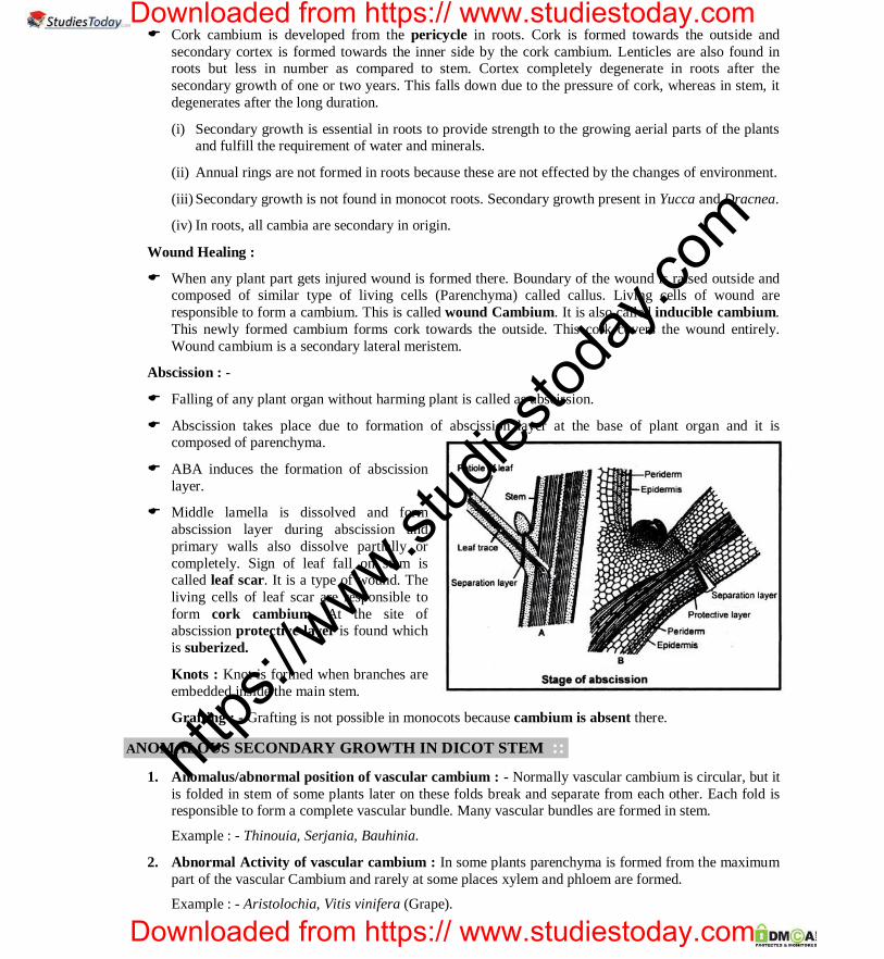
CAREER POINT : CP Tower, IPIA, Road No.1, Kota (Raj.), Ph: 0744-3040000 Anatomy of Flowering Plants 43
CAREER POINT . PRE-MEDICAL
Cork cambium is developed from the pericycle in roots. Cork is formed towards the outside and secondary cortex is formed towards the inner side by the cork cambium. Lenticles are also found in roots but less in number as compared to stem. Cortex completely degenerate in roots after the secondary growth of one or two years. This falls down due to the pressure of cork, whereas in stem, it degenerates after the long duration.
(i) Secondary growth is essential in roots to provide strength to the growing aerial parts of the plants and fulfill the requirement of water and minerals.
(ii) Annual rings are not formed in roots because these are not effected by the changes of environment.
(iii) Secondary growth is not found in monocot roots. Secondary growth present in Yucca and Dracnea.
(iv) In roots, all cambia are secondary in origin.
Wound Healing :
When any plant part gets injured wound is formed there. Boundary of the wound is raised outside and composed of similar type of living cells (Parenchyma) called callus. Living cells of wound are responsible to form a cambium. This is called wound Cambium. It is also called inducible cambium. This newly formed cambium forms cork towards the outside. This cork covers the wound entirely. Wound cambium is a secondary lateral meristem.
Abscission : -
Falling of any plant organ without harming plant is called as abscission.
Abscission takes place due to formation of abscission layer at the base of plant organ and it is composed of parenchyma.
ABA induces the formation of abscission layer.
Middle lamella is dissolved and form abscission layer during abscission and primary walls also dissolve partially or completely. Sign of leaf fall on stem is called leaf scar. It is a type of wound. The living cells of leaf scar are responsible to form cork cambium. At the site of abscission protective layer is found which is suberized.
Knots : Knot is formed when branches are embedded inside the main stem.
Grafting : - Grafting is not possible in monocots because cambium is absent there.
ANOMALOUS SECONDARY GROWTH IN DICOT STEM :: 1. Anomalus/abnormal position of vascular cambium : - Normally vascular cambium is circular, but it
is folded in stem of some plants later on these folds break and separate from each other. Each fold is responsible to form a complete vascular bundle. Many vascular bundles are formed in stem.
Example : - Thinouia, Serjania, Bauhinia.
2. Abnormal Activity of vascular cambium : In some plants parenchyma is formed from the maximum part of the vascular Cambium and rarely at some places xylem and phloem are formed.
Example : - Aristolochia, Vitis vinifera (Grape).
https
://www.st
udies
today
.com
Downloaded from https:// www.studiestoday.com
Downloaded from https:// www.studiestoday.com

CAREER POINT : CP Tower, IPIA, Road No.1, Kota (Raj.), Ph: 0744-3040000 Anatomy of Flowering Plants 44
PRE-MEDICAL CAREER POINT . 3. Sequential or successive ring of vascular cambium : - In some of the plants, a new ring of vascular
cambium is formed each year. This is formed out side the previous ring.
Example : - Mirabilis, Boerhaavia, Bougainvillea etc.
4. Formation of vascular cambium from pericycle : - Vascular cambium is formed from the pericycle in plants of Amaranthaceae and Chinopodiaceae family.
5. Interxylary Cork : - A cork layer that develops between the two growth rings of secondary xylem is called interxylary cork.
Example : - Artemesia tridentata.
6. Formation of cork cambium from Epidermis
Example : Malus pumila, Solanum dulcamara, Quercus suber (oak).
The commerical cork is obtained from the plant Quercus suber.
7. Formation of cork cambium from pericycle : - Example – Clematis &Thuja
8. Formation of cork cambium from phloem : - Vitis and Berberis
ANOMALOUS SECONDARY GROWTH IN MONOCOT STEM :: In some members of plants such as Dracaena, Yucca, Agave, Aloe arboresence, Lomandra, Kingia,
Sanseviera, etc. vascular cambium is formed from the outer region of the ground tissues. Parenchyma is formed towards the outside by the vascular cambium and secondary vascular bundles are formed towards the inner side.
In some plants, the girth of the stem increases without cambium. Such as – Palm, Musa, Tulipa etc. The apical meristem of these plants is of special type. This is known as primary thickening meristem. This apical meristem is repsonsible for the growth in both length and girth (thickness) of the plant.
In Cycas and Gnetum ula successive ring of vascular cambium is found.
In Thuja stem cork cambium is derived from pericycle.
TYPES OF WOOD :: [A] On the basis of amount of parenchyma : Wood is classified into two groups. 1. Manoxylic wood : - Such type of wood contains more living parenchyma. It is soft and loose
wood. eg. Cycas. 2. Pycnoxylic wood : - Such wood contains less amount of living parenchyma. It is hard wood. Such
type of wood are found in most of the dicot plant. Example : - Pinus (Chir), Mango, Acacia (Babool), Tectona (Teak), Dalbergia (Shisham).
[B] On the basis of distribution of parenchyma : Wood is classified into three groups : -
1. Apotracheal : - In this type of wood parenchyma is in scattered form.
Example : Gymnosperm
2. Paratracheal wood : - In this wood parenchyma is arranged or distributed in the form of masses or groups. Example : Dicotyledon plants.
3. Syntracheal wood : - In this wood parenchyma is arranged around the vessels.
Example : Terminalia arjuna.
https
://www.st
udies
today
.com
Downloaded from https:// www.studiestoday.com
Downloaded from https:// www.studiestoday.com

CAREER POINT : CP Tower, IPIA, Road No.1, Kota (Raj.), Ph: 0744-3040000 Anatomy of Flowering Plants 45
CAREER POINT . PRE-MEDICAL
[C] Classification based on vessels : -
On the basis of presence or absence of vessles, wood is classified in two categories –
1. Non-porous wood : - Vessels are absent in such type of wood.
Example :Gymnosperm
2. Porous wood : Vessels are present in such type of wood. On the basis of arrangement of vessels porous wood is divided into two groups.
I. Ring porous wood : - Vessels are arranged in the form of a ring. Such wood conducts water more efficiently. Example : Dalbergia
II. Diffused porous wood : Vessels are irregularly arranged in wood.
Example : Azadirachta.
Non-porous wood is also called as homoxylous wood and porous wood is also called as hetero xylous wood.
Special Point :
Cricket bat from Salix (Willow)
Hockey from Morus (Mulberry)
Billiards's ball Phytelophus (Ivory Palm)
Violin Picea (Spruce)
Hardest wood Hardwickia binata
Heaviest wood Guaiacum officinale
Lightest wood Ochroma pyramidale or Ochroma lagopus
Heaviest wood of India Acacia sundra
Most durable soft wood Cedrus deodar
https
://www.st
udies
today
.com
Downloaded from https:// www.studiestoday.com
Downloaded from https:// www.studiestoday.com

CAREER POINT : CP Tower, IPIA, Road No.1, Kota (Raj.), Ph: 0744-3040000 Anatomy of Flowering Plants 46
PRE-MEDICAL CAREER POINT . IMPORTANT COMPARISON TABLES ::
Porous wood (Hard wood) (i) This wood is found in angiosperms
(ii) Vessels are present.
(iii) This wood is hard.
(iv) Xylem fibres are present.
(v) Vessels are more in porous wood.
(vi) It is also called heteroxylous wood
Non porous wood (Soft wood) It is found in Gymnosperms.
Vessels do not present.
It is soft.
Xylem fibres are rarely present.
Xylem tracheids are dense.
It is called homoxylous wood.
Duramen (Heart wood)
(i) Duramen wood dark in colour.
(ii) This wood does not conduct water.
(iii) In this wood, Tyloses are present.
(iv) It gives the mechanical support to the plant.
(v) It is present in the central part of the tree stem.
Alburnum (Sap wood)
Alburnum wood light in colour.
Alburnum wood conduct the entire water.
In this wood tyloses are not present.
It does not gives mechanical support.
It is found in outer part of stem.
Spring wood
(i) Spring wood are generally light brown in colour.
(ii) In plant, its quantity is more.
(iii) Its vessel are broad and with large cavity.
(iv) It is made up in favourable conditions.
(v) It is produce from more activity of cambium.
(vi) In wood, the fibre are less present.
Autumn wood
Autumn wood are dark brown in colour.
In plant, its quantity is less.
Its vessels are small and with narrow cavity. It is made up in unfavourable conditions.
It is produce from less activity of cambium.
In wood, the fibre are more present.
htt
ps://w
ww.stud
iestod
ay.co
m
Downloaded from https:// www.studiestoday.com
Downloaded from https:// www.studiestoday.com