GASTROINTESTINAL ANATOMY - AWS
7
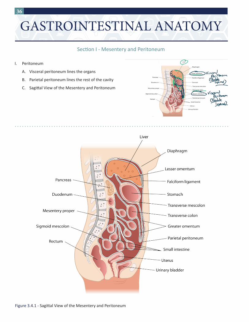
36 Secon I - Mesentery and Peritoneum I. Peritoneum A. Visceral peritoneum lines the organs B. Parietal peritoneum lines the rest of the cavity C. Sagial View of the Mesentery and Peritoneum GASTROINTESTINAL ANATOMY Figure 3.4.1 - Sagial View of the Mesentery and Peritoneum
-
Upload
khangminh22 -
Category
Documents
-
view
0 -
download
0
Transcript of GASTROINTESTINAL ANATOMY - AWS
36
Section I - Mesentery and Peritoneum
I. Peritoneum
A. Visceral peritoneum lines the organs
B. Parietal peritoneum lines the rest of the cavity
C. Sagittal View of the Mesentery and Peritoneum
GASTROINTESTINAL ANATOMY
Figure 3.4.1 - Sagittal View of the Mesentery and Peritoneum

37
II. Mesentery
A. Double layer of peritoneum
B. Contains arteries and veins that supply intestinal tract
C. Examples:
1. Lesser omentum
2. Greater omentum
Figure 3.4.2 - Anterior view of the mesentery

38
Structure Location Notes
Lesser omentum (hepatogastric ligament)
• From liver to lesser curvature of stomach • Contains gastric vessels
Lesser omentum(hepatoduodenal ligament)
• From liver to proximal duodenum
• Pringle maneuver (temporary clamp portal triad to reduce bleeding from liver)
• Contains portal triad (hepatic artery proper, portal vein and common bile duct)
Greater omentum (gastrocolic ligament)
• From greater curvature of stomach to transverse colon
• The ascending and descending colon are retroperitoneal → doesn’t connect to greater omentum
Greater omentum (gastrosplenic ligament)
• From greater curvature of stomach to spleen
• Contains branches of splenic artery and vein (short gastrics and left gastro-omental)
Splenorenal ligament
• From spleen to posterior abdominal wall • Contains splenic artery and vein
Falciform ligament • From anterior liver to abdominal wall
• Contains paraumbilical veins (forms caput medusae with superficial epigastric veins)
Table 3.4.1 - Peritoneal and mesenteric structures
Figure 3.4.3 - Axial view of the peritoneum and mesentery
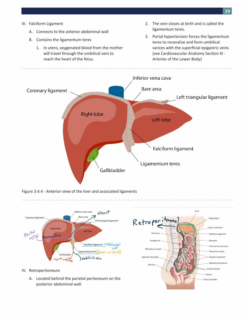
39
III. Falciform Ligament
A. Connects to the anterior abdominal wall
B. Contains the ligamentum teres
1. In utero, oxygenated blood from the mother will travel through the umbilical vein to reach the heart of the fetus.
Figure 3.4.4 - Anterior view of the liver and associated ligaments
2. The vein closes at birth and is called the ligamentum teres.
3. Portal hypertension forces the ligamentum teres to recanalize and form umbilical varices with the superficial epigastric veins (see Cardiovascular Anatomy Section III - Arteries of the Lower Body)
IV. Retroperitoneum
A. Located behind the parietal peritoneum on the posterior abdominal wall.
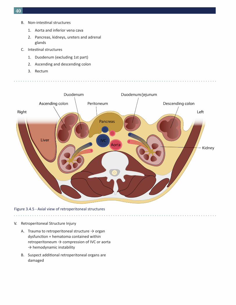
40
B. Non-intestinal structures
1. Aorta and inferior vena cava
2. Pancreas, kidneys, ureters and adrenal glands
C. Intestinal structures
1. Duodenum (excluding 1st part)
2. Ascending and descending colon
3. Rectum
V. Retroperitoneal Structure Injury
A. Trauma to retroperitoneal structure → organ dysfunction + hematoma contained within retroperitoneum → compression of IVC or aorta → hemodynamic instability
B. Suspect additional retroperitoneal organs are damaged
Figure 3.4.5 - Axial view of retroperitoneal structures
41
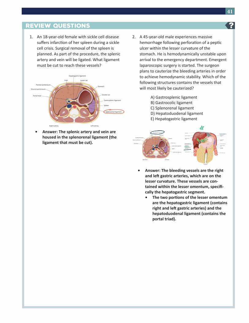
1. An 18-year-old female with sickle cell disease suffers infarction of her spleen during a sickle cell crisis. Surgical removal of the spleen is planned. As part of the procedure, the splenic artery and vein will be ligated. What ligament must be cut to reach these vessels?
• Answer: The splenic artery and vein are housed in the splenorenal ligament (the ligament that must be cut).
2. A 45-year-old male experiences massive hemorrhage following perforation of a peptic ulcer within the lesser curvature of the stomach. He is hemodynamically unstable upon arrival to the emergency department. Emergent laparoscopic surgery is started. The surgeon plans to cauterize the bleeding arteries in order to achieve hemodynamic stability. Which of the following structures contains the vessels that will most likely be cauterized?
A) Gastrosplenic ligamentB) Gastrocolic ligamentC) Splenorenal ligamentD) Hepatoduodenal ligamentE) Hepatogastric ligament
• Answer: The bleeding vessels are the right and left gastric arteries, which are on the lesser curvature. These vessels are con-tained within the lesser omentum, specifi-cally the hepatogastric segment. • The two portions of the lesser omentum
are the hepatogastric ligament (contains right and left gastric arteries) and the hepatoduodenal ligament (contains the portal triad).
REVIEW QUESTIONS ?
43
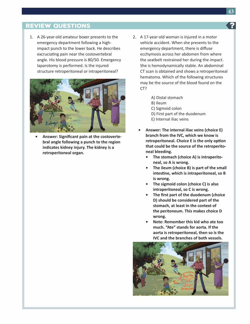
REVIEW QUESTIONS ?1. A 26-year-old amateur boxer presents to the
emergency department following a high-impact punch to the lower back. He describes excruciating pain near the costovertebral angle. His blood pressure is 80/50. Emergency laparotomy is performed. Is the injured structure retroperitoneal or intraperitoneal?
• Answer: Significant pain at the costoverte-bral angle following a punch to the region indicates kidney injury. The kidney is a retroperitoneal organ.
2. A 17-year-old woman is injured in a motor vehicle accident. When she presents to the emergency department, there is diffuse ecchymosis across her abdomen from where the seatbelt restrained her during the impact. She is hemodynamically stable. An abdominal CT scan is obtained and shows a retroperitoneal hematoma. Which of the following structures may be the source of the blood found on the CT?
A) Distal stomachB) IleumC) Sigmoid colonD) First part of the duodenumE) Internal iliac veins
• Answer: The internal iliac veins (choice E) branch from the IVC, which we know is retroperitoneal. Choice E is the only option that could be the source of the retroperito-neal bleeding.• The stomach (choice A) is intraperito-
neal, so A is wrong.• The ileum (choice B) is part of the small
intestine, which is intraperitoneal, so B is wrong.
• The sigmoid colon (choice C) is also intraperitoneal, so C is wrong.
• The first part of the duodenum (choice D) should be considered part of the stomach, at least in the context of the peritoneum. This makes choice D wrong.
• Note: Remember this kid who ate too much. “Ate” stands for aorta. If the aorta is retroperitoneal, then so is the IVC and the branches of both vessels.