World Journal of - Gastrointestinal Endoscopy - NET
20
World Journal of Gastrointestinal Endoscopy World J Gastrointest Endosc 2020 January 16; 12(1): 1-52 ISSN 1948-5190 (online) Published by Baishideng Publishing Group Inc
-
Upload
khangminh22 -
Category
Documents
-
view
0 -
download
0
Transcript of World Journal of - Gastrointestinal Endoscopy - NET

World Journal ofGastrointestinal Endoscopy
World J Gastrointest Endosc 2020 January 16; 12(1): 1-52
ISSN 1948-5190 (online)
Published by Baishideng Publishing Group Inc

W J G EWorld Journal ofGastrointestinalEndoscopy
Contents Monthly Volume 12 Number 1 January 16, 2020
MINIREVIEWS1 Endoscopic advances in the management of non-variceal upper gastrointestinal bleeding: A review
Naseer M, Lambert K, Hamed A, Ali E
ORIGINAL ARTICLE
Retrospective Study
17 Efficacy of mucosa-submucosa clip closure method after gastric endoscopic submucosal dissectionKinoshita S, Nishizawa T, Fujimoto A, Mori H, Nakazato Y, Kikuchi M, Uraoka T
Clinical Trials Study
23 High-definition optical magnification with digital chromoendoscopy detects gastric mucosal changes in
dyspeptic-patientsRobles-Medranda C, Valero M, Puga-Tejada M, Oleas R, Baquerizo-Burgos J, Soria-Alcívar M, Alvarado-Escobar H,
Pitanga-Lukashok H
Observational Study
33 Endoscopic removal of foreign bodies: A retrospective study in JapanLimpias Kamiya KJ, Hosoe N, Takabayashi K, Hayashi Y, Sun X, Miyanaga R, Fukuhara K, Fukuhara S, Naganuma M,
Nakayama A, Kato M, Maehata T, Nakamura R, Ueno K, Sasaki J, Kitagawa Y, Yahagi N, Ogata H, Kanai T
CASE REPORT42 Endoscopic vacuum assisted closure of esophagogastric anastomosis dehiscence: A case report
Cwaliński J, Hermann J, Kasprzyk M, Banasiewicz T
LETTER TO THE EDITOR49 Lack of proper reimbursement is hampering adoption of minimally invasive gastrointestinal endoscopy in
North AmericaIqbal S, Ali A, Razzaq A, Shahzad E
WJGE https://www.wjgnet.com January 16, 2020 Volume 12 Issue 1I

ContentsWorld Journal of Gastrointestinal Endoscopy
Volume 12 Number 1 January 16, 2020
ABOUT COVER Editorial Board Member of World Journal of Gastrointestinal Endoscopy,Dimitrios Kapetanos, FEBG, PhD, Doctor, Department of Gastroenterology,George Papanikolaou Hospital, Thessaloniki 55236, Greece.
AIMS AND SCOPE The primary aim of World Journal of Gastrointestinal Endoscopy (WJGE, WorldJ Gastrointest Endosc) is to provide scholars and readers from various fieldsof gastrointestinal endoscopy with a platform to publish high-quality basicand clinical research articles and communicate their research findingsonline. WJGE mainly publishes articles reporting research results and findingsobtained in the field of gastrointestinal endoscopy and covering a widerange of topics including capsule endoscopy, colonoscopy, double-balloonenteroscopy, duodenoscopy, endoscopic retrogradecholangiopancreatography, endosonography, esophagoscopy,gastrointestinal endoscopy, gastroscopy, laparoscopy, natural orificeendoscopic surgery, proctoscopy, and sigmoidoscopy.
INDEXING/ABSTRACTING The WJGE is now abstracted and indexed in Emerging Sources Citation Index (Web
of Science), PubMed, PubMed Central, China National Knowledge Infrastructure
(CNKI), and Superstar Journals Database.
RESPONSIBLE EDITORS FORTHIS ISSUE
Responsible Electronic Editor: Yu-Jie Ma
Proofing Production Department Director: Xiang Li
NAME OF JOURNALWorld Journal of Gastrointestinal Endoscopy
ISSNISSN 1948-5190 (online)
LAUNCH DATEOctober 15, 2009
FREQUENCYMonthly
EDITORS-IN-CHIEFBing Hu, Anastasios Koulaouzidis, Sang Chul Lee
EDITORIAL BOARD MEMBERShttps://www.wjgnet.com/1948-5190/editorialboard.htm
EDITORIAL OFFICERuo-Yu Ma, Director
PUBLICATION DATEJanuary 16, 2020
COPYRIGHT© 2020 Baishideng Publishing Group Inc
INSTRUCTIONS TO AUTHORShttps://www.wjgnet.com/bpg/gerinfo/204
GUIDELINES FOR ETHICS DOCUMENTShttps://www.wjgnet.com/bpg/GerInfo/287
GUIDELINES FOR NON-NATIVE SPEAKERS OF ENGLISHhttps://www.wjgnet.com/bpg/gerinfo/240
PUBLICATION MISCONDUCThttps://www.wjgnet.com/bpg/gerinfo/208
ARTICLE PROCESSING CHARGEhttps://www.wjgnet.com/bpg/gerinfo/242
STEPS FOR SUBMITTING MANUSCRIPTShttps://www.wjgnet.com/bpg/GerInfo/239
ONLINE SUBMISSIONhttps://www.f6publishing.com
© 2020 Baishideng Publishing Group Inc. All rights reserved. 7041 Koll Center Parkway, Suite 160, Pleasanton, CA 94566, USA
E-mail: [email protected] https://www.wjgnet.com
WJGE https://www.wjgnet.com January 16, 2020 Volume 12 Issue 1II
W J G EWorld Journal ofGastrointestinalEndoscopy
Submit a Manuscript: https://www.f6publishing.com World J Gastrointest Endosc 2020 January 16; 12(1): 1-16
DOI: 10.4253/wjge.v12.i1.1 ISSN 1948-5190 (online)
MINIREVIEWS
Endoscopic advances in the management of non-variceal uppergastrointestinal bleeding: A review
Maliha Naseer, Karissa Lambert, Ahmed Hamed, Eslam Ali
ORCID number: Maliha Naseer(0000-0001-6891-1378); KarissaLambert (0000-0002-0663-2549);Ahmed Hamed(0000-0001-7448-5715); Eslam Ali(0000-0002-3379-8683).
Author contributions: All authorsequally contributed to this paperwith conception and design of thestudy, literature review andanalysis, drafting and criticalrevision and editing, and finalapproval of the final version.
Conflict-of-interest statement: Nopotential conflicts of interest. Nofinancial support.
Open-Access: This article is anopen-access article which wasselected by an in-house editor andfully peer-reviewed by externalreviewers. It is distributed inaccordance with the CreativeCommons Attribution NonCommercial (CC BY-NC 4.0)license, which permits others todistribute, remix, adapt, buildupon this work non-commercially,and license their derivative workson different terms, provided theoriginal work is properly cited andthe use is non-commercial. See:http://creativecommons.org/licenses/by-nc/4.0/
Manuscript source: Invitedmanuscript
Received: March 30, 2019Peer-review started: April 4, 2019First decision: August 2, 2019Revised: August 28, 2019Accepted: October 19, 2019Article in press: October 19, 2019
Maliha Naseer, Eslam Ali, Division of Gastroenterology and Hepatology, Department of InternalMedicine, East Carolina University, Greenville, NC 27834, United States
Karissa Lambert, Ahmed Hamed, Department of Internal Medicine, East Carolina University,Greenville, NC 27834, United States
Corresponding author: Maliha Naseer, MD, Gastroenterology Fellow, Division ofGastroenterology and Hepatology, Department of Internal Medicine, East Carolina University,521 Moye Blvd, Greenville, NC 27834, United States. [email protected]
AbstractUpper gastrointestinal bleeding is defined as the bleeding originating from theesophagus to the ligament of Treitz and further classified into variceal and non-variceal gastrointestinal bleeding. Non-variceal upper gastrointestinal bleedingremains a common clinical problem globally. It is associated with high mortality,morbidity, and cost of the health care system. Despite the continuousimprovement of therapeutic endoscopy, the 30-d readmission rate secondary torebleeding and associated mortality is an ongoing issue. Available Food andDrug Administration approved traditional or conventional therapeuticendoscopic modalities includes epinephrine injection, argon plasma coagulation,heater probe, and placement of through the scope clip, which can be used aloneor in combination to decrease the risk of rebleeding. Recently, more attention hasbeen paid to the novel advanced endoscopic devices for primary treatment of thebleeding lesion and as a secondary measure when conventional therapies fail toachieve hemostasis. This review highlights emerging endoscopic modalities usedin the management of non-variceal upper gastrointestinal related bleeding suchas over-the-scope clip, Coagrasper, hemostatic sprays, radiofrequency ablation,cryotherapy, endoscopic suturing devices, and endoscopic ultrasound-guidedangiotherapy. In this review article, we will also discuss the technical aspects ofthe common procedures, outcomes in terms of safety and efficacy, and theiradvantages and limitations in the setting of non-variceal upper gastrointestinalbleeding.
Key words: Non-variceal upper gastrointestinal bleeding; Over the scope clip; Hemospray;Radiofrequency ablation; Endoscopic suturing device
©The Author(s) 2020. Published by Baishideng Publishing Group Inc. All rights reserved.
WJGE https://www.wjgnet.com January 16, 2020 Volume 12 Issue 11

Published online: January 16, 2020
P-Reviewer: Madalinski M, QuachDTS-Editor: Yan JPL-Editor: AE-Editor: Ma YJ
Core tip: In the last two decades, there has been drastic decline in the mortality andmorbidity caused non-variceal upper gastrointestinal bleeding due to significant progressin the therapeutic endoscopy. The use of devices such as over the scope clips system,Coagrasper, hemospray and endoscopic suturing has tremendously evolved andexpanded to achieve hemostasis as a primary method or when conventional therapeuticdevices such as heater probe, hemoclips or epinephrine injection fails to controlbleeding.
Citation: Naseer M, Lambert K, Hamed A, Ali E. Endoscopic advances in the management ofnon-variceal upper gastrointestinal bleeding: A review. World J Gastrointest Endosc 2020;12(1): 1-16URL: https://www.wjgnet.com/1948-5190/full/v12/i1/1.htmDOI: https://dx.doi.org/10.4253/wjge.v12.i1.1
INTRODUCTIONGastrointestinal bleeding is a medical emergency that results in substantial morbidity,mortality, and health care cost[1,2]. It can present as a massive life-threateninghemorrhage, or a slow chronic bleed. Upper gastrointestinal bleeding (UGIB) isdefined as any gastrointestinal bleeding that originates above the ligament ofTreitz[3,4]. UGIB can be further classified as non-variceal UGIB (NVUGIB) and varicealUGIB (VUGIB). The common causes of NVUGIB are listed in Table 1. The incidenceand mortality associated with NVUGIB have been decreasing due to theadvancements in the prevention and management of NVUGIB[5-7]. Yet, it remains acommon clinical problem with an annual incidence of about 90-108 per 100000 andmortality of 3% to 14%[8,9].
The initial approach to the patient presenting with acute NVUGIB is highlighted inFigure 1. Early endoscopic intervention within 24 h of presentation dramaticallyimproves patient outcomes, and there was no difference observed compared to thosewho underwent endoscopic intervention < 12 h after presentation[10,11]. When anendoscopic approach is performed, it is crucial to have a standardized method ofdiagnosing the cause of bleeding, evaluating the stigmata of recent hemorrhage (i.e.,active bleeding, a visible blood vessel, presence of clots, or red or black spots coveringthe ulcer lesion), and classifying gastric ulcers according to the Forrestclassification[12,13]. Endoscopy is important in revealing the etiology of NVUGIB.
The development and widespread use of endoscopy has been a major contributorto the reduced need for surgery and morbidity associated with NVUGIB[14].Endoscopic management is classified as injection, thermal, and mechanical methods.Amongst the traditional methods, injection of epinephrine is the most common andwidely used modality because of its feasibility to perform and requires lesscoordination between endoscopist and assistant. However, epinephrine alone is lesseffective than combination with thermal or mechanical and other monotherapies suchas clips, probes and electrocoagulation[15,16]. According to the Cochrane review,combination treatment has been associated with significant reduced risk ofrebleeding, surgery and mortality in peptic ulcers with active bleeding or high-riskstigmata such as adherent clot[17]. Through the scope endoclips or hemoclips are foundto be effective and safe hemostatic mechanical devices when applied precisely asmono or combine therapy. Clip grasp the vessel in the submucosa, seal the defect inthe target blood vessel with or without approximation of the sides of the lesion.Furthermore, the tissue damage is minimal with clips and ulcer healing process is nothampered[18,19]. Introduced in clinical practice in 1990s, over the years, clips areevolved in terms of functionality (such as precision, tensile strength, rotatability,overshoot and strength of closure), physical characteristics and cost[20,21]. Recentlypublished study by Wang et al[22] compared the functionality of the five different typesof hemostatic clips. According to the study findings, Resolution 360 (Boston Scientific,Marlborough, Mass) was the fastest rotating clip when operated by the physicians.Instinct (Cook Medical, Bloomington, Ind) was found more mechanically stronger andperformed better for compression of thick, fibrous tissue and crated ulcers. Overshootand whipping (defined as > 30° and > 1 half revolution respectively) tends to happenwhen clips are rotated multiply in same direction. For both overshoot and whip, theSureClip 16 mm performed well when compared with other types of through thescope clips[22].
WJGE https://www.wjgnet.com January 16, 2020 Volume 12 Issue 1
Naseer M et al. Advances in the management of NVUGIB
2
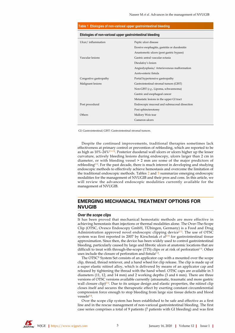
Table 1 Etiologies of non-variceal upper gastrointestinal bleeding
Etiologies of non-variceal upper gastrointestinal bleeding
Ulcer/ inflammation Peptic ulcer disease
Erosive esophagitis, gastritis or duodenitis
Anastomotic ulcers (post gastric bypass)
Vascular lesions Gastric antral vascular ectasia
Dieulafoy’s lesion
Angiodysplasia/ Arteriovenous malformation
Aorto-enteric fistula
Congestive gastropathy Portal hypertensive gastropathy
Malignant lesions Gastrointestinal stromal tumors (GIST)
Non-GIST (e.g., Lipoma, schwannoma)
Gastric and esophageal cancer
Metastatic lesions in the upper GI tract
Post procedural Endoscopic mucosal and submucosal dissection
Post sphincterotomy
Others Mallory Weis tear
Cameron ulcers
GI: Gastrointestinal; GIST: Gastrointestinal stromal tumors.
Despite the continued improvements, traditional therapies sometimes lackeffectiveness at primary control or prevention of rebleeding, which are reported to beas high as 10%-24%[23-25]. Posterior duodenal wall ulcers or ulcers higher up the lessercurvature, actively bleeding lesions during endoscopy, ulcers larger than 2 cm indiameter, or with bleeding vessel > 2 mm are some of the major predictors ofrebleeding[26]. For the past decade, there is much interest in developing and studyingendoscopic methods to effectively achieve hemostasis and overcome the limitation ofthe traditional endoscopic methods. Tables 2 and 3 summarize emerging endoscopicmodalities for the management of NVUGIB and their pros and cons. In this article, wewill review the advanced endoscopic modalities currently available for themanagement of NVUGIB.
EMERGING MECHANICAL TREATMENT OPTIONS FORNVUGIB
Over the scope clipsIt has been proved that mechanical hemostatic methods are more effective inachieving hemostasis than injections or thermal modalities alone. The Over-The-ScopeClip (OTSC, Ovesco Endoscopy GmbH, TÜbingen, Germany) is a Food and DrugAdministration approved novel endoscopic clipping device[27]. The use of OTSCsystem was first reported in 2007 by Kirschniak et al[28] for gastrointestinal tissueapproximation. Since then, the device has been widely used to control gastrointestinalbleeding, particularly caused by large and fibrotic ulcers at anatomic locations that aredifficult to treat with through-the-scope (TTS) clips or at risk of perforation[29]. Otheruses include the closure of perforation and fistula[30].
The OTSC® System Set consists of an applicator cap with a mounted over the scopeclip, thread, thread retriever, and a hand wheel for clip release. The clip is made up ofa super elastic nitinol alloy, which is delivered by means of an applicator cap andreleased by tightening the thread with the hand wheel. OTSC caps are available in 3diameters (11, 12, and 14 mm) and 2 working depths (3 and 6 mm). There are threeversions of OTSC versions available currently (atraumatic, traumatic and more gastricwall closure clip)[31]. Due to its unique design and elastic properties, the nitinol clipcloses itself and secures the therapeutic effect by exerting constant circumferentialcompression force enough to stop bleeding from large size tissue defects and bloodvessels[32].
Over the scope clip system has been established to be safe and effective as a firstline and in the rescue management of non-variceal gastrointestinal bleeding. The firstcase series comprises a total of 9 patients (7 patients with GI bleeding) and was first
WJGE https://www.wjgnet.com January 16, 2020 Volume 12 Issue 1
Naseer M et al. Advances in the management of NVUGIB
3

Figure 1
Figure 1 Initial evaluation and management of patient presented with suspected upper gastrointestinalbleeding. EGD: Esophagogastroduodenoscopy; CBC: Complete blood count; CMP: Comprehensive metabolic panel;INR: International normalized ratio; NVUGIB: Non-variceal upper gastrointestinal bleeding.
published in 2009 by Repici et al[33] from Italy. It was followed by several otherretrospective analyses of single and multi-center experience with OTSC to achievehemostasis (Table 4)[33-43]. Four retrospective studies with large sample sizes (n = 67-93)were published between 2016-2018. The primary outcomes of these studies were thetechnical success to control bleeding and rebleeding rates. Most of the studiesreported success rates between 78% to 100% with the rebleeding risk of < 1%.However, rebleeding was seen approximately 26% patients in a retrospective analysisconducted by Brandler et al[43]. In this study, the authors attributed high rebleedingrates and failure of OTSC to the history of coronary artery disease. Lamberts et al[42]
reported rebleeding rates of 26%. Their data suggest that first line endoscopictreatment of the ulcer with OTSC has higher success and low rebleeding rates ascompared to its use as second-line treatment. Also, they found OTSC as the lesspreferable treatment for diffusely bleeding polypoid lesions and vascularmalformations.
Only one prospective randomized control multicenter trial compared OTSC withstandard treatment (TTS clips or thermal therapy plus injection with dilutedadrenaline) of severe recurrent UGIB was published by Schmidt et al[44]. According tothe study, results demonstrated significant differences noted in the persistentbleeding rates between treatment (6.0%) and control group (42.4%) and rebleeding at30 d. However, the rebleeding rates at day 7 were not significantly different betweengroups. A recently published study analyzed 1517 cases treated with OTSC in 30published studies over a 9-year period. The overall success rate of OTSC to control
WJGE https://www.wjgnet.com January 16, 2020 Volume 12 Issue 1
Naseer M et al. Advances in the management of NVUGIB
4

Table 2 Summary of emerging endoscopic modalities for the management of non-variceal uppergastrointestinal bleeding
Emerging endoscopic modalities
Injection Endoscopic ultrasound guided angiotherapy
Thermal therapies Coagulation grasper, radiofrequency ablation, cryotherapy
Mechanical Over the scope clip system, endoscopic suturing, flexible linear stapler (experimental)
Topical Hemospray, endoclot, pure-Stat, ankaferd blood stopper, oxidized cellulose
hemorrhage was found to be 85% with the complication risk of about 1.7%.Procedural accidents for e.g. deviation of the over the scope clip system itself ordeviation of the clip from fibrotic tissue, intraluminal stenosis, and perforation of thethin duodenal wall with the bear claw were a few of the reported complication inthese studies[45].
One of the major advantages of the OTSC system is it’s simple to use and does notrequire special endoscopic skills to implant the clip[46]. However, it is difficult to closehard, chronic, and severely fibrotic lesions with OTSC. Another limitation is theapplication of the clip in the emergency situations because after identifying thebleeding source, the scope must be removed to mount OTSC system on the scope (justlike variceal band ligator) and reintroduced to deploy clips[47].
Endoscopic suturingAn endoscopic suturing device to perform minimally invasive endoscopicinterventions was first proposed by Kalloo et al[48] more than a decade ago. Since itsdevelopment, the endoscopic suturing device (Overstitch TM, Apollo Endosurgery,Austin, TX, United States) has continuously evolved and been established to besuccessfully used in a variety of endoscopic procedures including gastrointestinalfistula closure, perforations, leaks, endoscopic revision of gastro-jejunal bypass afterbariatric surgery, and endoscopic submucosal dissection (ESD)[49-51].
The endoscopic suturing device is introduced into the stomach through an overtube. The Overstitch system attaches proximally and distally to the double-channelendoscope, which is comprised of a cap-based suturing curved arm (to operate thetissue helix for atraumatic tissue manipulation), anchor exchange catheter (passsuture), and handle (to be mounted on the shaft of the endoscope to control suturingprocess). The suturing process begins at one of the edges of the ulcer using a curvedneedle. The curved needle then closes and grabbed by the anchor exchange anddetached from the driver. The endoscope, with Overstitch, then moves proximallytowards the other edge of the ulcer. This process repeats until the two edges of theulcer are pulled together. Once the edges of the ulcer approximate each other, the 2‘O’ polypropylene suture, placed by the cinching device, is tightened and secured[52].
Endoscopic suturing is found to be a promising modality in the management ofNVUGIB in several case reports and case series due to its excellent ability to closelarge mucosal defects after conventional methods fail to achieve hemostasis. Recently,Agarwal et al[53], published a case series of 10 patients and demonstrated theendoscopic suturing device was used successfully to control bleeding related to largerecurrent peptic ulcers. Mean suturing time was reported to be 13.4 ± 5.6 (range 3.5-20) min. No early or delayed procedural related complications were reported[53].
The endoscopic suturing device has several advantages over OTSC and hemospray(HS). Although these devices have high success rates in controlling NVUGIB, theendoscopic suturing device is technically more feasible and efficacious for larger,deep, and fibrotic ulcers. However, bleeding from small, shallow, and non-fibroticulcers can be more efficiently controlled with OTSC placement[54]. HS, on the otherhand, can be utilized as the temporizing measure to control bleeding, as publisheddata suggested that its rebleeding rate is up to 29% to 38%[55]. Limitations of theendoscopic suturing device include the need for a double channel endoscope andexpert endoscopic skills. The use of endoscopic suturing should be avoided if there isa suspicion of malignant ulcer[56].
Endoscopic band ligationEndoscopic band ligation (EBL) was initially developed for esophageal andhemorrhoidal ligation; however, it can be also used in the management of uppergastrointestinal vascular lesions, such as nodular gastric antral vascular ectasia(GAVE)[57]. Studies have demonstrated that EBL may be superior to argon plasmacoagulation and endoscopic thermal therapy regarding the reduction of treatmentsessions, control of bleeding and need for transfusion, proving to be a promising
WJGE https://www.wjgnet.com January 16, 2020 Volume 12 Issue 1
Naseer M et al. Advances in the management of NVUGIB
5

Table 3 Summary of the pros and cons of new emerging endoscopic treatment modalities for non-variceal gastrointestinal bleeding
Emerging endoscopic treatment Pros Cons
Over the scope clips 1 Simple to use 1 Difficult to close hard, chronic, and severelyfibrotic lesions with OTSC
2 Special endoscopic skills are not required toimplant the clip
2 Time consuming especially in the emergencysituations (after identifying the bleeding source,the scope must be removed to mount OTSCsystem on the scope and reintroduce to deployclips
3 Effective for the ulcers larger than 2 cm indiameter, or with bleeding vessel > 2 mm
Endoscopic suturing 1 Technically more feasible and efficacious forlarger, deep, and fibrotic ulcers
1 Double channel endoscope and expertendoscopic skills are required to operateendoscopic suturing device
Endoscopic band ligation (EVL) 1 Associated with the reduction of treatmentsessions, control of bleeding and need fortransfusion
Few cases of Hyperplastic gastric polyps
2 EVL is safe, technically straightforward, andhighly effective in this patient with completeeradication of GAVE
Coagrasper 1 One of the safest and most efficacious hemostasismodalities due to large surface area of the forcepsand anti-slip jaw design provides mechanicaltamponade effect to the surrounding tissue
1 Coagulation may be incomplete because ofelectrical leakage if the lesion submerged in wateror lesion with large tissue volume or surface area
2 The risk of perforation is extremely low becausecoagrasper works at a lower voltage as comparedto other thermal treatments coagulates tissueswithout any carbonization and does not extend todeeper tissue
2 Because the devices used for soft coagulation,including disposable hemostatic forceps, arerelatively expensive, the method may beappropriate only for centers that perform ESDfrequently
3 The forceps can be used to treat multiplebleeding sites proving to be cost-effective
3 Few cases of aspiration pneumonia reported
Radiofrequency ablation 1 Feasible and safe in ablating GAVE lesions 1 Endoscopic skills are required to perform RFA
2 Able to deliver high energy captive coagulationof superficial mucosa including blood vessels
2 Exact apposition of the gastric antral mucosawith electrode is required to allow effectivedelivery of the electric energy which means theendoscope may have to be removed, the electroderotated, and reintroduced multiple times Thenewer through-the-scope internally rotatableablating catheter may sidestep this disadvantagebut has smaller surface area
3 Wider surface area coverage of mucosa owing tothe various electrode sizes
4 Contact technique with uniform zone of energydistribution and penetration such that deeperectatic submucosal vascular channels arecoagulated
Endoscopic ultrasound guided angiotherapy 1 EUS-guided therapy of nonvariceal bleeding hasbeen shown to be feasible and safe for peptic ulcerdisease, Dieulafoy's lesions, bleeding tumors, andpseudoaneurysms due to the ability to directlyvisualize and target the bleeding vessel with aspecific therapy and subsequently confirmhemostasis with real-time Doppler ultrasound aresignificant advantages of EUS-guided therapy
1 Endoscopic skills are required to performendoscopic ultrasound
2 EUS guided angiotherapy more resourceintensive than other routine hemostasisendoscopic procedures
Topical therapies, i.e., Hemospray and Endoclot Easy to use, safe and effective Cost effective. Canbe used for malignant GI hemorrhage
1 Theoretically possible side effects of Hemosprayinclude embolization, intestinal obstruction, andallergic reaction to the powder
2 If hemostasis fails, there is the disadvantage thatthe powder attached to the mucous membranemay limit the use of other hemostatic modalities
3 Hemospray works only on active bleeding
EVL: Endoscopic band ligation; GAVE: Gastric antral vascular ectasia; EUS: Endoscopic ultrasound; GI: Gastrointestinal; RFA: Radiofrequency ablation;ESD: Endoscopic submucosal dissection; OTSC: Over-The-Scope Clip.
efficacious alternative modality[58,59]. However, further prospective studies arewarranted with larger sample sizes, longer follow-up interval, and examination ofcost-effectiveness and procedural time.
EMERGING THERMAL TREATMENT OPTIONS FOR NVUGIB
CoagrasperCoagrasper (Olympus Corp., Tokyo, Japan) or hemostatic forceps is a combination of
WJGE https://www.wjgnet.com January 16, 2020 Volume 12 Issue 1
Naseer M et al. Advances in the management of NVUGIB
6

Table 4 Efficacy and safety of over the scope clips in the management of non-variceal upper gastrointestinal bleeding (2009-2018)
Authors and yearof publication Study design Study participants Sample size Duration Outcomes of the
study Success rate
Repici et al[33], 2009 Retrospective Mean age, 70 yr,gender (M/F): 5/2
7 Unknown Success rates withthe first endoscopictherapy
Success rates withthe first endoscopictherapy
Kirschniak et al[34],2011
Retrospective Mean age, 68 yr,gender (M/F): 18/9
27 2006-2010 1 Success rates withthe first endoscopictherapy
Primary hemostasiswas achieved in allcases (100%)Rebleeding wasobserved in 2 cases
2 Rebleedingepisodes
Albert et al[35], 2011 Retrospective Mean age, 62 yr,gender (M/F): 5/2
7 Unknown 1 Success rates withthe first endoscopictherapy
Primary success ratewas observed in100%
2 Rebleedingepisodes
Skinner et al[36],2014
Retrospective Mean age, 59 yr,gender (M/F): 8/5
12 2012-2013 1 Success rates withthe first endoscopictherapy
Hemostasis wasachieved in allpatients. Rebleedingoccurred in twopatients 1 d and 7 dafter OTSCplacement
2 Rebleedingepisodes
Nishiyama et al[37],2013
Retrospective Mean age, 77 yr,gender (M/F): 5/4
9 2011-2012 Success rates withthe first endoscopictherapy
Primary success ratewas observed in77.8%
Manta et al[39], 2013 Retrospective Mean age, 64 yr,gender (M/F): 14/16
30 2011-2012 1 Success rates withthe first endoscopictherapy
Primary hemostasiswas achieved in 29of 30 cases (97%)Rebleeding wasobserved in twocases (one duodenalbulb and one gastriculcer)
2 Rebleedingepisodes
Manno et al[38], 2016 Retrospective Mean age, 69 yr,gender (M/F): 33/7
40 2013-2014 1 Success rates withthe first endoscopictherapy
Technical successand primaryhaemostasis wereachieved in allpatients (100%). Nore-bleeding need forsurgical orradiologicalembolizationtreatment or othercomplications wereobserved during thefollow-up period of30 d
2 Rebleedingepisodes
Richter-Schrag etal[40], 2016
Retrospective Mean age, 72 yr,gender (M/F): 58/35
93 2012-2016 1 Success rates withthe first endoscopictherapy
Primary hemostasisand clinical successof bleeding lesions(without rebleeding)was achieved in88/100 (88%) and78/100 (78%),respectively
2 Rebleedingepisodes
Wedi et al[41], 2016 Retrospective Mean age, 71 yr,gender (M/F): 50/34
84 2009-2012 Success rates withthe first endoscopictherapy
Success rate 35/41(85.36%)
WJGE https://www.wjgnet.com January 16, 2020 Volume 12 Issue 1
Naseer M et al. Advances in the management of NVUGIB
7
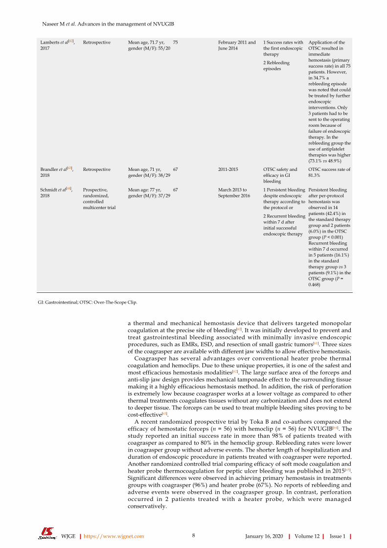
Lamberts et al[42],2017
Retrospective Mean age, 71.7 yr,gender (M/F): 55/20
75 February 2011 andJune 2014
1 Success rates withthe first endoscopictherapy
Application of theOTSC resulted inimmediatehemostasis (primarysuccess rate) in all 75patients. However,in 34.7% arebleeding episodewas noted that couldbe treated by furtherendoscopicinterventions. Only3 patients had to besent to the operatingroom because offailure of endoscopictherapy. In therebleeding group theuse of antiplatelettherapies was higher(73.1% vs 48.9%)
2 Rebleedingepisodes
Brandler et al[43],2018
Retrospective Mean age, 71 yr,gender (M/F): 38/29
67 2011-2015 OTSC safety andefficacy in GIbleeding
OTSC success rate of81.3%
Schmidt et al[44],2018
Prospective,randomized,controlledmulticenter trial
Mean age: 77 yr,gender (M/F): 37/29
67 March 2013 toSeptember 2016
1 Persistent bleedingdespite endoscopictherapy according tothe protocol or
Persistent bleedingafter per-protocolhemostasis wasobserved in 14patients (42.4%) inthe standard therapygroup and 2 patients(6.0%) in the OTSCgroup (P < 0.001)Recurrent bleedingwithin 7 d occurredin 5 patients (16.1%)in the standardtherapy group vs 3patients (9.1%) in theOTSC group (P =0.468)
2 Recurrent bleedingwithin 7 d afterinitial successfulendoscopic therapy
GI: Gastrointestinal; OTSC: Over-The-Scope Clip.
a thermal and mechanical hemostasis device that delivers targeted monopolarcoagulation at the precise site of bleeding[60]. It was initially developed to prevent andtreat gastrointestinal bleeding associated with minimally invasive endoscopicprocedures, such as EMRs, ESD, and resection of small gastric tumors[61]. Three sizesof the coagrasper are available with different jaw widths to allow effective hemostasis.
Coagrasper has several advantages over conventional heater probe thermalcoagulation and hemoclips. Due to these unique properties, it is one of the safest andmost efficacious hemostasis modalities[62]. The large surface area of the forceps andanti-slip jaw design provides mechanical tamponade effect to the surrounding tissuemaking it a highly efficacious hemostasis method. In addition, the risk of perforationis extremely low because coagrasper works at a lower voltage as compared to otherthermal treatments coagulates tissues without any carbonization and does not extendto deeper tissue. The forceps can be used to treat multiple bleeding sites proving to becost-effective[63].
A recent randomized prospective trial by Toka B and co-authors compared theefficacy of hemostatic forceps (n = 56) with hemoclip (n = 56) for NVUGIB[64]. Thestudy reported an initial success rate in more than 98% of patients treated withcoagrasper as compared to 80% in the hemoclip group. Rebleeding rates were lowerin coagrasper group without adverse events. The shorter length of hospitalization andduration of endoscopic procedure in patients treated with coagrasper were reported.Another randomized controlled trial comparing efficacy of soft mode coagulation andheater probe thermocoagulation for peptic ulcer bleeding was published in 2015[65].Significant differences were observed in achieving primary hemostasis in treatmentsgroups with coagrasper (96%) and heater probe (67%). No reports of rebleeding andadverse events were observed in the coagrasper group. In contrast, perforationoccurred in 2 patients treated with a heater probe, which were managedconservatively.
WJGE https://www.wjgnet.com January 16, 2020 Volume 12 Issue 1
Naseer M et al. Advances in the management of NVUGIB
8

Radiofrequency ablationRadiofrequency ablation (RFA) was primarily used for the treatment of Barrett’sesophagus; however, it is an emerging endoscopic treatment for GAVE[66]. RFA can beperformed by either using focal catheter (Barrx™ HALO90 and HALOULTRA) or BarrxTTS RFA catheter.
In a prospective open-label single center study, Raza and colleagues demonstrated100% technical success with the HALO system and 67% clinical success in 9 patientsafter an 11-mo follow-up interval[67]. Further studies confirmed similar results oftechnical and clinical success with improved post-procedural hemoglobin withoutmajor adverse events observed[67,68]. Despite the promising results, the studies do notpresent a randomized design and have a short follow-up interval. A multicenteropen-label retrospective case series demonstrated a significant increase in hemoglobinpost-procedural with the HALO system, as well as a reduction of blood transfusionsneeded in 24 patients[68]. There are limited studies examining the use of RFA in othergastrointestinal related bleeds.
CryotherapyCryotherapy has been proposed as a useful hemostasis modality by inducing cellnecrosis through localized freezing of the large surface area of tissue[69]. Cho andcolleagues demonstrated 50% of patients achieving complete response, while theother half achieved a partial response of GAVE related bleeding[70]. There was areduction of blood transfusions required post-procedural, and an increase inhemoglobin was observed. There were no immediate complications observed.However, this was a small single-study pilot study with a short follow-up period. Thenumber of treatment sessions and the type of cryogen need to be determined.
Endoscopic laser coagulationEndoscopic laser coagulation is another non-contact modality thermal method ofhemostasis. An Nd: YAG laser is applied through the channel of an endoscope withthe tip positioned 5 to 10 mm from the ulcer and the beam directed at the site ofbleeding. Although ND: YAG laser therapy has been shown to be effective, it is notroutinely used in the management of NVUGIB[55]. This is due to the technicalconstraints of the technique, the large size of laser delivery unit, requirement ofspecial electrical and water supplies, and least cost-effective as compared to othermodalities[71].
EMERGING TOPICAL TREATMENT OPTIONS FOR NVUGIB
HemosprayHemostatic spray (Cook Medical, Winston-Salem, NC, United States), also known asHS or TC-325, is an absorptive inorganic powder that coalesces and adheres to thebleeding site forming a mechanical barrier[72]. It is not absorbed or metabolized by thegastrointestinal tract, limiting systemic toxicity, and sloughs off once hemostasis isachieved allowing for re-application if necessary[72]. HS does not require direct contactwith the bleeding vessel and can, therefore, cover a larger surface area. In addition, itmay promote platelet aggregation, activate the clotting cascade, as well as promotetissue formation[72]. HS has been evaluated as a monotherapy modality, such as in themanagement of a bulbar ulcer related bleed, as well as with other conventionaltherapy and as a rescue therapy[73]. In addition, it has been studied in malignancyrelated bleeding and use after therapeutic endoscopic interventions (Table 5).
Several case series described the effect of HS on malignancy related bleeding. Chenet al[55] described 100% (5/5) of patients attaining immediate hemostasis with onerecurrence of bleeding in a patient with severe metastatic disease complicated bydisseminated intravascular coagulation[55,73]. As studied by Leblanc et al[72], 100% (5/5)of patients achieved immediate hemostasis (absent bleeding > 5 min after application)with one of two patients (esophageal tumor and stent placement) considered atreatment failure (not achieving immediate hemostasis or with recurrent bleedingdespite 2 separate applications)[72]. Furthermore, Arena et al[74] demonstrated 93%achieving immediate hemostasis with a rebleeding rate (drop in hemoglobin > 2g/dL) of 20%. Lastly, in a retrospective study, immediate hemostasis was achieved in97.7% patients with recurrent bleeding of 15% (classified as early, < 3 d) and 17%(classified as delayed, > 3 d)[75]. No adverse events or procedural complications wereobserved in either study. Although bleeding may recur, HS appears to be effective forNVUGIB related to malignancies. The rate of recurrent bleeding and mortality havealso been studied.
HS use in post-procedural related bleeds has also been studied. Leblanc and
WJGE https://www.wjgnet.com January 16, 2020 Volume 12 Issue 1
Naseer M et al. Advances in the management of NVUGIB
9

Table 5 Efficacy and safety of hemospray in the management of non-variceal upper gastrointestinal bleeding (2013-2018)
Study Type of study Sample size Bleeding source Modality Outcomes Results
Leblanc et al[72],2013
Case series, singlearm (July 2011-March 2012)
17 patients Procedural (12/17)and malignancyrelated bleeding(5/17)
Monotherapy orrescue therapy
Immediatehemostasis,recurrent bleedingand mortality at 7and 30 d, and relatedadverse events
Immediatehemostasis achievedin 100% patient inboth groups; 2patients withrecurrent bleedingwith 1 of 2 withtreatment failure. Noadverse events. Norelatedcomplications
Sakai et al[73], 2016 Case report 1 patient Ulcer relatedbleeding
Monotherapy Immediatehemostasis
Immediatehemostasis achieved.No recurrentbleeding. Noadverse events
Chen et al[55], 2015 Retrospectiv singlecenter study; (July2011-July 2013)
60 patients 21 for nonmalignantnonvariceal uppergastrointestinalbleeding, 19 formalignant uppergastrointestinalbleeding, 11 forlowergastrointestinalbleeding, and 16 forintra-proceduralbleeding
Monotherapy Immediatehemostasis and earlyrebleeding (≤ 72 h)
Immediatehemostasis achievedin 66 cases includingupper and lower(98.5%), with 6 cases(9.5%) of earlyrebleeding
Arena et al[74], 2017 Retrospective cohortstudy; (January2014-December2015)
A total of 15patients, 8 males,mean age 74 yr ± 7.7
Malignancy relatedbleeding
Monotherapy Immediatehemostasis, bleedingrecurrence, adverseevents, clinicaloutcome at 1 and 6mo
Immediatehemostasis achievedin 93% (14/15). 3(21%) patients withrecurrent bleeding.12/14 (80%) withgood clinicaloutcome at 30 d and50% (6/12) at 6 mo.No related adverseevents
Pittayanon et al[75],2018
Retrospective study;(2011-2016)
99 patients (70.5%were male, age 65 ±14 yr
Malignancy relatedbleeding
Monotherapy andadjuvant therapy
Immediatehemostasis, early (≤3 d) and late (> 3 d)recurrent bleeding
Immediatehemostasis was97.7%, withrecurrent bleeding in15% (early) and 17%(delayed). Six-monthsurvival was 53.4%
Baracat et al[76],2017
Case report 1 patient Post-sphincterotomybleeding
Rescue therapy Hemostasis Immediatehemostasis achieved
González et al[77],2016
Case report 1 patient Post-sclerotherapybleeding
Monotherapy Hemostasis Immediatehemostasis achieved
Sung et al[78], 2011 Prospective single-arm
20 patients (18 men,2 women; mean age60.2 yr)
Peptic ulcer bleeding(Forrest score Ia orIb)
Monotherapy Immediatehemostasis (max of 2applicationsallowed), bleedingrecurrence post-operatively, after 72h endoscopically,and after 30 d viaphone; mortality,need for surgery,and complications
Immediatehemostasis in 95%(19/20) of patients;(1/20) with apseudoaneurysmrequiring arterialembolization.Bleeding recurred in2 patients ≤ 72 h(hemoglobin drop);neither had activebleeding at the 72-hendoscopy. Nomortality, adverseevents, orprocedural-relatedcomplications at 30-d
WJGE https://www.wjgnet.com January 16, 2020 Volume 12 Issue 1
Naseer M et al. Advances in the management of NVUGIB
10

Sinha et al[79], 2016 Retrospective singlecenter
20 patients (medianage of 75 yr; 50%men)
Peptic ulcer relatedbleeding (forrest 1aand 1b)
Adjuvant therapy toadrenaline, or toadrenaline with clipsor a thermal device
Immediatehemostasis, 7 and30-d rebleeding; all-cause and GI-related30-d mortality
Initial hemostasiswas attained in 95%with an overallrebleeding rate(RBR) at 7 d of 16%.No differencebetween the 7 and30-d RBR.Hemospray +adrenaline = 100%initial hemostasisand 25% 7-d RBR.Hemospray as thirdagent = 92% initialhemostasis and 9%RBR. All-causemortality was 15%with 1 GI-relateddeath (3%)
Haddara et al[80],2016
Prospective registry;(published 2016)
202 patients Ulcer relatedbleeding in 75patients, malignancyrelated bleed in 61patients, proceduralrelated bleed in 35patients, and otherin 31 patients
Monotherapy orrescue therapy
Feasibility, efficacy,re-bleeding rate atday 8 and 30
Application ofhemospray wasfound to be veryeasy or easy in 31.7%and 55.4%,respectively.Immediatehemostasis achievedin 96.5%. Re-bleeding rate at day8 and 30 were 26.7%and 33.5%,respectively
Yau et al[81], 2014 Retrospective(February 2012-July2013)
19 patients (meanage 67.6 yr)
Peptic ulcers in 12(63.2%) patients,Dieulafoy lesions in2 (10.5%), mucosalerosion in 1 (5.3%),angiodysplasticlesion in 1 (5.3%),ampullectomy site in1 (5.3%),polypectomy site in1 (5.3%), and anunidentified lesionin 1 (5.3%)
Monotherapy,adjuvant therapy,and rescue therapy
Immediatehemostasis,recurrent bleeding at7- and 30 d,mortality at 7 and 30d (related to GIB),and adverse events(related toHemospray)
Hemostasis in 14 of15 (93.3%) patients;Rebleeding within 7d in 7/18 (38.9%)patients. Potentialadverse events in 2(10.5%) patients(visceral perforationand splenic infarct).Mortality in 5(26.3%) patients with1 withhemoperitoneum
Smith et al[82], 2014 Multicenter registry(June 2011-September 2011)
63 patients (44 men;median age 65)
30 patients withulcer relatedbleeding
Monotherapy orrescue therapy
Immediatehemostasis
47/55 (85%) patientsin monotherapygroup achievedimmediatehemostasis
Sulz et al[83], 2014 Case series;(published in 2014)
16 patients NVUGIB,unidentified
Monotherapy orrescue therapy
Immediatehemostasis
Immediatehemostasis of93.75% (15/16)
NVUGIB: Non-variceal upper gastrointestinal bleeding; GI: Gastrointestinal; GIB: Gastrointestinal bleeding; RBR: Rebleeding rate.
colleagues studied its efficacy after endoscopic intervention (5 patients afteresophageal endoscopic mucosal resection, 4 after duodenal endoscopic mucosalresection, 2 after ampullary resection, and 1 after biliary sphincterotomy)[72].Immediate hemostasis was achieved in 100% of patients whether used initially aloneor as rescue therapy (after epinephrine injection and hemostatic clip placement)[72].Further proving that HS is an appropriate and efficacious post-procedural hemostaticmodality, two case reports highlighted immediate hemostasis achieved in post-sphincterotomy and post-sclerotherapy related bleeding[76,77].
HS can be used as adjunct and rescue therapy[78-81]. Per Sinha, it was used as anadjunct therapy to adrenaline in 40% of patients. Hemostasis was achieved in 95% ofpatients with an overall rebleeding rate of 16% at 7 d suggesting it should beconsidered as an adjunct therapy. Per Yau, HS was used as rescue therapy in 84.2% ofpatients with an overall hemostasis rate of 93.3%, however with a rebleeding rate of38.9%[81]. Anticoagulant and antiplatelet use, coagulopathy, and thrombocytopenialikely contributed to the significant rebleeding rate[81].
To provide additional data on the efficacy of HS, there is a multicenter registry, bySmith and colleagues, which includes 63 patients[82]. Immediate hemostasis is defined
WJGE https://www.wjgnet.com January 16, 2020 Volume 12 Issue 1
Naseer M et al. Advances in the management of NVUGIB
11

as the absence of bleeding at the completion of the procedure, while rebleeding wasdefined as clinical manifestations of gastrointestinal bleeding and a reduction inhemoglobin by 2 g/dL. 10 of the 63 patients were treated for post-proceduralbleeding. As a monotherapy use, 85% (47/55) achieved immediate hemostasis, while100% achieved immediate hemostasis with HS used as adjunct therapy[82]. The efficacyof HS, whether as monotherapy, adjunct therapy, or rescue therapy, appearspromising in the management of NVUGIB[83]. However, further, larger prospectivestudies are warranted to confirm.
EndoclotEndoclot is an absorbable polysaccharide powder that has been proposed as a usefulhemostatic agent. It has been shown to have similar rates of immediate hemostasisachieved and rebleeding compared to standard conventional therapy[84]. Examiningendoclot as a primary monotherapy, Kim et al[85] studied its use in 12 patients withmalignancy-related bleeding. 11 of the 12 patients had advanced gastric cancer.Immediate hemostasis was achieved, regardless of the tumor location and size, orprevious use of antiplatelet medications, in all patients with a rebleeding rate in 2patients (16%) at three and five days after treatment. There were no proceduralrelated adverse events, nor all-cause mortality at 30 d after the procedure[85]. Althoughthe sample size was small and limited to forrest 1b classification of bleeding, as wellas the type of malignancy-related bleed, it appeared to be an efficacious modality.
To further evaluate its efficacy as a rescue therapy, Beg et al[86] studied the use ofendoclot in 21 patients with various gastrointestinal bleeding lesions. Immediatehemostasis was achieved in all patients. The 30-d rebleeding rate was 4.8% and themortality rate was 19.0%, however, without a statistically significant differencecompared to the dual or triple endoscopic therapy group (P = 0.51 and P = 0.31,respectively). Only one death was attributed to the UGI bleed in a patient with amalignant related bleed and significant comorbidities[86].
EMERGING INJECTION TREATMENT OPTION FOR NVUGIB
Endoscopic ultrasound guided angiotherapyEndoscopic ultrasound (EUS)–guided angiotherapy with doppler monitoring of thevascular response is a promising modality for the management of bleeding lesionsthat are inaccessible or refractory to standard endoscopic and interventionalradiologic techniques[87]. EUS can detect vascular lesions in the gastrointestinal tractthat are not visually apparent at endoscopy and target lesions for fine-needle injectionof therapeutic agents[88]. Despite most reports on EUS-guided angiotherapy pertain tovarices, the technique has also been described for the management of NVUGIBlesions. Although the feasibility and apparent safety of EUS-guided angiotherapy hasbeen demonstrated, the use of EUS as an interventional tool in the managingNVUGIH has remained limited to a few centers worldwide. This is because of the lackof endosonographer training expertise and limited availability of EUS in the acutecare setting.
CONCLUSIONIn conclusion, NVUGIB continues to be a persistent challenge despite advancementsin the both pharmacologic and endoscopic techniques. Several new modalities, as wellas, modifications to traditional therapeutic modalities have clearly shown promise inimproving outcomes whether used as monotherapy, adjuvant therapy, or rescuetherapy for the management of NVUGIB. Due to the numerous NVUGIB etiologies,the indications, efficacy, and safety of the emerging endoscopic techniques continue tobe defined. Additional studies are warranted to further define the role of thesemodalities into the treatment algorithm of NVUGIB and to determine the optimaltreatment modality for specific NVUGIB pathology.
REFERENCES1 Alzoubaidi D, Lovat LB, Haidry R. Management of non-variceal upper gastrointestinal bleeding: where
are we in 2018? Frontline Gastroenterol 2019; 10: 35-42 [PMID: 30651955 DOI:10.1136/flgastro-2017-100901]
2 Luo PJ, Lin XH, Lin CC, Luo JC, Hu HY, Ting PH, Hou MC. Risk factors for upper gastrointestinalbleeding among aspirin users: An old issue with new findings from a population-based cohort study. J
WJGE https://www.wjgnet.com January 16, 2020 Volume 12 Issue 1
Naseer M et al. Advances in the management of NVUGIB
12

Formos Med Assoc 2019; 118: 939-944 [PMID: 30366771 DOI: 10.1016/j.jfma.2018.10.007]3 Gralnek IM, Neeman Z, Strate LL. Acute Lower Gastrointestinal Bleeding. N Engl J Med 2017; 376: e50
[PMID: 28591535 DOI: 10.1056/NEJMc1705188]4 Khamaysi I, Gralnek IM. Acute upper gastrointestinal bleeding (UGIB) - initial evaluation and
management. Best Pract Res Clin Gastroenterol 2013; 27: 633-638 [PMID: 24160923 DOI:10.1016/j.bpg.2013.09.002]
5 Marmo R, Koch M, Cipolletta L, Capurso L, Pera A, Bianco MA, Rocca R, Dezi A, Fasoli R, Brunati S,Lorenzini I, Germani U, Di Matteo G, Giorgio P, Imperiali G, Minoli G, Barberani F, Boschetto S,Martorano M, Gatto G, Amuso M, Pastorelli A, Torre ES, Triossi O, Buzzi A, Cestari R, Della Casa D,Proietti M, Tanzilli A, Aragona G, Giangregorio F, Allegretta L, Tronci S, Michetti P, Romagnoli P, NucciA, Rogai F, Piubello W, Tebaldi M, Bonfante F, Casadei A, Cortini C, Chiozzini G, Girardi L, Leoci C,Bagnalasta G, Segato S, Chianese G, Salvagnini M, Rotondano G. Predictive factors of mortality fromnonvariceal upper gastrointestinal hemorrhage: a multicenter study. Am J Gastroenterol 2008; 103: 1639-47; quiz 1648 [PMID: 18564127 DOI: 10.1111/j.1572-0241.2008.01865.x]
6 van Leerdam ME. Epidemiology of acute upper gastrointestinal bleeding. Best Pract Res ClinGastroenterol 2008; 22: 209-224 [PMID: 18346679 DOI: 10.1016/j.bpg.2007.10.011]
7 Laine L, Yang H, Chang SC, Datto C. Trends for incidence of hospitalization and death due to GIcomplications in the United States from 2001 to 2009. Am J Gastroenterol 2012; 107: 1190-5; quiz 1196[PMID: 22688850 DOI: 10.1038/ajg.2012.168]
8 Wuerth BA, Rockey DC. Changing Epidemiology of Upper Gastrointestinal Hemorrhage in the LastDecade: A Nationwide Analysis. Dig Dis Sci 2018; 63: 1286-1293 [PMID: 29282637 DOI:10.1007/s10620-017-4882-6]
9 Jairath V, Martel M, Logan RF, Barkun AN. Why do mortality rates for nonvariceal uppergastrointestinal bleeding differ around the world? A systematic review of cohort studies. Can JGastroenterol 2012; 26: 537-543 [PMID: 22891179 DOI: 10.1155/2012/862905]
10 Vergara M, Bennett C, Calvet X, Gisbert JP. Epinephrine injection versus epinephrine injection and asecond endoscopic method in high-risk bleeding ulcers. Cochrane Database Syst Rev 2014; CD005584[PMID: 25308912 DOI: 10.1002/14651858.CD005584.pub3]
11 Fujishiro M, Iguchi M, Kakushima N, Kato M, Sakata Y, Hoteya S, Kataoka M, Shimaoka S, Yahagi N,Fujimoto K. Guidelines for endoscopic management of non-variceal upper gastrointestinal bleeding. DigEndosc 2016; 28: 363-378 [PMID: 26900095 DOI: 10.1111/den.12639]
12 Samuel R, Bilal M, Tayyem O, Guturu P. Evaluation and management of Non-variceal uppergastrointestinal bleeding. Dis Mon 2018; 64: 333-343 [PMID: 29525375 DOI:10.1016/j.disamonth.2018.02.003]
13 Troland D, Stanley A. Endotherapy of Peptic Ulcer Bleeding. Gastrointest Endosc Clin N Am 2018; 28:277-289 [PMID: 29933775 DOI: 10.1016/j.giec.2018.02.002]
14 Laine L, Jensen DM. Management of patients with ulcer bleeding. Am J Gastroenterol 2012; 107: 345-60;quiz 361 [PMID: 22310222 DOI: 10.1038/ajg.2011.480]
15 Tsoi KK, Chiu PW, Chan FK, Ching JY, Lau JY, Sung JJ. The risk of peptic ulcer bleeding mortality inrelation to hospital admission on holidays: a cohort study on 8,222 cases of peptic ulcer bleeding. Am JGastroenterol 2012; 107: 405-410 [PMID: 22108453 DOI: 10.1038/ajg.2011.409]
16 Camus M, Jensen DM, Kovacs TO, Jensen ME, Markovic D, Gornbein J. Independent risk factors of 30-day outcomes in 1264 patients with peptic ulcer bleeding in the USA: large ulcers do worse. AlimentPharmacol Ther 2016; 43: 1080-1089 [PMID: 27000531 DOI: 10.1111/apt.13591]
17 Gralnek IM, Dumonceau JM, Kuipers EJ, Lanas A, Sanders DS, Kurien M, Rotondano G, Hucl T, Dinis-Ribeiro M, Marmo R, Racz I, Arezzo A, Hoffmann RT, Lesur G, de Franchis R, Aabakken L, Veitch A,Radaelli F, Salgueiro P, Cardoso R, Maia L, Zullo A, Cipolletta L, Hassan C. Diagnosis and managementof nonvariceal upper gastrointestinal hemorrhage: European Society of Gastrointestinal Endoscopy(ESGE) Guideline. Endoscopy 2015; 47: a1-46 [PMID: 26417980 DOI: 10.1055/s-0034-1393172]
18 Barkun AN, Bardou M, Kuipers EJ, Sung J, Hunt RH, Martel M, Sinclair P; International ConsensusUpper Gastrointestinal Bleeding Conference Group. International consensus recommendations on themanagement of patients with nonvariceal upper gastrointestinal bleeding. Ann Intern Med 2010; 152: 101-113 [PMID: 20083829 DOI: 10.7326/0003-4819-152-2-201001190-00009]
19 Lai YC, Yang SS, Wu CH, Chen TK. Endoscopic hemoclip treatment for bleeding peptic ulcer. World JGastroenterol 2000; 6: 53-56 [PMID: 11819522 DOI: 10.3748/wjg.v6.i1.53]
20 Kovacs TO, Jensen DM. Endoscopic therapy for severe ulcer bleeding. Gastrointest Endosc Clin N Am2011; 21: 681-696 [PMID: 21944418 DOI: 10.1016/j.giec.2011.07.012]
21 Gevers AM, De Goede E, Simoens M, Hiele M, Rutgeerts P. A randomized trial comparing injectiontherapy with hemoclip and with injection combined with hemoclip for bleeding ulcers. GastrointestEndosc 2002; 55: 466-469 [PMID: 11923755 DOI: 10.1067/mge.2002.112613]
22 Wang TJ, Aihara H, Thompson AC, Schulman AR, Thompson CC, Ryou M. Choosing the right through-the-scope clip: a rigorous comparison of rotatability, whip, open/close precision, and closure strength (withvideos). Gastrointest Endosc 2019; 89: 77-86.e1 [PMID: 30056253 DOI: 10.1016/j.gie.2018.07.025]
23 Sarin N, Monga N, Adams PC. Time to endoscopy and outcomes in upper gastrointestinal bleeding. Can JGastroenterol 2009; 23: 489-493 [PMID: 19623332 DOI: 10.1155/2009/604639]
24 Laursen SB. Treatment and prognosis in peptic ulcer bleeding. Dan Med J 2014; 61: B4797 [PMID:24547604]
25 Maggio D, Barkun AN, Martel M, Elouali S, Gralnek IM; Reason Investigators. Predictors of earlyrebleeding after endoscopic therapy in patients with nonvariceal upper gastrointestinal bleeding secondaryto high-risk lesions. Can J Gastroenterol 2013; 27: 454-458 [PMID: 23936874 DOI:10.1155/2013/128760]
26 García-Iglesias P, Villoria A, Suarez D, Brullet E, Gallach M, Feu F, Gisbert JP, Barkun A, Calvet X.Meta-analysis: predictors of rebleeding after endoscopic treatment for bleeding peptic ulcer. AlimentPharmacol Ther 2011; 34: 888-900 [PMID: 21899582 DOI: 10.1111/j.1365-2036.2011.04830.x]
27 Angsuwatcharakon P, Prueksapanich P, Kongkam P, Rattanachu-Ek T, Sottisuporn J, Rerknimitr R.Efficacy of the Ovesco Clip for Closure of Endoscope Related Perforations. Diagn Ther Endosc 2016;2016: 9371878 [PMID: 27293368 DOI: 10.1155/2016/9371878]
28 Kirschniak A, Traub F, Kueper MA, Stüker D, Königsrainer A, Kratt T. Endoscopic treatment of gastricperforation caused by acute necrotizing pancreatitis using over-the-scope clips: a case report. Endoscopy2007; 39: 1100-1102 [PMID: 18072063 DOI: 10.1055/s-2007-966848]
29 Kirschniak A, Kratt T, Stüker D, Braun A, Schurr MO, Königsrainer A. A new endoscopic over-the-
WJGE https://www.wjgnet.com January 16, 2020 Volume 12 Issue 1
Naseer M et al. Advances in the management of NVUGIB
13

scope clip system for treatment of lesions and bleeding in the GI tract: first clinical experiences.Gastrointest Endosc 2007; 66: 162-167 [PMID: 17591492 DOI: 10.1016/j.gie.2007.01.034]
30 Cahyadi O, Caca K, Schmidt A. Over-the-scope clip is an effective therapy for postbanding ulcer bleedingafter initially successful transjugular intrahepatic portosystemic shunt therapy. Endoscopy 2017; 49: E258-E259 [PMID: 28759921 DOI: 10.1055/s-0043-115890]
31 Samarasena J, Chen CL, Chin M, Chang K, Lee J. Successful closure of a cryotherapy-induced bleedingjejunal perforation with the over-the-scope clip system. Gastrointest Endosc 2017; 85: 451 [PMID:27866906 DOI: 10.1016/j.gie.2016.10.038]
32 Haito-Chavez Y, Law JK, Kratt T, Arezzo A, Verra M, Morino M, Sharaiha RZ, Poley JW, Kahaleh M,Thompson CC, Ryan MB, Choksi N, Elmunzer BJ, Gosain S, Goldberg EM, Modayil RJ, StavropoulosSN, Schembre DB, DiMaio CJ, Chandrasekhara V, Hasan MK, Varadarajulu S, Hawes R, Gomez V,Woodward TA, Rubel-Cohen S, Fluxa F, Vleggaar FP, Akshintala VS, Raju GS, Khashab MA.International multicenter experience with an over-the-scope clipping device for endoscopic management ofGI defects (with video). Gastrointest Endosc 2014; 80: 610-622 [PMID: 24908191 DOI:10.1016/j.gie.2014.03.049]
33 Repici A, Arezzo A, De Caro G, Morino M, Pagano N, Rando G, Romeo F, Del Conte G, Danese S,Malesci A. Clinical experience with a new endoscopic over-the-scope clip system for use in the GI tract.Dig Liver Dis 2009; 41: 406-410 [PMID: 18930700 DOI: 10.1016/j.dld.2008.09.002]
34 Kirschniak A, Subotova N, Zieker D, Königsrainer A, Kratt T. The Over-The-Scope Clip (OTSC) for thetreatment of gastrointestinal bleeding, perforations, and fistulas. Surg Endosc 2011; 25: 2901-2905 [PMID:21424197 DOI: 10.1007/s00464-011-1640-2]
35 Albert JG, Friedrich-Rust M, Woeste G, Strey C, Bechstein WO, Zeuzem S, Sarrazin C. Benefit of aclipping device in use in intestinal bleeding and intestinal leakage. Gastrointest Endosc 2011; 74: 389-397[PMID: 21612776 DOI: 10.1016/j.gie.2011.03.1128]
36 Skinner M, Gutierrez JP, Neumann H, Wilcox CM, Burski C, Mönkemüller K. Over-the-scope clipplacement is effective rescue therapy for severe acute upper gastrointestinal bleeding. Endosc Int Open2014; 2: E37-E40 [PMID: 26134611 DOI: 10.1055/s-0034-1365282]
37 Nishiyama N, Mori H, Kobara H, Rafiq K, Fujihara S, Kobayashi M, Oryu M, Masaki T. Efficacy andsafety of over-the-scope clip: including complications after endoscopic submucosal dissection. World JGastroenterol 2013; 19: 2752-2760 [PMID: 23687412 DOI: 10.3748/wjg.v19.i18.2752]
38 Manno M, Mangiafico S, Caruso A, Barbera C, Bertani H, Mirante VG, Pigò F, Amardeep K, ConigliaroR. First-line endoscopic treatment with OTSC in patients with high-risk non-variceal upper gastrointestinalbleeding: preliminary experience in 40 cases. Surg Endosc 2016; 30: 2026-2029 [PMID: 26201415 DOI:10.1007/s00464-015-4436-y]
39 Manta R, Galloro G, Mangiavillano B, Conigliaro R, Pasquale L, Arezzo A, Masci E, Bassotti G,Frazzoni M. Over-the-scope clip (OTSC) represents an effective endoscopic treatment for acute GIbleeding after failure of conventional techniques. Surg Endosc 2013; 27: 3162-3164 [PMID: 23436101DOI: 10.1007/s00464-013-2871-1]
40 Richter-Schrag HJ, Glatz T, Walker C, Fischer A, Thimme R. First-line endoscopic treatment with over-the-scope clips significantly improves the primary failure and rebleeding rates in high-risk gastrointestinalbleeding: A single-center experience with 100 cases. World J Gastroenterol 2016; 22: 9162-9171 [PMID:27895403 DOI: 10.3748/wjg.v22.i41.9162]
41 Wedi E, Gonzalez S, Menke D, Kruse E, Matthes K, Hochberger J. One hundred and one over-the-scope-clip applications for severe gastrointestinal bleeding, leaks and fistulas. World J Gastroenterol 2016; 22:1844-1853 [PMID: 26855543 DOI: 10.3748/wjg.v22.i5.1844]
42 Lamberts R, Koch A, Binner C, Zachäus M, Knigge I, Bernhardt M, Halm U. Use of over-the-scope clips(OTSC) for hemostasis in gastrointestinal bleeding in patients under antithrombotic therapy. Endosc IntOpen 2017; 5: E324-E330 [PMID: 28484732 DOI: 10.1055/s-0043-104860]
43 Brandler J, Baruah A, Zeb M, Mehfooz A, Pophali P, Wong Kee Song L, AbuDayyeh B, Gostout C,Mara K, Dierkhising R, Buttar N. Efficacy of Over-the-Scope Clips in Management of High-RiskGastrointestinal Bleeding. Clin Gastroenterol Hepatol 2018; 16: 690-696.e1 [PMID: 28756055 DOI:10.1016/j.cgh.2017.07.020]
44 Schmidt A, Gölder S, Goetz M, Meining A, Lau J, von Delius S, Escher M, Hoffmann A, Wiest R,Messmann H, Kratt T, Walter B, Bettinger D, Caca K. Over-the-Scope Clips Are More Effective ThanStandard Endoscopic Therapy for Patients With Recurrent Bleeding of Peptic Ulcers. Gastroenterology2018; 155: 674-686.e6 [PMID: 29803838 DOI: 10.1053/j.gastro.2018.05.037]
45 Mercky P, Gonzalez JM, Aimore Bonin E, Emungania O, Brunet J, Grimaud JC, Barthet M. Usefulness ofover-the-scope clipping system for closing digestive fistulas. Dig Endosc 2015; 27: 18-24 [PMID:24720574 DOI: 10.1111/den.12295]
46 Kobara H, Mori H, Nishiyama N, Fujihara S, Okano K, Suzuki Y, Masaki T. Over-the-scope clip system:A review of 1517 cases over 9 years. J Gastroenterol Hepatol 2019; 34: 22-30 [PMID: 30069935 DOI:10.1111/jgh.14402]
47 Baron TH, Song LM, Ross A, Tokar JL, Irani S, Kozarek RA. Use of an over-the-scope clipping device:multicenter retrospective results of the first U.S. experience (with videos). Gastrointest Endosc 2012; 76:202-208 [PMID: 22726484 DOI: 10.1016/j.gie.2012.03.250]
48 Kalloo AN, Singh VK, Jagannath SB, Niiyama H, Hill SL, Vaughn CA, Magee CA, Kantsevoy SV.Flexible transgastric peritoneoscopy: a novel approach to diagnostic and therapeutic interventions in theperitoneal cavity. Gastrointest Endosc 2004; 60: 114-117 [PMID: 15229442 DOI:10.1016/S0016-5107(04)01309-4]
49 Mori H, Rahman A, Kobara H, Morishita A, Masaki T. The Development of Endoscopic SuturingDevices: Challenges in the Treatment of Iatrogenic Perforation and Bleeding. Intern Med 2016; 55: 3075-3076 [PMID: 27803396 DOI: 10.2169/internalmedicine.55.7508]
50 Barola S, Magnuson T, Schweitzer M, Chen YI, Ngamruengphong S, Khashab MA, Kumbhari V.Endoscopic Suturing for Massively Bleeding Marginal Ulcer 10 days Post Roux-en-Y Gastric Bypass.Obes Surg 2017; 27: 1394-1396 [PMID: 28247322 DOI: 10.1007/s11695-017-2621-x]
51 Fujihara S, Mori H, Kobara H, Nishiyama N, Kobayashi M, Rafiq K, Masaki T. The efficacy and safetyof prophylactic closure for a large mucosal defect after colorectal endoscopic submucosal dissection.Oncol Rep 2013; 30: 85-90 [PMID: 23674165 DOI: 10.3892/or.2013.2466]
52 Chiu PW, Chan FK, Lau JY. Endoscopic Suturing for Ulcer Exclusion in Patients With MassivelyBleeding Large Gastric Ulcer. Gastroenterology 2015; 149: 29-30 [PMID: 25962937 DOI:10.1053/j.gastro.2015.04.054]
WJGE https://www.wjgnet.com January 16, 2020 Volume 12 Issue 1
Naseer M et al. Advances in the management of NVUGIB
14
53 Agarwal A, Benias P, Brewer Gutierrez OI, Wong V, Hanada Y, Yang J, Villgran V, Kumbhari V, KallooA, Khashab MA, Chiu P, Ngamruengphong S. Endoscopic suturing for management of peptic ulcer-relatedupper gastrointestinal bleeding: a preliminary experience. Endosc Int Open 2018; 6: E1439-E1444 [PMID:30539067 DOI: 10.1055/a-0749-0011]
54 Barola S, Fayad L, Hill C, Magnuson T, Schweitzer M, Singh V, Chen YI, Ngamruengphong S, KhashabMA, Kalloo AN, Kumbhari V. Endoscopic Management of Recalcitrant Marginal Ulcers by Covering theUlcer Bed. Obes Surg 2018; 28: 2252-2260 [PMID: 29556889 DOI: 10.1007/s11695-018-3162-7]
55 Chen YI, Barkun A, Nolan S. Hemostatic powder TC-325 in the management of upper and lowergastrointestinal bleeding: a two-year experience at a single institution. Endoscopy 2015; 47: 167-171[PMID: 25264762 DOI: 10.1055/s-0034-1378098]
56 Mori H, Kobara H, Kazi R, Fujihara S, Nishiyama N, Masaki T. Balloon-armed mechanical countertraction and double-armed bar suturing systems for pure endoscopic full-thickness resection.Gastroenterology 2014; 147: 278-80.e1 [PMID: 24973723 DOI: 10.1053/j.gastro.2014.06.030]
57 Wells CD, Harrison ME, Gurudu SR, Crowell MD, Byrne TJ, Depetris G, Sharma VK. Treatment ofgastric antral vascular ectasia (watermelon stomach) with endoscopic band ligation. Gastrointest Endosc2008; 68: 231-236 [PMID: 18533150 DOI: 10.1016/j.gie.2008.02.021]
58 Elhendawy M, Mosaad S, Alkhalawany W, Abo-Ali L, Enaba M, Elsaka A, Elfert AA. Randomizedcontrolled study of endoscopic band ligation and argon plasma coagulation in the treatment of gastricantral and fundal vascular ectasia. United European Gastroenterol J 2016; 4: 423-428 [PMID: 27403309DOI: 10.1177/2050640615619837]
59 Takizawa K, Oda I, Gotoda T, Yokoi C, Matsuda T, Saito Y, Saito D, Ono H. Routine coagulation ofvisible vessels may prevent delayed bleeding after endoscopic submucosal dissection--an analysis of riskfactors. Endoscopy 2008; 40: 179-183 [PMID: 18322872 DOI: 10.1055/s-2007-995530]
60 Arima S, Sakata Y, Ogata S, Tominaga N, Tsuruoka N, Mannen K, Shiraishi R, Shimoda R, Tsunada S,Sakata H, Iwakiri R, Fujimoto K. Evaluation of hemostasis with soft coagulation using endoscopichemostatic forceps in comparison with metallic hemoclips for bleeding gastric ulcers: a prospective,randomized trial. J Gastroenterol 2010; 45: 501-505 [PMID: 20033825 DOI: 10.1007/s00535-009-0186-8]
61 Fujishiro M, Abe N, Endo M, Kawahara Y, Shimoda R, Nagata S, Homma K, Morita Y, Uedo N.Retrospective multicenter study concerning electrocautery forceps with soft coagulation for nonmalignantgastroduodenal ulcer bleeding in Japan. Dig Endosc 2010; 22 Suppl 1: S15-S18 [PMID: 20590763 DOI:10.1111/j.1443-1661.2010.00962.x]
62 Nagata S, Kimura S, Ogoshi H, Hidaka T. Endoscopic hemostasis of gastric ulcer bleeding by hemostaticforceps coagulation. Dig Endosc 2010; 22 Suppl 1: S22-S25 [PMID: 20590766 DOI:10.1111/j.1443-1661.2010.00973.x]
63 Tanaka S, Toyonaga T, Morita Y, Ishida T, Hoshi N, Grimes KL, Ohara Y, Yoshizaki T, Kawara F,Umegaki E, Azuma T. Efficacy of a new hemostatic forceps during gastric endoscopic submucosaldissection: A prospective randomized controlled trial. J Gastroenterol Hepatol 2017; 32: 846-851 [PMID:27648821 DOI: 10.1111/jgh.13599]
64 Toka B, Eminler AT, Karacaer C, Uslan MI, Koksal AS, Parlak E. Comparison of monopolar hemostaticforceps with soft coagulation versus hemoclip for peptic ulcer bleeding: a randomized trial (with video).Gastrointest Endosc 2019; 89: 792-802 [PMID: 30342026 DOI: 10.1016/j.gie.2018.10.011]
65 Nunoue T, Takenaka R, Hori K, Okazaki N, Hamada K, Baba Y, Yamasaki Y, Kono Y, Seki H, InokuchiT, Takemoto K, Taira A, Tsugeno H, Fujiki S, Kawahara Y, Okada H. A Randomized Trial of MonopolarSoft-mode Coagulation Versus Heater Probe Thermocoagulation for Peptic Ulcer Bleeding. J ClinGastroenterol 2015; 49: 472-476 [PMID: 25083773 DOI: 10.1097/MCG.0000000000000190]
66 Maida M, Camilleri S, Manganaro M, Garufi S, Scarpulla G. Radiofrequency Ablation for Treatment ofRefractory Gastric Antral Vascular Ectasia: A Systematic Review of the Literature. Gastroenterol ResPract 2017; 2017: 5609647 [PMID: 28835751 DOI: 10.1155/2017/5609647]
67 Raza N, Diehl DL. Radiofrequency ablation of treatment-refractory gastric antral vascular ectasia(GAVE). Surg Laparosc Endosc Percutan Tech 2015; 25: 79-82 [PMID: 24743681 DOI:10.1097/SLE.0000000000000033]
68 Dray X, Repici A, Gonzalez P, Fristrup C, Lecleire S, Kantsevoy S, Wengrower D, Elbe P, Camus M,Carlino A, Pérez-Roldán F, Adar T, Marteau P. Radiofrequency ablation for the treatment of gastric antralvascular ectasia. Endoscopy 2014; 46: 963-969 [PMID: 25111135 DOI: 10.1055/s-0034-1377695]
69 Patel AA, Trindade AJ, Diehl DL, Khara HS, Lee TP, Lee C, Sethi A. Nitrous oxide cryotherapy ablationfor refractory gastric antral vascular ectasia. United European Gastroenterol J 2018; 6: 1155-1160 [PMID:30288277 DOI: 10.1177/2050640618783537]
70 Cho S, Zanati S, Yong E, Cirocco M, Kandel G, Kortan P, May G, Marcon N. Endoscopic cryotherapy forthe management of gastric antral vascular ectasia. Gastrointest Endosc 2008; 68: 895-902 [PMID:18640673 DOI: 10.1016/j.gie.2008.03.1109]
71 Macrì A, Saladino E, Versaci A, Basile A, Lamberto S, De Francesco F, Familiari L, Famulari C. Massivebleeding from a Dieulafoy's lesion of the duodenum successfully treated with "adjuvant" transarterialembolization and endoscopic laser coagulation. Acta Chir Belg 2010; 110: 208-209 [PMID: 20514835DOI: 10.1080/00015458.2010.11680599]
72 Leblanc S, Vienne A, Dhooge M, Coriat R, Chaussade S, Prat F. Early experience with a novel hemostaticpowder used to treat upper GI bleeding related to malignancies or after therapeutic interventions (withvideos). Gastrointest Endosc 2013; 78: 169-175 [PMID: 23622976 DOI: 10.1016/j.gie.2013.03.006]
73 Sakai CM, Duarte RB, Baracat FI, Baracat R, de Moura EGH. Endoscopic treatment of upper-GI ulcerbleeding with hemostatic powder spray. VideoGIE 2016; 2: 12-13 [PMID: 29905242 DOI:10.1016/j.vgie.2016.11.005]
74 Arena M, Masci E, Eusebi LH, Iabichino G, Mangiavillano B, Viaggi P, Morandi E, Fanti L, Granata A,Traina M, Testoni PA, Opocher E, Luigiano C. Hemospray for treatment of acute bleeding due to uppergastrointestinal tumours. Dig Liver Dis 2017; 49: 514-517 [PMID: 28065526 DOI:10.1016/j.dld.2016.12.012]
75 Pittayanon R, Rerknimitr R, Barkun A. Prognostic factors affecting outcomes in patients with malignantGI bleeding treated with a novel endoscopically delivered hemostatic powder. Gastrointest Endosc 2018;87: 994-1002 [PMID: 29158179 DOI: 10.1016/j.gie.2017.11.013]
76 Baracat FI, Tranquillini CV, Brunaldi VO, Baracat R, de Moura EGH. Hemostatic powder: a new ally inthe management of postsphincterotomy bleeding. VideoGIE 2017; 2: 303-304 [PMID: 30027130 DOI:10.1016/j.vgie.2017.07.002]
77 González Ortiz B, Tapia Monge DM, Reyes Cerecedo A, Hernández Mondragón O. [Use of Hemospray®
WJGE https://www.wjgnet.com January 16, 2020 Volume 12 Issue 1
Naseer M et al. Advances in the management of NVUGIB
15

in post-sclerotherapy bleeding]. Bol Med Hosp Infant Mex 2016; 73: 335-337 [PMID: 29384127 DOI:10.1016/j.bmhimx.2016.06.005]
78 Sung JJ, Luo D, Wu JC, Ching JY, Chan FK, Lau JY, Mack S, Ducharme R, Okolo P, Canto M, KallooA, Giday SA. Early clinical experience of the safety and effectiveness of Hemospray in achievinghemostasis in patients with acute peptic ulcer bleeding. Endoscopy 2011; 43: 291-295 [PMID: 21455870DOI: 10.1055/s-0030-1256311]
79 Sinha R, Lockman KA, Church NI, Plevris JN, Hayes PC. The use of hemostatic spray as an adjunct toconventional hemostatic measures in high-risk nonvariceal upper GI bleeding (with video). GastrointestEndosc 2016; 84: 900-906.e3 [PMID: 27108061 DOI: 10.1016/j.gie.2016.04.016]
80 Haddara S, Jacques J, Lecleire S, Branche J, Leblanc S, Le Baleur Y, Privat J, Heyries L, Bichard P,Granval P, Chaput U, Koch S, Levy J, Godart B, Charachon A, Bourgaux JF, Metivier-Cesbron E,Chabrun E, Quentin V, Perrot B, Vanbiervliet G, Coron E. A novel hemostatic powder for uppergastrointestinal bleeding: a multicenter study (the "GRAPHE" registry). Endoscopy 2016; 48: 1084-1095[PMID: 27760437 DOI: 10.1055/s-0042-116148]
81 Yau AH, Ou G, Galorport C, Amar J, Bressler B, Donnellan F, Ko HH, Lam E, Enns RA. Safety andefficacy of Hemospray® in upper gastrointestinal bleeding. Can J Gastroenterol Hepatol 2014; 28: 72-76[PMID: 24501723 DOI: 10.1155/2014/759436]
82 Smith LA, Stanley AJ, Bergman JJ, Kiesslich R, Hoffman A, Tjwa ET, Kuipers EJ, von Holstein CS,Oberg S, Brullet E, Schmidt PN, Iqbal T, Mangiavillano B, Masci E, Prat F, Morris AJ. Hemosprayapplication in nonvariceal upper gastrointestinal bleeding: results of the Survey to Evaluate the Applicationof Hemospray in the Luminal Tract. J Clin Gastroenterol 2014; 48: e89-e92 [PMID: 24326829 DOI:10.1097/MCG.0000000000000054]
83 Sulz MC, Frei R, Meyenberger C, Bauerfeind P, Semadeni GM, Gubler C. Routine use of Hemospray forgastrointestinal bleeding: prospective two-center experience in Switzerland. Endoscopy 2014; 46: 619-624[PMID: 24770964 DOI: 10.1055/s-0034-1365505]
84 Park JC, Kim YJ, Kim EH, Lee J, Yang HS, Kim EH, Hahn KY, Shin SK, Lee SK, Lee YC.Effectiveness of the polysaccharide hemostatic powder in non-variceal upper gastrointestinal bleeding:Using propensity score matching. J Gastroenterol Hepatol 2018; 33: 1500-1506 [PMID: 29415371 DOI:10.1111/jgh.14118]
85 Kim YJ, Park JC, Kim EH, Shin SK, Lee SK, Lee YC. Hemostatic powder application for control of acuteupper gastrointestinal bleeding in patients with gastric malignancy. Endosc Int Open 2018; 6: E700-E705[PMID: 29868635 DOI: 10.1055/a-0593-5884]
86 Beg S, Al-Bakir I, Bhuva M, Patel J, Fullard M, Leahy A. Early clinical experience of the safety andefficacy of EndoClot in the management of non-variceal upper gastrointestinal bleeding. Endosc Int Open2015; 3: E605-E609 [PMID: 26716120 DOI: 10.1055/s-0034-1393087]
87 Anastasiou J, Berzin TM. Endoscopic Ultrasound-Guided Vascular Interventions: From Diagnosis toTreatment. Saudi J Med Med Sci 2018; 6: 61-67 [PMID: 30787823 DOI: 10.4103/sjmms.sjmms_131_17]
88 Satyavada S, Davitkov P, Akbar Ali M, Cooper G, Wong RCK, Chak A. Endoscopic Doppler Probe inthe Diagnosis and Management of Upper Gastrointestinal Hemorrhage. ACG Case Rep J 2018; 5: e68[PMID: 30280108 DOI: 10.14309/crj.2018.68]
WJGE https://www.wjgnet.com January 16, 2020 Volume 12 Issue 1
Naseer M et al. Advances in the management of NVUGIB
16

Published By Baishideng Publishing Group Inc
7041 Koll Center Parkway, Suite 160, Pleasanton, CA 94566, USA
Telephone: +1-925-2238242
E-mail: [email protected]
Help Desk: https://www.f6publishing.com/helpdesk
https://www.wjgnet.com
© 2020 Baishideng Publishing Group Inc. All rights reserved.