Flexible Endoscopy Workshop: Advanced Course Marti Felder ...
69
Flexible Endoscopy Workshop: Advanced Course Marti Felder, PA‐C Updated 2/13/2017 April 21, 2017 Chicago
-
Upload
khangminh22 -
Category
Documents
-
view
0 -
download
0
Transcript of Flexible Endoscopy Workshop: Advanced Course Marti Felder ...

Flexible Endoscopy Workshop: Advanced CourseMarti Felder, PA‐C
Updated 2/13/2017
April 21, 2017Chicago

Flexible Fiberoptic Workshop: Advanced Course
Basic Instruction Demonstration Hands‐On Practice
Learn by doing
Identify abnormal pathology Perform flexible endoscopy adult
Perform flexible endoscopy child/infant
Perform rigid endoscopy

Introduction
There are multiple methods and techniques available to successfully complete all the topics presented in this workshop. Some are based on
patient request, available equipment or supervising physician’s preference.
The goal of this workshop is to correctly demonstrate the most common methods and give participants time for hands‐on training.

ObjectivesLearning Objectives• Identify normal anatomy, normal variants and abnormal findings visible via flexible nasopharyngoscopy.
• Understand indications and perform flexible and rigid scope examination ‐ adult.
• Understand indications and perform flexible scope examination ‐ child/infant.
• Perform exam, intranasal culture and sinus debridement using rigid scope adult.

Indications for Flexible Endoscopy• Strong gag reflex*• Failed mirror exam*• Nasal obstruction
– Foreign Body– Septal deviation– Adenoid hypertrophy– Nasal mass– Unilateral otitis media– Polyps
• Sinusitis• Chronic throat pain• Chronic cough
• Dysphonia– Presbylarynx– VC paralysis– VC Nodules– LPR– Neoplasms
• Dysphagia– Candidiasis
• Odynophagia• Symptoms of aspiration
– Laryngomalacia– Angioedema
*Documentation of a strong gag reflex and failed mirror exam should be included in note to justify procedure for billing purposes.

Contraindications
• Epiglottitis (by inexperienced)• Relative:
– Coagulopathy– Craniofacial trauma
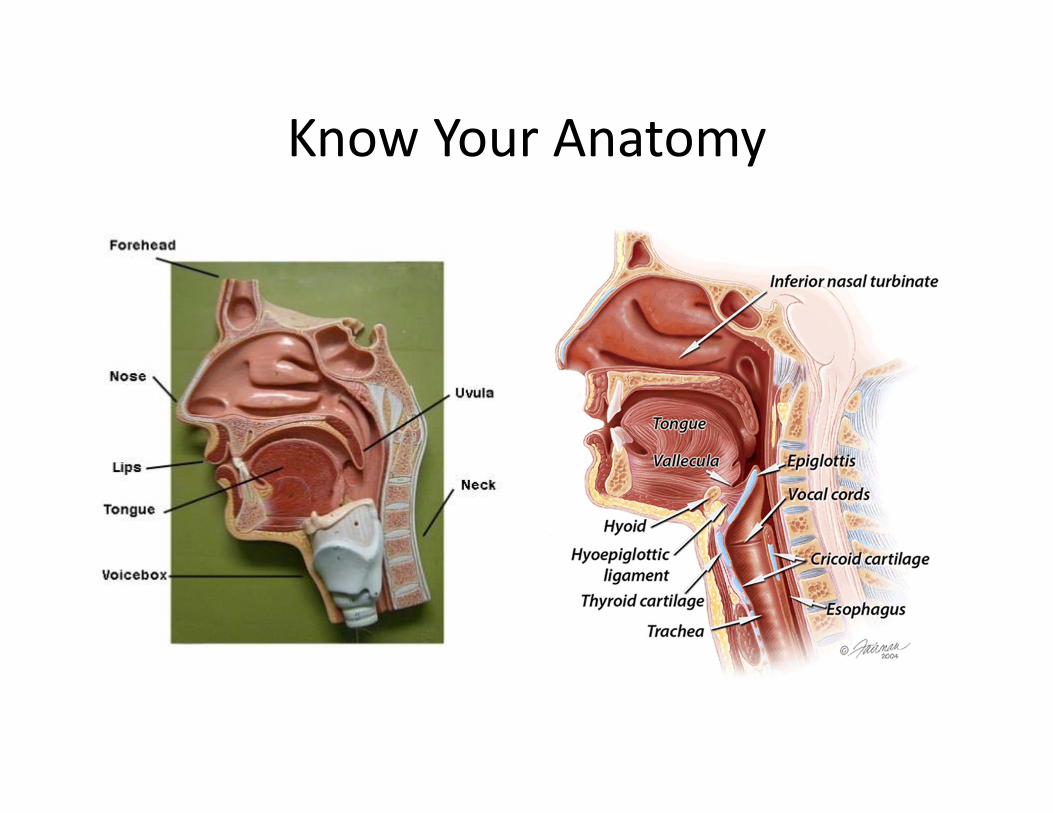
Know Your Anatomy

Laryngeal Anatomy (Mirror*)
1. True vocal cords2. False cords3. Epiglottis
4. Aryepiglottic folds5. Arytenoids6. Pyriform sinuses7. Base of Tongue
*With Mirror Laryngoscopy, image is inverted.

Laryngeal Anatomy*
True Vocal Cords abducted True Vocal Cords adducted
*With flexible endoscopy, images are true.

Review Tips For Starting the Exam• Patient informed of the procedure (obtain consent)• Proper positioning
– Sniffing, head supported, use non‐dominant hand to steady the patient’s head
– Choose the more patent of the nares• Appropriate equipment
– Adult vs. Pedi– Decongestant/anesthetic– Gloves– Chair– Photographic/video accessories– Biopsy materials if needed– Lubricant +/‐

Preparation for Flexible Endoscopy• May want patient to blow nose.
• Assess which is more patent of the nares.
• Antifogging solution.• Apply topical decongestant
– 0.05% Oxymetazoline– 0.25% ‐2 % Phenylephrine
• Apply topical anesthetic– 4% Lidocaine– Pontocaine
Photo Courtesy Bernadine Sonnier 2011

Normal Flexible Endoscopic Exam
Video Courtesy J. MercadoVideo

Causes of Nasal Obstruction
Nasal Foreign BodySeptal deviation
SynechiaeTurbinate hypertrophy
SinusitisAdenoid hypertrophy
Nasopharyngeal massesNasal polyps

Septal Deviation/Turbinate Hypertrophy

Synechiae• Fused intranasal tissues
are called adhesions or synechiae.
• Adhesions are a common, usually minor complication of nasal or sinus surgery and nasal packing.
• They also may develop because of trauma (nose‐picking or cocaine use) and in such conditions as syphilis, tuberculosis, lupus, or sarcoidosis.

Nasal Polyps
Video Courtesy J. MercadoVideo
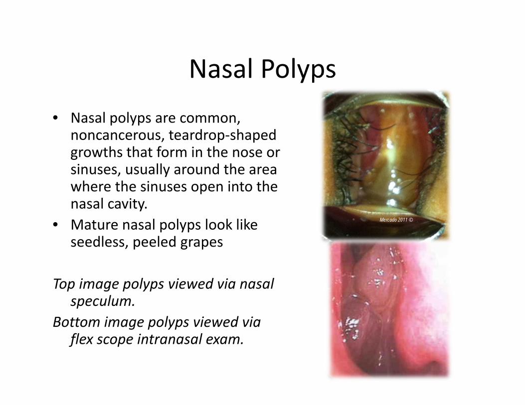
Nasal Polyps
Mercado 2011 ©
• Nasal polyps are common, noncancerous, teardrop‐shaped growths that form in the nose or sinuses, usually around the area where the sinuses open into the nasal cavity.
• Mature nasal polyps look like seedless, peeled grapes
Top image polyps viewed via nasal speculum.
Bottom image polyps viewed via flex scope intranasal exam.

Sinusitis
Video Courtesy J.MercadoVideo

Sinusitis
Mercado 2013 ©Mercado 2013 ©

Sinusitis
• Sinusitis is diagnoses by history and physical examination.
• Fiberoptic endoscopy (flexible or rigid) is ideal for evaluating the osteomeatal complex
Image of fungal sinusitis. Note thick debris vs purulent secretions in previous images.

Adenoid Hypertrophy
Video Courtesy J. MercadoVideo

Adenoid HypertrophyAdenoids are described based on % of obstruction– No obstruction– Partial (percentage) obstruction
– Complete obstruction• Endoscopic view of the nasopharynx showing adenoidal obstruction of the choana.
http://icarus.med.utoronto.ca/carr/atlas/atlasoutline.htm
Mercado 2011 ©
Mercado 2013 ©

Angiofibroma
Video Courtesy J. MercadoVideo
Juvenile Angiofibroma
Juvenile angiofibroma – benign, highly vascular invasive mass that occurs in the posterior nasal cavity in less than 0.5% of head and neck tumors.
Almost always found in adolescent boys.Presents with epistaxis and nasal congestion or both.
CT Scan w/contrast confirmed nasopharyngeal mass measuring 4.0 cm x 3.8 cm consistent with a juvenile angiofibroma.

Tornwaldt Cyst• A Tornwaldt cyst is a midline pouch within
the posterior roof of the nasopharnx which is caused by an adherence of the notochord to the pharyngeal ectoderm during development, resulting in the outpouchingof ectoderm into the pharyngobasilar fascia.
• They are typically asymptomatic but may present with middle ear symptoms (eustachian tube obstruction), halitosis (when associated with a leaking sinus tract), foul taste in mouth, and, rarely, occipital headaches.
• These lesions will demonstrate low to increased signal on T1‐weighted images depending on the protein content of the mucus. They will be high signal on T2‐weighted images and will not enhance.

Unilateral Otitis MediaUnilateral Otitis Media in adults
should raise suspicion of Nasopharyngeal mass blocking Eustachian Tube orifice which can cause unilateral middle ear effusion.
• 1.5:100,000 patients, more common in Asian and Alaskan people 20:100,000
• Top image normal Torus Tubarius• Bottom image opening covered
with mucosal tissue (squamous cell carcinoma).
Mercado 2011 ©
Mercado 2011 ©

Voice Change
Voice overuse/misuseVocal cord nodules/polypsLaryngopharyngeal Reflux
Vocal Cord ParalysisPresbylarynx
Laryngeal neoplasms

Vocal Cord Paralysis
Video Courtesy M. MitraniVideo

Vocal Cord Nodules and LPR
Video Courtesy J. MercadoVideo

Laryngeal Cancer
Video Courtesy J. MercadoVideo

Cervical Osteophyte
Video Courtesy J. MercadoVideo

•Cervical osteophytes are bone spurs that grow on any of the seven vertebrae in the cervical spine (C1 ‐ C7 vertebrae).
•More than half of people over the age of 60 have osteophytes somewhere in their bodies. Osteophytes in the spine are a normal sign of aging and usually do not cause symptoms. However, neurological symptoms or pain may occur if the osteophytes encroach upon the individual spinal nerves, the spinal cord itself, the vertebral discs, or the blood vessels in the region of the cervical vertebral column.
•The inflamed or damaged tissue that stimulates cervical osteophyte growth is often caused by cervical osteoarthritis, a degradation in the neck joints that occurs in many older people. These joints include the disc spaces themselves (a modified joint) and the facet joints, and this condition of cervical osteophyte formation is referred to as cervical spondylosis.
Mercado 2014 © Mercado 2014 ©

Adult Mannequin
Advanced airway and custom mannequins available to practice flexible endoscopy technique.
Mercado 2013 ©Mercado 2013 ©


Respiratory Complications
Children• Must rule out foreign body aspiration
• Choanal Atresia• Laryngomalacia
• Subglottic stenosis
Adults• Angioedema

Flexible scope exam on children.
Mercado 2013 ©Mercado 2013 ©
• Generally well tolerated by children.
• Explain procedure in detail.
• Secure patient (papoose vs. cradle).
• Anesthesia vs. decongestant?
• Give adequate time for anesthesia/decongestant.
• Provides better visualization

Flexible scope exam on infants.
Mercado 2013 ©Mercado 2013 ©Mercado 2013 ©
• Generally well tolerated by infants.
• Explain procedure in detail.
• Secure patient (papoose vs. cradle)
• Anesthesia vs. decongestant?
• Give adequate time for anesthesia/decongestant.
• Provides better visualization
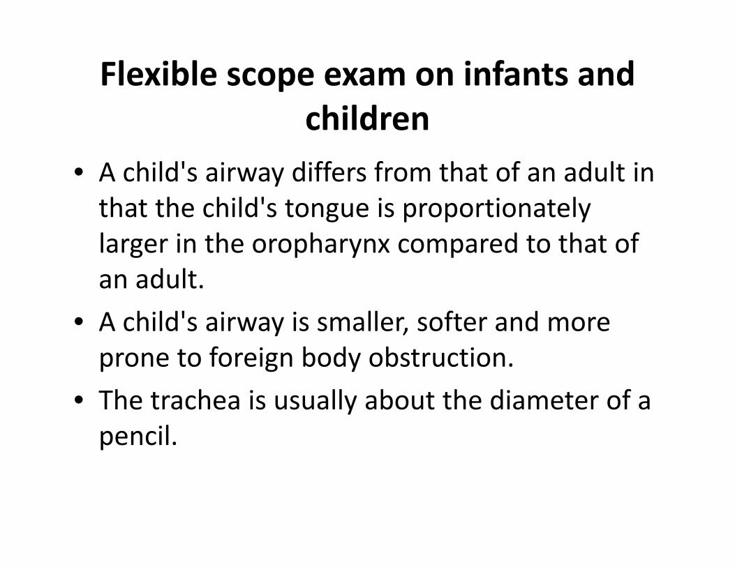
Flexible scope exam on infants and children
Evaluating pediatric airway via flexible scope• Assess nares/choanae (choanal atresia)• Assess adenoid and lingual tonsil (hypertrophy)
• Assess Airway – Epiglottis (laryngomalacia)– TVC mobility (paralyzed vocal cords)– Assess laryngeal structures (stenosis)– Foreign bodies
Flexible scope exam on infants and children
• A child's airway differs from that of an adult in that the child's tongue is proportionately larger in the oropharynx compared to that of an adult.
• A child's airway is smaller, softer and more prone to foreign body obstruction.
• The trachea is usually about the diameter of a pencil.

Difference Pediatric vs Adult AirwayRelatively larger tongue
Obstructs airwayObligate nasal breathersDifficult to visualize larynx
Angled vocal cordsInfant’s vocal cords have more angled
attachment to trachea, whereas adult vocal cords are more perpendicular
Image from: http://www.utmb.edu/otoref/Grnds/Pedi‐airway‐2001‐01/Pedi‐airway‐2001‐01‐slides.pdf
Flexible fiberoptic exam on infants and children

Laryngomalacia
Video Courtesy J. MercadoVideo

Laryngomalacia
• Most common congenital abnormality of the larynx.
• Most prominent symptom: inspiratory stridor.
• Immature development results in soft laryngeal walls that close airway.
• Child outgrows. Usually, no treatment necessary.
• Omega‐Shaped epiglottisMercado 2011 ©

Subglottic StenosisNarrowing of subglottis can be congenital or acquired in
etiology. Its nature can be membranous, cartilaginous, or mixed, with or without combination of glottic or upper tracheal stenosis.
The lower limit of normal subglottis dimension in full term infant is 4.0 mm and in premature infant 3.5 mm. Circumferential edema of 1mm reduces its cross‐sectional area by 60%.
Myer‐Cotton grading system is a useful classification for mature circumferential subglottic stenosis. It is divided into 4 grade as below:: Grade I ‐ Obstruction of 0‐50% of the lumen obstruction Grade II ‐ Obstruction of 51‐70% of the lumen Grade III ‐ Obstruction of 71‐99% of the lumen Grade IV ‐ Obstruction of 100% of the lumen (ie, no detectable lumen)
Class I
http://www.drrahmatorlummc.com/congenitaldevelopmental.htm
Class II
Class III

Infant/Child Mannequin
Practice mannequins available to practice flexible fiberoptic endoscopy technique.
Mercado 2013 ©Mercado 2013 ©
Mercado 2013 ©


AngioedemaVascular leakage beneath the dermis and subcutanis. Response is mediated by vasoactive mediators, i.e., histamine, serotonin, and kinins (eg, bradykinins), which cause the arterioles to dilate while inducing a brief episode of vascular leakage in the venules, where the junction between the endothelial cells appears looser than in the capillaries and arterioles.
Angioedema with or without urticaria, is classified as allergic, hereditary, or idiopathic. Complications range from dysphonia or dysphagia to respiratory distress, complete airway obstruction, and death.Symptoms ‐ Severe facial/oral edema, Urticaria –(hives) food allergy, erythema.
Most common cause ‐ ACE Inhibitor sensitivity, Food allergies such as fresh berries, shellfish, fish, nuts, tomatoes, eggs, milk, chocolate, food additives, and preservatives
Treatment ‐ H 1 (antihistamine), H 2 (antacid), Steroids. Fresh Frozen Plasma, Protect airway.
Mercado 2011 ©
Mercado 2011 ©

Angioedema
Edema floor of mouth Edema uvula
Mercado 2011 ©
Edema epiglottis Edema arytenoid
Rigid Scope

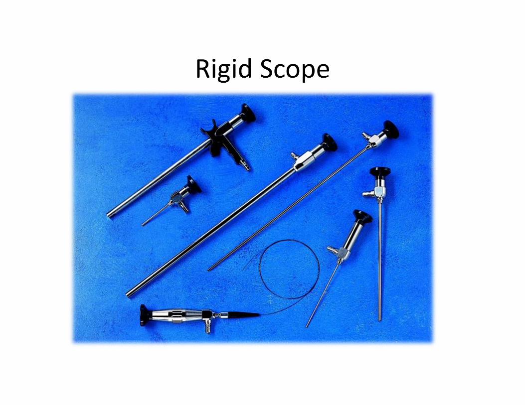
Rigid Scope
The rigid endoscope provides superior image clarity, facilitates culture and tissue sampling, controls epistaxis, and affords the endoscopist the ability to perform surgery.Rigid endoscopes for the nose come in diameters of 2.7‐4 mm and have tips of different angles (generally 0‐70º), allowing the clinician to visualize various sinuses and areas within the nasal cavity. This facilitates culture of the sinuses and debridement postoperatively.

Complications
• Tearing• Epistaxis• Coughing• Laryngospasm – rare• Bleeding
Advise patient not to eat or drink anything 1 hour after procedure.

Rigid Scope
Inferior Turbinate
Nasal Septum
Nasal endoscopic examination and culture for definitive diagnosis best accomplished with rigid scope.
Small culturette used to obtain mucous & pus sample from hiatus semilunaris. Be sure not to touch other tissue as this may contaminate specimen.
Mercado 2011 ©

Nasal Cultures• Nasal cultures are not routinely indicated in first‐line management of Acute Rhinosinusitis
• Endoscopically guided microswab or suction aspiration culture of a draining sinus ostium are a strong consideration in Chronic Rhinosinusitis, especially when poorly responsive to prior antibiotics.
Middle Turbinate
Microswab

Rigid Endoscopy After FESS
Mercado 2011 © Dr. Kevin Kavanaugh © www.entusa.com
Post operative debridement following FESS is best accomplished with rigid scope.
Rigid scope allows clinician to visualize area and suction or use forceps.

Rigid Endoscopy
Interactive, live demonstration of rigid endoscopy
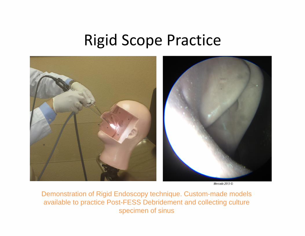
Rigid Scope Practice
Demonstration of Rigid Endoscopy technique. Custom-made models available to practice Post-FESS Debridement and collecting culture
specimen of sinus
Mercado 2013 ©Mercado 2013 ©

Rigid Scope EXAM: Practice on PhaconSinus model
Post‐FESS Polyp *PLEASE DO NOT REMOVE!


Care of Scope

EndosheathsLoosen endosheath Pull endosheath from distal end
Mercado 2011 © Mercado 2011 ©

Cold Sterilization• Flexible scopes are non‐
autoclavable.• Rigid scopes can be autoclaved.• Clean length of scope with an
enzymatic detergent solution like ENZOL® to remove debris and reduce bacterial burden before instruments are disinfected or sterilized. DO NOT ALLOW debris to dry.
• Soak scope in a glutaraldehydesolution like Cidex ® which provides quick high‐level disinfection.
• Noncorrosive solution reduces instrument damage and associated repair costs.
• Soaking times vary by product. Mercado 2011 ©

Leak Testing1. Routine leak testing in accordance with specific manufacture depending
on volume of use.2. Introduce air pressure via attached bulb (DO NOT overinflate) and
submerge looking for leaks. 3. Leaks can slowly damage fiberoptics and internal parts causing
expensive yet preventable damage.
Mercado 2011 © Mercado 2011 ©Mercado 2011 ©

Review of Scope Care
• Avoid bending scope in tight angles.• Clean lens with lens cleaner/paper.• Pre‐clean with enzymatic cleaner.• Soak only for required period depending on brand and manufacture.
• Store in dry safe place.• Perform regular leak testing to avoid damage.

Documentation• Effective January 1, 2014, any payer requesting documentation for a
scope procedure could either deny the service or reduce the payment if documentation doesn’t show that all areas were examined during the endoscopy and the findings noted.
• According to Medicare, documentation should identify specific anatomical landmarks. “form over substance” .
• CPT® guidelines state, “For endoscopic procedures, report appropriate endoscopy of each anatomic site examined.” – 31575 Flexible Fiberoptic Laryngoscopy includes examination of the
tongue base, larynx, and hypopharynx. – 31231 Nasal Endoscopy (diagnostic), and
31237 Debridement Endoscopy, Nasal should include mention of the superior turbinate, superior meatus and sphenoethmoid recess
• Clinicians need to clearly document each area’s examination and whatever findings were observed.
Otolaryngology Coding Alert, The Coding Institute, February 2015, Vol. 17, No. 2 (Pages 9‐16)Coding Corner, AAO‐HNSF http://www.entnet.org/content/coding‐corner

Sample Templates
Fiberoptic Nasal Endoscopy Findings Septum Right Side
• Inferior Turbinates ‐WNL• Middle Turbinates ‐WNL• Middle Meatus ‐WNL• Mucosa ‐WNL• Mucous ‐WNL• Polyps ‐ None
Left Side• Inferior Turbinates ‐WNL• Middle Turbinates ‐WNL• Middle Meatus ‐WNL• Mucosa ‐WNL• Mucous ‐WNL• Polyps ‐ None
Nasopharynx ‐patent Sphenoid and ethmoid cavities‐WNL
Fiberoptic Laryngoscopy Findings; Nasopharnyx ‐WNL Oropharynx ‐WNL Base of Tongue ‐WNL Vallecula –WNL* Lateral Pharyngeal Wall ‐WNL Posterior Pharyngeal Wall ‐WNL Epiglottis ‐WNL Aryepiglottic Folds –WNL* Pyriform Sinuses ‐WNL Interarytenoid ‐WNL Tracheal Rings ‐WNL Vocal Cords –WNL* False Cords –WNL* Ventricle –WNL*
* Denotes laterally i.e. Left side, Right side
Template should also include description of positioning of patient, application of topical anesthetic and any decongestants. If consent was obtained, verbal vs. written. Document indication for procedure. At completion of procedure note if patient tolerated procedure and if there were any complications.

Resources On‐LineNew England Journal of Medicine Video
http://www.youtube.com/watch?v=3tbuF7Qwmps
Excellent pictures and videos by Dr. Kevin Kavenaugh
http://www.entusa.com/larynx_photo.htm
Dr Rahmat Omarhttp://www.drrahmatorlummc.com/
Direct Laryngoscopy videohttp://www.airwaycam.com/video‐library.html
Functional Voice DisordersNeil N Chheda, MD
http://emedicine.medscape.com/article/865191‐overview

Recommend ReadingExamination of the Larynx and
Pharynx n engl j med 358;3
www.nejm.org january 17, 2008
Laryngeal Evaluation by Kendall & Leonard
Publication Date: August 2010324 pp, 309 illustrations
hardcover & videoISBN (Americas): 9781604062724ISBN (EUR, Asia, Africa, AUS):
9781604062724
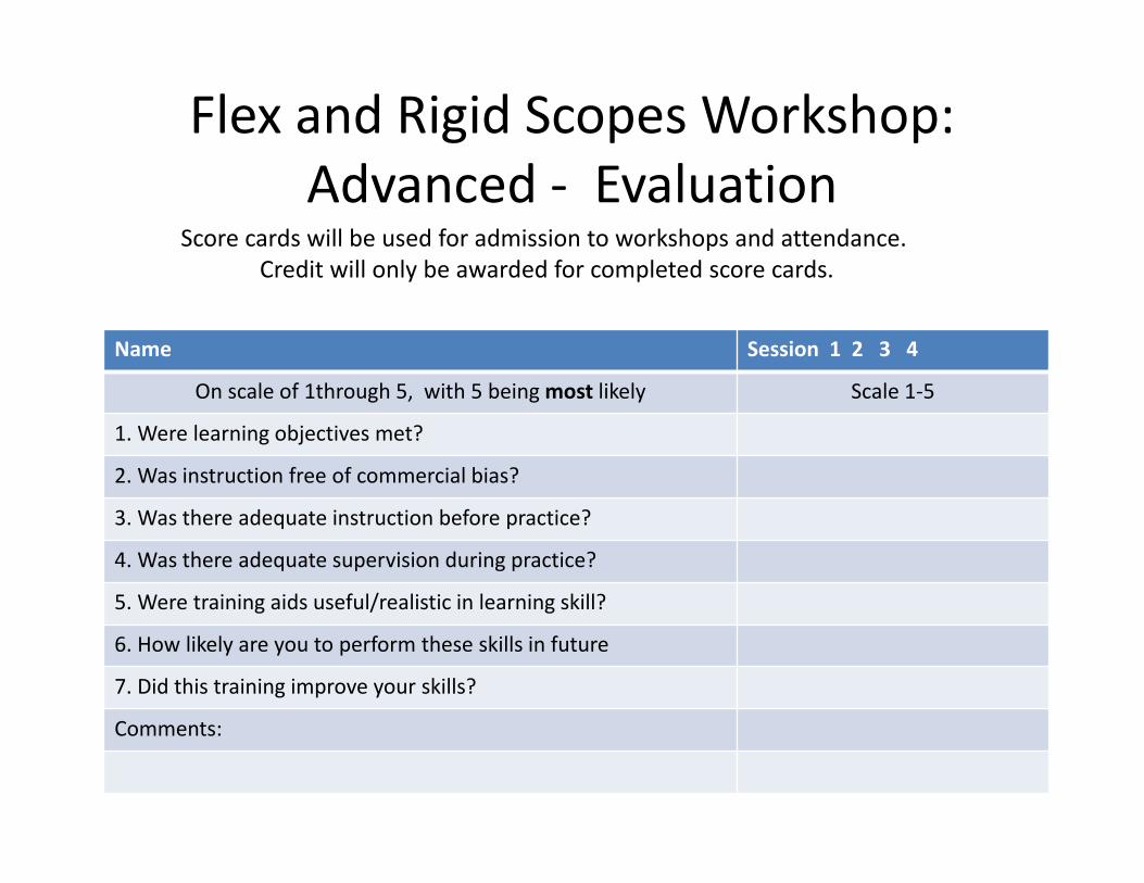
Flex and Rigid Scopes Workshop: Advanced ‐ Evaluation
Name Session 1 2 3 4
On scale of 1through 5, with 5 beingmost likely Scale 1‐5
1. Were learning objectives met?
2. Was instruction free of commercial bias?
3. Was there adequate instruction before practice?
4. Was there adequate supervision during practice?
5. Were training aids useful/realistic in learning skill?
6. How likely are you to perform these skills in future
7. Did this training improve your skills?
Comments:
Score cards will be used for admission to workshops and attendance. Credit will only be awarded for completed score cards.

Flex & Rigid Scopes Workshop: Advanced ‐ Score Card
Task Go No Go
Perform flexible endoscopy child mannequin.
Perform flexible endoscopy infant mannequin.
Perform flexible endoscopy on adult mannequin.
Perform flexible endoscopy on simulated patient.
Perform rigid endoscopy (culture collection)
Perform rigid endoscopy debridement.
Comments
Proctor Name and Signature:
Rotate and complete each station.“Go/No Go” for internal use only.
Completion of workshop is NOT contingent on pass/fail.

Station 5Video Tower
Adult examination
Station 1Pediatric
Screen
ProjectorSpeaker
Station 4Video Tower
Rigid
Station 3Video Tower
Rigid
Proctors
Station 2Adult
examination