Hemosuccus Pancreaticus in the Era of Capsule Endoscopy and Double Balloon Enteroscopy Complicated...
10
Case Rep Gastroenterol 2007;1:38–47 DOI: 10.1159/000104977 Published online: August 7, 2007 © 2007 S. Karger AG, Basel ISSN 1662–0631 www.karger.com/crg Shabana F. Pasha Division of Gastroenterology & Hepatology Department of Internal Medicine, Mayo Clinic Scottsdale Scottsdale, Ariz. 85259 (USA) Tel. +1 480 301 6990, Fax +1 480 301 8673, E-Mail [email protected] 38 Hemosuccus Pancreaticus in the Era of Capsule Endoscopy and Double Balloon Enteroscopy Complicated by Multifocal Mycobacterium chelonae/abscessus Infection Shabana F. Pasha a Janis E. Blair b Patrick B. Garvey c Richard J. Gray c David C. Mulligan d Joseph M. Collins e Russell I. Heigh a Divisions of a Gastroenterology & Hepatology and b Infectious Diseases, Department of Internal Medicine, c Department of General Surgery, d Department of Hepatology & Transplant Surgery, e Department of Radiology, Mayo Clinic Scottsdale, Scottsdale, Ariz., USA Key Words Hemosuccus pancreaticus · Splenic artery pseudoaneurysm · Obscure gastrointestinal bleeding · Mycobacterium chelonae/abscessus Abstract Hemosuccus pancreaticus is a rare etiology of obscure gastrointestinal bleeding characterized by bleeding into the pancreatic duct. The diagnosis may be delayed for months to years, due to the episodic nature of bleeding and failure to consider the diagnosis. Patients often undergo multiple endoscopies and radiologic evaluations prior to diagnosis. Incidental gastrointestinal findings may lead to unnecessary endoscopic and surgical interventions. This report describes a patient with hemosuccus pancreaticus diagnosed in the era of video capsule endoscopy and double balloon enteroscopy, whose management was complicated by multifocal Mycobacteria chelonae/abscessus infection. Case Report In 1999, a 58-year-old Caucasian male began experiencing recurrent gastrointestinal bleeding. He presented on multiple occasions with hematemesis, melena and/or hematochezia, associated with
-
Upload
independent -
Category
Documents
-
view
1 -
download
0
Transcript of Hemosuccus Pancreaticus in the Era of Capsule Endoscopy and Double Balloon Enteroscopy Complicated...

Case R ep Gast roent ero l 2007;1:38–47 DOI: 10.1159/000104977
Published online: August 7, 2007 © 2007 S. Karger AG, Basel ISSN 1662–0631 www.karger.com/crg
Shabana F. Pasha Division of Gastroenterology & Hepatology Department of Internal Medicine, Mayo Clinic Scottsdale Scottsdale, Ariz. 85259 (USA) Tel. +1 480 301 6990, Fax +1 480 301 8673, E-Mail [email protected]
38
Hemosuccus Pancreaticus in the Era of Capsule Endoscopy and Double Balloon Enteroscopy Complicated by Multifocal Mycobacterium chelonae/abscessus Infection
Shabana F. Pashaa Janis E. Blairb Patrick B. Garveyc
Richard J. Grayc David C. Mulligand Joseph M. Collinse
Russell I. Heigha
Divisions of aGastroenterology & Hepatology and bInfectious Diseases,
Department of Internal Medicine, cDepartment of General Surgery, dDepartment
of Hepatology & Transplant Surgery, eDepartment of Radiology, Mayo Clinic
Scottsdale, Scottsdale, Ariz., USA
Key Words Hemosuccus pancreaticus · Splenic artery pseudoaneurysm · Obscure gastrointestinal
bleeding · Mycobacterium chelonae/abscessus
Abstract Hemosuccus pancreaticus is a rare etiology of obscure gastrointestinal bleeding
characterized by bleeding into the pancreatic duct. The diagnosis may be delayed for
months to years, due to the episodic nature of bleeding and failure to consider the
diagnosis. Patients often undergo multiple endoscopies and radiologic evaluations prior
to diagnosis. Incidental gastrointestinal findings may lead to unnecessary endoscopic
and surgical interventions. This report describes a patient with hemosuccus pancreaticus
diagnosed in the era of video capsule endoscopy and double balloon enteroscopy,
whose management was complicated by multifocal Mycobacteria chelonae/abscessus
infection.
Case Report
In 1999, a 58-year-old Caucasian male began experiencing recurrent gastrointestinal bleeding. He presented on multiple occasions with hematemesis, melena and/or hematochezia, associated with

Case R ep Gast roent ero l 2007;1:38–47 DOI: 10.1159/000104977
Published online: August 7, 2007 © 2007 S. Karger AG, Basel ISSN 1662–0631 www.karger.com/crg
39
hemodynamic instability, and required a cumulative transfusion of more than 100 units packed red blood cells over a period of 8 years. The bleeding was typically preceded by cramping abdominal pain localized in the left upper quadrant. Multiple gastrointestinal evaluations were performed, and were unrevealing for a source of the intermittent bleeding.
Prior to his evaluation at our institution, the patient had undergone 2 esophagoduodenoscopies, a colonoscopy, a tagged nuclear red blood cell (RBC) scan and an angiogram, which were within normal limits. An intra-operative enteroscopy was performed in 2000, which was significant for blood in the 3rd and 4th portions of the duodenum, multiple arteriovenous malformations (AVMs) and a single jejunal diverticulum. The area of concern was oversewn. A pancreatic biopsy was obtained intra-operatively and revealed normal pancreatic tissue.
The patient presented to our institution in 2001 for further evaluation. Over time, repeat endoscopic and radiologic evaluations were performed including 3 upper endoscopies, a colonoscopy and 2 tagged nuclear RBC scans, which were within normal limits. Catheter angiograms were performed on three different occasions, including a provocative visceral angiogram with infusion of tissue plasminogen activator, but failed to identify the site of hemorrhage.
Wedged hepatic vein pressure (WHVP) was measured by the transjugular approach, and revealed an elevated portosystemic gradient of 18 mm Hg, suggestive of portal hypertension. A liver biopsy was within normal limits. The WHVP was repeated and found to be within normal limits. A CT angiogram was performed that showed evidence of a tortuous and prominent splenic artery, otherwise within normal limits. Endoscopic retrograde cholangiopancreatography (ERCP) showed mild dilation of the pancreatic duct, but no other significant findings were appreciated.
An intra-operative enteroscopy was then performed, which revealed blood in the distal duodenum, AVMs and a single diverticulum in the proximal jejunum. This led to a segmental resection of 100 cm of proximal jejunum. Despite the surgery, the patient continued to have recurrent hemorrhage.
The patient underwent 3 video capsule endoscopies between 2003 to 2005. Angioectasias were noted in the proximal small bowel on initial capsule study. However, follow-up push enteroscopy to the mid-jejunum did not reveal angioectasia. Nonspecific red spots in the proximal small bowel were seen on two additional video capsule endoscopies, but no definite source for bleeding was identified. Argon plasma coagulation of 2–3 non-bleeding AVMs in the gastric cardia was performed. Double balloon enteroscopy (DBE) was performed in 2005, with normal appearing 205 cm of the distal small bowel and 230 cm of proximal small bowel visualized on retrograde and antegrade DBE, respectively.
In July 2005, the patient developed fever, and multiple blood cultures grew Mycobacterium chelonae/abscessus group. Antibiotic treatment was initiated with amikacin and clarithromycin, and the porta-cath was explanted. His fever resolved with this intervention. The catheter tip grew ≥15 colony forming units Mycobacterium chelonae/abscessus. Liver function tests were elevated (AST 58 U/l, ALT 50 U/l and alkaline phosphatase 229 U/l), and splenomegaly (22.3 superior-inferior length) was found on adominal ultrasound. A CT scan of the abdomen revealed multiple large splenic lesions, consistent with subacute or chronic infarcts, a hypodensity at the tail of the pancreas, and inflammatory changes near the splenic hilum at the tail of the pancreas, suggestive of resolving pancreatitis.
An endoscopic ultrasound was performed in August 2005, to evaluate the CT findings, and revealed a 10 mm ill-defined hyperechoic lesion at the tail of the pancreas, suggestive of a benign pancreatic neoplasm. There were no findings of chronic pancreatitis or portal hypertension.
In September 2005, the patient presented with recurrent hematochezia, prompting a repeat provocative visceral angiogram with heparin. The angiogram revealed normal appearing splanchnic vasculature, and no evidence of bleeding. His central venous access was reimplanted.
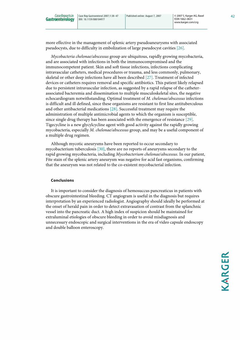
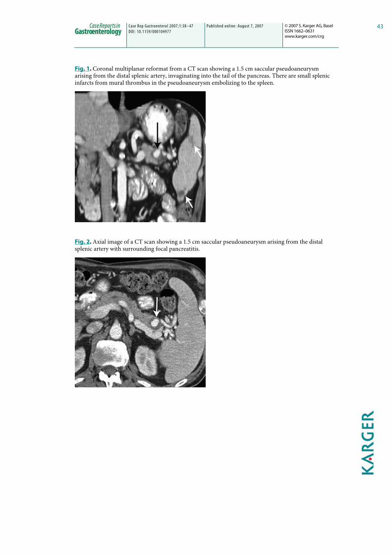
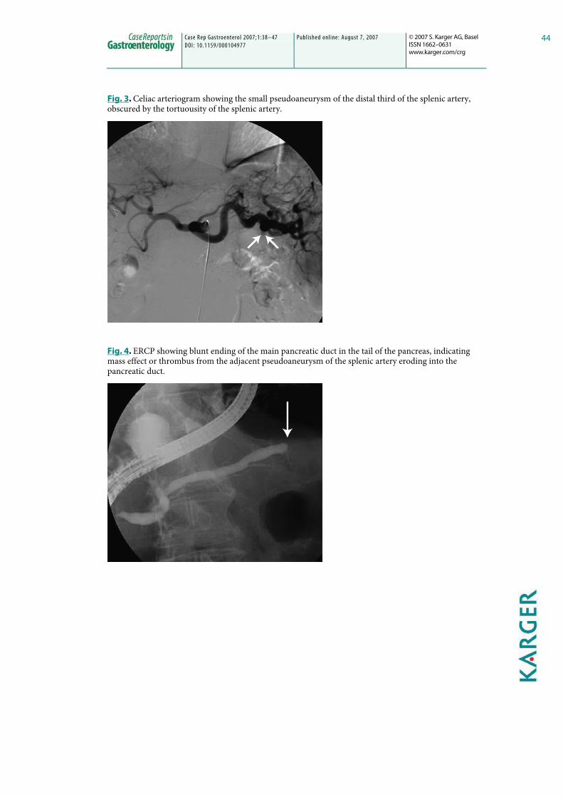
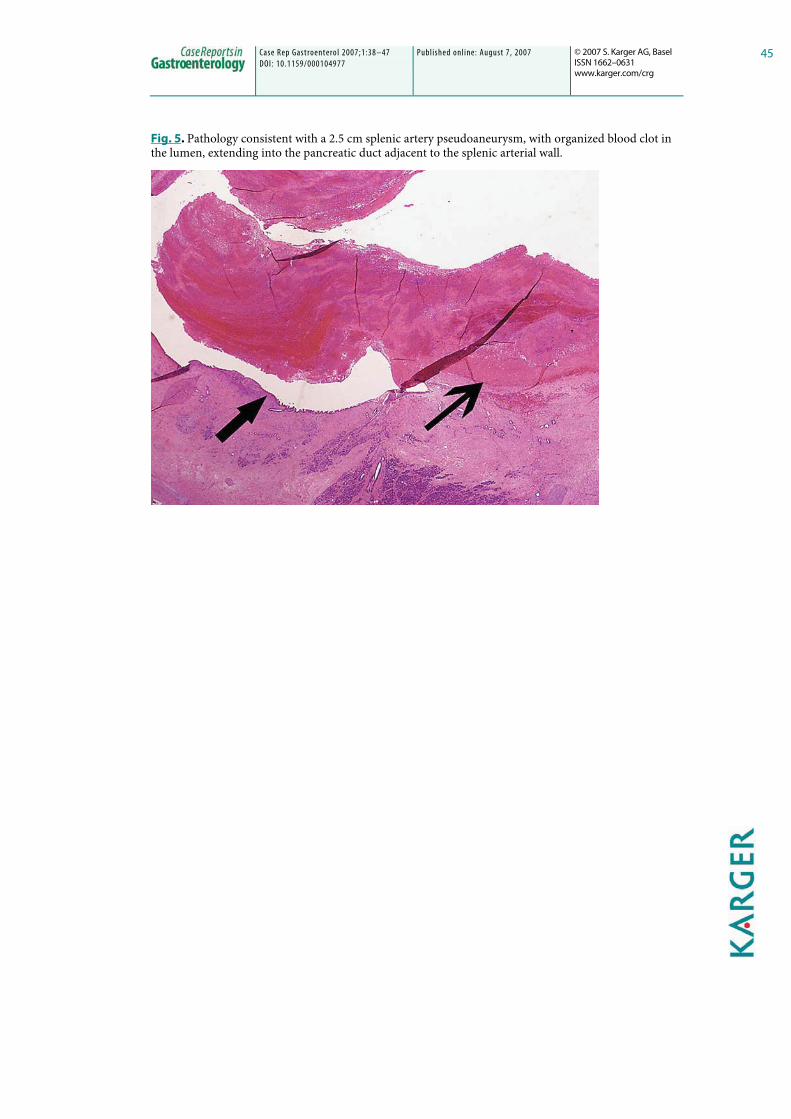
In November 2005, the patient’s fever recurred, prompting a fever reevaluation. A CT scan abdomen demonstrated a 1.5 × 1.5 × 1.0 cm saccular aneurysm arising from the distal splenic artery, invaginating the tail of the pancreas (fig. 1, fig. 2). The main pancreatic duct was mildly enlarged to 0.6 cm within the head of the pancreas, with gradual taper towards the tail. Mild splenomegaly was seen with cranio-caudal length 15 cm. Multiple small infarcts were seen involving the lower third of the spleen. Upon retrospective review, the prior angiogram was noted to show a small pseudoaneurysm of the distal third of the splenic artery, which was camouflaged by the tortuousity of the splenic artery (fig. 3). Similarly, images from the prior ERCP showed blunting of the main pancreatic duct at the tail of the pancreas, likely due to mass effect or thrombus from the adjacent splenic artery pseudoaneurysm eroding into the pancreatic duct (fig. 4). The benign neoplasm noted on prior EUS, was in fact the pseudoaneurysm

Case R ep Gast roent ero l 2007;1:38–47 DOI: 10.1159/000104977
Published online: August 7, 2007 © 2007 S. Karger AG, Basel ISSN 1662–0631 www.karger.com/crg
40
containing mural thrombus with focal surrounding pancreatitis. Fortunately, needle biopsy of this suspected pancreatic neoplasm was not attempted at the time of EUS.
Blood and urine cultures grew Mycobacterium chelonae/abscessus. The central venous access was explanted, and antibiotic treatment re-initiated with amikacin, clarithromycin, and gatifloxacin. Despite 4 weeks of antimicrobial therapy, he experienced progressive unilateral knee, neck and lumbar pains. Magnetic resonance imaging identified enhancement and fluid at the L4–5 and C4–5 interspaces. Percutaneous aspiration of the fluid and open knee debridement were performed, with cultures positive for M. chelonae/abscessus. Echocardiogram was negative for cardiac valve vegetations. Tigecycline was added to the antimicrobial regimen, and gatifloxacin discontinued for intolerance. Progressive clinical improvement was noted at monthly follow-up appointments, and his knee and spine symptoms resolved. He had no relapse of his infection following discontinuation of antimicrobial agents.
Laparotomy was performed, which confirmed the presence of a splenic artery pseudoaneurysm at the tail of the pancreas. The pancreas appeared thickened with calcifications, suggestive of chronic pancreatitis. A distal pancreatectomy and splenectomy were performed. Pathology was consistent with a 2.5 cm splenic artery pseudoaneurysm, with organized blood clot in the lumen, extending into an epithelial-lined cavity consistent with the pancreatic duct adjacent to the splenic arterial wall, suggestive of previous rupture into the tail of the pancreas (fig. 5). AVVG (Voerhoff Vangeesen) stain showed disruption of the internal elastic lamina of the aneurysm, confirming a fistula from the splenic aneurysm into a dilated section of pancreatic duct. Fite stain was negative for acid-fast organisms.
The patient’s post-operative course was complicated by a minor pancreatic duct leak, successfully treated by ERCP with a 7 cm, 7 F stent placement. Follow-up ERCP after 2 months showed resolution of leak and the stent was removed. Following the surgery, the patient did not have any recurrence of gastrointestinal hemorrhage. His hemoglobin, which had been in the range of 8–9 g/dl, was noted to be improved to 12.7 g/dl, with MCV increased from 75 fl to 89 fl at 6 months follow-up.
Discussion
Hemosuccus pancreaticus is defined as bleeding from the pancreatic duct, and was first reported by Lower and Farrell in 1931 [1]. The term ‘hemosuccus pancreaticus’ was subsequently coined by Sandblom in 1970 [2]. This entity has also been called Wirsungorrhagia or pseudohemobilia by different authors [3]. There are at least 160 cases of hemosuccus pancreatitis reported in the English literature since 1931.
The most common etiology of this relatively rare entity is communication of a splanchnic artery aneurysm with the pancreatic duct, either due to erosion or fistula formation [4]. Aneurysms originating from the splenic, hepatic and pancreaticoduodenal arteries have been described in association with hemosuccus pancreaticus [5]. These are usually pseudoaneurysms that occur as a result of pancreatitis or abdominal trauma to the left upper quadrant [4, 6]. Pseudoaneurysms of the splanchnic arteries presenting as hemosuccus have been reported to involve the splenic artery most commonly (60–65%), followed by the gastroduodenal (20–25%), pancreaticoduodenal (10–15%) and hepatic artery (5–10%) [7]. Splanchnic artery pseudoaneurysms have been found to be present in about 10% of patients with chronic pancreatitis [8]. In addition to fistulization into the pancreatic duct, they can present with intraperitoneal hemorrhage and hypotensive shock [9].
Clay et al. postulated that hemosuccus pancreaticus is usually preceded by an episode of pancreatitis, which may occur several years prior to the episode of bleeding. Proteolytic pancreatic enzymes cause a disruption of the splanchnic arterial wall with resultant bleeding. The rapid ductal distension that occurs as blood enters the pancreatic duct leads to herald pain prior to onset of clinical bleeding. This increased pressure in the pancreatic duct allows the leak to seal by clot formation. Lysis of the clot by pancreatic enzymes leads to recurrent episodes of bleeding. Hence, in order to diagnose hemosuccus pancreaticus, visceral angiogram should ideally be performed at the onset of epigastric pain, when

Case R ep Gast roent ero l 2007;1:38–47 DOI: 10.1159/000104977
Published online: August 7, 2007 © 2007 S. Karger AG, Basel ISSN 1662–0631 www.karger.com/crg
41
extravasation of contrast from the splanchnic artery into the pancreatic duct can be observed. If the angiogram is delayed until onset of clinical bleeding, the diagnosis may be missed [10].
Other etiologies for hemosuccus pancreaticus include erosion of a pseudocyst into a peripancreatic artery in patients with chronic pancreatitis [4], direct bleeding from the pancreatic duct as a result of pancreatic duct stones [11], primary aneurysm of a splenic artery [12], blunt or penetrating abdominal trauma [6], mycotic aneurysms [13], and rarely pancreatic malignancy [14].
Clinical presentation includes recurrent episodes of herald pain localized in the epigastric region, followed by gastrointestinal bleeding, presenting as hematemesis, melena or hematochezia [4, 12]. Patients can present with hemodynamic instability and require multiple units of packed red cell transfusion. The diagnosis may often be delayed several years after initial presentation due to the intermittent nature of bleeding and failure to consider hemosuccus pancreaticus in the differential diagnosis of obscure gastrointestinal bleeding [4, 10]. The overall mortality rate has been reported to be as high as 28% [15].
In the era of video capsule endoscopy and double balloon enteroscopy, it is common to identify incidental findings in the small bowel, which in turn may lead to unnecessary endoscopic and surgical interventions, with their associated risk. Video capsule endoscopy has been shown to identify abnormalities in up to 23% of healthy subjects [16]. Data from studies evaluating double balloon enteroscopy have noted inconsistent verification of video capsule findings in 19–35% of patients and detection of incidental findings [17, 18]. Similarly, laparotomy without a precise diagnosis can lead to unnecessary small bowel resections, with a high rate of recurrent bleeding in up to 52% of patients [19]. It is hence extremely important to consider extraluminal etiologies in the differential diagnosis of obscure gastrointestinal bleeding, and to maintain a high index of suspicion in order to establish a diagnosis of hemosuccus pancreaticus.
Visceral angiography is useful to confirm the site of bleeding and demonstrate a fistula between the pancreatic duct and peripancreatic vessels, with a sensitivity of 67–100% [20, 21]. The ‘sentinel clot sign’, characterized by presence of blood clots in the pancreatic duct on CT scan, may also provide a clue to the diagnosis [22]. Three-dimensional CT angiography in addition to establishing the diagnosis allows evaluation of details including size of the aneurysm and surrounding viscera, and is hence useful in determining the appropriate therapeutic intervention [12, 23]. However, CT interpretation by a radiologist with expertise in vascular radiology is extremely important to avoid missing subtle vascular findings. There have been isolated case reports of the diagnosis being made on endoscopy [24] or endoscopic retrograde cholangiopancreaticography, when blood is seen emanating from the Ampulla of Vater [4]. In addition, the diagnosis may be established in hemodynamically unstable patients at the time of exploratory laparotomy [9].
Conventional treatment of hemosuccus pancreaticus secondary to a splenic artery pseudoaneurysm involves distal pancreatectomy and splenectomy, with no reported failure rate [9]. In comparison, ligation of the splenic artery alone provides adequate treatment without vascular compromise, but has a reported failure rate of 43% [9, 12]. Transcatheter embolization by interventional radiology is a less invasive technique that involves embolization of the aneurysm using Gelfoam or platinum spirals [25]. This method of treatment is highly operator dependent, and may rarely be complicated by rebleed, splenic infarction, splenic or pancreatic abscess [9, 15]. Surgical resection may be
Case R ep Gast roent ero l 2007;1:38–47 DOI: 10.1159/000104977
Published online: August 7, 2007 © 2007 S. Karger AG, Basel ISSN 1662–0631 www.karger.com/crg
42
more effective in the management of splenic artery pseudoaneurysms with associated pseudocysts, due to difficulty in embolization of large pseudocyst cavities [26].
Mycobacteria chelonae/abscessus group are ubiquitous, rapidly growing mycobacteria, and are associated with infections in both the immunocompromised and the immunocompetent patient. Skin and soft tissue infections, infections complicating intravascular catheters, medical procedures or trauma, and less commonly, pulmonary, skeletal or other deep infections have all been described [27]. Treatment of infected devices or catheters requires removal and specific antibiotics. This patient likely relapsed due to persistent intravascular infection, as suggested by a rapid relapse of the catheter-associated bacteremia and dissemination to multiple musculoskeletal sites, the negative echocardiogram notwithstanding. Optimal treatment of M. chelonae/abscessus infections is difficult and ill defined, since these organisms are resistant to first line antituberculous and other antibacterial medications [28]. Successful treatment may require the administration of multiple antimicrobial agents to which the organism is susceptible, since single drug therapy has been associated with the emergence of resistance [29]. Tigecycline is a new glycylcycline agent with good activity against the rapidly growing mycobacteria, especially M. chelonae/abscessus group, and may be a useful component of a multiple drug regimen.
Although mycotic aneurysms have been reported to occur secondary to mycobacterium tuberculosis [30], there are no reports of aneurysms secondary to the rapid growing mycobacteria, including Mycobacterium chelonae/abscessus. In our patient, Fite stain of the splenic artery aneurysm was negative for acid fast organisms, confirming that the aneurysm was not related to the co-existent mycobacterial infection.
Conclusions
It is important to consider the diagnosis of hemosuccus pancreaticus in patients with obscure gastrointestinal bleeding. CT angiogram is useful in the diagnosis but requires interpretation by an experienced radiologist. Angiography should ideally be performed at the onset of herald pain in order to detect extravasation of contrast from the splanchnic vessel into the pancreatic duct. A high index of suspicion should be maintained for extraluminal etiologies of obscure bleeding in order to avoid misdiagnosis and unnecessary endoscopic and surgical interventions in the era of video capsule endoscopy and double balloon enteroscopy.
Case R ep Gast roent ero l 2007;1:38–47 DOI: 10.1159/000104977
Published online: August 7, 2007 © 2007 S. Karger AG, Basel ISSN 1662–0631 www.karger.com/crg
43
Fig. 1. Coronal multiplanar reformat from a CT scan showing a 1.5 cm saccular pseudoaneurysm arising from the distal splenic artery, invaginating into the tail of the pancreas. There are small splenic infarcts from mural thrombus in the pseudoaneurysm embolizing to the spleen.
Fig. 2. Axial image of a CT scan showing a 1.5 cm saccular pseudoaneurysm arising from the distal splenic artery with surrounding focal pancreatitis.
Case R ep Gast roent ero l 2007;1:38–47 DOI: 10.1159/000104977
Published online: August 7, 2007 © 2007 S. Karger AG, Basel ISSN 1662–0631 www.karger.com/crg
44
Fig. 3. Celiac arteriogram showing the small pseudoaneurysm of the distal third of the splenic artery, obscured by the tortuousity of the splenic artery.
Fig. 4. ERCP showing blunt ending of the main pancreatic duct in the tail of the pancreas, indicating mass effect or thrombus from the adjacent pseudoaneurysm of the splenic artery eroding into the pancreatic duct.
Case R ep Gast roent ero l 2007;1:38–47 DOI: 10.1159/000104977
Published online: August 7, 2007 © 2007 S. Karger AG, Basel ISSN 1662–0631 www.karger.com/crg
45
Fig. 5. Pathology consistent with a 2.5 cm splenic artery pseudoaneurysm, with organized blood clot in the lumen, extending into the pancreatic duct adjacent to the splenic arterial wall.

Case R ep Gast roent ero l 2007;1:38–47 DOI: 10.1159/000104977
Published online: August 7, 2007 © 2007 S. Karger AG, Basel ISSN 1662–0631 www.karger.com/crg
46
References
1 Lower WE, Farrell JI: Aneurysm of the splenic artery: Report of a case and review of the literature. Arch Surg 1932;23:182–190.
2 Sandblom P: Gastrointestinal hemorrhage through the pancreatic duct. Ann Surg 1970;171:61–62.
3 Yokoyama I, Hashmi MA, Srinivas D, Shaikh KA, Levine SM, Sorokin JJ, Carnishion RC: Wirsungorrhagia or hemoductal pancreatitis: Report of a case and review of the literature. Am J Gastroenterol 1984;79:764–768.
4 Risti B, Marincek B, Jost R, Decurtins M, Ammann R: Hemosuccus pancreaticus as a source of obscure upper gastrointestinal bleeding: Three cases and literature review. Am J Gastroenterol 1995;90:1878–1880.
5 Starling JR, Crummy AB: Hemosuccus pancreaticus secondary to ruptured splenic artery aneurysm. Dig Dis Sci 1979;24:726–729.
6 Kim SS, Roberts RR, Nagy KK, Joseph K, Bokhari F, An G, Barrett J: Hemosuccus pancreaticus after penetrating trauma to the abdomen. J Trauma 2000;49:948–950.
7 Gadacz TR,Trunkey D, Kieffer RF: Visceral vessel erosion associated with pancreatitis. Arch Surg 1978;113:1438–1440.
8 White AF, Baum S, Buranasiri S: Aneurysms secondary to pancreatitis. Am J Radiol 1976;127:393–396.
9 Tessier DJ, Stone WM, Fowl RJ, Abbas MA, Andrews JC, Bower TC, Gloviczki P: Clinical features and management of splenic artery pseudoaneurysm: case series and cumulative review of literature. J Vasc Surg 2003;38:969–974.
10 Clay RP, Farnell MB, Lancester JR, et al: Hemosuccus pancreaticus: An unusual cause of upper gastrointestinal bleeding. Ann Surg 1985;202:75–79.
11 Jakobs R, Riemann JF: Hemosuccus pancreaticus due to a pressure ulcer in pancreatolithiasis [in German]. Dtsch Med Wochenschr 2001;126:108–112.
12 Hasaj O, Di Stasi C, Perri V, Tringali A, Costamagna G: Hemosuccus pancreaticus secondary to intraductal rupture of a primary splenic artery aneurysm: diagnosis by ERCP and successful management by interventional radiology. Endoscopy 2004;36:437–441.
13 Laipply TC: Syphilitic aneurysm of coeliac artery. Am J Med Sci 1943;206:453–458.
14 Mizukami Y, Arisato S, Satou K, Nakano Y, Ohta H, Murakami M, Saito H, et al: A case of anaplastic carcinoma of the pancreas disclosed by hemosuccus pancreaticus [in Japanese]. Nippon Shokakibyo Gakkai Zasshi – Jpn J Gastroenterol 1997;94:706–711.
15 Suter M, Doenz F, Chapius G, et al: Hemorrhage into the pancreatic duct (hemosuccus pancreaticus): Recognition and management. Eur J Surg 1995;161:887–892.
16 Goldstein J, Eisen G, Lewis B, et al: Abnormal small bowel findings are common in healthy subjects screened for a multi-center, double blind, randomized, placebo controlled trial using capsule endoscopy. Gastroenterology 2003;124:337 [abstract].
17 Gross SA, Stark ME: Initial experience with double-balloon endoscopy at a US center: technical aspects, indications, findings and impact on care. Am J Gastroenterol 2006;101:S497 [abstract].
18 Lo SK, Ross AA, Leighton JA, et al: Double balloon enteroscopy: an initial multicenter US experience. Am J Gastroenterol 2005;100:S103 [abstract].
19 Ress AM, Bennacci JC, Sarr MG: Efficacy of intraoperative enteroscopy in diagnosis and prevention of recurrent, occult gastrointestinal bleeding. Am J Surg 1992;163:94 [abstract].
20 Cahow CE, Gusberg RG, et al: Gastrointestinal hemorrhage from pseudoaneurysms in pancreatic pseudocysts. Am J Surg 1983;145:534–541.
21 Koehler PR, Nelson JA, Berenson MM: Massive extra-enteric gastrointestinal bleeding: angiographic diagnosis. Radiology 1976;119:41–44.
22 Koizumi J, Inoue S, Yonekawa H, Kuneida T: Hemosuccus pancreaticus: diagnosis on CT and MRI and treatment with transcatheter embolization. Abdom Imaging 2002;27:77–81.
Case R ep Gast roent ero l 2007;1:38–47 DOI: 10.1159/000104977
Published online: August 7, 2007 © 2007 S. Karger AG, Basel ISSN 1662–0631 www.karger.com/crg
47
23 Malzfeldt EJ, Wegener OH: Aneurysms of peripancreatic arteries in spiral CT angiography in he, osuccus pancreaticus [German]. Rofo 1997;167:319–321.
24 Kuzuya A, Mizuno K, Miyake H, et al: Hemosuccus pancreaticus caused by rupture of a true splenic artery aneurysm following a failure of coil embolization. Ann Vasc Surg 2006;20:130–133.
25 Benz CA, Jakob P, Jakobs R, Riemann JF: Hemosuccus pancreaticus – a rare cause of gastrointestinal bleeding: diagnosis and interventional radiological therapy. Endoscopy 2000;32:428–431.
26 Bender JS, Levison MA: Pseudoaneurysm of the gastroduodenal artery arising within a pancreatic pseudocyst. Ann Vasc Surg 1992;6:171–172.
27 Brown-Elliott BA, Wallace RJ Jr: Infections caused by nontuberculous mycobacteria; in Mandell GL, Bennett JE, Dolin R (eds): Principles and Practice of Infectious Diseases, ed 8. Philadelphia, Churchill Livingstone, 2005.
28 Wallace RJ Jr, Brown-Elliott BA, Crist CJ, Mann L, Wilson RW: Comparison of the in vitro activity of the glycylcycline tigecycline (formerly GAR-936) with those of tetracycline, minocycline, and doxycycline against isolates of nontuberculous mycobacteria. Antimicrob Agents Chemother 2002;46:3164–3167.
29 Tibas P, Sultan F, Wallace RJ Jr, Fraser V: Rapid development of resistance to clarithromycin following monotherapy for disseminated Mycobacterium chelonae infection in a heart transplant patient. Clin Infect Dis 1995;20:443–444.
30 Beeresha, Ghotekar LH, Dutta TK, Verma SK, Elangovan S: Hepatic artery mycotic aneurysm of tubercular aetiology. J Assoc Physicians India 2000;48:247–248.
No conflicts of interest.