Review Article Video Capsule Endoscopy and Ingestible ...
30
Review Article Video Capsule Endoscopy and Ingestible Electronics: Emerging Trends in Sensors, Circuits, Materials, Telemetry, Optics, and Rapid Reading Software Dylan Miley, 1 Leonardo Bertoncello Machado, 1 Calvin Condo , 1 Albert E. Jergens , 2 Kyoung-Jin Yoon , 3 and Santosh Pandey 1 1 Department of Electrical and Computer Engineering, Iowa State University, Ames, Iowa, USA 2 Department of Veterinary Clinical Sciences, College of Veterinary Medicine, Iowa State University, Ames, Iowa, USA 3 Veterinary Diagnostic and Production Animal Medicine, College of Veterinary Medicine, Iowa State University, Ames, Iowa, USA Correspondence should be addressed to Santosh Pandey; [email protected] Received 19 February 2021; Accepted 14 October 2021; Published 10 November 2021 Copyright © 2021 Dylan Miley et al. Exclusive Licensee Beijing Institute of Aerospace Control Devices. Distributed under a Creative Commons Attribution License (CC BY 4.0). Real-time monitoring of the gastrointestinal tract in a safe and comfortable manner is valuable for the diagnosis and therapy of many diseases. Within this realm, our review captures the trends in ingestible capsule systems with a focus on hardware and software technologies used for capsule endoscopy and remote patient monitoring. We introduce the structure and functions of the gastrointestinal tract, and the FDA guidelines for ingestible wireless telemetric medical devices. We survey the advanced features incorporated in ingestible capsule systems, such as microrobotics, closed-loop feedback, physiological sensing, nerve stimulation, sampling and delivery, panoramic imaging with adaptive frame rates, and rapid reading software. Examples of experimental and commercialized capsule systems are presented with descriptions of their sensors, devices, and circuits for gastrointestinal health monitoring. We also show the recent research in biocompatible materials and batteries, edible electronics, and alternative energy sources for ingestible capsule systems. The results from clinical studies are discussed for the assessment of key performance indicators related to the safety and effectiveness of ingestible capsule procedures. Lastly, the present challenges and outlook are summarized with respect to the risks to health, clinical testing and approval process, and technology adoption by patients and clinicians. 1. Introduction The gastrointestinal (GI) tract is both an intriguing and elu- sive environment for diagnostic procedures and therapeutic interventions. Its broad repository of electrolytes, metabo- lites, enzymes, and microbes serves as potential targets for several diseases and disorders [1, 2]. In this regard, real- time monitoring of gastrointestinal biomarkers in a safe, unobtrusive, and cost-effective manner seems achievable in the near future—in part because of the concerted growth, commercialization, and adoption of video capsule endoscopy and ingestible electronics [2–4]. A recent survey indicates that the ingestible capsule mar- ket is expected to be worth $8.98 billion by 2024 with key application areas in capsule endoscopy, remote patient mon- itoring, and targeted drug delivery [5]. Other drivers for the ingestible capsule market are the patient preference for min- imally invasive procedures, favorable medical reimburse- ment policies, and the rising rate of stomach and colon cancers [5]. Modern ingestible capsules can navigate the gas- trointestinal tract and inspect the spatial and temporal land- marks pertinent to common gastrointestinal disorders. To enable the diagnostic and therapeutic functions, a number of advanced features are embedded in modern ingestible capsules, such as microrobotics, targeted stimulation and neuromodulation, controlled sampling and delivery, fine needle biopsy, closed-loop control, panoramic imaging, and rapid reading algorithms [6–9]. The assessment of cap- sule functions is done using key performance indicators (KPIs) of accuracy and resolution, repeatability and robust- ness, autonomous navigation, power consumption, form factor, and operational ease. Besides these KPIs, a central AAAS Advanced Devices & Instrumentation Volume 2021, Article ID 9854040, 30 pages https://doi.org/10.34133/2021/9854040
-
Upload
khangminh22 -
Category
Documents
-
view
2 -
download
0
Transcript of Review Article Video Capsule Endoscopy and Ingestible ...

Review ArticleVideo Capsule Endoscopy and Ingestible Electronics: EmergingTrends in Sensors, Circuits, Materials, Telemetry, Optics, andRapid Reading Software
Dylan Miley,1 Leonardo Bertoncello Machado,1 Calvin Condo ,1 Albert E. Jergens ,2
Kyoung-Jin Yoon ,3 and Santosh Pandey 1
1Department of Electrical and Computer Engineering, Iowa State University, Ames, Iowa, USA2Department of Veterinary Clinical Sciences, College of Veterinary Medicine, Iowa State University, Ames, Iowa, USA3Veterinary Diagnostic and Production Animal Medicine, College of Veterinary Medicine, Iowa State University, Ames, Iowa, USA
Correspondence should be addressed to Santosh Pandey; [email protected]
Received 19 February 2021; Accepted 14 October 2021; Published 10 November 2021
Copyright © 2021 Dylan Miley et al. Exclusive Licensee Beijing Institute of Aerospace Control Devices. Distributed under aCreative Commons Attribution License (CC BY 4.0).
Real-time monitoring of the gastrointestinal tract in a safe and comfortable manner is valuable for the diagnosis and therapy ofmany diseases. Within this realm, our review captures the trends in ingestible capsule systems with a focus on hardware andsoftware technologies used for capsule endoscopy and remote patient monitoring. We introduce the structure and functions ofthe gastrointestinal tract, and the FDA guidelines for ingestible wireless telemetric medical devices. We survey the advancedfeatures incorporated in ingestible capsule systems, such as microrobotics, closed-loop feedback, physiological sensing, nervestimulation, sampling and delivery, panoramic imaging with adaptive frame rates, and rapid reading software. Examples ofexperimental and commercialized capsule systems are presented with descriptions of their sensors, devices, and circuits forgastrointestinal health monitoring. We also show the recent research in biocompatible materials and batteries, edibleelectronics, and alternative energy sources for ingestible capsule systems. The results from clinical studies are discussed for theassessment of key performance indicators related to the safety and effectiveness of ingestible capsule procedures. Lastly, thepresent challenges and outlook are summarized with respect to the risks to health, clinical testing and approval process, andtechnology adoption by patients and clinicians.
1. Introduction
The gastrointestinal (GI) tract is both an intriguing and elu-sive environment for diagnostic procedures and therapeuticinterventions. Its broad repository of electrolytes, metabo-lites, enzymes, and microbes serves as potential targets forseveral diseases and disorders [1, 2]. In this regard, real-time monitoring of gastrointestinal biomarkers in a safe,unobtrusive, and cost-effective manner seems achievable inthe near future—in part because of the concerted growth,commercialization, and adoption of video capsule endoscopyand ingestible electronics [2–4].
A recent survey indicates that the ingestible capsule mar-ket is expected to be worth $8.98 billion by 2024 with keyapplication areas in capsule endoscopy, remote patient mon-itoring, and targeted drug delivery [5]. Other drivers for the
ingestible capsule market are the patient preference for min-imally invasive procedures, favorable medical reimburse-ment policies, and the rising rate of stomach and coloncancers [5]. Modern ingestible capsules can navigate the gas-trointestinal tract and inspect the spatial and temporal land-marks pertinent to common gastrointestinal disorders. Toenable the diagnostic and therapeutic functions, a numberof advanced features are embedded in modern ingestiblecapsules, such as microrobotics, targeted stimulation andneuromodulation, controlled sampling and delivery, fineneedle biopsy, closed-loop control, panoramic imaging,and rapid reading algorithms [6–9]. The assessment of cap-sule functions is done using key performance indicators(KPIs) of accuracy and resolution, repeatability and robust-ness, autonomous navigation, power consumption, formfactor, and operational ease. Besides these KPIs, a central
AAASAdvanced Devices & InstrumentationVolume 2021, Article ID 9854040, 30 pageshttps://doi.org/10.34133/2021/9854040

objective of capsule technologies lies in improving the safetyand quality of procedures while prioritizing patient comfortand involvement [10]. However, capsule system manufac-turers face multiple hurdles as they move up the technologyreadiness levels (TRLs). Some challenges arise from the factthat human clinical trials are demanding, time-consuming,and expensive and often compounded by the issues of inter-patient variability, disease complexities, and nonoptimalprocedural parameters [11]. As such, our work is motivatedby the competing tradeoffs within the field of ingestible cap-sules—the exuberance of research breakthroughs balancedby the rigors of clinical testing, approval process, and busi-ness sustainability [12–14].
Here, our purpose is to provide a comprehensive reviewof video capsule endoscopy and ingestible electronics with afocus on recent developments in hardware and softwaretechnologies (Figure 1). Section 1 introduces the structure,functioning, and disorders of the mammalian gastrointesti-nal tract. The time evolution of ingestible capsules and theguidelines for their safety and effectiveness are covered. Sec-tion 2 explains the capsule components for gastrointestinalsensing, imaging, and wireless telemetry with examples ofingestible sensors, devices, and circuits. Section 3 demon-strates the techniques for capsule navigation and localizationin the gastrointestinal tract using magnetic actuation,robotic manipulators, and electrical stimulation, in additionto capsule placement devices. Section 4 illustrates the cap-sules to monitor the physiological status, including corebody temperature, pH and gastroesophageal reflux, andintestinal gases. Section 5 shows the capsules to inducemechanical vibrations and bioelectric neuromodulation inthe gut. Section 6 deliberates the capsules to track medica-tion adherence, especially during self-administration ofdrugs at home. Section 7 presents experimental capsulesfor gut sample collection, bacterial biomarker detection,and microbiome analysis. Section 8 gives examples of video
capsule endoscopes and rapid reading software, X-ray imag-ing and microultrasound capsules, and imaging-guided ther-apeutics and gastrointestinal surgical procedures. Section 9highlights the trends in biocompatible materials, edible elec-tronics, batteries, and alternative energy sources for thenext-generation ingestible capsules. Section 10 discussesthe present challenges and outlook for this field.
1.1. Mammalian Gastrointestinal Tract. The structure andfunctions of the mammalian gastrointestinal tract are depen-dent on the body and size of the organism, nature and fre-quency of food intake, need for food storage, andadaptation needs [15]. The human gastrointestinal tract isapproximately 8m long and comprises the oral cavity,esophagus, stomach, small intestine (duodenum, jejunum,and ileum), and large intestine (cecum, colon, and rectum).Table 1 lists the anatomic units of the human gastrointesti-nal tract with its respective dimensions, pH, maximumstress, disorders, and diagnostic procedures [8, 16–19]. It isnoteworthy that the anatomy of the human gastrointestinaltract varies from person to person, and the physical dimen-sions of the anatomic units vary significantly with age, sex,gender, ethnicity, etc. The esophagus (pH = 5‐6) has anapproximate length of 18-25cm, diameter of 2.5 cm, and isaccessible by endoscopy [16]. The stomach has an approxi-mate length of 20 cm, diameter of 15 cm, and volume capac-ity of 1-1.6 liters. The stomach is very acidic (pH = 1:2‐3:5)where the supply of hydrogen ions is provided by the secre-tions from parietal cells. The small intestine has an approx-imate length of 6.25 meters, diameter of 2.5 cm, and a curvyand convoluted shape. The acidic content of the stomach isneutralized in the duodenum (pH = 4:6‐6:0) by alkaline fluidsecretions (rich in bicarbonate ions) from the pancreas stim-ulated by secretin and primary bile acids produced fromcholesterol in the liver. The small intestine is adapted fornutrient absorption by having a large surface area with villi
Challenges: Long-term powering, multimodal sensing, steering and localization, smaller size, biocompatibility, cost, safety, and comfort
Electronics: microcontroller, phototransistors, Reed switch, electrodes, magnets, magnetic coils, capacitors, multiplexer, data converters, RF transmitter, antenna, batteries, power management, and wireless power transfer
Imaging and Reading So�ware: white LED, near-infrared LED, fluorescent LED, CMOS image sensor, CCD camera, lenses, optical fibers, panoramic view, adaptive frame rate technology, and intelligent reading algorithms
Sensing parameters: core temperature, intestinal gases, pH, microbiota, medical adherence, GI motility, GI bleeding, mucosal health, lesions, and ulcers
Risks to Health: Biocompatibility, RF radiated power, electromagnetic compatibility, electrical and mechanical safety, functional reliability, intestinal obstruction or injury, and misinterpretation of captured images.
Performance Measures: Quality, safety, comfort, patient experience & involvement, comfort, privacy, dignity, information & consent, facilities & equipment, staffing & leadership
Figure 1: Scope of the review. This work surveys the hardware and software technologies for video capsule endoscopy and ingestibleelectronics. We discuss the sensing parameters, electronics, imaging and reading software, challenges, risks to health, and performancemeasures. The image of the human gastrointestinal tract is reproduced (adapted) from Selber-Hnatiw S. et al. Human gut microbiota:toward an ecology of disease, Frontiers in Microbiology, 8:1265 (2017) under the Creative Commons Attribution License (CC BY).
2 Advanced Devices & Instrumentation

Table1:Structureandfunction
sof
thehu
man
gastrointestinaltract.
Structure
Subsection
sSize
(D×L),w
allthickness
pH,m
axim
umstress
(MPa)
GIdisordersanddiseases
Diagnosticprocedures
Esoph
agus
N/A
2:5c
m×18‐25c
m,4
:7±0:2
5mm
pH:5-6,2:19±
0:06
(lon
gitudinal),
1:41
±0:05
(circumferential)
Achalasia,B
arrett’sesop
hagus,
esop
hagealcancer,G
ERD,p
eptic
stricture,websandrings
Esoph
agram,X
-ray,gastroscopy,
tissue
biop
sy,esoph
ageal
manom
etry,p
Hprobe
Stom
ach
N/A
15cm
×20
cm,3:92±
0:16
mm
pH:1.2-3.5,0:67±
0:19
(lon
gitudinal),0:5±0:12
(circumferential)
Gastritis,gastroenteritis,
gastroparesis,no
nulcer
dyspepsia,
pepticulcers,gastriccancer
pHmon
itoring,X-ray,gastroscopy,
endo
scop
icultrasou
nd,gastric
mon
ometry,H
elicobacterpylori
breath
test
Smallintestine
Duo
denu
mJejunu
mIleum
2:5c
m×26
cm,1:5±0:6m
m2:5c
m×250c
m,1:5±0:5m
mpH
:4.6-7.6,0:55±
0:33
(lon
gitudinal),0:92±
0:48
(circumferential)
Amyloido
sis,celiacdisease,
dysm
otility,infl
ammatorybowel
disease,infections,intestinal
lymph
oma,lactoseintolerance,
tumors,sm
allbowelobstruction
Smallb
owelmanom
etry,X
-ray,
endo
scop
icultrasou
nd,end
oscopic
retrograde
cholangiop
ancreatography
(ERCP),
hydrogen
breath
test,lactose
tolerancetest
2:5c
m×350c
m,1:5±0:5m
m
Largeintestine
Cecum
Ascendent
colon
Transversecolon
Descend
ingcolon
Colon
sigm
oid
9cm
×6c
m,4:8±0:8m
m6:6c
m×20:9cm
,5:0±2:0m
m5:8c
m×50:2cm
,4:2±0:7m
mpH
:7.5-8.0,1:19±
0:30
(lon
gitudinal),0
65±0:16
(circumferential)
Analstenosis,colon
cancer,colon
icintertia,C
rohn
’sdisease,
diverticulosis,infl
ammatorybowel
disease,irritablebowelsynd
rome,
rectaldescentandprolapse,p
olyps,
polypo
sis
Barium
enem
a,x-ray,
sigm
oido
scop
y,colono
scop
y,capsuleendo
scop
y,anorectal
manom
etry,stool
aciditytest,fecal
occultbloodtest,fecal
immun
ochemicaltest
6:3c
m×21:8cm
,3:2±0:6m
m5:7c
m×38:3c m
,3:2±0:7m
m
3Advanced Devices & Instrumentation

which project into the luminal cavity. The effective surfacearea per unit villus surface is roughly around 25μm2 [15].The large intestine is approximately 1.5m long with a diam-eter of 6.5 cm and is involved in the recovery of water andelectrolytes, formation and storage of feces, and fermenta-tion of indigestible food matter by bacteria. The ileocaecalvalve controls the passage of feces from the distal smallintestine (ileum) into the colon. The colon is home to thelargest number of bacterial microbes that produce a numberof beneficial metabolites including short chain fatty acids tomaintain intestinal health [20, 21].
Compared with the human alimentary tract, the gastro-intestinal tracts of common laboratory animals have certainsimilarities and differences in their physiological, biochemi-cal, and metabolic signatures [15, 22]. For instance, cyclicgastric motility is exhibited in both humans and commonlaboratory animals where stomach contractions aid in themixing and grinding of stomach contents for entry into theduodenum. Humans and dogs have similar gastric morpho-logic features (i.e., glandular structure lined with cardiac,gastric, and pyloric mucosa), emptying characteristics, anda similar gut microbiome [23]. The dog is also an excellentmodel for investigation of gastrointestinal diseases [24].Despite many anatomical difference of alimentary systembetween humans and pigs [25], pigs are considered a bettermodel for translational research of human gastrointestinaltract regarding function, nutrition, and digestive diseasesthan many other laboratory models, particularly rodents[26–28]. Besides the fact that pigs are an omnivore likehumans, the microbiota profile of the pig gut shows consid-erable similarity to that of humans at the phylum level. Pigsalso represent a useful in vivo model for investigation oftransplanted human fecal microbiomes under certain condi-tions [29]. Furthermore, the pig gut is immunologically sim-ilar to that in humans since it contains both Peyer’s patchesand lymphocytes that participate in mucosal immunity. Thepredominant microbiota of humans and common labora-tory animals typically contains E. coli, Streptococci, Lactoba-cilli, Clostridium perfringens, yeasts, and bacteroides [15, 30].The upper gastrointestinal tract in humans and rabbit issimilar and contains members of the Bacteroidetes and Bifi-dobacterium spp. (a probiotic microbe that is low to nonex-istent in the porcine gut). Pigs are susceptible to manyenteric pathogens affecting humans [28, 29]. Understandingthe subtle microbiome distinctions can help choose the cor-rect animal model for studies on absorption, bioavailability,nutrition, food transit, and gut-brain nexus within humans[15, 17, 31, 32].
The complex structure and functioning of the gastroin-testinal tract lend itself to a multitude of gastrointestinal dis-orders [17, 18, 33–35]. Esophageal disorders (such asgastroesophageal reflux disease (GERD), infection, corrosionor rupture of veins, and motility disorders) lead to sorethroat, difficulties in swallowing, and regurgitation. Investi-gation of the esophagus is performed using barium swallows,gastroscopy, endoscopy, and mucosal biopsy. Stomach dis-orders (such as inflammation caused by infection such asHelicobacter pylori, gastric ulceration, and peptic ulcers)result in indigestion, vomiting, malnutrition, or vomiting
blood in chronic cases. Small intestine disorders (such asinflammation, peptic ulcers, malabsorption, and tumors)cause diarrhea, malnutrition, lethargy, and/or weight loss.Investigation of the small intestine is conducted by bloodtests, diagnostic imaging with and without contrast, andsmall intestinal endoscopy. Large intestinal disorders (suchas inflammation, functional colonic diseases, and colonicinfections by enteropathogens) may cause bloody stool, con-stipation, fever, and/or abdominal pain. Investigation of thelarge intestine is carried out using diagnostic imaging, colo-noscopy, and capsule endoscopy. Rectal disorders (such asinflammation, hemorrhoids, and anal cancer) result in analpain while defecating, fresh blood in the stool, and constipa-tion. Investigation of the rectum is performed through regu-lar medical tests, rectal examination, and proctoscopy.Disorders of the liver, pancreas, gall bladder, and biliary tract(e.g., gallstones, inflammation, and cancer) adversely influ-ence the secretion of digestive enzymes and gastrointestinalfunctions and are examined by laboratory tests to measurethe levels of enzyme activities and specific organ function.
1.2. Time Evolution of Ingestible Wireless TelemetricCapsules. Historically, the basic concepts of endoscopy andits relevance in medical diagnostics were known to us formore than a century [36]. Rigid-wire endoscopy dates backto 1852 but was limited to the upper gastrointestinal tract.As summarized by Dr. Paul Swain (renowned gastroenterol-ogist at the Royal London Hospital, London, U.K.), thepotential benefits of imaging different sections of the gastro-intestinal tract (via wireless capsule endoscopy) were recog-nized in parallel with the need for detecting its biophysicaland biochemical signatures (via ingestible electronics) [37].In 1954, Harold Hopkins invented the fibrescope (i.e., anoptical unit composed of flexible fiberoptic bundles) to sendoptical images of the gut. This led to the invention of a fullyflexible, digestive endoscope by Basil Hirschowitz in 1957. Acollection of photographs of these pioneers and the endo-scopic equipment from that particular era is nicely illus-trated in a recent review article [10]. The same decadewitnessed the invention of the first wireless, swallowablecapsule (called the radio pill) to measure pH, core body tem-perature, and pressure of the gastrointestinal tract. In 1981,the first wireless video capsule was developed to captureimages of the gastrointestinal tract wall, even though imag-ing technologies were rudimentary. Heidelberg MedicalInc. (Germany) developed a pH capsule to diagnose abnor-mal hydrochloric acid production in the stomach. Its teth-ered capsule (15.4mm long by 7.1mm wide) consisted of aradio frequency (RF) transmitter with an electrode and hada 6-hour lifetime. In 1997, capsule endoscopy was conductedfor the first time on live and deceased pigs using capsule pro-totypes equipped with a light source, camera, video process-ing unit, battery, and transmitter [38].
By the early 2000s, low-power CMOS image sensorspaved the way for incorporating video capture functionswithin wireless capsules [37]. There were significantimprovements in the safety and performance of capsuletechnologies through adopting smart sensing devices, appli-cation specific integrated circuits (ASIC), improved white
4 Advanced Devices & Instrumentation

light optics, and adaptive signal processing [1, 8, 39]. One ofthe first specialized imaging capsules was the M2A (i.e.,mouth-to-anus) and PillCam SB capsules developed byGiven Imaging (Israel). The next-generation PillCam SB 2capsule had wider-angle lenses, autoexposure feature, and alonger battery life. The esophagus-specific (PillCam ESo)and colon-specific (PillCam Colon) capsules were releasedwith higher frame rates. Simultaneously, companies suchas Olympus (in Japan), IntroMedic (South Korea), and Jin-shan (China) developed their novel capsule endoscopy tech-nologies. In 2003, the SmartPill became the first FDA-approved wireless capsule technology that integrated sensorsfor measuring pH, core body temperature, and pressure.
Since 2004, there has been pioneering work in control-ling the navigation and localization of capsule endoscopeswithin the gastrointestinal tract by employing magneticfields, microrobotics, and self-stabilization techniques. Edi-ble and bioresorbable electronics were explored to reinventthe capsule circuitry and other system components. In thepast decade, new frontiers in gastrointestinal diagnosticsand therapy have been explored, such as real-time gas-sens-ing, X-ray and ultrasound imaging, vibrating capsules, med-ical adherence, microbiome studies, digital therapeutics,digiceuticals, and artificial intelligence. A timeline of theabovementioned ingestible capsule technologies is depictedin Figure 2. From the clinical perspective, emphasis startedto be placed on improving the quality and safety of endo-scopic service with a patient-centered approach [40]. Assuch, specialized nursing groups were created within gastro-enterology and endoscopy societies to improve the safetyand comfort of patients, monitor the purchase and mainte-nance of endoscopic equipment, and provide support duringpatient hospitalization, recovery, and discharge [10].
1.3. FDA Guidelines for Ingestible Wireless TelemetricCapsules. The Food and Drug Administration (FDA) hasclassified “ingestible wireless telemetric capsule systems” asclass II medical devices under 21CFR §876.1300, productcode NEZ [41]. The manufacturers of ingestible capsulesare required to have a Premarket Notification 510(k) which
shows that the device is substantially equivalent to a productalready in legal commercial distribution within the UnitedStates. The FDA Class II Special Controls Guidance providesdirections on how to collect clinical information on varioustopics, such as the ease of capsule ingestion, intestinal transittime, diagnostic yield, any adverse events, and agreementamongst reviewers on the interpretation of images [41].
The FDA has identified six risks to health from this“generic gastroenterology and renal device”: (i) biocompati-bility, (ii) electrical and mechanical safety, (iii) intestinalobstruction or injury, (iv) functional reliability, (v) misinter-pretation of recorded images, and (vi) RF-radiated powerand electromagnetic compatibility [41]. The FDA guidanceindicates that the testing on mechanical and structural integ-rity can be related to the battery life, field of view, depth offocus, and device exposure to pH levels and mechanicalforces that are typically experienced during clinical studies[41]. For software-controlled medical devices, the FDArequires sufficient evidence of software functions and perfor-mance testing which is essentially determined by the “levelof concern” (e.g., minor, moderate, or major) resulting fromsoftware failure [41, 42]. Patient labelling should haveinstructions on proper operation, monitoring, maintenance,reporting, and information on the risks and benefits. Someexamples of items to be included in patient labelling are die-tary restrictions, limitations on physical activity, safety fea-tures, use limits, possible symptoms (e.g., nausea, pain orvomiting), warnings, precautions, contraindications, andelectromagnetic interference [41]. Lastly, in order to facili-tate smooth licensing and approval, the FDA mentions thatit considers the least burdensome approach for device man-ufacturers to comply with its guidance and address the iden-tified issues [41].
2. Capsule Components for GastrointestinalSensing, Imaging, and Wireless Telemetry
Most ingestible capsules have a cylindrical form factor in thestandard 000 capsule size (diameter = 11mm and length =26mm). The ingestible electronics consists of miniscule
1957
1980 2000 2010
1988 2004
Transducers,telemetry
Continuousthermometrics
Micro-robotics forcapsule endoscopy
Prolonged GImotility monitoring
Wireless capsuleendoscopy
Magnetic control,self-stabilization
2020
2017 2015
2018 2016
X-ray Imagingcapsule
Medicationadherence
Bacterial sensor withphotoluminescence
Vibrating capsule,gas-sensing capsule
Biodegradable andedible electronics
2019Digital therapeutics,
digiceuticals, AI
2013
2014GI ultrasound withmagnetic control
Prolonged energyharvesting
Figure 2: Timeline of ingestible capsule technologies. The evolution of ingestible capsules is illustrated here to show the importantdiscoveries related to prolonged motility monitoring, continuous thermometrics, wireless capsule endoscopy, microrobotics, magneticcontrol and self-stabilization, biodegradable and edible electronics, medical adherence tracking, vibrating capsules, gas-sensing capsules,X-ray and ultrasound imaging, energy harvesting, bacterial sensing, digital therapeutics, digiceuticals, and artificial intelligence (AI).
5Advanced Devices & Instrumentation

sensors to detect physiological signals and capture images,ASIC chips to amplify and digitize sensors’ output, and wire-less telemetry to communicate the recorded data to an exter-nal receiver for data analytics and predictive diagnostics.
2.1. Ingestible Physiological Sensors, ASIC Chips, Optics, andBatteries. The physiological sensors and ASIC chips foringestible electronics are generally fabricated in the standardsilicon CMOS or CCD technology. For capsule video endos-copy, CMOS image sensors have faster readouts, smallerpixel size, and less power consumption, but suffer from noisesusceptibility. On the other hand, CCD image sensors pro-duce images of high quality and low-noise, but are inher-ently power hungry. Various sensors and ASIC chips canalso be purchased directly from semiconductor manufac-turers and distributors of commercial electronics and ASICchips (e.g., Mouser, Arrow Electronics, Digi-Key Electronics,Future Electronics, Avnet, Rutronik, and Newark Electron-ics). Image compression techniques, such as compressedsensing (CS), are employed to dramatically improve the effi-ciency of image reconstruction from sparse signals usingfewer samples than that required by the Nyquist-Shannontheorem [43]. A polymeric coating covers and protects theelectronic and optical devices. Typically, button silver-oxide batteries provide the power, each having an outputof 1.55V and 20mW. The silver-oxide battery has a silver(I)oxide cathode, zinc anode, and an electrolyte (sodiumhydroxide or potassium hydroxide). The silver(I) ions are
reduced to silver at the cathode, while zinc is oxidized tozinc(II) at the anode. The outer encapsulation material ischosen to make the capsule incompressible, nondisintegr-able, and nondegradable during its transit through the gas-trointestinal tract. Next-generation ingestible capsules areharnessing new materials from nature to redesign variouscomponents such as coatings, substrates, semiconductors,conductors, dielectrics, and batteries (Table 2).
2.2. Wireless Telemetry, Transmission Frequency Bands, andRF Transmitters. The design of wireless telemetry for ingest-ible electronics is dependent on various parameters such astransmission frequency, frame rate, data rate, pixel resolu-tion, specific absorption rate (SAR) limit, and power con-sumption [44]. A comprehensive table of RF bandallocations within Australia and the United States wasrecently published [45]. The ISM radio frequency bandsare reserved for industrial, scientific, and medical (ISM) pur-poses (433.05-434.79MHz for Europe and 902-928MHz forthe U.S.) [46]. While the optimum transmission frequencyfrom an ingestible capsule in the human body is 600-800MHz, transmitting signals at lower frequencies (e.g.,32MHz) requires smaller real estate, reduced power con-sumption, and longer battery life [46]. The common RFcommunication protocols suitable for ingestible capsulesare Bluetooth Low Energy (BLE), ZigBee, and WiFi [45].Other protocols, such as UWB, ANT, Thread, LoRa MICS,IrDA, RFID, and NFC, also have potential use within this
Table 2: Materials for next-generation ingestible capsules.
Categories Example materials
Insoluble materialsAgar, gelatin, cellulose acetate, ethyl cellulose, polyethylene, polyamide, polyvinylchloride, polyvinyl
acetate
Soluble materialsCarboxymethyl cellulose, methylcellulose, hydroxypropyl cellulose, croscarmellose, hydrogels,
hydroxyethyl cellulose, hypromellose, starch, sugars, polyvinyl alcohol, gums, alginates, polyacrylates
Biodegradable substratesCellulose nanofibril paper, polycaprolactone (PCL), poly lactic-glycolic acid (PLGA), rice paper, silk
fibroin, poly (1,8- octanediol-co-citrate) (POC)
Flexible substrates Chitin, gellan gum, pectin, collagen, sodium alginate, silk fibroin, polyesters
FillersGlucose, lactose, starch, oxides, carbonates, bicarbonates, sulfates, nitrates, magnesium silicate, NaCl,
KCl, alkali metals phosphates
Plasticizing agents Dibutyl sebacate, polyethylene glycol, polyethylene oxide, triacetin, triethyl citrate
Responsive materialsEnzyme-sensitive polymers (chitosan, starch), pH-sensitive polymers (polymethacrylates, enteric
elastomer), oxygen-sensitive polymers, temperature-sensitive polymers (cyclododecane,methanesulfonic acid wax), moisture-responsive polyanhydrides
Foodstuff Crackers, corn chips, fruit roll ups, gelatin, gummy, potato, pasta, rice paper, rice starch
Protective coatings Diamond-like carbon (DLC), tetrahedral amorphous carbon (ta-C), gelatin, shellac
Biopigments Melanins, chlorophylls, carotenoids, indigo, flavonoids, quinones, cytochromes, carotenes
Conductive elementsCopper, gold, silver, iron, platinum, magnesium, molybdenum, zinc, metal alloys, activated carbon,
graphene; conductive polymers; ion-containing hydrogels; conductive paste
Thin films Silicon nanomembranes, carbon, molybdenum, iron, tungsten, zinc, polymers
Semiconductors Silicon, germanium, silicon germanium, indium-gallium-zinc oxide (IGZO), zinc oxide
Dielectrics Silicon dioxide, silicon nitride, magnesium oxide, sucrose, albumin, spin-on-glass
Battery anodes Magnesium, zinc, sodium, lithium, iron, iron oxide, tungsten, palladium, molybdenum, and alloys
Battery cathodesCopper salts (chloride, bromide, iodide, sulfate), ferric salts (orthophosphate, pyrophosphate),
vanadium oxide, manganese oxide
6 Advanced Devices & Instrumentation

field. WiFi is designed to wirelessly connect consumer elec-tronic devices (e.g., phones and laptops) to the networkand hence requires large amount of power [45]. Zigbee andLoRa are better suited for embedded systems with low powerconsumption but have relatively smaller range [45]. BLEimplemented with Bluetooth 5.0 appears to be best suitedfor low power transceivers and consumer applications witha midpower consumption [45].
After choosing a suitable transmission frequency, thedesign and test of the RF transmitter is a critical aspect ofwireless telemetry. The RF transmitter consumes a signifi-cant portion of the power to overcome the signal attenuationfrom human body tissues, which can be greater than 23.4 to24.4 dB (based on estimates of a 916.5MHz electromagneticsource located within 15 cm of human tissue) [47]. As a ref-erence, the dielectric parameters of the human gastrointesti-nal tract are as follows: relative permittivity εr = 67:2 andconductivity σ = 1:01 S · m−1 [48]. To study the electromag-netic radiation from RF transmitters, numerical modellingsimulations and tissue phantoms are preferred [49]. Helicalantenna are best suited for ingestible capsules because theyprovide omnidirectional radiation distribution, circularpolarization, and consistent bandwidth across different tis-sues [50, 51]. The PillCam SB has used helix antenna(width = 8mm, diameter = 5mm) with seven turns withinits internal structure, which takes up considerable real estatewithin the capsule. As an alternative, the Sonopill uses a con-formal helix antenna that wraps around the exterior shell ofthe capsule to conserve interior space.
Modelling the channel attenuation of RF signals betweenthe endoscopy capsule systems and the external receivers hasbeen previously attempted for the ISM 2.4GHz band and the402 to 405MHz Medical Implant Communication Service(MICS) band [52, 53]. The ISM 2.4GHz band supports highrate of data transfer that is appropriate for several wirelessapplications but leads to higher chances of interference fromother wireless devices, greater attenuation, and larger powerconsumption [53]. On the other hand, the 402 to 405MHzMICS band has reduced attenuation, lower interferencefrom other wireless devices, and requires low power but doesnot support high rate of data transfer [53]. Previous studieshave computed the path loss (PL) and specific absorptionrate (SAR) using homogeneous or heterogeneous tissuesand often complemented by simulation results from 3D elec-tromagnetic solvers, such as the SEMCAD-X package(SPEAG, Zurich, Switzerland) [52, 53]. The conductivityand dielectric constant varies for the different tissues, andearlier studies have employed models of the muscle tissue,skin tissue, and esophagus tissue to characterize these varia-tions [53]. The path loss is directly proportional to medium’sconductivity and inversely proportional to the square root ofmedium’s permittivity [52]. The path loss also increaseswhen the separation between the antennas is increased orwhen the antennas are misaligned [52]. To give some quan-titative insights on the attenuation of the RF signals throughthe human tissue, one study modelled a capsule system witha spiral antenna [53]. At 402MHz, the reflection coefficientwas between -5.5 dB and -7 dB with the -5 dB bandwidtharound 45MHz [53]. The path loss ranged from 58dB in
the muscle tissues to 61 dB in the esophagus tissue usingaligned antennas separated by a distance of 250mm, whichwas considered satisfactory for effective data communicationin capsule endoscopy systems [53]. For the same setup, thepeak spatial SAR was around 2.2mW/kg, which was con-siderably lower than the 2W/kg limit set by the Interna-tional Commission on Non-Ionizing Radiation Protection(ICNIRP) [53].
Some examples of commercial wireless sensor nodes(available as one complete board) are the Mica2DOT (byCrossbow), T-node (by SOWNet), Sensium (by Toumaz),and Tmote Sky (by Sentilla) [54]. The Mica2DOT sensornode (dimensions: 58 × 32 × 7mm3, weight: 18 g) uses theATmega128L microcontroller (8MHz, 4 k SRAM, and128 k flash memory). It operates at 38.4 kb/s data rate andmultiple frequency bands (868/916MHz and 433MHz)while consuming 135mW power at 3.3V supply. The T-node is comparable to the Mica2DOT in its physical dimen-sion (23mm diameter), operating frequencies, and data rates(50 kb/s). The T-node uses a separate chip for the 10-bitADC, 128 k flash memory, and 4 k SRAM. However, the datatransmission rates of both these sensor nodes are low forreliable video capsule endoscopy. To accommodate fasterdata rates, a high frequency telemetry system (1.2GHz and20MHz bandwidth, 20Mb/s data rate) has been demon-strated, even though the 1.2GHz frequency band is unavail-able for medical device applications [55]. With risingdemand for consumer electronics, a number of low-powerBluetooth System-on-Chip (SoCs) chipsets are now avail-able, such as CC2540 and CC2650MODA by Texas Instru-ments, nRF1822 by Nordic Semiconductor, and BGM11Sby Silicon Labs [45].
2.3. Examples of Sensors, Devices, and Circuits for IngestibleCapsules. Below are some examples of sensors, devices, andcircuits within experimental capsules to measure the gutpH, core body temperature, gastrointestinal motility, andmedical adherence.
(a) A radiotelemetry capsule was designed for theassessment of pH and core body temperature withinthe gastrointestinal tract [56]. The capsule had inte-grated a pH ISFET device and a silicon temperaturesensor. The ASIC chip was fabricated in a 0.6μmCMOS process. The chip included a sensor interface(six operational amplifiers and a multiplexer), timer(with 32 kHz RC relaxation oscillator), and a systemscheduler (finite state machine with a 10-bit ADC).A double-sided printed circuit board (PCB) was usedto assemble the ASIC chip, two SR48 silver oxidebatteries, a magnet for telemetry, and RF transmitter(operating at 433.92MHz). The capsule (36 × 12mm,8 g) had a power consumption of 15.5mW in theactive mode. The capsule was operational for 42hours during in vitro testing in artificial gastric andintestinal solutions
(b) A wireless tablet-shaped ingestible capsule wasdeveloped to monitor the core body temperature
7Advanced Devices & Instrumentation

[57]. Power was generated by a battery with gold andmagnesium electrodes, and activated by gastric acid.The battery voltage output was boosted and stored ina 220μF ceramic capacitor. The ASIC chip wasfabricated in 0.6μm CMOS technology andincluded a temperature sensor, microcontroller, aresonant circuit, sequencer, coder, modulator, andtransmission coil. Data transmission was carriedout with binary phase-shift keying (BPSK) modula-tion at 13.56MHz, followed by wireless telemetryusing magnetic field coupling
(c) A wireless capsule was fabricated in 0.18μm CMOStechnology to identify gastrointestinal motility disor-ders. The capsule integrated three sensors (pH, tem-perature, and pressure), low power ASIC, batteries,RF transceiver, and Helix antenna [58]. The externaldata recorder had a RF transceiver with antenna,batteries, multimedia card, and migrating motorcomplex. A computer workstation performed thedata preprocessing and analytics. The sensing unitswere a thermistor, pH-sensing ISFET, and a C29AKTIV pressure sensor. The ASIC chip had an8-bit microcontroller, clock, and power manage-ment modules and 18-bit ADC, communicationprocessing unit, and memory. The RF transceiverused FSK modulation to transmit and receive dataat 9.6Kb/s with a 434MHz center frequency. Thechip (5mm × 5mm) used a 3.3V supply voltage,2-8MHz clock frequency, and 300μA current drain-age at 2MHz. The capsule was tested on three humanvolunteers and 20 cases of human experiments inmultiple hospitals. While its data analytics could notdistinguish patients with astriction or diarrhea, itsclustering algorithm could correctly recognize 83.3%of patients with absent gastrointestinal motility
(d) As an alternative to batteries for powering the capsuleelectronics, wireless power transmission was demon-strated in a radiotelemetry capsule robot [59]. Twoseparate communication bands were used—a lowerfrequency for power transmission (13.56MHz) and ahigher frequency for data carriers (433.92MHz). Thecapsule robot (10mm× 25mm) had pressure andtemperature sensors, FSK modulation transceiver(data rate of 112 kbps, 433.92MHz located in theISM band), class-E power amplifier and oscillator,16-bit ADC, timer module, flash storage module,power management module, and signal-processingmicrocontroller [59]. The receiving coil within thecapsule induced a voltage by inductive link with thetransmitter to receive up to 280mW of wireless powerat 13.56MHz. The sampling rate of the capsule robotwas 0.6Hz (i.e., 1 sample per 1.67 seconds), whichwas sufficient to capture the gastrointestinal motilitysignals in humans (that are from 1 to 18 cycle perminute). A data recorder was worn around patient’swaist and used a multimedia card to store incomingdata at 433.92MHz and later pass it to a workstation
(e) The Proteus Digital Health platform consisted of aningestible sensor and wearable patch to track poormedication adherence, especially in patients whoself-administer their prescriptive drugs at home[60]. Its ingestible sensor (1mm × 1mm × 0:3mm,CMOS integrated circuit) was activated by gastricfluids to transmit signals at 10-30 kHz and 200-800bits/sec using BPSK modulation. The data podwithin the wearable patch had power circuits, lownoise amplifier, custom system-on-chip (SoC),LEDs, accelerometer, temperature sensor, Bluetoothradio, ceramic radio, and coin cell battery. The datapod received and decoded the signals from theingestible sensor, in addition to monitoring the stepcount, body angle, and heart rate. The data stored inthe data pod’s flash memory was transferred to amobile device using Bluetooth Low Energy (BLE)protocols. The ingestible sensor had a high detectionaccuracy of over 98% in 1,200 unique ingestionsrecorded across 14 individuals [60].
3. Capsule Navigation and Localization in theGastrointestinal Tract, CapsulePlacement Devices
Most wireless ingestible capsules rely solely on the naturalperistalsis movement to navigate the digestive tract. Capsulemovement using peristalsis is satisfactory when capsule’sorientation, velocity, and resident time within each gastroin-testinal region are less important. However, guided capsulemovement using microrobotics and localization techniques(e.g., magnetic actuation and electrical stimulation) isdesired for taking a closer view of specific regions of the gas-trointestinal tract or controlling the resident time in a spe-cific region [6, 8].
3.1. Navigation by Magnetic Actuation and RoboticManipulators. Magnetic actuation employs electromagneticcoils or permanent magnets to generate and control themagnetic fields for capsule navigation and localizationwithin the gastrointestinal tract [61–63]. Electromagneticsystems modulate the magnetic field strengths by changingthe coil current, though high coil currents lead to Joule heat-ing and safety concerns [61]. Compared to electromagneticcoils, permanent magnets are cost-effective and have a com-pact form factor. In conjunction with handheld devices orrobotic guiding systems, permanent magnets within a cap-sule can apply desired forces and torque to the capsule,thereby facilitating better orientation during imaging andfaster transit speeds through the gastrointestinal tract. Mag-netic steering systems may have three types of propulsion:translational, rolling, or helical. Translational steering sys-tems use nonrotational magnetic fields, while rolling andhelical propulsion systems require rotating magnetic fields.Today, magnetic steering systems can have five degrees offreedom (DOF) (i.e., three translational DOF and two rota-tional DOF) as demonstrated by a team of researchers build-ing a wireless capsule gastroscope with a singular permanent
8 Advanced Devices & Instrumentation

magnet system [64]. The team used an external robotic mag-netic manipulator with 6-DOF to maneuver the capsule. Amagnetic force controlled capsule’s position and translationwhile a magnetic torque controlled capsule’s rotation andheading. The 5-DOF manipulation of the magnetic capsulewas tested in an in vitro environment simulating a fluid-filled stomach [64]. On the commercial front, Stereotaxishas a number of products involving the “robotic magneticnavigation” (RMN) technology. Stereotaxis Niobe RMN sys-tem uses two robotically controlled magnets to adjust themagnetic field and precisely steer a catheter (with a magnetat its tip) inside a patient. The Stereotaxis Genesis RMN sys-tem uses smaller magnets (compared to the Niobe system)with better flexibility of the robotic arms, improved patientsafety and outcomes, and unprecedented responsiveness tophysician control during procedures.
3.2. Navigation by Electrical Stimulation. Besides magneticactuation, electrical stimulation has been investigated forguiding the locomotion of ingestible capsules through thegastrointestinal tract [65]. In one study, a radio-controlledelectrostimulation capsule (12mm × 30mm) was con-structed having a RF receiver, decoder system, three 3V lith-ium batteries, and two peripheral nervous system electrodes[66]. The electrodes reached a maximum voltage of 18V. Anexternal controller was used to change the amplitude andperiod of electrical stimulations, which directly affected cap-sule’s velocity. The electrical stimulation of intestinal wallsallowed for capsular propulsion of up to 3.4mm/s in theaboral direction. In vivo tests using porcine intestinal tractsyielded a correlation between capsule’s speed and voltageamplitude, with the optimal voltage amplitude between 6Vand 9V. In another study, a predictive mathematical modelfor the electrostimulated capsular motion in the small intes-tine was built using Stokes’ drag equation and regressionanalysis [67]. The model considered friction and contractionforce as the primary forces acting on a capsule during itstransit. The model simulations provided the optimal shapeand size of the capsule. Thereafter, actual capsules were con-structed having similar internal features and having two setsof electrodes to allow for movement in the oral and aboraldirections. The capsules were tested in 72 experiments withstimulations having a constant voltage and duration (i.e.,6V, 5 millisecond pulse duration) and various frequencies(i.e., 10Hz, 20Hz, and 40Hz). The experiments yielded anaverage forward velocity of 2:91 ± 0:99mm/s and an averagebackward velocity of 2:23 ± 0:78mm/s for the capsules.Unlike magnetic actuation, the safety and efficacy of electri-cal stimulation techniques still needs further validationthrough clinical studies to realize its anticipated commercialsuccess.
3.3. Capsule Placement Devices. Capsule placement deviceshave been developed for individuals who are candidates forvideo capsule endoscopy but have difficulty swallowing thevideo capsule or passing it through the pylorus. Such anindividual may have known or suspected motility disordersor anatomical abnormalities (e.g., dysphagia, gastroparesis,or large hiatal hernias) that could result in stalled capsules
if directly swallowed through the mouth. The capsule place-ment device is used to facilitate direct endoscopic placementof video capsules into the stomach or duodenum while pos-ing minimal intubation risks to the esophagus.
As an example, the AdvanCE capsule endoscopy deliverydevice by STERIS has a sheath (diameter = 2:5mm, length= 180 cm) and a clear capsule cup for atraumatic capsuledeployment into the stomach or duodenum. The capsulecup firmly holds the video capsule during endoscopic intu-bation of the esophagus, and the capsule is then releasedby pulling an outer handle upon reaching the target GIsection. The AdvanCE device is compatible with most videocapsules (diameter = 10:5mm to 11:5mm, length = 23:5mm to 26:5mm), such as those manufactured by Olympus,Given Imaging, and IntroMedic. By directly placing the cap-sule within the stomach, the battery life spent passingthrough the esophagus or stomach is saved.
4. Commercialized Capsules for PhysiologicalStatus Monitoring
Below are examples of commercial ingestible capsules tomonitor the core body temperature, gut pH and acid reflux,and intestinal gases. Table 3 lists the companies for physio-logical status monitoring, sample products, dimensions, fea-tures, specifications, and clinical applications.
4.1. Core Body Temperature Monitoring. Maintaining thecore body temperature consumes approximately 40% ofbody’s energy expenditure [68]. The core body temperatureis not fixed but can fluctuate depending on the amount offood or drinks consumed, environmental factors, and phys-ical fitness of the individual [69, 70]. Measurements of thecore body temperature can be made invasively through theoral, esophageal, or rectal cavity. Oral and esophageal read-ings are not often accurate, while rectal temperatures areinconvenient. The core body temperature can also be pre-dicted from the skin temperature and heat flux measure-ments at different body sites, but the method is not reliablein outdoor settings. As an alternative to invasive and indirecttemperature monitoring methods, ingestible capsules arecapable of unobtrusively tracking the internal body temper-ature in mobile users, such as athletes, fire fighters, emer-gency care responders, and soldiers [71–73]. Capsule-basedmonitoring of the basal body temperature while sleepinghelps to manage the inner-body clock and sleep-wakerhythm [72, 74, 75]. Continuous, real-time monitoring ofthe core body temperature can assist people to stay withinsafe levels of hyperthermia while preventing overexertion,heat stress, and heat stroke. Some examples of commercialcapsules for core body temperature monitoring aredescribed below:
(a) The CorTemp by HQ Inc. is an ingestible body ther-mometer pill that is FDA cleared and registered as asingle-use device (Figure 3). The CorTemp pill has asilicone-coated sensor with a quartz crystal oscilla-tor, circuit board, battery, and communication mod-ule. In the gastrointestinal tract, its quartz crystal
9Advanced Devices & Instrumentation

Table3:Com
mercialized
ingestiblecapsules
forGIph
ysiologicalstatus
mon
itoring.
Com
pany
Sampleprod
ucts
Size
(D×L),w
eight
Features
andtechnicalspecification
sClin
icalapplications
HQ
Inc.
CorTem
pthermom
eter
pill,
CorTem
precorder
10:7mm
×22:4mm,2.8g
Microbattery,qu
artz
crystal,
commun
icationcoilandcircuitboard;
262kH
zor
300kH
z
Mon
itor
core
body
temperature
topreventheat
stress
andheat
illnesses
Philip
Respironics
VitalSensecapsule,derm
alpatch,
XHR
sensor
8:6m
m×23
mm,1
.6g
25° C
to50
° Ctemperature
range,240hrs
batterylife,1meter
transm
ission
range
Mon
itor
core
body
temperature,d
ermal
temperature,h
eartrate,and
respiration
rate
Bod
yCap
eCelsius
medicalcapsule,performance
capsule
8:9m
m×17:7mm,1.7g
Accuracyof
±0.2° C
intherange25-45°C,
20days
batterylife,1meter
range
Sportsperformance;m
edical/ind
ustrial
health
mon
itoring;severe
heat
orcold
environm
ents
SmartPill
Corpo
ration
SmartPill
wirelessmotility
capsule
13mm
×26
mm,4.5g
434.2MHz,silver
oxidebatteries,25
° C-
49° C,0.05-9.0pH
range,0-350mmHg
pressure
range
Usedforcapsulemotility
procedures;
alternativeto
scintigraphy
andradio-
opaque
markers
AnX
Robotica
VibraBot
capsule
11:8mm
×26:7mm,4
:5±0:5g
Adjustablevibrationfrequency,,180
minutes
continuo
usvibration,
smartpho
necontrol
Non
pharmacologicalconstipation
relief,
mechanically
stim
ulateperistalsisin
large
intestine
Vibrant
Gastro
Vibrant
Capsule
11:3mm
×24:2mm,N
/AFlat
motor,b
atteries,electroniccard,
algorithm-based
control,adjustable
vibrationfrequencies
Chemical-freeconstipation
treatm
ent
capsule,relieffrom
chronicidiopathic
constipation
Atm
oBiosciences
Atm
ogas-sensingcapsule
11mm
×20
mm,N
/AMeasureshydrogen,o
xygen,
carbon
dioxide,methane;d
atasent
every5min
for72
hrs
Measure
gasprod
uction
,onset
offood
ferm
entation
,and
gutmicrobiom
eactivity
Proteus
Discover,
Otsuk
aPharm
aProteus
DigitalHealth
,AbilifyMyC
ite,
ProteusCloud
N/A
Ingestiblepillwithbiosensors,w
earable
patch,medicationtrackerapp,singledo
seresolution
Track
medicaladherencefordrug
therapeutics;check
iforalmedications
wereingested
(e.g.,fortuberculosis,
schizoph
reniaandon
cology)
10 Advanced Devices & Instrumentation

oscillates at a frequency relative to the core bodytemperature. This creates a corresponding magneticflux, and the information is transmitted to the Cor-Temp Data Recorder (262 kHz to 300 kHz). The pillhas been applied to the detection and prevention ofheat stress and heat-related illnesses for sports,research, industry, and medicine
(b) The eCelsius by BodyCap is an ingestible electroniccapsule for the continuous monitoring and wirelesstransmission of core body temperatures (Figure 3).The eCelsius Medical has been used to diagnose apatient’s febrile state for applications in sleep disor-ders, oncology, and infectious diseases. The eCelsiusPerformance is designed for athletes and swimmerswho need continuous core temperature monitoringwithout any constraints on the subject. TheireViewer monitor manages the data from up to 3 cap-sules within a 1-3-meter range. Some other eCelsiusapplications include the early detection of peak fever
during infections and chemotherapy and trackingthe circadian rhythm of core body temperatures forsleep analysis
(c) MyTemp BV has developed a battery-free, core bodytemperature monitoring capsule. It uses self-induction to power the capsule and transmit thegathered data to a belt worn on the waist. TheMyTemp capsule has been used to detect overheat-ing of athletes’ core and to monitor animal heatstress in equestrian sports
4.2. pH Monitoring and Gastroesophageal Reflux Testing.Ingestible wireless pH testing capsules are being used tomonitor pH levels over time for improved gastrointestinaldiagnostics. One application area is esophageal pH testing,which is important for the diagnosis of gastroesophagealreflux disease (i.e., GERD) where a person suffers from fre-quent acid reflux. GERD has been traditionally diagnosedby a nasopharyngeal-wired pH monitoring system that
Outer silicone coating
(a)
(b)
Inner epoxy shell
Communication coils
Temperaturesensing crystal
Printed circuits andelectronic components
on ceramic substrates
Silver oxidebattery
Figure 3: Continuous core body temperature monitoring capsules. (a) The BodyCap e-Celsius Core Body Temperature Ingestible Capsulemeasures the gastrointestinal temperature and transmits the data to an external e-Viewer Performance Monitor. (b) The CorTempIngestible Core Body Temperature Sensor transmits information on the internal body temperature to the CorTemp Data Recorder wornexternally by the user. The images were reproduced with permission from BodyCap (a) and HQ Inc. (b).
11Advanced Devices & Instrumentation

causes patient discomfort. After swallowing the pH monitor-ing capsules, the pH profiles can be analyzed for four gastro-intestinal landmarks: ingestion, pylorus, ileocecal valve, andexcretion [76]. Ingestion is characterized by a rapid temper-ature rise and a rapid pH drop (>3 pH values), pylorus ischaracterized by a rapid pH rise (>3 pH values), ileocecalvalve is noted by a rapid pH drop to ≤6.5, and excretion isnoted by a rapid temperature drop [76]. Below are someexamples of commercial ingestible wireless pH testing cap-sules for the gastrointestinal tract.
(a) The Bravo Reflex Testing System is an ambulatorypH test and primarily assists in the diagnosis ofGERD symptoms. The esophageal pH is measuredby an antimony pH electrode every 6 seconds overa period of 96 hours. This extended time period ofpH monitoring increases the chances to detect refluxevents and accurately determine the associatedsymptoms
(b) Jinshan Science and Technology has developed awireless pH capsule system to record up to 96 hoursof data. The wireless pH capsule is designed to besmall in size (6:0 × 5:5 × 26:5mm) and weight(1.4 g). A conveyor is used to deliver the wirelesspH capsule to the lower esophageal sphincter(LES), followed by vacuum suction to attach the cap-sule to the mucosa. The pH sampling is fast (i.e.,every 3 seconds), and the range of data transmissionis sufficiently large (i.e., within 3 meters) for patientcomfort. After traversing the gastrointestinal tract,the wireless pH capsule is naturally excreted fromthe body. An accompanying recorder provides auto-matic pairing with the pH capsule, double data pro-tection, and improved user operability. Its dataanalysis software has options to autodetect eventsto reduce editing time, perform statistical calcula-tions, and generate customized reports
(c) The SmartPill capsule endoscope was designed forthe evaluation of gastric emptying and the measure-ments of transit time through the small bowel andcolon. The standard techniques of studying gastricemptying by whole gut transit scintigraphy (WGTS)and radio-opaque markers require long-time dura-tions and provide information only about colonictransit. The SmartPill capsule has integrated sensorsto transmit data on the pH, temperature, and pres-sure in the gastrointestinal tract at regular intervals.The SmartPill MotilGI software helps to estimatedifferent time parameters which have relevance tothe gastrointestinal functioning. For instance, thewhole gut transit time is calculated as the timebetween capsule’s ingestion and its exit from thebody (denoted by a sudden drop in temperature orloss of signal). The software also calculates the gas-tric motility index and gastric contractions perminute. The gastric residence time is calculated fromcapsule’s ingestion to the sudden pH rise (i.e., >3 pHunits from the acidic gastric region to the alkaline
duodenum). The transit times through the ileum,cecum, and colon are calculated from the raw datarelated to pH, temperature, and pressure at variousgastrointestinal sections [77, 78]. A clinical studyused the SmartPill capsule to show that diabetic indi-viduals with gastroparesis exhibited delayed colonictransit which correlated with delayed gastric empty-ing [79].
4.3. Sensing of Intestinal Gases. The process of carbohydratedigestion breaks down the poly- and disaccharides intoabsorbable monosaccharides which is followed by carbohy-drate absorption in the intestine [30]. The unabsorbed car-bohydrate reaches the colon where it is fermented by thebacteria [80, 81]. This bacterial fermentation can lead toexcess gas production and other discomforts (e.g., bloating,diarrhea, abdominal distension, and irritable bowel syn-drome (IBS)) [82]. Apart from the metabolic activity ofintestinal bacteria with unabsorbed food content, variouschemical conversions and enzymatic perturbations can pro-duce different gases in the gastrointestinal tract, such ashydrogen, carbon dioxide, oxygen, nitrogen, and methane[83]. The malabsorption of various carbohydrates and smallintestine bacterial overgrowth are commonly monitoredfrom end-expiratory breath samples for levels of hydrogenor carbon dioxide [84]. Breath tests can also reveal elevatedlevels of methane in the gastrointestinal tract, suggestinghigher colonization of methanogens (e.g., Methanobrevibac-ter smithii) in the gut who scavenge the hydrogen and pro-duce methane as a by-product [85, 86]. The presence ofboth hydrogen and methane in the breath of individuals iscorrelated to higher body mass index and percent body fat[86]. However, studies show that hydrogen breath tests cangive divergent or false-negative results due to several factorssuch as the presence of oral bacterial flora, nonhydrogen-producing bacteria, gastrointestinal motor disorders, orimproper adherence to carbohydrate diet [35, 84]. Further-more, the lack of accurate diagnosis in regard to the pointof origin of the gas and very low signal-to-noise ratio limitsthe conclusions that can be drawn from the hydrogenbreath test.
With the inherent deficiencies of breath tests and theinconvenient nature of other techniques (e.g., tube inser-tions, flatus analysis, and whole-body calorimetry), indirectmeasurements of gas production by in vitro fecal fermenta-tion systems using colonic bacteria has drawn attention fromexperts [87]. Here, an oxygen-free bacterial incubation envi-ronment is created, and fecal inocula is cultured with nutri-ents on a fiber substrate. The gas released is absorbed in solidphase microextraction fibers and assessed for its composi-tion using gas chromatography and mass spectrometry(GC-MS). To circumvent the offline gas analysis, portablegas-sensing capsules have been investigated to measure theintestinal gas species from in vitro fecal fermentation byemploying electrochemical, calorimetric, or optical sensingmodalities [87].
Wireless capsules have been demonstrated for the mea-surement of intestinal gas profiles in vivo and the discoveryof biomarkers related to the gut microbiota [35]. These
12 Advanced Devices & Instrumentation

studies can potentially help to formulate better diets whichpositively influence the gut microbiota and colon health[81, 83]. A human pilot trial was conducted to test the effi-cacy of ingestible electronic capsules that monitor differentintestinal gases (i.e., oxygen, hydrogen, and carbon dioxide)[88]. The capsule employed semiconducting metal oxide-based sensors and thermal conductivity sensors to measurethe gas concentrations. The polyethylene capsule shellhoused the gas sensors, temperature sensor, microcontroller,transmission system (433MHz), and silver oxide batteries.Ultrasound was used to evaluate the physical location ofthe capsule and the transit time within different sections ofthe gut. In their study, four healthy volunteers wererecruited, and subjected to high-fiber and low-fiber diets.The results showed that the oxygen-equivalent concentra-tion profile was an accurate marker for tracking the physicallocation of the capsule, while hydrogen gas profiles repre-sented the food fermentation patterns in the different sec-tions of the gut. With high-fiber diet, there was only asmall increase in hydrogen and carbon dioxide within the
colon, which are indicative of minimal colonic fermentation.With low-fiber diet, there was an increase in colonichydrogen and carbon dioxide with a significant shift inthe microbiota as confirmed from metabolomics analysisof fecal samples. The team founded the Atmo Biosciencesto commercialize their capsules (Figure 4) that can mea-sure the intestinal gases at the source and identify diges-tive issues using clinical analysis and predictive algorithms[1, 35, 87, 89–92].
5. Commercialized Capsules for MechanicalVibrations and Bioelectric Neuromodulation
Below are some examples of commercial vibrating capsulesfor constipation relief and electroceutical capsules for obe-sity and hormonal release.
5.1. Vibrating Capsules. Functional constipation is classifiedas normal-transit, slow-transit, or outlet-transit types andis generally manifested by abdominal discomfort, stomach
(a)
(c)
(b)
Figure 4: Gas-sensing capsules by Atmo Biosciences. (a) The Atmo Gas Capsule detects gases in the human gastrointestinal tract at realtime for the diagnosis and targeted treatment of gut disorders. (b) The Atmo gas capsule has a higher signal to noise ratio compared tobreath tests as the gas concentrations are measured at the site of production. (c) The patient can take the Atmo gas capsule at home, andthe data is continuously transmitted to the physician through a smartphone app for analysis. The images were reproduced withpermission from Atmo Biosciences.
13Advanced Devices & Instrumentation

aches, and infrequent or painful defecation [93]. Currenttreatment options for chronic constipation include fibersupplements, stool softeners, osmotic and stimulant laxa-tives, lubricants, prokinetics, guanylate cyclase-C agonists(GC-C), and surgical management. The sustained effective-ness of the current treatment options for chronic constipa-tion is not guaranteed due to their high recurrence rate,poor patient compliance, and treatment side effects. Toimprove the consistency of treatment for the aforemen-tioned conditions, vibrating capsule technologies have beendeveloped. Examples of commercial vibrating capsules forthe treatment of functional or chronic constipation are asfollows:
(a) Vibrant Capsule by Vibrant Ltd. is a nonpharmaco-logical device to provide relief from constipation andrestore the complete spontaneous bowel movements.As a chemical-free alternative to current treatmentoptions for chronic constipation, the Vibrant Cap-sule generates mechanical vibrations in the wall ofthe large intestine to induce bowel movements andaugment the normal circadian rhythm [93]. TheVibrant Base Unit activates the capsule using anelectromagnetic signal. Different vibration modescan be programmed to regulate when and how tovibrate the capsule. The Vibrant Capsule has beentested on patients with Chronic Idiopathic Constipa-tion and Irritable Bowel Syndrome with Constipa-tion. In a study to evaluate its safety and efficacy, itwas found that the vibrating capsule induced a sig-nificant increase in bowel movements per week in23 out of 26 patients with no serious adverse events[94]. The team established safety by quantifyingkey parameters related to pathological anatomy,physiology, stool, blood, and CT scans [94]. A sec-ond study saw evidence of faster colonic transit timewith the Vibrant Capsule treatment and mentionedthat the vibration parameters require further optimi-zation to accelerate the colonic transit in patientswith functional constipation [95].
(b) The VibraBot by AnX Robotica is designed to pro-vide nonpharmacological relief from constipation.One study tested the VibraBot capsule on the defeca-tion frequency of beagle dogs [96]. An external con-figuration device enabled by a smartphone was usedto communicate with the VibraBot capsule. Once thecapsule reached the abdominal cavity, it was acti-vated to generate abrupt vibrations at low, moderate,or high frequencies (with a frequency ratio of0.5 : 0.8 : 1.0, respectively). The VibraBot capsule pro-duced mechanical vibration to stimulate peristalsisin the large intestine. Their results indicated thatafter capsule ingestion, there was a significantincrease in the daily defecation frequency and adecrease in the capsule evaluation time
5.2. Electrical Stimulation Therapy and BioelectricNeuromodulation. Besides gastrointestinal motility disor-
ders, a class of electroceutical devices are being investigatedas therapeutic tool for obesity, hormone release, and neuralsignaling [8, 97, 98]. Bioelectric neuromodulation (whereelectrical stimulation is given at target sites along the gastro-intestinal tract) has gained significant interest as a method torelieve gastrointestinal disorders, even though the presentoutcomes are variable and the mechanisms of action areunclear [11]. Bioelectric neuromodulation may stimulatethe vagus nerve, thoracolumbar connections, and sacralnerves to inhibit gastrointestinal inflammation, treat feacalincontinence, or block signals for feeding to reduce appetiteand treat morbid obesity [11, 99, 100]. While there are nocommercial examples of ingestible capsules for the electricalstimulation of the gastrointestinal tract, there are two prece-dents as implantable bioelectric neuromodulators for thispurpose. In 2015, the VBLOC vagal blocking therapy byEnteroMedics Inc. was approved by FDA for the treatmentof obesity. The EnteroMedics VBLOC therapy involvedusing its implantable pacemaker-like device, MaestroRechargeable System, to intermittently block the vagal nervepathway to reduce hunger and generate earlier feelings offullness. This helps to promote a safe, healthy, chemical-free,and sustainable weight loss alternative to bariatric surgery(which has high surgical costs and increased rate of frequentcomplications) [101–103]. A ReCharge Study to evaluate thesafety and efficacy of the EnteroMedics Maestro Recharge-able System found that VBLOC-treated patients reached24.4% excess weight loss at 12 months [11].
6. Commercialized Capsules for TrackingMedication Adherence
One emerging application of ingestible capsules is to moni-tor and document the adherence to oral medications. Thecommon reasons cited by patients for not taking their med-ications correctly can be forgetfulness, mental disorders,emotional factors, dearth of information, poor communica-tion with caregivers, and deliberate intent to omit doses[104]. Poor medication adherence can result in treatmentfailure, multidrug resistance, and disease transmission tothe community. This can also lead to frequent sickness, doc-tor visits, and hospitalizations. The two current standards ofdrug administration are directly observed therapy and self-administered therapy. Surveillance technologies can helpmonitor medical adherence, such as video recording bysmartphones and manual time-register of the opening of pillbottles. However, the current methods do not ensure that allthe required medications are actually ingested regularly atscheduled times. The clinicians also do not have rapid accessto the medical adherence information from their patients.Connected medical devices are gaining attention for theremote tracking of drug management and patient outcomes.As an example of digital healthcare, a smart autoinjector,AdhereIT by Noble and Aptar Pharma, can assist untrainedpatients with at-home, self-administering of drug injections.The AdhereIT device accurately detects an injection eventand lets the patient know (through audio, visual, or hapticfeedback) whether the injection was done correctly.
14 Advanced Devices & Instrumentation

Ingestible capsules can provide an automatic techniqueto record the ingestion of medications at a single-dose reso-lution, thereby replacing direct observed therapy by wirelesslyobserved therapy. The Proteus Discover system developed byProteus Digital Health is an ingestible capsule technology tomeasure the effectiveness of medication treatment, which inturn helps to achieve desired clinical outcomes by trackingpatient’s health patterns. The Proteus Discover system con-sists of ingestible sensors, wearable sensor patch, mobileapp for the patient, and portal for the provider [60]. Oncethe medication and ingestible sensors are ingested, theyreach the stomach. The gastric acid in the stomach is usedby the ingestible sensors to generate a tiny biogalvanic signalwhich is transmitted to the wearable patch. The biogalvanicsignal is not pH dependent and lasts for roughly 7 minutes.The remaining contents of the ingestible sensor are inactiveand eliminated through the feces. The wearable patchrecords the information about the ingested medications(i.e., date, time, drug type, and its amount) and physiologicdata (i.e., step count, sleep quality, and circadian rhythm).A digital record is sent to the mobile app and to the providerthrough ProteusCloud after prior permission from thepatient. This helps to confirm the ingestion of medicationson a dose-by-dose basis and hence compliance to the treat-ment. The data also helps to quickly identify the root causefor nonadherence such as lack of understanding, undesiredside effects, or improper dosing schedules. The cliniciancan then make decisions on whether to initiate, optimize,or eliminate the medications. Proteus Discover was FDAcleared in 2012 and has been tested in different patientpopulations (e.g., those with heart failure, hypertension,or tuberculosis) [104–106]. In patients with hypertension,the Proteus technology helped the community pharmaciststo identify the root cause of persistent hypertension. Wire-less monitoring showed that two-thirds of the patients hadpharmaceutical resistance while the remaining one-thirdtook inadequate medications. A study on the adherenceto tuberculosis therapy found that the Proteus DigitalHealth system may be able to correctly identify ingestiblesensors with high accuracy, low risk, and high patientacceptance [105].
A collaboration between Proteus Digital Health and theJapanese Pharmaceutical Company Otsuka led to Abilify’sMyCite, which is the first digital medicine approved by theFDA to treat schizophrenia. The drug, Abilify (or Aripipra-zole), is a prescriptive medication to treat mental disorders,such as depression, schizophrenia, bipolar disorder, andTourette syndrome. MyCite is the smart pill version of thedrug Abilify that embeds a tiny Ingestible Event Marker(IEM) sensor. This sensor is made of natural ingredientsand passes naturally out of the body after use. Upon ingest-ing the smart pill, the IEM sensor transmits a Bluetooth sig-nal to a wearable patch worn by the patient. The patch sendsthe information to a smartphone app and subsequently to aMyCite Dashboard which can be shared with a caregiver.The Abilify manufacturers have mentioned that FDA hasevaluated and approved only the functions related to track-ing the drug ingestion, and it is not known if Ability MyCitecan help with patient compliance.
7. Ingestible Capsules for GutMicrobiome Analysis
Below are some examples of experimental ingestible capsulesto collect gastrointestinal samples and track the microbiomewithin the gastrointestinal tract.
7.1. Capsules for Collecting Samples of the GastrointestinalContent. The retrieval of physical samples from specific loca-tions in the gastrointestinal tract can help us understand thecomposition and diversity of the microorganisms. The cur-rently used stool test may not be reflective of the true intes-tinal health because fecal samples have shown to displaygreater bacterial richness and variation depending on factorssuch as stool consistency, medications, preexisting diseases,nutrition, and other personal traits [21, 107]. Tetheredendoscopy to retrieve samples of the gastrointestinal tractis invasive, inconvenient, and incapable of reaching the dis-tal regions of the small intestine. As an alternative, the gutsample can be collected by using magnetic fields to actuatethe opening or closing of capsules. In recent past, magneticfields have been used to create actuation forces and torqueto control the movement of medical catheters, tissue biopsysampling, targeted drug delivery, flexible endoscopes, and3D endoscopy with scanning catheters [62]. As an example,a drug delivery module was demonstrated using electromag-netic actuation and active locomotion of the capsule throughthe gastrointestinal tract [63]. The drug was initially encap-sulated by axially magnetizing two soft magnetic rings. Anexternal magnetic field was deployed to move the capsuleto the target lesion. Thereafter, a pulsating radial magneticfield was used to open up the capsule for drug release. Aplastic hinge ensured that the module returned to its initialshape [63].
Building on the principles of magnetic actuation, there isa recent interest in magnetically actuated devices for sam-pling the gut microbiome. There are multiple factors to con-sider while designing such capsules, such as the opening andclosing mechanisms, techniques for tight sealing, materialand structural strength, and sample crosscontamination.There is a tradeoff between capsule dimensions and the col-lected volume, where smaller capsules can pass convenientlythrough the gastrointestinal tract but would collect less sam-ple volume, and vice versa. It is estimated that the DNA andRNA concentrations in the samples should be at least 100-500 ng/μL for reliable detection during downstream analysis[108, 109].
A soft mobile robotic capsule was developed for nonin-vasive sampling of the liquid digesta [62]. Each of the twohalves of the capsule contained a disk magnet (6mm indiameter, 3mm in thickness) and were held together by aflexure hinge made from an elastomer composite reinforcedwith carbon fiber. Silicon rubber was the material for the sealrim, while polydopamine was used as the biocompatiblecoating for capsule’s inner surfaces. The capsule was nor-mally closed when ingested from the mouth. An externalmagnetic field generated a torque to open up the two halvesof the capsule to encapsulate the gut content. An oppositemagnetic torque was generated to create an intermagnet
15Advanced Devices & Instrumentation

force that sealed the capsule back to its normally closed con-figuration. With a capsule volume of 42mm3, the device col-lected 18 to 61mg of gut content from pigs during in vivostudies. This amount of content was sufficient as 10mg ofmaterial yields around 1.5μg of DNA and 4μg of RNA[110, 111]. Another study demonstrated the use of a highlyabsorbent hydrogel to first collect the gut microbiome andthen automatically seal the capsule by mechanical actuationof the hydrated gel [112]. The capsule dimensions were9mm in diameter and 15mm in length. A pH-dependententeric coating was made with cellulose acetate phthalate(CAP) to keep the capsule closed at pH values less than 5.0and to slowly degrade at higher pH. The inner surface ofthe capsule was treated with plasma and PEG to create asuperhydrophilic surface for easy wicking and flow of gutcontent. The performance of the capsule in extracting andretaining viable bacteria was tested in vitro using E. coli solu-tions [112].
A soft capsule endoscope was constructed for the fine-needle biopsy of submucosal tissues in the stomach [113].The capsule had an internal permanent magnet to respondto changes in the external magnetic field and indicate itsphysical location. A series of electromagnets generated themagnetic field to control the orientation and rolling motionof the capsule until the target location was reached. Anendoscope camera gathered positional data and other infor-mation on the stomach, such as the detection of lesions.Upon reaching the destination, magnetic torque was appliedto properly orient the capsule. Thereafter, magnetic forcewas applied to engage a thin, hollow needle to retrieve andstore a tissue sample using the fine-needle aspiration biopsytechnique. After sample collection, the needle was retractedwith a tether attached to the capsule. Ex vivo experimentswere conducted in a porcine stomach model with phantomtumors located underneath the outer stomach wall and pro-duced 85% yield with the capsule robot [113].
7.2. Capsules for Bacterial Biomarkers and Nucleic Acids.Ingestible capsules have been housed with engineeredmicrobes to detect health-related biomarkers and with elec-tronic readout modules to wirelessly communicate the sens-ing signal at real-time [114]. This approach has benefits overcurrent methods of probing the gut microbiome using plateanalysis of urine and stool samples [12, 115, 116].
In one study, genetically engineered probiotic E. coli bac-teria was leveraged to construct two biological sensors (thio-sulfate sensor and tetrathionate sensor) which can detectcolon inflammation and possibly lead to new methods tostudy gut microbiota pathways [117]. In another study, aningestible microbioelectronic device (IMBED) capsule wasdeveloped to detect gastrointestinal bleeding events. The tar-get biomarker was heme that was released from lysed redblood cells. The capsule combined engineered probiotic bac-terial sensor with a bioluminescence detection circuit andwireless communication electronics [118]. The probiotic E.coli bacteria was housed within individual wells covered bya semipermeable membrane and was engineered to internal-ize the extracellular heme through an outer membranetransporter, ChuA. Upon detecting the target biomarkers,
the heme-sensing genetic circuit was triggered to allow thebacterial luciferase operon, luxCDABE, to emit biolumines-cence. The optical signal was read by phototransistors, con-verted to digital codes by a nanowatt-level luminometerchip, and transmitted wirelessly for data analysis. In vitrostudies were conducted on a porcine model for gastrointesti-nal bleeding to correlate the dose-dependent heme inputfrom whole blood to the luminescence production. An oro-gastric tube was used to deposit the IMBED capsule in pig’sstomach, and photocurrent data was recorded for the next 2hours. The above method was expanded to sense thiosulfateand acyl-homoserine lactone (AHL) that are biomarkers forgut inflammation and infection, respectively.
In a separate study, an ingestible biomolecular sensingsystem was developed for the detection of microbiomenucleic acids in the gastrointestinal tract [119]. The devicefitted within the FDA-approved capsule size of 000. The cap-sule system was equipped with a fluorescence-based sensor,UV LED, optical waveguide, 1.55V 12.5mAh battery, andbidirectional communication features (124μW wirelessreceiver and 915MHz FSK/OOK transmitter). The CMOSfluorescent sensor chip had a 15-pixel array where each pixelhad a 100 pM DNA detection limit. This detection limit wassuitable for detecting the gut microbiome distributionin vivo.
8. Commercialized Platforms for VideoCapsule Endoscopy
Video capsule endoscopy is the specific implementation ofingestible capsules designed to image the gastrointestinaltract [120, 121]. Compared to tube endoscopes which areinvasively inserted through the oral or rectal cavity, capsuleendoscopes have the capability to reach and clearly captureimages inside the small bowel without causing discomfortto the patient [38, 122, 123]. Capsule endoscopes have beenused to diagnose abnormal conditions in the gastrointestinaltract, such as chronic refractory constipation, celiac disease,and Crohn’s disease [9]. The two common applications ofsmall bowel capsule endoscopy (SBCE) are the diagnosis ofsuspected Crohn’s disease and obscure gastrointestinalbleeding (OGIB) [124]. In fact, video capsule endoscopy isthe single most effective endoscopic procedure to identifythe source of OGIB [125]. Colon capsule endoscopy is com-monly used for incomplete colonoscopy and patients unwill-ing to try regular colonoscopy [124]. Over the past twodecades, video capsule endoscopy has become safer, andthe rate of adverse events has declined as concluded from asurvey of 402 published studies involving 108,000 capsuleendoscopy procedures [126]. The capsules for endoscopyare generally equipped with a high-resolution camera, LEDlight source, wireless transmitter, different sensors, and bat-teries [127]. The patient may wear a portable receiver toacquire, store, and even display the images captured.
8.1. Examples of Video Capsule Endoscopes and Rapid ReadingSoftware. Below are some examples of commercial platformsfor video capsule endoscopy. Table 4 lists the companies for
16 Advanced Devices & Instrumentation

Table4:Com
mercialized
prod
uctsforvideocapsuleendo
scop
y.
Com
pany
Sampleprod
ucts
Size
(D×L),w
eight
Features
andtechnicalspecification
sClin
icalapplications
Medtron
ic
PillCam
SB3capsule,SB
3sensor
belt,
SB3
sensor
array
11mm
×26
mm,2.9g
156°
FOV,2-6
fps,256×
256resolution
,4LE
Ds,3-lens
system
,8hrsbatterylife,
automaticlight
expo
sure,high-qu
alityim
aging,
adaptive
fram
erate,C
MOSim
agesensor
Directvisualizationof
smallb
owel;m
onitor
lesion
sfrom
obscurebleeding,iron-deficiency
anem
ia,and
Crohn
’sdisease
PillCam
Patency
Capsule
11mm
×26
mm,2.9g
Dissolvablecompo
nents,RFID
tagdetectable
byX-ray,startsto
dissolve
30hrsafter
ingestion
Accessory
toPillCam
SB3capsule;verify
anun
obstructed
GItractpriorto
capsule
endo
scop
y
PillCam
UGIsystem
11mm
×26
mm,2.9g
18-35fps,variablefram
erate
techno
logy,
dynamictechno
logy
topick
clinically
relevant
inform
ation
Visualizationof
theup
perGItract;detectgross
bloodin
esop
hagus,stom
ach,
duod
enum
PillCam
colon2capsule,colon2sensor
belt,
colon2sensor
Array
11:6mm
×32:3mm,2.9g
336°
FOV,4
or35
fps,256×
256resolution
,10
hrsbatterylife,2cameraheads,4white
LEDson
each
side,434.1MHz
Visualizationof
colon;
detect
colonpo
lyps;
alternativeto
colono
scop
y
PillCam
Crohn
’scapsule,sensor
belt,
sensor
array
11:6mm
×32:3mm,2.9g
336°
FOV,4
or35
fps,256×
256resolution
,10
hrsbatterylife,twoheads,4white
LEDson
each
side,434.1MHz
Visualizationof
smallb
owelandcolonic
mucosa;detect
subtlemucosallesion
s
Intro-Medic
MiroC
amcapsule,MiroC
amNavi,
MiroC
amgreen,
MiroV
iew4.0
11mm
×24
mm,3.25g
150°
FOV,6
fps,320×
320resolution
,11hrs
batterylife,6white
LEDs,magneticcontrolled
navigation
,CMOSim
ager
Detection
ofbleeding,vascularlesion
s,ulceration
s,po
lyps
Jinshan
OMOM
capsule,OMOM
Vue
software,belt
antenn
a13
mm
×28
mm,6
g140°
FOV,2
fps,0.1mm
resolution
,10hrs
batterylife,CMOSim
agesensor,image
enhancem
enttechno
logy
Low-costcapsuleendo
scop
y;detect
suspicious
GIbleeding,angiodysplasia,tumors,ulcers
CapsoVision
CapsoCam
Plus,CapsoView,C
apsoCloud
11mm
×31
mm,3.8g
360°
FOV,fullp
anoram
icview
,20fps,15
hrs
batterylife
Directlateralv
iewof
smallb
owelmucosa,
lesion
sandothergutabno
rmalities
RFSystem
Lab
Sayaka
9mm
×23
mm,N
/A
360°
FOV,3
0fps,410000-pixelCCD
camera,
75timemagnification
on-screen,
mosaic-based
movingim
agetechno
logy,b
attery-lesswireless
power
transfer
Viewwho
leinnersurfaceof
GItract;mon
itor
pepticcellmovem
ent;locate
seculardistortion
Olympu
sMedical
End
oCapsuleEC-10,sm
allintestinal
capsule,recorder
set,antenn
aun
its
11mm
×26
mm,3.3g
160°
FOV,2
fps,0-20
mm
depthof
field,
12hrs
batterylife,intelligent
readingalgorithm,
advanced
optics,C
CD
imager
Smallintestine
endo
scop
y;detailedobservation
ofsm
allintestine
mucosaandabno
rmalities
Sono
pillProgram
me
Sono
pillroboticcapsuleendo
scop
y(RCE),
MultiPill
21mm
×39
mm,N
/AClosed-loop
magneticcontrol,ultrasou
ndtransducers,LE
D,cam
era,echo
detection,
accuracy
of2mm
and3°
Roboticultrasou
ndendo
scop
y;scan
GItract;
identify
bowelfeatures
AnX
Robotica
NaviCam
SB,stomach,
UGI,coloncapsule
system
s,ESV
iew,engine
11:8mm
×27
mm,5
gMagneticguidance,360
°automated
scanning,
controllednavigation
,dualcam
era,AI-based
intelligent
reading
Magnetically
controlledcapsuleendo
scop
y,direct
andcontrolledvisualizationof
GI
disorders
Check-C
apC-ScanCap
X-ray
capsule,C-ScanTrack,
C-ScanView
N/A
Lowdo
seX-ray
beam
s,detectorsforX-ray
fluo
rescence
photon
sandCom
pton
back-
scatteredph
oton
s,integrated
position
ingand
control,clou
d-basedanalysis,3
Dmaps
Detectcolorectalpo
lyps,p
revent
colorectal
cancer,alternative
tocolono
scop
y
17Advanced Devices & Instrumentation

video capsule endoscopy, sample products, dimensions, fea-tures, specifications, and clinical applications.
(a) Medtronic offers a series of PillCam capsule endos-copy platforms for gastrointestinal care to detectabnormalities, monitor disease progressions, andevaluate treatment outcomes [124]. (i) The PillCamSB 3 System consists of the SB 3 capsule, SB 3 sensorbelt, SB 3 sensor arrays, and SB 3 Recorder. The Pill-Cam SB 3 capsule helps visualize the small bowel andmonitor lesions which may result from obscurebleeding, Crohn’s disease, or iron deficiency anemia(IDA). Its image capture rate is changeable from 2to 6 frames per second (fps). The PillCam Sensorbelt, sensor arrays, and PillCam Recorder 3 are wornby the patient. The PillCam Reader Software v9 pro-vides high-quality imaging and smarter video compi-lation with a simple and accurate mapping of pill’sprogress through the small bowel and colon. Thesesystem features help to identify pathologies withgreater diagnostic confidence [128]. (ii) The PillCamUGI capsule allows direct visualization of the uppergastrointestinal tract to detect gross blood and diag-nose bleeding events with confidence. It employs avariable frame rate technology where a faster framerate (i.e., 35 fps) is used in the initial 10 minutes,and a slower frame rate (i.e., 18 fps) is used in the last80 minutes of the procedure. (iii) The PillCam Colon2 System consists of the Colon 2 capsule, sensor belt,sensor arrays, and recorder. The PillCam Colon 2capsule provides direct and accurate visualization ofthe colon to identify pathologies, such as colonpolyps which can progress to colon cancer. The Pill-Cam Colon 2 System is suitable for patients wherecolonoscopy or sedation is not possible (e.g., thosewith bleeding in the lower gastrointestinal region).(iv) The PillCam Crohn’s capsule helps to directlyvisualize the mucosa of the small bowel and colonto detect subtle mucosal lesions for accurate diseasestatus. This capsule has an extrawide field of view(336 degrees) with two camera heads (168 degreeseach).
Apart from the video imaging capsules, the PillCamPatency Capsule can be given to verify the gastrointestinaltract is open and unobstructed prior to capsule endoscopy.The patency capsule is made of dissolvable componentsand a RFID tag that is detectable with X-rays. If the patencycapsule leaves the body before dissolving (i.e., within 24 to30 hours after ingestion), the gastrointestinal tract is con-firmed passable or “patent” for the PillCam video capsules.Otherwise, if the patency capsule is retained for a longerperiod (i.e., greater than 30 hours), it starts to dissolve anddisintegrate suggesting a possible obstruction in the gastro-intestinal tract.
(b) EndoCapsule 10 System was developed by OlympusMedical. Its EC-S10 capsule has a 160° field of viewand long observational time (i.e., up to 12 hours).
The EndoCapsule 10 software was built upon theOlympus rich experience in optical-digital technolo-gies to produce high-quality images of the smallintestine with less noise and less halation. The soft-ware has a Red Color Detection function to detectactive bleeds and ulcers, 3D track function to visual-ize capsule’s progression through the small intestine,Omni Mode function to accelerate the reading timewith safe detection, and Image Adjustment functionto enhance tiny mucosal structures and alter colortones. An all-in-one recorder combines the receiverwith a convenient belt-style antenna unit and theviewer to capture or play back endoscopic images.A recent multicenter study across nine Europeancenters tested the efficacy of Omni Mode rapidreading software on patients with possible anemiaor gastrointestinal bleeding [129]. A total of 2127gastrointestinal lesions were examined using thenormal mode (where every captured image wasreviewed) or the Omni Mode, where 647 lesions weremissed by normal mode and 539 lesions missed byOmni Mode. While the clinical accuracy were similarin both modes, the use of the Omni Mode reduced themean reading time of endoscopic images by 40% com-pared to the normal mode (42.5minutes) [129].Another multicenter study involving 63 patients withobscure gastrointestinal bleeding compared the effi-cacy of EndoCapsule and PillCam SB capsules on thesame patients [125]. The study found that both thecapsules were safe and displayed comparable diagnos-tic yield with a subjective difference in the image qual-ity in favor of the EndoCapsule
(c) MiroCam Capsule Endoscopy System was developedby IntroMedic as part of its Human Body Commu-nication Technology (Figure 5). The MiroCam1600 capsule has a higher frame rate (i.e., 6 fps) toprovide a clear view of the small bowel while theMiroCam 2000 capsule is double tipped and with alower frame rate (i.e., 3 fps) to provide more cover-age of the bowel. The MiroCam Navi Controller usesmagnetic forces to control the movement of capsulesfrom the esophagus into the duodenum. The Miro-Cam Receiver is attached to a disposable belt to viewand store the endoscopic images at real-time. TheMiroView 4.0 software has a new Express View fea-ture to remove redundant images and detect abnor-mal lesions, ulcers, bleeding, and polyps
(d) NaviCam Capsule System by AnX Robotica Corpo-ration does not rely on natural peristalsis but israther maneuvered by magnetically controlledrobotics. The NaviCam capsule has a permanentmagnet inside its dome and is activated by a capsulelocator device that uses a magnetic sensor to ensurethe NaviCam capsule is still present inside thepatient. There is an ergonomic sensor belt and datarecorder. The capsule is guided through the gastro-intestinal tract by an external C-arm type magnet
18 Advanced Devices & Instrumentation
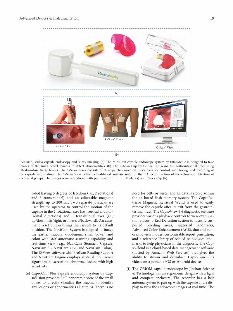
robot having 5 degrees of freedom (i.e., 2 rotationaland 3 translational) and an adjustable magneticstrength up to 200mT. Two separate joysticks areused by the operator to control the motion of thecapsule in the 2 rotational axes (i.e., vertical and hor-izontal directions) and 3 translational axes (i.e.,up/down, left/right, or forward/backward). An auto-matic reset button brings the capsule to its defaultposition. The NaviCam System is adapted to imagethe gastric mucosa, duodenum, small bowel, andcolon with 360° automatic scanning capability andreal-time view (e.g., NaviCam Stomach Capsule,NaviCam SB, NaviCam UGI, and NaviCam Colon).The ESView software with ProScan Reading Supportand NaviCam Engine employs artificial intelligencealgorithms to screen out abnormal lesions with highsensitivity
(e) CapsoCam Plus capsule endoscopy system by Cap-soVision provides 360° panoramic view of the smallbowel to directly visualize the mucosa to identifyany lesions or abnormalities (Figure 6). There is no
need for belts or wires, and all data is stored withinthe on-board flash memory system. The CapsoRe-trieve Magnetic Retrieval Wand is used to easilyremove the capsule after its exit from the gastroin-testinal tract. The CapsoView 3.6 diagnostic softwareprovides various playback controls to view examina-tion videos, a Red Detection system to identify sus-pected bleeding areas, suggested landmarks,Advanced Color Enhancement (ACE), duo and pan-oramic view modes, customizable report generation,and a reference library of related pathologies/land-marks to help physicians in the diagnosis. The Cap-soCloud is a cloud-based data management software(hosted by Amazon Web Services) that gives theability to stream and download CapsoCam Plusvideos on a portable iOS or Android devices
(f) The OMOM capsule endoscope by Jinshan Science& Technology has an ergonomic design with a lightand compact enclosure. The recorder has a beltantenna system to pair up with the capsule and a dis-play to view the endoscopic images at real time. The
(a)
(b)
C-Scan® Cap
C-Scan® Track
C-Scan® View
Figure 5: Video capsule endoscopy and X-ray imaging. (a) The MiroCam capsule endoscope system by IntroMedic is designed to takeimages of the small bowel mucosa to detect abnormalities. (b) The C-Scan Cap by Check Cap scans the gastrointestinal tract usingultralow-dose X-ray beams. The C-Scan Track consists of three patches worn on user’s back for control, monitoring, and recording ofthe capsule information. The C-Scan View is their cloud-based analysis suite for the 3D reconstruction of the colon and detection ofcolorectal polyps. The images were reproduced with permission from IntroMedic (a) and Check Cap (b).
19Advanced Devices & Instrumentation

OMOM Vue Smart software provides a simple andeasy workflow experience for the user to controlvideo play options, skim through images, identifysimilar traits, and compare multiple cases in parallel.Image color enhancement (ICE) is provided by theRGB mode to clearly view the capillaries, mucosa,and areas of suspected bleeding. The OMOM plat-form is cost-effective compared to its peers, whichmakes it suitable for large-scale clinical studies ondiagnostic efficiency [130]
(g) Norika and Sayaka endoscopic capsules were devel-oped by RF System Lab. While the older Norika cap-sule has the camera fixed at one end, the newerSayaka capsule puts its camera on the side and addeda tiny stepper motor to rotate the camera. The
Sayaka capsule provides a 360° panoramic view andcaptures around 870,000 images during a typical 8-hour journey through the gastrointestinal tract. Bothcapsules are battery-free and powered wirelesslyfrom a vest that sends out microwave signals. Theraw images from different angles are transmitted toa nearby receiver and stitched together to recreate arectangular gastrointestinal map using an imagemosaicking technology
8.2. X-Ray Imaging and Microultrasound Capsules. Besideswhite light optics for video capsule endoscopy, X-rays andmicroultrasound have been used for imaging clinically rele-vant landmarks in the gastrointestinal tract as describedbelow.
(a)
(b)
Figure 6: Rapid reading software by CapsoVision. (a) The CapsoCam Plus System uses four lateral cameras to capture full 360° panoramicview of the small bowel mucosa. The high-resolution images are reviewed on the CapsoView 3.6 diagnostic interface that has severalfeatures, such as multiple viewing modes, playback adjustments, easy archiving, Red Detection system, suggested landmarks, and reportgeneration. (b) Examples of image streams of the small bowel mucosa captured by the CapsoCam Plus and reviewed by the CapsoView3.6 diagnostic interface. The images were reproduced with permission from CapsoVision.
20 Advanced Devices & Instrumentation
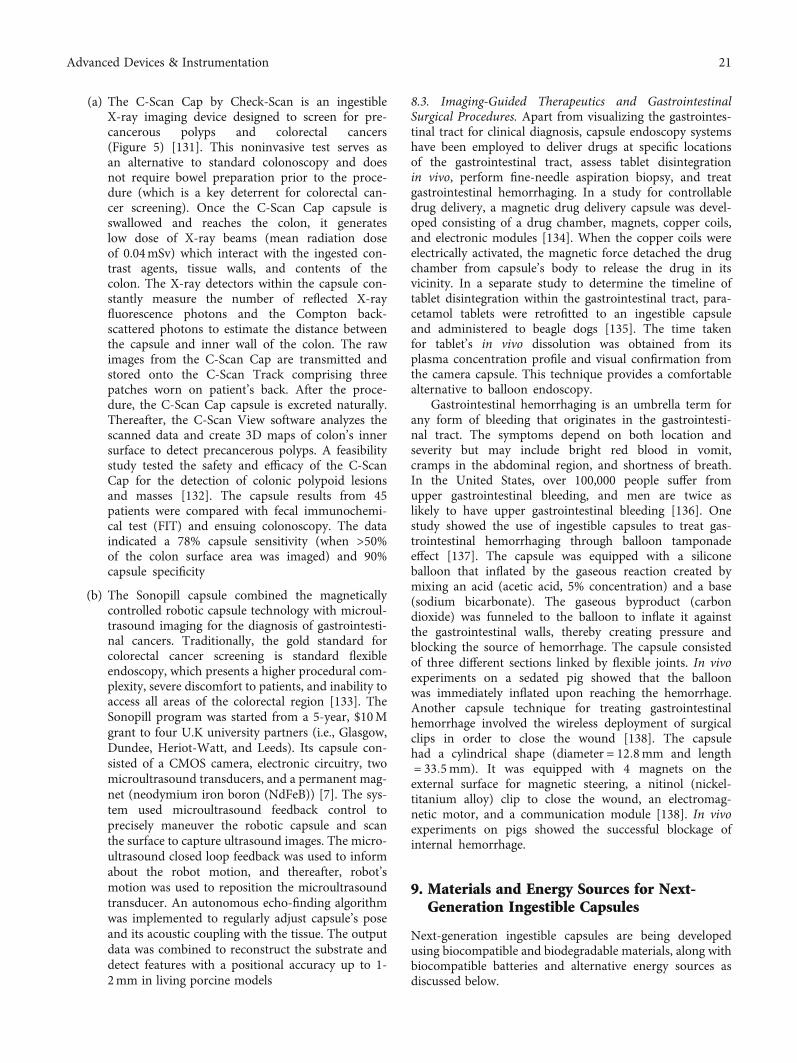
(a) The C-Scan Cap by Check-Scan is an ingestibleX-ray imaging device designed to screen for pre-cancerous polyps and colorectal cancers(Figure 5) [131]. This noninvasive test serves asan alternative to standard colonoscopy and doesnot require bowel preparation prior to the proce-dure (which is a key deterrent for colorectal can-cer screening). Once the C-Scan Cap capsule isswallowed and reaches the colon, it generateslow dose of X-ray beams (mean radiation doseof 0.04mSv) which interact with the ingested con-trast agents, tissue walls, and contents of thecolon. The X-ray detectors within the capsule con-stantly measure the number of reflected X-rayfluorescence photons and the Compton back-scattered photons to estimate the distance betweenthe capsule and inner wall of the colon. The rawimages from the C-Scan Cap are transmitted andstored onto the C-Scan Track comprising threepatches worn on patient’s back. After the proce-dure, the C-Scan Cap capsule is excreted naturally.Thereafter, the C-Scan View software analyzes thescanned data and create 3D maps of colon’s innersurface to detect precancerous polyps. A feasibilitystudy tested the safety and efficacy of the C-ScanCap for the detection of colonic polypoid lesionsand masses [132]. The capsule results from 45patients were compared with fecal immunochemi-cal test (FIT) and ensuing colonoscopy. The dataindicated a 78% capsule sensitivity (when >50%of the colon surface area was imaged) and 90%capsule specificity
(b) The Sonopill capsule combined the magneticallycontrolled robotic capsule technology with microul-trasound imaging for the diagnosis of gastrointesti-nal cancers. Traditionally, the gold standard forcolorectal cancer screening is standard flexibleendoscopy, which presents a higher procedural com-plexity, severe discomfort to patients, and inability toaccess all areas of the colorectal region [133]. TheSonopill program was started from a 5-year, $10Mgrant to four U.K university partners (i.e., Glasgow,Dundee, Heriot-Watt, and Leeds). Its capsule con-sisted of a CMOS camera, electronic circuitry, twomicroultrasound transducers, and a permanent mag-net (neodymium iron boron (NdFeB)) [7]. The sys-tem used microultrasound feedback control toprecisely maneuver the robotic capsule and scanthe surface to capture ultrasound images. The micro-ultrasound closed loop feedback was used to informabout the robot motion, and thereafter, robot’smotion was used to reposition the microultrasoundtransducer. An autonomous echo-finding algorithmwas implemented to regularly adjust capsule’s poseand its acoustic coupling with the tissue. The outputdata was combined to reconstruct the substrate anddetect features with a positional accuracy up to 1-2mm in living porcine models
8.3. Imaging-Guided Therapeutics and GastrointestinalSurgical Procedures. Apart from visualizing the gastrointes-tinal tract for clinical diagnosis, capsule endoscopy systemshave been employed to deliver drugs at specific locationsof the gastrointestinal tract, assess tablet disintegrationin vivo, perform fine-needle aspiration biopsy, and treatgastrointestinal hemorrhaging. In a study for controllabledrug delivery, a magnetic drug delivery capsule was devel-oped consisting of a drug chamber, magnets, copper coils,and electronic modules [134]. When the copper coils wereelectrically activated, the magnetic force detached the drugchamber from capsule’s body to release the drug in itsvicinity. In a separate study to determine the timeline oftablet disintegration within the gastrointestinal tract, para-cetamol tablets were retrofitted to an ingestible capsuleand administered to beagle dogs [135]. The time takenfor tablet’s in vivo dissolution was obtained from itsplasma concentration profile and visual confirmation fromthe camera capsule. This technique provides a comfortablealternative to balloon endoscopy.
Gastrointestinal hemorrhaging is an umbrella term forany form of bleeding that originates in the gastrointesti-nal tract. The symptoms depend on both location andseverity but may include bright red blood in vomit,cramps in the abdominal region, and shortness of breath.In the United States, over 100,000 people suffer fromupper gastrointestinal bleeding, and men are twice aslikely to have upper gastrointestinal bleeding [136]. Onestudy showed the use of ingestible capsules to treat gas-trointestinal hemorrhaging through balloon tamponadeeffect [137]. The capsule was equipped with a siliconeballoon that inflated by the gaseous reaction created bymixing an acid (acetic acid, 5% concentration) and a base(sodium bicarbonate). The gaseous byproduct (carbondioxide) was funneled to the balloon to inflate it againstthe gastrointestinal walls, thereby creating pressure andblocking the source of hemorrhage. The capsule consistedof three different sections linked by flexible joints. In vivoexperiments on a sedated pig showed that the balloonwas immediately inflated upon reaching the hemorrhage.Another capsule technique for treating gastrointestinalhemorrhage involved the wireless deployment of surgicalclips in order to close the wound [138]. The capsulehad a cylindrical shape (diameter = 12:8mm and length= 33:5mm). It was equipped with 4 magnets on theexternal surface for magnetic steering, a nitinol (nickel-titanium alloy) clip to close the wound, an electromag-netic motor, and a communication module [138]. In vivoexperiments on pigs showed the successful blockage ofinternal hemorrhage.
9. Materials and Energy Sources for Next-Generation Ingestible Capsules
Next-generation ingestible capsules are being developedusing biocompatible and biodegradable materials, along withbiocompatible batteries and alternative energy sources asdiscussed below.
21Advanced Devices & Instrumentation

9.1. Biocompatible Materials, Edible Food, and Nutrients.Biocompatibility describes the ability of the capsule systemto exist in harmony with the gastrointestinal system. By fab-ricating biocompatible capsule components, the risks of pro-longed capsule retention can be mitigated [122, 139]. Thephysical dimensions and mechanical characteristics of thecapsule also dictate its biocompatibility with the gastrointes-tinal tract. For instance, the maximum size of capsule’s rigidouter body is limited to the diameter of the smallest passagewithin the gastrointestinal tract (i.e., 12:8 ± 7mm). This spa-tial constraint sets an upper limit on capsule’s functionality.As such, flexible biocompatible materials are sought that cansimultaneously address the issues of size limitation and pro-longed capsule retention.
Research into biocompatible electronics for capsules hasgarnered considerable interest [14, 140–143]. A team ofresearchers printed active electronic devices on edible sub-strates using temporary tattoo paper, biocompatible con-ducting polymers, and inkjet circuit printing technology[144]. The p-type and n-type organic field-effect transistor-based circuits (OFETs) were fabricated and transferred ontothe surface of a hard gelatin capsule by soaking in water andpressing them onto the target surface. The submicron thickethyl cellulose film within the temporary tattoo paper servedas the transferrable substrate and as the gate dielectric of theOFETs. The biocompatible materials used to form the tran-sistors were poly(3-hexylthiophene) (P3HT) and differentdonor–acceptor copolymers. While this work realized a sim-ple logic inverter, it showed the possibility of using printableedible electronics for ingestible capsules.
A commercial example of an ingestible capsule systemthat incorporates biocompatible materials is the IngestibleSensor System (ISS) developed by Proteus Digital Health totest the patient adherence to oral medications. Its IngestibleEvent Marker (IEM) sensor has an integrated circuit housedwithin an edible, cellulose disc. Thin layers of copper andmagnesium (10μm) form the biogalvanic battery on the sur-face of the IEM sensor. The extractable amount of copperand magnesium from a single IEM sensor was minimal com-pared to the daily allowable intake levels (0.3% and 0.003%,respectively) [145]. Ethyl citrate was extracted from the IEMsensor as the organic species resulting from the breakdownof triethyl citrate (considered a GRAS food additive andinert material).
The design of ingestible capsules has benefitted from theemerging trends in transient electronics where the materialand devices can physically degrade in a controllable manner.Within the gastrointestinal tract, the degradability and depo-lymerization traits of the transient electronics can be tunedbased on its transit time or external triggers (e.g., pH, tem-perature, light, or solubility in solvents found in the gastro-intestinal tract). In one study, transient antennas weredeveloped in a degradable composite film made of water-soluble poly (vinyl alcohol) (PVA) [146]. The dissolutionrate and dielectric properties of the PVA film was tuned bydoping with TiO2 nanoparticles. The fabricated patch anten-nas degraded within one hour of water immersion and werespace-efficient to fit within capsules. Besides antennas, theflexible substrate films have been chosen that conform to
the capsule walls. Biodegradable films (e.g., polyanhydrides)have been tested as substrates that can partially orcompletely degrade at finite time points. The biocompatibil-ity of substrates reduces the health and safety risks associ-ated with long-term capsule retention beyond 24 to 48hours. Flexible and stretchable components, such as anten-nae, can be packaged in miniaturized form factors to facili-tate the initial swallowing and transit and expanded tolarger form factors upon reaching the small bowel or colon[8, 147]. On the other hand, sensors and microelectronicsare often compressed into mechanically rigid structures tosave the real estate within capsules.
The materials for transient electronics are being chosenbased on their inherent characteristics, such as permeability,degradation rates, morphology, hydrophobicity or hydro-philicity, chemical properties, and mechanical properties ofthe polymer [115, 148, 149]. In a study on bioresorbable sil-icon electronic sensors, devices were fabricated on siliconnanomembranes, thin SiO2 layers, and magnesium foilsand molybdenum interconnects that can dissolve or disinte-grate in a time-controlled manner [140]. Another studydemonstrated a range of functional transient devices fabri-cated on polyanhydride substrates (~124μm thick) wheretransience was triggered by moisture and time-controlledby the polymeric chemistry [150]. The anhydride groups inthe polymeric substrate absorbed the surrounding moistureto cause its hydrolysis and eventually degrade into a viscousliquid. Various transient electronic devices were fabricatedfrom metals (e.g., copper, nickel, and aluminum), semicon-ductors (e.g., indium gallium zinc oxide (IGZO)), anddielectrics (e.g., magnesium oxide) [150].
The material toolkit for next-generation ingestible cap-sules can be constructed from a number of constituents inedible food and nutrients (such as minerals, vitamins, inor-ganics, organics, and food additives). Table 2 lists the differ-ent categories of biocompatible materials for next-generation capsules: substrates, fillers, coatings, conductors,electrodes, and electrolytes. To help choose the appropriatebiocompatible material, a system of nutrition recommenda-tions and dietary reference intake (DRI) can be used asreference, which is maintained by the U.S. National Acade-mies and Institute of Medicine. As a note, the recommendeddietary allowance (RDA) indicates the sufficient daily intakelevel of essential nutrients, while the tolerable upper intakelevel (UL) refers to the highest intake level of nutrients con-sidered safe with no side effects. Materials tabulated by theUS RDA include minerals, organic molecules, polymers,and inorganic materials. Minerals that have tabulated RDAand electrical properties can be used to create essential com-ponents within ingestible electronic devices, such as theanode, cathode, electrode, flow cell ions, and active layer.The body can tolerate a threshold of most materials beforethe concentration within the blood stream and tissuebecomes detrimental [151]. In addition, FDA has classifiedcertain chemicals and substances as “Generally RecognizedAs Safe” (GRAS) for common human consumption. Thepreapproved safety of GRAS materials makes them goodcandidates for use in ingestible capsules as they have a lesserpath of resistance for clinical trials.
22 Advanced Devices & Instrumentation

9.2. Batteries and Alternative Energy Sources. Silver-oxidebutton batteries are popular energy sources for portable elec-tronics because of their high energy-to-weight ratio. How-ever, silver-oxide button batteries occupy significant realestate within ingestible capsules and often become the decid-ing factor for the capsule size. The shape and size of thesebatteries pose potential obstruction and health risks in chil-dren when swallowed [152, 153]. The alkaline solutionwithin ingested silver-oxide button batteries can cause severetissue damage in the mouth, vocal cord, trachea, or esopha-gus [154, 155]. Lithium cells are more dangerous than silver-oxide batteries, especially when ingested by children [153,155]. When the alkaline electrolyte of the lithium cell leaksinto the stomach and comes in contact with theelectrolyte-rich stomach fluid, undesired electrical currentis generated. This results in the local production of hydrox-ide ions and pH change within the stomach, leading tonecrosis and potential fatalities in children [152, 156, 157].
Solid-state cells and biocompatible batteries are poten-tially safer than conventional button batteries because thereis no leakage of toxic electrolytes or associated risks of inter-nal tissue damage. Biocompatible batteries have a variety ofmaterial choices for the anode (e.g., magnesium, zinc,magnesium-aluminum-zinc alloy, or melanin), cathode(copper, cupric chloride, iron, platinum, or manganese diox-ide), and electrolyte (e.g., sodium chloride, phosphate buffer,saliva, or simulated gastric fluid). As an example, a biocom-patible galvanic cell was demonstrated where gastric acidand intestinal fluids served as the electrolyte [158]. A redoxreaction occurred between the dissolving metallic anode(zinc or magnesium) and an inert cathode (pure coppermetal). In vitro experiments indicated that the zinc anodewas better suited for longer term use because the magnesiumanode was prone to greater corrosion and shorter lifetime(<24 hours). Their energy harvesting system was tested ina porcine stomach to power the temperature-sensing elec-tronics housed within the capsule and wirelessly transmitthe signal to a base station (as a 920MHz, FSK modulatedsignal). The average amount of zinc absorbed in the bodyfrom this galvanic cell (i.e., 27mg/day) was estimated to bebelow the recommended upper limit (i.e., 40mg/day). Adrawback of biocompatible batteries is that the battery per-formance can fluctuate due to variations in the environmen-tal conditions of the gastrointestinal tract.
Alternative energy sources have been explored foringestible capsules, including biofuel cells, nuclear batteries,optical charging, and transducers of various types (e.g., ther-moelectric, piezoelectric, electrostatic, electromagnetic, andultrasound). Within the field of chargeable ingestible batte-ries, a polysaccharide gel battery based on the Gibbs-Donnan effect (i.e., Donnan dialysis) was recently demon-strated to fight harmful bacteria in the oral cavity [159]. Intheir work, electrochemical potential was generated by ionicdiffusion from potassium chloride saline gradients withinagarose gels. The battery and triboelectric nanogeneratorwere created from biocompatible materials, such as agarosegel, platinum, polyethylene terephthalate (PET), polytetra-fluoroethylene (PTFE), chitosan/glycerol film, and silicon.An optimized battery configuration produced an open cir-
cuit potential up to 177mV and the nanogenerator wascapable of supplying 300mV. A system using two such bat-teries in series was demonstrated as an antibacterial electro-stimulation device for the oral cavity. Their test resultsshowed that 90% of E. coli were inhibited after 30 minutesof treatment.
Besides on-board energy sources, remote powering ofingestible capsules using wireless power transmission havebeen investigated by researchers [59, 160]. In one approach,a Helmholtz transmitting coil was attached to the patientand a ferrite-core three-dimensional receiving coil wasplaced in the capsule. Electromagnetic fields were used toenergize the capsule, providing sufficient power (i.e.,330mW to 500mW) to the capsule for most of its functions.The design and size of the antenna determined the fre-quency of optimal transmission. As a drawback, wirelesspower transmission is critically dependent on the distancebetween the source and receiving antenna and capsule’s rel-ative orientation to the incoming radiation. There is signifi-cant signal loss by the time the electromagnetic waves reachthe deep gastrointestinal tissues (where the ingestible cap-sules typically operate).
10. Discussion: Present Challenges and Outlook
First and foremost, it is challenging for a clinic or hospital toconstantly assess and improve the endoscopic service perfor-mance measures with root cause analyses of major eventsand feedback to their gastroenterologists and medical staff[40]. Moreover, these procedures are not appropriate forpatients having gastrointestinal obstructions, peptic strictureor ulcer disease, urethral strictures, urinary fistulas, uncon-trolled diabetes, swallowing disorders, prior gastrointestinalsurgery, and implanted electromedical devices. To avoidelectromagnetic interference during the time from capsuleingestion to excretion, it is advised to stay away from sourcesof powerful electromagnetic fields (e.g., MRI instrument).There may be frequent signal loss, difficulties in knowingthe exact location of the capsule, or loss of battery powerbefore excretion [76]. On the technical side, there are highcosts associated with advanced microchips, softwareupgrades, and clinical validation that is expected to limitthe market growth of smart pills [5]. Furthermore, it is diffi-cult to reconstruct and democratize the sensing and record-ing circuitry from off-the-shelf components, which limits thenumber of commercial ingestible capsule products [161].
The procedures involving ingestible capsules have healthrisks, such as capsule retention, aspiration, obstruction,mucosal injury, internal bleeding, and irritation. In particu-lar, capsule retention in the gastrointestinal tract can havesignificant health complications depending upon the preex-isting conditions of the patient and nature of capsule mate-rials [151]. A study found that capsule retention followingvideo capsule endoscopy was predominantly found in indi-viduals with bowel disease (1.4% retention rate) and Crohn’sdisease (13% retention rate) where the retention of capsulesoften resulted in gastrointestinal obstruction [162]. A surveyof 22,840 procedures using small bowel capsule endoscopyshowed a significant retention rate for common indications
23Advanced Devices & Instrumentation

such as obscure gastrointestinal bleeding (1.2%), suspectedCrohn’s disease (2.6%), and neoplastic lesions (2.1%) [163].Other underlying conditions for capsule retention areobstructive tumors and diaphragm disease from the sideeffects of anti-inflammatory drugs [164]. Extended retentionof a rigid-shell capsule can result in lumen perforation andcapsule disintegration with undesired toxicity from releasedmaterials. In such cases of prolonged capsule retention(especially having small-bowel strictures), the retrieval ofcapsule is assisted by conventional deep enteroscopy, pushenteroscopy, or motorized spiral enteroscopy [165]. Toaddress the risks of capsule retention, Medtronic has devisedmedical procedures to evaluate the safety of patients beforeproceeding with video capsule endoscopy. For instance, itsPillCam Patency Capsule is ingested by the patient prior tovideo capsule endoscopy to check for known obstructionsand is possibly excreted within 30 hours. A study of 38patients with risk factors of capsule retention showed thatthe PillCam Patency Capsule was naturally excreted in mostcases, and video capsule endoscopy was safely performedafterwards in those patients [166].
Clinical testing of capsule prototypes on humans andlaboratory animals is nontrivial, time-consuming, andexpensive. Because of interpatient variability in terms of dis-ease nature and treatment history, the patients who volun-teer for clinical trials may not be representative of a targetdiseased group or the general healthy population [11]. It isdifficult to correlate gastrointestinal parameters such as gas-tric emptying time, pH, gastrointestinal motility, colon con-tractions, and transit times both in healthy individuals andpatients with preexisting conditions (e.g., obesity, chronicconstipation, and diabetic gastroparesis) [79]. As an exam-ple, a validation study showed that temperature-sensingingestible capsules have rapid temperature oscillations, espe-cially in obese individuals, with temperature drifts comparedto electronic and mercury thermometers [68]. The samestudy also revealed the current dearth of knowledge in relat-ing the core body temperature to the body mass index, gen-der, age, and daily movement and activity and presence offood or anatomical location in the gut [68]. It is challengingto optimize the experimental parameters for differentpatients during video endoscopy, physiological sensing, orstimulation. In fact, any capsule system and its procedurehave a large number of parameters with a range of possiblevariations, which makes it difficult to make informed guessesand predict outcomes, especially in patients with gastrointes-tinal disorders [11]. Synthetic gut models fail to emulate thestructure and functions of live gastrointestinal tracts, such asperistalsis, bowel movement, and slippery mucosa layer.Peristalsis, for example, can force the capsules to go throughcertain sections of the gastrointestinal tract at differentspeeds, which may not allow sufficient time to take clearimages. This can lead to lesions and other diagnosticpointers being overlooked or misread [167]. For video cap-sule endoscopy, this can prevent the reliable acquisition ofhigh-resolution gastrointestinal images, reimaging and pin-pointing the origin of a biological marker, and rapid readingof the acquired images with acceptable speed and accu-racy [168].
The outlook for ingestible capsules involves addressingthe present challenges and testing strategies for technicaladvancements and patient adoption. The field is movingtowards a single, multifunctional, integrated circuit chipwith in-built CMOS image sensors, physiochemical sensors,ASICs, memory units, transceivers, and antenna-in-package.Novel materials are being investigated with properties toencapsulate electronic components, perform energy harvest-ing, provide mechanical strength for faster transit, and havechemical properties for bio-absorption and degradability[151]. In the near future, closed-loop feedback will becomeessential to monitor the target parameters for gastrointesti-nal disorders at quasi real time and to automatically adjustthe external control in a timely manner. The applicationsof small bowel capsule endoscopy will be expanded and val-idated for therapeutic functions such as for the treatment ofobscure gastrointestinal bleeding or the detection of mucosaldamage from drugs and NSAIDS injury [124]. As we betterunderstand the intricacies of the gut-brain axis connection,the probiotics industry is emerging to be a big business forthe assessment, exploitation, and rebuilding of the gutmicrobiota and commensal balance [107]. In this context,ingestible capsules could be developed to detect inflamma-tory proteins, hormones, or metabolites which are indicativeviral infections or a modulated gut microbiome [169]. Withexpanding knowledge of sacral and vagal nerve pathwaysand underlying mechanisms, the emerging technologies ofbioelectric neuromodulation and gastric electrical stimula-tion (GES) could be tailored and optimized for the treatmentof gut inflammation, obesity, gastroparesis, and fecal incon-tinence [11]. The rising rates of gastrointestinal diseases andcancers suggest that we will see further subspecializations intherapeutic and diagnostic endoscopy with emphasis onprocedural training, computing skills, and service quality[10]. The performance measures for endoscopic procedureswill evolve with time and affect the organization, leadership,staffing, quality and safety, and patient involvement [40].Artificial intelligence and personalized treatments will playa greater role in endoscopic therapeutics and diagnosis.Lastly, video capsule endoscopy could effectively comple-ment existing procedures in surgery and radiology by estab-lishing safe and high-quality out-patient procedures,reduced medical complications, and quicker diagnostics atlower costs [10].
Data Availability
All the data is presented in the paper. Please contact S.P. foradditional information.
Conflicts of Interest
The authors declare that they have no competing interests.
Authors’ Contributions
S. P. planned the content and organized the structure of thepaper. S. P., D. M., and L. B. communicated with the capsulemanufacturers. All authors contributed to the figures, data
24 Advanced Devices & Instrumentation

tabulation, and discussion. S. P., D.M., and L. B wrote thefirst draft of the manuscript. All authors, including S. P., K.Y., and A. J., revised the subsequent drafts. Dylan Mileyand Leonardo Bertoncello Machado contributed equally tothis work.
Acknowledgments
We are grateful to the capsule manufacturers for our discus-sions and giving permission to reproduce their technologyfigures here. This work was partially supported by theUnited States National Science Foundation (NSF IDBR-1556370) and U.S. Department of Agriculture (2020-67021-31964) to S.P.
References
[1] K. Kalantar-Zadeh, N. Ha, J. Z. Ou, and K. J. Berean, “Ingest-ible sensors,” ACS Sensors, vol. 2, no. 4, pp. 468–483, 2017.
[2] C. Steiger, A. Abramson, P. Nadeau, A. P. Chandrakasan,R. Langer, and G. Traverso, “Ingestible electronics for diag-nostics and therapy,” Nature Reviews Materials, vol. 4,no. 2, pp. 83–98, 2019.
[3] F. Carpi and C. Pappone, “Stereotaxis Niobe® magnetic nav-igation system for endocardial catheter ablation and gastroin-testinal capsule endoscopy,” Expert Review of MedicalDevices, vol. 6, no. 5, pp. 487–498, 2009.
[4] G. Cummins, “Smart pills for gastrointestinal diagnostics andtherapy,” Advanced Drug Delivery Reviews, vol. 177, article113931, 2021.
[5] Smart Pills Market, “Markets and Markets Research,” 2021,October 2021, https://www.marketsandmarkets.com/Market-Reports/smart-pill-technology-market-840.html.
[6] G. Ciuti, R. Caliò, D. Camboni et al., “Frontiers of roboticendoscopic capsules : a review,” Journal of Micro-Bio Robot-ics, vol. 11, no. 1-4, pp. 1–18, 2016.
[7] J. C. Norton, P. R. Slawinski, H. S. Lay et al., “Intelligent mag-netic manipulation for gastrointestinal ultrasound,” ScienceRobotics, vol. 4, no. 31, Article ID eaav7725, 2019.
[8] J. Min, Y. Yang, Z. Wu, and W. Gao, “Robotics in the gut,”Advanced Therapeutics, vol. 3, no. 4, article 1900125, 2020.
[9] A.-M. Singeap, “Capsule endoscopy: the road ahead,” WorldJournal of Gastroenterology, vol. 22, no. 1, pp. 369–378, 2016.
[10] A. Axon, “Fifty years of digestive endoscopy: successes, set-backs, solutions and the future,” Digestive Endoscopy,vol. 32, no. 3, pp. 290–297, 2020.
[11] S. C. Payne, J. B. Furness, and M. J. Stebbing, “Bioelectricneuromodulation for gastrointestinal disorders: effectivenessand mechanisms,” Nature Reviews Gastroenterology andHepatology, vol. 16, no. 2, pp. 89–105, 2019.
[12] M.-M. Pan, Y.-F. Wang, L. Wang, X. Yu, and L. Xu, “Recentadvances in visual detection for cancer biomarkers and infec-tious pathogens,” Journal of Materials Chemistry B, vol. 9,no. 1, pp. 35–52, 2021.
[13] N. van Helleputte, A. J. G. Even, F. Leonardi et al., “Miniatur-ized electronic circuit design challenges for ingestibledevices,” Journal of Microelectromechanical Systems, vol. 29,no. 5, pp. 645–652, 2020.
[14] C. J. Bettinger, “Advances in materials and structures foringestible electromechanical medical devices,” Angewandte
Chemie International Edition, vol. 57, no. 52, pp. 16946–16958, 2018.
[15] T. T. Kararli, “Comparison of the gastrointestinal anatomy,physiology, and biochemistry of humans and commonly usedlaboratory animals,” Biopharmaceutics & Drug Disposition,vol. 16, no. 5, pp. 351–380, 1995.
[16] G. Chawla, P. Gupta, V. Koradia, and A. K. Bansal, “Ameansto address intestinal drug absorption,” Pharmaceutical Tech-nology, vol. 27, pp. 50–68, 2003.
[17] M. Rao and M. D. Gershon, “The bowel and beyond: theenteric nervous system in neurological disorders,” NatureReviews Gastroenterology & Hepatology, vol. 13, no. 9,pp. 517–528, 2016.
[18] F. Sommer and F. Bäckhed, “The gut microbiota –masters ofhost development and physiology,” Nature Reviews Microbi-ology, vol. 11, no. 4, pp. 227–238, 2013.
[19] F. Munoz, G. Alici, and W. Li, “A review of drug delivery sys-tems for capsule endoscopy,” Advanced Drug DeliveryReviews, vol. 71, pp. 77–85, 2014.
[20] P. J. Turnbaugh, R. E. Ley, M. Hamady, C. M. Fraser-Liggett,R. Knight, and J. I. Gordon, “The human microbiome pro-ject,” Nature, vol. 449, no. 7164, pp. 804–810, 2007.
[21] G. Falony, M. Joossens, S. Vieira-Silva et al., “Population-level analysis of gut microbiome variation,” Science,vol. 352, no. 6285, pp. 560–564, 2016.
[22] G. B. Hatton, V. Yadav, A. W. Basit, and H. A. Merchant,“Animal farm: considerations in animal gastrointestinalphysiology and relevance to drug delivery in humans,” Jour-nal of Pharmaceutical Sciences, vol. 104, no. 9, pp. 2747–2776, 2015.
[23] L. P. Coelho, J. R. Kultima, P. I. Costea et al., “Similarityof the dog and human gut microbiomes in gene contentand response to diet,” Microbiome, vol. 6, no. 1, p. 72,2018.
[24] L. Chandra, D. C. Borcherding, D. Kingsbury et al., “Deriva-tion of adult canine intestinal organoids for translationalresearch in gastroenterology,” BMC Biology, vol. 17, no. 1,p. 33, 2019.
[25] J. B. Furness, J. J. Cottrell, and D. M. Bravo, “ComparativeGut Physiology Symposium: Comparative physiology ofdigestion,” Journal of Animal Science, vol. 93, no. 2,pp. 485–491, 2015.
[26] Q. Sciascia, G. Daş, and C. C. Metges, “Review: the pig as amodel for humans: effects of nutritional factors on intestinalfunction and health1,” Journal of Animal Science, vol. 94,Supplement 3, pp. 441–452, 2016.
[27] L. M. Gonzalez, A. J. Moeser, and A. T. Blikslager, “Porcinemodels of digestive disease: the future of large animal transla-tional research,” Translational Research, vol. 166, no. 1,pp. 12–27, 2015.
[28] M. Lukas, M. Kolar, O. Ryska et al., “Novel porcine model ofCrohn's disease anastomotic stricture suitable for evaluationand training of advanced endoscopic techniques,” Gastroin-testinal Endoscopy, vol. 93, no. 1, pp. 250–256, 2021.
[29] Q. Zhang, G. Widmer, and S. Tzipori, “A pig model of thehuman gastrointestinal tract,” Gut Microbes, vol. 4, no. 3,pp. 193–200, 2013.
[30] C. E. Stevens and I. D. Hume, “Contributions of microbes invertebrate gastrointestinal tract to production and conserva-tion of nutrients,” Physiological Reviews, vol. 78, no. 2,pp. 393–427, 1998.
25Advanced Devices & Instrumentation

[31] M. Carabotti, A. Scirocco, M. A. Maselli, and C. Severi, “Thegut-brain axis: interactions between enteric microbiota, cen-tral and enteric nervous systems,”Annals of Gastroenterology,vol. 28, no. 2, pp. 203–209, 2015.
[32] J. C. Clemente, J. Manasson, and J. U. Scher, “The role of thegut microbiome in systemic inflammatory disease,” BMJ,vol. 360, article j5145, 2018.
[33] L. Wang, A. Mannalithara, G. Singh, and U. Ladabaum, “Lowrates of gastrointestinal and non-gastrointestinal complica-tions for screening or surveillance colonoscopies in apopulation-based study,” Gastroenterology, vol. 154, no. 3,pp. 540–555.e8, 2018.
[34] D. Becker, J. Zhang, T. Heimbach et al., “Novel orally swal-lowable IntelliCap® device to quantify regional drug absorp-tion in human GI tract using diltiazem as model drug,”Ageing International, vol. 15, pp. 1490–1497, 2014.
[35] K. Kalantar-Zadeh, K. J. Berean, R. E. Burgell, J. G. Muir, andP. R. Gibson, “Intestinal gases: influence on gut disorders andthe role of dietary manipulations,” Nature Reviews Gastroen-terology & Hepatology, vol. 16, no. 12, pp. 733–747, 2019.
[36] S. N. Adler, “The history of time for capsule endoscopy,”Annals of Translational Medicine, vol. 5, no. 9, p. 194, 2017.
[37] P. Swain, “Wireless capsule endoscopy,” Gut, vol. 52,no. 90004, pp. iv48–iv50, 2003.
[38] G. Iddan, G. Meron, A. Glukhovsky, and P. Swain, “Wirelesscapsule endoscopy,” Nature, vol. 405, no. 6785, p. 417, 2000.
[39] L. A. Beardslee, G. E. Banis, S. Chu et al., “Ingestible sensorsand sensing systems for minimally invasive diagnosis andmonitoring: the next frontier in minimally invasive screen-ing,” ACS Sensors, vol. 5, no. 4, pp. 891–910, 2020.
[40] C. Spada, D. McNamara, E. J. Despott et al., “Performancemeasures for small-bowel endoscopy: a European Society ofGastrointestinal Endoscopy (ESGE) quality improvementinitiative,” United European Gastroenterology Journal,vol. 7, no. 5, pp. 614–641, 2019.
[41] “Ingestible telemetric gastrointestinal capsule imagingsystem-final class II special controls guidance document forindustry and FDA,” Food and Drug Administration, Centerfor Devices and Radiological Health, U.S. Department ofHealth and Human Services, 2001, February 2021, https://www.fda.gov/medical-devices/guidance-documents-medical-devices-and-radiation-emitting-products/ingestible-telemetric-gastrointestinal-capsule-imaging-system-final-class-ii-special-controls.
[42] “Guidance for the content of premarket submissions for soft-ware contained in medical devices,” Food and Drug Admin-istration, Center for Devices and Radiological Health, 2005,February 2021, https://www.fda.gov/regulatory-information/search-fda-guidance-documents/guidance-content-premarket-submissions-software-contained-medical-devices.
[43] D. L. Donoho, “Compressed sensing,” IEEE Transactions onInformation Theory, vol. 52, no. 4, pp. 1289–1306, 2006.
[44] A. Kiourti, K. A. Psathas, and K. S. Nikita, “Implantable andingestible medical devices with wireless telemetry functional-ities: a review of current status and challenges,” Bioelectro-magnetics, vol. 35, no. 1, pp. 1–15, 2014.
[45] M. J. Christoe, J. Han, and K. Kalantar-Zadeh, “Telecommu-nications and data processing in flexible electronic systems,”Advanced Materials Technologies, vol. 5, no. 1, article1900733, 2020.
[46] L. Wang, T. D. Drysdale, and D. R. S. Cumming, “In situcharacterization of two wireless transmission schemes foringestible capsules,” IEEE Transactions on Biomedical Engi-neering, vol. 54, no. 11, pp. 2020–2027, 2007.
[47] W. G. Scanlon, B. Burns, and N. E. Evans, “Radiowave prop-agation from a tissue-implanted source at 418MHz and 916.5MHz,” IEEE Transactions on Biomedical Engineering, vol. 47,no. 4, pp. 527–534, 2000.
[48] D. Nikolayev, M. Zhadobov, R. Sauleau, and P. Karban,“Antennas for ingestible capsule telemetry,” in Advances inBody-Centric Wireless Communication: Applications andstate-of-the-art, Q. H. Abbasi, M. Ur-Rehman, K. Qaraqe,and A. Alomainy, Eds., pp. 143–186, IET Press, 2016.
[49] J. Faerber, R. Gregson, R. E. Clutton et al., “In vivo character-ization of a wireless telemetry module for a capsule endos-copy system utilizing a conformal antenna,” IEEETransactions on Biomedical Circuits and Systems, vol. 12,no. 1, pp. 95–105, 2018.
[50] A. Kiourti and K. S. Nikita, “A review of in-body biotelemetrydevices: implantables, ingestibles, and injectables,” IEEETransactions on Biomedical Engineering, vol. 64, no. 7,pp. 1422–1430, 2017.
[51] K. S. Nikita, Handbook of Biomedical Telemetry, Wiley-IEEEPress, 2014.
[52] D. Kurup, W. Joseph, G. Vermeeren, and L. Martens, “In-body path loss model for homogeneous human tissues,” IEEETransactions on Electromagnetic Compatibility, vol. 54, no. 3,pp. 556–564, 2012.
[53] K. Lopez-Linares Roman, G. Vermeeren, A. Thielens,W. Joseph, and L. Martens, “Characterization of path lossand absorption for a wireless radio frequency link betweenan in-body endoscopy capsule and a receiver outside thebody,” EURASIP Journal on Wireless Communications andNetworking, vol. 2014, no. 1, 2014.
[54] M. R. Yuce and T. Dissanayake, “Easy-to-swallow wirelesstelemetry,” IEEE Microwave Magazine, vol. 13, no. 6,pp. 90–101, 2012.
[55] S. H. Woo, K. W. Yoon, Y. K. Moon et al., “High speedreceiver for capsule endoscope,” Journal of Medical Systems,vol. 34, no. 5, pp. 843–847, 2010.
[56] E. A. Johannessen, Lei Wang, C. Wyse, D. R. S. Cumming,and J. M. Cooper, “Biocompatibility of a lab-on-a-pill sensorin artificial gastrointestinal environments,” IEEE Transac-tions on Biomedical Engineering, vol. 53, no. 11, pp. 2333–2340, 2006.
[57] S. Yoshida, H. Miyaguchi, and T. Nakamura, “Developmentof tablet-shaped ingestible core-body thermometer poweredby gastric acid battery,” IEEE Sensors Journal, vol. 18,no. 23, pp. 9755–9762, 2018.
[58] F. Xu, G. Yan, K. Zhao, L. Lu, J. Gao, and G. Liu, “A wirelesscapsule system with ASIC for monitoring the physiologicalsignals of the human gastrointestinal tract,” IEEE transac-tions on biomedical circuits and systems, vol. 8, no. 6,pp. 871–880, 2014.
[59] Y. Guozheng, H. Biao, and Z. Peng, “Design of battery-less and real-time telemetry system for gastrointestinalapplications,” in 2007 IEEE International Conference onControl and Automation, pp. 245–249, Guangzhou,China, 2007.
[60] W. A. Weeks, A. Dua, J. Hutchison et al., “A low-power, low-cost ingestible and wearable sensing platform to measure
26 Advanced Devices & Instrumentation

medication adherence and physiological signals,” in 201840th Annual International Conference of the IEEE Engineer-ing in Medicine and Biology Society (EMBC), pp. 5549–5553, Honolulu, HI, USA, 2018.
[61] P. Ryan and E. Diller, “Magnetic actuation for full dexteritymicrorobotic control using rotating permanent magnets,”IEEE Transactions on Robotics, vol. 33, no. 6, pp. 1398–1409, 2017.
[62] P. Shokrollahi, Y. P. Lai, S. Rash-Ahmadi et al., “Blindly con-trolled magnetically actuated capsule for noninvasive samplingof the gastrointestinal microbiome,” IEEE/ASME Transactionson Mechatronics, vol. 26, no. 5, pp. 2616–2628, 2021.
[63] V. H. le, H. L. Rodriguez, C. Lee et al., “A soft-magnet-baseddrug-delivery module for active locomotive intestinal capsuleendoscopy using an electromagnetic actuation system,” Sen-sors and Actuators A: Physical, vol. 243, pp. 81–89, 2016.
[64] A. W. Mahoney and J. J. Abbott, “Five-degree-of-freedommanipulation of an untethered magnetic device in fluid usinga single permanent magnet with application in stomach cap-sule endoscopy,” International Journal of Robotics Research,vol. 35, no. 1-3, pp. 129–147, 2016.
[65] C. A. Mosse, T. N. Mills, M. N. Appleyard, S. S. Kadirkama-nathan, and C. P. Swain, “Electrical stimulation for propellingendoscopes,” Gastrointestinal Endoscopy, vol. 54, no. 1,pp. 79–83, 2001.
[66] H.-J. Park, J. H. Lee, Y. K. Moon et al., “New method of mov-ing control for wireless endoscopic capsule using electricalstimuli,” IEICE Transactions on Fundamentals of Electronics,Communications and Computer Sciences, vol. E88-A, no. 6,pp. 1476–1480, 2005.
[67] S. Woo, T. W. Kim, Z. Mohy-Ud-Din, I. Y. Park, and J.-H. Cho, “Small intestinal model for electrically propelled cap-sule endoscopy,” BioMedical Engineering OnLine, vol. 10,no. 1, p. 108, 2011.
[68] C. R. Monnard, E. J. Fares, J. Calonne et al., “Issues in contin-uous 24-h core body temperature monitoring in humansusing an ingestible capsule telemetric sensor,” Frontiers inEndocrinology, vol. 8, no. 130, 2017.
[69] L. Landsberg, “Core temperature: a forgotten variable inenergy expenditure and obesity?,” Obesity Reviews, vol. 13,pp. 97–104, 2012.
[70] M. E. Hoffmann, S. M. Rodriguez, D. M. Zeiss et al., “24-hcore temperature in obese and lean men and women,” Obe-sity, vol. 20, no. 8, pp. 1585–1590, 2012.
[71] K. P. Wright, J. T. Hull, and C. A. Czeisler, “Relationshipbetween alertness, performance, and body temperature inhumans,” American Journal of Physiology-Regulatory, Inte-grative and Comparative Physiology, vol. 283, no. 6,pp. R1370–R1377, 2002.
[72] P. Valdez, T. Reilly, and J. Waterhouse, “Rhythms of mentalperformance,” Mind, Brain, and Education, vol. 2, no. 1,pp. 7–16, 2008.
[73] N. P. Walsh, S. L. Halson, C. Sargent et al., “Sleep and the ath-lete: narrative review and 2021 expert consensus recommen-dations,” British Journal of Sports Medicine, vol. 55, no. 7,pp. 356–368, 2021.
[74] E. C. Harding, N. P. Franks, andW. Wisden, “Sleep and ther-moregulation,” Current Opinion in Physiology, vol. 15, pp. 7–13, 2020.
[75] W. E. Scales, A. J. Vander, M. B. Brown, and M. J. Kluger,“Human circadian rhythms in temperature, trace metals,
and blood variables,” Journal of Applied Physiology, vol. 65,no. 4, pp. 1840–1846, 1988.
[76] J. M. Maurer, R. C. A. Schellekens, H. M. van Rieke et al.,“Gastrointestinal pH and transit time profiling in healthy vol-unteers using the IntelliCap system confirms ileo-colonicrelease of ColoPulse tablets,” PLoS One, vol. 10, no. 7, articlee0129076, 2015.
[77] K. Tran, R. Brun, and B. Kuo, “Evaluation of regional andwhole gut motility using the wireless motility capsule: rele-vance in clinical practice,” Therapeutic Advances in Gastroen-terology, vol. 5, no. 4, pp. 249–260, 2012.
[78] M. Koziolek, M. Grimm, D. Becker et al., “Investigation ofpH and temperature profiles in the GI tract of fastedhuman subjects using the Intellicap® system,” Journal ofPharmaceutical Sciences, vol. 104, no. 9, pp. 2855–2863,2015.
[79] R. Coleski, G. E. Wilding, J. R. Semler, and W. L. Hasler,“Blunting of colon contractions in diabetics with gastropar-esis quantified by wireless motility capsule methods,” PLoSOne, vol. 10, no. 10, article e0141183, 2015.
[80] A. Ignacio, C. I. Morales, N. O. S. Câmara, and R. R. Almeida,“Innate sensing of the gut microbiota: modulation of inflam-matory and autoimmune diseases,” Frontiers in Immunology,vol. 7, p. 54, 2016.
[81] L. V. Hooper, D. R. Littman, and A. J. Macpherson, “Interac-tions between the microbiota and the immune system,” Sci-ence, vol. 336, no. 6086, pp. 1268–1273, 2012.
[82] M. M. Walker and N. J. Talley, “Review article: bacteriaand pathogenesis of disease in the upper gastrointestinaltract - beyond the era of Helicobacter pylori,” AlimentaryPharmacology & Therapeutics, vol. 39, no. 8, pp. 767–779, 2014.
[83] Y. J. Zhang, S. Li, R. Y. Gan, T. Zhou, D. P. Xu, and H. B. Li,“Impacts of gut bacteria on human health and diseases,”International Journal of Molecular Sciences, vol. 16, no. 12,pp. 7493–7519, 2015.
[84] M. Simrén and P. O. Stotzer, “Use and abuse of hydrogenbreath tests,” Gut, vol. 55, no. 3, pp. 297–303, 2006.
[85] M. Pimentel, H. C. Lin, P. Enayati et al., “Methane, a gas pro-duced by enteric bacteria, slows intestinal transit and aug-ments small intestinal contractile activity,” AmericanJournal of Physiology-Gastrointestinal and Liver Physiology,vol. 290, pp. 1089–1095, 2006.
[86] R. Mathur, M. Amichai, K. S. Chua, J. Mirocha, G. M. Barlow,and M. Pimentel, “Methane and hydrogen positivity onbreath test is associated with greater body mass index andbody fat,” The Journal of Clinical Endocrinology and Metabo-lism, vol. 98, no. 4, pp. E698–E702, 2013.
[87] J. Z. Ou, C. K. Yao, A. Rotbart, J. G. Muir, P. R. Gibson, andK. Kalantar-zadeh, “Human intestinal gas measurement sys-tems: _in vitro_ fermentation and gas capsules,” Trends inBiotechnology, vol. 33, no. 4, pp. 208–213, 2015.
[88] K. Kalantar-Zadeh, K. J. Berean, N. Ha et al., “A human pilottrial of ingestible electronic capsules capable of sensing differ-ent gases in the gut,” Nature Electronics, vol. 1, no. 1, pp. 79–87, 2018.
[89] K. J. Berean, N. Ha, J. Z. Ou et al., “The safety and sensitivityof a telemetric capsule to monitor gastrointestinal hydrogenproduction in vivo in healthy subjects: a pilot trial compari-son to concurrent breath analysis,” Alimentary Pharmacology& Therapeutics, vol. 48, no. 6, pp. 646–654, 2018.
27Advanced Devices & Instrumentation

[90] J. Z. Ou, J. J. Cottrell, N. Ha et al., “Potential of in vivo real-time gastric gas profiling: a pilot evaluation of heat-stressand modulating dietary cinnamon effect in an animalmodel,” Scientific Reports, vol. 6, no. 1, article 33387, 2016.
[91] W. L. Lau, K. Kalantar-Zadeh, and N. D. Vaziri, “The gut as asource of inflammation in chronic kidney disease,” Nephron,vol. 130, no. 2, pp. 92–98, 2015.
[92] K. Kalantar-Zadeh and S. A. Ward, “Future is ready for swal-lowable sensors,” Hepatobiliary Surgery and Nutrition, vol. 8,no. 3, pp. 267–269, 2019.
[93] S. S. C. Rao, A. Lembo,W. D. Chey, K. Friedenberg, and E. M.M. Quigley, “Effects of the vibrating capsule on colonic circa-dian rhythm and bowel symptoms in chronic idiopathic con-stipation,” Neurogastroenterology and Motility, vol. 32,no. 11, article e13890, 2020.
[94] Y. Ron, Z. Halpern, R. Safadi, R. Dickman, R. Dekel, andA. D. Sperber, “Safety and efficacy of the vibrating capsule,an innovative non-pharmacological treatment modality forchronic constipation,” Neurogastroenterology and Motility,vol. 27, no. 1, pp. 99–104, 2015.
[95] A. D. Nelson, M. Camilleri, A. Acosta et al., “A single-center,prospective, double-blind, sham-controlled, randomizedstudy of the effect of a vibrating capsule on colonic transitin patients with chronic constipation,”Neurogastroenterologyand Motility, vol. 29, no. 7, 2017.
[96] J. Yu, Y. Y. Qian, C. H. He et al., “Safety and efficacy of a newsmartphone-controlled vibrating capsule on defecation inbeagles,” Scientific Reports, vol. 7, no. 1, p. 2841, 2017.
[97] K. B. Ramadi, S. S. Srinivasan, and G. Traverso, “Electroceu-ticals in the gastrointestinal tract,” Trends in PharmacologicalSciences, vol. 41, no. 12, pp. 960–976, 2020.
[98] J. A. Carr, R. Lycke, A. Parashar, and S. Pandey, “Unidirec-tional, electrotactic-response valve for Caenorhabditis ele-gans in microfluidic devices,” Applied Physics Letters,vol. 98, no. 14, article 143701, 2011.
[99] A. Sukasem, Y. O. Cakmak, P. Khwaounjoo, A. Gharibans,and P. Du, “The effects of low-and high-frequency non-invasive transcutaneous auricular vagal nerve stimulation(taVNS) on gastric slow waves evaluated using in vivo high-resolution mapping in porcine,” Neurogastroenterology andMotility, vol. 32, no. 7, article e13852, 2020.
[100] M. Camilleri, H. P. Parkman, M. A. Shafi, T. L. Abell,L. Gerson, and American College of Gastroenterology, “Clin-ical guideline: management of gastroparesis,” The AmericanJournal of Gastroenterology, vol. 108, no. 1, pp. 18–37, 2013.
[101] R. Padwal, S. Klarenbach, N.Wiebe et al., “Bariatric surgery: asystematic review of the clinical and economic evidence,”Journal of General Internal Medicine, vol. 26, no. 10,pp. 1183–1194, 2011.
[102] J. Picot, J. Jones, J. L. Colquitt et al., “The clinical effectivenessand cost-effectiveness of bariatric (weight loss) surgery forobesity: a systematic review and economic evaluation,”Health Technology Assessment, vol. 13, no. 41, pp. 215–357,2009.
[103] S. H. Chang, C. R. T. Stoll, J. Song, J. E. Varela, C. J. Eagon, andG. A. Colditz, “The effectiveness and risks of bariatric surgeryan updated systematic review and meta-analysis, 2003-2012,”JAMA Surgery, vol. 149, no. 3, pp. 275–287, 2014.
[104] V. Kini and P. Michael Ho, “Interventions to improve medi-cation adherence: a review,” JAMA, vol. 320, no. 23,pp. 2461–2473, 2018.
[105] R. Belknap, S. Weis, A. Brookens et al., “Feasibility of aningestible sensor-based system for monitoring adherence totuberculosis therapy,” PLoS One, vol. 8, no. 1, articlee53373, 2013.
[106] R. S. Plowman, T. Peters-Strickland, and G. M. Savage, “Dig-ital medicines: clinical review on the safety of tablets withsensors,” Expert Opinion on Drug Safety, vol. 17, no. 9,pp. 849–852, 2018.
[107] C. Ash and K. Mueller, “Manipulating the microbiota,” Sci-ence, vol. 352, no. 6285, pp. 530-531, 2016.
[108] T. Okamoto and S. Okabe, “Ultraviolet absorbance at 260 and280 nm in RNA measurement is dependent on measurementsolution,” International Journal of Molecular Medicine, vol. 5,no. 6, pp. 657–659, 2000.
[109] Y. H. Huang, P. Leblanc, V. Apostolou, B. Stewart, and R. B.Moreland, “Comparison of Milli-Q PF plus water to DEPC-treated water in the preparation and analysis of RNA,” Bio-Techniques, vol. 19, no. 4, pp. 656–661, 1995.
[110] On behalf of COMBACTE consortium, M. Reck, J. Tomaschet al., “Stool metatranscriptomics: a technical guideline formRNA stabilisation and isolation,” BMC Genomics, vol. 16,no. 1, p. 494, 2015.
[111] S. Claassen, E. du Toit, M. Kaba, C. Moodley, H. J. Zar, andM. P. Nicol, “A comparison of the efficiency of five differentcommercial DNA extraction kits for extraction of DNA fromfaecal samples,” Journal of Microbiological Methods, vol. 94,no. 2, pp. 103–110, 2013.
[112] J. F. Waimin, S. Nejati, H. Jiang et al., “Smart capsule for non-invasive sampling and studying of the gastrointestinal micro-biome,” RSC Advances, vol. 10, no. 28, pp. 16313–16322,2020.
[113] D. Son, H. Gilbert, and M. Sitti, “Magnetically actuated softcapsule endoscope for fine-needle biopsy,” Soft Robot,vol. 7, no. 1, pp. 10–21, 2020.
[114] A. S. Bhatt, “Digesting new developments in biosensors,” TheNew England Journal of Medicine, vol. 379, no. 7, pp. 686–688, 2018.
[115] T. Kong, N. Backes, U. Kalwa, C. Legner, G. J. Phillips, andS. Pandey, “Adhesive tape microfluidics with an autofocusingmodule that incorporates CRISPR interference: applicationsto long-term bacterial antibiotic studies,” ACS Sensors,vol. 4, no. 10, pp. 2638–2645, 2019.
[116] X. C. Morgan, T. L. Tickle, H. Sokol et al., “Dysfunction ofthe intestinal microbiome in inflammatory bowel diseaseand treatment,” Genome Biology, vol. 13, no. 9, p. R79,2012.
[117] K. N. Daeffler, J. D. Galley, R. U. Sheth et al., “Engineeringbacterial thiosulfate and tetrathionate sensors for detectinggut inflammation,” Molecular Systems Biology, vol. 13, no. 4,p. 923, 2017.
[118] M. Mimee, P. Nadeau, A. Hayward et al., “An ingestiblebacterial-electronic system to monitor gastrointestinalhealth,” Science, vol. 360, no. 6391, pp. 915–918, 2018.
[119] C. Zhu, Y. Wen, T. Liu, H. Yang, and K. Sengupta, “A pack-aged ingestible bio-pill with 15-pixel multiplexed fluores-cence nucleic-acid sensor and bi-directional wirelessinterface for in-vivo bio-molecular sensing,” in 2020 IEEESymposium on VLSI Circuits, Honolulu, HI, USA, 2020.
[120] D. Friedel, R. Modayil, and S. Stavropoulos, “Colon capsuleendoscopy: review and perspectives,” GastroenterologyResearch and Practice, vol. 2016, Article ID 9643162, 2016.
28 Advanced Devices & Instrumentation

[121] ASGE Technology Committee, A. Wang, S. Banerjee et al.,“Wireless capsule endoscopy,” Gastrointestinal Endoscopy,vol. 78, no. 6, pp. 805–815, 2013.
[122] N. Hosoe, K. Takabayashi, H. Ogata, and T. Kanai, “Capsuleendoscopy for small-intestinal disorders: current status,”Digestive Endoscopy, vol. 31, no. 5, pp. 498–507, 2019.
[123] E. Toth, D. E. Yung, A. Nemeth, G. Wurm Johansson,H. Thorlacius, and A. Koulaouzidis, “Video capsule colonos-copy in routine clinical practice,” Annals of TranslationalMedicine, vol. 5, no. 9, p. 195, 2017.
[124] R. Eliakim, R. Eliakim, C. Sheba, and T. Hashomer, “Where doI see minimally invasive endoscopy in 2020: clock is ticking,”Annals of Translational Medicine, vol. 5, no. 9, p. 202, 2017.
[125] D. R. Cave, D. E. Fleischer, J. A. Leighton et al., “A multicen-ter randomized comparison of the Endocapsule and the Pill-cam SB,” Gastrointestinal Endoscopy, vol. 68, no. 3, pp. 487–494, 2008.
[126] Y. C. Wang, J. Pan, Y. W. Liu et al., “Adverse events of videocapsule endoscopy over the past two decades: a systematicreview and proportion meta-analysis,” BMC Gastroenterol-ogy, vol. 20, no. 1, p. 364, 2020.
[127] Z. Li, D. Carter, R. Eliakim et al., “The current main types ofcapsule endoscopy,” in Handbook of Capsule Endoscopy, Z.Li, Z. Liao, and M. McAlindon, Eds., pp. 5–45, Springer, Dor-drecht, 2014.
[128] T. Omori, T. Hara, S. Sakasai et al., “Does the PillCam SB3capsule endoscopy system improve image reading efficiencyirrespective of experience? A pilot study,” Endoscopy Interna-tional Open, vol. 6, pp. E669–E675, 2018.
[129] S. Beg, E. Wronska, I. Araujo et al., “Use of rapid reading soft-ware to reduce capsule endoscopy reading times while main-taining accuracy,” Gastrointestinal Endoscopy, vol. 91, no. 6,pp. 1322–1327, 2020.
[130] C. Y. Li, B. L. Zhang, C. X. Chen, and Y. M. Li, “OMOM cap-sule endoscopy in diagnosis of small bowel disease,” Journalof Zhejiang University. Science. B, vol. 9, no. 11, pp. 857–862, 2008.
[131] Y. Kimchy, R. Lifshitz, S. Lewkowitz et al., “Radiographiccapsule-based system for non-cathartic colorectal cancerscreening,” Abdominal Radiology, vol. 42, no. 5, pp. 1291–1297, 2017.
[132] N. Gluck, E. E. Half, V. Bieber et al., “Novel prep-less X-rayimaging capsule for colon cancer screening: a feasibilitystudy,” Gut, vol. 68, no. 5, pp. 774-775, 2019.
[133] B. F. Cox, F. Stewart, H. Lay et al., “Ultrasound capsuleendoscopy: sounding out the future,” Annals of TranslationalMedicine, vol. 5, no. 9, p. 201, 2017.
[134] M. Beccani, C. di Natali, G. Aiello, C. Benjamin, E. Susilo, andP. Valdastri, “A magnetic drug delivery capsule based on acoil actuation mechanism,” Procedia Engineering, vol. 120,pp. 53–56, 2015.
[135] L. I. Blaabjerg, L. Fan, X. Chen, and P. J. Sassene, “The use ofcapsule endoscopy to determine tablet disintegrationin vivo,” Pharmaceutics, vol. 12, no. 6, p. 498, 2020.
[136] S. Bhattarai, “Clinical profile and endoscopic findings inpatients with upper gastrointestinal bleed attending a tertiarycare hospital: a descriptive cross-sectional study,” Journal ofthe Nepal Medical Association, vol. 58, no. 226, pp. 409–415, 2020.
[137] B. H. K. Leung, C. C. Y. Poon, R. Zhang et al., “A therapeuticwireless capsule for treatment of gastrointestinal haemor-
rhage by balloon tamponade effect,” IEEE Transactions onBiomedical Engineering, vol. 64, no. 5, pp. 1106–1114, 2017.
[138] P. Valdastri, C. Quaglia, E. Susilo et al., “Wireless therapeuticendoscopic capsule: in vivo experiment,” Endoscopy, vol. 40,no. 12, pp. 979–982, 2008.
[139] A. Nemeth, G. Wurm Johansson, J. Nielsen, H. Thorlacius,and E. Toth, “Capsule retention related to small bowel cap-sule endoscopy: a large European single-center 10-year clini-cal experience,” United European Gastroenterology Journal,vol. 5, no. 5, pp. 677–686, 2017.
[140] S. K. Kang, R. K. J. Murphy, S. W. Hwang et al., “Bioresorba-ble silicon electronic sensors for the brain,” Nature, vol. 530,no. 7588, pp. 71–76, 2016.
[141] T. Lei, M. Guan, J. Liu et al., “Biocompatible and totally dis-integrable semiconducting polymer for ultrathin and ultra-lightweight transient electronics,” Proceedings of theNational Academy of Sciences, vol. 114, no. 20, pp. 5107–5112, 2017.
[142] C. Legner, U. Kalwa, V. Patel, A. Chesmore, and S. Pandey,“Sweat sensing in the smart wearables era: towards integra-tive, multifunctional and body-compliant perspiration analy-sis,” Sensors and Actuators A: Physical, vol. 296, pp. 200–221,2019.
[143] V. Dusastre and L. Martiradonna, “Materials for sustainableenergy,” Nature Materials, vol. 16, no. 1, p. 15, 2017.
[144] G. E. Bonacchini, C. Bossio, F. Greco et al., “Tattoo-papertransfer as a versatile platform for all-printed organic edibleelectronics,” AdvancedMaterials, vol. 30, no. 14, pp. 1–8, 2018.
[145] U. Eisenberger, R. P. Wüthrich, A. Bock et al., “Medicationadherence assessment: high accuracy of the new ingestiblesensor system in kidney transplants,” Transplantation,vol. 96, no. 3, pp. 245–250, 2013.
[146] F. Xu, H. Zhang, L. Jin et al., “Controllably degradable tran-sient electronic antennas based on water-soluble PVA/TiO2films,” Journal of Materials Science, vol. 53, no. 4, pp. 2638–2647, 2018.
[147] H. Zhang, Y. Lan, S. Qiu et al., “Flexible and StretchableMicrowave Electronics: Past, Present, and Future Perspec-tive,” Advanced Material Technologies, vol. 6, article2000759, 2021.
[148] X. Huang, D. Wang, Z. Yuan et al., “A fully biodegradablebattery for self-powered transient implants,” Small, vol. 14,no. 28, Article ID e1800994, 2018.
[149] S. K. Kang, J. Koo, Y. K. Lee, and J. A. Rogers, “Advancedmaterials and devices for bioresorbable electronics,” Accountsof Chemical Research, vol. 51, no. 5, pp. 988–998, 2018.
[150] Y. Gao, Y. Zhang, X. Wang et al., “Moisture-triggered physi-cally transient electronics,” Science Advances, vol. 3, no. 9,article e1701222, 2017.
[151] C. J. Bettinger, “Materials advances for next-generationingestible electronic medical devices,” Trends in Biotechnol-ogy, vol. 33, no. 10, pp. 575–585, 2015.
[152] S. Diaconescu, N. Gimiga, I. Sarbu et al., “Foreign bodiesingestion in children: experience of 61 cases in a pediatricgastroenterology unit from Romania,” GastroenterologyResearch and Practice, vol. 2016, 6 pages, 2016.
[153] M. Thomson and S. Sharma, “The hazards of button batteryingestion,” Archives of Disease in Childhood, vol. 100,no. 11, pp. 1010-1011, 2015.
[154] T. Semple, A. D. Calder, M. Ramaswamy, and K. Mchugh,“Button battery ingestion in children-a potentially catastrophic
29Advanced Devices & Instrumentation

event of which all radiologists must be aware,” British Journalof Radiology, vol. 91, no. 1081, p. 20160781, 2018.
[155] S. J. Sharpe, L. M. Rochette, and G. A. Smith, “Pediatricbattery-related emergency department visits in the UnitedStates, 1990-2009,” Pediatrics, vol. 129, no. 6, pp. 1111–1117,2012.
[156] J. H. Lee, J. H. Lee, J. O. Shim, J. H. Lee, B. L. Eun, andK. H. Yoo, “Foreign body ingestion in children: shouldbutton batteries in the stomach be urgently removed?,” Pedi-atric Gastroenterology, Hepatology & Nutrition, vol. 19, no. 1,pp. 20–28, 2016.
[157] R. T. Khalaf, W. Ruan, S. Orkin et al., “Gastric injury second-ary to button battery ingestions: a retrospective multicenterreview,” Gastrointestinal Endoscopy, vol. 92, no. 2, pp. 276–283, 2020.
[158] A. Mubarak, M. A. Benninga, I. Broekaert et al., “Diagnosis,Management, and Prevention of Button Battery Ingestion inChildhood: A European Society for Paediatric Gastroenterol-ogy Hepatology and Nutrition Position Paper,” Journal ofPediatric Gastroenterology and Nutrition, vol. 73, no. 1,pp. 129–136, 2021.
[159] Z. H. Lin,W. S. Hsu, A. Preet et al., “Ingestible polysaccharidebattery coupled with a self-charging nanogenerator for con-trollable disinfection system,” Nano Energy, vol. 79,p. 105440, 2021.
[160] M. Grzeskowiak, F. el Hatmi, A. Diet et al., “Coils for ingest-ible capsules: Near-field magnetic induction link,” ComptesRendus Physique, vol. 16, no. 9, pp. 819–835, 2015.
[161] S. Yang, V. Sencadas, S. S. You et al., “Powering Implantableand Ingestible Electronics,” Advanced Functional Materials,vol. 31, article 2009289, 2021.
[162] F. Li, S. R. Gurudu, G. de Petris et al., “Retention of the cap-sule endoscope: a single-center experience of 1000 capsuleendoscopy procedures,” Gastrointestinal Endoscopy, vol. 68,no. 1, pp. 174–180, 2008.
[163] Z. Liao, R. Gao, C. Xu, and Z. S. Li, “Indications and detec-tion, completion, and retention rates of small-bowel capsuleendoscopy: a systematic review,” Gastrointestinal Endoscopy,vol. 71, no. 2, pp. 280–286, 2010.
[164] C. M. Höög, L. Å. Bark, J. Arkani, J. Gorsetman, O. Broström,and U. Sjöqvist, “Capsule retentions and incomplete capsuleendoscopy examinations: an analysis of 2300 examinations,”Gastroenterology Research and Practice, vol. 2012, 2012.
[165] P. Inavolu, A. P. Singh, H. Kanakagiri, D. N. Reddy, andM. Ramchandani, “Motorized spiral enteroscope-assistedretrieval of video capsule in a patient with Crohn's disease,”VideoGIE, vol. 5, no. 10, pp. 488–491, 2020.
[166] C. Römmele, J. Brueckner, H. Messmann, and S. K. Gölder,“Clinical experience with the PillCam patency capsule priorto video capsule endoscopy: a real-world experience,”Gastro-enterology Research and Practice, vol. 2016, Article ID9657053, 2016.
[167] J.-C. Saurin, N. Beneche, C. Chambon, and M. Pioche, “Chal-lenges and future of wireless capsule endoscopy,” ClinicalEndoscopy, vol. 49, no. 1, pp. 26–29, 2016.
[168] M. F. Hale, R. Sidhu, and M. E. McAlindon, “Capsule endos-copy: current practice and future directions,” World Journalof Gastroenterology, vol. 20, no. 24, pp. 7752–7759, 2014.
[169] K. Kalantar-Zadeh, S. A. Ward, K. Kalantar-Zadeh, and E. M.El-Omar, “Considering the effects of microbiome and diet onSARS-CoV-2 infection: nanotechnology roles,” ACS Nano,vol. 14, no. 5, pp. 5179–5182, 2020.
30 Advanced Devices & Instrumentation