JCB: Article
14
JCB: Article The Rockefeller University Press $30.00 J. Cell Biol. Vol. 187 No. 4 513–524 www.jcb.org/cgi/doi/10.1083/jcb.200907047 JCB 513 Correspondence to Zhuoru Wu: [email protected] Dr. Garbers died on 5 September 2006. Abbreviations used in this paper: GCS, germ cell–specific; GDNF, glial cell line–derived neurotrophic factor; MEF, mouse embryonic fibroblast; SSC, sper- matogonial stem cell. Introduction In mammalian testes, spermatogonial stem cells (SSCs) sustain continuous spermatogenesis throughout a mammal’s reproduc- tive years. The power of SSCs depends on their ability to pro- duce two types of progeny: one type replicates the mother stem cell (self-renewal); the other acquires specialized function and morphology to become sperm (differentiation). Although no known molecular markers can unambiguously identify SSCs, it is generally accepted that SSCs abut the basement membrane of the seminiferous tubules as single cells (A single ). The earliest identifiable differentiating progeny are sibling spermatogonia (A paired ) that do not complete cytokinesis but form intercellu- lar bridges, which connect their cytoplasm into a syncytium (Huckins, 1971; Oakberg, 1971; de Rooij and Grootegoed, 1998; Oatley and Brinster, 2006, 2008). A paired then develop into A aligned (chains of 4, 8, 16, and 32 cells) through a series of synchronous mitotic divisions with incomplete cytokinesis. Subsequently, A aligned undergo a lengthy differentiation process that eventually leads to meiosis and formation of haploid sper- matids (Russell et al., 1990). How SSC progeny adopt different cell fates has not been elucidated. Most current models presume a causal correlation be- tween particular environmental cues and a specific cell fate out- come. For instance, in the niche model, it is proposed that a stem cell can only self-renew within a specialized microenvironment (niche) that promotes “stemness” and prevents differentiation, whereas differentiation only occurs outside the niche environment (Schofield, 1978; Spradling et al., 2001). Support for the niche model comes from many studies of diverse tissue stem cells in various organisms (Spradling et al., 2001; Fuchs et al., 2004; Li and Xie, 2005), especially in Drosophila testis, where the niche constituents as well as their detailed role in SSC fate determi- nation have been characterized (Gilboa and Lehmann, 2004; Yamashita and Fuller, 2005; Fuller and Spradling, 2007). How- ever, within mammalian seminiferous tubules, SSCs are inter- mingled with differentiating germ cells as a monolayer on the M ammalian spermatogenesis is initiated and sustained by spermatogonial stem cells (SSCs) through self-renewal and differentiation. The basic question of whether SSCs have the potential to spec- ify self-renewal and differentiation in a cell-autonomous manner has yet to be addressed. Here, we show that rat SSCs in ex vivo culture conditions consistently give rise to two distinct types of progeny: new SSCs and differentiating germ cells, even when they have been exposed to virtually identical microenvironments. Quantitative experimental measurements and mathematical modeling indicates that fate decision is stochastic, with constant probability. These results reveal an unexpected ability in a mammalian SSC to specify both self-renewal and differentiation through a self-directed mechanism, and further suggest that this mechanism operates according to stochastic principles. These findings provide an experimental basis for autono- mous and stochastic fate choice as an alternative strategy for SSC fate bifurcation, which may also be relevant to other stem cell types. Capacity for stochastic self-renewal and differentiation in mammalian spermatogonial stem cells Zhuoru Wu, 1,2 Katherine Luby-Phelps, 3,4 Abhijit Bugde, 3,4 Laura A. Molyneux, 1,2,6 Bray Denard, 1,2 Wen-Hong Li, 4 Gürol M. Süel, 2,5 and David L. Garbers 1,2,6 1 Cecil H. and Ida Green Center for Reproductive Biology Sciences, 2 Department of Pharmacology, 3 Live Cell Imaging Core Facility, 4 Department of Cell Biology, 5 Green Center Division for Systems Biology, and 6 Howard Hughes Medical Institute, University of Texas Southwestern Medical Center, Dallas, TX 75390 © 2009 Wu et al. This article is distributed under the terms of an Attribution–Noncommercial– Share Alike–No Mirror Sites license for the first six months after the publication date (see http://www.jcb.org/misc/terms.shtml). After six months it is available under a Creative Commons License (Attribution–Noncommercial–Share Alike 3.0 Unported license, as de- scribed at http://creativecommons.org/licenses/by-nc-sa/3.0/). THE JOURNAL OF CELL BIOLOGY on November 23, 2009 jcb.rupress.org Downloaded from Published http://jcb.rupress.org/cgi/content/full/jcb.200907047/DC1 Supplemental Material can be found at:
Transcript of JCB: Article
JCB: Article
The Rockefeller University Press $30.00J. Cell Biol. Vol. 187 No. 4 513–524www.jcb.org/cgi/doi/10.1083/jcb.200907047 JCB 513
Correspondence to Zhuoru Wu: [email protected]. Garbers died on 5 September 2006.Abbreviations used in this paper: GCS, germ cell–specific; GDNF, glial cell line–derived neurotrophic factor; MEF, mouse embryonic fibroblast; SSC, sper-matogonial stem cell.
IntroductionIn mammalian testes, spermatogonial stem cells (SSCs) sustain continuous spermatogenesis throughout a mammal’s reproduc-tive years. The power of SSCs depends on their ability to pro-duce two types of progeny: one type replicates the mother stem cell (self-renewal); the other acquires specialized function and morphology to become sperm (differentiation). Although no known molecular markers can unambiguously identify SSCs, it is generally accepted that SSCs abut the basement membrane of the seminiferous tubules as single cells (Asingle). The earliest identifiable differentiating progeny are sibling spermatogonia (Apaired) that do not complete cytokinesis but form intercellu-lar bridges, which connect their cytoplasm into a syncytium (Huckins, 1971; Oakberg, 1971; de Rooij and Grootegoed, 1998; Oatley and Brinster, 2006, 2008). Apaired then develop into Aaligned (chains of 4, 8, 16, and 32 cells) through a series of synchronous mitotic divisions with incomplete cytokinesis. Subsequently, Aaligned undergo a lengthy differentiation process
that eventually leads to meiosis and formation of haploid sper-matids (Russell et al., 1990).
How SSC progeny adopt different cell fates has not been elucidated. Most current models presume a causal correlation be-tween particular environmental cues and a specific cell fate out-come. For instance, in the niche model, it is proposed that a stem cell can only self-renew within a specialized microenvironment (niche) that promotes “stemness” and prevents differentiation, whereas differentiation only occurs outside the niche environment (Schofield, 1978; Spradling et al., 2001). Support for the niche model comes from many studies of diverse tissue stem cells in various organisms (Spradling et al., 2001; Fuchs et al., 2004; Li and Xie, 2005), especially in Drosophila testis, where the niche constituents as well as their detailed role in SSC fate determi-nation have been characterized (Gilboa and Lehmann, 2004; Yamashita and Fuller, 2005; Fuller and Spradling, 2007). How-ever, within mammalian seminiferous tubules, SSCs are inter-mingled with differentiating germ cells as a monolayer on the
Mammalian spermatogenesis is initiated and sustained by spermatogonial stem cells (SSCs) through self-renewal and differentiation. The
basic question of whether SSCs have the potential to spec-ify self-renewal and differentiation in a cell-autonomous manner has yet to be addressed. Here, we show that rat SSCs in ex vivo culture conditions consistently give rise to two distinct types of progeny: new SSCs and differentiating germ cells, even when they have been exposed to virtually identical microenvironments. Quantitative experimental
measurements and mathematical modeling indicates that fate decision is stochastic, with constant probability. These results reveal an unexpected ability in a mammalian SSC to specify both self-renewal and differentiation through a self-directed mechanism, and further suggest that this mechanism operates according to stochastic principles. These findings provide an experimental basis for autono-mous and stochastic fate choice as an alternative strategy for SSC fate bifurcation, which may also be relevant to other stem cell types.
Capacity for stochastic self-renewal and differentiation in mammalian spermatogonial stem cells
Zhuoru Wu,1,2 Katherine Luby-Phelps,3,4 Abhijit Bugde,3,4 Laura A. Molyneux,1,2,6 Bray Denard,1,2 Wen-Hong Li,4 Gürol M. Süel,2,5 and David L. Garbers1,2,6
1Cecil H. and Ida Green Center for Reproductive Biology Sciences, 2Department of Pharmacology, 3Live Cell Imaging Core Facility, 4Department of Cell Biology, 5Green Center Division for Systems Biology, and 6Howard Hughes Medical Institute, University of Texas Southwestern Medical Center, Dallas, TX 75390
© 2009 Wu et al. This article is distributed under the terms of an Attribution–Noncommercial–Share Alike–No Mirror Sites license for the first six months after the publication date (see http://www.jcb.org/misc/terms.shtml). After six months it is available under a Creative Commons License (Attribution–Noncommercial–Share Alike 3.0 Unported license, as de-scribed at http://creativecommons.org/licenses/by-nc-sa/3.0/).
TH
EJ
OU
RN
AL
OF
CE
LL
BIO
LO
GY
on Novem
ber 23, 2009 jcb.rupress.org
Dow
nloaded from
Published
http://jcb.rupress.org/cgi/content/full/jcb.200907047/DC1Supplemental Material can be found at:
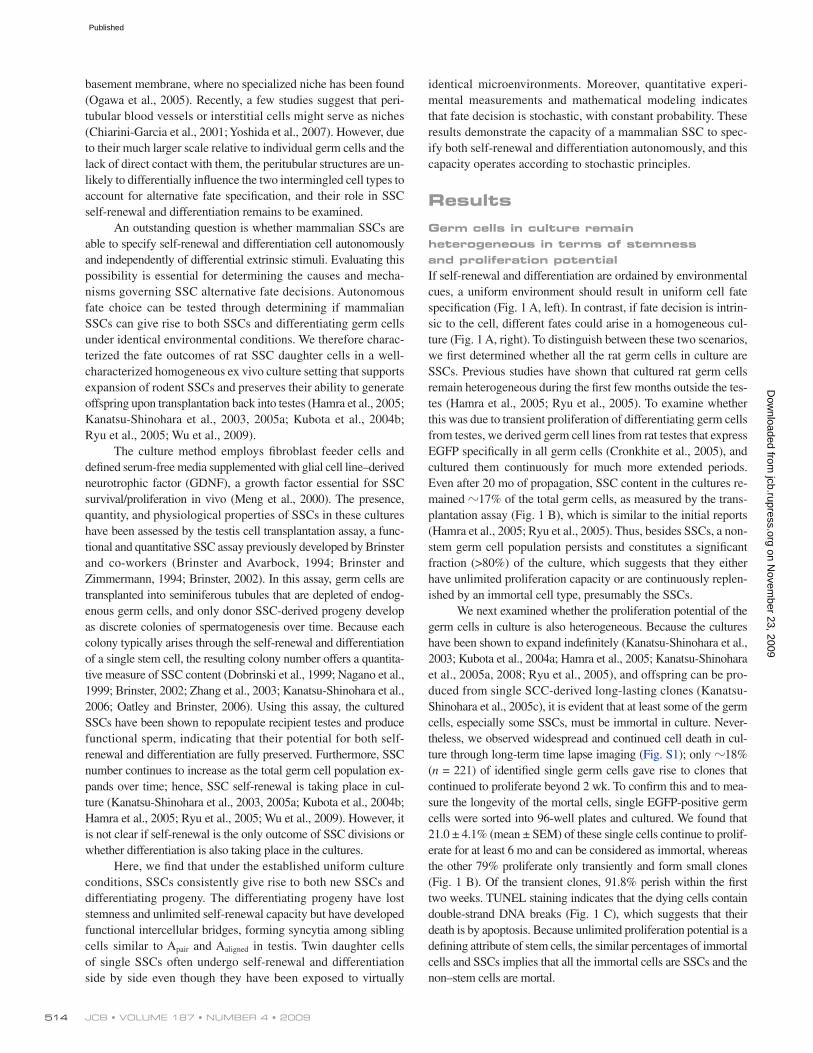
JCB • VOLUME 187 • NUMBER 4 • 2009 514
identical microenvironments. Moreover, quantitative experi-mental measurements and mathematical modeling indicates that fate decision is stochastic, with constant probability. These results demonstrate the capacity of a mammalian SSC to spec-ify both self-renewal and differentiation autonomously, and this capacity operates according to stochastic principles.
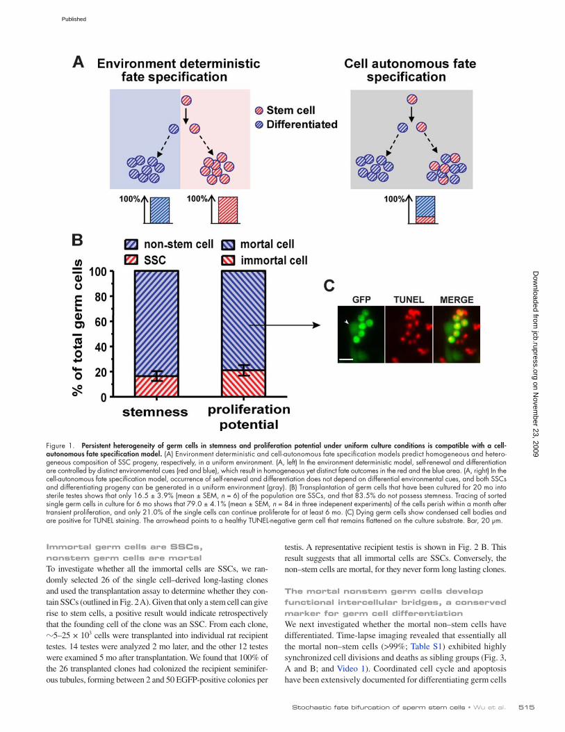
ResultsGerm cells in culture remain heterogeneous in terms of stemness and proliferation potentialIf self-renewal and differentiation are ordained by environmental cues, a uniform environment should result in uniform cell fate specification (Fig. 1 A, left). In contrast, if fate decision is intrin-sic to the cell, different fates could arise in a homogeneous cul-ture (Fig. 1 A, right). To distinguish between these two scenarios, we first determined whether all the rat germ cells in culture are SSCs. Previous studies have shown that cultured rat germ cells remain heterogeneous during the first few months outside the tes-tes (Hamra et al., 2005; Ryu et al., 2005). To examine whether this was due to transient proliferation of differentiating germ cells from testes, we derived germ cell lines from rat testes that express EGFP specifically in all germ cells (Cronkhite et al., 2005), and cultured them continuously for much more extended periods. Even after 20 mo of propagation, SSC content in the cultures re-mained 17% of the total germ cells, as measured by the trans-plantation assay (Fig. 1 B), which is similar to the initial reports (Hamra et al., 2005; Ryu et al., 2005). Thus, besides SSCs, a non-stem germ cell population persists and constitutes a significant fraction (>80%) of the culture, which suggests that they either have unlimited proliferation capacity or are continuously replen-ished by an immortal cell type, presumably the SSCs.
We next examined whether the proliferation potential of the germ cells in culture is also heterogeneous. Because the cultures have been shown to expand indefinitely (Kanatsu-Shinohara et al., 2003; Kubota et al., 2004a; Hamra et al., 2005; Kanatsu-Shinohara et al., 2005a, 2008; Ryu et al., 2005), and offspring can be pro-duced from single SCC-derived long-lasting clones (Kanatsu-Shinohara et al., 2005c), it is evident that at least some of the germ cells, especially some SSCs, must be immortal in culture. Never-theless, we observed widespread and continued cell death in cul-ture through long-term time lapse imaging (Fig. S1); only 18% (n = 221) of identified single germ cells gave rise to clones that continued to proliferate beyond 2 wk. To confirm this and to mea-sure the longevity of the mortal cells, single EGFP-positive germ cells were sorted into 96-well plates and cultured. We found that 21.0 ± 4.1% (mean ± SEM) of these single cells continue to prolif-erate for at least 6 mo and can be considered as immortal, whereas the other 79% proliferate only transiently and form small clones (Fig. 1 B). Of the transient clones, 91.8% perish within the first two weeks. TUNEL staining indicates that the dying cells contain double-strand DNA breaks (Fig. 1 C), which suggests that their death is by apoptosis. Because unlimited proliferation potential is a defining attribute of stem cells, the similar percentages of immortal cells and SSCs implies that all the immortal cells are SSCs and the non–stem cells are mortal.
basement membrane, where no specialized niche has been found (Ogawa et al., 2005). Recently, a few studies suggest that peri-tubular blood vessels or interstitial cells might serve as niches (Chiarini-Garcia et al., 2001; Yoshida et al., 2007). However, due to their much larger scale relative to individual germ cells and the lack of direct contact with them, the peritubular structures are un-likely to differentially influence the two intermingled cell types to account for alternative fate specification, and their role in SSC self-renewal and differentiation remains to be examined.
An outstanding question is whether mammalian SSCs are able to specify self-renewal and differentiation cell autonomously and independently of differential extrinsic stimuli. Evaluating this possibility is essential for determining the causes and mecha-nisms governing SSC alternative fate decisions. Autonomous fate choice can be tested through determining if mammalian SSCs can give rise to both SSCs and differentiating germ cells under identical environmental conditions. We therefore charac-terized the fate outcomes of rat SSC daughter cells in a well-characterized homogeneous ex vivo culture setting that supports expansion of rodent SSCs and preserves their ability to generate offspring upon transplantation back into testes (Hamra et al., 2005; Kanatsu-Shinohara et al., 2003, 2005a; Kubota et al., 2004b; Ryu et al., 2005; Wu et al., 2009).
The culture method employs fibroblast feeder cells and defined serum-free media supplemented with glial cell line–derived neurotrophic factor (GDNF), a growth factor essential for SSC survival/proliferation in vivo (Meng et al., 2000). The presence, quantity, and physiological properties of SSCs in these cultures have been assessed by the testis cell transplantation assay, a func-tional and quantitative SSC assay previously developed by Brinster and co-workers (Brinster and Avarbock, 1994; Brinster and Zimmermann, 1994; Brinster, 2002). In this assay, germ cells are transplanted into seminiferous tubules that are depleted of endog-enous germ cells, and only donor SSC-derived progeny develop as discrete colonies of spermatogenesis over time. Because each colony typically arises through the self-renewal and differentiation of a single stem cell, the resulting colony number offers a quantita-tive measure of SSC content (Dobrinski et al., 1999; Nagano et al., 1999; Brinster, 2002; Zhang et al., 2003; Kanatsu-Shinohara et al., 2006; Oatley and Brinster, 2006). Using this assay, the cultured SSCs have been shown to repopulate recipient testes and produce functional sperm, indicating that their potential for both self- renewal and differentiation are fully preserved. Furthermore, SSC number continues to increase as the total germ cell population ex-pands over time; hence, SSC self-renewal is taking place in cul-ture (Kanatsu-Shinohara et al., 2003, 2005a; Kubota et al., 2004b; Hamra et al., 2005; Ryu et al., 2005; Wu et al., 2009). However, it is not clear if self-renewal is the only outcome of SSC divisions or whether differentiation is also taking place in the cultures.
Here, we find that under the established uniform culture conditions, SSCs consistently give rise to both new SSCs and differentiating progeny. The differentiating progeny have lost stemness and unlimited self-renewal capacity but have developed functional intercellular bridges, forming syncytia among sibling cells similar to Apair and Aaligned in testis. Twin daughter cells of single SSCs often undergo self-renewal and differentiation side by side even though they have been exposed to virtually
on Novem
ber 23, 2009 jcb.rupress.org
Dow
nloaded from
Published
515Stochastic fate bifurcation of sperm stem cells • Wu et al.
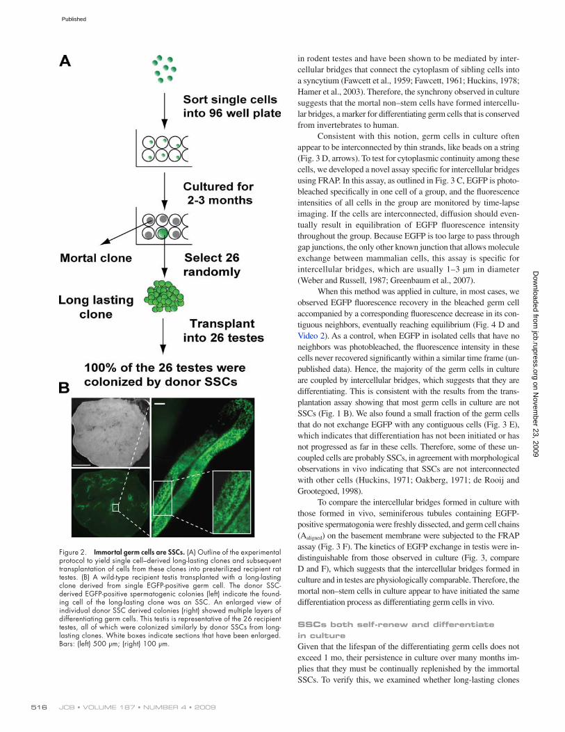
testis. A representative recipient testis is shown in Fig. 2 B. This result suggests that all immortal cells are SSCs. Conversely, the non–stem cells are mortal, for they never form long lasting clones.
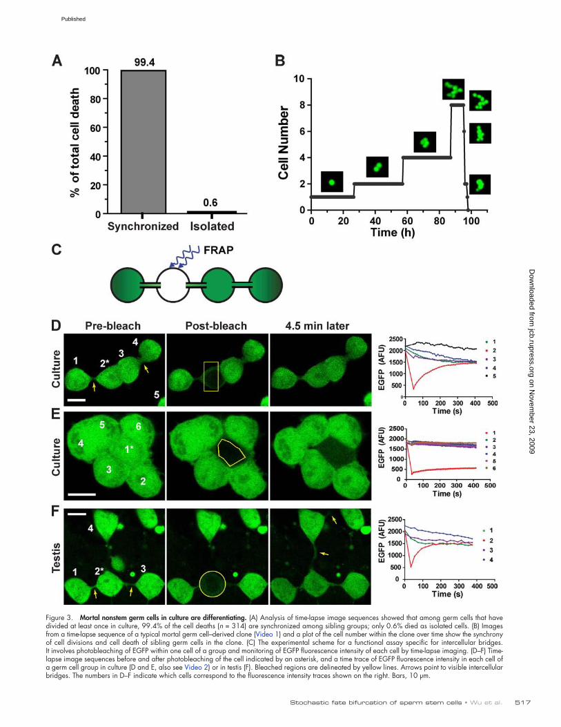
The mortal nonstem germ cells develop functional intercellular bridges, a conserved marker for germ cell differentiationWe next investigated whether the mortal non–stem cells have differentiated. Time-lapse imaging revealed that essentially all the mortal non–stem cells (>99%; Table S1) exhibited highly synchronized cell divisions and deaths as sibling groups (Fig. 3, A and B; and Video 1). Coordinated cell cycle and apoptosis have been extensively documented for differentiating germ cells
Immortal germ cells are SSCs, nonstem germ cells are mortalTo investigate whether all the immortal cells are SSCs, we ran-domly selected 26 of the single cell–derived long-lasting clones and used the transplantation assay to determine whether they con-tain SSCs (outlined in Fig. 2 A). Given that only a stem cell can give rise to stem cells, a positive result would indicate retrospectively that the founding cell of the clone was an SSC. From each clone, 5–25 × 103 cells were transplanted into individual rat recipient testes. 14 testes were analyzed 2 mo later, and the other 12 testes were examined 5 mo after transplantation. We found that 100% of the 26 transplanted clones had colonized the recipient seminifer-ous tubules, forming between 2 and 50 EGFP-positive colonies per
Figure 1. Persistent heterogeneity of germ cells in stemness and proliferation potential under uniform culture conditions is compatible with a cell- autonomous fate specification model. (A) Environment deterministic and cell-autonomous fate specification models predict homogeneous and hetero-geneous composition of SSC progeny, respectively, in a uniform environment. (A, left) In the environment deterministic model, self-renewal and differentiation are controlled by distinct environmental cues (red and blue), which result in homogeneous yet distinct fate outcomes in the red and the blue area. (A, right) In the cell-autonomous fate specification model, occurrence of self-renewal and differentiation does not depend on differential environmental cues, and both SSCs and differentiating progeny can be generated in a uniform environment (gray). (B) Transplantation of germ cells that have been cultured for 20 mo into sterile testes shows that only 16.5 ± 3.9% (mean ± SEM, n = 6) of the population are SSCs, and that 83.5% do not possess stemness. Tracing of sorted single germ cells in culture for 6 mo shows that 79.0 ± 4.1% (mean ± SEM, n = 84 in three indepenent experiments) of the cells perish within a month after transient proliferation, and only 21.0% of the single cells can continue proliferate for at least 6 mo. (C) Dying germ cells show condensed cell bodies and are positive for TUNEL staining. The arrowhead points to a healthy TUNEL-negative germ cell that remains flattened on the culture substrate. Bar, 20 µm.
on Novem
ber 23, 2009 jcb.rupress.org
Dow
nloaded from
Published
JCB • VOLUME 187 • NUMBER 4 • 2009 516
in rodent testes and have been shown to be mediated by inter-cellular bridges that connect the cytoplasm of sibling cells into a syncytium (Fawcett et al., 1959; Fawcett, 1961; Huckins, 1978; Hamer et al., 2003). Therefore, the synchrony observed in culture suggests that the mortal non–stem cells have formed intercellu-lar bridges, a marker for differentiating germ cells that is conserved from invertebrates to human.
Consistent with this notion, germ cells in culture often appear to be interconnected by thin strands, like beads on a string (Fig. 3 D, arrows). To test for cytoplasmic continuity among these cells, we developed a novel assay specific for intercellular bridges using FRAP. In this assay, as outlined in Fig. 3 C, EGFP is photo-bleached specifically in one cell of a group, and the fluorescence intensities of all cells in the group are monitored by time-lapse imaging. If the cells are interconnected, diffusion should even-tually result in equilibration of EGFP fluorescence intensity throughout the group. Because EGFP is too large to pass through gap junctions, the only other known junction that allows molecule exchange between mammalian cells, this assay is specific for intercellular bridges, which are usually 1–3 µm in diameter (Weber and Russell, 1987; Greenbaum et al., 2007).
When this method was applied in culture, in most cases, we observed EGFP fluorescence recovery in the bleached germ cell accompanied by a corresponding fluorescence decrease in its con-tiguous neighbors, eventually reaching equilibrium (Fig. 4 D and Video 2). As a control, when EGFP in isolated cells that have no neighbors was photobleached, the fluorescence intensity in these cells never recovered significantly within a similar time frame (un-published data). Hence, the majority of the germ cells in culture are coupled by intercellular bridges, which suggests that they are differentiating. This is consistent with the results from the trans-plantation assay showing that most germ cells in culture are not SSCs (Fig. 1 B). We also found a small fraction of the germ cells that do not exchange EGFP with any contiguous cells (Fig. 3 E), which indicates that differentiation has not been initiated or has not progressed as far in these cells. Therefore, some of these un-coupled cells are probably SSCs, in agreement with morphological observations in vivo indicating that SSCs are not interconnected with other cells (Huckins, 1971; Oakberg, 1971; de Rooij and Grootegoed, 1998).
To compare the intercellular bridges formed in culture with those formed in vivo, seminiferous tubules containing EGFP-positive spermatogonia were freshly dissected, and germ cell chains (Aaligned) on the basement membrane were subjected to the FRAP assay (Fig. 3 F). The kinetics of EGFP exchange in testis were in-distinguishable from those observed in culture (Fig. 3, compare D and F), which suggests that the intercellular bridges formed in culture and in testes are physiologically comparable. Therefore, the mortal non–stem cells in culture appear to have initiated the same differentiation process as differentiating germ cells in vivo.
SSCs both self-renew and differentiate in cultureGiven that the lifespan of the differentiating germ cells does not exceed 1 mo, their persistence in culture over many months im-plies that they must be continually replenished by the immortal SSCs. To verify this, we examined whether long-lasting clones
Figure 2. Immortal germ cells are SSCs. (A) Outline of the experimental protocol to yield single cell–derived long-lasting clones and subsequent transplantation of cells from these clones into presterilized recipient rat testes. (B) A wild-type recipient testis transplanted with a long-lasting clone derived from single EGFP-positive germ cell. The donor SSC- derived EGFP-positive spermatogenic colonies (left) indicate the found-ing cell of the long-lasting clone was an SSC. An enlarged view of individual donor SSC derived colonies (right) showed multiple layers of differentiating germ cells. This testis is representative of the 26 recipient testes, all of which were colonized similarly by donor SSCs from long-lasting clones. White boxes indicate sections that have been enlarged. Bars: (left) 500 µm; (right) 100 µm.
on Novem
ber 23, 2009 jcb.rupress.org
Dow
nloaded from
Published
517Stochastic fate bifurcation of sperm stem cells • Wu et al.
Figure 3. Mortal nonstem germ cells in culture are differentiating. (A) Analysis of time-lapse image sequences showed that among germ cells that have divided at least once in culture, 99.4% of the cell deaths (n = 314) are synchronized among sibling groups; only 0.6% died as isolated cells. (B) Images from a time-lapse sequence of a typical mortal germ cell–derived clone (Video 1) and a plot of the cell number within the clone over time show the synchrony of cell divisions and cell death of sibling germ cells in the clone. (C) The experimental scheme for a functional assay specific for intercellular bridges. It involves photobleaching of EGFP within one cell of a group and monitoring of EGFP fluorescence intensity of each cell by time-lapse imaging. (D–F) Time-lapse image sequences before and after photobleaching of the cell indicated by an asterisk, and a time trace of EGFP fluorescence intensity in each cell of a germ cell group in culture (D and E, also see Video 2) or in testis (F). Bleached regions are delineated by yellow lines. Arrows point to visible intercellular bridges. The numbers in D–F indicate which cells correspond to the fluorescence intensity traces shown on the right. Bars, 10 µm.
on Novem
ber 23, 2009 jcb.rupress.org
Dow
nloaded from
Published
JCB • VOLUME 187 • NUMBER 4 • 2009 518
by their long-lasting progeny (>17 d), and differentiating germ cells were identified by progeny that formed intercellular bridges and died synchronously. We observed examples in which twin daughter cells of single SSCs acquired different cell fates, and others in which they both became SSCs. However, the method cannot unambiguously distinguish between an SSC that gives rise to two differentiating daughter cells and a mother germ cell that is already differentiating.
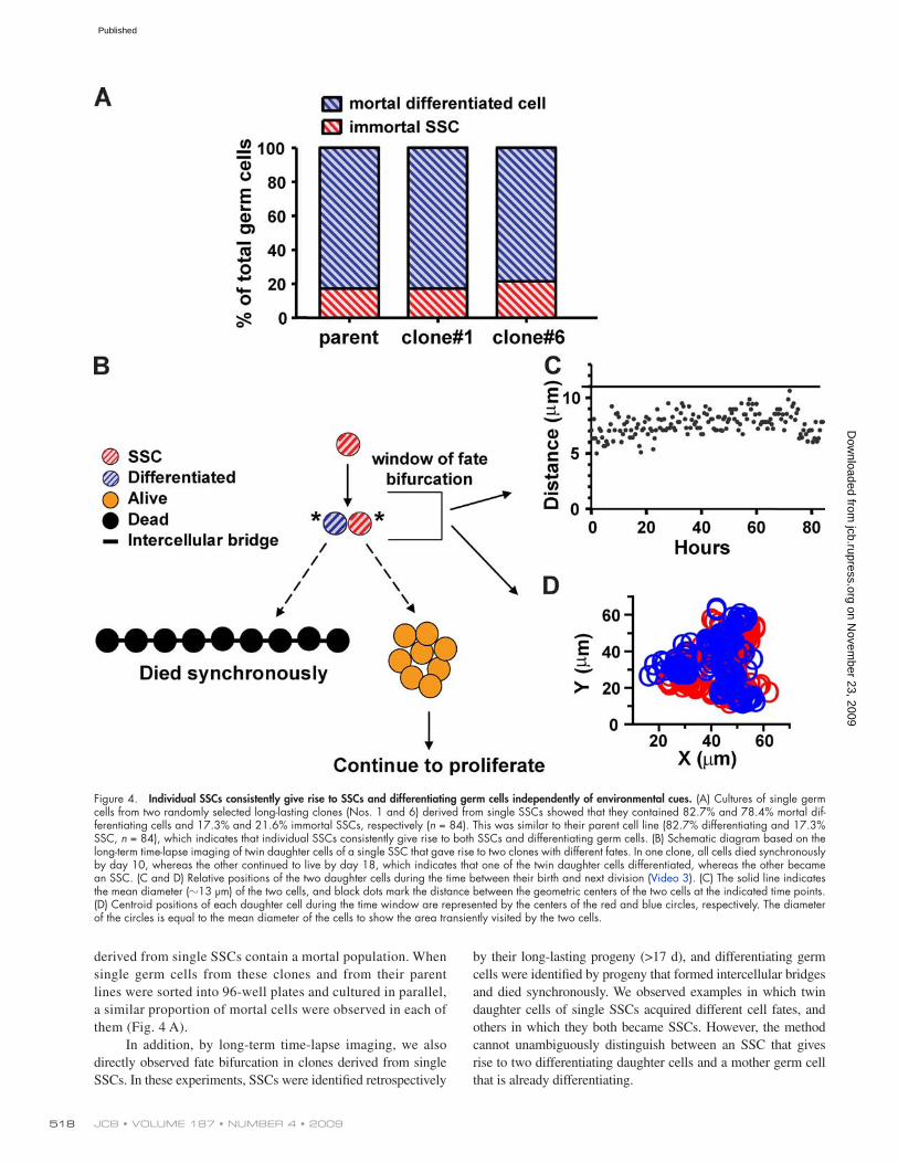
derived from single SSCs contain a mortal population. When single germ cells from these clones and from their parent lines were sorted into 96-well plates and cultured in parallel, a similar proportion of mortal cells were observed in each of them (Fig. 4 A).
In addition, by long-term time-lapse imaging, we also directly observed fate bifurcation in clones derived from single SSCs. In these experiments, SSCs were identified retrospectively
Figure 4. Individual SSCs consistently give rise to SSCs and differentiating germ cells independently of environmental cues. (A) Cultures of single germ cells from two randomly selected long-lasting clones (Nos. 1 and 6) derived from single SSCs showed that they contained 82.7% and 78.4% mortal dif-ferentiating cells and 17.3% and 21.6% immortal SSCs, respectively (n = 84). This was similar to their parent cell line (82.7% differentiating and 17.3% SSC, n = 84), which indicates that individual SSCs consistently give rise to both SSCs and differentiating germ cells. (B) Schematic diagram based on the long-term time-lapse imaging of twin daughter cells of a single SSC that gave rise to two clones with different fates. In one clone, all cells died synchronously by day 10, whereas the other continued to live by day 18, which indicates that one of the twin daughter cells differentiated, whereas the other became an SSC. (C and D) Relative positions of the two daughter cells during the time between their birth and next division (Video 3). (C) The solid line indicates the mean diameter (13 µm) of the two cells, and black dots mark the distance between the geometric centers of the two cells at the indicated time points. (D) Centroid positions of each daughter cell during the time window are represented by the centers of the red and blue circles, respectively. The diameter of the circles is equal to the mean diameter of the cells to show the area transiently visited by the two cells.
on Novem
ber 23, 2009 jcb.rupress.org
Dow
nloaded from
Published

519Stochastic fate bifurcation of sperm stem cells • Wu et al.
death, and composition. We noted that the proportions of SSC and differentiating cells in the parent cultures and in long-lasting clones derived from single SSCs are essentially invariant (Fig. 4 A), which suggests a constant ratio of SSC self-renewal and differ-entiation. In addition, we did not observe any consistent pattern of fate outcome along the lineage trees of long-lasting clones. These observations suggest that self-renewal and differentiation might be stochastic events occurring with a consistent probability. Based on this premise, we derived the following mathematical models for the kinetics of total germ cell growth (Eq. 1) and for the decay of single cell–derived clones over time (Eq. 2, de-tailed in Materials and methods):
ln ln 2 ln G s c t Gt = ( ) +/ .0 (1)
And when t < (q + 1)cmax,
P c S s s G s s S Galiveq t c= − −( ) ++ ln ( )]0
10 0 01 –( ) // [ / , (2)
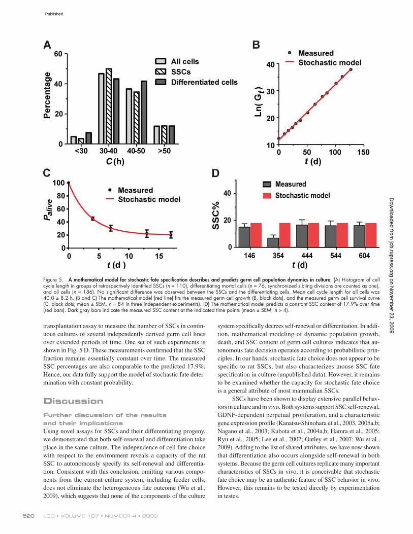
where Gt denotes the number of total live germ cells at time t, S0 denotes the number of SSCs of the given germ cell population at arbitrary time zero during continuous culture, s denotes the chance per cell division that an individual SSC daughter cell will become an SSC. The probability that it will become differenti-ated is 1 s, and q denotes the number of cell divisions needed for newly differentiated germ cells to produce the mean number of progeny before their death. c denotes mean cell cycle length (40.0 ± 8.2 h), which is not different for SSC and differentiating germ cells as measured by long-term time-lapse imaging (Fig. 5 A). cmax denotes the observed maximum cell cycle length, which is 86 h. Palive denotes percentage of live clones derived from single germ cells of a given population at time t.
These equations assume that SSCs are immortal and that differentiating germ cells die after transient propagation, as we have shown experimentally.
Eq. 1 fits the measured total germ cell growth well when s = 0.706 (r2 = 0.9987; Fig. 5 B), and Eq. 2 fits the measured survival curve of single cell–derived clones when s = 0.618 and q = 3.3 (r2 = 0.9720; Fig. 5 C). These analyses thus showed that the probabilistic model can coherently describe various un-correlated aspects of population dynamics in culture. The analy-ses also revealed that the probability for SSC self-renewal under the particular culture conditions is 67% (mean of 0.706 and 0.618); for differentiation, the probability is 33%. Even though fate decisions are biased toward self-renewal, differentiating germ cells actually outnumber the SSCs in culture because 100% of their progeny will be differentiated, whereas the SSC progeny will be a mixture of SSCs and differentiating progeny.
In addition, according to the model, the stem cell fraction in culture is:
R ssq= +( ) 1 , (3)
which is not a function of time (see Materials and methods). Therefore, the model predicts that the stem cell ratio in culture should remain constant, with a value of 17.9% (assum-ing s = 0.67 and q = 3.3). To verify this prediction, we used the
Within the SSC lineages observed, no obvious pattern of cell fate outcome was identified. However, divergence of SSC daughter cell fates, similar to the example diagrammed in Fig. 4 B, was always observed (100%, n > 50). Based on these observa-tions, we concluded that individual SSCs not only undergo self-renewal, but also give rise to differentiating germ cells in culture.
Because under certain conditions differentiating germ cells can de-differentiate and become SSCs in Drosophila (Brawley and Matunis, 2004; Cheng et al., 2008) and mouse testes (Nakagawa et al., 2007; Barroca et al., 2009; Yoshida, 2009), we therefore ex-amined if such fate conversion occurs in rat germ cell cultures. Even though the majority of rat germ cells in culture are c-kit nega-tive (>98%; Fig. S2), which indicates an early state of differentia-tion, none of the cells identified as differentiating germ cells by our criteria gave rise to long-lasting clones (n > 200 cells). Consis-tently, in the SSCs examined (n > 50), none of them were derived from a cell that seemed to be connected to other cells through inter-cellular bridges. These observations indicate that de-differentiation, if it occurs at all in the culture, is rare.
SSC self-renewal and differentiation are independent of extrinsic heterogeneityThe fact that self-renewal and differentiation occur concomitantly in a uniform culture setting suggests an independence of cell fate choice with respect to the environment. However, it is formally pos-sible that fate bifurcation is dependent on microscale heterogeneity associated with the feeder cells or extracellular matrix. Although direct and thorough evaluation of microscale heterogeneity is im-possible, a detailed analysis of germ cell behaviors in culture sug-gests it is unlikely to be the reason for fate bifurcation. We found that SSC daughter cell pairs, including those that have adopted dif-ferent fates, typically remained closely associated the whole time from their birth until they divided again to give rise to clones of either identical or distinct fates. This observation was quantified by measuring the distance between the geometric centers of twin daughter cells from time-lapse recordings. In most cases, as for the cell pair indicated by an asterisk in Fig. 4 B, the center-to-center distance was always less than their mean diameter, which indicated that the two cells were never separated into discrete microenviron-ments (Fig. 4 C and Video 3). Furthermore, twin daughter cells in all cases, instead of statically occupying one spot, constantly move around together, dynamically probing the same microenvironment. Typically, the areas transiently visited by each daughter cell dur-ing the fate bifurcation time window are nearly identical, as shown in Fig. 4 D. The intimate association and the dynamic movement of the two cells minimize the chances for their exposure to differ-ent extrinsic cues, including microheterogeneity. Nevertheless, as in this case, twin daughter cells derived from a single SSC often adopted different fates. Therefore, fate bifurcation does not seem to depend on extrinsic heterogeneity. Instead, cell-autonomous fate specification is most consistent with our data.
SSC self-renewal and differentiation are stochasticBecause SSC self-renewal results in continued proliferation, whereas differentiation leads to cell death in culture, fate speci-fication will directly influence germ cell population growth,
on Novem
ber 23, 2009 jcb.rupress.org
Dow
nloaded from
Published
JCB • VOLUME 187 • NUMBER 4 • 2009 520
system specifically decrees self-renewal or differentiation. In addi-tion, mathematical modeling of dynamic population growth, death, and SSC content of germ cell cultures indicates that au-tonomous fate decision operates according to probabilistic prin-ciples. In our hands, stochastic fate choice does not appear to be specific to rat SSCs, but also characterizes mouse SSC fate specification in culture (unpublished data). However, it remains to be examined whether the capacity for stochastic fate choice is a general attribute of most mammalian SSCs.
SSCs have been shown to display extensive parallel behav-iors in culture and in vivo. Both systems support SSC self-renewal, GDNF-dependent perpetual proliferation, and a characteristic gene expression profile (Kanatsu-Shinohara et al., 2003, 2005a,b; Nagano et al., 2003; Kubota et al., 2004a,b; Hamra et al., 2005; Ryu et al., 2005; Lee et al., 2007; Oatley et al., 2007; Wu et al., 2009). Adding to the list of shared attributes, we have now shown that differentiation also occurs alongside self-renewal in both systems. Because the germ cell cultures replicate many important characteristics of SSCs in vivo, it is conceivable that stochastic fate choice may be an authentic feature of SSC behavior in vivo. However, this remains to be tested directly by experimentation in testes.
transplantation assay to measure the number of SSCs in contin-uous cultures of several independently derived germ cell lines over extended periods of time. One set of such experiments is shown in Fig. 5 D. These measurements confirmed that the SSC fraction remains essentially constant over time. The measured SSC percentages are also comparable to the predicted 17.9%. Hence, our data fully support the model of stochastic fate deter-mination with constant probability.
DiscussionFurther discussion of the results and their implicationsUsing novel assays for SSCs and their differentiating progeny, we demonstrated that both self-renewal and differentiation take place in the same culture. The independence of cell fate choice with respect to the environment reveals a capacity of the rat SSC to autonomously specify its self-renewal and differentia-tion. Consistent with this conclusion, omitting various compo-nents from the current culture system, including feeder cells, does not eliminate the heterogeneous fate outcome (Wu et al., 2009), which suggests that none of the components of the culture
Figure 5. A mathematical model for stochastic fate specification describes and predicts germ cell population dynamics in culture. (A) Histogram of cell cycle length in groups of retrospectively identified SSCs (n = 110), differentiating mortal cells (n = 76, synchronized sibling divisions are counted as one), and all cells (n = 186). No significant difference was observed between the SSCs and the differentiating cells. Mean cell cycle length for all cells was 40.0 ± 8.2 h. (B and C) The mathematical model (red line) fits the measured germ cell growth (B, black dots), and the measured germ cell survival curve (C, black dots; mean ± SEM, n = 84 in three independent experiments). (D) The mathematical model predicts a constant SSC content of 17.9% over time (red bars). Dark gray bars indicate the measured SSC content at the indicated time points (mean ± SEM, n > 4).
on Novem
ber 23, 2009 jcb.rupress.org
Dow
nloaded from
Published

521Stochastic fate bifurcation of sperm stem cells • Wu et al.
were obtained from Invitrogen. BSA and DMSO were obtained from EMD. Mitomycin-C, mouse laminin, mouse EGF, rat GDNF, bovine apo-transferrin, human basic fibroblast growth factor (bFGF), d-(+)-glucose, d-biotin, ascorbic acid, sodium pyruvate, putrescine, progesterone, -estradiol 17-cypionate, insulin, sodium selenite, 2-mercaptoethanol (ME), dl-lactic acid, and all fatty acids were obtained from Sigma-Aldrich. Mouse leukemia inhibitory factor/ESGRO (LIF) was obtained from Millipore. FBS was obtained from Atlanta Biologicals. Dispase and rat-tail collagen I-coated culture dishes were obtained from Thermo Fisher Scientific, Inc. The In Situ Cell Death Detection kit (TMR red) was obtained from Roche. Busulfan was obtained from MP Biomedicals. Glass-bottom dishes were obtained from MatTek Corporation.
Derivation and maintenance of rat spermatogonial cell culturesAll spermatogonial cell lines used in this study were derived from testes of 20–30-d-old homozygous male SD-Tg(ROSA-EGFP)2-4Reh Sprague Dawley rats, which exhibit germ cell–specific expression of EGFP (GCS-EGFP; Cronkhite et al., 2005). The derivation and maintenance of the rat sper-matogonial cell cultures were performed largely as described previously (Hamra et al., 2002, 2005), but without FACS-based cell sorting of EGFP-positive germ cells. In brief, seminiferous tubules were minced and digested with dispase (50 units/testis) at 32°C for up to 30 min. The dissociated tes-ticular cells were cultured at 32°C in DHF12 medium supplemented with 5.5% horse serum, 2.5% FBS, and 1% antibiotic/antimycotic solution at 32°C in humidified incubators with 5% CO2 in order to allow the somatic cells to attach to the plastic or gelatin-coated tissue culture dishes. After 2–3 d of incubation, the germ cells on top of the somatic cells were har-vested by washing repeatedly with medium, plated on gelatin-coated dishes, and incubated at 37°C for another 2–3 h to allow remnant somatic cells to attach. At the end of the incubation, the floating germ cells were harvested and plated on laminin-coated dishes in DHF12 medium and sup-plemented with 10% FBS, 1% antibiotic/antimycotic solution, and 30 µM 2-mercaptoethanol to enrich for stem cells. After 20–40 min of incubation at 32°C, germ cells that did not attach to the laminin were washed away with medium. Germ cells attached to the laminin (LamB cells) were har-vested and transferred to gelatin-coated dishes in SA medium to further de-plete the remnant somatic cells. After 2 d of incubation at 37°C, the floating LamB germ cells were transferred onto mitomycin-C–treated or -irradiated primary mouse embryonic fibroblast (MEF) feeders; then they continued to be cultured in SA medium. Routinely, the culture medium was refreshed every 3–4 d. The cultured germ cells with MEF feeder cells were passaged by trypsinization with 0.05% or 0.25% trypsin-EDTA solution onto new MEF feeders every 2–3 wk.
Single spermatogonial cell cultureTo establish single cell cultures, GCS-EGFP spermatogonial cell cultures were trypsinized with 0.25% trypsin-EDTA solution to isolate and harvest the cells. After one wash, the cell pellet was resuspended at 0.5–2 × 10⁶/ml in SA medium and filtered through 40-µm cell strainers. 96-well plates with MEF feeder cells were washed once with PBS and replenished with 100 µl per well of SA medium. FACS using an Aria Cell Sorter (BD) was used to deposit one EGFP-positive germ cell into each well of the 96-well plates. Medium was refreshed every 3–4 d and the growth/death of the clones was monitored using a fluorescence microscope (IX70; Olympus). The long-lasting clones were continually cultured in the original well for 2–3 mo without passaging until transplantation. Then, each clone was passaged by trypsinization with 0.25% trypsin-EDTA solution about every 2 wk.
Germ cell transplantation and analysisGerm cell transplantations by retrograde injection through the rete of busulfan-treated wild-type rat recipient testes were performed as de-scribed previously (Ogawa et al., 1997, 1999; Hamra et al., 2002). In brief, wild-type Sprague Dawley rats at 8 d of age (Harlan Labora-tories, Inc.) were injected i.p. with 12.5 mg/kg busulfan (4 mg/ml in 50% DMSO and 50% PBS) to eliminate the endogenous germ cells, then used as recipient males for germ cell transplantation at 24–29 d of age (Ogawa et al., 1999). Donor cells were harvested from long-term in vitro cultures of GCS-EGFP rat spermatogonia by 0.25% trypsin-EDTA and filtered through a 40-µm cell strainer. EGFP-positive germ cells were counted and diluted to 3 × 10⁴–3 × 10⁵ cells/ml of SA or SG medium containing 0.05% trypan blue as a fluid phase marker. About 50 µl of the cell suspension was loaded into an injection needle pulled from a 100-µl glass capillary tube, and 20–50 µl was then transferred into the seminiferous tubules of anesthetized rats by retrograde injection through the rete of each testis (Ogawa et al., 1997, 1999). 2 or 5 mo after transplantation, recipient testes were dissected out and spread out
Stochastic fate choice and environmental regulation are not mutually exclusiveMaintenance of tissue homeostasis usually demands feedback regulation of stem cell activity by extrinsic physiological cues. This raises the question of how an autonomous and stochastic mechanism for SSC self-renewal and differentiation integrates input from its environment. Although our results argue against a deterministic role of extrinsic cues in SSC progeny fate specifica-tion, a nondeterministic influence of environment can fit well with the observed stochastic fate choice. For example, rather than ordaining fate of individual cells, extrinsic cues may serve to bias the probability of cell fate choices at the population level. A niche, in this case, can be viewed as an extreme environment in which fate choice is entirely skewed toward self-renewal. There-fore, stochastic fate choice and environmental regulation are not mutually exclusive but could work jointly to achieve SSC self-renewal and differentiation that is minimally dependent on the environment but responsive to its regulation.
Possible mechanisms of SSC stochastic fate choiceOne possible mechanism for autonomous cell fate diversification is asymmetric cell division (Knoblich, 2008; Wu et al., 2008). In this scenario, a parent cell segregates fate determinants unequally to its daughter cells, thereby creating intrinsic differences that predestine them for different fates. However, because asymmet-ric division alone cannot increase stem cell number, symmetric division must also take place to produce the net increase of SSC number we observed in culture. This requires some other mecha-nism to account for the choice between symmetric and asym-metric divisions, which should be stochastic, for no consistent pattern of cell fate outcome was observed within SSC lineages.
Alternatively, intrinsic variation (noise) in gene expression has been shown in a few simple organisms to cause cell fate diver-gence within isogenic populations. Examples include entry into the competent state by Bacillus subtilis (Maamar and Dubnau, 2005; Smits et al., 2005; Dubnau and Losick, 2006; Süel et al., 2006, 2007) and generation of photoreceptors with alterna-tive color vision in fly compound eyes (Wernet et al., 2006; Bell et al., 2007). These studies demonstrate that small and transient initial fluctuations in transcription of fate determinant genes can be amplified and stabilized at qualitatively different levels and cause differential cell fate outcome (Losick and Desplan, 2008). A recent study of mouse hematopoietic progenitor cells also indicates that transcription noise may control lineage choice in mammalians (Chang et al., 2008). Thus, it is possible that noisy expression of genes that drive self-renewal or differentiation may play a role in fate decision of SSC daughter cells. The methodol-ogy developed in this study can be used in future investigations to identify the molecular machineries responsible for SSC sto-chastic fate choice.
Materials and methodsMaterials and chemicalsCell culture media, PBS nonessential amino acids, MEM vitamin solution, l-glutamine solution, 0.05% or 0.25% trypsin-EDTA solutions, B27 minus vitamin A supplement, antibiotic-antimycotic solutions, and horse serum
on Novem
ber 23, 2009 jcb.rupress.org
Dow
nloaded from
Published
JCB • VOLUME 187 • NUMBER 4 • 2009 522
slope of 0.2064 ± 0.002245. Hence, Eq. 1 best fits the measured growth kinetics (Fig. 5 B) when ln (2s)/c = 0.2064. As the measured average cell cycle length c = 1.67 d (40 h), s = 0.706.
As experimentally determined, differentiating cells have limited prolif-eration capacity, assuming a differentiating cell can divide on an average q number of divisions before it dies, then in a given germ cell group of total number G0 with S0 number of SSCs, the derivative of the percentage of live clone (Palive) derived from this population at a given time point t is
Therefore, Palive is an exponential decay function of t, when t < (q+1) cmax:
P c S s s G s S Galiveq t c= − −( ) ++ ln (s)]0
10 0 01 –( ) // [ / . (2)
Because exponential decay function P = (P0 Plateau) e(Kt) + Plateau best fit to the measured data (R2 = 0.9720) when K = 0.2886, P0 = 100%, and Plateau = 20.02%, our model best fit with the measured data (Fig. 5 C) when s = 0.618, S0/G0 = 20.02%, and q = 3.3.
Plugging in the exponential expressions for St and Dt, the stem cell fraction (Rs) is:
R S G ss t tq= = +/ .( )1 (3)
Because Rs is not a function of time, this model predicts that the stem cell ratio remains unchanged over time. Assuming s = 0.67 and q = 3.3, it predicts that the stem cell ratio in culture should remain constant, with a value of 17.9%.
Online supplemental materialFig. S1 shows that a mortal population persists in long-term rat germ cell cul-tures. Fig. S2 shows flow cytometric analysis of cultured GCS-EGFP rat germ cells for c-kit expression. Table S1 shows that mortal germ cells in culture die synchronously as sibling groups. Video 1 shows synchronized cell division and death in a clone of germ cells. Video 2 shows EGFP exchange between cultured germ cells through intercellular bridges. Video 3 shows that daugh-ter cells of a single SSC dynamically sample the same microenvironment.
Z. Wu is especially grateful to Drs. Richard, G. Anderson and David Mangelsdorf for evaluating the work, reading the manuscript, and the mentoring they generously provided following the decease of Dr. David L. Garbers. We thank Dr. F. Kent Hamra for sharing his expertise in rat testis cell transplanta-tion and in culturing rat germ cells; Elizabeth Curry for conducting cell sort-ing; and Drs. Eric Olson, Luis Parada, Michael Roth, Chengcheng Zhang, and Xiaodong Zhang for critical reading of the manuscript. Time-lapse imaging and FRAP were carried out in the University of Texas Southwestern Live Cell Imaging Facility.
This work received funding from the Cecil H. & Ida Green Center for Reproductive Biology Sciences at the University of Texas Southwestern Medical Center, and from the Howard Hughes Medical Institute to D.L. Garbers, prior to his passing. G.M. Süel acknowledges funding by the Welch Foundation (I-1674) and the James S. McDonnell Foundation (220020141). G.M. Süel is a W.W. Caruth Jr. Scholar of Biomedical Research.
Submitted: 9 July 2009Accepted: 12 October 2009
ReferencesBarroca, V., B. Lassalle, M. Coureuil, J.P. Louis, F. Le Page, J. Testart,
I. Allemand, L. Riou, and P. Fouchet. 2009. Mouse differentiating sper-matogonia can generate germinal stem cells in vivo. Nat. Cell Biol. 11:190–196. doi:10.1038/ncb1826
Bell, M.L., J.B. Earl, and S.G. Britt. 2007. Two types of Drosophila R7 photo-receptor cells are arranged randomly: a model for stochastic cell-fate determination. J. Comp. Neurol. 502:75–85. doi:10.1002/cne.21298
Brawley, C., and E. Matunis. 2004. Regeneration of male germline stem cells by spermatogonial dedifferentiation in vivo. Science. 304:1331–1334. doi:10.1126/science.1097676
Brinster, R.L. 2002. Germline stem cell transplantation and transgenesis. Science. 296:2174–2176. doi:10.1126/science.1071607
d d - 1 1P t S s s Galiveq t c/ / ,( ) /= × −( ) × − + +
0 0d d - 1 1P t S s s Galiveq t c/ / ,( ) /= × −( ) × − + +
0 0 when 1t q cmax< +( ) .when 1t q cmax< +( ) .
on glass slides. The EGFP-positive colonies in each recipient testis were visualized and scored using a fluorescence microscope (IX70). The SSC number was then calculated based on 8% colonization efficiency (Nagano, 2003; Ogawa et al., 2003). Images of whole recipient tes-tes were taken with a fluorescence stereomicroscope using 0.63× and 1.5× lenses (SteREO Discovery.V12; Carl Zeiss, Inc.).
Long-term time-lapse microscopy and image analysisCultured rat germ cells were plated at 0.5–1 × 10⁴ cells/cm2 in 25-T flasks on MEF feeder cells in SA or SG medium immediately before imaging. The flasks were mounted on the computer-controlled stage of a DeltaVision RT microscope (Applied Precision) in a tempera-ture (37°C)- and humidity-controlled chamber. The flasks were per-fused continuously with 5% CO2. The medium was manually changed every 2–3 d without disturbing the position of the flask. Multiple fields were selected for automated time-lapse imaging. Images of each field at 4×, 10×, or 20× magnification were collected at regular time in-tervals of 15–30 min for 2–18 d. Time-lapse image sequences were analyzed using ImageJ (http://rsbweb.nih.gov/ij/). The Manual Track-ing plug-in (written by F. Cordelieres, Institut Curie, France; http://rsb .info.nih.gov/ij/plugins/track/track.html) was used to track the centroid positions of selected individual cells.
FRAP assay for intercellular bridges and image analysisEGFP-expressing germ cells were cultured under normal conditions in glass-bottom dishes for 4–7 d before analysis. Using a confocal micro-scope (LSM510; Carl Zeiss, Inc.) with a 40×/0.8 NA water immersion objective lens, photobleaching within a region of interest was performed with 488-nm illumination at 100% laser power, scanning at maximum speed 200–500 times (approximately 40 s). Time-lapse images were ac-quired immediately before and after photobleaching. For FRAP of EGFP germ cells in testis, seminiferous tubules were spread out between a glass slides and a coverslip. Chains of EGFP-positive germ cells near the base-ment membrane were selected for FRAP as described for cultured cells. The mean EGFP fluorescence intensity within a region of interest of each germ cell was measured as a function of time using ImageJ.
Mathematical models based on probabilistic fate determinationAs demonstrated in the results, germ cells in culture are composed of two distinct populations: the SSCs and the differentiating germ cells. SSCs are able to proliferate indefinitely and give rise to both new SSCs and differen-tiating progeny. The differentiating germ cells eventually die after transient amplification, producing only differentiating cells. In addition, the invariant proportions of SSC and differentiating cells in the parent culture and in long-lasting clones derived from single SSCs (Fig. 3 A) suggest that a con-stant probability for self-renewal (s) and differentiation (1 s) can be assumed. Therefore, the growth of the two populations over time (t) can be represented by the following two exponential equations:
where St and S0 are the number of SSCs at time t and time zero, respec-tively, Dt is the number of differentiating germ cells at time t, and q denotes the number of divisions required to produce the average number of prog-eny among the differentiating cells before they die. In both equations, c is the mean cell cycle length. Measurements from time-lapse imaging showed that c is the same for SSCs and differentiating cells (Fig. 5 A).
Therefore, at time point t, the total germ cell number (Gt) is:
G S D s S st t tt c q= + = ( ) ( )− −2 1/ ( ) .0
Taking the natural log of the above equation, we obtained:
ln ln 2 ln (S s ).( q 1)G t c st = ( ) +/ 0− −
When t = 0, then G0 = S0 (s(−q−1)). Therefore, the equation above becomes:
ln ln 2 ln G s c t Gt = ( ) +/ .0 (1)
Experimental values for G0 and Gt were measured as the total num-ber of germ cells at the beginning and at subsequent time points. A linear least squares fit to a semilog plot of the measured data (ln Gt vs. t) has a
S s Stt c= ( )2 and/
0S s Stt c= ( )2 and/
0 D s S stt c q= ( ) −( )− −2 1/ ( ) ,0
1D s S stt c q= ( ) −( )− −2 1/ ( ) ,0
1
on Novem
ber 23, 2009 jcb.rupress.org
Dow
nloaded from
Published
523Stochastic fate bifurcation of sperm stem cells • Wu et al.
Kanatsu-Shinohara, M., K. Inoue, H. Miki, N. Ogonuki, M. Takehashi, T. Morimoto, A. Ogura, and T. Shinohara. 2006. Clonal origin of germ cell colonies after spermatogonial transplantation in mice. Biol. Reprod. 75:68–74. doi:10.1095/biolreprod.106.051193
Kanatsu-Shinohara, M., T. Muneto, J. Lee, M. Takenaka, S. Chuma, N. Nakatsuji, T. Horiuchi, and T. Shinohara. 2008. Long-term culture of male germline stem cells from hamster testes. Biol. Reprod. 78:611–617. doi:10.1095/biolreprod.107.065615
Knoblich, J.A. 2008. Mechanisms of asymmetric stem cell division. Cell. 132:583–597. doi:10.1016/j.cell.2008.02.007
Kubota, H., M.R. Avarbock, and R.L. Brinster. 2004a. Culture conditions and sin-gle growth factors affect fate determination of mouse spermatogonial stem cells. Biol. Reprod. 71:722–731. doi:10.1095/biolreprod.104.029207
Kubota, H., M.R. Avarbock, and R.L. Brinster. 2004b. Growth factors essential for self-renewal and expansion of mouse spermatogonial stem cells. Proc. Natl. Acad. Sci. USA. 101:16489–16494. doi:10.1073/pnas.0407063101
Lee, J., M. Kanatsu-Shinohara, K. Inoue, N. Ogonuki, H. Miki, S. Toyokuni, T. Kimura, T. Nakano, A. Ogura, and T. Shinohara. 2007. Akt mediates self-renewal division of mouse spermatogonial stem cells. Development. 134:1853–1859. doi:10.1242/dev.003004
Li, L., and T. Xie. 2005. Stem cell niche: structure and function. Annu. Rev. Cell Dev. Biol. 21:605–631. doi:10.1146/annurev.cellbio.21.012704.131525
Losick, R., and C. Desplan. 2008. Stochasticity and cell fate. Science. 320: 65–68. doi:10.1126/science.1147888
Maamar, H., and D. Dubnau. 2005. Bistability in the Bacillus subtilis K-state (competence) system requires a positive feedback loop. Mol. Microbiol. 56:615–624. doi:10.1111/j.1365-2958.2005.04592.x
Meng, X., M. Lindahl, M.E. Hyvönen, M. Parvinen, D.G. de Rooij, M.W. Hess, A. Raatikainen-Ahokas, K. Sainio, H. Rauvala, M. Lakso, et al. 2000. Regulation of cell fate decision of undifferentiated spermatogonia by GDNF. Science. 287:1489–1493. doi:10.1126/science.287.5457.1489
Nagano, M.C. 2003. Homing efficiency and proliferation kinetics of male germ line stem cells following transplantation in mice. Biol. Reprod. 69:701–707. doi:10.1095/biolreprod.103.016352
Nagano, M., M.R. Avarbock, and R.L. Brinster. 1999. Pattern and kinetics of mouse donor spermatogonial stem cell colonization in recipient testes. Biol. Reprod. 60:1429–1436. doi:10.1095/biolreprod60.6.1429
Nagano, M., B.Y. Ryu, C.J. Brinster, M.R. Avarbock, and R.L. Brinster. 2003. Maintenance of mouse male germ line stem cells in vitro. Biol. Reprod. 68:2207–2214. doi:10.1095/biolreprod.102.014050
Nakagawa, T., Y. Nabeshima, and S. Yoshida. 2007. Functional identification of the actual and potential stem cell compartments in mouse spermatogen-esis. Dev. Cell. 12:195–206. doi:10.1016/j.devcel.2007.01.002
Oakberg, E.F. 1971. Spermatogonial stem-cell renewal in the mouse. Anat. Rec. 169:515–531. doi:10.1002/ar.1091690305
Oatley, J.M., and R.L. Brinster. 2006. Spermatogonial stem cells. Methods Enzymol. 419:259–282. doi:10.1016/S0076-6879(06)19011-4
Oatley, J.M., and R.L. Brinster. 2008. Regulation of spermatogonial stem cell self-renewal in mammals. Annu. Rev. Cell Dev. Biol. 24:263–286. doi:10.1146/annurev.cellbio.24.110707.175355
Oatley, J.M., M.R. Avarbock, and R.L. Brinster. 2007. Glial cell line-derived neurotrophic factor regulation of genes essential for self-renewal of mouse spermatogonial stem cells is dependent on Src family kinase sig-naling. J. Biol. Chem. 282:25842–25851. doi:10.1074/jbc.M703474200
Ogawa, T., J.M. Aréchaga, M.R. Avarbock, and R.L. Brinster. 1997. Transplantation of testis germinal cells into mouse seminiferous tubules. Int. J. Dev. Biol. 41:111–122.
Ogawa, T., I. Dobrinski, and R.L. Brinster. 1999. Recipient preparation is critical for spermatogonial transplantation in the rat. Tissue Cell. 31:461–472. doi:10.1054/tice.1999.0060
Ogawa, T., M. Ohmura, Y. Yumura, H. Sawada, and Y. Kubota. 2003. Expansion of murine spermatogonial stem cells through serial transplantation. Biol. Reprod. 68:316–322. doi:10.1095/biolreprod.102.004549
Ogawa, T., M. Ohmura, and K. Ohbo. 2005. The niche for spermatogonial stem cells in the mammalian testis. Int. J. Hematol. 82:381–388. doi:10.1532/ IJH97.05088
Russell, L.D., R.A. Ettlin, A.P. Hikim, and E.D. Clegg. 1990. Mammalian sper-matogenesis. In Histological and Histopathological Evaluation of the Testis. Cache River Press, Clearwater, FL. 1–40.
Ryu, B.Y., H. Kubota, M.R. Avarbock, and R.L. Brinster. 2005. Conservation of spermatogonial stem cell self-renewal signaling between mouse and rat. Proc. Natl. Acad. Sci. USA. 102:14302–14307. doi:10.1073/pnas .0506970102
Schofield, R. 1978. The relationship between the spleen colony-forming cell and the haemopoietic stem cell. Blood Cells. 4:7–25.
Smits, W.K., C.C. Eschevins, K.A. Susanna, S. Bron, O.P. Kuipers, and L.W. Hamoen. 2005. Stripping Bacillus: ComK auto-stimulation is responsible
Brinster, R.L., and M.R. Avarbock. 1994. Germline transmission of donor haplo-type following spermatogonial transplantation. Proc. Natl. Acad. Sci. USA. 91:11303–11307. doi:10.1073/pnas.91.24.11303
Brinster, R.L., and J.W. Zimmermann. 1994. Spermatogenesis following male germ-cell transplantation. Proc. Natl. Acad. Sci. USA. 91:11298–11302. doi:10.1073/pnas.91.24.11298
Chang, H.H., M. Hemberg, M. Barahona, D.E. Ingber, and S. Huang. 2008. Transcriptome-wide noise controls lineage choice in mammalian pro-genitor cells. Nature. 453:544–547. doi:10.1038/nature06965
Cheng, J., N. Türkel, N. Hemati, M.T. Fuller, A.J. Hunt, and Y.M. Yamashita. 2008. Centrosome misorientation reduces stem cell division during age-ing. Nature. 456:599–604. doi:10.1038/nature07386
Chiarini-Garcia, H., J.R. Hornick, M.D. Griswold, and L.D. Russell. 2001. Distribution of type A spermatogonia in the mouse is not random. Biol. Reprod. 65:1179–1185. doi:10.1095/biolreprod65.4.1179
Cronkhite, J.T., C. Norlander, J.K. Furth, G. Levan, D.L. Garbers, and R.E. Hammer. 2005. Male and female germline specific expression of an EGFP reporter gene in a unique strain of transgenic rats. Dev. Biol. 284:171–183. doi:10.1016/j.ydbio.2005.05.015
de Rooij, D.G., and J.A. Grootegoed. 1998. Spermatogonial stem cells. Curr. Opin. Cell Biol. 10:694–701. doi:10.1016/S0955-0674(98)80109-9
Dobrinski, I., T. Ogawa, M.R. Avarbock, and R.L. Brinster. 1999. Computer assisted image analysis to assess colonization of recipi-ent seminiferous tubules by spermatogonial stem cells from transgenic donor mice. Mol. Reprod. Dev. 53:142–148. doi:10.1002/(SICI)1098-2795(199906)53:2<142::AID-MRD3>3.0.CO;2-O
Dubnau, D., and R. Losick. 2006. Bistability in bacteria. Mol. Microbiol. 61:564–572. doi:10.1111/j.1365-2958.2006.05249.x
Fawcett, D.W. 1961. Intercellular bridges. Exp. Cell Res. Suppl 8:174–187. doi:10.1016/0014-4827(61)90347-0
Fawcett, D.W., S. Ito, and D. Slautterback. 1959. The occurrence of intercel-lular bridges in groups of cells exhibiting synchronous differentiation. J. Biophys. Biochem. Cytol. 5:453–460.
Fuchs, E., T. Tumbar, and G. Guasch. 2004. Socializing with the neighbors: stem cells and their niche. Cell. 116:769–778. doi:10.1016/S0092- 8674(04)00255-7
Fuller, M.T., and A.C. Spradling. 2007. Male and female Drosophila germ-line stem cells: two versions of immortality. Science. 316:402–404. doi:10.1126/science.1140861
Gilboa, L., and R. Lehmann. 2004. How different is Venus from Mars? The genetics of germ-line stem cells in Drosophila females and males. Development. 131:4895–4905. doi:10.1242/dev.01373
Greenbaum, M.P., L. Ma, and M.M. Matzuk. 2007. Conversion of midbodies into germ cell intercellular bridges. Dev. Biol. 305:389–396. doi:10.1016/ j.ydbio.2007.02.025
Hamer, G., H.L. Roepers-Gajadien, I.S. Gademan, H.B. Kal, and D.G. De Rooij. 2003. Intercellular bridges and apoptosis in clones of male germ cells. Int. J. Androl. 26:348–353. doi:10.1111/j.1365-2605.2003.00436.x
Hamra, F.K., J. Gatlin, K.M. Chapman, D.M. Grellhesl, J.V. Garcia, R.E. Hammer, and D.L. Garbers. 2002. Production of transgenic rats by len-tiviral transduction of male germ-line stem cells. Proc. Natl. Acad. Sci. USA. 99:14931–14936. doi:10.1073/pnas.222561399
Hamra, F.K., K.M. Chapman, D.M. Nguyen, A.A. Williams-Stephens, R.E. Hammer, and D.L. Garbers. 2005. Self renewal, expansion, and transfec-tion of rat spermatogonial stem cells in culture. Proc. Natl. Acad. Sci. USA. 102:17430–17435. doi:10.1073/pnas.0508780102
Huckins, C. 1971. The spermatogonial stem cell population in adult rats. I. Their morphology, proliferation and maturation. Anat. Rec. 169:533–557. doi:10.1002/ar.1091690306
Huckins, C. 1978. Spermatogonial intercellular bridges in whole-mounted semi-niferous tubules from normal and irradiated rodent testes. Am. J. Anat. 153:97–121. doi:10.1002/aja.1001530107
Kanatsu-Shinohara, M., N. Ogonuki, K. Inoue, H. Miki, A. Ogura, S. Toyokuni, and T. Shinohara. 2003. Long-term proliferation in culture and germline transmission of mouse male germline stem cells. Biol. Reprod. 69:612–616. doi:10.1095/biolreprod.103.017012
Kanatsu-Shinohara, M., H. Miki, K. Inoue, N. Ogonuki, S. Toyokuni, A. Ogura, and T. Shinohara. 2005a. Long-term culture of mouse male germline stem cells under serum-or feeder-free conditions. Biol. Reprod. 72:985–991. doi:10.1095/biolreprod.104.036400
Kanatsu-Shinohara, M., N. Ogonuki, T. Iwano, J. Lee, Y. Kazuki, K. Inoue, H. Miki, M. Takehashi, S. Toyokuni, Y. Shinkai, et al. 2005b. Genetic and epigenetic properties of mouse male germline stem cells during long-term culture. Development. 132:4155–4163. doi:10.1242/dev.02004
Kanatsu-Shinohara, M., S. Toyokuni, and T. Shinohara. 2005c. Genetic selection of mouse male germline stem cells in vitro: offspring from single stem cells. Biol. Reprod. 72:236–240. doi:10.1095/biolreprod.104.035659
on Novem
ber 23, 2009 jcb.rupress.org
Dow
nloaded from
Published
JCB • VOLUME 187 • NUMBER 4 • 2009 524
for the bistable response in competence development. Mol. Microbiol. 56:604–614. doi:10.1111/j.1365-2958.2005.04488.x
Spradling, A., D. Drummond-Barbosa, and T. Kai. 2001. Stem cells find their niche. Nature. 414:98–104. doi:10.1038/35102160
Süel, G.M., J. Garcia-Ojalvo, L.M. Liberman, and M.B. Elowitz. 2006. An ex-citable gene regulatory circuit induces transient cellular differentiation. Nature. 440:545–550. doi:10.1038/nature04588
Süel, G.M., R.P. Kulkarni, J. Dworkin, J. Garcia-Ojalvo, and M.B. Elowitz. 2007. Tunability and noise dependence in differentiation dynamics. Science. 315:1716–1719. doi:10.1126/science.1137455
Weber, J.E., and L.D. Russell. 1987. A study of intercellular bridges dur-ing spermatogenesis in the rat. Am. J. Anat. 180:1–24. doi:10.1002/aja .1001800102
Wernet, M.F., E.O. Mazzoni, A. Celik, D.M. Duncan, I. Duncan, and C. Desplan. 2006. Stochastic spineless expression creates the retinal mosaic for colour vision. Nature. 440:174–180. doi:10.1038/nature04615
Wu, P.S., B. Egger, and A.H. Brand. 2008. Asymmetric stem cell division: les-sons from Drosophila. Semin. Cell Dev. Biol. 19:283–293. doi:10.1016/ j.semcdb.2008.01.007
Wu, Z., I. Falciatori, L.A. Molyneux, T.E. Richardson, K.M. Chapman, and F.K. Hamra. 2009. Spermatogonial culture medium: an effective and efficient nutrient mixture for culturing rat spermatogonial stem cells. Biol. Reprod. 81:77–86. doi:10.1095/biolreprod.108.072645
Yamashita, Y.M., and M.T. Fuller. 2005. Asymmetric stem cell division and function of the niche in the Drosophila male germ line. Int. J. Hematol. 82:377–380. doi:10.1532/IJH97.05097
Yoshida, S. 2009. Casting back to stem cells. Nat. Cell Biol. 11:118–120. doi:10.1038/ncb0209-118
Yoshida, S., M. Sukeno, and Y.I. Nabeshima. 2007. A vasculature-associated niche for undifferentiated spermatogonia in the mouse testis. Science. 317:1722–1726. doi:10.1126/science.1144885
Zhang, X., K.T. Ebata, and M.C. Nagano. 2003. Genetic analysis of the clonal origin of regenerating mouse spermatogenesis following transplantation. Biol. Reprod. 69:1872–1878. doi:10.1095/biolreprod.103.019273
on Novem
ber 23, 2009 jcb.rupress.org
Dow
nloaded from
Published
Stochastic fate bifurcation of sperm stem cells • Wu et al.
TH
E J
OU
RN
AL
OF
CE
LL
BIO
LO
GY
S1
JCBSupplemental material
Wu et al., http://www.jcb.org/cgi/content/full/jcb.200907047/DC1
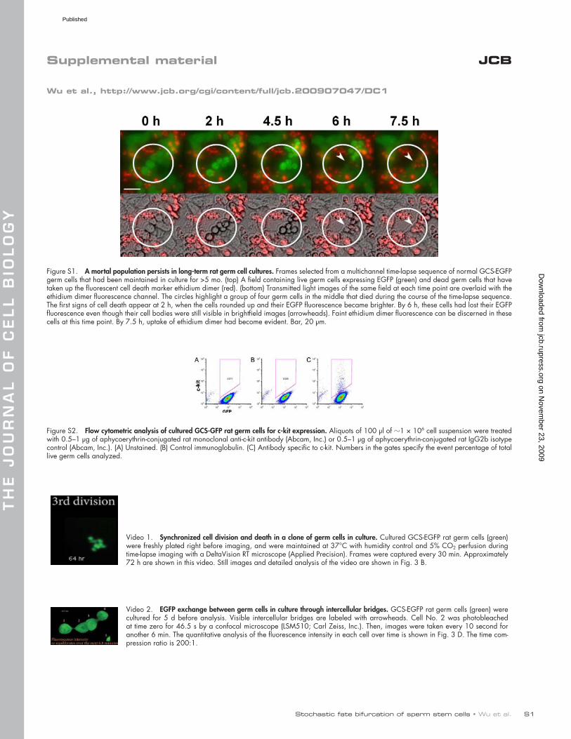
Figure S1. A mortal population persists in long-term rat germ cell cultures. Frames selected from a multichannel time-lapse sequence of normal GCS-EGFP germ cells that had been maintained in culture for >5 mo. (top) A field containing live germ cells expressing EGFP (green) and dead germ cells that have taken up the fluorescent cell death marker ethidium dimer (red). (bottom) Transmitted light images of the same field at each time point are overlaid with the ethidium dimer fluorescence channel. The circles highlight a group of four germ cells in the middle that died during the course of the time-lapse sequence. The first signs of cell death appear at 2 h, when the cells rounded up and their EGFP fluorescence became brighter. By 6 h, these cells had lost their EGFP fluorescence even though their cell bodies were still visible in brightfield images (arrowheads). Faint ethidium dimer fluorescence can be discerned in these cells at this time point. By 7.5 h, uptake of ethidium dimer had become evident. Bar, 20 µm.
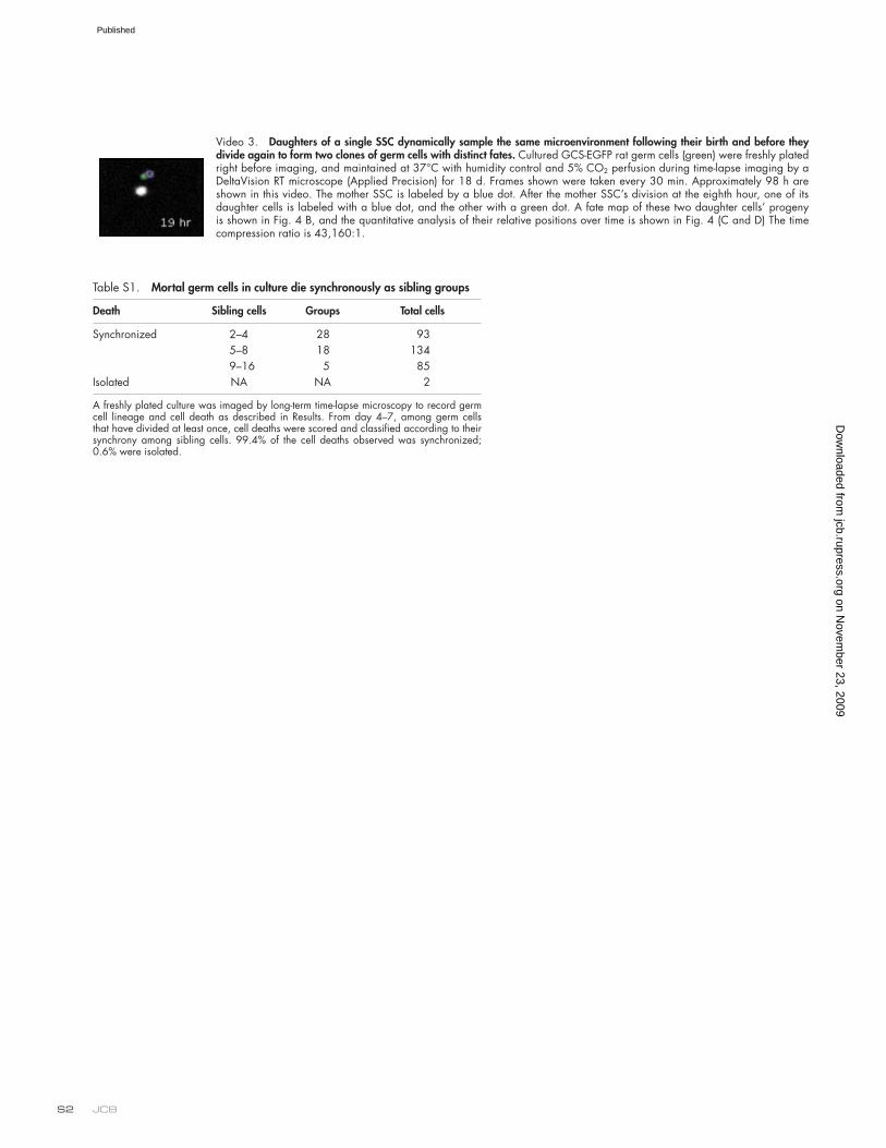
Figure S2. Flow cytometric analysis of cultured GCS-GFP rat germ cells for c-kit expression. Aliquots of 100 µl of 1 × 10⁶ cell suspension were treated with 0.5–1 µg of aphycoerythrin-conjugated rat monoclonal anti-c-kit antibody (Abcam, Inc.) or 0.5–1 µg of aphycoerythrin-conjugated rat IgG2b isotype control (Abcam, Inc.). (A) Unstained. (B) Control immunoglobulin. (C) Antibody specific to c-kit. Numbers in the gates specify the event percentage of total live germ cells analyzed.
Video 1. Synchronized cell division and death in a clone of germ cells in culture. Cultured GCS-EGFP rat germ cells (green) were freshly plated right before imaging, and were maintained at 37°C with humidity control and 5% CO2 perfusion during time-lapse imaging with a DeltaVision RT microscope (Applied Precision). Frames were captured every 30 min. Approximately 72 h are shown in this video. Still images and detailed analysis of the video are shown in Fig. 3 B.
Video 2. EGFP exchange between germ cells in culture through intercellular bridges. GCS-EGFP rat germ cells (green) were cultured for 5 d before analysis. Visible intercellular bridges are labeled with arrowheads. Cell No. 2 was photobleached at time zero for 46.5 s by a confocal microscope (LSM510; Carl Zeiss, Inc.). Then, images were taken every 10 second for another 6 min. The quantitative analysis of the fluorescence intensity in each cell over time is shown in Fig. 3 D. The time com-pression ratio is 200:1.
on Novem
ber 23, 2009 jcb.rupress.org
Dow
nloaded from
Published
JCB S2
Video 3. Daughters of a single SSC dynamically sample the same microenvironment following their birth and before they divide again to form two clones of germ cells with distinct fates. Cultured GCS-EGFP rat germ cells (green) were freshly plated right before imaging, and maintained at 37°C with humidity control and 5% CO2 perfusion during time-lapse imaging by a DeltaVision RT microscope (Applied Precision) for 18 d. Frames shown were taken every 30 min. Approximately 98 h are shown in this video. The mother SSC is labeled by a blue dot. After the mother SSC’s division at the eighth hour, one of its daughter cells is labeled with a blue dot, and the other with a green dot. A fate map of these two daughter cells’ progeny is shown in Fig. 4 B, and the quantitative analysis of their relative positions over time is shown in Fig. 4 (C and D) The time compression ratio is 43,160:1.
Table S1. Mortal germ cells in culture die synchronously as sibling groups
Death Sibling cells Groups Total cells
Synchronized 2–4 28 935–8 18 1349–16 5 85
Isolated NA NA 2
A freshly plated culture was imaged by long-term time-lapse microscopy to record germ cell lineage and cell death as described in Results. From day 4–7, among germ cells that have divided at least once, cell deaths were scored and classified according to their synchrony among sibling cells. 99.4% of the cell deaths observed was synchronized; 0.6% were isolated.
on Novem
ber 23, 2009 jcb.rupress.org
Dow
nloaded from
Published