ARTICLE IN PRESS
9
Please cite this article in press as: Rafiq, S., et al., Isolation and antiproliferative activity of Lotus corniculatus lectin towards human tumour cell lines. Phytomedicine (2013), http://dx.doi.org/10.1016/j.phymed.2013.08.005 ARTICLE IN PRESS G Model PHYMED-51492; No. of Pages 9 Phytomedicine xxx (2013) xxx–xxx Contents lists available at ScienceDirect Phytomedicine j ourna l h o mepage: www.elsevier.de/phymed Isolation and antiproliferative activity of Lotus corniculatus lectin towards human tumour cell lines Shaista Rafiq a , Rabiya Majeed b , Asif Khurshid Qazi b , Bashir Ahmad Ganai a , Ishfak Wani a , Syed Rakhshanda a , Yasrib Qurishi b , P.R. Sharma b , Abid Hamid b , Akbar Masood a , Rabia Hamid a,∗ a Department of Biochemistry, University of Kashmir, Hazratbal, Srinagar 190006, India b Indian Institute of Integrative Medicine, Canal Road, Jammu 180001, India a r t i c l e i n f o Article history: Received 4 May 2013 Received in revised form 2 July 2013 Accepted 4 August 2013 Keywords: Antiproliferative d-Galactose binding Lotus corniculatus lectin (LCL) Human leukemic cancer cells a b s t r a c t The objective of the study was to investigate the anti cancer activity of a lectin isolated from Lotus cornic- ulatus seeds. A tetrameric 70 kDa galactose specific lectin was purified using two step simple purification protocol which involved affinity chromatography on AF-BlueHC650M and gel filtration on Sephadex G- 100. The lectin was adsorbed on AF-BlueHC650M and desorbed using 1 M NaCl in the starting buffer. Gel filtration on Sephadex G-100 yielded a major peak absorbance that gave two bands of 15 kDa and 20 kDa in SDS PAGE. Hemagglutination activity was completely preserved, when the temperature was in the range of 20–60 ◦ C. However, drastic reduction in activity occurred at temperatures above 60 ◦ C. Full hemagglutination activity was retained at ambient pH 4–12. Thereafter no activity was observed above pH 13. Hemaglutination of the lectin was inhibited by d-galactose. The lectin showed a strong antipro- liferative activity towards human leukemic (THP-1) cancer cells followed by lung cancer (HOP62) cells and HCT116 with an IC 50 of 39 g/ml and 50 g/ml and 60 g/ml respectively. Flow cytometry analy- sis showed an increase in the percentage of cells in sub G0G1 phase confirming that Lotus corniculatus lectin induced apoptosis. Morphological observations showed that Lotus corniculatus lectin (LCL) treated THP-1 cells displayed apparent apoptosis characteristics such as nuclear fragmentation, appearance of membrane enclosed apoptotic bodies and DNA fragmentation. Lotus corniculatus lectin (LCL) effectively inhibits the cell migration in a dose dependent manner as indicated by the wound healing assay. © 2013 Elsevier GmbH. All rights reserved. Introduction Lectins are heterogeneous group of proteins with at least one non catalytic domain that selectively recognises and specifically binds to free sugars present on glycoproteins and glycolipids without altering the structure of the carbohydrates (Lannoo and Van Damme, 2010). Lectins are widely distributed in nature, mainly in the plant kingdom, even though they also occur in other organisms, such as animals and microorganisms (Pustai 1991; Drikarmer and Taylor 1993; Gabius 1997; Wong et al., 2010). Plant lectins have been established to possess remarkable anticancer properties in vivo, in vitro and in human case studies and have been successfully adopted for alternative cancer therapy (De Mejia and Prisecaru, 2005; Liu et al., 2010; Pusztai et al., 2008). Legume lectins are one of the most comprehensively studied plant lectins for their molecular basis of the protein-carbohydrate interactions ∗ Corresponding author. Tel.: +91 9419548985. E-mail address: [email protected] (R. Hamid). for several decades (Damodaran et al., 2008). In recent years, the main interest in this lectin family lay in their prospective application as antitumor agents that could bind specific cancer cell surface glycoconjugates (Ueno et al., 2000) e.g. a typical legume lectin with specificity towards sialic acid purified from Phasoleus coccinus L.(Phasoleus Multiflorus wild) seeds possess a remarkable antiproliferative activity. Lotus corniculatus commonly known as Bird’s foot trefoil belongs to a genus that contains many dozen of species distributes worldwide e.g. Lotus aboriginus (Rosy Bird’s foot trefoil), Lotus angustissimus (Slender Bird’s foot trefoil) and Lotus argophyllus (Silver Bird’s foot trefoil). Lotus corniculatus is a species of the legu- minosae family. Lotus corniculatus can fix nitrogen through the root nodules making it useful as cover crop. It has been stud- ied for its flavonoid content (Jay and Ibrahim 1986; Sarelli et al., 2003; Reynaud and Lassignol, 2005). Lotus corniculatus is known for its medicinal values. The flowers are antispasmodic and seda- tive (Chiej 1984). The root is carminative and febrifuge (Duke and Ayensu 1985). Although Lotus corniculatus belongs to legume fam- ily, none of the Lotus species have been investigated for the lectins. 0944-7113/$ – see front matter © 2013 Elsevier GmbH. All rights reserved. http://dx.doi.org/10.1016/j.phymed.2013.08.005
Transcript of ARTICLE IN PRESS

G
P
It
SIAa
b
a
ARRA
KAdLH
I
nbwVmoDlpbalf
0h
ARTICLE IN PRESS Model
HYMED-51492; No. of Pages 9
Phytomedicine xxx (2013) xxx– xxx
Contents lists available at ScienceDirect
Phytomedicine
j ourna l h o mepage: www.elsev ier .de /phymed
solation and antiproliferative activity of Lotus corniculatus lectinowards human tumour cell lines
haista Rafiqa, Rabiya Majeedb, Asif Khurshid Qazib, Bashir Ahmad Ganaia,shfak Wania, Syed Rakhshandaa, Yasrib Qurishib, P.R. Sharmab, Abid Hamidb,kbar Masooda, Rabia Hamida,∗
Department of Biochemistry, University of Kashmir, Hazratbal, Srinagar 190006, IndiaIndian Institute of Integrative Medicine, Canal Road, Jammu 180001, India
r t i c l e i n f o
rticle history:eceived 4 May 2013eceived in revised form 2 July 2013ccepted 4 August 2013
eywords:ntiproliferative-Galactose bindingotus corniculatus lectin (LCL)uman leukemic cancer cells
a b s t r a c t
The objective of the study was to investigate the anti cancer activity of a lectin isolated from Lotus cornic-ulatus seeds. A tetrameric 70 kDa galactose specific lectin was purified using two step simple purificationprotocol which involved affinity chromatography on AF-BlueHC650M and gel filtration on Sephadex G-100. The lectin was adsorbed on AF-BlueHC650M and desorbed using 1 M NaCl in the starting buffer.Gel filtration on Sephadex G-100 yielded a major peak absorbance that gave two bands of 15 kDa and20 kDa in SDS PAGE. Hemagglutination activity was completely preserved, when the temperature was inthe range of 20–60 ◦C. However, drastic reduction in activity occurred at temperatures above 60 ◦C. Fullhemagglutination activity was retained at ambient pH 4–12. Thereafter no activity was observed abovepH 13. Hemaglutination of the lectin was inhibited by d-galactose. The lectin showed a strong antipro-liferative activity towards human leukemic (THP-1) cancer cells followed by lung cancer (HOP62) cells
and HCT116 with an IC50 of 39 �g/ml and 50 �g/ml and 60 �g/ml respectively. Flow cytometry analy-sis showed an increase in the percentage of cells in sub G0G1 phase confirming that Lotus corniculatuslectin induced apoptosis. Morphological observations showed that Lotus corniculatus lectin (LCL) treatedTHP-1 cells displayed apparent apoptosis characteristics such as nuclear fragmentation, appearance ofmembrane enclosed apoptotic bodies and DNA fragmentation. Lotus corniculatus lectin (LCL) effectivelyinhibits the cell migration in a dose dependent manner as indicated by the wound healing assay.ntroduction
Lectins are heterogeneous group of proteins with at least oneon catalytic domain that selectively recognises and specificallyinds to free sugars present on glycoproteins and glycolipidsithout altering the structure of the carbohydrates (Lannoo andan Damme, 2010). Lectins are widely distributed in nature,ainly in the plant kingdom, even though they also occur in other
rganisms, such as animals and microorganisms (Pustai 1991;rikarmer and Taylor 1993; Gabius 1997; Wong et al., 2010). Plant
ectins have been established to possess remarkable anticancerroperties in vivo, in vitro and in human case studies and haveeen successfully adopted for alternative cancer therapy (De Mejia
Please cite this article in press as: Rafiq, S., et al., Isolation and antiproliferlines. Phytomedicine (2013), http://dx.doi.org/10.1016/j.phymed.2013.08.0
nd Prisecaru, 2005; Liu et al., 2010; Pusztai et al., 2008). Legumeectins are one of the most comprehensively studied plant lectinsor their molecular basis of the protein-carbohydrate interactions
∗ Corresponding author. Tel.: +91 9419548985.E-mail address: [email protected] (R. Hamid).
944-7113/$ – see front matter © 2013 Elsevier GmbH. All rights reserved.ttp://dx.doi.org/10.1016/j.phymed.2013.08.005
© 2013 Elsevier GmbH. All rights reserved.
for several decades (Damodaran et al., 2008). In recent years,the main interest in this lectin family lay in their prospectiveapplication as antitumor agents that could bind specific cancer cellsurface glycoconjugates (Ueno et al., 2000) e.g. a typical legumelectin with specificity towards sialic acid purified from Phasoleuscoccinus L.(Phasoleus Multiflorus wild) seeds possess a remarkableantiproliferative activity.
Lotus corniculatus commonly known as Bird’s foot trefoil belongsto a genus that contains many dozen of species distributesworldwide e.g. Lotus aboriginus (Rosy Bird’s foot trefoil), Lotusangustissimus (Slender Bird’s foot trefoil) and Lotus argophyllus(Silver Bird’s foot trefoil). Lotus corniculatus is a species of the legu-minosae family. Lotus corniculatus can fix nitrogen through theroot nodules making it useful as cover crop. It has been stud-ied for its flavonoid content (Jay and Ibrahim 1986; Sarelli et al.,2003; Reynaud and Lassignol, 2005). Lotus corniculatus is known
ative activity of Lotus corniculatus lectin towards human tumour cell05
for its medicinal values. The flowers are antispasmodic and seda-tive (Chiej 1984). The root is carminative and febrifuge (Duke andAyensu 1985). Although Lotus corniculatus belongs to legume fam-ily, none of the Lotus species have been investigated for the lectins.
ING Model
P
2 edicin
Wtacclb
M
P
rpsnlspyt7cTtTpcpTco
A
tpathoitht
C
iamNaaalfton
was washed 5–6 times with distilled water. Plate was air-dried.100 �l of SRB dye (0.4% in 1% acetic acid) was added to each wellof the plate and the plate left at room temperature for 30 min. Theplate was washed with 1% acetic acid after 30 min. The plate was
ARTICLEHYMED-51492; No. of Pages 9
S. Rafiq et al. / Phytom
e therefore isolated and purified a lectin from Lotus cornicula-us seeds with two simple chromatographic steps. Some physicalnd chemical properties such as thermo stability, pH stability andarbohydrate specificity of the lectin have been studied. Biologi-al activities of the Lotus corniculatus lectin were also studied. Theectin possessed antiproliferative effects on some tumour cell linesy inducing apoptosis and also possesses anti metastatic effects.
aterials and methods
urification of Lotus corniculatus lectin
The pods of Lotus corniculatus were obtained from the sur-oundings of Kashmir University and authenticated at centre oflant taxonomy (COPT), Department of Botany, Kashmir Univer-ity, India. The seeds (60 g) were crushed and powdered in liquiditrogen, dissolved in 300 ml of 10 mM Tris–HCl buffer (pH 7.6), fol-
owed by centrifugation at 3000 × g, at 4 ◦C, for 30 min. Ammoniumulphate was added to the supernatant to 40–60% saturation. Therecipitate was resuspended in 10 mM Tris–HCl buffer and dial-sed extensively overnight at 4 ◦C. The sample was then adjustedo 10 mM Tris–HCl (pH 7.6) by adding Tris–HCl buffer (1.5 M, pH.6). The sample was then applied to AF-BlueHC650M (Toyopearl)olumn (18 cm × 5 cm) that had been equilibrated with 10 mMris–HCl buffer (pH 7.6). Unabsorbed proteins were eluted withhe starting buffer. The column was washed with 1 M NaCl in10 mMris–HCl buffer to elute the bound protein. The active fractions wereooled, dialysed extensively against double distilled water and con-entrated by Ultra filtration using 10 kDa cut-off membrane. Theurified lectin was dissolved in sterile distilled water (16 mg/ml).he solution was subjected to Gel filtration on Sephadex G-100olumn. A major absorbance peak that contained purified LCL wasbtained.
ssay of hemagglutination activity
In a 96-well microtitre U-plate, a serial two fold dilution of theest sample (50 �l) in phosphate buffer saline (PBS) (pH 7.2) waserformed. A 2% rabbit red blood cell suspension (50 �l) in PBS wasdded to the sample. The mixture was incubated at room tempera-ure until the red blood cells in the blank (without protein sample)ad fully sedimented and appeared as a red spot at the bottomf the well. Presence of agglutinated red blood cells in the wellsndicated hemagglutinating activity. One hemagglutination unit ishe reciprocal of the highest dilution of the lectin sample inducingemagglutination. Specific activity is the number of hemagglutina-ion units per mg protein (Yagi et al., 2002).
arbohydrate binding specificity
The carbohydrate specificity was investigated by observing thenhibition of the lectin induced hemagglutination by various sug-rs namely d-glucose, d-galactose, d-mannose, fructose, lactose,altose, sucrose, ribose, xylose, manitol and sugar derivatives like-acetyl galactosamine and N-acetyl glucosamine. The inhibitionssay was performed in 96-well plate. Different dilutions of thebove sugars (final volume 20 �l) were added to the wells in whichgglutination was performed. To each dilution, 20 �l of purifiedectin was added. The mixture was incubated at room temperature
Please cite this article in press as: Rafiq, S., et al., Isolation and antiproliferlines. Phytomedicine (2013), http://dx.doi.org/10.1016/j.phymed.2013.08.
or 1 h after which 80 �l of 2% suspension of erythrocytes was addedo each well. The minimum concentrations of each sugar capablef fully inhibiting agglutination after 1 h at room temperature wereoted.
PRESSe xxx (2013) xxx– xxx
Molecular mass determination
Sodium dodecyl sulphate-polyacrylamide gel electrophoresis(SDS-PAGE) with a 8% separating gel and a 5% stacking gelwas performed. After electrophoresis, the gel was stained withCoomassie brilliant blue and destained with 10% acetic acidovernight (Laemmli and Favre, 1973).
Gel permeation chromatography was carried out usingSephadex G-100 column calibrated with molecular mass standard.
Protein concentration determination
The protein concentration was determined by the method of(Lowry et al., 1951) using BSA as the standard protein.
Effect of temperature, pH on lectin-induced hemagglutination
Thermal stability of LCL was monitored in the range of 10–100 ◦Cby incubating the lectin for 60 min at the respective temperatures,followed by cooling on ice and determination of agglutination activ-ity under standard conditions.
The pH dependence of the lectin was determined by incubating50 �g of LCL with buffers in different pH:HCl: pH 0–1, glycine/HCl(pH 2–3), 0.05 sodium acetate/acetic acid (pH 4–5), potassiumphosphate (pH 6–7), Tris–HCl (pH 8–9) and glycine-NaOH (pH10–11), pH 11–12 NaHCO3 and pH 13–14, NaOH, for 5 h at 25 ◦Cand pH was adjusted to 7.2 just prior to hemagglutination assay.
Tumour cell lines and culture conditions
Human leukemic cell line (adherent Type) (THP-1), lung cancercell line (HOP62) and colon cancer cell line (HCT116) were obtainedfrom American Type Culture Collection. All the cell lines were cul-tured in RPMI 1640 (Sigma) medium which contained FCS 10%(Sigma), 100 U/ml of pencillin (Sigma) and 100 �g/ml of strepto-mycin (Sigma). Cell cultures were incubated at 37 ◦C in a humidifiedatmosphere of 5% CO2. The cells were fed every 2–3 days, and atconfluency they were harvested with 0.05% (w/v) trypsin–EDTA(Sigma) and sub-cultured in identical medium.
Sulphorhodamine B (SRB) assay
Human leukemic (Adherent type) (THP-1), Lung cancer(HOP62), and colon (HCT116) cancer cells from American Type Cul-ture Collection were adjusted to a cell density of 5 × 10⁴ cells/mlin RPMI medium. The cells (100 ml) were seeded onto the wellsof a 96-well plate and incubated overnight. Different concentra-tions of Lotus corniculatus lectin (100 ml, final concentrations at30–100 �g/ml) were added to the wells followed by incubationfor 48 h. The plates was taken out from the incubator after 48 hof adding the sample. To stop the reaction, 50 �l of chilled 50% TCA(trichloroacetic acid) to each well of the plate was added, mak-ing final concentration to 10%. The plate was incubated at 4 ◦Cfor 1 h to fix the cells attached to bottom of the wells. The plate
ative activity of Lotus corniculatus lectin towards human tumour cell005
again air-dried. 100 �l of tris buffer (10.5 M) was added to eachwell. The plate was shaken gently for 10–15 min on a mechan-ical shaker. The optical density was recorded with ELISA readerat 540 nm.

IN PRESSG Model
P
edicine xxx (2013) xxx– xxx 3
Mr
itf1cpaflt
C
(2cwcA1c
O
imfitXuwn
D
2t83vww3optagag
W
Whtvc
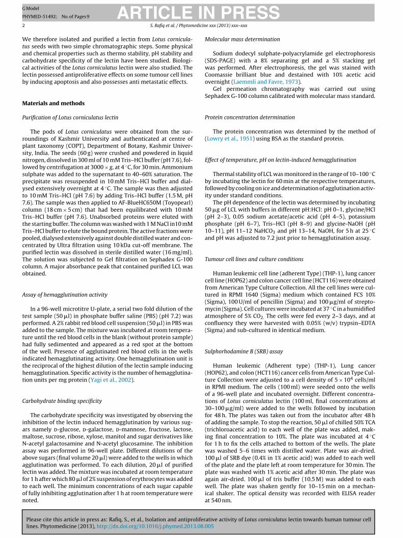
Fig. 1. Affinity purification of LCL from crude extract of its seeds on AF-BlueHC650M(18 cm × 5 cm). About 35 mg of crude extract of LCL was applied to the column,pre-equillibrated with 10 mM Tris–HCl buffer, pH 7.6. Bound protein was elutedwith sodium chloride (1 M) in 5 ml fractions at a flow rate of 30 ml/h. Only peak IIshowed agglutination activity. Series 1 represents Absorbance at 280 nm and Series2 represents hemagglutination titre.
ARTICLEHYMED-51492; No. of Pages 9
S. Rafiq et al. / Phytom
easurement of mitochondrial transmembrane potential byhodamine 123 (Rh-123) staining
THP-1 cells (5 × 10⁵) were seeded onto a 6-well plate andncubated overnight. Different concentrations of Lotus cornicula-us lectin (30–100 �g/ml) were added to the cells and incubatedor 24 h. The cells were trypsinized and centrifuged down at500 × g for 5 min. Then the cells were washed with PBS and againentrifuged at 1500 × g for 5 min, thrice. The cell pellets were resus-ended in RPMI medium (500 �l) containing 10 �M Rh-123 dye,nd incubated at 37 ◦C in dark. The cells were analysed using a FACsow cytometer for detecting the mitochondrial depolarization pat-erns.
ell cycle distribution analysis
After 24 h of incubation, to exponentially growing THP-1 cells1 × 105) 30–100 �g/ml of LCL was added and incubated further for4 h. Control cells were devoid of LCL. Both control and LCL treatedells were harvested by trypsin treatment, washed in cold PBS, fixedith 70% ethanol for about 16 h at 4 ◦C and again washed thrice with
old PBS. In order to remove RNA, the cells were treated with 500 �l of RNase (100 �g/ml) for 30 min at 37 ◦C followed by staining with00 �l of PI (50 �g/ml) at 4 ◦C for 5 min before analysing by flowytometry (BD Biosciences)
bservation of cell morphologic changes and nuclear damage
Exponentially growing THP-1 cells (1 × 10⁵ cells/ml) werencubated for 24 h with 30–100 �g/ml of LCL. Apoptotic nuclear
orphology was visualised by DAPI staining technique. Cells werexed with 3.7% of para-formaldehyde for 10 min at room tempera-ure, washed three times with PBS and immersed in 0.1% of Triton-100 for 2 min. Thus para-formaldehyde-fixed cells were stainedsing DAPI (10 �g/ml) under dark for 10 min. After three times ofashing with PBS the cells were observed under ultraviolet illumi-ation with a confocal microscope (Olympus).
NA fragmentation assay
DNA fragmentation was carried out as described by (Lin et al.,003) with slight variations. Five hundred microliters of (5 × 10⁶)reated cells were lysed in 55 ml lysis buffer [1 M Tris–HCI (pH.0), 0.5 M EDTA, and 100% Triton X-100] and incubated at 4 ◦C for0 min. DNA was extracted from the supernatant with an equalolume of phenol/choloroform/isoamyl alcohol (25:24:1). Samplesere spun and the upper aqueous layer transferred to a new tube, tohich an equal volume of ice-cold 100% ethanol and 1/10 volume of
M sodium acetate (pH 5.2) were added and incubated at −20 ◦Cvernight. After spinning the sample, supernatant was decanted,ellet air dried, and then dissolved in deionised water-RNase solu-ion [10 mg/ml RNase I] and incubated at 37 ◦C for 30 min. Equalmounts of DNA (10 mg/well) were electrophoresed in 1% agaroseel impregnated with ethidium bromide at 5 V for the first 5 minnd increased to 100 V for 1 h. After electrophoresis was complete,el was observed in gel doc system (Bio-Rad).
ound healing assay
THP-1 cells 1 × 10⁵ cells/ml/well were seeded in 6-well plate.hen the confluency reached 90% wounds were created with the
Please cite this article in press as: Rafiq, S., et al., Isolation and antiproliferlines. Phytomedicine (2013), http://dx.doi.org/10.1016/j.phymed.2013.08.0
elp of a 200-�l pipette tip. The cells were then rinsed with mediumo remove any free-floating cells and debris. Medium containingarious concentrations of LCL (30–100 �g/ml) was then added andulture plate was incubated at 37 ◦C. Wound healing was observed
Fig. 2. Gel filtration of the active fractions from the AF-BlueHC650M column onSephadex G-100 column. Eluent was PBS, pH 7.2. The flow rate was 20 ml/h andfraction size was 3 ml.
after 24 h within the scrape line and representative scrape lineswere photographed.
Statistical analysis
Results are expressed as the means and standard deviations oftriplicate measurements. Each experiment was performed at leastthrice. Statistical comparisons were made by student’s t-test andp < 0.05 was considered statistically significant.
Results
Purification of LCL by affinity chromatography on AF-BlueHC650M
Lectin (LCL) extracted from the seeds of Lotus corniculatus waspurified to homogeneity by affinity chromatography using AF-BlueHC650M column (Fig. 1) and Gel filtration chromatography onSephadex G-100 (Fig. 2). The summary of purification steps for LCLis presented in Table 1. The lectin was purified 6.54 folds with arecovery of 27.3% activity.
Electrophoretic and chromatographic analysis
ative activity of Lotus corniculatus lectin towards human tumour cell05
LCL showed two bands (15 kDa and 20 kDa) respectively on SDS-PAGE (Fig. 3). It was eluted in about 10th fraction in SephadexG-100 column. Based on the calibration curve of the column, LCL

ARTICLE IN PRESSG Model
PHYMED-51492; No. of Pages 9
4 S. Rafiq et al. / Phytomedicine xxx (2013) xxx– xxx
Table 1Summary of purification steps for LCL.
Purification step Total hemagglutination activity (HU) Total protein (mg) Specific activity (HU/mg) Purification fold Recovery of activity (%)
Crude 74350 98 774.5 1 100Ammonium sulphate (40–60%) 63486 38 1670 2.16 85.39AF-BlueC650M 44320 16 2770 3.58 59.6
5060 6.54 27.3
ha2
E
gfaow
S
Glc
F0m
Sephadex G-100 20240 4
as a molecular size of around 70 kDa. This showed that LCL is tetrameric protein consisting of two 15 kDa subunits and two0 kDa subunits.
ffect of pH and temperature on lectin activity
LCL had a moderate thermostability and pH stability. Hemag-lutination activity of LCL was completely retained upto 60 ◦C buturther increase in temperature caused abrupt disruption of thectivity (Fig. 4). Also complete hemaglutination activity of LCL wasbserved at pH 4–12, only minimal activity remained at pH 0–2 andhole of the activity was lost above pH 13 (Fig. 5).
ugar specificity
Please cite this article in press as: Rafiq, S., et al., Isolation and antiproliferlines. Phytomedicine (2013), http://dx.doi.org/10.1016/j.phymed.2013.08.
LCL was a d-galactose binding lectin. The presence of 25 mMalactose completely inhibited the hemagglutinating activity of the
ectin, other sugars were not able to inhibit LCL activity even at aoncentration of 200 mM (Table 2).
ig. 3. SDS-Polyacrylamide Gel Electrophoresis pattern of Lotus corniculatus lectin (LCL)..1% SDS. Tris–glycine buffer pH 8.3 was used. Current was 8 mA per well. The staining
arker proteins (Bio-Rad) and 1 represents purified lectin.
Fig. 4. Effect of temperature on lectin activity of LCL. The lectin was active at 60 ◦C.Hemagglutination % represents the activity left after treatment at various tempera-tures ranging from 20 ◦C to 100 ◦C.
ative activity of Lotus corniculatus lectin towards human tumour cell005
Effect of LCL on proliferation of tumour cells
The antiproliferative effect of LCL on THP-1, HOP62 and HCT-116cell lines is shown in Fig. 6. LCL was found to have consider-
About 40 �g of LCL was electrophoresed on 8% polyacrylamide gel in presence ofreagent used was Coomassie brilliant blue G-250. M – represents molecular mass

Please cite this article in press as: Rafiq, S., et al., Isolation and antiproliferlines. Phytomedicine (2013), http://dx.doi.org/10.1016/j.phymed.2013.08.0
ARTICLE IN PRESSG Model
PHYMED-51492; No. of Pages 9
S. Rafiq et al. / Phytomedicine xxx (2013) xxx– xxx 5
Fig. 5. Effect of pH on LCL activity. The lectin was stable in the pH range of 4–12.
Table 2Effect of different carbohydrates on hemagglutinating activity of Lotus corniculatuslectin (LCL).
Carbohydrate Inhibitory concentration (mM)a
Glucose NIb
Galactose 25Mannose NIFructose NILactose NIGlucosamine NIGalactosamine NIFucose NIArabinose NIMelibiose NIGlucuronic acid NIXylose NIMannosamine NIMannitol NI
a All the sugars except galactose, could not inhibit the hemagglutination activityeven at 200 mM.
b Not inhibited.
Fig. 7. DNA cell cycle analysis of LCL treated THP-1 cells for 24 h. Cells were exposed tdetermine DNA fluorescence and cell cycle phase distribution. The Data are representativ
Fig. 6. Result of SRB assay on different cell lines. The cells were treated with LCLfor 48 h. LCL exerted strong anti-proliferative activity on THP-1 cells, followed byHOP62 and HCT116. Results represent mean ± SD (n = 3).
able inhibitory effect on the proliferation of THP-1, HOP62 andHCT116 with an IC50 of 39 �g/ml, 50 �g/ml and 60 �g/ml respec-tively. As maximum inhibition of proliferation (95%) was observedin THP-1 cell line, further studies were done using THP-1 cellline.
Effect of LCL on tumour cell cycle
Healthy THP-1 cells exhibited normal cell cycle characteristics
ative activity of Lotus corniculatus lectin towards human tumour cell05
(G1/G0 and G2/M phases); the percentage of cells in sub G0/G1phase was 5.6%. After treatment of THP-1 cells with low concentra-tion of LCL (30 �g/ml), there was a mild increase. i.e. 7.2% of THP-1cells were in sub G0/G1 phase. However, when LCL concentration
o different concentrations (30–100 �g/ml) of LCL for 24 h and stained with PI toe of three independent experiments.

ARTICLE IN PRESSG Model
PHYMED-51492; No. of Pages 9
6 S. Rafiq et al. / Phytomedicine xxx (2013) xxx– xxx
Fig. 8. Rh-123 Staining. LCL induced loss of mitochondrial membrane potential (��m). THp-1 cells incubated with different concentrations (30–100 �g/ml) of LCL for 24 h.T in FL-e
wpctra
E
t(Lodd
E
cc(c
hereafter, cells were stained with Rhodamine-123 (Rh-123) for 1 h and analysed
xperiments.
as further increased, the percentage of THP-1 cells in sub G0G1hase had significantly increased to 38% (Fig. 7). These results indi-ate that with the increase in concentration of LCL (30–100 �g/ml),he cell cycle of THP-1 cells notably altered, that the sub G0G1atio gradually increased, suggesting LCL induces THP-1 cellpoptosis.
ffect of LCL on mitochondrial membrane potential
Loss of mitochondrial membrane potential has been linkedo the initiation and activation of apoptotic process in cellsReed 1995; Hockenbery et al., 1990). In Rh-123 staining ofCL treated THP-1 cells (Fig. 8), the increase in concentrationf LCL caused majority of the cells to experience mitochon-rial depolarization indicating that the cells were undergoing celleath.
ffect of LCL on cell morphology and nuclear integrity
When THP-1 cells were cultured with LCL for 24 h, morphologic
Please cite this article in press as: Rafiq, S., et al., Isolation and antiproliferlines. Phytomedicine (2013), http://dx.doi.org/10.1016/j.phymed.2013.08.
hanges were observed and confirmed by DAPI staining. In theontrol group, the nuclei were round and homogenously stainedFig. 9), whereas the LCL treated cells showed apoptotic body andondensed chromatin formation.
1 vs. FL-2 channels of flow cytometer. Data is representative of three independent
DNA ladder assay of apoptosis in THP-1 cells
To investigate the apoptosis inducing activity of LCL on THP-1 cells, the integrity of the genomic DNA of lectin treated cellswere analysed by agarose gel electrophoresis. As shown in Fig. 10,there is no degradation of genomic DNA extracted from THP-1cells of Negative control group (Lane 2), whereas there is faintladder like pattern in THP-1 cells treated at 50 �g/ml, 70 �g/mland 100 �g/ml, respectively, indicating therefore the occurrence ofapoptosis.
LCL inhibited migration of THP-1 cells
The effect of LCL on THP-1 cell migration was determined byscratch motility or wound healing assay. In the scratch motilityassay, untreated THP-1 cells exhibited a complete wound clo-sure activity after 24 h (Fig. 11). In contrary; the LCL treatedcells showed only limited wound closure at the end of its incu-bation time by migrating into the denuded zone. LCL showeda reduction in wound closure in a dose dependent manner i.e.(30–100 �g/ml).
Discussion
ative activity of Lotus corniculatus lectin towards human tumour cell005
It is now widely acknowledged that plant lectins have animmense potential in treating, preventing and helping to diagnosevarious chronic diseases including cancer (De Mejia and Prisecaru,

ARTICLE IN PRESSG Model
PHYMED-51492; No. of Pages 9
S. Rafiq et al. / Phytomedicine xxx (2013) xxx– xxx 7
Fig. 9. Morphology observation in THP-1 cells as indicated by DAPI staining. Cells were incubated in absence or presence of different concentrations (30–100 �g/ml) of LCLf ragme
2otpg
lhubLsbsTa2op
or 24 h. Arrows indicate apoptotic features (chromatin condensation and nuclear f
005). In the present study we have found that LCL possesses anbvious cytotoxicity against THP-1 cancer cells and inhibits migra-ion of the THP-1 cells in a dose dependent manner. LCL wasurified from the seed extract after only two simple chromato-raphic steps.
The molecular weight of LCL is 70 kDa, similar to other reportedectins like Dioscorea opposita lectin (Chan and Ng, 2013). LCL is aeterotetramer composed of two subunits of 15 kDa and two sub-nits of 20 kDa respectively and may be regarded as DBL (Dolichosiflorus lectin) type, which is a heterotetramer (Buts et al., 2001).ectins can be classified according to their carbohydrate bindingpecificity, such as galactose binding, glucose binding, mannoseinding, etc. LCL is a d-galactose specific lectin. LCL was demon-trated to possess remarkable antiproliferative effect towardsHP-1 cells. A number of lectins have been reported to possess
Please cite this article in press as: Rafiq, S., et al., Isolation and antiproliferlines. Phytomedicine (2013), http://dx.doi.org/10.1016/j.phymed.2013.08.0
ntiproliferative activity towards different cell lines (Dhuna et al.,007; Xia and Ng, 2006; Fang et al., 2010). The mechanism of actionf legume lectins is different from that of Ribosome inactivatingroteins (RIP sII) e.g., Sophora flavescens lectin induces apoptosis
ntation). Results are representative of triplicate experiments.
in HeLa cells in a caspase dependent manner (Liu et al., 2008),Astragalus membranaceus lectin also induces apoptosis in leukemiacells in a caspase dependent manner (Huang et al., 2011). Whileas, RIPsII like Mistletoe lectin (MLII) exerts anti-cancer effect byactivating extra-cellular signal regulated kinases and P38 mitogenactivated protein kinase (MAPK) (Heike et al., 1999). The cell cycledynamics of different phase is regulated due to different factorsand different mechanisms e.g. many anticancer agents arrest thecell cycle at the sub G0/G1, S, G2/M phase and then induce apop-totic cell death (Deepa et al., 2012; Yan et al., 2009; Lam and Ng,2010). The results suggest that LCL induces apoptosis and inhibitscell proliferation in THP-1 cells via sub G0–G1 phase arrest of thecell cycle. Apoptosis or programmed cell death is an intrinsic cellsuicidal mechanism that plays an imperative role in the mainte-nance of healthy tissues. Thus, searching for agents which prompt
ative activity of Lotus corniculatus lectin towards human tumour cell05
apoptosis of tumour cells has become an attractive strategy inanti-cancer drug discovery (Huang et al., 2011) Apoptosis is char-acterised morphologically by loss of cell membrane asymmetry,cell shrinkage, apoptotic body formation and condensation of the

ARTICLE ING Model
PHYMED-51492; No. of Pages 9
8 S. Rafiq et al. / Phytomedicin
Fig. 10. DNA fragmentation of THP-1 cells treated with LCL. M – represents molec-ular weight marker, −ve control represents untreated cells, Lane 1–3 representsincreasing concentration of LCL (50 �g/ml,70 �g/ml and 100 �g/ml) respectively.
c2asts
Nevertheless, a better understanding on the detailed mecha-
Fm
hromatin and DNA fragmentation (Huang et al., 2011; Hengartner,000; Liu et al., 2008). DNA condensation and the formation ofpoptotic bodies in LCL treated cells were observed with DAPI
Please cite this article in press as: Rafiq, S., et al., Isolation and antiproliferlines. Phytomedicine (2013), http://dx.doi.org/10.1016/j.phymed.2013.08.
taining (Fig. 9). The mitochondrial apoptotic pathway is one ofhe main routes to initiate apoptosis (Kuo et al., 2010). Differenttimuli cause changes in the inner mitochondrial membrane lead-
ig. 11. Wound closure activity of treated THP-1 cells after 24 h with increasing concentonolayered treated with LCL at 0 and 24 h. Untreated control had completely healed. Ty
PRESSe xxx (2013) xxx– xxx
ing to the opening of the mitochondrial permeability transitionpore, loss of the mitochondrial membrane potential (Saelens et al.,2004) and pro-apoptotic protein release from the intermembranespace into the cytosol (Borutaite, 2010; Singh et al., 2011). Our stud-ies demonstrated that treatment with LCL increased mitochondrialmembrane potential loss in a dose dependent manner, which mayindicate cell death by apoptosis in THP-1 cells (Fig. 8). Some lectinssuch as Con A, BKBL, yam, may cause disruption of the mitochon-drial membrane potential as an event associated with apoptosis(Liu et al., 2009; Chan et al., 2012; Chan and Ng, 2013). The mostdistinct biochemical characteristic of apoptosis is the activation ofendogenous Ca2+/Mg2+ dependent endonuclease mediated cleav-age of nucleosomes to generate oligonucleotide fragments withabout 180–200 bp length or their polymers (Wyllie, 1997). Thesefragments appear as DNA ladder on an agarose gel as shown inFig. 10, instead of a randomised DNA breakdown which is observedas a smear in case of necrosis. Cell motility and adhesion are thecritical processes in metastasis once the tumour cells invade bloodor lymph capillaries successfully. Several factors contribute to can-cer cell motility, including insulin-like growth factor II (IGF-II) andautotoxin (ATX), hepatocyte growth factor/scatter factor (HGF/SF),etc. (Noble et al., 2003; Gaetano et al., 2009; Martin and Jiang, 2010).LCL effectively inhibits the migration of THP-1 cells (Fig. 11) butwhether LCL inhibits the migration of THP-1 cells by affecting theactivity of these above mentioned molecules needs to be studiedseparately.
Conclusion
In conclusion, our observations indicated that LCL effec-tively caused cytotoxicity of THP-1 cells. In addition, it hasthe capability to exert inhibitory effect on migration ofTHP-1 cells, which is a crucial step in cancer metastasis.
ative activity of Lotus corniculatus lectin towards human tumour cell005
nism of how LCL exerts its anti apoptotic and antimetastaticactivity would be indispensable, and hence warrants furtherinvestigation.
ration of LCL (30–100 �g/ml). Representative photographs of wounded THP-1 cellpical result from three independent experiments is shown.

ING Model
P
edicin
A
“g
R
B
B
C
C
CD
D
D
D
D
D
F
GG
H
HH
H
J
ARTICLEHYMED-51492; No. of Pages 9
S. Rafiq et al. / Phytom
cknowledgment
This work was supported by a fellowship under the scheme,Research Fellowship in Science for Meritorious Students” (RFSMS),ranted by University Grants Commission (UGC), New Delhi, India.
eferences
orutaite, V., 2010. Mitochondria as decision-makers in cell death. Environ. Mol.Mutagen. 51, 406–416.
uts, L., Dao-Thi, M.H., Loris, R., Wyns, L., Etzler, M., Hamelryck, T., 2001. Weakprotein–protein interactions in lectins: the crystal structure of a vegetativelectin from the legume Dolichos biflorus. J. Mol. Biol. 309, 193–201.
han, Y.S., Ng, T.B., 2013. A Lectin with highly potent inhibitory activity towardbreast cancer cells from edible tubers of Dioscorea opposita cv. Nagaimo. PLoSONE 8 (1), e54212, http://dx.doi.org/10.1371/journal.pone.0054212.
han, Y.S., Wong, J.H., Fang, E.F., Pan, W., Ng, T.B., 2012. Isolation of a glucosaminebinding leguminous lectin with mitogenic activity towards splenocytes andanti-proliferative activity towards tumor cells. PLoS ONE 7 (6), e38961,http://dx.doi.org/10.1371/journal.pone.0038961.
hiej, R., 1984. Encyclopaedia of Medicinal Plants. MacDonald, Edinburgh.amodaran, D., Jeyakani, J., Chauhan, A., Kumar, N., Chandra, N.R., Surolia, A.,
2008. CancerLectinDB: a database of lectins relevant to cancer. Glycoconj. J. 25,191–198.
e Mejia, E.G., Prisecaru, V.I., 2005. Lectins as bioactive proteins: a potential in cancertreatment. Crit. Rev. Food Sci. 45, 425–445.
eepa, M., Sureshkumar, T., Satheeshkumar, P.K., Priya, S., 2012. Purified mulberryleaf lectin (MLL) induces apoptosis and cell cycle arrest in human breast cancerand colon cancer cells. Chem. Biol. Interact. 200 (1), 38–44.
huna, V., Kamboj, S.S., Kaur, A., Saxena, A.K., Bhide, S.V., Shanmugavel, Singh, J.,2007. Characterization of a lectin from Gonatanthus pumilus D.Don having anti-proliferative effect against human cancer cell lines. Protein Pept. Lett 14, 71–78.
rikarmer, K., Taylor, M.E., 1993. Biology of animal lectins. Annu. Rev. Cell Biol. 9,237–264.
uke, J.A., Ayensu, E.S., 1985. Medicinal Plants of China. 2 vols. Reference Publica-tions, Algonac, MI, pp. 705 (DAA).
ang, E.F., Lin, P., Wong, J.H., Tsao, S.W., Ng, T.B., 2010. A lectin with anti-HIV-1reverse transcriptase, antitumor, and nitric oxide inducing activities from seedsof Phaseolus vulgaris cv. extralong autumnpurple bean. J. Agric. Food Chem. 58,2221–2229.
abius, H.J., 1997. Animal lectins. Eur. J. Biochem. 243, 543–576.aetano, C.G., Samadi, N., Tomsig, J.L., Macdonald, T.L., Lynch, K.R., D Brind-
ley, D.N., 2009. Inhibition of autotaxin production or activity blockslysophosphatidylcholine-induced migration of human breast cancer andmelanoma cells. Mol. Carcinog. 48 (9), 801–809.
eike, B., Engels, I.H., Wolfgang, V., Klaus, S.O., Sebastian, W., 1999. Mistletoelectin activates caspase-8/flice independently of death receptor signaling andenhances anticancer drug-induced apoptosis. Cancer Res. 59, 2083–2090.
engartner, M.O., 2000. The biochemistry of apoptosis. Nature 407 (6805), 770–776.ockenbery, D., Nunez, G., Milliman, C., Schreiber, R.D., Korsmeyer, S.J., 1990. Bcl-2 is
an inner mitochondrial membrane protein that blocks programmed cell death.Nature 348, 334–336.
uang, L.H., Yan, Q.J., Kopparapu, N.K., Jiang, Z.Q., Sun, Y., 2011. Astragalus
Please cite this article in press as: Rafiq, S., et al., Isolation and antiproliferlines. Phytomedicine (2013), http://dx.doi.org/10.1016/j.phymed.2013.08.0
membranaceus lectin (AML) induces caspase-dependent apoptosis in humanleukemia cells. Cell Prolif. 45, 15–21.
ay, M., Ibrahim, R.K., 1986. Biosynthesis of 8 subtituted flavonols in relation tooutogeny of flower color in Lotus corniculatus. Biochem. Physiol. Pflanz. 181,199–206.
PRESSe xxx (2013) xxx– xxx 9
Kuo, W.T., Ho, Y.J., Kuo, S.M., Lin, F.H., Tsai, F.J., Chen, Y.S., Dong, G.C., Yao,C.H., 2010. Induction of the mitochondria apoptosis pathway by phytohemag-glutinin erythroagglutinating in human lung cancer cells. Ann. Surg. Oncol.,http://dx.doi.org/10.1245/s10434-010-1351-2.
Laemmli, U.K., Favre, M., 1973. Gel electrophoresis of proteins. J. Mol. Biol. 80,575–599.
Lam, S.K., Ng, T.B., 2010. First report of a haemagglutinin-induced apoptotic pathwayin breast cancer cells. Biosci. Rep. 30, 307–317.
Lannoo, N., Van Damme, E.J., 2010. Nucleocytoplasmic plant lectins. Biochim. Bio-phys. Acta 1800, 190–201.
Lin, J., Dong, H.F., Oppenheim, J.J., Howard, O.M., 2003. Effects of astragali radix onthe growth of different cancer cell lines. World J. Gastroenterol. 4, 670–673.
Liu, B., Bian, H.J., Bao, J.K., 2010. Plant lectins: potential antineoplastic drugs frombench to clinic. Cancer Lett. 287 (1), 1–12.
Liu, B., Li, C.Y., Bian, H.J., Min, M.W., Chen, L.F., Bao, J.K., 2009. Antiproliferative activ-ity and apoptosis-inducing mechanism of Concanavalin A on human melanomaA375 cells. Arch. Biochem. Biophys. 482, 1–6.
Liu, Z., Liu, B., Zhang, Z.T., Zhou, T.T., Bian, H.J., Min, M.W., Liu, Y.H., Chen, J., Bao, J.K.,2008. A mannose-binding lectin from Sophora flavescens induces apoptosisinHeLa cells. Phytomedicine 15, 867–875.
Lowry, O.H., Rosebrough, A.L., Farr, A.L., Randall, R.J., 1951. Protein measurementwith the folin-phenol reagent. J. Biol. Chem. 193, 165–275.
Martin, T.A., Jiang, W.G., 2010. Hepatocyte growth factor and its receptor signallingcomplex as targets in Cancer therapy. Endocrinology 144, 2417–2424.
Noble, A., Towne, C., Chopin, L., Leavesley, D., Upton, Z., 2003. Insulin like growthfactor-II bound to vitronectin enhances MCF-7 breast cancer cell migration.Endocrinology 144 (6), 2417–2424.
Pustai, A.J., 1991. Plant Lectins. Cambridge University Press, Cambridge, UK, pp. 253.Pusztai, A., Bardocz, S., Ewen, S.W.B., 2008. Uses of plant lectins in bioscience and
biomedicine. Front. Biosci. 13 (3), 1130–1140.Reed, J.C., 1995. Regulation of apoptosis by bcl-2 family proteins and its role in cancer
and chemoresistance. Curr. Opin. Oncol. 7, 541–546.Reynaud, J., Lassignol, M., 2005. The flavonoids of Lotus corniculatus. Lotus Newsl. 35
(1), 75–82.Saelens, X., Festjens, N., Walle, L.V., Van Gurp, M., Van Loo, G., Vandenabeele, P.,
2004. Toxic proteins released from mitochondria in cell death. Oncogene 23,2861–2874.
Sarelli, L., Tuori, M., Saastamoinen, J., Syrjala-Qvist, L., Saloniemi, H., 2003. Phytoes-trogen content of Bird’s foot trefoil and red clover: effects of growth stage andensiling method. Acta Agric. Scand. A: Anim. Sci. 53, 58–63.
Singh, T., Sharma, S.D., Katiyar, S.K., 2011. Grape proanthocyanidins induce apo-ptosis by loss of mitochondrial membrane potential of human non-smallcell lung cancer cells in vitro and in vivo. PLoS ONE 6 (11), e27444,http://dx.doi.org/10.1371/journal.pone.0027444.
Ueno, T., Ohtawa, K., Sakurai, Y., Kodera, Y., Hiroto, M., et al., 2000. Polyethyleneglycol-modified Concanavalin A as an effective agent to stimulate anti-tumourcytotoxicity. Cancer Detect. Prev. 24, 100–106.
Wong, J.H., Ng, T.B., Cheung, R.C., Ye, X.J., Wang, H.X., Lam, S.K., Lin, P., Chan, Y.S., Fang,E., Ngai,.F., Xia, P.H., Ye, L.X., Jiang, X.Y., Liu, Y.F., 2010. Proteins with antifungalproperties and other medicinal applications from plants and mushrooms. Appl.Microbiol. Biotechnol. 87, 1221–1235.
Wyllie, A.H., 1997. Apoptosis: an overview. Br. Med. Bull. 53, 451–465.Xia, L., Ng, T.B., 2006. A hemagglutinin with mitogenic activity from dark red kidney
beans. J. Chromatogr. B: Analyt. Technol. Biomed. Life Sci. 844, 213–216.Yagi, F., Iwaya, T., Haraguchi, T., Goldstein, I.J., 2002. The lectin from leaves of
ative activity of Lotus corniculatus lectin towards human tumour cell05
Japanese cycad, Cycas revoluta Thunb. (gymnosperm) is a member of the jacalin-related family. Eur. J. Biochem. 269, 4335–4341.
Yan, Q., Li, Y., Jiang, Z., Sun, Y., Zhu, L., Ding, Z., 2009. Antiproliferation and apo-ptosis of human tumor cell lines by a lectin (AMML) of Astragalus mongholicus.Phytomedicine 16, 586–593.