In Memoriam - Allen Press
157
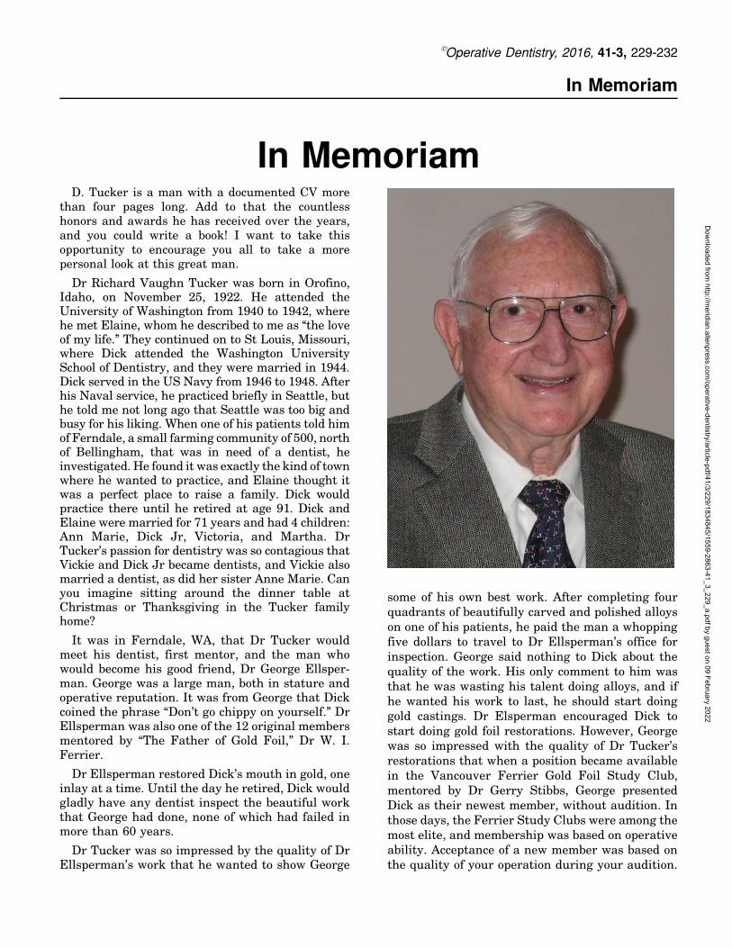
In Memoriam In Memoriam D. Tucker is a man with a documented CV more than four pages long. Add to that the countless honors and awards he has received over the years, and you could write a book! I want to take this opportunity to encourage you all to take a more personal look at this great man. Dr Richard Vaughn Tucker was born in Orofino, Idaho, on November 25, 1922. He attended the University of Washington from 1940 to 1942, where he met Elaine, whom he described to me as ‘‘the love of my life.’’ They continued on to St Louis, Missouri, where Dick attended the Washington University School of Dentistry, and they were married in 1944. Dick served in the US Navy from 1946 to 1948. After his Naval service, he practiced briefly in Seattle, but he told me not long ago that Seattle was too big and busy for his liking. When one of his patients told him of Ferndale, a small farming community of 500, north of Bellingham, that was in need of a dentist, he investigated. He found it was exactly the kind of town where he wanted to practice, and Elaine thought it was a perfect place to raise a family. Dick would practice there until he retired at age 91. Dick and Elaine were married for 71 years and had 4 children: Ann Marie, Dick Jr, Victoria, and Martha. Dr Tucker’s passion for dentistry was so contagious that Vickie and Dick Jr became dentists, and Vickie also married a dentist, as did her sister Anne Marie. Can you imagine sitting around the dinner table at Christmas or Thanksgiving in the Tucker family home? It was in Ferndale, WA, that Dr Tucker would meet his dentist, first mentor, and the man who would become his good friend, Dr George Ellsper- man. George was a large man, both in stature and operative reputation. It was from George that Dick coined the phrase ‘‘Don’t go chippy on yourself.’’ Dr Ellsperman was also one of the 12 original members mentored by ‘‘The Father of Gold Foil,’’ Dr W. I. Ferrier. Dr Ellsperman restored Dick’s mouth in gold, one inlay at a time. Until the day he retired, Dick would gladly have any dentist inspect the beautiful work that George had done, none of which had failed in more than 60 years. Dr Tucker was so impressed by the quality of Dr Ellsperman’s work that he wanted to show George some of his own best work. After completing four quadrants of beautifully carved and polished alloys on one of his patients, he paid the man a whopping five dollars to travel to Dr Ellsperman’s office for inspection. George said nothing to Dick about the quality of the work. His only comment to him was that he was wasting his talent doing alloys, and if he wanted his work to last, he should start doing gold castings. Dr Elsperman encouraged Dick to start doing gold foil restorations. However, George was so impressed with the quality of Dr Tucker’s restorations that when a position became available in the Vancouver Ferrier Gold Foil Study Club, mentored by Dr Gerry Stibbs, George presented Dick as their newest member, without audition. In those days, the Ferrier Study Clubs were among the most elite, and membership was based on operative ability. Acceptance of a new member was based on the quality of your operation during your audition. Ó Operative Dentistry, 2016, 41-3, 229-232 Downloaded from http://meridian.allenpress.com/operative-dentistry/article-pdf/41/3/229/1834845/1559-2863-41_3_229_a.pdf by guest on 09 February 2022
-
Upload
khangminh22 -
Category
Documents
-
view
0 -
download
0
Transcript of In Memoriam - Allen Press
In Memoriam
In MemoriamD. Tucker is a man with a documented CV more
than four pages long. Add to that the countlesshonors and awards he has received over the years,and you could write a book! I want to take thisopportunity to encourage you all to take a morepersonal look at this great man.
Dr Richard Vaughn Tucker was born in Orofino,Idaho, on November 25, 1922. He attended theUniversity of Washington from 1940 to 1942, wherehe met Elaine, whom he described to me as ‘‘the loveof my life.’’ They continued on to St Louis, Missouri,where Dick attended the Washington UniversitySchool of Dentistry, and they were married in 1944.Dick served in the US Navy from 1946 to 1948. Afterhis Naval service, he practiced briefly in Seattle, buthe told me not long ago that Seattle was too big andbusy for his liking. When one of his patients told himof Ferndale, a small farming community of 500, northof Bellingham, that was in need of a dentist, heinvestigated. He found it was exactly the kind of townwhere he wanted to practice, and Elaine thought itwas a perfect place to raise a family. Dick wouldpractice there until he retired at age 91. Dick andElaine were married for 71 years and had 4 children:Ann Marie, Dick Jr, Victoria, and Martha. DrTucker’s passion for dentistry was so contagious thatVickie and Dick Jr became dentists, and Vickie alsomarried a dentist, as did her sister Anne Marie. Canyou imagine sitting around the dinner table atChristmas or Thanksgiving in the Tucker familyhome?
It was in Ferndale, WA, that Dr Tucker wouldmeet his dentist, first mentor, and the man whowould become his good friend, Dr George Ellsper-man. George was a large man, both in stature andoperative reputation. It was from George that Dickcoined the phrase ‘‘Don’t go chippy on yourself.’’ DrEllsperman was also one of the 12 original membersmentored by ‘‘The Father of Gold Foil,’’ Dr W. I.Ferrier.
Dr Ellsperman restored Dick’s mouth in gold, oneinlay at a time. Until the day he retired, Dick wouldgladly have any dentist inspect the beautiful workthat George had done, none of which had failed inmore than 60 years.
Dr Tucker was so impressed by the quality of DrEllsperman’s work that he wanted to show George
some of his own best work. After completing fourquadrants of beautifully carved and polished alloyson one of his patients, he paid the man a whoppingfive dollars to travel to Dr Ellsperman’s office forinspection. George said nothing to Dick about thequality of the work. His only comment to him wasthat he was wasting his talent doing alloys, and ifhe wanted his work to last, he should start doinggold castings. Dr Elsperman encouraged Dick tostart doing gold foil restorations. However, Georgewas so impressed with the quality of Dr Tucker’srestorations that when a position became availablein the Vancouver Ferrier Gold Foil Study Club,mentored by Dr Gerry Stibbs, George presentedDick as their newest member, without audition. Inthose days, the Ferrier Study Clubs were among themost elite, and membership was based on operativeability. Acceptance of a new member was based onthe quality of your operation during your audition.
�Operative Dentistry, 2016, 41-3, 229-232
Dow
nloaded from http://m
eridian.allenpress.com/operative-dentistry/article-pdf/41/3/229/1834845/1559-2863-41_3_229_a.pdf by guest on 09 February 2022

Despite the tremendous pressure he must have felt,Dick flourished in this new environment.
Over the years, Dick recognized that he sharedthe same fundamental values as Dr Gerry Stibbs,and they became the very best of friends.
During this period, Dr Tucker continued toperfect his gold casting technique. Recognizing thatthe finest margin was that of a gold foil, he strivedto achieve that in his casting and finishingtechniques.
Over time, as Dr Tucker would bring his patientsto the Gold Foil Study Club, many members wouldtake note of and comment on the beautiful gold castrestorations that they could see next to his excellentfoils. A fellow study club member known for hisexcellent gold castings, Dr Joe Zokol, was soimpressed with the fit and finish of Dr Tucker’sbeautiful castings that he encouraged him tomentor a Cast Gold Study Club in his Vancouveroffice. Joe and Dick agreed that they would imitatethe format of the Ferrier Study Clubs: monthlylectures and operations, followed by critiques and adinner with refreshments, but they would use theguidelines of armamentarium, preparation design,and finishing techniques developed by Dr Tucker.Dick’s artistic genius was clearly evident in hisunique preparation designs and his extremelyefficient use of a minimal number of burs and handinstruments.
On January 8, 1976, the R. V. Tucker Academywas born in the office of Drs Joe and son, Ron,Zokol. By good fortune, my long-time friend, DrLaurie Vanzella, and I were invited to be membersof this new study club. We had no idea at the time,that we had literally won the Power Ball Lottery ofoperative dentistry! We were at the start of themost amazing and fulfilling journey of our profes-sional careers.
On that day, Dr Tucker gave two demonstrations.In the morning, he prepared five gold inlays, tookimpressions, and temporized the quadrant in lessthan 2.5 hours. After lunch, he seated, finished, andpolished a second quadrant of four gold inlays inless than two hours. No one could believe how fasthe was able to prepare and finish these two cases,especially when the finished afternoon case was themost beautifully finished work we’d ever seen. Hemade it look effortless. When we asked him how hewas able to operate with such speed, he explainedthat each operation was a series of steps. The keywas to do each step as perfectly as possible beforemoving on to the next step, eliminating the need to
go back. He told us over and over that the key tosuccess was to focus on learning to cut thepreparations as perfectly as possible, and over time,with repetition, the speed would come. Perfectionfirst, speed second. Over the years, we found thatthis Tucker philosophy applied to all aspects ofdentistry and would enable us to become muchbetter operators in other fields of dentistry.
Dr Tucker felt that the social aspect of our studyclub was vitally important. Our appreciation forexcellence in dentistry was matched by our appre-ciation for fine dining. It was at these dinnerswhere our friendships grew, and we felt comfortableto speak frankly about other issues related todentistry. At Christmas time, we would dress upin our tuxedos and have a grand dinner and finewines; this was one of Dr Tucker’s favoritetraditions.
Our annual Whistler ski weekend was anothertradition unique to our group. Apre ski consisted ofgreat dinners, lots of great wine, games of bridge,and Dick’s favorite, Honest Farmer, which he neverseemed to lose at. Dick’s ‘‘fireside chat’’ on invest-ment principles and strategies was a part of theweekend we always looked forward to. We nick-named him ‘‘the Warren Buffett of dentistry.’’
As his techniques gained notoriety and thedemand for him to lecture at various universitiesand dental meetings grew, he inspired other youngdentists to start Tucker Study Clubs of their own.The R. V. Tucker Academy was gaining worldwiderecognition. In 1986, Dr Tucker introduced the ideaof an associated meeting for all existing study clubsto gather once a year. The first meeting was held inthe Olympic Hotel in Seattle and at that timeconsisted of lectures and social activities only. Twoyears later, club 1 hosted the Tucker Associatedmeeting in Vancouver, BC, Canada, and it was atthat meeting where the George Ellsperman lectureand clinical operations were added to the programsstructure. When the demand for study clubs in Italyand Germany arose, Dick decided it was time tohave some of his founding members help mentorthese new groups. Dr Tucker took club 1 to Italyand later club 3 to Germany to present a TuckerTechnique program and assist in mentoring. It wasa very special time for us, as we really recognizedhow much Dick had taught us and how much of thatknowledge we could share with our new friends inEurope. Over the years, club 1 and club 3 madeother trips back to Italy and Germany to mentorand established lifelong friendships with the Italianand German study clubs. It was Dick’s idea that
230 Operative Dentistry
Dow
nloaded from http://m
eridian.allenpress.com/operative-dentistry/article-pdf/41/3/229/1834845/1559-2863-41_3_229_a.pdf by guest on 09 February 2022

whenever club 1 mentored in Italy or Hawaii, weshould spend a few days together socially after ourmentoring duties were over. This really enrichedour experience and confirmed to all that Dr Tuckerpracticed what he preached in all aspects of his life.
With the help of Dr Dennis Miya, Dick developedthe Tucker Institute. This was a 1-week summerprogram held at the University of Washington,modeled after the program that Dick started yearsearlier at the University of British Columbia, wheredentists were introduced to the Tucker Techniquethrough lectures and operations. As dentists wereexposed to these techniques, the demand for newstudy clubs grew exponentially. Dr Tucker devel-oped a new protocol whereby members of the sixfounding study clubs would have the opportunity tomentor a new study club. Dr Tucker’s schedule wasmore demanding than ever, yet he still found timeto attend the first meeting of a newly formed studyclub to lecture and often operate. He kept up thisschedule until 2007, when he, in his own words,‘‘started to slack off,’’ only continuing to mentor theVancouver Ferrier Gold Foil club plus three TuckerStudy clubs thereafter. I’ve calculated a roughestimate of how many hours he dedicated to studyclub mentoring and lecturing, and it is in excess of12,000 hours, which was close to six years of his lifebetween 1976 and 2013. This was on top ofmaintaining a full-time dental practice in Ferndale.During the last decade, he began to limit his studyclub activities further to be home with Elaine eachnight.
In September 2009, the R.V. Tucker Associatedmeeting was held in Vancouver, BC, Canada. DrTucker was presented with a hand-carved Squam-ish Nation ‘‘talking stick.’’ The Squamish elders usethe talking stick to teach the young people how tointeract in a respectful manner with others. In theirculture, they teach never look up or down atsomeone but to look across at them, as everyone isequal. This made the talking stick such a meaning-ful gift for Dick, as it really symbolized his ownteaching and mentoring style. I believe this is trulythe cornerstone of Dr Tucker’s genius. As Dick’swell-deserved reputation continued to grow, heremained always humble, gracious, and approach-able. He never looked down on anyone. Regardlessof a dentist’s educational or operative background,Dick had a magical ability to encourage the verybest in us. His gentle way, how he would askpermission to show you something that would help,and his quiet strength inspired us not only to do ourvery best in dentistry, but also in our lives.
On May 6, 2011, Dr Tucker gave his lastpresentation on conservative cast gold restorationsat the University of British Columbia. His instruc-tions to me were simple: tuition should be justenough to cover the costs so that as many dentistsas possible could attend, and he refused to take anhonorarium. Because this would be his last lecture,he would bring his entire staff and asked me if Iwould be so kind as to introduce each one of them inmy opening remarks as a way of honoring theircontribution to his success. The program sold out.When Dr Tucker concluded his presentation, theaudience gave him a standing ovation.
In November 2012, more than 200 of Dr Tucker’sfamily, friends, fellow academy members, and staffattended a surprise party for his 90th birthday inBellingham, WA. Dick knew only of a presentationfrom the Hawaiian clubs 44 and 50 to club 1, and DrTucker was honoring their longstanding relation-ship as the first mentors for the Hawaiians. Theypresented Dr Tucker and all members of club 1 witha silver goblet that they would all drink togetherwith the Hawaiians whenever they met to toast thisspecial friendship. Then into the next room forlunch and—SURPRISE! It was a night to remem-ber. We all thought he might just live forever.However, over the next year when he would travelto Vancouver to mentor club 1, he would oftenremark that it was time to retire as ‘‘no one shouldsee a dentist over 90 years of age!’’
As his health began to decline, Dick finally retired.He rallied enough to attend the R. V. TuckerAssociated meeting in Honolulu in August 2014due to the gracious care of his laboratory technicianand personal assistant Luba. She would stay on withDr Tucker as his personal assistant until his lastbreath. She is a true angel, and we are so gratefulthat she cared for him like her own father.
Some of you may know how much Dick loved tosail. He treasured time out on their beautifulsailboat, Line Angle. He had so many happymemories related to family and friends out on thewater sailing with him. I’ve never seen him lookhappier or more at peace than when he was sailingthe Line Angle in a good wind. On June 1, 2015, thewind was taken from his sails when his belovedElaine passed away.
He never truly recovered from this.
On December 23, 2015, Nancy and I met Dick Jr,Luba, and Dick Sr at his favorite Italian restaurantin Bellingham Bay for a Christmas lunch. Hegraciously signed four of the club 1 Tucker
McKay: In Memoriam 231
Dow
nloaded from http://m
eridian.allenpress.com/operative-dentistry/article-pdf/41/3/229/1834845/1559-2863-41_3_229_a.pdf by guest on 09 February 2022

anthology books as gifts for the founding membersof the Italian study clubs: still thinking of othersand making a contribution. This would be the verylast time I would see my dearest friend and mentorbefore he went to be with his Elaine. On themorning of January 12, 2016, at age 93, Dr RichardVaughn Tucker passed away peacefully.
All the years that Dr Tucker practiced inFerndale, a plaque hung in his main operatory thatsaid ‘‘It’s not what you think that counts, It’s notwhat you believe that counts, It’s not what you saythat counts, It’s what you do that counts!’’ Winston
Churchill once said that a man’s wealth and life isnot measured by what he gets, but by what he gives.For me, Dr Tucker was the wealthiest man I haveever met. I will miss him beyond measure. In thewords from our dear Hawaiian friends, written onhis goblet, which will now be turned over to read‘‘Pau Hana,’’ the work is done, it’s time to rest, we’vedone our best.
Respectfully submitted,
Terry McKay
January 17, 2015
232 Operative Dentistry
Dow
nloaded from http://m
eridian.allenpress.com/operative-dentistry/article-pdf/41/3/229/1834845/1559-2863-41_3_229_a.pdf by guest on 09 February 2022
Clinical Technique/Case Report
Ceramic Veneers and Direct-Composite Cases of Amelogenesis
Imperfecta Rehabilitation
S Shibata � CMC Taguchi � R GondoSC Stolf � LN Baratieri
Clinical Relevance
Amelogenesis imperfecta is a hereditary disease affecting the quality and quantity ofenamel. Patients usually suffer from oral complications and poor dental esthetics, whichdirectly affect their quality of life. Function and esthetics can be restored with differentrestorative materials, such as ceramic and composite resin. Dentists need to be aware ofthe best material to use for each patient.
SUMMARY
The aim of this article is to present two case
reports for the treatment of patients affected
with amelogenesis imperfecta. One case was
treated with composite resin and the other
case with ceramic veneers. Esthetic and func-tional results were achieved using both treat-ments, and a review of advantages anddisadvantages is presented.
INTRODUCTION
Amelogenesis imperfecta (AI) is a term for aclinically and genetically heterogeneous group ofconditions that are caused by mutations in a varietyof genes that are critical for normal enamelformation. The gene mutations alter the qualityand/or quantity of enamel in the primary andpermanent dentitions. Initial classifications of AIhad been based exclusively on the phenotype(appearance). More recent classifications includeboth the phenotype and the mode of inheritance.The outdated AI classification system recognizedfour phenotypes: 1) hypoplastic, 2) hypomaturation,3) hypocalcified, and 4) hypoplastic-hypomatura-tion. However, today, at least 14 AI subtypes areidentified when both phenotype and mode ofinheritance are considered.1-3
AI prevalence has been reported to vary from 1/700 to 1/14,000, depending on the population
Shizuma Shibata, DDS, MS, Operative Dentistry, FederalUniversity of Santa Catarina (UFSC), Florianopolis, SantaCatarina, Brazil
*Carolina Mayumi Cavalcanti Taguchi, DDS, MS, OperativeDentistry, Federal University of Santa Catarina (UFSC),Florianopolis, Santa Catarina, Brazil
Renata Gondo, DDS, PhD, Operative Dentistry, UniversidadeFederal de Santa Catarina (UFSC), Florianopolis, SantaCatarina, Brazil
Sheila Cristina Stolf, DDS, PhD, Operative Dentistry, Uni-versidade Federal de Santa Catarina (UFSC), Florianopolis,Brazil
Luiz Narciso Baratieri, DDS, PhD, Operative Dentistry,Universidade Federal de Santa Catarina (UFSC), Florianop-olis, Brazil
*Corresponding author: Rua Almirante Lamengo, 910ap.1001B, Florianopolis, Santa Catarina 88015-600, Bra-zil; e-mail: [email protected]
DOI: 10.2341/15-079-T
�Operative Dentistry, 2016, 41-3, 233-242
Dow
nloaded from http://m
eridian.allenpress.com/operative-dentistry/article-pdf/41/3/229/1834845/1559-2863-41_3_229_a.pdf by guest on 09 February 2022

studied. AI affects all tooth enamel of the affectedindividuals, without reference to chronology andoccasionally in association with other generalizedconditions. Based on the literature, regardless of AIsubtype, patients have similar oral complicationsand poor dental esthetics. For all patients, theaffected teeth may be discolored, sensitive, or proneto either preeruption or posteruption disintegra-tion.3,4
This developmental dental anomaly appears tohave a profound impact on patients’ quality of life.Hashem and others5 studied the impact of hypo-dontia and AI on the quality of life and self-esteemof adult patients. For AI patients, the conditionsignificantly affected psychological discomfort re-lated to physical, psychological, and social disabil-ities. Although different treatment modalities havebeen described for the rehabilitation of AI in adultsand children, treatment is always a great challengeto clinicians.
The aim of this article is to describe minimallyinvasive techniques for the prosthetic rehabilitation
of two young adult female patients with AI. This wasbased on conservative and adhesive treatmentsthrough the use of laminate veneers and directcomposite resins.
CLINICAL CASE REPORT
In both cases reported, female patients, 17 and 19years old, were diagnosed with the hypoplastic typeof AI (Figures 1 and 2). Both family historiesrevealed that the patients’ sisters also had similardental deformities. Clinical examination revealedporous enamel, with generalized mottled andchipped appearance, and generalized discolorationof all teeth (posterior and anterior). The enamellayer could be distinguished from the underlyingdentin; however, it was generally thin. Radiographicexamination with panoramic and periapical x-raysdid not reveal any missing teeth or periapicallesions. Both patients were dissatisfied with theirdental appearance.
Treatment goals were to prevent further toothdestruction, improve esthetics, and restore oral
Figure 1. Initial view of Case 1.Figure 2. Initial view of Case 2.
234 Operative Dentistry
Dow
nloaded from http://m
eridian.allenpress.com/operative-dentistry/article-pdf/41/3/229/1834845/1559-2863-41_3_229_a.pdf by guest on 09 February 2022

function. Initial impressions were obtained and
study casts were constructed with hard stone. A full
wax-up was performed on the study casts, and a
direct mock-up was carried out in the patient’s
mouth with an auto-mixing, self-curing bis-acrylic
resin (Protemp Plus). After checking the occlusion,
both patients approved the treatment plans, which
are as follows for each case.
Figure 3. Case 1: Composite resinmock-up.Figure 4. Case 1: Gingivectomy inanterior region.Figure 5. Case 1: Teeth after allpreparation.
Shibata & Others: Amelogenesis Imperfecta: Ceramic Veneers and Composite Resin Cases 235
Dow
nloaded from http://m
eridian.allenpress.com/operative-dentistry/article-pdf/41/3/229/1834845/1559-2863-41_3_229_a.pdf by guest on 09 February 2022

Case 1: 17-Year-Old Female
� Periodontal treatment (gingivectomy)� Preparation of maxillary anterior teeth (Nos. 4-13)
for ceramic laminate veneers� Fabrication of laminate veneers� Adhesive cementation
Initially, periodontal surgery was carried out using
a composite resin mock-up as a guide (Figure 3). A
gingivectomy was sufficient to achieve correction of
gingival levels and proper width-to-length tooth
ratios (Figure 4). Three months after the surgery,
the maxillary teeth were prepared with a diamond
bur No. 2135, under water spray. All preparation
had less than a 0.5-mm depth, and the margins
were placed on sound enamel (Figure 5). Final
impressions and occlusal registrations were ob-
tained with polyvinyl siloxane elastomer material,
and provisional restorations were made from com-
posite resin (Empress Direct) without previous
enamel etching (Table 1).
All-ceramic laminate veneers were fabricated witha lithium disilicate–reinforced ceramic (IPS e-maxPress; Figure 6). The internal surfaces of the ceramicrestorations were etched with 5% hydrofluoric acidfor 20 seconds, rinsed with water, and dried with anair spray. One layer of silane (Monobond-S) wasapplied for 60 seconds on the etched surface and driedfor 60 seconds. The enamel surfaces were etched with37% phosphoric acid for 20 seconds. After beingrinsed and dried, two layers of an adhesive (Ambar)and mild air jets were applied until a shinyappearance was observed on the uncured surface. Asmall amount of photo-cured resin cement (VariolinkVeneer) was applied over the restoration’s internalsurface and positioned. After the excess was removed,the resin was light-cured for 60 seconds using an LEDunit (900 mW/cm2 output). Finishing and polishing ofthe margins were carried out, and the occlusion waschecked (Figure 7).
The patient was satisfied and examined two weekslater. All restorations were intact, oral hygiene was
Figure 6. Case 1: Ceramic laminateveneers.Figure 7. Case 1: Final clinicalresult.
236 Operative Dentistry
Dow
nloaded from http://m
eridian.allenpress.com/operative-dentistry/article-pdf/41/3/229/1834845/1559-2863-41_3_229_a.pdf by guest on 09 February 2022

maintained, and gingiva appeared healthy with no
inflammation or recession.
Case 2: 19-Year-Old Female
� Removal of old restorations� Direct restoration with composite resin
Initially, old restorations were removed with dia-
mond burs and a scalpel blade. The restorations
were carefully removed to preserve sound enamel
and avoid any type of preparation. Afterward, the
hypoplastic enamel was sandblasted with aluminum
oxide particles to remove composite remains anddebris from the surface (Figure 8).
The anterior incisors were restored, one by one,respecting the following protocol: enamel surfaceetched with phosphoric acid for 30 seconds, rinsedwith air/water spray for the same period, dried for 60seconds, two layers of adhesive (Single Bond2)applied and light-cured for 15 seconds.
All restorations were performed using an auxiliarylingual index to determine the incisal edge. Aninitial layer of shade BL-L enamel composite resinwas used and light-cured for 60 seconds (Figure 9).To enhance restoration value, a composite layershade A2 was inserted and light-cured for 60 seconds(Figure 10). A transparent index made from polyvi-nyl siloxane (Elite Glass) had been fabricated torecord the facial surface of the wax-up (Figures 11and 12). Then, a final composite layer of enamelresin, shade BL-L, was inserted with the aid of theindex (Figures 13 and 14). Light-curing was per-formed through the index for 60 seconds and finalpolymerization for a further 60 seconds without theindex (Figures 15 and 16). While in the proximalareas, the restorations were performed by a pull-through technique. One increment of enamel resin,shade BL-L, was pulled from the facial toward thelingual surface with a celluloid strip (TDV). Theexcess of material was removed with scalpel bladeNo. 12 and diamond burs FF.
After 24 hours, finishing and polishing of therestorations were performed with diamond burs No.9642FF (KG Sorensen), abrasive discs of differentgrades (Sof-lex), and a brush impregnated withsilicon carbide (Astrobrush; Figure 17).
DISCUSSION
AI affects the quality and/or quantity of enamel inthe primary and permanent dentitions. Both casesshowed affected enamel, which was easily distin-guished from dentin. Although in the first case theenamel surface had not been equally affected by amottled appearance, a thin layer and incisal edgefracture were observed. The enamel severity de-pends on the gene mutation, which defines differentAI phenotypes.1,3
Restoring esthetics and function of a youngpatient with AI is a challenge for the clinician.The treatment options vary considerably, depend-ing mainly on the patient’s age, AI type, disorderseverity, and intraoral situation.6,7 Treatmentoptions advocated in the literature include compos-ite resins, stainless steel crowns, all-ceramic
Table 1: Materials Used
Case 1
Protemp Plus (bis-acryltemporally resin)
3M ESPE (St Paul, MN, USA)
KG #2135 (diamond bur) KG Sorensen (Cotia, Brazil)
Express XT (polyvinylsiloxane)
3M ESPE (St Paul, MN, USA)
Empress Direct (compositeresin)
Ivoclar Vivadent (Sao Paulo,Brazil)
IPS e-max Press (Lithiumdisilicate-reinforced ceramic)
Ivoclar Vivadent (Sao Paulo,Brazil)
5% Hydrofluoric acid(porcelain etching gel)
FGM (Joinville, Brazil)
Phosphoric acid 37% (etchinggel)
BM4 (Florianopolis, SC,Brazil)
Monobond-S (silane agent) Ivoclar Vivadent (Sao Paulo,Brazil)
Ambar (adhesive) FGM (Joinville, Brazil)
Variolink Veneer (resincement)
Ivoclar Vivadent (Sao Paulo,Brazil
Bluephase (LED unit) Ivoclar Vivadent (Sao Paulo,Brazil)
Case 2
Protemp Plus (bis-acryltempory resin)
3M ESPE (St Paul, MN, USA)
Phosphoric acid 37% (etchinggel)
BM4 (Florianopolis, SC,Brazil)
Single Bond2 (adhesive) 3M ESPE (St. Paul, MN,USA)
Empress Direct (compositeresin)
Ivoclar Vivadent (Sao Paulo,Brazil)
Elite Glass (polyvinylsiloxane)
Zhermack (Badia Polesine,Italy)
Bluephase (LED unit) Ivoclar Vivadent (Sao Paulo,Brazil)
Polyester Strip Matrix TDV (Santa Catarina, Brazil)
KG#9642FF (diamond bur) KG Sorensen (Cotia, Brazil)
Soflex (polishing discs) 3M ESPE (St. Paul, MN,USA)
Astrobrush (impregnatedbrush)
Ivoclar Vivadent (Sao Paulo,Brazil)
Shibata & Others: Amelogenesis Imperfecta: Ceramic Veneers and Composite Resin Cases 237
Dow
nloaded from http://m
eridian.allenpress.com/operative-dentistry/article-pdf/41/3/229/1834845/1559-2863-41_3_229_a.pdf by guest on 09 February 2022

Figure 8. Case 2: Sandblasting withaluminum oxide particles.Figure 9. Case 2: Composite resinpalatal enamel layer.Figure 10. Case 2: Composite resindentin layer.
238 Operative Dentistry
Dow
nloaded from http://m
eridian.allenpress.com/operative-dentistry/article-pdf/41/3/229/1834845/1559-2863-41_3_229_a.pdf by guest on 09 February 2022

Figure 11. Case 2: Additive diagnostic wax-up.Figure 12. Case 2: Transparent index fabricated.
Figure 13. Case 2: Enamel compos-ite inserted in the internal surface ofthe index.Figure 14. Case 2: Index in position.
Shibata & Others: Amelogenesis Imperfecta: Ceramic Veneers and Composite Resin Cases 239
Dow
nloaded from http://m
eridian.allenpress.com/operative-dentistry/article-pdf/41/3/229/1834845/1559-2863-41_3_229_a.pdf by guest on 09 February 2022

crowns, and currently, the more frequently usedlaminate veneers.2,3 In this clinical report, twoyoung patients were diagnosed with hypoplastictype AI but were rehabilitated with differentrestorative materials since the esthetic demandwas not the same.
Conventional crowns are the most common treat-ment recommended for patients with AI.8,9 Mostpatients, consequently, have a large amount ofhealthy tooth structure removed. Optimal prepara-tion design for ceramic crowns is a paradox, sincepatients already suffer from tooth-tissue loss andpulpal injury, especially young patients.10 Thedecision to remove all enamel or keep an enamel
layer depends on the depth and extent of the lesions.The clinical appearance of the enamel during toothpreparation plays a decisive role.7 Several studieshave illustrated the use of all-ceramic crowns,4,10,11
but other authors have described less invasivetreatments, including composite resin and laminateveneers.9,12,13
Composite resin is able to mimic tooth colorthrough anatomical stratification and proper place-ment of tints and opaquers, to enhance the estheticvalue. The long-term success of direct compositesmay depend on patient selection, cavity location andsize, material choice, and operative technique.Risks for failure include fracture and partial loss
Figure 15. Case 2: Composite resin enamel photoactivation through the silicone index.Figure 16. Case 2: Vestibular composite resin enamel layer.
Figure 17. Case 2: Final clinicalresult.
240 Operative Dentistry
Dow
nloaded from http://m
eridian.allenpress.com/operative-dentistry/article-pdf/41/3/229/1834845/1559-2863-41_3_229_a.pdf by guest on 09 February 2022

of restorative material.14 A randomized, split-mouth clinical study reported by Gresnigt andothers15 evaluated the survival rate of directlaminate veneers made of two resin-compositematerials. Clinical performance of the two micro-hybrid composite laminate veneers showed a simi-lar survival rate (87.5%). Besides absolute failures,surface roughness and marginal discoloration werethe main qualitative deteriorations observed untilthe final recall.
In case 2, composite resin was used in the anteriorand posterior teeth so that orthodontic treatmentcould be carried out in the future. An advantage tousing composite resin was that the sound enamel waspreserved and no type of preparation was needed.However, a concern regarding this treatment isrelated to the adhesive resistance of the hypoplasticenamel. Yaman and others,16 in an in vitro study,observed that self-etching and etch-and-rinse adhe-sive systems provide reliable bonding to the enamelaffected by hypoplastic AI. Another positive aspect isthe use of a transparent index to restore the facialsurface of the anterior teeth. The index made frompolyvinyl siloxane has an excellent reproductioncapability, being able to restore contour, shape, andanatomy according to the diagnostic wax-up.
Ceramic has some advantages when comparedwith composite resin restorations: it is more esthetic,has greater durability and biocompatibility, and hasless plaque accumulation.17,18 However, the vastmajority of teeth receiving porcelain laminate ve-neers should have some enamel removal, usuallyapproximately 0.5 mm. If dentin is exposed, protec-tion is recommended for the period between prepa-ration and cementation in order to preventpostoperative sensitivity and bacterial invasion.19
In case 1, ceramic laminate veneers were selectedfor rehabilitation of all upper teeth. The decision touse ceramic restorations was based on mock-up andpatient concern about esthetics and treatmentlongevity. Several studies20-23 demonstrated thatceramic laminate veneers have a low clinical failurerate. According to Gresnigt and others,24 there wasno statistically significant difference in survivalrates for up to 36 months compared with compositelaminate veneers. However, surface quality changeswere more frequently observed in composite veneers.In addition, good oral hygiene and absence ofparafunctional habits led to the choice of ceramicveneers. A clinical study by Granell-Ruız andothers25 found that the presence of fractures anddebonding of ceramic laminate veneers increasedconsiderably in patients with bruxism. The mock-up
indicated no need to extend the preparation depthbecause the space necessary for the laminate alreadyexisted. Based on this, the enamel surface was justregularized to provide a uniform adaptation of theceramic restoration.
The selection criteria for the two different mate-rials used in rehabilitation of AI patients can besummarized by the following: 1) disorder type andseverity, 2) patient age, 3) esthetic demand, 4)treatment longevity, 5) presence or absence ofparafunctional habits, 6) oral hygiene, and 7)financial cost. Proper diagnosis and good treatmentplanning are fundamental to obtaining a satisfactoryresult for rehabilitation of patients with AI.
CONCLUSION
In both cases presented, the AI disorder type was notvery severe. Therefore, less invasive techniquescould be performed; case 1 and case 2 could berehabilitated with ceramic veneers and direct com-posite resin restorations, respectively. Both treat-ments have advantages and disadvantages and canbe used to successfully restore esthetics and functionin patients with AI.
Regulatory Statement
This work was conducted in accordance with all the provi-sions, guidelines, and policies of the Universidade Federal deSanta Catarina, Florianopolis, Brazil.
Conflict of Interest
The authors have no proprietary, financial, or other personalinterest of any nature or kind in any product, service, and/orcompany that is presented in this article.
(Accepted 8 July 2015)
REFERENCES
1. Wright JT (2006) The molecular etiologies and associatedphenotypes of amelogenesis imperfecta American Journalof Medical Genetics. Part A 140(23) 2547-2555.
2. Ng FK, & Messer LB (2009) Dental management ofamelogenesis imperfecta patients: a primer on genotype-phenotype correlations Pediatric Dentistry 31(1) 20-30.
3. Crawford PJ, Aldred M, & Bloch-Zupan A (2007)Amelogenesis imperfecta Orphanet Journal of RareDiseases 2(17) 1-11.
4. Vivek R, Singh A, Singh RK, Soni R, & Chaturvedi TP(2013) Amelogenesis imperfecta: functional and estheticrehabilitation of a mutilated dentition Indian Journal ofDentistry 4(1) 60-65.
5. Hashem A, Kelly A, O’Connell B, & O’Sullivan M (2013)Impact of moderate and severe hypodontia and amelo-genesis imperfecta on quality of life and self-esteem ofadult patients Journal of Dentistry 41(8) 689-694.
Shibata & Others: Amelogenesis Imperfecta: Ceramic Veneers and Composite Resin Cases 241
Dow
nloaded from http://m
eridian.allenpress.com/operative-dentistry/article-pdf/41/3/229/1834845/1559-2863-41_3_229_a.pdf by guest on 09 February 2022

6. Sengun A, & Ozer F (2002) Restoring function andesthetics in a patient with amelogenesis imperfecta: acase report Quintessence International 33(3) 199-204.
7. Kostoulas I, Kourtis S, Andritsakis D, & Doukoudakis A(2005) Functional and esthetic rehabilitation in amelo-genesis imperfecta with all-ceramic restorations: a casereport Quintessence International 36(5) 329-38.
8. Gemalmaz D, Isik F, Keles A, & Kuker D (2003) Use ofadhesively inserted full-ceramic restorations in theconservative treatment of amelogenesis imperfecta: acase report Journal of Adhesive Dentistry 5(3) 235-242.
9. Oliveira IK, Fonseca Jde F, do Amaral FL, Pecorari VG,Basting RT, & Franca FM (2011) Diagnosis and estheticfunctional rehabilitation of a patient with amelogenesisimperfecta Quintessence International 42(6) 463-469.
10. Preissner S, Kostka E, & Blunck U (2013) A noninvasivetreatment of amelogenesis imperfecta Quintessence In-ternational 44(4) 303-305.
11. Chan KH, Ho EH, Botelho MG, & Pow EH (2011)Rehabilitation of amelogenesis imperfecta using a reor-ganized approach: a case report Quintessence Interna-tional 42(5) 385-391.
12. Ardu S, Duc O, Krejci I, & Perroud R (2013) Amelogenesisimperfecta: a conservative and progressive adhesivetreatment concept Operative Dentistry 38(3) 235-241.
13. Tunkiwala A, & Vazifdar D (2014) Conservative estheticrehabilitation of a young patient with amelogenesisimperfecta Compendium of Continuing Education inDentistry 35(3) 175-182.
14. Sadowsky SJ (2006) An overview of treatment consider-ations for esthetic restorations: a review of the literatureJournal of Prosthetic Dentistry 96(6) 433-442.
15. Gresnigt MM, Kalk W, & Ozcan M (2012) Randomizedcontrolled split-mouth clinical trial of direct laminateveneers with two micro-hybrid resin composites Journalof Dentistry 40(9) 766-775.
16. Yaman BC, Ozer F, Cabukusta CS, Eren MM, Koray F, &Blatz MB (2014) Microtensile bond strength to enamel
affected by hypoplastic amelogenesis imperfecta Journalof Adhesive Dentistry 16(1) 7-14.
17. Della Bona A, & Kelly JR (2008) The clinical success of allceramic restorations Journal of the American DentalAssociation 139(Supplement) 8-13.
18. Silva NR, Thompson VP, Valverde GB, Coelho PG,Powers JM, Farah JW, & Esquivel-Upshaw J (2011)Comparative reliability analyses of zirconium oxide andlithium disilicate restorations in vitro and in vivo Journalof the American Dental Association 142 (Supplement 2)4S-9S.
19. Peumans M, Van Meerbeek B, Lambrechts P, & VanherleG (2000) Porcelain veneers: a review of the literatureJournal of Dentistry 28(3) 163-177.
20. Fradeani M, Redemagni M, & Corrado M (2005) Porcelainlaminate veneers: 6- to 12-year clinical evaluation-aretrospective study International Journal of Periodonticsand Restorative Dentistry 25(1) 9-17.
21. Burke FJ (2012) Survival rates for porcelain laminateveneers with special reference to the effect of preparationin dentin: a literature review Journal of Esthetic andRestorative Dentistry 24(4) 257-265.
22. D’Arcangelo C, De Angelis F, Vadini M, & D’Amario M(2012) Clinical evaluation on porcelain laminate veneersbonded with light-cured composite: results up to 7 yearsClinical Oral Investigations 16(4) 1071-1079.
23. Layton DM, & Clarke M (2013) A systematic review andmeta-analysis of the survival of non-feldspathic porcelainveneers over 5 and 10 years International Journal ofProsthodontics 26(2) 111-124.
24. Gresnigt MM, Kalk W, & Ozcan M (2013) Randomizedclinical trial of indirect resin composite and ceramicveneers: up to 3 follow-up Journal of Adhesive Dentistry15(2) 181-190.
25. Granell-Ruız M, Agustın-Panadero R, Fons-Font A,Roman-Rodrıguez JL, & Sola-Ruız MF (2014) Influenceof bruxism on survival of porcelain laminate veneersMedicina Oral, Patologıa Oral y Cirugıa Bucal 19(5)426-432.
242 Operative Dentistry
Dow
nloaded from http://m
eridian.allenpress.com/operative-dentistry/article-pdf/41/3/229/1834845/1559-2863-41_3_229_a.pdf by guest on 09 February 2022

Restorative Technique Selectionin Class IV Direct Composite
Restorations: A Simplified Method
MF Romero � FJ Haddock � AG FreitesWW Brackett � MG Brackett
Clinical Relevance
Esthetic resin composite anterior restorations using the so-called multilayer techniquemay be accomplished when a detailed selection of the shade and an accurate reproductionof the tooth morphology are available.
SUMMARY
Use of the techniques presented here will yield
highly esthetic resin composite restorations in
minimal time. Although more elaborate com-
posite layering techniques exist and may be
used in complex esthetic scenarios, a simplified
approach combining two body shades and im-
plementing basic dental anatomy concepts often
will deliver highly acceptable esthetic results.
INTRODUCTION
Reproducing esthetically pleasant anterior restora-tions requires that clinicians combine artistic skillswith fundamental knowledge of tooth morphology,along with selection and use of appropriate compos-ite resin materials.1 According to Fahl, ‘‘Thisinvolves comprehensive understanding of toothshape, color and function and the teeth’s naturaloptical properties in order to select the mostappropriate replacement materials.’’2
Today’s composite resin systems offer the clinicianvarious enamel and dentin shades to mimic thevariations of tooth opacities and translucencies.3,4
Their main objective is to allow replication of thecombined optical properties of dentin and enamel.For small anterior class III or V restorations, only oneshade may be necessary, because composite resin isrelatively translucent, allowing the adjacent andunderlying tooth structure to reflect or show throughthe restoration.5 However, for larger through-and-through class III and IV restorations, which have nobacking tooth structure, a relatively translucentcomposite may not be able to mask the darkbackground of the oral cavity.6 Therefore, themultilayer technique is recommended, in which anopaque material is placed beneath a translucent
Mario F. Romero, DDS, Department of Oral Rehabilitation,Dental College of Georgia, Augusta University. Augusta, GA,USA
Fernando J. Haddock, DDS, Department of Oral Rehabilita-tion, Dental College of Georgia, Augusta University. Augus-ta, GA, USA
Anıbal G. Freites, DDS, MSD, private practice limited toesthetic dentistry, Caracas, Venezuela
William W. Brackett, DDS, MSD, Department of OralRehabilitation, Dental College of Georgia, Augusta Univer-sity. Augusta, GA, USA
*Martha G. Brackett, DDS, MSD, associate professor, De-partment of Oral Rehabilitation, Dental College of Georgia,Augusta University. Augusta, GA USA.
*Corresponding author: GC 4206, 1430 John Wesley GilbertDrive, Augusta, GA, 30912; email: [email protected]
DOI: 10.2341/15-158-T
�Operative Dentistry, 2016, 41-3, 243-248
Dow
nloaded from http://m
eridian.allenpress.com/operative-dentistry/article-pdf/41/3/229/1834845/1559-2863-41_3_229_a.pdf by guest on 09 February 2022

composite resin in an effort to create depth fromwithin the restoration and to mask the darkbackground.7 The decision of when to use thistechnique involves three considerations. Accordingto Vargas,8 if the adjacent teeth or the tooth to berestored in a through-and-through preparation ispolychromatic in nature and no incisal halo ortranslucency is evident, the tooth may be restoredwith two shades of composite resin; otherwise,translucent and white opaque shades are indicatedto restore the incisal translucency or halo effect. Oncethe decision is made to use more than one shade, theclinician needs to know the level of translucency ofthe composite resins being used, because in certainbrands, a 2-mm thickness of the body shade (referredto as Universal) of composite resin may be enough tomask the dark background of the oral cavity.9
Finally, it is important before restoration toevaluate the tooth morphology (line angles, develop-mental grooves, and superficial texture) and how toreproduce those details by sculpting the compositeand contouring with finishing burs and disks.
The purpose of this article is to describe in detailhow one patient’s maxillary central incisors wererestored using a direct composite resin technique.The previously placed layered class IV resin com-posite restorations on both central incisors wereremoved, and the patient’s smile was enhancedusing a two-shade simplified buildup technique.
CLINICAL CASE
Diagnosis
A caries-free 25-year-old male patient expresseddissatisfaction with the appearance of his smile afterrecently performed direct composite resin restora-tions. During the examination, it was determined
that the class IV composite resin restorations onboth central incisors did not match in color, contour,or texture. A composite veneer was also placed on theleft lateral incisor in order to ‘‘align the tooth’’ withthe central incisors. All the restorations containedopaque white and translucent resin composite usedin an attempt to simulate the natural appearance ofdental tissues. The layering technique used wasinadequate, and the final result was compromised(Figure 1). After discussion of alternative treat-ments, the patient decided on a direct bondingprocedure because of fewer visits and affordable cost.
Shade Selection
The right lateral incisor was used for shade selectionsince it had not been restored. A mild color gradientand translucency in the incisal third was found. Adecision was made to replace the existing restora-tions using a two-shade technique based on Vargas’sclassification on both central incisors, focusingmainly on establishing ideal contours and texture.Shade A2 body was selected for the dentin aspect ofthe restoration by placing the shade tab in ahorizontal position and matching the middle thirdof the tab to the middle unrestored third of the leftcentral incisor. The facial enamel shade shouldgenerally be one shade lighter, so A1 body wasselected for the facial aspect of the restoration. Itwas not considered necessary to use any opaque ordentin-shaded composite resin. Kalore compositeresin (GC America, Inc., Alsip, IL, USA) was chosenfor this case due to its optical properties.
In order to assess the needed thickness of thelingual layer using the selected body shade to maskthe darkness of the oral cavity, two disks 1 and 2 mmthick were fabricated of shade A2 composite resinand then placed on a white background with a blackstripe. This allowed the clinician to see that a 2-mmlingual layer thickness was necessary to create thenecessary masking effect (Figure 2).
CLINICAL STEPS
Lingual Putty Matrix
A polyvinylsiloxane impression putty material (Re-prosil, Dentsply International, York, PA, USA)lingual matrix was fabricated directly in the pa-tient’s mouth using the lingual surface of theremaining tooth structure and existing restorationsas guides. After local anesthesia was established viainfiltration with 2% Lidocaine with 1:100,000 epi-nephrine (Xylestesin-A 2%, 3M ESPE, St Paul MN,USA), cotton roll isolation was done.
Figure 1. Inadequate layering technique with compromised results.
244 Operative Dentistry
Dow
nloaded from http://m
eridian.allenpress.com/operative-dentistry/article-pdf/41/3/229/1834845/1559-2863-41_3_229_a.pdf by guest on 09 February 2022

Preparation Design
The existing restoration on the right central incisorwas removed. A 1.5-mm 758 functional-estheticenamel bevel was prepared using an 8888 diamondbur (Brasseler, Savannah, GA, USA) on the facial.The lingual bevel was a 458 functional bevel.10 Acoarse disc (Sof-lex, 3M ESPE) was then used toextend the facial bevel interproximally and towardthe gingival third of the facial surface to create a so-called ‘‘infinite bevel,’’ with which the compositeresin margin will be indistinguishable after restora-tion (Figure 3).11
Composite Resin Layering
Teflon tape was placed on the adjacent teeth toprevent their being etched. This was followed by theapplication of 32% phosphoric acid (Uni-Etch, Bisco,Schaumburg, IL, USA) to enamel and dentin for 15seconds. The acid etchant was then rinsed for 30seconds, excess water was eliminated, and a dentaladhesive (Optibond FL, Kerr, Orange, CA, USA) wasapplied. This adhesive was considered to provide amore reliable enamel bond than the supplied self-etching adhesive.12 The lingual PVS matrix wasthen seated (Figure 3), followed by application of thelingual layer of A2 body shade composite resin toform a lingual shell (Figure 4). After light curing thefirst increment, the PVS matrix and Teflon tapewere removed, and a Mylar strip (Crosstex,Hauppauge, NY, USA) was placed to restore theinterproximal walls and contacts. At the same time,thickness was added to the lingual shell (Figure 5). Afinal 1-mm A1 shade composite resin layer wasapplied, extending from the facial bevel toward theincisal edge and onto the mesial and distal contactareas to restore the line angles. After polymerization(Valo, Ultradent, South Jordan, UT, USA) of thislayer, a thin lead mechanical pencil was used toestablish the positions of transitional line angles
according to the tooth planes (Figure 6). The mainobjective was to establish correct lengths andcontours (Figure 7). After removal of the compositerestoration on the left central incisor (Figure 8),esthetic and functional bevels were prepared, andrestoration was completed following the same proto-col described above.
Finishing and Polishing
The finishing process was initiated with coarse andmedium-coarse discs (Sof-lex, 3M ESPE) followingthe contours of the contralateral tooth, followed bythe use of the 8888 fine diamond and ET6 extra finediamond bur (Brasseler) for texture and microanat-omy. Finishing strips (Sof-lex, 3M ESPE) were usedinterproximally to eliminate flash and coarse, andmedium and fine rubber polishing points were usedon the lingual surface (Jiffy Polishers, Ultradent)after occlusal adjustment (Figure 9). Final estheticevaluation of shade and texture of the restorationwas done 15 days postoperatively (Figure 10).
DISCUSSION
The existing restorations with which the patientpresented to the dental office are an example of howlack of planning and understanding of the way thatdifferent opacities and translucencies of compositeresin behave will compromise restorations.13 Dur-ing the shade selection process, the main goal wasto select a dentin shade that matched the area of thetooth that is less affected by extrinsic or intrinsicfactors.3 The cervical third of a tooth is affected bythe surrounding gingival tissue (extrinsic), whichadds red or pink to the existing dental shade. Onthe other hand, the incisal third of the tooth isaffected by the presence of different intrinsicopacities and translucencies, leaving the middlethird of the tooth as the area least affected by thesefactors. Another important factor to consider is the
Figure 2. Masking effect of theselected resin composite product:(Left) 1 mm; (Right) 2 mm.
Romero & Others: Class IV Resin Composite Restorations 245
Dow
nloaded from http://m
eridian.allenpress.com/operative-dentistry/article-pdf/41/3/229/1834845/1559-2863-41_3_229_a.pdf by guest on 09 February 2022

type of shade tab that was used. This was fabricatedto reproduce the natural color gradient of teeth, andonly the middle third of the tab represents theactual composite shade, so matching these areaswill give the dentin shade that, if used for a two-shade technique, will represent approximately 80%of the restoration.10
The length and size of the central incisors of thispatient when he presented were adequate for
making an intraoral putty matrix. Otherwise, itwould have been necessary to complete a diagnosticwax-up. Minor adjustments of the existing restora-
tions’ lingual contours were performed with afootball shape carbide bur (OS1, Brasseler), andlingual embrasures were rectified with a coarse disc
(Sof-lex, 3M ESPE) prior to fabricating the lingualmatrix. The eventual thickness of the lingual layer of
these restorations was approximately 2 mm, which isenough to create the needed opacity to hide the
Figure 3. Facial esthetic (infinite) bevel and PVS matrix andprotection of neighboring teeth.Figure 4. A2 lingual shell.Figure 5. Proximal contacts and final lingual thickness established.
Figure 6. Facial layer placed and transitional line angles marked.Figure 7. Correct length and contours established on tooth 8.Figure 8. Removal of previous restoration from tooth 9.
246 Operative Dentistry
Dow
nloaded from http://m
eridian.allenpress.com/operative-dentistry/article-pdf/41/3/229/1834845/1559-2863-41_3_229_a.pdf by guest on 09 February 2022

interface of tooth and restoration and to mask anypossible shadows. This layer also matched the dentinshade of the underlying tooth structure whileleaving space for the final composite layer thatreplaced the enamel (Figure 5).9
Development of natural contours in the finalcomposite layer using three separate increments isrecommended.10 The first and second incrementsshould recreate the mesial and distal line angles.Placement of the composite resin for these shouldbegin at the cervical extension of the esthetic beveland continue toward the incisal edge against theMylar strip. This should be followed by slowlypulling the Mylar strip to the lingual. This willresult in well-defined line angles prior to lightcuring.14 The final increment should be a flatterlayer of resin composite filling the area between theline angles, where developmental grooves may besculpted as needed. The finishing and polishingprocess can be challenging as we need to recognizewhen to go from one disk or bur to another. Ourrecommendation is to follow a five-step sequence.Step 1 can be accomplished by using a coarse Sof-lexdisk (3M ESPE) facing down (facing toward thehead of the hand piece) or a medium 8888 diamond
bur (Brasseler). This should achieve an adequateemergence profile (right central incisor) or blendthe resin to tooth interface (left central incisor).Step 2 should establish the correct length using acoarse Sof-lex disc (3M ESPE) facing up (facingaway from the head of the hand piece) and incisalembrasures using a medium Sof-lex disc (3M ESPE)facing down. Using discs for this step will givebetter control of the reduction. Step 3 can beaccomplished with medium and fine Sof-lex discs(3M ESPE) facing down and should recreate facialand lingual embrasures. Step 4 should reproduceany secondary anatomy in the incisal third using amedium 8888 diamond bur (Brasseler), whereasstep 5 should create a polished surface thatresembles the texture present in neighboring teethby using fine and super fine Sof-lex discs (3MESPE) facing down.
Although different manufacturers’ resin compositeand adhesive systems were combined to treat thispatient, it has been demonstrated that etch-and-rinse adhesive systems can be safely used withcomposites from different manufacturers withoutcompromising bond strength.15 The three-step etch-and-rinse adhesive system was used instead of theself-etching adhesive system supplied by the resincomposite manufacturer because it provides a morereliable enamel bond and has been demonstrated inmany clinical trials to be very effective.12,16 Inaddition, both manufacturers claim that the prod-ucts used in this case are compatible.
Conflict of Interest
The authors of this manuscript certify that they have noproprietary, financial, or other personal interest of any natureor kind in any product, service, and/or company that ispresented in this article.
(Accepted 28 August 2015)
REFERENCES
1. Heymann HO (1987) The artistry of conservative estheticdentistry Journal of the American Dental Association115(Supplement) 14-23.
2. Fahl N Jr (2012) Single-shaded direct anterior compositerestorations: A simplified technique for enhanced resultsCompendium of Continuing Education in Dentistry 33(2)150-154.
3. Fahl N Jr (2006) A polychromatic composite layeringapproach for solving a complex Class IV/direct veneer-diastema combination: Part I Practical Procedures &Aesthetic Dentistry 18(10) 641-645; quiz 646.
4. Nathanson D (1991) Current developments in estheticdentistry Current Opinion in Dentistry 1(2) 206-211.
Figure 9. Final contours and polished surface.Figure 10. Final evaluation done 15 days postoperatively.
Romero & Others: Class IV Resin Composite Restorations 247
Dow
nloaded from http://m
eridian.allenpress.com/operative-dentistry/article-pdf/41/3/229/1834845/1559-2863-41_3_229_a.pdf by guest on 09 February 2022

5. Sidhu SK, Ikeda T, Omata Y, Fujita M, & Sano H (2006)Change of color and translucency by light curing in resincomposites Operative Dentistry 31(5) 598-603.
6. Ikeda T, Murata Y, & Sano H (2004) Translucency ofopaque-shade resin composites American Journal ofDentistry 17(2) 127-130.
7. Kim SJ, Son HH, Cho BH, Lee IB, & Um CM (2009)Translucency and masking ability of various opaque-shade composite resins Journal of Dentistry 37(2)102-107.
8. Vargas M (2011) Clinical techniques: Monocromatic vs.polycromatic layering: How to select the appropriatetechnique ADA Professional Product Review 6(4) 16-17.
9. Ryan EA, Tam LE, & McComb D (2010) Comparativetranslucency of esthetic composite resin restorativematerials Journal Canadian Dental Association 76(a84)1-6.
10. Vargas M (2006) Conservative aesthetic enhancement ofthe anterior dentition using a predictable direct resinprotocol Practical Procedures & Aesthetic Dentistry 18(8)501-507.
11. Fahl N Jr (2000) Achieving ultimate anterior estheticswith a new microhybrid composite Compendium ofContinuing Education in Dentistry. 21(26) 4-13; quiz 26.
12. Hashimoto M, Ohno H, Yoshida E, Hori M, Sano H, KagaM, & Oguchi H (2003) Resin-enamel bonds made withself-etching primers on ground enamel European Journalof Oral Sciences 111(5) 447-453.
13. Mackenzie L, Parmar D, Shortall AC, & Burke FJ (2013)Direct anterior composites: A practical guide DentalUpdate 40(4) 297-299, 301-292, 305-298 passim.
14. Fahl N Jr (2007) A polychromatic composite layeringapproach for solving a complex Class IV/direct veneer/diastema combination: Part II Practical Procedures &Aesthetic Dentistry 19(1) 17-22.
15. Sabatini C, Campillo M, Hoelz S, Davis EL, & Munoz CA(2012) Cross-compatibility of methacrylate-based resincomposites and etch-and-rinse one-bottle adhesives. Op-erative Dentistry 37(1) 37-44.
16. Brackett WW (2007) The importance of enamel adhesionPractical Procedures & Aesthetic Dentistry 19(2) 78.
248 Operative Dentistry
Dow
nloaded from http://m
eridian.allenpress.com/operative-dentistry/article-pdf/41/3/229/1834845/1559-2863-41_3_229_a.pdf by guest on 09 February 2022

Clinical Research
Two-year Randomized Clinical Trialof Self-etching Adhesives and
Selective Enamel Etching
CE Pena � JA Rodrigues � C ElyM Giannini � AF Reis
Clinical Relevance
Selective enamel etching in combination with self-etching adhesives does not affect theoverall clinical performance of composite restorations.
SUMMARY
Objective: The aim of this randomized, con-trolled prospective clinical trial was to evalu-ate the clinical effectiveness of restoringnoncarious cervical lesions with two self-etch-ing adhesive systems applied with or withoutselective enamel etching.
Methods: A one-step self-etching adhesive(Xeno V+) and a two-step self-etching system
(Clearfil SE Bond) were used. The effectivenessof phosphoric acid selective etching of enamelmargins was also evaluated. Fifty-six cavitieswere restored with each adhesive system anddivided into two subgroups (n=28; etch andnon-etch). All 112 cavities were restored withthe nanohybrid composite Esthet.X HD. Theclinical effectiveness of restorations was re-corded in terms of retention, marginal integri-ty, marginal staining, caries recurrence, andpostoperative sensitivity after 3, 6, 12, 18, and24 months (modified United States PublicHealth Service).
Results: The Friedman test detected signifi-cant differences only after 18 months formarginal staining in the groups Clearfil SEnon-etch (p=0.009) and Xeno V+ etch (p=0.004).One restoration was lost during the trial (XenoV+ etch; p.0.05).
Conclusions: Although an increase in margin-al staining was recorded for groups ClearfilSE non-etch and Xeno V+ etch, the clinicaleffectiveness of restorations was consideredacceptable for the single-step and two-stepself-etching systems with or without selec-tive enamel etching in this 24-month clinicaltrial.
Carlos Eduardo Pena, DDS, MS, PhD, Department ofOperative Dentistry, Guarulhos University, Guarulhos, SP,Brazil
Jose Augusto Rodrigues, DDS, MS, PhD, Department ofOperative Dentistry, Guarulhos University, Guarulhos, SP,Brazil
Caroline Ely, DDS, MS, PhD, Department of OperativeDentistry, Guarulhos University, Guarulhos, SP, Brazil
Marcelo Giannini, DDS, MS, PhD, Department of RestorativeDentistry, Piracicaba Dental School, University of Campinas,Piracicaba, Brazil
*Andre Figueiredo Reis, DDS, MS, PhD, Department ofOperative Dentistry, Guarulhos University, Guarulhos, SP,Brazil
*Corresponding author: Pc Tereza Cristina, 229, Guarulhos,SP, Brazil 07023-070; e-mail: [email protected] [email protected].
DOI: 10.2341/15-130-C
�Operative Dentistry, 2016, 41-3, 249-257
Dow
nloaded from http://m
eridian.allenpress.com/operative-dentistry/article-pdf/41/3/229/1834845/1559-2863-41_3_229_a.pdf by guest on 09 February 2022

INTRODUCTION
Adhesive systems have gone through several changesin recent years in an attempt to simplify bondingprocedures without compromising adhesion to toothsubstrates.1,2 A few years ago, most adhesives wereavailable in three application steps, which werecombined into two steps (etch-and-rinse or self-etching), and later, into one single self-etchingapplication step. One-step self-etching adhesivespresent a shorter clinical application time, reductionin technique sensitivity, and are user-friendly. De-spite the simplified approach of all-in-one adhesives,early formulations did not promote an effective seal ofdentin.1 However, manufacturers claim that thechemistry behind newer all-in-one self-etching adhe-sives have been changed for improved performance.3,4
Self-etching systems have been widely accepted asa good alternative for bonding resin composite todentin. However, controversy still remains regard-ing their use for bonding composite to enamel.5,6
This concern centers around the shallower deminer-alization pattern produced by mild self-etchingsystems compared with etch-and-rinse systems.7,8
Therefore, selective enamel etching with phosphoricacid has been routinely indicated when self-etchingsystems are to be used. A long-term clinical studyhas demonstrated only minor benefits of selectiveetching of enamel margins when a two-step self-etching system was used.9 However, there is noinformation on whether selective etching is neces-sary for newer single-step self-etching formulations.
Laboratory-based studies are important for pre-dicting the clinical performance of adhesive proce-dures, whereas randomized clinical trials are theultimate tests to evaluate the clinical efficacy ofadhesive materials and techniques.10-12 Noncariouscervical lesions (NCCLs) are widely available andare normally used because they present no macro-mechanical retention, they present margins inenamel and dentin, and are subjected to high stressduring masticatory function.13,14
The null hypotheses of this randomized, controlledprospective clinical trial were that 1) there is nodifference in the long-term clinical performance ofNCCLs restored with a two-step and a one-step self-etching system; and 2) selective etching of enamelmargins produces no difference in the long-termclinical performance of restorations.
METHODS AND MATERIALS
Two self-etching adhesives were evaluated in thepresent investigation: a one-step, XENO Vþ (Dents-
ply De Trey, Konstanz, Germany), and a two-step,Clearfil SE Bond (Kuraray Noritake, Tokyo, Japan).Clinical effectiveness of adhesive systems wasevaluated when they were applied following themanufacturers’ recommendations, abbreviated asXV-NE (non-etch) and CSE-NE, and when appliedafter selective etching of enamel margins with 36%H3PO4, abbreviated as XV-E (etch) and CSE-E.Composition, manufacturers, and application tech-nique of materials are presented in Table 1.
Clinical effectiveness of restoration was deter-mined according to the following parameters: reten-tion rate, marginal integrity, marginal staining,secondary caries, postoperative sensitivity, and pulpvitality. Clinical performance of restorations wasevaluated at baseline and at 3, 6, 12, 18, and 24months of clinical service. Clinical success wasrecorded according to the modified United StatesPublic Health Service (USPHS) criteria.10
Fifty-six class V restorations were performed witheach adhesive and were divided into two subgroups(n=28; with or without selective enamel etching).
Inclusion and Exclusion Criteria
Previous to patient recruitment, the research proto-col was approved by the Ethics Committee inClinical Research. This clinical trial was registeredat ClinicalTrials.gov. Patients were examined by asingle investigator and needed at least four NCCLs,independent of tooth location. Patients with acompromised medical history, severe or chronicperiodontitis, extreme caries sensitivity, heavy brux-ism, under orthodontic treatment, having poor oralhygiene and smokers were excluded from the study.Based on these criteria, 25 patients were included inthe present investigation and signed the informedconsent.
Prior to restoration, lesions were classified interms of shape, depth, cervico-incisal size, degree ofdentin sclerosis, presence of antagonist, preoperativesensitivity, and type of tooth.
Restorative Procedure
Operative procedures were performed by an experi-enced dentist from the Department of OperativeDentistry. Each patient received at least fourrestorations, in which groups were randomly allo-cated (using randomization tables). Four restora-tions were placed in one appointment. Threepatients had eight lesions, which were restored intwo appointments. After shade selection, teeth wererestored using cotton roll and retraction cord (Ultra-
250 Operative Dentistry
Dow
nloaded from http://m
eridian.allenpress.com/operative-dentistry/article-pdf/41/3/229/1834845/1559-2863-41_3_229_a.pdf by guest on 09 February 2022

pack #000 or 00, Ultradent, Salt Lake City, UT,USA) isolation. Lesions were cleaned with pumiceand water in a rubber cup followed by rinsing anddrying. An enamel bevel of 1 to 2 mm was preparedwith a fine diamond bur (#1190F, FG 314 ISO no.890, 010, grit size 45 lm, KG Sorensen, Cotia, SaoPaulo, Brazil) operated in a high-speed handpieceunder air-water spray.
For groups with selective enamel etching, marginswere etched with 36% H3PO4 (De Trey Conditioner,Dentsply De Trey, Konstanz, Germany) for 15seconds and subsequently thoroughly rinsed andair-dried. Adhesive systems were applied accordingto the manufacturers’ instructions and light-curedwith a light emitting diode (LED) having a poweroutput of 1500 mW/cm2 for 10 seconds (Radii Plus,SDI, Bayswater, Australia). NCCLs were restoredincrementally with a microhybrid composite resin(Esthet.X HD, Dentsply Caulk, Milford, DE, USA).Increments were light cured for 20 seconds. After-ward, the retraction cord was removed, and finish-ing/polishing was performed with rubber pointsunder water spray (Enhance/PoGo, Dentsply Caulk).
Evaluation Criteria
Restorations were evaluated at baseline and 3, 6, 12,18, and 24 months of clinical service for retention,marginal integrity, marginal staining, postoperativesensitivity, caries recurrence, and pulp vitalityaccording to the modified USPHS criteria.10,15
High-resolution photographs were made preopera-tively, at baseline, and at each recall (DSLR CameraEOS Rebel T4i, Macro lens EF 100 mm, Flash TwinLite MT-24EX, Canon Inc, Tokyo, Japan). Two
independent examiners blinded to the adhesivesystems and technique carried out all evaluations.
Any discrepancy between examiners was resolved atchair side.
The statistical analyses followed the intention-to-treat protocol according to the Consolidated Stan-
dards of Reporting Trials.16 This protocol includes allparticipants in their originally randomized groups,
even those who were not able to keep their scheduled
recall visits. This approach is more conservative andless open to bias.2 The Friedman test was used for
statistical analysis of retention rate, marginalintegrity, marginal pigmentation, caries recurrence,
postoperative sensitivity, and pulp vitality at the 5%
confidence level.
RESULTS
A description of NCCL classification and distribution
is presented in Table 2. The majority of lesions(61.6%) presented a cervico-incisal height .2.5 mm.
Most of the cavities presented some degree of dentinsclerosis (83.9%). In addition, patients reported
preoperative sensitivity in 52.7% of lesions. The
clinical data for the different parameters evaluatedat different time intervals are presented in Table 3.
The recall rate at 3 and 6 months was 100%. At the12- and 18-month evaluation periods, the recall rate
was 96.4% (one patient having one restoration
allocated in each group had all teeth extracted forimplant placement). At the 24-month evaluation, the
recall rate dropped to 92.9% (one patient moved toanother city and through telephone contact related
that no restoration was lost).
Table 1: Materials, Manufacturers, Lot Number, Composition, and Application Technique
Materials Composition Application procedure
Clearfil SE (Kuraray-Noritake) Lot#Primer 00954A; Bond 01416A
Primer: 10-MDP, HEMA, hydrophilic dimethacrylate,CQ, N,N-diethanol p-toludine, water
Apply primer for 20 seconds; gently air-blow
Bond: 10-MDP, Bis-GMA, HEMA, hydrophilicdimethacrylate, CQ, N,N-diethanol p-toludine,silanized colloidal silica
Apply adhesive and light-cure for 10seconds
Xeno Vþ (Dentsply DeTrey) Lot# 00751 Bifunctional acrylate, acidic acrylate, functionalizedphosphoric acid ester, water, tertiary butanol, initiator,stabilizer
Apply adhesive for 20 seconds, gently air-blow and light-cure for 10 seconds
DeTrey Conditioner 36 (DentsplyDeTrey) Lot# 1004002386
Phosphoric acid, highly dispersed silicon dioxide,detergent, pigment, water
Apply etchant selectively on enamel andleave for 15 seconds; thoroughly rinse andgently air dry (only for CSE-E and XVþ-E)
Esthet.X HD (Dentsply Caulk) Lot#100726
Bis-GMA, Bis-EMA, triethylene glycol dimethacrylate,CQ, Stabilizer, pigments, barium fluoroborosilicateglass, nanofiller silica
Apply increments (maximum thickness of 2mm) and light cure for 20 seconds
Abbreviations: Bis-GMA, bisphenol-A glycidyl dimethacrylate; Bis-EMA, bisphenol-A ethoxylated dimethacrylate; CQ, di-camphorquinone; HEMA, hydroxyethylmethacrylate; 10-MDP, 10-methacryloyloxydecyl dihydrogen phosphate.
Pena & Others: Self-etching Adhesives and Selective Etching 251
Dow
nloaded from http://m
eridian.allenpress.com/operative-dentistry/article-pdf/41/3/229/1834845/1559-2863-41_3_229_a.pdf by guest on 09 February 2022

For retention rate, no significant differences were
observed among groups (p.0.05). One restorationwas lost, from a patient in group XV-E at the 12-
month recall. No significant differences were ob-served for marginal integrity among groups(p.0.05). However, a few minor superficial marginal
defects were observed on enamel margins for group
CSE-NE (3.6%) at the 12-, 18-, and 24-month recalls;for group XV-E (3.7%) at the 18- and 24-monthrecalls; and for group XV-NE (3.6%) at the 18- and24-month recalls. Group CSE-E did not present anymarginal defects throughout the study.
A significant increase in marginal discolorationwas observed after 18 months of clinical service forgroups CSE-NE (p=0.009) and XV-E (p=0.004).Small areas of discoloration were observed onenamel margins for XV-E, which increased withtime (3.6% at 6-month, 7.4% at 12-month, 11.1% at18-month, and 14.8% at 24-month recalls). The sametrend was observed for CSE-NE (3.6% at 12-month,10.7% at 18-month, and 14.3% at 24-month recalls)as shown in Figure 1. No significant differences weredetected in marginal discoloration for CSE-E(p.0.05). Also, no significant difference was detectedfor XV-NE (p.0.05), although a trend towardincreased marginal discoloration was observed(7.1% at 24-month recall).
For postoperative sensitivity, 100% of patientsreported no sensitivity in any recall period (p.0.05).Secondary caries were not observed in any group(p.0.05). The overall clinical success was notsignificantly different among groups (p.0.05). Lossof one restoration was recorded for group XV-E atthe 12-month recall (96.4% overall clinical success);for the other groups, overall clinical success was100%.
DISCUSSION
Despite being considered user-friendly, single-stepadhesive systems were often criticized due to lowclinical performance. Acidity (pH) adjustment ofadhesive solution and incorporation of new function-al monomers to promote clinical performance stabil-ity over time were the main changes proposed toimprove these materials. In this study, Clearfil SEBond (CSE) was chosen as the control, because it isconsidered the gold standard for self-etching adhe-sives and demonstrates a clinical performancesimilar to the three-step etch-and-rinse.9,17,18
CSE acidic primer contains 10-methacryloyloxy-decyl-dihydrogen-phosphate (10-MDP) dissolved inwater, with a pH of around 2. This promotes a milddentin surface etching, resulting in a thin butuniform and stable hybrid layer.19 In addition, aninteraction occurs between 10-MDP and hydroxyap-atite crystals present around and within collagenfibrils of the hybrid layer.18,20 Results of this studycorroborate data obtained by Peumans and others,18
who also evaluated CSE for 13 years, with the same
Table 2: Distribution of Noncarious Class V LesionsAccording to Patient Sex; Shape, Depth, andCervico-Incisal Size of the Lesion; Degree ofSclerotic Dentin; Presence of Antagonist;Presence of Preoperative Sensitivity; and Typeof Tooth
Characteristic ofclass V lesions
Number oflesions
%
Total 112 100
Patient sex
Male 13 60 53.5
Female 12 52 46.5
Shape and depth
Wedge-sharp, �1 mm depth 34 30.4
Wedge-sharp, .1 mm depth 36 32.1
Saucer-rounded, �1 mm depth 33 29.5
Saucer-rounded, .1 mm depth 9 8
Cervico-incisal height
,1.5 mm 5 4.5
1.5–2.5 mm 38 33.9
.2.5 mm 69 61.6
Degree of sclerotic dentin
No sclerosis 18 16.1
Slight sclerotic dentin (opaque) 47 42
Moderate sclerotic dentin (yellow) 24 21.4
Severe sclerotic dentin(transparent)
23 20.5
Presence of antagonist
Antagonist present 105 93.8
Antagonist not present 7 6.3
Pre-operative sensitivity(to air and/or tactile contact)
Yes 59 52.7
No 53 47.3
Tooth distribution
Lower incisor 2 1.8
Lower canine 3 2.7
Lower premolar 29 25.9
Lower first molar 2 1.8
Upper incisor 13 11.6
Upper canine 11 9.8
Upper premolar 47 42
Upper first molar 5 4.5
252 Operative Dentistry
Dow
nloaded from http://m
eridian.allenpress.com/operative-dentistry/article-pdf/41/3/229/1834845/1559-2863-41_3_229_a.pdf by guest on 09 February 2022

enamel treatment. Selective enamel etching pro-duced only minor positive effects on NCCLs.18
Another factor that contributes to satisfactoryperformance of CSE is the application of a separatehydrophobic-filled adhesive layer. This layer mayalso contribute to withstand stresses generated inthe composite/dentin interface during polymeriza-tion.21,22
Van Meerbeek and others11 showed a 100%retention rate for CSE after 24 months, withmarginal staining in enamel being the only differ-ence found in the group that did not receive selectiveetching after 24 months (there was no assessment at18 months). However, according to the authors, thisfinding did not determine the need for restorationreplacement. Similar findings were obtained in thisstudy; however, a significant increase was observedfor marginal staining after 18 and 24 months. Thehighest marginal enamel staining for CSE-NE is
probably due to the moderate CSE pH, which wasnot sufficient to produce an adequate etching patterncompared with phosphoric acid.11,23
XENO Vþ is an evolution of the adhesive familyXENO III (Dentsply DeTrey), XENO IV (DentsplyCaulk), and XENO V (Dentsply DeTrey). This latestversion (XENO Vþ) contains a phosphonated penta-acrylate ester (PENTA)-modified monomer, reducedthe curing time and the aroma to a softened butylalcohol odor. In the present study, there was nostatistical difference in most of the parametersstudied. Restorations performed with XENO Vþ
showed promising results in NCCL restorationsafter 2 years of clinical follow-up. XENO Vþ is arecent material with few clinical and laboratory testsavailable comparing it with other adhesives.24,25
Therefore, comparison with the gold standard self-etching adhesive (CSE) in a 2-year clinical trialproduces relevant clinical data. XENO Vþ presents a
Figure 1. (A) Preoperative view of noncarious cervical lesions. (B) Aspect immediately after placement of restoration. (C) After 6 months, regions ofmarginal discoloration were observed for XENO Vþ E. (D) After 24 months, marginal discoloration was also observed for CSE-NE. Arrows point toregions of marginal discoloration.
Pena & Others: Self-etching Adhesives and Selective Etching 253
Dow
nloaded from http://m
eridian.allenpress.com/operative-dentistry/article-pdf/41/3/229/1834845/1559-2863-41_3_229_a.pdf by guest on 09 February 2022

pH of approximately 1.3, being considered moreaggressive compared with SE Bond. Thus, theindication of selective etching with this materialmust be questioned.
Groups CSE-E, CSE-NE, and XV-NE presented a100% retention rate after 24 months, whereas theXV-E group had a retention rate of 96.4%, due to theloss of one restoration at the 12-month evaluationrecall. This small difference after 2 years did notresult in statistically significant differences betweenmaterials (p.0.05). Results obtained by XENO Vþ
may be due to the change of the ester group by anamide group that provided these monomers higherstability in acidic and aqueous medium.25 It has beensuggested that hydrogen bonds can occur betweenamide-based monomers and collagen carboxylgroups.26,27
Relative to marginal integrity, CSE-E presented100% of intact margins, whereas CSE-NE, XV-E,and XV-NE presented approximately 96%. Marginalproblems detected were only small surface enameldefects (24 months), requiring finishing and repol-ishing of restorations. There was no statisticaldifference between groups. Similar findings wereobserved by Van Meerbeek and others,11 in whichCSE was the subject of study.
For marginal staining, CSE-E (96.4%) and XV-NE(92.9%) presented no statistically significant differ-ence; however, they differed significantly from CSE-NE (85.7%) and XV-E (85.2%), which were similarafter 24 months. Enamel staining was small andsuperficial, without the need for restoration replace-ment. Despite some improvement with regard tomarginal staining for CSE, selective enamel etching
Table 3: Results of Evaluated Parameters in Percentage at Each Recall Period
Evaluated parameters Recall period
3 months 6 months 12 months
CSE XenoVþ CSE XenoVþ CSE XenoVþE NE E NE E NE E NE E NE E NE
Recall rate 100 100 96.4
Retention rate 100 100 100 100 100 100 100 100 100 100 96.4 100
(28) (28) (28) (28) (28) (28) (28) (28) (28) (28) (27) (28)
Absence of marginaldefects
100 100 100 100 100 100 100 100 100 96.4 100 100
(28) (28) (28) (28) (28) (28) (28) (28) (28) (27) (27) (28)
Enamel marginal defect 0 0 0 0 0 0 0 0 0 3.6 (1) 0 0
Small enamelmarginal defect
0 0 0 0 0 0 0 0 0 3.6 (1) 0 0
Severe enamelmarginal defect
0 0 0 0 0 0 0 0 0 0 0 0
Dentin marginal defect 0 0 0 0 0 0 0 0 0 0 0 0
Small dentinmarginal defect
0 0 0 0 0 0 0 0 0 0 0 0
Severe dentinmarginal defect
0 0 0 0 0 0 0 0 0 0 0 0
Absence of marginaldiscoloration
100 100 100 100 100 100 96.4 100 96.4 96.4 92.6 100
(28) (28) (28) (28) (28) (28) (27) (28) (27) (27) (25) (28)
Superficial localizedmarginal discoloration
0 0 0 0 0 0 3.6 0 3.6 3.6 7.4 0
(1) (1) (1) (2)
Deep generalizedmarginal discoloration
0 0 0 0 0 0 0 0 0 0 0 0
Absence of sensitivity 100 100 100 100 100 100 100 100 100 100 100 100
(28) (28) (28) (28) (28) (28) (28) (28) (28) (28) (27) (28)
Absence of cariesoccurrence
100 100 100 100 100 100 100 100 100 100 100 100
(28) (28) (28) (28) (28) (28) (28) (28) (28) (28) (28) (28)
Overall clinical successrate
100 100 100 100 100 100 100 100 100 100 96.4 100
(28) (28) (28) (28) (28) (28) (28) (28) (28) (28) (27) (28)
Number of noncarious cervical lesions is presented between parentheses.a Underlined values are significantly different by Friedman test at the 95% confidence level.
254 Operative Dentistry
Dow
nloaded from http://m
eridian.allenpress.com/operative-dentistry/article-pdf/41/3/229/1834845/1559-2863-41_3_229_a.pdf by guest on 09 February 2022

did not promote additional benefits for XENO Vþ
after 24 months. Differences in marginal stainingbetween CSE-E and CSE-NE have been attributed toa shallower enamel-etching pattern obtained due tothe mild self-etching primer pH.11,28 The highestmarginal staining observed in enamel margins forthe XV-E group may be due to exacerbated enameletching with phosphoric acid combined with theacidic adhesive solution (pH ; 1.3). A variation inetching time and acidity produces a series ofphenomena, weakening enamel structure and notproviding an effective etching pattern.29
Patients reported preoperative sensitivity in52.7% of lesions. However, after placement ofrestorations, none of the groups presented postoper-ative sensitivity at any recall. It can be speculatedthat CSE and XENO Vþ provided good sealing ofdentinal tubules, confirming that self-etching adhe-
sives perform well in preventing postoperativesensitivity.9,11 However, as most of the NCCLspresented some level of dentin sclerosis (83.9%),which could cause many of the dentin tubules to beoccluded at the start of the study, the insulatingeffect of the composite resin can also account for thereduction in sensitivity.
Although the degree of dentin sclerosis has beenreported to affect restorations in NCCLs,30 ourresults corroborate with several studies that foundno correlation between longevity of restorations anddentin sclerosis.9,11,12,31 Also, no correlation hasbeen found between performance of NCCL restora-tions and patient age, tooth type, size and shape oflesion, location, and occlusal load.9,11,12,31 Bothadhesive systems, with and without enamel selectiveetching, reached the American Dental Association(ADA) parameters for clinical use (less than 10% of
Table 3: Results of Evaluated Parameters in Percentage at Each Recall Period (ext.)
Evaluated parameters Recall period
18 months 24 months
CSE XenoVþ CSE XenoVþE NE E NE E NE E NE
Recall rate 96.4 92.9
Retention rate 100 100 96.4 100 100 100 96.4 100
(28) (28) (27) (28) (28) (28) (27) (28)
Absence of marginaldefects
100 96.4 96.3 96.4 100 96.4 96.3 96.4
(28) (27) (26) (27) (28) (27) (26) (27)
Enamel marginal defect 0 3.6 (1) 3.7 (1) 3.6(1) 0 3.6(1) 3.7(1) 3.6(1)
Small enamelmarginal defect
0 3.6 (1) 3.7 (1) 3.6(1) 0 3.6(1) 3.7(1) 3.6(1)
Severe enamelmarginal defect
0 0 0 0 0 0 0 0
Dentin marginal defect 0 0 0 0 0 0 0 0
Small dentinmarginal defect
0 0 0 0 0 0 0 0
Severe dentinmarginal defect
0 0 0 0 0 0 0 0
Absence of marginaldiscoloration
96.4 89.3 88.9 96.4 96.4 85.7 85.2 92.9
(27) (25) (24) (27) (27) (24) (23) (26)
Superficial localizedmarginal discoloration
3.6 10.7 11.1 3.6 3.6 14.3 14.8 7.1
(1) (3) (3) (1) (1) (4) (4) (2)
Deep generalizedmarginal discoloration
0 0 0 0 0 0 0 0
Absence of sensitivity 100 100 100 100 100 100 100 100
(28) (28) (27) (28) (28) (28) (27) (28)
Absence of cariesoccurrence
100 100 100 100 100 100 100 100
(28) (28) (28) (28) (28) (28) (28) (28)
Overall clinical successrate
100 100 96.4 100 100 100 96.4 100
(28) (28) (27) (28) (28) (28) (27) (28)
Pena & Others: Self-etching Adhesives and Selective Etching 255
Dow
nloaded from http://m
eridian.allenpress.com/operative-dentistry/article-pdf/41/3/229/1834845/1559-2863-41_3_229_a.pdf by guest on 09 February 2022

restoration loss or deep marginal staining in 18months). Other parameters, such as secondarycaries and pulp vitality, were considered excellentin all groups at all evaluation times.
CONCLUSION
Overall clinical success of the two self-etchingadhesive systems tested in this study was notaffected by selective enamel etching in the 24-monthevaluation. There was no significant differencebetween groups tested for retention rate, marginalintegrity, secondary caries, and postoperative sensi-tivity.
Acknowledgement
This study was supported by Sao Paulo State ResearchSupport Foundation Grant 2014/23401-3.
Regulatory Statement
This study was conducted in accordance with all theprovisions of the local human subjects oversight committeeguidelines and policies of: Guarulhos University EthicsCommittee. The approval code for this study is: #636.726.
Conflict of Interest
The authors of this manuscript certify that they have noproprietary, financial, or other personal interest of any natureor kind in any product, service, and/or company that ispresented in this article.
(Accepted 4 August 2015)
REFERENCES
1. Reis AF, Bedran-Russo AK, Giannini M, & Pereira PN(2007) Interfacial ultramorphology of single-step adhe-sives: Nanoleakage as a function of time Journal of OralRehabilitation 34(3) 213-221.
2. Perdigao J, Kose C, Mena-Serrano AP, De Paula EA, TayLY, Reis A, & Loguercio AD (2014) A new universalsimplified adhesive: 18-month clinical evaluation Opera-tive Dentistry 39(2) 113-27.
3. Van Meerbeek B, Yoshihara K, Yoshida Y, Mine A, DeMunck J, & Van Landuyt KL (2011) State of the art ofself-etch adhesives Dental Materials 27(1) 17-28.
4. Peumans M, De Munck J, Mine A, & Van Meerbeek B(2014) Clinical effectiveness of contemporary adhesivesfor the restoration of non-carious cervical lesions. Asystematic review Dental Materials 30(10) 1089-1103.
5. Ibarra G, Vargas MA, Armstrong SR, & Cobb DS (2002)Microtensile bond strength of self-etching adhesives toground and unground enamel Journal of AdhesiveDentistry 4(2) 115-124.
6. Reis AF, Aguiar FH, Pereira PN, & Giannini M (2009)Effects of surface texture and etching time on roughnessand bond strength to ground enamel Journal of Contem-porary Dental Practice 10(4) 17-25.
7. Hannig M, Bock H, Bott B, & Hoth-Hannig W (2002)Inter-crystallite nanoretention of self-etching adhesivesat enamel imaged by transmission electron microscopyEuropean Journal of Oral Sciences 110(6) 464-470.
8. Perdigao J, Lopes L, Lambrechts P, Leitao J, VanMeer-beek B, & Vanherle G (1997) Effects of a self-etchingprimer on enamel shear bond strengths and SEMmorphology American Journal of Dentistry 10(3) 141-146.
9. Peumans M, De Munck J, Van Landuyt KL, Poitevin A,Lambrechts P, & Van Meerbeek B (2010) Eight-yearclinical evaluation of a 2-step self-etch adhesive with andwithout selective enamel etching Dental Materials 26(12)1176-1184.
10. Hickel R, Roulet JF, Bayne S, Heintze SD, Mjor IA,Peters M, Rousson V, Randall R, Schmalz G, Tyas M, &Vanherle G (2007) Recommendations for conductingcontrolled clinical studies of dental restorative materialsInternational Dental Journal 57(5) 300-302.
11. Van Meerbeek B, Kanumilli P, De Munck J, Van LanduytK, Lambrechts P, & Peumans M (2005) A randomizedcontrolled study evaluating the effectiveness of a two-stepself-etch adhesive with and without selective phosphoric-acid etching of enamel Dental Materials 21(4) 375-383.
12. van Dijken JW (2010) A prospective 8-year evaluation of amild two-step self-etching adhesive and a heavily filledtwo-step etch-and-rinse system in non-carious cervicallesions Dental Materials 26(9) 940-946.
13. Chee B, Rickman LJ, & Satterthwaite JD (2012)Adhesives for the restoration of non-carious cervicallesions: A systematic review Journal of Dentistry 40(6)443-452.
14. Carvalho RM, Manso AP, Geraldeli S, Tay FR, & PashleyDH (2012) Durability of bonds and clinical success ofadhesive restorations Dental Materials 28(1) 72-86.
15. Cvar JF, & Ryge G (2005) Reprint of criteria for theclinical evaluation of dental restorative materials. Clin-ical Oral Investigations 9(4) 215-232.
16. Schulz KF, Altman DG, & Moher D; CONSORT Group(2011) CONSORT 2010 statement: Updated guidelines forreporting parallel group randomized trials InternationalJournal of Surgery 9(8) 672-677.
17. Heintze SD, Ruffieux C, & Rousson V (2010) Clinicalperformance of cervical restorations—A meta-analysisDental Materials 26(10) 993-1000.
18. Peumans M, De Munck J, Van Landuyt K, & VanMeerbeek B (2015) Thirteen-year randomized controlledclinical trial of a two-step self-etch adhesive in non-carious cervical lesions Dental Materials 31(3) 308-314.
19. Anchieta RB, Machado LS, Martini AP, Santos PH,Giannini M, Janal M, Tovar N, Sundfeld RH, Rocha EP,& Coelho PG (2015) Effect of long-term storage onnanomechanical and morphological properties of dentin-adhesive interfaces Dental Materials 31(2) 141-153.
20. Yoshida Y, Nagakane K, Fukuda R, Nakayama Y,Okazaki M, Shintani H, Inoue S, Tagawa Y, Suzuki K,De Munck J, & Van Meerbeek B (2004) Comparativestudy on adhesive performance of functional monomersJournal of Dental Research 83(6) 454-458.
256 Operative Dentistry
Dow
nloaded from http://m
eridian.allenpress.com/operative-dentistry/article-pdf/41/3/229/1834845/1559-2863-41_3_229_a.pdf by guest on 09 February 2022

21. Kemp-Scholte CM, & Davidson CL (1990) Marginalintegrity related to bond strength and strain capacity ofcomposite resin restorative systems Journal of ProstheticDentistry 64(6) 658-664.
22. Van Meerbeek B, Willems G, Celis JP, Roos JR, Braem M,Lambrechts P, & Vanherle G (1993) Assessment by nano-indentation of the hardness and elasticity of the resin-dentin bonding area Journal of Dental Research 72(10)1434-1442.
23. de Goes MF, Shinohara MS, & Freitas MS (2014)Performance of a new one-step multi-mode adhesive onetched vs non-etched enamel on bond strength andinterfacial morphology Journal of Adhesive Dentistry16(3) 243-250.
24. Kramer N, Tilch D, Lucker S, & Frankenberger R (2014)Status of ten self-etch adhesives for bonding to dentin ofprimary teeth International Journal of Paediatric Den-tistry 24(3) 192-199.
25. Salz U, & Bock T (2010) Adhesion performance of newhydrolytically stable one-component self-etching enamel/dentin adhesives Journal of Adhesive Dentistry 12(1)7-10.
26. Torii Y, Itou K, Nishitani Y, Yoshiyama M, Ishikawa K, &Suzuki K (2003) Effect of self-etching primer containingN-acryloyl aspartic acid on enamel adhesion DentalMaterials 19(4) 253-258.
27. Nishiyama N, Suzuki K, Asakura T, Komatsu K, &Nemoto K (2001) Adhesion of Nmethacryloyl-omega-amino acid primers to collagen analyzed by 13C NMRJournal of Dental Research 80(3) 855-859.
28. Gregoire G, & Ahmed Y (2007) Evaluation of the enameletching capacity of six contemporary self-etching adhe-sives Journal of Dentistry 35(5) 388-397.
29. Swift EJ Jr, Perdigao J, & Heymann HO (1995) Bondingto enamel and dentin: A brief history and state of the artQuintessence International 26(2) 95-110.
30. Ritter AV, Heymann HO, Swift EJ Jr, Sturdevant JR, &Wilder AD Jr (2008) Clinical evaluation of an all-in-oneadhesive in non-carious cervical lesions with differentdegrees of dentin sclerosis Operative Dentistry 33(4)370-378.
31. Van Meerbeek B, Perdigao J, Lambrechts P, & VanherleG (1998) The clinical performance of adhesives Journal ofDentistry 26(1) 1-20.
Pena & Others: Self-etching Adhesives and Selective Etching 257
Dow
nloaded from http://m
eridian.allenpress.com/operative-dentistry/article-pdf/41/3/229/1834845/1559-2863-41_3_229_a.pdf by guest on 09 February 2022

In Vivo and In Vitro Effects ofChlorhexidine Pretreatment on
Immediate and Aged DentinBond Strengths
Z Gunaydin � AR Yazici � ZC Cehreli
Clinical Relevance
For the tested etch-and-rinse and self-etch adhesives, 2%chlorhexidine application cancounteract time-dependent decline in adhesive bonds to dentin, and increase the bondingeffectiveness over time.
SUMMARY
This study evaluated the effect of 2% chlorhex-
idine (CHX) pretreatment of dentin on the
immediate and aged microtensile bond
strength (lTBS) of different adhesives to den-
tin in vivo and in vitro. Class I cavities were
prepared in 80 caries-free human third molars
of 40 patients in a split-mouth fashion. In each
tooth pair, one tooth received 2% CHX pre-
treatment after which both teeth were ran-
domly assigned to one of the following groups
with respect to the type of adhesive systemapplied: Adper Single Bond 2 (etch-and-rinse),Clearfil SE Bond (two-step self-etch), ClearfilS3 Bond (one-step self-etch), and AdperPrompt-L-Pop (all-in-one self-etch). The teethwere restored with resin composite and ex-tracted for lTBS testing either immediately orafter six months in function. In vitro specimenpairs were prepared as with the clinical pro-tocol in intact, freshly extracted human mo-lars, and thereafter, subjected to testingimmediately or after 50003 thermocycling. Da-ta were analyzed with four-way analysis ofvariance (ANOVA). Bonferroni test was uti-lized for pair-wise comparisons. The immedi-ate bond strength values were significantlyhigher than ‘‘aged’’ ones for all tested adhe-sives (p=0.00). The in vitro immediate bondstrength values were statistically higher thanin vivo bond strength values (p,0.05). Whilethe bond strength of in vitro aged, CHX-treatedsamples were higher than their in vivo coun-terparts (p,0.05), no difference was observedin non-CHX treated groups (p.0.05). In the
Zeren Gunaydin, DDS, PhD, Department of RestorativeDentistry, School of Dentistry, Hacettepe University, An-kara, Turkey
*Ayse Ruya Yazici, DDS, PhD, Department of RestorativeDentistry, School of Dentistry, Hacettepe University, An-kara, Turkey
Zafer C Cehreli, DDS, PhD, Department of Pedodontics,School of Dentistry, Hacettepe University, Ankara, Turkey
*Corresponding author: Hacettepe University, School ofDentistry, Sihhiye, Ankara 06100, Turkey; e-mail: [email protected]
DOI: 10.2341/14-231-C
�Operative Dentistry, 2016, 41-3, 258-267
Dow
nloaded from http://m
eridian.allenpress.com/operative-dentistry/article-pdf/41/3/229/1834845/1559-2863-41_3_229_a.pdf by guest on 09 February 2022
absence of CHX pretreatment, all adhesivesshowed significantly higher immediate bondstrength values than CHX-treated groups,while all ‘‘aged’’, non-pretreated adhesives ex-hibited significantly lower bond strength val-ues (both p,0.05). By contrast, chlorhexidinepretreatment resulted in significantly higheraged bond strengths, regardless of the adhe-sive system and testing condition. Aging-asso-ciated decline in dentin bond strength of etch-and rinse and self-etch adhesives can be coun-teracted by chlorhexidine application.
INTRODUCTION
Resin-dentin bonds are less durable than resin-enamel bonds, which might be one reason for therelatively short lifespan of tooth-colored fillings.1
Degradation of the resin-dentin bond has beenassociated with water/oral fluid sorption, polymerswelling, and resin leaching in vitro and in vivo.2,3
Endogenous matrix metalloproteinases (MMPs) alsoappear to be involved in the disintegration of hybridlayers over time.2,4-6 Collagen fibrils exposed duringthe acid etching procedure become susceptible tohydrolytic and enzymatic degradation, a processmediated by activation of dentin MMPs.4 Likewise,etch-and-rinse adhesives and self-etching adhesivescan activate endogenous MMPs during dentinbonding.7-9
Chlorhexidine (CHX) is a nonspecific proteaseinhibitor10 that can suppress collagenolytic/gelatino-lytic activity of dentin matrices.4 As CHX hascationic properties, it can bind electrostatically tonegative carboxyl groups and hydroxyl groups ofcollagen and noncollagenous phosphoproteins indemineralized dentin, as well as electrostatically tophosphate groups in hydroxyapatite crystallites inmineralized dentin. Application of CHX at differentconcentrations has been shown to maintain thehybrid layer preservation in several in vivo11-13 andin vitro studies.14-16 Based on published data, thereappears to be an increasing tendency among clini-cians to apply CHX on dentin prior to bondingprocedures. It has been recommended to apply CHXon etched dentin in association with etch-and-rinseadhesives, before primer and bonding application.17
The binding of CHX to demineralized dentin is muchhigher than that to mineralized dentin.18 There aremany results of in vitro studies about CHX applica-tion before bonding procedure,14-16,19-22 but theseresults are yet to be verified in vivo. However, theeffect of CHX on self-etching adhesives has not beenwell determined.
Based on these observations, the aim of thisclinical and laboratory study was to test the efficacyof 2% CHX pretreatment on the microtensile bondstrength (lTBS) of different adhesives to dentinimmediately and after aging. The null hypothesestested were that: 1) CHX pretreatment has noinfluence on lTBS values to dentin, 2) bond strengthis not influenced by in vivo or in vitro testing, and 3)bond strength values are not related to the testedadhesive systems.
METHODS AND MATERIALS
This study was conducted in both laboratory andclinical phases. The in vitro and clinical researchprotocols and consent forms were evaluated andapproved by the Institutional Human Subject Re-view Committee.
In Vitro Procedures
Eighty freshly extracted intact human third molarswere stored in distilled water for up to 30 days.Standardized Class I cavity preparations with 3-mmmesial-distal width, 2-mm buccolingual width, and2-mm depth were completed using a diamondcylinder bur (Diatech, Swiss Dental Instruments,Heerbrugg, Switzerland) in a water-cooled high-speed handpiece.
The teeth were randomly divided into four groups(n=20) with respect to the type of adhesive systemtested: group I, two-step etch-and-rinse (AdperSingle Bond 2, 3M ESPE, St Paul, MN, USA); groupII, two-step self-etch (Clearfil SE Bond, Kuraray,Tokyo, Japan); group III, one-step self-etch (ClearfilS3 Bond, Kuraray); and group IV, all-in-one self-etch, (Adper Prompt-L-Pop, 3M ESPE). Prior toadhesive application, specimens in each group werefurther randomly allocated into two subgroups(n=10): (A) no CHX digluconate pretreatment and(B) 2% CHX digluconate pretreatment. In subgroupsB, 2% CHX was applied for 30 seconds using a foampellet saturated with the solution. Excess CHX wasblot-dried prior to application of the adhesive. Ingroup I, CHX was applied after the acid-etchingprocedure, while in groups II, III, and IV, thecavities were treated with CHX prior to the applica-tion of the tested self-etch adhesives. In all groups,the cavities were restored with a hybrid resincomposite (Filtek Z250, 3M ESPE). The compositewas placed in 2-mm thick increments, each lightcured with a LED unit for 20 seconds. The maincomponents, modes of application, and batch num-bers of the materials used in the present study areshown in Table 1.
Gunaydin, Yazici & Cehreli: Chlorhexidine Effects on Immediate/Aged Dentin Bond Strengths 259
Dow
nloaded from http://m
eridian.allenpress.com/operative-dentistry/article-pdf/41/3/229/1834845/1559-2863-41_3_229_a.pdf by guest on 09 February 2022

Restored teeth were stored in distilled water for 24hours, after which finishing and polishing proce-dures were accomplished with diamond burs (Dia-tech) and flexible disks (Sof-Lex Pop-on, 3M ESPE),respectively. In each subgroup, half of the specimens(n=5) were randomly assigned for immediate bondstrength testing (ie, after 24 hours), while the otherhalf was subjected to thermocycling (5000X) in awater bath from 58C to 558C with a dwell time of 30seconds at each temperature and a transfer time of 5seconds. All specimens were stored in distilled wateruntil bond strength testing.
In Vivo Procedures
Forty 23- to 28-year-old healthy volunteers with apair of occluding, noncarious, contralateral thirdmolars scheduled for future extraction were enrolledfor in vivo sample preparation. Class I cavitiesconforming to the dimensions of the in vitrocounterparts (3 mm mesial-distal, 2 mm buccolin-gual, 2 mm deep) were prepared in third molarsunder rubber dam isolation. As with the in vitropart, patients were randomly allocated into fourgroups with respect to the adhesive system applied:
group V, two-step etch-and-rinse (Adper Single Bond2); group VI, two-step self-etch (Clearfil SE Bond);group VII, one-step self-etch (Clearfil S3 Bond); andgroup VIII, all-in-one self-etch, (Adper Prompt-L-Pop). For each tooth pair, one tooth receivedadhesive placement without prior CHX application,while the contralateral molar was pretreated with2% CHX in accordance with the in vitro specimenpreparation protocol. Following adhesive applica-tion, the cavities were restored with a hybrid resincomposite (Filtek Z250) as with the in vitro speci-mens, and finishing and polishing procedures wereperformed after 24 hours. Half of the patients’ teethwere extracted after 24 hours for evaluation ofimmediate bond strength, while the remaining teethwere periodically monitored and extracted sixmonths later. Extracted teeth were stored in distilledwater at room temperature until bond strengthevaluation.
Microtensile Bond Test
Restored teeth were longitudinally sectioned acrossthe bonded interface using a water-cooled diamondsaw in a precision cutting machine (Micracut 201,
Table 1. Materials, Manufacturers, Batch Numbers, Chemical Composition, and Application Modes
Materials (Batch Number) Composition Mode of Application
Adper Single Bond 2 (3M ESPE, MN,USA) (51202)
Etchant: Scotchbond acid-35%phosphoric acid
Apply Scotchbond etchant to enameland dentin. Wait 15 s. Rinse for 10 s.Blot excess water leaving tooth moist.
Bis-GMA, HEMA, co-polymer of acrylic/itaconic acids, diurethanedimethacrylate, glyceroldimethacrylate,water, and ethanol
Adhesive: Using a fully saturated brushtip for each coat, apply two consecutivecoats of Adper Single Bond adhesive toetched enamel and dentin. Dry gentlyfor 2-5 s. Light cure for 10 s.
Clearfil SE Bond (Kuraray Medical Inc,Okayama, Japan) (00972A - Primer)(01443A - Bond)
Primer: MDP, HEMA, dimethacrylatemonomer, water, catalyst
Prime for 20 s (no mixing required)
Bond: MDP, HEMA, dimethacrylatemonomer, microfiller, catalyst
Apply Bond and light-cure for 10 s.
Clearfil S3 (Kuraray Medical Inc,Okayama, Japan) (00143A)
MDP, Bis-GMA, HEMA, dl-camphoroquinone, ethanol, water,colloidal silica
Apply bond and wait 20 s. Dry withhigh-pressure air for 5 s. Light-cure for10 s.
Adper Prompt-L-Pop (3M ESPE, MN,USA) (387690)
Methacrylated phosphoric esters, Bis-GMA, camphoroquinone, stabilizers,water, 2- (HEMA), polyalkenoic acid
Mix adhesive according to instructions.Apply adhesive with a rubbing motionfor 15 s. Gently but thoroughly air-dry toremove the aqueous solvent. Light-curefor 10 s. Apply a second coat. Gentlybut thoroughly air-dry to remove theaqueous solvent. Light-cure for 10 s.
Klorhex (Drogsan Medicine, Ankara,Turkey) (2006/66)
2% Chlorhexidine digluconate Use a foam pellet saturated with thesolution for 30 s. Blot-dry excess CHX.
Filtek Z250 (3M ESPE, MN, USA)(N110475)
Bis-GMA, UDMA, Bis-EMA resin,zirconium, silica
Insert incrementally in 2-mm increments.Light-cure for 40 s.
Abbreviations: BisEMA, ethoxylated bisphenol-A-glycidyl methacrylate; Bis-GMA, bis-phenol A diglycidylmethacrylate; HEMA, 2-hydroxyethyl methacrylate; MDP, 10-methacryloyloxydecyl dihydrogen phosphate; TEGDMA, triethylene glycol dimethacrylate; UDMA, urethane dimethacrylate.
260 Operative Dentistry
Dow
nloaded from http://m
eridian.allenpress.com/operative-dentistry/article-pdf/41/3/229/1834845/1559-2863-41_3_229_a.pdf by guest on 09 February 2022
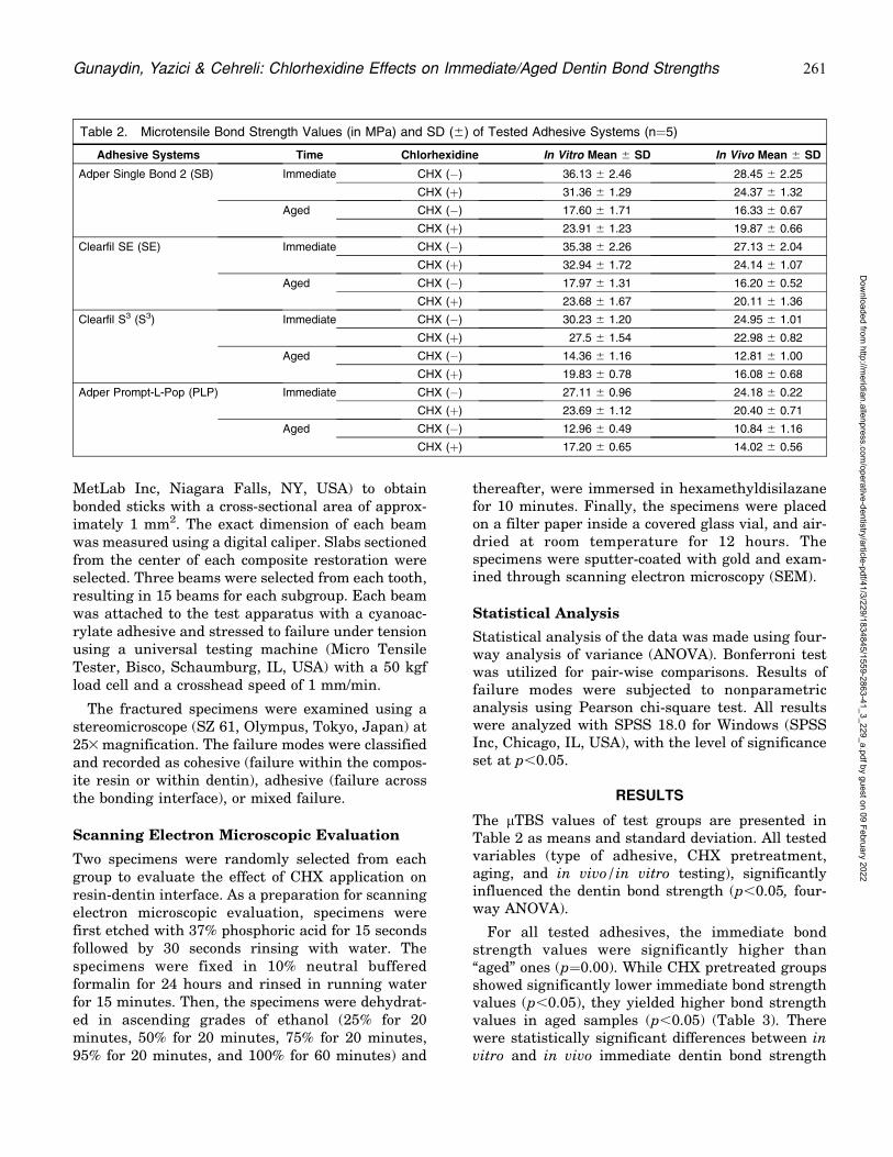
MetLab Inc, Niagara Falls, NY, USA) to obtainbonded sticks with a cross-sectional area of approx-imately 1 mm2. The exact dimension of each beamwas measured using a digital caliper. Slabs sectionedfrom the center of each composite restoration wereselected. Three beams were selected from each tooth,resulting in 15 beams for each subgroup. Each beamwas attached to the test apparatus with a cyanoac-rylate adhesive and stressed to failure under tensionusing a universal testing machine (Micro TensileTester, Bisco, Schaumburg, IL, USA) with a 50 kgfload cell and a crosshead speed of 1 mm/min.
The fractured specimens were examined using astereomicroscope (SZ 61, Olympus, Tokyo, Japan) at253 magnification. The failure modes were classifiedand recorded as cohesive (failure within the compos-ite resin or within dentin), adhesive (failure acrossthe bonding interface), or mixed failure.
Scanning Electron Microscopic Evaluation
Two specimens were randomly selected from eachgroup to evaluate the effect of CHX application onresin-dentin interface. As a preparation for scanningelectron microscopic evaluation, specimens werefirst etched with 37% phosphoric acid for 15 secondsfollowed by 30 seconds rinsing with water. Thespecimens were fixed in 10% neutral bufferedformalin for 24 hours and rinsed in running waterfor 15 minutes. Then, the specimens were dehydrat-ed in ascending grades of ethanol (25% for 20minutes, 50% for 20 minutes, 75% for 20 minutes,95% for 20 minutes, and 100% for 60 minutes) and
thereafter, were immersed in hexamethyldisilazanefor 10 minutes. Finally, the specimens were placedon a filter paper inside a covered glass vial, and air-dried at room temperature for 12 hours. Thespecimens were sputter-coated with gold and exam-ined through scanning electron microscopy (SEM).
Statistical Analysis
Statistical analysis of the data was made using four-way analysis of variance (ANOVA). Bonferroni testwas utilized for pair-wise comparisons. Results offailure modes were subjected to nonparametricanalysis using Pearson chi-square test. All resultswere analyzed with SPSS 18.0 for Windows (SPSSInc, Chicago, IL, USA), with the level of significanceset at p,0.05.
RESULTS
The lTBS values of test groups are presented inTable 2 as means and standard deviation. All testedvariables (type of adhesive, CHX pretreatment,aging, and in vivo/in vitro testing), significantlyinfluenced the dentin bond strength (p,0.05, four-way ANOVA).
For all tested adhesives, the immediate bondstrength values were significantly higher than‘‘aged’’ ones (p=0.00). While CHX pretreated groupsshowed significantly lower immediate bond strengthvalues (p,0.05), they yielded higher bond strengthvalues in aged samples (p,0.05) (Table 3). Therewere statistically significant differences between invitro and in vivo immediate dentin bond strength
Table 2. Microtensile Bond Strength Values (in MPa) and SD (6) of Tested Adhesive Systems (n=5)
Adhesive Systems Time Chlorhexidine In Vitro Mean 6 SD In Vivo Mean 6 SD
Adper Single Bond 2 (SB) Immediate CHX (�) 36.13 6 2.46 28.45 6 2.25
CHX (þ) 31.36 6 1.29 24.37 6 1.32
Aged CHX (�) 17.60 6 1.71 16.33 6 0.67
CHX (þ) 23.91 6 1.23 19.87 6 0.66
Clearfil SE (SE) Immediate CHX (�) 35.38 6 2.26 27.13 6 2.04
CHX (þ) 32.94 6 1.72 24.14 6 1.07
Aged CHX (�) 17.97 6 1.31 16.20 6 0.52
CHX (þ) 23.68 6 1.67 20.11 6 1.36
Clearfil S3 (S3) Immediate CHX (�) 30.23 6 1.20 24.95 6 1.01
CHX (þ) 27.5 6 1.54 22.98 6 0.82
Aged CHX (�) 14.36 6 1.16 12.81 6 1.00
CHX (þ) 19.83 6 0.78 16.08 6 0.68
Adper Prompt-L-Pop (PLP) Immediate CHX (�) 27.11 6 0.96 24.18 6 0.22
CHX (þ) 23.69 6 1.12 20.40 6 0.71
Aged CHX (�) 12.96 6 0.49 10.84 6 1.16
CHX (þ) 17.20 6 0.65 14.02 6 0.56
Gunaydin, Yazici & Cehreli: Chlorhexidine Effects on Immediate/Aged Dentin Bond Strengths 261
Dow
nloaded from http://m
eridian.allenpress.com/operative-dentistry/article-pdf/41/3/229/1834845/1559-2863-41_3_229_a.pdf by guest on 09 February 2022
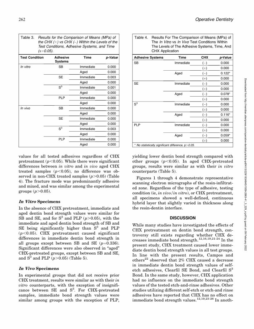
values for all tested adhesives regardless of CHXpretreatment (p,0.05). While there were significantdifferences between in vitro and in vivo aged CHXtreated samples (p,0.05), no difference was ob-served in non-CHX treated samples (p.0.05) (Table4). The fracture mode was predominantly adhesiveand mixed, and was similar among the experimentalgroups (p.0.05).
In Vitro Specimens
In the absence of CHX pretreatment, immediate andaged dentin bond strength values were similar forSB and SE, and for S3 and PLP (p.0.05), with theimmediate and aged dentin bond strength of SB andSE being significantly higher than S3 and PLP(p,0.05). CHX pretreatment caused significantdifferences in immediate dentin bond strength inall groups except between SB and SE (p=0.336).Significant differences were also observed in ‘‘aged’’CHX-pretreated groups, except between SB and SE,and S3 and PLP (p.0.05) (Table 5).
In Vivo Specimens
In experimental groups that did not receive priorCHX treatment, results were similar as with their invitro counterparts, with the exception of insignifi-cance between SE and S3. For CHX-pretreatedsamples, immediate bond strength values weresimilar among groups with the exception of PLP,
yielding lower dentin bond strength compared withother groups (p,0.05). In aged CHX-pretreatedgroups, results were similar as with their in vitrocounterparts (Table 5).
Figures 1 through 4 demonstrate representativescanning electron micrographs of the resin-infiltrat-ed zone. Regardless of the type of adhesive, testingcondition (ie, in vivo/in vitro), or CHX pretreatment,all specimens showed a well-defined, continuoushybrid layer that slightly varied in thickness alongthe resin-dentin interface.
DISCUSSION
While many studies have investigated the effects ofCHX pretreatment on dentin bond strength, con-troversy still exists regarding whether CHX de-creases immediate bond strength.12,16,19,21-24 In thepresent study, CHX treatment caused lower imme-diate dentin bond strength values in all test groups.In line with the present results, Campos andothers22 observed that 2% CHX caused a decreasein immediate dentin bond strength values of self-etch adhesives, Clearfil SE Bond, and Clearfil S3
Bond. In the same study, however, CHX applicationhad no influence on the immediate bond strengthvalues of the tested etch-and-rinse adhesives. Otherstudies utilizing different self-etch or etch-and rinseadhesives have reported that CHX has no effect onimmediate bond strength values.12,19,23-26 In anoth-
Table 3. Results for the Comparison of Means (MPa) ofthe CHX (þ) vs CHX (�) Within the Levels of theTest Conditions, Adhesive Systems, and Time(a=0.05).
Test Condition AdhesiveSystems
Time p-Value
In vitro SB Immediate 0.000
Aged 0.000
SE Immediate 0.003
Aged 0.000
S3 Immediate 0.001
Aged 0.000
PLP Immediate 0.000
Aged 0.000
In vivo SB Immediate 0.000
Aged 0.000
SE Immediate 0.000
Aged 0.000
S3 Immediate 0.003
Aged 0.000
PLP Immediate 0.000
Aged 0.000
Table 4. Results For The Comparison of Means (MPa) ofThe In Vitro vs In Vivo Test Conditions WithinThe Levels of The Adhesive Systems, Time, AndCHX Application
Adhesive Systems Time CHX p-Value
SB Immediate (�) 0.000
(þ) 0.000
Aged (�) 0.122*
(þ) 0.000
SE Immediate (�) 0.000
(þ) 0.000
Aged (�) 0.078*
(þ) 0.000
S3 Immediate (�) 0.000
(þ) 0.000
Aged (�) 0.116*
(þ) 0.000
PLP Immediate (�) 0.000
(þ) 0.000
Aged (�) 0.059*
(þ) 0.000
* No statistically significant difference, p.0.05.
262 Operative Dentistry
Dow
nloaded from http://m
eridian.allenpress.com/operative-dentistry/article-pdf/41/3/229/1834845/1559-2863-41_3_229_a.pdf by guest on 09 February 2022

er study, CHX was found to preserve the bondingdurability of an etch-and-rinse adhesive (SingleBond) but was unable to maintain a stable bond of aone-step self-etch adhesive system (GBond).27 Be-cause CHX generally inhibits MMPs without im-pairing dentin bond strength,28 the benefits of CHXpretreatment might be expected over the course oftime. Manfro and others21 demonstrated the effec-tiveness of CHX in preventing the degradation ofthe adhesive interface in primary dentin. In theirstudy, they found no significant reduction in dentinbond strength in aged samples, while in the absenceof CHX treatment, significant reductions wereobserved. The present results corroborate withthose of previous studies reporting that CHXapplication has no adverse effect on aged dentinbond strength values. Further, the present studydemonstrates that CHX treatment leads to higheraged dentin bond strength compared with non-CHX-treated groups. Therefore, the first nullhypothesis should be rejected. It has been proposedthat the use of 2% chlorhexidine before applicationof adhesive resins reduces the deterioration ofhybrid layers following exposure to water.29 In thepresent study, the higher bond strength valuesobtained in aged CHX-treated samples might berelated with the inhibition effect of CHX on MMPsthat had been activated by the acidity of the testedadhesives. As known, mild acids have a potential toactivate MMPs, and particularly, pH values rang-ing from 2.3 to 5 are effective in activatinggelatinases.30 The self-etch adhesive used in thisstudy has a pH value of 2.4 and is, thus, capable ofenhancing dentin proteolytic activity without dena-turation of the enzymes. The observation of in-c reased aged bond strength a f te r CHXpretreatment has been demonstrated previously,utilizing an etch-and-rinse and a self-etch adhe-sive.31
For the experimental groups that did not receiveCHX-pretreatment, our results confirm previousstudies that reported reductions in the dentin bondstrength of different self-etch and etch-and-rinseadhesives after aging.32-37 For CHX-pretreatedgroups, however, establishing correlations betweenthe present results and those of previous reportsmight be more complicated. It has been shown thatCHX has no significant effect on bond strengthstability, and that there was a decrease in bondstrength over a six-month period of water storage.37
Further, in an 18-month clinical study comparingthe performance of an etch-and-rinse and self-etchadhesives in noncarious cervical lesions with andwithout CHX pretreatment,38 the authors concludedthat CHX did not influence the performance of thetested adhesives. Undoubtedly, clinical retention is
Table 5: Results for the Comparison of Means (MPa) of the Adhesive Systems Within the Levels of the Test Conditions, CHXApplication, and Time
Adhesive Groups In Vitro p-Value In Vivo p-Value
CHX(�)Immediate
CHX(þ)Immediate
CHX(�)Aged
CHX(þ)Aged
CHX(�)Immediate
CHX(þ)Immediate
CHX(�)Aged
CHX(þ)Aged
SB vs SE 1.000* 0.336* 1.000* 1.000* 0.657* 1.000* 1.000* 1.000*
SB vs S3 0.000 0.000 0.001 0.000 0.002 0.557* 0.000 0.000
SB vs PLP 0.000 0.000 0.000 0.000 0.000 0.000 0.000 0.000
SE vs S3 0.000 0.000 0.000 0.000 0.259* 0.955* 0.000 0.000
SE vs PLP 0.000 0.000 0.000 0.000 0.003 0.000 0.000 0.000
S3 vs PLP 0.070* 0.000 0.217* 0.227* 0.735* 0.016 0.107* 0.077*
* No statistically significant difference, p.0.05.
Figure 1. SEM images of composite-dentin interface with AdperSingle Bond 2. (A): Immediate and CHX (þ). (B): Immediate and CHX(�). (C): Aged and CHX (þ). (D): Aged and CHX (�).
Gunaydin, Yazici & Cehreli: Chlorhexidine Effects on Immediate/Aged Dentin Bond Strengths 263
Dow
nloaded from http://m
eridian.allenpress.com/operative-dentistry/article-pdf/41/3/229/1834845/1559-2863-41_3_229_a.pdf by guest on 09 February 2022

affected by many other factors, but those resultsshould be considered when drawing conclusions.
Although clinical trials remain the ultimate instru-ment, preclinical screenings of materials are stillimportant. However, it is still not fully understoodwhether there is a relationship between laboratorydata and clinical outcomes and also whether theclinical performance is predictable in the lab. To date,only a few studies have compared the behavior ofdental biomaterials under both clinical and laboratoryconditions.11,37,39-41 The present results indicateddifferences in the bond strength values of CHX-pretreated groups, with higher bond strength valuesobtained in in vitro test results. This can be expected ,particularly due to the insufficient simulation of oralconditions with thermocycling. Thermocycling is acommon procedure to predict how a material wouldperform over time clinically. Although it is one of themost popular artificial aging methods, there is noevidence to indicate that thermocycling would createspecimens equivalent to those fabricated in vivo.Patient-related factors constitute the difference be-tween the laboratory and the clinical study. Compar-ing the bonding effectiveness of the etch-and-rinseadhesive to the two-step self-etch adhesive, it isevident that both adhesives bonded similarly underin vivo and in vitro conditions in non-CHX treatedsamples. Immediate bond strength values of one-stepand all-in-one self-etch adhesives, Clearfil S3 Bondand Adper Prompt-L-Pop, were also similar. There-
fore, the second null hypothesis should be partlyaccepted.
In the present study, the bond strength values ofAdper Single Bond and Clearfil SE Bond weresignificantly higher than the tested one-step self-etch adhesives. Several studies have shown thatetch-and-rinse adhesives yield bonding values supe-
Figure 2. SEM images of composite-dentin interface with Clearfil SEBond. (A): Immediate and CHX (þ). (B): Immediate and CHX (�). (C):Aged and CHX (þ). (D): Aged and CHX (�).
Figure 3. SEM images of composite-dentin interface with Clearfil S3.(A): Immediate and CHX (þ). (B): Immediate and CHX (�). (C): Agedand CHX (þ). (D) Aged and CHX (�).
Figure 4. SEM images of composite-dentin interface with Prompt-L-Pop. (A): Immediate and CHX (þ). (B): Immediate and CHX (�). (C):Aged and CHX (þ). (D): Aged and CHX (�).
264 Operative Dentistry
Dow
nloaded from http://m
eridian.allenpress.com/operative-dentistry/article-pdf/41/3/229/1834845/1559-2863-41_3_229_a.pdf by guest on 09 February 2022

rior to those of one-step self-etch adhesives. Perdigaoand others42 demonstrated that Adper Single Bondand Clearfil SE Bond showed similar bonding valuesthat were also higher than one-step self-etch adhe-sives. Indeed, in several studies, Adper Single Bondand Clearfil SE Bond have shown significantlyhigher bond strength values compared with otherdifferent adhesives.34,43 The ethanol and HEMAcontent of Single Bond enables high wettability,whereas 10-MDP in Clearfil SE Bond binds tocalcium salts that maintain a stable bonding inter-face.34 Further, the relatively higher concentrationof camphorquinone in Clearfil SE Bond improves itspolymerization rate.44
In the present study, the lowest bond strengthvalues were obtained in aged one-step self-etchadhesives. Presumably, such levels of bondstrength may result from the high acidity of theseadhesives that degrades the collagen matrix stabi-lization.45 It may be possible to speculate thatadditional factors, not possible to show with thepresent methodology, such as resin monomers withlow conversion rate44 and occurrence of watertrees,34,45 may have contributed to these results.As bond strength values differ according to thetested adhesive systems, the third null hypothesisshould also be rejected.
Adhesive and mixed failure types have beenobserved in a majority of microtensile bond strengthstudies, irrespective of CHX pretreatment.20-22,31,34
Osorio and others34 reported that while mixed failuretypes were observed after immediate bond strengthtesting, adhesive failures were prominent in agedgroups subjected to debonding forces. In the presentstudy, predominance of adhesive/mixed failure wasobserved in all groups with a lack of statisticalsignificance. In resin-dentin interface evaluation bySEM, the hybrid layer was found to be intact andmostly continuous in all tested samples.
Additional long-term studies might be necessary todetermine the long-term effects of CHX on the bondstrengths of different adhesive systems.
CONCLUSIONS
Within the limitations of this study, the followingconclusions were drawn:
1) The immediate bond strength values of all testedadhesives were significantly higher than ‘‘aged’’ones.
2) The application of CHX caused lower immediateand higher aged bond strength values.
Regulatory Statement
This study was conducted in accordance with all of theprovisions of the local human subjects oversight committeeguidelines and policies. The approval code for this study isLUT 09/72-21. This study was conducted at HacettepeUniversity.
Acknowledgements
This study was supported by a grant from the HacettepeUniversity Scientific Research Department (project012D06201001). The authors thank Sevilay Karahan forassistance with statistical analysis.
Conflict of Interest
The authors of this manuscript certify that they have noproprietary, financial, or other personal interest of any natureor kind in any product, service, and/or company that ispresented in this article.
(Accepted 6 December 2014)
REFERENCES
1. Carvalho RM, Manso AP, Geraldeli S, Tay FR, & PashleyDH (2012) Durability of bonds and clinical success ofadhesive restorations Dental Materials 28(1) 72-86.
2. Hashimoto M, Ohno H, Sano H, Kaga M, & Oguchi H(2003) In vitro degradation of resin-dentin bonds analyzedby microtensile bond test, scanning and transmissionelectron microscopy Biomaterials 24(21) 3795-3803.
3. De Munck J (2003) Four-year water degradation of total-etch adhesives bonded to dentin Journal of DentalResearch 82(2) 136-140.
4. Pashley D, Tay FR, Yiu C, Hashimoto M, Breschi L,Carvalho RM, & Ito S (2004) Collagen degradation byhost-derived enzymes during aging Journal of DentalResearch 83(3) 216-221.
5. Tjaderhane L, Nascimento FD, Breschi L, Mazzoni A,Tersariol IL, Geraldeli S, Tezvergil-Mutluay A, CarrilhoM, Carvalho RM, Tay FR, & Pashley DH (2013) Strategiesto prevent hydrolytic degradation of the hybrid layer—Areview Dental Materials 29(10) 999-1011.
6. Tjaderhane L, Nascimento FD, Breschi L, Mazzoni A,Tersariol IL, Geraldeli S, Tezvergil-Mutluay A, CarrilhoMR, Carvalho RM, Tay FR, & Pashley DH (2013)Optimizing dentin bond durability: Control of collagendegradation by matrix metalloproteinases and cysteinecathepsins Dental Materials 29(1) 116-135.
7. Mazzoni A, Pashley DH, Nishitani Y, Breschi L, MannelloF, Tjaderhane L, Toledano M, Pashley EL, & Tay FR(2006) Reactivation of inactivated endogenous proteolyticactivities in phosphoric acid-etched dentine by etch-and-rinse adhesives Biomaterials 27(25) 4470-4476.
8. Tay FR, Pashley DH, Loushine RJ, Weller RN, MonticelliF, & Osorio R (2006) Self-etching adhesives increasecollagenolytic activity in radicular dentin Journal ofEndodontics 32(9) 862-868.
9. Mazzoni A, Scaffa P, Carrilho M, Tjaderhane L, DiLenarda R, Polimeni A, Tezvergil-Mutluay A, Tay FR,
Gunaydin, Yazici & Cehreli: Chlorhexidine Effects on Immediate/Aged Dentin Bond Strengths 265
Dow
nloaded from http://m
eridian.allenpress.com/operative-dentistry/article-pdf/41/3/229/1834845/1559-2863-41_3_229_a.pdf by guest on 09 February 2022

Pashley DH, & Breschi L (2013) Effects of etch-and-rinseand self-etch adhesives on dentin MMP-2 and MMP-9Journal of Dental Research 92(1) 82-86.
10. Gendron R, Grenier D, Sorsa T, & Mayrand D (1999)Inhibition of the activities of matrix metalloproteinases 2,8, and 9 by chlorhexidine Clinical and DiagnosticLaboratory Immunology 6(3) 437-439.
11. Hebling J, Pashley DH, Tjaderhane L, & Tay FR (2005)Chlorhexidine arrests subclinical degradation of dentinhybrid layers in vivo Journal of Dental Research 84(8)741-746.
12. Carrilho MR, Geraldeli S, Tay F, de Goes MF, CarvalhoRM, Tjaderhane L, Reis AF, Hebling J, Mazzoni A,Breschi L, & Pashley D (2007) In vivo preservation of thehybrid layer by chlorhexidine Journal of Dental Research86(6) 529-533.
13. Brackett WW, Tay FR, Brackett MG, Dib A, Sword RJ, &Pashley DH (2007) The effect of chlorhexidine on dentinhybrid layers in vivo Operative Dentistry 32(2) 107-111.
14. Carrilho MR, Carvalho RM, de Goes MF, di Hipolito V,Geraldeli S, Tay FR, Pashley DH, & Tjaderhane L (2007)Chlorhexidine preserves dentin bond in vitro Journal ofDental Research 86(1) 90-94.
15. Breschi L, Cammelli F, Visintini E, Mazzoni A, Vita F,Carrilho M, Cadenaro M, Foulger S, Mazzoti G, Tay FR,Di Lenarda R, & Pashley D (2009) Influence of chlorhex-idine concentration on the durability of etch-and-rinsedentin bonds: A 12-month in vitro study Journal ofAdhesive Dentistry 11(3) 191-198.
16. Breschi L, Mazzoni A, Nato F, Carrilho M, Visintini E,Tjaderhane L, Ruggeri A Jr, Tay FR, Dorigo Ede S, &Pashley DH (2010) Chlorhexidine stabilizes the adhesiveinterface: A 2-year in vitro study Dental Materials 26(4)320-325.
17. Breschi L (2013) Chlorhexidine application to stabilizethe adhesive interface: Why and how? Journal ofAdhesive Dentistry 15(5) 492.
18. Kim J, Uchiyama T, Carrilho M, Agee KA, Mazzoni A,Breschi L, Carvalho RM, Tjaderhane L, Looney S,Wimmer C, Tezvergil-Mutluay A, Tay FR, & PashleyDH (2010) Chlorhexidine binding to mineralized versusdemineralized dentin powder Dental Materials 26(8)771-778.
19. Stanislawczuk R, Amaral RC, Zander-Grande C, GaglerD, Reis A, & Loguercio AD (2009) Chlorhexidine-contain-ing acid conditioner preserves the longevity of resin-dentin bonds Operative Dentistry 34(4) 481-490.
20. Mobarak EH (2011) Effect of chlorhexidine pretreat-ment on bond strength durability of caries-affecteddentin over 2-year aging in artificial saliva and undersimulated intrapulpal pressure Operative Dentistry36(6) 649-660.
21. Manfro AR, Reis A, Loguercio AD, Imparato JC, & RaggioD (2012) Effect of different concentrations of chlorhexi-dine on bond strength of primary dentin PediatricDentistry 34(2) 11-15.
22. Campos EA, Correr GM, Leonardi DP, Pizzatto E, &Morais EC (2009) Influence of chlorhexidine concentra-tion on microtensile bond strength of contemporaryadhesive systems Brazilian Oral Research 23(3) 340-345.
23. Zhou J, Tan J, Yang X, Cheng C, Wang X, & Chen L(2010) Effect of chlorhexidine application in a self-etchingadhesive on the immediate resin-dentin bond strengthJournal of Adhesive Dentistry 12(1) 27-31.
24. Zhou J, Tan J, Yan X, Xu X, Li D, & Chen L (2011) MMP-inhibitory effect of chlorhexidine applied in a self-etchingadhesive Journal of Adhesive Dentistry 13(2) 111-115.
25. de-Melo MA, Goes Dda C, de-Moraes MD, Santiago SL, &Rodrigues LK (2013) Effect of chlorhexidine on the bondstrength of a self-etch adhesive system to sound anddemineralized dentin Brazilian Oral Research 27(3)218-224.
26. Lenzi TL, Tedesco TK, Soares FZ, Loguercio AD, & RochaRde O (2012) Chlorhexidine does not increase immediatebond strength of etch-and-rinse adhesive to caries-affected dentin of primary and permanent teeth BrazilianDental Journal 23(4) 438-442.
27. Deng D, Huang X, Huang C, Yang T, Du X, Wang Y,Ouyang X, & Pei D (2013) Effects of chlorhexidine onbonding durability of different adhesive systems using anovel thermocycling method Australian Dental Journal58(2) 148-155.
28. De Munck J, Mine A, Van den Steen PE, Van LanduytKL, Poitevin A, Opdenakker G, & Van Meerbeek B (2010)Enzymatic degradation of adhesive-dentin interfacesproduced by mild self-etch adhesives European Journalof Oral Science 118(5) 494-501.
29. Lafuente D (2012) SEM analysis of hybrid layer andbonding interface after chlorhexidine use OperativeDentistry 37(2) 172-180.
30. Nishitani Y, Yoshiyama M, Wadgaonkar B, Breschi L,Mannello F, Mazzoni A, Carvalho RM, Tjaderhane L, TayFR, & Pashley DH (2006) Activation of gelatinolytic/collagenolytic activity in dentin by self-etching adhesivesEuropean Journal of Oral Science 114(2) 160-166.
31. Campos EA, Correr GM, Leonardi DP, Barato-Filho F,Gonzaga CC, & Zielak JC (2009) Chlorhexidine dimin-ishes the loss of bond strength over time under simulatedpulpal pressure and thermo-mechanical stressing Jour-nal of Dentistry 37(2) 108-114.
32. Hashimoto M, Fujita S, Endo K, & Ohno H (2009) In vitrodegradation of resin-dentin bonds with one-bottle self-etching adhesives European Journal of Oral Science117(5) 611-617.
33. Reis A, Grandi V, Carlotto L, Bortoli G, Patzlaff R,Rodrigues Accorinte Mde L, & Dourado Loguercio A(2005) Effect of smear layer thickness and acidity of self-etching solutions on early and long-term bond strength todentin Journal of Dentistry 33(7) 549-559.
34. Osorio R, Pisani-Proenca J, Erhardt MC, Osorio E,Aguilera FS, Tay FR, & Toledano M (2008) Resistanceof ten contemporary adhesives to resin-dentine bonddegradation Journal of Dentistry 36(2) 163-169.
266 Operative Dentistry
Dow
nloaded from http://m
eridian.allenpress.com/operative-dentistry/article-pdf/41/3/229/1834845/1559-2863-41_3_229_a.pdf by guest on 09 February 2022

35. Koshiro K, Inoue S, Tanaka T, Koase K, Fujita M,Hashimoto M, & Sano H (2004) In vivo degradation ofresin-dentin bonds produced by a self-etch vs a total-etchadhesive system European Journal of Oral Science 112(4)368-375.
36. Carrilho MR, Carvalho RM, Tay FR, Yiu C, & Pashley DH(2005) Durability of resin-dentin bonds related to waterand oil storage American Journal of Dentistry 18(6)315-319.
37. Sim~oes DM, Basting RT, Amaral FL, Turssi CP, & FrancaFM (2014) Influence of chlorhexidine and/or ethanoltreatment on bond strength of an etch-and-rinse adhesiveto dentin: An in vitro and in situ study OperativeDentistry 39(1) 64-71.
38. Dutra-Correa M, Saraceni CH, Ciaramicoli MT, KiyanVH, & Queiroz CS (2013) Effect of chlorhexidine on the18-month clinical performance of two adhesives AmericanJournal of Dentistry 15(3) 287-292.
39. Brackett MG, Tay FR, Brackett WW, Dib A, Dipp FA, MaiS, & Pashley DH (2009) In vivo chlorhexidine stabiliza-tion of hybrid layers of an acetone-based dentin adhesiveOperative Dentistry 34(4) 379-383.
40. Cehreli ZC, Uyanik MO, Nagas E, Tuncel B, Er N, &Comert FD (2013) A comparison of residual smear layer
and erosion following different endodontic irrigation
protocols tested under clinical and laboratory conditions
Acta Odontologica Scandinavica 71(5) 1261-1266.
41. Donmez N, Belli S, Pashley DH, & Tay FR (2005)
Ultrastructural correlates of in vivo/in vitro bond degra-
dation in self-etch adhesives Journal of Dental Research
84(4) 355-359.
42. Perdigao J, Gomes G, Gondo R, & Fundingsland JW
(2006) In vitro bonding performance of all-in-one adhe-
sives. Part I—Microtensile bond strengths Journal of
Adhesive Dentistry 8(6) 367-373.
43. Toledano M, Osorio R, Ceballos L, Fuentes MV, Fer-
nandes CA, Tay FR, & Carvalho RM (2003) Microtensile
bond strength of several adhesive systems to different
dentin depths American Journal of Dentistry 16(5)
292-298.
44. Nunes TG, Ceballos L, Osorio R, & Toledano M (2005)
Spatially resolved photopolymerization kinetics and oxy-
gen inhibition in dental adhesives Biomaterials 26(14)
1809-1817.
45. Tay FR, & Pashley DH (2003) Have dentin adhesives
become too hydrophilic? Journal of the Canadian Dental
Association 69(11) 726-731.
Gunaydin, Yazici & Cehreli: Chlorhexidine Effects on Immediate/Aged Dentin Bond Strengths 267
Dow
nloaded from http://m
eridian.allenpress.com/operative-dentistry/article-pdf/41/3/229/1834845/1559-2863-41_3_229_a.pdf by guest on 09 February 2022

Clinical Performance of CompositeRestorations with Resin-modified
Glass Ionomer Lining in RootSurface Carious Lesions
U Koc Vural � S Gokalp � A Kiremitci
Clinical Relevance
The application of cavity lining material did not affect clinical performance, particularlymarginal adaptation rate, over an 18-month period.
SUMMARY
Objective: The purpose of this study was toevaluate the clinical performance of compositerestorations in root surface carious lesionswith or without resin-modified glass ionomerlining.
Methods and Materials: The sample consistedof 25 female and 14 male patients. A maximumof four lesions were included for each patient.After caries removal, the depth, length, andwidth of the cavity were measured. Lesions inthe same patient were randomly divided intotwo groups, and the dentin surfaces wereeither lined with resin-modified glass ionomerliner (Glass Liner II) or left as they were. Self-
etch adhesive (All Bond SE) was applied and
cured for 20 seconds. All cavities were restored
with nanohybrid anterior composite resin
(Clearfil Majesty Esthetic). Two experienced
clinicians evaluated the marginal adaptation
(retention) rate, anatomic form, secondary
caries, sensitivity, and marginal staining of
restorations at the end of the first week and at
six, 12, and 18 months posttreatment. The data
were statistically analyzed using the Chi-
square and two-way repeated measures tests.
Results: At the end of 18 months, a total of five
lined and three unlined restorations were lost.
There was no significant relationship between
marginal adaptation and cavity lining at six,
12, and 18 months (p.0.05). Although marginal
stainings of restorations were mostly localized,
the total number of localized or generalized
discolored restorations increased with time
(p,0.001). There was a statistically significant
relationship between marginal staining and
smoking (p.0.05). There was no significant
relationship between marginal staining and
frequency of toothbrushing at six, 12, and 18
*Uzay Koc Vural, DDS, PhD, Department of RestorativeDentistry, Hacettepe University, Altindag, Ankara, Turkey
Saadet Gokalp, DDS, PhD, Department of RestorativeDentistry, Hacettepe University, Altindag, Ankara, Turkey
Arlin Kiremitci, DDS, PhD, Department of RestorativeDentistry, Hacettepe University, Altindag, Ankara, Turkey
*Corresponding author: Altındag, Ankara 06100, Turkey; e-mail: [email protected]
DOI: 10.2341/15-205-C
�Operative Dentistry, 2016, 41-3, 268-275
Dow
nloaded from http://m
eridian.allenpress.com/operative-dentistry/article-pdf/41/3/229/1834845/1559-2863-41_3_229_a.pdf by guest on 09 February 2022

months (p=0.286, p=0.098, and p=0.408, respec-tively).
Conclusion: Within the limitations of thisstudy, both restorative applications were ac-cepted as clinically appropriate.
INTRODUCTION
The etiology of root surface caries is multifactorial,with microbiological factors playing a critical role.The cervical margins of root surface carious lesionsare usually located in the cementum or dentin,making it more susceptible to bacterial invasion(microleakage). Microleakage can lead to problemssuch as marginal staining, secondary caries, andpulpal inflammation.1 The pulp requires protectionfrom further bacterial invasion of the carious processand chemical protection from the overlying restor-ative materials.2 This may be achieved by placementof a biocompatible liner on the cavity floor.3 Liningwith resin-modified glass ionomers (RMGICs) hasbeen shown to lead to clinical success and also topromote a dramatic reduction in the number ofcariogenic bacteria.4 Resilient liners, such asRMGICs, are capable of absorbing the polymeriza-tion contraction stress of the overlying resin com-posites.5,6 Contraction stress is one of the maincauses of microleakage and often leads to postoper-ative sensitivity, marginal staining, and secondarycaries.5,6 An in vitro study7 reported the lowestpercentage of marginal gap with RMGIC liners.
Materials such as resin composites are widely usedfor the restoration of root surface caries as they donot require excessive removal of sound tooth struc-ture and have a low modulus of elasticity. Self-etching adhesives are often used for bonding ofcomposite restorations of root surface caries as theyare easy to manipulate. On the other hand, someauthors8,9 reported that application of adhesives indeep cavities resulted in acceptable levels of biocom-patibility. However, the intensity of the pulpalresponse depended upon the thickness of theremaining dentin. These views motivated cliniciansto reevaluate the use of liners and adhesives.
The aim of this randomized, controlled clinicaltrial was to evaluate the clinical performance of ananohybrid composite resin restoration in cariousroot surface lesions lined with and without a RMGICliner. The cavity size, toothbrushing, and smokinghabits of patients were also included as additionalobservations.The null hypothesis tested was thatthere is no difference between the two restorativesystems with respect to marginal adaptation (reten-
tion), marginal staining, secondary caries, andpostoperative sensitivity.
METHODS AND MATERIALS
Thirty-nine nonhospitalized volunteer patients whopresented with root surface carious lesions at theRestorative Dental Clinic were randomly selected forthis study. Their ages ranged between 18 and 67years, with the mean age being 39.62 6 13.85 years.
The local institutional review board approved thisclinical trial prior to commencement. The volunteerswere informed about the conditions and objectives ofthe study and were asked to provide informedconsent.
A paired-tooth design was used for this study.Patients who were under the age of 18 years or whohad complex medical histories, severe or chronicperiodontitis, extreme carious activity, heavy brux-ism, very deep or superficial carious lesions, orpreviously restored and abutment teeth were ex-cluded from the study. Lesions were classified asinactive if their surface was leathery, shiny, or notcovered with plaque.
A single operator carried out all restorativeprocedures, and the lesion surface was pumicedbefore the intervention. Moisture control was carriedout with the help of cotton rolls and a saliva ejector.Cavity preparation was completed with a diamondbur, and the carious lesion was removed with thehelp of a steel bur. The enamel margins were beveledbut no mechanical retention was performed. Thedepth, width, and length of the cavity were mea-sured and recorded by two experienced and calibrat-ed clinicians using a periodontal (WHO 973/80-Martin, Solingen, Germany) explorer. In each pa-tient, two or four root surface carious lesions wererandomly restored according to two experimentalprotocols. A coin was tossed and depending on theoutcome cavities were either lined or left as theywere. The liner was placed and light-cured for 20seconds and the enamel walls were not sealed withthe lining material. Thereafter, two separate coats ofAll Bond SE (one-step self-etch adhesive) wereapplied using a microbrush for 15 seconds per coat(no light-cure between coats) for all cavities, air-dried for at least 10 seconds, and then LED light-cured for 10 seconds (Led Max 5; Hilux, BenliogluDental, Ankara, Turkey) set at 500-700 mW/cm2
intensity. The materials used in this study arepresented in Table 1.
Both groups were restored with the nanohybridcomposite (Clearfil Majesty Esthetic) using the
Koc Vural, Gokalp & Kiremitci: Performance of Composite Restoration in Root Surface 269
Dow
nloaded from http://m
eridian.allenpress.com/operative-dentistry/article-pdf/41/3/229/1834845/1559-2863-41_3_229_a.pdf by guest on 09 February 2022

incremental technique with a flat-faced condenser.Each resin composite layer was light-cured for 20seconds with the same LED light-curing unit. Allrestorations were finished with extra-/ultrafine com-posite finishing burs (Diatech Dental AC, Heerbrugg,Switzerland) and polished with discs (SwissFlex;Diatech Dental AC) and Enhance PoGo CompleteKit (Dentsply, Addlestone, York, PA, USA).
A double-blind study design was used, and twocalibrated clinicians (other than the operator) eval-uated all restorations. Direct intraoral clinicalexamination was carried out by two calibratedexaminers. The quality of the restorations wasevaluated according to marginal adaptation (reten-tion), anatomical form, caries in adjacent toothstructure, and caries at the cavosurface margin(Table 2). The criteria developed by Haveman andothers10 were modified by adding sensitivity andmarginal staining scores.
The sensitivity score was based on the subjectivesymptoms of the patient to cold air (yes/no), whilestaining of restorations was determined visually asbeing localized/generalized. Marginal staining wasdefined as localized if there was discoloration on lessthan half of the circumferential margin or defined asgeneralized if there was discoloration on more thanhalf of the circumferential margin.11
The width, length, and depth (in millimeters) ofthe cavities were grouped before statistical analysis.Toothbrushing frequencies were identified as once ortwice a day. Smokers were also divided into thefollowing three groups: nonsmoker, 1-10 cigarettesper day, and more than 10 cigarettes per day.
Restorations were evaluated at baseline and againat six, 12, and 18 months posttreatment. Thebaseline rating was carried out one week posttreat-ment. Chlorhexidine (Kloroben, Drogsan, Turkey)mouth-rinse was prescribed for five days, andstandard toothpaste (Colgate Total; 1450 ppmsodium fluoride [NaF] 0.3% triclosan/copolymer/0.22% NaF) was advised for daily use.
Table 1: Description of the Materials Used in this Study
Materials Composition Manufacturer Batch No.
All bond SE Part I—ethanol, sodium benzene sulfinate dehydratePart II—Bis (glyceryl 1,3 dimethacrylate)–phosphate;hydroxyethyl methacrylate biphenyl dimethacrylate
Bisco Inc, Schaumburg IL, USA 09000041810900004182
Glass Liner II Liquid: polycarbonic acid-waterPowder: calcium-aluminum-fluoro-silicate, barium glass
WP Dental, Barmstedt, Germany 120298
Clearfıl Majesty Esthetic Matrix: Bis-GMA, hydrophobicaromatic dimethacrylates,and hydrophobicaliphatic dimethacrylates, DL-camphorquinone
Kuraray, Tokyo, Japan 00014C
Abbreviation: Bis-GMA, bisphenol A diglycidyl ether dimethacryalate.
Table 2: Evaluation Criteria (1)
Score Marginal Adaptation (Retention)
0 The restoration appears to adapt closely to the surfaceof the tooth with no crevice formation. An explorereither did not catch when drawn along the margin oronly did so when passed in one direction.
1 An explorer caught lightly when run in both directions,and there was visible evidence of early creviceformation. Dentin was not visible.
2 An explorer got caught in both directions andpenetrated a marginal crevice. There was visibleevidence of crevice formation. However, the dentin wasnot visible.
3 The crevice had sufficient depth to expose the dentin.The restoration required replacement.
4 The restoration was fractured or lost.
Anatomical Form
0 The restoration was continuous with the existing toothanatomy.
1 The restoration was not continuous with the existingtooth anatomy, but no dentin was exposed. Therestoration was clinically acceptable.
2 The restoration was not continuous with the existingtooth anatomy and required replacement.
Caries in Adjacent Tooth Structure
0 Caries was not present within 3 mm of the border ofthe restoration.
1 Caries was present within 3 mm of the border of therestoration.
Caries at the Cavosurface Margin
0 No caries was present on the cavosurface margin.
1 Caries was present on the cavosurface margin.
Sensitivity
Yes Sensitive to cold air
No Not sensitive to cold air
Marginal Staining
0 No staining
1 Localized staining
2 Generalized staining
270 Operative Dentistry
Dow
nloaded from http://m
eridian.allenpress.com/operative-dentistry/article-pdf/41/3/229/1834845/1559-2863-41_3_229_a.pdf by guest on 09 February 2022
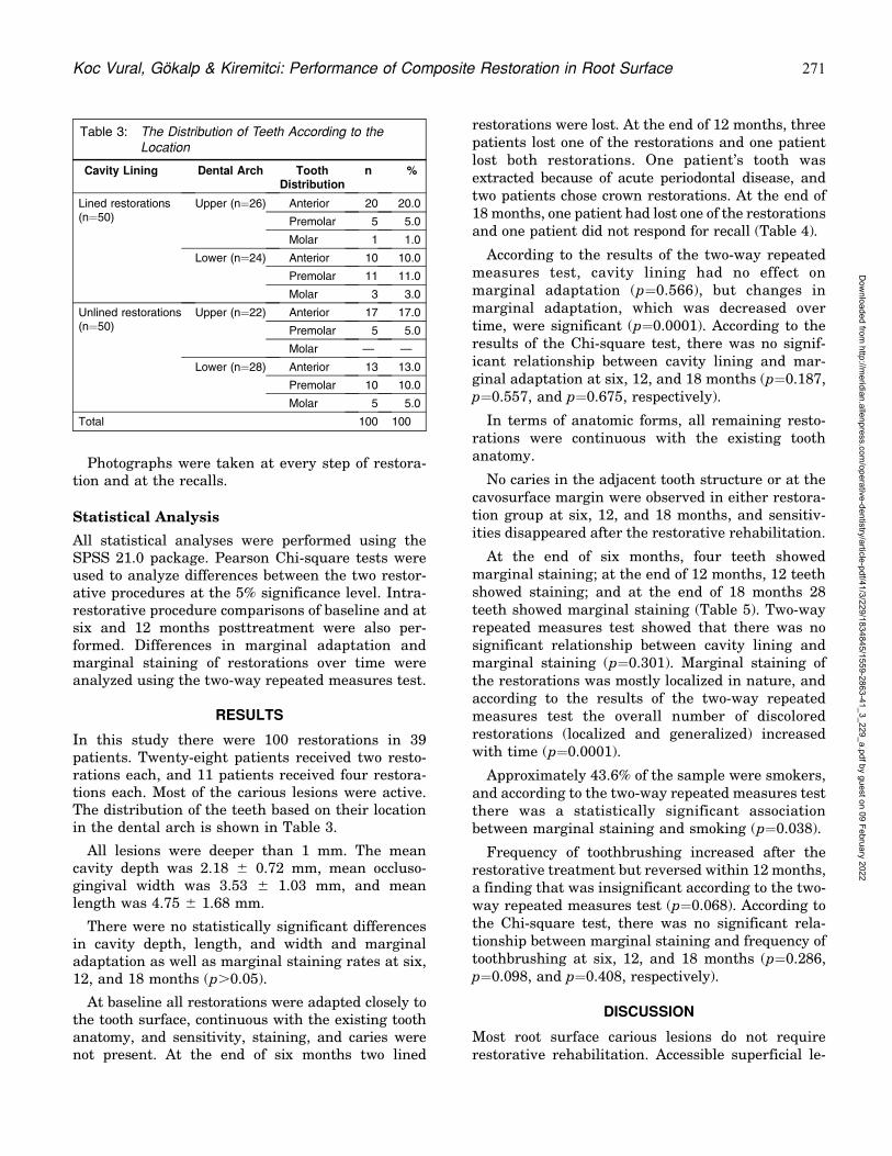
Photographs were taken at every step of restora-tion and at the recalls.
Statistical Analysis
All statistical analyses were performed using theSPSS 21.0 package. Pearson Chi-square tests wereused to analyze differences between the two restor-ative procedures at the 5% significance level. Intra-restorative procedure comparisons of baseline and atsix and 12 months posttreatment were also per-formed. Differences in marginal adaptation andmarginal staining of restorations over time wereanalyzed using the two-way repeated measures test.
RESULTS
In this study there were 100 restorations in 39patients. Twenty-eight patients received two resto-rations each, and 11 patients received four restora-tions each. Most of the carious lesions were active.The distribution of the teeth based on their locationin the dental arch is shown in Table 3.
All lesions were deeper than 1 mm. The meancavity depth was 2.18 6 0.72 mm, mean occluso-gingival width was 3.53 6 1.03 mm, and meanlength was 4.75 6 1.68 mm.
There were no statistically significant differencesin cavity depth, length, and width and marginaladaptation as well as marginal staining rates at six,12, and 18 months (p.0.05).
At baseline all restorations were adapted closely tothe tooth surface, continuous with the existing toothanatomy, and sensitivity, staining, and caries werenot present. At the end of six months two lined
restorations were lost. At the end of 12 months, threepatients lost one of the restorations and one patientlost both restorations. One patient’s tooth wasextracted because of acute periodontal disease, andtwo patients chose crown restorations. At the end of18 months, one patient had lost one of the restorationsand one patient did not respond for recall (Table 4).
According to the results of the two-way repeatedmeasures test, cavity lining had no effect onmarginal adaptation (p=0.566), but changes inmarginal adaptation, which was decreased overtime, were significant (p=0.0001). According to theresults of the Chi-square test, there was no signif-icant relationship between cavity lining and mar-ginal adaptation at six, 12, and 18 months (p=0.187,p=0.557, and p=0.675, respectively).
In terms of anatomic forms, all remaining resto-rations were continuous with the existing toothanatomy.
No caries in the adjacent tooth structure or at thecavosurface margin were observed in either restora-tion group at six, 12, and 18 months, and sensitiv-ities disappeared after the restorative rehabilitation.
At the end of six months, four teeth showedmarginal staining; at the end of 12 months, 12 teethshowed staining; and at the end of 18 months 28teeth showed marginal staining (Table 5). Two-wayrepeated measures test showed that there was nosignificant relationship between cavity lining andmarginal staining (p=0.301). Marginal staining ofthe restorations was mostly localized in nature, andaccording to the results of the two-way repeatedmeasures test the overall number of discoloredrestorations (localized and generalized) increasedwith time (p=0.0001).
Approximately 43.6% of the sample were smokers,and according to the two-way repeated measures testthere was a statistically significant associationbetween marginal staining and smoking (p=0.038).
Frequency of toothbrushing increased after therestorative treatment but reversed within 12 months,a finding that was insignificant according to the two-way repeated measures test (p=0.068). According tothe Chi-square test, there was no significant rela-tionship between marginal staining and frequency oftoothbrushing at six, 12, and 18 months (p=0.286,p=0.098, and p=0.408, respectively).
DISCUSSION
Most root surface carious lesions do not requirerestorative rehabilitation. Accessible superficial le-
Table 3: The Distribution of Teeth According to theLocation
Cavity Lining Dental Arch ToothDistribution
n %
Lined restorations(n=50)
Upper (n=26) Anterior 20 20.0
Premolar 5 5.0
Molar 1 1.0
Lower (n=24) Anterior 10 10.0
Premolar 11 11.0
Molar 3 3.0
Unlined restorations(n=50)
Upper (n=22) Anterior 17 17.0
Premolar 5 5.0
Molar — —
Lower (n=28) Anterior 13 13.0
Premolar 10 10.0
Molar 5 5.0
Total 100 100
Koc Vural, Gokalp & Kiremitci: Performance of Composite Restoration in Root Surface 271
Dow
nloaded from http://m
eridian.allenpress.com/operative-dentistry/article-pdf/41/3/229/1834845/1559-2863-41_3_229_a.pdf by guest on 09 February 2022
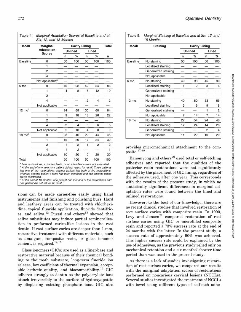
sions can be made caries-free easily using handinstruments and finishing and polishing burs. Hardand leathery areas can be treated with chlorhexi-dine, topical fluoride application, fluoride dentifric-es, and saliva.12 Turssi and others13 showed thatsaliva substitutes may induce partial remineraliza-tion in preformed caries-like lesions in the rootdentin. If root surface caries are deeper than 1 mm,restorative treatment with different materials, suchas amalgam, composite resin, or glass ionomercement, is required.14,15
Glass ionomers (GICs) are used as a liner/base andrestorative material because of their chemical bond-ing to the tooth substrate, long-term fluoride ionrelease, low coefficient of thermal expansion, accept-able esthetic quality, and biocompatibility.16 GICadheres strongly to dentin as the polyacrylate ionsattach irreversibly to the surface of hydroxyapatiteby displacing existing phosphate ions. GIC also
provides micromechanical attachment to the com-posite.17-19
Banomyong and others20 used total or self-etchingadhesives and reported that the qualities of theposterior resin restorations were not significantlyaffected by the placement of GIC lining, regardless ofthe adhesive used, after one year. This correspondswith the results of the present study, in which nostatistically significant differences in marginal ad-aptation rates were found between the lined andunlined restorations.
However, to the best of our knowledge, there areno recent clinical studies that involved restoration ofroot surface caries with composite resin. In 1990,Levy and Jensen21 compared restoration of rootsurface caries using GIC or microfilled compositeresin and reported a 73% success rate at the end of24 months with the latter. In the present study, asuccess rate of approximately 90% was achieved.This higher success rate could be explained by theuse of adhesives, as the previous study relied only onmechanical retention and a six months’ shorter timeperiod than was used in the present study.
As there is a lack of studies investigating restora-tion of root surface caries, we compared our resultswith the marginal adaptation scores of restorationsperformed on noncarious cervical lesions (NCCLs).Several studies investigated the treatment of NCCLswith bevel using different types of self-etch adhe-
Table 4: Marginal Adaptation Scores at Baseline and atSix, 12, and 18 Months
Recall MarginalAdaptation
Scores
Cavity Lining Total
Unlined Lined
n % n % n
Baseline 0 50 100 50 100 100
1 — — — —
2 — — — —
4 — — — —
Not applicablea — — — — —
6 mo 0 46 92 42 84 88
1 4 8 6 12 10
2 — — — — —
4 — — 2 4 2
Not applicable — — — — —
12 mob 0 34 68 30 60 64
1 9 18 13 26 22
2 — — — —
4 2 4 3 6 5
Not applicable 5 10 4 8 9
18 moc 0 23 46 22 44 45
1 15 30 17 34 32
2 1 2 1 2 2
4 1 2 — — 1
Not applicable 10 20 10 20 20
Total 50 100 50 100 100a Lost restorations, extracted teeth, or no attendance were not evaluated.b At the end of one year, one patient did not return for recall. Three patientslost one of the restorations; another patient lost both of the restorations,whereas another patient’s tooth has been extracted and two patients chosecrown restoration.c At the end of 18 months, one patient had lost one of the restorations andone patient did not return for recall.
Table 5: Marginal Staining at Baseline and at Six, 12, and18 Months
Recall Staining Cavity Lining
Unlined Lined
n % n %
Baseline No staining 50 100 50 100
Localized staining — — — —
Generalized staining — — — —
Not applicable — — — —
6 mo No staining 49 98 45 90
Localized staining 1 2 3 6
Generalized staining — — — —
Not applicable — — — —
12 mo No staining 40 80 33 66
Localized staining 3 6 9 18
Generaliszd staining — — 1 2
Not applicable 7 14 7 14
18 mo No staining 27 54 24 48
Localized staining 12 24 14 28
Generalized staining — — 2 4
Not applicable 11 22 10 20
272 Operative Dentistry
Dow
nloaded from http://m
eridian.allenpress.com/operative-dentistry/article-pdf/41/3/229/1834845/1559-2863-41_3_229_a.pdf by guest on 09 February 2022
sives and reported a greater than 80% success ratefor marginal adaptation of restorations at the end of12 to 24 months.22,23 Loguercio and others24 usedone-step and two-step All Bond SE self-etch adhe-sives and reported an 84.8% success rate with theformer at the end of 24 months. The findings of thelatter study were in accordance with those of thepresent study, which used the same adhesivesystem.
It has been suggested25 that the retention of anadhesive restoration depends on the marginal adap-tation capacity as well as the viscoelastic propertiesof the restorative system. Several factors can bringabout dimensional changes to these restorativematerials, including thermal changes and waterabsorption.26 In addition to using adhesive materialsfor marginal adaptation, retention can be betterachieved by preparing the cavity such that theadhesion surface increases.27 However, the resultsof the present study showed no statistically signifi-cant differences in cavity depth, length, and widthand marginal adaptation rates at six, 12, and 18months. These results do not support that increasingthe size of the preparation is beneficial.
Despite the initial color match being favorable, therestoration margins may show staining over time.Kubo and others26 compared two self-etch adhesivesystems on NCCLs. They reported that at the end oftwo years, 20% of the restorations had slightmarginal staining. Loguercio and others24 reportedthat marginal staining was observed in a fewrestorations at 24 months. Perdigao and others28
indicated that the number of satisfactory colorratings significantly decreased after one year withdifferent one-step self-etch adhesive systems. Inagreement with these studies, our results showedthat marginal staining increased over time (5.5% atsix months, 24.4% at 12 months, and 28% at 18months).
Peumans and others22 conducted a study on thetreatment of NCCLs using composite resin andreported superficial localized margin discoloration,which was observed significantly more often insmokers, in correlation with the results of this study.In addition, regarding the effect of clinical covari-ables (size of lesion and toothbrushing), no correla-tion was found, as was the case in this study.
The frequency of root caries among young agegroups was explored in this study and may partiallyreflect the poor oral health of Turkish people, aspreviously reported by Gokalp and Dogan29 in the2004 National Oral Health Survey. The prevalence
rate of root caries in the 35-44-year-old age groupwas 20.1%. Most of the patients claimed that theybrushed their teeth twice a day, which is sufficientfor daily oral hygiene. However, these patients stillpresented with root surface caries. It has beensuggested that though the frequency of brushingmay have been enough, its duration was short, andthe patient’s technical knowledge was inadequate orwrong. This may be responsible for the higherincidence of root caries in younger individuals. Atthe beginning of this study, patients were informedabout toothbrushing techniques and their impor-tance. They were asked to brush their teeth at leasttwice per day. Frequency of toothbrushing was seento increase after restorative procedures but declinedafter 12 months.
De Moor and others30 reported a significantlyhigher incidence of recurrent caries in glass ionomerand composite resin restorations of root caries inxerostomic irradiated head and neck cancer patientsafter 24 months. In contrast, Hu and others31
reported no active recurrent caries with glassionomer restorations in their radiation-induced rootsurface caries study. Differences may originate fromgeneral systemic disorders of the patients. In thisstudy, no caries were seen within the borders of therestoration. The dentifrice recommended in thisstudy may have increased fluoride levels andthereby prevented secondary caries.32,33
Tooth sensitivity before restorative rehabilitationmay be reduced with the help of self-etch adhesivesystems. Perdigao and others28 reported that post-operative sensitivity to air improved significantlywith different self-etch adhesive systems. In thepresent study we found a reduction in toothsensitivity.
The advantages of this clinical study include apaired-tooth study design, randomization, relativelyhigh restoration number, adequate follow-up (will becontinued over the duration of four successive years),and appropriate statistical analysis. Moreover, acomplete description of the study methodology andresults has been provided to allow comparison withother root surface caries clinical trials with similarstudy designs. The null hypothesis was accepted, asthe lining of the restoration had no effect on themarginal adaptation, marginal staining, secondarycaries, and postoperative sensitivity.
CONCLUSIONS
Within the limitations of this study, the applicationof cavity lining material did not affect clinical
Koc Vural, Gokalp & Kiremitci: Performance of Composite Restoration in Root Surface 273
Dow
nloaded from http://m
eridian.allenpress.com/operative-dentistry/article-pdf/41/3/229/1834845/1559-2863-41_3_229_a.pdf by guest on 09 February 2022
performance, particularly marginal adaptation rate.All Bond SE exhibited good performance; bothrestorative techniques were considered clinicallyappropriate at 18 months.
Acknowledgment
This study is supported by ‘‘Hacettepe University ScientificResearch Projects Coordination Unit’’ (H.U.B.A.B) with thegrant number ‘‘012D06201002.’’
Regulatory Statement
This study was conducted in accordance with all theprovisions of the local human subjects oversight committeeguidelines and policies of Hacettepe University. The approvalcode for this study is 11/54-8.
Conflict of Interest
The Authors of this manuscript certify that they have noproprietary, financial or other personal interest of any natureor kind in any product, service and/or company that ispresented in this article.
(Accepted 12 October 2015)
REFERENCES
1. Murray PE, About I, Franquin JC, Remusat M, & SmithAJ (2001) Restorative pulpal and repair responsesJournal of the American Dental Assocication 132(4)482-491.
2. Hilton TJ (2009) Keys to clinical success with pulpcapping: A review of the literature Operative Dentistry34(5) 615-625.
3. Costa CA, Giro EM, do Nascimento AB, Teixeira HM, &Hebling J (2003) Short-term evaluation of the pulpo-dentin complex response to a resin-modified glass-ion-omer cement and a bonding agent applied in deep cavitiesDental Materials 19(8) 739-746.
4. Duque C, Negrini Tde C, Sacono NT, Spolidorio DM, deSouza Costa CA, & Hebling J (2009) Clinical andmicrobiological performance of resin-modified glass-ion-omer liners after incomplete dentine caries removalClinical Oral Investigations 13(4) 465-471.
5. Strober B, Veitz-Keenan A, Barna JA, Matthews AG,Vena D, Craig RG, Curro FA, Thompson VP (2013)Effectiveness of a resin-modified glass ionomer liner inreducing hypersensitivity in posterior restorations: Astudy from the practitioners engaged in applied researchand learning network Journal of the American DentalAssociation 144(8) 886-897.
6. Ruiz JL, & Mitra S (2006) Using cavity liners with directposterior composite restorations Compendium of Continu-ing Education in Dentistry 27(6) 347-351, quiz 52.
7. Sampaio PC, de Almeida Junior AA, Francisconi LF,Casas-Apayco LC, Pereira JC, Wang L, Atta MT (2011)Effect of conventional and resin-modified glass-ionomerliner on dentin adhesive interface of Class I cavity wallsafter thermocycling Operative Dentistry 36(4) 403-412.
8. Hebling J, Giro EM, & Costa CA (1999) Human pulpresponse after an adhesive system application in deepcavities Journal of Dentistry 27(8) 557-564.
9. Nayyar S, Tewari S, & Arora B (2007) Comparison ofhuman pulp response to total-etch and self-etch bondingagents Oral Surgery, Oral Medicine, Oral Pathology, OralRadiology, Endodontics 104(2) e45-e52.
10. Haveman CW, Summitt JB, Burgess JO, & Carlson K(2003) Three restorative materials and topical fluoride gelused in xerostomic patients: A clinical comparisonJournal of the American Dental Association 134(2)177-184.
11. Moncada G, Silva F, Angel P, Oliveira OB Jr, Fresno MC,Cisternas P, Fernandez E, Estay J, Martin J (2014)Evaluation of dental restorations: A comparative studybetween clinical and digital photographic assessmentsOperative Dentistry 39(2) E45-E56.
12. Peters MC. Strategies for noninvasive demineralizedtissue repair (2010) Dental Clinics of North America54(3) 507-525.
13. Turssi CP, Lima RQ, Faraoni-Romano JJ, & Serra MC(2006) Rehardening of caries-like lesions in root surfacesby saliva substitutes Gerodontology 23(4) 226-230.
14. Burgess JO, & Gallo JR (2002) Treating root-surfacecaries Dental Clinics of North America 46(2) 385-404, vii-viii.
15. Saunders RH Jr, & Meyerowitz C (2005) Dental caries inolder adults Dental Clinics of North America 49(2)293-308.
16. Costa CA, Ribeiro AP, Giro EM, Randall RC, & Hebling J(2011) Pulp response after application of two resinmodified glass ionomer cements (RMGICs) in deepcavities of prepared human teeth Dental Materials27(7) e158-e170.
17. Yip HK, Tay FR, Ngo HC, Smales RJ, & Pashley DH(2001) Bonding of contemporary glass ionomer cements todentin Dental Materials 17(5) 456-470.
18. Wilson AD, Prosser HJ, & Powis DM (1983) Mechanism ofadhesion of polyelectrolyte cements to hydroxyapatiteJournal of Dental Research 62(5) 590-592.
19. Chadwick RG, & Woolford MJ (1993) A comparison of theshear bond strengths to a resin composite of twoconventional and two resin-modified glass polyalkenoate(ionomer) cements Journal of Dentistry 21(2) 111-116.
20. Banomyong D, Harnirattisai C, & Burrow MF (2011)Posterior resin composite restorations with or withoutresin-modified, glass-ionomer cement lining: A 1-yearrandomized, clinical trial Journal of Investigative andClinical Dentistry 2(1) 63-69.
21. Levy SM, & Jensen ME (1990) A clinical evaluation of therestoration of root surface caries Special Care in Dentistry10(5) 156-160.
22. Peumans M, Munck J, Van Landuyt K, Lambrechts P, &Van Meerbeek B (2005) Three-year clinical effectivenessof a two-step self-etch adhesive in cervical lesionsEuropean Journal of Oral Science 113(6) 512-518.
23. Burgess JO, Sadid-Zadeh R, Cakir D, & Ramp LC (2013)Clinical evaluation of self-etch and total-etch adhesive
274 Operative Dentistry
Dow
nloaded from http://m
eridian.allenpress.com/operative-dentistry/article-pdf/41/3/229/1834845/1559-2863-41_3_229_a.pdf by guest on 09 February 2022

systems in noncarious cervical lesions: A two-year reportOperative Dentistry 38(5) 477-487.
24. Loguercio AD, Manica D, Ferneda F, Zander-Grande C,Amaral R, Stanislawczuk R, de Carvalho RM, Manso A,Reis A (2010) A randomized clinical evaluation of a one-and two-step self-etch adhesive over 24 months OperativeDentistry 35(3) 265-272.
25. Kurokawa H, Miyazaki M, Takamizawa T, Rikuta A,Tsubota K, & Uekusa S (2007) One-year clinical evalua-tion of five single-step self-etch adhesive systems in non-carious cervical lesions Dental Materials Journal 26(1)14-20.
26. Kubo S, Yokota H, Yokota H, & Hayashi Y (2009) Two-year clinical evaluation of one-step self-etch systems innon-carious cervical lesions Journal of Dentistry 37(2)149-155.
27. Mazhari F, Mehrabkhani M, Sadeghi S, & Malekabadi KS(2009) Effect of bevelling on marginal microleakage ofbuccal-surface fissure sealants in permanent teeth Euro-pean Archives of Paediatric Dentistry 10(4) 241-243.
28. Perdigao J, Dutra-Correa M, Castilhos N, Carmo AR,Anauate-Netto C, Cordeiro HJ, Amore R, Lewgoy HR(2007) One-year clinical performance of self-etch adhe-
sives in posterior restorations American Journal ofDentistry 20(2) 125-133.
29. Gokalp S, & Dogan BG (2012) Root caries in 35-44 and 65-74 year-olds in Turkey Community Dental Health 29(3)233-238.
30. De Moor RJ, Stassen IG, van’t Veldt Y, Torbeyns D, &Hommez GM (2011) Two-year clinical performance ofglass ionomer and resin composite restorations in xeros-tomic head- and neck-irradiated cancer patients ClinicalOral Investigations 15(1) 31-38.
31. Hu JY, Chen XC, Li YQ, Smales RJ, & Yip KH (2005)Radiation-induced root surface caries restored with glass-ionomer cement placed in conventional and ART cavitypreparations: Results at two years. Australian DentalJournal 50(3) 186-190.
32. Arbabzadeh-Zavareh F, Gibbs T, Meyers IA, Bouzari M,Mortazavi S, & Walsh LJ (2012) Recharge pattern ofcontemporary glass ionomer restoratives Dental ResearchJournal (Isfahan) 9(2) 139-145.
33. Hara AT, Turssi CP, Ando M, Gonzalez-Cabezas C, ZeroDT, Rodrigues AL Jr, Serra MC, Cury JA (2006) Influenceof fluoride-releasing restorative material on root dentinesecondary caries in situ Caries Research 40(5) 435-439.
Koc Vural, Gokalp & Kiremitci: Performance of Composite Restoration in Root Surface 275
Dow
nloaded from http://m
eridian.allenpress.com/operative-dentistry/article-pdf/41/3/229/1834845/1559-2863-41_3_229_a.pdf by guest on 09 February 2022

Laboratory Research
Effect of Adhesive CementationStrategies on the Bonding of Y-TZP
to Human Dentin
MLL Alves � F Campos � CD BergoliMA Bottino � M Ozcan � ROA Souza
Clinical Relevance
A 10-methacryloxydecyl dihydrogen phosphate monomer–based universal adhesive primeris a viable alternative to air-abrasion surface conditioning when bonding zirconia to dentin.
SUMMARY
This study evaluated the effects of different
adhesive strategies on the adhesion of zirconia
to dentin using conventional and self-adhesivecements and their corresponding adhesiveresins. The occlusal parts of human molars(N=80) were sectioned, exposing the dentin.The teeth and zirconia cylinders (N=80) (diam-eter=3.4 mm; height=4 mm) were randomlydivided into eight groups according to thefactors ‘‘surface conditioning’’ and ‘‘cementtype’’ (n=10 per group). One conventionalcement (CC: RelyX ARC, 3M ESPE) and oneself-adhesive cement (SA: RelyX U200, 3MESPE) and their corresponding adhesive resin(for CC, Adper Single Bond Plus; for SA,Scotchbond Universal Adhesive-SU) were ap-plied on dentin. Zirconia specimens were con-ditioned either using chairside (CJ: CoJet, 30lm, 2.5 bar, four seconds), laboratory silicacoating (RC: Rocatec, 110 lm, 2.5 bar, fourseconds), or universal primer (Single BondUniversal-UP). Nonconditioned groups forboth cements acted as the control (C). Speci-mens were stored in water (378C, 30 days) andsubjected to shear bond strength (SBS) testing(1 mm/min). Data (MPa) were analyzed usingtwo-way analysis of variance and a Tukey test(a=0.05). While surface conditioning signifi-cantly affected the SBS values (p=0.0001)
Maria Luiza Lima Alves, DDS, Federal University of Paraıba(UFPB), Department of Restorative Dentistry, Division ofProsthodontics, Joao Pessoa, Brazil
Fernanda Campos, PhD student, Department of DentalMaterials and Prosthodontics, Sao Paulo State University(UNESP/SJC), Brazil
Cesar Dalmolin Bergoli, adjunct professor, Federal Universityof Pelotas (UFPel), Department of Restorative Dentistry,Pelotas/RS, Brazil
Marco Antonio Bottino, professor and chair, Department ofDental Materials and Prosthodontics, Sao Paulo StateUniversity (UNESP/SJC), Brazil
Mutlu Ozcan, Drmeddent, PhD, professor, University ofZurich, Center for Dental and Oral Medicine, DentalMaterials Unit, Clinic for Fixed and Removable Prosthodon-tics and Dental Materials Science, Zurich, Switzerland
*Rodrigo Othavio Assuncao Souza, Adjunct professor, FederalUniversity of Rio Grande do Norte (UFRN), Department ofDentistry, Division of Prosthodontics, Natal/RN, Brazil
*Corresponding author: Av. Salgado Filho, 1787, LagoaNova, Natal/RN. CEP: 59056-000, Brazil; e-mail:[email protected]
DOI: 10.2341/15-052-L
�Operative Dentistry, 2016, 41-3, 276-283
Dow
nloaded from http://m
eridian.allenpress.com/operative-dentistry/article-pdf/41/3/229/1834845/1559-2863-41_3_229_a.pdf by guest on 09 February 2022
(C,RC=CJ=UP), cement type did not (p=0.148)(CC=SA). The interaction terms were signifi-cant (p=0.014). Failure types were predomi-nantly adhesive. Air-abrasion and the use ofthe universal primer improved the bondstrength of zirconia to dentin compared tothe control group, regardless of the type ofresin cement used.
INTRODUCTION
Zirconia-based ceramics have superior mechanicalproperties compared to the other ceramic materials,such as glass-based ceramics.1,2 This material hasbeen used as the framework for crowns and fixeddental prostheses (FDPs) during the last twodecades and, more recently, has been indicated formonolithic crowns or FPDs.3 Since zirconia istotally crystalline and is not etchable like glass-based ceramics, the adhesion between resin cementand zirconia remains weak.4 Retention of singlecrowns is primarily dependent on the preparationform,5 but some restorations, such as inlay-retainedor surface-retained resin-bonded FDPs, short ortapered crown preparations, or more conservativemonolithic zirconia restorations, require more re-tention, which is usually obtained by adhesivecementation.
Several surface conditioning methods have beenproposed to improve the adhesion between resincement and zirconia.6-14 Airborne particle abrasionwith alumina particles has been used to increase thesurface area and, consequently, the micromechanicalretention between the zirconia surface and the resincement.9 Furthermore, air-abrasion with aluminaparticles coated by silica promotes micromechanicalretention on the zirconia surface, similar to abrasionwith alumina particles, with the advantage thatsilica is deposited on the zirconia surface.6,15 Thissilica incorporated onto the zirconia surface will thenbe bonded to the resin cement by a silane couplingagent that acts as a link between the silica and theresin cement matrix.11
Since some authors have reported on the possibledeleterious consequences caused by air-abrasion,16,17
other surface conditioning methods have beenproposed, such as application of an etchable glasslayer onto the zirconia surface,13 air-abrasion beforezirconia sintering,18 and etching zirconia with highlyconcentrated hydrofluoric acid.19 In addition to thesemethods, chemical adhesion between zirconia andresin cement could be enhanced using resin cementsor specific primers based on 10-methacryloxydecyldihydrogen phosphate monomer (MDP).20-25 In fact,
bonding zirconia FDPs involves two aspects, namelyzirconia-resin cement and resin cement–tooth inter-faces. Consequently, it is important to employ acementation strategy that achieves durable adhesionof resin cement to both the tooth and zirconia inorder to guarantee the success of the restoration.26
When conventional resin cements are used, condi-tioning the dental substrates with the correspondingadhesive resin is mandatory. In contrast, self-adhesive resin cements do not require any precondi-tioning of dental tissues, thereby saving clinicaltime. Hence, it can be anticipated that the combina-tion of surface conditioning methods for both thetooth and zirconia and the type of resin cement affectthe adhesion of zirconia.
The objective of this study, therefore, was toevaluate the effect of different adhesive cementationstrategies employing different surface conditioningmethods based on air-abrasion or the use ofuniversal primer only in conjunction with conven-tional or self-adhesive resin cements on the adhesionof zirconia to dentin. The null hypotheses testedwere that surface conditioning and resin cementtype would not affect the bond strength results.
METHODS AND MATERIALS
The types, brands, manufacturers, and batch num-bers of the materials used in this study are listed inTable 1.
Tooth Selection
The Committee on Ethics in Research (CEP, Process435.230) approved this study in which molars orpremolars (N=80) were used. Soft tissue and debriswere cleaned from tooth surfaces using periodontalinstruments and were stored in distilled water (58C)until the experiments. Each tooth was embedded inacrylic resin (JET, Artigos Odontologicos Classico,Ltd., Sao Paulo, SP, Brazil) with the aid of a metallicmold (height: 15 mm; diameter: 25 mm) and asurveyor, leaving the coronal portion exposed andthe long axis of the tooth parallel to the y-axis.Enamel was removed from the occlusal surface in acutting machine (Labcut 1010, EXTEC, Enfield, CT,USA) to expose the dentin. This surface was polishedfor 60 seconds with silicone carbide paper (#600) toachieve a standard smear layer. The teeth wererandomly allocated into eight groups (n=10 pergroup) according to surface conditioning and resincement type using a random allocation program(www.randomizer.org) (Table 2). The teeth werestored in distilled water for one week prior tocementation.
Alves & Others: Bonding of Y-TZP to Dentin 277
Dow
nloaded from http://m
eridian.allenpress.com/operative-dentistry/article-pdf/41/3/229/1834845/1559-2863-41_3_229_a.pdf by guest on 09 February 2022

Zirconia Specimens
Zirconia blocks (Vita InCeram YZ, Vita Zahnfabrik,Bad Sackingen, Germany) were positioned in acutting machine and zirconia disks (diameter=4.5mm; height=5 mm) were obtained by cutting theblocks with a cylindrical bur (diameter=4.5 mm).Zirconia disks were wet-finished using siliconecarbide paper (#1200), ultrasonically cleaned (Vita-sonict, Vita Zanhfabrik, Bad Sackingen, Germany)in distilled water, and dried. The cylinders were thensintered, according to the manufacturer’s instruc-tions, in a specific oven (Vita ZYrcomat, VitaZahnfabrik). The final dimensions of the zirconiadisks after approximately 20% shrinkage were 3.4mm in diameter and 4 mm in height. The cementa-tion surfaces of the disks were polished with #800,#1000, and #1200 silicon carbide papers in sequenceunder water cooling for 60 seconds in a polishingmachine (PSK-2V, Skill-tec Comercio e ManutencaoLtd, Sao Paulo, SP, Brazil). The disks were againultrasonically cleaned in isopropyl alcohol for fiveminutes.
Cementation Procedures
Zirconia discs were conditioned according to theexperimental groups (Table 2), and the dentinsubstrates were conditioned following the resin
cement manufacturer’s recommendations. The ce-mentation surfaces of the specimens were enclosedby adhesive tape (Scotch, 3M ESPE, St Paul, MN,USA) with a hole (diameter=3.4 mm) in the middle.For the groups cemented with the conventional resincement (CC: RelyX ARC, 3M ESPE), dentin wasetched with 37% phosphoric acid for 15 seconds,washed, and gently dried with absorbent paper. Onecoat of adhesive resin (Adper Single Bond Plus, 3MESPE) was then applied for 10 seconds, gently air-dried for five seconds, and photo-polymerized for 20seconds (Radii Cal, SDI, Victoria, Australia). For thegroups cemented with self-adhesive cement (SA:RelyX U200), dentin was cleaned only with waterand gently dried with absorbent paper.
The base and catalyst pastes of resin cements (CCand SA) were then dispensed in equal portions,mixed, and applied to the cementation surfaces ofthe zirconia discs. Next, each zirconia disc wasplaced on its corresponding dentin, in the regiondefined by the adhesive tape, and a load of 750g wasapplied. Excess cement was removed, and thecementation interface was photo-polymerized for 20seconds from four directions (Radii Cal). Afterpolymerization, the adhesive tape was removed bycutting with a blade. One operator performed allbonding procedures (MA).
Table 1: The Types, Brands, Manufacturers, and Batch Numbers of the Materials Used in this Study
Material Type Brand Manufacturer Batch No.
Zirconia VITA In-Ceram YZ Vita Zahnfabrik, Bad Sackingen, Germany 28070
Conventional resin cement (CC) RelyX ARC 3M ESPE, St Paul, MN, USA 305960
Self-adhesive resin cement (SA) RelyX U200 3M ESPE 473396
Alumina particles coated with silica (30 lm) CoJet 3M ESPE 351794
Alumina particles coated with silica (110 lm) Rocatec 3M ESPE 269078
Silane coupling agent RelyX Ceramic Primer 3M ESPE 286040
Phosphoric acid (37%) Condac 37 FGM, Joinvile, SC, Brazil 180612
Total-etch, single-component adhesive resin Adper Single Bond Plus 3M ESPE 297179
Universal primer (UP) Scotchbond Universal 3M ESPE 458640
Table 2: Abbreviation of the Experimental Groups with Respect to Surface Conditioning Methods for Zirconia and theConditioning Parameters
Abbreviationof the Groups
Surface CnditioningType for Zirconia
Conditioning Parameters
C No conditioning—control —
CJ CoJet þ silane Airborne particle abrasion (duration: 4 s; pressure: 2.5 bar; distance: 10 mm)þ one coat silane, waiting for 5 min
RC Rocatec þ silane Airborne particle abrasion (duration: 4 s; pressure: 2.5 bar; distance: 10 mm)þ one coat silane, waiting for 5 min
UP Universal primer One coat primer, rubbing for 20 s, air-dry for 5 s
278 Operative Dentistry
Dow
nloaded from http://m
eridian.allenpress.com/operative-dentistry/article-pdf/41/3/229/1834845/1559-2863-41_3_229_a.pdf by guest on 09 February 2022

Prior to the bond strength test, specimens from all
groups were immersed in distilled water at 378C and
stored for 30 days.
Testing Procedure and Failure Analysis
Specimens were mounted in the jig of the Universal
Testing Machine (AGS-X10kN, Shimadzu, Kyoto,
Japan), and a shear force was applied to the
adhesive interface until failure occurred. The
specimens were positioned in the testing machine
so that the adhesive interface was perpendicular to
the horizontal plane, and load was applied with a
knife-edge device until failure. The specimens were
loaded at a crosshead speed of 1 mm/min, and the
stress-strain curve was analyzed with the software
program. The bond strength was calculated accord-
ing to the formula SBS = F/A, where SBS is the
shear bond strength in MPa, F was the load
required for fracture (N), and A was the bonded
area (pxr2, where p=3.14 and r=1.7 mm/A=9.07
mm2) of the specimen (mm2).
All debonded specimens were initially evaluated
under a stereomicroscope (753) (Discovery Z-20,
Zeiss, Jena, Germany). The most prevalent failure
types were further observed with a scanning electron
microscope (Inspect S50, FEI Company, Eindhoven,
The Netherlands) (MA and FC). The types of failures
were classified as follows: Ad-CD (adhesive between
cement-dentin), Ad-CC (adhesive between cement-
ceramic), Coh-Cer (cohesive in ceramic), Coh-D
(cohesive in dentin), Coh-CM (cohesive in cement),
and Mixed (cohesive in cementþAd-CD or Ad-CC).
Statistical Analysis
Statistical analysis was performed using Statistix8.0 software for Windows (Analytical Software Inc,Tallahassee, FL, USA). Initially, data were subjectedto normality and homogeneity tests. Bond strengthdata (MPa) were submitted to two-way analysis ofvariance. Multiple comparisons were made with theTukey post hoc test (a=0.05), with the shear bondstrength as the dependent factor and surfaceconditioning protocols (three levels) and the resincement types (two levels) as the independent factors.p-values of less than 0.05 were considered to bestatistically significant in all tests.
RESULTS
While surface conditioning significantly affected theSBS values (p=0.0001), cement type did not(p=0.148). The interaction terms were significant(p=0.014) (Table 3).
Nonconditioned control groups presented thelowest mean SBS with both CC and SA cements.CJ and RC air-abrasion methods did not showsignificant difference for both CC and SA cements.The use of UP only, without air-abrasion protocols,presented mean SBS values that were not signifi-cantly different from CJ and RC conditioningmethods for both cement types.
No pretest failures were observed at the end ofwater storage. Most failures were adhesive (Table 3).For the CC cement, the failures were predominantlyAd-CC. Conversely, for the SA cement, the failureswere mainly Ad-CD. The most representative failuretypes are shown in Figure 1A-C.
Table 3: Mean Shear Bond Strength Values (MPa), Standard Deviations, and Distribution of Frequency of Failure Types inNumbers and Percentages per Experimental Group
Groups Mean (6SD) Failure Type
Ad-CD, No. (%) Ad-CC, No. (%) Mixed, No. (%) Coh-CM, No. (%)
CC-C 5.64 6 2.8 D 0 10 (100) 0 0
CC-RC 17.52 6 7.4 ABC 1 (10) 5 (50) 4 (40) 0
CC-CJ 20.17 6 6.07 AB 0 5 (50) 4 1 (10)
CC-UP 24.93 6 7.01 A 0 4 (40) 6 (60) 0
Total CC 2.5 60 35 2.5
SA-C 10.36 6 3.87 CD 1 (10) 6 (60) 3 (30) 0
SA-RC 16.83 6 6.81 BC 5 (50) 1 (10) 4 (40) 0
SA-CJ 15.54 6 4.66 BC 10 (100) 0 0 0
SA-UP 17.96 6 5.94 ABC 4 (40) 2 (20) 4 (40) 0
Total SA 50 22.5 27.5 —
Abbreviations: Ad-CD, adhesive between cement-dentin; Ad-CC, adhesive between cement-ceramic; Coh-CM, cohesive in cement; Mixed, Coh-CM þ Ad-CD or Ad-CC. For group abbreviations, see Tables 1 and 2.a Different letters indicate statistical difference between the experimental groups.
Alves & Others: Bonding of Y-TZP to Dentin 279
Dow
nloaded from http://m
eridian.allenpress.com/operative-dentistry/article-pdf/41/3/229/1834845/1559-2863-41_3_229_a.pdf by guest on 09 February 2022

DISCUSSION
This study was undertaken in order to evaluate theeffect of different adhesive cementation strategiesemploying two air-abrasion methods or the use ofuniversal primer only in combination with conven-tional or self-adhesive resin cements for the adhesionof zirconia to dentin. Based on the results of thisstudy, since surface conditioning significantly affect-ed the adhesion results but the cement type did not,the first null hypotheses could be rejected.
It is necessary to use adhesive cementationstrategies to enhance the adhesion of zirconiarestorations, especially with the smaller areasavailable for adhering to teeth, such as inlay-retained FDPs.25 In addition, the use of resincements could improve the fracture resistance ofall-ceramic restorations.27 Furthermore, a ceramicrestoration presents two interfaces (ceramic-resincement and resin cement–dentin), and an adhesive-only approach can create a union between resincement and teeth. Although it is important tomeasure the adhesion of ceramic restorations tothe tooth substrate for better clinical significance,9,28
most studies13,15,22,23 to date have focused on theadhesion between zirconia and resin cement withoutconsidering the tooth aspect. In that respect, thepresent study could be considered more clinicallyrelevant where the results showed that the interac-tion between the ceramic–resin cement–tooth inter-faces is pertinent in this scenario. This was moreevident not through the bond strength results butthrough the variations in modes of failure types.
This result corroborated with those of previousstudies29,30 in which the results showed that surfaceconditioning promoted higher bond strength valuescompared with the nonconditioned groups. For theair-abrasion surface conditioning, two particle types
were used in this study (Rocatec: 110 lm, CoJet: 30lm). Both chairside and laboratory air-abrasionversions showed similar results. This indicates thatroughness did not dictate the adhesion in the resincement–zirconia-dentin complex. With these meth-ods, silica coating was achieved where air-abrasionwith alumina coated by silica particles promotedadhesion between the silane coupling agent and thesilica adhered on the zirconia surface due to theimpact.15 These results are in agreement with thoseof some previous studies4,31,32 in which both particletypes were compared in terms of the bond strengthbetween resin materials and nonetchable ceramics.However, in these studies, tooth substrate was notinvolved. In another study28 with similar methodol-ogy to that used in this study, both particle typespresented comparable bond results. Accordingly,smaller particle size (30 lm) could be advised forair-abrasion of zirconia as this particle type wouldcreate less mechanical damage to zirconia, andchairside air-abrasion devices are certainly morecost-effective than laboratory ones.33
In the present study, the results expressed by theuse of UP only are remarkable as a result of the factthat in these groups air-abrasion was not practiced.According to the study of Amaral and others,23 air-particle abrasion and UP application on zirconiapromoted bond strength values similar to those ofthe groups air-abraded with CoJet. Nevertheless, thehighest bond strength values among the groups wereachieved by this primer, without air-abrasion, inthat study and among other primers in anotherstudy.34
From the clinical point of view, one singleadhesive promoter for the cementation of etchableand nonetchable ceramics or other restorativematerials, tooth substrate, and in intraoral repairswould eliminate multiple surface conditioning
Figure 1. (A-C) Scanning electron microscopy (SEM) micrographs (603) of representative failure types after debonding (A) Ad-CC (adhesivebetween cement-ceramic), (B) Ad-CD (adhesive between cement-dentin), and (C) Mixed failure (Cohesive in cementþAd-CD or Ad-CC).
280 Operative Dentistry
Dow
nloaded from http://m
eridian.allenpress.com/operative-dentistry/article-pdf/41/3/229/1834845/1559-2863-41_3_229_a.pdf by guest on 09 February 2022

methods.35 In its chemical composition UP, in thiscase Scotchbond Universal, contains MDP, dime-thacrylate, 2-hydroxyethyl methacrylate, Vitrebondcopolymer, filler, ethanol, water, initiators, andsilane. The mixture of these constituents could alsohinder the adhesion between resin-tooth and resin-zirconia pairs, as they react differently on these twosubstrates.36 Since this solution is a simplifiedadhesive, it contains a greater quantity of sol-vents.37 Hence, if the substances could be usedseparately for each indication they would be moreeffective on each individual substrate, producingmore durable results. Yet, in this study, UP waseffective on the dentin-resin cement–zirconia com-plex.
The results of this study showed no significanteffect of resin cement type. However, it has to benoted that the bond strength values obtained wererelated to a scenario with two interfaces. Ifadhesion had been measured only between resincements and dentin, the results could have beenvery different as a result of the differences betweenthe adhesion mechanisms of the two cements. Theconventional resin cements require preconditioningof the tooth substrate before cementation followedby the application of the adhesive system chosen,either an etch-and-rinse (three- or two-step) or self-etch (two- or one-step) system.37 In this study, atwo-step etch-and-rinse adhesive system was used,which removes the smear layer and exposes thecollagen fibers through application of phosphoricacid, followed by the application of a one-bottleadhesive.37 After phosphoric acid etching, thesurface wettability of dentin increases and betteradhesion is achieved.38 On the contrary, the self-adhesive resin cements do not require conditioningof the tooth substrate. Self-adhesive cements con-tain acidic monomers, which etch dentin,39 butwithout phosphoric acid etching they cannot inter-lock micromechanically with dentin, which resultsin lower bond strength values.40
The failure types clearly indicate the differencesbetween the effects of conventional resin and self-adhesive cements, in that, with the self-adhesivecement, adhesive failures were more commonlyobserved between the cement and dentin (Ad-CD).On the other hand, with conventional resin cement,adhesive failures between resin cement and zirconia(Ad-CC) were more frequent. One could concludethat the mechanism of adhesion between cement anddentin may have guided the bond strength values.However, it is important to emphasize that althoughthe failure patterns were different, the bond values
for the whole system were similar and in accordancewith those reported by other studies9,22,41 in whichthe self-adhesive resin cements also obtained goodresults.
The methodology used in this study attempted toapproximate the complex clinical scenario.28 Never-theless, in the shear bond strength test, the mannerin which the load is applied to the specimen interfacetypically generates an inhomogeneous stress distri-bution.5 Despite this irregular distribution, whenthere is a ‘‘double interface’’ (CC and CD), in theory,the failure would initiate from the weaker inter-face.11 The adhesive failure types (Ad-CD) observedin this and other studies11 are a consequence of theweakest interface between resin cement and dentin,due to the use of self-adhesive resin cement or aconventional resin cement followed by the use of aself-etch adhesive system. Self-adhesive cements donot create favorable resin tags, but because of MDPin their composition, adhesion to zirconia substratewas enhanced. Conversely, using a microtensilebond test to study the adhesion between zirconiaand resin cement with the same cements used in thisstudy, de Castro and others9 found no adhesivefailure between cement and dentin but more cohe-sive failures in the cement. Hence, the adhesionbetween zirconia and cement seems to be stable evenafter some aging.9,11 In this context, the type andduration of aging need to be critically evaluatedwhen interpreting results.
The use of human teeth in this study could beregarded as a limitation because of differencesbetween the ages of the collected teeth. Nonetheless,a random distribution was used to control for thisfactor.42 Other aging protocols, with longer durationof water storage or thermal and mechanical cycling,should be considered in future studies in order tochallenge the interfacial hydrolysis and cementdegradation.43,44 Future investigations should alsoverify the results of this study using other testconfigurations.
CONCLUSIONS
Based on this study, the following could be conclud-ed:
1. Both chairside and laboratory air-abrasionprotocols and the use of universal primerwithout air-abrasion improved the bondstrength of zirconia to dentin with conventionaland self-adhesive cements compared to noncon-ditioned control groups.
Alves & Others: Bonding of Y-TZP to Dentin 281
Dow
nloaded from http://m
eridian.allenpress.com/operative-dentistry/article-pdf/41/3/229/1834845/1559-2863-41_3_229_a.pdf by guest on 09 February 2022
2. Conventional resin and self-adhesive cementsshowed similar mean bond strength of zirconiato dentin after 30 days of water storage.
3. While conventional resin cement presented morefrequent failures between cement and zirconia,self-adhesive cement showed mainly adhesivefailures between cement and dentin.
Acknowledgement
This study was supported by grant No. 04-2011-0050 from theSeoul National University Dental Hospital Research Fund.
Regulatory Statement
This study was conducted in accordance with all theprovisions of the local human subjects oversight committeeguidelines and policies of the Federal University of RioGrande do Norte (UFRN). The approval code for this studyis Process n8 435.230.
Conflict of Interest
The authors have no proprietary, financial, or other personalinterest of any nature or kind in any product, service, and/orcompany that is presented in this article.
(Accepted 8 July 2015)
REFERENCES
1. Guazzato M, Albakry M, Ringer SP, & Swain MV (2004)Strength, fracture toughness and microstructure of aselection of all-ceramic materials. Part II. Zirconia-baseddental ceramics Dental Materials 20(5) 449-456.
2. Kelly JR, & Denry I (2008) Stabilized zirconia as astructural ceramic: An overview Dental Materials 24(3)289-298.
3. Inokoshi M, Zhang F, De Munck J, Minakuchi S, Naert I,Vleugels J, Van Meerbeek B, & Vanmeensel K (2014)Influence of sintering conditions on low-temperaturedegradation of dental zirconia Dental Materials 30(6)669-678.
4. Amaral R, Ozcan M, Valandro LF, Balducci I, & BottinoMA (2008) Effect of conditioning methods on the micro-tensile bond strength of phosphate monomer-basedcement on zirconia ceramic in dry and aged conditionsJournal of Biomedical Materials Research B AppliedBiomaterials 85(1) 1-9.
5. Turker SB, Ozcan M, Mandali G, Damla I, Bugurman B,& Valandro LF (2013) Bond strength and stability of 3luting systems on a zirconia-dentin complex GeneralDentistry 61(7) e10-e13.
6. Ozcan M, & Vallittu PK (2003) Effect of surfaceconditioning methods on the bond strength of lutingcement to ceramics Dental Materials 19(8) 725-731.
7. Aboushelib MN, Feilzer AJ, & Kleverlaan CJ (2010)Bonding to zirconia using a new surface treatmentJournal of Prosthodontics–Implant Esthetic and Recon-structive Dentistry 19(5) 340-346.
8. Lung CYK, Botelho MG, Heinonen M, & Matinlinna JP(2012) Resin zirconia bonding promotion with some novelcoupling agents Dental Materials 28(8) 863-872.
9. de Castro HL, Corazza PH, Paes-Junior Tde A, & DellaBona A (2012) Influence of Y-TZP ceramic treatment anddifferent resin cements on bond strength to dentin DentalMaterials 28(11) 1191-1197.
10. Amaral R, Rippe M, Oliveira BG, Cesar PF, Bottino MA,& Valandro LF (2014) Evaluation of tensile retention of Y-TZP crowns after long-term aging: Effect of the coresubstrate and crown surface conditioning OperativeDentistry 39(6) 619-626.
11. Bottino MA, Bergoli C, Lima EG, Marocho SM, Souza RO,& Valandro LF (2014) Bonding of Y-TZP to dentin: Effectsof Y-TZP surface conditioning, resin cement type, andaging Operative Dentistry 39(3) 291-300.
12. Reddy SM, Vijitha D, Deepak T, Balasubramanian R, &Satish A (2014) Evaluation of shear bond strength ofzirconia bonded to dentin after various surface treat-ments of zirconia Journal of the Indian ProsthodonticsSociety 14(1) 38-41.
13. Vanderlei A, Bottino MA, & Valandro LF (2014) Evalu-ation of resin bond strength to yttria-stabilized tetragonalzirconia and framework marginal fit: Comparison ofdifferent surface conditionings Operative Dentistry 39(1)50-63.
14. Sanli S, Comlekoglu MD, Comlekoglu E, Sonugelen M,Pamir T, & Darvell BW (2015) Influence of surfacetreatment on the resin-bonding of zirconia DentalMaterials 31(6) 657-668.
15. Sarmento HR, Campos F, Sousa RS, Machado JPB, SouzaROA, Bottino MA, & Ozcan M (2014) Influence of air-particle deposition protocols on the surface topographyand adhesion of resin cement to zirconia Acta Odontolog-ica Scandinavia 72(5) 346-353.
16. Zhang Y, Lawn BR, Rekow ED, & Thompson VP (2004)Effect of sandblasting on the long-term performance ofdental ceramics Journal of Biomedical Materials Re-search Part B-Applied Biomaterials 71b(2) 381-386.
17. Studart AR, Filser F, Kocher P, & Gauckler LJ (2007) Invitro lifetime of dental ceramics under cyclic loading inwater Biomaterials 28(17) 2695-2705.
18. Moon JE, Kim SH, Lee JB, Ha SR, & Choi YS (2011) Theeffect of preparation order on the crystal structure ofyttria-stabilized tetragonal zirconia polycrystal and theshear bond strength of dental resin cements DentalMaterials 27(7) 651-663.
19. Sriamporn T, Thamrongananskul N, Busabok C, Poolth-ong S, Uo M, & Tagami J (2014) Dental zirconia can beetched by hydrofluoric acid Dental Materials Journal33(1) 79-85.
20. Kern M, & Wegner SM (1998) Bonding to zirconiaceramic: Adhesion methods and their durability DentalMaterials 14(1) 64-71.
21. Komine F, Blatz MB, & Matsumura H (2010) Currentstatus of zirconia-based fixed restorations Journal of OralScience 52(4) 531-539.
282 Operative Dentistry
Dow
nloaded from http://m
eridian.allenpress.com/operative-dentistry/article-pdf/41/3/229/1834845/1559-2863-41_3_229_a.pdf by guest on 09 February 2022

22. Chen C, Xie H, Song X, & Zhang F (2013) Effects of azirconia primer and a self-adhesive resin cement onzirconia bonding Hua Xi Kou Qiang Yi Xue Za Zhi 31(5)500-503.
23. Amaral M, Belli R, Cesar PF, Valandro LF, Petschelt A, &Lohbauer U (2014) The potential of novel primers anduniversal adhesives to bond to zirconia Journal ofDentistry 42(1) 90-98.
24. Maeda FA, Bello-Silva MS, Eduardo CD, Miranda WG, &Cesar PF (2014) Association of different primers and resincements for adhesive bonding to zirconia ceramicsJournal of Adhesive Dentistry 16(3) 261-265.
25. Ozcan M, & Bernasconi M (2015) Adhesion to zirconiaused for dental restorations: A systematic review andmeta-analysis Journal of Adhesive Dentistry 17(1) 7-26.
26. Peutzfeldt A, Sahafi A, & Flury S (2011) Bonding ofrestorative materials to dentin with various luting agentsOperative Dentistry 36(3) 266-273.
27. Burke FJ, Fleming GJ, Nathanson D, & Marquis PM(2002) Are adhesive technologies needed to supportceramics? An assessment of the current evidence Journalof Adhesive Dentistry 4(1) 7-22.
28. Chai J, Chu FC, & Chow TW (2011) Effect of surfacetreatment on shear bond strength of zirconia to humandentin Journal of Prosthodontics 20(3) 173-179.
29. Qeblawi DM, Munoz CA, Brewer JD, & Monaco EA Jr(2010) The effect of zirconia surface treatment on flexuralstrength and shear bond strength to a resin cementJournal of Prosthetic Dentistry 103(4) 210-220.
30. Liu D, Pow EH, Tsoi JK, & Matinlinna JP (2014)Evaluation of four surface coating treatments for resinto zirconia bonding Journal of Mechanical BehaviourBiomedical Materials 32 300-309.
31. Michida SM, Valandro LF, Yoshiga S, Andreatta FilhoOD, Balducci I, & Bottino MA (2003) Effect of surfacetreatment of a glass-infiltrated alumina ceramic on themicrotensile bond strength Journal of Applied OralScience 11(4) 361-366.
32. Valandro LF, Leite FPP, Scotti R, Bottino MA, & NeisserMP (2004) Effect of ceramic surface treatment on themicrotensile bond strength between a resin cement andan alumina-based ceramic Journal of Adhesive Dentistry6(4) 327-332.
33. Sato H, Yamada K, Pezzotti G, Nawa M, & Ban S (2008)Mechanical properties of dental zirconia ceramics
changed with sandblasting and heat treatment DentalMaterials Journal 27(3) 408-414.
34. de Souza G, Hennig D, Aggarwal A, & Tam LE (2014) Theuse of MDP-based materials for bonding to zirconiaJournal of Prosthetic Dentistry 112(4) 895-902.
35. Azimian F, Klosa K, & Kern M (2012) Evaluation of a newuniversal primer for ceramics and alloys Journal ofAdhesive Dentistry 14(3) 275-282.
36. Inokoshi M, Poitevin A, De Munck J, Minakuchi S, & VanMeerbeek B (2014) Bonding effectiveness to differentchemically pre-treated dental zirconia Clinical OralInvestigations 18(7) 1803-1812.
37. van Meerbeek B, van Landuyt K, de Munck J, HashimotoM, Peumans M, Lambrechts P, Yoshida Y, Inoue S, &Suzuki K (2005) Technique-sensitivity of contemporaryadhesives Dental Materials Journal 24(1) 1-13.
38. Van Meerbeek B, De Munck J, Yoshida Y, Inoue S,Vargas M, Vijay P, Van Landuyt K, Lambrechts P, &Vanherle G (2003) Buonocore Memorial Lecture—Adhe-sion to enamel and dentin: Current status and futurechallenges Operative Dentistry 28(3) 215-235.
39. De Munck J, Vargas M, Van Landuyt K, Hikita K,Lambrechts P, & Van Meerbeek B (2004) Bonding of anauto-adhesive luting material to enamel and dentinDental Materials 20(10) 963-971.
40. Pisani-Proenca J, Erhardt MCG, Amaral R, Valandro LF,Bottino MA, & Del Castillo-Salmeron R (2011) Influenceof different surface conditioning protocols on microtensilebond strength of self-adhesive resin cements to dentinJournal of Prosthetic Dentistry 105(4) 227-235.
41. Chen C, Xie HF, Song X, Burrow MF, Chen G, & ZhangFM (2014) Evaluation of a commercial primer for bondingof zirconia to two different resin composite cementsJournal of Adhesive Dentistry 16(2) 169-176.
42. Montenegro R, Needleman I, Moles D, & Tonetti M (2002)Quality of RCTs in periodontology—A systematic reviewJournal of Dental Research 81(12) 866-870.
43. Attia A, Lehmann F, & Kern M (2011) Influence ofsurface conditioning and cleaning methods on resinbonding to zirconia ceramic Dental Materials 27(3)207-213.
44. Heikkinen TT, Matinlinna JP, Vallittu PK, & Lassila LV(2013) Long term water storage deteriorates bonding ofcomposite resin to alumina and zirconia short communi-cation Open Dental Journal 7 123-125.
Alves & Others: Bonding of Y-TZP to Dentin 283
Dow
nloaded from http://m
eridian.allenpress.com/operative-dentistry/article-pdf/41/3/229/1834845/1559-2863-41_3_229_a.pdf by guest on 09 February 2022
Effects of Surface Treatments on theBond Strength Between ResinCement and a New Zirconia-
reinforced Lithium Silicate Ceramic
TP Sato � LC Anami � RM MeloLF Valandro � MA Bottino
Clinical Relevance
Hydrofluoric acid etching promotes a high and stable resin bond to new zirconia-reinforcedlithium silicate ceramic. Thus, the intaglio surface of the restorations made with thismaterial should be etched and silanized.
SUMMARY
This study evaluated the effects of surface
treatments on the bond strength between the
new zirconia-reinforced lithium silicate ceramic
(ZLS) and resin cement. VITA Suprinity blocks
were crystallized according to the manufactur-
er’s instructions and randomly assigned to six
groups (N=36; n=6), according to the surface
treatment to be performed and aging conditions:
HF20, 10% hydrofluoric acid for 20 seconds,
baseline (control); HF20tc, 10% hydrofluoric acid
for 20 seconds, aging; HF40, 10% hydrofluoric
acid for 40 seconds, baseline; HF40tc, 10% hydro-fluoric acid for 40 seconds, aging; CJ, CoJetsandblasting (25 seconds, 2.5 bar, 15-mm dis-tance), baseline; and CJtc, CoJet sandblasting(25 seconds, 2.5 bar, 15-mm distance), aging. Allspecimens were silanized (Monobond S) andcemented with Panavia F to newly polymerizedZ250 resin blocks. After specimens were im-mersed for 24 hours in distilled water at 378 C,1-mm2 cross-section microbars were obtained bymeans of a cutting machine under constantcooling. Baseline groups were immediately test-ed, whereas ‘‘tc’’ groups were used to analyze the
Tabata P. Sato, DDS, MDs student, Department of DentalMaterials and Prosthodontics, Institute of Science andTechnology, Univ Estadual Paulista - UNESP, Sao Jose dosCampos, Brazil
Lilian Costa Anami, DDS, MSc, PhD, Department of DentalMaterials and Prosthodontics, Institute of Science andTechnology, Univ Estadual Paulista - UNESP, Sao Jose dosCampos, Brazil
*Renata M. Melo, DDS, MSc, PhD, researcher, Department ofDental Materials and Prosthodontics, Institute of Scienceand Technology, Univ Estadual Paulista - UNESP, Sao Josedos Campos, Brazil
Luiz Felipe Valandro, DDS, MSc, PhD, associate professor,Division of Prosthodontics, Department of Restorative Den-tistry, Federal University of Santa Maria, Santa Maria, RioGrande do Sul, Brazil
Marco Antonio Bottino, DDS, PhD, professor, Department ofDental Materials and Prosthodontics, Univ Estadual Paulis-ta - UNESP, Sao Jose dos Campos, Brazil
*Corresponding author: Av. Engenheiro Francisco JoseLongo, 777, SP, Brazil, 12245-820; e-mail: [email protected]
DOI: 10.2341/14-357-L
�Operative Dentistry, 2016, 41-3, 284-292
Dow
nloaded from http://m
eridian.allenpress.com/operative-dentistry/article-pdf/41/3/229/1834845/1559-2863-41_3_229_a.pdf by guest on 09 February 2022

effect of aging on bond strength (10,000 thermalcycles, 5/558C, 30-second bath). The microtensilebond strength test was performed with a uni-versal testing machine (0.5 mm/min), and bondstrength (MPa) was calculated when the load-to-failure (N) was divided by the adhesive area(mm2). We also evaluated the surface roughness(Sa, average roughness; Str, texture aspect ratio;Sdr, developed interfacial area ratio) and thecontact angle resulting from the treatments.Data were statistically analyzed by one- or two-way analysis of variance and Tukey’s test (alla=5%). The failure mode of each specimen wasevaluated by stereomicroscopy, and representa-tive specimens were analyzed by scanning elec-tron microscopy. The microtensile bondstrength was affected by the surface condition-ing (p,0.0001), storage condition (p,0.0001),and the interaction between them (p=0.0012).The adhesion for HF etching was stable, where-as for CJ, aging significantly damaged theadhesion. Most failures were predominantlyadhesive between ceramic and cement (52.6%).The roughness of the treated samples was high-er compared with that of polished specimens forthe three evaluated parameters (Sa, Str, andSdr; all p,0.0001). Contact angle was also influ-enced by treatments (p,0.0001), with the CJgroup showing values similar to those of controlspecimens. It can be concluded that the threesurface treatment techniques present favorableimmediate results, but silica coating was noteffective in maintaining the bond strength overthe long term.
INTRODUCTION
Glass ceramics have evolved over the years intheir compositions and processing techniques.Leucite or lithium disilicate were the main crystalphase of the first generation of these materials,which were marketed in ingots for injection.Nowadays, the glass ceramics are mainly lithiumdisilicate–based pressable ingots or computer-aid-ed design and computer-aided manufacturing(CAD-CAM) blocks. These restorations have satis-factorily served as monolithic restorations.1 Re-cently, a new material, zirconia-reinforced lithiumsilicate ceramic (ZLS), was launched under theargument that zirconia could act as a crystal phasethat can reinforce the material; that is, avoid crackpropagation.
The ZLS represents an attempt to unite resistanceproperties of polycrystalline ceramics with the
esthetic excellence of the glass-ceramics in a mono-lithic restoration. Moreover, because a ceramicmatrix is predominantly glass (from 8% to 12%zirconia), this material is considered acid sensitiveand susceptible to hydrofluoric acid etching, unlikepolycrystalline ceramics.2
The classification system proposed by Valandro andothers2 was based on the existence of ceramic surfacedegradation by hydrofluoric acid (HF). Ceramics withhigh glass content in their composition, such asfeldspar-, leucite-, and lithium disilicate–based ceram-ics, suffer as a result of the action of HF, resulting in amicromechanical retentive surface and thus are calledacid sensitive. Ceramics based on glass infiltratedalumina or zirconia, densely sintered alumina, andyttria-tetragonal zirconia polycrystal (Y-TZP) do notdegrade with HF, do not present micromechanicalretention and undercuts,2 and are referred to as acidresistant.
The extension of the etching time indicated by themanufacturer for an acid-sensitive ceramic was ana-lyzed by Zogheib and others,3 who concluded thatlithium disilicate ceramic requires more than 60seconds of HF etching for the creation of effectivemicroretention.3 Menees and others4 found that HFetching for 20 seconds in concentrations varying from5% and 9.5% is enough to remove the glass matrix.Despite extensive removal for 120 seconds, it was clearthat the resulting etch pattern for these conditions wasuniform and was not enough to affect the bendingstrength of the lithium disilicate ceramic.4
However, the use of HF requires careful attentiondue to its potential risk for the degradation oforganic matter,5-7 and for this reason, other optionshave been investigated for ceramic surface treat-ment, including air abrasion with silica-coatedaluminum oxide particles. During silica coating,the high energy of the shock resulting from thealuminum oxide particles is responsible for thefusion of these silica particles to the ceramic surface,making it chemically reactive to the resin cementthrough the silane agent and also increasing thebond strength to ceramics.8,9
Conversely, silica coating treatment is controver-sial, because some authors reported a decrease inmechanical strength of the material10-12 and theinduction of crack propagation,13 whereas othershave shown no deleterious effect on long-termmechanical behavior.11-18 There are no data in theliterature on the effects of silica coating on lithiumsilicate reinforced by zirconia ceramic. We believethis type of surface treatment can be particularly
Sato & Others: Adhesion to Zirconia-lithium Silicate Ceramic 285
Dow
nloaded from http://m
eridian.allenpress.com/operative-dentistry/article-pdf/41/3/229/1834845/1559-2863-41_3_229_a.pdf by guest on 09 February 2022

important if the zirconia content present in in ZLSmakes it less sensitive to acid conditioning.
The maintenance of bond strength in the long termis a factor that must be considered, because studieshave shown that thermocycling decreases bondstrength between the resin cement and glass-ceramic.9
The aim of this study was to evaluate the effects ofsurface treatments on the bond strength of a newceramic lithium silicate reinforced by zirconia andresin cement. The hypothesis that extended etchingtime and silica coating result in stronger and longer-lasting bond strength was tested.
METHODS AND MATERIALS
The ZLS ceramic (Vita Suprinity, Vita Zahnfabrik,Bad Sackingen, Germany) was cut into blocks (12 383 7 mm) and crystallized according to the manufac-turer’s instructions. Blocks were embedded in acrylicresin bases, and the top face of each ceramic waspolished with 800-grit sandpaper granulation underconstant cooling in a polishing machine.
Ceramic Surface Treatment
The blocks (N=36) were randomly divided into sixgroups (n=6), and these were subjected to sixexperimental conditions established by the twofactors under study: treatments (at three levels:HF20, HF40, and Cojet) and aging (at two levels:absence and presence).
For this, ZLS blocks were treated according tosurface treatments and aging conditions: HF20,etching with 10% hydrofluoric acid (Condacporcelana,FGM, Joinville, Brazil) for 20 seconds, followed byrinsing for the same time (control protocol indicated bythe manufacturer19); HF20tc, HF20 treatment þ
aging; HF40, etching with 10% hydrofluoric acid(Condacporcelana, FGM) for 40 seconds, followed bywashing for the same time; HF40tc, HF40 treatmentþ aging; CJ, air-abrasion with 30 micron silica-coatedalumina particles (CoJet Sand, 3M ESPE, Seefeld,Germany; 25 seconds, 2.5 bar, 15-mm distance); andCJtc, CJ treatment þ aging. All specimens weresilanized (Monobond S, Ivoclar Vivadent, Schaan,Liechtenstein) and cemented, one at a time, with aresin cement (Panavia F, Kuraray, Okayama, Japan)to composite resin (Filtek Z250, 3M ESPE, Irvine, CA,USA) blocks that had been made and polymerized justbefore cementation. Thus, six repetitions per groupwere obtained. Figure 1 presents a chart of specimenpreparation.
Microtensile Bond Test
After 24 hours in distilled water at 378C, 1-mm2
cross-section microbars composed of ceramic/cement/resin were obtained by means of a cutting machine(ISOMET 1000, Buehler, Lake Bluff, IL, USA) underconstant cooling. The ends of the blocks weredemarcated, and microbars from this area wereexcluded. The microtensile bond strength of allmicrobars from the same block was averaged toobtain the mean value for that block. Twentymicrobars were randomly selected from each blockand included in the analysis.
Microbars obtained from blocks of the baselinegroups were immediately tested. Microbars from the‘‘tc’’ groups were used to analyze the effect of agingon bond strength. For this, they were subjected to10,000 thermal cycles in water at a temperatureranging between 58C and 558C, with 30 seconds ofimmersion and five seconds of transition (bath forthe 521-6D cycle test, Ethik Technology, VargemGrande, Sao Paulo, Brazil).
Figure 1. Sketch of the samplefabrication process for microtensilebond strength test. Orange = ZLSceramic; blue = resin cement; beige= Composite. (A): Surface treatments(HF20, HF40, or CJ) were performedon ZLS blocks that were then ce-mented with a resin cement to newlypolymerized composite resin blocks(B). Twenty-four hours after polymer-ization, blocks were cut into x (C) andy (D) axes. One half of the specimensof each treatment were thermo cycled(HF20tc, HF40tc, and CJtc; n=6),whereas the other half were immedi-ately tested (HF20, HF40, and CJ;n=6). In total, 120 specimens pergroup were tested using the micro-tensile bond strength test.
286 Operative Dentistry
Dow
nloaded from http://m
eridian.allenpress.com/operative-dentistry/article-pdf/41/3/229/1834845/1559-2863-41_3_229_a.pdf by guest on 09 February 2022
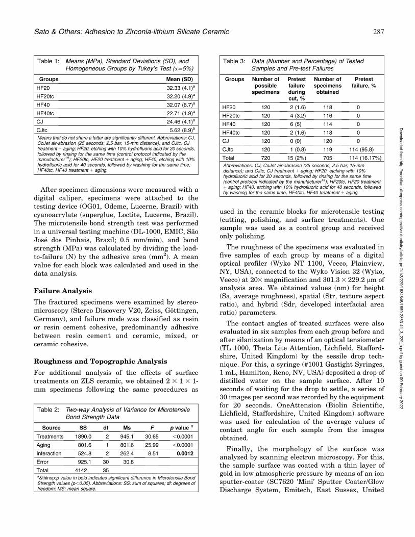
After specimen dimensions were measured with adigital caliper, specimens were attached to thetesting device (OG01, Odeme, Lucerne, Brazil) withcyanoacrylate (superglue, Loctite, Lucerne, Brazil).The microtensile bond strength test was performedin a universal testing machine (DL-1000, EMIC, SaoJose dos Pinhais, Brazil; 0.5 mm/min), and bondstrength (MPa) was calculated by dividing the load-to-failure (N) by the adhesive area (mm2). A meanvalue for each block was calculated and used in thedata analysis.
Failure Analysis
The fractured specimens were examined by stereo-microscopy (Stereo Discovery V20, Zeiss, Gottingen,Germany), and failure mode was classified as resinor resin cement cohesive, predominantly adhesivebetween resin cement and ceramic, mixed, orceramic cohesive.
Roughness and Topographic Analysis
For additional analysis of the effects of surfacetreatments on ZLS ceramic, we obtained 2 3 1 3 1-mm specimens following the same procedures as
used in the ceramic blocks for microtensile testing(cutting, polishing, and surface treatments). Onesample was used as a control group and receivedonly polishing.
The roughness of the specimens was evaluated infive samples of each group by means of a digitaloptical profiler (Wyko NT 1100, Veeco, Plainview,NY, USA), connected to the Wyko Vision 32 (Wyko,Veeco) at 203 magnification and 301.3 3 229.2 lm ofanalysis area. We obtained values (nm) for height(Sa, average roughness), spatial (Str, texture aspectratio), and hybrid (Sdr, developed interfacial arearatio) parameters.
The contact angles of treated surfaces were alsoevaluated in six samples from each group before andafter silanization by means of an optical tensiometer(TL 1000, Theta Lite Attention, Lichfield, Stafford-shire, United Kingdom) by the sessile drop tech-nique. For this, a syringe (#1001 Gastight Syringes,1 mL, Hamilton, Reno, NV, USA) deposited a drop ofdistilled water on the sample surface. After 10seconds of waiting for the drop to settle, a series of30 images per second was recorded by the equipmentfor 20 seconds. OneAttension (Biolin Scientific,Lichfield, Staffordshire, United Kingdom) softwarewas used for calculation of the average values ofcontact angle for each sample from the imagesobtained.
Finally, the morphology of the surface wasanalyzed by scanning electron microscopy. For this,the sample surface was coated with a thin layer ofgold in low atmospheric pressure by means of an ionsputter-coater (SC7620 ’Mini’ Sputter Coater/GlowDischarge System, Emitech, East Sussex, United
Table 2: Two-way Analysis of Variance for MicrotensileBond Strength Data
Source SS df Ms F p value a
Treatments 1890.0 2 945.1 30.65 ,0.0001
Aging 801.6 1 801.6 25.99 ,0.0001
Interaction 524.8 2 262.4 8.51 0.0012
Error 925.1 30 30.8
Total 4142 35a&thinsp:p value in bold indicates significant difference in Microtensile BondStrength values (p,0.05). Abbreviations: SS: sum of squares; df: degrees offreedom; MS: mean square.
Table 3: Data (Number and Percentage) of TestedSamples and Pre-test Failures
Groups Number ofpossible
specimens
Pretestfailureduringcut, %
Number ofspecimensobtained
Pretestfailure, %
HF20 120 2 (1.6) 118 0
HF20tc 120 4 (3.2) 116 0
HF40 120 6 (5) 114 0
HF40tc 120 2 (1.6) 118 0
CJ 120 0 (0) 120 0
CJtc 120 1 (0.8) 119 114 (95.8)
Total 720 15 (2%) 705 114 (16.17%)
Abbreviations: CJ, CoJet air-abrasion (25 seconds, 2.5 bar, 15-mmdistance); and CJtc, CJ treatment þ aging; HF20, etching with 10%hydrofluoric acid for 20 seconds, followed by rinsing for the same time(control protocol indicated by the manufacturer19); HF20tc, HF20 treatmentþ aging; HF40, etching with 10% hydrofluoric acid for 40 seconds, followedby washing for the same time; HF40tc, HF40 treatment þ aging.
Table 1: Means (MPa), Standard Deviations (SD), andHomogeneous Groups by Tukey’s Test (a=5%)
Groups Mean (SD)
HF20 32.33 (4.1)a
HF20tc 32.20 (4.9)a
HF40 32.07 (6.7)a
HF40tc 22.71 (1.9)a
CJ 24.46 (4.1)a
CJtc 5.62 (8.9)b
Means that do not share a letter are significantly different. Abbreviations: CJ,CoJet air-abrasion (25 seconds, 2.5 bar, 15-mm distance); and CJtc, CJtreatment þ aging; HF20, etching with 10% hydrofluoric acid for 20 seconds,followed by rinsing for the same time (control protocol indicated by themanufacturer19); HF20tc, HF20 treatment þ aging; HF40, etching with 10%hydrofluoric acid for 40 seconds, followed by washing for the same time;HF40tc, HF40 treatment þ aging.
Sato & Others: Adhesion to Zirconia-lithium Silicate Ceramic 287
Dow
nloaded from http://m
eridian.allenpress.com/operative-dentistry/article-pdf/41/3/229/1834845/1559-2863-41_3_229_a.pdf by guest on 09 February 2022

Kingdom), and the topography was analyzed andphotographed with high-vacuum equipment (InspectS 50, FEI Company, Brno, Czech Republic) operatingat 20-25 kV, 5.0 spot, and magnifications of 5003 and
50003.
Data Analysis
Data were tabulated, and the blocks were used asexperimental units for statistical analysis of themicrotensile bond strength data. The results of bond
strength were analyzed by two-way analysis ofvariance (ANOVA) (treatment and aging) and Tu-key’s test (both a=5%), and roughness and contactangle values were analyzed by one-way ANOVA andTukey’s test (both a=5%).
RESULTS
The microtensile bond strength results are presentedin Table 1. ANOVA showed that interaction of
surface and aging influenced the bond strength
(p=0.0012) (Table 2). The adhesion for the HF
groups was stable, whereas for CJ, aging signifi-
cantly damaged the adhesion. Samples in the CJtc
group also showed a large number of pretest failures
after thermal cycling (Table 3).
Failure was predominantly adhesive between the
ceramic and resin cement (52.6%) (Figure 2).
Surface roughness was also influenced by surface
treatments for all parameters evaluated: Sa
(p,0.0001), Str (p,0.0001), and Sdr (p,0.0001)
(Figure 3). All treatments increased roughness
parameters compared with those of control samples
(polished, without treatment).
The contact angle was also influenced by surface
treatments for both silanized (p,0.0001) and un-
silanized samples (p,0.0001). In both cases, the
group conditioned with hydrofluoric acid for 40
Figure 2. Bar graph of failure typedistribution. ADHES cem/cer = pre-dominantly adhesive failure betweenresin cement and ceramic; ADHEScomp/cem = predominantly adhesivefailure between composite resin andcement; COHES cer = cohesivefailure of ceramic; COHES comp =cohesive failure of composite/resincement; MIXED = cohesive andadhesive failures.
Figure 3. Bar graph of roughness values for tested groups. For each roughness parameter, means with different superscript letters are significantlydifferent.
288 Operative Dentistry
Dow
nloaded from http://m
eridian.allenpress.com/operative-dentistry/article-pdf/41/3/229/1834845/1559-2863-41_3_229_a.pdf by guest on 09 February 2022

seconds (HF40) showed the highest wettability
values (Figures 4 and 5).
Figure 6 shows representative profilometry imag-
es and scanning electron microscope micrographs of
the surface patterns for tested groups.
DISCUSSION
The baseline assessment showed that the three
treatments tested (HF20, HF40, and CJ) had similar
baseline bond strength values.
In the context of surface treatment by extension of
the etching time suggested by the manufacturer,
studies have found no deleterious effect of hydro-
fluoric acid on the resistance of glass-ceramic,20,21
because the excessive etching reduced existing
surface faults in size and depth, besides removing
or stabilizing surface defects.22,23 Thus, increasing
the etching time can enhance the materialsstrength24 but does not lead to improved bondstrengths, according to our findings.
We found that the thermocycling had a negativeeffect on the bond strength, and samples in thesilica-coated group (CJtc) showed an almost 80%decrease in the microtensile bond strength to resincement. Most specimens in this group did not resistthe thermal cycling, as well as the large percentageof predominantly adhesive failures between resincement and ceramic showed that the surfacemodification by the addition of silica did notguarantee a stable bonding between ZLS and resincement in the long term. According to Kern andThompson,25 this is attributable to the fact that theirregularities created by the sandblasting aredevoid of microretention. Corroborating this, Me-nees and others4 argued that for the ceramiclithium disilicate ceramic, hydrofluoric acid etchingintroduces more uniform and better-distributedsurface changes.
In fact, silica-coated topography in our studyconfirms the existence of small irregularities, al-though the surface roughness of the three groupswere not much different. In this same context, Kernand Wegner12 stated that, because of this limitedceramic roughness, chemical and physical Bis-GMA(Bisphenol A-glycidyl methacrylate)–based resinbonds are not water resistant and end up sufferingdetachment.
For roughness parameters, the three treatmentswere effective in changing the surface pattern,generating similar results. When a glass-ceramicsurface is exposed to HF etching, a selective removalof its vitreous matrix occurs, exposing the crystallinestructure6,26 and resulting in a topography with atotal contact area greater than that of a smoothsurface.27 Silica deposition generates an irregularrough surface with increased surface area thatimproves ceramic wettability. However, sandblastingcan also induce excessive gaps in the surface of thematerial or even a significant loss of material.25,28
This conformation of the material’s surface topog-raphy is closely linked to the characteristics that itpresents in terms of contact angle and consequentwettability.29
The contact angle measurement of the dispersionliquid (often water of high purity) on a substrate isused as an indicator of total surface energy andwettability of the substrate.27,30 The smallest contactangle resulting from silica coating represents bettersurface wettability. However, the silica coating
Figure 4. Bar graph of the mean values of contact angle (8) for testedgroups (not silanized). Means with different superscript letters aresignificantly different.
Figure 5. Bar graph of the mean values of contact angle (8) forsilanized specimens. Means with different superscript letters aresignificantly different.
Sato & Others: Adhesion to Zirconia-lithium Silicate Ceramic 289
Dow
nloaded from http://m
eridian.allenpress.com/operative-dentistry/article-pdf/41/3/229/1834845/1559-2863-41_3_229_a.pdf by guest on 09 February 2022
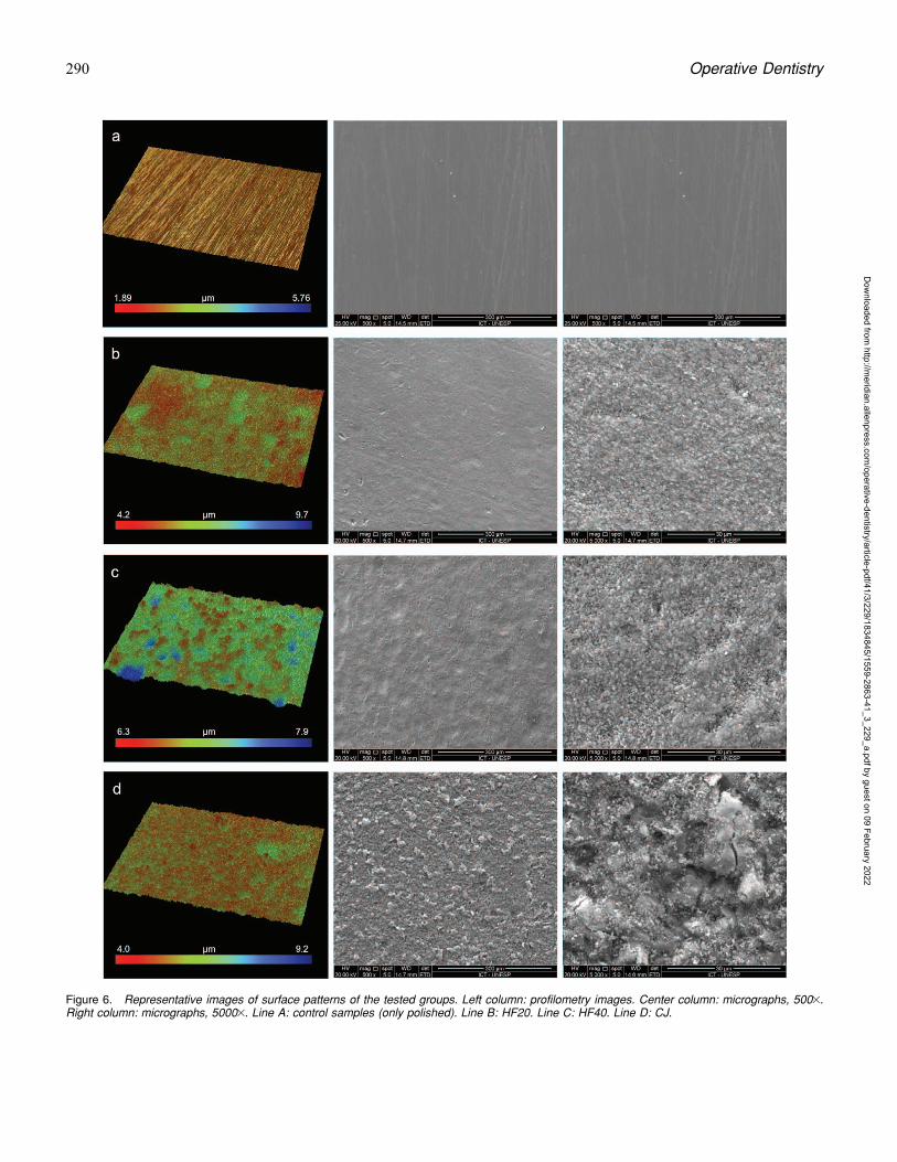
Figure 6. Representative images of surface patterns of the tested groups. Left column: profilometry images. Center column: micrographs, 5003.Right column: micrographs, 50003. Line A: control samples (only polished). Line B: HF20. Line C: HF40. Line D: CJ.
290 Operative Dentistry
Dow
nloaded from http://m
eridian.allenpress.com/operative-dentistry/article-pdf/41/3/229/1834845/1559-2863-41_3_229_a.pdf by guest on 09 February 2022
showed a reduction in bond strength after aging,suggesting that, although this technique ensuredgood initial bond strength to resin cement, thepattern generated by silica coating is not favorablefor the long-term maintenance of this resistance.29,31
The lower bond strength obtained with acid etchingfor 40 seconds is probably due to the removal of agreater quantity of glass matrix and exposure oflithium silicate crystals and particles of zirconiacreating a surface with lower wettability.
A few microbars were lost during cutting. Mostmicrobars showed predominantly adhesive failurebetween ceramic and resin cement. Thus, despite thedecline in bond strength of the HF40 group, the CJgroup was most vulnerable to temperature changesin the water, with great loss of microbars duringthermocycling.
Therefore, considering the results of microtensilebond strength, surface roughness, contact angle, andmorphology obtained for surface treatments andconsidering the potential risks with the use of HF,it is suggested that one should not extend the timesuggested by the manufacturer. The use of silicacoating is also not advantageous for long-termmaintenance of the bond.
CONCLUSION
On the basis of our results, it can be concluded thatthe silica coating was not efficient in maintainingthe bond strength after aging, and etching withhydrofluoric acid for 20 or 40 seconds was equallyeffective in producing stable resin bonding to a ZLSceramic.
Conflict of Interest
The authors have no proprietary, financial, or other personalinterest of any nature or kind in any product, service, and/orcompany that is presented in this article.
(Accepted 3 June 2015)
REFERENCES
1. Guess PC, Selz CF, Steinhart YN, Stampf S, & Strub JR(2013) Prospective clinical split-mouth study of pressedand CAD/CAM all-ceramic partial-coverage restorations:7-year results International Journal of Prosthodontics26(1) 21-25.
2. Valandro LF, Della Bona A, Bottino MA, & Neisser MP(2005) The effect of ceramic surface treatment on bondingto densely sintered alumina ceramic Journal of ProstheticDentistry 93(3) 253-259.
3. Zogheib LV, Bona AD, Kimpara ET, & McCabe JF (2011)Effect of hydrofluoric acid etching duration on theroughness and flexural strength of a lithium disilicate-
based glass ceramic Brazilian Dental Journal 22(1)45-50.
4. Menees TS, Lawson NC, Beck PR, & Burgess JO (2014)Influence of particle abrasion or hydrofluoric acid etchingon lithium disilicate flexural strength Journal of Pros-thetic Dentistry 112(5) 1164-1170.
5. Chen JH, Matsumura H, & Atsuta M (1998) Effect ofdifferent etching periods on the bond strength of acomposite resin to a machinable porcelain Journal ofDentistry 26(1) 53-58.
6. Chen JH, Matsumura H, & Atsuta M (1998) Effect ofetchant, etching period, and silane priming on bondstrength to porcelain of composite resin OperativeDentistry 23(5) 250-257.
7. Della Bona A, Anusavice KJ, & Hood JA (2002) Effect ofceramic surface treatment on tensile bond strength to aresin cement International Journal of Prosthodontics15(3) 248-253.
8. Michida SM, Valandro LF, Yoshiga S, Andreatta FilhoOD, Balducci I, & Bottino MA (2003) Effect of surfacetreatment of a glass-infiltrated alumina ceramic on themicrotensile bond strength Journal of Applied OralScience 11(4) 361-366.
9. Brentel AS, Ozcan M, Valandro LF, Alarca LG, Amaral R,& Bottino MA (2007) Microtensile bond strength of a resincement to feldspathic ceramic after different etching andsilanization regimens in dry and aged conditions DentalMaterials 23(11) 1323-1331.
10. Ban S, Sato H, Suehiro Y, Nakanishi H, & Nawa M (2008)Biaxial flexure strength and low temperature degrada-tion of Ce-TZP/Al2O3 nanocomposite and Y-TZP as dentalrestoratives Journal of Biomedical Materials ResearchPart B: Applied Biomaterial 87(2) 492-498.
11. Chevalier J, Olagnon C, & Fantozzi G (1999) Subcriticalcrack propagation in 3Y-TZP ceramics: static and cyclicfatigue Journal of the American Ceramic Society 82(3)128-129.
12. Kern M, & Wegner SM (1998) Bonding to zirconiaceramic: Adhesion methods and their durability DentalMaterials 14(1) 64-71.
13. Zhang Y, Lawn BR, Rekow ED, & Thompson VP (2004)Effect of sandblasting on the long-term performance ofdental ceramics Journal of Biomedical Materials Re-search Part B: Applied Biomaterial 71(2) 381-386.
14. Cristoforides P, Amaral R, May LG, Bottino MA, &Valandro LF (2012) Composite resin to yttria stabilizedtetragonal zirconia polycrystal bonding: Comparison ofrepair methods Operative Dentistry 37(3) 263-271.
15. Scherrer SS, Cattani-Lorente M, Vittecoq E, de MestralF, Griggs JA, & Wiskott HW (2011) Fatigue behavior inwater of Y-TZP zirconia ceramics after abrasion with 30mum silica-coated alumina particles Dental Materials27(2) e28-e42.
16. Passos SP, May LG, Barca DC, Ozcan M, Bottino MA, &Valandro LF (2010) Adhesive quality of self-adhesive andconventional adhesive resin cement to Y-TZP ceramicbefore and after aging conditions Operative Dentistry35(6) 689-696.
Sato & Others: Adhesion to Zirconia-lithium Silicate Ceramic 291
Dow
nloaded from http://m
eridian.allenpress.com/operative-dentistry/article-pdf/41/3/229/1834845/1559-2863-41_3_229_a.pdf by guest on 09 February 2022

17. Cattani Lorente M, Scherrer SS, Richard J, DemellayerR, Amez-Droz M, & Wiskott HW (2010) Surface rough-ness and EDS characterization of a Y-TZP dental ceramictreated with the CoJet Sand Dental Materials 26(11)1035-1042.
18. Ban S (2008) Realiability and properties of core materialsfor all-ceramic dental restorations Japanese DentalScience Review 44(1) 3-21.
19. VITA Suprinity Working Instructions nr. 1951 (2014)Vita Zahnfabrik. Retrieved online August 13, 2015 from:www. http://vitanorthamerica.com/wp-content/uploads/2013/10/VITA_1951_1951E_SUPRINITY_VA_EN_V03_screen_en.pdf
20. Posritong S, Borges AL, Chu TM, Eckert GJ, Bottino MA,& Bottino MC (2013) The impact of hydrofluoric acidetching followed by unfilled resin on the biaxial strengthof a glass-ceramic Dental Materials 29(11) e281-e290.
21. Magne P, Kwon KR, Belser UC, Hodges JS, & DouglasWH (1999) Crack propensity of porcelain laminateveneers: A simulated operatory evaluation Journal ofProsthetic Dentistry 81(3) 327-334.
22. Thompson JY, & Anusavice KJ (1994) Effect of surfaceetching on the flexural strength and fracture toughness ofDicor disks containing controlled flaws Journal of DentalResearch 73(2) 505-510.
23. Proctor B (1962) The effects of hydrofluoric acid etchingon the strength of glasses. Glass Physics and Chemistry3(7) 27.
24. Yen TW, Blackman RB, & Baez RJ (1993) Effect of acidetching on the flexural strength of a feldspathic porcelainand a castable glass ceramic Journal of ProstheticDentistry 70(3) 224-233.
25. Kern M, & Thompson VP (1994) Sandblasting and silicacoating of a glass-infiltrated alumina ceramic: Volumeloss, morphology, and changes in the surface compositionJournal of Prosthetic Dentistry 71(5) 453-461.
26. Sorensen JA, Engelman MJ, Torres TJ, & Avera SP(1991) Shear bond strength of composite resin to porcelainInternational Journal of Prosthodontics 4(1) 17-23.
27. Della Bona A, Shen C, & Anusavice KJ (2004) Work ofadhesion of resin on treated lithia disilicate-basedceramic Dental Materials 20(4) 338-344.
28. Blatz MB, Sadan A, & Kern M (2003) Resin-ceramicbonding: A review of the literature Journal of ProstheticDentistry 89(3) 268-274.
29. Phoenix RD, & Shen C (1995) Characterization of treatedporcelain surfaces via dynamic contact angle analysisInternational Journal of Prosthodontics 8(2) 187-194.
30. Zisman WA (1964) Relation of the equilibrium contactangle to liquid and solid constitution Advances inChemistry Series 43 1-51.
31. Kern M, & Thompson VP (1995) Bonding to glassinfiltrated alumina ceramic: Adhesive methods and theirdurability Journal of Prosthetic Dentistry 73(3) 240-249.
292 Operative Dentistry
Dow
nloaded from http://m
eridian.allenpress.com/operative-dentistry/article-pdf/41/3/229/1834845/1559-2863-41_3_229_a.pdf by guest on 09 February 2022

Influence of Restoration Type on theCytotoxicity of a 35% Hydrogen
Peroxide Bleaching Gel
DG Soares � N Marcomini � FG BassoTN Pansani � J Hebling � CA de Souza Costa
Clinical Relevance
The application of a 35%-H2O2 bleaching gel to a dental surface containing a hydrolyticallydegraded resin-modified glass ionomer cement restoration results in more intense diffusionof H2O2 into the pulp chamber, which increases pulp cell cytotoxicity.
SUMMARY
Objectives: The tooth/restoration interface
may act as a pathway for hydrogen peroxide
(H2O2) diffusion into the pulp chamber. There-
fore, the influence of resin-modified glass
ionomer cement (RMGIC) and resin composite
simulated restorations on the cytotoxicity of
an in-office bleaching gel was assessed in vitro.
Materials and Methods: Cavities in enamel/
dentin discs restored with RMGIC Vitremer
(3M ESPE) or Single Bond/Filtek Z350 (3M
ESPE) resin composite (RC) were subjected
or not subjected to hydrolytic degradation
(HD). A 35%-H2O2 bleaching gel was applied
to simulated restored and nonrestored enamel
surfaces, and culture medium in contact with
the dentin substrate (extract) was collected
and applied to MDPC-23 cells. Nonrestored
discs subjected or not subjected to bleaching
were used as positive and negative controls,
respectively. Cell viability, oxidative stress,
interleukin (IL)-1b expression, alkaline phos-
phatase (ALP) activity, and mineralized nod-
ule deposition were evaluated. The H2O2 in the
extracts was quantified. Data were subjected
to statistical analysis.
Diana Gabriela Soares DDS, Ms, PhD, Department ofPhysiology and Pathology, Araraquara School of Dentistry,Univ Estadual Paulista – UNESP, Sao Paulo, Brazil
Natalia Marcomini undergraduate student, Department ofPhysiology and Pathology, Araraquara School of Dentistry,Univ Estadual Paulista – UNESP, Sao Paulo, Brazil
Fernanda Goncalves Basso DDS, Ms, PhD, Department ofPhysiology and Pathology, Araraquara School of Dentistry,Univ Estadual Paulista – UNESP, Sao Paulo, Brazil
Taisa N Pansani DDS, Ms, Department of Dental Materialsand Prosthodontics, Araraquara School of Dentistry, UnivEstadual Paulista – UNESP, Sao Paulo, Brazil
Josimeri Hebling DDS, Ms, PhD, associate professor, Depart-ment of Orthodontics and Pediatric Dentistry, AraraquaraSchool of Dentistry, Univ Estadual Paulista – UNESP, SaoPaulo, Brazil
*Carlos A de Souza Costa DDS, Ms, PhD, professor,Department of Physiology and Pathology, Araraquara Schoolof Dentistry, Univ Estadual Paulista – UNESP, Sao Paulo,Brazil
*Corresponding author: Rua Humaita, 1680 – CentroAraraquara, Sao Paulo 14801-903, Brazil; e-mail:[email protected]
DOI: 10.2341/14-325-L
�Operative Dentistry, 2016, 41-3, 293-304
Dow
nloaded from http://m
eridian.allenpress.com/operative-dentistry/article-pdf/41/3/229/1834845/1559-2863-41_3_229_a.pdf by guest on 09 February 2022

Results: Higher oxidative stress associatedwith reduced cell viability, ALP activity, andmineralized nodule deposition was observedfor all bleached groups compared with thenegative control group. The RMGIC/HD group,which presented the highest H2O2 diffusion,had the lowest values of cell viability, ALPactivity, and mineralized nodule deposition, aswell as significantly increased IL-1b expres-sion.
Conclusions: Dental cavities restored with theRMGIC subjected to hydrolytic degradationallowed for more intense diffusion of H2O2 intothe pulp chamber, intensifying the toxicity of a35%-H2O2 bleaching gel to pulp cells.
INTRODUCTION
Vital tooth bleaching is a cosmetic technique widelyused in clinical dental practice. However, thisprocedure is characterized by a high prevalence oftooth sensitivity, which may cause considerable tointolerable discomfort to patients.1-5 This side effecthas been associated with the rapid transtoothdiffusion of hydrogen peroxide (H2O2),6,7 which istoxic to pulp cells.8-12 An important factor that caninfluence the diffusion of H2O2 into the pulpal spaceis the presence of cavity restorations on toothsurfaces.13-15 It has been shown that the tooth/restoration interface may act as a pathway for H2O2
diffusion into deep dentin and, consequently, intothe pulp chamber, depending on the dental materialused to create the restoration.14,15 Bonafe andothers16 showed that the application of a 35%-H2O2
bleaching gel to anterior teeth containing aged resincomposite restorations resulted in more intense andprevalent tooth sensitivity. The authors reportedthat the dental material and technique used torestore dental cavities, as well as the quality ofrestoration margins, may affect H2O2 diffusion. Thisfact was corroborated by Soares and others,17 whoshowed that resin composite (RC) restorationsperformed with etch-and-rinse adhesives did notinfluence the transtooth cytotoxicity of a professionalbleaching protocol. However, a significantly highertoxic effect was observed when a two-step self-etchadhesive was used to perform such adhesive resto-rations.18
Instead of the limited range of clinical indica-tions, resin-modified glass ionomer cements(RMGICs) have been used in operative dentistryto restore carious or noncarious cervical lesions inpermanent teeth, since these areas are not subject-ed to compressive loads.19 The main advantage of
this kind of dental material in this clinicalsituation is related to the chemical adhesion totooth structure, biocompatibility, and release offluoride.19 However, the bond strength of RMGICsto enamel and dentin is lower than that observedfor RC restorations,20,21 making the application ofbleaching gels to tooth/RMGIC restorative materialinterfaces a concern.22 Therefore, the present studyevaluated the effects of RMGIC and etch-and-rinseadhesive/RC restorations, subjected or not subject-ed to hydrolytic degradation, on the indirecttoxicity of a 35%-H2O2 bleaching gel to odonto-blast-like cells. The null hypothesis is that theserestorations have no effect on the cytotoxicity of thebleaching protocol.
METHODS AND MATERIALS
Specimen Preparation
Standardized 3.5-mm-thick and 5.6-mm-diameterenamel/dentin discs were obtained from intactbovine incisors, as previously described.12 Roundcavities measuring 1.6 mm in diameter and 2.5 mmdeep were prepared on part of each disc by means ofa high-speed, water-cooled cylindrical diamond bur(#1095; KG Sorensen, Barueri, SP, Brazil). There-fore, the remaining dentin thickness between thebase of the cavity preparation and the simulatedpulp chamber wall was set to 1.0 mm.17 Thecavities were restored with a RMGIC or with RCassociated with two-step etch-and-rinse adhesive,following the manufacturer’s instructions, as de-scribed in Table 1. The etch-and-rinse adhesive/RCrestoration was used as a control since it waspreviously demonstrated that this restoration doesnot impact the transenamel and transdentinalMDPC-23 cell cytotoxicity when bleaching, evenwhen it is subjected to hydrolytic degradation(HD).17
In the nonrestored discs, no cavity preparationwas performed; however, a resin coating wasprepared on the enamel surface, with the samediameter as that of the restoration performed ondiscs of restored groups. Therefore, the enamelsurface subjected to bleaching was standardized inboth restored and nonrestored groups. For thenonrestored groups, a round enamel surface with1.6-mm diameter was etched with 37% phosphoricacid for 30 seconds, followed by the application of twolayers of bonding agent and light-curing for 20seconds. Finally, a layer of composite resin (1.0 mmthick 3 1.6 mm diameter) was applied and also light-cured for 20 seconds (Figure 1A).
294 Operative Dentistry
Dow
nloaded from http://m
eridian.allenpress.com/operative-dentistry/article-pdf/41/3/229/1834845/1559-2863-41_3_229_a.pdf by guest on 09 February 2022

Half of the restored discs were subjected to HD,which consisted of a thermocycling regimen in athermal cycler (MSCT-3 plus; Marcelo Nucci-ME,Sao Carlos, SP, Brazil) for a total of 20,000 cycles at58C and 558C, with a 30-second dwell time in eachbath, followed by 12 months of storage at 378C in0.1% thymol-buffered solution, which was replacedweekly. The thymol solution was used to preventmicrobial contamination during the storage periods.The dentin surfaces of discs were treated withethylenediaminetetraacetic acid (EDTA), 0.5 N, for30 seconds for smear layer removal, and discs werethen individually adapted to artificial pulp chambers(APCs) (Figure 1B) by means of two silicone O-rings(Rodimar Rolamentos Ltda, Araraquara, SP, Brazil).To avoid any H2O2 diffusion through the edges of thediscs, a seal was created with wax on the siliconering area. The APCs with the enamel/dentin discs inposition were sterilized in ethylene oxide gas.
Experimental Procedure
The disc/APC sets were positioned in 24-well plates(Costar Corp, Cambridge, MA, USA) containing 1mL of serum-free Dulbecco Modified Eagle Medium(DMEM; Sigma-Aldrich, St Louis, MO, USA). A 35%H2O2 bleaching gel (Whiteness HP; FGM ProdutosOdontologicos Ltda, Joinville, SC, Brazil) was ap-plied to simulated restoration and enamel surfaces ofthe discs three times for 15 minutes each time. Thegroups are described in Table 2. Two control groupswere used in the present investigation. Nonrestored/nonbleached discs were used in the negative controlgroup (NC), which represents normal cell parame-ters. The nonrestored/bleached discs were used inthe positive control group (PC), which represents thecell responses to bleaching. In total, six APC/discsets were used for each experimental group. TheMDPC-23 cells were seeded on 96-well plates (CostarCorp) at 80% confluence (6000 cells/well; 48 hours) in
Table 1: Application Protocol for Each Restorative Material
Material Application Protocol
RMGIC (Vitremer; 3M ESPE,St Paul, MN, USA)
A brush was used to apply the Vitremer primer (HEMAþpolycarboxylic acid) for 30 seconds to enamel anddentin, followed by gentle air-drying for 15 seconds. This process was repeated once more, and the primerwas then light-cured (450 mW/cm2 Curing Light XL 300, 3M ESPE) for 20 seconds.
One level scoop and one drop of liquid of Vitremer were dispensed onto the mixing pad and mixed within45 seconds.
The material was applied to the cavity by means of a Centrix applicator and light-cured for 40 seconds.
Twenty-four hours after cavity restoration, the restoration surface was polished with sequential Soflex discs(3M ESPE) at low speed.
RC (Single Bond þ Filtek Z350;3M ESPE, St Paul, MN, USA)
The 37% phosphoric acid etchant (Scotchbond, 3M ESPE) was applied to enamel for 30 seconds and todentin for 15 seconds and then rinsed for 30 seconds, and the tooth structure was dried with sterilizedcotton.
The first layer of Single Bond was applied under friction, followed by 30 seconds of resting and 10 secondsof gentle air-drying. A second bonding agent layer was applied, and the product was light-cured for 20seconds under a halogen lamp (450 mW/cm2 Curing Light XL 300, 3M ESPE).
The cavity was restored by the application of two increments of the nanofilled composite resin Filtek Z350,which were individually light-cured for 20 seconds.
Twenty-four hours after cavity restoration, the resin surface was polished with sequential Soflex discs (3MESPE) at low speed.
Abbreviations: HEMA, 2-hydroxyethyl methacrylate; RC, resin composite; RMGIC, resin-modified glass ionomer cement.
Figure 1. Representative illustrationof experimental protocol. (A) Re-stored and control discs. (B) APC/disc set positioned in a well of a 24-well plate.
Soares & Others: Cytotoxicity of Bleaching Restored Teeth 295
Dow
nloaded from http://m
eridian.allenpress.com/operative-dentistry/article-pdf/41/3/229/1834845/1559-2863-41_3_229_a.pdf by guest on 09 February 2022
DMEM plus 10% fetal bovine serum (FBS; Sigma-Aldrich). Immediately after bleaching, the culturemedium in contact with the internal dentin surface(extract) of each APC/disc set was collected anddistributed into aliquots of 100 lL, which wereapplied to the cultured cells. The MPDC-23 cellswere incubated with the extracts for a period of 1hour at 378C and at 5% CO2, and then theexperimental protocols were conducted, as follows.
Cell Viability
For cell viability analysis, the methyl tetrazolium(MTT) assay was performed. Immediately after theone-hour contact time with extracts, the cells wereincubated for four hours with MTT solution (5 mg/mL; Sigma-Aldrich) diluted in DMEM without FBS(1:10), and the absorbance of formazan crystals onviable cells was read at a 570-nm wavelength(Synergy H1, Biotek, Winooski, VT, USA). Thenumeric values obtained from the MTT assay wereconverted into percentages according to mean absor-bance observed in the NC, which was considered as100% of cell viability.
Interluekin (IL)-1b Protein Expression
The enzyme-linked immunosorbent assay (ELISA;R&D Systems, Minneapolis, MN, USA) was used forthis analysis. Two 100-lL aliquots of the extractsfrom each APC/disc set were applied to two wells(100 lL per well) with previously seeded MDPC-23cells. Immediately after the contact time withextracts, the cells were incubated for 24 hours inDMEM with no FBS (100 lL). The culture media ofthe two wells of each APC/disc set were thencollected (total of 200 lL) and incubated for twohours in ELISA plates previously coated with theprimary antibody (overnight at room temperature)and blocked with bovine serum albumin (Santa CruzBiotechnology, Santa Cruz, CA, USA) solution (onehour). Secondary antibody was then added, followedby reagent and stop solutions. Protein expressionwas analyzed by spectrophotometry (455 nm) with
standard curves containing defined concentrations ofthe cytokine (Synergy H1, Biotek).
Oxidative Stress Generation
The production of reactive oxygen species (ROS) incultured cells was quantified by means of a cell-permeant fluorescence probe 6-carboxy-20,70-dichlor-odihydrofluorescein diacetate (carboxy-H2DCFDA;Life Technologies, San Francisco, CA, USA) (n=6).The cells were incubated at 378C and at 5% CO2 with5 lM carboxy-H2DCFDA for 10 minutes and werethen exposed for one hour to the extracts (100 lL) ofeach experimental group. After this period, thefluorescence intensity was monitored at 592-nmexcitation and 517-nm emission by means of amicroplate fluorescence reader (Synergy H1, Biotek).
Alkaline Phosphatase (ALP) Activity
Immediately after the contact with the extracts, thecells were cultured in osteogenic medium (DMEMplus 10% FBS, supplemented with 10 nmol/L b-glycerophosphate and 50 lg/mL sodium ascorbate;Sigma Chemical Co, St Louis, MO, USA) for sevendays (the medium was changed daily). After theseven-day incubation period, the ALP activity wasassessed (n=6). Cell lysis was performed (0.1%sodium lauryl sulfate; Sigma), and an aliquot wastransferred to glass tubes containing thymolphtha-lein monophosphate substrate at 378C (End pointassay; Labtest, Lagoa Santa, MG, Brazil). After a 10-minute incubation period at 378C, the color reagent(sodium carbonate and sodium hydroxide) wasadded, and the absorbance was read at a 590-nmwavelength (Synergy H1, Biotek), which was con-verted into U/L by means of a standard curve. Totalprotein (TP) dose was performed for normalization ofALP according to the Read and Northcote protocol,as previously described.10 The absorbance of the testand blank tubes was measured at a 655-nmwavelength (Synergy H1, Biotek) and converted intomg/L by a standard protein curve. The final value ofALP was normalized by TP obtained from each well,
Table 2: ACP Disc/Set Distribution into Experimental and Control Groups
Group Treatment Restorative Material Aging Procedure
NC Nonbleached Nonrestored Water storage – 24 h
PC Bleached Nonrestored Water storage – 24 h
RMGIC Bleached RMGIC Water storage – 24 h
RMGIC/HD Bleached RMGIC Water storage – 12 mo þ thermocycling
RC Bleached RC Water storage – 24 h
RC/HD Bleached RC Water storage – 12 mo þ thermocycling
Abbreviations: HD, hydrolytic degradation; NC, negative control group; PC, positive control group; RC, resin composite; RMGIC, resin-modified glass ionomer cement.
296 Operative Dentistry
Dow
nloaded from http://m
eridian.allenpress.com/operative-dentistry/article-pdf/41/3/229/1834845/1559-2863-41_3_229_a.pdf by guest on 09 February 2022
with the value of ALP activity measured as U/mg ofprotein. The mean ALP activity value of the NC wasconsidered as 100% of ALP activity, and thepercentage value for each sample was calculatedbased on this parameter and used for statisticalanalysis.
Mineralized Nodule Deposition
For assessment of the quantity of mineralizednodules deposited (n=6), Alizarin Red staining wasperformed. The cell cultures after the incubationperiod with extracts were also cultured in osteogenicmedium for seven days. The cells were then fixedwith cold 70% ethanol for one hour, washed withdeionized water, and then stained with Alizarin Reddye (40 mM, pH 4.2; Sigma-Aldrich) for 20 minutesunder gentle shaking (VDR Shaker, Biomixer,Ribeirao Preto, SP, Brazil). After aspiration ofunincorporated dye, the cells were washed twicewith deionized water for the removal of excess stain.The cells were then incubated with 10% cetylpyr-idinium chloride (Sigma-Aldrich) for 15 minutesunder agitation to solubilize the nodules. Theabsorbance of the resulting solution was measuredat 570 nm (Synergy H1, Biotek). The percentage ofcalcium deposition for each experimental group wascalculated based on the mean value of the NC as100% of staining.
Quantification of H2O2 Diffusion
The amount of H2O2 present in the extract was alsoquantified. Aliquots of 100 lL obtained from extractsof each group were transferred to tubes containing900 lL of acetate buffer solution (2 mol/L, pH 4.5) tostabilize the H2O2. After that, an aliquot of 500 lL ofthe buffer solution plus H2O2 from each tube wastransferred to test tubes containing 2.2 mL ofdeionized water and 250 lL of 0.5 mg/mL leuco-crystal violet (Sigma-Aldrich Corp). The tubes wereagitated, and a 50-lL quantity of horseradishperoxidase enzyme solution (1 mg/mL; Sigma-Al-drich Corp) was added. Then, aliquots of each tubewere transferred to 96-well plates, and the opticaldensity of the solutions was measured at 600-nmwavelength (Synergy H1, Biotek). A standard curveof known H2O2 concentrations was used for conver-sion of the optical density obtained in the samplesinto lg of H2O2 per mL of extract.
Statistical Analysis
To verify the reproducibility of data, we performedtwo independent experiments for all protocols in thisstudy. Thereafter, data were compiled and subjected
to the Levene test for the verification of homosce-dasticity. Data from cell viability, H2O2 diffusion,Alizarin Red, and ALP activity were subjected toone-way analysis of variance and Tukey test.Fluorescence intensity data of carboxy-H2DCFDAand protein expression of IL-1b were analyzed by theKruskal-Wallis and Mann-Whitney tests. The vari-ables cell viability and H2O2 diffusion were correlat-ed by Pearson linear correlation analysis. All testswere set at a significance level of 5%. SPSS 19.0software (SPSS Inc, Chicago, IL, USA) was used torun the statistical analyses.
RESULTS
Cell Viability/H2O2 Diffusion
Significant reductions in cell viability relative to theNC were observed for all bleached groups. Thepercentages of cell viability reduction were 38.7%,38.5%, 61.7%, 39.4%, and 31.5% for the PC, RMGIC,RMGIC/HD, RC, and RC/HD groups, respectively. Asignificant difference from the PC was observed onlyfor the RMGIC/HD group. No H2O2 was detected inthe NC, which was discarded from statisticalanalysis. The highest H2O2 value was found for theRMGIC/HD group, which was significantly higherthan that of the other experimental groups. Consid-ering the PC as presenting 100% of H2O2 diffusion,about 125% of H2O2 diffusion was observed in theRMGIC/HD group. These data are demonstrated onFigure 2. The linear Pearson correlation analysisdemonstrated a significant negative correlationbetween the variables cell viability and H2O2
diffusion (Pearson coefficient of correlation=�0.87,p=0.0001) (Figure 2). Therefore, the higher the H2O2
diffusion, the lower the cell viability.
Oxidative Stress Generation/IL-1b ProteinExpression
All bleached groups presented significantly moreintense fluorescence values for the carboxy-H2DCFDA probe than did the NC group, demon-strating that in all bleached groups, the cells wereunder oxidative stress. For IL-1b protein expression,only RMGIC/HD presented values significantlyhigher than those of the NC and PC (Figure 3).
ALP Activity/Mineralized Nodule Deposition
Significant reductions in ALP activity and mineral-ized nodule deposition were observed for all bleachedgroups relative to those in the NC. A reduction ofaround 29% to 67% of ALP activity and 66% to 82%of mineralized nodule deposition was observed in
Soares & Others: Cytotoxicity of Bleaching Restored Teeth 297
Dow
nloaded from http://m
eridian.allenpress.com/operative-dentistry/article-pdf/41/3/229/1834845/1559-2863-41_3_229_a.pdf by guest on 09 February 2022

bleached groups relative to those in the NC, withRMGIC/HD being the group that featured thehighest reduction values (Figure 4).
DISCUSSION
In the present investigation, a traditional in-officebleaching technique was performed on enamel/dentin discs restored with RMGIC or etch-and-rinseadhesive/RC, which were subjected or not subjectedto hydrolytic degradation. The amount of H2O2 thatreached the pulpal space was quantified, and itstoxicity to cultured odontoblast-like cells was as-sessed. The RMGIC used in the present investiga-tion (Vitremer) was previously assessed regardingthe tooth/restoration interface quality, such as bondstrength to enamel/dentin and nano/microinfiltra-tion. The results of in vitro and in vivo studies
demonstrated that Vitremer featured results similarto or better than those of other brands.23-28 It wasalso demonstrated that powder/liquid RMGICs havehigher microtensile bond strength to dentin andlower microleakage than do the paste/paste restora-tions when evaluated immediately after cavityrestoration placement and following simulated hy-drolytic degradation.29,30 Therefore, the interfacecreated with the powder/liquid Vitremer in thepresent study represents a challenging situation forRMGIC restorations exposed to bleaching. Regard-ing the adhesive system and composite resin used inthe RC group, the materials and restoration tech-niques performed were based upon those describedin a previous study17 in which the tooth/restorationinterface had no effect on transenamel and trans-dentinal H2O2 diffusion and cytotoxicity to theMDPC-23 cells, even when subjected to hydrolytic
Figure 2. Bar graph of cell viability (a) and H2O2 diffusion (b). Different letters indicate statistically significant differences among groups (Tukey,p,0.05). Values are average 6 standard deviation (n=6). (c) The linear Pearson correlation analysis demonstrating a significant negative correlationbetween the variables of cell viability and H2O2 diffusion (Pearson coefficient of correlation=�0.87, p=0.0001).
298 Operative Dentistry
Dow
nloaded from http://m
eridian.allenpress.com/operative-dentistry/article-pdf/41/3/229/1834845/1559-2863-41_3_229_a.pdf by guest on 09 February 2022

degradation and exposed to calcium-containing 20%
and 35%-H2O2 bleaching gels. Therefore, this group
served as a control for comparison of the effect of the
RMGIC interface subjected to bleaching. However,
materials from only one single brand were tested in
the present investigation, which represents a limi-
tation of this study; therefore, the results should be
interpreted with caution.
It has already been demonstrated16 that the in-
office bleaching protocol involving three 15-minute
applications of a 35% H2O2 gel causes intense tooth
sensitivity in sound human teeth, which is even
higher in adhesive-restored teeth. Therefore, this
professional bleaching technique was assessed in thepresent investigation to determine the role ofdifferent tooth/restoration interfaces in transenameland transdentinal cytotoxicity to pulp cells. Regard-ing the sound/bleached samples (SD group), theresults of the present study are in agreement withthose of previous investigations,10-12,17 in whichhighly concentrated H2O2 bleaching gels applied tosound tooth surfaces for 45 minutes caused moderatecytotoxicity, characterized by decreased cell viabilityfrom 21% to 50%.31,32 These data have beencorrelated with the intense tooth sensitivity claimedby the patients subjected to traditional in-officebleaching, as observed in clinical trials.1-5 Using a
Figure 3. Box-whisker plot of fluorescence intensity of H2DCFDA (a), indicating the occurrence or lack of occurrence of oxidative stress and IL-1bexpression (b). Different letters indicate statistically significant differences among groups (Mann-Whitney, p,0.05; n=6).
Figure 4. Bar graph of ALP activity (a) and Alizarin Red staining (b). Different letters indicate statistically significant differences among groups(Tukey, p,0.05). Values are average 6 standard deviation (n=6).
Soares & Others: Cytotoxicity of Bleaching Restored Teeth 299
Dow
nloaded from http://m
eridian.allenpress.com/operative-dentistry/article-pdf/41/3/229/1834845/1559-2863-41_3_229_a.pdf by guest on 09 February 2022

similar experimental protocol, Soares and others12
showed that the reduction in MDPC-23 cell viabilitymediated by traditional in-office protocols is relatedto intense oxidative stress generation, culminatingwith cell membrane damage and cell death bynecrosis. In clinical situations, oxidative cell damagetriggers an inflammatory reaction in pulp tissue,8,9
during which cell-derived factors are released. Thistissue response, associated with increased pulppressure, may trigger impulses to the intradentinalpulpal nerve fiber endings, causing clinical symp-toms of tooth sensitivity.33 According to the litera-ture, 8-12,17,18 the intensity of these adverse events isdirectly related to the amount of H2O2 that reachesthe pulp chamber.
Analysis of the data obtained in the present studyalso demonstrated that cells exposed to the extractsobtained from bleached enamel/dentin discs wereunder intense oxidative stress; the fluorescenceintensity of carboxy-H2DCFDA probe increasedabout 10 to 15 times in bleached groups comparedwith that in the NC, associated with significant cellviability reduction (from 31.5% to 61.7%). Oxidativestress arises when the balance between the produc-tion of ROS and their neutralization by antioxidantsystems is disturbed.34,35 Depending on the intensityof the oxidative stress, the cell components undergosevere oxidative damage, ultimately compromisingcell viability.12 It has been reported34-36 that ROSaccumulated during oxidative stress are transient asa result of their high reactivity, leading to oxidativedamage of indispensable biomolecules such asproteins, lipids, and nucleic acids. Therefore, theH2O2-mediating reduction in pulp cell viability hasbeen related to two basic mechanisms: 1) inductionof oxidative stress conditions due to H2O2 diffusionthrough cell membranes and 2) direct contact ofH2O2 by-products with cell membranes, causingdisruption and cell death.12,34-36 Associated withthese results, significantly decreased ALP activity(from 29% to 57%) and mineralized nodule deposi-tion (from 65% to 82%) were observed at seven dayspost–bleaching treatment compared with the NC.
The regulation of the odontoblastic phenotype iscritical for the homeostasis of the pulp-dentincomplex. Under a noxious stimulus of mild intensity,upregulation of odontoblastic secretory activity isobserved, which is accomplished by deposition andmineralization of collagen-rich matrix in a processcalled reactionary dentinogenesis. During this pro-cess, several proteins are expressed by odontoblasts,such as DSPP, DMP-1, and ALP.37 The ALP isconsidered an initial marker for in vitro analysis of
the odontoblastic maturation process, since theactivity of this protein provides the phosphateneeded for the biomineralization process. Neverthe-less, mineralization nodule deposition (MND) isconsidered a late marker, since it demonstrates thecapability of mature odontoblasts to deposit andmineralize the organic matrix.38,39 Recently, Lee andothers40 demonstrated that the toxic concentrationsof H2O2 in contact with human pulp cells causedintense oxidative stress and cell viability reductionassociated with the downregulation of odontoblasticmarker expression and the inhibition of odontoblas-tic differentiation. Also, as determined in the presentstudy, toxic concentrations of H2O2 in contact withodontoblast-like cells for a relatively short-termperiod (one hour) caused intense reductions in ALPactivity and MND. Intense reductions in ALPactivity by MDPC-23 cells after one-hour exposureto components released from 35%-H2O2 gel appliedfor 45 minutes to enamel/dentin discs were alsoshown previously.10 Therefore, traditional in-officebleaching may drastically alter the regenerativecapability and homeostasis of pulp tissue.
In the present study, the most intense cellalteration was observed for the RMGIC/HD group,which featured cell viability reduction that was 1.6times higher, and ALP activity and MND that werearound 1.8-1.9 times lower, than in the sound/bleached (SD) group. In addition, in this RMGIC/HD group, higher IL-1b expression was observedcompared with that in the other experimental andcontrol groups. One may suggest that the intense cellalterations observed in this study for the RMGIC/HDgroup were caused by the higher diffusion of H2O2
(about 25%) than was observed in the sound/bleached group. Pro-inflammatory cytokines, suchas IL-1b, are barely expressed or not expressed inhealthy human dental pulp, but their expressionlevel is enhanced in inflamed pulps or after in vitroexposure of odontoblasts to lipopolysaccharides.41-43
The higher the expression of pro-inflammatorycytokines, the higher the intensity of tissue damagein the in vivo situation, since pulp cells underinflammatory reaction increase the expression ofproteolytic enzymes, promoting disruption of theextracellular matrix.44,45 This histological event,characterized by inflammatory reactions associatedwith partial tissue necrosis and the absence oftertiary dentin deposition, was reported by de SouzaCosta and others8 after applying a bleaching gel witha high concentration of H2O2 to human sound teethfor 30 minutes. This effect has also been observed inprevious in vitro studies, in which pulp cells exposed
300 Operative Dentistry
Dow
nloaded from http://m
eridian.allenpress.com/operative-dentistry/article-pdf/41/3/229/1834845/1559-2863-41_3_229_a.pdf by guest on 09 February 2022

to pro-inflammatory cytokines at high concentra-tions for long periods impaired odontoblastic markerexpression and the deposition of mineralized ma-trix.46-48 Treatment of mesenchymal pulp stem cellsfrom pulp tissue with IL1-b for 48 hours significantlyreduced the expression of odontoblastic markers andMND, demonstrating that this cytokine significantlyaffected odontoblastic differentiation capability.49
In groups restored with etch-and-rinse adhesive/RC, no significant difference, compared with thesound/bleached group, was observed for all cellanalyses performed, nor was any difference observedfor H2O2 diffusion. Significant correlation betweenthe amount of H2O2 diffusion and cytotoxicity wasdemonstrated by Pearson correlation analysis; there-fore, the higher the H2O2 diffusion, the lower the cellviability. One can conclude that the exposure ofRMGIC restorations to hydrolytic degradation cre-ated an interface pathway for H2O2 diffusion.Therefore, the null hypothesis was partially rejected,since the RMGIC restoration, subjected to hydrolyticdegradation, had a significant effect on the indirectcytotoxicity of the 35%-H2O2 bleaching gel to pulpcells. It has already been demonstrated20,21 that theshear bond strength of RMGIC to tooth structure issignificantly lower than that observed for etch-and-rinse/RC restorations. Consequently, the nano/mi-croleakage of RMGIC is highly affected by hydrolyticdegradation.50,51 A clinical study with cervicalrestorations of different brands of RMGICs demon-strated that Vitremer scored the highest percentageof intact margins after 18 months relative to otherRMGICs; however, this material showed significantreductions in perfect margin adaptation (48%) andmicroleakage (26%) after six months in the oralcavity. An 18-month period of restoration marginevaluation showed that material fragments crum-bled off at the interface, leaving clear local defects.23
Additionally, Yu and others22 demonstrated that thetooth-bleaching procedure increased the microleak-age at the RMGIC interface subjected to hydrolyticdegradation by a factor of 3, whereas no alterationwas observed for etch-and-rinse adhesive/RC resto-rations. According to the authors, the marginal gapsat the RMGIC interface favor H2O2 diffusion tocause oxidation on both the restoration52-54 and thetooth structure,55,56 as previously demonstrated. Inthe present study, the RMGIC interface not subject-ed to hydrolytic degradation did not allow for moreintense H2O2 diffusion or cytotoxicity to the pulpcells. Khoroushi and Fardashtaki57 also observedthat the microleakage of class V RMGIC restorations(Vitremer), subjected only to 500 cycles of thermo-
cycle regime, was not affected by a 38%-H2O2.According to the authors, an appropriate bond inthe cavity margins protects restoration from the riskof peroxide penetration during the bleaching proce-dure, as those promoted by new and nondegradedRMGIC restorations.57 It is important to mentionthat the data obtained in this study should beinterpreted with caution, since only the conventionalRMGIC interface was tested. Previous studiesshowed that the inclusion of hydroxyapatite (HA)or bioactive glasses to RMGICs may result in thedeposition of HA at the restoration interface.58
Although several investigators have reported thatthe inclusion of these bioactive substances has littleor no effect on bond strength to dentin and micro-infiltration,59-61 one study has reported that thesematerials have a protective effect when dentin issubjected to demineralization, leading to the deposi-tion of HA at the tooth/restoration interface.61,62
Therefore, future studies should be performed toassess the effects of bleaching agents on the bioactiveRMGICs tooth/restoration interface and pulp cellcytotoxicity.
The more resistant interface created with etch-and-rinse/RC restorations may act as a barrier forH2O2 diffusion, and no significant alteration in thetooth/restoration interface takes place after bleach-ing.22 Previous studies63-66 have demonstrated thatthe quality of the adhesive interface has a significanteffect on H2O2 susceptibility and that differentadhesive systems have variable degrees of suscepti-bility, as follows: one-step self-etch . two-step self-etch . etch-and-rinse systems. Soares and others17
did not report differences in H2O2 diffusion and itscytotoxicity to MDPC-23 cells when the bleaching gelwas applied to sound or etch-and-rinse/RC restoredenamel/dentin discs, as observed in the presentinvestigation. However, the authors18 described theoccurrence of higher cytotoxicity when the samebleaching gel was applied to two-step self-etchadhesive/RC restored discs compared with sound/bleached discs.
Bonafe and others16 observed that teeth contain-ing old adhesive restorations with no clinicallyperceptible margin degradation and subjected tobleaching presented more intense tooth sensitivitycompared with that of sound and bleached teeth.However, the authors had no control over thematerials and application protocols used to performthe adhesive restorations. This same situation maybe frequently experienced in the clinical dentalpractice. Some authors22 have suggested the appli-cation of a resin coating around the restoration
Soares & Others: Cytotoxicity of Bleaching Restored Teeth 301
Dow
nloaded from http://m
eridian.allenpress.com/operative-dentistry/article-pdf/41/3/229/1834845/1559-2863-41_3_229_a.pdf by guest on 09 February 2022
margins to avoid the contact of bleaching gels withthe tooth/restoration interface. According to Yu andothers,22 the protection of RMGIC restoration mar-gins with resin prevented bleaching-induced micro-leakage in vitro. The authors observed no visuallydetectable color difference between the coated anduncoated tooth surfaces after bleaching. Therefore,this method seems to be an interesting alternative tobe assessed to verify its protective effect on bleach-ing-induced tooth sensitivity/pulp cell damage inrestored teeth.
Finally, according to the results of the presentinvestigation, it is possible to affirm that a degradedtooth/RMGIC interface may act as a pathway forH2O2 diffusion through the tooth structure, indirect-ly influencing pulp cell cytotoxicity. It is known thatthe association of H2O2 and resin monomers resultsin increased cytotoxicity to pulp cells in vitro;67,68
however, the effects of H2O2 and RMGIC compo-nents on the increased toxicity observed in thepresent investigation are unknown. Also, it isimportant to note that only the in-office bleachingprotocol with the three 15-minute applications of35%-H2O2 gel to sound and restored teeth wasevaluated in the present investigation. It is wellknown that H2O2 diffusion,6,7,10-12 pulp cell toxici-ty,10-12 and the tooth sensitivity intensity/preva-lence1-5 are directly related to the bleachingprotocol. Therefore, analysis of the data obtained inthe present study can guide clinicians to anunderstanding of the risks involved in the applica-tion of bleaching gels to restored teeth. However, thetoxic effects of at-home and other alternativeprofessional bleaching protocols on pulp cells shouldbe addressed in future studies.
CONCLUSION
According to the results found in this investigation,the presence of a tooth/RMGIC hydrolytically de-graded interface favors transtooth H2O2 diffusionafter three 15-minute applications of a 35%-H2O2 gelto its surface, which may increase its toxic effects onpulp cells.
Acknowledgments
This study was supported by the Fundacao de Amparo aPesquisa do Estado de Sao Paulo - FAPESP (grant # 2013/23520-0), Conselho Nacional de Desenvolvimento Cientıfico eTecnologico - CNPq (grant # 301291/2010-1 and 442336/2014-4) and FUNDUNESP (grant # 0024/021/13-PROPE-CDC).
Regulatory Statement
This study was conducted in accordance with all theprovisions of the local human subjects oversight committee
guidelines and policies of Departamento de Fisiologia ePatologia Faculdade de Odontologia de Araraquara - UNESP.
Conflict of Interest
The authors have no proprietary, financial, or other personalinterest of any nature or kind in any product, service, and/orcompany that is presented in this article.
(Accepted 18 July 2015)
REFERENCES
1. Bernardon JK, Sartori N, Ballarin A, Perdigao J, LopesGC, & Baratieri LN (2010) Clinical performance of vitalbleaching techniques Operative Dentistry 35(1) 3-10.
2. Reis A, Dalanhol AP, Cunha TS, Kossatz S, & LoguercioAD (2011) Assessment of tooth sensitivity using adesensitizer before light-activated bleaching OperativeDentistry 36(1) 12-17.
3. Reis A, Tay LY, Herrera DR, Kossatz S, & Loguercio AD(2011) Clinical effects of prolonged application time of anin-office bleaching gel Operative Dentistry 36(6) 590-596.
4. Kossatz S, Dalanhol AP, Cunha T, Loguercio A, & Reis A(2011) Effect of light activation on tooth sensitivity afterin-office bleaching Operative Dentistry 36(3) 251-257.
5. Reis A, Kossatz S, Martins GC, & Loguercio AD (2013)Efficacy of and effect on tooth sensitivity of in-officebleaching gel concentrations: A randomized clinical trialOperative Dentistry 38(4) 386-393.
6. Marson F, Goncalves R, Silva C, Cintra L, Pascotto R,Santos PD, & Briso A (2014) Penetration of hydrogenperoxide and degradation rate of different bleachingproducts Operative Dentistry 40(1) 72-79.
7. Briso AL, Lima AP, Goncalves RS, Gallinari MO, & dosSantos PH (2014) Transenamel and transdentinal pene-tration of hydrogen peroxide applied to cracked or micro-abrasioned enamel Operative Dentistry 39(2) 166-173.
8. de Souza Costa CA, Riehl H, Kina JF, Sacono NT, &Hebling J (2010) Human pulp responses to in-office toothbleaching Oral Surgery Oral Medicine Oral PathologyOral Radiology and Endodontics 109(4) e59-e64.
9. Cintra LT, Benetti F, da Silva Facundo AC, Ferreira LL,Gomes-Filho JE, Ervolino E, Rahal V, & Briso AL (2013)The number of bleaching sessions influences pulp tissuedamage in rat teeth Journal of Endodontics 39(12)1576-1580.
10. Soares DG, Ribeiro AP, da Silveira Vargas F, Hebling J, &de Souza Costa CA (2013) Efficacy and cytotoxicity of ableaching gel after short application times on dentalenamel Clinical Oral Investigations 17(8) 1901-1909.
11. Duque CC, Soares DG, Basso FG, Hebling J, & de SouzaCosta CA (2014) Bleaching effectiveness, hydrogen per-oxide diffusion, and cytotoxicity of a chemically activatedbleaching gel Clinical Oral Investigations 18(6)1631-1637.
12. Soares DG, Basso FG, Hebling J, & de Souza Costa CA(2014) Concentrations of and application protocols forhydrogen peroxide bleaching gels: Effects on pulp cellviability and whitening efficacy Journal of Dentistry42(2) 185-198.
302 Operative Dentistry
Dow
nloaded from http://m
eridian.allenpress.com/operative-dentistry/article-pdf/41/3/229/1834845/1559-2863-41_3_229_a.pdf by guest on 09 February 2022
13. Swift EJ Jr (1997) Restorative considerations with vitaltooth bleaching Journal of the American Dental Associ-ation 128(Suppl) 60S-64S.
14. Gokay O, Yilmaz F, Akin S, Tuncbılek M, & Ertan R(2000) Penetration of the pulp chamber by bleachingagents in teeth restored with various restorative materi-als Journal of Endodontics 26(2) 92-94.
15. Camargo SE, Valera MC, Camargo CH, GasparotoMancini MN, & Menezes MM (2007) Penetration of 38%hydrogen peroxide into the pulp chamber in bovine andhuman teeth submitted to office bleach techniqueJournal of Endodontics 33(9) 1074-1077.
16. Bonafe E, Bacovis CL, Iensen S, Loguercio AD, Reis A, &Kossatz S (2013) Tooth sensitivity and efficacy of in-officebleaching in restored teeth Journal of Dentistry 41(4)363-369.
17. Soares DG, Pastana JV, de Oliveira Duque CC, DiasRibeiro AP, Basso FG, Hebling J, & de Souza Costa CA(2014) Influence of adhesive restorations on diffusion ofH2O2 released from a bleaching agent and its toxic effectson pulp cells Journal of Adhesive Dentistry 16(2) 123-128.
18. Soares DG, Sacono NT, Ribeiro AP, Basso FG, ScheffelDS, Hebling J, & de Souza Costa CA (2015) Responses ofdental pulp cells to a less invasive bleaching techniqueapplied to adhesive-restored teeth Journal of AdhesiveDentistry. 17(2) 155-161.
19. Khoroushi M, & Keshani F (2013) A review of glass-ionomers: From conventional glass-ionomer to bioactiveglass-ionomer Dental Research Journal 10(4) 411-420.
20. Nujella BP, Choudary MT, Reddy SP, Kumar MK, &Gopal T (2012) Comparison of shear bond strength ofaesthetic restorative materials Contemporary ClinicalDentistry 3(1) 22-26.
21. Bonifacio CC, Shimaoka AM, de Andrade AP, Raggio DP,van Amerongen WE, & de Carvalho RC (2012) Micro-mechanical bond strength tests for the assessment of theadhesion of GIC to dentine Acta Odontologica Scandi-navica 70(6) 555-563.
22. Yu H, Li Q, Attin T, & Wang Y (2010) Protective effect ofresin coating on the microleakage of Class V restorationsfollowing treatment with carbamide peroxide in vitroOperative Dentistry 35(6) 634-640.
23. Gladys S, Van Meerbeek B, Lambrechts P, & Vanherle G(1998) Marginal adaptation and retention of a glass-ionomer, resin-modified glass-ionomers and a polyacid-modified resin composite in cervical Class-V lesionsDental Materials 14(4) 294-306.
24. Pereira PN, Sano H, Ogata M, Zheng L, Nakajima M,Tagami J, & Pashley DH (2000) Effect of region anddentin perfusion on bond strengths of resin-modifiedglass ionomer cements Journal of Dentistry 28(5)347-354.
25. de Souza-Gabriel AE, do Amaral FL, Pecora JD, Palma-Dibb RG, & Corona SA (2006) Shear bond strength ofresin-modified glass ionomer cements to Er:YAG laser-treated tooth structure Operative Dentistry 31(2)212-218.
26. Fagundes TC, Toledano M, Navarro MF, & Osorio R(2009) Resistance to degradation of resin-modified glass-
ionomer cements dentine bonds Journal of Dentistry37(5) 342-347.
27. Marquezan M, Fagundes TC, Toledano M, Navarro MF,& Osorio R (2009) Differential bonds degradation of tworesin-modified glass-ionomer cements in primary andpermanent teeth Journal of Dentistry 37(11) 857-864.
28. Abo-Hamar SE, El-Desouky SS, & Abu Hamila NA(2015) Two-year clinical performance in primary teeth ofnano-filled versus conventional resin-modified glass-ionomer restorations Quintessence International 46(5)381-388.
29. Suihkonen RW, Vandewalle KS, & Dossett JM (2012)Bond strength of resin-modified glass ionomer restorativematerials using a no-rinse conditioner General Dentistry60(6) e413-e417.
30. Cook N, Feitosa S, Patel A, Alfawaz Y, Eckert G, &Bottino M (2015) Bonding ability of paste-paste glassionomer systems to tooth structure: In vitro studiesOperative Dentistry 40(3) 304-312.
31. Bhatia SK, & Yetter AB (2008) Correlation of visual invitro cytotoxicity ratings of biomaterials with quantita-tive in vitro cell viability measurements Cell Biology andToxicology 24(4) 315-319.
32. ISO-Standards (2009)ISO 10993-5:2009(E). Biologicalevaluation of medical devices—Part 5: Tests for cytotox-icity in vitro. Geneve: International Organizational forStandardization.
33. Markowitz K (2010) Pretty painful: Why does toothbleaching hurt? Medical Hypotheses 74(5) 835-840.
34. Floyd RA (1997) The effect of peroxides and free radicalson body tissues Journal of the American Dental Associ-ation 128(Supplement) 37S-40S.
35. Cecarini V, Gee J, Fioretti E, Amici M, Angeletti M,Eleuteri AM, & Keller JN (2007) Protein oxidation andcellular homeostasis: Emphasis on metabolism Biochimi-ca et Biophysica Acta 1773(2) 93-104.
36. Sinha K, Das J, Pal PB, & Sil PC (2013) Oxidative stress:The mitochondria-dependent and mitochondria-indepen-dent pathways of apoptosis Archives of Toxicology 87(7)1157-1180.
37. Couve E, Osorio R, & Schmachtenberg O (2013) Theamazing odontoblast: Activity, autophagy, and agingJournal of Dental Research 92(9) 765-772.
38. Goldberg M, & Smith AJ (2004) Cells and extracellularmatrices of dentin and pulp: A biological basis for repairand tissue engineering Critical Reviews in Oral Biologyand Medicine 15(1) 13-27.
39. Goldberg M, Kulkarni AB, Young M, & Boskey A (2011)Dentin: Structure, composition and mineralization Fron-tiers of Bioscience 3(Elite Edition) 711-735.
40. Lee YH, Kang YM, Heo MJ, Kim GE, Bhattarai G, LeeNH, Yu MK, & Yi HK (2013) The survival role ofperoxisome proliferator-activated receptor gamma induc-es odontoblast differentiation against oxidative stress inhuman dental pulp cells Journal of Endodontics 39(2)236-241.
41. Pezelj-Ribaric S, Anic I, Brekalo I, Miletic I, Hasan M, &Simunovic-Soskic M (2002) Detection of tumor necrosis
Soares & Others: Cytotoxicity of Bleaching Restored Teeth 303
Dow
nloaded from http://m
eridian.allenpress.com/operative-dentistry/article-pdf/41/3/229/1834845/1559-2863-41_3_229_a.pdf by guest on 09 February 2022

factor alpha in normal and inflamed human dental pulpsArchives of Medical Research 33(5) 482-484.
42. Zehnder M, Delaleu N, Du Y, & Bickel M (2003) Cytokinegene expression—Part of host defence in pulpitis Cytokine22(3-4) 84-88.
43. Veerayutthwilai O, Byers MR, Pham TT, Darveau RP, &Dale BA (2007) Differential regulation of immuneresponses by odontoblasts Oral Microbiology and Immu-nology 22(1) 5-13.
44. Cooper PR, Takahashi Y, Graham LW, Simon S, ImazatoS, & Smith AJ (2010) Inflammation-regeneration inter-play in the dentine-pulp complex Journal of Dentistry38(9) 687-697.
45. Sato C, Rodrigues FA, Garcia DM, Vidal CM, PashleyDH, Tjaderhane L, Carrilho MR, Nascimento FD, &Tersariol IL (2013) Tooth bleaching increases dentinalprotease activity Journal of Dental Research 92(2)187-192.
46. Spoto G, Fioroni M, Rubini C, Tripodi D, Di Stilio M, &Piattelli A (2001) Alkaline phosphatase activity in normaland inflamed dental pulps Journal of Endodontics 27(3)180-182.
47. Min KS, Kwon YY, Lee HJ, Lee SK, Kang KH, Lee SK, &Kim EC (2006) Effects of proinflammatory cytokines onthe expression of mineralization markers and hemeoxygenase-1 in human pulp cells Journal of Endodontics32(1) 39-43.
48. Paula-Silva FW, Ghosh A, Silva LA, & Kapila YL (2009)TNF-alpha promotes an odontoblastic phenotype indental pulp cells Journal of Dental Research 88(4)339-344.
49. Alongi DJ, Yamaza T, Song Y, Fouad AF, Romberg EE,Shi S, Tuan RS, & Huang GT (2010) Stem/progenitor cellsfrom inflamed human dental pulp retain tissue regener-ation potential Regenerative Medicine 5(4) 617-631.
50. Setien VJ, Armstrong SR, & Wefel JS (2005) Interfacialfracture toughness between resin-modified glass ionomerand dentin using three different surface treatmentsDental Materials 21(6) 498-504.
51. Marquezan M, Skupien JA, da Silveira BL, & Ciamponi A(2011) Nanoleakage related to bond strength in RM-GICand adhesive restorations European Archives of Paediat-ric Dentistry 12(1) 15-21.
52. Cho SD, Bulpakdi P, Matis BA, & Platt JA (2009) Effect ofbleaching on fracture toughness of resin compositesOperative Dentistry 34(6) 703-708.
53. Okte Z, Villalta P, Garcıa-Godoy F, Lu H, & Powers JM(2006) Surface hardness of resin composites after stainingand bleaching Operative Dentistry 31(5) 623-628.
54. Malkondu O, Yurdaguven H, Say EC, Kazazoglu E, &Soyman M (2011) Effect of bleaching on microhardness ofesthetic restorative materials Operative Dentistry 36(2)177-186.
55. Khoroushi M, Mazaheri H, & Manoochehri A (2011)Effect of CPP-ACP application on flexural strength ofbleached enamel and dentin complex Operative Dentistry36(4) 372-379.
56. Parreiras SO, Vianna P, Kossatz S, Loguercio AD, & ReisA (2014) Effects of light activated in-office bleaching onpermeability, microhardness, and mineral content ofenamel Operative Dentistry 39(5) E225-E230.
57. Khoroushi M, & Fardashtaki SR (2009) Effect of light-activated bleaching on the microleakage of Class V tooth-colored restorations Operative Dentistry 34(5) 565-570.
58. Watson TF, Atmeh AR, Sajini S, Cook RJ, & Festy F(2014) Present and future of glass-ionomers and calcium-silicate cements as bioactive materials in dentistry:Biophotonics-based interfacial analyses in health anddisease Dental Materials 30(1) 50-61.
59. Lucas ME, Arita K, & Nishino M (2003) Toughness,bonding and fluoride-release properties of hydroxyapa-tite-added glass ionomer cement Biomaterials 24(21)3787-3794.
60. Moshaverinia A, Ansari S, Moshaverinia M, Roohpour N,Darr JA, & Rehman I (2008) Effects of incorporation ofhydroxyapatite and fluoroapatite nanobioceramics intoconventional glass ionomer cements (GIC) Acta Bioma-terialia 4(2) 432-440.
61. Sauro S, Watson TF, Thompson I, Toledano M, Nucci C, &Banerjee A (2012) Influence of air-abrasion executed withpolyacrylic acid-Bioglass 45S5 on the bonding perfor-mance of a resin-modified glass ionomer cement Europe-an Journal of Oral Sciences 20(2) 168-177.
62. Khoroushi M, Mousavinasab SM, Keshani F, & HashemiS (2013) Effect of resin-modified glass ionomer containingbioactive glass on the flexural strength and morphology ofdemineralized dentin Operative Dentistry 38(2) E1-E10.
63. Barcellos DC, Benetti P, Fernandes VV Jr, & Valera MC(2010) Effect of carbamide peroxide bleaching gel concen-tration on the bond strength of dental substrates andresin composite Operative Dentistry 35(4) 463-469.
64. Roubickova A, Dudek M, Comba L, Housova D, & BradnaP (2013) Effect of postoperative peroxide bleaching on themarginal seal of composite restorations bonded with self-etch adhesives Operative Dentistry 38(6) 644-654.
65. Dudek M, Roubickova A, Comba L, Housova D, & BradnaP (2013) Effect of postoperative peroxide bleaching on thestability of composite to enamel and dentin bondsOperative Dentistry 38(6) 394-407.
66. Cavalli V, de Carvalho RM, & Giannini M (2005)Influence of carbamide peroxide-based bleaching agentson the bond strength of resin-enamel/dentin interfacesBrazilian Oral Research 19(1) 23-29.
67. Reichl FX, Seiss M, Marquardt W, Kleinsasser N,Schweikl H, Kehe K, & Hickel R (2008) Toxicitypotentiation by H2O2 with components of dental restor-ative materials on human oral cells Archives of Toxicology82(1) 21-28.
68. Zhu T, Lim BS, Park HC, Son KM, & Yang HC (2012)Effects of the iron-chelating agent deferoxamine ontriethylene glycol dimethacrylate, 2-hydroxylethyl meth-acrylate, hydrogen peroxide-induced cytotoxicity Journalof Biomedical Materials Research Part B: AppliedBiomaterials 100(1) 197-205.
304 Operative Dentistry
Dow
nloaded from http://m
eridian.allenpress.com/operative-dentistry/article-pdf/41/3/229/1834845/1559-2863-41_3_229_a.pdf by guest on 09 February 2022

Effect of Storage Time on BondStrength and Nanoleakage
Expression of Universal AdhesivesBonded to Dentin and Etched
Enamel
P Makishi � CB Andre � APA AyresAL Martins � M Giannini
Clinical Relevance
The bond strength of a universal adhesive to dentin or etched enamel can be similar to thatof conventional restorative systems in the long term; however, the bonding efficacy of bothbonding techniques may decrease with aging, leading to leakage formation at the adhesive/dentin interface.
SUMMARY
Purpose: To investigate bond strength and
nanoleakage expression of universal adhesives
(UA) bonded to dentin and etched enamel.
Methods: Extracted human third molars were
sectioned and ground to obtain flat surfaces of
dentin (n = 36) and enamel (n = 48). Dentin and
etched enamel surfaces were bonded with one
of two UAs, All-Bond Universal (ABU) orScotchbond Universal (SBU); or a two-stepself-etching adhesive, Clearfil SE Bond(CSEB). A hydrophobic bonding resin, AdperScotchbond Multi-Purpose Bond (ASMP Bond)was applied only on etched enamel. Followingeach bonding procedure, resin compositeblocks were built up incrementally. The spec-imens were sectioned and subjected to micro-tensile bond strength (MTBS) testing after 24hours or one year water storage, or immersedinto ammoniacal silver nitrate solution afteraging with 10,000 thermocycles and observedusing scanning electron microscopy. The per-
*Patricia Makishi, DDS, PhD, Department of RestorativeDentistry, Piracicaba Dental School, State University ofCampinas, Piracicaba, Brazil
Carolina Bosso Andre, DDS, MS, Department of RestorativeDentistry, Piracicaba Dental School, State University ofCampinas, Piracicaba, Brazil
Ana Paula Almeida Ayres, DDS, MS, Department of Restor-ative Dentistry, Piracicaba Dental School, University ofCampinas, Piracicaba, Brazil
Adriano Luis Martins, DDS, MS, Laboratory of Oral Pathol-ogy-Oral Diagnosis Department, Piracicaba Dental School,State University of Campinas, Piracicaba, Brazil
Marcelo Giannini, DDS, MS, PhD, associate professor,Department of Restorative Dentistry, Piracicaba DentalSchool, University of Campinas, Piracicaba, Brazil
*Corresponding author: Avenida Limeira, 901, Piracicaba,SP 13414-903, Brazil; e-mail: [email protected]
DOI: 10.2341/15-163-L
�Operative Dentistry, 2016, 41-3, 305-317
Dow
nloaded from http://m
eridian.allenpress.com/operative-dentistry/article-pdf/41/3/229/1834845/1559-2863-41_3_229_a.pdf by guest on 09 February 2022

centage distribution of silver particles at theadhesive/tooth interface was calculated usingdigital image-analysis software.
Results: The MTBS (CSEB = SBU . ABU, fordentin; and CSEB . ABU = SBU = ASMP Bond,for etched enamel) differed significantly be-tween the adhesives after 24 hours. After oneyear, MTBS values were reduced significantlywithin the same adhesive for both substrates(analysis of variance, Bonferroni post hoc,p,0.05), and no significant differences werefound among the adhesives for etched enamel.Silver particles could be detected within theadhesive/dentin interface of all specimens test-ed. Kruskal-Wallis mean ranks for nanoleak-age in ABU, SBU, and CSEB were 16.9, 18.5 and11, respectively (p.0.05).
Conclusions: In the short term, MTBS valueswere material and dental-substrate dependent.After aging, a decrease in bonding effective-ness was observed in all materials, with nano-leakage at the adhesive/dentin interface. Thebonding of the UAs was equal or inferior tothat of the conventional restorative systemswhen applied to either substrate and aftereither storage period.
INTRODUCTION
The ultimate goal of adhesive dentistry is to providesimple and fast adhesive application with durablebonding to enamel and dentin.1 Self-etching adhe-sives contain acid monomers to enable etching ofdental structures, hydrophilic monomers to enhancewettability, and hydrophobic bond resin monomersthat infiltrate into the demineralized rough enamelor porous dentin surface,2 providing monomerconversion and strengthening of the tooth-resininterface. Self-etching primer systems combine theetching and the priming steps into one, whereas all-in-one systems combine a self-etching primer and abonding agent into one application step.
Among the self-etching adhesives available in themarket, there is a growing interest in multi-modeadhesives, or so-called universal adhesives (UA).They are designed with the same concept as all-in-one adhesives but incorporate the versatility ofbeing adaptable to different clinical situations.3
According to the manufacturers, this new categoryof simplified one-bottle adhesives is indicated fordirect and indirect restorations without the need fora primer. This adhesive can be applied to dentin orenamel using a self-etching approach or after
selectively etching the enamel. Some UAs contain10-methacryloyloxydecyl dihydrogen phosphate(MDP) monomer in their formulation. This func-tional monomer is considered one of the mosteffective monomers with regard to chemical inter-action and durability.4,5
Although the initial bonding performance of UAsto dentin seems to be material and techniquedependent,6,7 recent studies have reported goodlong-term performance of this new category ofadhesives.3,8 In addition, it has been suggested thatsimilar to other self-etching adhesives,9 pre-etchingthe enamel increases the bond strength values forUAs, based on a short-term evaluation.10-12 Despitepromising results, there are only a few studies withregard to the bonding effectiveness of MDP-contain-ing UAs compared with those investigating conven-tional restorative systems bonded to dentin andetched enamel structures.13
Nanoleakage is leakage at nanometer-sized chan-nels, which can occur within the hybrid layer and/orin the adhesive layer, in the absence of marginalgaps.14,15 This phenomenon has been widely impli-cated as an important factor that leads to thedegradation of the bonding to dental tissue.14-16 Itmay be caused by insufficient infiltration of resininto the demineralized collagen network or byincomplete polymerization of hydrophilic monomersin the submicron interfacial spaces. The unprotectedcollagen fibrils that result from this may bevulnerable to degradation by oral and bacterialenzymes.17,18 Under in vitro evaluations, interfacialsealing, bond strength, and artificial aging appear tobe related to one another,19 and these can beconsidered potential clinical predictors of the successof a restoration.20
Therefore, the aim of this study was to comparethe MTBS and nanoleakage expression of two UAscurrently in use with those of a conventional two-step self-etching adhesive to dentin and etchedenamel substrates. In addition, on etched enamelsubstrate, use of UAs was also compared with aconventional technique consisting of a hydrophobicbonding resin applied directly to etched enamel. Thenull hypotheses tested were 1) there is no significantdifference in bond strength between the materialstested after 24 hours or one year of water storagewhen bonded to dentin or etched enamel; and 2) after10,000 cycles of thermal stress, the use of UAs doesnot result in more nanoleakage along the adhesive/tooth substrate interface compared with the conven-tional adhesives tested.
306 Operative Dentistry
Dow
nloaded from http://m
eridian.allenpress.com/operative-dentistry/article-pdf/41/3/229/1834845/1559-2863-41_3_229_a.pdf by guest on 09 February 2022
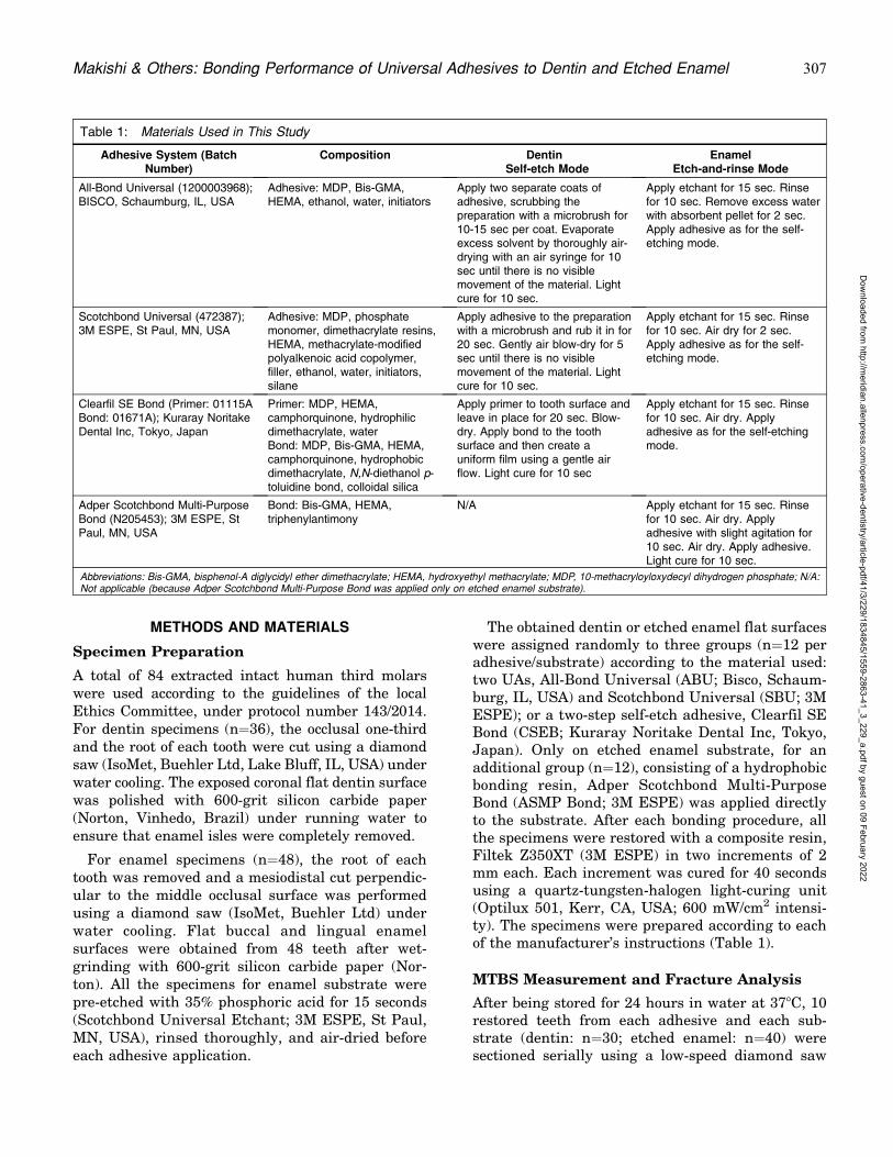
METHODS AND MATERIALS
Specimen Preparation
A total of 84 extracted intact human third molarswere used according to the guidelines of the localEthics Committee, under protocol number 143/2014.For dentin specimens (n=36), the occlusal one-thirdand the root of each tooth were cut using a diamondsaw (IsoMet, Buehler Ltd, Lake Bluff, IL, USA) underwater cooling. The exposed coronal flat dentin surfacewas polished with 600-grit silicon carbide paper(Norton, Vinhedo, Brazil) under running water toensure that enamel isles were completely removed.
For enamel specimens (n=48), the root of eachtooth was removed and a mesiodistal cut perpendic-ular to the middle occlusal surface was performedusing a diamond saw (IsoMet, Buehler Ltd) underwater cooling. Flat buccal and lingual enamelsurfaces were obtained from 48 teeth after wet-grinding with 600-grit silicon carbide paper (Nor-ton). All the specimens for enamel substrate werepre-etched with 35% phosphoric acid for 15 seconds(Scotchbond Universal Etchant; 3M ESPE, St Paul,MN, USA), rinsed thoroughly, and air-dried beforeeach adhesive application.
The obtained dentin or etched enamel flat surfaceswere assigned randomly to three groups (n=12 peradhesive/substrate) according to the material used:two UAs, All-Bond Universal (ABU; Bisco, Schaum-burg, IL, USA) and Scotchbond Universal (SBU; 3MESPE); or a two-step self-etch adhesive, Clearfil SEBond (CSEB; Kuraray Noritake Dental Inc, Tokyo,Japan). Only on etched enamel substrate, for anadditional group (n=12), consisting of a hydrophobicbonding resin, Adper Scotchbond Multi-PurposeBond (ASMP Bond; 3M ESPE) was applied directlyto the substrate. After each bonding procedure, allthe specimens were restored with a composite resin,Filtek Z350XT (3M ESPE) in two increments of 2mm each. Each increment was cured for 40 secondsusing a quartz-tungsten-halogen light-curing unit(Optilux 501, Kerr, CA, USA; 600 mW/cm2 intensi-ty). The specimens were prepared according to eachof the manufacturer’s instructions (Table 1).
MTBS Measurement and Fracture Analysis
After being stored for 24 hours in water at 378C, 10restored teeth from each adhesive and each sub-strate (dentin: n=30; etched enamel: n=40) weresectioned serially using a low-speed diamond saw
Table 1: Materials Used in This Study
Adhesive System (BatchNumber)
Composition Dentin EnamelSelf-etch Mode Etch-and-rinse Mode
All-Bond Universal (1200003968);BISCO, Schaumburg, IL, USA
Adhesive: MDP, Bis-GMA,HEMA, ethanol, water, initiators
Apply two separate coats ofadhesive, scrubbing thepreparation with a microbrush for10-15 sec per coat. Evaporateexcess solvent by thoroughly air-drying with an air syringe for 10sec until there is no visiblemovement of the material. Lightcure for 10 sec.
Apply etchant for 15 sec. Rinsefor 10 sec. Remove excess waterwith absorbent pellet for 2 sec.Apply adhesive as for the self-etching mode.
Scotchbond Universal (472387);3M ESPE, St Paul, MN, USA
Adhesive: MDP, phosphatemonomer, dimethacrylate resins,HEMA, methacrylate-modifiedpolyalkenoic acid copolymer,filler, ethanol, water, initiators,silane
Apply adhesive to the preparationwith a microbrush and rub it in for20 sec. Gently air blow-dry for 5sec until there is no visiblemovement of the material. Lightcure for 10 sec.
Apply etchant for 15 sec. Rinsefor 10 sec. Air dry for 2 sec.Apply adhesive as for the self-etching mode.
Clearfil SE Bond (Primer: 01115ABond: 01671A); Kuraray NoritakeDental Inc, Tokyo, Japan
Primer: MDP, HEMA,camphorquinone, hydrophilicdimethacrylate, waterBond: MDP, Bis-GMA, HEMA,camphorquinone, hydrophobicdimethacrylate, N,N-diethanol p-toluidine bond, colloidal silica
Apply primer to tooth surface andleave in place for 20 sec. Blow-dry. Apply bond to the toothsurface and then create auniform film using a gentle airflow. Light cure for 10 sec
Apply etchant for 15 sec. Rinsefor 10 sec. Air dry. Applyadhesive as for the self-etchingmode.
Adper Scotchbond Multi-PurposeBond (N205453); 3M ESPE, StPaul, MN, USA
Bond: Bis-GMA, HEMA,triphenylantimony
N/A Apply etchant for 15 sec. Rinsefor 10 sec. Air dry. Applyadhesive with slight agitation for10 sec. Air dry. Apply adhesive.Light cure for 10 sec.
Abbreviations: Bis-GMA, bisphenol-A diglycidyl ether dimethacrylate; HEMA, hydroxyethyl methacrylate; MDP, 10-methacryloyloxydecyl dihydrogen phosphate; N/A:Not applicable (because Adper Scotchbond Multi-Purpose Bond was applied only on etched enamel substrate).
Makishi & Others: Bonding Performance of Universal Adhesives to Dentin and Etched Enamel 307
Dow
nloaded from http://m
eridian.allenpress.com/operative-dentistry/article-pdf/41/3/229/1834845/1559-2863-41_3_229_a.pdf by guest on 09 February 2022

(IsoMet, Buehler Ltd) under water cooling toproduce parallel piped sticks (0.9 mm wide 3 0.9mm thick 3 6 mm length) with their long axisperpendicular to the bonded interface. Half thenumber of sticks from each tooth and each substratewere selected randomly for immediate testing, andthe remainder were stored for one year in distilledwater.
For the MTBS testing, the ends of the sticks werecarefully fixed with cyanoacrylate glue (ModelRepair II Blue, Sankin Industry Co, Tokyo, Japan)to a jig in a universal testing machine (EZ Test,Shimadzu, Kyoto, Japan) and subjected to a tensileforce at a crosshead speed of 1 mm/min. After thebond strength testing, the two ends of the fracturedsurfaces were mounted on brass stubs, gold-coated,and observed using scanning electron microscopy(SEM; JSM5600, JEOL Ltd, Tokyo, Japan) atmagnifications of 1003 and 10003.
For dentin specimens, the failure mode of eachbeam was determined by two experienced research-ers and then classified, by consensus, into categories:cohesive failure in composite resin (C); failurebetween composite resin and adhesive (CA); failurebetween adhesive and dentin (AD); mixed failure ofcomposite resin, adhesive, and dentin (CAD); cohe-sive failure in adhesive (A); cohesive failure inhybrid layer (HL); and cohesive failure in dentin (D).
In a similar way, for etched enamel specimens, thefailure mode of each beam was determined andclassified into cohesive failure in composite resin (C);failure between composite resin and adhesive (CA);failure between adhesive and etched enamel (AE);mixed failure of composite resin, adhesive, andetched enamel (CAE); cohesive failure in adhesive(A); failure between enamel and dentin (ED); andcohesive failure in enamel (E).
Thermocycling Procedure
In addition, two teeth from each adhesive and eachsubstrate were selected to undergo thermocycling.The specimens were fatigued with 10,000 thermo-cycles between 58C and 558C at a dwell time of 30seconds per temperature and a transfer time of 10seconds between baths (MSCT 3, Marnucci ME, SaoCarlos, Brazil).
Nanoleakage Evaluation
After the thermocycling procedure, the two teethfrom each material and each substrate were verti-cally sectioned with a diamond saw (IsoMet, BuehlerLtd) under water coolant, across the adhesive/tooth
substrate interface, into approximately 1-mm-thickslabs. Two central slabs were chosen from eachtooth, forming a total of four specimens per materialand per substrate. All the specimens were coatedwith two layers of nail varnish applied 1 mm fromthe bonded interface, followed by immersion into50% (wt/vol) ammoniacal silver nitrate solution for24 hours. Thereafter, they were rinsed thoroughlyunder running tap water and exposed to photo-developing solution for eight hours under fluorescentlight to reduce the penetration of the ammoniacalsilver nitrate into metallic silver grains. Each slabwas then wet-polished with 1200-grit silicon carbidepaper (Norton) and sonicated for five minutes toremove the superficial silver adsorption.
Following air-drying, the specimens were gold-coated and examined by SEM in back-scatteredelectron mode and energy dispersive x-ray spectros-copy (EDS; JSM5600, JEOL Ltd) at a magnificationof 20003. Interfacial images were obtained from eachspecimen (n=10). The percentage distribution ofmetallic silver particles at the adhesive/tooth sub-strate interface was calculated with a digital image-analysis software (NIH ImageJ 1.60, Scion, Freder-ick, MD, USA) in a selected area on each image at5003 (20.2 3 250 lm, height 3 width). Initial energyspectra analyses were performed to determine theelemental composition of the entire area. In addition,select surface areas were mapped for elementsincluding silver, calcium, and silicon.
Statistical Analysis
The MTBS data were statistically analyzed using athree-way analysis of variance (ANOVA) with thesignificance level defined as a = 0.05; bond strengthsto dentin or etched enamel were dependent vari-ables, and the adhesive, the storage period, and thesubstrate were factors. A Bonferroni post hoc testwith UNIANOVA syntax was used for multiplecomparisons with significant differences in bondstrength means. The nanoleakage data were statis-tically analyzed with a Kruskal-Wallis test, with thestatistical significance defined as a = 0.05. Allstatistical analyses were performed using StatisticalPackage for the Social Sciences software (SPSS forWindows, Version 16.0, SPSS Inc, Chicago, IL,USA).
RESULTS
The means and standard deviations of dentin andetched enamel MTBS values obtained in this studyare presented in Table 2. The three-way ANOVAshowed that the bond strength results for dentin and
308 Operative Dentistry
Dow
nloaded from http://m
eridian.allenpress.com/operative-dentistry/article-pdf/41/3/229/1834845/1559-2863-41_3_229_a.pdf by guest on 09 February 2022

etched enamel were influenced significantly by theadhesive used, by the storage period, and by thesubstrate. The interaction of these three factors wasnot significant (p=0.055). On the other hand,significant statistical interaction was observed be-tween adhesive material and storage period(p=0.011) and between adhesive and substrate(p,0.001), but no significant statistical interactionwas found between storage period and substrate(p=0.710).
A Bonferroni post hoc test revealed the presence ofstatistically significant differences between theMTBS results after 24 hours or one year of waterstorage for both substrates. For dentin, CSEB andSBU showed higher bond strength values than thoseof ABU after 24 hours or one year of storage.However, there was a significant decrease in MTBSvalues within all adhesives tested after one year ofstorage (p,0.05). For etched enamel, CSEB showedsignificantly higher bond strength values after 24hours; no significant difference was observed amongABU, SBU, and ASMP Bond at this time point. Afterone year of water storage, a significant decrease inbond strength was observed within all adhesivescompared with their baselines, and the bondstrength values did not differ from each other amongall adhesives (p.0.05). Using ABU, there were nosignificant MTBS differences between the dentinand etched enamel when the substrates werecompared after the same storage period. For SBU,the bonding to dentin did in fact result in asignificantly higher bond strength than etchedenamel after 24 hours. The same was true whenusing both SBU and CSEB after one year of waterstorage.
The failure modes of the tested groups aresummarized in Figure 1. Representative high mag-nification SEM micrographs of the fracture modepatterns are shown in Figure 2. For dentin, the C
failure mode predominated for SBU and CSEB atboth time points, whereas ABU showed an increasedincidence of AD mode of failure after one yearstorage. For etched enamel, a slight increase of Cand CAE mode of failure was observed in most of thematerials in the long term.
Representative images of silver-challenged speci-mens for dentin and etched enamel substrates after10,000 thermocycles are illustrated in Figures 3 and4, respectively. High magnification of SEM micro-graphs after the silver challenge revealed the exis-tence of nanoleakage formation only for dentinspecimens after 10,000 thermocycles. Images inwhich the total percentage distribution of silver tracerwithin the interface was calculated are shown inFigure 3. For the nanoleakage test, no significantstatistical difference was observed among all adhe-sives after 10,000 thermocycles (Kruskal-Wallis test,p.0.05). A distinctive silver-spotted pattern of nano-leakage formation could be recognized along theadhesive/dentin interface in all specimens, and itssilver percentage distribution means and mean rankscan be visualized in Table 3. In addition, SEM/EDSimages of the interface with silver particles are shownin Figure 5. Elemental silver was identified by EDSanalysis, confirming the results obtained.
DISCUSSION
In this study, the bonding strength of UAs wastested on dentin and etched enamel and comparedwith a conventional two-step self-etching adhesive.The MTBS test was chosen owing to its advantagesover other bond strength tests: Bonding measure-ment can be achieved for very small areas, andmultiple beams can be obtained from a singletooth.21 It also allowed us to divide the number ofbeams obtained from each specimen and to evaluatethem at two storage times.
Table 2: Mean MTBS of Different Adhesives Applied in a Self-etch Mode on Dentin Substrate and in an Etch-and-rinse Mode inEnamel Substrate
Material Dentin Substrate Etched Enamel Substrate
After 24 hWater Storage
After One yWater Storage
After 24 hWater Storage
After One yWater Storage
All-Bond Universal (ABU) 39.9 (11.9) A, a 25.1 (13.2) A, b 40.8 (5.9) A, c 30.7 (9.4) A, d
Scotchbond Universal (SBU) 63.1 (8.2) B, a* 45.9 (8.4) B, b** 40.7 (13.0) A, c* 28.4 (10.8) A, d**
Clearfil SE Bond (CSEB) 63.4 (9.2) B, a 44.1 (4.2) B, b** 59.7 (13.8) B, c 27.1 (5.7) A, d**
Adper Scotchbond Multi-Purpose Bond (ASMP Bond) N/A N/A 40.4 (7.7) A, a 24.4 (6.5) A, b
Abbreviation: N/A: Not applicable (because ASMP Bond was tested only on etched enamel substrate).a Data are presented as the mean (standard deviation) in megapascals (MPa; n = 10 teeth). Identical capital letters in a column indicate the absence of any statisticallysignificant difference. Identical lowercased letters in a row within the same substrate between after 24 h and one y water storage indicate the absence of anystatistically significant difference. Comparisons within the same material and storage period between different substrates, and marked with one asterisk for 24 h andtwo asterisks for one y water storage are statistically significant. (Analysis of variance and Bonferroni post hoc test; significance at p,0.05).
Makishi & Others: Bonding Performance of Universal Adhesives to Dentin and Etched Enamel 309
Dow
nloaded from http://m
eridian.allenpress.com/operative-dentistry/article-pdf/41/3/229/1834845/1559-2863-41_3_229_a.pdf by guest on 09 February 2022
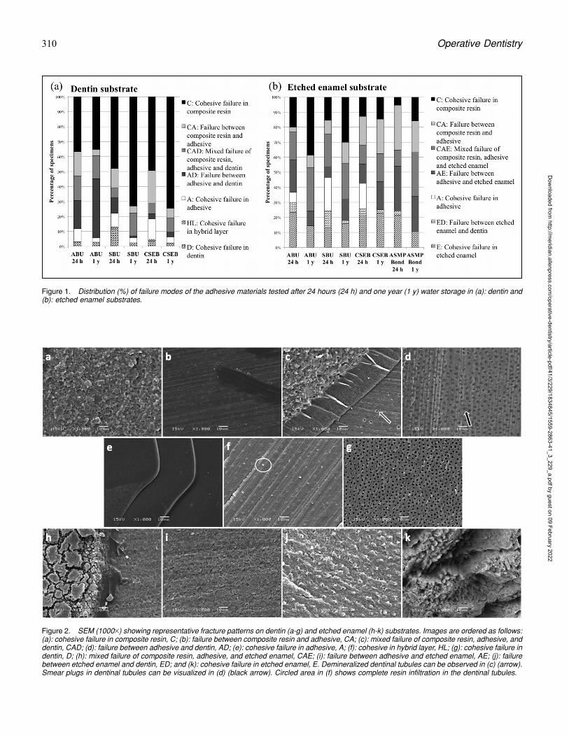
Figure 1. Distribution (%) of failure modes of the adhesive materials tested after 24 hours (24 h) and one year (1 y) water storage in (a): dentin and(b): etched enamel substrates.
Figure 2. SEM (10003) showing representative fracture patterns on dentin (a-g) and etched enamel (h-k) substrates. Images are ordered as follows:(a): cohesive failure in composite resin, C; (b): failure between composite resin and adhesive, CA; (c): mixed failure of composite resin, adhesive, anddentin, CAD; (d): failure between adhesive and dentin, AD; (e): cohesive failure in adhesive, A; (f): cohesive in hybrid layer, HL; (g): cohesive failure indentin, D; (h): mixed failure of composite resin, adhesive, and etched enamel, CAE; (i): failure between adhesive and etched enamel, AE; (j): failurebetween etched enamel and dentin, ED; and (k): cohesive failure in etched enamel, E. Demineralized dentinal tubules can be observed in (c) (arrow).Smear plugs in dentinal tubules can be visualized in (d) (black arrow). Circled area in (f) shows complete resin infiltration in the dentinal tubules.
310 Operative Dentistry
Dow
nloaded from http://m
eridian.allenpress.com/operative-dentistry/article-pdf/41/3/229/1834845/1559-2863-41_3_229_a.pdf by guest on 09 February 2022
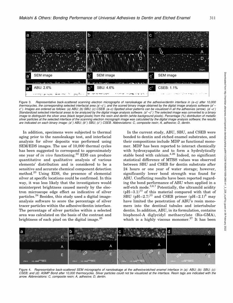
In addition, specimens were subjected to thermalaging prior to the nanoleakage test, and interfacialanalysis for silver deposits was performed usingSEM/EDS images. The use of 10,000 thermal cycleshas been suggested to correspond to approximatelyone year of in vivo functioning.22 EDS can producequantitative and qualitative analysis of variouselements’ distribution and is considered to be asensitive and accurate chemical component detectionmethod.23 Using EDS, the presence of elementalsilver at specific locations could be confirmed. In thisway, it was less likely that the investigators wouldmisinterpret brightness caused merely by the elec-tron microscope edge effect as indicative of silverparticles.24 Besides, this study used a digital image-analysis software to score the percentage of silvertracer particles within the adhesive/dentin interface.The percentage of silver particles within a selectedarea was calculated on the basis of the contrast andbrightness of each pixel on the digital image.25
In the current study, ABU, SBU, and CSEB werebonded to dentin and etched enamel substrates, andtheir compositions include MDP as functional mono-mer. MDP has been reported to interact chemicallywith hydroxyapatite and to form a hydrolyticallystable bond with calcium.4,26 Indeed, no significantstatistical difference of MTBS values was observedbetween SBU and CSEB for dentin substrate after24 hours or one year of water storage; however,significantly lower bond strength was found forABU. Conflicting results have been reported regard-ing the bond performance of ABU when applied in aself-etch mode.3,6,7 Potentially, the ultramild acidity(pH=3.1)13 of this material compared with that ofSBU (pH=2.7)13 and CSEB primer (pH=2.1)6 mayhave limited the penetration of ABU’s resin mono-mers into the dentinal tubules and intertubulardentin. In addition, ABU, in its formulation, containsbisphenol-A diglycidyl methacrylate (Bis-GMA),which is a highly viscous monomer.27 It has been
Figure 4. Representative back-scattered SEM micrographs of nanoleakage at the adhesive/etched enamel interface in (a): ABU; (b): SBU; (c):CSEB; and (d): ASMP Bond after 10,000 thermocycles. Silver particles could not be visualized at the interface. Resin tags are indicated with thearrow. Abbreviations: C, composite resin; A, adhesive; D, dentin.
Figure 3. Representative back-scattered scanning electron micrographs of nanoleakage at the adhesive/dentin interface in (a–c) after 10,000thermocycles, the corresponding selected interfacial area (a0–c0), and the scored binary image obtained by the digital image analysis software (a 00–c 00). Images are ordered as follows: (a) ABU; (b) SBU; (c) CSEB. (a–c) Spotted silver patterns can be visualized in all the adhesives (arrow). (a0–c0)Standardized selected interfacial areas to be analyzed by the digital image analysis software. (a 00–c 00) The selected image was converted to a binaryimage to distinguish the silver area (black target pixels) from the resin and dentin (white background pixels). Percentage (%) distribution of metallicsilver particles at the selected interface of the scanning electron micrograph image was calculated by the digital image analysis software; the resultsare indicated on each binary image: (a 00) ABU; (b 00) SBU; (c 00) CSEB. Abbreviations: C, composite resin; A, adhesive; D, dentin.
Makishi & Others: Bonding Performance of Universal Adhesives to Dentin and Etched Enamel 311
Dow
nloaded from http://m
eridian.allenpress.com/operative-dentistry/article-pdf/41/3/229/1834845/1559-2863-41_3_229_a.pdf by guest on 09 February 2022
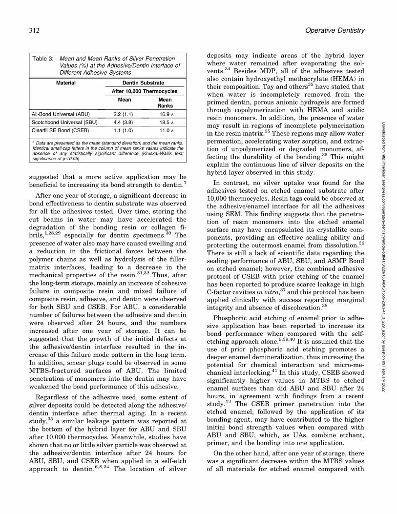
suggested that a more active application may bebeneficial to increasing its bond strength to dentin.7
After one year of storage, a significant decrease inbond effectiveness to dentin substrate was observedfor all the adhesives tested. Over time, storing thecut beams in water may have accelerated thedegradation of the bonding resin or collagen fi-brils,1,28,29 especially for dentin specimens.30 Thepresence of water also may have caused swelling anda reduction in the frictional forces between thepolymer chains as well as hydrolysis of the filler-matrix interfaces, leading to a decrease in themechanical properties of the resin.31,32 Thus, afterthe long-term storage, mainly an increase of cohesivefailure in composite resin and mixed failure ofcomposite resin, adhesive, and dentin were observedfor both SBU and CSEB. For ABU, a considerablenumber of failures between the adhesive and dentinwere observed after 24 hours, and the numbersincreased after one year of storage. It can besuggested that the growth of the initial defects atthe adhesive/dentin interface resulted in the in-crease of this failure mode pattern in the long term.In addition, smear plugs could be observed in someMTBS-fractured surfaces of ABU. The limitedpenetration of monomers into the dentin may haveweakened the bond performance of this adhesive.
Regardless of the adhesive used, some extent ofsilver deposits could be detected along the adhesive/dentin interface after thermal aging. In a recentstudy,33 a similar leakage pattern was reported atthe bottom of the hybrid layer for ABU and SBUafter 10,000 thermocycles. Meanwhile, studies haveshown that no or little silver particle was observed atthe adhesive/dentin interface after 24 hours forABU, SBU, and CSEB when applied in a self-etchapproach to dentin.6,8,24 The location of silver
deposits may indicate areas of the hybrid layerwhere water remained after evaporating the sol-vents.34 Besides MDP, all of the adhesives testedalso contain hydroxyethyl methacrylate (HEMA) intheir composition. Tay and others35 have stated thatwhen water is incompletely removed from theprimed dentin, porous anionic hydrogels are formedthrough copolymerization with HEMA and acidicresin monomers. In addition, the presence of watermay result in regions of incomplete polymerizationin the resin matrix.35 These regions may allow waterpermeation, accelerating water sorption, and extrac-tion of unpolymerized or degraded monomers, af-fecting the durability of the bonding.35 This mightexplain the continuous line of silver deposits on thehybrid layer observed in this study.
In contrast, no silver uptake was found for theadhesives tested on etched enamel substrate after10,000 thermocycles. Resin tags could be observed atthe adhesive/enamel interface for all the adhesivesusing SEM. This finding suggests that the penetra-tion of resin monomers into the etched enamelsurface may have encapsulated its crystallite com-ponents, providing an effective sealing ability andprotecting the outermost enamel from dissolution.36
There is still a lack of scientific data regarding thesealing performance of ABU, SBU, and ASMP Bondon etched enamel; however, the combined adhesiveprotocol of CSEB with prior etching of the enamelhas been reported to produce scarce leakage in highC-factor cavities in vitro,37 and this protocol has beenapplied clinically with success regarding marginalintegrity and absence of discoloration.38
Phosphoric acid etching of enamel prior to adhe-sive application has been reported to increase itsbond performance when compared with the self-etching approach alone.9,39,40 It is assumed that theuse of prior phosphoric acid etching promotes adeeper enamel demineralization, thus increasing thepotential for chemical interaction and micro-me-chanical interlocking.41 In this study, CSEB showedsignificantly higher values in MTBS to etchedenamel surfaces than did ABU and SBU after 24hours, in agreement with findings from a recentstudy.12 The CSEB primer penetration into theetched enamel, followed by the application of itsbonding agent, may have contributed to the higherinitial bond strength values when compared withABU and SBU, which, as UAs, combine etchant,primer, and the bonding into one application.
On the other hand, after one year of storage, therewas a significant decrease within the MTBS valuesof all materials for etched enamel compared with
Table 3: Mean and Mean Ranks of Silver PenetrationValues (%) at the Adhesive/Dentin Interface ofDifferent Adhesive Systems
Material Dentin Substrate
After 10,000 Thermocycles
Mean MeanRanks
All-Bond Universal (ABU) 2.2 (1.1) 16.9 A
Scotchbond Universal (SBU) 4.4 (3.8) 18.5 A
Clearfil SE Bond (CSEB) 1.1 (1.0) 11.0 A
a Data are presented as the mean (standard deviation) and the mean ranks.Identical small-cap letters in the column of mean ranks values indicate theabsence of any statistically significant difference (Kruskal-Wallis test;significance at p,0.05).
312 Operative Dentistry
Dow
nloaded from http://m
eridian.allenpress.com/operative-dentistry/article-pdf/41/3/229/1834845/1559-2863-41_3_229_a.pdf by guest on 09 February 2022

their baseline. It has been reported that the mixture
of the hydrophilic monomers contained in the primer
with the bonding agent may compromise the dura-
bility of the bond.42-45 In addition, Miyazaki and
others45 observed a small, statistically insignificant
decrease in the shear bond strength of self-priming
adhesive systems to enamel after 30,000 thermo-
cycles. Conversely, in the current study, storing the
MTBS cut beams in water may be considered a form
of accelerated aging.1 Therefore, as with dentin, the
collagen fibrils may have degraded and/or the
mechanical properties of the resin may have de-
Figure 5. Energy dispersive x-ray spectroscopy of the same specimens shown in Figure 3. (a): ABU; (b): SBU; and (c): CSEB. A distinct silver peakwas observed on the elemental energy spectra (arrow). A map scan (white square) of the same specimen detected metallic silver particles.
Makishi & Others: Bonding Performance of Universal Adhesives to Dentin and Etched Enamel 313
Dow
nloaded from http://m
eridian.allenpress.com/operative-dentistry/article-pdf/41/3/229/1834845/1559-2863-41_3_229_a.pdf by guest on 09 February 2022
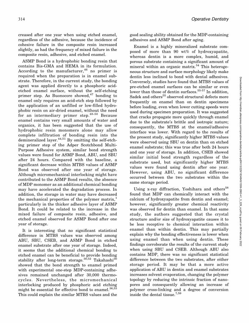
creased after one year when using etched enamel,regardless of the adhesive, because the incidence ofcohesive failure in the composite resin increasedslightly, as had the frequency of mixed failure in thecomposite resin, adhesive, and etched enamel.
ASMP Bond is a hydrophobic bonding resin thatcontains Bis-GMA and HEMA in its formulation.According to the manufacturer,46 no primer isrequired when the preparation is in enamel sub-strate. Therefore, in the current study, the bondingagent was applied directly to a phosphoric acid-etched enamel surface, without the self-etchingprimer step. As Buonocore showed,47 bonding toenamel only requires an acid-etch step followed bythe application of an unfilled or low-filled hydro-phobic resin on air-dried enamel, without the needfor an intermediary primer step.48-50 Becauseenamel contains very small amounts of water andorganics, it has been suggested that the use ofhydrophobic resin monomers alone may allowcomplete infiltration of bonding resin into thedemineralized layer.44,51 By omitting the self-etch-ing primer step of the Adper Scotchbond Multi-Purpose Adhesive system, similar bond strengthvalues were found for ASMP Bond, ABU, and SBUafter 24 hours. Compared with the baseline, asignificant decrease within MTBS values of ASMPBond was observed after one year of storage.Although micromechanical interlocking might havecontributed to the ASMP Bond results, the absenceof MDP monomer as an additional chemical bondingmay have accelerated the degradation process. Inaddition, the storage in water may have decreasedthe mechanical properties of the polymer matrix,1
particularly in the thicker adhesive layer of ASMPBond. It could be related to the increase in themixed failure of composite resin, adhesive, andetched enamel observed for ASMP Bond after oneyear of storage.
It is interesting that no significant statisticaldifference in MTBS values was observed amongABU, SBU, CSEB, and ASMP Bond in etchedenamel substrate after one year of storage. Indeed,it seems that the additional chemical bonding toetched enamel can be beneficial to provide bondingstability after long-term storage.30,52 Takahashi30
showed that the bond strength to enamel primedwith experimental one-step MDP-containing adhe-sives remained unchanged after 30,000 thermo-cycles. Nevertheless, the micromechanicalinterlocking produced by phosphoric acid etchingmight be essential for effective bond to enamel.36,53
This could explain the similar MTBS values and the
good sealing ability obtained for the MDP-containingadhesives and ASMP Bond after aging.
Enamel is a highly mineralized substrate com-posed of more than 90 wt% of hydroxyapatite,whereas dentin is a more complex, humid, andporous substrate containing a significant amount ofmineral within an organic matrix.54 This heteroge-neous structure and surface morphology likely makedentin less inclined to bond with dental adhesives.Conversely, studies have found that MTBS values ofpre-etched enamel surfaces can be similar or evenlower than those of dentin surfaces.55-57 In addition,Sadek and others55 observed structural defects morefrequently on enamel than on dentin specimensbefore loading, even when lower cutting speeds wereused during specimen preparation. It was suggestedthat cracks propagate more quickly through enameldue to the substrate’s brittle and isotropic nature;consequently, the MTBS at the enamel/adhesiveinterface was lower. With regard to the results ofthe present study, significantly higher MTBS valueswere observed using SBU on dentin than on etchedenamel substrate; this was true after both 24 hoursand one year of storage. In addition, CSEB showedsimilar initial bond strength regardless of thesubstrate used, but significantly higher MTBSvalues were found using dentin after one year.However, using ABU, no significant differenceoccurred between the two substrates within thesame storage period.
Using x-ray diffraction, Yoshihara and others58
found that MDP can chemically interact with thecalcium of hydroxyapatite from dentin and enamel;however, significantly greater chemical reactivitywas observed for dentin than enamel. In that samestudy, the authors suggested that the crystalstructure and/or size of hydroxyapatite causes it tobe less receptive to chemical interaction withinenamel than within dentin. This may partiallyexplain why the bonding effectiveness is lower whenusing enamel than when using dentin. Thesefindings corroborate the results of the current studywhen using SBU and CSEB. Although ABU alsocontains MDP, there was no significant statisticaldifference between the two substrates, after eitherstorage period. It may be that a more activeapplication of ABU in dentin and enamel substratesincreases solvent evaporation, changing the polymertopology by reducing the intrinsic fraction of nano-pores and consequently allowing an increase ofpolymer cross-linking and a degree of conversioninside the dental tissue.7,59
314 Operative Dentistry
Dow
nloaded from http://m
eridian.allenpress.com/operative-dentistry/article-pdf/41/3/229/1834845/1559-2863-41_3_229_a.pdf by guest on 09 February 2022

In clinical use, UAs can be considered a goodalternative with regard to user friendliness andtheir applicability to different substrates: enamel,dentin, alloys, ceramics, and composites. In a 36-month follow-up clinical report, a UA seemed tomaintain good performance when used with non-carious cervical lesions by applying either the etch-and-rinse (wet and dry) strategy to dentin or theself-etch strategy (with or without selective etching)to enamel. However, signs of bonding degradationsuch as marginal staining were reported when theUA was applied in a self-etch mode withoutselective enamel etching.60 That said, applyingUAs to dentin using the etch-and-rinse strategy iscontroversial.8 Indeed, on the basis of our results, itmay be that UAs applied to dentin using a self-etchapproach with selective enamel etching is morefavorable. Further investigations are required re-garding the interaction of UAs with differentstructures, such as caries-affected dentin andsclerotic dentin because alterations in the mineralcontent and structure of dentin may compromisethe bond,61 as well as the clinical stability of thispromising new category of adhesives.
Within the limitations of this study, the MTBSvalues revealed statistically significant differencesamong adhesives and/or storage time for dentin andetched enamel substrates. Therefore, the first nullhypothesis has to be rejected. On the other hand, alladhesives showed similar nanoleakage formationalong the adhesive/dentin interface, and no silverpresence was detected in etched enamel substrategroups after the artificial aging. Thus, the secondnull hypothesis has to be accepted.
CONCLUSIONS
In the short term, the MTBS values were adhesiveand substrate dependent. After aging, the bondingeffectiveness of both UAs and the conventionaladhesive systems were equal on etched enamelsubstrate, without leakage; however, UAs may havea bonding performance that is equal or inferior tothat of the two-step self-etching adhesive when usingdentin as a substrate, with similar amounts of silverdeposits at their interfaces.
Acknowledgements
This research was supported by the Brazilian NationalCouncil for Scientific and Technological Development (CNPqNo. 307217-2014-0). The funding body had no role in studydesign; collection, analysis, and interpretation of data; writingof the report; or in the decision to submit the article forpublication.
Regulatory Statement
This study was conducted in accordance with all theprovisions of the local human subjects oversight committeeguidelines and policies of Piracicaba Dental School, StateUniversity of Campinas. The approval code for this study is143/2014.
Conflict of Interest
The authors of this manuscript certify that they have noproprietary, financial, or other personal interest of any natureor kind in any product, service, and/or company that ispresented in this article.
(Accepted 10 September 2015)
References
1. De Munck J, Van Landuyt K, Peumans M, Poitevin A,Lambrechts P, Braem M, & Van Meerbeek B (2005) Acritical review of the durability of adhesion to tooth tissue:Methods and results Journal of Dental Research 84(2)118-132.
2. Moszner N, Salz U, & Zimmermann J (2005) Chemicalaspects of self-etching enamel-dentin adhesives: A sys-tematic review Dental Materials 21(10) 895-910.
3. Wagner A, Wendler M, Petschelt A, Belli R, & LohbauerU (2014) Bonding performance of universal adhesives indifferent etching modes Journal of Dentistry 42(7)800-807.
4. Yoshida Y, Nagakane K, Fukuda R, Nakayama Y,Okazaki M, Shintani H, Inoue S, Tagawa Y, Suzuki K,De Munck J, & Van Meerbeek B (2004) Comparativestudy on adhesive performance of functional monomersJournal of Dental Research 83(6) 454-458.
5. Peumans M, Munck J, Van Landuyt K, Lambrechts P, &Van Meerbeek B (2005) Three-year clinical effectivenessof a two-step self-etch adhesive in cervical lesionsEuropean Journal of Oral Sciences 113(6) 512-518.
6. Munoz MA, Luque I, Hass V, Reis A, Loguercio AD, &Bombarda NH (2013) Immediate bonding properties ofuniversal adhesives to dentine Journal of Dentistry 41(5)404-411.
7. Munoz MA, Sezinando A, Luque-Martinez I, Szesz AL,Reis A, Loguercio AD, Bombarda NH, & Perdigao J (2014)Influence of a hydrophobic resin coating on the bondingefficacy of three universal adhesives Journal of Dentistry42(5) 595-602.
8. Marchesi G, Frassetto A, Mazzoni A, Apolonio F, DiolosaM, Cadenaro M, Di Lenarda R, Pashley DH, Tay F, &Breschi L (2014) Adhesive performance of a multi-modeadhesive system: 1-year in vitro study Journal ofDentistry 42(5) 603-612.
9. Erickson RL, Barkmeier WW, & Kimmes NS (2009) Bondstrength of self-etch adhesives to pre-etched enamelDental Materials 25(10) 1187-1194.
10. Hanabusa M, Mine A, Kuboki T, Momoi Y, Van Ende A,Van Meerbeek B, & De Munck J (2012) Bondingeffectiveness of a new ‘‘multi-mode’’ adhesive to enameland dentine Journal of Dentistry 40(6) 475-484.
Makishi & Others: Bonding Performance of Universal Adhesives to Dentin and Etched Enamel 315
Dow
nloaded from http://m
eridian.allenpress.com/operative-dentistry/article-pdf/41/3/229/1834845/1559-2863-41_3_229_a.pdf by guest on 09 February 2022
11. De Goes MF, Shinohara MS, & Freitas MS (2014)Performance of a new one-step multi-mode adhesive onetched vs non-etched enamel on bond strength andinterfacial morphology Journal of Adhesive Dentistry16(3) 243-250.
12. McLean DE, Meyers E, Guillory V, & Vandewalle K(2015) Enamel bond strength of new universal adhesivebonding agents Operative Dentistry 40(4) 410-417.
13. Rosa WL, Piva E, & Silva AF (2015) Bond strength ofuniversal adhesives: A systematic review and meta-analysis Journal of Dentistry 43(7) 765-776.
14. Sano H, Takatsu T, Ciucchi B, Horner JA, Matthews WG,& Pashley DH (1995) Nanoleakage: Leakage within thehybrid layer Operative Dentistry 20(1) 18-25.
15. Li H, Burrow MF, & Tyas MJ (2000) Nanoleakagepatterns of four dentin bonding systems Dental Materials16(1) 48-56.
16. Hariri I, Shimada Y, Sadr A, Ichinose S, & Tagami J(2012) The effects of aging on shear bond strength andnanoleakage expression of an etch-and-rinse adhesive onhuman enamel and dentin Journal of Adhesive Dentistry14(3) 235-243.
17. Sano H, Yoshiyama M, Ebisu S, Burrow MF, Takatsu T,Ciucchi B, Carvalho R, & Pashley DH (1995) ComparativeSEM and TEM observations of nanoleakage within hybridlayer Operative Dentistry 20(4) 160-167.
18. Pioch T, Staehle HJ, Duschner H, & Garcıa-Godoy F(2001) Nanoleakage at the composite-dentin interface: Areview American Journal of Dentistry 14(4) 252-258.
19. Makishi P, Thitthaweerat S, Sadr A, Shimada Y, MartinsAL, Tagami J, & Giannini M (2015) Assessment ofcurrent adhesives in class I cavity: Nondestructiveimaging using optical coherence tomography and micro-tensile bond strength Dental Materials 31(9) e190-e200.
20. Heintze SD, Rousson V, & Mahn E (2015) Bond strengthtests of dental adhesive systems and their correlationwith the clinical results—A meta-analysis Dental Mate-rials 31(4) 423-434.
21. Pashley DH, Sano H, Ciucchi B, Yoshiyama M, &Carvalho RM (1995) Adhesion testing of dentin bondingagents: A review Dental Materials 11(2) 117-125.
22. Gale MS, & Darvell BW (1999) Thermal cycling proce-dures for laboratory testing of dental restorations Journalof Dentistry 27(2) 89-99.
23. Hashimoto M, De Munck J, Ito S, Sano H, Kaga M,Oguchi H, Van Meerbeek B, & Pashley DH (2004) In vitroeffect of nanoleakage expression on resin-dentin bondstrengths analyzed by microtensile bond test, SEM/EDXand TEM Biomaterials 25(25) 5565-5574.
24. Yuan Y, Shimada Y, Ichinose S, & Tagami J (2007)Qualitative analysis of adhesive interface nanoleakageusing FE-SEM/EDS Dental Materials 23(5) 561-569.
25. Makishi P, Shimada Y, Sadr A, Wei S, Ichinose S, &Tagami J (2010) Nanoleakage expression and microshearbond strength in the resin cement/dentin interfaceJournal of Adhesive Dentistry 12(5) 393-401.
26. Inoue S, Koshiro K, Yoshida Y, De Munck J, Nagakane K,Suzuki K, Sano H, & Van Meerbeek B (2005) Hydrolytic
stability of self-etch adhesives bonded to dentin Journalof Dental Research 84(12) 1160-1164.
27. Tay FR, Pashley DH, Kapur RR, Carrilho MR, Hur YB,Garrett LV, & Tay KC (2007) Bonding BisGMA todentin—A proof of concept for hydrophobic dentinbonding Journal of Dental Research 86(11) 1034-1039.
28. Feitosa VP, Leme AA, Sauro S, Correr-Sobrinho L,Watson TF, Sinhoreti MA, & Correr AB (2012) Hydrolyticdegradation of the resin-dentine interface induced by thesimulated pulpal pressure, direct and indirect waterageing Journal of Dentistry 40(12) 1134-1143.
29. Reis AF, Giannini M, & Pereira PN (2008) Effects of aperipheral enamel bond on the long-term effectiveness ofdentin bonding agents exposed to water in vitro Journalof Biomedical Materials Research Part B: AppliedBiomaterials 85(1) 10-17.
30. Takahashi H (2014) Effect of calcium salt of 10-meth-acryloyloxydecyl dihydrogen phosphate produced on thebond durability of one-step self-etch adhesive DentalMaterials Journal 33(3) 394-401.
31. Ferracane JL, Berge HX, & Condon JR (1998) In vitroaging of dental composites in water—Effect of degree ofconversion, filler volume, and filler/matrix couplingJournal of Biomedical Materials Research 42(3) 465-472.
32. Takeshige F, Kawakami Y, Hayashi M, & Ebisu S (2007)Fatigue behavior of resin composites in aqueous environ-ments Dental Materials 23(7) 893-899.
33. Chen C, Niu LN, Xie H, Zhang ZY, Zhou LQ, Jiao K, ChenJH, Pashley DH, & Tay FR (2015) Bonding of universaladhesives to dentine—Old wine in new bottles? Journal ofDentistry 43(5) 525-536.
34. Luque-Martinez IV, Perdigao J, Munoz MA, Sezinando A,Reis A, & Loguercio AD (2014) Effects of solventevaporation time on immediate adhesive properties ofuniversal adhesives to dentin Dental Materials 30(10)1126-1135.
35. Tay FR, King NM, Chan KM, & Pashley DH (2002) Howcan nanoleakage occur in self-etching adhesive systemsthat demineralize and infiltrate simultaneously? Journalof Adhesive Dentistry 4(4) 255-269.
36. Gwinnett AJ, & Matsui A (1967) A study of enameladhesives. The physical relationship between enamel andadhesive Archives of Oral Biology 12(12) 1615-1620.
37. Perdigao J, Monteiro P, & Gomes G (2009) In vitroenamel sealing of self-etch adhesives Quintessence Inter-national 40(3) 225-233.
38. Peumans M, De Munck J, Van Landuyt K, & VanMeerbeek B (2015) Thirteen-year randomized controlledclinical trial of a two-step self-etch adhesive in non-carious cervical lesions Dental Materials 31(3) 308-314.
39. Van Landuyt KL, Kanumilli P, De Munck J, Peumans M,Lambrechts P, & Van Meerbeek B (2006) Bond strengthof a mild self-etch adhesive with and without prior acid-etching Journal of Dentistry 34(1) 77-85.
40. Giannini M, Makishi P, Ayres AP, Vermelho PM, FronzaBM, Nikaido T, & Tagami J (2015) Self-etch adhesivesystems: A literature review Brazilian Dental Journal26(1) 3-10.
316 Operative Dentistry
Dow
nloaded from http://m
eridian.allenpress.com/operative-dentistry/article-pdf/41/3/229/1834845/1559-2863-41_3_229_a.pdf by guest on 09 February 2022

41. Van Meerbeek B, Yoshihara K, Yoshida Y, Mine A, DeMunck J, & Van Landuyt KL (2011) State of the art ofself-etch adhesives Dental Materials 27(1) 17-28.
42. Sadr A, Shimada Y, & Tagami J (2007) Effects of solventdrying time on micro-shear bond strength and mechanicalproperties of two self-etching adhesive systems DentalMaterials 23(9) 1114-1119.
43. Reis AF, Carrilho MR, Ghaname E, Pereira PN, GianniniM, Nikaido T, & Tagami J (2010) Effects of water-storageon the physical and ultramorphological features ofadhesives and primer/adhesive mixtures Dental Materi-als Journal 29(6) 697-705.
44. Rathke A, Ostermeier V, Muche R, & Haller B (2013)Reconsidering the double etching of enamel: Do self-etching primers contaminate phosphoric acid-etchedenamel? Journal of Adhesive Dentistry 15(2) 107-114.
45. Miyazaki M, Sato M, & Onose H (2000) Durability ofenamel bond strength of simplified bonding systemsOperative Dentistry 25(2) 75-80.
46. 3M ESPE Adper Scotchbond Multi-purpose Adhesive.Instruction sheet. Adper Scotchbond Multi-purpose Adhe-sive Instructions for Use. Retrieved online September 2,2015 from: http://www.3m.com/3M/en_US/Dental/Products/Catalog/;/Adper-Scotchbond-Multi-Purpose-Adhesive?N=5144788þ3294768970þ3294857497&rt=rud
47. Buonocore MG (1955) A simple method of increasing theadhesion of acryl filling materials to enamel surfacesJournal Dental Research 34(6) 849-853.
48. Buonocore MG, Matsui A, & Gwinnett AJ (1968)Penetration of resin dental materials into enamelsurfaces with reference to bonding Archives of OralBiology 13(1) 61-70.
49. Van Meerbeek B, De Munck J, Yoshida Y, Inoue S,Vargas M, Vijay P, Van Landuyt K, Lambrechts P, &Vanherle G (2003) Buonocore Memorial Lecture. Adhe-sion to enamel and dentin: Current status and futurechallenges Operative Dentistry 28(3) 215-235.
50. Van Meerbeek B, Vargas M, Inoue S, Yoshida Y,Peumans M, Lambrechts P, & Vanherle G (2001)Adhesives and cements to promote preservation dentistryOperative Dentistry 32(Supplement 6) 119-144.
51. Li N, Nikaido T, Alireza S, Takagaki T, Chen JH, &Tagami J (2013) Phosphoric acid-etching promotes bondstrength and formation of acid-base resistant zone onenamel Operative Dentistry 38(1) 82-90.
52. Zhang Z, Wang X, Zhang L, Liang B, Tang T, Fu B, &Hannig M (2013) The contribution of chemical bonding tothe short- and long-term enamel bond strengths DentalMaterials 29(7) e103-e112.
53. Erickson RL, Barkmeier WW, & Latta MA (2009) The roleof etching in bonding to enamel: A comparison of self-etching and etch-and-rinse adhesive systems DentalMaterials 25(11) 1459-1467.
54. Teruel J de D, Alcolea A, Hernandez A, & Ruiz AJ (2015)Comparison of chemical composition of enamel anddentine in human, bovine, porcine, and ovine teethArchives of Oral Biology 60(5) 768-775.
55. Sadek FT, Cury AH, Monticelli F, Ferrari M, & CardosoPE (2005) The influence of the cutting speed on bondstrength and integrity of microtensile specimens DentalMaterials 21(12) 1144-1149.
56. Goracci C, Sadek FT, Monticelli F, Cardoso PE, & FerrariM (2004) Influence of substrate, shape, and thickness onmicrotensile specimens’ structural integrity and theirmeasured bond strengths Dental Materials 20(7)643-654.
57. Cardoso PE, Sadek FT, Goracci C, & Ferrari M (2002)Adhesion testing with the microtensile method: Effects ofdental substrate and adhesive system on bond strengthmeasurements Journal of Adhesive Dentistry 4(4) 291-297.
58. Yoshihara K, Yoshida Y, Hayakawa S, Nagaoka N, IrieM, Ogawa T, Van Landuyt KL, Osaka A, Suzuki K,Minagi S, & Van Meerbeek B (2011) Nanolayering ofphosphoric acid ester monomer on enamel and dentinActa Biomaterialia 7(8) 3187-3195.
59. Loguercio AD, Munoz MA, Luque-Martinez I, Hass V,Reis A, & Perdigao J (2015) Does active application ofuniversal adhesives to enamel in self-etch mode improvetheir performance? Journal of Dentistry 43(9)1060-1070.
60. Loguercio AD, de Paula EA, Hass V, Luque-Martinez I,Reis A, & Perdigao J (2015) A new universal simplifiedadhesive: 36-month randomized double-blind clinical trialJournal of Dentistry 43(9) 1083-1092.
61. Cardoso MV, de Almeida Neves A, Mine A, Coutinho E,Van Landuyt K, De Munck J, & Van Meerbeek B (2011)Current aspects on bonding effectiveness and stability inadhesive dentistry Australian Dental Journal56(Supplement 1) 31-44.
Makishi & Others: Bonding Performance of Universal Adhesives to Dentin and Etched Enamel 317
Dow
nloaded from http://m
eridian.allenpress.com/operative-dentistry/article-pdf/41/3/229/1834845/1559-2863-41_3_229_a.pdf by guest on 09 February 2022

Bleaching Effects on Color,Chemical, and Mechanical
Properties of White Spot Lesions
Y Kim � HH Son � K YiJS Ahn � J Chang
Clinical Relevance
Bleaching initial enamel caries lesions with 10% carbamide peroxide can camouflage whitespot lesions without affecting the chemical and mechanical properties of the enamel.Application of casein phosphopeptide–amorphous calcium phosphate is a possiblesupportive treatment to promote remineralization of caries lesions.
SUMMARY
Objective: The purpose of the study was toevaluate the effect of bleaching on teeth withwhite spot lesions.
Methods and Materials: Carious lesions withstandardized whiteness were produced on thebuccal and lingual surfaces of human premo-lars by pH cycling. Specimens were subjectedto four experimental conditions (n=20/group)
as follows: group 1, control; group 2, cariesformation followed by remineralization usingfluoride-containing casein phosphopeptide–amorphous calcium phosphate (CPP-ACP;Tooth Mousse Plus, GC, Tokyo, Japan); group3, caries formation followed by bleaching using10% carbamide peroxide; and group 4, cariesformation followed by both bleaching andremineralization. The CIE L*a*b* color valueswere measured with a spectroradiometer, themineral content was measured with electronprobe microanalysis (EPMA) on the cross-sec-tional surface of each specimen, and the Knoophardness test was carried out along the EPMAscan line. Two-way analysis of variance wasperformed with Tukey post hoc comparison.
Results: The change in the CIE color valueswas not significantly different between thecaries-formed (DE*=7.03) and the bleachedenamel (DE*=7.60). Bleaching of the cariousenamel extended the whiteness (DE*=3.38)without additional mineral loss (p,0.05). Theremineralization treatment significantly in-creased the calcium (Ca), phosphate (P), andfluoride content of the subsurface lesion area(p,0.05). The cross-sectional microhardness
Younju Kim, Seoul National University, Department ofConservative Dentistry and Dental Research Institute,School of Dentistry, Seoul, Republic of Korea
Ho-Hyun Son, Seoul National University, Department ofConservative Dentistry and Dental Research Institute,School of Dentistry, Seoul, Republic of Korea
Keewook Yi, Korea Basic Science Institute, Daejeon, Republicof Korea
Jin-Soo Ahn, Seoul National University, Department ofDental Biomaterials Science and Dental Research Institute,School of Dentistry, Seoul, Republic of Korea
*Juhea Chang, Seoul National University Dental Hospital,Special Care Clinic, Seoul, Republic of Korea
*Corresponding author: 275-1 Yeongun-Dong, Seoul, 110,Republic of Korea; e-mail: [email protected]
DOI: 10.2341/15-015-L
�Operative Dentistry, 2016, 41-3, 318-326
Dow
nloaded from http://m
eridian.allenpress.com/operative-dentistry/article-pdf/41/3/229/1834845/1559-2863-41_3_229_a.pdf by guest on 09 February 2022
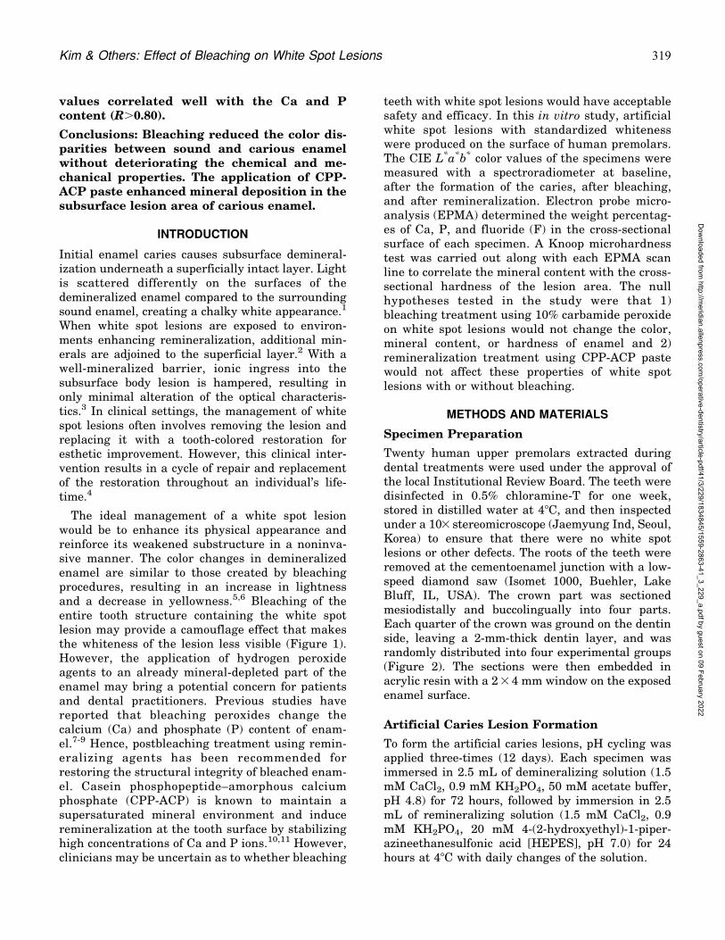
values correlated well with the Ca and Pcontent (R.0.80).
Conclusions: Bleaching reduced the color dis-parities between sound and carious enamelwithout deteriorating the chemical and me-chanical properties. The application of CPP-ACP paste enhanced mineral deposition in thesubsurface lesion area of carious enamel.
INTRODUCTION
Initial enamel caries causes subsurface demineral-ization underneath a superficially intact layer. Lightis scattered differently on the surfaces of thedemineralized enamel compared to the surroundingsound enamel, creating a chalky white appearance.1
When white spot lesions are exposed to environ-ments enhancing remineralization, additional min-erals are adjoined to the superficial layer.2 With awell-mineralized barrier, ionic ingress into thesubsurface body lesion is hampered, resulting inonly minimal alteration of the optical characteris-tics.3 In clinical settings, the management of whitespot lesions often involves removing the lesion andreplacing it with a tooth-colored restoration foresthetic improvement. However, this clinical inter-vention results in a cycle of repair and replacementof the restoration throughout an individual’s life-time.4
The ideal management of a white spot lesionwould be to enhance its physical appearance andreinforce its weakened substructure in a noninva-sive manner. The color changes in demineralizedenamel are similar to those created by bleachingprocedures, resulting in an increase in lightnessand a decrease in yellowness.5,6 Bleaching of theentire tooth structure containing the white spotlesion may provide a camouflage effect that makesthe whiteness of the lesion less visible (Figure 1).However, the application of hydrogen peroxideagents to an already mineral-depleted part of theenamel may bring a potential concern for patientsand dental practitioners. Previous studies havereported that bleaching peroxides change thecalcium (Ca) and phosphate (P) content of enam-el.7-9 Hence, postbleaching treatment using remin-eralizing agents has been recommended forrestoring the structural integrity of bleached enam-el. Casein phosphopeptide–amorphous calciumphosphate (CPP-ACP) is known to maintain asupersaturated mineral environment and induceremineralization at the tooth surface by stabilizinghigh concentrations of Ca and P ions.10,11 However,clinicians may be uncertain as to whether bleaching
teeth with white spot lesions would have acceptablesafety and efficacy. In this in vitro study, artificialwhite spot lesions with standardized whitenesswere produced on the surface of human premolars.The CIE L*a*b* color values of the specimens weremeasured with a spectroradiometer at baseline,after the formation of the caries, after bleaching,and after remineralization. Electron probe micro-analysis (EPMA) determined the weight percentag-es of Ca, P, and fluoride (F) in the cross-sectionalsurface of each specimen. A Knoop microhardnesstest was carried out along with each EPMA scanline to correlate the mineral content with the cross-sectional hardness of the lesion area. The nullhypotheses tested in the study were that 1)bleaching treatment using 10% carbamide peroxideon white spot lesions would not change the color,mineral content, or hardness of enamel and 2)remineralization treatment using CPP-ACP pastewould not affect these properties of white spotlesions with or without bleaching.
METHODS AND MATERIALS
Specimen Preparation
Twenty human upper premolars extracted duringdental treatments were used under the approval ofthe local Institutional Review Board. The teeth weredisinfected in 0.5% chloramine-T for one week,stored in distilled water at 48C, and then inspectedunder a 103 stereomicroscope (Jaemyung Ind, Seoul,Korea) to ensure that there were no white spotlesions or other defects. The roots of the teeth wereremoved at the cementoenamel junction with a low-speed diamond saw (Isomet 1000, Buehler, LakeBluff, IL, USA). The crown part was sectionedmesiodistally and buccolingually into four parts.Each quarter of the crown was ground on the dentinside, leaving a 2-mm-thick dentin layer, and wasrandomly distributed into four experimental groups(Figure 2). The sections were then embedded inacrylic resin with a 2 3 4 mm window on the exposedenamel surface.
Artificial Caries Lesion Formation
To form the artificial caries lesions, pH cycling wasapplied three-times (12 days). Each specimen wasimmersed in 2.5 mL of demineralizing solution (1.5mM CaCl2, 0.9 mM KH2PO4, 50 mM acetate buffer,pH 4.8) for 72 hours, followed by immersion in 2.5mL of remineralizing solution (1.5 mM CaCl2, 0.9mM KH2PO4, 20 mM 4-(2-hydroxyethyl)-1-piper-azineethanesulfonic acid [HEPES], pH 7.0) for 24hours at 48C with daily changes of the solution.
Kim & Others: Effect of Bleaching on White Spot Lesions 319
Dow
nloaded from http://m
eridian.allenpress.com/operative-dentistry/article-pdf/41/3/229/1834845/1559-2863-41_3_229_a.pdf by guest on 09 February 2022
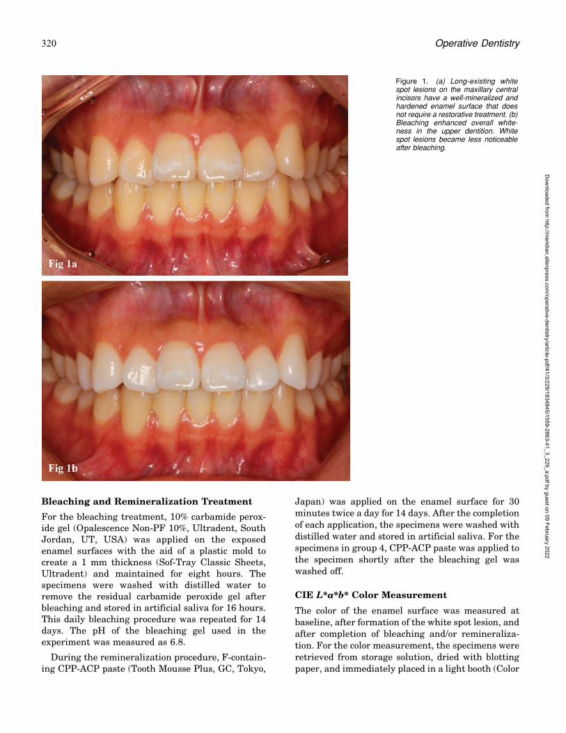
Bleaching and Remineralization Treatment
For the bleaching treatment, 10% carbamide perox-ide gel (Opalescence Non-PF 10%, Ultradent, SouthJordan, UT, USA) was applied on the exposedenamel surfaces with the aid of a plastic mold tocreate a 1 mm thickness (Sof-Tray Classic Sheets,Ultradent) and maintained for eight hours. Thespecimens were washed with distilled water toremove the residual carbamide peroxide gel afterbleaching and stored in artificial saliva for 16 hours.This daily bleaching procedure was repeated for 14days. The pH of the bleaching gel used in theexperiment was measured as 6.8.
During the remineralization procedure, F-contain-ing CPP-ACP paste (Tooth Mousse Plus, GC, Tokyo,
Japan) was applied on the enamel surface for 30minutes twice a day for 14 days. After the completionof each application, the specimens were washed with
distilled water and stored in artificial saliva. For thespecimens in group 4, CPP-ACP paste was applied tothe specimen shortly after the bleaching gel waswashed off.
CIE L*a*b* Color Measurement
The color of the enamel surface was measured atbaseline, after formation of the white spot lesion, andafter completion of bleaching and/or remineraliza-tion. For the color measurement, the specimens were
retrieved from storage solution, dried with blottingpaper, and immediately placed in a light booth (Color
Figure 1. (a) Long-existing whitespot lesions on the maxillary centralincisors have a well-mineralized andhardened enamel surface that doesnot require a restorative treatment. (b)Bleaching enhanced overall white-ness in the upper dentition. Whitespot lesions became less noticeableafter bleaching.
320 Operative Dentistry
Dow
nloaded from http://m
eridian.allenpress.com/operative-dentistry/article-pdf/41/3/229/1834845/1559-2863-41_3_229_a.pdf by guest on 09 February 2022
Sense II; Sungjin Hitech, Gyeonggi-Do, Korea) withMunsell N7 neutral gray walls and floor.
A spectroradiometer (PR-670; SpectraScan, PhotoResearch, Chatsworth, CA, USA) equipped with aMacro-Spectar MS-75 lens (Photo Research) wasfixed on a tripod at a distance of 355 mm from themeasured object and with a measurement area of2.63 mm in diameter, providing an optical configu-ration of 28 observation to the object. Four D65simulating tubes (F2DT12/65; Gretagmacbeth, Re-search Triangle Park, NC, USA), reportedly having acorrelated color temperature of 6500 K and a colorrendering index of 91, were used as the light source.The tubes were bidirectionally fixed with a 458
illumination angle at a distance of 30 cm from themeasured object. External light was excluded bycovering the equipment with a light-proof cover. Thepositioning of the lens toward the surface of thespecimen was kept constant to ensure a standard-ized measurement throughout the experiment. Spec-tral reflectance was obtained from 380 to 780 nmwith a 2-nm interval (Spectrawin 2.0, Photo Re-search) and was subsequently converted to CIE L*,a*, and b* values. The color difference (DE*) wascalculated by the equation: (DE*) = [(DL*)2 þ (Da*)2 þ(Db*)2]½. Every measurement was performed aftercalibration over a white background and repeatedthree times.
EPMA
Specimens were embedded in epoxy resin (Epofix,Struers, Glasgow, UK) and cross-sectioned along the
midline. The cut surfaces were serially polished with
1200-, 2400-, and 4000-grit silicon carbide abrasive
papers, followed by 1-lm and 0.25-lm diamond and
0.1-lm and 0.05-lm aluminum oxide polishing
suspensions (Struers, Copenhagen, Denmark). The
specimens were ultrasonically cleaned in deionizedwater for 10 minutes, dried for 72 hours in a
desiccator, and then sputter-coated with carbon.
The demineralization area on the cross-sectioned
enamel surfaces was identified using the phase
contrast of the backscattered electron imaging mode
of a scanning electron microscope (SEM) (JEOL
JSM-6610LV, JEOL, Akishima, Japan). Two-lineanalyses were performed perpendicular to the outer
enamel surface at 0.3-lm pixel intervals. The
observation areas (intact surface layer, demineral-
ized subsurface layer, and inner sound enamel) were
determined according to changes in Ca and P content
using an electron microprobe (JEOL JXA-8100,JEOL, Akishima, Japan). The operating conditions
for the elemental analyses were 15 kV of accelerat-
ing voltage and 50 nA of beam current. A fluorapa-
tite crystal (3.38% F) was used as a standard
comparison for analysis.
Figure 2. Procedural steps in four experimental groups.
Kim & Others: Effect of Bleaching on White Spot Lesions 321
Dow
nloaded from http://m
eridian.allenpress.com/operative-dentistry/article-pdf/41/3/229/1834845/1559-2863-41_3_229_a.pdf by guest on 09 February 2022

Knoop Microhardness Measurement
Cross-sectional enamel surface hardness was mea-
sured using a microhardness tester (Tukon 2100,
Instron Corp, Canton, MA, USA), and a Knoop
diamond indenter with a load of 10g was applied for
11.5 seconds. A total of 10 indentations were made
with a 20-lm interval from the surface to the sound
enamel alongside an EPMA scan line (Figure 3).
Statistical Analysis
The sample size calculation was based on the datafrom a pilot study using the microhardness test andhad 80% power to detect a 30–standard deviation(SD) difference between any two groups, assumingan overall 5% significance level and two-sided tests.The normality and homogeneity of the samples weretested using the Kolmogorov-Smirnov test. Assum-ing a normal distribution of differences, two-wayanalysis of variance was performed with Tukey posthoc comparison. The mean values of CIE L*, a*, andb* and the differences between each measurementpoint were compared among the four groups. Themean percentage weight loss of Ca and P and themean weight of F in the surface and the subsurfacelayer were compared among the groups. The meanvalues and percentage decreases of the microhard-ness at the lesion areas were compared among thegroups. The correlations between the Ca and Pcontent and microhardness values were evaluated byPearson correlation coefficients. A p-value of 0.05was selected as the threshold for statistical signifi-cance. Analyses were performed using SPSS 13.0(SPSS, Chicago, IL, USA).
RESULTS
The CIE L*a*b* values of the specimens at baseline(BA) and after caries formation were not significant-ly different among all groups. The final color changeafter treatment of bleaching, remineralization (RE)and/or bleaching (BL), DE*
TR-BA, ranged from 7.03 to7.60 in groups 1A (BL), 1B (CA), and 2 (CAþRE),without any significant differences (Table 1). How-ever, the DE*
TR-BA values in group 3 (CAþBL) andgroup 4 (CAþBLþRE) were 10.98 and 10.81, respec-tively, which was significantly greater than those inthe other three groups (p,0.05). The differences inthe three color parameters, DL*
TR-BA, Da*TR-BA, and
Db*TR-BA, were in accordance with DE*
TR-BA; eachvalue in groups 3 (CAþRE) and 4 (CAþBLþRE) wassignificantly larger than its counterpart in the otherthree groups (p,0.05).
The mean (SD) depth of the surface and subsur-face lesion areas ranged from 23.4 (5.8) lm to 157.8(19.0) lm, respectively, without any significantdifference among all groups (Table 2). The amountof Ca and P loss in the subsurface lesion in group 2(CAþRE) was significantly less than that in group1B (CA) (p,0.05). The mean (SD) Ca/P ratios were2.17 (0.10) and 2.15 (0.01) in the surface andsubsurface lesion areas, respectively, without signif-icant differences among groups. The weight of F washighest in the surface lesion, followed by the
Figure 3. Elemental analysis along the scan line (dark arrows)illustrated the elemental composition (Ca, P, and F) on the cross-sectional SEM image. Knoop microhardness values measured alongthe scan line corresponded to the elemental contents of eachindentation site.
322 Operative Dentistry
Dow
nloaded from http://m
eridian.allenpress.com/operative-dentistry/article-pdf/41/3/229/1834845/1559-2863-41_3_229_a.pdf by guest on 09 February 2022

subsurface lesion and the inner sound enamel acrossall groups (p,0.05, Figure 4). The amount of F in thesubsurface lesion was higher in group 2 (CAþRE)than in group 1B (CA) (p,0.05).
The mean (SD) Knoop microhardness value was231.3 (99.3) Hk in the surface lesion, 324.5 (76.0) Hk
in the subsurface lesion, and 426 (38.7) Hk in thesound enamel (Table 3). Each microhardness valueof the surface, subsurface, and total lesion area didnot show a significant difference across groups(Figure 5). A strong correlation existed between theCa and P content and the microhardness values forthe subsurface lesion area (Figure 6).
DISCUSSION
For our first hypothesis, we primarily investigatedwhether the whiteness of carious enamel could bemasked by the whitening effect of the surroundingsound enamel structure. The color change producedby the formation of artificial caries was similar tothat obtained from peroxide bleaching. The increasein lightness (L*) and decrease in yellowness (b*)contributed to the whitening of artificial caries,which was in accordance with the results of previousstudies.5,6 The color of artificially formed cariousenamel (DE*=7.03) was further changed by bleach-ing, resulting in extended whiteness (DE*=10.98).
However, considering the outcome of bleachingsound enamel (DE*=7.60), the color discrepancybetween the sound and carious enamel after bleach-ing (3.38 DE* units) was within a relatively accept-able range. In a widely cited study by Johnston andKao,12 3.7 DE* units was proposed as the percepti-bility threshold and 6.8 DE* units was proposed to bethe borderline for color mismatch. Therefore, froman esthetic standpoint, the bleaching treatment ofteeth containing white spot lesions may be aclinically relevant procedure to promote an opticalcamouflage effect.
Our second question was whether bleachingtreatment would cause further demineralization ofthe white spot lesion. We created a superficiallyintact enamel layer with a relatively uniform widthof 22-24 lm, in which the loss of Ca and P wasminimal (0-2 wt%). The underlying subsurface areawith mineral loss (15-25 wt%) had a depth of 150-160lm. This mineral-depleted porous substructureinduced an altered light-scattering mode within theenamel structure, producing a whitish appearance.Many previous studies have reported on the poten-tial impact of bleaching on the enamel microstruc-ture; those results were influenced by manyvariables, such as tooth type, peroxide concentration,the pH of the bleaching agent, the duration ofcontact, and the treatment interval. In this study,
Table 1: Comparison of the Mean (SD) Values of Color Difference Between at Baseline (BA) and After Treatment (TR) Amongthe Experimental Groups
Group: Treatment N Color Difference Between at Basement and After Treatmenta
DL*(TR-BA) Da*(TR-BA) Db*(TR-BA) DE*(TR-BA)
1A (BL) 10 4.95 (0.55) A �1.13 (0.15) A �5.60 (1.03) A 7.60 (0.84) A
1B (CA) 10 4.72 (0.54) A �0.50 (0.12) A �5.14 (0.95) A 7.03 (0.90) A
2 (CA þ RE) 20 4.69 (0.75) A �0.46 (0.13) B �5.30 (1.14) A 7.14 (1.09) A
3 (CA þ BL) 20 6.96 (0.75) B �1.47 (0.24) C �8.31 (0.94) B 10.98 (0.80) B
4 (CA þ BL þ RE) 20 6.86 (0.69) B �1.40 (0.23) C �8.20 (0.86) B 10.81 (0.85) B
Abbreviations: BL, bleaching; CA, caries formation; RE, remineralization.a Different superscript letters following means denote significant differences (p,0 .05).
Table 2: The Mean (SD) Depth (lm) and Weight Loss (%) of Calcium (Ca) and Phosphorus (P) on the Surface and theSubsurface Lesionsa
Group (Treatment) N Surface Lesion Subsurface Lesion
Depth, lm Ca Loss, wt% P Loss, wt% Depth, lm Ca Loss, wt% P Loss, wt%
1B (CA) 10 24.2 (7.9) 2.6 (4.8) 1.5 (5.6) 164.2 (20.7) 25.2 (8.5) A 24.8 (8.5) A
2 (CAþRE) 10 23.9 (6.6) 0 (3.8) 1.4 (3.7) 153.7 (12.5) 16.2 (5.6) B 15.5 (6.0) B
3 (CAþBL) 10 22.9 (5.4) 2.1 (4.8) 1.9 (3.0) 165.7 (18.4) 21 (7.2) AB 21.3 (7.1) AB
4 (CAþBLþRE) 10 22.3 (6.2) 0.2 (1.9) 1.5 (1.1) 157.4 (24.8) 19.8 (6.7) AB 18.4 (5.7) AB
Abbreviations: BL, bleaching; Ca, calcium; CA, caries formation; P, phosphorus; RE, remineralization.a Different superscript letters following means denote significant differences at p , 0.05.
Kim & Others: Effect of Bleaching on White Spot Lesions 323
Dow
nloaded from http://m
eridian.allenpress.com/operative-dentistry/article-pdf/41/3/229/1834845/1559-2863-41_3_229_a.pdf by guest on 09 February 2022

bleaching using 10% carbamide peroxide with a pHof slightly less than 7 did not induce a significantmineral loss in carious enamel. We obtained thecross-sectional microhardness values at each loca-tion adjacent to the EPMA scan line in order to
evaluate the depth-related chemical and mechanicalproperties. White spot lesions represent the prelim-inary stage of subsurface enamel demineralization,although they do not necessitate invasive restorativeintervention. Hence, it is clinically meaningful toreharden the surface enamel and maintain itsmechanical integrity. The Ca and P content and
Figure 4. The mean content of F in each lesion area.Figure 5. The mean cross-sectional Knoop microhardness values ofspecimens from the four experimental groups. The Knoop microhard-ness values were measured at intervals of 20 lm starting from thesurface enamel layer and reaching to the 200-lm-deep soundenamel.
Table 3: The Mean (SD) Microhardness and Percentile Decrease of the Surface and Subsurface Lesions
Group (Treatment) N Surface Lesion Subsurface Lesion Total Lesion
Microhardness, Hka Decrement, %b Microhardness, Hk Decrement, % Microhardness, Hk Decrement, %
1B (CA) 10 172.2 (49.6) 59.9 (16.1) 288.7 (47.8) 34.6 (13.6) 274.1 (44.8) 37.7 (13.6)
2 (CAþRE) 10 201.0 (27.4) 53.7 (8.1) 324.3 (46.2) 25.8 (9.4) 308.9 (39.7) 29.3 (8.2)
3 (CAþBL) 10 175.8 (19.8) 58.6 (5.7) 291.1 (86.9) 32.7 (16.6) 276.7 (77.6) 35.9 (14.8)
4 (CAþBLþRE) 10 198.6 (18.1) 51.9 (5.8) 292.4 (47.9) 29.2 (12.8) 280.7 (41.7) 32.1 (11.3)
Abbreviations: BL, bleaching; CA, caries formation; RE, remineralization.a Hk = 14.12P/‘2 (kgf/mm2).b Decrement (%) = (1�the mean microhardness value of each lesion area/the mean of the inner sound enamel ) 3 100.
Figure 6. Correlation coefficients and regression equations betweenCa and P contents and microhardness values in the subsurface lesionarea of the carious enamel.
324 Operative Dentistry
Dow
nloaded from http://m
eridian.allenpress.com/operative-dentistry/article-pdf/41/3/229/1834845/1559-2863-41_3_229_a.pdf by guest on 09 February 2022
hardness values were relatively well correlatedthroughout the lesion area. Only a small disparityexisted at the first 20-lm surface area withdecreased hardness. The surface of the enamel withthe lesion was softened and porous but mineral-abundant.13 This mechanically weakened layer maybe easily abraded by normal tooth brushing. In aclinical evaluation14 of initial caries, the remission ofwhiteness was not entirely due to color reversal fromthe remineralized microstructure but rather was dueto mechanical removal of the superficially weakenedlayer. The hardness of the cross-sectional enamelgradually increased, with elevating mineral contentreaching the level of the sound enamel. Overall,bleaching using 10% carbamide peroxide did notdeteriorate the chemical and mechanical propertiesof carious enamel. Our first hypothesis was accepted.
To test our second hypothesis, we evaluated theeffect of CPP-ACP on color, mineralization, andhardness of carious enamel before and after bleach-ing. Despite confirmation of mineral gain in thesubsurface lesion, no significant color reversal wasdetected by the spectroradiometer. This correspondsto a common clinical situation, with long-existingcaries lesions arrested or regressed by well-mineral-ized surface enamel but still showing a whitishappearance. Even when some mineral depositionoccurs in the underlying body lesion, the porevolume is decreased but the pore number isunchanged.2 There are several stages of mineraldeposition, including the formation and growth ofnew crystals and the regrowth of preexistingcrystals.15 Although the Ca/P ratio remains un-changed, the heterogeneity of the modified apatitestructure contributes to the altered optical charac-teristics.16 The color mismatch between the remin-eralized and sound enamel substrate is unsolved,often requiring esthetic enhancement.
As for the remineralizing and rehardening effect ofCPP-ACP on enamel during and after at-homebleaching procedures, many studies suffered fromdissimilar experimental conditions; some studiesused bovine teeth, which are more porous thanhuman teeth,10,17 while others measured the surfacemicrohardness or roughness on the superficialenamel instead of its subsurface structure.11,18,19 Inthis study, the change in mineralization was evalu-ated both at the surface and subsurface lesion areasafter the use of CPP-ACP. We determined thatcarious enamel without bleaching had the largestremineralization gains. In group 2, freshly formedcarious lesions, which were not subjected to asequence of 10% carbamide application and artificial
saliva storage, largely promoted incorporation of freeions from the CPP-ACP paste into the subsurfacelesion area. The microhardness values were alsohighest in group 2, both at the surface andsubsurface lesion areas, and the values in othergroups followed the same order as in the mineralcomposition. Overall, our second hypothesis was notaccepted.
Many previous studies9,20,21 have determined theeffect of remineralizing agents on bleached teeth andconcluded that any substantial recovery of hardnessor mineral deposition was mainly due to supplemen-tary ions in the storage media (artificial or humansaliva). Under in vivo conditions, the repair mecha-nism would more actively counteract the mineralloss than under in vitro conditions, even in the casethat the bleaching treatment might cause an initialdeterioration of the chemical or mechanical proper-ties of the enamel. We confirmed that the applicationof CPP-ACP paste enhanced the reversal of the earlycaries lesion stage, as shown in other studies.3,22
Considering the largest change shown in group 2,CPP-ACP’s remineralizing effect on white spotlesions seemed to be maximized prior to thebleaching procedure.
CONCLUSIONS
In this study, the 10% carbamide peroxide bleachingof enamel with white spot lesions decreased colordisparities without deteriorating mineral composi-tion or microhardness. The application of CPP-ACPpaste promoted mineral gain in the subsurface bodylesion. The bleaching treatment for teeth with whitespot lesions can be recommended as a noninvasiveesthetic treatment regimen with supplementaryremineralization protocols.
Acknowledgement
This study was supported by grant No. 04-2011-0050 from theSeoul National University Dental Hospital Research Fund.
Regulatory Statement
This study was conducted in accordance with all theprovisions of the local human subjects oversight committeeguidelines and policies of Seoul National University DentalHospital Institutional Review Board. The approval code forthis study is CRI13010.
Conflict of Interest
The authors have no proprietary, financial, or other personalinterest of any nature or kind in any product, service, and/orcompany that is presented in this article.
(Accepted 8 July 2015)
Kim & Others: Effect of Bleaching on White Spot Lesions 325
Dow
nloaded from http://m
eridian.allenpress.com/operative-dentistry/article-pdf/41/3/229/1834845/1559-2863-41_3_229_a.pdf by guest on 09 February 2022

REFERENCES
1. Kleter GA (1998) Discoloration of dental carious lesions (areview) Archives of Oral Biology 43(8) 629-632.
2. Jones RS, & Fried D (2006) Remineralization of enamelcaries can decrease optical reflectivity Journal of DentalResearch 85(9) 804-808.
3. Bailey DL, Adams GG, Tsao CE, Hyslop A, Escobar K,Manton DJ, Reynolds EC, & Morgan MV (2009) Regres-sion of post-orthodontic lesions by a remineralizing creamJournal of Dental Research 88(12) 1148-1153.
4. Mjor IA, & Gordan VV (2002) Failure, repair, refurbish-ing and longevity of restorations Operative Dentistry27(5) 528-534.
5. Iwami Y, Hayashi N, Takeshige F, & Ebisu S (2008)Relationship between the color of carious dentin withvarying lesion activity, and bacterial detection Journal ofDentistry 36(2) 143-151.
6. Kim Y, Son HH, Yi K, Kim HY, Ahn J, & Chang J (2013)The color change in artificial white spot lesions measuredusing a spectroradiometer Clinical and Oral Investiga-tions 17(1) 139-146.
7. Yeh ST, Su Y, Lu YC, & Lee SY (2005) Surface changesand acid dissolution of enamel after carbamide peroxidebleach treatment Operative Dentistry 30(4) 507-515.
8. Joiner A (2007) Review of the effects of peroxide onenamel and dentine properties Journal of Dentistry35(12) 889-896.
9. Attin T, Schmidlin PR, Wegehaupt F, & Wiegand A (2009)Influence of study design on the impact of bleachingagents on dental enamel microhardness: A review DentalMaterials 25(2) 143-157.
10. Borges BC, Borges JS, de Melo CD, Pinheiro IV, SantosAJ, Braz R, & Montes MA (2011) Efficacy of a novel at-home bleaching technique with carbamide peroxidesmodified by CPP-ACP and its effect on the microhardnessof bleached enamel Operative Dentistry 36(5) 521-528.
11. de Vasconcelos AA, Cunha AG, Borges BC, Vitoriano JdeO, Alves-Junior C, Machado CT, & dos Santos AJ (2012)Enamel properties after tooth bleaching with hydrogen/carbamide peroxides in association with a CPP-ACP pasteActa Odontologica Scandinavica 70(4) 337-343.
12. Johnston WM, & Kao EC (1989) Assessment of appear-ance match by visual observation and clinical colorimetryJournal of Dental Research 68(5) 819-822.
13. Arends J, & Christoffersen J (1986) The nature of earlycaries lesions in enamel Journal of Dental Research 65(1)2-11.
14. Kielbassa AM, Gillmann L, Zantner C, Meyer-Lueckel H,Hellwig E, & Schulte-Monting J (2005) Profilometric andmicroradiographic studies on the effects of toothpaste andacidic gel abrasivity on sound and demineralized bovinedental enamel Caries Research 39(5) 380-386.
15. Kawasaki K, Tanaka Y, & Takagi O (2000) Crystallo-graphic analysis of demineralized human enamel treatedby laser-irradiation or remineralization Archives of OralBiology 45(9) 797-804.
16. Ko CC, Tantbirojn D, Wang T, & Douglas WH (2000)Optical scattering power for characterization of mineralloss Journal of Dental Research 79(8) 1584-1589.
17. Bayrak S, Tunc ES, Sonmez IS, Egilmez T, & Ozmen B(2009) Effects of casein phosphopeptide-amorphous calci-um phosphate (CPP-ACP) application on enamel micro-hardness after bleaching American Journal of Dentistry22(6) 393-396.
18. Pinto CF, Paes Leme AF, Cavalli V, & Giannini M (2009)Effect of 10% carbamide peroxide bleaching on sound andartificial enamel carious lesions Braz Dent J 20(1) 48-53.
19. De Abreu DR, Sasaki RT, Amaral FL, Florio FM, &Basting RT (2011) Effect of home-use and in-officebleaching agents containing hydrogen peroxide associat-ed with amorphous calcium phosphate on enamel micro-hardness and surface roughness Journal of Esthetic andRestorative Dentistry 23(3) 158-168.
20. Metz MJ, Cochran MA, Matis BA, Gonzalez C, Platt JA, &Pund MR (2007) Clinical evaluation of 15% carbamideperoxide on the surface microhardness and shear bondstrength of human enamel Operative Dentistry 32(5)427-436.
21. Smidt A, Feuerstein O, & Topel M (2011) Mechanical,morphologic, and chemical effects of carbamide peroxidebleaching agents on human enamel in situ QuintessenceInternational 42(5) 407-412.
22. Cai F, Manton DJ, Shen P, Walker GD, Cross KJ, YuanY, Reynolds C, & Reynolds EC (2007) Effect of additionof citric acid and casein phosphopeptide-amorphouscalcium phosphate to a sugar-free chewing gum onenamel remineralization in situ Caries Research 41(5)377-383.
326 Operative Dentistry
Dow
nloaded from http://m
eridian.allenpress.com/operative-dentistry/article-pdf/41/3/229/1834845/1559-2863-41_3_229_a.pdf by guest on 09 February 2022

Simulated Wear of Self-AdhesiveResin Cements
T Takamizawa � WW Barkmeier � MA LattaTP Berry � A Tsujimoto � M Miyazaki
Clinical Relevance
Loss of cement at the margins of restorations can initiate a variety of clinical issues thatmay ultimately result in restoration loss and replacement.
SUMMARY
One of the primary areas of concern withluting agents is marginal gap erosion andattrition. The purpose of this laboratory studywas to evaluate bulk and marginal slit (gap)generalized wear of self-adhesive resin ce-ments. Three self-adhesive resin cements wereused in this study: G-CEM LinkAce (LA), Max-cem Elite (ME), and RelyX Unicem2 Automix
(RU). A custom stainless-steel fixture with acavity 4.5 mm in diameter and 4 mm deep wasused for simulated generalized (bulk) wear.For simulated marginal gap wear, a two-piecestainless-steel custom fixture was designedwith a slit (gap) 300 lm wide and 3 mm inlength. For both wear models, 20 specimenseach for each of the three adhesive cementswere made for both light-cure and chemical-cure techniques. The cured cements werepolished with a series of carbide papers to a4000-grit surface and subjected to 100,000 cy-cles using the slit (gap) wear model and 400,000cycles for generalized (bulk) wear in a Lein-felder-Suzuki (Alabama machine) wear simu-lator (maximum load of 78.5 N). Flat-endedstainless-steel antagonists were used in a wa-ter slurry of poly(methylmethacrylate) beadsfor simulation of generalized contact-free areawear with both wear models. Before and afterthe wear challenges, the specimens were pro-filed with a Proscan 2100 noncontact profilom-eter, and wear (volume loss [VL] and meanfacet depth [FD]) was determined using AnSur3D software. Two-way analysis of variance(ANOVA) and Tukey post hoc tests were usedfor data analysis for the two wear models.Scanning electron microscopy (SEM) was usedto examine polished surfaces of the resincements and the worn surfaces after the wear
Toshiki Takamizawa, DDS, PhD, assistant professor, Opera-tive Dentistry, Nihon University School of Dentistry, Tokyo,Japan
*Wayne W Barkmeier, DDS, MS, professor, General Dentist-ry, dean emeritus, Creighton University School of Dentistry,Omaha, NE, USA
Mark A Latta, DMD, MS, professor, General Dentistry, dean,Creighton University, Omaha, NE, USA
Thomas P Berry, DDS, assistant professor, General Dentistry,School of Dentistry, Creighton University, Omaha, NE, USA
Akimasa Tsujimoto, DDS, PhD, assistant professor OperativeDentistry, Nihon University School of Dentistry, Tokyo,Japan
Masashi Miyazaki, DDS, PhD, professor and chair, OperativeDentistry, Nihon University School of Dentistry, Tokyo,Japan
*Corresponding author: General Dentistry, Creighton Uni-versity School of Dentistry, 2500 California Plaza, Omaha,NE 68178, USA; e-mail: [email protected]
DOI: 10.2341/14-227-L
�Operative Dentistry, 2016, 41-3, 327-338
Dow
nloaded from http://m
eridian.allenpress.com/operative-dentistry/article-pdf/41/3/229/1834845/1559-2863-41_3_229_a.pdf by guest on 09 February 2022

challenges. The two-way ANOVA of VL usingthe generalized (bulk) wear model showed asignificant effect among the three resin cementmaterials for the factor of resin cement(p,0.001) and the interaction of the cementand cure method (p,0.001), but not for the curemethod (p=0.465). The two-way ANOVA for FDalso found a significant difference for thefactor of resin cement (p,0.001) and the inter-action of the resin cement and cure method(p,0.001), but not for the cure method(p=0.277). The simulated generalized (bulk)wear for the light-cure groups was as follows:VL (mm3): RU 0.631 (0.094), LA 0.692 (0.112), andME 1.046 (0.141) and FD (lm): RU 43.6 (6.5), LA47.0 (7.7), and ME 72.5 (9.9). The simulatedgeneralized (bulk) wear for the chemical-curegroups was as follows: VL (mm3): LA 0.741(0.105), RU 1.231 (0.234), and ME 1.305 (0.143)and FD (lm): LA 50.7 (7.2), RU 84.5 (16.1), andME 91.7 (10.2). Simulated wear using the slit(gap) model for the light-cure groups was asfollows: VL (mm3): RU 0.030 (0.006), LA 0.031(0.006), and ME 0.041 (0.009) and FD (lm): RU49.6 (5.7), LA 57.2 (8.4), and ME 70.9 (10.7). Thewear values for the chemical-cure slit (gap)groups were as follows: VL (mm3): LA 0.031(0.004), ME 0.038 (0.007), and RU 0.045 (0.009)and FD (lm): LA 53.9 (6.7), ME 63.5 (9.1), and RU74.2 (12.9). Pearson correlation tests revealed astrong relationship between the two wearmodels for the light-cure groups and a goodrelationship for the chemical-cure groups. Theobservations using SEM showed differences infiller particle shape and size among the ce-ments and the resultant effect of the wearchallenges. The worn surfaces of each cementwere essentially the same for both light-cureand chemical-cure methods. The bulk wearmodel and new slit (gap) model for evaluationof simulated generalized wear of luting agentsdemonstrated significant differences (p,0.05)in relative wear among three self-adhesiveresin cements and between visible light- andchemical-cure techniques.
INTRODUCTION
The evolution of adhesive dentistry procedures andmaterials has changed many facets of dentistry. Thedevelopment of resin luting agents, along withadhesive dentistry techniques, has rapidly advancedthe capability to bond indirect restorations tomineralized tooth structures and core buildup
materials. One of the main advantages of resincements, when compared to nonpolymer lutingagents, is enhanced mechanical properties and theability to adhesively bond to metal, ceramic, enamel,and dentin.1-8 The use of etch-and-rinse bondingprocedures along with resin luting agents has helpedto promote the use of high-strength ceramic resto-rations. The more recent introduction of self-adhe-sive resin cements has reduced the required numberof clinical steps in the bonding sequence, therebyreducing the number of treatment steps along withpatient chair time. The dual curing capability (light-cure and chemical-cure) of many of the newer resincements has extended their use to include restora-tions where light penetration to the intaglio surfaceis limited or not attainable. The use of resin cementsis now also well established for fiber-reinforcedcomposite materials and has extended into ortho-dontics for attachment of both metallic and ceramicbrackets.
One of the challenges that remains, regardless ofthe cementing media, is marginal integrity.9 A majorconcern with all dental cements is their ability toresist gap formation at the marginal closure areafrom attrition and erosion. Intact restoration mar-gins reduce the potential for marginal staining,secondary caries, tooth sensitivity, and periodontalissues. Clinical criteria have been used to assessmarginal integrity in long-term clinical trials. How-ever, these studies are costly and take years tocomplete. Investigators have used laboratory mar-ginal gap studies in an effort to assess the potentialfor cement loss at the margins of restorations.9-12
These studies have shown an excellent relationshipamong 1) tooth margin and restoration gap width, 2)type of cement, and 3) cement wear. They found thatenhanced wear resistance of resin cements isassociated with a smaller filler particle size.
Depending on the clinical situation, dual-cure self-adhesive resin cements can be light-cured, chemi-cally-cured, or can use a combination of light curingand chemical curing. The degree of polymerizationconversion can also impact the wear resistance of acement at the margin.13-15 Less efficiency in poly-merization is often seen in chemically-cured resincements and may lead to higher wear.15 Several self-adhesive resin cements are now available to theprofession. The acidic monomers in these formula-tions can be a challenge when designing chemical-cure based polymerization components, as manyamine initiators are quenched at low pH, leading toa lower degree of polymerization when compared tolight curing.15-17
328 Operative Dentistry
Dow
nloaded from http://m
eridian.allenpress.com/operative-dentistry/article-pdf/41/3/229/1834845/1559-2863-41_3_229_a.pdf by guest on 09 February 2022

Technology has rapidly advanced for assessingsimulated laboratory wear by the introduction ofnoncontact optical profilometers and enhanced com-puter software for data analysis.18 In addition, a newmarginal slit (gap) model was developed for labora-tory-generalized wear (contact-free area [CFA] wear)simulation in an effort to learn more about the wearresistance of newer-generation self-adhesive resincements. The purpose of this study was to use thenewly developed slit (gap) wear model and anestablished generalized (bulk) wear model to assessrelative wear characteristics of newer dual-cure self-adhesive resin cements. The two hypotheses testedwere the null hypotheses that 1) there will not be asignificant difference (a=0.05) in wear values amongthree dual-cured self-adhesive resin cements andthat 2) wear of self-adhesive resin cements usinglight-cure or chemical-cure methods will not bedifferent.
METHODS AND MATERIALS
Three self-adhesive resin cements (Table 1) wereevaluated in this study: G-CEM LinkAce (LA) (GCCorp, Tokyo, Japan), Maxcem Elite (ME) (Kerr Corp,Orange, CA, USA), and RelyX Unicem2 Automix(RU) (3M ESPE Dental Products, St Paul, MN,USA). The evaluation components in this studyincluded 1) simulated generalized (bulk) wear test-ing, 2) simulated marginal slit (gap) wear testing, 3)argon-ion etching scanning electron microscopy(SEM) of the resin cement surfaces, and 4) SEMexamination of the cement wear facets.
Generalized (Bulk) Wear Simulation
Forty specimens for each of the three self-etchingresin cement materials (total of 120 specimens) wereprepared for wear challenges of 400,000 cycles usinga generalized (bulk) wear model (CFA wear) in aLeinfelder-Suzuki (Alabama) wear simulation de-vice. The methodology for sample preparation andthe generalized wear model was previously describedby Barkmeier and others.18-20 In summary, stain-less-steel custom fixtures with cavities 4.5 mm indiameter and 4 mm deep were used to hold the resincement materials. Twenty specimens for each resin
cement were light-cured in two increments ofapproximately 2 mm for 40 seconds with a Spectrum800 curing unit (Dentsply Caulk, Milford, DE, USA)set at 600 mW/cm2. The other 20 specimens for eachcement) were chemically-cured (the light curing unitwas not used). After 24 hours, the cement surfaceswere polished flat to 4,000 grit (Figure 1) using asequence of silicon carbide papers (Struers Inc,Cleveland, OH, USA).
The custom fixtures were mounted inside a plasticwater bath, and a brass cylinder was placed aroundeach fixture. The water bath fixture was thenattached to the wear simulator. A water slurry ofpoly(methylmethacrylate) (PMMA) was used as theabrasive media and placed inside the cylinders overthe resin cement specimens.
Stainless-steel antagonists 6.5 mm in diameter(Figure 2), mounted in spring-loaded pistons, werethen used to deliver the wear challenges in the wearsimulation machine (Figure 3). The pistons rotatedapproximately 30 degrees as the load was applied(maximum load of 78.5 N) at a rate of 2 Hz and then
Table 1: Self-Adhesive Resin Cements
Material Manufacturer Lot Shade Study Code
G-CEM Link Ace GC Corp (Tokyo, Japan) 1212144 A2 LA
Maxcem Elite Kerr Corp (Orange, CA, USA) 4818000 Brown ME
RelyX Unicem2 Automix 3M ESPE Dental Products (St Paul, MN, USA) 50153 A2 RU
Figure 1. Custom stainless-steel fixture for generalized (bulk) wear.
Takamizawa & Others: Simulated Wear of Self-Adhesive Resin Cements 329
Dow
nloaded from http://m
eridian.allenpress.com/operative-dentistry/article-pdf/41/3/229/1834845/1559-2863-41_3_229_a.pdf by guest on 09 February 2022
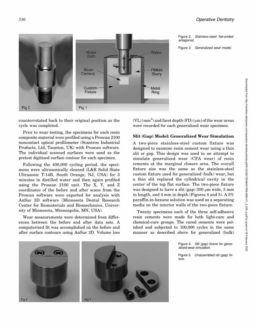
counterrotated back to their original position as thecycle was completed.
Prior to wear testing, the specimens for each resincomposite material were profiled using a Proscan 2100noncontact optical profilometer (Scantron IndustrialProducts, Ltd, Taunton, UK) with Proscan software.The individual scanned surfaces were used as thepretest digitized surface contour for each specimen.
Following the 400,000 cycling period, the speci-mens were ultrasonically cleaned (L&R Solid StateUltrasonic T-14B, South Orange, NJ, USA) for 3minutes in distilled water and then again profiledusing the Proscan 2100 unit. The X, Y, and Zcoordinates of the before and after scans from theProscan software were exported for analysis withAnSur 3D software (Minnesota Dental ResearchCenter for Biomaterials and Biomechanics, Univer-sity of Minnesota, Minneapolis, MN, USA).
Wear measurements were determined from differ-ences between the before and after data sets. Acomputerized fit was accomplished on the before andafter surface contours using AnSur 3D. Volume loss
(VL) (mm3) and facet depth (FD) (lm) of the wear areaswere recorded for each generalized wear specimen.
Slit (Gap) Model: Generalized Wear Simulation
A two-piece stainless-steel custom fixture wasdesigned to examine resin cement wear using a thinslit or gap. This design was used in an attempt tosimulate generalized wear (CFA wear) of resincements at the marginal closure area. The overallfixture size was the same as the stainless-steelcustom fixture used for generalized (bulk) wear, buta thin slit replaced the cylindrical cavity in thecenter of the top flat surface. The two-piece fixturewas designed to have a slit (gap) 300 lm wide, 3 mmin length, and 4 mm in depth (Figures 4 and 5). A 3%paraffin-in-hexane solution was used as a separatingmedia on the interior walls of the two-piece fixture.
Twenty specimens each of the three self-adhesiveresin cements were made for both light-cure andchemical-cure groups. The cured cements were pol-ished and subjected to 100,000 cycles in the samemanner as described above for generalized (bulk)
Figure 2. Stainless-steel flat-endedantagonist.
Figure 3. Generalized wear model.
Figure 4. Slit (gap) fixture for gener-alized wear simulation.
Figure 5. Unassembled slit (gap) fix-ture.
330 Operative Dentistry
Dow
nloaded from http://m
eridian.allenpress.com/operative-dentistry/article-pdf/41/3/229/1834845/1559-2863-41_3_229_a.pdf by guest on 09 February 2022

wear. The Proscan 2100 was used to make pretest andposttest scans, and the digitized surface contours wereexported for examination with AnSur 3D software.Cement VL and FD were determined on the resincement in the slit (gap) space as described above forthe simulated generalized (bulk) wear model.
SEM
Specimens of each of the three self-adhesive resincements were prepared for argon-ion etching andSEM examinations at Nihon University School ofDentistry (Tokyo, Japan). The three resin cementsexamined in this manner were not from the same lotnumbers as the materials subjected to wear simula-tion studies and postwear SEM examinations atCreighton University School of Dentistry (Omaha,NE, USA). The lot numbers of the cement materialsfor the argon-ion etching SEM examinations were asfollows: LA: 1402271, ME: 4394312, and RU: 497681.
The surfaces of the light-cure cements werepolished to a high gloss with abrasive discs (FujiStar Type DDC, Sankyo Rikagaku Co Ltd, Saitama,Japan) followed by a series of diamond pastes downto 0.25-lm particle size (DP-Paste, Struers, Baller-up, Denmark). The polished surfaces were thensubjected to argon-ion beam etching (IIS-200ER,Elionix, Tokyo, Japan) for 45 seconds with the ionbeam directed at the polished surfaces (acceleratingvoltage of 1.0 kV, ion current density of 0.4 mA/cm2).The surfaces were then coated in a vacuum evapo-rator with a thin film of gold. Observations weremade with a scanning electron microscope (FE-8000,Elionix) using an operating voltage of 10 kV and amagnification of 50003.
SEM examinations were completed at CreightonUniversity School of Dentistry on the wear facets ofthe three resin cement materials (light-cured andchemical-cured) following simulated generalizedwear using the two wear models. After the wearanalysis, representative samples of each materialwere sputter coated with gold and palladium usingan Emitech SC7620 Mini Sputter Coater (Quorum
Technologies, Ashford, UK). The coated wear spec-imens were then examined near the center of thewear facet with a TM3000 Tabletop Microscope(Hitachi-High Technologies Corp, Tokyo, Japan)using an accelerating voltage of 15 kV and magni-fications of 2,5003 and 5,0003.
Data Analysis of Simulated Wear
A two-way analysis of variance (ANOVA) (factors: [1]cement material and [2] cure method) and Tukeypost hoc tests were used for data analysis of VL andFD of both light-cure and chemical-cure groups forboth wear models. In addition, Pearson correlationtests were used to determine the relationshipbetween the two wear models for both VL and FDof the light-cure and chemical-cure groups.
RESULTS
Generalized (Bulk) Wear Simulation
The two-way ANOVA of the generalized (bulk) weardata for both VL and FD showed a significant effect(p,0.001) for the factor of resin cement and theinteraction of the resin cement and cure method.There was not a significant effect for the factor ofcure method for either VL (p=0.465) or FD(p=0.277). The two-way ANOVA values are shownin Tables 2 and 3.
The results of the simulated generalized (bulk)wear are presented in Table 4. The VL (mm3) and FD(lm) for the light-cure groups were as follows: VL:RU 0.631 (0.094), LA 0.692 (0.112), and ME 1.046(0.141) and FD: RU 43.6 (6.5), LA 47.0 (7.7), and ME72.5 (9.9). VL and FD for light-cure RU and LA werestatistically similar (p.0.05). ME exhibited statisti-cally (p,0.05) greater VL and mean FD than RU andLA for the light-cure cements.
The VL (mm3) and FD (lm) for the chemical-curegroups were as follows: VL: LA 0.741 (0.105), RU1.231 (0.234), and ME 1.305 (0.143) and FD: LA 50.7(7.2), RU 84.5 (16.1), and ME 91.7 (10.2). For thechemical-cure cements, RU and ME show statisti-
Table 2: Two-Way Analysis of Variance: SimulatedGeneralized (Bulk) Wear, Volume Loss
Source Sum ofSquares
Degreesof
Freedom
MeanSquare
F-Ratio p
Cement 10,449.9 2 5224.9 55.1 ,0.001
Cure method 51.1 1 51.1 0.538 0.465
Cement*curemethod
33,925.2 2 16,962.6 178.7 ,0.001
Table 3: Two-Way Analysis of Variance: SimulatedGeneralized (Bulk) Wear, Facet Depth
Source Sum ofSquares
Degreesof
Freedom
MeanSquare
F-Ratio p
Cement 1.97 2 0.986 49.8 ,0.001
Cure method 0.024 1 0.024 1.195 0.277
Cement*curemethod
6.78 2 3.39 171.0 ,0.001
Takamizawa & Others: Simulated Wear of Self-Adhesive Resin Cements 331
Dow
nloaded from http://m
eridian.allenpress.com/operative-dentistry/article-pdf/41/3/229/1834845/1559-2863-41_3_229_a.pdf by guest on 09 February 2022

cally (p,0.05) greater VL and FD than LA. Lightcuring of the RU and ME resin cements appeared tobe more effective than chemical curing in limitingsimulated generalized (bulk) wear. The FD and VLof RU were statistically greater (p,0.05) and nearlydouble for chemical-cure RU resin cement whencompared to light curing. When comparing LAgeneralized wear of light-cure and chemical-curecement, the results were statistically (p.0.05)similar for both VL and FD.
Slit (Gap) Model: Generalized Wear Simulation
The two-way ANOVA for both VL and FD using theslit (gap) model for generalized wear showed asignificant effect for the factors of resin cement(p,0.001), cure method (p=0.002), and the interac-tion of resin cement and cure method (p,0.001).The two-way ANOVA values are shown in Tables 5and 6.
The results of the slit (gap) model generalizedwear are presented in Table 7. The VL (mm3) andFD (lm) for the light-cure groups were as follows:VL: RU 0.030 (0.006), LA 0.031 (0.006), and ME0.041 (0.009) and FD: RU 49.6 (5.7), LA 57.2 (8.4),and ME 70.9 (10.7). VL in the slit model for light-cure RU and LA was statistically similar (p.0.05).ME exhibited statistically (p,0.05) greater VL thanRU and LA for the light-cure cements. RU exhibitedthe least slit (gap) model FD wear of the light-curecements.
The slit model VL (mm3) and FD (lm) for thechemical-cure groups were as follows: VL: LA 0.031
(0.004), ME 0.038 (0.007), and RU 0.045 (0.009) andFD: LA 53.9 (6.7), ME 63.5 (9.1), and RU 74.2 (12.9).The VL and mean FD of chemical-cure LA weresignificantly less (p,0.05) than RU and ME. RUexhibited the greatest VL and FD (p,0.05) when thechemical-cure groups were compared to light-curegroups while those of both LA and ME wereessentially the same (p.0.05).
The results of the Pearson correlation tests arepresented in Table 8. A strong relationship wasfound between the generalized (bulk) wear modeland the slit (gap) model for the light-cure groups forVL (r=0.999) and FD (r=0.968). A good relationshipwas found between the two wear models for thechemical-cure groups for VL (r=0.799) and FD(r=0.752).
SEM Observations
The ultrastructure examinations with argon-ionetching SEM revealed morphological differences infiller components of the cements (Figure 6a–c). Allthree resin cements exhibited a wide variety of fillerparticle sizes and shapes. The particle size distribu-tion of ME appeared to include larger particles thaneither LA or RU. The ultrastructure micrographsdemonstrated that the filler components were dif-ferent, and these compositional differences may haveinfluenced the wear characteristics of these materi-als.
The SEM exanimations of the worn surfaces ofall three of the resin cements showed evidence ofparticle loss (plucking) from the simulated gener-
Table 4: Simulated Generalized (Bulk) Wear of Self-Adhesive Resin Cements (n=20)a
Resin Cement Light-Cure Chemical-Cure
Volume Loss (mm3) Facet Depth (lm) Volume Loss (mm3) Facet Depth (lm)
RU 0.631 (0.094) aA 43.6 (6.5) aA 1.231 (0.234) aB 84.5 (16.1) aB
LA 0.692 (0.112) aA 47.0 (7.7) aA 0.741 (0.105) bA 50.7 (7.2) bA
ME 1.046 (0.141) bA 72.5 (9.9) bA 1.305 (0.143) aB 91.7 (10.2) aBa Lowercase letters in vertical columns are not different at the 5% significance level. Same uppercase letters between columns indicate no difference (5% significancelevel) in light-cure vs chemical-cure of the same cement.
Table 5: Two-Way Analysis of Variance: Slit (Gap) ModelGeneralized Wear, Volume Loss
Source Sum ofSquares
Degreesof
Freedom
MeanSquare
F-Ratio p
Cement 0.0015 2 0.000749 14.916 ,0.001
Cure method 0.000529 1 0.000529 10.541 0.002
Cement*curemethod
0.0016 2 0.000828 16.495 ,0.001
Table 6: Two-Way Analysis of Variance: Slit (Gap) ModelGeneralized Wear, Mean Facet Depth
Source Sum ofSquares
Degreesof
Freedom
MeanSquare
F-Ratio p
Cement 3143.047 2 1571.524 19.745 ,0.001
Cure method 813.49 1 813.490 10.221 0.002
Cement*curemethod
5637.538 2 2818.769 35.416 ,0.001
332 Operative Dentistry
Dow
nloaded from http://m
eridian.allenpress.com/operative-dentistry/article-pdf/41/3/229/1834845/1559-2863-41_3_229_a.pdf by guest on 09 February 2022
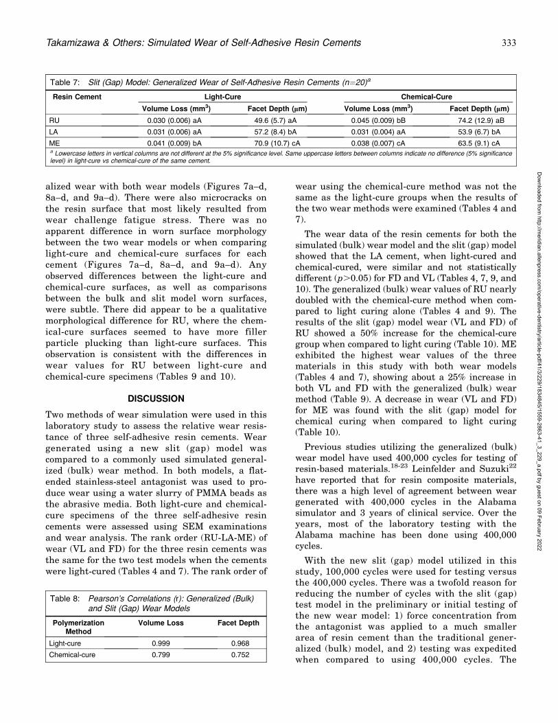
alized wear with both wear models (Figures 7a–d,8a–d, and 9a–d). There were also microcracks onthe resin surface that most likely resulted fromwear challenge fatigue stress. There was noapparent difference in worn surface morphologybetween the two wear models or when comparinglight-cure and chemical-cure surfaces for eachcement (Figures 7a–d, 8a–d, and 9a–d). Anyobserved differences between the light-cure andchemical-cure surfaces, as well as comparisonsbetween the bulk and slit model worn surfaces,were subtle. There did appear to be a qualitativemorphological difference for RU, where the chem-ical-cure surfaces seemed to have more fillerparticle plucking than light-cure surfaces. Thisobservation is consistent with the differences inwear values for RU between light-cure andchemical-cure specimens (Tables 9 and 10).
DISCUSSION
Two methods of wear simulation were used in thislaboratory study to assess the relative wear resis-tance of three self-adhesive resin cements. Weargenerated using a new slit (gap) model wascompared to a commonly used simulated general-ized (bulk) wear method. In both models, a flat-ended stainless-steel antagonist was used to pro-duce wear using a water slurry of PMMA beads asthe abrasive media. Both light-cure and chemical-cure specimens of the three self-adhesive resincements were assessed using SEM examinationsand wear analysis. The rank order (RU-LA-ME) ofwear (VL and FD) for the three resin cements wasthe same for the two test models when the cementswere light-cured (Tables 4 and 7). The rank order of
wear using the chemical-cure method was not thesame as the light-cure groups when the results ofthe two wear methods were examined (Tables 4 and7).
The wear data of the resin cements for both thesimulated (bulk) wear model and the slit (gap) modelshowed that the LA cement, when light-cured andchemical-cured, were similar and not statisticallydifferent (p.0.05) for FD and VL (Tables 4, 7, 9, and10). The generalized (bulk) wear values of RU nearlydoubled with the chemical-cure method when com-pared to light curing alone (Tables 4 and 9). Theresults of the slit (gap) model wear (VL and FD) ofRU showed a 50% increase for the chemical-curegroup when compared to light curing (Table 10). MEexhibited the highest wear values of the threematerials in this study with both wear models(Tables 4 and 7), showing about a 25% increase inboth VL and FD with the generalized (bulk) wearmethod (Table 9). A decrease in wear (VL and FD)for ME was found with the slit (gap) model forchemical curing when compared to light curing(Table 10).
Previous studies utilizing the generalized (bulk)wear model have used 400,000 cycles for testing ofresin-based materials.18-23 Leinfelder and Suzuki22
have reported that for resin composite materials,there was a high level of agreement between weargenerated with 400,000 cycles in the Alabamasimulator and 3 years of clinical service. Over theyears, most of the laboratory testing with theAlabama machine has been done using 400,000cycles.
With the new slit (gap) model utilized in thisstudy, 100,000 cycles were used for testing versusthe 400,000 cycles. There was a twofold reason forreducing the number of cycles with the slit (gap)test model in the preliminary or initial testing ofthe new wear model: 1) force concentration fromthe antagonist was applied to a much smallerarea of resin cement than the traditional gener-alized (bulk) model, and 2) testing was expeditedwhen compared to using 400,000 cycles. The
Table 7: Slit (Gap) Model: Generalized Wear of Self-Adhesive Resin Cements (n=20)a
Resin Cement Light-Cure Chemical-Cure
Volume Loss (mm3) Facet Depth (lm) Volume Loss (mm3) Facet Depth (lm)
RU 0.030 (0.006) aA 49.6 (5.7) aA 0.045 (0.009) bB 74.2 (12.9) aB
LA 0.031 (0.006) aA 57.2 (8.4) bA 0.031 (0.004) aA 53.9 (6.7) bA
ME 0.041 (0.009) bA 70.9 (10.7) cA 0.038 (0.007) cA 63.5 (9.1) cAa Lowercase letters in vertical columns are not different at the 5% significance level. Same uppercase letters between columns indicate no difference (5% significancelevel) in light-cure vs chemical-cure of the same cement.
Table 8: Pearson’s Correlations (r): Generalized (Bulk)and Slit (Gap) Wear Models
PolymerizationMethod
Volume Loss Facet Depth
Light-cure 0.999 0.968
Chemical-cure 0.799 0.752
Takamizawa & Others: Simulated Wear of Self-Adhesive Resin Cements 333
Dow
nloaded from http://m
eridian.allenpress.com/operative-dentistry/article-pdf/41/3/229/1834845/1559-2863-41_3_229_a.pdf by guest on 09 February 2022
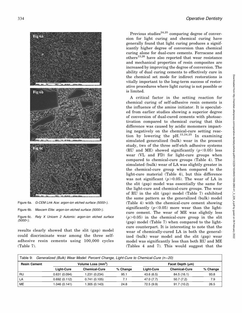
results clearly showed that the slit (gap) model
could discriminate wear among the three self-
adhesive resin cements using 100,000 cycles
(Table 7).
Previous studies24,25 comparing degree of conver-sion for light curing and chemical curing havegenerally found that light curing produces a signif-icantly higher degree of conversion than chemicalcuring alone for dual-cure cements. Ferracane andothers13,26 have also reported that wear resistanceand mechanical properties of resin composites areincreased by improving the degree of conversion. Theability of dual curing cements to effectively cure inthe chemical set mode for indirect restorations isvitally important to the long-term success of restor-ative procedures where light curing is not possible oris limited.
A critical factor in the setting reaction forchemical curing of self-adhesive resin cements isthe influence of the amine initiator. It is speculat-ed from earlier studies showing a superior degreeof conversion of dual-cured cements with photoac-tivation compared to chemical curing that thisdifference was caused by acidic monomers impact-ing negatively on the chemical-cure setting reac-tion by lowering the pH.15,24,25 In examiningsimulated generalized (bulk) wear in the presentstudy, two of the three self-etch adhesive systems(RU and ME) showed significantly (p,0.05) lesswear (VL and FD) for light-cure groups whencompared to chemical-cure groups (Table 4). Thesimulated (bulk) wear of LA was slightly greater inthe chemical-cure group when compared to thelight-cure material (Table 4), but this differencewas not significant (p.0.05). The wear of LA inthe slit (gap) model was essentially the same forthe light-cure and chemical-cure groups. The wearof RU in the slit (gap) model (Table 7) exhibitedthe same pattern as the generalized (bulk) model(Table 4) with the chemical-cure cement showingsignificantly (p,0.05) more wear than the light-cure cement. The wear of ME was slightly less(p.0.05) in the chemical-cure group in the slit(gap) model (Table 7) when compared to the light-cure counterpart. It is interesting to note that thewear of chemically-cured LA in both the general-ized (bulk) wear model and the slit (gap) wearmodel was significantly less than both RU and ME(Tables 4 and 7). This would suggest that the
Figure 6a. G-CEM Link Ace: argon-ion etched surface (50003).
Figure 6b. Maxcem Elite: argon-ion etched surface (50003).
Figure 6c. Rely X Unicem 2 Automix: argon-ion etched surface(50003).
Table 9: Generalized (Bulk) Wear Model: Percent Change, Light-Cure to Chemical-Cure (n=20)
Resin Cement Volume Loss (mm3) Facet Depth (lm)
Light-Cure Chemical-Cure % Change Light-Cure Chemical-Cure % Change
RU 0.631 (0.094) 1.231 (0.234) 95.1 43.6 (6.5) 84.5 (16.1) 93.8
LA 0.692 (0.112) 0.741 (0.105) 7.1 47.0 (7.7) 50.7 (7.2) 7.9
ME 1.046 (0.141) 1.305 (0.143) 24.8 72.5 (9.9) 91.7 (10.2) 26.5
334 Operative Dentistry
Dow
nloaded from http://m
eridian.allenpress.com/operative-dentistry/article-pdf/41/3/229/1834845/1559-2863-41_3_229_a.pdf by guest on 09 February 2022

polymerization reaction with chemical curing forLA is more effective than RU and ME. Thus, thenull hypotheses—1) there will not be a significantdifference in wear values among three dual-curedself-adhesive resin cements and 2) wear of self-adhesive resin cements using light-cure or chem-ical-cure methods will not be different—are reject-ed for RU and ME in the generalized (bulk) wearmodel but not for LA and rejected for RU in the slit(gap) wear model.
Belli and others14 reported data for self-adhesiveresin cements using a gap model for both tooth-brush abrasion and the ACTA wear method. Theyrelated that self-adhesive resin cements exhibitedgood wear resistance to toothbrush abrasion butshowed much more wear under the heavier loadingwith the ACTA test. They also reported that nocorrelation (R2=0.0567) was found between thetwo test methods. In the present study comparing
the generalized (bulk) wear model and the slit(gap) model, a strong correlation (Pearson) wasfound between the light-cure groups for both VL(r=0.999) and FD (r=0.968). For the chemical-curegroups, the correlations between the two modelswere r=0.799 for VL and r=0.752 for FD. Whilethe associations for the chemical-cure groups werenot as robust as for the light-cure groups, therelationships were good for the chemical-curegroups with the two wear methods used in thisstudy.
Barkmeier and others18-20 have conducted severalstudies with the generalized (bulk) wear model usedin this study to evaluate the wear resistance of resincomposite materials. The new slit or gap model wasused in the present study to more closely parallel aclinical situation in the assessment of wear resis-tance of self-adhesive resin cements. The new slit(gap) model delivers the wear challenges with the
Figure 7a. G-CEM Link Ace/light-cure: general ized (bulk) wear(400,000 cycles) near center of wearfacet (25003).
Figure 7b. G-CEM Link Ace/chemi-cal-cure: generalized (bulk) wear(400,000 cycles) near center of wearfacet (25003).
Figure 7c. G-CEM Link Ace/light-cure: slit (gap) generalized wear(100,000 cycles) near center of wearfacet (25003).
Figure 7d. G-CEM Link Ace/chemi-cal-cure: slit (gap) generalized wear(100,000 cycles) near center of wearfacet (25003).
Table 10: Slit (Gap) Wear Model: Percent Change, Light-Cure to Chemical-Cure (n=20)
Resin Cement Volume Loss (mm3) Facet Depth (lm)
Light-Cure Chemical-Cure % Change Light-Cure Chemical-Cure % Change
RU 0.030 (0.006) 0.045 (0.009) 50.0 49.6 (5.7) 74.2 (12.9) 49.6
LA 0.031 (0.006) 0.031 (0.004) 0.0 57.2 (8.4) 53.9 (6.7) �5.8
ME 0.041 (0.009) 0.038 (0.007) �7.3 70.9 (10.7) 63.5 (9.1) �10.4
Takamizawa & Others: Simulated Wear of Self-Adhesive Resin Cements 335
Dow
nloaded from http://m
eridian.allenpress.com/operative-dentistry/article-pdf/41/3/229/1834845/1559-2863-41_3_229_a.pdf by guest on 09 February 2022
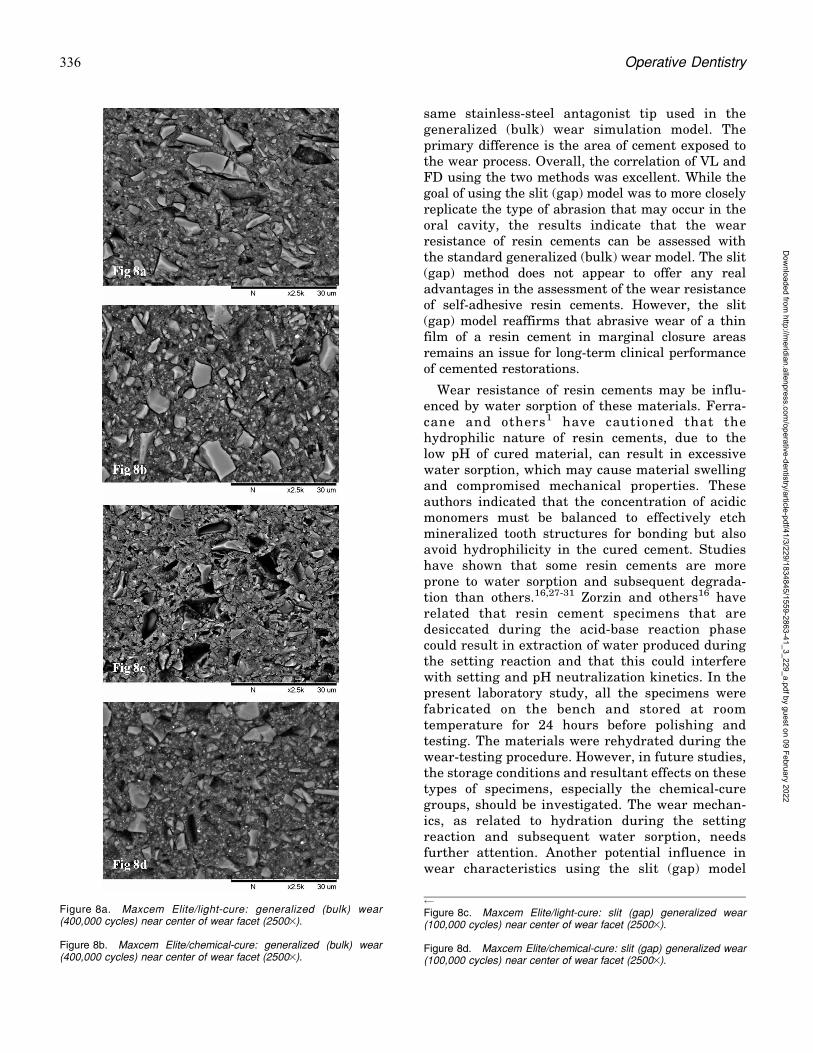
same stainless-steel antagonist tip used in thegeneralized (bulk) wear simulation model. Theprimary difference is the area of cement exposed tothe wear process. Overall, the correlation of VL andFD using the two methods was excellent. While thegoal of using the slit (gap) model was to more closelyreplicate the type of abrasion that may occur in theoral cavity, the results indicate that the wearresistance of resin cements can be assessed withthe standard generalized (bulk) wear model. The slit(gap) method does not appear to offer any realadvantages in the assessment of the wear resistanceof self-adhesive resin cements. However, the slit(gap) model reaffirms that abrasive wear of a thinfilm of a resin cement in marginal closure areasremains an issue for long-term clinical performanceof cemented restorations.
Wear resistance of resin cements may be influ-enced by water sorption of these materials. Ferra-cane and others1 have cautioned that thehydrophilic nature of resin cements, due to thelow pH of cured material, can result in excessivewater sorption, which may cause material swellingand compromised mechanical properties. Theseauthors indicated that the concentration of acidicmonomers must be balanced to effectively etchmineralized tooth structures for bonding but alsoavoid hydrophilicity in the cured cement. Studieshave shown that some resin cements are moreprone to water sorption and subsequent degrada-tion than others.16,27-31 Zorzin and others16 haverelated that resin cement specimens that aredesiccated during the acid-base reaction phasecould result in extraction of water produced duringthe setting reaction and that this could interferewith setting and pH neutralization kinetics. In thepresent laboratory study, all the specimens werefabricated on the bench and stored at roomtemperature for 24 hours before polishing andtesting. The materials were rehydrated during thewear-testing procedure. However, in future studies,the storage conditions and resultant effects on thesetypes of specimens, especially the chemical-curegroups, should be investigated. The wear mechan-ics, as related to hydration during the settingreaction and subsequent water sorption, needsfurther attention. Another potential influence inwear characteristics using the slit (gap) model
Figure 8a. Maxcem Elite/light-cure: generalized (bulk) wear(400,000 cycles) near center of wear facet (25003).
Figure 8b. Maxcem Elite/chemical-cure: generalized (bulk) wear(400,000 cycles) near center of wear facet (25003).
Figure 8c. Maxcem Elite/light-cure: slit (gap) generalized wear(100,000 cycles) near center of wear facet (25003).
Figure 8d. Maxcem Elite/chemical-cure: slit (gap) generalized wear(100,000 cycles) near center of wear facet (25003).
336 Operative Dentistry
Dow
nloaded from http://m
eridian.allenpress.com/operative-dentistry/article-pdf/41/3/229/1834845/1559-2863-41_3_229_a.pdf by guest on 09 February 2022
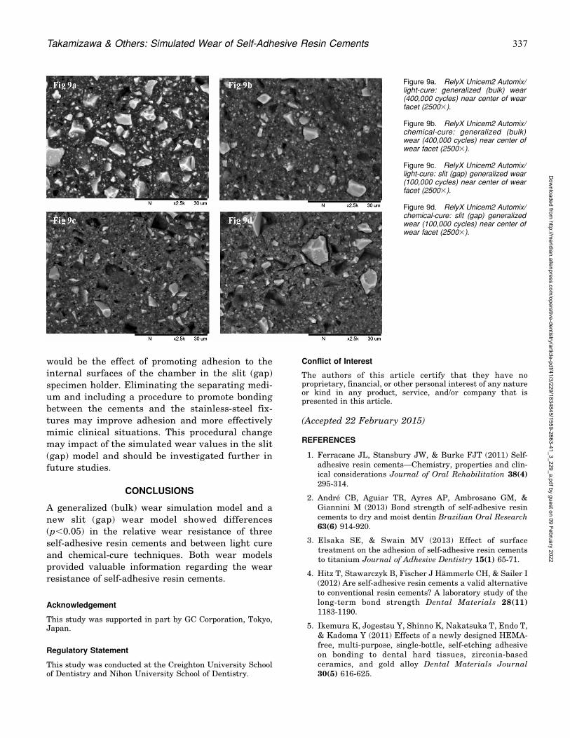
would be the effect of promoting adhesion to the
internal surfaces of the chamber in the slit (gap)
specimen holder. Eliminating the separating medi-
um and including a procedure to promote bonding
between the cements and the stainless-steel fix-
tures may improve adhesion and more effectively
mimic clinical situations. This procedural change
may impact of the simulated wear values in the slit
(gap) model and should be investigated further in
future studies.
CONCLUSIONS
A generalized (bulk) wear simulation model and a
new slit (gap) wear model showed differences
(p,0.05) in the relative wear resistance of three
self-adhesive resin cements and between light cure
and chemical-cure techniques. Both wear models
provided valuable information regarding the wear
resistance of self-adhesive resin cements.
Acknowledgement
This study was supported in part by GC Corporation, Tokyo,Japan.
Regulatory Statement
This study was conducted at the Creighton University Schoolof Dentistry and Nihon University School of Dentistry.
Conflict of Interest
The authors of this article certify that they have noproprietary, financial, or other personal interest of any natureor kind in any product, service, and/or company that ispresented in this article.
(Accepted 22 February 2015)
REFERENCES
1. Ferracane JL, Stansbury JW, & Burke FJT (2011) Self-adhesive resin cements—Chemistry, properties and clin-ical considerations Journal of Oral Rehabilitation 38(4)295-314.
2. Andre CB, Aguiar TR, Ayres AP, Ambrosano GM, &Giannini M (2013) Bond strength of self-adhesive resincements to dry and moist dentin Brazilian Oral Research63(6) 914-920.
3. Elsaka SE, & Swain MV (2013) Effect of surfacetreatment on the adhesion of self-adhesive resin cementsto titanium Journal of Adhesive Dentistry 15(1) 65-71.
4. Hitz T, Stawarczyk B, Fischer J Hammerle CH, & Sailer I(2012) Are self-adhesive resin cements a valid alternativeto conventional resin cements? A laboratory study of thelong-term bond strength Dental Materials 28(11)1183-1190.
5. Ikemura K, Jogestsu Y, Shinno K, Nakatsuka T, Endo T,& Kadoma Y (2011) Effects of a newly designed HEMA-free, multi-purpose, single-bottle, self-etching adhesiveon bonding to dental hard tissues, zirconia-basedceramics, and gold alloy Dental Materials Journal30(5) 616-625.
Figure 9a. RelyX Unicem2 Automix/light-cure: generalized (bulk) wear(400,000 cycles) near center of wearfacet (25003).
Figure 9b. RelyX Unicem2 Automix/chemical-cure: generalized (bulk)wear (400,000 cycles) near center ofwear facet (25003).
Figure 9c. RelyX Unicem2 Automix/light-cure: slit (gap) generalized wear(100,000 cycles) near center of wearfacet (25003).
Figure 9d. RelyX Unicem2 Automix/chemical-cure: slit (gap) generalizedwear (100,000 cycles) near center ofwear facet (25003).
Takamizawa & Others: Simulated Wear of Self-Adhesive Resin Cements 337
Dow
nloaded from http://m
eridian.allenpress.com/operative-dentistry/article-pdf/41/3/229/1834845/1559-2863-41_3_229_a.pdf by guest on 09 February 2022

6. Cavalcanti AN, Foxton RM, Watson TF, Oliveira MT,Giannini M, & Marchi GM (2009) Bond strength of resincements to a zirconia ceramic with different surfacetreatments Operative Dentistry 34(3) 280-287.
7. Hikita K, Van Meerbeek, De Munch J, Ikeda T. VanKanduyt K, Maida T, Lambrechts P, & Peumans M (2007)Bonding effectiveness of adhesive luting agents to enameland dentin Dental Materials 23(1) 71-80.
8. De Munch J, Vargas M, Van Landuyt K, Hikita K,Lambrechts P, & Van Meerbeek B (2004) Bonding of anauto-adhesive luting material to enamel and dentinDental Materials 20(10) 963-971.
9. Heintze SD (2007) Systematic reviews: I. The correlationbetween laboratory tests on marginal quality and bondstrength. II. The correlation between marginal qualityand clinical outcome Journal of Adhesive Dentistry9(Supplement 1) 77-106.
10. Kawai K, Isenberg BP, & Leinfelder, KF (1994) Effect ofgap dimension on composite resin cement wear Quintes-sence International 25(1) 53-58.
11. Torii Y, Itou K, Itota T, Hama K, Konishi N, Nagamine M,& Inoue K (1999) Influence of filler content and gapdimension on wear resistance of resin composite lutingcements around a CAD/CAM ceramic inlay restorationDental Materials Journal 18(4) 453-461.
12. Shinkai K, Suzuki S, Leinfelder KF, & Katoh Y (1995)Effect of gap dimension on wear resistance of lutingcements American Journal of Dentistry 8(2) 149-151.
13. Ferracane JL, Mitchem JC, Condon JR, & Todd T (1997)Wear and marginal breakdown of composites withvarious degree of cure Journal of Dental Research 76(8)1508-1516.
14. Belli R, Pelka M, Petschelt A, & Lohbauer U (2009) Invitro wear gap formation of self-adhesive resin cements: ACLSM evaluation Journal of Dentistry 37(12) 984-993.
15. Vrochari AD, Eliades G, Hellwig E, & Wrbas KT (2009)Curing efficiency of four self-etching, self-adhesive resincements Dental Materials 25(9) 1104-1108.
16. Zorzin J, Petschelt A, Ebert J, & Lohbauer U (2012) pHneutralization and influence on mechanical strength inself-adhesive resin luting agents Dental Materials 28(6)672-679.
17. Salz U, Zimmermann J, & Salzer T (2005) Self-curing,self-adhesive cements systems Journal of AdhesiveDentistry 7(1) 7-17.
18. Barkmeier WW, Takamizawa T, Erickson RL, TsujimotoA, Latta MA, & Miyazaki M (2015) Localized andgeneralized simulated wear of resin composites OperativeDentistry 40(3) 322-335.
19. Barkmeier WW, Erickson RL, Latta MA, & WilwerdingTM (2008) Wear simulation of resin composites and therelationship to clinical wear Operative Dentistry 33(2)177-182.
20. Barkmeier WW, Erickson RL, Latta MA, & WilwerdingTM (2013) Wear rates of resin composites OperativeDentistry 38(2) 226-233.
21. Suzuki S, Leinfelder KF, Kawai K, & Tsuchitani Y(1995) Effect of particle variation on wear rates ofposterior composites American Journal of Dentistry8(4) 173-178.
22. Leinfelder KF, & Suzuki S (1999) In vitro wear device fordetermining posterior composite wear Journal of theAmerican Dental Association 130(9) 1347-1353.
23. Suzuki S, Nagai E, Taira Y, & Minesaki Y (2002) In vitrowear of indirect composite restoratives Journal ofProsthetic Dentistry 88(4) 431-436.
24. Harashima I, Nomata T, & Hirasawa T (1991) Degree ofconversion of dual-cured composite luting cements DentalMaterials Journal 10(1) 8-17.
25. Braga RR, Cesar PF, & Gonzaga CC (2002) Mechanicalproperties of resin cements with different activationmodes Journal of Oral Rehabilitation 29(3) 257-262.
26. Ferracane JL, & Berge HX (1995) Fracture toughness ofexperimental dental composites aged in ethanol Journalof Dental Research 74(7) 1418-1423.
27. Vrochari AD, Eliades G, Hellwig E, & Wrbas KT (2010)Water sorption and solubility of four self-etching self-adhesive resin luting agents Journal of Adhesive Den-tistry 12(1) 39-43.
28. Marghalani HY (2012) Sorption and solubility character-istics of self-adhesive resin cements Dental Materials28(10) e187-e198.
29. Silva EM, Noronha-Filho JD, Amaral CM, Poskus LT, &Guimaraes JD (2013) Long-term degradation of resin-based cements in substances present in the oral environ-ment: Influence of activation mode Journal of AppliedOral Science 21(3) 271-273.
30. da Silva Fonseca AS, Mizrahi J, Menezes LR, Valente LL,DeMoraes RR, & Schneider LF (2014) The effect of timebetween handling and photoactivation on self-adhesiveresin cement properties Journal of Prosthodontics doi: 10.1111/jopr.12108 [Epub ahead of print].
31. Schittly E, Le Goff S, Besnault C, Sadoun M, & Ruse ND(2014) Effect of water storage on the flexural strength offour self-etching adhesive resin cements and on thedentin-titanium shear bond strength mediated by themOperative Dentistry 39(4) E171-E177.
338 Operative Dentistry
Dow
nloaded from http://m
eridian.allenpress.com/operative-dentistry/article-pdf/41/3/229/1834845/1559-2863-41_3_229_a.pdf by guest on 09 February 2022

Departments
ERRATUMOperative Dentistry would like to clarify authororder in ‘‘Brambilla E, Ionescu A, Cazzaniga G, &Ottobelli M (2016) Influence of light-curing pa-rameters on biofilm development and flexuralstrength of a silorane-based composite OperativeDentistry 41(2) 219-227. The correct author ordershould be ‘‘Ottobelli M, Ionescu A, Cazzaniga G, &Brambilla E.’’ Operative Dentistry apologizes forany confusion.
LETTER TO THE EDITOR
Dear Dr Platt:
I read with interest the following manuscript:Torres CR, & Borges AB (2015) Color masking ofdevelopmental enamel defects: A case series Opera-tive Dentistry 40(1) 25-33 (my special thanks to theauthors for such a great clinical manuscript).
I have to disagree with the credit given toreferences 13 and 14 on page 26. The resininfiltration concept is at least 40 years old. Robinsonand others1 in 1976 etched enamel with hydrochloricacid (HCl) and infiltrated this etched enamel with aresorcinol-formaldehyde resin as a potential cario-static treatment. Then, in 1987, Croll2 used a clearresin sealant on etched enamel to saturate thesurfaces with resin. Among several research paperson the topic of microabrasion followed by resininfiltration published in the 2000s, I would highlighttwo from the same research group. In 2007, Parisand others3 used confocal microscopy to (elegantly)study resin infiltration of carious lesions using 37%H3PO4 or 15% HCl to etch enamel, followed byimmersion in ethanol for 30 seconds and theapplication of the commercial dentin adhesiveExciTE (Ivoclar Vivadent). In 2009, Paris andMeyer-Lueckel4 described the masking of whitespots with resin infiltration using 15% HCl etchingfollowed by a drying step with ethanol and a very-low-viscosity light-cured resin (tetraethylene glycoldimethacrylate).
I hope that this clarification gives the deservedcredit to the authors of the missing references,
especially to the papers of Robinson and others1
and Croll2, which are often overlooked.
Sincerely,
Jorge Perdigao
REFERENCES
1. Robinson C, Hallsworth AS, Weatherell JA, & Kunzel W(1976) Arrest and control of carious lesions: A study basedon preliminary experiments with resorcinol-formaldehyderesin Journal of Dental Research 55 812-818.
2. Croll TP (1987) Bonded resin sealant for smooth surfaceenamel defects: New concepts in ‘‘microrestorative’’ den-tistry Quintessence International 18 5-10.
3. Paris S, Meyer-Lueckel H, & Kielbassa AM (2007) Resininfiltration of natural caries lesions Journal of DentalResearch 86 662-666.
4. Paris S, & Meyer-Lueckel H (2009) Masking of labialenamel white spot lesions by resin infiltration—A clinicalreport Quintessence International 40 713-718.
Author Response
Dear Dr Jorge Perdigao,
Thank you for expressing your opinion. Ourintention in citing references 13 and 14 in theoriginal manuscript was to reference the process ofpenetration, hardening, and inhibiting lesion pro-gression, as also demonstrated in these studies.Nevertheless, the author of the letter to the editor iscompletely correct about the development of theinfiltration concept. We apologize for this mistake,because we used the term ‘‘introduced’’ in thesentence, and we agree that the credit should begiven to the authors referenced in the letter to theeditor.
Best regards,
Dr Borges and coauthors
Prof. Dr. Alessandra Buhler BorgesInstitute of Science and Technology of Sao Jose dosCampos - UNESPDepartament of Restorative DentistryAv. Eng. Francisco Jose Longo, 777 Jd. Sao DimasSao Jose dos Campos - SP CEP: 12245-000e-mail: [email protected]
�Operative Dentistry, 2016, 41-3, 339-339
Dow
nloaded from http://m
eridian.allenpress.com/operative-dentistry/article-pdf/41/3/229/1834845/1559-2863-41_3_229_a.pdf by guest on 09 February 2022
Online Only Articles
Online Only Articles
On occasion we receive manuscripts that we would like to publish, but do not have the page room toinclude in the print journal. For the full article, please go to www.jopdentonline.org or enter theprovided address into your address bar.
Evaluation of Bond Strength and Microleakage of a Novel Metal-titanate Antibacterial Agent
S Deng � KH Chung � DCN Chan � C Spiekerman
Clinical Relevance: The novel antibacterial nanoparticulate metal-titanate complexes under investigation will allowclinicians to tackle the composite longevity problem at its weakest interface.
doi: http://dx.doi.org/10.2341/14-257-L
Effect of Cigarette Smoke on Resin Composite Bond Strength toEnamel and Dentin Using Different Adhesive Systems
JD Theobaldo � A Catelan � U Rodrigues-Filho � GM Marchi � DANL Lima � FHB Aguiar
Clinical Relevance: The exposure of dentin to cigarette smoke influences adhesive bonding strength. However,cigarette smoke does not influence the bond strength to enamel.
doi: http://dx.doi.org/10.2341/15-056-L
Longevity of Bonding of Self-adhesive Resin Cement to Dentin
TC Simoes � IV Luque-Martinez � RR Moraes � ATG Sa � AD Loguercio � SK Moura
Clinical Relevance: Regardless of the cementing strategy, the durability of bonding to root canal dentin may beinfluenced by the dentin treatment protocol.
doi: http://dx.doi.org/10.2341/14-266-LR
Composite Replacement of Amalgam Restoration VersusFreshly Cut Dentin: An In Vitro Microleakage Comparison
H Redwan � DN Bardwell � A Ali � M Finkelman � S Khayat � H-P Weber
Clinical Relevance: Bonding to dentin under replaced amalgam restorations may be as effective as bonding to freshlycut dentin.
doi: http://dx.doi.org/10.2341/14-278-L
Mechanical Properties and Sliding-impact Wear Resistance of Self-adhesive Resin Cements
T Furuichi � T Takamizawa � A Tsujimoto � M Miyazaki � WW Barkmeier � MA Latta
Clinical Relevance: Mechanical properties and wear resistance are important parameters for the selection of resinluting cements. For wear resistance, the self-adhesive resin cements, apart from GL, showed significantly lower valuesthan the conventional resin cements.
doi: http://dx.doi.org/10.2341/15-033-L
340 Operative Dentistry
Dow
nloaded from http://m
eridian.allenpress.com/operative-dentistry/article-pdf/41/3/229/1834845/1559-2863-41_3_229_a.pdf by guest on 09 February 2022
Evaluation of Bond Strength andMicroleakage of a Novel Metal-
titanate Antibacterial Agent
S Deng � KH ChungDCN Chan � C Spiekerman
Clinical Relevance
The novel antibacterial nanoparticulate metal-titanate complexes under investigation willallow clinicians to tackle the composite longevity problem at its weakest interface.
SUMMARY
Objectives: To evaluate the effect on both bondstrength and microleakage of incorporation ofa novel antibacterial nanoparticulate metal-titanate complex (nMT) into a dental adhesivesystem.
Materials and Methods: Eighty extracted hu-man molars were prepared to determinewhether incorporation of nMT into bondingagents can affect shear bond strength (SBS)and adhesive strength fatigue. SBS was mea-
sured with a universal testing machine, andthe peak force at failure was recorded. Anelectromechanical fatigue machine was usedfor cyclic loading treatment of specimens.Differences in the SBS values among groupswere identified using analysis of variance andTukey post hoc analyses (a=0.05). Twenty stan-dard Class V cavities were restored to examinemicroleakage when the primer/bonding resinwas modified with 10 wt% nMT. Microleakageat the enamel and dentin margins was calcu-lated as a percentage of the full length of thecavity. Results of the microleakage experimentwere analyzed with paired and independentsample t-tests (a=0.05).
Results: The mean (6 standard deviation)shear bond strength values of before fatigueand after fatigue ranged from 21.9 (2.5) MPa to23.9 (3.8) MPa and from 17.1 (2.5) MPa to 17.7(2.5) MPa respectively. No statistically signifi-cant differences in failure force were observedamong groups (p=0.70). Microleakage underall conditions was significantly greater in thedentin margins than in the enamel margins(p,0.05). There was no evidence that micro-leakage differed between the experimental
Shuli Deng, Department of Conservative Dentistry, AffiliatedHospital of Stomatology, Zhejiang University, Zhejiang,China
Kwok-Hung Chung, Department of Restorative Dentistry,University of Washington, School of Dentistry, Seattle, WA,USA
*Daniel CN Chan, Department of Restorative Dentistry,University of Washington, School of Dentistry, Seattle, WA,USA
Charles Spiekerman, Department of Oral Health Sciences,University of Washington, School of Dentistry, Seattle, WA,USA
*Corresponding author: Seattle, WA 98195-6365, USA; e-mail: [email protected]
DOI: 10.2341/14-257-L
�Operative Dentistry, 2016, 41-3, E48-E56
Dow
nloaded from http://m
eridian.allenpress.com/operative-dentistry/article-pdf/41/3/229/1834845/1559-2863-41_3_229_a.pdf by guest on 09 February 2022
groups with modified primer and bondingresin.
Conclusions: Incorporating nMT into a dentaladhesive system will not compromise the resincomposite’s tooth bonding and sealing ability.
INTRODUCTION
Biofilms are medically important because microbesin the biofilm state are more pathogenic than whenthey are planktonic (nonadherent and free floating).The National Institutes of Health reported1 thatbacteria growing as a biofilm cause 80% of infec-tions in the body. Biofilms at the margin of anexisting dental restoration give rise to secondarycaries, which are the main complication necessitat-ing replacement of composite fillings.2,3 Currentlythere is a lack of research data both on 1) the effectof the cariogenic biofilm community structure andits metabolic processes on the tooth/resin compositeinterface and 2) the effect of the tooth/resincomposite on biofilm formation and populationdistribution.
Monosodium titanate (MST) is an inorganic,particulate compound of titanium oxide (NaTi2O5H)with an amorphous core and crystalline surface.MST has a spherical morphology with an approxi-mate diameter of 1-10 lm. Our team has developedmicroparticulate metal-titanate complexes as a newclass of antibacterial agents.4-7 The microparticulategold (III)–loaded titanate complexes inhibit growthof oral bacteria at micromolar concentrations. Wepredict that nanoparticulate metal-titanate complex-es (nMTs) in turn will be even more effective atinhibiting oral bacteria growth as such complexeshave a significantly greater surface-to-volume ratio,resulting in more effective ion-exchange character-istics. This approach is innovative as nMTs areceramic in nature and act like resin filler. Inaddition, because these complexes are not organic,degradation is not an issue, and thus they can havelong-term effectiveness and also may be less likelythan organic antibacterial agents to contribute tobacterial resistance.8
Dental composite restorations are becoming thematerial of choice for posterior restorations; thus,their longevity is important to patients, dentists,federal health agencies, and insurance companies.A survey of 24 prospective studies on the clinicalperformance of posterior resin composites pub-lished between 1996 and 2002 indicated that theprimary reasons for composite failure were second-ary caries, restoration fracture, and marginal
defects.2 Secondary caries was the principal causeof composite failure necessitating replacement offillings; these caries are lesions at the margin of anexisting restoration and usually occur in areas ofbiofilm stagnation.9 For this reason, the cervicalmargins of restorations are commonly affected. Inthe oral cavity, mixed microbial biofilms canaccumulate on hard and soft tissues and areinvolved in the pathogenesis of caries and peri-odontitis. A biofilm is an accumulation of bacteria,fungi, or protozoa on solid surfaces. In dentistry,two popular approaches to preventing biofilmformation are 1) to design a biomaterial that slowlyreleases an agent that is lethal to the approachingbacterial cells and 2) to develop a nonadhesivesurface by modifying the surface chemistry ofrestorative materials.10 Various chemical agentscan affect bacterial adhesion indirectly by disrupt-ing bacterial cell metabolism. Numerous materialshave been impregnated with various antibioticsonly to have most of the agent released over a veryshort time, thus providing no long-term effect.11
Recent studies have shown that sublethal doses ofantibiotics can induce bacterial resistance andactually enhance biofilm formation. The potentialnegative consequences of bacterial resistance toantibiotics are dire because they put all of society atrisk.12
Metal-based antibacterials are an attractivealternative. Metal ions have chemical propertiesthat inhibit bacterial growth.13 The unique bind-ing, coordination, and redox properties makedevelopment of bacterial resistance less likelyand predict effectiveness across a broad bacterialspectrum. For example, Ag(I) and Hg have a longhistory as antibacterials.14 However, fears ofsystemic toxicity have limited Ag(I) and Hg usein recent years.15 Other metal ions, such asAu(III), Pd(II), and Pt(IV), have binding proper-ties, coordination chemistries, and redox propertiesthat suggest they also would be effective antibac-terials. Unfortunately, development of new metal-based antibacterials has been severely impededbecause of previous controversies and fears. Yet,given the increasing resistance of bacteria toorganic antibacterials, metal-based antibacterialsare a promising alternative. If systemic toxicitycould be limited and therapeutic indices wereoptimized, metal ions and their associated com-pounds could emerge as a new powerful class ofantibacterial agents.
The goal of this investigation was to evaluate thebond strength and microleakage of a kind of novel
Deng & Others: Bond Strength and Microleakage of Metal-titanate Complexes E49
Dow
nloaded from http://m
eridian.allenpress.com/operative-dentistry/article-pdf/41/3/229/1834845/1559-2863-41_3_229_a.pdf by guest on 09 February 2022

antibacterial nMT. This kind of nMT has been shownpreviously to have antimicrobial activity.6
METHODS AND MATERIALS
Experimental Materials
MSTs are particulate, micron-sized ion exchangersthat may be useful solid-phase platforms for deliveryof metal ions to inhibit bacterial growth.4-7 Thesynthesis of micro- and nano-titanate particles basedon a unique low-temperature (,808C) sol-gel processhas been reported.16 The process produces titanateswith superior ion-exchange characteristics comparedto those produced using hydrothermal materials.Previous work6 has shown that nanoparticulatetitanate complexes with loaded gold and cisplatinhave antimicrobial activities. An example of nano-particulate gold-loaded MST complexes (nMTs) isillustrated in Figure 1. A 10% wt/wt nMT powderwas added to Primer (All-Bond 2t, Bisco Inc,Schaumburg, IL, USA) and bonding resin (D/E resin,Bisco) and mixed in a dark room.
Shear Bond Strength (SBS) Test
The SBS test was conducted in three phases as themetal-titanate complexes were processed and avail-able. In the first phase, we examined whetherincorporation of MST affects the bond strength. Inthe second phase, we compared the effects of addingnMT. In the third phase, we examined the effect ofcyclic loading on bond strength.
In the first and second phases, 80 extracted
human molars were prepared with a coarse dia-
mond bur at axial surfaces to create an approxi-
mately 8-mm-diameter enamel or dentin flat
surface that was parallel with the long axis of each
tooth. The surfaces were further wet polished up to
600 grit. Two surfaces from each tooth were selected
for the bonding test, and the surfaces were etched
with 32% H3PO4 (Uni-Etch, Bisco) for 15 seconds.
They were thoroughly rinsed and the excess water
removed with a brief burst of air. The modification
of an adhesive system and bonding treatment are
described in Table 1. Filtek Supreme Plus (3M
ESPE, St Paul, MN, USA) was injected into a
Figure 1. Transmision electron mi-croscopy (TEM) image of nanoparti-culate gold-loaded monosodiumtitanate complexes.
Table 1: List of Experimental Groups for Shear BondStrength Test in This Study
Groups Bonding Treatment
Enamel (control) Etching, priming, and bondinga
Enamel þ 1 Etching, priming, and bonding with(D/E resinþ10 wt% MST)
Enamel þ 2 Etching, priming, and bonding with(D/E resinþ10 wt% nMT)
Dentin (control) Etching, priming, and bondinga
Dentin þ 1 Etching, priming with (primerþ10 wt% MST),and bonding
Dentin þ 2 Etching, priming with (primerþ10 wt% nMT),and bonding
Abbreviations: MST, monosodium titanate; nMT, nanoparticulate gold-loadedmonosodium titanate complex.a According to the manufacturer’s instruction for the Universal DentalAdhesive System: All-Bond 2t.
E50 Operative Dentistry
Dow
nloaded from http://m
eridian.allenpress.com/operative-dentistry/article-pdf/41/3/229/1834845/1559-2863-41_3_229_a.pdf by guest on 09 February 2022
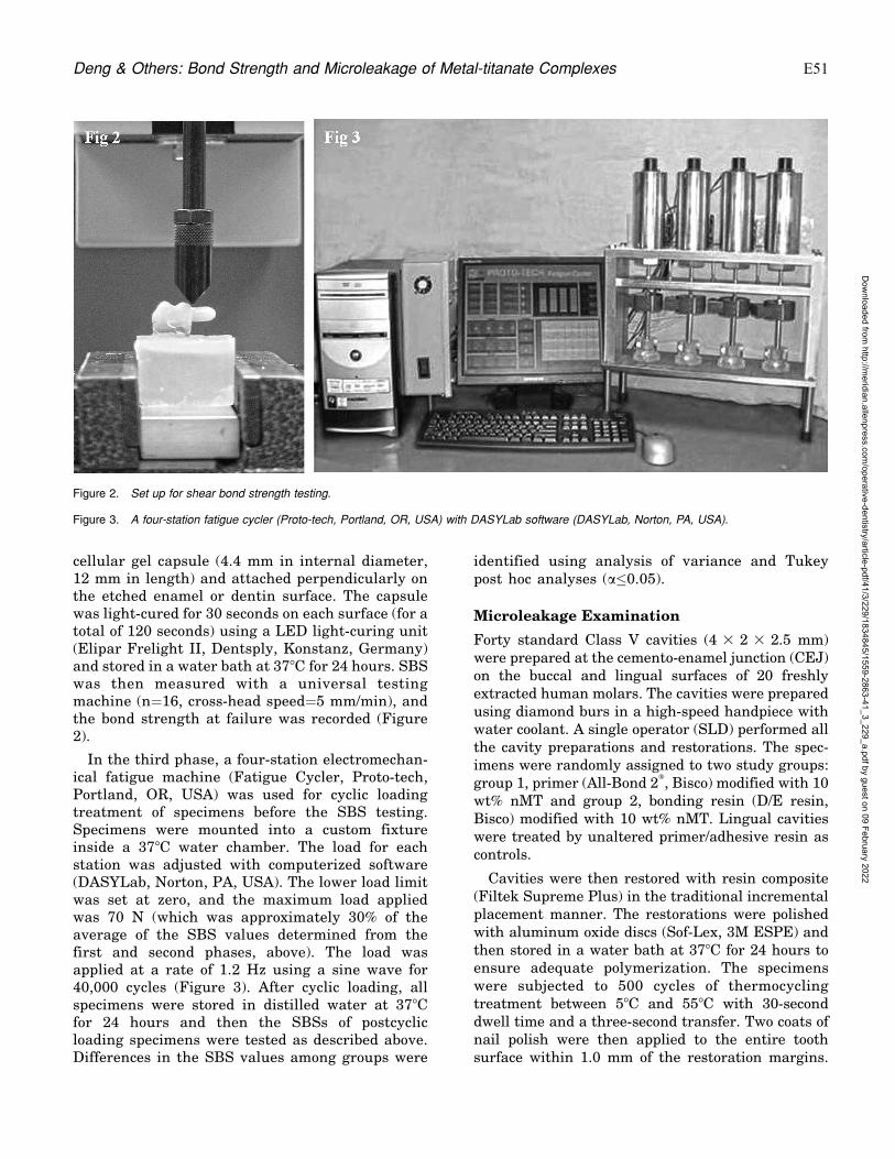
cellular gel capsule (4.4 mm in internal diameter,12 mm in length) and attached perpendicularly onthe etched enamel or dentin surface. The capsulewas light-cured for 30 seconds on each surface (for atotal of 120 seconds) using a LED light-curing unit(Elipar Frelight II, Dentsply, Konstanz, Germany)and stored in a water bath at 378C for 24 hours. SBSwas then measured with a universal testingmachine (n=16, cross-head speed=5 mm/min), andthe bond strength at failure was recorded (Figure2).
In the third phase, a four-station electromechan-ical fatigue machine (Fatigue Cycler, Proto-tech,Portland, OR, USA) was used for cyclic loadingtreatment of specimens before the SBS testing.Specimens were mounted into a custom fixtureinside a 378C water chamber. The load for eachstation was adjusted with computerized software(DASYLab, Norton, PA, USA). The lower load limitwas set at zero, and the maximum load appliedwas 70 N (which was approximately 30% of theaverage of the SBS values determined from thefirst and second phases, above). The load wasapplied at a rate of 1.2 Hz using a sine wave for40,000 cycles (Figure 3). After cyclic loading, allspecimens were stored in distilled water at 378Cfor 24 hours and then the SBSs of postcyclicloading specimens were tested as described above.Differences in the SBS values among groups were
identified using analysis of variance and Tukeypost hoc analyses (a�0.05).
Microleakage Examination
Forty standard Class V cavities (4 3 2 3 2.5 mm)were prepared at the cemento-enamel junction (CEJ)on the buccal and lingual surfaces of 20 freshlyextracted human molars. The cavities were preparedusing diamond burs in a high-speed handpiece withwater coolant. A single operator (SLD) performed allthe cavity preparations and restorations. The spec-imens were randomly assigned to two study groups:group 1, primer (All-Bond 2t, Bisco) modified with 10wt% nMT and group 2, bonding resin (D/E resin,Bisco) modified with 10 wt% nMT. Lingual cavitieswere treated by unaltered primer/adhesive resin ascontrols.
Cavities were then restored with resin composite(Filtek Supreme Plus) in the traditional incrementalplacement manner. The restorations were polishedwith aluminum oxide discs (Sof-Lex, 3M ESPE) andthen stored in a water bath at 378C for 24 hours toensure adequate polymerization. The specimenswere subjected to 500 cycles of thermocyclingtreatment between 58C and 558C with 30-seconddwell time and a three-second transfer. Two coats ofnail polish were then applied to the entire toothsurface within 1.0 mm of the restoration margins.
Figure 2. Set up for shear bond strength testing.
Figure 3. A four-station fatigue cycler (Proto-tech, Portland, OR, USA) with DASYLab software (DASYLab, Norton, PA, USA).
Deng & Others: Bond Strength and Microleakage of Metal-titanate Complexes E51
Dow
nloaded from http://m
eridian.allenpress.com/operative-dentistry/article-pdf/41/3/229/1834845/1559-2863-41_3_229_a.pdf by guest on 09 February 2022

The specimens were incubated separately in indi-vidual screw-capped tubes in a water bath at 378Cfor 24 hours before immersion in a 0.2% RhodamineB solution (Invitrogen, Carlsbad, CA, USA) for 24hours. The specimens were then rinsed with distilledwater for 10 minutes, dried, and embedded in a coldcuring epoxy resin for two hours at 608C. Blocks ofresin were left at room temperature for six hours toachieve complete polymerization. Finally, two bucco-lingual sections were made through each restorationwith a low-speed diamond saw (IsoMet, Buehler Ltd,Lake Bluff, IL, USA). Each tooth was sectioned intothree pieces, producing four cross-sectional faces forevaluation. Sections were assessed for dye penetra-tion with an optical microscope (Nikon Eclips E600,Tokyo, Japan) at 320 magnification at the occlusaland cervical margins. A total of 160 scores for dentinmargins and enamel margins were recorded. Micro-leakage at the enamel and dentin margins wascalculated as a percentage of the full length of thecavity (Figure 4). The results of the microleakageexamination were analyzed with paired and inde-pendent sample t-tests (a=0.05).
Operator Reliability Evaluation
To ensure consistency and reliability of the observa-tions, 20 sections were chosen randomly to assessinteroperator reliability. A second evaluator readeach section under the same conditions. Correlationcoefficients for both enamel and dentin percentagesbetween the two evaluators (DCNC and SLD) were
as follows: enamel%, r = 0.915; dentin%, r = 0.936.No statistically significant differences were observedbetween the two operators’ evaluations (paired t-test, p.0.05). Following the interoperator reliabilityassessment, selected sections were also evaluated bya confocal laser scanning electron microscope(Zeiss510, Carl Zeiss Ltd, Thornwood, NY, USA)(Figure 5).
RESULTS
The mean (6 standard deviation) SBS values ofbefore fatigue and after fatigue ranged from 21.9(2.5) MPa to 23.9 (3.8) MPa and from 17.1 (2.5) MPato 17.7 (2.5) MPa, respectively. No statisticallysignificant differences in failure force were observedamong groups (p=0.70) (Figure 6). The resultsindicated that adding 10 wt% nMT to the adhesivesystem (either primer or bonding resin) did not affectthe bond strength of the resin composite to eitherenamel or dentin.
The results of the microleakage experiment wereanalyzed with paired and independent sample t-tests(a=0.05). No significant differences were observedbetween test groups and control restorations (Table2). No significant differences were observed betweengroups 1 and 2 in either dentin or enamel margins.However, strong evidence of differential leakagesusceptibility was observed between enamel anddentin margins (a,0.01). Microleakage under allconditions was significantly greater in the dentinmargins than in the enamel margins (p,0.05). Wefound no evidence that microleakage differed be-tween the experimental groups with modified primerand bonding resin.
DISCUSSION
Antimicrobial antibiotics, nanoparticles (NPs), poly-mers, and peptides have been developed to preventand/or reduce bacterial growth and adhesion.17-20
Metal-based antibacterials are a desirable choicebecause bacterial resistance to conventional antibi-otics is a common problem. As metal complexes areinorganic, they can have long-term effectiveness aswell as potentially reduced contribution to bacterialresistance.8 Many recent studies have revealed thatnanoparticles, especially silver NP (AgNP), havevarious properties that can be exploited for numer-ous biomedical applications. In one study,21 theantibacterial activity of AgNP-modified hydrogelcoatings was tested evaluating in vitro inhibitiongrowth of Staphylococcus aureus, Pseudomonasaeruginosa, and Escherichia coli. The compositewith AgNP was strongly antibacterial and greatly
Figure 4. Restored Class V cavities at the CEJ on the buccal andlingual surfaces to be evaluated for microleakage.
E52 Operative Dentistry
Dow
nloaded from http://m
eridian.allenpress.com/operative-dentistry/article-pdf/41/3/229/1834845/1559-2863-41_3_229_a.pdf by guest on 09 February 2022
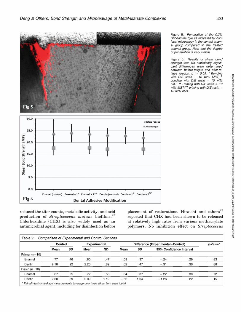
reduced the titer counts, metabolic activity, and acidproduction of Streptococcus mutans biofilms.22
Chlorhexidine (CHX) is also widely used as anantimicrobial agent, including for disinfection before
placement of restorations. Hiraishi and others23
reported that CHX had been shown to be releasedat relatively high rates from various methacrylatepolymers. No inhibition effect on Streptococcus
Figure 5. Penetration of the 0.2%Rhodamine dye as indicated by con-focal microscopy in the control enam-el group compared to the treatedenamel group. Note that the degreeof penetration is very similar.
Figure 6. Results of shear bondstrength test. No statistically signifi-cant differences were determinedbetween before-fatigue and after-fa-tigue groups, p . 0.05. * Bondingwith D/E resin þ 10 wt% MST; #
bonding with D/E resin þ 10 wt%nMT. ** Priming with D/E resin þ 10wt% MST; ## priming with D/E resin þ10 wt% nMT.
Table 2: Comparison of Experimental and Control Sections
Control Experimental Difference (Experimental�Control) p-Value*
Mean SD Mean SD Mean SD 95% Confidence Interval
Primer (n=10)
Enamel .77 .46 .80 .47 .03 .37 �.24 .29 .83
Dentin 2.18 .92 2.20 .89 .02 .47 �.31 .36 .88
Resin (n=10)
Enamel .67 .25 .72 .53 .04 .37 �.22 .30 .72
Dentin 2.60 .89 2.09 1.19 �.52 1.04 �1.26 .22 .15
* Paired t-test on leakage measurements (average over three slices from each tooth).
Deng & Others: Bond Strength and Microleakage of Metal-titanate Complexes E53
Dow
nloaded from http://m
eridian.allenpress.com/operative-dentistry/article-pdf/41/3/229/1834845/1559-2863-41_3_229_a.pdf by guest on 09 February 2022

mutans was further detected from resin disks aftertwo weeks of water storage. Matrix metalloprotei-nase (MMP) may be partially responsible for hybridlayer degradation. Loss of hybrid layer integritycompromised resin-dentin bond stability. CHX actedas an MMP inhibitor, so it had beneficial effects onthe preservation of dentin bond strength.23
Our research is focused on gold NP (AuNP) sincewe discovered that gold-loaded microparticulatemetal-titanate complexes (lMTs) were effective asa new class of antibacterials. Gold-loaded lMTsinhibit growth of oral bacteria at micromolarconcentrations. Other studies24 also demonstratedantifungal and antiviral actions of AuNP.
Compared to microparticles, NPs offer manyadvantages. Nanotechnology modulates metals intotheir nano-size, which drastically changes thechemical, physical, and optical properties of metals.Inorganic nanoparticles and their nanocompositesare applied as effective antibacterial agents. Nano-particulate metal oxides have unusual crystal mor-phologies, with an increased number of edges andcorners of NPs, which in turn generates a large NPsurface area for interaction with bacteria. Theoret-ically, nMTs will be even more effective because theyhave a significantly greater surface-to-volume ratio,which is expected to lead to more effective ion-exchange characteristics. More importantly, they areable to act as drug carriers or to concentrate drugson their surfaces, which results in polyvalent effectsthat enhance drug efficacy.25-27 NPs themselves canspecifically attack biological targets after modifica-tion with targeting molecules.28,29 nMT also has theadvantage of acting as a bioactive molecule to carryother drugs.
Oral applications of nanoparticles have recentlybeen considered.30 The potential of NPs to controloral biofilm formation is related to their biocidal andantiadhesive capabilities. NPs have been incorporat-ed into dental materials to improve antimicrobialactivity. The presence of antibacterials in both thebonding systems and the filling material theoreti-cally would inhibit or slow both the initiation andprogression of caries adjacent to restorations. Prob-lems can arise as a result of release of theantibacterial agent from the composite. Such prob-lems may include toxic effects, influence on mechan-ical properties, and loss of effectiveness.31 If systemictoxicity could be limited and therapeutic indiceswere optimized, metal ions and their associatedcompounds could emerge as a new powerful class ofantibacterial agents. Martınez-Gutierrez and oth-ers32-3 and Kasraei and others34 suggested that NPs
may also exert significant cytotoxicity on macro-phages in association with a proinflammatory re-sponse and cellular apoptosis. Their findings showedthat silver nanoparticles were cytotoxic in murinemacrophages and in fibroblasts at concentrations of10 and 50 lg/mL, respectively. Instead of incorpo-rating our nMTs into the body of the restorativematerials, our strategy was to add the materials tothe primer and adhesive layer. Such a strategy willminimize the quantity of materials needed. More-over, amorphous peroxititanates (APT) might beused to bind a variety of metal compounds withhigh-affinity forming complexes to control the deliv-ery of metal-based drugs to the target tissue,avoiding systemic toxicity. Wataha and others4
demonstrated that metal-APT complexes facilitatemetal ion delivery (such as gold and platinum) tomonocytes as well as fibroblasts. Composite resinscontaining 1% silver nanoparticles exhibited anti-bacterial activity against Streptococcus mutans andLactobacillus.34 In addition, the presence of antibac-terials in the bonding systems (ie, at the criticalinterface) would theoretically affect the initiationand progression of caries. Antibacterial adhesivescould inhibit the invading bacteria along tooth-restoration margins.
Our attempt to incorporate the nMT into eitherthe primer or the adhesive resin carries with itadvantages and disadvantages. Aside from the purechemical makeup point of view, putting the nMT intothe primer will be closer to the tooth substrate andmay have a more direct antibacterial effect. Howev-er, the nMT will be subsequently covered by bondingresin and composite resin restoration, and, thus, itslong-term release may be hampered. Incorporatingthe nMT into the resin separates the active ingredi-ent further from the targeted tooth surface. Bothconditions are still at the critical exposed interface.Zhang and others35 reported that adding dentalresins containing 12-methacryloyloxydodecylpyridi-nium bromide (MDPB) with AgNP into both primerand adhesive achieved the strongest antibiofilmefficacy.
Our results showed that 10 wt% nMT, when addedto the bonding agent, did not affect bond strength ormicroleakage for either enamel or dentin. Althoughcyclic loading did lower the after-fatigue SBS, whenthe groups are compared across the same conditions,we did not find any significant differences. There-fore, incorporating nMT into dental adhesive sys-tems will not compromise resin-tooth bonding andsealing ability. Hence, the incorporation of antibac-terial agents (ie, nMTs) into dentin bonding agents
E54 Operative Dentistry
Dow
nloaded from http://m
eridian.allenpress.com/operative-dentistry/article-pdf/41/3/229/1834845/1559-2863-41_3_229_a.pdf by guest on 09 February 2022

may become an indispensable method for inhibitingresidual bacteria in the cavity and secondary caries.
CONCLUSIONS
Within the limitations of this study, the addition ofnMT to All-Bond 2t dental adhesive system did notaffect the SBS and microleakage between compositeand either human dentin or enamel. Incorporation ofthis novel material into a dental adhesive systemdoes not appear to compromise either bonding orsealing ability and thus is a promising antibacterialagent with clinical significance.
Acknowledgements
This study was funded by University of Washington RAC 75-5827and National Institutes of Health (NIH) grant DE021373-01. The metal-titanate complexes are supplied by SavannahRiver Site National Laboratory through a contract with theNIH grant. Author SD was partly supported by grants fromthe 2011 China State key clinical department and the Board ofMedicine of Zhejiang Province (2012KYB121).
Regulatory Statement
This study was conducted in accordance with all theprovisions of the local human subjects oversight committeeguidelines and policies of the University of Washington,Seattle, Washington.
Conflict of Interest
Author DCNC is one of the holders of the US patent No. US-2012-0156145-A1, ‘‘Use of titanium-based materials as bacte-ricides,’’ related to this manuscript. The other authors of thismanuscript certify that they have no proprietary, financial, orother personal interest of any nature or kind in any product,service, and/or company that is presented in this article.
(Accepted 23 May 2015)
REFERENCES
1. National Institutes of Health (2002) Research on micro-bial biofilms National Heart, Lung and Blood Institutes(5) 3-47.
2. Brunthaler A, Konig F, Lucas T, Sperr W, & Schedle A(2003) Longevity of direct resin composite restorations inposterior teeth Clinical Oral Investigations 7(2) 63-70.
3. Mo SS, Bao W, Lai GY, Wang J, & Li MY (2010) Themicrofloral analysis of secondary caries biofilm aroundClass I and Class II composite and amalgam fillings BMCInfectious Diseases 17(10) 241-256.
4. Wataha JC, Hobbs DT, Lockwood PE, Davis RR, Elving-ton MC, Lewis JB, & Messer RL (2009) Peroxotitanatesfor biodelivery of metals Journal of Biomedical MaterialsResearch. Part B Applied Biomaterials 91(2) 489-496.
5. Wataha JC, Hobbs DT, Wong JJ, Dogan S, Zhang H,Chung KH, & Elvington MC (2010) Titanates delivermetal ions to human monocytes Journal of MaterialsScience—Materials in Medicine 21(4) 1289-1295.
6. Davis RR, Hobbs DT, Khashaba R, Sehkar P, Seta FN,Messer RL, Lewis JB, & Wataha JC (2010) Titanateparticles as agents to deliver gold compounds to fibro-blasts and monocytes Journal of Biomedical MaterialsResearch Part A 93(3) 864-869.
7. Hobbs DT, Elvington MC, Wataha JC, Chung WO,Rutherford RB, & Chan DC, inventors; Use of titanium-based materials as bactericides. US patent 8,545,820.October 1, 2013.
8. Pelgrift RY, & Friedman AJ (2013) Nanotechnology as atherapeutic tool to combat microbial resistance AdvancedDrug Delivery Reviews 65(13-14) 1803-1815.
9. Vasconcelos SM, Melo MA, Wenceslau JP, Zanin IC,Beltrao HC, Fernandes CA, Almeida PC, & Rodrigues LK(2014) In situ assessment of effects of the bromide- andfluoride-incorporating adhesive systems on biofilm andsecondary caries Journal of Contemporary Dental Prac-tice 15(2) 142-148.
10. Qin H, Cao H, Zhao Y, Zhu C, Cheng T, Wang Q, Peng X,Cheng M, Wang J, Jin G, Jiang Y, Zhang X, Liu X, & ChuPK (2014) In vitro and in vivo anti-biofilm effects of silvernanoparticles immobilized on titanium. Biomaterials35(34) 9114-9125.
11. Cheng L, Weir MD, Xu HH, Kraigsley AM, Lin NJ, Lin-Gibson S, & Zhou X (2012) Antibacterial and physicalproperties of calcium-phosphate and calcium-fluoridenanocomposites with chlorhexidine Dental Materials28(5) 573-583.
12. Kaye KS (2012) Antimicrobial de-escalation strategies inhospitalized patients with pneumonia, intra-abdominalinfections, and bacteremia British Journal of HospitalMedicine 7(Supplement 1) S13-S21.
13. Chung WO, Wataha JC, Hobbs DT, An J, Wong JJ, ParkCH, Dogan S, Elvington MC, & Rutherford RB (2011)Peroxotitanate- and monosodium metal-titanate com-pounds as inhibitors of bacterial growth Journal ofBiomedical Materials Research Part A 97(3) 348-354.
14. Morones-Ramirez JR, Winkler JA, Spina CS, & Collins JJ(2013) Silver enhances antibiotic activity against gram-negative bacteria Science Translational Medicine 5(190)181-190.
15. Zhou X, Dorn M, Vogt J, Spemann D, Yu W, Mao Z,Estrela-Lopis I, Donath E, & Gao C (2014) A quantitativestudy of the intracellular concentration of graphene/noblemetal nanoparticle composites and their cytotoxicityNanoscale 6(15) 8535-8542.
16. Sun X, & Li Y (2003) Synthesis and characterization ofion-exchangeable titanate nanotubes Chemistry 9(10)2229-2238.
17. Imazato S (2009) Bio-active restorative materials withantibacterial effects: New dimension of innovation inrestorative dentistry Dental Materials Journal 28(1)11-19.
18. Garcıa-Contreras R, Argueta-Figueroa L, Mejıa-Rubalca-va C, Jimenez-Martınez R, Cuevas-Guajardo S, Sanchez-Reyna PA, & Mendieta-Zeron H (2011) Perspectives forthe use of silver nanoparticles in dental practice Interna-tional Dental Journal 61(6) 297-301.
Deng & Others: Bond Strength and Microleakage of Metal-titanate Complexes E55
Dow
nloaded from http://m
eridian.allenpress.com/operative-dentistry/article-pdf/41/3/229/1834845/1559-2863-41_3_229_a.pdf by guest on 09 February 2022

19. Liu J, Gao Y, Cao D, Zhang L, & Guo Z (2011)Nanoparticle dispersion and aggregation in polymernanocomposites: Insights from molecular dynamics sim-ulation Langmuir 27(12) 7926-7933.
20. He J, Soderling E, Lassila LV, & Vallittu PK (2014)Synthesis of antibacterial and radio-opaque dimethacry-late monomers and their potential application in dentalresin Dental Materials 30(9) 968-976.
21. De Giglio E, Cafagna D, Cometa S, Allegretta A, Pedico A,Giannossa LC, Sabbatini L, Mattioli-Belmonte M, & IattaR (2012) An innovative, easily fabricated, silver nanopar-ticle-based titanium implant coating: Development andanalytical characterization Analytical and BioanalyticalChemistry 405(2-3) 805-816.
22. Cheng L, Weir MD, Xu HH, Antonucci JM, Kraigsley AM,Lin NJ, Lin-Gibson S, & Zhou X (2012) Antibacterialamorphous calcium phosphate nanocomposites with aquaternary ammonium dimethacrylate and silver nano-particles Dental Materials 28(5) 561-572.
23. Hiraishi N, Yiu CKY, King NM, Tay FR, & Pashley DH(2008) Chlorhexidine release and water sorption charac-teristics of chlorhexidine-incorporated hydrophobic/hy-drophilic resins Dental Materials 24(10) 1391-1399.
24. Botequim D, Maia J, Lino MM, Lopes LM, Sim~oes PN,Ilharco LM, & Ferreira L (2012) Nanoparticles andsurfaces presenting antifungal, antibacterial and antivi-ral properties Langmuir 28(20) 7646-7656.
25. Thomas M, & Klibanov AM (2003) Conjugation to goldnanoparticles enhances polyethylenimine’s transfer ofplasmid DNA into mammalian cells Proceedings of theNational Academy of Science United States of America100(16) 9138-9143.
26. Rosi NL, Giljohann DA, Thaxton CS, Lytton-Jean AK,Han MS, & Mirkin CA (2006) Oligonucleotide-modifiedgold nanoparticles for intracellular gene regulationScience 312(5776) 1027-1030.
27. Cho JS, Ko YN, Koo HY, & Kang YC (2010) Synthesis ofnano-sized biphasic calcium phosphate ceramics withspherical shape by flame spray pyrolysis Journal ofMaterials Science—Materials in Medicine 21(4) 1143-1149.
28. Bowman MC, Ballard TE, Ackerson CJ, Feldheim DL,Margolis DM, & Melander C (2008) Inhibition of HIVfusion with multivalent gold nanoparticles Journal of theAmerican Chemical Society 130(22) 6896-6897.
29. Gutierrez J, Tercjak A, Garcia I, & Mondragon I (2009)The effect of thermal and vapor annealing treatments onthe self-assembly of TiO2/PS-b-PMMA nanocompositesgenerated via the sol-gel process Nanotechnology 20(22)225603-225611.
30. Allaker RP (2010) The use of nanoparticles to control oralbiofilm formation Journal of Dental Research 89(11)1175-1186.
31. Celik EU, Sirin TC, Ergucu Z, Turkun M, & Ates M(2008) Can different chlorhexidine agents be used ascavity disinfectants? General Dentistry 56(6) e33-e37.
32. Martınez-Gutierrez F, Thi E, Silverman J, Oliveira C,Svensson S, Hoek A, Sanchez E, Reiner N, Gaynor E,Pryzdial E, Conway E, Orrantia E, Ruiz F, Av-Gay Y, &Bach H (2012) Antibacterial activity, inflammatoryresponse, coagulation and cytotoxicity effects of silvernanoparticles Nanomedicine: Nanotechnology, Biologyand Medicine 8(3) 328-336.
33. Martınez-Gutierrez F, Olive P, Banuelos A, Orrantia E,Nino N, Sanchez E, Ruiz F, & Bach H (2010) Synthesis,characterization, and evaluation of antimicrobial andcytotoxic effect of silver and titanium nanoparticlesNanomedicine: Nanotechnology, Biology and Medicine6(5) 681-688.
34. Kasraei S, Sami L, Hendi S, Alikhani MY, Rezaei-Soufi L,& Khamverdi Z (2014) Antibacterial properties of com-posite resins incorporating silver and zinc oxide nanopar-ticles on Streptococcus mutans and LactobacillusRestorative Dentistry & Endodontics 39(2) 109-114.
35. Zhang K, Li F, Imazato S, Cheng L, Liu H, Arola DD, BaiY, & Xu HH (2013) Dual antibacterial agents of nano-silver and 12-methacryloyloxydodecylpyridinium bromidein dental adhesive to inhibit caries Journal of BiomedicalMaterials Research. Part B, Applied Biomaterials 101(6)929-938.
E56 Operative Dentistry
Dow
nloaded from http://m
eridian.allenpress.com/operative-dentistry/article-pdf/41/3/229/1834845/1559-2863-41_3_229_a.pdf by guest on 09 February 2022
Effect of Cigarette Smoke on ResinComposite Bond Strength to
Enamel and Dentin Using DifferentAdhesive Systems
JD Theobaldo � A Catelan � U Rodrigues-FilhoGM Marchi � DANL Lima � FHB Aguiar
Clinical Relevance
The exposure of dentin to cigarette smoke influences adhesive bonding strength. However,cigarette smoke does not influence the bond strength to enamel.
SUMMARY
Objective: To evaluate the microshear bondstrength of composite resin restorations indental blocks with or without exposure tocigarette smoke.
Method: Eighty bovine dental blocks weredivided into eight groups (n=10) according tothe type of adhesive (Scotchbond Multi-Pur-pose, 3M ESPE, St Paul, MN, USA [SBMP];Single Bond 2, 3M ESPE [SB]; Clearfil SE Bond,Kuraray Medical Inc, Okayama, Japan [CSEB];
Single Bond Universal, 3M ESPE [SBU]) andexposure to smoke (no exposure; exposure forfive days/20 cigarettes per day). The adhesivesystems were applied to the tooth structure,and the blocks received a composite restora-tion made using a matrix of perforated pasta.Data were statistically analyzed using analysisof variance and Tukey test (a,0.05).
Results: For enamel, there was no differencebetween the presence or absence of cigarettesmoke (p=0.1397); however, there were differ-ences among the adhesive systems (p,0.001).CSEB showed higher values and did not differ
*Jessica Dias Theobaldo, DDS, Ms, PhD student, PiracicabaDental School, University of Campinas, Department ofRestorative Dentistry, Piracicaba, Sao Paulo, Brazil
Anderson Catelan, DDS, MS, PhD associate researcher,Piracicaba Dental School, University of Campinas, Depart-ment of Restorative Dentistry, Piracicaba, Sao Paulo, Brazil
Ubirajara Rodrigues-Filho, MSc, PhD, associate professor,Institute of Chemistry of Sao Carlos, University of Sao Paulo,Department of Chemistry and Materials Inorganic Hybrids,Sao Carlos, Brazil
Giselle M Marchi, DDS, MS, PhD, associate professor,Piracicaba Dental School, University of Campinas, Depart-ment of Restorative Dentistry, Piracicaba, Sao Paulo, Brazil
Debora A N L Lima, DDS, MS, PhD, associate professor ,Piracicaba Dental School, University of Campinas, Depart-ment of Restorative Dentistry, Piracicaba, Sao Paulo, Brazil
Flavio H B Aguiar, DDS, MS, PhD, associate professor,Piracicaba Dental School, University of Campinas, Depart-ment of Restorative Dentistry, Piracicaba, Sao Paulo, Brazil
*Corresponding author: Av. Limeira, 901, Piracicaba, SaoPaulo 13414903, Brazil; e-mail: [email protected]
DOI: 10.2341/15-056-L
�Operative Dentistry, 2016, 41-3, E57-E63
Dow
nloaded from http://m
eridian.allenpress.com/operative-dentistry/article-pdf/41/3/229/1834845/1559-2863-41_3_229_a.pdf by guest on 09 February 2022
from SBU, but both were statistically differentfrom SB. The SBMP showed intermediate val-ues, while SB demonstrated lower values. Fordentin, specimens subjected to cigarette smokepresented bond strength values that werelower when compared with those not exposedto smoke (p,0.001). For the groups withoutexposure to cigarette smoke, CSEB showedhigher values, differing from SBMP. SB andSBU showed intermediary values. For thegroups with exposure to cigarette smoke,SBU showed values that were higher andstatistically different from SB and CSEB,which presented lower values of bondstrength. SBMP demonstrated an intermediatevalue of bond strength.
Conclusion: The exposure of dentin to ciga-rette smoke influenced the bonding strength ofadhesives, but no differences were noted inenamel.
INTRODUCTION
Of the development of restorative techniques indentistry, adhesive systems have stood out for theirability to promote a bond between the toothstructure and restorative materials.1,2 One way ofclassifying adhesive systems is related to the use ofacid etching prior to the application of the adhesive(etch-and-rinse and self-etching). Although the cur-rent adhesive systems have been improved signifi-cantly after numerous studies, the interface betweenthe substrate and adhesive is the susceptible failurewhen exposed to the oral environment. The tooth-restoration interface, called the ‘‘hybrid layer,’’ isformed by a network of adhesive penetration intodentin tubules and enamel surface irregularitiesthat becomes rigid when polymerized, allowing forthe micromechanical retention of a resin restoration.The bonding durability between a restoration andtooth substrate is important for the longevity andclinical success of adhesive restorations.3 Commonclinical findings demonstrate that the exposure ofthis interface to the oral environment may cause thedeterioration of the hybrid layer due to a variety ofphysical and chemical factors, including hydrolysisand enzymatic degradation of the dentin collagen.4
Cigarette smoke is composed of more than 5000constituents, including carbon monoxide, ammonia,nickel, arsenic, and heavy metals such as lead andcadmium.5-7 When cigarette smoke comes intocontact with the tooth and restorations, it may causediscoloration, surface roughness, and hardness,which are considered important mechanical proper-
ties for clinical success of all restorations. Moreover,high temperatures (558C) can change the propertiesof adhesive resins, such as sorption and solubility.5,8
Furthermore, lead and cadmium are accumulated inteeth that are exposed to cigarette smoke, particu-larly on the enamel surface and within the dentinaccording to the exposure level,5 decreasing the bondstrength of resin-based restorative materials.9
Therefore, smoking can affect the chemical andmechanical interaction between the composite resinsand dental structures.9,10
It is not known to what extent the substancesfound in cigarette smoke may influence the adhesionof adhesive restorative materials to enamel anddentin and thus interfere with the durability andclinical success of these restorations. There are fewstudies in the literature that have evaluated thebond strength of different adhesive systems to dentinand the bond strength of resin-based restorativematerials in enamel/dentin previously subjected tocigarette smoke. Therefore, the aim of this study is toverify whether cigarette smoke interferes with theadhesion between the dental substrates and variousadhesive restorations. The null hypothesis of thisstudy is that cigarette smoke does not affect theadhesion of system adhesives to dental structures.
METHODS AND MATERIALS
For this study, 80 bovine dental blocks were usedand divided into eight groups (n=10; Table 1). Priorto the restoration process, half of the samples wereexposed to cigarette smoke, as described below.
Specimen Preparation
The bovine incisors were collected, disinfected, andstored in a 0.1% thymol-buffered solution anddistilled water. The crown was separated from theroot using a double-faced diamond disc (KG Sor-ensen Ind. Com Ltda, Barueri, SP, Brazil). Ametallographic cutting machine (Isomet 1000, Bueh-ler, Lake Bluff, IL, USA) and diamond disc (4 3012 3
1/2 inches, Buehler) were used to obtain enamel/dentin blocks with a bonding surface area of 25 mm2,3 mm long and having a 1-mm enamel thickness.The enamel surface was ground using No. 600 andNo. 1200 grit silicon carbide (SiC) abrasive papersunder constant irrigation in a polishing machine(Arotec, Cotia, SP, Brazil). The specimens were thenpolished with felt disks (Arotec, Cotia, SP, Brazil)and diamond pastes (1, ½, and ¼ lm), with thespecific lubricant (Arotec). Samples were placed inan ultrasonic tub (Marconi, Piracicaba, Sao Paulo,SP, Brazil) for 15 minutes between each application
E58 Operative Dentistry
Dow
nloaded from http://m
eridian.allenpress.com/operative-dentistry/article-pdf/41/3/229/1834845/1559-2863-41_3_229_a.pdf by guest on 09 February 2022

of sandpaper and felt and at the end of the polishingprocedures. All samples were stored in distilledwater at 378C until use.
Exposure to Cigarette Smoke
A smoke machine developed by the Department ofRestorative Dentistry, Piracicaba Dental School UNI-CAMP, 2011 (registered under No. 01810012043INPI, National Institute of Industrial Property) wasused to expose groups SBMP-WS (Scotchbond Multi-Purpose, 3M ESPE, St Paul, MN, USA), SB-WS(Single Bond 2, 3M ESPE), CSEB-WS (Clearfil SEBond, Kuraray Medical Inc, Okayama, Japan), andSBU-WS (Single Bond Universal, 3M ESPE; n=10) tocigarette smoke. The cycle was scheduled on a timeinterval, simulating the smoking behavior usuallyperformed by a smoker, with the smoke remaining incontact with the specimens for three seconds. Themachine allows for the ambient air to be inhaledevery 10 seconds, thus simulating smoke inhalationand subsequent elimination. The specimens weresubjected to smoke from one pack of Marlborocigarettes (Philip Morris Brazil Ind. e Com, SantaCruz do Sul, RS, Brazil) per day, for a total of fivedays.11 In the interval between one simulation andanother, the samples were stored in artificial saliva(1.5 mM Ca, 0.9 mM Pi, 150 mM KCL, 0.05 lg F/mL,0.1 M Tris buffer [pH=7.0]) at 378C, and every 24hours, the samples were washed with distilled waterand reimmersed in a fresh solution of artificial salivato prevent sedimentation.11,12 Prior to exposure of thesamples to cigarette smoke, all samples were isolatedwith acid-resistant varnish (Colorama, Sao Paulo,Brazil), with the exception of the polished enamelarea. The artificial saliva for all of the groups(exposed and not exposed to smoke) was changeddaily.
Microshear Strength Test
Four types of adhesive systems were tested: 1) three-step etch-and-rinse adhesive system (ScotchbondMulti-Purpose [3M ESPE]), 2) two-step etch-and-rinse adhesive system (Single Bond 2 [3M ESPE]), 3)two-step self-etching system (Clearfil SE Bond[Kuraray Medical Inc]), and 4) one-step self-etchingsystem (Single Bond Universal [3M ESPE]). Theseadhesive systems were applied to the tooth structureaccording to the manufacturer’s recommendations(Table 2), and subsequently, the enamel blocksreceived a composite resin restoration (FiltekZ350XT Flowable; A3 shade, lot N495761, 3M ESPE)using a matrix of perforated pasta (Furadinho 6,Pastifıcio Santa Amalia, Machado, Minas Gerais,Brazil) that was 1 mm in height and with a 1.15-mminternal diameter. This matrix does not causetension during its removal after water absorptionbecause the water gelatinizes the starch moleculesand consequently reduces its rigidity. All enamelsurfaces were etched using phosphoric acid 35%(Ultra Etch, Ultradent Products Inc, South Jordan,UT, USA) for 30 seconds. The photoactivation of thecomposite was performed using a third-generationLED source (Ultradent) at high-power mode, with apower density of 1400 mW/cm2 for 20 seconds. Themicroshear test was carried out using the universaltesting machine EZ Test-L (Shimadzu Corporation,Tokyo, Japan) at a speed of 0.5 mm/min. Themicroshear bond strength results were given inMega Pascals (MPa) after measuring the bondingarea using a digital caliper, according to thefollowing formula:
R¼ Rupture ForceðKgf Þ3 9:8=Areaðmm2Þ
where R is the bond strength in MPa.
Table 1: Experimental Groups for the Adhesion Test in Enamel and Dentin
Blocks Enameland Dentin
System Adhesive Exposure toCigarette Smoke
SBMP (n=10) Scotchbond Multi-Purpose (3M ESPE) No
SB (n=10) Single Bond 2 (3M ESPE) No
CSEB (n=10) Clearfil SE Bond (Kuraray) No
SBU (n=10) Single Bond Universal (3M ESPE) No
SBMP-WS (n=10) Scotchbond Multi-Purpose (3M ESPE) Yes
SB-WS (n=10) Single Bond 2 (3M ESPE) Yes
CSEB-WS (n=10) Clearfil SE Bond (Kuraray) Yes
SBU-WS (n=10) Single Bond Universal (3M ESPE) Yes
Abbreviations: SBMP, Scotchbond Multi-Purpose; SB, Single Bond 2; CSEB, Clearfil SE Bond; SBU, Single Bond Universal; WS, with smoke.
Theobaldo & Others: Effect of Cigarette Smoke on Adhesive Bond Strength E59
Dow
nloaded from http://m
eridian.allenpress.com/operative-dentistry/article-pdf/41/3/229/1834845/1559-2863-41_3_229_a.pdf by guest on 09 February 2022

After the enamel microshear strength test, the
enamel surface was removed to expose the dentin.
SiC sandpaper, with a No. 600 granulation, was
used to standardize the smear layer. The same
protocol was used for the restorative procedures in
dentin, and dentin microshear testing was per-
formed. After the microshear test, bond failure
mode was classified in percentages, as 1) cohesive in
tooth tissue (enamel or dentin), 2) adhesive, 3)
cohesive in the composite, and 4) mixed using a
stereomicroscope (MZ75, Leica Microsystems, Heer-
brugg, Switzerland) under 1003.
Statistical Analysis
The bond strength microshear data were subjected toanalysis of variance (ANOVA) and Tukey tests(a,0.05).
RESULTS
Table 3 presents the enamel microshear values.ANOVA showed significant differences for the factoradhesive system (p,0.001). However, no statisticaldifference was found for the factor exposure tocigarette smoke (p=0.1397) or for the interactionbetween the factors (cigarette smoke 3 adhesive
Table 3: Mean (SD) Enamel Microshear Strengtha
Exposure to Cigarette Smoke Adhesive System
ScotchbondMultipurpose
(3M ESPE)
SingleBond 2
(3M ESPE)
ClearfilSE Bond(Kuraray)
Single BondUniversal(3M ESPE)
Without 17.55 (2.9) 12.75 (4.56) 19.00 (3.48) 19.09 (3.45)
With 14.30 (2.55) 11.17 (5.0) 19.23 (4.30) 18.72 (2.7)
Pooled data 15.93 (3.13) B 11.97 (4,73) C 19.12 (3.81) A 18.90 (3.01) ABa No effect of treatment (p=0.1397).Different letters indicate a significant difference (p�0.05) between adhesive systems.
Table 2: Instructions for Use of Adhesives According to the Manufacturersa
Adhesive System Manufacturer’s Instructions Composition
Scotchbond Multi-Purpose(3M ESPE)
Etch enamel and dentin surface with phosphoric acid35% for 30 and 15 s, respectively. Water rinsingtwice as long the etching and dry carefully followingthe wet-bonding technique. Apply the primer toenamel and dentin and dry gently for 5 s (no waiting).Surface will appear shiny. Apply the adhesive toenamel and dentin and dry gently for 5 s. Light-curingfor 10 s.
PRIMER: Water; 2-hydroxyethyl methacrylate(HEMA); copolymer of acrylic and itaconic acids
BOND: Bisphenol a diglycidyl ether dimethacrylate(Bis-GMA)
HEMA; triphenylantimony
Single Bond 2 (3M ESPE) Etch enamel and dentin surface with phosphoric acid35% for 30 and 15 s, respectively. Rinse for 10 s.Blot excess water using a cotton pellet or mini-sponge. Do not air dry. The surface should appearglistening without pooling of water. Immediately afterblotting, apply two consecutive coats of adhesive for15 s with gentle agitation using a fully saturatedapplicator. Gently air thin for 5 s to evaporate solvent.Light-curing for 10 s.
Ethyl alcohol; Bis-GMA; silane-treated silica(nanofiller); HEMA; copolymer of acrylic anditaconic acids; glycerol 1,3-dimethacrylate; water;diurethane dimethacrylate (UDMA);diphenyliodonium hexafluorophosphate; ethyl 4-dimethyl aminobenzoate (EDMAB)
Clearfil SE Bond (Kuraray) Active application of the primer for 20 s. Air drygently. Apply the bond, air dry gently, light-cure for 10s. Etching enamel surface with phosphoric acid 35%for 30 s, previous application of adhesive.
PRIMER: 10-methacryloyloxydecyl dihydrogenphosphate hydrophilic aliphatic dimethacrylate; dl-camphorquinone (CQ); water
BOND: 10-MDP, Bis-GMA, HEMA, CQ,hydrophobic dimethacrylate; N,N-diethanol P-toluidine; colloidal silica
Single Bond Universal(3M ESPE),self-etching method
Apply the adhesive for 20 s. Air dry gently for 5 s andlight-cure for 10 s. Etch enamel surface withphosphoric acid 35% for 30 s, previous application ofadhesive.
10-MDP; dimethacrylate resins; HEMA; Vitrebondcopolymer; filler, ethanol; water; initiators; silane
a As informed by the manufacturers’ material safety data sheets.
E60 Operative Dentistry
Dow
nloaded from http://m
eridian.allenpress.com/operative-dentistry/article-pdf/41/3/229/1834845/1559-2863-41_3_229_a.pdf by guest on 09 February 2022

systems; p.0.050). Among these adhesive systems,CSEB showed higher values and did not differ fromSBU, but both were statistically different from SB.SBMP showed intermediate values, whereas SBdemonstrated the lowest values.
Table 4 presents the dentin microshear values.There was a significant difference between thefactors exposure to cigarette smoke (p,0.001) andadhesive system (p,0.001) and for the interactionbetween the factors (p,0.001). For the groupswithout exposure to cigarette smoke, CSEB showedhigher values that were statistically different fromSBMP (lower values). SB and SBU showed interme-diary values, without a significant difference be-tween them. For the groups with exposure tocigarette smoke, SBU showed the highest values,which differed statistically from SB and CSEB,which presented the lowest values and did not differstatistically from each other. SBMP presented anintermediate value. When exposed to cigarettesmoke, dentin showed lower values for SB andCSEB, which differed statistically from the groupsthat were not exposed to cigarette smoke. The dataobtained in the fracture pattern evaluation wereanalyzed by frequency distribution (Figure 1). The
mixed failure was the predominant pattern in almostall groups in enamel and dentin, with the exceptionfor CSEB with exposure to cigarette smoke, whichshowed more adhesive failures.
DISCUSSION
In this study, there was no significant differencebetween the enamel groups with or without exposureto cigarette smoke, but there were differencesbetween the adhesive systems (Table 3). The adhe-sion mechanism in enamel basically occurs throughmicromechanical retention from the infiltration ofthe adhesive system into enamel porosities thatresult from prior conditioning with phosphoricacid.13 Based on this finding, it can be supposedthat the incorporation of heavy metals did not affectthe adhesion to enamel. The application of phospho-ric acid for all groups provided demineralization ofenamel, and contaminants may be removed from theenamel during this preparation, which could avoidbond degradation.
In relation to the adhesive systems for the enamelsurface, CSEB showed the highest bond strengthresults. This agent contains functional monomers,
Table 4: Mean (SD) Microshear Strength in Dentina
Exposure to Cigarette Smoke Adhesive System
ScotchbondMultipurpose (3M)
SingleBond 2 (3M)
Clearfil SEBond (Kuraray)
Single BondUniversal (3M)
Without 14.60 (3.20) aB 15.24 (4.93) aAB 19.94 (4.45) aA 18.15 (4.49) aAB
With smoke 14.17 (5.05) aAB 10.16 (3.80) bB 9.97 (1.90) bB 18.82 (4.40) aAa Distinct letters (uppercase in the row and lowercase in the column) are statistically different (p,0.05).
Figure 1. Percentage of the failures: mixed, adhesive, and cohesive failure.
Theobaldo & Others: Effect of Cigarette Smoke on Adhesive Bond Strength E61
Dow
nloaded from http://m
eridian.allenpress.com/operative-dentistry/article-pdf/41/3/229/1834845/1559-2863-41_3_229_a.pdf by guest on 09 February 2022

which can establish chemical interactions betweenthe adhesive and the hydroxyapatite in the dentalsubstrate.14 The commonly used functional monomerin this adhesive is the phosphate monomer, 10-methacryloxydecyl dihydrogen phosphate (10-MDP).The AD–Concept can explain the results for CSEB,for which phosphate-based monomers (such asphenyl-P and 10-MDP, which are part of the acidicprimer) have a potential for chemical bonding withcalcium in hydroxyapatite. Therefore, all of the acidsinteract with the calcium in the hydroxyapatite,forming ionic bonds that are stable.14 Moreover,etching of the enamel surface removes the smearlayer and increases the reactivity of 10-MDP withthe calcium from hydroxyapatite, thus improving thebond strength.15
SBU presented intermediate values, without asignificant difference from the other groups. SBU isa one-step self-etching adhesive; this adhesivecategory presents complex mixtures of hydrophilicand hydrophobic components to produce thinnerhybrid layers when compared with two-step andetch-and-rinse adhesives.16-18 SBU also containsfunctional monomers and a Vitrebond copolymer.Both compounds interact with the calcium fromhydroxyapatite.19 Previous studies20,21 demonstrat-ed that etching the enamel significantly increasedbond strength values for the one-step multimodeadhesive, SBU, and the two-step self-etching adhe-sive, CSE.
SB presented the lowest bonding values toenamel, and SBMP presented intermediate values,without any significant differences from SBU. SB isclassified as a two-step etch-and rinse adhesivebecause the primer (part hydrophilic) and bond(part hydrophobic) are in the same bottle2,22 andbecause the solvent is mostly water (enamelcontains only 4% water). The infiltration of thetwo-step etch-and rinse adhesive system is lowerwhen compared with three-step etch-and rinseadhesives. The three-step etch-and rinse adhesivescontain hydrophilic functional monomers in theprimer, which allow for the monomer to permeateinto the demineralized matrix, while the hydropho-bic monomers contained in the bonding agentfacilitate adhesion of the composite restorativematerial to the conditioned tooth surface.23 More-over, SB showed problematic solvent evaporation,24
and the presence of the solvent in the adhesivelayer decreased the degree of conversion,24 whichconsequently provided a lower bond strength.
When considering the dentin surface, there was astatistical difference between some groups with and
without exposure to cigarette smoke, with SB-WSand CSEB-WS presenting lower bond strengthvalues when compared with their respective smoke-less groups (Table 4). It can be suggested that theheavy metal contaminants interfered with thechemical interaction between the functional mono-mers 10-MDP and the Ca hydroxyapatite, the mainbonding mechanism for CSEB-WS. There were nostatistical differences between the groups with orwithout exposure to cigarette smoke for the SBUgroup, which may indicate that there are othermonomers in addition to 10-MDP, including theVitrebond copolymer, that might have the chemicalproperty to interact with the tooth structure, sincethis adhesive demonstrated better results. However,more studies are necessary to verify this interaction,because there are no studies in the current litera-ture.
SB also showed a lower bond strength for the WSgroups. This result was expected: the total etchingwould remove the mostly heavy metals incorporatedfrom cigarette smoke. However, the presence ofheavy metals in an adhesive with problematicsolvent evaporation could inhibit the degree ofconversion and bond strength of this adhesive. Thismechanism did not occur with the three-step etch-and-rinse adhesives, since the primer adhesivesystem (hydrophilic) and bond (hydrophobic) are indifferent bottles.2,22
Only SBU maintained the same bond strengthwhen exposed to cigarette smoke when comparingthe adhesive systems that showed higher bondstrengths in dentin without smoke (SB, CSEB,and SBU). SBMP presented a lower bond strengthin dentin without smoke; however, this system alsomaintained the bond strength for the group withsmoke exposure. Almeida e Silva and others9
indicated that contamination by cigarette smokedecreased the bond strength between dentin andcomposite resin because of the reduced diameter ofthe particles from cigarette smoke, which arecapable of being absorbed into the dentin hydroxy-apatite based on the exposure level,5 reducing thecontact between the adhesive and dentin and thusreducing the bond strength values.3,5 After acidetching, it can be inferred that heavy metals mayremain in dentin, even after washing the surface,which can damage simplified adhesive systems.
The null hypothesis of this study was partiallyaccepted: cigarette smoke did not affect the adhesionto enamel but reduced the adhesion to dentin for theSB and CSEB adhesive systems.
E62 Operative Dentistry
Dow
nloaded from http://m
eridian.allenpress.com/operative-dentistry/article-pdf/41/3/229/1834845/1559-2863-41_3_229_a.pdf by guest on 09 February 2022
CONCLUSION
The exposure of cigarette smoke influenced thebonding strength of some adhesives to dentin, butno changes were observed for enamel.
Acknowledgement
This study was supported by FAPESP (grants 2013/15225-8).
Conflict of InterestThe authors have no proprietary, financial, or other personalinterest of any nature or kind in any product, service and/orcompany that is presented in this article.
(Accepted 8 July 2015)
REFERENCES
1. Pashley DH, Tay FR, Breschic L, Tjaderhanee L,Carvalho RM, Carrilho M, Tezvergil-& Mutluay A(2011) State of the art etch-and-rinse adhesives DentalMaterials 27(1) 1-16.
2. Van Meerbeek B, Yoshihara K, Yoshida Y, Mine A, DeMunck J, & Van Landuyt KL (2011) State of the art ofself-etch adhesives Dental Materials 27(1) 17-28.
3. Manuja N, Nagpal R, & Pandit IK (2012) Dentaladhesion: mechanism, techniques and durability JournalClinical Pediatric Dentistry 36(3) 223-234.
4. Breschi L, Mazzoni A, Ruggeri A, Cadenaro M, DiLenarda R, & Dorigo ES (2008) Dental adhesion review:aging and stability of the bonded interface DentalMaterials 24(1) 90-101.
5. Takeuchi CYG, Correa-Afonso AM, Pedrazzi H, Dinelli W,& Palma-Dibb RG (2011) Deposition of lead and cadmiumreleased by cigarette smoke in dental structures and resincomposite Microscopy Research and Technique 74(3)287-291.
6. Borgerding M, & Klus H (2005) Analysis of complexmixtures-cigarette smoke Experimental and ToxicologicPathology 57(1) 43-73.
7. Haussmann HJ (2012) Use of hazard indices for atheoretical evaluation of cigarette smoke compositionChemical Research Toxicology 25(4) 794-810.
8. Aguiar TR, Gaglianone LA, & Mathias P (2014) Anoverview of the impact of lifestyle behaviors on theoperative dentistry Journal of Interdisciplinary Medicineand Dental Science 2 128.
9. Almeida e Silva JS, de Araujo EM Jr, & Araujo E (2010)Cigarette smoke affects bonding to dentin GeneralDentistry 58(4) 326-330.
10. Pinzon LM, Oguri M, O’Keefe K, Dusevish V, Spencer P,Powers JM, & Marshall GW (2010) Bond strength ofadhesives to dentin contaminated with smoker’s salivaOdontology 98(1) 37-43.
11. Bertoldo CES, Miranda DA, Souza Junior EJ, AguiarFHB, Lima DANL, Ferreira RL, Claes I, & Lovadino JR(2011) Surface hardness and color change of dental
enamel exposed to cigarette smoke International Journalof Dental Clinics 3(4) 1-4.
12. Queiroz CS, Hara AT, Paes Leme AF, & Cury JA (2008)pH cycling models to evaluate the effect of low fluoridedentifrice on enamel de and remineralization BrazilianDentistry Journal 19(1) 21-27.
13. Erickson RL, Barkmeier WW, & Kimmes NS (2009) Bondstrength of self-etch adhesives to pre-etched enamelDental Materials 25(10) 1187-1194.
14. Yoshida Y, Nagakane K, Fukuda R, Nakayama Y,Okazaki M, Shintani H, Inoue S, Tagawa Y, Suzuki K,De Munck J, & Van Meerbeek B (2004) Comparativestudy on adhesive performance of adhesive monomersJournal of Dental Research 83(6) 454-458.
15. Mine A, De Munck J, Vivan Cardoso M, Van Landuyt KL,Poitevin A, Kuboki T, Yoshida Y, Suzuki K, & VanMeerbeek B (2010) Enamel-smear compromises bondingby mild self-etch adhesives Journal of Dental Research89(12) 1505-1509.
16. Itthagarun A, Tay FR, Pashley DH, Wefel JS, Garcia-Godoy F, & Wei SHY (2004) Single-step, self-etchadhesives behave as permeable membranes after poly-merization. Part III. Evidence from fluid conductance andartificial caries inhibition American Journal of Dentistry17(6) 394-400.
17. Hashimoto M, Fujita S, Kaga M, & Yawaka Y (2008)Effect of water on bonding of one-bottle self-etchingadhesives Dental Materials Journal 27(2) 172-178.
18. Vaidyanathan TK, & Vaidyanathan J (2009) Recentadvances in the theory and mechanism of adhesive resinbonding to dentin: a critical review Journal of BiomedicalMaterials Research Part B: Applied Biomaterials 88(2)558-578.
19. Yoshida Y, Van Meerbeek B, Nakayama Y, Snauwaert J,Hellemans L, Lambrechts P, Vanherle G, & Wakasa K(2000) Evidence of chemical bonding at biomaterial-hardtissue interfaces Journal of Dental Research 79(2)709-714.
20. de Goes MF, Shinohara MS, & Freitas MS (2014)Performance of a new one-step multi-mode adhesive onetched vs non-etched enamel on bond strength andinterfacial morphology Journal of Adhesive Dentistry16(3) 243-250.
21. McLean, Meyers E, Guillory V, & Vandewalle K (2015)Enamel bond strength of new universal adhesive bondingagents Operative Dentistry 40(4) 410-417.
22. Van Meerbeek B, Pneumans M, Poitevin A, Mine A, VanEnde A, Neves A, & De Munk J (2010) Relationshipbetween bond–strength tests and clinical outcomesDental Materials 26(2) 100-121.
23. Kanca JA (1996) Wet bonding: the effect of drying timeand distance American Journal of Dentistry 9(6) 273-276.
24. Argolo S, Oliveira DC, Fontes CM, Lima AF, de FreitasAP, & Cavalcanti N (2012) Effect of increased dwell timesfor solvent evaporation on the bond strength and degreeof conversion of an ethanol-based adhesive system ActaOdontologica Latinoamericana 25(1) 109-114.
Theobaldo & Others: Effect of Cigarette Smoke on Adhesive Bond Strength E63
Dow
nloaded from http://m
eridian.allenpress.com/operative-dentistry/article-pdf/41/3/229/1834845/1559-2863-41_3_229_a.pdf by guest on 09 February 2022

Longevity of Bonding of Self-adhesive Resin Cement to Dentin
TC Sim~oes � IV Luque-Martinez � RR MoraesATG Sa � AD Loguercio � SK Moura
Clinical Relevance
Regardless of the cementing strategy, the durability of bonding to root canal dentin may beinfluenced by the dentin treatment protocol.
SUMMARY
Objective: To evaluate the effect of root dentin
treatment on the bonding of self-adhesive
resin cement after 24 hours and after 6 months.
Methods: A total of 48 single-rooted premolars
were endodontically treated and divided into
four groups (n=12): Adper Scotchbond Multi-
Purpose + RelyX ARC (ARC); RelyX U200
(U200); EDTA + RelyX U200 (EU200); and phos-
phoric acid (H3PO4) + RelyX U200 (HU200).After filling the roots, an Exacto No. 2 fiberpost was cleaned, treated with silane (60 sec-onds), positioned, and light cured (LED; 60seconds at 1200 mW/cm2). After storage (378C/24h), the roots were cut to obtain two discs (1mm) of each third. They were stored in dis-tilled water (24 hours at 378C); one disc of eachroot-third was subjected to the push-out test(0.5 mm/min) at 24 hours and the other discafter six months of water storage (378C). Thedata on the root-thirds were averaged forstatistical purposes. The average values ofbond strength (MPa) were analyzed by two-way analysis of variance and post hoc Student-Newman-Keuls (5%).
Results: There were statistical differences forthe treatment of dentin (p,0.001), for time(p=0.003), and the interaction of treatment andtime (p=0.017). After 24 hours, we observedlower bond strength in the HU200 group whencompared with other groups (ARC, U200, andEU200). After six months, HU200 showed thelowest bond strength. Higher strengths wereobserved for EU200 and U200 similarly, whichwere higher than ARC.
Conclusion: The bonding of the self-adhesiveresin cement varied over time in the testedgroups.
Tania Christina Sim~oes, DDS, MS, PhD, Restorative Dentist-ry, University North of Parana, Londrina, Brazil
Issis Virgınia Luque-Martinez, Dentistry Academic Unit,Faculty of Medicine, Pontificia Universidad Catolica deChile, Santiago, Chile
Rafael R de Moraes, DDS, MS, PhD, professor, RestorativeDentistry, School of Dentistry, Federal University of Pelotas,Pelotas, Brazil
Andre Tomazini Gomes de Sa, PhD, professor, Prosthodontics,Federal Institute of Parana, Londrina, Brazil
Alessandro D Loguercio, DDS, MS, PhD, professor, Restor-ative Dentistry, State University of Ponta Grossa, PontaGrossa, Brazil
*Sandra Kiss Moura, DDS, MDS, PhD, professor, RestorativeDentistry, University North of Parana, Londrina, Brazil
*Corresponding author: Rua Marselha, 183, Jardim Piza,Londrina, Parana, 86041-140, Brazil; e-mail: [email protected]
DOI: 10.2341/14-266-LR
�Operative Dentistry, 2016, 41-3, E64-E72
Dow
nloaded from http://m
eridian.allenpress.com/operative-dentistry/article-pdf/41/3/229/1834845/1559-2863-41_3_229_a.pdf by guest on 09 February 2022

INTRODUCTION
Fiber posts have several properties (eg, esthetics andelastic modulus) that are similar to those of toothstructure, which provides excellent biomechanicalbehavior and better load dissipation, reducing therisk of radicular fracture.1,2 A post’s association withthe adhesive system and resin cement improves theperformance of their mechanical properties.3,4
Resin cements are recommended for cementingthe fiberglass posts in the root canal and are dividedinto self-adhesive and adhesive systems according tothe cementing strategy.5 Self-adhesive cement wasdeveloped a decade ago, with the purpose ofsimplifying the cementation process by assemblingall the components into a single product.6 Thiscombination has resulted in a material that self-adheres to dentin, does not require pretreatment ofthe surface of the tooth, is simple to implement, andcan be performed in a single step.5,7 Given that theremoval of the smear layer is not recommended withmost self-adhesive cements, there is increasedtolerance to moisture and the release of fluorideions.5
Some studies have indicated that retaining thesmear layer on the dentin could interfere with theadhesion of self-adhesive materials because it mayhinder the adaptation and bonding of the resinousmaterial to the walls of the root canal.8,9 Others haveindicated its retention because some solutions usedto remove it could modify the structure of the dentin,increase water flow, and compromise the bondingwith the resinous monomers by interfering with thepolymerization.10
There is no consensus in the literature regardingthe treatment of dentin in preparation for self-adhesive resin cement. Some studies have suggestedthat the cement is able to cross the smear layer andbond to the dentin.5-7 The simplification of thecementation technique has been welcomed by clinics,and according to a recent study11 has demonstratedbonding similar to the resin cement associated withthe adhesive system because the latter system ismore sensitive due to the various steps involved andcan incur operator errors.12 However, to our knowl-edge, the durability of the bond strength of self-adhesive resin cements is still unknown.
This investigation was conducted to test thehypotheses that 1) there is no difference betweentreating the dentin with ethylenediaminetetraaceticacid (EDTA) at a concentration of 17% or phosphoricacid (H3PO4) at a concentration of 35% in thestrength of the push-out bond strength to dentin
when resin cement is applied; 2) there is nodifference at 24 hours and after six months in thestrength of the push-out bond strength to dentinwhen resin cement is applied.
METHODS AND MATERIALS
A total of 48 single-rooted human premolars, withcomplete root apex formation, that were free ofcaries, fractures, root lacerations, and previousendodontic treatment and had at least 14 6 1 mmbetween the cementoenamel junction (CEJ) and theroot apex were disinfected in a 0.5% solution ofchloramine-T for seven days at 48C.
Preparation of the Teeth
The dental crowns were sectioned in a directionperpendicular to the long axis at the height of theCEJ to obtain roots of a minimum length of 14 mmand to create access to the root canal. The patentcanal length was the established working limit andthe instrumentation was performed up to the apicalforamen. The roots were filled using the hybridtrigger technique, maintaining a distance of 1 mmfrom the apex with the endodontic cement, Sealer 26(Dentsply, Dentsply DeTrey, Konstanz, Germany).The cones were condensed using a McSpaddendevice (Easy Dental Equipment, Belo Horizonte,Brazil). After allowing for the cement setting time,the 48 roots were unobstructed using the equipmentTermo Pack II (Easy Dental Equipment, Sao Paulo,Brazil). The specimens were wrapped in sterilegauze soaked in a solution of 0.9% sodium chloride(NaCl), packed in individual containers, and storedfor 24 hours at room temperature. For the finalcalibration of the conduit a No. 2 Exacto drill (1.6mm in diameter; Angelus, Londrina, Brazil) wasused, coupled to a low-speed turbine (Koncept, KaVo,Joinville, Brazil) to a maximum depth of 9 mm fromthe CEJ.
Experimental Groups
The composition and application mode of the mate-rials used are presented in Table 1. The preparedroots were irrigated with a solution of 0.9% NaCl anddried with absorbent paper towels before beingcategorized from one to 48 and randomly allocatedto four treatment groups (n=12). Each specimenreceived a translucent fiberglass post Exacto No. 2(Angelus) with a conical shape, smooth surface,diameter greater than 1.6 mm, and length of 15mm. Its surface was cleaned with sterile gauzesoaked in absolute ethanol (F Maia, Cotia, Brazil)and dried with jets of air. The binding agent, Silane
Sim~oes & Others: Bonding of Resin Cements to Dentin E65
Dow
nloaded from http://m
eridian.allenpress.com/operative-dentistry/article-pdf/41/3/229/1834845/1559-2863-41_3_229_a.pdf by guest on 09 February 2022
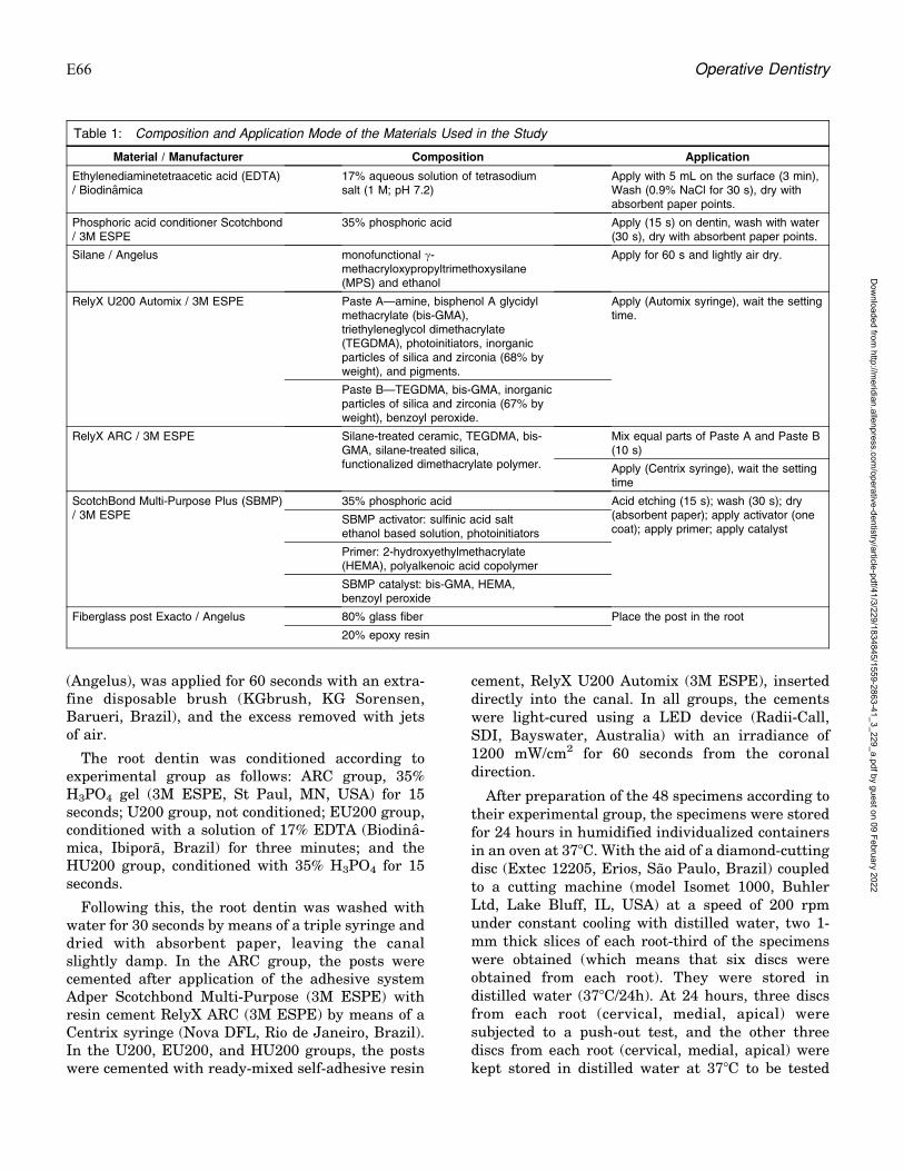
(Angelus), was applied for 60 seconds with an extra-fine disposable brush (KGbrush, KG Sorensen,Barueri, Brazil), and the excess removed with jetsof air.
The root dentin was conditioned according toexperimental group as follows: ARC group, 35%H3PO4 gel (3M ESPE, St Paul, MN, USA) for 15seconds; U200 group, not conditioned; EU200 group,conditioned with a solution of 17% EDTA (Biodina-mica, Ibipora, Brazil) for three minutes; and theHU200 group, conditioned with 35% H3PO4 for 15seconds.
Following this, the root dentin was washed withwater for 30 seconds by means of a triple syringe anddried with absorbent paper, leaving the canalslightly damp. In the ARC group, the posts werecemented after application of the adhesive systemAdper Scotchbond Multi-Purpose (3M ESPE) withresin cement RelyX ARC (3M ESPE) by means of aCentrix syringe (Nova DFL, Rio de Janeiro, Brazil).In the U200, EU200, and HU200 groups, the postswere cemented with ready-mixed self-adhesive resin
cement, RelyX U200 Automix (3M ESPE), inserteddirectly into the canal. In all groups, the cementswere light-cured using a LED device (Radii-Call,SDI, Bayswater, Australia) with an irradiance of1200 mW/cm2 for 60 seconds from the coronaldirection.
After preparation of the 48 specimens according totheir experimental group, the specimens were storedfor 24 hours in humidified individualized containersin an oven at 378C. With the aid of a diamond-cuttingdisc (Extec 12205, Erios, Sao Paulo, Brazil) coupledto a cutting machine (model Isomet 1000, BuhlerLtd, Lake Bluff, IL, USA) at a speed of 200 rpmunder constant cooling with distilled water, two 1-mm thick slices of each root-third of the specimenswere obtained (which means that six discs wereobtained from each root). They were stored indistilled water (378C/24h). At 24 hours, three discsfrom each root (cervical, medial, apical) weresubjected to a push-out test, and the other threediscs from each root (cervical, medial, apical) werekept stored in distilled water at 378C to be tested
Table 1: Composition and Application Mode of the Materials Used in the Study
Material / Manufacturer Composition Application
Ethylenediaminetetraacetic acid (EDTA)/ Biodinamica
17% aqueous solution of tetrasodiumsalt (1 M; pH 7.2)
Apply with 5 mL on the surface (3 min),Wash (0.9% NaCl for 30 s), dry withabsorbent paper points.
Phosphoric acid conditioner Scotchbond/ 3M ESPE
35% phosphoric acid Apply (15 s) on dentin, wash with water(30 s), dry with absorbent paper points.
Silane / Angelus monofunctional c-methacryloxypropyltrimethoxysilane(MPS) and ethanol
Apply for 60 s and lightly air dry.
RelyX U200 Automix / 3M ESPE Paste A—amine, bisphenol A glycidylmethacrylate (bis-GMA),triethyleneglycol dimethacrylate(TEGDMA), photoinitiators, inorganicparticles of silica and zirconia (68% byweight), and pigments.
Apply (Automix syringe), wait the settingtime.
Paste B—TEGDMA, bis-GMA, inorganicparticles of silica and zirconia (67% byweight), benzoyl peroxide.
RelyX ARC / 3M ESPE Silane-treated ceramic, TEGDMA, bis-GMA, silane-treated silica,functionalized dimethacrylate polymer.
Mix equal parts of Paste A and Paste B(10 s)
Apply (Centrix syringe), wait the settingtime
ScotchBond Multi-Purpose Plus (SBMP)/ 3M ESPE
35% phosphoric acid Acid etching (15 s); wash (30 s); dry(absorbent paper); apply activator (onecoat); apply primer; apply catalyst
SBMP activator: sulfinic acid saltethanol based solution, photoinitiators
Primer: 2-hydroxyethylmethacrylate(HEMA), polyalkenoic acid copolymer
SBMP catalyst: bis-GMA, HEMA,benzoyl peroxide
Fiberglass post Exacto / Angelus 80% glass fiber Place the post in the root
20% epoxy resin
E66 Operative Dentistry
Dow
nloaded from http://m
eridian.allenpress.com/operative-dentistry/article-pdf/41/3/229/1834845/1559-2863-41_3_229_a.pdf by guest on 09 February 2022
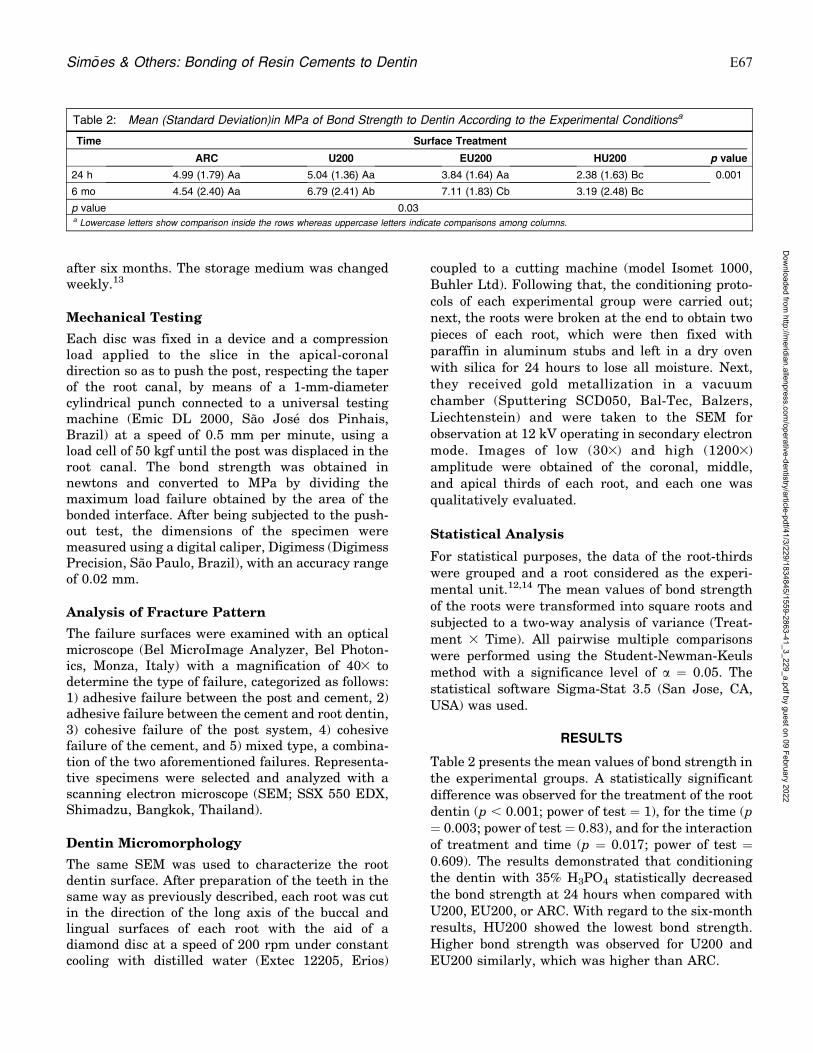
after six months. The storage medium was changedweekly.13
Mechanical Testing
Each disc was fixed in a device and a compressionload applied to the slice in the apical-coronaldirection so as to push the post, respecting the taperof the root canal, by means of a 1-mm-diametercylindrical punch connected to a universal testingmachine (Emic DL 2000, Sao Jose dos Pinhais,Brazil) at a speed of 0.5 mm per minute, using aload cell of 50 kgf until the post was displaced in theroot canal. The bond strength was obtained innewtons and converted to MPa by dividing themaximum load failure obtained by the area of thebonded interface. After being subjected to the push-out test, the dimensions of the specimen weremeasured using a digital caliper, Digimess (DigimessPrecision, Sao Paulo, Brazil), with an accuracy rangeof 0.02 mm.
Analysis of Fracture Pattern
The failure surfaces were examined with an opticalmicroscope (Bel MicroImage Analyzer, Bel Photon-ics, Monza, Italy) with a magnification of 403 todetermine the type of failure, categorized as follows:1) adhesive failure between the post and cement, 2)adhesive failure between the cement and root dentin,3) cohesive failure of the post system, 4) cohesivefailure of the cement, and 5) mixed type, a combina-tion of the two aforementioned failures. Representa-tive specimens were selected and analyzed with ascanning electron microscope (SEM; SSX 550 EDX,Shimadzu, Bangkok, Thailand).
Dentin Micromorphology
The same SEM was used to characterize the rootdentin surface. After preparation of the teeth in thesame way as previously described, each root was cutin the direction of the long axis of the buccal andlingual surfaces of each root with the aid of adiamond disc at a speed of 200 rpm under constantcooling with distilled water (Extec 12205, Erios)
coupled to a cutting machine (model Isomet 1000,Buhler Ltd). Following that, the conditioning proto-cols of each experimental group were carried out;next, the roots were broken at the end to obtain twopieces of each root, which were then fixed withparaffin in aluminum stubs and left in a dry ovenwith silica for 24 hours to lose all moisture. Next,they received gold metallization in a vacuumchamber (Sputtering SCD050, Bal-Tec, Balzers,Liechtenstein) and were taken to the SEM forobservation at 12 kV operating in secondary electronmode. Images of low (303) and high (12003)amplitude were obtained of the coronal, middle,and apical thirds of each root, and each one wasqualitatively evaluated.
Statistical Analysis
For statistical purposes, the data of the root-thirdswere grouped and a root considered as the experi-mental unit.12,14 The mean values of bond strengthof the roots were transformed into square roots andsubjected to a two-way analysis of variance (Treat-ment 3 Time). All pairwise multiple comparisonswere performed using the Student-Newman-Keulsmethod with a significance level of a = 0.05. Thestatistical software Sigma-Stat 3.5 (San Jose, CA,USA) was used.
RESULTS
Table 2 presents the mean values of bond strength inthe experimental groups. A statistically significantdifference was observed for the treatment of the rootdentin (p , 0.001; power of test = 1), for the time (p= 0.003; power of test = 0.83), and for the interactionof treatment and time (p = 0.017; power of test =0.609). The results demonstrated that conditioningthe dentin with 35% H3PO4 statistically decreasedthe bond strength at 24 hours when compared withU200, EU200, or ARC. With regard to the six-monthresults, HU200 showed the lowest bond strength.Higher bond strength was observed for U200 andEU200 similarly, which was higher than ARC.
Table 2: Mean (Standard Deviation)in MPa of Bond Strength to Dentin According to the Experimental Conditionsa
Time Surface Treatment
ARC U200 EU200 HU200 p value
24 h 4.99 (1.79) Aa 5.04 (1.36) Aa 3.84 (1.64) Aa 2.38 (1.63) Bc 0.001
6 mo 4.54 (2.40) Aa 6.79 (2.41) Ab 7.11 (1.83) Cb 3.19 (2.48) Bc
p value 0.03a Lowercase letters show comparison inside the rows whereas uppercase letters indicate comparisons among columns.
Sim~oes & Others: Bonding of Resin Cements to Dentin E67
Dow
nloaded from http://m
eridian.allenpress.com/operative-dentistry/article-pdf/41/3/229/1834845/1559-2863-41_3_229_a.pdf by guest on 09 February 2022

Observation of the fracture pattern showed thepredominance of adhesive failure between thecement and root dentin in all the experimentalgroups, followed by a lower rate of adhesive failurebetween the cement and root. There was no cohesivefailure reported in the present study (Figure 3). Arepresentative image of the predominant failuremode can be seen in Figure 1.
Figure 2 presents SEM imagery of the root dentintreated with acidic solutions, in which the rootcovered by the smear layer in the coronal (A), middle(B), and apical (C) thirds in the group that did notreceive acid conditioning is noted. After conditioningwith 17% EDTA (coronal [D], middle [E], apical [F])and 35% H3PO4 (coronal [G], middle [H], apical [I]),there was exposure of the dentinal tubules.
DISCUSSION
The objective of this study was to evaluate theinfluence of root dentin treatment prior to cementinga fiberglass post with self-adhesive resin cement anda resin cement associated with the adhesive systemafter 24 hours and after six months. The resultsshowed that the bonding between the dentin and thecement was influenced by the dentin treatment andthe storage time.
After 24 hours, the adhesion of the U200 wassimilar to that of the EU200 and ARC, and all threewere greater than the HU200, rejecting the firsthypothesis of this study. The results corroborate asystematic review11 that observed similar adhesionbetween resin cements, regardless of the cementingstrategy. The similarity could be explained by theconditioning of the dentin having been performedwith a weak acid such as 17% EDTA, which
superficially exposed the dentinal tubules9,10 of theroot canal (Figure 2) and enabled the interaction ofthe self-adhesive cement with the dentin underlyingthe smear layer. Promising results were reportedafter the etching of the dentin with EDTA15-17 bygreater unobstruction of the dentinal tubules andthe formation of the hybrid layer.
Despite the self-adhesive strategy theoreticallyeliminating the need for any surface treatment of thedentin, there are doubts about retaining the smearlayer, which, according to some authors,16,18 couldinterfere with the bonding to the root canal, andtherefore propose its removal with chemical solu-tions such as polyacrylic acid, EDTA, NaCl, andsodium hypochlorite (NaOCl). However, one review9
showed that the removal of the smear layer has notbeen standardized among different studies andcannot be removed uniformly along the full extentof the root canal, given that switching solutions of17% EDTA with 5.25% NaOCl led to greater removalof the smear layer. Figure 2 shows the removal of thesmear layer and unobstruction of the dentinaltubules by 17% EDTA, partially agreeing with theresults of this review, because in this study EDTAwas not switched with another substance. Condi-tioning of the dentin with 35% H3PO4 also demon-strated a similar result.
This study also showed that the etching of thedentin with 35% H3PO4 resulted in lower bondstrength after 24 hours and after six months. Butwhen the adhesive system was applied after etchingwith H3PO4 as in the ARC, the formation of thehybrid layer ensured bonding similar to the U200.Studies within the scope of the adhesive cementationof posts usually use the ARC as a comparison group19
and promising results have been observed; however,few of these assessed bond strength after 24 hours.20
Thus, the second objective of this investigation wasto evaluate the longevity of bonding of the resincement/dentin after six months, and the resultsshowed that the storage time influenced the adhesion,rejecting the second hypothesis of this study. After sixmonths, the adhesion of the EU200 group increased,and it was higher than that of all the other groups.These results can be explained by the use of EDTA, amolecule containing four carboxylic acid groups,which acts as a light chelator of calcium (Ca) inselective dissolution of hydroxyapatite.21 Unlikephosphoric acid etching, which is deep, with EDTAthe dentin is superficially demineralized.22 It can beunderstood that the adhesion of the EU200 group washigher than the ARC at six months due to theinteraction of the resin cement and the dentin, with
Figure 1. Scanning electron microscopy of adhesive fracture mode.
E68 Operative Dentistry
Dow
nloaded from http://m
eridian.allenpress.com/operative-dentistry/article-pdf/41/3/229/1834845/1559-2863-41_3_229_a.pdf by guest on 09 February 2022

the latter having been more uniformly demineralized
by the EDTA compared with the HU200 group.
The preservation of the longevity of adhesive
restorations was described when the conditioning of
the dentin with EDTA was carried out,22,23 where
surface removal of the hydroxyapatite ensured that
the collagen fibers would not be exposed or suffer
structural changes24 or dehydration.25 Another as-
pect to be considered is that EDTA is a potent
inhibitor of matrix metalloproteinases (MMPs), colla-
genolytic enzymes related to the degradation of the
organic matrix.24,26 The chelating activity of EDTA
promotes the sequestration of Caþ ions present in the
dentin,21 which together with zinc, act as potential
activators of MMPs.26,27 When performing the condi-
tioning with phosphoric acid etching, greater demin-
eralization and exposure of collagen occurs, activating
MMPs.24 These aspects may also explain the results
observed at six months in this study.
The bonding of resin cement to dentin will occur ifthere is an interaction between the surfaces.28
Although the pH of some self-adhesive cements isinitially acidic, it is unable to completely remove thesmear layer as a strong acid such as H3PO4
would.7,29,30 For some authors31 this limitation couldbe overcome by the possible chemical bond betweenthe dentin and some self-adhesive resin cements thatcontain the functional monomer. In the case of theU200, according to the manufacturer, the chemicaladhesion is ensured by the presence of 4-methacryl-oxyethyl trimellitic acid monomers and phosphoricacid esters.
Conditioning the root dentin with H3PO4 beforecementation seems to hinder adhesion to thedentin4,7,30 by exposing the collagen, deeply remov-ing the hydroxyapatite, and changing the waterflow.32,33 The results of the HU200 group in thisstudy agree with these assertions. However, theapplication of the adhesive system after conditioning
Figure 2. Scanning electron microscopy of radicular dentin after surface treatments. Without acid etching: (A) cervical, (B) middle, (C) apical; Afteretching with 17% EDTA: (D) cervical, (E) middle, (F) apical; After etching with 35% H3PO4 (G) cervical, (H) middle, (I) apical.
Sim~oes & Others: Bonding of Resin Cements to Dentin E69
Dow
nloaded from http://m
eridian.allenpress.com/operative-dentistry/article-pdf/41/3/229/1834845/1559-2863-41_3_229_a.pdf by guest on 09 February 2022

with this acid ensured adhesion in the ARC groupafter 24 hours and after six months. Thus, the resultof this study demonstrated that phosphoric acidshould not be used in isolation as a dentin condi-tioner, prior to the self-adhesive cement studied,because RelyX U200 contains hydrophilic monomersthat can interfere with the cement polymerizationprocess, compromising the bonding.17
The adhesion of the fiberglass post to the rootdentin can be influenced by the difficulty incontrolling the dentin moisture when used with aresin cement adhesive system, by visualizing thecanal along its entire length,29,33 and by theformation of bubbles in the cement interfaces.34 Inthe present study this aspect was minimized becausethe RelyX U200 cement was inserted with theadapted tips that accompanied the Automix kit (3MESPE) and for the RelyX ARC cement a Centrixsyringe (Nova DFL) was used. This detail of themethodology and the silanization of the fiberglassposts favored the prevalence of adhesive fracturesbetween the cement and root dentin, agreeing withprevious studies that found the majority of fracturesin the same place33,34 and disagreeing with a studythat observed a predominance of fractures in thecement-post interface.32
According to Heintze and Zimmerli,35 the push-outtest is commonly used to analyze the post adhesion ofroots, and well-designed and conducted in vitro bond
strength tests are useful as a screening test if thespecimens are tested after one day and after at leastthree months of water storage. In this study, thebonding of fiber posts to dentin was analyzed after24 hours and after six months of water storage, morethan suggested in the literature.35 The presentinvestigation analyzed the bonding of RelyX U200,a self-adhesive resin cement, comparatively withthat of RelyX ARC, a gold standard used oncementation of fiber posts to dentin, using naturalteeth, which gives results close to the clinicaloutcome.36 Further research on self-adhesive cemen-tation is needed, especially randomized controlledclinical trials.
CONCLUSIONS
Given the limitations of this study, it may beconcluded that the bonding durability of resincements to root dentin varied over time, accordingto the surface treatment of dentin.
Acknowledgments
The authors would like to thank Dr Wagner AlbertoMadalozzo Torres for the endodontic treatment and Angelusand 3M ESPE for the materials used in this study.
Regulatory Statement
This study was conducted in accordance with all theprovisions of the local human subjects oversight committee
Figure 3. Percentages of fracture modes according to the experimental conditions.
E70 Operative Dentistry
Dow
nloaded from http://m
eridian.allenpress.com/operative-dentistry/article-pdf/41/3/229/1834845/1559-2863-41_3_229_a.pdf by guest on 09 February 2022
guidelines and policies of the Ethics Committee of Unopar.The approval code for this study is 95060.
Conflict of Interest
The authors have no proprietary, financial, or other personalinterest of any nature or kind in any product, service, and/orcompany that is presented in this article.
(Accepted 4 December 2014)
REFERENCES
1. Cheung W (2005) A review of the management ofendodontically treated teeth Journal of the AmericanDental Association 136(5) 611-619.
2. Goracci C, & Ferrari M (2011) Current perspectives onpost systems: A literature review Australian DentalJournal 56(1) 77-83.
3. Ferrari M, Vichi A, Fadda GM, Cagidiaco MC, Tay FR,Breschi L, Polimeni A, & Goracci C (2012) A randomizedcontrolled trial of endodontically treated and restoredpremolars Journal of Dental Research 91(Supplement7) 72S-78S.
4. Monticelli F, Ferrari M, & Toledano M (2008) Cementsystem and surface treatment selection for fiber postluting Medicina Oral, Patologia Oral y Cirurgia Bucal13(3) E214-E221.
5. Radovic I, Monticelli F, Goracci C, Vulicevic ZR, & FerrariM (2008) Self-adhesive resin cements: A literature reviewJournal of Adhesive Dentistry 14(4) 251-258.
6. Ferracane JL, Stansbury JW, & Burke FJT (2011) Self-adhesive resin cements—Chemistry, properties and clin-ical considerations Journal of Oral Rehabilitation 38(4)295-314.
7. De Munck J, Vargas M, Van Landuyt K, Hikita K,Lambrechts P, & Van Meerbeek B (2004) Bonding of anauto-adhesive luting material to enamel and dentinDental Materials 20(10) 963-971.
8. Carvalho RM, Tjaderhane L, Manso AP, Carrilho MR, &Carvalho CAR (2012) Dentin as a bonding substrateEndodontic Topics 21(1) 62-88.
9. Violich DR, & Chandler NP (2010) The smear layer inendodontics—A review International Endodontic Journal43(1) 2-15.
10. Rasimick BJ, Shah RP, Musikant BL, & Deutsch AS(2008) Effect of EDTA conditioning upon the retention offiber posts luted with resin cements InternationalEndodontic Journal 41(12) 1101-1106.
11. Sarkis-Onofre R, Skupien JA, Cenci MS, Moraes RR, &Cenci TP (2013) The role of resin cement on bond strengthof glass-fiber posts (GFPs) luted into root canals: Asystematic review and meta-analysis of in vitro studiesOperative Dentistry 39(1) 1-14.
12. Gomes GM, Gomes OM, Reis A, Gomes JC, Loguercio AD,& Calixto AL (2013) Effect of operator experience on theoutcome of fiber post cementation with different resincements. Operative Dentistry 38(5) 555-564.
13. Skovron L, Kogeo D, Gordillo LA, Meier MM, Gomes OM,Reis A, Loguercio AD (2010) Effects of immersion time
and frequency of water exchange on durability of etch-and-rinse adhesive Journal Biomedical Materials Re-search B Applied Biomaterials 95(2) 339-346.
14. Gomes GM, Gomes OM, Gomes JC, Loguercio AD, CalixtoAL, & Reis A (2014) Evaluation of different restorativetechniques for filling flared root canals: Fracture resis-tance and bond strength after mechanical fatigue.Journal of Adhesive Dentistry 16(3) 267-276.
15. Shemesh H, Wu M-K, & Wesselink PR (2006) Leakagealong apical root fillings with and without smear layerusing two different leakage models: A two-month longi-tudinal ex vivo study International Endodontic Journal39(12) 968-976.
16. Gu X-H, Mao C-Y, Liang C, Wang H-M, & Kern M (2009)Does endodontic post space irrigation affect smear layerremoval and bonding effectiveness? European Journal ofOral Sciences 117(5) 597-603.
17. Mazzitelli C, Monticelli F, Toledano M, Ferrari M, &Osorio R (2010) Dentin treatment effects on the bondingperformance of self-adhesive resin cements EuropeanJournal of Oral Sciences 118(1) 80-86.
18. Faria-e-Silva AL, Menezes MS, Silva FP, Reis GR, &Moraes RR (2013) Intra-radicular dentin treatments andretention of fiber posts with self-adhesive resin cementsBrazilian Oral Research 27(1) 14-19.
19. Calixto LR, Bandeca MC, Clavijo V, Andrade MF, GeraldoVaz L, & Campos EA (2012) Effect of resin cement systemand root region on the push-out bond strength of atranslucent fiber post Operative Dentistry 37(1) 80-86.
20. Li X-J, Zhao S-J, Niu LN, Tay FR, Jiao K, Gao Y, & ChenJ-H (2014) Effect of luting cement and thermomechanicalloading on retention of glass fiber posts in root canalsJournal of Dentistry 42(1) 75-83.
21. Sano H, Takatsu T, Ciucchi B, Russell CM, & Pashley DH(1995) Tensile properties of resin-infiltrated demineral-ized human dentin Journal of Dental Research 74(4)1093-1102.
22. Osorio R, Erhardt MC, Pimenta LA, Osorio E, & ToledanoM (2005) EDTA treatment improves resin-dentin bonds’resistance to degradation Journal of Dental Research84(8) 736-740.
23. Soares CJ, Castro CG, Santos Filho PC, & da Mota AS(2007) Effect of previous treatments on bond strength oftwo self-etching adhesive systems to dental substrateJournal of Adhesive Dentistry 9(3) 291-296.
24. Carvalho RM, Tay F, Sano H, Yoshiyama M, & PashleyDH (2000) Long-term mechanical properties of EDTA-demineralized dentin matrix Journal of Adhesive Den-tistry 2(3) 193-199.
25. Habelitz S, Balooch M, Marshall SJ, Balooch G, MarshallGW Jr (2002) In situ atomic force microscopy of partiallydemineralized human dentin collagen fibrils Journal ofStructural Biology 138(3) 227-236.
26. Carrilho MR, Tay FR, Donnelly AM, Agee KA, Tjader-hane L, Mazzoni A, Breschi L, Folger S, & Pashley DH(2009) Host-derived loss of dentin matrix stiffnessassociated with solubilization of collagen Journal ofBiomedical Materials Research Part B Applied Biomate-rials 90(1) 373-380.
Sim~oes & Others: Bonding of Resin Cements to Dentin E71
Dow
nloaded from http://m
eridian.allenpress.com/operative-dentistry/article-pdf/41/3/229/1834845/1559-2863-41_3_229_a.pdf by guest on 09 February 2022

27. Tezvergil-Mutluay A, Agee KA, Hoshika T, Carrilho M,Breschi L, & Tjaderhane L (2010) The requirement of zincand calcium ions for functional MMP activity in demin-eralized dentin matrices Dental Materials 26(11)1059-1067.
28. Saar M, Mine A, De Munck J, Cardoso MV, Kane AW,Vreven J, Van Meerbeek B, & Van Landuyt KL (2010)Immediate bonding effectiveness of contemporary com-posite cements to dentin Clinical Oral Investigations14(5) 569-577.
29. Van Meerbeek B, De Munck J, Yoshida Y, Inoue S,Vargas M, Vijay V, Van Landuyt K, Lambrechts P, &Vanherle G (2003) Adhesion to enamel and dentin:Current status and future challenges Operative Dentistry28(3) 215-235.
30. Hikita K, Van Meerbeek B, De Munck J, Ikeda T, VanLanduyt K, Maida T, Lambrechts P, & Peumans M (2006)Bonding effectiveness of adhesive luting agents to enameland dentin Dental Materials 23(1) 71-80.
31. Fukegawa D, Hayakawa S, Yoshida Y, Suzuki K, OsakaA, & Van Meerbeek B (2006) Chemical interaction ofphosphoric acid ester with hydroxyapatite Journal ofDental Research 85(10) 941-944.
32. Perdigao J, Gomes G, & Lee IK (2006) The effect of silaneon the bond strengths of fiber posts Dental Materials22(8) 752-758.
33. Zicari F, Coutinho E, De Munck J, Poitevin A, Scotti R,Naert I, & Van Meerbeek B (2008) Bonding effectivenessand sealing ability of fiber-post bonding Dental Materials24(7) 967-977.
34. Watzke R, Frankenberger R, & Naumann M (2011)Different scanning electron microscopic evaluation meth-ods of cement interface homogeneity of adhesively lutedglass fiber posts Acta Odontologica Scandinavica 69(2)101-107.
35. Heintze SD, Zimmerli B (2011) Relevance of in vitro testsof adhesive and composite dental materials: A review in 3parts, Part 3: In vitro tests of adhesive systems SchweizerMonatsschrift fur Zahnmedizin 121(11) 1024-1040.
36. Heintze SD, Zimmerli B (2011) Relevance of in vitro testsof adhesive and composite dental materials: A review in 3parts, Part 1: Approval requirements and standardizedtesting of composite materials according to ISO specifica-tions Schweizer Monatsschrift fur Zahnmedizin. 121(9)804-816.
E72 Operative Dentistry
Dow
nloaded from http://m
eridian.allenpress.com/operative-dentistry/article-pdf/41/3/229/1834845/1559-2863-41_3_229_a.pdf by guest on 09 February 2022

Composite Replacement ofAmalgam Restoration Versus
Freshly Cut Dentin: An In VitroMicroleakage Comparison
H Redwan � DN Bardwell � A AliM Finkelman � S Khayat � H-P Weber
Clinical Relevance
Bonding to dentin under replaced amalgam restorations may be as effective as bonding tofreshly cut dentin.
SUMMARY
Objective: The aim of this study was to evaluatethe microleakage of the composite restorationswhen bonded to tooth structure previouslyrestored with amalgam material compared withthat of freshly cut dentin.
Methods and Materials: Thirty intact, extract-ed intact human molars were mounted inautopolymerizing acrylic resin. Class II boxpreparations were prepared on the occluso-proximal surfaces of each tooth (4-mm bucco-lingual width and 2-mm mesio-distal depth)with the gingival cavosurface margin 1 mmabove the CEJ. Each cavity was then restoredusing high copper amalgam restoration (Dis-peralloy, Dentsply) and then thermocycled for10,000 thermal cycles. Twenty-five of the amal-gam restorations were then carefully removedand replaced with Filtek Supreme Ultra Uni-versal (3M ESPE); the remaining five wereused for scanning electron microscopy andenergy dispersive x-ray spectroscopy analysis.A preparation of the same dimensions wasperformed on the opposite surface of the toothand restored with composite resin and ther-mocycled for 5000 thermal cycles. Twentysamples were randomly selected for dye pene-tration testing using silver nitrate staining todetect the microleakage. The specimens wereanalyzed with a stereomicroscope at a magni-fication of 203. All of the measurements weredone in micrometers; two readings were taken
*Hetaf Redwan, BDS, MS, Department of Restorative Den-tistry, Umm Al-Qura University, Makkah, Saudi Arabia
David N. Bardwell, DMD, MS, Prosthodontics and OperativeDentistry, Tufts University, Boston, MA, USA
Ala Ali, BDS, MS, Prosthodontics and Operative Dentistry,Tufts University School of Dental Medicine, Boston, MA,USA
Matthew Finkelman, PhD, Department of Public Health andCommunity Service, Tufts University School of DentalMedicine, Boston, MA, USA
Samer Khayat, DMD, CAGS, Prosthodontics and OperativeDentistry, Tufts University School of Dental Medicine,Boston, MA, USA
Hans-Peter Weber, DMD, DrMedDent, Prosthodontics andOperative Dentistry, Tufts University School of DentalMedicine, Boston, MA, USA
*Corresponding author: Al Abdiah, Makkah 21955, SaudiArabia; e-mail: [email protected]
DOI: 10.2341/14-278-L
�Operative Dentistry, 2016, 41-3, E73-E82
Dow
nloaded from http://m
eridian.allenpress.com/operative-dentistry/article-pdf/41/3/229/1834845/1559-2863-41_3_229_a.pdf by guest on 09 February 2022

for each cavity at the occlusal and proximalmargins. Two measurements were taken usinga 0-3 scale and the percentage measurements.
Results: Corrosion products were not detectedin either group (fresh cut dentin and teethpreviously restored with amalgam). No statis-tically significant difference was found be-tween the microleakage of the two groupsusing a 0-3 scale at the occlusal margins(McNemar test, p=0.727) or proximal margins(Wilcoxon signed-rank test, p=0.174). No sig-nificance difference was found between thetwo groups using the percentage measure-ments and a Wilcoxon signed-rank test ateither the occlusal (p=0.675) or proximal(p=0.513) margins. However, marginal micro-leakage was statistically significant betweenthe proximal and occlusal margins (p,0.001).
Conclusion: Within the limitations of this invitro study, no significant difference was foundbetween the microleakage of nondiscoloreddentin in teeth that were previously restoredwith amalgam compared with freshly cut den-tin. However, marginal microleakage in theproximal surface was higher than that in theocclusal surface.
INTRODUCTION
The use of composite resin material has increasedand has become the first choice for most cariouslesions. This change can be attributed to severalfactors.1 Some of the main factors are that amalgamrestorations do not adhere to the cavity wallchemically, and there are critical differences inthe coefficient of thermal expansion of amalgamand tooth structure. Amalgams allow the transportof fluids, ions, molecules, and possibly bacteria andtheir toxins more readily.2,3 These problems aremore obvious when poor operative technique isused. Moreover, mercury used in amalgams hasraised concerns about its biological toxicity andenvironmental hazards. People are exposed tomercury and other metals via vapor and corrosionproducts in swallowed saliva and by direct absorp-tion through the oral mucosa.4-6 However, appear-ance, in addition to the controversy about mercurytoxicity, is the public’s displeasure with amalgam.Additionally, amalgam has other disadvantages, inthat corrosion can cause increased porosity andreduced marginal integrity and strength, as well asthe release of metallic products into the oral cavity.4
The literature supports a lack of evidence thatamalgam and its mercury content can be harmful to
humans, with the only exception being specificmetal allergies.7
The use of tooth-colored restorative materialrather than amalgam restorations requires themeticulous use of an adhesive technique.8 This isbecause dentin is more hydrophilic in naturecompared with enamel and therefore is more difficultto deal with in terms of bonding to adhesive resin.However, enamel is known to have reliable bondstrength when bonded to resin-based composites,because its main components are inorganic, whereasdentin has a different composition and structure.9,10
Although various generations of bonding agentshave been developed to decrease polymerizationshrinkage, microleakage remains a significant prob-lem11 and a major factor that affects the longevity ofcomposite restorations.12 This is a primary cause ofmarginal discoloration, secondary caries, pulpalinflammation, postoperative sensitivity, and resto-ration replacement.13 Microleakage measurementsusually provide an assessment of the sealing abilityof adhesive materials, which is clinically relevant.14
According to Abo and others,15 an in vitro micro-leakage test combined with thermocycling is a usefulmethod to assess sealing performance. In theirstudy, they also demonstrated that the use of alarge number of thermal cycles could simulate theconditions in the oral environment. In a reviewperformed by Heintze16 on the evaluation of in vitrosealing ability, microleakage testing was preferredover other methods because of its ease and simplic-ity. The second most frequent method, the quantita-tive marginal analysis of replicas using scanningelectron microscopy (SEM) is used less often becauseit is more time consuming and complex16
In many clinical situations, adhesive resin is usedto bond to sclerotic dentin or dentin discolored bycorrosion products from the amalgam restorationsthat have penetrated the dentinal tubules. Severalstudies have shown that the bond strength was lowerthan or similar to that of unaffected dentin.17-19 If apoor technique is used, or contamination occursduring amalgam placement, it may elicit dentinstaining, as can the presence of recurrent caries.Even in the late 19th and early 20th centuries, it wassuggested that dentin staining could be caused by thedeposition of metallic sulfides and the penetration ofsilver and mercury ions from the overlying amal-gam.19-25 Scholtanus and others2 concluded that thediscoloration of dentin beneath amalgam was anindicator of the presence of amalgam constituents,the effect of which is unknown in adhesive restor-
E74 Operative Dentistry
Dow
nloaded from http://m
eridian.allenpress.com/operative-dentistry/article-pdf/41/3/229/1834845/1559-2863-41_3_229_a.pdf by guest on 09 February 2022
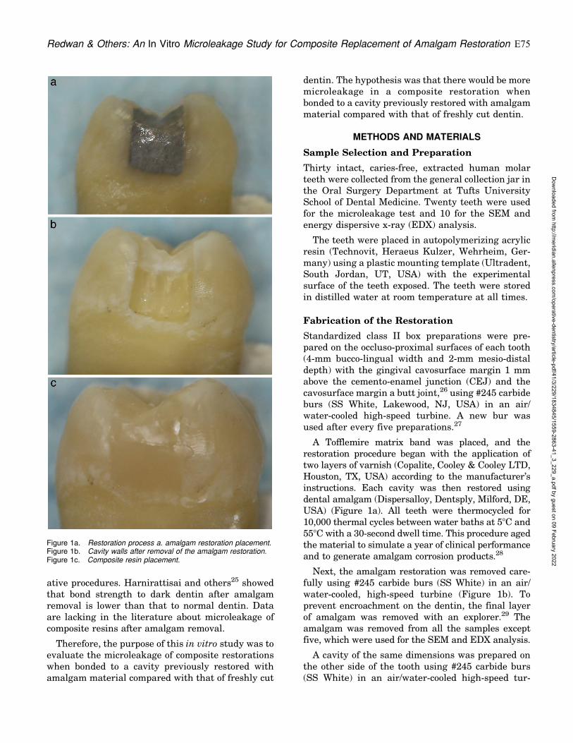
ative procedures. Harnirattisai and others25 showedthat bond strength to dark dentin after amalgamremoval is lower than that to normal dentin. Dataare lacking in the literature about microleakage ofcomposite resins after amalgam removal.
Therefore, the purpose of this in vitro study was toevaluate the microleakage of composite restorationswhen bonded to a cavity previously restored withamalgam material compared with that of freshly cut
dentin. The hypothesis was that there would be moremicroleakage in a composite restoration whenbonded to a cavity previously restored with amalgammaterial compared with that of freshly cut dentin.
METHODS AND MATERIALS
Sample Selection and Preparation
Thirty intact, caries-free, extracted human molarteeth were collected from the general collection jar inthe Oral Surgery Department at Tufts UniversitySchool of Dental Medicine. Twenty teeth were usedfor the microleakage test and 10 for the SEM andenergy dispersive x-ray (EDX) analysis.
The teeth were placed in autopolymerizing acrylicresin (Technovit, Heraeus Kulzer, Wehrheim, Ger-many) using a plastic mounting template (Ultradent,South Jordan, UT, USA) with the experimentalsurface of the teeth exposed. The teeth were storedin distilled water at room temperature at all times.
Fabrication of the Restoration
Standardized class II box preparations were pre-pared on the occluso-proximal surfaces of each tooth(4-mm bucco-lingual width and 2-mm mesio-distaldepth) with the gingival cavosurface margin 1 mmabove the cemento-enamel junction (CEJ) and thecavosurface margin a butt joint,26 using #245 carbideburs (SS White, Lakewood, NJ, USA) in an air/water-cooled high-speed turbine. A new bur wasused after every five preparations.27
A Tofflemire matrix band was placed, and therestoration procedure began with the application oftwo layers of varnish (Copalite, Cooley & Cooley LTD,Houston, TX, USA) according to the manufacturer’sinstructions. Each cavity was then restored usingdental amalgam (Dispersalloy, Dentsply, Milford, DE,USA) (Figure 1a). All teeth were thermocycled for10,000 thermal cycles between water baths at 58C and558C with a 30-second dwell time. This procedure agedthe material to simulate a year of clinical performanceand to generate amalgam corrosion products.28
Next, the amalgam restoration was removed care-fully using #245 carbide burs (SS White) in an air/water-cooled, high-speed turbine (Figure 1b). Toprevent encroachment on the dentin, the final layerof amalgam was removed with an explorer.29 Theamalgam was removed from all the samples exceptfive, which were used for the SEM and EDX analysis.
A cavity of the same dimensions was prepared onthe other side of the tooth using #245 carbide burs(SS White) in an air/water-cooled high-speed tur-
Figure 1a. Restoration process a. amalgam restoration placement.Figure 1b. Cavity walls after removal of the amalgam restoration.Figure 1c. Composite resin placement.
Redwan & Others: An In Vitro Microleakage Study for Composite Replacement of Amalgam Restoration E75
Dow
nloaded from http://m
eridian.allenpress.com/operative-dentistry/article-pdf/41/3/229/1834845/1559-2863-41_3_229_a.pdf by guest on 09 February 2022

bine. A Tofflemire matrix band was placed to ensurethat the light curing of the restoration occurred onlyfrom the occlusal side of the restoration. Bothcavities were restored with composite material usinga total-etch adhesive system.
The composite restoration began by etching theenamel with 35% phosphoric acid (Ultra-Etch, Ultra-dent) for 20 seconds and dentin for 15 seconds. Theteeth were rinsed with water for 10 seconds and blotdried with cotton pellets to achieve a moist dentinsurface and then were bonded with total-etch adhe-sive (ExciTE F DSC, Ivoclar Vivadent, Amherst, NY,USA) that was applied to the enamel and dentinsurfaces, then agitated for 10 seconds and light curedaccording to the manufacturer’s recommendations.
The teeth were restored with composite resinmaterial (Filtek Supreme Ultra Universal, 3M ESPE,St. Paul, MN, USA) via the Liebenberg technique.30 Ablue light emitting diode (LED) was used at 800 mW/cm2, followed by finishing and polishing (Figure 1c).The light source was monitored every five curing cyclesusing a radiometer (Demetron L.E.D, Kerr Corp.,Orange, CA, USA) to ensure that the light intensityremained stable.31 All the teeth were thermocycled for5000 thermal cycles between water baths at 58C and558C with a 30-second dwell time, which served to agethe material to simulate clinical performance.
Microleakage Test Preparation
All of the samples were coated with two layers of nailpolish (Vinyl shine nail polish, Rimmel London,
London, UK), except for a 2.0-mm rim around therestoration to allow the leakage-tracing agent tocontact the margins of the restoration. Thereafter,the teeth were immersed in a solution of 50 wt%ammoniacal silver nitrate (pH=9.5) (Fisher Scientif-ic, Fair Lawn, NJ, USAQ) for 24 hours, followed byeight hours in a photo-developing solution (EastmanKodak Co., Rochester, NY, USA).32
The specimens were washed under running waterfor one minute. The nail polish was removedcarefully with a #15 scalpel, after which thespecimen was embedded in epoxy material (Epox-icure resin, Buehler Ltd., Buehler, IL, USA). Eachtooth was sectioned mesiodistally with a diamondsaw, and the blocks were positioned in a precisioncutting machine (1000 Isomet, Buehler Ltd.).
The specimens were analyzed with a stereomicro-scope (Olympus America, Inc., Center Valley, PA,USA) at a magnification of 203. All of the measure-ments were made in micrometers; two readings weretaken for each cavity (occlusal and cervical = fourreadings per tooth).33 The specimens were scoredaccording to the following degree of dye penetration(Figures 2 and 3)—occlusal score: 0, no dye penetra-tion; 1, dye penetration into enamel; 2, dye penetra-tion beyond the dentinoenamel junction; 3, dyepenetration into the pulpal wall; proximal score: 0,no dye penetration; 1, dye penetration into enamel;2, dye penetration up to half extension of cervicalwall; 3, dye penetration into more than half orcomplete extension of the cervical wall.34
The dye penetrations were also recorded inpercentage measurements using the following for-mula33: leakage number = (distance evidenced fordye/overall distance for margin) 3 100.
SEM and EDX
All of the samples were embedded in epoxy resinmaterial (Epoxicure resin, Buehler Ltd.). Eachsample was sectioned mesiodistally with a diamondsaw (1000 Isomet, Buehler Ltd.). Each section wasmounted on an Hitachi table (Aluminum mount,Electron Microscopy Science, Hatfield, PA, USA)with conductive adhesive (Graphite conductive ad-hesive, Electron Microscopy Science). The sampleswere coated with a 4.5-lm platinum layer using acoating machine (Sputter coater 208hr, Cressington,Watford, UK). Each section was analyzed with ascanning electron microscope (Hitachi S-48000,Hitachi, Tokyo, Japan) and EDX to analyze themetal penetration in the dentinal tubules below theinterface between the restoration and the dentin, as
Figure 2a. Microleakage at the proximal margin a. Score 0.Figure 2b. Score 1.Figure 2c. Score 2.Figure 2d. Score 3.
E76 Operative Dentistry
Dow
nloaded from http://m
eridian.allenpress.com/operative-dentistry/article-pdf/41/3/229/1834845/1559-2863-41_3_229_a.pdf by guest on 09 February 2022

well as at the interface. All analyses were conductedusing EDAX Genesis software (EDAX Inc., Mahwah,NJ, USA) operated at 20 Kv.
Statistical Analysis
A power calculation was performed using nQueryAdvisor (Version 7.0). Assuming an 84% chance forthe group with nonpreviously restored teeth to havelower microleakage than the group with previouslyrestored teeth,35 a sample size of n = 20 teeth wasadequate to obtain a type I error rate of 5% and apower greater than 99%.
For the 0-3 scale of microleakage at the cervicalsurface, counts and percentages were calculated, and
statistical significance was assessed via the Wilcoxonsigned-rank test. For the occlusal score, counts andpercentages were calculated, and statistical signifi-cance was assessed via the McNemar test. For thepercentage scale of microleakage, median and inter-quartile ranges were calculated, and statisticalsignificance was assessed via the Wilcoxon signed-rank test, because the assumption of normallydistributed data was violated; p-values less than0.05 were considered statistically significant. (IBMSPSS Statistics 21, IBM Corp., Armonk, NY,USA)was used in the analyses.
RESULTS
Microleakage Test
Dye penetration and consequently microleakagevaried between the occlusal and proximal surfaces.
Occlusal Marginal Microleakage—The results ofmicroleakage on a 0-3 scale are presented in Table1. For the group of teeth that were previouslyrestored with amalgam, 12 teeth (60%) had a scoreof 0, and eight teeth (40%) had a score of 1; for thefresh-cut dentin group, 10 teeth (50%) had a score of0, and 10 teeth (50%) had a score of 1. TheMcNemar test revealed no significant difference inmicroleakage for the two groups at the occlusalsurface (p=0.727).
Percentage measurement data are presented inTable 2. The median value for teeth that werepreviously restored with amalgam was 0%, whereasit was 13%; for the fresh-cut dentin group. Theminimum percentage of microleakage for the amal-gam group was 0%, with a maximum value of 16%.The minimum microleakage for the fresh-cut dentinwas also 0%; the maximum value, however, was33%. No significant difference was found betweenthe groups (p=0.675).
Proximal Marginal Microleakage—The results ofthe microleakage on a 0-3 scale are presented inTable 3. For the teeth that were previously restoredwith amalgam, 18 teeth (90%) had a microleakagescore of 2 or 3. For the fresh-cut dentin group, 13teeth (65%) had a microleakage score of 2 or 3. Theoverall distribution of the microleakage scores did
Figure 3a. Microleakage at the occlusal margin a.Score 0.Figure 3b. Score 1.
Table 1: Counts (Percentages) for 0-3 MicroleakageScale at Occlusal Margin
Tested groups atocclusal margins
0 1 p Value
Previous amalgam 12 (60%) 8 (40%)0.727
Fresh-cut dentin 10 (50%) 10 (50%)
Redwan & Others: An In Vitro Microleakage Study for Composite Replacement of Amalgam Restoration E77
Dow
nloaded from http://m
eridian.allenpress.com/operative-dentistry/article-pdf/41/3/229/1834845/1559-2863-41_3_229_a.pdf by guest on 09 February 2022

not differ significantly between the two groups(p=0.174).
Percentage measurement results are presented inTable 4. The median value for teeth that werepreviously restored with amalgam was 78% and forthe fresh cut dentin was 67%. The minimumpercentage of the microleakage for the amalgamgroup was 39%, with a maximum value of 100%.Although the maximum microleakage for the fresh-cut dentin group was also 100%, the minimum valuewas 0%. No significant difference was found betweenthe groups (p=0.513). However, a statisticallysignificant difference was found between the occlusaland proximal margins (p,0.001).
SEM and EDX
The results of the SEM and EDX are presented ingraphs and elemental analysis charts.
Teeth With Amalgam Restorations (Figure 4a)—Several areas were analyzed (amalgam restoration,varnish layer, dentin surface, and the dentinaltubules beneath the restoration). The varnish layerrevealed that chlorine was associated with thechloroform in the varnish composition (Figure 4b)and platinum metal from the coating. No metal wasdetected in the dentinal tubules below the amalgamrestoration that was thermocycled for 10,000 or15,000 cycles (Figure 4c).
Teeth That Were Previously Restored With Amal-gam (Figure 5a)—No metal elements from theamalgam corrosion products were found in thedentinal tubules and the dentin surface beneaththe composite restoration. However, silicon wasdetected and was found to be associated with thedentin adhesive (Figure 5b).
Teeth With Fresh-cut Dentin (Figure 6a)—Siliconwas also detected to be associated with the dentinadhesive. However, there were differences betweenthe amount of silicon in the hybrid layer and thedentin beneath the restoration (Figure 6b).
DISCUSSION
In this study, dye penetration testing was chosenbecause it is the most widely used method ofevaluation and is the gold standard. Dye penetrationalso represents the most reliable quantitative mea-
Figure 4a. SEM of the dentin-amalgam interface showing the varnishlayer.Figure 4b. EDX analysis for varnish layer.Figure 4c. EDX analysis for the dentin below the amalgam restoration.
Table 2: Descriptive Statistics for the Percentage Microleakage Score at the Occlusal Margin
Tested groups at occlusal margins N Median Interquartile range Minimum Maximum p Value
Previous amalgam 20 0 8 0 160.675
Fresh-cut dentin 20 13 7 0 33
E78 Operative Dentistry
Dow
nloaded from http://m
eridian.allenpress.com/operative-dentistry/article-pdf/41/3/229/1834845/1559-2863-41_3_229_a.pdf by guest on 09 February 2022

surement of microleakage. One of the goals of an
ideal restoration is to prevent microleakage, as this
is an important aspect of the longevity of restora-
tions. Moreover, it represents the passage of bacteria
at the tooth restoration interface, which may cause
recurrent caries or pulpal irritation and subsequent
pulpal inflammation.36
Two measurements were used to evaluate the dye
penetration at the proximal and occlusal margins to
confirm whether or not different measuring tech-
niques might affect the results. Hypothesis tests
based on the two measurements led to the same
conclusion. All teeth previously restored with amal-
gam restorations had 90% microleakage at the
proximal margin with scores of 2 or 3. In contrast,
the other group showed 65% microleakage with the
same scores.
This may be due to the effect of the cavity
preparation and application of a cavity varnish
layer. The smear layer and subsequent contami-
nants could block the dentinal tubules from forming
a smear plug. Contaminated dentin could be
exposed to a variety of ions and molecules originat-
ing from amalgam and oral fluids. Further, differ-
ences in the coefficient of thermal expansion
between amalgam and tooth structure can result
in intermittent opening and closing of the gap, thus
Figure 6a. SEM of the dentin-composite interface for fresh-cutdentin.Figure 6b. EDX analysis for the dentin below the compositerestoration.
Figure 5a. SEM of the dentin-composite interface that was previ-ously restored with amalgam.Figure 5b. EDX analysis for the dentin below composite restoration.
Table 3: Counts (Percentages) at the Proximal Margin
Tested groups at proximal margins 0 1 2 3 p Value
Previous amalgam 0 (0%) 2 (10%) 12 (60%) 6 (30%)0.174
Fresh cut dentin 2 (10%) 5 (25%) 8 (40%) 5 (25%)
Redwan & Others: An In Vitro Microleakage Study for Composite Replacement of Amalgam Restoration E79
Dow
nloaded from http://m
eridian.allenpress.com/operative-dentistry/article-pdf/41/3/229/1834845/1559-2863-41_3_229_a.pdf by guest on 09 February 2022
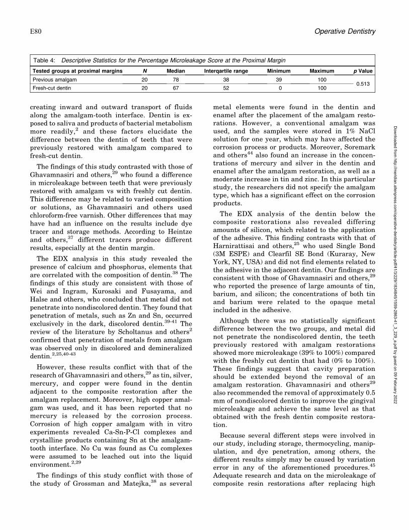
creating inward and outward transport of fluidsalong the amalgam-tooth interface. Dentin is ex-posed to saliva and products of bacterial metabolismmore readily,2 and these factors elucidate thedifference between the dentin of teeth that werepreviously restored with amalgam compared tofresh-cut dentin.
The findings of this study contrasted with those ofGhavamnasiri and others,29 who found a differencein microleakage between teeth that were previouslyrestored with amalgam vs with freshly cut dentin.This difference may be related to varied compositionor solutions, as Ghavamnasiri and others usedchloroform-free varnish. Other differences that mayhave had an influence on the results include dyetracer and storage methods. According to Heintzeand others,37 different tracers produce differentresults, especially at the dentin margin.
The EDX analysis in this study revealed thepresence of calcium and phosphorus, elements thatare correlated with the composition of dentin.38 Thefindings of this study are consistent with those ofWei and Ingram, Kurosaki and Fusayama, andHalse and others, who concluded that metal did notpenetrate into nondiscolored dentin. They found thatpenetration of metals, such as Zn and Sn, occurredexclusively in the dark, discolored dentin.39-41 Thereview of the literature by Scholtanus and others2
confirmed that penetration of metals from amalgamwas observed only in discolored and demineralizeddentin.2,25,40-43
However, these results conflict with that of theresearch of Ghavamnasiri and others,29 as tin, silver,mercury, and copper were found in the dentinadjacent to the composite restoration after theamalgam replacement. Moreover, high copper amal-gam was used, and it has been reported that nomercury is released by the corrosion process.Corrosion of high copper amalgam with in vitroexperiments revealed Ca-Sn-P-Cl complexes andcrystalline products containing Sn at the amalgam-tooth interface. No Cu was found as Cu complexeswere assumed to be leached out into the liquidenvironment.2,29
The findings of this study conflict with those ofthe study of Grossman and Matejka,38 as several
metal elements were found in the dentin andenamel after the placement of the amalgam resto-rations. However, a conventional amalgam wasused, and the samples were stored in 1% NaClsolution for one year, which may have affected thecorrosion process or products. Moreover, Soremarkand others44 also found an increase in the concen-trations of mercury and silver in the dentin andenamel after the amalgam restoration, as well as amoderate increase in tin and zinc. In this particularstudy, the researchers did not specify the amalgamtype, which has a significant effect on the corrosionproducts.
The EDX analysis of the dentin below thecomposite restorations also revealed differingamounts of silicon, which related to the applicationof the adhesive. This finding contrasts with that ofHarnirattisai and others,25 who used Single Bond(3M ESPE) and Clearfil SE Bond (Kuraray, NewYork, NY, USA) and did not find elements related tothe adhesive in the adjacent dentin. Our findings areconsistent with those of Ghavamnasiri and others,29
who reported the presence of large amounts of tin,barium, and silicon; the concentrations of both tinand barium were related to the opaque metalincluded in the adhesive.
Although there was no statistically significantdifference between the two groups, and metal didnot penetrate the nondiscolored dentin, the teethpreviously restored with amalgam restorationsshowed more microleakage (39% to 100%) comparedwith the freshly cut dentin that had (0% to 100%).These findings suggest that cavity preparationshould be extended beyond the removal of anamalgam restoration. Ghavamnasiri and others29
also recommended the removal of approximately 0.5mm of nondiscolored dentin to improve the gingivalmicroleakage and achieve the same level as thatobtained with the fresh dentin composite restora-tion.
Because several different steps were involved inour study, including storage, thermocycling, manip-ulation, and dye penetration, among others, thedifferent results simply may be caused by variationerror in any of the aforementioned procedures.45
Adequate research and data on the microleakage ofcomposite resin restorations after replacing high
Table 4: Descriptive Statistics for the Percentage Microleakage Score at the Proximal Margin
Tested groups at proximal margins N Median Interqartile range Minimum Maximum p Value
Previous amalgam 20 78 38 39 1000.513
Fresh-cut dentin 20 67 52 0 100
E80 Operative Dentistry
Dow
nloaded from http://m
eridian.allenpress.com/operative-dentistry/article-pdf/41/3/229/1834845/1559-2863-41_3_229_a.pdf by guest on 09 February 2022

copper amalgam with nondiscolored dentin is lack-ing. Harnirattisai and others25 concluded that thebond strength of discolored dentin after amalgamremoval was less than that of normal dentin.However, they did not test microleakage in theirstudy.
The limitations of this study include but are notlimited to 1) insufficient in vitro data for comparison,2) artificial aging (thermocycling) without salivaryintervention, and 3) dye penetration in a two-dimensional model.
Further research is recommended in which athree-dimensional analysis and thermomechanicalloading are used, as these are more analogous to theoral environment. Although clinical trials remainideal for the evaluation of dental restorations, theresults of this study provide scientific evidence forcomposite resin microleakage after amalgam resto-ration replacement.
Within the limitations of the study, the followingconclusions can be drawn: no significant differencewas found between the microleakage of nondiscol-ored dentin in teeth that were previously restoredwith amalgam compared with freshly cut dentin, andmarginal microleakage in the proximal surface washigher than that in the occlusal surface.
Regulatory Statement
This study was conducted in accordance with all theprovisions of the local human subjects oversight committeeguidelines and policies of the Tufts University School ofDental Medicine.
Conflict of Interest
The authors of this manuscript certify that they have noproprietary, financial, or other personal interest of any natureor kind in any product, service, and/or company that ispresented in this article.
(Accepted 26 October 2015)
REFERENCES
1. Akimoto N, Sakamoto T, Kubota Y, Kondo Y, & Momoi Y(2011) A novel composite-to-composite adhesive bondmechanism Dental Materials Journal 30(4) 523-527.
2. Scholtanus JD, Ozcan M, & Huysmans MC (2009)Penetration of amalgam constituents into dentine Jour-nal of Dentistry 37(5) 366-373.
3. Ben-Amar A, & Cardash HS (1991) The fluid-filled gapunder amalgam and resin composite restorations Amer-ican Journal of Dentistry 4(5) 226-230.
4. Fathi M (2004) A review on dental amalgam corrosion andits consequences Journal of Research in Medical Science9(1) 42-51.
5. Frankenberger R, Garcia-Godoy F, Murray PE, FeilzerAJ, & Kramer N (2013) Risk aspects of dental restor-atives: From amalgam to toothcolored materials WorldJournal of Stomatology 2(1) 1-11.
6. Roberts HW, Marek M, Kuehne JC, & Ragain JC (2005)Disinfectants’ effect on mercury release from amalgamJournal of the American Dental Association 136(7) 915-919.
7. Rathore M, Singh A, & Pant VA (2012) The dentalamalgam toxicity fear: A myth or actuality. ToxicologyInternational 19(2) 81-88.
8. Roulet JF (1997) Benefits and disadvantages of tooth-coloured alternatives to amalgam Journal of Dentistry25(6) 459-473.
9. Coelho-De-Souza FH, Camacho GB, Demarco FF, &Powers JM (2008) Fracture resistance and gap formationof MOD restorations: Influence of restorative technique,bevel preparation and water storage Operative Dentistry33(1) 37-43.
10. Zimmerli B, Strub M, Jeger F, Stadler O, & Lussi A(2010) Composite materials: Composition, properties andclinical applications. A literature review SchweizerMonatsschrift fur Zahnmedizin 120(11) 972-986.
11. Cheung GS (1990) Reducing marginal leakage of poste-rior composite resin restorations: A review of clinicaltechniques Journal of Prosthetic Dentistry 63(3) 286-288.
12. Kasraie S, Azarsina M, Khamverdi Z, & Shokraneh F(2012) Microleakage of dual-cured adhesive systems inclass V composite resin restorations Journal of Dentistry(Tehran) 9(2) 99-106.
13. Cho BH, Dickens SH, Bae JH, Chang CG, Son HH, & UmCM (2002) Effect of interfacial bond quality on thedirection of polymerization shrinkage flow in resincomposite restorations Operative Dentistry 27(3)297-304.
14. Rengo C, Goracci C, Juloski J, Chieffi N, Giovannetti A,Vichi A & Ferrari M (2012) Influence of phosphoric acidetching on microleakage of a self-etch adhesive and a self-adhering composite Australian Dental Journal 57(2)220-226.
15. Abo T, Uno S, & Sano H (2004) Comparison of bondingefficacy of an all-in-one adhesive with a self-etchingprimer system. European Journal of Oral Science 112(3)286-292.
16. Heintze SD (2007) Systematic reviews: I. The correlationbetween laboratory tests on marginal quality and bondstrength. II. The correlation between marginal qualityand clinical outcome Journal of Adhesive Dentistry9(Supplement 1) 77-106.
17. Yoshiyama M, Urayama A, Kimochi T, Matsuo T, &Pashley DH (2000) Comparison of conventional vs self-etching adhesive bonds to caries-affected dentin Opera-tive Dentistry 25(3) 163-169.
18. Pashley DH, & Carvalho RM (1997) Dentine permeabilityand dentine adhesion Journal of Dentistry 25(5) 355-372.
19. Nakajima M, Sano H, Burrow MF, Tagami J, YoshiyamaM, Ebisu S, Ciucchi B, Russell CM & Pashley DH (1995)Tensile bond strength and SEM evaluation of caries-
Redwan & Others: An In Vitro Microleakage Study for Composite Replacement of Amalgam Restoration E81
Dow
nloaded from http://m
eridian.allenpress.com/operative-dentistry/article-pdf/41/3/229/1834845/1559-2863-41_3_229_a.pdf by guest on 09 February 2022

affected dentin using dentin adhesives Journal of DentalResearch 74(10) 1679-1688.
20. Mjor IA, & Gordan VV (2002) Failure, repair, refurbish-ing and longevity of restorations Operative Dentistry27(5) 528-534.
21. Kleter GA (1998) Discoloration of dental carious lesions (areview) Archives of Oral Biology 43(8) 629-632.
22. Nakajima M, Sano H, Urabe I, Tagami J, & Pashley DH(2000) Bond strengths of single-bottle dentin adhesives tocaries-affected dentin Operative Dentistry 25(1) 2-10.
23. Yoshiyama M, Tay FR, Doi J, Nishitani Y, Yamada T,Itou K, Carvalho RM, Nakajima M & Pashley DH (2002)Bonding of self-etch and total-etch adhesives to cariousdentin Journal of Dental Research 81(8) 556-560.
24. Pereira PN, Nunes MF, Miguez PA, & Swift EJ Jr (2006)Bond strengths of a 1-step self-etching system to caries-affected and normal dentin Operative Dentistry 31(6)677-681.
25. Harnirattisai C, Senawongse P, & Tagami J (2007)Microtensile bond strengths of two adhesive resins todiscolored dentin after amalgam removal Journal ofDental Research 86(3) 232-236.
26. Majety KK, & Pujar M (2011) In vitro evaluation ofmicroleakage of class II packable composite resin resto-rations using flowable composite and resin modified glassionomers as intermediate layers Journal of ConservativeDentistry 14(4) 414-417.
27. Cenci M, Demarco F, & de Carvalho R (2005) Class IIcomposite resin restorations with two polymerizationtechniques: Relationship between microtensile bondstrength and marginal leakage Journal of Dentistry33(7) 603-610.
28. Schmid-Schwap M, Graf A, Preinerstorfer A, Watts DC,Piehslinger E, & Schedle A (2011) Microleakage afterthermocycling of cemented crowns—A meta-analysisDental Materials 27(9) 855-869.
29. Ghavamnasiri M, Motamed-Sanaye V, Chasteen J, AmeriH, Hajizadeh H, & Khashyarmanesh Z (2012) Energydispersive x-ray analysis of corrosion products in non-discolored dentin and a dye-extraction study of Class 2composite restorations following amalgam removal Quin-tessence International 43(4) 325-332.
30. Liebenberg WH (1996) Successive cusp build-up: Animproved placement technique for posterior direct resinrestorations Journal of the Canadian Dental Association62(6) 501-507.
31. Deliperi S, & Bardwell DN (2002) An alternative methodto reduce polymerization shrinkage in direct posteriorcomposite restorations Journal of the American DentalAssociation 133(10) 1387-1398.
32. Hebling J, Pashley DH, Tjaderhane L, & Tay FR (2005)Chlorhexidine arrests subclinical degradation of dentin
hybrid layers in vivo Journal of Dental Research 84(8)741-746.
33. da Silva AF, Piva E, Demarco FF, Correr Sobrinho L, &Osinga PW (2006) Microleakage in conventional andbonded amalgam restorations: Influence of cavity volume.Operative Dentistry 31(3) 377-383.
34. Radhika M, Sajjan GS, Kumaraswamy BN & Mittal N(2010) Effect of different placement techniques onmarginal microleakage of deep class-II cavities restoredwith two composite resin formulations Journal of Con-servative Dentistry 13(1) 9-15.
35. Alptekin T, Ozer F, Unlu N, Cobanoglu N, & Blatz MB(2010) In vivo and in vitro evaluations of microleakagearound Class I amalgam and composite restorationsOperative Dentistry 35(6) 641-648.
36. Bembi S, Bembi NN, Sood A, & Gambhir A (2013) Toevaluate the effect of different adhesive materials on themicroleakage of bonded amalgam restorations: An in vitrostudy International Journal of Clinical Pediatric Dentist-ry 6(2) 95-99.
37. Heintze S, Forjanic M, & Cavalleri A (2008) Microleakageof Class II restorations with different tracers—Compar-ison with SEM quantitative analysis Journal of AdhesiveDentistry 10(4) 259-267.
38. Grossman ES, & Matejka JM (1993) In vitro marginalleakage in varnished and lined amalgam restorationsJournal of Prosthetic Dentistry 69(5) 469-474.
39. Halse A (1975) Metals in dentinal tubules beneathamalgam fillings in human teeth Archives of Oral Biology20(1) 87-88.
40. Kurosaki N, & Fusayama T (1973) Penetration ofelements from amalgam into dentin Journal of DentalResearch 52(2) 309-317.
41. Wei SH, & Ingram MJ (1969) Analyses of the amalgam-tooth interface using the electron microprobe Journal ofDental Research 48(2) 317-320.
42. Hals E, & Halse A (1975) Electron probe microanalysis ofsecondary carious lesions associated with silver amal-gam fillings Acta Odontologica Scandinavica 33(3)149-160.
43. Mateer RS, & Reitz CD (1972) Galvanic degradation ofamalgam restorations Journal of Dental Research 51(6)1546-1551.
44. Soremark R, Wing K, Olsson K, & Goldin J (1968)Penetration of metallic ions from restorations into teethJournal of Prosthetic Dentistry 20(6) 531-540.
45. Yadav G, Rehani U, & Rana VA (2012) Comparativeevaluation of marginal leakage of different restorativematerials in deciduous molars: An in vitro study.International Journal of Clinical Pediatric Dentistry5(2) 101-107.
E82 Operative Dentistry
Dow
nloaded from http://m
eridian.allenpress.com/operative-dentistry/article-pdf/41/3/229/1834845/1559-2863-41_3_229_a.pdf by guest on 09 February 2022
Mechanical Properties andSliding-impact Wear Resistance of
Self-adhesive Resin Cements
T Furuichi � T Takamizawa � A TsujimotoM Miyazaki � WW Barkmeier � MA Latta
Clinical Relevance
Mechanical properties and wear resistance are important parameters for the selection of aresin luting cement. For wear resistance, the self-adhesive resin cements generally showsignificantly lower values than the conventional resin cements.
SUMMARY
The present study determined the mechanical
properties and impact-sliding wear character-
istics of self-adhesive resin cements. Five self-
adhesive resin cements were used: G-CEM
LinkAce, BeautiCem SA, Maxcem Elite, Clear-fil SA Automix, and RelyX Unicem 2. ClearfilEsthetic Cement was employed as a controlmaterial. Six specimens for each resin cementwere used to determine flexural strength,elastic modulus, and resilience according toISO specification #4049. Ten specimens foreach resin cement were used to determine thewear characteristics using an impact-slidingwear testing apparatus. Wear was generatedusing a stainless-steel ball bearing mountedinside a collet assembly. The maximum facetdepth and volume loss were determined usinga noncontact profilometer in combination withconfocal laser scanning microscopy. Data wereevaluated using analysis of variance followedby the Tukey honestly significantly differenttest (a=0.05). The flexural strength of the resincements ranged from 68.4 to 144.2 MPa; theelastic modulus ranged from 4.4 to 10.6 GPa;and the resilience ranged from 4.5 to 12.0 MJ/m3. The results for the maximum facet depthranged from 25.2 to 235.9 lm, and volume lossranged from 0.0107 to 0.5258 mm3. The flexuralproperties and wear resistance were found tovary depending upon the self-adhesive resin
Tetsuya Furuichi, DDS, graduate student, Operative Dentist-ry, Nihon University Graduate School of Dentistry, Tokyo,Japan
*Toshiki Takamizawa, DDS, PhD, assistant professor, Oper-ative Dentistry, Nihon University School of Dentistry, Tokyo,Japan
Akimasa Tsujimoto, DDS, PhD, assistant professor, OperativeDentistry, Nihon University School of Dentistry, Tokyo, Japan
Masashi Miyazaki, DDS, PhD, professor and chair, OperativeDentistry, Nihon University School of Dentistry, Tokyo, Japan
Wayne W. Barkmeier, DDS, MS, professor, Department ofGeneral Dentistry, dean emeritus, Creighton UniversitySchool of Dentistry, Omaha, Nebraska, USA
Mark A. Latta, DMD, MS, professor, Department of GeneralDentistry, dean, Creighton University School of Dentistry,Omaha, Nebraska, USA
*Corresponding author: 1-8-13 Kanda-Surugadai, Chiyoda-Ku, Tokyo 101-8310, Japan; e-mail: [email protected]
DOI: 10.2341/15-033-L
�Operative Dentistry, 2016, 41-3, E83-E92
Dow
nloaded from http://m
eridian.allenpress.com/operative-dentistry/article-pdf/41/3/229/1834845/1559-2863-41_3_229_a.pdf by guest on 09 February 2022

cement tested. The self-adhesive cements tend-ed to have lower mechanical properties thanthe conventional resin cement. All self-adhe-sive resin cements, apart from G-CEM Link-Ace, demonstrated significantly poorer wearresistance than did the conventional resincement.
INTRODUCTION
Self-adhesive resin cements utilized as luting agentsare defined as cements based on filled polymers thatadhere to the tooth structure in the absence of toothsurface pretreatment.1,2 Therefore, self-adhesiveresin cements are considered easier to apply com-pared with conventional resin cements.3 The appli-cation procedure is simple, and no postoperativesensitivity is expected because the smear layer is notremoved during the cementation process. Thesecements contain acidic functional monomers thatdemineralize the tooth structure and promote infil-tration of the resin components into the etched toothsubstrate.1,2 In addition, the setting reactions ofmost self-adhesive resin cements use a dual curingprocess based on the incorporation of photo-initiatorsalong with redox initiators. However, in contrast totheir simplified application, the compositions of self-adhesive resin cements are complicated. Moreover,incompatibility between acidic functional monomersand other resinous components can have adverseeffects on the mechanical properties of self-adhesiveresin cements. Nevertheless, previous studies4,5
have demonstrated that self-adhesive resin cementsare mechanically stronger than zinc phosphate, zincpolycarboxylate, glass ionomer, and resin-modifiedglass ionomer cements when comparing their flex-ural and compressive strengths. In addition, it hasbeen established that self-adhesive resin cementspossess similar flexural strengths to those of con-ventional resin cements.6-8
From the standpoint of clinical conditions, one ofthe challenges for luting cements is marginalintegrity. To select suitable luting cements forspecific clinical conditions, it is important to evalu-ate the wear performance characteristics of thecement to achieve long-lasting restorations. Margin-al gap formation may lead to a variety of concerns,including marginal staining, secondary caries, bond-ing failure, restoration fracture, and postoperativesensitivity.9 In addition, dental clinicians requiremore information related to the clinical relevance ofthe mechanical properties of more recently devel-oped self-adhesive resin cements, including detailabout their wear resistance.
Several studies10,12 have evaluated marginal gapsin an effort to assess the potential for cement loss atthe margins of restorations and to yield moreinformation concerning the expected quality ofrestorations. However, little information exists con-cerning the wear resistance properties of recentlydeveloped self-adhesive resin cements. The purposeof the present study was to assess the flexuralproperties and wear properties of self-adhesive resincements and to compare these properties with thoseof a conventional resin cement. The null hypothesisto be tested was that there are no significantdifferences in wear and mechanical propertiesbetween the conventional resin cement and theself-adhesive resin cements.
METHODS AND MATERIALS
Materials Used
Five self-adhesive resin cements were tested: G-CEMLinkAce (GL; GC Corp., Tokyo, Japan); BeautiCemSA Auto-mixing (BC; Shofu Inc, Kyoto, Japan);Maxcem Elite (ME; Kerr Corp, Orange, CA, USA);Clearfil SA Luting Automix (SA; Kuraray NoritakeDental Inc, Tokyo, Japan); and RelyX Unicem 2 (RU;3M ESPE Dental Products, St Paul, MN, USA). Aconventional resin cement, Clearfil Esthetic Cement(EC; Kuraray Noritake Dental Inc), was employed asa control material. The test materials and theircomponents are listed in Table 1. An Optilux 501visible–light-curing unit (sds Kerr, Danbury, CT,USA) was employed, and the light irradiance (average800 mW/cm2) of the curing unit was checked using adental radiometer (Model 100, Kerr).
Inorganic Filler Content
The inorganic filler content of the materials wasmeasured using thermogravimetry/differential ther-mal analysis employing a 6300 thermogravimeter(Seiko Instruments, Tokyo, Japan). For each resincement tested, a mixed cement paste sample (ap-proximately 50 mg) was heated in the thermo-gravimeter from 258C to 8008C at a heating rate of108C/min until the organic components were com-pletely consumed. The weight of the residual cementpaste was measured and the inorganic filler content(wt%) was calculated. Three measurements wereconducted to obtain an average inorganic fillercontent (wt%).
Coefficient of Linear Thermal Expansion
The coefficients of linear thermal expansion of thetest materials were measured using a thermome-
E84 Operative Dentistry
Dow
nloaded from http://m
eridian.allenpress.com/operative-dentistry/article-pdf/41/3/229/1834845/1559-2863-41_3_229_a.pdf by guest on 09 February 2022
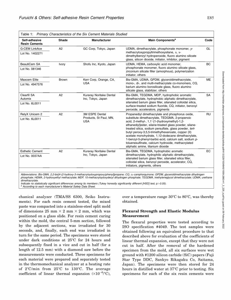
chanical analyzer (TMA/SS 6300, Seiko Instru-
ments). For each resin cement tested, the mixed
paste was compacted into a stainless-steel split mold
of dimensions 25 mm 3 2 mm 3 2 mm, which was
positioned on a glass slide. For resin cement curing
within the mold, the central 5-mm section, followed
by the adjacent sections, was irradiated for 30
seconds, and, finally, each end was irradiated in
turn for the same period. The specimens were stored
under dark conditions at 258C for 24 hours and
subsequently fixed in a vice and cut in half (for a
length of 12.5 mm) with a diamond saw before the
measurements were conducted. Three specimens for
each material were prepared and separately tested
in the thermomechanical analyzer at a heating rate
of 28C/min from 258C to 1308C. The average
coefficient of linear thermal expansion (310�6/8C),
over a temperature range 308C to 808C, was therebyobtained.
Flexural Strength and Elastic ModulusMeasurement
The flexural properties were tested according toISO specification #4049. The test samples wereobtained following an equivalent procedure to thatdescribed above for evaluation of the coefficients oflinear thermal expansion, except that they were notcut in half. After the removal of the hardenedspecimen from the mold, all six surfaces were wetground with #1200 silicon carbide (SiC) papers (FujiStar Type DDC, Sankyo Rikagaku Co, Saitama,Japan). The specimens were then stored for 24hours in distilled water at 378C prior to testing. Sixspecimens for each of the six resin cements were
Table 1: Primary Characteristics of the Six Cement Materials Studied
Self-adhesiveResin Cements
Shade Manufacturer Main Componentsa Code
G-CEM LinkAce A2 GC Corp, Tokyo, Japan UDMA, dimethacrylate, phosphonate monomer, y-methacryloxypropyltrimethoxysilane, a, a-dimethylbenzyl hydroperoxide, fluoro alumino silicateglass, silicon dioxide, initiator, inhibitor, pigment
GL
Lot No. 1402271
BeautiCem SA Ivory Shofu Inc, Kyoto, Japan UDMA, HEMA, carboxylic acid monomer,phosphonate monomer, fluoro alumino silicate glass,zirconium silicate filler (amorphous), polymerizationinitiator, others
BC
Lot No. 081346
Maxcem Elite Brown Kerr Corp, Orange, CA,USA
Bis-GMA, UDMA, GPDM, glyceroldimethacrylate,mono-, di-, and multi-methacrylate co-monomers, CQ,barium alumino borosilicate glass, fluoro aluminosilicate glass, stabilizer, others
ME
Lot No. 4947576
Clearfil SAAutomix
A2 Kuraray Noritake DentalInc, Tokyo, Japan
Bis-GMA, TEGDMA, MDP, hydrophobic aromaticdimethacrylate, hydrophobic aliphatic dimethacrylate,silanated barium glass filler, silanated colloidal silica,surface-treated sodium fluoride, CQ, initiator, benzoylperoxide, accelerators, pigments
SA
Lot No. 6L0011
RelyX Unicem 2 A2 3M ESPE DentalProducts, St Paul, MN,USA
Propanediyl dimethacrylate and phosphorus oxide,substitute dimethacrylate, TEDGMA, 2-propenoicacid, 2-methyl-, 1,10-[1-(hydroxymethyl)-1,2-ethanediyl]ester, silane-treated glass powder, silane-treated silica, sodium persulfate, glass powder, tert-butyl peroxy-3,5,5-trimethylhexanoate, copper (II)acetate monohydrate, 1,12-dodecane dimethacrylate,1-benzyl-5-phenyl-barbic-acid, calcium salt, sodium p-toluenesulfinate, calcium hydroxide, methacrylatedaliphatic amine, titanium dioxide
RU
Lot No. 6L0011
Esthetic Cement A2 Kuraray Noritake DentalInc, Tokyo, Japan
Bis-GMA, TEGDMA, hydrophobic aromaticdimethacrylate, hydrophilic aliphatic dimethacrylate,silanated barium glass filler, silanated silica filler,colloidal silica, benzoyl peroxide, accelerator, CQ,initiators, pigments, others
EC
Lot No. 0037AA
Abbreviations: Bis-GMA, 2,2-bis[4-(2-hydroxy-3-methacryloyloxypropoxy)phenyl]propane; CQ, DL-camphorquinone; GPDM, glyceroldimethacrylate dihydrogenphosphate; HEMA, 2-hydroxyethyl methacrylate; MDP, 10-methacryloyloxydecyl dihydrogen phosphate; TEGDMA, triethyleneglycol dimethacrylate; UDMA, urethanedimethacrylate.Indicate no statistically significant differences among its members (Tukey honestly significantly different [HSD] test, p.0.05).a According to each manufacturer’s Material Safety Data Sheet.
Furuichi & Others: Self-adhesive Resin Cement Properties E85
Dow
nloaded from http://m
eridian.allenpress.com/operative-dentistry/article-pdf/41/3/229/1834845/1559-2863-41_3_229_a.pdf by guest on 09 February 2022
subjected to a three-point bending flexural strengthtest using a universal testing instrument (Type5500R, Instron Corp, Canton, MA, USA) with aspan length of 20.0 mm at a cross-head speed of 1.0mm/min until the specimen fractured. The speci-mens were kept moist during testing. The peakbreaking stress, modulus of elasticity, and resil-ience were determined from the stress-strain curveof each sample using the Bluehill Ver 2.5 computersoftware (Instron Corp) linked to the testinginstrument.
Impact-sliding Wear Simulation
Ten specimens for each of the six resin cementswere tested to determine the wear behavior usingthe impact-sliding wear testing apparatus (K655-05, Tokyo Giken Inc, Tokyo, Japan). For eachspecimen, the mixed resin cement paste was placedinto a cylindrical Teflon mold (6 mm in diameter, 2mm in height). The cement paste was cured for 30seconds and stored under dark conditions for 24hours in distilled water at 378C. The flat surfaces ofeach specimen were polished to 4000 grit using agraded sequence of SiC papers. Specimens wereattached with a small amount of model-repair glue(Zapit, Dental Ventures of America Inc, Corona, CA,USA) to the centers of custom fixtures fabricatedfrom a cold-cure acrylic resin (Tray Resin II, ShofuInc).
The antagonist for the impact-sliding wear simu-lation was a stainless-steel ball bearing of radius r =2.387 mm that was mounted inside a collet assembly.The simulator incorporated a plastic water bath thatprovided water at a temperature of 378C constantly,and four of the custom wear fixtures were mountedinside the bath. During the course of the wearsimulation test, the antagonists directly impactedthe specimens from above with a maximum force of50 N at a rate of 0.5 Hz, and the antagonistssubsequently slid horizontally for a distance of 2mm. Each specimen was subjected to 50,000 cycles ofthe impact-sliding motion.
Wear Measurements
Following the wear simulation test, the specimenswere ultrasonically cleaned in distilled water forthree minutes and then profiled using a confocallaser scanning microscope (VK-9710, Keyence, Osa-ka, Japan) with built-in analysis software (VKanalyzer, Keyence). The maximum depth (lm) andvolume loss (mm3) of the wear facets were therebydetermined.
Scanning Electron Microscopy Observations
The surfaces of the cured resin cement sampleswere polished to a high gloss with abrasive discs(Fuji Star Type DDC, Sankyo Rikagaku Co) fol-lowed by a series of diamond pastes down to a 0.25-lm particle size (DP-Paste, Struers, Ballerup, Den-mark). The polished surfaces were then subjected tounfiltered argon-ion beam etching (IIS-200ER,Elionix, Tokyo, Japan) for 40 seconds perpendicularto the polished surface at an accelerating voltage of1.0 kV and ion current density of 0.4 mA/cm2. Thesurfaces were then coated in a vacuum evaporator(Quick Coater Type SC-701; Sanyu Denshi Inc,Tokyo, Japan) with a thin film of gold. Examina-tions of the surfaces were conducted by scanningelectron microscopy (SEM) using an Elionix FE-8000 instrument (Elionix, Tokyo, Japan) with anoperating voltage of 10 kV and a magnification of50003.
SEM examinations were conducted on the wearfacets of the six resin cement samples after impact-sliding wear simulation. Representative samples foreach resin cement were sputter-coated with gold.Examinations of the coated surfaces were conductedusing the SEM with an operating voltage of 10 kVand magnifications of 403 and 25003.
Statistical Analysis
The data for each material were subjected toanalysis of variance followed by the Tukey honestlysignificantly difference (HSD) test at a significancelevel of 0.05. The statistical analysis was conductedusing the Sigma Plot software system (Ver. 11.0;SPSS Inc, Chicago, IL, USA).
RESULTS
Inorganic Filler Content and Coefficient ofThermal Expansion
The average inorganic filler contents and coeffi-cients of thermal expansion are listed in Table 2.The average inorganic filler contents of the resincements ranged from 55.3 to 67.9 wt%. The highestinorganic filler content was determined for EC,whereas GL showed the lowest value of all the testmaterials, and these differences were statisticallysignificant. The average coefficients of thermalexpansion of the resin cements ranged from 37.7to 51.6 310�6/8C. A significantly low value wasexhibited by RU, whereas ME showed a signifi-cantly higher value than all other tested resincements.
E86 Operative Dentistry
Dow
nloaded from http://m
eridian.allenpress.com/operative-dentistry/article-pdf/41/3/229/1834845/1559-2863-41_3_229_a.pdf by guest on 09 February 2022

Flexural Properties
The results of flexural property testing are listed inTable 3. The average flexural strength of the resincements ranged from 68.4 to 144.2 MPa, and theaverage elastic modulus ranged from 4.4 to 10.6GPa. Tukey HSD test indicated that BC exhibitedthe highest flexural strength of all test resincements, and no significant difference was obtainedwhen BC was compared to the control material EC.Regarding the elastic modulus, EC demonstrated thehighest value. Self-adhesive resin cements BC, GL,and RU exhibited higher values compared to allother self-adhesive resin cements, and no significantdifferences were obtained among them. The averageresilience ranged from 4.5 to 12.0 MJ/m3. The controlmaterial EC exhibited the highest resilience.
Impact-sliding Wear
The average wear values (maximum facet depth andvolume loss) for the six resin cements are summa-
rized in Table 4. The rank orders of all six resincements were similar with regard to both facet weardepth and volumetric loss. Cements EC and GLdemonstrated significantly lower maximum facetdepth and volume loss when compared to all otherresin cements. In contrast, ME exhibited the highestmaximum facet depth and volume loss when com-pared to all other resin cements, and all differenceswere statistically significant.
SEM Observations
SEM examinations of the six resin cements afterargon-ion etching are presented in Figure 1a-f.Differences in filler particle size, shape, and distri-bution are clearly observable. For GL, RU, and ME,0.5-5-lm irregularly shaped glass filler particles areobserved, as shown in Figure 1a, c, and e, respec-tively. For BC, 0.2-5 lm–sized spherical silica fillerparticles and 0.2-1 lm irregular glass filler particlesare observed in Figure 1b. SA and EC includerelatively large irregular glass filler particles mea-
Table 2: Inorganic Filler Contents and Thermal ExpansionCoefficients of the Cement Materials Studied
Code InorganicFiller
Content,wt%a
TukeyGroupb
Thermal ExpansionCoefficient,
310�6/8C(from 308C to 808C)a
TukeyGroupb
GL 55.3 (0.2) F 44.7 (1.6) B
BC 59.6 (0.2) E 42.8 (1.5) B
ME 66.9 (0.2) B 51.6 (0.8) A
SA 65.8 (0.2) C 45.3 (0.9) B
RU 63.6 (0.2) D 37.7 (0.2) C
EC 67.9 (0.3) A 41.8 (2.0) B
Abbreviations: BC, BeautiCem SA; EC, Esthetic Cement; GL, G-CEMLinkAce G-CEM LinkAce; ME, Maxcem Elite; RU, RelyX Unicem 2; SA,Clearfil SA Automix.a Mean (standard deviation), n = 6.b Equivalent group letters indicate no statistically significant differencesamong its members (Tukey honestly significantly different [HSD] test,p.0.05).
Table 3: Flexural Strength, Elastic Modulus, and Resilience of the Cement Materials Studied
Code Flexural Strength,MPaa
TukeyGroupb
Flexural Modulus,GPaa
TukeyGroupb
Resilience,MJ/mm3a
TukeyGroupb
GL 121.4 (7.9) B 7.2 (0.4) B 9.5 (2.1) B
BC 144.2 (12.2) A 7.7 (0.5) B 8.5 (2.0) B,C
ME 68.4 (3.9) D 4.4 (0.3) D 4.5 (0.5) D
SA 86.6 (8.5) C 5.3 (0.5) C 6.3 (1.5) C,D
RU 99.7 (8.6) C 7.2 (0.4) B 6.0 (0.9) C,D
EC 138.5 (3.8) A 10.6 (0.8) A 12.0 (2.0) A
Abbreviations: BC, BeautiCem SA; EC, Esthetic Cement; GL, G-CEM LinkAce G-CEM LinkAce; ME, Maxcem Elite; RU, RelyX Unicem 2; SA, Clearfil SA Automix.a Mean (standard deviation), n = 6.b Equivalent group letters indicate no statistically significant differences among its members (Tukey honestly significantly different [HSD] test, p.0.05).
Table 4: Maximum Facet Depth and Volume Loss of theCement Materials Studied
Code MaximumFacet Depth, lma
TukeyGroupb
Volume Loss,mm3a
TukeyGroupb
GL 28.6 (1.5) E 0.0110 (0.003) D
BC 122.3 (31.7) D 0.1712 (0.059) C
ME 239.5 (13.9) A 0.5258 (0.051) A
SA 187.5 (30.8) C 0.3075 (0.103) B
RU 197.8 (44.9) B 0.2060 (0.046) C
EC 25.2 (10.2) E 0.0107 (0.005) D
Abbreviations: BC, BeautiCem SA; EC, Esthetic Cement; GL, G-CEMLinkAce G-CEM LinkAce; ME, Maxcem Elite; RU, RelyX Unicem 2; SA,Clearfil SA Automix.a Mean (standard deviation), n = 6.b Equivalent group letters indicate no statistically significant differencesamong its members (Tukey honestly significantly different [HSD] test,p.0.05).
Furuichi & Others: Self-adhesive Resin Cement Properties E87
Dow
nloaded from http://m
eridian.allenpress.com/operative-dentistry/article-pdf/41/3/229/1834845/1559-2863-41_3_229_a.pdf by guest on 09 February 2022

suring approximately 0.2-10 lm in size, as shown inFigure 1d and f.
Representative SEM images of wear facets afterimpact-sliding wear testing are presented in Figure2a-h. For GL and EC, the SEM examinations given
in Figure 2a and g, respectively, reveal relatively flat
and smooth surfaces, although some fine cracks are
observed on the worn surfaces, as shown in Figures
2b and h for GL and EC, respectively. However, all
other self-adhesive resin cements exhibited rougher
Figure 1. SEM micrographs at a 50003 magnification of the argon-ion etched surfaces of the six cement materials studied, as follows. (A) G-CEMLinkAce argon-ion etched surface at 50003. (B) BeautiCem SA argon-ion etched surface at 50003. (C) Maxcem Elite argon-ion etched surface at50003. (D) Clearfil SA Automix argon-ion etched surface at 50003. (E) RelyX Unicem 2 argon-ion etched surface at 50003. (F) Esthetic Cementargon-ion etched surface at 50003.
Figure 2. SEM micrographs at 403 and 25003 magnifications of the surfaces of representative samples of the six cement materials subjected toimpact-sliding wear testing. (A) G-CEM LinkAce Impact-sliding wear facet at 403. (B) G-CEM LinkAce Impact-sliding wear near center of facet at25003. (C) Clearfil SA Automix Impact-sliding wear facet 403. (D) Clearfil SA Automix Impact-sliding wear near center of facet at 25003. (E) RelyXUnicem 2 Impact-sliding wear facet 403. (F) RelyX Unicem 2 Impact-sliding wear near center of facet at 25003. (G) Esthetic Cement Impact-slidingwear facet 403. (H) Esthetic Cement Impact-sliding wear near center of facet at 25003.
E88 Operative Dentistry
Dow
nloaded from http://m
eridian.allenpress.com/operative-dentistry/article-pdf/41/3/229/1834845/1559-2863-41_3_229_a.pdf by guest on 09 February 2022

and deeper facets than those of either GL or thecontrol material EC. Similar wear patterns areobserved for the more highly worn self-adhesiveresin cement samples, with some plucking of fillerparticles from the surface and obvious cracks, asshown in Figure 2c-f.
DISCUSSION
Improvements in the resin matrix and filler compo-nents of direct composite restorations have resultedin markedly improved materials for applications inthe posterior region.13,14 However, dental profes-sionals remain concerned about the mechanicalproperties and wear resistance of these materialsfor application to larger posterior lesions. Indirectrestorations have been selected, rather than directrestorations, as the standard procedure for theposterior region because of their durable properties,ease of fabrication in a precise anatomical form, andadaptation to adjacent teeth.15 At present, metalalloys, resin composites, and ceramics are used asindirect restorative materials. However, to achievedurable bonds between an indirect restoration andthe surrounding tooth structure, each of thesematerials requires several pretreatment steps forluting.
Therefore, simplified self-adhesive resin cementsfor luting of restoratives have been developed thatcan reduce the clinical steps required for the lutingprocedure, mitigate technique sensitivity, and di-minish postoperative sensitivity.16,17 In addition, toavoid an insufficient degree of conversion when lightdelivery from a curing unit is inhibited as a result ofobstructions by overlying indirect restorations, self-adhesive resin cements have been developed thatincorporate a dual-polymerization system. This typeof cement contains inorganic fillers, matrix resins,initiator systems, and acidic functional monomers.However, a major concern has been expressedregarding a reduced degree of conversion due tothe presence of acidic functional monomers becauseof the risk of lower pH18,19 and interference with thereaction between the tertiary amine and camphor-quinone.20-22 Therefore, to predict in vivo perfor-mance of self-adhesive resin cements, it is importantto determine their endurance characteristics byevaluating their mechanical properties and wearresistance.
The results for inorganic filler content andthermal expansion of the resin cements appearedto be material dependent. Regarding the fillercontent, it is considered that increasing the amountof inorganic filler content plays an important role in
enhancing the mechanical properties, reducing thepolymerization shrinkage, and increasing the wearresistance. Although a high filler content is desirablefor resin-based materials, it is difficult to include alarge amount of filler because of the need tomaintain an appropriate viscosity for luting and toensure a suitable film thickness.
Thermal expansion is an important property ofluting cements when considering the longevity ofrestorations. Temperature cycling may induce di-mensional changes in the materials and rupture ofthe bond interface not only between the lutingcement and the tooth structure, but also betweenthe luting cement and the restoration as a result ofdifferences in the thermal expansion properties ofthe individual materials. Because the thermalexpansion of the restorative material and lutingcements usually does not match that of the toothstructure, a differential expansion occurs that mayresult in leakage of oral fluids into the interfacebetween the restoration and the tooth. From theresults of this study, thermal expansion of the sixresin cements ranged from 37.7 to 51.6 310�6 m/8C.In addition, in spite of the higher inorganic fillercontent, ME exhibited a significantly higher averagethermal expansion coefficient than all other resincements. A higher inorganic filler content has beenconsidered to reduce thermal expansion, but theresults obtained in this study suggested that otherfactors, such as the type of matrix resin and size ofthe filler, might have a significant influence on thethermal properties. Considering the long-term dura-bility of indirect restorations, further improvementof thermal properties is needed in the resin cements.
Fracture-related material properties, such asfracture resistance, elasticity, and the marginaldegradation of materials under stress, have typicallybeen evaluated by determining material parameterssuch as flexural strength, flexural modulus, andfracture toughness.23,24 Although flexural strengthunder constant loading may not reflect intraoralconditions, these values are helpful in comparingmaterials under controlled conditions.25 From theresults of flexural testing, the control material ECtended to have a higher flexural strength than theself-adhesive cements considered. However, BS andGL demonstrated a flexural strength similar to thatof EC. A previous study revealed a correlationbetween the filler content, filler size, and thedistribution of filler particles in resin-based materi-als and their material properties.26,27 However,analysis of those results in this study showed nosignificant correlation, so other factors appear to be
Furuichi & Others: Self-adhesive Resin Cement Properties E89
Dow
nloaded from http://m
eridian.allenpress.com/operative-dentistry/article-pdf/41/3/229/1834845/1559-2863-41_3_229_a.pdf by guest on 09 February 2022

more important. In addition, it is speculated that thefunctional monomers contained in self-adhesivecements might affect the degree of conversion ofthe cement itself.
It has been suggested that the desirable modulusof elasticity for a luting cement lies between those ofthe restorations and mineralized tooth structure,which contributes to a reduction of interfacial stressconcentrations. Furthermore, resistance to plasticdeformation may increase the resistance to marginalgap formation and cement fracture, and therefore ahigh proportional limit and resilience are pre-ferred.28 For resilience, the self-adhesive cementstended to exhibit lower values than that of thecontrol material EC. Thus, the lower elastic modulusand resilience of self-adhesive cements may remain adisadvantage in preventing deformation and frac-ture of brittle restorations, such as all-ceramic andmachinery-milled restorations.
Many types of filler systems, monomer systems,and coupling agents have been developed to improvethe mechanical properties and wear resistance ofresin composites. Resin luting cements, includingself-adhesive resin cements, have been developedusing resin composite technology. Thus, these typesof materials might have similar characteristics tothose of resin composites, and the wear resistance ofthe cement itself should be evaluated. Wear progres-sion of luting cements at the margin may lead togaps between a restoration and the surroundingtooth structure and may create a vulnerable regionthat serves as a foothold for crack propagation in thecement as a result of mastication. Increasing gapformation may induce secondary caries, bondingfailure, restorative fracture in the body or at themargins, and postoperative sensitivity.9
Clinical criteria have been used to assess marginalintegrity in long-term clinical trials.29,30 However,these investigations are costly, time-intensive, andoften technically challenging to complete. In con-trast, simulated wear measurement could provide arapid means with which to examine relative wearrates among materials and to predict expectedclinical performance.31,32
In the present study, a sliding-impact wearapparatus was used as a two-body contact wearsimulator. In a three-body contact wear model,abrasion, adhesive, and erosion wear are likely thedominant forces, but their relative contributions arenot known. However, for a two-body contact wearmodel, it is considered that abrasion, adhesive wear,and fatigue or attrition are dominant.33 Although
three-body wear testing with an abrasive mediumhas been used frequently to simulate the masticationof food, the use of abrasive media influences theresults as a result of the formation of an embeddedlayer of abrasive medium and creates a difficultywith standardizing these mixtures or maintaining aconsistent viscosity and composition during theentire wear simulation process.34 These consider-ations suggest that the two-body wear test employedin the present study has the advantages of eliminat-ing variability and establishing a simplified model.
The wear values (maximum facet depth andvolume loss) for the six resin cements appear to bematerial dependent, as was also determined for themechanical properties. The self-adhesive cements,apart from GL, demonstrated significantly lowerwear resistance than did the control material EC.Therefore, the null hypothesis that self-adhesiveresin cements will show significant differences inwear resistance and mechanical properties from thecontrol resin cement was not completely rejected.From the SEM images of wear facets after impact-sliding wear testing, GL and EC revealed relativelyflat and smooth surfaces, in addition to some finecracks on the worn surfaces. On the other hand, theother self-adhesive cements exhibited rougher anddeeper facets than did both GL and EC. The othermaterials also exhibited a similar wear pattern, withsome plucking of filler particles from the surface.Considering the impact and sliding force adminis-tered by the wear equipment employed, it could beconsidered that smaller filler particles would pre-serve the surface texture even if some filler wereplucked from the resin matrix and might inhibitcrack propagation.10
It has been reported35,36 that improvement in thedegree of conversion of resin composites is expectedto increase wear resistance and mechanical proper-ties. In addition, previous studies suggested thatdual-cured self-adhesive resin cements with photoactivation would obtain a superior degree of conver-sion compared with self-curing alone, because acidicfunctional monomers act negatively on the self-curesetting reaction by their relatively lower pH val-ue18,19 and interfere with the reaction between thetertiary amine and camphorquinone.20-22 Althoughthe specimens for mechanical properties and wearresistance were evaluated after photo activation, it ispossible that the acidic functional monomers influ-enced the polymerization of the self-adhesive resincements.
While mechanical properties and wear behaviorare not the only parameters for consideration in the
E90 Operative Dentistry
Dow
nloaded from http://m
eridian.allenpress.com/operative-dentistry/article-pdf/41/3/229/1834845/1559-2863-41_3_229_a.pdf by guest on 09 February 2022

selection of resin cements, these characteristics canprovide valuable information to dental professionals.
CONCLUSIONS
The results of this study showed that the self-adhesive cements tended to have poorer mechanicalproperties than the conventional resin cements. Forwear resistance, the self-adhesive resin cements,apart from GL, showed significantly lower valuesthan did the conventional resin cements. Theseresults augment the information base available tothe profession and provide guidance for clinicians inthe selection of resin cements.
Acknowledgements
This work was supported in part by a Grant-in-Aid forScientific Research (C) 23592810 and a Grant-in-Aid forYoung Scientists (B) 23792186 from the Japan Society forthe Promotion of Science. This project was also supported inpart by the Sato Fund and by a grant from the DentalResearch Center of the Nihon University School of Dentistry,Japan.
Conflict of Interest
The authors of this manuscript certify that they have noproprietary, financial, or other personal interest of any natureor kind in any product, service, and/or company that ispresented in this article.
(Accepted 27 May 2015)
REFERENCES
1. Radovic I, Monticelli F, Goracci C, Vulicevic ZR, & FerrariM (2008) Self-adhesive resin cements: A literature reviewJournal of Adhesive Dentistry 10(4) 251-258.
2. Ferracane JL, Stansbury JW, & Burke FJT (2011) Reviewarticle self-adhesive resin cements—Chemistry, proper-ties and clinical considerations Journal of Oral Rehabil-itation 38(4) 295-314.
3. Burke FJT, & Birmingham RJC (2006) A practice-basedevaluation of the handling of new self-adhesive universalresin luting material International Dental Journal 56(3)142-146.
4. Piwowarczyk A, & Lauer H-C (2003) Mechanical proper-ties of luting cements after water storage OperativeDentistry 28(5) 535-542.
5. Kumbuloglu O, Lassila LV, User A, & Vallittu PK (2004)A study of the physical and chemical properties of fourresin composite luting cements International Journal ofProsthodontics 17(3) 357-363.
6. Saskalauskaite E, Tam LE, & McComb D (2006) Flexuralstrength, elastic modulus, and pH profile of self-etch resinluting cements Journal of Prosthodontics 17(4) 262-268.
7. Behr M, Rosentritt M, Loher H, Kolbeck C, Templer C,Stemplinger B, Kopzon V, & Handel G (2008) Changes ofcement properties caused by mixing errors: The thera-
peutic range of different cement types Dental Materials24(9) 1187-1193.
8. Nakamura T, Wakabayashi K, Kinuta S, Nishida H,Miyamae M, & Yatani H (2010) Mechanical properties ofnew self-adhesive resin-based cement Journal of Prosth-odontic Research 54(2) 59-64.
9. Ibarra G, Johnson GH, Geurtsen W, & Vargas MA (2007)Microleakage of porcelain veneer restorations bonded toenamel and dentin with a new self-adhesive resin-baseddental cement Dental Materials 23(2) 218-225.
10. Shinkai K, Suzuki S, Leinfelder KF, & Katoh Y (1995)Effect of gap dimension on wear resistance of lutingcements American Journal of Dentistry 8(3) 149-151.
11. Braga RR, Condon JR, & Ferracane JL (2002) In vitrowear simulation measurements of composite versus resin-modified glass ionomer luting cements for all-ceramicrestorations Journal of Esthetic and Restorative Dentistry14(6) 368-376.
12. Belli R, Pelka M, Petschelt A, & Lohbauer U (2009) Invitro wear gap formation of self-adhesive resin cements: ACLSM evaluation Journal of Dentistry 37(12) 984-993.
13. Lutz F, Phillips RW, Roulet JF, & Setcos JC (1984) In vivoand in vitro wear of potential posterior compositesJournal of Dental Research 63(6) 914-920.
14. Heintze SD, Barkmeier WW, Latta MA, & Rousson V(2011) Round robin test: Wear of nine restorativematerials in six different wear simulators—Supplementto the round robin test of 2005 Dental Materials 27(2)e1-e9.
15. Wassell RW, Walls AW, & McCabe JF (1995) Directcomposite inlays versus conventional composite restora-tions: Three year clinical results British Dental Journal179(9) 343-349.
16. Monticelli F, Osorio R, Mazzitelli C, Ferrari M, &Toledano M (2008) Limited decalcification/diffusion ofself-adhesive cements into dentin Journal of DentalResearch 87(10) 974-979.
17. Cantoro A, Goracci C, Carvalho CA, Coniglio I, & FerrariM (2009) Bonding potential of self-adhesive luting agentsused at different temperatures to lute composite onlayJournal of Dentistry 37(6) 454-461.
18. Vrochari AD, Eliades G, Hellwig E, & Wrbas KT (2009)Curing efficiency of four self-etching, self-adhesive resincements Dental Materials 25(9) 1104-1108.
19. Zorzin J, Petschelt A, Ebert J, & Lohbauer U (2012) pHneutralization and influence on mechanical strength inself-adhesive resin luting agents Dental Materials 28(6)672-679.
20. Sanares AME, Itthagarun A, King NM, Tay FR, &Pashley DH (2001) Adverse surface interactions betweenone-bottle light-cured adhesives and chemical-curedcomposites Dental Materials 17(6) 542-556.
21. Suh BI, Feng L, Pashley DH, & Tay FR (2003) Factorscontributing to the incompatibility between simplified-step adhesives and chemically-cured or dual-cured com-posites. Part III. Effect of acidic resin monomers Journalof Adhesive Dentistry 5(4) 267-282.
Furuichi & Others: Self-adhesive Resin Cement Properties E91
Dow
nloaded from http://m
eridian.allenpress.com/operative-dentistry/article-pdf/41/3/229/1834845/1559-2863-41_3_229_a.pdf by guest on 09 February 2022

22. Salz U, Zimmermann J, & Salzer T (2005) Self-curing,self-adhesive cements systems Journal of AdhesiveDentistry 7(1) 7-17.
23. Watanabe H, Khera SC, Vargas MA, & Qian F (2008)Fracture toughness comparison of six resin compositesDental Materials 24(3) 418-425.
24. Sumino N, Tsubota K, Takamizawa T, Shiratsuchi K,Miyazaki M, & Latta MA (2013) Comparison of the wearand flexural characteristics of flowable resin compositesfor posterior lesions Acta Odontologica Scandinavica71(3-4) 820-827.
25. Lang R, Rosentritt M, Behr M, & Handel G (2003)Fracture resistance of PMMA and resin matrix compos-ite–based interim FPD materials International Journal ofProsthodontics 16(4) 381-384.
26. Jain P, & Belcher M (2000) Microleakage of Class II resin-based composite restorations with flowable composite inthe proximal box American Journal of Dentistry 13(5)235-238.
27. Turssi CP, Ferracane JL, & Ferracane LL (2006) Wearand fatigue behavior of nano-structured dental resincomposites Journal of Biomedical Materials ResearchPart B: Applied Biomaterials 78(1) 196-203.
28. Li ZC, & White SN (1999) Mechanical properties of dentalluting cements Journal of Prosthetic Dentistry 81(5)597-609.
29. Frankenberger R, Reinelt C, Petschelt A, & Kramer N(2009) Operator vs. material influence on clinical outcomeof bonded ceramic inlays Dental Materials 25(8) 960-968.
30. Peumans M, Voet MJ, De Munk J, Van Landuyt K, VanEnde B, & Van Meerbeek B (2013) Four-year clinicalevaluation of a self-adhesive luting agent for ceramicinlays Clinical Oral Investigations 17(3) 739-750.
31. Barkmeier WW, Erickson RL, Latta MA, & WilwerdingTM (2008) Wear simulation of resin composites and therelationship to clinical wear Operative Dentistry 33(2)177-182.
32. Barkmeier WW, Erickson RL, Latta MA, & WilwerdingTM (2013) Wear rates of resin composites OperativeDentistry 38(2) 226-233.
33. Condon JR, & Ferracane JL (1997) Factors effectingdental composite wear in vitro Journal of BiomedicalMaterial Research Part A 38(4) 303-313.
34. Heintze SD (2006) How to qualify and validate wearsimulation devices and methods Dental Materials 22(8)712-734.
35. Ferracane JL, & Berge HX (1995) Fracture toughness ofexperimental dental composites aged in ethanol Journalof Dental Research 74(7) 1418-1423.
36. Ferracane JL, Mitchem JC, Condon JR, & Todd T (1997)Wear and marginal breakdown of composites withvarious degree of cure Journal of Dental Research 76(8)1508-1516.
E92 Operative Dentistry
Dow
nloaded from http://m
eridian.allenpress.com/operative-dentistry/article-pdf/41/3/229/1834845/1559-2863-41_3_229_a.pdf by guest on 09 February 2022