World Journal of Gastrointestinal Endoscopy
58
WJGE|www.wjgnet.com ISSN 1948-5190 (online) World J Gastrointest Endosc 2010 January 16; 2(1): 1-46 World Journal of Gastrointestinal Endoscopy W JG E Endoscopic view of the Duette Band Mucosectomy device. A band is placed around a small segment of esophageal mucosa and a snare is placed around the entrapped mucosa. Electrocautery is applied through the snare to accomplished resection of the mucosa
-
Upload
khangminh22 -
Category
Documents
-
view
0 -
download
0
Transcript of World Journal of Gastrointestinal Endoscopy
WJGE|www.wjgnet.com
ISSN 1948-5190 (online)
World J Gastrointest Endosc 2010 January 16; 2(1): 1-46
World Journal ofGastrointestinal EndoscopyW J G E
Endoscopic view of the Duette Band Mucosectomy device. A band is placed around a small segment of esophageal mucosa and a snare is placed around the entrapped mucosa. Electrocautery is applied through the snare to accomplished resection of the mucosa

PRESIDENT AND EDITOR-IN-CHIEFLian-Sheng Ma, Beijing
STRATEGY ASSOCIATE EDITORS-IN-CHIEFKazuya Akahoshi, IizukaWilliam Robert Brugge, MassachusettsQiang Cai, GeorgiaJuan J Vila Costas, PamplonaAtsushi Irisawa, FukushimaAndreas Sieg, HeidelbergGaetana Ilaria Tarantino, PalermoTony CK Tham, Northern IrelandKonstantinos Triantafyllou, Haidari
GUEST EDITORIAL BOARD MEMBERSBor-Shyang Sheu, TainanSheng-Lei Yan, Tainan
MEMBERS OF THE EDITORIAL BOARD
Australia
Michael J Bourke, SydneyRupert W Leong, ConcordRajvinder Singh, South Australia
Belgium
Christophe Moreno, BrusselTom G Moreels, Antwerp
Brazil
Everson LA Artifon, Sao Paulo
Canada
André Roy, QuébecBrian Michael Yan, Alberta
China
Ka Ho Lok, Hong KongFu-Yu Li, ChengduTian-Le Ma, ShanghaiPhilip Wai Yan Chiu, Hong Kong
Ecuador
Carlos Robles-Medranda, Portoviejo
Finland
Paulina Salminen, Turku
France
Lesur Gilles, BoulogneSylvain Manfredi, RennesJF Rey, Saint Laurent Du Var CedexJosé Sahel, Marseille
GermanyP Born, MunichDirk Domagk, MuensterGünter Kampf, HamburgGeorg FBA Kähler, MannheimRalf Kiesslich, MainzOliver Pech, WiesbadenClaus Schäfer, MunichHubert J Scheidbach, MagdeburgThomas W Spahn, SchwerteUwe Will, Gera
Greece
Georgios K Anagnostopoulos, AthensDimitrios Kapetanos, ThessalonikiJohn A Karagiannis, AthensSpiros D Ladas, AthensKonstantinos A Papadakis, Heraklion
Hungary
Lujber László, PecsIstván Rácz, Gyor
India
Sri Prakash Misra, AllahabadSandeep Nijhawan, RajasthanD Nageshwar Reddy, HyderabadOmar Javed Shah, Kashmir
The World Journal of Gastrointestinal Endoscopy Editorial Board consists of 143 members, representing a team of worldwide experts in gastrointestinal endoscopy. They are from 28 countries, including Australia (3), Belgium (2), Brazil (1), Canada (2), China (7), Ecuador (1), Finland (1), France (4), Germany (11), Greece (6), Hungary (2), India (4), Ireland (1), Israel (3), Italy (12), Japan (27), Lithuania (1), Mexico (1), Poland (1), Romania (1), South Korea (4), Spain (8), Sweden (1), Thailand (2), The Netherlands (1), Turkey (2), United Kingdom (4), and United States (30).
Editorial Board2009-2013
Ⅰ October 15, 2009WJGE|www.wjgnet.com
Ireland
Eamonn Martin Quigley, Cork
Israel
Simon Bar-Meir, Ramat GanRami Eliakim, HaifaZvi Fireman, Hadea
Italy
Giampaolo Bresci, PisaSalvatore MA Campo, RomeCarlo M Girelli, Busto ArsizioAndrea Moglia, PisaGabriele Masselli, RomeMauro Manno, Baggiovara di ModenaRaffaele Pezzilli, BolognaEmanuele Rondonotti, MilanoFranco Radaelli, ComoMarmo Riccardo, Via Luigi Curto PollaAntonio Tucci, Bologna
Japan
Shotaro Enomoto, WakayamaAkira Hokama, OkinawaAkira Horiuchi, KomaganeMakoto Hashizume, FukuokaAtsushi Imagawa, KagawaHaruhiro Inoue, YokohamaHiroo Imazu, TokyoRyu Ishihara, OsakaTakao Itoi, TokyoKeiichiro Kume, KitakyusyuSatoru Kakizaki, GunmaTerumi Kamisawa, TokyoTakashi Kawai, TokyoAkihiro Mori, Inuyama AichiIruru Maetani, TokyoNaoki Muguruma, TokushimaKazuichi Okazaki, OsakaYutaka Saito, TokyoTakashi Shida, ChibaKazuki Sumiyama, TokyoGen Tohda, FukuiKyosuke Tanaka, MieShinji Tanaka, Hiroshima
Tomoyuki Tsujikawa, ShigaNoriya Uedo, Osaka
Lithuania
Laimas Virginijus Jonaitis, Kaunas
Mexico
OT Teramoto-Matsubara, México
Poland
Arthur Hoffman, Mainz
Romania
Mihai Ciocirlan, Bucharest
South KoreaYoung-Tae Bak, Seoul Seong Woo Jeon, DaeguDong Kyun Park, IncheonSi-Young Song, Seoul
Spain
Adolfo P Blanco, AsturiasJesús García-Cano, CuencaAlfredo José Lucendo, TomellosoG Payeras Llodrá, MadridEduardo Redondo-Cerezo, CuencaLuis Rodrigo, OviedoSantiago Vivas, León
SwedenCarlos A Rubio, Stockholm
Thailand
Somchai Amornyotin, BangkokRungsun Rerknimitr, Bangkok
The Netherlands
Chris JJ Mulder, Amsterdam
Turkey
Selcuk Disibeyaz, NkaraMehmet Eken, Istanbul
United Kingdom
Annette Fritscher-Ravens, LandonKrish Ragunath, NottinghamJayesh Sagar, Wishaw
United States
Douglas G Adler, UtahJamie S Barkin, FloridaJames Stephen Barthel, FloridaSherman M Chamberlain, GeorgiaLawrence B Cohen, New YorkViktor Ernst Eysselein, TorranceRonnie Fass, Southern ArizonaGeorg Feldmann, MarylandDavid Friedel, New YorkDenise W Gee, MassachusettsSamuel A Giday, MarylandSammy Ho, New YorkMichel Kahaleh, VirginiaSimon K Lo, CaliforniaChristopher Lawrence, Hiroshi Mashimo, MassachusettsMassimo Raimondo, JacksonvilleRobert J Richards, New YorkPraveen Roy, New MexicoPrateek Sharma, KansasBo Shen, OhioBennie Ray Upchurch, OhioShyam Varadarajulu, AlabamaMarcelo F Vela, South CarolinaIrving Waxman, IllinoisC Mel Wilcox, AlabamaField Farrar Willingham, MassachusettsShuhei Yoshida, Massachusetts
ⅡWJGE|www.wjgnet.com October 15, 2009
1 Endoscopy:Havewegastroenterologistslessenedourvaluethroughthe
perceptionofusasprofessionalproceduralists?
Chamberlain SM
3 Natural orifice transesophageal thoracoscopic surgery: A review of the current
state
Turner BG, Gee DW
10Operablemalignantjaundice:Tostentornottostentbeforetheoperation?
Rerknimitr R, Kullavanijaya P
15 Osteoclastic and pleomorphic giant cell tumors of the pancreas: A review of
clinical,endoscopic,andpathologicfeatures
Moore JC, Bentz JS, Hilden K, Adler DG
20Endoscopicapproachfordiagnosingautoimmunepancreatitis
Kamisawa T, Anjiki H, Takuma K, Egawa N, Itoi T, Itokawa F
25 ERCPinacutebiliarypancreatitis
Kapetanos DJ
29 Endoscopicballoondilationforbenigngastricoutletobstructioninadults
Kochhar R, Kochhar S
36 A prospective randomized trial of lafutidine vs rabeprazole on post-ESD
gastriculcers
Ohya TR, Endo H, Kawagoe K, Yanagawa T, Hanawa K, Ohata K, Asayama M, Hisatomi K,
Teratani T, Gunji T, Sato H, Matsuhashi N
41 An unusual presentation of fistulating Crohn’s disease: Ascites
Kia R, White D, Sarkar S
44 Rectal bleeding as a presenting symptom of AL amyloidosis and multiple
myeloma
Maza I, Vlodavsky E, Eliakim RA
Contents
EDITORIAL
BRIEF ARTICLE
GUIDELINES FOR
CLINICAL PRACTICE
MonthlyVolume2Number1January16,2010
REVIEW
January 16, 2010|Volume 2|Issue 1|WJGE|www.wjgnet.com I
CASE REPORT
OBSERVATION
TOPIC HIGHLIGHT

ContentsWorld Journal of Gastrointestinal EndoscopyVolume2Number1January16,2010
FLYLEAF
ACKNOWLEDGMENTS
APPENDIX
EDITORS FOR THIS ISSUE
Responsible Assistant Editor: Xiao-Fang Liu Responsible Science Editor: Hai-Ning ZhangResponsible Electronic Editor: Wen-Hua Ma Proofing Editorial Office Director: Hai-Ning ZhangProofing Editor-in-Chief: Lian-Sheng Ma
NAME OF JOURNAL World Journal of Gastrointestinal Endoscopy
LAUNCH DATEOctober 15, 2009
SPONSOR Beijing Baishideng BioMed Scientific Co., Ltd., Room 903, Building D, Ocean International Center, No. 62 Dongsihuan Zhonglu, Chaoyang District, Beijing 100025, China Telephone: +86-10-8538-1892Fax: +86-10-8538-1893E-mail: [email protected]://www.wjgnet.com
EDITINGEditorial Board of World Journal of Gastrointestinal Endoscopy, Room 903, Building D, Ocean International Center, No. 62 Dongsihuan Zhonglu, Chaoyang District, Beijing 100025, ChinaTelephone: +86-10-5908-0038Fax: +86-10-8538-1893E-mail: [email protected]://www.wjgnet.com
PUBLISHINGBeijing Baishideng BioMed Scientific Co., Ltd., Room 903, Building D, Ocean International Center, No. 62 Dongsihuan Zhonglu, Chaoyang District, Beijing 100025, ChinaTelephone: +86-10-8538-1892Fax: +86-10-8538-1893E-mail: [email protected]://www.wjgnet.com
SUBSCRIPTIONBeijing Baishideng BioMed Scientific Co., Ltd., Room 903, Building D, Ocean International Center, No. 62 Dongsihuan Zhonglu, Chaoyang District, Beijing 100025, ChinaTelephone: +86-10-8538-1892Fax: +86-10-8538-1893E-mail: [email protected]://www.wjgnet.com
ONLINE SUBSCRIPTION One-Year Price: 216.00 USD
PUBLICATION DATEJanuary 16, 2010
CSSNISSN 1948-5190 (online)
PRESIDENT AND EDITOR-IN-CHIEFLian-Sheng Ma, Beijing
STRATEGY ASSOCIATE EDITORS-IN-CHIEFKazuya Akahoshi, IizukaWilliam Robert Brugge, MassachusettsQiang Cai, GeorgiaJuan J Vila Costas, PamplonaAtsushi Irisawa, FukushimaAndreas Sieg, HeidelbergGaetana Ilaria Tarantino, PalermoTony CK Tham, Northern IrelandKonstantinos Triantafyllou, Haidari
EDITORIAL OFFICEHai-Ning Zhang, DirectorWorld Journal of Gastrointestinal EndoscopyRoom 903, Building D, Ocean International Center, No. 62 Dongsihuan Zhonglu, Chaoyang District, Beijing 100025, ChinaTelephone: +86-10-5908-0038Fax: +86-10-8538-1893E-mail: [email protected]://www.wjgnet.com
COPYRIGHT© 2010 Baishideng. All rights reserved; no part of this publication may be reproduced, stored in a retrieval system, or transmitted in any form or by any means, electronic, mechanical, photocopying, recording, or otherwise without the prior permission of Baishideng. Authors are required to grant World Journal of Gastrointestinal Endoscopy an exclusive license to publish.
SPECIAL STATEMENT All articles published in this journal represent the viewpoints of the authors except where indicated otherwise.
INSTRUCTIONS TO AUTHORSFull instructions are available online at http://www.wjgnet.com/1948-5190/g_info_14.htm. If you do not have web access please contact the editorial office.
ONLINE SUBMISSION http://www.wjgnet.com/1948-5190office/
ABOUT COVER
January 16, 2010|Volume 2|Issue 1|WJGE|www.wjgnet.com II
I Acknowledgments to reviewers of World Journal of Gastrointestinal Endoscopy
I Meetings
I-V Instructions to authors
Turner BG, Gee DW. Natural orifice transesophageal thoracoscopic surgery: A review of the current state. World J Gastrointest Endosc 2010; 2(1): 3-9 http://www.wjgnet.com/1948-5190/full/v2/i1/3.htm
World Journal of Gastrointestinal Endoscopy (World J Gastrointest Endosc, WJGE, online ISSN
1948-5190, DOI: 10.4253), is a monthly, open-access, peer-reviewed journal supported
by an editorial board of 143 experts in gastrointestinal endoscopy from 28 countries.
The major task of WJGE is to report rapidly the most recent results in basic
and clinical research on gastrointestinal endoscopy including: gastroscopy, intestinal
endoscopy, colonoscopy, capsule endoscopy, laparoscopy, interventional diagnosis and
therapy, as well as advances in technology. Emphasis is placed on the clinical practice
of treating gastrointestinal diseases with or under endoscopy. Papers on advances and
application of endoscopy-associated techniques, such as endoscopic ultrasonography,
endoscopic retrograde cholangiopancreatography, endoscopic submucosal dissection
and endoscopic balloon dilation are also welcome.
I-II Editorial Board
AIM AND SCOPE
EDITORIAL
Endoscopy: Have we gastroenterologists lessened our value through the perception of us as professional proceduralists?
Sherman M Chamberlain
Sherman M Chamberlain, Section of Gastroenterology, Medical College of Georgia, Augusta, GA 30912, United StatesAuthor contributions: Chamberlain SM wrote this paper.Correspondence to: Sherman M Chamberlain, MD, FACP, AGAF, FACG, Associate Professor of Medicine, Section of Gastroenterology, Medical College of Georgia, Augusta, GA 30912, United States. [email protected]: +1-706-7212238 Fax: +1-706-7210331Received: January 16, 2009 Revised: March 30, 2009Accepted: April 6, 2009Published online: January 16, 2010
AbstractThis is a commentary on the recently published meta-analysis by Wilkins et al which concluded that primary care physicians are able to provide comparable quality in performing colonoscopic colon cancer screening as gastroenterologists.
© 2010 Baishideng. All rights reserved.
Key words: Gastroenterologist; Endoscopy; Procedu-ralist; Professional
Peer reviewer: Oliver Pech, MD, PhD, Attending Physician of Gastroenterology, Vice Director of the Endoscopy Unit, Department of Internal Medicine 2, HSK Wiesbaden, Wiesbaden, Germany
Chamberlain SM. Endoscopy: Have we gastroenterologists lessened our value through the perception of us as professional proceduralists? World J Gastrointest Endosc 2010; 2(1): 1-2 Available from: URL: http://www.wjgnet.com/1948-5190/full/v2/i1/1.htm DOI: http://dx.doi.org/10.4253/wjge.v2.i1.1
INTRODUCTIONOn January 12, 2009, the press in the United States
picked up a disturbing and “flawed” meta-analysis from the Department of Family Practice at the Medical College of Georgia, my home institution. The article by Wilkins et al[1] entitled “Screening Colonoscopies by Primary Care Physicians: A Meta-Analysis” concluded that: “colonoscopies performed by primary care physicians have the quality, safety, and efficacy indicators that are comparable to those recommended by the American Society of Gastrointestinal Endoscopists, American College of Gastroenterology, and the Society of American Gastrointestinal Endoscopic Surgeon”.
DISCUSSIONUnfortunately, Dr. Wilkins’ meta-analysis was “flawed” analyzing 12 studies with 13363 of the 18292 patients (73%) coming from a single unpublished non-peer reviewed report[1]. This single analysis came from a South Carolina, USA endoscopy center that additionally employed a gastroenterologist and general surgeon, to assist primary care endoscopists to complete the colonoscopy or therapeutics necessary if the primary care endoscopists were unable to do so themselves. The background from this endoscopy center was not mentioned in Dr. Wilkins’ meta-analysis. When the data from this study is excluded the actual cecal intubation rate was an unacceptably low 83.5% for the remaining 4992 colonoscopies. This “potentially misleading” study was recognized by the American College of Gastroenterology, resulting in a scathing rebuttal by Drs. Eamonn Quigley and Douglas Rex[2].
Still this begs to question whether in the near future (given further endoscopic technical advancements): “Will our primary care colleagues be able to catch up with the endoscopic skills of a gastroenterologist?” Sadly, I believe the answer may be yes (at least for basic endoscopic procedures). This article must serve as a wake up call to all of us practicing gastroenterologists and our trainees that our gastroenterological professional niche goes beyond just completing procedures; rather it involves the correct interpretation of the normal and disease processes that we
World J Gastrointest Endosc 2010 January 16; 2(1): 1-2ISSN 1948-5190 (online)
© 2010 Baishideng. All rights reserved.
Online Submissions: http://www.wjgnet.com/[email protected]:10.4253/wjge.v2.i1.1
1 January 16, 2010|Volume 2|Issue 1|WJGE|www.wjgnet.com

Chamberlain SM. Gastroenterologists-Lessened value of professional proceduralists
may visualize in our patients when endoscopic procedures are performed, followed by the application of appropriate requisite therapeutics. More importantly, I believe that gastroenterology practice involves the initiation of the correct systems-based courses of action that we take in treating disease processes encountered in our patients. The unique skill of a gastroenterologist comes from the lengthy 3 year fellowship training process (in the United States), where we are immersed in gastroenterological disease biology, genetics, research, and therapeutics. The clinical skills acquired in fellowship, ultimately allow us to apply the necessary therapeutic and emotional support for our patients in dealing with the gastroenterological diseases with which they are afflicted.
We need to change the current public perception that the role of a gastroenterologist is just to perform
procedures, rather than being a physician who is uniquely qualified to diagnose, treat, and palliate gastrointestinal diseases. Without this necessary change in public perce-ption, and an imminent gastroenterological physician specialty shortage on the horizon, we will give our patient base (our livelihood) no good reason to seek our care.
REFERENCES1 Wilkins T, LeClair B, Smolkin M, Davies K, Thomas A,
Taylor ML, Strayer S. Screening colonoscopies by primary care physicians: a meta-analysis. Ann Fam Med 2009; 7: 56-62
2 Quigley EM, Rex DK. Colonoscopy quality critical factor to thorough exam and best colon cancer detection: flawed analysis misleading on key quality indicators. Am College Gastroenterol 2009; Available from: URL: http//[email protected]
S- Editor Li JL L- Editor Alpini GD E- Editor Ma WH
2 January 16, 2010|Volume 2|Issue 1|WJGE|www.wjgnet.com
EDITORIAL
Natural orifice transesophageal thoracoscopic surgery: A review of the current state
Brian G Turner, Denise W Gee
Brian G Turner, Gastrointestinal Unit, Massachusetts General Hospital, 55 Fruit Street, Boston, MA 02114, United StatesDenise W Gee, Department of Surgery, Massachusetts General Hospital, 15 Parkman Street, Boston, MA 02114, United StatesAuthor contributions: Turner BG and Gee DW are solely responsible for all contributions to the article.Correspondence to: Denise W Gee, MD, Department of Surgery, Massachusetts General Hospital, 15 Parkman Street, Boston, MA 02114, United States. [email protected]: +1-617-6434459 Fax: +1-617-7241117Received: June 8, 2009 Revised: October 7, 2009Accepted: October 14, 2009Published online: January 16, 2010
AbstractSince the concept of Natural Orifice Translumenal Endoscopic Surgery (NOTES) was introduced, it has continued to gain significantly in popularity and enthusiasm for its potential clinical applications. The ability to perform conventional laparoscopic and thoracoscopic procedures without the creation of scars and perhaps faster and less painful recovery has prompted a worldwide devotion to further this field. While intra-abdominal NOTES has rapidly transitioned from animal models to human trials, applying the NOTES concept to perform thoracic procedures has been slower to gain momentum. The goal of this review is to summarize the current state of transesophageal NOTES thoracoscopy by looking at its potential for diagnostic and therapeutic interventions as well as the challenges in transitioning to human trials.
© 2010 Baishideng. All rights reserved.
Key words: Natural orifice translumenal endoscopic surgery; Transesophageal; Thoracoscopy; Mediastinoscopy; Esophagotomy; Natural orifice; Endoscopy
Peer reviewer: Jesús García-Cano, MD, PhD, Department of
Gastroenterology, Hospital Virgen de la Luz, Cuenca 16002, Spain
Turner BG, Gee DW. Natural orifice transesophageal thoraco-scopic surgery: A review of the current state. World J Gastrointest Endosc 2010; 2(1): 3-9 Available from: URL: http://www.wjgnet.com/1948-5190/full/v2/i1/3.htm DOI: http://dx.doi.org/10.4253/wjge.v2.i1.3
INTRODUCTIONThe initial introduction of Natural Orifice Translumenal Endoscopic Surgery (NOTES) by Kalloo et al[1] prompt-ed significant interest in what has become a new frontier of endoscopic surgeries through natural orifices. Specifi-cally, NOTES refers to surgical procedures that involve the passage of a flexible endoscope through a natural or-ifice, including the mouth and rectum, where subsequent incisions are made in intra-abdominal or intra-thoracic viscera. To permit a safe and controlled introduction of this new concept, a White Paper was drafted describing natural orifice surgery and potential barriers to clinical practice[2,3].
Since the introduction of NOTES, many transgas-tric NOTES procedures have been developed including tubal ligation and oophorectomy[4,5], cholecystectomy[6], gastrojejunostomy[7], splenectomy[8], and pancreatec-tomy[9]. The field then moved beyond transgastric ex-ploration and intervention to crossing other visceral boundaries resulting in transvaginal[10], transcolonic[11], and transvesicular[12] access. In addition, several hybrid approaches have been explored combining NOTES with laparoscopy and transanal endoscopic microsurgery (TEM) in swine[13,14] and humans[15,16]. NOTES quickly moved from swine models to clinical experiments in hu-mans. In 2004, the first human NOTES operation was reported when an appendix was removed through the mouth. In the United States, currently reported studies
World J Gastrointest Endosc 2010 January 16; 2(1): 3-9ISSN 1948-5190 (online)
© 2010 Baishideng. All rights reserved.
Online Submissions: http://www.wjgnet.com/[email protected]:10.4253/wjge.v2.i1.3
3 January 16, 2010|Volume 2|Issue 1|WJGE|www.wjgnet.com

Turner BG et al . A review of NOTES thoracoscopy
have included the use of diagnostic peritoneoscopy[17] and transvaginal[18] and transgastric cholecystectomy[19]. Internationally, use of NOTES in humans continues in countries such as India, Japan, Turkey, Japan, South America, and France.
Despite the relatively rapid evolution of NOTES to human trials, entry into the thoracic cavity via a transesophageal route has been slower to gain atten-tion. Presently, access to the chest with conventional thoracoscopic and mediastinoscopic approaches has become routine for staging of oncologic disease, biopsy of pathologic tissues, and lung resection, among other uses. Unfortunately, even minimally invasive techniques can result in significant pain and prolonged recovery. A recent study of patients undergoing video-assisted thoracoscopic surgery (VATS) and thoracotomy found the prevalence of chronic pain was 40% and 47% after thoracotomy and VATS, respectively[20]. As a potential means to reduce post-operative and chronic pain from conventional thoracoscopic techniques, a transesopha-geal approach with NOTES evolved. The purpose was to develop a NOTES technique capable of accomplish-ing similar diagnostic studies and therapeutic interven-tions as conventional mediastinoscopy and thoracosco-py. In fact, it is felt that that access to the mediastinum via the esophagus would eliminate the need to dissect pre-tracheal fascia (as required in mediastinoscopy) and provide a better view of the lung hila with a flexible en-doscope.
Initial results showed the feasibility of this approach in both sacrificed and survived swine models[21,22]. Identi-fication and visualization of mediastinal and intrathorac-ic structures was accomplished and short-term survival with limited infectious complications was demonstrated. The development of a transesophageal platform could lead to less pain and scarring than occurs with conven-tional thoracoscopy and transcervical mediastinoscopy. The field of NOTES has permitted us to embark on the development of new approaches to laparoscopic and thoracoscopic techniques. The purpose of this article is to provide an overview of the currently available animal study data on trans-esophageal NOTES. In addition, we discuss potential barriers to the evolution of these tech-niques and speculate on future work and advancements needed to bring such innovative endoscopic surgeries to human trials.
TRANSESOPHAGEAL ACCESS TECH-NIQUESAs with the transgastric approach, techniques continue to be developed that permit safe and controlled trans-esophageal access to the mediastinum and thorax. Using endoscopic ultrasound (EUS) to identify an appropriate esophageal entry site, Fritscher-Ravens et al[23] performed an esophageal incision using a needle-knife and exited directing into the mediastinum (Figure 1). EUS permit-
ted identification of large vessels and positioning near the heart for planned procedures. After marking the site of entry into the esophagus by suctioning the esophageal wall and leaving an imprint, a standard gastroscope was introduced to perform an esophagotomy for mediastinal entry. However, the use of EUS was later abandoned due to lack of necessity and a standard gastroscope only was used along with a needle-knife to create a 2-cm full thickness incision in the esophageal wall.
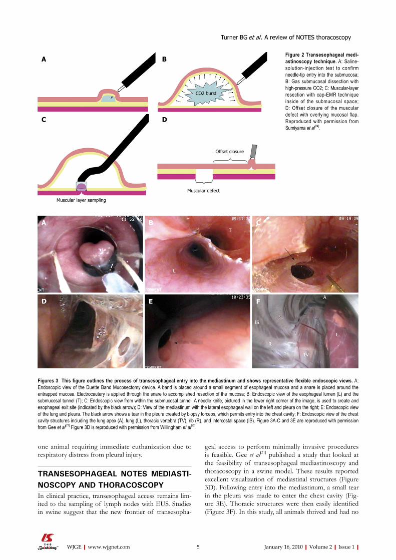
Sumiyama et al[24] reported a new technique, called submucosal endoscopy, with a mucosal flap safety valve (SEMF). In this approach, saline injection into the esophageal wall was used to confirm entry into the submucosa, and high-pressure gas was used to perform a submucosal dissection. A biliary catheter was then inserted into the submucosal layer and a 10-cm long submucosal tunnel was created. Subsequently, an endo-scopic mucosal resection (EMR) cap device (Olympus Optical Co, Ltd, Tokyo, Japan) was used to create a defect in the muscularis propria and the mediastinum was entered after removal of the EMR cap from the endoscope (Figure 2). The goal of this technique is to provide an offset closure of the defect with the overly-ing mucosal flap.
A similar approach was reported by Willingham et al[22], in which mediastinal access was demonstrated via submucosal tunneling. This technique employed a needle-knife, prototype flexible carbon dioxide laser fi-ber (OmniGuide Inc., Cambridge, MA, USA) or Duette multiband mucosectomy device (Cook Medical Inc) to incise the esophageal mucosal layer (Figure 3A). In this method, a long submucosal tunnel (Figure 3B) of at least 10-cm was created using air and blunt dissection with the endoscope and the aid of closed forceps. The tunnel was extended to the gastroesophageal junction. Unlike Sumiyama et al[24], a needle-knife was used to directly incise the muscular layer and provide a portal to the me-diastinum (Figure 3C).
Each of these techniques provides relatively safe and efficient access to the mediastinum. In the three studies combined, major complications were limited to
4 January 16, 2010|Volume 2|Issue 1|WJGE|www.wjgnet.com
Figure 1 Endoscopic view showing access to the mediastinum following a full thickness incision with a needle knife alone. Reproduced with permission from Fritscher-Ravens et al[23].
one animal requiring immediate euthanization due to respiratory distress from pleural injury.
TRANSESOPHAGEAL NOTES MEDIASTI-NOSCOPY AND THORACOSCOPYIn clinical practice, transesophageal access remains lim-ited to the sampling of lymph nodes with EUS. Studies in swine suggest that the new frontier of transesopha-
geal access to perform minimally invasive procedures is feasible. Gee et al[21] published a study that looked at the feasibility of transesophageal mediastinoscopy and thoracoscopy in a swine model. These results reported excellent visualization of mediastinal structures (Figure 3D). Following entry into the mediastinum, a small tear in the pleura was made to enter the chest cavity (Fig-ure 3E). Thoracic structures were then easily identified (Figure 3F). In this study, all animals thrived and had no
5 January 16, 2010|Volume 2|Issue 1|WJGE|www.wjgnet.com
A B
C D
CO2 burst
Muscular layer sampling
Muscular defect
Offset closure
Figure 2 Transesophageal mediastinoscopy technique. A: Saline-solution-injection test to confirm needle-tip entry into the submucosa; B: Gas submucosal dissection with high-pressure CO2; C: Muscular-layer resection with cap-EMR technique inside of the submucosal space; D: Offset closure of the muscular defect with overlying mucosal flap. Reproduced with permission from Sumiyama et al[24].
Figures 3 This figure outlines the process of transesophageal entry into the mediastinum and shows representative flexible endoscopic views. A: Endoscopic view of the Duette Band Mucosectomy device. A band is placed around a small segment of esophageal mucosa and a snare is placed around the entrapped mucosa. Electrocautery is applied through the snare to accomplished resection of the mucosa; B: Endoscopic view of the esophageal lumen (L) and the submucosal tunnel (T); C: Endoscopic view from within the submucosal tunnel. A needle knife, pictured in the lower right corner of the image, is used to create and esophageal exit site (indicated by the black arrow); D: View of the mediastinum with the lateral esophageal wall on the left and pleura on the right; E: Endoscopic view of the lung and pleura. The black arrow shows a tear in the pleura created by biopsy forceps, which permits entry into the chest cavity; F: Endoscopic view of the chest cavity structures including the lung apex (A), lung (L), thoracic vertebra (TV), rib (R), and intercostal space (IS). Figure 3A-C and 3E are reproduced with permission from Gee et al[21] Figure 3D is reproduced with permission from Willingham et al[22].
A B C
D E F
L
T
L
R
IS
TV
A
Turner BG et al . A review of NOTES thoracoscopy
clinical evidence of mediastinitis or thoracic contamina-tion. EUS has also been used to identify small mediasti-nal lymph nodes that could be targeted for sampling and complete removal[23]. In cases where fine needle aspirates do not provide sufficient information, the preserved lymph node architecture obtained with this technique could provide a more definitive pathologic sample.
The use of transesophageal access to perform diag-nostic and therapeutic interventions in the mediastinum and chest seems to be a growing possibility. To date, interventions in swine models have included lymph node biopsies and lymphadenectomy, pericardial fenestra-tion, myocardial saline injections, pleural biopsy, and the creation of a pericardial window among others[21,23]. A current summary of experience with transesophageal access to the mediastinum and thoracic cavity is detailed in Tables 1-2. Overall, the results are promising and pro-pose an array of intrathoracic interventions that could be accomplished with less post-operative and chronic pain. While several factors prevent large studies being carried out in swine models, larger, randomized studies are needed to compare procedure times and outcomes to standard thoracoscopic interventions.
ESOPHAGOTOMY CLOSUREAn important part of performing NOTES procedures
in humans lies in the esophagotomy closure technique and the ability to prevent infectious complications. Sumiyama et al[24] and Gee et al[21] have performed sur-vival studies in swine without the use of a closure de-vice. Both studies included the creation of a submuco-sal tunnel. Perhaps unexpectedly, these studies demon-strated good clinical outcomes and no evidence of large abscesses or mediastinitis. One group has experimented with endoscopic suturing devices for the closure of transesophageal entry sites[23]. While the endoscopic sutures successfully closed the mucosal defects in the esophagus, there were remaining defects in the esopha-geal muscular wall on necropsy. More recently, a group reported the first use of resorbable sutures at transgas-tric NOTES access sites, which could have applicability to esophageal sites as well[25]. It is unclear whether the use of endoscopic sutures or the submucosal tunneling technique will be superior in allowing proper healing of the transesophageal exit conduit without infectious complications. Animal trials comparing the outcomes of these different techniques have not yet been pub-lished.
It is possible that placement of an esophageal stent may prove useful in some cases and produce better out-comes than endoscopic suturing or the tunneling tech-niques. In humans, observational studies have looked at the utility of esophageal stent placement following
6 January 16, 2010|Volume 2|Issue 1|WJGE|www.wjgnet.com
Turner BG et al . A review of NOTES thoracoscopy
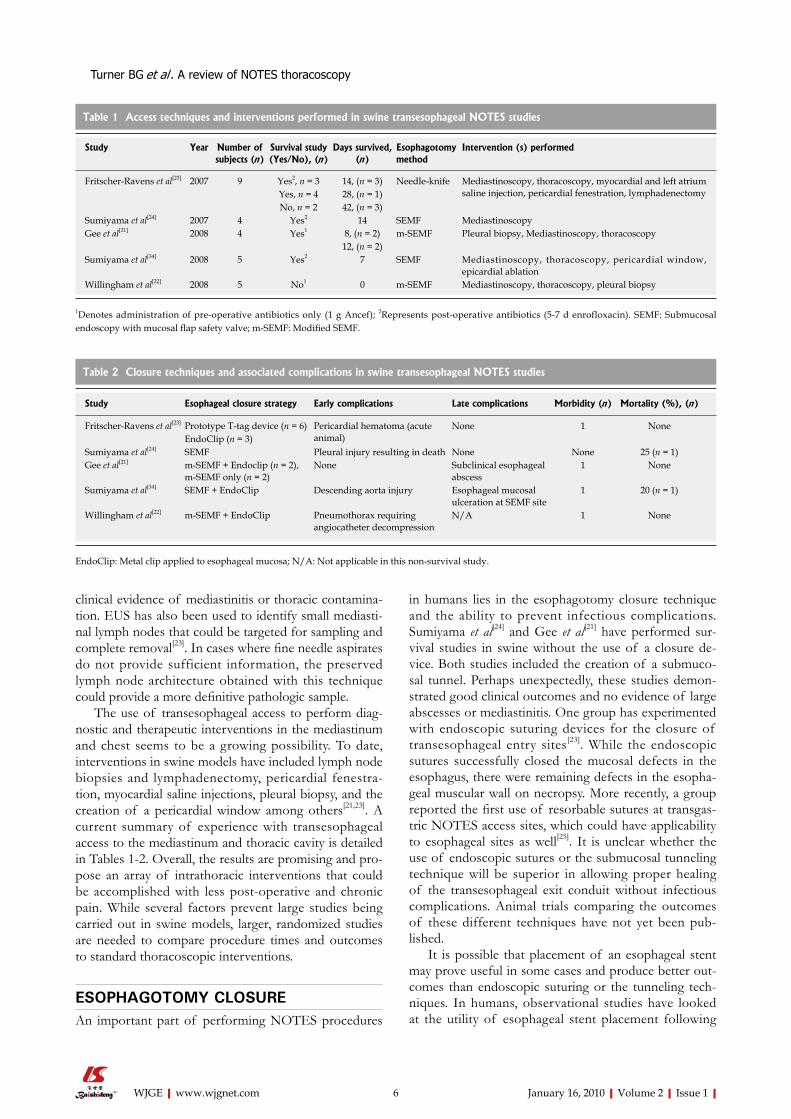
Table 1 Access techniques and interventions performed in swine transesophageal NOTES studies
Study Year Number ofsubjects (n )
Survival study (Yes/No), (n )
Days survived, (n )
Esophagotomymethod
Intervention (s) performed
Fritscher-Ravens et al[23] 2007 9 Yes2, n = 3 14, (n = 3) Needle-knife Mediastinoscopy, thoracoscopy, myocardial and left atrium saline injection, pericardial fenestration, lymphadenectomyYes, n = 4 28, (n = 1)
No, n = 2 42, (n = 3)Sumiyama et al[24] 2007 4 Yes2 14 SEMF MediastinoscopyGee et al[21] 2008 4 Yes1 8, (n = 2) m-SEMF Pleural biopsy, Mediastinoscopy, thoracoscopy
12, (n = 2)Sumiyama et al[34] 2008 5 Yes2 7 SEMF Mediastinoscopy, thoracoscopy, pericardial window,
epicardial ablationWillingham et al[22] 2008 5 No1 0 m-SEMF Mediastinoscopy, thoracoscopy, pleural biopsy
1Denotes administration of pre-operative antibiotics only (1 g Ancef); 2Represents post-operative antibiotics (5-7 d enrofloxacin). SEMF: Submucosal endoscopy with mucosal flap safety valve; m-SEMF: Modified SEMF.
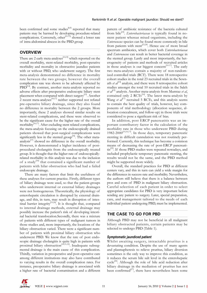
Table 2 Closure techniques and associated complications in swine transesophageal NOTES studies
Study Esophageal closure strategy Early complications Late complications Morbidity (n ) Mortality (%), (n )
Fritscher-Ravens et al[23] Prototype T-tag device (n = 6) Pericardial hematoma (acute animal)
None 1 NoneEndoClip (n = 3)
Sumiyama et al[24] SEMF Pleural injury resulting in death None None 25 (n = 1)Gee et al[21] m-SEMF + Endoclip (n = 2),
m-SEMF only (n = 2)None Subclinical esophageal
abscess1 None
Sumiyama et al[34] SEMF + EndoClip Descending aorta injury Esophageal mucosal ulceration at SEMF site
1 20 (n = 1)
Willingham et al[22] m-SEMF + EndoClip Pneumothorax requiring angiocatheter decompression
N/A 1 None
EndoClip: Metal clip applied to esophageal mucosa; N/A: Not applicable in this non-survival study.

esophageal perforations. One such study looked at 15 patients with non-malignant spontaneous or iatrogenic esophageal perforations treated with self-expandable metal stents[26]. The study demonstrated excellent out-comes in one group (7 patients) undergoing immediate stent placement following identification of the perfora-tion. This group had a mean delay of 45 min from the time the perforation was identified to placement of the stent. The second group (8 patients) had poorer out-comes, including one death, and a median delay of 123 h to stent placement. In the setting of transesophageal procedures, stents could be immediately placed follow-ing procedures resulting in significantly better outcomes. A second study in a series of 9 patients with non-malignant gastrointestinal perforations of the esophagus and colon, as well as anastomotic leaks and complete disunion, suggested that covered stents might support a new concept of “stent-guided regeneration and re-epithelialization” that would aid in healing. Though observational evidence for stents seems promising, ran-domized trials remain to be performed to better assess their utility to treat luminal perforations as opposed to traditional surgical interventions.
BARRIERS TO CLINICAL PRACTICEProblems with esophageal closure techniques, the risk of esophageal leaks, and infections including medias-tinitis, pneumonia, and bacteremia are major concerns when attempting to access the chest cavity with a trans-esophageal route. Large studies investigating infectious complications have not been reported and are challeng-ing to complete due to the limitations of animal models. The trials summarized in Table 2 suggest the rate of infectious complications could be low. Human trials in-vestigating transgastric instrumentation of the peritoneal cavity do report contamination of the peritoneal cavity, but the contamination was found to be clinically insig-nificant[17, 27, 28].
Other adverse events including bleeding and pneu-mothoraces are significant complications in human tho-racoscopic procedures[29,30]. These complications have also been observed in swine NOTES thoracic studies. Conventional interventions such as needle decompres-sion or use of chest tubes can be performed, though there has never been a need for chest tube placement in the animal studies reviewed in Table 2. Researchers have also turned their attention to improving meth-ods of hemostasis. Fritscher-Ravens et al[31] conducted a randomized controlled study comparing different methods of obtaining endoscopic hemostasis following artificially induced hemorrhage in the peritoneal cav-ity. The study assessed several methods of hemostasis including an endoscopic suturing device, prototype monopolar electrocautery forceps, and forced argon plasma coagulation (FAPC). In the end, FAPC was found to have significantly faster times in controlling bleeding and in achieving complete cessation of blood
loss when compared to the other methods. It will be important to extend these studies to look at hemostatic methods in the chest since vessels within the chest, in-cluding intercostal arteries and veins, can be difficult to access due to surrounding bony structures (i.e. ribs and vertebral bodies).
In a systematic review of thoracic NOTES pro-cedures, mortality was found to be 5% and morbidity 19% when combining all published studies of thoracic-related studies using a NOTES technique[32]. This review included two studies in which thoracic procedures were accomplished with a transvesicular, transdiaphragmatic, or transgastric approach, while the remaining five stud-ies were transesophageal. The morbidity and mortality found in the combined studies represent one of the ma-jor challenges in creating a new, minimally invasive tech-nique and underscores the technological improvements that are necessary to move transesophageal NOTES to human clinical applications.
FUTURE DIRECTIONSA foundation for potential transesophageal NOTES thoracic procedures has been established. Moving for-ward, there is a need for studying the hemodynamic and physiologic consequences of these transesophageal interventions. In the literature, studies have been per-formed on the effects of carbon dioxide insufflation during transthoracic thoracoscopy[33]. This study of 32 consecutive patients demonstrated that intrapleu-ral pressures of 2-14 mmHg did not have significant adverse hemodynamic consequences and that insuffla-tion at pressures of < 10 mmHg were safe. Studies will need to be performed examining the consequences of controlled endoscopic insufflation with room air versus the use of carbon dioxide regulated insufflation and other potential consequences of an esophageal entry site.
Future work will continue to focus on potential infectious complications and how to best prevent these occurrences. The field will also need continued instrument development to improve hemostatic ability when bleeding complications occur or when transesophageal surgical resections are performed. Finally, a closure device and/or technique permitting full thickness closure of the esophageal wall without the development of an esophageal wall abscess, stricturing, or discontinuous muscular wall closure needs further development.
CONCLUSIONTransesophageal NOTES is a promising platform that may offer hope for a less invasive means of accessing the mediastinum and chest cavity. The continued relationships of surgeons, gastroenterologists, and researchers in industry are crucial for the development of devices that will permit better endoscopic control
7 January 16, 2010|Volume 2|Issue 1|WJGE|www.wjgnet.com
Turner BG et al . A review of NOTES thoracoscopy

and precision during planned operative procedures. Technological advances remain to be made that will make transesophageal NOTES a viable approach in humans, however, preliminary studies suggest this technique is of great potential to the field of thoracic surgery.
REFERENCES1 Kalloo AN, Singh VK, Jagannath SB, Niiyama H, Hill
SL, Vaughn CA, Magee CA, Kantsevoy SV. Flexible transgastric peritoneoscopy: a novel approach to diagnostic and therapeutic interventions in the peritoneal cavity. Gastrointest Endosc 2004; 60: 114-117
2 Rattner D, Kalloo A. ASGE/SAGES Working Group on Natural Orifice Translumenal Endoscopic Surgery. October 2005. Surg Endosc 2006; 20: 329-333
3 Rattner D. Introduction to NOTES White Paper. Surg Endosc 2006; 20: 185
4 Wagh MS, Merrifield BF, Thompson CC. Survival studies after endoscopic transgastric oophorectomy and tubectomy in a porcine model. Gastrointest Endosc 2006; 63: 473-478
5 Jagannath SB, Kantsevoy SV, Vaughn CA, Chung SS, Cotton PB, Gostout CJ, Hawes RH, Pasricha PJ, Scorpio DG, Magee CA, Pipitone LJ, Kalloo AN. Peroral transgastric endoscopic ligation of fallopian tubes with long-term survival in a porcine model. Gastrointest Endosc 2005; 61: 449-453
6 Park PO, Bergström M, Ikeda K, Fritscher-Ravens A, Swain P. Experimental studies of transgastric gallbladder surgery: cholecystectomy and cholecystogastric anastomosis (videos). Gastrointest Endosc 2005; 61: 601-606
7 Kantsevoy SV, Jagannath SB, Niiyama H, Chung SS, Cotton PB, Gostout CJ, Hawes RH, Pasricha PJ, Magee CA, Vaughn CA, Barlow D, Shimonaka H, Kalloo AN. Endoscopic gastrojejunostomy with survival in a porcine model. Gastrointest Endosc 2005; 62: 287-292
8 Kantsevoy SV, Hu B, Jagannath SB, Vaughn CA, Beitler DM, Chung SS, Cotton PB, Gostout CJ, Hawes RH, Pasricha PJ, Magee CA, Pipitone LJ, Talamini MA, Kalloo AN. Transgastric endoscopic splenectomy: is it possible? Surg Endosc 2006; 20: 522-525
9 Matthes K, Yusuf TE, Willingham FF, Mino-Kenudson M, Rattner DW, Brugge WR. Feasibility of endoscopic transgastric distal pancreatectomy in a porcine animal model. Gastrointest Endosc 2007; 66: 762-766
10 Bessler M , Stevens PD, Milone L, Parikh M, Fowler D. Transvaginal laparoscopically assisted endoscopic cholecystectomy: a hybrid approach to natural orifice surgery. Gastrointest Endosc 2007; 66: 1243-1245
11 Pai RD, Fong DG, Bundga ME, Odze RD, Rattner DW, Thompson CC. Transcolonic endoscopic cholecystectomy: a NOTES survival study in a porcine model (with video). Gastrointest Endosc 2006; 64: 428-434
12 Lima E, Rolanda C, Pêgo JM, Henriques-Coelho T, Silva D, Carvalho JL, Correia-Pinto J. Transvesical endoscopic peritoneoscopy: a novel 5 mm port for intra-abdominal scarless surgery. J Urol 2006; 176: 802-805
13 Denk PM, Swanström LL, Whiteford MH. Transanal endoscopic microsurgical platform for natural orifice surgery. Gastrointest Endosc 2008; 68: 954-959
14 Sylla P, Willingham FF, Sohn DK, Gee D, Brugge WR, Rattner DW. NOTES rectosigmoid resection using transanal endoscopic microsurgery (TEM) with transgastric endoscopic assistance: a pilot study in swine. J Gastrointest Surg 2008; 12: 1717-1723
15 Abe N , Takeuchi H, Yanagida O, Masaki T, Mori T, Sugiyama M, Atomi Y. Endoscopic full-thickness resection with laparoscopic assistance as hybrid NOTES for gastric
submucosal tumor. Surg Endosc 2009; 23: 1908-191316 Palanivelu C, Rajan PS, Rangarajan M, Prasad M, Kalyan-
akumari V, Parthasarathi R, Senthilnathan P. NOTES: Transvaginal endoscopic cholecystectomy in humans-preliminary report of a case series. Am J Gastroenterol 2009; 104: 843-847
17 Hazey JW, Narula VK, Renton DB, Reavis KM, Paul CM, Hinshaw KE, Muscarella P, Ellison EC, Melvin WS. Natural-orifice transgastric endoscopic peritoneoscopy in humans: Initial clinical trial. Surg Endosc 2008; 22: 16-20
18 Noguera J, Dolz C, Cuadrado A, Olea J, Vilella A, Morales R. Hybrid transvaginal cholecystectomy, NOTES, and minilaparoscopy: analysis of a prospective clinical series. Surg Endosc 2009; 23: 876-881
19 Auyang ED, Hungness ES, Vaziri K, Martin JA, Soper NJ. Human NOTES cholecystectomy: transgastric hybrid technique. J Gastrointest Surg 2009; 13: 1149-1150
20 Steegers MA, Snik DM, Verhagen AF, van der Drift MA, Wilder-Smith OH. Only half of the chronic pain after thoracic surgery shows a neuropathic component. J Pain 2008; 9: 955-961
21 Gee DW, Willingham FF, Lauwers GY, Brugge WR, Rattner DW. Natural orifice transesophageal mediastinoscopy and thoracoscopy: a survival series in swine. Surg Endosc 2008; 22: 2117-2122
22 Willingham FF, Gee DW, Lauwers GY, Brugge WR, Rattner DW. Natural orifice transesophageal mediastinoscopy and thoracoscopy. Surg Endosc 2008; 22: 1042-1047
23 Fritscher-Ravens A, Patel K, Ghanbari A, Kahle E, von Herbay A, Fritscher T, Niemann H, Koehler P. Natural orifice transluminal endoscopic surgery (NOTES) in the mediastinum: long-term survival animal experiments in transesophageal access, including minor surgical procedures. Endoscopy 2007; 39: 870-875
24 Sumiyama K, Gostout CJ, Rajan E, Bakken TA, Knipschield MA. Transesophageal mediastinoscopy by submucosal endoscopy with mucosal flap safety valve technique. Gastrointest Endosc 2007; 65: 679-683
25 von Renteln D, Eickhoff A, Kaehler G, Riecken B, Caca K. Endoscopic closure of the natural orifice transluminal endoscopic surgery (NOTES) access site to the peritoneal cavity by means of transmural resorbable sutures: an animal survival study. Endoscopy 2009; 41: 154-159
26 Fischer A, Thomusch O, Benz S, von Dobschuetz E, Baier P, Hopt UT. Nonoperative treatment of 15 benign esophageal perforations with self-expandable covered metal stents. Ann Thorac Surg 2006; 81: 467-472
27 Narula VK , Happel LC, Volt K, Bergman S, Roland JC, Dettorre R, Renton DB, Reavis KM, Needleman BJ, Mikami DJ, Ellison EC, Melvin WS, Hazey JW. Trans-gastric endoscopic peritoneoscopy does not require dec-ontamination of the stomach in humans. Surg Endosc 2009; 23: 1331-1336
28 Narula VK, Hazey JW, Renton DB, Reavis KM, Paul CM, Hinshaw KE, Needleman BJ, Mikami DJ, Ellison EC, Melvin WS. Transgastric instrumentation and bacterial contamination of the peritoneal cavity. Surg Endosc 2008; 22: 605-611
29 Li X, Tu YR, Lin M, Lai FC, Chen JF, Dai ZJ. Endoscopic thoracic sympathectomy for palmar hyperhidrosis: a randomized control trial comparing T3 and T2-4 ablation. Ann Thorac Surg 2008; 85: 1747-1751
30 Rodríguez PM, Freixinet JL, Hussein M, Valencia JM, Gil RM, Herrero J, Caballero-Hidalgo A. Side effects, complications and outcome of thoracoscopic sympathectomy for palmar and axillary hyperhidrosis in 406 patients. Eur J Cardiothorac Surg 2008; 34: 514-519
31 Fritscher-Ravens A, Ghanbari A, Holland C, Olagbeye F, Hardeler KG, Seehusen F, Jacobsen B, Mannur K. Beyond NOTES: randomized controlled study of different methods
8 January 16, 2010|Volume 2|Issue 1|WJGE|www.wjgnet.com
Turner BG et al . A review of NOTES thoracoscopy

of flexible endoscopic hemostasis of artificially induced hemorrhage, via NOTES access to the peritoneal cavity. Endoscopy 2009; 41: 29-35
32 Clark J, Sodergren M, Correia-Pinto J, Zacharakis E, Teare J, Yang GZ, Darzi A, Athanasiou T. Natural orifice translumenal thoracoscopic surgery: does the slow progress and the associated risks affect feasibility and potential clinical applications? Surg Innov 2009; 16: 9-15
33 Wolfer RS, Krasna MJ, Hasnain JU, McLaughlin JS. Hemo-
dynamic effects of carbon dioxide insufflation during thoracoscopy. Ann Thorac Surg 1994; 58: 404-407; discussion 407-408
34 Sumiyama K, Gostout CJ, Rajan E, Bakken TA, Knipschield MA, Chung S, Cotton PB, Hawes RH, Kalloo AN, Kantsevoy SV, Pasricha PJ. Pilot study of transesophageal endoscopic epicardial coagulation by submucosal endoscopy with the mucosal flap safety valve technique (with videos). Gastrointest Endosc 2008; 67: 497-501
S- Editor Zhang HN L- Editor Lutze M E- Editor Ma WH
9 January 16, 2010|Volume 2|Issue 1|WJGE|www.wjgnet.com
Turner BG et al . A review of NOTES thoracoscopy

TOPIC HIGHLIGHT
Operable malignant jaundice: To stent or not to stent before the operation?
Rungsun Rerknimitr, Pinit Kullavanijaya
Rungsun Rerknimitr, Pinit Kullavanijaya, Division of Gastroenterology, Department of Medicine, Chulalongkorn University, Bangkok 10310, ThailandAuthor contributions: Rerknimitr R reviewed the literature and wrote the paper; Kullavanijaya P commented on and reviewed the paper.Correspondence to: Rungsun Rerknimitr, MD, Division of Gastroenterology, Department of Internal Medicine, Faculty of Medicine, Chulalongkorn University, Bangkok 10310, Thailand. [email protected]: +6622564265 Fax: +6622527839Received: May 20, 2009 Revised: August 26, 2009Accepted: September 2, 2009Published online: January 16, 2010
AbstractTraditionally, pre-operative biliary drainage (PBD) was believed to improve multi-organ dysfunction, and for this reason, was practiced worldwide. Over the last decade, this concept was challenged by many reports, including meta-analyses that showed no difference in morbidity and mortality between surgery with, and surgery without PBD, in operable malignant jaundice. The main disadvantages of PBD are seen to be the additional cost of the procedure itself, and the need for longer hospitalization. In addition, many studies showed the significance of specific complications resulting from PBD, such as recurrent jaundice, cholangitis, pancreatitis, cutaneous fistula, and bleeding. However, the results of these studies remain inconclusive as to date there has been no perfect study that equally randomized comparable patients according to the level of obstruction and technique used for PBD. Generally, endoscopic stent insertion (ES) is preferred for common duct obstruction, whereas endoscopic nasobiliary drainage and percutaneous biliary drainage is reserved for hilar obstruction, since ES in hilar block confers a high rate of cholangitis. Although, there is no guideline which either supports or refutes this approach, certain
subgroups of patients, including those with symptomatic jaundice, cholangitis, impending renal failure, hilar block requiring preoperative portal vein embolization, and those who need pre-operative neoadjuvant therapy, are suitable candidates for PBD.
© 2010 Baishideng. All rights reserved.
Key words: Biliary drainage; Surgery; Complication; Mobidity; Mortality
Peer reviewer: Juan J Vila Costas, MD, Gastroenterology Department, Hospital de Navarra, c\Irunlarrea, 3, Pamplona 31.008, Spain
Rerknimitr R, Kullavanijaya P. Operable malignant jaundice: To stent or not to stent before the operation? World J Gastrointest Endosc 2010; 2(1): 1014 Available from: URL: http://www.wjgnet.com/19485190/full/v2/i1/10.htm DOI: http://dx.doi.org/10.4253/wjge.v2.i1.10
INTRODUCTIONLiver, gallbladder, bile duct and pancreas share a common embryologic origin and also play their parts as the common etiologies for resectable biliary related tumors[1]. Malignant biliary obstruction presents mainly as jaundice and pruritus. In a prolonged obstruction, multi-organ dysfunction including renal failure, cardiac dysfunction, pulmonary dysfunction, poor hepatic metabolism and hemostasis impairment[2-7] may develop. This, in turn, can compromise the outcome of surgery. For years, it has been a routine practice to achieve pre-operative biliary drainage (PBD), either by means of an endoscopically placed stent (a plastic stent, nasobiliary tube, or a removable metallic stent) or by means of a percutaneously placed catheter (either externally or a combination of external and internal drainage). However, the benefit of PBD has not
Konstantinos Triantafyllou, MD, PhD, Series Editor
World J Gastrointest Endosc 2010 January 16; 2(1): 10-14ISSN 1948-5190 (online)
© 2010 Baishideng. All rights reserved.
Online Submissions: http://www.wjgnet.com/[email protected]:10.4253/wjge.v2.i1.10
10 January 16, 2010|Volume 2|Issue 1|WJGE|www.wjgnet.com
Rerknimitr R et al . Operable malignant jaundice: Should we stent?
been confirmed and some studies[8,9] reported that many patients may be harmed by developing procedure-related complications. Conversely, other[10-13] showed a lower rate of intra-abdominal abscess in the PBD group.
OVERVIEWThere are 2 early meta-analyses[14,15] which reported on the overall morbidity, stent-related morbidity, post-operative morbidity and mortality in obstructive jaundice patients with or without PBD, who underwent surgery. The first meta-analysis demonstrated no difference in mortality rate between the two groups; however the overall complication rate was shown to be adversely affected by PBD[14]. By contrast, another meta-analysis reported no adverse effects after preoperative endoscopic biliary stent placement when compared with direct surgery[15]. Another 2 recent meta-analyses[16,17] neither supported nor refuted pre-operative biliary drainage, since they also found no difference in mortality between the 2 groups. More importantly, these 2 analyses showed similar results on stent-related complications, and these were observed to be the significant cause for the higher rate of the overall morbidity[16,17]. After excluding stent related complications, the meta-analysis focusing on the endoscopically drained patients showed that post-surgical complications were significantly less in the stented group[16]. The other meta-analysis[17] showed no difference in the overall morbidity. However, it demonstrated a higher incidence of post-procedural cholangitis from the endoscopically treated group. It is thought that the higher rate of endoscopically-related morbidity in this analysis was due to the inclusion of a study[18] that contained a significant number of patients with hilar obstruction who had had a failed endoscopic drainage.
There are many factors that limit the usefulness of these analyses for current practice. Firstly, different types of biliary drainage were used, so that the patient-groups who underwent internal or external biliary drainages were not homogeneous. Theoretically, the physiology of enterohepatic circulation is disrupted by external drain-age, and this, in turn, may result in disruption of intes-tinal barrier integrity[19,20]. It is thought that, compared to internal drainage methods, external drainage may possibly increase the patient’s risk of developing intesti-nal bacterial translocation.Secondly, there was a mixture of patients with different types of malignant tumors in those studies and, more importantly, the locations of the biliary obstruction varied. There were a significant num-ber of patients with proximal biliary obstruction who underwent PBD. We know that the rate of post endo-scopic drainage cholangitis is quite high in patients with proximal biliary obstruction[18,21,22]. Inadequate subseg-mental drainage is the main cause of this complication. Thirdly, variation in preoperative and post-operative care among different institutions may also have contributed to varying results in the overall complication rates. For instance, preoperative biliary drainage is associated with a higher rate of bacterial contamination and a different
pattern of antibiotic resistance of the bacteria cultured from bile[23]. Enterobacteriaceae is typically found in no-stent patient whereas mixed organisms, including the Enterococcus species can be found more commonly in bile from patients with stent[23,24]. Hence use of more broad spectrum antibiotics, which cover both Enterobacteriaceae and Enterococcus can result in better bacterial coverage in the stented group. Lastly and most importantly, the het-erogeneity of patients and methods of recruited articles in those analyses is our biggest concern[14-17]. The early two meta-analyses contain a majority of non-random-ized controlled trials (RCT). There were 18 retrospective cohort studies in the total 23 recruited trials in the Sewn-ath et al[14] analysis, and there were 8 retrospective cohort studies amongst the total 10 recruited trials in the Saleh et al[15] analysis. Another meta-analysis from Mumtaz et al, contained only 2 RCTs[16]. The latest meta-analysis by Wang et al[17] recruited 5 RCTs, and this analysis seems to contain the best quality of trials, however, key com-ponents of trial methodology (allocation sequence, al-location concealment, and follow-up) in those trials were considered to pose a significant risk of bias.
In addition, post ERCP pancreatitis was an im-portant contributory factor in the calculation of the morbidity rate in those who underwent PBD during 1982-2000[14,16,17]. In those days, temporary pancreatic stenting in difficult cannulation was not routinely per-formed. Currently, this technique has become a common means of decreasing the rate of post-ERCP pancreati-tis[25]. If those PBD studies were repeated nowadays, and included prophylactic temporary pancreatic stenting, the results would not be the same, and the PBD method might be supported more widely.
Overall, the standard practices for PBD at different centers vary, and this in turn can yield a wide margin for the differences in success rate and morbidity. Nevertheless, the authors still believe that there is a balance between pros and cons of PBD in malignant biliary obstruction. Careful selection of each patient in order to select appropriate candidates for PBD is very important before sending any patient to surgery. Later, special preparation, care, and management tailored to the needs of each individual patient undergoing PBD, must be implemented.
THE CASE TO GO FOR PBDAlthough PBD may not be beneficial in all malignant biliary obstructed patients, certain patients may be selected to undergo PBD (Table 1).
Symptomatic jaundiced patientWhilst awaiting surgery, intractable pruritus is a devastating condition. Despite the use of many agents and plasmapheresis to relieve pruritus, biliary diversion sometimes is the only way to improve this condition, as it reduces the serum bile salt level in the enterohepatic cycle[26]. Although the role of bile acid reduction after biliary drainage in the mediation of pruritus has not been confirmed[27], there have nevertheless been some
11 January 16, 2010|Volume 2|Issue 1|WJGE|www.wjgnet.com

reports which demonstrate a transient relief of pruritus within 24 h after biliary drainage[28,29].
Fluid and electrolyte balance have to be precisely maintained in all biliary obstructed patients undergoing surgical resection. Lactulose and a bile salt supplement can offer renal protection[30]. However, in patients with pre-existing renal impairment, these measures may not be enough to prevent the development of acute renal failure. Some surgeons may therefore advocate the patient undergoing PBD prior to surgery.
Patients with acute cholangitisAlthough de novo case of acute cholangitis in patients with malignant biliary obstruction is quite unusual, ampullary tumors, intraductal papillary mucinous neoplasm (IPMN), and biliary papillomatosis are certain conditions that acute cholangitis may develop spontaneously[31-33]. For this subgroup of patients, biliary decompression plays an important role in the management of acute cholangitis and this, in turn, can reduce the operative mortality and morbidity[34].
Certain hilar obstructed patientsIn the past, central hepatectomy was the standard surgical technique for hilar cholangiocarcinoma. With the use of surgery and the introduction of portal vein embolization (PVE), typical major hepatectomies including right or left hepatectomy, and right or left trisectionectomy have increasingly been performed[35-37]. After PVE of the affected lobe, the enlarged contralateral lobe that is preserved from embolization is supposed to carry out all hepatic functions[38]. A report from Nagoya, Japan, showed that the risk of post-operative liver failure in the group who underwent PVE dropped from 33% to 23%[38]. However, a delay of at least 3 wk is advised before the contralateral lobe is fully able to compensate and the patient is ready for hepatectomy[39]. PBD is therefore needed as a bridge for this package. Practically, unilateral PBD in a hilar block is sufficient and the preferred side for drainage is the future remnant lobe[40]. However, bilateral drainage is considered in the following situations: patients with pre-existing cholangitis; patients who develop post-procedural cholangitis, despite board-spectrum antibiotics, and additional drainage from the same side; and patients with persistent jaundice. Apart form the discomfort from nasal irritation, endoscopic nasobiliary drainage (ENBD) is the preferred initial technique that has replaced percutaneous transhepatic
biliary drainage (PTBD) in many Japanese endoscopy centers[40-42] and PTBD is currently reserved as a salvage method in patients with suboptimal endoscopic drainage who develop subsegmental cholangitis. In addition, because of the higher risk of cholangitis reported in advanced hilar blocks[21,22] and the fear that duodenal fluid could flows back into the biliary tree, endoscopic stent placement (ES) is not recommended in this group.
Patients requiring neoadjuvant therapyTraditionally, radio-chemotherapy for pancreatic cancer was administered post-operatively. Unfortunately, this strategy had limited success. Recently, these neoadjuvant agents have been given pre-operatively, with the objective of tumor down-staging and in the expectation of a higher number of complete resections[43]. To minimize the toxicity from chemotherapy, many of these patients with obstructive jaundice will benefit from PBD prior to the treatment protocol. However, to date, no randomized trials comparing neoadjuvant with no adjuvant therapies given preoperatively have yet been conducted.
THE DISADVANTAGES OF PBDThe disadvantages of PBD can be reviewed in term of morbidity, mortality, and cost of treatment when compared with the group without PBD.
The majority of studies did not demonstrate any difference in the overall morbidity and mortality between those patients undergoing PBD and and those with no drainage. Only an early study from UCLA in 1985 showed a slightly, but not significantly, higher rate of morbidity in patients having undergone PBD than the no PBD group (57% vs 53%)[44]. In addition, the total number of days for hospitalization was longer in the PBD group (31.4 d vs 23.1 d), and in 1985, the estimated cost relating to both the additional stay in hospital and the cost of the procedure was more than $US 8000[44]. In contrast, a study reported by a group from New York University[45] demonstrated a shorter hospital stay in the PBD group than in non-PBD group (13.5 d vs 19 d, P = 0.02). Moreover, PBD group tended to have fewer overall complications (P = 0.054). This study suggested that “the increased cost of preoperative ERCP and PBD may be offset by the decreased length of hospitalization and decreased complication rate”[45]. The important difference between the two studies was the technique for PBD. The first study used PTBD, and the second, ES. Better fluid and electrolyte control and an improvement in immune response resulting from ES may play an important role in the different results found. Of note, the majority of the cases in these two studies involved patients with common bile duct obstruction.
Post-operative fistula is a common complication of bilio-pancreatic resection, leading to prolonged hospitalization, increased cost of treatment and delayed further adjuvant therapy. The largest retrospective study[46] by a group from John Hopkins on patients who underwent pancreaticoduodenectomy (n = 567) reported
Table 1 Indication for pre-operative biliary drainage
Symptoms
PruritusRenal impairmentAcute cholangitisHilar block requiring portal vein embolization prior to surgeryPancreatic cancer undergoing preoperative chemotherapyDelay in surgery
12 January 16, 2010|Volume 2|Issue 1|WJGE|www.wjgnet.com
Rerknimitr R et al . Operable malignant jaundice: Should we stent?

a higher incidence of pancreatic fistula (10% vs 4%, P = 0.02) and wound infection (10% vs 4%, P = 0.02) in the PBD group, whereas other smaller studies (n = 38-257) have not shown significant incidence of fistula development in PBD groups[44,47-49].
Intra-operative hemorrhage is an important factor in morbidity and mortality of patients undergoing surgery. Only one small study[50] reported a higher volume of intra-operative bloodloss in the PBD group than the undrained group (1207 mL vs 1122 mL), whereas other larger studies have failed to demonstrate the different effects of PBD or the lack of PBD on these issues[45,46,48,49].
CONCLUSIONIn conclusion, a routine PBD for every patient under-going bilio-pancreatic surgery is not recommended. PBD carries with it risks of recurrent cholangitis, pancreatitis, cutaneous fistula development, and intra-operative hemorrhage. These can result in a prolonged hospital stay and increase in the total cost of therapy. However, the rate of pancreatitis may be reduced by temporary pancreatic stenting. At this moment we can advocate PBD only in a certain subset of patients, including those with symptomatic jaundice, cholangitis, impending renal failure, hilar block requiring PVE, and those who need pre-operative neoadjuvant therapy.
REFERENCES1 Carriaga MT, Henson DE. Liver, gallbladder, extrahepatic
bile ducts, and pancreas. Cancer 1995; 75: 171-1902 Oussoultzoglou E, Jaeck D. Patient preparation before
surgery for cholangiocarcinoma. HPB (Oxford) 2008; 10: 150-153
3 Uslu A, Cayci M, Nart A, Karaca C, Zalluhoglu N, Gurkan A, Varilsuha C, Adagulu H. Renal failure in obstructive jaundice. Hepatogastroenterology 2005; 52: 52-54
4 Padillo J, Puente J, Gomez M, Dios F, Naranjo A, Vallejo JA, Mino G, Pera C, Sitges-Serra A. Improved cardiac function in patients with obstructive jaundice after internal biliary drainage: hemodynamic and hormonal assessment. Ann Surg 2001; 234: 652-656
5 Watanapa P. Recovery patterns of liver function after complete and partial surgical biliary decompression. Am J Surg 1996; 171: 230-234
6 Mesner O, Miller MJ, Iben SC, Prabha KC, Mayer CA, Haxhiu MA, Martin RJ. Hyperbilirubinemia diminishes respiratory drive in a rat pup model. Pediatr Res 2008; 64: 270-274
7 Papadopoulos V, Filippou D, Manolis E, Mimidis K. Haemostasis impairment in patients with obstructive jaundice. J Gastrointestin Liver Dis 2007; 16: 177-186
8 Ferrero A, Lo Tesoriere R, Vigano L, Caggiano L, Sgotto E, Capussotti L. Preoperative biliary drainage increases infectious complications after hepatectomy for proximal bile duct tumor obstruction. World J Surg 2009; 33: 318-325
9 Hochwald SN, Burke EC, Jarnagin WR, Fong Y, Blumgart LH. Association of preoperative biliary stenting with increased postoperative infectious complications in proximal cholangiocarcinoma. Arch Surg 1999; 134: 261-266
10 Velanovich V , Kheibek T, Khan M. Relationship of postoperative complications from preoperative biliary stents
after pancreaticoduodenectomy. A new cohort analysis and meta-analysis of modern studies. JOP 2009; 10: 24-29
11 Marcus SG, Dobryansky M, Shamamian P, Cohen H, Gouge TH, Pachter HL, Eng K. Endoscopic biliary drainage before pancreaticoduodenectomy for periampullary malignancies. J Clin Gastroenterol 1998; 26: 125-129
12 Mullen JT, Lee JH, Gomez HF, Ross WA, Fukami N, Wolff RA, Abdalla EK, Vauthey JN, Lee JE, Pisters PW, Evans DB. Pancreaticoduodenectomy after placement of endobiliary metal stents. J Gastrointest Surg 2005; 9: 1094-1104; discussion 1104-1105
13 Howard TJ, Yu J, Greene RB, George V, Wairiuko GM, Moore SA, Madura JA. Influence of bactibilia after preoperative biliary stenting on postoperative infectious complications. J Gastrointest Surg 2006; 10: 523-531
14 Sewnath ME, Karsten TM, Prins MH, Rauws EJ, Obertop H, Gouma DJ. A meta-analysis on the efficacy of preoperative biliary drainage for tumors causing obstructive jaundice. Ann Surg 2002; 236: 17-27
15 Saleh MM, Norregaard P, Jorgensen HL, Andersen PK, Matzen P. Preoperative endoscopic stent placement before pancreaticoduodenectomy: a meta-analysis of the effect on morbidity and mortality. Gastrointest Endosc 2002; 56: 529-534
16 Mumtaz K, Hamid S, Jafri W. Endoscopic retrograde cholangiopancreaticography with or without stenting in patients with pancreaticobiliary malignancy, prior to surgery. Cochrane Database Syst Rev 2007; CD006001
17 Wang Q, Gurusamy KS, Lin H, Xie X, Wang C. Preoperative biliary drainage for obstructive jaundice. Cochrane Database Syst Rev 2008; CD005444
18 Lai EC, Mok FP, Fan ST, Lo CM, Chu KM, Liu CL, Wong J. Preoperative endoscopic drainage for malignant obstructive jaundice. Br J Surg 1994; 81: 1195-1198
19 Kamiya S, Nagino M, Kanazawa H, Komatsu S, Mayumi T, Takagi K, Asahara T, Nomoto K, Tanaka R, Nimura Y. The value of bile replacement during external biliary drainage: an analysis of intestinal permeability, integrity, and microflora. Ann Surg 2004; 239: 510-517
20 Parks RW, Clements WD, Smye MG, Pope C, Rowlands BJ, Diamond T. Intestinal barrier dysfunction in clinical and experimental obstructive jaundice and its reversal by internal biliary drainage. Br J Surg 1996; 83: 1345-1349
21 Rerknimitr R, Kladcharoen N, Mahachai V, Kullavanijaya P. Result of endoscopic biliary drainage in hilar cholangio-carcinoma. J Clin Gastroenterol 2004; 38: 518-523
22 Rerknimitr R, Kongkam P, Kullavanijaya P. Outcome of self-expandable metallic stents in low-grade versus advanced hilar obstruction. J Gastroenterol Hepatol 2008; 23: 1695-1701
23 Sudo T, Murakami Y, Uemura K, Hayashidani Y, Hashimoto Y, Ohge H, Sueda T. Specific antibiotic prophylaxis based on bile cultures is required to prevent postoperative infectious complications in pancreatoduodenectomy patients who have undergone preoperative biliary drainage. World J Surg 2007; 31: 2230-2235
24 Rerknimitr R, Fogel EL, Kalayci C, Esber E, Lehman GA, Sherman S. Microbiology of bile in patients with cholangitis or cholestasis with and without plastic biliary endoprosthesis. Gastrointest Endosc 2002; 56: 885-889
25 Das A, Singh P, Sivak MV Jr, Chak A. Pancreatic-stent placement for prevention of post-ERCP pancreatitis: a cost-effectiveness analysis. Gastrointest Endosc 2007; 65: 960-968
26 Ng VL, Ryckman FC, Porta G, Miura IK, de Carvalho E, Servidoni MF, Bezerra JA, Balistreri WF. Long-term outcome after partial external biliary diversion for intractable pruritus in patients with intrahepatic cholestasis. J Pediatr Gastroenterol Nutr 2000; 30: 152-156
27 Bergasa NV. The pruritus of cholestasis. J Hepatol 2005; 43: 1078-1088
13 January 16, 2010|Volume 2|Issue 1|WJGE|www.wjgnet.com
Rerknimitr R et al . Operable malignant jaundice: Should we stent?
28 Beuers U, Gerken G, Pusl T. Biliary drainage transiently relieves intractable pruritus in primary biliary cirrhosis. Hepatology 2006; 44: 280-281
29 Ng VL, Ryckman FC, Porta G, Miura IK, de Carvalho E, Servidoni MF, Bezerra JA, Balistreri WF. Long-term outcome after partial external biliary diversion for intractable pruritus in patients with intrahepatic cholestasis. J Pediatr Gastroenterol Nutr 2000; 30: 152-156
30 Pain JA, Cahill CJ, Gilbert JM, Johnson CD, Trapnell JE, Bailey ME. Prevention of postoperative renal dysfunction in patients with obstructive jaundice: a multicentre study of bile salts and lactulose. Br J Surg 1991; 78: 467-469
31 Kahaleh M, Shami VM, Brock A, Conaway MR, Yoshida C, Moskaluk CA, Adams RB, Tokar J, Yeaton P. Factors predictive of malignancy and endoscopic resectability in ampullary neoplasia. Am J Gastroenterol 2004; 99: 2335-2339
32 Tibayan F, Vierra M, Mindelzun B, Tsang D, McClenathan J, Young H, Trueblood HW. Clinical presentation of mucin-secreting tumors of the pancreas. Am J Surg 2000; 179: 349-351
33 Cheng MS , AhChong AK, Mak KL, Yip AW. Case report: two cases of biliary papillomatosis with unusual associations. J Gastroenterol Hepatol 1999; 14: 464-467
34 Bornman PC, van Beljon JI, Krige JE. Management of cholangitis. J Hepatobiliary Pancreat Surg 2003; 10: 406-414
35 Kawasaki S, Imamura H, Kobayashi A, Noike T, Miwa S, Miyagawa S. Results of surgical resection for patients with hilar bile duct cancer: application of extended hepatectomy after bil iary drainage and hemihepatic portal vein embolization. Ann Surg 2003; 238: 84-92
36 Seyama Y , Kubota K, Sano K, Noie T, Takayama T, Kosuge T, Makuuchi M. Long-term outcome of extended hemihepatectomy for hilar bile duct cancer with no mortality and high survival rate. Ann Surg 2003; 238: 73-83
37 Nagino M, Kamiya J, Nishio H, Ebata T, Arai T, Nimura Y. Two hundred forty consecutive portal vein embolizations before extended hepatectomy for biliary cancer: surgical outcome and long-term follow-up. Ann Surg 2006; 243: 364-372
38 Yokoyama Y, Nagino M, Nishio H, Ebata T, Igami T, Nimura Y. Recent advances in the treatment of hilar cholangiocarcinoma: portal vein embolization. J Hepatobiliary Pancreat Surg 2007; 14: 447-454
39 van Gulik TM, van den Esschert JW, de Graaf W, van Lienden KP, Busch OR, Heger M, van Delden OM, Lameris
JS, Gouma DJ. Controversies in the use of portal vein embolization. Dig Surg 2008; 25: 436-444
40 Nagino M, Takada T, Miyazaki M, Miyakawa S, Tsukada K, Kondo S, Furuse J, Saito H, Tsuyuguchi T, Yoshikawa T, Ohta T, Kimura F, Ohta T, Yoshitomi H, Nozawa S, Yoshida M, Wada K, Amano H, Miura F. Preoperative biliary drainage for biliary tract and ampullary carcinomas. J Hepatobiliary Pancreat Surg 2008; 15: 25-30
41 Arakura N, Takayama M, Ozaki Y, Maruyama M, Chou Y, Kodama R, Ochi Y, Hamano H, Nakata T, Kajikawa S, Tanaka E, Kawa S. Efficacy of preoperative endoscopic nasobiliary drainage for hilar cholangiocarcinoma. J Hepatobiliary Pancreat Surg 2009; 16: 473-477
42 Maguchi H , Takahashi K, Katanuma A, Osanai M, Nakahara K, Matuzaki S, Urata T, Iwano H. Preoperative biliary drainage for hilar cholangiocarcinoma. J Hepatobiliary Pancreat Surg 2007; 14: 441-446
43 Lowy AM. Neoadjuvant therapy for pancreatic cancer. J Gastrointest Surg 2008; 12: 1600-1608
44 Pitt HA , Gomes AS, Lois JF, Mann LL, Deutsch LS, Longmire WP Jr. Does preoperative percutaneous biliary drainage reduce operative risk or increase hospital cost? Ann Surg 1985; 201: 545-553
45 Marcus SG, Dobryansky M, Shamamian P, Cohen H, Gouge TH, Pachter HL, Eng K. Endoscopic biliary drainage before pancreaticoduodenectomy for periampullary malignancies. J Clin Gastroenterol 1998; 26: 125-129
46 Sohn TA, Yeo CJ, Cameron JL, Pitt HA, Lillemoe KD. Do preoperative biliary stents increase postpancreaticoduodenectomy complications? J Gastrointest Surg 2000; 4: 258-267; discussion 267-268
47 Lygidakis NJ, van der Heyde MN, Lubbers MJ. Evaluation of preoperative biliary drainage in the surgical management of pancreatic head carcinoma. Acta Chir Scand 1987; 153: 665-668
48 Martignoni ME , Wagner M, Krahenbuhl L, Redaelli CA, Friess H, Buchler MW. Effect of preoperative biliary drainage on surgical outcome after pancreatoduodenectomy. Am J Surg 2001; 181: 52-59; discussion 87
49 Sewnath ME, Birjmohun RS, Rauws EA, Huibregtse K, Obertop H, Gouma DJ. The effect of preoperative biliary drainage on postoperative complications after pancreatico-duodenectomy. J Am Coll Surg 2001; 192: 726-734
50 Hodul P, Creech S, Pickleman J, Aranha GV. The effect of preoperative biliary stenting on postoperative complications after pancreaticoduodenectomy. Am J Surg 2003; 186: 420-425
S- Editor Zhang HN L- Editor Herholdt AV E- Editor Ma WH
14 January 16, 2010|Volume 2|Issue 1|WJGE|www.wjgnet.com
Rerknimitr R et al . Operable malignant jaundice: Should we stent?
OBSERVATION
Osteoclastic and pleomorphic giant cell tumors of the pancreas: A review of clinical, endoscopic, and pathologic features
Jill C Moore, Joel S Bentz, Kristen Hilden, Douglas G Adler
Jill C Moore, Kristen Hilden, Douglas G Adler, Division of Gastroenterology and Hepatology, Department of Internal Medicine, University of Utah School of Medicine, Salt Lake City, UT 84132, United StatesJoel S Bentz, Department of Pathology, University of Utah School of Medicine, Salt Lake City, UT 84132, United StatesAuthor contributions: Moore JC, Adler DG, Hilden K Writing and approving manuscript; Bentz JS, review and approval of pathology content.Correspondence to: Douglas G Adler, MD, Associate Professor of Medicine, Therapeutic Endoscopy, Gastroenterology and Hepatology, Huntsman Cancer Center, University of Utah, Salt Lake City, UT 84132, United States. [email protected]: +1-801-5815036 Fax: +1-801-5818007Received: June 8, 2009 Revised: November 30, 2009Accepted: December 7, 2009Published online: January 16, 2010
AbstractGiant cell tumors of the pancreas come in three varieties-osteoclastic, pleomorphic, and mixed histology. These tumors have distinctive endoscopic, clinical, and cytological features. Giant cell tumors have a controversial histogenesis, with some authors favoring an epithelial origin and others favoring a mesenchymal origin. The true origin of these lesions remains unclear at this time. These are also very rare tumors but proper identification and differentiation from more common pancreatic adenocarcinoma is important. The risk factors of these tumors and the prognosis may be different from those associated with standard pancreatic adenocarcinoma. Recognition of these differences can significantly affect patient care. These lesions have a unique appearance when imaged with endoscopic ultrasound (EUS), and these lesions can be diagnosed via EUS guided Fine Needle Aspiration (FNA). This manuscript will review the endoscopic, clinical, and pathologic features of these tumors.
© 2010 Baishideng. All rights reserved.
Key words: Osteoclastic; Pleomorphic; Giant cell tumor; Pancreas; Endoscopic; Pathologic; Clinical; Feature
Peer reviewer: Qiang Cai, Associate Professor of Medicine, Division of Digestive Diseases, Emory University School of Medicine, 1365 Clifton Road, Suite B1262 Atlanta, GA 30322, United States
Moore JC, Bentz JS, Hilden K, Adler DG. Osteoclastic and pleomorphic giant cell tumors of the pancreas: A review of clinical, endoscopic, and pathologic features. World J Gastrointest Endosc 2010; 2(1): 15-19 Available from: URL: http://www.wjgnet.com/1948-5190/full/v2/i1/15.htm DOI: http://dx.doi.org/10.4253/wjge.v2.i1.15
INTRODUCTIONGiant cell tumors of the pancreas have distinctive endoscopic, clinical, and cytological features. Giant cell tumors have a controversial histogenesis, with some authors favoring an epithelial origin and others favoring a mesenchymal origin. The true origin of these lesions remains unclear and difficult to evaluate due to the rarity of the tumor. The risk factors of these tumors and the typical prognosis may be different from those associated with pancreatic adenocarcinoma. Recognition of these differences can affect patient care. These lesions have a distinct appearance when imaged with endoscopic ultrasound (EUS) as compared to typical pancreatic adenocarcinomas, and these lesions can be diagnosed via EUS- guided Fine Needle Aspiration (FNA). This paper will review the endoscopic, clinical, and pathologic features of these tumors.
EPIDEMIOLOGY AND PATHOLOGYPancreatic cancer is the second most common gastrointes-
World J Gastrointest Endosc 2010 January 16; 2(1): 15-19ISSN 1948-5190 (online)
© 2010 Baishideng. All rights reserved.
Online Submissions: http://www.wjgnet.com/[email protected]:10.4253/wjge.v2.i1.15
15 January 16, 2010|Volume 2|Issue 1|WJGE|www.wjgnet.com

Moore JC et al . Osteoclastic and pleomorphic giant cell tumors of the pancreas
tinal malignancy. The majority of patients with pancreatic masses develop adenocarcinoma[1]. Giant cell tumors (GCT) are rare pancreatic tumors, accounting for less than 1% of all pancreatic malignancies[2]. There are few reported cases of either of these tumors in the published literature, with most authors reporting only one patient at a time[2-5].
Three types of giant cell tumors are described: osteoclastic, pleomorphic, and mixed[2,3]. Osteoclastic giant cell tumor (OGCT) resembles giant cell tumor of the bone, containing benign appearing osteoclast-like multinucleated cells and mononuclear cells[3].
Pleomorphic giant cell tumor (PGCT) of the pancreas is a highly anaplastic neoplasm with bizarre pleomorphic mononucleated and multinucleated giant cells. The mixed variety has features seen in both OGCT and PGCT[2]. Pleomorphic giant cell tumors and OGCT of the pancreas are often linked together in the literature because of the common pathological finding of giant cells. Case reports have described giant cell tumors of the pancreas with both pleomorphic and osteoclastic-like features (mixed type), suggesting some overlap in the diagnosis[6].
Virtually all reported giant cell tumors of the pancreas have been diagnosed via surgery[2-5]. Our group has previously reported a series of five patients with GCT, diagnosed via endoscopic ultrasound (EUS) with fine needle aspiration (FNA), demonstrating the efficacy of the technique in this setting[7]. This represents the largest single institution series of patients with GCTs to date.
Similar to pancreatic adenocarcinoma, giant cell tumors of the pancreas tend to occur in the elderly[8,9]. Giant cell tumors are reported roughly equally in women and men[7]. Most patients with giant cell tumors of the pancreas present with epigastric pain and jaundice due to malignant biliary obstruction[8]. Risk factors for the development of GCTs may differ from those of pancreatic adenocarcinoma. Our series noted no prior exposure to alcohol or tobacco, and no episodes of pancreatitis[7].
Giant cell tumors have a controversial histogenesis, with some authors favoring an epithelial origin and oth-
ers favoring a mesenchymal origin[4,7]. The true origin of these lesions remains unclear and difficult to evaluate due to the rarity of the tumor.
Giant cell tumors of the pancreas are considered a rare variant of adenocarcinoma, and currently are histo-pathologically classified as “undifferentiated carcinoma with osteoclast-like giant cells” by the World Health Or-ganization[10]. Some authors have divided GCT further into two distinct types[11]. Osteoclastic-giant cell tumors are neoplasms dominated by benign-appearing osteoclast-like giant-cells closely resembling conventional giant-cell tumor of bone[12]. (Figure 1A and B) Pleomorphic giant-cell tumor is a highly anaplastic malignancy with bizarre pleomorphic mononucleated and multinucleated giant-cells. (Figure 2A). This tumor usually lacks the osteoclast-like giant-cells but contains a prominent population of pleomorphic multinucleated giant-cells. (Figure 2B) Both types of GCT have bizarre mononuclear cells of oval, po-lygonal, or spindle morphology.
A small number of neoplasms showing a mixture of OGCT and PGCT patterns have been reported[13]. Distinc-tion of true OGCT of the pancreas from PGCT has not always been clearly documented and overlap of descrip-tions of these neoplasms appears to exist in the pathology literature. This may in part be due to the fact that hybrids of the two tumors have been reported histologically.
Based on immunohistochemical and ultra-structural study, a number of authors have favored an epithelial origin based in part on positive immunohistochemical staining for CEA and low molecular weight keratin in both the mononuclear and osteoclast-like giant-cells[14-18]. Others have suggested a histiocyte/macrophage lineage for the osteoclast-like giant-cells[19-22].
Primary GCT of the pancreas need to be separated from non-neoplastic giant-cell containing lesions of the pancreas and giant-cell containing neoplasms not arising within the pancreas. Langhans’-type giant cells associated with mycobacterial organisms have peripheral polarized nuclei and are often associated with epithelioid granulo-mas. Foreign-body-type giant-cells seen with fat necrosis
16 January 16, 2010|Volume 2|Issue 1|WJGE|www.wjgnet.com
Figure 1 Osteoclastic giant cell tumor of pancreas. A: Endoscopic-ultrasound guided fine needle aspirate specimen of a pancreatic mass. The smear slide contains a loose admixture of osteoclastic-giant cells admixed with oval to polygonal mononuclear cells (a-arrow). The individual giant cells have multiple bland appearing nuclei. The pleomorphic mononuclear cells are usually more numerous than the giant cells (b-arrow). (Romanowsky stain); B: A tissue section from the pancreatectomy specimen. The neoplasm contains a scattering of osteoclastic multinucleated giant cells with bland nuclei (arrows). These giant cells lie in a background of atypical mononuclear cells with pleomorphic nuclei. (HE stain).
A Ba
b
b
a

and some pseudo-cysts have centrally clustered bland nu-clei or nuclei scattered throughout the cytoplasm. Necrot-ic debris often accompanies them in FNA smears. The anaplastic giant cells of PGCT appear to be inseparable from the giant-cells seen in giant cell variant of malignant fibrous histiocytoma (MFH). Thus, osteoclast-like giant-cell rich lesions arising in the pancreas appear to define the neoplasm designated OGCT of the pancreas. Anaplastic giant cells can be seen in a variety of neoplasms, including PGCT of the pancreas and giant-cell rich MFH.
IMAGINGLittle data is available on the CT or MRI appearance of these lesions and no published studies have been specifically dedicated to the appearance of these lesions on imaging. Leighton and Shum describe one case of an OGT appearing as a large, proteinaceous, fluid-filled cyst with a mural nodule upon imaging, but this appears to be an unusual appearance of a GCT[23]. In our experience, these lesions tend to mimic more typical pancreatic adenocarcinomas except for the fact that they tend to be larger, with sizes of the primary tumor of 5-6 cm or greater. The EUS appearance of GCT differs from typical pancreatic adenocarcinoma[7]. The echotexture of these tumors tends to be markedly heterogeneous with well demarcated hyperechoic and hypoechoic areas closely opposed within the same lesion. (Figure 3) This is in contrast to pancreatic adenocarcinomas which are
often more uniformly hypoechoic throughout. GCT also appear different than neuroendocrine tumors, which are hypoechoic compared to normal pancreatic tissue, or isoechoic with a hypoechoic ring[24]. Vascular invasion is very typical of pancreatic adenocarcinoma, especially in lesions of the head and body of the gland[25]. Head lesions in adenocarcinoma tend to involve the superior mesenteric vein or the portal vein, while body lesions tend to involve the celiac artery and superior mesenteric artery. In our series, no patients had any vascular invol-vement, a highly unusual finding for patients with large, bulky pancreatic tumors. Three patients in our series had malignant lymph nodes on diagnosis, in contrast to reported literature which tend to find an absence of malignant adenopathy[2,11].
CLINICAL COURSE AND DISCUSSIONOGCT of the pancreas are less aggressive and may confer a better prognosis when compared to either pancreatic adenocarcinoma or PGCT[11,26,27]. Osteoclastic giant cell tumors may metastasize more slowly. Reported series note fewer patients with metastases at the time of diagnosis[11,26,27]. OGCT may have a better response to surgical resection and/or chemotherapy[28]. In our series we identified unique clinical and pathologic features[7]. All five patients were diagnosed via EUS FNA. None of these tumors had any vascular involvement although three patients had malignant adenopathy identified on EUS. It is thought that PGCT behaves like pancreatic adenocarcinoma, with a similarly poor prognosis[2]. OGCT have a better prognosis in reported cases when compared to adenocarcinomas[7,8,10,11,29]. Prognosis is challenging to determine as limited outcome data is available in these patients[11].
The published survival data on patients with OGCT varies greatly (4 mo to 15 yr)[8,10,11,29]. It is suggested that the survival difference may be related to variations in histogenic origin, with tumors of mesenchymal origin having a better prognosis when compared to those of epithelial origin[8]. Our series contained five patients, three PGCT, one mixed, one OGCT, and reported variable outcome partially due to histology[7]. The three patients with pleomorphic cytology chose palliative care as their only therapy. These patients died at a mean of 12.3 wk
17 January 16, 2010|Volume 2|Issue 1|WJGE|www.wjgnet.com
Figure 3 EUS image of a patient with an osteoclastic giant cell tumor of the pancreas. Note large size of mass and heterogeneous echotexture.
Moore JC et al . Osteoclastic and pleomorphic giant cell tumors of the pancreas
Figure 2 Pleomorphic giant cell tumor of pancreas. A: Endoscopic-ultrasound guided fine needle aspirate of a pancreatic mass. These tumors are characterized by a mixture of markedly atypical mono and multinucleated giant cells. Both the mononuclear and multinucleated giant cells have similar appearing anaplastic nuclei (Papanicolaou stain); B: A tissue section from the pancreatectomy specimen. The neoplasm contains multinucleated giant cells (arrows) with hyperchromatic large, bizarre irregular tumor cell nuclei similar to the atypical mononuclear cells in the background (HE stain).
A B

from diagnosis. A single patient with mixed cytology underwent pylorus-preserving pancreaticoduodenectomy 19 d after EUS diagnosis followed by one cycle of gemcitabine (dose unavailable) and Tarceva 100. Shortly thereafter, a PET scan revealed recurrent disease in the pancreas and liver. At last evaluation, this patient was still alive more than 13 mo following their diagnosis. The fifth patient with purely osteoclastic cytology underwent a pancreaticoduodenectomy, segment 2 hepatic segmentectomy and radiofrequency ablation of five liver lesions 17 d after EUS diagnosis, followed by local radiation therapy (37.5 Gray in 15 fractions), as well as six cycles of gemcitabine (700 mg/m2 = 1218 mg Ⅳ over 70 min on days 1, 8 and 15 of each cycle with dexamethasone 8 mg IV prior to each infusion). This patient was still alive greater than 18 mo from diagnosis.
The pleomorphic form is thought to behave much like pancreatic adenocarcinoma, with a similarly poor prognosis[2]. The published survival data on patients with OGCT varies greatly (4 mo to 15 yr)[10,11,29,30]. It is suggested that the survival difference may be related to variations in histogenic origin, with tumors of mesenchymal origin having a better prognosis when compared to those of epithelial origin[30].
Similar to pancreatic adenocarcinoma, giant cell tumors of the pancreas tend to occur in the elderly[9,30].Giant cell tumors are reported roughly equally in women and men. In our study, the average age at diagnosis was > 60, similar to pancreatic adenocarcinoma, with a 3:2 male to female ratio[7].
Most patients with giant cell tumors of the pancreas present with epigastric pain and jaundice due to malignant biliary obstruction[8]. The largest series reported that two patients presented with painless jaundice, two with epigastric pain, and one with polymyalgia rheumatica who later developed epigastric pain[7].
Prognosis in patients with GCT appears similar to that seen in pancreatic adenocarcinoma overall. In our series, three patients died an average of 3.3 mo from diagnosis, one patient was fairly recently diagnosed with recurrent disease but alive thirteen months from diagnosis, and one patient is still alive eighteen months from diagnosis. It is notable that the three deceased patients all had purely pleomorphic histology. The patient with early recurrence had mixed cytology with both pleomorphic and osteoclastic histology. The patient still living without known evidence of disease has purely osteoclastic histology, consistent with previously reported OGCT data[10,11,29,30].
With regards to treatment, there is little published data to guide clinicians. Lesions that appear to be surgically resectable should undergo operative inter-vention. Some patients with GCT’s have undergone standard chemotherapy and/or radiation therapy as used in pancreatic adenocarcinoma, but no firm guidelines are available.
CONCLUSIONGiant cell tumors of the pancreas are rare and have
unique clinical, endoscopic, and pathologic findings that differentiate them from more commonly encountered pancreatic adenocarcinomas. The risk factors for these lesions may also differ from pancreatic adenocarcinoma. Clinicians should be aware of these lesions as their reco-gnition may play a role in treatment and clinical outcome, although specific guidelines for treatment of these tumors as compared to pancreatic adenocarcinomas are lacking.
REFERENCES1 Espey DK, Wu XC, Swan J, Wiggins C, Jim MA, Ward E,
Wingo PA, Howe HL, Ries LA, Miller BA, Jemal A, Ahmed F, Cobb N, Kaur JS, Edwards BK. Annual report to the nation on the status of cancer, 1975-2004, featuring cancer in American Indians and Alaska Natives. Cancer 2007; 110: 2119-2152
2 Loya AC, Ratnakar KS, Shastry RA. Combined osteoclastic giant cell and pleomorphic giant cell tumor of the pancreas: a rarity. An immunohistochemical analysis and review of the literature. JOP 2004; 5: 220-224
3 Watanabe M, Miura H, Inoue H, Uzuki M, Noda Y, Fujita N, Yamazaki T, Sawai T. Mixed osteoclastic/pleomorphic-type giant cell tumor of the pancreas with ductal adenocarcinoma: histochemical and immunohistochemical study with review of the literature. Pancreas 1997; 15: 201-208
4 Manci EA , Gardner LL, Pol lock WJ, Dowling EA. Osteoclastic giant cell tumor of the pancreas. Aspiration cytology, light microscopy, and ultrastructure with review of the literature. Diagn Cytopathol 1985; 1: 105-110
5 Ezenekwe AM, Collins BT, Ponder TB. Mixed osteoclastic/pleomorphic giant cell tumor of the pancreas: a case report. Acta Cytol 2005; 49: 549-553
6 Martin A, Texier P, Bahnini JM, Diebold J. An unusual epithelial pleomorphic giant cell tumour of the pancreas with osteoclast-type cells. J Clin Pathol 1994; 47: 372-374
7 Moore JC, Hilden K, Bentz JS, Pearson RK, Adler DG. Osteoclastic and pleomorphic giant cell tumors of the pancreas diagnosed via EUS-guided FNA: unique clinical, endoscopic, and pathologic findings in a series of 5 patients. Gastrointest Endosc 2009; 69: 162-166
8 Nai GA, Amico E, Gimenez VR, Guilmar M. Osteoclast-like giant cell tumor of the pancreas associated with mucus-secreting adenocarcinoma. Case report and discussion of the histogenesis. Pancreatology 2005; 5: 279-284
9 Molberg KH, Heffess C, Delgado R, Albores-Saavedra J. Undifferentiated carcinoma with osteoclast-like giant cells of the pancreas and periampullary region. Cancer 1998; 82: 1279-1287
10 Lukás Z, Dvorák K, Kroupová I, Valásková I, Habanec B. Immunohistochemical and genetic analysis of osteoclastic giant cell tumor of the pancreas. Pancreas 2006; 32: 325-329
11 Lewandrowski KB, Weston L, Dickersin GR, Rattner DW, Compton CC. Giant cell tumor of the pancreas of mixed osteoclastic and pleomorphic cell type: evidence for a histogenetic relationship and mesenchymal differentiation. Hum Pathol 1990; 21: 1184-1187
12 Rosai J. Carcinoma of pancreas simulating giant cell tumor of bone. Electron-microscopic evidence of its acinar cell origin. Cancer 1968; 22: 333-344
13 Cubilla AL, Fitzgerald PJ. Morphological patterns of primary nonendocrine human pancreas carcinoma. Cancer Res 1975; 35: 2234-2248
14 Dworak O, Wittekind C, Koerfgen HP, Gall FP. Osteoclastic giant cell tumor of the pancreas. An immunohistological study and review of the literature. Pathol Res Pract 1993; 189: 228-231; discussion 232-234
15 Berendt RC, Shnitka TK, Wiens E, Manickavel V, Jewell LD. The osteoclast-type giant cell tumor of the pancreas. Arch
18 January 16, 2010|Volume 2|Issue 1|WJGE|www.wjgnet.com
Moore JC et al . Osteoclastic and pleomorphic giant cell tumors of the pancreas

Pathol Lab Med 1987; 111: 43-4816 Dizon MA , Multhaupt HA, Paskin DL, Warhol MJ.
Osteoclastic giant cell tumor of the pancreas: an immunohisto-chemical study. Arch Pathol Lab Med 1996; 120: 306-309
17 Trepeta RW, Mathur B, Lagin S, LiVolsi VA. Giant cell tumor (“osteoclastoma”) of the pancreas: a tumor of epithelial origin. Cancer 1981; 48: 2022-2028
18 Robinson L, Damjenov I, Brezina P. Multinucleated giant cell neoplasm of pancreas: light and electron microscopy features. Arch Pathol Lab Med 1977; 101: 590-593
19 Goldberg RD, Michelassi F, Montag AG. Osteoclast-like giant cell tumor of the pancreas: immunophenotypic similarity to giant cell tumor of bone. Hum Pathol 1991; 22: 618-622
20 Sakai Y, Kupelioglu AA, Yanagisawa A, Yamaguchi K, Hidaka E, Matsuya S, Ohbuchi T, Tada Y, Saisho H, Kato Y. Origin of giant cells in osteoclast-like giant cell tumors of the pancreas. Hum Pathol 2000; 31: 1223-1229
21 Newbould MJ, Benbow EW, Sene A, Young M, Taylor TV. Adenocarcinoma of the pancreas with osteoclast-like giant cells: a case report with immunocytochemistry. Pancreas 1992; 7: 611-615
22 Nojima T, Nakamura F, Ishikura M, Inoue K, Nagashima K, Kato H. Pleomorphic carcinoma of the pancreas with osteoclast-like giant cells. Int J Pancreatol 1993; 14: 275-281
23 Leighton CC, Shum DT. Osteoclastic giant cell tumor of the
pancreas: case report and literature review. Am J Clin Oncol 2001; 24: 77-80
24 Kann P, Bittinger F, Engelbach M, Bohner S, Weis A, Beyer J. Endosonography of insulin-secreting and clinically non-functioning neuroendocrine tumors of the pancreas: criteria for benignancy and malignancy. Eur J Med Res 2001; 6: 385-390
25 Smith SL, Rajan PS. Imaging of pancreatic adenocarcinoma with emphasis on multidetector CT. Clin Radiol 2004; 59: 26-38
26 Baniel J, Konichezky M, Wolloch Y. Osteoclast-type giant cell tumor of the pancreas. Case report. Acta Chir Scand 1987; 153: 67-69
27 Jeffrey I, Crow J, Ellis BW. Osteoclast-type giant cell tumour of the pancreas. J Clin Pathol 1983; 36: 1165-1170
28 Machado MA, Herman P, Montagnini AL, Jukemura J, Leite KR, Machado MC. Benign variant of osteoclast-type giant cell tumor of the pancreas: importance of the lack of epithelial differentiation. Pancreas 2001; 22: 105-107
29 Osaka H, Yashiro M, Nishino H, Nakata B, Ohira M, Hirakawa K. A case of osteoclast-type giant cell tumor of the pancreas with high-frequency microsatellite instability. Pancreas 2004; 29: 239-241
30 Nai GA, Amico E, Gimenez VR, Guilmar M. Osteoclast-like giant cell tumor of the pancreas associated with mucus-secreting adenocarcinoma. Case report and discussion of the histogenesis. Pancreatology 2005; 5: 279-284
S- Editor Zhang HN L- Editor Hughes D E- Editor Ma WH
19 January 16, 2010|Volume 2|Issue 1|WJGE|www.wjgnet.com
Moore JC et al . Osteoclastic and pleomorphic giant cell tumors of the pancreas
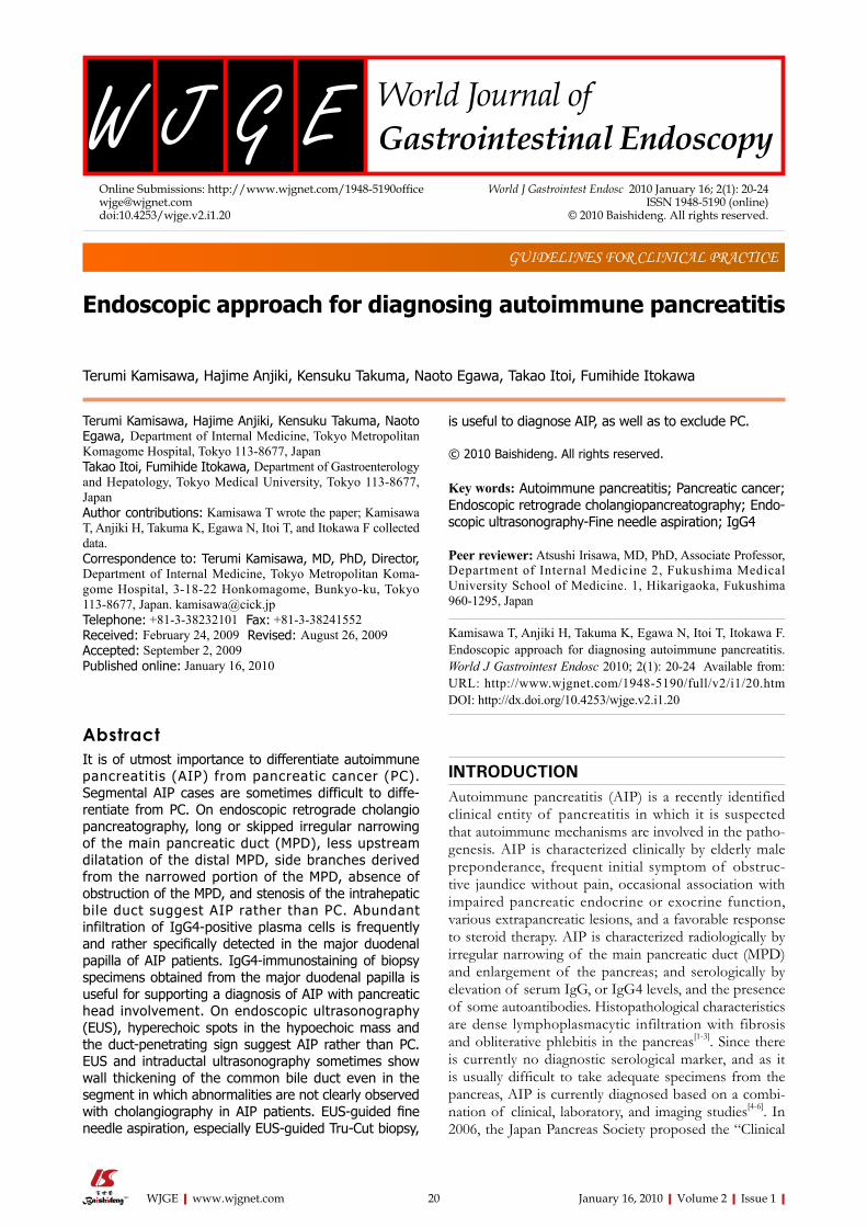
GUIDELINES FOR CLINICAL PRACTICE
Endoscopic approach for diagnosing autoimmune pancreatitis
Terumi Kamisawa, Hajime Anjiki, Kensuku Takuma, Naoto Egawa, Takao Itoi, Fumihide Itokawa
Terumi Kamisawa, Hajime Anjiki, Kensuku Takuma, Naoto Egawa, Department of Internal Medicine, Tokyo Metropolitan Komagome Hospital, Tokyo 113-8677, JapanTakao Itoi, Fumihide Itokawa, Department of Gastroenterology and Hepatology, Tokyo Medical University, Tokyo 113-8677, JapanAuthor contributions: Kamisawa T wrote the paper; Kamisawa T, Anjiki H, Takuma K, Egawa N, Itoi T, and Itokawa F collected data.Correspondence to: Terumi Kamisawa, MD, PhD, Director, Department of Internal Medicine, Tokyo Metropolitan Koma-gome Hospital, 3-18-22 Honkomagome, Bunkyo-ku, Tokyo 113-8677, Japan. [email protected]: +81-3-38232101 Fax: +81-3-38241552Received: February 24, 2009 Revised: August 26, 2009Accepted: September 2, 2009Published online: January 16, 2010
AbstractIt is of utmost importance to differentiate autoimmune pancreatitis (AIP) from pancreatic cancer (PC). Segmental AIP cases are sometimes difficult to differentiate from PC. On endoscopic retrograde cholangiopancreatography, long or skipped irregular narrowing of the main pancreatic duct (MPD), less upstream dilatation of the distal MPD, side branches derived from the narrowed portion of the MPD, absence of obstruction of the MPD, and stenosis of the intrahepatic bile duct suggest AIP rather than PC. Abundant infiltration of IgG4positive plasma cells is frequently and rather specifically detected in the major duodenal papilla of AIP patients. IgG4immunostaining of biopsy specimens obtained from the major duodenal papilla is useful for supporting a diagnosis of AIP with pancreatic head involvement. On endoscopic ultrasonography (EUS), hyperechoic spots in the hypoechoic mass and the ductpenetrating sign suggest AIP rather than PC. EUS and intraductal ultrasonography sometimes show wall thickening of the common bile duct even in the segment in which abnormalities are not clearly observed with cholangiography in AIP patients. EUS-guided fine needle aspiration, especially EUSguided TruCut biopsy,
is useful to diagnose AIP, as well as to exclude PC.
© 2010 Baishideng. All rights reserved.
Key words: Autoimmune pancreatitis; Pancreatic cancer; Endoscopic retrograde cholangiopancreatography; Endoscopic ultrasonographyFine needle aspiration; IgG4
Peer reviewer: Atsushi Irisawa, MD, PhD, Associate Professor, Department of Internal Medicine 2, Fukushima Medical University School of Medicine. 1, Hikarigaoka, Fukushima 960-1295, Japan
Kamisawa T, Anjiki H, Takuma K, Egawa N, Itoi T, Itokawa F. Endoscopic approach for diagnosing autoimmune pancreatitis. World J Gastrointest Endosc 2010; 2(1): 20-24 Available from: URL: http://www.wjgnet.com/1948-5190/full/v2/i1/20.htm DOI: http://dx.doi.org/10.4253/wjge.v2.i1.20
INTRODUCTIONAutoimmune pancreatitis (AIP) is a recently identified clinical entity of pancreatitis in which it is suspected that autoimmune mechanisms are involved in the patho-genesis. AIP is characterized clinically by elderly male preponderance, frequent initial symptom of obstruc-tive jaundice without pain, occasional association with impaired pancreatic endocrine or exocrine function, various extrapancreatic lesions, and a favorable response to steroid therapy. AIP is characterized radiologically by irregular narrowing of the main pancreatic duct (MPD) and enlargement of the pancreas; and serologically by elevation of serum IgG, or IgG4 levels, and the presence of some autoantibodies. Histopathological characteristics are dense lymphoplasmacytic infiltration with fibrosis and obliterative phlebitis in the pancreas[1-3]. Since there is currently no diagnostic serological marker, and as it is usually difficult to take adequate specimens from the pancreas, AIP is currently diagnosed based on a combi-nation of clinical, laboratory, and imaging studies[4-6]. In 2006, the Japan Pancreas Society proposed the “Clinical
World J Gastrointest Endosc 2010 January 16; 2(1): 20-24ISSN 1948-5190 (online)
© 2010 Baishideng. All rights reserved.
Online Submissions: http://www.wjgnet.com/[email protected]:10.4253/wjge.v2.i1.20
20 January 16, 2010|Volume 2|Issue 1|WJGE|www.wjgnet.com

Kamisawa T et al . Endoscopic approach for diagnosing autoimmune pancreatitis
Diagnostic Criteria for Autoimmune Pancreatitis”[4]. It contained three items: (1) radiological imaging showing diffuse or localized enlargement of the pancreas and diffuse or segmental irregular narrowing of the MPD; (2) laboratory data showing abnormally elevated levels of serum gammaglobulin, IgG or IgG4, or the presence of autoantibodies; and (3) histological findings showing marked interlobular fibrosis and prominent lymphoplas-macytic infiltration in the pancreas. To make the diagno-sis of AIP, criterion 1 is mandatory, and either criterion 2 or criterion 3 must be present.
AIP responds dramatically to steroid therapy and therefore to avoid unnecessary surgery, an accurate diag-nosis of AIP is required. The most important disease that should be differentiated from AIP is pancreatic cancer (PC)[1-3,7]. Serum IgG4 levels are elevated most frequently in AIP patients. However, the sensitivity of elevated serum IgG4 levels in AIP patients is reported to be 73%-80%[8,9], and elevation of serum IgG4 level is detected in some cases of PC[10,11]. In particular, AIP forming a mass-like le-sion in the head of the pancreas is sometimes difficult to differentiate from locally advanced pancreatic head cancer. Based on our experience with 50 cases of AIP, this review focuses on the endoscopic approach for diagnosing AIP, with special emphasis on differentiating AIP from PC.
ENDOSCOPIC RETROGRADE CHOLANGIOPANCREATOGRAPHY Diffuse irregular narrowing of the MPD on endoscopic retrograde cholangiopancreatography (ERCP) is one of the radiological features rather specific to AIP (Figure 1). PC rarely shows this pancreatogram. However, some AIP patients show segmental narrowing of the MPD (Figure 2A), which is rather difficult to differentiate from steno-sis of the MPD in PC. In our series[12], the length of the narrowed portion of the MPD on endoscopic retrograde pancreatography (ERP) was 6.7 ± 3.2 (mean ± SD) cm in AIP patients, which was significantly longer than in PC patients (2.6 ± 0.8 cm; P < 0.001). The length of the narrowed portion of the MPD on ERP was longer than 3 cm in 76% of AIP patients, which was significantly
higher than in PC patients (20%; P < 0.001). In AIP pa-tients, the degree of narrowing of the MPD varied in the same patient, and skipped, narrowed lesions of the MPD were detected in 35% of our AIP patients, but in none of our PC patients (P < 0.001). In AIP patients with seg-mental narrowing of the MPD, upstream dilatation of the distal MPD was less often noted than in PC. The maximal diameter of the upstream MPD on ERP was 2.9 ± 0.7 mm in segmental AIP patients, which was significantly smaller than in pancreatic head cancer patients (7.1 ± 1.9 mm; P < 0.001). The maximal diameter of the upstream MPD was smaller than 5 mm in 94% of segmental AIP patients, significantly higher than in PC patients (18%; P < 0.001). Side branches were more frequently derived from the narrowed portion of the MPD in AIP patients (65%) than in PC patients (25%; P < 0.036). Obstruction of the MPD was detected more often in PC patients (60%) than in AIP patients (6%; P < 0.001).
On endoscopic retrograde cholangiography, stenosis of the lower bile duct was detected frequently in both AIP and PC patients (Figure 2A). Brushing cytology of the stenotic biliary lesion and cytology of the bile via an endoscopic nasobiliary drainage tube inserted to manage jaundice, are useful to differentiate AIP from pancreatic or biliary cancer. Stenosis of the intrahepatic bile duct was detected in a few AIP patients, but it was not detect-ed in PC patients. When AIP patients develop stenosis in the intrahepatic bile duct, the cholangiographic appear-ance is similar to that of primary sclerosing cholangitis
Figure 1 Diffuse irregular narrowing of the main pancreatic duct of an AIP patient on ERP.
Figure 2 ERCP findings of an AIP patient. A: Segmental narrowing of the main pancreatic duct and stenosis of the lower bile duct before steroid therapy; B: After steroid therapy, stenosis of the main pancreatic duct and bile duct is improved.
A
B
21 January 16, 2010|Volume 2|Issue 1|WJGE|www.wjgnet.com
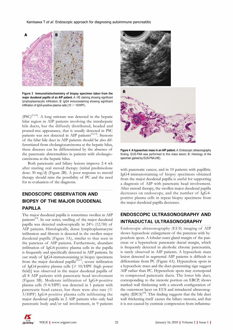
(PSC)[13,14]. A long stricture was detected in the hepatic hilar region in AIP patients involving the intrahepatic bile ducts, but the diffusely distributed, beaded and pruned-tree appearance, that is usually detected in PSC patients was not detected in AIP patients[12,13]. Stenosis of the hilar bile duct in AIP patients should be also dif-ferentiated from cholangiocarcinoma at the hepatic hilus, these diseases can be differentiated by the absence of the pancreatic abnormalities in patients with cholangio-carcinoma at the hepatic hilus.
Both pancreatic and biliary lesions improve 2-4 wk after starting oral steroid therapy (initial prednisolone dose: 30 mg/d) (Figure 2B). A poor response to steroid therapy should raise the possibility of PC and the need for re-evaluation of the diagnosis.
ENDOSCOPIC OBSERVATION AND BIOPSY OF THE MAJOR DUODENAL PAPILLAThe major duodenal papilla is sometimes swollen in AIP patients[15]. In our series, swelling of the major duodenal papilla was detected endoscopically in 24% (12/50) of AIP patients. Histologically, dense lymphoplasmacytic infiltration and fibrosis is detected in the swollen major duodenal papilla (Figure 3A), similar to that seen in the pancreas of AIP patients. Furthermore, abundant infiltration of IgG4-positive plasma cells in the papilla is frequently and specifically detected in AIP patients. In our study of IgG4-immunostaining in biopsy specimens from the major duodenal papilla[16,17], severe infiltration of IgG4-positive plasma cells [≥ 10/HPF (high power field)] was observed in the major duodenal papilla of all 8 AIP patients with pancreatic head involvement (Figure 3B). Moderate infiltration of IgG4-positive plasma cells (9-4/HPF) was detected in 1 patient with pancreatic head cancer, but there were also rare (≤ 3/HPF) IgG4-positive plasma cells infiltrating the major duodenal papilla in 2 AIP patients who only had pancreatic body and/or tail involvement, in 9 patients
with pancreatic cancer, and in 10 patients with papillitis. IgG4-immunostaining of biopsy specimens obtained from the major duodenal papilla is useful for supporting a diagnosis of AIP with pancreatic head involvement. After steroid therapy, the swollen major duodenal papilla decreases on endoscopy, and the number of IgG4-positive plasma cells in repeat biopsy specimens from the major duodenal papilla decreases.
ENDOSCOPIC ULTRASONOGRAPHY AND INTRADUCTAL ULTRASONOGRAPHYEndoscopic ultrasonography (EUS) imaging of AIP shows hypoechoic enlargement of the pancreas with hy-poechoic spots. A lobular outer gland margin of the pan-creas or a hyperechoic pancreatic ductal margin, which is frequently detected in alcoholic chronic pancreatitis, is rarely observed in AIP patients. A hypoechoic mass lesion detected in segmental AIP patients is difficult to differentiate from PC (Figure 4A). Hyperechoic spots in a hypoechoic mass and the duct-penetrating sign suggest AIP rather than PC. Hyperechoic spots may correspond to compressed pancreatic ducts. The lower bile duct, corresponding to the stenotic portion on ERCP, shows marked wall thickening with a smooth configuration of the outermost layer on EUS and intraductal ultrasonog-raphy (IDUS)[18]. This finding suggests that the bile duct wall thickening itself causes the biliary stenosis, and that it is not caused by extrinsic compression from inflamma-
Figure 3 Immunohistochemistry of biopsy specimen taken from the major duodenal papilla of an AIP patient. A: HE staining showing significant lymphoplasmacytic infiltration; B: IgG4 immunostaining showing significant infiltration of IgG4-positive plasma cells (10 ≥ 10/HPF).
A B
Figure 4 A hypoechoic mass in an AIP patient. A: Endoscopic ultrasonography finding. EUS-FNA was performed to this mass lesion; B: Histology of the specimen gained by EUS-FNA (HE).
A
B
22 January 16, 2010|Volume 2|Issue 1|WJGE|www.wjgnet.com
Kamisawa T et al . Endoscopic approach for diagnosing autoimmune pancreatitis
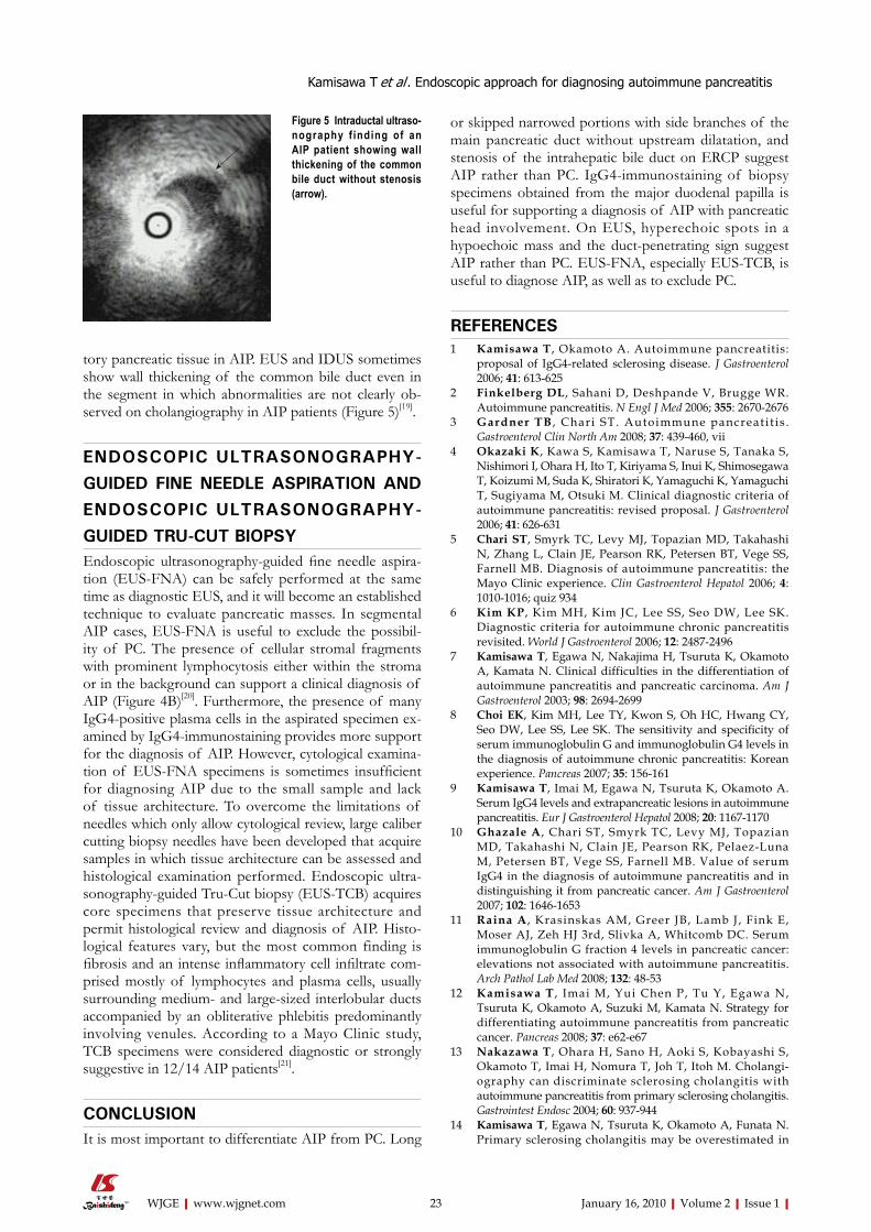
tory pancreatic tissue in AIP. EUS and IDUS sometimes show wall thickening of the common bile duct even in the segment in which abnormalities are not clearly ob-served on cholangiography in AIP patients (Figure 5)[19].
ENDOSCOPIC ULTRASONOGRAPHY-GUIDED FINE NEEDLE ASPIRATION AND ENDOSCOPIC ULTRASONOGRAPHY-GUIDED TRU-CUT BIOPSYEndoscopic ultrasonography-guided fine needle aspira-tion (EUS-FNA) can be safely performed at the same time as diagnostic EUS, and it will become an established technique to evaluate pancreatic masses. In segmental AIP cases, EUS-FNA is useful to exclude the possibil-ity of PC. The presence of cellular stromal fragments with prominent lymphocytosis either within the stroma or in the background can support a clinical diagnosis of AIP (Figure 4B)[20]. Furthermore, the presence of many IgG4-positive plasma cells in the aspirated specimen ex-amined by IgG4-immunostaining provides more support for the diagnosis of AIP. However, cytological examina-tion of EUS-FNA specimens is sometimes insufficient for diagnosing AIP due to the small sample and lack of tissue architecture. To overcome the limitations of needles which only allow cytological review, large caliber cutting biopsy needles have been developed that acquire samples in which tissue architecture can be assessed and histological examination performed. Endoscopic ultra-sonography-guided Tru-Cut biopsy (EUS-TCB) acquires core specimens that preserve tissue architecture and permit histological review and diagnosis of AIP. Histo-logical features vary, but the most common finding is fibrosis and an intense inflammatory cell infiltrate com-prised mostly of lymphocytes and plasma cells, usually surrounding medium- and large-sized interlobular ducts accompanied by an obliterative phlebitis predominantly involving venules. According to a Mayo Clinic study, TCB specimens were considered diagnostic or strongly suggestive in 12/14 AIP patients[21].
CONCLUSIONIt is most important to differentiate AIP from PC. Long
or skipped narrowed portions with side branches of the main pancreatic duct without upstream dilatation, and stenosis of the intrahepatic bile duct on ERCP suggest AIP rather than PC. IgG4-immunostaining of biopsy specimens obtained from the major duodenal papilla is useful for supporting a diagnosis of AIP with pancreatic head involvement. On EUS, hyperechoic spots in a hypoechoic mass and the duct-penetrating sign suggest AIP rather than PC. EUS-FNA, especially EUS-TCB, is useful to diagnose AIP, as well as to exclude PC.
REFERENCES1 Kamisawa T, Okamoto A. Autoimmune pancreatitis:
proposal of IgG4-related sclerosing disease. J Gastroenterol 2006; 41: 613-625
2 Finkelberg DL, Sahani D, Deshpande V, Brugge WR. Autoimmune pancreatitis. N Engl J Med 2006; 355: 2670-2676
3 Gardner TB , Chari ST. Autoimmune pancreat i t is . Gastroenterol Clin North Am 2008; 37: 439-460, vii
4 Okazaki K, Kawa S, Kamisawa T, Naruse S, Tanaka S, Nishimori I, Ohara H, Ito T, Kiriyama S, Inui K, Shimosegawa T, Koizumi M, Suda K, Shiratori K, Yamaguchi K, Yamaguchi T, Sugiyama M, Otsuki M. Clinical diagnostic criteria of autoimmune pancreatitis: revised proposal. J Gastroenterol 2006; 41: 626-631
5 Chari ST, Smyrk TC, Levy MJ, Topazian MD, Takahashi N, Zhang L, Clain JE, Pearson RK, Petersen BT, Vege SS, Farnell MB. Diagnosis of autoimmune pancreatitis: the Mayo Clinic experience. Clin Gastroenterol Hepatol 2006; 4: 1010-1016; quiz 934
6 Kim KP , Kim MH, Kim JC, Lee SS, Seo DW, Lee SK. Diagnostic criteria for autoimmune chronic pancreatitis revisited. World J Gastroenterol 2006; 12: 2487-2496
7 Kamisawa T, Egawa N, Nakajima H, Tsuruta K, Okamoto A, Kamata N. Clinical difficulties in the differentiation of autoimmune pancreatitis and pancreatic carcinoma. Am J Gastroenterol 2003; 98: 2694-2699
8 Choi EK, Kim MH, Lee TY, Kwon S, Oh HC, Hwang CY, Seo DW, Lee SS, Lee SK. The sensitivity and specificity of serum immunoglobulin G and immunoglobulin G4 levels in the diagnosis of autoimmune chronic pancreatitis: Korean experience. Pancreas 2007; 35: 156-161
9 Kamisawa T, Imai M, Egawa N, Tsuruta K, Okamoto A. Serum IgG4 levels and extrapancreatic lesions in autoimmune pancreatitis. Eur J Gastroenterol Hepatol 2008; 20: 1167-1170
10 Ghazale A , Chari ST, Smyrk TC, Levy MJ, Topazian MD, Takahashi N, Clain JE, Pearson RK, Pelaez-Luna M, Petersen BT, Vege SS, Farnell MB. Value of serum IgG4 in the diagnosis of autoimmune pancreatitis and in distinguishing it from pancreatic cancer. Am J Gastroenterol 2007; 102: 1646-1653
11 Raina A , Krasinskas AM, Greer JB, Lamb J, Fink E, Moser AJ, Zeh HJ 3rd, Slivka A, Whitcomb DC. Serum immunoglobulin G fraction 4 levels in pancreatic cancer: elevations not associated with autoimmune pancreatitis. Arch Pathol Lab Med 2008; 132: 48-53
12 Kamisawa T , Imai M, Yui Chen P, Tu Y, Egawa N, Tsuruta K, Okamoto A, Suzuki M, Kamata N. Strategy for differentiating autoimmune pancreatitis from pancreatic cancer. Pancreas 2008; 37: e62-e67
13 Nakazawa T, Ohara H, Sano H, Aoki S, Kobayashi S, Okamoto T, Imai H, Nomura T, Joh T, Itoh M. Cholangi-ography can discriminate sclerosing cholangitis with autoimmune pancreatitis from primary sclerosing cholangitis. Gastrointest Endosc 2004; 60: 937-944
14 Kamisawa T, Egawa N, Tsuruta K, Okamoto A, Funata N. Primary sclerosing cholangitis may be overestimated in
23 January 16, 2010|Volume 2|Issue 1|WJGE|www.wjgnet.com
Kamisawa T et al . Endoscopic approach for diagnosing autoimmune pancreatitis
Figure 5 Intraductal ultrasonography f inding of an AIP patient showing wall thickening of the common bile duct without stenosis (arrow).
Japan. J Gastroenterol 2005; 40: 318-31915 Unno H, Saegusa H, Fukushima M, Hamano H. Usefulness
of endoscopic observation of the main duodenal papilla in the diagnosis of sclerosing pancreatitis. Gastrointest Endosc 2002; 56: 880-884
16 Kamisawa T, Tu Y, Nakajima H, Egawa N, Tsuruta K, Okamoto A. Usefulness of biopsying the major duodenal papilla to diagnose autoimmune pancreatitis: a prospective study using IgG4-immunostaining. World J Gastroenterol 2006; 12: 2031-2033
17 Kamisawa T, Tu Y, Egawa N, Tsuruta K, Okamoto A. A new diagnostic endoscopic tool for autoimmune pancreatitis. Gastrointest Endosc 2008; 68: 358-361
18 Hyodo N, Hyodo T. Ultrasonographic evaluation in patients with autoimmune-related pancreatitis. J Gastroenterol 2003;
38: 1155-116119 Hirano K, Shiratori Y, Komatsu Y, Yamamoto N, Sasahira N,
Toda N, Isayama H, Tada M, Tsujino T, Nakata R, Kawase T, Katamoto T, Kawabe T, Omata M. Involvement of the biliary system in autoimmune pancreatitis: a follow-up study. Clin Gastroenterol Hepatol 2003; 1: 453-464
20 Deshpande V, Mino-Kenudson M, Brugge WR, Pitman MB, Fernandez-del Castillo C, Warshaw AL, Lauwers GY. Endoscopic ultrasound guided fine needle aspiration biopsy of autoimmune pancreatitis: diagnostic criteria and pitfalls. Am J Surg Pathol 2005; 29: 1464-1471
21 Levy MJ, Wiersema MJ, Chari ST. Chronic pancreatitis: focal pancreatitis or cancer? Is there a role for FNA/biopsy? Autoimmune pancreatitis. Endoscopy 2006; 38 Suppl 1: S30-S35
S- Editor Zhang HN L- Editor Lalor PF E- Editor Ma WH
24 January 16, 2010|Volume 2|Issue 1|WJGE|www.wjgnet.com
Kamisawa T et al . Endoscopic approach for diagnosing autoimmune pancreatitis
GUIDELINES FOR CLINICAL PRACTICE
ERCP in acute biliary pancreatitis
Dimitrios J Kapetanos
Dimitrios J Kapetanos, Gastroenterology Department, George Papanikolaou Hospital, Thessaloniki 57010, GreeceAuthor contributions: Kapetanos DJ wrote this paper. Correspondence to: Dimitrios J Kapetanos, MD, PhD, Gastroenterology Department, George Papanikolaou Hospital, Thessaloniki 57010, Greece. [email protected]: +30-2313-307102Received: March 10, 2009 Revised: August 26, 2009Accepted: September 2, 2009Published online: January 16, 2010
AbstractThe role of urgent endoscopic retrograde cholangiopancreatography (ERCP) in acute biliary pancreatitis is for many years a subject for disagreement among physicians. Although the evidence seemed to be in favor of performing ERCP, endoscopists usually hesitate to conform to the guidelines. ERCP is an invasive procedure, with complications which can affect patients’ outcome. Recent evidence suggests that we should probably modify our policy, recruiting less invasive procedures, like magnetic resonance cholangiopancreatography and endoscopic ultrasound, before conducting ERCP in patients with acute biliary pancreatitis. In this editorial the different aspects regarding the role of ERCP in acute biliary pancreatitis are discussed.
© 2010 Baishideng. All rights reserved.
Key words: Endoscopic retrograde cholangiopancreatography; Acute pancreatitis; Gallstone; Magnetic resonance cholangiopancreatography; Endoscopic ultrasound
Peer reviewers: Akira Horiuchi, MD, Director, Digestive Disease Center, Showa Inan General Hospital, Komagane 399-4191, Japan
Kapetanos DJ. ERCP in acute biliary pancreatitis. World J Gastrointest Endosc 2010; 2(1): 25-28 Available from: URL: http://www.wjgnet.com/1948-5190/full/v2/i1/25.htm DOI: http://dx.doi.org/10.4253/wjge.v2.i1.25
INTRODUCTIONThe pathogenesis of acute biliary pancreatitis is attributed to transient obstruction of the bile and pancreatic duct, which causes reflux of bile and duodenal content in the pancreatic duct or/and increases the hydrostatic pressure in the pancreatic duct[1]. The severity of pancreatitis is determined by the size of the following local and systemic inflammatory reaction that depends on the interaction of multiple factors probably including genetic predisposition. It has been suggested by animal models and human studies that the duration of bile duct obstruction is a critical factor contributing to the severity of pancreatitis[2-5]. Pancreatic necrosis develops more often when the duration of obstruction exceeds 48 h. This obstruction may be constant, due to an impacted stone, or intermittent, when a stone remains in the bile duct or multiple stones try to pass the ampulla.
Initial surgical attempts to decompress the bile duct soon after the diagnosis of pancreatitis failed, because they were associated with increased mortality in the urgent surgery group[6,7]. Endoscopic retrograde cholangiopancreatography (ERCP) is a less invasive method to clear the bile duct, so it could favorably affect the severity of biliary pancreatitis if utilized properly. On the other hand, it is difficult to perform ERCP in a pa-tient with acute pancreatitis, because the duodenum and ampulla are swollen and the patients’ physical condition is compromised. Thus, we should have strong evidence to attempt an urgent invasive procedure which is incon-venient for both the patient and the doctor.
Four randomized studies have been published the last twenty years and they will be reviewed in the light of two recent meta-analyses. We should address in advance that each of those studies focused in different populations, so the results must be interpreted within this context.
Neoptolemos et al[8] studied patients with probable bili-ary pancreatitis, stratified according to severity based on the modified Glasgow criteria. They found that patients with predicted severe pancreatitis had fewer complications if they underwent ERCP within 72 h (24% vs 61%, P < 0.01). When patients with acute cholangitis were excluded
World J Gastrointest Endosc 2010 January 16; 2(1): 25-28ISSN 1948-5190 (online)
© 2010 Baishideng. All rights reserved.
Online Submissions: http://www.wjgnet.com/[email protected]:10.4253/wjge.v2.i1.25
25 January 16, 2010|Volume 2|Issue 1|WJGE|www.wjgnet.com
Kapetanos DJ. ERCP in acute biliary pancreatitis
the difference remained (15% vs 60%, P = 0.003). It is reasonable to exclude patients with acute cholangitis from the studied population because the coexistence of acute pancreatitis and cholangitis is accompanied by higher rate of complications, therefore these patients are the most probable to benefit from early ERCP[9].
A few years later Fan et al[10] published a similar study in a mixed population, but only 66% of them had biliary pancreatitis. The pancreatitis severity was predicted by se-rum urea and plasma glucose levels, a system with disputed value[11]. ERCP was performed within 24 h after admission. In the subgroup with biliary stones (a stone located in any part of the biliary tract) the authors from Hong Kong also found, in agreement with the Leicester study, that the early ERCP group with predicted severe pancreatitis had fewer complications (13% vs 54%, P = 0.002), although one half of the conservative treatment group eventually underwent ERCP (median time 60 h). Patients with cholangitis were not excluded in that study but were offered ERCP irre-spectively of the assigned group.
The results of those studies suggested that patients with predicted severe acute biliary pancreatitis should have urgent ERCP.
IS THIS TRUE OR THE FAVORABLE RE-SULTS COME FROM THOSE PATIENTS WITH IMPACTED COMMON DUCT STONES AND PERSISTING OBSTRUC-TION?The study of Folsch et al[12] tried to answer this question by excluding patients with bilirubin level higher than 5 mg/dL. The modified Glasgow criteria were used to predict sever-ity and ERCP was performed within 72 h after the onset of pain. This study was prematurely terminated because an interim analysis found that the primary goal (superior-ity of ERCP treatment) could not be reached. The rate of morbidity and mortality was similar between the invasive and conservative treatment group. Mortality in the severe pancreatitis group was 6/26 in the ERCP arm and 2/20 in the conservative arm (P = 0.44)[13]. A worrying finding was an estimated 8% mortality in the mild pancreatitis group who underwent ERCP (5.4% in the conservative treat-ment group, P = 0.7) which was much higher than that observed in other studies[13]. The rate of respiratory failure was significantly higher in the ERCP group, which could not be explained by the authors. This study highlights that ERCP is an invasive procedure, with complications that can affect patients’ outcome.
IS THERE A SUBGROUP OF PATIENTS WITH ACUTE BILIARY PANCREATITIS WHO COULD BENEFIT FROM EARLY ERCP?Twenty years ago Neoptolemos et al[14] found in a retro-
spective study that more patients with severe pancreatitis had bile duct stones 72 h after the attack (61% vs 35%) and even 3-30 d after the attack (50% vs 24%) than patients with mild pancreatitis (P < 0.01). In a random-ized study Acosta et al[15] tested the hypothesis that it is the duration of bile duct obstruction that determines the outcome of biliary pancreatitis and not the pres-ence of stones per se. The authors subjected to ERCP the patients enrolled in the intervention arm, if signs of obstruction persisted over 24 h. Indications of obstruc-tion were severe and continuous epigastric pain, bile free gastric aspirate and elevated serum bilirubin, while relief of pain, decrease of bilirubin level and reappearance of bile in the gastric aspirate were signs of spontaneous termination of obstruction. Half of the patients in the intervention group eventually underwent ERCP. When discontinuation of the obstruction occurred spontane-ously or after ERCP in less than 48 h, the rate of com-plications was lower than in cases with obstruction last-ing more than 48 h (8% vs 78%, P < 0.001).
According to the above results it would be reason-able to assume that patients with persisting biliary ob-struction could benefit from urgent ERCP. In a study that could be regarded supplementary to that of Folsch et al[12], Oria et al[16] randomized patients with signs of obstruction (main bile duct diameter ≥ 8 mm and total serum bilirubin ≥ 1.20 mg/dL) without cholangitis, to urgent ERCP within 72 h after the onset of the attack or to conservative treatment. The incidence of compli-cations was similar between the two groups. Bile duct stones were found in 72% of patients with predicted mild pancreatitis (APACHE Ⅱ score) and in 73% of patients with predicted severe pancreatitis. These results intrigued the results by Neoptolemos et al[14].
A Cochrane meta-analysis of the first three trials (Neoptolemos et al[14], Fan et al[10] and Folsch et al[12]) con-cluded that early ERCP decreases significantly the rate of complications in patients with predicted severe pan-creatitis[17]. A recent meta-analysis from Italy included the aforementioned studies and added the study of Oria et al[16] and one study from China with disputable ran-domization[18-20]. The authors reached the same conclu-sions with the Cochrane meta-analysis. They also stated that excluding the Chinese study the results would be similar[19]. Another meta-analysis, which was published concurrently, approached this issue excluding patients with acute cholangitis[21]. Petrov et al[21] included the trials by Neoptolemos et al[14], Folsch et al[12] and Oria et al[16] and excluded the study by Fan et al[10] because it did not provide separate data on acute cholangitis. This meta-analysis failed to prove a substantial benefit from early ERCP even in patients with predicted severe pancreatitis. The authors noted that even in the case they had includ-ed the study by Fan et al[10], the results would not differ.
Each of the previous studies and consequently the meta-analyses should be interpreted within the context of several limitations: First, the diagnosis of acute chol-angitis in a patient with inflammatory reaction due to acute pancreatitis is cumbersome and until recently there
26 January 16, 2010|Volume 2|Issue 1|WJGE|www.wjgnet.com

were no specific criteria[22]. Second, the criteria used for prediction of pancreatitis’ severity (APACHE Ⅱ, Glasgow) have low positive predictive value (50%-60%), which means that about half of those predicted to have severe pancreatitis actually prove to have mild pancre-atitis. How could someone evaluate the effectiveness of a given intervention, if half of the patients would probably not benefit and the intervention itself may af-fect morbidity and mortality in both ways? Third, stones were eventually found in only half of the patients who underwent ERCP, which means that ERCP would not benefit half of those in whom an indication for the in-tervention was determined and the procedure could pos-sibly deteriorate their clinical condition.
The UK guidelines for the management of acute pancreatitis advocate urgent therapeutic ERCP in every patient with suspected gall stone etiology and predicted severe pancreatitis or when there is cholangitis, jaun-dice or a dilated common bile duct[23]. The indications for early ERCP in the AGA Institute review on acute pancreatitis are more restricted[24]. According to these guidelines, early ERCP should be performed in patients with cholangitis or when there is suspicion of persistent common bile duct stone (a dilated common bile duct or visible common bile duct stone, or jaundice or persis-tently abnormal liver chemistry values). Urgent ERCP in predicted severe pancreatitis without concomitant chol-angitis or high suspicion of a persistent common bile duct stone is controversial.
Medical community appears reluctant to conform to these guidelines as clearly shown in a recent study from UK[25]. Physicians complied with all the UK guidelines except for urgent ERCP for severe acute pancreatitis. Only 48% of patients with this indication finally un-derwent ERCP within 72 h. Difficulties in transferring patients to specialized centers capable to perform ERCP and in providing ERCP out of hours may have contrib-uted to these results. It was obvious that urgent ERCP was reserved for patients with severe gallstone pancreati-tis who had biliary obstruction or cholangitis. Neverthe-less this policy did not increase the mortality of acute pancreatitis. The same viewpoint is also encountered in the USA[26].
According to the Tokyo guidelines a definite diagno-sis of cholangitis is reached when there are two or more of the following i.e. (a) history of biliary disease, (b) fe-ver and/or chills, (c) jaundice (d) upper abdominal pain and in addition, laboratory evidence of inflammatory response and abnormal liver function tests and imaging findings of biliary dilatation or of specific etiology (e.g. a stone, in the case of pancreatitis)[22]. The diagnosis of cholangitis in a patient with severe acute biliary pancre-atitis with systemic inflammatory response syndrome (SIRS) could be problematic. From the aforementioned criteria only the imaging finding of an impacted stone could differentiate superimposed cholangitis on acute pancreatitis from pancreatitis with SIRS.
MRCP is useful in the diagnosis of biliary obstruction, although it should not be recommended in a patient with
unstable condition who could not be monitored in the MRCP chamber[27]. Endoscopic ultrasound (EUS) can also detect biliary obstruction, at least equally to MRCP[28,29]. These procedures could be applied before ERCP, if they are available, therefore ERCP would be reserved for pa-tients with strong evidence of biliary obstruction.
CONCLUSIONIn conclusion, (1) early ERCP should be reserved for pa-tients with acute cholangitis superimposed to acute pan-creatitis; (2) There is no indication for urgent ERCP in patients with mild pancreatitis without cholangitis; and (3) In cases with severe biliary pancreatitis the differen-tial diagnosis between acute cholangitis and pancreatitis with SIRS may be difficult. In those patients every effort should be made to identify biliary obstruction, including MRCP and EUS when accessible, before resorting to ERCP.
REFERENCES1 Frakes JT. Biliary pancreatitis: a review. Emphasizing
appropriate endoscopic intervention. J Clin Gastroenterol 1999; 28: 97-109
2 Acosta JM, Rubio Galli OM, Rossi R, Chinellato AV, Pellegrini CA. Effect of duration of ampullary gallstone obstruction on severity of lesions of acute pancreatitis. J Am Coll Surg 1997; 184: 499-505
3 Hirano T, Manabe T. A possible mechanism for gallstone pancreatitis: repeated short-term pancreaticobiliary duct obstruction with exocrine stimulation in rats. Proc Soc Exp Biol Med 1993; 202: 246-252
4 Runzi M, Saluja A, Lerch MM, Dawra R, Nishino H, Steer ML. Early ductal decompression prevents the progression of biliary pancreatitis: an experimental study in the opossum. Gastroenterology 1993; 105: 157-164
5 Senninger N, Moody FG, Coelho JC, Van Buren DH. The role of biliary obstruction in the pathogenesis of acute pancreatitis in the opossum. Surgery 1986; 99: 688-693
6 Kelly TR. Gallstone pancreatitis: the timing of surgery. Surgery 1980; 88: 345-350
7 Tondelli P, Stutz K, Harder F, Schuppisser JP, Allgower M. Acute gallstone pancreatitis: best timing for biliary surgery. Br J Surg 1982; 69: 709-710
8 Neoptolemos JP, Carr-Locke DL, London NJ, Bailey IA, James D, Fossard DP. Controlled trial of urgent endoscopic retrograde cholangiopancreatography and endoscopic sphincterotomy versus conservative treatment for acute pancreatitis due to gallstones. Lancet 1988; 2: 979-983
9 Neoptolemos JP, Carr-Locke DL, Leese T, James D. Acute cholangitis in association with acute pancreatitis: incidence, clinical features and outcome in relation to ERCP and endoscopic sphincterotomy. Br J Surg 1987; 74: 1103-1106
10 Fan ST, Lai EC, Mok FP, Lo CM, Zheng SS, Wong J. Early treatment of acute biliary pancreatitis by endoscopic papillotomy. N Engl J Med 1993; 328: 228-232
11 Heath DI, Imrie CW. The Hong Kong criteria and severity prediction in acute pancreatitis. Int J Pancreatol 1994; 15: 179-185
12 Folsch UR, Nitsche R, Ludtke R, Hilgers RA, Creutzfeldt W. Early ERCP and papillotomy compared with conservative treatment for acute biliary pancreatitis. The German Study Group on Acute Biliary Pancreatitis. N Engl J Med 1997; 336: 237-242
13 Folsch UR , Neoptolemos J . Reason for performing
27 January 16, 2010|Volume 2|Issue 1|WJGE|www.wjgnet.com
Kapetanos DJ. ERCP in acute biliary pancreatitis

endoscopic retrograde cholangiopancreatography (ERCP) and ES in a patient with severe gallstone pancreatitis even in the absence of main bile duct stones. Pancreas 2002; 24: 412-414; author reply 414-417
14 Neoptolemos JP, Carr-Locke DL, London N, Bailey I, Fossard DP. ERCP findings and the role of endoscopic sphincterotomy in acute gallstone pancreatitis. Br J Surg 1988; 75: 954-960
15 Acosta JM, Katkhouda N, Debian KA, Groshen SG, Tsao-Wei DD, Berne TV. Early ductal decompression versus conservative management for gallstone pancreatitis with ampullary obstruction: a prospective randomized clinical trial. Ann Surg 2006; 243: 33-40
16 Oria A , Cimmino D, Ocampo C, Silva W, Kohan G, Zandalazini H, Szelagowski C, Chiappetta L. Early endos-copic intervention versus early conservative management in patients with acute gallstone pancreatitis and biliopancreatic obstruction: a randomized clinical trial. Ann Surg 2007; 245: 10-17
17 Ayub K, Imada R, Slavin J. Endoscopic retrograde chol-angiopancreatography in gallstone-associated acute pancreatitis. Cochrane Database Syst Rev 2004; CD003630
18 Moretti A, Papi C, Aratari A, Festa V, Tanga M, Koch M, Capurso L. Is early endoscopic retrograde cholangiopancrea-tography useful in the management of acute biliary pancreatitis? A meta-analysis of randomized controlled trials. Dig Liver Dis 2008; 40: 379-385
19 Petrov MS. ERCP versus conservative treatment in acute pancreatitis: meta-analysis or meta-confusion? Dig Liver Dis 2008; 40: 800-801; author reply 801-802
20 Zhou MQ, Li NP, Lu RD. Duodenoscopy in treatment of acute gallstone pancreatitis. Hepatobiliary Pancreat Dis Int 2002; 1: 608-610
21 Petrov MS, van Santvoort HC, Besselink MG, van der
Heijden GJ, van Erpecum KJ, Gooszen HG. Early endoscopic retrograde cholangiopancreatography versus conservative management in acute biliary pancreatitis without cholangitis: a meta-analysis of randomized trials. Ann Surg 2008; 247: 250-257
22 Wada K, Takada T, Kawarada Y, Nimura Y, Miura F, Yoshida M, Mayumi T, Strasberg S, Pitt HA, Gadacz TR, Buchler MW, Belghiti J, de Santibanes E, Gouma DJ, Neuhaus H, Dervenis C, Fan ST, Chen MF, Ker CG, Bornman PC, Hilvano SC, Kim SW, Liau KH, Kim MH. Diagnostic criteria and severity assessment of acute cholangitis: Tokyo Guidelines. J Hepatobiliary Pancreat Surg 2007; 14: 52-58
23 UK guidelines for the management of acute pancreatitis. Gut 2005; 54 Suppl 3: iii1-iii9
24 Forsmark CE, Baillie J. AGA Institute technical review on acute pancreatitis. Gastroenterology 2007; 132: 2022-2044
25 Mofidi R, Madhavan KK, Garden OJ, Parks RW. An audit of the management of patients with acute pancreatitis against national standards of practice. Br J Surg 2007; 94: 844-848
26 Baillie J. Should urgent ERCP be performed in patients with acute biliary pancreatitis without acute cholangitis? Nat Clin Pract Gastroenterol Hepatol 2008; 5: 484-485
27 Romagnuolo J, Bardou M, Rahme E, Joseph L, Reinhold C, Barkun AN. Magnetic resonance cholangiopancreatography: a meta-analysis of test performance in suspected biliary disease. Ann Intern Med 2003; 139: 547-557
28 Garrow D, Miller S, Sinha D, Conway J, Hoffman BJ, Hawes RH, Romagnuolo J. Endoscopic ultrasound: a meta-analysis of test performance in suspected biliary obstruction. Clin Gastroenterol Hepatol 2007; 5: 616-623
29 Ledro-Cano D. Suspected choledocholithiasis: endoscopic ultrasound or magnetic resonance cholangio-pancrea-tography? A systematic review. Eur J Gastroenterol Hepatol 2007; 19: 1007-1011
S- Editor Zhang HN L- Editor Negro F E- Editor Ma WH
28 January 16, 2010|Volume 2|Issue 1|WJGE|www.wjgnet.com
Kapetanos DJ. ERCP in acute biliary pancreatitis

REVIEW
Endoscopic balloon dilation for benign gastric outlet obstruction in adults
Rakesh Kochhar, Suman Kochhar
Rakesh Kochhar, Department of Gastroenterology, Postgraduate Institute of Medical Education and Research, Chandigarh 160012, IndiaSuman Kochhar, Department of Radiodiagnosis, Government Medical College and Hospital, Chandigarh 160020, IndiaAuthor contributions: Kochhar R wrote this paper; Kochhar S provided the radiological support and pictures.Correspondence to: Rakesh Kochhar, Professor, Department of Gastroenterology, Postgraduate Institute of Medical Education and Research, Chandigarh 160012, India. [email protected]: +91-172-2715016 Fax: +91-172-2744401Received: April 8, 2009 Revised: September 1, 2009Accepted: September 8, 2009Published online: January 16, 2010
AbstractGastric outlet obstruction (GOO) includes obstruction in the antropyloric area or in the bulbar or post bulbar duo-denal segments. Though malignancy remains the most common cause of GOO in adults, a significant number of patients have benign disease. The latter include peptic ulcer disease, caustic ingestion, post-operative anasto-motic state and inflammatory causes like Crohn’s disease and tuberculosis. Peptic ulcer remains the most common benign cause of GOO. Management of benign GOO re-volves around confirmation of the etiology, removing the offending agent Helicobacter pylori (H. pylori ), non-ste-roidal anti-inflammatory drugs, etc . and definitive thera-py. Traditionally, surgery has been the standard mode of treatment for benign GOO. However, after the advent of through-the-scope balloon dilators, endoscopic balloon dilation (EBD) has emerged as an effective alternative to surgery in selected groups of patients. So far, this form of therapy has been shown to be effective in caustic-induced GOO with short segment cicatrization and ulcer related GOO. In the latter, EBD must be combined with eradication of H. pylori . Dilation is preferably done with wire-guided balloon catheters of incremental diameter with the aim to reach the end-point of 15 mm. While it is recommended that fluoroscopic control be used for
EBD, this is not used by most endoscopists. Frequency of dilation has varied from once a week to once in three weeks. Complications are uncommon with perforation occurring more often with balloons larger than 15 mm. Attempts to augment efficacy of EBD include intralesion-al steroids and endoscopic incision.
© 2010 Baishideng. All rights reserved.
Key words: Gastric outlet obstruction; Endoscopic bal-loon dilation; Helicobacter pylori ; Management; Adult; Benign
Peer reviewer: Tian-Le Ma, MD, Department of Gastro-enterology, Rui Jin Hospital Affiliated to Medical College of Shanghai Jiao Tong University, Shanghai 200040, China
Kochhar R, Kochhar S. Endoscopic balloon dilation for benign gastric outlet obstruction in adults. World J Gastrointest Endosc 2010; 2(1): 29-35 Available from: URL: http://www.wjg-net.com/1948-5190/full/v2/i1/29.htm DOI: http://dx.doi.org/10.4253/wjge.v2.i1.29
INTRODUCTIONGastric outlet obstruction (GOO) includes obstruction in the antropyloric area or in the bulbar or post bulbar duodenal segments. Though malignancy remains a com-mon cause of GOO in adults[1,2], a significant number of patients with GOO have benign causes. Among the latter are peptic ulcer disease, caustic ingestion, post-operative anastomotic state and inflammatory causes such as Crohn’s disease and tuberculosis. Less often, chronic pancreatitis, annular pancreas and non-steroidal anti-inflammatory drug-included strictures result in GOO (Table 1). Peptic ulcer disease is the most common cause of benign GOO. After the association between Helicobacter pylori (H. pylori) and peptic ulcer was recognized, less than 5% patients with complicated duodenal ulcer disease and less than 1%-2% with complicated gastric ulcer disease have developed this
World J Gastrointest Endosc 2010 January 16; 2(1): 29-35ISSN 1948-5190 (online)
© 2010 Baishideng. All rights reserved.
Online Submissions: http://www.wjgnet.com/[email protected]:10.4253/wjge.v2.i1.29
29 January 16, 2010|Volume 2|Issue 1|WJGE|www.wjgnet.com
Kochhar R. EBD for benign gastric outlet obstruction
complication[3,4]. Patients with ulcer related GOO often have a long history of symptoms due to ulcer disease[5]. In a study carried out in the United States, only 14% of 85 patients with ulcer-related GOO had acute disease and obstruction was the initial manifestation of the disease[6]. It has been estimated that > 95% of cases of obstructing du-odenal ulcer disease have the obstruction in the duodenal bulb, and the rest were in the postbulbar region[4]. Caustic ingestion is another important cause of benign GOO. Both acid ingestion and alkali ingestion can cause antral/pyloric scarring resulting in GOO[7,8]. About one third of patients with ingestion of strong caustics end up having GOO. In a study of 41 cases of acid ingestion, it was reported that 44.4% developed GOO[7], while in another study on alkali ingestion, 36.8% of 31 patients developed GOO[8].
EVALUATING PATIENTS WITH BENIGN
GASTRIC OUTLET OBSTRUCTIONUnless the cause of GOO is obvious from the anteced-ent history, such as caustic ingestion, prior surgery, or known peptic ulcer disease, one must exclude malignancy in all adult patients by endoscopy and biopsy. A CT scan is very helpful in evaluating mural thickening, lymphnode enlargement and status of pancreas, biliary tract and ret-roperitoneum[4]. Linitus plastica can often be difficult to di-agnose[4]. Barium studies are helpful in delineating the site of obstruction as well its extent.
MANAGEMENTAll patients with symptoms of persistent GOO should be admitted to the hospital. Mild cases may be man-aged on an out-patient basis, however, those with fluid and electrolyte imbalance should be hospitalized. The principles of fluid and electrolyte resuscitation are: (1) intravenous volume resuscitation with normal saline; (2) replacement of potassium losses; and (3) periodic mea-surements of electrolytes and anterial pH[4].
Nasogastric decompensation should be instituted in all patients on hospitalization. It is especially useful in relieving pain in patients who have edema and spasm due to active ulceration. It also facilitates endoscopic ex-amination at a later stage. Quite often, after 48-72 h of decompression and institution of proton pump inhibi-tors, patients feel better with subsidence of edema and spasm. Some of them will not require further treatment, while others will require a definitive treatment. Depend-ing upon the likelihood of malignancy, a CT scan should be ordered first followed by endoscopy.
INTERVENTIONIf the GOO is irreversible, or is caused by fibrotic scar-ring, rather than edema and spasm, it requires a defini-tive treatment. Until the advent of endoscopic balloon dilation (EBD), surgery was the only treatment for these
patients. In the past, approximately 80%-90% of all pa-tients with ulcer-related GOO had surgery[6]. In one study, about 60% of patients with ulcer-related GOO required surgery in the first hospitalization and 20% on subsequent hospitalization[9]. For caustic-induced GOO, surgery was the only option available as well[10]. However, recent data suggest that EBD is an effective alternative to surgery in a majority of patients with ulcer-related and caustic induced GOO[11-19]. Of course, patients with a possibility of malig-nancy would not be candidates for EBD. In inflammatory conditions like Crohn’s disease or infection like tubercu-losis causing GOO, specific treatment for the antecedent disease is mandatory and may obviate the need for surgery or EBD.
EBDThe initial experience with EBD in patients with GOO was with fluoroscopic guided balloon catheters. How-ever, with the advent of through-the-scope (TTS) bal-loon dilating catheters, EBD has become the first line of therapy in a majority of patients with benign GOO[20]. Benjamin et al[21,22] were the first workers to report the use of EBD in two studies. They combined endoscopic placement of guidewire with fluoroscopically-guided balloon dilation to treat GOO. Of the 7 patients in these two reports, only one required surgery[22]. Subsequently, a number of reports appeared highlighting the safety and efficacy of the procedure[11-20]. The use of wire-guided TTS balloons has further facilitated the procedure.
30 January 16, 2010|Volume 2|Issue 1|WJGE|www.wjgnet.com
Table 1 Benign causes of gastric outlet obstruction
Peptic ulcer diseaseCaustic ingestion Benign tumors Adenoma Lipoma Stromal tumors CarcinoidInflammatory causes Acute pancreatitis Chronic pancreatitis Crohn's disease Cholecystitis Eosinophilic gastroenteritis Inflammatory polypNSAID induced strictures/ringsIatrogenic Post surgical scarring or anastomotic stricture Postvagotomy Endoscopic mucosal resectionOther causes Tuberculosis, CMV, Cryptosporidium, Annular pancreas Adult hypertrophic pyloric stenosis Duplication cyst Amyloidosis Bouveret's syndrome Bezoar
NSAID: Nonsteroidal anti-Inflammatory drug; CMV: Cytomegalovirus.
EQUIPMENTThe currently used pyloric balloon dilators are available from a number of manufacturers. The balloons are avail-able in lengths of 5.5-8.0 cm and are inflated using a hy-drostatic device that is attached to a pressure gauge. Two types of balloons are currently available, one which can be dilated to a single diameter (Olympus SWIFT Balloon Dilators, Microvasive Rigiflex balloon) and the other ones that can be dilated to pre-fixed increasing diameters de-pending on the pressure with which they are inflated, e.g. CRE® dilators from Boston Scientific Inc and Quantum TTC® Balloon Dilators from Wilson Cook.
The balloons are available in sizes of inflated diam-eters of 6 mm to 20 mm. The CRE and Quantum TTC® Balloon Dilators balloons have the advantage that the same balloon can be dilated to different diameters; e.g. from 10 mm to 12 mm, or 15 mm to 18 mm, making the procedure simple. The pyloric balloon dilators have an inner lumen with a flexible radio-opaque guide-wire in it, which can be used to negotiate tight strictures, and the balloon can be rail-roaded over the wire for optimal positioning.
GUIDELINES FOR BALLOON DILATIONPatient selectionOnly localized stricture of the stomach should be cho-sen. The site of gastric cicatrisation is not important. CT scan to assess antral wall thickness may be a good modality to identify the “right patients” and to exclude malignancy. Endosonography may also emerge as a use-ful adjunct in this regard, especially in helping direct in-tralesional steroid injections.
Patient preparationPrior to the procedure, the patient should be kept fasting for 8-12 h. A gastric decompression should be carried on using a wide bore Ryles’ tube in patients who have gastric residue. Aspiration of gastric contents should be done to ensure a clearer view and to prevent regurgitation of contents into the air-passages. The patient’s diet should be restricted to liquids only, in those with severe stenosis. The patient should be taken into confidence about the need for the procedure, the surgical alternatives and pos-sible complications, and an informed consent should be obtained. Patients are given pharyngeal anesthesia and conscious sedation administered along with hyoscine bu-tylbromide injection prior to the procedure.
DESCRIPTION OF THE PROCEDUREAn endoscopic examination is done to visualize the nar-rowed segment and to look for the presence of active ulcer disease. A wire-guided balloon is preferred over a non-wire guided balloon, as it allows greater maneuver-ability and aids passage across a stenosed or displaced pylorus. Using an over-the-wire TTS balloon, the guide
wire is pushed out of the balloon catheter tip and ad-vanced through the stricture segment. Fluoroscopy is helpful to guide the placement of balloons in tight, tor-tuous and long strictures.
If the antral narrowing is associated with shorten-ing of the lesser curvature of the stomach as well, it may be difficult to negotiate the lumen of the antrum because the plane of the antrum is at a right angle to the plane of the body. Since the capacity of the stomach is reduced in most such patients, the endoscope cannot be positioned along the long-route. It is best, in such in-stances, to insufflate as little air as possible and approach the narrowed segment from a distance with the bal-loon. However, it must be ensured that the endoscope is close to the narrowed segment when the balloon is inflated, otherwise the shaft of the balloon catheter can get kinked and the balloon can slip out of the narrowed segment. Another useful trick is to give a curve to the tip of the balloon catheter to gain access into the narrowed segment.
The balloon is negotiated across the stricture so as to ensure the centre of the stricture lies at the centre of the balloon. This can be ensured by using contrast mixed saline to inflate the balloon while using fluoro-scopic assistance. The balloon is then inflated to a pre-determined pressure using an inflation device. During inflation, care has to be taken that the balloon does not slip in or slip out of the stricture. Dilation of the bal-loons or a single balloon with incremental diameters can be used in the same session. The choice of the diameter of the balloon is made depending on the endoscopist’s subjective assessment of the luminal diameter. During the balloon inflation, the patient is monitored for pain and endoscopic observation made for bleeding or any tear. Post-procedure the patient is monitored for signs and symptoms of perforation and bleeding for 4-6 h. In patients with suspected perforation, a contrast study should be carried out immediately using water soluble contrast medium.
Stable patients are allowed a liquid diet on the same day. The procedure is repeated every 1-2 wk until the end point of 15-18 mm lumen is achieved (see below). Once the end point has been reached, patients should be followed up for recurrence of symptoms and repeat endoscopy should be performed to look for persistence or re-appearance of ulceration. In patients with H. pylori related peptic ulcer, eradication of H. pylori should be confirmed.
When should dilation be started?Patients with peptic-GOO can be dilated any time after gastric decompression is done as most have chronic cicatrisation. How ever in those with active ulceration one can wait for response to proton pump inhibitors. As stated previously, it is best to wait for 8 wk after caustic ingestion to allow for natural healing. There are no stud-ies that show whether balloon dilation can be carried out in the sub-acute phase.
31 January 16, 2010|Volume 2|Issue 1|WJGE|www.wjgnet.com
Kochhar R. EBD for benign gastric outlet obstruction
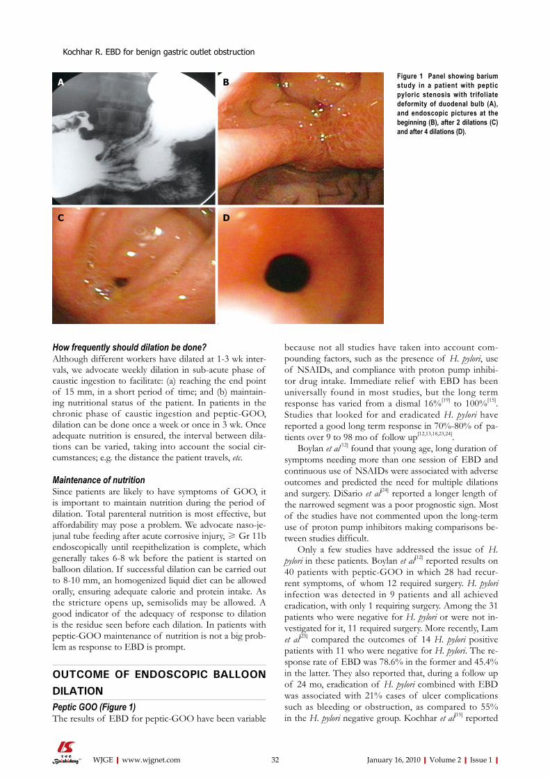
How frequently should dilation be done?Although different workers have dilated at 1-3 wk inter-vals, we advocate weekly dilation in sub-acute phase of caustic ingestion to facilitate: (a) reaching the end point of 15 mm, in a short period of time; and (b) maintain-ing nutritional status of the patient. In patients in the chronic phase of caustic ingestion and peptic-GOO, dilation can be done once a week or once in 3 wk. Once adequate nutrition is ensured, the interval between dila-tions can be varied, taking into account the social cir-cumstances; e.g. the distance the patient travels, etc.
Maintenance of nutritionSince patients are likely to have symptoms of GOO, it is important to maintain nutrition during the period of dilation. Total parenteral nutrition is most effective, but affordability may pose a problem. We advocate naso-je-junal tube feeding after acute corrosive injury, ≥ Gr 11b endoscopically until reepithelization is complete, which generally takes 6-8 wk before the patient is started on balloon dilation. If successful dilation can be carried out to 8-10 mm, an homogenized liquid diet can be allowed orally, ensuring adequate calorie and protein intake. As the stricture opens up, semisolids may be allowed. A good indicator of the adequacy of response to dilation is the residue seen before each dilation. In patients with peptic-GOO maintenance of nutrition is not a big prob-lem as response to EBD is prompt.
OUTCOME OF ENDOSCOPIC BALLOON DILATIONPeptic GOO (Figure 1)The results of EBD for peptic-GOO have been variable
because not all studies have taken into account com-pounding factors, such as the presence of H. pylori, use of NSAIDs, and compliance with proton pump inhibi-tor drug intake. Immediate relief with EBD has been universally found in most studies, but the long term response has varied from a dismal 16%[19] to 100%[15]. Studies that looked for and eradicated H. pylori have reported a good long term response in 70%-80% of pa-tients over 9 to 98 mo of follow up[12,13,18,23,24].
Boylan et al[12] found that young age, long duration of symptoms needing more than one session of EBD and continuous use of NSAIDs were associated with adverse outcomes and predicted the need for multiple dilations and surgery. DiSario et al[24] reported a longer length of the narrowed segment was a poor prognostic sign. Most of the studies have not commented upon the long-term use of proton pump inhibitors making comparisons be-tween studies difficult.
Only a few studies have addressed the issue of H. pylori in these patients. Boylan et al[12] reported results on 40 patients with peptic-GOO in which 28 had recur-rent symptoms, of whom 12 required surgery. H. pylori infection was detected in 9 patients and all achieved eradication, with only 1 requiring surgery. Among the 31 patients who were negative for H. pylori or were not in-vestigated for it, 11 required surgery. More recently, Lam et al[25] compared the outcomes of 14 H. pylori positive patients with 11 who were negative for H. pylori. The re-sponse rate of EBD was 78.6% in the former and 45.4% in the latter. They also reported that, during a follow up of 24 mo, eradication of H. pylori combined with EBD was associated with 21% cases of ulcer complications such as bleeding or obstruction, as compared to 55% in the H. pylori negative group. Kochhar et al[15] reported
32 January 16, 2010|Volume 2|Issue 1|WJGE|www.wjgnet.com
Kochhar R. EBD for benign gastric outlet obstruction
Figure 1 Panel showing barium study in a patient with peptic pyloric stenosis with trifoliate deformity of duodenal bulb (A), and endoscopic pictures at the beginning (B), after 2 dilations (C) and after 4 dilations (D).
A B
C D

a 100% response rate in 11 patients, all of whom had successful eradication of H. pylori after 1-3 sessions of EBD. A study by Cherian et al[17] also reported similar results. They subjected 21 patients to EBD for GOO (H. pylori = 12, NSAID-related = 3, both H. pylori and NSAID-related = 5 and others = 3). After eradicating H. pylori and removing the other offending cause, they achieved success in all their patients, with 17 of them requiring maintenance acid suppressive therapy.
The data quoted above shows that more than three-fourths of patients with peptic-GOO can be successfully treated with EBD provided H. pylori is eradicated, offend-ing NSAIDs are stopped and maintenance acid suppres-sant therapy is given. The available literature, therefore, suggests that long term proton pump inhibitor usage may be required to prevent recurrences. However, prospective studies are needed to answer this question definitely.
Caustic-induced GOO (Figure 2)Surgery has been the mainstay of treatment for caustic in-duced GOO, but recent reports suggest that EBD can be an effective alternative form of therapy in a select group of these patients[15,26]. However, compared to peptic-GOO cases, these patients have more recurrences and require a larger number of EBD sessions. Solt et al[13] studied 17 patients with caustic-GOO out of a total of 72 and only one-third had a long-term response with EBD.
Kochhar et al[15] reported that 8 of their 23 patients with GOO had a history of caustic ingestion, and they required 2-9 sessions of EBD, as compared to 1-3 ses-sions for peptic-GOO cases. In another recent study on 41 patients, they showed that caustic-induced GOO could be treated with EBD in 39 of these patients, with no recurrences over 18-58 mo of follow up[26]. The 39
patients required a mean of 5-8 sessions (range 2-13 ses-sions) to reach the end point of 15 mm. Only 2 patients failed to respond, one who had a perforation and anoth-er who had recurrent pain during each dilation. Only one patient had a supplementary procedure in the form of electrosurgical incision to augment the effect of EBD. However, they excluded patients with active ulceration and did not include patients with stricture length more than 2.5 cm.
Other causesChronic pancreatitis associated GOO is reported to be more resistant to EBD, and in one study all 4 such pa-tients required surgery. Tuberculosis was incriminated as a cause of GOO by Misra and Dwivedi[14] and they reported successful EBD in that patient. Anastomotic strictures following gastric resection have been treated with EBD by a few workers. Kozarek et al[18] had 7 such patients, out of a total of 23 patients with GOO. Only 4 of the 7 patients with anastomotic strictures had good long-term responses.
GOO associated with use of NSAID use has also been treated with EBD by a few workers. Cherian et al[17] have emphasized the fact that stopping NSAIDs is essen-tial to successful long-term outcomes in these patients.
Patients with Crohn’s disease causing GOO have also been subjected to EBD, with variable results. Murthy[27] reported one case of obstructive Crohn’s disease who underwent repeated sessions of EBD, but failed to re-spond. Solt et al[13] also treated one such patient who did not respond and required surgery. Kim et al[28], however, had a satisfactory response in one patient with postbul-bar Crohn’s disease, in whom they used fluoroscopically guided balloon dilation.
33 January 16, 2010|Volume 2|Issue 1|WJGE|www.wjgnet.com
Kochhar R. EBD for benign gastric outlet obstruction
Figure 2 Panel showing barium study of a patient with caustic induced pyloric stenosis (A), and endoscopic pictures at beginning of dilation (B), with balloon in-situ (C) and after 6 dilations (D).
A B
C D
Anastomotic strictures following vertical band gastroplasty or gastric bypass surgery for morbid obesity have also been successfully dilated with balloon catheters endoscopically. In one such study, EBD was successful in 68% of the 40 patients studied[29]. In a retrospective analysis, Solt et al[13] found good responses in all 18 patients with postoperative strictures and 6 patients with postvagotomy functional stenosis. Fluoroscopically guided balloon dilation has also been equally successful with response rates as high as 94%[30].
AUGMENTING THE EFFICACY OF ENDOSCOPIC BALLOON DILATIONA number of workers have used supplementary tech-niques to augment the effect of EBD. These include in-tralesional steroid injections and endoscopic incision of the strictures segment.
Combination of balloon dilation and intralesional steroid injectionsWe have reported use of intralesional steroids injection in patients with caustic GOO augmented the effect of balloon dilation[31]. Two of these patients had failed to show satisfactory responses to balloon dilation while the third was given steroids right at the time of first dilation. All three patients responded with (1, 2 and 2) sessions of steroid injections. The idea of using steroids is to facilitate dilation and reduce recurrence. Intralesional steroid injec-tions have been shown to inhibit stricture formation by interfering with collagen synthesis, fibrosis and chronic scarring[32]. It has been suggested that triamcinolone presents the cross linking of collagen that results in scar contracture; so if the scar is stretched and steroid injected into it, presumably the contracture will not occur[33]. Ste-roids also decrease the fibrotic healing that appears after dilation[32]. Gandhi et al[34] observed that with cortico-steroid injections and dilation, longer corrosive esopha-geal strictures become shorter with time and thus more amenable to nonsurgical treatment. We have shown that intralesional steroid injections reduce the need for dilation in caustic esophageal strictures, and even longer strictures (> 3 cm) can be managed successfully[35]. There is only one other report of use of intralesional steroids in pyloric stenosis apart from ours, in which Lee et al[36] successfully treated two cases (one peptic, another post-pyloroplasty) with steroids and balloon dilations.
Endoscopic incision and balloon dilationBaron et al[37] used electrosurgical incision using a stan-dard sphincterotomy to successfully treat a patient with pyloric stenosis resistant to EBD. Hagiwara et al[38] used needle-knife radial incisions electrosurgically at gastros-copy combined with EBD in patients with refractory anastomotic pyloric stenosis. We have also used this technique in one of the patients with caustic-induced GOO, who had become pregnant while on a dilation programme[26].
End point of balloon dilationThere is as yet no consensus on the issue of end point of balloon dilation. Kozarek et al[18] felt that the effec-tive balloon size required to dilate pyloric stenosis ap-pears to be 41 Fr, as it achieves immediate relief in 80%. Most workers[12,15-17,23,26] have used 15 mm balloons as the end point while some have dilated to 10 mm or 12 mm only[13,18]. Rarely, balloons of 16 mm, 18 mm and 20 mm are used[11,13,18]. Most workers inflate the balloon for 30 s to 60 s and repeat once again before withdraw-ing[17]. While some workers-have used a single session of dilation, others use repeat dilations at variable intervals. All these observations are on pyloric stenosis caused by peptic ulceration or anastomotic strictures. We have been more cautious than others; in our patients with peptic and caustic induced GOO, we dilate in step-wise manner, from 8 mm to 10 mm in the first few sessions, 10 mm to 12 mm in the next few sessions and 12 mm to 15 mm subsequently[15,26].
Safety of balloon dilationHydrostatic balloon dilation is generally a safe procedure. Kozarek et al[18] encountered one perforation out of 23 cases with peptic ulcer related stenosis. The balloon di-ameter used was not mentioned. Lau et al[11] reported 4 perforations in 54 patients. Two of their 16 patients un-dergoing dilation with a 16 mm balloon had perforation while 2 of the 3 undergoing 20 mm dilation perforated, with none of the 10 undergoing 18 mm dilation having any complication. It thus seems that increasing the bal-loon diameter beyond 15 mm is more likely to be associ-ated with perforation. Arterial bleeding has been rarely reported, though self limiting minor bleeding is not uncommon[28]. Pain during EBD is not uncommon, but is often self limiting. In a recent study, 19.5% of patients with caustic-GOO had self-limiting pain during the pro-cedure[26].
REFERENCES1 Khullar SK , DiSario JA. Gastric outlet obstruction.
Gastrointest Endosc Clin N Am 1996; 6: 585-6032 Johnson CD, Ellis H. Gastric outlet obstruction now
predicts malignancy. Br J Surg 1990; 77: 1023-10243 Paimela H, Tuompo PK, Peräkyl T, Saario I, Höckerstedt K,
Kivilaakso E. Peptic ulcer surgery during the H2-receptor antagonist era: a population-based epidemiological study of ulcer surgery in Helsinki from 1972 to 1987. Br J Surg 1991; 78: 28-31
4 Ferzoco SJ, Soybel DI. Gastric outlet obstruction, perforation and other complications of gastroduodenal ulcer. In: Wolfe HM, editor. Therapy of digestive disorders. 2nd ed. New Delhi: Elsevier Inc, 2007: 357-375
5 Kreel L, Ellis H. Pyloric stenosis in adults: A clinical and radiological study of 100 consecutive patients. Gut 1965; 6: 253-261
6 Weiland D, Dunn DH, Humphrey EW, Schwartz ML. Gastric outlet obstruction in peptic ulcer disease: an indication for surgery. Am J Surg 1982; 143: 90-93
7 Zargar SA , Kochhar R, Nagi B, Mehta S, Mehta SK. Ingestion of corrosive acids. Spectrum of injury to upper gastrointestinal tract and natural history. Gastroenterology
34 January 16, 2010|Volume 2|Issue 1|WJGE|www.wjgnet.com
Kochhar R. EBD for benign gastric outlet obstruction

1989; 97: 702-7078 Zargar SA , Kochhar R, Nagi B, Mehta S, Mehta SK.
Ingestion of strong corrosive alkalis: spectrum of injury to upper gastrointestinal tract and natural history. Am J Gastroenterol 1992; 87: 337-341
9 Jaffin BW, Kaye MD. The prognosis of gastric outlet obstruction. Ann Surg 1985; 201: 176-179
10 Chaudhary A, Puri AS, Dhar P, Reddy P, Sachdev A, Lahoti D, Kumar N, Broor SL. Elective surgery for corrosive-induced gastric injury. World J Surg 1996; 20: 703-706; discussion 706
11 Lau JY, Chung SC, Sung JJ, Chan AC, Ng EK, Suen RC, Li AK. Through-the-scope balloon dilation for pyloric stenosis: long-term results. Gastrointest Endosc 1996; 43: 98-101
12 Boylan JJ, Gradzka MI. Long-term results of endoscopic balloon dilatation for gastric outlet obstruction. Dig Dis Sci 1999; 44: 1883-1886
13 Solt J, Bajor J, Szabó M, Horváth OP. Long-term results of balloon catheter dilation for benign gastric outlet stenosis. Endoscopy 2003; 35: 490-495
14 Misra SP, Dwivedi M. Long-term follow-up of patients undergoing ballon dilation for benign pyloric stenoses. Endoscopy 1996; 28: 552-554
15 Kochhar R, Sethy PK, Nagi B, Wig JD. Endoscopic balloon dilatation of benign gastric outlet obstruction. J Gastroenterol Hepatol 2004; 19: 418-422
16 Perng CL , Lin HJ, Lo WC, Lai CR, Guo WS, Lee SD. Characteristics of patients with benign gastric outlet obstruction requiring surgery after endoscopic balloon dilation. Am J Gastroenterol 1996; 91: 987-990
17 Cherian PT, Cherian S, Singh P. Long-term follow-up of patients with gastric outlet obstruction related to peptic ulcer disease treated with endoscopic balloon dilatation and drug therapy. Gastrointest Endosc 2007; 66: 491-497
18 Kozarek RA , Botoman VA, Patterson DJ. Long-term follow-up in patients who have undergone balloon dilation for gastric outlet obstruction. Gastrointest Endosc 1990; 36: 558-561
19 Kuwada SK , Alexander GL. Long-term outcome of endoscopic dilation of nonmalignant pyloric stenosis. Gastrointest Endosc 1995; 41: 15-17
20 Yusuf TE, Brugge WR. Endoscopic therapy of benign pyloric stenosis and gastric outlet obstruction. Curr Opin Gastroenterol 2006; 22: 570-573
21 Benjamin SB, Cattau EL, Glass RL. Balloon dilation of the pylorus: therapy for gastric outlet obstruction. Gastrointest Endosc 1982; 28: 253-254
22 Benjamin SB , Glass RL, Cattau EL Jr , Mil ler WB. Preliminary experience with balloon dilation of the pylorus. Gastrointest Endosc 1984; 30: 93-95
23 Griffin SM, Chung SC, Leung JW, Li AK. Peptic pyloric stenosis treated by endoscopic balloon dilatation. Br J Surg 1989; 76: 1147-1148
24 DiSario JA, Fennerty MB, Tietze CC, Hutson WR, Burt RW. Endoscopic balloon dilation for ulcer-induced gastric outlet obstruction. Am J Gastroenterol 1994; 89: 868-871
25 Lam YH, Lau JY, Fung TM, Ng EK, Wong SK, Sung JJ, Chung SS. Endoscopic balloon dilation for benign gastric outlet obstruction with or without Helicobacter pylori infection. Gastrointest Endosc 2004; 60: 229-233
26 Kochhar R, Dutta U, Sethy PK, Singh G, Sinha SK, Nagi B, Wig JD, Singh K. Endoscopic balloon dilation in caustic-induced chronic gastric outlet obstruction. Gastrointest Endosc 2009; 69: 800-805
27 Murthy UK. Repeated hydrostatic balloon dilation in obstructive gastroduodenal Crohn’s disease. Gastrointest Endosc 1991; 37: 484-485
28 Kim JH, Shin JH, Di ZH, Ko GY, Yoon HK, Sung KB, Song HY. Benign duodenal strictures: treatment by means of fluoroscopically guided balloon dilation. J Vasc Interv Radiol 2005; 16: 543-548
29 Sataloff DM, Lieber CP, Seinige UL. Strictures following gastric stapling for morbid obesity. Results of endoscopic dilatation. Am Surg 1990; 56: 167-174
30 Kim JH, Shin JH, Bae JI, Di ZH, Lim JO, Kim TH, Ko GY, Yoon HK, Sung KB, Song HY. Gastric outlet obstruction caused by benign anastomotic stricture: treatment by fluoroscopically guided balloon dilation. J Vasc Interv Radiol 2005; 16: 699-704
31 Kochhar R, Sriram PV, Ray JD, Kumar S, Nagi B, Singh K. Intralesional steroid injections for corrosive induced pyloric stenosis. Endoscopy 1998; 30: 734-736
32 Ashcraft KW, Holder TM. The experimental treatment of esophageal strictures by intralesional steroid injections. J Thorac Cardiovasc Surg 1969; 58: 685-693
33 Ketchum LD, Smith J, Robinson DW, Masters FW. The treatment of hypertrophic scar, keloid and scar contracture by triamcinolone acetonide. Plast Reconstr Surg 1966; 38: 209-218
34 Gandhi RP, Cooper A, Barlow BA. Successful management of esophageal strictures without resection or replacement. J Pediatr Surg 1989; 24: 745-749; discussion 749-750
35 Kochhar R , Ray JD, Sriram PV, Kumar S, Singh K. Intralesional steroids augment the effects of endoscopic dilation in corrosive esophageal strictures. Gastrointest Endosc 1999; 49: 509-513
36 Lee M, Kubik CM, Polhamus CD, Brady CE 3rd, Kadakia SC. Preliminary experience with endoscopic intralesional steroid injection therapy for refractory upper gastrointestinal strictures. Gastrointest Endosc 1995; 41: 598-601
37 Baron B, Gross KR. Successful dilation of pyloric stricture resistant to balloon dilation with electrocautery using a sphincterotome. J Clin Gastroenterol 1996; 23: 239-241
38 Hagiwara A, Sonoyama Y, Togawa T, Yamasaki J, Sakakura C, Yamagishi H. Combined use of electrosurgical incisions and balloon dilatation for the treatment of refractory postoperative pyloric stenosis. Gastrointest Endosc 2001; 53: 504-508
S- Editor Zhang HN L- Editor Lutze M E- Editor Ma WH
35 January 16, 2010|Volume 2|Issue 1|WJGE|www.wjgnet.com
Kochhar R. EBD for benign gastric outlet obstruction
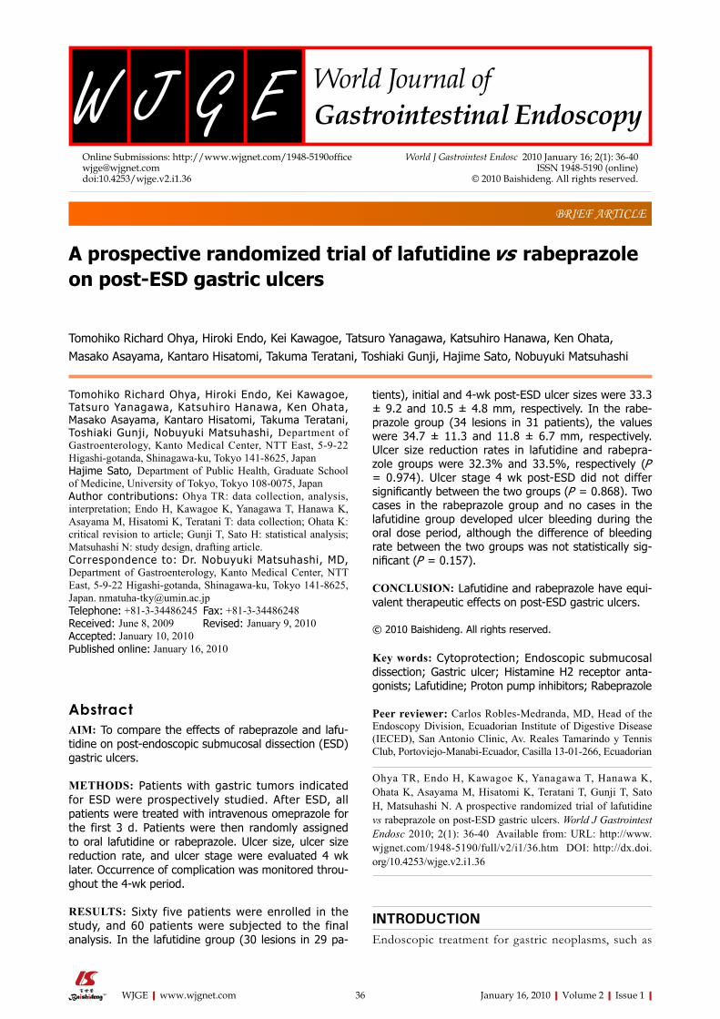
BRIEF ARTICLE
A prospective randomized trial of lafutidine vs rabeprazole on post-ESD gastric ulcers
Tomohiko Richard Ohya, Hiroki Endo, Kei Kawagoe, Tatsuro Yanagawa, Katsuhiro Hanawa, Ken Ohata, Masako Asayama, Kantaro Hisatomi, Takuma Teratani, Toshiaki Gunji, Hajime Sato, Nobuyuki Matsuhashi
Tomohiko Richard Ohya, Hiroki Endo, Kei Kawagoe, Tatsuro Yanagawa, Katsuhiro Hanawa, Ken Ohata, Masako Asayama, Kantaro Hisatomi, Takuma Teratani, Toshiaki Gunji, Nobuyuki Matsuhashi, Department of Gastroenterology, Kanto Medical Center, NTT East, 5-9-22 Higashi-gotanda, Shinagawa-ku, Tokyo 141-8625, JapanHajime Sato, Department of Public Health, Graduate School of Medicine, University of Tokyo, Tokyo 108-0075, JapanAuthor contributions: Ohya TR: data collection, analysis, interpretation; Endo H, Kawagoe K, Yanagawa T, Hanawa K, Asayama M, Hisatomi K, Teratani T: data collection; Ohata K: critical revision to article; Gunji T, Sato H: statistical analysis; Matsuhashi N: study design, drafting article.Correspondence to: Dr. Nobuyuki Matsuhashi, MD, Department of Gastroenterology, Kanto Medical Center, NTT East, 5-9-22 Higashi-gotanda, Shinagawa-ku, Tokyo 141-8625, Japan. [email protected]: +81-3-34486245 Fax: +81-3-34486248Received: June 8, 2009 Revised: January 9, 2010Accepted: January 10, 2010Published online: January 16, 2010
AbstractAIM: To compare the effects of rabeprazole and lafutidine on postendoscopic submucosal dissection (ESD) gastric ulcers.
METHODS: Patients with gastric tumors indicated for ESD were prospectively studied. After ESD, all patients were treated with intravenous omeprazole for the first 3 d. Patients were then randomly assigned to oral lafutidine or rabeprazole. Ulcer size, ulcer size reduction rate, and ulcer stage were evaluated 4 wk later. Occurrence of complication was monitored throughout the 4wk period.
RESULTS: Sixty five patients were enrolled in the study, and 60 patients were subjected to the final analysis. In the lafutidine group (30 lesions in 29 pa
tients), initial and 4wk postESD ulcer sizes were 33.3 ± 9.2 and 10.5 ± 4.8 mm, respectively. In the rabeprazole group (34 lesions in 31 patients), the values were 34.7 ± 11.3 and 11.8 ± 6.7 mm, respectively. Ulcer size reduction rates in lafutidine and rabeprazole groups were 32.3% and 33.5%, respectively (P = 0.974). Ulcer stage 4 wk postESD did not differ significantly between the two groups (P = 0.868). Two cases in the rabeprazole group and no cases in the lafutidine group developed ulcer bleeding during the oral dose period, although the difference of bleeding rate between the two groups was not statistically significant (P = 0.157).
CONCLUSION: Lafutidine and rabeprazole have equivalent therapeutic effects on postESD gastric ulcers.
© 2010 Baishideng. All rights reserved.
Key words: Cytoprotection; Endoscopic submucosal dissection; Gastric ulcer; Histamine H2 receptor antagonists; Lafutidine; Proton pump inhibitors; Rabeprazole
Peer reviewer: Carlos Robles-Medranda, MD, Head of the Endoscopy Division, Ecuadorian Institute of Digestive Disease (IECED), San Antonio Clinic, Av. Reales Tamarindo y Tennis Club, Portoviejo-Manabi-Ecuador, Casilla 13-01-266, Ecuadorian
Ohya TR, Endo H, Kawagoe K, Yanagawa T, Hanawa K, Ohata K, Asayama M, Hisatomi K, Teratani T, Gunji T, Sato H, Matsuhashi N. A prospective randomized trial of lafutidine vs rabeprazole on post-ESD gastric ulcers. World J Gastrointest Endosc 2010; 2(1): 36-40 Available from: URL: http://www.wjgnet.com/1948-5190/full/v2/i1/36.htm DOI: http://dx.doi.org/10.4253/wjge.v2.i1.36
INTRODUCTIONEndoscopic treatment for gastric neoplasms, such as
World J Gastrointest Endosc 2010 January 16; 2(1): 36-40ISSN 1948-5190 (online)
© 2010 Baishideng. All rights reserved.
Online Submissions: http://www.wjgnet.com/[email protected]:10.4253/wjge.v2.i1.36
36 January 16, 2010|Volume 2|Issue 1|WJGE|www.wjgnet.com
Ohya TR et al . Lafutidine vs rabeprazole in postESD ulcerss
early gastric cancer or adenoma, is now a routine pro-cedure[1]. Endoscopic submucosal dissection (ESD) is a minimally invasive treatment that provides a high en bloc resection rate and allows for precise histologic stag-ing[2,3]. Iatrogenic gastric ulcers created by ESD, however, are often rather large, and bleeding occasionally occurs in the post-ESD period. There is currently no consensus regarding the management of these iatrogenic ulcers.
Lafutidine is a new-generation histamine H2 receptor antagonist (H2RA) that not only suppresses gastric acid secretion, but also has cytoprotective properties in gastric epithelial cells[4]. Proton pump inhibitors (PPIs) are more potent inhibitors of gastric acid secretion than H2RAs[5], and in human gastro-duodenal ulcers, PPIs are reported to have greater anti-ulcer effects than H2RAs[6,7]. In certain animal models of artificial gastric ulcers, cytoprotective agents exert greater anti-ulcer effects than H2RAs[8]. Because gastric ulcers following ESD are artificially induced, we hypothesized that lafutidine, an H2RA with cytoprotective properties[4], may exert anti-ulcer effects equivalent, or even superior, to PPIs. Therefore, we compared the anti-ulcer effects of lafutidine and the PPI rabeprazole on post-ESD gastric ulcers.
MATERIALS AND METHODSEthicsThis study was conducted in the Department of Gastroenterology and approved by the ethics committee of Kanto Medical Center. Written informed consent was obtained from all subjects prior to the study.
Study subjects and treatmentsBetween July 2005 and November 2006, 65 consecutive patients with early gastric cancer or a gastric adenoma that was considered curable with endoscopic treatment were enrolled in the study. The treated lesions were either adenomas with severe atypia, well-differentiated intramu-cosal cancers without ulcer findings, or well-differentiated intramucosal cancers less than 3 cm in size with ulcer findings. The diagnosis was based on endoscopic findings and histological examination of biopsied specimen. Endo-scopic ultrasonography was not performed. Exclusion criteria included failure to obtain written informed consent, severe cardiopulmonary complications, and prior history of allergic reactions against H2RA or PPI. None of the patients had a previous history of upper gastrointestinal tract surgery. All patients agreed to take part in the study, and were treated by ESD. After ESD, all patients were treated with intravenous omeprazole for the first 3 d. Fol-lowing omeprazole treatment, patients were randomly assigned to oral lafutidine (group L; 20 mg/d) or oral rabeprazole (group R; 10 mg/d). Doses of these agents were determined according to their standard doses. Ten mg/d of rabeprazole has been shown to have stronger suppressive effect on acid secretion compared to the full dose of omeprazole or lansoprazole[9], and so this dose was thought to be sufficient.
A hook knife (Olympus, Tokyo, Japan) or insulated-
tip knife (Olympus, Tokyo, Japan) was used for the ESD. We marked the normal mucosa surrounding the lesion, at least 5 mm away from the tumor, using the hook knife. After injection of a 1:1 mixture of hyaluronic acid solution (Kaken Pharmaceuticals, Tokyo, Japan) and 10% glycerin (Chugai Pharmaceuticals, Tokyo, Japan) into the submucosal layer, circumferential cutting was performed, followed by submucosal dissection. An ICC 200 electrosurgical generator ICC (ERBE, Tubingen, Germany: Endocut mode 85W, forced coagulation mode 45W) was used to produce the surgical current.
Endoscopic evaluation was performed 28 d after ESD. The size of the ulcer was defined as the longitu-dinal diameter of the gastric ulcer, and measured using measuring forceps (Olympus, Tokyo, Japan). Ulcer heal-ing was assessed by measuring the changes in ulcer size and stage. The ulcer reduction rate was calculated as: (ulcer size 4 wk post ESD)/(ulcer size immediately after ESD procedure) × 100 (%)[10]. Gastric ulcer stage was classified using a 6-stage system Sakita-Miwa classifica-tion[11]: active (A1, A2), healing (H1, H2), and scarring (S1, S2) (Table 1). All cases were monitored for the occur-rence of complications, including bleeding or perfora-tion, throughout the 4-wk observation period.
In most cases, one lesion was resected in the ESD procedure, but in 4 cases (3 in group R and 1 in group L) two separate lesions were resected in a single ESD pro-cedure. Patient characteristics were analyzed on a patient number basis, whereas tumor or ulcer characteristics were analyzed on a tumor/ulcer number basis.
Sample size determinationWe determined the sample size for the two groups so that the difference between the two study groups could be detected if the mean ulcer size of the group with the larger mean, if any, is larger than that for its counterpart at least by one-fourth (25.0%) of the mean for the for-mer group. It was assumed that the mean ulcer size at day 28 for the group with the larger mean was 11.0, and that both groups had the same sd (5.0). Alpha was set to 0.10, while power was set to 0.70.
Statistical analysisContinuous values and categorical parameters were
37 January 16, 2010|Volume 2|Issue 1|WJGE|www.wjgnet.com
Table 1 Gastric ulcer stages using a six-stage system[11]
Stage Finding
A1 (active stage 1)
Ulcer that contains mucus coating, with marginal elevation because of edema
A2 (active stage 2)
Mucus-coated ulcer with discrete margin and less edema than active stage 1
H1 (healing stage 1)
Unhealed ulcer covered by regenerating epithelium < 50%, with or without converging folds
H2 (healing stage 2)
Ulcer with a mucosal break but almost covered with regenerating epithelium
S1 (scar stage 1)
Red scar with rough epithelialization without mucosal break
S2 (scar stage 2)
White scar with complete re-epithelialization

analyzed by Student’s t-test and Pearson’s chi-square test, respectively. Ulcer sizes just before and after the oral dose period were compared between the two groups. The statistical difference of ulcer size reduction was determined by using the two-way repeated-measures ANOVA. A P-value of less than 0.05 was considered statistically significant. All analyses were performed with the use of SPSS software (15.0 J for Windows).
RESULTSEffects of lafutidine and rabeprazole on post-ESD gastric ulcersA total of 65 patients were enrolled in the study (Figure 1); 5 patients (3 in group L and 2 in group R) were, however,excluded from the final analysis. Of the 5 patients, 1 in group L and 2 in group R had to undergo additional surgical treatment after pathological examination because of submucosal invasion of the cancer; 1 in group L was newly diagnosed with pancreatic cancer after the treatment, and so follow-up esophagogastroduodenoscopy was not performed; and 1 in group L had to be treated for exacerbation of pre-existing Guillain-Barre syndrome
after ESD and so follow-up esophagogastroduodenoscopy was not performed. Patient and lesion characteristics are shown in Table 2. There were 30 lesions in 29 patients in group L and 34 lesions in 31 patients in group R who completed the study, and there were no significant differences between the two groups with regard to patient characteristics (gender, age, body mass index, presence of serum anti-Helicobacter pylori antibody), initial endoscopic findings (ulcerated lesion or not, and location in the stomach), lesion depth on pathological examination, or post-ESD ulcer size. In group L, initial and 4-wk post-ESD ulcer sizes were 33.3 ± 9.2 and 10.5 ± 4.8 mm, respectively. In group R, the values were 34.7 ± 11.3 and 11.8 ± 6.7 mm, respectively. The ulcer size reduction rate in group L and group R was 32.3% and 33.5%, respectively (Table 3, P = 0.974). Ulcer stages at 4-wk post ESD in group L were: A1; 0 case (0%), A2; 1 (3%), H1; 10 (33%), H2; 16 (54%), S1; 2 (7%) and S2; 1 (3%); ulcer stages in group R were: A1; 0%, A2: 1 (3%), H1; 11 (32%), H2; 19 (56%), and S1; 3 (9%) (Table 4, P = 0.868). Thus, there were no significant differences in ulcer size, ulcer size reduction rate, or proportion of patients at each ulcer stage between the two groups 4 wk post ESD.
Preventive effects of lafutidine and rabeprazole on bleeding from post-ESD gastric ulcersNo cases in group L (0%) and three cases in group R (9%) developed bleeding. One patient in group R de-veloped bleeding 1 d after ESD. Two cases in group R developed bleeding within the oral-treatment period (day 4 through day 28); 1 case 7 d after ESD, and the other 10 d after ESD (P = 0.157). The last case required
38 January 16, 2010|Volume 2|Issue 1|WJGE|www.wjgnet.com
Ohya TR et al . Lafutidine vs rabeprazole in postESD ulcerss
Table 2 Characteristics of patients and lesions
Lafutidine Rabeprazole P
Gender (M:F) 21:8 23:8 0.607Age (mean ± SD) 65.3 ± 8.0 65.4 ± 9.0 0.769Body mass index (kg/m2) (mean ± SD)
23.2 ± 3.1 23.0 ± 2.7 0.751
Anti-H. pylori antibody (positive:negative)
23:6 24:7 0.729
Ulcerated lesion (yes:no) 5:25 1:33 0.181Location (upper:middle:lower) 1:16:13 4:20:10 0.251Lesion depth (mucosal layer: submucosal layer)
27:3 33:1 0.677
Ulcer size immediately after ESD (mm) (mean ± SD)
33.3 ± 9.2 34.7 ± 11.3 0.635
Patient characteristics were analyzed on patient number basis, whereas lesion or ulcer characteristics were analyzed on tumor/ulcer number basis.
Table 3 Ulcer size reduction rate
Lafutidine Rabeprazole P
Ulcer size (Day 0) 33.3 ± 9.2 34.7 ± 11.3 0.635Ulcer size (Day 28) 10.5 ± 4.8 11.8 ± 6.7 0.421Reduction Rate 32.30% 33.50% 0.974
ESD65 cases
Bleeding during oral dose period 2
Bleeding before oral dose period 1
No bleeding 2929Group L 32
Drop out 3
Group R 33
Drop out 2
32 No bleeding 28
Figure 1 Patient flow. Three patients in group L and 2 in group R were excluded from the final analysis; 1 in group L and 2 in group R had to undergo additional surgical treatment after pathological examination due to submucosal invasion of cancer; 1 in group L was newly diagnosed with pancreatic cancer; and 1 in group L had to be treated for exacerbation of pre-existing Guillain-Barre syndrome after ESD.
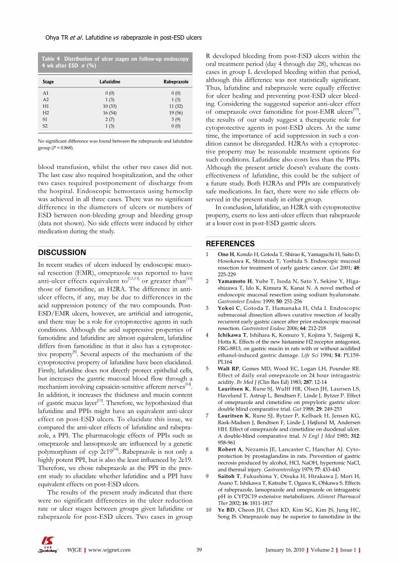
blood transfusion, whilst the other two cases did not. The last case also required hospitalization, and the other two cases required postponement of discharge from the hospital. Endoscopic hemostasis using hemoclip was achieved in all three cases. There was no significant difference in the diameters of ulcers or numbers of ESD between non-bleeding group and bleeding group (data not shown). No side effects were induced by either medication during the study.
DISCUSSIONIn recent studies of ulcers induced by endoscopic muco-sal resection (EMR), omeprazole was reported to have anti-ulcer effects equivalent to[12,13] or greater than[10] those of famotidine, an H2RA. The difference in anti-ulcer effects, if any, may be due to differences in the acid suppression potency of the two compounds. Post-ESD/EMR ulcers, however, are artificial and iatrogenic, and there may be a role for cytoprotective agents in such conditions. Although the acid suppressive properties of famotidine and lafutidine are almost equivalent, lafutidine differs from famotidine in that it also has a cytoprotec-tive property[4]. Several aspects of the mechanism of the cytoprotective property of lafutidine have been elucidated. Firstly, lafutidine does not directly protect epithelial cells, but increases the gastric mucosal blood flow through a mechanism involving capsaicin-sensitive afferent nerves[14]. In addition, it increases the thickness and mucin content of gastric mucus layer[15]. Therefore, we hypothesized that lafutidine and PPIs might have an equivalent anti-ulcer effect on post-ESD ulcers. To elucidate this issue, we compared the anti-ulcer effects of lafutidine and rabepra-zole, a PPI. The pharmacologic effects of PPIs such as omeprazole and lansoprazole are influenced by a genetic polymorphism of cyp 2c19[16]. Rabeprazole is not only a highly potent PPI, but is also the least influenced by 2c19. Therefore, we chose rabeprazole as the PPI in the pres-ent study to elucidate whether lafutidine and a PPI have equivalent effects on post-ESD ulcers.
The results of the present study indicated that there were no significant differences in the ulcer reduction rate or ulcer stages between groups given lafutidine or rabeprazole for post-ESD ulcers. Two cases in group
R developed bleeding from post-ESD ulcers within the oral treatment period (day 4 through day 28), whereas no cases in group L developed bleeding within that period, although this difference was not statistically significant. Thus, lafutidine and rabeprazole were equally effective for ulcer healing and preventing post-ESD ulcer bleed-ing. Considering the suggested superior anti-ulcer effect of omeprazole over famotidine for post-EMR ulcers[10], the results of our study suggest a therapeutic role for cytoprotective agents in post-ESD ulcers. At the same time, the importance of acid suppression in such a con-dition cannot be disregarded. H2RAs with a cytoprotec-tive property may be reasonable treatment options for such conditions. Lafutidine also costs less than the PPIs. Although the present article doesn’t evaluate the costs-effectiveness of lafutidine, this could be the subject of a future study. Both H2RAs and PPIs are comparatively safe medications. In fact, there were no side effects ob-served in the present study in either group.
In conclusion, lafutidine, an H2RA with cytoprotective property, exerts no less anti-ulcer effects than rabeprazole at a lower cost in post-ESD gastric ulcers.
REFERENCES1 Ono H, Kondo H, Gotoda T, Shirao K, Yamaguchi H, Saito D,
Hosokawa K, Shimoda T, Yoshida S. Endoscopic mucosal resection for treatment of early gastric cancer. Gut 2001; 48: 225-229
2 Yamamoto H, Yube T, Isoda N, Sato Y, Sekine Y, Higa-shizawa T, Ido K, Kimura K, Kanai N. A novel method of endoscopic mucosal resection using sodium hyaluronate. Gastrointest Endosc 1999; 50: 251-256
3 Yokoi C, Gotoda T, Hamanaka H, Oda I. Endoscopic submucosal dissection allows curative resection of locally recurrent early gastric cancer after prior endoscopic mucosal resection. Gastrointest Endosc 2006; 64: 212-218
4 Ichikawa T, Ishihara K, Komuro Y, Kojima Y, Saigenji K, Hotta K. Effects of the new histamine H2 receptor antagonist, FRG-8813, on gastric mucin in rats with or without acidified ethanol-induced gastric damage. Life Sci 1994; 54: PL159-PL164
5 Walt RP, Gomes MD, Wood EC, Logan LH, Pounder RE. Effect of daily oral omeprazole on 24 hour intragastric acidity. Br Med J (Clin Res Ed) 1983; 287: 12-14
6 Lauritsen K, Rune SJ, Wulff HR, Olsen JH, Laursen LS, Havelund T, Astrup L, Bendtsen F, Linde J, Bytzer P. Effect of omeprazole and cimetidine on prepyloric gastric ulcer: double blind comparative trial. Gut 1988; 29: 249-253
7 Lauritsen K, Rune SJ, Bytzer P, Kelbaek H, Jensen KG, Rask-Madsen J, Bendtsen F, Linde J, Højlund M, Andersen HH. Effect of omeprazole and cimetidine on duodenal ulcer. A double-blind comparative trial. N Engl J Med 1985; 312: 958-961
8 Robert A, Nezamis JE, Lancaster C, Hanchar AJ. Cyto-protection by prostaglandins in rats. Prevention of gastric necrosis produced by alcohol, HCl, NaOH, hypertonic NaCl, and thermal injury. Gastroenterology 1979; 77: 433-443
9 Saitoh T, Fukushima Y, Otsuka H, Hirakawa J, Mori H, Asano T, Ishikawa T, Katsube T, Ogawa K, Ohkawa S. Effects of rabeprazole, lansoprazole and omeprazole on intragastric pH in CYP2C19 extensive metabolizers. Aliment Pharmacol Ther 2002; 16: 1811-1817
10 Ye BD, Cheon JH, Choi KD, Kim SG, Kim JS, Jung HC, Song IS. Omeprazole may be superior to famotidine in the
39 January 16, 2010|Volume 2|Issue 1|WJGE|www.wjgnet.com
Ohya TR et al . Lafutidine vs rabeprazole in postESD ulcers
Table 4 Distribution of ulcer stages on follow-up endoscopy 4 wk after ESD n (%)
Stage Lafutidine Rabeprazole
A1 0 (0) 0 (0)A2 1 (3) 1 (3)H1 10 (33) 11 (32)H2 16 (54) 19 (56)S1 2 (7) 3 (9)S2 1 (3) 0 (0)
No significant difference was found between the rabeprazole and lafutidine group (P = 0.868).

management of iatrogenic ulcer after endoscopic mucosal resection: a prospective randomized controlled trial. Aliment Pharmacol Ther 2006; 24: 837-843
11 Sakita T, Fukutomi H. Endoscopic diagnosis. In: Yoshitoshi Y, editor. Ulcer of stomach and duodenum [in Japanese]. Tokyo: Nankodo, 1971: 198-208
12 Yamaguchi Y, Katsumi N, Tauchi M, Toki M, Naka¬mura K, Aoki K, Morita Y, Miura M, Morozumi K, Ishida H, Takahashi S. A prospective randomized trial of either famotidine or omeprazole for the pre¬vention of bleeding after endoscopic mucosal resection and the healing of endoscopic mucosal resection-induced ulceration. Aliment Pharmacol Ther 2005; 21 Suppl 2: 111-115
13 Esaki M, Aoyagi K, Matsumoto T, Kuwano Y, Shimizu M, Fujishima M. Effects of omeprazole and famotidine on
fibroblast growth factor-2 during artificial gastric ulcer healing in humans. Eur J Gastroenterol Hepatol 2002; 14: 365-369
14 Onodera S, Shibata M, Tanaka M, Inaba N, Arai Y, Aoyama M, Lee B, Yamaura T. Gastroprotective mechanism of lafutidine, a novel anti-ulcer drug with histamine H2-receptor antagonistic activity. Arzneimittelforschung 1999; 49: 519-526
15 Ichikawa T, Ota H, Sugiyama A, Maruta F, Ikezawa T, Hotta K, Ishihara K. Effects of a novel histamine H2-receptor antagonist, lafutidine, on the mucus barrier of human gastric mucosa. J Gastroenterol Hepatol 2007; 22: 1800-1805
16 Stedman CA, Barclay ML. Review article: comparison of the pharmacokinetics, acid suppression and efficacy of proton pump inhibitors. Aliment Pharmacol Ther 2000; 14: 963-978
S- Editor Zhang HN L- Editor Hughes D E- Editor Ma WH
40 January 16, 2010|Volume 2|Issue 1|WJGE|www.wjgnet.com
Ohya TR et al . Lafutidine vs rabeprazole in postESD ulcers

CASE REPORT
An unusual presentation of fistulating Crohn’s disease: Ascites
Richard Kia, David White, Sanchoy Sarkar
Richard Kia, David White, Sanchoy Sarkar, Digestive Diseases Centre, University Hospital Aintree, Longmoor Lane, Liverpool L9 7AL, United KingdomRichard Kia, Specialty Registrar in Gastroenterology and General (Internal) Medicine, Wirral University Teaching Hospital NHS Foundation Trust, Arrowe Park Road CH49 5PE Wirral, United KingdomAuthor contributions: Kia R and Sarkar S contributed equally to this work; Kia R, White D and Sarkar S drafted and revised the manuscript; White D contributed to the interpretation of the radiology images.Correspondence to: Dr. Richard Kia, Specialty Registrar in Gastroenterology and General (Internal) Medicine, Wirral University Teaching Hospital NHS Foundation Trust, Arrowe Park Road CH49 5PE Wirral, United Kingdom. [email protected]: +44-151-6785111 Fax: +44-151-6785111Received: March 12, 2009 Revised: March 31, 2009 Accepted: April 7, 2009 Published online: January 16, 2010
AbstractWhilst ascites is a common presenting complaint in patients with decompensated chronic liver disease and disseminated malignancy, in Crohn’s disease however, it is exceptionally rare. We describe a patient with no prior history of inflammatory bowel or liver disease, presenting with rapid onset gross ascites and scro-tal swelling. Further investigations revealed severe hypoalbuminemia and transudative ascitic fluid with normal other liver function tests and a negative liver screen. Computed tomography revealed widespread ascites and pleural effusions with no features of ma-lignancy or portal hypertension, and a small bowel barium series showed features of fistulating small bowel Crohn’s disease. An ileo-colonoscopy confirmed the presence of terminal ileal inflammatory stricture. The patient’s clinical condition and serum albumin im-proved with a combination of diuretics, elemental diet, antibiotics and oral 5-aminosalicylic acid therapy.
© 2010 Baishideng. All rights reserved.
Key words: Ascites; Fistulating; Crohn’s disease; Pro-tein-Losing enteropathies; Hypoalbuminemia
Peer reviewer: James F Trotter, MD, Associate Professor, University of Colorado, Division of Gastroenterology, 4200 E. 9th Avenue, b-154, Denver, CO 80262, United States
Kia R, White D, Sarkar S. An unusual presentation of fistulating Crohn’s disease: Ascites. World J Gastrointest Endosc 2010; 2(1): 41-43 Available from: URL: http://www.wjgnet.com/1948-5190/full/v2/i1/41.htm DOI: http://dx.doi.org/10.4253/wjge.v2.i1.41
INTRODUCTIONCrohn’s disease is a chronic, relapsing and remitting in-flammatory disease of the gastrointestinal tract, with a prevalence of 140 cases per 100 000 in the West[1]. Com-mon presenting symptoms include diarrhea, abdominal
pain, weight loss, and fatigue[2]. Ascites as the main pre-senting complaint in undiagnosed Crohn’s disease is ex-tremely rare and to our knowledge has only been reported once within the literature[3]. However, ascites secondary to conditions associated with Crohn’s disease, such as malignancy and portal hypertension is well recognised[4-6]. We describe a case where a patient with previously undi-agnosed fistulating Crohn’s disease presented as an emer-gency with gross ascites, scrotal swelling and severe hy-poalbuminemia. We also highlight the diagnostic features of Crohn’s disease in barium studies and ileo-colonoscopy, which helped in the diagnosis of this atypical case.
CASE REPORTA 61-year-old man presented in the emergency depart-ment with rapid onset abdominal and scrotal swelling. The only other symptom of note was mild intermittent diarrhea, which had been present since a sigmoid colec-
World J Gastrointest Endosc 2010 January 16; 2(1): 41-43ISSN 1948-5190 (online)
© 2010 Baishideng. All rights reserved.
Online Submissions: http://www.wjgnet.com/[email protected]:10.4253/wjge.v2.i1.41
41 January 16, 2010|Volume 2|Issue 1|WJGE|www.wjgnet.com

Kia R et al . Fistulating crohn’s disease
tomy was performed 5 years previously for a diverticular abscess. He was a non-smoker and past medical history included angina and atrial fibrillation. There was no his-tory of chronic liver disease, alcohol abuse, or any signifi-cant family history.
Examination revealed tense ascites, pedal and scrotal edema but no stigmata of liver disease. Blood tests includ-ing full blood count, inflammatory markers (CRP/ESR) and liver function tests were normal (total bilirubin 17 μmol/L, alkaline phosphatase 107 IU/L, INR 1.1, aspar-tate transaminase 30 IU/L) with the exception of severe hypoalbuminemia of 17 g/L. Auto-antibodies, celiac im-munology, vasculitic screen and hepatitis serologies were negative. Echocardiogram and 24-h urinary protein excre-tion were also normal. CT of the thorax and abdomen revealed widespread ascites and pleural effusions with no features of malignancy or portal hypertension. Analysis of the ascitic fluid revealed a transudate with negative mi-crobiology (total white blood cells of 23/mm3) and cytol-ogy investigations, and normal amylase. To investigate the intermittent diarrhea and hypoalbuminemia, a gastroscopy with second part duodenal biopsies and colonoscopy with random colonic biopsies were performed, and were es-sentially normal with the exception of widespread colonic diverticular disease. A small bowel barium series was then performed with the relevant images shown in Figure 1. Figure 1A shows diseased loops of distal ileum (labeled
a) with stricture formation and ulceration, as well as pre-mature filling of the rectum (labeled b) in the absence of contrast filling the proximal colon (labeled c), implying a fistula between the ileum and the sigmoid colon (entero-colonic fistula). Figure 1B, is a lateral image demonstrating the diseased terminal ileum (labeled d). Figure 1C, indi-rectly illustrates the entero-sigmoid fistula with contrast within the small bowel and rectum, but no filling of the rest of the colon, thus suggesting a connection between the small bowel and very distal colon.
In view of the atypical presentation of gross ascites in a new diagnosis of fistulating Crohn’s disease and the lack of histological confirmation, a colonoscopy was repeated with terminal ileoscopy. This revealed a termi-nal ileal inflammatory stricture, with mucosal edema as shown on the endoscopic picture in Figure 2. Biopsies from the stricture showed gross distortion of the crypt architecture with focal cryptitis and occasional crypt abscesses, with overall appearances consistent with non-granulomatous ileitis typical of Crohn’s disease.
The patient was treated initially with a combination of diuretics (spironolactone 100 mg daily and furosemide 40 mg daily), elemental diet (3 mo), antibiotics (clarithromycin 500 mg b.i.d. for 3 mo) and high dose oral 5-ASA (Pentasa 4 g daily). Over the course of a few months, the patient’s clinical condition and serum albumin improved with the diuretics weaned off over 6 mo. The patient was main-tained solely on high dose 5-ASA preparation (patient was found to be allergic to azathioprine) with no recurrence of ascites 12 mo after his initial presentation.
DISCUSSIONAs previously mentioned, ascites associated with Crohn’s disease has only been reported in very few cases and the majority has been secondary to associated conditions, such as malignancy and portal hypertension[4-6]. In our case, there was no evidence of portal hypertension on cross-sectional imaging (CT showed no splenomegaly or varices) or endoscopy (gastroscopy showed no esopha-geal varices). Associated malignancy was also excluded as a cause on the basis of negative ascitic fluid cytology, ascitic fluid protein analysis (transudate), and the lack of
Figure 1 Small bowel barium study. A: Showing features of fistulating Crohn’s disease; B: Fistula; C: This figure shows contrast in the rectum but none in the rest of the large bowel, implying a connecting fistula.
A B C
c
a
b
d
Figure 2 Terminal ileal inflammatory stricture with mucosal edema.
42 January 16, 2010|Volume 2|Issue 1|WJGE|www.wjgnet.com

diagnostic features on CT imaging or upper and lower GI endoscopy.
We attributed the ascites to a hypoalbuminemic state secondary to a protein losing enteropathy as sequelae of small bowel Crohn’s disease. Due to the fistulating nature of the disease there may or may not have been a contribution from small bowel bacterial overgrowth to the protein losing enteropathy. Furthermore, increased small intestinal permeability in Crohn’s disease, possibly secondary to inflammatory cytokines is likely to play a role[7,8]. Paspatis et al[6] also postulated that lymphatic sta-sis, could be partially involved, though inexplicably most patients do not have ascites.
Our patient had an extremely rare presentation of a relatively common gastrointestinal condition and this case illustrates the various guises that fistulating Crohn’s disease may present with.
REFERENCES1 Loftus EV Jr. Clinical epidemiology of inflammatory
bowel disease: Incidence, prevalence, and environmental influences. Gastroenterology 2004; 126: 1504-1517
2 Cummings JR, Keshav S, Travis SP. Medical management of Crohn’s disease. BMJ 2008; 336: 1062-1066
3 Tekin F, Vatansever S, Ozütemiz O, Musoğlu A, Ilter T. Severe exudative ascites as an initial presentation of Crohn’s disease. Turk J Gastroenterol 2005; 16: 171-173
4 Tsujikawa T, Ihara T, Sasaki M, Inoue H, Fujiyama Y, Bamba T. Effectiveness of combined anticoagulant therapy for extending portal vein thrombosis in Crohn’s disease. Report of a case. Dis Colon Rectum 1996; 39: 823-825
5 Perosio PM , Brooks JJ, Saul SH, Haller DG. Primary intestinal lymphoma in Crohn’s disease: minute tumor with a fatal outcome. Am J Gastroenterol 1992; 87: 894-898
6 Paspatis GA, Kissamitaki V, Kyriakakis E, Aretoulaki D, Giannikaki ES, Kokkinaki M, Kabbalo T, Xroniaris N. Ascites associated with the initial presentation of Crohn’s disease. Am J Gastroenterol 1999; 94: 1974-1976
7 Head K, Jurenka JS. Inflammatory bowel disease. Part II: Crohn’s disease--pathophysiology and conventional and alternative treatment options. Altern Med Rev 2004; 9: 360-401
8 MacDonald TT, Murch SH. Aetiology and pathogenesis of chronic inflammatory bowel disease. Baillieres Clin Gastroenterol 1994; 8: 1-34
S- Editor Li JL L- Editor Alpini GD E- Editor Ma WH
43 January 16, 2010|Volume 2|Issue 1|WJGE|www.wjgnet.com
Kia R et al . Fistulating crohn’s disease
CASE REPORT
Rectal bleeding as a presenting symptom of AL amyloidosis and multiple myeloma
Itay Maza, Evgene Vlodavsky, Rami A Eliakim
Itay Maza, Rami A Eliakim, Department of Gastroenterology, Rambam Health Care Campus, Technion School of Medicine, Haifa 31096, IsraelEvgene Vlodavsky, Department of Pathology, Rambam Health Care Campus, Technion School of Medicine, Haifa 31096, IsraelAuthor contributions: Both Maza I and Eliakim RA treated the patient and wrote the article; Vlodavsky E examined the pathology specimens. Correspondence to: Itay Maza, MD, Department of Gastroenterology, Rambam Health Care Campus, Technion School of Medicine, PO Box 9602, Haifa 31096, Israel. [email protected]: +972-4-8541777 Fax: +972-4-8543252Received: February 25, 2009 Revised: August 20, 2009Accepted: August 27, 2009Published online: January 16, 2010
AbstractAmyloidosis of the gastrointestinal tract is a rare dis-ease that presents with common, nonspecific signs and symptoms. It may affect any part of the gastro-intestinal tract from mouth to anus. The clinical and endoscopic features are diverse and may mimic other diseases, such as inflammatory bowel disease, malig-nancy, ischemic colitis and, at times, collagenous coli-tis. We describe an uncommon case of rectal bleeding and anemia with polypoid lesions and ulcerations in the colon, as the presenting symptom of AL amyloido-sis and light chain multiple myeloma.
© 2010 Baishideng. All rights reserved.
Key words: Amyloidosis; Multiple myeloma; Colonic polyp; Rectal bleeding
Peer reviewers: Jean-Francois Rey, MD, Hepatogastroenterology Institut Arnault Tzanck, 06721 Saint Laurent Du Var Cedex, France; Luis Rodrigo, Professor, Gastroenterology Service, Hospital Universitario Central de Asturias, c/ Celestino Villamil s. nº, Oviedo 33-006, Spain
Maza I, Vlodavsky E, Eliakim RA. Rectal bleeding as a presenting symptom of AL amyloidosis and multiple myeloma. World J Gastrointest Endosc 2010; 2(1): 44-46 Available from: URL: http://www.wjgnet.com/1948-5190/full/v2/i1/44.htm DOI: http://dx.doi.org/10.4253/wjge.v2.i1.44
INTRODUCTIONAmyloidosis is characterized by extracellular deposi-tion of abnormal protein. The current nomenclature consists of the first letter, A (for amyloid), followed by a description of the precursor protein. AL amyloidosis is associated with plasma cell dyscrasia and monoclonal light chains in serum and/or urine. Fifteen percent of patients with AL amyloidosis have multiple myeloma and this type of amyloidosis is the prominent type involving the gastrointestinal tract.
Patient’s signs and symptoms depend on the part of the gastrointestinal tract involved. Amyloidosis of stom-ach and duodenum is uncommon with most patients being asymptomatic. Symptoms may include nausea, vomiting, hematemesis and epigastric pain. Duodenal involvement, sometimes without gastric disease, includes scalloped folds, duodenitis, ulcers, masses, hypotonia, and dilatation. The most common endoscopic findings in the small bowel include fine granular appearance, pol-yps, erosions, ulcerations, or mucosal friability[1].
When present, amyloid deposition is greatest in the small intestine. In 31% of patients with amyloidosis, the small bowel was found to be affected at autopsy. Clini-cally, patients with amyloidosis of the small intestine may present with diarrhea, steatorrhea, Protein LOSING en-teropathy, hemorrhage, obstruction, mesenteric ischemia, intussusceptions, pneumatosis intestinalis or pseudo-obstruction.
Amyloid of the colon is most frequently located in the descending and recto sigmoid colon. The clinical mani-festations in the colon may mimic other diseases, such as inflammatory bowel disease, malignancy, ischemic colitis
World J Gastrointest Endosc 2010 January 16; 2(1): 44-46ISSN 1948-5190 (online)
© 2010 Baishideng. All rights reserved.
Online Submissions: http://www.wjgnet.com/[email protected]:10.4253/wjge.v2.i1.44
44 January 16, 2010|Volume 2|Issue 1|WJGE|www.wjgnet.com
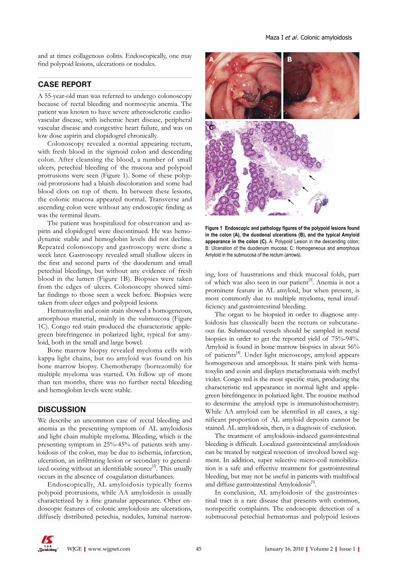
Maza I et al . Colonic amyloidosis
and at times collagenous colitis. Endoscopically, one may find polypoid lesions, ulcerations or nodules.
CASE REPORTA 55-year-old man was referred to undergo colonoscopy because of rectal bleeding and normocytic anemia. The patient was known to have severe atherosclerotic cardio-vascular disease, with ischemic heart disease, peripheral vascular disease and congestive heart failure, and was on low dose aspirin and clopidogrel chronically.
Colonoscopy revealed a normal appearing rectum, with fresh blood in the sigmoid colon and descending colon. After cleansing the blood, a number of small ulcers, petechial bleeding of the mucosa and polypoid protrusions were seen (Figure 1). Some of these polyp-oid protrusions had a bluish discoloration and some had blood clots on top of them. In between these lesions, the colonic mucosa appeared normal. Transverse and ascending colon were without any endoscopic finding as was the terminal ileum.
The patient was hospitalized for observation and as-pirin and clopidogrel were discontinued. He was hemo-dynamic stable and hemoglobin levels did not decline. Repeated colonoscopy and gastroscopy were done a week later. Gastroscopy revealed small shallow ulcers in the first and second parts of the duodenum and small petechial bleedings, but without any evidence of fresh blood in the lumen (Figure 1B). Biopsies were taken from the edges of ulcers. Colonoscopy showed simi-lar findings to those seen a week before. Biopsies were taken from ulcer edges and polypoid lesions.
Hematoxylin and eosin stain showed a homogeneous, amorphous material, mainly in the submucosa (Figure 1C). Congo red stain produced the characteristic apple-green birefringence in polarized light, typical for amy-loid, both in the small and large bowel.
Bone marrow biopsy revealed myeloma cells with kappa light chains, but no amyloid was found on his bone marrow biopsy. Chemotherapy (bortezomib) for multiple myeloma was started. On follow up of more than ten months, there was no further rectal bleeding and hemoglobin levels were stable.
DISCUSSION We describe an uncommon case of rectal bleeding and anemia as the presenting symptom of AL amyloidosis and light chain multiple myeloma. Bleeding, which is the presenting symptom in 25%-45% of patients with amy-loidosis of the colon, may be due to ischemia, infarction, ulceration, an infiltrating lesion or secondary to general-ized oozing without an identifiable source[2]. This usually occurs in the absence of coagulation disturbances.
Endoscopically, AL amyloidosis typically forms polypoid protrusions, while AA amyloidosis is usually characterized by a fine granular appearance. Other en-doscopic features of colonic amyloidosis are ulcerations, diffusely distributed petechia, nodules, luminal narrow-
ing, loss of haustrations and thick mucosal folds, part of which was also seen in our patient[3]. Anemia is not a prominent feature in AL amyloid, but when present, is most commonly due to multiple myeloma, renal insuf-ficiency and gastrointestinal bleeding.
The organ to be biopsied in order to diagnose amy-loidosis has classically been the rectum or subcutane-ous fat. Submucosal vessels should be sampled in rectal biopsies in order to get the reported yield of 75%-94%. Amyloid is found in bone marrow biopsies in about 56% of patients[4]. Under light microscopy, amyloid appears homogeneous and amorphous. It stains pink with hema-toxylin and eosin and displays metachromasia with methyl violet. Congo red is the most specific stain, producing the characteristic red appearance in normal light and apple-green birefringence in polarized light. The routine method to determine the amyloid type is immunohistochemistry. While AA amyloid can be identified in all cases, a sig-nificant proportion of AL amyloid deposits cannot be stained. AL amyloidosis, then, is a diagnosis of exclusion.
The treatment of amyloidosis-induced gastrointestinal bleeding is difficult. Localized gastrointestinal amyloidosis can be treated by surgical resection of involved bowel seg-ment. In addition, super selective micro-coil remobiliza-tion is a safe and effective treatment for gastrointestinal bleeding, but may not be useful in patients with multifocal and diffuse gastrointestinal Amyloidosis[5].
In conclusion, AL amyloidosis of the gastrointes-tinal tract is a rare disease that presents with common, nonspecific complaints. The endoscopic detection of a submucosal petechial hematomas and polypoid lesions
Figure 1 Endoscopic and pathology figures of the polypoid lesions found in the colon (A), the duodenal ulcerations (B), and the typical Amyloid appearance in the colon (C). A: Polypoid Lesion in the descending colon; B: Ulceration of the duodenum mucosa; C: Homogeneous and amorphous Amyloid in the submucosa of the rectum (arrows).
←
←←
A B
C
45 January 16, 2010|Volume 2|Issue 1|WJGE|www.wjgnet.com
in the setting of gastrointestinal bleeding should raise suspicion of the disease.
REFERENCES1 Ebert EC, Nagar M. Gastrointestinal manifestations of
amyloidosis. Am J Gastroenterol 2008; 103: 776-7872 Kim SH, Kang EJ, Park JW, Jo JH, Kim SJ, Cho JH, Kang
MJ, Park BH. Gastrointestinal amyloidosis presenting with multiple episodes of gastrointestinal bleeding. Cardiovasc
Intervent Radiol 2009; 32: 577-5803 Spier BJ, Einstein M, Johnson EA, Zuricik AO 3rd, Hu JL,
Pfau PR. Amyloidosis presenting as lower gastrointestinal hemorrhage. WMJ 2008; 107: 40-43
4 Petre S, Shah IA, Gilani N. Review article: gastrointestinal amyloidosis - clinical features, diagnosis and therapy. Aliment Pharmacol Ther 2008; 27: 1006-1016
5 James DG, Zuckerman GR, Sayuk GS, Wang HL, Prakash C. Clinical recognition of Al type amyloidosis of the luminal gastrointestinal tract. Clin Gastroenterol Hepatol 2007; 5: 582-588
S- Editor Zhang HN L- Editor Lutze M E- Editor Ma WH
46 January 16, 2010|Volume 2|Issue 1|WJGE|www.wjgnet.com
Maza I et al . Colonic amyloidosis
ACKNOWLEDGMENTS
Acknowledgments to reviewers of World Journal of Gastrointestinal Endoscopy
Many reviewers have contributed their expertise and time to the peer review, a critical process to ensure the quality of World Journal of Gastrointestinal Endoscopy. The editors and authors of the articles submitted to the journal are grateful to the following reviewers for evaluating the articles (including those published in this issue and those rejected for this issue) during the last editing time period.
Qiang Cai, Associate Professor of Medicine, Division of Digestive Diseases, Emory University School of Medicine, 1365 Clifton Road, Suite B1262 Atlanta, GA 30322, United States
Juan J Vila Costas, MD, Gastroenterology Department, Hospital de Navarra, c\Irunlarrea, 3, Pamplona 31.008, Spain
Jesús García-Cano, MD, PhD, Department of Gastroente-rology, Hospital Virgen de la Luz, Cuenca 16002, Spain
Akira Horiuchi, MD, Director, Digestive Disease Center, Showa Inan General Hospital, Komagane 399-4191, Japan
Tian-Le Ma, MD, Department of Gastroenterology, Rui Jin Hospital Affiliated to Medical College of Shanghai Jiao Tong
University, Shanghai 200040, China
Oliver Pech, MD, PhD, Attending Physician of Gastroenter-ology, Vice Director of the Endoscopy Unit, Department of Internal Medicine 2, HSK Wiesbaden, Wiesbaden, Germany
Jean-Francois Rey, MD, Hepatogastroenterology Institut Ar-nault Tzanck, 06721 Saint Laurent Du Var Cedex, France
Atsushi Irisawa, MD, PhD, Associate Professor, Depart-ment of Internal Medicine 2, Fukushima Medical University School of Medicine. 1, Hikarigaoka, Fukushima 960-1295, Japan
Carlos Robles-Medranda, MD, Head of the Endoscopy Division, Ecuadorian Institute of Digestive Disease (IECED), San Antonio Clinic, Av. Reales Tamarindo y Tennis Club, Portoviejo-Manabi-Ecuador, Casilla 13-01-266, Ecuadorian
Luis Rodrigo, Professor, Gastroenterology Service, Hospital Universitario Central de Asturias, c/ Celestino Villamil s. nº, Oviedo 33-006, Spain
James F Trotter, MD, Associate Professor, University of Colorado, Division of Gastroenterology, 4200 E. 9th Avenue, b-154, Denver, CO 80262, United States
January 16, 2010|Volume 2|Issue 1|ⅠWJGE|www.wjgnet.com
World J Gastrointest Endosc 2010 January 16; 2(1): ⅠISSN 1948-5190 (online)
© 2010 Baishideng. All rights reserved.
Online Submissions: http://www.wjgnet.com/[email protected]
Meetings
Events Calendar 2010January 25-26Tamilnadu, India International Conference on Medical Negligence and Litigation in Medical Practice
January 25-29Waikoloa, HI, United States Selected Topics in Internal Medicine
January 26-27Dubai, United Arab Emirates 2nd Middle East Gastroenterology Conference
February 11-13Fort Lauderdale, FL, United States21th Annual International Colorectal Disease Symposium
February 26-28Carolina, United StatesFirst Symposium of GI Oncology at The Caribbean
March 05-07Peshawar, Pakistan26th Pakistan Society of Gastroenterology & Endoscopy Meeting
March 12-14Bhubaneswar, India18th Annual Meeting of Indian National Association for Study of the Liver
March 25-28Beijing, ChinaThe 20th Conference of the Asian Pacific Association for the Study of the Liver
March 27-28San Diego, California, United States25th Annual New Treatments in Chronic Liver Disease
April 07-09Dubai, United Arab EmiratesThe 6th Emirates Gastroenterology and Hepatology Conference, EGHC 2010
April 14-17Landover, Maryland, United States12th World Congress of Endoscopic Surgery
April 14-18Vienna, AustriaThe International Liver Congress™ 2010
April 28-May 01Dubrovnik, Croatia3rd Central European Congress of surgery and the 5th Croatian Congress of Surgery
May 01-05New Orleans, LA, United StatesDigestive Disease Week Annual Meeting
May 15-19Minneapolis, MN, United StatesAmerican Society of Colon and Rectal Surgeons Annual Meeting
June 04-06Chicago, IL, United StatesAmerican Society of Clinical Oncologists Annual Meeting
June 16-19Hong Kong, ChinaILTS: International Liver Transplantation Society ILTS Annual International Congress
June 20-23Mannheim, Germany16th World Congress for Bronchoesophagology-WCBE
August 28-31Boston, Massachusetts, United States10th OESO World Congress on Diseases of the Oesophagus 2010
September 10-12Montreal, CanadaInternational Liver Association's Fourth Annual Conference
September 11-12La Jolla, CA, United StatesNew Advances in Inflammatory Bowel Disease
September 16-18Prague, Czech RepublicPrague Hepatology Meeting 2010
September 23-26Prague, Czech RepublicThe 1st World Congress on Controversies in Gastroenterology & Liver Diseases
October 07-09Belgrade, SerbiaThe 7th Biannual International
Symposium of Society of Coloproctology
October 15-20San Antonio, TX, United StatesACG 2010: American College of Gastroenterology Annual Scienitfic Meeting
October 23-27Barcelona, Spain18th United European Gastroenterology Week
October 29-November 02Boston, Massachusetts, United StatesThe Liver Meeting® 2010--AASLD's 61st Annual Meeting
November 13-14San Francisco, CA, United StatesCase-Based Approach to the Management of Inflammatory Bowel Disease
January 16, 2010|Volume 2|Issue 1|ⅠWJGE|www.wjgnet.com
World J Gastrointest Endosc 2010 January 16; 2(1): ⅠISSN 1948-5190 (online)
© 2010 Baishideng. All rights reserved.
Online Submissions: http://www.wjgnet.com/[email protected]
GENERAL INFORMATIONWorld Journal of Gastrointestinal Endoscopy (World J Gastrointest Endosc, WJGE, ISSN 1948-5190, DOI: 10.4253), is a monthly, open-access (OA), peer-reviewed online journal supported by an editorial board of 143 experts in gastrointestinal endoscopy from 28 countries.
The biggest advantage of the OA model is that it provides free, full-text articles in PDF and other formats for experts and the public without registration, which eliminates the obstacle that traditional journals possess and usually delays the speed of the propagation and communication of scientific research results.
The role of academic journals is to exhibit the scientific levels of a country, a university, a center, a department, and even a scientist, and build an important bridge for commu-nication between scientists and the public. As we all know, the significance of the publication of scientific articles lies not only in disseminating and communicating innovative scientific achievements and academic views, as well as promoting the application of scientific achievements, but also in formally recognizing the “priority” and “copyright” of innovative achieve-ments published, as well as evaluating research performance and academic levels. So, to realize these desired attributes of WJGE and create a well-recognized journal, the following four types of personal benefits should be maximized. The maximization of personal benefits refers to the pursuit of the maximum personal benefits in a well-considered optimal manner without violation of the laws, ethical rules and the benefits of others. (1) Maximization of the benefits of editorial board members: The primary task of editorial board members is to give a peer review of an unpublished scientific article via online office system to evaluate its innovativeness, scientific and practical values and determine whether it should be published or not. During peer review, editorial board members can also obtain cutting-edge information in that field at first hand. As leaders in their field, they have priority to be invited to write articles and publish commentary articles. We will put peer reviewers’ names and affiliations along with the article they reviewed in the journal to acknowledge their contribution; (2) Maximization of the benefits of authors: Since WJGE is an open-access journal, readers around the world can immediately download and read, free of charge, high-quality, peer-reviewed articles from WJGE official website, thereby realizing the goals and significance of the communication between authors and peers as well as public reading; (3) Maximization of the benefits of readers: Readers can read or use, free of charge, high-quality peer-reviewed articles without any limits, and cite the arguments, viewpoints, concepts, theories, methods, results, conclusion or facts and data of pertinent literature so as to validate the innovativeness, scientific and practical values of their own research achievements, thus ensuring that their articles have novel arguments or viewpoints, solid evidence and correct conclusion; and (4) Maximization of the benefits of employees: It is an iron law that a first-class journal is unable to exist without first-class editors, and only first-class editors can create a first-class academic journal. We insist on
strengthening our team cultivation and construction so that every employee, in an open, fair and transparent environment, could contribute their wisdom to edit and publish high-quality articles, thereby realizing the maximization of the personal benefits of editorial board members, authors and readers, and yielding the greatest social and economic benefits.
The major task of WJGE is to report rapidly the most recent results in basic and clinical research on gastrointestinal endoscopy including: gastroscopy, intestinal endoscopy, colonoscopy, capsule endoscopy, laparoscopy, interventional diagnosis and therapy, as well as advances in technology. Emphasis is placed on the clinical practice of treating gastroin-testinal diseases with or under endoscopy. Papers on advances and application of endoscopy-associated techniques, such as endoscopic ultrasonography, endoscopic retrograde cholangiopancreatography, endoscopic submucosal dissection and endoscopic balloon dilation are also welcome.
The columns in the issues of WJGE will include: (1) Editorial: To introduce and comment on the substantial advance and its importance in the fast-developing areas; (2) Frontier: To review the most representative achievements and comment on the current research status in the important fields, and propose directions for the future research; (3) Topic Highlight: This column consists of three formats, including (A) 10 invited review articles on a hot topic, (B) a commentary on common issues of this hot topic, and (C) a commentary on the 10 individual articles; (4) Observation: To update the development of old and new questions, highlight unsolved problems, and provide strategies on how to solve the questions; (5) Guidelines for Basic Research: To provide Guidelines for basic research; (6) Guidelines for Clinical Practice: To provide guidelines for clinical diagnosis and treatment; (7) Review: To systemically review the most representative progress and unsolved problems in the major scientific disciplines, comment on the current research status, and make suggestions on the future work; (8) Original Articles: To originally report the innovative and valuable findings in gastrointestinal endoscopy; (9) Brief Articles: To briefly report the novel and innovative findings in gastrointestinal endoscopy; (10) Case Report: To report a rare or typical case; (11) Letters to the Editor: To discuss and make reply to the contributions published in WJGE, or to introduce and comment on a controversial issue of general interest; (12) Book Reviews: To introduce and comment on quality monographs of gastrointestinal endoscopy; and (13) Guidelines: To introduce Consensuses and Guidelines reached by international and national academic authorities worldwide on basic research and clinical practice in gastrointestinal endoscopy.
CSSNISSN 1948-5190 (online)
Published byBeijing Baishideng BioMed Scientific Co., Ltd.
SUBMISSION OF MANUSCRIPTSManuscripts should be typed in 1.5 line spacing and 12 pt. Book
Instructions to authors
January 16, 2010|Volume 2|Issue 1|ⅠWJGE|www.wjgnet.com
World J Gastrointest Endosc 2010 January 16; 2(1): Ⅰ-ⅤISSN 1948-5190 (online)
© 2010 Baishideng. All rights reserved.
Online Submissions: http://www.wjgnet.com/[email protected]

Antiqua with ample margins. Number all pages consecutively, and start each of the following sections on a new page: Title Page, Abstract, Introduction, Materials and Methods, Results, Discussion, Acknowledgements, References, Tables, Figures, and Figure Legends. Neither the editors nor the publisher are responsible for the opinions expressed by contributors. Manuscripts formally accepted for publication become the permanent property of Beijing Baishideng BioMed Scientific Co., Ltd, and may not be reproduced by any means, in whole or in part, without the written permission of both the authors and the publisher. We reserve the right to copy-edit and put onto our website accepted manuscripts. Authors should follow the relevant guidelines for the care and use of laboratory animals of their institution or national animal welfare committee. For the sake of transparency in regard to the performance and reporting of clinical trials, we endorse the policy of the International Committee of Medical Journal Editors to refuse to publish papers on clinical trial results if the trial was not recorded in a publicly-accessible registry at its outset. The only register now available, to our knowledge, is http://www. clinicaltrials.gov sponsored by the United States National Library of Medicine and we encourage all potential contributors to register with it. However, in the case that other registers become available you will be duly notified. A letter of recommendation from each author’s organization should be provided with the contributed article to ensure the privacy and secrecy of research is protected.
Authors should retain one copy of the text, tables, photo-graphs and illustrations because rejected manuscripts will not be returned to the author(s) and the editors will not be responsible for loss or damage to photographs and illustrations sustained during mailing.
Online submissionsManuscripts should be submitted through the Online Submission System at: [email protected]. Authors are highly recommended to consult the ONLINE INSTRUCTIONS TO AUTHORS (http://www.wjgnet.com/1948-5190/g_info_14.htm) before attempting to submit online. For assistance, authors encountering problems with the Online Submission System may send an email describing the problem to http://www.wjgnet.com/1948-5190office/, or by telephone: +86-10-59080038. If you submit your manuscript online, do not make a postal contribution. Repeated online submission for the same manuscript is strictly prohibited.
MANUSCRIPT PREPARATIONAll contributions should be written in English. All articles must be submitted using word-processing software. All submissions must be typed in 1.5 line spacing and 12 pt. Book Antiqua with ample margins. Style should conform to our house format. Required information for each of the manuscript sections is as follows:
Title pageTitle: Title should be less than 12 words.
Running title: A short running title of less than 6 words should be provided.
Authorship: Authorship credit should be in accordance with the standard proposed by International Committee of Medical Journal Editors, based on (1) substantial contributions to conception and design, acquisition of data, or analysis and interpretation of data; (2) drafting the article or revising
it critically for important intellectual content; and (3) final approval of the version to be published. Authors should meet conditions 1, 2, and 3.
Institution: Author names should be given first, then the complete name of institution, city, province and postcode. For example, Xu-Chen Zhang, Li-Xin Mei, Department of Pathology, Chengde Medical College, Chengde 067000, Hebei Province, China. One author may be represented from two institutions, for example, George Sgourakis, Department of General, Visceral, and Transplantation Surgery, Essen 45122, Germany; George Sgourakis, 2nd Surgical Department, Korgialenio-Benakio Red Cross Hospital, Athens 15451, Greece
Author contributions: The format of this section should be: Author contributions: Wang CL and Liang L contributed equally to this work; Wang CL, Liang L, Fu JF, Zou CC, Hong F and Wu XM designed the research; Wang CL, Zou CC, Hong F and Wu XM performed the research; Xue JZ and Lu JR contributed new reagents/analytic tools; Wang CL, Liang L and Fu JF analyzed the data; and Wang CL, Liang L and Fu JF wrote the paper.
Supportive foundations: The complete name and number of supportive foundations should be provided, e.g., Supported by National Natural Science Foundation of China, No. 30224801
Correspondence to: Only one corresponding address should be provided. Author names should be given first, then author title, affiliation, the complete name of institution, city, postcode, province, country, and email. All the letters in the email should be in lower case. A space interval should be inserted between country name and email address. For example, Montgomery Bissell, MD, Professor of Medicine, Chief, Liver Center, Gastroenterology Division, University of California, Box 0538, San Francisco, CA 94143, United States. [email protected]
Telephone and fax: Telephone and fax should consist of +, country number, district number and telephone or fax number, e.g., Telephone: +86-10-59080039 Fax: +86-10-85381893
Peer reviewers: All articles received are subject to peer review. Normally, three experts are invited for each article. Decision for acceptance is made only when at least two experts recommend an article for publication. Reviewers for accepted manuscripts are acknowledged in each manuscript, and reviewers of articles which were not accepted will be acknowledged at the end of each issue. To ensure the quality of the articles published in WJGE, reviewers of accepted manuscripts will be announced by publishing the name, title/position and institution of the reviewer in the footnote accompanying the printed article. For example, reviewers: Professor Jing-Yuan Fang, Shanghai Institute of Digestive Disease, Shanghai, Affiliated Renji Hospital, Medical Faculty, Shanghai Jiaotong University, Shanghai, China; Professor Xin-Wei Han, Department of Radiology, The First Affiliated Hospital, Zhengzhou University, Zhengzhou, Henan Province, China; and Professor Anren Kuang, Department of Nuclear Medicine, Huaxi Hospital, Sichuan University, Chengdu, Sichuan Province, China.
AbstractThere are unstructured abstracts (no more than 256 words)
Instructions to authors
January 16, 2010|Volume 2|Issue 1|ⅡWJGE|www.wjgnet.com
and structured abstracts (no more than 480). The specific requirements for structured abstracts are as follows:
An informative, structured abstracts of no more than 480 words should accompany each manuscript. Abstracts for original contributions should be structured into the following sections. AIM (no more than 20 words): Only the purpose should be included. Please write the aim as the form of “To investigate/study/…; MATERIALS AND METHODS (no more than 140 words); RESULTS (no more than 294 words): You should present P values where appropriate and must provide relevant data to illustrate how they were obtained, e.g. 6.92 ± 3.86 vs 3.61 ± 1.67, P < 0.001; CONCLUSION (no more than 26 words). Available from: http://www.wjgnet.com/wjg/help/8.doc.
Key wordsPlease list 5-10 key words, selected mainly from Index Medicus, which reflect the content of the study.
TextFor ar ticles of these sections, original ar ticles, rapid communication and case reports, the main text should be structured into the following sections: INTRODUCTION, M AT E R I A L S A N D M E T H O D S, R E S U LT S a n d DISCUSSION, and should include appropriate Figures and Tables. Data should be presented in the main text or in Figures and Tables, but not in both. The main text format of these sections, editorial, topic highlight, case report, letters to the editors, can be found at: http://www.wjgnet.com/wjg/help/instructions.jsp.
IllustrationsFigures should be numbered as 1, 2, 3, etc., and mentioned clearly in the main text. Provide a brief title for each figure on a separate page. Detailed legends should not be provided under the figures. This part should be added into the text where the figures are applicable. Figures should be either Photoshop or Illustrator files (in tiff, eps, jpeg formats) at high-resolution. Examples can be found at: http://www.wjgnet.com/1007-9327/13/4520.pdf; http://www.wjgnet.com/1007-9327/13/4554.pdf; http://www.wjgnet.com/1007-9327/13/4891.pdf; http://www.wjgnet.com/1007-9327/13/4986.pdf ; h t tp ://www.wjgnet .com/1007-9327/13/4498.pdf. Keeping all elements compiled is necessary in line-art image. Scale bars should be used rather than magnification factors, with the length of the bar defined in the legend rather than on the bar itself. File names should identify the figure and panel. Avoid layering type directly over shaded or textured areas. Please use uniform legends for the same subjects. For example: Figure 1 Pathological changes in atrophic gastritis after treatment. A: ...; B: ...; C: ...; D: ...; E: ...; F: ...; G: …etc. It is our principle to publish high resolution-figures for the printed and E-versions.
TablesThree-line tables should be numbered 1, 2, 3, etc . , and mentioned clearly in the main text. Provide a brief title for each table. Detailed legends should not be included under tables, but rather added into the text where applicable. The information should complement, but not duplicate the text. Use one horizontal line under the title, a second under column heads, and a third below the Table, above any footnotes. Vertical and italic lines should be omitted.
Notes in tables and illustrationsData that are not statistically significant should not be noted.
aP < 0.05, bP < 0.01 should be noted (P > 0.05 should not be noted). If there are other series of P values, cP < 0.05 and dP < 0.01 are used. A third series of P values can be expressed as eP < 0.05 and fP < 0.01. Other notes in tables or under illustrations should be expressed as 1F, 2F, 3F; or sometimes as other symbols with a superscript (Arabic numerals) in the upper left corner. In a multi-curve illustration, each curve should be labeled with ●, ○, ■, □, ▲, △, etc., in a certain sequence.
AcknowledgmentsBrief acknowledgments of persons who have made genuine contributions to the manuscript and who endorse the data and conclusions should be included. Authors are responsible for obtaining written permission to use any copyrighted text and/or illustrations.
REFERENCESCoding systemThe author should number the references in Arabic numerals according to the citation order in the text. Put reference numbers in square brackets in superscript at the end of citation content or after the cited author’s name. For citation content which is part of the narration, the coding number and square brackets should be typeset normally. For example, “Crohn’s disease (CD) is associated with increased intestinal permeability[1,2]”. If references are cited directly in the text, they should be put together within the text, for example, “From references[19,22-24], we know that...”
When the authors write the references, please ensure that the order in text is the same as in the references section, and also ensure the spelling accuracy of the first author’s name. Do not list the same citation twice.
PMID and DOIPleased provide PubMed citation numbers to the reference list, e.g. PMID and DOI, which can be found at http://www.ncbi.nlm.nih.gov/sites/entrez?db=pubmed and http://www.crossref.org/SimpleTextQuery/, respectively. The numbers will be used in E-version of this journal.
Style for journal referencesAuthors: the name of the first author should be typed in bold-faced letters. The family name of all authors should be typed with the initial letter capitalized, followed by their abbreviated first and middle initials. (For example, Lian-Sheng Ma is abbreviated as Ma LS, Bo-Rong Pan as Pan BR). The title of the cited article and italicized journal title (journal title should be in its abbreviated form as shown in PubMed), publication date, volume number (in black), start page, and end page [PMID: 11819634 DOI: 10.3748/wjg.13.5396].
Style for book referencesAuthors: the name of the first author should be typed in bold-faced letters. The surname of all authors should be typed with the initial letter capitalized, followed by their abbreviated middle and first initials. (For example, Lian-Sheng Ma is abbreviated as Ma LS, Bo-Rong Pan as Pan BR) Book title. Publication number. Publication place: Publication press, Year: start page and end page.
FormatJournalsEnglish journal article (list all authors and include the PMID where
applicable)
Instructions to authors
January 16, 2010|Volume 2|Issue 1|ⅢWJGE|www.wjgnet.com

1 Jung EM, Clevert DA, Schreyer AG, Schmitt S, Rennert J, Kubale R, Feuerbach S, Jung F. Evaluation of quantitative contrast harmonic imaging to assess malignancy of liver tumors: A prospective controlled two-center study. World J Gastroenterol 2007; 13: 6356-6364 [PMID: 18081224 DOI: 10.3748/wjg.13.6356]
Chinese journal article (list all authors and include the PMID where applicable)
2 Lin GZ, Wang XZ, Wang P, Lin J, Yang FD. Immunologic effect of Jianpi Yishen decoction in treatment of Pixu-diarrhoea. Shijie Huaren Xiaohua Zazhi 1999; 7: 285-287
In press3 Tian D, Araki H, Stahl E, Bergelson J, Kreitman M.
Signature of balancing selection in Arabidopsis. Proc Natl Acad Sci USA 2006; In press
Organization as author4 Diabetes Prevention Program Research Group. Hyp-
ertension, insulin, and proinsulin in participants with impaired glucose tolerance. Hypertension 2002; 40: 679-686 [PMID: 12411462 PMCID:2516377 DOI:10.1161/01.HYP.0000035706.28494.09]
Both personal authors and an organization as author 5 Vallancien G, Emberton M, Harving N, van Moorselaar
RJ; Alf-One Study Group. Sexual dysfunction in 1, 274 European men suffering from lower urinary tract symptoms. J Urol 2003; 169: 2257-2261 [PMID: 12771764 DOI:10.1097/01.ju.0000067940.76090.73]
No author given6 21st century heart solution may have a sting in the tail.
BMJ 2002; 325: 184 [PMID: 12142303 DOI:10.1136/bmj.325.7357.184]
Volume with supplement7 Geraud G, Spierings EL, Keywood C. Tolerability and
safety of frovatriptan with short- and long-term use for treatment of migraine and in comparison with sumatriptan. Headache 2002; 42 Suppl 2: S93-99 [PMID: 12028325 DOI:10.1046/j.1526-4610.42.s2.7.x]
Issue with no volume8 Banit DM, Kaufer H, Hartford JM. Intraoperative frozen
section analysis in revision total joint arthroplasty. Clin Orthop Relat Res 2002; (401): 230-238 [PMID: 12151900 DOI:10.1097/00003086-200208000-00026]
No volume or issue9 Outreach: Bringing HIV-positive individuals into care.
HRSA Careaction 2002; 1-6 [PMID: 12154804]
BooksPersonal author(s)10 Sherlock S, Dooley J. Diseases of the liver and billiary
system. 9th ed. Oxford: Blackwell Sci Pub, 1993: 258-296Chapter in a book (list all authors)11 Lam SK. Academic investigator’s perspectives of medical
treatment for peptic ulcer. In: Swabb EA, Azabo S. Ulcer disease: investigation and basis for therapy. New York: Marcel Dekker, 1991: 431-450
Author(s) and editor(s)12 Breedlove GK, Schorfheide AM. Adolescent pregnancy.
2nd ed. Wieczorek RR, editor. White Plains (NY): March of Dimes Education Services, 2001: 20-34
Conference proceedings13 Harnden P, Joffe JK, Jones WG, editors. Germ cell
tumours V. Proceedings of the 5th Germ cell tumours Conference; 2001 Sep 13-15; Leeds, UK. New York: Springer, 2002: 30-56
Conference paper14 Christensen S, Oppacher F. An analysis of Koza's
computational effort statistic for genetic programming. In: Foster JA, Lutton E, Miller J, Ryan C, Tettamanzi AG, editors. Genetic programming. EuroGP 2002: Proceedings of the 5th European Conference on Genetic Programming; 2002 Apr 3-5; Kinsdale, Ireland. Berlin: Springer, 2002: 182-191
Electronic journal (list all authors)15 Morse SS. Factors in the emergence of infectious
diseases. Emerg Infect Dis serial online, 1995-01-03, cited 1996-06-05; 1(1): 24 screens. Available from: URL: http//www.cdc.gov/ncidod/EID/eid.htm
Patent (list all authors)16 Pagedas AC , inventor; Ancel Surgical R&D Inc.,
assignee. Flexible endoscopic grasping and cutting device and positioning tool assembly. United States patent US 20020103498. 2002 Aug 1
Statistical dataWrite as mean ± SD or mean ± SE.
Statistical expressionExpress t test as t (in italics), F test as F (in italics), chi square test as χ2 (in Greek), related coefficient as r (in italics), degree of freedom as υ (in Greek), sample number as n (in italics), and probability as P (in italics).
UnitsUse SI units. For example: body mass, m (B) = 78 kg; blood pressure, p (B) = 16.2/12.3 kPa; incubation time, t (incubation) = 96 h, blood glucose concentration, c (glucose) 6.4 ± 2.1 mmol/L; blood CEA mass concentration, p (CEA) = 8.6 24.5 mg/L; CO2 volume fraction, 50 mL/L CO2, not 5% CO2; likewise for 40 g/L formaldehyde, not 10% formalin; and mass fraction, 8 ng/g, etc. Arabic numerals such as 23, 243, 641 should be read 23 243 641.
The format for how to accurately write common units and quantums can be found at: http://www.wjgnet.com/wjg/help/15.doc.
AbbreviationsStandard abbreviations should be defined in the abstract and on first mention in the text. In general, terms should not be abbreviated unless they are used repeatedly and the abbreviation is helpful to the reader. Permissible abbreviations are listed in Units, Symbols and Abbreviations: A Guide for Biological and Medical Editors and Authors (Ed. Baron DN, 1988) published by The Royal Society of Medicine, London. Certain commonly used abbreviations, such as DNA, RNA, HIV, LD50, PCR, HBV, ECG, WBC, RBC, CT, ESR, CSF, IgG, ELISA, PBS, ATP, EDTA, mAb, can be used directly without further explanation.
ItalicsQuantities: t time or temperature, c concentration, A area, l length, m mass, V volume.Genotypes: gyrA, arg 1, c myc, c fos, etc.Restriction enzymes: EcoRI, HindI, BamHI, Kbo I, Kpn I, etc.Biology: H. pylori, E coli, etc.
SUBMISSION OF THE REVISED MANUSCRIPTS AFTER ACCEPTEDPlease revise your article according to the revision policies
Instructions to authors
January 16, 2010|Volume 2|Issue 1|ⅣWJGE|www.wjgnet.com
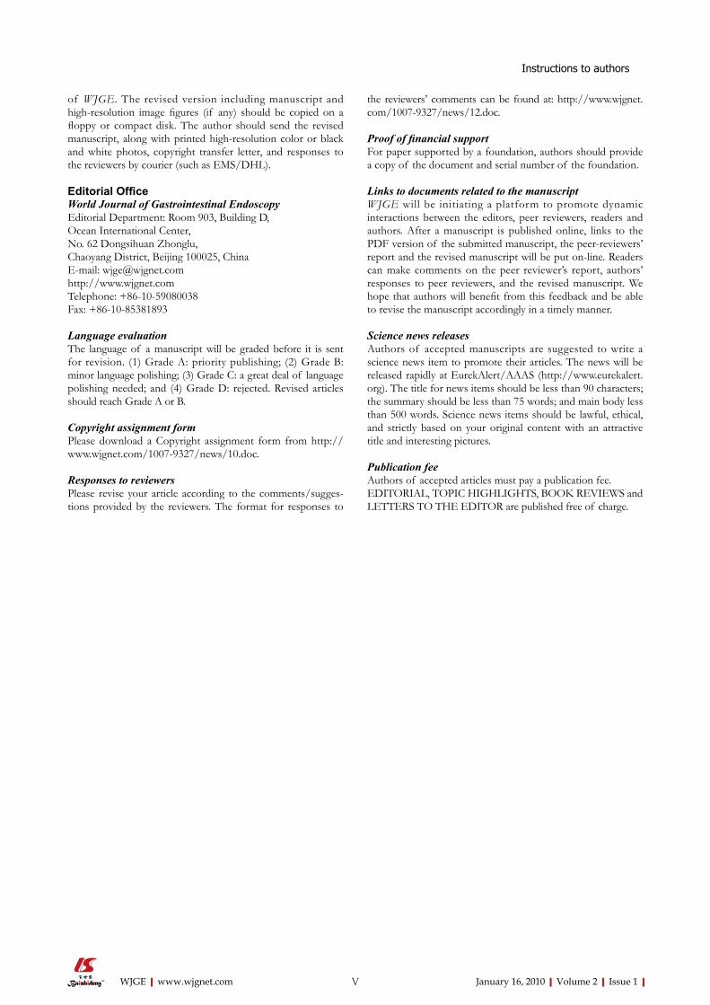
of WJGE. The revised version including manuscript and high-resolution image figures (if any) should be copied on a floppy or compact disk. The author should send the revised manuscript, along with printed high-resolution color or black and white photos, copyright transfer letter, and responses to the reviewers by courier (such as EMS/DHL).
Editorial Office World Journal of Gastrointestinal EndoscopyEditorial Department: Room 903, Building D, Ocean International Center,No. 62 Dongsihuan Zhonglu, Chaoyang District, Beijing 100025, ChinaE-mail: [email protected]://www.wjgnet.comTelephone: +86-10-59080038Fax: +86-10-85381893
Language evaluation The language of a manuscript will be graded before it is sent for revision. (1) Grade A: priority publishing; (2) Grade B: minor language polishing; (3) Grade C: a great deal of language polishing needed; and (4) Grade D: rejected. Revised articles should reach Grade A or B.
Copyright assignment formPlease download a Copyright assignment form from http://www.wjgnet.com/1007-9327/news/10.doc.
Responses to reviewersPlease revise your article according to the comments/sugges-tions provided by the reviewers. The format for responses to
the reviewers’ comments can be found at: http://www.wjgnet.com/1007-9327/news/12.doc.
Proof of financial supportFor paper supported by a foundation, authors should provide a copy of the document and serial number of the foundation.
Links to documents related to the manuscript WJGE will be initiating a platform to promote dynamic interactions between the editors, peer reviewers, readers and authors. After a manuscript is published online, links to the PDF version of the submitted manuscript, the peer-reviewers’ report and the revised manuscript will be put on-line. Readers can make comments on the peer reviewer’s report, authors’ responses to peer reviewers, and the revised manuscript. We hope that authors will benefit from this feedback and be able to revise the manuscript accordingly in a timely manner.
Science news releasesAuthors of accepted manuscripts are suggested to write a science news item to promote their articles. The news will be released rapidly at EurekAlert/AAAS (http://www.eurekalert.org). The title for news items should be less than 90 characters; the summary should be less than 75 words; and main body less than 500 words. Science news items should be lawful, ethical, and strictly based on your original content with an attractive title and interesting pictures.
Publication feeAuthors of accepted articles must pay a publication fee.EDITORIAL, TOPIC HIGHLIGHTS, BOOK REVIEWS and LETTERS TO THE EDITOR are published free of charge.
Instructions to authors
January 16, 2010|Volume 2|Issue 1|Ⅴ WJGE|www.wjgnet.com