Gastrointestinal Microbiota Dysbiosis Associated with SARS ...
25
Citation: Odun-Ayo, F.; Reddy, L. Gastrointestinal Microbiota Dysbiosis Associated with SARS-CoV-2 Infection in Colorectal Cancer: The Implication of Probiotics. Gastroenterol. Insights 2022, 13, 35–59. https://doi.org/10.3390/ gastroent13010006 Academic Editors: Tsvetelina Velikova and Radislav Nakov Received: 15 December 2021 Accepted: 25 January 2022 Published: 7 February 2022 Publisher’s Note: MDPI stays neutral with regard to jurisdictional claims in published maps and institutional affil- iations. Copyright: © 2022 by the authors. Licensee MDPI, Basel, Switzerland. This article is an open access article distributed under the terms and conditions of the Creative Commons Attribution (CC BY) license (https:// creativecommons.org/licenses/by/ 4.0/). Review Gastrointestinal Microbiota Dysbiosis Associated with SARS-CoV-2 Infection in Colorectal Cancer: The Implication of Probiotics Frederick Odun-Ayo * and Lalini Reddy * Department of Biotechnology and Consumer Sciences, Faculty of Applied Sciences, District Six Campus, Cape Peninsula University of Technology, Cape Town 7530, South Africa * Correspondence: [email protected] (F.O.-A.); [email protected] (L.R.); Tel.: +27-78-784-9231 (F.O.-A.); +27-83-382-9455 (L.R.) Abstract: The complexity of coronavirus disease 2019 (COVID-19)’s pathophysiology is such that microbial dysbiosis in the lung and gastrointestinal (GI) microbiota may be involved in its pathogenic process. GI microbiota dysbiosis has been associated with respiratory disorders, including COVID-19, as well as sporadic colorectal cancer (CRC) through imbalanced microbiota and compromised immune response. It is pertinent to understand the possible role of probiotics in stabilizing the microbial environment and maintaining the integrity of the respiratory and GI tracts in SARS-CoV-2 induced dysbiosis and colorectal carcinogenesis. The long-term implication of SARS-CoV-2 in GI dysbiosis via microbiota-gut-lung cross-talk could increase the risk of new CRC diagnosis or worsen the condition of previously diagnosed individuals. Recent knowledge shows that the immune-modulatory response to probiotics is shifting the beneficial use of probiotics towards the treatment of various diseases. In this review, we highlight the potential impact of probiotics on SARS-CoV-2 infection associated with CRC through microbiota imbalance in COVID-19 patients. Keywords: probiotics; gut microbiota; colorectal cancer; virus; respiratory tract infection; SARS-CoV-2; COVID-19 1. Introduction The outbreak of coronavirus disease 2019 (COVID-19), which was reported from Wuhan, China in late December 2019, was caused by severe acute respiratory syndrome coronavirus-2 (SARS-CoV-2). Over 5 million deaths globally were reported as of 22 Novem- ber 2021 [1]. SARS-CoV-2 belongs to the enveloped RNA viruses, the subgenus betacoron- avirus of the family coronaviridae [2–4]. As the pathogenesis of COVID-19 is still unclear, recent studies have reported the possible inclusion of gastrointestinal (GI) symptoms, in- cluding diarrhea (2.0–10.1%), nausea, and vomiting (1.0–3.6%) in COVID-19 patients [5,6]. SARS-CoV-2 nucleic acids were found in the fecal samples and anal swabs of COVID-19 patients [7]. It was suggested that SARS-CoV-2 may enter the peripheral blood and pri- marily targets cells from the lung, heart, renal, and gastrointestinal tract that express angiotensin-converting enzyme-2 (ACE-2) receptor [8]. The ACE-2 receptor expressed in these target cells is recognized and bound by the SARS-CoV-2 spike glycoprotein S1 domain, which enhances the virus’ attachment to the host cell [9,10]. Recent studies have demonstrated that TMPRSS2 [11], galectin-3 (GAL-3) [12,13], and N-acetylneuraminic acid (Neu5Ac) [14] receptors are highly expressed in the GI and lung epithelia. They are also necessary for viral cell-to-cell interaction and the subsequent entrance of SARS-CoV-2 into the host cells [15–18]. As a result, higher expression levels of ACE-2 and TMPRSS2 proteins indicate a higher risk of COVID-19 infection in colorectal cancer (CRC) patients compared to those with normal intestinal tissue [19]. The World Health Organization (WHO), through the GLOBOCAN cancer research agency, reported that 990,000 deaths were caused by CRC in 2020 [20]. Deaths attributed to Gastroenterol. Insights 2022, 13, 35–59. https://doi.org/10.3390/gastroent13010006 https://www.mdpi.com/journal/gastroent
-
Upload
khangminh22 -
Category
Documents
-
view
3 -
download
0
Transcript of Gastrointestinal Microbiota Dysbiosis Associated with SARS ...

�����������������
Citation: Odun-Ayo, F.; Reddy, L.
Gastrointestinal Microbiota Dysbiosis
Associated with SARS-CoV-2
Infection in Colorectal Cancer: The
Implication of Probiotics.
Gastroenterol. Insights 2022, 13, 35–59.
https://doi.org/10.3390/
gastroent13010006
Academic Editors: Tsvetelina
Velikova and Radislav Nakov
Received: 15 December 2021
Accepted: 25 January 2022
Published: 7 February 2022
Publisher’s Note: MDPI stays neutral
with regard to jurisdictional claims in
published maps and institutional affil-
iations.
Copyright: © 2022 by the authors.
Licensee MDPI, Basel, Switzerland.
This article is an open access article
distributed under the terms and
conditions of the Creative Commons
Attribution (CC BY) license (https://
creativecommons.org/licenses/by/
4.0/).
Review
Gastrointestinal Microbiota Dysbiosis Associated withSARS-CoV-2 Infection in Colorectal Cancer: The Implicationof ProbioticsFrederick Odun-Ayo * and Lalini Reddy *
Department of Biotechnology and Consumer Sciences, Faculty of Applied Sciences, District Six Campus,Cape Peninsula University of Technology, Cape Town 7530, South Africa* Correspondence: [email protected] (F.O.-A.); [email protected] (L.R.);
Tel.: +27-78-784-9231 (F.O.-A.); +27-83-382-9455 (L.R.)
Abstract: The complexity of coronavirus disease 2019 (COVID-19)’s pathophysiology is such thatmicrobial dysbiosis in the lung and gastrointestinal (GI) microbiota may be involved in its pathogenicprocess. GI microbiota dysbiosis has been associated with respiratory disorders, including COVID-19,as well as sporadic colorectal cancer (CRC) through imbalanced microbiota and compromised immuneresponse. It is pertinent to understand the possible role of probiotics in stabilizing the microbialenvironment and maintaining the integrity of the respiratory and GI tracts in SARS-CoV-2 induceddysbiosis and colorectal carcinogenesis. The long-term implication of SARS-CoV-2 in GI dysbiosis viamicrobiota-gut-lung cross-talk could increase the risk of new CRC diagnosis or worsen the conditionof previously diagnosed individuals. Recent knowledge shows that the immune-modulatory responseto probiotics is shifting the beneficial use of probiotics towards the treatment of various diseases. Inthis review, we highlight the potential impact of probiotics on SARS-CoV-2 infection associated withCRC through microbiota imbalance in COVID-19 patients.
Keywords: probiotics; gut microbiota; colorectal cancer; virus; respiratory tract infection; SARS-CoV-2;COVID-19
1. Introduction
The outbreak of coronavirus disease 2019 (COVID-19), which was reported fromWuhan, China in late December 2019, was caused by severe acute respiratory syndromecoronavirus-2 (SARS-CoV-2). Over 5 million deaths globally were reported as of 22 Novem-ber 2021 [1]. SARS-CoV-2 belongs to the enveloped RNA viruses, the subgenus betacoron-avirus of the family coronaviridae [2–4]. As the pathogenesis of COVID-19 is still unclear,recent studies have reported the possible inclusion of gastrointestinal (GI) symptoms, in-cluding diarrhea (2.0–10.1%), nausea, and vomiting (1.0–3.6%) in COVID-19 patients [5,6].SARS-CoV-2 nucleic acids were found in the fecal samples and anal swabs of COVID-19patients [7]. It was suggested that SARS-CoV-2 may enter the peripheral blood and pri-marily targets cells from the lung, heart, renal, and gastrointestinal tract that expressangiotensin-converting enzyme-2 (ACE-2) receptor [8]. The ACE-2 receptor expressedin these target cells is recognized and bound by the SARS-CoV-2 spike glycoprotein S1domain, which enhances the virus’ attachment to the host cell [9,10]. Recent studies havedemonstrated that TMPRSS2 [11], galectin-3 (GAL-3) [12,13], and N-acetylneuraminic acid(Neu5Ac) [14] receptors are highly expressed in the GI and lung epithelia. They are alsonecessary for viral cell-to-cell interaction and the subsequent entrance of SARS-CoV-2 intothe host cells [15–18]. As a result, higher expression levels of ACE-2 and TMPRSS2 proteinsindicate a higher risk of COVID-19 infection in colorectal cancer (CRC) patients comparedto those with normal intestinal tissue [19].
The World Health Organization (WHO), through the GLOBOCAN cancer researchagency, reported that 990,000 deaths were caused by CRC in 2020 [20]. Deaths attributed to
Gastroenterol. Insights 2022, 13, 35–59. https://doi.org/10.3390/gastroent13010006 https://www.mdpi.com/journal/gastroent

Gastroenterol. Insights 2022, 13 36
cancer have been projected to continue rising worldwide, with an estimated 18.1 milliondeaths as of 2020 [20]. CRC is recognized as the second most common cancer worldwide,with high a morbidity and mortality rate [20]. CRC is a complicated association of tumorcells, non-neoplastic cells, and a huge number of microorganisms. Many alterations in thebacterial makeup of the GI microbiota have been described in CRC, implying that dysbiosisplays a crucial role in the development of CRC [21,22]. Although the microbiota’s role incolorectal carcinogenesis is becoming clearer, the impact of SARS-CoV-2 on the microbialdysbiosis of the GI in CRC remains unclear.
Recent studies have demonstrated that the complexity of COVID-19’s pathophysi-ology is such that microbial dysbiosis in the lung and GI microbiota may be involved inits pathogenic process [23–25]. GI microbiota dysbiosis has been associated with sporadicCRC [21,26,27] as well as respiratory disorders, including chronic obstructive pulmonarydisease [28]. The principal site for pathogen colonization is the nasopharynx, which con-tributes to the development of respiratory illnesses. Studies have shown that any imbalancein the mucosal nasopharyngeal microbiota may play a key role in viral respiratory infectionsusceptibility [29,30]. Similarly, patients with COVID-19 had a significant alteration intheir GI microbiota when compared to controls [25,31]. Recent studies have linked GImicrobiota dysbiosis to severe cases of SARS-CoV-2 through the imbalance of microbiotaand compromised immune response [32–34]. The detection of the exact relationship be-tween changing gut microbiota and SARS-CoV-2 infection as well as colon cancer is verycomplicated. Nevertheless, the role of probiotics in stabilizing the microbial environmentand maintaining the integrity of the gastrointestinal tract (GIT) in SARS-CoV-2-induceddysbiosis and colorectal carcinogenesis may be worthwhile.
Probiotics are defined as “live microorganisms which when administered orally inadequate amount confer a health benefit on the host” [35,36]. They are described as a livemicrobial feed and food supplement that beneficially affects the host’s intestinal tract [37].Probiotics are non-pathogenic microbes that exert a variety of beneficial effects, such asantipathogenic effects, immunomodulatory factors, the production of key nutrients, andthe development of mucosal epithelia. Products derived from bacteria or their end productscannot be considered probiotic because they are not alive when administered or duringconsumption [38]. One important point common to all these definitions is the ability of theprobiotic to confer a beneficial effect on the health of the host. The implantation or coloniza-tion of these viable microorganisms improves the microbial balance of the intestinal tract.Viruses are the cause of nearly 90% of upper respiratory tract infections [39]. However, cer-tain probiotic strains may prevent bacterial and viral diseases, such as gastroenteritis [40,41]and respiratory tract infections (RTIs), including COVID-19 [39,42–45]. It is worth notingthat not all probiotics, even those that offer GI advantages, help to reduce the risk of respi-ratory infection in every way. For example, Lactobacillus rhamnosus GG and Bifidobacteriumanimalis ssp. lactis may help the GIT, but they do not diminish the number of viruses in thenasopharynx [46]. Many in vivo and in vitro studies reveal an association between thesebeneficial bacteria and human immune-modulatory responses. This has led to a shift inthe focus of research towards the beneficial use of probiotics in the treatment of variousdiseases in recent years. It is vital, therefore, to understand some of the areas regarding GITand RTI diseases to which probiotics have been applied extensively in recent years, as wellas to perform meaningful estimates for future applications, particularly in the treatmentof COVID-19. Some of the more recently studied aspects of microbiota and CRC are theeffects/counter effects of microbiota and probiotics on chemotherapy. In this review, wehighlight the potential impact of probiotics on SARS-CoV-2 infection associated with CRCthrough imbalances in the microbiota.
2. GI Microbiota and CRC
The gut microbiota is linked to the occurrence and progression of CRC. Alterationsin the immunological response, epithelial hemostasis, metabolic profile and activity, DNAdamage, and abnormal cellular and molecular activities in colonocytes can all contribute to

Gastroenterol. Insights 2022, 13 37
carcinogenesis [47–49]. The whole microbial composition of an organ or system is referredto as the human microbiome, which includes bacteria, fungi, viruses, their surroundingenvironmental circumstances, genomes, and host relationships [50]. The human GI mi-crobiota consists of hundreds of types of microorganism, with an estimated value of over1013–1014 bacteria acting as a natural infection-defeating barrier. Furthermore, the micro-biota plays an important role in gut homeostasis by performing a variety of defensive,structural, and metabolic functions in the intestinal epithelium, as well as the developmentof a healthy immune system [51]. Some of these bacteria grow and colonize the intestinalregion of the host, becoming the Gl microbiota, which acts as a line of defense againstpathogenic organisms. Microorganisms are spread unevenly throughout the digestivetract, including the stomach (<103), duodenum (<103), small intestine (102–103), and largeintestine (1010–1012) [52]. The human colon consists of a complex microbial compositionmostly of bacteria, which consist of more than 50 genera [53,54]. The bacterial compositionof the colon is estimated to be as high as 1014 [55,56]. The colon mostly comprises anaerobes,such as Bacteroides, Porphyromonas, Bifidobacterium, Lactobacillus, and Clostridium, whichoutgrow aerobes by a factor of 102–103: 1 [57]. Some bacteria, including Bacteroides fragilisand Eubacterium rectale, inhabit discrete zones within the intestinal lumen of the humancolon, while some become adherent to the mucosal surface [58]. Microorganisms mayoccasionally find themselves in a favorable environment for proliferation, but this is notthe same habitat as their typical flora, resulting in the overgrowth and, eventually, thesuppression of the normal flora [59]. The GI microbiota can exert both positive and harmfuleffects by modulating epithelial proliferation and differentiation, in addition to impactinghost nutrition via the metabolism [60].
Several bacterial species appear to be involved in the pathogenesis of CRC [27,61,62].The loss of bacterial diversity and dysbiosis are common observations in CRC. However,despite the existence of conflicting evidence, several studies have found significant changesin the mucosal and fecal microbiota of CRC patients and controls. Streptococcus gallolyticus(formerly Streptococcus bovis) is found in around 20–50% of CRC and less than 5% of healthypeople. CRC patients were reported to have lower levels of Bifidobacterium longum, Clostrid-ium clostridioforme, and Ruminococcus bromii than healthy people [63]. However, uponfurther study, Bacteroides were shown to be more prevalent in CRC tissues than in normaltissues; they were associated with an increase in IL 17 immunoreactive cells in the mucosaof CRC patients [63]. The presence of Fusobacterium nucleatum sequences was detected inCRC tumors and linked to lymph node metastasis [64]. In addition, another taxonomy-based comparison study was undertaken to assess the differences between the microbiotaof cancerous and neighboring non-cancerous colorectal tissues [27]. Firmicutes were themost prevalent phyla, accounting for 63.46% and 39.54% of the GI microbiota in malignantcancerous and adjacent non-cancerous tissues, respectively. This was followed by 12.77%and 19% of Bacteroidetes in the cancerous and adjacent non-cancerous colorectal tissues.This study further confirms that the genera Lactococcus, Bacteroides, Fusobacterium, Prevotella,and Streptococcus were found in greater abundance in cancerous cells than in non-cancerouscells [27]. Even though Firmicutes, Bacteroides, and lactic acid bacteria are frequently re-duced, Fusobacterium and Porphyromonas are often increased [49]. It was demonstrated thatthe concentration of Fusobacterium within the tumor microenvironment is the most notableand consistent finding. This suggests that Fusobacterium is linked to inflammatory boweldiseases, such as ulcerative colitis and Crohn’s disease, which are known to increase therisk of CRC [27,65]. Fusobacterium sp have virulence properties that promote their adhesionto host epithelial cells and their ability to infiltrate epithelial cells, as well as the ability totrigger host pro-inflammatory responses [61]. Fusobacterium nucleatum, a typical drivingbacteria, promotes CRC carcinogenesis in APCmin mice. However, the F. nucleatum cannotcolonize the colon on its own. This requires the help of a few other species to form colonies,which then support the growth of Peptostreptococcus and Porphyromonas [66,67]. Lactococcus,which are commonly known to be GIT commensals with probiotic properties, were foundto be over-represented in CRC patients. This implies that the microbial shifts are induced
Gastroenterol. Insights 2022, 13 38
by the quite severe physiological and metabolic changes that occur as a result of coloncarcinogenesis [68]. These species could be considered CRC bacterial passengers, accordingto the driver–passenger concept in CRC [27,69]. The “driver-passenger” paradigm proposesthat a microbial leader assembles a group of disease-facilitating microorganisms to start thebiological mechanisms that cause CRC. First, “driver” bacteria cause DNA damage and themalignant transformation of epithelial stem cells, resulting in a pro-oncogenic environment.After cancer begins, “passenger” bacteria that are better adapted to the tumor environmentappear, such as F. nucleatum and S. gallolyticus [69,70].
In an animal study performed under germ-free conditions, it was noted that mutantmice genetically prone to CRC produce considerably fewer tumors than when they havetypical microbiota [71]. Enterococcus faecalis produces extracellular genotoxins and DNA-damaging superoxide, causing the acute induction of chromosomal instability, which cancontribute to the development of CRC [72]. CRC is caused by the activation of oncogenesin combination with the inactivation of tumor-suppressor genes due to mutations. In total,85% of CRC cases involve gene mutations in APC or other tumor suppressor genes thatactivate the Wnt pathway, leading to chromosomal instability [73]. In the majority of CRCpatients, the hyperactivation of Wnt/β-catenin signaling is a typical characteristic. Theneural cell adhesion receptor L1CAM (L1) is a target gene of β-catenin signaling activatedin CRC patients’ carcinoma cells, where it plays a significant role in CRC metastasis [74]. Byacting as a co-transcriptional activator of Wnt target genes in the nucleus, together with theT-cell factor, β-catenin aids in the transmission of the Wnt signal to the nucleus [75]. The lossof DNA mismatch repair affects 15% of patients, resulting in a high level of microsatelliteinstability [76]. As demonstrated by Escherichia coli NC101, the inflammatory environmentcan change microbial gene functions and boost the cancer-promoting activities of specificbacterium strains [77].
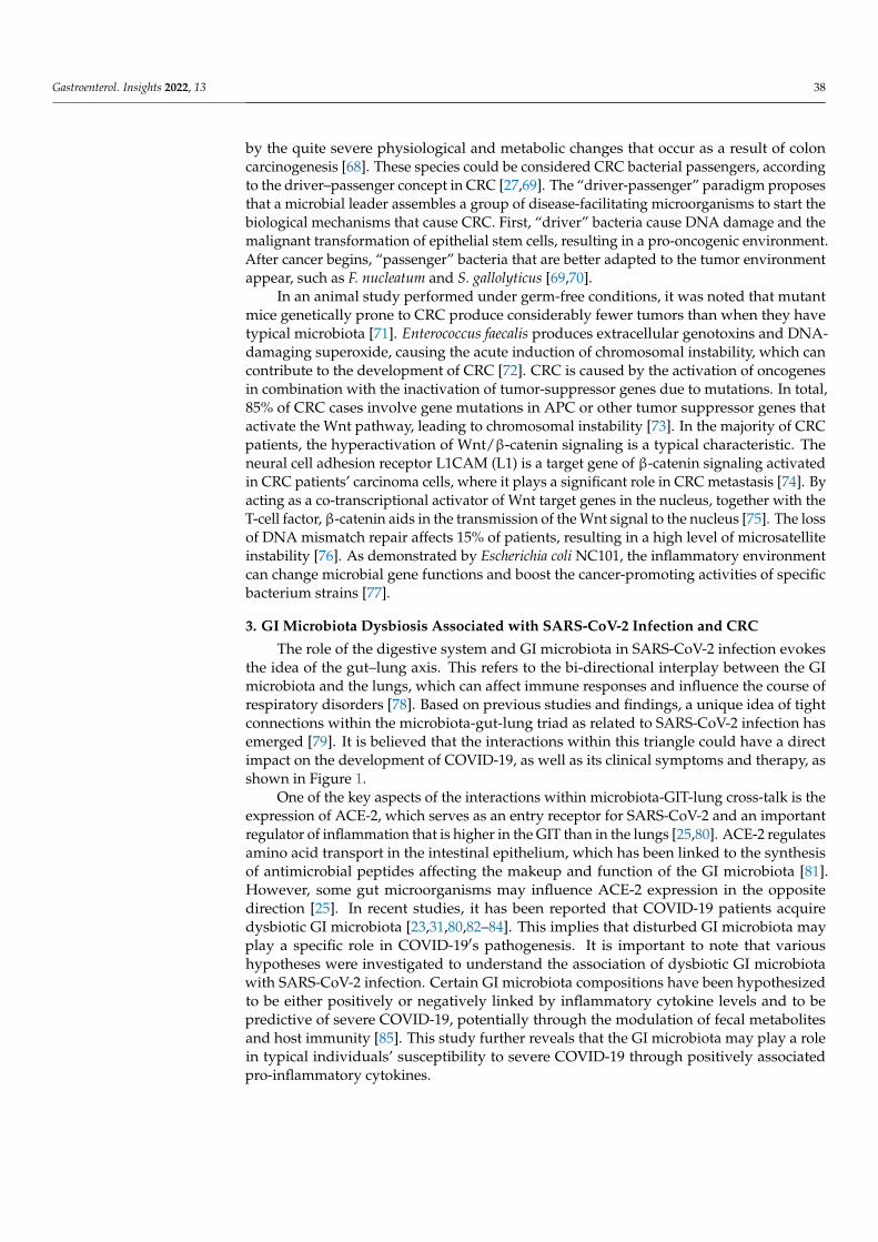
3. GI Microbiota Dysbiosis Associated with SARS-CoV-2 Infection and CRC
The role of the digestive system and GI microbiota in SARS-CoV-2 infection evokesthe idea of the gut–lung axis. This refers to the bi-directional interplay between the GImicrobiota and the lungs, which can affect immune responses and influence the course ofrespiratory disorders [78]. Based on previous studies and findings, a unique idea of tightconnections within the microbiota-gut-lung triad as related to SARS-CoV-2 infection hasemerged [79]. It is believed that the interactions within this triangle could have a directimpact on the development of COVID-19, as well as its clinical symptoms and therapy, asshown in Figure 1.
One of the key aspects of the interactions within microbiota-GIT-lung cross-talk is theexpression of ACE-2, which serves as an entry receptor for SARS-CoV-2 and an importantregulator of inflammation that is higher in the GIT than in the lungs [25,80]. ACE-2 regulatesamino acid transport in the intestinal epithelium, which has been linked to the synthesisof antimicrobial peptides affecting the makeup and function of the GI microbiota [81].However, some gut microorganisms may influence ACE-2 expression in the oppositedirection [25]. In recent studies, it has been reported that COVID-19 patients acquiredysbiotic GI microbiota [23,31,80,82–84]. This implies that disturbed GI microbiota mayplay a specific role in COVID-19′s pathogenesis. It is important to note that varioushypotheses were investigated to understand the association of dysbiotic GI microbiotawith SARS-CoV-2 infection. Certain GI microbiota compositions have been hypothesizedto be either positively or negatively linked by inflammatory cytokine levels and to bepredictive of severe COVID-19, potentially through the modulation of fecal metabolitesand host immunity [85]. This study further reveals that the GI microbiota may play a rolein typical individuals’ susceptibility to severe COVID-19 through positively associatedpro-inflammatory cytokines.

Gastroenterol. Insights 2022, 13 39
Figure 1. The interaction within the microbiota-GIT-lung could affect the development of COVID-19;hence, potential therapy through probiotic intervention could maintain microbial balance and im-mune modulation.
SARS-CoV-2 and COVID-19 severity were linked to alterations in the fecal microbiota.During or throughout hospitalization, patients with COVID-19 had significant abnormal-ities in their fecal microbiota compared to controls, characterized by the enrichment ofopportunistic pathogens and the loss of beneficial commensals [25]. It was further reportedthat Bacteroides sp. reduce the expression of ACE-2 in the mouse gut, which is inverselyrelated to SARS-CoV-2 burden in the patient’s fecal material. Even after SARS-CoV-2clearance (as indicated by throat swabs) and the resolution of respiratory symptoms, de-pleted symbionts, and gut dysbiosis persisted [25]. Bacteroides and Streptococcus genus werenegatively associated with most pro-inflammatory factors. The fecal metabolomics anal-ysis of the gut microbial from a COVID-19 patient was linked to amino acid metabolism,particularly the aminoacyl-tRNA, arginine, valine, leucine, and isoleucine biosynthesispathways [85]. A deficiency in or insufficiency of amino acids results in the depletion ofthe available aminoacylated tRNA, which is essential for host immune response [86,87];hence, the pro-inflammatory response induced by cytokines was significantly reduced [88].As these amino acids play a key role in immunoregulation and enhancing intestinal devel-opment [89], they may invariably affect GI microbiota in COVID-19 patients and CRC.
ACE-2 and TMPRSS2 expressions have been linked to a variety of bacterial generasuch as Chlamydia, which is the microbiota that has been found to be the most stronglypositively correlated with ACE-2 expression in CRC patients [90]. Given that ACE-2 ishighly expressed in the ileum and colon, the importance of ACE-2 is key to maintainingdietary amino acid balance and innate immunity [7]. In SARS-CoV-2 infection, the virusis attached to the host’s ACE-2 receptor, with the upper airway and lungs being thepredominant sites of infection. On the other hand, studies have shown that the intestinalenterocytes at the epithelial layer and the colon epithelial cells have the highest expressionof ACE-2 in the human body. SARS-CoV-2 replication is aided by their support, culminatingin GI barrier disruption [11,91–93], and possibly in CRC. Although the influence of the gutmicrobiota on COVID-19 risk in CRC patients remains poorly understood, the possiblemechanisms targeting microbial dysbiosis should be further investigated [94].
During influenza infection, the inducement of interferons type I promotes the re-duction of obligatory anaerobic bacteria and the enrichment of Proteobacteria in the gut,resulting in a “dysbiotic” milieu [95]. Interferons have been demonstrated to decreaseantimicrobial and inflammatory responses in the GIT during Salmonella-induced colitis.This has been linked to increased Salmonella intestinal colonization and dissemination, arisk factor for CRC [96]. Salmonella can lead to protracted intestinal infection, dysbiotic gut

Gastroenterol. Insights 2022, 13 40
microbiota, and chronic inflammation, all of which can lead to DNA damage and chromo-some instability or epigenetic change. Salmonella effector proteins activate cancer-relatedsignaling pathways. They promote the Wnt/-catenin signaling pathway during persistentinfection, causing host cell change. Bacterial proteins cause leaky gut, microbiota imbalance,and inflammation, all of which contribute to the development of CRC [96]. Furthermore, inHINI influenza infection, interleukin 17A (IL-17A) signaling enhanced fast viral infiltrationof the lungs by pleural cavity B-1a cells via the increase in Blimp-1 expression and NF-kBactivation in B-1a cells. IL-17A deficit resulted in highly diminished B-1a-derived antibodyproduction in the respiratory tract, leading to viral clearance deficiencies [97,98]. In CRCpatients’ tumor tissue samples, IL-17 immune cells were discovered in the majority ofsamples and the lamina propria of homologous normal mucosa, whereas they were rarelyor not observed in normal mucosa in typical individuals. In addition, the gene amplifica-tion of Bacteroides was substantially detected at a higher level in tumor tissue comparedto normal homologous tissue [63]. This implies an association between the IL-17 immunecells and Bacteroides in the mucosa cells causing dysbiosis in CRC patients. It was furtherreported that it was unclear why there was a link between Bacteroides density elevationand malignant CRC, as measured by qPCR [63]. It is noteworthy that the activation of pro-inflammatory and immunological cells in the colon mucosa is crucial in the developmentof cancer, as well as in the development of severe cases of COVID-19. Some GIT microbiotamembers may influence host mucosal regulatory T-cell responses involving Th17 cells. Asa result, T-cell activation may be linked to a shift in mucosal IL-17 caused by Bacteroides, asseen in animal models [99,100]. These findings support the presence of a skewed immuneresponse in CRC tissues, with IL-17 overproduction aggravating the disease, which wasmost likely caused by Bacteroides [101,102].
The interaction between the host immune environment and CRC or SARS-CoV-2infection uses similar mechanisms, such as hypercoagulability, dysregulated immuneresponse, elevated cytokine levels, altered expression of ACE-2 and TMPRSS2, and pro-thrombotic states. This throws the human body into disarray and may exacerbate theeffects of SARS-CoV-2 in some cancer patients [103]. Numerous infiltrating plasma cellsand lymphocytes with interstitial edema were detected in the lamina propria of the stom-ach, duodenum, and rectum [17]. Most patients infected with SARS-CoV-2 have mild GIsymptoms and a good prognosis after infection, indicating that the immune function isa strong defense against this virus. Seven to fourteen days after the onset of symptoms,lymphopenia (changes in T lymphocytes) is commonly observed, with an increase in IL-6and other inflammatory cytokines (pneumonia phase). Lymphopenia and cytokine stormsyndrome lead to disease development and a poor prognosis. Lymphocytes are principallyresponsible for immunological responses to viral infections. However, within the first fewdays of infection, which is a critical stage, the rate of immune response and the level oflymphocytes produced may not be sufficient to combat or immunosuppress the rate ofreplication of the virus, especially in the case of SARS-CoV-2, which is new to the hostbody. This implies that a higher number of lymphocytes is required to compete againstthe virus [43], irrespective of how quickly the virus or T cell replicates. Although there arevarious ways for the immune defense function to eradicate infections, it is noteworthy thatif the immune response is effective, viral suppression occurs. However, this may not occurif the patient has other co-morbidities, including cancer [104]. The immune dysregulationproduced by SARS-CoV-2 could result in even more serious problems for an already fragilepopulation [105].
The gut microbiota has been linked to the development of CRC. The SARS-CoV-2infection causes changes in the gut microbiota, including the enrichment of opportunisticpathogens, the depletion of beneficial commensals, an overall drop in microbial diversity,and a loss of butyrate-producing bacteria. The increased expression of CRC carcino-genesis markers, tumor immunosuppression, and inflammation induction produced bySARS-CoV-2 infection may exacerbate CRC progression, resulting in gut barrier breakdownand the worsening of CRC advancement [105]. Regardless of the clinical stage of disease,
Gastroenterol. Insights 2022, 13 41
patients with CRC may be at a high risk of contracting COVID-19 and are crucial protectiontargets in epidemic prevention. Although further validation of clinical data is needed, thesefindings are of practical importance. Patients with clinically mild or moderate COVID-19with a diagnosis of CRC should be paid special attention because of a possible longercourse of the disease or a higher risk of severe infection. Although further studies arerequired, this review lays the groundwork for the influence and impact of SARS-CoV-2 onthe progression of CRC.
4. Probiotics, GI Microbiota, CRC, and RTIs
The term “probiotic” includes a large range of microbial organisms, main bacteria suchas lactic acid bacteria (LAB), non-lactic acid bacteria, and yeasts. LAB include the generaLactobacillus, Lactococcus, Streptococcus, Enterococcus, Leuconostoc, and Pediococcus [44,106].Non-lactic acid bacteria include Escherichia coli Nissle and some yeasts, such as Saccha-romyces cerevisiae and Saccharomyces boulardii. The most commonly used probiotic mi-croorganisms associated with the human GIT are members of the genera Lactobacillus andBifidobacterium. Species of the genus Lactobacillus include L. acidophilus, L. casei, L. reuteri,L. rhamnosus, L. gasseri, L. Brevis, L. amylovorous, L. crispatus, L. johnsonii, L. paracasei, andL. Plantarum, while the commonly used probiotics of the genus Bifidobacterium includeB. longum, B. lactis, B. bifidum, B. infantis, and B. breve [107]. The beneficial effects of probi-otics are strain-specific [108], which means the health benefits vary for different strains ofprobiotics. It is important to know that a considerable amount of viable probiotics shouldbe consumed or administered by the host for effective functionality. The standard for anyprobiotic products must contain a minimum of 106–107 cfu/g products per day [109,110].The ability of probiotic bacteria to survive, multiply, and become metabolically stable inthe GIT strongly determines the benefits derived by the host. The benefits associated withprobiotics can be of therapeutic (protective) or nutritional significance, depending on theirmode of action [38].
4.1. The Effect of Probiotics on CRC and Other GIT Disorders
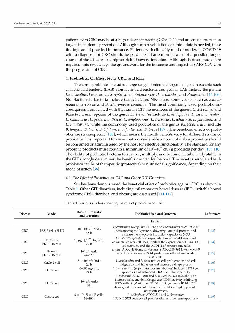
Studies have demonstrated the beneficial effect of probiotics against CRC, as shown inTable 1. Other GIT disorders, including inflammatory bowel disease (IBD), irritable bowelsyndrome (IBS), diarrhea, and obesity, are discussed [111,112].
Table 1. Various studies showing the role of probiotics on CRC.
Disease Model Dose of Probioticand Duration Probiotic Used and Outcome References
In vitro
CRC LS513 cell + 5-FU 106–109 cfu/mL;48 h
Lactobacillus acidophilus CL1285 and Lactobacillus casei LBC80Ractivate caspase-3 protein, downregulate p21 protein, and
increase the apoptosis induction capacity of 5-FU.[113]
CRC HT-29 andHCT-116 cells
10 µg (≥107 cfu/mL);72 h
Lactobacillus plantarum supernatant inhibits 5-FU-resistantcolorectal cancer cell lines, inhibits the expression of CD44, 133,
166 markers, and the ALDH1 of cancer stem cells.[114]
CRC HumanHCT-116 cells
109 cfu/mL;24–72 h
L. casei ATCC 4356 and L. rhamnosus ATCC 39,392 lower MMP-9activity and increase ZO-1 protein in cultured metastatic
CRC cells.[115]
CRC CaCo-2 cell 5 × 10⁶ cfu/mL;24 h
L. acidophilus and L. casei reduce cell proliferation and cellmigration and invasion and increase cell apoptosis. [116]
CRC HT29 cell 0–100 ng/mL;24 h
P. freudenreichii (supernatant or metabolites) induced HT29 cellapoptosis and enhanced TRAIL cytotoxic activity. [117]
CRC HT29 cell 109 cfu/mL;8 h
L. johnsonii BCRC17010 and L. reuteri BCRC14625 show anincrease in lactate dehydrogenase (LDH) activity inhibitingHT29 cells. L. plantarum PM153 and L. johnsonii BCRC17010
show good adhesion ability while the latter display potentialapoptotic effects.
[118]
CRC Caco-2 cell 6 × 10³–5 × 10⁶ cells;24–48 h
L. acidophilus ATCC 314 and L. fermentumNCIMB 5221 reduce cell proliferation and increase apoptosis. [119]
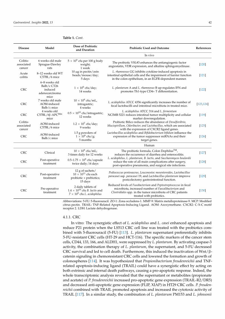
Gastroenterol. Insights 2022, 13 42
Table 1. Cont.
Disease Model Dose of Probioticand Duration Probiotic Used and Outcome References
In vivo
Colitis-associated
cancer
6 weeks old maleSprague-Dawley
rats
5 × 10⁹ cfu per 100 g bodyweight;1 week
The probiotic VSL#3 enhances the antiangiogenic factorangiostatin, VDR expression, and alkaline sphingomyelinase. [120]
Acutecolitis
8–12 weeks old WTC57BL/6 mice
10 µg in pectin/zeinbeads/mouse/day;
5 days
L. rhamnosus GG inhibits cytokine-induced apoptosis inintestinal epithelial cells and the impairment of barrier function
in the colon epithelium, in an EGFR-dependent manner.[121]
CRC
6–8 weeks oldBalb/c CT26
inducedadenocarcinoma
mice
1 × 10⁹ cfu/day;14 weeks
L. plantarum A and L. rhamnosus B up-regulates IFN andpromotes Th1-type CD4+ T differentiation. [122]
CRC7 weeks old male
AOM-inducedBalb/c mice
10 × 109 cfu/mL,intragastric;
4 weeks
L. acidophilus ATCC 4356 significantly increases the number offecal lactobacilli and intestinal microbiota in treated mice. [123,124]
CRC4 weeks old
C57BL/6J–APCMin
mice
0.5 × 10¹⁰ cfu/intragastric;12 weeks
L. acidophilus ATCC 314 and L. fermentumNCIMB 5221 reduces intestinal tumor multiplicity and cellular
marker downregulation.[119]
Colitis-associated
cancer
AOM-inducedC57BL/6 mice
1.2 × 10⁷ cfu/day;9 weeks
Probiotic Bifico reduces the abundance of Desulfovibrio,Mucispirillum, Odoribacter, and Lactobacillus, which are associated
with the expression of CXCR2 ligand genes.[125]
CRC AOM-inducedBalb/c mice
1.5 g powders of1 × 109 cfu/g;
5 months
Lactobacillus acidophilus and Bifidobacterium bifidum influence theexpression of the tumor suppressor miRNAs and their
target genes.[126]
Human
CRC Clinical 10 × 109 cfu/mL;3 times daily for 12 weeks
The probiotic formula, Colon DophilusTM,reduces the occurrence of diarrhea and enterocolitis.
[127]
CRC Post-operativetreatment
0.5–1.75 × 10⁹ cfu/capsuletwice daily; 14 days
L. acidophilus, L. plantarum, B. lactis, and Saccharomyces boulardiireduce the rate of all main complications after surgery,post-operative pneumonia, and surgical site infections.
[128]
CRC Post-operativetreatment
12 g of sachets/10 × 1011 cfu each
probiotic + prebiotics;15 days
Pediococcus pentosaceus, Leuconostoc mesenteroides, Lactobacillusparacasei ssp. paracasei 19, and Lactobacillus plantarum improve
postcolectomy gastrointestinal function.[129]
CRC Pre-operativetreatment
2 daily tablets of1.4 × 1010 cfu B. lactis and7 × 109 cfu L. acidophilus
Reduced levels of Fusobacterium and Peptostreptococcus in fecalmicrobiota, increased number of Faecalibacterium and
Clostridiales spp. in the tumor microbiota of CRC patientstreated with probiotics.
[130]
Abbreviations: 5-FU: 5-fluorouracil. ZO-1: Zona occludens-1. MMP-9: Matrix metalloproteinase-9. MCP: Modifiedcitrus pectin. TRAIL: TNF-Related Apoptosis-Inducing Ligand. AOM: Azoxymethane. CXCR2: C-X-C motifreceptor 2. LDH: Lactate dehydrogenase.
4.1.1. CRC
In vitro: The synergistic effect of L. acidophilus and L. casei enhanced apoptosis andreduce P21 protein when the LS513 CRC cell line was treated with the probiotics com-bined with 5-fluorouracil (5-FU) [113]. L. plantarum supernatant preferentially inhibits5-FU-resistant CRC cells (HT-29 and HCT-116). The specific markers of the cancer stemcells, CD44, 133, 166, and ALDH1, were suppressed by L. plantarum. By activating caspase-3activity, the combination therapy of L. plantarum, the supernatant, and 5-FU decreasedCRC survival and led to cell death. Furthermore, this induced the inactivation of Wnt/β-catenin signaling in chemoresistant CRC cells and lowered the formation and growth ofcolonospheres [114]. It was hypothesized that Propionibacterium freudenreichii and TNF-related apoptosis-inducing ligand (TRAIL) could have a synergistic effect by acting onboth extrinsic and internal death pathways, causing a pro-apoptotic response. Indeed, thewhole transcriptomic analysis revealed that the supernatant or metabolites (propionateand acetate) of P. freudenreichii increased pro-apoptotic gene expression (TRAIL-R2/DR5)and decreased anti-apoptotic gene expression (FLIP, XIAP) in HT29 CRC cells. P. freuden-reichii combined with TRAIL promoted apoptosis and increased the cytotoxic activity ofTRAIL [117]. In a similar study, the combination of L. plantarum PM153 and L. johnsonii

Gastroenterol. Insights 2022, 13 43
BCRC17010 with supernatants inhibits the proliferation of HT29 cells by inducing thesecretion of nitric oxide and elevated levels of LDH [118]. The synergistic anti-cancereffect of L. acidophilus ATCC 314 and L. fermentum NCIMB 5221 significantly increasesapoptosis (p < 0.001) as well as offering significant protection to normal colon cell growthfrom toxic treatment (18.6 ± 9.8%, p = 0.001). Both the probiotic LABs influenced intestinaltumorigenesis by lowering intestinal tumor multiplicity and downregulating the expres-sion levels of Ki-67 and β-catenin markers [131]. HCT-116 cells were treated with cell-freesupernatants from L. casei, L. rhamnosus, or Bacteroides thetaiotaomicron (a gut commensal).This altered the matrix metalloproteinase-9 activity and levels of the tight junction protein,zona occludens-1, leading to the inhibition of colon cancer cell migration [115]. Caco-2CRC cells treated with the probiotics, L. acidophilus ATCC 4356 and L. casei ATCC 39,392,increase cell apoptosis and reduce cell proliferation, migration, and invasion. However, nosignificant effect on cell necrosis was noted [132].
In vivo: Our previous study reported that the daily intake of probiotic L. acidophilusATCC 4356 increases the total number of fecal lactobacilli by 10.2% (0.8 ± 0.08 log10 cfu/g;p < 0.05) from the initial fecal count after 4 weeks of once-daily probiotic consumption [124].Furthermore, the genomic sequence identification of the fecal lactobacilli showed an in-crease in the number of bacteria in the treated Azoxymethane (AOM)-induced colon tumorBalb/c mouse model compared to the control. This implies that probiotics enhanced thestimulation and growth of the colonic microbiota in the CRC mouse model used in thestudy [123]. In a further study, the percentage of colonic pre-cancerous lesions in theprobiotic-treated groups was low (20%) compared to the untreated control group (40–50%).In addition, there was a significant reduction in the tumors in the probiotic-treated groupscompared to the control group (p < 0.05) [133]. It was noted that treatment with the probi-otic VSL#3 consisting of eight different strains, B breve, B. infantis, B. longum, L. acidophilus,L. bulgaricus, L. casei, L. plantarum, and S. salivarus ssp thermophiles, prevents the develop-ment of tumors and high-grade dysplasia in the proximal and mid-colon in rats. Thiscorrelates with the decreased richness and diversity of mucosally adherent microbiota inthe colon. This implies that VSL#3 can reduce a number of inflammatory-related factors,preventing the onset of dysplasia and cancer [120]. The potential effect of L. Plantarum Aand L. rhamnosus B on the inducement of anti-tumor immune responses was demonstratedby pre-inoculating Balb/c mice subcutaneously with CT26 murine adenocarcinoma cells.In comparison to mice treated with L. rhamnosus, the oral administration of L. plantarumdecreased CT26 cell development in the Balb/c mice and prolonged the survival time oftumor-bearing mice. The L. plantarum provided protective immunity against the challengewith CT26 cells by activating the effector functions of CD8+ and natural killer cell infiltra-tion into cancer tissue. This up-regulated interferon (but not IL-4 or IL-17) production andpromoted Th1-type CD4+ T differentiation [122]. Probiotic Bifico containing 1.0 × 10⁷ cfulyophilized B. longum, L. acidophilus, and E. faecalis decreased tumor development and low-ered intestinal inflammation. Furthermore, a collection of genes, including CXCL1, CXCL2,CXCL3, and CXCL5, were identified as possible Bifico therapy targets. According to 16SrRNA sequencing, Bifico lowered the abundance of Desulfovibrio, Mucispirillum, Odoribacter,and Lactobacillus, which was substantially related to the expression of CXC motif receptor2 ligand genes [125]. Another study reported that L. acidophilus and B. bifidum affected theexpression of miRNAs 135b, 26b, 18a, and 155, as well as their target genes, such as APC,PTEN, KRAS, and PU.1, in an AOM-induced CRC mouse model. The expressions of thetumor suppressors miR-135b, miR-155, and KRAS all increased [126].
Human: The microbiota composition of patients with colon cancer was compared tothat of non-neoplastic controls. Patients with normal mucosa and tumors were administered2 tablets of 1.4 × 10¹⁰ cfu of B. lactis Bl-04 and 7 × 10⁹ cfu of L. acidophilus NCFM daily.The 16S rRNA gene amplicon sequencing of fecal and colonic samples showed increasedFusobacterium, Selenomonas, and Peptostreptococcus in the tumor microbiota compared tothe control [130]. A meta-analysis was conducted to determine the effect of probiotics,including Bifidobacterium and Escherichia, on the intestinal mucosa barrier in CRC after

Gastroenterol. Insights 2022, 13 44
surgery. It was observed that the probiotics effectively protected the intestinal mucosa’sphysical and biological barriers in the patients with CRC after operation [134]. The immune-modulatory effect of probiotics on the intestinal gut is a biotherapeutic/preventive strategy,as probiotics inhibit the colonization of the intestinal microbiota and the translocation ofpathogens to other sites [135–138]. Most diseases that are common to the human largebowel emanate from the distal colon, most notably CRC. However, it has been notedthat proximal CRC is also increasing. A gene sequencing comparison study showed that59.6% of the bacterial genera were identified in the proximal colon and 71.6% in the distalcolon of the CRC. This suggests that microbial diversity increases from the proximal colonto the distal colon in rectal malignancy. Prevotella, Pyramidobacterium, Selenomonas, andPeptostreptoccus were found in greater numbers in proximal tumors, while Fusobacterium,Escherichia-Shigella, and Leptotrichia were relatively prevalent in distal CRC [27]. Patientsundergoing CRC surgery were enrolled in a randomized, double-blind, placebo-controlledstudy. Capsules of placebo or formulation including L. acidophilus LA-5, L. plantarum,B. lactis BB-12, and Saccharomyces boulardii were given a day before surgery and continuedfor another 15 days post-operatively. On day 4, the gene expression and cytokine levelsin the blood were examined. The probiotics considerably reduced the rate of all the maincomplications after surgery, as well as reducing post-operative pneumonia and surgicalsite infections [128].
4.1.2. IBD
IBD, including Crohn’s disease and ulcerative colitis, is a group of disorders charac-terized by chronic or recurrent inflammation of the mucosal lining marked by an auto-immune response, usually by the body’s immune system. Probiotic administration reg-ulates innate inflammatory responses in the mucosa through the modulation of the gutmicrobiota composition and its effect on the epithelial and T cells on the surface of the lam-ina propria of the gut. It was demonstrated that the colon-specific probiotic, L. rhamnosusGG, suppresses inflammation and reduces apoptosis by activating the epithelial growthfactor receptor [121]. The secretion of IgA antibody is one of the basic immunologicalresponses induced by Bifidobacterium sp.’s binding to specific receptors on the intestinalepithelial surface and subsequent release into the intestinal lumen [52]. L. salivarius Ls33showed an anti-inflammatory effect in a colitis mouse model through the recognitionof bacterial peptidoglycan and protein-derived mucopeptides [139]. Probiotics such asnon-pathogenic Escherichia coli, Bifidobacterium sp., and Saccharomyces boulardii have shownefficacy in reducing the post-operative recurrence and relapse in Crohn’s disease. A meta-analysis evaluation was performed to justify the effect of probiotics on Crohn’s diseasecompared to a placebo for the prevention of post-operative recurrence, which effectivelydefines the preventative and therapeutic role of probiotics [140].
4.1.3. IBS
IBS is a functional disorder in the colon that is characterized by symptoms of abdomi-nal discomfort, usually related to disturbed defecation (motility of the colon), which causesa drastic reduction in beneficial gut bacteria. L. rhamnosus GG was reported to have littleeffect on IBS, while L. plantarum 299 V had a significant beneficial effect [141]. Notablestability in the microbiota composition reduces abdominal pain and bowel distension whendifferent species of probiotic supplements were administered, including L. rhamnosus Lc705,L. rhamnosus GG, B. animalis ssp. lactis Bb12, and P. freudenreichii ssp. Shermanii JS [142].A clinical trial meta-analysis was conducted on the effectiveness of probiotics in the pre-vention of IBS and probiotic therapy. For instance, probiotic VSL#3 reduces the clinicalsymptoms and abdominal pain of IBS [143]. Multispecies probiotic supplementation onIBS stimulates the microbial composition of the colon compared to single species, leadingto improved bowel movement and replenishing the loss of beneficial gut bacteria.
Gastroenterol. Insights 2022, 13 45
4.1.4. Diarrhea
The efficacy of probiotics in cases of infectious diarrhea has reduced the occurrenceof diarrheal episodes, early symptoms, and even rotavirus infection [144–146]. The ad-ministration of L. rhamnosus GG associated with oral rehydration therapy, once or twicedaily, has effectively helped in the treatment of rotavirus-associated diarrhea. It furtherreduces the presence of the virus in stools, thus offering an effective strategy to control thespread of nosocomial gastrointestinal infection [147,148]. The treatment of gastroenteritisby the probiotic, L. paracasei ST1, improved non-rotavirus diarrhea condition in childrenbut did not affect rotavirus gastroenteritis [149]. Clostridium difficile caused about 10–20%of antibiotic-associated diarrhea. A double-blind placebo-controlled study showed that theoral consumption of a probiotic preparation of L. bulgaricus, L. casei, and S. thermophilusinvariably reduced the incidence of C. difficile-associated diarrhea [143]. A study wasconducted to determine how efficient probiotics are at preventing irinotecan-induced di-arrhea by lowering intestinal β-D-glucuronidase activity. The probiotic formula, ColonDophilusTM, was administered orally at a dose of 10 × 109 cfu/mL of bacteria three timesdaily for 12 weeks to CRC patients starting a new line of irinotecan-based therapy; theresults were compared with those from a placebo-treated group [127].
4.1.5. Obesity
Obesity is closely related in terms of risk factors for CRC. It was suggested that infantspresenting with a low level of Bifidobacterium sp. and a higher number of Staphylococcusaureus in their stool are at a higher risk of obesity. A mouse model study demonstratedthat the transplantation of microbial communities within the gut could manipulate thepropensity for the deposition of fat. The administration of probiotics influences the in-testinal microbiota, inhibiting susceptibility to obesity. This suggests a possible probiotictherapy that may prove to be a strategy for controlling childhood obesity [137,150]. In arandomized placebo-controlled trial, fermented milk containing L. gasseri SBT2055 wasadministered to healthy and obese patients. A decrease in fat, body weight, and mass indexwas observed compared to the control group [151]. Probiotics interact with the endogenousbacteria in the gut by modifying or regulating fat metabolic pathways. Although the energyintake was less affected, the administration of L. rhamnosus PL60 in diet-induced obese miceled to a significant loss of body weight by reducing the mass of white adipose tissue [135].
4.1.6. Possible Mechanism of Probiotics
Even though various studies have sought to explain the mechanism of probiotics’anticarcinogenic properties, a definite mechanism for probiotic anti-CRC activity is yet to bediscovered [152–154]. Possible mechanisms may involve the cell cycle, apoptosis, reactiveoxygen species (ROS), the production of specific bacterial metabolic enzymes, and effects onhost metabolism. However, other mechanisms were also suggested, including the alterationof quantitative and/or qualitative intestinal microbiota involved in the production ofcarcinogens and promoters; the alteration of the physicochemical conditions in the colon;the production of anti-tumourigenic or anti-mutagenic compounds; the enhancement ofthe host immune response system; the effect on host physiology; and the binding anddegradation of potential carcinogens, including mutagenic compounds [131,152–156].
The G1 phase is an important early phase in the cell cycle that is necessary for cellproliferation. L. rhamnosus GG induces an anti-proliferative effect in cancer cell lines byreducing the biosynthesis of polyamines. These may also be a result of the Lactobacilli’sability to adhere to cells, inducing apoptosis, which eventually may prevent the prolifera-tion of colon cancer [157]. The presence of reactive oxygen species (ROS) is a prominentevent in colon inflammation. The antioxidant properties of probiotic strains can inhibit/orreduce this effect and thereby increase probiotic gut colonization [158–160]. The interactionbetween the epithelial cells and the microbiota gut is an active process that can be inducedby the presence of probiotics in the production of pro-inflammatory cytokines. Interleukin12 (IL-12) can be induced after the activation of the innate immune system by some Lacto-

Gastroenterol. Insights 2022, 13 46
bacilli strains stimulating dendritic cells, native T cells in the lamina propria of the gut, andmacrophages [154,161].
The probiotic effect of the alteration of quantitative intestinal microbiota has been sug-gested. A daily intake of probiotic L. acidophilus ATCC 4356 with MCP altered the numberof colonic microbiota in the CRC mouse model. It is plausible that the MCP modifies thefunctionality and physiological properties of the probiotic during gastric transit, causingadhesion/colonization within the microbiota environment [123,124]. Chronic inflammation,which disrupts the gut microbiota, is one of the fatal factors linked to the development ofCRC [69]. The formation of immunity against invading pathogens and the maturation ofthe immune system requires a healthy gut microbiota. Probiotic supplementation improvesthe immune system by modulating the release of anti-inflammatory cytokines and theirrelated regulatory genes [162,163].
One important function of GI microbiota is the ability to catabolize complex polysac-charides into short-chain fatty acids (SCFAs), such as butyrate, lactic acids, acetic acid, andpropionate, which are major energy sources for the epithelium cells of the colon [164–166].In our previous study, using an azoxymethane-induced colon cancer mouse model, weobserved that the fermentation of modified citrus pectin and starch by L. acidophilus ATCC4356 and intestinal microbiota possibly increased the production of SFCAs, gases, andbutyrate [124]. Fecal lactobacilli utilize butyrate as the preferred energy source for theirgrowth and the proliferation of colonic epithelial cells. These SFCAs reduce the effect ofbile in the intestine by initiating bile salt hydrolase, which deconjugates bile acid [22,167].Previous studies reported that Lactobacilli in the GI lower the risk of CRC by displacingother bacteria, such as Bacteroides and Clostridium genera, that may produce fecal enzymesor toxins (carcinogen) during metabolism [168,169]. L. plantarum LS/07 was reported tohave a protective effect on colon cancer-induced DNA damage in the colon cells of rats.The metabolic activity of the Lactobacilli lowers the level of colonic/fecal enzymes, such asβ-glucosidase and β-glucuronidase, which hydrolyze many glucuronide-releasing carcino-gens in the intestinal tract, causing CRC [170,171]. The ability of this colonic microbiota toproduce SCFA and low levels of some colonic enzymes, such as β-glucoronidase, is sug-gested as the main process that prevents CRC [172]. The Bifidobacterium genome has beenshown to contain some metabolic enzymes that the bacteria produce to alter the nutrientcomposition of the environment. This invariably adjusts their physiology; consequently,they adapt to new conditions. B. adolescentis SPM0212 was reported to exhibit bacterialenzyme activities and some anti-cancer properties, including: the inhibition of the prolif-eration of certain human cancer cell lines; the inhibition of harmful fecal enzymes, suchas β-glucuronidase, β-glucosidase, tryptophanase, and urease; and the dose-dependentinhibition of the production of TNF-α [173].
Bacterial enzymes, such as β-glucuronidase, can hydrolyze many glucuronides, therebyreleasing metabolites into the intestinal tract [174]. Based on animal and human studies,the consumption of LABs can reduce fecal enzyme levels, which may be involved in theformation of toxins. In one study, the effect of L. acidophilus NCFM and L. acidophilusN-2 on 21 healthy people was evaluated for 10 days by checking the activity levels ofβ-glucuronidase, nitroreductase, and azoreductase, which resulted in a decrease in specificactivities of these enzymes. The administration of L. rhamnosus LC705 and Propionibac-terium freudenreichii ssp. Shermanii JS to 38 healthy men for 4 weeks decreased the activityof β-glucosidase and increased the fecal count of Lactobacilli and Propionibacteria in thesubjects [175]. GIT is dominated by the Lactobacilli sp., which contributes to the metabolicactivities taking place in this part of the host body. A hypothesis concerning colon carcino-genesis is that bile acids in the aqueous phase of feces exert cytotoxic effects on colonicepithelium cells, thereby causing an increase in cell proliferation in the intestine [176,177].Some studies have shown significant decreases in bile acid with the intake of Lactobacilli sp.Mutagenic compounds in food diets bind to the cells of Lactobacilli strains and the intestinalwall after exposure, resulting in a reduction in the ratio of bound to free toxins within the
Gastroenterol. Insights 2022, 13 47
intestine. The colonic mucosa can absorb mutagenic compounds and pass them into thebloodstream as either modified or unmodified metabolites from the intestinal lumen [152].
4.2. The Potential Effect of Probiotics on RTIs, including SARS-CoV-2 Infection, andPossible Mechanisms
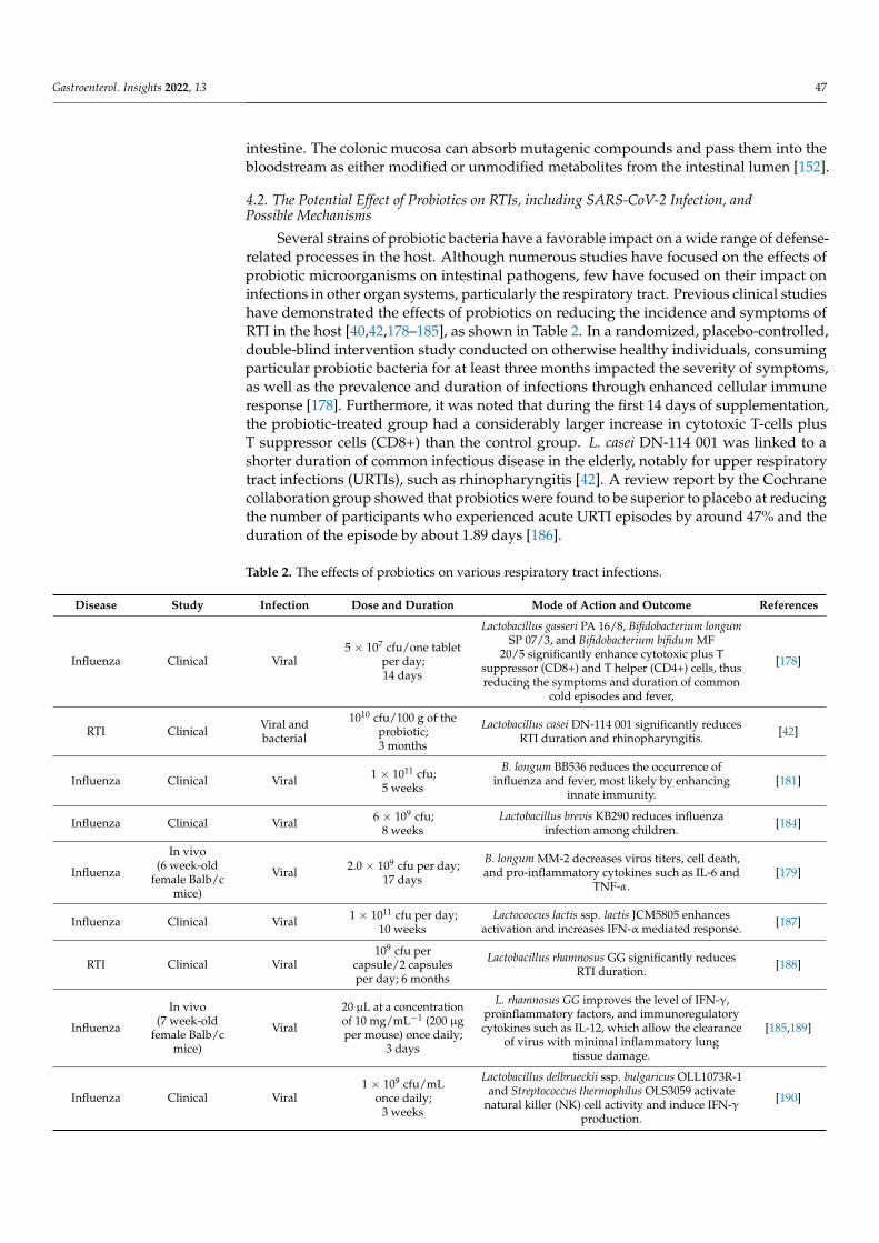
Several strains of probiotic bacteria have a favorable impact on a wide range of defense-related processes in the host. Although numerous studies have focused on the effects ofprobiotic microorganisms on intestinal pathogens, few have focused on their impact oninfections in other organ systems, particularly the respiratory tract. Previous clinical studieshave demonstrated the effects of probiotics on reducing the incidence and symptoms ofRTI in the host [40,42,178–185], as shown in Table 2. In a randomized, placebo-controlled,double-blind intervention study conducted on otherwise healthy individuals, consumingparticular probiotic bacteria for at least three months impacted the severity of symptoms,as well as the prevalence and duration of infections through enhanced cellular immuneresponse [178]. Furthermore, it was noted that during the first 14 days of supplementation,the probiotic-treated group had a considerably larger increase in cytotoxic T-cells plusT suppressor cells (CD8+) than the control group. L. casei DN-114 001 was linked to ashorter duration of common infectious disease in the elderly, notably for upper respiratorytract infections (URTIs), such as rhinopharyngitis [42]. A review report by the Cochranecollaboration group showed that probiotics were found to be superior to placebo at reducingthe number of participants who experienced acute URTI episodes by around 47% and theduration of the episode by about 1.89 days [186].
Table 2. The effects of probiotics on various respiratory tract infections.
Disease Study Infection Dose and Duration Mode of Action and Outcome References
Influenza Clinical Viral5 × 107 cfu/one tablet
per day;14 days
Lactobacillus gasseri PA 16/8, Bifidobacterium longumSP 07/3, and Bifidobacterium bifidum MF
20/5 significantly enhance cytotoxic plus Tsuppressor (CD8+) and T helper (CD4+) cells, thusreducing the symptoms and duration of common
cold episodes and fever,
[178]
RTI Clinical Viral andbacterial
1010 cfu/100 g of theprobiotic;3 months
Lactobacillus casei DN-114 001 significantly reducesRTI duration and rhinopharyngitis. [42]
Influenza Clinical Viral 1 × 10¹¹ cfu;5 weeks
B. longum BB536 reduces the occurrence ofinfluenza and fever, most likely by enhancing
innate immunity.[181]
Influenza Clinical Viral 6 × 10⁹ cfu;8 weeks
Lactobacillus brevis KB290 reduces influenzainfection among children. [184]
Influenza
In vivo(6 week-old
female Balb/cmice)
Viral 2.0 × 10⁹ cfu per day;17 days
B. longum MM-2 decreases virus titers, cell death,and pro-inflammatory cytokines such as IL-6 and
TNF-α.[179]
Influenza Clinical Viral 1 × 10¹¹ cfu per day;10 weeks
Lactococcus lactis ssp. lactis JCM5805 enhancesactivation and increases IFN-α mediated response. [187]
RTI Clinical Viral109 cfu per
capsule/2 capsulesper day; 6 months
Lactobacillus rhamnosus GG significantly reducesRTI duration. [188]
Influenza
In vivo(7 week-old
female Balb/cmice)
Viral
20 µL at a concentrationof 10 mg/mL−1 (200 µgper mouse) once daily;
3 days
L. rhamnosus GG improves the level of IFN-γ,proinflammatory factors, and immunoregulatorycytokines such as IL-12, which allow the clearance
of virus with minimal inflammatory lungtissue damage.
[185,189]
Influenza Clinical Viral1 × 109 cfu/mL
once daily;3 weeks
Lactobacillus delbrueckii ssp. bulgaricus OLL1073R-1and Streptococcus thermophilus OLS3059 activate
natural killer (NK) cell activity and induce IFN-γproduction.
[190]

Gastroenterol. Insights 2022, 13 48
Table 2. Cont.
Disease Study Infection Dose and Duration Mode of Action and Outcome References
LRTI andrhinovirusinfection
Clinical Viral109 cfu/capsule/seven
daily doses;60 days
L. rhamnosus GG reduces LRTI and the incidence ofrhinovirus-induced episodes in children. [191]
rhinovirusinfection Clinical Viral ≥2 × 10⁹ cfu/satchet
daily dose; 28 days
B. animalis subspecies lactis Bl-04reduces nasal lavage virus titer and influences the
innate immune response in the nasal cavity.[183]
URTI Clinical Viral 1 × 10⁹ cfu/subjectsonce daily; 6 weeks
L. paracasei subsp. Paracasei and L. casei 431 reducethe frequency of RTI episodes. [192]
CoronavirusIn vitro
(IPEC-J2 cellline)
Viral 2 × 10⁶ recombinantcells; 2 h
L. plantarum enhances the expression levels of IFNstimulated genes, thus suppressing the viral
infection.[180]
COVID-19 In silicodocking Viral
Probiotics-derivedpeptides were dockedtargeting viral proteins
Probiotics-derived polypeptides show strongaffinity binding to the S1-protein receptor-binding
domain of SARS-CoV-2[43]
Abbreviations: RTI: Respiratory tract infection. IL-6: Interleukin 6. TNF-α: Tumor necrosis factor α. IL-10:Interleukin 10. IFN-γ: Interferon γ. IPEC-J2: Intestinal porcine epithelial cell line J2. SARS-CoV-2: Severe acuterespiratory syndrome coronavirus-2. COVID-19: Coronavirus disease 2019.
The relevance of probiotics in the treatment of coronaviruses remains controversialas the mechanisms may not be fully known yet and very little information is available.Based on previous studies and many pieces of clinical evidence for the positive effect ofprobiotics as supplements or adjuvants against primary and/or secondary (bacterial andviral) diseases, the existence of a basic mechanism of action of probiotics in the preventionor reduce SARS-CoV-2 infection and its importance to COVID-19 may not be far-fetched.
4.2.1. Microbial Dysbiosis and GIT–Lung Stability Cross-Talk
The intervention of probiotics in the interactions within microbiota-GIT-lung cross-talk could contribute to the enhancement of the intestinal epithelial barrier, competitionwith pathogens for nutrients, and adherence to the intestinal epithelium. The generationof antimicrobial compounds and the manipulation of the host immune system are allmechanisms that could explain probiotics’ therapeutic success [39], as shown in Figure 2.Many microbial metabolites and endotoxins from the GIT can affect the lungs and viceversa when they cross the GIT–blood barrier. Furthermore, the microbiota can be affectedby lung inflammation [78] through the production of several metabolites with antiviralproperties, such as lactic acid, hydrogen peroxides to stimulate interleukins, natural killercells, and Th1 helper immune responses [193].
Different strains of probiotic revealed a strain-specific potential for improving theintestinal epithelium’s reliability and controlling immunological components. The GITfrom the oral cavity to the rectum is regarded as the principal immunological barrier withthe environment in controlling compound-mediated immune reactions. It has been demon-strated that probiotic microorganisms can connect to an invading virus and thereby inhibitthe viral structural protein from binding to the host cell receptor, preventing viral entryinto human cells [194,195]. Probiotic bacteria cling to the epithelial surface and use stericinterference to prevent viral attachment by competing for specific carbohydrate receptorsor by covering the receptor sites in a nonspecific manner [196]. Recent studies have iden-tified GAL-3 as a binding mediator in the attachment and entrance of viruses, includinginfluenza A H5N1 virus, into the host cell [197,198]. This is in addition to ACE-2, whichhas been identified as a therapeutic target to mitigate SARS-CoV-2 entry and replication inthe host cell [199]. In our previous study, the S-glycoprotein layer of probiotic L. acidophilusATCC 4356 comprising both the amino and carboxyl-terminal domains competed withthe intestinal microbiota to bind to the GAL-3 COOH terminal carbohydrate recognitiondomain in a colonic cancer model. The initiation of the colonization/adhesion of theprobiotic bacteria to specific receptors, such as GAL-3, on the epithelial cell surface ofthe colon may competitively inhibit GAL-3 extracellular matrix interactions in addition

Gastroenterol. Insights 2022, 13 49
to probiotic-GAL-3 binding [133]. As a result, the probiotic significantly increases fecallactobacilli and improves the integrity of the colonic intestinal microbiota [123,124,133].Another study found that probiotic supplementation is an effective method for preventingviral RTIs in pre-term infants throughout their first year of life. The findings revealed thatprobiotic-induced changes in the GI microbiota result in the production of long-lastingeffects that can minimize the incidence of viral RTIs [191].
Gastroenterol. Insights 2022, 13, FOR PEER REVIEW 16
Figure 2. Immunomodulatory response and possible mechanism of probiotics effect in GI dysbiosis associated with SARS-CoV-2 infection and CRC. The potential probiotic effect against the attachment of SARS-CoV-2 to the spike protein receptor may take place through the production of certain metabolites, enzymes, SFCAs, inflammatory cytokines, T-helper cells, and cytotoxic compounds. This intervention could influence microbiota dysbiosis within the areas of infection, such as the lung and the GIT (colon), to stabilize the microbial system, reduce the symptoms or severity of RTI and pro-inflammatory cytokines and, consequently, reduce the cytokine storm effect in SARS-CoV-2 infection.
Different strains of probiotic revealed a strain-specific potential for improving the intestinal epithelium’s reliability and controlling immunological components. The GIT from the oral cavity to the rectum is regarded as the principal immunological barrier with the environment in controlling compound-mediated immune reactions. It has been demonstrated that probiotic microorganisms can connect to an invading virus and thereby inhibit the viral structural protein from binding to the host cell receptor, preventing viral entry into human cells [194,195]. Probiotic bacteria cling to the epithelial surface and use steric interference to prevent viral attachment by competing for specific carbohydrate receptors or by covering the receptor sites in a nonspecific manner [196]. Recent studies have identified GAL-3 as a binding mediator in the attachment and entrance of viruses, including influenza A H5N1 virus, into the host cell [197,198]. This is in addition to ACE-2, which has been identified as a therapeutic target to mitigate SARS-CoV-2 entry and replication in the host cell [199]. In our previous study, the S-glycoprotein layer of probiotic L. acidophilus ATCC 4356 comprising both the amino and carboxyl-terminal domains competed with the intestinal microbiota to bind to the GAL-3 COOH terminal carbohydrate recognition domain in a colonic cancer model. The initiation of the colonization/adhesion of the probiotic bacteria to specific receptors, such as GAL-3, on the epithelial cell surface of the colon may competitively inhibit GAL-3 extracellular matrix interactions in addition to probiotic-GAL-3 binding [133]. As a result, the probiotic significantly increases fecal lactobacilli and improves the integrity of the colonic intestinal microbiota [123,124,133]. Another study found that probiotic supplementation is an effective method for preventing viral RTIs in pre-term infants throughout their first year of life. The findings revealed that probiotic-induced changes in the GI microbiota result in the production of long-lasting effects that can minimize the incidence of viral RTIs [191].
Figure 2. Immunomodulatory response and possible mechanism of probiotics effect in GI dysbiosisassociated with SARS-CoV-2 infection and CRC. The potential probiotic effect against the attachmentof SARS-CoV-2 to the spike protein receptor may take place through the production of certainmetabolites, enzymes, SFCAs, inflammatory cytokines, T-helper cells, and cytotoxic compounds. Thisintervention could influence microbiota dysbiosis within the areas of infection, such as the lung andthe GIT (colon), to stabilize the microbial system, reduce the symptoms or severity of RTI and pro-inflammatory cytokines and, consequently, reduce the cytokine storm effect in SARS-CoV-2 infection.
4.2.2. Immunomodulatory Effects
Some probiotic strains can help prevent bacterial and viral diseases, such as gas-troenteritis, sepsis, and respiratory infections through immunomodulatory responses. Byreducing pro-inflammatory signals and maintaining gut barrier integrity, probiotics canhelp patients maintain immunological homeostasis in the gut and avoid the overactivationof the immune response. Certain strains of Bifidobacterium and Lactobacilli were given tohelp eliminate the influenza virus from the respiratory system with little inflammatorydamage to the lung tissue [185]. The probiotic strains impact the modulation of a sys-temic equilibrium between pro-inflammatory and anti-inflammatory immunoregulatorycytokines [185]. This could be important in preventing acute respiratory diseases, a sig-nificant COVID-19 consequence. A probiotic using L. plantarum DR7 reduced plasmaIFN-γ and TNF-α pro-inflammatory cytokines and stimulated increases in IL-4 and IL-10anti-inflammatory cytokines in a randomized control trial study on upper respiratorytract-infected middle-aged patients [200]. Given the cytokine storm that many COVID-19patients tend to experience, this form of regulation could be crucial. Orally administeredprobiotic strains contribute to the manner in which the immune response emanates fromthe intestine. Probiotics could limit SARS-CoV-2 invasion by boosting butyrate, a fuel forcolonocytes. The immunological response, which is mediated by macrophages, dendriticcells, and the differentiation of CD8+ T lymphocytes into cytotoxic T lymphocytes in thepresence of probiotics, can devastate infection-infected cells [201]. T-helper type 1 (Th1) and
Gastroenterol. Insights 2022, 13 50
type 2 (Th2) cells are produced when probiotics stimulate CD4+ T lymphocytes. Th1 hasbeen discovered to energize phagocytes and aid in the elimination of respiratory viruses,while Th2 induces B cells multiplication [201].
The presence of ROS is a prominent event in intestinal (colonic) inflammation. Theantioxidant properties of probiotic strains can inhibit/reduce this effect and thereby increaseprobiotic GIT colonization [159,202]. The interaction between the epithelial cells and thegut microbiota is an active process that can be induced by the presence of probiotics inthe production of pro-inflammatory cytokines [202]. Interleukin 12 (IL-12) can be inducedafter the activation of the innate immune system by some lactobacilli strains stimulatingdendritic cells, naive T cells in the lamina propria of the GIT, and macrophages [154]. Afterthe oral administration of Bifidobacterium longum MM-2 in an influenza-induced mousemodel for 17 consecutive days, decreased virus titers, cell death, reduced mortality, andsuppressed inflammation in the lower respiratory tract improved clinical symptoms. Inaddition, pro-inflammatory cytokines, such as IL-6 and TNF- α, in the bronchoalveolarlavage fluid were noted [179]. This implies that innate immunity, particularly NK cellactivation, is aided by probiotics, resulting in an anti-influenza virus impact that couldbe used as a preventative measure in the event of an influenza-like outbreak, such asCOVID-19. In a similar study, conducted by China’s National Health Commission (CNHC),subjects administered with influenza vaccinations and probiotic Bifidobacterium longumBB536 showed considerably higher NK cell activity and neutrophil bactericidal activity atweek 5 after the administration of the probiotic. Although the placebo and the probioticgroups’ NK cell and neutrophilic activities decreased towards the end of the study, theytended to remain marginally higher in the probiotic group than in the placebo group whensubjected to continuous probiotic consumption for 14 weeks [181]. It is therefore noteworthythat probiotic treatment may serve as an immune booster to augment the immune capacityand response of the host to resist viral infection, in addition to the administration of avaccine. The consumption of probiotics regularly lowers the risk of influenza and fever,most likely by enhancing innate immunity.
4.3. Limitations
The limitations of this review include the fact that there are little or insufficient clinicaldata to validate the potential risk of CRC patients contracting COVID-19. However, furtherstudies providing more data in this area would clarify the possibility of a longer courseof infection or a higher risk of severe infection. This would also enlighten us as to theimpact of SARS-CoV-2 on CRC patients and the management of these diseases. In addition,there are currently limited data, and no direct clinical evidence, that modulating the gutmicrobiota has a therapeutic function in COVID-19 treatment. Probiotic supplementsbecoming beneficial in the event of a COVID-19 pandemic in the future is worth a trial. Itis worth mentioning that more studies are required on the administration of probiotics inaddition to the various vaccines against SARS-CoV-2 in order to determine whether thiscould improve the immunity level of an individual against COVID-19 with or withoutco-morbidity, particularly in a CRC patient. Given the varied nature of these diseases andimmunosuppressive therapies, it is pertinent to understand the role of COVID-19 in CRC.
5. Conclusions and Future Directions
The use of probiotics, a low-cost, safe, and non-invasive approach, is one of the mostsignificant therapeutic additions for humans to activate the immunity required by thebody. Certain probiotic strains taken orally can help to lower the occurrence and severityof viral RTIs. Probiotics can be utilized in treatment regimens to boost immune defensesagainst RTIs and therefore, potentially, SARS-CoV-2 infection. According to the CNHC’sguidance recommendation (Version 5), probiotics may be administered to maintain thebalance of intestinal microecology and prevent subsequent bacterial infection in severeCOVID-19 illnesses. This would be through the modification of the GI microbiota toimprove GI symptoms while also providing respiratory protection [203]. The role of
Gastroenterol. Insights 2022, 13 51
GI dysbiosis in COVID-19, as well as its diagnostic and prognostic value, is now beingresearched extensively. Unraveling the link and relationship between gut microbiotachanges in COVID-19 patients, disease severity, and patients with co-morbidities such asCRC could lead to the development of a new therapeutic approach based on GI microbiotamanipulation, for example, using probiotics. However, probiotics are being proposed fortherapeutic trials against COVID-19 [204].
Recently, the emergence of new variants due to incessant mutation in the SARS-CoV-2genome has caused amino acid changes in vaccine-targeted structural proteins. This hasled to controversial debates over the long-term efficacy of these vaccines in protectingthe host, sustaining specific resistance to re-infection, and the future need for additionalvaccine boosters. Hence, it is important to understand whether the administration ofvaccines alone may be sufficient to fight against SARS-CoV-2 infection. The inclusion ofthe administration of immune booster agents, such as probiotics, in addition to the variousvaccines available, may improve the immunological protection of host cells against the virusand could be a good recommendation at this point. While none of these effects have beenevaluated on the novel SARS-CoV-2, this should not disqualify this approach, especially asprobiotics have been shown to have antiviral benefits against certain coronavirus strains [39].However, further investigation is required to evaluate changes in the antibody titers to theSARS-CoV-2 vaccine and cell-mediated immunity with or without probiotics. This willimprove our understanding of the impact of probiotics on the immune system in COVID-19.
Author Contributions: F.O.-A. and L.R. conceived the study. F.O.-A. performed the literature searchand wrote the manuscript. F.O.-A. and L.R. contributed to the critical review of the manuscript andapproval for submission. All authors have read and agreed to the published version of the manuscript.
Funding: This research was funded by the Research, Technology, Innovation, and Partnerships ofCape Peninsula University of Technology (CPUT).
Institutional Review Board Statement: Not applicable.
Informed Consent Statement: Not applicable.
Data Availability Statement: Not applicable.
Acknowledgments: The authors would like to thank the Research, Technology, Innovation, andPartnerships Department of CPUT for the support and postdoctoral opportunity.
Conflicts of Interest: The authors declare no conflict of interest.
References1. WHO. WHO Coronavirus (COVID-19). Dashboard 2021. Available online: https://covid19.who.int/ (accessed on 23 November 2021).2. Andersen, K.G.; Rambaut, A.; Lipkin, W.I.; Holmes, E.C.; Garry, R.F. The proximal origin of SARS-CoV-2. Nat. Med. 2020, 26,
450–452. [CrossRef] [PubMed]3. Gorbalenya, A.E.; Baker, S.C.; Baric, R.S.; de Groot, R.J.; Drosten, C.; Gulyaeva, A.A.; Haagmans, B.; Lauber, C.; Leontovich,
A.; Neuman, B. Coronaviridae Study Group of the International Committee on Taxonomy of Viruses. The species severe acuterespiratory syndrome-related coronavirus: Classifying 2019-nCoV and naming it SARS-CoV-2. Nat. Microbiol. 2020, 5, 536–544.
4. Lu, R.; Zhao, X.; Li, J.; Niu, P.; Yang, B.; Wu, H.; Wang, W.; Song, H.; Huang, B.; Zhu, N. Genomic characterization andepidemiology of 2019 novel coronavirus: Implications for virus origins and receptor binding. Lancet 2020, 395, 565–574. [CrossRef]
5. Chen, N.; Zhou, M.; Dong, X.; Qu, J.; Gong, F.; Han, Y.; Qiu, Y.; Wang, J.; Liu, Y.; Wei, Y. Epidemiological and clinical characteristicsof 99 cases of 2019 novel coronavirus pneumonia in Wuhan, China: A descriptive study. Lancet 2020, 395, 507–513. [CrossRef]
6. Wang, D.; Hu, B.; Hu, C.; Zhu, F.; Liu, X.; Zhang, J.; Wang, B.; Xiang, H.; Cheng, Z.; Xiong, Y. Clinical characteristics of 138hospitalized patients with 2019 novel coronavirus–infected pneumonia in Wuhan, China. JAMA 2020, 323, 1061–1069. [CrossRef]
7. Zhang, H.; Kang, Z.; Gong, H.; Xu, D.; Wang, J.; Li, Z.; Li, Z.; Cui, X.; Xiao, J.; Zhan, J. Digestive system is a potential route ofCOVID-19: An analysis of single-cell coexpression pattern of key proteins in the viral entry process. Gut 2020, 69, 1010–1018.[CrossRef]
8. Lin, L.; Jiang, X.; Zhang, Z.; Huang, S.; Zhang, Z.; Fang, Z.; Gu, Z.; Gao, L.; Shi, H.; Mai, L. Gastrointestinal symptoms of 95 caseswith SARS-CoV-2 infection. Gut 2020, 69, 997–1001. [CrossRef]
9. Hikmet, F.; Méar, L.; Edvinsson, Å.; Micke, P.; Uhlén, M.; Lindskog, C. The protein expression profile of ACE2 in human tissues.Mol. Syst. Biol. 2020, 16, e9610. [CrossRef]

Gastroenterol. Insights 2022, 13 52
10. Shang, J.; Wan, Y.; Luo, C.; Ye, G.; Geng, Q.; Auerbach, A.; Li, F. Cell entry mechanisms of SARS-CoV-2. Proc. Natl. Acad. Sci. USA2020, 117, 11727–11734. [CrossRef]
11. Hoffmann, M.; Kleine-Weber, H.; Schroeder, S.; Krüger, N.; Herrler, T.; Erichsen, S.; Schiergens, T.S.; Herrler, G.; Wu, N.-H.;Nitsche, A. SARS-CoV-2 cell entry depends on ACE2 and TMPRSS2 and is blocked by a clinically proven protease inhibitor. Cell2020, 181, 271–280.e278. [CrossRef]
12. Caniglia, J.L.; Asuthkar, S.; Tsung, A.J.; Guda, M.R.; Velpula, K.K. Immunopathology of galectin-3: An increasingly promisingtarget in COVID-19. F1000Research 2020, 9, 1078. [CrossRef] [PubMed]
13. Garcia-Revilla, J.; Deierborg, T.; Venero, J.L.; Boza-Serrano, A. Hyperinflammation and fibrosis in severe COVID-19 patients:Galectin-3, a target molecule to consider. Front. Immunol. 2020, 11, 2069. [CrossRef] [PubMed]
14. Baker, A.N.; Richards, S.-J.; Guy, C.S.; Congdon, T.R.; Hasan, M.; Zwetsloot, A.J.; Gallo, A.; Lewandowski, J.R.; Stansfeld, P.J.;Straube, A. The SARS-CoV-2 spike protein binds sialic acids and enables rapid detection in a lateral flow point of care diagnosticdevice. ACS Cent. Sci. 2020, 6, 2046–2052. [CrossRef] [PubMed]
15. Helal, M.A.; Shouman, S.; Abdelwaly, A.; Elmehrath, A.O.; Essawy, M.; Sayed, S.M.; Saleh, A.H.; El-Badri, N. Molecular basis ofthe potential interaction of SARS-CoV-2 spike protein to CD147 in COVID-19 associated-lymphopenia. J. Biomol. Struct. Dyn.2020, 1–11. [CrossRef]
16. Reyfman, P.A.; Walter, J.M.; Joshi, N.; Anekalla, K.R.; McQuattie-Pimentel, A.C.; Chiu, S.; Fernandez, R.; Akbarpour, M.;Chen, C.-I.; Ren, Z. Single-cell transcriptomic analysis of human lung provides insights into the pathobiology of pulmonaryfibrosis. Am. J. Respir. Crit. Care Med. 2019, 199, 1517–1536. [CrossRef]
17. Xiao, F.; Tang, M.; Zheng, X.; Liu, Y.; Li, X.; Shan, H. Evidence for gastrointestinal infection of SARS-CoV-2. Gastroenterology 2020,158, 1831–1833.e3. [CrossRef]
18. Zang, R.; Castro, M.F.G.; McCune, B.T.; Zeng, Q.; Rothlauf, P.W.; Sonnek, N.M.; Liu, Z.; Brulois, K.F.; Wang, X.; Greenberg, H.B.TMPRSS2 and TMPRSS4 promote SARS-CoV-2 infection of human small intestinal enterocytes. Sci. Immunol. 2020, 5, eabc3582.[CrossRef]
19. Niu, P.; Lei, F.; Gu, J. Colorectal cancer and COVID-19: Do we need to raise awareness and vigilance? Cancer 2021, 127, 979.[CrossRef]
20. Sung, H.; Ferlay, J.; Siegel, R.L.; Laversanne, M.; Soerjomataram, I.; Jemal, A.; Bray, F. Global Cancer Statistics 2020: GLOBOCANEstimates of Incidence and Mortality Worldwide for 36 Cancers in 185 Countries. CA Cancer J. Clin. 2021, 71, 209–249. [CrossRef]
21. Gagnière, J.; Raisch, J.; Veziant, J.; Barnich, N.; Bonnet, R.; Buc, E.; Bringer, M.-A.; Pezet, D.; Bonnet, M. Gut microbiota imbalanceand colorectal cancer. World J. Gastroenterol. 2016, 22, 501. [CrossRef]
22. Louis, P.; Hold, G.L.; Flint, H.J. The gut microbiota, bacterial metabolites, and colorectal cancer. Nat. Rev. Microbiol. 2014, 12,661–672. [CrossRef]
23. Gu, S.; Chen, Y.; Wu, Z.; Chen, Y.; Gao, H.; Lv, L.; Guo, F.; Zhang, X.; Luo, R.; Huang, C. Alterations of the gut microbiota inpatients with coronavirus disease 2019 or H1N1 influenza. Clin. Infect. Dis. 2020, 71, 2669–2678. [CrossRef]
24. Khatiwada, S.; Subedi, A. Lung microbiome and coronavirus disease 2019 (COVID-19): Possible link and implications. Hum.Microbiome J. 2020, 17, 100073. [CrossRef] [PubMed]
25. Zuo, T.; Zhang, F.; Lui, G.C.; Yeoh, Y.K.; Li, A.Y.; Zhan, H.; Wan, Y.; Chung, A.C.; Cheung, C.P.; Chen, N. Alterations in thegut microbiota of patients with COVID-19 during the time of hospitalization. Gastroenterology 2020, 159, 944–955. [CrossRef][PubMed]
26. Chen, C.-C.; Lin, W.-C.; Kong, M.-S.; Shi, H.N.; Walker, W.A.; Lin, C.-Y.; Huang, C.-T.; Lin, Y.-C.; Jung, S.-M.; Lin, T.-Y. Oralinoculation of probiotics Lactobacillus acidophilus NCFM suppresses tumor growth both in segmental orthotopic colon cancer andextra-intestinal tissue. Br. J. Nutr. 2012, 107, 1623–1634. [CrossRef] [PubMed]
27. Gao, Z.; Guo, B.; Gao, R.; Zhu, Q.; Qin, H. Microbiota dysbiosis is associated with colorectal cancer. Front. Microbiol. 2015, 6, 20.[CrossRef]
28. Beech, A.S.; Lea, S.; Kolsum, U.; Wang, Z.; Miller, B.E.; Donaldson, G.C.; Wedzicha, J.A.; Brightling, C.E.; Singh, D. Bacteria andsputum inflammatory cell counts; a COPD cohort analysis. Respir. Res. 2020, 21, 289. [CrossRef] [PubMed]
29. Edouard, S.; Million, M.; Bachar, D.; Dubourg, G.; Michelle, C.; Ninove, L.; Charrel, R.; Raoult, D. The nasopharyngeal microbiotain patients with viral respiratory tract infections is enriched in bacterial pathogens. Eur. J. Clin. Microbiol. Infect. Dis. 2018, 37,1725–1733. [CrossRef]
30. Man, W.H.; van Houten, M.A.; Mérelle, M.E.; Vlieger, A.M.; Chu, M.L.J.; Jansen, N.J.; Sanders, E.A.; Bogaert, D. Bacterial and viralrespiratory tract microbiota and host characteristics in children with lower respiratory tract infections: A matched case-controlstudy. Lancet Respir. Med. 2019, 7, 417–426. [CrossRef]
31. Zuo, T.; Zhan, H.; Zhang, F.; Liu, Q.; Tso, E.Y.; Lui, G.C.; Chen, N.; Li, A.; Lu, W.; Chan, F.K. Alterations in the fecal fungalmicrobiome of patients with COVID-19 during the time of hospitalization until discharge. Gastroenterology 2020, 159, 1302–1310.[CrossRef] [PubMed]
32. Ahlawat, S.; Sharma, K.K. Immunological coordination between gut and lungs in SARS-CoV-2 infection. Virus Res. 2020,286, 198103. [CrossRef] [PubMed]
33. AKTAS, B.; Aslim, B. Gut-lung axis and dysbiosis in COVID-19. Turk. J. Biol. 2020, 44, 265–272. [CrossRef] [PubMed]34. Din, A.U.; Mazhar, M.; Waseem, M.; Ahmad, W.; Bibi, A.; Hassan, A.; Ali, N.; Gang, W.; Qian, G.; Ullah, R. SARS-CoV-2
microbiome dysbiosis linked disorders and possible probiotics role. Biomed. Pharmacother. 2021, 133, 110947. [CrossRef] [PubMed]

Gastroenterol. Insights 2022, 13 53
35. Hill, C.; Guarner, F.; Reid, G.; Gibson, G.R.; Merenstein, D.J.; Pot, B.; Morelli, L.; Canani, R.B.; Flint, H.J.; Salminen, S. Expertconsensus document: The International Scientific Association for Probiotics and Prebiotics consensus statement on the scope andappropriate use of the term probiotic. Nat. Rev. Gastroenterol. Hepatol. 2014, 11, 506–514. [CrossRef] [PubMed]
36. Hotel, A.C.P.; Cordoba, A. Health and nutritional properties of probiotics in food including powder milk with live lactic acidbacteria. Prevention 2001, 5, 1–10.
37. Vasiljevic, T.; Shah, N.P. Probiotics—from Metchnikoff to bioactive. Int. Dairy J. 2008, 18, 714–728. [CrossRef]38. Sanders, M.E.; Gibson, G.; Gill, H.S.; Guarner, F. Probiotics: Their potential to impact human health. Counc. Agric. Sci. Technol.
Issue Pap. 2007, 36, 1–20.39. Baud, D.; Dimopoulou Agri, V.; Gibson, G.R.; Reid, G.; Giannoni, E. Using probiotics to flatten the curve of coronavirus disease
COVID-2019 pandemic. Front. Public Health 2020, 8, 186. [CrossRef]40. Lenoir-Wijnkoop, I.; Gerlier, L.; Roy, D.; Reid, G. The clinical and economic impact of probiotics consumption on respiratory tract
infections: Projections for Canada. PLoS ONE 2016, 11, e0166232. [CrossRef]41. Szajewska, H.; Kołodziej, M.; Gieruszczak-Białek, D.; Skórka, A.; Ruszczynski, M.; Shamir, R. Systematic review with meta-
analysis: Lactobacillus rhamnosus GG for treating acute gastroenteritis in children—A 2019 update. Aliment. Pharmacol. Ther. 2019,49, 1376–1384. [CrossRef]
42. Guillemard, E.; Tondu, F.; Lacoin, F.; Schrezenmeir, J. Consumption of a fermented dairy product containing the probioticLactobacillus casei DN-114 001 reduces the duration of respiratory infections in the elderly in a randomized controlled trial. Br. J.Nutr. 2010, 103, 58–68. [CrossRef] [PubMed]
43. Manna, S.; Chowdhury, T.; Chakraborty, R.; Mandal, S.M. Probiotics-derived peptides and their immunomodulatory moleculescan play a preventive role against viral diseases including COVID-19. Probiotics Antimicrob. Proteins 2021, 13, 611–623. [CrossRef][PubMed]
44. Power, D.; Burton, J.; Chilcott, C.; Dawes, P.; Tagg, J. Preliminary investigations of the colonization of upper respiratory tracttissues of infants using a pediatric formulation of the oral probiotic Streptococcus salivarius K12. Eur. J. Clin. Microbiol. Infect. Dis.2008, 27, 1261–1263. [CrossRef] [PubMed]
45. Weiss, G.; Rasmussen, S.; Zeuthen, L.H.; Nielsen, B.N.; Jarmer, H.; Jespersen, L.; Frøkiær, H. Lactobacillus acidophilus induces virusimmune defense genes in murine dendritic cells by a Toll-like receptor-2-dependent mechanism. Immunology 2010, 131, 268–281.[CrossRef] [PubMed]
46. Lehtoranta, L.; Kalima, K.; He, L.; Lappalainen, M.; Roivainen, M.; Närkiö, M.; Mäkelä, M.; Siitonen, S.; Korpela, R.; Pitkäranta, A.Specific probiotics and virological findings in symptomatic conscripts attending military service in Finland. J. Clin. Virol. 2014, 60,276–281. [CrossRef]
47. Hendler, R.; Zhang, Y. Probiotics in the treatment of colorectal cancer. Medicines 2018, 5, 101. [CrossRef]48. Meng, C.; Bai, C.; Brown, T.D.; Hood, L.E.; Tian, Q. Human gut microbiota and gastrointestinal cancer. Genom. Proteom. Bioinform.
2018, 16, 33–49. [CrossRef]49. Raskov, H.; Burcharth, J.; Pommergaard, H.-C. Linking gut microbiota to colorectal cancer. J. Cancer 2017, 8, 3378. [CrossRef]50. Sender, R.; Fuchs, S.; Milo, R. Are we really vastly outnumbered? Revisiting the ratio of bacterial to host cells in humans. Cell
2016, 164, 337–340. [CrossRef]51. Boleij, A.; Tjalsma, H. Gut bacteria in health and disease: A survey on the interface between intestinal microbiology and colorectal
cancer. Biol. Rev. 2012, 87, 701–730. [CrossRef]52. Aureli, P.; Capurso, L.; Castellazzi, A.M.; Clerici, M.; Giovannini, M.; Morelli, L.; Poli, A.; Pregliasco, F.; Salvini, F.; Zuccotti, G.V.
Probiotics and health: An evidence-based review. Pharmacol. Res. 2011, 63, 366–376. [CrossRef] [PubMed]53. Claesson, M.J.; Cusack, S.; O′Sullivan, O.; Greene-Diniz, R.; de Weerd, H.; Flannery, E.; Marchesi, J.R.; Falush, D.; Dinan, T.;
Fitzgerald, G. Composition, variability, and temporal stability of the intestinal microbiota of the elderly. Proc. Natl. Acad. Sci. USA2011, 108 (Suppl. S1), 4586–4591. [CrossRef]
54. Marchesi, J.R. Human distal gut microbiome. Environ. Microbiol. 2011, 13, 3088–3102. [CrossRef] [PubMed]55. Hakansson, A.; Molin, G. Gut microbiota and inflammation. Nutrients 2011, 3, 637–682. [CrossRef] [PubMed]56. Sekirov, I.; Russell, S.L.; Antunes, L.C.M.; Finlay, B.B. Gut microbiota in health and disease. Physiol. Rev. 2010, 90, 859–904.
[CrossRef]57. Quigley, E.M.M. Gut microbiota and the role of probiotics in therapy. Curr. Opin. Pharmacol. 2011, 11, 593–603. [CrossRef]58. Swidsinski, A.; Weber, J.; Loening-Baucke, V.; Hale, L.P.; Lochs, H. Spatial organization, and composition of the mucosal flora in
patients with inflammatory bowel disease. J. Clin. Microbiol. 2005, 43, 3380–3389. [CrossRef]59. Quigley, E.M.; Abu-Shanab, A. Small intestinal bacterial overgrowth. Infect. Dis. Clin. 2010, 24, 943–959. [CrossRef]60. Srikanth, C.; McCormick, B.A. Interactions of the intestinal epithelium with the pathogen and the indigenous microbiota:
A three-way crosstalk. Interdiscip. Perspect. Infect. Dis. 2008, 2008, 626827. [CrossRef]61. Castellarin, M.; Warren, R.L.; Freeman, J.D.; Dreolini, L.; Krzywinski, M.; Strauss, J.; Barnes, R.; Watson, P.; Allen-Vercoe, E.;
Moore, R.A. Fusobacterium nucleatum infection is prevalent in human colorectal carcinoma. Genome Res. 2012, 22, 299–306.[CrossRef]
62. Marchesi, J.R.; Dutilh, B.E.; Hall, N.; Peters, W.H.; Roelofs, R.; Boleij, A.; Tjalsma, H. Towards the human colorectal cancermicrobiome. PLoS ONE 2011, 6, e20447. [CrossRef] [PubMed]

Gastroenterol. Insights 2022, 13 54
63. Sobhani, I.; Tap, J.; Roudot-Thoraval, F.; Roperch, J.P.; Letulle, S.; Langella, P.; Corthier, G.; Van Nhieu, J.T.; Furet, J.P. Microbialdysbiosis in colorectal cancer (CRC) patients. PLoS ONE 2011, 6, e16393. [CrossRef] [PubMed]
64. Ray, K. Fusobacterium nucleatum found in colon cancer tissue—Could an infection cause colorectal cancer? Nat. Rev. Gastroenterol.Hepatol. 2011, 8, 662. [CrossRef] [PubMed]
65. Strauss, J.; Kaplan, G.G.; Beck, P.L.; Rioux, K.; Panaccione, R.; DeVinney, R.; Lynch, T.; Allen-Vercoe, E. Invasive potential of gutmucosa-derived Fusobacterium nucleatum positively correlates with IBD status of the host. Inflamm. Bowel Dis. 2011, 17, 1971–1978.[CrossRef]
66. Flynn, K.J.; Baxter, N.T.; Schloss, P.D. Metabolic and community synergy of oral bacteria in colorectal cancer. Msphere 2016, 1,e00102–e00116. [CrossRef] [PubMed]
67. Kostic, A.D.; Chun, E.; Robertson, L.; Glickman, J.N.; Gallini, C.A.; Michaud, M.; Clancy, T.E.; Chung, D.C.; Lochhead, P.; Hold,G.L. Fusobacterium nucleatum potentiates intestinal tumorigenesis and modulates the tumor-immune microenvironment. Cell HostMicrobe 2013, 14, 207–215. [CrossRef] [PubMed]
68. Hirayama, A.; Kami, K.; Sugimoto, M.; Sugawara, M.; Toki, N.; Onozuka, H.; Kinoshita, T.; Saito, N.; Ochiai, A.; Tomita, M.Quantitative metabolome profiling of colon and stomach cancer microenvironment by capillary electrophoresis time-of-flightmass spectrometry. Cancer Res. 2009, 69, 4918–4925. [CrossRef]
69. Tjalsma, H.; Boleij, A.; Marchesi, J.R.; Dutilh, B.E. A bacterial driver–passenger model for colorectal cancer: Beyond the usualsuspects. Nat. Rev. Microbiol. 2012, 10, 575–582. [CrossRef] [PubMed]
70. Sears, C.L.; Garrett, W.S. Microbes, microbiota, and colon cancer. Cell Host Microbe 2014, 15, 317–328. [CrossRef] [PubMed]71. Uronis, J.M.; Mühlbauer, M.; Herfarth, H.H.; Rubinas, T.C.; Jones, G.S.; Jobin, C. Modulation of the intestinal microbiota alters
colitis-associated colorectal cancer susceptibility. PLoS ONE 2009, 4, e6026. [CrossRef]72. Wang, X.; Allen, T.D.; May, R.J.; Lightfoot, S.; Houchen, C.W.; Huycke, M.M. Enterococcus faecalis induces aneuploidy and
tetraploidy in colonic epithelial cells through a bystander effect. Cancer Res. 2008, 68, 9909–9917. [CrossRef] [PubMed]73. Advani, S.M.; Advani, P.S.; Brown, D.W.; DeSantis, S.M.; Korphaisarn, K.; VonVille, H.M.; Bressler, J.; Lopez, D.S.; Davis, J.S.;
Daniel, C.R. Global differences in the prevalence of the CpG island methylator phenotype of colorectal cancer. BMC Cancer 2019,19, 964. [CrossRef]
74. Cheriyamundath, S.; Ben-Ze’ev, A. Wnt/β-Catenin target genes in colon cancer metastasis: The special case of L1cam. Cancers2020, 12, 3444. [CrossRef] [PubMed]
75. McCrea, P.D.; Gottardi, C.J. Beyond β-catenin: Prospects for a larger catenin network in the nucleus. Nat. Rev. Mol. Cell Biol. 2016,17, 55–64. [CrossRef] [PubMed]
76. Boland, C.R.; Goel, A. Microsatellite instability in colorectal cancer. Gastroenterology 2010, 138, 2073–2087. [CrossRef] [PubMed]77. Arthur, J.C.; Gharaibeh, R.Z.; Mühlbauer, M.; Perez-Chanona, E.; Uronis, J.M.; McCafferty, J.; Fodor, A.A.; Jobin, C. Microbial
genomic analysis reveals the essential role of inflammation in bacteria-induced colorectal cancer. Nat. Commun. 2014, 5, 4724.[CrossRef]
78. Enaud, R.; Prevel, R.; Ciarlo, E.; Beaufils, F.; Wieërs, G.; Guery, B.; Delhaes, L. The gut-lung axis in health and respiratory diseases:A place for inter-organ and inter-kingdom crosstalks. Front. Cell. Infect. Microbiol. 2020, 10, 9. [CrossRef]
79. Mulak, A. The impact of probiotics on interactions within the microbiota-gut-lung triad in COVID-19. Int. J. Food Sci. Nutr. 2021,72, 577–578. [CrossRef]
80. Zuo, T.; Liu, Q.; Zhang, F.; Lui, G.C.-Y.; Tso, E.Y.; Yeoh, Y.K.; Chen, Z.; Boon, S.S.; Chan, F.K.; Chan, P.K. Depicting SARS-CoV-2fecal viral activity in association with gut microbiota composition in patients with COVID-19. Gut 2021, 70, 276–284.
81. Viana, S.D.; Nunes, S.; Reis, F. ACE2 imbalance as a key player for the poor outcomes in COVID-19 patients with age-relatedcomorbidities–Role of gut microbiota dysbiosis. Ageing Res. Rev. 2020, 62, 101123. [CrossRef]
82. Mönkemüller, K.; Fry, L.C.; Rickes, S. Systemic inflammatory response and thrombosis due to alterations in the gut microbiota inCOVID-19. Rev. Esp. Enferm. Dig. Organo Of. Soc. Esp. Patol. Dig. 2020, 112, 584–585.
83. Tang, L.; Gu, S.; Gong, Y.; Li, B.; Lu, H.; Li, Q.; Zhang, R.; Gao, X.; Wu, Z.; Zhang, J. Clinical significance of the correlation betweenchanges in the major intestinal bacteria species and COVID-19 severity. Engineering 2020, 6, 1178–1184. [CrossRef]
84. Dhar, D.; Mohanty, A. Gut microbiota and COVID-19—Possible link and implications. Virus Res. 2020, 285, 198018. [CrossRef][PubMed]
85. Gou, W.; Fu, Y.; Yue, L.; Chen, G.-d.; Cai, X.; Shuai, M.; Xu, F.; Yi, X.; Chen, H.; Zhu, Y.J. Gut microbiota may underlie thepredisposition of healthy individuals to COVID-19. medRxiv 2020. [CrossRef]
86. Brown, A.; Fernández, I.S.; Gordiyenko, Y.; Ramakrishnan, V. Ribosome-dependent activation of stringent control. Nature 2016,534, 277–280. [CrossRef]
87. Brown, M.V.; Reader, J.S.; Tzima, E. Mammalian aminoacyl-tRNA synthetases: Cell signaling functions of the protein translationmachinery. Vasc. Pharmacol. 2010, 52, 21–26. [CrossRef]
88. Kim, Y.; Sundrud, M.S.; Zhou, C.; Edenius, M.; Zocco, D.; Powers, K.; Zhang, M.; Mazitschek, R.; Rao, A.; Yeo, C.-Y. Aminoacyl-tRNA synthetase inhibition activates a pathway that branches from the canonical amino acid response in mammalian cells. Proc.Natl. Acad. Sci. USA 2020, 117, 8900–8911. [CrossRef]
89. Zhang, S.; Zeng, X.; Ren, M.; Mao, X.; Qiao, S. Novel metabolic and physiological functions of branched-chain amino acids:A review. J. Anim. Sci. Biotechnol. 2017, 8, 10. [CrossRef]

Gastroenterol. Insights 2022, 13 55
90. Bao, R.; Hernandez, K.; Huang, L.; Luke, J.J. ACE2 and TMPRSS2 expression by clinical, HLA, immune, and microbial correlatesacross 34 human cancers and matched normal tissues: Implications for SARS-CoV-2 COVID-19. J. Immunother. Cancer 2020,8, e001020. [CrossRef]
91. Lamers, M.M.; Beumer, J.; van der Vaart, J.; Knoops, K.; Puschhof, J.; Breugem, T.I.; Ravelli, R.B.; Van Schayck, J.P.; Mykytyn, A.Z.;Duimel, H.Q. SARS-CoV-2 productively infects human gut enterocytes. Science 2020, 369, 50–54. [CrossRef]
92. Liu, C.; Wang, K.; Zhang, M.; Hu, X.; Hu, T.; Liu, Y.; Hu, Q.; Wu, S.; Yue, J. High expression of ACE2 and TMPRSS2 and clinicalcharacteristics of COVID-19 in colorectal cancer patients. NPJ Precis. Oncol. 2021, 5, 1. [CrossRef] [PubMed]
93. Zhang, H.; Li, H.-B.; Lyu, J.-R.; Lei, X.-M.; Li, W.; Wu, G.; Lyu, J.; Dai, Z.-M. Specific ACE2 expression in small intestinalenterocytes may cause gastrointestinal symptoms and injury after 2019-nCoV infection. Int. J. Infect. Dis. 2020, 96, 19–24.[CrossRef] [PubMed]
94. Wu, Q.; Zhang, H.; Zhong, Y.; Chua, M.L.K.; Xie, C. Reply to colorectal cancer and COVID-19: Do we need to raise awareness andvigilance? Cancer 2021, 127, 980–981. [CrossRef] [PubMed]
95. Deriu, E.; Boxx, G.M.; He, X.; Pan, C.; Benavidez, S.D.; Cen, L.; Rozengurt, N.; Shi, W.; Cheng, G. Influenza virus affects intestinalmicrobiota and secondary Salmonella infection in the gut through type I Interferons. PLoS Pathog. 2016, 12, e1005572. [CrossRef]
96. Zha, L.; Garrett, S.; Sun, J. Salmonella infection in chronic inflammation and gastrointestinal cancer. Diseases 2019, 7, 28. [CrossRef]97. Ma, W.-T.; Yao, X.-T.; Peng, Q.; Chen, D.-K. The protective and pathogenic roles of IL-17 in viral infections: Friend or foe? Open
Biol. 2019, 9, 190109. [CrossRef]98. Wang, X.; Ma, K.; Chen, M.; Ko, K.-H.; Zheng, B.-J.; Lu, L. IL-17A Promotes Pulmonary B-1a Cell Differentiation via Induction of
Blimp-1 Expression during Influenza Virus Infection. PLoS Pathog. 2016, 12, e1005367. [CrossRef]99. Ivanov, I.I.; de Llanos Frutos, R.; Manel, N.; Yoshinaga, K.; Rifkin, D.B.; Sartor, R.B.; Finlay, B.B.; Littman, D.R. Specific microbiota
direct the differentiation of IL-17-producing T-helper cells in the mucosa of the small intestine. Cell Host. Microb. 2008, 4, 337–349.[CrossRef]
100. Mazmanian, S.K.; Round, J.L.; Kasper, D.L. A microbial symbiosis factor prevents intestinal inflammatory disease. Nature 2008,453, 620–625. [CrossRef]
101. Sobhani, I.; Le Gouvello, S. Critical role for CD8+ FoxP3+ regulatory T cells in colon cancer immune response in humans. Gut2009, 58, 743–744. [CrossRef]
102. Wu, S.; Rhee, K.-J.; Albesiano, E.; Rabizadeh, S.; Wu, X.; Yen, H.-R.; Huso, D.L.; Brancati, F.L.; Wick, E.; McAllister, F. A humancolonic commensal promotes colon tumorigenesis via activation of T helper type 17 T cell responses. Nat. Med. 2009, 15, 1016–1022.[CrossRef]
103. van Dam, P.A.; Huizing, M.; Mestach, G.; Dierckxsens, S.; Tjalma, W.; Trinh, X.B.; Papadimitriou, K.; Altintas, S.; Vermorken, J.;Vulsteke, C.; et al. SARS-CoV-2 and cancer: Are they really partners in crime? Cancer Treat. Rev. 2020, 89, 102068. [CrossRef]
104. McGill, A.R.; Kahlil, R.; Dutta, R.; Green, R.; Howell, M.; Mohapatra, S.; Mohapatra, S.S. SARS-CoV-2 Immuno-pathogenesisand potential for diverse vaccines and therapies: Opportunities and challenges. Infect. Dis. Rep. 2021, 13, 102–125. [CrossRef][PubMed]
105. Howell, M.C.; Green, R.; McGill, A.R.; Dutta, R.; Mohapatra, S.; Mohapatra, S.S. SARS-CoV-2-Induced Gut Microbiome Dysbiosis:Implications for Colorectal Cancer. Cancers 2021, 13, 2676. [CrossRef] [PubMed]
106. Vandenplas, Y.; Salvatore, S.; Viera, M.; Devreker, T.; Hauser, B. Probiotics in infectious diarrhea in children: Are they indicated?Eur. J. Pediatr. 2007, 166, 1211–1218. [CrossRef] [PubMed]
107. Meurman, J.H.; Stamatova, I. Probiotics: Contributions to oral health. Oral Dis. 2007, 13, 443–451. [CrossRef]108. Canani, R.B.; Cirillo, P.; Terrin, G.; Cesarano, L.; Spagnuolo, M.I.; De Vincenzo, A.; Albano, F.; Passariello, A.; De Marco, G.;
Manguso, F. Probiotics for treatment of acute diarrhea in children: A randomized clinical trial of five different preparations. BMJ2007, 335, 340. [CrossRef]
109. Krasaekoopt, W.; Bhandari, B.; Deeth, H. Evaluation of encapsulation techniques of probiotics for yogurt. Int. Dairy J. 2003, 13,3–13. [CrossRef]
110. Food and Agriculture Organisation. WHO Working Group Report on Drafting Guidelines for the Evaluation of Probiotics in Food; Foodand Agriculture Organization of the United Nations, World Health Organization: London, ON, Canada, 2002; p. 30.
111. Quigley, E.M. Gut bacteria in health and disease. Gastroenterol. Hepatol. 2013, 9, 560.112. Saad, N.; Delattre, C.; Urdaci, M.; Schmitter, J.-M.; Bressollier, P. An overview of the last advances in the probiotic and prebiotic
field. LWT Food. Sci. Technol. 2013, 50, 1–16. [CrossRef]113. Baldwin, C.; Millette, M.; Oth, D.; Ruiz, M.T.; Luquet, F.-M.; Lacroix, M. Probiotic Lactobacillus acidophilus and Lactobacillus casei
mix sensitize colorectal tumoral cells to 5-fluorouracil-induced apoptosis. Nutr. Cancer 2010, 62, 371–378. [CrossRef]114. An, J.; Ha, E.-M. Combination therapy of Lactobacillus plantarum supernatant and 5-fluouracil increases chemosensitivity in
colorectal cancer cells. J. Microbiol. Biotechnol. 2016, 26, 1490–1503. [CrossRef]115. Escamilla, J.; Lane, M.A.; Maitin, V. Cell-free supernatants from probiotic Lactobacillus casei and Lactobacillus rhamnosus GG
decrease colon cancer cell invasion in vitro. Nutr. Cancer 2012, 64, 871–878. [CrossRef] [PubMed]116. Dallal, M.M.S.; Mojarrad, M.; Baghbani, F.; Raoofian, R.; Mardaneh, J.; Salehipour, Z. Effects of probiotic Lactobacillus acidophilus
and Lactobacillus casei on colorectal tumor cells activity (CaCo-2). Arch. Iran. Med. 2015, 18, 167–172.
Gastroenterol. Insights 2022, 13 56
117. Cousin, F.J.; Jouan-Lanhouet, S.; Théret, N.; Brenner, C.; Jouan, E.; Le Moigne-Muller, G.; Dimanche-Boitrel, M.-T.; Jan, G. Theprobiotic Propionibacterium freudenreichii as a new adjuvant for TRAIL-based therapy in colorectal cancer. Oncotarget 2016, 7, 7161.[CrossRef] [PubMed]
118. Chen, Z.-Y.; Hsieh, Y.-M.; Huang, C.-C.; Tsai, C.-C. Inhibitory effects of probiotic Lactobacillus on the growth of human coloniccarcinoma cell line HT-29. Molecules 2017, 22, 107. [CrossRef]
119. Kahouli, I.; Malhotra, M.; Westfall, S.; Alaoui-Jamali, M.A.; Prakash, S. Design and validation of an orally administrated active L.fermentum-L. acidophilus probiotic formulation using colorectal cancer Apc Min/+ mouse model. Appl. Microbiol. Biotechnol.2017, 101, 1999–2019. [CrossRef]
120. Appleyard, C.B.; Cruz, M.L.; Isidro, A.A.; Arthur, J.C.; Jobin, C.; De Simone, C. Pretreatment with the probiotic VSL# 3 delaystransition from inflammation to dysplasia in a rat model of colitis-associated cancer. Am. J. Physiol. Gastrointest. 2011, 301,G1004–G1013.
121. Yan, F.; Cao, H.; Cover, T.L.; Washington, M.K.; Shi, Y.; Liu, L.; Chaturvedi, R.; Peek, R.M.; Wilson, K.T.; Polk, D.B. Colon-specific delivery of a probiotic-derived soluble protein ameliorates intestinal inflammation in mice through an EGFR-dependentmechanism. J. Clin. Investig. 2011, 121, 2242–2253. [CrossRef]
122. Hu, J.; Wang, C.; Ye, L.; Yang, W.; Huang, H.; Meng, F.; Shi, S.; Ding, Z. Anti-tumour immune effect of oral administration ofLactobacillus plantarum to CT26 tumor-bearing mice. J. Biosci. 2015, 40, 269–279. [CrossRef]
123. Odun-Ayo, F.; Mellem, J.; Reddy, L. Improving the survival of probiotics in simulated conditions and azoxymethane-inducedcolon tumor-bearing mice using modified citrus pectin-alginate microencapsulation. Afr. J. Tradit. Complement. Altern. Med. 2016,13, 101–109. [CrossRef]
124. Odun-Ayo, F.; Mellem, J.; Reddy, L. The effect of modified citrus pectin-probiotic on fecal Lactobacilli in Balb/c mice. Food. Sci.Technol. 2017, 37, 478–482. [CrossRef]
125. Song, H.; Wang, W.; Shen, B.; Jia, H.; Hou, Z.; Chen, P.; Sun, Y. Pretreatment with probiotic Bifico ameliorates colitis-associatedcancer in mice: Transcriptome and gut flora profiling. Cancer Sci. 2018, 109, 666–677. [CrossRef] [PubMed]
126. Heydari, Z.; Rahaie, M.; Alizadeh, A.M.; Agah, S.; Khalighfard, S.; Bahmani, S. Effects of Lactobacillus acidophilus and Bifidobacteriumbifidum probiotics on the expression of microRNAs 135b, 26b, 18a and 155, and their involving genes in mice colon cancer. ProbioticsAntimicrob. 2019, 11, 1155–1162. [CrossRef]
127. Mego, M.; Chovanec, J.; Vochyanova-Andrezalova, I.; Konkolovsky, P.; Mikulova, M.; Reckova, M.; Miskovska, V.; Bystricky, B.;Beniak, J.; Medvecova, L. Prevention of irinotecan induced diarrhea by probiotics: A randomized double blind, placebo controlledpilot study. Complement. Ther. Med. 2015, 23, 356–362. [CrossRef] [PubMed]
128. Kotzampassi, K.; Stavrou, G.; Damoraki, G.; Georgitsi, M.; Basdanis, G.; Tsaousi, G.; Giamarellos-Bourboulis, E.J. A four-probioticsregimen reduces postoperative complications after colorectal surgery: A randomized, double-blind, placebo-controlled study.World J. Surg. 2015, 39, 2776–2783. [CrossRef]
129. Theodoropoulos, G.E.; Memos, N.A.; Peitsidou, K.; Karantanos, T.; Spyropoulos, B.G.; Zografos, G. Synbiotics and gastrointestinalfunction-related quality of life after elective colorectal cancer resection. Ann. Gastroenterol. 2016, 29, 56. [PubMed]
130. Hibberd, A.A.; Lyra, A.; Ouwehand, A.C.; Rolny, P.; Lindegren, H.; Cedgård, L.; Wettergren, Y. Intestinal microbiota is altered inpatients with colon cancer and modified by probiotic intervention. BMJ Open Gastroenterol. 2017, 4, e000145. [CrossRef]
131. Kahouli, I.; Tomaro-Duchesneau, C.; Prakash, S. Probiotics in colorectal cancer (CRC) with emphasis on mechanisms of actionand current perspectives. J. Med. Microbiol. 2013, 62, 1107–1123. [CrossRef]
132. Malago, J.J.; Tooten, P.C.; Koninkx, J.F. Anti-inflammatory properties of probiotic bacteria on Salmonella-induced IL-8 synthesisin enterocyte-like Caco-2 cells. Benef. Microbes 2010, 1, 121–130. [CrossRef]
133. Odun-Ayo, F.; Mellem, J.; Naicker, T.; Reddy, L. Chemoprevention of azoxymethane-induced colonic carcinogenesis in Balb/cmice using a modified pectin alginate probiotic. Anticancer Res. 2015, 35, 4765–4775.
134. Liu, D.; Jiang, X.-Y.; Zhou, L.-S.; Song, J.-H.; Zhang, X. Effects of probiotics on intestinal mucosa barrier in patients with colorectalcancer after operation: A meta-analysis of randomized controlled trials. Medicine 2016, 95, e3342. [CrossRef] [PubMed]
135. Arora, T.; Singh, S.; Sharma, R.K. Probiotics: Interaction with gut microbiome and antiobesity potential. Nutrition 2013, 29,591–596. [CrossRef] [PubMed]
136. Gholizadeh, P.; Mahallei, M.; Pormohammad, A.; Varshochi, M.; Ganbarov, K.; Zeinalzadeh, E.; Yousefi, B.; Bastami, M.;Tanomand, A.; Mahmood, S.S. Microbial balance in the intestinal microbiota and its association with diabetes, obesity and allergicdisease. Microb. Pathog. 2019, 127, 48–55. [CrossRef] [PubMed]
137. Gregoret, V.; Perezlindo, M.; Vinderola, G.; Reinheimer, J.; Binetti, A. A comprehensive approach to determine the probioticpotential of human-derived Lactobacillus for industrial use. Food Microbiol. 2013, 34, 19–28. [CrossRef] [PubMed]
138. Yadav, H.; Jain, S.; Yadav, M. Probiotics and Diabetes/Obesity. In Bioactive Food as Dietary Interventions for Diabetes; AcademicPress: Cambridge, MA, USA, 2012; pp. 307–317.
139. Fernandez, E.M.; Valenti, V.; Rockel, C.; Hermann, C.; Pot, B.; Boneca, I.G.; Grangette, C. Anti-inflammatory capacity of selectedlactobacilli in experimental colitis is driven by NOD2-mediated recognition of a specific peptidoglycan-derived muropeptide.Gut 2011, 60, 1050–1059. [CrossRef]
140. Doherty, G.; Bennett, G.; Cheifetz, A.; Moss, A. Meta-analysis: Targeting the intestinal microbiota in prophylaxis for postoperativeCrohn’s disease. Aliment. Pharmacol. Ther. 2010, 31, 802–809. [CrossRef]

Gastroenterol. Insights 2022, 13 57
141. Niedzielin, K.; Kordecki, H.; ena Birkenfeld, B. A controlled, double-blind, randomized study on the efficacy of Lactobacillusplantarum 299V in patients with irritable bowel syndrome. Eur. J. Gastroenterol. Hepatol. 2001, 13, 1143–1147. [CrossRef]
142. Kajander, K.; Myllyluoma, E.; Rajilic-Stojanovic, M.; Kyrönpalo, S.; Rasmussen, M.; Järvenpää, S.; Zoetendal, E.G.; de Vos, W.M.;Vapaatalo, H.; Korpela, R. Clinical trial: Multispecies probiotic supplementation alleviates the symptoms of irritable bowelsyndrome and stabilizes intestinal microbiota. Aliment. Pharmacol. Ther. 2008, 27, 48–57. [CrossRef]
143. McFarland, L.V.; Dublin, S. Meta-analysis of probiotics for the treatment of irritable bowel syndrome. World J. Gastroenterol. 2008,14, 2650. [CrossRef]
144. Fernandez-Duarte, K.P.; Olaya-Galán, N.N.; Salas-Cárdenas, S.P.; Lopez-Rozo, J.; Gutierrez-Fernandez, M.F. Bifidobacteriumadolescentis (DSM 20083) and Lactobacillus casei (Lafti L26-DSL): Probiotics able to block the in vitro adherence of rotavirus inMA104 cells. Probiotics Antimicrob. 2018, 10, 56–63. [CrossRef]
145. Lee, D.K.; Park, J.E.; Kim, M.J.; Seo, J.G.; Lee, J.H.; Ha, N.J. Probiotic bacteria, B. longum, and L. acidophilus inhibit infection byrotavirus in vitro and decrease the duration of diarrhea in pediatric patients. Clin. Res. Hepatol. Gastroenterol. 2015, 39, 237–244.[CrossRef] [PubMed]
146. Olaya Galán, N.; Ulloa Rubiano, J.; Velez Reyes, F.; Fernandez Duarte, K.; Salas Cardenas, S.; Gutierrez Fernandez, M. In vitroantiviral activity of Lactobacillus casei and Bifidobacterium adolescentis against rotavirus infection monitored by NSP 4 proteinproduction. J. Appl. Microbiol. 2016, 120, 1041–1051. [CrossRef] [PubMed]
147. Hojsak, I.; Abdovic, S.; Szajewska, H.; Miloševic, M.; Krznaric, Ž.; Kolacek, S. Lactobacillus rhamnosus GG in the prevention ofnosocomial gastrointestinal and respiratory tract infections. Pediatrics 2010, 125, e1171–e1177. [CrossRef] [PubMed]
148. Preidis, G.A.; Hill, C.; Guerrant, R.L.; Ramakrishna, B.; Tannock, G.W.; Versalovic, J. Probiotics, enteric and diarrheal diseases,and global health. Gastroenterology 2011, 140, 8–14. [CrossRef] [PubMed]
149. Sarker, S.A.; Sultana, S.; Fuchs, G.J.; Alam, N.H.; Azim, T.; Brüssow, H.; Hammarström, L. Lactobacillus paracasei strain ST11 hasno effect on rotavirus but ameliorates the outcome of nonrotavirus diarrhea in children from Bangladesh. Pediatrics 2005, 116,e221–e228. [CrossRef]
150. Turnbaugh, P.J.; Bäckhed, F.; Fulton, L.; Gordon, J.I. Diet-induced obesity is linked to marked but reversible alterations in themouse distal gut microbiome. Cell Host Microbe 2008, 3, 213–223. [CrossRef]
151. Kadooka, Y.; Sato, M.; Imaizumi, K.; Ogawa, A.; Ikuyama, K.; Akai, Y.; Okano, M.; Kagoshima, M.; Tsuchida, T. Regulation ofabdominal adiposity by probiotics (Lactobacillus gasseri SBT2055) in adults with obese tendencies in a randomized controlled trial.Eur. J. Clin. Nutr. 2010, 64, 636–643. [CrossRef]
152. Rafter, J. Probiotics and colon cancer. Best Pract. Res. Clin. Gastroenterol. 2003, 17, 849–859. [CrossRef]153. Sivamaruthi, B.S.; Kesika, P.; Chaiyasut, C. The role of probiotics in colorectal cancer management. Evid. Based Complement.
Alternat. Med. 2020, 2020, 3535982. [CrossRef]154. Zhu, Y.; Luo, T.M.; Jobin, C.; Young, H.A. Gut microbiota and probiotics in colon tumorigenesis. Cancer Lett. 2011, 309, 119–127.
[CrossRef]155. Chong, E.S.L. A potential role of probiotics in colorectal cancer prevention: A review of possible mechanisms of action. Best Pract.
Res. Clin. Gastroenterol. 2014, 30, 351–374. [CrossRef] [PubMed]156. Faghfoori, Z.; Gargari, B.P.; Gharamaleki, A.S.; Bagherpour, H.; Khosroushahi, A.Y. Cellular and molecular mechanisms of
probiotics effects on colorectal cancer. J. Funct. Foods 2015, 18, 463–472. [CrossRef]157. Pagnini, C.; Corleto, V.D.; Hoang, S.B.; Saeed, R.; Cominelli, F.; Delle Fave, G. Commensal bacteria and “oncologic surveillance”:
Suggestions from an experimental model. J. Clin. Gastroenterol. 2008, 42, S193–S196. [CrossRef]158. Kumar, R.S.; Kanmani, P.; Yuvaraj, N.; Paari, K.; Pattukumar, V.; Thirunavukkarasu, C.; Arul, V. Lactobacillus plantarum AS1
isolated from south Indian fermented food Kallappam suppress 1,2-dimethyl hydrazine (DMH)-induced colorectal cancer inmale Wistar rats. Appl. Biochem. Biotechnol. 2012, 166, 620–631. [CrossRef]
159. Lin, P.W.; Myers, L.E.; Ray, L.; Song, S.-C.; Nasr, T.R.; Berardinelli, A.J.; Kundu, K.; Murthy, N.; Hansen, J.M.; Neish, A.S.Lactobacillus rhamnosus blocks inflammatory signaling in vivo via reactive oxygen species generation. Free Radic. Biol. Med. 2009,47, 1205–1211. [CrossRef]
160. Mohania, D.; Kansal, V.K.; Sagwal, R.; Shah, D. Anticarcinogenic effect of probiotic Dahi and piroxicam on DMH-inducedcolorectal carcinogenesis in Wistar rats. Am. J. Phys. 2013, 1, 8–24.
161. Shida, K.; Kiyoshima-Shibata, J.; Nagaoka, M.; Watanabe, K.; Nanno, M. Induction of interleukin-12 by Lactobacillus strains havinga rigid cell wall resistant to intracellular digestion. J. Dairy Sci. 2006, 89, 3306–3317. [CrossRef]
162. Jahani-Sherafat, S.; Alebouyeh, M.; Moghim, S.; Amoli, H.A.; Ghasemian-Safaei, H. Role of gut microbiota in the pathogenesis ofcolorectal cancer; a review article. Gastroenterol. Hepatol. Bed Bench 2018, 11, 101. [PubMed]
163. Zhu, Q.; Gao, R.; Wu, W.; Qin, H. The role of gut microbiota in the pathogenesis of colorectal cancer. Tumor Biol. 2013, 34,1285–1300. [CrossRef] [PubMed]
164. Den Besten, G.; Van Eunen, K.; Groen, A.K.; Venema, K.; Reijngoud, D.-J.; Bakker, B.M. The role of short-chain fatty acids in theinterplay between diet, gut microbiota, and host energy metabolism. J. Lipid Res. 2013, 54, 2325–2340. [CrossRef]
165. Macfarlane, G.T.; Macfarlane, S. Bacteria, colonic fermentation, and gastrointestinal health. J. AOAC Int. 2012, 95, 50–60.[CrossRef]
166. Nogal, A.; Valdes, A.M.; Menni, C. The role of short-chain fatty acids in the interplay between gut microbiota and diet incardio-metabolic health. Gut Microbes 2021, 13, 1897212. [CrossRef]

Gastroenterol. Insights 2022, 13 58
167. Ooi, L.-G.; Liong, M.-T. Cholesterol-lowering effects of probiotics and prebiotics: A review of in vivo and in vitro findings. Int. J.Mol. Sci. 2010, 11, 2499–2522. [CrossRef]
168. De Preter, V.; Raemen, H.; Cloetens, L.; Houben, E.; Rutgeerts, P.; Verbeke, K. Effect of dietary intervention with different pre-andprobiotics on intestinal bacterial enzyme activities. Eur. J. Clin. Nutr. 2008, 62, 225–231. [CrossRef]
169. Nakamura, J.; Kubota, Y.; Miyaoka, M.; Saitoh, T.; Mizuno, F.; Benno, Y. Comparison of four microbial enzymes in Clostridia andBacteroides isolated from human feces. Microbiol. Immunol. 2002, 46, 487–490. [CrossRef]
170. Hijová, E.; Kuzma, J.; Strojný, L.; Bomba, A.; Bertková, I.; Chmelárová, A.; Hertelyová, Z.; Kuliková, L.; Štofilová, J.; Ambro, L’.Effect of Lactobacillus plantarum LS/07 on intestinal bacterial enzyme activities in the prevention of cancer, atherosclerosis, anddysbiosis. Acta Vet. Beogr. 2016, 66, 294–303. [CrossRef]
171. Mroczynska, M.; Libudzisz, Z. Beta-glucuronidase and beta-glucosidase activity of Lactobacillus and Enterococcus isolated fromhuman feces. Pol. J. Microbiol. 2010, 59, 265–269. [CrossRef]
172. Sadeghi-Aliabadi, H.; Mohammadi, F.; Fazeli, H.; Mirlohi, M. Effects of Lactobacillus plantarum A7 with probiotic potential oncolon cancer and normal cells proliferation in comparison with a commercial strain. Iran. J. Basic Med. Sci. 2014, 17, 815.
173. Kim, S.-J.; Cho, S.Y.; Kim, S.H.; Song, O.-J.; Shin, I.-S.; Cha, D.S.; Park, H.J. Effect of microencapsulation on viability and othercharacteristics in Lactobacillus acidophilus ATCC 43121. LWT-Food Sci. Technol. 2008, 41, 493–500. [CrossRef]
174. Rowland, I.; Rumney, C.; Coutts, J.; Lievense, L. Effect of Bifidobacterium longum and inulin on gut bacterial metabolism andcarcinogen-induced aberrant crypt foci in rats. Carcinogenesis 1998, 19, 281–285. [CrossRef] [PubMed]
175. Hatakka, K.; Holma, R.; El-Nezami, H.; Suomalainen, T.; Kuisma, M.; Saxelin, M.; Poussa, T.; Mykkänen, H.; Korpela, R.The influence of Lactobacillus rhamnosus LC705 together with Propionibacterium freudenreichii ssp. shermanii JS on potentiallycarcinogenic bacterial activity in the human colon. Int. J. Food Microbiol. 2008, 128, 406–410. [CrossRef]
176. Bruce, W.R. Recent hypotheses for the origin of colon cancer. Cancer Res. 1987, 47, 4237–4242. [PubMed]177. Lidbeck, A.; Allinger, U.G.; Orrhage, K.; Ottova, L.; Brismar, B.; Gustafsson, J.-Å.; Rafter, J.; Nord, C. Impact of Lactobacillus
acidophilus supplements on the fecal microflora and soluble fecal bile acids in colon cancer patients. Microb. Ecol. Health Dis. 1991,4, 81–88.
178. De Vrese, M.; Winkler, P.; Rautenberg, P.; Harder, T.; Noah, C.; Laue, C.; Ott, S.; Hampe, J.; Schreiber, S.; Heller, K.; et al. Effectof Lactobacillus gasseri PA 16/8, Bifidobacterium longum SP 07/3, B. bifidum MF 20/5 on common cold episodes: A double-blind,randomized, controlled trial. Clin. Nutr. 2005, 24, 481–491. [CrossRef]
179. Kawahara, T.; Takahashi, T.; Oishi, K.; Tanaka, H.; Masuda, M.; Takahashi, S.; Takano, M.; Kawakami, T.; Fukushima, K.;Kanazawa, H. Consecutive oral administration of Bifidobacterium longum MM-2 improves the defense system against influenzavirus infection by enhancing natural killer cell activity in a murine model. Microbiol. Immunol. 2015, 59, 1–12. [CrossRef]
180. Liu, Y.; Liu, Q.; Jiang, Y.; Yang, W.; Huang, H.; Shi, C.; Yang, G.; Wang, C. Surface-displayed porcine IFN-λ3 in Lactobacillusplantarum inhibits porcine enteric coronavirus infection of porcine intestinal epithelial cells. J. Microbiol. Biotechnol. 2020, 30,515–525. [CrossRef]
181. Namba, K.; Hatano, M.; Yaeshima, T.; Takase, M.; Suzuki, K. Effects of Bifidobacterium longum BB536 administration on influenzainfection, influenza vaccine antibody titer, and cell-mediated immunity in the elderly. Biosci. Biotechnol. Biochem. 2010, 74, 939–945.[CrossRef]
182. Panigrahi, P.; Parida, S.; Nanda, N.C.; Satpathy, R.; Pradhan, L.; Chandel, D.S.; Baccaglini, L.; Mohapatra, A.; Mohapatra, S.S.;Misra, P.R. A randomized synbiotic trial to prevent sepsis among infants in rural India. Nature 2017, 548, 407–412. [CrossRef]
183. Turner, R.B.; Woodfolk, J.A.; Borish, L.; Steinke, J.W.; Patrie, J.T.; Muehling, L.M.; Lahtinen, S.; Lehtinen, M.J. Effect of probiotic oninnate inflammatory response and viral shedding in experimental rhinovirus infection—A randomized controlled trial. Benef.Microbes 2017, 8, 207–215. [CrossRef]
184. Waki, N.; Matsumoto, M.; Fukui, Y.; Suganuma, H. Effects of probiotic Lactobacillus brevis KB 290 on the incidence of influenzainfection among schoolchildren: An open-label pilot study. Lett. Appl. Microbiol. 2014, 59, 565–571. [CrossRef]
185. Zelaya, H.; Alvarez, S.; Kitazawa, H.; Villena, J. Respiratory antiviral immunity and immunobiotics: Beneficial effects oninflammation-coagulation interaction during influenza virus infection. Front. Immunol. 2016, 7, 633. [CrossRef]
186. Hao, Q.; Dong, B.R.; Wu, T. Probiotics for preventing acute upper respiratory tract infections. Cochrane Database Syst. Rev. 2015,21, Cd006895. [CrossRef]
187. Sugimura, T.; Takahashi, H.; Jounai, K.; Ohshio, K.; Kanayama, M.; Tazumi, K.; Tanihata, Y.; Miura, Y.; Fujiwara, D.; Yamamoto, N.Effects of oral intake of plasmacytoid dendritic cells-stimulative lactic acid bacterial strain on the pathogenesis of influenza-likeillness and immunological response to influenza virus. Br. J. Nutr. 2015, 114, 727–733. [CrossRef]
188. Wang, B.; Hylwka, T.; Smieja, M.; Surrette, M.; Bowdish, D.M.; Loeb, M. Probiotics to prevent respiratory infections in nursinghomes: A pilot randomized controlled trial. J. Am. Geriatr. Soc. 2018, 66, 1346–1352. [CrossRef]
189. Izumo, T.; Maekawa, T.; Ida, M.; Noguchi, A.; Kitagawa, Y.; Shibata, H.; Yasui, H.; Kiso, Y. Effect of intranasal administration ofLactobacillus pentosus S-PT84 on influenza virus infection in mice. Int. Immunopharmacol. 2010, 10, 1101–1106. [CrossRef]
190. Makino, S.; Sato, A.; Goto, A.; Nakamura, M.; Ogawa, M.; Chiba, Y.; Hemmi, J.; Kano, H.; Takeda, K.; Okumura, K. Enhancednatural killer cell activation by exopolysaccharides derived from yogurt fermented with Lactobacillus delbrueckii ssp. bulgaricusOLL1073R-1. J. Dairy Sci. 2016, 99, 915–923. [CrossRef]
Gastroenterol. Insights 2022, 13 59
191. Luoto, R.; Ruuskanen, O.; Waris, M.; Kalliomäki, M.; Salminen, S.; Isolauri, E. Prebiotic and probiotic supplementation preventsrhinovirus infections in preterm infants: A randomized, placebo-controlled trial. J. Allergy Clin. Immunol. 2014, 133, 405–413.[CrossRef]
192. Jespersen, L.; Tarnow, I.; Eskesen, D.; Morberg, C.M.; Michelsen, B.; Bügel, S.; Dragsted, L.O.; Rijkers, G.T.; Calder, P.C. Effect ofLactobacillus paracasei subsp. paracasei, L. casei 431 on the immune response to influenza vaccination and upper respiratory tractinfections in healthy adult volunteers: A randomized, double-blind, placebo-controlled, parallel-group study. Am. J. Clin. Nutr.2015, 101, 1188–1196. [CrossRef]
193. Rodríguez-Díaz, J.; Monedero, V. Probiotics against digestive tract viral infections. Bioact. Food Diet Int. Liver Gastrointest. Dis.2013, 271–284.
194. Al Kassaa, I.; Hober, D.; Hamze, M.; Chihib, N.E.; Drider, D. Antiviral potential of lactic acid bacteria and their bacteriocins.Probiotics Antimicrob. Proteins 2014, 6, 177–185s. [CrossRef]
195. Wan, L.; Chen, Z.; Shah, N.; El-Nezami, H. Modulation of intestinal epithelial defense responses by probiotic bacteria. Crit. Rev.Food Sci. Nutr. 2016, 56, 2628–2641. [CrossRef]
196. Iwabuchi, N.; Xiao, J.-Z.; Yaeshima, T.; Iwatsuki, K. Oral administration of Bifidobacterium longum ameliorates influenza virusinfection in mice. Biol. Pharm. Bull. 2011, 34, 1352–1355. [CrossRef]
197. Chen, Y.-J.; Wang, S.-F.; Weng, I.-C.; Hong, M.-H.; Lo, T.-H.; Jan, J.-T.; Hsu, L.-C.; Chen, H.-Y.; Liu, F.-T. Galectin-3 enhances avianH5N1 influenza a virus-induced pulmonary inflammation by promoting NLRP3 inflammasome activation. Am. J. Pathol. 2018,188, 1031–1042. [CrossRef]
198. Woodward, A.; Mauris, J.; Argüeso, P. Binding of transmembrane mucins to galectin-3 limits herpesvirus 1 infection of humancorneal keratinocytes. J. Virol. 2013, 87, 5841–5847. [CrossRef]
199. Odun-Ayo, F.; Reddy, L. Potential roles of modified pectin targeting galectin-3 against Severe Acute Respiratory SyndromeCoronavirus-2. Multidiscip. Sci. J. 2021, 4, 824–837. [CrossRef]
200. Chong, H.-X.; Yusoff, N.A.A.; Hor, Y.-Y.; Lew, L.-C.; Jaafar, M.H.; Choi, S.-B.; Yusoff, M.S.; Wahid, N.; Abdullah, M.F.I.; Zakaria, N.Lactobacillus plantarum DR7 improved upper respiratory tract infections via enhancing immune and inflammatory parameters:A randomized, double-blind, placebo-controlled study. J. Dairy Sci. 2019, 102, 4783–4797. [CrossRef]
201. Garcia-Crespo, K.E.; Chan, C.C.; Gabryszewski, S.J.; Percopo, C.M.; Rigaux, P.; Dyer, K.D.; Domachowske, J.B.; Rosenberg, H.F.Lactobacillus priming of the respiratory tract: Heterologous immunity and protection against lethal pneumovirus infection. Antivir.Res. 2013, 97, 270–279. [CrossRef]
202. Paolillo, R.; Carratelli, C.R.; Sorrentino, S.; Mazzola, N.; Rizzo, A. Immunomodulatory effects of Lactobacillus plantarum on humancolon cancer cells. Int. Immunopharmacol. 2009, 9, 1265–1271. [CrossRef]
203. Gao, Q.Y.; Chen, Y.X.; Fang, J.Y. 2019 Novel coronavirus infection and gastrointestinal tract. J. Dig. Dis. 2020, 21, 125. [CrossRef]204. Baindara, P.; Chakraborty, R.; Holliday, Z.M.; Mandal, S.M.; Schrum, A.G. Oral probiotics in coronavirus disease 2019: Connecting
the gut–lung axis to viral pathogenesis, inflammation, secondary infection and clinical trials. New Microbes New Infect. 2021, 40,4783–4797. [CrossRef]