Prognostic factors for primary gastrointestinal lymphoma
11
HEMATOLOGICAL ONCOLOGY, VOL. 13, 153-163 (1995) PROGNOSTIC FACTORS FOR PRIMARY GASTROINTESTINAL LYMPHOMA RAYMOND LIANG, DAVID TODD, T. K. CHAN, EDMOND CHIU, ALBERT LIE, YOK-LAM KWONG, DAMON CHOY* AND FAITH C. S. HOT University Departmenis of Medicine and tPalholoxy and *Institute of Radiotherapy and Oncology, Queen Mary Hospital, Hung Kong SUMMARY The gastrointestinal tract is a common primary extranodal site for non-Hodgkin’s lymphoma. There is however no uniform consensus on its pathological classification, clinical staging system and management. This paper reports the experience in the management of 425 Chinese patients with primary gastrointestinal lymphoma in Hong Kong from January 1975 to June 1993. There were 230 (54 per cent) males and 195 (46 per cent) females. Their median age was 53 years. The primary sites were: the esophagus in three (1 per cent), stomach in 238 (56 per cent), small intestine in 131 (31 per cent) and large intestine in 53 (12 per cent). According to the Working Formulation, there were 20 (4.7 per cent) small lymphocytic, 10 (2.4 per cent) follicular small cleaved cell, 15 (3.5 per cent) follicular mixed, five (1.2 per cent) follicular large cell, 40 (9.4 per cent) diffuse small cleaved cell, 50 (12 per cent) diffuse mixed, 181 (43 per cent) diffuse large cell, 30 (7.1 per cent) immunoblastic, five (1.2 per cent) lymphoblastic, 10 (2.4 per cent) diffuse small non-cleaved cell and 50 (14 per cent) unclassifiable lymphoma. Immunophenotyping was performed in 199 (47 per cent) patients: 90 per cent B-cell, 7 per cent T-cell and 3 per cent uncertain. According to a Manchester system, 81 (19 per cent) patients had stage I disease, 44 (10 per cent) stage 11, 85 (20 per cent) stage 111 and 215 (51 per cent) stage IV. B symptoms were present in 275 (65 per cent) patients and bulky disease in 104 (25 per cent). Surgery followed by chemotherapy was the mainstay of treatment. Of the 408 patients treated, 63 per cent had a complete remission with relapse rate of 42 per cent. For those with complete remission, 47 per cent were free from disease at 5 years. The overall median survival of all patients was 45 per cent at 5 years. Multivariate analysis revealed that significant independent prognostic factors predicting better survival were young age of <60 years, low grade histology, stage I and I1 disease and absence of bulky tumour. For gastric lymphoma, aggressive surgery did not significantly improve their outcome. Chemotherapy appears to play an important role in the management of gastrointestinal lymphoma. Better classification of the primary gastrointestinal lymphoma and more refined stratification of the patients according to the prognostic variables may allow individualization of treatment. Prospective randomized studies are essential to define the relative roles of surgery, chemotherapy and radiotherapy. KEY WORDS non-Hodgkin’s lymphoma; extranodal; gastrointestinal neoplasms; mucosa-associated lymphoid tissue INTRODUCTION Primary gastrointestinal lymphoma can be defined as the presence of either obvious predomi- nantly gastrointestinal lesions or symptoms at presentation proven histologically to be caused by gastrointestinal involvement with lymphoma.’ This definition may not be entirely satisfactory as it is often difficult to be certain about the primary site in cases of disseminated disease. Addressee for correspondence: Dr Raymond Liang, Department of Medicine, Queen Mary Hospital, Pokfulam, Hong Kong. CCC 0278-0232/95/030153-11 0 1995 by John Wiley & Sons, Ltd. Received 13 February 1995 Accepted 2 March 1995
-
Upload
independent -
Category
Documents
-
view
0 -
download
0
Transcript of Prognostic factors for primary gastrointestinal lymphoma

HEMATOLOGICAL ONCOLOGY, VOL. 13, 153-163 (1995)
PROGNOSTIC FACTORS FOR PRIMARY GASTROINTESTINAL LYMPHOMA
RAYMOND LIANG, DAVID TODD, T. K. CHAN, EDMOND CHIU, ALBERT LIE, YOK-LAM KWONG, DAMON CHOY* AND FAITH C. S. HOT
University Departmenis of Medicine and tPalholoxy and *Institute of Radiotherapy and Oncology, Queen Mary Hospital, Hung Kong
SUMMARY
The gastrointestinal tract is a common primary extranodal site for non-Hodgkin’s lymphoma. There is however no uniform consensus on its pathological classification, clinical staging system and management. This paper reports the experience in the management of 425 Chinese patients with primary gastrointestinal lymphoma in Hong Kong from January 1975 to June 1993. There were 230 (54 per cent) males and 195 (46 per cent) females. Their median age was 53 years. The primary sites were: the esophagus in three (1 per cent), stomach in 238 (56 per cent), small intestine in 131 (31 per cent) and large intestine in 53 (12 per cent). According to the Working Formulation, there were 20 (4.7 per cent) small lymphocytic, 10 (2.4 per cent) follicular small cleaved cell, 15 (3.5 per cent) follicular mixed, five (1.2 per cent) follicular large cell, 40 (9.4 per cent) diffuse small cleaved cell, 50 (12 per cent) diffuse mixed, 181 (43 per cent) diffuse large cell, 30 (7.1 per cent) immunoblastic, five (1.2 per cent) lymphoblastic, 10 (2.4 per cent) diffuse small non-cleaved cell and 50 (14 per cent) unclassifiable lymphoma. Immunophenotyping was performed in 199 (47 per cent) patients: 90 per cent B-cell, 7 per cent T-cell and 3 per cent uncertain. According to a Manchester system, 81 (19 per cent) patients had stage I disease, 44 (10 per cent) stage 11, 85 (20 per cent) stage 111 and 215 (51 per cent) stage IV. B symptoms were present in 275 (65 per cent) patients and bulky disease in 104 (25 per cent). Surgery followed by chemotherapy was the mainstay of treatment. Of the 408 patients treated, 63 per cent had a complete remission with relapse rate of 42 per cent. For those with complete remission, 47 per cent were free from disease at 5 years. The overall median survival of all patients was 45 per cent at 5 years. Multivariate analysis revealed that significant independent prognostic factors predicting better survival were young age of <60 years, low grade histology, stage I and I1 disease and absence of bulky tumour. For gastric lymphoma, aggressive surgery did not significantly improve their outcome. Chemotherapy appears to play an important role in the management of gastrointestinal lymphoma. Better classification of the primary gastrointestinal lymphoma and more refined stratification of the patients according to the prognostic variables may allow individualization of treatment. Prospective randomized studies are essential to define the relative roles of surgery, chemotherapy and radiotherapy.
KEY WORDS non-Hodgkin’s lymphoma; extranodal; gastrointestinal neoplasms; mucosa-associated lymphoid tissue
INTRODUCTION
Primary gastrointestinal lymphoma can be defined as the presence of either obvious predomi- nantly gastrointestinal lesions or symptoms at presentation proven histologically to be caused by gastrointestinal involvement with lymphoma.’ This definition may not be entirely satisfactory as it is often difficult to be certain about the primary site in cases of disseminated disease.
Addressee for correspondence: Dr Raymond Liang, Department of Medicine, Queen Mary Hospital, Pokfulam, Hong Kong.
CCC 0278-0232/95/030153-11 0 1995 by John Wiley & Sons, Ltd.
Received 13 February 1995 Accepted 2 March 1995
154 R. LIANG ET AL.
The gastrointestinal tract is a common primary extranodal site for non-Hodgkin’s lym- phoma.’ In a report by Ho et al., 18 per cent of the Hong Kong Chinese patients with non-Hodgkin’s lymphoma had a gastrointestinal primary.3 If only intermediate grade lymphomas are considered, the figure is as high as 25 per ~ e n t . 4 ’ ~
Primary gastrointestinal lymphoma remains a widely discussed topic in the literature with many unsettled arguments. There is no uniform consensus in its pathological classification, clinical staging system and management.
Although the Working Formulation or the Kiel classification is commonly used to classify non-Hodgkin’s lymphoma, both have been recognized to be imperfect for primary gastro- intestinal disease.6 With better understanding of the pathogenetic mechanism, an alternative system is necessary. The concept of B-cell lymphoma of mucosa-associated lymphoid tissue (MALT) has been introduced. T-cell lymphoma is uncommon in the gastrointestinal tract and is often associated with enteropathy and malabsorption.
The Ann Arbor staging classification designed for Hodgkin’s disease has been used for NHL including primary gastrointestinal lymphoma but is found to be unsatisfactory. A Manchester system has been p r ~ p o s e d . ~ The major features of the system include the designation of paraortic lymph node involvement as stage I11 instead of stage I1 disease. The optimal management for gastrointestinal lymphoma needs to be better defined. Unlike the nodal counterparts, surgery may play an important role in the management of primary gastrointestinal lymphoma. Although endoscopic biopsy may confirm the diagnosis of lymphoma in most cases, precise histological classification is often difficult and can be inaccurate with the small amount of material available. Although multiple endoscopic biopsies are often informative, focal areas of histological transformation can be easily missed, especially in the case of small bowel disease and an underlying low grade component in a high grade tumour may not be recognized. A laparotomy will allow the surgeon to obtain adequate tissue for pathological study and to obtain biopsies from the abdominal lymph nodes and liver for accurate staging. For small bowel disease, a laparotomy is often the only way to obtain the tissue diagnosis. The spleen is sometimes removed at operation to permit adequate resection of the primary lesion and this may reveal occult lymphomatous involvement. Not all will agree that resection of the primary gastrointestinal tumour is necessary before giving chemotherapy and/or radiotherapy.
This paper reports our experience in the management of 425 Chinese patients with primary gastrointestinal lymphoma in Hong Kong over a 20-year period.
PATIENTS AND METHODS
From January 1975 to June 1993, 425 adult Chinese patients attending the Department of Medicine, Queen Mary Hospital, University of Hong Kong, were newly diagnosed to have primary gastrointestinal lymphoma. The pathological materials were reviewed and classified according to the Working Formulation.6
Patients were staged according to the Manchester system7 (Table 1). Clinical staging procedures used on presentation included: complete physical examination, plain chest x-ray, full blood counts, blood biochemistry, marrow aspirate and trephine biopsy. Lymphogram and/or computerized axial tomography of abdomen were performed. Barium studies and/or endoscopy with biopsy were used to define and to assess the extent of involvement of the lesions. Laparotomy were performed in 345 of the 425 patients (81 per cent). Patients diagnosed before 1980 were retrospectively staged.
Surgical resection of the primary gastrointestinal lesion, often preceded by endoscopy and biopsy, was performed if the patients’ clinical condition permitted. Post-operative chemotherapy

GASTROINTESTINAL LYMPHOMA 155
Table 1. Manchester system for staging of PGL ~~~~~~ ~
Ia Ib
IIa IIb IIc
I11
Localized tumour confined to gastrointestinal tract Multiple tumours confined to gastrointestinal tract
Tumour with local nodes (gastric or mesenteric) Tumour with perforation and adherence to adjacent structures Tumour with perforation and peritonitis
Tumour with widespread nodal involvement (paraortic or more distant nodes)
IV Tumour with disseminated disease (e.g. liver, marrow)
was the mainstay of therapy. Radiotherapy was reserved for patients with bulky tumour or a low grade histology, or those who did not tolerate chemotherapy.
Tumour response was assessed using standard criteria.’ Kaplan-Meier product limit method was used to generate disease-free survival and overall survival curves.’ Disease-free survival time was measured from the date of first remission to the date of first relapse. The overall survival time was measured from the date of diagnosis to the date of death or last follow-up. The log rank procedure was used to compare survival curves and chi square test with Yates’ correction was used to compare complete response and relapse rates. The variables studied in the univariate fashion were also studied by multivariate analysis using the stepwise logistic regression model or the Cox regression model.
RESULTS
There were 230 (54 per cent) males and 195 (46 per cent) females. Their median age was 53 years with a range of 19-92 years. The primary sites were: the esophagus in three (1 per cent), stomach in 238 (56 per cent), small intestine in 131 (31 per cent) and large intestine in 53 (12 per cent). The disease involves primarily the duodenum in five (1 per cent), jejunum in 22 (5 per cent), ileum in 59 (14 per cent), ileudcaecum in 45 (11 per cent), colon in 31 (7 per cent) and anorectum in 22 (5 per cent).
According to the Working Formulation, there were 20 (4-7 per cent) small lymphocytic, 10 (2.4 per cent) follicular small cleaved cell, 15 (3.5 per cent) follicular mixed, five (1.2 per cent) follicular large cell, 40 (9.4 per cent) diffuse small cleaved cell, 50 (12 per cent) diffuse mixed, 181 (43 per cent) diffuse large cell, 30 (7.1 per cent) immunoblastic, five (1.2 per cent) lymphoblastic, 10 (2.4 per cent) diffuse small non-cleaved cell and 50 (14 per cent) unclassifiable lymphoma. Immunophenotyping was performed in 199 (47 per cent) patients: 90 per cent B-cell, 7 per cent T-cell and 3 per cent uncertain.
Eighty-one (19 per cent) patients had stage I disease, 44 (10 per cent) stage 11, 85 (20 per cent) stage I11 and 215 (51 per cent) stage IV. Table 2 shows their stage of disease grouped according to the primary site of the gastrointestinal lesions. B symptoms were present in 275 (65 per cent) patients and bulky disease (diameter 2 10 cm) in 104 (25 per cent). The metastatic sites were lymph nodes in 340 (80 per cent) of them, marrow 55 (13 per cent), liver 49 (12 per cent), spleen 47 (11 per cent), tonsil in 20 (4.7 per cent), skin 16 (3.8 per cent), central nervous system 13 (3.1 per cent) and others 41 (9.6 per cent). The patients’ presenting symptoms are shown in Table 3. The serum lactate dehydrogenase level was raised above the normal range of 400 pmol/min/l in 38 per cent of the patients.

156 R. LIANG ET AL.
Table 2. Stage of disease at presentation
Primary sites Stage of disease Oesophagus Stomach Bowel
Ia Ib IIa IIb IIc I11 IV
3 (100%) -
-
3 (100%)
50 (21%) 2 (8.40/0)
18 (7.6%) 6 (2.5%)
44 (1 8%) 116 (49%) 238 (100%)
2 (OWO)
26 (14%) 3 (1.6%)
5 (2.7%) 1 (0.5%)
38 (21%) 91 (49%)
184 (l0O'Yo)
20 (1 lY0)
Table 3. Symptoms at presentation
Oesophagus Stomach Intestine
Abdominal pain Gastrointestinal bleeding Nausea and vomiting Weight loss Anorexia Perforation Abdominal mass Fever Gastrointestinal obstruction Diarrhoea Dy sphagia
220 (92%) 119 (SOY')
113 (470/0) 1 18 (49%)
142 (60%)
30 (1 3%) 22 (9.2%) 23 (9.6%) 4 (1.7%) -
238 (lOO?'o)
162 (SSYo) 155 (84%) 114 (62%) 92 (50%) 93 (51%) 46 (25%) 39 (21%) 22 (1 2%) 20 (11%) 10 (5.4%) -
184 (1 00%)
The primary gastrointestinal lesions were resected in 315 (74 per cent) patients (172 gastric and 143 intestinal) and 161 (38 per cent) received local radiotherapy. The tumour was too extensive for resection at laparotomy for 7 per cent of the patients and laparotomy was not performed for the remaining 19 per cent either because the patient was too ill for operation (1 1 per cent) or the patient refused (8 per cent). The dose of radiotherapy was 40 Gy using anterior-posterior opposing field. Surgery or radiotherapy was the only treatment in 12 (2.9 per cent) and 16 (3.8 per cent) patients respectively while in six (1.4 per cent) both were used, without chemotherapy. No specific treatment was given to 17 (4.1 per cent) patients because they were too ill, refused therapy or died too soon after admission. A great majority of 374 (88 per cent) patients received chemotherapy with or without additional radiotherapy.
The chemotherapy regimens employed included: single agent (cyclophosphamide, chloram- bucil or vincristine) in 22 (5.3 per cent), CVP (cyclophosphamide, vincristine and prednisolone) in 25 (6 per cent), COPP (cyclophosphamide, vincristine, procarbazine and prednisolone) in 75 (1 8 per cent), CHOP (cyclophosphamide, doxorubicin, vincristine and prednisolone) in 87 (21 per cent), BACOP (bleomycin, doxorubicin, cyclophosphamide, vincristine and prednisolone) in 52 (1 2 per cent), m-BACOD (methothrexate and folinic acid rescue, bleomycin, doxorubicin,

GASTROINTESTINAL LYMPHOMA 157
cyclophosphamide, vincristine and prednisolone) in 18 (4.3 per cent), PAC (prednisone, cytosine arabinoside and prednisolone) in 15 (3.6 per cent), HOAP-bleo (doxorubicin, vincristine, cytosine arabinoside, prednisolone and bleomycin) in 10 (2.4 per cent), IVMP-16 (ifosfamide and mesna, methotrexate and etoposide) in 16 (3.8 per cent) and others in 54 (13 per Patients below the age of 60 years with no other contraindication were usually given an anthracycline containing regimen (CHOP, BACOP or m-BACOD). Less aggressive chemo- therapy regimens were used for the elderly patients or those with a low grade histology. A minimum of six courses of chemotherapy were given unless it was followed by local radio- therapy. Following chemotherapy, four patients (two stomach and two small intestine) devel- oped fatal gastrointestinal bleeding from the tumour and two (one stomach and one small intestine) had a fatal gastrointestinal perforation. In all six incidences, the primary lesions were not resected.
Of the 408 patients treated, 63 per cent had a complete remission overall but 42 per cent of the patients in complete remission relapsed. For those in complete remission, the median disease- free survival was 54 months with 47 per cent free from disease at 5 years. The overall median survival of all patients treated was 36 months with 45 per cent alive at 5 years. The treatment results are summarized in Table 4 according to patient characteristics.
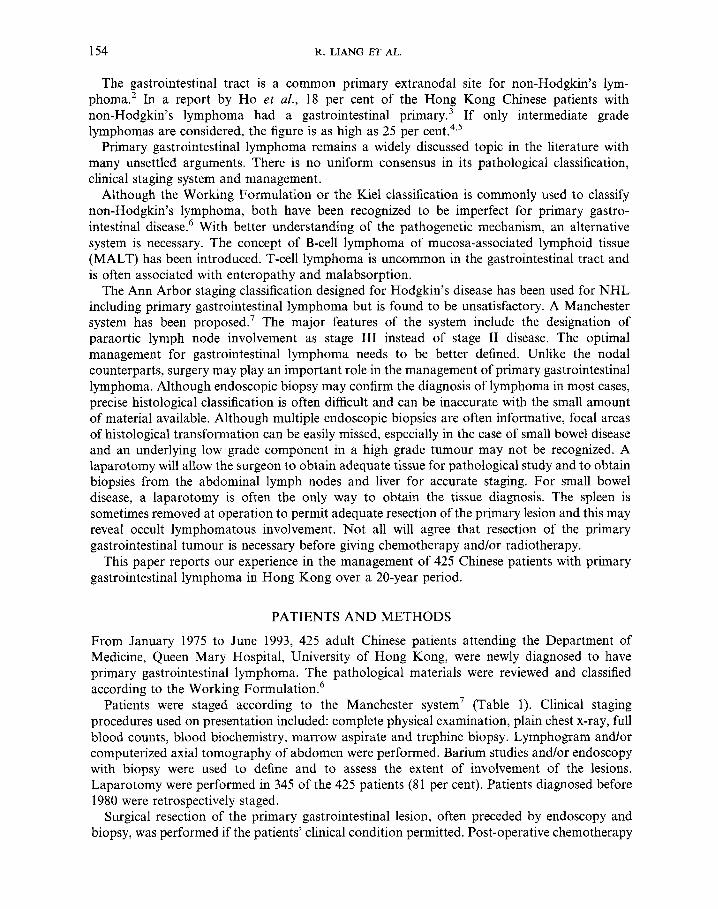
Multivariate analysis was performed on the 374 patients who had received chemotherapy with or without additional radiotherapy. Factors included in the analysis included: sex, age, histology, immunophenotype, stage of disease, B symptoms, primary site, bulky disease, chemotherapy regimens, pre-chemotherapy serum lactate dehydrogenase level, complete surgi- cal resection of tumour and additional radiotherapy. It revealed that significant independent prognostic factors predicting better survival were young age of <60 years, low grade histology, stage I and I1 disease and absence of bulky tumour (Figures 1 and 2). Surgical resection of the primary lesions and local radiotherapy did not turn out to be significant in the multiple analysis.
Total gastrectomy instead of a conservative resection according to the choice of the surgeon was performed in 78 of the 172 patients with gastric lymphoma who had their primary lesions resected. Despite higher post-operative morbidity, aggressive surgery did not significantly improve complete remission rate (64 per cent versus 61 per cent), relapse rate (40 per cent versus 44 per cent), 5-year disease-free survival following complete remission (46 per cent versus 50 per cent) and overall survival at 5 years (44 per cent versus 48 per cent).
DISCUSSION
This series of 425 Chinese patients with primary gastrointestinal lymphoma from Hong Kong is one of the largest reported in the Their median age at presentation was 53 years and there was no significant sex preference. Stomach was most commonly involved. The pattern is similar in western countries and Chinese. Initial symptoms were mostly referable to the gastrointestinal tract and abdominal pain and bleeding were most common. Perforation and palpable abdominal mass were more frequent in intestinal lesions.
The majority of our cases were intermediate or high grade according to Working Formulation and of B-cell origin. It appears that a better classification for gastrointestinal lymphoma is e s~en t i a l .~~- ’~ A study of the British National Lymphoma Investigation has shown that tumour origin from MALT is a significant favourable prognostic factor for gastric lymphoma and ‘malignant histiocytosis’ of the intestine is also associated with poorer clinical ou t~ome .~ ’
According to the Manchester system, most of our patients presented with advanced stage I11 and IV d i~ease .~ Regional lymph node involvement was common and frequent metastatic sites included the liver, spleen and marrow. Advanced stage 111 and IV disease was associated with

158 R. LIANG ET AL.
Table 4. Treatment results according to patient characteristics ~
DFS Survival (CR patients) (all patients)
Median 5-yr Median 5-yr CR
P4 (%) (months) (%) (months) (%)
Relapse
Sex Male 58 40 51 45 24 39 Female 69 44 60 49 111 52 p-value n.s. n s . n.s. 0.009
<60 years 64 43 84 52 84 51 >60 years 61 38 45 34 24 33 p-value ns . n.s. n.s. 0.03
68 44 150 62 111 75
Age
Histology according to Working Formulation Low grade" Intermediateb High gradec p-value
Immunopheno type T-cell B-cell p-value
I I1 I11 IV p-value
B symptoms
Stage of disease
No Yes p-value
Primary site Stomach" Small intestineb Large intestine' p-value
68 58
n.s.
45 35 ns .
93 69 70 31
n.s. 0.006
79 14 89 37 70 37 49 62
<0~0001 < 0.000 1 (MI versus III/IV) (ID1 versus III/IV)
65 35 56 52
n.s. 0.0 1
66 35 56 54 65 53
n.s. 0.01 (a versus b+c)
Presence of bulky disease (2 10 cm) No 68 41 Yes 51 56 p-value0.02 n.s. n.s.
CVP/COPP" 64 25 CHOP/BACOP/m- 74 32
Others' 53 62
Chemotherapy regimens
BACOD~
p-value 0.003 <0~00001 (b versus a+c) (b versus a+c)
51 42 27 43 nr 61 15 41
0.0035 <0.0001 (a versus b+c) (a versus b+c)
18 30 51 40 105 51 66 57
ns . n s .
nr 63 114 71 nr 72 nr 70 54 44 18 45 21 32 15 32
<0~00001 <0~00001 (I/II versus III/IV) (101 versus III/IV)
150 55 84 52 27 38 65 36
n.s. < 0.000 1
150 53 45 49 18 43 15 37 60 41 60 49
0.03 0.01 (a versus b+c)
54 49 51 45
150 65 nr 60
24 30 0.008
(b versus a+c)
(a versus b+c)
51 48 12 36
0.0003
27 44 84 55
21 38 0.002
(b versus a+c)

GASTROINTESTINAL LYMPHOMA 159
Table 4. (Continued)
DFS Survival (CR patients) (all patients)
Median 5-yr Median 5-yr CR
(%I (%) (months) (YO) (months) (%)
Relapse
Pre-chemotherapy serum lactate dehydrogenase level @mol/min/l) ~ 4 0 0 67 40 58 48 >400 62 42 52 46 p-value n.s. n.s. n s .
No 49 69 18 8 Yes 67 36 105 54 p-value 0.00 1 0.00004 0~00001
Surgical resection of primary lesions
Local radiotherapy No 55 30 51 41 Yes 75 54 57 47 p-value 0.0005 n.s. n.s.
60 52 52 45
n.s.
15 36 51 40
0.01
24 35 84 58
0~00001
CR=complete remission; DFSZdisease free survival; nr=not reached; n.s.=not significant, yr=year.
significantly poorer prognosis. Local features including multicentric tumours, perforation, adhesion, and peritonitis are considered by the Manchester system. However, the small number of patients in the subgroups in our series prevents proper determination of their prognostic value.
There is no general consensus in the optimal management for primary gastrointestinal l y rnph~ma . ’~ -~~ Most of our patients had surgical resection of the primary lesion and this was usually followed by combination chemotherapy. The younger patients received one of the anthracycline-containing regimens and less intensive regimens were employed for the more elderly. Selected patients received additional local radiotherapy.
Univariate analysis revealed that better complete remission rates were seen in patients with early stages of disease, without bulky tumour, receiving an anthracycline-containing regimen, having a surgical resection and receiving local radiotherapy. Also, superior disease-free survival following complete remission was observed in those with a low grade histology, early stages of disease, a primary gastric tumour and having a primary surgical resection. Furthermore, factors associated with superior overall survival included female sex, age <60 years, a low grade histology, early stages of disease, absence of B symptoms, a primary gastric or large bowel tumour, absence of bulky tumour, receiving anthracycline, having surgical resection and receiving local radiotherapy. Independent prognostic variables were determined by multivariate analysis and the survival was significantly better only in the subgroups of patients with a young age of <60 years, low grade histology, stage I and I1 disease and absence of bulky tumour.
The role of surgery in managing gastrointestinal lymphoma remains c o n t r ~ v e r s i a l . ~ ~ - ~ ~ From our experience, removal of the primary gastrointestinal lesion appears to prevent potentially fatal bleeding and perforation following chemotherapy or radiotherapy although one may argue that patients who have locally invasive and bulky lesions, are not operable most of the time and are likely to develop complications. On the other hand, a patient with an operable lesion is likely to do well with or without ~ u r g e r y . ~ ~ . ~ ~ Aggressive surgery however does not appear to be

160 R. LIANG ET AL.
1
0.9
0.8
0.7
t 0 0.6
e 0 0.5 Q
0.4
0.3
0.2
0.1
0
.-
........ ....... .... .... .... .-..
........ ....... .... P =0.00001
(1/11 vs. III/IV)
Ill
......
IV 0 20 40 60 80 100 120 140 160 180 200
Months Figure 1. The survival of patients with primary gastrointestinal lymphoma according to
stage of disease
1
0.9
0.8
0.7
L=: 0 0.6
c 0 0.5 a
0.4
0.3
0.2
0.1
0
.-
P=0.03
.... ...... ............... ..............................................................
< 60 years
>60 years
0 20 40 60 80 100 120 140 160 180 200
Months Figure 2. The survival of patients with primary gastrointestinal lymphoma according
to age
necessary and may increase the operative m~rtali ty. '~ The post-operative morbidity of total gastrectoniy may also make the subsequent administration of chemotherapy or radiotherapy more difficult. Whether local surgical resection improves prognosis of primary gastrointestinal

GASTROINTESTINAL LYMPHOMA 161
lymphoma has not been prospectively studied. It remains possible that surgery alone may be curative in selective patients with truly localized stage I lesion.
Chemotherapy is the mainstay of treatment for our patients with primary gastrointestinal lymphoma.36 Our younger patients treated with an anthracycline-containing regimen had better survival and the age factor turned out to be more significant on multivariate analysis. Surgical resection and local radiotherapy did not turn out to be significant on multivariate analysis. Although surgical resection and local radiotherapy were associated with better prognosis on univariate analysis, both were rejected in the multivariate analysis. It was likely that patients with other favourable features were selected for surgical resection. Patients who responded poorly to chemotherapy and died early were unlikely to have the chance to receive local r ad i~ the rapy .~~
Better classification of the primary gastrointestinal lymphoma and more refined stratification of the patients according to the prognostic variables are necessary to allow individualization of treatment. Prospective randomized studies are essential to define the relative role of surgery, chemotherapy and radiotherapy. It has been proposed that Helicobacter pylori may be pathogenetically related to the mucosa associated lymphoid tissue related lymphoma of stomach and antibiotics may be effective in treating those who still have a low grade tumour. If further substantiated, these exciting findings may provide a new approach to treatment.3843
ACKNOWLEDGEMENT
We wish to thank Professor John Wong and members of the Department of Surgery at Queen Mary Hospital, Hong Kong, for providing the surgical support.
REFERENCES
1. 2.
3.
4.
5.
6.
7.
8.
9.
10.
11.
12.
Haber, D. A., Mayer, R. J. Primary gastrointestinal lymphoma. Sem. Oncol. 1988, 15, 154-169. Paryani, S., Hoppe, R. T., Burkes, J. S., el al. Extralymphatic involvement in diffuse non-Hodgkin’s lymphoma. J. Clin. Oncol. 1983, 1, 682-688. Ho, F. C. S., Todd, D., Loke, S. L., Ng, R. P., Khoo, R. K. K. Clinico-pathological features of malignant lymphoma in 294 Hong Kong Chinese patients-retrospective study covering an eight-year period. Int. J. Cancer 1984, 34, 143-148. Liang, R., Choy, D., Todd, D., Chan, T. K., Loke, S. L. Chemotherapy versus radiotherapy for stage I and I1 intermediate grade non-Hodgkin’s lymphoma. Clin. Oncol. 1991, 23, 35fL359. Liang, R., Chiu, E., Chan, T. K., Todd, D., Loke, S. L. Management of advanced stage intermediate grade lymphoma. Hematol. Oncol. 1990, 8, 147-154. The non-Hodgkin’s Lymphoma Pathologic Classification Project. National Cancer Institute spon- sored study of classifications of non-Hodgkin’s lymphomas: summary and description of a working formulation for clinical usage. Cancer 1982, 49, 21 12-2135. Blackledge, G., Bush, H., Dodge, 0. G., Crowther, D. A study of gastrointestinal lymphoma. Clin. Oncol. 1979, 5, 209-219. Hoogstraten, B. Reporting treatment results in solid tumours. In: Byse, M. E., Staquet, M. J., Sylvester, R. J. eds. Cancer Clinical Trials, Method and Practice. Oxford: Oxford University Press, 1984: 139-152. Kaplan, E. L., Meier, P. Nonparametric estimation from incomplete observations. J. Am Stat. Assoc. 1958, 53,457481. Anderson, T., Bender, R. A., Fisher, R. I., et al. Combination chemotherapy in non-Hodgkin’s lymphomas: results of long-term follow up. Cancer Treat. Rep. 1977, 61, 1057-1061. De Vita, V. T., Serpick, A. A,, Carbone, P. P. Combination chemotherapy in the treatment of Hodgkin’s disease. Ann. Intern. Med. 1970, 73, 881-886. McKelvey, E. M., Gottlieb, J. A., Wilson, H. E., et al. Hydroxy-daunorubicin (adriamycin) combination chemotherapy in malignant lymphoma. Cancer 1976, 38, 14841490.

162 R. LIANG ET AL.
13.
14.
15.
16.
17.
18.
19.
20.
21
22.
23.
24.
25.
26.
27.
28.
29.
30.
31.
32.
33.
34.
35.
36.
37.
Schein, P. S., De Vita, V. T. Jr, Hubbard, S., et al. Bleomycin, adriamycin, cyclophosphamide, vincristine and prednisone (BACOP) combination chemotherapy in the treatment of advanced aggressive histiocytic lymphomas. Ann. Intern. Med. 1976, 85, 41 7-422. Shipp, M. A,, Harrington, D. P., Klatt, M. M., et al. Identification of major prognostic subgroups of patients with diffuse large cell lymphomas treated with m-BACOD of M-BACOD. Ann. Intern. Med.
Ng, R. P., Todd, D., Khoo, R. K. K. Salvage chemotherapy for non-Hodgkin’s lymphoma. Cancer Treat. Rep. 1982, 66, 1977-1979. Cabanillas, P. P., Burgess, M. A., Bodey, G. P., Freireich, E. F. Sequential chemotherapy and late intensification for malignant lymphomas of aggressive histologic type. Am. J. Med. 1983,74,382-385. Cabanillas. P. P., Hagemeister, F. B., Bodey, G. P., Freireich, E. F. IMVP-16, an effective regimen for patients with lymphoma who have relapsed after initial combination chemotherapy. Blood 1982, 60, 693-697. Lewis, K. J., Ranchod, Dorfman. Lymphomas of the gastrointestinal tract: a study of 117 cases presenting with gastrointestinal disease. Cancer 1978, 42, 693-707. Lee, W. J., Chang, K. J., Wang, S. M., et al. Primary malignant tumour of the small intestine. Taiwan I Hsueh Hui Tsa Chih 1991, 90, 776-781. Sandler, R. S. Has primary gastric lymphoma become more common? J. Clin. Gastroenterol. 1984, 6, 10 1-107. Pinotti, G., Novario, R., Berrino, F., et a/. Primary gastric non-Hodgkin’s lymphoma in a population-based registry. Haematologica 1922, 77, 405412. Domizio, P., Owen, R. A., Shepherd, N. A,, et al. Primary lymphoma of small intestine. Am. J. Surg. Path. 1933, 17, 429-442. Kusomoto, H., Takahashi, I. , Yoshida, M., et al. Primary malignant tumours of small intestines: analysis of 40 Japanese patients. J. Surg. Oncol. 1992, 50, 139-143. Weingrad, D. N., Decosse, J. J., Sherlock, P., Straus, D., Liebemian, P. H., Filippe, D. A. Primary gastrointestinal lymphoma. Cancer 1982, 49, 1258-1266. Isaacson, P. G., Spencer, J., Wright, D. H. Classifying primary gut lymphomas. Lancet 1988, ii,
Khojasteh, A., Hahshenass, M., Haghighi, P. Current concepts-immunoproliferative small intestinal disease. A ‘third world’ lesion. New Engl. J. Med. 1983, 308, 1401-1405. Ramot, B., Rechavi, G. Primary intestinal lymphoma and alpha heavy chain disease. In: Magrath, I. T., ed. The Non-Hodgkin’s Lymphoma. London: Arnold, 1990: 352-358. Chiu, E. K. W., Loke, S. L., Chan, A. C. L., Liang, R. T-lymphoblastic lymphoma arising in the small intestine. Pathology 1991, 23, 356-359. Chott, A., Dragosics, B., Radaszkiewiez, T. Peripheral T-cell lymphoma of the intestine. Am. J. Path.
Renton, P., Blackshaw, A. J. Colonic lymphoma complicating ulcerative colitis. Br. J. Surg. 1976,63, 542-545. Morton, J. E., Leyland, M. J., Vanghan Hudson, G., et al. Primary gastrointestinal non-Hodgkin’s lymphoma: a review of 175 British National Lymphoma Investigation cases. Br. J. Cancer 1993, 67, 776-782. Liang, T., Todd, Chan, T. K., et al. Gastrointestinal lymphoma in Chinese: a retrospective analysis. Hematol. Oncol. 1987, 5, 115-126. Vorsos, G., Yahalom, J. Alternatives in the management of gastric lymphoma. Leuk. Lymph. 1991, 4, 1-8. Shiu, M. H., Nisce, L. Z., Pinna, A., et al. Results of multimodality therapy for gastric lymphoma. Cancer 1986, 58, 1389-1395. Tondini, C., Giardini, R., Valagussa, Santoro, A., et al. Combined modality therapy for primary gastrointestinal lymphoma: the Milan Cancer Institute experience (abstract). The fifth International Conference on Malignant Lymphoma, Lugano. 1993: no. 94. Liang, R., Chiu, E., Todd, Chan, T. K., Choy, D., Loke, S. L. Chemotherapy for early stage gastrointestinal lymphoma. Cancer Chemother. Pharmacol. 1991, 27, 385-388. d’Amore, F., Brincker, H., Gronbaek, K., et al. Non-Hodgkin’s lymphoma of the gastro- intestinal tract: a population-based analysis of incidence, geographic distribution, clinicopathologic presentation features and prognosis. J. Clin. Oncol. 1994, 12, 1673-1684.
1986, 104, 757-763.
1148-1149.
1992, 141, 1361-1371.

GASTROINTESTINAL LYMPHOMA I63
38. Wotherspoon, A. C., Doglioni, C., Diss, T. C., et al. Regression of primary low grade B-cell gastric lymphoma of mucosa-associated lymphoid tisue type after eradication of Heliobacter pylori. Lancet 1993, 342, 375-517. Isaacson, P. G., Wright, D. H. Extranodal malignant lymphoma arising from the mucosa associated lymphoid tissue. Cancer 1984, 53, 25 15-2524. Isaacson, P. G. Lymphomas of mucosa associated lymphoid tissue (MALT). Histopathology 1990,16, 617-619. Castrillo, J. M., Montalban, C., Obeso, G., et al. Gastric B-cell mucosa associated lymphoid tissue lymphoma: a clinicopathological study in 56 patients. Gut 1992, 33, 1307-131 1. Harris, N. L. Extranodal lymphoid infiltrates and mucosa-associated lymphoid tissue (MALT). Am. J. Surg. Path. 1991, 15, 879-884. Blazque, M., Haioun, C . , Chaumette, M. T., et al. Low grade B-cell mucosa associated lymphoid tissue lymphoma of the stomach: clinical, and endoscopic features, treatment and outcome. Gut 1992,
39.
40.
41.
42.
43.
33, 1621-1625.